Bodily Secretions
Intestinal Secretions
Secretory Rate
Bacterial Secretion Systems
Insulin
Cells, Cultured
Islets of Langerhans
Glucose
Pancreatic Juice
Gastric Juice
Bicarbonates
Luteinizing Hormone
Calcium
Pancreas
Molecular Sequence Data
Synovial Fluid
RNA, Messenger
Saliva
Secretory Pathway
Pentagastrin
Insulin-Secreting Cells
Exocytosis
Secretin
Dose-Response Relationship, Drug
Amylases
Amino Acid Sequence
Radioimmunoassay
Gastric Mucosa
Fluid Therapy
Stimulation, Chemical
Prolactin
Enzyme-Linked Immunosorbent Assay
Mucus
Signal Transduction
Pituitary Gland
Rats, Sprague-Dawley
Gastrins
Progesterone
Amniotic Fluid
Sheep
Secretory Vesicles
Bile
Cytokines
Glucagon
Mice, Inbred C57BL
Epithelial Cells
Exocrine Glands
Protein Transport
Biological Transport
Duodenum
Cattle
Somatostatin
Rats, Inbred Strains
Rats, Wistar
Cyclic AMP
Gonadotropin-Releasing Hormone
Estradiol
Liver
Follicle Stimulating Hormone
Blotting, Western
Hydrogen-Ion Concentration
Interleukin-8
Intestinal Mucosa
Adrenocorticotropic Hormone
Glucagon-Like Peptide 1
Labyrinthine Fluids
Tumor Necrosis Factor-alpha
Pituitary Gland, Anterior
Macrophages
Cell Membrane
Carbachol
Parotid Gland
Growth Hormone
Carrier Proteins
Interleukin-6
Models, Biological
Base Sequence
Mutation
Rabbits
Dialysis Solutions
Mice, Knockout
Histamine
Reverse Transcriptase Polymerase Chain Reaction
Exudates and Transudates
Proteins
Swine
Virulence Factors
Potassium
Pregnancy
Membrane Proteins
Gene Expression
Gene Expression Regulation
Dogs
Protein Sorting Signals
Stomach
Sodium
Apolipoproteins B
Electrophoresis, Polyacrylamide Gel
Bethanechol Compounds
Interleukin-1
Colforsin
Epithelium
Culture Media
Peptides
Gene Expression Regulation, Bacterial
Water-Electrolyte Balance
Protein Precursors
Lipopolysaccharides
Mice, Inbred BALB C
Glycoproteins
Hydrocortisone
Osmolar Concentration
Interferon-gamma
Enteroendocrine Cells
Transfection
C-Peptide
Testosterone
Isotonic Solutions
Sterilization
Adrenal Glands
Chromatography, High Pressure Liquid
Immunohistochemistry
Bronchoalveolar Lavage Fluid
Parietal Cells, Gastric
Monocytes
Glucose Tolerance Test
Protein Processing, Post-Translational
Salivation
Extracellular Space
Membrane Transport Proteins
Mucins
Microscopy, Electron
Immunoglobulin G
Growth Hormone-Releasing Hormone
Enzyme Inhibitors
Inappropriate ADH Syndrome
Adenosine Triphosphate
Tumor Cells, Cultured
Lacrimal Apparatus
Inhibins
Virulence
Adrenal Medulla
Colon
Lipoproteins, VLDL
Submandibular Gland
Atropine
Triglycerides
Jejunum
Golgi Apparatus
Peptide Fragments
Intestines
Human Growth Hormone
Lung
Estrus
Electrolytes
Escherichia coli
Sincalide
Cimetidine
Kidney
Bile Acids and Salts
Cystic Fibrosis Transmembrane Conductance Regulator
Protein Binding
Trachea
Albumins
Insulinoma
Bethanechol
Gastric Inhibitory Polypeptide
Gonadotropins, Pituitary
Bacterial Outer Membrane Proteins
Gastrointestinal Hormones
Fluorescent Antibody Technique
Vagotomy
Follicular Fluid
Biological Assay
Endoplasmic Reticulum
Pepsin A
Brefeldin A
Exosomes
Interleukin-1beta
Lymphocyte Activation
DIFFERENTIAL FERRIOXAMINE TEST FOR MEASURING CHELATABLE BODY IRON. (1/9)
The differential ferrioxamine test is a simple method for the measurement of chelation of body iron by desferrioxamine. A single six-hour specimen of urine is obtained after intravenous Desferal, accompanied by (59)Fe-ferrioxamine. Two values are measured: F(d), the excretion of ferrioxamine derived from body iron by chelation, and F(ex), the proportion of ferrioxamine excreted from a known intravenous dose. The data enables F(v), chelation of iron in vivo, to be calculated by simple proportion. Desferrioxamine chelation proceeds for about half an hour after injection. The results in normal subjects, in cases with known high iron stores, and in cases of iron-deficiency anaemia are described. High, normal, and low body iron states have been differentiated. F(v) values in the higher ranges obtained in iron-storage diseases and in haemolytic states are differentiated by the pattern of excretion, high F(d) values and low F(ex) values respectively. IT IS SUGGESTED THAT THERE ARE TWO MAIN SOURCES OF CHELATABLE BODY IRON: as ferritin-haemosiderin and as iron newly released from haem in a more readily chelatable form. The significance of variable chelation susceptibility in iron metabolism is briefly discussed. It is suggested that variable chelatability of different sources of body iron may explain the preferential utilization of iron released from red cells or absorbed from the intestine, rather than storage iron, in the biosynthesis of haem. (+info)THE HEPATIC UPTAKE AND EXCRETION OF SULFOBROMOPHTHALEIN AND BILIRUBIN. (2/9)
Experimental evidence bearing on the hepatic uptake and excretion of sulfobromophthalein and bilirubin is considered. From this examination it is postulated that the hepatic handling of each compound consists of three steps: the uptake at the sinusoidal surface of the parenchymal cell by a concentrative membrane transport system of high capacity; the conjugation within the cell with glutathione and glucuronic acid, respectively; and the excretion into the biliary canaliculus of the respective conjugates by a second concentrative system of relatively low capacity. Two of the operating characteristics of this linked system, the easy attainment of saturation of the biliary excretory transport system and the presence of an intracellular storage phenomenon, have been shown to result from the disproportionate capacities of the two sequentially placed concentrative membrane transport systems. The disorders which may occur in the three steps underlying the handling of each of these compounds are also considered. (+info)PORPHOBILINOGEN AND DELTA-AMINOLEVULINIC ACID EXCRETION IN MULTIPLE SCLEROSIS. (3/9)
The excretion of delta-aminolevulinic acid and porphobilinogen in the urine of 31 patients with multiple sclerosis did not differ significantly from that of 51 hospitalized control patients or eight patients with poliomyelitis. There was no relationship between exacerbations, remissions or duration of the illness, and levels of delta-aminolevulinic acid or of porphobilinogen. These assays therefore appear to be of no value in the differential diagnosis of multiple sclerosis or in following the severity or stage of this illness. Whereas demyelination does occur in acute porphyria where the levels of delta-aminolevulinic acid and porphobilinogen are elevated, the converse is not true; that is, demyelination is not always associated with an increase in the excretion of porphobilinogen or delta-aminolevulinic acid. (+info)BIOCHEMICAL STUDIES OF TOXIC AGENTS. THE METABOLISM OF IODOMETHANE. (4/9)
1. The metabolism of iodomethane has been studied after the administration of the compound to rats by subcutaneous injection. 2. The urinary excretion of S-methylcysteine, methylmercapturic acid, methylthioacetic acid and N-(methylthioacetyl)glycine has been demonstrated by paper chromatography. 3. Methylmercapturic acid and N-(methylthioacetyl)glycine have been isolated from the urine of the dosed animals. 4. The methylthio compounds detected represented about 2% of the iodomethane administered. (+info)ISOTOPIC STUDIES OF UREA METABOLISM IN RABBITS. (5/9)
1. The half-life of [(15)N]urea was found to be significantly longer than that of [(14)C]urea injected at the same time, the differences being due to endogenous catabolism of urea, which is accompanied by little or no reutilization of (14)C but is approx. 20% for (15)N. [(15)N]Urea therefore appears to be valueless as an indicator of nitrogen metabolism unless the extents of endogenous catabolism of urea and of fractional reutilization of (15)N can be separately estimated. 2. Though measurements of the radioactivity of expired (14)CO(2) confirmed the existence of considerable urea catabolism these could not be used for quantitative assessments. 3. Alternative graphical methods based on [(14)C]urea specific activities in plasma and urine samples were used to calculate the fraction of urea production that is excreted. Values by the two methods were in good agreement and showed that some animals excrete less than half the urea that they produce. 4. Specific activity differences between simultaneous samples of urinary and plasma urea reflect the presence of a pool of urea in the kidney that is not in equilibrium with the body urea pool. Calculations indicate the presence of urea in the kidney that in some cases may represent as much as 15% of the body pool, and in two animals in which post-mortem renal analyses were performed the masses of urea found agreed closely with the calculated values. 5. A model for urea metabolism is proposed that includes this pool in the excretory pathway. The related theory is shown to be adequate to explain the shape of the specific activity curves of urinary urea from the time of injection and the constant delay of the specific activity of urinary urea, relative to that of plasma urea, that is observed after a short preliminary equilibration period. 6. The body urea pool was calculated from the activity retained at 1.5hr. by excluding renal activity and the corrected specific activity of plasma urea at the same time. The urea pool was calculated to be distributed at the plasma concentration in a substantially smaller water volume than that found by injecting tritiated water in five animals. Reasons for this are discussed. 7. Urea synthesis rates calculated from the pool values are in close agreement with rates calculated from the mass of urea recovered in the urine and the fraction of newly synthesized urea that is excreted. (+info)URINARY EXCRETION OF C-20-REDUCTION PRODUCTS OF CORTICOSTERONE. (6/9)
1. A 200 mg. portion of corticosterone was ingested by a healthy man and the urine collected. Part of the urine was treated with the gastric juice of Helix pomatia and extracted with ethyl acetate, and the extract fractionated with Girard T. Paper-chromatographic separation of the non-ketonic fraction in the Bush (1952) system B(5) revealed the presence of two unknown polar components. 2. The unknown compounds did not possess a reducing (blue tetrazolium) or a reducible (potassium borohydride) grouping. Both contained a terminal alpha-glycollic fragment as shown by the formation of formaldehyde, and of a non-volatile aldehyde on oxidation with sodium bismuthate. 3. Unknown compound (I) had paper-chromatographic mobilities identical with those of 5beta-pregnane-3alpha,11beta,20beta,21-tetraol. The oxidation product of compound (I) had a retention time (gas-liquid chromatography) on an SE30 column identical with that of 3alpha,11beta-dihydroxy-21-nor-5beta-pregnan-20-al. The retention times of various derivatives agreed with those produced in an identical manner on the standard, and accordingly compound (I) is formulated as 5beta-pregnane-3alpha,11beta,20xi,21-tetraol. 4. Unknown compound (II) had a higher R(F) than compound (I), and its oxidation product had a longer retention time than that of compound (I). From the group effects observed in paper and gas-liquid chromatography, compound (II) is tentatively formulated as 5alpha-pregnane-3alpha,11beta,20xi,21-tetraol. The 5alpha/5beta ratio found was about 2.0. (+info)THE REGULATION OF UREA-BIOSYNTHESIS ENZYMES IN VERTEBRATES. (7/9)
1. Carbamoyl phosphate synthetase, ornithine transcarbamoylase, the arginine-synthetase system and arginase were measured in the livers of ammoniotelic, ureotelic and uricotelic animals. The chelonian reptiles, whose nitrogen excretory patterns vary according to the habitat, and the Mexican axolotl, a neotenic species, were also studied. 2. The levels of the activities of the first three enzymes mentioned correlate with the amount of nitrogen excreted as urea. 3. The terrestrial turtle, which excretes mainly uric acid, maintains a high arginase activity but has very low levels of the activities of the other three enzymes. 4. The first three enzymes of the urea cycle vary in the phylogenic scale in a co-ordinated manner, which suggests that they are under the same regulatory mechanism. 5. Urea formation from endogenous arginine in vitro has a low efficiency in the Mexican axolotl. 6. The induction of metamorphosis in the Mexican axolotl by the administration of l-tri-iodothyronine, which causes a shift from ammonio-ureotelism to complete ureotelism, is accompanied by an increase mainly in carbamoyl phosphate synthetase and also by an improvement in the efficiency of hydrolysis of endogenous arginine in vitro to give urea. 7. The results obtained by differential centrifugation of the urea-cycle enzymes in rat and Mexican-axolotl livers are presented. The location requirements for the integration of a metabolic cycle are discussed. (+info)The prevention of the fluid-electrolyte "problem" by simple means. (8/9)
Proper fluid balance may be maintained in patients after operation by the employment of simple, inexpensive procedures which may be carried out even in the smallest hospitals. Daily weighing of patients, measurement of fluid intake and output, and knowledge of the probable electrolyte content of fluid losses are adequate guides for replacement of fluids and electrolytes. An increase in body weight is a warning of overhydration.The content of the solution used for replacement is dictated by the route of fluid output-whether from the gastrointestinal tract, the skin, or the kidneys. Insensible losses (by perspiration and respiration) are fairly static. Except to replace extrarenal losses, parenteral administration of normal saline solution in the immediate postoperative period is contraindicated. Mistakes in replacement methods, especially those causing overhydration, are particularly hazardous for elderly patients. (+info)Body fluids refer to the various liquids that can be found within and circulating throughout the human body. These fluids include, but are not limited to:
1. Blood: A fluid that carries oxygen, nutrients, hormones, and waste products throughout the body via the cardiovascular system. It is composed of red and white blood cells suspended in plasma.
2. Lymph: A clear-to-white fluid that circulates through the lymphatic system, helping to remove waste products, bacteria, and damaged cells from tissues while also playing a crucial role in the immune system.
3. Interstitial fluid: Also known as tissue fluid or extracellular fluid, it is the fluid that surrounds the cells in the body's tissues, allowing for nutrient exchange and waste removal between cells and blood vessels.
4. Cerebrospinal fluid (CSF): A clear, colorless fluid that circulates around the brain and spinal cord, providing protection, cushioning, and nutrients to these delicate structures while also removing waste products.
5. Pleural fluid: A small amount of lubricating fluid found in the pleural space between the lungs and the chest wall, allowing for smooth movement during respiration.
6. Pericardial fluid: A small amount of lubricating fluid found within the pericardial sac surrounding the heart, reducing friction during heart contractions.
7. Synovial fluid: A viscous, lubricating fluid found in joint spaces, allowing for smooth movement and protecting the articular cartilage from wear and tear.
8. Urine: A waste product produced by the kidneys, consisting of water, urea, creatinine, and various ions, which is excreted through the urinary system.
9. Gastrointestinal secretions: Fluids produced by the digestive system, including saliva, gastric juice, bile, pancreatic juice, and intestinal secretions, which aid in digestion, absorption, and elimination of food particles.
10. Reproductive fluids: Secretions from the male (semen) and female (cervical mucus, vaginal lubrication) reproductive systems that facilitate fertilization and reproduction.
Bodily secretions are substances that are produced and released by various glands and organs in the body. These secretions help maintain the body's homeostasis, protect it from external threats, and aid in digestion and other physiological processes. Examples of bodily secretions include:
1. Sweat: A watery substance produced by sweat glands to regulate body temperature through evaporation.
2. Sebaceous secretions: Oily substances produced by sebaceous glands to lubricate and protect the skin and hair.
3. Saliva: A mixture of water, enzymes, electrolytes, and mucus produced by salivary glands to aid in digestion and speech.
4. Tears: A mixture of water, electrolytes, and proteins produced by the lacrimal glands to lubricate and protect the eyes.
5. Mucus: A slippery substance produced by mucous membranes lining various body cavities, such as the respiratory and gastrointestinal tracts, to trap and remove foreign particles and pathogens.
6. Gastric juices: Digestive enzymes and hydrochloric acid produced by the stomach to break down food.
7. Pancreatic juices: Digestive enzymes produced by the pancreas to further break down food in the small intestine.
8. Bile: A greenish-brown alkaline fluid produced by the liver and stored in the gallbladder, which helps digest fats and eliminate waste products.
9. Menstrual blood: The shedding of the uterine lining that occurs during menstruation, containing blood, mucus, and endometrial tissue.
10. Vaginal secretions: Fluid produced by the vagina to maintain its moisture, pH balance, and provide a protective barrier against infections.
11. Semen: A mixture of sperm cells, fluids from the seminal vesicles, prostate gland, and bulbourethral glands that aids in the transportation and survival of sperm during sexual reproduction.
Intestinal secretions refer to the fluids and electrolytes that are released by the cells lining the small intestine in response to various stimuli. These secretions play a crucial role in the digestion and absorption of nutrients from food. The major components of intestinal secretions include water, electrolytes (such as sodium, chloride, bicarbonate, and potassium), and enzymes that help break down carbohydrates, proteins, and fats.
The small intestine secretes these substances in response to hormonal signals, neural stimulation, and the presence of food in the lumen of the intestine. The secretion of water and electrolytes helps maintain the proper hydration and pH of the intestinal contents, while the enzymes facilitate the breakdown of nutrients into smaller molecules that can be absorbed across the intestinal wall.
Abnormalities in intestinal secretions can lead to various gastrointestinal disorders, such as diarrhea, malabsorption, and inflammatory bowel disease.
Secretory rate refers to the amount or volume of a secretion produced by a gland or an organ over a given period of time. It is a measure of the productivity or activity level of the secreting structure. The secretory rate can be quantified for various bodily fluids, such as saliva, sweat, digestive enzymes, hormones, or milk, depending on the context and the specific gland or organ being studied.
In clinical settings, measuring the secretory rate might involve collecting and analyzing samples over a certain duration to estimate the production rate of the substance in question. This information can be helpful in diagnosing conditions related to impaired secretion, monitoring treatment responses, or understanding the physiological adaptations of the body under different circumstances.
Bacterial secretion systems are specialized molecular machines that allow bacteria to transport proteins and other molecules across their cell membranes. These systems play a crucial role in bacterial survival, pathogenesis, and communication with their environment. They are composed of several protein components organized into complex structures that span the bacterial cell envelope.
There are several types of bacterial secretion systems, including type I to type IX secretion systems (T1SS to T9SS). Each type has a unique structure and mechanism for transporting specific substrates across the membrane. Here are some examples:
* Type II secretion system (T2SS): This system transports folded proteins across the outer membrane of gram-negative bacteria. It is composed of 12 to 15 protein components that form a complex structure called the secretion apparatus or "secretion nanomachine." The T2SS secretes various virulence factors, such as exotoxins and hydrolases, which contribute to bacterial pathogenesis.
* Type III secretion system (T3SS): This system transports effector proteins directly into the cytosol of host cells during bacterial infection. It is composed of a hollow needle-like structure that extends from the bacterial cell surface and injects effectors into the host cell. The T3SS plays a critical role in the pathogenesis of many gram-negative bacteria, including Yersinia, Salmonella, and Shigella.
* Type IV secretion system (T4SS): This system transports DNA or proteins across the bacterial cell envelope and into target cells. It is composed of a complex structure that spans both the inner and outer membranes of gram-negative bacteria and the cytoplasmic membrane of gram-positive bacteria. The T4SS plays a role in bacterial conjugation, DNA uptake and release, and delivery of effector proteins to host cells.
* Type VI secretion system (T6SS): This system transports effector proteins into neighboring cells or the extracellular environment. It is composed of a contractile sheath-tube structure that propels effectors through a hollow inner tube and out of the bacterial cell. The T6SS plays a role in interbacterial competition, biofilm formation, and virulence.
Overall, these secretion systems play crucial roles in bacterial survival, pathogenesis, and communication with their environment. Understanding how they function and how they contribute to bacterial infection and disease is essential for developing new strategies to combat bacterial infections and improve human health.
Insulin is a hormone produced by the beta cells of the pancreatic islets, primarily in response to elevated levels of glucose in the circulating blood. It plays a crucial role in regulating blood glucose levels and facilitating the uptake and utilization of glucose by peripheral tissues, such as muscle and adipose tissue, for energy production and storage. Insulin also inhibits glucose production in the liver and promotes the storage of excess glucose as glycogen or triglycerides.
Deficiency in insulin secretion or action leads to impaired glucose regulation and can result in conditions such as diabetes mellitus, characterized by chronic hyperglycemia and associated complications. Exogenous insulin is used as a replacement therapy in individuals with diabetes to help manage their blood glucose levels and prevent long-term complications.
"Cells, cultured" is a medical term that refers to cells that have been removed from an organism and grown in controlled laboratory conditions outside of the body. This process is called cell culture and it allows scientists to study cells in a more controlled and accessible environment than they would have inside the body. Cultured cells can be derived from a variety of sources, including tissues, organs, or fluids from humans, animals, or cell lines that have been previously established in the laboratory.
Cell culture involves several steps, including isolation of the cells from the tissue, purification and characterization of the cells, and maintenance of the cells in appropriate growth conditions. The cells are typically grown in specialized media that contain nutrients, growth factors, and other components necessary for their survival and proliferation. Cultured cells can be used for a variety of purposes, including basic research, drug development and testing, and production of biological products such as vaccines and gene therapies.
It is important to note that cultured cells may behave differently than they do in the body, and results obtained from cell culture studies may not always translate directly to human physiology or disease. Therefore, it is essential to validate findings from cell culture experiments using additional models and ultimately in clinical trials involving human subjects.
Gastric acid, also known as stomach acid, is a digestive fluid produced in the stomach. It's primarily composed of hydrochloric acid (HCl), potassium chloride (KCl), and sodium chloride (NaCl). The pH of gastric acid is typically between 1.5 and 3.5, making it a strong acid that helps to break down food by denaturing proteins and activating digestive enzymes.
The production of gastric acid is regulated by the enteric nervous system and several hormones. The primary function of gastric acid is to initiate protein digestion, activate pepsinogen into the active enzyme pepsin, and kill most ingested microorganisms. However, an excess or deficiency in gastric acid secretion can lead to various gastrointestinal disorders such as gastritis, ulcers, and gastroesophageal reflux disease (GERD).
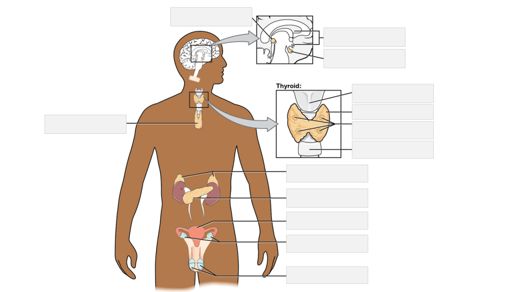
The Islets of Langerhans are clusters of specialized cells within the pancreas, an organ located behind the stomach. These islets are named after Paul Langerhans, who first identified them in 1869. They constitute around 1-2% of the total mass of the pancreas and are distributed throughout its substance.
The Islets of Langerhans contain several types of cells, including:
1. Alpha (α) cells: These produce and release glucagon, a hormone that helps to regulate blood sugar levels by promoting the conversion of glycogen to glucose in the liver when blood sugar levels are low.
2. Beta (β) cells: These produce and release insulin, a hormone that promotes the uptake and utilization of glucose by cells throughout the body, thereby lowering blood sugar levels.
3. Delta (δ) cells: These produce and release somatostatin, a hormone that inhibits the release of both insulin and glucagon and helps regulate their secretion in response to changing blood sugar levels.
4. PP cells (gamma or γ cells): These produce and release pancreatic polypeptide, which plays a role in regulating digestive enzyme secretion and gastrointestinal motility.
Dysfunction of the Islets of Langerhans can lead to various endocrine disorders, such as diabetes mellitus, where insulin-producing beta cells are damaged or destroyed, leading to impaired blood sugar regulation.
Glucose is a simple monosaccharide (or single sugar) that serves as the primary source of energy for living organisms. It's a fundamental molecule in biology, often referred to as "dextrose" or "grape sugar." Glucose has the molecular formula C6H12O6 and is vital to the functioning of cells, especially those in the brain and nervous system.
In the body, glucose is derived from the digestion of carbohydrates in food, and it's transported around the body via the bloodstream to cells where it can be used for energy. Cells convert glucose into a usable form through a process called cellular respiration, which involves a series of metabolic reactions that generate adenosine triphosphate (ATP)—the main currency of energy in cells.
Glucose is also stored in the liver and muscles as glycogen, a polysaccharide (multiple sugar) that can be broken down back into glucose when needed for energy between meals or during physical activity. Maintaining appropriate blood glucose levels is crucial for overall health, and imbalances can lead to conditions such as diabetes mellitus.
Pancreatic juice is an alkaline fluid secreted by the exocrine component of the pancreas, primarily containing digestive enzymes such as amylase, lipase, and trypsin. These enzymes aid in the breakdown of carbohydrates, fats, and proteins, respectively, in the small intestine during the digestion process. The bicarbonate ions present in pancreatic juice help neutralize the acidic chyme that enters the duodenum from the stomach, creating an optimal environment for enzymatic activity.
Gastric juice is a digestive fluid that is produced in the stomach. It is composed of several enzymes, including pepsin, which helps to break down proteins, and gastric amylase, which begins the digestion of carbohydrates. Gastric juice also contains hydrochloric acid, which creates a low pH environment in the stomach that is necessary for the activation of pepsin and the digestion of food. Additionally, gastric juice contains mucus, which helps to protect the lining of the stomach from the damaging effects of the hydrochloric acid. The production of gastric juice is controlled by hormones and the autonomic nervous system.
Bacterial proteins are a type of protein that are produced by bacteria as part of their structural or functional components. These proteins can be involved in various cellular processes, such as metabolism, DNA replication, transcription, and translation. They can also play a role in bacterial pathogenesis, helping the bacteria to evade the host's immune system, acquire nutrients, and multiply within the host.
Bacterial proteins can be classified into different categories based on their function, such as:
1. Enzymes: Proteins that catalyze chemical reactions in the bacterial cell.
2. Structural proteins: Proteins that provide structural support and maintain the shape of the bacterial cell.
3. Signaling proteins: Proteins that help bacteria to communicate with each other and coordinate their behavior.
4. Transport proteins: Proteins that facilitate the movement of molecules across the bacterial cell membrane.
5. Toxins: Proteins that are produced by pathogenic bacteria to damage host cells and promote infection.
6. Surface proteins: Proteins that are located on the surface of the bacterial cell and interact with the environment or host cells.
Understanding the structure and function of bacterial proteins is important for developing new antibiotics, vaccines, and other therapeutic strategies to combat bacterial infections.
A cell line is a culture of cells that are grown in a laboratory for use in research. These cells are usually taken from a single cell or group of cells, and they are able to divide and grow continuously in the lab. Cell lines can come from many different sources, including animals, plants, and humans. They are often used in scientific research to study cellular processes, disease mechanisms, and to test new drugs or treatments. Some common types of human cell lines include HeLa cells (which come from a cancer patient named Henrietta Lacks), HEK293 cells (which come from embryonic kidney cells), and HUVEC cells (which come from umbilical vein endothelial cells). It is important to note that cell lines are not the same as primary cells, which are cells that are taken directly from a living organism and have not been grown in the lab.
Bicarbonates, also known as sodium bicarbonate or baking soda, is a chemical compound with the formula NaHCO3. In the context of medical definitions, bicarbonates refer to the bicarbonate ion (HCO3-), which is an important buffer in the body that helps maintain normal pH levels in blood and other bodily fluids.
The balance of bicarbonate and carbonic acid in the body helps regulate the acidity or alkalinity of the blood, a condition known as pH balance. Bicarbonates are produced by the body and are also found in some foods and drinking water. They work to neutralize excess acid in the body and help maintain the normal pH range of 7.35 to 7.45.
In medical testing, bicarbonate levels may be measured as part of an electrolyte panel or as a component of arterial blood gas (ABG) analysis. Low bicarbonate levels can indicate metabolic acidosis, while high levels can indicate metabolic alkalosis. Both conditions can have serious consequences if not treated promptly and appropriately.
Luteinizing Hormone (LH) is a glycoprotein hormone, which is primarily produced and released by the anterior pituitary gland. In women, a surge of LH triggers ovulation, the release of an egg from the ovaries during the menstrual cycle. During pregnancy, LH stimulates the corpus luteum to produce progesterone. In men, LH stimulates the testes to produce testosterone. It plays a crucial role in sexual development, reproduction, and maintaining the reproductive system.
Chlorides are simple inorganic ions consisting of a single chlorine atom bonded to a single charged hydrogen ion (H+). Chloride is the most abundant anion (negatively charged ion) in the extracellular fluid in the human body. The normal range for chloride concentration in the blood is typically between 96-106 milliequivalents per liter (mEq/L).
Chlorides play a crucial role in maintaining electrical neutrality, acid-base balance, and osmotic pressure in the body. They are also essential for various physiological processes such as nerve impulse transmission, maintenance of membrane potentials, and digestion (as hydrochloric acid in the stomach).
Chloride levels can be affected by several factors, including diet, hydration status, kidney function, and certain medical conditions. Increased or decreased chloride levels can indicate various disorders, such as dehydration, kidney disease, Addison's disease, or diabetes insipidus. Therefore, monitoring chloride levels is essential for assessing a person's overall health and diagnosing potential medical issues.
Calcium is an essential mineral that is vital for various physiological processes in the human body. The medical definition of calcium is as follows:
Calcium (Ca2+) is a crucial cation and the most abundant mineral in the human body, with approximately 99% of it found in bones and teeth. It plays a vital role in maintaining structural integrity, nerve impulse transmission, muscle contraction, hormonal secretion, blood coagulation, and enzyme activation.
Calcium homeostasis is tightly regulated through the interplay of several hormones, including parathyroid hormone (PTH), calcitonin, and vitamin D. Dietary calcium intake, absorption, and excretion are also critical factors in maintaining optimal calcium levels in the body.
Hypocalcemia refers to low serum calcium levels, while hypercalcemia indicates high serum calcium levels. Both conditions can have detrimental effects on various organ systems and require medical intervention to correct.
The pancreas is a glandular organ located in the abdomen, posterior to the stomach. It has both exocrine and endocrine functions. The exocrine portion of the pancreas consists of acinar cells that produce and secrete digestive enzymes into the duodenum via the pancreatic duct. These enzymes help in the breakdown of proteins, carbohydrates, and fats in food.
The endocrine portion of the pancreas consists of clusters of cells called islets of Langerhans, which include alpha, beta, delta, and F cells. These cells produce and secrete hormones directly into the bloodstream, including insulin, glucagon, somatostatin, and pancreatic polypeptide. Insulin and glucagon are critical regulators of blood sugar levels, with insulin promoting glucose uptake and storage in tissues and glucagon stimulating glycogenolysis and gluconeogenesis to raise blood glucose when it is low.
Molecular sequence data refers to the specific arrangement of molecules, most commonly nucleotides in DNA or RNA, or amino acids in proteins, that make up a biological macromolecule. This data is generated through laboratory techniques such as sequencing, and provides information about the exact order of the constituent molecules. This data is crucial in various fields of biology, including genetics, evolution, and molecular biology, allowing for comparisons between different organisms, identification of genetic variations, and studies of gene function and regulation.
Synovial fluid is a viscous, clear, and straw-colored fluid found in the cavities of synovial joints, bursae, and tendon sheaths. It is produced by the synovial membrane, which lines the inner surface of the capsule surrounding these structures.
The primary function of synovial fluid is to reduce friction between articulating surfaces, providing lubrication for smooth and painless movement. It also acts as a shock absorber, protecting the joints from external forces during physical activities. Synovial fluid contains nutrients that nourish the articular cartilage, hyaluronic acid, which provides its viscoelastic properties, and lubricin, a protein responsible for boundary lubrication.
Abnormalities in synovial fluid composition or volume can indicate joint-related disorders, such as osteoarthritis, rheumatoid arthritis, gout, infection, or trauma. Analysis of synovial fluid is often used diagnostically to determine the underlying cause of joint pain, inflammation, or dysfunction.
In the field of medicine, "time factors" refer to the duration of symptoms or time elapsed since the onset of a medical condition, which can have significant implications for diagnosis and treatment. Understanding time factors is crucial in determining the progression of a disease, evaluating the effectiveness of treatments, and making critical decisions regarding patient care.
For example, in stroke management, "time is brain," meaning that rapid intervention within a specific time frame (usually within 4.5 hours) is essential to administering tissue plasminogen activator (tPA), a clot-busting drug that can minimize brain damage and improve patient outcomes. Similarly, in trauma care, the "golden hour" concept emphasizes the importance of providing definitive care within the first 60 minutes after injury to increase survival rates and reduce morbidity.
Time factors also play a role in monitoring the progression of chronic conditions like diabetes or heart disease, where regular follow-ups and assessments help determine appropriate treatment adjustments and prevent complications. In infectious diseases, time factors are crucial for initiating antibiotic therapy and identifying potential outbreaks to control their spread.
Overall, "time factors" encompass the significance of recognizing and acting promptly in various medical scenarios to optimize patient outcomes and provide effective care.
Messenger RNA (mRNA) is a type of RNA (ribonucleic acid) that carries genetic information copied from DNA in the form of a series of three-base code "words," each of which specifies a particular amino acid. This information is used by the cell's machinery to construct proteins, a process known as translation. After being transcribed from DNA, mRNA travels out of the nucleus to the ribosomes in the cytoplasm where protein synthesis occurs. Once the protein has been synthesized, the mRNA may be degraded and recycled. Post-transcriptional modifications can also occur to mRNA, such as alternative splicing and addition of a 5' cap and a poly(A) tail, which can affect its stability, localization, and translation efficiency.
Saliva is a complex mixture of primarily water, but also electrolytes, enzymes, antibacterial compounds, and various other substances. It is produced by the salivary glands located in the mouth. Saliva plays an essential role in maintaining oral health by moistening the mouth, helping to digest food, and protecting the teeth from decay by neutralizing acids produced by bacteria.
The medical definition of saliva can be stated as:
"A clear, watery, slightly alkaline fluid secreted by the salivary glands, consisting mainly of water, with small amounts of electrolytes, enzymes (such as amylase), mucus, and antibacterial compounds. Saliva aids in digestion, lubrication of oral tissues, and provides an oral barrier against microorganisms."
The secretory pathway is a series of membrane-enclosed compartments within eukaryotic cells that are involved in the synthesis, modification, and transport of proteins and lipids. The pathway begins in the endoplasmic reticulum (ER), where proteins and lipids are synthesized and folded.
Proteins that are destined for secretion or for incorporation into membranes are then transported from the ER to the Golgi apparatus, where they undergo further modifications such as glycosylation and sorting. After passing through the Golgi, proteins and lipids are sorted and packaged into vesicles that bud off from the Golgi and are transported to their final destinations, which may include the plasma membrane, lysosomes, or other organelles.
The secretory pathway is essential for many cellular processes, including the production and secretion of hormones, enzymes, and other proteins, as well as the maintenance of cell membranes and the regulation of intracellular signaling.
Pentagastrin is a synthetic polypeptide hormone that stimulates the release of gastrin and hydrochloric acid from the stomach. It is used diagnostically to test for conditions such as Zollinger-Ellison syndrome, a rare disorder in which tumors in the pancreas or duodenum produce excessive amounts of gastrin, leading to severe ulcers and other digestive problems.
Pentagastrin is typically administered intravenously, and its effects are monitored through blood tests that measure gastric acid secretion. It is a potent stimulant of gastric acid production, and its use is limited to diagnostic purposes due to the risk of adverse effects such as nausea, flushing, and increased heart rate.
Insulin-secreting cells, also known as beta cells, are a type of cell found in the pancreas. They are responsible for producing and releasing insulin, a hormone that regulates blood glucose levels by allowing cells in the body to take in glucose from the bloodstream. Insulin-secreting cells are clustered together in the pancreatic islets, along with other types of cells that produce other hormones such as glucagon and somatostatin. In people with diabetes, these cells may not function properly, leading to an impaired ability to regulate blood sugar levels.
Exocytosis is the process by which cells release molecules, such as hormones or neurotransmitters, to the extracellular space. This process involves the transport of these molecules inside vesicles (membrane-bound sacs) to the cell membrane, where they fuse and release their contents to the outside of the cell. It is a crucial mechanism for intercellular communication and the regulation of various physiological processes in the body.
Secretin is a hormone that is produced and released by the S cells in the duodenum, which is the first part of the small intestine. It is released in response to the presence of acidic chyme (partially digested food) entering the duodenum from the stomach. Secretin stimulates the pancreas to produce bicarbonate-rich alkaline secretions, which help neutralize the acidity of the chyme and create an optimal environment for enzymatic digestion in the small intestine.
Additionally, secretin also promotes the production of watery fluids from the liver, which aids in the digestion process. Overall, secretin plays a crucial role in maintaining the pH balance and facilitating proper nutrient absorption in the gastrointestinal tract.
A dose-response relationship in the context of drugs refers to the changes in the effects or symptoms that occur as the dose of a drug is increased or decreased. Generally, as the dose of a drug is increased, the severity or intensity of its effects also increases. Conversely, as the dose is decreased, the effects of the drug become less severe or may disappear altogether.
The dose-response relationship is an important concept in pharmacology and toxicology because it helps to establish the safe and effective dosage range for a drug. By understanding how changes in the dose of a drug affect its therapeutic and adverse effects, healthcare providers can optimize treatment plans for their patients while minimizing the risk of harm.
The dose-response relationship is typically depicted as a curve that shows the relationship between the dose of a drug and its effect. The shape of the curve may vary depending on the drug and the specific effect being measured. Some drugs may have a steep dose-response curve, meaning that small changes in the dose can result in large differences in the effect. Other drugs may have a more gradual dose-response curve, where larger changes in the dose are needed to produce significant effects.
In addition to helping establish safe and effective dosages, the dose-response relationship is also used to evaluate the potential therapeutic benefits and risks of new drugs during clinical trials. By systematically testing different doses of a drug in controlled studies, researchers can identify the optimal dosage range for the drug and assess its safety and efficacy.
Amylases are enzymes that break down complex carbohydrates, such as starch and glycogen, into simpler sugars like maltose, glucose, and maltotriose. There are several types of amylases found in various organisms, including humans.
In humans, amylases are produced by the pancreas and salivary glands. Pancreatic amylase is released into the small intestine where it helps to digest dietary carbohydrates. Salivary amylase, also known as alpha-amylase, is secreted into the mouth and begins breaking down starches in food during chewing.
Deficiency or absence of amylases can lead to difficulties in digesting carbohydrates and may cause symptoms such as bloating, diarrhea, and abdominal pain. Elevated levels of amylase in the blood may indicate conditions such as pancreatitis, pancreatic cancer, or other disorders affecting the pancreas.
An amino acid sequence is the specific order of amino acids in a protein or peptide molecule, formed by the linking of the amino group (-NH2) of one amino acid to the carboxyl group (-COOH) of another amino acid through a peptide bond. The sequence is determined by the genetic code and is unique to each type of protein or peptide. It plays a crucial role in determining the three-dimensional structure and function of proteins.
Radioimmunoassay (RIA) is a highly sensitive analytical technique used in clinical and research laboratories to measure concentrations of various substances, such as hormones, vitamins, drugs, or tumor markers, in biological samples like blood, urine, or tissues. The method relies on the specific interaction between an antibody and its corresponding antigen, combined with the use of radioisotopes to quantify the amount of bound antigen.
In a typical RIA procedure, a known quantity of a radiolabeled antigen (also called tracer) is added to a sample containing an unknown concentration of the same unlabeled antigen. The mixture is then incubated with a specific antibody that binds to the antigen. During the incubation period, the antibody forms complexes with both the radiolabeled and unlabeled antigens.
After the incubation, the unbound (free) radiolabeled antigen is separated from the antibody-antigen complexes, usually through a precipitation or separation step involving centrifugation, filtration, or chromatography. The amount of radioactivity in the pellet (containing the antibody-antigen complexes) is then measured using a gamma counter or other suitable radiation detection device.
The concentration of the unlabeled antigen in the sample can be determined by comparing the ratio of bound to free radiolabeled antigen in the sample to a standard curve generated from known concentrations of unlabeled antigen and their corresponding bound/free ratios. The higher the concentration of unlabeled antigen in the sample, the lower the amount of radiolabeled antigen that will bind to the antibody, resulting in a lower bound/free ratio.
Radioimmunoassays offer high sensitivity, specificity, and accuracy, making them valuable tools for detecting and quantifying low levels of various substances in biological samples. However, due to concerns about radiation safety and waste disposal, alternative non-isotopic immunoassay techniques like enzyme-linked immunosorbent assays (ELISAs) have become more popular in recent years.
Gastric mucosa refers to the innermost lining of the stomach, which is in contact with the gastric lumen. It is a specialized mucous membrane that consists of epithelial cells, lamina propria, and a thin layer of smooth muscle. The surface epithelium is primarily made up of mucus-secreting cells (goblet cells) and parietal cells, which secrete hydrochloric acid and intrinsic factor, and chief cells, which produce pepsinogen.
The gastric mucosa has several important functions, including protection against self-digestion by the stomach's own digestive enzymes and hydrochloric acid. The mucus layer secreted by the epithelial cells forms a physical barrier that prevents the acidic contents of the stomach from damaging the underlying tissues. Additionally, the bicarbonate ions secreted by the surface epithelial cells help neutralize the acidity in the immediate vicinity of the mucosa.
The gastric mucosa is also responsible for the initial digestion of food through the action of hydrochloric acid and pepsin, an enzyme that breaks down proteins into smaller peptides. The intrinsic factor secreted by parietal cells plays a crucial role in the absorption of vitamin B12 in the small intestine.
The gastric mucosa is constantly exposed to potential damage from various factors, including acid, pepsin, and other digestive enzymes, as well as mechanical stress due to muscle contractions during digestion. To maintain its integrity, the gastric mucosa has a remarkable capacity for self-repair and regeneration. However, chronic exposure to noxious stimuli or certain medical conditions can lead to inflammation, erosions, ulcers, or even cancer of the gastric mucosa.
Fluid therapy, in a medical context, refers to the administration of fluids into a patient's circulatory system for various therapeutic purposes. This can be done intravenously (through a vein), intraosseously (through a bone), or subcutaneously (under the skin). The goal of fluid therapy is to correct or prevent imbalances in the body's fluids and electrolytes, maintain or restore blood volume, and support organ function.
The types of fluids used in fluid therapy can include crystalloids (which contain electrolytes and water) and colloids (which contain larger molecules like proteins). The choice of fluid depends on the patient's specific needs and condition. Fluid therapy is commonly used in the treatment of dehydration, shock, sepsis, trauma, surgery, and other medical conditions that can affect the body's fluid balance.
Proper administration of fluid therapy requires careful monitoring of the patient's vital signs, urine output, electrolyte levels, and overall clinical status to ensure that the therapy is effective and safe.
In the context of medicine and pharmacology, "kinetics" refers to the study of how a drug moves throughout the body, including its absorption, distribution, metabolism, and excretion (often abbreviated as ADME). This field is called "pharmacokinetics."
1. Absorption: This is the process of a drug moving from its site of administration into the bloodstream. Factors such as the route of administration (e.g., oral, intravenous, etc.), formulation, and individual physiological differences can affect absorption.
2. Distribution: Once a drug is in the bloodstream, it gets distributed throughout the body to various tissues and organs. This process is influenced by factors like blood flow, protein binding, and lipid solubility of the drug.
3. Metabolism: Drugs are often chemically modified in the body, typically in the liver, through processes known as metabolism. These changes can lead to the formation of active or inactive metabolites, which may then be further distributed, excreted, or undergo additional metabolic transformations.
4. Excretion: This is the process by which drugs and their metabolites are eliminated from the body, primarily through the kidneys (urine) and the liver (bile).
Understanding the kinetics of a drug is crucial for determining its optimal dosing regimen, potential interactions with other medications or foods, and any necessary adjustments for special populations like pediatric or geriatric patients, or those with impaired renal or hepatic function.
A chemical stimulation in a medical context refers to the process of activating or enhancing physiological or psychological responses in the body using chemical substances. These chemicals can interact with receptors on cells to trigger specific reactions, such as neurotransmitters and hormones that transmit signals within the nervous system and endocrine system.
Examples of chemical stimulation include the use of medications, drugs, or supplements that affect mood, alertness, pain perception, or other bodily functions. For instance, caffeine can chemically stimulate the central nervous system to increase alertness and decrease feelings of fatigue. Similarly, certain painkillers can chemically stimulate opioid receptors in the brain to reduce the perception of pain.
It's important to note that while chemical stimulation can have therapeutic benefits, it can also have adverse effects if used improperly or in excessive amounts. Therefore, it's essential to follow proper dosing instructions and consult with a healthcare provider before using any chemical substances for stimulation purposes.
Prolactin is a hormone produced by the pituitary gland, a small gland located at the base of the brain. Its primary function is to stimulate milk production in women after childbirth, a process known as lactation. However, prolactin also plays other roles in the body, including regulating immune responses, metabolism, and behavior. In men, prolactin helps maintain the sexual glands and contributes to paternal behaviors.
Prolactin levels are usually low in both men and non-pregnant women but increase significantly during pregnancy and after childbirth. Various factors can affect prolactin levels, including stress, sleep, exercise, and certain medications. High prolactin levels can lead to medical conditions such as amenorrhea (absence of menstruation), galactorrhea (spontaneous milk production not related to childbirth), infertility, and reduced sexual desire in both men and women.
An Enzyme-Linked Immunosorbent Assay (ELISA) is a type of analytical biochemistry assay used to detect and quantify the presence of a substance, typically a protein or peptide, in a liquid sample. It takes its name from the enzyme-linked antibodies used in the assay.
In an ELISA, the sample is added to a well containing a surface that has been treated to capture the target substance. If the target substance is present in the sample, it will bind to the surface. Next, an enzyme-linked antibody specific to the target substance is added. This antibody will bind to the captured target substance if it is present. After washing away any unbound material, a substrate for the enzyme is added. If the enzyme is present due to its linkage to the antibody, it will catalyze a reaction that produces a detectable signal, such as a color change or fluorescence. The intensity of this signal is proportional to the amount of target substance present in the sample, allowing for quantification.
ELISAs are widely used in research and clinical settings to detect and measure various substances, including hormones, viruses, and bacteria. They offer high sensitivity, specificity, and reproducibility, making them a reliable choice for many applications.
Mucus is a viscous, slippery secretion produced by the mucous membranes that line various body cavities such as the respiratory and gastrointestinal tracts. It serves to lubricate and protect these surfaces from damage, infection, and foreign particles. Mucus contains water, proteins, salts, and other substances, including antibodies, enzymes, and glycoproteins called mucins that give it its characteristic gel-like consistency.
In the respiratory system, mucus traps inhaled particles such as dust, allergens, and pathogens, preventing them from reaching the lungs. The cilia, tiny hair-like structures lining the airways, move the mucus upward toward the throat, where it can be swallowed or expelled through coughing or sneezing. In the gastrointestinal tract, mucus helps protect the lining of the stomach and intestines from digestive enzymes and other harmful substances.
Excessive production of mucus can occur in various medical conditions such as allergies, respiratory infections, chronic lung diseases, and gastrointestinal disorders, leading to symptoms such as coughing, wheezing, nasal congestion, and diarrhea.
Signal transduction is the process by which a cell converts an extracellular signal, such as a hormone or neurotransmitter, into an intracellular response. This involves a series of molecular events that transmit the signal from the cell surface to the interior of the cell, ultimately resulting in changes in gene expression, protein activity, or metabolism.
The process typically begins with the binding of the extracellular signal to a receptor located on the cell membrane. This binding event activates the receptor, which then triggers a cascade of intracellular signaling molecules, such as second messengers, protein kinases, and ion channels. These molecules amplify and propagate the signal, ultimately leading to the activation or inhibition of specific cellular responses.
Signal transduction pathways are highly regulated and can be modulated by various factors, including other signaling molecules, post-translational modifications, and feedback mechanisms. Dysregulation of these pathways has been implicated in a variety of diseases, including cancer, diabetes, and neurological disorders.
The pituitary gland is a small, endocrine gland located at the base of the brain, in the sella turcica of the sphenoid bone. It is often called the "master gland" because it controls other glands and makes the hormones that trigger many body functions. The pituitary gland measures about 0.5 cm in height and 1 cm in width, and it weighs approximately 0.5 grams.
The pituitary gland is divided into two main parts: the anterior lobe (adenohypophysis) and the posterior lobe (neurohypophysis). The anterior lobe is further divided into three zones: the pars distalis, pars intermedia, and pars tuberalis. Each part of the pituitary gland has distinct functions and produces different hormones.
The anterior pituitary gland produces and releases several important hormones, including:
* Growth hormone (GH), which regulates growth and development in children and helps maintain muscle mass and bone strength in adults.
* Thyroid-stimulating hormone (TSH), which controls the production of thyroid hormones by the thyroid gland.
* Adrenocorticotropic hormone (ACTH), which stimulates the adrenal glands to produce cortisol and other steroid hormones.
* Follicle-stimulating hormone (FSH) and luteinizing hormone (LH), which regulate reproductive function in both males and females.
* Prolactin, which stimulates milk production in pregnant and lactating women.
The posterior pituitary gland stores and releases two hormones that are produced by the hypothalamus:
* Antidiuretic hormone (ADH), which helps regulate water balance in the body by controlling urine production.
* Oxytocin, which stimulates uterine contractions during childbirth and milk release during breastfeeding.
Overall, the pituitary gland plays a critical role in maintaining homeostasis and regulating various bodily functions, including growth, development, metabolism, and reproductive function.
Sprague-Dawley rats are a strain of albino laboratory rats that are widely used in scientific research. They were first developed by researchers H.H. Sprague and R.C. Dawley in the early 20th century, and have since become one of the most commonly used rat strains in biomedical research due to their relatively large size, ease of handling, and consistent genetic background.
Sprague-Dawley rats are outbred, which means that they are genetically diverse and do not suffer from the same limitations as inbred strains, which can have reduced fertility and increased susceptibility to certain diseases. They are also characterized by their docile nature and low levels of aggression, making them easier to handle and study than some other rat strains.
These rats are used in a wide variety of research areas, including toxicology, pharmacology, nutrition, cancer, and behavioral studies. Because they are genetically diverse, Sprague-Dawley rats can be used to model a range of human diseases and conditions, making them an important tool in the development of new drugs and therapies.
Gastrins are a group of hormones that are produced by G cells in the stomach lining. These hormones play an essential role in regulating gastric acid secretion and motor functions of the gastrointestinal tract. The most well-known gastrin is known as "gastrin-17," which is released into the bloodstream and stimulates the release of hydrochloric acid from parietal cells in the stomach lining.
Gastrins are stored in secretory granules within G cells, and their release is triggered by several factors, including the presence of food in the stomach, gastrin-releasing peptide (GRP), and vagus nerve stimulation. Once released, gastrins bind to specific receptors on parietal cells, leading to an increase in intracellular calcium levels and the activation of enzymes that promote hydrochloric acid secretion.
Abnormalities in gastrin production can lead to several gastrointestinal disorders, including gastrinomas (tumors that produce excessive amounts of gastrin), which can cause severe gastric acid hypersecretion and ulcers. Conversely, a deficiency in gastrin production can result in hypochlorhydria (low stomach acid levels) and impaired digestion.
Progesterone is a steroid hormone that is primarily produced in the ovaries during the menstrual cycle and in pregnancy. It plays an essential role in preparing the uterus for implantation of a fertilized egg and maintaining the early stages of pregnancy. Progesterone works to thicken the lining of the uterus, creating a nurturing environment for the developing embryo.
During the menstrual cycle, progesterone is produced by the corpus luteum, a temporary structure formed in the ovary after an egg has been released from a follicle during ovulation. If pregnancy does not occur, the levels of progesterone will decrease, leading to the shedding of the uterine lining and menstruation.
In addition to its reproductive functions, progesterone also has various other effects on the body, such as helping to regulate the immune system, supporting bone health, and potentially influencing mood and cognition. Progesterone can be administered medically in the form of oral pills, intramuscular injections, or vaginal suppositories for various purposes, including hormone replacement therapy, contraception, and managing certain gynecological conditions.
Amniotic fluid is a clear, slightly yellowish liquid that surrounds and protects the developing baby in the uterus. It is enclosed within the amniotic sac, which is a thin-walled sac that forms around the embryo during early pregnancy. The fluid is composed of fetal urine, lung secretions, and fluids that cross over from the mother's bloodstream through the placenta.
Amniotic fluid plays several important roles in pregnancy:
1. It provides a shock-absorbing cushion for the developing baby, protecting it from injury caused by movement or external forces.
2. It helps to maintain a constant temperature around the fetus, keeping it warm and comfortable.
3. It allows the developing baby to move freely within the uterus, promoting normal growth and development of the muscles and bones.
4. It provides a source of nutrients and hydration for the fetus, helping to support its growth and development.
5. It helps to prevent infection by providing a barrier between the fetus and the outside world.
Throughout pregnancy, the volume of amniotic fluid increases as the fetus grows. The amount of fluid typically peaks around 34-36 weeks of gestation, after which it begins to gradually decrease. Abnormalities in the volume of amniotic fluid can indicate problems with the developing baby or the pregnancy itself, and may require medical intervention.
I apologize, but "sheep" is not a term that has a medical definition. It is a common animal with the scientific name Ovis aries. If you have any medical or health-related questions, I would be happy to try and help answer those for you.
Secretory vesicles are membrane-bound organelles found within cells that store and transport secretory proteins and other molecules to the plasma membrane for exocytosis. Exocytosis is the process by which these molecules are released from the cell, allowing them to perform various functions, such as communication with other cells or participation in biochemical reactions. Secretory vesicles can be found in a variety of cell types, including endocrine cells, exocrine cells, and neurons. The proteins and molecules contained within secretory vesicles are synthesized in the rough endoplasmic reticulum and then transported to the Golgi apparatus, where they are processed, modified, and packaged into the vesicles for subsequent release.
Bile is a digestive fluid that is produced by the liver and stored in the gallbladder. It plays an essential role in the digestion and absorption of fats and fat-soluble vitamins in the small intestine. Bile consists of bile salts, bilirubin, cholesterol, phospholipids, electrolytes, and water.
Bile salts are amphipathic molecules that help to emulsify fats into smaller droplets, increasing their surface area and allowing for more efficient digestion by enzymes such as lipase. Bilirubin is a breakdown product of hemoglobin from red blood cells and gives bile its characteristic greenish-brown color.
Bile is released into the small intestine in response to food, particularly fats, entering the digestive tract. It helps to break down large fat molecules into smaller ones that can be absorbed through the walls of the intestines and transported to other parts of the body for energy or storage.
Cytokines are a broad and diverse category of small signaling proteins that are secreted by various cells, including immune cells, in response to different stimuli. They play crucial roles in regulating the immune response, inflammation, hematopoiesis, and cellular communication.
Cytokines mediate their effects by binding to specific receptors on the surface of target cells, which triggers intracellular signaling pathways that ultimately result in changes in gene expression, cell behavior, and function. Some key functions of cytokines include:
1. Regulating the activation, differentiation, and proliferation of immune cells such as T cells, B cells, natural killer (NK) cells, and macrophages.
2. Coordinating the inflammatory response by recruiting immune cells to sites of infection or tissue damage and modulating their effector functions.
3. Regulating hematopoiesis, the process of blood cell formation in the bone marrow, by controlling the proliferation, differentiation, and survival of hematopoietic stem and progenitor cells.
4. Modulating the development and function of the nervous system, including neuroinflammation, neuroprotection, and neuroregeneration.
Cytokines can be classified into several categories based on their structure, function, or cellular origin. Some common types of cytokines include interleukins (ILs), interferons (IFNs), tumor necrosis factors (TNFs), chemokines, colony-stimulating factors (CSFs), and transforming growth factors (TGFs). Dysregulation of cytokine production and signaling has been implicated in various pathological conditions, such as autoimmune diseases, chronic inflammation, cancer, and neurodegenerative disorders.
Glucagon is a hormone produced by the alpha cells of the pancreas. Its main function is to regulate glucose levels in the blood by stimulating the liver to convert stored glycogen into glucose, which can then be released into the bloodstream. This process helps to raise blood sugar levels when they are too low, such as during hypoglycemia.
Glucagon is a 29-amino acid polypeptide that is derived from the preproglucagon protein. It works by binding to glucagon receptors on liver cells, which triggers a series of intracellular signaling events that lead to the activation of enzymes involved in glycogen breakdown.
In addition to its role in glucose regulation, glucagon has also been shown to have other physiological effects, such as promoting lipolysis (the breakdown of fat) and inhibiting gastric acid secretion. Glucagon is often used clinically in the treatment of hypoglycemia, as well as in diagnostic tests to assess pancreatic function.
C57BL/6 (C57 Black 6) is an inbred strain of laboratory mouse that is widely used in biomedical research. The term "inbred" refers to a strain of animals where matings have been carried out between siblings or other closely related individuals for many generations, resulting in a population that is highly homozygous at most genetic loci.
The C57BL/6 strain was established in 1920 by crossing a female mouse from the dilute brown (DBA) strain with a male mouse from the black strain. The resulting offspring were then interbred for many generations to create the inbred C57BL/6 strain.
C57BL/6 mice are known for their robust health, longevity, and ease of handling, making them a popular choice for researchers. They have been used in a wide range of biomedical research areas, including studies of cancer, immunology, neuroscience, cardiovascular disease, and metabolism.
One of the most notable features of the C57BL/6 strain is its sensitivity to certain genetic modifications, such as the introduction of mutations that lead to obesity or impaired glucose tolerance. This has made it a valuable tool for studying the genetic basis of complex diseases and traits.
Overall, the C57BL/6 inbred mouse strain is an important model organism in biomedical research, providing a valuable resource for understanding the genetic and molecular mechanisms underlying human health and disease.
Epithelial cells are types of cells that cover the outer surfaces of the body, line the inner surfaces of organs and glands, and form the lining of blood vessels and body cavities. They provide a protective barrier against the external environment, regulate the movement of materials between the internal and external environments, and are involved in the sense of touch, temperature, and pain. Epithelial cells can be squamous (flat and thin), cuboidal (square-shaped and of equal height), or columnar (tall and narrow) in shape and are classified based on their location and function.
Exocrine glands are a type of gland in the human body that produce and release substances through ducts onto an external or internal surface. These glands are responsible for secreting various substances such as enzymes, hormones, and lubricants that help in digestion, protection, and other bodily functions.
Exocrine glands can be further classified into three types based on their mode of secretion:
1. Merocrine glands: These glands release their secretions by exocytosis, where the secretory product is enclosed in a vesicle that fuses with the cell membrane and releases its contents outside the cell. Examples include sweat glands and mucous glands.
2. Apocrine glands: These glands release their secretions by pinching off a portion of the cytoplasm along with the secretory product. An example is the apocrine sweat gland found in the armpits and genital area.
3. Holocrine glands: These glands release their secretions by disintegrating and releasing the entire cell, including its organelles and secretory products. An example is the sebaceous gland found in the skin, which releases an oily substance called sebum.
Protein transport, in the context of cellular biology, refers to the process by which proteins are actively moved from one location to another within or between cells. This is a crucial mechanism for maintaining proper cell function and regulation.
Intracellular protein transport involves the movement of proteins within a single cell. Proteins can be transported across membranes (such as the nuclear envelope, endoplasmic reticulum, Golgi apparatus, or plasma membrane) via specialized transport systems like vesicles and transport channels.
Intercellular protein transport refers to the movement of proteins from one cell to another, often facilitated by exocytosis (release of proteins in vesicles) and endocytosis (uptake of extracellular substances via membrane-bound vesicles). This is essential for communication between cells, immune response, and other physiological processes.
It's important to note that any disruption in protein transport can lead to various diseases, including neurological disorders, cancer, and metabolic conditions.
Biological transport refers to the movement of molecules, ions, or solutes across biological membranes or through cells in living organisms. This process is essential for maintaining homeostasis, regulating cellular functions, and enabling communication between cells. There are two main types of biological transport: passive transport and active transport.
Passive transport does not require the input of energy and includes:
1. Diffusion: The random movement of molecules from an area of high concentration to an area of low concentration until equilibrium is reached.
2. Osmosis: The diffusion of solvent molecules (usually water) across a semi-permeable membrane from an area of lower solute concentration to an area of higher solute concentration.
3. Facilitated diffusion: The assisted passage of polar or charged substances through protein channels or carriers in the cell membrane, which increases the rate of diffusion without consuming energy.
Active transport requires the input of energy (in the form of ATP) and includes:
1. Primary active transport: The direct use of ATP to move molecules against their concentration gradient, often driven by specific transport proteins called pumps.
2. Secondary active transport: The coupling of the movement of one substance down its electrochemical gradient with the uphill transport of another substance, mediated by a shared transport protein. This process is also known as co-transport or counter-transport.
The duodenum is the first part of the small intestine, immediately following the stomach. It is a C-shaped structure that is about 10-12 inches long and is responsible for continuing the digestion process that begins in the stomach. The duodenum receives partially digested food from the stomach through the pyloric valve and mixes it with digestive enzymes and bile produced by the pancreas and liver, respectively. These enzymes help break down proteins, fats, and carbohydrates into smaller molecules, allowing for efficient absorption in the remaining sections of the small intestine.
"Cattle" is a term used in the agricultural and veterinary fields to refer to domesticated animals of the genus *Bos*, primarily *Bos taurus* (European cattle) and *Bos indicus* (Zebu). These animals are often raised for meat, milk, leather, and labor. They are also known as bovines or cows (for females), bulls (intact males), and steers/bullocks (castrated males). However, in a strict medical definition, "cattle" does not apply to humans or other animals.
Ascitic fluid is defined as the abnormal accumulation of fluid in the peritoneal cavity, which is the space between the two layers of the peritoneum, a serous membrane that lines the abdominal cavity and covers the abdominal organs. This buildup of fluid, also known as ascites, can be caused by various medical conditions such as liver cirrhosis, cancer, heart failure, or infection. The fluid itself is typically straw-colored and clear, but it may also contain cells, proteins, and other substances depending on the underlying cause. Analysis of ascitic fluid can help doctors diagnose and manage the underlying condition causing the accumulation of fluid.
Somatostatin is a hormone that inhibits the release of several hormones and also has a role in slowing down digestion. It is produced by the body in various parts of the body, including the hypothalamus (a part of the brain), the pancreas, and the gastrointestinal tract.
Somatostatin exists in two forms: somatostatin-14 and somatostatin-28, which differ in their length. Somatostatin-14 is the predominant form found in the brain, while somatostatin-28 is the major form found in the gastrointestinal tract.
Somatostatin has a wide range of effects on various physiological processes, including:
* Inhibiting the release of several hormones such as growth hormone, insulin, glucagon, and gastrin
* Slowing down digestion by inhibiting the release of digestive enzymes from the pancreas and reducing blood flow to the gastrointestinal tract
* Regulating neurotransmission in the brain
Somatostatin is used clinically as a diagnostic tool for detecting certain types of tumors that overproduce growth hormone or other hormones, and it is also used as a treatment for some conditions such as acromegaly (a condition characterized by excessive growth hormone production) and gastrointestinal disorders.
"Inbred strains of rats" are genetically identical rodents that have been produced through many generations of brother-sister mating. This results in a high degree of homozygosity, where the genes at any particular locus in the genome are identical in all members of the strain.
Inbred strains of rats are widely used in biomedical research because they provide a consistent and reproducible genetic background for studying various biological phenomena, including the effects of drugs, environmental factors, and genetic mutations on health and disease. Additionally, inbred strains can be used to create genetically modified models of human diseases by introducing specific mutations into their genomes.
Some commonly used inbred strains of rats include the Wistar Kyoto (WKY), Sprague-Dawley (SD), and Fischer 344 (F344) rat strains. Each strain has its own unique genetic characteristics, making them suitable for different types of research.
"Wistar rats" are a strain of albino rats that are widely used in laboratory research. They were developed at the Wistar Institute in Philadelphia, USA, and were first introduced in 1906. Wistar rats are outbred, which means that they are genetically diverse and do not have a fixed set of genetic characteristics like inbred strains.
Wistar rats are commonly used as animal models in biomedical research because of their size, ease of handling, and relatively low cost. They are used in a wide range of research areas, including toxicology, pharmacology, nutrition, cancer, cardiovascular disease, and behavioral studies. Wistar rats are also used in safety testing of drugs, medical devices, and other products.
Wistar rats are typically larger than many other rat strains, with males weighing between 500-700 grams and females weighing between 250-350 grams. They have a lifespan of approximately 2-3 years. Wistar rats are also known for their docile and friendly nature, making them easy to handle and work with in the laboratory setting.
Cyclic adenosine monophosphate (cAMP) is a key secondary messenger in many biological processes, including the regulation of metabolism, gene expression, and cellular excitability. It is synthesized from adenosine triphosphate (ATP) by the enzyme adenylyl cyclase and is degraded by the enzyme phosphodiesterase.
In the body, cAMP plays a crucial role in mediating the effects of hormones and neurotransmitters on target cells. For example, when a hormone binds to its receptor on the surface of a cell, it can activate a G protein, which in turn activates adenylyl cyclase to produce cAMP. The increased levels of cAMP then activate various effector proteins, such as protein kinases, which go on to regulate various cellular processes.
Overall, the regulation of cAMP levels is critical for maintaining proper cellular function and homeostasis, and abnormalities in cAMP signaling have been implicated in a variety of diseases, including cancer, diabetes, and neurological disorders.
Gonadotropin-Releasing Hormone (GnRH), also known as Luteinizing Hormone-Releasing Hormone (LHRH), is a hormonal peptide consisting of 10 amino acids. It is produced and released by the hypothalamus, an area in the brain that links the nervous system to the endocrine system via the pituitary gland.
GnRH plays a crucial role in regulating reproduction and sexual development through its control of two gonadotropins: follicle-stimulating hormone (FSH) and luteinizing hormone (LH). These gonadotropins, in turn, stimulate the gonads (ovaries or testes) to produce sex steroids and eggs or sperm.
GnRH acts on the anterior pituitary gland by binding to its specific receptors, leading to the release of FSH and LH. The hypothalamic-pituitary-gonadal axis is under negative feedback control, meaning that when sex steroid levels are high, they inhibit the release of GnRH, which subsequently decreases FSH and LH secretion.
GnRH agonists and antagonists have clinical applications in various medical conditions, such as infertility treatments, precocious puberty, endometriosis, uterine fibroids, prostate cancer, and hormone-responsive breast cancer.
Estradiol is a type of estrogen, which is a female sex hormone. It is the most potent and dominant form of estrogen in humans. Estradiol plays a crucial role in the development and maintenance of secondary sexual characteristics in women, such as breast development and regulation of the menstrual cycle. It also helps maintain bone density, protect the lining of the uterus, and is involved in cognition and mood regulation.
Estradiol is produced primarily by the ovaries, but it can also be synthesized in smaller amounts by the adrenal glands and fat cells. In men, estradiol is produced from testosterone through a process called aromatization. Abnormal levels of estradiol can contribute to various health issues, such as hormonal imbalances, infertility, osteoporosis, and certain types of cancer.
The liver is a large, solid organ located in the upper right portion of the abdomen, beneath the diaphragm and above the stomach. It plays a vital role in several bodily functions, including:
1. Metabolism: The liver helps to metabolize carbohydrates, fats, and proteins from the food we eat into energy and nutrients that our bodies can use.
2. Detoxification: The liver detoxifies harmful substances in the body by breaking them down into less toxic forms or excreting them through bile.
3. Synthesis: The liver synthesizes important proteins, such as albumin and clotting factors, that are necessary for proper bodily function.
4. Storage: The liver stores glucose, vitamins, and minerals that can be released when the body needs them.
5. Bile production: The liver produces bile, a digestive juice that helps to break down fats in the small intestine.
6. Immune function: The liver plays a role in the immune system by filtering out bacteria and other harmful substances from the blood.
Overall, the liver is an essential organ that plays a critical role in maintaining overall health and well-being.
Follicle-Stimulating Hormone (FSH) is a glycoprotein hormone secreted and released by the anterior pituitary gland. In females, it promotes the growth and development of ovarian follicles in the ovary, which ultimately leads to the maturation and release of an egg (ovulation). In males, FSH stimulates the testes to produce sperm. It works in conjunction with luteinizing hormone (LH) to regulate reproductive processes. The secretion of FSH is controlled by the hypothalamic-pituitary-gonadal axis and its release is influenced by the levels of gonadotropin-releasing hormone (GnRH), estrogen, inhibin, and androgens.
Western blotting is a laboratory technique used in molecular biology to detect and quantify specific proteins in a mixture of many different proteins. This technique is commonly used to confirm the expression of a protein of interest, determine its size, and investigate its post-translational modifications. The name "Western" blotting distinguishes this technique from Southern blotting (for DNA) and Northern blotting (for RNA).
The Western blotting procedure involves several steps:
1. Protein extraction: The sample containing the proteins of interest is first extracted, often by breaking open cells or tissues and using a buffer to extract the proteins.
2. Separation of proteins by electrophoresis: The extracted proteins are then separated based on their size by loading them onto a polyacrylamide gel and running an electric current through the gel (a process called sodium dodecyl sulfate-polyacrylamide gel electrophoresis or SDS-PAGE). This separates the proteins according to their molecular weight, with smaller proteins migrating faster than larger ones.
3. Transfer of proteins to a membrane: After separation, the proteins are transferred from the gel onto a nitrocellulose or polyvinylidene fluoride (PVDF) membrane using an electric current in a process called blotting. This creates a replica of the protein pattern on the gel but now immobilized on the membrane for further analysis.
4. Blocking: The membrane is then blocked with a blocking agent, such as non-fat dry milk or bovine serum albumin (BSA), to prevent non-specific binding of antibodies in subsequent steps.
5. Primary antibody incubation: A primary antibody that specifically recognizes the protein of interest is added and allowed to bind to its target protein on the membrane. This step may be performed at room temperature or 4°C overnight, depending on the antibody's properties.
6. Washing: The membrane is washed with a buffer to remove unbound primary antibodies.
7. Secondary antibody incubation: A secondary antibody that recognizes the primary antibody (often coupled to an enzyme or fluorophore) is added and allowed to bind to the primary antibody. This step may involve using a horseradish peroxidase (HRP)-conjugated or alkaline phosphatase (AP)-conjugated secondary antibody, depending on the detection method used later.
8. Washing: The membrane is washed again to remove unbound secondary antibodies.
9. Detection: A detection reagent is added to visualize the protein of interest by detecting the signal generated from the enzyme-conjugated or fluorophore-conjugated secondary antibody. This can be done using chemiluminescent, colorimetric, or fluorescent methods.
10. Analysis: The resulting image is analyzed to determine the presence and quantity of the protein of interest in the sample.
Western blotting is a powerful technique for identifying and quantifying specific proteins within complex mixtures. It can be used to study protein expression, post-translational modifications, protein-protein interactions, and more. However, it requires careful optimization and validation to ensure accurate and reproducible results.
Hydrogen-ion concentration, also known as pH, is a measure of the acidity or basicity of a solution. It is defined as the negative logarithm (to the base 10) of the hydrogen ion activity in a solution. The standard unit of measurement is the pH unit. A pH of 7 is neutral, less than 7 is acidic, and greater than 7 is basic.
In medical terms, hydrogen-ion concentration is important for maintaining homeostasis within the body. For example, in the stomach, a high hydrogen-ion concentration (low pH) is necessary for the digestion of food. However, in other parts of the body such as blood, a high hydrogen-ion concentration can be harmful and lead to acidosis. Conversely, a low hydrogen-ion concentration (high pH) in the blood can lead to alkalosis. Both acidosis and alkalosis can have serious consequences on various organ systems if not corrected.
Perfusion, in medical terms, refers to the process of circulating blood through the body's organs and tissues to deliver oxygen and nutrients and remove waste products. It is a measure of the delivery of adequate blood flow to specific areas or tissues in the body. Perfusion can be assessed using various methods, including imaging techniques like computed tomography (CT) scans, magnetic resonance imaging (MRI), and perfusion scintigraphy.
Perfusion is critical for maintaining proper organ function and overall health. When perfusion is impaired or inadequate, it can lead to tissue hypoxia, acidosis, and cell death, which can result in organ dysfunction or failure. Conditions that can affect perfusion include cardiovascular disease, shock, trauma, and certain surgical procedures.
Interleukin-8 (IL-8) is a type of cytokine, which is a small signaling protein involved in immune response and inflammation. IL-8 is also known as neutrophil chemotactic factor or NCF because it attracts neutrophils, a type of white blood cell, to the site of infection or injury.
IL-8 is produced by various cells including macrophages, epithelial cells, and endothelial cells in response to bacterial or inflammatory stimuli. It acts by binding to specific receptors called CXCR1 and CXCR2 on the surface of neutrophils, which triggers a series of intracellular signaling events leading to neutrophil activation, migration, and degranulation.
IL-8 plays an important role in the recruitment of neutrophils to the site of infection or tissue damage, where they can phagocytose and destroy invading microorganisms. However, excessive or prolonged production of IL-8 has been implicated in various inflammatory diseases such as chronic obstructive pulmonary disease (COPD), rheumatoid arthritis, and cancer.
The intestinal mucosa is the innermost layer of the intestines, which comes into direct contact with digested food and microbes. It is a specialized epithelial tissue that plays crucial roles in nutrient absorption, barrier function, and immune defense. The intestinal mucosa is composed of several cell types, including absorptive enterocytes, mucus-secreting goblet cells, hormone-producing enteroendocrine cells, and immune cells such as lymphocytes and macrophages.
The surface of the intestinal mucosa is covered by a single layer of epithelial cells, which are joined together by tight junctions to form a protective barrier against harmful substances and microorganisms. This barrier also allows for the selective absorption of nutrients into the bloodstream. The intestinal mucosa also contains numerous lymphoid follicles, known as Peyer's patches, which are involved in immune surveillance and defense against pathogens.
In addition to its role in absorption and immunity, the intestinal mucosa is also capable of producing hormones that regulate digestion and metabolism. Dysfunction of the intestinal mucosa can lead to various gastrointestinal disorders, such as inflammatory bowel disease, celiac disease, and food allergies.
Recombinant proteins are artificially created proteins produced through the use of recombinant DNA technology. This process involves combining DNA molecules from different sources to create a new set of genes that encode for a specific protein. The resulting recombinant protein can then be expressed, purified, and used for various applications in research, medicine, and industry.
Recombinant proteins are widely used in biomedical research to study protein function, structure, and interactions. They are also used in the development of diagnostic tests, vaccines, and therapeutic drugs. For example, recombinant insulin is a common treatment for diabetes, while recombinant human growth hormone is used to treat growth disorders.
The production of recombinant proteins typically involves the use of host cells, such as bacteria, yeast, or mammalian cells, which are engineered to express the desired protein. The host cells are transformed with a plasmid vector containing the gene of interest, along with regulatory elements that control its expression. Once the host cells are cultured and the protein is expressed, it can be purified using various chromatography techniques.
Overall, recombinant proteins have revolutionized many areas of biology and medicine, enabling researchers to study and manipulate proteins in ways that were previously impossible.
Adrenocorticotropic Hormone (ACTH) is a hormone produced and released by the anterior pituitary gland, a small endocrine gland located at the base of the brain. ACTH plays a crucial role in the regulation of the body's stress response and has significant effects on various physiological processes.
The primary function of ACTH is to stimulate the adrenal glands, which are triangular-shaped glands situated on top of the kidneys. The adrenal glands consist of two parts: the outer cortex and the inner medulla. ACTH specifically targets the adrenal cortex, where it binds to specific receptors and initiates a series of biochemical reactions leading to the production and release of steroid hormones, primarily cortisol (a glucocorticoid) and aldosterone (a mineralocorticoid).
Cortisol is involved in various metabolic processes, such as regulating blood sugar levels, modulating the immune response, and helping the body respond to stress. Aldosterone plays a vital role in maintaining electrolyte and fluid balance by promoting sodium reabsorption and potassium excretion in the kidneys.
ACTH release is controlled by the hypothalamus, another part of the brain, which produces corticotropin-releasing hormone (CRH). CRH stimulates the anterior pituitary gland to secrete ACTH, which in turn triggers cortisol production in the adrenal glands. This complex feedback system helps maintain homeostasis and ensures that appropriate amounts of cortisol are released in response to various physiological and psychological stressors.
Disorders related to ACTH can lead to hormonal imbalances, resulting in conditions such as Cushing's syndrome (excessive cortisol production) or Addison's disease (insufficient cortisol production). Proper diagnosis and management of these disorders typically involve assessing the function of the hypothalamic-pituitary-adrenal axis and addressing any underlying issues affecting ACTH secretion.
I'm sorry for any confusion, but "Industrial Oils" is not a term that has a specific medical definition. Industrial oils are typically used in machinery and equipment for various industrial applications, such as hydraulic systems, lubricants, and coolants. They are not directly related to medicine or human health.
However, if a person were to come into contact with these oils through their occupation, there could be potential health effects, such as skin irritation or respiratory issues, depending on the specific type of oil and the nature of the exposure. But this would not fall under a medical definition of the term itself.
Glucagon-like peptide 1 (GLP-1) is a hormone that is secreted by the intestines in response to food intake. It plays a crucial role in regulating blood sugar levels through several mechanisms, including stimulation of insulin secretion from the pancreas, inhibition of glucagon release, slowing gastric emptying, and promoting satiety. GLP-1 is an important target for the treatment of type 2 diabetes due to its insulin-secretory and glucose-lowering effects. In addition, GLP-1 receptor agonists are used in the management of obesity due to their ability to promote weight loss by reducing appetite and increasing feelings of fullness.
Labyrinthine fluids, also known as endolymph and perilymph, are fluids that fill the inner ear structures, specifically the bony labyrinth. The bony labyrinth is divided into two main parts: the cochlea, responsible for hearing, and the vestibular system, responsible for balance.
Endolymph is a clear, plasma-like fluid found within the membranous labyrinth, which is a series of interconnected tubes and sacs that lie inside the bony labyrinth. Endolymph plays a crucial role in the functioning of both the cochlea and vestibular system by creating an electrochemical gradient necessary for the conversion of mechanical sound vibrations into electrical signals in the cochlea, as well as facilitating the detection of head movements and maintaining balance in the vestibular system.
Perilymph, on the other hand, is a clear, colorless fluid that fills the space between the bony labyrinth and the membranous labyrinth. It is similar in composition to cerebrospinal fluid (CSF) and serves as a protective cushion for the delicate inner ear structures. Perilymph also helps maintain the electrochemical gradient required for sound transduction in the cochlea.
Disorders related to these labyrinthine fluids, such as endolymphatic hydrops or perilymph fistula, can lead to hearing and balance problems.
Tumor Necrosis Factor-alpha (TNF-α) is a cytokine, a type of small signaling protein involved in immune response and inflammation. It is primarily produced by activated macrophages, although other cell types such as T-cells, natural killer cells, and mast cells can also produce it.
TNF-α plays a crucial role in the body's defense against infection and tissue injury by mediating inflammatory responses, activating immune cells, and inducing apoptosis (programmed cell death) in certain types of cells. It does this by binding to its receptors, TNFR1 and TNFR2, which are found on the surface of many cell types.
In addition to its role in the immune response, TNF-α has been implicated in the pathogenesis of several diseases, including autoimmune disorders such as rheumatoid arthritis, inflammatory bowel disease, and psoriasis, as well as cancer, where it can promote tumor growth and metastasis.
Therapeutic agents that target TNF-α, such as infliximab, adalimumab, and etanercept, have been developed to treat these conditions. However, these drugs can also increase the risk of infections and other side effects, so their use must be carefully monitored.
The anterior pituitary, also known as the adenohypophysis, is the front portion of the pituitary gland. It is responsible for producing and secreting several important hormones that regulate various bodily functions. These hormones include:
* Growth hormone (GH), which stimulates growth and cell reproduction in bones and other tissues.
* Thyroid-stimulating hormone (TSH), which regulates the production of thyroid hormones by the thyroid gland.
* Adrenocorticotropic hormone (ACTH), which stimulates the adrenal glands to produce cortisol and other steroid hormones.
* Follicle-stimulating hormone (FSH) and luteinizing hormone (LH), which regulate reproductive function in both males and females by controlling the development and release of eggs or sperm.
* Prolactin, which stimulates milk production in pregnant and nursing women.
* Melanocyte-stimulating hormone (MSH), which regulates skin pigmentation and appetite.
The anterior pituitary gland is controlled by the hypothalamus, a small region of the brain located just above it. The hypothalamus produces releasing and inhibiting hormones that regulate the secretion of hormones from the anterior pituitary. These hormones are released into a network of blood vessels called the portal system, which carries them directly to the anterior pituitary gland.
Damage or disease of the anterior pituitary can lead to hormonal imbalances and various medical conditions, such as growth disorders, thyroid dysfunction, adrenal insufficiency, reproductive problems, and diabetes insipidus.
Macrophages are a type of white blood cell that are an essential part of the immune system. They are large, specialized cells that engulf and destroy foreign substances, such as bacteria, viruses, parasites, and fungi, as well as damaged or dead cells. Macrophages are found throughout the body, including in the bloodstream, lymph nodes, spleen, liver, lungs, and connective tissues. They play a critical role in inflammation, immune response, and tissue repair and remodeling.
Macrophages originate from monocytes, which are a type of white blood cell produced in the bone marrow. When monocytes enter the tissues, they differentiate into macrophages, which have a larger size and more specialized functions than monocytes. Macrophages can change their shape and move through tissues to reach sites of infection or injury. They also produce cytokines, chemokines, and other signaling molecules that help coordinate the immune response and recruit other immune cells to the site of infection or injury.
Macrophages have a variety of surface receptors that allow them to recognize and respond to different types of foreign substances and signals from other cells. They can engulf and digest foreign particles, bacteria, and viruses through a process called phagocytosis. Macrophages also play a role in presenting antigens to T cells, which are another type of immune cell that helps coordinate the immune response.
Overall, macrophages are crucial for maintaining tissue homeostasis, defending against infection, and promoting wound healing and tissue repair. Dysregulation of macrophage function has been implicated in a variety of diseases, including cancer, autoimmune disorders, and chronic inflammatory conditions.
Blood glucose, also known as blood sugar, is the concentration of glucose in the blood. Glucose is a simple sugar that serves as the main source of energy for the body's cells. It is carried to each cell through the bloodstream and is absorbed into the cells with the help of insulin, a hormone produced by the pancreas.
The normal range for blood glucose levels in humans is typically between 70 and 130 milligrams per deciliter (mg/dL) when fasting, and less than 180 mg/dL after meals. Levels that are consistently higher than this may indicate diabetes or other metabolic disorders.
Blood glucose levels can be measured through a variety of methods, including fingerstick blood tests, continuous glucose monitoring systems, and laboratory tests. Regular monitoring of blood glucose levels is important for people with diabetes to help manage their condition and prevent complications.
A cell membrane, also known as the plasma membrane, is a thin semi-permeable phospholipid bilayer that surrounds all cells in animals, plants, and microorganisms. It functions as a barrier to control the movement of substances in and out of the cell, allowing necessary molecules such as nutrients, oxygen, and signaling molecules to enter while keeping out harmful substances and waste products. The cell membrane is composed mainly of phospholipids, which have hydrophilic (water-loving) heads and hydrophobic (water-fearing) tails. This unique structure allows the membrane to be flexible and fluid, yet selectively permeable. Additionally, various proteins are embedded in the membrane that serve as channels, pumps, receptors, and enzymes, contributing to the cell's overall functionality and communication with its environment.
Carbachol is a cholinergic agonist, which means it stimulates the parasympathetic nervous system by mimicking the action of acetylcholine, a neurotransmitter that is involved in transmitting signals between nerves and muscles. Carbachol binds to both muscarinic and nicotinic receptors, but its effects are more pronounced on muscarinic receptors.
Carbachol is used in medical treatments to produce miosis (pupil constriction), lower intraocular pressure, and stimulate gastrointestinal motility. It can also be used as a diagnostic tool to test for certain conditions such as Hirschsprung's disease.
Like any medication, carbachol can have side effects, including sweating, salivation, nausea, vomiting, diarrhea, bradycardia (slow heart rate), and bronchoconstriction (narrowing of the airways in the lungs). It should be used with caution and under the supervision of a healthcare professional.
The parotid gland is the largest of the major salivary glands. It is a bilobed, accessory digestive organ that secretes serous saliva into the mouth via the parotid duct (Stensen's duct), located near the upper second molar tooth. The parotid gland is primarily responsible for moistening and lubricating food to aid in swallowing and digestion.
Anatomically, the parotid gland is located in the preauricular region, extending from the zygomatic arch superiorly to the angle of the mandible inferiorly, and from the masseter muscle anteriorly to the sternocleidomastoid muscle posteriorly. It is enclosed within a fascial capsule and has a rich blood supply from the external carotid artery and a complex innervation pattern involving both parasympathetic and sympathetic fibers.
Parotid gland disorders can include salivary gland stones (sialolithiasis), infections, inflammatory conditions, benign or malignant tumors, and autoimmune diseases such as Sjögren's syndrome.
Growth Hormone (GH), also known as somatotropin, is a peptide hormone secreted by the somatotroph cells in the anterior pituitary gland. It plays a crucial role in regulating growth, cell reproduction, and regeneration by stimulating the production of another hormone called insulin-like growth factor 1 (IGF-1) in the liver and other tissues. GH also has important metabolic functions, such as increasing glucose levels, enhancing protein synthesis, and reducing fat storage. Its secretion is regulated by two hypothalamic hormones: growth hormone-releasing hormone (GHRH), which stimulates its release, and somatostatin (SRIF), which inhibits its release. Abnormal levels of GH can lead to various medical conditions, such as dwarfism or gigantism if there are deficiencies or excesses, respectively.
Carrier proteins, also known as transport proteins, are a type of protein that facilitates the movement of molecules across cell membranes. They are responsible for the selective and active transport of ions, sugars, amino acids, and other molecules from one side of the membrane to the other, against their concentration gradient. This process requires energy, usually in the form of ATP (adenosine triphosphate).
Carrier proteins have a specific binding site for the molecule they transport, and undergo conformational changes upon binding, which allows them to move the molecule across the membrane. Once the molecule has been transported, the carrier protein returns to its original conformation, ready to bind and transport another molecule.
Carrier proteins play a crucial role in maintaining the balance of ions and other molecules inside and outside of cells, and are essential for many physiological processes, including nerve impulse transmission, muscle contraction, and nutrient uptake.
Interleukin-6 (IL-6) is a cytokine, a type of protein that plays a crucial role in communication between cells, especially in the immune system. It is produced by various cells including T-cells, B-cells, fibroblasts, and endothelial cells in response to infection, injury, or inflammation.
IL-6 has diverse effects on different cell types. In the immune system, it stimulates the growth and differentiation of B-cells into plasma cells that produce antibodies. It also promotes the activation and survival of T-cells. Moreover, IL-6 plays a role in fever induction by acting on the hypothalamus to raise body temperature during an immune response.
In addition to its functions in the immune system, IL-6 has been implicated in various physiological processes such as hematopoiesis (the formation of blood cells), bone metabolism, and neural development. However, abnormal levels of IL-6 have also been associated with several diseases, including autoimmune disorders, chronic inflammation, and cancer.
Biological models, also known as physiological models or organismal models, are simplified representations of biological systems, processes, or mechanisms that are used to understand and explain the underlying principles and relationships. These models can be theoretical (conceptual or mathematical) or physical (such as anatomical models, cell cultures, or animal models). They are widely used in biomedical research to study various phenomena, including disease pathophysiology, drug action, and therapeutic interventions.
Examples of biological models include:
1. Mathematical models: These use mathematical equations and formulas to describe complex biological systems or processes, such as population dynamics, metabolic pathways, or gene regulation networks. They can help predict the behavior of these systems under different conditions and test hypotheses about their underlying mechanisms.
2. Cell cultures: These are collections of cells grown in a controlled environment, typically in a laboratory dish or flask. They can be used to study cellular processes, such as signal transduction, gene expression, or metabolism, and to test the effects of drugs or other treatments on these processes.
3. Animal models: These are living organisms, usually vertebrates like mice, rats, or non-human primates, that are used to study various aspects of human biology and disease. They can provide valuable insights into the pathophysiology of diseases, the mechanisms of drug action, and the safety and efficacy of new therapies.
4. Anatomical models: These are physical representations of biological structures or systems, such as plastic models of organs or tissues, that can be used for educational purposes or to plan surgical procedures. They can also serve as a basis for developing more sophisticated models, such as computer simulations or 3D-printed replicas.
Overall, biological models play a crucial role in advancing our understanding of biology and medicine, helping to identify new targets for therapeutic intervention, develop novel drugs and treatments, and improve human health.
A base sequence in the context of molecular biology refers to the specific order of nucleotides in a DNA or RNA molecule. In DNA, these nucleotides are adenine (A), guanine (G), cytosine (C), and thymine (T). In RNA, uracil (U) takes the place of thymine. The base sequence contains genetic information that is transcribed into RNA and ultimately translated into proteins. It is the exact order of these bases that determines the genetic code and thus the function of the DNA or RNA molecule.
A mutation is a permanent change in the DNA sequence of an organism's genome. Mutations can occur spontaneously or be caused by environmental factors such as exposure to radiation, chemicals, or viruses. They may have various effects on the organism, ranging from benign to harmful, depending on where they occur and whether they alter the function of essential proteins. In some cases, mutations can increase an individual's susceptibility to certain diseases or disorders, while in others, they may confer a survival advantage. Mutations are the driving force behind evolution, as they introduce new genetic variability into populations, which can then be acted upon by natural selection.
I believe there may be some confusion in your question. "Rabbits" is a common name used to refer to the Lagomorpha species, particularly members of the family Leporidae. They are small mammals known for their long ears, strong legs, and quick reproduction.
However, if you're referring to "rabbits" in a medical context, there is a term called "rabbit syndrome," which is a rare movement disorder characterized by repetitive, involuntary movements of the fingers, resembling those of a rabbit chewing. It is also known as "finger-chewing chorea." This condition is usually associated with certain medications, particularly antipsychotics, and typically resolves when the medication is stopped or adjusted.
Dialysis solutions are fluids that are used during the process of dialysis, which is a treatment for patients with kidney failure. The main function of these solutions is to help remove waste products and excess fluid from the bloodstream, as the kidneys are no longer able to do so effectively.
The dialysis solution typically contains a mixture of water, electrolytes (such as sodium, potassium, chloride, and bicarbonate), and a small amount of glucose. The composition of the solution may vary depending on the individual patient's needs, but it is carefully controlled to match the patient's blood as closely as possible.
During dialysis, the patient's blood is circulated through a special filter called a dialyzer, which separates waste products and excess fluids from the blood. The used dialysis solution, which contains these waste products and excess fluids, is then discarded. Fresh dialysis solution is continuously introduced into the dialyzer to replace the used solution, creating a continuous flow of fluid that helps remove waste products and maintain the proper balance of electrolytes in the patient's blood.
Overall, dialysis solutions play a critical role in helping patients with kidney failure maintain their health and quality of life.
A "knockout" mouse is a genetically engineered mouse in which one or more genes have been deleted or "knocked out" using molecular biology techniques. This allows researchers to study the function of specific genes and their role in various biological processes, as well as potential associations with human diseases. The mice are generated by introducing targeted DNA modifications into embryonic stem cells, which are then used to create a live animal. Knockout mice have been widely used in biomedical research to investigate gene function, disease mechanisms, and potential therapeutic targets.
Histamine is defined as a biogenic amine that is widely distributed throughout the body and is involved in various physiological functions. It is derived primarily from the amino acid histidine by the action of histidine decarboxylase. Histamine is stored in granules (along with heparin and proteases) within mast cells and basophils, and is released upon stimulation or degranulation of these cells.
Once released into the tissues and circulation, histamine exerts a wide range of pharmacological actions through its interaction with four types of G protein-coupled receptors (H1, H2, H3, and H4 receptors). Histamine's effects are diverse and include modulation of immune responses, contraction and relaxation of smooth muscle, increased vascular permeability, stimulation of gastric acid secretion, and regulation of neurotransmission.
Histamine is also a potent mediator of allergic reactions and inflammation, causing symptoms such as itching, sneezing, runny nose, and wheezing. Antihistamines are commonly used to block the actions of histamine at H1 receptors, providing relief from these symptoms.
Reverse Transcriptase Polymerase Chain Reaction (RT-PCR) is a laboratory technique used in molecular biology to amplify and detect specific DNA sequences. This technique is particularly useful for the detection and quantification of RNA viruses, as well as for the analysis of gene expression.
The process involves two main steps: reverse transcription and polymerase chain reaction (PCR). In the first step, reverse transcriptase enzyme is used to convert RNA into complementary DNA (cDNA) by reading the template provided by the RNA molecule. This cDNA then serves as a template for the PCR amplification step.
In the second step, the PCR reaction uses two primers that flank the target DNA sequence and a thermostable polymerase enzyme to repeatedly copy the targeted cDNA sequence. The reaction mixture is heated and cooled in cycles, allowing the primers to anneal to the template, and the polymerase to extend the new strand. This results in exponential amplification of the target DNA sequence, making it possible to detect even small amounts of RNA or cDNA.
RT-PCR is a sensitive and specific technique that has many applications in medical research and diagnostics, including the detection of viruses such as HIV, hepatitis C virus, and SARS-CoV-2 (the virus that causes COVID-19). It can also be used to study gene expression, identify genetic mutations, and diagnose genetic disorders.
Cytoplasmic granules are small, membrane-bound organelles or inclusions found within the cytoplasm of cells. They contain various substances such as proteins, lipids, carbohydrates, and genetic material. Cytoplasmic granules have diverse functions depending on their specific composition and cellular location. Some examples include:
1. Secretory granules: These are found in secretory cells and store hormones, neurotransmitters, or enzymes before they are released by exocytosis.
2. Lysosomes: These are membrane-bound organelles that contain hydrolytic enzymes for intracellular digestion of waste materials, foreign substances, and damaged organelles.
3. Melanosomes: Found in melanocytes, these granules produce and store the pigment melanin, which is responsible for skin, hair, and eye color.
4. Weibel-Palade bodies: These are found in endothelial cells and store von Willebrand factor and P-selectin, which play roles in hemostasis and inflammation.
5. Peroxisomes: These are single-membrane organelles that contain enzymes for various metabolic processes, such as β-oxidation of fatty acids and detoxification of harmful substances.
6. Lipid bodies (also called lipid droplets): These are cytoplasmic granules that store neutral lipids, such as triglycerides and cholesteryl esters. They play a role in energy metabolism and intracellular signaling.
7. Glycogen granules: These are cytoplasmic inclusions that store glycogen, a polysaccharide used for energy storage in animals.
8. Protein bodies: Found in plants, these granules store excess proteins and help regulate protein homeostasis within the cell.
9. Electron-dense granules: These are found in certain immune cells, such as mast cells and basophils, and release mediators like histamine during an allergic response.
10. Granules of unknown composition or function may also be present in various cell types.
Exudates and transudates are two types of bodily fluids that can accumulate in various body cavities or tissues as a result of injury, inflammation, or other medical conditions. Here are the medical definitions:
1. Exudates: These are fluids that accumulate due to an active inflammatory process. Exudates contain high levels of protein, white blood cells (such as neutrophils and macrophages), and sometimes other cells like red blood cells or cellular debris. They can be yellow, green, or brown in color and may have a foul odor due to the presence of dead cells and bacteria. Exudates are often seen in conditions such as abscesses, pneumonia, pleurisy, or wound infections.
Examples of exudative fluids include pus, purulent discharge, or inflammatory effusions.
2. Transudates: These are fluids that accumulate due to increased hydrostatic pressure or decreased oncotic pressure within the blood vessels. Transudates contain low levels of protein and cells compared to exudates. They are typically clear and pale yellow in color, with no odor. Transudates can be found in conditions such as congestive heart failure, liver cirrhosis, or nephrotic syndrome.
Examples of transudative fluids include ascites, pleural effusions, or pericardial effusions.
It is essential to differentiate between exudates and transudates because their underlying causes and treatment approaches may differ significantly. Medical professionals often use various tests, such as fluid analysis, to determine whether a fluid sample is an exudate or transudate.
Proteins are complex, large molecules that play critical roles in the body's functions. They are made up of amino acids, which are organic compounds that are the building blocks of proteins. Proteins are required for the structure, function, and regulation of the body's tissues and organs. They are essential for the growth, repair, and maintenance of body tissues, and they play a crucial role in many biological processes, including metabolism, immune response, and cellular signaling. Proteins can be classified into different types based on their structure and function, such as enzymes, hormones, antibodies, and structural proteins. They are found in various foods, especially animal-derived products like meat, dairy, and eggs, as well as plant-based sources like beans, nuts, and grains.
"Swine" is a common term used to refer to even-toed ungulates of the family Suidae, including domestic pigs and wild boars. However, in a medical context, "swine" often appears in the phrase "swine flu," which is a strain of influenza virus that typically infects pigs but can also cause illness in humans. The 2009 H1N1 pandemic was caused by a new strain of swine-origin influenza A virus, which was commonly referred to as "swine flu." It's important to note that this virus is not transmitted through eating cooked pork products; it spreads from person to person, mainly through respiratory droplets produced when an infected person coughs or sneezes.
Virulence factors are characteristics or components of a microorganism, such as bacteria, viruses, fungi, or parasites, that contribute to its ability to cause damage or disease in a host organism. These factors can include various structures, enzymes, or toxins that allow the pathogen to evade the host's immune system, attach to and invade host tissues, obtain nutrients from the host, or damage host cells directly.
Examples of virulence factors in bacteria include:
1. Endotoxins: lipopolysaccharides found in the outer membrane of Gram-negative bacteria that can trigger a strong immune response and inflammation.
2. Exotoxins: proteins secreted by some bacteria that have toxic effects on host cells, such as botulinum toxin produced by Clostridium botulinum or diphtheria toxin produced by Corynebacterium diphtheriae.
3. Adhesins: structures that help the bacterium attach to host tissues, such as fimbriae or pili in Escherichia coli.
4. Capsules: thick layers of polysaccharides or proteins that surround some bacteria and protect them from the host's immune system, like those found in Streptococcus pneumoniae or Klebsiella pneumoniae.
5. Invasins: proteins that enable bacteria to invade and enter host cells, such as internalins in Listeria monocytogenes.
6. Enzymes: proteins that help bacteria obtain nutrients from the host by breaking down various molecules, like hemolysins that lyse red blood cells to release iron or hyaluronidases that degrade connective tissue.
Understanding virulence factors is crucial for developing effective strategies to prevent and treat infectious diseases caused by these microorganisms.
Potassium is a essential mineral and an important electrolyte that is widely distributed in the human body. The majority of potassium in the body (approximately 98%) is found within cells, with the remaining 2% present in blood serum and other bodily fluids. Potassium plays a crucial role in various physiological processes, including:
1. Regulation of fluid balance and maintenance of normal blood pressure through its effects on vascular tone and sodium excretion.
2. Facilitation of nerve impulse transmission and muscle contraction by participating in the generation and propagation of action potentials.
3. Protein synthesis, enzyme activation, and glycogen metabolism.
4. Regulation of acid-base balance through its role in buffering systems.
The normal serum potassium concentration ranges from 3.5 to 5.0 mEq/L (milliequivalents per liter) or mmol/L (millimoles per liter). Potassium levels outside this range can have significant clinical consequences, with both hypokalemia (low potassium levels) and hyperkalemia (high potassium levels) potentially leading to serious complications such as cardiac arrhythmias, muscle weakness, and respiratory failure.
Potassium is primarily obtained through the diet, with rich sources including fruits (e.g., bananas, oranges, and apricots), vegetables (e.g., leafy greens, potatoes, and tomatoes), legumes, nuts, dairy products, and meat. In cases of deficiency or increased needs, potassium supplements may be recommended under the guidance of a healthcare professional.
Pregnancy is a physiological state or condition where a fertilized egg (zygote) successfully implants and grows in the uterus of a woman, leading to the development of an embryo and finally a fetus. This process typically spans approximately 40 weeks, divided into three trimesters, and culminates in childbirth. Throughout this period, numerous hormonal and physical changes occur to support the growing offspring, including uterine enlargement, breast development, and various maternal adaptations to ensure the fetus's optimal growth and well-being.
Catecholamines are a group of hormones and neurotransmitters that are derived from the amino acid tyrosine. The most well-known catecholamines are dopamine, norepinephrine (also known as noradrenaline), and epinephrine (also known as adrenaline). These hormones are produced by the adrenal glands and are released into the bloodstream in response to stress. They play important roles in the "fight or flight" response, increasing heart rate, blood pressure, and alertness. In addition to their role as hormones, catecholamines also function as neurotransmitters, transmitting signals in the nervous system. Disorders of catecholamine regulation can lead to a variety of medical conditions, including hypertension, mood disorders, and neurological disorders.
Membrane proteins are a type of protein that are embedded in the lipid bilayer of biological membranes, such as the plasma membrane of cells or the inner membrane of mitochondria. These proteins play crucial roles in various cellular processes, including:
1. Cell-cell recognition and signaling
2. Transport of molecules across the membrane (selective permeability)
3. Enzymatic reactions at the membrane surface
4. Energy transduction and conversion
5. Mechanosensation and signal transduction
Membrane proteins can be classified into two main categories: integral membrane proteins, which are permanently associated with the lipid bilayer, and peripheral membrane proteins, which are temporarily or loosely attached to the membrane surface. Integral membrane proteins can further be divided into three subcategories based on their topology:
1. Transmembrane proteins, which span the entire width of the lipid bilayer with one or more alpha-helices or beta-barrels.
2. Lipid-anchored proteins, which are covalently attached to lipids in the membrane via a glycosylphosphatidylinositol (GPI) anchor or other lipid modifications.
3. Monotopic proteins, which are partially embedded in the membrane and have one or more domains exposed to either side of the bilayer.
Membrane proteins are essential for maintaining cellular homeostasis and are targets for various therapeutic interventions, including drug development and gene therapy. However, their structural complexity and hydrophobicity make them challenging to study using traditional biochemical methods, requiring specialized techniques such as X-ray crystallography, nuclear magnetic resonance (NMR) spectroscopy, and single-particle cryo-electron microscopy (cryo-EM).
Gene expression is the process by which the information encoded in a gene is used to synthesize a functional gene product, such as a protein or RNA molecule. This process involves several steps: transcription, RNA processing, and translation. During transcription, the genetic information in DNA is copied into a complementary RNA molecule, known as messenger RNA (mRNA). The mRNA then undergoes RNA processing, which includes adding a cap and tail to the mRNA and splicing out non-coding regions called introns. The resulting mature mRNA is then translated into a protein on ribosomes in the cytoplasm through the process of translation.
The regulation of gene expression is a complex and highly controlled process that allows cells to respond to changes in their environment, such as growth factors, hormones, and stress signals. This regulation can occur at various stages of gene expression, including transcriptional activation or repression, RNA processing, mRNA stability, and translation. Dysregulation of gene expression has been implicated in many diseases, including cancer, genetic disorders, and neurological conditions.
'Gene expression regulation' refers to the processes that control whether, when, and where a particular gene is expressed, meaning the production of a specific protein or functional RNA encoded by that gene. This complex mechanism can be influenced by various factors such as transcription factors, chromatin remodeling, DNA methylation, non-coding RNAs, and post-transcriptional modifications, among others. Proper regulation of gene expression is crucial for normal cellular function, development, and maintaining homeostasis in living organisms. Dysregulation of gene expression can lead to various diseases, including cancer and genetic disorders.
I believe there might be a misunderstanding in your question. "Dogs" is not a medical term or condition. It is the common name for a domesticated carnivore of the family Canidae, specifically the genus Canis, which includes wolves, foxes, and other extant and extinct species of mammals. Dogs are often kept as pets and companions, and they have been bred in a wide variety of forms and sizes for different purposes, such as hunting, herding, guarding, assisting police and military forces, and providing companionship and emotional support.
If you meant to ask about a specific medical condition or term related to dogs, please provide more context so I can give you an accurate answer.
Protein sorting signals, also known as sorting motifs or sorting determinants, are specific sequences or domains within a protein that determine its intracellular trafficking and localization. These signals can be found in the amino acid sequence of a protein and are recognized by various sorting machinery such as receptors, coat proteins, and transport vesicles. They play a crucial role in directing newly synthesized proteins to their correct destinations within the cell, including the endoplasmic reticulum (ER), Golgi apparatus, lysosomes, plasma membrane, or extracellular space.
There are several types of protein sorting signals, such as:
1. Signal peptides: These are short sequences of amino acids found at the N-terminus of a protein that direct it to the ER for translocation across the membrane and subsequent processing in the secretory pathway.
2. Transmembrane domains: Hydrophobic regions within a protein that span the lipid bilayer, often serving as anchors to tether proteins to specific organelle membranes or the plasma membrane.
3. Glycosylphosphatidylinositol (GPI) anchors: These are post-translational modifications added to the C-terminus of a protein, allowing it to be attached to the outer leaflet of the plasma membrane.
4. Endoplasmic reticulum retrieval signals: KDEL or KKXX-like sequences found at the C-terminus of proteins that direct their retrieval from the Golgi apparatus back to the ER.
5. Lysosomal targeting signals: Sequences within a protein, such as mannose 6-phosphate (M6P) residues or tyrosine-based motifs, that facilitate its recognition and transport to lysosomes.
6. Nuclear localization signals (NLS): Short sequences of basic amino acids that direct a protein to the nuclear pore complex for import into the nucleus.
7. Nuclear export signals (NES): Sequences rich in leucine residues that facilitate the export of proteins from the nucleus to the cytoplasm.
These various targeting and localization signals help ensure that proteins are delivered to their proper destinations within the cell, allowing for the coordinated regulation of cellular processes and functions.
In anatomical terms, the stomach is a muscular, J-shaped organ located in the upper left portion of the abdomen. It is part of the gastrointestinal tract and plays a crucial role in digestion. The stomach's primary functions include storing food, mixing it with digestive enzymes and hydrochloric acid to break down proteins, and slowly emptying the partially digested food into the small intestine for further absorption of nutrients.
The stomach is divided into several regions, including the cardia (the area nearest the esophagus), the fundus (the upper portion on the left side), the body (the main central part), and the pylorus (the narrowed region leading to the small intestine). The inner lining of the stomach, called the mucosa, is protected by a layer of mucus that prevents the digestive juices from damaging the stomach tissue itself.
In medical contexts, various conditions can affect the stomach, such as gastritis (inflammation of the stomach lining), peptic ulcers (sores in the stomach or duodenum), gastroesophageal reflux disease (GERD), and stomach cancer. Symptoms related to the stomach may include abdominal pain, bloating, nausea, vomiting, heartburn, and difficulty swallowing.
Sodium is an essential mineral and electrolyte that is necessary for human health. In a medical context, sodium is often discussed in terms of its concentration in the blood, as measured by serum sodium levels. The normal range for serum sodium is typically between 135 and 145 milliequivalents per liter (mEq/L).
Sodium plays a number of important roles in the body, including:
* Regulating fluid balance: Sodium helps to regulate the amount of water in and around your cells, which is important for maintaining normal blood pressure and preventing dehydration.
* Facilitating nerve impulse transmission: Sodium is involved in the generation and transmission of electrical signals in the nervous system, which is necessary for proper muscle function and coordination.
* Assisting with muscle contraction: Sodium helps to regulate muscle contractions by interacting with other minerals such as calcium and potassium.
Low sodium levels (hyponatremia) can cause symptoms such as confusion, seizures, and coma, while high sodium levels (hypernatremia) can lead to symptoms such as weakness, muscle cramps, and seizures. Both conditions require medical treatment to correct.
Apolipoprotein B (ApoB) is a type of protein that plays a crucial role in the metabolism of lipids, particularly low-density lipoprotein (LDL) or "bad" cholesterol. ApoB is a component of LDL particles and serves as a ligand for the LDL receptor, which is responsible for the clearance of LDL from the bloodstream.
There are two main forms of ApoB: ApoB-100 and ApoB-48. ApoB-100 is found in LDL particles, very low-density lipoprotein (VLDL) particles, and chylomicrons, while ApoB-48 is only found in chylomicrons, which are produced in the intestines and responsible for transporting dietary lipids.
Elevated levels of ApoB are associated with an increased risk of cardiovascular disease (CVD), as they indicate a higher concentration of LDL particles in the bloodstream. Therefore, measuring ApoB levels can provide additional information about CVD risk beyond traditional lipid profile tests that only measure total cholesterol, LDL cholesterol, HDL cholesterol, and triglycerides.
Electrophoresis, polyacrylamide gel (EPG) is a laboratory technique used to separate and analyze complex mixtures of proteins or nucleic acids (DNA or RNA) based on their size and electrical charge. This technique utilizes a matrix made of cross-linked polyacrylamide, a type of gel, which provides a stable and uniform environment for the separation of molecules.
In this process:
1. The polyacrylamide gel is prepared by mixing acrylamide monomers with a cross-linking agent (bis-acrylamide) and a catalyst (ammonium persulfate) in the presence of a buffer solution.
2. The gel is then poured into a mold and allowed to polymerize, forming a solid matrix with uniform pore sizes that depend on the concentration of acrylamide used. Higher concentrations result in smaller pores, providing better resolution for separating smaller molecules.
3. Once the gel has set, it is placed in an electrophoresis apparatus containing a buffer solution. Samples containing the mixture of proteins or nucleic acids are loaded into wells on the top of the gel.
4. An electric field is applied across the gel, causing the negatively charged molecules to migrate towards the positive electrode (anode) while positively charged molecules move toward the negative electrode (cathode). The rate of migration depends on the size, charge, and shape of the molecules.
5. Smaller molecules move faster through the gel matrix and will migrate farther from the origin compared to larger molecules, resulting in separation based on size. Proteins and nucleic acids can be selectively stained after electrophoresis to visualize the separated bands.
EPG is widely used in various research fields, including molecular biology, genetics, proteomics, and forensic science, for applications such as protein characterization, DNA fragment analysis, cloning, mutation detection, and quality control of nucleic acid or protein samples.
Bethanechol compounds are a type of cholinergic agent used in medical treatment. They are parasympathomimetic drugs, which means they mimic the actions of the neurotransmitter acetylcholine at muscarinic receptors. Specifically, bethanechol compounds stimulate the muscarinic receptors in the smooth muscle of the bladder and gastrointestinal tract, increasing tone and promoting contractions.
Bethanechol is primarily used to treat urinary retention and associated symptoms, such as those that can occur after certain types of surgery or with conditions like spinal cord injury or multiple sclerosis. It works by helping the bladder muscle contract, which can promote urination.
It's important to note that bethanechol should be used with caution, as it can have various side effects, including sweating, increased salivation, flushed skin, and gastrointestinal symptoms like nausea, vomiting, or diarrhea. It may also interact with other medications, so it's crucial to discuss any potential risks with a healthcare provider before starting this treatment.
Interleukin-1 (IL-1) is a type of cytokine, which are proteins that play a crucial role in cell signaling. Specifically, IL-1 is a pro-inflammatory cytokine that is involved in the regulation of immune and inflammatory responses in the body. It is produced by various cells, including monocytes, macrophages, and dendritic cells, in response to infection or injury.
IL-1 exists in two forms, IL-1α and IL-1β, which have similar biological activities but are encoded by different genes. Both forms of IL-1 bind to the same receptor, IL-1R, and activate intracellular signaling pathways that lead to the production of other cytokines, chemokines, and inflammatory mediators.
IL-1 has a wide range of biological effects, including fever induction, activation of immune cells, regulation of hematopoiesis (the formation of blood cells), and modulation of bone metabolism. Dysregulation of IL-1 production or activity has been implicated in various inflammatory diseases, such as rheumatoid arthritis, gout, and inflammatory bowel disease. Therefore, IL-1 is an important target for the development of therapies aimed at modulating the immune response and reducing inflammation.
Colforsin is a drug that belongs to a class of medications called phosphodiesterase inhibitors. It works by increasing the levels of a chemical called cyclic AMP (cyclic adenosine monophosphate) in the body, which helps to relax and widen blood vessels.
Colforsin is not approved for use in humans in many countries, including the United States. However, it has been used in research settings to study its potential effects on heart function and other physiological processes. In animals, colforsin has been shown to have positive inotropic (contractility-enhancing) and lusitropic (relaxation-enhancing) effects on the heart, making it a potential therapeutic option for heart failure and other cardiovascular conditions.
It is important to note that while colforsin has shown promise in preclinical studies, more research is needed to establish its safety and efficacy in humans. Therefore, it should only be used under the supervision of a qualified healthcare professional and in the context of a clinical trial or research study.
Epithelium is the tissue that covers the outer surface of the body, lines the internal cavities and organs, and forms various glands. It is composed of one or more layers of tightly packed cells that have a uniform shape and size, and rest on a basement membrane. Epithelial tissues are avascular, meaning they do not contain blood vessels, and are supplied with nutrients by diffusion from the underlying connective tissue.
Epithelial cells perform a variety of functions, including protection, secretion, absorption, excretion, and sensation. They can be classified based on their shape and the number of cell layers they contain. The main types of epithelium are:
1. Squamous epithelium: composed of flat, scalelike cells that fit together like tiles on a roof. It forms the lining of blood vessels, air sacs in the lungs, and the outermost layer of the skin.
2. Cuboidal epithelium: composed of cube-shaped cells with equal height and width. It is found in glands, tubules, and ducts.
3. Columnar epithelium: composed of tall, rectangular cells that are taller than they are wide. It lines the respiratory, digestive, and reproductive tracts.
4. Pseudostratified epithelium: appears stratified or layered but is actually made up of a single layer of cells that vary in height. The nuclei of these cells appear at different levels, giving the tissue a stratified appearance. It lines the respiratory and reproductive tracts.
5. Transitional epithelium: composed of several layers of cells that can stretch and change shape to accommodate changes in volume. It is found in the urinary bladder and ureters.
Epithelial tissue provides a barrier between the internal and external environments, protecting the body from physical, chemical, and biological damage. It also plays a crucial role in maintaining homeostasis by regulating the exchange of substances between the body and its environment.
Culture media is a substance that is used to support the growth of microorganisms or cells in an artificial environment, such as a petri dish or test tube. It typically contains nutrients and other factors that are necessary for the growth and survival of the organisms being cultured. There are many different types of culture media, each with its own specific formulation and intended use. Some common examples include blood agar, which is used to culture bacteria; Sabouraud dextrose agar, which is used to culture fungi; and Eagle's minimum essential medium, which is used to culture animal cells.
Peptides are short chains of amino acid residues linked by covalent bonds, known as peptide bonds. They are formed when two or more amino acids are joined together through a condensation reaction, which results in the elimination of a water molecule and the formation of an amide bond between the carboxyl group of one amino acid and the amino group of another.
Peptides can vary in length from two to about fifty amino acids, and they are often classified based on their size. For example, dipeptides contain two amino acids, tripeptides contain three, and so on. Oligopeptides typically contain up to ten amino acids, while polypeptides can contain dozens or even hundreds of amino acids.
Peptides play many important roles in the body, including serving as hormones, neurotransmitters, enzymes, and antibiotics. They are also used in medical research and therapeutic applications, such as drug delivery and tissue engineering.
Gene expression regulation in bacteria refers to the complex cellular processes that control the production of proteins from specific genes. This regulation allows bacteria to adapt to changing environmental conditions and ensure the appropriate amount of protein is produced at the right time.
Bacteria have a variety of mechanisms for regulating gene expression, including:
1. Operon structure: Many bacterial genes are organized into operons, which are clusters of genes that are transcribed together as a single mRNA molecule. The expression of these genes can be coordinately regulated by controlling the transcription of the entire operon.
2. Promoter regulation: Transcription is initiated at promoter regions upstream of the gene or operon. Bacteria have regulatory proteins called sigma factors that bind to the promoter and recruit RNA polymerase, the enzyme responsible for transcribing DNA into RNA. The binding of sigma factors can be influenced by environmental signals, allowing for regulation of transcription.
3. Attenuation: Some operons have regulatory regions called attenuators that control transcription termination. These regions contain hairpin structures that can form in the mRNA and cause transcription to stop prematurely. The formation of these hairpins is influenced by the concentration of specific metabolites, allowing for regulation of gene expression based on the availability of those metabolites.
4. Riboswitches: Some bacterial mRNAs contain regulatory elements called riboswitches that bind small molecules directly. When a small molecule binds to the riboswitch, it changes conformation and affects transcription or translation of the associated gene.
5. CRISPR-Cas systems: Bacteria use CRISPR-Cas systems for adaptive immunity against viruses and plasmids. These systems incorporate short sequences from foreign DNA into their own genome, which can then be used to recognize and cleave similar sequences in invading genetic elements.
Overall, gene expression regulation in bacteria is a complex process that allows them to respond quickly and efficiently to changing environmental conditions. Understanding these regulatory mechanisms can provide insights into bacterial physiology and help inform strategies for controlling bacterial growth and behavior.
Water-electrolyte balance refers to the regulation of water and electrolytes (sodium, potassium, chloride, bicarbonate) in the body to maintain homeostasis. This is crucial for various bodily functions such as nerve impulse transmission, muscle contraction, fluid balance, and pH regulation. The body maintains this balance through mechanisms that control water intake, excretion, and electrolyte concentration in various body fluids like blood and extracellular fluid. Disruptions in water-electrolyte balance can lead to dehydration or overhydration, and imbalances in electrolytes can cause conditions such as hyponatremia (low sodium levels) or hyperkalemia (high potassium levels).
Protein precursors, also known as proproteins or prohormones, are inactive forms of proteins that undergo post-translational modification to become active. These modifications typically include cleavage of the precursor protein by specific enzymes, resulting in the release of the active protein. This process allows for the regulation and control of protein activity within the body. Protein precursors can be found in various biological processes, including the endocrine system where they serve as inactive hormones that can be converted into their active forms when needed.
Lipopolysaccharides (LPS) are large molecules found in the outer membrane of Gram-negative bacteria. They consist of a hydrophilic polysaccharide called the O-antigen, a core oligosaccharide, and a lipid portion known as Lipid A. The Lipid A component is responsible for the endotoxic activity of LPS, which can trigger a powerful immune response in animals, including humans. This response can lead to symptoms such as fever, inflammation, and septic shock, especially when large amounts of LPS are introduced into the bloodstream.
BALB/c is an inbred strain of laboratory mouse that is widely used in biomedical research. The strain was developed at the Institute of Cancer Research in London by Henry Baldwin and his colleagues in the 1920s, and it has since become one of the most commonly used inbred strains in the world.
BALB/c mice are characterized by their black coat color, which is determined by a recessive allele at the tyrosinase locus. They are also known for their docile and friendly temperament, making them easy to handle and work with in the laboratory.
One of the key features of BALB/c mice that makes them useful for research is their susceptibility to certain types of tumors and immune responses. For example, they are highly susceptible to developing mammary tumors, which can be induced by chemical carcinogens or viral infection. They also have a strong Th2-biased immune response, which makes them useful models for studying allergic diseases and asthma.
BALB/c mice are also commonly used in studies of genetics, neuroscience, behavior, and infectious diseases. Because they are an inbred strain, they have a uniform genetic background, which makes it easier to control for genetic factors in experiments. Additionally, because they have been bred in the laboratory for many generations, they are highly standardized and reproducible, making them ideal subjects for scientific research.
Glycoproteins are complex proteins that contain oligosaccharide chains (glycans) covalently attached to their polypeptide backbone. These glycans are linked to the protein through asparagine residues (N-linked) or serine/threonine residues (O-linked). Glycoproteins play crucial roles in various biological processes, including cell recognition, cell-cell interactions, cell adhesion, and signal transduction. They are widely distributed in nature and can be found on the outer surface of cell membranes, in extracellular fluids, and as components of the extracellular matrix. The structure and composition of glycoproteins can vary significantly depending on their function and location within an organism.
Hydrocortisone is a synthetic glucocorticoid, which is a class of steroid hormones. It is identical to the naturally occurring cortisol, a hormone produced by the adrenal gland that helps regulate metabolism and helps your body respond to stress. Hydrocortisone has anti-inflammatory effects and is used to treat various inflammatory conditions such as allergies, skin disorders, and autoimmune diseases. It works by suppressing the immune system's response to reduce swelling, redness, itching, and other symptoms caused by inflammation.
Hydrocortisone is available in different forms, including oral tablets, topical creams, lotions, gels, and ointments, as well as injectable solutions. The specific use and dosage depend on the condition being treated and the individual patient's medical history and current health status.
As with any medication, hydrocortisone can have side effects, especially when used in high doses or for extended periods. Common side effects include increased appetite, weight gain, mood changes, insomnia, and skin thinning. Long-term use of hydrocortisone may also increase the risk of developing osteoporosis, diabetes, cataracts, and other health problems. Therefore, it is essential to follow your healthcare provider's instructions carefully when using this medication.
Osmolar concentration is a measure of the total number of solute particles (such as ions or molecules) dissolved in a solution per liter of solvent (usually water), which affects the osmotic pressure. It is expressed in units of osmoles per liter (osmol/L). Osmolarity and osmolality are related concepts, with osmolarity referring to the number of osmoles per unit volume of solution, typically measured in liters, while osmolality refers to the number of osmoles per kilogram of solvent. In clinical contexts, osmolar concentration is often used to describe the solute concentration of bodily fluids such as blood or urine.
Interferon-gamma (IFN-γ) is a soluble cytokine that is primarily produced by the activation of natural killer (NK) cells and T lymphocytes, especially CD4+ Th1 cells and CD8+ cytotoxic T cells. It plays a crucial role in the regulation of the immune response against viral and intracellular bacterial infections, as well as tumor cells. IFN-γ has several functions, including activating macrophages to enhance their microbicidal activity, increasing the presentation of major histocompatibility complex (MHC) class I and II molecules on antigen-presenting cells, stimulating the proliferation and differentiation of T cells and NK cells, and inducing the production of other cytokines and chemokines. Additionally, IFN-γ has direct antiproliferative effects on certain types of tumor cells and can enhance the cytotoxic activity of immune cells against infected or malignant cells.
Enteroendocrine cells are specialized cells found within the epithelial lining of the gastrointestinal tract, which play a crucial role in regulating digestion and energy balance. They are responsible for producing and secreting various hormones in response to mechanical or chemical stimuli, such as the presence of nutrients in the gut lumen. These hormones include:
1. Gastrin: Secreted by G cells in the stomach, gastrin promotes the release of hydrochloric acid from parietal cells and increases gastric motility.
2. Cholecystokinin (CCK): Produced by I cells in the small intestine, CCK stimulates the secretion of digestive enzymes from the pancreas, promotes gallbladder contraction, and inhibits gastric emptying.
3. Secretin: Released by S cells in the duodenum, secretin stimulates bicarbonate secretion from the pancreas to neutralize stomach acid and increases pancreatic secretions.
4. Serotonin (5-HT): Found in enterochromaffin cells throughout the gastrointestinal tract, serotonin regulates gut motility, sensation, and secretion. It also plays a role in modulating the immune response and affecting mood and cognition when released into the bloodstream.
5. Motilin: Produced by MO cells in the small intestine, motilin stimulates gastrointestinal motility and regulates the migrating motor complex (MMC), which is responsible for the housekeeping functions of the gut during fasting periods.
6. Gastric inhibitory peptide (GIP): Secreted by K cells in the duodenum, GIP promotes insulin secretion, inhibits gastric acid secretion, and stimulates intestinal motility and pancreatic bicarbonate secretion.
7. Glucagon-like peptide-1 (GLP-1) and glucagon-like peptide-2 (GLP-2): Released by L cells in the ileum and colon, GLP-1 stimulates insulin secretion, inhibits glucagon release, slows gastric emptying, and promotes satiety. GLP-2 enhances intestinal growth and absorption.
These hormones play crucial roles in regulating various aspects of gastrointestinal function, including digestion, motility, secretion, sensation, and immune response. Dysregulation of these hormones can contribute to the development of several gastrointestinal disorders, such as irritable bowel syndrome (IBS), inflammatory bowel disease (IBD), functional dyspepsia, and diabetes. Understanding the complex interactions between these hormones and their receptors is essential for developing targeted therapeutic strategies to treat gastrointestinal diseases.
Transfection is a term used in molecular biology that refers to the process of deliberately introducing foreign genetic material (DNA, RNA or artificial gene constructs) into cells. This is typically done using chemical or physical methods, such as lipofection or electroporation. Transfection is widely used in research and medical settings for various purposes, including studying gene function, producing proteins, developing gene therapies, and creating genetically modified organisms. It's important to note that transfection is different from transduction, which is the process of introducing genetic material into cells using viruses as vectors.
C-peptide is a byproduct that is produced when the hormone insulin is generated in the body. Insulin is a hormone that helps regulate blood sugar levels, and it is produced in the pancreas by specialized cells called beta cells. When these cells produce insulin, they also generate C-peptide as a part of the same process.
C-peptide is often used as a marker to measure the body's insulin production. By measuring C-peptide levels in the blood, healthcare providers can get an idea of how much insulin the body is producing on its own. This can be helpful in diagnosing and monitoring conditions such as diabetes, which is characterized by impaired insulin production or function.
It's worth noting that C-peptide is not typically used as a treatment for any medical conditions. Instead, it is primarily used as a diagnostic tool to help healthcare providers better understand their patients' health status and make informed treatment decisions.
Testosterone is a steroid hormone that belongs to androsten class of hormones. It is primarily secreted by the Leydig cells in the testes of males and, to a lesser extent, by the ovaries and adrenal glands in females. Testosterone is the main male sex hormone and anabolic steroid. It plays a key role in the development of masculine characteristics, such as body hair and muscle mass, and contributes to bone density, fat distribution, red cell production, and sex drive. In females, testosterone contributes to sexual desire and bone health. Testosterone is synthesized from cholesterol and its production is regulated by luteinizing hormone (LH) and follicle-stimulating hormone (FSH).
Isotonic solutions are defined in the context of medical and physiological sciences as solutions that contain the same concentration of solutes (dissolved particles) as another solution, usually the bodily fluids like blood. This means that if you compare the concentration of solute particles in two isotonic solutions, they will be equal.
A common example is a 0.9% sodium chloride (NaCl) solution, also known as normal saline. The concentration of NaCl in this solution is approximately equal to the concentration found in the fluid portion of human blood, making it isotonic with blood.
Isotonic solutions are crucial in medical settings for various purposes, such as intravenous (IV) fluids replacement, wound care, and irrigation solutions. They help maintain fluid balance, prevent excessive water movement across cell membranes, and reduce the risk of damaging cells due to osmotic pressure differences between the solution and bodily fluids.
Sterilization, in a medical context, refers to the process of eliminating or removing all forms of microbial life, including fungi, bacteria, viruses, spores, and any other biological agents from a surface, object, or environment. This is typically achieved through various methods such as heat (using autoclaves), chemical processes, irradiation, or filtration.
In addition, sterilization can also refer to the surgical procedure that renders individuals unable to reproduce. This is often referred to as "permanent contraception" and can be performed through various methods such as vasectomy for men and tubal ligation for women. It's important to note that these procedures are typically permanent and not easily reversible.
The adrenal glands are a pair of endocrine glands that are located on top of the kidneys. Each gland has two parts: the outer cortex and the inner medulla. The adrenal cortex produces hormones such as cortisol, aldosterone, and androgens, which regulate metabolism, blood pressure, and other vital functions. The adrenal medulla produces catecholamines, including epinephrine (adrenaline) and norepinephrine (noradrenaline), which help the body respond to stress by increasing heart rate, blood pressure, and alertness.
High-performance liquid chromatography (HPLC) is a type of chromatography that separates and analyzes compounds based on their interactions with a stationary phase and a mobile phase under high pressure. The mobile phase, which can be a gas or liquid, carries the sample mixture through a column containing the stationary phase.
In HPLC, the mobile phase is a liquid, and it is pumped through the column at high pressures (up to several hundred atmospheres) to achieve faster separation times and better resolution than other types of liquid chromatography. The stationary phase can be a solid or a liquid supported on a solid, and it interacts differently with each component in the sample mixture, causing them to separate as they travel through the column.
HPLC is widely used in analytical chemistry, pharmaceuticals, biotechnology, and other fields to separate, identify, and quantify compounds present in complex mixtures. It can be used to analyze a wide range of substances, including drugs, hormones, vitamins, pigments, flavors, and pollutants. HPLC is also used in the preparation of pure samples for further study or use.
Immunohistochemistry (IHC) is a technique used in pathology and laboratory medicine to identify specific proteins or antigens in tissue sections. It combines the principles of immunology and histology to detect the presence and location of these target molecules within cells and tissues. This technique utilizes antibodies that are specific to the protein or antigen of interest, which are then tagged with a detection system such as a chromogen or fluorophore. The stained tissue sections can be examined under a microscope, allowing for the visualization and analysis of the distribution and expression patterns of the target molecule in the context of the tissue architecture. Immunohistochemistry is widely used in diagnostic pathology to help identify various diseases, including cancer, infectious diseases, and immune-mediated disorders.
Bronchoalveolar lavage (BAL) fluid is a type of clinical specimen obtained through a procedure called bronchoalveolar lavage. This procedure involves inserting a bronchoscope into the lungs and instilling a small amount of saline solution into a specific area of the lung, then gently aspirating the fluid back out. The fluid that is recovered is called bronchoalveolar lavage fluid.
BAL fluid contains cells and other substances that are present in the lower respiratory tract, including the alveoli (the tiny air sacs where gas exchange occurs). By analyzing BAL fluid, doctors can diagnose various lung conditions, such as pneumonia, interstitial lung disease, and lung cancer. They can also monitor the effectiveness of treatments for these conditions by comparing the composition of BAL fluid before and after treatment.
BAL fluid is typically analyzed for its cellular content, including the number and type of white blood cells present, as well as for the presence of bacteria, viruses, or other microorganisms. The fluid may also be tested for various proteins, enzymes, and other biomarkers that can provide additional information about lung health and disease.
Parietal cells, also known as oxyntic cells, are a type of cell found in the gastric glands of the stomach lining. They play a crucial role in digestion by releasing hydrochloric acid and intrinsic factor into the stomach lumen. Hydrochloric acid is essential for breaking down food particles and creating an acidic environment that kills most bacteria, while intrinsic factor is necessary for the absorption of vitamin B12 in the small intestine. Parietal cells are stimulated by histamine, acetylcholine, and gastrin to release their secretory products.
Monocytes are a type of white blood cell that are part of the immune system. They are large cells with a round or oval shape and a nucleus that is typically indented or horseshoe-shaped. Monocytes are produced in the bone marrow and then circulate in the bloodstream, where they can differentiate into other types of immune cells such as macrophages and dendritic cells.
Monocytes play an important role in the body's defense against infection and tissue damage. They are able to engulf and digest foreign particles, microorganisms, and dead or damaged cells, which helps to clear them from the body. Monocytes also produce cytokines, which are signaling molecules that help to coordinate the immune response.
Elevated levels of monocytes in the bloodstream can be a sign of an ongoing infection, inflammation, or other medical conditions such as cancer or autoimmune disorders.
A Glucose Tolerance Test (GTT) is a medical test used to diagnose prediabetes, type 2 diabetes, and gestational diabetes. It measures how well your body is able to process glucose, which is a type of sugar.
During the test, you will be asked to fast (not eat or drink anything except water) for at least eight hours before the test. Then, a healthcare professional will take a blood sample to measure your fasting blood sugar level. After that, you will be given a sugary drink containing a specific amount of glucose. Your blood sugar levels will be measured again after two hours and sometimes also after one hour.
The results of the test will indicate how well your body is able to process the glucose and whether you have normal, impaired, or diabetic glucose tolerance. If your blood sugar levels are higher than normal but not high enough to be diagnosed with diabetes, you may have prediabetes, which means that you are at increased risk of developing type 2 diabetes in the future.
It is important to note that a Glucose Tolerance Test should be performed under the supervision of a healthcare professional, as high blood sugar levels can be dangerous if not properly managed.
Post-translational protein processing refers to the modifications and changes that proteins undergo after their synthesis on ribosomes, which are complex molecular machines responsible for protein synthesis. These modifications occur through various biochemical processes and play a crucial role in determining the final structure, function, and stability of the protein.
The process begins with the translation of messenger RNA (mRNA) into a linear polypeptide chain, which is then subjected to several post-translational modifications. These modifications can include:
1. Proteolytic cleavage: The removal of specific segments or domains from the polypeptide chain by proteases, resulting in the formation of mature, functional protein subunits.
2. Chemical modifications: Addition or modification of chemical groups to the side chains of amino acids, such as phosphorylation (addition of a phosphate group), glycosylation (addition of sugar moieties), methylation (addition of a methyl group), acetylation (addition of an acetyl group), and ubiquitination (addition of a ubiquitin protein).
3. Disulfide bond formation: The oxidation of specific cysteine residues within the polypeptide chain, leading to the formation of disulfide bonds between them. This process helps stabilize the three-dimensional structure of proteins, particularly in extracellular environments.
4. Folding and assembly: The acquisition of a specific three-dimensional conformation by the polypeptide chain, which is essential for its function. Chaperone proteins assist in this process to ensure proper folding and prevent aggregation.
5. Protein targeting: The directed transport of proteins to their appropriate cellular locations, such as the nucleus, mitochondria, endoplasmic reticulum, or plasma membrane. This is often facilitated by specific signal sequences within the protein that are recognized and bound by transport machinery.
Collectively, these post-translational modifications contribute to the functional diversity of proteins in living organisms, allowing them to perform a wide range of cellular processes, including signaling, catalysis, regulation, and structural support.
Salivation is the process of producing and secreting saliva by the salivary glands in the mouth. It is primarily a reflex response to various stimuli such as thinking about or tasting food, chewing, and speaking. Saliva plays a crucial role in digestion by moistening food and helping to create a food bolus that can be swallowed easily. Additionally, saliva contains enzymes like amylase which begin the process of digesting carbohydrates even before food enters the stomach. Excessive salivation is known as hypersalivation or ptyalism, while reduced salivation is called xerostomia.
The extracellular space is the region outside of cells within a tissue or organ, where various biological molecules and ions exist in a fluid medium. This space is filled with extracellular matrix (ECM), which includes proteins like collagen and elastin, glycoproteins, and proteoglycans that provide structural support and biochemical cues to surrounding cells. The ECM also contains various ions, nutrients, waste products, signaling molecules, and growth factors that play crucial roles in cell-cell communication, tissue homeostasis, and regulation of cell behavior. Additionally, the extracellular space includes the interstitial fluid, which is the fluid component of the ECM, and the lymphatic and vascular systems, through which cells exchange nutrients, waste products, and signaling molecules with the rest of the body. Overall, the extracellular space is a complex and dynamic microenvironment that plays essential roles in maintaining tissue structure, function, and homeostasis.
Membrane transport proteins are specialized biological molecules, specifically integral membrane proteins, that facilitate the movement of various substances across the lipid bilayer of cell membranes. They are responsible for the selective and regulated transport of ions, sugars, amino acids, nucleotides, and other molecules into and out of cells, as well as within different cellular compartments. These proteins can be categorized into two main types: channels and carriers (or pumps). Channels provide a passive transport mechanism, allowing ions or small molecules to move down their electrochemical gradient, while carriers actively transport substances against their concentration gradient, requiring energy usually in the form of ATP. Membrane transport proteins play a crucial role in maintaining cell homeostasis, signaling processes, and many other physiological functions.
Mucins are high molecular weight, heavily glycosylated proteins that are the major components of mucus. They are produced and secreted by specialized epithelial cells in various organs, including the respiratory, gastrointestinal, and urogenital tracts, as well as the eyes and ears.
Mucins have a characteristic structure consisting of a protein backbone with numerous attached oligosaccharide side chains, which give them their gel-forming properties and provide a protective barrier against pathogens, environmental insults, and digestive enzymes. They also play important roles in lubrication, hydration, and cell signaling.
Mucins can be classified into two main groups based on their structure and function: secreted mucins and membrane-bound mucins. Secreted mucins are released from cells and form a physical barrier on the surface of mucosal tissues, while membrane-bound mucins are integrated into the cell membrane and participate in cell adhesion and signaling processes.
Abnormalities in mucin production or function have been implicated in various diseases, including chronic inflammation, cancer, and cystic fibrosis.
Electron microscopy (EM) is a type of microscopy that uses a beam of electrons to create an image of the sample being examined, resulting in much higher magnification and resolution than light microscopy. There are several types of electron microscopy, including transmission electron microscopy (TEM), scanning electron microscopy (SEM), and reflection electron microscopy (REM).
In TEM, a beam of electrons is transmitted through a thin slice of the sample, and the electrons that pass through the sample are focused to form an image. This technique can provide detailed information about the internal structure of cells, viruses, and other biological specimens, as well as the composition and structure of materials at the atomic level.
In SEM, a beam of electrons is scanned across the surface of the sample, and the electrons that are scattered back from the surface are detected to create an image. This technique can provide information about the topography and composition of surfaces, as well as the structure of materials at the microscopic level.
REM is a variation of SEM in which the beam of electrons is reflected off the surface of the sample, rather than scattered back from it. This technique can provide information about the surface chemistry and composition of materials.
Electron microscopy has a wide range of applications in biology, medicine, and materials science, including the study of cellular structure and function, disease diagnosis, and the development of new materials and technologies.
Immunoglobulin G (IgG) is a type of antibody, which is a protective protein produced by the immune system in response to foreign substances like bacteria or viruses. IgG is the most abundant type of antibody in human blood, making up about 75-80% of all antibodies. It is found in all body fluids and plays a crucial role in fighting infections caused by bacteria, viruses, and toxins.
IgG has several important functions:
1. Neutralization: IgG can bind to the surface of bacteria or viruses, preventing them from attaching to and infecting human cells.
2. Opsonization: IgG coats the surface of pathogens, making them more recognizable and easier for immune cells like neutrophils and macrophages to phagocytose (engulf and destroy) them.
3. Complement activation: IgG can activate the complement system, a group of proteins that work together to help eliminate pathogens from the body. Activation of the complement system leads to the formation of the membrane attack complex, which creates holes in the cell membranes of bacteria, leading to their lysis (destruction).
4. Antibody-dependent cellular cytotoxicity (ADCC): IgG can bind to immune cells like natural killer (NK) cells and trigger them to release substances that cause target cells (such as virus-infected or cancerous cells) to undergo apoptosis (programmed cell death).
5. Immune complex formation: IgG can form immune complexes with antigens, which can then be removed from the body through various mechanisms, such as phagocytosis by immune cells or excretion in urine.
IgG is a critical component of adaptive immunity and provides long-lasting protection against reinfection with many pathogens. It has four subclasses (IgG1, IgG2, IgG3, and IgG4) that differ in their structure, function, and distribution in the body.
Growth Hormone-Releasing Hormone (GHRH) is a hormone that is produced and released by the hypothalamus, a small gland located in the brain. Its primary function is to stimulate the anterior pituitary gland to release growth hormone (GH) into the bloodstream. GH plays a crucial role in growth and development, particularly during childhood and adolescence, by promoting the growth of bones and muscles.
GHRH is a 44-amino acid peptide that binds to specific receptors on the surface of pituitary cells, triggering a series of intracellular signals that ultimately lead to the release of GH. The production and release of GHRH are regulated by various factors, including sleep, stress, exercise, and nutrition.
Abnormalities in the production or function of GHRH can lead to growth disorders, such as dwarfism or gigantism, as well as other hormonal imbalances. Therefore, understanding the role of GHRH in regulating GH release is essential for diagnosing and treating these conditions.
Enzyme inhibitors are substances that bind to an enzyme and decrease its activity, preventing it from catalyzing a chemical reaction in the body. They can work by several mechanisms, including blocking the active site where the substrate binds, or binding to another site on the enzyme to change its shape and prevent substrate binding. Enzyme inhibitors are often used as drugs to treat various medical conditions, such as high blood pressure, abnormal heart rhythms, and bacterial infections. They can also be found naturally in some foods and plants, and can be used in research to understand enzyme function and regulation.
Inappropriate Antidiuretic Hormone (ADH) Syndrome, also known as the Syndrome of Inappropriate Antidiuresis (SIAD), is a condition characterized by the excessive release or action of antidiuretic hormone (ADH) leading to an imbalance of water and electrolytes in the body.
ADH is a hormone produced by the pituitary gland that helps regulate water balance in the body by controlling the amount of urine produced by the kidneys. In normal conditions, ADH levels increase in response to dehydration or decreased blood volume, causing the kidneys to retain water and decrease urine output.
However, in Inappropriate ADH Syndrome, there is an overproduction or inappropriate release of ADH, even when the body does not need it. This can lead to a condition called hyponatremia, which is low sodium levels in the blood. Hyponatremia can cause symptoms such as headache, confusion, seizures, and in severe cases, coma or death.
Inappropriate ADH Syndrome can be caused by various factors, including certain medications, brain tumors, lung diseases, and other medical conditions that affect the production or release of ADH. It is important to diagnose and treat Inappropriate ADH Syndrome promptly to prevent serious complications from hyponatremia. Treatment typically involves addressing the underlying cause and adjusting fluid intake and electrolyte levels as needed.
Adenosine Triphosphate (ATP) is a high-energy molecule that stores and transports energy within cells. It is the main source of energy for most cellular processes, including muscle contraction, nerve impulse transmission, and protein synthesis. ATP is composed of a base (adenine), a sugar (ribose), and three phosphate groups. The bonds between these phosphate groups contain a significant amount of energy, which can be released when the bond between the second and third phosphate group is broken, resulting in the formation of adenosine diphosphate (ADP) and inorganic phosphate. This process is known as hydrolysis and can be catalyzed by various enzymes to drive a wide range of cellular functions. ATP can also be regenerated from ADP through various metabolic pathways, such as oxidative phosphorylation or substrate-level phosphorylation, allowing for the continuous supply of energy to cells.
'Tumor cells, cultured' refers to the process of removing cancerous cells from a tumor and growing them in controlled laboratory conditions. This is typically done by isolating the tumor cells from a patient's tissue sample, then placing them in a nutrient-rich environment that promotes their growth and multiplication.
The resulting cultured tumor cells can be used for various research purposes, including the study of cancer biology, drug development, and toxicity testing. They provide a valuable tool for researchers to better understand the behavior and characteristics of cancer cells outside of the human body, which can lead to the development of more effective cancer treatments.
It is important to note that cultured tumor cells may not always behave exactly the same way as they do in the human body, so findings from cell culture studies must be validated through further research, such as animal models or clinical trials.
The lacrimal apparatus is a complex system in the eye that produces, stores, and drains tears. It consists of several components including:
1. Lacrimal glands: These are located in the upper outer part of the eyelid and produce tears to keep the eye surface moist and protected from external agents.
2. Tear ducts (lacrimal canaliculi): These are small tubes that drain tears from the surface of the eye into the lacrimal sac.
3. Lacrimal sac: This is a small pouch-like structure located in the inner part of the eyelid, which collects tears from the tear ducts and drains them into the nasolacrimal duct.
4. Nasolacrimal duct: This is a tube that runs from the lacrimal sac to the nose and drains tears into the nasal cavity.
The lacrimal apparatus helps maintain the health and comfort of the eye by keeping it lubricated, protecting it from infection, and removing any foreign particles or debris.
Inhibins are a group of protein hormones that play a crucial role in regulating the function of the reproductive system, specifically by inhibiting the production of follicle-stimulating hormone (FSH) in the pituitary gland. They are produced and secreted primarily by the granulosa cells in the ovaries of females and Sertoli cells in the testes of males.
Inhibins consist of two subunits, an alpha subunit, and a beta subunit, which can be further divided into two types: inhibin A and inhibin B. Inhibin A is primarily produced by the granulosa cells of developing follicles in the ovary, while inhibin B is mainly produced by the Sertoli cells in the testes.
By regulating FSH production, inhibins help control the development and maturation of ovarian follicles in females and spermatogenesis in males. Abnormal levels of inhibins have been associated with various reproductive disorders, including polycystic ovary syndrome (PCOS) and certain types of cancer.
Virulence, in the context of medicine and microbiology, refers to the degree or severity of damage or harm that a pathogen (like a bacterium, virus, fungus, or parasite) can cause to its host. It is often associated with the ability of the pathogen to invade and damage host tissues, evade or suppress the host's immune response, replicate within the host, and spread between hosts.
Virulence factors are the specific components or mechanisms that contribute to a pathogen's virulence, such as toxins, enzymes, adhesins, and capsules. These factors enable the pathogen to establish an infection, cause tissue damage, and facilitate its transmission between hosts. The overall virulence of a pathogen can be influenced by various factors, including host susceptibility, environmental conditions, and the specific strain or species of the pathogen.
The adrenal medulla is the inner part of the adrenal gland, which is located on top of the kidneys. It is responsible for producing and releasing hormones such as epinephrine (also known as adrenaline) and norepinephrine (also known as noradrenaline). These hormones play a crucial role in the body's "fight or flight" response, preparing the body for immediate action in response to stress.
Epinephrine increases heart rate, blood pressure, and respiratory rate, while also increasing blood flow to muscles and decreasing blood flow to the skin and digestive system. Norepinephrine has similar effects but is generally less potent than epinephrine. Together, these hormones help to prepare the body for physical activity and increase alertness and focus.
Disorders of the adrenal medulla can lead to a variety of symptoms, including high blood pressure, rapid heart rate, anxiety, and tremors. Some conditions that affect the adrenal medulla include pheochromocytoma, a tumor that causes excessive production of epinephrine and norepinephrine, and neuroblastoma, a cancerous tumor that arises from immature nerve cells in the adrenal gland.
The colon, also known as the large intestine, is a part of the digestive system in humans and other vertebrates. It is an organ that eliminates waste from the body and is located between the small intestine and the rectum. The main function of the colon is to absorb water and electrolytes from digested food, forming and storing feces until they are eliminated through the anus.
The colon is divided into several regions, including the cecum, ascending colon, transverse colon, descending colon, sigmoid colon, rectum, and anus. The walls of the colon contain a layer of muscle that helps to move waste material through the organ by a process called peristalsis.
The inner surface of the colon is lined with mucous membrane, which secretes mucus to lubricate the passage of feces. The colon also contains a large population of bacteria, known as the gut microbiota, which play an important role in digestion and immunity.
VLDL (Very Low-Density Lipoproteins) are a type of lipoprotein that play a crucial role in the transport and metabolism of fat molecules, known as triglycerides, in the body. They are produced by the liver and consist of a core of triglycerides surrounded by a shell of proteins called apolipoproteins, phospholipids, and cholesterol.
VLDL particles are responsible for delivering fat molecules from the liver to peripheral tissues throughout the body, where they can be used as an energy source or stored for later use. During this process, VLDL particles lose triglycerides and acquire more cholesterol, transforming into intermediate-density lipoproteins (IDL) and eventually low-density lipoproteins (LDL), which are also known as "bad" cholesterol.
Elevated levels of VLDL in the blood can contribute to the development of cardiovascular disease due to their association with increased levels of triglycerides and LDL cholesterol, as well as decreased levels of high-density lipoproteins (HDL), which are considered "good" cholesterol.
Cholinergic agonists are substances that bind to and activate cholinergic receptors, which are neuroreceptors that respond to the neurotransmitter acetylcholine. These agents can mimic the effects of acetylcholine in the body and are used in medical treatment to produce effects such as pupil constriction, increased gastrointestinal motility, bronchodilation, and improved cognition. Examples of cholinergic agonists include pilocarpine, bethanechol, and donepezil.
The submandibular glands are one of the major salivary glands in the human body. They are located beneath the mandible (jawbone) and produce saliva that helps in digestion, lubrication, and protection of the oral cavity. The saliva produced by the submandibular glands contains enzymes like amylase and mucin, which aid in the digestion of carbohydrates and provide moisture to the mouth and throat. Any medical condition or disease that affects the submandibular gland may impact its function and could lead to problems such as dry mouth (xerostomia), swelling, pain, or infection.
Atropine is an anticholinergic drug that blocks the action of the neurotransmitter acetylcholine in the central and peripheral nervous system. It is derived from the belladonna alkaloids, which are found in plants such as deadly nightshade (Atropa belladonna), Jimson weed (Datura stramonium), and Duboisia spp.
In clinical medicine, atropine is used to reduce secretions, increase heart rate, and dilate the pupils. It is often used before surgery to dry up secretions in the mouth, throat, and lungs, and to reduce salivation during the procedure. Atropine is also used to treat certain types of nerve agent and pesticide poisoning, as well as to manage bradycardia (slow heart rate) and hypotension (low blood pressure) caused by beta-blockers or calcium channel blockers.
Atropine can have several side effects, including dry mouth, blurred vision, dizziness, confusion, and difficulty urinating. In high doses, it can cause delirium, hallucinations, and seizures. Atropine should be used with caution in patients with glaucoma, prostatic hypertrophy, or other conditions that may be exacerbated by its anticholinergic effects.
Triglycerides are the most common type of fat in the body, and they're found in the food we eat. They're carried in the bloodstream to provide energy to the cells in our body. High levels of triglycerides in the blood can increase the risk of heart disease, especially in combination with other risk factors such as high LDL (bad) cholesterol, low HDL (good) cholesterol, and high blood pressure.
It's important to note that while triglycerides are a type of fat, they should not be confused with cholesterol, which is a waxy substance found in the cells of our body. Both triglycerides and cholesterol are important for maintaining good health, but high levels of either can increase the risk of heart disease.
Triglyceride levels are measured through a blood test called a lipid panel or lipid profile. A normal triglyceride level is less than 150 mg/dL. Borderline-high levels range from 150 to 199 mg/dL, high levels range from 200 to 499 mg/dL, and very high levels are 500 mg/dL or higher.
Elevated triglycerides can be caused by various factors such as obesity, physical inactivity, excessive alcohol consumption, smoking, and certain medical conditions like diabetes, hypothyroidism, and kidney disease. Medications such as beta-blockers, steroids, and diuretics can also raise triglyceride levels.
Lifestyle changes such as losing weight, exercising regularly, eating a healthy diet low in saturated and trans fats, avoiding excessive alcohol consumption, and quitting smoking can help lower triglyceride levels. In some cases, medication may be necessary to reduce triglycerides to recommended levels.
The jejunum is the middle section of the small intestine, located between the duodenum and the ileum. It is responsible for the majority of nutrient absorption that occurs in the small intestine, particularly carbohydrates, proteins, and some fats. The jejunum is characterized by its smooth muscle structure, which allows it to contract and mix food with digestive enzymes and absorb nutrients through its extensive network of finger-like projections called villi.
The jejunum is also lined with microvilli, which further increase the surface area available for absorption. Additionally, the jejunum contains numerous lymphatic vessels called lacteals, which help to absorb fats and fat-soluble vitamins into the bloodstream. Overall, the jejunum plays a critical role in the digestion and absorption of nutrients from food.
The Golgi apparatus, also known as the Golgi complex or simply the Golgi, is a membrane-bound organelle found in the cytoplasm of most eukaryotic cells. It plays a crucial role in the processing, sorting, and packaging of proteins and lipids for transport to their final destinations within the cell or for secretion outside the cell.
The Golgi apparatus consists of a series of flattened, disc-shaped sacs called cisternae, which are stacked together in a parallel arrangement. These stacks are often interconnected by tubular structures called tubules or vesicles. The Golgi apparatus has two main faces: the cis face, which is closest to the endoplasmic reticulum (ER) and receives proteins and lipids directly from the ER; and the trans face, which is responsible for sorting and dispatching these molecules to their final destinations.
The Golgi apparatus performs several essential functions in the cell:
1. Protein processing: After proteins are synthesized in the ER, they are transported to the cis face of the Golgi apparatus, where they undergo various post-translational modifications, such as glycosylation (the addition of sugar molecules) and sulfation. These modifications help determine the protein's final structure, function, and targeting.
2. Lipid modification: The Golgi apparatus also modifies lipids by adding or removing different functional groups, which can influence their properties and localization within the cell.
3. Protein sorting and packaging: Once proteins and lipids have been processed, they are sorted and packaged into vesicles at the trans face of the Golgi apparatus. These vesicles then transport their cargo to various destinations, such as lysosomes, plasma membrane, or extracellular space.
4. Intracellular transport: The Golgi apparatus serves as a central hub for intracellular trafficking, coordinating the movement of vesicles and other transport carriers between different organelles and cellular compartments.
5. Cell-cell communication: Some proteins that are processed and packaged in the Golgi apparatus are destined for secretion, playing crucial roles in cell-cell communication and maintaining tissue homeostasis.
In summary, the Golgi apparatus is a vital organelle involved in various cellular processes, including post-translational modification, sorting, packaging, and intracellular transport of proteins and lipids. Its proper functioning is essential for maintaining cellular homeostasis and overall organismal health.
A peptide fragment is a short chain of amino acids that is derived from a larger peptide or protein through various biological or chemical processes. These fragments can result from the natural breakdown of proteins in the body during regular physiological processes, such as digestion, or they can be produced experimentally in a laboratory setting for research or therapeutic purposes.
Peptide fragments are often used in research to map the structure and function of larger peptides and proteins, as well as to study their interactions with other molecules. In some cases, peptide fragments may also have biological activity of their own and can be developed into drugs or diagnostic tools. For example, certain peptide fragments derived from hormones or neurotransmitters may bind to receptors in the body and mimic or block the effects of the full-length molecule.
The intestines, also known as the bowel, are a part of the digestive system that extends from the stomach to the anus. They are responsible for the further breakdown and absorption of nutrients from food, as well as the elimination of waste products. The intestines can be divided into two main sections: the small intestine and the large intestine.
The small intestine is a long, coiled tube that measures about 20 feet in length and is lined with tiny finger-like projections called villi, which increase its surface area and enhance nutrient absorption. The small intestine is where most of the digestion and absorption of nutrients takes place.
The large intestine, also known as the colon, is a wider tube that measures about 5 feet in length and is responsible for absorbing water and electrolytes from digested food, forming stool, and eliminating waste products from the body. The large intestine includes several regions, including the cecum, colon, rectum, and anus.
Together, the intestines play a critical role in maintaining overall health and well-being by ensuring that the body receives the nutrients it needs to function properly.
Bumetanide is a loop diuretic medication that is primarily used to treat fluid buildup and swelling caused by various medical conditions, such as heart failure, liver cirrhosis, and kidney disease. It works by increasing the excretion of salt and water from the body through urination.
The increased urine output helps reduce the amount of fluid in the body, which can help alleviate symptoms such as shortness of breath, weight gain, and swelling in the legs, ankles, and feet. Bumetanide is a potent diuretic and should be used under the close supervision of a healthcare provider to monitor its effects on the body's electrolyte balance and fluid levels.
Like other loop diuretics, bumetanide can cause side effects such as dehydration, electrolyte imbalances, hearing loss, and kidney damage if used inappropriately or in excessive doses. It is important to follow the prescribed dosage regimen and inform your healthcare provider of any changes in your health status while taking this medication.
Human Growth Hormone (HGH), also known as somatotropin, is a peptide hormone produced in the pituitary gland. It plays a crucial role in human development and growth by stimulating the production of another hormone called insulin-like growth factor 1 (IGF-1). IGF-1 promotes the growth and reproduction of cells throughout the body, particularly in bones and other tissues. HGH also helps regulate body composition, body fluids, muscle and bone growth, sugar and fat metabolism, and possibly heart function. It is essential for human development and continues to have important effects throughout life. The secretion of HGH decreases with age, which is thought to contribute to the aging process.
A lung is a pair of spongy, elastic organs in the chest that work together to enable breathing. They are responsible for taking in oxygen and expelling carbon dioxide through the process of respiration. The left lung has two lobes, while the right lung has three lobes. The lungs are protected by the ribcage and are covered by a double-layered membrane called the pleura. The trachea divides into two bronchi, which further divide into smaller bronchioles, leading to millions of tiny air sacs called alveoli, where the exchange of gases occurs.
Estrus is a term used in veterinary medicine to describe the physiological and behavioral state of female mammals that are ready to mate and conceive. It refers to the period of time when the female's reproductive system is most receptive to fertilization.
During estrus, the female's ovaries release one or more mature eggs (ovulation) into the fallopian tubes, where they can be fertilized by sperm from a male. This phase of the estrous cycle is often accompanied by changes in behavior and physical appearance, such as increased vocalization, restlessness, and swelling of the genital area.
The duration and frequency of estrus vary widely among different species of mammals. In some animals, such as dogs and cats, estrus occurs regularly at intervals of several weeks or months, while in others, such as cows and mares, it may only occur once or twice a year.
It's important to note that the term "estrus" is not used to describe human reproductive physiology. In humans, the equivalent phase of the menstrual cycle is called ovulation.
Electrolytes are substances that, when dissolved in water, break down into ions that can conduct electricity. In the body, electrolytes are responsible for regulating various important physiological functions, including nerve and muscle function, maintaining proper hydration and acid-base balance, and helping to repair tissue damage.
The major electrolytes found in the human body include sodium, potassium, chloride, bicarbonate, calcium, magnesium, and phosphate. These electrolytes are tightly regulated by various mechanisms, including the kidneys, which help to maintain their proper balance in the body.
When there is an imbalance of electrolytes in the body, it can lead to a range of symptoms and health problems. For example, low levels of sodium (hyponatremia) can cause confusion, seizures, and even coma, while high levels of potassium (hyperkalemia) can lead to heart arrhythmias and muscle weakness.
Electrolytes are also lost through sweat during exercise or illness, so it's important to replace them through a healthy diet or by drinking fluids that contain electrolytes, such as sports drinks or coconut water. In some cases, electrolyte imbalances may require medical treatment, such as intravenous (IV) fluids or medication.
'Escherichia coli' (E. coli) is a type of gram-negative, facultatively anaerobic, rod-shaped bacterium that commonly inhabits the intestinal tract of humans and warm-blooded animals. It is a member of the family Enterobacteriaceae and one of the most well-studied prokaryotic model organisms in molecular biology.
While most E. coli strains are harmless and even beneficial to their hosts, some serotypes can cause various forms of gastrointestinal and extraintestinal illnesses in humans and animals. These pathogenic strains possess virulence factors that enable them to colonize and damage host tissues, leading to diseases such as diarrhea, urinary tract infections, pneumonia, and sepsis.
E. coli is a versatile organism with remarkable genetic diversity, which allows it to adapt to various environmental niches. It can be found in water, soil, food, and various man-made environments, making it an essential indicator of fecal contamination and a common cause of foodborne illnesses. The study of E. coli has contributed significantly to our understanding of fundamental biological processes, including DNA replication, gene regulation, and protein synthesis.
Sincalide is a synthetic hormone that stimulates the contraction of the gallbladder and the release of digestive enzymes from the pancreas. It is used in diagnostic procedures to help diagnose conditions such as gallstones or obstructions of the bile ducts.
Sincalide is a synthetic form of cholecystokinin (CCK), a hormone that is naturally produced in the body and stimulates the contraction of the gallbladder and the release of digestive enzymes from the pancreas. When sincalide is administered, it mimics the effects of CCK and causes the gallbladder to contract and release bile into the small intestine. This can help doctors see if there are any obstructions or abnormalities in the bile ducts or gallbladder.
Sincalide is usually given as an injection, and its effects can be monitored through imaging tests such as ultrasound or CT scans. It is important to note that sincalide should only be used under the supervision of a healthcare professional, as it can cause side effects such as abdominal pain, nausea, and vomiting.
Hyponatremia is a condition characterized by abnormally low sodium levels in the blood, specifically levels less than 135 mEq/L. Sodium is an essential electrolyte that helps regulate water balance in and around your cells and plays a crucial role in nerve and muscle function. Hyponatremia can occur due to various reasons, including certain medical conditions, medications, or excessive water intake leading to dilution of sodium in the body. Symptoms may range from mild, such as nausea, confusion, and headache, to severe, like seizures, coma, or even death in extreme cases. It's essential to seek medical attention if you suspect hyponatremia, as prompt diagnosis and treatment are vital for a favorable outcome.
Cimetidine is a histamine-2 (H2) receptor antagonist, which is a type of medication that reduces the production of stomach acid. It works by blocking the action of histamine on the H2 receptors in the stomach, which are responsible for stimulating the release of stomach acid. By blocking these receptors, cimetidine reduces the amount of stomach acid produced and can help to relieve symptoms such as heartburn, indigestion, and stomach ulcers.
Cimetidine is available by prescription in various forms, including tablets, capsules, and liquid. It is typically taken two or three times a day, depending on the specific condition being treated. Common side effects of cimetidine may include headache, dizziness, diarrhea, and constipation.
In addition to its use in treating stomach acid-related conditions, cimetidine has also been studied for its potential anti-cancer properties. Some research suggests that it may help to enhance the immune system's response to cancer cells and reduce the growth of certain types of tumors. However, more research is needed to confirm these effects and determine the optimal dosage and duration of treatment.
A kidney, in medical terms, is one of two bean-shaped organs located in the lower back region of the body. They are essential for maintaining homeostasis within the body by performing several crucial functions such as:
1. Regulation of water and electrolyte balance: Kidneys help regulate the amount of water and various electrolytes like sodium, potassium, and calcium in the bloodstream to maintain a stable internal environment.
2. Excretion of waste products: They filter waste products from the blood, including urea (a byproduct of protein metabolism), creatinine (a breakdown product of muscle tissue), and other harmful substances that result from normal cellular functions or external sources like medications and toxins.
3. Endocrine function: Kidneys produce several hormones with important roles in the body, such as erythropoietin (stimulates red blood cell production), renin (regulates blood pressure), and calcitriol (activated form of vitamin D that helps regulate calcium homeostasis).
4. pH balance regulation: Kidneys maintain the proper acid-base balance in the body by excreting either hydrogen ions or bicarbonate ions, depending on whether the blood is too acidic or too alkaline.
5. Blood pressure control: The kidneys play a significant role in regulating blood pressure through the renin-angiotensin-aldosterone system (RAAS), which constricts blood vessels and promotes sodium and water retention to increase blood volume and, consequently, blood pressure.
Anatomically, each kidney is approximately 10-12 cm long, 5-7 cm wide, and 3 cm thick, with a weight of about 120-170 grams. They are surrounded by a protective layer of fat and connected to the urinary system through the renal pelvis, ureters, bladder, and urethra.
Bile acids and salts are naturally occurring steroidal compounds that play a crucial role in the digestion and absorption of lipids (fats) in the body. They are produced in the liver from cholesterol and then conjugated with glycine or taurine to form bile acids, which are subsequently converted into bile salts by the addition of a sodium or potassium ion.
Bile acids and salts are stored in the gallbladder and released into the small intestine during digestion, where they help emulsify fats, allowing them to be broken down into smaller molecules that can be absorbed by the body. They also aid in the elimination of waste products from the liver and help regulate cholesterol metabolism.
Abnormalities in bile acid synthesis or transport can lead to various medical conditions, such as cholestatic liver diseases, gallstones, and diarrhea. Therefore, understanding the role of bile acids and salts in the body is essential for diagnosing and treating these disorders.
Cystic Fibrosis Transmembrane Conductance Regulator (CFTR) is a protein that functions as a chloride channel in the membranes of various cells, including those in the lungs and pancreas. Mutations in the gene encoding CFTR can lead to Cystic Fibrosis, a genetic disorder characterized by thick, sticky mucus in the lungs and other organs, leading to severe respiratory and digestive problems.
CFTR is normally activated by cyclic AMP-dependent protein kinase (PKA) and regulates the movement of chloride ions across cell membranes. In Cystic Fibrosis, mutations in CFTR can result in impaired channel function or reduced amounts of functional CFTR at the cell surface, leading to an imbalance in ion transport and fluid homeostasis. This can cause the production of thick, sticky mucus that clogs the airways and leads to chronic lung infections, as well as other symptoms associated with Cystic Fibrosis.
Protein binding, in the context of medical and biological sciences, refers to the interaction between a protein and another molecule (known as the ligand) that results in a stable complex. This process is often reversible and can be influenced by various factors such as pH, temperature, and concentration of the involved molecules.
In clinical chemistry, protein binding is particularly important when it comes to drugs, as many of them bind to proteins (especially albumin) in the bloodstream. The degree of protein binding can affect a drug's distribution, metabolism, and excretion, which in turn influence its therapeutic effectiveness and potential side effects.
Protein-bound drugs may be less available for interaction with their target tissues, as only the unbound or "free" fraction of the drug is active. Therefore, understanding protein binding can help optimize dosing regimens and minimize adverse reactions.
The trachea, also known as the windpipe, is a tube-like structure in the respiratory system that connects the larynx (voice box) to the bronchi (the two branches leading to each lung). It is composed of several incomplete rings of cartilage and smooth muscle, which provide support and flexibility. The trachea plays a crucial role in directing incoming air to the lungs during inspiration and outgoing air to the larynx during expiration.
Albumins are a type of protein found in various biological fluids, including blood plasma. The most well-known albumin is serum albumin, which is produced by the liver and is the most abundant protein in blood plasma. Serum albumin plays several important roles in the body, such as maintaining oncotic pressure (which helps to regulate fluid balance in the body), transporting various substances (such as hormones, fatty acids, and drugs), and acting as an antioxidant.
Albumins are soluble in water and have a molecular weight ranging from 65,000 to 69,000 daltons. They are composed of a single polypeptide chain that contains approximately 585 amino acid residues. The structure of albumin is characterized by a high proportion of alpha-helices and beta-sheets, which give it a stable, folded conformation.
In addition to their role in human physiology, albumins are also used as diagnostic markers in medicine. For example, low serum albumin levels may indicate liver disease, malnutrition, or inflammation, while high levels may be seen in dehydration or certain types of kidney disease. Albumins may also be used as a replacement therapy in patients with severe protein loss, such as those with nephrotic syndrome or burn injuries.
Insulinoma is a rare type of neuroendocrine tumor that originates from the beta cells of the pancreatic islets (islets of Langerhans). These tumors produce and secrete excessive amounts of insulin, leading to hypoglycemia (low blood sugar levels) even when the person hasn't eaten for a while. Insulinomas are typically slow-growing and benign (noncancerous), but about 10% of them can be malignant (cancerous) and may spread to other parts of the body. Common symptoms include sweating, confusion, dizziness, and weakness due to low blood sugar levels. The diagnosis is often confirmed through imaging tests like CT scans or MRI, and measuring insulin and C-peptide levels in the blood during a fasting test. Treatment usually involves surgical removal of the tumor.
Bethanechol is a parasympathomimetic drug, which means it stimulates the parasympathetic nervous system. This system is responsible for regulating many automatic functions in the body, including digestion and urination. Bethanechol works by causing the smooth muscles of the bladder to contract, which can help to promote urination in people who have difficulty emptying their bladder completely due to certain medical conditions such as surgery, spinal cord injury, or multiple sclerosis.
The medical definition of 'Bethanechol' is:
A parasympathomimetic agent that stimulates the muscarinic receptors of the autonomic nervous system, causing contraction of smooth muscle and increased secretion of exocrine glands. It is used to treat urinary retention and associated symptoms, such as those caused by bladder-neck obstruction due to prostatic hypertrophy or neurogenic bladder dysfunction. Bethanechol may also be used to diagnose urinary tract obstruction and to test the integrity of the bladder's innervation.
Molecular weight, also known as molecular mass, is the mass of a molecule. It is expressed in units of atomic mass units (amu) or daltons (Da). Molecular weight is calculated by adding up the atomic weights of each atom in a molecule. It is a useful property in chemistry and biology, as it can be used to determine the concentration of a substance in a solution, or to calculate the amount of a substance that will react with another in a chemical reaction.
Gastric Inhibitory Polypeptide (GIP) is a 42-amino acid long peptide hormone that is released from the K cells in the duodenum and jejunum of the small intestine in response to food intake, particularly carbohydrates and fats. It is also known as glucose-dependent insulinotropic polypeptide.
GIP has several physiological effects on the body, including:
* Incretin effect: GIP stimulates the release of insulin from the pancreas in a glucose-dependent manner, which means that it only increases insulin secretion when blood glucose levels are high. This is known as the incretin effect and helps to regulate postprandial glucose levels.
* Inhibition of gastric acid secretion: GIP inhibits the release of gastric acid from the stomach, which helps to protect the intestinal mucosa from damage caused by excessive acid production.
* Increase in blood flow: GIP increases blood flow to the intestines, which helps to facilitate nutrient absorption.
* Energy storage: GIP promotes the storage of energy by increasing fat synthesis and reducing fat breakdown in adipose tissue.
Overall, GIP plays an important role in regulating glucose metabolism, energy balance, and gastrointestinal function.
Gonadotropins are hormones produced and released by the anterior pituitary gland, a small endocrine gland located at the base of the brain. These hormones play crucial roles in regulating reproduction and sexual development. There are two main types of gonadotropins:
1. Follicle-Stimulating Hormone (FSH): FSH is essential for the growth and development of follicles in the ovaries (in females) or sperm production in the testes (in males). In females, FSH stimulates the maturation of eggs within the follicles.
2. Luteinizing Hormone (LH): LH triggers ovulation in females, causing the release of a mature egg from the dominant follicle. In males, LH stimulates the production and secretion of testosterone in the testes.
Together, FSH and LH work synergistically to regulate various aspects of reproductive function and sexual development. Their secretion is controlled by the hypothalamus, which releases gonadotropin-releasing hormone (GnRH) to stimulate the production and release of FSH and LH from the anterior pituitary gland.
Abnormal levels of gonadotropins can lead to various reproductive disorders, such as infertility or menstrual irregularities in females and issues related to sexual development or function in both sexes. In some cases, synthetic forms of gonadotropins may be used clinically to treat these conditions or for assisted reproductive technologies (ART).
Sodium Chloride is defined as the inorganic compound with the chemical formula NaCl, representing a 1:1 ratio of sodium and chloride ions. It is commonly known as table salt or halite, and it is used extensively in food seasoning and preservation due to its ability to enhance flavor and inhibit bacterial growth. In medicine, sodium chloride is used as a balanced electrolyte solution for rehydration and as a topical wound irrigant and antiseptic. It is also an essential component of the human body's fluid balance and nerve impulse transmission.
Bacterial outer membrane proteins (OMPs) are a type of protein found in the outer membrane of gram-negative bacteria. The outer membrane is a unique characteristic of gram-negative bacteria, and it serves as a barrier that helps protect the bacterium from hostile environments. OMPs play a crucial role in maintaining the structural integrity and selective permeability of the outer membrane. They are involved in various functions such as nutrient uptake, transport, adhesion, and virulence factor secretion.
OMPs are typically composed of beta-barrel structures that span the bacterial outer membrane. These proteins can be classified into several groups based on their size, function, and structure. Some of the well-known OMP families include porins, autotransporters, and two-partner secretion systems.
Porins are the most abundant type of OMPs and form water-filled channels that allow the passive diffusion of small molecules, ions, and nutrients across the outer membrane. Autotransporters are a diverse group of OMPs that play a role in bacterial pathogenesis by secreting virulence factors or acting as adhesins. Two-partner secretion systems involve the cooperation between two proteins to transport effector molecules across the outer membrane.
Understanding the structure and function of bacterial OMPs is essential for developing new antibiotics and therapies that target gram-negative bacteria, which are often resistant to conventional treatments.
Gastrointestinal (GI) hormones are a group of hormones that are secreted by cells in the gastrointestinal tract in response to food intake and digestion. They play crucial roles in regulating various physiological processes, including appetite regulation, gastric acid secretion, motility of the gastrointestinal tract, insulin secretion, and pancreatic enzyme release.
Examples of GI hormones include:
* Gastrin: Secreted by G cells in the stomach, gastrin stimulates the release of hydrochloric acid from parietal cells in the stomach lining.
* Ghrelin: Produced by the stomach, ghrelin is often referred to as the "hunger hormone" because it stimulates appetite and food intake.
* Cholecystokinin (CCK): Secreted by I cells in the small intestine, CCK promotes digestion by stimulating the release of pancreatic enzymes and bile from the liver. It also inhibits gastric emptying and reduces appetite.
* Gastric inhibitory peptide (GIP): Produced by K cells in the small intestine, GIP promotes insulin secretion and inhibits glucagon release.
* Secretin: Released by S cells in the small intestine, secretin stimulates the pancreas to produce bicarbonate-rich fluid that neutralizes stomach acid in the duodenum.
* Motilin: Secreted by MO cells in the small intestine, motilin promotes gastrointestinal motility and regulates the migrating motor complex (MMC), which is responsible for cleaning out the small intestine between meals.
These hormones work together to regulate digestion and maintain homeostasis in the body. Dysregulation of GI hormones can contribute to various gastrointestinal disorders, such as gastroparesis, irritable bowel syndrome (IBS), and diabetes.
The Fluorescent Antibody Technique (FAT) is a type of immunofluorescence assay used in laboratory medicine and pathology for the detection and localization of specific antigens or antibodies in tissues, cells, or microorganisms. In this technique, a fluorescein-labeled antibody is used to selectively bind to the target antigen or antibody, forming an immune complex. When excited by light of a specific wavelength, the fluorescein label emits light at a longer wavelength, typically visualized as green fluorescence under a fluorescence microscope.
The FAT is widely used in diagnostic microbiology for the identification and characterization of various bacteria, viruses, fungi, and parasites. It has also been applied in the diagnosis of autoimmune diseases and certain cancers by detecting specific antibodies or antigens in patient samples. The main advantage of FAT is its high sensitivity and specificity, allowing for accurate detection and differentiation of various pathogens and disease markers. However, it requires specialized equipment and trained personnel to perform and interpret the results.
A vagotomy is a surgical procedure that involves cutting or blocking the vagus nerve, which is a parasympathetic nerve that runs from the brainstem to the abdomen and helps regulate many bodily functions such as heart rate, gastrointestinal motility, and digestion. In particular, vagotomy is often performed as a treatment for peptic ulcers, as it can help reduce gastric acid secretion.
There are several types of vagotomy procedures, including:
1. Truncal vagotomy: This involves cutting the main trunks of the vagus nerve as they enter the abdomen. It is a more extensive procedure that reduces gastric acid secretion significantly but can also lead to side effects such as delayed gastric emptying and diarrhea.
2. Selective vagotomy: This involves cutting only the branches of the vagus nerve that supply the stomach, leaving the rest of the nerve intact. It is a less extensive procedure that reduces gastric acid secretion while minimizing side effects.
3. Highly selective vagotomy (HSV): Also known as parietal cell vagotomy, this involves cutting only the branches of the vagus nerve that supply the acid-secreting cells in the stomach. It is a highly targeted procedure that reduces gastric acid secretion while minimizing side effects such as delayed gastric emptying and diarrhea.
Vagotomy is typically performed using laparoscopic or open surgical techniques, depending on the patient's individual needs and the surgeon's preference. While vagotomy can be effective in treating peptic ulcers, it is not commonly performed today due to the development of less invasive treatments such as proton pump inhibitors (PPIs) that reduce gastric acid secretion without surgery.
The vagina is the canal that joins the cervix (the lower part of the uterus) to the outside of the body. It also is known as the birth canal because babies pass through it during childbirth. The vagina is where sexual intercourse occurs and where menstrual blood exits the body. It has a flexible wall that can expand and retract. During sexual arousal, the vaginal walls swell with blood to become more elastic in order to accommodate penetration.
It's important to note that sometimes people use the term "vagina" to refer to the entire female genital area, including the external structures like the labia and clitoris. But technically, these are considered part of the vulva, not the vagina.
Follicular fluid is the fluid that accumulates within the follicle (a small sac or cyst) in the ovary where an egg matures. This fluid contains various chemicals, hormones, and proteins that support the growth and development of the egg cell. It also contains metabolic waste products and other substances from the granulosa cells (the cells that surround the egg cell within the follicle). Follicular fluid is often analyzed in fertility treatments and studies as it can provide valuable information about the health and viability of the egg cell.
A biological assay is a method used in biology and biochemistry to measure the concentration or potency of a substance (like a drug, hormone, or enzyme) by observing its effect on living cells or tissues. This type of assay can be performed using various techniques such as:
1. Cell-based assays: These involve measuring changes in cell behavior, growth, or viability after exposure to the substance being tested. Examples include proliferation assays, apoptosis assays, and cytotoxicity assays.
2. Protein-based assays: These focus on measuring the interaction between the substance and specific proteins, such as enzymes or receptors. Examples include enzyme-linked immunosorbent assays (ELISAs), radioimmunoassays (RIAs), and pull-down assays.
3. Genetic-based assays: These involve analyzing the effects of the substance on gene expression, DNA structure, or protein synthesis. Examples include quantitative polymerase chain reaction (qPCR) assays, reporter gene assays, and northern blotting.
Biological assays are essential tools in research, drug development, and diagnostic applications to understand biological processes and evaluate the potential therapeutic efficacy or toxicity of various substances.
The endoplasmic reticulum (ER) is a network of interconnected tubules and sacs that are present in the cytoplasm of eukaryotic cells. It is a continuous membranous organelle that plays a crucial role in the synthesis, folding, modification, and transport of proteins and lipids.
The ER has two main types: rough endoplasmic reticulum (RER) and smooth endoplasmic reticulum (SER). RER is covered with ribosomes, which give it a rough appearance, and is responsible for protein synthesis. On the other hand, SER lacks ribosomes and is involved in lipid synthesis, drug detoxification, calcium homeostasis, and steroid hormone production.
In summary, the endoplasmic reticulum is a vital organelle that functions in various cellular processes, including protein and lipid metabolism, calcium regulation, and detoxification.
Pepsin A is defined as a digestive enzyme that is primarily secreted by the chief cells in the stomach's fundic glands. It plays a crucial role in protein catabolism, helping to break down food proteins into smaller peptides during the digestive process. Pepsin A has an optimal pH range of 1.5-2.5 for its enzymatic activity and is activated from its inactive precursor, pepsinogen, upon exposure to acidic conditions in the stomach.
Brefeldin A is a fungal metabolite that inhibits protein transport from the endoplasmic reticulum to the Golgi apparatus. It disrupts the organization of the Golgi complex and causes the redistribution of its proteins to the endoplasmic reticulum. Brefeldin A is used in research to study various cellular processes, including vesicular transport, protein trafficking, and signal transduction pathways. In medicine, it has been studied as a potential anticancer agent due to its ability to induce apoptosis (programmed cell death) in certain types of cancer cells. However, its clinical use is not yet approved.
Exosomes are small membrane-bound vesicles that are released by many types of cells into the extracellular space. They are typically 30 to 150 nanometers in diameter and contain a variety of proteins, lipids, and nucleic acids, including mRNA, miRNA, and DNA. Exosomes are formed within multivesicular bodies (MVBs), which are membrane-bound compartments inside the cell. When MVBs fuse with the plasma membrane, the exosomes are released into the extracellular space.
Exosomes were originally thought to be a mechanism for cells to dispose of waste products, but it is now clear that they play important roles in intercellular communication and the regulation of various biological processes. They have been implicated in a variety of physiological and pathological processes, including immune function, development, tissue repair, and disease progression.
In medicine, exosomes have attracted interest as potential biomarkers for disease and as therapeutic agents. For example, exosomes derived from stem cells have been shown to promote tissue repair and regeneration in animal models of injury and disease. Additionally, exosomes can be engineered to deliver therapeutic cargo, such as drugs or genetic material, to specific target cells. However, more research is needed to fully understand the biology of exosomes and their potential clinical applications.
Ovariectomy is a surgical procedure in which one or both ovaries are removed. It is also known as "ovary removal" or "oophorectomy." This procedure is often performed as a treatment for various medical conditions, including ovarian cancer, endometriosis, uterine fibroids, and pelvic pain. Ovariectomy can also be part of a larger surgical procedure called an hysterectomy, in which the uterus is also removed.
In some cases, an ovariectomy may be performed as a preventative measure for individuals at high risk of developing ovarian cancer. This is known as a prophylactic ovariectomy. After an ovariectomy, a person will no longer have menstrual periods and will be unable to become pregnant naturally. Hormone replacement therapy may be recommended in some cases to help manage symptoms associated with the loss of hormones produced by the ovaries.
Interleukin-1 beta (IL-1β) is a member of the interleukin-1 cytokine family and is primarily produced by activated macrophages in response to inflammatory stimuli. It is a crucial mediator of the innate immune response and plays a key role in the regulation of various biological processes, including cell proliferation, differentiation, and apoptosis. IL-1β is involved in the pathogenesis of several inflammatory diseases, such as rheumatoid arthritis, inflammatory bowel disease, and atherosclerosis. It exerts its effects by binding to the interleukin-1 receptor, which triggers a signaling cascade that leads to the activation of various transcription factors and the expression of target genes.
Lymphocyte activation is the process by which B-cells and T-cells (types of lymphocytes) become activated to perform effector functions in an immune response. This process involves the recognition of specific antigens presented on the surface of antigen-presenting cells, such as dendritic cells or macrophages.
The activation of B-cells leads to their differentiation into plasma cells that produce antibodies, while the activation of T-cells results in the production of cytotoxic T-cells (CD8+ T-cells) that can directly kill infected cells or helper T-cells (CD4+ T-cells) that assist other immune cells.
Lymphocyte activation involves a series of intracellular signaling events, including the binding of co-stimulatory molecules and the release of cytokines, which ultimately result in the expression of genes involved in cell proliferation, differentiation, and effector functions. The activation process is tightly regulated to prevent excessive or inappropriate immune responses that can lead to autoimmunity or chronic inflammation.
Homeostasis is a fundamental concept in the field of medicine and physiology, referring to the body's ability to maintain a stable internal environment, despite changes in external conditions. It is the process by which biological systems regulate their internal environment to remain in a state of dynamic equilibrium. This is achieved through various feedback mechanisms that involve sensors, control centers, and effectors, working together to detect, interpret, and respond to disturbances in the system.
For example, the body maintains homeostasis through mechanisms such as temperature regulation (through sweating or shivering), fluid balance (through kidney function and thirst), and blood glucose levels (through insulin and glucagon secretion). When homeostasis is disrupted, it can lead to disease or dysfunction in the body.
In summary, homeostasis is the maintenance of a stable internal environment within biological systems, through various regulatory mechanisms that respond to changes in external conditions.
Tetradecanoylphorbol acetate (TPA) is defined as a pharmacological agent that is a derivative of the phorbol ester family. It is a potent tumor promoter and activator of protein kinase C (PKC), a group of enzymes that play a role in various cellular processes such as signal transduction, proliferation, and differentiation. TPA has been widely used in research to study PKC-mediated signaling pathways and its role in cancer development and progression. It is also used in topical treatments for skin conditions such as psoriasis.
The hypothalamus is a small, vital region of the brain that lies just below the thalamus and forms part of the limbic system. It plays a crucial role in many important functions including:
1. Regulation of body temperature, hunger, thirst, fatigue, sleep, and circadian rhythms.
2. Production and regulation of hormones through its connection with the pituitary gland (the hypophysis). It controls the release of various hormones by producing releasing and inhibiting factors that regulate the anterior pituitary's function.
3. Emotional responses, behavior, and memory formation through its connections with the limbic system structures like the amygdala and hippocampus.
4. Autonomic nervous system regulation, which controls involuntary physiological functions such as heart rate, blood pressure, and digestion.
5. Regulation of the immune system by interacting with the autonomic nervous system.
Damage to the hypothalamus can lead to various disorders like diabetes insipidus, growth hormone deficiency, altered temperature regulation, sleep disturbances, and emotional or behavioral changes.
Immunoglobulin A (IgA), Secretory is a type of antibody that plays a crucial role in the immune function of mucous membranes. These membranes line various body openings, such as the respiratory and gastrointestinal tracts, and serve to protect the body from potential pathogens by producing mucus.
Secretory IgA (SIgA) is the primary immunoglobulin found in secretions of the mucous membranes, and it is produced by a special type of immune cell called plasma cells located in the lamina propria, a layer of tissue beneath the epithelial cells that line the mucosal surfaces.
SIgA exists as a dimer, consisting of two IgA molecules linked together by a protein called the J chain. This complex is then transported across the epithelial cell layer to the luminal surface, where it becomes associated with another protein called the secretory component (SC). The SC protects the SIgA from degradation by enzymes and helps it maintain its function in the harsh environment of the mucosal surfaces.
SIgA functions by preventing the attachment and entry of pathogens into the body, thereby neutralizing their infectivity. It can also agglutinate (clump together) microorganisms, making them more susceptible to removal by mucociliary clearance or peristalsis. Furthermore, SIgA can modulate immune responses and contribute to the development of oral tolerance, which is important for maintaining immune homeostasis in the gut.
Immunoblotting, also known as western blotting, is a laboratory technique used in molecular biology and immunogenetics to detect and quantify specific proteins in a complex mixture. This technique combines the electrophoretic separation of proteins by gel electrophoresis with their detection using antibodies that recognize specific epitopes (protein fragments) on the target protein.
The process involves several steps: first, the protein sample is separated based on size through sodium dodecyl sulfate-polyacrylamide gel electrophoresis (SDS-PAGE). Next, the separated proteins are transferred onto a nitrocellulose or polyvinylidene fluoride (PVDF) membrane using an electric field. The membrane is then blocked with a blocking agent to prevent non-specific binding of antibodies.
After blocking, the membrane is incubated with a primary antibody that specifically recognizes the target protein. Following this, the membrane is washed to remove unbound primary antibodies and then incubated with a secondary antibody conjugated to an enzyme such as horseradish peroxidase (HRP) or alkaline phosphatase (AP). The enzyme catalyzes a colorimetric or chemiluminescent reaction that allows for the detection of the target protein.
Immunoblotting is widely used in research and clinical settings to study protein expression, post-translational modifications, protein-protein interactions, and disease biomarkers. It provides high specificity and sensitivity, making it a valuable tool for identifying and quantifying proteins in various biological samples.
"Bronchi" are a pair of airways in the respiratory system that branch off from the trachea (windpipe) and lead to the lungs. They are responsible for delivering oxygen-rich air to the lungs and removing carbon dioxide during exhalation. The right bronchus is slightly larger and more vertical than the left, and they further divide into smaller branches called bronchioles within the lungs. Any abnormalities or diseases affecting the bronchi can impact lung function and overall respiratory health.
Dinoprostone is a prostaglandin E2 analog used in medical practice for the induction of labor and ripening of the cervix in pregnant women. It is available in various forms, including vaginal suppositories, gel, and tablets. Dinoprostone works by stimulating the contraction of uterine muscles and promoting cervical dilation, which helps in facilitating a successful delivery.
It's important to note that dinoprostone should only be administered under the supervision of a healthcare professional, as its use is associated with certain risks and side effects, including uterine hyperstimulation, fetal distress, and maternal infection. The dosage and duration of treatment are carefully monitored to minimize these risks and ensure the safety of both the mother and the baby.
An ovary is a part of the female reproductive system in which ova or eggs are produced through the process of oogenesis. They are a pair of solid, almond-shaped structures located one on each side of the uterus within the pelvic cavity. Each ovary measures about 3 to 5 centimeters in length and weighs around 14 grams.
The ovaries have two main functions: endocrine (hormonal) function and reproductive function. They produce and release eggs (ovulation) responsible for potential fertilization and development of an embryo/fetus during pregnancy. Additionally, they are essential in the production of female sex hormones, primarily estrogen and progesterone, which regulate menstrual cycles, sexual development, and reproduction.
During each menstrual cycle, a mature egg is released from one of the ovaries into the fallopian tube, where it may be fertilized by sperm. If not fertilized, the egg, along with the uterine lining, will be shed, leading to menstruation.
Up-regulation is a term used in molecular biology and medicine to describe an increase in the expression or activity of a gene, protein, or receptor in response to a stimulus. This can occur through various mechanisms such as increased transcription, translation, or reduced degradation of the molecule. Up-regulation can have important functional consequences, for example, enhancing the sensitivity or response of a cell to a hormone, neurotransmitter, or drug. It is a normal physiological process that can also be induced by disease or pharmacological interventions.
Animal disease models are specialized animals, typically rodents such as mice or rats, that have been genetically engineered or exposed to certain conditions to develop symptoms and physiological changes similar to those seen in human diseases. These models are used in medical research to study the pathophysiology of diseases, identify potential therapeutic targets, test drug efficacy and safety, and understand disease mechanisms.
The genetic modifications can include knockout or knock-in mutations, transgenic expression of specific genes, or RNA interference techniques. The animals may also be exposed to environmental factors such as chemicals, radiation, or infectious agents to induce the disease state.
Examples of animal disease models include:
1. Mouse models of cancer: Genetically engineered mice that develop various types of tumors, allowing researchers to study cancer initiation, progression, and metastasis.
2. Alzheimer's disease models: Transgenic mice expressing mutant human genes associated with Alzheimer's disease, which exhibit amyloid plaque formation and cognitive decline.
3. Diabetes models: Obese and diabetic mouse strains like the NOD (non-obese diabetic) or db/db mice, used to study the development of type 1 and type 2 diabetes, respectively.
4. Cardiovascular disease models: Atherosclerosis-prone mice, such as ApoE-deficient or LDLR-deficient mice, that develop plaque buildup in their arteries when fed a high-fat diet.
5. Inflammatory bowel disease models: Mice with genetic mutations affecting intestinal barrier function and immune response, such as IL-10 knockout or SAMP1/YitFc mice, which develop colitis.
Animal disease models are essential tools in preclinical research, but it is important to recognize their limitations. Differences between species can affect the translatability of results from animal studies to human patients. Therefore, researchers must carefully consider the choice of model and interpret findings cautiously when applying them to human diseases.
The cervix is the lower, narrow part of the uterus that opens into the vagina. Cervical mucus is a clear or cloudy secretion produced by glands in the cervix. The amount and consistency of cervical mucus changes throughout a woman's menstrual cycle, influenced by hormonal fluctuations.
During the fertile window (approximately mid-cycle), estrogen levels rise, causing the cervical mucus to become more abundant, clear, and stretchy (often described as resembling raw egg whites). This "fertile" mucus facilitates the movement of sperm through the cervix and into the uterus, increasing the chances of fertilization.
As the menstrual cycle progresses and progesterone levels rise after ovulation, cervical mucus becomes thicker, cloudier, and less abundant, making it more difficult for sperm to penetrate. This change in cervical mucus helps prevent additional sperm from entering and fertilizing an already-fertilized egg.
Changes in cervical mucus can be used as a method of natural family planning or fertility awareness, with women checking their cervical mucus daily to identify their most fertile days. However, this method should be combined with other tracking methods for increased accuracy and reliability.
Glycosylation is the enzymatic process of adding a sugar group, or glycan, to a protein, lipid, or other organic molecule. This post-translational modification plays a crucial role in modulating various biological functions, such as protein stability, trafficking, and ligand binding. The structure and composition of the attached glycans can significantly influence the functional properties of the modified molecule, contributing to cell-cell recognition, signal transduction, and immune response regulation. Abnormal glycosylation patterns have been implicated in several disease states, including cancer, diabetes, and neurodegenerative disorders.
Vasoactive Intestinal Peptide (VIP) is a 28-amino acid polypeptide hormone that has potent vasodilatory, secretory, and neurotransmitter effects. It is widely distributed throughout the body, including in the gastrointestinal tract, where it is synthesized and released by nerve cells (neurons) in the intestinal mucosa. VIP plays a crucial role in regulating various physiological functions such as intestinal secretion, motility, and blood flow. It also has immunomodulatory effects and may play a role in neuroprotection. High levels of VIP are found in the brain, where it acts as a neurotransmitter or neuromodulator and is involved in various cognitive functions such as learning, memory, and social behavior.
A cell line that is derived from tumor cells and has been adapted to grow in culture. These cell lines are often used in research to study the characteristics of cancer cells, including their growth patterns, genetic changes, and responses to various treatments. They can be established from many different types of tumors, such as carcinomas, sarcomas, and leukemias. Once established, these cell lines can be grown and maintained indefinitely in the laboratory, allowing researchers to conduct experiments and studies that would not be feasible using primary tumor cells. It is important to note that tumor cell lines may not always accurately represent the behavior of the original tumor, as they can undergo genetic changes during their time in culture.
Glucagon-secreting cells, also known as alpha (α) cells, are a type of cell located in the pancreatic islets of Langerhans. These cells are responsible for producing and secreting the hormone glucagon, which plays a crucial role in regulating blood glucose levels.
Glucagon works in opposition to insulin, another hormone produced by different cells in the pancreas called beta (β) cells. When blood glucose levels are low, such as during fasting or exercise, glucagon is released into the bloodstream and travels to the liver, where it stimulates the breakdown of glycogen (stored glucose) into glucose, which is then released into the bloodstream to raise blood glucose levels.
Abnormalities in glucagon-secreting cells can contribute to various endocrine disorders, such as diabetes and hypoglycemia.
Inflammation is a complex biological response of tissues to harmful stimuli, such as pathogens, damaged cells, or irritants. It is characterized by the following signs: rubor (redness), tumor (swelling), calor (heat), dolor (pain), and functio laesa (loss of function). The process involves the activation of the immune system, recruitment of white blood cells, and release of inflammatory mediators, which contribute to the elimination of the injurious stimuli and initiation of the healing process. However, uncontrolled or chronic inflammation can also lead to tissue damage and diseases.
Chromaffin cells are specialized neuroendocrine cells that are responsible for the synthesis and release of catecholamines, which are hormones such as adrenaline (epinephrine) and noradrenaline (norepinephrine). These cells are located in the medulla of the adrenal gland and in some autonomic ganglia outside the central nervous system. Chromaffin cells contain secretory granules that stain brown with chromium salts, hence their name. They play a crucial role in the body's response to stress by releasing catecholamines into the bloodstream, which helps prepare the body for the "fight or flight" response.
Therapeutic irrigation, also known as lavage, is a medical procedure that involves the introduction of fluids or other agents into a body cavity or natural passageway for therapeutic purposes. This technique is used to cleanse, flush out, or introduce medication into various parts of the body, such as the bladder, lungs, stomach, or colon.
The fluid used in therapeutic irrigation can be sterile saline solution, distilled water, or a medicated solution, depending on the specific purpose of the procedure. The flow and pressure of the fluid are carefully controlled to ensure that it reaches the desired area without causing damage to surrounding tissues.
Therapeutic irrigation is used to treat a variety of medical conditions, including infections, inflammation, obstructions, and toxic exposures. It can also be used as a diagnostic tool to help identify abnormalities or lesions within body cavities.
Overall, therapeutic irrigation is a valuable technique in modern medicine that allows healthcare providers to deliver targeted treatment directly to specific areas of the body, improving patient outcomes and quality of life.
Oxytocin is a hormone that is produced in the hypothalamus and released by the posterior pituitary gland. It plays a crucial role in various physiological processes, including social bonding, childbirth, and breastfeeding. During childbirth, oxytocin stimulates uterine contractions to facilitate labor and delivery. After giving birth, oxytocin continues to be released in large amounts during breastfeeding, promoting milk letdown and contributing to the development of the maternal-infant bond.
In social contexts, oxytocin has been referred to as the "love hormone" or "cuddle hormone," as it is involved in social bonding, trust, and attachment. It can be released during physical touch, such as hugging or cuddling, and may contribute to feelings of warmth and closeness between individuals.
In addition to its roles in childbirth, breastfeeding, and social bonding, oxytocin has been implicated in other physiological functions, including regulating blood pressure, reducing anxiety, and modulating pain perception.
Fasting is defined in medical terms as the abstinence from food or drink for a period of time. This practice is often recommended before certain medical tests or procedures, as it helps to ensure that the results are not affected by recent eating or drinking.
In some cases, fasting may also be used as a therapeutic intervention, such as in the management of seizures or other neurological conditions. Fasting can help to lower blood sugar and insulin levels, which can have a variety of health benefits. However, it is important to note that prolonged fasting can also have negative effects on the body, including malnutrition, dehydration, and electrolyte imbalances.
Fasting is also a spiritual practice in many religions, including Christianity, Islam, Buddhism, and Hinduism. In these contexts, fasting is often seen as a way to purify the mind and body, to focus on spiritual practices, or to express devotion or mourning.
Polymerase Chain Reaction (PCR) is a laboratory technique used to amplify specific regions of DNA. It enables the production of thousands to millions of copies of a particular DNA sequence in a rapid and efficient manner, making it an essential tool in various fields such as molecular biology, medical diagnostics, forensic science, and research.
The PCR process involves repeated cycles of heating and cooling to separate the DNA strands, allow primers (short sequences of single-stranded DNA) to attach to the target regions, and extend these primers using an enzyme called Taq polymerase, resulting in the exponential amplification of the desired DNA segment.
In a medical context, PCR is often used for detecting and quantifying specific pathogens (viruses, bacteria, fungi, or parasites) in clinical samples, identifying genetic mutations or polymorphisms associated with diseases, monitoring disease progression, and evaluating treatment effectiveness.
Protein Kinase C (PKC) is a family of serine-threonine kinases that play crucial roles in various cellular signaling pathways. These enzymes are activated by second messengers such as diacylglycerol (DAG) and calcium ions (Ca2+), which result from the activation of cell surface receptors like G protein-coupled receptors (GPCRs) and receptor tyrosine kinases (RTKs).
Once activated, PKC proteins phosphorylate downstream target proteins, thereby modulating their activities. This regulation is involved in numerous cellular processes, including cell growth, differentiation, apoptosis, and membrane trafficking. There are at least 10 isoforms of PKC, classified into three subfamilies based on their second messenger requirements and structural features: conventional (cPKC; α, βI, βII, and γ), novel (nPKC; δ, ε, η, and θ), and atypical (aPKC; ζ and ι/λ). Dysregulation of PKC signaling has been implicated in several diseases, such as cancer, diabetes, and neurological disorders.
Recombinant fusion proteins are artificially created biomolecules that combine the functional domains or properties of two or more different proteins into a single protein entity. They are generated through recombinant DNA technology, where the genes encoding the desired protein domains are linked together and expressed as a single, chimeric gene in a host organism, such as bacteria, yeast, or mammalian cells.
The resulting fusion protein retains the functional properties of its individual constituent proteins, allowing for novel applications in research, diagnostics, and therapeutics. For instance, recombinant fusion proteins can be designed to enhance protein stability, solubility, or immunogenicity, making them valuable tools for studying protein-protein interactions, developing targeted therapies, or generating vaccines against infectious diseases or cancer.
Examples of recombinant fusion proteins include:
1. Etaglunatide (ABT-523): A soluble Fc fusion protein that combines the heavy chain fragment crystallizable region (Fc) of an immunoglobulin with the extracellular domain of the human interleukin-6 receptor (IL-6R). This fusion protein functions as a decoy receptor, neutralizing IL-6 and its downstream signaling pathways in rheumatoid arthritis.
2. Etanercept (Enbrel): A soluble TNF receptor p75 Fc fusion protein that binds to tumor necrosis factor-alpha (TNF-α) and inhibits its proinflammatory activity, making it a valuable therapeutic option for treating autoimmune diseases like rheumatoid arthritis, ankylosing spondylitis, and psoriasis.
3. Abatacept (Orencia): A fusion protein consisting of the extracellular domain of cytotoxic T-lymphocyte antigen 4 (CTLA-4) linked to the Fc region of an immunoglobulin, which downregulates T-cell activation and proliferation in autoimmune diseases like rheumatoid arthritis.
4. Belimumab (Benlysta): A monoclonal antibody that targets B-lymphocyte stimulator (BLyS) protein, preventing its interaction with the B-cell surface receptor and inhibiting B-cell activation in systemic lupus erythematosus (SLE).
5. Romiplostim (Nplate): A fusion protein consisting of a thrombopoietin receptor agonist peptide linked to an immunoglobulin Fc region, which stimulates platelet production in patients with chronic immune thrombocytopenia (ITP).
6. Darbepoetin alfa (Aranesp): A hyperglycosylated erythropoiesis-stimulating protein that functions as a longer-acting form of recombinant human erythropoietin, used to treat anemia in patients with chronic kidney disease or cancer.
7. Palivizumab (Synagis): A monoclonal antibody directed against the F protein of respiratory syncytial virus (RSV), which prevents RSV infection and is administered prophylactically to high-risk infants during the RSV season.
8. Ranibizumab (Lucentis): A recombinant humanized monoclonal antibody fragment that binds and inhibits vascular endothelial growth factor A (VEGF-A), used in the treatment of age-related macular degeneration, diabetic retinopathy, and other ocular disorders.
9. Cetuximab (Erbitux): A chimeric monoclonal antibody that binds to epidermal growth factor receptor (EGFR), used in the treatment of colorectal cancer and head and neck squamous cell carcinoma.
10. Adalimumab (Humira): A fully humanized monoclonal antibody that targets tumor necrosis factor-alpha (TNF-α), used in the treatment of various inflammatory diseases, including rheumatoid arthritis, psoriasis, and Crohn's disease.
11. Bevacizumab (Avastin): A recombinant humanized monoclonal antibody that binds to VEGF-A, used in the treatment of various cancers, including colorectal, lung, breast, and kidney cancer.
12. Trastuzumab (Herceptin): A humanized monoclonal antibody that targets HER2/neu receptor, used in the treatment of breast cancer.
13. Rituximab (Rituxan): A chimeric monoclonal antibody that binds to CD20 antigen on B cells, used in the treatment of non-Hodgkin's lymphoma and rheumatoid arthritis.
14. Palivizumab (Synagis): A humanized monoclonal antibody that binds to the F protein of respiratory syncytial virus, used in the prevention of respiratory syncytial virus infection in high-risk infants.
15. Infliximab (Remicade): A chimeric monoclonal antibody that targets TNF-α, used in the treatment of various inflammatory diseases, including Crohn's disease, ulcerative colitis, rheumatoid arthritis, and ankylosing spondylitis.
16. Natalizumab (Tysabri): A humanized monoclonal antibody that binds to α4β1 integrin, used in the treatment of multiple sclerosis and Crohn's disease.
17. Adalimumab (Humira): A fully human monoclonal antibody that targets TNF-α, used in the treatment of various inflammatory diseases, including rheumatoid arthritis, psoriatic arthritis, ankylosing spondylitis, Crohn's disease, and ulcerative colitis.
18. Golimumab (Simponi): A fully human monoclonal antibody that targets TNF-α, used in the treatment of rheumatoid arthritis, psoriatic arthritis, ankylosing spondylitis, and ulcerative colitis.
19. Certolizumab pegol (Cimzia): A PEGylated Fab' fragment of a humanized monoclonal antibody that targets TNF-α, used in the treatment of rheumatoid arthritis, psoriatic arthritis, ankylosing spondylitis, and Crohn's disease.
20. Ustekinumab (Stelara): A fully human monoclonal antibody that targets IL-12 and IL-23, used in the treatment of psoriasis, psoriatic arthritis, and Crohn's disease.
21. Secukinumab (Cosentyx): A fully human monoclonal antibody that targets IL-17A, used in the treatment of psoriasis, psoriatic arthritis, and ankylosing spondylitis.
22. Ixekizumab (Taltz): A fully human monoclonal antibody that targets IL-17A, used in the treatment of psoriasis and psoriatic arthritis.
23. Brodalumab (Siliq): A fully human monoclonal antibody that targets IL-17 receptor A, used in the treatment of psoriasis.
24. Sarilumab (Kevzara): A fully human monoclonal antibody that targets the IL-6 receptor, used in the treatment of rheumatoid arthritis.
25. Tocilizumab (Actemra): A humanized monoclonal antibody that targets the IL-6 receptor, used in the treatment of rheumatoid arthritis, systemic juvenile idiopathic arthritis, polyarticular juvenile idiopathic arthritis, giant cell arteritis, and chimeric antigen receptor T-cell-induced cytokine release syndrome.
26. Siltuximab (Sylvant): A chimeric monoclonal antibody that targets IL-6, used in the treatment of multicentric Castleman disease.
27. Satralizumab (Enspryng): A humanized monoclonal antibody that targets IL-6 receptor alpha, used in the treatment of neuromyelitis optica spectrum disorder.
28. Sirukumab (Plivensia): A human monoclonal antibody that targets IL-6, used in the treatment
Cricetinae is a subfamily of rodents that includes hamsters, gerbils, and relatives. These small mammals are characterized by having short limbs, compact bodies, and cheek pouches for storing food. They are native to various parts of the world, particularly in Europe, Asia, and Africa. Some species are popular pets due to their small size, easy care, and friendly nature. In a medical context, understanding the biology and behavior of Cricetinae species can be important for individuals who keep them as pets or for researchers studying their physiology.
Fibroblasts are specialized cells that play a critical role in the body's immune response and wound healing process. They are responsible for producing and maintaining the extracellular matrix (ECM), which is the non-cellular component present within all tissues and organs, providing structural support and biochemical signals for surrounding cells.
Fibroblasts produce various ECM proteins such as collagens, elastin, fibronectin, and laminins, forming a complex network of fibers that give tissues their strength and flexibility. They also help in the regulation of tissue homeostasis by controlling the turnover of ECM components through the process of remodeling.
In response to injury or infection, fibroblasts become activated and start to proliferate rapidly, migrating towards the site of damage. Here, they participate in the inflammatory response, releasing cytokines and chemokines that attract immune cells to the area. Additionally, they deposit new ECM components to help repair the damaged tissue and restore its functionality.
Dysregulation of fibroblast activity has been implicated in several pathological conditions, including fibrosis (excessive scarring), cancer (where they can contribute to tumor growth and progression), and autoimmune diseases (such as rheumatoid arthritis).
Culture techniques are methods used in microbiology to grow and multiply microorganisms, such as bacteria, fungi, or viruses, in a controlled laboratory environment. These techniques allow for the isolation, identification, and study of specific microorganisms, which is essential for diagnostic purposes, research, and development of medical treatments.
The most common culture technique involves inoculating a sterile growth medium with a sample suspected to contain microorganisms. The growth medium can be solid or liquid and contains nutrients that support the growth of the microorganisms. Common solid growth media include agar plates, while liquid growth media are used for broth cultures.
Once inoculated, the growth medium is incubated at a temperature that favors the growth of the microorganisms being studied. During incubation, the microorganisms multiply and form visible colonies on the solid growth medium or turbid growth in the liquid growth medium. The size, shape, color, and other characteristics of the colonies can provide important clues about the identity of the microorganism.
Other culture techniques include selective and differential media, which are designed to inhibit the growth of certain types of microorganisms while promoting the growth of others, allowing for the isolation and identification of specific pathogens. Enrichment cultures involve adding specific nutrients or factors to a sample to promote the growth of a particular type of microorganism.
Overall, culture techniques are essential tools in microbiology and play a critical role in medical diagnostics, research, and public health.
A plasmid is a small, circular, double-stranded DNA molecule that is separate from the chromosomal DNA of a bacterium or other organism. Plasmids are typically not essential for the survival of the organism, but they can confer beneficial traits such as antibiotic resistance or the ability to degrade certain types of pollutants.
Plasmids are capable of replicating independently of the chromosomal DNA and can be transferred between bacteria through a process called conjugation. They often contain genes that provide resistance to antibiotics, heavy metals, and other environmental stressors. Plasmids have also been engineered for use in molecular biology as cloning vectors, allowing scientists to replicate and manipulate specific DNA sequences.
Plasmids are important tools in genetic engineering and biotechnology because they can be easily manipulated and transferred between organisms. They have been used to produce vaccines, diagnostic tests, and genetically modified organisms (GMOs) for various applications, including agriculture, medicine, and industry.
Thyrotropin-Releasing Hormone (TRH) is a tripeptide hormone that is produced and released by the hypothalamus in the brain. Its main function is to regulate the release of thyroid-stimulating hormone (TSH) from the anterior pituitary gland. TRH acts on the pituitary gland to stimulate the synthesis and secretion of TSH, which then stimulates the thyroid gland to produce and release thyroid hormones (triiodothyronine (T3) and thyroxine (T4)) into the bloodstream.
TRH is a tripeptide amino acid sequence with the structure of pGlu-His-Pro-NH2, and it is synthesized as a larger precursor molecule called preprothyrotropin-releasing hormone (preproTRH) in the hypothalamus. PreproTRH undergoes post-translational processing to produce TRH, which is then stored in secretory vesicles and released into the hypophyseal portal system, where it travels to the anterior pituitary gland and binds to TRH receptors on thyrotroph cells.
In addition to its role in regulating TSH release, TRH has been shown to have other physiological functions, including modulation of feeding behavior, body temperature, and neurotransmitter release. Dysregulation of the TRH-TSH axis can lead to various thyroid disorders, such as hypothyroidism or hyperthyroidism.
Dehydration is a condition that occurs when your body loses more fluids than it takes in. It's normal to lose water throughout the day through activities like breathing, sweating, and urinating; however, if you don't replenish this lost fluid, your body can become dehydrated.
Mild to moderate dehydration can cause symptoms such as:
- Dry mouth
- Fatigue or weakness
- Dizziness or lightheadedness
- Headache
- Dark colored urine
- Muscle cramps
Severe dehydration can lead to more serious health problems, including heat injury, urinary and kidney problems, seizures, and even hypovolemic shock, a life-threatening condition that occurs when your blood volume is too low.
Dehydration can be caused by various factors such as illness (e.g., diarrhea, vomiting), excessive sweating, high fever, burns, alcohol consumption, and certain medications. It's essential to stay hydrated by drinking plenty of fluids, especially during hot weather, exercise, or when you're ill.
Oleic acid is a monounsaturated fatty acid that is commonly found in various natural oils such as olive oil, sunflower oil, and peanut oil. Its chemical formula is cis-9-octadecenoic acid, and it is a colorless liquid at room temperature with a slight odor. Oleic acid is an important component of human diet and has been shown to have various health benefits, including reducing the risk of heart disease and improving immune function. It is also used in the manufacture of soaps, cosmetics, and other industrial products.
Apolipoprotein B-100 (apoB-100) is a large protein component of low-density lipoprotein (LDL), also known as "bad cholesterol." It plays a crucial role in the metabolism and transport of fats and cholesterol in the body. ApoB-100 is responsible for the binding of LDL to specific receptors on cell surfaces, facilitating the uptake of lipoprotein particles by cells. Elevated levels of apoB-100 in the blood are associated with an increased risk of developing cardiovascular diseases, such as atherosclerosis and coronary artery disease.
Antibodies are proteins produced by the immune system in response to the presence of a foreign substance, such as a bacterium or virus. They are capable of identifying and binding to specific antigens (foreign substances) on the surface of these invaders, marking them for destruction by other immune cells. Antibodies are also known as immunoglobulins and come in several different types, including IgA, IgD, IgE, IgG, and IgM, each with a unique function in the immune response. They are composed of four polypeptide chains, two heavy chains and two light chains, that are held together by disulfide bonds. The variable regions of the heavy and light chains form the antigen-binding site, which is specific to a particular antigen.
DNA primers are short single-stranded DNA molecules that serve as a starting point for DNA synthesis. They are typically used in laboratory techniques such as the polymerase chain reaction (PCR) and DNA sequencing. The primer binds to a complementary sequence on the DNA template through base pairing, providing a free 3'-hydroxyl group for the DNA polymerase enzyme to add nucleotides and synthesize a new strand of DNA. This allows for specific and targeted amplification or analysis of a particular region of interest within a larger DNA molecule.
Reference values, also known as reference ranges or reference intervals, are the set of values that are considered normal or typical for a particular population or group of people. These values are often used in laboratory tests to help interpret test results and determine whether a patient's value falls within the expected range.
The process of establishing reference values typically involves measuring a particular biomarker or parameter in a large, healthy population and then calculating the mean and standard deviation of the measurements. Based on these statistics, a range is established that includes a certain percentage of the population (often 95%) and excludes extreme outliers.
It's important to note that reference values can vary depending on factors such as age, sex, race, and other demographic characteristics. Therefore, it's essential to use reference values that are specific to the relevant population when interpreting laboratory test results. Additionally, reference values may change over time due to advances in measurement technology or changes in the population being studied.
A gastric fistula is an abnormal connection or passage between the stomach and another organ or the skin surface. This condition can occur as a result of complications from surgery, injury, infection, or certain diseases such as cancer. Symptoms may include persistent drainage from the site of the fistula, pain, malnutrition, and infection. Treatment typically involves surgical repair of the fistula and management of any underlying conditions.
Peptones are not a medical term per se, but they are commonly used in medical and clinical laboratory settings. Peptones are complex organic compounds that result from the partial hydrolysis of proteins. They consist of a mixture of polypeptides, peptides, and free amino acids.
In medical laboratories, peptones are often used as a nutrient source in various culture media for the growth of microorganisms such as bacteria and fungi. Peptone water is a common liquid medium used to culture and isolate bacteria. It contains peptones, sodium chloride, and other ingredients that provide essential nutrients for bacterial growth.
Peptones are also used in biochemical tests to identify specific microorganisms based on their ability to metabolize certain components of the peptone. For example, in the sulfur-indole-motility (SIM) medium, peptones serve as a source of amino acids and other nutrients that support the growth of bacteria producing enzymes responsible for the production of indole from tryptophan.
Salivary glands are exocrine glands that produce saliva, which is secreted into the oral cavity to keep the mouth and throat moist, aid in digestion by initiating food breakdown, and help maintain dental health. There are three major pairs of salivary glands: the parotid glands located in the cheeks, the submandibular glands found beneath the jaw, and the sublingual glands situated under the tongue. Additionally, there are numerous minor salivary glands distributed throughout the oral cavity lining. These glands release their secretions through a system of ducts into the mouth.
Semen is a complex, whitish fluid that is released from the male reproductive system during ejaculation. It is produced by several glands, including the seminal vesicles, prostate gland, and bulbourethral glands. Semen contains several components, including sperm (the male reproductive cells), as well as various proteins, enzymes, vitamins, and minerals. Its primary function is to transport sperm through the female reproductive tract during sexual intercourse, providing nutrients and aiding in the protection of the sperm as they travel toward the egg for fertilization.
Immunoglobulins (Igs), also known as antibodies, are glycoprotein molecules produced by the immune system's B cells in response to the presence of foreign substances, such as bacteria, viruses, and toxins. These Y-shaped proteins play a crucial role in identifying and neutralizing pathogens and other antigens, thereby protecting the body against infection and disease.
Immunoglobulins are composed of four polypeptide chains: two identical heavy chains and two identical light chains, held together by disulfide bonds. The variable regions of these chains form the antigen-binding sites, which recognize and bind to specific epitopes on antigens. Based on their heavy chain type, immunoglobulins are classified into five main isotypes or classes: IgA, IgD, IgE, IgG, and IgM. Each class has distinct functions in the immune response, such as providing protection in different body fluids and tissues, mediating hypersensitivity reactions, and aiding in the development of immunological memory.
In medical settings, immunoglobulins can be administered therapeutically to provide passive immunity against certain diseases or to treat immune deficiencies, autoimmune disorders, and other conditions that may benefit from immunomodulation.
Blood proteins, also known as serum proteins, are a group of complex molecules present in the blood that are essential for various physiological functions. These proteins include albumin, globulins (alpha, beta, and gamma), and fibrinogen. They play crucial roles in maintaining oncotic pressure, transporting hormones, enzymes, vitamins, and minerals, providing immune defense, and contributing to blood clotting.
Albumin is the most abundant protein in the blood, accounting for about 60% of the total protein mass. It functions as a transporter of various substances, such as hormones, fatty acids, and drugs, and helps maintain oncotic pressure, which is essential for fluid balance between the blood vessels and surrounding tissues.
Globulins are divided into three main categories: alpha, beta, and gamma globulins. Alpha and beta globulins consist of transport proteins like lipoproteins, hormone-binding proteins, and enzymes. Gamma globulins, also known as immunoglobulins or antibodies, are essential for the immune system's defense against pathogens.
Fibrinogen is a protein involved in blood clotting. When an injury occurs, fibrinogen is converted into fibrin, which forms a mesh to trap platelets and form a clot, preventing excessive bleeding.
Abnormal levels of these proteins can indicate various medical conditions, such as liver or kidney disease, malnutrition, infections, inflammation, or autoimmune disorders. Blood protein levels are typically measured through laboratory tests like serum protein electrophoresis (SPE) and immunoelectrophoresis (IEP).
1-Methyl-3-isobutylxanthine is a chemical compound that belongs to the class of xanthines. It is a methylated derivative of xanthine and is commonly found in some types of tea, coffee, and chocolate. This compound acts as a non-selective phosphodiesterase inhibitor, which means it can increase the levels of intracellular cyclic AMP (cAMP) by preventing its breakdown.
In medical terms, 1-Methyl-3-isobutylxanthine is often used as a bronchodilator and a stimulant of central nervous system. It is also known to have diuretic properties. This compound is sometimes used in the treatment of asthma, COPD (chronic obstructive pulmonary disease), and other respiratory disorders.
It's important to note that 1-Methyl-3-isobutylxanthine can have side effects, including increased heart rate, blood pressure, and anxiety. It should be used under the supervision of a medical professional and its use should be carefully monitored to avoid potential adverse reactions.
Pleural effusion is a medical condition characterized by the abnormal accumulation of fluid in the pleural space, which is the thin, fluid-filled space that surrounds the lungs and lines the inside of the chest wall. This space typically contains a small amount of fluid to allow for smooth movement of the lungs during breathing. However, when an excessive amount of fluid accumulates, it can cause symptoms such as shortness of breath, coughing, and chest pain.
Pleural effusions can be caused by various underlying medical conditions, including pneumonia, heart failure, cancer, pulmonary embolism, and autoimmune disorders. The fluid that accumulates in the pleural space can be transudative or exudative, depending on the cause of the effusion. Transudative effusions are caused by increased pressure in the blood vessels or decreased protein levels in the blood, while exudative effusions are caused by inflammation, infection, or cancer.
Diagnosis of pleural effusion typically involves a physical examination, chest X-ray, and analysis of the fluid in the pleural space. Treatment depends on the underlying cause of the effusion and may include medications, drainage of the fluid, or surgery.
I apologize for the confusion, but "Bucladesine" is not a recognized medical term or a medication in current use in medicine. It's possible that there may be some mistake or typo in the spelling. If you have any more context about where you encountered this term, I might be able to provide a more accurate and helpful response.
Cholesterol is a type of lipid (fat) molecule that is an essential component of cell membranes and is also used to make certain hormones and vitamins in the body. It is produced by the liver and is also obtained from animal-derived foods such as meat, dairy products, and eggs.
Cholesterol does not mix with blood, so it is transported through the bloodstream by lipoproteins, which are particles made up of both lipids and proteins. There are two main types of lipoproteins that carry cholesterol: low-density lipoproteins (LDL), also known as "bad" cholesterol, and high-density lipoproteins (HDL), also known as "good" cholesterol.
High levels of LDL cholesterol in the blood can lead to a buildup of cholesterol in the walls of the arteries, increasing the risk of heart disease and stroke. On the other hand, high levels of HDL cholesterol are associated with a lower risk of these conditions because HDL helps remove LDL cholesterol from the bloodstream and transport it back to the liver for disposal.
It is important to maintain healthy levels of cholesterol through a balanced diet, regular exercise, and sometimes medication if necessary. Regular screening is also recommended to monitor cholesterol levels and prevent health complications.
Enzyme activation refers to the process by which an enzyme becomes biologically active and capable of carrying out its specific chemical or biological reaction. This is often achieved through various post-translational modifications, such as proteolytic cleavage, phosphorylation, or addition of cofactors or prosthetic groups to the enzyme molecule. These modifications can change the conformation or structure of the enzyme, exposing or creating a binding site for the substrate and allowing the enzymatic reaction to occur.
For example, in the case of proteolytic cleavage, an inactive precursor enzyme, known as a zymogen, is cleaved into its active form by a specific protease. This is seen in enzymes such as trypsin and chymotrypsin, which are initially produced in the pancreas as inactive precursors called trypsinogen and chymotrypsinogen, respectively. Once they reach the small intestine, they are activated by enteropeptidase, a protease that cleaves a specific peptide bond, releasing the active enzyme.
Phosphorylation is another common mechanism of enzyme activation, where a phosphate group is added to a specific serine, threonine, or tyrosine residue on the enzyme by a protein kinase. This modification can alter the conformation of the enzyme and create a binding site for the substrate, allowing the enzymatic reaction to occur.
Enzyme activation is a crucial process in many biological pathways, as it allows for precise control over when and where specific reactions take place. It also provides a mechanism for regulating enzyme activity in response to various signals and stimuli, such as hormones, neurotransmitters, or changes in the intracellular environment.
"Pseudomonas aeruginosa" is a medically important, gram-negative, rod-shaped bacterium that is widely found in the environment, such as in soil, water, and on plants. It's an opportunistic pathogen, meaning it usually doesn't cause infection in healthy individuals but can cause severe and sometimes life-threatening infections in people with weakened immune systems, burns, or chronic lung diseases like cystic fibrosis.
P. aeruginosa is known for its remarkable ability to resist many antibiotics and disinfectants due to its intrinsic resistance mechanisms and the acquisition of additional resistance determinants. It can cause various types of infections, including respiratory tract infections, urinary tract infections, gastrointestinal infections, dermatitis, and severe bloodstream infections known as sepsis.
The bacterium produces a variety of virulence factors that contribute to its pathogenicity, such as exotoxins, proteases, and pigments like pyocyanin and pyoverdine, which aid in iron acquisition and help the organism evade host immune responses. Effective infection control measures, appropriate use of antibiotics, and close monitoring of high-risk patients are crucial for managing P. aeruginosa infections.
Hormones are defined as chemical messengers that are produced by endocrine glands or specialized cells and are transported through the bloodstream to tissues and organs, where they elicit specific responses. They play crucial roles in regulating various physiological processes such as growth, development, metabolism, reproduction, and mood. Examples of hormones include insulin, estrogen, testosterone, adrenaline, and thyroxine.
T-lymphocytes, also known as T-cells, are a type of white blood cell that plays a key role in the adaptive immune system's response to infection. They are produced in the bone marrow and mature in the thymus gland. There are several different types of T-cells, including CD4+ helper T-cells, CD8+ cytotoxic T-cells, and regulatory T-cells (Tregs).
CD4+ helper T-cells assist in activating other immune cells, such as B-lymphocytes and macrophages. They also produce cytokines, which are signaling molecules that help coordinate the immune response. CD8+ cytotoxic T-cells directly kill infected cells by releasing toxic substances. Regulatory T-cells help maintain immune tolerance and prevent autoimmune diseases by suppressing the activity of other immune cells.
T-lymphocytes are important in the immune response to viral infections, cancer, and other diseases. Dysfunction or depletion of T-cells can lead to immunodeficiency and increased susceptibility to infections. On the other hand, an overactive T-cell response can contribute to autoimmune diseases and chronic inflammation.
Cell membrane permeability refers to the ability of various substances, such as molecules and ions, to pass through the cell membrane. The cell membrane, also known as the plasma membrane, is a thin, flexible barrier that surrounds all cells, controlling what enters and leaves the cell. Its primary function is to protect the cell's internal environment and maintain homeostasis.
The permeability of the cell membrane depends on its structure, which consists of a phospholipid bilayer interspersed with proteins. The hydrophilic (water-loving) heads of the phospholipids face outward, while the hydrophobic (water-fearing) tails face inward, creating a barrier that is generally impermeable to large, polar, or charged molecules.
However, specific proteins within the membrane, called channels and transporters, allow certain substances to cross the membrane. Channels are protein structures that span the membrane and provide a pore for ions or small uncharged molecules to pass through. Transporters, on the other hand, are proteins that bind to specific molecules and facilitate their movement across the membrane, often using energy in the form of ATP.
The permeability of the cell membrane can be influenced by various factors, such as temperature, pH, and the presence of certain chemicals or drugs. Changes in permeability can have significant consequences for the cell's function and survival, as they can disrupt ion balances, nutrient uptake, waste removal, and signal transduction.
Phosphorylation is the process of adding a phosphate group (a molecule consisting of one phosphorus atom and four oxygen atoms) to a protein or other organic molecule, which is usually done by enzymes called kinases. This post-translational modification can change the function, localization, or activity of the target molecule, playing a crucial role in various cellular processes such as signal transduction, metabolism, and regulation of gene expression. Phosphorylation is reversible, and the removal of the phosphate group is facilitated by enzymes called phosphatases.
Melatonin is a hormone that is produced by the pineal gland in the brain. It helps regulate sleep-wake cycles and is often referred to as the "hormone of darkness" because its production is stimulated by darkness and inhibited by light. Melatonin plays a key role in synchronizing the circadian rhythm, the body's internal clock that regulates various biological processes over a 24-hour period.
Melatonin is primarily released at night, and its levels in the blood can rise and fall in response to changes in light and darkness in an individual's environment. Supplementing with melatonin has been found to be helpful in treating sleep disorders such as insomnia, jet lag, and delayed sleep phase syndrome. It may also have other benefits, including antioxidant properties and potential uses in the treatment of certain neurological conditions.
It is important to note that while melatonin supplements are available over-the-counter in many countries, they should still be used under the guidance of a healthcare professional, as their use can have potential side effects and interactions with other medications.
Formaldehyde is not a medication or a term commonly used in human medicine. It is a chemical compound with the formula CH2O, which is commonly used in industry for various purposes such as a preservative, disinfectant, and embalming agent. Formaldehyde is also found naturally in the environment and is produced in small amounts by certain animals, plants, and humans.
Exposure to formaldehyde can cause irritation of the eyes, nose, throat, and skin, and prolonged exposure has been linked to cancer, particularly nasopharyngeal cancer and leukemia. Therefore, it is important to limit exposure to this chemical and use appropriate protective equipment when handling it.
Proinsulin is the precursor protein to insulin, produced in the beta cells of the pancreas. It has a molecular weight of around 9,000 daltons and is composed of three distinct regions: the A-chain, the B-chain, and the C-peptide. The A-chain and B-chain are linked together by disulfide bonds and will eventually become the insulin molecule after a series of enzymatic cleavages. The C-peptide is removed during this process and is released into the bloodstream in equimolar amounts to insulin. Proinsulin levels can be measured in the blood and are sometimes used as a marker for beta cell function in certain clinical settings, such as diagnosing or monitoring insulinoma (a tumor of the pancreas that produces insulin) or assessing the risk of diabetes-related complications.
Neutrophils are a type of white blood cell that are part of the immune system's response to infection. They are produced in the bone marrow and released into the bloodstream where they circulate and are able to move quickly to sites of infection or inflammation in the body. Neutrophils are capable of engulfing and destroying bacteria, viruses, and other foreign substances through a process called phagocytosis. They are also involved in the release of inflammatory mediators, which can contribute to tissue damage in some cases. Neutrophils are characterized by the presence of granules in their cytoplasm, which contain enzymes and other proteins that help them carry out their immune functions.
Calcimycin is a ionophore compound that is produced by the bacterium Streptomyces chartreusensis. It is also known as Calcineurin A inhibitor because it can bind to and inhibit the activity of calcineurin, a protein phosphatase. In medical research, calcimycin is often used to study calcium signaling in cells.
It has been also used in laboratory studies for its antiproliferative and pro-apoptotic effects on certain types of cancer cells. However, it is not approved for use as a drug in humans.
Lipoproteins are complex particles composed of multiple proteins and lipids (fats) that play a crucial role in the transport and metabolism of fat molecules in the body. They consist of an outer shell of phospholipids, free cholesterols, and apolipoproteins, enclosing a core of triglycerides and cholesteryl esters.
There are several types of lipoproteins, including:
1. Chylomicrons: These are the largest lipoproteins and are responsible for transporting dietary lipids from the intestines to other parts of the body.
2. Very-low-density lipoproteins (VLDL): Produced by the liver, VLDL particles carry triglycerides to peripheral tissues for energy storage or use.
3. Low-density lipoproteins (LDL): Often referred to as "bad cholesterol," LDL particles transport cholesterol from the liver to cells throughout the body. High levels of LDL in the blood can lead to plaque buildup in artery walls and increase the risk of heart disease.
4. High-density lipoproteins (HDL): Known as "good cholesterol," HDL particles help remove excess cholesterol from cells and transport it back to the liver for excretion or recycling. Higher levels of HDL are associated with a lower risk of heart disease.
Understanding lipoproteins and their roles in the body is essential for assessing cardiovascular health and managing risks related to heart disease and stroke.
The ileum is the third and final segment of the small intestine, located between the jejunum and the cecum (the beginning of the large intestine). It plays a crucial role in nutrient absorption, particularly for vitamin B12 and bile salts. The ileum is characterized by its thin, lined walls and the presence of Peyer's patches, which are part of the immune system and help surveil for pathogens.
The peritoneum is the serous membrane that lines the abdominal cavity and covers the abdominal organs. It is composed of a mesothelial cell monolayer supported by a thin, loose connective tissue. The peritoneum has two layers: the parietal peritoneum, which lines the abdominal wall, and the visceral peritoneum, which covers the organs.
The potential space between these two layers is called the peritoneal cavity, which contains a small amount of serous fluid that allows for the smooth movement of the organs within the cavity. The peritoneum plays an important role in the absorption and secretion of fluids and electrolytes, as well as providing a surface for the circulation of immune cells.
In addition, it also provides a route for the spread of infection or malignant cells throughout the abdominal cavity, known as peritonitis. The peritoneum is highly vascularized and innervated, making it sensitive to pain and distention.
Bacterial antigens are substances found on the surface or produced by bacteria that can stimulate an immune response in a host organism. These antigens can be proteins, polysaccharides, teichoic acids, lipopolysaccharides, or other molecules that are recognized as foreign by the host's immune system.
When a bacterial antigen is encountered by the host's immune system, it triggers a series of responses aimed at eliminating the bacteria and preventing infection. The host's immune system recognizes the antigen as foreign through the use of specialized receptors called pattern recognition receptors (PRRs), which are found on various immune cells such as macrophages, dendritic cells, and neutrophils.
Once a bacterial antigen is recognized by the host's immune system, it can stimulate both the innate and adaptive immune responses. The innate immune response involves the activation of inflammatory pathways, the recruitment of immune cells to the site of infection, and the production of antimicrobial peptides.
The adaptive immune response, on the other hand, involves the activation of T cells and B cells, which are specific to the bacterial antigen. These cells can recognize and remember the antigen, allowing for a more rapid and effective response upon subsequent exposures.
Bacterial antigens are important in the development of vaccines, as they can be used to stimulate an immune response without causing disease. By identifying specific bacterial antigens that are associated with virulence or pathogenicity, researchers can develop vaccines that target these antigens and provide protection against infection.
Molecular cloning is a laboratory technique used to create multiple copies of a specific DNA sequence. This process involves several steps:
1. Isolation: The first step in molecular cloning is to isolate the DNA sequence of interest from the rest of the genomic DNA. This can be done using various methods such as PCR (polymerase chain reaction), restriction enzymes, or hybridization.
2. Vector construction: Once the DNA sequence of interest has been isolated, it must be inserted into a vector, which is a small circular DNA molecule that can replicate independently in a host cell. Common vectors used in molecular cloning include plasmids and phages.
3. Transformation: The constructed vector is then introduced into a host cell, usually a bacterial or yeast cell, through a process called transformation. This can be done using various methods such as electroporation or chemical transformation.
4. Selection: After transformation, the host cells are grown in selective media that allow only those cells containing the vector to grow. This ensures that the DNA sequence of interest has been successfully cloned into the vector.
5. Amplification: Once the host cells have been selected, they can be grown in large quantities to amplify the number of copies of the cloned DNA sequence.
Molecular cloning is a powerful tool in molecular biology and has numerous applications, including the production of recombinant proteins, gene therapy, functional analysis of genes, and genetic engineering.
Polygeline is a colloidal plasma expander, which is a type of intravenous fluid used to increase blood volume in hypovolemia or shock. It is made up of polyvinylpyrrolidone (PVP) molecules that are cross-linked with divinyl sulfone and then suspended in an electrolyte solution. Polygeline works by drawing water into the circulation, thereby increasing the volume of the plasma.
It is important to note that polygeline has been associated with anaphylactic reactions and therefore should be used with caution. It is also not recommended for use in patients with renal impairment or those who are allergic to PVP. As with any medical treatment, it should only be administered under the direction of a qualified healthcare professional.
Diabetes Mellitus, Type 2 is a metabolic disorder characterized by high blood glucose (or sugar) levels resulting from the body's inability to produce sufficient amounts of insulin or effectively use the insulin it produces. This form of diabetes usually develops gradually over several years and is often associated with older age, obesity, physical inactivity, family history of diabetes, and certain ethnicities.
In Type 2 diabetes, the body's cells become resistant to insulin, meaning they don't respond properly to the hormone. As a result, the pancreas produces more insulin to help glucose enter the cells. Over time, the pancreas can't keep up with the increased demand, leading to high blood glucose levels and diabetes.
Type 2 diabetes is managed through lifestyle modifications such as weight loss, regular exercise, and a healthy diet. Medications, including insulin therapy, may also be necessary to control blood glucose levels and prevent long-term complications associated with the disease, such as heart disease, nerve damage, kidney damage, and vision loss.
Sexual maturation is the process of physical development during puberty that leads to the ability to reproduce. This process involves the development of primary and secondary sexual characteristics, changes in hormone levels, and the acquisition of reproductive capabilities. In females, this includes the onset of menstruation and the development of breasts and hips. In males, this includes the deepening of the voice, growth of facial hair, and the production of sperm. Achieving sexual maturation is an important milestone in human development and typically occurs during adolescence.
Bacterial toxins are poisonous substances produced and released by bacteria. They can cause damage to the host organism's cells and tissues, leading to illness or disease. Bacterial toxins can be classified into two main types: exotoxins and endotoxins.
Exotoxins are proteins secreted by bacterial cells that can cause harm to the host. They often target specific cellular components or pathways, leading to tissue damage and inflammation. Some examples of exotoxins include botulinum toxin produced by Clostridium botulinum, which causes botulism; diphtheria toxin produced by Corynebacterium diphtheriae, which causes diphtheria; and tetanus toxin produced by Clostridium tetani, which causes tetanus.
Endotoxins, on the other hand, are components of the bacterial cell wall that are released when the bacteria die or divide. They consist of lipopolysaccharides (LPS) and can cause a generalized inflammatory response in the host. Endotoxins can be found in gram-negative bacteria such as Escherichia coli and Pseudomonas aeruginosa.
Bacterial toxins can cause a wide range of symptoms depending on the type of toxin, the dose, and the site of infection. They can lead to serious illnesses or even death if left untreated. Vaccines and antibiotics are often used to prevent or treat bacterial infections and reduce the risk of severe complications from bacterial toxins.
Epinephrine, also known as adrenaline, is a hormone and a neurotransmitter that is produced in the body. It is released by the adrenal glands in response to stress or excitement, and it prepares the body for the "fight or flight" response. Epinephrine works by binding to specific receptors in the body, which causes a variety of physiological effects, including increased heart rate and blood pressure, improved muscle strength and alertness, and narrowing of the blood vessels in the skin and intestines. It is also used as a medication to treat various medical conditions, such as anaphylaxis (a severe allergic reaction), cardiac arrest, and low blood pressure.
Lactation is the process by which milk is produced and secreted from the mammary glands of female mammals, including humans, for the nourishment of their young. This physiological function is initiated during pregnancy and continues until it is deliberately stopped or weaned off. The primary purpose of lactation is to provide essential nutrients, antibodies, and other bioactive components that support the growth, development, and immune system of newborns and infants.
The process of lactation involves several hormonal and physiological changes in a woman's body. During pregnancy, the hormones estrogen and progesterone stimulate the growth and development of the mammary glands. After childbirth, the levels of these hormones drop significantly, allowing another hormone called prolactin to take over. Prolactin is responsible for triggering the production of milk in the alveoli, which are tiny sacs within the breast tissue.
Another hormone, oxytocin, plays a crucial role in the release or "let-down" of milk from the alveoli to the nipple during lactation. This reflex is initiated by suckling or thinking about the baby, which sends signals to the brain to release oxytocin. The released oxytocin then binds to receptors in the mammary glands, causing the smooth muscles around the alveoli to contract and push out the milk through the ducts and into the nipple.
Lactation is a complex and highly regulated process that ensures the optimal growth and development of newborns and infants. It provides not only essential nutrients but also various bioactive components, such as immunoglobulins, enzymes, and growth factors, which protect the infant from infections and support their immune system.
In summary, lactation is the physiological process by which milk is produced and secreted from the mammary glands of female mammals for the nourishment of their young. It involves hormonal changes, including the actions of prolactin, oxytocin, estrogen, and progesterone, to regulate the production, storage, and release of milk.
Tolbutamide is defined as a first-generation sulfonylurea oral hypoglycemic agent used in the management of type 2 diabetes mellitus. It acts by stimulating the release of insulin from the pancreas, thereby reducing blood glucose levels. Tolbutamide is metabolized and excreted rapidly, with a half-life of about 6 hours, making it useful in patients with renal impairment.
Common side effects of tolbutamide include gastrointestinal symptoms such as nausea, vomiting, and diarrhea, as well as skin reactions such as rash and itching. Hypoglycemia is a potential adverse effect, particularly if the medication is dosed improperly or if the patient skips meals. Tolbutamide should be used with caution in patients with hepatic impairment, kidney disease, and the elderly due to an increased risk of hypoglycemia.
It's important to note that tolbutamide is not commonly used as a first-line treatment for type 2 diabetes mellitus due to the availability of newer medications with more favorable side effect profiles and efficacy.
The uterus, also known as the womb, is a hollow, muscular organ located in the female pelvic cavity, between the bladder and the rectum. It has a thick, middle layer called the myometrium, which is composed of smooth muscle tissue, and an inner lining called the endometrium, which provides a nurturing environment for the fertilized egg to develop into a fetus during pregnancy.
The uterus is where the baby grows and develops until it is ready for birth through the cervix, which is the lower, narrow part of the uterus that opens into the vagina. The uterus plays a critical role in the menstrual cycle as well, by shedding its lining each month if pregnancy does not occur.
Molecular chaperones are a group of proteins that assist in the proper folding and assembly of other protein molecules, helping them achieve their native conformation. They play a crucial role in preventing protein misfolding and aggregation, which can lead to the formation of toxic species associated with various neurodegenerative diseases. Molecular chaperones are also involved in protein transport across membranes, degradation of misfolded proteins, and protection of cells under stress conditions. Their function is generally non-catalytic and ATP-dependent, and they often interact with their client proteins in a transient manner.
Membrane glycoproteins are proteins that contain oligosaccharide chains (glycans) covalently attached to their polypeptide backbone. They are integral components of biological membranes, spanning the lipid bilayer and playing crucial roles in various cellular processes.
The glycosylation of these proteins occurs in the endoplasmic reticulum (ER) and Golgi apparatus during protein folding and trafficking. The attached glycans can vary in structure, length, and composition, which contributes to the diversity of membrane glycoproteins.
Membrane glycoproteins can be classified into two main types based on their orientation within the lipid bilayer:
1. Type I (N-linked): These glycoproteins have a single transmembrane domain and an extracellular N-terminus, where the oligosaccharides are predominantly attached via asparagine residues (Asn-X-Ser/Thr sequon).
2. Type II (C-linked): These glycoproteins possess two transmembrane domains and an intracellular C-terminus, with the oligosaccharides linked to tryptophan residues via a mannose moiety.
Membrane glycoproteins are involved in various cellular functions, such as:
* Cell adhesion and recognition
* Receptor-mediated signal transduction
* Enzymatic catalysis
* Transport of molecules across membranes
* Cell-cell communication
* Immunological responses
Some examples of membrane glycoproteins include cell surface receptors (e.g., growth factor receptors, cytokine receptors), adhesion molecules (e.g., integrins, cadherins), and transporters (e.g., ion channels, ABC transporters).
Cell division is the process by which a single eukaryotic cell (a cell with a true nucleus) divides into two identical daughter cells. This complex process involves several stages, including replication of DNA, separation of chromosomes, and division of the cytoplasm. There are two main types of cell division: mitosis and meiosis.
Mitosis is the type of cell division that results in two genetically identical daughter cells. It is a fundamental process for growth, development, and tissue repair in multicellular organisms. The stages of mitosis include prophase, prometaphase, metaphase, anaphase, and telophase, followed by cytokinesis, which divides the cytoplasm.
Meiosis, on the other hand, is a type of cell division that occurs in the gonads (ovaries and testes) during the production of gametes (sex cells). Meiosis results in four genetically unique daughter cells, each with half the number of chromosomes as the parent cell. This process is essential for sexual reproduction and genetic diversity. The stages of meiosis include meiosis I and meiosis II, which are further divided into prophase, prometaphase, metaphase, anaphase, and telophase.
In summary, cell division is the process by which a single cell divides into two daughter cells, either through mitosis or meiosis. This process is critical for growth, development, tissue repair, and sexual reproduction in multicellular organisms.
Cell surface receptors, also known as membrane receptors, are proteins located on the cell membrane that bind to specific molecules outside the cell, known as ligands. These receptors play a crucial role in signal transduction, which is the process of converting an extracellular signal into an intracellular response.
Cell surface receptors can be classified into several categories based on their structure and mechanism of action, including:
1. Ion channel receptors: These receptors contain a pore that opens to allow ions to flow across the cell membrane when they bind to their ligands. This ion flux can directly activate or inhibit various cellular processes.
2. G protein-coupled receptors (GPCRs): These receptors consist of seven transmembrane domains and are associated with heterotrimeric G proteins that modulate intracellular signaling pathways upon ligand binding.
3. Enzyme-linked receptors: These receptors possess an intrinsic enzymatic activity or are linked to an enzyme, which becomes activated when the receptor binds to its ligand. This activation can lead to the initiation of various signaling cascades within the cell.
4. Receptor tyrosine kinases (RTKs): These receptors contain intracellular tyrosine kinase domains that become activated upon ligand binding, leading to the phosphorylation and activation of downstream signaling molecules.
5. Integrins: These receptors are transmembrane proteins that mediate cell-cell or cell-matrix interactions by binding to extracellular matrix proteins or counter-receptors on adjacent cells. They play essential roles in cell adhesion, migration, and survival.
Cell surface receptors are involved in various physiological processes, including neurotransmission, hormone signaling, immune response, and cell growth and differentiation. Dysregulation of these receptors can contribute to the development of numerous diseases, such as cancer, diabetes, and neurological disorders.
Cystic fibrosis (CF) is a genetic disorder that primarily affects the lungs and digestive system. It is caused by mutations in the CFTR gene, which regulates the movement of salt and water in and out of cells. When this gene is not functioning properly, thick, sticky mucus builds up in various organs, leading to a range of symptoms.
In the lungs, this mucus can clog the airways, making it difficult to breathe and increasing the risk of lung infections. Over time, lung damage can occur, which may lead to respiratory failure. In the digestive system, the thick mucus can prevent the release of digestive enzymes from the pancreas, impairing nutrient absorption and leading to malnutrition. CF can also affect the reproductive system, liver, and other organs.
Symptoms of cystic fibrosis may include persistent coughing, wheezing, lung infections, difficulty gaining weight, greasy stools, and frequent greasy diarrhea. The severity of the disease can vary significantly among individuals, depending on the specific genetic mutations they have inherited.
Currently, there is no cure for cystic fibrosis, but treatments are available to help manage symptoms and slow the progression of the disease. These may include airway clearance techniques, medications to thin mucus, antibiotics to treat infections, enzyme replacement therapy, and a high-calorie, high-fat diet. Lung transplantation is an option for some individuals with advanced lung disease.
Cell differentiation is the process by which a less specialized cell, or stem cell, becomes a more specialized cell type with specific functions and structures. This process involves changes in gene expression, which are regulated by various intracellular signaling pathways and transcription factors. Differentiation results in the development of distinct cell types that make up tissues and organs in multicellular organisms. It is a crucial aspect of embryonic development, tissue repair, and maintenance of homeostasis in the body.
A circadian rhythm is a roughly 24-hour biological cycle that regulates various physiological and behavioral processes in living organisms. It is driven by the body's internal clock, which is primarily located in the suprachiasmatic nucleus (SCN) of the hypothalamus in the brain.
The circadian rhythm controls many aspects of human physiology, including sleep-wake cycles, hormone secretion, body temperature, and metabolism. It helps to synchronize these processes with the external environment, particularly the day-night cycle caused by the rotation of the Earth.
Disruptions to the circadian rhythm can have negative effects on health, leading to conditions such as insomnia, sleep disorders, depression, bipolar disorder, and even increased risk of chronic diseases like cancer, diabetes, and cardiovascular disease. Factors that can disrupt the circadian rhythm include shift work, jet lag, irregular sleep schedules, and exposure to artificial light at night.
Fluorescence microscopy is a type of microscopy that uses fluorescent dyes or proteins to highlight and visualize specific components within a sample. In this technique, the sample is illuminated with high-energy light, typically ultraviolet (UV) or blue light, which excites the fluorescent molecules causing them to emit lower-energy, longer-wavelength light, usually visible light in the form of various colors. This emitted light is then collected by the microscope and detected to produce an image.
Fluorescence microscopy has several advantages over traditional brightfield microscopy, including the ability to visualize specific structures or molecules within a complex sample, increased sensitivity, and the potential for quantitative analysis. It is widely used in various fields of biology and medicine, such as cell biology, neuroscience, and pathology, to study the structure, function, and interactions of cells and proteins.
There are several types of fluorescence microscopy techniques, including widefield fluorescence microscopy, confocal microscopy, two-photon microscopy, and total internal reflection fluorescence (TIRF) microscopy, each with its own strengths and limitations. These techniques can provide valuable insights into the behavior of cells and proteins in health and disease.
An anion is an ion that has a negative electrical charge because it has more electrons than protons. The term "anion" is derived from the Greek word "anion," which means "to go up" or "to move upward." This name reflects the fact that anions are attracted to positively charged electrodes, or anodes, and will move toward them during electrolysis.
Anions can be formed when a neutral atom or molecule gains one or more extra electrons. For example, if a chlorine atom gains an electron, it becomes a chloride anion (Cl-). Anions are important in many chemical reactions and processes, including the conduction of electricity through solutions and the formation of salts.
In medicine, anions may be relevant in certain physiological processes, such as acid-base balance. For example, the concentration of anions such as bicarbonate (HCO3-) and chloride (Cl-) in the blood can affect the pH of the body fluids and help maintain normal acid-base balance. Abnormal levels of anions may indicate the presence of certain medical conditions, such as metabolic acidosis or alkalosis.
Analysis of Variance (ANOVA) is a statistical technique used to compare the means of two or more groups and determine whether there are any significant differences between them. It is a way to analyze the variance in a dataset to determine whether the variability between groups is greater than the variability within groups, which can indicate that the groups are significantly different from one another.
ANOVA is based on the concept of partitioning the total variance in a dataset into two components: variance due to differences between group means (also known as "between-group variance") and variance due to differences within each group (also known as "within-group variance"). By comparing these two sources of variance, ANOVA can help researchers determine whether any observed differences between groups are statistically significant, or whether they could have occurred by chance.
ANOVA is a widely used technique in many areas of research, including biology, psychology, engineering, and business. It is often used to compare the means of two or more experimental groups, such as a treatment group and a control group, to determine whether the treatment had a significant effect. ANOVA can also be used to compare the means of different populations or subgroups within a population, to identify any differences that may exist between them.
'Escherichia coli (E. coli) proteins' refer to the various types of proteins that are produced and expressed by the bacterium Escherichia coli. These proteins play a critical role in the growth, development, and survival of the organism. They are involved in various cellular processes such as metabolism, DNA replication, transcription, translation, repair, and regulation.
E. coli is a gram-negative, facultative anaerobe that is commonly found in the intestines of warm-blooded organisms. It is widely used as a model organism in scientific research due to its well-studied genetics, rapid growth, and ability to be easily manipulated in the laboratory. As a result, many E. coli proteins have been identified, characterized, and studied in great detail.
Some examples of E. coli proteins include enzymes involved in carbohydrate metabolism such as lactase, sucrase, and maltose; proteins involved in DNA replication such as the polymerases, single-stranded binding proteins, and helicases; proteins involved in transcription such as RNA polymerase and sigma factors; proteins involved in translation such as ribosomal proteins, tRNAs, and aminoacyl-tRNA synthetases; and regulatory proteins such as global regulators, two-component systems, and transcription factors.
Understanding the structure, function, and regulation of E. coli proteins is essential for understanding the basic biology of this important organism, as well as for developing new strategies for combating bacterial infections and improving industrial processes involving bacteria.
Coculture techniques refer to a type of experimental setup in which two or more different types of cells or organisms are grown and studied together in a shared culture medium. This method allows researchers to examine the interactions between different cell types or species under controlled conditions, and to study how these interactions may influence various biological processes such as growth, gene expression, metabolism, and signal transduction.
Coculture techniques can be used to investigate a wide range of biological phenomena, including the effects of host-microbe interactions on human health and disease, the impact of different cell types on tissue development and homeostasis, and the role of microbial communities in shaping ecosystems. These techniques can also be used to test the efficacy and safety of new drugs or therapies by examining their effects on cells grown in coculture with other relevant cell types.
There are several different ways to establish cocultures, depending on the specific research question and experimental goals. Some common methods include:
1. Mixed cultures: In this approach, two or more cell types are simply mixed together in a culture dish or flask and allowed to grow and interact freely.
2. Cell-layer cultures: Here, one cell type is grown on a porous membrane or other support structure, while the second cell type is grown on top of it, forming a layered coculture.
3. Conditioned media cultures: In this case, one cell type is grown to confluence and its culture medium is collected and then used to grow a second cell type. This allows the second cell type to be exposed to any factors secreted by the first cell type into the medium.
4. Microfluidic cocultures: These involve growing cells in microfabricated channels or chambers, which allow for precise control over the spatial arrangement and flow of nutrients, waste products, and signaling molecules between different cell types.
Overall, coculture techniques provide a powerful tool for studying complex biological systems and gaining insights into the mechanisms that underlie various physiological and pathological processes.
The epididymis is a tightly coiled tube located on the upper and posterior portion of the testicle that serves as the site for sperm maturation and storage. It is an essential component of the male reproductive system. The epididymis can be divided into three parts: the head (where newly produced sperm enter from the testicle), the body, and the tail (where mature sperm exit and are stored). Any abnormalities or inflammation in the epididymis may lead to discomfort, pain, or infertility.
Medically, "milk" is not defined. However, it is important to note that human babies are fed with breast milk, which is the secretion from the mammary glands of humans. It is rich in nutrients like proteins, fats, carbohydrates (lactose), vitamins and minerals that are essential for growth and development.
Other mammals also produce milk to feed their young. These include cows, goats, and sheep, among others. Their milk is often consumed by humans as a source of nutrition, especially in dairy products. However, the composition of these milks can vary significantly from human breast milk.
Vasopressin, also known as antidiuretic hormone (ADH), is a hormone that helps regulate water balance in the body. It is produced by the hypothalamus and stored in the posterior pituitary gland. When the body is dehydrated or experiencing low blood pressure, vasopressin is released into the bloodstream, where it causes the kidneys to decrease the amount of urine they produce and helps to constrict blood vessels, thereby increasing blood pressure. This helps to maintain adequate fluid volume in the body and ensure that vital organs receive an adequate supply of oxygen-rich blood. In addition to its role in water balance and blood pressure regulation, vasopressin also plays a role in social behaviors such as pair bonding and trust.
Salivary proteins and peptides refer to the diverse group of molecules that are present in saliva, which is the clear, slightly alkaline fluid produced by the salivary glands in the mouth. These proteins and peptides play a crucial role in maintaining oral health and contributing to various physiological functions.
Some common types of salivary proteins and peptides include:
1. **Mucins**: These are large, heavily glycosylated proteins that give saliva its viscous quality. They help to lubricate the oral cavity, protect the mucosal surfaces, and aid in food bolus formation.
2. **Amylases**: These enzymes break down carbohydrates into simpler sugars, initiating the digestive process even before food reaches the stomach.
3. **Proline-rich proteins (PRPs)**: PRPs contribute to the buffering capacity of saliva and help protect against tooth erosion by forming a protective layer on tooth enamel.
4. **Histatins**: These are small cationic peptides with antimicrobial properties, playing a significant role in maintaining oral microbial homeostasis and preventing dental caries.
5. **Lactoferrin**: An iron-binding protein that exhibits antibacterial, antifungal, and anti-inflammatory activities, contributing to the overall oral health.
6. **Statherin and Cystatins**: These proteins regulate calcium phosphate precipitation, preventing dental calculus formation and maintaining tooth mineral homeostasis.
Salivary proteins and peptides have attracted significant interest in recent years due to their potential diagnostic and therapeutic applications. Alterations in the composition of these molecules can provide valuable insights into various oral and systemic diseases, making them promising biomarkers for disease detection and monitoring.
Lactoferrin is a glycoprotein that belongs to the transferrin family. It is an iron-binding protein found in various exocrine secretions such as milk, tears, and saliva, as well as in neutrophils, which are a type of white blood cell involved in immune response. Lactoferrin plays a role in iron homeostasis, antimicrobial activity, and anti-inflammatory responses. It has the ability to bind free iron, which can help prevent bacterial growth by depriving them of an essential nutrient. Additionally, lactoferrin has been shown to have direct antimicrobial effects against various bacteria, viruses, and fungi. Its role in the immune system also includes modulating the activity of immune cells and regulating inflammation.
A biological marker, often referred to as a biomarker, is a measurable indicator that reflects the presence or severity of a disease state, or a response to a therapeutic intervention. Biomarkers can be found in various materials such as blood, tissues, or bodily fluids, and they can take many forms, including molecular, histologic, radiographic, or physiological measurements.
In the context of medical research and clinical practice, biomarkers are used for a variety of purposes, such as:
1. Diagnosis: Biomarkers can help diagnose a disease by indicating the presence or absence of a particular condition. For example, prostate-specific antigen (PSA) is a biomarker used to detect prostate cancer.
2. Monitoring: Biomarkers can be used to monitor the progression or regression of a disease over time. For instance, hemoglobin A1c (HbA1c) levels are monitored in diabetes patients to assess long-term blood glucose control.
3. Predicting: Biomarkers can help predict the likelihood of developing a particular disease or the risk of a negative outcome. For example, the presence of certain genetic mutations can indicate an increased risk for breast cancer.
4. Response to treatment: Biomarkers can be used to evaluate the effectiveness of a specific treatment by measuring changes in the biomarker levels before and after the intervention. This is particularly useful in personalized medicine, where treatments are tailored to individual patients based on their unique biomarker profiles.
It's important to note that for a biomarker to be considered clinically valid and useful, it must undergo rigorous validation through well-designed studies, including demonstrating sensitivity, specificity, reproducibility, and clinical relevance.
Ghrelin is a hormone primarily produced and released by the stomach with some production in the small intestine, pancreas, and brain. It is often referred to as the "hunger hormone" because it stimulates appetite, promotes food intake, and contributes to the regulation of energy balance.
Ghrelin levels increase before meals and decrease after eating. In addition to its role in regulating appetite and meal initiation, ghrelin also has other functions, such as modulating glucose metabolism, insulin secretion, gastric motility, and cardiovascular function. Its receptor, the growth hormone secretagogue receptor (GHS-R), is found in various tissues throughout the body, indicating its wide range of physiological roles.
Leptin is a hormone primarily produced and released by adipocytes, which are the fat cells in our body. It plays a crucial role in regulating energy balance and appetite by sending signals to the brain when the body has had enough food. This helps control body weight by suppressing hunger and increasing energy expenditure. Leptin also influences various metabolic processes, including glucose homeostasis, neuroendocrine function, and immune response. Defects in leptin signaling can lead to obesity and other metabolic disorders.
Phospholipids are a major class of lipids that consist of a hydrophilic (water-attracting) head and two hydrophobic (water-repelling) tails. The head is composed of a phosphate group, which is often bound to an organic molecule such as choline, ethanolamine, serine or inositol. The tails are made up of two fatty acid chains.
Phospholipids are a key component of cell membranes and play a crucial role in maintaining the structural integrity and function of the cell. They form a lipid bilayer, with the hydrophilic heads facing outwards and the hydrophobic tails facing inwards, creating a barrier that separates the interior of the cell from the outside environment.
Phospholipids are also involved in various cellular processes such as signal transduction, intracellular trafficking, and protein function regulation. Additionally, they serve as emulsifiers in the digestive system, helping to break down fats in the diet.
Nasal lavage fluid refers to the fluid that is obtained through a process called nasal lavage or nasal washing. This procedure involves instilling a saline solution into the nose and then allowing it to drain out, taking with it any mucus, debris, or other particles present in the nasal passages. The resulting fluid can be collected and analyzed for various purposes, such as diagnosing sinus infections, allergies, or other conditions affecting the nasal cavity and surrounding areas.
It is important to note that the term "nasal lavage fluid" may also be used interchangeably with "nasal wash fluid," "nasal irrigation fluid," or "sinus rinse fluid." These terms all refer to the same basic concept of using a saline solution to clean out the nasal passages and collect the resulting fluid for analysis.
Hydrochloric acid, also known as muriatic acid, is not a substance that is typically found within the human body. It is a strong mineral acid with the chemical formula HCl. In a medical context, it might be mentioned in relation to gastric acid, which helps digest food in the stomach. Gastric acid is composed of hydrochloric acid, potassium chloride and sodium chloride dissolved in water. The pH of hydrochloric acid is very low (1-2) due to its high concentration of H+ ions, making it a strong acid. However, it's important to note that the term 'hydrochloric acid' does not directly refer to a component of human bodily fluids or tissues.
Blood platelets, also known as thrombocytes, are small, colorless cell fragments in our blood that play an essential role in normal blood clotting. They are formed in the bone marrow from large cells called megakaryocytes and circulate in the blood in an inactive state until they are needed to help stop bleeding. When a blood vessel is damaged, platelets become activated and change shape, releasing chemicals that attract more platelets to the site of injury. These activated platelets then stick together to form a plug, or clot, that seals the wound and prevents further blood loss. In addition to their role in clotting, platelets also help to promote healing by releasing growth factors that stimulate the growth of new tissue.
Ion transport refers to the active or passive movement of ions, such as sodium (Na+), potassium (K+), chloride (Cl-), and calcium (Ca2+) ions, across cell membranes. This process is essential for various physiological functions, including nerve impulse transmission, muscle contraction, and maintenance of resting membrane potential.
Ion transport can occur through several mechanisms, including:
1. Diffusion: the passive movement of ions down their concentration gradient, from an area of high concentration to an area of low concentration.
2. Facilitated diffusion: the passive movement of ions through specialized channels or transporters in the cell membrane.
3. Active transport: the energy-dependent movement of ions against their concentration gradient, requiring the use of ATP. This process is often mediated by ion pumps, such as the sodium-potassium pump (Na+/K+-ATPase).
4. Co-transport or symport: the coupled transport of two or more different ions or molecules in the same direction, often driven by an electrochemical gradient.
5. Counter-transport or antiport: the coupled transport of two or more different ions or molecules in opposite directions, also often driven by an electrochemical gradient.
Abnormalities in ion transport can lead to various medical conditions, such as cystic fibrosis (which involves defective chloride channel function), hypertension (which may be related to altered sodium transport), and certain forms of heart disease (which can result from abnormal calcium handling).
A bacterial gene is a segment of DNA (or RNA in some viruses) that contains the genetic information necessary for the synthesis of a functional bacterial protein or RNA molecule. These genes are responsible for encoding various characteristics and functions of bacteria such as metabolism, reproduction, and resistance to antibiotics. They can be transmitted between bacteria through horizontal gene transfer mechanisms like conjugation, transformation, and transduction. Bacterial genes are often organized into operons, which are clusters of genes that are transcribed together as a single mRNA molecule.
It's important to note that the term "bacterial gene" is used to describe genetic elements found in bacteria, but not all genetic elements in bacteria are considered genes. For example, some DNA sequences may not encode functional products and are therefore not considered genes. Additionally, some bacterial genes may be plasmid-borne or phage-borne, rather than being located on the bacterial chromosome.
Acid-base equilibrium refers to the balance between the concentration of acids and bases in a solution, which determines its pH level. In a healthy human body, maintaining acid-base equilibrium is crucial for proper cellular function and homeostasis.
The balance is maintained by several buffering systems in the body, including the bicarbonate buffer system, which helps to regulate the pH of blood. This system involves the reaction between carbonic acid (a weak acid) and bicarbonate ions (a base) to form water and carbon dioxide.
The balance between acids and bases is carefully regulated by the body's respiratory and renal systems. The lungs control the elimination of carbon dioxide, a weak acid, through exhalation, while the kidneys regulate the excretion of hydrogen ions and the reabsorption of bicarbonate ions.
When the balance between acids and bases is disrupted, it can lead to acid-base disorders such as acidosis (excessive acidity) or alkalosis (excessive basicity). These conditions can have serious consequences on various organ systems if left untreated.
I'm sorry for any confusion, but "metallurgy" is not a term that has a medical definition. Metallurgy is a branch of materials science that deals with the properties, physical and chemical behavior, and production of metals. It involves studying the techniques and processes used to isolate, alloy, and fabricate various types of metal products. If you have any questions related to medicine or health, I'd be happy to try to help answer those for you!
Flow cytometry is a medical and research technique used to measure physical and chemical characteristics of cells or particles, one cell at a time, as they flow in a fluid stream through a beam of light. The properties measured include:
* Cell size (light scatter)
* Cell internal complexity (granularity, also light scatter)
* Presence or absence of specific proteins or other molecules on the cell surface or inside the cell (using fluorescent antibodies or other fluorescent probes)
The technique is widely used in cell counting, cell sorting, protein engineering, biomarker discovery and monitoring disease progression, particularly in hematology, immunology, and cancer research.
Respiratory mucosa refers to the mucous membrane that lines the respiratory tract, including the nose, throat, bronchi, and lungs. It is a specialized type of tissue that is composed of epithelial cells, goblet cells, and glands that produce mucus, which helps to trap inhaled particles such as dust, allergens, and pathogens.
The respiratory mucosa also contains cilia, tiny hair-like structures that move rhythmically to help propel the mucus and trapped particles out of the airways and into the upper part of the throat, where they can be swallowed or coughed up. This defense mechanism is known as the mucociliary clearance system.
In addition to its role in protecting the respiratory tract from harmful substances, the respiratory mucosa also plays a crucial role in immune function by containing various types of immune cells that help to detect and respond to pathogens and other threats.
Monoclonal antibodies are a type of antibody that are identical because they are produced by a single clone of cells. They are laboratory-produced molecules that act like human antibodies in the immune system. They can be designed to attach to specific proteins found on the surface of cancer cells, making them useful for targeting and treating cancer. Monoclonal antibodies can also be used as a therapy for other diseases, such as autoimmune disorders and inflammatory conditions.
Monoclonal antibodies are produced by fusing a single type of immune cell, called a B cell, with a tumor cell to create a hybrid cell, or hybridoma. This hybrid cell is then able to replicate indefinitely, producing a large number of identical copies of the original antibody. These antibodies can be further modified and engineered to enhance their ability to bind to specific targets, increase their stability, and improve their effectiveness as therapeutic agents.
Monoclonal antibodies have several mechanisms of action in cancer therapy. They can directly kill cancer cells by binding to them and triggering an immune response. They can also block the signals that promote cancer growth and survival. Additionally, monoclonal antibodies can be used to deliver drugs or radiation directly to cancer cells, increasing the effectiveness of these treatments while minimizing their side effects on healthy tissues.
Monoclonal antibodies have become an important tool in modern medicine, with several approved for use in cancer therapy and other diseases. They are continuing to be studied and developed as a promising approach to treating a wide range of medical conditions.
Solubility is a fundamental concept in pharmaceutical sciences and medicine, which refers to the maximum amount of a substance (solute) that can be dissolved in a given quantity of solvent (usually water) at a specific temperature and pressure. Solubility is typically expressed as mass of solute per volume or mass of solvent (e.g., grams per liter, milligrams per milliliter). The process of dissolving a solute in a solvent results in a homogeneous solution where the solute particles are dispersed uniformly throughout the solvent.
Understanding the solubility of drugs is crucial for their formulation, administration, and therapeutic effectiveness. Drugs with low solubility may not dissolve sufficiently to produce the desired pharmacological effect, while those with high solubility might lead to rapid absorption and short duration of action. Therefore, optimizing drug solubility through various techniques like particle size reduction, salt formation, or solubilization is an essential aspect of drug development and delivery.
Genetic transcription is the process by which the information in a strand of DNA is used to create a complementary RNA molecule. This process is the first step in gene expression, where the genetic code in DNA is converted into a form that can be used to produce proteins or functional RNAs.
During transcription, an enzyme called RNA polymerase binds to the DNA template strand and reads the sequence of nucleotide bases. As it moves along the template, it adds complementary RNA nucleotides to the growing RNA chain, creating a single-stranded RNA molecule that is complementary to the DNA template strand. Once transcription is complete, the RNA molecule may undergo further processing before it can be translated into protein or perform its functional role in the cell.
Transcription can be either "constitutive" or "regulated." Constitutive transcription occurs at a relatively constant rate and produces essential proteins that are required for basic cellular functions. Regulated transcription, on the other hand, is subject to control by various intracellular and extracellular signals, allowing cells to respond to changing environmental conditions or developmental cues.
Parenteral infusions refer to the administration of fluids or medications directly into a patient's vein or subcutaneous tissue using a needle or catheter. This route bypasses the gastrointestinal tract and allows for rapid absorption and onset of action. Parenteral infusions can be used to correct fluid and electrolyte imbalances, administer medications that cannot be given orally, provide nutritional support, and deliver blood products. Common types of parenteral infusions include intravenous (IV) drips, IV push, and subcutaneous infusions. It is important that parenteral infusions are administered using aseptic technique to reduce the risk of infection.
Intestinal absorption refers to the process by which the small intestine absorbs water, nutrients, and electrolytes from food into the bloodstream. This is a critical part of the digestive process, allowing the body to utilize the nutrients it needs and eliminate waste products. The inner wall of the small intestine contains tiny finger-like projections called villi, which increase the surface area for absorption. Nutrients are absorbed into the bloodstream through the walls of the capillaries in these villi, and then transported to other parts of the body for use or storage.
Hypoglycemic agents are a class of medications that are used to lower blood glucose levels in the treatment of diabetes mellitus. These medications work by increasing insulin sensitivity, stimulating insulin release from the pancreas, or inhibiting glucose production in the liver. Examples of hypoglycemic agents include sulfonylureas, meglitinides, biguanides, thiazolidinediones, DPP-4 inhibitors, SGLT2 inhibitors, and GLP-1 receptor agonists. It's important to note that the term "hypoglycemic" refers to a condition of abnormally low blood glucose levels, but in this context, the term is used to describe agents that are used to treat high blood glucose levels (hyperglycemia) associated with diabetes.
The Hypothalamo-Hypophyseal system, also known as the hypothalamic-pituitary system, is a crucial part of the endocrine system that regulates many bodily functions. It consists of two main components: the hypothalamus and the pituitary gland.
The hypothalamus is a region in the brain that receives information from various parts of the body and integrates them to regulate vital functions such as body temperature, hunger, thirst, sleep, and emotional behavior. It also produces and releases neurohormones that control the secretion of hormones from the pituitary gland.
The pituitary gland is a small gland located at the base of the brain, just below the hypothalamus. It consists of two parts: the anterior pituitary (also called adenohypophysis) and the posterior pituitary (also called neurohypophysis). The anterior pituitary produces and releases several hormones that regulate various bodily functions such as growth, metabolism, reproduction, and stress response. The posterior pituitary stores and releases hormones produced by the hypothalamus, including antidiuretic hormone (ADH) and oxytocin.
The hypothalamo-hypophyseal system works together to maintain homeostasis in the body by regulating various physiological processes through hormonal signaling. Dysfunction of this system can lead to several endocrine disorders, such as diabetes insipidus, pituitary tumors, and hypothalamic-pituitary axis disorders.
Arginine is an α-amino acid that is classified as a semi-essential or conditionally essential amino acid, depending on the developmental stage and health status of the individual. The adult human body can normally synthesize sufficient amounts of arginine to meet its needs, but there are certain circumstances, such as periods of rapid growth or injury, where the dietary intake of arginine may become necessary.
The chemical formula for arginine is C6H14N4O2. It has a molecular weight of 174.20 g/mol and a pKa value of 12.48. Arginine is a basic amino acid, which means that it contains a side chain with a positive charge at physiological pH levels. The side chain of arginine is composed of a guanidino group, which is a functional group consisting of a nitrogen atom bonded to three methyl groups.
In the body, arginine plays several important roles. It is a precursor for the synthesis of nitric oxide, a molecule that helps regulate blood flow and immune function. Arginine is also involved in the detoxification of ammonia, a waste product produced by the breakdown of proteins. Additionally, arginine can be converted into other amino acids, such as ornithine and citrulline, which are involved in various metabolic processes.
Foods that are good sources of arginine include meat, poultry, fish, dairy products, nuts, seeds, and legumes. Arginine supplements are available and may be used for a variety of purposes, such as improving exercise performance, enhancing wound healing, and boosting immune function. However, it is important to consult with a healthcare provider before taking arginine supplements, as they can interact with certain medications and have potential side effects.
Transgenic mice are genetically modified rodents that have incorporated foreign DNA (exogenous DNA) into their own genome. This is typically done through the use of recombinant DNA technology, where a specific gene or genetic sequence of interest is isolated and then introduced into the mouse embryo. The resulting transgenic mice can then express the protein encoded by the foreign gene, allowing researchers to study its function in a living organism.
The process of creating transgenic mice usually involves microinjecting the exogenous DNA into the pronucleus of a fertilized egg, which is then implanted into a surrogate mother. The offspring that result from this procedure are screened for the presence of the foreign DNA, and those that carry the desired genetic modification are used to establish a transgenic mouse line.
Transgenic mice have been widely used in biomedical research to model human diseases, study gene function, and test new therapies. They provide a valuable tool for understanding complex biological processes and developing new treatments for a variety of medical conditions.
Tertiary protein structure refers to the three-dimensional arrangement of all the elements (polypeptide chains) of a single protein molecule. It is the highest level of structural organization and results from interactions between various side chains (R groups) of the amino acids that make up the protein. These interactions, which include hydrogen bonds, ionic bonds, van der Waals forces, and disulfide bridges, give the protein its unique shape and stability, which in turn determines its function. The tertiary structure of a protein can be stabilized by various factors such as temperature, pH, and the presence of certain ions. Any changes in these factors can lead to denaturation, where the protein loses its tertiary structure and thus its function.
Sensitivity and specificity are statistical measures used to describe the performance of a diagnostic test or screening tool in identifying true positive and true negative results.
* Sensitivity refers to the proportion of people who have a particular condition (true positives) who are correctly identified by the test. It is also known as the "true positive rate" or "recall." A highly sensitive test will identify most or all of the people with the condition, but may also produce more false positives.
* Specificity refers to the proportion of people who do not have a particular condition (true negatives) who are correctly identified by the test. It is also known as the "true negative rate." A highly specific test will identify most or all of the people without the condition, but may also produce more false negatives.
In medical testing, both sensitivity and specificity are important considerations when evaluating a diagnostic test. High sensitivity is desirable for screening tests that aim to identify as many cases of a condition as possible, while high specificity is desirable for confirmatory tests that aim to rule out the condition in people who do not have it.
It's worth noting that sensitivity and specificity are often influenced by factors such as the prevalence of the condition in the population being tested, the threshold used to define a positive result, and the reliability and validity of the test itself. Therefore, it's important to consider these factors when interpreting the results of a diagnostic test.
I must clarify that the term "Guinea Pigs" is not typically used in medical definitions. However, in colloquial or informal language, it may refer to people who are used as the first to try out a new medical treatment or drug. This is known as being a "test subject" or "in a clinical trial."
In the field of scientific research, particularly in studies involving animals, guinea pigs are small rodents that are often used as experimental subjects due to their size, cost-effectiveness, and ease of handling. They are not actually pigs from Guinea, despite their name's origins being unclear. However, they do not exactly fit the description of being used in human medical experiments.
A phenotype is the physical or biochemical expression of an organism's genes, or the observable traits and characteristics resulting from the interaction of its genetic constitution (genotype) with environmental factors. These characteristics can include appearance, development, behavior, and resistance to disease, among others. Phenotypes can vary widely, even among individuals with identical genotypes, due to differences in environmental influences, gene expression, and genetic interactions.
Dexamethasone is a type of corticosteroid medication, which is a synthetic version of a natural hormone produced by the adrenal glands. It is often used to reduce inflammation and suppress the immune system in a variety of medical conditions, including allergies, asthma, rheumatoid arthritis, and certain skin conditions.
Dexamethasone works by binding to specific receptors in cells, which triggers a range of anti-inflammatory effects. These include reducing the production of chemicals that cause inflammation, suppressing the activity of immune cells, and stabilizing cell membranes.
In addition to its anti-inflammatory effects, dexamethasone can also be used to treat other medical conditions, such as certain types of cancer, brain swelling, and adrenal insufficiency. It is available in a variety of forms, including tablets, liquids, creams, and injectable solutions.
Like all medications, dexamethasone can have side effects, particularly if used for long periods of time or at high doses. These may include mood changes, increased appetite, weight gain, acne, thinning skin, easy bruising, and an increased risk of infections. It is important to follow the instructions of a healthcare provider when taking dexamethasone to minimize the risk of side effects.
Aminopyrine is a type of medication known as a non-opioid analgesic, which is used to relieve pain and reduce fever. It is an antipyretic and analgesic drug that was widely used in the past, but its use has been limited or discontinued in many countries due to the risk of rare but serious side effects such as agranulocytosis (a severe decrease in white blood cells), which can make individuals more susceptible to infections.
Chemically, aminopyrine is an aromatic heterocyclic compound containing a pyridine ring substituted with an amino group and a phenyl group. It works by inhibiting the enzyme cyclooxygenase (COX), which is involved in the production of prostaglandins, chemicals that mediate pain and inflammation. By reducing prostaglandin levels, aminopyrine helps to alleviate pain and reduce fever.
It's important to note that due to its potential side effects, aminopyrine is not commonly used in modern medical practice, and other safer and more effective medications are available for pain relief and fever reduction.
The testis, also known as the testicle, is a male reproductive organ that is part of the endocrine system. It is located in the scrotum, outside of the abdominal cavity. The main function of the testis is to produce sperm and testosterone, the primary male sex hormone.
The testis is composed of many tiny tubules called seminiferous tubules, where sperm are produced. These tubules are surrounded by a network of blood vessels, nerves, and supportive tissues. The sperm then travel through a series of ducts to the epididymis, where they mature and become capable of fertilization.
Testosterone is produced in the Leydig cells, which are located in the interstitial tissue between the seminiferous tubules. Testosterone plays a crucial role in the development and maintenance of male secondary sexual characteristics, such as facial hair, deep voice, and muscle mass. It also supports sperm production and sexual function.
Abnormalities in testicular function can lead to infertility, hormonal imbalances, and other health problems. Regular self-examinations and medical check-ups are recommended for early detection and treatment of any potential issues.
Northern blotting is a laboratory technique used in molecular biology to detect and analyze specific RNA molecules (such as mRNA) in a mixture of total RNA extracted from cells or tissues. This technique is called "Northern" blotting because it is analogous to the Southern blotting method, which is used for DNA detection.
The Northern blotting procedure involves several steps:
1. Electrophoresis: The total RNA mixture is first separated based on size by running it through an agarose gel using electrical current. This separates the RNA molecules according to their length, with smaller RNA fragments migrating faster than larger ones.
2. Transfer: After electrophoresis, the RNA bands are denatured (made single-stranded) and transferred from the gel onto a nitrocellulose or nylon membrane using a technique called capillary transfer or vacuum blotting. This step ensures that the order and relative positions of the RNA fragments are preserved on the membrane, similar to how they appear in the gel.
3. Cross-linking: The RNA is then chemically cross-linked to the membrane using UV light or heat treatment, which helps to immobilize the RNA onto the membrane and prevent it from washing off during subsequent steps.
4. Prehybridization: Before adding the labeled probe, the membrane is prehybridized in a solution containing blocking agents (such as salmon sperm DNA or yeast tRNA) to minimize non-specific binding of the probe to the membrane.
5. Hybridization: A labeled nucleic acid probe, specific to the RNA of interest, is added to the prehybridization solution and allowed to hybridize (form base pairs) with its complementary RNA sequence on the membrane. The probe can be either a DNA or an RNA molecule, and it is typically labeled with a radioactive isotope (such as ³²P) or a non-radioactive label (such as digoxigenin).
6. Washing: After hybridization, the membrane is washed to remove unbound probe and reduce background noise. The washing conditions (temperature, salt concentration, and detergent concentration) are optimized based on the stringency required for specific hybridization.
7. Detection: The presence of the labeled probe is then detected using an appropriate method, depending on the type of label used. For radioactive probes, this typically involves exposing the membrane to X-ray film or a phosphorimager screen and analyzing the resulting image. For non-radioactive probes, detection can be performed using colorimetric, chemiluminescent, or fluorescent methods.
8. Data analysis: The intensity of the signal is quantified and compared to controls (such as housekeeping genes) to determine the relative expression level of the RNA of interest. This information can be used for various purposes, such as identifying differentially expressed genes in response to a specific treatment or comparing gene expression levels across different samples or conditions.
Cholera toxin is a protein toxin produced by the bacterium Vibrio cholerae, which causes the infectious disease cholera. The toxin is composed of two subunits, A and B, and its primary mechanism of action is to alter the normal function of cells in the small intestine.
The B subunit of the toxin binds to ganglioside receptors on the surface of intestinal epithelial cells, allowing the A subunit to enter the cell. Once inside, the A subunit activates a signaling pathway that results in the excessive secretion of chloride ions and water into the intestinal lumen, leading to profuse, watery diarrhea, dehydration, and other symptoms associated with cholera.
Cholera toxin is also used as a research tool in molecular biology and immunology due to its ability to modulate cell signaling pathways. It has been used to study the mechanisms of signal transduction, protein trafficking, and immune responses.
"Animal pregnancy" is not a term that is typically used in medical definitions. However, in biological terms, animal pregnancy refers to the condition where a fertilized egg (or eggs) implants and develops inside the reproductive tract of a female animal, leading to the birth of offspring (live young).
The specific details of animal pregnancy can vary widely between different species, with some animals exhibiting phenomena such as placental development, gestation periods, and hormonal changes that are similar to human pregnancy, while others may have very different reproductive strategies.
It's worth noting that the study of animal pregnancy and reproduction is an important area of biological research, as it can provide insights into fundamental mechanisms of embryonic development, genetics, and evolution.
Corticotropin-Releasing Hormone (CRH) is a hormone that is produced and released by the hypothalamus, a small gland located in the brain. CRH plays a critical role in the body's stress response system.
When the body experiences stress, the hypothalamus releases CRH, which then travels to the pituitary gland, another small gland located at the base of the brain. Once there, CRH stimulates the release of adrenocorticotropic hormone (ACTH) from the pituitary gland.
ACTH then travels through the bloodstream to the adrenal glands, which are located on top of the kidneys. ACTH stimulates the adrenal glands to produce and release cortisol, a hormone that helps the body respond to stress by regulating metabolism, immune function, and blood pressure, among other things.
Overall, CRH is an important part of the hypothalamic-pituitary-adrenal (HPA) axis, which regulates many bodily functions related to stress response, mood, and cognition. Dysregulation of the HPA axis and abnormal levels of CRH have been implicated in various psychiatric and medical conditions, including depression, anxiety disorders, post-traumatic stress disorder (PTSD), and Cushing's syndrome.
Membrane potential is the electrical potential difference across a cell membrane, typically for excitable cells such as nerve and muscle cells. It is the difference in electric charge between the inside and outside of a cell, created by the selective permeability of the cell membrane to different ions. The resting membrane potential of a typical animal cell is around -70 mV, with the interior being negative relative to the exterior. This potential is generated and maintained by the active transport of ions across the membrane, primarily through the action of the sodium-potassium pump. Membrane potentials play a crucial role in many physiological processes, including the transmission of nerve impulses and the contraction of muscle cells.
Intravenous injections are a type of medical procedure where medication or fluids are administered directly into a vein using a needle and syringe. This route of administration is also known as an IV injection. The solution injected enters the patient's bloodstream immediately, allowing for rapid absorption and onset of action. Intravenous injections are commonly used to provide quick relief from symptoms, deliver medications that are not easily absorbed by other routes, or administer fluids and electrolytes in cases of dehydration or severe illness. It is important that intravenous injections are performed using aseptic technique to minimize the risk of infection.
The exocrine portion of the pancreas refers to the part that releases digestive enzymes into the duodenum, which is the first section of the small intestine. These enzymes help in the breakdown of proteins, fats, and carbohydrates in food, enabling their absorption and utilization by the body.
The exocrine pancreas is made up of acinar cells that cluster together to form acini (singular: acinus), which are small sac-like structures. When stimulated by hormones such as secretin and cholecystokinin, these acinar cells release digestive enzymes like amylase, lipase, and trypsin into a network of ducts that ultimately merge into the main pancreatic duct. This duct then joins the common bile duct, which carries bile from the liver and gallbladder, before emptying into the duodenum.
It is important to note that the pancreas has both exocrine and endocrine functions. The endocrine portion of the pancreas consists of the islets of Langerhans, which release hormones like insulin and glucagon directly into the bloodstream, regulating blood sugar levels.
Viscosity is a physical property of a fluid that describes its resistance to flow. In medical terms, viscosity is often discussed in relation to bodily fluids such as blood or synovial fluid (found in joints). The unit of measurement for viscosity is the poise, although it is more commonly expressed in millipascals-second (mPa.s) in SI units. Highly viscous fluids flow more slowly than less viscous fluids. Changes in the viscosity of bodily fluids can have significant implications for health and disease; for example, increased blood viscosity has been associated with cardiovascular diseases, while decreased synovial fluid viscosity can contribute to joint pain and inflammation in conditions like osteoarthritis.
Medical definitions of water generally describe it as a colorless, odorless, tasteless liquid that is essential for all forms of life. It is a universal solvent, making it an excellent medium for transporting nutrients and waste products within the body. Water constitutes about 50-70% of an individual's body weight, depending on factors such as age, sex, and muscle mass.
In medical terms, water has several important functions in the human body:
1. Regulation of body temperature through perspiration and respiration.
2. Acting as a lubricant for joints and tissues.
3. Facilitating digestion by helping to break down food particles.
4. Transporting nutrients, oxygen, and waste products throughout the body.
5. Helping to maintain healthy skin and mucous membranes.
6. Assisting in the regulation of various bodily functions, such as blood pressure and heart rate.
Dehydration can occur when an individual does not consume enough water or loses too much fluid due to illness, exercise, or other factors. This can lead to a variety of symptoms, including dry mouth, fatigue, dizziness, and confusion. Severe dehydration can be life-threatening if left untreated.
Extracellular fluid (ECF) is the fluid that exists outside of the cells in the body. It makes up about 20-25% of the total body weight in a healthy adult. ECF can be further divided into two main components: interstitial fluid and intravascular fluid.
Interstitial fluid is the fluid that surrounds the cells and fills the spaces between them. It provides nutrients to the cells, removes waste products, and helps maintain a balanced environment around the cells.
Intravascular fluid, also known as plasma, is the fluid component of blood that circulates in the blood vessels. It carries nutrients, hormones, and waste products throughout the body, and helps regulate temperature, pH, and osmotic pressure.
Maintaining the proper balance of ECF is essential for normal bodily functions. Disruptions in this balance can lead to various medical conditions, such as dehydration, edema, and heart failure.
Lipid metabolism is the process by which the body breaks down and utilizes lipids (fats) for various functions, such as energy production, cell membrane formation, and hormone synthesis. This complex process involves several enzymes and pathways that regulate the digestion, absorption, transport, storage, and consumption of fats in the body.
The main types of lipids involved in metabolism include triglycerides, cholesterol, phospholipids, and fatty acids. The breakdown of these lipids begins in the digestive system, where enzymes called lipases break down dietary fats into smaller molecules called fatty acids and glycerol. These molecules are then absorbed into the bloodstream and transported to the liver, which is the main site of lipid metabolism.
In the liver, fatty acids may be further broken down for energy production or used to synthesize new lipids. Excess fatty acids may be stored as triglycerides in specialized cells called adipocytes (fat cells) for later use. Cholesterol is also metabolized in the liver, where it may be used to synthesize bile acids, steroid hormones, and other important molecules.
Disorders of lipid metabolism can lead to a range of health problems, including obesity, diabetes, cardiovascular disease, and non-alcoholic fatty liver disease (NAFLD). These conditions may be caused by genetic factors, lifestyle habits, or a combination of both. Proper diagnosis and management of lipid metabolism disorders typically involves a combination of dietary changes, exercise, and medication.
NF-κB (Nuclear Factor kappa-light-chain-enhancer of activated B cells) is a protein complex that plays a crucial role in regulating the immune response to infection and inflammation, as well as in cell survival, differentiation, and proliferation. It is composed of several subunits, including p50, p52, p65 (RelA), c-Rel, and RelB, which can form homodimers or heterodimers that bind to specific DNA sequences called κB sites in the promoter regions of target genes.
Under normal conditions, NF-κB is sequestered in the cytoplasm by inhibitory proteins known as IκBs (inhibitors of κB). However, upon stimulation by various signals such as cytokines, bacterial or viral products, and stress, IκBs are phosphorylated, ubiquitinated, and degraded, leading to the release and activation of NF-κB. Activated NF-κB then translocates to the nucleus, where it binds to κB sites and regulates the expression of target genes involved in inflammation, immunity, cell survival, and proliferation.
Dysregulation of NF-κB signaling has been implicated in various pathological conditions such as cancer, chronic inflammation, autoimmune diseases, and neurodegenerative disorders. Therefore, targeting NF-κB signaling has emerged as a potential therapeutic strategy for the treatment of these diseases.
Lipids are a broad group of organic compounds that are insoluble in water but soluble in nonpolar organic solvents. They include fats, waxes, sterols, fat-soluble vitamins (such as vitamins A, D, E, and K), monoglycerides, diglycerides, triglycerides, and phospholipids. Lipids serve many important functions in the body, including energy storage, acting as structural components of cell membranes, and serving as signaling molecules. High levels of certain lipids, particularly cholesterol and triglycerides, in the blood are associated with an increased risk of cardiovascular disease.
Down-regulation is a process that occurs in response to various stimuli, where the number or sensitivity of cell surface receptors or the expression of specific genes is decreased. This process helps maintain homeostasis within cells and tissues by reducing the ability of cells to respond to certain signals or molecules.
In the context of cell surface receptors, down-regulation can occur through several mechanisms:
1. Receptor internalization: After binding to their ligands, receptors can be internalized into the cell through endocytosis. Once inside the cell, these receptors may be degraded or recycled back to the cell surface in smaller numbers.
2. Reduced receptor synthesis: Down-regulation can also occur at the transcriptional level, where the expression of genes encoding for specific receptors is decreased, leading to fewer receptors being produced.
3. Receptor desensitization: Prolonged exposure to a ligand can lead to a decrease in receptor sensitivity or affinity, making it more difficult for the cell to respond to the signal.
In the context of gene expression, down-regulation refers to the decreased transcription and/or stability of specific mRNAs, leading to reduced protein levels. This process can be induced by various factors, including microRNA (miRNA)-mediated regulation, histone modification, or DNA methylation.
Down-regulation is an essential mechanism in many physiological processes and can also contribute to the development of several diseases, such as cancer and neurodegenerative disorders.
ATP-binding cassette (ABC) transporters are a family of membrane proteins that utilize the energy from ATP hydrolysis to transport various substrates across extra- and intracellular membranes. These transporters play crucial roles in several biological processes, including detoxification, drug resistance, nutrient uptake, and regulation of cellular cholesterol homeostasis.
The structure of ABC transporters consists of two nucleotide-binding domains (NBDs) that bind and hydrolyze ATP, and two transmembrane domains (TMDs) that form the substrate-translocation pathway. The NBDs are typically located adjacent to each other in the cytoplasm, while the TMDs can be either integral membrane domains or separate structures associated with the membrane.
The human genome encodes 48 distinct ABC transporters, which are classified into seven subfamilies (ABCA-ABCG) based on their sequence similarity and domain organization. Some well-known examples of ABC transporters include P-glycoprotein (ABCB1), multidrug resistance protein 1 (ABCC1), and breast cancer resistance protein (ABCG2).
Dysregulation or mutations in ABC transporters have been implicated in various diseases, such as cystic fibrosis, neurological disorders, and cancer. In cancer, overexpression of certain ABC transporters can contribute to drug resistance by actively effluxing chemotherapeutic agents from cancer cells, making them less susceptible to treatment.
Interleukin-10 (IL-10) is an anti-inflammatory cytokine that plays a crucial role in the modulation of immune responses. It is produced by various cell types, including T cells, macrophages, and dendritic cells. IL-10 inhibits the production of pro-inflammatory cytokines, such as TNF-α, IL-1, IL-6, IL-8, and IL-12, and downregulates the expression of costimulatory molecules on antigen-presenting cells. This results in the suppression of T cell activation and effector functions, which ultimately helps to limit tissue damage during inflammation and promote tissue repair. Dysregulation of IL-10 has been implicated in various pathological conditions, including chronic infections, autoimmune diseases, and cancer.
Aldosterone is a hormone produced by the adrenal gland. It plays a key role in regulating sodium and potassium balance and maintaining blood pressure through its effects on the kidneys. Aldosterone promotes the reabsorption of sodium ions and the excretion of potassium ions in the distal tubules and collecting ducts of the nephrons in the kidneys. This increases the osmotic pressure in the blood, which in turn leads to water retention and an increase in blood volume and blood pressure.
Aldosterone is released from the adrenal gland in response to a variety of stimuli, including angiotensin II (a peptide hormone produced as part of the renin-angiotensin-aldosterone system), potassium ions, and adrenocorticotropic hormone (ACTH) from the pituitary gland. The production of aldosterone is regulated by a negative feedback mechanism involving sodium levels in the blood. High sodium levels inhibit the release of aldosterone, while low sodium levels stimulate its release.
In addition to its role in maintaining fluid and electrolyte balance and blood pressure, aldosterone has been implicated in various pathological conditions, including hypertension, heart failure, and primary hyperaldosteronism (a condition characterized by excessive production of aldosterone).
Rheumatoid arthritis (RA) is a systemic autoimmune disease that primarily affects the joints. It is characterized by persistent inflammation, synovial hyperplasia, and subsequent damage to the articular cartilage and bone. The immune system mistakenly attacks the body's own tissues, specifically targeting the synovial membrane lining the joint capsule. This results in swelling, pain, warmth, and stiffness in affected joints, often most severely in the hands and feet.
RA can also have extra-articular manifestations, affecting other organs such as the lungs, heart, skin, eyes, and blood vessels. The exact cause of RA remains unknown, but it is believed to involve a complex interplay between genetic susceptibility and environmental triggers. Early diagnosis and treatment are crucial in managing rheumatoid arthritis to prevent joint damage, disability, and systemic complications.
Cytosol refers to the liquid portion of the cytoplasm found within a eukaryotic cell, excluding the organelles and structures suspended in it. It is the site of various metabolic activities and contains a variety of ions, small molecules, and enzymes. The cytosol is where many biochemical reactions take place, including glycolysis, protein synthesis, and the regulation of cellular pH. It is also where some organelles, such as ribosomes and vesicles, are located. In contrast to the cytosol, the term "cytoplasm" refers to the entire contents of a cell, including both the cytosol and the organelles suspended within it.
Metiamide is not generally considered a medical term, but it is a medication that has been used in the past. Medically, metiamide is defined as a synthetic histamine H2-receptor antagonist, which means it blocks the action of histamine at the H2 receptors in the stomach. This effect reduces gastric acid secretion and can be useful in treating gastroesophageal reflux disease (GERD), peptic ulcers, and other conditions associated with excessive stomach acid production.
However, metiamide has largely been replaced by other H2 blockers like cimetidine, ranitidine, and famotidine due to its association with a rare but serious side effect called agranulocytosis, which is a severe decrease in white blood cell count that can increase the risk of infections.
The Respiratory System is a complex network of organs and tissues that work together to facilitate the process of breathing, which involves the intake of oxygen and the elimination of carbon dioxide. This system primarily includes the nose, throat (pharynx), voice box (larynx), windpipe (trachea), bronchi, bronchioles, lungs, and diaphragm.
The nostrils or mouth take in air that travels through the pharynx, larynx, and trachea into the lungs. Within the lungs, the trachea divides into two bronchi, one for each lung, which further divide into smaller tubes called bronchioles. At the end of these bronchioles are tiny air sacs known as alveoli where the exchange of gases occurs. Oxygen from the inhaled air diffuses through the walls of the alveoli into the bloodstream, while carbon dioxide, a waste product, moves from the blood to the alveoli and is exhaled out of the body.
The diaphragm, a large muscle that separates the chest from the abdomen, plays a crucial role in breathing by contracting and relaxing to change the volume of the chest cavity, thereby allowing air to flow in and out of the lungs. Overall, the Respiratory System is essential for maintaining life by providing the body's cells with the oxygen needed for metabolism and removing waste products like carbon dioxide.
In a medical context, feedback refers to the information or data about the results of a process, procedure, or treatment that is used to evaluate and improve its effectiveness. This can include both quantitative data (such as vital signs or laboratory test results) and qualitative data (such as patient-reported symptoms or satisfaction). Feedback can come from various sources, including patients, healthcare providers, medical equipment, and electronic health records. It is an essential component of quality improvement efforts, allowing healthcare professionals to make informed decisions about changes to care processes and treatments to improve patient outcomes.
Cell survival refers to the ability of a cell to continue living and functioning normally, despite being exposed to potentially harmful conditions or treatments. This can include exposure to toxins, radiation, chemotherapeutic drugs, or other stressors that can damage cells or interfere with their normal processes.
In scientific research, measures of cell survival are often used to evaluate the effectiveness of various therapies or treatments. For example, researchers may expose cells to a particular drug or treatment and then measure the percentage of cells that survive to assess its potential therapeutic value. Similarly, in toxicology studies, measures of cell survival can help to determine the safety of various chemicals or substances.
It's important to note that cell survival is not the same as cell proliferation, which refers to the ability of cells to divide and multiply. While some treatments may promote cell survival, they may also inhibit cell proliferation, making them useful for treating diseases such as cancer. Conversely, other treatments may be designed to specifically target and kill cancer cells, even if it means sacrificing some healthy cells in the process.
Gene deletion is a type of mutation where a segment of DNA, containing one or more genes, is permanently lost or removed from a chromosome. This can occur due to various genetic mechanisms such as homologous recombination, non-homologous end joining, or other types of genomic rearrangements.
The deletion of a gene can have varying effects on the organism, depending on the function of the deleted gene and its importance for normal physiological processes. If the deleted gene is essential for survival, the deletion may result in embryonic lethality or developmental abnormalities. However, if the gene is non-essential or has redundant functions, the deletion may not have any noticeable effects on the organism's phenotype.
Gene deletions can also be used as a tool in genetic research to study the function of specific genes and their role in various biological processes. For example, researchers may use gene deletion techniques to create genetically modified animal models to investigate the impact of gene deletion on disease progression or development.
The follicular phase is a term used in reproductive endocrinology, which refers to the first part of the menstrual cycle. This phase begins on the first day of menstruation and lasts until ovulation. During this phase, several follicles in the ovaries begin to mature under the influence of follicle-stimulating hormone (FSH) released by the pituitary gland.
Typically, one follicle becomes dominant and continues to mature, while the others regress. The dominant follicle produces increasing amounts of estrogen, which causes the lining of the uterus to thicken in preparation for a possible pregnancy. The follicular phase can vary in length, but on average it lasts about 14 days.
It's important to note that the length and characteristics of the follicular phase can provide valuable information in diagnosing various reproductive disorders, such as polycystic ovary syndrome (PCOS) or thyroid dysfunction.
The corpus luteum is a temporary endocrine structure that forms in the ovary after an oocyte (egg) has been released from a follicle during ovulation. It's formed by the remaining cells of the ruptured follicle, which transform into large, hormone-secreting cells.
The primary function of the corpus luteum is to produce progesterone and, to a lesser extent, estrogen during the menstrual cycle or pregnancy. Progesterone plays a crucial role in preparing the uterus for potential implantation of a fertilized egg and maintaining the early stages of pregnancy. If pregnancy does not occur, the corpus luteum will typically degenerate and stop producing hormones after approximately 10-14 days, leading to menstruation.
However, if pregnancy occurs, the developing embryo starts to produce human chorionic gonadotropin (hCG), which signals the corpus luteum to continue secreting progesterone and estrogen until the placenta takes over hormonal production, usually around the end of the first trimester.
A mucous membrane is a type of moist, protective lining that covers various body surfaces inside the body, including the respiratory, gastrointestinal, and urogenital tracts, as well as the inner surface of the eyelids and the nasal cavity. These membranes are composed of epithelial cells that produce mucus, a slippery secretion that helps trap particles, microorganisms, and other foreign substances, preventing them from entering the body or causing damage to tissues. The mucous membrane functions as a barrier against infection and irritation while also facilitating the exchange of gases, nutrients, and waste products between the body and its environment.
Mass spectrometry (MS) is an analytical technique used to identify and quantify the chemical components of a mixture or compound. It works by ionizing the sample, generating charged molecules or fragments, and then measuring their mass-to-charge ratio in a vacuum. The resulting mass spectrum provides information about the molecular weight and structure of the analytes, allowing for identification and characterization.
In simpler terms, mass spectrometry is a method used to determine what chemicals are present in a sample and in what quantities, by converting the chemicals into ions, measuring their masses, and generating a spectrum that shows the relative abundances of each ion type.
Peritoneal dialysis is a type of renal replacement therapy used to treat patients with severe kidney dysfunction or end-stage renal disease. It is a process that utilizes the peritoneum, a membranous sac lining the abdominal cavity, as a natural semipermeable membrane for filtering waste products, excess fluids, and electrolytes from the bloodstream.
In peritoneal dialysis, a sterile dialysate solution is infused into the peritoneal cavity via a permanently implanted catheter. The dialysate contains various substances such as glucose or other osmotic agents, electrolytes, and buffer solutions that facilitate the diffusion of waste products and fluids from the blood vessels surrounding the peritoneum into the dialysate.
There are two primary types of peritoneal dialysis: continuous ambulatory peritoneal dialysis (CAPD) and automated peritoneal dialysis (APD). CAPD is performed manually, several times a day, while APD is carried out using a cycler machine overnight.
Peritoneal dialysis offers certain advantages over hemodialysis, such as better preservation of residual renal function, fewer dietary restrictions, and greater flexibility in scheduling treatments. However, it also has potential complications, including peritonitis (inflammation of the peritoneum), catheter-related infections, fluid imbalances, and membrane failure over time.
Amino acids are organic compounds that serve as the building blocks of proteins. They consist of a central carbon atom, also known as the alpha carbon, which is bonded to an amino group (-NH2), a carboxyl group (-COOH), a hydrogen atom (H), and a variable side chain (R group). The R group can be composed of various combinations of atoms such as hydrogen, oxygen, sulfur, nitrogen, and carbon, which determine the unique properties of each amino acid.
There are 20 standard amino acids that are encoded by the genetic code and incorporated into proteins during translation. These include:
1. Alanine (Ala)
2. Arginine (Arg)
3. Asparagine (Asn)
4. Aspartic acid (Asp)
5. Cysteine (Cys)
6. Glutamine (Gln)
7. Glutamic acid (Glu)
8. Glycine (Gly)
9. Histidine (His)
10. Isoleucine (Ile)
11. Leucine (Leu)
12. Lysine (Lys)
13. Methionine (Met)
14. Phenylalanine (Phe)
15. Proline (Pro)
16. Serine (Ser)
17. Threonine (Thr)
18. Tryptophan (Trp)
19. Tyrosine (Tyr)
20. Valine (Val)
Additionally, there are several non-standard or modified amino acids that can be incorporated into proteins through post-translational modifications, such as hydroxylation, methylation, and phosphorylation. These modifications expand the functional diversity of proteins and play crucial roles in various cellular processes.
Amino acids are essential for numerous biological functions, including protein synthesis, enzyme catalysis, neurotransmitter production, energy metabolism, and immune response regulation. Some amino acids can be synthesized by the human body (non-essential), while others must be obtained through dietary sources (essential).
The Juxtaglomerular Apparatus (JGA) is a specialized region located at the junction between the afferent arteriole and the distal convoluted tubule in the nephron of the kidney. It plays a crucial role in regulating blood pressure and fluid balance within the body through the renin-angiotensin-aldosterone system (RAAS).
The JGA consists of three main components:
1. Juxtaglomerular Cells: These are specialized smooth muscle cells found in the media layer of the afferent arteriole. They have the ability to synthesize and release renin, an enzyme that initiates the RAAS cascade. When blood pressure decreases or when sodium levels in the distal convoluted tubule are low, these cells are stimulated to release renin.
2. Macula Densa: This is a group of specialized epithelial cells located within the wall of the distal convoluted tubule at its point of contact with the afferent arteriole. These cells monitor the concentration and flow rate of filtrate in the tubule and provide feedback to the juxtaglomerular cells regarding sodium levels and pressure changes in the nephron.
3. Lacis Cells: Also known as extraglomerular mesangial cells, lacis cells are located within the connective tissue surrounding the JGA. They help regulate blood flow to the glomerulus by contracting or relaxing in response to various stimuli.
In summary, the Juxtaglomerular Apparatus is a critical structure involved in maintaining homeostasis through its role in regulating blood pressure and fluid balance via the renin-angiotensin-aldosterone system.
In medical terms, "tears" are a clear, salty liquid that is produced by the tear glands (lacrimal glands) in our eyes. They serve to keep the eyes moist, protect against dust and other foreign particles, and help to provide clear vision by maintaining a smooth surface on the front of the eye. Tears consist of water, oil, and mucus, which help to prevent evaporation and ensure that the tears spread evenly across the surface of the eye. Emotional or reflexive responses, such as crying or yawning, can also stimulate the production of tears.
Species specificity is a term used in the field of biology, including medicine, to refer to the characteristic of a biological entity (such as a virus, bacterium, or other microorganism) that allows it to interact exclusively or preferentially with a particular species. This means that the biological entity has a strong affinity for, or is only able to infect, a specific host species.
For example, HIV is specifically adapted to infect human cells and does not typically infect other animal species. Similarly, some bacterial toxins are species-specific and can only affect certain types of animals or humans. This concept is important in understanding the transmission dynamics and host range of various pathogens, as well as in developing targeted therapies and vaccines.
A hypertonic saline solution is a type of medical fluid that contains a higher concentration of salt (sodium chloride) than is found in the average person's blood. This solution is used to treat various medical conditions, such as dehydration, brain swelling, and increased intracranial pressure.
The osmolarity of a hypertonic saline solution typically ranges from 1500 to 23,400 mOsm/L, with the most commonly used solutions having an osmolarity of around 3000 mOsm/L. The high sodium concentration in these solutions creates an osmotic gradient that draws water out of cells and into the bloodstream, helping to reduce swelling and increase fluid volume in the body.
It is important to note that hypertonic saline solutions should be administered with caution, as they can cause serious side effects such as electrolyte imbalances, heart rhythm abnormalities, and kidney damage if not used properly. Healthcare professionals must carefully monitor patients receiving these solutions to ensure safe and effective treatment.
Alpha-amylases are a type of enzyme that breaks down complex carbohydrates, such as starch and glycogen, into simpler sugars like maltose, maltotriose, and glucose. These enzymes catalyze the hydrolysis of alpha-1,4 glycosidic bonds in these complex carbohydrates, making them more easily digestible.
Alpha-amylases are produced by various organisms, including humans, animals, plants, and microorganisms such as bacteria and fungi. In humans, alpha-amylases are primarily produced by the salivary glands and pancreas, and they play an essential role in the digestion of dietary carbohydrates.
Deficiency or malfunction of alpha-amylases can lead to various medical conditions, such as diabetes, kidney disease, and genetic disorders like congenital sucrase-isomaltase deficiency. On the other hand, excessive production of alpha-amylases can contribute to dental caries and other oral health issues.
Cell proliferation is the process by which cells increase in number, typically through the process of cell division. In the context of biology and medicine, it refers to the reproduction of cells that makes up living tissue, allowing growth, maintenance, and repair. It involves several stages including the transition from a phase of quiescence (G0 phase) to an active phase (G1 phase), DNA replication in the S phase, and mitosis or M phase, where the cell divides into two daughter cells.
Abnormal or uncontrolled cell proliferation is a characteristic feature of many diseases, including cancer, where deregulated cell cycle control leads to excessive and unregulated growth of cells, forming tumors that can invade surrounding tissues and metastasize to distant sites in the body.
Methacholine compounds are medications that are used as a diagnostic tool to help identify and confirm the presence of airway hyperresponsiveness in patients with respiratory symptoms such as cough, wheeze, or shortness of breath. These compounds act as bronchoconstrictors, causing narrowing of the airways in individuals who have heightened sensitivity and reactivity of their airways, such as those with asthma.
Methacholine is a synthetic derivative of acetylcholine, a neurotransmitter that mediates nerve impulse transmission in the body. When inhaled, methacholine binds to muscarinic receptors on the smooth muscle surrounding the airways, leading to their contraction and narrowing. The degree of bronchoconstriction is then measured to assess the patient's airway responsiveness.
It is important to note that methacholine compounds are not used as therapeutic agents but rather as diagnostic tools in a controlled medical setting under the supervision of healthcare professionals.
Neuropeptides are small protein-like molecules that are used by neurons to communicate with each other and with other cells in the body. They are produced in the cell body of a neuron, processed from larger precursor proteins, and then transported to the nerve terminal where they are stored in secretory vesicles. When the neuron is stimulated, the vesicles fuse with the cell membrane and release their contents into the extracellular space.
Neuropeptides can act as neurotransmitters or neuromodulators, depending on their target receptors and the duration of their effects. They play important roles in a variety of physiological processes, including pain perception, appetite regulation, stress response, and social behavior. Some neuropeptides also have hormonal functions, such as oxytocin and vasopressin, which are produced in the hypothalamus and released into the bloodstream to regulate reproductive and cardiovascular function, respectively.
There are hundreds of different neuropeptides that have been identified in the nervous system, and many of them have multiple functions and interact with other signaling molecules to modulate neural activity. Dysregulation of neuropeptide systems has been implicated in various neurological and psychiatric disorders, such as chronic pain, addiction, depression, and anxiety.
Paracrine communication is a form of cell-to-cell communication in which a cell releases a signaling molecule, known as a paracrine factor, that acts on nearby cells within the local microenvironment. This type of communication allows for the coordination and regulation of various cellular processes, including growth, differentiation, and survival.
Paracrine factors can be released from a cell through various mechanisms, such as exocytosis or diffusion through the extracellular matrix. Once released, these factors bind to specific receptors on the surface of nearby cells, triggering intracellular signaling pathways that lead to changes in gene expression and cell behavior.
Paracrine communication is an important mechanism for maintaining tissue homeostasis and coordinating responses to injury or disease. For example, during wound healing, paracrine signals released by immune cells can recruit other cells to the site of injury and stimulate their proliferation and differentiation to promote tissue repair.
It's worth noting that paracrine communication should be distinguished from autocrine signaling, where a cell releases a signaling molecule that binds back to its own receptors, and endocrine signaling, where a hormone is released into the bloodstream and travels to distant target cells.
Amiloride is a medication that belongs to a class of drugs called potassium-sparing diuretics. It works by preventing the reabsorption of salt and water in the kidneys, which helps to increase urine output and decrease fluid buildup in the body. At the same time, amiloride also helps to preserve the level of potassium in the body, which is why it is known as a potassium-sparing diuretic.
Amiloride is commonly used to treat high blood pressure, heart failure, and edema (fluid buildup) in the body. It is available in tablet form and is typically taken once or twice a day, with or without food. Common side effects of amiloride include headache, dizziness, and stomach upset.
It's important to note that amiloride can interact with other medications, including some over-the-counter products, so it's essential to inform your healthcare provider of all the medications you are taking before starting amiloride therapy. Additionally, regular monitoring of blood pressure, kidney function, and electrolyte levels is necessary while taking this medication.
Gastric acidity determination is a medical test used to measure the amount of acid in the stomach. This test is often performed to diagnose or monitor conditions such as gastritis, gastroesophageal reflux disease (GERD), and Zollinger-Ellison syndrome. The test involves measuring the pH level of the stomach contents using a thin, flexible tube called a catheter that is passed through the nose and down into the stomach. In some cases, a small sample of stomach fluid may also be collected for further testing.
The normal range for gastric acidity is typically considered to be a pH level below 4. A higher pH level may indicate that the stomach is producing too little acid, while a lower pH level may suggest that it is producing too much. Based on the results of the test, healthcare providers can develop an appropriate treatment plan for the underlying condition causing abnormal gastric acidity.
A fetus is the developing offspring in a mammal, from the end of the embryonic period (approximately 8 weeks after fertilization in humans) until birth. In humans, the fetal stage of development starts from the eleventh week of pregnancy and continues until childbirth, which is termed as full-term pregnancy at around 37 to 40 weeks of gestation. During this time, the organ systems become fully developed and the body grows in size. The fetus is surrounded by the amniotic fluid within the amniotic sac and is connected to the placenta via the umbilical cord, through which it receives nutrients and oxygen from the mother. Regular prenatal care is essential during this period to monitor the growth and development of the fetus and ensure a healthy pregnancy and delivery.
Diazoxide is a medication that is primarily used to treat hypoglycemia (low blood sugar) in newborns and infants. It works by inhibiting the release of insulin from the pancreas, which helps to prevent the blood sugar levels from dropping too low. Diazoxide may also be used in adults with certain rare conditions that cause hypoglycemia.
In addition to its use as a hypoglycemic agent, diazoxide has been used off-label for other indications, such as the treatment of hypertension (high blood pressure) that is resistant to other medications. It works as a vasodilator, relaxing the smooth muscle in the walls of blood vessels and causing them to widen, which reduces the resistance to blood flow and lowers blood pressure.
Diazoxide is available as an injection and is typically administered in a hospital setting under the close supervision of a healthcare professional. Common side effects of diazoxide include fluid retention, headache, nausea, and vomiting. It may also cause rare but serious side effects such as heart rhythm disturbances and allergic reactions.
Biological transport, active is the process by which cells use energy to move materials across their membranes from an area of lower concentration to an area of higher concentration. This type of transport is facilitated by specialized proteins called transporters or pumps that are located in the cell membrane. These proteins undergo conformational changes to physically carry the molecules through the lipid bilayer of the membrane, often against their concentration gradient.
Active transport requires energy because it works against the natural tendency of molecules to move from an area of higher concentration to an area of lower concentration, a process known as diffusion. Cells obtain this energy in the form of ATP (adenosine triphosphate), which is produced through cellular respiration.
Examples of active transport include the uptake of glucose and amino acids into cells, as well as the secretion of hormones and neurotransmitters. The sodium-potassium pump, which helps maintain resting membrane potential in nerve and muscle cells, is a classic example of an active transporter.
Gonadotropins are hormones that stimulate the gonads (sex glands) to produce sex steroids and gametes (sex cells). In humans, there are two main types of gonadotropins: follicle-stimulating hormone (FSH) and luteinizing hormone (LH), which are produced and released by the anterior pituitary gland.
FSH plays a crucial role in the development and maturation of ovarian follicles in females and sperm production in males. LH triggers ovulation in females, causing the release of a mature egg from the ovary, and stimulates testosterone production in males.
Gonadotropins are often used in medical treatments to stimulate the gonads, such as in infertility therapies where FSH and LH are administered to induce ovulation or increase sperm production.
Hormone antagonists are substances or drugs that block the action of hormones by binding to their receptors without activating them, thereby preventing the hormones from exerting their effects. They can be classified into two types: receptor antagonists and enzyme inhibitors. Receptor antagonists bind directly to hormone receptors and prevent the hormone from binding, while enzyme inhibitors block the production or breakdown of hormones by inhibiting specific enzymes involved in their metabolism. Hormone antagonists are used in the treatment of various medical conditions, such as cancer, hormonal disorders, and cardiovascular diseases.
Shigella flexneri is a species of Gram-negative, facultatively anaerobic, rod-shaped bacteria that belongs to the family Enterobacteriaceae. It is one of the four species of the genus Shigella, which are the causative agents of shigellosis, also known as bacillary dysentery.
Shigella flexneri is responsible for causing a significant proportion of shigellosis cases worldwide, particularly in developing countries with poor sanitation and hygiene practices. The bacteria can be transmitted through the fecal-oral route, often via contaminated food or water, and can cause severe gastrointestinal symptoms such as diarrhea, abdominal cramps, fever, and tenesmus (the urgent need to defecate).
The infection can lead to inflammation of the mucous membrane lining the intestines, resulting in the destruction of the epithelial cells and the formation of ulcers. In severe cases, Shigella flexneri can invade the bloodstream and cause systemic infections, which can be life-threatening for young children, the elderly, and immunocompromised individuals.
The diagnosis of Shigella flexneri infection typically involves the detection of the bacteria in stool samples using culture methods or molecular techniques such as PCR. Treatment usually involves antibiotics, although resistance to multiple drugs has been reported in some strains. Preventive measures include good hygiene practices, safe food handling, and access to clean water.
Insulin resistance is a condition in which the body's cells become less responsive to insulin, a hormone produced by the pancreas that regulates blood sugar levels. In response to this decreased sensitivity, the pancreas produces more insulin to help glucose enter the cells. However, over time, the pancreas may not be able to keep up with the increased demand for insulin, leading to high levels of glucose in the blood and potentially resulting in type 2 diabetes, prediabetes, or other health issues such as metabolic syndrome, cardiovascular disease, and non-alcoholic fatty liver disease. Insulin resistance is often associated with obesity, physical inactivity, and genetic factors.
A duodenal ulcer is a type of peptic ulcer that develops in the lining of the first part of the small intestine, called the duodenum. It is characterized by a break in the mucosal layer of the duodinal wall, leading to tissue damage and inflammation. Duodenal ulcers are often caused by an imbalance between digestive acid and mucus production, which can be exacerbated by factors such as bacterial infection (commonly with Helicobacter pylori), nonsteroidal anti-inflammatory drug use, smoking, and stress. Symptoms may include gnawing or burning abdominal pain, often occurring a few hours after meals or during the night, bloating, nausea, vomiting, loss of appetite, and weight loss. Complications can be severe, including bleeding, perforation, and obstruction of the duodenum. Diagnosis typically involves endoscopy, and treatment may include antibiotics (if H. pylori infection is present), acid-suppressing medications, lifestyle modifications, and potentially surgery in severe cases.
Dendritic cells (DCs) are a type of immune cell that play a critical role in the body's defense against infection and cancer. They are named for their dendrite-like projections, which they use to interact with and sample their environment. DCs are responsible for processing antigens (foreign substances that trigger an immune response) and presenting them to T cells, a type of white blood cell that plays a central role in the immune system's response to infection and cancer.
DCs can be found throughout the body, including in the skin, mucous membranes, and lymphoid organs. They are able to recognize and respond to a wide variety of antigens, including those from bacteria, viruses, fungi, and parasites. Once they have processed an antigen, DCs migrate to the lymph nodes, where they present the antigen to T cells. This interaction activates the T cells, which then go on to mount a targeted immune response against the invading pathogen or cancerous cells.
DCs are a diverse group of cells that can be divided into several subsets based on their surface markers and function. Some DCs, such as Langerhans cells and dermal DCs, are found in the skin and mucous membranes, where they serve as sentinels for invading pathogens. Other DCs, such as plasmacytoid DCs and conventional DCs, are found in the lymphoid organs, where they play a role in activating T cells and initiating an immune response.
Overall, dendritic cells are essential for the proper functioning of the immune system, and dysregulation of these cells has been implicated in a variety of diseases, including autoimmune disorders and cancer.
Sulfur radioisotopes are unstable forms of the element sulfur that emit radiation as they decay into more stable forms. These isotopes can be used in medical imaging and treatment, such as in the detection and treatment of certain cancers. Common sulfur radioisotopes used in medicine include sulfur-35 and sulfur-32. Sulfur-35 is used in research and diagnostic applications, while sulfur-32 is used in brachytherapy, a type of internal radiation therapy. It's important to note that handling and usage of radioisotopes should be done by trained professionals due to the potential radiation hazards they pose.
Caco-2 cells are a type of human epithelial colorectal adenocarcinoma cell line that is commonly used in scientific research, particularly in the field of drug development and toxicology. These cells are capable of forming a monolayer with tight junctions, which makes them an excellent model for studying intestinal absorption, transport, and metabolism of drugs and other xenobiotic compounds.
Caco-2 cells express many of the transporters and enzymes that are found in the human small intestine, making them a valuable tool for predicting drug absorption and bioavailability in humans. They are also used to study the mechanisms of drug transport across the intestinal epithelium, including passive diffusion and active transport by various transporters.
In addition to their use in drug development, Caco-2 cells are also used to study the toxicological effects of various compounds on human intestinal cells. They can be used to investigate the mechanisms of toxicity, as well as to evaluate the potential for drugs and other compounds to induce intestinal damage or inflammation.
Overall, Caco-2 cells are a widely used and valuable tool in both drug development and toxicology research, providing important insights into the absorption, transport, metabolism, and toxicity of various compounds in the human body.
Malpighian tubules are specialized excretory structures found in the circulatory system of many arthropods, including insects. They are named after Marcello Malpighi, an Italian physician and biologist who was one of the first to describe them. These tubules play a crucial role in eliminating waste products and maintaining water and ion balance within the insect's body.
Functionally, Malpighian tubules are analogous to the vertebrate kidneys as they filter the hemolymph (insect blood) and reabsorb necessary substances while excreting waste materials. The main waste product excreted by these tubules is uric acid, which is a less toxic form of nitrogenous waste compared to urea or ammonia, making it more suitable for terrestrial arthropods.
Malpighian tubules originate from the midgut epithelium and extend into the hemocoel (insect body cavity). They are lined with a single layer of epithelial cells that contain microvilli, increasing their surface area for efficient filtration. The tubules receive nutrient-rich hemolymph from the hemocoel through open-ended or blind-ended structures called ostia.
The filtrate formed by Malpighian tubules passes through a series of cellular transport processes involving both active and passive transport mechanisms. These processes help in reabsorbing water, ions, and nutrients back into the hemolymph while concentrating waste products for excretion. The final waste-laden fluid is then released into the hindgut, where it gets mixed with fecal material before being eliminated from the body through the anus.
In summary, Malpighian tubules are vital excretory organs in arthropods that filter hemolymph, reabsorb essential substances, and excrete waste products to maintain homeostasis within their bodies.
Hexosaminidases are a group of enzymes that play a crucial role in the breakdown of complex carbohydrates, specifically glycoproteins and glycolipids, in the human body. These enzymes are responsible for cleaving the terminal N-acetyl-D-glucosamine (GlcNAc) residues from these molecules during the process of glycosidase digestion.
There are several types of hexosaminidases, including Hexosaminidase A and Hexosaminidase B, which are encoded by different genes and have distinct functions. Deficiencies in these enzymes can lead to serious genetic disorders, such as Tay-Sachs disease and Sandhoff disease, respectively. These conditions are characterized by the accumulation of undigested glycolipids and glycoproteins in various tissues, leading to progressive neurological deterioration and other symptoms.
Ovulation is the medical term for the release of a mature egg from an ovary during a woman's menstrual cycle. The released egg travels through the fallopian tube where it may be fertilized by sperm if sexual intercourse has occurred recently. If the egg is not fertilized, it will break down and leave the body along with the uterine lining during menstruation. Ovulation typically occurs around day 14 of a 28-day menstrual cycle, but the timing can vary widely from woman to woman and even from cycle to cycle in the same woman.
During ovulation, there are several physical changes that may occur in a woman's body, such as an increase in basal body temperature, changes in cervical mucus, and mild cramping or discomfort on one side of the lower abdomen (known as mittelschmerz). These symptoms can be used to help predict ovulation and improve the chances of conception.
It's worth noting that some medical conditions, such as polycystic ovary syndrome (PCOS) or premature ovarian failure, may affect ovulation and make it difficult for a woman to become pregnant. In these cases, medical intervention may be necessary to help promote ovulation and increase the chances of conception.
The vagus nerve, also known as the 10th cranial nerve (CN X), is the longest of the cranial nerves and extends from the brainstem to the abdomen. It has both sensory and motor functions and plays a crucial role in regulating various bodily functions such as heart rate, digestion, respiratory rate, speech, and sweating, among others.
The vagus nerve is responsible for carrying sensory information from the internal organs to the brain, and it also sends motor signals from the brain to the muscles of the throat and voice box, as well as to the heart, lungs, and digestive tract. The vagus nerve helps regulate the body's involuntary responses, such as controlling heart rate and blood pressure, promoting relaxation, and reducing inflammation.
Dysfunction in the vagus nerve can lead to various medical conditions, including gastroparesis, chronic pain, and autonomic nervous system disorders. Vagus nerve stimulation (VNS) is a therapeutic intervention that involves delivering electrical impulses to the vagus nerve to treat conditions such as epilepsy, depression, and migraine headaches.
Mast cells are a type of white blood cell that are found in connective tissues throughout the body, including the skin, respiratory tract, and gastrointestinal tract. They play an important role in the immune system and help to defend the body against pathogens by releasing chemicals such as histamine, heparin, and leukotrienes, which help to attract other immune cells to the site of infection or injury. Mast cells also play a role in allergic reactions, as they release histamine and other chemicals in response to exposure to an allergen, leading to symptoms such as itching, swelling, and redness. They are derived from hematopoietic stem cells in the bone marrow and mature in the tissues where they reside.
The small intestine is the portion of the gastrointestinal tract that extends from the pylorus of the stomach to the beginning of the large intestine (cecum). It plays a crucial role in the digestion and absorption of nutrients from food. The small intestine is divided into three parts: the duodenum, jejunum, and ileum.
1. Duodenum: This is the shortest and widest part of the small intestine, approximately 10 inches long. It receives chyme (partially digested food) from the stomach and begins the process of further digestion with the help of various enzymes and bile from the liver and pancreas.
2. Jejunum: The jejunum is the middle section, which measures about 8 feet in length. It has a large surface area due to the presence of circular folds (plicae circulares), finger-like projections called villi, and microvilli on the surface of the absorptive cells (enterocytes). These structures increase the intestinal surface area for efficient absorption of nutrients, electrolytes, and water.
3. Ileum: The ileum is the longest and final section of the small intestine, spanning about 12 feet. It continues the absorption process, mainly of vitamin B12, bile salts, and any remaining nutrients. At the end of the ileum, there is a valve called the ileocecal valve that prevents backflow of contents from the large intestine into the small intestine.
The primary function of the small intestine is to absorb the majority of nutrients, electrolytes, and water from ingested food. The mucosal lining of the small intestine contains numerous goblet cells that secrete mucus, which protects the epithelial surface and facilitates the movement of chyme through peristalsis. Additionally, the small intestine hosts a diverse community of microbiota, which contributes to various physiological functions, including digestion, immunity, and protection against pathogens.
'4,4'-Diisothiocyanostilbene-2,2'-Disulfonic Acid' is a chemical compound that is often used in research and scientific studies. Its molecular formula is C14H10N2O6S2. This compound is a derivative of stilbene, which is a type of organic compound that consists of two phenyl rings joined by a ethylene bridge. In '4,4'-Diisothiocyanostilbene-2,2'-Disulfonic Acid', the hydrogen atoms on the carbon atoms of the ethylene bridge have been replaced with isothiocyanate groups (-N=C=S), and the phenyl rings have been sulfonated (introduction of a sulfuric acid group, -SO3H) to increase its water solubility.
This compound is often used as a fluorescent probe in biochemical and cell biological studies due to its ability to form covalent bonds with primary amines, such as those found on proteins. This property allows researchers to label and track specific proteins or to measure the concentration of free primary amines in a sample.
It is important to note that '4,4'-Diisothiocyanostilbene-2,2'-Disulfonic Acid' is a hazardous chemical and should be handled with care, using appropriate personal protective equipment and safety measures.
Bromocriptine is a dopamine receptor agonist drug, which means it works by binding to and activating dopamine receptors in the brain. It has several therapeutic uses, including:
* Treatment of Parkinson's disease: Bromocriptine can be used alone or in combination with levodopa to help manage the symptoms of Parkinson's disease, such as stiffness, tremors, spasms, and poor muscle control.
* Suppression of lactation: Bromocriptine can be used to suppress milk production in women who are not breastfeeding or who have stopped breastfeeding but still have high levels of prolactin, a hormone that stimulates milk production.
* Treatment of pituitary tumors: Bromocriptine can be used to shrink certain types of pituitary tumors, such as prolactinomas, which are tumors that secrete excessive amounts of prolactin.
* Management of acromegaly: Bromocriptine can be used to manage the symptoms of acromegaly, a rare hormonal disorder characterized by abnormal growth and enlargement of body tissues, by reducing the production of growth hormone.
Bromocriptine is available in immediate-release and long-acting formulations, and it is usually taken orally. Common side effects of bromocriptine include nausea, dizziness, lightheadedness, and drowsiness. Serious side effects are rare but can include hallucinations, confusion, and priapism (prolonged erection).
Temperature, in a medical context, is a measure of the degree of hotness or coldness of a body or environment. It is usually measured using a thermometer and reported in degrees Celsius (°C), degrees Fahrenheit (°F), or kelvin (K). In the human body, normal core temperature ranges from about 36.5-37.5°C (97.7-99.5°F) when measured rectally, and can vary slightly depending on factors such as time of day, physical activity, and menstrual cycle. Elevated body temperature is a common sign of infection or inflammation, while abnormally low body temperature can indicate hypothermia or other medical conditions.
Immunoglobulin M (IgM) is a type of antibody that is primarily found in the blood and lymph fluid. It is the first antibody to be produced in response to an initial exposure to an antigen, making it an important part of the body's primary immune response. IgM antibodies are large molecules that are composed of five basic units, giving them a pentameric structure. They are primarily found on the surface of B cells as membrane-bound immunoglobulins (mlgM), where they function as receptors for antigens. Once an mlgM receptor binds to an antigen, it triggers the activation and differentiation of the B cell into a plasma cell that produces and secretes large amounts of soluble IgM antibodies.
IgM antibodies are particularly effective at agglutination (clumping) and complement activation, which makes them important in the early stages of an immune response to help clear pathogens from the bloodstream. However, they are not as stable or long-lived as other types of antibodies, such as IgG, and their levels tend to decline after the initial immune response has occurred.
In summary, Immunoglobulin M (IgM) is a type of antibody that plays a crucial role in the primary immune response to antigens by agglutination and complement activation. It is primarily found in the blood and lymph fluid, and it is produced by B cells after they are activated by an antigen.
Body weight is the measure of the force exerted on a scale or balance by an object's mass, most commonly expressed in units such as pounds (lb) or kilograms (kg). In the context of medical definitions, body weight typically refers to an individual's total weight, which includes their skeletal muscle, fat, organs, and bodily fluids.
Healthcare professionals often use body weight as a basic indicator of overall health status, as it can provide insights into various aspects of a person's health, such as nutritional status, metabolic function, and risk factors for certain diseases. For example, being significantly underweight or overweight can increase the risk of developing conditions like malnutrition, diabetes, heart disease, and certain types of cancer.
It is important to note that body weight alone may not provide a complete picture of an individual's health, as it does not account for factors such as muscle mass, bone density, or body composition. Therefore, healthcare professionals often use additional measures, such as body mass index (BMI), waist circumference, and blood tests, to assess overall health status more comprehensively.
Mononuclear leukocytes are a type of white blood cells (leukocytes) that have a single, large nucleus. They include lymphocytes (B-cells, T-cells, and natural killer cells), monocytes, and dendritic cells. These cells play important roles in the body's immune system, including defending against infection and disease, and participating in immune responses and surveillance. Mononuclear leukocytes can be found in the bloodstream as well as in tissues throughout the body. They are involved in both innate and adaptive immunity, providing specific and nonspecific defense mechanisms to protect the body from harmful pathogens and other threats.
In the context of medical research, "methods" refers to the specific procedures or techniques used in conducting a study or experiment. This includes details on how data was collected, what measurements were taken, and what statistical analyses were performed. The methods section of a medical paper allows other researchers to replicate the study if they choose to do so. It is considered one of the key components of a well-written research article, as it provides transparency and helps establish the validity of the findings.
The medical definition of "eating" refers to the process of consuming and ingesting food or nutrients into the body. This process typically involves several steps, including:
1. Food preparation: This may involve cleaning, chopping, cooking, or combining ingredients to make them ready for consumption.
2. Ingestion: The act of taking food or nutrients into the mouth and swallowing it.
3. Digestion: Once food is ingested, it travels down the esophagus and enters the stomach, where it is broken down by enzymes and acids to facilitate absorption of nutrients.
4. Absorption: Nutrients are absorbed through the walls of the small intestine and transported to cells throughout the body for use as energy or building blocks for growth and repair.
5. Elimination: Undigested food and waste products are eliminated from the body through the large intestine (colon) and rectum.
Eating is an essential function that provides the body with the nutrients it needs to maintain health, grow, and repair itself. Disorders of eating, such as anorexia nervosa or bulimia nervosa, can have serious consequences for physical and mental health.
Arginine vasopressin (AVP), also known as antidiuretic hormone (ADH), is a hormone produced in the hypothalamus and stored in the posterior pituitary gland. It plays a crucial role in regulating water balance and blood pressure in the body.
AVP acts on the kidneys to promote water reabsorption, which helps maintain adequate fluid volume and osmotic balance in the body. It also constricts blood vessels, increasing peripheral vascular resistance and thereby helping to maintain blood pressure. Additionally, AVP has been shown to have effects on cognitive function, mood regulation, and pain perception.
Deficiencies or excesses of AVP can lead to a range of medical conditions, including diabetes insipidus (characterized by excessive thirst and urination), hyponatremia (low sodium levels in the blood), and syndrome of inappropriate antidiuretic hormone secretion (SIADH).
Serotonin, also known as 5-hydroxytryptamine (5-HT), is a monoamine neurotransmitter that is found primarily in the gastrointestinal (GI) tract, blood platelets, and the central nervous system (CNS) of humans and other animals. It is produced by the conversion of the amino acid tryptophan to 5-hydroxytryptophan (5-HTP), and then to serotonin.
In the CNS, serotonin plays a role in regulating mood, appetite, sleep, memory, learning, and behavior, among other functions. It also acts as a vasoconstrictor, helping to regulate blood flow and blood pressure. In the GI tract, it is involved in peristalsis, the contraction and relaxation of muscles that moves food through the digestive system.
Serotonin is synthesized and stored in serotonergic neurons, which are nerve cells that use serotonin as their primary neurotransmitter. These neurons are found throughout the brain and spinal cord, and they communicate with other neurons by releasing serotonin into the synapse, the small gap between two neurons.
Abnormal levels of serotonin have been linked to a variety of disorders, including depression, anxiety, schizophrenia, and migraines. Medications that affect serotonin levels, such as selective serotonin reuptake inhibitors (SSRIs), are commonly used to treat these conditions.
"Yersinia enterocolitica" is a gram-negative, facultatively anaerobic, rod-shaped bacterium that is capable of causing gastrointestinal infections in humans. It is commonly found in the environment, particularly in water and soil, as well as in animals such as pigs, cattle, and birds.
Infection with Yersinia enterocolitica can cause a range of symptoms, including diarrhea, abdominal pain, fever, and vomiting. The infection is typically transmitted through the consumption of contaminated food or water, although it can also be spread through person-to-person contact.
Yersinia enterocolitica infections are more common in young children and older adults, and they tend to occur more frequently during colder months of the year. The bacterium is able to survive at low temperatures, which may contribute to its prevalence in cooler climates.
Diagnosis of Yersinia enterocolitica infection typically involves the detection of the bacterium in stool samples or other clinical specimens. Treatment usually involves antibiotics and supportive care to manage symptoms. Prevention measures include good hygiene practices, such as washing hands thoroughly after using the bathroom and before handling food, as well as cooking meats thoroughly and avoiding consumption of raw or undercooked foods.
The allantois is a fetal membranous structure in mammals, including humans, that arises from the posterior end of the embryonic hindgut during early development. It plays an essential role in the exchange of waste products and nutrients between the developing fetus and the mother's uterus.
The allantois serves as a reservoir for urinary waste produced by the fetal kidneys, which are the primitive metanephros at this stage. As the allantois grows, it extends toward the chorion, another fetal membrane lining the uterine wall. The point where these two structures meet forms the allantoic bud, which eventually develops into the umbilical cord.
In some non-mammalian vertebrates, like birds and reptiles, the allantois plays a significant role in gas exchange and calcium transport for eggshell formation. However, in humans and other mammals, its primary function is to form part of the umbilical cord, which connects the developing fetus to the placenta, allowing for nutrient and waste exchange between the mother and the fetus.
After birth, the remnants of the allantois become a small fibrous structure called the urachus or median umbilical ligament, which extends from the bladder to the umbilicus. This structure usually obliterates during infancy but may persist as a variant anatomical feature in some individuals.
Extraembryonic membranes are specialized structures that form around the developing embryo in utero and provide vital support and protection during fetal development. There are three main extraembryonic membranes: the amnion, the chorion, and the allantois.
The amnion is the innermost membrane that surrounds the embryo itself, forming a fluid-filled sac known as the amniotic cavity. This sac provides a protective cushion for the developing embryo and helps to regulate its temperature and moisture levels.
The chorion is the outermost of the extraembryonic membranes, and it forms the boundary between the developing fetus and the mother's uterine wall. The chorion contains blood vessels that exchange nutrients and waste products with the mother's circulation, allowing for the growth and development of the fetus.
The allantois is a small membranous sac that arises from the developing fetal gut and eventually becomes part of the umbilical cord. It serves as a reservoir for fetal urine and helps to exchange waste products between the fetal and maternal circulations.
Together, these extraembryonic membranes play a critical role in supporting fetal development and ensuring a healthy pregnancy.
Taurocholic acid is a bile salt, which is a type of organic compound that plays a crucial role in the digestion and absorption of fats and fat-soluble vitamins in the small intestine. It is formed in the liver by conjugation of cholic acid with taurine, an amino sulfonic acid.
Taurocholic acid has a detergent-like effect on the lipids in our food, helping to break them down into smaller molecules that can be absorbed through the intestinal wall and transported to other parts of the body for energy production or storage. It also helps to maintain the flow of bile from the liver to the gallbladder and small intestine, where it is stored until needed for digestion.
Abnormal levels of taurocholic acid in the body have been linked to various health conditions, including gallstones, liver disease, and gastrointestinal disorders. Therefore, it is important to maintain a healthy balance of bile salts, including taurocholic acid, for optimal digestive function.
Indomethacin is a non-steroidal anti-inflammatory drug (NSAID) that is commonly used to reduce pain, inflammation, and fever. It works by inhibiting the activity of certain enzymes in the body, including cyclooxygenase (COX), which plays a role in producing prostaglandins, chemicals involved in the inflammatory response.
Indomethacin is available in various forms, such as capsules, suppositories, and injectable solutions, and is used to treat a wide range of conditions, including rheumatoid arthritis, osteoarthritis, ankylosing spondylitis, gout, and bursitis. It may also be used to relieve pain and reduce fever in other conditions, such as dental procedures or after surgery.
Like all NSAIDs, indomethacin can have side effects, including stomach ulcers, bleeding, and kidney damage, especially when taken at high doses or for long periods of time. It may also increase the risk of heart attack and stroke. Therefore, it is important to use indomethacin only as directed by a healthcare provider and to report any unusual symptoms or side effects promptly.
An immunoassay is a biochemical test that measures the presence or concentration of a specific protein, antibody, or antigen in a sample using the principles of antibody-antigen reactions. It is commonly used in clinical laboratories to diagnose and monitor various medical conditions such as infections, hormonal disorders, allergies, and cancer.
Immunoassays typically involve the use of labeled reagents, such as enzymes, radioisotopes, or fluorescent dyes, that bind specifically to the target molecule. The amount of label detected is proportional to the concentration of the target molecule in the sample, allowing for quantitative analysis.
There are several types of immunoassays, including enzyme-linked immunosorbent assay (ELISA), radioimmunoassay (RIA), fluorescence immunoassay (FIA), and chemiluminescent immunoassay (CLIA). Each type has its own advantages and limitations, depending on the sensitivity, specificity, and throughput required for a particular application.
Ceruletide is a synthetic analog of the natural hormone cholecystokinin (CCK). It is a decapeptide with the following sequence: cyclo(D-Asp-Tic-Phe-Ser-Leu-Hand-Ala-Lys-Thr-Nle-NH2).
Ceruletide has several pharmacological actions, including stimulation of the release of digestive enzymes from the pancreas, contraction of the gallbladder and sphincter of Oddi, and inhibition of gastric acid secretion. It is used in clinical medicine for diagnostic purposes to test the motor function of the biliary tract and to diagnose gastrointestinal motility disorders.
Ceruletide has also been investigated as a potential treatment for certain conditions such as pancreatitis, gallstones, and intestinal obstruction, but its use is limited due to its side effects, which include nausea, vomiting, abdominal cramps, and diarrhea.
Tissue culture techniques refer to the methods used to maintain and grow cells, tissues or organs from multicellular organisms in an artificial environment outside of the living body, called an in vitro culture. These techniques are widely used in various fields such as biology, medicine, and agriculture for research, diagnostics, and therapeutic purposes.
The basic components of tissue culture include a sterile growth medium that contains nutrients, growth factors, and other essential components to support the growth of cells or tissues. The growth medium is often supplemented with antibiotics to prevent contamination by microorganisms. The cells or tissues are cultured in specialized containers called culture vessels, which can be plates, flasks, or dishes, depending on the type and scale of the culture.
There are several types of tissue culture techniques, including:
1. Monolayer Culture: In this technique, cells are grown as a single layer on a flat surface, allowing for easy observation and manipulation of individual cells.
2. Organoid Culture: This method involves growing three-dimensional structures that resemble the organization and function of an organ in vivo.
3. Co-culture: In co-culture, two or more cell types are grown together to study their interactions and communication.
4. Explant Culture: In this technique, small pieces of tissue are cultured to maintain the original structure and organization of the cells within the tissue.
5. Primary Culture: This refers to the initial culture of cells directly isolated from a living organism. These cells can be further subcultured to generate immortalized cell lines.
Tissue culture techniques have numerous applications, such as studying cell behavior, drug development and testing, gene therapy, tissue engineering, and regenerative medicine.
Hyperglycemia is a medical term that refers to an abnormally high level of glucose (sugar) in the blood. Fasting hyperglycemia is defined as a fasting blood glucose level greater than or equal to 126 mg/dL (milligrams per deciliter) on two separate occasions. Alternatively, a random blood glucose level greater than or equal to 200 mg/dL in combination with symptoms of hyperglycemia (such as increased thirst, frequent urination, blurred vision, and fatigue) can also indicate hyperglycemia.
Hyperglycemia is often associated with diabetes mellitus, a chronic metabolic disorder characterized by high blood glucose levels due to insulin resistance or insufficient insulin production. However, hyperglycemia can also occur in other conditions such as stress, surgery, infection, certain medications, and hormonal imbalances.
Prolonged or untreated hyperglycemia can lead to serious complications such as diabetic ketoacidosis (DKA), hyperosmolar hyperglycemic state (HHS), and long-term damage to various organs such as the eyes, kidneys, nerves, and blood vessels. Therefore, it is essential to monitor blood glucose levels regularly and maintain them within normal ranges through proper diet, exercise, medication, and lifestyle modifications.
Tissue distribution, in the context of pharmacology and toxicology, refers to the way that a drug or xenobiotic (a chemical substance found within an organism that is not naturally produced by or expected to be present within that organism) is distributed throughout the body's tissues after administration. It describes how much of the drug or xenobiotic can be found in various tissues and organs, and is influenced by factors such as blood flow, lipid solubility, protein binding, and the permeability of cell membranes. Understanding tissue distribution is important for predicting the potential effects of a drug or toxin on different parts of the body, and for designing drugs with improved safety and efficacy profiles.
Chorionic Gonadotropin (hCG) is a hormone that is produced during pregnancy. It is produced by the placenta after implantation of the fertilized egg in the uterus. The main function of hCG is to prevent the disintegration of the corpus luteum, which is a temporary endocrine structure that forms in the ovary after ovulation and produces progesterone during early pregnancy. Progesterone is essential for maintaining the lining of the uterus and supporting the pregnancy.
hCG can be detected in the blood or urine as early as 10 days after conception, and its levels continue to rise throughout the first trimester of pregnancy. In addition to its role in maintaining pregnancy, hCG is also used as a clinical marker for pregnancy and to monitor certain medical conditions such as gestational trophoblastic diseases.
Drug synergism is a pharmacological concept that refers to the interaction between two or more drugs, where the combined effect of the drugs is greater than the sum of their individual effects. This means that when these drugs are administered together, they produce an enhanced therapeutic response compared to when they are given separately.
Drug synergism can occur through various mechanisms, such as:
1. Pharmacodynamic synergism - When two or more drugs interact with the same target site in the body and enhance each other's effects.
2. Pharmacokinetic synergism - When one drug affects the metabolism, absorption, distribution, or excretion of another drug, leading to an increased concentration of the second drug in the body and enhanced therapeutic effect.
3. Physiochemical synergism - When two drugs interact physically, such as when one drug enhances the solubility or permeability of another drug, leading to improved absorption and bioavailability.
It is important to note that while drug synergism can result in enhanced therapeutic effects, it can also increase the risk of adverse reactions and toxicity. Therefore, healthcare providers must carefully consider the potential benefits and risks when prescribing combinations of drugs with known or potential synergistic effects.
Caspase-1 is a type of protease enzyme that plays a crucial role in the inflammatory response and programmed cell death, also known as apoptosis. It is produced as an inactive precursor protein, which is then cleaved into its active form by other proteases or through self-cleavage.
Once activated, caspase-1 helps to process and activate several pro-inflammatory cytokines, such as interleukin (IL)-1β and IL-18, which are involved in the recruitment of immune cells to sites of infection or tissue damage. Caspase-1 also contributes to programmed cell death by cleaving and activating other caspases, leading to the controlled destruction of the cell.
Dysregulation of caspase-1 has been implicated in various inflammatory diseases, such as autoimmune disorders and neurodegenerative conditions. Therefore, understanding the mechanisms that regulate caspase-1 activity is an important area of research for developing new therapeutic strategies to treat these diseases.
The Fallopian tubes, also known as uterine tubes or oviducts, are a pair of slender tubular structures in the female reproductive system. They play a crucial role in human reproduction by providing a passageway for the egg (ovum) from the ovary to the uterus (womb).
Each Fallopian tube is typically around 7.6 to 10 centimeters long and consists of four parts: the interstitial part, the isthmus, the ampulla, and the infundibulum. The fimbriated end of the infundibulum, which resembles a fringe or frill, surrounds and captures the released egg from the ovary during ovulation.
Fertilization usually occurs in the ampulla when sperm meets the egg after sexual intercourse. Once fertilized, the zygote (fertilized egg) travels through the Fallopian tube toward the uterus for implantation and further development. The cilia lining the inner surface of the Fallopian tubes help propel the egg and the zygote along their journey.
In some cases, abnormalities or blockages in the Fallopian tubes can lead to infertility or ectopic pregnancies, which are pregnancies that develop outside the uterus, typically within the Fallopian tube itself.
B-lymphocytes, also known as B-cells, are a type of white blood cell that plays a key role in the immune system's response to infection. They are responsible for producing antibodies, which are proteins that help to neutralize or destroy pathogens such as bacteria and viruses.
When a B-lymphocyte encounters a pathogen, it becomes activated and begins to divide and differentiate into plasma cells, which produce and secrete large amounts of antibodies specific to the antigens on the surface of the pathogen. These antibodies bind to the pathogen, marking it for destruction by other immune cells such as neutrophils and macrophages.
B-lymphocytes also have a role in presenting antigens to T-lymphocytes, another type of white blood cell involved in the immune response. This helps to stimulate the activation and proliferation of T-lymphocytes, which can then go on to destroy infected cells or help to coordinate the overall immune response.
Overall, B-lymphocytes are an essential part of the adaptive immune system, providing long-lasting immunity to previously encountered pathogens and helping to protect against future infections.
The parathyroid glands are four small endocrine glands located in the neck, usually near or behind the thyroid gland. They secrete parathyroid hormone (PTH), which plays a critical role in regulating calcium and phosphate levels in the blood and bones. PTH helps maintain the balance of these minerals by increasing the absorption of calcium from food in the intestines, promoting reabsorption of calcium in the kidneys, and stimulating the release of calcium from bones when needed. Additionally, PTH decreases the excretion of calcium through urine and reduces phosphate reabsorption in the kidneys, leading to increased phosphate excretion. Disorders of the parathyroid glands can result in conditions such as hyperparathyroidism (overactive glands) or hypoparathyroidism (underactive glands), which can have significant impacts on calcium and phosphate homeostasis and overall health.
Renin is a medically recognized term and it is defined as:
"A protein (enzyme) that is produced and released by specialized cells (juxtaglomerular cells) in the kidney. Renin is a key component of the renin-angiotensin-aldosterone system (RAAS), which helps regulate blood pressure and fluid balance in the body.
When the kidney detects a decrease in blood pressure or a reduction in sodium levels, it releases renin into the bloodstream. Renin then acts on a protein called angiotensinogen, converting it to angiotensin I. Angiotensin-converting enzyme (ACE) subsequently converts angiotensin I to angiotensin II, which is a potent vasoconstrictor that narrows blood vessels and increases blood pressure.
Additionally, angiotensin II stimulates the adrenal glands to release aldosterone, a hormone that promotes sodium reabsorption in the kidneys and increases water retention, further raising blood pressure.
Therefore, renin plays a critical role in maintaining proper blood pressure and electrolyte balance in the body."
Cytoplasm is the material within a eukaryotic cell (a cell with a true nucleus) that lies between the nuclear membrane and the cell membrane. It is composed of an aqueous solution called cytosol, in which various organelles such as mitochondria, ribosomes, endoplasmic reticulum, Golgi apparatus, lysosomes, and vacuoles are suspended. Cytoplasm also contains a variety of dissolved nutrients, metabolites, ions, and enzymes that are involved in various cellular processes such as metabolism, signaling, and transport. It is where most of the cell's metabolic activities take place, and it plays a crucial role in maintaining the structure and function of the cell.
Sequence homology, amino acid, refers to the similarity in the order of amino acids in a protein or a portion of a protein between two or more species. This similarity can be used to infer evolutionary relationships and functional similarities between proteins. The higher the degree of sequence homology, the more likely it is that the proteins are related and have similar functions. Sequence homology can be determined through various methods such as pairwise alignment or multiple sequence alignment, which compare the sequences and calculate a score based on the number and type of matching amino acids.
Inbred strains of mice are defined as lines of mice that have been brother-sister mated for at least 20 consecutive generations. This results in a high degree of homozygosity, where the mice of an inbred strain are genetically identical to one another, with the exception of spontaneous mutations.
Inbred strains of mice are widely used in biomedical research due to their genetic uniformity and stability, which makes them useful for studying the genetic basis of various traits, diseases, and biological processes. They also provide a consistent and reproducible experimental system, as compared to outbred or genetically heterogeneous populations.
Some commonly used inbred strains of mice include C57BL/6J, BALB/cByJ, DBA/2J, and 129SvEv. Each strain has its own unique genetic background and phenotypic characteristics, which can influence the results of experiments. Therefore, it is important to choose the appropriate inbred strain for a given research question.
Lipase is an enzyme that is produced by the pancreas and found in the digestive system of most organisms. Its primary function is to catalyze the hydrolysis of fats (triglycerides) into smaller molecules, such as fatty acids and glycerol, which can then be absorbed by the intestines and utilized for energy or stored for later use.
In medical terms, lipase levels in the blood are often measured to diagnose or monitor conditions that affect the pancreas, such as pancreatitis (inflammation of the pancreas), pancreatic cancer, or cystic fibrosis. Elevated lipase levels may indicate damage to the pancreas and its ability to produce digestive enzymes.
Endopeptidases are a type of enzyme that breaks down proteins by cleaving peptide bonds inside the polypeptide chain. They are also known as proteinases or endoproteinases. These enzymes work within the interior of the protein molecule, cutting it at specific points along its length, as opposed to exopeptidases, which remove individual amino acids from the ends of the protein chain.
Endopeptidases play a crucial role in various biological processes, such as digestion, blood coagulation, and programmed cell death (apoptosis). They are classified based on their catalytic mechanism and the structure of their active site. Some examples of endopeptidase families include serine proteases, cysteine proteases, aspartic proteases, and metalloproteases.
It is important to note that while endopeptidases are essential for normal physiological functions, they can also contribute to disease processes when their activity is unregulated or misdirected. For instance, excessive endopeptidase activity has been implicated in the pathogenesis of neurodegenerative disorders, cancer, and inflammatory conditions.
Pituitary neoplasms refer to abnormal growths or tumors in the pituitary gland, a small endocrine gland located at the base of the brain. These neoplasms can be benign (non-cancerous) or malignant (cancerous), with most being benign. They can vary in size and may cause various symptoms depending on their location, size, and hormonal activity.
Pituitary neoplasms can produce and secrete excess hormones, leading to a variety of endocrine disorders such as Cushing's disease (caused by excessive ACTH production), acromegaly (caused by excessive GH production), or prolactinoma (caused by excessive PRL production). They can also cause local compression symptoms due to their size, leading to headaches, vision problems, and cranial nerve palsies.
The exact causes of pituitary neoplasms are not fully understood, but genetic factors, radiation exposure, and certain inherited conditions may increase the risk of developing these tumors. Treatment options for pituitary neoplasms include surgical removal, radiation therapy, and medical management with drugs that can help control hormonal imbalances.
Calcium signaling is the process by which cells regulate various functions through changes in intracellular calcium ion concentrations. Calcium ions (Ca^2+^) are crucial second messengers that play a critical role in many cellular processes, including muscle contraction, neurotransmitter release, gene expression, and programmed cell death (apoptosis).
Intracellular calcium levels are tightly regulated by a complex network of channels, pumps, and exchangers located on the plasma membrane and intracellular organelles such as the endoplasmic reticulum (ER) and mitochondria. These proteins control the influx, efflux, and storage of calcium ions within the cell.
Calcium signaling is initiated when an external signal, such as a hormone or neurotransmitter, binds to a specific receptor on the plasma membrane. This interaction triggers the opening of ion channels, allowing extracellular Ca^2+^ to flow into the cytoplasm. In some cases, this influx of calcium ions is sufficient to activate downstream targets directly. However, in most instances, the increase in intracellular Ca^2+^ serves as a trigger for the release of additional calcium from internal stores, such as the ER.
The release of calcium from the ER is mediated by ryanodine receptors (RyRs) and inositol trisphosphate receptors (IP3Rs), which are activated by specific second messengers generated in response to the initial external signal. The activation of these channels leads to a rapid increase in cytoplasmic Ca^2+^, creating a transient intracellular calcium signal known as a "calcium spark" or "calcium puff."
These localized increases in calcium concentration can then propagate throughout the cell as waves of elevated calcium, allowing for the spatial and temporal coordination of various cellular responses. The duration and amplitude of these calcium signals are finely tuned by the interplay between calcium-binding proteins, pumps, and exchangers, ensuring that appropriate responses are elicited in a controlled manner.
Dysregulation of intracellular calcium signaling has been implicated in numerous pathological conditions, including neurodegenerative diseases, cardiovascular disorders, and cancer. Therefore, understanding the molecular mechanisms governing calcium homeostasis and signaling is crucial for the development of novel therapeutic strategies targeting these diseases.
Incretins are hormones that are released from the gut in response to food intake, with two major types being glucagon-like peptide-1 (GLP-1) and glucose-dependent insulinotropic polypeptide (GIP). These hormones stimulate the pancreas to produce insulin, suppress the release of glucagon from the pancreas, slow down gastric emptying, and promote satiety. Incretins play a significant role in regulating blood sugar levels after meals, and medications that mimic or enhance incretin action are used in the treatment of type 2 diabetes.
Interleukin-4 (IL-4) is a type of cytokine, which is a cell signaling molecule that mediates communication between cells in the immune system. Specifically, IL-4 is produced by activated T cells and mast cells, among other cells, and plays an important role in the differentiation and activation of immune cells called Th2 cells.
Th2 cells are involved in the immune response to parasites, as well as in allergic reactions. IL-4 also promotes the growth and survival of B cells, which produce antibodies, and helps to regulate the production of certain types of antibodies. In addition, IL-4 has anti-inflammatory effects and can help to downregulate the immune response in some contexts.
Defects in IL-4 signaling have been implicated in a number of diseases, including asthma, allergies, and certain types of cancer.
Lysosomes are membrane-bound organelles found in the cytoplasm of eukaryotic cells. They are responsible for breaking down and recycling various materials, such as waste products, foreign substances, and damaged cellular components, through a process called autophagy or phagocytosis. Lysosomes contain hydrolytic enzymes that can break down biomolecules like proteins, nucleic acids, lipids, and carbohydrates into their basic building blocks, which can then be reused by the cell. They play a crucial role in maintaining cellular homeostasis and are often referred to as the "garbage disposal system" of the cell.
Chemokines are a family of small cytokines, or signaling proteins, that are secreted by cells and play an important role in the immune system. They are chemotactic, meaning they can attract and guide the movement of various immune cells to specific locations within the body. Chemokines do this by binding to G protein-coupled receptors on the surface of target cells, initiating a signaling cascade that leads to cell migration.
There are four main subfamilies of chemokines, classified based on the arrangement of conserved cysteine residues near the amino terminus: CXC, CC, C, and CX3C. Different chemokines have specific roles in inflammation, immune surveillance, hematopoiesis, and development. Dysregulation of chemokine function has been implicated in various diseases, including autoimmune disorders, infections, and cancer.
In summary, Chemokines are a group of signaling proteins that play a crucial role in the immune system by directing the movement of immune cells to specific locations within the body, thus helping to coordinate the immune response.
Castration is a surgical procedure to remove the testicles in males or ovaries in females. In males, it is also known as orchiectomy. This procedure results in the inability to produce sex hormones and gametes (sperm in men and eggs in women), and can be done for various reasons such as medical treatment for certain types of cancer, to reduce sexual urges in individuals with criminal tendencies, or as a form of birth control in animals.
Serine endopeptidases are a type of enzymes that cleave peptide bonds within proteins (endopeptidases) and utilize serine as the nucleophilic amino acid in their active site for catalysis. These enzymes play crucial roles in various biological processes, including digestion, blood coagulation, and programmed cell death (apoptosis). Examples of serine endopeptidases include trypsin, chymotrypsin, thrombin, and elastase.
Oligopeptides are defined in medicine and biochemistry as short chains of amino acids, typically containing fewer than 20 amino acid residues. These small peptides are important components in various biological processes, such as serving as signaling molecules, enzyme inhibitors, or structural elements in some proteins. They can be found naturally in foods and may also be synthesized for use in medical research and therapeutic applications.
Inflammation mediators are substances that are released by the body in response to injury or infection, which contribute to the inflammatory response. These mediators include various chemical factors such as cytokines, chemokines, prostaglandins, leukotrienes, and histamine, among others. They play a crucial role in regulating the inflammatory process by attracting immune cells to the site of injury or infection, increasing blood flow to the area, and promoting the repair and healing of damaged tissues. However, an overactive or chronic inflammatory response can also contribute to the development of various diseases and conditions, such as autoimmune disorders, cardiovascular disease, and cancer.
Insulin-like growth factor I (IGF-I) is a hormone that plays a crucial role in growth and development. It is a small protein with structural and functional similarity to insulin, hence the name "insulin-like." IGF-I is primarily produced in the liver under the regulation of growth hormone (GH).
IGF-I binds to its specific receptor, the IGF-1 receptor, which is widely expressed throughout the body. This binding activates a signaling cascade that promotes cell proliferation, differentiation, and survival. In addition, IGF-I has anabolic effects on various tissues, including muscle, bone, and cartilage, contributing to their growth and maintenance.
IGF-I is essential for normal growth during childhood and adolescence, and it continues to play a role in maintaining tissue homeostasis throughout adulthood. Abnormal levels of IGF-I have been associated with various medical conditions, such as growth disorders, diabetes, and certain types of cancer.
Monensin is a type of antibiotic known as a polyether ionophore, which is used primarily in the veterinary field for the prevention and treatment of coccidiosis, a parasitic disease caused by protozoa in animals. It works by selectively increasing the permeability of cell membranes to sodium ions, leading to disruption of the ion balance within the cells of the parasite and ultimately causing its death.
In addition to its use as an animal antibiotic, monensin has also been studied for its potential effects on human health, including its ability to lower cholesterol levels and improve insulin sensitivity in type 2 diabetes. However, it is not currently approved for use in humans due to concerns about toxicity and potential side effects.
Interleukin-2 (IL-2) is a type of cytokine, which are signaling molecules that mediate and regulate immunity, inflammation, and hematopoiesis. Specifically, IL-2 is a growth factor for T cells, a type of white blood cell that plays a central role in the immune response. It is primarily produced by CD4+ T cells (also known as T helper cells) and stimulates the proliferation and differentiation of activated T cells, including effector T cells and regulatory T cells. IL-2 also has roles in the activation and function of other immune cells, such as B cells, natural killer cells, and dendritic cells. Dysregulation of IL-2 production or signaling can contribute to various pathological conditions, including autoimmune diseases, chronic infections, and cancer.
An ovarian follicle is a fluid-filled sac in the ovary that contains an immature egg or ovum (oocyte). It's a part of the female reproductive system and plays a crucial role in the process of ovulation.
Ovarian follicles start developing in the ovaries during fetal development, but only a small number of them will mature and release an egg during a woman's reproductive years. The maturation process is stimulated by hormones like follicle-stimulating hormone (FSH) and luteinizing hormone (LH).
There are different types of ovarian follicles, including primordial, primary, secondary, and tertiary or Graafian follicles. The Graafian follicle is the mature follicle that ruptures during ovulation to release the egg into the fallopian tube, where it may be fertilized by sperm.
It's important to note that abnormal growth or development of ovarian follicles can lead to conditions like polycystic ovary syndrome (PCOS) and ovarian cancer.
I'm sorry for any confusion, but "horses" is not a medical term. It is the common name given to the species *Equus ferus caballus*, which are large domesticated mammals used for transportation, work, and recreation. If you have any questions about horses or a related topic that you would like a medical perspective on, please let me know and I'd be happy to help!
Chromogranins are a group of proteins that are stored in the secretory vesicles of neuroendocrine cells, including neurons and endocrine cells. These proteins are co-released with neurotransmitters and hormones upon stimulation of the cells. Chromogranin A is the most abundant and best studied member of this protein family.
Chromogranins have several functions in the body. They play a role in the biogenesis, processing, and storage of neuropeptides and neurotransmitters within secretory vesicles. Additionally, chromogranins can be cleaved into smaller peptides, some of which have hormonal or regulatory activities. For example, vasostatin-1, a peptide derived from chromogranin A, has been shown to have vasodilatory and cardioprotective effects.
Measurement of chromogranin levels in blood can be used as a biomarker for the diagnosis and monitoring of neuroendocrine tumors, which are characterized by excessive secretion of chromogranins and other neuroendocrine markers.
"Genomic Islands" are horizontally acquired DNA segments in bacterial and archaeal genomes that exhibit distinct features, such as different nucleotide composition (e.g., GC content) and codon usage compared to the rest of the genome. They often contain genes associated with mobile genetic elements, such as transposons, integrases, and phages, and are enriched for functions related to adaptive traits like antibiotic resistance, heavy metal tolerance, and virulence factors. These islands can be transferred between different strains or species through various mechanisms of horizontal gene transfer (HGT), including conjugation, transformation, and transduction, contributing significantly to bacterial evolution and diversity.
Organ culture techniques refer to the methods used to maintain or grow intact organs or pieces of organs under controlled conditions in vitro, while preserving their structural and functional characteristics. These techniques are widely used in biomedical research to study organ physiology, pathophysiology, drug development, and toxicity testing.
Organ culture can be performed using a variety of methods, including:
1. Static organ culture: In this method, the organs or tissue pieces are placed on a porous support in a culture dish and maintained in a nutrient-rich medium. The medium is replaced periodically to ensure adequate nutrition and removal of waste products.
2. Perfusion organ culture: This method involves perfusing the organ with nutrient-rich media, allowing for better distribution of nutrients and oxygen throughout the tissue. This technique is particularly useful for studying larger organs such as the liver or kidney.
3. Microfluidic organ culture: In this approach, microfluidic devices are used to create a controlled microenvironment for organ cultures. These devices allow for precise control over the flow of nutrients and waste products, as well as the application of mechanical forces.
Organ culture techniques can be used to study various aspects of organ function, including metabolism, secretion, and response to drugs or toxins. Additionally, these methods can be used to generate three-dimensional tissue models that better recapitulate the structure and function of intact organs compared to traditional two-dimensional cell cultures.
I'm sorry for any confusion, but "goats" is not a term commonly used in medical definitions. It is a common noun referring to the domesticated animal species Capra aegagrus hircus. If you have any questions about a specific medical condition or term, please provide that and I would be happy to help.
A genetic vector is a vehicle, often a plasmid or a virus, that is used to introduce foreign DNA into a host cell as part of genetic engineering or gene therapy techniques. The vector contains the desired gene or genes, along with regulatory elements such as promoters and enhancers, which are needed for the expression of the gene in the target cells.
The choice of vector depends on several factors, including the size of the DNA to be inserted, the type of cell to be targeted, and the efficiency of uptake and expression required. Commonly used vectors include plasmids, adenoviruses, retroviruses, and lentiviruses.
Plasmids are small circular DNA molecules that can replicate independently in bacteria. They are often used as cloning vectors to amplify and manipulate DNA fragments. Adenoviruses are double-stranded DNA viruses that infect a wide range of host cells, including human cells. They are commonly used as gene therapy vectors because they can efficiently transfer genes into both dividing and non-dividing cells.
Retroviruses and lentiviruses are RNA viruses that integrate their genetic material into the host cell's genome. This allows for stable expression of the transgene over time. Lentiviruses, a subclass of retroviruses, have the advantage of being able to infect non-dividing cells, making them useful for gene therapy applications in post-mitotic tissues such as neurons and muscle cells.
Overall, genetic vectors play a crucial role in modern molecular biology and medicine, enabling researchers to study gene function, develop new therapies, and modify organisms for various purposes.
Acetylcholine is a neurotransmitter, a type of chemical messenger that transmits signals across a chemical synapse from one neuron (nerve cell) to another "target" neuron, muscle cell, or gland cell. It is involved in both peripheral and central nervous system functions.
In the peripheral nervous system, acetylcholine acts as a neurotransmitter at the neuromuscular junction, where it transmits signals from motor neurons to activate muscles. Acetylcholine also acts as a neurotransmitter in the autonomic nervous system, where it is involved in both the sympathetic and parasympathetic systems.
In the central nervous system, acetylcholine plays a role in learning, memory, attention, and arousal. Disruptions in cholinergic neurotransmission have been implicated in several neurological disorders, including Alzheimer's disease, Parkinson's disease, and myasthenia gravis.
Acetylcholine is synthesized from choline and acetyl-CoA by the enzyme choline acetyltransferase and is stored in vesicles at the presynaptic terminal of the neuron. When a nerve impulse arrives, the vesicles fuse with the presynaptic membrane, releasing acetylcholine into the synapse. The acetylcholine then binds to receptors on the postsynaptic membrane, triggering a response in the target cell. Acetylcholine is subsequently degraded by the enzyme acetylcholinesterase, which terminates its action and allows for signal transduction to be repeated.
Histamine H2 antagonists, also known as H2 blockers, are a class of medications that work by blocking the action of histamine on the H2 receptors in the stomach. Histamine is a chemical that is released by the body during an allergic reaction and can also be released by certain cells in the stomach in response to food or other stimuli. When histamine binds to the H2 receptors in the stomach, it triggers the release of acid. By blocking the action of histamine on these receptors, H2 antagonists reduce the amount of acid produced by the stomach, which can help to relieve symptoms such as heartburn, indigestion, and stomach ulcers. Examples of H2 antagonists include ranitidine (Zantac), famotidine (Pepcid), and cimetidine (Tagamet).
Dinoprost is a synthetic form of prostaglandin F2α, which is a naturally occurring hormone-like substance in the body. It is used in veterinary medicine as a uterotonic agent to induce labor and abortion in various animals such as cows and pigs. In human medicine, it may be used off-label for similar purposes, but its use must be under the close supervision of a healthcare provider due to potential side effects and risks.
It is important to note that Dinoprost is not approved by the FDA for use in humans, and its availability may vary depending on the country or region. Always consult with a licensed healthcare professional before using any medication, including Dinoprost.
Corticosterone is a hormone produced by the adrenal gland in many animals, including humans. It is a type of glucocorticoid steroid hormone that plays an important role in the body's response to stress, immune function, metabolism, and regulation of inflammation. Corticosterone helps to regulate the balance of sodium and potassium in the body and also plays a role in the development and functioning of the nervous system. It is the primary glucocorticoid hormone in rodents, while cortisol is the primary glucocorticoid hormone in humans and other primates.
'Immune sera' refers to the serum fraction of blood that contains antibodies produced in response to an antigenic stimulus, such as a vaccine or an infection. These antibodies are proteins known as immunoglobulins, which are secreted by B cells (a type of white blood cell) and can recognize and bind to specific antigens. Immune sera can be collected from an immunized individual and used as a source of passive immunity to protect against infection or disease. It is often used in research and diagnostic settings to identify or measure the presence of specific antigens or antibodies.
The pancreatic ducts are a set of tubular structures within the pancreas that play a crucial role in the digestive system. The main pancreatic duct, also known as the duct of Wirsung, is responsible for transporting pancreatic enzymes and bicarbonate-rich fluid from the pancreas to the duodenum, which is the first part of the small intestine.
The exocrine portion of the pancreas contains numerous smaller ducts called interlobular ducts and intralobular ducts that merge and ultimately join the main pancreatic duct. This system ensures that the digestive enzymes and fluids produced by the pancreas are effectively delivered to the small intestine, where they aid in the breakdown and absorption of nutrients from food.
In addition to the main pancreatic duct, there is an accessory pancreatic duct, also known as Santorini's duct, which can sometimes join the common bile duct before emptying into the duodenum through a shared opening called the ampulla of Vater. However, in most individuals, the accessory pancreatic duct usually drains into the main pancreatic duct before entering the duodenum.
Bombesin is a type of peptide that occurs naturally in the body. It is a small protein-like molecule made up of amino acids, and it is involved in various physiological processes, including regulating appetite and digestion. Bombesin was first discovered in the skin of a frog species called Bombina bombina, hence its name. In the human body, bombesin-like peptides are produced by various tissues, including the stomach and brain. They bind to specific receptors in the body, triggering a range of responses, such as stimulating the release of hormones and increasing gut motility. Bombesin has been studied for its potential role in treating certain medical conditions, including cancer, although more research is needed to establish its safety and efficacy.
Peptide hormones are a type of hormone consisting of short chains of amino acids known as peptides. They are produced and released by various endocrine glands and play crucial roles in regulating many physiological processes in the body, including growth and development, metabolism, stress response, and reproductive functions.
Peptide hormones exert their effects by binding to specific receptors on the surface of target cells, which triggers a series of intracellular signaling events that ultimately lead to changes in cell behavior or function. Some examples of peptide hormones include insulin, glucagon, growth hormone, prolactin, oxytocin, and vasopressin.
Peptide hormones are synthesized as larger precursor proteins called prohormones, which are cleaved by enzymes to release the active peptide hormone. They are water-soluble and cannot pass through the cell membrane, so they exert their effects through autocrine, paracrine, or endocrine mechanisms. Autocrine signaling occurs when a cell releases a hormone that binds to receptors on the same cell, while paracrine signaling involves the release of a hormone that acts on nearby cells. Endocrine signaling, on the other hand, involves the release of a hormone into the bloodstream, which then travels to distant target cells to exert its effects.
Fatty acids are carboxylic acids with a long aliphatic chain, which are important components of lipids and are widely distributed in living organisms. They can be classified based on the length of their carbon chain, saturation level (presence or absence of double bonds), and other structural features.
The two main types of fatty acids are:
1. Saturated fatty acids: These have no double bonds in their carbon chain and are typically solid at room temperature. Examples include palmitic acid (C16:0) and stearic acid (C18:0).
2. Unsaturated fatty acids: These contain one or more double bonds in their carbon chain and can be further classified into monounsaturated (one double bond) and polyunsaturated (two or more double bonds) fatty acids. Examples of unsaturated fatty acids include oleic acid (C18:1, monounsaturated), linoleic acid (C18:2, polyunsaturated), and alpha-linolenic acid (C18:3, polyunsaturated).
Fatty acids play crucial roles in various biological processes, such as energy storage, membrane structure, and cell signaling. Some essential fatty acids cannot be synthesized by the human body and must be obtained through dietary sources.
Chemokine (C-C motif) ligand 2, also known as monocyte chemoattractant protein-1 (MCP-1), is a small signaling protein that belongs to the chemokine family. Chemokines are a group of cytokines, or regulatory proteins, that play important roles in immune responses and inflammation by recruiting various immune cells to sites of infection or injury.
CCL2 specifically acts as a chemoattractant for monocytes, memory T cells, and dendritic cells, guiding them to migrate towards the source of infection or tissue damage. It does this by binding to its receptor, CCR2, which is expressed on the surface of these immune cells.
CCL2 has been implicated in several pathological conditions, including atherosclerosis, rheumatoid arthritis, and various cancers, where it contributes to the recruitment of immune cells that can exacerbate tissue damage or promote tumor growth and metastasis. Therefore, targeting CCL2 or its signaling pathways has emerged as a potential therapeutic strategy for these diseases.
Gel chromatography is a type of liquid chromatography that separates molecules based on their size or molecular weight. It uses a stationary phase that consists of a gel matrix made up of cross-linked polymers, such as dextran, agarose, or polyacrylamide. The gel matrix contains pores of various sizes, which allow smaller molecules to penetrate deeper into the matrix while larger molecules are excluded.
In gel chromatography, a mixture of molecules is loaded onto the top of the gel column and eluted with a solvent that moves down the column by gravity or pressure. As the sample components move down the column, they interact with the gel matrix and get separated based on their size. Smaller molecules can enter the pores of the gel and take longer to elute, while larger molecules are excluded from the pores and elute more quickly.
Gel chromatography is commonly used to separate and purify proteins, nucleic acids, and other biomolecules based on their size and molecular weight. It is also used in the analysis of polymers, colloids, and other materials with a wide range of applications in chemistry, biology, and medicine.
Pancreatic hormones are chemical messengers produced and released by the pancreas, a gland located in the abdomen. The two main types of pancreatic hormones are insulin and glucagon, which are released by specialized cells called islets of Langerhans.
Insulin is produced by beta cells and helps regulate blood sugar levels by allowing cells in the body to take in sugar (glucose) from the bloodstream. It also helps the body store excess glucose in the liver for later use.
Glucagon is produced by alpha cells and has the opposite effect of insulin. When blood sugar levels are low, glucagon stimulates the release of stored glucose from the liver to raise blood sugar levels.
Together, insulin and glucagon help maintain balanced blood sugar levels and are essential for the proper functioning of the body's metabolism. Other hormones produced by the pancreas include somatostatin, which regulates the release of insulin and glucagon, and gastrin, which stimulates the production of digestive enzymes in the stomach.
In the context of medicine, "periodicity" refers to the occurrence of events or phenomena at regular intervals or cycles. This term is often used in reference to recurring symptoms or diseases that have a pattern of appearing and disappearing over time. For example, some medical conditions like menstrual cycles, sleep-wake disorders, and certain infectious diseases exhibit periodicity. It's important to note that the duration and frequency of these cycles can vary depending on the specific condition or individual.
Confocal microscopy is a powerful imaging technique used in medical and biological research to obtain high-resolution, contrast-rich images of thick samples. This super-resolution technology provides detailed visualization of cellular structures and processes at various depths within a specimen.
In confocal microscopy, a laser beam focused through a pinhole illuminates a small spot within the sample. The emitted fluorescence or reflected light from this spot is then collected by a detector, passing through a second pinhole that ensures only light from the focal plane reaches the detector. This process eliminates out-of-focus light, resulting in sharp images with improved contrast compared to conventional widefield microscopy.
By scanning the laser beam across the sample in a raster pattern and collecting fluorescence at each point, confocal microscopy generates optical sections of the specimen. These sections can be combined to create three-dimensional reconstructions, allowing researchers to study cellular architecture and interactions within complex tissues.
Confocal microscopy has numerous applications in medical research, including studying protein localization, tracking intracellular dynamics, analyzing cell morphology, and investigating disease mechanisms at the cellular level. Additionally, it is widely used in clinical settings for diagnostic purposes, such as analyzing skin lesions or detecting pathogens in patient samples.
Nonesterified fatty acids (NEFA), also known as free fatty acids (FFA), refer to fatty acid molecules that are not bound to glycerol in the form of triglycerides or other esters. In the bloodstream, NEFAs are transported while bound to albumin and can serve as a source of energy for peripheral tissues. Under normal physiological conditions, NEFA levels are tightly regulated by the body; however, elevated NEFA levels have been associated with various metabolic disorders such as insulin resistance, obesity, and type 2 diabetes.
"Salmonella enterica" serovar "Typhimurium" is a subspecies of the bacterial species Salmonella enterica, which is a gram-negative, facultatively anaerobic, rod-shaped bacterium. It is a common cause of foodborne illness in humans and animals worldwide. The bacteria can be found in a variety of sources, including contaminated food and water, raw meat, poultry, eggs, and dairy products.
The infection caused by Salmonella Typhimurium is typically self-limiting and results in gastroenteritis, which is characterized by symptoms such as diarrhea, abdominal cramps, fever, and vomiting. However, in some cases, the infection can spread to other parts of the body and cause more severe illness, particularly in young children, older adults, and people with weakened immune systems.
Salmonella Typhimurium is a major public health concern due to its ability to cause outbreaks of foodborne illness, as well as its potential to develop antibiotic resistance. Proper food handling, preparation, and storage practices can help prevent the spread of Salmonella Typhimurium and other foodborne pathogens.
Colchicine is a medication that is primarily used to treat gout, a type of arthritis characterized by sudden and severe attacks of pain, swelling, redness, and tenderness in the joints. It works by reducing inflammation and preventing the formation of uric acid crystals that cause gout symptoms.
Colchicine is also used to treat familial Mediterranean fever (FMF), a genetic disorder that causes recurrent fevers and inflammation in the abdomen, chest, and joints. It can help prevent FMF attacks and reduce their severity.
The medication comes in the form of tablets or capsules that are taken by mouth. Common side effects of colchicine include diarrhea, nausea, vomiting, and abdominal pain. In rare cases, it can cause more serious side effects such as muscle weakness, nerve damage, and bone marrow suppression.
It is important to follow the dosage instructions carefully when taking colchicine, as taking too much of the medication can be toxic. People with certain health conditions, such as liver or kidney disease, may need to take a lower dose or avoid using colchicine altogether.
Autocrine communication is a type of cell signaling in which a cell produces and releases a chemical messenger (such as a hormone or growth factor) that binds to receptors on the same cell, thereby affecting its own behavior or function. This process allows the cell to regulate its own activities in response to internal or external stimuli. Autocrine communication plays important roles in various physiological and pathological processes, including tissue repair, immune responses, and cancer progression.
In a medical context, "hot temperature" is not a standard medical term with a specific definition. However, it is often used in relation to fever, which is a common symptom of illness. A fever is typically defined as a body temperature that is higher than normal, usually above 38°C (100.4°F) for adults and above 37.5-38°C (99.5-101.3°F) for children, depending on the source.
Therefore, when a medical professional talks about "hot temperature," they may be referring to a body temperature that is higher than normal due to fever or other causes. It's important to note that a high environmental temperature can also contribute to an elevated body temperature, so it's essential to consider both the body temperature and the environmental temperature when assessing a patient's condition.
Intravenous (IV) infusion is a medical procedure in which liquids, such as medications, nutrients, or fluids, are delivered directly into a patient's vein through a needle or a catheter. This route of administration allows for rapid absorption and distribution of the infused substance throughout the body. IV infusions can be used for various purposes, including resuscitation, hydration, nutrition support, medication delivery, and blood product transfusion. The rate and volume of the infusion are carefully controlled to ensure patient safety and efficacy of treatment.
Pulmonary surfactants are a complex mixture of lipids and proteins that are produced by the alveolar type II cells in the lungs. They play a crucial role in reducing the surface tension at the air-liquid interface within the alveoli, which helps to prevent collapse of the lungs during expiration. Surfactants also have important immunological functions, such as inhibiting the growth of certain bacteria and modulating the immune response. Deficiency or dysfunction of pulmonary surfactants can lead to respiratory distress syndrome (RDS) in premature infants and other lung diseases.
A salt gland is a type of exocrine gland found in certain animals, including birds and reptiles, that helps regulate the balance of salt and water in their bodies. These glands are capable of excreting a highly concentrated solution of sodium chloride, or salt, which allows these animals to drink seawater and still maintain the proper osmotic balance in their tissues.
In birds, salt glands are typically located near the eyes and are responsible for producing tears that contain high levels of salt. These tears then drain into the nasal passages and are eventually expelled from the body. In reptiles, salt glands can be found in various locations, depending on the species, but they serve the same function of helping to regulate salt and water balance.
It's worth noting that mammals do not have salt glands and must rely on other mechanisms to regulate their salt and water balance, such as through the kidneys and the production of sweat.
The spleen is an organ in the upper left side of the abdomen, next to the stomach and behind the ribs. It plays multiple supporting roles in the body:
1. It fights infection by acting as a filter for the blood. Old red blood cells are recycled in the spleen, and platelets and white blood cells are stored there.
2. The spleen also helps to control the amount of blood in the body by removing excess red blood cells and storing platelets.
3. It has an important role in immune function, producing antibodies and removing microorganisms and damaged red blood cells from the bloodstream.
The spleen can be removed without causing any significant problems, as other organs take over its functions. This is known as a splenectomy and may be necessary if the spleen is damaged or diseased.
The splanchnic nerves are a set of nerve fibers that originate from the thoracic and lumbar regions of the spinal cord and innervate various internal organs. They are responsible for carrying both sensory information, such as pain and temperature, from the organs to the brain, and motor signals, which control the function of the organs, from the brain to the organs.
There are several splanchnic nerves, including the greater, lesser, and least splanchnic nerves, as well as the lumbar splanchnic nerves. These nerves primarily innervate the autonomic nervous system, which controls the involuntary functions of the body, such as heart rate, digestion, and respiration.
The greater splanchnic nerve arises from the fifth to the ninth thoracic ganglia and passes through the diaphragm to reach the abdomen. It innervates the stomach, esophagus, liver, pancreas, and adrenal glands.
The lesser splanchnic nerve arises from the tenth and eleventh thoracic ganglia and innervates the upper part of the small intestine, the pancreas, and the adrenal glands.
The least splanchnic nerve arises from the twelfth thoracic ganglion and innervates the lower part of the small intestine and the colon.
The lumbar splanchnic nerves arise from the first three or four lumbar ganglia and innervate the lower parts of the colon, the rectum, and the reproductive organs.
Enzyme precursors are typically referred to as zymogens or proenzymes. These are inactive forms of enzymes that can be activated under specific conditions. When the need for the enzyme's function arises, the proenzyme is converted into its active form through a process called proteolysis, where it is cleaved by another enzyme. This mechanism helps control and regulate the activation of certain enzymes in the body, preventing unwanted or premature reactions. A well-known example of an enzyme precursor is trypsinogen, which is converted into its active form, trypsin, in the digestive system.
Isoenzymes, also known as isoforms, are multiple forms of an enzyme that catalyze the same chemical reaction but differ in their amino acid sequence, structure, and/or kinetic properties. They are encoded by different genes or alternative splicing of the same gene. Isoenzymes can be found in various tissues and organs, and they play a crucial role in biological processes such as metabolism, detoxification, and cell signaling. Measurement of isoenzyme levels in body fluids (such as blood) can provide valuable diagnostic information for certain medical conditions, including tissue damage, inflammation, and various diseases.
Blood is the fluid that circulates in the body of living organisms, carrying oxygen and nutrients to the cells and removing carbon dioxide and other waste products. It is composed of red and white blood cells suspended in a liquid called plasma. The main function of blood is to transport oxygen from the lungs to the body's tissues and carbon dioxide from the tissues to the lungs. It also transports nutrients, hormones, and other substances to the cells and removes waste products from them. Additionally, blood plays a crucial role in the body's immune system by helping to fight infection and disease.
The chromaffin system is a part of the autonomic nervous system that consists of specialized cells called chromaffin cells. These cells are found in two main locations: the adrenal medulla, which is the inner portion of the adrenal glands located on top of the kidneys; and scattered throughout various nerve ganglia along the sympathetic trunk, a chain of ganglia that runs parallel to the spinal cord.
Chromaffin cells are responsible for synthesizing, storing, and releasing catecholamines, which are hormones and neurotransmitters that help regulate various bodily functions such as heart rate, blood pressure, and metabolism. The most well-known catecholamines are adrenaline (epinephrine) and noradrenaline (norepinephrine), which are released in response to stress or excitement.
The term "chromaffin" refers to the ability of these cells to take up chromium salts and produce a brown coloration, which is why they are called chromaffin cells. The chromaffin system plays an important role in the body's fight-or-flight response, helping to prepare the body for immediate action in response to perceived threats or stressors.
Chromogranin A is a protein that is widely used as a marker for neuroendocrine tumors. These are tumors that arise from cells of the neuroendocrine system, which is a network of cells throughout the body that produce hormones and help to regulate various bodily functions. Chromogranin A is stored in secretory granules within these cells and is released into the bloodstream when the cells are stimulated to release their hormones.
Chromogranin A is measured in the blood as a way to help diagnose neuroendocrine tumors, monitor the effectiveness of treatment, and track the progression of the disease. Elevated levels of chromogranin A in the blood may indicate the presence of a neuroendocrine tumor, although other factors can also cause an increase in this protein.
It's important to note that while chromogranin A is a useful marker for neuroendocrine tumors, it is not specific to any one type of tumor and should be used in conjunction with other diagnostic tests and clinical evaluation.
Chloride channels are membrane proteins that form hydrophilic pores or gaps, allowing the selective passage of chloride ions (Cl-) across the lipid bilayer of cell membranes. They play crucial roles in various physiological processes, including regulation of neuronal excitability, maintenance of resting membrane potential, fluid and electrolyte transport, and pH and volume regulation of cells.
Chloride channels can be categorized into several groups based on their structure, function, and mechanism of activation. Some of the major classes include:
1. Voltage-gated chloride channels (ClC): These channels are activated by changes in membrane potential and have a variety of functions, such as regulating neuronal excitability and transepithelial transport.
2. Ligand-gated chloride channels: These channels are activated by the binding of specific ligands or messenger molecules, like GABA (gamma-aminobutyric acid) or glycine, and are involved in neurotransmission and neuromodulation.
3. Cystic fibrosis transmembrane conductance regulator (CFTR): This is a chloride channel primarily located in the apical membrane of epithelial cells, responsible for secreting chloride ions and water to maintain proper hydration and mucociliary clearance in various organs, including the lungs and pancreas.
4. Calcium-activated chloride channels (CaCCs): These channels are activated by increased intracellular calcium concentrations and participate in various physiological processes, such as smooth muscle contraction, neurotransmitter release, and cell volume regulation.
5. Swelling-activated chloride channels (ClSwells): Also known as volume-regulated anion channels (VRACs), these channels are activated by cell swelling or osmotic stress and help regulate cell volume and ionic homeostasis.
Dysfunction of chloride channels has been implicated in various human diseases, such as cystic fibrosis, myotonia congenita, epilepsy, and certain forms of cancer.
The rete testis is a network of tubules in the male reproductive system that serves as a passageway for sperm to travel from the seminiferous tubules, where sperm are produced, to the epididymis, where they mature. It is located in the mediastinum testis, which is the central part of the testicle.
The rete testis is made up of a series of interconnected tubules that are lined with simple cuboidal epithelial cells. These tubules merge to form larger ducts called efferent ductules, which then connect to the epididymis. The rete testis plays an important role in the transport and maturation of sperm, as well as in the regulation of fluid balance in the male reproductive system.
Prostaglandin E (PGE) is a type of prostaglandin, which is a group of lipid compounds that are synthesized in the body from fatty acids and have diverse hormone-like effects. Prostaglandins are not actually hormones, but are similar to them in that they act as chemical messengers that have specific effects on certain cells.
Prostaglandin E is one of the most abundant prostaglandins in the body and has a variety of physiological functions. It is involved in the regulation of inflammation, pain perception, fever, and smooth muscle contraction. Prostaglandin E also plays a role in the regulation of blood flow, platelet aggregation, and gastric acid secretion.
Prostaglandin E is synthesized from arachidonic acid, which is released from cell membranes by the action of enzymes called phospholipases. Once formed, prostaglandin E binds to specific receptors on the surface of cells, leading to a variety of intracellular signaling events that ultimately result in changes in cell behavior.
Prostaglandin E is used medically in the treatment of several conditions, including dysmenorrhea (painful menstruation), postpartum hemorrhage, and patent ductus arteriosus (a congenital heart defect). It is also used as a diagnostic tool in the evaluation of kidney function.
In medicine, "absorption" refers to the process by which substances, including nutrients, medications, or toxins, are taken up and assimilated into the body's tissues or bloodstream after they have been introduced into the body via various routes (such as oral, intravenous, or transdermal).
The absorption of a substance depends on several factors, including its chemical properties, the route of administration, and the presence of other substances that may affect its uptake. For example, some medications may be better absorbed when taken with food, while others may require an empty stomach for optimal absorption.
Once a substance is absorbed into the bloodstream, it can then be distributed to various tissues throughout the body, where it may exert its effects or be metabolized and eliminated by the body's detoxification systems. Understanding the process of absorption is crucial in developing effective medical treatments and determining appropriate dosages for medications.
Potassium chloride is an essential electrolyte that is often used in medical settings as a medication. It's a white, crystalline salt that is highly soluble in water and has a salty taste. In the body, potassium chloride plays a crucial role in maintaining fluid and electrolyte balance, nerve function, and muscle contraction.
Medically, potassium chloride is commonly used to treat or prevent low potassium levels (hypokalemia) in the blood. Hypokalemia can occur due to various reasons such as certain medications, kidney diseases, vomiting, diarrhea, or excessive sweating. Potassium chloride is available in various forms, including tablets, capsules, and liquids, and it's usually taken by mouth.
It's important to note that potassium chloride should be used with caution and under the supervision of a healthcare provider, as high levels of potassium (hyperkalemia) can be harmful and even life-threatening. Hyperkalemia can cause symptoms such as muscle weakness, irregular heartbeat, and cardiac arrest.
Granulosa cells are specialized cells that surround and enclose the developing egg cells (oocytes) in the ovaries. They play a crucial role in the growth, development, and maturation of the follicles (the fluid-filled sacs containing the oocytes) by providing essential nutrients and hormones.
Granulosa cells are responsible for producing estrogen, which supports the development of the endometrium during the menstrual cycle in preparation for a potential pregnancy. They also produce inhibin and activin, two hormones that regulate the function of the pituitary gland and its secretion of follicle-stimulating hormone (FSH) and luteinizing hormone (LH).
These cells are critical for female reproductive health and fertility. Abnormalities in granulosa cell function can lead to various reproductive disorders, such as polycystic ovary syndrome (PCOS), premature ovarian failure, and infertility.
Medical Definition:
Matrix metalloproteinase 9 (MMP-9), also known as gelatinase B or 92 kDa type IV collagenase, is a member of the matrix metalloproteinase family. These enzymes are involved in degrading and remodeling the extracellular matrix (ECM) components, playing crucial roles in various physiological and pathological processes such as wound healing, tissue repair, and tumor metastasis.
MMP-9 is secreted as an inactive zymogen and activated upon removal of its propeptide domain. It can degrade several ECM proteins, including type IV collagen, elastin, fibronectin, and gelatin. MMP-9 has been implicated in numerous diseases, such as cancer, rheumatoid arthritis, neurological disorders, and cardiovascular diseases. Its expression is regulated at the transcriptional, translational, and post-translational levels, and its activity can be controlled by endogenous inhibitors called tissue inhibitors of metalloproteinases (TIMPs).
In medical terms, the skin is the largest organ of the human body. It consists of two main layers: the epidermis (outer layer) and dermis (inner layer), as well as accessory structures like hair follicles, sweat glands, and oil glands. The skin plays a crucial role in protecting us from external factors such as bacteria, viruses, and environmental hazards, while also regulating body temperature and enabling the sense of touch.
Glycoside hydrolases are a class of enzymes that catalyze the hydrolysis of glycosidic bonds found in various substrates such as polysaccharides, oligosaccharides, and glycoproteins. These enzymes break down complex carbohydrates into simpler sugars by cleaving the glycosidic linkages that connect monosaccharide units.
Glycoside hydrolases are classified based on their mechanism of action and the type of glycosidic bond they hydrolyze. The classification system is maintained by the International Union of Biochemistry and Molecular Biology (IUBMB). Each enzyme in this class is assigned a unique Enzyme Commission (EC) number, which reflects its specificity towards the substrate and the type of reaction it catalyzes.
These enzymes have various applications in different industries, including food processing, biofuel production, pulp and paper manufacturing, and biomedical research. In medicine, glycoside hydrolases are used to diagnose and monitor certain medical conditions, such as carbohydrate-deficient glycoprotein syndrome, a rare inherited disorder affecting the structure of glycoproteins.
Cyst fluid refers to the fluid accumulated within a cyst, which is a closed sac-like or capsular structure, typically filled with liquid or semi-solid material. Cysts can develop in various parts of the body for different reasons, and the composition of cyst fluid may vary depending on the type of cyst and its location.
In some cases, cyst fluid might contain proteins, sugars, hormones, or even cells from the surrounding tissue. Infected cysts may have pus-like fluid, while cancerous or precancerous cysts might contain abnormal cells or tumor markers. The analysis of cyst fluid can help medical professionals diagnose and manage various medical conditions, including infections, inflammatory diseases, genetic disorders, and cancers.
It is important to note that the term 'cyst fluid' generally refers to the liquid content within a cyst, but the specific composition and appearance of this fluid may vary significantly depending on the underlying cause and type of cyst.
Vesicular transport proteins are specialized proteins that play a crucial role in the intracellular trafficking and transportation of various biomolecules, such as proteins and lipids, within eukaryotic cells. These proteins facilitate the formation, movement, and fusion of membrane-bound vesicles, which are small, spherical structures that carry cargo between different cellular compartments or organelles.
There are several types of vesicular transport proteins involved in this process:
1. Coat Proteins (COPs): These proteins form a coat around the vesicle membrane and help shape it into its spherical form during the budding process. They also participate in selecting and sorting cargo for transportation. Two main types of COPs exist: COPI, which is involved in transport between the Golgi apparatus and the endoplasmic reticulum (ER), and COPII, which mediates transport from the ER to the Golgi apparatus.
2. SNARE Proteins: These proteins are responsible for the specific recognition and docking of vesicles with their target membranes. They form complexes that bring the vesicle and target membranes close together, allowing for fusion and the release of cargo into the target organelle. There are two types of SNARE proteins: v-SNAREs (vesicle SNAREs) and t-SNAREs (target SNAREs), which interact to form a stable complex during membrane fusion.
3. Rab GTPases: These proteins act as molecular switches that regulate the recruitment of coat proteins, motor proteins, and SNAREs during vesicle transport. They cycle between an active GTP-bound state and an inactive GDP-bound state, controlling the various stages of vesicular trafficking, such as budding, transport, tethering, and fusion.
4. Tethering Proteins: These proteins help to bridge the gap between vesicles and their target membranes before SNARE-mediated fusion occurs. They play a role in ensuring specificity during vesicle docking and may also contribute to regulating the timing of membrane fusion events.
5. Soluble N-ethylmaleimide-sensitive factor Attachment Protein Receptors (SNAREs): These proteins are involved in intracellular transport, particularly in the trafficking of vesicles between organelles. They consist of a family of coiled-coil domain-containing proteins that form complexes to mediate membrane fusion events.
Overall, these various classes of proteins work together to ensure the specificity and efficiency of vesicular transport in eukaryotic cells. Dysregulation or mutation of these proteins can lead to various diseases, including neurodegenerative disorders and cancer.
Bacterial DNA refers to the genetic material found in bacteria. It is composed of a double-stranded helix containing four nucleotide bases - adenine (A), thymine (T), guanine (G), and cytosine (C) - that are linked together by phosphodiester bonds. The sequence of these bases in the DNA molecule carries the genetic information necessary for the growth, development, and reproduction of bacteria.
Bacterial DNA is circular in most bacterial species, although some have linear chromosomes. In addition to the main chromosome, many bacteria also contain small circular pieces of DNA called plasmids that can carry additional genes and provide resistance to antibiotics or other environmental stressors.
Unlike eukaryotic cells, which have their DNA enclosed within a nucleus, bacterial DNA is present in the cytoplasm of the cell, where it is in direct contact with the cell's metabolic machinery. This allows for rapid gene expression and regulation in response to changing environmental conditions.
Serum albumin is the most abundant protein in human blood plasma, synthesized by the liver. It plays a crucial role in maintaining the oncotic pressure or colloid osmotic pressure of blood, which helps to regulate the fluid balance between the intravascular and extravascular spaces.
Serum albumin has a molecular weight of around 66 kDa and is composed of a single polypeptide chain. It contains several binding sites for various endogenous and exogenous substances, such as bilirubin, fatty acids, hormones, and drugs, facilitating their transport throughout the body. Additionally, albumin possesses antioxidant properties, protecting against oxidative damage.
Albumin levels in the blood are often used as a clinical indicator of liver function, nutritional status, and overall health. Low serum albumin levels may suggest liver disease, malnutrition, inflammation, or kidney dysfunction.
Colloids are a type of mixture that contains particles that are intermediate in size between those found in solutions and suspensions. These particles range in size from about 1 to 1000 nanometers in diameter, which is smaller than what can be seen with the naked eye, but larger than the molecules in a solution.
Colloids are created when one substance, called the dispersed phase, is dispersed in another substance, called the continuous phase. The dispersed phase can consist of particles such as proteins, emulsified fats, or finely divided solids, while the continuous phase is usually a liquid, but can also be a gas or a solid.
Colloids are important in many areas of medicine and biology, including drug delivery, diagnostic imaging, and tissue engineering. They are also found in nature, such as in milk, blood, and fog. The properties of colloids can be affected by factors such as pH, temperature, and the presence of other substances, which can influence their stability and behavior.
Glucose Transporter Type 2 (GLUT2) is a protein responsible for the facilitated diffusion of glucose across the cell membrane. It is a member of the solute carrier family 2 (SLC2), also known as the facilitative glucose transporter family. GLUT2 is primarily expressed in the liver, kidney, and intestines, where it plays a crucial role in regulating glucose homeostasis.
In the pancreas, GLUT2 is found in the beta cells of the islets of Langerhans, where it facilitates the uptake of glucose from the bloodstream into the cells. Once inside the cell, glucose is metabolized, leading to an increase in ATP levels and the closure of ATP-sensitive potassium channels. This results in the depolarization of the cell membrane and the subsequent opening of voltage-gated calcium channels, allowing for the release of insulin from secretory vesicles into the bloodstream.
In the intestines, GLUT2 is expressed in the enterocytes of the small intestine, where it facilitates the absorption of glucose and other monosaccharides from the lumen into the bloodstream. In the kidneys, GLUT2 is found in the proximal tubules, where it plays a role in reabsorbing glucose from the filtrate back into the bloodstream.
Mutations in the gene that encodes GLUT2 (SLC2A2) can lead to several genetic disorders, including Fanconi-Bickel syndrome, which is characterized by impaired glucose and galactose absorption in the intestines, hepatic glycogen accumulation, and renal tubular dysfunction.
Protein isoforms are different forms or variants of a protein that are produced from a single gene through the process of alternative splicing, where different exons (or parts of exons) are included in the mature mRNA molecule. This results in the production of multiple, slightly different proteins that share a common core structure but have distinct sequences and functions. Protein isoforms can also arise from genetic variations such as single nucleotide polymorphisms or mutations that alter the protein-coding sequence of a gene. These differences in protein sequence can affect the stability, localization, activity, or interaction partners of the protein isoform, leading to functional diversity and specialization within cells and organisms.
Drug contamination refers to the presence of impurities or foreign substances in a pharmaceutical drug or medication. These impurities can include things like bacteria, chemicals, or other drugs that are not intended to be present in the final product. Drug contamination can occur at any stage during the production, storage, or distribution of a medication and can potentially lead to reduced effectiveness, increased side effects, or serious health risks for patients. It is closely monitored and regulated by various health authorities to ensure the safety and efficacy of medications.
The Parasympathetic Nervous System (PNS) is the part of the autonomic nervous system that primarily controls vegetative functions during rest, relaxation, and digestion. It is responsible for the body's "rest and digest" activities including decreasing heart rate, lowering blood pressure, increasing digestive activity, and stimulating sexual arousal. The PNS utilizes acetylcholine as its primary neurotransmitter and acts in opposition to the Sympathetic Nervous System (SNS), which is responsible for the "fight or flight" response.
Alpha 1-antitrypsin (AAT, or α1-antiproteinase, A1AP) is a protein that is primarily produced by the liver and released into the bloodstream. It belongs to a group of proteins called serine protease inhibitors, which help regulate inflammation and protect tissues from damage caused by enzymes involved in the immune response.
Alpha 1-antitrypsin is particularly important for protecting the lungs from damage caused by neutrophil elastase, an enzyme released by white blood cells called neutrophils during inflammation. In the lungs, AAT binds to and inhibits neutrophil elastase, preventing it from degrading the extracellular matrix and damaging lung tissue.
Deficiency in alpha 1-antitrypsin can lead to chronic obstructive pulmonary disease (COPD) and liver disease. The most common cause of AAT deficiency is a genetic mutation that results in abnormal folding and accumulation of the protein within liver cells, leading to reduced levels of functional AAT in the bloodstream. This condition is called alpha 1-antitrypsin deficiency (AATD) and can be inherited in an autosomal codominant manner. Individuals with severe AATD may require augmentation therapy with intravenous infusions of purified human AAT to help prevent lung damage.
Collagen is the most abundant protein in the human body, and it is a major component of connective tissues such as tendons, ligaments, skin, and bones. Collagen provides structure and strength to these tissues and helps them to withstand stretching and tension. It is made up of long chains of amino acids, primarily glycine, proline, and hydroxyproline, which are arranged in a triple helix structure. There are at least 16 different types of collagen found in the body, each with slightly different structures and functions. Collagen is important for maintaining the integrity and health of tissues throughout the body, and it has been studied for its potential therapeutic uses in various medical conditions.
Calcium-sensing receptors (CaSR) are a type of G protein-coupled receptor that play a crucial role in the regulation of extracellular calcium homeostasis. They are widely expressed in various tissues, including the parathyroid gland, kidney, and bone.
The primary function of CaSR is to detect changes in extracellular calcium concentrations and transmit signals to regulate the release of parathyroid hormone (PTH) from the parathyroid gland. When the concentration of extracellular calcium increases, CaSR is activated, which leads to a decrease in PTH secretion, thereby preventing further elevation of calcium levels. Conversely, when calcium levels decrease, CaSR is inhibited, leading to an increase in PTH release and restoration of normal calcium levels.
In addition to regulating calcium homeostasis, CaSR also plays a role in other physiological processes, including cell proliferation, differentiation, and apoptosis. Dysregulation of CaSR has been implicated in various diseases, such as hyperparathyroidism, hypoparathyroidism, and cancer. Therefore, understanding the function and regulation of CaSR is essential for developing new therapeutic strategies to treat these conditions.
"Newborn animals" refers to the very young offspring of animals that have recently been born. In medical terminology, newborns are often referred to as "neonates," and they are classified as such from birth until about 28 days of age. During this time period, newborn animals are particularly vulnerable and require close monitoring and care to ensure their survival and healthy development.
The specific needs of newborn animals can vary widely depending on the species, but generally, they require warmth, nutrition, hydration, and protection from harm. In many cases, newborns are unable to regulate their own body temperature or feed themselves, so they rely heavily on their mothers for care and support.
In medical settings, newborn animals may be examined and treated by veterinarians to ensure that they are healthy and receiving the care they need. This can include providing medical interventions such as feeding tubes, antibiotics, or other treatments as needed to address any health issues that arise. Overall, the care and support of newborn animals is an important aspect of animal medicine and conservation efforts.
Cell polarity refers to the asymmetric distribution of membrane components, cytoskeleton, and organelles in a cell. This asymmetry is crucial for various cellular functions such as directed transport, cell division, and signal transduction. The plasma membrane of polarized cells exhibits distinct domains with unique protein and lipid compositions that define apical, basal, and lateral surfaces of the cell.
In epithelial cells, for example, the apical surface faces the lumen or external environment, while the basolateral surface interacts with other cells or the extracellular matrix. The establishment and maintenance of cell polarity are regulated by various factors including protein complexes, lipids, and small GTPases. Loss of cell polarity has been implicated in several diseases, including cancer and neurological disorders.
Theophylline is a medication that belongs to a class of drugs called methylxanthines. It is used in the management of respiratory diseases such as asthma, chronic obstructive pulmonary disease (COPD), and other conditions that cause narrowing of the airways in the lungs.
Theophylline works by relaxing the smooth muscle around the airways, which helps to open them up and make breathing easier. It also acts as a bronchodilator, increasing the flow of air into and out of the lungs. Additionally, theophylline has anti-inflammatory effects that can help reduce swelling in the airways and relieve symptoms such as coughing, wheezing, and shortness of breath.
Theophylline is available in various forms, including tablets, capsules, and liquid solutions. It is important to take this medication exactly as prescribed by a healthcare provider, as the dosage may vary depending on individual factors such as age, weight, and liver function. Regular monitoring of blood levels of theophylline is also necessary to ensure safe and effective use of the medication.
Plasma substitutes are fluids that are used to replace the plasma volume in conditions such as hypovolemia (low blood volume) or plasma loss, for example due to severe burns, trauma, or major surgery. They do not contain cells or clotting factors, but they help to maintain intravascular volume and tissue perfusion. Plasma substitutes can be divided into two main categories: crystalloids and colloids.
Crystalloid solutions contain small molecules that can easily move between intracellular and extracellular spaces. Examples include normal saline (0.9% sodium chloride) and lactated Ringer's solution. They are less expensive and have a lower risk of allergic reactions compared to colloids, but they may require larger volumes to achieve the same effect due to their rapid distribution in the body.
Colloid solutions contain larger molecules that tend to stay within the intravascular space for longer periods, thus increasing the oncotic pressure and helping to maintain fluid balance. Examples include albumin, fresh frozen plasma, and synthetic colloids such as hydroxyethyl starch (HES) and gelatin. Colloids may be more effective in restoring intravascular volume, but they carry a higher risk of allergic reactions and anaphylaxis, and some types have been associated with adverse effects such as kidney injury and coagulopathy.
The choice of plasma substitute depends on various factors, including the patient's clinical condition, the underlying cause of plasma loss, and any contraindications or potential side effects of the available products. It is important to monitor the patient's hemodynamic status, electrolyte balance, and coagulation profile during and after the administration of plasma substitutes to ensure appropriate resuscitation and avoid complications.
Scanning electron microscopy (SEM) is a type of electron microscopy that uses a focused beam of electrons to scan the surface of a sample and produce a high-resolution image. In SEM, a beam of electrons is scanned across the surface of a specimen, and secondary electrons are emitted from the sample due to interactions between the electrons and the atoms in the sample. These secondary electrons are then detected by a detector and used to create an image of the sample's surface topography. SEM can provide detailed images of the surface of a wide range of materials, including metals, polymers, ceramics, and biological samples. It is commonly used in materials science, biology, and electronics for the examination and analysis of surfaces at the micro- and nanoscale.
Intracellular fluid (ICF) refers to the fluid that is contained within the cells of the body. It makes up about two-thirds of the total body water and is found in the cytosol, which is the liquid inside the cell's membrane. The intracellular fluid contains various ions, nutrients, waste products, and other molecules that are necessary for the proper functioning of the cell.
The main ions present in the ICF include potassium (K+), magnesium (Mg2+), and phosphate (HPO42-). The concentration of these ions inside the cell is different from their concentration outside the cell, which creates an electrochemical gradient that plays a crucial role in various physiological processes such as nerve impulse transmission, muscle contraction, and cell volume regulation.
Maintaining the balance of intracellular fluid is essential for normal cell function, and any disruption in this balance can lead to various health issues. Factors that can affect the ICF balance include changes in hydration status, electrolyte imbalances, and certain medical conditions such as kidney disease or heart failure.
Oleic acid is a monounsaturated fatty acid that is commonly found in various natural oils such as olive oil, sunflower oil, and grapeseed oil. Its chemical formula is cis-9-octadecenoic acid, and it is a colorless liquid at room temperature. Oleic acid is an important component of human diet and has been shown to have potential health benefits, including reducing the risk of heart disease and improving immune function. It is also used in the manufacture of soaps, cosmetics, and other personal care products.
The endometrium is the innermost layer of the uterus, which lines the uterine cavity and has a critical role in the menstrual cycle and pregnancy. It is composed of glands and blood vessels that undergo cyclic changes under the influence of hormones, primarily estrogen and progesterone. During the menstrual cycle, the endometrium thickens in preparation for a potential pregnancy. If fertilization does not occur, it will break down and be shed, resulting in menstruation. In contrast, if implantation takes place, the endometrium provides essential nutrients to support the developing embryo and placenta throughout pregnancy.
Metalloendopeptidases are a type of enzymes that cleave peptide bonds in proteins, specifically at interior positions within the polypeptide chain. They require metal ions as cofactors for their catalytic activity, typically zinc (Zn2+) or cobalt (Co2+). These enzymes play important roles in various biological processes such as protein degradation, processing, and signaling. Examples of metalloendopeptidases include thermolysin, matrix metalloproteinases (MMPs), and neutrophil elastase.
Small interfering RNA (siRNA) is a type of short, double-stranded RNA molecule that plays a role in the RNA interference (RNAi) pathway. The RNAi pathway is a natural cellular process that regulates gene expression by targeting and destroying specific messenger RNA (mRNA) molecules, thereby preventing the translation of those mRNAs into proteins.
SiRNAs are typically 20-25 base pairs in length and are generated from longer double-stranded RNA precursors called hairpin RNAs or dsRNAs by an enzyme called Dicer. Once generated, siRNAs associate with a protein complex called the RNA-induced silencing complex (RISC), which uses one strand of the siRNA (the guide strand) to recognize and bind to complementary sequences in the target mRNA. The RISC then cleaves the target mRNA, leading to its degradation and the inhibition of protein synthesis.
SiRNAs have emerged as a powerful tool for studying gene function and have shown promise as therapeutic agents for a variety of diseases, including viral infections, cancer, and genetic disorders. However, their use as therapeutics is still in the early stages of development, and there are challenges associated with delivering siRNAs to specific cells and tissues in the body.
Fluids and secretions in a medical context refer to the various liquids produced by different tissues, organs, and systems in the body. These fluids have specific compositions and functions that are essential for maintaining homeostasis and supporting physiological processes. Here are brief definitions of some key fluids and secretions:
1. Intracellular fluid (ICF): The fluid inside cells, accounting for about 65% of total body water. It contains various ions, nutrients, waste products, and organelles necessary for cell function.
2. Extracellular fluid (ECF): The fluid outside cells, making up around 35% of total body water. ECF is further divided into interstitial fluid, blood plasma, and transcellular fluid.
* Interstitial fluid: The fluid that surrounds the cells in the body, acting as a medium for exchanging nutrients, waste products, and gases between cells and blood vessels.
* Blood plasma: The liquid component of blood, containing water, ions, nutrients, waste products, hormones, gases, and proteins that help regulate fluid balance, immunity, and other vital functions.
* Transcellular fluid: Specialized fluids found in specific locations, such as cerebrospinal fluid (CSF) surrounding the brain and spinal cord, synovial fluid lubricating joints, pleural fluid surrounding lungs, pericardial fluid around the heart, and aqueous humor within the eyes.
3. Secretions: Fluids produced by glands or specialized cells, which can be classified into exocrine and endocrine secretions.
* Exocrine secretions: Fluids released through ducts to specific locations on the body's surface or into cavities. Examples include saliva from salivary glands, digestive enzymes from the pancreas, bile from the liver and gallbladder, sweat from sweat glands, and mucus from respiratory and reproductive tracts.
* Endocrine secretions: Hormones released directly into the bloodstream by endocrine glands or cells, such as insulin from the pancreas, thyroid hormones from the thyroid gland, adrenal hormones from the adrenal gland, and steroid hormones from the ovaries and testes.
4. Excretions: Waste products eliminated from the body, including urine (containing urea and other metabolic waste), feces (containing undigested food particles, bacteria, and bile pigments), sweat (containing water, salts, and small amounts of waste products), and carbon dioxide exhaled through the lungs.
5. Diapedesis: The process by which white blood cells (leukocytes) move from blood vessels into surrounding tissues to combat infection or inflammation. This occurs through gaps between endothelial cells lining blood vessel walls, allowing leukocytes to migrate towards sites of injury or infection.
6. Transudate: A clear, straw-colored fluid that forms when there is increased hydrostatic pressure or decreased oncotic (protein) pressure in the capillaries, causing leakage into surrounding tissues. This can occur due to heart failure, liver cirrhosis, or kidney disease.
7. Exudate: A cloudy, yellowish-green or brown fluid that forms when there is inflammation or infection in the body, leading to increased vascular permeability and leakage of proteins, white blood cells, and other cellular debris into surrounding tissues. This can be seen in conditions such as pneumonia, abscesses, or burns.
8. Plasma: The liquid component of blood, which carries cells (red blood cells, white blood cells, and platelets) and various substances (nutrients, hormones, waste products, gases) throughout the body. Plasma is about 90% water and 10% dissolved solutes, including proteins, electrolytes, glucose, lipids, vitamins, and gases.
9. Serum: The clear, straw-colored fluid that remains after blood clots and the clotting factors are removed. Serum contains all the dissolved substances found in plasma except for fibrinogen and other clotting factors. It is commonly used for diagnostic tests to measure various components, such as electrolytes, enzymes, hormones, antibodies, and drugs.
10. Hematocrit: The percentage of red blood cells in whole blood, measured by centrifuging a sample of blood to separate the cellular components from the plasma. A high hematocrit indicates increased numbers of red blood cells, which may be due to dehydration, living at high altitudes, or certain medical conditions like polycythemia vera. A low hematocrit suggests anemia or blood loss.
11. Hemoglobin: The iron-containing protein in red blood cells responsible for carrying oxygen from the lungs to tissues and carbon dioxide from tissues to the lungs. Each hemoglobin molecule can bind four oxygen molecules, making it highly efficient at transporting oxygen throughout the body.
12. Mean corpuscular volume (MCV): The average size of red blood cells, measured in femtoliters (fL). MCV is calculated by dividing the hematocrit by the red blood cell count. Normal values range from 80-100 fL. High MCV suggests macrocytic anemia, while low MCV indicates microcytic anemia.
13. Mean corpuscular hemoglobin (MCH): The average amount of hemoglobin in a red blood cell, measured in picograms (pg). MCH is calculated by dividing the total amount of hemoglobin by the red blood cell count. Normal values range from 27-31 pg. High MCH suggests macrocytic anemia, while low MCH indicates microcytic anemia.
14. Mean corpuscular hemoglobin concentration (MCHC): The average concentration of hemoglobin in a red blood cell, measured as a percentage. MCHC is calculated by dividing the total amount of hemoglobin by the hematocrit. Normal values range from 32-36%. High MCHC suggests hemoglobinopathies or dehydration, while low MCHC indicates iron deficiency anemia.
15. Red cell distribution width (RDW): A measure of the variation in red blood cell size, expressed as a coefficient of variation (CV). RDW is calculated by dividing the standard deviation of red blood cell volume by the MCV and multiplying by 100. Normal values range from 11-14.5%. High RDW suggests anisocytosis, which can be seen in various types of anemia, including iron deficiency anemia, megaloblastic anemia, and hemolytic anemia.
16. Platelet count: The number of platelets (thrombocytes) present in a sample of blood, expressed as cells per liter (cells/L). Normal values range from 150-450 x 10^9/L. Low platelet counts can be seen in various conditions, including vitamin B12 or folate deficiency, disseminated intravascular coagulation (DIC), and certain medications. High platelet counts can be seen in inflammatory conditions, malignancies, and certain medications.
17. Mean platelet volume (MPV): The average size of platelets in a sample of blood, expressed as femtoliters (fL). Normal values range from 7-12 fL. High MPV suggests increased platelet production or activation, which can be seen in various conditions, including inflammatory disorders, malignancies, and certain medications. Low MPV suggests decreased platelet production or increased destruction, which can be seen in various conditions, including vitamin B12 or folate deficiency, DIC, and certain medications.
18. Platelet distribution width (PDW): A measure of the variability in size of platelets in a sample of blood, expressed as a percentage. Normal values range from 10-20%. High PDW suggests increased platelet production or activation, which can be seen in various conditions, including inflammatory disorders, malignancies, and certain medications. Low PDW suggests decreased platelet production or increased destruction, which can be seen in various conditions, including vitamin B12 or folate deficiency, DIC, and certain medications.
19. Reticulocyte count: The number of reticulocytes (immature red blood cells) present in a sample of blood, expressed as a percentage of the total red blood cell count. Normal values range from 0.5-2%. High reticulocyte counts can be seen in various conditions that cause increased red blood cell production, such as hemolytic anemia or blood loss. Low reticulocyte counts can be seen in various conditions that cause decreased
The term "drinking" is commonly used to refer to the consumption of beverages, but in a medical context, it usually refers to the consumption of alcoholic drinks. According to the Merriam-Webster Medical Dictionary, "drinking" is defined as:
1. The act or habit of swallowing liquid (such as water, juice, or alcohol)
2. The ingestion of alcoholic beverages
It's important to note that while moderate drinking may not pose significant health risks for some individuals, excessive or binge drinking can lead to a range of negative health consequences, including addiction, liver disease, heart disease, and increased risk of injury or violence.
Neurosecretory systems are specialized components of the nervous system that produce and release chemical messengers called neurohormones. These neurohormones are released into the bloodstream and can have endocrine effects on various target organs in the body. The cells that make up neurosecretory systems, known as neurosecretory cells, are found in specific regions of the brain, such as the hypothalamus, and in peripheral nerves.
Neurosecretory systems play a critical role in regulating many physiological processes, including fluid and electrolyte balance, stress responses, growth and development, reproductive functions, and behavior. The neurohormones released by these systems can act synergistically or antagonistically to maintain homeostasis and coordinate the body's response to internal and external stimuli.
Neurosecretory cells are characterized by their ability to synthesize and store neurohormones in secretory granules, which are released upon stimulation. The release of neurohormones can be triggered by a variety of signals, including neural impulses, hormonal changes, and other physiological cues. Once released into the bloodstream, neurohormones can travel to distant target organs, where they bind to specific receptors and elicit a range of responses.
Overall, neurosecretory systems are an essential component of the neuroendocrine system, which plays a critical role in regulating many aspects of human physiology and behavior.
Tunicamycin is not a medical condition or disease, but rather a bacterial antibiotic and a research tool used in biochemistry and cell biology. It is produced by certain species of bacteria, including Streptomyces lysosuperificus and Streptomyces chartreusis.
Tunicamycin works by inhibiting the enzyme that catalyzes the first step in the biosynthesis of N-linked glycoproteins, which are complex carbohydrates that are attached to proteins during their synthesis. This leads to the accumulation of misfolded proteins and endoplasmic reticulum (ER) stress, which can ultimately result in cell death.
In medical research, tunicamycin is often used to study the role of N-linked glycoproteins in various biological processes, including protein folding, quality control, and trafficking. It has also been explored as a potential therapeutic agent for cancer and other diseases, although its use as a drug is limited by its toxicity to normal cells.
G-protein-coupled receptors (GPCRs) are a family of membrane receptors that play an essential role in cellular signaling and communication. These receptors possess seven transmembrane domains, forming a structure that spans the lipid bilayer of the cell membrane. They are called "G-protein-coupled" because they interact with heterotrimeric G proteins upon activation, which in turn modulate various downstream signaling pathways.
When an extracellular ligand binds to a GPCR, it causes a conformational change in the receptor's structure, leading to the exchange of guanosine diphosphate (GDP) for guanosine triphosphate (GTP) on the associated G protein's α subunit. This exchange triggers the dissociation of the G protein into its α and βγ subunits, which then interact with various effector proteins to elicit cellular responses.
There are four main families of GPCRs, classified based on their sequence similarities and downstream signaling pathways:
1. Gq-coupled receptors: These receptors activate phospholipase C (PLC), which leads to the production of inositol trisphosphate (IP3) and diacylglycerol (DAG). IP3 induces calcium release from intracellular stores, while DAG activates protein kinase C (PKC).
2. Gs-coupled receptors: These receptors activate adenylyl cyclase, which increases the production of cyclic adenosine monophosphate (cAMP) and subsequently activates protein kinase A (PKA).
3. Gi/o-coupled receptors: These receptors inhibit adenylyl cyclase, reducing cAMP levels and modulating PKA activity. Additionally, they can activate ion channels or regulate other signaling pathways through the βγ subunits.
4. G12/13-coupled receptors: These receptors primarily activate RhoGEFs, which in turn activate RhoA and modulate cytoskeletal organization and cellular motility.
GPCRs are involved in various physiological processes, including neurotransmission, hormone signaling, immune response, and sensory perception. Dysregulation of GPCR function has been implicated in numerous diseases, making them attractive targets for drug development.
Muramidase, also known as lysozyme, is an enzyme that hydrolyzes the glycosidic bond between N-acetylmuramic acid and N-acetylglucosamine in peptidoglycan, a polymer found in bacterial cell walls. This enzymatic activity plays a crucial role in the innate immune system by contributing to the destruction of invading bacteria. Muramidase is widely distributed in various tissues and bodily fluids, such as tears, saliva, and milk, and is also found in several types of white blood cells, including neutrophils and monocytes.
Achlorhydria is a medical condition characterized by the absence or near-absence of hydrochloric acid in the stomach. Hydrochloric acid is a digestive fluid that helps to break down food, particularly proteins, and also creates an acidic environment that prevents harmful bacteria from growing in the stomach.
Achlorhydria can be caused by various factors, including certain medications, autoimmune disorders, aging, or surgical removal of the stomach. Symptoms of achlorhydria may include indigestion, bloating, abdominal pain, and malabsorption of nutrients. If left untreated, it can lead to complications such as anemia, vitamin B12 deficiency, and increased risk of gastrointestinal infections.
It is important to note that achlorhydria can be diagnosed through various tests, including a gastric acid analysis or a pH test. Treatment for achlorhydria may involve supplementing with hydrochloric acid or other digestive enzymes, modifying the diet, and addressing any underlying conditions.
Protein biosynthesis is the process by which cells generate new proteins. It involves two major steps: transcription and translation. Transcription is the process of creating a complementary RNA copy of a sequence of DNA. This RNA copy, or messenger RNA (mRNA), carries the genetic information to the site of protein synthesis, the ribosome. During translation, the mRNA is read by transfer RNA (tRNA) molecules, which bring specific amino acids to the ribosome based on the sequence of nucleotides in the mRNA. The ribosome then links these amino acids together in the correct order to form a polypeptide chain, which may then fold into a functional protein. Protein biosynthesis is essential for the growth and maintenance of all living organisms.
Enterochromaffin cells, also known as Kulchitsky cells or enteroendocrine cells, are a type of neuroendocrine cell found in the epithelial lining of the gastrointestinal tract. These cells are responsible for producing and secreting a variety of hormones and neuropeptides that play important roles in regulating gastrointestinal motility, secretion, and sensation.
Enterochromaffin cells are named for their ability to take up chromaffin stains, which contain silver salts and oxidizing agents that react with the catecholamines stored within the cells. These cells can be further classified based on their morphology, location within the gastrointestinal tract, and the types of hormones they produce.
Some examples of hormones produced by enterochromaffin cells include serotonin (5-hydroxytryptamine), histamine, gastrin, somatostatin, and cholecystokinin. Serotonin is one of the most well-known hormones produced by these cells, and it plays a critical role in regulating gastrointestinal motility and secretion, as well as mood and cognition.
Abnormalities in enterochromaffin cell function have been implicated in a number of gastrointestinal disorders, including irritable bowel syndrome (IBS), functional dyspepsia, and gastroparesis. Additionally, mutations in genes associated with enterochromaffin cells have been linked to several inherited cancer syndromes, such as multiple endocrine neoplasia type 1 (MEN1) and neurofibromatosis type 1 (NF1).
Norepinephrine, also known as noradrenaline, is a neurotransmitter and a hormone that is primarily produced in the adrenal glands and is released into the bloodstream in response to stress or physical activity. It plays a crucial role in the "fight-or-flight" response by preparing the body for action through increasing heart rate, blood pressure, respiratory rate, and glucose availability.
As a neurotransmitter, norepinephrine is involved in regulating various functions of the nervous system, including attention, perception, motivation, and arousal. It also plays a role in modulating pain perception and responding to stressful or emotional situations.
In medical settings, norepinephrine is used as a vasopressor medication to treat hypotension (low blood pressure) that can occur during septic shock, anesthesia, or other critical illnesses. It works by constricting blood vessels and increasing heart rate, which helps to improve blood pressure and perfusion of vital organs.
Peptide hydrolases, also known as proteases or peptidases, are a group of enzymes that catalyze the hydrolysis of peptide bonds in proteins and peptides. They play a crucial role in various biological processes such as protein degradation, digestion, cell signaling, and regulation of various physiological functions. Based on their catalytic mechanism and the specificity for the peptide bond, they are classified into several types, including serine proteases, cysteine proteases, aspartic proteases, and metalloproteases. These enzymes have important clinical applications in the diagnosis and treatment of various diseases, such as cancer, viral infections, and inflammatory disorders.
Cell culture is a technique used in scientific research to grow and maintain cells from plants, animals, or humans in a controlled environment outside of their original organism. This environment typically consists of a sterile container called a cell culture flask or plate, and a nutrient-rich liquid medium that provides the necessary components for the cells' growth and survival, such as amino acids, vitamins, minerals, and hormones.
There are several different types of cell culture techniques used in research, including:
1. Adherent cell culture: In this technique, cells are grown on a flat surface, such as the bottom of a tissue culture dish or flask. The cells attach to the surface and spread out, forming a monolayer that can be observed and manipulated under a microscope.
2. Suspension cell culture: In suspension culture, cells are grown in liquid medium without any attachment to a solid surface. These cells remain suspended in the medium and can be agitated or mixed to ensure even distribution of nutrients.
3. Organoid culture: Organoids are three-dimensional structures that resemble miniature organs and are grown from stem cells or other progenitor cells. They can be used to study organ development, disease processes, and drug responses.
4. Co-culture: In co-culture, two or more different types of cells are grown together in the same culture dish or flask. This technique is used to study cell-cell interactions and communication.
5. Conditioned medium culture: In this technique, cells are grown in a medium that has been conditioned by previous cultures of other cells. The conditioned medium contains factors secreted by the previous cells that can influence the growth and behavior of the new cells.
Cell culture techniques are widely used in biomedical research to study cellular processes, develop drugs, test toxicity, and investigate disease mechanisms. However, it is important to note that cell cultures may not always accurately represent the behavior of cells in a living organism, and results from cell culture experiments should be validated using other methods.
Body water refers to the total amount of water present in the human body. It is an essential component of life and makes up about 60-70% of an adult's body weight. Body water is distributed throughout various fluid compartments within the body, including intracellular fluid (water inside cells), extracellular fluid (water outside cells), and transcellular fluid (water found in specific bodily spaces such as the digestive tract, eyes, and joints). Maintaining proper hydration and balance of body water is crucial for various physiological processes, including temperature regulation, nutrient transportation, waste elimination, and overall health.
Protease inhibitors are a class of antiviral drugs that are used to treat infections caused by retroviruses, such as the human immunodeficiency virus (HIV), which is responsible for causing AIDS. These drugs work by blocking the activity of protease enzymes, which are necessary for the replication and multiplication of the virus within infected cells.
Protease enzymes play a crucial role in the life cycle of retroviruses by cleaving viral polyproteins into functional units that are required for the assembly of new viral particles. By inhibiting the activity of these enzymes, protease inhibitors prevent the virus from replicating and spreading to other cells, thereby slowing down the progression of the infection.
Protease inhibitors are often used in combination with other antiretroviral drugs as part of highly active antiretroviral therapy (HAART) for the treatment of HIV/AIDS. Common examples of protease inhibitors include saquinavir, ritonavir, indinavir, and atazanavir. While these drugs have been successful in improving the outcomes of people living with HIV/AIDS, they can also cause side effects such as nausea, diarrhea, headaches, and lipodystrophy (changes in body fat distribution).
Transforming Growth Factor-beta (TGF-β) is a type of cytokine, which is a cell signaling protein involved in the regulation of various cellular processes, including cell growth, differentiation, and apoptosis (programmed cell death). TGF-β plays a critical role in embryonic development, tissue homeostasis, and wound healing. It also has been implicated in several pathological conditions such as fibrosis, cancer, and autoimmune diseases.
TGF-β exists in multiple isoforms (TGF-β1, TGF-β2, and TGF-β3) that are produced by many different cell types, including immune cells, epithelial cells, and fibroblasts. The protein is synthesized as a precursor molecule, which is cleaved to release the active TGF-β peptide. Once activated, TGF-β binds to its receptors on the cell surface, leading to the activation of intracellular signaling pathways that regulate gene expression and cell behavior.
In summary, Transforming Growth Factor-beta (TGF-β) is a multifunctional cytokine involved in various cellular processes, including cell growth, differentiation, apoptosis, embryonic development, tissue homeostasis, and wound healing. It has been implicated in several pathological conditions such as fibrosis, cancer, and autoimmune diseases.
Bacteria are single-celled microorganisms that are among the earliest known life forms on Earth. They are typically characterized as having a cell wall and no membrane-bound organelles. The majority of bacteria have a prokaryotic organization, meaning they lack a nucleus and other membrane-bound organelles.
Bacteria exist in diverse environments and can be found in every habitat on Earth, including soil, water, and the bodies of plants and animals. Some bacteria are beneficial to their hosts, while others can cause disease. Beneficial bacteria play important roles in processes such as digestion, nitrogen fixation, and biogeochemical cycling.
Bacteria reproduce asexually through binary fission or budding, and some species can also exchange genetic material through conjugation. They have a wide range of metabolic capabilities, with many using organic compounds as their source of energy, while others are capable of photosynthesis or chemosynthesis.
Bacteria are highly adaptable and can evolve rapidly in response to environmental changes. This has led to the development of antibiotic resistance in some species, which poses a significant public health challenge. Understanding the biology and behavior of bacteria is essential for developing strategies to prevent and treat bacterial infections and diseases.
Anti-bacterial agents, also known as antibiotics, are a type of medication used to treat infections caused by bacteria. These agents work by either killing the bacteria or inhibiting their growth and reproduction. There are several different classes of anti-bacterial agents, including penicillins, cephalosporins, fluoroquinolones, macrolides, and tetracyclines, among others. Each class of antibiotic has a specific mechanism of action and is used to treat certain types of bacterial infections. It's important to note that anti-bacterial agents are not effective against viral infections, such as the common cold or flu. Misuse and overuse of antibiotics can lead to antibiotic resistance, which is a significant global health concern.
Hemodialysis solutions are sterile, pyrogen-free fluids used in the process of hemodialysis, a renal replacement therapy for patients with kidney failure. These solutions are formulated to remove waste products and excess fluid from the blood by means of diffusion and osmosis across a semipermeable membrane.
The primary components of hemodialysis solutions include:
1. Electrolytes: Sodium, potassium, chloride, calcium, and magnesium ions are present in concentrations that aim to restore normal levels in the body or to correct for abnormalities in patients' serum electrolyte levels.
2. Buffer: Bicarbonate or acetate is added as a buffer to maintain the pH of the dialysate and prevent acidification of the blood during hemodialysis.
3. Glucose: A small amount of glucose may be included in the solution to provide energy for the patient.
4. Water: Ultrapure water is used to prepare the solution, free from microbial contaminants and endotoxins.
Hemodialysis solutions are available in different concentrations and formulations to address individual patient needs and specific clinical situations. The composition of these solutions must be carefully controlled to ensure their effectiveness and safety during hemodialysis treatments.
A buffer in the context of physiology and medicine refers to a substance or system that helps to maintain stable or neutral conditions, particularly in relation to pH levels, within the body or biological fluids.
Buffers are weak acids or bases that can react with strong acids or bases to minimize changes in the pH level. They do this by taking up excess hydrogen ions (H+) when acidity increases or releasing hydrogen ions when alkalinity increases, thereby maintaining a relatively constant pH.
In the human body, some of the key buffer systems include:
1. Bicarbonate buffer system: This is the major buffer in blood and extracellular fluids. It consists of bicarbonate ions (HCO3-) and carbonic acid (H2CO3). When there is an increase in acidity, the bicarbonate ion accepts a hydrogen ion to form carbonic acid, which then dissociates into water and carbon dioxide. The carbon dioxide can be exhaled, helping to remove excess acid from the body.
2. Phosphate buffer system: This is primarily found within cells. It consists of dihydrogen phosphate (H2PO4-) and monohydrogen phosphate (HPO42-) ions. When there is an increase in alkalinity, the dihydrogen phosphate ion donates a hydrogen ion to form monohydrogen phosphate, helping to neutralize the excess base.
3. Protein buffer system: Proteins, particularly histidine-rich proteins, can also act as buffers due to the presence of ionizable groups on their surfaces. These groups can bind or release hydrogen ions in response to changes in pH, thus maintaining a stable environment within cells and organelles.
Maintaining appropriate pH levels is crucial for various biological processes, including enzyme function, cell membrane stability, and overall homeostasis. Buffers play a vital role in preserving these balanced conditions despite internal or external challenges that might disrupt them.
The postprandial period is the time frame following a meal, during which the body is engaged in the process of digestion, absorption, and assimilation of nutrients. In a medical context, this term generally refers to the few hours after eating when the body is responding to the ingested food, particularly in terms of changes in metabolism and insulin levels.
The postprandial period can be of specific interest in the study and management of conditions such as diabetes, where understanding how the body handles glucose during this time can inform treatment decisions and strategies for maintaining healthy blood sugar levels.
Parathyroid hormone (PTH) is a polypeptide hormone that plays a crucial role in the regulation of calcium and phosphate levels in the body. It is produced and secreted by the parathyroid glands, which are four small endocrine glands located on the back surface of the thyroid gland.
The primary function of PTH is to maintain normal calcium levels in the blood by increasing calcium absorption from the gut, mobilizing calcium from bones, and decreasing calcium excretion by the kidneys. PTH also increases phosphate excretion by the kidneys, which helps to lower serum phosphate levels.
In addition to its role in calcium and phosphate homeostasis, PTH has been shown to have anabolic effects on bone tissue, stimulating bone formation and preventing bone loss. However, chronic elevations in PTH levels can lead to excessive bone resorption and osteoporosis.
Overall, Parathyroid Hormone is a critical hormone that helps maintain mineral homeostasis and supports healthy bone metabolism.
Innate immunity, also known as non-specific immunity or natural immunity, is the inherent defense mechanism that provides immediate protection against potentially harmful pathogens (like bacteria, viruses, fungi, and parasites) without the need for prior exposure. This type of immunity is present from birth and does not adapt to specific threats over time.
Innate immune responses involve various mechanisms such as:
1. Physical barriers: Skin and mucous membranes prevent pathogens from entering the body.
2. Chemical barriers: Enzymes, stomach acid, and lysozyme in tears, saliva, and sweat help to destroy or inhibit the growth of microorganisms.
3. Cellular responses: Phagocytic cells (neutrophils, monocytes, macrophages) recognize and engulf foreign particles and pathogens, while natural killer (NK) cells target and eliminate virus-infected or cancerous cells.
4. Inflammatory response: When an infection occurs, the innate immune system triggers inflammation to increase blood flow, recruit immune cells, and remove damaged tissue.
5. Complement system: A group of proteins that work together to recognize and destroy pathogens directly or enhance phagocytosis by coating them with complement components (opsonization).
Innate immunity plays a crucial role in initiating the adaptive immune response, which is specific to particular pathogens and provides long-term protection through memory cells. Both innate and adaptive immunity work together to maintain overall immune homeostasis and protect the body from infections and diseases.
Chloride-bicarbonate antiporters, also known as chloride-bicarbonate exchangers, are membrane transport proteins that facilitate the exchange of chloride and bicarbonate ions across a biological membrane. These transporters play a crucial role in maintaining acid-base balance and electrical neutrality within cells and organisms.
In general, when chloride ions (Cl-) move down their electrochemical gradient into the cell, they are exchanged for bicarbonate ions (HCO3-) that move out of the cell, also following their own electrochemical gradient. This coupled exchange helps maintain electroneutrality across the membrane and allows cells to regulate their intracellular pH and chloride concentration.
There are several types of chloride-bicarbonate antiporters found in various tissues, including:
1. SLC4A family: This family includes several isoforms, such as AE1 (anion exchanger 1), AE2, AE3, and AE4. They are widely expressed in different tissues, including red blood cells, kidney, gastrointestinal tract, and brain.
2. SLC26A family: This family includes several isoforms, such as SLC26A3 (also known as Downregulated in Adenoma or DRA), SLC26A4 (pendrin), and SLC26A6 (PAT1). They are involved in various physiological processes, including intestinal ion transport, inner ear homeostasis, and airway surface liquid secretion.
Dysfunction of chloride-bicarbonate antiporters has been implicated in several diseases, such as distal renal tubular acidosis (dRTA), cystic fibrosis, and Bartter syndrome.
Tissue extracts refer to the substances or compounds that are extracted from various types of biological tissues, such as plants, animals, or microorganisms. These extracts contain bioactive molecules, including proteins, peptides, lipids, carbohydrates, nucleic acids, and other small molecules, which can have therapeutic or diagnostic potential. The process of tissue extraction involves homogenizing the tissue, followed by separation and purification of the desired components using various techniques such as centrifugation, filtration, chromatography, or precipitation.
In medical research and clinical settings, tissue extracts are often used to study the biochemical and molecular properties of cells and tissues, investigate disease mechanisms, develop diagnostic tests, and identify potential drug targets. Examples of tissue extracts include cell lysates, subcellular fractions, organelle preparations, plasma membrane extracts, nuclear extracts, and various types of protein or nucleic acid extracts. It is important to note that the quality and purity of tissue extracts can significantly impact the accuracy and reproducibility of experimental results, and appropriate controls and validation methods should be employed to ensure their proper use.
Leucine is an essential amino acid, meaning it cannot be produced by the human body and must be obtained through the diet. It is one of the three branched-chain amino acids (BCAAs), along with isoleucine and valine. Leucine is critical for protein synthesis and muscle growth, and it helps to regulate blood sugar levels, promote wound healing, and produce growth hormones.
Leucine is found in various food sources such as meat, dairy products, eggs, and certain plant-based proteins like soy and beans. It is also available as a dietary supplement for those looking to increase their intake for athletic performance or muscle recovery purposes. However, it's important to consult with a healthcare professional before starting any new supplement regimen.
Pituitary Adenylate Cyclase-Activating Polypeptide (PACAP) is a neuropeptide that belongs to the vasoactive intestinal polypeptide (VIP)/secretin/glucagon family. It was first isolated from the ovine hypothalamus and later found in various tissues and organs throughout the body, including the brain, pituitary gland, and peripheral nerves.
PACAP exists in two forms, PACAP-38 and PACAP-27, which differ in their length but share the same amino acid sequence at the N-terminus. PACAP exerts its effects through specific G protein-coupled receptors, including PAC1, VPAC1, and VPAC2 receptors, which are widely distributed throughout the body.
PACAP has a wide range of biological activities, including neurotrophic, neuroprotective, vasodilatory, and immunomodulatory effects. In the pituitary gland, PACAP stimulates adenylate cyclase activity, leading to an increase in intracellular cAMP levels, which in turn regulates the release of various hormones, including growth hormone, prolactin, and thyroid-stimulating hormone.
Overall, PACAP is a crucial neuropeptide involved in various physiological processes, and its dysregulation has been implicated in several pathological conditions, such as neurodegenerative diseases, mood disorders, and cancer.
Oral administration is a route of giving medications or other substances by mouth. This can be in the form of tablets, capsules, liquids, pastes, or other forms that can be swallowed. Once ingested, the substance is absorbed through the gastrointestinal tract and enters the bloodstream to reach its intended target site in the body. Oral administration is a common and convenient route of medication delivery, but it may not be appropriate for all substances or in certain situations, such as when rapid onset of action is required or when the patient has difficulty swallowing.
Promoter regions in genetics refer to specific DNA sequences located near the transcription start site of a gene. They serve as binding sites for RNA polymerase and various transcription factors that regulate the initiation of gene transcription. These regulatory elements help control the rate of transcription and, therefore, the level of gene expression. Promoter regions can be composed of different types of sequences, such as the TATA box and CAAT box, and their organization and composition can vary between different genes and species.
Solute Carrier Family 12, Member 2 (SLC12A2) is a gene that encodes for a protein called the potassium-chloride cotransporter type 2 (KCC2). This protein is a member of the solute carrier family, which are membrane transport proteins that move various molecules across cell membranes. KCC2 is specifically responsible for the active transport of chloride and potassium ions out of neurons in the brain and spinal cord.
KCC2 plays a crucial role in maintaining the proper balance of ions within neurons, which is essential for normal electrical signaling and communication between nerve cells. Mutations in the SLC12A2 gene have been associated with several neurological disorders, including epilepsy, infantile spasms, and intellectual disability.
Intraventricular injections are a type of medical procedure where medication is administered directly into the cerebral ventricles of the brain. The cerebral ventricles are fluid-filled spaces within the brain that contain cerebrospinal fluid (CSF). This procedure is typically used to deliver drugs that target conditions affecting the central nervous system, such as infections or tumors.
Intraventricular injections are usually performed using a thin, hollow needle that is inserted through a small hole drilled into the skull. The medication is then injected directly into the ventricles, allowing it to circulate throughout the CSF and reach the brain tissue more efficiently than other routes of administration.
This type of injection is typically reserved for situations where other methods of drug delivery are not effective or feasible. It carries a higher risk of complications, such as bleeding, infection, or damage to surrounding tissues, compared to other routes of administration. Therefore, it is usually performed by trained medical professionals in a controlled clinical setting.
Vascular Endothelial Growth Factor A (VEGFA) is a specific isoform of the vascular endothelial growth factor (VEGF) family. It is a well-characterized signaling protein that plays a crucial role in angiogenesis, the process of new blood vessel formation from pre-existing vessels. VEGFA stimulates the proliferation and migration of endothelial cells, which line the interior surface of blood vessels, thereby contributing to the growth and development of new vasculature. This protein is essential for physiological processes such as embryonic development and wound healing, but it has also been implicated in various pathological conditions, including cancer, age-related macular degeneration, and diabetic retinopathy. The regulation of VEGFA expression and activity is critical to maintaining proper vascular function and homeostasis.
Cyclic AMP (cAMP)-dependent protein kinases, also known as protein kinase A (PKA), are a family of enzymes that play a crucial role in intracellular signaling pathways. These enzymes are responsible for the regulation of various cellular processes, including metabolism, gene expression, and cell growth and differentiation.
PKA is composed of two regulatory subunits and two catalytic subunits. When cAMP binds to the regulatory subunits, it causes a conformational change that leads to the dissociation of the catalytic subunits. The freed catalytic subunits then phosphorylate specific serine and threonine residues on target proteins, thereby modulating their activity.
The cAMP-dependent protein kinases are activated in response to a variety of extracellular signals, such as hormones and neurotransmitters, that bind to G protein-coupled receptors (GPCRs) or receptor tyrosine kinases (RTKs). These signals lead to the activation of adenylyl cyclase, which catalyzes the conversion of ATP to cAMP. The resulting increase in intracellular cAMP levels triggers the activation of PKA and the downstream phosphorylation of target proteins.
Overall, cAMP-dependent protein kinases are essential regulators of many fundamental cellular processes and play a critical role in maintaining normal physiology and homeostasis. Dysregulation of these enzymes has been implicated in various diseases, including cancer, diabetes, and neurological disorders.
Apoptosis is a programmed and controlled cell death process that occurs in multicellular organisms. It is a natural process that helps maintain tissue homeostasis by eliminating damaged, infected, or unwanted cells. During apoptosis, the cell undergoes a series of morphological changes, including cell shrinkage, chromatin condensation, and fragmentation into membrane-bound vesicles called apoptotic bodies. These bodies are then recognized and engulfed by neighboring cells or phagocytic cells, preventing an inflammatory response. Apoptosis is regulated by a complex network of intracellular signaling pathways that involve proteins such as caspases, Bcl-2 family members, and inhibitors of apoptosis (IAPs).
L-Lactate Dehydrogenase (LDH) is an enzyme found in various tissues within the body, including the heart, liver, kidneys, muscles, and brain. It plays a crucial role in the process of energy production, particularly during anaerobic conditions when oxygen levels are low.
In the presence of the coenzyme NADH, LDH catalyzes the conversion of pyruvate to lactate, generating NAD+ as a byproduct. Conversely, in the presence of NAD+, LDH can convert lactate back to pyruvate using NADH. This reversible reaction is essential for maintaining the balance between lactate and pyruvate levels within cells.
Elevated blood levels of LDH may indicate tissue damage or injury, as this enzyme can be released into the circulation following cellular breakdown. As a result, LDH is often used as a nonspecific biomarker for various medical conditions, such as myocardial infarction (heart attack), liver disease, muscle damage, and certain types of cancer. However, it's important to note that an isolated increase in LDH does not necessarily pinpoint the exact location or cause of tissue damage, and further diagnostic tests are usually required for confirmation.
A blister is a small fluid-filled bubble that forms on the skin due to friction, burns, or contact with certain chemicals or irritants. Blisters are typically filled with a clear fluid called serum, which is a component of blood. They can also be filled with blood (known as blood blisters) if the blister is caused by a more severe injury.
Blisters act as a natural protective barrier for the underlying skin and tissues, preventing infection and promoting healing. It's generally recommended to leave blisters intact and avoid breaking them, as doing so can increase the risk of infection and delay healing. If a blister is particularly large or painful, medical attention may be necessary to prevent complications.
The glucose clamp technique is a method used in medical research, particularly in the study of glucose metabolism and insulin action. It's a controlled procedure that aims to maintain a steady state of plasma glucose concentration in an individual for a specific period.
In this technique, a continuous infusion of glucose is administered intravenously at a variable rate to balance the amount of glucose being removed from the circulation (for example, by insulin-stimulated uptake in muscle and fat tissue). This creates a "clamp" of stable plasma glucose concentration.
The rate of glucose infusion is adjusted according to frequent measurements of blood glucose levels, typically every 5 to 10 minutes, to keep the glucose level constant. The glucose clamp technique allows researchers to study how different factors, such as various doses of insulin or other drugs, affect glucose metabolism under standardized conditions.
There are two primary types of glucose clamps: the hyperglycemic clamp and the euglycemic clamp. The former aims to raise and maintain plasma glucose at a higher-than-normal level, while the latter maintains plasma glucose at a normal, euglycemic level.
"Yersinia pseudotuberculosis" is a gram-negative, rod-shaped bacterium that is facultatively anaerobic, meaning it can grow in the presence or absence of oxygen. It is a causative agent of gastrointestinal illness in humans and animals, known as yersiniosis. The infection can cause symptoms such as diarrhea, abdominal pain, fever, and vomiting.
The bacterium is commonly found in the environment, particularly in soil and water, and can be transmitted to humans through contaminated food or water. It can also be spread through contact with infected animals, including birds and mammals.
Yersinia pseudotuberculosis is closely related to Yersinia pestis, the bacterium that causes plague, but it is generally less virulent in humans. However, in rare cases, it can cause severe illness, particularly in individuals with weakened immune systems.
Complementary DNA (cDNA) is a type of DNA that is synthesized from a single-stranded RNA molecule through the process of reverse transcription. In this process, the enzyme reverse transcriptase uses an RNA molecule as a template to synthesize a complementary DNA strand. The resulting cDNA is therefore complementary to the original RNA molecule and is a copy of its coding sequence, but it does not contain non-coding regions such as introns that are present in genomic DNA.
Complementary DNA is often used in molecular biology research to study gene expression, protein function, and other genetic phenomena. For example, cDNA can be used to create cDNA libraries, which are collections of cloned cDNA fragments that represent the expressed genes in a particular cell type or tissue. These libraries can then be screened for specific genes or gene products of interest. Additionally, cDNA can be used to produce recombinant proteins in heterologous expression systems, allowing researchers to study the structure and function of proteins that may be difficult to express or purify from their native sources.
Ouabain is defined as a cardiac glycoside, a type of steroid, that is found in the seeds and roots of certain plants native to Africa. It is used in medicine as a digitalis-like agent to increase the force of heart contractions and slow the heart rate, particularly in the treatment of congestive heart failure and atrial fibrillation. Ouabain functions by inhibiting the sodium-potassium pump (Na+/K+-ATPase) in the cell membrane, leading to an increase in intracellular sodium and calcium ions, which ultimately enhances cardiac muscle contractility. It is also known as g-strophanthin or ouabaine.
The median eminence is a small, elevated region located at the base of the hypothalamus in the brain. It plays a crucial role in the regulation of the endocrine system by controlling the release of hormones from the pituitary gland. The median eminence contains numerous specialized blood vessels called portal capillaries that carry hormones and neurotransmitters from the hypothalamus to the anterior pituitary gland.
The median eminence is also the site where several releasing and inhibiting hormones produced in the hypothalamus are secreted into the portal blood vessels, which then transport them to the anterior pituitary gland. These hormones include thyroid-stimulating hormone (TSH) releasing hormone, growth hormone-releasing hormone, prolactin-inhibiting hormone, and gonadotropin-releasing hormone, among others.
Once these hormones reach the anterior pituitary gland, they bind to specific receptors on the surface of target cells, triggering a cascade of intracellular signals that ultimately lead to the synthesis and release of various pituitary hormones. In this way, the median eminence serves as an essential link between the nervous system and the endocrine system, allowing for precise regulation of hormone secretion and overall homeostasis in the body.
Immunoenzyme techniques are a group of laboratory methods used in immunology and clinical chemistry that combine the specificity of antibody-antigen reactions with the sensitivity and amplification capabilities of enzyme reactions. These techniques are primarily used for the detection, quantitation, or identification of various analytes (such as proteins, hormones, drugs, viruses, or bacteria) in biological samples.
In immunoenzyme techniques, an enzyme is linked to an antibody or antigen, creating a conjugate. This conjugate then interacts with the target analyte in the sample, forming an immune complex. The presence and amount of this immune complex can be visualized or measured by detecting the enzymatic activity associated with it.
There are several types of immunoenzyme techniques, including:
1. Enzyme-linked Immunosorbent Assay (ELISA): A widely used method for detecting and quantifying various analytes in a sample. In ELISA, an enzyme is attached to either the capture antibody or the detection antibody. After the immune complex formation, a substrate is added that reacts with the enzyme, producing a colored product that can be measured spectrophotometrically.
2. Immunoblotting (Western blot): A method used for detecting specific proteins in a complex mixture, such as a protein extract from cells or tissues. In this technique, proteins are separated by gel electrophoresis and transferred to a membrane, where they are probed with an enzyme-conjugated antibody directed against the target protein.
3. Immunohistochemistry (IHC): A method used for detecting specific antigens in tissue sections or cells. In IHC, an enzyme-conjugated primary or secondary antibody is applied to the sample, and the presence of the antigen is visualized using a chromogenic substrate that produces a colored product at the site of the antigen-antibody interaction.
4. Immunofluorescence (IF): A method used for detecting specific antigens in cells or tissues by employing fluorophore-conjugated antibodies. The presence of the antigen is visualized using a fluorescence microscope.
5. Enzyme-linked immunosorbent assay (ELISA): A method used for detecting and quantifying specific antigens or antibodies in liquid samples, such as serum or culture supernatants. In ELISA, an enzyme-conjugated detection antibody is added after the immune complex formation, and a substrate is added that reacts with the enzyme to produce a colored product that can be measured spectrophotometrically.
These techniques are widely used in research and diagnostic laboratories for various applications, including protein characterization, disease diagnosis, and monitoring treatment responses.
Cycloheximide is an antibiotic that is primarily used in laboratory settings to inhibit protein synthesis in eukaryotic cells. It is derived from the actinobacteria species Streptomyces griseus. In medical terms, it is not used as a therapeutic drug in humans due to its significant side effects, including liver toxicity and potential neurotoxicity. However, it remains a valuable tool in research for studying protein function and cellular processes.
The antibiotic works by binding to the 60S subunit of the ribosome, thereby preventing the transfer RNA (tRNA) from delivering amino acids to the growing polypeptide chain during translation. This inhibition of protein synthesis can be lethal to cells, making cycloheximide a useful tool in studying cellular responses to protein depletion or misregulation.
In summary, while cycloheximide has significant research applications due to its ability to inhibit protein synthesis in eukaryotic cells, it is not used as a therapeutic drug in humans because of its toxic side effects.
Gas Chromatography-Mass Spectrometry (GC-MS) is a powerful analytical technique that combines the separating power of gas chromatography with the identification capabilities of mass spectrometry. This method is used to separate, identify, and quantify different components in complex mixtures.
In GC-MS, the mixture is first vaporized and carried through a long, narrow column by an inert gas (carrier gas). The various components in the mixture interact differently with the stationary phase inside the column, leading to their separation based on their partition coefficients between the mobile and stationary phases. As each component elutes from the column, it is then introduced into the mass spectrometer for analysis.
The mass spectrometer ionizes the sample, breaks it down into smaller fragments, and measures the mass-to-charge ratio of these fragments. This information is used to generate a mass spectrum, which serves as a unique "fingerprint" for each compound. By comparing the generated mass spectra with reference libraries or known standards, analysts can identify and quantify the components present in the original mixture.
GC-MS has wide applications in various fields such as forensics, environmental analysis, drug testing, and research laboratories due to its high sensitivity, specificity, and ability to analyze volatile and semi-volatile compounds.
Gastric emptying is the process by which the stomach empties its contents into the small intestine. In medical terms, it refers to the rate and amount of food that leaves the stomach and enters the duodenum, which is the first part of the small intestine. This process is regulated by several factors, including the volume and composition of the meal, hormonal signals, and neural mechanisms. Abnormalities in gastric emptying can lead to various gastrointestinal symptoms and disorders, such as gastroparesis, where the stomach's ability to empty food is delayed.
Sodium-Potassium-Chloride Symporters are membrane transport proteins that facilitate the active transport of sodium, potassium, and chloride ions across the cell membrane. These symporters use the energy derived from the concentration gradient of sodium ions to co-transport potassium and chloride ions into or out of the cell. This process helps maintain electrolyte balance, regulate cell volume, and facilitate various physiological functions such as nerve impulse transmission and kidney function. An example of a Sodium-Potassium-Chloride Symporter is the NKCC1 (Na-K-2Cl cotransporter).
Organ size refers to the volume or physical measurement of an organ in the body of an individual. It can be described in terms of length, width, and height or by using specialized techniques such as imaging studies (like CT scans or MRIs) to determine the volume. The size of an organ can vary depending on factors such as age, sex, body size, and overall health status. Changes in organ size may indicate various medical conditions, including growths, inflammation, or atrophy.
Cell movement, also known as cell motility, refers to the ability of cells to move independently and change their location within tissue or inside the body. This process is essential for various biological functions, including embryonic development, wound healing, immune responses, and cancer metastasis.
There are several types of cell movement, including:
1. **Crawling or mesenchymal migration:** Cells move by extending and retracting protrusions called pseudopodia or filopodia, which contain actin filaments. This type of movement is common in fibroblasts, immune cells, and cancer cells during tissue invasion and metastasis.
2. **Amoeboid migration:** Cells move by changing their shape and squeezing through tight spaces without forming protrusions. This type of movement is often observed in white blood cells (leukocytes) as they migrate through the body to fight infections.
3. **Pseudopodial extension:** Cells extend pseudopodia, which are temporary cytoplasmic projections containing actin filaments. These protrusions help the cell explore its environment and move forward.
4. **Bacterial flagellar motion:** Bacteria use a whip-like structure called a flagellum to propel themselves through their environment. The rotation of the flagellum is driven by a molecular motor in the bacterial cell membrane.
5. **Ciliary and ependymal movement:** Ciliated cells, such as those lining the respiratory tract and fallopian tubes, have hair-like structures called cilia that beat in coordinated waves to move fluids or mucus across the cell surface.
Cell movement is regulated by a complex interplay of signaling pathways, cytoskeletal rearrangements, and adhesion molecules, which enable cells to respond to environmental cues and navigate through tissues.
Interleukin-12 (IL-12) is a naturally occurring protein that is primarily produced by activated macrophages and dendritic cells, which are types of immune cells. It plays a crucial role in the regulation of the immune response, particularly in the development of cell-mediated immunity.
IL-12 is composed of two subunits, p35 and p40, which combine to form a heterodimer. This cytokine stimulates the differentiation and activation of naive T cells into Th1 cells, which are important for fighting intracellular pathogens such as viruses and bacteria. IL-12 also enhances the cytotoxic activity of natural killer (NK) cells and CD8+ T cells, which can directly kill infected or malignant cells.
In addition to its role in the immune response, IL-12 has been implicated in the pathogenesis of several autoimmune diseases, including multiple sclerosis, rheumatoid arthritis, and psoriasis. As a result, therapeutic strategies targeting IL-12 or its signaling pathways have been explored as potential treatments for these conditions.
Glucagon receptors are a type of G protein-coupled receptor found on the surface of cells in the body, particularly in the liver, fat, and muscle tissues. These receptors bind to the hormone glucagon, which is produced and released by the alpha cells of the pancreas in response to low blood sugar levels (hypoglycemia).
When glucagon binds to its receptor, it triggers a series of intracellular signaling events that lead to the breakdown of glycogen (a stored form of glucose) in the liver and the release of glucose into the bloodstream. This helps to raise blood sugar levels back to normal.
Glucagon receptors also play a role in regulating fat metabolism, as activation of these receptors in adipose tissue can stimulate the breakdown of triglycerides (a type of fat) into free fatty acids and glycerol, which can then be used as energy sources.
Abnormalities in glucagon receptor function or expression have been implicated in various metabolic disorders, including diabetes and obesity.
Pancreatic elastase is a type of elastase that is specifically produced by the pancreas. It is an enzyme that helps in digesting proteins found in the food we eat. Pancreatic elastase breaks down elastin, a protein that provides elasticity to tissues and organs in the body.
In clinical practice, pancreatic elastase is often measured in stool samples as a diagnostic tool to assess exocrine pancreatic function. Low levels of pancreatic elastase in stool may indicate malabsorption or exocrine pancreatic insufficiency, which can be caused by various conditions such as chronic pancreatitis, cystic fibrosis, or pancreatic cancer.
Interleukins (ILs) are a group of naturally occurring proteins that are important in the immune system. They are produced by various cells, including immune cells like lymphocytes and macrophages, and they help regulate the immune response by facilitating communication between different types of cells. Interleukins can have both pro-inflammatory and anti-inflammatory effects, depending on the specific interleukin and the context in which it is produced. They play a role in various biological processes, including the development of immune responses, inflammation, and hematopoiesis (the formation of blood cells).
There are many different interleukins that have been identified, and they are numbered according to the order in which they were discovered. For example, IL-1, IL-2, IL-3, etc. Each interleukin has a specific set of functions and targets certain types of cells. Dysregulation of interleukins has been implicated in various diseases, including autoimmune disorders, infections, and cancer.
In medical terms, acids refer to a class of chemicals that have a pH less than 7 and can donate protons (hydrogen ions) in chemical reactions. In the context of human health, acids are an important part of various bodily functions, such as digestion. However, an imbalance in acid levels can lead to medical conditions. For example, an excess of hydrochloric acid in the stomach can cause gastritis or peptic ulcers, while an accumulation of lactic acid due to strenuous exercise or decreased blood flow can lead to muscle fatigue and pain.
Additionally, in clinical laboratory tests, certain substances may be tested for their "acidity" or "alkalinity," which is measured using a pH scale. This information can help diagnose various medical conditions, such as kidney disease or diabetes.
Cholecystokinin (CCK) receptors are a type of G protein-coupled receptor that bind to and are activated by the hormone cholecystokinin. CCK is a peptide hormone that is released by cells in the duodenum in response to the presence of nutrients, particularly fat and protein. It has several physiological roles, including stimulating the release of digestive enzymes from the pancreas, promoting the contraction of the gallbladder and relaxation of the sphincter of Oddi (which controls the flow of bile and pancreatic juice into the duodenum), and inhibiting gastric emptying.
There are two main types of CCK receptors, known as CCK-A and CCK-B receptors. CCK-A receptors are found in the pancreas, gallbladder, and gastrointestinal tract, where they mediate the effects of CCK on digestive enzyme secretion, gallbladder contraction, and gastric emptying. CCK-B receptors are found primarily in the brain, where they play a role in regulating appetite and satiety.
CCK receptors have been studied as potential targets for the development of drugs to treat various gastrointestinal disorders, such as pancreatitis, gallstones, and obesity. However, more research is needed to fully understand their roles and therapeutic potential.
Tritium is not a medical term, but it is a term used in the field of nuclear physics and chemistry. Tritium (symbol: T or 3H) is a radioactive isotope of hydrogen with two neutrons and one proton in its nucleus. It is also known as heavy hydrogen or superheavy hydrogen.
Tritium has a half-life of about 12.3 years, which means that it decays by emitting a low-energy beta particle (an electron) to become helium-3. Due to its radioactive nature and relatively short half-life, tritium is used in various applications, including nuclear weapons, fusion reactors, luminous paints, and medical research.
In the context of medicine, tritium may be used as a radioactive tracer in some scientific studies or medical research, but it is not a term commonly used to describe a medical condition or treatment.
Apolipoprotein B-48 (apoB-48) is a protein component of chylomicrons, which are lipoprotein particles responsible for carrying dietary fat and cholesterol from the intestines to other parts of the body. ApoB-48 is produced in the intestines and is a shorter version of apolipoprotein B-100 (apoB-100), which is a component of low-density lipoproteins (LDL) or "bad cholesterol."
Chylomicrons are assembled and secreted by intestinal cells after a meal, and apoB-48 is essential for the formation and function of these particles. ApoB-48-containing chylomicrons transport dietary lipids to various tissues, including the liver, where they contribute to the maintenance of lipid homeostasis.
Elevated levels of apoB-48 in the blood have been associated with an increased risk of cardiovascular disease, particularly in individuals with familial chylomicronemia syndrome (FCS), a rare genetic disorder characterized by severely elevated triglyceride levels due to impaired clearance of chylomicrons.
Endorphins are a type of neurotransmitter, which are chemicals that transmit signals in the nervous system and brain. The term "endorphin" comes from "endogenous morphine," reflecting the fact that these substances are produced naturally within the body and have effects similar to opiate drugs like morphine.
Endorphins are released in response to stress or pain, but they also occur naturally during exercise, excitement, laughter, love, and orgasm. They work by interacting with the opiate receptors in the brain to reduce the perception of pain and promote feelings of pleasure and well-being. Endorphins also play a role in regulating various physiological processes, including appetite, mood, and sleep.
In summary, endorphins are natural painkillers and mood elevators produced by the body in response to stress, pain, or enjoyable activities.
Mucosal immunity refers to the immune system's defense mechanisms that are specifically adapted to protect the mucous membranes, which line various body openings such as the respiratory, gastrointestinal, and urogenital tracts. These membranes are constantly exposed to foreign substances, including potential pathogens, and therefore require a specialized immune response to maintain homeostasis and prevent infection.
Mucosal immunity is primarily mediated by secretory IgA (SIgA) antibodies, which are produced by B cells in the mucosa-associated lymphoid tissue (MALT). These antibodies can neutralize pathogens and prevent them from adhering to and invading the epithelial cells that line the mucous membranes.
In addition to SIgA, other components of the mucosal immune system include innate immune cells such as macrophages, dendritic cells, and neutrophils, which can recognize and respond to pathogens through pattern recognition receptors (PRRs). T cells also play a role in mucosal immunity, particularly in the induction of cell-mediated immunity against viruses and other intracellular pathogens.
Overall, mucosal immunity is an essential component of the body's defense system, providing protection against a wide range of potential pathogens while maintaining tolerance to harmless antigens present in the environment.
A hypotonic solution is a type of fluid that has a lower osmotic pressure than another fluid. In the context of medical and physiological terms, it typically refers to a solution that has a lower solute concentration (and therefore lower osmolarity) than the fluids found in the body's cells.
When a hypotonic solution is introduced into the body or comes into contact with body tissues, water molecules tend to move from the area of lower solute concentration (the hypotonic solution) to the area of higher solute concentration (the body's fluids), in an attempt to equalize the osmotic pressure. This movement of water can cause cells to swell and potentially burst if the difference in osmolarity is significant or if the exposure is prolonged.
Hypotonic solutions are sometimes used medically for specific purposes, such as in irrigation solutions or in certain types of intravenous fluids, where careful control of osmotic pressure is required. However, it's important to use them appropriately and under medical supervision to avoid potential adverse effects.
Famotidine is a type of medication called an H2 blocker, or histamine-2 receptor antagonist. It works by reducing the amount of acid produced in the stomach. Famotidine is commonly used to treat and prevent ulcers in the stomach and intestines, and to manage conditions where the stomach produces too much acid, such as Zollinger-Ellison syndrome. It is also used to treat gastroesophageal reflux disease (GERD) and other conditions in which acid backs up from the stomach into the esophagus, causing heartburn.
Famotidine is available by prescription and over-the-counter in various forms, including tablets, capsules, and liquid. It is important to take famotidine exactly as directed by a healthcare professional, and to talk to them about any potential risks or side effects.
"Pectobacterium chrysanthemi" is a species of gram-negative, rod-shaped bacteria that belongs to the family Enterobacteriaceae. It is a plant pathogen that causes soft rot disease in a wide range of plants, including ornamental and vegetable crops. The bacterium produces pectolytic enzymes that break down pectin, a major component of plant cell walls, leading to maceration and rotting of the plant tissue. It is primarily transmitted through contaminated seeds, soil, and water, and can cause significant economic losses in agriculture. In humans, it is not considered a pathogen and does not cause disease.
Pancreatic polypeptide (PP) is a hormone that is produced and released by the pancreas, specifically by the F cells located in the islets of Langerhans. It is a small protein consisting of 36 amino acids, and it plays a role in regulating digestive functions, particularly by inhibiting pancreatic enzyme secretion and gastric acid secretion.
PP is released into the bloodstream in response to food intake, especially when nutrients such as proteins and fats are present in the stomach. It acts on the brain to produce a feeling of fullness or satiety, which helps to regulate appetite and eating behavior. Additionally, PP has been shown to have effects on glucose metabolism, insulin secretion, and energy balance.
In recent years, there has been growing interest in the potential therapeutic uses of PP for a variety of conditions, including obesity, diabetes, and gastrointestinal disorders. However, more research is needed to fully understand its mechanisms of action and clinical applications.
Orchiectomy is a surgical procedure where one or both of the testicles are removed. It is also known as castration. This procedure can be performed for various reasons, including the treatment of testicular cancer, prostate cancer, or other conditions that may affect the testicles. It can also be done to reduce levels of male hormones in the body, such as in the case of transgender women undergoing gender affirming surgery. The specific medical definition may vary slightly depending on the context and the extent of the procedure.
Potassium channels are membrane proteins that play a crucial role in regulating the electrical excitability of cells, including cardiac, neuronal, and muscle cells. These channels facilitate the selective passage of potassium ions (K+) across the cell membrane, maintaining the resting membrane potential and shaping action potentials. They are composed of four or six subunits that assemble to form a central pore through which potassium ions move down their electrochemical gradient. Potassium channels can be modulated by various factors such as voltage, ligands, mechanical stimuli, or temperature, allowing cells to fine-tune their electrical properties and respond to different physiological demands. Dysfunction of potassium channels has been implicated in several diseases, including cardiac arrhythmias, epilepsy, and neurodegenerative disorders.
Transmission electron microscopy (TEM) is a type of microscopy in which an electron beam is transmitted through a ultra-thin specimen, interacting with it as it passes through. An image is formed from the interaction of the electrons with the specimen; the image is then magnified and visualized on a fluorescent screen or recorded on an electronic detector (or photographic film in older models).
TEM can provide high-resolution, high-magnification images that can reveal the internal structure of specimens including cells, viruses, and even molecules. It is widely used in biological and materials science research to investigate the ultrastructure of cells, tissues and materials. In medicine, TEM is used for diagnostic purposes in fields such as virology and bacteriology.
It's important to note that preparing a sample for TEM is a complex process, requiring specialized techniques to create thin (50-100 nm) specimens. These include cutting ultrathin sections of embedded samples using an ultramicrotome, staining with heavy metal salts, and positive staining or negative staining methods.
Isoproterenol is a medication that belongs to a class of drugs called beta-adrenergic agonists. Medically, it is defined as a synthetic catecholamine with both alpha and beta adrenergic receptor stimulating properties. It is primarily used as a bronchodilator to treat conditions such as asthma and chronic obstructive pulmonary disease (COPD) by relaxing the smooth muscles in the airways, thereby improving breathing.
Isoproterenol can also be used in the treatment of bradycardia (abnormally slow heart rate), cardiac arrest, and heart blocks by increasing the heart rate and contractility. However, due to its non-selective beta-agonist activity, it may cause various side effects such as tremors, palpitations, and increased blood pressure. Its use is now limited due to the availability of more selective and safer medications.
Medical Definition of Mineral Oil:
Mineral oil is a commonly used laxative, which is a substance that promotes bowel movements. It is a non-digestible, odorless, and tasteless oil that is derived from petroleum. When taken orally, mineral oil passes through the digestive system without being absorbed, helping to soften stools and relieve constipation by increasing the weight and size of the stool, stimulating the reflexes in the intestines that trigger bowel movements.
Mineral oil is also used topically as a moisturizer and emollient for dry skin conditions such as eczema and dermatitis. It forms a barrier on the skin, preventing moisture loss and protecting the skin from irritants. However, mineral oil should not be used on broken or inflamed skin, as it can trap bacteria and delay healing.
It is important to note that long-term use of mineral oil laxatives can lead to dependence and may interfere with the absorption of fat-soluble vitamins such as A, D, E, and K. Therefore, it should be used only under the guidance of a healthcare professional.
Growth substances, in the context of medical terminology, typically refer to natural hormones or chemically synthesized agents that play crucial roles in controlling and regulating cell growth, differentiation, and division. They are also known as "growth factors" or "mitogens." These substances include:
1. Proteins: Examples include insulin-like growth factors (IGFs), transforming growth factor-beta (TGF-β), platelet-derived growth factor (PDGF), and fibroblast growth factors (FGFs). They bind to specific receptors on the cell surface, activating intracellular signaling pathways that promote cell proliferation, differentiation, and survival.
2. Steroids: Certain steroid hormones, such as androgens and estrogens, can also act as growth substances by binding to nuclear receptors and influencing gene expression related to cell growth and division.
3. Cytokines: Some cytokines, like interleukins (ILs) and hematopoietic growth factors (HGFs), contribute to the regulation of hematopoiesis, immune responses, and inflammation, thus indirectly affecting cell growth and differentiation.
These growth substances have essential roles in various physiological processes, such as embryonic development, tissue repair, and wound healing. However, abnormal or excessive production or response to these growth substances can lead to pathological conditions, including cancer, benign tumors, and other proliferative disorders.
Ionomycin is not a medical term per se, but it is a chemical compound used in medical and biological research. Ionomycin is a type of ionophore, which is a molecule that can transport ions across cell membranes. Specifically, ionomycin is known to transport calcium ions (Ca²+).
In medical research, ionomycin is often used to study the role of calcium in various cellular processes, such as signal transduction, gene expression, and muscle contraction. It can be used to selectively increase intracellular calcium concentrations in experiments, allowing researchers to observe the effects on cell function. Ionomycin is also used in the study of calcium-dependent enzymes and channels.
It's important to note that ionomycin is not used as a therapeutic agent in clinical medicine due to its potential toxicity and narrow range of applications.
Host-pathogen interactions refer to the complex and dynamic relationship between a living organism (the host) and a disease-causing agent (the pathogen). This interaction can involve various molecular, cellular, and physiological processes that occur between the two entities. The outcome of this interaction can determine whether the host will develop an infection or not, as well as the severity and duration of the illness.
During host-pathogen interactions, the pathogen may release virulence factors that allow it to evade the host's immune system, colonize tissues, and obtain nutrients for its survival and replication. The host, in turn, may mount an immune response to recognize and eliminate the pathogen, which can involve various mechanisms such as inflammation, phagocytosis, and the production of antimicrobial agents.
Understanding the intricacies of host-pathogen interactions is crucial for developing effective strategies to prevent and treat infectious diseases. This knowledge can help identify new targets for therapeutic interventions, inform vaccine design, and guide public health policies to control the spread of infectious agents.
A fluoroimmunoassay (FIA) is a type of biochemical test that uses fluorescence to detect and measure the presence or concentration of a specific component, such as a protein or hormone, in a sample. In a FIA, the sample is mixed with a reagent that contains a fluorescent label, which binds to the target component. When the mixture is exposed to light of a specific wavelength, the labeled component emits light at a different wavelength, allowing it to be detected and measured.
FIAs are often used in clinical laboratories to diagnose and monitor various medical conditions, as they can provide sensitive and accurate measurements of specific components in biological samples. They are also used in research settings to study the interactions between biomolecules and to develop new diagnostic tests.
Thrombin is a serine protease enzyme that plays a crucial role in the coagulation cascade, which is a complex series of biochemical reactions that leads to the formation of a blood clot (thrombus) to prevent excessive bleeding during an injury. Thrombin is formed from its precursor protein, prothrombin, through a process called activation, which involves cleavage by another enzyme called factor Xa.
Once activated, thrombin converts fibrinogen, a soluble plasma protein, into fibrin, an insoluble protein that forms the structural framework of a blood clot. Thrombin also activates other components of the coagulation cascade, such as factor XIII, which crosslinks and stabilizes the fibrin network, and platelets, which contribute to the formation and growth of the clot.
Thrombin has several regulatory mechanisms that control its activity, including feedback inhibition by antithrombin III, a plasma protein that inactivates thrombin and other serine proteases, and tissue factor pathway inhibitor (TFPI), which inhibits the activation of factor Xa, thereby preventing further thrombin formation.
Overall, thrombin is an essential enzyme in hemostasis, the process that maintains the balance between bleeding and clotting in the body. However, excessive or uncontrolled thrombin activity can lead to pathological conditions such as thrombosis, atherosclerosis, and disseminated intravascular coagulation (DIC).
CD (cluster of differentiation) antigens are cell-surface proteins that are expressed on leukocytes (white blood cells) and can be used to identify and distinguish different subsets of these cells. They are important markers in the field of immunology and hematology, and are commonly used to diagnose and monitor various diseases, including cancer, autoimmune disorders, and infectious diseases.
CD antigens are designated by numbers, such as CD4, CD8, CD19, etc., which refer to specific proteins found on the surface of different types of leukocytes. For example, CD4 is a protein found on the surface of helper T cells, while CD8 is found on cytotoxic T cells.
CD antigens can be used as targets for immunotherapy, such as monoclonal antibody therapy, in which antibodies are designed to bind to specific CD antigens and trigger an immune response against cancer cells or infected cells. They can also be used as markers to monitor the effectiveness of treatments and to detect minimal residual disease (MRD) after treatment.
It's important to note that not all CD antigens are exclusive to leukocytes, some can be found on other cell types as well, and their expression can vary depending on the activation state or differentiation stage of the cells.
Cell separation is a process used to separate and isolate specific cell types from a heterogeneous mixture of cells. This can be accomplished through various physical or biological methods, depending on the characteristics of the cells of interest. Some common techniques for cell separation include:
1. Density gradient centrifugation: In this method, a sample containing a mixture of cells is layered onto a density gradient medium and then centrifuged. The cells are separated based on their size, density, and sedimentation rate, with denser cells settling closer to the bottom of the tube and less dense cells remaining near the top.
2. Magnetic-activated cell sorting (MACS): This technique uses magnetic beads coated with antibodies that bind to specific cell surface markers. The labeled cells are then passed through a column placed in a magnetic field, which retains the magnetically labeled cells while allowing unlabeled cells to flow through.
3. Fluorescence-activated cell sorting (FACS): In this method, cells are stained with fluorochrome-conjugated antibodies that recognize specific cell surface or intracellular markers. The stained cells are then passed through a laser beam, which excites the fluorophores and allows for the detection and sorting of individual cells based on their fluorescence profile.
4. Filtration: This simple method relies on the physical size differences between cells to separate them. Cells can be passed through filters with pore sizes that allow smaller cells to pass through while retaining larger cells.
5. Enzymatic digestion: In some cases, cells can be separated by enzymatically dissociating tissues into single-cell suspensions and then using various separation techniques to isolate specific cell types.
These methods are widely used in research and clinical settings for applications such as isolating immune cells, stem cells, or tumor cells from biological samples.
Thyrotropin, also known as thyroid-stimulating hormone (TSH), is a hormone secreted by the anterior pituitary gland. Its primary function is to regulate the production and release of thyroxine (T4) and triiodothyronine (T3) hormones from the thyroid gland. Thyrotropin binds to receptors on the surface of thyroid follicular cells, stimulating the uptake of iodide and the synthesis and release of T4 and T3. The secretion of thyrotropin is controlled by the hypothalamic-pituitary-thyroid axis: thyrotropin-releasing hormone (TRH) from the hypothalamus stimulates the release of thyrotropin, while T3 and T4 inhibit its release through a negative feedback mechanism.
"Yersinia pestis" is a bacterial species that is the etiological agent (cause) of plague. Plague is a severe and often fatal infectious disease that can take various forms, including bubonic, septicemic, and pneumonic plagues. The bacteria are typically transmitted to humans through the bites of infected fleas, but they can also be spread by direct contact with infected animals or by breathing in droplets from an infected person's cough.
The bacterium is named after Alexandre Yersin, a Swiss-French bacteriologist who discovered it in 1894 during an epidemic of bubonic plague in Hong Kong. The disease has had a significant impact on human history, causing widespread pandemics such as the Justinian Plague in the 6th century and the Black Death in the 14th century, which resulted in millions of deaths across Europe and Asia.
Yersinia pestis is a gram-negative, non-motile, coccobacillus that can survive in various environments, including soil and water. It has several virulence factors that contribute to its ability to cause disease, such as the production of antiphagocytic capsules, the secretion of proteases, and the ability to resist phagocytosis by host immune cells.
Modern antibiotic therapy can effectively treat plague if diagnosed early, but without treatment, the disease can progress rapidly and lead to severe complications or death. Preventive measures include avoiding contact with infected animals, using insect repellent and protective clothing in areas where plague is endemic, and seeking prompt medical attention for any symptoms of infection.
Macrophage activation is a process in which these immune cells become increasingly active and responsive to various stimuli, such as pathogens or inflammatory signals. This activation triggers a series of changes within the macrophages, allowing them to perform important functions like phagocytosis (ingesting and destroying foreign particles or microorganisms), antigen presentation (presenting microbial fragments to T-cells to stimulate an immune response), and production of cytokines and chemokines (signaling molecules that help coordinate the immune response).
There are two main types of macrophage activation: classical (or M1) activation and alternative (or M2) activation. Classical activation is typically induced by interferon-gamma (IFN-γ) and lipopolysaccharide (LPS), leading to a proinflammatory response, enhanced microbicidal activity, and the production of reactive oxygen and nitrogen species. Alternative activation, on the other hand, is triggered by cytokines like interleukin-4 (IL-4) and IL-13, resulting in an anti-inflammatory response, tissue repair, and the promotion of wound healing.
It's important to note that macrophage activation plays a crucial role in various physiological and pathological processes, including immune defense, inflammation, tissue remodeling, and even cancer progression. Dysregulation of macrophage activation has been implicated in several diseases, such as autoimmune disorders, chronic infections, and cancer.
Nitric oxide (NO) is a molecule made up of one nitrogen atom and one oxygen atom. In the body, it is a crucial signaling molecule involved in various physiological processes such as vasodilation, immune response, neurotransmission, and inhibition of platelet aggregation. It is produced naturally by the enzyme nitric oxide synthase (NOS) from the amino acid L-arginine. Inhaled nitric oxide is used medically to treat pulmonary hypertension in newborns and adults, as it helps to relax and widen blood vessels, improving oxygenation and blood flow.
Hepatocytes are the predominant type of cells in the liver, accounting for about 80% of its cytoplasmic mass. They play a key role in protein synthesis, protein storage, transformation of carbohydrates, synthesis of cholesterol, bile salts and phospholipids, detoxification, modification, and excretion of exogenous and endogenous substances, initiation of formation and secretion of bile, and enzyme production. Hepatocytes are essential for the maintenance of homeostasis in the body.
Osteoarthritis (OA) is a type of joint disease that is characterized by the breakdown and eventual loss of cartilage - the tissue that cushions the ends of bones where they meet in the joints. This breakdown can cause the bones to rub against each other, causing pain, stiffness, and loss of mobility. OA can occur in any joint, but it most commonly affects the hands, knees, hips, and spine. It is often associated with aging and can be caused or worsened by obesity, injury, or overuse.
The medical definition of osteoarthritis is: "a degenerative, non-inflammatory joint disease characterized by the loss of articular cartilage, bone remodeling, and the formation of osteophytes (bone spurs). It is often associated with pain, stiffness, and decreased range of motion in the affected joint."
Urine is a physiological excretory product that is primarily composed of water, urea, and various ions (such as sodium, potassium, chloride, and others) that are the byproducts of protein metabolism. It also contains small amounts of other substances like uric acid, creatinine, ammonia, and various organic compounds. Urine is produced by the kidneys through a process called urination or micturition, where it is filtered from the blood and then stored in the bladder until it is excreted from the body through the urethra. The color, volume, and composition of urine can provide important diagnostic information about various medical conditions.
Female genitalia refer to the reproductive and sexual organs located in the female pelvic region. They are primarily involved in reproduction, menstruation, and sexual activity. The external female genitalia, also known as the vulva, include the mons pubis, labia majora, labia minora, clitoris, and the external openings of the urethra and vagina. The internal female genitalia consist of the vagina, cervix, uterus, fallopian tubes, and ovaries. These structures work together to facilitate menstruation, fertilization, pregnancy, and childbirth.
Nerve tissue proteins are specialized proteins found in the nervous system that provide structural and functional support to nerve cells, also known as neurons. These proteins include:
1. Neurofilaments: These are type IV intermediate filaments that provide structural support to neurons and help maintain their shape and size. They are composed of three subunits - NFL (light), NFM (medium), and NFH (heavy).
2. Neuronal Cytoskeletal Proteins: These include tubulins, actins, and spectrins that provide structural support to the neuronal cytoskeleton and help maintain its integrity.
3. Neurotransmitter Receptors: These are specialized proteins located on the postsynaptic membrane of neurons that bind neurotransmitters released by presynaptic neurons, triggering a response in the target cell.
4. Ion Channels: These are transmembrane proteins that regulate the flow of ions across the neuronal membrane and play a crucial role in generating and transmitting electrical signals in neurons.
5. Signaling Proteins: These include enzymes, receptors, and adaptor proteins that mediate intracellular signaling pathways involved in neuronal development, differentiation, survival, and death.
6. Adhesion Proteins: These are cell surface proteins that mediate cell-cell and cell-matrix interactions, playing a crucial role in the formation and maintenance of neural circuits.
7. Extracellular Matrix Proteins: These include proteoglycans, laminins, and collagens that provide structural support to nerve tissue and regulate neuronal migration, differentiation, and survival.
Cell communication, also known as cell signaling, is the process by which cells exchange and transmit signals between each other and their environment. This complex system allows cells to coordinate their functions and maintain tissue homeostasis. Cell communication can occur through various mechanisms including:
1. Autocrine signaling: When a cell releases a signal that binds to receptors on the same cell, leading to changes in its behavior or function.
2. Paracrine signaling: When a cell releases a signal that binds to receptors on nearby cells, influencing their behavior or function.
3. Endocrine signaling: When a cell releases a hormone into the bloodstream, which then travels to distant target cells and binds to specific receptors, triggering a response.
4. Synaptic signaling: In neurons, communication occurs through the release of neurotransmitters that cross the synapse and bind to receptors on the postsynaptic cell, transmitting electrical or chemical signals.
5. Contact-dependent signaling: When cells physically interact with each other, allowing for the direct exchange of signals and information.
Cell communication is essential for various physiological processes such as growth, development, differentiation, metabolism, immune response, and tissue repair. Dysregulation in cell communication can contribute to diseases, including cancer, diabetes, and neurological disorders.
Prostaglandins are naturally occurring, lipid-derived hormones that play various important roles in the human body. They are produced in nearly every tissue in response to injury or infection, and they have diverse effects depending on the site of release and the type of prostaglandin. Some of their functions include:
1. Regulation of inflammation: Prostaglandins contribute to the inflammatory response by increasing vasodilation, promoting fluid accumulation, and sensitizing pain receptors, which can lead to symptoms such as redness, heat, swelling, and pain.
2. Modulation of gastrointestinal functions: Prostaglandins protect the stomach lining from acid secretion and promote mucus production, maintaining the integrity of the gastric mucosa. They also regulate intestinal motility and secretion.
3. Control of renal function: Prostaglandins help regulate blood flow to the kidneys, maintain sodium balance, and control renin release, which affects blood pressure and fluid balance.
4. Regulation of smooth muscle contraction: Prostaglandins can cause both relaxation and contraction of smooth muscles in various tissues, such as the uterus, bronchioles, and vascular system.
5. Modulation of platelet aggregation: Some prostaglandins inhibit platelet aggregation, preventing blood clots from forming too quickly or becoming too large.
6. Reproductive system regulation: Prostaglandins are involved in the menstrual cycle, ovulation, and labor induction by promoting uterine contractions.
7. Neurotransmission: Prostaglandins can modulate neurotransmitter release and neuronal excitability, affecting pain perception, mood, and cognition.
Prostaglandins exert their effects through specific G protein-coupled receptors (GPCRs) found on the surface of target cells. There are several distinct types of prostaglandins (PGs), including PGD2, PGE2, PGF2α, PGI2 (prostacyclin), and thromboxane A2 (TXA2). Each type has unique functions and acts through specific receptors. Prostaglandins are synthesized from arachidonic acid, a polyunsaturated fatty acid derived from membrane phospholipids, by the action of cyclooxygenase (COX) enzymes. Nonsteroidal anti-inflammatory drugs (NSAIDs), such as aspirin and ibuprofen, inhibit COX activity, reducing prostaglandin synthesis and providing analgesic, anti-inflammatory, and antipyretic effects.
Beta-endorphins are naturally occurring opioid peptides that are produced in the brain and other parts of the body. They are synthesized from a larger precursor protein called proopiomelanocortin (POMC) and consist of 31 amino acids. Beta-endorphins have potent analgesic effects, which means they can reduce the perception of pain. They also play a role in regulating mood, emotions, and various physiological processes such as immune function and hormonal regulation.
Beta-endorphins bind to opioid receptors in the brain and other tissues, leading to a range of effects including pain relief, sedation, euphoria, and reduced anxiety. They are released in response to stress, physical activity, and certain physiological conditions such as pregnancy and lactation. Beta-endorphins have been studied for their potential therapeutic uses in the treatment of pain, addiction, and mood disorders. However, more research is needed to fully understand their mechanisms of action and potential side effects.
The gallbladder is a small, pear-shaped organ located just under the liver in the right upper quadrant of the abdomen. Its primary function is to store and concentrate bile, a digestive enzyme produced by the liver, which helps in the breakdown of fats during the digestion process. When food, particularly fatty foods, enter the stomach and small intestine, the gallbladder contracts and releases bile through the common bile duct into the duodenum, the first part of the small intestine, to aid in fat digestion.
The gallbladder is made up of three main parts: the fundus, body, and neck. It has a muscular wall that allows it to contract and release bile. Gallstones, an inflammation of the gallbladder (cholecystitis), or other gallbladder diseases can cause pain, discomfort, and potentially serious health complications if left untreated.
Peritoneal macrophages are a type of immune cell that are present in the peritoneal cavity, which is the space within the abdomen that contains the liver, spleen, stomach, and intestines. These macrophages play a crucial role in the body's defense against infection and injury by engulfing and destroying foreign substances such as bacteria, viruses, and other microorganisms.
Macrophages are large phagocytic cells that originate from monocytes, which are a type of white blood cell produced in the bone marrow. When monocytes enter tissue, they can differentiate into macrophages, which have a variety of functions depending on their location and activation state.
Peritoneal macrophages are involved in various physiological processes, including the regulation of inflammation, tissue repair, and the breakdown of foreign substances. They also play a role in the development and progression of certain diseases, such as cancer and autoimmune disorders.
These macrophages can be collected from animals or humans for research purposes by injecting a solution into the peritoneal cavity and then withdrawing the fluid, which contains the macrophages. These cells can then be studied in vitro to better understand their functions and potential therapeutic targets.
8-Bromo Cyclic Adenosine Monophosphate (8-Br-cAMP) is a synthetic, cell-permeable analog of cyclic adenosine monophosphate (cAMP). Cyclic AMP is an important second messenger in many signal transduction pathways, and 8-Br-cAMP is often used in research to mimic or study the effects of increased cAMP levels. The bromine atom at the 8-position makes 8-Br-cAMP more resistant to degradation by phosphodiesterases, allowing it to have a longer duration of action compared to cAMP. It is used in various biochemical and cellular studies as a tool compound to investigate the role of cAMP in different signaling pathways.
Glucose intolerance is a condition in which the body has difficulty processing and using glucose, or blood sugar, effectively. This results in higher than normal levels of glucose in the blood after eating, particularly after meals that are high in carbohydrates. Glucose intolerance can be an early sign of developing diabetes, specifically type 2 diabetes, and it may also indicate other metabolic disorders such as prediabetes or insulin resistance.
In a healthy individual, the pancreas produces insulin to help regulate blood sugar levels by facilitating glucose uptake in muscles, fat tissue, and the liver. When someone has glucose intolerance, their body may not produce enough insulin, or their cells may have become less responsive to insulin (insulin resistance), leading to impaired glucose metabolism.
Glucose intolerance can be diagnosed through various tests, including the oral glucose tolerance test (OGTT) and hemoglobin A1c (HbA1c) test. Treatment for glucose intolerance often involves lifestyle modifications such as weight loss, increased physical activity, and a balanced diet with reduced sugar and refined carbohydrate intake. In some cases, medication may be prescribed to help manage blood sugar levels more effectively.
Pepsinogens are inactive precursor forms of the enzyme pepsin, which is produced in the stomach. They are composed of two types: Pepsinogen I (or gastric intrinsic factor) and Pepsinogen II. When exposed to acid in the stomach, these pepsinogens get converted into their active form, pepsin, which helps digest proteins in food. Measurement of pepsinogens in blood can be used as a diagnostic marker for certain stomach conditions, such as atrophic gastritis and gastric cancer.
Rheology is not a term that is specific to medicine, but rather it is a term used in the field of physics to describe the flow and deformation of matter. It specifically refers to the study of how materials flow or deform under various stresses or strains. This concept can be applied to various medical fields such as studying the flow properties of blood (hematology), understanding the movement of tissues and organs during surgical procedures, or analyzing the mechanical behavior of biological materials like bones and cartilages.
Kisspeptins are a family of peptides that are derived from the preproprotein kisspeptin. The most well-known member of this family is kisspeptin-54, which is also known as metastin. Kisspeptins play important roles in several physiological processes, including the regulation of growth, inflammation, and energy homeostasis. However, they are perhaps best known for their role in the reproductive system.
In the reproductive system, kisspeptins act as key regulators of the hypothalamic-pituitary-gonadal (HPG) axis, which is responsible for controlling reproductive function. Kisspeptins are produced by neurons in the hypothalamus and bind to receptors on other neurons that release gonadotropin-releasing hormone (GnRH). GnRH then stimulates the pituitary gland to release follicle-stimulating hormone (FSH) and luteinizing hormone (LH), which act on the gonads to promote the production of sex steroids and eggs or sperm.
Dysregulation of the HPG axis, including abnormal kisspeptin signaling, has been implicated in a number of reproductive disorders, such as precocious puberty, delayed puberty, and infertility. As such, there is significant interest in understanding the role of kisspeptins in reproductive function and developing therapies that target this pathway.
Phentolamine is a non-selective alpha-blocker drug, which means it blocks both alpha-1 and alpha-2 receptors. It works by relaxing the muscle around blood vessels, which increases blood flow and lowers blood pressure. Phentolamine is used medically for various purposes, including the treatment of high blood pressure, the diagnosis and treatment of pheochromocytoma (a tumor that releases hormones causing high blood pressure), and as an antidote to prevent severe hypertension caused by certain medications or substances. It may also be used in diagnostic tests to determine if a patient's blood pressure is reactive to drugs, and it can be used during some surgical procedures to help lower the risk of hypertensive crises.
Phentolamine is available in two forms: an injectable solution and oral tablets. The injectable form is typically administered by healthcare professionals in a clinical setting, while the oral tablets are less commonly used due to their short duration of action and potential for causing severe drops in blood pressure. As with any medication, phentolamine should be taken under the supervision of a healthcare provider, and patients should follow their doctor's instructions carefully to minimize the risk of side effects and ensure the drug's effectiveness.
An antigen is a substance (usually a protein) that is recognized as foreign by the immune system and stimulates an immune response, leading to the production of antibodies or activation of T-cells. Antigens can be derived from various sources, including bacteria, viruses, fungi, parasites, and tumor cells. They can also come from non-living substances such as pollen, dust mites, or chemicals.
Antigens contain epitopes, which are specific regions on the antigen molecule that are recognized by the immune system. The immune system's response to an antigen depends on several factors, including the type of antigen, its size, and its location in the body.
In general, antigens can be classified into two main categories:
1. T-dependent antigens: These require the help of T-cells to stimulate an immune response. They are typically larger, more complex molecules that contain multiple epitopes capable of binding to both MHC class II molecules on antigen-presenting cells and T-cell receptors on CD4+ T-cells.
2. T-independent antigens: These do not require the help of T-cells to stimulate an immune response. They are usually smaller, simpler molecules that contain repetitive epitopes capable of cross-linking B-cell receptors and activating them directly.
Understanding antigens and their properties is crucial for developing vaccines, diagnostic tests, and immunotherapies.
Gram-negative bacteria are a type of bacteria that do not retain the crystal violet stain used in the Gram staining method, a standard technique used in microbiology to classify and identify different types of bacteria based on their structural differences. This method was developed by Hans Christian Gram in 1884.
The primary characteristic distinguishing Gram-negative bacteria from Gram-positive bacteria is the composition and structure of their cell walls:
1. Cell wall: Gram-negative bacteria have a thin peptidoglycan layer, making it more susceptible to damage and less rigid compared to Gram-positive bacteria.
2. Outer membrane: They possess an additional outer membrane that contains lipopolysaccharides (LPS), which are endotoxins that can trigger strong immune responses in humans and animals. The outer membrane also contains proteins, known as porins, which form channels for the passage of molecules into and out of the cell.
3. Periplasm: Between the inner and outer membranes lies a compartment called the periplasm, where various enzymes and other molecules are located.
Some examples of Gram-negative bacteria include Escherichia coli (E. coli), Pseudomonas aeruginosa, Klebsiella pneumoniae, Salmonella enterica, Shigella spp., and Neisseria meningitidis. These bacteria are often associated with various infections, such as urinary tract infections, pneumonia, sepsis, and meningitis. Due to their complex cell wall structure, Gram-negative bacteria can be more resistant to certain antibiotics, making them a significant concern in healthcare settings.
Atrial natriuretic factor (ANF), also known as atrial natriuretic peptide (ANP), is a hormone that is primarily produced and secreted by the atria of the heart in response to stretching of the cardiac muscle cells due to increased blood volume. ANF plays a crucial role in regulating body fluid homeostasis, blood pressure, and cardiovascular function.
The main physiological action of ANF is to promote sodium and water excretion by the kidneys, which helps lower blood volume and reduce blood pressure. ANF also relaxes vascular smooth muscle, dilates blood vessels, and inhibits the renin-angiotensin-aldosterone system (RAAS), further contributing to its blood pressure-lowering effects.
Defects in ANF production or action have been implicated in several cardiovascular disorders, including heart failure, hypertension, and kidney disease. Therefore, ANF and its analogs are being investigated as potential therapeutic agents for the treatment of these conditions.
Pilocarpine is a cholinergic agonist, which means it stimulates the parasympathetic nervous system by binding to muscarinic receptors. It is primarily used in the treatment of dry mouth (xerostomia) caused by radiation therapy or Sjögren's syndrome, as well as in the management of glaucoma due to its ability to construct the pupils and reduce intraocular pressure. Pilocarpine can also be used to treat certain cardiovascular conditions and chronic bronchitis. It is available in various forms, including tablets, ophthalmic solutions, and topical gels.
Inwardly rectifying potassium channels (Kir) are a type of potassium channel that allow for the selective passage of potassium ions (K+) across cell membranes. The term "inwardly rectifying" refers to their unique property of allowing potassium ions to flow more easily into the cell (inward current) than out of the cell (outward current). This characteristic is due to the voltage-dependent blockage of these channels by intracellular magnesium and polyamines at depolarized potentials.
These channels play crucial roles in various physiological processes, including:
1. Resting membrane potential maintenance: Kir channels help establish and maintain the negative resting membrane potential in cells by facilitating potassium efflux when the membrane potential is near the potassium equilibrium potential (Ek).
2. Action potential repolarization: In excitable cells like neurons and muscle fibers, Kir channels contribute to the rapid repolarization phase of action potentials, allowing for proper electrical signaling.
3. Cell volume regulation: Kir channels are involved in regulating cell volume by mediating potassium influx during osmotic stress or changes in intracellular ion concentrations.
4. Insulin secretion: In pancreatic β-cells, Kir channels control the membrane potential and calcium signaling necessary for insulin release.
5. Renal function: Kir channels are essential for maintaining electrolyte balance and controlling renal tubular transport in the kidneys.
There are several subfamilies of inwardly rectifying potassium channels (Kir1-7), each with distinct biophysical properties, tissue distributions, and functions. Mutations in genes encoding these channels can lead to various human diseases, including cardiac arrhythmias, epilepsy, and Bartter syndrome.
Platelet aggregation is the clumping together of platelets (thrombocytes) in the blood, which is an essential step in the process of hemostasis (the stopping of bleeding) after injury to a blood vessel. When the inner lining of a blood vessel is damaged, exposure of subendothelial collagen and tissue factor triggers platelet activation. Activated platelets change shape, become sticky, and release the contents of their granules, which include ADP (adenosine diphosphate).
ADP then acts as a chemical mediator to attract and bind additional platelets to the site of injury, leading to platelet aggregation. This forms a plug that seals the damaged vessel and prevents further blood loss. Platelet aggregation is also a crucial component in the formation of blood clots (thrombosis) within blood vessels, which can have pathological consequences such as heart attacks and strokes if they obstruct blood flow to vital organs.
Ionophores are compounds that have the ability to form complexes with ions and facilitate their transportation across biological membranes. They can be either organic or inorganic molecules, and they play important roles in various physiological processes, including ion homeostasis, signal transduction, and antibiotic activity. In medicine and research, ionophores are used as tools to study ion transport, modulate cellular functions, and as therapeutic agents, especially in the treatment of bacterial and fungal infections.
Proteomics is the large-scale study and analysis of proteins, including their structures, functions, interactions, modifications, and abundance, in a given cell, tissue, or organism. It involves the identification and quantification of all expressed proteins in a biological sample, as well as the characterization of post-translational modifications, protein-protein interactions, and functional pathways. Proteomics can provide valuable insights into various biological processes, diseases, and drug responses, and has applications in basic research, biomedicine, and clinical diagnostics. The field combines various techniques from molecular biology, chemistry, physics, and bioinformatics to study proteins at a systems level.
"Cat" is a common name that refers to various species of small carnivorous mammals that belong to the family Felidae. The domestic cat, also known as Felis catus or Felis silvestris catus, is a popular pet and companion animal. It is a subspecies of the wildcat, which is found in Europe, Africa, and Asia.
Domestic cats are often kept as pets because of their companionship, playful behavior, and ability to hunt vermin. They are also valued for their ability to provide emotional support and therapy to people. Cats are obligate carnivores, which means that they require a diet that consists mainly of meat to meet their nutritional needs.
Cats are known for their agility, sharp senses, and predatory instincts. They have retractable claws, which they use for hunting and self-defense. Cats also have a keen sense of smell, hearing, and vision, which allow them to detect prey and navigate their environment.
In medical terms, cats can be hosts to various parasites and diseases that can affect humans and other animals. Some common feline diseases include rabies, feline leukemia virus (FeLV), feline immunodeficiency virus (FIV), and toxoplasmosis. It is important for cat owners to keep their pets healthy and up-to-date on vaccinations and preventative treatments to protect both the cats and their human companions.
Collecting kidney tubules, also known as collecting ducts, are the final portion of the renal tubule in the nephron of the kidney. They collect filtrate from the distal convoluted tubules and glomeruli and are responsible for the reabsorption of water and electrolytes back into the bloodstream under the influence of antidiuretic hormone (ADH) and aldosterone. The collecting ducts then deliver the remaining filtrate to the ureter, which transports it to the bladder for storage until urination.
The biliary tract is a system of ducts that transport bile from the liver to the gallbladder and then to the small intestine. Bile is a digestive fluid produced by the liver that helps in the breakdown and absorption of fats in the small intestine. The main components of the biliary tract are:
1. Intrahepatic bile ducts: These are the smaller branches of bile ducts located within the liver that collect bile from the liver cells or hepatocytes.
2. Gallbladder: A small pear-shaped organ located beneath the liver, which stores and concentrates bile received from the intrahepatic bile ducts. The gallbladder releases bile into the small intestine when food is ingested, particularly fats, to aid digestion.
3. Common hepatic duct: This is a duct that forms by the union of the right and left hepatic ducts, which carry bile from the right and left lobes of the liver, respectively.
4. Cystic duct: A short duct that connects the gallbladder to the common hepatic duct, forming the beginning of the common bile duct.
5. Common bile duct: This is a larger duct formed by the union of the common hepatic duct and the cystic duct. It carries bile from the liver and gallbladder into the small intestine.
6. Pancreatic duct: A separate duct that originates from the pancreas, a gland located near the liver and stomach. The pancreatic duct joins the common bile duct just before they both enter the duodenum, the first part of the small intestine.
7. Ampulla of Vater: This is the dilated portion where the common bile duct and the pancreatic duct join together and empty their contents into the duodenum through a shared opening called the papilla of Vater.
Disorders related to the biliary tract include gallstones, cholecystitis (inflammation of the gallbladder), bile duct stones, bile duct strictures or obstructions, and primary sclerosing cholangitis, among others.
Anestrus is a term used in veterinary medicine to describe the period of sexual quiescence in female animals, during which they do not exhibit estrous cycles. This phase is characterized by low levels of reproductive hormones and is seen in some species as a part of their natural reproductive cycle, while in others it may indicate an abnormality or underlying health issue.
For example, in dogs, anestrus is the period between heat cycles when the reproductive system is relatively inactive. In contrast, in domestic cats, continuous estrous cycling is the norm, and they do not typically exhibit an anestrus phase.
In some cases, anestrus may be induced by factors such as poor nutrition, stress, or illness, and it can have negative consequences for an animal's reproductive health if it persists for too long. If an animal is experiencing prolonged anestrus or other reproductive issues, it is important to consult with a veterinarian for proper diagnosis and treatment.
Lymphokines are a type of cytokines that are produced and released by activated lymphocytes, a type of white blood cell, in response to an antigenic stimulation. They play a crucial role in the regulation of immune responses and inflammation. Lymphokines can mediate various biological activities such as chemotaxis, activation, proliferation, and differentiation of different immune cells including lymphocytes, monocytes, macrophages, and eosinophils. Examples of lymphokines include interleukins (ILs), interferons (IFNs), tumor necrosis factor (TNF), and colony-stimulating factors (CSFs).
The placenta is an organ that develops in the uterus during pregnancy and provides oxygen and nutrients to the growing baby through the umbilical cord. It also removes waste products from the baby's blood. The placenta attaches to the wall of the uterus, and the baby's side of the placenta contains many tiny blood vessels that connect to the baby's circulatory system. This allows for the exchange of oxygen, nutrients, and waste between the mother's and baby's blood. After the baby is born, the placenta is usually expelled from the uterus in a process called afterbirth.
Kallikreins are a group of serine proteases, which are enzymes that help to break down other proteins. They are found in various tissues and body fluids, including the pancreas, kidneys, and saliva. In the body, kallikreins play important roles in several physiological processes, such as blood pressure regulation, inflammation, and fibrinolysis (the breakdown of blood clots).
There are two main types of kallikreins: tissue kallikreins and plasma kallikreins. Tissue kallikreins are primarily involved in the activation of kininogen, a protein that leads to the production of bradykinin, a potent vasodilator that helps regulate blood pressure. Plasma kallikreins, on the other hand, play a key role in the coagulation cascade by activating factors XI and XII, which ultimately lead to the formation of a blood clot.
Abnormal levels or activity of kallikreins have been implicated in various diseases, including cancer, cardiovascular disease, and inflammatory disorders. For example, some studies suggest that certain tissue kallikreins may promote tumor growth and metastasis, while others indicate that they may have protective effects against cancer. Plasma kallikreins have also been linked to the development of thrombosis (blood clots) and inflammation in cardiovascular disease.
Overall, kallikreins are important enzymes with diverse functions in the body, and their dysregulation has been associated with various pathological conditions.
Phagocytosis is the process by which certain cells in the body, known as phagocytes, engulf and destroy foreign particles, bacteria, or dead cells. This mechanism plays a crucial role in the immune system's response to infection and inflammation. Phagocytes, such as neutrophils, monocytes, and macrophages, have receptors on their surface that recognize and bind to specific molecules (known as antigens) on the target particles or microorganisms.
Once attached, the phagocyte extends pseudopodia (cell extensions) around the particle, forming a vesicle called a phagosome that completely encloses it. The phagosome then fuses with a lysosome, an intracellular organelle containing digestive enzymes and other chemicals. This fusion results in the formation of a phagolysosome, where the engulfed particle is broken down by the action of these enzymes, neutralizing its harmful effects and allowing for the removal of cellular debris or pathogens.
Phagocytosis not only serves as a crucial defense mechanism against infections but also contributes to tissue homeostasis by removing dead cells and debris.
HeLa cells are a type of immortalized cell line used in scientific research. They are derived from a cancer that developed in the cervical tissue of Henrietta Lacks, an African-American woman, in 1951. After her death, cells taken from her tumor were found to be capable of continuous division and growth in a laboratory setting, making them an invaluable resource for medical research.
HeLa cells have been used in a wide range of scientific studies, including research on cancer, viruses, genetics, and drug development. They were the first human cell line to be successfully cloned and are able to grow rapidly in culture, doubling their population every 20-24 hours. This has made them an essential tool for many areas of biomedical research.
It is important to note that while HeLa cells have been instrumental in numerous scientific breakthroughs, the story of their origin raises ethical questions about informed consent and the use of human tissue in research.
Site-directed mutagenesis is a molecular biology technique used to introduce specific and targeted changes to a specific DNA sequence. This process involves creating a new variant of a gene or a specific region of interest within a DNA molecule by introducing a planned, deliberate change, or mutation, at a predetermined site within the DNA sequence.
The methodology typically involves the use of molecular tools such as PCR (polymerase chain reaction), restriction enzymes, and/or ligases to introduce the desired mutation(s) into a plasmid or other vector containing the target DNA sequence. The resulting modified DNA molecule can then be used to transform host cells, allowing for the production of large quantities of the mutated gene or protein for further study.
Site-directed mutagenesis is a valuable tool in basic research, drug discovery, and biotechnology applications where specific changes to a DNA sequence are required to understand gene function, investigate protein structure/function relationships, or engineer novel biological properties into existing genes or proteins.
An inflammasome is a large cytosolic protein complex that plays a crucial role in the innate immune system's response to infection and stress. It is responsible for the activation of caspase-1, which subsequently leads to the processing and secretion of proinflammatory cytokines, such as interleukin (IL)-1β and IL-18, and the induction of a form of cell death known as pyroptosis.
The inflammasome is formed when certain pattern recognition receptors (PRRs), such as NOD-like receptors (NLRs) or AIM2-like receptors (ALRs), recognize specific pathogen-associated molecular patterns (PAMPs) or damage-associated molecular patterns (DAMPs). This interaction results in the recruitment and assembly of the inflammasome complex, which includes the adaptor protein ASC and pro-caspase-1.
Once activated, caspase-1 cleaves pro-IL-1β and pro-IL-18 into their active forms, which are then released from the cell to recruit immune cells and initiate an inflammatory response. Dysregulation of inflammasome activation has been implicated in various diseases, including autoinflammatory disorders, autoimmune diseases, and neurodegenerative conditions.
Trypsin is a proteolytic enzyme, specifically a serine protease, that is secreted by the pancreas as an inactive precursor, trypsinogen. Trypsinogen is converted into its active form, trypsin, in the small intestine by enterokinase, which is produced by the intestinal mucosa.
Trypsin plays a crucial role in digestion by cleaving proteins into smaller peptides at specific arginine and lysine residues. This enzyme helps to break down dietary proteins into amino acids, allowing for their absorption and utilization by the body. Additionally, trypsin can activate other zymogenic pancreatic enzymes, such as chymotrypsinogen and procarboxypeptidases, thereby contributing to overall protein digestion.
Neurosecretion is the process by which certain neurons, known as neurosecretory cells, release chemical messengers called neurosecretory hormones or neurotransmitters into the bloodstream or directly into the extracellular space. These neurosecretory hormones can have endocrine effects by acting on distant target organs via the bloodstream, or they can have paracrine or autocrine effects by acting on neighboring cells or on the same cell that released them, respectively.
Neurosecretory cells are found in specialized regions of the brain called neurosecretory nuclei. These cells have long processes called axons that terminate in swellings known as neurosecretory terminals. The neurosecretory hormones are synthesized within the cell body and then transported along the axon to the terminals, where they are stored in secretory vesicles.
The release of neurosecretory hormones is triggered by a variety of stimuli, including neural activity, changes in ion concentrations, and hormonal signals. The process of neurosecretion involves the fusion of the secretory vesicles with the plasma membrane, resulting in the exocytosis of the neurosecretory hormones into the extracellular space or bloodstream.
Neurosecretion plays important roles in regulating a variety of physiological processes, including growth, development, reproduction, and stress responses. Dysregulation of neurosecretion can contribute to the development of various diseases, such as diabetes, hypertension, and neurological disorders.
CHO cells, or Chinese Hamster Ovary cells, are a type of immortalized cell line that are commonly used in scientific research and biotechnology. They were originally derived from the ovaries of a female Chinese hamster (Cricetulus griseus) in the 1950s.
CHO cells have several characteristics that make them useful for laboratory experiments. They can grow and divide indefinitely under appropriate conditions, which allows researchers to culture large quantities of them for study. Additionally, CHO cells are capable of expressing high levels of recombinant proteins, making them a popular choice for the production of therapeutic drugs, vaccines, and other biologics.
In particular, CHO cells have become a workhorse in the field of biotherapeutics, with many approved monoclonal antibody-based therapies being produced using these cells. The ability to genetically modify CHO cells through various methods has further expanded their utility in research and industrial applications.
It is important to note that while CHO cells are widely used in scientific research, they may not always accurately represent human cell behavior or respond to drugs and other compounds in the same way as human cells do. Therefore, results obtained using CHO cells should be validated in more relevant systems when possible.
Digitonin is a type of saponin, which is a natural substance found in some plants. It is often used in laboratory settings as a detergent to disrupt cell membranes and make it easier to study the contents of cells. Digitonin specifically binds to cholesterol in cell membranes, making it a useful tool for studying cholesterol-rich structures such as lipid rafts. It is not used as a medication in humans.
The pituitary-adrenal system, also known as the hypothalamic-pituitary-adrenal (HPA) axis, is a complex set of interactions between the hypothalamus, the pituitary gland, and the adrenal glands. This system plays a crucial role in the body's response to stress through the release of hormones that regulate various physiological processes.
The hypothalamus, located within the brain, receives information from the nervous system about the internal and external environment and responds by releasing corticotropin-releasing hormone (CRH) and vasopressin. These hormones then travel to the anterior pituitary gland, where they stimulate the release of adrenocorticotropic hormone (ACTH).
ACTH is transported through the bloodstream to the adrenal glands, which are located on top of the kidneys. The adrenal glands consist of two parts: the outer cortex and the inner medulla. ACTH specifically targets the adrenal cortex, causing it to release cortisol and other glucocorticoids, as well as androgens such as dehydroepiandrosterone (DHEA).
Cortisol has numerous effects on metabolism, immune function, and cardiovascular regulation. It helps regulate blood sugar levels, suppresses the immune system, and aids in the breakdown of fats, proteins, and carbohydrates to provide energy during stressful situations. DHEA can be converted into male and female sex hormones (androgens and estrogens) in various tissues throughout the body.
The pituitary-adrenal system is tightly regulated through negative feedback mechanisms. High levels of cortisol, for example, inhibit the release of CRH and ACTH from the hypothalamus and pituitary gland, respectively, thereby limiting further cortisol production. Dysregulation of this system has been implicated in several medical conditions, including Cushing's syndrome (overproduction of cortisol) and Addison's disease (underproduction of cortisol).
Microbial collagenase is not a medical term per se, but it does refer to an enzyme that is used in various medical and research contexts. Collagenases are a group of enzymes that break down collagen, a structural protein found in connective tissues such as skin, tendons, and ligaments. Microbial collagenase is a type of collagenase that is produced by certain bacteria, such as Clostridium histolyticum.
In medical terms, microbial collagenase is used in various therapeutic and research applications, including:
1. Wound healing: Microbial collagenase can be used to break down and remove necrotic tissue from wounds, which can help promote healing and prevent infection.
2. Dental applications: Collagenases have been used in periodontal therapy to remove calculus and improve the effectiveness of root planing and scaling procedures.
3. Research: Microbial collagenase is a valuable tool for researchers studying the structure and function of collagen and other extracellular matrix proteins. It can be used to digest tissue samples, allowing scientists to study the individual components of the extracellular matrix.
It's important to note that while microbial collagenase has many useful applications, it must be used with care, as excessive or improper use can damage healthy tissues and cause adverse effects.
Hydroxyethyl starch derivatives are modified starches that are used as plasma expanders in medicine. They are created by chemically treating corn, potato, or wheat starch with hydroxylethyl groups, which makes the starch more soluble and less likely to be broken down by enzymes in the body. This results in a large molecule that can remain in the bloodstream for an extended period, increasing intravascular volume and improving circulation.
These derivatives are available in different molecular weights and substitution patterns, which affect their pharmacokinetics and pharmacodynamics. They are used to treat or prevent hypovolemia (low blood volume) due to various causes such as bleeding, burns, or dehydration. Common brand names include Hetastarch, Pentastarch, and Voluven.
It's important to note that the use of hydroxyethyl starch derivatives has been associated with adverse effects, including kidney injury, coagulopathy, and pruritus (severe itching). Therefore, their use should be carefully monitored and restricted to specific clinical situations.
CD4-positive T-lymphocytes, also known as CD4+ T cells or helper T cells, are a type of white blood cell that plays a crucial role in the immune response. They express the CD4 receptor on their surface and help coordinate the immune system's response to infectious agents such as viruses and bacteria.
CD4+ T cells recognize and bind to specific antigens presented by antigen-presenting cells, such as dendritic cells or macrophages. Once activated, they can differentiate into various subsets of effector cells, including Th1, Th2, Th17, and Treg cells, each with distinct functions in the immune response.
CD4+ T cells are particularly important in the immune response to HIV (human immunodeficiency virus), which targets and destroys these cells, leading to a weakened immune system and increased susceptibility to opportunistic infections. The number of CD4+ T cells is often used as a marker of disease progression in HIV infection, with lower counts indicating more advanced disease.
Pituitary hormone-releasing hormones (PRHs), also known as hypothalamic releasing hormones or hypothalamic hormones, are small neuropeptides produced and released by the hypothalamus - a small region of the brain. These hormones play crucial roles in regulating the secretion and release of various pituitary hormones, which in turn control several essential bodily functions, including growth, development, metabolism, stress response, reproduction, and lactation.
There are several PRHs, each with a specific target pituitary hormone:
1. Thyrotropin-releasing hormone (TRH): Stimulates the release of thyroid-stimulating hormone (TSH) from the anterior pituitary gland, which then promotes the production and release of thyroid hormones.
2. Gonadotropin-releasing hormone (GnRH): Regulates the secretion of follicle-stimulating hormone (FSH) and luteinizing hormone (LH) from the anterior pituitary gland, which are essential for reproductive functions.
3. Corticotropin-releasing hormone (CRH): Stimulates the release of adrenocorticotropic hormone (ACTH) from the anterior pituitary gland, which then promotes the production and release of cortisol and other glucocorticoids from the adrenal glands.
4. Growth hormone-releasing hormone (GHRH): Stimulates the release of growth hormone (GH) from the anterior pituitary gland, which is essential for growth, development, and metabolism regulation.
5. Somatostatin or growth hormone-inhibiting hormone (GHIH): Inhibits the release of GH from the anterior pituitary gland and also suppresses the secretion of thyroid hormones.
6. Prolactin-releasing hormone (PRH) or prolactin-releasing factor (PRF): Stimulates the release of prolactin from the anterior pituitary gland, which is essential for lactation and reproductive functions.
7. Prolactin-inhibiting hormone (PIH) or dopamine: Inhibits the release of prolactin from the anterior pituitary gland.
These releasing hormones and inhibitory hormones work together to maintain a delicate balance in various physiological processes, including growth, development, metabolism, stress response, and reproductive functions. Dysregulation of these hormonal systems can lead to various endocrine disorders and diseases.
Deoxyribonucleic acid (DNA) is the genetic material present in the cells of organisms where it is responsible for the storage and transmission of hereditary information. DNA is a long molecule that consists of two strands coiled together to form a double helix. Each strand is made up of a series of four nucleotide bases - adenine (A), guanine (G), cytosine (C), and thymine (T) - that are linked together by phosphate and sugar groups. The sequence of these bases along the length of the molecule encodes genetic information, with A always pairing with T and C always pairing with G. This base-pairing allows for the replication and transcription of DNA, which are essential processes in the functioning and reproduction of all living organisms.
Peptide YY (PYY) is a small peptide hormone consisting of 36 amino acids, that is released by the L cells in the intestinal epithelium in response to feeding. It is a member of the neuropeptide Y (NPY) family and plays a crucial role in regulating appetite and energy balance.
After eating, PYY is released into the circulation and acts on specific receptors in the hypothalamus to inhibit food intake. This anorexigenic effect of PYY is mediated by its ability to decrease gastric emptying, reduce intestinal motility, and increase satiety.
PYY has also been shown to have effects on glucose homeostasis, insulin secretion, and inflammation, making it a potential therapeutic target for the treatment of obesity, diabetes, and other metabolic disorders.
The posterior pituitary gland, also known as the neurohypophysis, is the posterior portion of the pituitary gland. It is primarily composed of nerve fibers that originate from the hypothalamus, a region of the brain. These nerve fibers release two important hormones: oxytocin and vasopressin (also known as antidiuretic hormone or ADH).
Oxytocin plays a role in social bonding, sexual reproduction, and childbirth. During childbirth, it stimulates uterine contractions to help facilitate delivery, and after birth, it helps to trigger the release of milk from the mother's breasts during breastfeeding.
Vasopressin, on the other hand, helps regulate water balance in the body by controlling the amount of water that is excreted by the kidneys. It does this by increasing the reabsorption of water in the collecting ducts of the kidney, which leads to a more concentrated urine and helps prevent dehydration.
Overall, the posterior pituitary gland plays a critical role in maintaining fluid balance, social bonding, and reproduction.
Steroids, also known as corticosteroids, are a type of hormone that the adrenal gland produces in your body. They have many functions, such as controlling the balance of salt and water in your body and helping to reduce inflammation. Steroids can also be synthetically produced and used as medications to treat a variety of conditions, including allergies, asthma, skin conditions, and autoimmune disorders.
Steroid medications are available in various forms, such as oral pills, injections, creams, and inhalers. They work by mimicking the effects of natural hormones produced by your body, reducing inflammation and suppressing the immune system's response to prevent or reduce symptoms. However, long-term use of steroids can have significant side effects, including weight gain, high blood pressure, osteoporosis, and increased risk of infections.
It is important to note that anabolic steroids are a different class of drugs that are sometimes abused for their muscle-building properties. These steroids are synthetic versions of the male hormone testosterone and can have serious health consequences when taken in large doses or without medical supervision.
Prostatic secretory proteins are a group of proteins that are produced and secreted by the prostate gland, which is a small gland that is part of the male reproductive system. These proteins play an important role in maintaining the health and function of the reproductive system.
One of the most well-known prostatic secretory proteins is prostate-specific antigen (PSA), which is often used as a biomarker for the early detection and monitoring of prostate cancer. PSA is a protein that is produced by the cells in the prostate gland and is normally found in low levels in the blood. However, when the prostate gland becomes enlarged or cancerous, the levels of PSA in the blood can increase, making it possible to detect these conditions through a simple blood test.
Other prostatic secretory proteins include prostate-specific acid phosphatase (PSAP), prostatein, and prolactin-inducible protein (PIP). These proteins are also produced by the prostate gland and have various functions, such as helping to liquefy semen and protecting sperm from the immune system.
It is important to note that while these proteins can provide valuable information about the health of the prostate gland, they are not foolproof indicators of disease. Other factors, such as age, inflammation, and benign prostatic hyperplasia (BPH), can also affect the levels of these proteins in the blood. Therefore, it is important to consult with a healthcare professional for proper interpretation and follow-up care.
Deoxyglucose is a glucose molecule that has had one oxygen atom removed, resulting in the absence of a hydroxyl group (-OH) at the 2' position of the carbon chain. It is used in research and medical settings as a metabolic tracer to study glucose uptake and metabolism in cells and organisms.
Deoxyglucose can be taken up by cells through glucose transporters, but it cannot be further metabolized by glycolysis or other glucose-utilizing pathways. This leads to the accumulation of deoxyglucose within the cell, which can interfere with normal cellular processes and cause toxicity in high concentrations.
In medical research, deoxyglucose is sometimes labeled with radioactive isotopes such as carbon-14 or fluorine-18 to create radiolabeled deoxyglucose (FDG), which can be used in positron emission tomography (PET) scans to visualize and measure glucose uptake in tissues. This technique is commonly used in cancer imaging, as tumors often have increased glucose metabolism compared to normal tissue.
Neurotransmitter agents are substances that affect the synthesis, storage, release, uptake, degradation, or reuptake of neurotransmitters, which are chemical messengers that transmit signals across a chemical synapse from one neuron to another. These agents can be either agonists, which mimic the action of a neurotransmitter and bind to its receptor, or antagonists, which block the action of a neurotransmitter by binding to its receptor without activating it. They are used in medicine to treat various neurological and psychiatric disorders, such as depression, anxiety, and Parkinson's disease.
The peritoneal cavity is the potential space within the abdominal and pelvic regions, bounded by the parietal peritoneum lining the inner aspect of the abdominal and pelvic walls, and the visceral peritoneum covering the abdominal and pelvic organs. It contains a small amount of serous fluid that allows for the gliding of organs against each other during normal physiological activities such as digestion and movement. This cavity can become pathologically involved in various conditions, including inflammation, infection, hemorrhage, or neoplasia, leading to symptoms like abdominal pain, distention, or tenderness.
In genetics, sequence alignment is the process of arranging two or more DNA, RNA, or protein sequences to identify regions of similarity or homology between them. This is often done using computational methods to compare the nucleotide or amino acid sequences and identify matching patterns, which can provide insight into evolutionary relationships, functional domains, or potential genetic disorders. The alignment process typically involves adjusting gaps and mismatches in the sequences to maximize the similarity between them, resulting in an aligned sequence that can be visually represented and analyzed.
Hemolysins are a type of protein toxin produced by certain bacteria, fungi, and plants that have the ability to damage and destroy red blood cells (erythrocytes), leading to their lysis or hemolysis. This results in the release of hemoglobin into the surrounding environment. Hemolysins can be classified into two main categories:
1. Exotoxins: These are secreted by bacteria and directly damage host cells. They can be further divided into two types:
* Membrane attack complex/perforin-like proteins (MACPF): These hemolysins create pores in the membrane of red blood cells, disrupting their integrity and causing lysis. Examples include alpha-hemolysin from Staphylococcus aureus and streptolysin O from Streptococcus pyogenes.
* Enzymatic hemolysins: These hemolysins are enzymes that degrade specific components of the red blood cell membrane, ultimately leading to lysis. An example is streptolysin S from Streptococcus pyogenes, which is a thiol-activated, oxygen-labile hemolysin.
2. Endotoxins: These are part of the outer membrane of Gram-negative bacteria and can cause indirect hemolysis by activating the complement system or by stimulating the release of inflammatory mediators from host cells.
Hemolysins play a significant role in bacterial pathogenesis, contributing to tissue damage, impaired immune responses, and disease progression.
Electric stimulation, also known as electrical nerve stimulation or neuromuscular electrical stimulation, is a therapeutic treatment that uses low-voltage electrical currents to stimulate nerves and muscles. It is often used to help manage pain, promote healing, and improve muscle strength and mobility. The electrical impulses can be delivered through electrodes placed on the skin or directly implanted into the body.
In a medical context, electric stimulation may be used for various purposes such as:
1. Pain management: Electric stimulation can help to block pain signals from reaching the brain and promote the release of endorphins, which are natural painkillers produced by the body.
2. Muscle rehabilitation: Electric stimulation can help to strengthen muscles that have become weak due to injury, illness, or surgery. It can also help to prevent muscle atrophy and improve range of motion.
3. Wound healing: Electric stimulation can promote tissue growth and help to speed up the healing process in wounds, ulcers, and other types of injuries.
4. Urinary incontinence: Electric stimulation can be used to strengthen the muscles that control urination and reduce symptoms of urinary incontinence.
5. Migraine prevention: Electric stimulation can be used as a preventive treatment for migraines by applying electrical impulses to specific nerves in the head and neck.
It is important to note that electric stimulation should only be administered under the guidance of a qualified healthcare professional, as improper use can cause harm or discomfort.
A genetic complementation test is a laboratory procedure used in molecular genetics to determine whether two mutated genes can complement each other's function, indicating that they are located at different loci and represent separate alleles. This test involves introducing a normal or wild-type copy of one gene into a cell containing a mutant version of the same gene, and then observing whether the presence of the normal gene restores the normal function of the mutated gene. If the introduction of the normal gene results in the restoration of the normal phenotype, it suggests that the two genes are located at different loci and can complement each other's function. However, if the introduction of the normal gene does not restore the normal phenotype, it suggests that the two genes are located at the same locus and represent different alleles of the same gene. This test is commonly used to map genes and identify genetic interactions in a variety of organisms, including bacteria, yeast, and animals.
"Legionella pneumophila" is a species of Gram-negative, aerobic bacteria that are commonly found in freshwater environments such as lakes and streams. It can also be found in man-made water systems like hot tubs, cooling towers, and decorative fountains. This bacterium is the primary cause of Legionnaires' disease, a severe form of pneumonia, and Pontiac fever, a milder illness resembling the flu. Infection typically occurs when people inhale tiny droplets of water containing the bacteria. It is not transmitted from person to person.
"Cell count" is a medical term that refers to the process of determining the number of cells present in a given volume or sample of fluid or tissue. This can be done through various laboratory methods, such as counting individual cells under a microscope using a specialized grid called a hemocytometer, or using automated cell counters that use light scattering and electrical impedance techniques to count and classify different types of cells.
Cell counts are used in a variety of medical contexts, including hematology (the study of blood and blood-forming tissues), microbiology (the study of microscopic organisms), and pathology (the study of diseases and their causes). For example, a complete blood count (CBC) is a routine laboratory test that includes a white blood cell (WBC) count, red blood cell (RBC) count, hemoglobin level, hematocrit value, and platelet count. Abnormal cell counts can indicate the presence of various medical conditions, such as infections, anemia, or leukemia.
Glucocorticoids are a class of steroid hormones that are naturally produced in the adrenal gland, or can be synthetically manufactured. They play an essential role in the metabolism of carbohydrates, proteins, and fats, and have significant anti-inflammatory effects. Glucocorticoids suppress immune responses and inflammation by inhibiting the release of inflammatory mediators from various cells, such as mast cells, eosinophils, and lymphocytes. They are frequently used in medical treatment for a wide range of conditions, including allergies, asthma, rheumatoid arthritis, dermatological disorders, and certain cancers. Prolonged use or high doses of glucocorticoids can lead to several side effects, such as weight gain, mood changes, osteoporosis, and increased susceptibility to infections.
Adrenalectomy is a surgical procedure in which one or both adrenal glands are removed. The adrenal glands are small, triangular-shaped glands located on top of each kidney that produce hormones such as cortisol, aldosterone, and adrenaline (epinephrine).
There are several reasons why an adrenalectomy may be necessary. For example, the procedure may be performed to treat tumors or growths on the adrenal glands, such as pheochromocytomas, which can cause high blood pressure and other symptoms. Adrenalectomy may also be recommended for patients with Cushing's syndrome, a condition in which the body is exposed to too much cortisol, or for those with adrenal cancer.
During an adrenalectomy, the surgeon makes an incision in the abdomen or back and removes the affected gland or glands. In some cases, laparoscopic surgery may be used, which involves making several small incisions and using specialized instruments to remove the gland. After the procedure, patients may need to take hormone replacement therapy to compensate for the loss of adrenal gland function.
Cyclopentanes are a class of hydrocarbons that contain a cycloalkane ring of five carbon atoms. The chemical formula for cyclopentane is C5H10. It is a volatile, flammable liquid that is used as a solvent and in the production of polymers. Cyclopentanes are also found naturally in petroleum and coal tar.
Cyclopentanes have a unique structure in which the carbon atoms are arranged in a pentagonal shape, with each carbon atom bonded to two other carbon atoms and one or two hydrogen atoms. This structure gives cyclopentane its characteristic "bowl-shaped" geometry, which allows it to undergo various chemical reactions, such as ring-opening reactions, that can lead to the formation of other chemicals.
Cyclopentanes have a variety of industrial and commercial applications. For example, they are used in the production of plastics, resins, and synthetic rubbers. They also have potential uses in the development of new drugs and medical technologies, as their unique structure and reactivity make them useful building blocks for the synthesis of complex molecules.
COS cells are a type of cell line that are commonly used in molecular biology and genetic research. The name "COS" is an acronym for "CV-1 in Origin," as these cells were originally derived from the African green monkey kidney cell line CV-1. COS cells have been modified through genetic engineering to express high levels of a protein called SV40 large T antigen, which allows them to efficiently take up and replicate exogenous DNA.
There are several different types of COS cells that are commonly used in research, including COS-1, COS-3, and COS-7 cells. These cells are widely used for the production of recombinant proteins, as well as for studies of gene expression, protein localization, and signal transduction.
It is important to note that while COS cells have been a valuable tool in scientific research, they are not without their limitations. For example, because they are derived from monkey kidney cells, there may be differences in the way that human genes are expressed or regulated in these cells compared to human cells. Additionally, because COS cells express SV40 large T antigen, they may have altered cell cycle regulation and other phenotypic changes that could affect experimental results. Therefore, it is important to carefully consider the choice of cell line when designing experiments and interpreting results.
Alveolar macrophages are a type of macrophage (a large phagocytic cell) that are found in the alveoli of the lungs. They play a crucial role in the immune defense system of the lungs by engulfing and destroying any foreign particles, such as dust, microorganisms, and pathogens, that enter the lungs through the process of inhalation. Alveolar macrophages also produce cytokines, which are signaling molecules that help to coordinate the immune response. They are important for maintaining the health and function of the lungs by removing debris and preventing infection.
DNA Sequence Analysis is the systematic determination of the order of nucleotides in a DNA molecule. It is a critical component of modern molecular biology, genetics, and genetic engineering. The process involves determining the exact order of the four nucleotide bases - adenine (A), guanine (G), cytosine (C), and thymine (T) - in a DNA molecule or fragment. This information is used in various applications such as identifying gene mutations, studying evolutionary relationships, developing molecular markers for breeding, and diagnosing genetic diseases.
The process of DNA Sequence Analysis typically involves several steps, including DNA extraction, PCR amplification (if necessary), purification, sequencing reaction, and electrophoresis. The resulting data is then analyzed using specialized software to determine the exact sequence of nucleotides.
In recent years, high-throughput DNA sequencing technologies have revolutionized the field of genomics, enabling the rapid and cost-effective sequencing of entire genomes. This has led to an explosion of genomic data and new insights into the genetic basis of many diseases and traits.
Lymphocytes are a type of white blood cell that is an essential part of the immune system. They are responsible for recognizing and responding to potentially harmful substances such as viruses, bacteria, and other foreign invaders. There are two main types of lymphocytes: B-lymphocytes (B-cells) and T-lymphocytes (T-cells).
B-lymphocytes produce antibodies, which are proteins that help to neutralize or destroy foreign substances. When a B-cell encounters a foreign substance, it becomes activated and begins to divide and differentiate into plasma cells, which produce and secrete large amounts of antibodies. These antibodies bind to the foreign substance, marking it for destruction by other immune cells.
T-lymphocytes, on the other hand, are involved in cell-mediated immunity. They directly attack and destroy infected cells or cancerous cells. T-cells can also help to regulate the immune response by producing chemical signals that activate or inhibit other immune cells.
Lymphocytes are produced in the bone marrow and mature in either the bone marrow (B-cells) or the thymus gland (T-cells). They circulate throughout the body in the blood and lymphatic system, where they can be found in high concentrations in lymph nodes, the spleen, and other lymphoid organs.
Abnormalities in the number or function of lymphocytes can lead to a variety of immune-related disorders, including immunodeficiency diseases, autoimmune disorders, and cancer.
I believe there might be a slight confusion in your question as intubation is a procedure typically related to the respiratory system rather than the gastrointestinal system.
Intubation generally refers to the process of inserting a tube into a specific part of the body. In the context of medical terminology, intubation usually means the placement of a flexible plastic tube through the mouth or nose and into the trachea (windpipe). This is done to secure and maintain an open airway during surgery or in emergency situations when a person cannot breathe on their own.
However, if you're referring to a procedure that involves the gastrointestinal tract, it might be "gastric lavage" or "nasogastric intubation."
Gastric lavage is a medical procedure where a tube is inserted through the mouth or nose, down the esophagus, and into the stomach to wash out its contents. This can help remove harmful substances from the stomach in case of poisoning.
Nasogastric intubation refers to the insertion of a thin, flexible tube through the nostril, down the back of the throat, and into the stomach. The tube can be used for various purposes, such as draining the stomach of fluids and air or administering nutrients and medications directly into the stomach.
I hope this clarifies any confusion. If you have further questions, please let me know!
Enterocytes are the absorptive cells that line the villi of the small intestine. They are a type of epithelial cell and play a crucial role in the absorption of nutrients from food into the bloodstream. Enterocytes have finger-like projections called microvilli on their apical surface, which increases their surface area and enhances their ability to absorb nutrients. They also contain enzymes that help digest and break down carbohydrates, proteins, and fats into smaller molecules that can be absorbed. Additionally, enterocytes play a role in the absorption of ions, water, and vitamins.
Bacterial antibodies are a type of antibodies produced by the immune system in response to an infection caused by bacteria. These antibodies are proteins that recognize and bind to specific antigens on the surface of the bacterial cells, marking them for destruction by other immune cells. Bacterial antibodies can be classified into several types based on their structure and function, including IgG, IgM, IgA, and IgE. They play a crucial role in the body's defense against bacterial infections and provide immunity to future infections with the same bacteria.
Oxidation-Reduction (redox) reactions are a type of chemical reaction involving a transfer of electrons between two species. The substance that loses electrons in the reaction is oxidized, and the substance that gains electrons is reduced. Oxidation and reduction always occur together in a redox reaction, hence the term "oxidation-reduction."
In biological systems, redox reactions play a crucial role in many cellular processes, including energy production, metabolism, and signaling. The transfer of electrons in these reactions is often facilitated by specialized molecules called electron carriers, such as nicotinamide adenine dinucleotide (NAD+/NADH) and flavin adenine dinucleotide (FAD/FADH2).
The oxidation state of an element in a compound is a measure of the number of electrons that have been gained or lost relative to its neutral state. In redox reactions, the oxidation state of one or more elements changes as they gain or lose electrons. The substance that is oxidized has a higher oxidation state, while the substance that is reduced has a lower oxidation state.
Overall, oxidation-reduction reactions are fundamental to the functioning of living organisms and are involved in many important biological processes.
Adipocytes are specialized cells that comprise adipose tissue, also known as fat tissue. They are responsible for storing energy in the form of lipids, particularly triglycerides, and releasing energy when needed through a process called lipolysis. There are two main types of adipocytes: white adipocytes and brown adipocytes. White adipocytes primarily store energy, while brown adipocytes dissipate energy as heat through the action of uncoupling protein 1 (UCP1).
In addition to their role in energy metabolism, adipocytes also secrete various hormones and signaling molecules that contribute to whole-body homeostasis. These include leptin, adiponectin, resistin, and inflammatory cytokines. Dysregulation of adipocyte function has been implicated in the development of obesity, insulin resistance, type 2 diabetes, and cardiovascular disease.
Matrix metalloproteinase 2 (MMP-2), also known as gelatinase A, is an enzyme that belongs to the matrix metalloproteinase family. MMPs are involved in the breakdown of extracellular matrix components, and MMP-2 is responsible for degrading type IV collagen, a major component of the basement membrane. This enzyme plays a crucial role in various physiological processes, including tissue remodeling, wound healing, and angiogenesis. However, its dysregulation has been implicated in several pathological conditions, such as cancer, arthritis, and cardiovascular diseases. MMP-2 is synthesized as an inactive proenzyme and requires activation by other proteases or chemical modifications before it can exert its proteolytic activity.
Aging is a complex, progressive and inevitable process of bodily changes over time, characterized by the accumulation of cellular damage and degenerative changes that eventually lead to increased vulnerability to disease and death. It involves various biological, genetic, environmental, and lifestyle factors that contribute to the decline in physical and mental functions. The medical field studies aging through the discipline of gerontology, which aims to understand the underlying mechanisms of aging and develop interventions to promote healthy aging and extend the human healthspan.
A stomach ulcer, also known as a gastric ulcer, is a sore that forms in the lining of the stomach. It's caused by a breakdown in the mucous layer that protects the stomach from digestive juices, allowing acid to come into contact with the stomach lining and cause an ulcer. The most common causes are bacterial infection (usually by Helicobacter pylori) and long-term use of nonsteroidal anti-inflammatory drugs (NSAIDs). Stomach ulcers may cause symptoms such as abdominal pain, bloating, heartburn, and nausea. If left untreated, they can lead to more serious complications like internal bleeding, perforation, or obstruction.
Ascites is an abnormal accumulation of fluid in the peritoneal cavity, which is the space between the lining of the abdominal wall and the organs within it. This buildup of fluid can cause the belly to swell and become distended. Ascites can be caused by various medical conditions, including liver cirrhosis, cancer, heart failure, and kidney disease. The accumulation of fluid in the peritoneal cavity can lead to complications such as infection, reduced mobility, and difficulty breathing. Treatment for ascites depends on the underlying cause and may include diuretics, paracentesis (a procedure to remove excess fluid from the abdomen), or treatment of the underlying medical condition.
Acetazolamide is a medication that belongs to a class of drugs called carbonic anhydrase inhibitors. It works by decreasing the production of bicarbonate in the body, which helps to reduce the amount of fluid in the eye and brain, making it useful for treating conditions such as glaucoma and epilepsy.
In medical terms, acetazolamide can be defined as: "A carbonic anhydrase inhibitor that is used to treat glaucoma, epilepsy, altitude sickness, and other conditions. It works by decreasing the production of bicarbonate in the body, which helps to reduce the amount of fluid in the eye and brain."
Acetazolamide may also be used for other purposes not listed here, so it is important to consult with a healthcare provider for specific medical advice.
Immunodiffusion is a laboratory technique used in immunology to detect and measure the presence of specific antibodies or antigens in a sample. It is based on the principle of diffusion, where molecules move from an area of high concentration to an area of low concentration until they reach equilibrium. In this technique, a sample containing an unknown quantity of antigen or antibody is placed in a gel or agar medium that contains a known quantity of antibody or antigen, respectively.
The two substances then diffuse towards each other and form a visible precipitate at the point where they meet and reach equivalence, which indicates the presence and quantity of the specific antigen or antibody in the sample. There are several types of immunodiffusion techniques, including radial immunodiffusion (RID) and double immunodiffusion (Ouchterlony technique). These techniques are widely used in diagnostic laboratories to identify and measure various antigens and antibodies, such as those found in infectious diseases, autoimmune disorders, and allergic reactions.
Glyoxal is an organic compound with the formula CH(O)CHO. It is a colorless liquid that is used primarily as a building block in the synthesis of other chemicals, including pharmaceuticals and agrochemicals. Glyoxal is also found in small amounts in the environment, including in tobacco smoke and in certain foods.
In the body, glyoxal can be produced as a byproduct of normal metabolic processes, particularly when sugars are broken down. Under some circumstances, high levels of glyoxal may contribute to the development of chronic diseases, including diabetes and its complications. This is because glyoxal can react with proteins and other biological molecules in the body, forming advanced glycation end-products (AGEs) that can disrupt normal cellular function and contribute to tissue damage. However, more research is needed to fully understand the role of glyoxal in human health and disease.
Propranolol is a medication that belongs to a class of drugs called beta blockers. Medically, it is defined as a non-selective beta blocker, which means it blocks the effects of both epinephrine (adrenaline) and norepinephrine (noradrenaline) on the heart and other organs. These effects include reducing heart rate, contractility, and conduction velocity, leading to decreased oxygen demand by the myocardium. Propranolol is used in the management of various conditions such as hypertension, angina pectoris, arrhythmias, essential tremor, anxiety disorders, and infants with congenital heart defects. It may also be used to prevent migraines and reduce the risk of future heart attacks. As with any medication, it should be taken under the supervision of a healthcare provider due to potential side effects and contraindications.
Goblet cells are specialized epithelial cells that are located in various mucosal surfaces, including the respiratory and gastrointestinal tracts. They are named for their goblet-like shape, which is characterized by a narrow base and a wide, rounded top that contains secretory granules. These cells play an essential role in producing and secreting mucins, which are high molecular weight glycoproteins that form the gel-like component of mucus.
Mucus serves as a protective barrier for the underlying epithelial cells by trapping foreign particles, microorganisms, and toxins, preventing them from coming into contact with the epithelium. Goblet cells also help maintain the hydration of the mucosal surface, which is important for normal ciliary function in the respiratory tract and for the movement of food through the gastrointestinal tract.
In summary, goblet cells are secretory cells that produce and release mucins to form the mucus layer, providing a protective barrier and maintaining the homeostasis of mucosal surfaces.
A serous membrane is a type of thin, smooth tissue that lines the inside of body cavities and surrounds certain organs. It consists of two layers: an outer parietal layer that lines the cavity wall, and an inner visceral layer that covers the organ. Between these two layers is a small amount of fluid called serous fluid, which reduces friction and allows for easy movement of the organs within the body cavity.
Serous membranes are found in several areas of the body, including the pleural cavity (around the lungs), the pericardial cavity (around the heart), and the peritoneal cavity (around the abdominal organs). They play an important role in protecting these organs and allowing them to move smoothly within their respective cavities.
Cell degranulation is the process by which cells, particularly immune cells like mast cells and basophils, release granules containing inflammatory mediators in response to various stimuli. These mediators include histamine, leukotrienes, prostaglandins, and other chemicals that play a role in allergic reactions, inflammation, and immune responses. The activation of cell surface receptors triggers a signaling cascade that leads to the exocytosis of these granules, resulting in degranulation. This process is important for the immune system's response to foreign invaders and for the development of allergic reactions.
Diffusion chambers are devices used in tissue culture and microbiology to maintain a sterile environment while allowing for the exchange of nutrients, gases, or other molecules between two separate environments. In the context of cell or tissue culture, diffusion chambers are often used to maintain cells or tissues in a controlled environment while allowing them to interact with other cells, molecules, or drugs present in a separate compartment.
Culture diffusion chambers typically consist of two compartments separated by a semi-permeable membrane that allows for the passive diffusion of small molecules. One compartment contains the cells or tissues of interest, while the other compartment may contain various nutrients, growth factors, drugs, or other substances to be tested.
The use of diffusion chambers in cell and tissue culture has several advantages, including:
1. Maintaining a sterile environment for the cells or tissues being cultured.
2. Allowing for the exchange of nutrients, gases, or other molecules between the two compartments.
3. Enabling the study of cell-cell interactions and the effects of various substances on cell behavior without direct contact between the cells and the test substance.
4. Providing a means to culture sensitive or difficult-to-grow cells in a controlled environment.
Diffusion chambers are widely used in research settings, particularly in the fields of cell biology, tissue engineering, and drug development.
Hyperinsulinism is a medical condition characterized by an excess production and release of insulin from the pancreas. Insulin is a hormone that helps regulate blood sugar levels by allowing cells in the body to take in sugar (glucose) for energy or storage. In hyperinsulinism, the increased insulin levels can cause low blood sugar (hypoglycemia), which can lead to symptoms such as sweating, shaking, confusion, and in severe cases, seizures or loss of consciousness.
There are several types of hyperinsulinism, including congenital forms that are present at birth and acquired forms that develop later in life. Congenital hyperinsulinism is often caused by genetic mutations that affect the way insulin is produced or released from the pancreas. Acquired hyperinsulinism can be caused by factors such as certain medications, hormonal disorders, or tumors of the pancreas.
Treatment for hyperinsulinism depends on the underlying cause and severity of the condition. Treatment options may include dietary changes, medication to reduce insulin secretion, or surgery to remove part or all of the pancreas.
Arthritis is a medical condition characterized by inflammation in one or more joints, leading to symptoms such as pain, stiffness, swelling, and reduced range of motion. There are many different types of arthritis, including osteoarthritis, rheumatoid arthritis, psoriatic arthritis, gout, and lupus, among others.
Osteoarthritis is the most common form of arthritis and is caused by wear and tear on the joints over time. Rheumatoid arthritis, on the other hand, is an autoimmune disorder in which the body's immune system mistakenly attacks the joint lining, causing inflammation and damage.
Arthritis can affect people of all ages, including children, although it is more common in older adults. Treatment for arthritis may include medications to manage pain and reduce inflammation, physical therapy, exercise, and in some cases, surgery.
Pituitary hormones are chemical messengers produced and released by the pituitary gland, a small endocrine gland located at the base of the brain. The pituitary gland is often referred to as the "master gland" because it controls several other endocrine glands and regulates various bodily functions.
There are two main types of pituitary hormones: anterior pituitary hormones and posterior pituitary hormones, which are produced in different parts of the pituitary gland and have distinct functions.
Anterior pituitary hormones include:
1. Growth hormone (GH): regulates growth and metabolism.
2. Thyroid-stimulating hormone (TSH): stimulates the thyroid gland to produce thyroid hormones.
3. Adrenocorticotropic hormone (ACTH): stimulates the adrenal glands to produce cortisol and other steroid hormones.
4. Follicle-stimulating hormone (FSH) and luteinizing hormone (LH): regulate reproductive function in both males and females.
5. Prolactin: stimulates milk production in lactating women.
6. Melanocyte-stimulating hormone (MSH): regulates skin pigmentation and appetite.
Posterior pituitary hormones include:
1. Oxytocin: stimulates uterine contractions during childbirth and milk ejection during lactation.
2. Vasopressin (antidiuretic hormone, ADH): regulates water balance in the body by controlling urine production in the kidneys.
Overall, pituitary hormones play crucial roles in regulating growth, development, metabolism, reproductive function, and various other bodily functions. Abnormalities in pituitary hormone levels can lead to a range of medical conditions, such as dwarfism, acromegaly, Cushing's disease, infertility, and diabetes insipidus.
Bacterial adhesion is the initial and crucial step in the process of bacterial colonization, where bacteria attach themselves to a surface or tissue. This process involves specific interactions between bacterial adhesins (proteins, fimbriae, or pili) and host receptors (glycoproteins, glycolipids, or extracellular matrix components). The attachment can be either reversible or irreversible, depending on the strength of interaction. Bacterial adhesion is a significant factor in initiating biofilm formation, which can lead to various infectious diseases and medical device-associated infections.
The adrenal cortex is the outer portion of the adrenal gland, which is located on top of the kidneys. It plays a crucial role in producing hormones that are essential for various bodily functions. The adrenal cortex is divided into three zones:
1. Zona glomerulosa: This outermost zone produces mineralocorticoids, primarily aldosterone. Aldosterone helps regulate sodium and potassium balance and thus influences blood pressure by controlling the amount of fluid in the body.
2. Zona fasciculata: The middle layer is responsible for producing glucocorticoids, with cortisol being the most important one. Cortisol regulates metabolism, helps manage stress responses, and has anti-inflammatory properties. It also plays a role in blood sugar regulation and maintaining the body's response to injury and illness.
3. Zona reticularis: The innermost zone produces androgens, primarily dehydroepiandrosterone (DHEA) and its sulfate form (DHEAS). These androgens are weak compared to those produced by the gonads (ovaries or testes), but they can be converted into more potent androgens or estrogens in peripheral tissues.
Disorders related to the adrenal cortex can lead to hormonal imbalances, affecting various bodily functions. Examples include Addison's disease (insufficient adrenal cortical hormone production) and Cushing's syndrome (excessive glucocorticoid levels).
Indicators and reagents are terms commonly used in the field of clinical chemistry and laboratory medicine. Here are their definitions:
1. Indicator: An indicator is a substance that changes its color or other physical properties in response to a chemical change, such as a change in pH, oxidation-reduction potential, or the presence of a particular ion or molecule. Indicators are often used in laboratory tests to monitor or signal the progress of a reaction or to indicate the end point of a titration. A familiar example is the use of phenolphthalein as a pH indicator in acid-base titrations, which turns pink in basic solutions and colorless in acidic solutions.
2. Reagent: A reagent is a substance that is added to a system (such as a sample or a reaction mixture) to bring about a chemical reaction, test for the presence or absence of a particular component, or measure the concentration of a specific analyte. Reagents are typically chemicals with well-defined and consistent properties, allowing them to be used reliably in analytical procedures. Examples of reagents include enzymes, antibodies, dyes, metal ions, and organic compounds. In laboratory settings, reagents are often prepared and standardized according to strict protocols to ensure their quality and performance in diagnostic tests and research applications.
Glucans are polysaccharides (complex carbohydrates) that are made up of long chains of glucose molecules. They can be found in the cell walls of certain plants, fungi, and bacteria. In medicine, beta-glucans derived from yeast or mushrooms have been studied for their potential immune-enhancing effects. However, more research is needed to fully understand their role and effectiveness in human health.
Furans are not a medical term, but a class of organic compounds that contain a four-membered ring with four atoms, usually carbon and oxygen. They can be found in some foods and have been used in the production of certain industrial chemicals. Some furan derivatives have been identified as potentially toxic or carcinogenic, but the effects of exposure to these substances depend on various factors such as the level and duration of exposure.
In a medical context, furans may be mentioned in relation to environmental exposures, food safety, or occupational health. For example, some studies have suggested that high levels of exposure to certain furan compounds may increase the risk of liver damage or cancer. However, more research is needed to fully understand the potential health effects of these substances.
It's worth noting that furans are not a specific medical condition or diagnosis, but rather a class of chemical compounds with potential health implications. If you have concerns about exposure to furans or other environmental chemicals, it's best to consult with a healthcare professional for personalized advice and recommendations.
An antigen-antibody complex is a type of immune complex that forms when an antibody binds to a specific antigen. An antigen is any substance that triggers an immune response, while an antibody is a protein produced by the immune system to neutralize or destroy foreign substances like antigens.
When an antibody binds to an antigen, it forms a complex that can be either soluble or insoluble. Soluble complexes are formed when the antigen is small and can move freely through the bloodstream. Insoluble complexes, on the other hand, are formed when the antigen is too large to move freely, such as when it is part of a bacterium or virus.
The formation of antigen-antibody complexes plays an important role in the immune response. Once formed, these complexes can be recognized and cleared by other components of the immune system, such as phagocytes, which help to prevent further damage to the body. However, in some cases, the formation of large numbers of antigen-antibody complexes can lead to inflammation and tissue damage, contributing to the development of certain autoimmune diseases.
Nitrobenzoates are a type of organic compound that consists of a benzoate group (a carboxylate derived from benzoic acid) with a nitro group (-NO2) attached to the benzene ring. They are often used in chemical synthesis and have also been studied for their potential medicinal properties, such as their antimicrobial and anti-inflammatory effects. However, they are not commonly used in modern medicine as therapeutic agents.
Gene expression regulation, enzymologic refers to the biochemical processes and mechanisms that control the transcription and translation of specific genes into functional proteins or enzymes. This regulation is achieved through various enzymatic activities that can either activate or repress gene expression at different levels, such as chromatin remodeling, transcription factor activation, mRNA processing, and protein degradation.
Enzymologic regulation of gene expression involves the action of specific enzymes that catalyze chemical reactions involved in these processes. For example, histone-modifying enzymes can alter the structure of chromatin to make genes more or less accessible for transcription, while RNA polymerase and its associated factors are responsible for transcribing DNA into mRNA. Additionally, various enzymes are involved in post-transcriptional modifications of mRNA, such as splicing, capping, and tailing, which can affect the stability and translation of the transcript.
Overall, the enzymologic regulation of gene expression is a complex and dynamic process that allows cells to respond to changes in their environment and maintain proper physiological function.
Immunization is defined medically as the process where an individual is made immune or resistant to an infectious disease, typically through the administration of a vaccine. The vaccine stimulates the body's own immune system to recognize and fight off the specific disease-causing organism, thereby preventing or reducing the severity of future infections with that organism.
Immunization can be achieved actively, where the person is given a vaccine to trigger an immune response, or passively, where antibodies are transferred to the person through immunoglobulin therapy. Immunizations are an important part of preventive healthcare and have been successful in controlling and eliminating many infectious diseases worldwide.
Glyburide is a medication that falls under the class of drugs known as sulfonylureas. It is primarily used to manage type 2 diabetes by lowering blood sugar levels. Glyburide works by stimulating the release of insulin from the pancreas, thereby increasing the amount of insulin available in the body to help glucose enter cells and decrease the level of glucose in the bloodstream.
The medical definition of Glyburide is:
A second-generation sulfonylurea antidiabetic drug (oral hypoglycemic) used in the management of type 2 diabetes mellitus. It acts by stimulating pancreatic beta cells to release insulin and increases peripheral glucose uptake and utilization, thereby reducing blood glucose levels. Glyburide may also decrease glucose production in the liver.
It is important to note that Glyburide should be used as part of a comprehensive diabetes management plan that includes proper diet, exercise, regular monitoring of blood sugar levels, and other necessary lifestyle modifications. As with any medication, it can have side effects and potential interactions with other drugs, so it should only be taken under the supervision of a healthcare provider.
The proximal kidney tubule is the initial portion of the renal tubule in the nephron of the kidney. It is located in the renal cortex and is called "proximal" because it is closer to the glomerulus, compared to the distal tubule. The proximal tubule plays a crucial role in the reabsorption of water, electrolytes, and nutrients from the filtrate that has been formed by the glomerulus. It also helps in the secretion of waste products and other substances into the urine.
The proximal tubule is divided into two segments: the pars convoluta and the pars recta. The pars convoluta is the curved portion that receives filtrate from the Bowman's capsule, while the pars recta is the straight portion that extends deeper into the renal cortex.
The proximal tubule is lined with a simple cuboidal epithelium, and its cells are characterized by numerous mitochondria, which provide energy for active transport processes. The apical surface of the proximal tubular cells has numerous microvilli, forming a brush border that increases the surface area for reabsorption.
In summary, the proximal kidney tubule is a critical site for the reabsorption of water, electrolytes, and nutrients from the glomerular filtrate, contributing to the maintenance of fluid and electrolyte balance in the body.
A Structure-Activity Relationship (SAR) in the context of medicinal chemistry and pharmacology refers to the relationship between the chemical structure of a drug or molecule and its biological activity or effect on a target protein, cell, or organism. SAR studies aim to identify patterns and correlations between structural features of a compound and its ability to interact with a specific biological target, leading to a desired therapeutic response or undesired side effects.
By analyzing the SAR, researchers can optimize the chemical structure of lead compounds to enhance their potency, selectivity, safety, and pharmacokinetic properties, ultimately guiding the design and development of novel drugs with improved efficacy and reduced toxicity.
I'm sorry for any confusion, but "geological phenomena" is not a term typically used in medical definitions. Geological phenomena refer to natural processes and features related to the earth's physical structure, composition, and the various changes it undergoes over time. This could include things like volcanic eruptions, earthquakes, rock formations, or the formation of fossil fuels. If you have a term that you would like me to provide a medical definition for, I'd be happy to help!
Neurotensin is a neuropeptide that is widely distributed in the central nervous system and the gastrointestinal tract. It is composed of 13 amino acids and plays a role as a neurotransmitter or neuromodulator in various physiological functions, including pain regulation, temperature regulation, and feeding behavior. Neurotensin also has been shown to have potential roles in the development of certain diseases such as cancer and neurological disorders. It exerts its effects by binding to specific receptors, known as neurotensin receptors (NTSR1, NTSR2, and NTSR3), which are widely distributed throughout the body.
Human milk, also known as breast milk, is the nutrient-rich fluid produced by the human female mammary glands to feed and nourish their infants. It is the natural and species-specific first food for human babies, providing all the necessary nutrients in a form that is easily digestible and absorbed. Human milk contains a balance of proteins, carbohydrates, fats, vitamins, minerals, and other bioactive components that support the growth, development, and immunity of newborns and young infants. Its composition changes over time, adapting to meet the changing needs of the growing infant.
Chromatography, gas (GC) is a type of chromatographic technique used to separate, identify, and analyze volatile compounds or vapors. In this method, the sample mixture is vaporized and carried through a column packed with a stationary phase by an inert gas (carrier gas). The components of the mixture get separated based on their partitioning between the mobile and stationary phases due to differences in their adsorption/desorption rates or solubility.
The separated components elute at different times, depending on their interaction with the stationary phase, which can be detected and quantified by various detection systems like flame ionization detector (FID), thermal conductivity detector (TCD), electron capture detector (ECD), or mass spectrometer (MS). Gas chromatography is widely used in fields such as chemistry, biochemistry, environmental science, forensics, and food analysis.
Alkaline phosphatase (ALP) is an enzyme found in various body tissues, including the liver, bile ducts, digestive system, bones, and kidneys. It plays a role in breaking down proteins and minerals, such as phosphate, in the body.
The medical definition of alkaline phosphatase refers to its function as a hydrolase enzyme that removes phosphate groups from molecules at an alkaline pH level. In clinical settings, ALP is often measured through blood tests as a biomarker for various health conditions.
Elevated levels of ALP in the blood may indicate liver or bone diseases, such as hepatitis, cirrhosis, bone fractures, or cancer. Therefore, physicians may order an alkaline phosphatase test to help diagnose and monitor these conditions. However, it is essential to interpret ALP results in conjunction with other diagnostic tests and clinical findings for accurate diagnosis and treatment.
Hypoglycemia is a medical condition characterized by an abnormally low level of glucose (sugar) in the blood. Generally, hypoglycemia is defined as a blood glucose level below 70 mg/dL (3.9 mmol/L), although symptoms may not occur until the blood sugar level falls below 55 mg/dL (3.0 mmol/L).
Hypoglycemia can occur in people with diabetes who are taking insulin or medications that increase insulin production, as well as those with certain medical conditions such as hormone deficiencies, severe liver illnesses, or disorders of the adrenal glands. Symptoms of hypoglycemia include sweating, shaking, confusion, rapid heartbeat, and in severe cases, loss of consciousness or seizures.
Hypoglycemia is typically treated by consuming fast-acting carbohydrates such as fruit juice, candy, or glucose tablets to rapidly raise blood sugar levels. If left untreated, hypoglycemia can lead to serious complications, including brain damage and even death.
Needlestick injuries are sharp object injuries typically involving hollow-bore needles, which can result in exposure to bloodborne pathogens. They often occur during the use or disposal of contaminated needles in healthcare settings. These injuries pose a significant risk for transmission of infectious diseases such as HIV, Hepatitis B, and Hepatitis C. It is essential to follow strict protocols for handling and disposing of needles and other sharp objects to minimize the risk of needlestick injuries.
The luteal phase is the second half of the menstrual cycle, starting from ovulation (release of an egg from the ovaries) and lasting until the start of the next menstruation. This phase typically lasts around 12-14 days in a regular 28-day menstrual cycle. During this phase, the remains of the dominant follicle that released the egg transform into the corpus luteum, which produces progesterone and some estrogen to support the implantation of a fertilized egg and maintain the early stages of pregnancy. If pregnancy does not occur, the corpus luteum degenerates, leading to a drop in hormone levels and the start of a new menstrual cycle.
Indole is not strictly a medical term, but it is a chemical compound that can be found in the human body and has relevance to medical and biological research. Indoles are organic compounds that contain a bicyclic structure consisting of a six-membered benzene ring fused to a five-membered pyrrole ring.
In the context of medicine, indoles are particularly relevant due to their presence in certain hormones and other biologically active molecules. For example, the neurotransmitter serotonin contains an indole ring, as does the hormone melatonin. Indoles can also be found in various plant-based foods, such as cruciferous vegetables (e.g., broccoli, kale), and have been studied for their potential health benefits.
Some indoles, like indole-3-carbinol and diindolylmethane, are found in these vegetables and can have anti-cancer properties by modulating estrogen metabolism, reducing inflammation, and promoting cell death (apoptosis) in cancer cells. However, it is essential to note that further research is needed to fully understand the potential health benefits and risks associated with indoles.
Cerebrospinal fluid (CSF) is a clear, colorless fluid that surrounds and protects the brain and spinal cord. It acts as a shock absorber for the central nervous system and provides nutrients to the brain while removing waste products. CSF is produced by specialized cells called ependymal cells in the choroid plexus of the ventricles (fluid-filled spaces) inside the brain. From there, it circulates through the ventricular system and around the outside of the brain and spinal cord before being absorbed back into the bloodstream. CSF analysis is an important diagnostic tool for various neurological conditions, including infections, inflammation, and cancer.
Liquid chromatography (LC) is a type of chromatography technique used to separate, identify, and quantify the components in a mixture. In this method, the sample mixture is dissolved in a liquid solvent (the mobile phase) and then passed through a stationary phase, which can be a solid or a liquid that is held in place by a solid support.
The components of the mixture interact differently with the stationary phase and the mobile phase, causing them to separate as they move through the system. The separated components are then detected and measured using various detection techniques, such as ultraviolet (UV) absorbance or mass spectrometry.
Liquid chromatography is widely used in many areas of science and medicine, including drug development, environmental analysis, food safety testing, and clinical diagnostics. It can be used to separate and analyze a wide range of compounds, from small molecules like drugs and metabolites to large biomolecules like proteins and nucleic acids.
Neurokinin B is a neuropeptide belonging to the tachykinin family, which also includes substance P and neurokinin A. It is encoded by the TAC3 gene in humans and is widely distributed throughout the central and peripheral nervous systems. Neurokinin B exerts its effects by binding to the neurokinin 3 receptor (NK3R) and plays a role in various physiological processes, including the regulation of feeding behavior, reproduction, and nociception (pain perception). It has also been implicated in several pathological conditions, such as inflammatory diseases, chronic pain, and certain types of cancer.
Immunologic adjuvants are substances that are added to a vaccine to enhance the body's immune response to the antigens contained in the vaccine. They work by stimulating the immune system and promoting the production of antibodies and activating immune cells, such as T-cells and macrophages, which help to provide a stronger and more sustained immune response to the vaccine.
Immunologic adjuvants can be derived from various sources, including bacteria, viruses, and chemicals. Some common examples include aluminum salts (alum), oil-in-water emulsions (such as MF59), and bacterial components (such as lipopolysaccharide or LPS).
The use of immunologic adjuvants in vaccines can help to improve the efficacy of the vaccine, particularly for vaccines that contain weak or poorly immunogenic antigens. They can also help to reduce the amount of antigen needed in a vaccine, which can be beneficial for vaccines that are difficult or expensive to produce.
It's important to note that while adjuvants can enhance the immune response to a vaccine, they can also increase the risk of adverse reactions, such as inflammation and pain at the injection site. Therefore, the use of immunologic adjuvants must be carefully balanced against their potential benefits and risks.
"Random allocation," also known as "random assignment" or "randomization," is a process used in clinical trials and other research studies to distribute participants into different intervention groups (such as experimental group vs. control group) in a way that minimizes selection bias and ensures the groups are comparable at the start of the study.
In random allocation, each participant has an equal chance of being assigned to any group, and the assignment is typically made using a computer-generated randomization schedule or other objective methods. This process helps to ensure that any differences between the groups are due to the intervention being tested rather than pre-existing differences in the participants' characteristics.
Rehydration solutions are medically formulated drinks designed to restore fluid and electrolyte balance in the body, particularly when someone is dehydrated due to vomiting, diarrhea, or excessive sweating. These solutions typically contain water, glucose (or sucrose), and essential electrolytes such as sodium, potassium, chloride, and bicarbonate in specific concentrations to match the body's needs. Common examples of rehydration solutions include oral rehydration salts (ORS) and sports drinks, which help replenish the body's water and electrolyte levels, promoting rapid and effective rehydration.
Obesity is a complex disease characterized by an excess accumulation of body fat to the extent that it negatively impacts health. It's typically defined using Body Mass Index (BMI), a measure calculated from a person's weight and height. A BMI of 30 or higher is indicative of obesity. However, it's important to note that while BMI can be a useful tool for identifying obesity in populations, it does not directly measure body fat and may not accurately reflect health status in individuals. Other factors such as waist circumference, blood pressure, cholesterol levels, and blood sugar levels should also be considered when assessing health risks associated with weight.
Immunoglobulin E (IgE) is a type of antibody that plays a key role in the immune response to parasitic infections and allergies. It is produced by B cells in response to stimulation by antigens, such as pollen, pet dander, or certain foods. Once produced, IgE binds to receptors on the surface of mast cells and basophils, which are immune cells found in tissues and blood respectively. When an individual with IgE antibodies encounters the allergen again, the cross-linking of IgE molecules bound to the FcεRI receptor triggers the release of mediators such as histamine, leukotrienes, prostaglandins, and various cytokines from these cells. These mediators cause the symptoms of an allergic reaction, such as itching, swelling, and redness. IgE also plays a role in protecting against certain parasitic infections by activating eosinophils, which can kill the parasites.
In summary, Immunoglobulin E (IgE) is a type of antibody that plays a crucial role in the immune response to allergens and parasitic infections, it binds to receptors on the surface of mast cells and basophils, when an individual with IgE antibodies encounters the allergen again, it triggers the release of mediators from these cells causing the symptoms of an allergic reaction.
Imidazoles are a class of heterocyclic organic compounds that contain a double-bonded nitrogen atom and two additional nitrogen atoms in the ring. They have the chemical formula C3H4N2. In a medical context, imidazoles are commonly used as antifungal agents. Some examples of imidazole-derived antifungals include clotrimazole, miconazole, and ketoconazole. These medications work by inhibiting the synthesis of ergosterol, a key component of fungal cell membranes, leading to increased permeability and death of the fungal cells. Imidazoles may also have anti-inflammatory, antibacterial, and anticancer properties.
'Cercopithecus aethiops' is the scientific name for the monkey species more commonly known as the green monkey. It belongs to the family Cercopithecidae and is native to western Africa. The green monkey is omnivorous, with a diet that includes fruits, nuts, seeds, insects, and small vertebrates. They are known for their distinctive greenish-brown fur and long tail. Green monkeys are also important animal models in biomedical research due to their susceptibility to certain diseases, such as SIV (simian immunodeficiency virus), which is closely related to HIV.
Luteal cells, also known as granulosa-lutein cells, are specialized cells found in the ovary that play a crucial role in the menstrual cycle and pregnancy. They are formed from the granulosa cells of the ovarian follicle after ovulation, during which the follicle ruptures and releases the egg (oocyte). The remaining cells then transform into luteal cells, forming a structure called the corpus luteum.
The primary function of luteal cells is to produce and secrete progesterone and estrogen, two hormones that are essential for preparing the uterus for implantation of a fertilized egg and maintaining early pregnancy. Progesterone stimulates the growth of blood vessels in the endometrium (the lining of the uterus), making it thicker and more receptive to the implantation of a fertilized egg. It also suppresses further development of ovarian follicles, preventing the release of additional eggs during pregnancy.
If pregnancy does not occur, the corpus luteum will degenerate, and the levels of progesterone and estrogen will decrease, leading to menstruation. However, if pregnancy occurs, the developing embryo will produce human chorionic gonadotropin (hCG), which stimulates the luteal cells to continue producing progesterone and estrogen until the placenta takes over these functions around the 10th week of gestation.
In summary, luteal cells are specialized ovarian cells that produce and secrete progesterone and estrogen during the menstrual cycle and early pregnancy to prepare the uterus for implantation and maintain pregnancy.
Two-dimensional (2D) gel electrophoresis is a type of electrophoretic technique used in the separation and analysis of complex protein mixtures. This method combines two types of electrophoresis – isoelectric focusing (IEF) and sodium dodecyl sulfate polyacrylamide gel electrophoresis (SDS-PAGE) – to separate proteins based on their unique physical and chemical properties in two dimensions.
In the first dimension, IEF separates proteins according to their isoelectric points (pI), which is the pH at which a protein carries no net electrical charge. The proteins are focused into narrow zones along a pH gradient established within a gel strip. In the second dimension, SDS-PAGE separates the proteins based on their molecular weights by applying an electric field perpendicular to the first dimension.
The separated proteins form distinct spots on the 2D gel, which can be visualized using various staining techniques. The resulting protein pattern provides valuable information about the composition and modifications of the protein mixture, enabling researchers to identify and compare different proteins in various samples. Two-dimensional gel electrophoresis is widely used in proteomics research, biomarker discovery, and quality control in protein production.
I'm sorry for any confusion, but the term "hydrothermal vents" is not typically used in medical definitions. Hydrothermal vents are a geological phenomenon found on the ocean floor, where heated water rich in minerals from the Earth's crust escapes into the ocean. They are of interest in various scientific fields, including geology, marine biology, and astrobiology, but they do not have a direct relevance to medical definitions.
Aqueous humor is a clear, watery fluid that fills the anterior and posterior chambers of the eye. It is produced by the ciliary processes in the posterior chamber and circulates through the pupil into the anterior chamber, where it provides nutrients to the cornea and lens, maintains intraocular pressure, and helps to shape the eye. The aqueous humor then drains out of the eye through the trabecular meshwork and into the canal of Schlemm, eventually reaching the venous system.
The vitreous body, also known simply as the vitreous, is the clear, gel-like substance that fills the space between the lens and the retina in the eye. It is composed mainly of water, but also contains collagen fibers, hyaluronic acid, and other proteins. The vitreous helps to maintain the shape of the eye and provides a transparent medium for light to pass through to reach the retina. With age, the vitreous can become more liquefied and may eventually separate from the retina, leading to symptoms such as floaters or flashes of light.
The intracellular space refers to the interior of a cell, specifically the area enclosed by the plasma membrane that is occupied by organelles, cytoplasm, and other cellular structures. It excludes the extracellular space, which is the area outside the cell surrounded by the plasma membrane. The intracellular space is where various metabolic processes, such as protein synthesis, energy production, and waste removal, occur. It is essential for maintaining the cell's structure, function, and survival.
A clone is a group of cells that are genetically identical to each other because they are derived from a common ancestor cell through processes such as mitosis or asexual reproduction. Therefore, the term "clone cells" refers to a population of cells that are genetic copies of a single parent cell.
In the context of laboratory research, cells can be cloned by isolating a single cell and allowing it to divide in culture, creating a population of genetically identical cells. This is useful for studying the behavior and characteristics of individual cell types, as well as for generating large quantities of cells for use in experiments.
It's important to note that while clone cells are genetically identical, they may still exhibit differences in their phenotype (physical traits) due to epigenetic factors or environmental influences.
Androstenedione is a steroid hormone produced by the adrenal glands, ovaries, and testes. It is a precursor to both male and female sex hormones, including testosterone and estrogen. In the adrenal glands, it is produced from cholesterol through a series of biochemical reactions involving several enzymes. Androstenedione can also be converted into other steroid hormones, such as dehydroepiandrosterone (DHEA) and estrone.
In the body, androstenedione plays an important role in the development and maintenance of secondary sexual characteristics, such as facial hair and a deep voice in men, and breast development and menstrual cycles in women. It also contributes to bone density, muscle mass, and overall physical strength.
Androstenedione is available as a dietary supplement and has been marketed as a way to boost athletic performance and increase muscle mass. However, its effectiveness for these purposes is not supported by scientific evidence, and it may have harmful side effects when taken in high doses or for extended periods of time. Additionally, the use of androstenedione as a dietary supplement is banned by many sports organizations, including the International Olympic Committee and the National Collegiate Athletic Association.
Drug receptors are specific protein molecules found on the surface of cells, to which drugs can bind. These receptors are part of the cell's communication system and are responsible for responding to neurotransmitters, hormones, and other signaling molecules in the body. When a drug binds to its corresponding receptor, it can alter the receptor's function and trigger a cascade of intracellular events that ultimately lead to a biological response.
Drug receptors can be classified into several types based on their function, including:
1. G protein-coupled receptors (GPCRs): These are the largest family of drug receptors and are involved in various physiological processes such as vision, olfaction, neurotransmission, and hormone signaling. They activate intracellular signaling pathways through heterotrimeric G proteins.
2. Ion channel receptors: These receptors form ion channels that allow the flow of ions across the cell membrane when activated. They are involved in rapid signal transduction and can be directly gated by ligands or indirectly through G protein-coupled receptors.
3. Enzyme-linked receptors: These receptors have an intracellular domain that functions as an enzyme, activating intracellular signaling pathways when bound to a ligand. Examples include receptor tyrosine kinases and receptor serine/threonine kinases.
4. Nuclear receptors: These receptors are located in the nucleus and function as transcription factors, regulating gene expression upon binding to their ligands.
Understanding drug receptors is crucial for developing new drugs and predicting their potential therapeutic and adverse effects. By targeting specific receptors, drugs can modulate cellular responses and produce desired pharmacological actions.
Cell adhesion refers to the binding of cells to extracellular matrices or to other cells, a process that is fundamental to the development, function, and maintenance of multicellular organisms. Cell adhesion is mediated by various cell surface receptors, such as integrins, cadherins, and immunoglobulin-like cell adhesion molecules (Ig-CAMs), which interact with specific ligands in the extracellular environment. These interactions lead to the formation of specialized junctions, such as tight junctions, adherens junctions, and desmosomes, that help to maintain tissue architecture and regulate various cellular processes, including proliferation, differentiation, migration, and survival. Disruptions in cell adhesion can contribute to a variety of diseases, including cancer, inflammation, and degenerative disorders.
Sodium-potassium-chloride symporters, also known as sodium-potassium-chloride cotransporters or NKCCs, are a type of membrane transport protein that facilitates the movement of ions across the cell membrane. Specifically, they mediate the simultaneous transport of sodium (Na+), potassium (K+), and chloride (Cl-) ions into cells.
Sodium-potassium-chloride symporter inhibitors are pharmacological agents that block the activity of these transporters, thereby preventing the uptake of these ions into cells. These drugs have been used in various clinical settings to treat or manage conditions such as hypertension, edema, and certain types of epilepsy.
Examples of sodium-potassium-chloride symporter inhibitors include loop diuretics such as furosemide, bumetanide, and torasemide, which target the NKCC2 transporter in the thick ascending limb of the loop of Henle in the kidney. By blocking this transporter, these drugs increase sodium and water excretion, reducing blood volume and lowering blood pressure.
It's worth noting that while "sodium-potassium-chloride symporter inhibitors" is a valid term, it may be less commonly used than more specific terms such as "loop diuretics."
Naloxone is a medication used to reverse the effects of opioids, both illicit and prescription. It works by blocking the action of opioids on the brain and restoring breathing in cases where opioids have caused depressed respirations. Common brand names for naloxone include Narcan and Evzio.
Naloxone is an opioid antagonist, meaning that it binds to opioid receptors in the body without activating them, effectively blocking the effects of opioids already present at these sites. It has no effect in people who have not taken opioids and does not reverse the effects of other sedatives or substances.
Naloxone can be administered via intranasal, intramuscular, intravenous, or subcutaneous routes. The onset of action varies depending on the route of administration but generally ranges from 1 to 5 minutes when given intravenously and up to 10-15 minutes with other methods.
The duration of naloxone's effects is usually shorter than that of most opioids, so multiple doses or a continuous infusion may be necessary in severe cases to maintain reversal of opioid toxicity. Naloxone has been used successfully in emergency situations to treat opioid overdoses and has saved many lives.
It is important to note that naloxone does not reverse the effects of other substances or address the underlying causes of addiction, so it should be used as part of a comprehensive treatment plan for individuals struggling with opioid use disorders.
Protein synthesis inhibitors are a class of medications or chemical substances that interfere with the process of protein synthesis in cells. Protein synthesis is the biological process by which cells create proteins, essential components for the structure, function, and regulation of tissues and organs. This process involves two main stages: transcription and translation.
Translation is the stage where the genetic information encoded in messenger RNA (mRNA) is translated into a specific sequence of amino acids, resulting in a protein molecule. Protein synthesis inhibitors work by targeting various components of the translation machinery, such as ribosomes, transfer RNAs (tRNAs), or translation factors, thereby preventing or disrupting the formation of new proteins.
These inhibitors have clinical applications in treating various conditions, including bacterial and viral infections, cancer, and autoimmune disorders. Some examples of protein synthesis inhibitors include:
1. Antibiotics: Certain antibiotics, like tetracyclines, macrolides, aminoglycosides, and chloramphenicol, target bacterial ribosomes and inhibit their ability to synthesize proteins, thereby killing or inhibiting the growth of bacteria.
2. Antiviral drugs: Protein synthesis inhibitors are used to treat viral infections by targeting various stages of the viral replication cycle, including protein synthesis. For example, ribavirin is an antiviral drug that can inhibit viral RNA-dependent RNA polymerase and mRNA capping, which are essential for viral protein synthesis.
3. Cancer therapeutics: Some chemotherapeutic agents target rapidly dividing cancer cells by interfering with their protein synthesis machinery. For instance, puromycin is an aminonucleoside antibiotic that can be incorporated into elongating polypeptide chains during translation, causing premature termination and inhibiting overall protein synthesis in cancer cells.
4. Immunosuppressive drugs: Protein synthesis inhibitors are also used as immunosuppressants to treat autoimmune disorders and prevent organ rejection after transplantation. For example, tacrolimus and cyclosporine bind to and inhibit the activity of calcineurin, a protein phosphatase that plays a crucial role in T-cell activation and cytokine production.
In summary, protein synthesis inhibitors are valuable tools for treating various diseases, including bacterial and viral infections, cancer, and autoimmune disorders. By targeting the protein synthesis machinery of pathogens or abnormal cells, these drugs can selectively inhibit their growth and proliferation while minimizing harm to normal cells.
Acidosis is a medical condition that occurs when there is an excess accumulation of acid in the body or when the body loses its ability to effectively regulate the pH level of the blood. The normal pH range of the blood is slightly alkaline, between 7.35 and 7.45. When the pH falls below 7.35, it is called acidosis.
Acidosis can be caused by various factors, including impaired kidney function, respiratory problems, diabetes, severe dehydration, alcoholism, and certain medications or toxins. There are two main types of acidosis: metabolic acidosis and respiratory acidosis.
Metabolic acidosis occurs when the body produces too much acid or is unable to eliminate it effectively. This can be caused by conditions such as diabetic ketoacidosis, lactic acidosis, kidney failure, and ingestion of certain toxins.
Respiratory acidosis, on the other hand, occurs when the lungs are unable to remove enough carbon dioxide from the body, leading to an accumulation of acid. This can be caused by conditions such as chronic obstructive pulmonary disease (COPD), asthma, and sedative overdose.
Symptoms of acidosis may include fatigue, shortness of breath, confusion, headache, rapid heartbeat, and in severe cases, coma or even death. Treatment for acidosis depends on the underlying cause and may include medications, oxygen therapy, fluid replacement, and dialysis.
Extrinsic allergic alveolitis is a type of lung inflammation that occurs in response to inhaling organic dusts or mold spores that contain allergens. It is also known as hypersensitivity pneumonitis. This condition typically affects people who have been repeatedly exposed to the allergen over a period of time, such as farmers, bird fanciers, and workers in certain industries.
The symptoms of extrinsic allergic alveolitis can vary but often include cough, shortness of breath, fever, and fatigue. These symptoms may develop gradually or suddenly, depending on the frequency and intensity of exposure to the allergen. In some cases, the condition may progress to cause permanent lung damage if it is not treated promptly.
Diagnosis of extrinsic allergic alveolitis typically involves a combination of medical history, physical examination, imaging studies such as chest X-rays or CT scans, and pulmonary function tests. In some cases, blood tests or bronchoscopy with lavage may also be used to help confirm the diagnosis.
Treatment for extrinsic allergic alveolitis typically involves avoiding further exposure to the allergen, as well as using medications such as corticosteroids to reduce inflammation and relieve symptoms. In severe cases, hospitalization and oxygen therapy may be necessary. With prompt and appropriate treatment, most people with extrinsic allergic alveolitis can recover fully and avoid long-term lung damage.
Second messenger systems are a type of intracellular signaling pathway that allows cells to respond to external signals, such as hormones and neurotransmitters. When an extracellular signal binds to a specific receptor on the cell membrane, it activates a G-protein or an enzyme associated with the receptor. This activation leads to the production of a second messenger molecule inside the cell, which then propagates the signal and triggers various intracellular responses.
Examples of second messengers include cyclic adenosine monophosphate (cAMP), cyclic guanosine monophosphate (cGMP), inositol trisphosphate (IP3), diacylglycerol (DAG), and calcium ions (Ca2+). These second messengers activate or inhibit various downstream effectors, such as protein kinases, ion channels, and gene transcription factors, leading to changes in cellular functions, such as metabolism, gene expression, cell growth, differentiation, and apoptosis.
Second messenger systems play crucial roles in many physiological processes, including sensory perception, neurotransmission, hormonal regulation, immune response, and development. Dysregulation of these systems can contribute to various diseases, such as cancer, diabetes, cardiovascular disease, and neurological disorders.
Pertussis toxin is an exotoxin produced by the bacterium Bordetella pertussis, which is responsible for causing whooping cough in humans. This toxin has several effects on the host organism, including:
1. Adenylyl cyclase activation: Pertussis toxin enters the host cell and modifies a specific G protein (Gαi), leading to the continuous activation of adenylyl cyclase. This results in increased levels of intracellular cAMP, which disrupts various cellular processes.
2. Inhibition of immune response: Pertussis toxin impairs the host's immune response by inhibiting the migration and function of immune cells like neutrophils and macrophages. It also interferes with antigen presentation and T-cell activation, making it difficult for the body to clear the infection.
3. Increased inflammation: The continuous activation of adenylyl cyclase by pertussis toxin leads to increased production of proinflammatory cytokines, contributing to the severe coughing fits and other symptoms associated with whooping cough.
Pertussis toxin is an essential virulence factor for Bordetella pertussis, and its effects contribute significantly to the pathogenesis of whooping cough. Vaccination against pertussis includes inactivated or genetically detoxified forms of pertussis toxin, which provide immunity without causing disease symptoms.
A precipitin test is a type of immunodiagnostic test used to detect and measure the presence of specific antibodies or antigens in a patient's serum. The test is based on the principle of antigen-antibody interaction, where the addition of an antigen to a solution containing its corresponding antibody results in the formation of an insoluble immune complex known as a precipitin.
In this test, a small amount of the patient's serum is added to a solution containing a known antigen or antibody. If the patient has antibodies or antigens that correspond to the added reagent, they will bind and form a visible precipitate. The size and density of the precipitate can be used to quantify the amount of antibody or antigen present in the sample.
Precipitin tests are commonly used in the diagnosis of various infectious diseases, autoimmune disorders, and allergies. They can also be used in forensic science to identify biological samples. However, they have largely been replaced by more modern immunological techniques such as enzyme-linked immunosorbent assays (ELISAs) and radioimmunoassays (RIAs).
Antibody specificity refers to the ability of an antibody to bind to a specific epitope or antigenic determinant on an antigen. Each antibody has a unique structure that allows it to recognize and bind to a specific region of an antigen, typically a small portion of the antigen's surface made up of amino acids or sugar residues. This highly specific binding is mediated by the variable regions of the antibody's heavy and light chains, which form a pocket that recognizes and binds to the epitope.
The specificity of an antibody is determined by its unique complementarity-determining regions (CDRs), which are loops of amino acids located in the variable domains of both the heavy and light chains. The CDRs form a binding site that recognizes and interacts with the epitope on the antigen. The precise fit between the antibody's binding site and the epitope is critical for specificity, as even small changes in the structure of either can prevent binding.
Antibody specificity is important in immune responses because it allows the immune system to distinguish between self and non-self antigens. This helps to prevent autoimmune reactions where the immune system attacks the body's own cells and tissues. Antibody specificity also plays a crucial role in diagnostic tests, such as ELISA assays, where antibodies are used to detect the presence of specific antigens in biological samples.
Neurons, also known as nerve cells or neurocytes, are specialized cells that constitute the basic unit of the nervous system. They are responsible for receiving, processing, and transmitting information and signals within the body. Neurons have three main parts: the dendrites, the cell body (soma), and the axon. The dendrites receive signals from other neurons or sensory receptors, while the axon transmits these signals to other neurons, muscles, or glands. The junction between two neurons is called a synapse, where neurotransmitters are released to transmit the signal across the gap (synaptic cleft) to the next neuron. Neurons vary in size, shape, and structure depending on their function and location within the nervous system.
Physiological feedback, also known as biofeedback, is a technique used to train an individual to become more aware of and gain voluntary control over certain physiological processes that are normally involuntary, such as heart rate, blood pressure, skin temperature, muscle tension, and brain activity. This is done by using specialized equipment to measure these processes and provide real-time feedback to the individual, allowing them to see the effects of their thoughts and actions on their body. Over time, with practice and reinforcement, the individual can learn to regulate these processes without the need for external feedback.
Physiological feedback has been found to be effective in treating a variety of medical conditions, including stress-related disorders, headaches, high blood pressure, chronic pain, and anxiety disorders. It is also used as a performance enhancement technique in sports and other activities that require focused attention and physical control.
Sulfonylurea receptors (SURs) are a type of transmembrane protein found in the beta cells of the pancreas. They are part of the ATP-sensitive potassium (KATP) channel complex, which plays a crucial role in regulating insulin secretion.
SURs have two subtypes, SUR1 and SUR2, which are associated with different KATP channel subunits. SUR1 is primarily found in the pancreas and brain, while SUR2 is expressed in various tissues, including skeletal muscle and heart.
Sulfonylurea drugs, used to treat type 2 diabetes, bind to SURs and stimulate insulin secretion by closing the KATP channel, which leads to membrane depolarization and subsequent calcium influx, triggering insulin release from beta cells.
Subcellular fractions refer to the separation and collection of specific parts or components of a cell, including organelles, membranes, and other structures, through various laboratory techniques such as centrifugation and ultracentrifugation. These fractions can be used in further biochemical and molecular analyses to study the structure, function, and interactions of individual cellular components. Examples of subcellular fractions include nuclear extracts, mitochondrial fractions, microsomal fractions (membrane vesicles), and cytosolic fractions (cytoplasmic extracts).
The cervix uteri, often simply referred to as the cervix, is the lower part of the uterus (womb) that connects to the vagina. It has an opening called the external os through which menstrual blood exits the uterus and sperm enters during sexual intercourse. During childbirth, the cervix dilates or opens to allow for the passage of the baby through the birth canal.
Galanin is a neuropeptide, which is a type of small protein molecule that functions as a neurotransmitter or neuromodulator in the nervous system. It is widely distributed throughout the central and peripheral nervous systems of vertebrates and plays important roles in various physiological functions, including modulation of pain perception, regulation of feeding behavior, control of circadian rhythms, and cognitive processes such as learning and memory.
Galanin is synthesized from a larger precursor protein called preprogalanin, which is cleaved into several smaller peptides, including galanin itself, galanin message-associated peptide (GMAP), and alarin. Galanin exerts its effects by binding to specific G protein-coupled receptors, known as the galanin receptor family, which includes three subtypes: GalR1, GalR2, and GalR3. These receptors are widely expressed in various tissues and organs, including the brain, spinal cord, gastrointestinal tract, pancreas, and cardiovascular system.
Galanin has been implicated in several pathological conditions, such as chronic pain, depression, anxiety, epilepsy, and neurodegenerative disorders like Alzheimer's disease and Parkinson's disease. As a result, there is ongoing research into the development of galanin-based therapies for these conditions.
Real-Time Polymerase Chain Reaction (RT-PCR) is a laboratory technique used in molecular biology to amplify and detect specific DNA sequences in real-time. It is a sensitive and specific method that allows for the quantification of target nucleic acids, such as DNA or RNA, through the use of fluorescent reporter molecules.
The RT-PCR process involves several steps: first, the template DNA is denatured to separate the double-stranded DNA into single strands. Then, primers (short sequences of DNA) specific to the target sequence are added and allowed to anneal to the template DNA. Next, a heat-stable enzyme called Taq polymerase adds nucleotides to the annealed primers, extending them along the template DNA until a new double-stranded DNA molecule is formed.
During each amplification cycle, fluorescent reporter molecules are added that bind specifically to the newly synthesized DNA. As more and more copies of the target sequence are generated, the amount of fluorescence increases in proportion to the number of copies present. This allows for real-time monitoring of the PCR reaction and quantification of the target nucleic acid.
RT-PCR is commonly used in medical diagnostics, research, and forensics to detect and quantify specific DNA or RNA sequences. It has been widely used in the diagnosis of infectious diseases, genetic disorders, and cancer, as well as in the identification of microbial pathogens and the detection of gene expression.
Furosemide is a loop diuretic medication that is primarily used to treat edema (fluid retention) associated with various medical conditions such as heart failure, liver cirrhosis, and kidney disease. It works by inhibiting the sodium-potassium-chloride cotransporter in the ascending loop of Henle in the kidneys, thereby promoting the excretion of water, sodium, and chloride ions. This increased urine output helps reduce fluid accumulation in the body and lower blood pressure.
Furosemide is also known by its brand names Lasix and Frusid. It can be administered orally or intravenously, depending on the patient's condition and the desired rate of diuresis. Common side effects include dehydration, electrolyte imbalances, hearing loss (in high doses), and increased blood sugar levels.
It is essential to monitor kidney function, electrolyte levels, and fluid balance while using furosemide to minimize potential adverse effects and ensure appropriate treatment.
Reproducibility of results in a medical context refers to the ability to obtain consistent and comparable findings when a particular experiment or study is repeated, either by the same researcher or by different researchers, following the same experimental protocol. It is an essential principle in scientific research that helps to ensure the validity and reliability of research findings.
In medical research, reproducibility of results is crucial for establishing the effectiveness and safety of new treatments, interventions, or diagnostic tools. It involves conducting well-designed studies with adequate sample sizes, appropriate statistical analyses, and transparent reporting of methods and findings to allow other researchers to replicate the study and confirm or refute the results.
The lack of reproducibility in medical research has become a significant concern in recent years, as several high-profile studies have failed to produce consistent findings when replicated by other researchers. This has led to increased scrutiny of research practices and a call for greater transparency, rigor, and standardization in the conduct and reporting of medical research.
In the context of medicine and biology, sulfates are ions or compounds that contain the sulfate group (SO4−2). Sulfate is a polyatomic anion with the structure of a sphere. It consists of a central sulfur atom surrounded by four oxygen atoms in a tetrahedral arrangement.
Sulfates can be found in various biological molecules, such as glycosaminoglycans and proteoglycans, which are important components of connective tissue and the extracellular matrix. Sulfate groups play a crucial role in these molecules by providing negative charges that help maintain the structural integrity and hydration of tissues.
In addition to their biological roles, sulfates can also be found in various medications and pharmaceutical compounds. For example, some laxatives contain sulfate salts, such as magnesium sulfate (Epsom salt) or sodium sulfate, which work by increasing the water content in the intestines and promoting bowel movements.
It is important to note that exposure to high levels of sulfates can be harmful to human health, particularly in the form of sulfur dioxide (SO2), a common air pollutant produced by burning fossil fuels. Prolonged exposure to SO2 can cause respiratory problems and exacerbate existing lung conditions.
Osmosis is a physiological process in which solvent molecules move from an area of lower solute concentration to an area of higher solute concentration, through a semi-permeable membrane, with the goal of equalizing the solute concentrations on the two sides. This process occurs naturally and is essential for the functioning of cells and biological systems.
In medical terms, osmosis plays a crucial role in maintaining water balance and regulating the distribution of fluids within the body. For example, it helps to control the flow of water between the bloodstream and the tissues, and between the different fluid compartments within the body. Disruptions in osmotic balance can lead to various medical conditions, such as dehydration, swelling, and electrolyte imbalances.
Th2 cells, or T helper 2 cells, are a type of CD4+ T cell that plays a key role in the immune response to parasites and allergens. They produce cytokines such as IL-4, IL-5, IL-13 which promote the activation and proliferation of eosinophils, mast cells, and B cells, leading to the production of antibodies such as IgE. Th2 cells also play a role in the pathogenesis of allergic diseases such as asthma, atopic dermatitis, and allergic rhinitis.
It's important to note that an imbalance in Th1/Th2 response can lead to immune dysregulation and disease states. For example, an overactive Th2 response can lead to allergic reactions while an underactive Th2 response can lead to decreased ability to fight off parasitic infections.
It's also worth noting that there are other subsets of CD4+ T cells such as Th1, Th17, Treg and others, each with their own specific functions and cytokine production profiles.
'Activity cycles' is a term that can have different meanings in different contexts, and I could not find a specific medical definition for it. However, in the context of physiology or chronobiology, activity cycles often refer to the natural rhythms of behavior and physiological processes that occur over a 24-hour period, also known as circadian rhythms.
Circadian rhythms are biological processes that follow an approximate 24-hour cycle and regulate various functions in living organisms, including sleep-wake cycles, body temperature, hormone secretion, and metabolism. These rhythms help the body adapt to the changing environment and coordinate various physiological processes to optimize function and maintain homeostasis.
Therefore, activity cycles in a medical or physiological context may refer to the natural fluctuations in physical activity, alertness, and other behaviors that follow a circadian rhythm. Factors such as sleep deprivation, jet lag, and shift work can disrupt these rhythms and lead to various health problems, including sleep disorders, mood disturbances, and impaired cognitive function.
Sucrose is a type of simple sugar, also known as a carbohydrate. It is a disaccharide, which means that it is made up of two monosaccharides: glucose and fructose. Sucrose occurs naturally in many fruits and vegetables and is often extracted and refined for use as a sweetener in food and beverages.
The chemical formula for sucrose is C12H22O11, and it has a molecular weight of 342.3 g/mol. In its pure form, sucrose is a white, odorless, crystalline solid that is highly soluble in water. It is commonly used as a reference compound for determining the sweetness of other substances, with a standard sucrose solution having a sweetness value of 1.0.
Sucrose is absorbed by the body through the small intestine and metabolized into glucose and fructose, which are then used for energy or stored as glycogen in the liver and muscles. While moderate consumption of sucrose is generally considered safe, excessive intake can contribute to weight gain, tooth decay, and other health problems.
A drug interaction is the effect of combining two or more drugs, or a drug and another substance (such as food or alcohol), which can alter the effectiveness or side effects of one or both of the substances. These interactions can be categorized as follows:
1. Pharmacodynamic interactions: These occur when two or more drugs act on the same target organ or receptor, leading to an additive, synergistic, or antagonistic effect. For example, taking a sedative and an antihistamine together can result in increased drowsiness due to their combined depressant effects on the central nervous system.
2. Pharmacokinetic interactions: These occur when one drug affects the absorption, distribution, metabolism, or excretion of another drug. For example, taking certain antibiotics with grapefruit juice can increase the concentration of the antibiotic in the bloodstream, leading to potential toxicity.
3. Food-drug interactions: Some drugs may interact with specific foods, affecting their absorption, metabolism, or excretion. An example is the interaction between warfarin (a blood thinner) and green leafy vegetables, which can increase the risk of bleeding due to enhanced vitamin K absorption from the vegetables.
4. Drug-herb interactions: Some herbal supplements may interact with medications, leading to altered drug levels or increased side effects. For instance, St. John's Wort can decrease the effectiveness of certain antidepressants and oral contraceptives by inducing their metabolism.
5. Drug-alcohol interactions: Alcohol can interact with various medications, causing additive sedative effects, impaired judgment, or increased risk of liver damage. For example, combining alcohol with benzodiazepines or opioids can lead to dangerous levels of sedation and respiratory depression.
It is essential for healthcare providers and patients to be aware of potential drug interactions to minimize adverse effects and optimize treatment outcomes.
Glucagon-like peptides (GLPs) are hormones that are produced in the intestines in response to food consumption. They belong to a class of hormones known as incretins, which play a role in regulating blood sugar levels by stimulating the pancreas to produce insulin and inhibiting the release of glucagon.
There are two main types of GLPs: GLP-1 and GLP-2. GLP-1 is secreted in response to meals and stimulates the pancreas to produce insulin, suppresses glucagon production, slows gastric emptying, and promotes satiety. GLP-2, on the other hand, promotes intestinal growth and improves nutrient absorption.
GLP-1 receptor agonists are a class of medications used to treat type 2 diabetes. They mimic the effects of natural GLP-1 by stimulating insulin secretion, suppressing glucagon release, slowing gastric emptying, and promoting satiety. These medications have been shown to improve blood sugar control, reduce body weight, and lower the risk of cardiovascular events in people with type 2 diabetes.
Gene expression profiling is a laboratory technique used to measure the activity (expression) of thousands of genes at once. This technique allows researchers and clinicians to identify which genes are turned on or off in a particular cell, tissue, or organism under specific conditions, such as during health, disease, development, or in response to various treatments.
The process typically involves isolating RNA from the cells or tissues of interest, converting it into complementary DNA (cDNA), and then using microarray or high-throughput sequencing technologies to determine which genes are expressed and at what levels. The resulting data can be used to identify patterns of gene expression that are associated with specific biological states or processes, providing valuable insights into the underlying molecular mechanisms of diseases and potential targets for therapeutic intervention.
In recent years, gene expression profiling has become an essential tool in various fields, including cancer research, drug discovery, and personalized medicine, where it is used to identify biomarkers of disease, predict patient outcomes, and guide treatment decisions.
PC12 cells are a type of rat pheochromocytoma cell line, which are commonly used in scientific research. Pheochromocytomas are tumors that develop from the chromaffin cells of the adrenal gland, and PC12 cells are a subtype of these cells.
PC12 cells have several characteristics that make them useful for research purposes. They can be grown in culture and can be differentiated into a neuron-like phenotype when treated with nerve growth factor (NGF). This makes them a popular choice for studies involving neuroscience, neurotoxicity, and neurodegenerative disorders.
PC12 cells are also known to express various neurotransmitter receptors, ion channels, and other proteins that are relevant to neuronal function, making them useful for studying the mechanisms of drug action and toxicity. Additionally, PC12 cells can be used to study the regulation of cell growth and differentiation, as well as the molecular basis of cancer.
Hydrolysis is a chemical process, not a medical one. However, it is relevant to medicine and biology.
Hydrolysis is the breakdown of a chemical compound due to its reaction with water, often resulting in the formation of two or more simpler compounds. In the context of physiology and medicine, hydrolysis is a crucial process in various biological reactions, such as the digestion of food molecules like proteins, carbohydrates, and fats. Enzymes called hydrolases catalyze these hydrolysis reactions to speed up the breakdown process in the body.
Somatostatin receptors (SSTRs) are a group of G protein-coupled receptors that bind to the neuropeptide hormone somatostatin. There are five subtypes of SSTRs, named SSTR1 through SSTR5, each with distinct physiological roles and tissue distributions.
Somatostatin is a small peptide that is widely distributed throughout the body, including in the central nervous system, gastrointestinal tract, pancreas, and other endocrine organs. It has multiple functions, including inhibition of hormone release, regulation of cell proliferation, and modulation of neurotransmission.
SSTRs are expressed on the surface of many different types of cells, including neurons, endocrine cells, and immune cells. They play important roles in regulating various physiological processes, such as inhibiting the release of hormones like insulin, glucagon, and growth hormone. SSTRs have also been implicated in a number of pathophysiological conditions, including cancer, neurodegenerative diseases, and inflammatory disorders.
In recent years, SSTRs have become an important target for the development of new therapeutic strategies, particularly in the treatment of neuroendocrine tumors (NETs). Several radiolabeled somatostatin analogues have been developed that can selectively bind to SSTRs on NET cells and deliver targeted radiation therapy. These agents have shown promising results in clinical trials and are now being used as standard of care for patients with advanced NETs.
A medical definition of 'food' would be:
"Substances consumed by living organisms, usually in the form of meals, which contain necessary nutrients such as carbohydrates, proteins, fats, vitamins, minerals, and water. These substances are broken down during digestion to provide energy, build and repair tissues, and regulate bodily functions."
It's important to note that while this is a medical definition, it also aligns with common understanding of what food is.
Chromaffin granules are membrane-bound organelles found in the cytoplasm of chromaffin cells, which are a type of neuroendocrine cell. These cells are located in the adrenal medulla and some sympathetic ganglia and play a crucial role in the body's stress response.
Chromaffin granules contain a variety of substances, including catecholamines such as epinephrine (adrenaline) and norepinephrine (noradrenaline), as well as proteins and other molecules. When the chromaffin cell is stimulated, the granules fuse with the cell membrane and release their contents into the extracellular space, where they can bind to receptors on nearby cells and trigger a variety of physiological responses.
The name "chromaffin" comes from the fact that these granules contain enzymes that can react with chromium salts to produce a brown color, which is why they are also sometimes referred to as "black-brown granules."
The gastrointestinal (GI) tract, also known as the digestive tract, is a continuous tube that starts at the mouth and ends at the anus. It is responsible for ingesting, digesting, absorbing, and excreting food and waste materials. The GI tract includes the mouth, esophagus, stomach, small intestine (duodenum, jejunum, ileum), large intestine (cecum, colon, rectum, anus), and accessory organs such as the liver, gallbladder, and pancreas. The primary function of this system is to process and extract nutrients from food while also protecting the body from harmful substances, pathogens, and toxins.
Isoquinolines are not a medical term per se, but a chemical classification. They refer to a class of organic compounds that consist of a benzene ring fused to a piperidine ring. This structure is similar to that of quinoline, but with the nitrogen atom located at a different position in the ring.
Isoquinolines have various biological activities and can be found in some natural products, including certain alkaloids. Some isoquinoline derivatives have been developed as drugs for the treatment of various conditions, such as cardiovascular diseases, neurological disorders, and cancer. However, specific medical definitions related to isoquinolines typically refer to the use or effects of these specific drugs rather than the broader class of compounds.
Lactic acid, also known as 2-hydroxypropanoic acid, is a chemical compound that plays a significant role in various biological processes. In the context of medicine and biochemistry, lactic acid is primarily discussed in relation to muscle metabolism and cellular energy production. Here's a medical definition for lactic acid:
Lactic acid (LA): A carboxylic acid with the molecular formula C3H6O3 that plays a crucial role in anaerobic respiration, particularly during strenuous exercise or conditions of reduced oxygen availability. It is formed through the conversion of pyruvate, catalyzed by the enzyme lactate dehydrogenase (LDH), when there is insufficient oxygen to complete the final step of cellular respiration in the Krebs cycle. The accumulation of lactic acid can lead to acidosis and muscle fatigue. Additionally, lactic acid serves as a vital intermediary in various metabolic pathways and is involved in the production of glucose through gluconeogenesis in the liver.
Parasympathomimetics are substances or drugs that mimic the actions of the parasympathetic nervous system. The parasympathetic nervous system is one of the two branches of the autonomic nervous system, which regulates involuntary physiological functions. It is responsible for the "rest and digest" response, and its neurotransmitter is acetylcholine.
Parasympathomimetic drugs work by either directly stimulating muscarinic receptors or increasing the availability of acetylcholine in the synaptic cleft. These drugs can have various effects on different organs, depending on the specific receptors they target. Some common effects include decreasing heart rate and contractility, reducing respiratory rate, constricting pupils, increasing glandular secretions (such as saliva and sweat), stimulating digestion, and promoting urination and defecation.
Examples of parasympathomimetic drugs include pilocarpine, which is used to treat dry mouth and glaucoma; bethanechol, which is used to treat urinary retention and neurogenic bladder; and neostigmine, which is used to treat myasthenia gravis and reverse the effects of non-depolarizing muscle relaxants.
Virulence factors in Bordetella pertussis, the bacterium that causes whooping cough, refer to the characteristics or components of the organism that contribute to its ability to cause disease. These virulence factors include:
1. Pertussis Toxin (PT): A protein exotoxin that inhibits the immune response and affects the nervous system, leading to the characteristic paroxysmal cough of whooping cough.
2. Adenylate Cyclase Toxin (ACT): A toxin that increases the levels of cAMP in host cells, disrupting their function and contributing to the pathogenesis of the disease.
3. Filamentous Hemagglutinin (FHA): A surface protein that allows the bacterium to adhere to host cells and evade the immune response.
4. Fimbriae: Hair-like appendages on the surface of the bacterium that facilitate adherence to host cells.
5. Pertactin (PRN): A surface protein that also contributes to adherence and is a common component of acellular pertussis vaccines.
6. Dermonecrotic Toxin: A toxin that causes localized tissue damage and necrosis, contributing to the inflammation and symptoms of whooping cough.
7. Tracheal Cytotoxin: A toxin that damages ciliated epithelial cells in the respiratory tract, impairing mucociliary clearance and increasing susceptibility to infection.
These virulence factors work together to enable Bordetella pertussis to colonize the respiratory tract, evade the host immune response, and cause the symptoms of whooping cough.
Electrophysiology is a branch of medicine that deals with the electrical activities of the body, particularly the heart. In a medical context, electrophysiology studies (EPS) are performed to assess abnormal heart rhythms (arrhythmias) and to evaluate the effectiveness of certain treatments, such as medication or pacemakers.
During an EPS, electrode catheters are inserted into the heart through blood vessels in the groin or neck. These catheters can record the electrical activity of the heart and stimulate it to help identify the source of the arrhythmia. The information gathered during the study can help doctors determine the best course of treatment for each patient.
In addition to cardiac electrophysiology, there are also other subspecialties within electrophysiology, such as neuromuscular electrophysiology, which deals with the electrical activity of the nervous system and muscles.
The periplasm is a term used in the field of microbiology, specifically in reference to gram-negative bacteria. It refers to the compartment or region located between the bacterial cell's inner membrane (cytoplasmic membrane) and its outer membrane. This space contains a unique mixture of proteins, ions, and other molecules that play crucial roles in various cellular processes, such as nutrient uptake, waste excretion, and the maintenance of cell shape.
The periplasm is characterized by its peptidoglycan layer, which provides structural support to the bacterial cell and protects it from external pressures. This layer is thinner in gram-negative bacteria compared to gram-positive bacteria, which do not have an outer membrane and thus lack a periplasmic space.
Understanding the periplasmic region of gram-negative bacteria is essential for developing antibiotics and other therapeutic agents that can target specific cellular processes or disrupt bacterial growth and survival.
Mannitol is a type of sugar alcohol (a sugar substitute) used primarily as a diuretic to reduce brain swelling caused by traumatic brain injury or other causes that induce increased pressure in the brain. It works by drawing water out of the body through the urine. It's also used before surgeries in the heart, lungs, and kidneys to prevent fluid buildup.
In addition, mannitol is used in medical laboratories as a medium for growing bacteria and other microorganisms, and in some types of chemical research. In the clinic, it is also used as an osmotic agent in eye drops to reduce the pressure inside the eye in conditions such as glaucoma.
It's important to note that mannitol should be used with caution in patients with heart or kidney disease, as well as those who are dehydrated, because it can lead to electrolyte imbalances and other complications.
Green Fluorescent Protein (GFP) is not a medical term per se, but a scientific term used in the field of molecular biology. GFP is a protein that exhibits bright green fluorescence when exposed to light, particularly blue or ultraviolet light. It was originally discovered in the jellyfish Aequorea victoria.
In medical and biological research, scientists often use recombinant DNA technology to introduce the gene for GFP into other organisms, including bacteria, plants, and animals, including humans. This allows them to track the expression and localization of specific genes or proteins of interest in living cells, tissues, or even whole organisms.
The ability to visualize specific cellular structures or processes in real-time has proven invaluable for a wide range of research areas, from studying the development and function of organs and organ systems to understanding the mechanisms of diseases and the effects of therapeutic interventions.
Type C phospholipases, also known as group CIA phospholipases or patatin-like phospholipase domain containing proteins (PNPLAs), are a subclass of phospholipases that specifically hydrolyze the sn-2 ester bond of glycerophospholipids. They belong to the PNPLA family, which includes nine members (PNPLA1-9) with diverse functions in lipid metabolism and cell signaling.
Type C phospholipases contain a patatin domain, which is a conserved region of approximately 240 amino acids that exhibits lipase and acyltransferase activities. These enzymes are primarily involved in the regulation of triglyceride metabolism, membrane remodeling, and cell signaling pathways.
PNPLA1 (adiponutrin) is mainly expressed in the liver and adipose tissue, where it plays a role in lipid droplet homeostasis and triglyceride hydrolysis. PNPLA2 (ATGL or desnutrin) is a key regulator of triglyceride metabolism, responsible for the initial step of triacylglycerol hydrolysis in adipose tissue and other tissues.
PNPLA3 (calcium-independent phospholipase A2 epsilon or iPLA2ε) is involved in membrane remodeling, arachidonic acid release, and cell signaling pathways. Mutations in PNPLA3 have been associated with an increased risk of developing nonalcoholic fatty liver disease (NAFLD), alcoholic liver disease, and hepatic steatosis.
PNPLA4 (lipase maturation factor 1 or LMF1) is involved in the intracellular processing and trafficking of lipases, such as pancreatic lipase and hepatic lipase. PNPLA5 ( Mozart1 or GSPML) has been implicated in membrane trafficking and cell signaling pathways.
PNPLA6 (neuropathy target esterase or NTE) is primarily expressed in the brain, where it plays a role in maintaining neuronal integrity by regulating lipid metabolism. Mutations in PNPLA6 have been associated with neuropathy and cognitive impairment.
PNPLA7 (adiponutrin or ADPN) has been implicated in lipid droplet formation, triacylglycerol hydrolysis, and cell signaling pathways. Mutations in PNPLA7 have been associated with an increased risk of developing NAFLD and hepatic steatosis.
PNPLA8 (diglyceride lipase or DGLα) is involved in the regulation of intracellular triacylglycerol metabolism, particularly in adipocytes and muscle cells. PNPLA9 (calcium-independent phospholipase A2 gamma or iPLA2γ) has been implicated in membrane remodeling, arachidonic acid release, and cell signaling pathways.
PNPLA10 (calcium-independent phospholipase A2 delta or iPLA2δ) is involved in the regulation of intracellular triacylglycerol metabolism, particularly in adipocytes and muscle cells. PNPLA11 (calcium-independent phospholipase A2 epsilon or iPLA2ε) has been implicated in membrane remodeling, arachidonic acid release, and cell signaling pathways.
PNPLA12 (calcium-independent phospholipase A2 zeta or iPLA2ζ) is involved in the regulation of intracellular triacylglycerol metabolism, particularly in adipocytes and muscle cells. PNPLA13 (calcium-independent phospholipase A2 eta or iPLA2η) has been implicated in membrane remodeling, arachidonic acid release, and cell signaling pathways.
PNPLA14 (calcium-independent phospholipase A2 theta or iPLA2θ) is involved in the regulation of intracellular triacylglycerol metabolism, particularly in adipocytes and muscle cells. PNPLA15 (calcium-independent phospholipase A2 iota or iPLA2ι) has been implicated in membrane remodeling, arachidonic acid release, and cell signaling pathways.
PNPLA16 (calcium-independent phospholipase A2 kappa or iPLA2κ) is involved in the regulation of intracellular triacylglycerol metabolism, particularly in adipocytes and muscle cells. PNPLA17 (calcium-independent phospholipase A2 lambda or iPLA2λ) has been implicated in membrane remodeling, arachidonic acid release, and cell signaling pathways.
PNPLA18 (calcium-independent phospholipase A2 mu or iPLA2μ) is involved in the regulation of intracellular triacylglycerol metabolism, particularly in adipocytes and muscle cells. PNPLA19 (calcium-independent phospholipase A2 nu or iPLA2ν) has been implicated in membrane remodeling, arachidonic acid release, and cell signaling pathways.
PNPLA20 (calcium-independent phospholipase A2 xi or iPLA2ξ) is involved in the regulation of intracellular triacylglycerol metabolism, particularly in adipocytes and muscle cells. PNPLA21 (calcium-independent phospholipase A2 omicron or iPLA2ο) has been implicated in membrane remodeling, arachidonic acid release, and cell signaling pathways.
PNPLA22 (calcium-independent phospholipase A2 pi or iPLA2π) is involved in the regulation of intracellular triacylglycerol metabolism, particularly in adipocytes and muscle cells. PNPLA23 (calcium-independent phospholipase A2 rho or iPLA2ρ) has been implicated in membrane remodeling, arachidonic acid release, and cell signaling pathways.
PNPLA24 (calcium-independent phospholipase A2 sigma or iPLA2σ) is involved in the regulation of intracellular triacylglycerol metabolism, particularly in adipocytes and muscle cells. PNPLA25 (calcium-independent phospholipase A2 tau or iPLA2τ) has been implicated in membrane remodeling, arachidonic acid release, and cell signaling pathways.
PNPLA26 (calcium-independent phospholipase A2 upsilon or iPLA2υ) is involved in the regulation of intracellular triacylglycerol metabolism, particularly in adipocytes and muscle cells. PNPLA27 (calcium-independent phospholipase A2 phi or iPLA2φ) has been implicated in membrane remodeling, arachidonic acid release, and cell signaling pathways.
PNPLA28 (calcium-independent phospholipase A2 chi or iPLA2χ) is involved in the regulation of intracellular triacylglycerol metabolism, particularly in adipocytes and muscle cells. PNPLA29 (calcium-independent phospholipase A2 psi or iPLA2ψ) has been implicated in membrane remodeling, arachidonic acid release, and cell signaling pathways.
PNPLA30 (calcium-independent phospholipase A2 omega or iPLA2ω) is involved in the regulation of intracellular triacylglycerol metabolism, particularly in adipocytes and muscle cells. PNPLA31 (calcium-independent phospholipase A2 pi or iPLA2π) has been implicated in membrane remodeling, arachidonic acid release, and cell signaling pathways.
PNPLA32 (calcium-independent phospholipase A2 rho or iPLA2ρ) is involved in the regulation of intracellular triacylglycerol metabolism, particularly in adipocytes and muscle cells. PNPLA33 (calcium-independent phospholipase A2 sigma or iPLA2σ) has been implicated in membrane remodeling, ar
Phosphatidylcholines (PtdCho) are a type of phospholipids that are essential components of cell membranes in living organisms. They are composed of a hydrophilic head group, which contains a choline moiety, and two hydrophobic fatty acid chains. Phosphatidylcholines are crucial for maintaining the structural integrity and function of cell membranes, and they also serve as important precursors for the synthesis of signaling molecules such as acetylcholine. They can be found in various tissues and biological fluids, including blood, and are abundant in foods such as soybeans, eggs, and meat. Phosphatidylcholines have been studied for their potential health benefits, including their role in maintaining healthy lipid metabolism and reducing the risk of cardiovascular disease.
The brain is the central organ of the nervous system, responsible for receiving and processing sensory information, regulating vital functions, and controlling behavior, movement, and cognition. It is divided into several distinct regions, each with specific functions:
1. Cerebrum: The largest part of the brain, responsible for higher cognitive functions such as thinking, learning, memory, language, and perception. It is divided into two hemispheres, each controlling the opposite side of the body.
2. Cerebellum: Located at the back of the brain, it is responsible for coordinating muscle movements, maintaining balance, and fine-tuning motor skills.
3. Brainstem: Connects the cerebrum and cerebellum to the spinal cord, controlling vital functions such as breathing, heart rate, and blood pressure. It also serves as a relay center for sensory information and motor commands between the brain and the rest of the body.
4. Diencephalon: A region that includes the thalamus (a major sensory relay station) and hypothalamus (regulates hormones, temperature, hunger, thirst, and sleep).
5. Limbic system: A group of structures involved in emotional processing, memory formation, and motivation, including the hippocampus, amygdala, and cingulate gyrus.
The brain is composed of billions of interconnected neurons that communicate through electrical and chemical signals. It is protected by the skull and surrounded by three layers of membranes called meninges, as well as cerebrospinal fluid that provides cushioning and nutrients.
Activins are a type of protein that belongs to the transforming growth factor-beta (TGF-β) superfamily. They are produced and released by various cells in the body, including those in the ovaries, testes, pituitary gland, and other tissues. Activins play important roles in regulating several biological processes, such as cell growth, differentiation, and apoptosis (programmed cell death).
Activins bind to specific receptors on the surface of cells, leading to the activation of intracellular signaling pathways that control gene expression. They are particularly well-known for their role in reproductive biology, where they help regulate follicle stimulation and hormone production in the ovaries and testes. Activins also have been implicated in various disease processes, including cancer, fibrosis, and inflammation.
There are three main isoforms of activin in humans: activin A, activin B, and inhibin A. While activins and inhibins share similar structures and functions, they have opposite effects on the activity of the pituitary gland. Activins stimulate the production of follicle-stimulating hormone (FSH), while inhibins suppress it. This delicate balance between activins and inhibins helps regulate reproductive function and other physiological processes in the body.
Forensic medicine, also known as legal medicine or medical jurisprudence, is a branch of medicine that deals with the application of medical knowledge to legal issues and questions. It involves the examination, interpretation, and analysis of medical evidence for use in courts of law. This may include determining the cause and manner of death, identifying injuries or diseases, assessing the effects of substances or treatments, and evaluating the competency or capacity of individuals. Forensic medicine is often used in criminal investigations and court cases, but it can also be applied to civil matters such as personal injury claims or medical malpractice suits.
Glycerol, also known as glycerine or glycerin, is a simple polyol (a sugar alcohol) with a sweet taste and a thick, syrupy consistency. It is a colorless, odorless, viscous liquid that is slightly soluble in water and freely miscible with ethanol and ether.
In the medical field, glycerol is often used as a medication or supplement. It can be used as a laxative to treat constipation, as a source of calories and energy for people who cannot eat by mouth, and as a way to prevent dehydration in people with certain medical conditions.
Glycerol is also used in the production of various medical products, such as medications, skin care products, and vaccines. It acts as a humectant, which means it helps to keep things moist, and it can also be used as a solvent or preservative.
In addition to its medical uses, glycerol is also widely used in the food industry as a sweetener, thickening agent, and moisture-retaining agent. It is generally recognized as safe (GRAS) by the U.S. Food and Drug Administration (FDA).
I'm sorry for any confusion, but "geology" is not a medical term. It is a scientific discipline that deals with the Earth's physical structure and substance, its history, and the processes that act on it. If you have any questions about medical terminology or concepts, I would be happy to help!
Muscarinic agonists are a type of medication that binds to and activates muscarinic acetylcholine receptors, which are found in various organ systems throughout the body. These receptors are activated naturally by the neurotransmitter acetylcholine, and when muscarinic agonists bind to them, they mimic the effects of acetylcholine.
Muscarinic agonists can have a range of effects on different organ systems, depending on which receptors they activate. For example, they may cause bronchodilation (opening up of the airways) in the respiratory system, decreased heart rate and blood pressure in the cardiovascular system, increased glandular secretions in the gastrointestinal and salivary systems, and relaxation of smooth muscle in the urinary and reproductive systems.
Some examples of muscarinic agonists include pilocarpine, which is used to treat dry mouth and glaucoma, and bethanechol, which is used to treat urinary retention. It's important to note that muscarinic agonists can also have side effects, such as sweating, nausea, vomiting, and diarrhea, due to their activation of receptors in various organ systems.
Inhibin-β subunits are proteins that combine to form inhibins, which are hormones that play a role in regulating the function of the reproductive system. Specifically, inhibins help to regulate the production of follicle-stimulating hormone (FSH) by the pituitary gland.
There are two main types of Inhibin-β subunits, Inhibin-β A and Inhibin-β B, which combine with a common α subunit to form the inhibins. Inhibin-β A is produced primarily in the granulosa cells of the ovaries, while Inhibin-beta B is produced primarily in the testicular Sertoli cells.
Abnormal levels of Inhibin-β subunits have been associated with various reproductive disorders, such as polycystic ovary syndrome (PCOS) and certain types of cancer. Measurement of Inhibin-β subunits can be used as a biomarker for ovarian function, ovarian reserve and ovarian cancer detection.
Toll-Like Receptor 4 (TLR4) is a type of protein found on the surface of some cells in the human body, including immune cells like macrophages and dendritic cells. It belongs to a class of proteins called pattern recognition receptors (PRRs), which play a crucial role in the innate immune system's response to infection.
TLR4 recognizes and responds to specific molecules found on gram-negative bacteria, such as lipopolysaccharide (LPS), also known as endotoxin. When TLR4 binds to LPS, it triggers a signaling cascade that leads to the activation of immune cells, production of pro-inflammatory cytokines and chemokines, and initiation of the adaptive immune response.
TLR4 is an essential component of the body's defense against gram-negative bacterial infections, but its overactivation can also contribute to the development of various inflammatory diseases, such as sepsis, atherosclerosis, and certain types of cancer.
Endocrine glands are ductless glands in the human body that release hormones directly into the bloodstream, which then carry the hormones to various tissues and organs in the body. These glands play a crucial role in regulating many of the body's functions, including metabolism, growth and development, tissue function, sexual function, reproduction, sleep, and mood.
Examples of endocrine glands include the pituitary gland, thyroid gland, parathyroid glands, adrenal glands, pineal gland, pancreas, ovaries, and testes. Each of these glands produces specific hormones that have unique effects on various target tissues in the body.
The endocrine system works closely with the nervous system to regulate many bodily functions through a complex network of feedback mechanisms. Disorders of the endocrine system can result in a wide range of symptoms and health problems, including diabetes, thyroid disease, growth disorders, and sexual dysfunction.
The chemical element aluminum (or aluminium in British English) is a silvery-white, soft, non-magnetic, ductile metal. The atomic number of aluminum is 13 and its symbol on the periodic table is Al. It is the most abundant metallic element in the Earth's crust and is found in a variety of minerals such as bauxite.
Aluminum is resistant to corrosion due to the formation of a thin layer of aluminum oxide on its surface that protects it from further oxidation. It is lightweight, has good thermal and electrical conductivity, and can be easily formed and machined. These properties make aluminum a widely used metal in various industries such as construction, packaging, transportation, and electronics.
In the medical field, aluminum is used in some medications and medical devices. For example, aluminum hydroxide is commonly used as an antacid to neutralize stomach acid and treat heartburn, while aluminum salts are used as adjuvants in vaccines to enhance the immune response. However, excessive exposure to aluminum can be harmful and has been linked to neurological disorders such as Alzheimer's disease, although the exact relationship between aluminum and these conditions is not fully understood.
Sulfonamides are a group of synthetic antibacterial drugs that contain the sulfonamide group (SO2NH2) in their chemical structure. They are bacteriostatic agents, meaning they inhibit bacterial growth rather than killing them outright. Sulfonamides work by preventing the bacteria from synthesizing folic acid, which is essential for their survival.
The first sulfonamide drug was introduced in the 1930s and since then, many different sulfonamides have been developed with varying chemical structures and pharmacological properties. They are used to treat a wide range of bacterial infections, including urinary tract infections, respiratory tract infections, skin and soft tissue infections, and ear infections.
Some common sulfonamide drugs include sulfisoxazole, sulfamethoxazole, and trimethoprim-sulfamethoxazole (a combination of a sulfonamide and another antibiotic called trimethoprim). While sulfonamides are generally safe and effective when used as directed, they can cause side effects such as rash, nausea, and allergic reactions. It is important to follow the prescribing physician's instructions carefully and to report any unusual symptoms or side effects promptly.
Gastrointestinal (GI) contents refer to the physical substances within the gastrointestinal tract, which includes the stomach, small intestine, and large intestine. These contents can vary depending on the time since the last meal and the digestive process that is underway. Generally, GI contents include food, fluids, digestive enzymes, secretions, bacteria, and other waste products.
In a more specific context, GI contents may also refer to the stomach contents, which are often analyzed during autopsies or in cases of suspected poisoning or overdose. Stomach contents can provide valuable information about the type and amount of substances that have been ingested within a few hours prior to the analysis.
It is important to note that GI contents should not be confused with gastrointestinal fluids, which specifically refer to the secretions produced by the gastrointestinal tract, such as gastric juice in the stomach or bile in the small intestine.
Adipose tissue, also known as fatty tissue, is a type of connective tissue that is composed mainly of adipocytes (fat cells). It is found throughout the body, but is particularly abundant in the abdominal cavity, beneath the skin, and around organs such as the heart and kidneys.
Adipose tissue serves several important functions in the body. One of its primary roles is to store energy in the form of fat, which can be mobilized and used as an energy source during periods of fasting or exercise. Adipose tissue also provides insulation and cushioning for the body, and produces hormones that help regulate metabolism, appetite, and reproductive function.
There are two main types of adipose tissue: white adipose tissue (WAT) and brown adipose tissue (BAT). WAT is the more common form and is responsible for storing energy as fat. BAT, on the other hand, contains a higher number of mitochondria and is involved in heat production and energy expenditure.
Excessive accumulation of adipose tissue can lead to obesity, which is associated with an increased risk of various health problems such as diabetes, heart disease, and certain types of cancer.
"Yersinia" is a genus of gram-negative bacteria that includes several species capable of causing human diseases. The most notable species are:
1. Yersinia pestis: This is the causative agent of plague, a severe and potentially fatal infectious disease. Plague can manifest in different forms, such as bubonic, septicemic, or pneumonic plague, depending on the route of infection. Historically, it has been associated with major pandemics like the Justinian Plague and the Black Death.
2. Yersinia pseudotuberculosis: This species is responsible for causing a gastrointestinal illness known as pseudoappendicitis or mesenteric adenitis, which can mimic appendicitis symptoms. Infection often results from consuming contaminated food or water.
3. Yersinia enterocolitica: Similar to Y. pseudotuberculosis, this species causes gastrointestinal infections, typically presenting as enterocolitis or terminal ileitis. Symptoms may include diarrhea, abdominal pain, and fever. In rare cases, it can lead to severe complications like sepsis or extraintestinal infections.
These bacteria are primarily transmitted through the fecal-oral route, either by consuming contaminated food or water or coming into contact with infected animals or their excrement. Proper hygiene practices and adequate cooking of food can help prevent Yersinia infections.
Proestrus is a stage in the estrous cycle of animals, specifically referring to the phase preceding estrus (heat) during which follicle development and estrogen production occur. It is characterized by the swelling of the vulva and the onset of behaviors indicating readiness to mate, although the animal is not yet receptive to males. This stage typically lasts around 2-13 days, depending on the species. In humans, this equivalent phase does not exist due to menstrual cycles rather than estrous cycles.
Taurochenodeoxycholic acid (TCDCA) is a bile acid that is conjugated with the amino acid taurine. Bile acids are synthesized from cholesterol in the liver and released into the small intestine to aid in the digestion and absorption of fats and fat-soluble vitamins. TCDCA, along with other bile acids, is reabsorbed in the terminal ileum and transported back to the liver through the enterohepatic circulation. It plays a role in maintaining cholesterol homeostasis and has been studied for its potential therapeutic effects in various medical conditions, including gallstones, cholestatic liver diseases, and neurological disorders.
Immunoelectrophoresis (IEP) is a laboratory technique used in the field of clinical pathology and immunology. It is a method for separating and identifying proteins, particularly immunoglobulins or antibodies, in a sample. This technique combines the principles of electrophoresis, which separates proteins based on their electric charge and size, with immunological reactions, which detect specific proteins using antigen-antibody interactions.
In IEP, a protein sample is first separated by electrophoresis in an agarose or agar gel matrix on a glass slide or in a test tube. After separation, an antibody specific to the protein of interest is layered on top of the gel and allowed to diffuse towards the separated proteins. This creates a reaction between the antigen (protein) and the antibody, forming a visible precipitate at the point where they meet. The precipitate line's position and intensity can then be analyzed to identify and quantify the protein of interest.
Immunoelectrophoresis is particularly useful in diagnosing various medical conditions, such as immunodeficiency disorders, monoclonal gammopathies (like multiple myeloma), and other plasma cell dyscrasias. It can help detect abnormal protein patterns, quantify specific immunoglobulins, and identify the presence of M-proteins or Bence Jones proteins, which are indicative of monoclonal gammopathies.
Alkaloids are a type of naturally occurring organic compounds that contain mostly basic nitrogen atoms. They are often found in plants, and are known for their complex ring structures and diverse pharmacological activities. Many alkaloids have been used in medicine for their analgesic, anti-inflammatory, and therapeutic properties. Examples of alkaloids include morphine, quinine, nicotine, and caffeine.
Mammary glands are specialized exocrine glands found in mammals, including humans and other animals. These glands are responsible for producing milk, which is used to nurse offspring after birth. The mammary glands are located in the breast region of female mammals and are usually rudimentary or absent in males.
In animals, mammary glands can vary in number and location depending on the species. For example, humans and other primates have two mammary glands, one in each breast. Cows, goats, and sheep, on the other hand, have multiple pairs of mammary glands located in their lower abdominal region.
Mammary glands are made up of several structures, including lobules, ducts, and connective tissue. The lobules contain clusters of milk-secreting cells called alveoli, which produce and store milk. The ducts transport the milk from the lobules to the nipple, where it is released during lactation.
Mammary glands are an essential feature of mammals, as they provide a source of nutrition for newborn offspring. They also play a role in the development and maintenance of the mother-infant bond, as nursing provides opportunities for physical contact and bonding between the mother and her young.
Histochemistry is the branch of pathology that deals with the microscopic localization of cellular or tissue components using specific chemical reactions. It involves the application of chemical techniques to identify and locate specific biomolecules within tissues, cells, and subcellular structures. This is achieved through the use of various staining methods that react with specific antigens or enzymes in the sample, allowing for their visualization under a microscope. Histochemistry is widely used in diagnostic pathology to identify different types of tissues, cells, and structures, as well as in research to study cellular and molecular processes in health and disease.
Protein folding is the process by which a protein molecule naturally folds into its three-dimensional structure, following the synthesis of its amino acid chain. This complex process is determined by the sequence and properties of the amino acids, as well as various environmental factors such as temperature, pH, and the presence of molecular chaperones. The final folded conformation of a protein is crucial for its proper function, as it enables the formation of specific interactions between different parts of the molecule, which in turn define its biological activity. Protein misfolding can lead to various diseases, including neurodegenerative disorders such as Alzheimer's and Parkinson's disease.
Omeprazole is defined as a proton pump inhibitor (PPI) used in the treatment of gastroesophageal reflux disease (GERD), gastric ulcers, and other conditions where reducing stomach acid is desired. It works by blocking the action of the proton pumps in the stomach, which are responsible for producing stomach acid. By inhibiting these pumps, omeprazole reduces the amount of acid produced in the stomach, providing relief from symptoms such as heartburn and pain caused by excess stomach acid.
It is available in various forms, including tablets, capsules, and oral suspension, and is typically taken once or twice a day, depending on the condition being treated. As with any medication, omeprazole should be used under the guidance of a healthcare professional, and its potential side effects and interactions with other medications should be carefully considered before use.
Calcium channel blockers (CCBs) are a class of medications that work by inhibiting the influx of calcium ions into cardiac and smooth muscle cells. This action leads to relaxation of the muscles, particularly in the blood vessels, resulting in decreased peripheral resistance and reduced blood pressure. Calcium channel blockers also have anti-arrhythmic effects and are used in the management of various cardiovascular conditions such as hypertension, angina, and certain types of arrhythmias.
Calcium channel blockers can be further classified into two main categories based on their chemical structure: dihydropyridines (e.g., nifedipine, amlodipine) and non-dihydropyridines (e.g., verapamil, diltiazem). Dihydropyridines are more selective for vascular smooth muscle and have a greater effect on blood pressure than heart rate or conduction. Non-dihydropyridines have a more significant impact on cardiac conduction and contractility, in addition to their vasodilatory effects.
It is important to note that calcium channel blockers may interact with other medications and should be used under the guidance of a healthcare professional. Potential side effects include dizziness, headache, constipation, and peripheral edema.
Physiological stress is a response of the body to a demand or threat that disrupts homeostasis and activates the autonomic nervous system and hypothalamic-pituitary-adrenal (HPA) axis. This results in the release of stress hormones such as adrenaline, cortisol, and noradrenaline, which prepare the body for a "fight or flight" response. Increased heart rate, rapid breathing, heightened sensory perception, and increased alertness are some of the physiological changes that occur during this response. Chronic stress can have negative effects on various bodily functions, including the immune, cardiovascular, and nervous systems.
LHRH (Luteinizing Hormone-Releasing Hormone) receptors are a type of G protein-coupled receptor found on the surface of certain cells in the body, most notably in the anterior pituitary gland. These receptors bind to LHRH, a hormone that is produced and released by the hypothalamus in the brain.
When LHRH binds to its receptor, it triggers a series of intracellular signaling events that ultimately lead to the release of two other hormones from the anterior pituitary gland: luteinizing hormone (LH) and follicle-stimulating hormone (FSH). These hormones play critical roles in regulating reproductive function, including the development and maturation of sex cells (sperm and eggs), the production of sex steroid hormones (such as testosterone and estrogen), and the regulation of the menstrual cycle in females.
Disorders of the LHRH receptor or its signaling pathway can lead to a variety of reproductive disorders, including precocious puberty, delayed puberty, and infertility.
Sputum is defined as a mixture of saliva and phlegm that is expelled from the respiratory tract during coughing, sneezing or deep breathing. It can be clear, mucoid, or purulent (containing pus) depending on the underlying cause of the respiratory issue. Examination of sputum can help diagnose various respiratory conditions such as infections, inflammation, or other lung diseases.
Methionine is an essential amino acid, which means that it cannot be synthesized by the human body and must be obtained through the diet. It plays a crucial role in various biological processes, including:
1. Protein synthesis: Methionine is one of the building blocks of proteins, helping to create new proteins and maintain the structure and function of cells.
2. Methylation: Methionine serves as a methyl group donor in various biochemical reactions, which are essential for DNA synthesis, gene regulation, and neurotransmitter production.
3. Antioxidant defense: Methionine can be converted to cysteine, which is involved in the formation of glutathione, a potent antioxidant that helps protect cells from oxidative damage.
4. Homocysteine metabolism: Methionine is involved in the conversion of homocysteine back to methionine through a process called remethylation, which is essential for maintaining normal homocysteine levels and preventing cardiovascular disease.
5. Fat metabolism: Methionine helps facilitate the breakdown and metabolism of fats in the body.
Foods rich in methionine include meat, fish, dairy products, eggs, and some nuts and seeds.
In the context of medical and biological sciences, a "binding site" refers to a specific location on a protein, molecule, or cell where another molecule can attach or bind. This binding interaction can lead to various functional changes in the original protein or molecule. The other molecule that binds to the binding site is often referred to as a ligand, which can be a small molecule, ion, or even another protein.
The binding between a ligand and its target binding site can be specific and selective, meaning that only certain ligands can bind to particular binding sites with high affinity. This specificity plays a crucial role in various biological processes, such as signal transduction, enzyme catalysis, or drug action.
In the case of drug development, understanding the location and properties of binding sites on target proteins is essential for designing drugs that can selectively bind to these sites and modulate protein function. This knowledge can help create more effective and safer therapeutic options for various diseases.
Immunoelectron microscopy (IEM) is a specialized type of electron microscopy that combines the principles of immunochemistry and electron microscopy to detect and localize specific antigens within cells or tissues at the ultrastructural level. This technique allows for the visualization and identification of specific proteins, viruses, or other antigenic structures with a high degree of resolution and specificity.
In IEM, samples are first fixed, embedded, and sectioned to prepare them for electron microscopy. The sections are then treated with specific antibodies that have been labeled with electron-dense markers, such as gold particles or ferritin. These labeled antibodies bind to the target antigens in the sample, allowing for their visualization under an electron microscope.
There are several different methods of IEM, including pre-embedding and post-embedding techniques. Pre-embedding involves labeling the antigens before embedding the sample in resin, while post-embedding involves labeling the antigens after embedding. Post-embedding techniques are generally more commonly used because they allow for better preservation of ultrastructure and higher resolution.
IEM is a valuable tool in many areas of research, including virology, bacteriology, immunology, and cell biology. It can be used to study the structure and function of viruses, bacteria, and other microorganisms, as well as the distribution and localization of specific proteins and antigens within cells and tissues.
The proteome is the entire set of proteins produced or present in an organism, system, organ, or cell at a certain time under specific conditions. It is a dynamic collection of protein species that changes over time, responding to various internal and external stimuli such as disease, stress, or environmental factors. The study of the proteome, known as proteomics, involves the identification and quantification of these protein components and their post-translational modifications, providing valuable insights into biological processes, functional pathways, and disease mechanisms.
Acetylglucosaminidase (ACG) is an enzyme that catalyzes the hydrolysis of N-acetyl-beta-D-glucosaminides, which are found in glycoproteins and glycolipids. This enzyme plays a crucial role in the degradation and recycling of these complex carbohydrates within the body.
Deficiency or malfunction of Acetylglucosaminidase can lead to various genetic disorders, such as mucolipidosis II (I-cell disease) and mucolipidosis III (pseudo-Hurler polydystrophy), which are characterized by the accumulation of glycoproteins and glycolipids in lysosomes, resulting in cellular dysfunction and progressive damage to multiple organs.
Phosphodiesterase inhibitors (PDE inhibitors) are a class of drugs that work by blocking the action of phosphodiesterase enzymes, which are responsible for breaking down cyclic adenosine monophosphate (cAMP) and cyclic guanosine monophosphate (cGMP), two crucial intracellular signaling molecules.
By inhibiting these enzymes, PDE inhibitors increase the concentration of cAMP and cGMP in the cells, leading to a variety of effects depending on the specific type of PDE enzyme that is inhibited. These drugs have been used in the treatment of various medical conditions such as erectile dysfunction, pulmonary arterial hypertension, and heart failure.
Examples of PDE inhibitors include sildenafil (Viagra), tadalafil (Cialis), vardenafil (Levitra) for erectile dysfunction, and iloprost, treprostinil, and sildenafil for pulmonary arterial hypertension. It's important to note that different PDE inhibitors have varying levels of selectivity for specific PDE isoforms, which can result in different therapeutic effects and side effect profiles.
Electric conductivity, also known as electrical conductance, is a measure of a material's ability to allow the flow of electric current through it. It is usually measured in units of Siemens per meter (S/m) or ohm-meters (Ω-m).
In medical terms, electric conductivity can refer to the body's ability to conduct electrical signals, which is important for various physiological processes such as nerve impulse transmission and muscle contraction. Abnormalities in electrical conductivity can be associated with various medical conditions, including neurological disorders and heart diseases.
For example, in electrocardiography (ECG), the electric conductivity of the heart is measured to assess its electrical activity and identify any abnormalities that may indicate heart disease. Similarly, in electromyography (EMG), the electric conductivity of muscles is measured to diagnose neuromuscular disorders.
Proglumide is not a medication that has a widely accepted or commonly used medical definition in current clinical practice. However, historically, it has been described as a synthetic benzamide derivative with antidomaminergic and gastrointestinal properties. It was initially investigated as a potential treatment for various gastrointestinal disorders, such as gastric ulcers, due to its ability to inhibit gastric acid secretion.
Proglumide has been found to act as an antagonist at certain dopamine receptors (D2 and D3) and serotonin receptors (5-HT3), which may contribute to its effects on gastrointestinal motility and gastric acid secretion. However, due to the development of more effective treatments and some uncertainty regarding its efficacy, proglumide is not widely used in modern medical practice.
It is important to note that this information might not be comprehensive or entirely up-to-date, as the use and understanding of proglumide have evolved over time. Always consult a reliable medical source or healthcare professional for the most accurate and current information.
Ion channels are specialized transmembrane proteins that form hydrophilic pores or gaps in the lipid bilayer of cell membranes. They regulate the movement of ions (such as sodium, potassium, calcium, and chloride) across the cell membrane by allowing these charged particles to pass through selectively in response to various stimuli, including voltage changes, ligand binding, mechanical stress, or temperature changes. This ion movement is essential for many physiological processes, including electrical signaling, neurotransmission, muscle contraction, and maintenance of resting membrane potential. Ion channels can be categorized based on their activation mechanisms, ion selectivity, and structural features. Dysfunction of ion channels can lead to various diseases, making them important targets for drug development.
Anti-inflammatory agents are a class of drugs or substances that reduce inflammation in the body. They work by inhibiting the production of inflammatory mediators, such as prostaglandins and leukotrienes, which are released during an immune response and contribute to symptoms like pain, swelling, redness, and warmth.
There are two main types of anti-inflammatory agents: steroidal and nonsteroidal. Steroidal anti-inflammatory drugs (SAIDs) include corticosteroids, which mimic the effects of hormones produced by the adrenal gland. Nonsteroidal anti-inflammatory drugs (NSAIDs) are a larger group that includes both prescription and over-the-counter medications, such as aspirin, ibuprofen, naproxen, and celecoxib.
While both types of anti-inflammatory agents can be effective in reducing inflammation and relieving symptoms, they differ in their mechanisms of action, side effects, and potential risks. Long-term use of NSAIDs, for example, can increase the risk of gastrointestinal bleeding, kidney damage, and cardiovascular events. Corticosteroids can have significant side effects as well, particularly with long-term use, including weight gain, mood changes, and increased susceptibility to infections.
It's important to use anti-inflammatory agents only as directed by a healthcare provider, and to be aware of potential risks and interactions with other medications or health conditions.
Insertional mutagenesis is a process of introducing new genetic material into an organism's genome at a specific location, which can result in a change or disruption of the function of the gene at that site. This technique is often used in molecular biology research to study gene function and regulation. The introduction of the foreign DNA is typically accomplished through the use of mobile genetic elements, such as transposons or viruses, which are capable of inserting themselves into the genome.
The insertion of the new genetic material can lead to a loss or gain of function in the affected gene, resulting in a mutation. This type of mutagenesis is called "insertional" because the mutation is caused by the insertion of foreign DNA into the genome. The effects of insertional mutagenesis can range from subtle changes in gene expression to the complete inactivation of a gene.
This technique has been widely used in genetic research, including the study of developmental biology, cancer, and genetic diseases. It is also used in the development of genetically modified organisms (GMOs) for agricultural and industrial applications.
Sulfonylurea compounds are a group of medications used in the management of type 2 diabetes. They work by stimulating the release of insulin from the pancreas, thereby lowering blood glucose levels. These compounds bind to specific receptors on the beta cells of the pancreas, which triggers the release of insulin.
Examples of sulfonylurea compounds include glipizide, glyburide, and glimepiride. It's important to note that these medications can cause hypoglycemia (low blood sugar) if not properly monitored and dosed. They are often used in combination with other medications, such as metformin, to achieve optimal blood glucose control.
As with any medication, sulfonylurea compounds should be taken under the supervision of a healthcare provider, who can monitor their effectiveness and potential side effects.
ATP-sensitive potassium (KATP) channels are a type of ion channel found in the membranes of cells, including those in the heart, muscle, and pancreas. These channels are unique because their opening and closing are regulated by the levels of adenosine triphosphate (ATP) and adenosine diphosphate (ADP) in the cell.
Under normal conditions, when ATP levels are high and ADP levels are low, the KATP channels are closed, which allows the cells to maintain their normal electrical activity. However, during times of metabolic stress or ischemia (a lack of blood flow), the levels of ATP in the cell decrease while the levels of ADP increase. This change in the ATP-to-ADP ratio causes the KATP channels to open, which allows potassium ions to flow out of the cell. The efflux of potassium ions then leads to hyperpolarization of the cell membrane, which helps to protect the cells from damage.
In the pancreas, KATP channels play a crucial role in regulating insulin secretion. In the beta cells of the pancreas, an increase in blood glucose levels leads to an increase in ATP production and a decrease in ADP levels, which causes the KATP channels to close. This closure of the KATP channels leads to depolarization of the cell membrane, which triggers the release of insulin.
Overall, KATP channels are important regulators of cellular electrical activity and play a critical role in protecting cells from damage during times of metabolic stress or ischemia.
The extracellular matrix (ECM) is a complex network of biomolecules that provides structural and biochemical support to cells in tissues and organs. It is composed of various proteins, glycoproteins, and polysaccharides, such as collagens, elastin, fibronectin, laminin, and proteoglycans. The ECM plays crucial roles in maintaining tissue architecture, regulating cell behavior, and facilitating communication between cells. It provides a scaffold for cell attachment, migration, and differentiation, and helps to maintain the structural integrity of tissues by resisting mechanical stresses. Additionally, the ECM contains various growth factors, cytokines, and chemokines that can influence cellular processes such as proliferation, survival, and differentiation. Overall, the extracellular matrix is essential for the normal functioning of tissues and organs, and its dysregulation can contribute to various pathological conditions, including fibrosis, cancer, and degenerative diseases.
The seminal vesicles are a pair of glands located in the male reproductive system, posterior to the urinary bladder and superior to the prostate gland. They are approximately 5 cm long and have a convoluted structure with many finger-like projections called infoldings. The primary function of seminal vesicles is to produce and secrete a significant portion of the seminal fluid, which makes up the bulk of semen along with spermatozoa from the testes and fluids from the prostate gland and bulbourethral glands.
The secretion of the seminal vesicles is rich in fructose, which serves as an energy source for sperm, as well as various proteins, enzymes, vitamins, and minerals that contribute to maintaining the optimal environment for sperm survival, nourishment, and transport. During sexual arousal and ejaculation, the smooth muscles in the walls of the seminal vesicles contract, forcing the stored secretion into the urethra, where it mixes with other fluids before being expelled from the body as semen.
Diarrhea is a condition in which an individual experiences loose, watery stools frequently, often exceeding three times a day. It can be acute, lasting for several days, or chronic, persisting for weeks or even months. Diarrhea can result from various factors, including viral, bacterial, or parasitic infections, food intolerances, medications, and underlying medical conditions such as inflammatory bowel disease or irritable bowel syndrome. Dehydration is a potential complication of diarrhea, particularly in severe cases or in vulnerable populations like young children and the elderly.
Organ specificity, in the context of immunology and toxicology, refers to the phenomenon where a substance (such as a drug or toxin) or an immune response primarily affects certain organs or tissues in the body. This can occur due to various reasons such as:
1. The presence of specific targets (like antigens in the case of an immune response or receptors in the case of drugs) that are more abundant in these organs.
2. The unique properties of certain cells or tissues that make them more susceptible to damage.
3. The way a substance is metabolized or cleared from the body, which can concentrate it in specific organs.
For example, in autoimmune diseases, organ specificity describes immune responses that are directed against antigens found only in certain organs, such as the thyroid gland in Hashimoto's disease. Similarly, some toxins or drugs may have a particular affinity for liver cells, leading to liver damage or specific drug interactions.
A ligand, in the context of biochemistry and medicine, is a molecule that binds to a specific site on a protein or a larger biomolecule, such as an enzyme or a receptor. This binding interaction can modify the function or activity of the target protein, either activating it or inhibiting it. Ligands can be small molecules, like hormones or neurotransmitters, or larger structures, like antibodies. The study of ligand-protein interactions is crucial for understanding cellular processes and developing drugs, as many therapeutic compounds function by binding to specific targets within the body.
Molecular models are three-dimensional representations of molecular structures that are used in the field of molecular biology and chemistry to visualize and understand the spatial arrangement of atoms and bonds within a molecule. These models can be physical or computer-generated and allow researchers to study the shape, size, and behavior of molecules, which is crucial for understanding their function and interactions with other molecules.
Physical molecular models are often made up of balls (representing atoms) connected by rods or sticks (representing bonds). These models can be constructed manually using materials such as plastic or wooden balls and rods, or they can be created using 3D printing technology.
Computer-generated molecular models, on the other hand, are created using specialized software that allows researchers to visualize and manipulate molecular structures in three dimensions. These models can be used to simulate molecular interactions, predict molecular behavior, and design new drugs or chemicals with specific properties. Overall, molecular models play a critical role in advancing our understanding of molecular structures and their functions.
Transcription factors are proteins that play a crucial role in regulating gene expression by controlling the transcription of DNA to messenger RNA (mRNA). They function by binding to specific DNA sequences, known as response elements, located in the promoter region or enhancer regions of target genes. This binding can either activate or repress the initiation of transcription, depending on the properties and interactions of the particular transcription factor. Transcription factors often act as part of a complex network of regulatory proteins that determine the precise spatiotemporal patterns of gene expression during development, differentiation, and homeostasis in an organism.
The pylorus is the lower, narrow part of the stomach that connects to the first part of the small intestine (duodenum). It consists of the pyloric canal, which is a short muscular tube, and the pyloric sphincter, a circular muscle that controls the passage of food from the stomach into the duodenum. The pylorus regulates the entry of chyme (partially digested food) into the small intestine by adjusting the size and frequency of the muscular contractions that push the chyme through the pyloric sphincter. This process helps in further digestion and absorption of nutrients in the small intestine.
The estrous cycle is the reproductive cycle in certain mammals, characterized by regular changes in the reproductive tract and behavior, which are regulated by hormonal fluctuations. It is most commonly observed in non-primate mammals such as dogs, cats, cows, pigs, and horses.
The estrous cycle consists of several stages:
1. Proestrus: This stage lasts for a few days and is characterized by the development of follicles in the ovaries and an increase in estrogen levels. During this time, the female may show signs of sexual receptivity, but will not allow mating to occur.
2. Estrus: This is the period of sexual receptivity, during which the female allows mating to take place. It typically lasts for a few days and is marked by a surge in luteinizing hormone (LH) and follicle-stimulating hormone (FSH), which triggers ovulation.
3. Metestrus: This stage follows ovulation and is characterized by the formation of a corpus luteum, a structure that produces progesterone to support pregnancy. If fertilization does not occur, the corpus luteum will eventually regress, leading to the next phase.
4. Diestrus: This is the final stage of the estrous cycle and can last for several weeks or months. During this time, the female's reproductive tract returns to its resting state, and she is not sexually receptive. If pregnancy has occurred, the corpus luteum will continue to produce progesterone until the placenta takes over this function later in pregnancy.
It's important to note that the human menstrual cycle is different from the estrous cycle. While both cycles involve hormonal fluctuations and changes in the reproductive tract, the menstrual cycle includes a shedding of the uterine lining (menstruation) if fertilization does not occur, which is not a feature of the estrous cycle.
Thin-layer chromatography (TLC) is a type of chromatography used to separate, identify, and quantify the components of a mixture. In TLC, the sample is applied as a small spot onto a thin layer of adsorbent material, such as silica gel or alumina, which is coated on a flat, rigid support like a glass plate. The plate is then placed in a developing chamber containing a mobile phase, typically a mixture of solvents.
As the mobile phase moves up the plate by capillary action, it interacts with the stationary phase and the components of the sample. Different components of the mixture travel at different rates due to their varying interactions with the stationary and mobile phases, resulting in distinct spots on the plate. The distance each component travels can be measured and compared to known standards to identify and quantify the components of the mixture.
TLC is a simple, rapid, and cost-effective technique that is widely used in various fields, including forensics, pharmaceuticals, and research laboratories. It allows for the separation and analysis of complex mixtures with high resolution and sensitivity, making it an essential tool in many analytical applications.
Intercellular signaling peptides and proteins are molecules that mediate communication and interaction between different cells in living organisms. They play crucial roles in various biological processes, including cell growth, differentiation, migration, and apoptosis (programmed cell death). These signals can be released into the extracellular space, where they bind to specific receptors on the target cell's surface, triggering intracellular signaling cascades that ultimately lead to a response.
Peptides are short chains of amino acids, while proteins are larger molecules made up of one or more polypeptide chains. Both can function as intercellular signaling molecules by acting as ligands for cell surface receptors or by being cleaved from larger precursor proteins and released into the extracellular space. Examples of intercellular signaling peptides and proteins include growth factors, cytokines, chemokines, hormones, neurotransmitters, and their respective receptors.
These molecules contribute to maintaining homeostasis within an organism by coordinating cellular activities across tissues and organs. Dysregulation of intercellular signaling pathways has been implicated in various diseases, such as cancer, autoimmune disorders, and neurodegenerative conditions. Therefore, understanding the mechanisms underlying intercellular signaling is essential for developing targeted therapies to treat these disorders.
Microfluidics is a multidisciplinary field that involves the study, manipulation, and control of fluids that are geometrically constrained to a small, typically sub-millimeter scale. It combines elements from physics, chemistry, biology, materials science, and engineering to design and fabricate microscale devices that can handle and analyze small volumes of fluids, often in the range of picoliters to microliters.
In medical contexts, microfluidics has numerous applications, including diagnostic testing, drug discovery, and personalized medicine. For example, microfluidic devices can be used to perform rapid and sensitive molecular assays for detecting pathogens or biomarkers in patient samples, as well as to screen drugs and evaluate their efficacy and toxicity in vitro.
Microfluidics also enables the development of organ-on-a-chip platforms that mimic the structure and function of human tissues and organs, allowing researchers to study disease mechanisms and test new therapies in a more physiologically relevant context than traditional cell culture models. Overall, microfluidics offers significant potential for improving healthcare outcomes by enabling faster, more accurate, and more cost-effective diagnostic and therapeutic strategies.
Colostrum is the first type of milk produced by the mammary glands of mammals (including humans) after giving birth. It is a yellowish, sticky fluid that contains a higher concentration of nutrients, antibodies, and immune-boosting components compared to mature milk. Colostrum provides essential protection and nourishment for newborns during their most vulnerable period, helping them establish a healthy immune system and promoting optimal growth and development. It is rich in proteins, vitamins, minerals, and growth factors that support the baby's gut health, brain development, and overall well-being. In humans, colostrum is usually produced in small quantities during the first few days after delivery, and its consumption by newborns is crucial for setting a strong foundation for their health.
An animal model in medicine refers to the use of non-human animals in experiments to understand, predict, and test responses and effects of various biological and chemical interactions that may also occur in humans. These models are used when studying complex systems or processes that cannot be easily replicated or studied in human subjects, such as genetic manipulation or exposure to harmful substances. The choice of animal model depends on the specific research question being asked and the similarities between the animal's and human's biological and physiological responses. Examples of commonly used animal models include mice, rats, rabbits, guinea pigs, and non-human primates.
Carbon radioisotopes are radioactive isotopes of carbon, which is an naturally occurring chemical element with the atomic number 6. The most common and stable isotope of carbon is carbon-12 (^12C), but there are also several radioactive isotopes, including carbon-11 (^11C), carbon-14 (^14C), and carbon-13 (^13C). These radioisotopes have different numbers of neutrons in their nuclei, which makes them unstable and causes them to emit radiation.
Carbon-11 has a half-life of about 20 minutes and is used in medical imaging techniques such as positron emission tomography (PET) scans. It is produced by bombarding nitrogen-14 with protons in a cyclotron.
Carbon-14, also known as radiocarbon, has a half-life of about 5730 years and is used in archaeology and geology to date organic materials. It is produced naturally in the atmosphere by cosmic rays.
Carbon-13 is stable and has a natural abundance of about 1.1% in carbon. It is not radioactive, but it can be used as a tracer in medical research and in the study of metabolic processes.
Matrix-Assisted Laser Desorption/Ionization Mass Spectrometry (MALDI-MS) is a type of mass spectrometry that is used to analyze large biomolecules such as proteins and peptides. In this technique, the sample is mixed with a matrix compound, which absorbs laser energy and helps to vaporize and ionize the analyte molecules.
The matrix-analyte mixture is then placed on a target plate and hit with a laser beam, causing the matrix and analyte molecules to desorb from the plate and become ionized. The ions are then accelerated through an electric field and into a mass analyzer, which separates them based on their mass-to-charge ratio.
The separated ions are then detected and recorded as a mass spectrum, which can be used to identify and quantify the analyte molecules present in the sample. MALDI-MS is particularly useful for the analysis of complex biological samples, such as tissue extracts or biological fluids, because it allows for the detection and identification of individual components within those mixtures.
Actin is a type of protein that forms part of the contractile apparatus in muscle cells, and is also found in various other cell types. It is a globular protein that polymerizes to form long filaments, which are important for many cellular processes such as cell division, cell motility, and the maintenance of cell shape. In muscle cells, actin filaments interact with another type of protein called myosin to enable muscle contraction. Actins can be further divided into different subtypes, including alpha-actin, beta-actin, and gamma-actin, which have distinct functions and expression patterns in the body.
In the context of pharmacology, "half-life" refers to the time it takes for the concentration or amount of a drug in the body to be reduced by half during its elimination phase. This is typically influenced by factors such as metabolism and excretion rates of the drug. It's a key factor in determining dosage intervals and therapeutic effectiveness of medications, as well as potential side effects or toxicity risks.
Aquaporin 5 (AQP5) is a type of aquaporin, which is a family of water channel proteins that facilitate the transport of water molecules across cell membranes. Specifically, AQP5 is found in various tissues, including the lungs, salivary and lacrimal glands, sweat glands, and cornea. It plays a crucial role in maintaining water homeostasis and lubrication in these tissues.
In the lungs, AQP5 helps regulate airway surface liquid volume and composition, contributing to proper lung function. In the salivary and lacrimal glands, it aids in fluid secretion, ensuring adequate moisture in the mouth and eyes. In sweat glands, AQP5 facilitates water transport during sweating, helping to regulate body temperature. Lastly, in the cornea, AQP5 helps maintain transparency and hydration, contributing to clear vision.
Defects or dysfunctions in AQP5 can lead to various conditions, such as dry mouth (xerostomia), dry eye (keratoconjunctivitis sicca), and potentially impaired lung function.
Neuropeptide Y (NPY) is a neurotransmitter and neuropeptide that is widely distributed in the central and peripheral nervous systems. It is a member of the pancreatic polypeptide family, which includes peptide YY and pancreatic polypeptide. NPY plays important roles in various physiological functions such as energy balance, feeding behavior, stress response, anxiety, memory, and cardiovascular regulation. It is involved in the modulation of neurotransmitter release, synaptic plasticity, and neural development. NPY is synthesized from a larger precursor protein called prepro-NPY, which is post-translationally processed to generate the mature NPY peptide. The NPY system has been implicated in various pathological conditions such as obesity, depression, anxiety disorders, hypertension, and drug addiction.
Antibody formation, also known as humoral immune response, is the process by which the immune system produces proteins called antibodies in response to the presence of a foreign substance (antigen) in the body. This process involves several steps:
1. Recognition: The antigen is recognized and bound by a type of white blood cell called a B lymphocyte or B cell, which then becomes activated.
2. Differentiation: The activated B cell undergoes differentiation to become a plasma cell, which is a type of cell that produces and secretes large amounts of antibodies.
3. Antibody production: The plasma cells produce and release antibodies, which are proteins made up of four polypeptide chains (two heavy chains and two light chains) arranged in a Y-shape. Each antibody has two binding sites that can recognize and bind to specific regions on the antigen called epitopes.
4. Neutralization or elimination: The antibodies bind to the antigens, neutralizing them or marking them for destruction by other immune cells. This helps to prevent the spread of infection and protect the body from harmful substances.
Antibody formation is an important part of the adaptive immune response, which allows the body to specifically recognize and respond to a wide variety of pathogens and foreign substances.
Venom is a complex mixture of toxic compounds produced by certain animals, such as snakes, spiders, scorpions, and marine creatures like cone snails and stonefish. These toxic substances are specifically designed to cause damage to the tissues or interfere with the normal physiological processes of other organisms, which can lead to harmful or even lethal effects.
Venoms typically contain a variety of components, including enzymes, peptides, proteins, and small molecules, each with specific functions that contribute to the overall toxicity of the mixture. Some of these components may cause localized damage, such as tissue necrosis or inflammation, while others can have systemic effects, impacting various organs and bodily functions.
The study of venoms, known as toxinology, has important implications for understanding the evolution of animal behavior, developing new therapeutics, and advancing medical treatments for envenomation (the process of being poisoned by venom). Additionally, venoms have been used in traditional medicine for centuries, and ongoing research continues to uncover novel compounds with potential applications in modern pharmacology.
ICR (Institute of Cancer Research) is a strain of albino Swiss mice that are widely used in scientific research. They are an outbred strain, which means that they have been bred to maintain maximum genetic heterogeneity. However, it is also possible to find inbred strains of ICR mice, which are genetically identical individuals produced by many generations of brother-sister mating.
Inbred ICR mice are a specific type of ICR mouse that has been inbred for at least 20 generations. This means that they have a high degree of genetic uniformity and are essentially genetically identical to one another. Inbred strains of mice are often used in research because their genetic consistency makes them more reliable models for studying biological phenomena and testing new therapies or treatments.
It is important to note that while inbred ICR mice may be useful for certain types of research, they do not necessarily represent the genetic diversity found in human populations. Therefore, it is important to consider the limitations of using any animal model when interpreting research findings and applying them to human health.
Domperidone is a medication that belongs to the class of dopamine antagonists. It works by blocking the action of dopamine, a chemical in the brain that can cause nausea and vomiting. Domperidone is primarily used to treat symptoms of gastroesophageal reflux disease (GERD) and gastric motility disorders, including bloating, fullness, and regurgitation. It works by increasing the contractions of the stomach muscles, which helps to move food and digestive juices through the stomach more quickly.
Domperidone is available in various forms, such as tablets, suspension, and injection. The medication is generally well-tolerated, but it can cause side effects such as dry mouth, diarrhea, headache, and dizziness. In rare cases, domperidone may cause more serious side effects, including irregular heart rhythms, tremors, or muscle stiffness.
It is important to note that domperidone has a risk of causing cardiac arrhythmias, particularly at higher doses and in patients with pre-existing heart conditions. Therefore, it should be used with caution and only under the supervision of a healthcare professional.
"Pregnancy proteins" is not a standard medical term, but it may refer to specific proteins that are produced or have increased levels during pregnancy. Two common pregnancy-related proteins are:
1. Human Chorionic Gonadotropin (hCG): A hormone produced by the placenta shortly after fertilization. It is often detected in urine or blood tests to confirm pregnancy. Its primary function is to maintain the corpus luteum, which produces progesterone and estrogen during early pregnancy until the placenta takes over these functions.
2. Pregnancy-Specific beta-1 Glycoprotein (SP1): A protein produced by the placental trophoblasts during pregnancy. Its function is not well understood, but it may play a role in implantation, placentation, and protection against the mother's immune system. SP1 levels increase throughout pregnancy and are used as a marker for fetal growth and well-being.
These proteins have clinical significance in monitoring pregnancy progression, detecting potential complications, and diagnosing certain pregnancy-related conditions.
Blood-borne pathogens are microorganisms that are present in human blood and can cause disease. They include viruses such as HIV, Hepatitis B and C, and other bacteria and parasites. These pathogens can be transmitted through contact with infected blood or other bodily fluids, primarily through needlesticks or other sharps-related injuries, mucous membrane exposure, or skin exposure with open wounds or cuts. It's important for healthcare workers and others who may come into contact with blood or bodily fluids to be aware of the risks and take appropriate precautions to prevent exposure and transmission.
Substance P is an undecapeptide neurotransmitter and neuromodulator, belonging to the tachykinin family of peptides. It is widely distributed in the central and peripheral nervous systems and is primarily found in sensory neurons. Substance P plays a crucial role in pain transmission, inflammation, and various autonomic functions. It exerts its effects by binding to neurokinin 1 (NK-1) receptors, which are expressed on the surface of target cells. Apart from nociception and inflammation, Substance P is also involved in regulating emotional behaviors, smooth muscle contraction, and fluid balance.
Polysaccharides are complex carbohydrates consisting of long chains of monosaccharide units (simple sugars) bonded together by glycosidic linkages. They can be classified based on the type of monosaccharides and the nature of the bonds that connect them.
Polysaccharides have various functions in living organisms. For example, starch and glycogen serve as energy storage molecules in plants and animals, respectively. Cellulose provides structural support in plants, while chitin is a key component of fungal cell walls and arthropod exoskeletons.
Some polysaccharides also have important roles in the human body, such as being part of the extracellular matrix (e.g., hyaluronic acid) or acting as blood group antigens (e.g., ABO blood group substances).
The Secretory Component (SC) is the receptor protein for the Fc region of IgA immunoglobulins. It is also known as the transporter protein, which helps in the transport of polymeric IgA and pentameric IgM across the epithelial cells and into various secretions such as saliva, tears, and milk. The SC plays a crucial role in mucosal immunity by facilitating the local immune defense against pathogens. It is produced by the epithelial cells and can be cleaved from the polymeric IgA to become the free SC, which has been shown to have anti-inflammatory properties.
Plasminogen activators are a group of enzymes that play a crucial role in the body's fibrinolytic system, which is responsible for breaking down and removing blood clots. These enzymes activate plasminogen, a zymogen (inactive precursor) found in circulation, converting it into plasmin - a protease that degrades fibrin, the insoluble protein mesh that forms the structural basis of a blood clot.
There are two main types of plasminogen activators:
1. Tissue Plasminogen Activator (tPA): This is a serine protease primarily produced by endothelial cells lining blood vessels. tPA has a higher affinity for fibrin-bound plasminogen and is therefore more specific in activating plasmin at the site of a clot, helping to localize fibrinolysis and minimize bleeding risks.
2. Urokinase Plasminogen Activator (uPA): This is another serine protease found in various tissues and body fluids, including urine. uPA can be produced by different cell types, such as macrophages and fibroblasts. Unlike tPA, uPA does not have a strong preference for fibrin-bound plasminogen and can activate plasminogen in a more general manner, which might contribute to its role in processes like tissue remodeling and cancer progression.
Plasminogen activators are essential for maintaining vascular homeostasis by ensuring the proper removal of blood clots and preventing excessive fibrin accumulation. They have also been implicated in various pathological conditions, including thrombosis, hemorrhage, and tumor metastasis.
I'm sorry for any confusion, but "seasons" is not a term that has a medical definition. Seasons refer to the four divisions of the year (spring, summer, autumn or fall, and winter) based on the position of the earth in its orbit around the sun. If you have any questions related to health or medicine, I'd be happy to try to help answer those!
Purinergic P2Y2 receptors are a type of G-protein coupled receptor (GPCR) that bind to and are activated by extracellular nucleotides, such as ATP and UTP. These receptors play a role in various physiological processes, including regulation of inflammation, smooth muscle contraction, and wound healing.
P2Y2 receptors are widely expressed in various tissues, including the respiratory, gastrointestinal, and urinary tracts, as well as the skin and central nervous system. They have been shown to play a role in the pathophysiology of several diseases, such as cystic fibrosis, asthma, and cancer.
Activation of P2Y2 receptors leads to a variety of cellular responses, including increased intracellular calcium levels, activation of protein kinases, and regulation of gene expression. These downstream signaling events can ultimately lead to changes in cell behavior, such as increased proliferation, migration, or secretion of cytokines and other mediators.
In summary, Purinergic P2Y2 receptors are a type of GPCR that bind to extracellular nucleotides and play a role in various physiological processes and diseases. Activation of these receptors leads to downstream signaling events that can ultimately affect cell behavior.
Phospholipases A are a group of enzymes that hydrolyze phospholipids into fatty acids and lysophospholipids by cleaving the ester bond at the sn-1 or sn-2 position of the glycerol backbone. There are three main types of Phospholipases A:
* Phospholipase A1 (PLA1): This enzyme specifically hydrolyzes the ester bond at the sn-1 position, releasing a free fatty acid and a lysophospholipid.
* Phospholipase A2 (PLA2): This enzyme specifically hydrolyzes the ester bond at the sn-2 position, releasing a free fatty acid (often arachidonic acid, which is a precursor for eicosanoids) and a lysophospholipid.
* Phospholipase A/B (PLA/B): This enzyme has both PLA1 and PLA2 activity and can hydrolyze the ester bond at either the sn-1 or sn-2 position.
Phospholipases A play important roles in various biological processes, including cell signaling, membrane remodeling, and host defense. They are also involved in several diseases, such as atherosclerosis, neurodegenerative disorders, and cancer.
"Mesocricetus" is a genus of rodents, more commonly known as hamsters. It includes several species of hamsters that are native to various parts of Europe and Asia. The best-known member of this genus is the Syrian hamster, also known as the golden hamster or Mesocricetus auratus, which is a popular pet due to its small size and relatively easy care. These hamsters are burrowing animals and are typically solitary in the wild.
Calcium channels are specialized proteins that span the membrane of cells and allow calcium ions (Ca²+) to flow in and out of the cell. They are crucial for many physiological processes, including muscle contraction, neurotransmitter release, hormone secretion, and gene expression.
There are several types of calcium channels, classified based on their biophysical and pharmacological properties. The most well-known are:
1. Voltage-gated calcium channels (VGCCs): These channels are activated by changes in the membrane potential. They are further divided into several subtypes, including L-type, P/Q-type, N-type, R-type, and T-type. VGCCs play a critical role in excitation-contraction coupling in muscle cells and neurotransmitter release in neurons.
2. Receptor-operated calcium channels (ROCCs): These channels are activated by the binding of an extracellular ligand, such as a hormone or neurotransmitter, to a specific receptor on the cell surface. ROCCs are involved in various physiological processes, including smooth muscle contraction and platelet activation.
3. Store-operated calcium channels (SOCCs): These channels are activated by the depletion of intracellular calcium stores, such as those found in the endoplasmic reticulum. SOCCs play a critical role in maintaining calcium homeostasis and signaling within cells.
Dysregulation of calcium channel function has been implicated in various diseases, including hypertension, arrhythmias, migraine, epilepsy, and neurodegenerative disorders. Therefore, calcium channels are an important target for drug development and therapy.
Benzodiazepines are a class of psychoactive drugs that possess anxiolytic, anticonvulsant, amnesic, sedative, hypnotic, and muscle relaxant properties. Benzodiazepinones are a subclass of benzodiazepines that share a specific chemical structure, characterized by a 1,4-benzodiazepine ring with an additional nitrogen-containing ring attached at the 2-position of the benzodiazepine ring.
Examples of benzodiazepinones include clonazepam (Klonopin), diazepam (Valium), and flurazepam (Dalmane). These medications are commonly used in the treatment of anxiety disorders, insomnia, seizures, and muscle spasms. However, they can also cause physical dependence and withdrawal symptoms, so they should be prescribed with caution and under medical supervision.
The endothelium is a thin layer of simple squamous epithelial cells that lines the interior surface of blood vessels, lymphatic vessels, and heart chambers. The vascular endothelium, specifically, refers to the endothelial cells that line the blood vessels. These cells play a crucial role in maintaining vascular homeostasis by regulating vasomotor tone, coagulation, platelet activation, inflammation, and permeability of the vessel wall. They also contribute to the growth and repair of the vascular system and are involved in various pathological processes such as atherosclerosis, hypertension, and diabetes.
Octreotide is a synthetic analogue of the natural hormone somatostatin, which is used in medical treatment. It is a octapeptide with similar effects to somatostatin, but with a longer duration of action. Octreotide is primarily used in the management of acromegaly, gastroenteropancreatic neuroendocrine tumors (GEP-NETs), and diarrhea and flushing associated with carcinoid syndrome.
It works by inhibiting the release of several hormones, including growth hormone, insulin, glucagon, and gastrin. This results in a decrease in symptoms caused by excessive hormone secretion, such as reduced growth hormone levels in acromegaly, decreased tumor size in some GEP-NETs, and improved diarrhea and flushing in carcinoid syndrome.
Octreotide is available in several forms, including short-acting subcutaneous injections (Sandostatin®), long-acting depot intramuscular injections (Sandostatin LAR®), and a slow-release formulation for the treatment of diarrhea associated with AIDS (Mycapssa™).
The medical definition of Octreotide is:
A synthetic octapeptide analogue of somatostatin, used in the management of acromegaly, gastroenteropancreatic neuroendocrine tumors (GEP-NETs), and diarrhea and flushing associated with carcinoid syndrome. Octreotide inhibits the release of several hormones, including growth hormone, insulin, glucagon, and gastrin, leading to symptomatic improvement in these conditions. It is available as short-acting subcutaneous injections, long-acting depot intramuscular injections, and a slow-release formulation for diarrhea associated with AIDS.
Probenecid is a medication that is primarily used to treat gout and hyperuricemia (high levels of uric acid in the blood). It works by decreasing the production of uric acid in the body and increasing its excretion through the kidneys.
In medical terms, probenecid is a uricosuric agent, which means it increases the urinary excretion of urate, the salt form of uric acid. It does this by inhibiting the reabsorption of urate in the proximal tubules of the kidneys, thereby promoting its elimination in the urine.
Probenecid is also used in conjunction with certain antibiotics, such as penicillin and cephalosporins, to increase their concentration in the body by reducing their excretion by the kidneys. This is known as probenecid-antibiotic interaction.
It's important to note that probenecid should be used under the supervision of a healthcare provider, and its use may be contraindicated in certain medical conditions or in combination with specific medications.
Glucokinase is an enzyme that plays a crucial role in regulating glucose metabolism. It is primarily found in the liver, pancreas, and brain. In the pancreas, glucokinase helps to trigger the release of insulin in response to rising blood glucose levels. In the liver, it plays a key role in controlling glucose storage and production.
Glucokinase has a unique property among hexokinases (enzymes that phosphorylate six-carbon sugars) in that it is not inhibited by its product, glucose-6-phosphate. This allows it to continue functioning even when glucose levels are high, making it an important regulator of glucose metabolism.
Defects in the gene that codes for glucokinase can lead to several types of inherited diabetes and other metabolic disorders.
I couldn't find a medical definition specifically for "Diabetes Mellitus, Experimental." However, I can provide you with information about experimental diabetes research and its relevance to understanding the disease.
Experimental diabetes models are used in biomedical research to study the pathophysiology of diabetes mellitus and to test potential therapies or treatments. These models can be broadly categorized into two types: chemically-induced and genetically modified.
1. Chemically-induced diabetes models: These involve administering chemicals, such as alloxan or streptozotocin, to animals (commonly mice or rats) to destroy their pancreatic β-cells, which produce insulin. This results in hyperglycemia and symptoms similar to those seen in type 1 diabetes in humans.
2. Genetically modified diabetes models: These involve altering the genes of animals (commonly mice) to create a diabetes phenotype. Examples include non-obese diabetic (NOD) mice, which develop an autoimmune form of diabetes similar to human type 1 diabetes, and various strains of obese mice with insulin resistance, such as ob/ob or db/db mice, which model aspects of type 2 diabetes.
These experimental models help researchers better understand the mechanisms behind diabetes development and progression, identify new therapeutic targets, and test potential treatments before moving on to human clinical trials. However, it's essential to recognize that these models may not fully replicate all aspects of human diabetes, so findings from animal studies should be interpreted with caution.
Adenosine triphosphatases (ATPases) are a group of enzymes that catalyze the conversion of adenosine triphosphate (ATP) into adenosine diphosphate (ADP) and inorganic phosphate. This reaction releases energy, which is used to drive various cellular processes such as muscle contraction, transport of ions across membranes, and synthesis of proteins and nucleic acids.
ATPases are classified into several types based on their structure, function, and mechanism of action. Some examples include:
1. P-type ATPases: These ATPases form a phosphorylated intermediate during the reaction cycle and are involved in the transport of ions across membranes, such as the sodium-potassium pump and calcium pumps.
2. F-type ATPases: These ATPases are found in mitochondria, chloroplasts, and bacteria, and are responsible for generating a proton gradient across the membrane, which is used to synthesize ATP.
3. V-type ATPases: These ATPases are found in vacuolar membranes and endomembranes, and are involved in acidification of intracellular compartments.
4. A-type ATPases: These ATPases are found in the plasma membrane and are involved in various functions such as cell signaling and ion transport.
Overall, ATPases play a crucial role in maintaining the energy balance of cells and regulating various physiological processes.
Antibodies, viral are proteins produced by the immune system in response to an infection with a virus. These antibodies are capable of recognizing and binding to specific antigens on the surface of the virus, which helps to neutralize or destroy the virus and prevent its replication. Once produced, these antibodies can provide immunity against future infections with the same virus.
Viral antibodies are typically composed of four polypeptide chains - two heavy chains and two light chains - that are held together by disulfide bonds. The binding site for the antigen is located at the tip of the Y-shaped structure, formed by the variable regions of the heavy and light chains.
There are five classes of antibodies in humans: IgA, IgD, IgE, IgG, and IgM. Each class has a different function and is distributed differently throughout the body. For example, IgG is the most common type of antibody found in the bloodstream and provides long-term immunity against viruses, while IgA is found primarily in mucous membranes and helps to protect against respiratory and gastrointestinal infections.
In addition to their role in the immune response, viral antibodies can also be used as diagnostic tools to detect the presence of a specific virus in a patient's blood or other bodily fluids.
Trophoblasts are specialized cells that make up the outer layer of a blastocyst, which is a hollow ball of cells that forms in the earliest stages of embryonic development. In humans, this process occurs about 5-6 days after fertilization. The blastocyst consists of an inner cell mass (which will eventually become the embryo) and an outer layer of trophoblasts.
Trophoblasts play a crucial role in implantation, which is the process by which the blastocyst attaches to and invades the lining of the uterus. Once implanted, the trophoblasts differentiate into two main layers: the cytotrophoblasts (which are closer to the inner cell mass) and the syncytiotrophoblasts (which form a multinucleated layer that is in direct contact with the maternal tissues).
The cytotrophoblasts proliferate and fuse to form the syncytiotrophoblasts, which have several important functions. They secrete enzymes that help to degrade and remodel the extracellular matrix of the uterine lining, allowing the blastocyst to implant more deeply. They also form a barrier between the maternal and fetal tissues, helping to protect the developing embryo from the mother's immune system.
Additionally, trophoblasts are responsible for the formation of the placenta, which provides nutrients and oxygen to the developing fetus and removes waste products. The syncytiotrophoblasts in particular play a key role in this process by secreting hormones such as human chorionic gonadotropin (hCG), which helps to maintain pregnancy, and by forming blood vessels that allow for the exchange of nutrients and waste between the mother and fetus.
Abnormalities in trophoblast development or function can lead to a variety of pregnancy-related complications, including preeclampsia, intrauterine growth restriction, and gestational trophoblastic diseases such as hydatidiform moles and choriocarcinomas.
Spermatozoa are the male reproductive cells, or gametes, that are produced in the testes. They are microscopic, flagellated (tail-equipped) cells that are highly specialized for fertilization. A spermatozoon consists of a head, neck, and tail. The head contains the genetic material within the nucleus, covered by a cap-like structure called the acrosome which contains enzymes to help the sperm penetrate the female's egg (ovum). The long, thin tail propels the sperm forward through fluid, such as semen, enabling its journey towards the egg for fertilization.
Tissue Inhibitor of Metalloproteinase-1 (TIMP-1) is a protein that inhibits the activity of matrix metalloproteinases (MMPs), which are enzymes responsible for breaking down extracellular matrix proteins. TIMP-1 plays a crucial role in regulating the balance between the synthesis and degradation of the extracellular matrix, thereby maintaining tissue homeostasis. It is involved in various biological processes, including cell growth, differentiation, and apoptosis (programmed cell death). An imbalance between MMPs and TIMPs has been implicated in several pathological conditions, such as cancer, fibrosis, and inflammatory diseases.
Chemotactic factors are substances that attract or repel cells, particularly immune cells, by stimulating directional movement in response to a chemical gradient. These factors play a crucial role in the body's immune response and inflammation process. They include:
1. Chemokines: A family of small signaling proteins that direct the migration of immune cells to sites of infection or tissue damage.
2. Cytokines: A broad category of signaling molecules that mediate and regulate immunity, inflammation, and hematopoiesis. Some cytokines can also act as chemotactic factors.
3. Complement components: Cleavage products of the complement system can attract immune cells to the site of infection or tissue injury.
4. Growth factors: Certain growth factors, like colony-stimulating factors (CSFs), can stimulate the migration and proliferation of specific cell types.
5. Lipid mediators: Products derived from arachidonic acid metabolism, such as leukotrienes and prostaglandins, can also act as chemotactic factors.
6. Formyl peptides: Bacterial-derived formylated peptides can attract and activate neutrophils during an infection.
7. Extracellular matrix (ECM) components: Fragments of ECM proteins, like collagen and fibronectin, can serve as chemotactic factors for immune cells.
These factors help orchestrate the immune response by guiding the movement of immune cells to specific locations in the body where they are needed.
Anti-ulcer agents are a class of medications that are used to treat and prevent ulcers in the gastrointestinal tract. These medications work by reducing the production of stomach acid, neutralizing stomach acid, or protecting the lining of the stomach and duodenum from damage caused by stomach acid.
There are several types of anti-ulcer agents, including:
1. Proton pump inhibitors (PPIs): These medications block the action of proton pumps in the stomach, which are responsible for producing stomach acid. PPIs include drugs such as omeprazole, lansoprazole, and pantoprazole.
2. H-2 receptor antagonists: These medications block the action of histamine on the H-2 receptors in the stomach, reducing the production of stomach acid. Examples include ranitidine, famotidine, and cimetidine.
3. Antacids: These medications neutralize stomach acid and provide quick relief from symptoms such as heartburn and indigestion. Common antacids include calcium carbonate, magnesium hydroxide, and aluminum hydroxide.
4. Protective agents: These medications form a barrier between the stomach lining and stomach acid, protecting the lining from damage. Examples include sucralfate and misoprostol.
Anti-ulcer agents are used to treat conditions such as gastroesophageal reflux disease (GERD), peptic ulcers, and Zollinger-Ellison syndrome. It is important to take these medications as directed by a healthcare provider, as they can have side effects and interactions with other medications.
Th1 cells, or Type 1 T helper cells, are a subset of CD4+ T cells that play a crucial role in the cell-mediated immune response. They are characterized by the production of specific cytokines, such as interferon-gamma (IFN-γ), tumor necrosis factor-alpha (TNF-α), and interleukin-2 (IL-2). Th1 cells are essential for protecting against intracellular pathogens, including viruses, bacteria, and parasites. They activate macrophages to destroy ingested microorganisms, stimulate the differentiation of B cells into plasma cells that produce antibodies, and recruit other immune cells to the site of infection. Dysregulation of Th1 cell responses has been implicated in various autoimmune diseases, such as multiple sclerosis, rheumatoid arthritis, and type 1 diabetes.
Diuretics are a type of medication that increase the production of urine and help the body eliminate excess fluid and salt. They work by interfering with the reabsorption of sodium in the kidney tubules, which in turn causes more water to be excreted from the body. Diuretics are commonly used to treat conditions such as high blood pressure, heart failure, liver cirrhosis, and kidney disease. There are several types of diuretics, including loop diuretics, thiazide diuretics, potassium-sparing diuretics, and osmotic diuretics, each with its own mechanism of action and potential side effects. It is important to use diuretics under the guidance of a healthcare professional, as they can interact with other medications and have an impact on electrolyte balance in the body.
Lymph is a colorless, transparent fluid that circulates throughout the lymphatic system, which is a part of the immune and circulatory systems. It consists of white blood cells called lymphocytes, proteins, lipids, glucose, electrolytes, hormones, and waste products. Lymph plays an essential role in maintaining fluid balance, absorbing fats from the digestive tract, and defending the body against infection by transporting immune cells to various tissues and organs. It is collected from tissues through lymph capillaries and flows through increasingly larger lymphatic vessels, ultimately returning to the bloodstream via the subclavian veins in the chest region.
Gelatin is not strictly a medical term, but it is often used in medical contexts. Medically, gelatin is recognized as a protein-rich substance that is derived from collagen, which is found in the skin, bones, and connective tissue of animals. It is commonly used in the production of various medical and pharmaceutical products such as capsules, wound dressings, and drug delivery systems due to its biocompatibility and ability to form gels.
In a broader sense, gelatin is a translucent, colorless, flavorless food ingredient that is derived from collagen through a process called hydrolysis. It is widely used in the food industry as a gelling agent, thickener, stabilizer, and texturizer in various foods such as candies, desserts, marshmallows, and yogurts.
It's worth noting that while gelatin has many uses, it may not be suitable for vegetarians or those with dietary restrictions since it is derived from animal products.
In the context of medicine, plasma refers to the clear, yellowish fluid that is the liquid component of blood. It's composed of water, enzymes, hormones, antibodies, clotting factors, and other proteins. Plasma serves as a transport medium for cells, nutrients, waste products, gases, and other substances throughout the body. Additionally, it plays a crucial role in the immune response and helps regulate various bodily functions.
Plasma can be collected from blood donors and processed into various therapeutic products, such as clotting factors for people with hemophilia or immunoglobulins for patients with immune deficiencies. This process is called plasma fractionation.
Adenosine diphosphate (ADP) is a chemical compound that plays a crucial role in energy transfer within cells. It is a nucleotide, which consists of a adenosine molecule (a sugar molecule called ribose attached to a nitrogenous base called adenine) and two phosphate groups.
In the cell, ADP functions as an intermediate in the conversion of energy from one form to another. When a high-energy phosphate bond in ADP is broken, energy is released and ADP is converted to adenosine triphosphate (ATP), which serves as the main energy currency of the cell. Conversely, when ATP donates a phosphate group to another molecule, it is converted back to ADP, releasing energy for the cell to use.
ADP also plays a role in blood clotting and other physiological processes. In the coagulation cascade, ADP released from damaged red blood cells can help activate platelets and initiate the formation of a blood clot.
"Intraperitoneal injection" is a medical term that refers to the administration of a substance or medication directly into the peritoneal cavity, which is the space between the lining of the abdominal wall and the organs contained within it. This type of injection is typically used in clinical settings for various purposes, such as delivering chemotherapy drugs, anesthetics, or other medications directly to the abdominal organs.
The procedure involves inserting a needle through the abdominal wall and into the peritoneal cavity, taking care to avoid any vital structures such as blood vessels or nerves. Once the needle is properly positioned, the medication can be injected slowly and carefully to ensure even distribution throughout the cavity.
It's important to note that intraperitoneal injections are typically reserved for situations where other routes of administration are not feasible or effective, as they carry a higher risk of complications such as infection, bleeding, or injury to surrounding organs. As with any medical procedure, it should only be performed by trained healthcare professionals under appropriate clinical circumstances.
Protein conformation refers to the specific three-dimensional shape that a protein molecule assumes due to the spatial arrangement of its constituent amino acid residues and their associated chemical groups. This complex structure is determined by several factors, including covalent bonds (disulfide bridges), hydrogen bonds, van der Waals forces, and ionic bonds, which help stabilize the protein's unique conformation.
Protein conformations can be broadly classified into two categories: primary, secondary, tertiary, and quaternary structures. The primary structure represents the linear sequence of amino acids in a polypeptide chain. The secondary structure arises from local interactions between adjacent amino acid residues, leading to the formation of recurring motifs such as α-helices and β-sheets. Tertiary structure refers to the overall three-dimensional folding pattern of a single polypeptide chain, while quaternary structure describes the spatial arrangement of multiple folded polypeptide chains (subunits) that interact to form a functional protein complex.
Understanding protein conformation is crucial for elucidating protein function, as the specific three-dimensional shape of a protein directly influences its ability to interact with other molecules, such as ligands, nucleic acids, or other proteins. Any alterations in protein conformation due to genetic mutations, environmental factors, or chemical modifications can lead to loss of function, misfolding, aggregation, and disease states like neurodegenerative disorders and cancer.
In the context of medical terminology, "solutions" refers to a homogeneous mixture of two or more substances, in which one substance (the solute) is uniformly distributed within another substance (the solvent). The solvent is typically the greater component of the solution and is capable of dissolving the solute.
Solutions can be classified based on the physical state of the solvent and solute. For instance, a solution in which both the solvent and solute are liquids is called a liquid solution or simply a solution. A solid solution is one where the solvent is a solid and the solute is either a gas, liquid, or solid. Similarly, a gas solution refers to a mixture where the solvent is a gas and the solute can be a gas, liquid, or solid.
In medical applications, solutions are often used as vehicles for administering medications, such as intravenous (IV) fluids, oral rehydration solutions, eye drops, and topical creams or ointments. The composition of these solutions is carefully controlled to ensure the appropriate concentration and delivery of the active ingredients.
Immunoprecipitation (IP) is a research technique used in molecular biology and immunology to isolate specific antigens or antibodies from a mixture. It involves the use of an antibody that recognizes and binds to a specific antigen, which is then precipitated out of solution using various methods, such as centrifugation or chemical cross-linking.
In this technique, an antibody is first incubated with a sample containing the antigen of interest. The antibody specifically binds to the antigen, forming an immune complex. This complex can then be captured by adding protein A or G agarose beads, which bind to the constant region of the antibody. The beads are then washed to remove any unbound proteins, leaving behind the precipitated antigen-antibody complex.
Immunoprecipitation is a powerful tool for studying protein-protein interactions, post-translational modifications, and signal transduction pathways. It can also be used to detect and quantify specific proteins in biological samples, such as cells or tissues, and to identify potential biomarkers of disease.
Reproduction, in the context of biology and medicine, refers to the process by which organisms produce offspring. It is a complex process that involves the creation, development, and growth of new individuals from parent organisms. In sexual reproduction, this process typically involves the combination of genetic material from two parents through the fusion of gametes (sex cells) such as sperm and egg cells. This results in the formation of a zygote, which then develops into a new individual with a unique genetic makeup.
In contrast, asexual reproduction does not involve the fusion of gametes and can occur through various mechanisms such as budding, fragmentation, or parthenogenesis. Asexual reproduction results in offspring that are genetically identical to the parent organism.
Reproduction is a fundamental process that ensures the survival and continuation of species over time. It is also an area of active research in fields such as reproductive medicine, where scientists and clinicians work to understand and address issues related to human fertility, contraception, and genetic disorders.
"Blood physiological phenomena" is a broad term that refers to various functions, processes, and characteristics related to the blood in the body. Here are some definitions of specific blood-related physiological phenomena:
1. Hematopoiesis: The process of producing blood cells in the bone marrow. This includes the production of red blood cells (erythropoiesis), white blood cells (leukopoiesis), and platelets (thrombopoiesis).
2. Hemostasis: The body's response to stop bleeding or prevent excessive blood loss after injury. It involves a complex interplay between blood vessels, platelets, and clotting factors that work together to form a clot.
3. Osmoregulation: The regulation of water and electrolyte balance in the blood. This is achieved through various mechanisms such as thirst, urine concentration, and hormonal control.
4. Acid-base balance: The maintenance of a stable pH level in the blood. This involves the balance between acidic and basic components in the blood, which can be affected by factors such as respiration, metabolism, and kidney function.
5. Hemoglobin function: The ability of hemoglobin molecules in red blood cells to bind and transport oxygen from the lungs to tissues throughout the body.
6. Blood viscosity: The thickness or flowability of blood, which can affect its ability to circulate through the body. Factors that can influence blood viscosity include hematocrit (the percentage of red blood cells in the blood), plasma proteins, and temperature.
7. Immunological function: The role of white blood cells and other components of the immune system in protecting the body against infection and disease. This includes the production of antibodies, phagocytosis (the engulfing and destruction of foreign particles), and inflammation.
Dextrans are a type of complex glucose polymers that are formed by the action of certain bacteria on sucrose. They are branched polysaccharides consisting of linear chains of α-1,6 linked D-glucopyranosyl units with occasional α-1,3 branches.
Dextrans have a wide range of applications in medicine and industry. In medicine, dextrans are used as plasma substitutes, volume expanders, and anticoagulants. They are also used as carriers for drugs and diagnostic agents, and in the manufacture of immunoadsorbents for the removal of toxins and pathogens from blood.
Dextrans can be derived from various bacterial sources, but the most common commercial source is Leuconostoc mesenteroides B-512(F) or L. dextranicum. The molecular weight of dextrans can vary widely, ranging from a few thousand to several million Daltons, depending on the method of preparation and purification.
Dextrans are generally biocompatible and non-toxic, but they can cause allergic reactions in some individuals. Therefore, their use as medical products requires careful monitoring and testing for safety and efficacy.
Anterior pituitary hormones are a group of six major hormones that are produced and released by the anterior portion (lobe) of the pituitary gland, a small endocrine gland located at the base of the brain. These hormones play crucial roles in regulating various bodily functions and activities. The six main anterior pituitary hormones are:
1. Growth Hormone (GH): Also known as somatotropin, GH is essential for normal growth and development in children and adolescents. It helps regulate body composition, metabolism, and bone density in adults.
2. Prolactin (PRL): A hormone that stimulates milk production in females after childbirth and is also involved in various reproductive and immune functions in both sexes.
3. Follicle-Stimulating Hormone (FSH): FSH regulates the development, growth, and maturation of follicles in the ovaries (in females) and sperm production in the testes (in males).
4. Luteinizing Hormone (LH): LH plays a key role in triggering ovulation in females and stimulating testosterone production in males.
5. Thyroid-Stimulating Hormone (TSH): TSH regulates the function of the thyroid gland, which is responsible for producing and releasing thyroid hormones that control metabolism and growth.
6. Adrenocorticotropic Hormone (ACTH): ACTH stimulates the adrenal glands to produce cortisol, a steroid hormone involved in stress response, metabolism, and immune function.
These anterior pituitary hormones are regulated by the hypothalamus, which is located above the pituitary gland. The hypothalamus releases releasing and inhibiting factors that control the synthesis and secretion of anterior pituitary hormones, creating a complex feedback system to maintain homeostasis in the body.
Platelet activation is the process by which platelets (also known as thrombocytes) become biologically active and change from their inactive discoid shape to a spherical shape with pseudopodia, resulting in the release of chemical mediators that are involved in hemostasis and thrombosis. This process is initiated by various stimuli such as exposure to subendothelial collagen, von Willebrand factor, or thrombin during vascular injury, leading to platelet aggregation and the formation of a platelet plug to stop bleeding. Platelet activation also plays a role in inflammation, immune response, and wound healing.
Cyclic guanosine monophosphate (cGMP) is a important second messenger molecule that plays a crucial role in various biological processes within the human body. It is synthesized from guanosine triphosphate (GTP) by the enzyme guanylyl cyclase.
Cyclic GMP is involved in regulating diverse physiological functions, such as smooth muscle relaxation, cardiovascular function, and neurotransmission. It also plays a role in modulating immune responses and cellular growth and differentiation.
In the medical field, changes in cGMP levels or dysregulation of cGMP-dependent pathways have been implicated in various disease states, including pulmonary hypertension, heart failure, erectile dysfunction, and glaucoma. Therefore, pharmacological agents that target cGMP signaling are being developed as potential therapeutic options for these conditions.
'C3H' is the name of an inbred strain of laboratory mice that was developed at the Jackson Laboratory in Bar Harbor, Maine. The mice are characterized by their uniform genetic background and have been widely used in biomedical research for many decades.
The C3H strain is particularly notable for its susceptibility to certain types of cancer, including mammary tumors and lymphomas. It also has a high incidence of age-related macular degeneration and other eye diseases. The strain is often used in studies of immunology, genetics, and carcinogenesis.
Like all inbred strains, the C3H mice are the result of many generations of brother-sister matings, which leads to a high degree of genetic uniformity within the strain. This makes them useful for studying the effects of specific genes or environmental factors on disease susceptibility and other traits. However, it also means that they may not always be representative of the genetic diversity found in outbred populations, including humans.
p38 Mitogen-Activated Protein Kinases (p38 MAPKs) are a family of conserved serine-threonine protein kinases that play crucial roles in various cellular processes, including inflammation, immune response, differentiation, apoptosis, and stress responses. They are activated by diverse stimuli such as cytokines, ultraviolet radiation, heat shock, osmotic stress, and lipopolysaccharides (LPS).
Once activated, p38 MAPKs phosphorylate and regulate several downstream targets, including transcription factors and other protein kinases. This regulation leads to the expression of genes involved in inflammation, cell cycle arrest, and apoptosis. Dysregulation of p38 MAPK signaling has been implicated in various diseases, such as cancer, neurodegenerative disorders, and autoimmune diseases. Therefore, p38 MAPKs are considered promising targets for developing new therapeutic strategies to treat these conditions.
Urea is not a medical condition but it is a medically relevant substance. Here's the definition:
Urea is a colorless, odorless solid that is the primary nitrogen-containing compound in the urine of mammals. It is a normal metabolic end product that is excreted by the kidneys and is also used as a fertilizer and in various industrial applications. Chemically, urea is a carbamide, consisting of two amino groups (NH2) joined by a carbon atom and having a hydrogen atom and a hydroxyl group (OH) attached to the carbon atom. Urea is produced in the liver as an end product of protein metabolism and is then eliminated from the body by the kidneys through urination. Abnormal levels of urea in the blood, known as uremia, can indicate impaired kidney function or other medical conditions.
Glucuronidase is an enzyme that catalyzes the hydrolysis of glucuronic acid from various substrates, including molecules that have been conjugated with glucuronic acid as part of the detoxification process in the body. This enzyme plays a role in the breakdown and elimination of certain drugs, toxins, and endogenous compounds, such as bilirubin. It is found in various tissues and organisms, including humans, bacteria, and insects. In clinical contexts, glucuronidase activity may be measured to assess liver function or to identify the presence of certain bacterial infections.
Toll-like receptors (TLRs) are a type of pattern recognition receptors (PRRs) that play a crucial role in the innate immune system. They are transmembrane proteins located on the surface of various immune cells, including macrophages, dendritic cells, and B cells. TLRs recognize specific patterns of molecules called pathogen-associated molecular patterns (PAMPs) that are found on microbes such as bacteria, viruses, fungi, and parasites.
Once TLRs bind to PAMPs, they initiate a signaling cascade that activates the immune response, leading to the production of cytokines and chemokines, which in turn recruit and activate other immune cells. TLRs also play a role in the adaptive immune response by activating antigen-presenting cells and promoting the differentiation of T cells.
There are ten known human TLRs, each with distinct ligand specificity and cellular localization. TLRs can be found on the cell surface or within endosomes, where they recognize different types of PAMPs. For example, TLR4 recognizes lipopolysaccharides (LPS) found on gram-negative bacteria, while TLR3 recognizes double-stranded RNA from viruses.
Overall, TLRs are critical components of the immune system's ability to detect and respond to infections, and dysregulation of TLR signaling has been implicated in various inflammatory diseases and cancers.
Gonadal steroid hormones, also known as gonadal sex steroids, are hormones that are produced and released by the gonads (i.e., ovaries in women and testes in men). These hormones play a critical role in the development and maintenance of secondary sexual characteristics, reproductive function, and overall health.
The three main classes of gonadal steroid hormones are:
1. Androgens: These are male sex hormones that are primarily produced by the testes but also produced in smaller amounts by the ovaries and adrenal glands. The most well-known androgen is testosterone, which plays a key role in the development of male secondary sexual characteristics such as facial hair, deepening of the voice, and increased muscle mass.
2. Estrogens: These are female sex hormones that are primarily produced by the ovaries but also produced in smaller amounts by the adrenal glands. The most well-known estrogen is estradiol, which plays a key role in the development of female secondary sexual characteristics such as breast development and the menstrual cycle.
3. Progestogens: These are hormones that are produced by the ovaries during the second half of the menstrual cycle and play a key role in preparing the uterus for pregnancy. The most well-known progestogen is progesterone, which also plays a role in maintaining pregnancy and regulating the menstrual cycle.
Gonadal steroid hormones can have significant effects on various physiological processes, including bone density, cognitive function, mood, and sexual behavior. Disorders of gonadal steroid hormone production or action can lead to a range of health problems, including infertility, osteoporosis, and sexual dysfunction.
Gingival crevicular fluid (GCF) is defined as the serum transudate or inflammatory exudate that flows from the gingival sulcus or periodontal pocket. It is a physiological fluid found in the narrow space between the tooth and the surrounding gum tissue, which deepens during periodontal disease. The analysis of GCF has been used as a non-invasive method to assess the status of periodontal health and disease since it contains various markers of inflammation, host response, and bacterial products.
Haplorhini is a term used in the field of primatology and physical anthropology to refer to a parvorder of simian primates, which includes humans, apes (both great and small), and Old World monkeys. The name "Haplorhini" comes from the Greek words "haploos," meaning single or simple, and "rhinos," meaning nose.
The defining characteristic of Haplorhini is the presence of a simple, dry nose, as opposed to the wet, fleshy noses found in other primates, such as New World monkeys and strepsirrhines (which include lemurs and lorises). The nostrils of haplorhines are located close together at the tip of the snout, and they lack the rhinarium or "wet nose" that is present in other primates.
Haplorhini is further divided into two infraorders: Simiiformes (which includes apes and Old World monkeys) and Tarsioidea (which includes tarsiers). These groups are distinguished by various anatomical and behavioral differences, such as the presence or absence of a tail, the structure of the hand and foot, and the degree of sociality.
Overall, Haplorhini is a group of primates that share a number of distinctive features related to their sensory systems, locomotion, and social behavior. Understanding the evolutionary history and diversity of this group is an important area of research in anthropology, biology, and psychology.
Thapsigargin is not a medical term per se, but it is a chemical compound that has been studied in the field of medicine and biology. Thapsigargin is a substance that is derived from the plant Thapsia garganica, also known as the "deadly carrot." It is a powerful inhibitor of the sarcoendoplasmic reticulum calcium ATPase (SERCA) pump, which is responsible for maintaining calcium homeostasis within cells.
Thapsigargin has been studied for its potential use in cancer therapy due to its ability to induce cell death in certain types of cancer cells. However, its use as a therapeutic agent is still being investigated and is not yet approved for medical use. It should be noted that thapsigargin can also have toxic effects on normal cells, so its therapeutic use must be carefully studied and optimized to minimize harm to healthy tissues.
Antiporters, also known as exchange transporters, are a type of membrane transport protein that facilitate the exchange of two or more ions or molecules across a biological membrane in opposite directions. They allow for the movement of one type of ion or molecule into a cell while simultaneously moving another type out of the cell. This process is driven by the concentration gradient of one or both of the substances being transported. Antiporters play important roles in various physiological processes, including maintaining electrochemical balance and regulating pH levels within cells.
Fibronectin is a high molecular weight glycoprotein that is found in many tissues and body fluids, including plasma, connective tissue, and the extracellular matrix. It is composed of two similar subunits that are held together by disulfide bonds. Fibronectin plays an important role in cell adhesion, migration, and differentiation by binding to various cell surface receptors, such as integrins, and other extracellular matrix components, such as collagen and heparan sulfate proteoglycans.
Fibronectin has several isoforms that are produced by alternative splicing of a single gene transcript. These isoforms differ in their biological activities and can be found in different tissues and developmental stages. Fibronectin is involved in various physiological processes, such as wound healing, tissue repair, and embryonic development, and has been implicated in several pathological conditions, including fibrosis, tumor metastasis, and thrombosis.
RNA interference (RNAi) is a biological process in which RNA molecules inhibit the expression of specific genes. This process is mediated by small RNA molecules, including microRNAs (miRNAs) and small interfering RNAs (siRNAs), that bind to complementary sequences on messenger RNA (mRNA) molecules, leading to their degradation or translation inhibition.
RNAi plays a crucial role in regulating gene expression and defending against foreign genetic elements, such as viruses and transposons. It has also emerged as an important tool for studying gene function and developing therapeutic strategies for various diseases, including cancer and viral infections.
Lactoglobulins, specifically referring to β-lactoglobulin, are a type of protein found in the whey fraction of milk from ruminant animals such as cows and sheep. They are one of the major proteins in bovine milk, making up about 10% of the total protein content.
β-lactoglobulin is a small, stable protein that is resistant to heat and acid denaturation. It has an important role in the nutrition of young mammals as it can bind to fat molecules and help with their absorption. In addition, β-lactoglobulin has been studied for its potential health benefits, including its antioxidant and anti-inflammatory properties.
However, some people may have allergies to β-lactoglobulin, which can cause symptoms such as hives, swelling, and difficulty breathing. In these cases, it is important to avoid foods that contain this protein.
Chemotaxis, Leukocyte is the movement of leukocytes (white blood cells) towards a higher concentration of a particular chemical substance, known as a chemotactic factor. This process plays a crucial role in the immune system's response to infection and injury.
When there is an infection or tissue damage, certain cells release chemotactic factors, which are small molecules or proteins that can attract leukocytes to the site of inflammation. Leukocytes have receptors on their surface that can detect these chemotactic factors and move towards them through a process called chemotaxis.
Once they reach the site of inflammation, leukocytes can help eliminate pathogens or damaged cells by phagocytosis (engulfing and destroying) or releasing toxic substances that kill the invading microorganisms. Chemotaxis is an essential part of the immune system's defense mechanisms and helps to maintain tissue homeostasis and prevent the spread of infection.
A sequence deletion in a genetic context refers to the removal or absence of one or more nucleotides (the building blocks of DNA or RNA) from a specific region in a DNA or RNA molecule. This type of mutation can lead to the loss of genetic information, potentially resulting in changes in the function or expression of a gene. If the deletion involves a critical portion of the gene, it can cause diseases, depending on the role of that gene in the body. The size of the deleted sequence can vary, ranging from a single nucleotide to a large segment of DNA.
Sweat, also known as perspiration, is the fluid secreted by the sweat glands in human skin. It's primarily composed of water, with small amounts of sodium chloride, potassium, and other electrolytes. Sweat helps regulate body temperature through the process of evaporation, where it absorbs heat from the skin as it turns from a liquid to a gas.
There are two types of sweat glands: eccrine and apocrine. Eccrine glands are found all over the body and produce a watery, odorless sweat in response to heat, physical activity, or emotional stress. Apocrine glands, on the other hand, are mainly located in the armpits and groin area and become active during puberty. They produce a thicker, milky fluid that can mix with bacteria on the skin's surface, leading to body odor.
It is important to note that while sweating is essential for maintaining normal body temperature and overall health, excessive sweating or hyperhidrosis can be a medical condition requiring treatment.
Perilymph is a type of fluid found in the inner ear, more specifically within the bony labyrinth of the inner ear. It fills the space between the membranous labyrinth and the bony labyrinth in the cochlea and vestibular system. Perilymph is similar in composition to cerebrospinal fluid (CSF) and contains sodium, chloride, and protein ions. Its main function is to protect the inner ear from damage, maintain hydrostatic pressure, and facilitate the transmission of sound waves to the hair cells in the cochlea for hearing.
A hypertonic solution is a type of bodily fluid or medical solution that has a higher solute concentration than another solution with which it is being compared. In the context of medicine and physiology, this comparison often refers to the concentration of solutes in the intracellular fluid (ICF) inside cells versus the extracellular fluid (ECF) outside cells.
In a hypertonic solution, there are more particles or solute molecules per unit of volume compared to another solution. When a cell is exposed to a hypertonic environment, water molecules tend to move out of the cell and into the surrounding fluid in an attempt to balance out the concentration gradient. This can lead to cell shrinkage or dehydration, as the intracellular fluid level decreases.
An example of a hypertonic solution is seawater, which has a higher solute concentration than human blood plasma. If someone with normal blood composition were to drink seawater, water would move out of their cells and into the surrounding fluids due to osmosis, potentially causing severe dehydration and other harmful effects.
Adenoviridae is a family of viruses that includes many species that can cause various types of illnesses in humans and animals. These viruses are non-enveloped, meaning they do not have a lipid membrane, and have an icosahedral symmetry with a diameter of approximately 70-90 nanometers.
The genome of Adenoviridae is composed of double-stranded DNA, which contains linear chromosomes ranging from 26 to 45 kilobases in length. The family is divided into five genera: Mastadenovirus, Aviadenovirus, Atadenovirus, Siadenovirus, and Ichtadenovirus.
Human adenoviruses are classified under the genus Mastadenovirus and can cause a wide range of illnesses, including respiratory infections, conjunctivitis, gastroenteritis, and upper respiratory tract infections. Some serotypes have also been associated with more severe diseases such as hemorrhagic cystitis, hepatitis, and meningoencephalitis.
Adenoviruses are highly contagious and can be transmitted through respiratory droplets, fecal-oral route, or by contact with contaminated surfaces. They can also be spread through contaminated water sources. Infections caused by adenoviruses are usually self-limiting, but severe cases may require hospitalization and supportive care.
The nasopharynx is the uppermost part of the pharynx (throat), which is located behind the nose. It is a muscular cavity that serves as a passageway for air and food. The nasopharynx extends from the base of the skull to the lower border of the soft palate, where it continues as the oropharynx. Its primary function is to allow air to flow into the respiratory system through the nostrils while also facilitating the drainage of mucus from the nose into the throat. The nasopharynx contains several important structures, including the adenoids and the opening of the Eustachian tubes, which connect the middle ear to the back of the nasopharynx.
Estrogens are a group of steroid hormones that are primarily responsible for the development and regulation of female sexual characteristics and reproductive functions. They are also present in lower levels in males. The main estrogen hormone is estradiol, which plays a key role in promoting the growth and development of the female reproductive system, including the uterus, fallopian tubes, and breasts. Estrogens also help regulate the menstrual cycle, maintain bone density, and have important effects on the cardiovascular system, skin, hair, and cognitive function.
Estrogens are produced primarily by the ovaries in women, but they can also be produced in smaller amounts by the adrenal glands and fat cells. In men, estrogens are produced from the conversion of testosterone, the primary male sex hormone, through a process called aromatization.
Estrogen levels vary throughout a woman's life, with higher levels during reproductive years and lower levels after menopause. Estrogen therapy is sometimes used to treat symptoms of menopause, such as hot flashes and vaginal dryness, or to prevent osteoporosis in postmenopausal women. However, estrogen therapy also carries risks, including an increased risk of certain cancers, blood clots, and stroke, so it is typically recommended only for women who have a high risk of these conditions.
Resuscitation is a medical term that refers to the process of reversing cardiopulmonary arrest or preventing further deterioration of someone in cardiac or respiratory arrest. It involves a series of interventions aimed at restoring spontaneous blood circulation and breathing, thereby preventing or minimizing tissue damage due to lack of oxygen.
The most common form of resuscitation is cardiopulmonary resuscitation (CPR), which combines chest compressions to manually pump blood through the body with rescue breaths to provide oxygen to the lungs. In a hospital setting, more advanced techniques such as defibrillation, medication administration, and intubation may also be used as part of the resuscitation process.
The goal of resuscitation is to stabilize the patient's condition and prevent further harm while treating the underlying cause of the arrest. Successful resuscitation can lead to a full recovery or, in some cases, result in varying degrees of neurological impairment depending on the severity and duration of the cardiac or respiratory arrest.
Bovine Serum Albumin (BSA) is not a medical term per se, but a biochemical term. It is widely used in medical and biological research. Here's the definition:
Bovine Serum Albumin is a serum albumin protein derived from cows. It is often used as a stabilizer, an emulsifier, or a protein source in various laboratory and industrial applications, including biochemical experiments, cell culture media, and diagnostic kits. BSA has a high solubility in water and can bind to many different types of molecules, making it useful for preventing unwanted interactions between components in a solution. It also has a consistent composition and is relatively inexpensive compared to human serum albumin, which are factors that contribute to its widespread use.
Antimicrobial cationic peptides (ACPs) are a group of small, naturally occurring peptides that possess broad-spectrum antimicrobial activity against various microorganisms, including bacteria, fungi, viruses, and parasites. They are called "cationic" because they contain positively charged amino acid residues (such as lysine and arginine), which allow them to interact with and disrupt the negatively charged membranes of microbial cells.
ACPs are produced by a wide range of organisms, including humans, animals, and plants, as part of their innate immune response to infection. They play an important role in protecting the host from invading pathogens by directly killing them or inhibiting their growth.
The antimicrobial activity of ACPs is thought to be mediated by their ability to disrupt the membranes of microbial cells, leading to leakage of cellular contents and death. Some ACPs may also have intracellular targets, such as DNA or protein synthesis, that contribute to their antimicrobial activity.
ACPs are being studied for their potential use as therapeutic agents to treat infectious diseases, particularly those caused by drug-resistant bacteria. However, their clinical application is still in the early stages of development due to concerns about their potential toxicity to host cells and the emergence of resistance mechanisms in microbial pathogens.
Palmitic acid is a type of saturated fatty acid, which is a common component in many foods and also produced naturally by the human body. Its chemical formula is C16H32O2. It's named after palm trees because it was first isolated from palm oil, although it can also be found in other vegetable oils, animal fats, and dairy products.
In the human body, palmitic acid plays a role in energy production and storage. However, consuming large amounts of this fatty acid has been linked to an increased risk of heart disease due to its association with elevated levels of bad cholesterol (LDL). The World Health Organization recommends limiting the consumption of saturated fats, including palmitic acid, to less than 10% of total energy intake.
A newborn infant is a baby who is within the first 28 days of life. This period is also referred to as the neonatal period. Newborns require specialized care and attention due to their immature bodily systems and increased vulnerability to various health issues. They are closely monitored for signs of well-being, growth, and development during this critical time.
Acetates, in a medical context, most commonly refer to compounds that contain the acetate group, which is an functional group consisting of a carbon atom bonded to two hydrogen atoms and an oxygen atom (-COO-). An example of an acetate is sodium acetate (CH3COONa), which is a salt formed from acetic acid (CH3COOH) and is often used as a buffering agent in medical solutions.
Acetates can also refer to a group of medications that contain acetate as an active ingredient, such as magnesium acetate, which is used as a laxative, or calcium acetate, which is used to treat high levels of phosphate in the blood.
In addition, acetates can also refer to a process called acetylation, which is the addition of an acetyl group (-COCH3) to a molecule. This process can be important in the metabolism and regulation of various substances within the body.
Lectins are a type of proteins that bind specifically to carbohydrates and have been found in various plant and animal sources. They play important roles in biological recognition events, such as cell-cell adhesion, and can also be involved in the immune response. Some lectins can agglutinate certain types of cells or precipitate glycoproteins, while others may have a more direct effect on cellular processes. In some cases, lectins from plants can cause adverse effects in humans if ingested, such as digestive discomfort or allergic reactions.
Qa-SNARE proteins, also known as R-SNAREs, are a subgroup of SNARE (Soluble NSF Attachment REceptor) proteins that play a crucial role in intracellular membrane fusion events. These proteins contain a conserved Qa-SNARE domain, which is characterized by the presence of a glutamine (Q) residue at a specific position within the SNARE motif.
Qa-SNAREs are typically located on the vesicle membrane and interact with other SNARE proteins on the target membrane to form a stable complex, known as a SNARE complex. This interaction brings the two membranes into close proximity, allowing for the fusion of the membranes and the release of cargo from the vesicle into the target compartment.
Examples of Qa-SNARE proteins include syntaxin 1, syntaxin 2, syntaxin 3, and syntaxin 4, which are involved in various intracellular trafficking pathways, such as neurotransmitter release, endocytosis, and Golgi transport. Mutations or dysregulation of Qa-SNARE proteins have been implicated in several human diseases, including neurological disorders and cancer.
A diet, in medical terms, refers to the planned and regular consumption of food and drinks. It is a balanced selection of nutrient-rich foods that an individual eats on a daily or periodic basis to meet their energy needs and maintain good health. A well-balanced diet typically includes a variety of fruits, vegetables, whole grains, lean proteins, and low-fat dairy products.
A diet may also be prescribed for therapeutic purposes, such as in the management of certain medical conditions like diabetes, hypertension, or obesity. In these cases, a healthcare professional may recommend specific restrictions or modifications to an individual's regular diet to help manage their condition and improve their overall health.
It is important to note that a healthy and balanced diet should be tailored to an individual's age, gender, body size, activity level, and any underlying medical conditions. Consulting with a healthcare professional, such as a registered dietitian or nutritionist, can help ensure that an individual's dietary needs are being met in a safe and effective way.
'Agrobacterium tumefaciens' is a gram-negative, soil-dwelling bacterium that is known for its ability to cause plant tumors or crown galls. It does this through the transfer and integration of a segment of DNA called the Ti (Tumor-inducing) plasmid into the plant's genome. This transferred DNA includes genes that encode enzymes for the production of opines, which serve as a nutrient source for the bacterium, and genes that cause unregulated plant cell growth leading to tumor formation.
This unique ability of 'Agrobacterium tumefaciens' to transfer and integrate foreign DNA into plants has been exploited in genetic engineering to create transgenic plants with desired traits. The Ti plasmid is often used as a vector to introduce new genes into the plant genome, making it an essential tool in plant biotechnology.
A case-control study is an observational research design used to identify risk factors or causes of a disease or health outcome. In this type of study, individuals with the disease or condition (cases) are compared with similar individuals who do not have the disease or condition (controls). The exposure history or other characteristics of interest are then compared between the two groups to determine if there is an association between the exposure and the disease.
Case-control studies are often used when it is not feasible or ethical to conduct a randomized controlled trial, as they can provide valuable insights into potential causes of diseases or health outcomes in a relatively short period of time and at a lower cost than other study designs. However, because case-control studies rely on retrospective data collection, they are subject to biases such as recall bias and selection bias, which can affect the validity of the results. Therefore, it is important to carefully design and conduct case-control studies to minimize these potential sources of bias.
Beta-fructofuranosidase is an enzyme that catalyzes the hydrolysis of certain sugars, specifically those that have a fructose molecule bound to another sugar at its beta-furanose form. This enzyme is also known as invertase or sucrase, and it plays a crucial role in breaking down sucrose (table sugar) into its component parts, glucose and fructose.
Beta-fructofuranosidase can be found in various organisms, including yeast, fungi, and plants. In yeast, for example, this enzyme is involved in the fermentation of sugars during the production of beer, wine, and bread. In humans, beta-fructofuranosidase is present in the small intestine, where it helps to digest sucrose in the diet.
The medical relevance of beta-fructofuranosidase lies mainly in its role in sugar metabolism and digestion. Deficiencies or mutations in this enzyme can lead to various genetic disorders, such as congenital sucrase-isomaltase deficiency (CSID), which is characterized by the inability to digest certain sugars properly. This condition can cause symptoms such as bloating, diarrhea, and abdominal pain after consuming foods containing sucrose or other affected sugars.
Endocytosis is the process by which cells absorb substances from their external environment by engulfing them in membrane-bound structures, resulting in the formation of intracellular vesicles. This mechanism allows cells to take up large molecules, such as proteins and lipids, as well as small particles, like bacteria and viruses. There are two main types of endocytosis: phagocytosis (cell eating) and pinocytosis (cell drinking). Phagocytosis involves the engulfment of solid particles, while pinocytosis deals with the uptake of fluids and dissolved substances. Other specialized forms of endocytosis include receptor-mediated endocytosis and caveolae-mediated endocytosis, which allow for the specific internalization of molecules through the interaction with cell surface receptors.
Toll-like receptor 2 (TLR2) is a type of protein belonging to the family of pattern recognition receptors (PRRs), which play a crucial role in the innate immune system's response to pathogens. TLR2 is primarily expressed on the surface of various immune cells, including monocytes, macrophages, dendritic cells, and B cells.
TLR2 recognizes a wide range of microbial components, such as lipopeptides, lipoteichoic acid, and zymosan, derived from both gram-positive and gram-negative bacteria, fungi, and certain viruses. Upon recognition and binding to these ligands, TLR2 initiates a signaling cascade that activates various transcription factors, leading to the production of proinflammatory cytokines, chemokines, and costimulatory molecules. This response is essential for the activation and recruitment of immune cells to the site of infection, thereby contributing to the clearance of invading pathogens.
In summary, TLR2 is a vital pattern recognition receptor that helps the innate immune system detect and respond to various microbial threats by initiating an inflammatory response upon ligand binding.
In the context of medicine and physiology, permeability refers to the ability of a tissue or membrane to allow the passage of fluids, solutes, or gases. It is often used to describe the property of the capillary walls, which control the exchange of substances between the blood and the surrounding tissues.
The permeability of a membrane can be influenced by various factors, including its molecular structure, charge, and the size of the molecules attempting to pass through it. A more permeable membrane allows for easier passage of substances, while a less permeable membrane restricts the movement of substances.
In some cases, changes in permeability can have significant consequences for health. For example, increased permeability of the blood-brain barrier (a specialized type of capillary that regulates the passage of substances into the brain) has been implicated in a number of neurological conditions, including multiple sclerosis, Alzheimer's disease, and traumatic brain injury.
Lysophospholipids are a type of glycerophospholipid, which is a major component of cell membranes. They are characterized by having only one fatty acid chain attached to the glycerol backbone, as opposed to two in regular phospholipids. This results in a more polar and charged molecule, which can play important roles in cell signaling and regulation.
Lysophospholipids can be derived from the breakdown of regular phospholipids through the action of enzymes such as phospholipase A1 or A2. They can also be synthesized de novo in the cell. Some lysophospholipids, such as lysophosphatidic acid (LPA) and sphingosine-1-phosphate (S1P), have been found to act as signaling molecules that bind to specific G protein-coupled receptors and regulate various cellular processes, including proliferation, survival, and migration.
Abnormal levels of lysophospholipids have been implicated in several diseases, such as cancer, inflammation, and neurological disorders. Therefore, understanding the biology of lysophospholipids has important implications for developing new therapeutic strategies.
Magnesium is an essential mineral that plays a crucial role in various biological processes in the human body. It is the fourth most abundant cation in the body and is involved in over 300 enzymatic reactions, including protein synthesis, muscle and nerve function, blood glucose control, and blood pressure regulation. Magnesium also contributes to the structural development of bones and teeth.
In medical terms, magnesium deficiency can lead to several health issues, such as muscle cramps, weakness, heart arrhythmias, and seizures. On the other hand, excessive magnesium levels can cause symptoms like diarrhea, nausea, and muscle weakness. Magnesium supplements or magnesium-rich foods are often recommended to maintain optimal magnesium levels in the body.
Some common dietary sources of magnesium include leafy green vegetables, nuts, seeds, legumes, whole grains, and dairy products. Magnesium is also available in various forms as a dietary supplement, including magnesium oxide, magnesium citrate, magnesium chloride, and magnesium glycinate.
Cell compartmentation, also known as intracellular compartmentalization, refers to the organization of cells into distinct functional and spatial domains. This is achieved through the separation of cellular components and biochemical reactions into membrane-bound organelles or compartments. Each compartment has its unique chemical composition and environment, allowing for specific biochemical reactions to occur efficiently and effectively without interfering with other processes in the cell.
Some examples of membrane-bound organelles include the nucleus, mitochondria, chloroplasts, endoplasmic reticulum, Golgi apparatus, lysosomes, peroxisomes, and vacuoles. These organelles have specific functions, such as energy production (mitochondria), protein synthesis and folding (endoplasmic reticulum and Golgi apparatus), waste management (lysosomes), and lipid metabolism (peroxisomes).
Cell compartmentation is essential for maintaining cellular homeostasis, regulating metabolic pathways, protecting the cell from potentially harmful substances, and enabling complex biochemical reactions to occur in a controlled manner. Dysfunction of cell compartmentation can lead to various diseases, including neurodegenerative disorders, cancer, and metabolic disorders.
Rab GTP-binding proteins, also known as Rab GTPases or simply Rabs, are a large family of small GTP-binding proteins that play a crucial role in regulating intracellular vesicle trafficking. They function as molecular switches that cycle between an active GTP-bound state and an inactive GDP-bound state.
In the active state, Rab proteins interact with various effector molecules to mediate specific membrane trafficking events such as vesicle budding, transport, tethering, and fusion. Each Rab protein is thought to have a unique function and localize to specific intracellular compartments or membranes, where they regulate the transport of vesicles and organelles within the cell.
Rab proteins are involved in several important cellular processes, including endocytosis, exocytosis, Golgi apparatus function, autophagy, and intracellular signaling. Dysregulation of Rab GTP-binding proteins has been implicated in various human diseases, such as cancer, neurodegenerative disorders, and infectious diseases.
Prostaglandin F (PGF) is a type of prostaglandin, which is a group of lipid compounds that are synthesized in the body from fatty acids and have diverse hormone-like effects. Prostaglandin F is a naturally occurring compound that is produced in various tissues throughout the body, including the uterus, lungs, and kidneys.
There are two major types of prostaglandin F: PGF1α and PGF2α. These compounds play important roles in a variety of physiological processes, including:
* Uterine contraction: Prostaglandin F helps to stimulate uterine contractions during labor and childbirth. It is also involved in the shedding of the uterine lining during menstruation.
* Bronchodilation: In the lungs, prostaglandin F can help to relax bronchial smooth muscle and promote bronchodilation.
* Renal function: Prostaglandin F helps to regulate blood flow and fluid balance in the kidneys.
Prostaglandin F is also used as a medication to induce labor, treat postpartum hemorrhage, and manage some types of glaucoma. It is available in various forms, including injections, tablets, and eye drops.
Calcium-binding proteins (CaBPs) are a diverse group of proteins that have the ability to bind calcium ions (Ca^2+^) with high affinity and specificity. They play crucial roles in various cellular processes, including signal transduction, muscle contraction, neurotransmitter release, and protection against oxidative stress.
The binding of calcium ions to these proteins induces conformational changes that can either activate or inhibit their functions. Some well-known CaBPs include calmodulin, troponin C, S100 proteins, and parvalbumins. These proteins are essential for maintaining calcium homeostasis within cells and for mediating the effects of calcium as a second messenger in various cellular signaling pathways.
"Vibrio cholerae" is a species of gram-negative, comma-shaped bacteria that is the causative agent of cholera, a diarrheal disease. It can be found in aquatic environments, such as estuaries and coastal waters, and can sometimes be present in raw or undercooked seafood. The bacterium produces a toxin called cholera toxin, which causes the profuse, watery diarrhea that is characteristic of cholera. In severe cases, cholera can lead to dehydration and electrolyte imbalances, which can be life-threatening if not promptly treated with oral rehydration therapy or intravenous fluids.
Mutagenesis is the process by which the genetic material (DNA or RNA) of an organism is changed in a way that can alter its phenotype, or observable traits. These changes, known as mutations, can be caused by various factors such as chemicals, radiation, or viruses. Some mutations may have no effect on the organism, while others can cause harm, including diseases and cancer. Mutagenesis is a crucial area of study in genetics and molecular biology, with implications for understanding evolution, genetic disorders, and the development of new medical treatments.
Thirst, also known as dry mouth or polydipsia, is a physiological need or desire to drink fluids to maintain fluid balance and hydration in the body. It is primarily regulated by the hypothalamus in response to changes in osmolality and volume of bodily fluids, particularly blood. Thirst can be triggered by various factors such as dehydration, excessive sweating, diarrhea, vomiting, fever, burns, certain medications, and medical conditions affecting the kidneys, adrenal glands, or other organs. It is a vital homeostatic mechanism to ensure adequate hydration and proper functioning of various bodily systems.
Intranasal administration refers to the delivery of medication or other substances through the nasal passages and into the nasal cavity. This route of administration can be used for systemic absorption of drugs or for localized effects in the nasal area.
When a medication is administered intranasally, it is typically sprayed or dropped into the nostril, where it is absorbed by the mucous membranes lining the nasal cavity. The medication can then pass into the bloodstream and be distributed throughout the body for systemic effects. Intranasal administration can also result in direct absorption of the medication into the local tissues of the nasal cavity, which can be useful for treating conditions such as allergies, migraines, or pain in the nasal area.
Intranasal administration has several advantages over other routes of administration. It is non-invasive and does not require needles or injections, making it a more comfortable option for many people. Additionally, intranasal administration can result in faster onset of action than oral administration, as the medication bypasses the digestive system and is absorbed directly into the bloodstream. However, there are also some limitations to this route of administration, including potential issues with dosing accuracy and patient tolerance.
Glutamine is defined as a conditionally essential amino acid in humans, which means that it can be produced by the body under normal circumstances, but may become essential during certain conditions such as stress, illness, or injury. It is the most abundant free amino acid found in the blood and in the muscles of the body.
Glutamine plays a crucial role in various biological processes, including protein synthesis, energy production, and acid-base balance. It serves as an important fuel source for cells in the intestines, immune system, and skeletal muscles. Glutamine has also been shown to have potential benefits in wound healing, gut function, and immunity, particularly during times of physiological stress or illness.
In summary, glutamine is a vital amino acid that plays a critical role in maintaining the health and function of various tissues and organs in the body.
Trypsin inhibitors are substances that inhibit the activity of trypsin, an enzyme that helps digest proteins in the small intestine. Trypsin inhibitors can be found in various foods such as soybeans, corn, and raw egg whites. In the case of soybeans, trypsin inhibitors are denatured and inactivated during cooking and processing.
In a medical context, trypsin inhibitors may be used therapeutically to regulate excessive trypsin activity in certain conditions such as pancreatitis, where there is inflammation of the pancreas leading to the release of activated digestive enzymes, including trypsin, into the pancreas and surrounding tissues. By inhibiting trypsin activity, these inhibitors can help reduce tissue damage and inflammation.
Pro-opiomelanocortin (POMC) is a precursor protein that gets cleaved into several biologically active peptides in the body. These peptides include adrenocorticotropic hormone (ACTH), beta-lipotropin, and multiple opioid peptides such as beta-endorphin, met-enkephalin, and leu-enkephalin.
ACTH stimulates the release of cortisol from the adrenal gland, while beta-lipotropin has various metabolic functions. The opioid peptides derived from POMC have pain-relieving (analgesic) and rewarding effects in the brain. Dysregulation of the POMC system has been implicated in several medical conditions, including obesity, addiction, and certain types of hormone deficiencies.
Angiotensin II is a potent vasoactive peptide hormone that plays a critical role in the renin-angiotensin-aldosterone system (RAAS), which is a crucial regulator of blood pressure and fluid balance in the body. It is formed from angiotensin I through the action of an enzyme called angiotensin-converting enzyme (ACE).
Angiotensin II has several physiological effects on various organs, including:
1. Vasoconstriction: Angiotensin II causes contraction of vascular smooth muscle, leading to an increase in peripheral vascular resistance and blood pressure.
2. Aldosterone release: Angiotensin II stimulates the adrenal glands to release aldosterone, a hormone that promotes sodium reabsorption and potassium excretion in the kidneys, thereby increasing water retention and blood volume.
3. Sympathetic nervous system activation: Angiotensin II activates the sympathetic nervous system, leading to increased heart rate and contractility, further contributing to an increase in blood pressure.
4. Thirst regulation: Angiotensin II stimulates the hypothalamus to increase thirst, promoting water intake and helping to maintain intravascular volume.
5. Cell growth and fibrosis: Angiotensin II has been implicated in various pathological processes, such as cell growth, proliferation, and fibrosis, which can contribute to the development of cardiovascular and renal diseases.
Angiotensin-converting enzyme inhibitors (ACEIs) and angiotensin receptor blockers (ARBs) are two classes of medications commonly used in clinical practice to target the RAAS by blocking the formation or action of angiotensin II, respectively. These drugs have been shown to be effective in managing hypertension, heart failure, and chronic kidney disease.
Bile canaliculi are the smallest bile-transporting structures in the liver. They are formed by the close apposition of hepatocyte (liver cell) plasma membranes, and they are responsible for the majority of bile production. The bile canaliculi merge to form bile ductules, which then merge to form larger bile ducts that transport bile to the gallbladder and small intestine. Bile is a fluid that contains water, electrolytes, bile salts, cholesterol, phospholipids, and bilirubin, which are produced by the liver and play important roles in digestion and elimination of waste products.
Dietary fats, also known as fatty acids, are a major nutrient that the body needs for energy and various functions. They are an essential component of cell membranes and hormones, and they help the body absorb certain vitamins. There are several types of dietary fats:
1. Saturated fats: These are typically solid at room temperature and are found in animal products such as meat, butter, and cheese, as well as tropical oils like coconut and palm oil. Consuming a high amount of saturated fats can raise levels of unhealthy LDL cholesterol and increase the risk of heart disease.
2. Unsaturated fats: These are typically liquid at room temperature and can be further divided into monounsaturated and polyunsaturated fats. Monounsaturated fats, found in foods such as olive oil, avocados, and nuts, can help lower levels of unhealthy LDL cholesterol while maintaining levels of healthy HDL cholesterol. Polyunsaturated fats, found in foods such as fatty fish, flaxseeds, and walnuts, have similar effects on cholesterol levels and also provide essential omega-3 and omega-6 fatty acids that the body cannot produce on its own.
3. Trans fats: These are unsaturated fats that have been chemically modified to be solid at room temperature. They are often found in processed foods such as baked goods, fried foods, and snack foods. Consuming trans fats can raise levels of unhealthy LDL cholesterol and lower levels of healthy HDL cholesterol, increasing the risk of heart disease.
It is recommended to limit intake of saturated and trans fats and to consume more unsaturated fats as part of a healthy diet.
SNARE proteins, which stands for Soluble N-ethylmaleimide sensitive factor Attachment protein REceptor, are a family of small proteins that play a crucial role in the process of membrane fusion in cells. They are essential for various cellular processes such as neurotransmitter release, hormone secretion, and intracellular trafficking.
SNARE proteins are located on both sides of the membranes that are about to fuse, with one set of SNAREs (v-SNAREs) present on the vesicle membrane and the other set (t-SNAREs) present on the target membrane. During membrane fusion, v-SNAREs and t-SNAREs interact to form a tight complex called a SNARE complex, which brings the two membranes into close proximity and facilitates their fusion.
The formation of the SNARE complex is a highly specific process that involves the alignment of specific amino acid sequences on the v-SNARE and t-SNARE proteins. Once formed, the SNARE complex provides the energy required for membrane fusion, and its disassembly is necessary for the completion of the fusion event.
Mutations in SNARE proteins have been implicated in various neurological disorders, including motor neuron disease and epilepsy. Therefore, understanding the structure and function of SNARE proteins is essential for developing therapies for these conditions.
Kidney tubules are the structural and functional units of the kidney responsible for reabsorption, secretion, and excretion of various substances. They are part of the nephron, which is the basic unit of the kidney's filtration and reabsorption process.
There are three main types of kidney tubules:
1. Proximal tubule: This is the initial segment of the kidney tubule that receives the filtrate from the glomerulus. It is responsible for reabsorbing approximately 65% of the filtrate, including water, glucose, amino acids, and electrolytes.
2. Loop of Henle: This U-shaped segment of the tubule consists of a thin descending limb, a thin ascending limb, and a thick ascending limb. The loop of Henle helps to concentrate urine by creating an osmotic gradient that allows water to be reabsorbed in the collecting ducts.
3. Distal tubule: This is the final segment of the kidney tubule before it empties into the collecting duct. It is responsible for fine-tuning the concentration of electrolytes and pH balance in the urine by selectively reabsorbing or secreting substances such as sodium, potassium, chloride, and hydrogen ions.
Overall, kidney tubules play a critical role in maintaining fluid and electrolyte balance, regulating acid-base balance, and removing waste products from the body.
Interleukin-18 (IL-18) is a pro-inflammatory cytokine, a type of signaling molecule used in intercellular communication. It belongs to the interleukin-1 (IL-1) family and is primarily produced by macrophages, although other cells such as keratinocytes, osteoblasts, and Kupffer cells can also produce it.
IL-18 plays a crucial role in the innate and adaptive immune responses. It contributes to the differentiation of Th1 (T helper 1) cells, which are critical for fighting intracellular pathogens, and enhances the cytotoxic activity of natural killer (NK) cells and CD8+ T cells. IL-18 also has a role in the production of interferon-gamma (IFN-γ), a cytokine that activates immune cells and has antiviral properties.
Dysregulation of IL-18 has been implicated in several inflammatory diseases, such as rheumatoid arthritis, Crohn's disease, and psoriasis. It is also involved in the pathogenesis of some autoimmune disorders and has been investigated as a potential therapeutic target for these conditions.
Pore-forming cytotoxic proteins are a group of toxins that can create pores or holes in the membranes of cells, leading to cell damage or death. These toxins are produced by various organisms, including bacteria, fungi, and plants, as a defense mechanism or to help establish an infection.
The pore-forming cytotoxic proteins can be divided into two main categories:
1. Membrane attack complex/perforin (MACPF) domain-containing proteins: These are found in many organisms, including humans. They form pores by oligomerizing, or clustering together, in the target cell membrane. An example of this type of toxin is the perforin protein, which is released by cytotoxic T cells and natural killer cells to destroy virus-infected or cancerous cells.
2. Cholesterol-dependent cytolysins (CDCs): These are mainly produced by gram-positive bacteria. They bind to cholesterol in the target cell membrane, forming a prepore structure that then undergoes conformational changes to create a pore. An example of a CDC is alpha-hemolysin from Staphylococcus aureus, which can lyse red blood cells and damage various other cell types.
These pore-forming cytotoxic proteins play a significant role in host-pathogen interactions and have implications for the development of novel therapeutic strategies.
Bacteriological techniques refer to the various methods and procedures used in the laboratory for the cultivation, identification, and study of bacteria. These techniques are essential in fields such as medicine, biotechnology, and research. Here are some common bacteriological techniques:
1. **Sterilization**: This is a process that eliminates or kills all forms of life, including bacteria, viruses, fungi, and spores. Common sterilization methods include autoclaving (using steam under pressure), dry heat (in an oven), chemical sterilants, and radiation.
2. **Aseptic Technique**: This refers to practices used to prevent contamination of sterile materials or environments with microorganisms. It includes the use of sterile equipment, gloves, and lab coats, as well as techniques such as flaming, alcohol swabbing, and using aseptic transfer devices.
3. **Media Preparation**: This involves the preparation of nutrient-rich substances that support bacterial growth. There are various types of media, including solid (agar), liquid (broth), and semi-solid (e.g., stab agar). The choice of medium depends on the type of bacteria being cultured and the purpose of the investigation.
4. **Inoculation**: This is the process of introducing a bacterial culture into a medium. It can be done using a loop, swab, or needle. The inoculum should be taken from a pure culture to avoid contamination.
5. **Incubation**: After inoculation, the bacteria are allowed to grow under controlled conditions of temperature, humidity, and atmospheric composition. This process is called incubation.
6. **Staining and Microscopy**: Bacteria are too small to be seen with the naked eye. Therefore, they need to be stained and observed under a microscope. Gram staining is a common method used to differentiate between two major groups of bacteria based on their cell wall composition.
7. **Biochemical Tests**: These are tests used to identify specific bacterial species based on their biochemical characteristics, such as their ability to ferment certain sugars, produce particular enzymes, or resist certain antibiotics.
8. **Molecular Techniques**: Advanced techniques like PCR and DNA sequencing can provide more precise identification of bacteria. They can also be used for genetic analysis and epidemiological studies.
Remember, handling microorganisms requires careful attention to biosafety procedures to prevent accidental infection or environmental contamination.
Helicobacter pylori (H. pylori) is a gram-negative, microaerophilic bacterium that colonizes the stomach of approximately 50% of the global population. It is closely associated with gastritis and peptic ulcer disease, and is implicated in the pathogenesis of gastric adenocarcinoma and mucosa-associated lymphoid tissue (MALT) lymphoma. H. pylori infection is usually acquired in childhood and can persist for life if not treated. The bacterium's spiral shape and flagella allow it to penetrate the mucus layer and adhere to the gastric epithelium, where it releases virulence factors that cause inflammation and tissue damage. Diagnosis of H. pylori infection can be made through various tests, including urea breath test, stool antigen test, or histological examination of a gastric biopsy. Treatment typically involves a combination of antibiotics and proton pump inhibitors to eradicate the bacteria and promote healing of the stomach lining.
I'm sorry for any confusion, but "Microchemistry" is not a commonly used term in medicine. It is, however, a branch of chemistry that deals with the separation, identification, and analysis of chemical substances in minute quantities. This field can be applied in various scientific disciplines, including forensic science, environmental science, and materials science.
In the medical field, you might encounter similar concepts under terms like "microanalysis" or "clinical chemistry," which refer to the identification and measurement of chemical components in body fluids (like blood or urine) for diagnostic purposes. But again, "Microchemistry" is not a standard term used in this context.
Centrifugation, Density Gradient is a medical laboratory technique used to separate and purify different components of a mixture based on their size, density, and shape. This method involves the use of a centrifuge and a density gradient medium, such as sucrose or cesium chloride, to create a stable density gradient within a column or tube.
The sample is carefully layered onto the top of the gradient and then subjected to high-speed centrifugation. During centrifugation, the particles in the sample move through the gradient based on their size, density, and shape, with heavier particles migrating faster and further than lighter ones. This results in the separation of different components of the mixture into distinct bands or zones within the gradient.
This technique is commonly used to purify and concentrate various types of biological materials, such as viruses, organelles, ribosomes, and subcellular fractions, from complex mixtures. It allows for the isolation of pure and intact particles, which can then be collected and analyzed for further study or use in downstream applications.
In summary, Centrifugation, Density Gradient is a medical laboratory technique used to separate and purify different components of a mixture based on their size, density, and shape using a centrifuge and a density gradient medium.
Endotoxins are toxic substances that are associated with the cell walls of certain types of bacteria. They are released when the bacterial cells die or divide, and can cause a variety of harmful effects in humans and animals. Endotoxins are made up of lipopolysaccharides (LPS), which are complex molecules consisting of a lipid and a polysaccharide component.
Endotoxins are particularly associated with gram-negative bacteria, which have a distinctive cell wall structure that includes an outer membrane containing LPS. These toxins can cause fever, inflammation, and other symptoms when they enter the bloodstream or other tissues of the body. They are also known to play a role in the development of sepsis, a potentially life-threatening condition characterized by a severe immune response to infection.
Endotoxins are resistant to heat, acid, and many disinfectants, making them difficult to eliminate from contaminated environments. They can also be found in a variety of settings, including hospitals, industrial facilities, and agricultural operations, where they can pose a risk to human health.
The term "Area Under Curve" (AUC) is commonly used in the medical field, particularly in the analysis of diagnostic tests or pharmacokinetic studies. The AUC refers to the mathematical calculation of the area between a curve and the x-axis in a graph, typically representing a concentration-time profile.
In the context of diagnostic tests, the AUC is used to evaluate the performance of a test by measuring the entire two-dimensional area underneath the receiver operating characteristic (ROC) curve, which plots the true positive rate (sensitivity) against the false positive rate (1-specificity) at various threshold settings. The AUC ranges from 0 to 1, where a higher AUC indicates better test performance:
* An AUC of 0.5 suggests that the test is no better than chance.
* An AUC between 0.7 and 0.8 implies moderate accuracy.
* An AUC between 0.8 and 0.9 indicates high accuracy.
* An AUC greater than 0.9 signifies very high accuracy.
In pharmacokinetic studies, the AUC is used to assess drug exposure over time by calculating the area under a plasma concentration-time curve (AUC(0-t) or AUC(0-\∞)) following drug administration. This value can help determine dosing regimens and evaluate potential drug interactions:
* AUC(0-t): Represents the area under the plasma concentration-time curve from time zero to the last measurable concentration (t).
* AUC(0-\∞): Refers to the area under the plasma concentration-time curve from time zero to infinity, which estimates total drug exposure.
In medical terms, lubrication refers to the application of a slippery substance or fluid to reduce friction and facilitate smooth movement between two surfaces. This is particularly relevant in the context of human anatomy, where lubrication plays a crucial role in various bodily functions. For instance, the mucous membranes that line body cavities such as the mouth, vagina, and rectum secrete fluids to provide lubrication for easy movement of tissues and foreign substances (like food or during sexual intercourse). Similarly, synovial fluid, a viscous substance found in joints, provides lubrication that enables smooth articulation between bones. Artificial lubricants may also be used in medical procedures to facilitate the insertion and movement of medical devices such as catheters or endoscopes.
Proglucagon is a precursor protein that gets cleaved into several hormones, including glucagon, GLP-1 (Glucagon-like peptide-1), and GLP-2 (Glucagon-like peptide-2). These hormones play crucial roles in regulating blood sugar levels, energy balance, and gut function. Proglucagon is primarily produced by the alpha cells of the pancreas and L cells in the intestine. Glucagon helps to raise blood sugar levels during fasting or hypoglycemia, while GLP-1 and GLP-2 contribute to glucose regulation, satiety, and gut motility, among other functions.
A drug implant is a medical device that is specially designed to provide controlled release of a medication into the body over an extended period of time. Drug implants can be placed under the skin or in various body cavities, depending on the specific medical condition being treated. They are often used when other methods of administering medication, such as oral pills or injections, are not effective or practical.
Drug implants come in various forms, including rods, pellets, and small capsules. The medication is contained within the device and is released slowly over time, either through diffusion or erosion of the implant material. This allows for a steady concentration of the drug to be maintained in the body, which can help to improve treatment outcomes and reduce side effects.
Some common examples of drug implants include:
1. Hormonal implants: These are small rods that are inserted under the skin of the upper arm and release hormones such as progestin or estrogen over a period of several years. They are often used for birth control or to treat conditions such as endometriosis or uterine fibroids.
2. Intraocular implants: These are small devices that are placed in the eye during surgery to release medication directly into the eye. They are often used to treat conditions such as age-related macular degeneration or diabetic retinopathy.
3. Bone cement implants: These are specially formulated cements that contain antibiotics and are used to fill bone defects or joint spaces during surgery. The antibiotics are released slowly over time, helping to prevent infection.
4. Implantable pumps: These are small devices that are placed under the skin and deliver medication directly into a specific body cavity, such as the spinal cord or the peritoneal cavity. They are often used to treat chronic pain or cancer.
Overall, drug implants offer several advantages over other methods of administering medication, including improved compliance, reduced side effects, and more consistent drug levels in the body. However, they may also have some disadvantages, such as the need for surgical placement and the potential for infection or other complications. As with any medical treatment, it is important to discuss the risks and benefits of drug implants with a healthcare provider.
Ortho-Aminobenzoates are chemical compounds that contain a benzene ring substituted with an amino group in the ortho position and an ester group in the form of a benzoate. They are often used as pharmaceutical intermediates, plastic additives, and UV stabilizers. In medical contexts, one specific ortho-aminobenzoate, para-aminosalicylic acid (PABA), is an antibiotic used in the treatment of tuberculosis. However, it's important to note that "ortho-aminobenzoates" in general do not have a specific medical definition and can refer to any compound with this particular substitution pattern on a benzene ring.
Hyaluronic acid is a glycosaminoglycan, a type of complex carbohydrate, that is naturally found in the human body. It is most abundant in the extracellular matrix of soft connective tissues, including the skin, eyes, and joints. Hyaluronic acid is known for its remarkable capacity to retain water, which helps maintain tissue hydration, lubrication, and elasticity. Its functions include providing structural support, promoting wound healing, and regulating cell growth and differentiation. In the medical field, hyaluronic acid is often used in various forms as a therapeutic agent for conditions like osteoarthritis, dry eye syndrome, and skin rejuvenation.
Scent glands are specialized sebaceous (oil) or sudoriferous (sweat) glands in various animals that produce and release scents for different purposes, such as marking territory, attracting mates, or providing warning signals. These scents can be released through various methods, including rubbing, spraying, or secreting onto fur or skin. Examples of scent glands include the anal glands in dogs and cats, the musk glands in deer, and the civet gland in civets. In humans, scent glands are not as developed or specialized, but some sebaceous glands can produce scents associated with personal body odor.
Beta-N-Acetylhexosaminidases are a group of enzymes that play a role in the breakdown and recycling of complex carbohydrates in the body. Specifically, they help to break down gangliosides, which are a type of molecule found in cell membranes.
There are several different isoforms of beta-N-Acetylhexosaminidases, including A, B, and S. These isoforms are formed by different combinations of subunits, which can affect their activity and substrate specificity.
Mutations in the genes that encode for these enzymes can lead to a variety of genetic disorders, including Tay-Sachs disease and Sandhoff disease. These conditions are characterized by an accumulation of gangliosides in the brain, which can cause progressive neurological deterioration and death.
Treatment for these conditions typically involves managing symptoms and providing supportive care, as there is currently no cure. Enzyme replacement therapy has been explored as a potential treatment option, but its effectiveness varies depending on the specific disorder and the age of the patient.
"Chickens" is a common term used to refer to the domesticated bird, Gallus gallus domesticus, which is widely raised for its eggs and meat. However, in medical terms, "chickens" is not a standard term with a specific definition. If you have any specific medical concern or question related to chickens, such as food safety or allergies, please provide more details so I can give a more accurate answer.
Immunologic receptors are specialized proteins found on the surface of immune cells that recognize and bind to specific molecules, known as antigens, on the surface of pathogens or infected cells. This binding triggers a series of intracellular signaling events that activate the immune cell and initiate an immune response.
There are several types of immunologic receptors, including:
1. T-cell receptors (TCRs): These receptors are found on the surface of T cells and recognize antigens presented in the context of major histocompatibility complex (MHC) molecules.
2. B-cell receptors (BCRs): These receptors are found on the surface of B cells and recognize free antigens in solution.
3. Pattern recognition receptors (PRRs): These receptors are found inside immune cells and recognize conserved molecular patterns associated with pathogens, such as lipopolysaccharides and flagellin.
4. Fc receptors: These receptors are found on the surface of various immune cells and bind to the constant region of antibodies, mediating effector functions such as phagocytosis and antibody-dependent cellular cytotoxicity (ADCC).
Immunologic receptors play a critical role in the recognition and elimination of pathogens and infected cells, and dysregulation of these receptors can lead to immune disorders and diseases.
Adenosine is a purine nucleoside that is composed of a sugar (ribose) and the base adenine. It plays several important roles in the body, including serving as a precursor for the synthesis of other molecules such as ATP, NAD+, and RNA.
In the medical context, adenosine is perhaps best known for its use as a pharmaceutical agent to treat certain cardiac arrhythmias. When administered intravenously, it can help restore normal sinus rhythm in patients with paroxysmal supraventricular tachycardia (PSVT) by slowing conduction through the atrioventricular node and interrupting the reentry circuit responsible for the arrhythmia.
Adenosine can also be used as a diagnostic tool to help differentiate between narrow-complex tachycardias of supraventricular origin and those that originate from below the ventricles (such as ventricular tachycardia). This is because adenosine will typically terminate PSVT but not affect the rhythm of VT.
It's worth noting that adenosine has a very short half-life, lasting only a few seconds in the bloodstream. This means that its effects are rapidly reversible and generally well-tolerated, although some patients may experience transient symptoms such as flushing, chest pain, or shortness of breath.
Staphylococcus aureus is a type of gram-positive, round (coccal) bacterium that is commonly found on the skin and mucous membranes of warm-blooded animals and humans. It is a facultative anaerobe, which means it can grow in the presence or absence of oxygen.
Staphylococcus aureus is known to cause a wide range of infections, from mild skin infections such as pimples, impetigo, and furuncles (boils) to more severe and potentially life-threatening infections such as pneumonia, endocarditis, osteomyelitis, and sepsis. It can also cause food poisoning and toxic shock syndrome.
The bacterium is often resistant to multiple antibiotics, including methicillin, which has led to the emergence of methicillin-resistant Staphylococcus aureus (MRSA) strains that are difficult to treat. Proper hand hygiene and infection control practices are critical in preventing the spread of Staphylococcus aureus and MRSA.
Carbohydrates are a major nutrient class consisting of organic compounds that primarily contain carbon, hydrogen, and oxygen atoms. They are classified as saccharides, which include monosaccharides (simple sugars), disaccharides (double sugars), oligosaccharides (short-chain sugars), and polysaccharides (complex carbohydrates).
Monosaccharides, such as glucose, fructose, and galactose, are the simplest form of carbohydrates. They consist of a single sugar molecule that cannot be broken down further by hydrolysis. Disaccharides, like sucrose (table sugar), lactose (milk sugar), and maltose (malt sugar), are formed from two monosaccharide units joined together.
Oligosaccharides contain a small number of monosaccharide units, typically less than 20, while polysaccharides consist of long chains of hundreds to thousands of monosaccharide units. Polysaccharides can be further classified into starch (found in plants), glycogen (found in animals), and non-starchy polysaccharides like cellulose, chitin, and pectin.
Carbohydrates play a crucial role in providing energy to the body, with glucose being the primary source of energy for most cells. They also serve as structural components in plants (cellulose) and animals (chitin), participate in various metabolic processes, and contribute to the taste, texture, and preservation of foods.
Serum, in the context of clinical and medical laboratory science, refers to the fluid that is obtained after blood coagulation. It is the yellowish, straw-colored liquid fraction of whole blood that remains after the clotting factors have been removed. Serum contains various proteins, electrolytes, hormones, antibodies, antigens, and other substances, which can be analyzed to help diagnose and monitor a wide range of medical conditions. It is commonly used for various clinical tests such as chemistry panels, immunological assays, drug screening, and infectious disease testing.
A dose-response relationship in immunology refers to the quantitative relationship between the dose or amount of an antigen (a substance that triggers an immune response) and the magnitude or strength of the resulting immune response. Generally, as the dose of an antigen increases, the intensity and/or duration of the immune response also increase, up to a certain point. This relationship helps in determining the optimal dosage for vaccines and immunotherapies, ensuring sufficient immune activation while minimizing potential adverse effects.
GTP-binding proteins, also known as G proteins, are a family of molecular switches present in many organisms, including humans. They play a crucial role in signal transduction pathways, particularly those involved in cellular responses to external stimuli such as hormones, neurotransmitters, and sensory signals like light and odorants.
G proteins are composed of three subunits: α, β, and γ. The α-subunit binds GTP (guanosine triphosphate) and acts as the active component of the complex. When a G protein-coupled receptor (GPCR) is activated by an external signal, it triggers a conformational change in the associated G protein, allowing the α-subunit to exchange GDP (guanosine diphosphate) for GTP. This activation leads to dissociation of the G protein complex into the GTP-bound α-subunit and the βγ-subunit pair. Both the α-GTP and βγ subunits can then interact with downstream effectors, such as enzymes or ion channels, to propagate and amplify the signal within the cell.
The intrinsic GTPase activity of the α-subunit eventually hydrolyzes the bound GTP to GDP, which leads to re-association of the α and βγ subunits and termination of the signal. This cycle of activation and inactivation makes G proteins versatile signaling elements that can respond quickly and precisely to changing environmental conditions.
Defects in G protein-mediated signaling pathways have been implicated in various diseases, including cancer, neurological disorders, and cardiovascular diseases. Therefore, understanding the function and regulation of GTP-binding proteins is essential for developing targeted therapeutic strategies.
'Inbred CFTR mice' refers to a strain of laboratory mice that have been selectively bred to carry a specific genetic mutation in the cystic fibrosis transmembrane conductance regulator (CFTR) gene. The CFTR gene provides instructions for making a protein that helps regulate the movement of salt and water in and out of cells.
In humans, mutations in the CFTR gene can lead to cystic fibrosis (CF), a genetic disorder that affects multiple organs, particularly the lungs and digestive system. The most common CF-causing mutation is called ΔF508, which results in the production of a misfolded CFTR protein that does not function properly.
Inbred CFTR mice carry the same ΔF508 mutation as human CF patients and can serve as an important model for studying the disease mechanisms and testing potential therapies. These mice exhibit many of the symptoms seen in human CF, including lung inflammation, mucus accumulation, and digestive problems. By using inbred CFTR mice, researchers can control for genetic background and focus on the effects of the CFTR mutation, providing valuable insights into the pathophysiology of cystic fibrosis.
A neoplasm is a tumor or growth that is formed by an abnormal and excessive proliferation of cells, which can be benign or malignant. Neoplasm proteins are therefore any proteins that are expressed or produced in these neoplastic cells. These proteins can play various roles in the development, progression, and maintenance of neoplasms.
Some neoplasm proteins may contribute to the uncontrolled cell growth and division seen in cancer, such as oncogenic proteins that promote cell cycle progression or inhibit apoptosis (programmed cell death). Others may help the neoplastic cells evade the immune system, allowing them to proliferate undetected. Still others may be involved in angiogenesis, the formation of new blood vessels that supply the tumor with nutrients and oxygen.
Neoplasm proteins can also serve as biomarkers for cancer diagnosis, prognosis, or treatment response. For example, the presence or level of certain neoplasm proteins in biological samples such as blood or tissue may indicate the presence of a specific type of cancer, help predict the likelihood of cancer recurrence, or suggest whether a particular therapy will be effective.
Overall, understanding the roles and behaviors of neoplasm proteins can provide valuable insights into the biology of cancer and inform the development of new diagnostic and therapeutic strategies.
Nonparametric statistics is a branch of statistics that does not rely on assumptions about the distribution of variables in the population from which the sample is drawn. In contrast to parametric methods, nonparametric techniques make fewer assumptions about the data and are therefore more flexible in their application. Nonparametric tests are often used when the data do not meet the assumptions required for parametric tests, such as normality or equal variances.
Nonparametric statistical methods include tests such as the Wilcoxon rank-sum test (also known as the Mann-Whitney U test) for comparing two independent groups, the Wilcoxon signed-rank test for comparing two related groups, and the Kruskal-Wallis test for comparing more than two independent groups. These tests use the ranks of the data rather than the actual values to make comparisons, which allows them to be used with ordinal or continuous data that do not meet the assumptions of parametric tests.
Overall, nonparametric statistics provide a useful set of tools for analyzing data in situations where the assumptions of parametric methods are not met, and can help researchers draw valid conclusions from their data even when the data are not normally distributed or have other characteristics that violate the assumptions of parametric tests.
An ion is an atom or molecule that has gained or lost one or more electrons, resulting in a net electric charge. Cations are positively charged ions, which have lost electrons, while anions are negatively charged ions, which have gained electrons. Ions can play a significant role in various physiological processes within the human body, including enzyme function, nerve impulse transmission, and maintenance of acid-base balance. They also contribute to the formation of salts and buffer systems that help regulate fluid composition and pH levels in different bodily fluids.
Fluorescent dyes are substances that emit light upon excitation by absorbing light of a shorter wavelength. In a medical context, these dyes are often used in various diagnostic tests and procedures to highlight or mark certain structures or substances within the body. For example, fluorescent dyes may be used in imaging techniques such as fluorescence microscopy or fluorescence angiography to help visualize cells, tissues, or blood vessels. These dyes can also be used in flow cytometry to identify and sort specific types of cells. The choice of fluorescent dye depends on the specific application and the desired properties, such as excitation and emission spectra, quantum yield, and photostability.
Cytoplasmic vesicles are membrane-bound sacs or compartments within the cytoplasm of a cell. They are formed by the pinching off of a portion of the cell membrane (a process called budding) or by the breakdown of larger organelles within the cell. These vesicles can contain various substances, such as proteins, lipids, carbohydrates, and enzymes, and they play a crucial role in many cellular processes, including intracellular transport, membrane trafficking, and waste disposal.
There are several types of cytoplasmic vesicles, including:
1. Endosomes: Vesicles that form when endocytic vesicles fuse with early endosomes, which then mature into late endosomes. These vesicles are involved in the transport and degradation of extracellular molecules that have been taken up by the cell through endocytosis.
2. Lysosomes: Membrane-bound organelles that contain hydrolytic enzymes for breaking down and recycling various biomolecules, such as proteins, carbohydrates, and lipids.
3. Transport vesicles: Small, membrane-bound sacs that transport proteins and other molecules between different cellular compartments. These vesicles can be classified based on their function, such as COPI (coat protein complex I) vesicles, which are involved in retrograde transport from the Golgi apparatus to the endoplasmic reticulum, or COPII (coat protein complex II) vesicles, which are involved in anterograde transport from the endoplasmic reticulum to the Golgi apparatus.
4. Secretory vesicles: Membrane-bound sacs that store proteins and other molecules destined for secretion from the cell. These vesicles fuse with the plasma membrane, releasing their contents into the extracellular space through a process called exocytosis.
5. Autophagosomes: Double-membraned vesicles that form around cytoplasmic components during the process of autophagy, a cellular mechanism for degrading and recycling damaged organelles and protein aggregates. The autophagosome fuses with a lysosome, forming an autolysosome, where the contents are broken down and recycled.
6. Peroxisomes: Membrane-bound organelles that contain enzymes for oxidizing and detoxifying various molecules, such as fatty acids and amino acids. They also play a role in the synthesis of bile acids and plasmalogens, a type of lipid found in cell membranes.
7. Lysosomes: Membrane-bound organelles that contain hydrolytic enzymes for breaking down various biomolecules, such as proteins, carbohydrates, and lipids. They are involved in the degradation of materials delivered to them through endocytosis, phagocytosis, or autophagy.
8. Endosomes: Membrane-bound organelles that form during the process of endocytosis, where extracellular material is internalized into the cell. Early endosomes are involved in sorting and trafficking of internalized molecules, while late endosomes are acidic compartments that mature into lysosomes for degradation of their contents.
9. Golgi apparatus: Membrane-bound organelles that function as a central hub for the processing, modification, and sorting of proteins and lipids. They receive newly synthesized proteins from the endoplasmic reticulum and modify them through various enzymatic reactions before packaging them into vesicles for transport to their final destinations.
10. Endoplasmic reticulum (ER): Membrane-bound organelles that function as a site for protein synthesis, folding, and modification. The ER is continuous with the nuclear membrane and consists of two distinct domains: the rough ER, which contains ribosomes on its surface for protein synthesis, and the smooth ER, which lacks ribosomes and functions in lipid metabolism and detoxification of xenobiotics.
11. Mitochondria: Membrane-bound organelles that function as the powerhouse of the cell, generating ATP through oxidative phosphorylation. They contain their own DNA and are believed to have originated from free-living bacteria that were engulfed by a eukaryotic host cell in an ancient endosymbiotic event.
12. Nucleus: Membrane-bound organelle that contains the genetic material of the cell, including DNA and histone proteins. The nucleus is surrounded by a double membrane called the nuclear envelope, which is perforated by nuclear pores that allow for the selective transport of molecules between the nucleus and the cytoplasm.
13. Cytoskeleton: A network of protein filaments that provide structural support and organization to the cell. The cytoskeleton consists of three main types of filaments: microtubules, intermediate filaments, and actin filaments, which differ in their composition, structure, and function.
14. Plasma membrane: Membrane-bound organelle that surrounds the cell and separates it from its external environment. The plasma membrane is composed of a phospholipid bilayer with embedded proteins and carbohydrate chains, and functions as a selective barrier that regulates the exchange of molecules between the cell and its surroundings.
15. Endoplasmic reticulum (ER): Membrane-bound organelle that consists of an interconnected network of tubules and sacs that extend throughout the cytoplasm. The ER is involved in various cellular processes, including protein synthesis, lipid metabolism, and calcium homeostasis.
16. Golgi apparatus: Membrane-bound organelle that consists of a series of flattened sacs called cisternae, which are arranged in a stack-like structure. The Golgi apparatus is involved in the modification and sorting of proteins and lipids, and plays a key role in the formation of lysosomes, secretory vesicles, and the plasma membrane.
17. Lysosomes: Membrane-bound organelles that contain hydrolytic enzymes that can break down various biomolecules, including proteins, carbohydrates, lipids, and nucleic acids. Lysosomes are involved in the degradation of cellular waste, damaged organelles, and foreign particles, and play a crucial role in the maintenance of cellular homeostasis.
18. Peroxisomes: Membrane-bound organelles that contain various enzymes that are involved in oxidative metabolism, including the breakdown of fatty acids and the detoxification of harmful substances. Peroxisomes also play a role in the biosynthesis of certain lipids and hormones.
19. Mitochondria: Membrane-bound organelles that are involved in energy production, metabolism, and signaling. Mitochondria contain their own DNA and are believed to have originated from ancient bacteria that were engulfed by eukaryotic cells. They consist of an outer membrane, an inner membrane, and a matrix, and are involved in various cellular processes, including oxidative phosphorylation, the citric acid cycle, and the regulation of calcium homeostasis.
20. Nucleus: Membrane-bound organelle that contains the genetic material of the cell, including DNA and histone proteins. The nucleus is involved in various cellular processes, including gene expression, DNA replication, and RNA processing. It is surrounded by a double membrane called the nuclear envelope, which is pierced by numerous pores that allow for the exchange of molecules between the nucleus and the cytoplasm.
21. Endoplasmic reticulum (ER): Membranous network that is involved in protein synthesis, folding, and modification. The ER consists of a system of interconnected tubules and sacs that are continuous with the nuclear envelope. It is divided into two main regions: the rough ER, which is studded with ribosomes and is involved in protein synthesis, and the smooth ER, which lacks ribosomes and is involved in lipid metabolism and detoxification.
22. Golgi apparatus: Membranous organelle that is involved in the sorting, modification, and transport of proteins and lipids. The Golgi apparatus consists of a stack of flattened sacs called cisternae, which are surrounded by vesicles and tubules. It receives proteins and lipids from the ER and modifies them by adding sugar molecules or other modifications before sending them to their final destinations.
23. Lysosomes: Membrane-bound organelles that contain hydrolytic enzymes that break down and recycle cellular waste and foreign materials. Lysosomes are formed by the fusion of vesicles derived
Intracellular membranes refer to the membrane structures that exist within a eukaryotic cell (excluding bacteria and archaea, which are prokaryotic and do not have intracellular membranes). These membranes compartmentalize the cell, creating distinct organelles or functional regions with specific roles in various cellular processes.
Major types of intracellular membranes include:
1. Nuclear membrane (nuclear envelope): A double-membraned structure that surrounds and protects the genetic material within the nucleus. It consists of an outer and inner membrane, perforated by nuclear pores that regulate the transport of molecules between the nucleus and cytoplasm.
2. Endoplasmic reticulum (ER): An extensive network of interconnected tubules and sacs that serve as a major site for protein folding, modification, and lipid synthesis. The ER has two types: rough ER (with ribosomes on its surface) and smooth ER (without ribosomes).
3. Golgi apparatus/Golgi complex: A series of stacked membrane-bound compartments that process, sort, and modify proteins and lipids before they are transported to their final destinations within the cell or secreted out of the cell.
4. Lysosomes: Membrane-bound organelles containing hydrolytic enzymes for breaking down various biomolecules (proteins, carbohydrates, lipids, and nucleic acids) in the process called autophagy or from outside the cell via endocytosis.
5. Peroxisomes: Single-membrane organelles involved in various metabolic processes, such as fatty acid oxidation and detoxification of harmful substances like hydrogen peroxide.
6. Vacuoles: Membrane-bound compartments that store and transport various molecules, including nutrients, waste products, and enzymes. Plant cells have a large central vacuole for maintaining turgor pressure and storing metabolites.
7. Mitochondria: Double-membraned organelles responsible for generating energy (ATP) through oxidative phosphorylation and other metabolic processes, such as the citric acid cycle and fatty acid synthesis.
8. Chloroplasts: Double-membraned organelles found in plant cells that convert light energy into chemical energy during photosynthesis, producing oxygen and organic compounds (glucose) from carbon dioxide and water.
9. Endoplasmic reticulum (ER): A network of interconnected membrane-bound tubules involved in protein folding, modification, and transport; it is divided into two types: rough ER (with ribosomes on the surface) and smooth ER (without ribosomes).
10. Nucleus: Double-membraned organelle containing genetic material (DNA) and associated proteins involved in replication, transcription, RNA processing, and DNA repair. The nuclear membrane separates the nucleoplasm from the cytoplasm and contains nuclear pores for transporting molecules between the two compartments.
The pyloric antrum is the distal part of the stomach, which is the last portion that precedes the pylorus and the beginning of the duodenum. It is a thickened, muscular area responsible for grinding and mixing food with gastric juices during digestion. The pyloric antrum also helps regulate the passage of chyme (partially digested food) into the small intestine through the pyloric sphincter, which controls the opening and closing of the pylorus. This region is crucial in the gastrointestinal tract's motor functions and overall digestive process.
Sodium-bicarbonate symporters, also known as sodium bicarbonate co-transporters, are membrane transport proteins that facilitate the movement of both sodium ions (Na+) and bicarbonate ions (HCO3-) across the cell membrane in the same direction. These transporters play a crucial role in maintaining acid-base balance in the body by regulating the concentration of bicarbonate ions, which is an important buffer in the blood and other bodily fluids.
The term "symporter" refers to the fact that these proteins transport two or more different molecules or ions in the same direction across a membrane. In this case, sodium-bicarbonate symporters co-transport one sodium ion and one bicarbonate ion together, usually using a concentration gradient of sodium to drive the uptake of bicarbonate.
These transporters are widely expressed in various tissues, including the kidneys, where they help reabsorb bicarbonate ions from the urine back into the bloodstream, and the gastrointestinal tract, where they contribute to the absorption of sodium and bicarbonate from food and drink. Dysfunction of sodium-bicarbonate symporters has been implicated in several diseases, including renal tubular acidosis and hypertension.
Mannoheptulose is a type of sugar that occurs naturally in some plants, including avocados and a few other fruits. Its chemical formula is C7H14O7, and it's a heptose (a monosaccharide or simple sugar with seven carbon atoms) with a mannose configuration.
In the context of medical definitions, mannoheptulose might be mentioned in relation to certain metabolic disorders or dietary considerations. For instance, some research has suggested that mannoheptulose may have an impact on insulin secretion and glucose metabolism, although its effects are not fully understood and it is not widely used in clinical practice.
It's worth noting that while mannoheptulose does occur naturally in some foods, it's not a common or well-known sugar, and it's not typically included as an added ingredient in processed foods. As with any sugar or sweetener, it's generally a good idea to consume it in moderation as part of a balanced diet.
Occupational exposure refers to the contact of an individual with potentially harmful chemical, physical, or biological agents as a result of their job or occupation. This can include exposure to hazardous substances such as chemicals, heavy metals, or dusts; physical agents such as noise, radiation, or ergonomic stressors; and biological agents such as viruses, bacteria, or fungi.
Occupational exposure can occur through various routes, including inhalation, skin contact, ingestion, or injection. Prolonged or repeated exposure to these hazards can increase the risk of developing acute or chronic health conditions, such as respiratory diseases, skin disorders, neurological damage, or cancer.
Employers have a legal and ethical responsibility to minimize occupational exposures through the implementation of appropriate control measures, including engineering controls, administrative controls, personal protective equipment, and training programs. Regular monitoring and surveillance of workers' health can also help identify and prevent potential health hazards in the workplace.
"Evaluation studies" is a broad term that refers to the systematic assessment or examination of a program, project, policy, intervention, or product. The goal of an evaluation study is to determine its merits, worth, and value by measuring its effects, efficiency, and impact. There are different types of evaluation studies, including formative evaluations (conducted during the development or implementation of a program to provide feedback for improvement), summative evaluations (conducted at the end of a program to determine its overall effectiveness), process evaluations (focusing on how a program is implemented and delivered), outcome evaluations (assessing the short-term and intermediate effects of a program), and impact evaluations (measuring the long-term and broad consequences of a program).
In medical contexts, evaluation studies are often used to assess the safety, efficacy, and cost-effectiveness of new treatments, interventions, or technologies. These studies can help healthcare providers make informed decisions about patient care, guide policymakers in developing evidence-based policies, and promote accountability and transparency in healthcare systems. Examples of evaluation studies in medicine include randomized controlled trials (RCTs) that compare the outcomes of a new treatment to those of a standard or placebo treatment, observational studies that examine the real-world effectiveness and safety of interventions, and economic evaluations that assess the costs and benefits of different healthcare options.
Proteoglycans are complex, highly negatively charged macromolecules that are composed of a core protein covalently linked to one or more glycosaminoglycan (GAG) chains. They are a major component of the extracellular matrix (ECM) and play crucial roles in various biological processes, including cell signaling, regulation of growth factor activity, and maintenance of tissue structure and function.
The GAG chains, which can vary in length and composition, are long, unbranched polysaccharides that are composed of repeating disaccharide units containing a hexuronic acid (either glucuronic or iduronic acid) and a hexosamine (either N-acetylglucosamine or N-acetylgalactosamine). These GAG chains can be sulfated to varying degrees, which contributes to the negative charge of proteoglycans.
Proteoglycans are classified into four major groups based on their core protein structure and GAG composition: heparan sulfate/heparin proteoglycans, chondroitin/dermatan sulfate proteoglycans, keratan sulfate proteoglycans, and hyaluronan-binding proteoglycans. Each group has distinct functions and is found in specific tissues and cell types.
In summary, proteoglycans are complex macromolecules composed of a core protein and one or more GAG chains that play important roles in the ECM and various biological processes, including cell signaling, growth factor regulation, and tissue structure maintenance.
Luminescent measurements refer to the quantitative assessment of the emission of light from a substance that has been excited, typically through some form of energy input such as electrical energy or radiation. In the context of medical diagnostics and research, luminescent measurements can be used in various applications, including bioluminescence imaging, which is used to study biological processes at the cellular and molecular level.
Bioluminescence occurs when a chemical reaction produces light within a living organism, often through the action of enzymes such as luciferase. By introducing a luciferase gene into cells or organisms, researchers can use bioluminescent measurements to track cellular processes and monitor gene expression in real time.
Luminescent measurements may also be used in medical research to study the properties of materials used in medical devices, such as LEDs or optical fibers, or to develop new diagnostic tools based on light-emitting nanoparticles or other luminescent materials.
In summary, luminescent measurements are a valuable tool in medical research and diagnostics, providing a non-invasive way to study biological processes and develop new technologies for disease detection and treatment.
Endothelial cells are the type of cells that line the inner surface of blood vessels, lymphatic vessels, and heart chambers. They play a crucial role in maintaining vascular homeostasis by controlling vasomotor tone, coagulation, platelet activation, and inflammation. Endothelial cells also regulate the transport of molecules between the blood and surrounding tissues, and contribute to the maintenance of the structural integrity of the vasculature. They are flat, elongated cells with a unique morphology that allows them to form a continuous, nonthrombogenic lining inside the vessels. Endothelial cells can be isolated from various tissues and cultured in vitro for research purposes.
A cross-over study is a type of experimental design in which participants receive two or more interventions in a specific order. After a washout period, each participant receives the opposite intervention(s). The primary advantage of this design is that it controls for individual variability by allowing each participant to act as their own control.
In medical research, cross-over studies are often used to compare the efficacy or safety of two treatments. For example, a researcher might conduct a cross-over study to compare the effectiveness of two different medications for treating high blood pressure. Half of the participants would be randomly assigned to receive one medication first and then switch to the other medication after a washout period. The other half of the participants would receive the opposite order of treatments.
Cross-over studies can provide valuable insights into the relative merits of different interventions, but they also have some limitations. For example, they may not be suitable for studying conditions that are chronic or irreversible, as it may not be possible to completely reverse the effects of the first intervention before administering the second one. Additionally, carryover effects from the first intervention can confound the results if they persist into the second treatment period.
Overall, cross-over studies are a useful tool in medical research when used appropriately and with careful consideration of their limitations.
Enteropathogenic Escherichia coli (EPEC) are a type of bacteria that can cause diarrheal illness in humans, particularly in children under the age of 2. These bacteria colonize and infect the small intestine, causing inflammation and damage to the intestinal lining. This results in a variety of symptoms, including watery diarrhea, abdominal cramps, vomiting, and fever.
EPEC are characterized by their ability to form attaching and effacing (A/E) lesions on intestinal cells. These lesions cause the cells to reorganize and form a structure called a pedestal, which helps the bacteria attach to the cell surface and evade the host's immune system. EPEC also produce toxins that can damage the intestinal lining and contribute to the development of diarrhea.
EPEC are transmitted through contaminated food and water, as well as person-to-person contact. They are a common cause of traveler's diarrhea and have been associated with outbreaks in child care centers and other settings where people are in close proximity to each other. Prevention measures include good hygiene practices, such as handwashing and proper food handling and preparation, as well as avoiding contaminated food and water sources.
Carbonates are a class of chemical compounds that consist of a metal or metalloid combined with carbonate ions (CO32-). These compounds form when carbon dioxide (CO2) reacts with a base, such as a metal hydroxide. The reaction produces water (H2O), carbonic acid (H2CO3), and the corresponding carbonate.
Carbonates are important in many biological and geological processes. In the body, for example, calcium carbonate is a major component of bones and teeth. It also plays a role in maintaining pH balance by reacting with excess acid in the stomach to form carbon dioxide and water.
In nature, carbonates are common minerals found in rocks such as limestone and dolomite. They can also be found in mineral waters and in the shells of marine organisms. Carbonate rocks play an important role in the global carbon cycle, as they can dissolve or precipitate depending on environmental conditions, which affects the amount of carbon dioxide in the atmosphere.
A larva is a distinct stage in the life cycle of various insects, mites, and other arthropods during which they undergo significant metamorphosis before becoming adults. In a medical context, larvae are known for their role in certain parasitic infections. Specifically, some helminth (parasitic worm) species use larval forms to infect human hosts. These invasions may lead to conditions such as cutaneous larva migrans, visceral larva migrans, or gnathostomiasis, depending on the specific parasite involved and the location of the infection within the body.
The larval stage is characterized by its markedly different morphology and behavior compared to the adult form. Larvae often have a distinct appearance, featuring unsegmented bodies, simple sense organs, and undeveloped digestive systems. They are typically adapted for a specific mode of life, such as free-living or parasitic existence, and rely on external sources of nutrition for their development.
In the context of helminth infections, larvae may be transmitted to humans through various routes, including ingestion of contaminated food or water, direct skin contact with infective stages, or transmission via an intermediate host (such as a vector). Once inside the human body, these parasitic larvae can cause tissue damage and provoke immune responses, leading to the clinical manifestations of disease.
It is essential to distinguish between the medical definition of 'larva' and its broader usage in biology and zoology. In those fields, 'larva' refers to any juvenile form that undergoes metamorphosis before reaching adulthood, regardless of whether it is parasitic or not.
'Mycobacterium tuberculosis' is a species of slow-growing, aerobic, gram-positive bacteria that demonstrates acid-fastness. It is the primary causative agent of tuberculosis (TB) in humans. This bacterium has a complex cell wall rich in lipids, including mycolic acids, which provides a hydrophobic barrier and makes it resistant to many conventional antibiotics. The ability of M. tuberculosis to survive within host macrophages and resist the immune response contributes to its pathogenicity and the difficulty in treating TB infections.
M. tuberculosis is typically transmitted through inhalation of infectious droplets containing the bacteria, which primarily targets the lungs but can spread to other parts of the body (extrapulmonary TB). The infection may result in a spectrum of clinical manifestations, ranging from latent TB infection (LTBI) to active disease. LTBI represents a dormant state where individuals are infected with M. tuberculosis but do not show symptoms and cannot transmit the bacteria. However, they remain at risk of developing active TB throughout their lifetime, especially if their immune system becomes compromised.
Effective prevention and control strategies for TB rely on early detection, treatment, and public health interventions to limit transmission. The current first-line treatments for drug-susceptible TB include a combination of isoniazid, rifampin, ethambutol, and pyrazinamide for at least six months. Multidrug-resistant (MDR) and extensively drug-resistant (XDR) strains of M. tuberculosis present significant challenges in TB control and require more complex treatment regimens.
Chelating agents are substances that can bind and form stable complexes with certain metal ions, preventing them from participating in chemical reactions. In medicine, chelating agents are used to remove toxic or excessive amounts of metal ions from the body. For example, ethylenediaminetetraacetic acid (EDTA) is a commonly used chelating agent that can bind with heavy metals such as lead and mercury, helping to eliminate them from the body and reduce their toxic effects. Other chelating agents include dimercaprol (BAL), penicillamine, and deferoxamine. These agents are used to treat metal poisoning, including lead poisoning, iron overload, and copper toxicity.
Epidermal Growth Factor (EGF) is a small polypeptide that plays a significant role in various biological processes, including cell growth, proliferation, differentiation, and survival. It primarily binds to the Epidermal Growth Factor Receptor (EGFR) on the surface of target cells, leading to the activation of intracellular signaling pathways that regulate these functions.
EGF is naturally produced in various tissues, such as the skin, and is involved in wound healing, tissue regeneration, and maintaining the integrity of epithelial tissues. In addition to its physiological roles, EGF has been implicated in several pathological conditions, including cancer, where it can contribute to tumor growth and progression by promoting cell proliferation and survival.
As a result, EGF and its signaling pathways have become targets for therapeutic interventions in various diseases, particularly cancer. Inhibitors of EGFR or downstream signaling components are used in the treatment of several types of malignancies, such as non-small cell lung cancer, colorectal cancer, and head and neck cancer.
Extracellular matrix (ECM) proteins are a group of structural and functional molecules that provide support, organization, and regulation to the cells in tissues and organs. The ECM is composed of a complex network of proteins, glycoproteins, and carbohydrates that are secreted by the cells and deposited outside of them.
ECM proteins can be classified into several categories based on their structure and function, including:
1. Collagens: These are the most abundant ECM proteins and provide strength and stability to tissues. They form fibrils that can withstand high tensile forces.
2. Proteoglycans: These are complex molecules made up of a core protein and one or more glycosaminoglycan (GAG) chains. The GAG chains attract water, making proteoglycans important for maintaining tissue hydration and resilience.
3. Elastin: This is an elastic protein that allows tissues to stretch and recoil, such as in the lungs and blood vessels.
4. Fibronectins: These are large glycoproteins that bind to cells and ECM components, providing adhesion, migration, and signaling functions.
5. Laminins: These are large proteins found in basement membranes, which provide structural support for epithelial and endothelial cells.
6. Tenascins: These are large glycoproteins that modulate cell adhesion and migration, and regulate ECM assembly and remodeling.
Together, these ECM proteins create a microenvironment that influences cell behavior, differentiation, and function. Dysregulation of ECM proteins has been implicated in various diseases, including fibrosis, cancer, and degenerative disorders.
The middle ear is the middle of the three parts of the ear, located between the outer ear and inner ear. It contains three small bones called ossicles (the malleus, incus, and stapes) that transmit and amplify sound vibrations from the eardrum to the inner ear. The middle ear also contains the Eustachian tube, which helps regulate air pressure in the middle ear and protects against infection by allowing fluid to drain from the middle ear into the back of the throat.
"Pichia" is a genus of single-celled yeast organisms that are commonly found in various environments, including on plant and animal surfaces, in soil, and in food. Some species of Pichia are capable of causing human infection, particularly in individuals with weakened immune systems. These infections can include fungemia (bloodstream infections), pneumonia, and urinary tract infections.
Pichia species are important in a variety of industrial processes, including the production of alcoholic beverages, biofuels, and enzymes. They are also used as model organisms for research in genetics and cell biology.
It's worth noting that Pichia was previously classified under the genus "Candida," but it has since been reclassified due to genetic differences between the two groups.
A Sodium-Hydrogen Antiporter (NHA) is a type of membrane transport protein that exchanges sodium ions (Na+) and protons (H+) across a biological membrane. It is also known as a Na+/H+ antiporter or exchanger. This exchange mechanism plays a crucial role in regulating pH, cell volume, and intracellular sodium concentration within various cells and organelles, including the kidney, brain, heart, and mitochondria.
In general, NHA transporters utilize the energy generated by the electrochemical gradient of sodium ions across a membrane to drive the uphill transport of protons from inside to outside the cell or organelle. This process helps maintain an optimal intracellular pH and volume, which is essential for proper cellular function and homeostasis.
There are several isoforms of Sodium-Hydrogen Antiporters found in different tissues and organelles, each with distinct physiological roles and regulatory mechanisms. Dysfunction or alterations in NHA activity have been implicated in various pathophysiological conditions, such as hypertension, heart failure, neurological disorders, and cancer.
Barium is a naturally occurring, silvery-white metallic chemical element with the symbol Ba and atomic number 56. In medical terms, barium is commonly used as a contrast agent in radiology, particularly in X-ray examinations such as an upper GI series or barium enema. The barium sulfate powder is mixed with water to create a liquid or thick paste that is swallowed or inserted through the rectum. This provides a white coating on the inside lining of the digestive tract, allowing it to be seen more clearly on X-ray images and helping doctors diagnose various conditions such as ulcers, tumors, or inflammation.
It's important to note that barium is not absorbed by the body and does not cause any harm when used in medical imaging procedures. However, if it is accidentally inhaled or aspirated into the lungs during administration, it can cause chemical pneumonitis, a potentially serious condition. Therefore, it should only be administered under the supervision of trained medical professionals.
Vacuoles are membrane-bound organelles found in the cells of most eukaryotic organisms. They are essentially fluid-filled sacs that store various substances, such as enzymes, waste products, and nutrients. In plants, vacuoles often contain water, ions, and various organic compounds, while in fungi, they may store lipids or pigments. Vacuoles can also play a role in maintaining the turgor pressure of cells, which is critical for cell shape and function.
In animal cells, vacuoles are typically smaller and less numerous than in plant cells. Animal cells have lysosomes, which are membrane-bound organelles that contain digestive enzymes and break down waste materials, cellular debris, and foreign substances. Lysosomes can be considered a type of vacuole, but they are more specialized in their function.
Overall, vacuoles are essential for maintaining the health and functioning of cells by providing a means to store and dispose of various substances.
Pulmonary surfactant-associated proteins are a group of proteins that are found in the pulmonary surfactant, a complex mixture of lipids and proteins that coats the inside surfaces of the alveoli in the lungs. The primary function of pulmonary surfactant is to reduce the surface tension at the air-liquid interface in the alveoli, which facilitates breathing by preventing collapse of the alveoli during expiration.
There are four main pulmonary surfactant-associated proteins, designated as SP-A, SP-B, SP-C, and SP-D. These proteins play important roles in maintaining the stability and function of the pulmonary surfactant film, as well as participating in host defense mechanisms in the lungs.
SP-A and SP-D are members of the collectin family of proteins and have been shown to have immunomodulatory functions, including binding to pathogens and modulating immune cell responses. SP-B and SP-C are hydrophobic proteins that play critical roles in reducing surface tension at the air-liquid interface and maintaining the stability of the surfactant film.
Deficiencies or dysfunction of pulmonary surfactant-associated proteins have been implicated in various lung diseases, including respiratory distress syndrome (RDS) in premature infants, chronic interstitial lung diseases, and pulmonary fibrosis.
Phylogeny is the evolutionary history and relationship among biological entities, such as species or genes, based on their shared characteristics. In other words, it refers to the branching pattern of evolution that shows how various organisms have descended from a common ancestor over time. Phylogenetic analysis involves constructing a tree-like diagram called a phylogenetic tree, which depicts the inferred evolutionary relationships among organisms or genes based on molecular sequence data or other types of characters. This information is crucial for understanding the diversity and distribution of life on Earth, as well as for studying the emergence and spread of diseases.
A multigene family is a group of genetically related genes that share a common ancestry and have similar sequences or structures. These genes are arranged in clusters on a chromosome and often encode proteins with similar functions. They can arise through various mechanisms, including gene duplication, recombination, and transposition. Multigene families play crucial roles in many biological processes, such as development, immunity, and metabolism. Examples of multigene families include the globin genes involved in oxygen transport, the immune system's major histocompatibility complex (MHC) genes, and the cytochrome P450 genes associated with drug metabolism.
Alkalies are a type of basic compound that has a pH level greater than 7. They are also known as bases and can neutralize acids. Alkalies can react with acids to form salts and water. Some common alkalies include sodium hydroxide (lye), potassium hydroxide, and calcium hydroxide. When in solution, alkalies can increase the pH level of a substance, making it more basic or alkaline. They are widely used in various industries for different purposes such as cleaning, manufacturing, and processing.
Acromegaly is a rare hormonal disorder that typically occurs in middle-aged adults. It results from the pituitary gland producing too much growth hormone (GH) during adulthood. The excessive production of GH leads to abnormal growth of body tissues, particularly in the hands, feet, and face.
The term "acromegaly" is derived from two Greek words: "akros," meaning extremities, and "megaly," meaning enlargement. In most cases, acromegaly is caused by a benign tumor (adenoma) of the pituitary gland, which results in overproduction of GH.
Common symptoms include enlarged hands and feet, coarse facial features, deepened voice, joint pain, and sweating. If left untreated, acromegaly can lead to serious complications such as diabetes, hypertension, heart disease, and arthritis. Treatment usually involves surgical removal of the tumor, radiation therapy, or medication to control GH production.
Aspiration pneumonia is a type of pneumonia that occurs when foreign materials such as food, liquid, or vomit enter the lungs, resulting in inflammation or infection. It typically happens when a person inhales these materials involuntarily due to impaired swallowing mechanisms, which can be caused by various conditions such as stroke, dementia, Parkinson's disease, or general anesthesia. The inhalation of foreign materials can cause bacterial growth in the lungs, leading to symptoms like cough, chest pain, fever, and difficulty breathing. Aspiration pneumonia can be a serious medical condition, particularly in older adults or individuals with weakened immune systems, and may require hospitalization and antibiotic treatment.
Wasp venoms are complex mixtures of bioactive molecules produced by wasps (Hymenoptera: Vespidae) to defend themselves and paralyze prey. The main components include:
1. Phospholipases A2 (PLA2): Enzymes that can cause pain, inflammation, and damage to cell membranes.
2. Hyaluronidase: An enzyme that helps spread the venom by breaking down connective tissues.
3. Proteases: Enzymes that break down proteins and contribute to tissue damage and inflammation.
4. Antigen 5: A major allergen that can cause severe allergic reactions (anaphylaxis) in sensitive individuals.
5. Mastoparan: A peptide that induces histamine release, leading to localized inflammation and pain.
6. Neurotoxins: Some wasp venoms contain neurotoxins that can cause paralysis or neurological symptoms.
The composition of wasp venoms may vary among species, and individual sensitivity to the components can result in different reactions ranging from localized pain, swelling, and redness to systemic allergic responses.
Diabetes Mellitus is a chronic metabolic disorder characterized by elevated levels of glucose in the blood (hyperglycemia) due to absolute or relative deficiency in insulin secretion and/or insulin action. There are two main types: Type 1 diabetes, which results from the autoimmune destruction of pancreatic beta cells leading to insulin deficiency, and Type 2 diabetes, which is associated with insulin resistance and relative insulin deficiency.
Type 1 diabetes typically presents in childhood or young adulthood, while Type 2 diabetes tends to occur later in life, often in association with obesity and physical inactivity. Both types of diabetes can lead to long-term complications such as damage to the eyes, kidneys, nerves, and cardiovascular system if left untreated or not well controlled.
The diagnosis of diabetes is usually made based on fasting plasma glucose levels, oral glucose tolerance tests, or hemoglobin A1c (HbA1c) levels. Treatment typically involves lifestyle modifications such as diet and exercise, along with medications to lower blood glucose levels and manage associated conditions.
Zinc is an essential mineral that is vital for the functioning of over 300 enzymes and involved in various biological processes in the human body, including protein synthesis, DNA synthesis, immune function, wound healing, and cell division. It is a component of many proteins and participates in the maintenance of structural integrity and functionality of proteins. Zinc also plays a crucial role in maintaining the sense of taste and smell.
The recommended daily intake of zinc varies depending on age, sex, and life stage. Good dietary sources of zinc include red meat, poultry, seafood, beans, nuts, dairy products, and fortified cereals. Zinc deficiency can lead to various health problems, including impaired immune function, growth retardation, and developmental delays in children. On the other hand, excessive intake of zinc can also have adverse effects on health, such as nausea, vomiting, and impaired immune function.
Disulfides are a type of organic compound that contains a sulfur-sulfur bond. In the context of biochemistry and medicine, disulfide bonds are often found in proteins, where they play a crucial role in maintaining their three-dimensional structure and function. These bonds form when two sulfhydryl groups (-SH) on cysteine residues within a protein molecule react with each other, releasing a molecule of water and creating a disulfide bond (-S-S-) between the two cysteines. Disulfide bonds can be reduced back to sulfhydryl groups by various reducing agents, which is an important process in many biological reactions. The formation and reduction of disulfide bonds are critical for the proper folding, stability, and activity of many proteins, including those involved in various physiological processes and diseases.
DNA-binding proteins are a type of protein that have the ability to bind to DNA (deoxyribonucleic acid), the genetic material of organisms. These proteins play crucial roles in various biological processes, such as regulation of gene expression, DNA replication, repair and recombination.
The binding of DNA-binding proteins to specific DNA sequences is mediated by non-covalent interactions, including electrostatic, hydrogen bonding, and van der Waals forces. The specificity of binding is determined by the recognition of particular nucleotide sequences or structural features of the DNA molecule.
DNA-binding proteins can be classified into several categories based on their structure and function, such as transcription factors, histones, and restriction enzymes. Transcription factors are a major class of DNA-binding proteins that regulate gene expression by binding to specific DNA sequences in the promoter region of genes and recruiting other proteins to modulate transcription. Histones are DNA-binding proteins that package DNA into nucleosomes, the basic unit of chromatin structure. Restriction enzymes are DNA-binding proteins that recognize and cleave specific DNA sequences, and are widely used in molecular biology research and biotechnology applications.
Mitogen-activated protein kinase (MAPK) signaling system is a crucial pathway for the transmission and regulation of various cellular responses in eukaryotic cells. It plays a significant role in several biological processes, including proliferation, differentiation, apoptosis, inflammation, and stress response. The MAPK cascade consists of three main components: MAP kinase kinase kinase (MAP3K or MEKK), MAP kinase kinase (MAP2K or MEK), and MAP kinase (MAPK).
The signaling system is activated by various extracellular stimuli, such as growth factors, cytokines, hormones, and stress signals. These stimuli initiate a phosphorylation cascade that ultimately leads to the activation of MAPKs. The activated MAPKs then translocate into the nucleus and regulate gene expression by phosphorylating various transcription factors and other regulatory proteins.
There are four major MAPK families: extracellular signal-regulated kinases (ERK1/2), c-Jun N-terminal kinases (JNK1/2/3), p38 MAPKs (p38α/β/γ/δ), and ERK5. Each family has distinct functions, substrates, and upstream activators. Dysregulation of the MAPK signaling system can lead to various diseases, including cancer, diabetes, cardiovascular diseases, and neurological disorders. Therefore, understanding the molecular mechanisms underlying this pathway is crucial for developing novel therapeutic strategies.
Prospective studies, also known as longitudinal studies, are a type of cohort study in which data is collected forward in time, following a group of individuals who share a common characteristic or exposure over a period of time. The researchers clearly define the study population and exposure of interest at the beginning of the study and follow up with the participants to determine the outcomes that develop over time. This type of study design allows for the investigation of causal relationships between exposures and outcomes, as well as the identification of risk factors and the estimation of disease incidence rates. Prospective studies are particularly useful in epidemiology and medical research when studying diseases with long latency periods or rare outcomes.
Intracellular signaling peptides and proteins are molecules that play a crucial role in transmitting signals within cells, which ultimately lead to changes in cell behavior or function. These signals can originate from outside the cell (extracellular) or within the cell itself. Intracellular signaling molecules include various types of peptides and proteins, such as:
1. G-protein coupled receptors (GPCRs): These are seven-transmembrane domain receptors that bind to extracellular signaling molecules like hormones, neurotransmitters, or chemokines. Upon activation, they initiate a cascade of intracellular signals through G proteins and secondary messengers.
2. Receptor tyrosine kinases (RTKs): These are transmembrane receptors that bind to growth factors, cytokines, or hormones. Activation of RTKs leads to autophosphorylation of specific tyrosine residues, creating binding sites for intracellular signaling proteins such as adapter proteins, phosphatases, and enzymes like Ras, PI3K, and Src family kinases.
3. Second messenger systems: Intracellular second messengers are small molecules that amplify and propagate signals within the cell. Examples include cyclic adenosine monophosphate (cAMP), cyclic guanosine monophosphate (cGMP), diacylglycerol (DAG), inositol triphosphate (IP3), calcium ions (Ca2+), and nitric oxide (NO). These second messengers activate or inhibit various downstream effectors, leading to changes in cellular responses.
4. Signal transduction cascades: Intracellular signaling proteins often form complex networks of interacting molecules that relay signals from the plasma membrane to the nucleus. These cascades involve kinases (protein kinases A, B, C, etc.), phosphatases, and adapter proteins, which ultimately regulate gene expression, cell cycle progression, metabolism, and other cellular processes.
5. Ubiquitination and proteasome degradation: Intracellular signaling pathways can also control protein stability by modulating ubiquitin-proteasome degradation. E3 ubiquitin ligases recognize specific substrates and conjugate them with ubiquitin molecules, targeting them for proteasomal degradation. This process regulates the abundance of key signaling proteins and contributes to signal termination or amplification.
In summary, intracellular signaling pathways involve a complex network of interacting proteins that relay signals from the plasma membrane to various cellular compartments, ultimately regulating gene expression, metabolism, and other cellular processes. Dysregulation of these pathways can contribute to disease development and progression, making them attractive targets for therapeutic intervention.
A Glucose Solution, Hypertonic is a medical solution that contains a higher concentration of glucose (sugar) than is found in normal body fluids. This results in an osmotic gradient that draws water from the surrounding tissues and increases the osmolarity of the body fluids. It is often used in medical settings to treat certain conditions such as hypoglycemia (low blood sugar) or dehydration due to diarrhea or vomiting. However, it's important to note that hypertonic glucose solutions should be used with caution because high concentrations of glucose can lead to complications like hyperglycemia and dehydration if not properly managed.
Mitogen-Activated Protein Kinases (MAPKs) are a family of serine/threonine protein kinases that play crucial roles in various cellular processes, including proliferation, differentiation, transformation, and apoptosis, in response to diverse stimuli such as mitogens, growth factors, hormones, cytokines, and environmental stresses. They are highly conserved across eukaryotes and consist of a three-tiered kinase module composed of MAPK kinase kinases (MAP3Ks), MAPK kinases (MKKs or MAP2Ks), and MAPKs.
Activation of MAPKs occurs through a sequential phosphorylation and activation cascade, where MAP3Ks phosphorylate and activate MKKs, which in turn phosphorylate and activate MAPKs at specific residues (Thr-X-Tyr or Ser-Pro motifs). Once activated, MAPKs can further phosphorylate and regulate various downstream targets, including transcription factors and other protein kinases.
There are four major groups of MAPKs in mammals: extracellular signal-regulated kinases (ERK1/2), c-Jun N-terminal kinases (JNK1/2/3), p38 MAPKs (p38α/β/γ/δ), and ERK5/BMK1. Each group of MAPKs has distinct upstream activators, downstream targets, and cellular functions, allowing for a high degree of specificity in signal transduction and cellular responses. Dysregulation of MAPK signaling pathways has been implicated in various human diseases, including cancer, diabetes, neurodegenerative disorders, and inflammatory diseases.
Sodium bicarbonate, also known as baking soda, is a chemical compound with the formula NaHCO3. It is a white solid that is crystalline but often appears as a fine powder. It has a slightly salty, alkaline taste and is commonly used in cooking as a leavening agent.
In a medical context, sodium bicarbonate is used as a medication to treat conditions caused by high levels of acid in the body, such as metabolic acidosis. It works by neutralizing the acid and turning it into a harmless salt and water. Sodium bicarbonate can be given intravenously or orally, depending on the severity of the condition being treated.
It is important to note that sodium bicarbonate should only be used under the supervision of a healthcare professional, as it can have serious side effects if not used properly. These may include fluid buildup in the body, electrolyte imbalances, and an increased risk of infection.
Phosphatidylinositols (PIs) are a type of phospholipid that are abundant in the cell membrane. They contain a glycerol backbone, two fatty acid chains, and a head group consisting of myo-inositol, a cyclic sugar molecule, linked to a phosphate group.
Phosphatidylinositols can be phosphorylated at one or more of the hydroxyl groups on the inositol ring, forming various phosphoinositides (PtdInsPs) with different functions. These signaling molecules play crucial roles in regulating cellular processes such as membrane trafficking, cytoskeletal organization, and signal transduction pathways that control cell growth, differentiation, and survival.
Phosphatidylinositol 4,5-bisphosphate (PIP2) is a prominent phosphoinositide involved in the regulation of ion channels, enzymes, and cytoskeletal proteins. Upon activation of certain receptors, PIP2 can be cleaved by the enzyme phospholipase C into diacylglycerol (DAG) and inositol 1,4,5-trisphosphate (InsP3), which act as second messengers to trigger downstream signaling events.
A "mutant strain of mice" in a medical context refers to genetically engineered mice that have specific genetic mutations introduced into their DNA. These mutations can be designed to mimic certain human diseases or conditions, allowing researchers to study the underlying biological mechanisms and test potential therapies in a controlled laboratory setting.
Mutant strains of mice are created through various techniques, including embryonic stem cell manipulation, gene editing technologies such as CRISPR-Cas9, and radiation-induced mutagenesis. These methods allow scientists to introduce specific genetic changes into the mouse genome, resulting in mice that exhibit altered physiological or behavioral traits.
These strains of mice are widely used in biomedical research because their short lifespan, small size, and high reproductive rate make them an ideal model organism for studying human diseases. Additionally, the mouse genome has been well-characterized, and many genetic tools and resources are available to researchers working with these animals.
Examples of mutant strains of mice include those that carry mutations in genes associated with cancer, neurodegenerative disorders, metabolic diseases, and immunological conditions. These mice provide valuable insights into the pathophysiology of human diseases and help advance our understanding of potential therapeutic interventions.
An operon is a genetic unit in prokaryotic organisms (like bacteria) consisting of a cluster of genes that are transcribed together as a single mRNA molecule, which then undergoes translation to produce multiple proteins. This genetic organization allows for the coordinated regulation of genes that are involved in the same metabolic pathway or functional process. The unit typically includes promoter and operator regions that control the transcription of the operon, as well as structural genes encoding the proteins. Operons were first discovered in bacteria, but similar genetic organizations have been found in some eukaryotic organisms, such as yeast.
Adenylate cyclase is an enzyme that catalyzes the conversion of adenosine triphosphate (ATP) to cyclic adenosine monophosphate (cAMP). It plays a crucial role in various cellular processes, including signal transduction and metabolism. Adenylate cyclase is activated by hormones and neurotransmitters that bind to G-protein-coupled receptors on the cell membrane, leading to the production of cAMP, which then acts as a second messenger to regulate various intracellular responses. There are several isoforms of adenylate cyclase, each with distinct regulatory properties and subcellular localization.
'Aeromonas hydrophila' is a gram-negative, rod-shaped bacterium that is commonly found in fresh and brackish water environments. It is a facultative anaerobe, meaning it can grow in the presence or absence of oxygen. This bacterium is known to cause various types of infections in humans, including gastrointestinal illnesses, wound infections, and septicemia, particularly in individuals with weakened immune systems.
The bacterium produces a range of virulence factors that contribute to its pathogenicity, such as exotoxins, hemolysins, and proteases. The symptoms of Aeromonas hydrophila infection can vary widely depending on the site of infection and the overall health of the individual. Treatment typically involves antibiotics, although the effectiveness of different antibiotics may vary depending on the strain of the bacterium. Proper hygiene and wound care are important measures to prevent infection with Aeromonas hydrophila.
Zona glomerulosa is a region of the adrenal gland, specifically the outer portion of the adrenal cortex. It is responsible for producing mineralocorticoids, with the principal one being aldosterone. Aldosterone helps regulate electrolyte and fluid balance in the body by increasing the reabsorption of sodium ions and water in the distal nephron of the kidney while promoting the excretion of potassium ions. This process assists in maintaining blood pressure and volume within normal ranges. The zona glomerulosa's function is primarily under the control of the renin-angiotensin-aldosterone system (RAAS).
Natural Killer (NK) cells are a type of lymphocyte, which are large granular innate immune cells that play a crucial role in the host's defense against viral infections and malignant transformations. They do not require prior sensitization to target and destroy abnormal cells, such as virus-infected cells or tumor cells. NK cells recognize their targets through an array of germline-encoded activating and inhibitory receptors that detect the alterations in the cell surface molecules of potential targets. Upon activation, NK cells release cytotoxic granules containing perforins and granzymes to induce target cell apoptosis, and they also produce a variety of cytokines and chemokines to modulate immune responses. Overall, natural killer cells serve as a critical component of the innate immune system, providing rapid and effective responses against infected or malignant cells.
Metabolic clearance rate is a term used in pharmacology to describe the volume of blood or plasma from which a drug is completely removed per unit time by metabolic processes. It is a measure of the body's ability to eliminate a particular substance and is usually expressed in units of volume (e.g., milliliters or liters) per time (e.g., minutes, hours, or days).
The metabolic clearance rate can be calculated by dividing the total amount of drug eliminated by the plasma concentration of the drug and the time over which it was eliminated. It provides important information about the pharmacokinetics of a drug, including its rate of elimination and the potential for drug-drug interactions that may affect metabolism.
It is worth noting that there are different types of clearance rates, such as renal clearance rate (which refers to the removal of a drug by the kidneys) or hepatic clearance rate (which refers to the removal of a drug by the liver). Metabolic clearance rate specifically refers to the elimination of a drug through metabolic processes, which can occur in various organs throughout the body.
Devazepide is not a medical term, but it is a pharmaceutical compound. It is a selective and competitive antagonist of the benzodiazepine site on GABA(A) receptors. This means that devazepide blocks the effects of benzodiazepines by binding to the same site on the GABA(A) receptor without activating it.
Devazepide has been studied in research settings as a potential treatment for alcohol use disorder and anxiety disorders, but it is not currently approved for medical use in any country.
Therefore, there is no official medical definition for 'Devazepide'.
Caseins are a group of phosphoproteins found in the milk of mammals, including cows and humans. They are the major proteins in milk, making up about 80% of the total protein content. Caseins are characterized by their ability to form micelles, or tiny particles, in milk when it is mixed with calcium. This property allows caseins to help transport calcium and other minerals throughout the body.
Caseins are also known for their nutritional value, as they provide essential amino acids and are easily digestible. They are often used as ingredients in infant formula and other food products. Additionally, caseins have been studied for their potential health benefits, such as reducing the risk of cardiovascular disease and improving bone health. However, more research is needed to confirm these potential benefits.
Collagenases are a group of enzymes that have the ability to break down collagen, which is a structural protein found in connective tissues such as tendons, ligaments, and skin. Collagen is an important component of the extracellular matrix, providing strength and support to tissues throughout the body.
Collagenases are produced by various organisms, including bacteria, animals, and humans. In humans, collagenases play a crucial role in normal tissue remodeling and repair processes, such as wound healing and bone resorption. However, excessive or uncontrolled activity of collagenases can contribute to the development of various diseases, including arthritis, periodontitis, and cancer metastasis.
Bacterial collagenases are often used in research and medical applications for their ability to digest collagen quickly and efficiently. For example, they may be used to study the structure and function of collagen or to isolate cells from tissues. However, the clinical use of bacterial collagenases is limited due to concerns about their potential to cause tissue damage and inflammation.
Overall, collagenases are important enzymes that play a critical role in maintaining the health and integrity of connective tissues throughout the body.
Extracellular signal-regulated mitogen-activated protein kinases (ERKs or Extracellular signal-regulated kinases) are a subfamily of the MAPK (mitogen-activated protein kinase) family, which are serine/threonine protein kinases that regulate various cellular processes such as proliferation, differentiation, migration, and survival in response to extracellular signals.
ERKs are activated by a cascade of phosphorylation events initiated by the binding of growth factors, hormones, or other extracellular molecules to their respective receptors. This activation results in the formation of a complex signaling pathway that involves the sequential activation of several protein kinases, including Ras, Raf, MEK (MAPK/ERK kinase), and ERK.
Once activated, ERKs translocate to the nucleus where they phosphorylate and activate various transcription factors, leading to changes in gene expression that ultimately result in the appropriate cellular response. Dysregulation of the ERK signaling pathway has been implicated in a variety of diseases, including cancer, diabetes, and neurological disorders.
Bacterial fimbriae are thin, hair-like protein appendages that extend from the surface of many types of bacteria. They are involved in the attachment of bacteria to surfaces, other cells, or extracellular structures. Fimbriae enable bacteria to adhere to host tissues and form biofilms, which contribute to bacterial pathogenicity and survival in various environments. These protein structures are composed of several thousand subunits of a specific protein called pilin. Some fimbriae can recognize and bind to specific receptors on host cells, initiating the process of infection and colonization.
Phosphatidylinositol 3-Kinases (PI3Ks) are a family of enzymes that play a crucial role in intracellular signal transduction. They phosphorylate the 3-hydroxyl group of the inositol ring in phosphatidylinositol and its derivatives, which results in the production of second messengers that regulate various cellular processes such as cell growth, proliferation, differentiation, motility, and survival.
PI3Ks are divided into three classes based on their structure and substrate specificity. Class I PI3Ks are further subdivided into two categories: class IA and class IB. Class IA PI3Ks are heterodimers consisting of a catalytic subunit (p110α, p110β, or p110δ) and a regulatory subunit (p85α, p85β, p55γ, or p50γ). They are primarily activated by receptor tyrosine kinases and G protein-coupled receptors. Class IB PI3Ks consist of a catalytic subunit (p110γ) and a regulatory subunit (p101 or p84/87). They are mainly activated by G protein-coupled receptors.
Dysregulation of PI3K signaling has been implicated in various human diseases, including cancer, diabetes, and autoimmune disorders. Therefore, PI3Ks have emerged as important targets for drug development in these areas.
Mitochondria are specialized structures located inside cells that convert the energy from food into ATP (adenosine triphosphate), which is the primary form of energy used by cells. They are often referred to as the "powerhouses" of the cell because they generate most of the cell's supply of chemical energy. Mitochondria are also involved in various other cellular processes, such as signaling, differentiation, and apoptosis (programmed cell death).
Mitochondria have their own DNA, known as mitochondrial DNA (mtDNA), which is inherited maternally. This means that mtDNA is passed down from the mother to her offspring through the egg cells. Mitochondrial dysfunction has been linked to a variety of diseases and conditions, including neurodegenerative disorders, diabetes, and aging.
Cross reactions, in the context of medical diagnostics and immunology, refer to a situation where an antibody or a immune response directed against one antigen also reacts with a different antigen due to similarities in their molecular structure. This can occur in allergy testing, where a person who is allergic to a particular substance may have a positive test result for a different but related substance because of cross-reactivity between them. For example, some individuals who are allergic to birch pollen may also have symptoms when eating certain fruits, such as apples, due to cross-reactive proteins present in both.
Thiazoles are organic compounds that contain a heterocyclic ring consisting of a nitrogen atom and a sulfur atom, along with two carbon atoms and two hydrogen atoms. They have the chemical formula C3H4NS. Thiazoles are present in various natural and synthetic substances, including some vitamins, drugs, and dyes. In the context of medicine, thiazole derivatives have been developed as pharmaceuticals for their diverse biological activities, such as anti-inflammatory, antifungal, antibacterial, and antihypertensive properties. Some well-known examples include thiazide diuretics (e.g., hydrochlorothiazide) used to treat high blood pressure and edema, and the antidiabetic drug pioglitazone.
"Palmitates" are salts or esters of palmitic acid, a saturated fatty acid that is commonly found in animals and plants. Palmitates can be found in various substances, including cosmetics, food additives, and medications. For example, sodium palmitate is a common ingredient in soaps and detergents, while retinyl palmitate is a form of vitamin A used in skin care products and dietary supplements.
In a medical context, "palmitates" may be mentioned in the results of laboratory tests that measure lipid metabolism or in discussions of nutrition and dietary fats. However, it is important to note that "palmitates" themselves are not typically a focus of medical diagnosis or treatment, but rather serve as components of various substances that may have medical relevance.
In the context of medical definitions, polymers are large molecules composed of repeating subunits called monomers. These long chains of monomers can have various structures and properties, depending on the type of monomer units and how they are linked together. In medicine, polymers are used in a wide range of applications, including drug delivery systems, medical devices, and tissue engineering scaffolds. Some examples of polymers used in medicine include polyethylene, polypropylene, polystyrene, polyvinyl chloride (PVC), and biodegradable polymers such as polylactic acid (PLA) and polycaprolactone (PCL).
Eosinophils are a type of white blood cell that play an important role in the body's immune response. They are produced in the bone marrow and released into the bloodstream, where they can travel to different tissues and organs throughout the body. Eosinophils are characterized by their granules, which contain various proteins and enzymes that are toxic to parasites and can contribute to inflammation.
Eosinophils are typically associated with allergic reactions, asthma, and other inflammatory conditions. They can also be involved in the body's response to certain infections, particularly those caused by parasites such as worms. In some cases, elevated levels of eosinophils in the blood or tissues (a condition called eosinophilia) can indicate an underlying medical condition, such as a parasitic infection, autoimmune disorder, or cancer.
Eosinophils are named for their staining properties - they readily take up eosin dye, which is why they appear pink or red under the microscope. They make up only about 1-6% of circulating white blood cells in healthy individuals, but their numbers can increase significantly in response to certain triggers.
Distal kidney tubules are the final segment of the renal tubule in the nephron of the kidney. The nephron is the basic unit of the kidney that filters blood and produces urine. After the filtrate leaves the glomerulus, it enters the proximal tubule where most of the reabsorption of water, electrolytes, and nutrients occurs.
The filtrate then moves into the loop of Henle, which is divided into a thin and thick descending limb and a thin and thick ascending limb. The loop of Henle helps to establish a concentration gradient in the medullary interstitium, allowing for the reabsorption of water in the collecting ducts.
The distal tubule is the last segment of the renal tubule before the filtrate enters the collecting duct. It is a relatively short structure that receives filtrate from the thick ascending limb of the loop of Henle. The distal tubule plays an important role in regulating electrolyte and water balance by actively transporting ions such as sodium, potassium, and chloride.
The distal tubule also contains specialized cells called principal cells and intercalated cells that are responsible for secreting or reabsorbing hydrogen and potassium ions to maintain acid-base balance. Additionally, the distal tubule is a site of action for several hormones, including aldosterone, which stimulates sodium reabsorption and potassium excretion, and vasopressin (antidiuretic hormone), which promotes water reabsorption in the collecting ducts.
Leukocytes, also known as white blood cells (WBCs), are a crucial component of the human immune system. They are responsible for protecting the body against infections and foreign substances. Leukocytes are produced in the bone marrow and circulate throughout the body in the bloodstream and lymphatic system.
There are several types of leukocytes, including:
1. Neutrophils - These are the most abundant type of leukocyte and are primarily responsible for fighting bacterial infections. They contain enzymes that can destroy bacteria.
2. Lymphocytes - These are responsible for producing antibodies and destroying virus-infected cells, as well as cancer cells. There are two main types of lymphocytes: B-lymphocytes and T-lymphocytes.
3. Monocytes - These are the largest type of leukocyte and help to break down and remove dead or damaged tissues, as well as microorganisms.
4. Eosinophils - These play a role in fighting parasitic infections and are also involved in allergic reactions and inflammation.
5. Basophils - These release histamine and other chemicals that cause inflammation in response to allergens or irritants.
An abnormal increase or decrease in the number of leukocytes can indicate an underlying medical condition, such as an infection, inflammation, or a blood disorder.
p-Aminohippuric acid (PAH) is a small organic compound that is primarily used as a diagnostic agent in measuring renal plasma flow. It is freely filtered by the glomeruli and almost completely secreted by the proximal tubules of the kidney. This makes it an ideal candidate for measuring effective renal plasma flow, as changes in its clearance can indicate alterations in renal function.
In a medical context, PAH is often used in conjunction with other tests to help diagnose and monitor kidney diseases or conditions that affect renal function. The compound is typically administered intravenously, and its clearance is then measured through blood or urine samples collected over a specific period. This information can be used to calculate the renal plasma flow and assess the overall health of the kidneys.
It's important to note that while PAH is a valuable tool in clinical nephrology, it should be used as part of a comprehensive diagnostic workup and interpreted in conjunction with other test results and clinical findings.
Inulin is a soluble fiber that is not digestible by human enzymes. It is a fructan, a type of carbohydrate made up of chains of fructose molecules, and is found in various plants such as chicory root, Jerusalem artichokes, and onions.
Inulin has a number of potential health benefits, including promoting the growth of beneficial bacteria in the gut (prebiotic effect), slowing down the absorption of sugar to help regulate blood glucose levels, and increasing feelings of fullness to aid in weight management. It is often used as a functional food ingredient or dietary supplement for these purposes.
Inulin can also be used as a diagnostic tool in medical testing to measure kidney function, as it is excreted unchanged in the urine.
Acid phosphatase is a type of enzyme that is found in various tissues and organs throughout the body, including the prostate gland, red blood cells, bone, liver, spleen, and kidneys. This enzyme plays a role in several biological processes, such as bone metabolism and the breakdown of molecules like nucleotides and proteins.
Acid phosphatase is classified based on its optimum pH level for activity. Acid phosphatases have an optimal activity at acidic pH levels (below 7.0), while alkaline phosphatases have an optimal activity at basic or alkaline pH levels (above 7.0).
In clinical settings, measuring the level of acid phosphatase in the blood can be useful as a tumor marker for prostate cancer. Elevated acid phosphatase levels may indicate the presence of metastatic prostate cancer or disease progression. However, it is important to note that acid phosphatase is not specific to prostate cancer and can also be elevated in other conditions, such as bone diseases, liver disorders, and some benign conditions. Therefore, acid phosphatase should be interpreted in conjunction with other diagnostic tests and clinical findings for a more accurate diagnosis.
Maternal-fetal exchange, also known as maternal-fetal transport or placental transfer, refers to the physiological process by which various substances are exchanged between the mother and fetus through the placenta. This exchange includes the transfer of oxygen and nutrients from the mother's bloodstream to the fetal bloodstream, as well as the removal of waste products and carbon dioxide from the fetal bloodstream to the mother's bloodstream.
The process occurs via passive diffusion, facilitated diffusion, and active transport mechanisms across the placental barrier, which is composed of fetal capillary endothelial cells, the extracellular matrix, and the syncytiotrophoblast layer of the placenta. The maternal-fetal exchange is crucial for the growth, development, and survival of the fetus throughout pregnancy.
Uridine Triphosphate (UTP) is a nucleotide that plays a crucial role in the synthesis and repair of DNA and RNA. It consists of a nitrogenous base called uracil, a pentose sugar (ribose), and three phosphate groups. UTP is one of the four triphosphates used in the biosynthesis of RNA during transcription, where it donates its uracil base to the growing RNA chain. Additionally, UTP serves as an energy source and a substrate in various biochemical reactions within the cell, including phosphorylation processes and the synthesis of glycogen and other molecules.
Pulmonary Surfactant-Associated Protein A (SP-A) is a protein that is a major component of pulmonary surfactant, which is a complex mixture of lipids and proteins found in the alveoli of the lungs. SP-A is produced by specialized cells called type II alveolar epithelial cells and has several important functions in the lung.
SP-A plays a role in innate immunity by binding to pathogens, such as bacteria and viruses, and facilitating their clearance from the lungs. It also helps to regulate surfactant homeostasis by participating in the reuptake and recycling of surfactant components. Additionally, SP-A has been shown to have anti-inflammatory effects and may help to modulate the immune response in the lung.
Deficiencies or mutations in SP-A have been associated with various respiratory diseases, including acute respiratory distress syndrome (ARDS), pulmonary fibrosis, and chronic obstructive pulmonary disease (COPD).
Advanced Glycosylation End Products (AGEs) are formed through the non-enzymatic glycation and oxidative modification of proteins, lipids, and nucleic acids. This process occurs when a sugar molecule, such as glucose, binds to a protein or lipid without the regulation of an enzyme, leading to the formation of a Schiff base. This then rearranges to form a more stable ketoamine, known as an Amadori product. Over time, these Amadori products can undergo further reactions, including oxidation, fragmentation, and cross-linking, resulting in the formation of AGEs.
AGEs can alter the structure and function of proteins and lipids, leading to damage in tissues and organs. They have been implicated in the development and progression of several age-related diseases, including diabetes, atherosclerosis, kidney disease, and Alzheimer's disease. AGEs can also contribute to inflammation and oxidative stress, which can further exacerbate tissue damage.
In summary, Advanced Glycosylation End Products (AGEs) are the result of non-enzymatic glycation and oxidation of proteins, lipids, and nucleic acids, leading to structural and functional changes in tissues and organs, and contributing to the development and progression of several age-related diseases.
Peritonitis is a medical condition characterized by inflammation of the peritoneum, which is the serous membrane that lines the inner wall of the abdominal cavity and covers the abdominal organs. The peritoneum has an important role in protecting the abdominal organs and providing a smooth surface for them to move against each other.
Peritonitis can occur as a result of bacterial or fungal infection, chemical irritation, or trauma to the abdomen. The most common cause of peritonitis is a rupture or perforation of an organ in the abdominal cavity, such as the appendix, stomach, or intestines, which allows bacteria from the gut to enter the peritoneal cavity.
Symptoms of peritonitis may include abdominal pain and tenderness, fever, nausea and vomiting, loss of appetite, and decreased bowel movements. In severe cases, peritonitis can lead to sepsis, a life-threatening condition characterized by widespread inflammation throughout the body.
Treatment for peritonitis typically involves antibiotics to treat the infection, as well as surgical intervention to repair any damage to the abdominal organs and remove any infected fluid or tissue from the peritoneal cavity. In some cases, a temporary or permanent drain may be placed in the abdomen to help remove excess fluid and promote healing.
Apolipoproteins are a group of proteins that are associated with lipids (fats) in the body and play a crucial role in the metabolism, transportation, and regulation of lipids. They are structural components of lipoprotein particles, which are complexes of lipids and proteins that transport lipids in the bloodstream.
There are several types of apolipoproteins, including ApoA, ApoB, ApoC, ApoD, ApoE, and others. Each type has a specific function in lipid metabolism. For example, ApoA is a major component of high-density lipoprotein (HDL), often referred to as "good cholesterol," and helps remove excess cholesterol from cells and tissues and transport it to the liver for excretion. ApoB, on the other hand, is a major component of low-density lipoprotein (LDL), or "bad cholesterol," and plays a role in the delivery of cholesterol to cells and tissues.
Abnormal levels of apolipoproteins or dysfunctional forms of these proteins have been linked to various diseases, including cardiovascular disease, Alzheimer's disease, and metabolic disorders such as diabetes. Therefore, measuring apolipoprotein levels in the blood can provide valuable information for diagnosing and monitoring these conditions.
Apolipoprotein A-I (ApoA-I) is a major protein component of high-density lipoproteins (HDL) in human plasma. It plays a crucial role in the metabolism and transport of lipids, particularly cholesterol, within the body. ApoA-I facilitates the formation of HDL particles, which are involved in the reverse transport of cholesterol from peripheral tissues to the liver for excretion. This process is known as reverse cholesterol transport and helps maintain appropriate cholesterol levels in the body. Low levels of ApoA-I or dysfunctional ApoA-I have been associated with an increased risk of developing cardiovascular diseases.
RNA (Ribonucleic Acid) is a single-stranded, linear polymer of ribonucleotides. It is a nucleic acid present in the cells of all living organisms and some viruses. RNAs play crucial roles in various biological processes such as protein synthesis, gene regulation, and cellular signaling. There are several types of RNA including messenger RNA (mRNA), ribosomal RNA (rRNA), transfer RNA (tRNA), small nuclear RNA (snRNA), microRNA (miRNA), and long non-coding RNA (lncRNA). These RNAs differ in their structure, function, and location within the cell.
Microfluidic analytical techniques refer to the use of microfluidics, which is the manipulation of fluids in channels with dimensions of tens to hundreds of micrometers, for analytical measurements and applications. These techniques involve the integration of various functional components such as pumps, valves, mixers, and detectors onto a single chip or platform to perform chemical, biochemical, or biological analyses.
Microfluidic analytical techniques offer several advantages over traditional analytical methods, including reduced sample and reagent consumption, faster analysis times, increased sensitivity and throughput, and improved automation and portability. Examples of microfluidic analytical techniques include lab-on-a-chip devices, digital microfluidics, bead-based assays, and micro total analysis systems (μTAS). These techniques have found applications in various fields such as diagnostics, drug discovery, environmental monitoring, and food safety.
Genotype, in genetics, refers to the complete heritable genetic makeup of an individual organism, including all of its genes. It is the set of instructions contained in an organism's DNA for the development and function of that organism. The genotype is the basis for an individual's inherited traits, and it can be contrasted with an individual's phenotype, which refers to the observable physical or biochemical characteristics of an organism that result from the expression of its genes in combination with environmental influences.
It is important to note that an individual's genotype is not necessarily identical to their genetic sequence. Some genes have multiple forms called alleles, and an individual may inherit different alleles for a given gene from each parent. The combination of alleles that an individual inherits for a particular gene is known as their genotype for that gene.
Understanding an individual's genotype can provide important information about their susceptibility to certain diseases, their response to drugs and other treatments, and their risk of passing on inherited genetic disorders to their offspring.
A "cell line, transformed" is a type of cell culture that has undergone a stable genetic alteration, which confers the ability to grow indefinitely in vitro, outside of the organism from which it was derived. These cells have typically been immortalized through exposure to chemical or viral carcinogens, or by introducing specific oncogenes that disrupt normal cell growth regulation pathways.
Transformed cell lines are widely used in scientific research because they offer a consistent and renewable source of biological material for experimentation. They can be used to study various aspects of cell biology, including signal transduction, gene expression, drug discovery, and toxicity testing. However, it is important to note that transformed cells may not always behave identically to their normal counterparts, and results obtained using these cells should be validated in more physiologically relevant systems when possible.
Ultrafiltration is a medical process that separates fluids and dissolved solutes based on their size and charge. It's a type of membrane filtration that uses a semipermeable membrane with pores small enough to allow the passage of water and low molecular weight solutes, while retaining larger molecules and cells.
In clinical practice, ultrafiltration is often used in patients with acute or chronic kidney failure to remove excess fluid from the bloodstream, a process known as renal replacement therapy or dialysis. During this procedure, the patient's blood is passed through a hollow fiber membrane, and pressure differences across the membrane cause water and small solutes to move through the pores, while larger molecules such as proteins and cells are retained.
Ultrafiltration can also be used in other medical contexts, such as plasma exchange or therapeutic apheresis, where specific components of the blood are removed for therapeutic purposes.
Cathepsins are a type of proteolytic enzymes, which are found in lysosomes and are responsible for breaking down proteins inside the cell. They are classified as papain-like cysteine proteases and play important roles in various physiological processes, including tissue remodeling, antigen presentation, and apoptosis (programmed cell death). There are several different types of cathepsins, including cathepsin B, C, D, F, H, K, L, S, V, and X/Z, each with distinct substrate specificities and functions.
Dysregulation of cathepsins has been implicated in various pathological conditions, such as cancer, neurodegenerative diseases, and inflammatory disorders. For example, overexpression or hyperactivation of certain cathepsins has been shown to contribute to tumor invasion and metastasis, while their inhibition has been explored as a potential therapeutic strategy in cancer treatment. Similarly, abnormal levels of cathepsins have been linked to the progression of neurodegenerative diseases like Alzheimer's and Parkinson's, making them attractive targets for drug development.
A plant extract is a preparation containing chemical constituents that have been extracted from a plant using a solvent. The resulting extract may contain a single compound or a mixture of several compounds, depending on the extraction process and the specific plant material used. These extracts are often used in various industries including pharmaceuticals, nutraceuticals, cosmetics, and food and beverage, due to their potential therapeutic or beneficial properties. The composition of plant extracts can vary widely, and it is important to ensure their quality, safety, and efficacy before use in any application.
Aspartic acid endopeptidases are a type of enzyme that cleave peptide bonds within proteins. They are also known as aspartyl proteases or aspartic proteinases. These enzymes contain two catalytic aspartic acid residues in their active site, which work together to hydrolyze the peptide bond.
Aspartic acid endopeptidases play important roles in various biological processes, including protein degradation, processing, and activation. They are found in many organisms, including viruses, bacteria, fungi, plants, and animals. Some well-known examples of aspartic acid endopeptidases include pepsin, cathepsin D, and HIV protease.
Pepsin is a digestive enzyme found in the stomach that helps break down proteins in food. Cathepsin D is a lysosomal enzyme that plays a role in protein turnover and degradation within cells. HIV protease is an essential enzyme for the replication of the human immunodeficiency virus (HIV), which causes AIDS. Inhibitors of HIV protease are used as antiretroviral drugs to treat HIV infection.
Hypophysectomy is a surgical procedure that involves the removal or partial removal of the pituitary gland, also known as the hypophysis. The pituitary gland is a small endocrine gland located at the base of the brain, just above the nasal cavity, and is responsible for producing and secreting several important hormones that regulate various bodily functions.
Hypophysectomy may be performed for therapeutic or diagnostic purposes. In some cases, it may be used to treat pituitary tumors or other conditions that affect the function of the pituitary gland. It may also be performed as a research procedure in animal models to study the effects of pituitary hormone deficiency on various physiological processes.
The surgical approach for hypophysectomy may vary depending on the specific indication and the patient's individual anatomy. In general, however, the procedure involves making an incision in the skull and exposing the pituitary gland through a small opening in the bone. The gland is then carefully dissected and removed or partially removed as necessary.
Potential complications of hypophysectomy include damage to surrounding structures such as the optic nerves, which can lead to vision loss, and cerebrospinal fluid leaks. Additionally, removal of the pituitary gland can result in hormonal imbalances that may require long-term management with hormone replacement therapy.
"Cricetulus" is a genus of rodents that includes several species of hamsters. These small, burrowing animals are native to Asia and have a body length of about 8-15 centimeters, with a tail that is usually shorter than the body. They are characterized by their large cheek pouches, which they use to store food. Some common species in this genus include the Chinese hamster (Cricetulus griseus) and the Daurian hamster (Cricetulus dauuricus). These animals are often kept as pets or used in laboratory research.
The kidney cortex is the outer region of the kidney where most of the functional units called nephrons are located. It plays a crucial role in filtering blood and regulating water, electrolyte, and acid-base balance in the body. The kidney cortex contains the glomeruli, proximal tubules, loop of Henle, and distal tubules, which work together to reabsorb necessary substances and excrete waste products into the urine.
The prostate is a small gland that is part of the male reproductive system. Its main function is to produce a fluid that, together with sperm cells from the testicles and fluids from other glands, makes up semen. This fluid nourishes and protects the sperm, helping it to survive and facilitating its movement.
The prostate is located below the bladder and in front of the rectum. It surrounds part of the urethra, the tube that carries urine and semen out of the body. This means that prostate problems can affect urination and sexual function. The prostate gland is about the size of a walnut in adult men.
Prostate health is an important aspect of male health, particularly as men age. Common prostate issues include benign prostatic hyperplasia (BPH), which is an enlarged prostate not caused by cancer, and prostate cancer, which is one of the most common types of cancer in men. Regular check-ups with a healthcare provider can help to detect any potential problems early and improve outcomes.
The synovial membrane, also known as the synovium, is the soft tissue that lines the inner surface of the capsule of a synovial joint, which is a type of joint that allows for smooth movement between bones. This membrane secretes synovial fluid, a viscous substance that lubricates and nourishes the cartilage and helps to reduce friction within the joint during movement.
The synovial membrane has a highly specialized structure, consisting of two layers: the intima and the subintima. The intima is a thin layer of cells that are in direct contact with the synovial fluid, while the subintima is a more fibrous layer that contains blood vessels and nerves.
The main function of the synovial membrane is to produce and regulate the production of synovial fluid, as well as to provide nutrients to the articular cartilage. It also plays a role in the immune response within the joint, helping to protect against infection and inflammation. However, abnormalities in the synovial membrane can lead to conditions such as rheumatoid arthritis, where the membrane becomes inflamed and produces excess synovial fluid, leading to pain, swelling, and joint damage.
Phosphoproteins are proteins that have been post-translationally modified by the addition of a phosphate group (-PO3H2) onto specific amino acid residues, most commonly serine, threonine, or tyrosine. This process is known as phosphorylation and is mediated by enzymes called kinases. Phosphoproteins play crucial roles in various cellular processes such as signal transduction, cell cycle regulation, metabolism, and gene expression. The addition or removal of a phosphate group can activate or inhibit the function of a protein, thereby serving as a switch to control its activity. Phosphoproteins can be detected and quantified using techniques such as Western blotting, mass spectrometry, and immunofluorescence.
Glucosamine is a natural compound found in the body, primarily in the fluid around joints. It is a building block of cartilage, which is the tissue that cushions bones and allows for smooth joint movement. Glucosamine can also be produced in a laboratory and is commonly sold as a dietary supplement.
Medical definitions of glucosamine describe it as a type of amino sugar that plays a crucial role in the formation and maintenance of cartilage, ligaments, tendons, and other connective tissues. It is often used as a supplement to help manage osteoarthritis symptoms, such as pain, stiffness, and swelling in the joints, by potentially reducing inflammation and promoting cartilage repair.
There are different forms of glucosamine available, including glucosamine sulfate, glucosamine hydrochloride, and N-acetyl glucosamine. Glucosamine sulfate is the most commonly used form in supplements and has been studied more extensively than other forms. While some research suggests that glucosamine may provide modest benefits for osteoarthritis symptoms, its effectiveness remains a topic of ongoing debate among medical professionals.
Streptolysins are exotoxins produced by certain strains of Streptococcus bacteria, primarily Group A Streptococcus (GAS). These toxins are classified into two types: streptolysin O (SLO) and streptolysin S (SLS).
1. Streptolysin O (SLO): It is a protein exotoxin that exhibits oxygen-labile hemolytic activity, meaning it can lyse or destroy red blood cells in the presence of oxygen. SLO is capable of entering host cells and causing various cellular damages, including inhibition of phagocytosis, modulation of immune responses, and induction of apoptosis (programmed cell death).
2. Streptolysin S (SLS): It is a non-protein, oxygen-stable hemolysin that can also lyse red blood cells but does so independently of oxygen presence. SLS is more heat-resistant than SLO and has a stronger ability to penetrate host cell membranes.
Both streptolysins contribute to the virulence of Streptococcus pyogenes, which can cause various clinical infections such as pharyngitis (strep throat), impetigo, scarlet fever, and invasive diseases like necrotizing fasciitis and toxic shock syndrome.
The detection of streptolysin O antibodies (ASO titer) is often used as a diagnostic marker for past or recent GAS infections, particularly in cases of rheumatic fever, where elevated ASO titers indicate ongoing or previous streptococcal infection.
Ovalbumin is the major protein found in egg white, making up about 54-60% of its total protein content. It is a glycoprotein with a molecular weight of around 45 kDa and has both hydrophilic and hydrophobic regions. Ovalbumin is a single polypeptide chain consisting of 385 amino acids, including four disulfide bridges that contribute to its structure.
Ovalbumin is often used in research as a model antigen for studying immune responses and allergies. In its native form, ovalbumin is not allergenic; however, when it is denatured or degraded into smaller peptides through cooking or digestion, it can become an allergen for some individuals.
In addition to being a food allergen, ovalbumin has been used in various medical and research applications, such as vaccine development, immunological studies, and protein structure-function analysis.
Chemotaxis is a term used in biology and medicine to describe the movement of an organism or cell towards or away from a chemical stimulus. This process plays a crucial role in various biological phenomena, including immune responses, wound healing, and the development and progression of diseases such as cancer.
In chemotaxis, cells can detect and respond to changes in the concentration of specific chemicals, known as chemoattractants or chemorepellents, in their environment. These chemicals bind to receptors on the cell surface, triggering a series of intracellular signaling events that ultimately lead to changes in the cytoskeleton and directed movement of the cell towards or away from the chemical gradient.
For example, during an immune response, white blood cells called neutrophils use chemotaxis to migrate towards sites of infection or inflammation, where they can attack and destroy invading pathogens. Similarly, cancer cells can use chemotaxis to migrate towards blood vessels and metastasize to other parts of the body.
Understanding chemotaxis is important for developing new therapies and treatments for a variety of diseases, including cancer, infectious diseases, and inflammatory disorders.
4-Methoxy-N-methylphenethylamine (also known as 4-MeO-N-MEPEA or 4-MeO-PMA) is a synthetic psychoactive substance that belongs to the phenethylamine class. It is a designer drug, which means it is manufactured and distributed for recreational use as an alternative to illegal drugs.
It acts as a stimulant and entactogen, producing effects similar to those of MDMA (ecstasy) but with less potency. The compound has been linked to several cases of severe intoxication, including fatalities, due to its ability to increase heart rate and blood pressure, cause dehydration, hyperthermia, and serotonin syndrome.
It is important to note that the use of 4-Methoxy-N-methylphenethylamine and other designer drugs can be dangerous and illegal in many jurisdictions. Always consult a medical professional for accurate information regarding specific substances.
Patch-clamp techniques are a group of electrophysiological methods used to study ion channels and other electrical properties of cells. These techniques were developed by Erwin Neher and Bert Sakmann, who were awarded the Nobel Prize in Physiology or Medicine in 1991 for their work. The basic principle of patch-clamp techniques involves creating a high resistance seal between a glass micropipette and the cell membrane, allowing for the measurement of current flowing through individual ion channels or groups of channels.
There are several different configurations of patch-clamp techniques, including:
1. Cell-attached configuration: In this configuration, the micropipette is attached to the outer surface of the cell membrane, and the current flowing across a single ion channel can be measured. This configuration allows for the study of the properties of individual channels in their native environment.
2. Whole-cell configuration: Here, the micropipette breaks through the cell membrane, creating a low resistance electrical connection between the pipette and the inside of the cell. This configuration allows for the measurement of the total current flowing across all ion channels in the cell membrane.
3. Inside-out configuration: In this configuration, the micropipette is pulled away from the cell after establishing a seal, resulting in the exposure of the inner surface of the cell membrane to the solution in the pipette. This configuration allows for the study of the properties of ion channels in isolation from other cellular components.
4. Outside-out configuration: Here, the micropipette is pulled away from the cell after establishing a seal, resulting in the exposure of the outer surface of the cell membrane to the solution in the pipette. This configuration allows for the study of the properties of ion channels in their native environment, but with the ability to control the composition of the extracellular solution.
Patch-clamp techniques have been instrumental in advancing our understanding of ion channel function and have contributed to numerous breakthroughs in neuroscience, pharmacology, and physiology.
Apolipoprotein D (apoD) is a protein that is associated with high-density lipoprotein (HDL) particles in the blood. It is one of several apolipoproteins that are involved in the transport and metabolism of lipids, such as cholesterol and triglycerides, in the body.
ApoD is produced by the APOD gene and is found in various tissues, including the brain, where it is believed to play a role in protecting nerve cells from oxidative stress. It has also been studied for its potential role in Alzheimer's disease and other neurological disorders.
In addition to its role in lipid metabolism and neuroprotection, apoD has been shown to have anti-inflammatory properties and may be involved in the regulation of immune responses. However, more research is needed to fully understand the functions and mechanisms of action of this protein.
Cyproheptadine is an antihistamine and anticholinergic medication that is primarily used to treat symptoms of allergies, such as runny nose, sneezing, and itching. It works by blocking the action of histamine, a substance in the body that causes allergic reactions.
Cyproheptadine also has other uses, including the treatment of migraines and cluster headaches, appetite stimulation in people with certain medical conditions, and as a sedative in some cases. It is available in various forms, such as tablets, capsules, and syrup.
Like all medications, cyproheptadine can have side effects, including drowsiness, dry mouth, dizziness, and blurred vision. It is important to follow the dosage instructions carefully and talk to a healthcare provider if you experience any bothersome or persistent side effects.
Blood pressure is the force exerted by circulating blood on the walls of the blood vessels. It is measured in millimeters of mercury (mmHg) and is given as two figures:
1. Systolic pressure: This is the pressure when the heart pushes blood out into the arteries.
2. Diastolic pressure: This is the pressure when the heart rests between beats, allowing it to fill with blood.
Normal blood pressure for adults is typically around 120/80 mmHg, although this can vary slightly depending on age, sex, and other factors. High blood pressure (hypertension) is generally considered to be a reading of 130/80 mmHg or higher, while low blood pressure (hypotension) is usually defined as a reading below 90/60 mmHg. It's important to note that blood pressure can fluctuate throughout the day and may be affected by factors such as stress, physical activity, and medication use.
"Saccharomyces cerevisiae" is not typically considered a medical term, but it is a scientific name used in the field of microbiology. It refers to a species of yeast that is commonly used in various industrial processes, such as baking and brewing. It's also widely used in scientific research due to its genetic tractability and eukaryotic cellular organization.
However, it does have some relevance to medical fields like medicine and nutrition. For example, certain strains of S. cerevisiae are used as probiotics, which can provide health benefits when consumed. They may help support gut health, enhance the immune system, and even assist in the digestion of certain nutrients.
In summary, "Saccharomyces cerevisiae" is a species of yeast with various industrial and potential medical applications.
Bile ducts are tubular structures that carry bile from the liver to the gallbladder for storage or directly to the small intestine to aid in digestion. There are two types of bile ducts: intrahepatic and extrahepatic. Intrahepatic bile ducts are located within the liver and drain bile from liver cells, while extrahepatic bile ducts are outside the liver and include the common hepatic duct, cystic duct, and common bile duct. These ducts can become obstructed or inflamed, leading to various medical conditions such as cholestasis, cholecystitis, and gallstones.
Glycosaminoglycans (GAGs) are long, unbranched polysaccharides composed of repeating disaccharide units. They are a major component of the extracellular matrix and connective tissues in the body. GAGs are negatively charged due to the presence of sulfate and carboxyl groups, which allows them to attract positively charged ions and water molecules, contributing to their ability to retain moisture and maintain tissue hydration and elasticity.
GAGs can be categorized into four main groups: heparin/heparan sulfate, chondroitin sulfate/dermatan sulfate, keratan sulfate, and hyaluronic acid. These different types of GAGs have varying structures and functions in the body, including roles in cell signaling, inflammation, and protection against enzymatic degradation.
Heparin is a highly sulfated form of heparan sulfate that is found in mast cells and has anticoagulant properties. Chondroitin sulfate and dermatan sulfate are commonly found in cartilage and contribute to its resiliency and ability to withstand compressive forces. Keratan sulfate is found in corneas, cartilage, and bone, where it plays a role in maintaining the structure and function of these tissues. Hyaluronic acid is a large, nonsulfated GAG that is widely distributed throughout the body, including in synovial fluid, where it provides lubrication and shock absorption for joints.
The abomasum is the fourth and final stomach chamber in ruminant animals, such as cows, sheep, and goats. It is often referred to as the "true" stomach because its structure and function are most similar to the stomachs of non-ruminant animals, including humans.
In the abomasum, gastric juices containing hydrochloric acid and digestive enzymes are secreted, which help to break down proteins and fats in the ingested feed. The abomasum also serves as a site for nutrient absorption and further mechanical breakdown of food particles before they enter the small intestine.
The term "abomasum" is derived from Latin, where "ab-" means "away from," and "omassum" refers to the "stomach." This name reflects its location away from the other three stomach chambers in ruminants.
Cushing syndrome is a hormonal disorder that occurs when your body is exposed to high levels of the hormone cortisol for a long time. This can happen due to various reasons such as taking high doses of corticosteroid medications or tumors that produce cortisol or adrenocorticotropic hormone (ACTH).
The symptoms of Cushing syndrome may include:
* Obesity, particularly around the trunk and upper body
* Thinning of the skin, easy bruising, and purple or red stretch marks on the abdomen, thighs, breasts, and arms
* Weakened bones, leading to fractures
* High blood pressure
* High blood sugar
* Mental changes such as depression, anxiety, and irritability
* Increased fatigue and weakness
* Menstrual irregularities in women
* Decreased fertility in men
Cushing syndrome can be diagnosed through various tests, including urine and blood tests to measure cortisol levels, saliva tests, and imaging tests to locate any tumors. Treatment depends on the cause of the condition but may include surgery, radiation therapy, chemotherapy, or adjusting medication dosages.
An adenoma is a benign (noncancerous) tumor that develops from glandular epithelial cells. These types of cells are responsible for producing and releasing fluids, such as hormones or digestive enzymes, into the surrounding tissues. Adenomas can occur in various organs and glands throughout the body, including the thyroid, pituitary, adrenal, and digestive systems.
Depending on their location, adenomas may cause different symptoms or remain asymptomatic. Some common examples of adenomas include:
1. Colorectal adenoma (also known as a polyp): These growths occur in the lining of the colon or rectum and can develop into colorectal cancer if left untreated. Regular screenings, such as colonoscopies, are essential for early detection and removal of these polyps.
2. Thyroid adenoma: This type of adenoma affects the thyroid gland and may result in an overproduction or underproduction of hormones, leading to conditions like hyperthyroidism (overactive thyroid) or hypothyroidism (underactive thyroid).
3. Pituitary adenoma: These growths occur in the pituitary gland, which is located at the base of the brain and controls various hormonal functions. Depending on their size and location, pituitary adenomas can cause vision problems, headaches, or hormonal imbalances that affect growth, reproduction, and metabolism.
4. Liver adenoma: These rare benign tumors develop in the liver and may not cause any symptoms unless they become large enough to press on surrounding organs or structures. In some cases, liver adenomas can rupture and cause internal bleeding.
5. Adrenal adenoma: These growths occur in the adrenal glands, which are located above the kidneys and produce hormones that regulate stress responses, metabolism, and blood pressure. Most adrenal adenomas are nonfunctioning, meaning they do not secrete excess hormones. However, functioning adrenal adenomas can lead to conditions like Cushing's syndrome or Conn's syndrome, depending on the type of hormone being overproduced.
It is essential to monitor and manage benign tumors like adenomas to prevent potential complications, such as rupture, bleeding, or hormonal imbalances. Treatment options may include surveillance with imaging studies, medication to manage hormonal issues, or surgical removal of the tumor in certain cases.
A nipple is a small projection or tubular structure located at the center of the areola, which is the darker circle of skin surrounding the nipple on the breast. The primary function of the nipple is to provide a pathway for milk flow from the mammary glands during lactation in females.
The nipple contains smooth muscle fibers that contract and cause the nipple to become erect when stimulated, such as during sexual arousal or cold temperatures. Nipples can come in various shapes, sizes, and colors, and some individuals may have inverted or flat nipples. It is essential to monitor any changes in the appearance or sensation of the nipples, as these could be indicative of underlying medical conditions, such as breast cancer.
"Xanthomonas campestris" is a gram-negative, rod-shaped bacterium that is a plant pathogen, causing diseases in various crops such as black rot in crucifers (e.g., cabbage, broccoli, and cauliflower). It is characterized by the production of yellow pigment xanthomonadin and the formation of extracellular polysaccharides, which contribute to its virulence. The bacterium infects plants through wounds or natural openings, causing necrotic lesions and wilting of leaves. Some strains of X. campestris can also cause disease in immunocompromised humans.
Purinergic P2 receptors are a type of cell surface receptor that bind to purine nucleotides and nucleosides, such as ATP (adenosine triphosphate) and ADP (adenosine diphosphate), and mediate various physiological responses. These receptors are divided into two main families: P2X and P2Y.
P2X receptors are ionotropic receptors, meaning they form ion channels that allow the flow of ions across the cell membrane upon activation. There are seven subtypes of P2X receptors (P2X1-7), each with distinct functional and pharmacological properties.
P2Y receptors, on the other hand, are metabotropic receptors, meaning they activate intracellular signaling pathways through G proteins. There are eight subtypes of P2Y receptors (P2Y1, P2Y2, P2Y4, P2Y6, P2Y11, P2Y12, P2Y13, and P2Y14), each with different G protein coupling specificities and downstream signaling pathways.
Purinergic P2 receptors are widely expressed in various tissues, including the nervous system, cardiovascular system, respiratory system, gastrointestinal tract, and immune system. They play important roles in regulating physiological functions such as neurotransmission, vasodilation, platelet aggregation, smooth muscle contraction, and inflammation. Dysregulation of purinergic P2 receptors has been implicated in various pathological conditions, including pain, ischemia, hypertension, atherosclerosis, and cancer.
A drug combination refers to the use of two or more drugs in combination for the treatment of a single medical condition or disease. The rationale behind using drug combinations is to achieve a therapeutic effect that is superior to that obtained with any single agent alone, through various mechanisms such as:
* Complementary modes of action: When different drugs target different aspects of the disease process, their combined effects may be greater than either drug used alone.
* Synergistic interactions: In some cases, the combination of two or more drugs can result in a greater-than-additive effect, where the total response is greater than the sum of the individual responses to each drug.
* Antagonism of adverse effects: Sometimes, the use of one drug can mitigate the side effects of another, allowing for higher doses or longer durations of therapy.
Examples of drug combinations include:
* Highly active antiretroviral therapy (HAART) for HIV infection, which typically involves a combination of three or more antiretroviral drugs to suppress viral replication and prevent the development of drug resistance.
* Chemotherapy regimens for cancer treatment, where combinations of cytotoxic agents are used to target different stages of the cell cycle and increase the likelihood of tumor cell death.
* Fixed-dose combination products, such as those used in the treatment of hypertension or type 2 diabetes, which combine two or more active ingredients into a single formulation for ease of administration and improved adherence to therapy.
However, it's important to note that drug combinations can also increase the risk of adverse effects, drug-drug interactions, and medication errors. Therefore, careful consideration should be given to the selection of appropriate drugs, dosing regimens, and monitoring parameters when using drug combinations in clinical practice.
Transferrin is a glycoprotein that plays a crucial role in the transport and homeostasis of iron in the body. It's produced mainly in the liver and has the ability to bind two ferric (Fe3+) ions in its N-lobe and C-lobe, thus creating transferrin saturation.
This protein is essential for delivering iron to cells while preventing the harmful effects of free iron, which can catalyze the formation of reactive oxygen species through Fenton reactions. Transferrin interacts with specific transferrin receptors on the surface of cells, particularly in erythroid precursors and brain endothelial cells, to facilitate iron uptake via receptor-mediated endocytosis.
In addition to its role in iron transport, transferrin also has antimicrobial properties due to its ability to sequester free iron, making it less available for bacterial growth and survival. Transferrin levels can be used as a clinical marker of iron status, with decreased levels indicating iron deficiency anemia and increased levels potentially signaling inflammation or liver disease.
A radicular cyst is a type of dental cyst that forms around the root of a tooth, usually as a result of chronic infection or inflammation. It is also known as a periapical cyst. The cyst develops from the accumulation of fluid and cells in the periodontal ligament, which is the tissue that connects the tooth to the jawbone.
Radicular cysts are often caused by untreated dental caries or trauma to the tooth that allows bacteria to enter the pulp chamber of the tooth and cause an infection. Over time, the infection can spread to the surrounding tissues, leading to the formation of a cyst. Symptoms of a radicular cyst may include pain, swelling, and tenderness in the affected area. Treatment typically involves removing the affected tooth and the cyst through a surgical procedure.
A chronic disease is a long-term medical condition that often progresses slowly over a period of years and requires ongoing management and care. These diseases are typically not fully curable, but symptoms can be managed to improve quality of life. Common chronic diseases include heart disease, stroke, cancer, diabetes, arthritis, and COPD (chronic obstructive pulmonary disease). They are often associated with advanced age, although they can also affect children and younger adults. Chronic diseases can have significant impacts on individuals' physical, emotional, and social well-being, as well as on healthcare systems and society at large.
Seminiferous tubules are the long, convoluted tubes within the testicles that are responsible for producing sperm in males. They are lined with specialized epithelial cells called Sertoli cells, which provide structural support and nourishment to developing sperm cells. The seminiferous tubules also contain germ cells, which divide and differentiate into spermatozoa (sperm) through the process of spermatogenesis.
The seminiferous tubules are surrounded by a thin layer of smooth muscle called the tunica albuginea, which helps to maintain the structure and integrity of the testicle. The tubules are connected to the rete testis, a network of channels that transport sperm to the epididymis for further maturation and storage before ejaculation.
Damage or dysfunction of the seminiferous tubules can lead to male infertility, as well as other reproductive health issues.
Affinity chromatography is a type of chromatography technique used in biochemistry and molecular biology to separate and purify proteins based on their biological characteristics, such as their ability to bind specifically to certain ligands or molecules. This method utilizes a stationary phase that is coated with a specific ligand (e.g., an antibody, antigen, receptor, or enzyme) that selectively interacts with the target protein in a sample.
The process typically involves the following steps:
1. Preparation of the affinity chromatography column: The stationary phase, usually a solid matrix such as agarose beads or magnetic beads, is modified by covalently attaching the ligand to its surface.
2. Application of the sample: The protein mixture is applied to the top of the affinity chromatography column, allowing it to flow through the stationary phase under gravity or pressure.
3. Binding and washing: As the sample flows through the column, the target protein selectively binds to the ligand on the stationary phase, while other proteins and impurities pass through. The column is then washed with a suitable buffer to remove any unbound proteins and contaminants.
4. Elution of the bound protein: The target protein can be eluted from the column using various methods, such as changing the pH, ionic strength, or polarity of the buffer, or by introducing a competitive ligand that displaces the bound protein.
5. Collection and analysis: The eluted protein fraction is collected and analyzed for purity and identity, often through techniques like SDS-PAGE or mass spectrometry.
Affinity chromatography is a powerful tool in biochemistry and molecular biology due to its high selectivity and specificity, enabling the efficient isolation of target proteins from complex mixtures. However, it requires careful consideration of the binding affinity between the ligand and the protein, as well as optimization of the elution conditions to minimize potential damage or denaturation of the purified protein.
An ovarian cyst is a sac or pouch filled with fluid that forms on the ovary. Ovarian cysts are quite common in women during their childbearing years, and they often cause no symptoms. In most cases, ovarian cysts disappear without treatment over a few months. However, larger or persistent cysts may require medical intervention, including surgical removal.
There are various types of ovarian cysts, such as functional cysts (follicular and corpus luteum cysts), which develop during the menstrual cycle due to hormonal changes, and non-functional cysts (dermoid cysts, endometriomas, and cystadenomas), which can form due to different causes.
While many ovarian cysts are benign, some may have malignant potential or indicate an underlying medical condition like polycystic ovary syndrome (PCOS). Regular gynecological check-ups, including pelvic examinations and ultrasounds, can help detect and monitor ovarian cysts.
Secretory proteinase inhibitory proteins (SPIPs) are a group of proteins that function to regulate the activity of proteinases, which are enzymes that break down other proteins. SPIPs are produced by various cell types and secreted into extracellular spaces, where they help maintain the balance between protein degradation and synthesis.
Proteinases play crucial roles in many physiological processes, including tissue remodeling, wound healing, and immune defense. However, uncontrolled or excessive proteinase activity can lead to tissue damage and disease. SPIPs help prevent this by inhibiting the activity of specific proteinases, thereby protecting tissues from unwanted proteolysis.
Examples of SPIPs include:
1. Alpha-1 antitrypsin (AAT): A serine proteinase inhibitor that primarily inhibits neutrophil elastase and protects lung tissue from damage during inflammation.
2. Secretory leukocyte protease inhibitor (SLPI): A serine proteinase inhibitor that inhibits several proteinases, including elastase, cathepsin G, and trypsin. SLPI is produced by epithelial cells and has anti-inflammatory properties.
3. Elafin: A serine proteinase inhibitor mainly expressed in the skin and mucous membranes that inhibits neutrophil elastase, proteinase 3, and trypsin.
4. Tissue inhibitors of metalloproteinases (TIMPs): A family of proteins that inhibit matrix metalloproteinases (MMPs), which are involved in extracellular matrix remodeling.
5. Cystatins: A group of proteins that inhibit cysteine proteinases, which play a role in various physiological and pathological processes, including inflammation, immune response, and cancer.
Dysregulation of SPIPs has been implicated in several diseases, such as emphysema, chronic obstructive pulmonary disease (COPD), cystic fibrosis, and cancer.
Electrochemistry is a branch of chemistry that deals with the interconversion of electrical energy and chemical energy. It involves the study of chemical processes that cause electrons to move, resulting in the transfer of electrical charge, and the reverse processes by which electrical energy can be used to drive chemical reactions. This field encompasses various phenomena such as the generation of electricity from chemical sources (as in batteries), the electrolysis of substances, and corrosion. Electrochemical reactions are fundamental to many technologies, including energy storage and conversion, environmental protection, and medical diagnostics.
A plant disease is a disorder that affects the normal growth and development of plants, caused by pathogenic organisms such as bacteria, viruses, fungi, parasites, or nematodes, as well as environmental factors like nutrient deficiencies, extreme temperatures, or physical damage. These diseases can cause various symptoms, including discoloration, wilting, stunted growth, necrosis, and reduced yield or productivity, which can have significant economic and ecological impacts.
"Competitive binding" is a term used in pharmacology and biochemistry to describe the behavior of two or more molecules (ligands) competing for the same binding site on a target protein or receptor. In this context, "binding" refers to the physical interaction between a ligand and its target.
When a ligand binds to a receptor, it can alter the receptor's function, either activating or inhibiting it. If multiple ligands compete for the same binding site, they will compete to bind to the receptor. The ability of each ligand to bind to the receptor is influenced by its affinity for the receptor, which is a measure of how strongly and specifically the ligand binds to the receptor.
In competitive binding, if one ligand is present in high concentrations, it can prevent other ligands with lower affinity from binding to the receptor. This is because the higher-affinity ligand will have a greater probability of occupying the binding site and blocking access to the other ligands. The competition between ligands can be described mathematically using equations such as the Langmuir isotherm, which describes the relationship between the concentration of ligand and the fraction of receptors that are occupied by the ligand.
Competitive binding is an important concept in drug development, as it can be used to predict how different drugs will interact with their targets and how they may affect each other's activity. By understanding the competitive binding properties of a drug, researchers can optimize its dosage and delivery to maximize its therapeutic effect while minimizing unwanted side effects.
Hemolysis is the destruction or breakdown of red blood cells, resulting in the release of hemoglobin into the surrounding fluid (plasma). This process can occur due to various reasons such as chemical agents, infections, autoimmune disorders, mechanical trauma, or genetic abnormalities. Hemolysis may lead to anemia and jaundice, among other complications. It is essential to monitor hemolysis levels in patients undergoing medical treatments that might cause this condition.
Adiponectin is a hormone that is produced and secreted by adipose tissue, which is another name for body fat. This hormone plays an important role in regulating metabolism and energy homeostasis. It helps to regulate glucose levels, break down fatty acids, and has anti-inflammatory effects.
Adiponectin is unique because it is exclusively produced by adipose tissue, and its levels are inversely related to body fat mass. This means that lean individuals tend to have higher levels of adiponectin than obese individuals. Low levels of adiponectin have been associated with an increased risk of developing various metabolic disorders, such as insulin resistance, type 2 diabetes, and cardiovascular disease.
Overall, adiponectin is an important hormone that plays a crucial role in maintaining metabolic health, and its levels may serve as a useful biomarker for assessing metabolic risk.
Diestrus is a stage in the estrous cycle of animals, which is similar to the menstrual cycle in humans. It follows the phase of estrus (or heat), during which the animal is receptive to mating. Diestrus is the period of relative sexual quiescence and hormonal stability between cycles. In this phase, the corpus luteum in the ovary produces progesterone, preparing the uterus for potential pregnancy. If fertilization does not occur, the corpus luteum will degenerate, leading to a drop in progesterone levels and the onset of the next estrous cycle. The duration of diestrus varies among species.
In humans, this phase is analogous to the luteal phase of the menstrual cycle. However, since humans do not exhibit estrous behavior, the term 'diestrus' is typically not used in human reproductive physiology discussions.
"Freezing" is a term used in the medical field to describe a phenomenon that can occur in certain neurological conditions, most notably in Parkinson's disease. It refers to a sudden and temporary inability to move or initiate movement, often triggered by environmental factors such as narrow spaces, turning, or approaching a destination. This can increase the risk of falls and make daily activities challenging for affected individuals.
Freezing is also known as "freezing of gait" (FOG) when it specifically affects a person's ability to walk. During FOG episodes, the person may feel like their feet are glued to the ground, making it difficult to take steps forward. This can be very distressing and debilitating for those affected.
It is important to note that "freezing" has different meanings in different medical contexts, such as in the field of orthopedics, where it may refer to a loss of joint motion due to stiffness or inflammation. Always consult with a healthcare professional for accurate information tailored to your specific situation.
I cannot precisely define "obese mice" from a medical perspective because "obesity" is typically defined for humans and companion animals based on body weight relative to body size. However, I can provide you with relevant information regarding obese mice in a research or laboratory context.
Obesity in mice is often induced by providing them with a high-fat diet (HFD) to promote excessive weight gain and metabolic dysfunction. This allows researchers to study the effects of obesity on various health parameters, such as insulin resistance, inflammation, and cardiovascular function.
In laboratory settings, mice are often considered obese if their body weight is 10-20% higher than the average for their strain, age, and sex. Researchers also use body mass index (BMI) or body fat percentage to determine obesity in mice. For example:
* Body Mass Index (BMI): Mice with a BMI greater than 0.69 g/cm² are considered obese. To calculate BMI, divide the body weight in grams by the square of the nose-to-anus length in centimeters.
* Body Fat Percentage: Obesity can also be determined based on body fat percentage using non-invasive methods like magnetic resonance imaging (MRI) or computed tomography (CT) scans. Mice with more than 45% body fat are generally considered obese.
It is important to note that these thresholds may vary depending on the mouse strain, age, and sex. Researchers should consult relevant literature for their specific experimental setup when defining obesity in mice.
Concanavalin A (Con A) is a type of protein known as a lectin, which is found in the seeds of the plant Canavalia ensiformis, also known as jack bean. It is often used in laboratory settings as a tool to study various biological processes, such as cell division and the immune response, due to its ability to bind specifically to certain sugars on the surface of cells. Con A has been extensively studied for its potential applications in medicine, including as a possible treatment for cancer and viral infections. However, more research is needed before these potential uses can be realized.
In the context of medical and health sciences, particle size generally refers to the diameter or dimension of particles, which can be in the form of solid particles, droplets, or aerosols. These particles may include airborne pollutants, pharmaceutical drugs, or medical devices such as nanoparticles used in drug delivery systems.
Particle size is an important factor to consider in various medical applications because it can affect the behavior and interactions of particles with biological systems. For example, smaller particle sizes can lead to greater absorption and distribution throughout the body, while larger particle sizes may be filtered out by the body's natural defense mechanisms. Therefore, understanding particle size and its implications is crucial for optimizing the safety and efficacy of medical treatments and interventions.
Chemokine (C-C motif) ligand 5, also known as RANTES (Regulated on Activation, Normal T cell Expressed and Secreted), is a chemokine that plays a crucial role in the immune system. It is a small signaling protein that attracts and activates immune cells, such as leukocytes, to the sites of infection or inflammation. Chemokine CCL5 binds to specific receptors on the surface of target cells, including CCR1, CCR3, and CCR5, and triggers a cascade of intracellular signaling events that result in cell migration and activation.
Chemokine CCL5 is involved in various physiological and pathological processes, such as wound healing, immune surveillance, and inflammation. It has been implicated in the pathogenesis of several diseases, including HIV infection, rheumatoid arthritis, multiple sclerosis, and cancer. In HIV infection, Chemokine CCL5 can bind to and inhibit the entry of the virus into CD4+ T cells by blocking the interaction between the viral envelope protein gp120 and the chemokine receptor CCR5. However, in advanced stages of HIV infection, the virus may develop resistance to this inhibitory effect, leading to increased viral replication and disease progression.
Specimen handling is a set of procedures and practices followed in the collection, storage, transportation, and processing of medical samples or specimens (e.g., blood, tissue, urine, etc.) for laboratory analysis. Proper specimen handling ensures accurate test results, patient safety, and data integrity. It includes:
1. Correct labeling of the specimen container with required patient information.
2. Using appropriate containers and materials to collect, store, and transport the specimen.
3. Following proper collection techniques to avoid contamination or damage to the specimen.
4. Adhering to specific storage conditions (temperature, time, etc.) before testing.
5. Ensuring secure and timely transportation of the specimen to the laboratory.
6. Properly documenting all steps in the handling process for traceability and quality assurance.
HEK293 cells, also known as human embryonic kidney 293 cells, are a line of cells used in scientific research. They were originally derived from human embryonic kidney cells and have been adapted to grow in a lab setting. HEK293 cells are widely used in molecular biology and biochemistry because they can be easily transfected (a process by which DNA is introduced into cells) and highly express foreign genes. As a result, they are often used to produce proteins for structural and functional studies. It's important to note that while HEK293 cells are derived from human tissue, they have been grown in the lab for many generations and do not retain the characteristics of the original embryonic kidney cells.
Lactates, also known as lactic acid, are compounds that are produced by muscles during intense exercise or other conditions of low oxygen supply. They are formed from the breakdown of glucose in the absence of adequate oxygen to complete the full process of cellular respiration. This results in the production of lactate and a hydrogen ion, which can lead to a decrease in pH and muscle fatigue.
In a medical context, lactates may be measured in the blood as an indicator of tissue oxygenation and metabolic status. Elevated levels of lactate in the blood, known as lactic acidosis, can indicate poor tissue perfusion or hypoxia, and may be seen in conditions such as sepsis, cardiac arrest, and severe shock. It is important to note that lactates are not the primary cause of acidemia (low pH) in lactic acidosis, but rather a marker of the underlying process.
Phospholipase A2 (PLA2) is a type of enzyme that catalyzes the hydrolysis of the sn-2 ester bond in glycerophospholipids, releasing free fatty acids, such as arachidonic acid, and lysophospholipids. These products are important precursors for the biosynthesis of various signaling molecules, including eicosanoids, platelet-activating factor (PAF), and lipoxins, which play crucial roles in inflammation, immunity, and other cellular processes.
Phospholipases A2 are classified into several groups based on their structure, mechanism of action, and cellular localization. The secreted PLA2s (sPLA2s) are found in extracellular fluids and are characterized by a low molecular weight, while the calcium-dependent cytosolic PLA2s (cPLA2s) are larger proteins that reside within cells.
Abnormal regulation or activity of Phospholipase A2 has been implicated in various pathological conditions, such as inflammation, neurodegenerative diseases, and cancer. Therefore, understanding the biology and function of these enzymes is essential for developing novel therapeutic strategies to target these disorders.
Non-steroidal anti-inflammatory agents (NSAIDs) are a class of medications that reduce pain, inflammation, and fever. They work by inhibiting the activity of cyclooxygenase (COX) enzymes, which are involved in the production of prostaglandins, chemicals that contribute to inflammation and cause blood vessels to dilate and become more permeable, leading to symptoms such as pain, redness, warmth, and swelling.
NSAIDs are commonly used to treat a variety of conditions, including arthritis, muscle strains and sprains, menstrual cramps, headaches, and fever. Some examples of NSAIDs include aspirin, ibuprofen, naproxen, and celecoxib.
While NSAIDs are generally safe and effective when used as directed, they can have side effects, particularly when taken in large doses or for long periods of time. Common side effects include stomach ulcers, gastrointestinal bleeding, and increased risk of heart attack and stroke. It is important to follow the recommended dosage and consult with a healthcare provider if you have any concerns about using NSAIDs.
R-SNARE proteins are a subgroup of SNARE (Soluble N-ethylmaleimide sensitive factor Attachment protein REceptor) proteins that are characterized by the presence of an arginine (R) residue at a specific position in their SNARE motif. The SNARE motif is a conserved region of around 60-70 amino acids that plays a crucial role in mediating membrane fusion events in cells.
R-SNARE proteins are typically located on the target membrane, where they interact with Q-SNARE proteins (which contain a glutamine (Q) residue at the corresponding position) on the vesicle membrane to form a stable complex known as a SNARE complex. The formation of this complex brings the two membranes into close proximity and provides the energy required for their fusion, allowing for the transport of cargo between intracellular compartments or from the outside to the inside of the cell.
R-SNARE proteins are involved in various intracellular trafficking pathways, including endocytosis, exocytosis, and membrane recycling. Mutations in R-SNARE proteins have been implicated in several human diseases, such as neurological disorders and cancer.
Leukocyte elastase is a type of enzyme that is released by white blood cells (leukocytes), specifically neutrophils, during inflammation. Its primary function is to help fight infection by breaking down the proteins in bacteria and viruses. However, if not properly regulated, leukocyte elastase can also damage surrounding tissues, contributing to the progression of various diseases such as chronic obstructive pulmonary disease (COPD), acute respiratory distress syndrome (ARDS), and cystic fibrosis.
Leukocyte elastase is often measured in clinical settings as a marker of inflammation and neutrophil activation, particularly in patients with lung diseases. Inhibitors of leukocyte elastase have been developed as potential therapeutic agents for these conditions.
A cell wall is a rigid layer found surrounding the plasma membrane of plant cells, fungi, and many types of bacteria. It provides structural support and protection to the cell, maintains cell shape, and acts as a barrier against external factors such as chemicals and mechanical stress. The composition of the cell wall varies among different species; for example, in plants, it is primarily made up of cellulose, hemicellulose, and pectin, while in bacteria, it is composed of peptidoglycan.
Relaxin is a hormone produced by the ovaries and, during pregnancy, also by the placenta and the fetal membranes. Its primary function is to relax the uterus and pelvic joints in preparation for childbirth, hence its name. It does this by softening the connective tissues and increasing their elasticity, which allows them to stretch more easily. Relaxin also plays a role in the cardiovascular system during pregnancy, helping to maintain healthy blood pressure levels.
Additionally, relaxin has been shown to have effects on other parts of the body, such as reducing muscle stiffness and joint pain, increasing flexibility, and potentially even playing a role in bone metabolism. However, more research is needed to fully understand all of its functions and potential therapeutic uses.
Sodium-Potassium-Exchanging ATPase (also known as Na+/K+ ATPase) is a type of active transporter found in the cell membrane of many types of cells. It plays a crucial role in maintaining the electrochemical gradient and membrane potential of animal cells by pumping sodium ions (Na+) out of the cell and potassium ions (K+) into the cell, using energy derived from ATP hydrolysis.
This transporter is composed of two main subunits: a catalytic α-subunit that contains the binding sites for Na+, K+, and ATP, and a regulatory β-subunit that helps in the proper targeting and functioning of the pump. The Na+/K+ ATPase plays a critical role in various physiological processes, including nerve impulse transmission, muscle contraction, and kidney function.
In summary, Sodium-Potassium-Exchanging ATPase is an essential membrane protein that uses energy from ATP to transport sodium and potassium ions across the cell membrane, thereby maintaining ionic gradients and membrane potentials necessary for normal cellular function.
Patient-to-professional transmission of infectious diseases refers to the spread of an infectious agent or disease from a patient to a healthcare professional. This can occur through various routes, including:
1. Contact transmission: This includes direct contact, such as touching or shaking hands with an infected patient, or indirect contact, such as touching a contaminated surface or object.
2. Droplet transmission: This occurs when an infected person coughs, sneezes, talks, or breathes out droplets containing the infectious agent, which can then be inhaled by a nearby healthcare professional.
3. Airborne transmission: This involves the spread of infectious agents through the air over long distances, usually requiring specialized medical procedures or equipment.
Healthcare professionals are at risk of patient-to-professional transmission of infectious diseases due to their close contact with patients and the potential for exposure to various pathogens. It is essential for healthcare professionals to follow standard precautions, including hand hygiene, personal protective equipment (PPE), and respiratory protection, to minimize the risk of transmission. Additionally, proper vaccination and education on infection prevention and control measures can further reduce the risk of patient-to-professional transmission of infectious diseases.
Anesthesia is a medical term that refers to the loss of sensation or awareness, usually induced by the administration of various drugs. It is commonly used during surgical procedures to prevent pain and discomfort. There are several types of anesthesia, including:
1. General anesthesia: This type of anesthesia causes a complete loss of consciousness and is typically used for major surgeries.
2. Regional anesthesia: This type of anesthesia numbs a specific area of the body, such as an arm or leg, while the patient remains conscious.
3. Local anesthesia: This type of anesthesia numbs a small area of the body, such as a cut or wound, and is typically used for minor procedures.
Anesthesia can be administered through various routes, including injection, inhalation, or topical application. The choice of anesthesia depends on several factors, including the type and duration of the procedure, the patient's medical history, and their overall health. Anesthesiologists are medical professionals who specialize in administering anesthesia and monitoring patients during surgical procedures to ensure their safety and comfort.
The complement system is a group of proteins found in the blood and on the surface of cells that when activated, work together to help eliminate pathogens such as bacteria, viruses, and fungi from the body. The proteins are normally inactive in the bloodstream. When they encounter an invading microorganism or foreign substance, a series of reactions take place leading to the activation of the complement system. Activation results in the production of effector molecules that can punch holes in the cell membranes of pathogens, recruit and activate immune cells, and help remove debris and dead cells from the body.
There are three main pathways that can lead to complement activation: the classical pathway, the lectin pathway, and the alternative pathway. Each pathway involves a series of proteins that work together in a cascade-like manner to amplify the response and generate effector molecules. The three main effector molecules produced by the complement system are C3b, C4b, and C5b. These molecules can bind to the surface of pathogens, marking them for destruction by other immune cells.
Complement proteins also play a role in the regulation of the immune response. They help to prevent excessive activation of the complement system, which could damage host tissues. Dysregulation of the complement system has been implicated in a number of diseases, including autoimmune disorders and inflammatory conditions.
In summary, Complement System Proteins are a group of proteins that play a crucial role in the immune response by helping to eliminate pathogens and regulate the immune response. They can be activated through three different pathways, leading to the production of effector molecules that mark pathogens for destruction. Dysregulation of the complement system has been linked to various diseases.
Androgens are a class of hormones that are primarily responsible for the development and maintenance of male sexual characteristics and reproductive function. Testosterone is the most well-known androgen, but other androgens include dehydroepiandrosterone (DHEA), androstenedione, and dihydrotestosterone (DHT).
Androgens are produced primarily by the testes in men and the ovaries in women, although small amounts are also produced by the adrenal glands in both sexes. They play a critical role in the development of male secondary sexual characteristics during puberty, such as the growth of facial hair, deepening of the voice, and increased muscle mass.
In addition to their role in sexual development and function, androgens also have important effects on bone density, mood, and cognitive function. Abnormal levels of androgens can contribute to a variety of medical conditions, including infertility, erectile dysfunction, acne, hirsutism (excessive hair growth), and prostate cancer.
'Bacillus subtilis' is a gram-positive, rod-shaped bacterium that is commonly found in soil and vegetation. It is a facultative anaerobe, meaning it can grow with or without oxygen. This bacterium is known for its ability to form durable endospores during unfavorable conditions, which allows it to survive in harsh environments for long periods of time.
'Bacillus subtilis' has been widely studied as a model organism in microbiology and molecular biology due to its genetic tractability and rapid growth. It is also used in various industrial applications, such as the production of enzymes, antibiotics, and other bioproducts.
Although 'Bacillus subtilis' is generally considered non-pathogenic, there have been rare cases of infection in immunocompromised individuals. It is important to note that this bacterium should not be confused with other pathogenic species within the genus Bacillus, such as B. anthracis (causative agent of anthrax) or B. cereus (a foodborne pathogen).
I'm sorry for any confusion, but "Volcanic Eruptions" are not a medical term or concept. Volcanic eruptions refer to the release of molten rock, ash, and gases from a volcano's opening, or vent, onto the Earth's surface. This is a geological event that occurs due to the movement of tectonic plates and the build-up of pressure within the Earth's crust.
If you have any medical questions or terms you would like me to define, please feel free to ask!
Gestational age is the length of time that has passed since the first day of the last menstrual period (LMP) in pregnant women. It is the standard unit used to estimate the age of a pregnancy and is typically expressed in weeks. This measure is used because the exact date of conception is often not known, but the start of the last menstrual period is usually easier to recall.
It's important to note that since ovulation typically occurs around two weeks after the start of the LMP, gestational age is approximately two weeks longer than fetal age, which is the actual time elapsed since conception. Medical professionals use both gestational and fetal age to track the development and growth of the fetus during pregnancy.
Gastrointestinal motility refers to the coordinated muscular contractions and relaxations that propel food, digestive enzymes, and waste products through the gastrointestinal tract. This process involves the movement of food from the mouth through the esophagus into the stomach, where it is mixed with digestive enzymes and acids to break down food particles.
The contents are then emptied into the small intestine, where nutrients are absorbed, and the remaining waste products are moved into the large intestine for further absorption of water and electrolytes and eventual elimination through the rectum and anus.
Gastrointestinal motility is controlled by a complex interplay between the autonomic nervous system, hormones, and local reflexes. Abnormalities in gastrointestinal motility can lead to various symptoms such as bloating, abdominal pain, nausea, vomiting, diarrhea, or constipation.
Dihydrotestosterone (DHT) is a sex hormone and androgen that plays a critical role in the development and maintenance of male characteristics, such as facial hair, deep voice, and muscle mass. It is synthesized from testosterone through the action of the enzyme 5-alpha reductase. DHT is essential for the normal development of the male genitalia during fetal development and for the maturation of the sexual organs at puberty.
In addition to its role in sexual development, DHT also contributes to the growth of hair follicles, the health of the prostate gland, and the maintenance of bone density. However, an excess of DHT has been linked to certain medical conditions, such as benign prostatic hyperplasia (BPH) and androgenetic alopecia (male pattern baldness).
DHT exerts its effects by binding to androgen receptors in various tissues throughout the body. Once bound, DHT triggers a series of cellular responses that regulate gene expression and influence the growth and differentiation of cells. In some cases, these responses can lead to unwanted side effects, such as hair loss or prostate enlargement.
Medications that block the action of 5-alpha reductase, such as finasteride and dutasteride, are sometimes used to treat conditions associated with excess DHT production. These drugs work by reducing the amount of DHT available to bind to androgen receptors, thereby alleviating symptoms and slowing disease progression.
In summary, dihydrotestosterone is a potent sex hormone that plays a critical role in male sexual development and function. While it is essential for normal growth and development, an excess of DHT has been linked to certain medical conditions, such as BPH and androgenetic alopecia. Medications that block the action of 5-alpha reductase are sometimes used to treat these conditions by reducing the amount of DHT available to bind to androgen receptors.
Gelatinases are a group of matrix metalloproteinases (MMPs) that have the ability to degrade gelatin, which is denatured collagen. There are two main types of gelatinases: MMP-2 (gelatinase A) and MMP-9 (gelatinase B). These enzymes play important roles in various physiological processes such as tissue remodeling and wound healing, but they have also been implicated in several pathological conditions, including cancer, cardiovascular diseases, and neurological disorders.
MMP-2 is produced by a variety of cells, including fibroblasts, endothelial cells, and immune cells. It plays a crucial role in angiogenesis (the formation of new blood vessels) and tumor cell invasion and metastasis. MMP-9 is primarily produced by inflammatory cells such as neutrophils and macrophages, and it has been associated with the degradation of the extracellular matrix during inflammation and tissue injury.
Both MMP-2 and MMP-9 are synthesized as inactive zymogens and require activation by other proteases or physicochemical factors before they can exert their enzymatic activity. The regulation of gelatinase activity is tightly controlled at multiple levels, including gene expression, protein synthesis, secretion, activation, and inhibition. Dysregulation of gelatinase activity has been linked to various diseases, making them attractive targets for therapeutic intervention.
Insulin antagonists are drugs or substances that interfere with the action of insulin, a hormone that regulates blood sugar levels in the body. These agents can either block the binding of insulin to its receptors on cell surfaces or inhibit the signaling pathways that mediate insulin's effects.
Examples of insulin antagonists include some glucocorticoids, thyroid hormones, and certain medications used to treat diabetes such as sulfonylureas and meglitinides. These drugs can increase blood sugar levels by stimulating the release of glucose from the liver or impairing the ability of insulin to promote glucose uptake in muscle and fat tissues.
It's important to note that while insulin antagonists can be useful in managing certain medical conditions, they can also contribute to the development of insulin resistance and diabetes if used inappropriately or in excess.
Triazenes are a class of organic compounds that contain a triazene functional group, which is composed of three nitrogen atoms bonded in a row (-N=N-NH-). In the context of medicine, certain triazene derivatives have been studied and used in cancer chemotherapy. For example, dacarbazine (also known as DTIC) is a triazene anticancer drug that is used to treat malignant melanoma and Hodgkin's lymphoma. These compounds are believed to work by alkylating DNA, which can disrupt cancer cell growth and division. However, their use is limited due to side effects and the development of resistance in some cases.
Lysophosphatidylcholines (LPCs) are a type of glycerophospholipids, which are major components of cell membranes. They are formed by the hydrolysis of phosphatidylcholines, another type of glycerophospholipids, catalyzed by the enzyme phospholipase A2. LPCs contain a single fatty acid chain attached to a glycerol backbone and a choline headgroup.
In medical terms, LPCs have been implicated in various physiological and pathological processes, such as cell signaling, membrane remodeling, and inflammation. Elevated levels of LPCs have been found in several diseases, including cardiovascular disease, neurodegenerative disorders, and cancer. They can also serve as biomarkers for the diagnosis and prognosis of these conditions.
Ketosis is a metabolic state characterized by elevated levels of ketone bodies in the blood or urine. Ketone bodies are molecules produced from fatty acids during the breakdown of fats for energy, particularly when carbohydrate intake is low. This process occurs naturally in our body, and it's a part of normal metabolism. However, ketosis becomes significant under certain conditions such as:
1. Diabetic ketoacidosis (DKA): A serious complication in people with diabetes, typically type 1 diabetes, which happens when there are extremely high levels of ketones and blood sugar due to insulin deficiency or a severe infection. DKA is a medical emergency that requires immediate treatment.
2. Starvation or fasting: When the body doesn't receive enough carbohydrates from food, it starts breaking down fats for energy, leading to ketosis. This can occur during prolonged fasting or starvation.
3. Low-carbohydrate diets (LCDs) or ketogenic diets: Diets that restrict carbohydrate intake and emphasize high fat and protein consumption can induce a state of nutritional ketosis, where ketone bodies are used as the primary energy source. This type of ketosis is not harmful and can be beneficial for weight loss and managing certain medical conditions like epilepsy.
It's important to note that there is a difference between diabetic ketoacidosis (DKA), which is a dangerous condition, and nutritional ketosis, which is a normal metabolic process and can be achieved through dietary means without negative health consequences for most individuals.
Reactive Oxygen Species (ROS) are highly reactive molecules containing oxygen, including peroxides, superoxide, hydroxyl radical, and singlet oxygen. They are naturally produced as byproducts of normal cellular metabolism in the mitochondria, and can also be generated by external sources such as ionizing radiation, tobacco smoke, and air pollutants. At low or moderate concentrations, ROS play important roles in cell signaling and homeostasis, but at high concentrations, they can cause significant damage to cell structures, including lipids, proteins, and DNA, leading to oxidative stress and potential cell death.
Sweat glands are specialized tubular structures in the skin that produce and secrete sweat, also known as perspiration. They are part of the body's thermoregulatory system, helping to maintain optimal body temperature by releasing water and heat through evaporation. There are two main types of sweat glands: eccrine and apocrine.
1. Eccrine sweat glands: These are distributed throughout the body, with a higher concentration on areas like the palms, soles, and forehead. They are responsible for producing a watery, odorless sweat that primarily helps to cool down the body through evaporation.
2. Apocrine sweat glands: These are mainly found in the axillary (armpit) region and around the anogenital area. They become active during puberty and produce a thick, milky fluid that does not have a strong odor on its own but can mix with bacteria on the skin's surface, leading to body odor.
Sweat glands are controlled by the autonomic nervous system, meaning they function involuntarily in response to various stimuli such as emotions, physical activity, or changes in environmental temperature.
Neoplasms are abnormal growths of cells or tissues in the body that serve no physiological function. They can be benign (non-cancerous) or malignant (cancerous). Benign neoplasms are typically slow growing and do not spread to other parts of the body, while malignant neoplasms are aggressive, invasive, and can metastasize to distant sites.
Neoplasms occur when there is a dysregulation in the normal process of cell division and differentiation, leading to uncontrolled growth and accumulation of cells. This can result from genetic mutations or other factors such as viral infections, environmental exposures, or hormonal imbalances.
Neoplasms can develop in any organ or tissue of the body and can cause various symptoms depending on their size, location, and type. Treatment options for neoplasms include surgery, radiation therapy, chemotherapy, immunotherapy, and targeted therapy, among others.
Thyroxine (T4) is a type of hormone produced and released by the thyroid gland, a small butterfly-shaped endocrine gland located in the front of your neck. It is one of two major hormones produced by the thyroid gland, with the other being triiodothyronine (T3).
Thyroxine plays a crucial role in regulating various metabolic processes in the body, including growth, development, and energy expenditure. Specifically, T4 helps to control the rate at which your body burns calories for energy, regulates protein, fat, and carbohydrate metabolism, and influences the body's sensitivity to other hormones.
T4 is produced by combining iodine and tyrosine, an amino acid found in many foods. Once produced, T4 circulates in the bloodstream and gets converted into its active form, T3, in various tissues throughout the body. Thyroxine has a longer half-life than T3, which means it remains active in the body for a more extended period.
Abnormal levels of thyroxine can lead to various medical conditions, such as hypothyroidism (underactive thyroid) or hyperthyroidism (overactive thyroid). These conditions can cause a range of symptoms, including weight gain or loss, fatigue, mood changes, and changes in heart rate and blood pressure.
ADP Ribose Transferases are a group of enzymes that catalyze the transfer of ADP-ribose groups from donor molecules, such as NAD+ (nicotinamide adenine dinucleotide), to specific acceptor molecules. This transfer process plays a crucial role in various cellular processes, including DNA repair, gene expression regulation, and modulation of protein function.
The reaction catalyzed by ADP Ribose Transferases can be represented as follows:
Donor (NAD+ or NADP+) + Acceptor → Product (NR + ADP-ribosylated acceptor)
There are two main types of ADP Ribose Transferases based on their function and the type of modification they perform:
1. Poly(ADP-ribose) polymerases (PARPs): These enzymes add multiple ADP-ribose units to a single acceptor protein, forming long, linear, or branched chains known as poly(ADP-ribose) (PAR). PARylation is involved in DNA repair, genomic stability, and cell death pathways.
2. Monomeric ADP-ribosyltransferases: These enzymes transfer a single ADP-ribose unit to an acceptor protein, which is called mono(ADP-ribosyl)ation. This modification can regulate protein function, localization, and stability in various cellular processes, such as signal transduction, inflammation, and stress response.
Dysregulation of ADP Ribose Transferases has been implicated in several diseases, including cancer, neurodegenerative disorders, and cardiovascular diseases. Therefore, understanding the function and regulation of these enzymes is essential for developing novel therapeutic strategies to target these conditions.
Aminoquinolines are a class of drugs that contain a quinoline chemical structure and an amino group. They are primarily used as antimalarial agents, with the most well-known members of this class being chloroquine and hydroxychloroquine. These drugs work by inhibiting the parasite's ability to digest hemoglobin in the red blood cells, which is necessary for its survival and reproduction.
In addition to their antimalarial properties, aminoquinolines have also been studied for their potential anti-inflammatory and immunomodulatory effects. They have been investigated as a treatment for various autoimmune diseases, such as rheumatoid arthritis and lupus, although their use in these conditions is not yet widely accepted.
It's important to note that aminoquinolines can have significant side effects, including gastrointestinal symptoms, retinopathy, and cardiac toxicity. They should only be used under the close supervision of a healthcare provider, and their use may be contraindicated in certain populations, such as pregnant women or individuals with preexisting heart conditions.
The Amyloid Beta-Protein Precursor (AβPP) is a type of transmembrane protein that is widely expressed in various tissues and organs, including the brain. It plays a crucial role in normal physiological processes, such as neuronal development, synaptic plasticity, and repair.
AβPP undergoes proteolytic processing by enzymes called secretases, resulting in the production of several protein fragments, including the amyloid-beta (Aβ) peptide. Aβ is a small peptide that can aggregate and form insoluble fibrils, which are the main component of amyloid plaques found in the brains of patients with Alzheimer's disease (AD).
The accumulation of Aβ plaques is believed to contribute to the neurodegeneration and cognitive decline observed in AD. Therefore, AβPP and its proteolytic processing have been the focus of extensive research aimed at understanding the pathogenesis of AD and developing potential therapies.
Calcium pyrophosphate is a mineral compound made up of calcium and pyrophosphate ions. In the body, it can form crystals that deposit in joints, causing a type of arthritis known as calcium pyrophosphate deposition (CPPD) disease or pseudogout. CPPD disease is characterized by sudden attacks of joint pain and swelling, often in the knee or wrist. The condition is more common in older adults and can also occur in people with underlying medical conditions such as hyperparathyroidism, hemochromatosis, and hypophosphatasia. Calcium pyrophosphate crystals may also be found in the fluid around the heart (pericardial fluid) or in other tissues, but they do not always cause symptoms.
Filtration in the medical context refers to a process used in various medical treatments and procedures, where a substance is passed through a filter with the purpose of removing impurities or unwanted components. The filter can be made up of different materials such as paper, cloth, or synthetic membranes, and it works by trapping particles or molecules based on their size, shape, or charge.
For example, filtration is commonly used in kidney dialysis to remove waste products and excess fluids from the blood. In this case, the patient's blood is pumped through a special filter called a dialyzer, which separates waste products and excess fluids from the blood based on size differences between these substances and the blood cells. The clean blood is then returned to the patient's body.
Filtration is also used in other medical applications such as water purification, air filtration, and tissue engineering. In each case, the goal is to remove unwanted components or impurities from a substance, making it safer or more effective for use in medical treatments and procedures.
Anion transport proteins are specialized membrane transport proteins that facilitate the movement of negatively charged ions, known as anions, across biological membranes. These proteins play a crucial role in maintaining ionic balance and regulating various physiological processes within the body.
There are several types of anion transport proteins, including:
1. Cl-/HCO3- exchangers (also known as anion exchangers or band 3 proteins): These transporters facilitate the exchange of chloride (Cl-) and bicarbonate (HCO3-) ions across the membrane. They are widely expressed in various tissues, including the red blood cells, gastrointestinal tract, and kidneys, where they help regulate pH, fluid balance, and electrolyte homeostasis.
2. Sulfate permeases: These transporters facilitate the movement of sulfate ions (SO42-) across membranes. They are primarily found in the epithelial cells of the kidneys, intestines, and choroid plexus, where they play a role in sulfur metabolism and absorption.
3. Cl- channels: These proteins form ion channels that allow chloride ions to pass through the membrane. They are involved in various physiological processes, such as neuronal excitability, transepithelial fluid transport, and cell volume regulation.
4. Cation-chloride cotransporters: These transporters move both cations (positively charged ions) and chloride anions together across the membrane. They are involved in regulating neuronal excitability, cell volume, and ionic balance in various tissues.
Dysfunction of anion transport proteins has been implicated in several diseases, such as cystic fibrosis (due to mutations in the CFTR Cl- channel), distal renal tubular acidosis (due to defects in Cl-/HCO3- exchangers), and some forms of epilepsy (due to abnormalities in cation-chloride cotransporters).
Malignant pleural effusion is a medical condition characterized by the abnormal accumulation of fluid in the pleural space (the area between the lungs and the chest wall) due to the spread of malignant (cancerous) cells from a primary tumor located elsewhere in the body. This type of effusion is typically associated with advanced-stage cancer, and it can cause symptoms such as shortness of breath, coughing, and chest pain. The presence of malignant pleural effusion often indicates a poor prognosis, and treatment is generally focused on palliating symptoms and improving quality of life.
Cattle diseases are a range of health conditions that affect cattle, which include but are not limited to:
1. Bovine Respiratory Disease (BRD): Also known as "shipping fever," BRD is a common respiratory illness in feedlot cattle that can be caused by several viruses and bacteria.
2. Bovine Viral Diarrhea (BVD): A viral disease that can cause a variety of symptoms, including diarrhea, fever, and reproductive issues.
3. Johne's Disease: A chronic wasting disease caused by the bacterium Mycobacterium avium subspecies paratuberculosis. It primarily affects the intestines and can cause severe diarrhea and weight loss.
4. Digital Dermatitis: Also known as "hairy heel warts," this is a highly contagious skin disease that affects the feet of cattle, causing lameness and decreased productivity.
5. Infectious Bovine Keratoconjunctivitis (IBK): Also known as "pinkeye," IBK is a common and contagious eye infection in cattle that can cause blindness if left untreated.
6. Salmonella: A group of bacteria that can cause severe gastrointestinal illness in cattle, including diarrhea, dehydration, and septicemia.
7. Leptospirosis: A bacterial disease that can cause a wide range of symptoms in cattle, including abortion, stillbirths, and kidney damage.
8. Blackleg: A highly fatal bacterial disease that causes rapid death in young cattle. It is caused by Clostridium chauvoei and vaccination is recommended for prevention.
9. Anthrax: A serious infectious disease caused by the bacterium Bacillus anthracis. Cattle can become infected by ingesting spores found in contaminated soil, feed or water.
10. Foot-and-Mouth Disease (FMD): A highly contagious viral disease that affects cloven-hooved animals, including cattle. It is characterized by fever and blisters on the feet, mouth, and teats. FMD is not a threat to human health but can have serious economic consequences for the livestock industry.
It's important to note that many of these diseases can be prevented or controlled through good management practices, such as vaccination, biosecurity measures, and proper nutrition. Regular veterinary care and monitoring are also crucial for early detection and treatment of any potential health issues in your herd.
"Mycobacterium marinum" is a slow-growing, gram-positive bacterium that belongs to the group of nontuberculous mycobacteria (NTM). It is commonly found in fresh and saltwater environments, including aquariums and swimming pools. This pathogen can cause skin infections, known as swimmer's granuloma or fish tank granuloma, in individuals who have exposure to contaminated water. The infection typically occurs through minor cuts or abrasions on the skin, leading to a localized, chronic, and slowly progressive lesion. In some cases, disseminated infection can occur in people with weakened immune systems.
References:
1. Chan, R. C., & Cohen, S. M. (2017). Nontuberculous mycobacterial skin infections. Clinics in dermatology, 35(4), 416-423.
2. Kohler, P., Bloch, A., & Pfyffer, G. E. (2002). Nontuberculous mycobacteria: an overview. Swiss medical weekly, 132(35-36), 548-557.
3. Sanguinetti, M., & Bloch, S. A. (2019). Mycobacterium marinum skin infection. American journal of clinical dermatology, 20(2), 219-226.
Peritoneal dialysis, continuous ambulatory (CAPD), is a type of renal replacement therapy used to treat patients with end-stage kidney disease. It is a form of peritoneal dialysis that is performed continuously, without the need for machines or hospitalization. CAPD uses the patient's own peritoneum, a thin membrane that lines the abdominal cavity, as a natural filter to remove waste products and excess fluids from the bloodstream.
In CAPD, a sterile dialysis solution is introduced into the peritoneal cavity through a permanent catheter implanted in the patient's abdomen. The solution remains in the peritoneal cavity for a dwell time of several hours, during which diffusion occurs across the peritoneal membrane, allowing waste products and excess fluids to move from the bloodstream into the dialysis solution.
After the dwell time, the used dialysis solution is drained from the peritoneal cavity and discarded, and a fresh batch of dialysis solution is introduced. This process is typically repeated four to five times a day, with each exchange taking about 30 minutes to complete. Patients can perform CAPD exchanges while going about their daily activities, making it a convenient and flexible treatment option for many patients with end-stage kidney disease.
Overall, CAPD is a highly effective form of dialysis that offers several advantages over other types of renal replacement therapy, including improved quality of life, better preservation of residual kidney function, and lower costs. However, it does require careful attention to sterile technique and regular monitoring to ensure proper functioning of the peritoneal membrane and adequate clearance of waste products and fluids.
Carbon dioxide (CO2) is a colorless, odorless gas that is naturally present in the Earth's atmosphere. It is a normal byproduct of cellular respiration in humans, animals, and plants, and is also produced through the combustion of fossil fuels such as coal, oil, and natural gas.
In medical terms, carbon dioxide is often used as a respiratory stimulant and to maintain the pH balance of blood. It is also used during certain medical procedures, such as laparoscopic surgery, to insufflate (inflate) the abdominal cavity and create a working space for the surgeon.
Elevated levels of carbon dioxide in the body can lead to respiratory acidosis, a condition characterized by an increased concentration of carbon dioxide in the blood and a decrease in pH. This can occur in conditions such as chronic obstructive pulmonary disease (COPD), asthma, or other lung diseases that impair breathing and gas exchange. Symptoms of respiratory acidosis may include shortness of breath, confusion, headache, and in severe cases, coma or death.
A leukocyte count, also known as a white blood cell (WBC) count, is a laboratory test that measures the number of leukocytes in a sample of blood. Leukocytes are a vital part of the body's immune system and help fight infection and inflammation. A high or low leukocyte count may indicate an underlying medical condition, such as an infection, inflammation, or a bone marrow disorder. The normal range for a leukocyte count in adults is typically between 4,500 and 11,000 cells per microliter (mcL) of blood. However, the normal range can vary slightly depending on the laboratory and the individual's age and sex.
Muscarinic antagonists, also known as muscarinic receptor antagonists or parasympatholytics, are a class of drugs that block the action of acetylcholine at muscarinic receptors. Acetylcholine is a neurotransmitter that plays an important role in the parasympathetic nervous system, which helps to regulate various bodily functions such as heart rate, digestion, and respiration.
Muscarinic antagonists work by binding to muscarinic receptors, which are found in various organs throughout the body, including the eyes, lungs, heart, and gastrointestinal tract. By blocking the action of acetylcholine at these receptors, muscarinic antagonists can produce a range of effects depending on the specific receptor subtype that is affected.
For example, muscarinic antagonists may be used to treat conditions such as chronic obstructive pulmonary disease (COPD) and asthma by relaxing the smooth muscle in the airways and reducing bronchoconstriction. They may also be used to treat conditions such as urinary incontinence or overactive bladder by reducing bladder contractions.
Some common muscarinic antagonists include atropine, scopolamine, ipratropium, and tiotropium. It's important to note that these drugs can have significant side effects, including dry mouth, blurred vision, constipation, and confusion, especially when used in high doses or for prolonged periods of time.
Cell size refers to the volume or spatial dimensions of a cell, which can vary widely depending on the type and function of the cell. In general, eukaryotic cells (cells with a true nucleus) tend to be larger than prokaryotic cells (cells without a true nucleus). The size of a cell is determined by various factors such as genetic makeup, the cell's role in the organism, and its environment.
The study of cell size and its relationship to cell function is an active area of research in biology, with implications for our understanding of cellular processes, evolution, and disease. For example, changes in cell size have been linked to various pathological conditions, including cancer and neurodegenerative disorders. Therefore, measuring and analyzing cell size can provide valuable insights into the health and function of cells and tissues.
Peroxidase is a type of enzyme that catalyzes the chemical reaction in which hydrogen peroxide (H2O2) is broken down into water (H2O) and oxygen (O2). This enzymatic reaction also involves the oxidation of various organic and inorganic compounds, which can serve as electron donors.
Peroxidases are widely distributed in nature and can be found in various organisms, including bacteria, fungi, plants, and animals. They play important roles in various biological processes, such as defense against oxidative stress, breakdown of toxic substances, and participation in metabolic pathways.
The peroxidase-catalyzed reaction can be represented by the following chemical equation:
H2O2 + 2e- + 2H+ → 2H2O
In this reaction, hydrogen peroxide is reduced to water, and the electron donor is oxidized. The peroxidase enzyme facilitates the transfer of electrons between the substrate (hydrogen peroxide) and the electron donor, making the reaction more efficient and specific.
Peroxidases have various applications in medicine, industry, and research. For example, they can be used for diagnostic purposes, as biosensors, and in the treatment of wastewater and medical wastes. Additionally, peroxidases are involved in several pathological conditions, such as inflammation, cancer, and neurodegenerative diseases, making them potential targets for therapeutic interventions.
Subcutaneous injection is a route of administration where a medication or vaccine is delivered into the subcutaneous tissue, which lies between the skin and the muscle. This layer contains small blood vessels, nerves, and connective tissues that help to absorb the medication slowly and steadily over a period of time. Subcutaneous injections are typically administered using a short needle, at an angle of 45-90 degrees, and the dose is injected slowly to minimize discomfort and ensure proper absorption. Common sites for subcutaneous injections include the abdomen, thigh, or upper arm. Examples of medications that may be given via subcutaneous injection include insulin, heparin, and some vaccines.
Fetal blood refers to the blood circulating in a fetus during pregnancy. It is essential for the growth and development of the fetus, as it carries oxygen and nutrients from the placenta to the developing tissues and organs. Fetal blood also removes waste products, such as carbon dioxide, from the fetal tissues and transports them to the placenta for elimination.
Fetal blood has several unique characteristics that distinguish it from adult blood. For example, fetal hemoglobin (HbF) is the primary type of hemoglobin found in fetal blood, whereas adults primarily have adult hemoglobin (HbA). Fetal hemoglobin has a higher affinity for oxygen than adult hemoglobin, which allows it to more efficiently extract oxygen from the maternal blood in the placenta.
Additionally, fetal blood contains a higher proportion of reticulocytes (immature red blood cells) and nucleated red blood cells compared to adult blood. These differences reflect the high turnover rate of red blood cells in the developing fetus and the need for rapid growth and development.
Examination of fetal blood can provide important information about the health and well-being of the fetus during pregnancy. For example, fetal blood sampling (also known as cordocentesis or percutaneous umbilical blood sampling) can be used to diagnose genetic disorders, infections, and other conditions that may affect fetal development. However, this procedure carries risks, including preterm labor, infection, and fetal loss, and is typically only performed when there is a significant risk of fetal compromise or when other diagnostic tests have been inconclusive.
In medical terms, pressure is defined as the force applied per unit area on an object or body surface. It is often measured in millimeters of mercury (mmHg) in clinical settings. For example, blood pressure is the force exerted by circulating blood on the walls of the arteries and is recorded as two numbers: systolic pressure (when the heart beats and pushes blood out) and diastolic pressure (when the heart rests between beats).
Pressure can also refer to the pressure exerted on a wound or incision to help control bleeding, or the pressure inside the skull or spinal canal. High or low pressure in different body systems can indicate various medical conditions and require appropriate treatment.
An amino acid substitution is a type of mutation in which one amino acid in a protein is replaced by another. This occurs when there is a change in the DNA sequence that codes for a particular amino acid in a protein. The genetic code is redundant, meaning that most amino acids are encoded by more than one codon (a sequence of three nucleotides). As a result, a single base pair change in the DNA sequence may not necessarily lead to an amino acid substitution. However, if a change does occur, it can have a variety of effects on the protein's structure and function, depending on the nature of the substituted amino acids. Some substitutions may be harmless, while others may alter the protein's activity or stability, leading to disease.
Hepatocellular carcinoma (HCC) is the most common type of primary liver cancer in adults. It originates from the hepatocytes, which are the main functional cells of the liver. This type of cancer is often associated with chronic liver diseases such as cirrhosis caused by hepatitis B or C virus infection, alcohol abuse, non-alcoholic fatty liver disease (NAFLD), and aflatoxin exposure.
The symptoms of HCC can vary but may include unexplained weight loss, lack of appetite, abdominal pain or swelling, jaundice, and fatigue. The diagnosis of HCC typically involves imaging tests such as ultrasound, CT scan, or MRI, as well as blood tests to measure alpha-fetoprotein (AFP) levels. Treatment options for Hepatocellular carcinoma depend on the stage and extent of the cancer, as well as the patient's overall health and liver function. Treatment options may include surgery, radiation therapy, chemotherapy, targeted therapy, or liver transplantation.
Histamine antagonists, also known as histamine blockers or H1-blockers, are a class of medications that work by blocking the action of histamine, a substance in the body that is released during an allergic reaction. Histamine causes many of the symptoms of an allergic response, such as itching, sneezing, runny nose, and hives. By blocking the effects of histamine, these medications can help to relieve or prevent allergy symptoms.
Histamine antagonists are often used to treat conditions such as hay fever, hives, and other allergic reactions. They may also be used to treat stomach ulcers caused by excessive production of stomach acid. Some examples of histamine antagonists include diphenhydramine (Benadryl), loratadine (Claritin), and famotidine (Pepcid).
It's important to note that while histamine antagonists can be effective at relieving allergy symptoms, they do not cure allergies or prevent the release of histamine. They simply block its effects. It's also worth noting that these medications can have side effects, such as drowsiness, dry mouth, and dizziness, so it's important to follow your healthcare provider's instructions carefully when taking them.
Chromatography is a technique used in analytical chemistry for the separation, identification, and quantification of the components of a mixture. It is based on the differential distribution of the components of a mixture between a stationary phase and a mobile phase. The stationary phase can be a solid or liquid, while the mobile phase is a gas, liquid, or supercritical fluid that moves through the stationary phase carrying the sample components.
The interaction between the sample components and the stationary and mobile phases determines how quickly each component will move through the system. Components that interact more strongly with the stationary phase will move more slowly than those that interact more strongly with the mobile phase. This difference in migration rates allows for the separation of the components, which can then be detected and quantified.
There are many different types of chromatography, including paper chromatography, thin-layer chromatography (TLC), gas chromatography (GC), liquid chromatography (LC), and high-performance liquid chromatography (HPLC). Each type has its own strengths and weaknesses, and is best suited for specific applications.
In summary, chromatography is a powerful analytical technique used to separate, identify, and quantify the components of a mixture based on their differential distribution between a stationary phase and a mobile phase.
Weibel-Palade bodies are rod-shaped, membrane-bound organelles found in the cytoplasm of endothelial cells, which line the interior surface of blood vessels. They were first described by Edwin Weibel and George Palade in 1964. These organelles are unique to endothelial cells and serve as storage sites for von Willebrand factor (vWF) and other proteins involved in hemostasis, inflammation, and vasomotor functions.
The main components of Weibel-Palade bodies include:
1. Von Willebrand factor (vWF): A multimeric glycoprotein that plays a crucial role in platelet adhesion and aggregation at the site of vascular injury, as well as mediating the transport of coagulation factors VIII and V.
2. P-selectin: A cell adhesion molecule that facilitates leukocyte rolling and recruitment to sites of inflammation.
3. Endothelial nitric oxide synthase (eNOS): An enzyme responsible for the production of nitric oxide, a potent vasodilator that regulates vascular tone and blood flow.
4. Angiopoietin-2: A growth factor involved in angiogenesis and vascular remodeling.
5. Tissue plasminogen activator (tPA): A serine protease that plays a role in fibrinolysis, the process of breaking down blood clots.
Upon stimulation by various agonists such as thrombin, histamine, or vascular endothelial growth factor (VEGF), Weibel-Palade bodies undergo exocytosis, releasing their contents into the extracellular space. This process contributes to hemostatic responses, inflammatory reactions, and modulation of vascular tone.
In anatomical terms, the reticulum is the second chamber in the ruminant stomach, located between the rumen and the omasum. It is responsible for the continued breakdown of cellulose through microbial fermentation.
However, I believe you may be referring to a term used in pathology or histology. In these contexts, "reticulum" refers to a network of fine fibers, often composed of collagen, that surround cells or organize tissue. It is an important component of the extracellular matrix and provides structural support.
For example, within the liver, the reticulin fibers are part of the hepatic sinusoids' walls and help maintain the liver's architecture. In some disease processes like cirrhosis, these reticulin fibers can become abnormally thickened and contribute to the distortion of the liver's normal structure.
Please let me know if you were looking for information in a different context, and I would be happy to help further!
Alprostadil is a synthetic form of prostaglandin E1, which is a naturally occurring substance in the body. It is used medically for several purposes, including:
1. Treatment of erectile dysfunction (ED): Alprostadil can be administered directly into the penis as an injection or inserted as a suppository into the urethra to help improve blood flow and achieve an erection.
2. Prevention of closure of a patent ductus arteriosus (PDA) in premature infants: Alprostadil is used to keep the PDA open, allowing for proper blood flow between the pulmonary artery and the aorta, until surgery can be performed.
3. Treatment of peripheral arterial disease: Alprostadil can be administered intravenously to help improve blood flow in patients with peripheral arterial disease.
Alprostadil works by relaxing smooth muscle tissue in blood vessels, which increases blood flow and helps to lower blood pressure. It may also have other effects on the body, such as reducing the risk of blood clots and modulating inflammation.
It is important to note that alprostadil should only be used under the supervision of a healthcare provider, as it can have serious side effects if not used properly.
The gastric fundus is the upper, rounded portion of the stomach that lies above the level of the cardiac orifice and extends up to the left dome-shaped part of the diaphragm. It is the part of the stomach where food and liquids are first stored after entering through the esophagus. The gastric fundus contains parietal cells, which secrete hydrochloric acid, and chief cells, which produce pepsinogen, a precursor to the digestive enzyme pepsin. It is also the site where the hormone ghrelin is produced, which stimulates appetite.
"Gene knockout techniques" refer to a group of biomedical research methods used in genetics and molecular biology to study the function of specific genes in an organism. These techniques involve introducing a deliberate, controlled genetic modification that results in the inactivation or "knockout" of a particular gene. This is typically achieved through various methods such as homologous recombination, where a modified version of the gene with inserted mutations is introduced into the organism's genome, replacing the original functional gene. The resulting organism, known as a "knockout mouse" or other model organisms, lacks the function of the targeted gene and can be used to study its role in biological processes, disease development, and potential therapeutic interventions.
Antidiarrheals are a class of medications that are used to treat diarrhea. They work by either slowing down the movement of the gut or increasing the absorption of water and electrolytes in the intestines, which helps to thicken the stool and reduce the frequency of bowel movements.
Some common examples of antidiarrheal medications include loperamide (Imodium), diphenoxylate/atropine (Lomotil), and bismuth subsalicylate (Pepto-Bismol). These medications can be effective in managing acute diarrhea, but it's important to use them only as directed and for a limited period of time. Prolonged use or overuse of antidiarrheals can lead to serious side effects, such as constipation, dehydration, and dependence.
It's also worth noting that while antidiarrheals can help manage the symptoms of diarrhea, they do not address the underlying cause of the condition. If you have chronic or severe diarrhea, it's important to speak with a healthcare provider to determine the root cause and develop an appropriate treatment plan.
Apolipoprotein E (ApoE) is a protein involved in the metabolism of lipids, particularly cholesterol. It is produced primarily by the liver and is a component of several types of lipoproteins, including very low-density lipoproteins (VLDL) and high-density lipoproteins (HDL).
ApoE plays a crucial role in the transport and uptake of lipids in the body. It binds to specific receptors on cell surfaces, facilitating the delivery of lipids to cells for energy metabolism or storage. ApoE also helps to clear cholesterol from the bloodstream and is involved in the repair and maintenance of tissues.
There are three major isoforms of ApoE, designated ApoE2, ApoE3, and ApoE4, which differ from each other by only a few amino acids. These genetic variations can have significant effects on an individual's risk for developing certain diseases, particularly cardiovascular disease and Alzheimer's disease. For example, individuals who inherit the ApoE4 allele have an increased risk of developing Alzheimer's disease, while those with the ApoE2 allele may have a reduced risk.
In summary, Apolipoprotein E is a protein involved in lipid metabolism and transport, and genetic variations in this protein can influence an individual's risk for certain diseases.
Cholesteryl esters are formed when cholesterol, a type of lipid (fat) that is important for the normal functioning of the body, becomes combined with fatty acids through a process called esterification. This results in a compound that is more hydrophobic (water-repelling) than cholesterol itself, which allows it to be stored more efficiently in the body.
Cholesteryl esters are found naturally in foods such as animal fats and oils, and they are also produced by the liver and other cells in the body. They play an important role in the structure and function of cell membranes, and they are also precursors to the synthesis of steroid hormones, bile acids, and vitamin D.
However, high levels of cholesteryl esters in the blood can contribute to the development of atherosclerosis, a condition characterized by the buildup of plaque in the arteries, which can increase the risk of heart disease and stroke. Cholesteryl esters are typically measured as part of a lipid profile, along with other markers such as total cholesterol, HDL cholesterol, and triglycerides.
Autoradiography is a medical imaging technique used to visualize and localize the distribution of radioactively labeled compounds within tissues or organisms. In this process, the subject is first exposed to a radioactive tracer that binds to specific molecules or structures of interest. The tissue is then placed in close contact with a radiation-sensitive film or detector, such as X-ray film or an imaging plate.
As the radioactive atoms decay, they emit particles (such as beta particles) that interact with the film or detector, causing chemical changes and leaving behind a visible image of the distribution of the labeled compound. The resulting autoradiogram provides information about the location, quantity, and sometimes even the identity of the molecules or structures that have taken up the radioactive tracer.
Autoradiography has been widely used in various fields of biology and medical research, including pharmacology, neuroscience, genetics, and cell biology, to study processes such as protein-DNA interactions, gene expression, drug metabolism, and neuronal connectivity. However, due to the use of radioactive materials and potential hazards associated with them, this technique has been gradually replaced by non-radioactive alternatives like fluorescence in situ hybridization (FISH) or immunofluorescence techniques.
"Vibrio parahaemolyticus" is a species of Gram-negative, rod-shaped bacteria that are commonly found in brackish waters and marine environments. They can be found on the surface of shellfish such as oysters, shrimps, and crabs. These bacteria are facultative anaerobes, meaning they can grow in both the presence and absence of oxygen.
"Vibrio parahaemolyticus" is a significant cause of foodborne illness, particularly in regions where the consumption of raw or undercooked seafood is common. The bacterium produces two types of heat-labile enterotoxins known as thermostable direct hemolysin (TDH) and TDH-related hemolysin (TRH), which can cause watery diarrhea, abdominal cramps, nausea, vomiting, fever, and headache.
The illness caused by "Vibrio parahaemolyticus" is usually self-limiting and lasts for 2-5 days. However, in some cases, particularly in individuals with weakened immune systems, the infection can be severe and may require hospitalization. Proper cooking and handling of seafood can help prevent "Vibrio parahaemolyticus" infections.
Edetic acid, also known as ethylenediaminetetraacetic acid (EDTA), is not a medical term per se, but a chemical compound with various applications in medicine. EDTA is a synthetic amino acid that acts as a chelating agent, which means it can bind to metallic ions and form stable complexes.
In medicine, EDTA is primarily used in the treatment of heavy metal poisoning, such as lead or mercury toxicity. It works by binding to the toxic metal ions in the body, forming a stable compound that can be excreted through urine. This helps reduce the levels of harmful metals in the body and alleviate their toxic effects.
EDTA is also used in some diagnostic tests, such as the determination of calcium levels in blood. Additionally, it has been explored as a potential therapy for conditions like atherosclerosis and Alzheimer's disease, although its efficacy in these areas remains controversial and unproven.
It is important to note that EDTA should only be administered under medical supervision due to its potential side effects and the need for careful monitoring of its use.
"Pseudomonas syringae" is a gram-negative, aerobic bacterium that is widely found in various environments, including water, soil, and plant surfaces. It is known to be a plant pathogen, causing diseases in a wide range of plants such as beans, peas, tomatoes, and other crops. The bacteria can infect plants through wounds or natural openings, leading to symptoms like spots on leaves, wilting, and dieback. Some strains of "P. syringae" are also associated with frost damage on plants, as they produce a protein that facilitates ice crystal formation at higher temperatures.
It's important to note that while "Pseudomonas syringae" can cause disease in plants, it is not typically considered a human pathogen and does not usually cause illness in humans.
I'm sorry for any confusion, but "silicates" is not a term typically used in medical definitions. Silicates are a broad class of minerals that are composed of silicon and oxygen atoms in various arrangements. They are abundant in Earth's crust and are commonly found in sand, quartz, and many types of rocks.
While not directly related to human health, some silicate-based materials can have medical applications. For example, certain forms of magnesium silicate (talc) have been used as a component in some medications for their ability to absorb moisture and help reduce the risk of skin irritation. However, exposure to certain types of silica dust (like crystalline silica) has been linked to lung diseases such as silicosis, bronchitis, and lung cancer, especially in occupational settings like construction, sandblasting, and mining.
If you have any concerns about silicates or their potential impact on your health, I would recommend consulting a healthcare professional for personalized advice based on your specific situation.
Uremia is not a disease itself, but rather it's a condition that results from the buildup of waste products in the blood due to kidney failure. The term "uremia" comes from the word "urea," which is one of the waste products that accumulate when the kidneys are not functioning properly.
In uremia, the kidneys are unable to effectively filter waste and excess fluids from the blood, leading to a variety of symptoms such as nausea, vomiting, fatigue, itching, mental confusion, and ultimately, if left untreated, can lead to coma and death. It is a serious condition that requires immediate medical attention, often involving dialysis or a kidney transplant to manage the underlying kidney dysfunction.
A mutant protein is a protein that has undergone a genetic mutation, resulting in an altered amino acid sequence and potentially changed structure and function. These changes can occur due to various reasons such as errors during DNA replication, exposure to mutagenic substances, or inherited genetic disorders. The alterations in the protein's structure and function may have no significant effects, lead to benign phenotypic variations, or cause diseases, depending on the type and location of the mutation. Some well-known examples of diseases caused by mutant proteins include cystic fibrosis, sickle cell anemia, and certain types of cancer.
Propionates, in a medical context, most commonly refer to a group of medications that are used as topical creams or gels to treat fungal infections of the skin. Propionic acid and its salts, such as propionate, are the active ingredients in these medications. They work by inhibiting the growth of fungi, which causes the infection. Common examples of propionate-containing medications include creams used to treat athlete's foot, ringworm, and jock itch.
It is important to note that there are many different types of medications and compounds that contain the word "propionate" in their name, as it refers to a specific chemical structure. However, in a medical context, it most commonly refers to antifungal creams or gels.
The thyroid gland is a major endocrine gland located in the neck, anterior to the trachea and extends from the lower third of the Adams apple to the suprasternal notch. It has two lateral lobes, connected by an isthmus, and sometimes a pyramidal lobe. This gland plays a crucial role in the metabolism, growth, and development of the human body through the production of thyroid hormones (triiodothyronine/T3 and thyroxine/T4) and calcitonin. The thyroid hormones regulate body temperature, heart rate, and the production of protein, while calcitonin helps in controlling calcium levels in the blood. The function of the thyroid gland is controlled by the hypothalamus and pituitary gland through the thyroid-stimulating hormone (TSH).
Trans-activators are proteins that increase the transcriptional activity of a gene or a set of genes. They do this by binding to specific DNA sequences and interacting with the transcription machinery, thereby enhancing the recruitment and assembly of the complexes needed for transcription. In some cases, trans-activators can also modulate the chromatin structure to make the template more accessible to the transcription machinery.
In the context of HIV (Human Immunodeficiency Virus) infection, the term "trans-activator" is often used specifically to refer to the Tat protein. The Tat protein is a viral regulatory protein that plays a critical role in the replication of HIV by activating the transcription of the viral genome. It does this by binding to a specific RNA structure called the Trans-Activation Response Element (TAR) located at the 5' end of all nascent HIV transcripts, and recruiting cellular cofactors that enhance the processivity and efficiency of RNA polymerase II, leading to increased viral gene expression.
Chlorine is a chemical element with the symbol Cl and atomic number 17. It is a member of the halogen group of elements and is the second-lightest halogen after fluorine. In its pure form, chlorine is a yellow-green gas under standard conditions.
Chlorine is an important chemical compound that has many uses in various industries, including water treatment, disinfection, and bleaching. It is also used in the production of a wide range of products, such as plastics, solvents, and pesticides.
In medicine, chlorine compounds are sometimes used for their antimicrobial properties. For example, sodium hypochlorite (bleach) is a common disinfectant used to clean surfaces and equipment in healthcare settings. Chlorhexidine is another chlorine compound that is widely used as an antiseptic and disinfectant in medical and dental procedures.
However, it's important to note that exposure to high concentrations of chlorine gas can be harmful to human health, causing respiratory irritation, coughing, and shortness of breath. Long-term exposure to chlorine can also lead to more serious health effects, such as damage to the lungs and other organs.
Diacylglycerols (also known as diglycerides) are a type of glyceride, which is a compound that consists of glycerol and one or more fatty acids. Diacylglycerols contain two fatty acid chains bonded to a glycerol molecule through ester linkages. They are important intermediates in the metabolism of lipids and can be found in many types of food, including vegetable oils and dairy products. In the body, diacylglycerols can serve as a source of energy and can also play roles in cell signaling processes.
"Specific Pathogen-Free (SPF)" is a term used to describe animals or organisms that are raised and maintained in a controlled environment, free from specific pathogens (disease-causing agents) that could interfere with research outcomes or pose a risk to human or animal health. The "specific" part of the term refers to the fact that the exclusion of pathogens is targeted to those that are relevant to the particular organism or research being conducted.
To maintain an SPF status, animals are typically housed in specialized facilities with strict biosecurity measures, such as air filtration systems, quarantine procedures, and rigorous sanitation protocols. They are usually bred and raised in isolation from other animals, and their health status is closely monitored to ensure that they remain free from specific pathogens.
It's important to note that SPF does not necessarily mean "germ-free" or "sterile," as some microorganisms may still be present in the environment or on the animals themselves, even in an SPF facility. Instead, it means that the animals are free from specific pathogens that have been identified and targeted for exclusion.
In summary, Specific Pathogen-Free Organisms refer to animals or organisms that are raised and maintained in a controlled environment, free from specific disease-causing agents that are relevant to the research being conducted or human/animal health.
Glyceraldehyde is a triose, a simple sugar consisting of three carbon atoms. It is a clear, colorless, sweet-tasting liquid that is used as a sweetener and preservative in the food industry. In the medical field, glyceraldehyde is used in research and diagnostics, particularly in the study of carbohydrate metabolism and enzyme function.
Glyceraldehyde is also an important intermediate in the glycolytic pathway, which is a series of reactions that convert glucose into pyruvate, producing ATP and NADH as energy-rich compounds. Glyceraldehyde 3-phosphate dehydrogenase (GAPDH) is an enzyme that catalyzes the conversion of glyceraldehyde 3-phosphate to 1,3-bisphosphoglycerate in this pathway.
In addition, glyceraldehyde has been studied for its potential role in the development of diabetic complications and other diseases associated with carbohydrate metabolism disorders.
Chylomicrons are a type of lipoprotein that are responsible for carrying dietary lipids, such as triglycerides and cholesterol, from the intestines to other parts of the body through the lymphatic system and bloodstream. They are the largest lipoproteins and are composed of an outer layer of phospholipids, free cholesterol, and apolipoproteins, which surrounds a core of triglycerides and cholesteryl esters. Chylomicrons are produced in the intestinal mucosa after a meal containing fat, and their production is stimulated by the hormone cholecystokinin. Once in the bloodstream, chylomicrons interact with other lipoproteins and enzymes to deliver their lipid cargo to various tissues, including muscle and adipose tissue, where they are used for energy or stored for later use.
Purinergic P2X7 receptors are a type of ligand-gated ion channel that are activated by the binding of extracellular adenosine triphosphate (ATP) to the P2X7 receptor subunit. These receptors play important roles in various physiological and pathophysiological processes, including inflammation, immune response, pain perception, and cell death.
Upon activation of P2X7 receptors, there is an increase in membrane permeability to small cations such as Na+, K+, and Ca2+, which can lead to the depolarization of the cell membrane. Prolonged activation of these receptors can result in the formation of large pores that allow for the passage of larger molecules, including inflammatory mediators and even small proteins. This can ultimately lead to the induction of apoptosis or necrosis in certain cells.
P2X7 receptors are widely expressed in various tissues, including the brain, spinal cord, immune cells, and epithelial cells. In recent years, there has been growing interest in targeting P2X7 receptors for therapeutic purposes, particularly in the context of inflammatory diseases and chronic pain.
A "reporter gene" is a type of gene that is linked to a gene of interest in order to make the expression or activity of that gene detectable. The reporter gene encodes for a protein that can be easily measured and serves as an indicator of the presence and activity of the gene of interest. Commonly used reporter genes include those that encode for fluorescent proteins, enzymes that catalyze colorimetric reactions, or proteins that bind to specific molecules.
In the context of genetics and genomics research, a reporter gene is often used in studies involving gene expression, regulation, and function. By introducing the reporter gene into an organism or cell, researchers can monitor the activity of the gene of interest in real-time or after various experimental treatments. The information obtained from these studies can help elucidate the role of specific genes in biological processes and diseases, providing valuable insights for basic research and therapeutic development.
Transforming Growth Factor-beta 1 (TGF-β1) is a cytokine that belongs to the TGF-β superfamily. It is a multifunctional protein involved in various cellular processes, including cell growth, differentiation, apoptosis, and extracellular matrix production. TGF-β1 plays crucial roles in embryonic development, tissue homeostasis, and repair, as well as in pathological conditions such as fibrosis and cancer. It signals through a heteromeric complex of type I and type II serine/threonine kinase receptors, leading to the activation of intracellular signaling pathways, primarily the Smad-dependent pathway. TGF-β1 has context-dependent functions, acting as a tumor suppressor in normal and early-stage cancer cells but promoting tumor progression and metastasis in advanced cancers.
Flagellin is a protein that makes up the structural filament of the flagellum, which is a whip-like structure found on many bacteria that enables them to move. It is also known as a potent stimulator of the innate immune response and can be recognized by Toll-like receptor 5 (TLR5) in the host's immune system, triggering an inflammatory response. Flagellin is highly conserved among different bacterial species, making it a potential target for broad-spectrum vaccines and immunotherapies against bacterial infections.
Menstruation is the regular, cyclical shedding of the uterine lining (endometrium) in women and female individuals of reproductive age, accompanied by the discharge of blood and other materials from the vagina. It typically occurs every 21 to 35 days and lasts for approximately 2-7 days. This process is a part of the menstrual cycle, which is under the control of hormonal fluctuations involving follicle-stimulating hormone (FSH), luteinizing hormone (LH), estrogen, and progesterone.
The menstrual cycle can be divided into three main phases:
1. Menstruation phase: The beginning of the cycle is marked by the start of menstrual bleeding, which signals the breakdown and shedding of the endometrium due to the absence of pregnancy and low levels of estrogen and progesterone. This phase typically lasts for 2-7 days.
2. Proliferative phase: After menstruation, under the influence of rising estrogen levels, the endometrium starts to thicken and regenerate. The uterine lining becomes rich in blood vessels and glands, preparing for a potential pregnancy. This phase lasts from day 5 until around day 14 of an average 28-day cycle.
3. Secretory phase: Following ovulation (release of an egg from the ovaries), which usually occurs around day 14, increased levels of progesterone cause further thickening and maturation of the endometrium. The glands in the lining produce nutrients to support a fertilized egg. If pregnancy does not occur, both estrogen and progesterone levels will drop, leading to menstruation and the start of a new cycle.
Understanding menstruation is essential for monitoring reproductive health, identifying potential issues such as irregular periods or menstrual disorders, and planning family planning strategies.
In the context of medicine, iron is an essential micromineral and key component of various proteins and enzymes. It plays a crucial role in oxygen transport, DNA synthesis, and energy production within the body. Iron exists in two main forms: heme and non-heme. Heme iron is derived from hemoglobin and myoglobin in animal products, while non-heme iron comes from plant sources and supplements.
The recommended daily allowance (RDA) for iron varies depending on age, sex, and life stage:
* For men aged 19-50 years, the RDA is 8 mg/day
* For women aged 19-50 years, the RDA is 18 mg/day
* During pregnancy, the RDA increases to 27 mg/day
* During lactation, the RDA for breastfeeding mothers is 9 mg/day
Iron deficiency can lead to anemia, characterized by fatigue, weakness, and shortness of breath. Excessive iron intake may result in iron overload, causing damage to organs such as the liver and heart. Balanced iron levels are essential for maintaining optimal health.
A "colony count" is a method used to estimate the number of viable microorganisms, such as bacteria or fungi, in a sample. In this technique, a known volume of the sample is spread onto the surface of a solid nutrient medium in a petri dish and then incubated under conditions that allow the microorganisms to grow and form visible colonies. Each colony that grows on the plate represents an individual cell (or small cluster of cells) from the original sample that was able to divide and grow under the given conditions. By counting the number of colonies that form, researchers can make a rough estimate of the concentration of microorganisms in the original sample.
The term "microbial" simply refers to microscopic organisms, such as bacteria, fungi, or viruses. Therefore, a "colony count, microbial" is a general term that encompasses the use of colony counting techniques to estimate the number of any type of microorganism in a sample.
Colony counts are used in various fields, including medical research, food safety testing, and environmental monitoring, to assess the levels of contamination or the effectiveness of disinfection procedures. However, it is important to note that colony counts may not always provide an accurate measure of the total number of microorganisms present in a sample, as some cells may be injured or unable to grow under the conditions used for counting. Additionally, some microorganisms may form clusters or chains that can appear as single colonies, leading to an overestimation of the true cell count.
Photoperiod is a term used in chronobiology, which is the study of biological rhythms and their synchronization with environmental cycles. In medicine, photoperiod specifically refers to the duration of light and darkness in a 24-hour period, which can significantly impact various physiological processes in living organisms, including humans.
In human medicine, photoperiod is often considered in relation to circadian rhythms, which are internal biological clocks that regulate several functions such as sleep-wake cycles, hormone secretion, and metabolism. The length of the photoperiod can influence these rhythms and contribute to the development or management of certain medical conditions, like mood disorders, sleep disturbances, and metabolic disorders.
For instance, exposure to natural daylight or artificial light sources with specific intensities and wavelengths during particular times of the day can help regulate circadian rhythms and improve overall health. Conversely, disruptions in the photoperiod due to factors like shift work, jet lag, or artificial lighting can lead to desynchronization of circadian rhythms and related health issues.
Genistein is defined as a type of isoflavone, which is a plant-derived compound with estrogen-like properties. It is found in soybeans and other legumes. Genistein acts as a phytoestrogen, meaning it can bind to estrogen receptors and have both weak estrogenic and anti-estrogenic effects in the body.
In addition to its estrogenic activity, genistein has been found to have various biological activities, such as antioxidant, anti-inflammatory, and anticancer properties. It has been studied for its potential role in preventing or treating a variety of health conditions, including cancer, cardiovascular disease, osteoporosis, and menopausal symptoms. However, more research is needed to fully understand the potential benefits and risks of genistein supplementation.
The pineal gland, also known as the epiphysis cerebri, is a small endocrine gland located in the brain. It is shaped like a pinecone, hence its name, and is situated near the center of the brain, between the two hemispheres, attached to the third ventricle. The primary function of the pineal gland is to produce melatonin, a hormone that helps regulate sleep-wake cycles and circadian rhythms in response to light and darkness. Additionally, it plays a role in the onset of puberty and has been suggested to have other functions related to cognition, mood, and reproduction, although these are not as well understood.
In medical terms, the mouth is officially referred to as the oral cavity. It is the first part of the digestive tract and includes several structures: the lips, vestibule (the space enclosed by the lips and teeth), teeth, gingiva (gums), hard and soft palate, tongue, floor of the mouth, and salivary glands. The mouth is responsible for several functions including speaking, swallowing, breathing, and eating, as it is the initial point of ingestion where food is broken down through mechanical and chemical processes, beginning the digestive process.
Acinar cells are the type of exocrine gland cells that produce and release enzymes or other secretory products into a lumen or duct. These cells are most commonly found in the acini (plural of acinus) of the pancreas, where they produce digestive enzymes that are released into the small intestine to help break down food.
The acinar cells in the pancreas are arranged in clusters called acini, which are surrounded by a network of ducts that transport the secreted enzymes to the duodenum. Each acinus contains a central lumen, into which the digestive enzymes are released by the acinar cells.
Acinar cells have a distinctive morphology, with a large, centrally located nucleus and abundant cytoplasm that contains numerous secretory granules. These granules contain the enzymes that are synthesized and stored within the acinar cells until they are released in response to hormonal or neural signals.
In addition to their role in digestion, acinar cells can also be found in other exocrine glands, such as the salivary glands, where they produce and release enzymes that help to break down food in the mouth.
Feces are the solid or semisolid remains of food that could not be digested or absorbed in the small intestine, along with bacteria and other waste products. After being stored in the colon, feces are eliminated from the body through the rectum and anus during defecation. Feces can vary in color, consistency, and odor depending on a person's diet, health status, and other factors.
Immunologic cytotoxicity refers to the damage or destruction of cells that occurs as a result of an immune response. This process involves the activation of immune cells, such as cytotoxic T cells and natural killer (NK) cells, which release toxic substances, such as perforins and granzymes, that can kill target cells.
In addition, antibodies produced by B cells can also contribute to immunologic cytotoxicity by binding to antigens on the surface of target cells and triggering complement-mediated lysis or antibody-dependent cellular cytotoxicity (ADCC) by activating immune effector cells.
Immunologic cytotoxicity plays an important role in the body's defense against viral infections, cancer cells, and other foreign substances. However, it can also contribute to tissue damage and autoimmune diseases if the immune system mistakenly targets healthy cells or tissues.
Cholagogues and choleretics are terms used to describe medications or substances that affect bile secretion and flow in the body. Here is a medical definition for each:
1. Cholagogue: A substance that promotes the discharge of bile from the gallbladder into the duodenum, often by stimulating the contraction of the gallbladder muscle. This helps in the digestion and absorption of fats. Examples include chenodeoxycholic acid, ursodeoxycholic acid, and some herbal remedies like dandelion root and milk thistle.
2. Choleretic: A substance that increases the production of bile by the liver or its flow through the biliary system. This can help with the digestion of fats and the elimination of waste products from the body. Examples include certain medications like ursodeoxycholic acid, as well as natural substances such as lemon juice, artichoke extract, and turmeric.
It is important to note that while cholagogues and choleretics can aid in digestion, they should be used under the guidance of a healthcare professional, as improper use or overuse may lead to complications like diarrhea or gallstone formation.
The pleural cavity is the potential space between the visceral and parietal pleura, which are the two membranes that surround the lungs. The visceral pleura covers the outside of the lungs, while the parietal pleura lines the inside of the chest wall. Under normal conditions, these two layers are in contact with each other, and the space between them is virtually nonexistent. However, when air, fluid or inflammation accumulates within this space, it results in the formation of a pleural effusion, which can cause discomfort and difficulty breathing.
Triiodothyronine (T3) is a thyroid hormone, specifically the active form of thyroid hormone, that plays a critical role in the regulation of metabolism, growth, and development in the human body. It is produced by the thyroid gland through the iodination and coupling of the amino acid tyrosine with three atoms of iodine. T3 is more potent than its precursor, thyroxine (T4), which has four iodine atoms, as T3 binds more strongly to thyroid hormone receptors and accelerates metabolic processes at the cellular level.
In circulation, about 80% of T3 is bound to plasma proteins, while the remaining 20% is unbound or free, allowing it to enter cells and exert its biological effects. The primary functions of T3 include increasing the rate of metabolic reactions, promoting protein synthesis, enhancing sensitivity to catecholamines (e.g., adrenaline), and supporting normal brain development during fetal growth and early infancy. Imbalances in T3 levels can lead to various medical conditions, such as hypothyroidism or hyperthyroidism, which may require clinical intervention and management.
Ethanol is the medical term for pure alcohol, which is a colorless, clear, volatile, flammable liquid with a characteristic odor and burning taste. It is the type of alcohol that is found in alcoholic beverages and is produced by the fermentation of sugars by yeasts.
In the medical field, ethanol is used as an antiseptic and disinfectant, and it is also used as a solvent for various medicinal preparations. It has central nervous system depressant properties and is sometimes used as a sedative or to induce sleep. However, excessive consumption of ethanol can lead to alcohol intoxication, which can cause a range of negative health effects, including impaired judgment, coordination, and memory, as well as an increased risk of accidents, injuries, and chronic diseases such as liver disease and addiction.
Dihydroergotamine is a medication that belongs to a class of drugs called ergot alkaloids. It is a semi-synthetic derivative of ergotamine, which is found naturally in the ergot fungus. Dihydroergotamine is used to treat migraines and cluster headaches.
The drug works by narrowing blood vessels around the brain, which helps to reduce the pain and other symptoms associated with migraines and cluster headaches. It can be administered via injection, nasal spray, or oral tablet. Dihydroergotamine may cause serious side effects, including medication overuse headache, ergotism, and cardiovascular events such as heart attack or stroke. Therefore, it is important to use this medication only as directed by a healthcare provider.
Rab3 GTP-binding proteins are a subfamily of the Rab family of small GTPases, which are involved in regulating intracellular vesicle trafficking. These proteins play a crucial role in the regulation of neurotransmitter release at synapses in neurons. They are responsible for mediating the docking and fusion of synaptic vesicles with the presynaptic membrane during exocytosis. Rab3 GTP-binding proteins exist in four isoforms (Rab3A, Rab3B, Rab3C, and Rab3D) that share a high degree of sequence similarity. They cycle between an active GTP-bound state and an inactive GDP-bound state, and their activity is regulated by various accessory proteins, including GTP exchange factors (GEFs) and GTPase-activating proteins (GAPs).
'Bordetella pertussis' is a gram-negative, coccobacillus bacterium that is the primary cause of whooping cough (pertussis) in humans. This highly infectious disease affects the respiratory system, resulting in severe coughing fits and other symptoms. The bacteria's ability to evade the immune system and attach to ciliated epithelial cells in the respiratory tract contributes to its pathogenicity.
The bacterium produces several virulence factors, including pertussis toxin, filamentous hemagglutinin, fimbriae, and tracheal cytotoxin, which contribute to the colonization and damage of respiratory tissues. The pertussis toxin, in particular, is responsible for many of the clinical manifestations of the disease, such as the characteristic whooping cough and inhibition of immune responses.
Prevention and control measures primarily rely on vaccination using acellular pertussis vaccines (aP) or whole-cell pertussis vaccines (wP), which are included in combination with other antigens in pediatric vaccines. Continuous efforts to improve vaccine efficacy, safety, and coverage are essential for controlling the global burden of whooping cough caused by Bordetella pertussis.
Capsaicin is defined in medical terms as the active component of chili peppers (genus Capsicum) that produces a burning sensation when it comes into contact with mucous membranes or skin. It is a potent irritant and is used topically as a counterirritant in some creams and patches to relieve pain. Capsaicin works by depleting substance P, a neurotransmitter that relays pain signals to the brain, from nerve endings.
Here is the medical definition of capsaicin from the Merriam-Webster's Medical Dictionary:
caпсаісіn : an alkaloid (C18H27NO3) that is the active principle of red peppers and is used in topical preparations as a counterirritant and analgesic.
Chemical precipitation is a process in which a chemical compound becomes a solid, insoluble form, known as a precipitate, from a liquid solution. This occurs when the concentration of the compound in the solution exceeds its solubility limit and forms a separate phase. The reaction that causes the formation of the precipitate can be a result of various factors such as changes in temperature, pH, or the addition of another chemical reagent.
In the medical field, chemical precipitation is used in diagnostic tests to detect and measure the presence of certain substances in body fluids, such as blood or urine. For example, a common test for kidney function involves adding a chemical reagent to a urine sample, which causes the excess protein in the urine to precipitate out of solution. The amount of precipitate formed can then be measured and used to diagnose and monitor kidney disease.
Chemical precipitation is also used in the treatment of certain medical conditions, such as heavy metal poisoning. In this case, a chelating agent is administered to bind with the toxic metal ions in the body, forming an insoluble compound that can be excreted through the urine or feces. This process helps to reduce the amount of toxic metals in the body and alleviate symptoms associated with poisoning.
Protein hydrolysates are defined as proteins that have been broken down into smaller peptide chains or individual amino acids through a process called hydrolysis. This process involves the use of water, enzymes, or acids to break the bonds between the amino acids in the protein molecule.
Protein hydrolysates are often used in medical and nutritional applications because they are easier to digest and absorb than intact proteins. They are also less likely to cause allergic reactions or digestive discomfort in individuals who have difficulty tolerating whole proteins. Protein hydrolysates can be derived from a variety of sources, including animal proteins such as collagen and casein, as well as plant proteins such as soy and wheat.
In addition to their use in medical and nutritional applications, protein hydrolysates are also used in the food industry as flavor enhancers, emulsifiers, and texturizers. They are commonly found in products such as infant formula, sports drinks, and clinical nutrition formulas.
Butyrates are a type of fatty acid, specifically called short-chain fatty acids (SCFAs), that are produced in the gut through the fermentation of dietary fiber by gut bacteria. The name "butyrate" comes from the Latin word for butter, "butyrum," as butyrate was first isolated from butter.
Butyrates have several important functions in the body. They serve as a primary energy source for colonic cells and play a role in maintaining the health and integrity of the intestinal lining. Additionally, butyrates have been shown to have anti-inflammatory effects, regulate gene expression, and may even help prevent certain types of cancer.
In medical contexts, butyrate supplements are sometimes used to treat conditions such as ulcerative colitis, a type of inflammatory bowel disease (IBD), due to their anti-inflammatory properties and ability to promote gut health. However, more research is needed to fully understand the potential therapeutic uses of butyrates and their long-term effects on human health.
I'm sorry for any confusion, but "Pyridines" is not a medical term. It is a chemical term that refers to a class of organic compounds with the chemical structure of a six-membered ring containing one nitrogen atom and five carbon atoms (heterocyclic aromatic compound).
In a biological or medical context, pyridine derivatives can be found in various natural and synthetic substances. For example, some medications contain pyridine rings as part of their chemical structure. However, "Pyridines" itself is not a medical term or condition.
Salmonella is a genus of rod-shaped, Gram-negative bacteria that are facultative anaerobes and are motile due to peritrichous flagella. They are non-spore forming and often have a single polar flagellum when grown in certain conditions. Salmonella species are important pathogens in humans and other animals, causing foodborne illnesses known as salmonellosis.
Salmonella can be found in the intestinal tracts of humans, birds, reptiles, and mammals. They can contaminate various foods, including meat, poultry, eggs, dairy products, and fresh produce. The bacteria can survive and multiply in a wide range of temperatures and environments, making them challenging to control completely.
Salmonella infection typically leads to gastroenteritis, characterized by symptoms such as diarrhea, abdominal cramps, fever, and vomiting. In some cases, the infection may spread beyond the intestines, leading to more severe complications like bacteremia (bacterial infection of the blood) or focal infections in various organs.
There are two main species of Salmonella: S. enterica and S. bongori. S. enterica is further divided into six subspecies and numerous serovars, with over 2,500 distinct serotypes identified to date. Some well-known Salmonella serovars include S. Typhi (causes typhoid fever), S. Paratyphi A, B, and C (cause paratyphoid fever), and S. Enteritidis and S. Typhimurium (common causes of foodborne salmonellosis).
Mucin 5AC, also known as MUC5AC, is a type of mucin protein that is heavily glycosylated and secreted by the goblet cells in the mucous membranes of the respiratory and gastrointestinal tracts. It plays an essential role in the protection and lubrication of these surfaces, as well as in the clearance of inhaled particles and microorganisms from the lungs.
MUC5AC is a high molecular weight mucin that forms a gel-like substance when secreted, which traps foreign particles and pathogens, facilitating their removal from the body. Abnormalities in MUC5AC production or function have been implicated in various respiratory and gastrointestinal diseases, including chronic obstructive pulmonary disease (COPD), asthma, cystic fibrosis, and inflammatory bowel disease (IBD).
In summary, Mucin 5AC is a crucial component of the mucosal defense system in the respiratory and gastrointestinal tracts, contributing to the maintenance of tissue homeostasis and protection against infection and injury.
Blood chemical analysis, also known as clinical chemistry or chemistry panel, is a series of tests that measure the levels of various chemicals in the blood. These tests can help evaluate the function of organs such as the kidneys and liver, and can also detect conditions such as diabetes and heart disease.
The tests typically include:
* Glucose: to check for diabetes
* Electrolytes (such as sodium, potassium, chloride, and bicarbonate): to check the body's fluid and electrolyte balance
* Calcium: to check for problems with bones, nerves, or kidneys
* Creatinine: to check for kidney function
* Urea Nitrogen (BUN): to check for kidney function
* Albumin: to check for liver function and nutrition status
* ALT (Alanine Transaminase) and AST (Aspartate Transaminase): to check for liver function
* Alkaline Phosphatase: to check for liver or bone disease
* Total Bilirubin: to check for liver function and gallbladder function
* Cholesterol: to check for heart disease risk
* Triglycerides: to check for heart disease risk
These tests are usually ordered by a doctor as part of a routine check-up, or to help diagnose and monitor specific medical conditions. The results of the blood chemical analysis are compared to reference ranges provided by the laboratory performing the test, which take into account factors such as age, sex, and race.
In situ hybridization (ISH) is a molecular biology technique used to detect and localize specific nucleic acid sequences, such as DNA or RNA, within cells or tissues. This technique involves the use of a labeled probe that is complementary to the target nucleic acid sequence. The probe can be labeled with various types of markers, including radioisotopes, fluorescent dyes, or enzymes.
During the ISH procedure, the labeled probe is hybridized to the target nucleic acid sequence in situ, meaning that the hybridization occurs within the intact cells or tissues. After washing away unbound probe, the location of the labeled probe can be visualized using various methods depending on the type of label used.
In situ hybridization has a wide range of applications in both research and diagnostic settings, including the detection of gene expression patterns, identification of viral infections, and diagnosis of genetic disorders.
I'm sorry for any confusion, but "autonomic agents" is not a medical term or concept that has a specific definition in the field of medicine. The term "autonomic" refers to the part of the nervous system that regulates involuntary physiological functions, such as heart rate, digestion, and respiratory rate. The term "agents," on the other hand, is often used in computer science and artificial intelligence to refer to software entities that can act on behalf of a user or another entity.
Therefore, "autonomic agents" might refer to software programs that are designed to regulate physiological functions autonomously, but this would be more related to the field of biomedical engineering or artificial intelligence than to medicine itself. If you have more context or information about where you encountered this term, I may be able to provide a more specific answer.
Fungal proteins are a type of protein that is specifically produced and present in fungi, which are a group of eukaryotic organisms that include microorganisms such as yeasts and molds. These proteins play various roles in the growth, development, and survival of fungi. They can be involved in the structure and function of fungal cells, metabolism, pathogenesis, and other cellular processes. Some fungal proteins can also have important implications for human health, both in terms of their potential use as therapeutic targets and as allergens or toxins that can cause disease.
Fungal proteins can be classified into different categories based on their functions, such as enzymes, structural proteins, signaling proteins, and toxins. Enzymes are proteins that catalyze chemical reactions in fungal cells, while structural proteins provide support and protection for the cell. Signaling proteins are involved in communication between cells and regulation of various cellular processes, and toxins are proteins that can cause harm to other organisms, including humans.
Understanding the structure and function of fungal proteins is important for developing new treatments for fungal infections, as well as for understanding the basic biology of fungi. Research on fungal proteins has led to the development of several antifungal drugs that target specific fungal enzymes or other proteins, providing effective treatment options for a range of fungal diseases. Additionally, further study of fungal proteins may reveal new targets for drug development and help improve our ability to diagnose and treat fungal infections.
Complement fixation tests are a type of laboratory test used in immunology and serology to detect the presence of antibodies in a patient's serum. These tests are based on the principle of complement activation, which is a part of the immune response. The complement system consists of a group of proteins that work together to help eliminate pathogens from the body.
In a complement fixation test, the patient's serum is mixed with a known antigen and complement proteins. If the patient has antibodies against the antigen, they will bind to it and activate the complement system. This results in the consumption or "fixation" of the complement proteins, which are no longer available to participate in a secondary reaction.
A second step involves adding a fresh source of complement proteins and a dye-labeled antibody that recognizes a specific component of the complement system. If complement was fixed during the first step, it will not be available for this secondary reaction, and the dye-labeled antibody will remain unbound. Conversely, if no antibodies were present in the patient's serum, the complement proteins would still be available for the second reaction, leading to the binding of the dye-labeled antibody.
The mixture is then examined under a microscope or using a spectrophotometer to determine whether the dye-labeled antibody has bound. If it has not, this indicates that the patient's serum contains antibodies specific to the antigen used in the test, and a positive result is recorded.
Complement fixation tests have been widely used for the diagnosis of various infectious diseases, such as syphilis, measles, and influenza. However, they have largely been replaced by more modern serological techniques, like enzyme-linked immunosorbent assays (ELISAs) and nucleic acid amplification tests (NAATs), due to their increased sensitivity, specificity, and ease of use.
Oxygen is a colorless, odorless, tasteless gas that constitutes about 21% of the earth's atmosphere. It is a crucial element for human and most living organisms as it is vital for respiration. Inhaled oxygen enters the lungs and binds to hemoglobin in red blood cells, which carries it to tissues throughout the body where it is used to convert nutrients into energy and carbon dioxide, a waste product that is exhaled.
Medically, supplemental oxygen therapy may be provided to patients with conditions such as chronic obstructive pulmonary disease (COPD), pneumonia, heart failure, or other medical conditions that impair the body's ability to extract sufficient oxygen from the air. Oxygen can be administered through various devices, including nasal cannulas, face masks, and ventilators.
Heparin is defined as a highly sulfated glycosaminoglycan (a type of polysaccharide) that is widely present in many tissues, but is most commonly derived from the mucosal tissues of mammalian lungs or intestinal mucosa. It is an anticoagulant that acts as an inhibitor of several enzymes involved in the blood coagulation cascade, primarily by activating antithrombin III which then neutralizes thrombin and other clotting factors.
Heparin is used medically to prevent and treat thromboembolic disorders such as deep vein thrombosis, pulmonary embolism, and certain types of heart attacks. It can also be used during hemodialysis, cardiac bypass surgery, and other medical procedures to prevent the formation of blood clots.
It's important to note that while heparin is a powerful anticoagulant, it does not have any fibrinolytic activity, meaning it cannot dissolve existing blood clots. Instead, it prevents new clots from forming and stops existing clots from growing larger.
Liver extracts are preparations made from animal livers, often from cows or pigs, that contain various nutrients, vitamins, and minerals found in liver tissue. They have been used historically in medicine as a source of nutrition and to treat certain medical conditions.
Liver extracts contain high levels of vitamin B12, iron, and other essential nutrients. They were once commonly prescribed to treat anemia, pernicious anemia (a type of anemia caused by vitamin B12 deficiency), and other conditions related to malnutrition. However, with the advent of more modern treatments and better methods for addressing nutritional deficiencies, liver extracts are less commonly used in modern medicine.
It's important to note that while liver extracts can be a good source of nutrition, they should not be used as a substitute for a balanced diet. Moreover, individuals with certain medical conditions, such as liver disease or hemochromatosis (a condition characterized by excessive iron absorption), should avoid liver extracts or use them only under the supervision of a healthcare provider.
"Male genitalia" refers to the reproductive and sexual organs that are typically present in male individuals. These structures include:
1. Testes: A pair of oval-shaped glands located in the scrotum that produce sperm and testosterone.
2. Epididymis: A long, coiled tube that lies on the surface of each testicle where sperm matures and is stored.
3. Vas deferens: A pair of muscular tubes that transport sperm from the epididymis to the urethra.
4. Seminal vesicles: Glands that produce a fluid that mixes with sperm to create semen.
5. Prostate gland: A small gland that surrounds the urethra and produces a fluid that also mixes with sperm to create semen.
6. Bulbourethral glands (Cowper's glands): Two pea-sized glands that produce a lubricating fluid that is released into the urethra during sexual arousal.
7. Urethra: A tube that runs through the penis and carries urine from the bladder out of the body, as well as semen during ejaculation.
8. Penis: The external organ that serves as both a reproductive and excretory organ, expelling both semen and urine.
'Brucella suis' is a gram-negative, facultatively anaerobic coccobacillus that causes brucellosis in both humans and animals, particularly swine. It is one of several species in the genus *Brucella* that are pathogenic to humans. The infection can be acquired through contact with infected animals or consumption of contaminated food or drink. In humans, symptoms may include fever, sweats, malaise, headache, muscle and joint pain, and can lead to serious complications if not treated promptly and appropriately.
Neutralization tests are a type of laboratory assay used in microbiology and immunology to measure the ability of a substance, such as an antibody or antitoxin, to neutralize the activity of a toxin or infectious agent. In these tests, the substance to be tested is mixed with a known quantity of the toxin or infectious agent, and the mixture is then incubated under controlled conditions. After incubation, the mixture is tested for residual toxicity or infectivity using a variety of methods, such as cell culture assays, animal models, or biochemical assays.
The neutralization titer is then calculated based on the highest dilution of the test substance that completely neutralizes the toxin or infectious agent. Neutralization tests are commonly used in the diagnosis and evaluation of immune responses to vaccines, as well as in the detection and quantification of toxins and other harmful substances.
Examples of neutralization tests include the serum neutralization test for measles antibodies, the plaque reduction neutralization test (PRNT) for dengue virus antibodies, and the cytotoxicity neutralization assay for botulinum neurotoxins.
Ammonium chloride is an inorganic compound with the formula NH4Cl. It is a white crystalline salt that is highly soluble in water and can be produced by combining ammonia (NH3) with hydrochloric acid (HCl). Ammonium chloride is commonly used as a source of hydrogen ions in chemical reactions, and it has a variety of industrial and medical applications.
In the medical field, ammonium chloride is sometimes used as a expectorant to help thin and loosen mucus in the respiratory tract, making it easier to cough up and clear from the lungs. It may also be used to treat conditions such as metabolic alkalosis, a condition characterized by an excess of base in the body that can lead to symptoms such as confusion, muscle twitching, and irregular heartbeat.
However, it is important to note that ammonium chloride can have side effects, including stomach upset, nausea, vomiting, and diarrhea. It should be used under the guidance of a healthcare professional and should not be taken in large amounts or for extended periods of time without medical supervision.
Chromogranin B is a protein that is primarily found in the secretory granules of neuroendocrine cells, including neurons and endocrine cells. These granules are specialized organelles where hormones and neurotransmitters are stored before being released into the extracellular space. Chromogranin B is co-synthesized and packaged with these secretory products and is therefore often used as a marker for neuroendocrine differentiation.
Chromogranin B is a member of the chromogranin/secretogranin family of proteins, which are characterized by their ability to form large aggregates in the acidic environment of secretory granules. These aggregates play a role in the sorting and processing of secretory products, as well as in the regulation of granule biogenesis and exocytosis.
Chromogranin B has been shown to have various biological activities, including inhibition of protein kinase C, stimulation of calmodulin-dependent processes, and modulation of ion channel activity. However, its precise physiological functions remain to be fully elucidated. Dysregulation of chromogranin B expression and processing has been implicated in several pathological conditions, including neurodegenerative diseases and neoplasia.
Ion exchange chromatography is a type of chromatography technique used to separate and analyze charged molecules (ions) based on their ability to exchange bound ions in a solid resin or gel with ions of similar charge in the mobile phase. The stationary phase, often called an ion exchanger, contains fixed ated functional groups that can attract counter-ions of opposite charge from the sample mixture.
In this technique, the sample is loaded onto an ion exchange column containing the charged resin or gel. As the sample moves through the column, ions in the sample compete for binding sites on the stationary phase with ions already present in the column. The ions that bind most strongly to the stationary phase will elute (come off) slower than those that bind more weakly.
Ion exchange chromatography can be performed using either cation exchangers, which exchange positive ions (cations), or anion exchangers, which exchange negative ions (anions). The pH and ionic strength of the mobile phase can be adjusted to control the binding and elution of specific ions.
Ion exchange chromatography is widely used in various applications such as water treatment, protein purification, and chemical analysis.
Interleukin-1 alpha (IL-1α) is a member of the interleukin-1 cytokine family, which plays a crucial role in the regulation of inflamation and immune responses. IL-1α is primarily produced by activated macrophages, epithelial cells, and fibroblasts. It is a potent proinflammatory cytokine that binds to the interleukin-1 receptor (IL-1R) and activates signaling pathways leading to the expression of genes involved in inflammation, fever, and cellular activation. IL-1α is involved in various physiological processes such as hematopoiesis, bone remodeling, and response to infection or injury. Dysregulation of IL-1α has been implicated in several pathological conditions including autoimmune diseases, atherosclerosis, and cancer.
Diabetes Mellitus, Type 1 is a chronic autoimmune disease characterized by the destruction of insulin-producing beta cells in the pancreas, leading to an absolute deficiency of insulin. This results in an inability to regulate blood glucose levels, causing hyperglycemia (high blood sugar). Type 1 diabetes typically presents in childhood or early adulthood, although it can develop at any age. It is usually managed with regular insulin injections or the use of an insulin pump, along with monitoring of blood glucose levels and adjustments to diet and physical activity. Uncontrolled type 1 diabetes can lead to serious complications such as kidney damage, nerve damage, blindness, and cardiovascular disease.
Serine proteinase inhibitors, also known as serine protease inhibitors or serpins, are a group of proteins that inhibit serine proteases, which are enzymes that cut other proteins in a process called proteolysis. Serine proteinases are important in many biological processes such as blood coagulation, fibrinolysis, inflammation and cell death. The inhibition of these enzymes by serpin proteins is an essential regulatory mechanism to maintain the balance and prevent uncontrolled proteolytic activity that can lead to diseases.
Serpins work by forming a covalent complex with their target serine proteinases, irreversibly inactivating them. The active site of serpins contains a reactive center loop (RCL) that mimics the protease's target protein sequence and acts as a bait for the enzyme. When the protease cleaves the RCL, it gets trapped within the serpin structure, leading to its inactivation.
Serpin proteinase inhibitors play crucial roles in various physiological processes, including:
1. Blood coagulation and fibrinolysis regulation: Serpins such as antithrombin, heparin cofactor II, and protease nexin-2 control the activity of enzymes involved in blood clotting and dissolution to prevent excessive or insufficient clot formation.
2. Inflammation modulation: Serpins like α1-antitrypsin, α2-macroglobulin, and C1 inhibitor regulate the activity of proteases released during inflammation, protecting tissues from damage.
3. Cell death regulation: Some serpins, such as PI-9/SERPINB9, control apoptosis (programmed cell death) by inhibiting granzyme B, a protease involved in this process.
4. Embryonic development and tissue remodeling: Serpins like plasminogen activator inhibitor-1 (PAI-1) and PAI-2 regulate the activity of enzymes involved in extracellular matrix degradation during embryonic development and tissue remodeling.
5. Neuroprotection: Serpins such as neuroserpin protect neurons from damage by inhibiting proteases released during neuroinflammation or neurodegenerative diseases.
Dysregulation of serpins has been implicated in various pathological conditions, including thrombosis, emphysema, Alzheimer's disease, and cancer. Understanding the roles of serpins in these processes may provide insights into potential therapeutic strategies for treating these diseases.
I'm happy to help, but I want to clarify that the term "sheep diseases" refers to a wide range of health issues that can affect sheep populations. It's not a single medical condition with a specific definition. Here are some examples of common diseases that can affect sheep:
1. Ovine Johne's Disease (OJD): A chronic, infectious disease caused by the bacterium Mycobacterium avium subspecies paratuberculosis. It affects the intestines and can cause weight loss, diarrhea, and death.
2. Footrot: A highly contagious bacterial infection that affects the feet of sheep, causing lameness, swelling, and pain. It's caused by the bacteria Dichelobacter nodosus.
3. Caseous Lymphadenitis (CL): A chronic infectious disease caused by the bacterium Corynebacterium pseudotuberculosis. It affects the lymph nodes and can cause abscesses, weight loss, and death.
4. Contagious Ecthyma (Orf): A highly contagious viral infection that affects the skin and mucous membranes of sheep, causing sores and lesions.
5. Mastitis: An inflammation of the mammary gland in sheep, usually caused by a bacterial infection. It can cause decreased milk production, fever, and loss of appetite.
6. Pneumonia: A respiratory infection that can affect sheep, causing coughing, difficulty breathing, and fever. It can be caused by various bacteria or viruses.
7. Enterotoxemia: A potentially fatal disease caused by the overproduction of toxins in the intestines of sheep, usually due to a bacterial infection with Clostridium perfringens.
8. Polioencephalomalacia (PEM): A neurological disorder that affects the brain of sheep, causing symptoms such as blindness, circling, and seizures. It's often caused by a thiamine deficiency or excessive sulfur intake.
9. Toxoplasmosis: A parasitic infection that can affect sheep, causing abortion, stillbirth, and neurological symptoms.
10. Blue tongue: A viral disease that affects sheep, causing fever, respiratory distress, and mouth ulcers. It's transmitted by insect vectors and is often associated with climate change.
Polyethylene glycols (PEGs) are a family of synthetic, water-soluble polymers with a wide range of molecular weights. They are commonly used in the medical field as excipients in pharmaceutical formulations due to their ability to improve drug solubility, stability, and bioavailability. PEGs can also be used as laxatives to treat constipation or as bowel cleansing agents prior to colonoscopy examinations. Additionally, some PEG-conjugated drugs have been developed for use in targeted cancer therapies.
In a medical context, PEGs are often referred to by their average molecular weight, such as PEG 300, PEG 400, PEG 1500, and so on. Higher molecular weight PEGs tend to be more viscous and have longer-lasting effects in the body.
It's worth noting that while PEGs are generally considered safe for use in medical applications, some people may experience allergic reactions or hypersensitivity to these compounds. Prolonged exposure to high molecular weight PEGs has also been linked to potential adverse effects, such as decreased fertility and developmental toxicity in animal studies. However, more research is needed to fully understand the long-term safety of PEGs in humans.
Benzimidazoles are a class of heterocyclic compounds containing a benzene fused to a imidazole ring. They have a wide range of pharmacological activities and are used in the treatment of various diseases. Some of the benzimidazoles are used as antiparasitics, such as albendazole and mebendazole, which are effective against a variety of worm infestations. Other benzimidazoles have antifungal properties, such as thiabendazole and fuberidazole, and are used to treat fungal infections. Additionally, some benzimidazoles have been found to have anti-cancer properties and are being investigated for their potential use in cancer therapy.
Pancreatic neoplasms refer to abnormal growths in the pancreas that can be benign or malignant. The pancreas is a gland located behind the stomach that produces hormones and digestive enzymes. Pancreatic neoplasms can interfere with the normal functioning of the pancreas, leading to various health complications.
Benign pancreatic neoplasms are non-cancerous growths that do not spread to other parts of the body. They are usually removed through surgery to prevent any potential complications, such as blocking the bile duct or causing pain.
Malignant pancreatic neoplasms, also known as pancreatic cancer, are cancerous growths that can invade and destroy surrounding tissues and organs. They can also spread (metastasize) to other parts of the body, such as the liver, lungs, or bones. Pancreatic cancer is often aggressive and difficult to treat, with a poor prognosis.
There are several types of pancreatic neoplasms, including adenocarcinomas, neuroendocrine tumors, solid pseudopapillary neoplasms, and cystic neoplasms. The specific type of neoplasm is determined through various diagnostic tests, such as imaging studies, biopsies, and blood tests. Treatment options depend on the type, stage, and location of the neoplasm, as well as the patient's overall health and preferences.
Pseudomonas infections are infections caused by the bacterium Pseudomonas aeruginosa or other species of the Pseudomonas genus. These bacteria are gram-negative, opportunistic pathogens that can cause various types of infections, including respiratory, urinary tract, gastrointestinal, dermatological, and bloodstream infections.
Pseudomonas aeruginosa is a common cause of healthcare-associated infections, particularly in patients with weakened immune systems, chronic lung diseases, or those who are hospitalized for extended periods. The bacteria can also infect wounds, burns, and medical devices such as catheters and ventilators.
Pseudomonas infections can be difficult to treat due to the bacteria's resistance to many antibiotics. Treatment typically involves the use of multiple antibiotics that are effective against Pseudomonas aeruginosa. In severe cases, intravenous antibiotics or even hospitalization may be necessary.
Prevention measures include good hand hygiene, contact precautions for patients with known Pseudomonas infections, and proper cleaning and maintenance of medical equipment.
Electric impedance is a measure of opposition to the flow of alternating current (AC) in an electrical circuit or component, caused by both resistance (ohmic) and reactance (capacitive and inductive). It is expressed as a complex number, with the real part representing resistance and the imaginary part representing reactance. The unit of electric impedance is the ohm (Ω).
In the context of medical devices, electric impedance may be used to measure various physiological parameters, such as tissue conductivity or fluid composition. For example, bioelectrical impedance analysis (BIA) uses electrical impedance to estimate body composition, including fat mass and lean muscle mass. Similarly, electrical impedance tomography (EIT) is a medical imaging technique that uses electric impedance to create images of internal organs and tissues.
Von Willebrand factor (vWF) is a large multimeric glycoprotein that plays a crucial role in hemostasis, the process which leads to the cessation of bleeding and the formation of a blood clot. It was named after Erik Adolf von Willebrand, a Finnish physician who first described the disorder associated with its deficiency, known as von Willebrand disease (vWD).
The primary functions of vWF include:
1. Platelet adhesion and aggregation: vWF mediates the initial attachment of platelets to damaged blood vessel walls by binding to exposed collagen fibers and then interacting with glycoprotein Ib (GPIb) receptors on the surface of platelets, facilitating platelet adhesion. Subsequently, vWF also promotes platelet-platelet interactions (aggregation) through its interaction with platelet glycoprotein IIb/IIIa (GPIIb/IIIa) receptors under high shear stress conditions found in areas of turbulent blood flow, such as arterioles and the capillary bed.
2. Transport and stabilization of coagulation factor VIII: vWF serves as a carrier protein for coagulation factor VIII (FVIII), protecting it from proteolytic degradation and maintaining its stability in circulation. This interaction between vWF and FVIII is essential for the proper functioning of the coagulation cascade, particularly in the context of vWD, where impaired FVIII function can lead to bleeding disorders.
3. Wound healing: vWF contributes to wound healing by promoting platelet adhesion and aggregation at the site of injury, which facilitates the formation of a provisional fibrin-based clot that serves as a scaffold for tissue repair and regeneration.
In summary, von Willebrand factor is a vital hemostatic protein involved in platelet adhesion, aggregation, coagulation factor VIII stabilization, and wound healing. Deficiencies or dysfunctions in vWF can lead to bleeding disorders such as von Willebrand disease.
Inositol phosphates are a family of molecules that consist of an inositol ring, which is a six-carbon heterocyclic compound, linked to one or more phosphate groups. These molecules play important roles as intracellular signaling intermediates and are involved in various cellular processes such as cell growth, differentiation, and metabolism.
Inositol hexakisphosphate (IP6), also known as phytic acid, is a form of inositol phosphate that is found in plant-based foods. IP6 has the ability to bind to minerals such as calcium, magnesium, and iron, which can reduce their bioavailability in the body.
Inositol phosphates have been implicated in several diseases, including cancer, diabetes, and neurodegenerative disorders. For example, altered levels of certain inositol phosphates have been observed in cancer cells, suggesting that they may play a role in tumor growth and progression. Additionally, mutations in enzymes involved in the metabolism of inositol phosphates have been associated with several genetic diseases.
Macromolecular substances, also known as macromolecules, are large, complex molecules made up of repeating subunits called monomers. These substances are formed through polymerization, a process in which many small molecules combine to form a larger one. Macromolecular substances can be naturally occurring, such as proteins, DNA, and carbohydrates, or synthetic, such as plastics and synthetic fibers.
In the context of medicine, macromolecular substances are often used in the development of drugs and medical devices. For example, some drugs are designed to bind to specific macromolecules in the body, such as proteins or DNA, in order to alter their function and produce a therapeutic effect. Additionally, macromolecular substances may be used in the creation of medical implants, such as artificial joints and heart valves, due to their strength and durability.
It is important for healthcare professionals to have an understanding of macromolecular substances and how they function in the body, as this knowledge can inform the development and use of medical treatments.
Dopamine is a type of neurotransmitter, which is a chemical messenger that transmits signals in the brain and nervous system. It plays several important roles in the body, including:
* Regulation of movement and coordination
* Modulation of mood and motivation
* Control of the reward and pleasure centers of the brain
* Regulation of muscle tone
* Involvement in memory and attention
Dopamine is produced in several areas of the brain, including the substantia nigra and the ventral tegmental area. It is released by neurons (nerve cells) and binds to specific receptors on other neurons, where it can either excite or inhibit their activity.
Abnormalities in dopamine signaling have been implicated in several neurological and psychiatric conditions, including Parkinson's disease, schizophrenia, and addiction.
'Staining and labeling' are techniques commonly used in pathology, histology, cytology, and molecular biology to highlight or identify specific components or structures within tissues, cells, or molecules. These methods enable researchers and medical professionals to visualize and analyze the distribution, localization, and interaction of biological entities, contributing to a better understanding of diseases, cellular processes, and potential therapeutic targets.
Medical definitions for 'staining' and 'labeling' are as follows:
1. Staining: A process that involves applying dyes or stains to tissues, cells, or molecules to enhance their contrast and reveal specific structures or components. Stains can be categorized into basic stains (which highlight acidic structures) and acidic stains (which highlight basic structures). Common staining techniques include Hematoxylin and Eosin (H&E), which differentiates cell nuclei from the surrounding cytoplasm and extracellular matrix; special stains, such as PAS (Periodic Acid-Schiff) for carbohydrates or Masson's trichrome for collagen fibers; and immunostains, which use antibodies to target specific proteins.
2. Labeling: A process that involves attaching a detectable marker or tag to a molecule of interest, allowing its identification, quantification, or tracking within a biological system. Labels can be direct, where the marker is directly conjugated to the targeting molecule, or indirect, where an intermediate linker molecule is used to attach the label to the target. Common labeling techniques include fluorescent labels (such as FITC, TRITC, or Alexa Fluor), enzymatic labels (such as horseradish peroxidase or alkaline phosphatase), and radioactive labels (such as ³²P or ¹⁴C). Labeling is often used in conjunction with staining techniques to enhance the specificity and sensitivity of detection.
Together, staining and labeling provide valuable tools for medical research, diagnostics, and therapeutic development, offering insights into cellular and molecular processes that underlie health and disease.
Surface antigens are molecules found on the surface of cells that can be recognized by the immune system as being foreign or different from the host's own cells. Antigens are typically proteins or polysaccharides that are capable of stimulating an immune response, leading to the production of antibodies and activation of immune cells such as T-cells.
Surface antigens are important in the context of infectious diseases because they allow the immune system to identify and target infected cells for destruction. For example, viruses and bacteria often display surface antigens that are distinct from those found on host cells, allowing the immune system to recognize and attack them. In some cases, these surface antigens can also be used as targets for vaccines or other immunotherapies.
In addition to their role in infectious diseases, surface antigens are also important in the context of cancer. Tumor cells often display abnormal surface antigens that differ from those found on normal cells, allowing the immune system to potentially recognize and attack them. However, tumors can also develop mechanisms to evade the immune system, making it difficult to mount an effective response.
Overall, understanding the properties and behavior of surface antigens is crucial for developing effective immunotherapies and vaccines against infectious diseases and cancer.
Phosphates, in a medical context, refer to the salts or esters of phosphoric acid. Phosphates play crucial roles in various biological processes within the human body. They are essential components of bones and teeth, where they combine with calcium to form hydroxyapatite crystals. Phosphates also participate in energy transfer reactions as phosphate groups attached to adenosine diphosphate (ADP) and adenosine triphosphate (ATP). Additionally, they contribute to buffer systems that help maintain normal pH levels in the body.
Abnormal levels of phosphates in the blood can indicate certain medical conditions. High phosphate levels (hyperphosphatemia) may be associated with kidney dysfunction, hyperparathyroidism, or excessive intake of phosphate-containing products. Low phosphate levels (hypophosphatemia) might result from malnutrition, vitamin D deficiency, or certain diseases affecting the small intestine or kidneys. Both hypophosphatemia and hyperphosphatemia can have significant impacts on various organ systems and may require medical intervention.
A ferret is a domesticated mammal that belongs to the weasel family, Mustelidae. The scientific name for the common ferret is Mustela putorius furo. Ferrets are native to Europe and have been kept as pets for thousands of years due to their playful and curious nature. They are small animals, typically measuring between 13-20 inches in length, including their tail, and weighing between 1.5-4 pounds.
Ferrets have a slender body with short legs, a long neck, and a pointed snout. They have a thick coat of fur that can vary in color from white to black, with many different patterns in between. Ferrets are known for their high level of activity and intelligence, and they require regular exercise and mental stimulation to stay healthy and happy.
Ferrets are obligate carnivores, which means that they require a diet that is high in protein and low in carbohydrates. They have a unique digestive system that allows them to absorb nutrients efficiently from their food, but it also means that they are prone to certain health problems if they do not receive proper nutrition.
Ferrets are social animals and typically live in groups. They communicate with each other using a variety of vocalizations, including barks, chirps, and purrs. Ferrets can be trained to use a litter box and can learn to perform simple tricks. With proper care and attention, ferrets can make loving and entertaining pets.
I'm not aware of a specific medical definition for "amphibian proteins." However, I can provide some context that might help you understand what you might be looking for.
Proteins are complex molecules that perform a wide variety of functions within organisms, including catalyzing metabolic reactions, DNA replication, responding to stimuli, and transporting molecules from one location to another. Amphibians are a class of animals that include frogs, toads, salamanders, and newts.
If you're looking for information about proteins that are found in amphibians or are unique to amphibians, then you might be interested in researching the specific proteins that are involved in various biological processes in these animals. For example, some amphibian proteins have been studied for their potential roles in wound healing, immune response, and developmental biology.
One well-known example of an amphibian protein is antimicrobial peptides (AMPs), which are produced by the skin of many amphibians as a defense against pathogens. These peptides have been studied for their potential therapeutic applications in human medicine, particularly in the context of antibiotic resistance.
If you could provide more context or clarify what you're looking for, I might be able to give you a more specific answer!
Granulocyte-Macrophage Colony-Stimulating Factor (GM-CSF) is a type of cytokine, which is a small signaling protein involved in immune response and hematopoiesis (the formation of blood cells). GM-CSF's specific role is to stimulate the production, proliferation, and activation of granulocytes (a type of white blood cell that fights against infection) and macrophages (large white blood cells that eat foreign substances, bacteria, and dead or dying cells).
In medical terms, GM-CSF is often used in therapeutic settings to boost the production of white blood cells in patients undergoing chemotherapy or radiation treatment for cancer. This can help to reduce the risk of infection during these treatments. It can also be used to promote the growth and differentiation of stem cells in bone marrow transplant procedures.
Flavonoids are a type of plant compounds with antioxidant properties that are beneficial to health. They are found in various fruits, vegetables, grains, and wine. Flavonoids have been studied for their potential to prevent chronic diseases such as heart disease and cancer due to their ability to reduce inflammation and oxidative stress.
There are several subclasses of flavonoids, including:
1. Flavanols: Found in tea, chocolate, grapes, and berries. They have been shown to improve blood flow and lower blood pressure.
2. Flavones: Found in parsley, celery, and citrus fruits. They have anti-inflammatory and antioxidant properties.
3. Flavanonols: Found in citrus fruits, onions, and tea. They have been shown to improve blood flow and reduce inflammation.
4. Isoflavones: Found in soybeans and legumes. They have estrogen-like effects and may help prevent hormone-related cancers.
5. Anthocyanidins: Found in berries, grapes, and other fruits. They have antioxidant properties and may help improve vision and memory.
It is important to note that while flavonoids have potential health benefits, they should not be used as a substitute for medical treatment or a healthy lifestyle. It is always best to consult with a healthcare professional before starting any new supplement regimen.
"Intramuscular injections" refer to a medical procedure where a medication or vaccine is administered directly into the muscle tissue. This is typically done using a hypodermic needle and syringe, and the injection is usually given into one of the large muscles in the body, such as the deltoid (shoulder), vastus lateralis (thigh), or ventrogluteal (buttock) muscles.
Intramuscular injections are used for a variety of reasons, including to deliver medications that need to be absorbed slowly over time, to bypass stomach acid and improve absorption, or to ensure that the medication reaches the bloodstream quickly and directly. Common examples of medications delivered via intramuscular injection include certain vaccines, antibiotics, and pain relievers.
It is important to follow proper technique when administering intramuscular injections to minimize pain and reduce the risk of complications such as infection or injury to surrounding tissues. Proper site selection, needle length and gauge, and injection technique are all critical factors in ensuring a safe and effective intramuscular injection.
Medical definitions of "lubricants" refer to substances that are used to reduce friction between two surfaces in medical procedures or devices. They can be used during various medical examinations, surgeries, or when inserting medical equipment, such as catheters, to make the process smoother and more comfortable for the patient.
Lubricants used in medical settings may include water-based gels, oil-based jellies, or silicone-based lubricants. It's important to choose a lubricant that is safe and suitable for the specific medical procedure or device being used. For example, some lubricants may not be compatible with certain medical materials or may need to be sterile.
It's worth noting that while lubricants are commonly used in medical settings, they should not be used as a substitute for proper medical care or treatment. If you have any concerns about your health or medical condition, it's important to consult with a qualified healthcare professional.
"Pharmaceutical solutions" is a term that refers to medications or drugs that are formulated in a liquid state, as opposed to solid forms like tablets or capsules. These solutions are typically created by dissolving the active pharmaceutical ingredient (API) in a solvent, such as water or ethanol, along with other excipients that help stabilize and preserve the solution.
Pharmaceutical solutions can be administered to patients through various routes, including oral, intravenous, subcutaneous, or intramuscular injection, depending on the desired site of action and the specific properties of the drug. Some examples of pharmaceutical solutions include antibiotic infusions, pain medications, and electrolyte replacement drinks.
It's important to note that the term "pharmaceutical solutions" can also refer more broadly to the process of developing and manufacturing drugs, as well as to the industry as a whole. However, in a medical context, it most commonly refers to liquid medications.
C-type lectins are a family of proteins that contain one or more carbohydrate recognition domains (CRDs) with a characteristic pattern of conserved sequence motifs. These proteins are capable of binding to specific carbohydrate structures in a calcium-dependent manner, making them important in various biological processes such as cell adhesion, immune recognition, and initiation of inflammatory responses.
C-type lectins can be further classified into several subfamilies based on their structure and function, including selectins, collectins, and immunoglobulin-like receptors. They play a crucial role in the immune system by recognizing and binding to carbohydrate structures on the surface of pathogens, facilitating their clearance by phagocytic cells. Additionally, C-type lectins are involved in various physiological processes such as cell development, tissue repair, and cancer progression.
It is important to note that some C-type lectins can also bind to self-antigens and contribute to autoimmune diseases. Therefore, understanding the structure and function of these proteins has important implications for developing new therapeutic strategies for various diseases.
Gastrointestinal (GI) hormone receptors are specialized protein structures found on the surface of cells in the gastrointestinal tract. These receptors recognize and respond to specific hormones that are released by enteroendocrine cells in the GI tract. Examples of GI hormones include gastrin, secretin, cholecystokinin (CCK), motilin, and ghrelin.
When a GI hormone binds to its specific receptor, it triggers a series of intracellular signaling events that ultimately lead to changes in cell function. These changes can include increased or decreased secretion of digestive enzymes, altered motility (movement) of the GI tract, and regulation of appetite and satiety.
Abnormalities in GI hormone receptors have been implicated in a variety of gastrointestinal disorders, including functional dyspepsia, irritable bowel syndrome, and obesity. Therefore, understanding the role of these receptors in GI physiology and pathophysiology is an important area of research.
Parasympatholytics are a type of medication that blocks the action of the parasympathetic nervous system. The parasympathetic nervous system is responsible for the body's rest and digest response, which includes slowing the heart rate, increasing intestinal and glandular activity, and promoting urination and defecation.
Parasympatholytics work by selectively binding to muscarinic receptors, which are found in various organs throughout the body, including the heart, lungs, and digestive system. By blocking these receptors, parasympatholytics can cause a range of effects, such as an increased heart rate, decreased glandular secretions, and reduced intestinal motility.
Some common examples of parasympatholytics include atropine, scopolamine, and ipratropium. These medications are often used to treat conditions such as bradycardia (slow heart rate), excessive salivation, and gastrointestinal cramping or diarrhea. However, because they can have significant side effects, parasympatholytics are typically used only when necessary and under the close supervision of a healthcare provider.
Pulsatile flow is a type of fluid flow that occurs in a rhythmic, wave-like pattern, typically seen in the cardiovascular system. It refers to the periodic variation in the volume or velocity of a fluid (such as blood) that is caused by the regular beating of the heart. In pulsatile flow, there are periods of high flow followed by periods of low or no flow, which creates a distinct pattern on a graph or tracing. This type of flow is important for maintaining proper function and health in organs and tissues throughout the body.
Tumor markers are substances that can be found in the body and their presence can indicate the presence of certain types of cancer or other conditions. Biological tumor markers refer to those substances that are produced by cancer cells or by other cells in response to cancer or certain benign (non-cancerous) conditions. These markers can be found in various bodily fluids such as blood, urine, or tissue samples.
Examples of biological tumor markers include:
1. Proteins: Some tumor markers are proteins that are produced by cancer cells or by other cells in response to the presence of cancer. For example, prostate-specific antigen (PSA) is a protein produced by normal prostate cells and in higher amounts by prostate cancer cells.
2. Genetic material: Tumor markers can also include genetic material such as DNA, RNA, or microRNA that are shed by cancer cells into bodily fluids. For example, circulating tumor DNA (ctDNA) is genetic material from cancer cells that can be found in the bloodstream.
3. Metabolites: Tumor markers can also include metabolic products produced by cancer cells or by other cells in response to cancer. For example, lactate dehydrogenase (LDH) is an enzyme that is released into the bloodstream when cancer cells break down glucose for energy.
It's important to note that tumor markers are not specific to cancer and can be elevated in non-cancerous conditions as well. Therefore, they should not be used alone to diagnose cancer but rather as a tool in conjunction with other diagnostic tests and clinical evaluations.
Prostaglandins E, Synthetic are a class of medications that mimic the effects of natural prostaglandins, which are hormone-like substances involved in various bodily functions, including inflammation, pain perception, and regulation of the female reproductive system. Prostaglandin E1 (PGE1) is one of the most commonly synthesized prostaglandins used in medical treatments.
Synthetic prostaglandins E are often used for their vasodilatory effects, which help to improve blood flow and reduce blood pressure. They may also be used to prevent or treat blood clots, as well as to manage certain conditions related to the female reproductive system, such as inducing labor or causing an abortion.
Some examples of synthetic prostaglandins E include misoprostol (Cytotec), dinoprostone (Cervidil, Prepidil), and alprostadil (Edex, Caverject). These medications are available in various forms, such as tablets, suppositories, or injectable solutions, and their use depends on the specific medical condition being treated.
It is important to note that synthetic prostaglandins E can have significant side effects, including gastrointestinal symptoms (such as diarrhea, nausea, and vomiting), abdominal pain, and uterine contractions. Therefore, they should only be used under the close supervision of a healthcare provider.
Calcitonin is a hormone that is produced and released by the parafollicular cells (also known as C cells) of the thyroid gland. It plays a crucial role in regulating calcium homeostasis in the body. Specifically, it helps to lower elevated levels of calcium in the blood by inhibiting the activity of osteoclasts, which are bone cells that break down bone tissue and release calcium into the bloodstream. Calcitonin also promotes the uptake of calcium in the bones and increases the excretion of calcium in the urine.
Calcitonin is typically released in response to high levels of calcium in the blood, and its effects help to bring calcium levels back into balance. In addition to its role in calcium regulation, calcitonin may also have other functions in the body, such as modulating immune function and reducing inflammation.
Clinically, synthetic forms of calcitonin are sometimes used as a medication to treat conditions related to abnormal calcium levels, such as hypercalcemia (high blood calcium) or osteoporosis. Calcitonin can be administered as an injection, nasal spray, or oral tablet, depending on the specific formulation and intended use.
Mucin-2, also known as MUC2, is a type of mucin that is primarily produced by the goblet cells in the mucous membranes lining the gastrointestinal tract. It is a large, heavily glycosylated protein that forms the gel-like structure of mucus, which provides lubrication and protection to the epithelial surfaces. Mucin-2 is the major component of intestinal mucus and plays an important role in maintaining the integrity of the gut barrier by preventing the adhesion and colonization of harmful microorganisms. Additionally, it has been shown to have anti-inflammatory properties and may play a role in regulating immune responses in the gut.
Intermediate-conductance calcium-activated potassium channels (IKCa) are a type of ion channel found in various cell types, including immune cells, endothelial cells, and neurons. These channels are activated by an increase in intracellular calcium ions (Ca2+) and allow the flow of potassium ions (K+) out of the cell.
IKCa channels have a single-channel conductance that is intermediate between small-conductance (SKCa) and large-conductance (BKCa) calcium-activated potassium channels, typically ranging from 20 to 100 picosiemens (pS). They are encoded by the KCNN4 gene in humans.
The activation of IKCa channels plays a crucial role in regulating various cellular processes, such as membrane potential, calcium signaling, and immune response. For example, in activated immune cells, the opening of IKCa channels helps to repolarize the membrane potential and limit further Ca2+ entry into the cell, thereby modulating cytokine production and inflammatory responses. In endothelial cells, IKCa channel activation can regulate vascular tone and blood flow by controlling the diameter of blood vessels.
Natriuretic peptides are a group of hormones that help regulate the balance of sodium and water in the body, as well as blood volume and blood pressure. They are produced by the heart and other tissues in response to stretching or distension of the cells due to increased fluid volume.
There are several types of natriuretic peptides, including:
1. Atrial natriuretic peptide (ANP): This hormone is produced by the atria of the heart in response to stretching of the atrial walls caused by increased blood volume. ANP promotes sodium and water excretion by the kidneys, which helps lower blood pressure and reduce fluid volume.
2. Brain natriuretic peptide (BNP): This hormone is produced by the ventricles of the heart in response to stretching of the ventricular walls caused by increased blood volume or pressure. BNP also promotes sodium and water excretion by the kidneys, as well as dilating blood vessels and reducing the force of heart contractions.
3. C-type natriuretic peptide (CNP): This hormone is produced by endothelial cells lining the blood vessels and has similar effects to ANP and BNP, but its main role is to regulate bone growth and development.
Natriuretic peptides have important diagnostic and therapeutic implications in various medical conditions, such as heart failure, hypertension, and kidney disease. Elevated levels of natriuretic peptides may indicate the presence of cardiac dysfunction or damage, while administering synthetic forms of these hormones has been shown to have beneficial effects on blood pressure, fluid balance, and cardiovascular function.
Asclepiadaceae is a former family of flowering plants that is now considered to be part of the larger family Apocynaceae. It was named after Asclepius, the Greek god of medicine and healing, due to the medicinal properties of some of its members. The plants in this family are primarily tropical or subtropical vines, shrubs, and trees that have milky sap and opposite leaves. They are known for their unique flower structure, which includes a corona of fleshy, modified stamens surrounding the central reproductive structures. Some examples of plants that were once classified in Asclepiadaceae include milkweeds (Asclepias spp.), periwinkles (Catharanthus roseus), and mandevillas (Mandevilla spp.).
Bacterial adhesins are proteins or structures on the surface of bacterial cells that allow them to attach to other cells or surfaces. This ability to adhere to host tissues is an important first step in the process of bacterial infection and colonization. Adhesins can recognize and bind to specific receptors on host cells, such as proteins or sugars, enabling the bacteria to establish a close relationship with the host and evade immune responses.
There are several types of bacterial adhesins, including fimbriae, pili, and non-fimbrial adhesins. Fimbriae and pili are thin, hair-like structures that extend from the bacterial surface and can bind to a variety of host cell receptors. Non-fimbrial adhesins are proteins that are directly embedded in the bacterial cell wall and can also mediate attachment to host cells.
Bacterial adhesins play a crucial role in the pathogenesis of many bacterial infections, including urinary tract infections, respiratory tract infections, and gastrointestinal infections. Understanding the mechanisms of bacterial adhesion is important for developing new strategies to prevent and treat bacterial infections.
Mifepristone is a synthetic steroid that is used in the medical termination of pregnancy (also known as medication abortion or RU-486). It works by blocking the action of progesterone, a hormone necessary for maintaining pregnancy. Mifepristone is often used in combination with misoprostol to cause uterine contractions and expel the products of conception from the uterus.
It's also known as an antiprogestin or progesterone receptor modulator, which means it can bind to progesterone receptors in the body and block their activity. In addition to its use in pregnancy termination, mifepristone has been studied for its potential therapeutic uses in conditions such as Cushing's syndrome, endometriosis, uterine fibroids, and hormone-dependent cancers.
It is important to note that Mifepristone should be administered under the supervision of a licensed healthcare professional and it is not available over the counter. Also, it has some contraindications and potential side effects, so it's essential to have a consultation with a doctor before taking this medication.
Interleukin-5 (IL-5) is a type of cytokine, which is a small signaling protein that mediates and regulates immunity, inflammation, and hematopoiesis. IL-5 is primarily produced by activated T cells, especially Th2 cells, as well as mast cells, eosinophils, and innate lymphoid cells (ILCs).
The primary function of IL-5 is to regulate the growth, differentiation, activation, and survival of eosinophils, a type of white blood cell that plays a crucial role in the immune response against parasitic infections. IL-5 also enhances the ability of eosinophils to migrate from the bone marrow into the bloodstream and then into tissues, where they can participate in immune responses.
In addition to its effects on eosinophils, IL-5 has been shown to have a role in the regulation of B cell function, including promoting the survival and differentiation of B cells into antibody-secreting plasma cells. Dysregulation of IL-5 production and activity has been implicated in several diseases, including asthma, allergies, and certain parasitic infections.
"Macaca mulatta" is the scientific name for the Rhesus macaque, a species of monkey that is native to South, Central, and Southeast Asia. They are often used in biomedical research due to their genetic similarity to humans.
Amino acid motifs are recurring patterns or sequences of amino acids in a protein molecule. These motifs can be identified through various sequence analysis techniques and often have functional or structural significance. They can be as short as two amino acids in length, but typically contain at least three to five residues.
Some common examples of amino acid motifs include:
1. Active site motifs: These are specific sequences of amino acids that form the active site of an enzyme and participate in catalyzing chemical reactions. For example, the catalytic triad in serine proteases consists of three residues (serine, histidine, and aspartate) that work together to hydrolyze peptide bonds.
2. Signal peptide motifs: These are sequences of amino acids that target proteins for secretion or localization to specific organelles within the cell. For example, a typical signal peptide consists of a positively charged n-region, a hydrophobic h-region, and a polar c-region that directs the protein to the endoplasmic reticulum membrane for translocation.
3. Zinc finger motifs: These are structural domains that contain conserved sequences of amino acids that bind zinc ions and play important roles in DNA recognition and regulation of gene expression.
4. Transmembrane motifs: These are sequences of hydrophobic amino acids that span the lipid bilayer of cell membranes and anchor transmembrane proteins in place.
5. Phosphorylation sites: These are specific serine, threonine, or tyrosine residues that can be phosphorylated by protein kinases to regulate protein function.
Understanding amino acid motifs is important for predicting protein structure and function, as well as for identifying potential drug targets in disease-associated proteins.
Oxidative stress is defined as an imbalance between the production of reactive oxygen species (free radicals) and the body's ability to detoxify them or repair the damage they cause. This imbalance can lead to cellular damage, oxidation of proteins, lipids, and DNA, disruption of cellular functions, and activation of inflammatory responses. Prolonged or excessive oxidative stress has been linked to various health conditions, including cancer, cardiovascular diseases, neurodegenerative disorders, and aging-related diseases.
Chemokines are a family of small signaling proteins that are involved in immune regulation and inflammation. They mediate their effects by interacting with specific cell surface receptors, leading to the activation and migration of various types of immune cells. Chemokines can be divided into four subfamilies based on the arrangement of conserved cysteine residues near the N-terminus: CXC, CC, C, and CX3C.
CXC chemokines are characterized by the presence of a single amino acid (X) between the first two conserved cysteine residues. They play important roles in the recruitment and activation of neutrophils, which are critical effector cells in the early stages of inflammation. CXC chemokines can be further divided into two subgroups based on the presence or absence of a specific amino acid sequence (ELR motif) near the N-terminus: ELR+ and ELR-.
ELR+ CXC chemokines, such as IL-8, are potent chemoattractants for neutrophils and play important roles in the recruitment of these cells to sites of infection or injury. They bind to and activate the CXCR1 and CXCR2 receptors on the surface of neutrophils, leading to their migration towards the source of the chemokine.
ELR- CXC chemokines, such as IP-10 and MIG, are involved in the recruitment of T cells and other immune cells to sites of inflammation. They bind to and activate different receptors, such as CXCR3, on the surface of these cells, leading to their migration towards the source of the chemokine.
Overall, CXC chemokines play important roles in the regulation of immune responses and inflammation, and dysregulation of their expression or activity has been implicated in a variety of diseases, including cancer, autoimmune disorders, and infectious diseases.
A proximal gastric vagotomy is a surgical procedure that involves selectively cutting the vagus nerve (the tenth cranial nerve) close to its origin in the stomach. The vagus nerve plays an important role in controlling the motor functions and secretions of the gastrointestinal tract, including the stomach.
In a proximal gastric vagotomy, the nerve branches that innervate the proximal part of the stomach are selectively cut, which reduces acid secretion from the stomach. This type of vagotomy is often performed as part of a surgical treatment for peptic ulcers, particularly those located in the upper part of the stomach or in the duodenum (the first section of the small intestine).
It's worth noting that there are different types of vagotomies, and the specific type used depends on the individual patient's needs and medical history. Other types of vagotomy include truncal vagotomy, selective vagotomy, and highly selective vagotomy. Each of these procedures has its own advantages and disadvantages, and the choice of procedure depends on various factors such as the location and severity of the ulcer, the patient's overall health, and the risk of complications.
Pneumonia is an infection or inflammation of the alveoli (tiny air sacs) in one or both lungs. It's often caused by bacteria, viruses, or fungi. Accumulated pus and fluid in these air sacs make it difficult to breathe, which can lead to coughing, chest pain, fever, and difficulty breathing. The severity of symptoms can vary from mild to life-threatening, depending on the underlying cause, the patient's overall health, and age. Pneumonia is typically diagnosed through a combination of physical examination, medical history, and diagnostic tests such as chest X-rays or blood tests. Treatment usually involves antibiotics for bacterial pneumonia, antivirals for viral pneumonia, and supportive care like oxygen therapy, hydration, and rest.
Flagella are long, thin, whip-like structures that some types of cells use to move themselves around. They are made up of a protein called tubulin and are surrounded by a membrane. In bacteria, flagella rotate like a propeller to push the cell through its environment. In eukaryotic cells (cells with a true nucleus), such as sperm cells or certain types of algae, flagella move in a wave-like motion to achieve locomotion. The ability to produce flagella is called flagellation.
Hypogonadism is a medical condition characterized by the inability of the gonads (testes in males and ovaries in females) to produce sufficient amounts of sex hormones, such as testosterone and estrogen. This can lead to various symptoms including decreased libido, erectile dysfunction in men, irregular menstrual periods in women, and reduced fertility in both sexes. Hypogonadism may be caused by genetic factors, aging, injury to the gonads, or certain medical conditions such as pituitary disorders. It can be treated with hormone replacement therapy.
"Pseudomonas" is a genus of Gram-negative, rod-shaped bacteria that are widely found in soil, water, and plants. Some species of Pseudomonas can cause disease in animals and humans, with P. aeruginosa being the most clinically relevant as it's an opportunistic pathogen capable of causing various types of infections, particularly in individuals with weakened immune systems.
P. aeruginosa is known for its remarkable ability to resist many antibiotics and disinfectants, making infections caused by this bacterium difficult to treat. It can cause a range of healthcare-associated infections, such as pneumonia, bloodstream infections, urinary tract infections, and surgical site infections. In addition, it can also cause external ear infections and eye infections.
Prompt identification and appropriate antimicrobial therapy are crucial for managing Pseudomonas infections, although the increasing antibiotic resistance poses a significant challenge in treatment.
Mitogen-Activated Protein Kinase 3 (MAPK3), also known as extracellular signal-regulated kinase 1 (ERK1), is a serine/threonine protein kinase that plays a crucial role in intracellular signal transduction pathways. It is involved in the regulation of various cellular processes, including proliferation, differentiation, and survival, in response to extracellular stimuli such as growth factors, hormones, and stress.
MAPK3 is activated through a phosphorylation cascade that involves the activation of upstream MAPK kinases (MKK or MEK). Once activated, MAPK3 can phosphorylate and activate various downstream targets, including transcription factors, to regulate gene expression. Dysregulation of MAPK3 signaling has been implicated in several diseases, including cancer and neurological disorders.
The Fluorescent Antibody Technique (FAT), Indirect is a type of immunofluorescence assay used to detect the presence of specific antigens in a sample. In this method, the sample is first incubated with a primary antibody that binds to the target antigen. After washing to remove unbound primary antibodies, a secondary fluorescently labeled antibody is added, which recognizes and binds to the primary antibody. This indirect labeling approach allows for amplification of the signal, making it more sensitive than direct methods. The sample is then examined under a fluorescence microscope to visualize the location and amount of antigen based on the emitted light from the fluorescent secondary antibody. It's commonly used in diagnostic laboratories for detection of various bacteria, viruses, and other antigens in clinical specimens.
Secretory Leukocyte Protease Inhibitor (SLPI) is a protein that belongs to the family of serine protease inhibitors. It is primarily produced by the epithelial cells of various tissues, including the respiratory and gastrointestinal tracts, as well as the genital mucosa. SLPI functions as an important defense mechanism against inflammation and infection by inhibiting the activity of proteolytic enzymes released by neutrophils and other immune cells during the inflammatory response. These enzymes can cause tissue damage if they are not properly regulated, so SLPI plays a crucial role in maintaining the integrity and health of the epithelial barrier. In addition to its anti-inflammatory effects, SLPI has also been shown to have antimicrobial properties against a variety of pathogens, including bacteria, viruses, and fungi.
Sertoli cells, also known as sustentacular cells or nurse cells, are specialized cells in the seminiferous tubules of the testis in mammals. They play a crucial role in supporting and nurturing the development of sperm cells (spermatogenesis). Sertoli cells create a microenvironment within the seminiferous tubules that facilitates the differentiation, maturation, and survival of germ cells.
These cells have several essential functions:
1. Blood-testis barrier formation: Sertoli cells form tight junctions with each other, creating a physical barrier called the blood-testis barrier, which separates the seminiferous tubules into basal and adluminal compartments. This barrier protects the developing sperm cells from the immune system and provides an isolated environment for their maturation.
2. Nutrition and support: Sertoli cells provide essential nutrients and growth factors to germ cells, ensuring their proper development and survival. They also engulf and digest residual bodies, which are byproducts of spermatid differentiation.
3. Phagocytosis: Sertoli cells have phagocytic properties, allowing them to remove debris and dead cells within the seminiferous tubules.
4. Hormone metabolism: Sertoli cells express receptors for various hormones, such as follicle-stimulating hormone (FSH), testosterone, and estradiol. They play a role in regulating hormonal signaling within the testis by metabolizing these hormones or producing inhibins, which modulate FSH secretion from the pituitary gland.
5. Regulation of spermatogenesis: Sertoli cells produce and secrete various proteins and growth factors that influence germ cell development and proliferation. They also control the release of mature sperm cells into the epididymis through a process called spermiation.
Energy metabolism is the process by which living organisms produce and consume energy to maintain life. It involves a series of chemical reactions that convert nutrients from food, such as carbohydrates, fats, and proteins, into energy in the form of adenosine triphosphate (ATP).
The process of energy metabolism can be divided into two main categories: catabolism and anabolism. Catabolism is the breakdown of nutrients to release energy, while anabolism is the synthesis of complex molecules from simpler ones using energy.
There are three main stages of energy metabolism: glycolysis, the citric acid cycle (also known as the Krebs cycle), and oxidative phosphorylation. Glycolysis occurs in the cytoplasm of the cell and involves the breakdown of glucose into pyruvate, producing a small amount of ATP and nicotinamide adenine dinucleotide (NADH). The citric acid cycle takes place in the mitochondria and involves the further breakdown of pyruvate to produce more ATP, NADH, and carbon dioxide. Oxidative phosphorylation is the final stage of energy metabolism and occurs in the inner mitochondrial membrane. It involves the transfer of electrons from NADH and other electron carriers to oxygen, which generates a proton gradient across the membrane. This gradient drives the synthesis of ATP, producing the majority of the cell's energy.
Overall, energy metabolism is a complex and essential process that allows organisms to grow, reproduce, and maintain their bodily functions. Disruptions in energy metabolism can lead to various diseases, including diabetes, obesity, and neurodegenerative disorders.
Cathepsin D is a lysosomal aspartic protease that plays a role in intracellular protein degradation and turnover. It is produced as an inactive precursor and is activated by cleavage into two subunits within the acidic environment of the lysosome. Cathepsin D is also known to be secreted by certain cells, where it can contribute to extracellular matrix remodeling and tissue degradation. In addition, abnormal levels or activity of cathepsin D have been implicated in various diseases, including cancer, neurodegenerative disorders, and infectious diseases.
A bacterial genome is the complete set of genetic material, including both DNA and RNA, found within a single bacterium. It contains all the hereditary information necessary for the bacterium to grow, reproduce, and survive in its environment. The bacterial genome typically includes circular chromosomes, as well as plasmids, which are smaller, circular DNA molecules that can carry additional genes. These genes encode various functional elements such as enzymes, structural proteins, and regulatory sequences that determine the bacterium's characteristics and behavior.
Bacterial genomes vary widely in size, ranging from around 130 kilobases (kb) in Mycoplasma genitalium to over 14 megabases (Mb) in Sorangium cellulosum. The complete sequencing and analysis of bacterial genomes have provided valuable insights into the biology, evolution, and pathogenicity of bacteria, enabling researchers to better understand their roles in various diseases and potential applications in biotechnology.
Methazolamide is a sulfonamide-derived carbonic anhydrase inhibitor primarily used in the prevention and treatment of glaucoma. It works by decreasing the production of fluid inside the eye, which in turn lowers the pressure within the eye. Additionally, it has been used off-label for conditions such as altitude sickness, epilepsy, intracranial hypertension, and benign prostatic hyperplasia.
The medical definition of Methazolamide is:
A carbonic anhydrase inhibitor that reduces the secretion of aqueous humor, thereby lowering intraocular pressure; used in the treatment of glaucoma. It also has diuretic properties and has been used in the management of altitude sickness, epilepsy, intracranial hypertension, and benign prostatic hyperplasia.
Stromal cells, also known as stromal/stroma cells, are a type of cell found in various tissues and organs throughout the body. They are often referred to as the "connective tissue" or "supporting framework" of an organ because they play a crucial role in maintaining the structure and function of the tissue. Stromal cells include fibroblasts, adipocytes (fat cells), and various types of progenitor/stem cells. They produce and maintain the extracellular matrix, which is the non-cellular component of tissues that provides structural support and biochemical cues for other cells. Stromal cells also interact with immune cells and participate in the regulation of the immune response. In some contexts, "stromal cells" can also refer to cells found in the microenvironment of tumors, which can influence cancer growth and progression.
Mucociliary clearance is a vital defense mechanism of the respiratory system that involves the coordinated movement of tiny hair-like structures called cilia, which are present on the surface of the respiratory epithelium, and the mucus layer. This mechanism helps to trap inhaled particles, microorganisms, and other harmful substances and move them away from the lungs towards the upper airways, where they can be swallowed or coughed out.
The cilia beat in a coordinated manner, moving in a wave-like motion to propel the mucus layer upwards. This continuous movement helps to clear the airways of any debris and maintain a clean and healthy respiratory system. Mucociliary clearance plays an essential role in preventing respiratory infections and maintaining lung function. Any impairment in this mechanism, such as due to smoking or certain respiratory conditions, can increase the risk of respiratory infections and other related health issues.
Matrix metalloproteinases (MMPs) are a group of enzymes responsible for the degradation and remodeling of the extracellular matrix, the structural framework of most tissues in the body. These enzymes play crucial roles in various physiological processes such as tissue repair, wound healing, and embryonic development. They also participate in pathological conditions like tumor invasion, metastasis, and inflammatory diseases by breaking down the components of the extracellular matrix, including collagens, elastins, proteoglycans, and gelatins. MMPs are zinc-dependent endopeptidases that require activation from their proenzyme form to become fully functional. Their activity is tightly regulated at various levels, including gene expression, protein synthesis, and enzyme inhibition by tissue inhibitors of metalloproteinases (TIMPs). Dysregulation of MMPs has been implicated in several diseases, making them potential therapeutic targets for various clinical interventions.
I believe there might be a bit of confusion in your question as "automobiles" is a general term used to describe wheeled motor vehicles for transportation, and it's not typically a medical term with its own specific definition. However, I can provide you with some automobile-related terms that have medical implications:
1. **Driving fitness**: This refers to the physical and mental abilities required to operate a vehicle safely. Various medical conditions or treatments can impact driving fitness, such as seizure disorders, sleep apnea, certain medications, or alcohol/substance use disorders.
2. **Driving simulator**: A device used in research and rehabilitation settings that presents a realistic driving environment for assessing and training individuals with various medical conditions or disabilities affecting their ability to drive.
3. **Adaptive automobile equipment**: Devices designed to assist people with disabilities in operating vehicles, such as hand controls, wheelchair lifts, or pedal extensions.
4. **Transportation disadvantage**: A situation where an individual's medical condition, disability, or lack of access to suitable transportation limits their ability to obtain necessary healthcare services.
5. **Motor vehicle crash (MVC) outcomes**: Medical consequences resulting from motor vehicle crashes, including injuries and fatalities. These outcomes are often studied in public health and injury prevention research.
If you have a specific medical term or concept related to automobiles that you would like me to define or explain, please provide more details, and I will be happy to help.
Quaternary ammonium compounds (QACs) are a group of disinfectants and antiseptics that contain a nitrogen atom surrounded by four organic groups, resulting in a charged "quat" structure. They are widely used in healthcare settings due to their broad-spectrum activity against bacteria, viruses, fungi, and spores. QACs work by disrupting the cell membrane of microorganisms, leading to their death. Common examples include benzalkonium chloride and cetyltrimethylammonium bromide. It is important to note that some microorganisms have developed resistance to QACs, and they may not be effective against all types of pathogens.
An emulsion is a type of stable mixture of two immiscible liquids, such as oil and water, which are normally unable to mix together uniformly. In an emulsion, one liquid (the dispersed phase) is broken down into small droplets and distributed throughout the other liquid (the continuous phase), creating a stable, cloudy mixture.
In medical terms, emulsions can be used in various pharmaceutical and cosmetic applications. For example, certain medications may be formulated as oil-in-water or water-in-oil emulsions to improve their absorption, stability, or palatability. Similarly, some skincare products and makeup removers contain emulsifiers that help create stable mixtures of water and oils, allowing for effective cleansing and moisturizing.
Emulsions can also occur naturally in the body, such as in the digestion of fats. The bile salts produced by the liver help to form small droplets of dietary lipids (oil) within the watery environment of the small intestine, allowing for efficient absorption and metabolism of these nutrients.
I'm sorry for any confusion, but "Rats, Mutant Strains" is not a recognized medical term or concept. It may be a term used in science fiction, gaming, or other non-medical contexts to refer to genetically modified rats with altered characteristics. However, in the field of medical research, scientists do conduct studies using various strains of lab rats, some of which have been selectively bred or genetically modified to exhibit specific traits, but these are not referred to as "mutant strains." If you have any questions related to medical definitions or concepts, I'd be happy to help with those!
Thionucleotides are chemical compounds that are analogs of nucleotides, which are the building blocks of DNA and RNA. In thionucleotides, one or more of the oxygen atoms in the nucleotide's chemical structure is replaced by a sulfur atom. This modification can affect the way the thionucleotide interacts with other molecules, including enzymes that work with nucleotides and nucleic acids.
Thionucleotides are sometimes used in research to study the biochemistry of nucleic acids and their interactions with other molecules. They can also be used as inhibitors of certain enzymes, such as reverse transcriptase, which is an important target for HIV/AIDS therapy. However, thionucleotides are not normally found in natural biological systems and are not themselves components of DNA or RNA.
Thiazolidinediones (TZDs), also known as glitazones, are a class of drugs used in the management of type 2 diabetes. They function as insulin sensitizers, improving the body's response to insulin, particularly in muscle, fat, and liver tissues. This helps to lower blood sugar levels.
Examples of TZDs include pioglitazone (Actos) and rosiglitazone (Avandia). While effective at controlling blood sugar, these medications have been associated with serious side effects such as an increased risk of heart failure, fractures, and bladder cancer. Therefore, their use is typically reserved for patients who cannot achieve good glucose control with other medications and who do not have a history of heart failure or bladder cancer.
It's important to note that the medical community continues to evaluate and re-evaluate the risks and benefits of thiazolidinediones, and their use may change based on new research findings. As always, patients should consult with their healthcare providers for personalized medical advice regarding their diabetes treatment plan.
Tetragastrin is not a medical condition but a synthetic peptide hormone that is used in medical research and diagnostic tests. It is composed of four amino acids (glutamic acid, proline, tryptophan, and methionine) and is similar to the natural hormone gastrin, which is produced by the stomach and helps regulate digestion.
Tetragastrin is used in medical research to study the function of the stomach and intestines, and it is also used in diagnostic tests to stimulate the release of gastric acid from the stomach. This can help diagnose conditions such as pernicious anemia, a condition in which the body cannot absorb vitamin B12 due to a lack of intrinsic factor, a protein produced by the stomach.
In summary, Tetragastrin is a synthetic hormone that mimics the function of natural gastrin and is used for research and diagnostic purposes related to the digestive system.
Androstadienes are a class of steroid hormones that are derived from androstenedione, which is a weak male sex hormone. Androstadienes include various compounds such as androstadiene-3,17-dione and androstanedione, which are intermediate products in the biosynthesis of more potent androgens like testosterone and dihydrotestosterone.
Androstadienes are present in both males and females but are found in higher concentrations in men. They can be detected in various bodily fluids, including blood, urine, sweat, and semen. In addition to their role in steroid hormone synthesis, androstadienes have been studied for their potential use as biomarkers of physiological processes and disease states.
It's worth noting that androstadienes are sometimes referred to as "androstenes" in the literature, although this term can also refer to other related compounds.
Beta-defensins are a group of small, cationic host defense peptides that play an important role in the innate immune system. They have broad-spectrum antimicrobial activity against various pathogens, including bacteria, fungi, and viruses. Beta-defensins are produced by epithelial cells, phagocytes, and other cell types in response to infection or inflammation. They function by disrupting the membranes of microbes, leading to their death. Additionally, beta-defensins can also modulate the immune response by recruiting immune cells to the site of infection and regulating inflammation. Mutations in beta-defensin genes have been associated with increased susceptibility to infectious diseases.
The cytoskeleton is a complex network of various protein filaments that provides structural support, shape, and stability to the cell. It plays a crucial role in maintaining cellular integrity, intracellular organization, and enabling cell movement. The cytoskeleton is composed of three major types of protein fibers: microfilaments (actin filaments), intermediate filaments, and microtubules. These filaments work together to provide mechanical support, participate in cell division, intracellular transport, and help maintain the cell's architecture. The dynamic nature of the cytoskeleton allows cells to adapt to changing environmental conditions and respond to various stimuli.
Molecular structure, in the context of biochemistry and molecular biology, refers to the arrangement and organization of atoms and chemical bonds within a molecule. It describes the three-dimensional layout of the constituent elements, including their spatial relationships, bond lengths, and angles. Understanding molecular structure is crucial for elucidating the functions and reactivities of biological macromolecules such as proteins, nucleic acids, lipids, and carbohydrates. Various experimental techniques, like X-ray crystallography, nuclear magnetic resonance (NMR) spectroscopy, and cryo-electron microscopy (cryo-EM), are employed to determine molecular structures at atomic resolution, providing valuable insights into their biological roles and potential therapeutic targets.
The omentum, in anatomical terms, refers to a large apron-like fold of abdominal fatty tissue that hangs down from the stomach and loops over the intestines. It is divided into two portions: the greater omentum, which is larger and hangs down further, and the lesser omentum, which is smaller and connects the stomach to the liver.
The omentum has several functions in the body, including providing protection and cushioning for the abdominal organs, assisting with the immune response by containing a large number of immune cells, and helping to repair damaged tissues. It can also serve as a source of nutrients and energy for the body during times of starvation or other stressors.
In medical contexts, the omentum may be surgically mobilized and used to wrap around injured or inflamed tissues in order to promote healing and reduce the risk of infection. This technique is known as an "omentopexy" or "omentoplasty."
Substrate specificity in the context of medical biochemistry and enzymology refers to the ability of an enzyme to selectively bind and catalyze a chemical reaction with a particular substrate (or a group of similar substrates) while discriminating against other molecules that are not substrates. This specificity arises from the three-dimensional structure of the enzyme, which has evolved to match the shape, charge distribution, and functional groups of its physiological substrate(s).
Substrate specificity is a fundamental property of enzymes that enables them to carry out highly selective chemical transformations in the complex cellular environment. The active site of an enzyme, where the catalysis takes place, has a unique conformation that complements the shape and charge distribution of its substrate(s). This ensures efficient recognition, binding, and conversion of the substrate into the desired product while minimizing unwanted side reactions with other molecules.
Substrate specificity can be categorized as:
1. Absolute specificity: An enzyme that can only act on a single substrate or a very narrow group of structurally related substrates, showing no activity towards any other molecule.
2. Group specificity: An enzyme that prefers to act on a particular functional group or class of compounds but can still accommodate minor structural variations within the substrate.
3. Broad or promiscuous specificity: An enzyme that can act on a wide range of structurally diverse substrates, albeit with varying catalytic efficiencies.
Understanding substrate specificity is crucial for elucidating enzymatic mechanisms, designing drugs that target specific enzymes or pathways, and developing biotechnological applications that rely on the controlled manipulation of enzyme activities.
I'm sorry for any confusion, but "Pyruvaldehyde" is not a recognized term in medical or clinical sciences. It is, however, a chemical compound with the formula CH3COCHO, which is sometimes used in laboratory research. It might be referred to in the context of biochemistry or pathophysiology of certain diseases, but it's not a term commonly used in medical diagnosis or treatment. Always consult with a healthcare professional or trusted medical source for information related to health and medicine.
Trifluoperazine is an antipsychotic medication that belongs to the class of drugs called phenothiazines. It works by blocking the action of dopamine, a neurotransmitter in the brain, and helps to reduce symptoms of schizophrenia such as hallucinations, delusions, paranoia, and disordered thought. Trifluoperazine may also be used to manage anxiety or agitation in certain medical conditions. It is available in the form of tablets for oral administration. As with any medication, trifluoperazine should be taken under the supervision of a healthcare provider due to potential side effects and risks associated with its use.
Diuresis is a medical term that refers to an increased production of urine by the kidneys. It can occur as a result of various factors, including certain medications, medical conditions, or as a response to a physiological need, such as in the case of dehydration. Diuretics are a class of drugs that promote diuresis and are often used to treat conditions such as high blood pressure, heart failure, and edema.
Diuresis can be classified into several types based on its underlying cause or mechanism, including:
1. Osmotic diuresis: This occurs when the kidneys excrete large amounts of urine in response to a high concentration of solutes (such as glucose) in the tubular fluid. The high osmolarity of the tubular fluid causes water to be drawn out of the bloodstream and into the urine, leading to an increase in urine output.
2. Forced diuresis: This is a medical procedure in which large amounts of intravenous fluids are administered to promote diuresis. It is used in certain clinical situations, such as to enhance the excretion of toxic substances or to prevent kidney damage.
3. Natriuretic diuresis: This occurs when the kidneys excrete large amounts of sodium and water in response to the release of natriuretic peptides, which are hormones that regulate sodium balance and blood pressure.
4. Aquaresis: This is a type of diuresis that occurs in response to the ingestion of large amounts of water, leading to dilute urine production.
5. Pathological diuresis: This refers to increased urine production due to underlying medical conditions such as diabetes insipidus or pyelonephritis.
It is important to note that excessive diuresis can lead to dehydration and electrolyte imbalances, so it should be monitored carefully in clinical settings.
Lipoprotein lipase (LPL) is an enzyme that plays a crucial role in the metabolism of lipids. It is responsible for breaking down triglycerides, which are the main constituent of dietary fats and chylomicrons, into fatty acids and glycerol. These products are then taken up by cells for energy production or storage.
LPL is synthesized in various tissues, including muscle and fat, where it is attached to the inner lining of blood vessels (endothelium). The enzyme is activated when it comes into contact with lipoprotein particles, such as chylomicrons and very-low-density lipoproteins (VLDL), which transport triglycerides in the bloodstream.
Deficiencies or mutations in LPL can lead to various metabolic disorders, including hypertriglyceridemia, a condition characterized by high levels of triglycerides in the blood. Conversely, overexpression of LPL has been associated with increased risk of atherosclerosis due to excessive uptake of fatty acids by macrophages and their conversion into foam cells, which contribute to plaque formation in the arteries.
Immunosorbent techniques are a group of laboratory methods used in immunology and clinical chemistry to isolate or detect specific proteins, antibodies, or antigens from a complex mixture. These techniques utilize the specific binding properties of antibodies or antigens to capture and concentrate target molecules.
The most common immunosorbent technique is the Enzyme-Linked Immunosorbent Assay (ELISA), which involves coating a solid surface with a capture antibody, allowing the sample to bind, washing away unbound material, and then detecting bound antigens or antibodies using an enzyme-conjugated detection reagent. The enzyme catalyzes a colorimetric reaction that can be measured and quantified, providing a sensitive and specific assay for the target molecule.
Other immunosorbent techniques include Radioimmunoassay (RIA), Immunofluorescence Assay (IFA), and Lateral Flow Immunoassay (LFIA). These methods have wide-ranging applications in research, diagnostics, and drug development.
The double-blind method is a study design commonly used in research, including clinical trials, to minimize bias and ensure the objectivity of results. In this approach, both the participants and the researchers are unaware of which group the participants are assigned to, whether it be the experimental group or the control group. This means that neither the participants nor the researchers know who is receiving a particular treatment or placebo, thus reducing the potential for bias in the evaluation of outcomes. The assignment of participants to groups is typically done by a third party not involved in the study, and the codes are only revealed after all data have been collected and analyzed.
Bronchoalveolar lavage (BAL) is a medical procedure in which a small amount of fluid is introduced into a segment of the lung and then gently suctioned back out. The fluid contains cells and other materials that can be analyzed to help diagnose various lung conditions, such as inflammation, infection, or cancer.
The procedure is typically performed during bronchoscopy, which involves inserting a thin, flexible tube with a light and camera on the end through the nose or mouth and into the lungs. Once the bronchoscope is in place, a small catheter is passed through the bronchoscope and into the desired lung segment. The fluid is then introduced and suctioned back out, and the sample is sent to a laboratory for analysis.
BAL can be helpful in diagnosing various conditions such as pneumonia, interstitial lung diseases, alveolar proteinosis, and some types of cancer. It can also be used to monitor the effectiveness of treatment for certain lung conditions. However, like any medical procedure, it carries some risks, including bleeding, infection, and respiratory distress. Therefore, it is important that the procedure is performed by a qualified healthcare professional in a controlled setting.
Hypernatremia is a medical condition characterized by an abnormally high concentration of sodium (na+) in the blood, specifically a serum sodium level greater than 145 mEq/L. Sodium is an essential electrolyte that helps regulate water balance in and around your cells. It's crucial for many body functions, including the maintenance of blood pressure, regulation of nerve and muscle function, and regulation of fluid balance.
Hypernatremia typically results from a deficit of total body water relative to solute, which can be caused by decreased water intake, increased water loss, or a combination of both. Common causes include dehydration due to severe vomiting or diarrhea, excessive sweating, burns, kidney diseases, and the use of certain medications such as diuretics.
Symptoms of hypernatremia can range from mild to severe and may include thirst, muscle weakness, lethargy, irritability, confusion, seizures, and in extreme cases, coma or even death. Treatment typically involves correcting the underlying cause and gradually rehydrating the individual with intravenous fluids to restore normal sodium levels.
"Toxoplasma" is a genus of protozoan parasites, and the most well-known species is "Toxoplasma gondii." This particular species is capable of infecting virtually all warm-blooded animals, including humans. It's known for its complex life cycle that involves felines (cats) as the definitive host.
Infection in humans, called toxoplasmosis, often occurs through ingestion of contaminated food or water, or through contact with cat feces that contain T. gondii oocysts. While many people infected with Toxoplasma show no symptoms, it can cause serious health problems in immunocompromised individuals and developing fetuses if a woman becomes infected during pregnancy.
It's important to note that while I strive to provide accurate information, this definition should not be used for self-diagnosis or treatment. Always consult with a healthcare professional for medical advice.
Carbohydrate metabolism is the process by which the body breaks down carbohydrates into glucose, which is then used for energy or stored in the liver and muscles as glycogen. This process involves several enzymes and chemical reactions that convert carbohydrates from food into glucose, fructose, or galactose, which are then absorbed into the bloodstream and transported to cells throughout the body.
The hormones insulin and glucagon regulate carbohydrate metabolism by controlling the uptake and storage of glucose in cells. Insulin is released from the pancreas when blood sugar levels are high, such as after a meal, and promotes the uptake and storage of glucose in cells. Glucagon, on the other hand, is released when blood sugar levels are low and signals the liver to convert stored glycogen back into glucose and release it into the bloodstream.
Disorders of carbohydrate metabolism can result from genetic defects or acquired conditions that affect the enzymes or hormones involved in this process. Examples include diabetes, hypoglycemia, and galactosemia. Proper management of these disorders typically involves dietary modifications, medication, and regular monitoring of blood sugar levels.
Gamma-globulins are a type of protein found in the blood serum, specifically a class of immunoglobulins (antibodies) known as IgG. They are the most abundant type of antibody and provide long-term defense against bacterial and viral infections. Gamma-globulins can also be referred to as "gamma globulin" or "gamma immune globulins."
These proteins are produced by B cells, a type of white blood cell, in response to an antigen (a foreign substance that triggers an immune response). IgG gamma-globulins have the ability to cross the placenta and provide passive immunity to the fetus. They can be measured through various medical tests such as serum protein electrophoresis (SPEP) or immunoelectrophoresis, which are used to diagnose and monitor conditions related to immune system disorders, such as multiple myeloma or primary immunodeficiency diseases.
In addition, gamma-globulins can be administered therapeutically in the form of intravenous immunoglobulin (IVIG) to provide passive immunity for patients with immunodeficiencies, autoimmune disorders, or infectious diseases.
Aminohippuric acids are a type of organic compound that contain both an amino group and a hippuric acid group in their chemical structure. Hippuric acid is a derivative of benzoic acid, which is conjugated with glycine in the body. Aminohippuric acids are primarily known for their use as diagnostic agents in renal function tests.
The most common aminohippuric acid is p-aminohippuric acid (PAH), which is used as a marker to measure effective renal plasma flow (ERPF) in the kidneys. PAH is freely filtered by the glomeruli and then actively secreted by the proximal tubules of the nephrons, making it an ideal agent for measuring ERPF.
In a renal function test using PAH, a small dose of the compound is injected into the patient's bloodstream, and its concentration in the blood is measured over time. By analyzing the clearance rate of PAH from the blood, healthcare providers can estimate the ERPF and assess kidney function.
Overall, aminohippuric acids are important diagnostic tools for evaluating renal function and identifying potential kidney-related health issues.
Cell fractionation is a laboratory technique used to separate different cellular components or organelles based on their size, density, and other physical properties. This process involves breaking open the cell (usually through homogenization), and then separating the various components using various methods such as centrifugation, filtration, and ultracentrifugation.
The resulting fractions can include the cytoplasm, mitochondria, nuclei, endoplasmic reticulum, Golgi apparatus, lysosomes, peroxisomes, and other organelles. Each fraction can then be analyzed separately to study the biochemical and functional properties of the individual components.
Cell fractionation is a valuable tool in cell biology research, allowing scientists to study the structure, function, and interactions of various cellular components in a more detailed and precise manner.
Carbonic anhydrase inhibitors are a class of medications that work by blocking the action of carbonic anhydrase, an enzyme that is responsible for converting carbon dioxide and water into carbonic acid. This enzyme is found in various tissues throughout the body, including the eyes, kidneys, and nervous system.
By inhibiting the activity of carbonic anhydrase, these medications can reduce the production of bicarbonate ions in the body, which helps to lower the rate of fluid buildup in certain tissues. As a result, carbonic anhydrase inhibitors are often used to treat conditions such as glaucoma, epilepsy, and altitude sickness.
In glaucoma, for example, these medications can help to reduce pressure within the eye by promoting the drainage of fluid from the eye. In epilepsy, carbonic anhydrase inhibitors can help to reduce the frequency and severity of seizures by reducing the acidity of the blood and brain. And in altitude sickness, these medications can help to alleviate symptoms such as headache, nausea, and shortness of breath by reducing the buildup of fluid in the lungs.
Some common examples of carbonic anhydrase inhibitors include acetazolamide, methazolamide, and dorzolamide. These medications are available in various forms, including tablets, capsules, and eye drops, and are typically prescribed by a healthcare professional.
Cyclooxygenase (COX) inhibitors are a class of drugs that work by blocking the activity of cyclooxygenase enzymes, which are involved in the production of prostaglandins. Prostaglandins are hormone-like substances that play a role in inflammation, pain, and fever.
There are two main types of COX enzymes: COX-1 and COX-2. COX-1 is produced continuously in various tissues throughout the body and helps maintain the normal function of the stomach and kidneys, among other things. COX-2, on the other hand, is produced in response to inflammation and is involved in the production of prostaglandins that contribute to pain, fever, and inflammation.
COX inhibitors can be non-selective, meaning they block both COX-1 and COX-2, or selective, meaning they primarily block COX-2. Non-selective COX inhibitors include drugs such as aspirin, ibuprofen, and naproxen, while selective COX inhibitors are often referred to as coxibs and include celecoxib (Celebrex) and rofecoxib (Vioxx).
COX inhibitors are commonly used to treat pain, inflammation, and fever. However, long-term use of non-selective COX inhibitors can increase the risk of gastrointestinal side effects such as ulcers and bleeding, while selective COX inhibitors may be associated with an increased risk of cardiovascular events such as heart attack and stroke. It is important to talk to a healthcare provider about the potential risks and benefits of COX inhibitors before using them.
Uric acid is a chemical compound that is formed when the body breaks down purines, which are substances that are found naturally in certain foods such as steak, organ meats and seafood, as well as in our own cells. After purines are broken down, they turn into uric acid and then get excreted from the body in the urine.
However, if there is too much uric acid in the body, it can lead to a condition called hyperuricemia. High levels of uric acid can cause gout, which is a type of arthritis that causes painful swelling and inflammation in the joints, especially in the big toe. Uric acid can also form crystals that can collect in the kidneys and lead to kidney stones.
It's important for individuals with gout or recurrent kidney stones to monitor their uric acid levels and follow a treatment plan prescribed by their healthcare provider, which may include medications to lower uric acid levels and dietary modifications.
Hemolymph is not a term typically used in human medicine, but it is commonly used in the study of invertebrates, particularly arthropods such as insects and crustaceans. Hemolymph is the fluid that circulates within the open circulatory system of these animals, serving multiple functions similar to both blood and lymphatic systems in vertebrates.
In simpler terms, hemolymph is a combined fluid that performs the functions of both blood and lymph in invertebrates. It serves as a transport medium for nutrients, waste products, hormones, and immune cells (hemocytes) throughout the body. Hemolymph does not contain red and white blood cells like human blood; instead, hemocytes are the primary cellular components responsible for immune responses and wound healing in these animals.
Counterimmunoelectrophoresis (CIEP) is a laboratory technique used in the field of immunology and serology for the identification and detection of antigens or antibodies in a sample. It is a type of electrophoretic technique that involves the migration of antigens and antibodies in an electric field towards each other, resulting in the formation of a precipitin line at the point where they meet and react.
In CIEP, the antigen is placed in the gel matrix in a trough or well, while the antibody is placed in a separate trough located perpendicularly to the antigen trough. An electric current is then applied, causing both the antigens and antibodies to migrate towards each other through the gel matrix. When they meet, they form a precipitin line, which can be visualized as a white band or line in the gel.
CIEP is a rapid and sensitive technique that can be used to detect and identify specific antigens or antibodies in a sample. It is often used in the diagnosis of infectious diseases, autoimmune disorders, and other medical conditions where the presence of specific antigens or antibodies needs to be detected.
It's important to note that CIEP has been largely replaced by more modern techniques such as ELISA and Western blotting, which offer greater sensitivity and specificity. However, it is still used in some research and diagnostic settings due to its simplicity and cost-effectiveness.
Carboxylic acids are organic compounds that contain a carboxyl group, which is a functional group made up of a carbon atom doubly bonded to an oxygen atom and single bonded to a hydroxyl group. The general formula for a carboxylic acid is R-COOH, where R represents the rest of the molecule.
Carboxylic acids can be found in various natural sources such as in fruits, vegetables, and animal products. Some common examples of carboxylic acids include formic acid (HCOOH), acetic acid (CH3COOH), propionic acid (C2H5COOH), and butyric acid (C3H7COOH).
Carboxylic acids have a variety of uses in industry, including as food additives, pharmaceuticals, and industrial chemicals. They are also important intermediates in the synthesis of other organic compounds. In the body, carboxylic acids play important roles in metabolism and energy production.
Organelles are specialized structures within cells that perform specific functions essential for the cell's survival and proper functioning. They can be thought of as the "organs" of the cell, and they are typically membrane-bound to separate them from the rest of the cellular cytoplasm. Examples of organelles include the nucleus (which contains the genetic material), mitochondria (which generate energy for the cell), ribosomes (which synthesize proteins), endoplasmic reticulum (which is involved in protein and lipid synthesis), Golgi apparatus (which modifies, sorts, and packages proteins and lipids for transport), lysosomes (which break down waste materials and cellular debris), peroxisomes (which detoxify harmful substances and produce certain organic compounds), and vacuoles (which store nutrients and waste products). The specific organelles present in a cell can vary depending on the type of cell and its function.
20-Alpha-Dihydroprogesterone is a weak endogenous progestin, a form of progesterone, naturally occurring in the body. It is a metabolite of progesterone and has only about 1% of the activity of its parent compound. It is formed by the action of the enzyme 5-alpha-reductase on progesterone.
Medical Definition:
20-Alpha-Dihydroprogesterone (20-α-DHP): An endogenous progestin, a weak metabolite of progesterone, formed by the action of 5-alpha-reductase on progesterone. It has only about 1% of the activity of its parent compound, progesterone.
"Sex characteristics" refer to the anatomical, chromosomal, and genetic features that define males and females. These include both primary sex characteristics (such as reproductive organs like ovaries or testes) and secondary sex characteristics (such as breasts or facial hair) that typically develop during puberty. Sex characteristics are primarily determined by the presence of either X or Y chromosomes, with XX individuals usually developing as females and XY individuals usually developing as males, although variations and exceptions to this rule do occur.
Dibutyryl cyclic guanosine monophosphate (cAMP) is a chemically modified form of the second messenger molecule, cyclic GMP (guanosine monophosphate). The addition of butyryl groups to the cyclic GMP molecule makes it more lipid-soluble and allows for easier passage through cell membranes. This compound is often used in research to activate protein kinases and study the effects of increased intracellular levels of cyclic GMP, which plays a role in various cellular processes such as smooth muscle relaxation, regulation of ion channels, and inhibition of platelet aggregation.
Complement C3 is a protein that plays a central role in the complement system, which is a part of the immune system that helps to clear pathogens and damaged cells from the body. Complement C3 can be activated through three different pathways: the classical pathway, the lectin pathway, and the alternative pathway. Once activated, it breaks down into two fragments, C3a and C3b.
C3a is an anaphylatoxin that helps to recruit immune cells to the site of infection or injury, while C3b plays a role in opsonization, which is the process of coating pathogens or damaged cells with proteins to make them more recognizable to the immune system. Additionally, C3b can also activate the membrane attack complex (MAC), which forms a pore in the membrane of target cells leading to their lysis or destruction.
In summary, Complement C3 is an important protein in the complement system that helps to identify and eliminate pathogens and damaged cells from the body through various mechanisms.
A dipeptide is a type of molecule that is formed by the condensation of two amino acids. In this process, the carboxyl group (-COOH) of one amino acid combines with the amino group (-NH2) of another amino acid, releasing a water molecule and forming a peptide bond.
The resulting molecule contains two amino acids joined together by a single peptide bond, which is a type of covalent bond that forms between the carboxyl group of one amino acid and the amino group of another. Dipeptides are relatively simple molecules compared to larger polypeptides or proteins, which can contain hundreds or even thousands of amino acids linked together by multiple peptide bonds.
Dipeptides have a variety of biological functions in the body, including serving as building blocks for larger proteins and playing important roles in various physiological processes. Some dipeptides also have potential therapeutic uses, such as in the treatment of hypertension or muscle wasting disorders.
Two-dimensional immunoelectrophoresis (2DE) is a specialized laboratory technique used in the field of clinical pathology and immunology. This technique is a refined version of traditional immunoelectrophoresis that adds an additional electrophoretic separation step, enhancing its resolution and allowing for more detailed analysis of complex protein mixtures.
In two-dimensional immunoelectrophoresis, proteins are first separated based on their isoelectric points (pI) in the initial dimension using isoelectric focusing (IEF). This process involves applying an electric field to a protein mixture contained within a gel matrix, where proteins will migrate and stop migrating once they reach the pH that matches their own isoelectric point.
Following IEF, the separated proteins are then subjected to a second electrophoretic separation in the perpendicular direction (second dimension) based on their molecular weights using sodium dodecyl sulfate-polyacrylamide gel electrophoresis (SDS-PAGE). SDS is a negatively charged molecule that binds to proteins, giving them a uniform negative charge and allowing for separation based solely on size.
Once the two-dimensional separation is complete, the gel is then overlaid with specific antisera to detect and identify proteins of interest. The resulting precipitin arcs formed at the intersection of the antibody and antigen are compared to known standards or patterns to determine the identity and quantity of the separated proteins.
Two-dimensional immunoelectrophoresis is particularly useful in identifying and quantifying proteins in complex mixtures, such as those found in body fluids like serum, urine, or cerebrospinal fluid (CSF). It can be applied to various clinical scenarios, including diagnosis and monitoring of monoclonal gammopathies, autoimmune disorders, and certain infectious diseases.
Phagocytes are a type of white blood cell in the immune system that engulf and destroy foreign particles, microbes, and cellular debris. They play a crucial role in the body's defense against infection and tissue damage. There are several types of phagocytes, including neutrophils, monocytes, macrophages, and dendritic cells. These cells have receptors that recognize and bind to specific molecules on the surface of foreign particles or microbes, allowing them to engulf and digest the invaders. Phagocytosis is an important mechanism for maintaining tissue homeostasis and preventing the spread of infection.
"Serratia marcescens" is a medically significant species of gram-negative, facultatively anaerobic, motile bacillus bacteria that belongs to the family Enterobacteriaceae. It is commonly found in soil, water, and in the gastrointestinal tracts of humans and animals. The bacteria are known for their ability to produce a red pigment called prodigiosin, which gives them a distinctive pink color on many types of laboratory media.
"Serratia marcescens" can cause various types of infections, including respiratory tract infections, urinary tract infections, wound infections, and bacteremia (bloodstream infections). It is also known to be an opportunistic pathogen, which means that it primarily causes infections in individuals with weakened immune systems, such as those with chronic illnesses or who are undergoing medical treatments that suppress the immune system.
In healthcare settings, "Serratia marcescens" can cause outbreaks of infection, particularly in patients who are hospitalized for extended periods of time. It is resistant to many commonly used antibiotics, which makes it difficult to treat and control the spread of infections caused by this organism.
In addition to its medical significance, "Serratia marcescens" has also been used as a model organism in various areas of microbiological research, including studies on bacterial motility, biofilm formation, and antibiotic resistance.
Sialglycoproteins are a type of glycoprotein that have sialic acid as the terminal sugar in their oligosaccharide chains. These complex molecules are abundant on the surface of many cell types and play important roles in various biological processes, including cell recognition, cell-cell interactions, and protection against proteolytic degradation.
The presence of sialic acid on the outermost part of these glycoproteins makes them negatively charged, which can affect their interaction with other molecules such as lectins, antibodies, and enzymes. Sialglycoproteins are also involved in the regulation of various physiological functions, including blood coagulation, inflammation, and immune response.
Abnormalities in sialglycoprotein expression or structure have been implicated in several diseases, such as cancer, autoimmune disorders, and neurodegenerative conditions. Therefore, understanding the biology of sialoglycoproteins is important for developing new diagnostic and therapeutic strategies for these diseases.
Veratridine is not a medical term, but it is a chemical compound that has been used in scientific research. It's a plant alkaloid found primarily in the seeds and roots of various Veratrum species (also known as false hellebore or white hellebore).
In a pharmacological context, veratridine can be defined as:
A steroidal alkaloid that acts as a potent agonist at voltage-gated sodium channels in excitable membranes. It causes persistent activation of these channels, leading to sustained depolarization and increased neuronal excitability. Veratridine has been used in research to study the properties and functions of sodium channels, as well as neurotransmission and nerve impulse transmission.
However, it is not a term typically used in clinical medicine or patient care.
Oligosaccharides are complex carbohydrates composed of relatively small numbers (3-10) of monosaccharide units joined together by glycosidic linkages. They occur naturally in foods such as milk, fruits, vegetables, and legumes. In the body, oligosaccharides play important roles in various biological processes, including cell recognition, signaling, and protection against pathogens.
There are several types of oligosaccharides, classified based on their structures and functions. Some common examples include:
1. Disaccharides: These consist of two monosaccharide units, such as sucrose (glucose + fructose), lactose (glucose + galactose), and maltose (glucose + glucose).
2. Trisaccharides: These contain three monosaccharide units, like maltotriose (glucose + glucose + glucose) and raffinose (galactose + glucose + fructose).
3. Oligosaccharides found in human milk: Human milk contains unique oligosaccharides that serve as prebiotics, promoting the growth of beneficial bacteria in the gut. These oligosaccharides also help protect infants from pathogens by acting as decoy receptors and inhibiting bacterial adhesion to intestinal cells.
4. N-linked and O-linked glycans: These are oligosaccharides attached to proteins in the body, playing crucial roles in protein folding, stability, and function.
5. Plant-derived oligosaccharides: Fructooligosaccharides (FOS) and galactooligosaccharides (GOS) are examples of plant-derived oligosaccharides that serve as prebiotics, promoting the growth of beneficial gut bacteria.
Overall, oligosaccharides have significant impacts on human health and disease, particularly in relation to gastrointestinal function, immunity, and inflammation.
Biofilms are defined as complex communities of microorganisms, such as bacteria and fungi, that adhere to surfaces and are enclosed in a matrix made up of extracellular polymeric substances (EPS). The EPS matrix is composed of polysaccharides, proteins, DNA, and other molecules that provide structural support and protection to the microorganisms within.
Biofilms can form on both living and non-living surfaces, including medical devices, implants, and biological tissues. They are resistant to antibiotics, disinfectants, and host immune responses, making them difficult to eradicate and a significant cause of persistent infections. Biofilms have been implicated in a wide range of medical conditions, including chronic wounds, urinary tract infections, middle ear infections, and device-related infections.
The formation of biofilms typically involves several stages, including initial attachment, microcolony formation, maturation, and dispersion. Understanding the mechanisms underlying biofilm formation and development is crucial for developing effective strategies to prevent and treat biofilm-associated infections.
Vacuolar Proton-Translocating ATPases (V-ATPases) are complex enzyme systems that are found in the membranes of various intracellular organelles, such as vacuoles, endosomes, lysosomes, and Golgi apparatus. They play a crucial role in the establishment and maintenance of electrochemical gradients across these membranes by actively pumping protons (H+) from the cytosol to the lumen of the organelles.
The V-ATPases are composed of two major components: a catalytic domain, known as V1, which contains multiple subunits and is responsible for ATP hydrolysis; and a membrane-bound domain, called V0, which consists of several subunits and facilitates proton translocation. The energy generated from ATP hydrolysis in the V1 domain is used to drive conformational changes in the V0 domain, resulting in the vectorial transport of protons across the membrane.
These electrochemical gradients established by V-ATPases are essential for various cellular processes, including secondary active transport, maintenance of organellar pH, protein sorting and trafficking, and regulation of cell volume. Dysfunction in V-ATPases has been implicated in several human diseases, such as neurodegenerative disorders, renal tubular acidosis, and certain types of cancer.
Nasal provocation tests are a type of diagnostic procedure used in allergy testing to determine the specific allergens that cause a person's nasal symptoms. In this test, a small amount of an allergen is introduced into the patient's nostril using a spray or drops. The patient's response is then observed and measured for any signs of an allergic reaction, such as sneezing, runny nose, or congestion.
The test may be performed with a single allergen or with a series of allergens to identify which specific substances the patient is allergic to. The results of the test can help guide treatment decisions and management strategies for allergies, including immunotherapy (allergy shots) and avoidance measures.
It's important to note that nasal provocation tests should only be performed under the supervision of a trained healthcare professional, as there is a small risk of inducing a severe allergic reaction.
The digestive system is a complex group of organs and glands that process food. It converts the food we eat into nutrients, which the body uses for energy, growth, and cell repair. The digestive system also eliminates waste from the body. It is made up of the gastrointestinal tract (GI tract) and other organs that help the body break down and absorb food.
The GI tract includes the mouth, esophagus, stomach, small intestine, large intestine, and anus. Other organs that are part of the digestive system include the liver, pancreas, gallbladder, and salivary glands.
The process of digestion begins in the mouth, where food is chewed and mixed with saliva. The food then travels down the esophagus and into the stomach, where it is broken down further by stomach acids. The digested food then moves into the small intestine, where nutrients are absorbed into the bloodstream. The remaining waste material passes into the large intestine, where it is stored until it is eliminated through the anus.
The liver, pancreas, and gallbladder play important roles in the digestive process as well. The liver produces bile, a substance that helps break down fats in the small intestine. The pancreas produces enzymes that help digest proteins, carbohydrates, and fats. The gallbladder stores bile until it is needed in the small intestine.
Overall, the digestive system is responsible for breaking down food, absorbing nutrients, and eliminating waste. It plays a critical role in maintaining our health and well-being.
In medical or clinical terms, "ethers" do not have a specific relevance as a single medical condition or diagnosis. However, in a broader chemical context, ethers are a class of organic compounds characterized by an oxygen atom connected to two alkyl or aryl groups. Ethers are not typically used as therapeutic agents but can be found in certain medications as solvents or as part of the drug's chemical structure.
An example of a medication with an ether group is the antihistamine diphenhydramine (Benadryl), which has a phenyl ether moiety in its chemical structure. Another example is the anesthetic sevoflurane, which is a fluorinated methyl isopropyl ether used for inducing and maintaining general anesthesia during surgeries.
It's important to note that 'ethers' as a term primarily belongs to the field of chemistry rather than medicine.
Annexin A1 is a protein that belongs to the annexin family, which are calcium-dependent phospholipid-binding proteins. This protein is found in various tissues, including the human body, and has multiple functions, such as anti-inflammatory, anti-proliferative, and pro-resolving activities. It plays a crucial role in regulating cellular processes like apoptosis (programmed cell death), membrane organization, and signal transduction.
Annexin A1 is also known to interact with other proteins and receptors, such as the formyl peptide receptor 2 (FPR2), which contributes to its immunomodulatory functions. In addition, it has been implicated in several pathophysiological conditions, including cancer, inflammation, and autoimmune diseases.
Modulating Annexin A1 levels or activity may provide therapeutic benefits for various medical conditions; however, further research is required to fully understand its potential as a drug target.
Vaginal douching is the practice of cleaning out the vagina with water or a mixture of water and other substances, such as vinegar or baking soda. The solution is typically inserted into the vagina using a douche, which is a device that looks like a squeeze bottle or a syringe.
It's important to note that douching is not recommended by medical professionals. The vagina is self-cleaning and does not require any additional cleaning products. Douching can disrupt the natural balance of bacteria in the vagina, which can increase the risk of infection and other health problems. It can also increase the risk of pelvic inflammatory disease (PID), ectopic pregnancy, and low birth weight in babies born to women who douche during pregnancy.
If you have any concerns about your vaginal health or hygiene, it's best to speak with a healthcare provider for advice and recommendations tailored to your specific needs.
The conjunctiva is the mucous membrane that lines the inner surface of the eyelids and covers the front part of the eye, also known as the sclera. It helps to keep the eye moist and protected from irritants. The conjunctiva can become inflamed or infected, leading to conditions such as conjunctivitis (pink eye).
The sublingual glands are a pair of salivary glands located in the floor of the mouth, beneath the tongue. They are the smallest of the major salivary glands and produce around 5-10% of the total saliva in the mouth. The sublingual glands secrete saliva containing electrolytes, enzymes (such as amylase), and antibacterial compounds that help in digestion, lubrication, and protection against microorganisms.
The sublingual glands' secretions are released through multiple small ducts called the ducts of Rivinus or minor sublingual ducts, as well as a larger duct called the duct of Wharton, which is a common excretory duct for both sublingual and submandibular glands.
Sublingual gland dysfunction can lead to conditions such as dry mouth (xerostomia), dental caries, or oral infections.
Osmotic pressure is a fundamental concept in the field of physiology and biochemistry. It refers to the pressure that is required to be applied to a solution to prevent the flow of solvent (like water) into it, through a semi-permeable membrane, when the solution is separated from a pure solvent or a solution of lower solute concentration.
In simpler terms, osmotic pressure is the force that drives the natural movement of solvent molecules from an area of lower solute concentration to an area of higher solute concentration, across a semi-permeable membrane. This process is crucial for maintaining the fluid balance and nutrient transport in living organisms.
The osmotic pressure of a solution can be determined by its solute concentration, temperature, and the ideal gas law. It is often expressed in units of atmospheres (atm), millimeters of mercury (mmHg), or pascals (Pa). In medical contexts, understanding osmotic pressure is essential for managing various clinical conditions such as dehydration, fluid and electrolyte imbalances, and dialysis treatments.
Immunochemistry is a branch of biochemistry and immunology that deals with the chemical basis of antigen-antibody interactions. It involves the application of chemical techniques and principles to the study of immune system components, particularly antibodies and antigens. Immunochemical methods are widely used in various fields such as clinical diagnostics, research, and forensic science for the detection, quantification, and characterization of different molecules, cells, and microorganisms. These methods include techniques like ELISA (Enzyme-Linked Immunosorbent Assay), Western blotting, immunoprecipitation, and immunohistochemistry.
Bone marrow cells are the types of cells found within the bone marrow, which is the spongy tissue inside certain bones in the body. The main function of bone marrow is to produce blood cells. There are two types of bone marrow: red and yellow. Red bone marrow is where most blood cell production takes place, while yellow bone marrow serves as a fat storage site.
The three main types of bone marrow cells are:
1. Hematopoietic stem cells (HSCs): These are immature cells that can differentiate into any type of blood cell, including red blood cells, white blood cells, and platelets. They have the ability to self-renew, meaning they can divide and create more hematopoietic stem cells.
2. Red blood cell progenitors: These are immature cells that will develop into mature red blood cells, also known as erythrocytes. Red blood cells carry oxygen from the lungs to the body's tissues and carbon dioxide back to the lungs.
3. Myeloid and lymphoid white blood cell progenitors: These are immature cells that will develop into various types of white blood cells, which play a crucial role in the body's immune system by fighting infections and diseases. Myeloid progenitors give rise to granulocytes (neutrophils, eosinophils, and basophils), monocytes, and megakaryocytes (which eventually become platelets). Lymphoid progenitors differentiate into B cells, T cells, and natural killer (NK) cells.
Bone marrow cells are essential for maintaining a healthy blood cell count and immune system function. Abnormalities in bone marrow cells can lead to various medical conditions, such as anemia, leukopenia, leukocytosis, thrombocytopenia, or thrombocytosis, depending on the specific type of blood cell affected. Additionally, bone marrow cells are often used in transplantation procedures to treat patients with certain types of cancer, such as leukemia and lymphoma, or other hematologic disorders.
Interferon type I is a class of signaling proteins, also known as cytokines, that are produced and released by cells in response to the presence of pathogens such as viruses, bacteria, and parasites. These interferons play a crucial role in the body's innate immune system and help to establish an antiviral state in surrounding cells to prevent the spread of infection.
Interferon type I includes several subtypes, such as interferon-alpha (IFN-α), interferon-beta (IFN-β), and interferon-omega (IFN-ω). When produced, these interferons bind to specific receptors on the surface of nearby cells, triggering a cascade of intracellular signaling events that lead to the activation of genes involved in the antiviral response.
The activation of these genes results in the production of enzymes that inhibit viral replication and promote the destruction of infected cells. Interferon type I also enhances the adaptive immune response by promoting the activation and proliferation of immune cells such as T-cells and natural killer (NK) cells, which can directly target and eliminate infected cells.
Overall, interferon type I plays a critical role in the body's defense against viral infections and is an important component of the immune response to many different types of pathogens.
Pyrrolidinones are a class of organic compounds that contain a pyrrolidinone ring, which is a five-membered ring containing four carbon atoms and one nitrogen atom. The nitrogen atom is part of an amide functional group, which consists of a carbonyl (C=O) group bonded to a nitrogen atom.
Pyrrolidinones are commonly found in various natural and synthetic compounds, including pharmaceuticals, agrochemicals, and materials. They exhibit a wide range of biological activities, such as anti-inflammatory, antiviral, and anticancer properties. Some well-known drugs that contain pyrrolidinone rings include the pain reliever tramadol, the muscle relaxant cyclobenzaprine, and the antipsychotic aripiprazole.
Pyrrolidinones can be synthesized through various chemical reactions, such as the cyclization of γ-amino acids or the reaction of α-amino acids with isocyanates. The unique structure and reactivity of pyrrolidinones make them valuable intermediates in organic synthesis and drug discovery.
Phosphoric diester hydrolases are a class of enzymes that catalyze the hydrolysis of phosphoric diester bonds. These enzymes are also known as phosphatases or nucleotidases. They play important roles in various biological processes, such as signal transduction, metabolism, and regulation of cellular activities.
Phosphoric diester hydrolases can be further classified into several subclasses based on their substrate specificity and catalytic mechanism. For example, alkaline phosphatases (ALPs) are a group of phosphoric diester hydrolases that preferentially hydrolyze phosphomonoester bonds in a variety of organic molecules, releasing phosphate ions and alcohols. On the other hand, nucleotidases are a subclass of phosphoric diester hydrolases that specifically hydrolyze the phosphodiester bonds in nucleotides, releasing nucleosides and phosphate ions.
Overall, phosphoric diester hydrolases are essential for maintaining the balance of various cellular processes by regulating the levels of phosphorylated molecules and nucleotides.
An epitope is a specific region on the surface of an antigen (a molecule that can trigger an immune response) that is recognized by an antibody, B-cell receptor, or T-cell receptor. It is also commonly referred to as an antigenic determinant. Epitopes are typically composed of linear amino acid sequences or conformational structures made up of discontinuous amino acids in the antigen. They play a crucial role in the immune system's ability to differentiate between self and non-self molecules, leading to the targeted destruction of foreign substances like viruses and bacteria. Understanding epitopes is essential for developing vaccines, diagnostic tests, and immunotherapies.
Fibrocystic breast disease, also known as fibrocystic change or chronic cystic mastitis, is not actually a disease but a condition that affects many women at some point in their lives. It is characterized by the formation of benign (non-cancerous) lumps or cysts in the breasts, often accompanied by breast pain, tenderness, and swelling.
The condition is caused by hormonal fluctuations that affect the breast tissue, making it more prone to developing fibrous tissue and fluid-filled sacs called cysts. Fibrocystic breast changes are usually harmless and do not increase the risk of breast cancer. However, in some cases, they can make it harder to detect early signs of breast cancer through mammography or self-examination.
The symptoms of fibrocystic breast change may vary from woman to woman and can range from mild to severe. They tend to be more noticeable just before a woman's menstrual period and may improve after menopause. Treatment options for fibrocystic breast changes include pain relievers, hormonal medications, and lifestyle modifications such as reducing caffeine intake and wearing a well-supportive bra. In some cases, draining or removing the cysts may be necessary to alleviate symptoms.
Pepsinogen A is the inactive precursor form of the enzyme pepsin, which is produced in the stomach chief cells. Once exposed to acidic environment in the stomach, pepsinogen A is converted into its active form, pepsin. Pepsin plays a crucial role in digestion by breaking down proteins into smaller peptides. An elevated level of pepsinogen A in the blood may indicate damage to the stomach lining, such as that seen in gastritis or gastric cancer.
Immune tolerance, also known as immunological tolerance or specific immune tolerance, is a state of unresponsiveness or non-reactivity of the immune system towards a particular substance (antigen) that has the potential to elicit an immune response. This occurs when the immune system learns to distinguish "self" from "non-self" and does not attack the body's own cells, tissues, and organs.
In the context of transplantation, immune tolerance refers to the absence of a destructive immune response towards the transplanted organ or tissue, allowing for long-term graft survival without the need for immunosuppressive therapy. Immune tolerance can be achieved through various strategies, including hematopoietic stem cell transplantation, costimulation blockade, and regulatory T cell induction.
In summary, immune tolerance is a critical mechanism that prevents the immune system from attacking the body's own structures while maintaining the ability to respond appropriately to foreign pathogens and antigens.
Lovastatin is a medication that belongs to a class of drugs called statins, which are used to lower cholesterol levels in the blood. It works by inhibiting HMG-CoA reductase, an enzyme that plays a crucial role in the production of cholesterol in the body. By reducing the amount of cholesterol produced in the liver, lovastatin helps to decrease the levels of low-density lipoprotein (LDL) or "bad" cholesterol and triglycerides in the blood, while increasing the levels of high-density lipoprotein (HDL) or "good" cholesterol.
Lovastatin is available in both immediate-release and extended-release forms, and it is typically taken orally once or twice a day, depending on the dosage prescribed by a healthcare provider. Common side effects of lovastatin include headache, nausea, diarrhea, and muscle pain, although more serious side effects such as liver damage and muscle weakness are possible, particularly at higher doses.
It is important to note that lovastatin should not be taken by individuals with active liver disease or by those who are pregnant or breastfeeding. Additionally, it may interact with certain other medications, so it is essential to inform a healthcare provider of all medications being taken before starting lovastatin therapy.
A two-hybrid system technique is a type of genetic screening method used in molecular biology to identify protein-protein interactions within an organism, most commonly baker's yeast (Saccharomyces cerevisiae) or Escherichia coli. The name "two-hybrid" refers to the fact that two separate proteins are being examined for their ability to interact with each other.
The technique is based on the modular nature of transcription factors, which typically consist of two distinct domains: a DNA-binding domain (DBD) and an activation domain (AD). In a two-hybrid system, one protein of interest is fused to the DBD, while the second protein of interest is fused to the AD. If the two proteins interact, the DBD and AD are brought in close proximity, allowing for transcriptional activation of a reporter gene that is linked to a specific promoter sequence recognized by the DBD.
The main components of a two-hybrid system include:
1. Bait protein (fused to the DNA-binding domain)
2. Prey protein (fused to the activation domain)
3. Reporter gene (transcribed upon interaction between bait and prey proteins)
4. Promoter sequence (recognized by the DBD when brought in proximity due to interaction)
The two-hybrid system technique has several advantages, including:
1. Ability to screen large libraries of potential interacting partners
2. High sensitivity for detecting weak or transient interactions
3. Applicability to various organisms and protein types
4. Potential for high-throughput analysis
However, there are also limitations to the technique, such as false positives (interactions that do not occur in vivo) and false negatives (lack of detection of true interactions). Additionally, the fusion proteins may not always fold or localize correctly, leading to potential artifacts. Despite these limitations, two-hybrid system techniques remain a valuable tool for studying protein-protein interactions and have contributed significantly to our understanding of various cellular processes.
Hexamethonium is defined as a ganglionic blocker, which is a type of medication that blocks the activity at the junction between two nerve cells (neurons) called the neurotransmitter receptor site. It is a non-depolarizing neuromuscular blocking agent, which means it works by binding to and inhibiting the action of the nicotinic acetylcholine receptors at the motor endplate, where the nerve meets the muscle.
Hexamethonium was historically used in anesthesia practice as a adjunct to provide muscle relaxation during surgical procedures. However, its use has largely been replaced by other neuromuscular blocking agents that have a faster onset and shorter duration of action. It is still used in research settings to study the autonomic nervous system and for the treatment of hypertensive emergencies in some cases.
It's important to note that the use of Hexamethonium requires careful monitoring and management, as it can have significant effects on cardiovascular function and other body systems.
Verapamil is a calcium channel blocker medication that is primarily used to treat hypertension (high blood pressure), angina (chest pain), and certain types of cardiac arrhythmias (irregular heart rhyats). It works by relaxing the smooth muscle cells in the walls of blood vessels, which causes them to dilate or widen, reducing the resistance to blood flow and thereby lowering blood pressure. Verapamil also slows down the conduction of electrical signals within the heart, which can help to regulate the heart rate and rhythm.
In addition to its cardiovascular effects, verapamil is sometimes used off-label for the treatment of other conditions such as migraine headaches, Raynaud's phenomenon, and certain types of tremors. It is available in various forms, including immediate-release tablets, extended-release capsules, and intravenous (IV) injection.
It is important to note that verapamil can interact with other medications, so it is essential to inform your healthcare provider about all the drugs you are taking before starting this medication. Additionally, verapamil should be used with caution in people with certain medical conditions, such as heart failure, liver disease, and low blood pressure.
Adrenergic alpha-antagonists, also known as alpha-blockers, are a class of medications that block the effects of adrenaline and noradrenaline at alpha-adrenergic receptors. These receptors are found in various tissues throughout the body, including the smooth muscle of blood vessels, the heart, the genitourinary system, and the eyes.
When alpha-blockers bind to these receptors, they prevent the activation of the sympathetic nervous system, which is responsible for the "fight or flight" response. This results in a relaxation of the smooth muscle, leading to vasodilation (widening of blood vessels), decreased blood pressure, and increased blood flow.
Alpha-blockers are used to treat various medical conditions, such as hypertension (high blood pressure), benign prostatic hyperplasia (enlarged prostate), pheochromocytoma (a rare tumor of the adrenal gland), and certain types of glaucoma.
Examples of alpha-blockers include doxazosin, prazosin, terazosin, and tamsulosin. Side effects of alpha-blockers may include dizziness, lightheadedness, headache, weakness, and orthostatic hypotension (a sudden drop in blood pressure upon standing).
Erythrocytes, also known as red blood cells (RBCs), are the most common type of blood cell in circulating blood in mammals. They are responsible for transporting oxygen from the lungs to the body's tissues and carbon dioxide from the tissues to the lungs.
Erythrocytes are formed in the bone marrow and have a biconcave shape, which allows them to fold and bend easily as they pass through narrow blood vessels. They do not have a nucleus or mitochondria, which makes them more flexible but also limits their ability to reproduce or repair themselves.
In humans, erythrocytes are typically disc-shaped and measure about 7 micrometers in diameter. They contain the protein hemoglobin, which binds to oxygen and gives blood its red color. The lifespan of an erythrocyte is approximately 120 days, after which it is broken down in the liver and spleen.
Abnormalities in erythrocyte count or function can lead to various medical conditions, such as anemia, polycythemia, and sickle cell disease.
Placental lactogen is a hormone produced by the placenta during pregnancy in humans and some other mammals. It is similar in structure to human growth hormone and prolactin, and has both growth-promoting and lactogenic (milk-producing) properties. Placental lactogen plays an important role in regulating maternal metabolism during pregnancy, promoting the growth and development of the fetus, and preparing the mother's body for lactation after birth. It helps to stimulate the growth of the mammary glands and the production of milk by increasing the availability of nutrients such as glucose, amino acids, and fatty acids in the mother's bloodstream. Placental lactogen also helps to regulate the mother's insulin sensitivity, which can affect her energy levels and the growth of the fetus.
Chlorpromazine is a type of antipsychotic medication, also known as a phenothiazine. It works by blocking dopamine receptors in the brain, which helps to reduce the symptoms of psychosis such as hallucinations, delusions, and disordered thinking. Chlorpromazine is used to treat various mental health conditions including schizophrenia, bipolar disorder, and severe behavioral problems in children. It may also be used for the short-term management of severe anxiety or agitation, and to control nausea and vomiting.
Like all medications, chlorpromazine can have side effects, which can include drowsiness, dry mouth, blurred vision, constipation, weight gain, and sexual dysfunction. More serious side effects may include neurological symptoms such as tremors, rigidity, or abnormal movements, as well as cardiovascular problems such as low blood pressure or irregular heart rhythms. It is important for patients to be monitored closely by their healthcare provider while taking chlorpromazine, and to report any unusual symptoms or side effects promptly.
Rheumatoid factor (RF) is an autoantibody, specifically an immunoglobulin M (IgM) antibody, that can be detected in the blood serum of some people with rheumatoid arthritis (RA), other inflammatory conditions, and infectious diseases. RF targets the Fc portion of IgG, leading to immune complex formation and subsequent inflammation, which contributes to the pathogenesis of RA. However, not all patients with RA test positive for RF, and its presence does not necessarily confirm a diagnosis of RA. Other conditions can also lead to elevated RF levels, such as infections, liver diseases, and certain malignancies. Therefore, the interpretation of RF results should be considered alongside other clinical, laboratory, and imaging findings for an accurate diagnosis and appropriate management.
Gene knockdown techniques are methods used to reduce the expression or function of specific genes in order to study their role in biological processes. These techniques typically involve the use of small RNA molecules, such as siRNAs (small interfering RNAs) or shRNAs (short hairpin RNAs), which bind to and promote the degradation of complementary mRNA transcripts. This results in a decrease in the production of the protein encoded by the targeted gene.
Gene knockdown techniques are often used as an alternative to traditional gene knockout methods, which involve completely removing or disrupting the function of a gene. Knockdown techniques allow for more subtle and reversible manipulation of gene expression, making them useful for studying genes that are essential for cell survival or have redundant functions.
These techniques are widely used in molecular biology research to investigate gene function, genetic interactions, and disease mechanisms. However, it is important to note that gene knockdown can have off-target effects and may not completely eliminate the expression of the targeted gene, so results should be interpreted with caution.
Sepsis is a life-threatening condition that arises when the body's response to an infection injures its own tissues and organs. It is characterized by a whole-body inflammatory state (systemic inflammation) that can lead to blood clotting issues, tissue damage, and multiple organ failure.
Sepsis happens when an infection you already have triggers a chain reaction throughout your body. Infections that lead to sepsis most often start in the lungs, urinary tract, skin, or gastrointestinal tract.
Sepsis is a medical emergency. If you suspect sepsis, seek immediate medical attention. Early recognition and treatment of sepsis are crucial to improve outcomes. Treatment usually involves antibiotics, intravenous fluids, and may require oxygen, medication to raise blood pressure, and corticosteroids. In severe cases, surgery may be required to clear the infection.
Adrenergic receptors are a type of G protein-coupled receptor that bind and respond to catecholamines, which include the neurotransmitters norepinephrine (noradrenaline) and epinephrine (adrenaline). These receptors play a crucial role in the body's "fight or flight" response and are involved in regulating various physiological functions such as heart rate, blood pressure, respiration, and metabolism.
There are nine different subtypes of adrenergic receptors, which are classified into two main groups based on their pharmacological properties: alpha (α) and beta (β) receptors. Alpha receptors are further divided into two subgroups, α1 and α2, while beta receptors are divided into three subgroups, β1, β2, and β3. Each subtype has a unique distribution in the body and mediates distinct physiological responses.
Activation of adrenergic receptors occurs when catecholamines bind to their specific binding sites on the receptor protein. This binding triggers a cascade of intracellular signaling events that ultimately lead to changes in cell function. Different subtypes of adrenergic receptors activate different G proteins and downstream signaling pathways, resulting in diverse physiological responses.
In summary, adrenergic receptors are a class of G protein-coupled receptors that bind catecholamines and mediate various physiological functions. Understanding the function and regulation of these receptors is essential for developing therapeutic strategies to treat a range of medical conditions, including hypertension, heart failure, asthma, and anxiety disorders.
Occupational dermatitis is a specific type of contact dermatitis that results from exposure to certain substances or conditions in the workplace. It can be caused by direct contact with chemicals, irritants, or allergens present in the work environment. This condition typically affects the skin on the hands and forearms but can also involve other areas of the body, depending on the nature of the exposure.
There are two main types of occupational dermatitis:
1. Irritant contact dermatitis (ICD): This type occurs when the skin comes into direct contact with an irritating substance, leading to redness, swelling, itching, and sometimes blistering. Common irritants include solvents, detergents, oils, and other industrial chemicals.
2. Allergic contact dermatitis (ACD): This type is a result of an allergic reaction to a specific substance. The immune system identifies the allergen as harmful and mounts a response, causing skin inflammation. Common allergens include latex, metals (such as nickel), and certain plants (like poison ivy).
Prevention measures for occupational dermatitis include using appropriate personal protective equipment (PPE) like gloves, masks, and aprons, as well as practicing good hygiene, such as washing hands regularly and avoiding touching the face with contaminated hands. If you suspect you have developed occupational dermatitis, consult a healthcare professional for proper diagnosis and treatment.
Nifedipine is an antihypertensive and calcium channel blocker medication. It works by relaxing the muscles of the blood vessels, which helps to lower blood pressure and improve the supply of oxygen and nutrients to the heart. Nifedipine is used to treat high blood pressure (hypertension), angina (chest pain), and certain types of heart rhythm disorders.
In medical terms, nifedipine can be defined as: "A dihydropyridine calcium channel blocker that is used in the treatment of hypertension, angina pectoris, and Raynaud's phenomenon. It works by inhibiting the influx of calcium ions into vascular smooth muscle and cardiac muscle, which results in relaxation of the vascular smooth muscle and decreased workload on the heart."
Mitogen-Activated Protein Kinase 1 (MAPK1), also known as Extracellular Signal-Regulated Kinase 2 (ERK2), is a protein kinase that plays a crucial role in intracellular signal transduction pathways. It is a member of the MAPK family, which regulates various cellular processes such as proliferation, differentiation, apoptosis, and stress response.
MAPK1 is activated by a cascade of phosphorylation events initiated by upstream activators like MAPKK (Mitogen-Activated Protein Kinase Kinase) in response to various extracellular signals such as growth factors, hormones, and mitogens. Once activated, MAPK1 phosphorylates downstream targets, including transcription factors and other protein kinases, thereby modulating their activities and ultimately influencing gene expression and cellular responses.
MAPK1 is widely expressed in various tissues and cells, and its dysregulation has been implicated in several pathological conditions, including cancer, inflammation, and neurodegenerative diseases. Therefore, understanding the regulation and function of MAPK1 signaling pathways has important implications for developing therapeutic strategies to treat these disorders.
Fluorometry is not a medical term per se, but it is a scientific technique that has applications in the medical field. Fluorometry refers to the measurement of the intensity of fluorescence emitted by a substance when it absorbs light at a specific wavelength. This technique is widely used in various fields such as biochemistry, molecular biology, and clinical chemistry.
In the medical context, fluorometry is often used in diagnostic tests to detect and measure the concentration of certain substances in biological samples such as blood, urine, or tissues. For example, fluorometric assays are commonly used to measure the levels of enzymes, hormones, vitamins, and other biomolecules that exhibit fluorescence.
Fluorometry is also used in research and clinical settings to study various biological processes at the cellular and molecular level. For instance, fluorescent probes can be used to label specific proteins or organelles within cells, allowing researchers to track their movement, localization, and interactions in real-time.
Overall, fluorometry is a valuable tool in medical research and diagnostics, providing sensitive and specific measurements of various biological molecules and processes.
Antioxidants are substances that can prevent or slow damage to cells caused by free radicals, which are unstable molecules that the body produces as a reaction to environmental and other pressures. Antioxidants are able to neutralize free radicals by donating an electron to them, thus stabilizing them and preventing them from causing further damage to the cells.
Antioxidants can be found in a variety of foods, including fruits, vegetables, nuts, and grains. Some common antioxidants include vitamins C and E, beta-carotene, and selenium. Antioxidants are also available as dietary supplements.
In addition to their role in protecting cells from damage, antioxidants have been studied for their potential to prevent or treat a number of health conditions, including cancer, heart disease, and age-related macular degeneration. However, more research is needed to fully understand the potential benefits and risks of using antioxidant supplements.
Gastrin-Releasing Peptide (GRP) is defined as a 27-amino acid peptide that shares structural and functional similarities with the C-terminal part of gastrin. It is widely distributed in the central and peripheral nervous systems, where it functions as a neurotransmitter or neuromodulator. GRP plays a crucial role in various physiological processes such as regulation of gastrointestinal motility, smooth muscle relaxation, and mucous secretion. Additionally, GRP has been implicated in several pathophysiological conditions, including cancer, where it can act as a growth factor for certain types of tumors, such as small cell lung carcinoma.
Urokinase-type plasminogen activator (uPA) is a serine protease enzyme that plays a crucial role in the degradation of the extracellular matrix and cell migration. It catalyzes the conversion of plasminogen to plasmin, which then breaks down various proteins in the extracellular matrix, leading to tissue remodeling and repair.
uPA is synthesized as a single-chain molecule, pro-uPA, which is activated by cleavage into two chains, forming the mature and active enzyme. uPA binds to its specific receptor, uPAR, on the cell surface, where it exerts its proteolytic activity.
Abnormal regulation of uPA and uPAR has been implicated in various pathological conditions, including cancer, where they contribute to tumor invasion and metastasis. Therefore, uPA is a potential target for therapeutic intervention in cancer and other diseases associated with excessive extracellular matrix degradation.
U937 cells are a type of human histiocytic lymphoma cell line that is commonly used in scientific research and studies. They are derived from the peripheral blood of a patient with histiocytic lymphoma, which is a rare type of cancer that affects the immune system's cells called histiocytes.
U937 cells have a variety of uses in research, including studying the mechanisms of cancer cell growth and proliferation, testing the effects of various drugs and treatments on cancer cells, and investigating the role of different genes and proteins in cancer development and progression. These cells are easy to culture and maintain in the laboratory, making them a popular choice for researchers in many fields.
It is important to note that while U937 cells can provide valuable insights into the behavior of cancer cells, they do not necessarily reflect the complexity and diversity of human cancers. Therefore, findings from studies using these cells should be validated in more complex models or clinical trials before being applied to patient care.
Beta-galactosidase is an enzyme that catalyzes the hydrolysis of beta-galactosides into monosaccharides. It is found in various organisms, including bacteria, yeast, and mammals. In humans, it plays a role in the breakdown and absorption of certain complex carbohydrates, such as lactose, in the small intestine. Deficiency of this enzyme in humans can lead to a disorder called lactose intolerance. In scientific research, beta-galactosidase is often used as a marker for gene expression and protein localization studies.
Dimethylphenylpiperazinium iodide is not a medical term or a medication commonly used in clinical practice. It's a chemical compound with the formula (C12H18N2)I, where dimethylphenylpiperazinium is the cation and iodide is the anion.
The dimethylphenylpiperazinium portion of the molecule consists of a phenyl ring with two methyl groups attached to it and a piperazine ring, which contains two nitrogen atoms. This compound may be used in research settings for various purposes, including as a reagent or an intermediate in chemical synthesis.
As this compound is not a medication, there is no medical definition associated with it. If you have any questions about its use or potential applications, please consult a relevant professional such as a chemist or pharmacologist.
Receptor-like protein tyrosine phosphatases, class 8 (RPTPs μ/β) are a subfamily of the receptor-like protein tyrosine phosphatase superfamily. These transmembrane proteins contain two extracellular carbonic anhydrase-like domains, a single membrane-spanning region, and one intracellular protein tyrosine phosphatase domain. They are involved in the regulation of various cellular processes, including cell growth, differentiation, and migration, by dephosphorylating specific tyrosine residues on target proteins. RPTPs μ/β have been implicated in the development and function of the nervous system, and their dysregulation has been associated with several neurological disorders and cancers.
I understand you are asking for a medical definition of "Horse Diseases." However, it's important to note that there isn't a specific medical field dedicated to horse diseases as we typically categorize medical fields by human diseases and conditions. Veterinary medicine is the field responsible for studying, diagnosing, and treating diseases in animals, including horses.
Here's a general definition of 'Horse Diseases':
Horse diseases are health issues or medical conditions that affect equine species, particularly horses. These diseases can be caused by various factors such as bacterial, viral, fungal, or parasitic infections; genetic predispositions; environmental factors; and metabolic disorders. Examples of horse diseases include Strangles (Streptococcus equi), Equine Influenza, Equine Herpesvirus, West Nile Virus, Rabies, Potomac Horse Fever, Lyme Disease, and internal or external parasites like worms and ticks. Additionally, horses can suffer from musculoskeletal disorders such as arthritis, laminitis, and various injuries. Regular veterinary care, preventative measures, and proper management are crucial for maintaining horse health and preventing diseases.
Leydig cells, also known as interstitial cells of Leydig or interstitial cell-stroma, are cells in the testes that produce and release testosterone and other androgens into the bloodstream. They are located in the seminiferous tubules of the testis, near the blood vessels, and are named after Franz Leydig, the German physiologist who discovered them in 1850.
Leydig cells contain cholesterol esters, which serve as precursors for the synthesis of testosterone. They respond to luteinizing hormone (LH) released by the anterior pituitary gland, which stimulates the production and release of testosterone. Testosterone is essential for the development and maintenance of male secondary sexual characteristics, such as facial hair, deep voice, and muscle mass. It also plays a role in sperm production and bone density.
In addition to their endocrine function, Leydig cells have been shown to have non-hormonal functions, including phagocytosis, antigen presentation, and immune regulation. However, these functions are not as well understood as their hormonal roles.
Nitric Oxide Synthase (NOS) is a group of enzymes that catalyze the production of nitric oxide (NO) from L-arginine. There are three distinct isoforms of NOS, each with different expression patterns and functions:
1. Neuronal Nitric Oxide Synthase (nNOS or NOS1): This isoform is primarily expressed in the nervous system and plays a role in neurotransmission, synaptic plasticity, and learning and memory processes.
2. Inducible Nitric Oxide Synthase (iNOS or NOS2): This isoform is induced by various stimuli such as cytokines, lipopolysaccharides, and hypoxia in a variety of cells including immune cells, endothelial cells, and smooth muscle cells. iNOS produces large amounts of NO, which functions as a potent effector molecule in the immune response, particularly in the defense against microbial pathogens.
3. Endothelial Nitric Oxide Synthase (eNOS or NOS3): This isoform is constitutively expressed in endothelial cells and produces low levels of NO that play a crucial role in maintaining vascular homeostasis by regulating vasodilation, inhibiting platelet aggregation, and preventing smooth muscle cell proliferation.
Overall, NOS plays an essential role in various physiological processes, including neurotransmission, immune response, cardiovascular function, and respiratory regulation. Dysregulation of NOS activity has been implicated in several pathological conditions such as hypertension, atherosclerosis, neurodegenerative diseases, and inflammatory disorders.
Anti-infective agents are a class of medications that are used to treat infections caused by various microorganisms such as bacteria, viruses, fungi, and parasites. These agents work by either killing the microorganism or inhibiting its growth, thereby helping to control the infection and alleviate symptoms.
There are several types of anti-infective agents, including:
1. Antibiotics: These are medications that are used to treat bacterial infections. They work by either killing bacteria (bactericidal) or inhibiting their growth (bacteriostatic).
2. Antivirals: These are medications that are used to treat viral infections. They work by interfering with the replication of the virus, preventing it from spreading and causing further damage.
3. Antifungals: These are medications that are used to treat fungal infections. They work by disrupting the cell membrane of the fungus, killing it or inhibiting its growth.
4. Antiparasitics: These are medications that are used to treat parasitic infections. They work by either killing the parasite or inhibiting its growth and reproduction.
It is important to note that anti-infective agents are not effective against all types of infections, and it is essential to use them appropriately to avoid the development of drug-resistant strains of microorganisms.
A protein subunit refers to a distinct and independently folding polypeptide chain that makes up a larger protein complex. Proteins are often composed of multiple subunits, which can be identical or different, that come together to form the functional unit of the protein. These subunits can interact with each other through non-covalent interactions such as hydrogen bonds, ionic bonds, and van der Waals forces, as well as covalent bonds like disulfide bridges. The arrangement and interaction of these subunits contribute to the overall structure and function of the protein.
Oligonucleotide Array Sequence Analysis is a type of microarray analysis that allows for the simultaneous measurement of the expression levels of thousands of genes in a single sample. In this technique, oligonucleotides (short DNA sequences) are attached to a solid support, such as a glass slide, in a specific pattern. These oligonucleotides are designed to be complementary to specific target mRNA sequences from the sample being analyzed.
During the analysis, labeled RNA or cDNA from the sample is hybridized to the oligonucleotide array. The level of hybridization is then measured and used to determine the relative abundance of each target sequence in the sample. This information can be used to identify differences in gene expression between samples, which can help researchers understand the underlying biological processes involved in various diseases or developmental stages.
It's important to note that this technique requires specialized equipment and bioinformatics tools for data analysis, as well as careful experimental design and validation to ensure accurate and reproducible results.
A nose, in a medical context, refers to the external part of the human body that is located on the face and serves as the primary organ for the sense of smell. It is composed of bone and cartilage, with a thin layer of skin covering it. The nose also contains nasal passages that are lined with mucous membranes and tiny hairs known as cilia. These structures help to filter, warm, and moisturize the air we breathe in before it reaches our lungs. Additionally, the nose plays an essential role in the process of verbal communication by shaping the sounds we make when we speak.
Adrenal gland neoplasms refer to abnormal growths or tumors in the adrenal glands. These glands are located on top of each kidney and are responsible for producing hormones that regulate various bodily functions such as metabolism, blood pressure, and stress response. Adrenal gland neoplasms can be benign (non-cancerous) or malignant (cancerous).
Benign adrenal tumors are called adenomas and are usually small and asymptomatic. However, some adenomas may produce excessive amounts of hormones, leading to symptoms such as high blood pressure, weight gain, and mood changes.
Malignant adrenal tumors are called adrenocortical carcinomas and are rare but aggressive cancers that can spread to other parts of the body. Symptoms of adrenocortical carcinoma may include abdominal pain, weight loss, and hormonal imbalances.
It is important to diagnose and treat adrenal gland neoplasms early to prevent complications and improve outcomes. Diagnostic tests may include imaging studies such as CT scans or MRIs, as well as hormone level testing and biopsy. Treatment options may include surgery, radiation therapy, chemotherapy, or a combination of these approaches.
Physical restraint, in a medical context, refers to the use of physical force or equipment to limit a person's movements or access to their own body. This is typically done to prevent harm to the individual themselves or to others. It can include various devices such as wrist restraints, vest restraints, or bed rails. The use of physical restraints should be a last resort and must be in accordance with established guidelines and regulations to ensure the safety and rights of the patient are respected.
Calcium carbonate is a chemical compound with the formula CaCO3. It is a common substance found in rocks and in the shells of many marine animals. As a mineral, it is known as calcite or aragonite.
In the medical field, calcium carbonate is often used as a dietary supplement to prevent or treat calcium deficiency. It is also commonly used as an antacid to neutralize stomach acid and relieve symptoms of heartburn, acid reflux, and indigestion.
Calcium carbonate works by reacting with hydrochloric acid in the stomach to form water, carbon dioxide, and calcium chloride. This reaction helps to raise the pH level in the stomach and neutralize excess acid.
It is important to note that excessive use of calcium carbonate can lead to hypercalcemia, a condition characterized by high levels of calcium in the blood, which can cause symptoms such as nausea, vomiting, constipation, confusion, and muscle weakness. Therefore, it is recommended to consult with a healthcare provider before starting any new supplement regimen.
Physiological adaptation refers to the changes or modifications that occur in an organism's biological functions or structures as a result of environmental pressures or changes. These adaptations enable the organism to survive and reproduce more successfully in its environment. They can be short-term, such as the constriction of blood vessels in response to cold temperatures, or long-term, such as the evolution of longer limbs in animals that live in open environments.
In the context of human physiology, examples of physiological adaptation include:
1. Acclimatization: The process by which the body adjusts to changes in environmental conditions, such as altitude or temperature. For example, when a person moves to a high-altitude location, their body may produce more red blood cells to compensate for the lower oxygen levels, leading to improved oxygen delivery to tissues.
2. Exercise adaptation: Regular physical activity can lead to various physiological adaptations, such as increased muscle strength and endurance, enhanced cardiovascular function, and improved insulin sensitivity.
3. Hormonal adaptation: The body can adjust hormone levels in response to changes in the environment or internal conditions. For instance, during prolonged fasting, the body releases stress hormones like cortisol and adrenaline to help maintain energy levels and prevent muscle wasting.
4. Sensory adaptation: Our senses can adapt to different stimuli over time. For example, when we enter a dark room after being in bright sunlight, it takes some time for our eyes to adjust to the new light level. This process is known as dark adaptation.
5. Aging-related adaptations: As we age, various physiological changes occur that help us adapt to the changing environment and maintain homeostasis. These include changes in body composition, immune function, and cognitive abilities.
Proteinase-activated receptor 2 (PAR-2) is a type of G protein-coupled receptor that is widely expressed in various tissues, including the respiratory and gastrointestinal tracts, skin, and nervous system. PAR-2 can be activated by serine proteases such as trypsin, mast cell tryptase, and thrombin, which cleave the N-terminal extracellular domain of the receptor to expose a tethered ligand that binds to and activates the receptor.
Once activated, PAR-2 signaling can lead to a variety of cellular responses, including inflammation, pain, and altered ion channel activity. PAR-2 has been implicated in several physiological and pathophysiological processes, such as airway hyperresponsiveness, asthma, cough, gastrointestinal motility disorders, and skin disorders.
In summary, PAR-2 is a type of receptor that can be activated by serine proteases, leading to various cellular responses and involvement in several disease processes.
Arachnida is a class of joint-legged invertebrate animals that includes spiders, scorpions, mites, and ticks. They are characterized by having two main body segments (the cephalothorax and the abdomen), eight legs, and simple eyes. Most arachnids produce silk, which they use for various purposes such as capturing prey or building shelters.
Arachnids are arthropods, a group that also includes insects, crustaceans, and other related animals. They are found worldwide in diverse habitats, ranging from forests and grasslands to deserts and caves. Many arachnids are predators, feeding on insects and other small animals. Some species are parasites, living on the blood or tissue of other organisms.
Arachnids have a hard exoskeleton made of chitin, which provides protection and support for their soft internal organs. They molt periodically to grow and replace damaged body parts. Arachnids also have a complex reproductive system that involves the transfer of sperm from the male to the female through specialized structures called pedipalps.
While some arachnids are harmless or even beneficial to humans, others can be dangerous or pests. For example, spider bites can cause painful reactions and in rare cases, death. Ticks and mites can transmit diseases such as Lyme disease and scrub typhus. Scorpions can deliver venomous stings that can be fatal to humans. Despite these risks, arachnids play important roles in ecosystems, controlling pests and contributing to nutrient cycling.
Inborn errors of metabolism (IEM) refer to a group of genetic disorders caused by defects in enzymes or transporters that play a role in the body's metabolic processes. These disorders result in the accumulation or deficiency of specific chemicals within the body, which can lead to various clinical manifestations, such as developmental delay, intellectual disability, seizures, organ damage, and in some cases, death.
Examples of IEM include phenylketonuria (PKU), maple syrup urine disease (MSUD), galactosemia, and glycogen storage diseases, among many others. These disorders are typically inherited in an autosomal recessive manner, meaning that an affected individual has two copies of the mutated gene, one from each parent.
Early diagnosis and management of IEM are crucial to prevent or minimize complications and improve outcomes. Treatment options may include dietary modifications, supplementation with missing enzymes or cofactors, medication, and in some cases, stem cell transplantation or gene therapy.
Bacterial infections are caused by the invasion and multiplication of bacteria in or on tissues of the body. These infections can range from mild, like a common cold, to severe, such as pneumonia, meningitis, or sepsis. The symptoms of a bacterial infection depend on the type of bacteria invading the body and the area of the body that is affected.
Bacteria are single-celled microorganisms that can live in many different environments, including in the human body. While some bacteria are beneficial to humans and help with digestion or protect against harmful pathogens, others can cause illness and disease. When bacteria invade the body, they can release toxins and other harmful substances that damage tissues and trigger an immune response.
Bacterial infections can be treated with antibiotics, which work by killing or inhibiting the growth of bacteria. However, it is important to note that misuse or overuse of antibiotics can lead to antibiotic resistance, making treatment more difficult. It is also essential to complete the full course of antibiotics as prescribed, even if symptoms improve, to ensure that all bacteria are eliminated and reduce the risk of recurrence or development of antibiotic resistance.
Phenolsulfonphthalein (PSP) is a chemical compound that has been historically used in medicine as a diagnostic test for kidney function. It's an acid-base indicator, which means it changes color depending on the pH of the solution it's in. In its colored form, PSP is pink, and in its uncolored form, it's colorless.
In the context of renal function testing, PSP is given to a patient orally or intravenously, and then its clearance from the body is measured through urine and blood samples. The rate at which PSP is cleared from the body can provide information about the glomerular filtration rate (GFR), which is an important indicator of kidney function. However, this test has largely been replaced by more modern and accurate methods for measuring GFR.
It's worth noting that phenolsulfonphthalein is not a medication or therapeutic agent, but rather a diagnostic tool that has been used in the past to assess kidney function.
A cyst is a closed sac, having a distinct membrane and division between the sac and its surrounding tissue, that contains fluid, air, or semisolid material. Cysts can occur in various parts of the body, including the skin, internal organs, and bones. They can be caused by various factors, such as infection, genetic predisposition, or blockage of a duct or gland. Some cysts may cause symptoms, such as pain or discomfort, while others may not cause any symptoms at all. Treatment for cysts depends on the type and location of the cyst, as well as whether it is causing any problems. Some cysts may go away on their own, while others may need to be drained or removed through a surgical procedure.
Leukotriene B4 (LTB4) is a type of lipid mediator called eicosanoid, which is derived from arachidonic acid through the 5-lipoxygenase pathway. It is primarily produced by neutrophils, eosinophils, monocytes, and macrophages in response to various stimuli such as infection, inflammation, or injury. LTB4 acts as a potent chemoattractant and activator of these immune cells, playing a crucial role in the recruitment and activation of neutrophils during acute inflammatory responses. It also enhances the adhesion of leukocytes to endothelial cells, contributing to the development of tissue damage and edema. Dysregulation of LTB4 production has been implicated in several pathological conditions, including asthma, atherosclerosis, and cancer.
Cellular immunity, also known as cell-mediated immunity, is a type of immune response that involves the activation of immune cells, such as T lymphocytes (T cells), to protect the body against infected or damaged cells. This form of immunity is important for fighting off infections caused by viruses and intracellular bacteria, as well as for recognizing and destroying cancer cells.
Cellular immunity involves a complex series of interactions between various immune cells and molecules. When a pathogen infects a cell, the infected cell displays pieces of the pathogen on its surface in a process called antigen presentation. This attracts T cells, which recognize the antigens and become activated. Activated T cells then release cytokines, chemicals that help coordinate the immune response, and can directly attack and kill infected cells or help activate other immune cells to do so.
Cellular immunity is an important component of the adaptive immune system, which is able to learn and remember specific pathogens in order to mount a faster and more effective response upon subsequent exposure. This form of immunity is also critical for the rejection of transplanted organs, as the immune system recognizes the transplanted tissue as foreign and attacks it.
Hep G2 cells are a type of human liver cancer cell line that were isolated from a well-differentiated hepatocellular carcinoma (HCC) in a patient with hepatitis C virus (HCV) infection. These cells have the ability to grow and divide indefinitely in culture, making them useful for research purposes. Hep G2 cells express many of the same markers and functions as normal human hepatocytes, including the ability to take up and process lipids and produce bile. They are often used in studies related to hepatitis viruses, liver metabolism, drug toxicity, and cancer biology. It is important to note that Hep G2 cells are tumorigenic and should be handled with care in a laboratory setting.
3T3-L1 cells are a widely used cell line in biomedical research, particularly in the study of adipocytes (fat cells) and adipose tissue. These cells are derived from mouse embryo fibroblasts and have the ability to differentiate into adipocytes under specific culture conditions.
When 3T3-L1 cells are exposed to a cocktail of hormones and growth factors, they undergo a process called adipogenesis, during which they differentiate into mature adipocytes. These differentiated cells exhibit many characteristics of fat cells, including the accumulation of lipid droplets, expression of adipocyte-specific genes and proteins, and the ability to respond to hormones such as insulin.
Researchers use 3T3-L1 cells to study various aspects of adipocyte biology, including the regulation of fat metabolism, the development of obesity and related metabolic disorders, and the effects of drugs or other compounds on adipose tissue function. However, it is important to note that because these cells are derived from mice, they may not always behave exactly the same way as human adipocytes, so results obtained using 3T3-L1 cells must be validated in human cell lines or animal models before they can be applied to human health.
Diagnostic techniques in endocrinology are methods used to identify and diagnose various endocrine disorders. These techniques include:
1. Hormone measurements: Measuring the levels of hormones in blood, urine, or saliva can help identify excess or deficiency of specific hormones. This is often done through immunoassays, which use antibodies to detect and quantify hormones.
2. Provocative and suppression tests: These tests involve administering a medication that stimulates or suppresses the release of a particular hormone. Blood samples are taken before and after the medication is given to assess changes in hormone levels. Examples include the glucose tolerance test for diabetes, the ACTH stimulation test for adrenal insufficiency, and the thyroid suppression test for hyperthyroidism.
3. Imaging studies: Various imaging techniques can be used to visualize endocrine glands and identify structural abnormalities such as tumors or nodules. These include X-rays, ultrasound, computed tomography (CT), magnetic resonance imaging (MRI), and nuclear medicine scans using radioactive tracers.
4. Genetic testing: Molecular genetic tests can be used to identify genetic mutations associated with certain endocrine disorders, such as multiple endocrine neoplasia type 1 or 2, or congenital adrenal hyperplasia.
5. Biopsy: In some cases, a small sample of tissue may be removed from an endocrine gland for microscopic examination (biopsy). This can help confirm the presence of cancer or other abnormalities.
6. Functional tests: These tests assess the ability of an endocrine gland to produce and secrete hormones in response to various stimuli. Examples include the glucagon stimulation test for gastrinoma and the calcium infusion test for hyperparathyroidism.
7. Wearable monitoring devices: Continuous glucose monitoring systems (CGMS) are wearable devices that measure interstitial glucose levels continuously over several days, providing valuable information about glycemic control in patients with diabetes.
Alpha-macroglobulins are a type of large protein molecule found in blood plasma, which play a crucial role in the human body's immune system. They are called "macro" globulins because of their large size, and "alpha" refers to their electrophoretic mobility, which is a laboratory technique used to separate proteins based on their electrical charge.
Alpha-macroglobulins function as protease inhibitors, which means they help regulate the activity of enzymes called proteases that can break down other proteins in the body. By inhibiting these proteases, alpha-macroglobulins help protect tissues and organs from excessive protein degradation and also help maintain the balance of various biological processes.
One of the most well-known alpha-macroglobulins is alpha-1-antitrypsin, which helps protect the lungs from damage caused by inflammation and protease activity. Deficiencies in this protein have been linked to lung diseases such as emphysema and chronic obstructive pulmonary disease (COPD).
Overall, alpha-macroglobulins are an essential component of the human immune system and play a critical role in maintaining homeostasis and preventing excessive tissue damage.
Enzyme induction is a process by which the activity or expression of an enzyme is increased in response to some stimulus, such as a drug, hormone, or other environmental factor. This can occur through several mechanisms, including increasing the transcription of the enzyme's gene, stabilizing the mRNA that encodes the enzyme, or increasing the translation of the mRNA into protein.
In some cases, enzyme induction can be a beneficial process, such as when it helps the body to metabolize and clear drugs more quickly. However, in other cases, enzyme induction can have negative consequences, such as when it leads to the increased metabolism of important endogenous compounds or the activation of harmful procarcinogens.
Enzyme induction is an important concept in pharmacology and toxicology, as it can affect the efficacy and safety of drugs and other xenobiotics. It is also relevant to the study of drug interactions, as the induction of one enzyme by a drug can lead to altered metabolism and effects of another drug that is metabolized by the same enzyme.
Immunoglobulin A (IgA) is a type of antibody that plays a crucial role in the immune function of the human body. It is primarily found in external secretions, such as saliva, tears, breast milk, and sweat, as well as in mucous membranes lining the respiratory and gastrointestinal tracts. IgA exists in two forms: a monomeric form found in serum and a polymeric form found in secretions.
The primary function of IgA is to provide immune protection at mucosal surfaces, which are exposed to various environmental antigens, such as bacteria, viruses, parasites, and allergens. By doing so, it helps prevent the entry and colonization of pathogens into the body, reducing the risk of infections and inflammation.
IgA functions by binding to antigens present on the surface of pathogens or allergens, forming immune complexes that can neutralize their activity. These complexes are then transported across the epithelial cells lining mucosal surfaces and released into the lumen, where they prevent the adherence and invasion of pathogens.
In summary, Immunoglobulin A (IgA) is a vital antibody that provides immune defense at mucosal surfaces by neutralizing and preventing the entry of harmful antigens into the body.
Macropodidae is not a medical term, but a taxonomic family in the order Diprotodontia, which includes large marsupials commonly known as kangaroos, wallabies, and tree-kangaroos. These animals are native to Australia and New Guinea. They are characterized by their strong hind legs, large feet adapted for leaping, and a long muscular tail used for balance. Some members of this family, particularly the larger kangaroo species, can pose a risk to humans in certain situations, such as vehicle collisions or aggressive encounters during breeding season. However, they are not typically associated with medical conditions or human health.
The pleura is the medical term for the double-layered serous membrane that surrounds the lungs and lines the inside of the chest cavity. The two layers of the pleura are called the parietal pleura, which lines the chest cavity, and the visceral pleura, which covers the surface of the lungs.
The space between these two layers is called the pleural cavity, which contains a small amount of lubricating fluid that allows the lungs to move smoothly within the chest during breathing. The main function of the pleura is to protect the lungs and facilitate their movement during respiration.
Arachidonic acid is a type of polyunsaturated fatty acid that is found naturally in the body and in certain foods. It is an essential fatty acid, meaning that it cannot be produced by the human body and must be obtained through the diet. Arachidonic acid is a key component of cell membranes and plays a role in various physiological processes, including inflammation and blood clotting.
In the body, arachidonic acid is released from cell membranes in response to various stimuli, such as injury or infection. Once released, it can be converted into a variety of bioactive compounds, including prostaglandins, thromboxanes, and leukotrienes, which mediate various physiological responses, including inflammation, pain, fever, and blood clotting.
Arachidonic acid is found in high concentrations in animal products such as meat, poultry, fish, and eggs, as well as in some plant sources such as certain nuts and seeds. It is also available as a dietary supplement. However, it is important to note that excessive intake of arachidonic acid can contribute to the development of inflammation and other health problems, so it is recommended to consume this fatty acid in moderation as part of a balanced diet.
Analytical chemistry techniques are a collection of methods and tools used to identify and quantify the chemical composition of matter. These techniques can be used to analyze the presence and amount of various chemicals in a sample, including ions, molecules, and atoms. Some common analytical chemistry techniques include:
1. Spectroscopy: This technique uses the interaction between electromagnetic radiation and matter to identify and quantify chemical species. There are many different types of spectroscopy, including UV-Vis, infrared (IR), fluorescence, and nuclear magnetic resonance (NMR) spectroscopy.
2. Chromatography: This technique separates the components of a mixture based on their physical or chemical properties, such as size, charge, or polarity. Common types of chromatography include gas chromatography (GC), liquid chromatography (LC), and thin-layer chromatography (TLC).
3. Mass spectrometry: This technique uses the mass-to-charge ratio of ions to identify and quantify chemical species. It can be used in combination with other techniques, such as GC or LC, to provide structural information about unknown compounds.
4. Electrochemical methods: These techniques use the movement of electrons to measure the concentration of chemical species. Examples include potentiometry, voltammetry, and amperometry.
5. Thermal analysis: This technique uses changes in the physical or chemical properties of a sample as it is heated or cooled to identify and quantify chemical species. Examples include differential scanning calorimetry (DSC) and thermogravimetric analysis (TGA).
These are just a few examples of the many analytical chemistry techniques that are available. Each technique has its own strengths and limitations, and the choice of which to use will depend on the specific needs of the analysis.
Syntaxin 1 is a specific type of protein called a SNARE (Soluble N-ethylmaleimide sensitive factor Attachment protein REceptor) protein, which plays a crucial role in the process of synaptic vesicle fusion with the presynaptic membrane during neurotransmitter release. This protein is primarily localized to the presynaptic active zone and helps regulate the precise docking and fusion of synaptic vesicles containing neurotransmitters with the presynaptic membrane, enabling rapid and efficient communication between neurons. Syntaxin 1 interacts with other SNARE proteins such as SNAP-25 (Synaptosomal Associated Protein of 25 kDa) and synaptobrevin/VAMP (Vesicle Associated Membrane Protein), forming a stable complex that facilitates membrane fusion. Dysregulation or mutations in syntaxin 1 have been implicated in various neurological disorders, including epilepsy and autism spectrum disorder.
F344 is a strain code used to designate an outbred stock of rats that has been inbreeded for over 100 generations. The F344 rats, also known as Fischer 344 rats, were originally developed at the National Institutes of Health (NIH) and are now widely used in biomedical research due to their consistent and reliable genetic background.
Inbred strains, like the F344, are created by mating genetically identical individuals (siblings or parents and offspring) for many generations until a state of complete homozygosity is reached, meaning that all members of the strain have identical genomes. This genetic uniformity makes inbred strains ideal for use in studies where consistent and reproducible results are important.
F344 rats are known for their longevity, with a median lifespan of around 27-31 months, making them useful for aging research. They also have a relatively low incidence of spontaneous tumors compared to other rat strains. However, they may be more susceptible to certain types of cancer and other diseases due to their inbred status.
It's important to note that while F344 rats are often used as a standard laboratory rat strain, there can still be some genetic variation between individual animals within the same strain, particularly if they come from different suppliers or breeding colonies. Therefore, it's always important to consider the source and history of any animal model when designing experiments and interpreting results.
A missense mutation is a type of point mutation in which a single nucleotide change results in the substitution of a different amino acid in the protein that is encoded by the affected gene. This occurs when the altered codon (a sequence of three nucleotides that corresponds to a specific amino acid) specifies a different amino acid than the original one. The function and/or stability of the resulting protein may be affected, depending on the type and location of the missense mutation. Missense mutations can have various effects, ranging from benign to severe, depending on the importance of the changed amino acid for the protein's structure or function.
"CBA" is an abbreviation for a specific strain of inbred mice that were developed at the Cancer Research Institute in London. The "Inbred CBA" mice are genetically identical individuals within the same strain, due to many generations of brother-sister matings. This results in a homozygous population, making them valuable tools for research because they reduce variability and increase reproducibility in experimental outcomes.
The CBA strain is known for its susceptibility to certain diseases, such as autoimmune disorders and cancer, which makes it a popular choice for researchers studying those conditions. Additionally, the CBA strain has been widely used in studies related to transplantation immunology, infectious diseases, and genetic research.
It's important to note that while "Inbred CBA" mice are a well-established and useful tool in biomedical research, they represent only one of many inbred strains available for scientific investigation. Each strain has its own unique characteristics and advantages, depending on the specific research question being asked.
The umbilical veins are blood vessels in the umbilical cord that carry oxygenated and nutrient-rich blood from the mother to the developing fetus during pregnancy. There are typically two umbilical veins, one of which usually degenerates and becomes obliterated, leaving a single functional vein. This remaining vein is known as the larger umbilical vein or the venous duct. It enters the fetal abdomen through the umbilicus and passes through the liver, where it branches off to form the portal sinus. Ultimately, the blood from the umbilical vein mixes with the blood from the inferior vena cava and is pumped to the heart through the right atrium.
It's important to note that after birth, the umbilical veins are no longer needed and undergo involution, becoming the ligamentum teres in the adult.
The arcuate nucleus is a part of the hypothalamus in the brain. It is involved in the regulation of various physiological functions, including appetite, satiety, and reproductive hormones. The arcuate nucleus contains two main types of neurons: those that produce neuropeptide Y and agouti-related protein, which stimulate feeding and reduce energy expenditure; and those that produce pro-opiomelanocortin and cocaine-and-amphetamine-regulated transcript, which suppress appetite and increase energy expenditure. These neurons communicate with other parts of the brain to help maintain energy balance and reproductive function.
An injection is a medical procedure in which a medication, vaccine, or other substance is introduced into the body using a needle and syringe. The substance can be delivered into various parts of the body, including into a vein (intravenous), muscle (intramuscular), under the skin (subcutaneous), or into the spinal canal (intrathecal or spinal).
Injections are commonly used to administer medications that cannot be taken orally, have poor oral bioavailability, need to reach the site of action quickly, or require direct delivery to a specific organ or tissue. They can also be used for diagnostic purposes, such as drawing blood samples (venipuncture) or injecting contrast agents for imaging studies.
Proper technique and sterile conditions are essential when administering injections to prevent infection, pain, and other complications. The choice of injection site depends on the type and volume of the substance being administered, as well as the patient's age, health status, and personal preferences.
Subtilisins are a group of serine proteases that are produced by certain bacteria, including Bacillus subtilis. They are named after the bacterium and the Latin word "subtilis," which means delicate or finely made. Subtilisins are alkaline proteases, meaning they work best in slightly basic conditions.
Subtilisins have a broad specificity for cleaving peptide bonds and can hydrolyze a wide range of protein substrates. They are widely used in industry for various applications such as detergents, food processing, leather treatment, and biotechnology due to their ability to function at high temperatures and in the presence of denaturing agents.
In medicine, subtilisins have been studied for their potential use in therapeutic applications, including as anti-inflammatory agents and in wound healing. However, more research is needed to fully understand their mechanisms of action and potential benefits.
Ammonia is a colorless, pungent-smelling gas with the chemical formula NH3. It is a compound of nitrogen and hydrogen and is a basic compound, meaning it has a pH greater than 7. Ammonia is naturally found in the environment and is produced by the breakdown of organic matter, such as animal waste and decomposing plants. In the medical field, ammonia is most commonly discussed in relation to its role in human metabolism and its potential toxicity.
In the body, ammonia is produced as a byproduct of protein metabolism and is typically converted to urea in the liver and excreted in the urine. However, if the liver is not functioning properly or if there is an excess of protein in the diet, ammonia can accumulate in the blood and cause a condition called hyperammonemia. Hyperammonemia can lead to serious neurological symptoms, such as confusion, seizures, and coma, and is treated by lowering the level of ammonia in the blood through medications, dietary changes, and dialysis.
In a medical context, "latex" refers to the natural rubber milk-like substance that is tapped from the incisions made in the bark of the rubber tree (Hevea brasiliensis). This sap is then processed to create various products such as gloves, catheters, and balloons. It's important to note that some people may have a latex allergy, which can cause mild to severe reactions when they come into contact with latex products.
Electrophoresis is a laboratory technique used in the field of molecular biology and chemistry to separate charged particles, such as DNA, RNA, or proteins, based on their size and charge. This technique uses an electric field to drive the movement of these charged particles through a medium, such as gel or liquid.
In electrophoresis, the sample containing the particles to be separated is placed in a matrix, such as a gel or a capillary tube, and an electric current is applied. The particles in the sample have a net charge, either positive or negative, which causes them to move through the matrix towards the oppositely charged electrode.
The rate at which the particles move through the matrix depends on their size and charge. Larger particles move more slowly than smaller ones, and particles with a higher charge-to-mass ratio move faster than those with a lower charge-to-mass ratio. By comparing the distance that each particle travels in the matrix, researchers can identify and quantify the different components of a mixture.
Electrophoresis has many applications in molecular biology and medicine, including DNA sequencing, genetic fingerprinting, protein analysis, and diagnosis of genetic disorders.
Phorbol esters are a type of chemical compound that is derived from the seeds of croton plants. They are known for their ability to activate certain proteins in cells, specifically the protein kinase C (PKC) enzymes. This activation can lead to a variety of cellular responses, including changes in gene expression and cell growth.
Phorbol esters are often used in laboratory research as tools to study cell signaling pathways and have been shown to have tumor-promoting properties. They are also found in some types of skin irritants and have been used in traditional medicine in some cultures. However, due to their potential toxicity and carcinogenicity, they are not used medically in humans.
Virus cultivation, also known as virus isolation or viral culture, is a laboratory method used to propagate and detect viruses by introducing them to host cells and allowing them to replicate. This process helps in identifying the specific virus causing an infection and studying its characteristics, such as morphology, growth pattern, and sensitivity to antiviral agents.
The steps involved in virus cultivation typically include:
1. Collection of a clinical sample (e.g., throat swab, blood, sputum) from the patient.
2. Preparation of the sample by centrifugation or filtration to remove cellular debris and other contaminants.
3. Inoculation of the prepared sample into susceptible host cells, which can be primary cell cultures, continuous cell lines, or embryonated eggs, depending on the type of virus.
4. Incubation of the inoculated cells under appropriate conditions to allow viral replication.
5. Observation for cytopathic effects (CPE), which are changes in the host cells caused by viral replication, such as cell rounding, shrinkage, or lysis.
6. Confirmation of viral presence through additional tests, like immunofluorescence assays, polymerase chain reaction (PCR), or electron microscopy.
Virus cultivation is a valuable tool in diagnostic virology, vaccine development, and research on viral pathogenesis and host-virus interactions. However, it requires specialized equipment, trained personnel, and biosafety measures due to the potential infectivity of the viruses being cultured.
Luteinizing Hormone (LH) is a glycoprotein hormone secreted by the anterior pituitary gland. It plays a crucial role in regulating the reproductive system. The beta subunit of LH is one of the two non-identical polypeptide chains that make up the LH molecule (the other being the alpha subunit, which is common to several hormones).
The beta subunit of LH is unique to LH and is often used in assays to measure and determine the concentration of LH in blood or urine. It's responsible for the biological specificity and activity of the LH hormone. Any changes in the structure of this subunit can affect the function of LH, which in turn can have implications for reproductive processes such as ovulation and testosterone production.
I believe you may be referring to the "ventral" part of the hypothalamus, as there isn't a widely recognized anatomical division called the "middle" hypothalamus. The ventral hypothalamus is a region that contains several critical structures, including:
1. The infundibular stem: This is a funnel-shaped structure that extends downward from the hypothalamus and forms the beginning of the pituitary stalk. It contains tuber cinereum and the median eminence.
2. Tuber cinereum: A region with several nuclei, including the arcuate nucleus, which plays a role in regulating feeding behavior, growth hormone release, and sexual function.
3. Median eminence: A crucial area where the hypothalamus interacts with the pituitary gland. It contains nerve terminals that release neurohormones into the portal capillaries, which then carry these substances to the anterior pituitary to regulate hormone secretion.
The ventral hypothalamus is essential for various functions, such as releasing and inhibiting hormones, regulating body temperature, hunger, thirst, sleep, emotional behavior, and parental behaviors.
Sulpiride is an antipsychotic drug that belongs to the chemical class of benzamides. It primarily acts as a selective dopamine D2 and D3 receptor antagonist. Sulpiride is used in the treatment of various psychiatric disorders such as schizophrenia, psychosis, anxiety, and depression. In addition, it has been found to be effective in managing gastrointestinal disorders like gastroparesis due to its prokinetic effects on the gastrointestinal tract.
The medical definition of Sulpiride is as follows:
Sulpiride (INN, BAN), also known as Sultopride (USAN) or SP, is a selective dopamine D2 and D3 receptor antagonist used in the treatment of various psychiatric disorders such as schizophrenia, psychosis, anxiety, and depression. It has been found to be effective in managing gastrointestinal disorders like gastroparesis due to its prokinetic effects on the gastrointestinal tract. Sulpiride is available under various brand names worldwide, including Dogmatil, Sulpitac, and Espirid."
Please note that this definition includes information about the drug's therapeutic uses, which are essential aspects of understanding a medication in its entirety.
Protein multimerization refers to the process where multiple protein subunits assemble together to form a complex, repetitive structure called a multimer or oligomer. This can involve the association of identical or similar protein subunits through non-covalent interactions such as hydrogen bonding, ionic bonding, and van der Waals forces. The resulting multimeric structures can have various shapes, sizes, and functions, including enzymatic activity, transport, or structural support. Protein multimerization plays a crucial role in many biological processes and is often necessary for the proper functioning of proteins within cells.
Leupeptins are a type of protease inhibitors, which are substances that can inhibit the activity of enzymes called proteases. Proteases play a crucial role in breaking down proteins into smaller peptides or individual amino acids. Leupeptins are naturally occurring compounds found in some types of bacteria and are often used in laboratory research to study various cellular processes that involve protease activity.
Leupeptins can inhibit several different types of proteases, including serine proteases, cysteine proteases, and some metalloproteinases. They work by binding to the active site of these enzymes and preventing them from cleaving their protein substrates. Leupeptins have been used in various research applications, such as studying protein degradation, signal transduction pathways, and cell death mechanisms.
It is important to note that leupeptins are not typically used as therapeutic agents in clinical medicine due to their potential toxicity and lack of specificity for individual proteases. Instead, they are primarily used as research tools in basic science investigations.
A hybridoma is a type of hybrid cell that is created in a laboratory by fusing a cancer cell (usually a B cell) with a normal immune cell. The resulting hybrid cell combines the ability of the cancer cell to grow and divide indefinitely with the ability of the immune cell to produce antibodies, which are proteins that help the body fight infection.
Hybridomas are commonly used to produce monoclonal antibodies, which are identical copies of a single antibody produced by a single clone of cells. These antibodies can be used for a variety of purposes, including diagnostic tests and treatments for diseases such as cancer and autoimmune disorders.
To create hybridomas, B cells are first isolated from the spleen or blood of an animal that has been immunized with a specific antigen (a substance that triggers an immune response). The B cells are then fused with cancer cells using a chemical agent such as polyethylene glycol. The resulting hybrid cells are called hybridomas and are grown in culture medium, where they can be selected for their ability to produce antibodies specific to the antigen of interest. These antibody-producing hybridomas can then be cloned to produce large quantities of monoclonal antibodies.
CD40 is a type of protein known as a tumor necrosis factor receptor that is found on the surface of various cells in the body, including B cells, dendritic cells, and activated T cells. It plays an important role in the immune system by interacting with another protein called CD154 (also known as CD40 ligand) to activate immune responses.
CD40 antigens are molecules that can stimulate an immune response when introduced into the body because they are recognized as foreign substances by the immune system. They may be used in vaccines or other immunotherapies to induce an immune response against specific targets, such as cancer cells or infectious agents.
CD40 antigens can also be found on some types of tumor cells, and activating CD40 with CD154 has been shown to enhance the anti-tumor immune response in preclinical models. Therefore, CD40 agonists are being investigated as potential cancer therapies.
In summary, CD40 antigens are proteins that can stimulate an immune response and are involved in activating immune cells. They have potential applications in vaccines, immunotherapies, and cancer treatments.
CD3 antigens are a group of proteins found on the surface of T-cells, which are a type of white blood cell that plays a central role in the immune response. The CD3 antigens are composed of several different subunits (ε, δ, γ, and α) that associate to form the CD3 complex, which is involved in T-cell activation and signal transduction.
The CD3 complex is associated with the T-cell receptor (TCR), which recognizes and binds to specific antigens presented by antigen-presenting cells. When the TCR binds to an antigen, it triggers a series of intracellular signaling events that lead to T-cell activation and the initiation of an immune response.
CD3 antigens are important targets for immunotherapy in some diseases, such as certain types of cancer. For example, monoclonal antibodies that target CD3 have been developed to activate T-cells and enhance their ability to recognize and destroy tumor cells. However, CD3-targeted therapies can also cause side effects, such as cytokine release syndrome, which can be serious or life-threatening in some cases.
Fibrinogen is a soluble protein present in plasma, synthesized by the liver. It plays an essential role in blood coagulation. When an injury occurs, fibrinogen gets converted into insoluble fibrin by the action of thrombin, forming a fibrin clot that helps to stop bleeding from the injured site. Therefore, fibrinogen is crucial for hemostasis, which is the process of stopping bleeding and starting the healing process after an injury.
Muscarine is a naturally occurring organic compound that is classified as an alkaloid. It is found in various mushrooms, particularly those in the Amanita genus such as Amanita muscaria (the fly agaric) and Amanita pantherina. Muscarine acts as a parasympathomimetic, which means it can bind to and stimulate the same receptors as the neurotransmitter acetylcholine in the parasympathetic nervous system. This can lead to various effects on the body, including slowed heart rate, increased salivation, constricted pupils, and difficulty breathing. In high doses, muscarine can be toxic and even life-threatening.
1-Deoxynojirimycin (DNJ) is an antagonist of the enzyme alpha-glucosidase, which is involved in the digestion of carbohydrates. DNJ is a naturally occurring compound found in some plants, including mulberry leaves and the roots of the African plant Moringa oleifera. It works by binding to the active site of alpha-glucosidase and inhibiting its activity, which can help to slow down the digestion and absorption of carbohydrates in the small intestine. This can help to reduce postprandial glucose levels (the spike in blood sugar that occurs after a meal) and may have potential benefits for the management of diabetes and other metabolic disorders. DNJ is also being studied for its potential anti-cancer effects.
T-lymphocyte subsets refer to distinct populations of T-cells, which are a type of white blood cell that plays a central role in cell-mediated immunity. The two main types of T-lymphocytes are CD4+ and CD8+ cells, which are defined by the presence or absence of specific proteins called cluster differentiation (CD) molecules on their surface.
CD4+ T-cells, also known as helper T-cells, play a crucial role in activating other immune cells, such as B-lymphocytes and macrophages, to mount an immune response against pathogens. They also produce cytokines that help regulate the immune response.
CD8+ T-cells, also known as cytotoxic T-cells, directly kill infected cells or tumor cells by releasing toxic substances such as perforins and granzymes.
The balance between these two subsets of T-cells is critical for maintaining immune homeostasis and mounting effective immune responses against pathogens while avoiding excessive inflammation and autoimmunity. Therefore, the measurement of T-lymphocyte subsets is essential in diagnosing and monitoring various immunological disorders, including HIV infection, cancer, and autoimmune diseases.
The rectum is the lower end of the digestive tract, located between the sigmoid colon and the anus. It serves as a storage area for feces before they are eliminated from the body. The rectum is about 12 cm long in adults and is surrounded by layers of muscle that help control defecation. The mucous membrane lining the rectum allows for the detection of stool, which triggers the reflex to have a bowel movement.
Tandem mass spectrometry (MS/MS) is a technique used to identify and quantify specific molecules, such as proteins or metabolites, within complex mixtures. This method uses two or more sequential mass analyzers to first separate ions based on their mass-to-charge ratio and then further fragment the selected ions into smaller pieces for additional analysis. The fragmentation patterns generated in MS/MS experiments can be used to determine the structure and identity of the original molecule, making it a powerful tool in various fields such as proteomics, metabolomics, and forensic science.
Heat-shock proteins (HSPs) are a group of conserved proteins that are produced by cells in response to stressful conditions, such as increased temperature, exposure to toxins, or infection. They play an essential role in protecting cells and promoting their survival under stressful conditions by assisting in the proper folding and assembly of other proteins, preventing protein aggregation, and helping to refold or degrade damaged proteins. HSPs are named according to their molecular weight, for example, HSP70 and HSP90. They are found in all living organisms, from bacteria to humans, indicating their fundamental importance in cellular function and survival.
A primary cell culture is the very first cell culture generation that is established by directly isolating cells from an original tissue or organ source. These cells are removed from the body and then cultured in controlled conditions in a laboratory setting, allowing them to grow and multiply. Primary cell cultures maintain many of the characteristics of the cells in their original tissue environment, making them valuable for research purposes. However, they can only be passaged (subcultured) a limited number of times before they undergo senescence or change into a different type of cell.
Liver neoplasms refer to abnormal growths in the liver that can be benign or malignant. Benign liver neoplasms are non-cancerous tumors that do not spread to other parts of the body, while malignant liver neoplasms are cancerous tumors that can invade and destroy surrounding tissue and spread to other organs.
Liver neoplasms can be primary, meaning they originate in the liver, or secondary, meaning they have metastasized (spread) to the liver from another part of the body. Primary liver neoplasms can be further classified into different types based on their cell of origin and behavior, including hepatocellular carcinoma, cholangiocarcinoma, and hepatic hemangioma.
The diagnosis of liver neoplasms typically involves a combination of imaging studies, such as ultrasound, CT scan, or MRI, and biopsy to confirm the type and stage of the tumor. Treatment options depend on the type and extent of the neoplasm and may include surgery, radiation therapy, chemotherapy, or liver transplantation.
Sympathomimetic drugs are substances that mimic or stimulate the actions of the sympathetic nervous system. The sympathetic nervous system is one of the two divisions of the autonomic nervous system, which regulates various automatic physiological functions in the body. The sympathetic nervous system's primary function is to prepare the body for the "fight-or-flight" response, which includes increasing heart rate, blood pressure, respiratory rate, and metabolism while decreasing digestive activity.
Sympathomimetic drugs can exert their effects through various mechanisms, including directly stimulating adrenergic receptors (alpha and beta receptors) or indirectly causing the release of norepinephrine and epinephrine from nerve endings. These drugs are used in various clinical settings to treat conditions such as asthma, nasal congestion, low blood pressure, and attention deficit hyperactivity disorder (ADHD). Examples of sympathomimetic drugs include epinephrine, norepinephrine, dopamine, dobutamine, albuterol, pseudoephedrine, and methylphenidate.
It is important to note that sympathomimetic drugs can also have adverse effects, particularly when used in high doses or in individuals with certain medical conditions. These adverse effects may include anxiety, tremors, palpitations, hypertension, arrhythmias, and seizures. Therefore, these medications should be used under the close supervision of a healthcare provider.
I'm sorry for any confusion, but "Indolizines" is not a medical term. It is a chemical term that refers to a class of heterocyclic organic compounds which contain a seven-membered ring with two nitrogen atoms and a carbon-carbon double bond. They are used in the synthesis of various pharmaceuticals and natural products, but they are not a medical condition or diagnosis.
Proteolipids are a type of complex lipid-containing proteins that are insoluble in water and have a high content of hydrophobic amino acids. They are primarily found in the plasma membrane of cells, where they play important roles in maintaining the structural integrity and function of the membrane. Proteolipids are also found in various organelles, including mitochondria, lysosomes, and peroxisomes.
Proteolipids are composed of a hydrophobic protein core that is tightly associated with a lipid bilayer through non-covalent interactions. The protein component of proteolipids typically contains several transmembrane domains that span the lipid bilayer, as well as hydrophilic regions that face the cytoplasm or the lumen of organelles.
Proteolipids have been implicated in various cellular processes, including signal transduction, membrane trafficking, and ion transport. They are also associated with several neurological disorders, such as Alzheimer's disease, Parkinson's disease, and multiple sclerosis. The study of proteolipids is an active area of research in biochemistry and cell biology, with potential implications for the development of new therapies for neurological disorders.
Body temperature is the measure of heat produced by the body. In humans, the normal body temperature range is typically between 97.8°F (36.5°C) and 99°F (37.2°C), with an average oral temperature of 98.6°F (37°C). Body temperature can be measured in various ways, including orally, rectally, axillary (under the arm), and temporally (on the forehead).
Maintaining a stable body temperature is crucial for proper bodily functions, as enzymes and other biological processes depend on specific temperature ranges. The hypothalamus region of the brain regulates body temperature through feedback mechanisms that involve shivering to produce heat and sweating to release heat. Fever is a common medical sign characterized by an elevated body temperature above the normal range, often as a response to infection or inflammation.
Bacterial physiological phenomena refer to the various functional processes and activities that occur within bacteria, which are necessary for their survival, growth, and reproduction. These phenomena include:
1. Metabolism: This is the process by which bacteria convert nutrients into energy and cellular components. It involves a series of chemical reactions that break down organic compounds such as carbohydrates, lipids, and proteins to produce energy in the form of ATP (adenosine triphosphate).
2. Respiration: This is the process by which bacteria use oxygen to convert organic compounds into carbon dioxide and water, releasing energy in the form of ATP. Some bacteria can also perform anaerobic respiration, using alternative electron acceptors such as nitrate or sulfate instead of oxygen.
3. Fermentation: This is a type of anaerobic metabolism in which bacteria convert organic compounds into simpler molecules, releasing energy in the form of ATP. Unlike respiration, fermentation does not require an external electron acceptor.
4. Motility: Many bacteria are capable of moving independently, using various mechanisms such as flagella or twitching motility. This allows them to move towards favorable environments and away from harmful ones.
5. Chemotaxis: Bacteria can sense and respond to chemical gradients in their environment, allowing them to move towards attractants and away from repellents.
6. Quorum sensing: Bacteria can communicate with each other using signaling molecules called autoinducers. When the concentration of autoinducers reaches a certain threshold, the bacteria can coordinate their behavior, such as initiating biofilm formation or producing virulence factors.
7. Sporulation: Some bacteria can form spores, which are highly resistant to heat, radiation, and chemicals. Spores can remain dormant for long periods of time and germinate when conditions are favorable.
8. Biofilm formation: Bacteria can form complex communities called biofilms, which are composed of cells embedded in a matrix of extracellular polymeric substances (EPS). Biofilms can provide protection from environmental stressors and host immune responses.
9. Cell division: Bacteria reproduce by binary fission, where the cell divides into two identical daughter cells. This process is regulated by various cell cycle checkpoints and can be influenced by environmental factors such as nutrient availability.
Suppuration is the process of forming or discharging pus. It is a condition that results from infection, tissue death (necrosis), or injury, where white blood cells (leukocytes) accumulate to combat the infection and subsequently die, forming pus. The pus consists of dead leukocytes, dead tissue, debris, and microbes (bacteria, fungi, or protozoa). Suppuration can occur in various body parts such as the lungs (empyema), brain (abscess), or skin (carbuncle, furuncle). Treatment typically involves draining the pus and administering appropriate antibiotics to eliminate the infection.
Carbonic anhydrases (CAs) are a group of enzymes that catalyze the reversible reaction between carbon dioxide and water to form carbonic acid, which then quickly dissociates into bicarbonate and a proton. This reaction is crucial for maintaining pH balance and regulating various physiological processes in the body, including respiration, secretion of electrolytes, and bone resorption.
There are several isoforms of carbonic anhydrases found in different tissues and organelles, each with distinct functions and properties. For example, CA I and II are primarily found in red blood cells, while CA III is present in various tissues such as the kidney, lung, and eye. CA IV is a membrane-bound enzyme that plays a role in transporting ions across cell membranes.
Carbonic anhydrases have been targeted for therapeutic interventions in several diseases, including glaucoma, epilepsy, and cancer. Inhibitors of carbonic anhydrases can reduce the production of bicarbonate and lower the pH of tumor cells, which may help to slow down their growth and proliferation. However, these inhibitors can also have side effects such as kidney stones and metabolic acidosis, so they must be used with caution.
Zymosan is a type of substance that is derived from the cell walls of yeast and some types of fungi. It's often used in laboratory research as an agent to stimulate inflammation, because it can activate certain immune cells (such as neutrophils) and cause them to release pro-inflammatory chemicals.
In medical terms, Zymosan is sometimes used as a tool for studying the immune system and inflammation in experimental settings. It's important to note that Zymosan itself is not a medical condition or disease, but rather a research reagent with potential applications in understanding human health and disease.
SERPINs are an acronym for "serine protease inhibitors." They are a group of proteins that inhibit serine proteases, which are enzymes that cut other proteins. SERPINs are found in various tissues and body fluids, including blood, and play important roles in regulating biological processes such as inflammation, blood clotting, and cell death. They do this by forming covalent complexes with their target proteases, thereby preventing them from carrying out their proteolytic activities. Mutations in SERPIN genes have been associated with several genetic disorders, including emphysema, cirrhosis, and dementia.
Mass spectrometry with electrospray ionization (ESI-MS) is an analytical technique used to identify and quantify chemical species in a sample based on the mass-to-charge ratio of charged particles. In ESI-MS, analytes are ionized through the use of an electrospray, where a liquid sample is introduced through a metal capillary needle at high voltage, creating an aerosol of charged droplets. As the solvent evaporates, the analyte molecules become charged and can be directed into a mass spectrometer for analysis.
ESI-MS is particularly useful for the analysis of large biomolecules such as proteins, peptides, and nucleic acids, due to its ability to gently ionize these species without fragmentation. The technique provides information about the molecular weight and charge state of the analytes, which can be used to infer their identity and structure. Additionally, ESI-MS can be interfaced with separation techniques such as liquid chromatography (LC) for further purification and characterization of complex samples.
Sterol O-Acyltransferase (SOAT, also known as ACAT for Acyl-CoA:cholesterol acyltransferase) is an enzyme that plays a crucial role in cholesterol homeostasis within cells. Specifically, it catalyzes the reaction of esterifying free cholesterol with fatty acyl-coenzyme A (fatty acyl-CoA) to form cholesteryl esters. This enzymatic activity allows for the intracellular storage of excess cholesterol in lipid droplets, reducing the levels of free cholesterol in the cell and thus preventing its potential toxic effects on membranes and proteins. There are two isoforms of SOAT, SOAT1 and SOAT2, which exhibit distinct subcellular localization and functions. Dysregulation of SOAT activity has been implicated in various pathological conditions, including atherosclerosis and neurodegenerative disorders.
Ligation, in the context of medical terminology, refers to the process of tying off a part of the body, usually blood vessels or tissue, with a surgical suture or another device. The goal is to stop the flow of fluids such as blood or other substances within the body. It is commonly used during surgeries to control bleeding or to block the passage of fluids, gases, or solids in various parts of the body.
Phytohemagglutinins (PHA) are a type of lectin, specifically a mitogen, found in certain plants such as red kidney beans, white kidney beans, and butter beans. They have the ability to agglutinate erythrocytes (red blood cells) and stimulate the proliferation of lymphocytes (a type of white blood cell). PHA is often used in medical research and diagnostics as a means to study immune system function, particularly the activation and proliferation of T-cells. It's also used in some immunological assays. However, it should be noted that ingesting large amounts of raw or undercooked beans containing high levels of PHA can cause adverse gastrointestinal symptoms due to their ability to interact with the cells lining the digestive tract.
Piperidines are not a medical term per se, but they are a class of organic compounds that have important applications in the pharmaceutical industry. Medically relevant piperidines include various drugs such as some antihistamines, antidepressants, and muscle relaxants.
A piperidine is a heterocyclic amine with a six-membered ring containing five carbon atoms and one nitrogen atom. The structure can be described as a cyclic secondary amine. Piperidines are found in some natural alkaloids, such as those derived from the pepper plant (Piper nigrum), which gives piperidines their name.
In a medical context, it is more common to encounter specific drugs that belong to the class of piperidines rather than the term itself.
Gastrointestinal agents are a class of pharmaceutical drugs that affect the gastrointestinal (GI) tract, which includes the organs involved in digestion such as the mouth, esophagus, stomach, small intestine, large intestine, and anus. These agents can have various effects on the GI tract, including:
1. Increasing gastric motility (promoting bowel movements) - laxatives, prokinetics
2. Decreasing gastric motility (reducing bowel movements) - antidiarrheal agents
3. Neutralizing gastric acid - antacids
4. Reducing gastric acid secretion - H2-blockers, proton pump inhibitors
5. Protecting the mucosal lining of the GI tract - sucralfate, misoprostol
6. Relieving symptoms associated with GI disorders such as bloating, abdominal pain, and nausea - antispasmodics, antiemetics
Examples of gastrointestinal agents include:
* Laxatives (e.g., psyllium, docusate)
* Prokinetics (e.g., metoclopramide)
* Antacids (e.g., calcium carbonate, aluminum hydroxide)
* H2-blockers (e.g., ranitidine, famotidine)
* Proton pump inhibitors (e.g., omeprazole, lansoprazole)
* Sucralfate
* Misoprostol
* Antispasmodics (e.g., hyoscyamine, dicyclomine)
* Antiemetics (e.g., ondansetron, promethazine)
It is important to note that gastrointestinal agents can have both therapeutic and adverse effects, and their use should be based on a careful evaluation of the patient's condition and medical history.
Thioglycolates are a group of chemical compounds that contain a thiol (sulfhydryl) group (-SH) bonded to a glycolate group. In the context of medical and cosmetic use, the term "thioglycolates" often refers to salts of thioglycolic acid, which are used as depilatories or hair-curling agents.
Thioglycolates work by breaking the disulfide bonds in keratin, the protein that makes up hair and nails. When applied to hair, thioglycolates reduce the disulfide bonds into sulfhydryl groups, making the hair more flexible and easier to shape or remove. This property is exploited in hair-curling products and depilatories (hair removal creams).
It's important to note that thioglycolates can cause skin irritation, allergic reactions, and respiratory issues in some individuals. Therefore, they should be used with caution, following the manufacturer's instructions, and in a well-ventilated area.
Cysteine endopeptidases are a type of enzymes that cleave peptide bonds within proteins. They are also known as cysteine proteases or cysteine proteinases. These enzymes contain a catalytic triad consisting of three amino acids: cysteine, histidine, and aspartate. The thiol group (-SH) of the cysteine residue acts as a nucleophile and attacks the carbonyl carbon of the peptide bond, leading to its cleavage.
Cysteine endopeptidases play important roles in various biological processes, including protein degradation, cell signaling, and inflammation. They are involved in many physiological and pathological conditions, such as apoptosis, immune response, and cancer. Some examples of cysteine endopeptidases include cathepsins, caspases, and calpains.
It is important to note that these enzymes require a reducing environment to maintain the reduced state of their active site cysteine residue. Therefore, they are sensitive to oxidizing agents and inhibitors that target the thiol group. Understanding the structure and function of cysteine endopeptidases is crucial for developing therapeutic strategies that target these enzymes in various diseases.
Blocking antibodies are a type of antibody that binds to a specific antigen but does not cause the immune system to directly attack the antigen. Instead, blocking antibodies prevent the antigen from interacting with other molecules or receptors, effectively "blocking" its activity. This can be useful in therapeutic settings, where blocking antibodies can be used to inhibit the activity of harmful proteins or toxins.
For example, some blocking antibodies have been developed to target and block the activity of specific cytokines, which are signaling molecules involved in inflammation and immune responses. By blocking the interaction between the cytokine and its receptor, these antibodies can help to reduce inflammation and alleviate symptoms in certain autoimmune diseases or chronic inflammatory conditions.
It's important to note that while blocking antibodies can be useful for therapeutic purposes, they can also have unintended consequences if they block the activity of essential proteins or molecules. Therefore, careful consideration and testing are required before using blocking antibodies as a treatment.
Tyrosine is an non-essential amino acid, which means that it can be synthesized by the human body from another amino acid called phenylalanine. Its name is derived from the Greek word "tyros," which means cheese, as it was first isolated from casein, a protein found in cheese.
Tyrosine plays a crucial role in the production of several important substances in the body, including neurotransmitters such as dopamine, norepinephrine, and epinephrine, which are involved in various physiological processes, including mood regulation, stress response, and cognitive functions. It also serves as a precursor to melanin, the pigment responsible for skin, hair, and eye color.
In addition, tyrosine is involved in the structure of proteins and is essential for normal growth and development. Some individuals may require tyrosine supplementation if they have a genetic disorder that affects tyrosine metabolism or if they are phenylketonurics (PKU), who cannot metabolize phenylalanine, which can lead to elevated tyrosine levels in the blood. However, it is important to consult with a healthcare professional before starting any supplementation regimen.
A beverage is a drink intended for human consumption. The term is often used to refer to any drink that is not alcoholic or, in other words, non-alcoholic beverages. This includes drinks such as water, juice, tea, coffee, and soda. However, it can also include alcoholic drinks like beer, wine, and spirits.
In a medical context, beverages are often discussed in relation to their impact on health. For example, sugary drinks like soda and energy drinks have been linked to obesity, diabetes, and other health problems. On the other hand, drinks like water and unsweetened tea can help to keep people hydrated and may have other health benefits.
It's important for individuals to be mindful of their beverage choices and to choose options that are healthy and support their overall well-being. This may involve limiting sugary drinks, choosing water or unsweetened tea instead of soda, and avoiding excessive caffeine intake.
Medical Definition of Water Intoxication:
Water intoxication, also known as hyponatremia, is a condition that occurs when an individual consumes water in such large quantities that the body's electrolyte balance is disrupted. This results in an abnormally low sodium level in the blood (hyponatremia), which can cause symptoms ranging from mild to severe, including nausea, headache, confusion, seizures, coma, and even death in extreme cases. It's important to note that water intoxication is rare and typically only occurs in situations where large amounts of water are consumed in a short period of time, such as during endurance sports or when someone is trying to intentionally harm themselves.
'Acanthamoeba castellanii' is a species of free-living amoebae that are widely found in the environment, such as in water, soil, and air. These amoebae are known for their ability to survive under various conditions and can cause opportunistic infections in humans, particularly in individuals with weakened immune systems.
'Acanthamoeba castellanii' is known to be associated with a range of diseases, including Acanthamoeba keratitis, a sight-threatening eye infection that primarily affects contact lens wearers, and granulomatous amoebic encephalitis, a rare but serious central nervous system infection.
It is important to note that while 'Acanthamoeba castellanii' can cause infections in humans, these cases are relatively uncommon and typically occur in individuals with compromised immune systems or those who come into contact with contaminated water or soil. Proper hygiene practices and the use of sterile solutions when handling contact lenses can help reduce the risk of infection.
I believe there might be a slight confusion in your question. Sulfuric acid is not a medical term, but instead a chemical compound with the formula H2SO4. It's one of the most important industrial chemicals, being a strong mineral acid with numerous applications.
If you are asking for a definition related to human health or medicine, I can tell you that sulfuric acid has no physiological role in humans. Exposure to sulfuric acid can cause irritation and burns to the skin, eyes, and respiratory tract. Prolonged exposure may lead to more severe health issues. However, it is not a term typically used in medical diagnoses or treatments.
Lipolysis is the process by which fat cells (adipocytes) break down stored triglycerides into glycerol and free fatty acids. This process occurs when the body needs to use stored fat as a source of energy, such as during fasting, exercise, or in response to certain hormonal signals. The breakdown products of lipolysis can be used directly by cells for energy production or can be released into the bloodstream and transported to other tissues for use. Lipolysis is regulated by several hormones, including adrenaline (epinephrine), noradrenaline (norepinephrine), cortisol, glucagon, and growth hormone, which act on lipases, enzymes that mediate the breakdown of triglycerides.
Escherichia coli (E. coli) infections refer to illnesses caused by the bacterium E. coli, which can cause a range of symptoms depending on the specific strain and site of infection. The majority of E. coli strains are harmless and live in the intestines of healthy humans and animals. However, some strains, particularly those that produce Shiga toxins, can cause severe illness.
E. coli infections can occur through various routes, including contaminated food or water, person-to-person contact, or direct contact with animals or their environments. Common symptoms of E. coli infections include diarrhea (often bloody), abdominal cramps, nausea, and vomiting. In severe cases, complications such as hemolytic uremic syndrome (HUS) can occur, which may lead to kidney failure and other long-term health problems.
Preventing E. coli infections involves practicing good hygiene, cooking meats thoroughly, avoiding cross-contamination of food during preparation, washing fruits and vegetables before eating, and avoiding unpasteurized dairy products and juices. Prompt medical attention is necessary if symptoms of an E. coli infection are suspected to prevent potential complications.
A heterozygote is an individual who has inherited two different alleles (versions) of a particular gene, one from each parent. This means that the individual's genotype for that gene contains both a dominant and a recessive allele. The dominant allele will be expressed phenotypically (outwardly visible), while the recessive allele may or may not have any effect on the individual's observable traits, depending on the specific gene and its function. Heterozygotes are often represented as 'Aa', where 'A' is the dominant allele and 'a' is the recessive allele.
Piroxicam is a non-steroidal anti-inflammatory drug (NSAID) that is used to treat pain, inflammation, and fever. It works by inhibiting the activity of cyclooxygenase (COX) enzymes, which are involved in the production of prostaglandins, chemicals that contribute to inflammation and pain.
Piroxicam is available as a prescription medication and is used to treat conditions such as osteoarthritis, rheumatoid arthritis, and ankylosing spondylitis. It is typically taken orally in the form of tablets or capsules, and its effects can last for up to 12 hours.
Like other NSAIDs, piroxicam can cause side effects such as stomach ulcers, bleeding, and kidney problems, especially when used at high doses or for long periods of time. It is important to use piroxicam only as directed by a healthcare provider and to follow any recommended precautions.
Membrane fusion is a fundamental biological process that involves the merging of two initially separate lipid bilayers, such as those surrounding cells or organelles, to form a single continuous membrane. This process plays a crucial role in various physiological events including neurotransmitter release, hormone secretion, fertilization, viral infection, and intracellular trafficking of proteins and lipids. Membrane fusion is tightly regulated and requires the participation of specific proteins called SNAREs (Soluble NSF Attachment Protein REceptors) and other accessory factors that facilitate the recognition, approximation, and merger of the membranes. The energy required to overcome the repulsive forces between the negatively charged lipid headgroups is provided by these proteins, which undergo conformational changes during the fusion process. Membrane fusion is a highly specific and coordinated event, ensuring that the correct membranes fuse at the right time and place within the cell.
Nicotine is defined as a highly addictive psychoactive alkaloid and stimulant found in the nightshade family of plants, primarily in tobacco leaves. It is the primary component responsible for the addiction to cigarettes and other forms of tobacco. Nicotine can also be produced synthetically.
When nicotine enters the body, it activates the release of several neurotransmitters such as dopamine, norepinephrine, and serotonin, leading to feelings of pleasure, stimulation, and relaxation. However, with regular use, tolerance develops, requiring higher doses to achieve the same effects, which can contribute to the development of nicotine dependence.
Nicotine has both short-term and long-term health effects. Short-term effects include increased heart rate and blood pressure, increased alertness and concentration, and arousal. Long-term use can lead to addiction, lung disease, cardiovascular disease, and reproductive problems. It is important to note that nicotine itself is not the primary cause of many tobacco-related diseases, but rather the result of other harmful chemicals found in tobacco smoke.
4-Acetamido-4'-isothiocyanatostilbene-2,2'-disulfonic acid is a chemical compound that is often used in research and scientific studies. It is a type of stilbene derivative, which is a class of compounds characterized by the presence of a central double bond flanked by two phenyl rings.
In this particular compound, one of the phenyl rings has been substituted with an acetamido group (-NH-C(=O)CH3), while the other phenyl ring has been substituted with an isothiocyanato group (-N=C=S) and two sulfonic acid groups (-SO3H).
The compound is often used as a fluorescent probe in biochemical and cellular studies, as it exhibits strong fluorescence when bound to certain proteins or other biological molecules. It can be used to study the interactions between these molecules and to investigate their structure and function.
It's important to note that this compound is not approved for medical use in humans and should only be handled by trained professionals in a controlled laboratory setting.
Chief cells, also known as zymogenic cells or peptic cells, are a type of cell located in the gastric glands of the stomach. They are responsible for producing and secreting pepsinogen, a precursor to the enzyme pepsin, which plays a crucial role in digesting proteins in the stomach.
The gastric glands are tubular structures that extend deep into the lamina propria of the stomach mucosa. They consist of several types of cells, including chief cells, parietal cells, mucous neck cells, and enteroendocrine cells. Chief cells are located in the base of the gastric glands, and they are characterized by their large, basophilic cytoplasm and apical secretory granules.
When stimulated by gastrin, a hormone produced by the G cells in the antrum of the stomach, chief cells release pepsinogen into the stomach lumen. Once in the acidic environment of the stomach, pepsinogen is converted to pepsin, which begins the process of protein digestion.
It's worth noting that chronic inflammation or damage to the stomach lining, such as that caused by gastritis or Helicobacter pylori infection, can lead to decreased numbers of chief cells and reduced production of pepsinogen, which can impair protein digestion and contribute to malnutrition.
Osteopontin (OPN) is a phosphorylated glycoprotein that is widely distributed in many tissues, including bone, teeth, and mineralized tissues. It plays important roles in various biological processes such as bone remodeling, immune response, wound healing, and tissue repair. In the skeletal system, osteopontin is involved in the regulation of bone formation and resorption by modulating the activity of osteoclasts and osteoblasts. It also plays a role in the development of chronic inflammatory diseases such as rheumatoid arthritis, atherosclerosis, and cancer metastasis to bones. Osteopontin is considered a potential biomarker for various disease states, including bone turnover, cardiovascular disease, and cancer progression.
Solute carrier family 4A (anion exchanger) proteins, also known as SLC4A proteins, are a group of membrane transport proteins that facilitate the exchange of bicarbonate (HCO3-) and chloride (Cl-) ions across biological membranes. They play crucial roles in various physiological processes, including pH regulation, intracellular signaling, and fluid secretion/absorption in different tissues such as the kidney, brain, and red blood cells.
There are several members of this protein family, including:
1. SLC4A1 (AE1): Also known as band 3 anion transport protein, it is primarily expressed in the erythrocyte membrane and facilitates chloride-bicarbonate exchange. It also plays a role in carbon dioxide transport and maintaining the stability of red blood cells.
2. SLC4A2 (AE2): Expressed in various tissues, including the kidney, gastrointestinal tract, and brain. AE2 mediates chloride-bicarbonate exchange in these tissues and is involved in pH regulation and fluid secretion/absorption.
3. SLC4A3 (AE3): Found mainly in the heart, skeletal muscle, and brain, where it facilitates chloride-bicarbonate exchange. AE3 plays a role in regulating intracellular pH during muscle contraction and neuronal activity.
4. SLC4A4 (NBCe1): Expressed primarily in the kidney and brain, NBCe1 is a sodium-bicarbonate cotransporter that mediates the uptake of bicarbonate into cells. It plays a critical role in maintaining acid-base balance by reabsorbing bicarbonate from the urine filtrate in the kidney.
5. SLC4A5 (NBCe2): Found in various tissues, including the kidney and brain, NBCe2 is another sodium-bicarbonate cotransporter that facilitates bicarbonate uptake into cells. It contributes to pH regulation and acid-base balance.
6. SLC4A7 (NBCn1): Present in various tissues, including the eye, brain, and heart, NBCn1 is a sodium-bicarbonate cotransporter that mediates bicarbonate efflux from cells. It plays a role in maintaining intracellular pH homeostasis and has been implicated in certain diseases such as epilepsy and glaucoma.
7. SLC4A8 (NDCBE): Expressed mainly in the brain, NDCBE is a sodium-dependent chloride-bicarbonate exchanger that plays a role in regulating intracellular pH during neuronal activity.
8. SLC4A9 (AE4): Found primarily in the gastrointestinal tract and kidney, AE4 is a chloride-bicarbonate exchanger involved in pH regulation and fluid secretion/absorption.
9. SLC4A10 (NBCn2): Expressed mainly in the eye, NBCn2 is a sodium-bicarbonate cotransporter that plays a role in maintaining intracellular pH homeostasis and has been implicated in certain diseases such as epilepsy.
10. SLC4A11 (BTR1): Present in various tissues, including the eye and inner ear, BTR1 is a sodium-dependent borate cotransporter that plays a role in maintaining intracellular pH homeostasis and has been implicated in certain diseases such as Fuchs endothelial corneal dystrophy.
An oocyte, also known as an egg cell or female gamete, is a large specialized cell found in the ovary of female organisms. It contains half the number of chromosomes as a normal diploid cell, as it is the product of meiotic division. Oocytes are surrounded by follicle cells and are responsible for the production of female offspring upon fertilization with sperm. The term "oocyte" specifically refers to the immature egg cell before it reaches full maturity and is ready for fertilization, at which point it is referred to as an ovum or egg.
HIV-1 (Human Immunodeficiency Virus type 1) is a species of the retrovirus genus that causes acquired immunodeficiency syndrome (AIDS). It is primarily transmitted through sexual contact, exposure to infected blood or blood products, and from mother to child during pregnancy, childbirth, or breastfeeding. HIV-1 infects vital cells in the human immune system, such as CD4+ T cells, macrophages, and dendritic cells, leading to a decline in their numbers and weakening of the immune response over time. This results in the individual becoming susceptible to various opportunistic infections and cancers that ultimately cause death if left untreated. HIV-1 is the most prevalent form of HIV worldwide and has been identified as the causative agent of the global AIDS pandemic.
Apocrine glands are a type of sweat gland found in mammals, including humans. They are most concentrated in areas with dense hair follicles, such as the axillae (armpits) and genital region. These glands release their secretions into the hair follicle, which then reaches the skin surface through the pores.
Apocrine glands become active during puberty and are associated with the production of odorous sweat. The sweat produced by apocrine glands is initially odorless but can acquire a smell when it comes into contact with bacteria on the skin surface, which break down the organic compounds in the sweat. This can contribute to body odor.
It's important to note that while apocrine glands are often associated with body odor, they do not cause body odor directly. The odor is produced when the sweat from apocrine glands mixes with bacteria on the skin surface.
Fructose is a simple monosaccharide, also known as "fruit sugar." It is a naturally occurring carbohydrate that is found in fruits, vegetables, and honey. Fructose has the chemical formula C6H12O6 and is a hexose, or six-carbon sugar.
Fructose is absorbed directly into the bloodstream during digestion and is metabolized primarily in the liver. It is sweeter than other sugars such as glucose and sucrose (table sugar), which makes it a popular sweetener in many processed foods and beverages. However, consuming large amounts of fructose can have negative health effects, including increasing the risk of obesity, diabetes, and heart disease.
Interleukin-13 (IL-13) is a cytokine that plays a crucial role in the immune response, particularly in the development of allergic inflammation and hypersensitivity reactions. It is primarily produced by activated Th2 cells, mast cells, basophils, and eosinophils. IL-13 mediates its effects through binding to the IL-13 receptor complex, which consists of the IL-13Rα1 and IL-4Rα chains.
IL-13 has several functions in the body, including:
* Regulation of IgE production by B cells
* Induction of eosinophil differentiation and activation
* Inhibition of proinflammatory cytokine production by macrophages
* Promotion of mucus production and airway hyperresponsiveness in the lungs, contributing to the pathogenesis of asthma.
Dysregulation of IL-13 has been implicated in various diseases, such as allergic asthma, atopic dermatitis, and chronic rhinosinusitis. Therefore, targeting IL-13 with biologic therapies has emerged as a promising approach for the treatment of these conditions.
Interleukin-17 (IL-17) is a type of cytokine, which are proteins that play a crucial role in cell signaling and communication during the immune response. IL-17 is primarily produced by a subset of T helper cells called Th17 cells, although other cell types like neutrophils, mast cells, natural killer cells, and innate lymphoid cells can also produce it.
IL-17 has several functions in the immune system, including:
1. Promoting inflammation: IL-17 stimulates the production of various proinflammatory cytokines, chemokines, and enzymes from different cell types, leading to the recruitment of immune cells like neutrophils to the site of infection or injury.
2. Defending against extracellular pathogens: IL-17 plays a critical role in protecting the body against bacterial and fungal infections by enhancing the recruitment and activation of neutrophils, which can engulf and destroy these microorganisms.
3. Regulating tissue homeostasis: IL-17 helps maintain the balance between immune tolerance and immunity in various tissues by regulating the survival, proliferation, and differentiation of epithelial cells, fibroblasts, and other structural components.
However, dysregulated IL-17 production or signaling has been implicated in several inflammatory and autoimmune diseases, such as psoriasis, rheumatoid arthritis, multiple sclerosis, and inflammatory bowel disease. Therefore, targeting the IL-17 pathway with specific therapeutics has emerged as a promising strategy for treating these conditions.
Wound healing is a complex and dynamic process that occurs after tissue injury, aiming to restore the integrity and functionality of the damaged tissue. It involves a series of overlapping phases: hemostasis, inflammation, proliferation, and remodeling.
1. Hemostasis: This initial phase begins immediately after injury and involves the activation of the coagulation cascade to form a clot, which stabilizes the wound and prevents excessive blood loss.
2. Inflammation: Activated inflammatory cells, such as neutrophils and monocytes/macrophages, infiltrate the wound site to eliminate pathogens, remove debris, and release growth factors that promote healing. This phase typically lasts for 2-5 days post-injury.
3. Proliferation: In this phase, various cell types, including fibroblasts, endothelial cells, and keratinocytes, proliferate and migrate to the wound site to synthesize extracellular matrix (ECM) components, form new blood vessels (angiogenesis), and re-epithelialize the wounded area. This phase can last up to several weeks depending on the size and severity of the wound.
4. Remodeling: The final phase of wound healing involves the maturation and realignment of collagen fibers, leading to the restoration of tensile strength in the healed tissue. This process can continue for months to years after injury, although the tissue may never fully regain its original structure and function.
It is important to note that wound healing can be compromised by several factors, including age, nutrition, comorbidities (e.g., diabetes, vascular disease), and infection, which can result in delayed healing or non-healing chronic wounds.
Hypocalcemia is a medical condition characterized by an abnormally low level of calcium in the blood. Calcium is a vital mineral that plays a crucial role in various bodily functions, including muscle contraction, nerve impulse transmission, and bone formation. Normal calcium levels in the blood usually range from 8.5 to 10.2 milligrams per deciliter (mg/dL). Hypocalcemia is typically defined as a serum calcium level below 8.5 mg/dL or, when adjusted for albumin (a protein that binds to calcium), below 8.4 mg/dL (ionized calcium).
Hypocalcemia can result from several factors, such as vitamin D deficiency, hypoparathyroidism (underactive parathyroid glands), kidney dysfunction, certain medications, and severe magnesium deficiency. Symptoms of hypocalcemia may include numbness or tingling in the fingers, toes, or lips; muscle cramps or spasms; seizures; and, in severe cases, cognitive impairment or cardiac arrhythmias. Treatment typically involves correcting the underlying cause and administering calcium and vitamin D supplements to restore normal calcium levels in the blood.
Tetrodotoxin (TTX) is a potent neurotoxin that is primarily found in certain species of pufferfish, blue-ringed octopuses, and other marine animals. It blocks voltage-gated sodium channels in nerve cell membranes, leading to muscle paralysis and potentially respiratory failure. TTX has no known antidote, and medical treatment focuses on supportive care for symptoms. Exposure can occur through ingestion, inhalation, or skin absorption, depending on the route of toxicity.
Respiratory Syncytial Viruses (RSV) are a common type of virus that cause respiratory infections, particularly in young children and older adults. They are responsible for inflammation and narrowing of the small airways in the lungs, leading to breathing difficulties and other symptoms associated with bronchiolitis and pneumonia.
The term "syncytial" refers to the ability of these viruses to cause infected cells to merge and form large multinucleated cells called syncytia, which is a characteristic feature of RSV infections. The virus spreads through respiratory droplets when an infected person coughs or sneezes, and it can also survive on surfaces for several hours, making transmission easy.
RSV infections are most common during the winter months and can cause mild to severe symptoms depending on factors such as age, overall health, and underlying medical conditions. While RSV is typically associated with respiratory illnesses in children, it can also cause significant disease in older adults and immunocompromised individuals. Currently, there is no vaccine available for RSV, but antiviral medications and supportive care are used to manage severe infections.
Hemagglutinins are proteins found on the surface of some viruses, including influenza viruses. They have the ability to bind to specific receptors on the surface of red blood cells, causing them to clump together (a process known as hemagglutination). This property is what allows certain viruses to infect host cells and cause disease. Hemagglutinins play a crucial role in the infection process of influenza viruses, as they facilitate the virus's entry into host cells by binding to sialic acid receptors on the surface of respiratory epithelial cells. There are 18 different subtypes of hemagglutinin (H1-H18) found in various influenza A viruses, and they are a major target of the immune response to influenza infection. Vaccines against influenza contain hemagglutinins from the specific strains of virus that are predicted to be most prevalent in a given season, and induce immunity by stimulating the production of antibodies that can neutralize the virus.
Dietary Potassium is a mineral and an essential electrolyte that is required in the human body for various physiological processes. It is primarily obtained through dietary sources. The recommended daily intake of potassium for adults is 4700 milligrams (mg).
Potassium plays a crucial role in maintaining normal blood pressure, heart function, and muscle and nerve activity. It also helps to balance the body's fluids and prevent kidney stones. Foods that are rich in dietary potassium include fruits such as bananas, oranges, and melons; vegetables such as leafy greens, potatoes, and tomatoes; legumes such as beans and lentils; dairy products such as milk and yogurt; and nuts and seeds.
It is important to maintain a balanced intake of dietary potassium, as both deficiency and excess can have negative health consequences. A deficiency in potassium can lead to muscle weakness, fatigue, and heart arrhythmias, while an excess can cause hyperkalemia, which can result in serious cardiac complications.
Cyclic nucleotides are formed by the intramolecular phosphoester bond between the phosphate group and the hydroxyl group at the 3'-carbon atom of the ribose sugar in a nucleotide. This creates a cyclic structure, specifically a cyclic phosphate. The most common cyclic nucleotides are cyclic adenosine monophosphate (cAMP) and cyclic guanosine monophosphate (cGMP). These molecules function as second messengers in cells, playing crucial roles in various cellular signaling pathways related to metabolism, gene expression, and cell differentiation. The levels of cAMP and cGMP are tightly regulated by the activities of enzymes such as adenylate cyclase and guanylate cyclase for their synthesis, and phosphodiesterases for their degradation.
Citrates are the salts or esters of citric acid, a weak organic acid that is naturally found in many fruits and vegetables. In a medical context, citrates are often used as a buffering agent in intravenous fluids to help maintain the pH balance of blood and other bodily fluids. They are also used in various medical tests and treatments, such as in urine alkalinization and as an anticoagulant in kidney dialysis solutions. Additionally, citrate is a component of some dietary supplements and medications.
Aerosols are defined in the medical field as suspensions of fine solid or liquid particles in a gas. In the context of public health and medicine, aerosols often refer to particles that can remain suspended in air for long periods of time and can be inhaled. They can contain various substances, such as viruses, bacteria, fungi, or chemicals, and can play a role in the transmission of respiratory infections or other health effects.
For example, when an infected person coughs or sneezes, they may produce respiratory droplets that can contain viruses like influenza or SARS-CoV-2 (the virus that causes COVID-19). Some of these droplets can evaporate quickly and leave behind smaller particles called aerosols, which can remain suspended in the air for hours and potentially be inhaled by others. This is one way that respiratory viruses can spread between people in close proximity to each other.
Aerosols can also be generated through medical procedures such as bronchoscopy, suctioning, or nebulizer treatments, which can produce aerosols containing bacteria, viruses, or other particles that may pose an infection risk to healthcare workers or other patients. Therefore, appropriate personal protective equipment (PPE) and airborne precautions are often necessary to reduce the risk of transmission in these settings.
Benzofurans are a class of organic compounds that consist of a benzene ring fused to a furan ring. The furan ring is a five-membered aromatic heterocycle containing one oxygen atom and four carbon atoms. Benzofurans can be found in various natural and synthetic substances. Some benzofuran derivatives have biological activity and are used in medicinal chemistry, while others are used as flavorings or fragrances. However, some benzofuran compounds are also known to have psychoactive effects and can be abused as recreational drugs.
Glycine is a simple amino acid that plays a crucial role in the body. According to the medical definition, glycine is an essential component for the synthesis of proteins, peptides, and other biologically important compounds. It is also involved in various metabolic processes, such as the production of creatine, which supports muscle function, and the regulation of neurotransmitters, affecting nerve impulse transmission and brain function. Glycine can be found as a free form in the body and is also present in many dietary proteins.
Follistatin is a glycoprotein that is naturally produced in various tissues, including the ovaries, pituitary gland, and skeletal muscle. It plays an essential role in regulating the activity of members of the transforming growth factor-β (TGF-β) superfamily, particularly the bone morphogenetic proteins (BMPs) and activins.
Follistatin binds to these signaling molecules with high affinity, preventing them from interacting with their receptors and thereby inhibiting their downstream signaling pathways. By doing so, follistatin helps regulate processes such as follicle stimulation in the ovaries, neurogenesis, muscle growth, and inflammation.
Increased levels of follistatin have been associated with muscle hypertrophy, while its deficiency can lead to impaired fertility and developmental abnormalities.
Cholinergic agents are a class of drugs that mimic the action of acetylcholine, a neurotransmitter in the body that is involved in the transmission of nerve impulses. These agents work by either increasing the amount of acetylcholine in the synapse (the space between two neurons) or enhancing its action on receptors.
Cholinergic agents can be classified into two main categories: direct-acting and indirect-acting. Direct-acting cholinergic agents, also known as parasympathomimetics, directly stimulate muscarinic and nicotinic acetylcholine receptors. Examples of direct-acting cholinergic agents include pilocarpine, bethanechol, and carbamate.
Indirect-acting cholinergic agents, on the other hand, work by inhibiting the enzyme acetylcholinesterase, which is responsible for breaking down acetylcholine in the synapse. By inhibiting this enzyme, indirect-acting cholinergic agents increase the amount of acetylcholine available to stimulate receptors. Examples of indirect-acting cholinergic agents include physostigmine, neostigmine, and edrophonium.
Cholinergic agents are used in the treatment of a variety of medical conditions, including myasthenia gravis, Alzheimer's disease, glaucoma, and gastrointestinal disorders. However, they can also have significant side effects, such as bradycardia, bronchoconstriction, and increased salivation, due to their stimulation of muscarinic receptors. Therefore, they must be used with caution and under the close supervision of a healthcare provider.
Immunophenotyping is a medical laboratory technique used to identify and classify cells, usually in the context of hematologic (blood) disorders and malignancies (cancers), based on their surface or intracellular expression of various proteins and antigens. This technique utilizes specific antibodies tagged with fluorochromes, which bind to the target antigens on the cell surface or within the cells. The labeled cells are then analyzed using flow cytometry, allowing for the detection and quantification of multiple antigenic markers simultaneously.
Immunophenotyping helps in understanding the distribution of different cell types, their subsets, and activation status, which can be crucial in diagnosing various hematological disorders, immunodeficiencies, and distinguishing between different types of leukemias, lymphomas, and other malignancies. Additionally, it can also be used to monitor the progression of diseases, evaluate the effectiveness of treatments, and detect minimal residual disease (MRD) during follow-up care.
Choline is an essential nutrient that is vital for the normal functioning of all cells, particularly those in the brain and liver. It is a water-soluble compound that is neither a vitamin nor a mineral, but is often grouped with vitamins because it has many similar functions. Choline is a precursor to the neurotransmitter acetylcholine, which plays an important role in memory, mood, and other cognitive processes. It also helps to maintain the structural integrity of cell membranes and is involved in the transport and metabolism of fats.
Choline can be synthesized by the body in small amounts, but it is also found in a variety of foods such as eggs, meat, fish, nuts, and cruciferous vegetables. Some people may require additional choline through supplementation, particularly if they follow a vegetarian or vegan diet, are pregnant or breastfeeding, or have certain medical conditions that affect choline metabolism.
Deficiency in choline can lead to a variety of health problems, including liver disease, muscle damage, and neurological disorders. On the other hand, excessive intake of choline can cause fishy body odor, sweating, and gastrointestinal symptoms such as diarrhea and vomiting. It is important to maintain adequate levels of choline through a balanced diet and, if necessary, supplementation under the guidance of a healthcare professional.
Xanthomonas is a genus of Gram-negative, rod-shaped bacteria that are widely distributed in various environments, including water, soil, and plant surfaces. They are known to cause diseases in plants, such as black rot in crucifers, bacterial spot in tomatoes and peppers, and citrus canker in citrus trees. Some species of Xanthomonas can also infect humans, although this is relatively rare. Infections in humans typically occur through contact with contaminated water or soil, and can cause various symptoms such as pneumonia, skin infections, and bloodstream infections. However, it's important to note that Xanthomonas species are not typically associated with human diseases and are mainly known for their impact on plants.
Chymotrypsinogen is the inactive precursor form of the enzyme chymotrypsin, which is produced in the pancreas and plays a crucial role in digesting proteins in the small intestine. This zymogen is activated when it is cleaved by another protease called trypsin, resulting in the formation of the active enzyme chymotrypsin. Chymotrypsinogen is synthesized and stored in the pancreas as a proenzyme to prevent premature activation and potential damage to the pancreatic tissue. Once released into the small intestine, trypsin-mediated cleavage of chymotrypsinogen leads to the formation of chymotrypsin, which then contributes to protein breakdown and absorption in the gut.
Naphthalene is not typically referred to as a medical term, but it is a chemical compound with the formula C10H8. It is a white crystalline solid that is aromatic and volatile, and it is known for its distinctive mothball smell. In a medical context, naphthalene is primarily relevant as a potential toxin or irritant.
Naphthalene can be found in some chemical products, such as mothballs and toilet deodorant blocks. Exposure to high levels of naphthalene can cause symptoms such as nausea, vomiting, diarrhea, and headaches. Long-term exposure has been linked to anemia and damage to the liver and nervous system.
In addition, naphthalene is a known environmental pollutant that can be found in air, water, and soil. It is produced by the combustion of fossil fuels and is also released from some industrial processes. Naphthalene has been shown to have toxic effects on aquatic life and may pose a risk to human health if exposure levels are high enough.
Gonadotrophs are a type of hormone-secreting cells located in the anterior pituitary gland, a small endocrine gland at the base of the brain. These cells produce and release two important gonadotropin hormones: follicle-stimulating hormone (FSH) and luteinizing hormone (LH).
Follicle-stimulating hormone (FSH) plays a crucial role in the reproductive system by stimulating the growth and development of ovarian follicles in females and sperm production in males. In females, FSH also promotes the production of estrogen during the menstrual cycle.
Luteinizing hormone (LH) is responsible for triggering ovulation in females, releasing a mature egg from the ovary into the fallopian tube. In addition, LH stimulates the production of progesterone by the remaining cells of the ruptured follicle, which forms the corpus luteum. In males, LH helps regulate testosterone production in the testes.
Gonadotrophs are essential for maintaining reproductive function and hormonal balance in both sexes. Their activity is controlled by the hypothalamus, another part of the brain that releases gonadotropin-releasing hormone (GnRH) to regulate FSH and LH secretion.
Cathepsin B is a lysosomal cysteine protease that plays a role in various physiological processes, including intracellular protein degradation, antigen presentation, and extracellular matrix remodeling. It is produced as an inactive precursor (procathepsin B) and activated upon cleavage of the propeptide by other proteases or autocatalytically. Cathepsin B has a wide range of substrates, including collagen, elastin, and various intracellular proteins. Its dysregulation has been implicated in several pathological conditions, such as cancer, neurodegenerative diseases, and inflammatory disorders.
Phenylephrine is a medication that belongs to the class of drugs known as sympathomimetic amines. It primarily acts as an alpha-1 adrenergic receptor agonist, which means it stimulates these receptors, leading to vasoconstriction (constriction of blood vessels). This effect can be useful in various medical situations, such as:
1. Nasal decongestion: When applied topically in the nose, phenylephrine causes constriction of the blood vessels in the nasal passages, which helps to relieve congestion and swelling. It is often found in over-the-counter (OTC) cold and allergy products.
2. Ocular circulation: In ophthalmology, phenylephrine is used to dilate the pupils before eye examinations. The increased pressure from vasoconstriction helps to open up the pupil, allowing for a better view of the internal structures of the eye.
3. Hypotension management: In some cases, phenylephrine may be given intravenously to treat low blood pressure (hypotension) during medical procedures like spinal anesthesia or septic shock. The vasoconstriction helps to increase blood pressure and improve perfusion of vital organs.
It is essential to use phenylephrine as directed, as improper usage can lead to adverse effects such as increased heart rate, hypertension, arrhythmias, and rebound congestion (when used as a nasal decongestant). Always consult with a healthcare professional for appropriate guidance on using this medication.
Clusterin is a protein that is widely expressed in many tissues and body fluids, including the tears, blood plasma, seminal fluid, milk, and cerebrospinal fluid. It is also known as apolipoprotein J or sulfated glycoprotein 2. Clusterin has diverse functions, including cell-cell communication, lipid transport, and protection against oxidative stress.
In the context of medicine and disease, clusterin has been studied for its potential role in several pathological processes, such as neurodegeneration, inflammation, cancer, and aging. In particular, clusterin has been implicated in the development and progression of various types of cancer, including prostate, breast, ovarian, and lung cancer. It is thought to contribute to tumor growth, invasion, and metastasis by promoting cell survival, angiogenesis, and resistance to chemotherapy.
Therefore, clusterin has been considered as a potential therapeutic target for cancer treatment, and several strategies have been developed to inhibit its expression or activity. However, more research is needed to fully understand the molecular mechanisms of clusterin in health and disease, and to translate these findings into effective clinical interventions.
Ranitidine is a histamine-2 (H2) blocker medication that works by reducing the amount of acid your stomach produces. It is commonly used to treat and prevent ulcers in the stomach and intestines, and to manage conditions where the stomach produces too much acid, such as Zollinger-Ellison syndrome.
Ranitidine is also used to treat gastroesophageal reflux disease (GERD) and other conditions in which acid backs up from the stomach into the esophagus, causing heartburn. Additionally, ranitidine can be used to prevent and treat upper gastrointestinal bleeding caused by stress or injury in critically ill patients.
The medication is available in both prescription and over-the-counter forms, and it comes in various forms, including tablets, capsules, and liquid solutions. As with any medication, ranitidine should be taken as directed by a healthcare professional, and its potential side effects and interactions with other medications should be carefully monitored.
Guanidines are organic compounds that contain a guanidino group, which is a functional group with the formula -NH-C(=NH)-NH2. Guanidines can be found in various natural sources, including some animals, plants, and microorganisms. They also occur as byproducts of certain metabolic processes in the body.
In a medical context, guanidines are most commonly associated with the treatment of muscle weakness and neuromuscular disorders. The most well-known guanidine compound is probably guanidine hydrochloride, which has been used as a medication to treat conditions such as myasthenia gravis and Eaton-Lambert syndrome.
However, the use of guanidines as medications has declined in recent years due to their potential for toxicity and the development of safer and more effective treatments. Today, guanidines are mainly used in research settings to study various biological processes, including protein folding and aggregation, enzyme inhibition, and cell signaling.
Protein-Serine-Threonine Kinases (PSTKs) are a type of protein kinase that catalyzes the transfer of a phosphate group from ATP to the hydroxyl side chains of serine or threonine residues on target proteins. This phosphorylation process plays a crucial role in various cellular signaling pathways, including regulation of metabolism, gene expression, cell cycle progression, and apoptosis. PSTKs are involved in many physiological and pathological processes, and their dysregulation has been implicated in several diseases, such as cancer, diabetes, and neurodegenerative disorders.
The menstrual cycle is a series of natural changes that occur in the female reproductive system over an approximate 28-day interval, marking the body's preparation for potential pregnancy. It involves the interplay of hormones that regulate the growth and disintegration of the uterine lining (endometrium) and the release of an egg (ovulation) from the ovaries.
The menstrual cycle can be divided into three main phases:
1. Menstrual phase: The cycle begins with the onset of menstruation, where the thickened uterine lining is shed through the vagina, lasting typically for 3-7 days. This shedding occurs due to a decrease in estrogen and progesterone levels, which are hormones essential for maintaining the endometrium during the previous cycle.
2. Follicular phase: After menstruation, the follicular phase commences with the pituitary gland releasing follicle-stimulating hormone (FSH). FSH stimulates the growth of several ovarian follicles, each containing an immature egg. One dominant follicle usually becomes selected to mature and release an egg during ovulation. Estrogen levels rise as the dominant follicle grows, causing the endometrium to thicken in preparation for a potential pregnancy.
3. Luteal phase: Following ovulation, the ruptured follicle transforms into the corpus luteum, which produces progesterone and estrogen to further support the endometrial thickening. If fertilization does not occur within approximately 24 hours after ovulation, the corpus luteum will degenerate, leading to a decline in hormone levels. This drop triggers the onset of menstruation, initiating a new menstrual cycle.
Understanding the menstrual cycle is crucial for monitoring reproductive health and planning or preventing pregnancies. Variations in cycle length and symptoms are common among women, but persistent irregularities may indicate underlying medical conditions requiring further evaluation by a healthcare professional.
Virus shedding refers to the release of virus particles by an infected individual, who can then transmit the virus to others through various means such as respiratory droplets, fecal matter, or bodily fluids. This occurs when the virus replicates inside the host's cells and is released into the surrounding environment, where it can infect other individuals. The duration of virus shedding varies depending on the specific virus and the individual's immune response. It's important to note that some individuals may shed viruses even before they show symptoms, making infection control measures such as hand hygiene, mask-wearing, and social distancing crucial in preventing the spread of infectious diseases.
Cytochalasin B is a fungal metabolite that inhibits actin polymerization in cells, which can disrupt the cytoskeleton and affect various cellular processes such as cell division and motility. It is often used in research to study actin dynamics and cell shape.
Thiourea is not a medical term, but a chemical compound. It's a colorless crystalline solid with the formula SC(NH2)2. Thiourea is used in some industrial processes and can be found in some laboratory reagents. It has been studied for its potential effects on certain medical conditions, such as its ability to protect against radiation damage, but it is not a medication or a treatment that is currently in clinical use.
Preclinical drug evaluation refers to a series of laboratory tests and studies conducted to determine the safety and effectiveness of a new drug before it is tested in humans. These studies typically involve experiments on cells and animals to evaluate the pharmacological properties, toxicity, and potential interactions with other substances. The goal of preclinical evaluation is to establish a reasonable level of safety and understanding of how the drug works, which helps inform the design and conduct of subsequent clinical trials in humans. It's important to note that while preclinical studies provide valuable information, they may not always predict how a drug will behave in human subjects.
Cyclooxygenase-2 (COX-2) is an enzyme involved in the synthesis of prostaglandins, which are hormone-like substances that play a role in inflammation, pain, and fever. COX-2 is primarily expressed in response to stimuli such as cytokines and growth factors, and its expression is associated with the development of inflammation.
COX-2 inhibitors are a class of nonsteroidal anti-inflammatory drugs (NSAIDs) that selectively block the activity of COX-2, reducing the production of prostaglandins and providing analgesic, anti-inflammatory, and antipyretic effects. These medications are often used to treat pain and inflammation associated with conditions such as arthritis, menstrual cramps, and headaches.
It's important to note that while COX-2 inhibitors can be effective in managing pain and inflammation, they may also increase the risk of cardiovascular events such as heart attack and stroke, particularly when used at high doses or for extended periods. Therefore, it's essential to use these medications under the guidance of a healthcare provider and to follow their instructions carefully.
Sperm transport refers to the series of events that occur from the production of sperm in the testes to their release into the female reproductive tract during sexual intercourse. This process involves several stages:
1. Spermatogenesis: The production of sperm cells (spermatozoa) takes place in the seminiferous tubules within the testes.
2. Maturation: The newly produced sperm are immature and incapable of fertilization. They undergo a maturation process as they move through the epididymis, where they acquire motility and the ability to fertilize an egg.
3. Ejaculation: During sexual arousal, sperm are mixed with seminal fluid produced by the seminal vesicles, prostate gland, and bulbourethral glands to form semen. This mixture is propelled through the urethra during orgasm (ejaculation) and released from the penis into the female reproductive tract.
4. Transport within the female reproductive tract: Once inside the female reproductive tract, sperm must travel through the cervix, uterus, and fallopian tubes to reach the site of fertilization, the ampullary-isthmic junction of the fallopian tube. This journey can take several hours to a few days.
5. Capacitation: During their transport within the female reproductive tract, sperm undergo further changes called capacitation, which prepares them for fertilization by increasing their motility and making them more responsive to the egg's chemical signals.
6. Acrosome reaction: The final step in sperm transport is the acrosome reaction, where the sperm releases enzymes from the acrosome (a cap-like structure on the head of the sperm) to penetrate and fertilize the egg.
Metyrapone is a medication that is primarily used in the diagnosis and treatment of Cushing's syndrome, a condition characterized by excessive levels of cortisol hormone in the body. It works as an inhibitor of steroidogenesis, specifically blocking the enzyme 11-beta-hydroxylase, which is involved in the production of cortisol in the adrenal gland.
By inhibiting this enzyme, metyrapone prevents the formation of cortisol and leads to an accumulation of its precursor, 11-deoxycortisol. This can help restore the balance of hormones in the body and alleviate symptoms associated with Cushing's syndrome.
It is important to note that metyrapone should only be used under the supervision of a healthcare professional, as it can have significant side effects and interactions with other medications.
An antigen is any substance that can stimulate an immune response, particularly the production of antibodies. Viral antigens are antigens that are found on or produced by viruses. They can be proteins, glycoproteins, or carbohydrates present on the surface or inside the viral particle.
Viral antigens play a crucial role in the immune system's recognition and response to viral infections. When a virus infects a host cell, it may display its antigens on the surface of the infected cell. This allows the immune system to recognize and target the infected cells for destruction, thereby limiting the spread of the virus.
Viral antigens are also important targets for vaccines. Vaccines typically work by introducing a harmless form of a viral antigen to the body, which then stimulates the production of antibodies and memory T-cells that can recognize and respond quickly and effectively to future infections with the actual virus.
It's worth noting that different types of viruses have different antigens, and these antigens can vary between strains of the same virus. This is why there are often different vaccines available for different viral diseases, and why flu vaccines need to be updated every year to account for changes in the circulating influenza virus strains.
Cholecystokinin A (CCK-A) receptor is a type of G protein-coupled receptor that binds the hormone cholecystokinin (CCK). CCK is a peptide hormone that is released by cells in the duodenum in response to food intake, particularly fat and protein. The binding of CCK to the CCK-A receptor triggers several physiological responses, including contraction of the gallbladder and relaxation of the sphincter of Oddi, which controls the flow of bile from the gallbladder into the small intestine.
The CCK-A receptor is also found in the central nervous system, where it plays a role in regulating satiety and feeding behavior. Activation of the CCK-A receptor in the brain can lead to a decrease in food intake, making it a potential target for the development of anti-obesity drugs.
In summary, the Cholecystokinin A (CCK-A) receptor is a type of G protein-coupled receptor that binds the hormone cholecystokinin (CCK), and plays a role in regulating several physiological responses including gallbladder contraction, relaxation of the sphincter of Oddi, satiety and feeding behavior.
Chromones are a type of chemical compound that contain a benzopyran ring, which is a structural component made up of a benzene ring fused to a pyran ring. They can be found in various plants and have been used in medicine for their anti-inflammatory, antimicrobial, and antitussive (cough suppressant) properties. Some chromones are also known to have estrogenic activity and have been studied for their potential use in hormone replacement therapy. Additionally, some synthetic chromones have been developed as drugs for the treatment of asthma and other respiratory disorders.
Protein kinase inhibitors (PKIs) are a class of drugs that work by interfering with the function of protein kinases. Protein kinases are enzymes that play a crucial role in many cellular processes by adding a phosphate group to specific proteins, thereby modifying their activity, localization, or interaction with other molecules. This process of adding a phosphate group is known as phosphorylation and is a key mechanism for regulating various cellular functions, including signal transduction, metabolism, and cell division.
In some diseases, such as cancer, protein kinases can become overactive or mutated, leading to uncontrolled cell growth and division. Protein kinase inhibitors are designed to block the activity of these dysregulated kinases, thereby preventing or slowing down the progression of the disease. These drugs can be highly specific, targeting individual protein kinases or families of kinases, making them valuable tools for targeted therapy in cancer and other diseases.
Protein kinase inhibitors can work in various ways to block the activity of protein kinases. Some bind directly to the active site of the enzyme, preventing it from interacting with its substrates. Others bind to allosteric sites, changing the conformation of the enzyme and making it inactive. Still, others target upstream regulators of protein kinases or interfere with their ability to form functional complexes.
Examples of protein kinase inhibitors include imatinib (Gleevec), which targets the BCR-ABL kinase in chronic myeloid leukemia, and gefitinib (Iressa), which inhibits the EGFR kinase in non-small cell lung cancer. These drugs have shown significant clinical benefits in treating these diseases and have become important components of modern cancer therapy.
I. Definition:
An abortion in a veterinary context refers to the intentional or unintentional termination of pregnancy in a non-human animal before the fetus is capable of surviving outside of the uterus. This can occur spontaneously (known as a miscarriage) or be induced through medical intervention (induced abortion).
II. Common Causes:
Spontaneous abortions may result from genetic defects, hormonal imbalances, infections, exposure to toxins, trauma, or other maternal health issues. Induced abortions are typically performed for population control, humane reasons (such as preventing the birth of a severely deformed or non-viable fetus), or when the pregnancy poses a risk to the mother's health.
III. Methods:
Veterinarians may use various methods to induce abortion depending on the species, stage of gestation, and reason for the procedure. These can include administering drugs that stimulate uterine contractions (such as prostaglandins), physically removing the fetus through surgery (dilation and curettage or hysterectomy), or using techniques specific to certain animal species (e.g., intrauterine infusion of hypertonic saline in equids).
IV. Ethical Considerations:
The ethics surrounding veterinary abortions are complex and multifaceted, often involving considerations related to animal welfare, conservation, population management, and human-animal relationships. Veterinarians must weigh these factors carefully when deciding whether to perform an abortion and which method to use. In some cases, legal regulations may also influence the decision-making process.
V. Conclusion:
Abortion in veterinary medicine is a medical intervention that can be used to address various clinical scenarios, ranging from unintentional pregnancy loss to deliberate termination of pregnancy for humane or population control reasons. Ethical considerations play a significant role in the decision-making process surrounding veterinary abortions, and veterinarians must carefully evaluate each situation on a case-by-case basis.
Immunosuppressive agents are medications that decrease the activity of the immune system. They are often used to prevent the rejection of transplanted organs and to treat autoimmune diseases, where the immune system mistakenly attacks the body's own tissues. These drugs work by interfering with the immune system's normal responses, which helps to reduce inflammation and damage to tissues. However, because they suppress the immune system, people who take immunosuppressive agents are at increased risk for infections and other complications. Examples of immunosuppressive agents include corticosteroids, azathioprine, cyclophosphamide, mycophenolate mofetil, tacrolimus, and sirolimus.
Purinergic receptors are a type of cell surface receptor that bind and respond to purines and pyrimidines, which are nucleotides and nucleosides. These receptors are involved in various physiological processes, including neurotransmission, muscle contraction, and inflammation. There are two main types of purinergic receptors: P1 receptors, which are activated by adenosine, and P2 receptors, which are activated by ATP and other nucleotides.
P2 receptors are further divided into two subtypes: P2X and P2Y. P2X receptors are ionotropic receptors that form cation channels upon activation, allowing the flow of ions such as calcium and sodium into the cell. P2Y receptors, on the other hand, are metabotropic receptors that activate G proteins upon activation, leading to the activation or inhibition of various intracellular signaling pathways.
Purinergic receptors have been found to play a role in many diseases and conditions, including neurological disorders, cardiovascular disease, and cancer. They are also being studied as potential targets for drug development.
I'm sorry for any confusion, but "Morpholines" is not a medical term. It is a chemical term that refers to a class of heterocyclic organic compounds containing one nitrogen atom and one oxygen atom in the ring. They are widely used as intermediates in the synthesis of various pharmaceuticals, agrochemicals, and dyes. If you have any questions about a medical issue or term, I'd be happy to try to help answer those for you!
Brunner glands, also known as submucosal glands of Brunner, are tubulo-acinar exocrine glands located in the submucosa of the duodenum, which is the first part of the small intestine. These glands secrete alkaline mucus that helps neutralize the acidic chyme (partially digested food) entering from the stomach, providing a more favorable environment for the enzymes involved in nutrient absorption and protecting the duodenal mucosa from acid-induced damage.
'Edwardsiella tarda' is a gram-negative, rod-shaped bacterium that can cause various infections in humans, animals, and fish. It is named after Francis E. Edwards, an American microbiologist who first isolated the bacterium in 1965. The bacterium is found in aquatic environments, including freshwater and brackish water, as well as in the intestines of animals and fish.
In humans, 'E. tarda' can cause a range of infections, including gastroenteritis, wound infections, meningitis, and sepsis. The bacterium is often associated with exposure to contaminated water or food, particularly raw or undercooked seafood. People with weakened immune systems, such as those with liver disease or cancer, are at higher risk of developing severe infections.
Treatment for 'E. tarda' infections typically involves antibiotics, such as ciprofloxacin or trimethoprim-sulfamethoxazole. Prevention measures include practicing good hygiene, avoiding consumption of raw or undercooked seafood, and promptly treating any wounds that come into contact with contaminated water.
Cerebrospinal fluid (CSF) proteins refer to the proteins present in the cerebrospinal fluid, which is a clear, colorless fluid that surrounds and protects the brain and spinal cord. The protein concentration in the CSF is much lower than that in the blood, and it contains a specific set of proteins that are produced by the brain, spinal cord, and associated tissues.
The normal range for CSF protein levels is typically between 15-45 mg/dL, although this can vary slightly depending on the laboratory's reference range. An elevation in CSF protein levels may indicate the presence of neurological disorders such as meningitis, encephalitis, multiple sclerosis, or Guillain-Barre syndrome. Additionally, certain conditions such as spinal cord injury, brain tumors, or neurodegenerative diseases can also cause an increase in CSF protein levels.
Therefore, measuring CSF protein levels is an important diagnostic tool for neurologists to evaluate various neurological disorders and monitor disease progression. However, it's essential to interpret the results of CSF protein tests in conjunction with other clinical findings and laboratory test results to make an accurate diagnosis.
Chemical fractionation is a process used in analytical chemistry to separate and isolate individual components or fractions from a mixture based on their chemical properties. This technique typically involves the use of various chemical reactions, such as precipitation, extraction, or chromatography, to selectively interact with specific components in the mixture and purify them.
In the context of medical research or clinical analysis, chemical fractionation may be used to isolate and identify individual compounds in a complex biological sample, such as blood, urine, or tissue. For example, fractionating a urine sample might involve separating out various metabolites, proteins, or other molecules based on their solubility, charge, or other chemical properties, allowing researchers to study the individual components and their roles in health and disease.
It's worth noting that while chemical fractionation can be a powerful tool for analyzing complex mixtures, it can also be time-consuming and technically challenging, requiring specialized equipment and expertise to perform accurately and reliably.
Nitriles, in a medical context, refer to a class of organic compounds that contain a cyano group (-CN) bonded to a carbon atom. They are widely used in the chemical industry and can be found in various materials, including certain plastics and rubber products.
In some cases, nitriles can pose health risks if ingested, inhaled, or come into contact with the skin. Short-term exposure to high levels of nitriles can cause irritation to the eyes, nose, throat, and respiratory tract. Prolonged or repeated exposure may lead to more severe health effects, such as damage to the nervous system, liver, and kidneys.
However, it's worth noting that the medical use of nitriles is not very common. Some nitrile gloves are used in healthcare settings due to their resistance to many chemicals and because they can provide a better barrier against infectious materials compared to latex or vinyl gloves. But beyond this application, nitriles themselves are not typically used as medications or therapeutic agents.
Ovarian neoplasms refer to abnormal growths or tumors in the ovary, which can be benign (non-cancerous) or malignant (cancerous). These growths can originate from various cell types within the ovary, including epithelial cells, germ cells, and stromal cells. Ovarian neoplasms are often classified based on their cell type of origin, histological features, and potential for invasive or metastatic behavior.
Epithelial ovarian neoplasms are the most common type and can be further categorized into several subtypes, such as serous, mucinous, endometrioid, clear cell, and Brenner tumors. Some of these epithelial tumors have a higher risk of becoming malignant and spreading to other parts of the body.
Germ cell ovarian neoplasms arise from the cells that give rise to eggs (oocytes) and can include teratomas, dysgerminomas, yolk sac tumors, and embryonal carcinomas. Stromal ovarian neoplasms develop from the connective tissue cells supporting the ovary and can include granulosa cell tumors, thecomas, and fibromas.
It is essential to diagnose and treat ovarian neoplasms promptly, as some malignant forms can be aggressive and potentially life-threatening if not managed appropriately. Regular gynecological exams, imaging studies, and tumor marker tests are often used for early detection and monitoring of ovarian neoplasms. Treatment options may include surgery, chemotherapy, or radiation therapy, depending on the type, stage, and patient's overall health condition.
Plasma cells are a type of white blood cell that are derived from B cells (another type of white blood cell) and are responsible for producing antibodies. Antibodies are proteins that help the body to fight against infections by recognizing and binding to specific antigens, such as bacteria or viruses. Plasma cells are found in the bone marrow, spleen, and lymph nodes, and they play a crucial role in the immune system's response to infection.
Plasma cells are characterized by their large size, eccentric nucleus, and abundant cytoplasm filled with rough endoplasmic reticulum, which is where antibody proteins are synthesized and stored. When activated, plasma cells can produce and secrete large amounts of antibodies into the bloodstream and lymphatic system, where they can help to neutralize or eliminate pathogens.
It's worth noting that while plasma cells play an important role in the immune response, abnormal accumulations of these cells can also be a sign of certain diseases, such as multiple myeloma, a type of cancer that affects plasma cells.
The postpartum period refers to the time frame immediately following childbirth, typically defined as the first 6-12 weeks. During this time, significant physical and emotional changes occur as the body recovers from pregnancy and delivery. Hormone levels fluctuate dramatically, leading to various symptoms such as mood swings, fatigue, and breast engorgement. The reproductive system also undergoes significant changes, with the uterus returning to its pre-pregnancy size and shape, and the cervix closing.
It is essential to monitor physical and emotional health during this period, as complications such as postpartum depression, infection, or difficulty breastfeeding may arise. Regular check-ups with healthcare providers are recommended to ensure a healthy recovery and address any concerns. Additionally, proper rest, nutrition, and support from family and friends can help facilitate a smooth transition into this new phase of life.
Cysteine is a semi-essential amino acid, which means that it can be produced by the human body under normal circumstances, but may need to be obtained from external sources in certain conditions such as illness or stress. Its chemical formula is HO2CCH(NH2)CH2SH, and it contains a sulfhydryl group (-SH), which allows it to act as a powerful antioxidant and participate in various cellular processes.
Cysteine plays important roles in protein structure and function, detoxification, and the synthesis of other molecules such as glutathione, taurine, and coenzyme A. It is also involved in wound healing, immune response, and the maintenance of healthy skin, hair, and nails.
Cysteine can be found in a variety of foods, including meat, poultry, fish, dairy products, eggs, legumes, nuts, seeds, and some grains. It is also available as a dietary supplement and can be used in the treatment of various medical conditions such as liver disease, bronchitis, and heavy metal toxicity. However, excessive intake of cysteine may have adverse effects on health, including gastrointestinal disturbances, nausea, vomiting, and headaches.
Medical Definition of Matrix Metalloproteinase 1 (MMP-1):
Matrix metalloproteinase 1, also known as collagenase-1 or fibroblast collagenase, is a member of the matrix metalloproteinase family of enzymes. These enzymes are involved in degrading and remodeling extracellular matrix components, such as collagens, gelatins, and other proteins. MMP-1 specifically targets interstitial collagens (types I, II, III, VII, and X) and plays a crucial role in tissue repair, wound healing, and pathological processes like tumor invasion and metastasis. It is secreted as an inactive proenzyme and requires activation before it can carry out its proteolytic functions. MMP-1 activity is regulated at various levels, including transcription, activation, and inhibition by endogenous tissue inhibitors of metalloproteinases (TIMPs). Dysregulation of MMP-1 has been implicated in several diseases, such as arthritis, cancer, and fibrosis.
The rough endoplasmic reticulum (RER) is a type of organelle found in eukaryotic cells, which are characterized by the presence of ribosomes on their cytoplasmic surface. These ribosomes give the RER a "rough" appearance and are responsible for the synthesis of proteins that are destined to be exported from the cell or targeted to various organelles within the cell.
The RER is involved in several important cellular processes, including:
1. Protein folding and modification: Once proteins are synthesized by ribosomes on the RER, they are transported into the lumen of the RER where they undergo folding and modifications such as glycosylation.
2. Quality control: The RER plays a crucial role in ensuring that only properly folded and modified proteins are transported to their final destinations within the cell or exported from the cell. Misfolded or improperly modified proteins are retained within the RER and targeted for degradation.
3. Transport: Proteins that are synthesized on the RER are packaged into vesicles and transported to the Golgi apparatus, where they undergo further modifications and sorting before being transported to their final destinations.
Overall, the rough endoplasmic reticulum is a critical organelle for protein synthesis, folding, modification, and transport in eukaryotic cells.
Keratinocytes are the predominant type of cells found in the epidermis, which is the outermost layer of the skin. These cells are responsible for producing keratin, a tough protein that provides structural support and protection to the skin. Keratinocytes undergo constant turnover, with new cells produced in the basal layer of the epidermis and older cells moving upward and eventually becoming flattened and filled with keratin as they reach the surface of the skin, where they are then shed. They also play a role in the immune response and can release cytokines and other signaling molecules to help protect the body from infection and injury.
Phospholipase D is an enzyme that catalyzes the hydrolysis of phosphatidylcholine and other glycerophospholipids to produce phosphatidic acid and a corresponding alcohol. This reaction plays a crucial role in various cellular processes, including signal transduction, membrane trafficking, and lipid metabolism. There are several isoforms of Phospholipase D identified in different tissues and organisms, each with distinct regulatory mechanisms and functions. The enzyme's activity can be modulated by various factors such as calcium ions, protein kinases, and G proteins, making it a critical component in the regulation of cellular homeostasis.
Iodine isotopes are different forms of the chemical element iodine, which have different numbers of neutrons in their nuclei. Iodine has a total of 53 protons in its nucleus, and its stable isotope, iodine-127, has 74 neutrons, giving it a mass number of 127. However, there are also radioactive isotopes of iodine, which have different numbers of neutrons and are therefore unstable.
Radioactive isotopes of iodine emit radiation as they decay towards a stable state. For example, iodine-131 is a commonly used isotope in medical imaging and therapy, with a half-life of about 8 days. It decays by emitting beta particles and gamma rays, making it useful for treating thyroid cancer and other conditions that involve overactive thyroid glands.
Other radioactive iodine isotopes include iodine-123, which has a half-life of about 13 hours and is used in medical imaging, and iodine-125, which has a half-life of about 60 days and is used in brachytherapy (a type of radiation therapy that involves placing radioactive sources directly into or near tumors).
It's important to note that exposure to radioactive iodine isotopes can be harmful, especially if it occurs through inhalation or ingestion. This is because the iodine can accumulate in the thyroid gland and cause damage over time. Therefore, appropriate safety measures must be taken when handling or working with radioactive iodine isotopes.
Taurodeoxycholic acid (TDCA) is a bile acid, which is a type of organic compound that is produced in the liver and essential for the digestion and absorption of fats. It is a conjugated bile acid, meaning it is formed from the combination of a deoxycholic acid with a taurine molecule.
TDCA helps to emulsify dietary fats, making them easier to absorb in the small intestine. It also plays a role in the elimination of cholesterol from the body by promoting its conversion into bile acids and excretion through the digestive system.
Abnormal levels of TDCA and other bile acids have been associated with various medical conditions, including liver disease, gallstones, and intestinal disorders. Therefore, measuring the levels of TDCA in blood or other bodily fluids can provide valuable diagnostic information for these conditions.
Electric capacitance is a measure of the amount of electrical charge that a body or system can hold for a given electric potential. In other words, it is a measure of the capacity of a body or system to store an electric charge. The unit of electric capacitance is the farad (F), which is defined as the capacitance of a conductor that, when charged with one coulomb of electricity, has a potential difference of one volt between its surfaces.
In medical terms, electric capacitance may be relevant in the context of electrical stimulation therapies, such as transcutaneous electrical nerve stimulation (TENS) or functional electrical stimulation (FES). In these therapies, electrodes are placed on the skin and a controlled electric current is applied to stimulate nerves or muscles. The electric capacitance of the tissue and electrodes can affect the distribution and intensity of the electric field, which in turn can influence the therapeutic effect.
It is important to note that while electric capacitance is a fundamental concept in physics and engineering, it is not a commonly used term in medical practice or research. Instead, terms such as impedance or resistance are more commonly used to describe the electrical properties of biological tissues.
Endolymph is a specific type of fluid that is found within the inner ear, more specifically in the membranous labyrinth of the inner ear. This fluid plays a crucial role in maintaining balance and hearing functions. It helps in the stimulation of hair cells present in the inner ear which then transmit signals to the brain, enabling us to hear and maintain our balance. Any disturbance or changes in the composition or flow of endolymph can lead to various vestibular disorders and hearing problems.
Enteral nutrition refers to the delivery of nutrients to a person through a tube that is placed into the gastrointestinal tract, specifically into the stomach or small intestine. This type of nutrition is used when a person is unable to consume food or liquids by mouth due to various medical conditions such as swallowing difficulties, malabsorption, or gastrointestinal disorders.
Enteral nutrition can be provided through different types of feeding tubes, including nasogastric tubes, which are inserted through the nose and down into the stomach, and gastrostomy or jejunostomy tubes, which are placed directly into the stomach or small intestine through a surgical incision.
The nutrients provided through enteral nutrition may include commercially prepared formulas that contain a balance of carbohydrates, proteins, fats, vitamins, and minerals, or blenderized whole foods that are pureed and delivered through the feeding tube. The choice of formula or type of feed depends on the individual's nutritional needs, gastrointestinal function, and medical condition.
Enteral nutrition is a safe and effective way to provide nutrition support to people who are unable to meet their nutritional needs through oral intake alone. It can help prevent malnutrition, promote wound healing, improve immune function, and enhance overall health and quality of life.
Muscarinic receptors are a type of G protein-coupled receptor (GPCR) that bind to the neurotransmitter acetylcholine. They are found in various organ systems, including the nervous system, cardiovascular system, and respiratory system. Muscarinic receptors are activated by muscarine, a type of alkaloid found in certain mushrooms, and are classified into five subtypes (M1-M5) based on their pharmacological properties and signaling pathways.
Muscarinic receptors play an essential role in regulating various physiological functions, such as heart rate, smooth muscle contraction, glandular secretion, and cognitive processes. Activation of M1, M3, and M5 muscarinic receptors leads to the activation of phospholipase C (PLC) and the production of inositol trisphosphate (IP3) and diacylglycerol (DAG), which increase intracellular calcium levels and activate protein kinase C (PKC). Activation of M2 and M4 muscarinic receptors inhibits adenylyl cyclase, reducing the production of cAMP and modulating ion channel activity.
In summary, muscarinic receptors are a type of GPCR that binds to acetylcholine and regulates various physiological functions in different organ systems. They are classified into five subtypes based on their pharmacological properties and signaling pathways.
Somatostatin-28 is a form of somatostatin, which is a naturally occurring hormone in the body that inhibits the release of several hormones and also acts as a neurotransmitter. Somatostatin exists in two major forms, namely somatostatin-14 and somatostatin-28, with the latter being a longer variant containing 28 amino acids.
Somatostatin-28 is produced by various tissues, including the hypothalamus, pancreas, and gastrointestinal tract. It exerts its effects through specific receptors (SST1-5) that are widely distributed in the body. Somatostatin-28 has a higher potency than somatostatin-14 in inhibiting the release of several hormones such as growth hormone, thyroid-stimulating hormone, insulin, glucagon, and gastrin.
In addition to its endocrine functions, somatostatin-28 also has neuromodulatory effects on the central nervous system, where it regulates neurotransmission and neural excitability. Overall, somatostatin-28 plays a crucial role in regulating various physiological processes, including hormonal homeostasis, appetite regulation, and neurotransmission.
Clotrimazole is an antifungal medication used to treat various fungal infections such as athlete's foot, jock itch, ringworm, candidiasis (yeast infection), and oral thrush. It works by inhibiting the growth of fungi that cause these infections. Clotrimazole is available in several forms, including creams, lotions, powders, tablets, and lozenges.
The medical definition of Clotrimazole is:
A synthetic antifungal agent belonging to the imidazole class, used topically to treat various fungal infections such as candidiasis, tinea pedis, tinea cruris, and tinea versicolor. It works by inhibiting the biosynthesis of ergosterol, a key component of fungal cell membranes, leading to increased permeability and death of fungal cells.
Meibomian glands are sebaceous glands located in the eyelids, specifically at the rim of the eyelid near the lashes. They produce an oily substance called meibum that forms the outermost layer of the tear film, helping to prevent evaporation and keep the eye surface lubricated. The Meibomian glands play a crucial role in maintaining the health and comfort of the eyes by providing stability to the tear film and protecting the eye from irritants and dryness.
Cytoplasmic receptors and nuclear receptors are two types of intracellular receptors that play crucial roles in signal transduction pathways and regulation of gene expression. They are classified based on their location within the cell. Here are the medical definitions for each:
1. Cytoplasmic Receptors: These are a group of intracellular receptors primarily found in the cytoplasm of cells, which bind to specific hormones, growth factors, or other signaling molecules. Upon binding, these receptors undergo conformational changes that allow them to interact with various partners, such as adapter proteins and enzymes, leading to activation of downstream signaling cascades. These pathways ultimately result in modulation of cellular processes like proliferation, differentiation, and apoptosis. Examples of cytoplasmic receptors include receptor tyrosine kinases (RTKs), serine/threonine kinase receptors, and cytokine receptors.
2. Nuclear Receptors: These are a distinct class of intracellular receptors that reside primarily in the nucleus of cells. They bind to specific ligands, such as steroid hormones, thyroid hormones, vitamin D, retinoic acid, and various other lipophilic molecules. Upon binding, nuclear receptors undergo conformational changes that facilitate their interaction with co-regulatory proteins and the DNA. This interaction results in the modulation of gene transcription, ultimately leading to alterations in protein expression and cellular responses. Examples of nuclear receptors include estrogen receptor (ER), androgen receptor (AR), glucocorticoid receptor (GR), thyroid hormone receptor (TR), vitamin D receptor (VDR), and peroxisome proliferator-activated receptors (PPARs).
Both cytoplasmic and nuclear receptors are essential components of cellular communication networks, allowing cells to respond appropriately to extracellular signals and maintain homeostasis. Dysregulation of these receptors has been implicated in various diseases, including cancer, diabetes, and autoimmune disorders.
Cholera is an infectious disease caused by the bacterium Vibrio cholerae, which is usually transmitted through contaminated food or water. The main symptoms of cholera are profuse watery diarrhea, vomiting, and dehydration, which can lead to electrolyte imbalances, shock, and even death if left untreated. Cholera remains a significant public health concern in many parts of the world, particularly in areas with poor sanitation and hygiene. The disease is preventable through proper food handling, safe water supplies, and improved sanitation, as well as vaccination for those at high risk.
"Mycobacterium bovis" is a species of slow-growing, aerobic, gram-positive bacteria in the family Mycobacteriaceae. It is the causative agent of tuberculosis in cattle and other animals, and can also cause tuberculosis in humans, particularly in those who come into contact with infected animals or consume unpasteurized dairy products from infected cows. The bacteria are resistant to many common disinfectants and survive for long periods in a dormant state, making them difficult to eradicate from the environment. "Mycobacterium bovis" is closely related to "Mycobacterium tuberculosis," the bacterium that causes tuberculosis in humans, and both species share many genetic and biochemical characteristics.
Basophils are a type of white blood cell that are part of the immune system. They are granulocytes, which means they contain granules filled with chemicals that can be released in response to an infection or inflammation. Basophils are relatively rare, making up less than 1% of all white blood cells.
When basophils become activated, they release histamine and other chemical mediators that can contribute to allergic reactions, such as itching, swelling, and redness. They also play a role in inflammation, helping to recruit other immune cells to the site of an infection or injury.
Basophils can be identified under a microscope based on their characteristic staining properties. They are typically smaller than other granulocytes, such as neutrophils and eosinophils, and have a multi-lobed nucleus with dark purple-staining granules in the cytoplasm.
While basophils play an important role in the immune response, abnormal levels of basophils can be associated with various medical conditions, such as allergies, infections, and certain types of leukemia.
Regional blood flow (RBF) refers to the rate at which blood flows through a specific region or organ in the body, typically expressed in milliliters per minute per 100 grams of tissue (ml/min/100g). It is an essential physiological parameter that reflects the delivery of oxygen and nutrients to tissues while removing waste products. RBF can be affected by various factors such as metabolic demands, neural regulation, hormonal influences, and changes in blood pressure or vascular resistance. Measuring RBF is crucial for understanding organ function, diagnosing diseases, and evaluating the effectiveness of treatments.
Amyloid beta-peptides (Aβ) are small protein fragments that are crucially involved in the pathogenesis of Alzheimer's disease. They are derived from a larger transmembrane protein called the amyloid precursor protein (APP) through a series of proteolytic cleavage events.
The two primary forms of Aβ peptides are Aβ40 and Aβ42, which differ in length by two amino acids. While both forms can be harmful, Aβ42 is more prone to aggregation and is considered to be the more pathogenic form. These peptides have the tendency to misfold and accumulate into oligomers, fibrils, and eventually insoluble plaques that deposit in various areas of the brain, most notably the cerebral cortex and hippocampus.
The accumulation of Aβ peptides is believed to initiate a cascade of events leading to neuroinflammation, oxidative stress, synaptic dysfunction, and neuronal death, which are all hallmarks of Alzheimer's disease. Although the exact role of Aβ in the onset and progression of Alzheimer's is still under investigation, it is widely accepted that they play a central part in the development of this debilitating neurodegenerative disorder.
Histamine H1 antagonists, also known as H1 blockers or antihistamines, are a class of medications that work by blocking the action of histamine at the H1 receptor. Histamine is a chemical mediator released by mast cells and basophils in response to an allergic reaction or injury. It causes various symptoms such as itching, sneezing, runny nose, and wheal and flare reactions (hives).
H1 antagonists prevent the binding of histamine to its receptor, thereby alleviating these symptoms. They are commonly used to treat allergic conditions such as hay fever, hives, and eczema, as well as motion sickness and insomnia. Examples of H1 antagonists include diphenhydramine (Benadryl), loratadine (Claritin), cetirizine (Zyrtec), and doxylamine (Unisom).
Periplasmic proteins are a type of protein that are found in the periplasm, which is the compartment between the inner and outer membranes of gram-negative bacteria. This region contains a variety of enzymes and other proteins that play important roles in various cellular processes, including nutrient transport, metabolism, and protection against antibiotics.
Periplasmic proteins are synthesized on the cytoplasmic side of the inner membrane and are then translocated across the membrane into the periplasm through specialized protein channels. Once in the periplasm, these proteins can perform a variety of functions, such as binding to and transporting nutrients, breaking down toxic compounds, or participating in quality control processes that help ensure the proper folding and assembly of other proteins.
Periplasmic proteins are often involved in important bacterial processes, such as the production of antibiotics, the degradation of complex carbohydrates, and the resistance to environmental stresses. As a result, they have attracted interest as potential targets for new antibiotics and other therapeutic agents.
Lymph nodes are small, bean-shaped organs that are part of the immune system. They are found throughout the body, especially in the neck, armpits, groin, and abdomen. Lymph nodes filter lymph fluid, which carries waste and unwanted substances such as bacteria, viruses, and cancer cells. They contain white blood cells called lymphocytes that help fight infections and diseases by attacking and destroying the harmful substances found in the lymph fluid. When an infection or disease is present, lymph nodes may swell due to the increased number of immune cells and fluid accumulation as they work to fight off the invaders.
The preoptic area (POA) is a region within the anterior hypothalamus of the brain. It is named for its location near the optic chiasm, where the optic nerves cross. The preoptic area is involved in various functions, including body temperature regulation, sexual behavior, and sleep-wake regulation.
The preoptic area contains several groups of neurons that are sensitive to changes in temperature and are responsible for generating heat through shivering or non-shivering thermogenesis. It also contains neurons that release inhibitory neurotransmitters such as GABA and galanin, which help regulate arousal and sleep.
Additionally, the preoptic area has been implicated in the regulation of sexual behavior, particularly in males. Certain populations of neurons within the preoptic area are involved in the expression of male sexual behavior, such as mounting and intromission.
Overall, the preoptic area is a critical region for the regulation of various physiological and behavioral functions, making it an important area of study in neuroscience research.
Biological toxins are poisonous substances that are produced by living organisms such as bacteria, plants, and animals. They can cause harm to humans, animals, or the environment. Biological toxins can be classified into different categories based on their mode of action, such as neurotoxins (affecting the nervous system), cytotoxins (damaging cells), and enterotoxins (causing intestinal damage).
Examples of biological toxins include botulinum toxin produced by Clostridium botulinum bacteria, tetanus toxin produced by Clostridium tetani bacteria, ricin toxin from the castor bean plant, and saxitoxin produced by certain types of marine algae.
Biological toxins can cause a range of symptoms depending on the type and amount of toxin ingested or exposed to, as well as the route of exposure (e.g., inhalation, ingestion, skin contact). They can cause illnesses ranging from mild to severe, and some can be fatal if not treated promptly and effectively.
Prevention and control measures for biological toxins include good hygiene practices, vaccination against certain toxin-producing bacteria, avoidance of contaminated food or water sources, and personal protective equipment (PPE) when handling or working with potential sources of toxins.
Endometriosis is a medical condition in which tissue similar to the lining of the uterus (endometrium) grows outside the uterine cavity, most commonly on the ovaries, fallopian tubes, and the pelvic peritoneum. This misplaced endometrial tissue continues to act as it would inside the uterus, thickening, breaking down, and bleeding with each menstrual cycle. However, because it is outside the uterus, this blood and tissue have no way to exit the body and can lead to inflammation, scarring, and the formation of adhesions (tissue bands that bind organs together).
The symptoms of endometriosis may include pelvic pain, heavy menstrual periods, painful intercourse, and infertility. The exact cause of endometriosis is not known, but several theories have been proposed, including retrograde menstruation (the backflow of menstrual blood through the fallopian tubes into the pelvic cavity), genetic factors, and immune system dysfunction.
Endometriosis can be diagnosed through a combination of methods, such as medical history, physical examination, imaging tests like ultrasound or MRI, and laparoscopic surgery with tissue biopsy. Treatment options for endometriosis include pain management, hormonal therapies, and surgical intervention to remove the misplaced endometrial tissue. In severe cases, a hysterectomy (removal of the uterus) may be recommended, but this is typically considered a last resort due to its impact on fertility and quality of life.
Adrenal cortex neoplasms refer to abnormal growths (tumors) in the adrenal gland's outer layer, known as the adrenal cortex. These neoplasms can be benign or malignant (cancerous). Benign tumors are called adrenal adenomas, while cancerous tumors are called adrenocortical carcinomas.
Adrenal cortex neoplasms can produce various hormones, leading to different clinical presentations. For instance, they may cause Cushing's syndrome (characterized by excessive cortisol production), Conn's syndrome (caused by aldosterone excess), or virilization (due to androgen excess). Some tumors may not produce any hormones and are discovered incidentally during imaging studies for unrelated conditions.
The diagnosis of adrenal cortex neoplasms typically involves a combination of imaging techniques, such as CT or MRI scans, and hormonal assessments to determine if the tumor is functional or non-functional. In some cases, a biopsy may be necessary to confirm the diagnosis and differentiate between benign and malignant tumors. Treatment options depend on the type, size, location, and hormonal activity of the neoplasm and may include surgical excision, radiation therapy, chemotherapy, or a combination of these approaches.
Calcium channel agonists are substances that increase the activity or function of calcium channels. Calcium channels are specialized proteins in cell membranes that regulate the flow of calcium ions into and out of cells. They play a crucial role in various physiological processes, including muscle contraction, hormone secretion, and nerve impulse transmission.
Calcium channel agonists can enhance the opening of these channels, leading to an increased influx of calcium ions into the cells. This can result in various pharmacological effects, depending on the type of cell and tissue involved. For example, calcium channel agonists may be used to treat conditions such as hypotension (low blood pressure) or heart block by increasing cardiac contractility and heart rate. However, these agents should be used with caution due to their potential to cause adverse effects, including increased heart rate, hypertension, and arrhythmias.
Examples of calcium channel agonists include drugs such as Bay K 8644, FPL 64176, and A23187. It's important to note that some substances can act as both calcium channel agonists and antagonists, depending on the dose, concentration, or duration of exposure.
HT-29 is a human colon adenocarcinoma cell line that is commonly used in research. These cells are derived from a colorectal cancer tumor and have the ability to differentiate into various cell types found in the intestinal mucosa, such as absorptive enterocytes and mucus-secreting goblet cells. HT-29 cells are often used to study the biology of colon cancer, including the effects of drugs on cancer cell growth and survival, as well as the role of various genes and signaling pathways in colorectal tumorigenesis.
It is important to note that when working with cell lines like HT-29, it is essential to use proper laboratory techniques and follow established protocols to ensure the integrity and reproducibility of experimental results. Additionally, researchers should regularly authenticate their cell lines to confirm their identity and verify that they are free from contamination with other cell types.
Dehydroepiandrosterone (DHEA) is a steroid hormone produced by the adrenal glands. It serves as a precursor to other hormones, including androgens such as testosterone and estrogens such as estradiol. DHEA levels typically peak during early adulthood and then gradually decline with age.
DHEA has been studied for its potential effects on various health conditions, including aging, cognitive function, sexual dysfunction, and certain chronic diseases. However, the evidence supporting its use for these purposes is generally limited and inconclusive. As with any supplement or medication, it's important to consult with a healthcare provider before taking DHEA to ensure safety and effectiveness.
Cell adhesion molecules (CAMs) are a type of protein found on the surface of cells that mediate the attachment or adhesion of cells to either other cells or to the extracellular matrix (ECM), which is the network of proteins and carbohydrates that provides structural and biochemical support to surrounding cells.
CAMs play crucial roles in various biological processes, including tissue development, differentiation, repair, and maintenance of tissue architecture and function. They are also involved in cell signaling, migration, and regulation of the immune response.
There are several types of CAMs, classified based on their structure and function, such as immunoglobulin-like CAMs (IgCAMs), cadherins, integrins, and selectins. Dysregulation of CAMs has been implicated in various diseases, including cancer, inflammation, and neurological disorders.
The chorion is the outermost fetal membrane that surrounds the developing conceptus (the embryo or fetus and its supporting structures). It forms early in pregnancy as an extraembryonic structure, meaning it arises from cells that will not become part of the actual body of the developing organism. The chorion plays a crucial role in pregnancy by contributing to the formation of the placenta, which provides nutrients and oxygen to the growing embryo/fetus and removes waste products.
One of the most important functions of the chorion is to produce human chorionic gonadotropin (hCG), a hormone that signals the presence of pregnancy and maintains the corpus luteum, a temporary endocrine structure in the ovary that produces progesterone during early pregnancy. Progesterone is essential for preparing the uterus for implantation and maintaining the pregnancy.
The chorion consists of two layers: an inner cytotrophoblast layer and an outer syncytiotrophoblast layer. The cytotrophoblast layer is made up of individual cells, while the syncytiotrophoblast layer is a multinucleated mass of fused cytotrophoblast cells. These layers interact with the maternal endometrium (the lining of the uterus) to form the placenta and facilitate exchange between the mother and the developing fetus.
In summary, the chorion is a vital extraembryonic structure in pregnancy that contributes to the formation of the placenta, produces hCG, and interacts with the maternal endometrium to support fetal development.
Endothelin-1 is a small peptide (21 amino acids) and a potent vasoconstrictor, which means it narrows blood vessels. It is primarily produced by the endothelial cells that line the interior surface of blood vessels. Endothelin-1 plays a crucial role in regulating vascular tone, cell growth, and inflammation. Its dysregulation has been implicated in various cardiovascular diseases, such as hypertension and heart failure. It exerts its effects by binding to specific G protein-coupled receptors (ETA and ETB) on the surface of target cells.
Eosinophil granule proteins are a group of biologically active molecules that are stored within the granules of eosinophils, which are types of white blood cells. These proteins include:
1. Eosinophil cationic protein (ECP): A protein with potent ribonuclease activity and the ability to disrupt cell membranes. It is involved in the immune response against parasites and has been implicated in the pathogenesis of several inflammatory diseases, such as asthma and allergies.
2. Eosinophil peroxidase (EPO): An enzyme that generates hypohalous acids, which can cause oxidative damage to cells and tissues. It contributes to the microbicidal activity of eosinophils and has been implicated in the pathogenesis of various inflammatory diseases.
3. Major basic protein (MBP): A highly cationic protein that can disrupt cell membranes, leading to cell lysis. MBP is involved in the immune response against parasites and has been linked to tissue damage in several inflammatory conditions, such as asthma, chronic rhinosinusitis, and eosinophilic esophagitis.
4. Eosinophil-derived neurotoxin (EDN): A protein with ribonuclease activity that can induce histamine release from mast cells and contribute to the inflammatory response. EDN is also involved in the immune response against parasites and has been implicated in the pathogenesis of asthma, allergies, and other inflammatory diseases.
These eosinophil granule proteins are released during eosinophil activation and degranulation, which can occur in response to various stimuli, such as immune complexes, cytokines, and infectious agents. Their release contributes to the inflammatory response and can lead to tissue damage in various diseases.
Milk proteins are a complex mixture of proteins that are naturally present in milk, consisting of casein and whey proteins. Casein makes up about 80% of the total milk protein and is divided into several types including alpha-, beta-, gamma- and kappa-casein. Whey proteins account for the remaining 20% and include beta-lactoglobulin, alpha-lactalbumin, bovine serum albumin, and immunoglobulins. These proteins are important sources of essential amino acids and play a crucial role in the nutrition of infants and young children. Additionally, milk proteins have various functional properties that are widely used in the food industry for their gelling, emulsifying, and foaming abilities.
Dialysis is a medical treatment that is used to remove waste and excess fluid from the blood when the kidneys are no longer able to perform these functions effectively. This life-sustaining procedure uses a specialized machine, called a dialyzer or artificial kidney, to filter the blood outside of the body and return clean, chemically balanced blood back into the body.
There are two main types of dialysis: hemodialysis and peritoneal dialysis.
1. Hemodialysis: In this method, a patient's blood is passed through an external filter (dialyzer) that removes waste products, toxins, and excess fluids. The cleaned blood is then returned to the body with the help of a specialized machine. Hemodialysis typically requires access to a large vein, often created by a surgical procedure called an arteriovenous (AV) fistula or graft. Hemodialysis sessions usually last for about 3-5 hours and are performed three times a week in a clinical setting, such as a dialysis center or hospital.
2. Peritoneal Dialysis: This method uses the lining of the patient's own abdomen (peritoneum) as a natural filter to clean the blood. A sterile dialysate solution is introduced into the peritoneal cavity via a permanently implanted catheter. The solution absorbs waste products and excess fluids from the blood vessels lining the peritoneum through a process called diffusion. After a dwell time, usually several hours, the used dialysate is drained out and replaced with fresh dialysate. This process is known as an exchange and is typically repeated multiple times throughout the day or night, depending on the specific type of peritoneal dialysis (continuous ambulatory peritoneal dialysis or automated peritoneal dialysis).
Both methods have their advantages and disadvantages, and the choice between them depends on various factors, such as a patient's overall health, lifestyle, and personal preferences. Dialysis is a life-saving treatment for people with end-stage kidney disease or severe kidney dysfunction, allowing them to maintain their quality of life and extend their lifespan until a kidney transplant becomes available or their kidney function improves.
Photofluorography is not a widely used medical term, but it generally refers to a radiographic technique that uses fluorescent screens to produce images. It was historically used for mass screening of pulmonary diseases such as tuberculosis. The patient would be exposed to a low-dose X-ray, and the resulting image would be captured on a special film or sensor that is sensitive to light emitted by the fluorescent screen.
However, it's worth noting that photofluorography has largely been replaced by digital radiography and other modern imaging techniques in clinical practice.
In the context of medicine, there is no specific medical definition for 'metals.' However, certain metals have significant roles in biological systems and are thus studied in physiology, pathology, and pharmacology. Some metals are essential to life, serving as cofactors for enzymatic reactions, while others are toxic and can cause harm at certain levels.
Examples of essential metals include:
1. Iron (Fe): It is a crucial component of hemoglobin, myoglobin, and various enzymes involved in energy production, DNA synthesis, and electron transport.
2. Zinc (Zn): This metal is vital for immune function, wound healing, protein synthesis, and DNA synthesis. It acts as a cofactor for over 300 enzymes.
3. Copper (Cu): Copper is essential for energy production, iron metabolism, antioxidant defense, and connective tissue formation. It serves as a cofactor for several enzymes.
4. Magnesium (Mg): Magnesium plays a crucial role in many biochemical reactions, including nerve and muscle function, protein synthesis, and blood pressure regulation.
5. Manganese (Mn): This metal is necessary for bone development, protein metabolism, and antioxidant defense. It acts as a cofactor for several enzymes.
6. Molybdenum (Mo): Molybdenum is essential for the function of certain enzymes involved in the metabolism of nucleic acids, proteins, and drugs.
7. Cobalt (Co): Cobalt is a component of vitamin B12, which plays a vital role in DNA synthesis, fatty acid metabolism, and nerve function.
Examples of toxic metals include:
1. Lead (Pb): Exposure to lead can cause neurological damage, anemia, kidney dysfunction, and developmental issues.
2. Mercury (Hg): Mercury is highly toxic and can cause neurological problems, kidney damage, and developmental issues.
3. Arsenic (As): Arsenic exposure can lead to skin lesions, cancer, neurological disorders, and cardiovascular diseases.
4. Cadmium (Cd): Cadmium is toxic and can cause kidney damage, bone demineralization, and lung irritation.
5. Chromium (Cr): Excessive exposure to chromium can lead to skin ulcers, respiratory issues, and kidney and liver damage.
Adrenergic alpha-agonists are a type of medication that binds to and activates adrenergic alpha receptors, which are found in the nervous system and other tissues throughout the body. These receptors are activated naturally by chemicals called catecholamines, such as norepinephrine and epinephrine (also known as adrenaline), that are released in response to stress or excitement.
When adrenergic alpha-agonists bind to these receptors, they mimic the effects of catecholamines and cause various physiological responses, such as vasoconstriction (constriction of blood vessels), increased heart rate and force of heart contractions, and relaxation of smooth muscle in the airways.
Adrenergic alpha-agonists are used to treat a variety of medical conditions, including hypertension (high blood pressure), glaucoma, nasal congestion, and attention deficit hyperactivity disorder (ADHD). Examples of adrenergic alpha-agonists include phenylephrine, clonidine, and guanfacine.
It's important to note that adrenergic alpha-agonists can have both beneficial and harmful effects, depending on the specific medication, dosage, and individual patient factors. Therefore, they should only be used under the guidance of a healthcare professional.
Follicle-stimulating hormone (FSH) is a glycoprotein hormone produced and released by the anterior pituitary gland. It plays crucial roles in the reproductive system, primarily by promoting the growth and development of follicles in the ovaries or sperm production in the testes.
The FSH molecule consists of two subunits: α (alpha) and β (beta). The α-subunit is common to several glycoprotein hormones, including thyroid-stimulating hormone (TSH), luteinizing hormone (LH), and human chorionic gonadotropin (hCG). In contrast, the β-subunit is unique to each hormone and determines its specific biological activity.
A medical definition of 'Follicle Stimulating Hormone, beta Subunit' refers to the distinct portion of the FSH molecule that is responsible for its particular functions in the body. The β-subunit of FSH enables the hormone to bind to its specific receptors in the gonads and initiate downstream signaling pathways leading to follicular development and spermatogenesis. Any alterations or mutations in the FSH beta subunit can lead to disruptions in reproductive processes, potentially causing infertility or other related disorders.
Low-density lipoproteins (LDL), also known as "bad cholesterol," are a type of lipoprotein that carry cholesterol and other fats from the liver to cells throughout the body. High levels of LDL in the blood can lead to the buildup of cholesterol in the walls of the arteries, which can increase the risk of heart disease and stroke.
Lipoproteins are complex particles composed of proteins (apolipoproteins) and lipids (cholesterol, triglycerides, and phospholipids) that are responsible for transporting fat molecules around the body in the bloodstream. LDL is one type of lipoprotein, along with high-density lipoproteins (HDL), very low-density lipoproteins (VLDL), and chylomicrons.
LDL particles are smaller than HDL particles and can easily penetrate the artery walls, leading to the formation of plaques that can narrow or block the arteries. Therefore, maintaining healthy levels of LDL in the blood is essential for preventing cardiovascular disease.
Mucin-5B, also known as MUC5B, is a type of mucin protein that is heavily glycosylated and found in the respiratory tract. It is one of the major components of airway mucus, which helps to trap and remove inhaled particles and microorganisms from the lungs.
Mucin-5B is a large molecular weight gel-forming mucin that is produced by goblet cells and submucosal glands in the respiratory epithelium. It has a complex structure, consisting of a protein backbone with numerous oligosaccharide side chains that give it its gel-like properties.
Mutations in the MUC5B gene have been associated with several lung diseases, including chronic obstructive pulmonary disease (COPD), bronchiectasis, and idiopathic pulmonary fibrosis (IPF). In particular, a common genetic variant in the MUC5B promoter region has been identified as a significant risk factor for developing IPF.
Adrenergic agonists are medications or substances that bind to and activate adrenergic receptors, which are a type of receptor in the body that respond to neurotransmitters such as norepinephrine and epinephrine (also known as adrenaline).
There are two main types of adrenergic receptors: alpha and beta receptors. Alpha-adrenergic agonists activate alpha receptors, while beta-adrenergic agonists activate beta receptors. These medications can have a variety of effects on the body, depending on which type of receptor they act on.
Alpha-adrenergic agonists are often used to treat conditions such as nasal congestion, glaucoma, and low blood pressure. Examples include phenylephrine, oxymetazoline, and clonidine.
Beta-adrenergic agonists are commonly used to treat respiratory conditions such as asthma and COPD (chronic obstructive pulmonary disease). They work by relaxing the smooth muscle in the airways, which makes it easier to breathe. Examples include albuterol, salmeterol, and formoterol.
It's important to note that adrenergic agonists can have both desired and undesired effects on the body. They should be used under the guidance of a healthcare professional, who can monitor their effectiveness and potential side effects.
"Cold temperature" is a relative term and its definition can vary depending on the context. In general, it refers to temperatures that are lower than those normally experienced or preferred by humans and other warm-blooded animals. In a medical context, cold temperature is often defined as an environmental temperature that is below 16°C (60.8°F).
Exposure to cold temperatures can have various physiological effects on the human body, such as vasoconstriction of blood vessels near the skin surface, increased heart rate and metabolic rate, and shivering, which helps to generate heat and maintain body temperature. Prolonged exposure to extreme cold temperatures can lead to hypothermia, a potentially life-threatening condition characterized by a drop in core body temperature below 35°C (95°F).
It's worth noting that some people may have different sensitivities to cold temperatures due to factors such as age, health status, and certain medical conditions. For example, older adults, young children, and individuals with circulatory or neurological disorders may be more susceptible to the effects of cold temperatures.
Insulin-like growth factor binding proteins (IGFBPs) are a family of proteins that bind to and regulate the biological activity of insulin-like growth factors (IGFs), specifically IGF-1 and IGF-2. There are six distinct IGFBPs (IGFBP-1 to IGFBP-6) in humans, each with unique structural features, expression patterns, and functions.
The primary function of IGFBPs is to modulate the interaction between IGFs and their cell surface receptors, thereby controlling IGF-mediated intracellular signaling pathways involved in cell growth, differentiation, and survival. IGFBPs can either enhance or inhibit IGF actions depending on the specific context, such as cell type, subcellular localization, and presence of other binding partners.
In addition to their role in IGF regulation, some IGFBPs have IGF-independent functions, including direct interaction with cell surface receptors, modulation of extracellular matrix composition, and participation in cell migration and apoptosis. Dysregulation of IGFBP expression and function has been implicated in various pathological conditions, such as cancer, diabetes, and cardiovascular diseases.
Glycoprotein hormones are a group of hormones that share a similar structure and are made up of four subunits: two identical alpha subunits and two distinct beta subunits. The alpha subunit is common to all glycoprotein hormones, including thyroid-stimulating hormone (TSH), follicle-stimulating hormone (FSH), luteinizing hormone (LH), and human chorionic gonadotropin (hCG).
The alpha subunit of glycoprotein hormones is a 92 amino acid polypeptide chain that contains several disulfide bonds, which help to stabilize its structure. It is heavily glycosylated, meaning that it contains many carbohydrate groups attached to the protein backbone. The alpha subunit plays an important role in the biological activity of the hormone by interacting with a specific receptor on the target cell surface.
The alpha subunit contains several regions that are important for its function, including a signal peptide, a variable region, and a conserved region. The signal peptide is a short sequence of amino acids at the N-terminus of the protein that directs it to the endoplasmic reticulum for processing and secretion. The variable region contains several amino acid residues that differ between different glycoprotein hormones, while the conserved region contains amino acids that are identical or very similar in all glycoprotein hormones.
Together with the beta subunit, the alpha subunit forms the functional hormone molecule. The beta subunit determines the specificity of the hormone for its target cells and regulates its biological activity.
The cerebral ventricles are a system of interconnected fluid-filled cavities within the brain. They are located in the center of the brain and are filled with cerebrospinal fluid (CSF), which provides protection to the brain by cushioning it from impacts and helping to maintain its stability within the skull.
There are four ventricles in total: two lateral ventricles, one third ventricle, and one fourth ventricle. The lateral ventricles are located in each cerebral hemisphere, while the third ventricle is located between the thalami of the two hemispheres. The fourth ventricle is located at the base of the brain, above the spinal cord.
CSF flows from the lateral ventricles into the third ventricle through narrow passageways called the interventricular foramen. From there, it flows into the fourth ventricle through another narrow passageway called the cerebral aqueduct. CSF then leaves the fourth ventricle and enters the subarachnoid space surrounding the brain and spinal cord, where it can be absorbed into the bloodstream.
Abnormalities in the size or shape of the cerebral ventricles can indicate underlying neurological conditions, such as hydrocephalus (excessive accumulation of CSF) or atrophy (shrinkage) of brain tissue. Imaging techniques, such as computed tomography (CT) or magnetic resonance imaging (MRI), are often used to assess the size and shape of the cerebral ventricles in clinical settings.
The amnion is the innermost fetal membrane in mammals, forming a sac that contains and protects the developing embryo and later the fetus within the uterus. It is one of the extraembryonic membranes that are derived from the outer cell mass of the blastocyst during early embryonic development. The amnion is filled with fluid (amniotic fluid) that allows for the freedom of movement and protection of the developing fetus.
The primary function of the amnion is to provide a protective environment for the growing fetus, allowing for expansion and preventing physical damage from outside forces. Additionally, the amniotic fluid serves as a medium for the exchange of waste products and nutrients between the fetal membranes and the placenta. The amnion also contributes to the formation of the umbilical cord and plays a role in the initiation of labor during childbirth.
Thiocyanates are chemical compounds that contain the thiocyanate ion (SCN-), which consists of a sulfur atom, a carbon atom, and a nitrogen atom. The thiocyanate ion is formed by the removal of a hydrogen ion from thiocyanic acid (HSCN). Thiocyanates are used in various applications, including pharmaceuticals, agrochemicals, and industrial chemicals. In medicine, thiocyanates have been studied for their potential effects on the thyroid gland and their use as a treatment for cyanide poisoning. However, excessive exposure to thiocyanates can be harmful and may cause symptoms such as irritation of the eyes, skin, and respiratory tract, as well as potential impacts on thyroid function.
Somatomedins are a type of insulin-like growth factors (IGFs), specifically IGF-1 and IGF-2. They are peptide hormones that play an essential role in the regulation of growth, development, and metabolism in the human body. Somatomedins are primarily produced by the liver in response to stimulation by growth hormone (GH) and act as mediators of GH's effects on cell growth, differentiation, and survival. They also have important functions in glucose homeostasis, energy metabolism, and tissue repair. Somatomedins exert their actions by binding to specific receptors on the surface of target cells, leading to intracellular signaling cascades that regulate various cellular processes.
CD8-positive T-lymphocytes, also known as CD8+ T cells or cytotoxic T cells, are a type of white blood cell that plays a crucial role in the adaptive immune system. They are named after the CD8 molecule found on their surface, which is a protein involved in cell signaling and recognition.
CD8+ T cells are primarily responsible for identifying and destroying virus-infected cells or cancerous cells. When activated, they release cytotoxic granules that contain enzymes capable of inducing apoptosis (programmed cell death) in the target cells. They also produce cytokines such as interferon-gamma, which can help coordinate the immune response and activate other immune cells.
CD8+ T cells are generated in the thymus gland and are a type of T cell, which is a lymphocyte that matures in the thymus and plays a central role in cell-mediated immunity. They recognize and respond to specific antigens presented on the surface of infected or cancerous cells in conjunction with major histocompatibility complex (MHC) class I molecules.
Overall, CD8+ T cells are an essential component of the immune system's defense against viral infections and cancer.
Secondary protein structure refers to the local spatial arrangement of amino acid chains in a protein, typically described as regular repeating patterns held together by hydrogen bonds. The two most common types of secondary structures are the alpha-helix (α-helix) and the beta-pleated sheet (β-sheet). In an α-helix, the polypeptide chain twists around itself in a helical shape, with each backbone atom forming a hydrogen bond with the fourth amino acid residue along the chain. This forms a rigid rod-like structure that is resistant to bending or twisting forces. In β-sheets, adjacent segments of the polypeptide chain run parallel or antiparallel to each other and are connected by hydrogen bonds, forming a pleated sheet-like arrangement. These secondary structures provide the foundation for the formation of tertiary and quaternary protein structures, which determine the overall three-dimensional shape and function of the protein.
'Squalus acanthias' is the scientific name for the species known as the spiny dogfish shark. While it is sometimes used in medical contexts, such as in research or when discussing potential zoonotic diseases, it is not a medical condition or disease itself. Therefore, there isn't a formal medical definition for 'Squalus acanthias'. However, I can provide some information about this species.
The spiny dogfish shark is a small shark species, with adults reaching an average length of 3 to 4 feet (about 1 meter). They are found in temperate and cold waters worldwide, including the Atlantic and Pacific Oceans. The 'acanthias' part of their scientific name refers to the presence of spines on their dorsal fins, which they can use to defend themselves against predators.
Spiny dogfish sharks play an essential role in marine ecosystems as both predators and prey. They feed on various fish species and invertebrates, while larger sharks, seals, and sea lions may prey upon them. The meat, skin, liver oil, and cartilage of spiny dogfish sharks have been used in human industries such as food, leather production, and dietary supplements. However, overfishing has led to population declines in some regions, making the species a subject of conservation concern.
Mitogens are substances that stimulate mitosis, or cell division, in particular, the proliferation of cells derived from the immune system. They are often proteins or glycoproteins found on the surface of certain bacteria, viruses, and other cells, which can bind to receptors on the surface of immune cells and trigger a signal transduction pathway that leads to cell division.
Mitogens are commonly used in laboratory research to study the growth and behavior of immune cells, as well as to assess the function of the immune system. For example, mitogens can be added to cultures of lymphocytes (a type of white blood cell) to stimulate their proliferation and measure their response to various stimuli.
Examples of mitogens include phytohemagglutinin (PHA), concanavalin A (ConA), and pokeweed mitogen (PWM). It's important to note that while mitogens can be useful tools in research, they can also have harmful effects if they are introduced into the body in large quantities or inappropriately, as they can stimulate an overactive immune response.
Water deprivation is a condition that occurs when an individual is deliberately or unintentionally not given access to adequate water for a prolonged period. This can lead to dehydration, which is the excessive loss of body water and electrolytes. In severe cases, water deprivation can result in serious health complications, including seizures, kidney damage, brain damage, coma, and even death. It's important to note that water is essential for many bodily functions, including maintaining blood pressure, regulating body temperature, and removing waste products from the body. Therefore, it's crucial to stay hydrated by drinking an adequate amount of water each day.
Hemagglutination tests are laboratory procedures used to detect the presence of antibodies or antigens in a sample, typically in blood serum. These tests rely on the ability of certain substances, such as viruses or bacteria, to agglutinate (clump together) red blood cells.
In a hemagglutination test, a small amount of the patient's serum is mixed with a known quantity of red blood cells that have been treated with a specific antigen. If the patient has antibodies against that antigen in their serum, they will bind to the antigens on the red blood cells and cause them to agglutinate. This clumping can be observed visually, indicating a positive test result.
Hemagglutination tests are commonly used to diagnose infectious diseases caused by viruses or bacteria that have hemagglutinating properties, such as influenza, parainfluenza, and HIV. They can also be used in blood typing and cross-matching before transfusions.
Spider venoms are complex mixtures of bioactive compounds produced by the specialized glands of spiders. These venoms are primarily used for prey immobilization and defense. They contain a variety of molecules such as neurotoxins, proteases, peptides, and other biologically active substances. Different spider species have unique venom compositions, which can cause different reactions when they bite or come into contact with humans or other animals. Some spider venoms can cause mild symptoms like pain and swelling, while others can lead to more severe reactions such as tissue necrosis or even death in extreme cases.
Autoantibodies are defined as antibodies that are produced by the immune system and target the body's own cells, tissues, or organs. These antibodies mistakenly identify certain proteins or molecules in the body as foreign invaders and attack them, leading to an autoimmune response. Autoantibodies can be found in various autoimmune diseases such as rheumatoid arthritis, lupus, and thyroiditis. The presence of autoantibodies can also be used as a diagnostic marker for certain conditions.
Arachidonic acids are a type of polyunsaturated fatty acid that is primarily found in the phospholipids of cell membranes. They contain 20 carbon atoms and four double bonds (20:4n-6), with the first double bond located at the sixth carbon atom from the methyl end.
Arachidonic acids are derived from linoleic acid, an essential fatty acid that cannot be synthesized by the human body and must be obtained through dietary sources such as meat, fish, and eggs. Once ingested, linoleic acid is converted to arachidonic acid in a series of enzymatic reactions.
Arachidonic acids play an important role in various physiological processes, including inflammation, immune response, and cell signaling. They serve as precursors for the synthesis of eicosanoids, which are signaling molecules that include prostaglandins, thromboxanes, and leukotrienes. These eicosanoids have diverse biological activities, such as modulating blood flow, platelet aggregation, and pain perception, among others.
However, excessive production of arachidonic acid-derived eicosanoids has been implicated in various pathological conditions, including inflammation, atherosclerosis, and cancer. Therefore, the regulation of arachidonic acid metabolism is an important area of research for the development of new therapeutic strategies.
Adrenergic beta-agonists are a class of medications that bind to and activate beta-adrenergic receptors, which are found in various tissues throughout the body. These receptors are part of the sympathetic nervous system and mediate the effects of the neurotransmitter norepinephrine (also called noradrenaline) and the hormone epinephrine (also called adrenaline).
When beta-agonists bind to these receptors, they stimulate a range of physiological responses, including relaxation of smooth muscle in the airways, increased heart rate and contractility, and increased metabolic rate. As a result, adrenergic beta-agonists are often used to treat conditions such as asthma, chronic obstructive pulmonary disease (COPD), and bronchitis, as they can help to dilate the airways and improve breathing.
There are several different types of beta-agonists, including short-acting and long-acting formulations. Short-acting beta-agonists (SABAs) are typically used for quick relief of symptoms, while long-acting beta-agonists (LABAs) are used for more sustained symptom control. Examples of adrenergic beta-agonists include albuterol (also known as salbutamol), terbutaline, formoterol, and salmeterol.
It's worth noting that while adrenergic beta-agonists can be very effective in treating respiratory conditions, they can also have side effects, particularly if used in high doses or for prolonged periods of time. These may include tremors, anxiety, palpitations, and increased blood pressure. As with any medication, it's important to use adrenergic beta-agonists only as directed by a healthcare professional.
Ethacrynic acid is a loop diuretic drug that is primarily used to treat edema (swelling) associated with heart failure, liver cirrhosis, and kidney disease. It works by increasing the excretion of water and sodium in the urine, which helps reduce fluid buildup in the body. Ethacrynic acid is also known as a "high-ceiling" diuretic because it has a strong effect on urine production.
The drug is available in oral form and is typically taken once or twice a day, depending on the severity of the edema and the patient's response to treatment. Ethacrynic acid can have side effects, including hearing loss, kidney damage, and electrolyte imbalances, so it is important for patients to be monitored closely by their healthcare provider while taking this medication.
It is worth noting that ethacrynic acid is not as commonly used as other loop diuretics, such as furosemide or torsemide, due to its higher risk of side effects and the availability of safer alternatives.
Virus replication is the process by which a virus produces copies or reproduces itself inside a host cell. This involves several steps:
1. Attachment: The virus attaches to a specific receptor on the surface of the host cell.
2. Penetration: The viral genetic material enters the host cell, either by invagination of the cell membrane or endocytosis.
3. Uncoating: The viral genetic material is released from its protective coat (capsid) inside the host cell.
4. Replication: The viral genetic material uses the host cell's machinery to produce new viral components, such as proteins and nucleic acids.
5. Assembly: The newly synthesized viral components are assembled into new virus particles.
6. Release: The newly formed viruses are released from the host cell, often through lysis (breaking) of the cell membrane or by budding off the cell membrane.
The specific mechanisms and details of virus replication can vary depending on the type of virus. Some viruses, such as DNA viruses, use the host cell's DNA polymerase to replicate their genetic material, while others, such as RNA viruses, use their own RNA-dependent RNA polymerase or reverse transcriptase enzymes. Understanding the process of virus replication is important for developing antiviral therapies and vaccines.
Sweetening agents are substances that are added to foods or drinks to give them a sweet taste. They can be natural, like sugar (sucrose), honey, and maple syrup, or artificial, like saccharin, aspartame, and sucralose. Artificial sweeteners are often used by people who want to reduce their calorie intake or control their blood sugar levels. However, it's important to note that some sweetening agents may have potential health concerns when consumed in large amounts.
HIV (Human Immunodeficiency Virus) infection is a viral illness that progressively attacks and weakens the immune system, making individuals more susceptible to other infections and diseases. The virus primarily infects CD4+ T cells, a type of white blood cell essential for fighting off infections. Over time, as the number of these immune cells declines, the body becomes increasingly vulnerable to opportunistic infections and cancers.
HIV infection has three stages:
1. Acute HIV infection: This is the initial stage that occurs within 2-4 weeks after exposure to the virus. During this period, individuals may experience flu-like symptoms such as fever, fatigue, rash, swollen glands, and muscle aches. The virus replicates rapidly, and the viral load in the body is very high.
2. Chronic HIV infection (Clinical latency): This stage follows the acute infection and can last several years if left untreated. Although individuals may not show any symptoms during this phase, the virus continues to replicate at low levels, and the immune system gradually weakens. The viral load remains relatively stable, but the number of CD4+ T cells declines over time.
3. AIDS (Acquired Immunodeficiency Syndrome): This is the most advanced stage of HIV infection, characterized by a severely damaged immune system and numerous opportunistic infections or cancers. At this stage, the CD4+ T cell count drops below 200 cells/mm3 of blood.
It's important to note that with proper antiretroviral therapy (ART), individuals with HIV infection can effectively manage the virus, maintain a healthy immune system, and significantly reduce the risk of transmission to others. Early diagnosis and treatment are crucial for improving long-term health outcomes and reducing the spread of HIV.
Saponins are a type of naturally occurring chemical compound found in various plants, including soapwords, ginseng, and many others. They are known for their foaming properties, similar to that of soap, which gives them their name "saponin" derived from the Latin word "sapo" meaning soap.
Medically, saponins have been studied for their potential health benefits, including their ability to lower cholesterol levels, reduce inflammation, and boost the immune system. However, they can also have toxic effects in high concentrations, causing gastrointestinal disturbances and potentially damaging red blood cells.
Saponins are typically found in the cell walls of plants and can be extracted through various methods for use in pharmaceuticals, food additives, and cosmetics.
Carbon isotopes are variants of the chemical element carbon that have different numbers of neutrons in their atomic nuclei. The most common and stable isotope of carbon is carbon-12 (^{12}C), which contains six protons and six neutrons. However, carbon can also come in other forms, known as isotopes, which contain different numbers of neutrons.
Carbon-13 (^{13}C) is a stable isotope of carbon that contains seven neutrons in its nucleus. It makes up about 1.1% of all carbon found on Earth and is used in various scientific applications, such as in tracing the metabolic pathways of organisms or in studying the age of fossilized materials.
Carbon-14 (^{14}C), also known as radiocarbon, is a radioactive isotope of carbon that contains eight neutrons in its nucleus. It is produced naturally in the atmosphere through the interaction of cosmic rays with nitrogen gas. Carbon-14 has a half-life of about 5,730 years, which makes it useful for dating organic materials, such as archaeological artifacts or fossils, up to around 60,000 years old.
Carbon isotopes are important in many scientific fields, including geology, biology, and medicine, and are used in a variety of applications, from studying the Earth's climate history to diagnosing medical conditions.
Luteolysis is the physiological process that leads to the breakdown and regression of the corpus luteum, a temporary endocrine structure in the ovary that forms after ovulation. The corpus luteum produces progesterone, which supports pregnancy in mammals. If pregnancy does not occur, luteolysis takes place approximately 10-14 days after ovulation in humans and is characterized by the degeneration of the corpus luteum, decreased production of progesterone, and the initiation of the menstrual cycle or the onset of a new reproductive cycle.
The primary event that triggers luteolysis is the release of prostaglandin F2α (PGF2α) from the uterus, which reaches the corpus luteum through the systemic circulation and causes vasoconstriction, reduced blood flow, and structural damage to the corpus luteum. This results in a decline in progesterone levels, which ultimately leads to menstruation or the onset of a new reproductive cycle.
In summary, luteolysis is a crucial process in the female reproductive system that regulates hormonal balance and prepares the body for a new reproductive cycle when pregnancy does not occur.
Synaptotagmins are a family of calcium-binding proteins that are primarily located in the presynaptic terminals of neurons. They play a crucial role in the regulation of synaptic vesicle exocytosis, which is the process by which neurotransmitters are released into the synaptic cleft. Synaptotagmins function as calcium sensors for synaptic vesicle fusion, and they are involved in the rapid synchronization of neurotransmitter release in response to action potentials. There are several isoforms of synaptotagmin, each with distinct biochemical and functional properties, that contribute to the diversity and specificity of synaptic transmission.
Histamine H2 receptors are a type of G protein-coupled receptor that are widely distributed throughout the body, including in the stomach, heart, and brain. They are activated by the neurotransmitter histamine, which is released by mast cells in response to an allergen or injury. When histamine binds to H2 receptors, it triggers a variety of physiological responses, such as increasing gastric acid secretion, regulating heart rate and contractility, and modulating neurotransmitter release in the brain. Histamine H2 receptor antagonists, also known as H2 blockers, are commonly used to treat gastroesophageal reflux disease (GERD) and peptic ulcers by reducing gastric acid production. Examples of H2 blockers include ranitidine (Zantac), famotidine (Pepcid), and cimetidine (Tagamet).
Microsomes are subcellular membranous vesicles that are obtained as a byproduct during the preparation of cellular homogenates. They are not naturally occurring structures within the cell, but rather formed due to fragmentation of the endoplasmic reticulum (ER) during laboratory procedures. Microsomes are widely used in various research and scientific studies, particularly in the fields of biochemistry and pharmacology.
Microsomes are rich in enzymes, including the cytochrome P450 system, which is involved in the metabolism of drugs, toxins, and other xenobiotics. These enzymes play a crucial role in detoxifying foreign substances and eliminating them from the body. As such, microsomes serve as an essential tool for studying drug metabolism, toxicity, and interactions, allowing researchers to better understand and predict the effects of various compounds on living organisms.
Cytochalasin D is a toxin produced by certain fungi that inhibits the polymerization and elongation of actin filaments, which are crucial components of the cytoskeleton in cells. This results in the disruption of various cellular processes such as cell division, motility, and shape maintenance. It is often used in research to study actin dynamics and cellular structure.
Trypsinogen is a precursor protein that is converted into the enzyme trypsin in the small intestine. It is produced by the pancreas and released into the duodenum, where it is activated by enterokinase, an enzyme produced by the intestinal mucosa. Trypsinogen plays a crucial role in digestion by helping to break down proteins into smaller peptides and individual amino acids.
In medical terms, an elevated level of trypsinogen in the blood may indicate pancreatic disease or injury, such as pancreatitis or pancreatic cancer. Therefore, measuring trypsinogen levels in the blood is sometimes used as a diagnostic tool to help identify these conditions.
Vasoactive Intestinal Peptide (VIP) receptors are a type of G-protein coupled receptor found in various tissues and organs throughout the body, including the heart, blood vessels, lungs, gastrointestinal tract, and nervous system. These receptors bind to VIP, a neuropeptide that acts as a potent vasodilator, increasing blood flow and reducing vascular resistance.
There are two main types of VIP receptors: VPAC1 and VPAC2. Both receptor subtypes have similar structures and functions, but they differ in their distribution throughout the body and their sensitivity to different ligands. For example, VPAC1 is more abundant in the heart, lungs, and gastrointestinal tract, while VPAC2 is more prevalent in the nervous system and endocrine organs.
VIP receptors play important roles in regulating various physiological processes, including cardiovascular function, smooth muscle relaxation, neurotransmission, and immune response. Abnormalities in VIP signaling have been implicated in a variety of diseases, including inflammatory disorders, neurological conditions, and cancer.
In summary, Vasoactive Intestinal Peptide (VIP) receptors are a type of G-protein coupled receptor that bind to the neuropeptide VIP and play important roles in regulating various physiological processes throughout the body.
"Age factors" refer to the effects, changes, or differences that age can have on various aspects of health, disease, and medical care. These factors can encompass a wide range of issues, including:
1. Physiological changes: As people age, their bodies undergo numerous physical changes that can affect how they respond to medications, illnesses, and medical procedures. For example, older adults may be more sensitive to certain drugs or have weaker immune systems, making them more susceptible to infections.
2. Chronic conditions: Age is a significant risk factor for many chronic diseases, such as heart disease, diabetes, cancer, and arthritis. As a result, age-related medical issues are common and can impact treatment decisions and outcomes.
3. Cognitive decline: Aging can also lead to cognitive changes, including memory loss and decreased decision-making abilities. These changes can affect a person's ability to understand and comply with medical instructions, leading to potential complications in their care.
4. Functional limitations: Older adults may experience physical limitations that impact their mobility, strength, and balance, increasing the risk of falls and other injuries. These limitations can also make it more challenging for them to perform daily activities, such as bathing, dressing, or cooking.
5. Social determinants: Age-related factors, such as social isolation, poverty, and lack of access to transportation, can impact a person's ability to obtain necessary medical care and affect their overall health outcomes.
Understanding age factors is critical for healthcare providers to deliver high-quality, patient-centered care that addresses the unique needs and challenges of older adults. By taking these factors into account, healthcare providers can develop personalized treatment plans that consider a person's age, physical condition, cognitive abilities, and social circumstances.
Macrophage migration-inhibitory factors (MIFs) are a group of proteins that were initially identified for their ability to inhibit the random migration of macrophages. However, subsequent research has revealed that MIFs have diverse functions in the immune system and other biological processes. They play crucial roles in inflammation, immunoregulation, and stress responses.
MIF is constitutively expressed and secreted by various cell types, including T-cells, macrophages, epithelial cells, endothelial cells, and neurons. It functions as a proinflammatory cytokine that can counteract the anti-inflammatory effects of glucocorticoids. MIF is involved in several signaling pathways and contributes to various physiological and pathophysiological processes, such as cell growth, differentiation, and survival.
Dysregulation of MIF has been implicated in numerous diseases, including autoimmune disorders, cancer, cardiovascular diseases, and neurodegenerative conditions. Therefore, understanding the functions and regulation of MIFs is essential for developing novel therapeutic strategies to target these diseases.
The trans-Golgi network (TGN) is a structure in the cell's endomembrane system that is involved in the sorting and distribution of proteins and lipids to their final destinations within the cell or for secretion. It is a part of the Golgi apparatus, which consists of a series of flattened, membrane-bound sacs called cisternae. The TGN is located at the trans face (or "exit" side) of the Golgi complex and is the final stop for proteins that have been modified as they pass through the Golgi stacks.
At the TGN, proteins are sorted into different transport vesicles based on their specific targeting signals. These vesicles then bud off from the TGN and move to their respective destinations, such as endosomes, lysosomes, the plasma membrane, or secretory vesicles for exocytosis. The TGN also plays a role in the modification of lipids and the formation of primary lysosomes.
In summary, the trans-Golgi network is a crucial sorting and distribution center within the cell that ensures proteins and lipids reach their correct destinations to maintain proper cellular function.
"Trichosurus" is not a medical term, but a genus name in the family Phalangeridae, which includes several species of marsupials commonly known as "possums." The most well-known species is the common brushtail possum (Trichosurus vulpecula), which is native to Australia and New Guinea.
In a medical or veterinary context, a possum might be mentioned in relation to bites or scratches from these animals, or as a potential carrier of zoonotic diseases (diseases that can be transmitted between animals and humans). However, "Trichosurus" itself is not a medical term.
A dentigerous cyst is a type of odontogenic cyst that forms around the crown of an unerupted tooth. It is typically slow-growing and often asymptomatic, but it can cause displacement or resorption of adjacent teeth if it becomes large enough. Dentigerous cysts are more common in permanent teeth than primary teeth, and they are more likely to occur in the mandible (lower jaw) than the maxilla (upper jaw). They are usually diagnosed through radiographic examination and can be treated by surgical removal of the cyst along with the affected tooth. If left untreated, dentigerous cysts can continue to grow and may eventually develop into a tumor or cancer.
Endosomes are membrane-bound compartments within eukaryotic cells that play a critical role in intracellular trafficking and sorting of various cargoes, including proteins and lipids. They are formed by the invagination of the plasma membrane during endocytosis, resulting in the internalization of extracellular material and cell surface receptors.
Endosomes can be classified into early endosomes, late endosomes, and recycling endosomes based on their morphology, molecular markers, and functional properties. Early endosomes are the initial sorting stations for internalized cargoes, where they undergo sorting and processing before being directed to their final destinations. Late endosomes are more acidic compartments that mature from early endosomes and are responsible for the transport of cargoes to lysosomes for degradation.
Recycling endosomes, on the other hand, are involved in the recycling of internalized cargoes back to the plasma membrane or to other cellular compartments. Endosomal sorting and trafficking are regulated by a complex network of molecular interactions involving various proteins, lipids, and intracellular signaling pathways.
Defects in endosomal function have been implicated in various human diseases, including neurodegenerative disorders, developmental abnormalities, and cancer. Therefore, understanding the mechanisms underlying endosomal trafficking and sorting is of great importance for developing therapeutic strategies to treat these conditions.
Synaptosomal-associated protein 25 (SNAP-25) is a protein found in the presynaptic membrane of neurons, which plays a crucial role in the process of synaptic transmission. It is a component of the SNARE complex, a group of proteins that facilitate vesicle docking and fusion with the presynaptic membrane during neurotransmitter release. SNAP-25 binds to other SNARE proteins, syntaxin and VAMP (vesicle-associated membrane protein), forming a tight complex that brings the vesicle membrane into close apposition with the presynaptic membrane, allowing for the fusion of the two membranes and the release of neurotransmitters into the synaptic cleft.
"Sarcoptes scabiei" is a medical term that refers to a species of mite known as the human itch mite or simply scabies mite. This tiny arthropod burrows into the upper layer of human skin, where it lives and lays its eggs, causing an intensely itchy skin condition called scabies. The female mite measures about 0.3-0.5 mm in length and has eight legs. It is barely visible to the naked eye.
The mite's burrowing and feeding activities trigger an immune response in the host, leading to a characteristic rash and intense itching, particularly at night. The rash typically appears as small red bumps or blisters and can occur anywhere on the body, but is most commonly found in skin folds such as the wrists, elbows, armpits, waistline, and buttocks.
Scabies is highly contagious and can spread rapidly through close physical contact with an infected person, shared bedding or towels, or prolonged skin-to-skin contact. It is important to seek medical treatment promptly if scabies is suspected, as the condition can cause significant discomfort and lead to secondary bacterial infections if left untreated. Treatment typically involves topical medications that kill the mites and their eggs, as well as thorough cleaning of bedding, clothing, and other items that may have come into contact with the infected person.
The enteric nervous system (ENS) is a part of the autonomic nervous system that directly controls the gastrointestinal tract, including the stomach, small intestine, colon, and rectum. It is sometimes referred to as the "second brain" because it can operate independently of the central nervous system (CNS).
The ENS contains around 500 million neurons that are organized into two main plexuses: the myenteric plexus, which lies between the longitudinal and circular muscle layers of the gut, and the submucosal plexus, which is located in the submucosa. These plexuses contain various types of neurons that are responsible for regulating gastrointestinal motility, secretion, and blood flow.
The ENS can communicate with the CNS through afferent nerve fibers that transmit information about the state of the gut to the brain, and efferent nerve fibers that carry signals from the brain back to the ENS. However, the ENS is also capable of functioning independently of the CNS, allowing it to regulate gastrointestinal functions in response to local stimuli such as food intake, inflammation, or infection.
Immunologic techniques are a group of laboratory methods that utilize the immune system's ability to recognize and respond to specific molecules, known as antigens. These techniques are widely used in medicine, biology, and research to detect, measure, or identify various substances, including proteins, hormones, viruses, bacteria, and other antigens.
Some common immunologic techniques include:
1. Enzyme-linked Immunosorbent Assay (ELISA): A sensitive assay used to detect and quantify antigens or antibodies in a sample. This technique uses an enzyme linked to an antibody or antigen, which reacts with a substrate to produce a colored product that can be measured and quantified.
2. Immunofluorescence: A microscopic technique used to visualize the location of antigens or antibodies in tissues or cells. This technique uses fluorescent dyes conjugated to antibodies, which bind to specific antigens and emit light when excited by a specific wavelength of light.
3. Western Blotting: A laboratory technique used to detect and identify specific proteins in a sample. This technique involves separating proteins based on their size using electrophoresis, transferring them to a membrane, and then probing the membrane with antibodies that recognize the protein of interest.
4. Immunoprecipitation: A laboratory technique used to isolate and purify specific antigens or antibodies from a complex mixture. This technique involves incubating the mixture with an antibody that recognizes the antigen or antibody of interest, followed by precipitation of the antigen-antibody complex using a variety of methods.
5. Radioimmunoassay (RIA): A sensitive assay used to detect and quantify antigens or antibodies in a sample. This technique uses radioactively labeled antigens or antibodies, which bind to specific antigens or antibodies in the sample, allowing for detection and quantification using a scintillation counter.
These techniques are important tools in medical diagnosis, research, and forensic science.
Plasminogen inactivators are substances that inhibit the activity of plasminogen, a proenzyme that is converted into the active enzyme plasmin. Plasmin plays a crucial role in the breakdown of blood clots by cleaving fibrin, the protein that forms the structural framework of a clot.
There are two main types of plasminogen activators: tissue plasminogen activator (tPA) and urokinase-type plasminogen activator (uPA). These enzymes convert plasminogen into plasmin, thereby promoting fibrinolysis, or the dissolution of blood clots. Plasminogen inactivators, on the other hand, inhibit this process by blocking the conversion of plasminogen to plasmin.
Plasminogen activator inhibitor-1 (PAI-1) and plasminogen activator inhibitor-2 (PAI-2) are two main types of plasminogen inactivators. PAI-1 is a serine protease inhibitor that inactivates tPA and uPA by forming covalent complexes with them. PAI-1 is produced by various cells, including endothelial cells, hepatocytes, and adipocytes. Elevated levels of PAI-1 have been associated with an increased risk of thrombosis and cardiovascular disease.
PAI-2 is another serine protease inhibitor that primarily inhibits uPA. It is produced by various cells, including monocytes, macrophages, and trophoblasts. PAI-2 has been shown to play a role in pregnancy, immune response, and cancer.
Overall, plasminogen inactivators are important regulators of the fibrinolytic system, which helps maintain blood flow and prevent excessive clotting or bleeding. Dysregulation of this system can lead to various pathological conditions, such as thrombosis, hemorrhage, and cancer.
Embryo implantation is the process by which a fertilized egg, or embryo, becomes attached to the wall of the uterus (endometrium) and begins to receive nutrients from the mother's blood supply. This process typically occurs about 6-10 days after fertilization and is a critical step in the establishment of a successful pregnancy.
During implantation, the embryo secretes enzymes that help it to burrow into the endometrium, while the endometrium responds by producing receptors for the embryo's enzymes and increasing blood flow to the area. The embryo then begins to grow and develop, eventually forming the placenta, which will provide nutrients and oxygen to the developing fetus throughout pregnancy.
Implantation is a complex process that requires precise timing and coordination between the embryo and the mother's body. Factors such as age, hormonal imbalances, and uterine abnormalities can affect implantation and increase the risk of miscarriage or difficulty becoming pregnant.
Somatotrophs are a type of cell found within the anterior pituitary gland, a small endocrine gland located at the base of the brain. These cells are responsible for producing and secreting the hormone known as somatotropin or growth hormone (GH). This hormone plays a crucial role in regulating growth, cell reproduction, and regeneration. It also helps to regulate the body's metabolism and maintain proper body composition by promoting the breakdown of fats and the synthesis of proteins. Disorders related to somatotrophs can lead to conditions such as gigantism or dwarfism, depending on whether there is an overproduction or underproduction of growth hormone.
Munc18 proteins, also known as Sec1/Munc18 (SM) proteins, are a family of conserved cofactor proteins that play a crucial role in the regulation of membrane fusion events in intracellular trafficking. They are essential for the priming and docking steps of vesicle fusion with target membranes, particularly in neurotransmitter release at synapses.
Munc18 proteins have a characteristic three-domain structure: an N-terminal domain that interacts with SNARE (soluble N-ethylmaleimide-sensitive factor attachment protein receptor) proteins, a central helical domain, and a C-terminal domain. The N-terminal domain of Munc18 proteins binds to the SNARE complex and stabilizes it in a closed conformation, preventing spontaneous fusion of vesicles with target membranes. Upon stimulation, Munc18 proteins undergo conformational changes that allow for the formation of a stable four-helix bundle between the SNARE proteins, leading to membrane fusion.
Mammalian cells express three isoforms of Munc18 proteins: Munc18-1, Munc18-2, and Munc18-3. Munc18-1 is primarily expressed in neurons and plays a critical role in synaptic vesicle exocytosis. Mutations in the gene encoding Munc18-1 have been associated with certain forms of human neurological disorders, such as epilepsy and intellectual disability. Munc18-2 is widely expressed in non-neuronal cells and regulates the fusion of secretory vesicles, while Munc18-3 is primarily expressed in the testis and regulates spermatogenesis.
Mineralocorticoids are a class of steroid hormones that primarily regulate electrolyte and fluid balance in the body. The most important mineralocorticoid is aldosterone, which is produced by the adrenal gland in response to signals from the renin-angiotensin system. Aldosterone acts on the distal tubules and collecting ducts of the nephrons in the kidneys to increase the reabsorption of sodium ions (Na+) and water into the bloodstream, while promoting the excretion of potassium ions (K+) and hydrogen ions (H+) into the urine. This helps maintain blood pressure and volume, as well as ensuring a proper balance of electrolytes in the body. Other mineralocorticoids include cortisol and corticosterone, which have weak mineralocorticoid activity and play a more significant role as glucocorticoids, regulating metabolism and immune response.
Ascorbic acid is the chemical name for Vitamin C. It is a water-soluble vitamin that is essential for human health. Ascorbic acid is required for the synthesis of collagen, a protein that plays a role in the structure of bones, tendons, ligaments, and blood vessels. It also functions as an antioxidant, helping to protect cells from damage caused by free radicals.
Ascorbic acid cannot be produced by the human body and must be obtained through diet or supplementation. Good food sources of vitamin C include citrus fruits, strawberries, bell peppers, broccoli, and spinach.
In the medical field, ascorbic acid is used to treat or prevent vitamin C deficiency and related conditions, such as scurvy. It may also be used in the treatment of various other health conditions, including common cold, cancer, and cardiovascular disease, although its effectiveness for these uses is still a matter of scientific debate.
Congenital hyperinsulinism is a medical condition that is present at birth and characterized by the excessive production and release of insulin from the beta cells of the pancreas. Insulin is a hormone that regulates blood sugar levels, and an overproduction of it can lead to low blood sugar (hypoglycemia).
There are two main types of congenital hyperinsulinism: diffuse and focal. Diffuse hyperinsulinism affects the entire pancreas, while focal hyperinsulinism affects only a small part of it. The condition can be caused by genetic mutations that affect the way insulin is produced or released from the beta cells.
Symptoms of congenital hyperinsulinism may include hypoglycemia, which can cause symptoms such as seizures, lethargy, irritability, and poor feeding. If left untreated, severe hypoglycemia can lead to brain damage or even death. Treatment for congenital hyperinsulinism typically involves medication to control blood sugar levels, as well as dietary modifications and, in some cases, surgery to remove the affected part of the pancreas.
Potassium channel blockers are a class of medications that work by blocking potassium channels, which are proteins in the cell membrane that control the movement of potassium ions into and out of cells. By blocking these channels, potassium channel blockers can help to regulate electrical activity in the heart, making them useful for treating certain types of cardiac arrhythmias (irregular heart rhythms).
There are several different types of potassium channel blockers, including:
1. Class III antiarrhythmic drugs: These medications, such as amiodarone and sotalol, are used to treat and prevent serious ventricular arrhythmias (irregular heart rhythms that originate in the lower chambers of the heart).
2. Calcium channel blockers: While not strictly potassium channel blockers, some calcium channel blockers also have effects on potassium channels. These medications, such as diltiazem and verapamil, are used to treat hypertension (high blood pressure), angina (chest pain), and certain types of arrhythmias.
3. Non-selective potassium channel blockers: These medications, such as 4-aminopyridine and tetraethylammonium, have a broader effect on potassium channels and are used primarily in research settings to study the electrical properties of cells.
It's important to note that potassium channel blockers can have serious side effects, particularly when used in high doses or in combination with other medications that affect heart rhythms. They should only be prescribed by a healthcare provider who is familiar with their use and potential risks.
I'm sorry for any confusion, but "Nickel" is not a medical term. It is a chemical element with the symbol Ni and atomic number 28. Nickel is a hard, silvery-white lustrous metal with a slight golden tinge. It is one of four elements that are ferromagnetic and is used as a common component in various alloys due to its properties such as resistance to corrosion and heat.
However, in a medical context, nickel may refer to:
* Nickel allergy: A type of allergic contact dermatitis caused by an immune system response to the presence of nickel in jewelry, clothing fasteners, or other items that come into contact with the skin. Symptoms can include redness, itching, and rash at the site of exposure.
* Nickel carbonyl: A highly toxic chemical compound (Ni(CO)4) that can cause respiratory and neurological problems if inhaled. It is produced during some industrial processes involving nickel and carbon monoxide and poses a health risk to workers if proper safety measures are not taken.
If you have any concerns about exposure to nickel or symptoms related to nickel allergy, it's best to consult with a healthcare professional for further evaluation and treatment.
Restriction mapping is a technique used in molecular biology to identify the location and arrangement of specific restriction endonuclease recognition sites within a DNA molecule. Restriction endonucleases are enzymes that cut double-stranded DNA at specific sequences, producing fragments of various lengths. By digesting the DNA with different combinations of these enzymes and analyzing the resulting fragment sizes through techniques such as agarose gel electrophoresis, researchers can generate a restriction map - a visual representation of the locations and distances between recognition sites on the DNA molecule. This information is crucial for various applications, including cloning, genome analysis, and genetic engineering.
Cobalt is a chemical element with the symbol Co and atomic number 27. It is a hard, silver-white, lustrous, and brittle metal that is found naturally only in chemically combined form, except for small amounts found in meteorites. Cobalt is used primarily in the production of magnetic, wear-resistant, and high-strength alloys, as well as in the manufacture of batteries, magnets, and pigments.
In a medical context, cobalt is sometimes used in the form of cobalt-60, a radioactive isotope, for cancer treatment through radiation therapy. Cobalt-60 emits gamma rays that can be directed at tumors to destroy cancer cells. Additionally, small amounts of cobalt are present in some vitamin B12 supplements and fortified foods, as cobalt is an essential component of vitamin B12. However, exposure to high levels of cobalt can be harmful and may cause health effects such as allergic reactions, lung damage, heart problems, and neurological issues.
"Nude mice" is a term used in the field of laboratory research to describe a strain of mice that have been genetically engineered to lack a functional immune system. Specifically, nude mice lack a thymus gland and have a mutation in the FOXN1 gene, which results in a failure to develop a mature T-cell population. This means that they are unable to mount an effective immune response against foreign substances or organisms.
The name "nude" refers to the fact that these mice also have a lack of functional hair follicles, resulting in a hairless or partially hairless phenotype. This feature is actually a secondary consequence of the same genetic mutation that causes their immune deficiency.
Nude mice are commonly used in research because their weakened immune system makes them an ideal host for transplanted tumors, tissues, and cells from other species, including humans. This allows researchers to study the behavior of these foreign substances in a living organism without the complication of an immune response. However, it's important to note that because nude mice lack a functional immune system, they must be kept in sterile conditions and are more susceptible to infection than normal mice.
A point mutation is a type of genetic mutation where a single nucleotide base (A, T, C, or G) in DNA is altered, deleted, or substituted with another nucleotide. Point mutations can have various effects on the organism, depending on the location of the mutation and whether it affects the function of any genes. Some point mutations may not have any noticeable effect, while others might lead to changes in the amino acids that make up proteins, potentially causing diseases or altering traits. Point mutations can occur spontaneously due to errors during DNA replication or be inherited from parents.
Endothelin is a type of peptide (small protein) that is produced by the endothelial cells, which line the interior surface of blood vessels. Endothelins are known to be potent vasoconstrictors, meaning they cause the narrowing of blood vessels, and thus increase blood pressure. There are three major types of endothelin molecules, known as Endothelin-1, Endothelin-2, and Endothelin-3. These endothelins bind to specific receptors (ETA, ETB) on the surface of smooth muscle cells in the blood vessel walls, leading to contraction and subsequent vasoconstriction. Additionally, endothelins have been implicated in various physiological and pathophysiological processes such as regulation of cell growth, inflammation, and fibrosis.
Growth inhibitors, in a medical context, refer to substances or agents that reduce or prevent the growth and proliferation of cells. They play an essential role in regulating normal cellular growth and can be used in medical treatments to control the excessive growth of unwanted cells, such as cancer cells.
There are two main types of growth inhibitors:
1. Endogenous growth inhibitors: These are naturally occurring molecules within the body that help regulate cell growth and division. Examples include retinoids, which are vitamin A derivatives, and interferons, which are signaling proteins released by host cells in response to viruses.
2. Exogenous growth inhibitors: These are synthetic or natural substances from outside the body that can be used to inhibit cell growth. Many chemotherapeutic agents and targeted therapies for cancer treatment fall into this category. They work by interfering with specific pathways involved in cell division, such as DNA replication or mitosis, or by inducing apoptosis (programmed cell death) in cancer cells.
It is important to note that growth inhibitors may also affect normal cells, which can lead to side effects during treatment. The challenge for medical researchers is to develop targeted therapies that specifically inhibit the growth of abnormal cells while minimizing harm to healthy cells.
Viral proteins are the proteins that are encoded by the viral genome and are essential for the viral life cycle. These proteins can be structural or non-structural and play various roles in the virus's replication, infection, and assembly process. Structural proteins make up the physical structure of the virus, including the capsid (the protein shell that surrounds the viral genome) and any envelope proteins (that may be present on enveloped viruses). Non-structural proteins are involved in the replication of the viral genome and modulation of the host cell environment to favor viral replication. Overall, a thorough understanding of viral proteins is crucial for developing antiviral therapies and vaccines.
IgE receptors, also known as Fc epsilon RI receptors, are membrane-bound proteins found on the surface of mast cells and basophils. They play a crucial role in the immune response to parasitic infections and allergies. IgE receptors bind to the Fc region of immunoglobulin E (IgE) antibodies, which are produced by B cells in response to certain antigens. When an allergen cross-links two adjacent IgE molecules bound to the same IgE receptor, it triggers a signaling cascade that leads to the release of mediators such as histamine, leukotrienes, and prostaglandins. These mediators cause the symptoms associated with allergic reactions, including inflammation, itching, and vasodilation. IgE receptors are also involved in the activation of the adaptive immune response by promoting the presentation of antigens to T cells.
Pancreatitis is a medical condition characterized by inflammation of the pancreas, a gland located in the abdomen that plays a crucial role in digestion and regulating blood sugar levels. The inflammation can be acute (sudden and severe) or chronic (persistent and recurring), and it can lead to various complications if left untreated.
Acute pancreatitis often results from gallstones or excessive alcohol consumption, while chronic pancreatitis may be caused by long-term alcohol abuse, genetic factors, autoimmune conditions, or metabolic disorders like high triglyceride levels. Symptoms of acute pancreatitis include severe abdominal pain, nausea, vomiting, fever, and increased heart rate, while chronic pancreatitis may present with ongoing abdominal pain, weight loss, diarrhea, and malabsorption issues due to impaired digestive enzyme production. Treatment typically involves supportive care, such as intravenous fluids, pain management, and addressing the underlying cause. In severe cases, hospitalization and surgery may be necessary.
Islet Amyloid Polypeptide (IAPP), also known as amylin, is a 37-amino acid peptide co-secreted with insulin from pancreatic beta-cells in response to meals. It plays crucial roles in regulating glucose homeostasis by suppressing glucagon secretion, slowing gastric emptying, and promoting satiety. In type 2 diabetes, IAPP can form amyloid fibrils, which deposit in pancreatic islets, contributing to beta-cell dysfunction and death. This contributes to the progressive nature of type 2 diabetes.
Fura-2 is not a medical term per se, but a chemical compound used in scientific research, particularly in the field of physiology and cell biology. Fura-2 is a calcium indicator dye that is commonly used to measure intracellular calcium concentrations in living cells. It works by binding to calcium ions (Ca²+) in the cytoplasm of cells, which causes a change in its fluorescence emission spectrum.
When excited with ultraviolet light at specific wavelengths, Fura-2 exhibits different fluorescence intensities depending on the concentration of calcium ions it has bound to. By measuring these changes in fluorescence intensity, researchers can quantify intracellular calcium levels and study how they change in response to various stimuli or experimental conditions.
While Fura-2 is not a medical term itself, understanding its function and use is essential for researchers working in the fields of physiology, pharmacology, neuroscience, and other biomedical disciplines.
The anterior hypothalamus is a region in the brain that has various functions related to endocrine regulation, autonomic function, and behavior. It contains several nuclei, including the paraventricular nucleus and the supraoptic nucleus, which are involved in the release of hormones from the pituitary gland. The anterior hypothalamus helps regulate body temperature, hunger, thirst, fatigue, and sleep-wake cycles. It also plays a role in processing emotions and stress responses. Damage to the anterior hypothampus can result in various endocrine and behavioral disorders.
Lanthanum is not a medical term itself, but it is a chemical element with the symbol "La" and atomic number 57. It is a soft, ductile, silvery-white metal that belongs to the lanthanide series in the periodic table.
However, in medical contexts, lanthanum may be mentioned as a component of certain medications or medical devices. For example, lanthanum carbonate (trade name Fosrenol) is a medication used to treat hyperphosphatemia (elevated levels of phosphate in the blood) in patients with chronic kidney disease. Lanthanum carbonate works by binding to phosphate in the gastrointestinal tract, preventing its absorption into the bloodstream.
It is important to note that lanthanum compounds are not biologically active and do not have any specific medical effects on their own. Any medical uses of lanthanum are related to its physical or chemical properties, rather than its biological activity.
Guanosine triphosphate (GTP) is a nucleotide that plays a crucial role in various cellular processes, such as protein synthesis, signal transduction, and regulation of enzymatic activities. It serves as an energy currency, similar to adenosine triphosphate (ATP), and undergoes hydrolysis to guanosine diphosphate (GDP) or guanosine monophosphate (GMP) to release energy required for these processes. GTP is also a precursor for the synthesis of other essential molecules, including RNA and certain signaling proteins. Additionally, it acts as a molecular switch in many intracellular signaling pathways by binding and activating specific GTPase proteins.
Polysaccharide-lyases are a class of enzymes that cleave polysaccharides through a β-elimination mechanism, leading to the formation of unsaturated sugars. These enzymes are also known as depolymerizing enzymes and play an essential role in the breakdown and modification of complex carbohydrates found in nature. They have important applications in various industries such as food, pharmaceuticals, and biofuels.
Polysaccharide-lyases specifically target polysaccharides containing uronic acid residues, such as pectins, alginates, and heparin sulfate. The enzymes cleave the glycosidic bond between two sugar residues by breaking the alpha configuration at carbon 4 of the uronic acid residue, resulting in a double bond between carbons 4 and 5 of the non-reducing end of the polysaccharide chain.
Polysaccharide-lyases are classified into several subclasses based on their substrate specificity and reaction mechanism. These enzymes have potential therapeutic applications, such as in the treatment of bacterial infections, cancer, and other diseases associated with abnormal glycosylation.
The Paraventricular Hypothalamic Nucleus (PVN) is a nucleus in the hypothalamus, which is a part of the brain that regulates various autonomic functions and homeostatic processes. The PVN plays a crucial role in the regulation of neuroendocrine and autonomic responses to stress, as well as the control of fluid and electrolyte balance, cardiovascular function, and energy balance.
The PVN is composed of several subdivisions, including the magnocellular and parvocellular divisions. The magnocellular neurons produce and release two neuropeptides, oxytocin and vasopressin (also known as antidiuretic hormone), into the circulation via the posterior pituitary gland. These neuropeptides play important roles in social behavior, reproduction, and fluid balance.
The parvocellular neurons, on the other hand, project to various brain regions and the pituitary gland, where they release neurotransmitters and neuropeptides that regulate the hypothalamic-pituitary-adrenal (HPA) axis, which is responsible for the stress response. The PVN also contains neurons that produce corticotropin-releasing hormone (CRH), a key neurotransmitter involved in the regulation of the HPA axis and the stress response.
Overall, the Paraventricular Hypothalamic Nucleus is an essential component of the brain's regulatory systems that help maintain homeostasis and respond to stressors. Dysfunction of the PVN has been implicated in various pathological conditions, including hypertension, obesity, and mood disorders.
Methylamines are organic compounds that contain a methyl group (CH3) and an amino group (-NH2). They have the general formula of CH3-NH-R, where R can be a hydrogen atom or any organic group. Methylamines are derivatives of ammonia (NH3), in which one or more hydrogen atoms have been replaced by methyl groups.
There are several types of methylamines, including:
1. Methylamine (CH3-NH2): This is the simplest methylamine and is a colorless gas at room temperature with a strong odor. It is highly flammable and reactive.
2. Dimethylamine (CH3)2-NH: This is a colorless liquid at room temperature with an unpleasant fishy odor. It is less reactive than methylamine but still highly flammable.
3. Trimethylamine (CH3)3-N: This is a colorless liquid at room temperature that has a strong, unpleasant odor often described as "fishy." It is less reactive than dimethylamine and is used in various industrial applications.
Methylamines are used in the production of various chemicals, including pesticides, dyes, and pharmaceuticals. They can also be found naturally in some foods and are produced by certain types of bacteria in the body. Exposure to high levels of methylamines can cause irritation to the eyes, skin, and respiratory tract, and prolonged exposure can lead to more serious health effects.
Apolipoprotein A (apoA) is a type of apolipoprotein that is primarily associated with high-density lipoproteins (HDL), often referred to as "good cholesterol." There are several subtypes of apoA, including apoA-I, apoA-II, and apoA-IV.
ApoA-I is the major protein component of HDL particles and plays a crucial role in reverse cholesterol transport, which is the process by which excess cholesterol is removed from tissues and delivered to the liver for excretion. Low levels of apoA-I have been linked to an increased risk of cardiovascular disease.
ApoA-II is another protein component of HDL particles, although its function is less well understood than that of apoA-I. Some studies suggest that apoA-II may play a role in regulating the metabolism of HDL particles.
ApoA-IV is found in both HDL and chylomicrons, which are lipoprotein particles that transport dietary lipids from the intestine to the liver. The function of apoA-IV is not well understood, but it may play a role in regulating appetite and energy metabolism.
Overall, apolipoproteins A are important components of HDL particles and play a critical role in maintaining healthy lipid metabolism and reducing the risk of cardiovascular disease.
Divalent cations are ions that carry a positive charge of +2. They are called divalent because they have two positive charges. Common examples of divalent cations include calcium (Ca²+), magnesium (Mg²+), and iron (Fe²+). These ions play important roles in various biological processes, such as muscle contraction, nerve impulse transmission, and bone metabolism. They can also interact with certain drugs and affect their absorption, distribution, and elimination in the body.
Vascular Endothelial Growth Factors (VEGFs) are a family of signaling proteins that stimulate the growth and development of new blood vessels, a process known as angiogenesis. They play crucial roles in both physiological and pathological conditions, such as embryonic development, wound healing, and tumor growth. Specifically, VEGFs bind to specific receptors on the surface of endothelial cells, which line the interior surface of blood vessels, triggering a cascade of intracellular signaling events that promote cell proliferation, migration, and survival. Dysregulation of VEGF signaling has been implicated in various diseases, including cancer, age-related macular degeneration, and diabetic retinopathy.
Seminal plasma proteins are a group of proteins that are present in the seminal fluid, which is the liquid component of semen. These proteins originate primarily from the accessory sex glands, including the prostate, seminal vesicles, and bulbourethral glands, and play various roles in the maintenance of sperm function and fertility.
Some of the key functions of seminal plasma proteins include:
1. Nutrition: Seminal plasma proteins provide energy sources and essential nutrients to support sperm survival and motility during their journey through the female reproductive tract.
2. Protection: These proteins help protect sperm from oxidative stress, immune attack, and other environmental factors that could negatively impact sperm function or viability.
3. Lubrication: Seminal plasma proteins contribute to the formation of a fluid medium that facilitates the ejaculation and transport of sperm through the female reproductive tract.
4. Coagulation and liquefaction: Some seminal plasma proteins are involved in the initial coagulation and subsequent liquefaction of semen, which helps ensure proper sperm release and distribution during ejaculation.
5. Interaction with female reproductive system: Seminal plasma proteins can interact with components of the female reproductive tract to modulate immune responses, promote implantation, and support early embryonic development.
Examples of seminal plasma proteins include prostate-specific antigen (PSA), prostate-specific acid phosphatase (PSAP), and semenogelins. Abnormal levels or dysfunctions in these proteins have been associated with various reproductive disorders, such as infertility, prostatitis, and prostate cancer.
Lecithins are a group of naturally occurring compounds called phospholipids, which are essential components of biological membranes. They are composed of a molecule that contains a hydrophilic (water-attracting) head and two hydrophobic (water-repelling) tails. This unique structure allows lecithins to act as emulsifiers, helping to mix oil-based and water-based substances together.
Lecithins are found in various foods such as egg yolks, soybeans, sunflower seeds, and some other plants. In the medical field, lecithins may be used in dietary supplements or as a component of nutritional support for patients with certain conditions. They have been studied for their potential benefits in improving liver function, supporting brain health, and reducing cholesterol levels; however, more research is needed to confirm these effects and establish recommended dosages.
Ribonucleases (RNases) are a group of enzymes that catalyze the degradation of ribonucleic acid (RNA) molecules by hydrolyzing the phosphodiester bonds. These enzymes play crucial roles in various biological processes, such as RNA processing, turnover, and quality control. They can be classified into several types based on their specificities, mechanisms, and cellular localizations.
Some common classes of ribonucleases include:
1. Endoribonucleases: These enzymes cleave RNA internally, at specific sequences or structural motifs. Examples include RNase A, which targets single-stranded RNA; RNase III, which cuts double-stranded RNA at specific stem-loop structures; and RNase T1, which recognizes and cuts unpaired guanosine residues in RNA molecules.
2. Exoribonucleases: These enzymes remove nucleotides from the ends of RNA molecules. They can be further divided into 5'-3' exoribonucleases, which degrade RNA starting from the 5' end, and 3'-5' exoribonucleases, which start at the 3' end. Examples include Xrn1, a 5'-3' exoribonuclease involved in mRNA decay; and Dis3/RRP6, a 3'-5' exoribonuclease that participates in ribosomal RNA processing and degradation.
3. Specific ribonucleases: These enzymes target specific RNA molecules or regions with high precision. For example, RNase P is responsible for cleaving the 5' leader sequence of precursor tRNAs (pre-tRNAs) during their maturation; and RNase MRP is involved in the processing of ribosomal RNA and mitochondrial RNA molecules.
Dysregulation or mutations in ribonucleases have been implicated in various human diseases, such as neurological disorders, cancer, and viral infections. Therefore, understanding their functions and mechanisms is crucial for developing novel therapeutic strategies.
Protein-kinase B, also known as AKT, is a group of intracellular proteins that play a crucial role in various cellular processes such as glucose metabolism, apoptosis, cell proliferation, transcription, and cell migration. The AKT family includes three isoforms: AKT1, AKT2, and AKT3, which are encoded by the genes PKBalpha, PKBbeta, and PKBgamma, respectively.
Proto-oncogene proteins c-AKT refer to the normal, non-mutated forms of these proteins that are involved in the regulation of cell growth and survival under physiological conditions. However, when these genes are mutated or overexpressed, they can become oncogenes, leading to uncontrolled cell growth and cancer development.
Activation of c-AKT occurs through a signaling cascade that begins with the binding of extracellular ligands such as insulin-like growth factor 1 (IGF-1) or epidermal growth factor (EGF) to their respective receptors on the cell surface. This triggers a series of phosphorylation events that ultimately lead to the activation of c-AKT, which then phosphorylates downstream targets involved in various cellular processes.
In summary, proto-oncogene proteins c-AKT are normal intracellular proteins that play essential roles in regulating cell growth and survival under physiological conditions. However, their dysregulation can contribute to cancer development and progression.
Prostaglandin-Endoperoxide Synthases (PTGS), also known as Cyclooxygenases (COX), are a group of enzymes that catalyze the conversion of arachidonic acid into prostaglandin G2 and H2, which are further metabolized to produce various prostaglandins and thromboxanes. These lipid mediators play crucial roles in several physiological processes such as inflammation, pain, fever, and blood clotting. There are two major isoforms of PTGS: PTGS-1 (COX-1) and PTGS-2 (COX-2). While COX-1 is constitutively expressed in most tissues and involved in homeostatic functions, COX-2 is usually induced during inflammation and tissue injury. Nonsteroidal anti-inflammatory drugs (NSAIDs) exert their therapeutic effects by inhibiting these enzymes, thereby reducing the production of prostaglandins and thromboxanes.
Centrifugation is a laboratory technique that involves the use of a machine called a centrifuge to separate mixtures based on their differing densities or sizes. The mixture is placed in a rotor and spun at high speeds, causing the denser components to move away from the center of rotation and the less dense components to remain nearer the center. This separation allows for the recovery and analysis of specific particles, such as cells, viruses, or subcellular organelles, from complex mixtures.
The force exerted on the mixture during centrifugation is described in terms of relative centrifugal force (RCF) or g-force, which represents the number of times greater the acceleration due to centrifugation is than the acceleration due to gravity. The RCF is determined by the speed of rotation (revolutions per minute, or RPM), the radius of rotation, and the duration of centrifugation.
Centrifugation has numerous applications in various fields, including clinical laboratories, biochemistry, molecular biology, and virology. It is a fundamental technique for isolating and concentrating particles from solutions, enabling further analysis and characterization.
Spectrophotometry, Ultraviolet (UV-Vis) is a type of spectrophotometry that measures how much ultraviolet (UV) and visible light is absorbed or transmitted by a sample. It uses a device called a spectrophotometer to measure the intensity of light at different wavelengths as it passes through a sample. The resulting data can be used to determine the concentration of specific components within the sample, identify unknown substances, or evaluate the physical and chemical properties of materials.
UV-Vis spectroscopy is widely used in various fields such as chemistry, biology, pharmaceuticals, and environmental science. It can detect a wide range of substances including organic compounds, metal ions, proteins, nucleic acids, and dyes. The technique is non-destructive, meaning that the sample remains unchanged after the measurement.
In UV-Vis spectroscopy, the sample is placed in a cuvette or other container, and light from a source is directed through it. The light then passes through a monochromator, which separates it into its component wavelengths. The monochromatic light is then directed through the sample, and the intensity of the transmitted or absorbed light is measured by a detector.
The resulting absorption spectrum can provide information about the concentration and identity of the components in the sample. For example, if a compound has a known absorption maximum at a specific wavelength, its concentration can be determined by measuring the absorbance at that wavelength and comparing it to a standard curve.
Overall, UV-Vis spectrophotometry is a versatile and powerful analytical technique for quantitative and qualitative analysis of various samples in different fields.
The endocrine system is a complex network of glands and organs that produce, store, and secrete hormones. It plays a crucial role in regulating various functions and processes in the body, including metabolism, growth and development, tissue function, sexual function, reproduction, sleep, and mood.
The major endocrine glands include:
1. Pituitary gland: located at the base of the brain, it is often referred to as the "master gland" because it controls other glands' functions. It produces and releases several hormones that regulate growth, development, and reproduction.
2. Thyroid gland: located in the neck, it produces hormones that regulate metabolism, growth, and development.
3. Parathyroid glands: located near the thyroid gland, they produce parathyroid hormone, which regulates calcium levels in the blood.
4. Adrenal glands: located on top of the kidneys, they produce hormones that regulate stress response, metabolism, and blood pressure.
5. Pancreas: located in the abdomen, it produces hormones such as insulin and glucagon that regulate blood sugar levels.
6. Sex glands (ovaries and testes): they produce sex hormones such as estrogen, progesterone, and testosterone that regulate sexual development and reproduction.
7. Pineal gland: located in the brain, it produces melatonin, a hormone that regulates sleep-wake cycles.
The endocrine system works closely with the nervous system to maintain homeostasis or balance in the body's internal environment. Hormones are chemical messengers that travel through the bloodstream to target cells or organs, where they bind to specific receptors and elicit a response. Disorders of the endocrine system can result from overproduction or underproduction of hormones, leading to various health problems such as diabetes, thyroid disorders, growth disorders, and sexual dysfunction.
Hexamethonium compounds are a type of ganglionic blocker, which are medications that block the transmission of nerve impulses at the ganglia ( clusters of nerve cells) in the autonomic nervous system. These compounds contain hexamethonium as the active ingredient, which is a compound with the chemical formula C16H32N2O4.
Hexamethonium works by blocking the nicotinic acetylcholine receptors at the ganglia, which prevents the release of neurotransmitters and ultimately inhibits the transmission of nerve impulses. This can have various effects on the body, depending on which part of the autonomic nervous system is affected.
Hexamethonium compounds were once used to treat hypertension (high blood pressure), but they are rarely used today due to their numerous side effects and the availability of safer and more effective medications. Some of the side effects associated with hexamethonium include dry mouth, blurred vision, constipation, difficulty urinating, and dizziness upon standing.
Ergolines are a group of ergot alkaloids that have been widely used in the development of various pharmaceutical drugs. These compounds are known for their ability to bind to and stimulate specific receptors in the brain, particularly dopamine receptors. As a result, they have been explored for their potential therapeutic benefits in the treatment of various neurological and psychiatric conditions, such as Parkinson's disease, migraine, and depression.
However, ergolines can also have significant side effects, including hallucinations, nausea, and changes in blood pressure. In addition, some ergot alkaloids have been associated with a rare but serious condition called ergotism, which is characterized by symptoms such as muscle spasms, vomiting, and gangrene. Therefore, the use of ergolines must be carefully monitored and managed to ensure their safety and effectiveness.
Some specific examples of drugs that contain ergolines include:
* Dihydroergotamine (DHE): used for the treatment of migraine headaches
* Pergolide: used for the treatment of Parkinson's disease
* Cabergoline: used for the treatment of Parkinson's disease and certain types of hormonal disorders
It is important to note that while ergolines have shown promise in some therapeutic areas, they are not without their risks. As with any medication, it is essential to consult with a healthcare provider before using any drug containing ergolines to ensure that it is safe and appropriate for an individual's specific needs.
Clinical chemistry is a branch of medical laboratory science that deals with the chemical analysis of biological specimens such as blood, urine, and tissue samples to provide information about the health status of a patient. It involves the use of various analytical techniques and instruments to measure different chemicals, enzymes, hormones, and other substances in the body. The results of these tests help healthcare professionals diagnose and monitor diseases, evaluate therapy effectiveness, and make informed decisions about patient care. Clinical chemists work closely with physicians, nurses, and other healthcare providers to ensure accurate and timely test results, which are crucial for proper medical diagnosis and treatment.
Pregnenolone is defined as a steroid hormone produced in the body from cholesterol. It's often referred to as the "mother hormone" since many other hormones, including cortisol, aldosterone, progesterone, testosterone, and estrogen, are synthesized from it.
Pregnenolone is primarily produced in the adrenal glands but can also be produced in smaller amounts in the brain, skin, and sex organs (ovaries and testes). It plays a crucial role in various physiological processes such as maintaining membrane fluidity, acting as an antioxidant, and contributing to cognitive function.
However, it's important to note that while pregnenolone is a hormone, over-the-counter supplements containing this compound are not approved by the FDA for any medical use or condition. As always, consult with a healthcare provider before starting any new supplement regimen.
Glucagon-like peptide 2 (GLP-2) is a hormone that is produced in the intestines by the enteroendocrine L cells. It is a 33-amino acid peptide that is derived from the preproglucagon gene and has a variety of effects on the gastrointestinal system, including increasing nutrient absorption, stimulating intestinal growth, and reducing gut permeability.
GLP-2 acts by binding to its receptor, which is found on the surface of intestinal epithelial cells, as well as on blood vessels and immune cells in the gut. Activation of the GLP-2 receptor leads to a variety of intracellular signaling pathways that promote cell survival, proliferation, and differentiation.
In addition to its role in normal intestinal function, GLP-2 has been investigated as a potential therapeutic agent for various gastrointestinal disorders, including short bowel syndrome, inflammatory bowel disease, and intestinal injury. Synthetic GLP-2 agonists have been developed and are currently being studied in clinical trials for these indications.
Biocompatible materials are non-toxic and non-reacting substances that can be used in medical devices, tissue engineering, and drug delivery systems without causing harm or adverse reactions to living tissues or organs. These materials are designed to mimic the properties of natural tissues and are able to integrate with biological systems without being rejected by the body's immune system.
Biocompatible materials can be made from a variety of substances, including metals, ceramics, polymers, and composites. The specific properties of these materials, such as their mechanical strength, flexibility, and biodegradability, are carefully selected to meet the requirements of their intended medical application.
Examples of biocompatible materials include titanium used in dental implants and joint replacements, polyethylene used in artificial hips, and hydrogels used in contact lenses and drug delivery systems. The use of biocompatible materials has revolutionized modern medicine by enabling the development of advanced medical technologies that can improve patient outcomes and quality of life.
The sympathetic nervous system (SNS) is a part of the autonomic nervous system that operates largely below the level of consciousness, and it functions to produce appropriate physiological responses to perceived danger. It's often associated with the "fight or flight" response. The SNS uses nerve impulses to stimulate target organs, causing them to speed up (e.g., increased heart rate), prepare for action, or otherwise respond to stressful situations.
The sympathetic nervous system is activated due to stressful emotional or physical situations and it prepares the body for immediate actions. It dilates the pupils, increases heart rate and blood pressure, accelerates breathing, and slows down digestion. The primary neurotransmitter involved in this system is norepinephrine (also known as noradrenaline).
Pyruvic acid, also known as 2-oxopropanoic acid, is a key metabolic intermediate in both anaerobic and aerobic respiration. It is a carboxylic acid with a ketone functional group, making it a β-ketoacid. In the cytosol, pyruvate is produced from glucose during glycolysis, where it serves as a crucial link between the anaerobic breakdown of glucose and the aerobic process of cellular respiration in the mitochondria.
During low oxygen availability or high energy demands, pyruvate can be converted into lactate through anaerobic glycolysis, allowing for the continued production of ATP (adenosine triphosphate) without oxygen. In the presence of adequate oxygen and functional mitochondria, pyruvate is transported into the mitochondrial matrix where it undergoes oxidative decarboxylation to form acetyl-CoA by the enzyme pyruvate dehydrogenase complex (PDC). This reaction also involves the reduction of NAD+ to NADH and the release of CO2. Acetyl-CoA then enters the citric acid cycle, where it is further oxidized to produce energy in the form of ATP, NADH, FADH2, and GTP (guanosine triphosphate) through a series of enzymatic reactions.
In summary, pyruvic acid is a vital metabolic intermediate that plays a significant role in energy production pathways, connecting glycolysis to both anaerobic and aerobic respiration.
Mannosyl-glycoprotein endo-beta-N-acetylglucosaminidase (MGNAG) is an enzyme that is involved in the breakdown and recycling of glycoproteins, which are proteins that contain oligosaccharide chains attached to them. The enzyme's primary function is to cleave the beta-N-acetylglucosaminyl linkages in the chitobiose core of N-linked glycans, which are complex carbohydrates that are attached to many proteins in eukaryotic cells.
MGNAG is a lysosomal enzyme, meaning it is located within the lysosomes, which are membrane-bound organelles found in the cytoplasm of eukaryotic cells. Lysosomes contain hydrolytic enzymes that break down various biomolecules, including glycoproteins, lipids, and nucleic acids, into their constituent parts for recycling or disposal.
Deficiency in MGNAG activity can lead to a rare genetic disorder known as alpha-mannosidosis, which is characterized by the accumulation of mannose-rich oligosaccharides in various tissues and organs throughout the body. This condition can result in a range of symptoms, including developmental delays, intellectual disability, coarse facial features, skeletal abnormalities, hearing loss, and immune dysfunction.
Septic shock is a serious condition that occurs as a complication of an infection that has spread throughout the body. It's characterized by a severe drop in blood pressure and abnormalities in cellular metabolism, which can lead to organ failure and death if not promptly treated.
In septic shock, the immune system overreacts to an infection, releasing an overwhelming amount of inflammatory chemicals into the bloodstream. This leads to widespread inflammation, blood vessel dilation, and leaky blood vessels, which can cause fluid to leak out of the blood vessels and into surrounding tissues. As a result, the heart may not be able to pump enough blood to vital organs, leading to organ failure.
Septic shock is often caused by bacterial infections, but it can also be caused by fungal or viral infections. It's most commonly seen in people with weakened immune systems, such as those who have recently undergone surgery, have chronic medical conditions, or are taking medications that suppress the immune system.
Prompt diagnosis and treatment of septic shock is critical to prevent long-term complications and improve outcomes. Treatment typically involves aggressive antibiotic therapy, intravenous fluids, vasopressors to maintain blood pressure, and supportive care in an intensive care unit (ICU).
Pathologic neovascularization is the abnormal growth of new blood vessels in previously avascular tissue or excessive growth within existing vasculature, which occurs as a result of hypoxia, inflammation, or angiogenic stimuli. These newly formed vessels are often disorganized, fragile, and lack proper vessel hierarchy, leading to impaired blood flow and increased vascular permeability. Pathologic neovascularization can be observed in various diseases such as cancer, diabetic retinopathy, age-related macular degeneration, and chronic inflammation. This process contributes to disease progression by promoting tumor growth, metastasis, and edema formation, ultimately leading to tissue damage and organ dysfunction.
Streptozocin is an antibiotic and antineoplastic agent, which is primarily used in the treatment of metastatic pancreatic islet cell carcinoma (a type of pancreatic cancer). It is a naturally occurring compound produced by the bacterium Streptomyces achromogenes.
Medically, streptozocin is classified as an alkylating agent due to its ability to interact with DNA and RNA, disrupting the growth and multiplication of malignant cells. However, it can also have adverse effects on non-cancerous cells, particularly in the kidneys and pancreas, leading to potential side effects such as nephrotoxicity (kidney damage) and hyperglycemia (high blood sugar).
It is essential that streptozocin be administered under the supervision of a healthcare professional, who can monitor its effectiveness and potential side effects. The drug is typically given through intravenous infusion, with the dosage and duration tailored to individual patient needs and treatment responses.
Coloring agents, also known as food dyes or color additives, are substances that are added to foods, medications, and cosmetics to improve their appearance by giving them a specific color. These agents can be made from both synthetic and natural sources. They must be approved by regulatory agencies such as the U.S. Food and Drug Administration (FDA) before they can be used in products intended for human consumption.
Coloring agents are used for various reasons, including:
* To replace color lost during food processing or preparation
* To make foods more visually appealing
* To help consumers easily identify certain types of food
* To indicate the flavor of a product (e.g., fruit-flavored candies)
It's important to note that while coloring agents can enhance the appearance of products, they do not affect their taste or nutritional value. Some people may have allergic reactions to certain coloring agents, so it's essential to check product labels if you have any known allergies. Additionally, excessive consumption of some synthetic coloring agents has been linked to health concerns, so moderation is key.
Carboxypeptidase H is also known as carboxypeptidase E or CPE. It is an enzyme that plays a role in the processing and activation of neuropeptides, which are small protein-like molecules that function as chemical messengers within the nervous system. Carboxypeptidase H/E is responsible for removing certain amino acids from the end of newly synthesized neuropeptides, allowing them to become biologically active. It is widely expressed in the brain and other tissues throughout the body.
Apocynaceae is a family of flowering plants that includes shrubs, trees, and vines. It's also known as the dogbane or milkweed family. The plants in this family have milky sap and contain toxic compounds, which can be harmful or even fatal to animals and humans if ingested. Some well-known members of Apocynaceae include the various species of milkweeds (Asclepias spp.), oleander (Nerium oleander), and periwinkle (Vinca spp.).
The family is characterized by having opposite leaves, flowers with five petals and five sepals, and a superior ovary. The fruits are usually paired follicles that contain numerous seeds with tufts of hair to aid in wind dispersal. Many species in this family have medicinal or toxic properties, and some have economic importance as ornamental plants, sources of fiber, or for their use in traditional medicine.
Intercellular Adhesion Molecule-1 (ICAM-1), also known as CD54, is a transmembrane glycoprotein expressed on the surface of various cell types including endothelial cells, fibroblasts, and immune cells. ICAM-1 plays a crucial role in the inflammatory response and the immune system by mediating the adhesion of leukocytes (white blood cells) to the endothelium, allowing them to migrate into surrounding tissues during an immune response or inflammation.
ICAM-1 contains five immunoglobulin-like domains in its extracellular region and binds to several integrins present on leukocytes, such as LFA-1 (lymphocyte function-associated antigen 1) and Mac-1 (macrophage-1 antigen). This interaction facilitates the firm adhesion of leukocytes to the endothelium, which is a critical step in the extravasation process.
In addition to its role in inflammation and immunity, ICAM-1 has been implicated in several pathological conditions, including atherosclerosis, cancer, and autoimmune diseases. Increased expression of ICAM-1 on endothelial cells is associated with the recruitment of immune cells to sites of injury or infection, making it an important target for therapeutic interventions in various inflammatory disorders.
Streptococcus is a genus of Gram-positive, spherical bacteria that typically form pairs or chains when clustered together. These bacteria are facultative anaerobes, meaning they can grow in the presence or absence of oxygen. They are non-motile and do not produce spores.
Streptococcus species are commonly found on the skin and mucous membranes of humans and animals. Some strains are part of the normal flora of the body, while others can cause a variety of infections, ranging from mild skin infections to severe and life-threatening diseases such as sepsis, meningitis, and toxic shock syndrome.
The pathogenicity of Streptococcus species depends on various virulence factors, including the production of enzymes and toxins that damage tissues and evade the host's immune response. One of the most well-known Streptococcus species is Streptococcus pyogenes, also known as group A streptococcus (GAS), which is responsible for a wide range of clinical manifestations, including pharyngitis (strep throat), impetigo, cellulitis, necrotizing fasciitis, and rheumatic fever.
It's important to note that the classification of Streptococcus species has evolved over time, with many former members now classified as different genera within the family Streptococcaceae. The current classification system is based on a combination of phenotypic characteristics (such as hemolysis patterns and sugar fermentation) and genotypic methods (such as 16S rRNA sequencing and multilocus sequence typing).
Microbial viability is the ability of a microorganism to grow, reproduce and maintain its essential life functions. It can be determined through various methods such as cell growth in culture media, staining techniques that detect metabolic activity, or direct observation of active movement. In contrast, non-viable microorganisms are those that have been killed or inactivated and cannot replicate or cause further harm. The measurement of microbial viability is important in various fields such as medicine, food safety, water quality, and environmental monitoring to assess the effectiveness of disinfection and sterilization procedures, and to determine the presence and concentration of harmful bacteria in different environments.
Lipocalins are a family of small, mostly secreted proteins characterized by their ability to bind and transport small hydrophobic molecules, including lipids, steroids, retinoids, and odorants. They share a conserved tertiary structure consisting of a beta-barrel core with an internal ligand-binding pocket. Lipocalins are involved in various biological processes such as cell signaling, immune response, and metabolic regulation. Some well-known members of this family include tear lipocalin (TLSP), retinol-binding protein 4 (RBP4), and odorant-binding proteins (OBPs).
Chemokines are a family of small proteins that are involved in immune responses and inflammation. They mediate the chemotaxis (directed migration) of various cells, including leukocytes (white blood cells). Chemokines are classified into four major subfamilies based on the arrangement of conserved cysteine residues near the amino terminus: CXC, CC, C, and CX3C.
CC chemokines, also known as β-chemokines, are characterized by the presence of two adjacent cysteine residues near their N-terminal end. There are 27 known human CC chemokines, including MCP-1 (monocyte chemoattractant protein-1), RANTES (regulated on activation, normal T cell expressed and secreted), and eotaxin.
CC chemokines play important roles in the recruitment of immune cells to sites of infection or injury, as well as in the development and maintenance of immune responses. They bind to specific G protein-coupled receptors (GPCRs) on the surface of target cells, leading to the activation of intracellular signaling pathways that regulate cell migration, proliferation, and survival.
Dysregulation of CC chemokines and their receptors has been implicated in various inflammatory and autoimmune diseases, as well as in cancer. Therefore, targeting CC chemokine-mediated signaling pathways has emerged as a promising therapeutic strategy for the treatment of these conditions.
Surfactants, also known as surface-active agents, are amphiphilic compounds that reduce the surface tension between two liquids or between a liquid and a solid. They contain both hydrophilic (water-soluble) and hydrophobic (water-insoluble) components in their molecular structure. This unique property allows them to interact with and stabilize interfaces, making them useful in various medical and healthcare applications.
In the medical field, surfactants are commonly used in pulmonary medicine, particularly for treating respiratory distress syndrome (RDS) in premature infants. The lungs of premature infants often lack sufficient amounts of natural lung surfactant, which can lead to RDS and other complications. Exogenous surfactants, derived from animal sources or synthetically produced, are administered to replace the missing or dysfunctional lung surfactant, improving lung compliance and gas exchange.
Surfactants also have applications in topical formulations for dermatology, as they can enhance drug penetration into the skin, reduce irritation, and improve the spreadability of creams and ointments. Additionally, they are used in diagnostic imaging to enhance contrast between tissues and improve visualization during procedures such as ultrasound and X-ray examinations.
Calcium radioisotopes are radioactive isotopes of the element calcium. An isotope is a variant of an element that has the same number of protons in its atoms but a different number of neutrons, resulting in different mass numbers. Calcium has several radioisotopes, including calcium-41, calcium-45, calcium-47, and calcium-49.
These radioisotopes are used in various medical applications, such as in diagnostic imaging and research. For example, calcium-45 is commonly used in bone scans to help diagnose conditions like fractures, tumors, or infections. When administered to the patient, the calcium-45 is taken up by the bones, and a special camera can detect the gamma rays emitted by the radioisotope, providing images of the skeleton.
Similarly, calcium-47 is used in research to study calcium metabolism and bone physiology. The short half-life and low energy of the radiation emitted by these radioisotopes make them relatively safe for medical use, with minimal risk of harm to patients. However, as with any medical procedure involving radiation, appropriate precautions must be taken to ensure safety and minimize exposure.
IgG receptors, also known as Fcγ receptors (Fc gamma receptors), are specialized protein molecules found on the surface of various immune cells, such as neutrophils, monocytes, macrophages, and some lymphocytes. These receptors recognize and bind to the Fc region of IgG antibodies, one of the five classes of immunoglobulins in the human body.
IgG receptors play a crucial role in immune responses by mediating different effector functions, including:
1. Antibody-dependent cellular cytotoxicity (ADCC): IgG receptors on natural killer (NK) cells and other immune cells bind to IgG antibodies coated on the surface of virus-infected or cancer cells, leading to their destruction.
2. Phagocytosis: When IgG antibodies tag pathogens or foreign particles, phagocytes like neutrophils and macrophages recognize and bind to these immune complexes via IgG receptors, facilitating the engulfment and removal of the targeted particles.
3. Antigen presentation: IgG receptors on antigen-presenting cells (APCs) can internalize immune complexes, process the antigens, and present them to T cells, thereby initiating adaptive immune responses.
4. Inflammatory response regulation: IgG receptors can modulate inflammation by activating or inhibiting downstream signaling pathways in immune cells, depending on the specific type of Fcγ receptor and its activation state.
There are several types of IgG receptors (FcγRI, FcγRII, FcγRIII, and FcγRIV) with varying affinities for different subclasses of IgG antibodies (IgG1, IgG2, IgG3, and IgG4). The distinct functions and expression patterns of these receptors contribute to the complexity and fine-tuning of immune responses in the human body.
Pimozide is an antipsychotic medication that is primarily used to treat chronic tics and Tourette's disorder. It works by blocking the action of dopamine, a neurotransmitter in the brain that is involved in regulating movement and mood. By blocking dopamine receptors, pimozide helps to reduce the severity and frequency of tics and other symptoms associated with these conditions.
Pimozide may also be used off-label for the treatment of other conditions, such as severe behavioral problems in children with developmental disabilities. It is important to note that pimozide can have serious side effects, including cardiac arrhythmias and neurological symptoms, and should only be prescribed by a healthcare professional who is experienced in managing its use.
As with all medications, it's essential to follow the dosage instructions carefully and to report any unusual or concerning symptoms to your healthcare provider promptly.
Gene order, in the context of genetics and genomics, refers to the specific sequence or arrangement of genes along a chromosome. The order of genes on a chromosome is not random, but rather, it is highly conserved across species and is often used as a tool for studying evolutionary relationships between organisms.
The study of gene order has also provided valuable insights into genome organization, function, and regulation. For example, the clustering of genes that are involved in specific pathways or functions can provide information about how those pathways or functions have evolved over time. Similarly, the spatial arrangement of genes relative to each other can influence their expression levels and patterns, which can have important consequences for phenotypic traits.
Overall, gene order is an important aspect of genome biology that continues to be a focus of research in fields such as genomics, genetics, evolutionary biology, and bioinformatics.
The isoelectric point (pI) is a term used in biochemistry and molecular biology to describe the pH at which a molecule, such as a protein or peptide, carries no net electrical charge. At this pH, the positive and negative charges on the molecule are equal and balanced. The pI of a protein can be calculated based on its amino acid sequence and is an important property that affects its behavior in various chemical and biological environments. Proteins with different pIs may have different solubilities, stabilities, and interactions with other molecules, which can impact their function and role in the body.
The mouth mucosa refers to the mucous membrane that lines the inside of the mouth, also known as the oral mucosa. It covers the tongue, gums, inner cheeks, palate, and floor of the mouth. This moist tissue is made up of epithelial cells, connective tissue, blood vessels, and nerve endings. Its functions include protecting the underlying tissues from physical trauma, chemical irritation, and microbial infections; aiding in food digestion by producing enzymes; and providing sensory information about taste, temperature, and texture.
'Candida albicans' is a species of yeast that is commonly found in the human body, particularly in warm and moist areas such as the mouth, gut, and genital region. It is a part of the normal microbiota and usually does not cause any harm. However, under certain conditions like a weakened immune system, prolonged use of antibiotics or steroids, poor oral hygiene, or diabetes, it can overgrow and cause infections known as candidiasis. These infections can affect various parts of the body including the skin, nails, mouth (thrush), and genital area (yeast infection).
The medical definition of 'Candida albicans' is:
A species of yeast belonging to the genus Candida, which is commonly found as a commensal organism in humans. It can cause opportunistic infections when there is a disruption in the normal microbiota or when the immune system is compromised. The overgrowth of C. albicans can lead to various forms of candidiasis, such as oral thrush, vaginal yeast infection, and invasive candidiasis.
Aniline compounds, also known as aromatic amines, are organic compounds that contain a benzene ring substituted with an amino group (-NH2). Aniline itself is the simplest and most common aniline compound, with the formula C6H5NH2.
Aniline compounds are important in the chemical industry and are used in the synthesis of a wide range of products, including dyes, pharmaceuticals, and rubber chemicals. They can be produced by reducing nitrobenzene or by directly substituting ammonia onto benzene in a process called amination.
It is important to note that aniline compounds are toxic and can cause serious health effects, including damage to the liver, kidneys, and central nervous system. They can also be absorbed through the skin and are known to have carcinogenic properties. Therefore, appropriate safety measures must be taken when handling aniline compounds.
Tissue Inhibitor of Metalloproteinase-2 (TIMP-2) is a protein that inhibits the activity of matrix metalloproteinases (MMPs), which are enzymes involved in breaking down and remodeling extracellular matrix (ECM) components. TIMP-2 specifically inhibits MMP-2, also known as gelatinase A, by forming a 1:1 complex with it.
TIMP-2 is produced by various cell types, including fibroblasts, endothelial cells, and smooth muscle cells. It plays important roles in regulating ECM turnover, tissue remodeling, and wound healing. Imbalances between MMPs and TIMPs have been implicated in several pathological conditions, such as cancer, fibrosis, and cardiovascular diseases.
In the context of cancer, increased MMP-2 activity has been associated with tumor invasion and metastasis. TIMP-2 can counteract this effect by inhibiting MMP-2, thus potentially reducing tumor progression. However, the precise role of TIMP-2 in cancer is complex and may depend on various factors, including the type of cancer and the stage of disease progression.
Clonidine is an medication that belongs to a class of drugs called centrally acting alpha-agonist hypotensives. It works by stimulating certain receptors in the brain and lowering the heart rate, which results in decreased blood pressure. Clonidine is commonly used to treat hypertension (high blood pressure), but it can also be used for other purposes such as managing withdrawal symptoms from opioids or alcohol, treating attention deficit hyperactivity disorder (ADHD), and preventing migraines. It can be taken orally in the form of tablets or transdermally through a patch applied to the skin. As with any medication, clonidine should be used under the guidance and supervision of a healthcare provider.
"Salmonella enterica" is a gram-negative, facultatively anaerobic bacterium that belongs to the family Enterobacteriaceae. It is a common cause of foodborne illnesses worldwide, often resulting in gastroenteritis, which is characterized by symptoms such as diarrhea, abdominal cramps, fever, and vomiting.
"Salmonella enterica" is further divided into several serovars or subspecies, with some of the most common ones causing human illness being Typhimurium and Enteritidis. These bacteria are typically transmitted to humans through contaminated food or water sources, such as raw or undercooked meat, poultry, eggs, and dairy products.
Once ingested, "Salmonella enterica" can colonize the gastrointestinal tract and release endotoxins that cause inflammation and damage to the intestinal lining. In some cases, the bacteria can spread to other parts of the body, leading to more severe and potentially life-threatening infections, particularly in individuals with weakened immune systems.
Preventing "Salmonella enterica" infections involves proper food handling and preparation practices, such as washing hands and surfaces thoroughly, cooking meats and eggs to appropriate temperatures, and avoiding cross-contamination between raw and cooked foods.
Furin is not a medical condition or disease, but rather it is a type of enzyme that belongs to the group of proteases. It's also known as paired basic amino acid cleaving enzyme (PACE) or convertase 6.
Furin plays an essential role in processing and activating various proteins in the body, particularly those involved in cell signaling, growth regulation, and viral infectivity. Furin works by cutting or cleaving specific amino acid sequences in proteins, allowing them to become active and perform their functions.
In a medical context, furin is often discussed in relation to its role in activating certain viruses, such as HIV, influenza, and coronaviruses (including SARS-CoV-2). Inhibiting furin activity has been explored as a potential therapeutic strategy for treating these viral infections.
Alcian Blue is a type of dye that is commonly used in histology, which is the study of the microscopic structure of tissues. It is particularly useful for staining acidic mucopolysaccharides and proteoglycans, which are important components of the extracellular matrix in many tissues.
Alcian Blue binds to these negatively charged molecules through ionic interactions, forming a complex that can be visualized under a microscope. The dye is often used in combination with other stains to provide contrast and highlight specific structures within tissues.
The intensity of the Alcian Blue stain can also provide information about the degree of sulfation or carboxylation of the mucopolysaccharides, which can be useful in diagnosing certain diseases or abnormalities. For example, changes in the staining pattern of proteoglycans have been associated with various types of arthritis and other joint disorders.
Overall, Alcian Blue is an important tool in the field of histology and has contributed significantly to our understanding of tissue structure and function.
Meningitis is a medical condition characterized by the inflammation of the meninges, which are the membranes that cover the brain and spinal cord. This inflammation can be caused by various infectious agents, such as bacteria, viruses, fungi, or parasites, or by non-infectious causes like autoimmune diseases, cancer, or certain medications.
The symptoms of meningitis may include fever, headache, stiff neck, nausea, vomiting, confusion, and sensitivity to light. In severe cases, it can lead to seizures, coma, or even death if not treated promptly and effectively. Bacterial meningitis is usually more severe and requires immediate medical attention, while viral meningitis is often less severe and may resolve on its own without specific treatment.
It's important to note that meningitis can be a serious and life-threatening condition, so if you suspect that you or someone else has symptoms of meningitis, you should seek medical attention immediately.
Kallidin is a naturally occurring peptide in the body, consisting of 10 amino acids. It is a vasodilator and has been found to have a role in regulating blood pressure and inflammatory responses. Kallidin is derived from the decapeptide kininogen by the action of enzymes called kallikreins, hence its name. Once formed, kallidin can be further broken down into several other active compounds, including bradykinin, which also has various physiological effects on the body.
Luminescence is not a term that has a specific medical definition. However, in general terms, luminescence refers to the emission of light by a substance that has absorbed energy. This phenomenon can occur in some medical contexts, such as in medical imaging techniques like bioluminescence imaging (BLI) and chemiluminescence immunoassays (CLIA).
In BLI, genetically modified organisms or cells are used to produce light at specific wavelengths that can be detected and measured. This technique is often used in preclinical research to study biological processes such as gene expression, cell proliferation, and metastasis.
In CLIA, an enzymatic reaction produces light that is used to detect and quantify the presence of a specific analyte or target molecule. This technique is commonly used in clinical laboratories for the detection of various biomarkers, such as hormones, drugs, and infectious agents.
Therefore, while luminescence is not a medical term per se, it has important applications in medical research and diagnostics.
Magnetic Resonance Spectroscopy (MRS) is a non-invasive diagnostic technique that provides information about the biochemical composition of tissues, including their metabolic state. It is often used in conjunction with Magnetic Resonance Imaging (MRI) to analyze various metabolites within body tissues, such as the brain, heart, liver, and muscles.
During MRS, a strong magnetic field, radio waves, and a computer are used to produce detailed images and data about the concentration of specific metabolites in the targeted tissue or organ. This technique can help detect abnormalities related to energy metabolism, neurotransmitter levels, pH balance, and other biochemical processes, which can be useful for diagnosing and monitoring various medical conditions, including cancer, neurological disorders, and metabolic diseases.
There are different types of MRS, such as Proton (^1^H) MRS, Phosphorus-31 (^31^P) MRS, and Carbon-13 (^13^C) MRS, each focusing on specific elements or metabolites within the body. The choice of MRS technique depends on the clinical question being addressed and the type of information needed for diagnosis or monitoring purposes.
A chick embryo refers to the developing organism that arises from a fertilized chicken egg. It is often used as a model system in biological research, particularly during the stages of development when many of its organs and systems are forming and can be easily observed and manipulated. The study of chick embryos has contributed significantly to our understanding of various aspects of developmental biology, including gastrulation, neurulation, organogenesis, and pattern formation. Researchers may use various techniques to observe and manipulate the chick embryo, such as surgical alterations, cell labeling, and exposure to drugs or other agents.
Fucose is a type of sugar molecule that is often found in complex carbohydrates known as glycans, which are attached to many proteins and lipids in the body. It is a hexose sugar, meaning it contains six carbon atoms, and is a type of L-sugar, which means that it rotates plane-polarized light in a counterclockwise direction.
Fucose is often found at the ends of glycan chains and plays important roles in various biological processes, including cell recognition, signaling, and interaction. It is also a component of some blood group antigens and is involved in the development and function of the immune system. Abnormalities in fucosylation (the addition of fucose to glycans) have been implicated in various diseases, including cancer, inflammation, and neurological disorders.
Efferent neurons are specialized nerve cells that transmit signals from the central nervous system (CNS), which includes the brain and spinal cord, to effector organs such as muscles or glands. These signals typically result in a response or action, hence the term "efferent," derived from the Latin word "efferre" meaning "to carry away."
Efferent neurons are part of the motor pathway and can be further classified into two types:
1. Somatic efferent neurons: These neurons transmit signals to skeletal muscles, enabling voluntary movements and posture maintenance. They have their cell bodies located in the ventral horn of the spinal cord and send their axons through the ventral roots to innervate specific muscle fibers.
2. Autonomic efferent neurons: These neurons are responsible for controlling involuntary functions, such as heart rate, digestion, respiration, and pupil dilation. They have a two-neuron chain arrangement, with the preganglionic neuron having its cell body in the CNS (brainstem or spinal cord) and synapsing with the postganglionic neuron in an autonomic ganglion near the effector organ. Autonomic efferent neurons can be further divided into sympathetic, parasympathetic, and enteric subdivisions based on their functions and innervation patterns.
In summary, efferent neurons are a critical component of the nervous system, responsible for transmitting signals from the CNS to various effector organs, ultimately controlling and coordinating numerous bodily functions and responses.
A prolactinoma is a type of pituitary tumor that produces an excess amount of the hormone prolactin, leading to various symptoms. The pituitary gland, located at the base of the brain, is responsible for producing and releasing several hormones that regulate different bodily functions. Prolactin is one such hormone, primarily known for its role in stimulating milk production in women during lactation (breastfeeding).
Prolactinoma tumors can be classified into two types: microprolactinomas and macroprolactinomas. Microprolactinomas are smaller tumors, typically less than 10 millimeters in size, while macroprolactinomas are larger tumors, generally greater than 10 millimeters in size.
The overproduction of prolactin caused by these tumors can lead to several clinical manifestations, including:
1. Galactorrhea: Unusual and often spontaneous milk production or leakage from the nipples, which can occur in both men and women who do not have a recent history of pregnancy or breastfeeding.
2. Menstrual irregularities: In women, high prolactin levels can interfere with the normal functioning of other hormones, leading to menstrual irregularities such as infrequent periods (oligomenorrhea) or absent periods (amenorrhea), and sometimes infertility.
3. Sexual dysfunction: In both men and women, high prolactin levels can cause decreased libido and sexual desire. Men may also experience erectile dysfunction and reduced sperm production.
4. Bone loss: Over time, high prolactin levels can lead to decreased bone density and an increased risk of osteoporosis due to the disruption of other hormones that regulate bone health.
5. Headaches and visual disturbances: As the tumor grows, it may put pressure on surrounding structures in the brain, leading to headaches and potential vision problems such as blurred vision or decreased peripheral vision.
Diagnosis typically involves measuring prolactin levels in the blood and performing imaging tests like an MRI (magnetic resonance imaging) scan to assess the size of the tumor. Treatment usually consists of medication to lower prolactin levels, such as dopamine agonists (e.g., bromocriptine or cabergoline), which can also help shrink the tumor. In some cases, surgery may be necessary if medication is ineffective or if the tumor is large and causing severe symptoms.
Sebaceous glands are microscopic, exocrine glands that are found in the dermis of mammalian skin. They are attached to hair follicles and produce an oily substance called sebum, which is composed of triglycerides, wax esters, squalene, and metabolites of fat-producing cells (fatty acids, cholesterol). Sebum is released through a duct onto the surface of the skin, where it forms a protective barrier that helps to prevent water loss, keeps the skin and hair moisturized, and has antibacterial properties.
Sebaceous glands are distributed throughout the body, but they are most numerous on the face, scalp, and upper trunk. They can also be found in other areas of the body such as the eyelids (where they are known as meibomian glands), the external ear canal, and the genital area.
Abnormalities in sebaceous gland function can lead to various skin conditions, including acne, seborrheic dermatitis, and certain types of skin cancer.
Neurokinin-3 (NK-3) receptors are a type of G protein-coupled receptor that binds the neuropeptide neurokinin B, which is a member of the tachykinin family. These receptors are widely distributed in the central and peripheral nervous systems and play important roles in various physiological functions, including the regulation of nociception (pain perception), inflammation, and reproduction.
NK-3 receptors have been identified as key mediators of female reproductive function, particularly in the hypothalamus where they are involved in the control of gonadotropin-releasing hormone (GnRH) secretion. Dysregulation of NK-3 receptor signaling has been implicated in several reproductive disorders, including polycystic ovary syndrome and endometriosis.
In addition to their role in reproduction, NK-3 receptors have also been implicated in various neurological and psychiatric conditions, such as anxiety, depression, and drug addiction. As a result, NK-3 receptor antagonists have emerged as potential therapeutic targets for the treatment of these disorders.
Oxygen consumption, also known as oxygen uptake, is the amount of oxygen that is consumed or utilized by the body during a specific period of time, usually measured in liters per minute (L/min). It is a common measurement used in exercise physiology and critical care medicine to assess an individual's aerobic metabolism and overall health status.
In clinical settings, oxygen consumption is often measured during cardiopulmonary exercise testing (CPET) to evaluate cardiovascular function, pulmonary function, and exercise capacity in patients with various medical conditions such as heart failure, chronic obstructive pulmonary disease (COPD), and other respiratory or cardiac disorders.
During exercise, oxygen is consumed by the muscles to generate energy through a process called oxidative phosphorylation. The amount of oxygen consumed during exercise can provide important information about an individual's fitness level, exercise capacity, and overall health status. Additionally, measuring oxygen consumption can help healthcare providers assess the effectiveness of treatments and rehabilitation programs in patients with various medical conditions.
Dioctyl Sulfosuccinic Acid (DOS) is a type of organic compound that is used as a surfactant and a dispersing agent in various industrial and commercial applications. It is a white to off-white crystalline powder, soluble in water and most organic solvents.
In medical terms, Dioctyl Sulfosuccinic Acid is not commonly used as a therapeutic agent. However, it may be used as an excipient or a component of the formulation in some pharmaceutical products. It has been used as a component in some oral and topical medications to improve their solubility, absorption, and stability.
It is important to note that while Dioctyl Sulfosuccinic Acid itself is not considered harmful, like any other chemical substance, it should be handled with care and used appropriately to avoid any potential health risks.
Phospholipid transfer proteins (PLTPs) are a group of proteins found in the bloodstream that play a crucial role in the distribution and metabolism of phospholipids, which are key components of cell membranes. These proteins facilitate the transfer of phospholipids between different lipoprotein particles, such as high-density lipoproteins (HDL) and low-density lipoproteins (LDL), in a process known as non-vesicular lipid transport.
PLTPs can also modulate the size, composition, and function of these lipoprotein particles, which has implications for lipid metabolism, inflammation, and atherosclerosis. Additionally, PLTPs have been implicated in various physiological processes, including cell signaling, membrane trafficking, and host defense mechanisms.
It is worth noting that while PLTPs are important regulators of lipid metabolism, their precise role in human health and disease is still an area of active research.
I apologize for any confusion, but "insects" are not a medical term. Insects are small air-breathing arthropods that have a segmented body with six legs and usually have wings. They make up the largest group of animals on Earth, with over a million described species.
If you're looking for information about a specific medical condition or topic, please provide more details so I can offer a relevant response.
Staurosporine is an alkaloid compound that is derived from the bacterium Streptomyces staurosporeus. It is a potent and broad-spectrum protein kinase inhibitor, which means it can bind to and inhibit various types of protein kinases, including protein kinase C (PKC), cyclin-dependent kinases (CDKs), and tyrosine kinases.
Protein kinases are enzymes that play a crucial role in cell signaling by adding phosphate groups to other proteins, thereby modulating their activity. The inhibition of protein kinases by staurosporine can disrupt these signaling pathways and lead to various biological effects, such as the induction of apoptosis (programmed cell death) and the inhibition of cell proliferation.
Staurosporine has been widely used in research as a tool to study the roles of protein kinases in various cellular processes and diseases, including cancer, neurodegenerative disorders, and inflammation. However, its use as a therapeutic agent is limited due to its lack of specificity and high toxicity.
Cell death is the process by which cells cease to function and eventually die. There are several ways that cells can die, but the two most well-known and well-studied forms of cell death are apoptosis and necrosis.
Apoptosis is a programmed form of cell death that occurs as a normal and necessary process in the development and maintenance of healthy tissues. During apoptosis, the cell's DNA is broken down into small fragments, the cell shrinks, and the membrane around the cell becomes fragmented, allowing the cell to be easily removed by phagocytic cells without causing an inflammatory response.
Necrosis, on the other hand, is a form of cell death that occurs as a result of acute tissue injury or overwhelming stress. During necrosis, the cell's membrane becomes damaged and the contents of the cell are released into the surrounding tissue, causing an inflammatory response.
There are also other forms of cell death, such as autophagy, which is a process by which cells break down their own organelles and proteins to recycle nutrients and maintain energy homeostasis, and pyroptosis, which is a form of programmed cell death that occurs in response to infection and involves the activation of inflammatory caspases.
Cell death is an important process in many physiological and pathological processes, including development, tissue homeostasis, and disease. Dysregulation of cell death can contribute to the development of various diseases, including cancer, neurodegenerative disorders, and autoimmune diseases.
Blood volume refers to the total amount of blood present in an individual's circulatory system at any given time. It is the combined volume of both the plasma (the liquid component of blood) and the formed elements (such as red and white blood cells and platelets) in the blood. In a healthy adult human, the average blood volume is approximately 5 liters (or about 1 gallon). However, blood volume can vary depending on several factors, including age, sex, body weight, and overall health status.
Blood volume plays a critical role in maintaining proper cardiovascular function, as it affects blood pressure, heart rate, and the delivery of oxygen and nutrients to tissues throughout the body. Changes in blood volume can have significant impacts on an individual's health and may be associated with various medical conditions, such as dehydration, hemorrhage, heart failure, and liver disease. Accurate measurement of blood volume is essential for diagnosing and managing these conditions, as well as for guiding treatment decisions in clinical settings.
"Listeria monocytogenes" is a gram-positive, facultatively anaerobic, rod-shaped bacterium that is a major cause of foodborne illness. It is widely distributed in the environment and can be found in water, soil, vegetation, and various animal species. This pathogen is particularly notable for its ability to grow at low temperatures, allowing it to survive and multiply in refrigerated foods.
In humans, Listeria monocytogenes can cause a serious infection known as listeriosis, which primarily affects pregnant women, newborns, older adults, and individuals with weakened immune systems. The bacterium can cross the intestinal barrier, enter the bloodstream, and spread to the central nervous system, causing meningitis or encephalitis. Pregnant women infected with Listeria monocytogenes may experience mild flu-like symptoms but are at risk of transmitting the infection to their unborn children, which can result in stillbirth, premature delivery, or severe illness in newborns.
Common sources of Listeria monocytogenes include raw or undercooked meat, poultry, and seafood; unpasteurized dairy products; and ready-to-eat foods like deli meats, hot dogs, and soft cheeses. Proper food handling, cooking, and storage practices can help prevent listeriosis.
Fimbriae proteins are specialized protein structures found on the surface of certain bacteria, including some pathogenic species. Fimbriae, also known as pili, are thin, hair-like appendages that extend from the bacterial cell wall and play a role in the attachment of the bacterium to host cells or surfaces.
Fimbrial proteins are responsible for the assembly and structure of these fimbriae. They are produced by the bacterial cell and then self-assemble into long, thin fibers that extend from the surface of the bacterium. The proteins have a highly conserved sequence at their carboxy-terminal end, which is important for their polymerization and assembly into fimbriae.
Fimbrial proteins can vary widely between different species of bacteria, and even between strains of the same species. Some fimbrial proteins are adhesins, meaning they bind to specific receptors on host cells, allowing the bacterium to attach to and colonize tissues. Other fimbrial proteins may play a role in biofilm formation or other aspects of bacterial pathogenesis.
Understanding the structure and function of fimbrial proteins is important for developing new strategies to prevent or treat bacterial infections, as these proteins can be potential targets for vaccines or therapeutic agents.
A phagosome is a type of membrane-bound organelle that forms around a particle or microorganism following its engulfment by a cell, through the process of phagocytosis. This results in the formation of a vesicle containing the ingested material, which then fuses with another organelle called a lysosome to form a phago-lysosome. The lysosome contains enzymes that digest and break down the contents of the phagosome, allowing the cell to neutralize and dispose of potentially harmful substances or pathogens.
In summary, phagosomes are important organelles involved in the immune response, helping to protect the body against infection and disease.
Fluorescein is not a medical condition, but rather a diagnostic dye that is used in various medical tests and procedures. It is a fluorescent compound that absorbs light at one wavelength and emits light at another wavelength, which makes it useful for imaging and detecting various conditions.
In ophthalmology, fluorescein is commonly used in eye examinations to evaluate the health of the cornea, conjunctiva, and anterior chamber of the eye. A fluorescein dye is applied to the surface of the eye, and then the eye is examined under a blue light. The dye highlights any damage or abnormalities on the surface of the eye, such as scratches, ulcers, or inflammation.
Fluorescein is also used in angiography, a medical imaging technique used to examine blood vessels in the body. A fluorescein dye is injected into a vein, and then a special camera takes pictures of the dye as it flows through the blood vessels. This can help doctors diagnose and monitor conditions such as cancer, diabetes, and macular degeneration.
Overall, fluorescein is a valuable diagnostic tool that helps medical professionals detect and monitor various conditions in the body.
CD40 ligand (CD40L or CD154) is a type II transmembrane protein and a member of the tumor necrosis factor (TNF) superfamily. It is primarily expressed on activated CD4+ T cells, but can also be found on other immune cells such as activated B cells, macrophages, and dendritic cells.
CD40 ligand binds to its receptor, CD40, which is mainly expressed on the surface of antigen-presenting cells (APCs) such as B cells, dendritic cells, and macrophages. The interaction between CD40L and CD40 plays a crucial role in the activation and regulation of the immune response.
CD40L-CD40 signaling is essential for T cell-dependent B cell activation, antibody production, and class switching. It also contributes to the activation and maturation of dendritic cells, promoting their ability to stimulate T cell responses. Dysregulation of CD40L-CD40 signaling has been implicated in various autoimmune diseases, transplant rejection, and cancer.
Bronchitis is a medical condition characterized by inflammation of the bronchi, which are the large airways that lead to the lungs. This inflammation can cause a variety of symptoms, including coughing, wheezing, chest tightness, and shortness of breath. Bronchitis can be either acute or chronic.
Acute bronchitis is usually caused by a viral infection, such as a cold or the flu, and typically lasts for a few days to a week. Symptoms may include a productive cough (coughing up mucus or phlegm), chest discomfort, and fatigue. Acute bronchitis often resolves on its own without specific medical treatment, although rest, hydration, and over-the-counter medications to manage symptoms may be helpful.
Chronic bronchitis, on the other hand, is a long-term condition that is characterized by a persistent cough with mucus production that lasts for at least three months out of the year for two consecutive years. Chronic bronchitis is typically caused by exposure to irritants such as cigarette smoke, air pollution, or occupational dusts and chemicals. It is often associated with chronic obstructive pulmonary disease (COPD), which includes both chronic bronchitis and emphysema.
Treatment for chronic bronchitis may include medications to help open the airways, such as bronchodilators and corticosteroids, as well as lifestyle changes such as smoking cessation and avoiding irritants. In severe cases, oxygen therapy or lung transplantation may be necessary.
An allergen is a substance that can cause an allergic reaction in some people. These substances are typically harmless to most people, but for those with allergies, the immune system mistakenly identifies them as threats and overreacts, leading to the release of histamines and other chemicals that cause symptoms such as itching, sneezing, runny nose, rashes, hives, and difficulty breathing. Common allergens include pollen, dust mites, mold spores, pet dander, insect venom, and certain foods or medications. When a person comes into contact with an allergen, they may experience symptoms that range from mild to severe, depending on the individual's sensitivity to the substance and the amount of exposure.
Adrenomedullin is a hormone that is produced and released by the adrenal glands, specifically from the chromaffin cells in the adrenal medulla. It is a small peptide made up of 52 amino acids and has various physiological functions, including vasodilation, bronchodilation, and inhibition of cell growth.
Adrenomedullin acts as a potent vasodilator by binding to specific G protein-coupled receptors in the vascular smooth muscle cells, leading to relaxation of the blood vessels. It also has a role in regulating blood pressure and fluid balance in the body.
In addition to its effects on the cardiovascular system, adrenomedullin has been shown to have anti-inflammatory and neuroprotective properties. It is involved in various physiological processes such as wound healing, tissue repair, and angiogenesis (the formation of new blood vessels).
Abnormal levels of adrenomedullin have been implicated in several disease states, including hypertension, heart failure, sepsis, and cancer. Therefore, measuring adrenomedullin levels in the body can provide valuable diagnostic and prognostic information for these conditions.
Iodine radioisotopes are radioactive isotopes of the element iodine, which decays and emits radiation in the form of gamma rays. Some commonly used iodine radioisotopes include I-123, I-125, I-131. These radioisotopes have various medical applications such as in diagnostic imaging, therapy for thyroid disorders, and cancer treatment.
For example, I-131 is commonly used to treat hyperthyroidism and differentiated thyroid cancer due to its ability to destroy thyroid tissue. On the other hand, I-123 is often used in nuclear medicine scans of the thyroid gland because it emits gamma rays that can be detected by a gamma camera, allowing for detailed images of the gland's structure and function.
It is important to note that handling and administering radioisotopes require specialized training and safety precautions due to their radiation-emitting properties.
Hyperplasia is a medical term that refers to an abnormal increase in the number of cells in an organ or tissue, leading to an enlargement of the affected area. It's a response to various stimuli such as hormones, chronic irritation, or inflammation. Hyperplasia can be physiological, like the growth of breast tissue during pregnancy, or pathological, like in the case of benign or malignant tumors. The process is generally reversible if the stimulus is removed. It's important to note that hyperplasia itself is not cancerous, but some forms of hyperplasia can increase the risk of developing cancer over time.
Purinergic P2 receptor agonists are substances that bind and activate purinergic P2 receptors, which are a type of cell surface receptor found in many tissues throughout the body. These receptors are activated by extracellular nucleotides, such as ATP (adenosine triphosphate) and ADP (adenosine diphosphate), and play important roles in various physiological processes, including neurotransmission, muscle contraction, and inflammation.
P2 receptors are divided into two main subfamilies: P2X and P2Y. P2X receptors are ligand-gated ion channels that allow the flow of ions across the cell membrane when activated, while P2Y receptors are G protein-coupled receptors that activate intracellular signaling pathways.
Purinergic P2 receptor agonists can be synthetic or naturally occurring compounds that selectively bind to and activate specific subtypes of P2 receptors. They have potential therapeutic applications in various medical conditions, such as pain management, cardiovascular diseases, and neurological disorders. However, their use must be carefully monitored due to the potential for adverse effects, including desensitization of receptors and activation of unwanted signaling pathways.
Cloprostenol is a synthetic prostaglandin analog used primarily in veterinary medicine for the treatment and prevention of various conditions. The main therapeutic uses of Cloprostenol include:
1. Induction of parturition (labor) in cows, helping to synchronize calving in managed herds.
2. Termination of pregnancy in cattle, especially in cases where the fetus is nonviable or the pregnancy poses a risk to the animal's health.
3. Treatment of uterine and oviductal disorders, such as pyometra (infection of the uterus) and salpingitis (inflammation of the oviduct), in cattle and pigs.
4. Prevention of postpartum disorders, like endometritis (inflammation of the lining of the uterus) and mastitis (inflammation of the mammary glands), by promoting uterine involution and improving overall reproductive performance in cattle.
5. Control of estrus (heat) in cattle, as an aid in estrous synchronization programs for artificial insemination.
Cloprostenol is available in various formulations, such as intramuscular or subcutaneous injectable solutions, and is typically administered by a veterinarian or trained personnel. It is important to note that the use of Cloprostenol and other prostaglandin analogs should be carried out under the guidance and supervision of a veterinary professional, as improper usage can lead to adverse effects or complications.
Erwinia is a genus of gram-negative, facultatively anaerobic, rod-shaped bacteria that are primarily plant pathogens. They are part of the Enterobacteriaceae family and can be found in soil, water, and plant surfaces. Some species of Erwinia cause diseases in plants such as fireblight in apples and pears, soft rot in a wide range of vegetables, and bacterial leaf spot in ornamental plants. They can infect plants through wounds or natural openings and produce enzymes that break down plant tissues, causing decay and wilting.
It's worth noting that Erwinia species are not typically associated with human or animal diseases, except for a few cases where they have been reported to cause opportunistic infections in immunocompromised individuals.
Taurine is an organic compound that is widely distributed in animal tissues. It is a conditionally essential amino acid, meaning it can be synthesized by the human body under normal circumstances, but there may be increased requirements during certain periods such as infancy, infection, or illness. Taurine plays important roles in various physiological functions, including bile salt formation, membrane stabilization, neuromodulation, and antioxidation. It is particularly abundant in the brain, heart, retina, and skeletal muscles. In the human body, taurine is synthesized from the amino acids cysteine and methionine with the aid of vitamin B6.
Taurine can also be found in certain foods like meat, fish, and dairy products, as well as in energy drinks, where it is often added as a supplement for its potential performance-enhancing effects. However, there is ongoing debate about the safety and efficacy of taurine supplementation in healthy individuals.
I'm sorry for any confusion, but "hydrodynamics" is not a term typically used in medical definitions. Hydrodynamics is a branch of physics that deals with the motion of fluids and the forces acting on them. It is commonly used in fields such as engineering, particularly in the design of fluid-handling systems, and in the study of phenomena like water waves and blood flow in certain scientific contexts.
If you have any questions related to medicine or health, I would be happy to try to help answer those for you!
Carnivora is an order of mammals that consists of animals whose primary diet consists of flesh. The term "Carnivora" comes from the Latin words "caro", meaning flesh, and "vorare", meaning to devour. This order includes a wide variety of species, ranging from large predators such as lions, tigers, and bears, to smaller animals such as weasels, otters, and raccoons.
While members of the Carnivora order are often referred to as "carnivores," it is important to note that not all members exclusively eat meat. Some species, such as raccoons and bears, have an omnivorous diet that includes both plants and animals. Additionally, some species within this order have evolved specialized adaptations for their specific diets, such as the elongated canines and carnassial teeth of felids (cats) and canids (dogs), which are adapted for tearing and shearing meat.
Overall, the medical definition of Carnivora refers to an order of mammals that have a diet primarily consisting of flesh, although not all members exclusively eat meat.
Tobacco is not a medical term, but it refers to the leaves of the plant Nicotiana tabacum that are dried and fermented before being used in a variety of ways. Medically speaking, tobacco is often referred to in the context of its health effects. According to the World Health Organization (WHO), "tobacco" can also refer to any product prepared from the leaf of the tobacco plant for smoking, sucking, chewing or snuffing.
Tobacco use is a major risk factor for a number of diseases, including cancer, heart disease, stroke, lung disease, and various other medical conditions. The smoke produced by burning tobacco contains thousands of chemicals, many of which are toxic and can cause serious health problems. Nicotine, one of the primary active constituents in tobacco, is highly addictive and can lead to dependence.
Platelet-activating factor (PAF) is a potent phospholipid mediator that plays a significant role in various inflammatory and immune responses. It is a powerful lipid signaling molecule released mainly by activated platelets, neutrophils, monocytes, endothelial cells, and other cell types during inflammation or injury.
PAF has a molecular structure consisting of an alkyl chain linked to a glycerol moiety, a phosphate group, and an sn-2 acetyl group. This unique structure allows PAF to bind to its specific G protein-coupled receptor (PAF-R) on the surface of target cells, triggering various intracellular signaling cascades that result in cell activation, degranulation, and aggregation.
The primary functions of PAF include:
1. Platelet activation and aggregation: PAF stimulates platelets to aggregate, release their granules, and activate the coagulation cascade, which can lead to thrombus formation.
2. Neutrophil and monocyte activation: PAF activates these immune cells, leading to increased adhesion, degranulation, and production of reactive oxygen species (ROS) and pro-inflammatory cytokines.
3. Vasodilation and increased vascular permeability: PAF can cause vasodilation by acting on endothelial cells, leading to an increase in blood flow and facilitating the extravasation of immune cells into inflamed tissues.
4. Bronchoconstriction: In the respiratory system, PAF can induce bronchoconstriction and recruitment of inflammatory cells, contributing to asthma symptoms.
5. Neurotransmission modulation: PAF has been implicated in neuroinflammation and may play a role in neuronal excitability, synaptic plasticity, and cognitive functions.
Dysregulated PAF signaling has been associated with several pathological conditions, including atherosclerosis, sepsis, acute respiratory distress syndrome (ARDS), ischemia-reperfusion injury, and neuroinflammatory disorders. Therefore, targeting the PAF pathway may provide therapeutic benefits in these diseases.
In the context of medical terminology, "light" doesn't have a specific or standardized definition on its own. However, it can be used in various medical terms and phrases. For example, it could refer to:
1. Visible light: The range of electromagnetic radiation that can be detected by the human eye, typically between wavelengths of 400-700 nanometers. This is relevant in fields such as ophthalmology and optometry.
2. Therapeutic use of light: In some therapies, light is used to treat certain conditions. An example is phototherapy, which uses various wavelengths of ultraviolet (UV) or visible light for conditions like newborn jaundice, skin disorders, or seasonal affective disorder.
3. Light anesthesia: A state of reduced consciousness in which the patient remains responsive to verbal commands and physical stimulation. This is different from general anesthesia where the patient is completely unconscious.
4. Pain relief using light: Certain devices like transcutaneous electrical nerve stimulation (TENS) units have a 'light' setting, indicating lower intensity or frequency of electrical impulses used for pain management.
Without more context, it's hard to provide a precise medical definition of 'light'.
Hematocrit is a medical term that refers to the percentage of total blood volume that is made up of red blood cells. It is typically measured as part of a complete blood count (CBC) test. A high hematocrit may indicate conditions such as dehydration, polycythemia, or living at high altitudes, while a low hematocrit may be a sign of anemia, bleeding, or overhydration. It is important to note that hematocrit values can vary depending on factors such as age, gender, and pregnancy status.
Vasopressin receptors are a type of G protein-coupled receptor that bind to and are activated by the hormone vasopressin (also known as antidiuretic hormone or ADH). There are two main types of vasopressin receptors, V1 and V2.
V1 receptors are found in various tissues throughout the body, including vascular smooth muscle, heart, liver, and kidney. Activation of V1 receptors leads to vasoconstriction (constriction of blood vessels), increased heart rate and force of heart contractions, and release of glycogen from the liver.
V2 receptors are primarily found in the kidney's collecting ducts. When activated, they increase water permeability in the collecting ducts, allowing for the reabsorption of water into the bloodstream and reducing urine production. This helps to regulate fluid balance and maintain normal blood pressure.
Abnormalities in vasopressin receptor function can contribute to various medical conditions, including hypertension, heart failure, and kidney disease.
Chronic kidney failure, also known as chronic kidney disease (CKD) stage 5 or end-stage renal disease (ESRD), is a permanent loss of kidney function that occurs gradually over a period of months to years. It is defined as a glomerular filtration rate (GFR) of less than 15 ml/min, which means the kidneys are filtering waste and excess fluids at less than 15% of their normal capacity.
CKD can be caused by various underlying conditions such as diabetes, hypertension, glomerulonephritis, polycystic kidney disease, and recurrent kidney infections. Over time, the damage to the kidneys can lead to a buildup of waste products and fluids in the body, which can cause a range of symptoms including fatigue, weakness, shortness of breath, nausea, vomiting, and confusion.
Treatment for chronic kidney failure typically involves managing the underlying condition, making lifestyle changes such as following a healthy diet, and receiving supportive care such as dialysis or a kidney transplant to replace lost kidney function.
"Yersinia pseudotuberculosis" infections refer to illnesses caused by the bacterium Yersinia pseudotuberculosis. This gram-negative, rod-shaped bacterium is found in the environment, particularly in soil and water contaminated with animal feces. It can cause gastrointestinal infection in humans, leading to symptoms such as diarrhea, abdominal pain, fever, and vomiting. In severe cases, it can spread beyond the intestines and cause complications like skin lesions, joint inflammation, and spread to the bloodstream (septicemia). The incubation period for Yersinia pseudotuberculosis infections is typically 5-10 days. Diagnosis is usually made through culture of the bacterium from stool or other bodily fluids, and treatment typically involves antibiotics. Prevention measures include good hygiene practices, such as proper handwashing and avoiding consumption of contaminated food and water.
Helicobacter infections are caused by the bacterium Helicobacter pylori (H. pylori), which colonizes the stomach lining and is associated with various gastrointestinal diseases. The infection can lead to chronic active gastritis, peptic ulcers, gastric mucosa-associated lymphoid tissue (MALT) lymphoma, and gastric cancer.
The spiral-shaped H. pylori bacteria are able to survive in the harsh acidic environment of the stomach by producing urease, an enzyme that neutralizes gastric acid in their immediate vicinity. This allows them to adhere to and colonize the epithelial lining of the stomach, where they can cause inflammation (gastritis) and disrupt the normal functioning of the stomach.
Transmission of H. pylori typically occurs through oral-oral or fecal-oral routes, and infection is more common in developing countries and in populations with lower socioeconomic status. The diagnosis of Helicobacter infections can be confirmed through various tests, including urea breath tests, stool antigen tests, or gastric biopsy with histology and culture. Treatment usually involves a combination of antibiotics and proton pump inhibitors to eradicate the bacteria and reduce stomach acidity.
Endothelial growth factors (ECGFs or EGFs) are a group of signaling proteins that stimulate the growth, proliferation, and survival of endothelial cells, which line the interior surface of blood vessels. These growth factors play crucial roles in various physiological processes, including angiogenesis (the formation of new blood vessels), wound healing, and vascular development during embryogenesis.
One of the most well-studied EGFs is the vascular endothelial growth factor (VEGF) family, which consists of several members like VEGFA, VEGFB, VEGFC, VEGFD, and placental growth factor (PlGF). These factors bind to specific receptors on the surface of endothelial cells, leading to a cascade of intracellular signaling events that ultimately result in cell proliferation, migration, and survival.
Other EGFs include fibroblast growth factors (FGFs), hepatocyte growth factor (HGF), platelet-derived growth factor (PDGF), and transforming growth factor-beta (TGF-β). Dysregulation of endothelial growth factors has been implicated in various pathological conditions, such as cancer, diabetic retinopathy, age-related macular degeneration, and cardiovascular diseases. Therefore, understanding the functions and regulation of EGFs is essential for developing novel therapeutic strategies to treat these disorders.
Plasminogen Activator Inhibitor 1 (PAI-1) is a protein involved in the regulation of fibrinolysis, which is the body's natural process of breaking down blood clots. PAI-1 inhibits tissue plasminogen activator (tPA) and urokinase-type plasminogen activator (uPA), two enzymes that convert plasminogen to plasmin, which degrades fibrin clots. Therefore, PAI-1 acts as a natural antagonist of the fibrinolytic system, promoting clot formation and stability. Increased levels of PAI-1 have been associated with thrombotic disorders, such as deep vein thrombosis and pulmonary embolism.
Gingiva is the medical term for the soft tissue that surrounds the teeth and forms the margin of the dental groove, also known as the gum. It extends from the mucogingival junction to the base of the cervical third of the tooth root. The gingiva plays a crucial role in protecting and supporting the teeth and maintaining oral health by providing a barrier against microbial invasion and mechanical injury.
Bacterial vaccines are types of vaccines that are created using bacteria or parts of bacteria as the immunogen, which is the substance that triggers an immune response in the body. The purpose of a bacterial vaccine is to stimulate the immune system to develop protection against specific bacterial infections.
There are several types of bacterial vaccines, including:
1. Inactivated or killed whole-cell vaccines: These vaccines contain entire bacteria that have been killed or inactivated through various methods, such as heat or chemicals. The bacteria can no longer cause disease, but they still retain the ability to stimulate an immune response.
2. Subunit, protein, or polysaccharide vaccines: These vaccines use specific components of the bacterium, such as proteins or polysaccharides, that are known to trigger an immune response. By using only these components, the vaccine can avoid using the entire bacterium, which may reduce the risk of adverse reactions.
3. Live attenuated vaccines: These vaccines contain live bacteria that have been weakened or attenuated so that they cannot cause disease but still retain the ability to stimulate an immune response. This type of vaccine can provide long-lasting immunity, but it may not be suitable for people with weakened immune systems.
Bacterial vaccines are essential tools in preventing and controlling bacterial infections, reducing the burden of diseases such as tuberculosis, pneumococcal disease, meningococcal disease, and Haemophilus influenzae type b (Hib) disease. They work by exposing the immune system to a harmless form of the bacteria or its components, which triggers the production of antibodies and memory cells that can recognize and fight off future infections with that same bacterium.
It's important to note that while vaccines are generally safe and effective, they may cause mild side effects such as pain, redness, or swelling at the injection site, fever, or fatigue. Serious side effects are rare but can occur, so it's essential to consult with a healthcare provider before receiving any vaccine.
Puberty is the period of sexual maturation, generally occurring between the ages of 10 and 16 in females and between 12 and 18 in males. It is characterized by a series of events including rapid growth, development of secondary sexual characteristics, and the acquisition of reproductive capabilities. Puberty is initiated by the activation of the hypothalamic-pituitary-gonadal axis, leading to the secretion of hormones such as estrogen and testosterone that drive the physical changes associated with this stage of development.
In females, puberty typically begins with the onset of breast development (thelarche) and the appearance of pubic hair (pubarche), followed by the start of menstruation (menarche). In males, puberty usually starts with an increase in testicular size and the growth of pubic hair, followed by the deepening of the voice, growth of facial hair, and the development of muscle mass.
It's important to note that the onset and progression of puberty can vary widely among individuals, and may be influenced by genetic, environmental, and lifestyle factors.
Chymotrypsin is a proteolytic enzyme, specifically a serine protease, that is produced in the pancreas and secreted into the small intestine as an inactive precursor called chymotrypsinogen. Once activated, chymotrypsin helps to digest proteins in food by breaking down specific peptide bonds in protein molecules. Its activity is based on the recognition of large hydrophobic side chains in amino acids like phenylalanine, tryptophan, and tyrosine. Chymotrypsin plays a crucial role in maintaining normal digestion and absorption processes in the human body.
A muscarinic M3 receptor is a type of G protein-coupled receptor (GPCR) that binds to the neurotransmitter acetylcholine. It is a subtype of muscarinic receptors, which are named after the muscarine mushroom alkaloid that can activate them.
The M3 receptor is widely expressed in various tissues and organs, including the smooth muscle of the gastrointestinal tract, urinary bladder, respiratory system, and vasculature. When activated by acetylcholine or muscarinic agonists, it triggers a range of intracellular signaling pathways that lead to various physiological responses, such as smooth muscle contraction, glandular secretion, and modulation of neurotransmitter release.
The M3 receptor is known to couple primarily to the Gq/11 family of G proteins, which activate phospholipase C (PLC) and increase intracellular calcium levels. This leads to smooth muscle contraction and other downstream effects. The M3 receptor also interacts with other signaling pathways, such as those involving adenylyl cyclase, mitogen-activated protein kinases (MAPKs), and ion channels.
Dysregulation of muscarinic M3 receptors has been implicated in various diseases, including gastrointestinal disorders, overactive bladder syndrome, asthma, and cardiovascular diseases. Therefore, selective modulation of this receptor subtype is a potential therapeutic strategy for these conditions.
The pigment epithelium of the eye, also known as the retinal pigment epithelium (RPE), is a layer of cells located between the photoreceptor cells of the retina and the choroid, which is the vascular layer of the eye. The RPE plays a crucial role in maintaining the health and function of the photoreceptors by providing them with nutrients, removing waste products, and helping to regulate the light that enters the eye.
The RPE cells contain pigment granules that absorb excess light, preventing it from scattering within the eye and improving visual acuity. They also help to create a barrier between the retina and the choroid, which is important for maintaining the proper functioning of the photoreceptors. Additionally, the RPE plays a role in the regeneration of visual pigments in the photoreceptor cells, allowing us to see in different light conditions.
Damage to the RPE can lead to various eye diseases and conditions, including age-related macular degeneration (AMD), which is a leading cause of vision loss in older adults.
T-lymphocytes, also known as T-cells, are a type of white blood cell that plays a key role in the immune response. They help to protect the body from infection and disease by identifying and attacking foreign substances such as viruses and bacteria.
Helper-inducer T-lymphocytes, also known as CD4+ T-cells or Th0 cells, are a specific subset of T-lymphocytes that help to coordinate the immune response. They do this by activating other immune cells, such as B-lymphocytes (which produce antibodies) and cytotoxic T-lymphocytes (which directly attack infected cells). Helper-inducer T-lymphocytes also release cytokines, which are signaling molecules that help to regulate the immune response.
Helper-inducer T-lymphocytes can differentiate into different subsets of T-cells, depending on the type of cytokines they are exposed to. For example, they can differentiate into Th1 cells, which produce cytokines that help to activate cytotoxic T-lymphocytes and macrophages; or Th2 cells, which produce cytokines that help to activate B-lymphocytes and eosinophils.
It is important to note that helper-inducer T-lymphocytes play a crucial role in the immune response, and dysfunction of these cells can lead to immunodeficiency or autoimmune disorders.
Adenocarcinoma is a type of cancer that arises from glandular epithelial cells. These cells line the inside of many internal organs, including the breasts, prostate, colon, and lungs. Adenocarcinomas can occur in any of these organs, as well as in other locations where glands are present.
The term "adenocarcinoma" is used to describe a cancer that has features of glandular tissue, such as mucus-secreting cells or cells that produce hormones. These cancers often form glandular structures within the tumor mass and may produce mucus or other substances.
Adenocarcinomas are typically slow-growing and tend to spread (metastasize) to other parts of the body through the lymphatic system or bloodstream. They can be treated with surgery, radiation therapy, chemotherapy, targeted therapy, or a combination of these treatments. The prognosis for adenocarcinoma depends on several factors, including the location and stage of the cancer, as well as the patient's overall health and age.
Hyperprolactinemia is a medical condition characterized by abnormally high levels of prolactin, a hormone produced by the pituitary gland. In women, this can lead to menstrual irregularities, milk production outside of pregnancy (galactorrhea), and infertility. In men, it can cause decreased libido, erectile dysfunction, breast enlargement (gynecomastia), and infertility. The condition can be caused by various factors, including pituitary tumors, certain medications, and hypothyroidism. Treatment typically involves addressing the underlying cause and may include medication to lower prolactin levels.
I believe there may be a slight misunderstanding in your question. "Plant leaves" are not a medical term, but rather a general biological term referring to a specific organ found in plants.
Leaves are organs that are typically flat and broad, and they are the primary site of photosynthesis in most plants. They are usually green due to the presence of chlorophyll, which is essential for capturing sunlight and converting it into chemical energy through photosynthesis.
While leaves do not have a direct medical definition, understanding their structure and function can be important in various medical fields, such as pharmacognosy (the study of medicinal plants) or environmental health. For example, certain plant leaves may contain bioactive compounds that have therapeutic potential, while others may produce allergens or toxins that can impact human health.
Proline is an organic compound that is classified as a non-essential amino acid, meaning it can be produced by the human body and does not need to be obtained through the diet. It is encoded in the genetic code as the codon CCU, CCC, CCA, or CCG. Proline is a cyclic amino acid, containing an unusual secondary amine group, which forms a ring structure with its carboxyl group.
In proteins, proline acts as a structural helix breaker, disrupting the alpha-helix structure and leading to the formation of turns and bends in the protein chain. This property is important for the proper folding and function of many proteins. Proline also plays a role in the stability of collagen, a major structural protein found in connective tissues such as tendons, ligaments, and skin.
In addition to its role in protein structure, proline has been implicated in various cellular processes, including signal transduction, apoptosis, and oxidative stress response. It is also a precursor for the synthesis of other biologically important compounds such as hydroxyproline, which is found in collagen and elastin, and glutamate, an excitatory neurotransmitter in the brain.
'Burkholderia pseudomallei' is a Gram-negative, aerobic, motile, rod-shaped bacterium that is the causative agent of melioidosis. It is found in soil and water in tropical and subtropical regions, particularly in Southeast Asia and northern Australia. The bacterium can infect humans and animals through inhalation, ingestion, or direct contact with contaminated soil or water. Melioidosis can cause a wide range of symptoms, including pneumonia, sepsis, and abscesses in various organs. It is a serious and potentially fatal disease, especially in people with underlying medical conditions such as diabetes, kidney disease, or compromised immune systems. Proper diagnosis and treatment with appropriate antibiotics are essential for managing melioidosis.
Yersinia infections are caused by bacteria of the genus Yersinia, with Y. pestis (causing plague), Y. enterocolitica, and Y. pseudotuberculosis being the most common species associated with human illness. These bacteria can cause a range of symptoms depending on the site of infection.
Y. enterocolitica and Y. pseudotuberculosis primarily infect the gastrointestinal tract, causing yersiniosis. Symptoms may include diarrhea (often containing blood), abdominal pain, fever, vomiting, and inflammation of the lymph nodes in the abdomen. In severe cases, these bacteria can spread to other parts of the body, leading to more serious complications such as sepsis or meningitis.
Y. pestis is infamous for causing plague, which can manifest as bubonic, septicemic, or pneumonic forms. Bubonic plague results from the bite of an infected flea and causes swollen, painful lymph nodes (buboes) in the groin, armpits, or neck. Septicemic plague occurs when Y. pestis spreads through the bloodstream, causing fever, chills, extreme weakness, and potential organ failure. Pneumonic plague is a severe respiratory infection caused by inhaling infectious droplets from an infected person or animal; it can lead to rapidly progressing pneumonia, sepsis, and respiratory failure if left untreated.
Proper diagnosis of Yersinia infections typically involves laboratory testing of bodily fluids (e.g., blood, stool) or tissue samples to identify the bacteria through culture, PCR, or serological methods. Treatment usually consists of antibiotics such as doxycycline, fluoroquinolones, or aminoglycosides, depending on the severity and type of infection. Preventive measures include good hygiene practices, prompt treatment of infected individuals, and vector control to reduce the risk of transmission.
Purinergic agonists are substances that bind to and activate purinergic receptors, which are a type of cell surface receptor found in many tissues throughout the body. These receptors are activated by endogenous molecules called purines, including adenosine triphosphate (ATP) and uridine triphosphate (UTP), as well as their breakdown products such as adenosine.
Purinergic agonists can have a variety of effects on different tissues, depending on the type of purinergic receptor that they activate. For example, ATP acting as a purinergic agonist can cause smooth muscle contraction, increase heart rate and blood pressure, and modulate neurotransmission in the brain.
Purinergic agonists are used in research to study the functions of purinergic receptors and their roles in various physiological processes. They also have potential therapeutic applications, such as in the treatment of cardiovascular diseases, pain, and neurological disorders. However, it is important to note that the use of purinergic agonists as drugs must be carefully studied and regulated due to their potential for adverse effects.
In the field of organic chemistry, imines are a class of compounds that contain a functional group with the general structure =CR-NR', where C=R and R' can be either alkyl or aryl groups. Imines are also commonly referred to as Schiff bases. They are formed by the condensation of an aldehyde or ketone with a primary amine, resulting in the loss of a molecule of water.
It is important to note that imines do not have a direct medical application, but they can be used as intermediates in the synthesis of various pharmaceuticals and bioactive compounds. Additionally, some imines have been found to exhibit biological activity, such as antimicrobial or anticancer properties. However, these are areas of ongoing research and development.
Phosphorus is an essential mineral that is required by every cell in the body for normal functioning. It is a key component of several important biomolecules, including adenosine triphosphate (ATP), which is the primary source of energy for cells, and deoxyribonucleic acid (DNA) and ribonucleic acid (RNA), which are the genetic materials in cells.
Phosphorus is also a major constituent of bones and teeth, where it combines with calcium to provide strength and structure. In addition, phosphorus plays a critical role in various metabolic processes, including energy production, nerve impulse transmission, and pH regulation.
The medical definition of phosphorus refers to the chemical element with the atomic number 15 and the symbol P. It is a highly reactive non-metal that exists in several forms, including white phosphorus, red phosphorus, and black phosphorus. In the body, phosphorus is primarily found in the form of organic compounds, such as phospholipids, phosphoproteins, and nucleic acids.
Abnormal levels of phosphorus in the body can lead to various health problems. For example, high levels of phosphorus (hyperphosphatemia) can occur in patients with kidney disease or those who consume large amounts of phosphorus-rich foods, and can contribute to the development of calcification of soft tissues and cardiovascular disease. On the other hand, low levels of phosphorus (hypophosphatemia) can occur in patients with malnutrition, vitamin D deficiency, or alcoholism, and can lead to muscle weakness, bone pain, and an increased risk of infection.
Mannose is a simple sugar (monosaccharide) that is similar in structure to glucose. It is a hexose, meaning it contains six carbon atoms. Mannose is a stereoisomer of glucose, meaning it has the same chemical formula but a different structural arrangement of its atoms.
Mannose is not as commonly found in foods as other simple sugars, but it can be found in some fruits, such as cranberries, blueberries, and peaches, as well as in certain vegetables, like sweet potatoes and turnips. It is also found in some dietary fibers, such as those found in beans and whole grains.
In the body, mannose can be metabolized and used for energy, but it is also an important component of various glycoproteins and glycolipids, which are molecules that play critical roles in many biological processes, including cell recognition, signaling, and adhesion.
Mannose has been studied as a potential therapeutic agent for various medical conditions, including urinary tract infections (UTIs), because it can inhibit the attachment of certain bacteria to the cells lining the urinary tract. Additionally, mannose-binding lectins have been investigated for their potential role in the immune response to viral and bacterial infections.
Chymosin, also known as rennin or rennet, is a proteolytic enzyme that is naturally present in the stomachs of ruminant animals such as cows, goats, and sheep. It plays an essential role in the digestion of milk in these animals by curdling or coagulating the milk protein casein, which helps in the separation of solid curds from liquid whey during the process of stomach digestion.
In the context of food production, chymosin is often used as a coagulant in the manufacturing of cheese and other dairy products. Traditionally, rennet was obtained by extracting it from the fourth stomach chamber (abomasum) of young calves, but nowadays, most commercial chymosin is produced through microbial fermentation using genetically modified bacteria or yeast that have been engineered to produce this enzyme. This method of production allows for a more consistent and animal-friendly source of chymosin for industrial applications.
The primary function of chymosin in cheese making is to catalyze the coagulation of casein, leading to the formation of a curd that can be further processed into various types of cheese. The enzyme specifically cleaves a bond in the casein protein called Phe105-Met106, resulting in the formation of para-κ-casein and paracaseinompholine, which then interact to form the curd. This reaction is crucial for initiating the cheese making process, as it allows for the separation of solid curds from liquid whey, which can then be pressed, aged, and transformed into a wide variety of cheese styles.
Microspheres are tiny, spherical particles that range in size from 1 to 1000 micrometers in diameter. They are made of biocompatible and biodegradable materials such as polymers, glass, or ceramics. In medical terms, microspheres have various applications, including drug delivery systems, medical imaging, and tissue engineering.
In drug delivery, microspheres can be used to encapsulate drugs and release them slowly over time, improving the efficacy of the treatment while reducing side effects. They can also be used for targeted drug delivery, where the microspheres are designed to accumulate in specific tissues or organs.
In medical imaging, microspheres can be labeled with radioactive isotopes or magnetic materials and used as contrast agents to enhance the visibility of tissues or organs during imaging procedures such as X-ray, CT, MRI, or PET scans.
In tissue engineering, microspheres can serve as a scaffold for cell growth and differentiation, promoting the regeneration of damaged tissues or organs. Overall, microspheres have great potential in various medical applications due to their unique properties and versatility.
Dinitrophenols (DNP) are a class of chemical compounds that contain two nitro groups (-NO2) attached to a phenol group. Dinitrophenols have been used in the past as industrial dyes, wood preservatives, and pesticides. However, they have also been misused as weight loss supplements due to their ability to increase metabolic rate and cause weight loss.
The use of DNP for weight loss is dangerous and has been linked to several fatalities. DNP works by disrupting the normal functioning of the mitochondria in cells, which are responsible for producing energy. This disruption causes an increase in metabolic rate, leading to a rapid breakdown of fat and carbohydrates, and ultimately weight loss. However, this increased metabolism can also produce excessive heat, leading to hyperthermia, dehydration, and damage to organs such as the heart, liver, and kidneys.
Due to their potential for serious harm, DNP-containing products are banned in many countries, including the United States. Medical professionals should be aware of the dangers associated with DNP use and advise patients accordingly.
Calnexin is a type I transmembrane protein found in the endoplasmic reticulum (ER) of eukaryotic cells. It is a chaperone protein involved in the folding and quality control of newly synthesized glycoproteins. Calnexin binds to monoglucosylated oligosaccharides on unfolded or misfolded proteins, facilitating their correct folding and preventing their aggregation. Once the protein is correctly folded, calnexin dissociates from it and it can proceed through the ER for further processing and transport to its final destination in the cell. Calnexin also plays a role in the degradation of misfolded proteins by targeting them for ER-associated degradation (ERAD).
Gabexate is a medicinal drug that belongs to the class of agents known as serine protease inhibitors. It is used in the treatment and prevention of inflammation and damage to tissues caused by various surgical procedures, pancreatitis, and other conditions associated with the activation of proteolytic enzymes.
Gabexate works by inhibiting the activity of certain enzymes such as trypsin, chymotrypsin, and thrombin, which play a key role in the inflammatory response and blood clotting cascade. By doing so, it helps to reduce the release of inflammatory mediators, prevent further tissue damage, and promote healing.
Gabexate is available in various forms, including injectable solutions and enteric-coated tablets, and its use is typically reserved for clinical settings under the supervision of a healthcare professional. As with any medication, it should be used only under the direction of a qualified medical practitioner, and its potential benefits and risks should be carefully weighed against those of other available treatment options.
Luminescent proteins are a type of protein that emit light through a chemical reaction, rather than by absorbing and re-emitting light like fluorescent proteins. This process is called bioluminescence. The light emitted by luminescent proteins is often used in scientific research as a way to visualize and track biological processes within cells and organisms.
One of the most well-known luminescent proteins is Green Fluorescent Protein (GFP), which was originally isolated from jellyfish. However, GFP is actually a fluorescent protein, not a luminescent one. A true example of a luminescent protein is the enzyme luciferase, which is found in fireflies and other bioluminescent organisms. When luciferase reacts with its substrate, luciferin, it produces light through a process called oxidation.
Luminescent proteins have many applications in research, including as reporters for gene expression, as markers for protein-protein interactions, and as tools for studying the dynamics of cellular processes. They are also used in medical imaging and diagnostics, as well as in the development of new therapies.
Dactinomycin is an antineoplastic antibiotic, which means it is used to treat cancer. It is specifically used to treat certain types of testicular cancer, Wilms' tumor (a type of kidney cancer that occurs in children), and some gestational trophoblastic tumors (a type of tumor that can develop in the uterus after pregnancy). Dactinomycin works by interfering with the DNA in cancer cells, which prevents them from dividing and growing. It is often used in combination with other chemotherapy drugs as part of a treatment regimen.
Dactinomycin is administered intravenously (through an IV) and its use is usually limited to hospitals or specialized cancer treatment centers due to the need for careful monitoring during administration. Common side effects include nausea, vomiting, and hair loss. More serious side effects can include bone marrow suppression, which can lead to an increased risk of infection, and tissue damage at the site where the drug is injected. Dactinomycin can also cause severe allergic reactions in some people.
It's important to note that dactinomycin should only be used under the supervision of a qualified healthcare professional, as its use requires careful monitoring and management of potential side effects.
Brain chemistry refers to the chemical processes that occur within the brain, particularly those involving neurotransmitters, neuromodulators, and neuropeptides. These chemicals are responsible for transmitting signals between neurons (nerve cells) in the brain, allowing for various cognitive, emotional, and physical functions.
Neurotransmitters are chemical messengers that transmit signals across the synapse (the tiny gap between two neurons). Examples of neurotransmitters include dopamine, serotonin, norepinephrine, GABA (gamma-aminobutyric acid), and glutamate. Each neurotransmitter has a specific role in brain function, such as regulating mood, motivation, attention, memory, and movement.
Neuromodulators are chemicals that modify the effects of neurotransmitters on neurons. They can enhance or inhibit the transmission of signals between neurons, thereby modulating brain activity. Examples of neuromodulators include acetylcholine, histamine, and substance P.
Neuropeptides are small protein-like molecules that act as neurotransmitters or neuromodulators. They play a role in various physiological functions, such as pain perception, stress response, and reward processing. Examples of neuropeptides include endorphins, enkephalins, and oxytocin.
Abnormalities in brain chemistry can lead to various neurological and psychiatric conditions, such as depression, anxiety disorders, schizophrenia, Parkinson's disease, and Alzheimer's disease. Understanding brain chemistry is crucial for developing effective treatments for these conditions.
Pyrazoles are heterocyclic aromatic organic compounds that contain a six-membered ring with two nitrogen atoms at positions 1 and 2. The chemical structure of pyrazoles consists of a pair of nitrogen atoms adjacent to each other in the ring, which makes them unique from other azole heterocycles such as imidazoles or triazoles.
Pyrazoles have significant biological activities and are found in various pharmaceuticals, agrochemicals, and natural products. Some pyrazole derivatives exhibit anti-inflammatory, analgesic, antipyretic, antimicrobial, antiviral, antifungal, and anticancer properties.
In the medical field, pyrazoles are used in various drugs to treat different conditions. For example, celecoxib (Celebrex) is a selective COX-2 inhibitor used for pain relief and inflammation reduction in arthritis patients. It contains a pyrazole ring as its core structure. Similarly, febuxostat (Uloric) is a medication used to treat gout, which also has a pyrazole moiety.
Overall, pyrazoles are essential compounds with significant medical applications and potential for further development in drug discovery and design.
Vasotocin is not generally recognized as a medical term or a well-established physiological concept in human medicine. However, it is a term used in comparative endocrinology and animal physiology to refer to a nonapeptide hormone that is functionally and structurally similar to arginine vasopressin (AVP) or antidiuretic hormone (ADH) in mammals.
Vasotocin is found in various non-mammalian vertebrates, including fish, amphibians, and reptiles, where it plays roles in regulating water balance, blood pressure, social behaviors, and reproduction. In these animals, vasotocin is produced by the hypothalamus and stored in the posterior pituitary gland before being released into the circulation to exert its effects on target organs.
Therefore, while not a medical definition per se, vasotocin can be defined as a neuropeptide hormone that regulates various physiological functions in non-mammalian vertebrates, with structural and functional similarities to mammalian arginine vasopressin.
Sitosterols are a type of plant sterol or phytosterol that are structurally similar to cholesterol, a steroid lipid found in animals. They are found in small amounts in human diets, primarily in vegetable oils, nuts, seeds, and avocados. Sitosterols are not synthesized by the human body but can be absorbed from the diet and have been shown to lower cholesterol levels in the blood when consumed in sufficient quantities. This is because sitosterols compete with cholesterol for absorption in the digestive tract, reducing the amount of cholesterol that enters the bloodstream. Some margarines and other foods are fortified with sitosterols or other phytosterols to help reduce cholesterol levels in people with high cholesterol.
CD14 is a type of protein found on the surface of certain cells in the human body, including monocytes, macrophages, and some types of dendritic cells. These cells are part of the immune system and play a crucial role in detecting and responding to infections and other threats.
CD14 is not an antigen itself, but it can bind to certain types of antigens, such as lipopolysaccharides (LPS) found on the surface of gram-negative bacteria. When CD14 binds to an LPS molecule, it helps to activate the immune response and trigger the production of cytokines and other inflammatory mediators.
CD14 can also be found in soluble form in the bloodstream, where it can help to neutralize LPS and prevent it from causing damage to tissues and organs.
It's worth noting that while CD14 plays an important role in the immune response, it is not typically used as a target for vaccines or other immunotherapies. Instead, it is often studied as a marker of immune activation and inflammation in various diseases, including sepsis, atherosclerosis, and Alzheimer's disease.
Enterohepatic circulation is the process by which certain substances, such as bile salts, bilirubin, and some drugs, are chemically modified and reabsorbed in the enterohepatic system. This system includes the liver, bile ducts, and small intestine.
In the case of bile salts, they are synthesized in the liver, secreted into the bile, and stored in the gallbladder. After a meal, the gallbladder contracts and releases bile into the small intestine to aid in fat digestion. The bile salts help to emulsify fats, allowing them to be absorbed by the intestines. Once absorbed, they are transported back to the liver through the portal vein, where they can be reused for further bile production.
Similarly, bilirubin, a waste product produced from the breakdown of red blood cells, is also conjugated in the liver and excreted into the bile. In the small intestine, bacteria break down bilirubin into colorless urobilinogen, which can be reabsorbed and transported back to the liver for further processing.
Certain drugs may also undergo enterohepatic circulation, where they are metabolized in the liver, excreted into the bile, and then reabsorbed in the small intestine. This can prolong the duration of drug action and affect its overall effectiveness.
Protein-Tyrosine Kinases (PTKs) are a type of enzyme that plays a crucial role in various cellular functions, including signal transduction, cell growth, differentiation, and metabolism. They catalyze the transfer of a phosphate group from ATP to the tyrosine residues of proteins, thereby modifying their activity, localization, or interaction with other molecules.
PTKs can be divided into two main categories: receptor tyrosine kinases (RTKs) and non-receptor tyrosine kinases (NRTKs). RTKs are transmembrane proteins that become activated upon binding to specific ligands, such as growth factors or hormones. NRTKs, on the other hand, are intracellular enzymes that can be activated by various signals, including receptor-mediated signaling and intracellular messengers.
Dysregulation of PTK activity has been implicated in several diseases, such as cancer, diabetes, and inflammatory disorders. Therefore, PTKs are important targets for drug development and therapy.
Urinalysis is a medical examination and analysis of urine. It's used to detect and manage a wide range of disorders, such as diabetes, kidney disease, and liver problems. A urinalysis can also help monitor medications and drug compliance. The test typically involves checking the color, clarity, and specific gravity (concentration) of urine. It may also include chemical analysis to detect substances like glucose, protein, blood, and white blood cells, which could indicate various medical conditions. In some cases, a microscopic examination is performed to identify any abnormal cells, casts, or crystals present in the urine.
Annexin A7 is a type of protein that belongs to the annexin family, which are characterized by their ability to bind to cell membranes in a calcium-dependent manner. Specifically, Annexin A7 (also known as Syntaxin-binding protein 1 or SBP1) is involved in various cellular processes such as exocytosis, endocytosis, and signal transduction. It has been shown to interact with other proteins, including syntaxins, which are important for vesicle trafficking and fusion. Additionally, Annexin A7 may have a role in regulating apoptosis (programmed cell death) and has been implicated in several diseases, including cancer and neurodegenerative disorders. However, more research is needed to fully understand the functions and regulatory mechanisms of this protein.
Inositol 1,4,5-trisphosphate (IP3) is a intracellular signaling molecule that plays a crucial role in the release of calcium ions from the endoplasmic reticulum into the cytoplasm. It is a second messenger, which means it relays signals received by a cell's surface receptors to various effector proteins within the cell. IP3 is produced through the hydrolysis of phosphatidylinositol 4,5-bisphosphate (PIP2) by activated phospholipase C (PLC) enzymes in response to extracellular signals such as hormones and neurotransmitters. The binding of IP3 to its receptor on the endoplasmic reticulum triggers the release of calcium ions, which then activates various cellular processes like gene expression, metabolism, and muscle contraction.
Complement C5a is a protein fragment that is generated during the activation of the complement system, which is a part of the immune system. The complement system helps to eliminate pathogens and damaged cells from the body by tagging them for destruction and attracting immune cells to the site of infection or injury.
C5a is formed when the fifth component of the complement system (C5) is cleaved into two smaller fragments, C5a and C5b, during the complement activation cascade. C5a is a potent pro-inflammatory mediator that can attract and activate various immune cells, such as neutrophils, monocytes, and eosinophils, to the site of infection or injury. It can also increase vascular permeability, promote the release of histamine, and induce the production of reactive oxygen species, all of which contribute to the inflammatory response.
However, excessive or uncontrolled activation of the complement system and generation of C5a can lead to tissue damage and inflammation, contributing to the pathogenesis of various diseases, such as sepsis, acute respiratory distress syndrome (ARDS), and autoimmune disorders. Therefore, targeting C5a or its receptors has been explored as a potential therapeutic strategy for these conditions.
"Drug storage" refers to the proper handling, maintenance, and preservation of medications in a safe and suitable environment to ensure their effectiveness and safety until they are used. Proper drug storage includes:
1. Protecting drugs from light, heat, and moisture: Exposure to these elements can degrade the quality and potency of medications. Therefore, it is recommended to store most drugs in a cool, dry place, away from direct sunlight.
2. Keeping drugs out of reach of children and pets: Medications should be stored in a secure location, such as a locked cabinet or medicine chest, to prevent accidental ingestion or harm to young children and animals.
3. Following storage instructions on drug labels and packaging: Some medications require specific storage conditions, such as refrigeration or protection from freezing. Always follow the storage instructions provided by the manufacturer or pharmacist.
4. Regularly inspecting drugs for signs of degradation or expiration: Check medications for changes in color, consistency, or odor, and discard any that have expired or show signs of spoilage.
5. Storing drugs separately from one another: Keep different medications separate to prevent cross-contamination, incorrect dosing, or accidental mixing of incompatible substances.
6. Avoiding storage in areas with high humidity or temperature fluctuations: Bathrooms, kitchens, and garages are generally not ideal for storing medications due to their exposure to moisture, heat, and temperature changes.
Proper drug storage is crucial for maintaining the safety, efficacy, and stability of medications. Improper storage can lead to reduced potency, increased risk of adverse effects, or even life-threatening situations. Always consult a healthcare professional or pharmacist for specific storage instructions and recommendations.
Dopamine beta-hydroxylase (DBH) is an enzyme that plays a crucial role in the synthesis of catecholamines, which are important neurotransmitters and hormones in the human body. Specifically, DBH converts dopamine into norepinephrine, another essential catecholamine.
DBH is primarily located in the adrenal glands and nerve endings of the sympathetic nervous system. It requires molecular oxygen, copper ions, and vitamin C (ascorbic acid) as cofactors to perform its enzymatic function. Deficiency or dysfunction of DBH can lead to various medical conditions, such as orthostatic hypotension and neuropsychiatric disorders.
Dimerization is a process in which two molecules, usually proteins or similar structures, bind together to form a larger complex. This can occur through various mechanisms, such as the formation of disulfide bonds, hydrogen bonding, or other non-covalent interactions. Dimerization can play important roles in cell signaling, enzyme function, and the regulation of gene expression.
In the context of medical research and therapy, dimerization is often studied in relation to specific proteins that are involved in diseases such as cancer. For example, some drugs have been developed to target and inhibit the dimerization of certain proteins, with the goal of disrupting their function and slowing or stopping the progression of the disease.
Aviation accidents are events in which an aircraft is involved in a sudden or unexpected occurrence that results in damage to the aircraft, injury to its occupants or other persons, or death. These accidents can be caused by a variety of factors, including pilot error, mechanical failure, adverse weather conditions, and air traffic control errors. Aviation accidents are typically investigated by government agencies such as the National Transportation Safety Board (NTSB) in the United States to determine their causes and to develop recommendations for preventing similar occurrences in the future.
Phenylmethylsulfonyl Fluoride (PMSF) is not a medication or a treatment, but it is a chemical compound with the formula C8H9FO3S. It is commonly used in biochemistry and molecular biology research as a serine protease inhibitor.
Proteases are enzymes that break down other proteins by cleaving specific peptide bonds. Serine proteases are a class of proteases that use a serine residue in their active site to carry out the hydrolysis reaction. PMSF works by irreversibly modifying this serine residue, inhibiting the enzyme's activity.
PMSF is used in laboratory settings to prevent protein degradation during experiments such as protein purification or Western blotting. It is important to note that PMSF is highly toxic and must be handled with care, using appropriate personal protective equipment (PPE) and safety measures.
Metestrus is the second phase of the estrous cycle in animals, specifically referring to the period of sexual receptivity and ovulation. In humans, this phase corresponds to the luteal phase of the menstrual cycle. During metestrus, the corpus luteum, a temporary endocrine structure formed from the remains of the ovarian follicle after ovulation, produces progesterone, which prepares the uterus for potential implantation of a fertilized egg. The duration of metestrus varies among species and can last several days to a few weeks. It is followed by diestrus, the final phase of the estrous cycle, during which the corpus luteum regresses, and hormone levels drop, leading to the shedding of the uterine lining in non-pregnant individuals.
Neoplastic gene expression regulation refers to the processes that control the production of proteins and other molecules from genes in neoplastic cells, or cells that are part of a tumor or cancer. In a normal cell, gene expression is tightly regulated to ensure that the right genes are turned on or off at the right time. However, in cancer cells, this regulation can be disrupted, leading to the overexpression or underexpression of certain genes.
Neoplastic gene expression regulation can be affected by a variety of factors, including genetic mutations, epigenetic changes, and signals from the tumor microenvironment. These changes can lead to the activation of oncogenes (genes that promote cancer growth and development) or the inactivation of tumor suppressor genes (genes that prevent cancer).
Understanding neoplastic gene expression regulation is important for developing new therapies for cancer, as targeting specific genes or pathways involved in this process can help to inhibit cancer growth and progression.
Sodium oxybate is a central nervous system depressant, which is a sodium salt of gamma-hydroxybutyric acid (GHB). It is also known as gamma-hydroxybutyrate monosodium salt or sodium GHB. Sodium oxybate is used in the medical field for the treatment of narcolepsy, a sleep disorder characterized by excessive daytime sleepiness and cataplexy (sudden loss of muscle tone). It is sold under the brand name Xyrem.
Sodium oxybate works by affecting the neurotransmitters in the brain, specifically increasing the levels of gamma-aminobutyric acid (GABA), an inhibitory neurotransmitter that helps regulate sleep and wakefulness. The medication is available only through a restricted distribution program due to its potential for abuse and dependence. It is usually taken at night in two doses, one at bedtime and the other about 2.5 to 4 hours later.
It's important to note that sodium oxybate has a high potential for misuse and addiction, and it should only be used under the close supervision of a healthcare provider.
Protein Disulfide-Isomerases (PDIs) are a family of enzymes found in the endoplasmic reticulum (ER) of eukaryotic cells. They play a crucial role in the folding and maturation of proteins by catalyzing the formation, breakage, and rearrangement of disulfide bonds between cysteine residues in proteins. This process helps to stabilize the three-dimensional structure of proteins and is essential for their proper function. PDIs also have chaperone activity, helping to prevent protein aggregation and assisting in the correct folding of nascent polypeptides. Dysregulation of PDI function has been implicated in various diseases, including cancer, neurodegenerative disorders, and diabetes.
I apologize, but "estrenes" is not a term commonly used in medical terminology in English. It may be a term used in another language or context. If you could provide more context or clarify what you are looking for, I would be happy to help further.
VLDL, or very low-density lipoproteins, are a type of lipoprotein that carries triglycerides and cholesterol from the liver to other parts of the body. Cholesterol is a fatty substance found in the blood, and VLDL contains both triglycerides and cholesterol.
Cholesterol itself cannot dissolve in the blood and needs to be transported around the body by lipoproteins, which are protein molecules that encapsulate and carry fat molecules, such as cholesterol and triglycerides, through the bloodstream. VLDL is one of several types of lipoproteins, including low-density lipoproteins (LDL) and high-density lipoproteins (HDL).
Elevated levels of VLDL in the blood can contribute to the development of atherosclerosis, a condition characterized by the buildup of plaque in the arteries, which can increase the risk of heart disease and stroke. Therefore, maintaining healthy levels of VLDL and other lipoproteins is an important part of overall cardiovascular health.
Cyclic peptides are a type of peptides in which the N-terminus and C-terminus of the peptide chain are linked to form a circular structure. This is in contrast to linear peptides, which have a straight peptide backbone with a free N-terminus and C-terminus. The cyclization of peptides can occur through various mechanisms, including the formation of an amide bond between the N-terminal amino group and the C-terminal carboxylic acid group (head-to-tail cyclization), or through the formation of a bond between side chain functional groups.
Cyclic peptides have unique structural and chemical properties that make them valuable in medical and therapeutic applications. For example, they are more resistant to degradation by enzymes compared to linear peptides, which can increase their stability and half-life in the body. Additionally, the cyclic structure allows for greater conformational rigidity, which can enhance their binding affinity and specificity to target molecules.
Cyclic peptides have been explored as potential therapeutics for a variety of diseases, including cancer, infectious diseases, and neurological disorders. They have also been used as tools in basic research to study protein-protein interactions and cell signaling pathways.
Oligodeoxyribonucleotides (ODNs) are relatively short, synthetic single-stranded DNA molecules. They typically contain 15 to 30 nucleotides, but can range from 2 to several hundred nucleotides in length. ODNs are often used as tools in molecular biology research for various applications such as:
1. Nucleic acid detection and quantification (e.g., real-time PCR)
2. Gene regulation (antisense, RNA interference)
3. Gene editing (CRISPR-Cas systems)
4. Vaccine development
5. Diagnostic purposes
Due to their specificity and affinity towards complementary DNA or RNA sequences, ODNs can be designed to target a particular gene or sequence of interest. This makes them valuable tools in understanding gene function, regulation, and interaction with other molecules within the cell.
Haemophilus influenzae is a gram-negative, coccobacillary bacterium that can cause a variety of infectious diseases in humans. It is part of the normal respiratory flora but can become pathogenic under certain circumstances. The bacteria are named after their initial discovery in 1892 by Richard Pfeiffer during an influenza pandemic, although they are not the causative agent of influenza.
There are six main serotypes (a-f) based on the polysaccharide capsule surrounding the bacterium, with type b (Hib) being the most virulent and invasive. Hib can cause severe invasive diseases such as meningitis, pneumonia, epiglottitis, and sepsis, particularly in children under 5 years of age. The introduction of the Hib conjugate vaccine has significantly reduced the incidence of these invasive diseases.
Non-typeable Haemophilus influenzae (NTHi) strains lack a capsule and are responsible for non-invasive respiratory tract infections, such as otitis media, sinusitis, and exacerbations of chronic obstructive pulmonary disease (COPD). NTHi can also cause invasive diseases but at lower frequency compared to Hib.
Proper diagnosis and antibiotic susceptibility testing are crucial for effective treatment, as Haemophilus influenzae strains may display resistance to certain antibiotics.
Plasma volume refers to the total amount of plasma present in an individual's circulatory system. Plasma is the fluid component of blood, in which cells and chemical components are suspended. It is composed mainly of water, along with various dissolved substances such as nutrients, waste products, hormones, gases, and proteins.
Plasma volume is a crucial factor in maintaining proper blood flow, regulating body temperature, and facilitating the transportation of oxygen, carbon dioxide, and other essential components throughout the body. The average plasma volume for an adult human is approximately 3 liters, but it can vary depending on factors like age, sex, body weight, and overall health status.
Changes in plasma volume can have significant effects on an individual's cardiovascular function and fluid balance. For example, dehydration or blood loss can lead to a decrease in plasma volume, while conditions such as heart failure or liver cirrhosis may result in increased plasma volume due to fluid retention. Accurate measurement of plasma volume is essential for diagnosing various medical conditions and monitoring the effectiveness of treatments.
Treatment outcome is a term used to describe the result or effect of medical treatment on a patient's health status. It can be measured in various ways, such as through symptoms improvement, disease remission, reduced disability, improved quality of life, or survival rates. The treatment outcome helps healthcare providers evaluate the effectiveness of a particular treatment plan and make informed decisions about future care. It is also used in clinical research to compare the efficacy of different treatments and improve patient care.
Microdialysis is a minimally invasive technique used in clinical and research settings to continuously monitor the concentration of various chemicals, such as neurotransmitters, drugs, or metabolites, in biological fluids (e.g., extracellular fluid of tissues, blood, or cerebrospinal fluid). This method involves inserting a small, flexible catheter with a semipermeable membrane into the region of interest. A physiological solution is continuously perfused through the catheter, allowing molecules to diffuse across the membrane based on their concentration gradient. The dialysate that exits the catheter is then collected and analyzed for target compounds using various analytical techniques (e.g., high-performance liquid chromatography, mass spectrometry).
In summary, microdialysis is a valuable tool for monitoring real-time changes in chemical concentrations within biological systems, enabling better understanding of physiological processes or pharmacokinetic properties of drugs.
Chromatography, agarose is a type of chromatography technique that utilizes agarose gel as the stationary phase in the separation and analysis of biological molecules, such as DNA, RNA, and proteins. This method is commonly used in molecular biology for various applications, including DNA fragment separation, protein purification, and detection of specific nucleic acid sequences or proteins.
Agarose gel is a matrix made from agarose, a polysaccharide derived from seaweed. It has a porous structure with uniform pore size that allows for the size-based separation of molecules based on their ability to migrate through the gel under an electric field (in the case of electrophoresis) or by capillary action (in the case of capillary electrophoresis).
The charged molecules, such as DNA or proteins, interact with the agarose matrix and move through the gel at different rates depending on their size, charge, and shape. Smaller molecules can migrate more quickly through the pores of the gel, while larger molecules are retarded due to their inability to easily pass through the pores. This results in a separation of the molecules based on their physical properties, allowing for their analysis and characterization.
In summary, chromatography, agarose refers to the use of agarose gel as the stationary phase in the separation and analysis of biological molecules using various chromatography techniques, such as electrophoresis or capillary electrophoresis.
Diterpenes are a class of naturally occurring compounds that are composed of four isoprene units, which is a type of hydrocarbon. They are synthesized by a wide variety of plants and animals, and are found in many different types of organisms, including fungi, insects, and marine organisms.
Diterpenes have a variety of biological activities and are used in medicine for their therapeutic effects. Some diterpenes have anti-inflammatory, antimicrobial, and antiviral properties, and are used to treat a range of conditions, including respiratory infections, skin disorders, and cancer.
Diterpenes can be further classified into different subgroups based on their chemical structure and biological activity. Some examples of diterpenes include the phytocannabinoids found in cannabis plants, such as THC and CBD, and the paclitaxel, a diterpene found in the bark of the Pacific yew tree that is used to treat cancer.
It's important to note that while some diterpenes have therapeutic potential, others may be toxic or have adverse effects, so it is essential to use them under the guidance and supervision of a healthcare professional.
The first trimester of pregnancy is defined as the period of gestational development that extends from conception (fertilization of the egg by sperm) to the end of the 13th week. This critical phase marks significant transformations in both the mother's body and the growing embryo/fetus.
During the first trimester, the fertilized egg implants into the uterine lining (implantation), initiating a series of complex interactions leading to the formation of the placenta - an organ essential for providing nutrients and oxygen to the developing fetus while removing waste products. Simultaneously, the embryo undergoes rapid cell division and differentiation, giving rise to various organs and systems. By the end of the first trimester, most major structures are present, although they continue to mature and grow throughout pregnancy.
The mother may experience several physiological changes during this time, including:
- Morning sickness (nausea and vomiting)
- Fatigue
- Breast tenderness
- Frequent urination
- Food aversions or cravings
- Mood swings
Additionally, hormonal shifts can cause various symptoms and prepare the body for potential changes in lactation, posture, and pelvic alignment as pregnancy progresses. Regular prenatal care is crucial during this period to monitor both maternal and fetal wellbeing, identify any potential complications early on, and provide appropriate guidance and support throughout the pregnancy.
Clopenthixol is a type of antipsychotic medication that is primarily used to manage and treat symptoms associated with various mental health disorders, such as schizophrenia and other psychotic disorders. It belongs to a class of drugs known as "typical" or "first-generation" antipsychotics, which work by blocking dopamine receptors in the brain.
Clopenthixol has potent activity at both dopamine D2 and serotonin 5-HT2 receptors, which contributes to its efficacy in treating positive symptoms of schizophrenia, such as hallucinations and delusions, as well as negative symptoms, like apathy and social withdrawal. It is also used off-label for the treatment of agitation and aggression in individuals with dementia or intellectual disabilities.
The medication is available in two forms: immediate-release tablets (Clopenthixol decanoate) and a long-acting injectable form (Clopenthixol decanoate). The long-acting injection is typically administered every 2-4 weeks, while the oral tablet is taken daily.
Like all medications, clopenthixol can have side effects, which may include extrapyramidal symptoms (EPS), such as Parkinsonism, akathisia, and dystonia; weight gain; metabolic changes; sexual dysfunction; and cardiovascular issues. It is essential to monitor patients taking clopenthixol for these potential adverse effects and adjust the treatment plan accordingly.
It's important to note that clopenthixol should only be prescribed and administered under the supervision of a qualified healthcare professional, and patients should follow their instructions carefully to ensure safe and effective use.
Peptide receptors are a type of cell surface receptor that bind to peptide hormones and neurotransmitters. These receptors play crucial roles in various physiological processes, including regulation of appetite, pain perception, immune function, and cardiovascular homeostasis. Peptide receptors belong to the G protein-coupled receptor (GPCR) superfamily or the tyrosine kinase receptor family. Upon binding of a peptide ligand, these receptors activate intracellular signaling cascades that ultimately lead to changes in cell behavior and communication with other cells.
Peptide receptors can be classified into two main categories: metabotropic and ionotropic. Metabotropic peptide receptors are GPCRs, which activate intracellular signaling pathways through coupling with heterotrimeric G proteins. These receptors typically have seven transmembrane domains and undergo conformational changes upon ligand binding, leading to the activation of downstream effectors such as adenylyl cyclase, phospholipase C, or ion channels.
Ionotropic peptide receptors are ligand-gated ion channels that directly modulate ion fluxes across the cell membrane upon ligand binding. These receptors contain four or five subunits arranged around a central pore and undergo conformational changes to allow ion flow through the channel.
Examples of peptide receptors include:
1. Opioid receptors (μ, δ, κ) - bind endogenous opioid peptides such as enkephalins, endorphins, and dynorphins to modulate pain perception and reward processing.
2. Somatostatin receptors (SSTR1-5) - bind somatostatin and cortistatin to regulate hormone secretion, cell proliferation, and angiogenesis.
3. Neuropeptide Y receptors (Y1-Y5) - bind neuropeptide Y to modulate feeding behavior, energy metabolism, and cardiovascular function.
4. Calcitonin gene-related peptide receptor (CGRP-R) - binds calcitonin gene-related peptide to mediate vasodilation and neurogenic inflammation.
5. Bradykinin B2 receptor (B2R) - binds bradykinin to induce pain, inflammation, and vasodilation.
6. Vasoactive intestinal polypeptide receptors (VPAC1, VPAC2) - bind vasoactive intestinal peptide to regulate neurotransmission, hormone secretion, and smooth muscle contraction.
7. Oxytocin receptor (OXTR) - binds oxytocin to mediate social bonding, maternal behavior, and uterine contractions during childbirth.
8. Angiotensin II type 1 receptor (AT1R) - binds angiotensin II to regulate blood pressure, fluid balance, and cell growth.
I apologize for any confusion, but "Plant Nectar" is not a term used in medical definitions. Nectar is a sweet liquid produced by plants in flowers to attract pollinators such as insects and birds. It's a sugar-rich substance that serves as a source of energy for these animals. While it may have some nutritional value, it's not something that would be relevant to medical definitions or human health in a direct sense.
Allantoin is a naturally occurring substance that is found in some plants and animals, including humans. It is a white, crystalline powder that is only slightly soluble in water and more soluble in alcohol and ether. In the medical field, allantoin is often used as an ingredient in topical creams, ointments, and other products due to its ability to promote wound healing, skin soothing, and softening. It can also help to increase the water content of the extracellular matrix, which can be beneficial for dry or damaged skin. Allantoin has been shown to have anti-inflammatory properties, making it useful in the treatment of various skin conditions such as eczema, dermatitis, and sunburn. It is considered safe and non-irritating, making it a popular ingredient in many cosmetic and personal care products.
Catheterization is a medical procedure in which a catheter (a flexible tube) is inserted into the body to treat various medical conditions or for diagnostic purposes. The specific definition can vary depending on the area of medicine and the particular procedure being discussed. Here are some common types of catheterization:
1. Urinary catheterization: This involves inserting a catheter through the urethra into the bladder to drain urine. It is often performed to manage urinary retention, monitor urine output in critically ill patients, or assist with surgical procedures.
2. Cardiac catheterization: A procedure where a catheter is inserted into a blood vessel, usually in the groin or arm, and guided to the heart. This allows for various diagnostic tests and treatments, such as measuring pressures within the heart chambers, assessing blood flow, or performing angioplasty and stenting of narrowed coronary arteries.
3. Central venous catheterization: A catheter is inserted into a large vein, typically in the neck, chest, or groin, to administer medications, fluids, or nutrition, or to monitor central venous pressure.
4. Peritoneal dialysis catheterization: A catheter is placed into the abdominal cavity for individuals undergoing peritoneal dialysis, a type of kidney replacement therapy.
5. Neurological catheterization: In some cases, a catheter may be inserted into the cerebrospinal fluid space (lumbar puncture) or the brain's ventricular system (ventriculostomy) to diagnose or treat various neurological conditions.
These are just a few examples of catheterization procedures in medicine. The specific definition and purpose will depend on the medical context and the particular organ or body system involved.
Artificial gene fusion refers to the creation of a new gene by joining together parts or whole sequences from two or more different genes. This is achieved through genetic engineering techniques, where the DNA segments are cut and pasted using enzymes called restriction endonucleases and ligases. The resulting artificial gene may encode for a novel protein with unique functions that neither of the parental genes possess. This approach has been widely used in biomedical research to study gene function, create new diagnostic tools, and develop gene therapies.
Food deprivation is not a medical term per se, but it is used in the field of nutrition and psychology. It generally refers to the deliberate withholding of food for a prolonged period, leading to a state of undernutrition or malnutrition. This can occur due to various reasons such as famine, starvation, anorexia nervosa, or as a result of certain medical treatments or conditions. Prolonged food deprivation can have serious consequences on physical health, including weight loss, muscle wasting, organ damage, and decreased immune function, as well as psychological effects such as depression, anxiety, and cognitive impairment.
Proprotein convertases (PCs) are a group of calcium-dependent serine proteases that play a crucial role in the post-translational modification of proteins. They are responsible for cleaving proproteins into their active forms by removing the propeptide or inhibitory sequences, thereby regulating various biological processes such as protein maturation, activation, and trafficking.
There are nine known human proprotein convertases, including PC1/3, PC2, PC4, PACE4, PC5/6, PC7, Furin, Subtilisin/Kexin type 1 Protease (SKI-1/S1P), and Neuropsin. These enzymes are characterized by their conserved catalytic domain and a distinct prodomain that regulates their activity.
Proprotein convertases have been implicated in several physiological processes, including blood coagulation, neuroendocrine signaling, immune response, and cell differentiation. Dysregulation of these enzymes has been associated with various diseases, such as cancer, cardiovascular disorders, neurological disorders, and infectious diseases. Therefore, understanding the function and regulation of proprotein convertases is essential for developing novel therapeutic strategies to target these diseases.
Colonic neoplasms refer to abnormal growths in the large intestine, also known as the colon. These growths can be benign (non-cancerous) or malignant (cancerous). The two most common types of colonic neoplasms are adenomas and carcinomas.
Adenomas are benign tumors that can develop into cancer over time if left untreated. They are often found during routine colonoscopies and can be removed during the procedure.
Carcinomas, on the other hand, are malignant tumors that invade surrounding tissues and can spread to other parts of the body. Colorectal cancer is the third leading cause of cancer-related deaths in the United States, and colonic neoplasms are a significant risk factor for developing this type of cancer.
Regular screenings for colonic neoplasms are recommended for individuals over the age of 50 or those with a family history of colorectal cancer or other risk factors. Early detection and removal of colonic neoplasms can significantly reduce the risk of developing colorectal cancer.
Specific gravity is a term used in medicine, particularly in the context of urinalysis and other bodily fluid analysis. It refers to the ratio of the density (mass of a substance per unit volume) of a sample to the density of a reference substance, usually water. At body temperature, this is expressed as:
Specific gravity = Density of sample / Density of water at 37 degrees Celsius
In urinalysis, specific gravity is used to help evaluate renal function and hydration status. It can indicate whether the kidneys are adequately concentrating or diluting the urine. A lower specific gravity (closer to 1) may suggest overhydration or dilute urine, while a higher specific gravity (greater than 1) could indicate dehydration or concentrated urine. However, specific gravity should be interpreted in conjunction with other urinalysis findings and clinical context for accurate assessment.
Nerve Growth Factors (NGFs) are a family of proteins that play an essential role in the growth, maintenance, and survival of certain neurons (nerve cells). They were first discovered by Rita Levi-Montalcini and Stanley Cohen in 1956. NGF is particularly crucial for the development and function of the peripheral nervous system, which connects the central nervous system to various organs and tissues throughout the body.
NGF supports the differentiation and survival of sympathetic and sensory neurons during embryonic development. In adults, NGF continues to regulate the maintenance and repair of these neurons, contributing to neuroplasticity – the brain's ability to adapt and change over time. Additionally, NGF has been implicated in pain transmission and modulation, as well as inflammatory responses.
Abnormal levels or dysfunctional NGF signaling have been associated with various medical conditions, including neurodegenerative diseases (e.g., Alzheimer's and Parkinson's), chronic pain disorders, and certain cancers (e.g., small cell lung cancer). Therefore, understanding the role of NGF in physiological and pathological processes may provide valuable insights into developing novel therapeutic strategies for these conditions.
Hydrogen peroxide (H2O2) is a colorless, odorless, clear liquid with a slightly sweet taste, although drinking it is harmful and can cause poisoning. It is a weak oxidizing agent and is used as an antiseptic and a bleaching agent. In diluted form, it is used to disinfect wounds and kill bacteria and viruses on the skin; in higher concentrations, it can be used to bleach hair or remove stains from clothing. It is also used as a propellant in rocketry and in certain industrial processes. Chemically, hydrogen peroxide is composed of two hydrogen atoms and two oxygen atoms, and it is structurally similar to water (H2O), with an extra oxygen atom. This gives it its oxidizing properties, as the additional oxygen can be released and used to react with other substances.
Immunologic factors refer to the elements of the immune system that contribute to the body's defense against foreign substances, infectious agents, and cancerous cells. These factors include various types of white blood cells (such as lymphocytes, neutrophils, monocytes, and eosinophils), antibodies, complement proteins, cytokines, and other molecules involved in the immune response.
Immunologic factors can be categorized into two main types: innate immunity and adaptive immunity. Innate immunity is the non-specific defense mechanism that provides immediate protection against pathogens through physical barriers (e.g., skin, mucous membranes), chemical barriers (e.g., stomach acid, enzymes), and inflammatory responses. Adaptive immunity, on the other hand, is a specific defense mechanism that develops over time as the immune system learns to recognize and respond to particular pathogens or antigens.
Abnormalities in immunologic factors can lead to various medical conditions, such as autoimmune disorders, immunodeficiency diseases, and allergies. Therefore, understanding immunologic factors is crucial for diagnosing and treating these conditions.
A prediabetic state, also known as impaired glucose tolerance or prediabetes, is a metabolic condition where blood sugar levels are higher than normal but not high enough to meet the diagnostic criteria for diabetes. It is often characterized by insulin resistance and beta-cell dysfunction, which can lead to an increased risk of developing type 2 diabetes, cardiovascular disease, and other complications if left untreated.
In the prediabetic state, fasting plasma glucose levels are between 100 and 125 mg/dL (5.6-6.9 mmol/L), or hemoglobin A1c (HbA1c) levels are between 5.7% and 6.4%. Lifestyle modifications, such as regular exercise, healthy eating habits, and weight loss, can help prevent or delay the progression of prediabetes to diabetes.
Sexual behavior in animals refers to a variety of behaviors related to reproduction and mating that occur between members of the same species. These behaviors can include courtship displays, mating rituals, and various physical acts. The specific forms of sexual behavior displayed by a given species are influenced by a combination of genetic, hormonal, and environmental factors.
In some animals, sexual behavior is closely tied to reproductive cycles and may only occur during certain times of the year or under specific conditions. In other species, sexual behavior may be more frequent and less closely tied to reproduction, serving instead as a means of social bonding or communication.
It's important to note that while humans are animals, the term "sexual behavior" is often used in a more specific sense to refer to sexual activities between human beings. The study of sexual behavior in animals is an important area of research within the field of animal behavior and can provide insights into the evolutionary origins of human sexual behavior as well as the underlying mechanisms that drive it.
I'm sorry for any confusion, but "Hydrogen" is not a medical term. It is a chemical element with the symbol H and atomic number 1. It is the lightest and most abundant chemical element in the universe, making up about 75% of its elemental mass.
In a medical context, hydrogen can be discussed in terms of molecular hydrogen (H2) which has been studied for potential therapeutic benefits. Some research explores its use as an antioxidant and anti-inflammatory agent, but more studies are needed to confirm these effects and understand the mechanisms behind them.
I'm sorry for any confusion, but "Rubidium" is not a medical term. It is a chemical element with the symbol Rb and atomic number 37. Rubidium is a soft, silvery-white metal that is highly reactive and flammable. It is found in trace amounts in minerals such as leucite and pollucite.
While rubidium itself does not have a direct medical application, its radioisotopes (such as rubidium-82) are used in medical imaging, particularly in positron emission tomography (PET) scans, to study heart function and blood flow. However, the term "Rubidium" itself is not used in a medical context to define a condition or disease.
In chemistry, an alcohol is a broad term that refers to any organic compound characterized by the presence of a hydroxyl (-OH) functional group attached to a carbon atom. This means that alcohols are essentially hydrocarbons with a hydroxyl group. The simplest alcohol is methanol (CH3OH), and ethanol (C2H5OH), also known as ethyl alcohol, is the type of alcohol found in alcoholic beverages.
In the context of medical definitions, alcohol primarily refers to ethanol, which has significant effects on the human body when consumed. Ethanol can act as a central nervous system depressant, leading to various physiological and psychological changes depending on the dose and frequency of consumption. Excessive or prolonged use of ethanol can result in various health issues, including addiction, liver disease, neurological damage, and increased risk of injuries due to impaired judgment and motor skills.
It is important to note that there are other types of alcohols (e.g., methanol, isopropyl alcohol) with different chemical structures and properties, but they are not typically consumed by humans and can be toxic or even lethal in high concentrations.
Seawater is not a medical term, but it is a type of water that covers more than 70% of the Earth's surface. Medically, seawater can be relevant in certain contexts, such as in discussions of marine biology, environmental health, or water safety. Seawater has a high salt content, with an average salinity of around 3.5%, which is much higher than that of freshwater. This makes it unsuitable for drinking or irrigation without desalination.
Exposure to seawater can also have medical implications, such as in cases of immersion injuries, marine envenomations, or waterborne illnesses. However, there is no single medical definition of seawater.
Phenoxybenzamine is an antihypertensive medication that belongs to a class of drugs known as non-selective alpha blockers. It works by blocking both alpha-1 and alpha-2 receptors, which results in the relaxation of smooth muscle tissue in blood vessel walls and other organs. This leads to a decrease in peripheral vascular resistance and a reduction in blood pressure.
Phenoxybenzamine is primarily used for the preoperative management of patients with pheochromocytoma, a rare tumor that produces excessive amounts of catecholamines, such as adrenaline and noradrenaline. By blocking alpha receptors, phenoxybenzamine prevents the hypertensive crisis that can occur during surgery to remove the tumor.
It's important to note that phenoxybenzamine has a long duration of action (up to 14 days) and can cause orthostatic hypotension, tachycardia, and other side effects. Therefore, it should be used with caution and under the close supervision of a healthcare professional.
Swine diseases refer to a wide range of infectious and non-infectious conditions that affect pigs. These diseases can be caused by viruses, bacteria, fungi, parasites, or environmental factors. Some common swine diseases include:
1. Porcine Reproductive and Respiratory Syndrome (PRRS): a viral disease that causes reproductive failure in sows and respiratory problems in piglets and grower pigs.
2. Classical Swine Fever (CSF): also known as hog cholera, is a highly contagious viral disease that affects pigs of all ages.
3. Porcine Circovirus Disease (PCVD): a group of diseases caused by porcine circoviruses, including Porcine CircoVirus Associated Disease (PCVAD) and Postweaning Multisystemic Wasting Syndrome (PMWS).
4. Swine Influenza: a respiratory disease caused by type A influenza viruses that can infect pigs and humans.
5. Mycoplasma Hyopneumoniae: a bacterial disease that causes pneumonia in pigs.
6. Actinobacillus Pleuropneumoniae: a bacterial disease that causes severe pneumonia in pigs.
7. Salmonella: a group of bacteria that can cause food poisoning in humans and a variety of diseases in pigs, including septicemia, meningitis, and abortion.
8. Brachyspira Hyodysenteriae: a bacterial disease that causes dysentery in pigs.
9. Erysipelothrix Rhusiopathiae: a bacterial disease that causes erysipelas in pigs.
10. External and internal parasites, such as lice, mites, worms, and flukes, can also cause diseases in swine.
Prevention and control of swine diseases rely on good biosecurity practices, vaccination programs, proper nutrition, and management practices. Regular veterinary check-ups and monitoring are essential to detect and treat diseases early.
Enterohemorrhagic Escherichia coli (EHEC) are a type of Shiga toxin-producing E. coli (STEC). They are characterized by their ability to cause hemorrhagic diarrhea and the presence of a virulence factor known as Shiga toxin or Verocytotoxin. The most well-known serotype of EHEC is O157:H7, but there are other non-O157 serotypes that can also cause human illness.
EHEC infection typically occurs through the consumption of contaminated food or water, or direct contact with infected animals or their environment. Once ingested, EHEC colonize the intestines and produce Shiga toxins, which can damage the lining of the intestine and cause bloody diarrhea. In severe cases, Shiga toxins can also enter the bloodstream and cause hemolytic uremic syndrome (HUS), a serious complication that can lead to kidney failure and other long-term health problems.
Preventing EHEC infection involves practicing good food safety habits, such as washing hands thoroughly before preparing or eating food, cooking meats to the recommended internal temperature, avoiding unpasteurized dairy products and juices, and washing fruits and vegetables thoroughly before eating. It is also important to handle and store food properly to prevent cross-contamination with EHEC bacteria.
I'm sorry for any confusion, but "Oxazoles" is not a medical term, it is a chemical term. Oxazoles are heterocyclic aromatic organic compounds that contain a five-membered ring made up of one nitrogen atom, one oxygen atom, and three carbon atoms. They have the molecular formula C4H4NO.
Oxazoles do not have specific medical relevance, but they can be found in some natural and synthetic substances, including certain drugs and bioactive molecules. Some oxazole-containing compounds have been studied for their potential medicinal properties, such as anti-inflammatory, antimicrobial, and anticancer activities. However, these studies are primarily within the field of chemistry and pharmacology, not medicine itself.
Lysophosphatidic acid (LPA) receptors are a group of G protein-coupled receptors that play a crucial role in various cellular responses, including cell proliferation, survival, migration, and differentiation. LPA is a bioactive phospholipid that acts as a signaling molecule and binds to these receptors, leading to the activation of downstream signaling pathways.
There are six known subtypes of LPA receptors, designated as LPA1-6, which belong to the endothelial differentiation gene (EDG) family or the non-EDG family. These receptors have distinct expression patterns in various tissues and mediate specific cellular responses upon activation by LPA.
Abnormal regulation of LPA signaling has been implicated in several pathological conditions, including cancer, fibrosis, inflammation, and neurological disorders. Therefore, targeting LPA receptors has emerged as a potential therapeutic strategy for the treatment of these diseases.
Protein Kinase C-delta (PKC-δ) is a specific isoform of the Protein Kinase C (PKC) family, which are serine/threonine protein kinases that play crucial roles in various cellular signaling pathways. PKC-δ is involved in several cellular processes such as proliferation, differentiation, apoptosis, and motility. It is activated by second messengers like diacylglycerol (DAG) and calcium ions (Ca2+), and its activation leads to the phosphorylation of specific target proteins, thereby modulating their functions. Aberrant regulation of PKC-δ has been implicated in various diseases, including cancer and neurodegenerative disorders.
In a medical context, nitrites are typically referred to as organic compounds that contain a functional group with the formula R-N=O, where R represents an alkyl or aryl group. They are commonly used in medicine as vasodilators, which means they widen and relax blood vessels, improving blood flow and lowering blood pressure.
One example of a nitrite used medically is amyl nitrite, which was previously used to treat angina pectoris, a type of chest pain caused by reduced blood flow to the heart muscle. However, its use has largely been replaced by other medications due to safety concerns and the availability of more effective treatments.
It's worth noting that inorganic nitrites, such as sodium nitrite, are also used in medicine for various purposes, including as a preservative in food and as a medication to treat cyanide poisoning. However, these compounds have different chemical properties and uses than organic nitrites.
Diffusion, in the context of medicine and physiology, refers to the process by which molecules move from an area of high concentration to an area of low concentration until they are evenly distributed throughout a space or solution. This passive transport mechanism does not require energy and relies solely on the random motion of particles. Diffusion is a vital process in many biological systems, including the exchange of gases in the lungs, the movement of nutrients and waste products across cell membranes, and the spread of drugs and other substances throughout tissues.
CD86 is a type of protein found on the surface of certain immune cells called antigen-presenting cells (APCs), such as dendritic cells, macrophages, and B cells. These proteins are known as co-stimulatory molecules and play an important role in activating T cells, a type of white blood cell that is crucial for adaptive immunity.
When APCs encounter a pathogen or foreign substance, they engulf it, break it down into smaller peptides, and display these peptides on their surface in conjunction with another protein called the major histocompatibility complex (MHC) class II molecule. This presentation of antigenic peptides to T cells is not sufficient to activate them fully. Instead, APCs must also provide a co-stimulatory signal through interactions between co-stimulatory molecules like CD86 and receptors on the surface of T cells, such as CD28.
CD86 binds to its receptor CD28 on T cells, providing a critical second signal that promotes T cell activation, proliferation, and differentiation into effector cells. This interaction is essential for the development of an effective immune response against pathogens or foreign substances. In addition to its role in activating T cells, CD86 also helps regulate immune tolerance by contributing to the suppression of self-reactive T cells that could otherwise attack the body's own tissues and cause autoimmune diseases.
Overall, CD86 is an important player in the regulation of the immune response, helping to ensure that T cells are activated appropriately in response to pathogens or foreign substances while also contributing to the maintenance of self-tolerance.
Cytochalasins are a group of fungal metabolites that have the ability to disrupt the organization and dynamics of the cytoskeleton in eukaryotic cells. They bind to the barbed end of actin filaments, preventing the addition or loss of actin subunits, which results in the inhibition of actin polymerization and depolymerization. This can lead to changes in cell shape, motility, and cytokinesis (the process by which a cell divides into two daughter cells).
There are several different types of cytochalasins, including cytochalasin A, B, C, D, and E, among others. Each type has slightly different effects on the actin cytoskeleton and may also have other cellular targets. Cytochalasins have been widely used in research to study the role of the actin cytoskeleton in various cellular processes.
In addition to their use in research, cytochalasins have also been investigated for their potential therapeutic applications. For example, some studies have suggested that cytochalasins may have anti-cancer properties by inhibiting the proliferation and migration of cancer cells. However, more research is needed before these compounds can be developed into effective treatments for human diseases.
Gene transfer techniques, also known as gene therapy, refer to medical procedures where genetic material is introduced into an individual's cells or tissues to treat or prevent diseases. This can be achieved through various methods:
1. **Viral Vectors**: The most common method uses modified viruses, such as adenoviruses, retroviruses, or lentiviruses, to carry the therapeutic gene into the target cells. The virus infects the cell and inserts the new gene into the cell's DNA.
2. **Non-Viral Vectors**: These include methods like electroporation (using electric fields to create pores in the cell membrane), gene guns (shooting gold particles coated with DNA into cells), or liposomes (tiny fatty bubbles that can enclose DNA).
3. **Direct Injection**: In some cases, the therapeutic gene can be directly injected into a specific tissue or organ.
The goal of gene transfer techniques is to supplement or replace a faulty gene with a healthy one, thereby correcting the genetic disorder. However, these techniques are still largely experimental and have their own set of challenges, including potential immune responses, issues with accurate targeting, and risks of mutations or cancer development.
Platelet membrane glycoproteins are specialized proteins found on the surface of platelets, which are small blood cells responsible for clotting. These glycoproteins play crucial roles in various processes related to hemostasis and thrombosis, including platelet adhesion, activation, and aggregation.
There are several key platelet membrane glycoproteins, such as:
1. Glycoprotein (GP) Ia/IIa (also known as integrin α2β1): This glycoprotein mediates the binding of platelets to collagen fibers in the extracellular matrix, facilitating platelet adhesion and activation.
2. GP IIb/IIIa (also known as integrin αIIbβ3): This is the most abundant glycoprotein on the platelet surface and functions as a receptor for fibrinogen, von Willebrand factor, and other adhesive proteins. Upon activation, GP IIb/IIIa undergoes conformational changes that enable it to bind these ligands, leading to platelet aggregation and clot formation.
3. GPIb-IX-V: This glycoprotein complex is involved in the initial tethering and adhesion of platelets to von Willebrand factor (vWF) in damaged blood vessels. It consists of four subunits: GPIbα, GPIbβ, GPIX, and GPV.
4. GPVI: This glycoprotein is essential for platelet activation upon contact with collagen. It associates with the Fc receptor γ-chain (FcRγ) to form a signaling complex that triggers intracellular signaling pathways, leading to platelet activation and aggregation.
Abnormalities in these platelet membrane glycoproteins can lead to bleeding disorders or thrombotic conditions. For example, mutations in GPIIb/IIIa can result in Glanzmann's thrombasthenia, a severe bleeding disorder characterized by impaired platelet aggregation. On the other hand, increased expression or activation of these glycoproteins may contribute to the development of arterial thrombosis and cardiovascular diseases.
'Sus scrofa' is the scientific name for the wild boar, a species of suid that is native to much of Eurasia and North Africa. It is not a medical term or concept. If you have any questions related to medical terminology or health-related topics, I would be happy to help with those instead!
Nitrendipine is an antihypertensive drug, which belongs to the class of calcium channel blockers. It works by relaxing and widening the blood vessels, making it easier for the heart to pump blood and reducing the workload on the cardiovascular system. This helps to lower high blood pressure (hypertension) and improve overall cardiovascular health. Nitrendipine is available in oral tablet form and is typically prescribed by a healthcare professional for the treatment of hypertension.
It's important to note that this definition is intended to be a general overview of the medical use and properties of Nitrendipine, and it should not be used as a substitute for professional medical advice or treatment. Always consult with a qualified healthcare provider for information regarding any specific medical condition or treatment.
5-Hydroxytryptophan (5-HTP) is a chemical compound that is produced by the body as a precursor to serotonin, a neurotransmitter that helps regulate mood, appetite, sleep, and pain sensation. 5-HTP is not present in food but can be derived from the amino acid tryptophan, which is found in high-protein foods such as turkey, chicken, milk, and cheese.
5-HTP supplements are sometimes used to treat conditions related to low serotonin levels, including depression, anxiety, insomnia, migraines, and fibromyalgia. However, the effectiveness of 5-HTP for these conditions is not well established, and it can have side effects and interact with certain medications. Therefore, it's important to consult a healthcare provider before taking 5-HTP supplements.
Theca cells are specialized cells that are part of the follicle where the egg matures in the ovary. They are located in the outer layer of the follicle and play an important role in producing hormones necessary for the growth and development of the follicle and the egg within it. Specifically, they produce androgens, such as testosterone, which are then converted into estrogens by another type of cells in the follicle called granulosa cells. These hormones help to thicken the lining of the uterus in preparation for a possible pregnancy. In some cases, theca cells can become overactive and produce too much testosterone, leading to conditions such as polycystic ovary syndrome (PCOS).
Galanin receptors are a group of G protein-coupled receptors (GPCRs) that bind to and are activated by the neuropeptide galanin. There are three subtypes of galanin receptors, named GalR1, GalR2, and GalR3, each encoded by separate genes. These receptors are widely distributed in the central and peripheral nervous systems, as well as in various endocrine organs.
Galanin receptors play important roles in modulating a variety of physiological functions, including neurotransmission, neuronal excitability, hormone release, and pain perception. Activation of these receptors can lead to either inhibitory or excitatory effects on neurons, depending on the receptor subtype and the specific cellular context.
Galanin has been implicated in several pathological conditions, such as chronic pain, epilepsy, depression, anxiety, and neurodegenerative disorders. Therefore, galanin receptors have become attractive targets for the development of novel therapeutic strategies to treat these conditions.
I'm sorry for any confusion, but "Hemolytic Plaque Technique" is not a widely recognized or established medical term. It seems like it might be a combination of two different concepts in medical and scientific research: the Hemolytic Assay and the Plaque Assay technique.
A Hemolytic Assay is a method used to measure the amount of hemolysis, or the rupturing of red blood cells, caused by a substance such as a toxin or an antibody. This assay can help determine the concentration of the hemolysin in a sample.
On the other hand, the Plaque Assay Technique is a method used to measure the number of infectious virus particles in a sample. It involves adding a layer of cells (like bacteria) that the virus can infect and then covering it with a nutrient agar overlay. After a period of incubation, clear areas or "plaques" appear in the agar where the viruses have infected and lysed the cells. By counting these plaques, researchers can estimate the number of infectious virus particles present in the original sample.
Therefore, if you're looking for a definition of a Hemolytic Plaque Technique, it might refer to a research method that combines both concepts, possibly measuring the amount of a substance (like an antibody) that causes hemolysis in red blood cells and correlating it with the number of infectious virus particles present. However, I would recommend consulting the original source or author for clarification on their intended meaning.
Sodium benzoate is a chemical compound with the formula NaC7H5O2. It is a white crystalline powder that is readily soluble in water and alcohol. Sodium benzoate is a preservative commonly added to foods, beverages, and pharmaceuticals to inhibit microbial growth.
In medical terms, sodium benzoate may also be used as a medication to treat certain metabolic disorders such as hyperammonemia, which can occur in conditions like urea cycle disorders or liver disease. In these cases, sodium benzoate acts by binding with excess ammonia in the body and converting it into a compound that can be excreted through the kidneys.
It is important to note that people with a rare genetic disorder called benzoic aciduria should avoid foods or medications containing sodium benzoate, as they are unable to metabolize this compound properly.
The decidua is a specialized type of tissue that lines the uterus during pregnancy. It forms after the implantation of a fertilized egg (embryo) into the uterine lining, and it plays an important role in supporting the growth and development of the embryo and fetus.
The decidua is composed of several layers, including the decidual capsularis, which surrounds the embryo, and the decidual parietalis, which lines the rest of the uterus. The tissue is rich in blood vessels and contains a variety of immune cells that help to protect the developing fetus from infection.
During pregnancy, the decidua produces various hormones and growth factors that support the growth of the placenta, which provides nutrients and oxygen to the fetus. After the birth of the baby, the decidua is shed along with the placenta in a process called childbirth or parturition.
It's worth noting that abnormalities in the decidua can contribute to pregnancy complications such as preeclampsia, preterm labor, and miscarriage.
Mitochondrial proteins are any proteins that are encoded by the nuclear genome or mitochondrial genome and are located within the mitochondria, an organelle found in eukaryotic cells. These proteins play crucial roles in various cellular processes including energy production, metabolism of lipids, amino acids, and steroids, regulation of calcium homeostasis, and programmed cell death or apoptosis.
Mitochondrial proteins can be classified into two main categories based on their origin:
1. Nuclear-encoded mitochondrial proteins (NEMPs): These are proteins that are encoded by genes located in the nucleus, synthesized in the cytoplasm, and then imported into the mitochondria through specific import pathways. NEMPs make up about 99% of all mitochondrial proteins and are involved in various functions such as oxidative phosphorylation, tricarboxylic acid (TCA) cycle, fatty acid oxidation, and mitochondrial dynamics.
2. Mitochondrial DNA-encoded proteins (MEPs): These are proteins that are encoded by the mitochondrial genome, synthesized within the mitochondria, and play essential roles in the electron transport chain (ETC), a key component of oxidative phosphorylation. The human mitochondrial genome encodes only 13 proteins, all of which are subunits of complexes I, III, IV, and V of the ETC.
Defects in mitochondrial proteins can lead to various mitochondrial disorders, which often manifest as neurological, muscular, or metabolic symptoms due to impaired energy production. These disorders are usually caused by mutations in either nuclear or mitochondrial genes that encode mitochondrial proteins.
Fc receptors (FcRs) are specialized proteins found on the surface of various immune cells, including neutrophils, monocytes, macrophages, eosinophils, basophils, mast cells, and B lymphocytes. They play a crucial role in the immune response by recognizing and binding to the Fc region of antibodies (IgG, IgA, and IgE) after they have interacted with their specific antigens.
FcRs can be classified into several types based on the class of antibody they bind:
1. FcγRs - bind to the Fc region of IgG antibodies
2. FcαRs - bind to the Fc region of IgA antibodies
3. FcεRs - bind to the Fc region of IgE antibodies
The binding of antibodies to Fc receptors triggers various cellular responses, such as phagocytosis, degranulation, and antibody-dependent cellular cytotoxicity (ADCC), which contribute to the elimination of pathogens, immune complexes, and other foreign substances. Dysregulation of Fc receptor function has been implicated in several diseases, including autoimmune disorders and allergies.
Carbazoles are aromatic organic compounds that consist of a tricyclic structure with two benzene rings fused to a five-membered ring containing two nitrogen atoms. The chemical formula for carbazole is C12H9N. Carbazoles are found in various natural sources, including coal tar and certain plants. They also have various industrial applications, such as in the production of dyes, pigments, and pharmaceuticals. In a medical context, carbazoles are not typically referred to as a single entity but rather as a class of compounds with potential therapeutic activity. Some carbazole derivatives have been studied for their anti-cancer, anti-inflammatory, and anti-microbial properties.
Protein Kinase C-alpha (PKC-α) is a specific isoform of the Protein Kinase C (PKC) family, which are serine/threonine protein kinases that play crucial roles in various cellular processes such as proliferation, differentiation, and apoptosis. PKC-α is activated by diacylglycerol (DAG) and calcium ions (Ca2+). It is involved in signal transduction pathways related to cell growth, differentiation, and oncogenic transformation. Mutations or dysregulation of PKC-alpha have been implicated in several diseases including cancer, diabetes, and neurological disorders.
Isoelectric focusing (IEF) is a technique used in electrophoresis, which is a method for separating proteins or other molecules based on their electrical charges. In IEF, a mixture of ampholytes (molecules that can carry both positive and negative charges) is used to create a pH gradient within a gel matrix. When an electric field is applied, the proteins or molecules migrate through the gel until they reach the point in the gradient where their net charge is zero, known as their isoelectric point (pI). At this point, they focus into a sharp band and stop moving, resulting in a highly resolved separation of the different components based on their pI. This technique is widely used in protein research for applications such as protein identification, characterization, and purification.
Central Nervous System (CNS) Tuberculosis is a specific form of tuberculosis (TB) that refers to the infection and inflammation caused by Mycobacterium tuberculosis in the brain or spinal cord. The two most common forms of CNS tuberculosis are tuberculous meningitis and tuberculomas.
1. Tuberculous Meningitis (TBM): This is the most frequent form of CNS TB, characterized by the inflammation of the membranes surrounding the brain and spinal cord (meninges). The infection can lead to the formation of caseous lesions (granulomas), which may obstruct cerebrospinal fluid (CSF) flow and result in increased intracranial pressure. Symptoms often include headache, fever, altered mental status, neck stiffness, vomiting, and focal neurological deficits.
2. Tuberculomas: These are localized granulomatous lesions formed by the immune response to M. tuberculosis in the brain parenchyma. They can cause various neurological symptoms depending on their size and location, such as seizures, focal deficits, or increased intracranial pressure.
CNS TB is a severe manifestation of tuberculosis that requires prompt diagnosis and treatment to prevent long-term neurological damage or even death. Diagnosis typically involves imaging studies (CT or MRI scans) and analysis of cerebrospinal fluid obtained through lumbar puncture. Treatment usually consists of a prolonged course of multiple antituberculous drugs, along with corticosteroids to manage inflammation and prevent complications.
Guanine Nucleotide Exchange Factors (GEFs) are a group of regulatory proteins that play a crucial role in the activation of GTPases, which are enzymes that regulate various cellular processes such as signal transduction, cytoskeleton reorganization, and vesicle trafficking.
GEFs function by promoting the exchange of guanosine diphosphate (GDP) for guanosine triphosphate (GTP) on GTPases. GTP is the active form of the GTPase, and its binding to the GTPase leads to a conformational change that activates the enzyme's function.
In the absence of GEFs, GTPases remain in their inactive GDP-bound state, and cellular signaling pathways are not activated. Therefore, GEFs play a critical role in regulating the activity of GTPases and ensuring proper signal transduction in cells.
There are many different GEFs that are specific to various GTPase families, including Ras, Rho, and Arf families. Dysregulation of GEFs has been implicated in various diseases, including cancer and neurological disorders.
Rhinitis is a medical condition characterized by inflammation and irritation of the nasal passages, leading to symptoms such as sneezing, runny nose, congestion, and postnasal drip. It can be caused by various factors, including allergies (such as pollen, dust mites, or pet dander), infections (viral or bacterial), environmental irritants (such as smoke or pollution), and hormonal changes. Depending on the cause, rhinitis can be classified as allergic rhinitis, non-allergic rhinitis, infectious rhinitis, or hormonal rhinitis. Treatment options vary depending on the underlying cause but may include medications such as antihistamines, decongestants, nasal sprays, and immunotherapy (allergy shots).
Isomerism is a term used in chemistry and biochemistry, including the field of medicine, to describe the existence of molecules that have the same molecular formula but different structural formulas. This means that although these isomers contain the same number and type of atoms, they differ in the arrangement of these atoms in space.
There are several types of isomerism, including constitutional isomerism (also known as structural isomerism) and stereoisomerism. Constitutional isomers have different arrangements of atoms, while stereoisomers have the same arrangement of atoms but differ in the spatial arrangement of their atoms in three-dimensional space.
Stereoisomerism can be further divided into subcategories such as enantiomers (mirror-image stereoisomers), diastereomers (non-mirror-image stereoisomers), and conformational isomers (stereoisomers that can interconvert by rotating around single bonds).
In the context of medicine, isomerism can be important because different isomers of a drug may have different pharmacological properties. For example, some drugs may exist as pairs of enantiomers, and one enantiomer may be responsible for the desired therapeutic effect while the other enantiomer may be inactive or even harmful. In such cases, it may be important to develop methods for producing pure enantiomers of the drug in order to maximize its efficacy and minimize its side effects.
Retinol-binding proteins (RBPs) are specialized transport proteins that bind and carry retinol (vitamin A alcohol) in the bloodstream. The most well-known and studied RBP is serum retinol-binding protein 4 (RBP4), which is primarily produced in the liver and circulates in the bloodstream.
RBP4 plays a crucial role in delivering retinol to target tissues, where it gets converted into active forms of vitamin A, such as retinal and retinoic acid, which are essential for various physiological functions, including vision, immune response, cell growth, and differentiation. RBP4 binds to retinol in a 1:1 molar ratio, forming a complex that is stable and soluble in the bloodstream.
Additionally, RBP4 has been identified as an adipokine, a protein hormone produced by adipose tissue, and has been associated with insulin resistance, metabolic syndrome, and type 2 diabetes. However, the precise mechanisms through which RBP4 contributes to these conditions are not yet fully understood.
Fertility is the natural ability to conceive or to cause conception of offspring. In humans, it is the capacity of a woman and a man to reproduce through sexual reproduction. For women, fertility usually takes place during their reproductive years, which is from adolescence until menopause. A woman's fertility depends on various factors including her age, overall health, and the health of her reproductive system.
For men, fertility can be affected by a variety of factors such as age, genetics, general health, sexual function, and environmental factors that may affect sperm production or quality. Factors that can negatively impact male fertility include exposure to certain chemicals, radiation, smoking, alcohol consumption, drug use, and sexually transmitted infections (STIs).
Infertility is a common medical condition affecting about 10-15% of couples trying to conceive. Infertility can be primary or secondary. Primary infertility refers to the inability to conceive after one year of unprotected sexual intercourse, while secondary infertility refers to the inability to conceive following a previous pregnancy.
Infertility can be treated with various medical and surgical interventions depending on the underlying cause. These may include medications to stimulate ovulation, intrauterine insemination (IUI), in vitro fertilization (IVF), or surgery to correct anatomical abnormalities.
Dehydroepiandrosterone sulfate (DHEA-S) is a steroid hormone that is produced by the adrenal glands. It is a modified form of dehydroepiandrosterone (DHEA), which is converted to DHEA-S in the body for storage and later conversion back to DHEA or other steroid hormones, such as testosterone and estrogen. DHEA-S is often measured in the blood as a marker of adrenal function. It is also available as a dietary supplement, although its effectiveness for any medical purpose is not well established.
Medical Definition of "Multiprotein Complexes" :
Multiprotein complexes are large molecular assemblies composed of two or more proteins that interact with each other to carry out specific cellular functions. These complexes can range from relatively simple dimers or trimers to massive structures containing hundreds of individual protein subunits. They are formed through a process known as protein-protein interaction, which is mediated by specialized regions on the protein surface called domains or motifs.
Multiprotein complexes play critical roles in many cellular processes, including signal transduction, gene regulation, DNA replication and repair, protein folding and degradation, and intracellular transport. The formation of these complexes is often dynamic and regulated in response to various stimuli, allowing for precise control of their function.
Disruption of multiprotein complexes can lead to a variety of diseases, including cancer, neurodegenerative disorders, and infectious diseases. Therefore, understanding the structure, composition, and regulation of these complexes is an important area of research in molecular biology and medicine.
Pheochromocytoma is a rare type of tumor that develops in the adrenal glands, which are triangular-shaped glands located on top of each kidney. These tumors produce excessive amounts of hormones called catecholamines, including adrenaline and noradrenaline. This can lead to a variety of symptoms such as high blood pressure, sweating, headaches, rapid heartbeat, and anxiety.
Pheochromocytomas are typically slow-growing and can be benign or malignant (cancerous). While the exact cause of these tumors is not always known, some genetic factors have been identified that may increase a person's risk. Treatment usually involves surgical removal of the tumor, along with medications to manage symptoms and control blood pressure before and after surgery.
I'm sorry for any confusion, but "Rats, Zucker" is not a standard medical term or abbreviation in human medicine. It seems to be an incorrect combination of two terms from the field of laboratory animal science.
1. "Rats" are commonly used laboratory animals.
2. "Zucker" is a surname and also refers to a strain of laboratory rats, specifically the Zucker Diabetic Fatty (ZDF) rat, which is a model for studying type 2 diabetes mellitus.
If you have any questions related to human medicine or healthcare, I would be happy to help clarify those for you.
Dipeptidyl-Peptidase IV (DPP-4) inhibitors are a class of medications used to treat type 2 diabetes. They work by increasing the levels of incretin hormones, such as glucagon-like peptide-1 (GLP-1) and gastric inhibitory polypeptide (GIP), which help regulate blood sugar levels in the body.
Incretin hormones are released from the gut in response to food intake and promote insulin secretion, suppress glucagon secretion, slow down gastric emptying, and reduce appetite. However, these hormones are rapidly degraded by the enzyme DPP-4, which reduces their effectiveness.
DPP-4 inhibitors block the action of this enzyme, thereby increasing the levels of incretin hormones in the body and enhancing their effects on blood sugar control. Some examples of DPP-4 inhibitors include sitagliptin, saxagliptin, linagliptin, and alogliptin.
These medications are usually taken orally once or twice a day and are often used in combination with other diabetes medications, such as metformin or sulfonylureas, to achieve better blood sugar control. Common side effects of DPP-4 inhibitors include upper respiratory tract infections, headache, and nasopharyngitis (inflammation of the throat and nasal passages).
Esters are organic compounds that are formed by the reaction between an alcohol and a carboxylic acid. They are widely found in nature and are used in various industries, including the production of perfumes, flavors, and pharmaceuticals. In the context of medical definitions, esters may be mentioned in relation to their use as excipients in medications or in discussions of organic chemistry and biochemistry. Esters can also be found in various natural substances such as fats and oils, which are triesters of glycerol and fatty acids.
Maleimides are a class of chemical compounds that contain a maleimide functional group, which is characterized by a five-membered ring containing two carbon atoms and three nitrogen atoms. The double bond in the maleimide ring makes it highly reactive towards nucleophiles, particularly thiol groups found in cysteine residues of proteins.
In medical and biological contexts, maleimides are often used as cross-linking agents to modify or label proteins, peptides, and other biomolecules. For example, maleimide-functionalized probes such as fluorescent dyes, biotin, or radioisotopes can be covalently attached to thiol groups in proteins for various applications, including protein detection, purification, and imaging.
However, it is important to note that maleimides can also react with other nucleophiles such as amines, although at a slower rate. Therefore, careful control of reaction conditions is necessary to ensure specificity towards thiol groups.
Body fluid compartments refer to the distribution of body fluids in the human body, which are divided into two main compartments: the intracellular fluid compartment and the extracellular fluid compartment. The intracellular fluid compartment contains fluid that is inside the cells, while the extracellular fluid compartment contains fluid that is outside the cells.
The extracellular fluid compartment is further divided into two sub-compartments: the interstitial fluid compartment and the intravascular fluid compartment. The interstitial fluid compartment is the space between the cells, while the intravascular fluid compartment is the fluid inside the blood vessels.
These body fluid compartments are essential for maintaining homeostasis in the human body, as they help to regulate the balance of water and electrolytes, transport nutrients and waste products, and provide a medium for immune cells to travel through the body. Abnormalities in the distribution of body fluids can lead to various medical conditions, such as edema, dehydration, and heart failure.
Nitrogen is not typically referred to as a medical term, but it is an element that is crucial to medicine and human life.
In a medical context, nitrogen is often mentioned in relation to gas analysis, respiratory therapy, or medical gases. Nitrogen (N) is a colorless, odorless, and nonreactive gas that makes up about 78% of the Earth's atmosphere. It is an essential element for various biological processes, such as the growth and maintenance of organisms, because it is a key component of amino acids, nucleic acids, and other organic compounds.
In some medical applications, nitrogen is used to displace oxygen in a mixture to create a controlled environment with reduced oxygen levels (hypoxic conditions) for therapeutic purposes, such as in certain types of hyperbaric chambers. Additionally, nitrogen gas is sometimes used in cryotherapy, where extremely low temperatures are applied to tissues to reduce pain, swelling, and inflammation.
However, it's important to note that breathing pure nitrogen can be dangerous, as it can lead to unconsciousness and even death due to lack of oxygen (asphyxiation) within minutes.
Methacholine chloride is a medication that is used as a diagnostic tool to help identify and assess the severity of asthma or other respiratory conditions that cause airway hyperresponsiveness. It is a synthetic derivative of acetylcholine, which is a neurotransmitter that causes smooth muscle contraction in the body.
When methacholine chloride is inhaled, it stimulates the muscarinic receptors in the airways, causing them to constrict or narrow. This response is measured and used to determine the degree of airway hyperresponsiveness, which can help diagnose asthma and assess its severity.
The methacholine challenge test involves inhaling progressively higher doses of methacholine chloride until a significant decrease in lung function is observed or until a maximum dose is reached. The test results are then used to guide treatment decisions and monitor the effectiveness of therapy. It's important to note that this test should be conducted under the supervision of a healthcare professional, as it carries some risks, including bronchoconstriction and respiratory distress.
Rheumatic diseases are a group of disorders that cause pain, stiffness, and swelling in the joints, muscles, tendons, ligaments, or bones. They include conditions such as rheumatoid arthritis, osteoarthritis, systemic lupus erythematosus (SLE), gout, ankylosing spondylitis, psoriatic arthritis, and many others. These diseases can also affect other body systems including the skin, eyes, lungs, heart, kidneys, and nervous system. Rheumatic diseases are often chronic and may be progressive, meaning they can worsen over time. They can cause significant pain, disability, and reduced quality of life if not properly diagnosed and managed. The exact causes of rheumatic diseases are not fully understood, but genetics, environmental factors, and immune system dysfunction are believed to play a role in their development.
Immunoglobulins, also known as antibodies, are proteins produced by the immune system to recognize and neutralize foreign substances like pathogens or antigens. The term "immunoglobulin isotypes" refers to the different classes of immunoglobulins that share a similar structure but have distinct functions and properties.
There are five main isotypes of immunoglobulins in humans, namely IgA, IgD, IgE, IgG, and IgM. Each isotype has a unique heavy chain constant region (CH) that determines its effector functions, such as binding to Fc receptors, complement activation, or protection against pathogens.
IgA is primarily found in external secretions like tears, saliva, and breast milk, providing localized immunity at mucosal surfaces. IgD is expressed on the surface of B cells and plays a role in their activation and differentiation. IgE is associated with allergic responses and binds to mast cells and basophils, triggering the release of histamine and other mediators of inflammation.
IgG is the most abundant isotype in serum and has several subclasses (IgG1, IgG2, IgG3, and IgG4) that differ in their effector functions. IgG can cross the placenta, providing passive immunity to the fetus. IgM is the first antibody produced during an immune response and is primarily found in the bloodstream, where it forms large pentameric complexes that are effective at agglutination and complement activation.
Overall, immunoglobulin isotypes play a crucial role in the adaptive immune response, providing specific and diverse mechanisms for recognizing and neutralizing foreign substances.
In medical terms, "ether" is an outdated term that was used to refer to a group of compounds known as diethyl ethers. The most common member of this group, and the one most frequently referred to as "ether," is diethyl ether, also known as sulfuric ether or simply ether.
Diethyl ether is a highly volatile, flammable liquid that was once widely used as an anesthetic agent in surgical procedures. It has a characteristic odor and produces a state of unconsciousness when inhaled, allowing patients to undergo surgery without experiencing pain. However, due to its numerous side effects, such as nausea, vomiting, and respiratory depression, as well as the risk of explosion or fire during use, it has largely been replaced by safer and more effective anesthetic agents.
It's worth noting that "ether" also has other meanings in different contexts, including a term used to describe a substance that produces a feeling of detachment from reality or a sense of unreality, as well as a class of organic compounds characterized by the presence of an ether group (-O-, a functional group consisting of an oxygen atom bonded to two alkyl or aryl groups).
Viral envelope proteins are structural proteins found in the envelope that surrounds many types of viruses. These proteins play a crucial role in the virus's life cycle, including attachment to host cells, fusion with the cell membrane, and entry into the host cell. They are typically made up of glycoproteins and are often responsible for eliciting an immune response in the host organism. The exact structure and function of viral envelope proteins vary between different types of viruses.
Yohimbine is defined as an alkaloid derived from the bark of the Pausinystalia yohimbe tree, primarily found in Central Africa. It functions as a selective antagonist of α2-adrenergers, which results in increased noradrenaline levels and subsequent vasodilation, improved sexual dysfunction, and potentially increased energy and alertness.
It is used in traditional medicine for the treatment of erectile dysfunction and as an aphrodisiac, but its efficacy and safety are still subjects of ongoing research and debate. It's important to note that yohimbine can have significant side effects, including anxiety, increased heart rate, and high blood pressure, and should only be used under the supervision of a healthcare professional.
Fluorescence spectrometry is a type of analytical technique used to investigate the fluorescent properties of a sample. It involves the measurement of the intensity of light emitted by a substance when it absorbs light at a specific wavelength and then re-emits it at a longer wavelength. This process, known as fluorescence, occurs because the absorbed energy excites electrons in the molecules of the substance to higher energy states, and when these electrons return to their ground state, they release the excess energy as light.
Fluorescence spectrometry typically measures the emission spectrum of a sample, which is a plot of the intensity of emitted light versus the wavelength of emission. This technique can be used to identify and quantify the presence of specific fluorescent molecules in a sample, as well as to study their photophysical properties.
Fluorescence spectrometry has many applications in fields such as biochemistry, environmental science, and materials science. For example, it can be used to detect and measure the concentration of pollutants in water samples, to analyze the composition of complex biological mixtures, or to study the properties of fluorescent nanomaterials.
Phorbol 12,13-dibutyrate (PDB) is not a medical term per se, but a chemical compound used in scientific research. It's a type of phorbol ester, which are tumor promoters and active components of croton oil. PDB is often used as a biochemical tool to study cell signaling pathways, particularly those involving protein kinase C (PKC) activation.
Medically, it may be mentioned in research or clinical studies related to cellular processes, cancer, or inflammation. However, it is not something that a patient would typically encounter in a medical setting.
Parenteral nutrition (PN) is a medical term used to describe the delivery of nutrients directly into a patient's bloodstream through a vein, bypassing the gastrointestinal tract. It is a specialized medical treatment that is typically used when a patient cannot receive adequate nutrition through enteral feeding, which involves the ingestion and digestion of food through the mouth or a feeding tube.
PN can be used to provide essential nutrients such as carbohydrates, proteins, fats, vitamins, minerals, and electrolytes to patients who have conditions that prevent them from absorbing nutrients through their gut, such as severe gastrointestinal tract disorders, malabsorption syndromes, or short bowel syndrome.
PN is administered through a catheter that is inserted into a vein, typically in the chest or arm. The nutrient solution is prepared under sterile conditions and delivered through an infusion pump to ensure accurate and controlled delivery of the solution.
While PN can be a life-saving intervention for some patients, it also carries risks such as infection, inflammation, and organ damage. Therefore, it should only be prescribed and administered by healthcare professionals with specialized training in this area.
Ghrelin receptors are a type of G protein-coupled receptor found in the central nervous system and other tissues throughout the body. They are also known as growth hormone secretagogue receptor 1a (GHS-R1a) because they were initially identified as being activated by synthetic ligands called growth hormone secretagogues, which stimulate the release of growth hormone.
However, it was later discovered that ghrelin, a hormone produced in the stomach, is the natural endogenous ligand for these receptors. Ghrelin is often referred to as the "hunger hormone" because its levels rise before meals and decrease after eating, signaling to the brain that it's time to eat.
Activation of ghrelin receptors has been shown to have a variety of effects on the body, including stimulating appetite, increasing growth hormone secretion, promoting fat storage, and modulating glucose metabolism. Dysregulation of the ghrelin system has been implicated in various pathological conditions such as obesity, anorexia nervosa, and type 2 diabetes.
Dehydrocholic acid is not typically considered a medical term, but it does have relevance to the field of medicine as a gastrointestinal stimulant and choleretic agent. Here's a brief definition:
Dehydrocholic acid (C~24~H~39~NO~5~) is a bile salt that is formed from cholic acid through the introduction of a double bond between carbons 7 and 8. It is used in medical research and practice as a pharmacological agent to stimulate the production and flow of bile from the liver, which can aid in digestion and absorption of fats. Dehydrocholic acid may also be used in diagnostic tests to assess liver function and biliary tract patency.
It is important to note that dehydrocholic acid is not commonly used as a therapeutic agent in clinical practice due to the availability of safer and more effective alternatives for treating gastrointestinal disorders and promoting liver health.
Hypersensitivity is an exaggerated or inappropriate immune response to a substance that is generally harmless to most people. It's also known as an allergic reaction. This abnormal response can be caused by various types of immunological mechanisms, including antibody-mediated reactions (types I, II, and III) and cell-mediated reactions (type IV). The severity of the hypersensitivity reaction can range from mild discomfort to life-threatening conditions. Common examples of hypersensitivity reactions include allergic rhinitis, asthma, atopic dermatitis, food allergies, and anaphylaxis.
Guanine nucleotides are molecules that play a crucial role in intracellular signaling, cellular regulation, and various biological processes within cells. They consist of a guanine base, a sugar (ribose or deoxyribose), and one or more phosphate groups. The most common guanine nucleotides are GDP (guanosine diphosphate) and GTP (guanosine triphosphate).
GTP is hydrolyzed to GDP and inorganic phosphate by certain enzymes called GTPases, releasing energy that drives various cellular functions such as protein synthesis, signal transduction, vesicle transport, and cell division. On the other hand, GDP can be rephosphorylated back to GTP by nucleotide diphosphate kinases, allowing for the recycling of these molecules within the cell.
In addition to their role in signaling and regulation, guanine nucleotides also serve as building blocks for RNA (ribonucleic acid) synthesis during transcription, where they pair with cytosine nucleotides via hydrogen bonds to form base pairs in the resulting RNA molecule.
Ultracentrifugation is a medical and laboratory technique used for the separation of particles of different sizes, densities, or shapes from a mixture based on their sedimentation rates. This process involves the use of a specialized piece of equipment called an ultracentrifuge, which can generate very high centrifugal forces, much greater than those produced by a regular centrifuge.
In ultracentrifugation, a sample is placed in a special tube and spun at extremely high speeds, causing the particles within the sample to separate based on their size, shape, and density. The larger or denser particles will sediment faster and accumulate at the bottom of the tube, while smaller or less dense particles will remain suspended in the solution or sediment more slowly.
Ultracentrifugation is a valuable tool in various fields, including biochemistry, molecular biology, and virology. It can be used to purify and concentrate viruses, subcellular organelles, membrane fractions, ribosomes, DNA, and other macromolecules from complex mixtures. The technique can also provide information about the size, shape, and density of these particles, making it a crucial method for characterizing and studying their properties.
Sodium is an element with the atomic number 11 and symbol Na. An isotope of an element is a variant that has the same number of protons in its nucleus (and therefore the same atomic number), but a different number of neutrons, resulting in a different atomic mass.
There are several isotopes of sodium, including:
* Sodium-23: This is the most common isotope, making up about 99.9% of natural sodium. It has 11 protons and 12 neutrons in its nucleus, giving it an atomic mass of 23.00 u (unified atomic mass units).
* Sodium-22: This is a radioactive isotope that decays via beta plus decay to neon-22 with a half-life of about 2.6 years. It has 11 protons and 11 neutrons in its nucleus, giving it an atomic mass of 22.00 u.
* Sodium-24: This is another radioactive isotope that decays via beta minus decay to magnesium-24 with a half-life of about 15 hours. It has 11 protons and 13 neutrons in its nucleus, giving it an atomic mass of 24.00 u.
Isotopes of sodium are used in various applications, including as tracers in medical research and as a source of radiation in cancer treatment.
A cell-free system is a biochemical environment in which biological reactions can occur outside of an intact living cell. These systems are often used to study specific cellular processes or pathways, as they allow researchers to control and manipulate the conditions in which the reactions take place. In a cell-free system, the necessary enzymes, substrates, and cofactors for a particular reaction are provided in a test tube or other container, rather than within a whole cell.
Cell-free systems can be derived from various sources, including bacteria, yeast, and mammalian cells. They can be used to study a wide range of cellular processes, such as transcription, translation, protein folding, and metabolism. For example, a cell-free system might be used to express and purify a specific protein, or to investigate the regulation of a particular metabolic pathway.
One advantage of using cell-free systems is that they can provide valuable insights into the mechanisms of cellular processes without the need for time-consuming and resource-intensive cell culture or genetic manipulation. Additionally, because cell-free systems are not constrained by the limitations of a whole cell, they offer greater flexibility in terms of reaction conditions and the ability to study complex or transient interactions between biological molecules.
Overall, cell-free systems are an important tool in molecular biology and biochemistry, providing researchers with a versatile and powerful means of investigating the fundamental processes that underlie life at the cellular level.
Staphylococcal Protein A (SpA) is a cell wall-associated protein found on many strains of the bacterium Staphylococcus aureus. It plays an important role in the pathogenesis of staphylococcal infections. SpA has several domains that allow it to bind to various host proteins, including immunoglobulins (Igs), complement components, and fibrinogen.
The protein A's ability to bind to the Fc region of Igs, particularly IgG, enables it to inhibit phagocytosis by masking the antibodies' binding sites, thus helping the bacterium evade the host immune system. Additionally, SpA can activate complement component C1 and initiate the classical complement pathway, leading to the release of anaphylatoxins and the formation of the membrane attack complex, which can cause tissue damage.
Furthermore, SpA's binding to fibrinogen promotes bacterial adherence and colonization of host tissues, contributing to the establishment of infection. Overall, Staphylococcal Protein A is a crucial virulence factor in S. aureus infections, making it an important target for the development of novel therapeutic strategies.
Pronase is not a medical term itself, but it is a proteolytic enzyme mixture derived from the bacterium Streptomyces griseus. The term "pronase" refers to a group of enzymes that can break down proteins into smaller peptides and individual amino acids by hydrolyzing their peptide bonds.
Pronase is used in various laboratory applications, including protein degradation, DNA and RNA isolation, and the removal of contaminating proteins from nucleic acid samples. It has also been used in some medical research contexts to study protein function and structure, as well as in certain therapeutic settings for its ability to break down proteins.
It is important to note that pronase is not a drug or a medical treatment itself but rather a laboratory reagent with potential applications in medical research and diagnostics.
Biotechnology is defined in the medical field as a branch of technology that utilizes biological processes, organisms, or systems to create products that are technologically useful. This can include various methods and techniques such as genetic engineering, cell culture, fermentation, and others. The goal of biotechnology is to harness the power of biology to produce drugs, vaccines, diagnostic tests, biofuels, and other industrial products, as well as to advance our understanding of living systems for medical and scientific research.
The use of biotechnology has led to significant advances in medicine, including the development of new treatments for genetic diseases, improved methods for diagnosing illnesses, and the creation of vaccines to prevent infectious diseases. However, it also raises ethical and societal concerns related to issues such as genetic modification of organisms, cloning, and biosecurity.
Feeding behavior refers to the various actions and mechanisms involved in the intake of food and nutrition for the purpose of sustaining life, growth, and health. This complex process encompasses a coordinated series of activities, including:
1. Food selection: The identification, pursuit, and acquisition of appropriate food sources based on sensory cues (smell, taste, appearance) and individual preferences.
2. Preparation: The manipulation and processing of food to make it suitable for consumption, such as chewing, grinding, or chopping.
3. Ingestion: The act of transferring food from the oral cavity into the digestive system through swallowing.
4. Digestion: The mechanical and chemical breakdown of food within the gastrointestinal tract to facilitate nutrient absorption and eliminate waste products.
5. Assimilation: The uptake and utilization of absorbed nutrients by cells and tissues for energy production, growth, repair, and maintenance.
6. Elimination: The removal of undigested material and waste products from the body through defecation.
Feeding behavior is regulated by a complex interplay between neural, hormonal, and psychological factors that help maintain energy balance and ensure adequate nutrient intake. Disruptions in feeding behavior can lead to various medical conditions, such as malnutrition, obesity, eating disorders, and gastrointestinal motility disorders.
Phosphatidic acids (PAs) are a type of phospholipid that are essential components of cell membranes. They are composed of a glycerol backbone linked to two fatty acid chains and a phosphate group. The phosphate group is esterified to another molecule, usually either serine, inositol, or choline, forming different types of phosphatidic acids.
PAs are particularly important as they serve as key regulators of many cellular processes, including signal transduction, membrane trafficking, and autophagy. They can act as signaling molecules by binding to and activating specific proteins, such as the enzyme phospholipase D, which generates second messengers involved in various signaling pathways.
PAs are also important intermediates in the synthesis of other phospholipids, such as phosphatidylcholine, phosphatidylethanolamine, and phosphatidylinositol. They are produced by the enzyme diacylglycerol kinase (DGK), which adds a phosphate group to diacylglycerol (DAG) to form PA.
Abnormal levels of PAs have been implicated in various diseases, including cancer, diabetes, and neurological disorders. Therefore, understanding the regulation and function of PAs is an active area of research with potential therapeutic implications.
Benzhydryl compounds are organic chemical compounds that contain the benzhydryl group, which is a functional group consisting of a diphenylmethane moiety. The benzhydryl group can be represented by the formula Ph2CH, where Ph represents the phenyl group (C6H5).
Benzhydryl compounds are characterized by their unique structure, which consists of two aromatic rings attached to a central carbon atom. This structure gives benzhydryl compounds unique chemical and physical properties, such as stability, rigidity, and high lipophilicity.
Benzhydryl compounds have various applications in organic synthesis, pharmaceuticals, and materials science. For example, they are used as building blocks in the synthesis of complex natural products, drugs, and functional materials. They also serve as useful intermediates in the preparation of other chemical compounds.
Some examples of benzhydryl compounds include diphenylmethane, benzphetamine, and diphenhydramine. These compounds have been widely used in medicine as stimulants, appetite suppressants, and antihistamines. However, some benzhydryl compounds have also been associated with potential health risks, such as liver toxicity and carcinogenicity, and their use should be carefully monitored and regulated.
Enterochromaffin-like (ECL) cells are a type of neuroendocrine cell found in the stomach lining. They are located in the mucosa of the gastric glands and are responsible for producing and secreting hormones, such as histamine, that regulate gastric acid secretion. ECL cells are stimulated by the hormone gastrin, which is released by G cells in response to food intake or other stimuli. The histamine produced by ECL cells then acts on H2 receptors located on parietal cells, leading to the release of hydrochloric acid into the stomach.
ECL cells are named for their ability to take up and decarboxylate certain amines, such as serotonin and dopamine, which results in the formation of chromaffin granules that can be stained with chromium salts. These cells play an important role in regulating gastric acid secretion and are also involved in the development of some stomach disorders, such as gastrinomas and atrophic gastritis.
Diabetic ketoacidosis (DKA) is a serious metabolic complication characterized by the triad of hyperglycemia, metabolic acidosis, and increased ketone bodies. It primarily occurs in individuals with diabetes mellitus type 1, but it can also be seen in some people with diabetes mellitus type 2, particularly during severe illness or surgery.
The condition arises when there is a significant lack of insulin in the body, which impairs the ability of cells to take up glucose for energy production. As a result, the body starts breaking down fatty acids to produce energy, leading to an increase in ketone bodies (acetoacetate, beta-hydroxybutyrate, and acetone) in the bloodstream. This process is called ketosis.
In DKA, the excessive production of ketone bodies results in metabolic acidosis, which is characterized by a lower than normal pH level in the blood (< 7.35) and an elevated serum bicarbonate level (< 18 mEq/L). The hyperglycemia in DKA is due to both increased glucose production and decreased glucose utilization by cells, which can lead to severe dehydration and electrolyte imbalances.
Symptoms of diabetic ketoacidosis include excessive thirst, frequent urination, nausea, vomiting, abdominal pain, fatigue, fruity breath odor, and altered mental status. If left untreated, DKA can progress to coma and even lead to death. Treatment typically involves administering insulin, fluid replacement, and electrolyte management in a hospital setting.
Alpha-glucosidases are a group of enzymes that break down complex carbohydrates into simpler sugars, such as glucose, by hydrolyzing the alpha-1,4 and alpha-1,6 glycosidic bonds in oligosaccharides, disaccharides, and polysaccharides. These enzymes are located on the brush border of the small intestine and play a crucial role in carbohydrate digestion and absorption.
Inhibitors of alpha-glucosidases, such as acarbose and miglitol, are used in the treatment of type 2 diabetes to slow down the digestion and absorption of carbohydrates, which helps to reduce postprandial glucose levels and improve glycemic control.
Lung neoplasms refer to abnormal growths or tumors in the lung tissue. These tumors can be benign (non-cancerous) or malignant (cancerous). Malignant lung neoplasms are further classified into two main types: small cell lung carcinoma and non-small cell lung carcinoma. Lung neoplasms can cause symptoms such as cough, chest pain, shortness of breath, and weight loss. They are often caused by smoking or exposure to secondhand smoke, but can also occur due to genetic factors, radiation exposure, and other environmental carcinogens. Early detection and treatment of lung neoplasms is crucial for improving outcomes and survival rates.
Breast neoplasms refer to abnormal growths in the breast tissue that can be benign or malignant. Benign breast neoplasms are non-cancerous tumors or growths, while malignant breast neoplasms are cancerous tumors that can invade surrounding tissues and spread to other parts of the body.
Breast neoplasms can arise from different types of cells in the breast, including milk ducts, milk sacs (lobules), or connective tissue. The most common type of breast cancer is ductal carcinoma, which starts in the milk ducts and can spread to other parts of the breast and nearby structures.
Breast neoplasms are usually detected through screening methods such as mammography, ultrasound, or MRI, or through self-examination or clinical examination. Treatment options for breast neoplasms depend on several factors, including the type and stage of the tumor, the patient's age and overall health, and personal preferences. Treatment may include surgery, radiation therapy, chemotherapy, hormone therapy, or targeted therapy.
Pyrrolidonecarboxylic acid, also known as Proline or Prolinic acid, is an organic compound with the formula N-pyrrolidinecarboxylic acid. It is a cyclic amino acid, which means that its side chain is bonded to the rest of the molecule in a ring structure.
Proline is an important constituent of many proteins and plays a crucial role in maintaining the structural integrity of the protein. It is classified as a non-essential amino acid because it can be synthesized by the human body from other amino acids, such as glutamic acid.
Pyrrolidonecarboxylic acid has a variety of uses in medicine and industry, including as a chiral auxiliary in organic synthesis, a building block for pharmaceuticals, and a component in cosmetics and personal care products. It is also used as a buffering agent and a stabilizer in various medical and industrial applications.
Alpha-fetoprotein (AFP) is a protein produced by the yolk sac and the liver during fetal development. In adults, AFP is normally present in very low levels in the blood. However, abnormal production of AFP can occur in certain medical conditions, such as:
* Liver cancer or hepatocellular carcinoma (HCC)
* Germ cell tumors, including non-seminomatous testicular cancer and ovarian cancer
* Hepatitis or liver inflammation
* Certain types of benign liver disease, such as cirrhosis or hepatic adenomas
Elevated levels of AFP in the blood can be detected through a simple blood test. This test is often used as a tumor marker to help diagnose and monitor certain types of cancer, particularly HCC. However, it's important to note that an elevated AFP level alone is not enough to diagnose cancer, and further testing is usually needed to confirm the diagnosis. Additionally, some non-cancerous conditions can also cause elevated AFP levels, so it's important to interpret the test results in the context of the individual's medical history and other diagnostic tests.
Infectious arthritis, also known as septic arthritis, is a type of joint inflammation that is caused by a bacterial or fungal infection. The infection can enter the joint through the bloodstream or directly into the synovial fluid of the joint, often as a result of a traumatic injury, surgery, or an underlying condition such as diabetes or a weakened immune system.
The most common symptoms of infectious arthritis include sudden onset of severe pain and swelling in the affected joint, fever, chills, and difficulty moving the joint. If left untreated, infectious arthritis can lead to serious complications such as joint damage or destruction, sepsis, and even death. Treatment typically involves antibiotics or antifungal medications to eliminate the infection, along with rest, immobilization, and sometimes surgery to drain the infected synovial fluid.
It is important to seek medical attention promptly if you experience symptoms of infectious arthritis, as early diagnosis and treatment can help prevent long-term complications and improve outcomes.
Cartilage is a type of connective tissue that is found throughout the body in various forms. It is made up of specialized cells called chondrocytes, which are embedded in a firm, flexible matrix composed of collagen fibers and proteoglycans. This unique structure gives cartilage its characteristic properties of being both strong and flexible.
There are three main types of cartilage in the human body: hyaline cartilage, elastic cartilage, and fibrocartilage.
1. Hyaline cartilage is the most common type and is found in areas such as the articular surfaces of bones (where they meet to form joints), the nose, trachea, and larynx. It has a smooth, glassy appearance and provides a smooth, lubricated surface for joint movement.
2. Elastic cartilage contains more elastin fibers than hyaline cartilage, which gives it greater flexibility and resilience. It is found in structures such as the external ear and parts of the larynx and epiglottis.
3. Fibrocartilage has a higher proportion of collagen fibers and fewer chondrocytes than hyaline or elastic cartilage. It is found in areas that require high tensile strength, such as the intervertebral discs, menisci (found in joints like the knee), and the pubic symphysis.
Cartilage plays a crucial role in supporting and protecting various structures within the body, allowing for smooth movement and providing a cushion between bones to absorb shock and prevent wear and tear. However, cartilage has limited capacity for self-repair and regeneration, making damage or degeneration of cartilage tissue a significant concern in conditions such as osteoarthritis.
Asthma is a chronic respiratory disease characterized by inflammation and narrowing of the airways, leading to symptoms such as wheezing, coughing, shortness of breath, and chest tightness. The airway obstruction in asthma is usually reversible, either spontaneously or with treatment.
The underlying cause of asthma involves a combination of genetic and environmental factors that result in hypersensitivity of the airways to certain triggers, such as allergens, irritants, viruses, exercise, and emotional stress. When these triggers are encountered, the airways constrict due to smooth muscle spasm, swell due to inflammation, and produce excess mucus, leading to the characteristic symptoms of asthma.
Asthma is typically managed with a combination of medications that include bronchodilators to relax the airway muscles, corticosteroids to reduce inflammation, and leukotriene modifiers or mast cell stabilizers to prevent allergic reactions. Avoiding triggers and monitoring symptoms are also important components of asthma management.
There are several types of asthma, including allergic asthma, non-allergic asthma, exercise-induced asthma, occupational asthma, and nocturnal asthma, each with its own set of triggers and treatment approaches. Proper diagnosis and management of asthma can help prevent exacerbations, improve quality of life, and reduce the risk of long-term complications.
Respiratory tract infections (RTIs) are infections that affect the respiratory system, which includes the nose, throat (pharynx), voice box (larynx), windpipe (trachea), bronchi, and lungs. These infections can be caused by viruses, bacteria, or, less commonly, fungi.
RTIs are classified into two categories based on their location: upper respiratory tract infections (URTIs) and lower respiratory tract infections (LRTIs). URTIs include infections of the nose, sinuses, throat, and larynx, such as the common cold, flu, laryngitis, and sinusitis. LRTIs involve the lower airways, including the bronchi and lungs, and can be more severe. Examples of LRTIs are pneumonia, bronchitis, and bronchiolitis.
Symptoms of RTIs depend on the location and cause of the infection but may include cough, congestion, runny nose, sore throat, difficulty breathing, wheezing, fever, fatigue, and chest pain. Treatment for RTIs varies depending on the severity and underlying cause of the infection. For viral infections, treatment typically involves supportive care to manage symptoms, while antibiotics may be prescribed for bacterial infections.
Zollinger-Ellison Syndrome (ZES) is a rare digestive disorder that is characterized by the development of one or more gastrin-secreting tumors, also known as gastrinomas. These tumors are usually found in the pancreas and duodenum (the first part of the small intestine). Gastrinomas produce excessive amounts of the hormone gastrin, which leads to the overproduction of stomach acid.
The increased stomach acid can cause severe peptic ulcers, often multiple or refractory to treatment, in the duodenum and jejunum (the second part of the small intestine). ZES may also result in diarrhea due to the excess acid irritating the intestines. In some cases, gastrinomas can be malignant and metastasize to other organs such as the liver and lymph nodes.
The diagnosis of Zollinger-Ellison Syndrome typically involves measuring serum gastrin levels and performing a secretin stimulation test. Imaging tests like CT scans, MRI, or endoscopic ultrasounds may be used to locate the tumors. Treatment usually includes medications to reduce stomach acid production (such as proton pump inhibitors) and surgery to remove the gastrinomas when possible.
A biliary fistula is an abnormal connection or passage between the biliary system (which includes the gallbladder, bile ducts, and liver) and another organ or structure, usually in the abdominal cavity. This connection allows bile, which is a digestive fluid produced by the liver, to leak out of its normal pathway and into other areas of the body.
Biliary fistulas can occur as a result of trauma, surgery, infection, or inflammation in the biliary system. Symptoms may include abdominal pain, fever, jaundice (yellowing of the skin and eyes), nausea, vomiting, and clay-colored stools. Treatment typically involves addressing the underlying cause of the fistula, such as draining an infection or repairing damaged tissue, and diverting bile flow away from the site of the leak. In some cases, surgery may be necessary to repair the fistula.
Cholinergic fibers are nerve cell extensions (neurons) that release the neurotransmitter acetylcholine at their synapses, which are the junctions where they transmit signals to other neurons or effector cells such as muscles and glands. These fibers are a part of the cholinergic system, which plays crucial roles in various physiological processes including learning and memory, attention, arousal, sleep, and muscle contraction.
Cholinergic fibers can be found in both the central nervous system (CNS) and the peripheral nervous system (PNS). In the CNS, cholinergic neurons are primarily located in the basal forebrain and brainstem, and their projections innervate various regions of the cerebral cortex, hippocampus, thalamus, and other brain areas. In the PNS, cholinergic fibers are responsible for activating skeletal muscles through neuromuscular junctions, as well as regulating functions in smooth muscles, cardiac muscles, and glands via the autonomic nervous system.
Dysfunction of the cholinergic system has been implicated in several neurological disorders, such as Alzheimer's disease, Parkinson's disease, and myasthenia gravis.
Eccrine glands are the most numerous type of sweat glands in the human body, found in virtually all skin locations. They play a crucial role in thermoregulation by producing a watery sweat that cools the body when it evaporates on the skin surface. These glands are distributed over the entire body, with a higher concentration on the soles of the feet, palms of the hands, and forehead.
Structurally, eccrine glands consist of two main parts: the coiled secretory portion located in the dermis and the straight duct that extends through the dermis and epidermis to reach the skin surface. The secretory portion is lined with a simple cuboidal epithelium, while the duct is lined with a simple squamous or low cuboidal epithelium.
Eccrine glands are stimulated to produce sweat by the activation of the sympathetic nervous system, particularly through the release of acetylcholine at the neuro-glandular junction. The sweat produced is primarily water with small amounts of electrolytes, such as sodium, chloride, and potassium. This composition helps maintain the body's electrolyte balance while facilitating heat loss during physical exertion or in hot environments.
Superoxides are partially reduced derivatives of oxygen that contain one extra electron, giving them an overall charge of -1. They are highly reactive and unstable, with the most common superoxide being the hydroxyl radical (•OH-) and the superoxide anion (O2-). Superoxides are produced naturally in the body during metabolic processes, particularly within the mitochondria during cellular respiration. They play a role in various physiological processes, but when produced in excess or not properly neutralized, they can contribute to oxidative stress and damage to cells and tissues, potentially leading to the development of various diseases such as cancer, atherosclerosis, and neurodegenerative disorders.
Botulinum toxins are neurotoxic proteins produced by the bacterium Clostridium botulinum and related species. They are the most potent naturally occurring toxins, and are responsible for the paralytic illness known as botulism. There are seven distinct botulinum toxin serotypes (A-G), each of which targets specific proteins in the nervous system, leading to inhibition of neurotransmitter release and subsequent muscle paralysis.
In clinical settings, botulinum toxins have been used for therapeutic purposes due to their ability to cause temporary muscle relaxation. Botulinum toxin type A (Botox) is the most commonly used serotype in medical treatments, including management of dystonias, spasticity, migraines, and certain neurological disorders. Additionally, botulinum toxins are widely employed in aesthetic medicine for reducing wrinkles and fine lines by temporarily paralyzing facial muscles.
It is important to note that while botulinum toxins have therapeutic benefits when used appropriately, they can also pose significant health risks if misused or improperly handled. Proper medical training and supervision are essential for safe and effective utilization of these powerful toxins.
Citric acid is a weak organic acid that is widely found in nature, particularly in citrus fruits such as lemons and oranges. Its chemical formula is C6H8O7, and it exists in a form known as a tribasic acid, which means it can donate three protons in chemical reactions.
In the context of medical definitions, citric acid may be mentioned in relation to various physiological processes, such as its role in the Krebs cycle (also known as the citric acid cycle), which is a key metabolic pathway involved in energy production within cells. Additionally, citric acid may be used in certain medical treatments or therapies, such as in the form of citrate salts to help prevent the formation of kidney stones. It may also be used as a flavoring agent or preservative in various pharmaceutical preparations.
Interferons (IFNs) are a group of signaling proteins made and released by host cells in response to the presence of pathogens such as viruses, bacteria, parasites, or tumor cells. They belong to the larger family of cytokines and are crucial for the innate immune system's defense against infections. Interferons exist in multiple forms, classified into three types: type I (alpha and beta), type II (gamma), and type III (lambda). These proteins play a significant role in modulating the immune response, inhibiting viral replication, regulating cell growth, and promoting apoptosis of infected cells. Interferons are used as therapeutic agents for various medical conditions, including certain viral infections, cancers, and autoimmune diseases.
'Animal structures' is a broad term that refers to the various physical parts and organs that make up animals. These structures can include everything from the external features, such as skin, hair, and scales, to the internal organs and systems, such as the heart, lungs, brain, and digestive system.
Animal structures are designed to perform specific functions that enable the animal to survive, grow, and reproduce. For example, the heart pumps blood throughout the body, delivering oxygen and nutrients to the cells, while the lungs facilitate gas exchange between the animal and its environment. The brain serves as the control center of the nervous system, processing sensory information and coordinating motor responses.
Animal structures can be categorized into different systems based on their function, such as the circulatory system, respiratory system, nervous system, digestive system, and reproductive system. Each system is made up of various structures that work together to perform a specific function.
Understanding animal structures and how they function is essential for understanding animal biology and behavior. It also has important implications for human health, as many animals serve as models for studying human disease and developing new treatments.
I'm sorry for any confusion, but "Cyclic CMP" is not a standard medical term or abbreviation that I am familiar with. It appears to be related to biochemistry, specifically in the context of cyclic nucleotides. However, I would recommend consulting a reliable biochemistry or molecular biology resource for a precise definition and further information.
Cyclic nucleotides are important second messengers in cells, and they include molecules like cAMP (cyclic adenosine monophosphate) and cGMP (cyclic guanosine monophosphate). If "Cyclic CMP" refers to a cyclic nucleotide, it would most likely be referring to cyclic cytidine monophosphate. However, the use of this term in the medical field is not widespread or well-known.
Agglutination tests are laboratory diagnostic procedures used to detect the presence of antibodies or antigens in a sample, such as blood or serum. These tests work by observing the clumping (agglutination) of particles, like red blood cells or bacteriophages, coated with specific antigens or antibodies when mixed with a patient's sample.
In an agglutination test, the sample is typically combined with a reagent containing known antigens or antibodies on the surface of particles, such as latex beads, red blood cells, or bacteriophages. If the sample contains the corresponding antibodies or antigens, they will bind to the particles, forming visible clumps or agglutinates. The presence and strength of agglutination are then assessed visually or with automated equipment to determine the presence and quantity of the target antigen or antibody in the sample.
Agglutination tests are widely used in medical diagnostics for various applications, including:
1. Bacterial and viral infections: To identify specific bacterial or viral antigens in a patient's sample, such as group A Streptococcus, Legionella pneumophila, or HIV.
2. Blood typing: To determine the ABO blood group and Rh type of a donor or recipient before a blood transfusion or organ transplantation.
3. Autoimmune diseases: To detect autoantibodies in patients with suspected autoimmune disorders, such as rheumatoid arthritis, systemic lupus erythematosus, or Hashimoto's thyroiditis.
4. Allergies: To identify specific IgE antibodies in a patient's sample to determine allergic reactions to various substances, such as pollen, food, or venom.
5. Drug monitoring: To detect and quantify the presence of drug-induced antibodies, such as those developed in response to penicillin or hydralazine therapy.
Agglutination tests are simple, rapid, and cost-effective diagnostic tools that provide valuable information for clinical decision-making and patient management. However, they may have limitations, including potential cross-reactivity with other antigens, false-positive results due to rheumatoid factors or heterophile antibodies, and false-negative results due to the prozone effect or insufficient sensitivity. Therefore, it is essential to interpret agglutination test results in conjunction with clinical findings and other laboratory data.
Macrolides are a class of antibiotics derived from natural products obtained from various species of Streptomyces bacteria. They have a large ring structure consisting of 12, 14, or 15 atoms, to which one or more sugar molecules are attached. Macrolides inhibit bacterial protein synthesis by binding to the 50S ribosomal subunit, thereby preventing peptide bond formation. Common examples of macrolides include erythromycin, azithromycin, and clarithromycin. They are primarily used to treat respiratory, skin, and soft tissue infections caused by susceptible gram-positive and gram-negative bacteria.
Calcium chloride is an inorganic compound with the chemical formula CaCl2. It is a white, odorless, and tasteless solid that is highly soluble in water. Calcium chloride is commonly used as a de-icing agent, a desiccant (drying agent), and a food additive to enhance texture and flavor.
In medical terms, calcium chloride can be used as a medication to treat hypocalcemia (low levels of calcium in the blood) or hyperkalemia (high levels of potassium in the blood). It is administered intravenously and works by increasing the concentration of calcium ions in the blood, which helps to regulate various physiological processes such as muscle contraction, nerve impulse transmission, and blood clotting.
However, it is important to note that calcium chloride can have adverse effects if not used properly or in excessive amounts. It can cause tissue irritation, cardiac arrhythmias, and other serious complications. Therefore, its use should be monitored carefully by healthcare professionals.
Terbutaline is a medication that belongs to a class of drugs called beta-2 adrenergic agonists. It works by relaxing muscles in the airways and increasing the flow of air into the lungs, making it easier to breathe. Terbutaline is used to treat bronchospasm (wheezing, shortness of breath) associated with asthma, chronic bronchitis, emphysema, and other lung diseases. It may also be used to prevent or treat bronchospasm caused by exercise or to prevent premature labor in pregnant women.
The medical definition of Terbutaline is: "A synthetic sympathomimetic amine used as a bronchodilator for the treatment of asthma, bronchitis, and emphysema. It acts as a nonselective beta-2 adrenergic agonist, relaxing smooth muscle in the airways and increasing airflow to the lungs."
Neuraminidase is an enzyme that occurs on the surface of influenza viruses. It plays a crucial role in the life cycle of the virus by helping it to infect host cells and to spread from cell to cell within the body. Neuraminidase works by cleaving sialic acid residues from glycoproteins, allowing the virus to detach from infected cells and to move through mucus and other bodily fluids. This enzyme is a major target of antiviral drugs used to treat influenza, such as oseltamivir (Tamiflu) and zanamivir (Relenza). Inhibiting the activity of neuraminidase can help to prevent the spread of the virus within the body and reduce the severity of symptoms.
Pleural Tuberculosis is a form of extrapulmonary tuberculosis (EPTB) that involves the infection and inflammation of the pleura, which are the thin membranes that surround the lungs and line the inside of the chest cavity. This condition is caused by the Mycobacterium tuberculosis bacterium, which can spread through the air when an infected person coughs, sneezes, or talks.
In pleural tuberculosis, the bacteria reach the pleura either through direct extension from a nearby lung infection or via bloodstream dissemination. The infection can cause the pleura to become inflamed and produce excess fluid, leading to pleural effusion. This accumulation of fluid in the pleural space can cause chest pain, coughing, and difficulty breathing.
Diagnosis of pleural tuberculosis typically involves a combination of medical history, physical examination, imaging studies such as chest X-rays or CT scans, and laboratory tests such as acid-fast bacilli (AFB) smear microscopy, culture, and nucleic acid amplification tests (NAATs) to detect the presence of M. tuberculosis in the pleural fluid or tissue samples.
Treatment of pleural tuberculosis typically involves a standard course of anti-tuberculosis therapy (ATT), which includes a combination of multiple antibiotics such as isoniazid, rifampin, ethambutol, and pyrazinamide. The duration of treatment may vary depending on the severity of the infection and the patient's response to therapy. In some cases, surgical intervention may be necessary to drain the pleural effusion or remove the infected pleura.
A Diazepam Binding Inhibitor (DBI) is a protein that inhibits the binding of benzodiazepines, such as diazepam, to their receptor site in the central nervous system. DBI is also known as the alpha-2-macroglobulin-like protein 1 or A2ML1. It is involved in regulating the activity of the GABA-A receptor complex, which plays a crucial role in inhibitory neurotransmission in the brain. When DBI binds to the benzodiazepine site on the GABA-A receptor, it prevents diazepam and other benzodiazepines from exerting their effects, which include sedation, anxiety reduction, muscle relaxation, and anticonvulsant activity.
Lactose is a disaccharide, a type of sugar, that is naturally found in milk and dairy products. It is made up of two simple sugars, glucose and galactose, linked together. In order for the body to absorb and use lactose, it must be broken down into these simpler sugars by an enzyme called lactase, which is produced in the lining of the small intestine.
People who have a deficiency of lactase are unable to fully digest lactose, leading to symptoms such as bloating, diarrhea, and abdominal cramps, a condition known as lactose intolerance.
Experimental neoplasms refer to abnormal growths or tumors that are induced and studied in a controlled laboratory setting, typically in animals or cell cultures. These studies are conducted to understand the fundamental mechanisms of cancer development, progression, and potential treatment strategies. By manipulating various factors such as genetic mutations, environmental exposures, and pharmacological interventions, researchers can gain valuable insights into the complex processes underlying neoplasm formation and identify novel targets for cancer therapy. It is important to note that experimental neoplasms may not always accurately represent human cancers, and further research is needed to translate these findings into clinically relevant applications.
Diptera is an order of insects that includes flies, mosquitoes, and gnats. The name "Diptera" comes from the Greek words "di," meaning two, and "pteron," meaning wing. This refers to the fact that all members of this order have a single pair of functional wings for flying, while the other pair is reduced to small knob-like structures called halteres, which help with balance and maneuverability during flight.
Some common examples of Diptera include houseflies, fruit flies, horseflies, tsetse flies, and midges. Many species in this order are important pollinators, while others can be significant pests or disease vectors. The study of Diptera is called dipterology.
Sperm motility is the ability of sperm to move actively and effectively through the female reproductive tract towards the egg for fertilization. It is typically measured as the percentage of moving sperm in a sample, and their progressiveness or velocity. Normal human sperm motility is generally defined as forward progression of at least 25 micrometers per second, with at least 50% of sperm showing progressive motility. Reduced sperm motility, also known as asthenozoospermia, can negatively impact fertility and reproductive outcomes.
Reference standards in a medical context refer to the established and widely accepted norms or benchmarks used to compare, evaluate, or measure the performance, accuracy, or effectiveness of diagnostic tests, treatments, or procedures. These standards are often based on extensive research, clinical trials, and expert consensus, and they help ensure that healthcare practices meet certain quality and safety thresholds.
For example, in laboratory medicine, reference standards may consist of well-characterized samples with known concentrations of analytes (such as chemicals or biological markers) that are used to calibrate instruments and validate testing methods. In clinical practice, reference standards may take the form of evidence-based guidelines or best practices that define appropriate care for specific conditions or patient populations.
By adhering to these reference standards, healthcare professionals can help minimize variability in test results, reduce errors, improve diagnostic accuracy, and ensure that patients receive consistent, high-quality care.
Chemical engineering is a branch of engineering that deals with the design, construction, and operation of plants and machinery for the large-scale production or processing of chemicals, fuels, foods, pharmaceuticals, and biologicals, as well as the development of new materials and technologies. It involves the application of principles of chemistry, physics, mathematics, biology, and economics to optimize chemical processes that convert raw materials into valuable products. Chemical engineers are also involved in developing and improving environmental protection methods, such as pollution control and waste management. They work in a variety of industries, including pharmaceuticals, energy, food processing, and environmental protection.
Mannans are a type of complex carbohydrate, specifically a heteropolysaccharide, that are found in the cell walls of certain plants, algae, and fungi. They consist of chains of mannose sugars linked together, often with other sugar molecules such as glucose or galactose.
Mannans have various biological functions, including serving as a source of energy for microorganisms that can break them down. In some cases, mannans can also play a role in the immune response and are used as a component of vaccines to stimulate an immune response.
In the context of medicine, mannans may be relevant in certain conditions such as gut dysbiosis or allergic reactions to foods containing mannans. Additionally, some research has explored the potential use of mannans as a delivery vehicle for drugs or other therapeutic agents.
Eye proteins, also known as ocular proteins, are specific proteins that are found within the eye and play crucial roles in maintaining proper eye function and health. These proteins can be found in various parts of the eye, including the cornea, iris, lens, retina, and other structures. They perform a wide range of functions, such as:
1. Structural support: Proteins like collagen and elastin provide strength and flexibility to the eye's tissues, enabling them to maintain their shape and withstand mechanical stress.
2. Light absorption and transmission: Proteins like opsins and crystallins are involved in capturing and transmitting light signals within the eye, which is essential for vision.
3. Protection against damage: Some eye proteins, such as antioxidant enzymes and heat shock proteins, help protect the eye from oxidative stress, UV radiation, and other environmental factors that can cause damage.
4. Regulation of eye growth and development: Various growth factors and signaling molecules, which are protein-based, contribute to the proper growth, differentiation, and maintenance of eye tissues during embryonic development and throughout adulthood.
5. Immune defense: Proteins involved in the immune response, such as complement components and immunoglobulins, help protect the eye from infection and inflammation.
6. Maintenance of transparency: Crystallin proteins in the lens maintain its transparency, allowing light to pass through unobstructed for clear vision.
7. Neuroprotection: Certain eye proteins, like brain-derived neurotrophic factor (BDNF), support the survival and function of neurons within the retina, helping to preserve vision.
Dysfunction or damage to these eye proteins can contribute to various eye disorders and diseases, such as cataracts, age-related macular degeneration, glaucoma, diabetic retinopathy, and others.
Neomycin is an antibiotic drug derived from the bacterium Streptomyces fradiae. It belongs to the class of aminoglycoside antibiotics and works by binding to the 30S subunit of the bacterial ribosome, thereby inhibiting protein synthesis and leading to bacterial cell death. Neomycin is primarily used topically (on the skin or mucous membranes) due to its poor absorption into the bloodstream when taken orally. It is effective against a wide range of gram-positive and gram-negative bacteria. Medical definitions for Neomycin include:
1. An antibiotic (aminoglycoside) derived from Streptomyces fradiae, used primarily for topical application in the treatment of superficial infections, burns, and wounds. It is not usually used systemically due to its potential ototoxicity and nephrotoxicity.
2. A medication (generic name) available as a cream, ointment, solution, or powder, often combined with other active ingredients such as bacitracin and polymyxin B for broader-spectrum antibacterial coverage. Neomycin is used to treat various skin conditions, including eczema, dermatitis, and minor cuts or abrasions.
3. A component of some over-the-counter products (e.g., ear drops, eye drops) intended for the treatment of external otitis, swimmer's ear, or bacterial conjunctivitis. It is crucial to follow the instructions carefully and avoid using neomycin-containing products for extended periods or in larger quantities than recommended, as this may increase the risk of antibiotic resistance and potential side effects.
In summary, Neomycin is an aminoglycoside antibiotic primarily used topically for treating various superficial bacterial infections due to its effectiveness against a wide range of gram-positive and gram-negative bacteria. It should be used cautiously and as directed to minimize the risk of side effects and antibiotic resistance.
High-Density Lipoproteins (HDL) are a type of lipoprotein that play a crucial role in the transportation and metabolism of cholesterol in the body. They are often referred to as "good" cholesterol because they help remove excess cholesterol from cells and carry it back to the liver, where it can be broken down and removed from the body. This process is known as reverse cholesterol transport.
HDLs are composed of a lipid core containing cholesteryl esters and triglycerides, surrounded by a shell of phospholipids, free cholesterol, and apolipoproteins, primarily apoA-I. The size and composition of HDL particles can vary, leading to the classification of different subclasses of HDL with varying functions and metabolic fates.
Elevated levels of HDL have been associated with a lower risk of developing cardiovascular diseases, while low HDL levels increase the risk. However, it is essential to consider that HDL function and quality may be more important than just the quantity in determining cardiovascular risk.
Metoclopramide is a medication that is primarily used to manage gastrointestinal disorders. It is classified as a dopamine antagonist and a prokinetic agent, which means it works by blocking the action of dopamine, a chemical in the brain that can slow down stomach and intestine function.
The medical definition of Metoclopramide is:
A synthetic congener of procainamide, used as an antiemetic and to increase gastrointestinal motility. It has a antidopaminergic action, binding to D2 receptors in the chemoreceptor trigger zone and stomach, and it may also block 5HT3 receptors at intrapyloric and central levels. Its actions on the gut smooth muscle are mediated via cholinergic muscarinic receptors. (Source: Dorland's Medical Dictionary)
Metoclopramide is commonly used to treat conditions such as gastroesophageal reflux disease (GERD), gastritis, and gastroparesis, which is a condition that affects the normal movement of food through the digestive tract. It can also be used to prevent nausea and vomiting caused by chemotherapy or radiation therapy.
Like any medication, Metoclopramide can have side effects, including drowsiness, restlessness, and muscle spasms. In some cases, it may cause more serious side effects such as tardive dyskinesia, a condition characterized by involuntary movements of the face, tongue, or limbs. It is important to use Metoclopramide only under the supervision of a healthcare provider and to follow their instructions carefully.
Alanine is an alpha-amino acid that is used in the biosynthesis of proteins. The molecular formula for alanine is C3H7NO2. It is a non-essential amino acid, which means that it can be produced by the human body through the conversion of other nutrients, such as pyruvate, and does not need to be obtained directly from the diet.
Alanine is classified as an aliphatic amino acid because it contains a simple carbon side chain. It is also a non-polar amino acid, which means that it is hydrophobic and tends to repel water. Alanine plays a role in the metabolism of glucose and helps to regulate blood sugar levels. It is also involved in the transfer of nitrogen between tissues and helps to maintain the balance of nitrogen in the body.
In addition to its role as a building block of proteins, alanine is also used as a neurotransmitter in the brain and has been shown to have a calming effect on the nervous system. It is found in many foods, including meats, poultry, fish, eggs, dairy products, and legumes.
Protein array analysis is a high-throughput technology used to detect and measure the presence and activity of specific proteins in biological samples. This technique utilizes arrays or chips containing various capture agents, such as antibodies or aptamers, that are designed to bind to specific target proteins. The sample is then added to the array, allowing the target proteins to bind to their corresponding capture agents. After washing away unbound materials, a detection system is used to identify and quantify the bound proteins. This method can be used for various applications, including protein-protein interaction studies, biomarker discovery, and drug development. The results of protein array analysis provide valuable information about the expression levels, post-translational modifications, and functional states of proteins in complex biological systems.
Corynebacterium is a genus of Gram-positive, rod-shaped bacteria that are commonly found on the skin and mucous membranes of humans and animals. Some species of Corynebacterium can cause disease in humans, including C. diphtheriae, which causes diphtheria, and C. jeikeium, which can cause various types of infections in immunocompromised individuals. Other species are part of the normal flora and are not typically pathogenic. The bacteria are characterized by their irregular, club-shaped appearance and their ability to form characteristic arrangements called palisades. They are facultative anaerobes, meaning they can grow in the presence or absence of oxygen.
Genetic conjugation is a type of genetic transfer that occurs between bacterial cells. It involves the process of one bacterium (the donor) transferring a piece of its DNA to another bacterium (the recipient) through direct contact or via a bridge-like connection called a pilus. This transferred DNA may contain genes that provide the recipient cell with new traits, such as antibiotic resistance or virulence factors, which can make the bacteria more harmful or difficult to treat. Genetic conjugation is an important mechanism for the spread of antibiotic resistance and other traits among bacterial populations.
Sulfones are a group of medications that contain a sulfur atom bonded to two oxygen atoms and one other group, typically a hydrogen or carbon atom. They have various medical uses, including as antibacterial, antifungal, and anti-inflammatory agents. One example of a sulfone is dapsone, which is used to treat bacterial infections such as leprosy and Pneumocystis jirovecii pneumonia (PJP), as well as some inflammatory skin conditions. It's important to note that sulfones can have significant side effects and should only be used under the supervision of a healthcare professional.
Glycoconjugates are a type of complex molecule that form when a carbohydrate (sugar) becomes chemically linked to a protein or lipid (fat) molecule. This linkage, known as a glycosidic bond, results in the formation of a new molecule that combines the properties and functions of both the carbohydrate and the protein or lipid component.
Glycoconjugates can be classified into several categories based on the type of linkage and the nature of the components involved. For example, glycoproteins are glycoconjugates that consist of a protein backbone with one or more carbohydrate chains attached to it. Similarly, glycolipids are molecules that contain a lipid anchor linked to one or more carbohydrate residues.
Glycoconjugates play important roles in various biological processes, including cell recognition, signaling, and communication. They are also involved in the immune response, inflammation, and the development of certain diseases such as cancer and infectious disorders. As a result, understanding the structure and function of glycoconjugates is an active area of research in biochemistry, cell biology, and medical science.
Estrone is a type of estrogen, which is a female sex hormone. It's one of the three major naturally occurring estrogens in women, along with estradiol and estriol. Estrone is weaker than estradiol but has a longer half-life, meaning it remains active in the body for a longer period of time.
Estrone is produced primarily in the ovaries, adrenal glands, and fat tissue. In postmenopausal women, when the ovaries stop producing estradiol, estrone becomes the dominant form of estrogen. It plays a role in maintaining bone density, regulating the menstrual cycle, and supporting the development and maintenance of female sexual characteristics.
Like other forms of estrogen, estrone can also have effects on various tissues throughout the body, including the brain, heart, and breast tissue. Abnormal levels of estrone, either too high or too low, can contribute to a variety of health issues, such as osteoporosis, menstrual irregularities, and increased risk of certain types of cancer.
Biological preservation is the process of preventing decomposition or decay of biological materials, such as tissues, cells, organs, or organisms, in order to maintain their structural and functional integrity for further studies, research, education, or conservation purposes. This can be achieved through various methods, including fixation, freezing, drying, or the use of chemical preservatives. The goal is to maintain the samples in a stable state so that they can be examined, analyzed, or used in experiments at a later time.
Flavanones are a type of flavonoid, which is a class of plant pigments widely found in fruits, vegetables, and other plants. Flavanones are known for their antioxidant properties and potential health benefits. They are typically found in citrus fruits such as oranges, lemons, and grapefruits. Some common flavanones include hesperetin, naringenin, and eriodictyol. These compounds have been studied for their potential effects on cardiovascular health, cancer prevention, and neuroprotection, although more research is needed to fully understand their mechanisms of action and therapeutic potential.
Antigens are substances (usually proteins) on the surface of cells, viruses, fungi, or bacteria that can be recognized by the immune system and provoke an immune response. In the context of differentiation, antigens refer to specific markers that identify the developmental stage or lineage of a cell.
Differentiation antigens are proteins or carbohydrates expressed on the surface of cells during various stages of differentiation, which can be used to distinguish between cells at different maturation stages or of different cell types. These antigens play an essential role in the immune system's ability to recognize and respond to abnormal or infected cells while sparing healthy cells.
Examples of differentiation antigens include:
1. CD (cluster of differentiation) molecules: A group of membrane proteins used to identify and define various cell types, such as T cells, B cells, natural killer cells, monocytes, and granulocytes.
2. Lineage-specific antigens: Antigens that are specific to certain cell lineages, such as CD3 for T cells or CD19 for B cells.
3. Maturation markers: Antigens that indicate the maturation stage of a cell, like CD34 and CD38 on hematopoietic stem cells.
Understanding differentiation antigens is crucial in immunology, cancer research, transplantation medicine, and vaccine development.
Hemagglutination inhibition (HI) tests are a type of serological assay used in medical laboratories to detect and measure the amount of antibodies present in a patient's serum. These tests are commonly used to diagnose viral infections, such as influenza or HIV, by identifying the presence of antibodies that bind to specific viral antigens and prevent hemagglutination (the agglutination or clumping together of red blood cells).
In an HI test, a small amount of the patient's serum is mixed with a known quantity of the viral antigen, which has been treated to attach to red blood cells. If the patient's serum contains antibodies that bind to the viral antigen, they will prevent the antigen from attaching to the red blood cells and inhibit hemagglutination. The degree of hemagglutination inhibition can be measured and used to estimate the amount of antibody present in the patient's serum.
HI tests are relatively simple and inexpensive to perform, but they have some limitations. For example, they may not detect early-stage infections before the body has had a chance to produce antibodies, and they may not be able to distinguish between different strains of the same virus. Nonetheless, HI tests remain an important tool for diagnosing viral infections and monitoring immune responses to vaccination or infection.
Stereoisomerism is a type of isomerism (structural arrangement of atoms) in which molecules have the same molecular formula and sequence of bonded atoms, but differ in the three-dimensional orientation of their atoms in space. This occurs when the molecule contains asymmetric carbon atoms or other rigid structures that prevent free rotation, leading to distinct spatial arrangements of groups of atoms around a central point. Stereoisomers can have different chemical and physical properties, such as optical activity, boiling points, and reactivities, due to differences in their shape and the way they interact with other molecules.
There are two main types of stereoisomerism: enantiomers (mirror-image isomers) and diastereomers (non-mirror-image isomers). Enantiomers are pairs of stereoisomers that are mirror images of each other, but cannot be superimposed on one another. Diastereomers, on the other hand, are non-mirror-image stereoisomers that have different physical and chemical properties.
Stereoisomerism is an important concept in chemistry and biology, as it can affect the biological activity of molecules, such as drugs and natural products. For example, some enantiomers of a drug may be active, while others are inactive or even toxic. Therefore, understanding stereoisomerism is crucial for designing and synthesizing effective and safe drugs.
Odontogenic cysts are a type of cyst that originates from the dental tissues or odontogenic apparatus. They are typically found in the jawbones, and can be classified as developmental or inflammatory in origin. Developmental odontogenic cysts arise from remnants of the tooth-forming structures, while inflammatory odontogenic cysts result from an infection or injury to a tooth.
The most common types of odontogenic cysts include:
1. Periapical cyst - an inflammatory cyst that forms at the tip of the root of a dead or non-vital tooth.
2. Dentigerous cyst - a developmental cyst that surrounds the crown of an unerupted or impacted tooth.
3. Follicular cyst - a type of dentigerous cyst that forms around the crown of an unerupted wisdom tooth.
4. Odontogenic keratocyst - a developmental cyst that arises from the dental lamina and has a high recurrence rate.
5. Lateral periodontal cyst - a rare, developmental cyst that forms in the periodontal ligament of a vital tooth.
Odontogenic cysts can cause various symptoms such as swelling, pain, or numbness in the affected area. They may also displace or resorb adjacent teeth. Diagnosis is typically made through radiographic imaging and histopathological examination of tissue samples obtained through biopsy. Treatment options include surgical excision, marsupialization (a procedure that creates an opening between the cyst and oral cavity), or enucleation (removal of the cyst lining).
Nerve endings, also known as terminal branches or sensory receptors, are the specialized structures present at the termination point of nerve fibers (axons) that transmit electrical signals to and from the central nervous system (CNS). They primarily function in detecting changes in the external environment or internal body conditions and converting them into electrical impulses.
There are several types of nerve endings, including:
1. Free Nerve Endings: These are unencapsulated nerve endings that respond to various stimuli like temperature, pain, and touch. They are widely distributed throughout the body, especially in the skin, mucous membranes, and visceral organs.
2. Encapsulated Nerve Endings: These are wrapped by specialized connective tissue sheaths, which can modify their sensitivity to specific stimuli. Examples include Pacinian corpuscles (responsible for detecting deep pressure and vibration), Meissner's corpuscles (for light touch), Ruffini endings (for stretch and pressure), and Merkel cells (for sustained touch).
3. Specialised Nerve Endings: These are nerve endings that respond to specific stimuli, such as auditory, visual, olfactory, gustatory, and vestibular information. They include hair cells in the inner ear, photoreceptors in the retina, taste buds in the tongue, and olfactory receptors in the nasal cavity.
Nerve endings play a crucial role in relaying sensory information to the CNS for processing and initiating appropriate responses, such as reflex actions or conscious perception of the environment.
Parathyroid Hormone-Related Protein (PTHrP) is a protein that is encoded by the PTHLH gene in humans. It is structurally similar to parathyroid hormone (PTH) and was initially identified due to its role in humoral hypercalcemia of malignancy, a condition characterized by high levels of calcium in the blood caused by certain types of cancer.
PTHrP has a variety of functions in the body, including regulation of calcium and phosphate homeostasis, cell growth and differentiation, and bone metabolism. It acts through a specific G protein-coupled receptor called the PTH/PTHrP receptor, which is found in many tissues throughout the body, including bone, kidney, and cartilage.
In contrast to PTH, which is primarily produced by the parathyroid glands and regulates calcium levels in the blood, PTHrP is produced by many different types of cells throughout the body. Its expression is regulated in a tissue-specific manner, and its functions can vary depending on the context in which it is produced.
Overall, PTHrP plays important roles in normal physiology as well as in various disease states, including cancer, bone disorders, and developmental abnormalities.
Luteinizing Hormone (LH) receptors are specialized protein structures found on the surface of certain cells in the body. They play a crucial role in the endocrine system by binding to specific hormones, such as Luteinizing Hormone, and triggering a series of intracellular events that ultimately lead to changes in cell function.
In particular, LH receptors are found on the cells of the ovaries and testes. In females, when LH binds to its receptor in the ovary, it stimulates ovulation and the development of the corpus luteum, which produces progesterone. In males, LH (also known as Interstitial Cell-Stimulating Hormone in this context) binding to its receptor on testicular Leydig cells triggers the production of testosterone.
Therefore, LH receptors are essential for reproductive processes and the maintenance of secondary sexual characteristics.
Cosyntropin is a synthetic form of adrenocorticotropic hormone (ACTH) that is used in medical testing to assess the function of the adrenal glands. ACTH is a hormone produced and released by the pituitary gland that stimulates the production and release of cortisol, a steroid hormone produced by the adrenal glands.
Cosyntropin is typically administered as an injection, and its effects on cortisol production are measured through blood tests taken at various time points after administration. This test, known as a cosyntropin stimulation test or ACTH stimulation test, can help diagnose conditions that affect the adrenal glands, such as Addison's disease or adrenal insufficiency.
It is important to note that while cosyntropin is a synthetic form of ACTH, it is not identical to the natural hormone and may have slightly different effects on the body. Therefore, it should only be used under the supervision of a healthcare professional.
Glutathione is a tripeptide composed of three amino acids: cysteine, glutamic acid, and glycine. It is a vital antioxidant that plays an essential role in maintaining cellular health and function. Glutathione helps protect cells from oxidative stress by neutralizing free radicals, which are unstable molecules that can damage cells and contribute to aging and diseases such as cancer, heart disease, and dementia. It also supports the immune system, detoxifies harmful substances, and regulates various cellular processes, including DNA synthesis and repair.
Glutathione is found in every cell of the body, with particularly high concentrations in the liver, lungs, and eyes. The body can produce its own glutathione, but levels may decline with age, illness, or exposure to toxins. As such, maintaining optimal glutathione levels through diet, supplementation, or other means is essential for overall health and well-being.
Myeloid Differentiation Factor 88 (MYD88) is a signaling adaptor protein that plays a crucial role in the innate immune response. It is involved in the signal transduction pathways of several Toll-like receptors (TLRs), which are pattern recognition receptors that recognize pathogen-associated molecular patterns (PAMPs) and damage-associated molecular patterns (DAMPs).
Upon activation of TLRs, MYD88 is recruited to the receptor complex where it interacts with IL-1 receptor-associated kinase 4 (IRAK4) and activates IRAK1. This leads to the activation of downstream signaling pathways, including the mitogen-activated protein kinases (MAPKs) and nuclear factor kappa B (NF-κB), resulting in the production of proinflammatory cytokines and type I interferons.
MYD88 is widely expressed in various cell types, including hematopoietic cells, endothelial cells, and fibroblasts. Mutations in MYD88 have been associated with several human diseases, such as lymphomas, leukemias, and autoimmune disorders.
Mammals are a group of warm-blooded vertebrates constituting the class Mammalia, characterized by the presence of mammary glands (which produce milk to feed their young), hair or fur, three middle ear bones, and a neocortex region in their brain. They are found in a diverse range of habitats and come in various sizes, from tiny shrews to large whales. Examples of mammals include humans, apes, monkeys, dogs, cats, bats, mice, raccoons, seals, dolphins, horses, and elephants.
Enzymes are complex proteins that act as catalysts to speed up chemical reactions in the body. They help to lower activation energy required for reactions to occur, thereby enabling the reaction to happen faster and at lower temperatures. Enzymes work by binding to specific molecules, called substrates, and converting them into different molecules, called products. This process is known as catalysis.
Enzymes are highly specific and will only catalyze one particular reaction with a specific substrate. The shape of the enzyme's active site, where the substrate binds, determines this specificity. Enzymes can be regulated by various factors such as temperature, pH, and the presence of inhibitors or activators. They play a crucial role in many biological processes, including digestion, metabolism, and DNA replication.
Drainage, in medical terms, refers to the removal of excess fluid or accumulated collections of fluids from various body parts or spaces. This is typically accomplished through the use of medical devices such as catheters, tubes, or drains. The purpose of drainage can be to prevent the buildup of fluids that may cause discomfort, infection, or other complications, or to treat existing collections of fluid such as abscesses, hematomas, or pleural effusions. Drainage may also be used as a diagnostic tool to analyze the type and composition of the fluid being removed.
Sinusitis, also known as rhinosinusitis, is a medical condition characterized by inflammation of the paranasal sinuses, which are air-filled cavities located within the skull near the nose. The inflammation can be caused by viral, bacterial, or fungal infections, as well as allergies, structural issues, or autoimmune disorders.
In sinusitis, the mucous membranes lining the sinuses become swollen and may produce excess mucus, leading to symptoms such as nasal congestion, thick green or yellow nasal discharge, facial pain or pressure, reduced sense of smell, cough, fatigue, and fever.
Sinusitis can be classified into acute (lasting less than 4 weeks), subacute (lasting 4-12 weeks), chronic (lasting more than 12 weeks), or recurrent (multiple episodes within a year). Treatment options depend on the underlying cause and severity of symptoms, and may include antibiotics, nasal corticosteroids, decongestants, saline irrigation, and in some cases, surgery.
Adrenocortical hyperfunction, also known as Cushing's syndrome, is a condition characterized by the overproduction of cortisol hormone from the adrenal glands. The adrenal glands are located on top of the kidneys and are responsible for producing several essential hormones, including cortisol. Cortisol helps regulate metabolism, blood pressure, and the body's response to stress.
In Adrenocortical hyperfunction, the adrenal glands produce too much cortisol, leading to a range of symptoms such as weight gain, particularly around the trunk and face, thinning of the skin, easy bruising, muscle weakness, mood changes, and high blood pressure. The condition can be caused by several factors, including tumors in the pituitary gland or adrenal glands, long-term use of corticosteroid medications, or genetic disorders that affect the adrenal glands.
Treatment for Adrenocortical hyperfunction depends on the underlying cause of the condition and may include surgery to remove tumors, medication to reduce cortisol production, or radiation therapy. It is essential to diagnose and treat this condition promptly, as long-term exposure to high levels of cortisol can lead to serious health complications such as diabetes, osteoporosis, and heart disease.
Cell shape refers to the physical form or configuration of a cell, which is determined by the cytoskeleton (the internal framework of the cell) and the extracellular matrix (the external environment surrounding the cell). The shape of a cell can vary widely depending on its type and function. For example, some cells are spherical, such as red blood cells, while others are elongated or irregularly shaped. Changes in cell shape can be indicative of various physiological or pathological processes, including development, differentiation, migration, and disease.
'Erwinia amylovora' is a species of gram-negative, facultatively anaerobic bacteria that is a plant pathogen and the causative agent of fire blight, a destructive disease affecting members of the Rosaceae family, including apple and pear trees. The bacteria are capable of producing various virulence factors, such as cell wall-degrading enzymes and toxins, which contribute to their ability to cause disease in plants.
The bacteria typically enter the plant through wounds or natural openings, such as flowers, and then spread through the vascular system, causing wilting, discoloration, and death of infected tissues. In severe cases, fire blight can lead to the death of entire trees or orchards. The disease is difficult to control once it becomes established in an area, and management strategies typically involve a combination of cultural practices, such as pruning and sanitation, and the use of protective chemicals.
In addition to its economic impact on agriculture, 'Erwinia amylovora' has also been studied as a model organism for understanding plant-pathogen interactions and the mechanisms of bacterial pathogenesis.
I'm sorry for any confusion, but "Hyaluronoglucosaminidase" appears to be a made-up term or a typographical error. The correct term related to hyaluronic acid metabolism is "hyaluronidase," which is an enzyme that degrades hyaluronic acid, a component of the extracellular matrix in various tissues. If you meant to ask about this enzyme or its functions, I'd be happy to provide more information on that. However, if "Hyaluronoglucosaminidase" is intended to represent another medical term, could you please clarify so I can provide an accurate and helpful response?
A viral RNA (ribonucleic acid) is the genetic material found in certain types of viruses, as opposed to viruses that contain DNA (deoxyribonucleic acid). These viruses are known as RNA viruses. The RNA can be single-stranded or double-stranded and can exist as several different forms, such as positive-sense, negative-sense, or ambisense RNA. Upon infecting a host cell, the viral RNA uses the host's cellular machinery to translate the genetic information into proteins, leading to the production of new virus particles and the continuation of the viral life cycle. Examples of human diseases caused by RNA viruses include influenza, COVID-19 (SARS-CoV-2), hepatitis C, and polio.
Genetic transduction is a process in molecular biology that describes the transfer of genetic material from one bacterium to another by a viral vector called a bacteriophage (or phage). In this process, the phage infects one bacterium and incorporates a portion of the bacterial DNA into its own genetic material. When the phage then infects a second bacterium, it can transfer the incorporated bacterial DNA to the new host. This can result in the horizontal gene transfer (HGT) of traits such as antibiotic resistance or virulence factors between bacteria.
There are two main types of transduction: generalized and specialized. In generalized transduction, any portion of the bacterial genome can be packaged into the phage particle, leading to a random assortment of genetic material being transferred. In specialized transduction, only specific genes near the site where the phage integrates into the bacterial chromosome are consistently transferred.
It's important to note that genetic transduction is not to be confused with transformation or conjugation, which are other mechanisms of HGT in bacteria.
"Renal agents" is not a standardized medical term with a single, widely accepted definition. However, in a general sense, renal agents could refer to medications or substances that have an effect on the kidneys or renal function. This can include drugs that are primarily used to treat kidney diseases or disorders (such as certain types of diuretics, ACE inhibitors, or ARBs), as well as chemicals or toxins that can negatively impact renal function if they are not properly eliminated from the body.
It's worth noting that the term "renal agent" is not commonly used in medical literature or clinical practice, and its meaning may vary depending on the context in which it is used. If you have any specific questions about a particular medication or substance and its effect on renal function, I would recommend consulting with a healthcare professional for more accurate information.
In the context of medicine and medical devices, calibration refers to the process of checking, adjusting, or confirming the accuracy of a measurement instrument or system. This is typically done by comparing the measurements taken by the device being calibrated to those taken by a reference standard of known accuracy. The goal of calibration is to ensure that the medical device is providing accurate and reliable measurements, which is critical for making proper diagnoses and delivering effective treatment. Regular calibration is an important part of quality assurance and helps to maintain the overall performance and safety of medical devices.
I believe there may be a misunderstanding in your question. The term "fishes" is not typically used in a medical context. "Fish" or "fishes" refers to any aquatic organism belonging to the taxonomic class Actinopterygii (bony fish), Chondrichthyes (sharks and rays), or Agnatha (jawless fish).
However, if you are referring to a condition related to fish or consuming fish, there is a medical issue called scombroid fish poisoning. It's a foodborne illness caused by eating spoiled or improperly stored fish from the Scombridae family, which includes tuna, mackerel, and bonito, among others. The bacteria present in these fish can produce histamine, which can cause symptoms like skin flushing, headache, diarrhea, and itchy rash. But again, this is not related to the term "fishes" itself but rather a condition associated with consuming certain types of fish.
Toll-like receptor 9 (TLR9) is a type of protein belonging to the family of Toll-like receptors, which play a crucial role in the innate immune system. TLR9 is primarily expressed on the endosomal membranes of various immune cells, including dendritic cells, B cells, and macrophages. It recognizes specific molecular patterns, particularly unmethylated CpG DNA motifs, which are commonly found in bacterial and viral genomes but are underrepresented in vertebrate DNA.
Upon recognition and binding to its ligands, TLR9 initiates a signaling cascade that activates various transcription factors, such as NF-κB and IRF7, leading to the production of proinflammatory cytokines, type I interferons, and the activation of adaptive immune responses. This process is essential for the clearance of pathogens and the development of immunity against them. Dysregulation of TLR9 signaling has been implicated in several autoimmune diseases and chronic inflammatory conditions.
Dextrins are a group of carbohydrates that are produced by the hydrolysis of starches. They are made up of shorter chains of glucose molecules than the original starch, and their molecular weight and physical properties can vary depending on the degree of hydrolysis. Dextrins are often used in food products as thickeners, stabilizers, and texturizers, and they also have applications in industry as adhesives and binders. In a medical context, dextrins may be used as a source of calories for patients who have difficulty digesting other types of carbohydrates.
Denervation is a medical term that refers to the loss or removal of nerve supply to an organ or body part. This can occur as a result of surgical intervention, injury, or disease processes that damage the nerves leading to the affected area. The consequences of denervation depend on the specific organ or tissue involved, but generally, it can lead to changes in function, sensation, and muscle tone. For example, denervation of a skeletal muscle can cause weakness, atrophy, and altered reflexes. Similarly, denervation of an organ such as the heart can lead to abnormalities in heart rate and rhythm. In some cases, denervation may be intentional, such as during surgical procedures aimed at treating chronic pain or spasticity.
DNA Mutational Analysis is a laboratory test used to identify genetic variations or changes (mutations) in the DNA sequence of a gene. This type of analysis can be used to diagnose genetic disorders, predict the risk of developing certain diseases, determine the most effective treatment for cancer, or assess the likelihood of passing on an inherited condition to offspring.
The test involves extracting DNA from a patient's sample (such as blood, saliva, or tissue), amplifying specific regions of interest using polymerase chain reaction (PCR), and then sequencing those regions to determine the precise order of nucleotide bases in the DNA molecule. The resulting sequence is then compared to reference sequences to identify any variations or mutations that may be present.
DNA Mutational Analysis can detect a wide range of genetic changes, including single-nucleotide polymorphisms (SNPs), insertions, deletions, duplications, and rearrangements. The test is often used in conjunction with other diagnostic tests and clinical evaluations to provide a comprehensive assessment of a patient's genetic profile.
It is important to note that not all mutations are pathogenic or associated with disease, and the interpretation of DNA Mutational Analysis results requires careful consideration of the patient's medical history, family history, and other relevant factors.
16,16-Dimethylprostaglandin E2 is a synthetic analogue of prostaglandin E2, which is a naturally occurring hormone-like compound that plays various roles in the body, including regulation of inflammation, immune response, and female reproductive system.
Prostaglandin E2 exerts its effects by binding to specific receptors on the surface of cells, leading to changes in cellular function. 16,16-Dimethylprostaglandin E2 is used in medical treatments because it has a longer half-life and is more stable than natural prostaglandin E2.
It is primarily used as a treatment for ocular conditions such as glaucoma and ocular hypertension, as it helps to reduce the pressure inside the eye by increasing the outflow of fluid from the eye. It may also have potential uses in other medical conditions, such as bronchial asthma and cancer, but further research is needed to establish its safety and efficacy for these indications.
A homozygote is an individual who has inherited the same allele (version of a gene) from both parents and therefore possesses two identical copies of that allele at a specific genetic locus. This can result in either having two dominant alleles (homozygous dominant) or two recessive alleles (homozygous recessive). In contrast, a heterozygote has inherited different alleles from each parent for a particular gene.
The term "homozygote" is used in genetics to describe the genetic makeup of an individual at a specific locus on their chromosomes. Homozygosity can play a significant role in determining an individual's phenotype (observable traits), as having two identical alleles can strengthen the expression of certain characteristics compared to having just one dominant and one recessive allele.
Hyperaldosteronism is a medical condition characterized by the overproduction of aldosterone, a hormone produced by the adrenal glands. Aldosterone helps regulate sodium and potassium balance and blood pressure by promoting sodium retention and potassium excretion in the kidneys.
There are two types of hyperaldosteronism: primary and secondary. Primary hyperaldosteronism is caused by an overproduction of aldosterone from an abnormality within the adrenal gland, such as a tumor (Conn's syndrome) or hyperplasia. Secondary hyperaldosteronism occurs when there is an excess production of renin, a hormone produced by the kidneys, which then stimulates the adrenal glands to produce more aldosterone. This can be caused by various conditions that affect kidney function, such as renal artery stenosis or heart failure.
Symptoms of hyperaldosteronism may include high blood pressure, low potassium levels (hypokalemia), muscle weakness, and frequent urination. Diagnosis typically involves measuring aldosterone and renin levels in the blood, as well as other tests to determine the underlying cause. Treatment depends on the type and cause of hyperaldosteronism but may include medications, surgery, or lifestyle changes.
Surface properties in the context of medical science refer to the characteristics and features of the outermost layer or surface of a biological material or structure, such as cells, tissues, organs, or medical devices. These properties can include physical attributes like roughness, smoothness, hydrophobicity or hydrophilicity, and electrical conductivity, as well as chemical properties like charge, reactivity, and composition.
In the field of biomaterials science, understanding surface properties is crucial for designing medical implants, devices, and drug delivery systems that can interact safely and effectively with biological tissues and fluids. Surface modifications, such as coatings or chemical treatments, can be used to alter surface properties and enhance biocompatibility, improve lubricity, reduce fouling, or promote specific cellular responses like adhesion, proliferation, or differentiation.
Similarly, in the field of cell biology, understanding surface properties is essential for studying cell-cell interactions, cell signaling, and cell behavior. Cells can sense and respond to changes in their environment, including variations in surface properties, which can influence cell shape, motility, and function. Therefore, characterizing and manipulating surface properties can provide valuable insights into the mechanisms of cellular processes and offer new strategies for developing therapies and treatments for various diseases.
Protective gloves are a type of personal protective equipment (PPE) used to shield the hands from potential harm or contamination. They can be made from various materials such as latex, nitrile rubber, vinyl, or polyethylene and are designed to provide a barrier against chemicals, biological agents, radiation, or mechanical injuries. Protective gloves come in different types, including examination gloves, surgical gloves, chemical-resistant gloves, and heavy-duty work gloves, depending on the intended use and level of protection required.
Metalloproteases are a group of enzymes that require a metal ion as a cofactor for their enzymatic activity. They are also known as matrix metalloproteinases (MMPs) or extracellular proteinases, and they play important roles in various biological processes such as tissue remodeling, wound healing, and cell migration. These enzymes are capable of degrading various types of extracellular matrix proteins, including collagens, gelatins, and proteoglycans. The metal ion cofactor is usually zinc, although other ions such as calcium or cobalt can also be involved. Metalloproteases are implicated in several diseases, including cancer, cardiovascular disease, and neurodegenerative disorders. Inhibitors of metalloproteases have been developed for therapeutic purposes.
Exotoxins are a type of toxin that are produced and released by certain bacteria into their external environment, including the surrounding tissues or host's bloodstream. These toxins can cause damage to cells and tissues, and contribute to the symptoms and complications associated with bacterial infections.
Exotoxins are typically proteins, and they can have a variety of effects on host cells, depending on their specific structure and function. Some exotoxins act by disrupting the cell membrane, leading to cell lysis or death. Others interfere with intracellular signaling pathways, alter gene expression, or modify host immune responses.
Examples of bacterial infections that are associated with the production of exotoxins include:
* Botulism, caused by Clostridium botulinum
* Diphtheria, caused by Corynebacterium diphtheriae
* Tetanus, caused by Clostridium tetani
* Pertussis (whooping cough), caused by Bordetella pertussis
* Food poisoning, caused by Staphylococcus aureus or Bacillus cereus
Exotoxins can be highly potent and dangerous, and some have been developed as biological weapons. However, many exotoxins are also used in medicine for therapeutic purposes, such as botulinum toxin (Botox) for the treatment of wrinkles or dystonia.
Interleukin-12 (IL-12) is a heterodimeric cytokine composed of two subunits, p35 and p40. IL-12 subunit p40 is a 40 kDa protein that forms the alpha chain of the IL-12 heterodimer. It can also form a homodimer called IL-23 with another subunit, p19, which has distinct biological activities from IL-12.
IL-12 plays an essential role in the differentiation of naive CD4+ T cells into Th1 cells and the production of interferon-gamma (IFN-γ). It is produced primarily by activated dendritic cells, macrophages, and neutrophils in response to bacterial or viral infections. IL-12 p40 subunit is involved in the binding of IL-12 to its receptor, which consists of two chains, IL-12Rβ1 and IL-12Rβ2.
Abnormalities in IL-12 signaling have been implicated in various diseases, including autoimmune disorders, chronic infections, and cancer. Therefore, IL-12 p40 subunit has become a target for therapeutic interventions in these conditions.
Bacterial RNA refers to the genetic material present in bacteria that is composed of ribonucleic acid (RNA). Unlike higher organisms, bacteria contain a single circular chromosome made up of DNA, along with smaller circular pieces of DNA called plasmids. These bacterial genetic materials contain the information necessary for the growth and reproduction of the organism.
Bacterial RNA can be divided into three main categories: messenger RNA (mRNA), ribosomal RNA (rRNA), and transfer RNA (tRNA). mRNA carries genetic information copied from DNA, which is then translated into proteins by the rRNA and tRNA molecules. rRNA is a structural component of the ribosome, where protein synthesis occurs, while tRNA acts as an adapter that brings amino acids to the ribosome during protein synthesis.
Bacterial RNA plays a crucial role in various cellular processes, including gene expression, protein synthesis, and regulation of metabolic pathways. Understanding the structure and function of bacterial RNA is essential for developing new antibiotics and other therapeutic strategies to combat bacterial infections.
Polymyxin B is an antibiotic derived from the bacterium Paenibacillus polymyxa. It belongs to the class of polypeptide antibiotics and has a cyclic structure with a hydrophobic and a hydrophilic region, which allows it to interact with and disrupt the bacterial cell membrane. Polymyxin B is primarily active against gram-negative bacteria, including many multidrug-resistant strains. It is used clinically to treat serious infections caused by these organisms, such as sepsis, pneumonia, and urinary tract infections. However, its use is limited due to potential nephrotoxicity and neurotoxicity.
Terpenes are a large and diverse class of organic compounds produced by a variety of plants, including cannabis. They are responsible for the distinctive aromas and flavors found in different strains of cannabis. Terpenes have been found to have various therapeutic benefits, such as anti-inflammatory, analgesic, and antimicrobial properties. Some terpenes may also enhance the psychoactive effects of THC, the main psychoactive compound in cannabis. It's important to note that more research is needed to fully understand the potential medical benefits and risks associated with terpenes.
Ectopic hormone production refers to the situation when a hormone is produced in an unusual location or by a type of cell that does not typically produce it. This can occur due to various reasons such as genetic mutations, cancer, or other medical conditions. The ectopic hormone production can lead to hormonal imbalances and related symptoms, as the regulation of hormones in the body becomes disrupted.
For example, in some cases of lung cancer, the tumor cells may produce adrenocorticotropic hormone (ACTH), which is typically produced by the pituitary gland. This ectopic ACTH production can result in Cushing's syndrome, a condition characterized by symptoms such as weight gain, muscle weakness, and high blood pressure.
It's important to note that ectopic hormone production is relatively rare and usually occurs in the context of specific medical conditions. If you suspect that you or someone else may have ectopic hormone production, it's important to seek medical attention from a healthcare professional who can provide appropriate evaluation and treatment.
"Serum-free culture media" refers to a type of nutrient medium used in cell culture and tissue engineering that does not contain fetal bovine serum (FBS) or other animal serums. Instead, it is supplemented with defined, chemically-defined components such as hormones, growth factors, vitamins, and amino acids.
The use of serum-free media offers several advantages over traditional media formulations that contain serum. For example, it reduces the risk of contamination with adventitious agents, such as viruses and prions, that may be present in animal serums. Additionally, it allows for greater control over the culture environment, as the concentration and composition of individual components can be carefully regulated. This is particularly important in applications where precise control over cell behavior is required, such as in the production of therapeutic proteins or in stem cell research.
However, serum-free media may not be suitable for all cell types, as some cells require the complex mixture of growth factors and other components found in animal serums to survive and proliferate. Therefore, it is important to carefully evaluate the needs of each specific cell type when selecting a culture medium.
A nephron is the basic structural and functional unit of the kidney. It is responsible for filtering blood, reabsorbing necessary substances, and excreting waste products into the urine. Each human kidney contains approximately one million nephrons.
The structure of a nephron includes a glomerulus, which is a tuft of capillaries surrounded by Bowman's capsule. The glomerulus filters blood, allowing small molecules like water and solutes to pass through while keeping larger molecules like proteins and blood cells within the capillaries.
The filtrate then passes through the tubular portion of the nephron, which includes the proximal convoluted tubule, loop of Henle, distal convoluted tubule, and collecting duct. The tubular portion reabsorbs necessary substances like water, glucose, amino acids, and electrolytes back into the bloodstream while excreting waste products like urea and creatinine into the urine.
Overall, nephrons play a critical role in maintaining fluid and electrolyte balance, regulating blood pressure, and removing waste products from the body.
Esterification is a chemical reaction that involves the conversion of an alcohol and a carboxylic acid into an ester, typically through the removal of a molecule of water. This reaction is often catalyzed by an acid or a base, and it is a key process in organic chemistry. Esters are commonly found in nature and are responsible for the fragrances of many fruits and flowers. They are also important in the production of various industrial and consumer products, including plastics, resins, and perfumes.
Prostatic neoplasms refer to abnormal growths in the prostate gland, which can be benign or malignant. The term "neoplasm" simply means new or abnormal tissue growth. When it comes to the prostate, neoplasms are often referred to as tumors.
Benign prostatic neoplasms, such as prostate adenomas, are non-cancerous overgrowths of prostate tissue. They usually grow slowly and do not spread to other parts of the body. While they can cause uncomfortable symptoms like difficulty urinating, they are generally not life-threatening.
Malignant prostatic neoplasms, on the other hand, are cancerous growths. The most common type of prostate cancer is adenocarcinoma, which arises from the glandular cells in the prostate. Prostate cancer often grows slowly and may not cause any symptoms for many years. However, some types of prostate cancer can be aggressive and spread quickly to other parts of the body, such as the bones or lymph nodes.
It's important to note that while prostate neoplasms can be concerning, early detection and treatment can significantly improve outcomes for many men. Regular check-ups with a healthcare provider are key to monitoring prostate health and catching any potential issues early on.
A cation is a type of ion, which is a charged particle, that has a positive charge. In chemistry and biology, cations are formed when a neutral atom loses one or more electrons during chemical reactions. The removal of electrons results in the atom having more protons than electrons, giving it a net positive charge.
Cations are important in many biological processes, including nerve impulse transmission, muscle contraction, and enzyme function. For example, sodium (Na+), potassium (K+), calcium (Ca2+), and magnesium (Mg2+) are all essential cations that play critical roles in various physiological functions.
In medical contexts, cations can also be relevant in the diagnosis and treatment of various conditions. For instance, abnormal levels of certain cations, such as potassium or calcium, can indicate specific diseases or disorders. Additionally, medications used to treat various conditions may work by altering cation concentrations or activity within the body.
Dietary carbohydrates refer to the organic compounds in food that are primarily composed of carbon, hydrogen, and oxygen atoms, with a general formula of Cm(H2O)n. They are one of the three main macronutrients, along with proteins and fats, that provide energy to the body.
Carbohydrates can be classified into two main categories: simple carbohydrates (also known as simple sugars) and complex carbohydrates (also known as polysaccharides).
Simple carbohydrates are made up of one or two sugar molecules, such as glucose, fructose, and lactose. They are quickly absorbed by the body and provide a rapid source of energy. Simple carbohydrates are found in foods such as fruits, vegetables, dairy products, and sweeteners like table sugar, honey, and maple syrup.
Complex carbohydrates, on the other hand, are made up of long chains of sugar molecules that take longer to break down and absorb. They provide a more sustained source of energy and are found in foods such as whole grains, legumes, starchy vegetables, and nuts.
It is recommended that adults consume between 45-65% of their daily caloric intake from carbohydrates, with a focus on complex carbohydrates and limiting added sugars.
Fat emulsions for intravenous use are a type of parenteral nutrition solution that contain fat in the form of triglycerides, which are broken down and absorbed into the body to provide a source of energy and essential fatty acids. These emulsions are typically used in patients who are unable to consume food orally or enterally, such as those with gastrointestinal tract disorders, malabsorption syndromes, or severe injuries.
The fat emulsion is usually combined with other nutrients, such as carbohydrates and amino acids, to create a complete parenteral nutrition solution that meets the patient's nutritional needs. The emulsion is administered through a vein using a sterile technique to prevent infection.
Fat emulsions are typically made from soybean oil or a mixture of soybean and medium-chain triglyceride (MCT) oils. MCTs are more easily absorbed than long-chain triglycerides (LCTs), which are found in soybean oil, and may be used in patients with malabsorption syndromes or other conditions that affect fat absorption.
It is important to monitor patients receiving intravenous fat emulsions for signs of complications such as infection, hyperlipidemia (elevated levels of fats in the blood), and liver function abnormalities.
Alpha-globulins are a group of proteins present in blood plasma, which are classified based on their electrophoretic mobility. They migrate between albumin and beta-globulins during electrophoresis. Alpha-globulins include several proteins, such as alpha-1 antitrypsin, alpha-1 acid glycoprotein, and haptoglobin. These proteins play various roles in the body, including transporting and regulating other molecules, participating in immune responses, and maintaining oncotic pressure in blood vessels.
Astrocytes are a type of star-shaped glial cell found in the central nervous system (CNS), including the brain and spinal cord. They play crucial roles in supporting and maintaining the health and function of neurons, which are the primary cells responsible for transmitting information in the CNS.
Some of the essential functions of astrocytes include:
1. Supporting neuronal structure and function: Astrocytes provide structural support to neurons by ensheathing them and maintaining the integrity of the blood-brain barrier, which helps regulate the entry and exit of substances into the CNS.
2. Regulating neurotransmitter levels: Astrocytes help control the levels of neurotransmitters in the synaptic cleft (the space between two neurons) by taking up excess neurotransmitters and breaking them down, thus preventing excessive or prolonged activation of neuronal receptors.
3. Providing nutrients to neurons: Astrocytes help supply energy metabolites, such as lactate, to neurons, which are essential for their survival and function.
4. Modulating synaptic activity: Through the release of various signaling molecules, astrocytes can modulate synaptic strength and plasticity, contributing to learning and memory processes.
5. Participating in immune responses: Astrocytes can respond to CNS injuries or infections by releasing pro-inflammatory cytokines and chemokines, which help recruit immune cells to the site of injury or infection.
6. Promoting neuronal survival and repair: In response to injury or disease, astrocytes can become reactive and undergo morphological changes that aid in forming a glial scar, which helps contain damage and promote tissue repair. Additionally, they release growth factors and other molecules that support the survival and regeneration of injured neurons.
Dysfunction or damage to astrocytes has been implicated in several neurological disorders, including Alzheimer's disease, Parkinson's disease, amyotrophic lateral sclerosis (ALS), and multiple sclerosis (MS).
Laminin is a family of proteins that are an essential component of the basement membrane, which is a specialized type of extracellular matrix. Laminins are large trimeric molecules composed of three different chains: α, β, and γ. There are five different α chains, three different β chains, and three different γ chains that can combine to form at least 15 different laminin isoforms.
Laminins play a crucial role in maintaining the structure and integrity of basement membranes by interacting with other components of the extracellular matrix, such as collagen IV, and cell surface receptors, such as integrins. They are involved in various biological processes, including cell adhesion, differentiation, migration, and survival.
Laminin dysfunction has been implicated in several human diseases, including cancer, diabetic nephropathy, and muscular dystrophy.
Genetic therapy, also known as gene therapy, is a medical intervention that involves the use of genetic material, such as DNA or RNA, to treat or prevent diseases. It works by introducing functional genes into cells to replace missing or faulty ones caused by genetic disorders or mutations. The introduced gene is incorporated into the recipient's genome, allowing for the production of a therapeutic protein that can help manage the disease symptoms or even cure the condition.
There are several approaches to genetic therapy, including:
1. Replacing a faulty gene with a healthy one
2. Inactivating or "silencing" a dysfunctional gene causing a disease
3. Introducing a new gene into the body to help fight off a disease, such as cancer
Genetic therapy holds great promise for treating various genetic disorders, including cystic fibrosis, muscular dystrophy, hemophilia, and certain types of cancer. However, it is still an evolving field with many challenges, such as efficient gene delivery, potential immune responses, and ensuring the safety and long-term effectiveness of the therapy.
Corrosion is a process of deterioration or damage to a material, usually a metal, caused by chemical reactions with its environment. In the medical context, corrosion may refer to the breakdown and destruction of living tissue due to exposure to harsh substances or environmental conditions. This can occur in various parts of the body, such as the skin, mouth, or gastrointestinal tract, and can be caused by factors like acid reflux, infection, or exposure to chemicals.
In the case of medical devices made of metal, corrosion can also refer to the degradation of the device due to chemical reactions with bodily fluids or tissues. This can compromise the function and safety of the device, potentially leading to complications or failure. Therefore, understanding and preventing corrosion is an important consideration in the design and use of medical devices made of metal.
Silicon dioxide is not a medical term, but a chemical compound with the formula SiO2. It's commonly known as quartz or sand and is not something that would typically have a medical definition. However, in some cases, silicon dioxide can be used in pharmaceutical preparations as an excipient (an inactive substance that serves as a vehicle or medium for a drug) or as a food additive, often as an anti-caking agent.
In these contexts, it's important to note that silicon dioxide is considered generally recognized as safe (GRAS) by the U.S. Food and Drug Administration (FDA). However, exposure to very high levels of respirable silica dust, such as in certain industrial settings, can increase the risk of lung disease, including silicosis.
17-α-Hydroxyprogesterone is a naturally occurring hormone produced by the adrenal glands and, in smaller amounts, by the ovaries and testes. It is an intermediate in the biosynthesis of steroid hormones, including cortisol, aldosterone, and sex hormones such as testosterone and estrogen.
In a medical context, 17-α-Hydroxyprogesterone may also refer to a synthetic form of this hormone that is used in the treatment of certain medical conditions. For example, a medication called 17-alpha-hydroxyprogesterone caproate (17-OHP) is used to reduce the risk of preterm birth in women who have previously given birth prematurely. It works by suppressing uterine contractions and promoting fetal lung maturity.
It's important to note that 17-alpha-Hydroxyprogesterone should only be used under the supervision of a healthcare provider, as it can have side effects and may interact with other medications.
Gram-positive bacteria are a type of bacteria that stain dark purple or blue when subjected to the Gram staining method, which is a common technique used in microbiology to classify and identify different types of bacteria based on their structural differences. This staining method was developed by Hans Christian Gram in 1884.
The key characteristic that distinguishes Gram-positive bacteria from other types, such as Gram-negative bacteria, is the presence of a thick layer of peptidoglycan in their cell walls, which retains the crystal violet stain used in the Gram staining process. Additionally, Gram-positive bacteria lack an outer membrane found in Gram-negative bacteria.
Examples of Gram-positive bacteria include Staphylococcus aureus, Streptococcus pyogenes, and Bacillus subtilis. Some Gram-positive bacteria can cause various human diseases, while others are beneficial or harmless.
Biotinyllation is a process of introducing biotin (a vitamin) into a molecule, such as a protein or nucleic acid (DNA or RNA), through chemical reaction. This modification allows the labeled molecule to be easily detected and isolated using streptavidin-biotin interaction, which has one of the strongest non-covalent bonds in nature. Biotinylated molecules are widely used in various research applications such as protein-protein interaction studies, immunohistochemistry, and blotting techniques.
Neoplasm antigens, also known as tumor antigens, are substances that are produced by cancer cells (neoplasms) and can stimulate an immune response. These antigens can be proteins, carbohydrates, or other molecules that are either unique to the cancer cells or are overexpressed or mutated versions of normal cellular proteins.
Neoplasm antigens can be classified into two main categories: tumor-specific antigens (TSAs) and tumor-associated antigens (TAAs). TSAs are unique to cancer cells and are not expressed by normal cells, while TAAs are present at low levels in normal cells but are overexpressed or altered in cancer cells.
TSAs can be further divided into viral antigens and mutated antigens. Viral antigens are produced when cancer is caused by a virus, such as human papillomavirus (HPV) in cervical cancer. Mutated antigens are the result of genetic mutations that occur during cancer development and are unique to each patient's tumor.
Neoplasm antigens play an important role in the immune response against cancer. They can be recognized by the immune system, leading to the activation of immune cells such as T cells and natural killer (NK) cells, which can then attack and destroy cancer cells. However, cancer cells often develop mechanisms to evade the immune response, allowing them to continue growing and spreading.
Understanding neoplasm antigens is important for the development of cancer immunotherapies, which aim to enhance the body's natural immune response against cancer. These therapies include checkpoint inhibitors, which block proteins that inhibit T cell activation, and therapeutic vaccines, which stimulate an immune response against specific tumor antigens.
Alkalosis is a medical condition that refers to an excess of bases or a decrease in the concentration of hydrogen ions (H+) in the blood, leading to a higher than normal pH level. The normal range for blood pH is typically between 7.35 and 7.45. A pH above 7.45 indicates alkalosis.
Alkalosis can be caused by several factors, including:
1. Metabolic alkalosis: This type of alkalosis occurs due to an excess of bicarbonate (HCO3-) in the body, which can result from conditions such as excessive vomiting, hyperventilation, or the use of certain medications like diuretics.
2. Respiratory alkalosis: This form of alkalosis is caused by a decrease in carbon dioxide (CO2) levels in the blood due to hyperventilation or other conditions that affect breathing, such as high altitude, anxiety, or lung disease.
Symptoms of alkalosis can vary depending on its severity and underlying cause. Mild alkalosis may not produce any noticeable symptoms, while severe cases can lead to muscle twitching, cramps, tremors, confusion, and even seizures. Treatment for alkalosis typically involves addressing the underlying cause and restoring the body's normal pH balance through medications or other interventions as necessary.
Aldehydes are a class of organic compounds characterized by the presence of a functional group consisting of a carbon atom bonded to a hydrogen atom and a double bonded oxygen atom, also known as a formyl or aldehyde group. The general chemical structure of an aldehyde is R-CHO, where R represents a hydrocarbon chain.
Aldehydes are important in biochemistry and medicine as they are involved in various metabolic processes and are found in many biological molecules. For example, glucose is converted to pyruvate through a series of reactions that involve aldehyde intermediates. Additionally, some aldehydes have been identified as toxicants or environmental pollutants, such as formaldehyde, which is a known carcinogen and respiratory irritant.
Formaldehyde is also commonly used in medical and laboratory settings for its disinfectant properties and as a fixative for tissue samples. However, exposure to high levels of formaldehyde can be harmful to human health, causing symptoms such as coughing, wheezing, and irritation of the eyes, nose, and throat. Therefore, appropriate safety measures must be taken when handling aldehydes in medical and laboratory settings.
Flow Injection Analysis (FIA) is not a medical term, but a technique used in analytical chemistry. However, I can provide you with the definition of this term:
Flow Injection Analysis (FIA) is a method for automated, continuous monitoring and quantitative analysis of chemical substances. It involves the injection of a sample into a flowing carrier stream, which transports it to a detector after mixing and reaction in a flow-through cell or system. The analyte interacts with reagents to produce a signal that can be measured and related to the concentration of the substance being analyzed. FIA is widely used for environmental monitoring, quality control, process control, and clinical analysis.
In the context of medicine and biology, symbiosis is a type of close and long-term biological interaction between two different biological organisms. Generally, one organism, called the symbiont, lives inside or on another organism, called the host. This interaction can be mutually beneficial (mutualistic), harmful to the host organism (parasitic), or have no effect on either organism (commensal).
Examples of mutualistic symbiotic relationships in humans include the bacteria that live in our gut and help us digest food, as well as the algae that live inside corals and provide them with nutrients. Parasitic symbioses, on the other hand, involve organisms like viruses or parasitic worms that live inside a host and cause harm to it.
It's worth noting that while the term "symbiosis" is often used in popular culture to refer to any close relationship between two organisms, in scientific contexts it has a more specific meaning related to long-term biological interactions.
Islets of Langerhans transplantation is a surgical procedure that involves the transplantation of isolated islets from a deceased donor's pancreas into another person with type 1 diabetes. The islets of Langerhans are clusters of cells within the pancreas that produce hormones, including insulin, which regulates blood sugar levels.
In type 1 diabetes, the body's immune system mistakenly attacks and destroys these insulin-producing cells, leading to high blood sugar levels. Islet transplantation aims to replace the damaged islets with healthy ones from a donor, allowing the recipient's body to produce and regulate its own insulin again.
The procedure involves extracting the islets from the donor pancreas and infusing them into the recipient's liver through a small incision in the abdomen. Once inside the liver, the islets can sense glucose levels in the bloodstream and release insulin as needed to maintain normal blood sugar levels.
Islet transplantation has shown promising results in improving blood sugar control and reducing the risk of severe hypoglycemia (low blood sugar) in people with type 1 diabetes. However, it requires long-term immunosuppressive therapy to prevent rejection of the transplanted islets, which can have side effects and increase the risk of infections.
Dipeptidyl peptidase 4 (DPP-4) is a serine protease enzyme that is widely distributed in various tissues and organs, including the kidney, liver, intestines, and immune cells. It plays a crucial role in regulating several biological processes, such as glucose metabolism, immune function, and cell signaling.
In terms of glucose metabolism, DPP-4 is responsible for breaking down incretin hormones, including glucagon-like peptide-1 (GLP-1) and glucose-dependent insulinotropic polypeptide (GIP), which are released from the gut in response to food intake. These hormones stimulate insulin secretion from pancreatic beta cells, suppress glucagon release, and promote satiety, thereby helping to regulate blood sugar levels. By degrading GLP-1 and GIP, DPP-4 reduces their activity and contributes to the development of type 2 diabetes.
DPP-4 inhibitors are a class of drugs used to treat type 2 diabetes by blocking the action of DPP-4 and increasing incretin hormone levels, leading to improved insulin secretion and glucose control.
17-Hydroxycorticosteroids are a class of steroid hormones that are produced in the adrenal gland. They are formed from the metabolism of cortisol, which is a hormone that helps regulate metabolism, immune response, and stress response. 17-Hydroxycorticosteroids include compounds such as cortisone and corticosterone.
These hormones have various functions in the body, including:
* Regulation of carbohydrate, fat, and protein metabolism
* Suppression of the immune system
* Modulation of the stress response
* Influence on blood pressure and electrolyte balance
Abnormal levels of 17-hydroxycorticosteroids can indicate problems with the adrenal gland or pituitary gland, which regulates adrenal function. They are often measured in urine or blood tests to help diagnose conditions such as Cushing's syndrome (overproduction of cortisol) and Addison's disease (underproduction of cortisol).
A fistula is an abnormal connection or passage between two organs, vessels, or body parts that usually do not connect. It can form as a result of injury, infection, surgery, or disease. A fistula can occur anywhere in the body but commonly forms in the digestive system, genital area, or urinary system. The symptoms and treatment options for a fistula depend on its location and underlying cause.
Organic cation transport proteins (OCTs) are a group of membrane transporters that facilitate the movement of organic cations across biological membranes. These transporters play an essential role in the absorption, distribution, and elimination of various endogenous and exogenous substances, including drugs and toxins.
There are four main types of OCTs, namely OCT1, OCT2, OCT3, and OCTN1 (also known as novel organic cation transporter 1 or OCT6). These proteins belong to the solute carrier (SLC) family, specifically SLC22A.
OCTs have a broad substrate specificity and can transport various organic cations, such as neurotransmitters (e.g., serotonin, dopamine, histamine), endogenous compounds (e.g., creatinine, choline), and drugs (e.g., metformin, quinidine, morphine). The transport process is typically sodium-independent and can occur in both directions, depending on the concentration gradient of the substrate.
OCTs are widely expressed in various tissues, including the liver, kidney, intestine, brain, heart, and placenta. Their expression patterns and functions vary among different OCT types, contributing to their diverse roles in physiology and pharmacology. Dysfunction of OCTs has been implicated in several diseases, such as drug toxicity, neurodegenerative disorders, and cancer.
In summary, organic cation transport proteins are membrane transporters that facilitate the movement of organic cations across biological membranes, playing crucial roles in the absorption, distribution, and elimination of various substances, including drugs and toxins.
The retinal pigment epithelium (RPE) is a single layer of cells located between the photoreceptor cells of the retina and the choroid, which is a part of the eye containing blood vessels. The RPE plays a crucial role in maintaining the health and function of the photoreceptors by providing them with nutrients, removing waste products, and helping to regulate the light-sensitive visual pigments within the photoreceptors.
The RPE cells contain pigment granules that absorb excess light to prevent scattering within the eye and improve visual acuity. They also help to form the blood-retina barrier, which restricts the movement of certain molecules between the retina and the choroid, providing an important protective function for the retina.
Damage to the RPE can lead to a variety of eye conditions, including age-related macular degeneration (AMD), which is a leading cause of vision loss in older adults.
"Suckling animals" refers to young mammals that are in the process of nursing from their mother's teats or nipples, typically for the purpose of obtaining milk and nutrition. This behavior is instinctual in newborn mammals and helps to establish a strong bond between the mother and offspring, as well as providing essential nutrients for growth and development.
The duration of suckling can vary widely among different species, ranging from just a few days or weeks in some animals to several months or even years in others. In many cases, suckling also helps to stimulate milk production in the mother, ensuring an adequate supply of milk for her offspring.
Examples of suckling animals include newborn humans, as well as young mammals such as puppies, kittens, piglets, lambs, calves, and fawns, among others.
LDL receptors (Low-Density Lipoprotein Receptors) are cell surface receptors that play a crucial role in the regulation of cholesterol homeostasis within the body. They are responsible for recognizing and binding to LDL particles, also known as "bad cholesterol," which are then internalized by the cell through endocytosis.
Once inside the cell, the LDL particles are broken down, releasing their cholesterol content, which can be used for various cellular processes such as membrane synthesis and hormone production. The LDL receptors themselves are recycled back to the cell surface, allowing for continued uptake of LDL particles.
Mutations in the LDL receptor gene can lead to a condition called familial hypercholesterolemia, which is characterized by high levels of LDL cholesterol in the blood and an increased risk of premature cardiovascular disease.
Orthomyxoviridae is a family of viruses that includes influenza A, B, and C viruses, which are the causative agents of flu in humans and animals. These viruses are enveloped, meaning they have a lipid membrane derived from the host cell, and have a single-stranded, negative-sense RNA genome. The genome is segmented, meaning it consists of several separate pieces of RNA, which allows for genetic reassortment or "shuffling" when two different strains infect the same cell, leading to the emergence of new strains.
The viral envelope contains two major glycoproteins: hemagglutinin (HA) and neuraminidase (NA). The HA protein is responsible for binding to host cells and facilitating entry into the cell, while NA helps release newly formed virus particles from infected cells by cleaving sialic acid residues on the host cell surface.
Orthomyxoviruses are known to cause respiratory infections in humans and animals, with influenza A viruses being the most virulent and capable of causing pandemics. Influenza B viruses typically cause less severe illness and are primarily found in humans, while influenza C viruses generally cause mild upper respiratory symptoms and are also mainly restricted to humans.
Interleukin-2 (IL-2) receptors are a type of cell surface receptor that bind to and interact with the cytokine interleukin-2. IL-2 is a protein that plays an important role in the immune system, particularly in the activation and proliferation of T cells, a type of white blood cell that helps protect the body from infection and disease.
IL-2 receptors are composed of three subunits: alpha (CD25), beta (CD122), and gamma (CD132). These subunits can combine to form different types of IL-2 receptors, each with different functions. The high-affinity IL-2 receptor is made up of all three subunits and is found on the surface of activated T cells. This type of receptor has a strong binding affinity for IL-2 and plays a crucial role in T cell activation and proliferation.
The intermediate-affinity IL-2 receptor, which consists of the beta and gamma subunits, is found on the surface of resting T cells and natural killer (NK) cells. This type of receptor has a lower binding affinity for IL-2 and plays a role in activating and proliferating these cells.
IL-2 receptors are important targets for immunotherapy, as they play a key role in the regulation of the immune response. Drugs that target IL-2 receptors, such as aldesleukin (Proleukin), have been used to treat certain types of cancer and autoimmune diseases.
Adenylate cyclase toxin is a type of exotoxin produced by certain bacteria, including Bordetella pertussis (the causative agent of whooping cough) and Vibrio cholerae. This toxin functions by entering host cells and catalyzing the conversion of adenosine triphosphate (ATP) to cyclic adenosine monophosphate (cAMP), leading to increased intracellular cAMP levels.
The elevated cAMP levels can disrupt various cellular processes, such as signal transduction and ion transport, resulting in a range of physiological effects that contribute to the pathogenesis of the bacterial infection. For example, in the case of Bordetella pertussis, adenylate cyclase toxin impairs the function of immune cells, allowing the bacteria to evade host defenses and establish a successful infection.
In summary, adenylate cyclase toxin is a virulence factor produced by certain pathogenic bacteria that increases intracellular cAMP levels in host cells, leading to disrupted cellular processes and contributing to bacterial pathogenesis.
Pheromones are chemical signals that one organism releases into the environment that can affect the behavior or physiology of other organisms of the same species. They are primarily used for communication in animals, including insects and mammals. In humans, the existence and role of pheromones are still a subject of ongoing research and debate.
In a medical context, pheromones may be discussed in relation to certain medical conditions or treatments that involve olfactory (smell) stimuli, such as some forms of aromatherapy. However, it's important to note that the use of pheromones as a medical treatment is not widely accepted and more research is needed to establish their effectiveness and safety.
Luciferases are a class of enzymes that catalyze the oxidation of their substrates, leading to the emission of light. This bioluminescent process is often associated with certain species of bacteria, insects, and fish. The term "luciferase" comes from the Latin word "lucifer," which means "light bearer."
The most well-known example of luciferase is probably that found in fireflies, where the enzyme reacts with a compound called luciferin to produce light. This reaction requires the presence of oxygen and ATP (adenosine triphosphate), which provides the energy needed for the reaction to occur.
Luciferases have important applications in scientific research, particularly in the development of sensitive assays for detecting gene expression and protein-protein interactions. By labeling a protein or gene of interest with luciferase, researchers can measure its activity by detecting the light emitted during the enzymatic reaction. This allows for highly sensitive and specific measurements, making luciferases valuable tools in molecular biology and biochemistry.
Transport vesicles are membrane-bound sacs or containers within cells that are responsible for the intracellular transport of proteins, lipids, and other cargo. These vesicles form when a portion of a donor membrane buds off, enclosing the cargo inside. There are different types of transport vesicles, including:
1. Endoplasmic reticulum (ER) vesicles: These vesicles form from the ER and transport proteins to the Golgi apparatus for further processing.
2. Golgi-derived vesicles: After proteins have been processed in the Golgi, they are packaged into transport vesicles that can deliver them to their final destinations within the cell or to the plasma membrane for secretion.
3. Endocytic vesicles: These vesicles form when a portion of the plasma membrane invaginates and pinches off, engulfing extracellular material or fluid. Examples include clathrin-coated vesicles and caveolae.
4. Lysosomal vesicles: These vesicles transport materials to lysosomes for degradation.
5. Secretory vesicles: These vesicles store proteins and other molecules that will be secreted from the cell. When stimulated, these vesicles fuse with the plasma membrane, releasing their contents to the extracellular space.
Bronchopneumonia is a type of pneumonia that involves inflammation and infection of the bronchioles (small airways in the lungs) and alveoli (tiny air sacs in the lungs). It can be caused by various bacteria, viruses, or fungi and often occurs as a complication of a respiratory tract infection.
The symptoms of bronchopneumonia may include cough, chest pain, fever, chills, shortness of breath, and fatigue. In severe cases, it can lead to complications such as respiratory failure or sepsis. Treatment typically involves antibiotics for bacterial infections, antiviral medications for viral infections, and supportive care such as oxygen therapy and hydration.
Cathepsin L is a lysosomal cysteine protease that plays a role in various physiological processes, including protein degradation, antigen presentation, and extracellular matrix remodeling. It is produced as an inactive precursor and activated by cleavage of its propeptide domain. Cathepsin L has a broad specificity for peptide bonds and can cleave both intracellular and extracellular proteins, making it an important player in various pathological conditions such as cancer, neurodegenerative diseases, and infectious diseases. Inhibition of cathepsin L has been explored as a potential therapeutic strategy for these conditions.
3-Hydroxysteroid dehydrogenases (3-HSDs) are a group of enzymes that play a crucial role in steroid hormone biosynthesis. These enzymes catalyze the conversion of 3-beta-hydroxy steroids to 3-keto steroids, which is an essential step in the production of various steroid hormones, including progesterone, cortisol, aldosterone, and sex hormones such as testosterone and estradiol.
There are several isoforms of 3-HSDs that are expressed in different tissues and have distinct substrate specificities. For instance, 3-HSD type I is primarily found in the ovary and adrenal gland, where it catalyzes the conversion of pregnenolone to progesterone and 17-hydroxyprogesterone to 17-hydroxycortisol. On the other hand, 3-HSD type II is mainly expressed in the testes, adrenal gland, and placenta, where it catalyzes the conversion of dehydroepiandrosterone (DHEA) to androstenedione and androstenedione to testosterone.
Defects in 3-HSDs can lead to various genetic disorders that affect steroid hormone production and metabolism, resulting in a range of clinical manifestations such as adrenal insufficiency, ambiguous genitalia, and sexual development disorders.
Peritoneal lavage is a medical procedure where a sterile fluid is introduced into the peritoneal cavity, which is the space between the lining of the abdominal wall and the organs within it. The fluid is then allowed to mix with any potentially present infectious or inflammatory material in the cavity. Afterward, the fluid is drained out and sent for laboratory analysis to diagnose various conditions such as bacterial peritonitis or other sources of abdominal infection or inflammation.
The procedure can help identify the presence of infection, determine the type of bacteria causing it, and guide appropriate antibiotic therapy. It is an invasive diagnostic test that requires careful monitoring and proper aseptic technique to avoid complications such as infection or bleeding.
Creatinine is a waste product that's produced by your muscles and removed from your body by your kidneys. Creatinine is a breakdown product of creatine, a compound found in meat and fish, as well as in the muscles of vertebrates, including humans.
In healthy individuals, the kidneys filter out most of the creatinine and eliminate it through urine. However, when the kidneys are not functioning properly, creatinine levels in the blood can rise. Therefore, measuring the amount of creatinine in the blood or urine is a common way to test how well the kidneys are working. High creatinine levels in the blood may indicate kidney damage or kidney disease.
Amphibian venoms are toxic secretions produced by certain species of amphibians, such as frogs, toads, and salamanders. These secretions are often produced by specialized glands in the skin and can contain a variety of bioactive compounds, including alkaloids, steroids, peptides, and proteins. Some amphibian venoms can cause painful burns or irritation upon contact with the skin, while others can be deadly if ingested or introduced into the bloodstream through wounds or mucous membranes.
The study of amphibian venoms has gained increasing attention in recent years due to their potential as sources of novel bioactive compounds with therapeutic applications. For example, some peptides found in amphibian venoms have been shown to have potent analgesic, anti-inflammatory, and antimicrobial properties, making them promising candidates for the development of new drugs.
It is important to note that not all amphibians produce venom, and even those that do may use their toxic secretions primarily for defense against predators rather than for hunting prey. Additionally, while some amphibian venoms can be dangerous or even lethal to humans, most cases of envenomation occur in the context of intentional handling or accidental contact with these animals in their natural habitats.
Medical definition: "Milk ejection," also known as the "let-down reflex," is the release of milk from the alveoli (milk-producing sacs) of the breast during breastfeeding or pumping. It occurs when the hormone oxytocin is released into the bloodstream, causing the smooth muscles surrounding the alveoli to contract and push out the milk. This reflex is an essential part of lactation and helps ensure that the baby receives enough milk during feeding. The milk ejection can be triggered by various stimuli such as suckling, thinking about or hearing the baby, or physical touch.
Gastrin-secreting cells, also known as G cells, are endocrine cells that produce and release the hormone gastrin. These cells are primarily located in the antrum of the stomach, but they can also be found in the duodenum and pancreas. Gastrin stimulates the release of hydrochloric acid from parietal cells in the stomach, which aids in digestion by breaking down proteins and activating other digestive enzymes.
Gastrin secretion is regulated by several factors, including the presence of food in the stomach, particularly protein-rich foods, and the hormone gastric inhibitory peptide (GIP), which is released from the small intestine in response to nutrient absorption. Gastrin also plays a role in regulating gastric motility and mucosal growth.
Abnormalities in gastrin-secreting cells can lead to various gastrointestinal disorders, such as Zollinger-Ellison syndrome, which is characterized by excessive gastrin production and severe gastric acid hypersecretion, leading to peptic ulcers and other digestive complications.
"Anura" is a term used in the field of zoology, particularly in the study of amphibians. It refers to a order that includes frogs and toads. The name "Anura" comes from the Greek language, with "an-" meaning "without," and "oura" meaning "tail." This is a reference to the fact that members of this order lack tails in their adult form.
The Anura order is characterized by several distinct features:
1. They have short, powerful legs that are well adapted for jumping or leaping.
2. Their forelimbs are smaller and less specialized than their hind limbs.
3. Most anurans have a moist, glandular skin, which helps them to breathe and absorb water.
4. Anura includes both aquatic and terrestrial species, with varying degrees of adaptations for each environment.
5. They lay their eggs in water, and their larvae (tadpoles) are aquatic, undergoing a process called metamorphosis to transform into the adult form.
Anura contains approximately 7,000 known species, making it one of the largest orders of vertebrates. They have a cosmopolitan distribution and can be found on every continent except Antarctica. Anurans play essential roles in many ecosystems as both predators and prey, contributing to the regulation of insect populations and serving as indicators of environmental health.
Genitalia, also known as the genitals, refer to the reproductive organs located in the pelvic region. In males, these include the penis and testicles, while in females, they consist of the vulva, vagina, clitoris, and ovaries. Genitalia are essential for sexual reproduction and can also be associated with various medical conditions, such as infections, injuries, or congenital abnormalities.
I'm happy to help! However, I believe there may be a slight misunderstanding in your question. "Pili" is a Filipino word that means "hair," and it is not a medical term. On the other hand, "sex" is a broad term that refers to various biological and social characteristics related to males and females.
If you are asking for a medical definition related to hair and sex, there are several possible options depending on the context. For example, hirsutism is a medical condition where women have excessive hair growth in a male-like pattern, such as on the face, chest, or back. This condition can be caused by hormonal imbalances or genetic factors.
If you could provide more context or clarify your question, I would be happy to try and give you a more specific answer!
Bronchoscopy is a medical procedure that involves the examination of the inside of the airways and lungs with a flexible or rigid tube called a bronchoscope. This procedure allows healthcare professionals to directly visualize the airways, take tissue samples for biopsy, and remove foreign objects or secretions. Bronchoscopy can be used to diagnose and manage various respiratory conditions such as lung infections, inflammation, cancer, and bleeding. It is usually performed under local or general anesthesia to minimize discomfort and risks associated with the procedure.
The digestive system is a complex network of organs and glands that work together to break down food into nutrients, which are then absorbed and utilized by the body for energy, growth, and cell repair. The physiological phenomena associated with the digestive system include:
1. Ingestion: This is the process of taking in food through the mouth.
2. Mechanical digestion: This involves the physical breakdown of food into smaller pieces through processes such as chewing, churning, and segmentation.
3. Chemical digestion: This involves the chemical breakdown of food molecules into simpler forms that can be absorbed by the body. This is achieved through the action of enzymes produced by the mouth, stomach, pancreas, and small intestine.
4. Motility: This refers to the movement of food through the digestive tract, which is achieved through a series of coordinated muscle contractions called peristalsis.
5. Secretion: This involves the production and release of various digestive juices and enzymes by glands such as the salivary glands, gastric glands, pancreas, and liver.
6. Absorption: This is the process of absorbing nutrients from the digested food into the bloodstream through the walls of the small intestine.
7. Defecation: This is the final process of eliminating undigested food and waste products from the body through the rectum and anus.
Overall, the coordinated functioning of these physiological phenomena ensures the proper digestion and absorption of nutrients, maintaining the health and well-being of the individual.
Phenols, also known as phenolic acids or phenol derivatives, are a class of chemical compounds consisting of a hydroxyl group (-OH) attached to an aromatic hydrocarbon ring. In the context of medicine and biology, phenols are often referred to as a type of antioxidant that can be found in various foods and plants.
Phenols have the ability to neutralize free radicals, which are unstable molecules that can cause damage to cells and contribute to the development of chronic diseases such as cancer, heart disease, and neurodegenerative disorders. Some common examples of phenolic compounds include gallic acid, caffeic acid, ferulic acid, and ellagic acid, among many others.
Phenols can also have various pharmacological activities, including anti-inflammatory, antimicrobial, and analgesic effects. However, some phenolic compounds can also be toxic or irritating to the body in high concentrations, so their use as therapeutic agents must be carefully monitored and controlled.
Aquaporins are a type of membrane protein that function as water channels, allowing the selective and efficient transport of water molecules across biological membranes. They play crucial roles in maintaining fluid homeostasis, regulating cell volume, and supporting various physiological processes in the body. In humans, there are 13 different aquaporin subtypes (AQP0 to AQP12) that have been identified, each with distinct tissue expression patterns and functions. Some aquaporins also facilitate the transport of small solutes such as glycerol and urea. Dysfunction or misregulation of aquaporins has been implicated in several pathological conditions, including neurological disorders, cancer, and water balance-related diseases.
Hypothyroidism is a medical condition where the thyroid gland, which is a small butterfly-shaped gland located in the front of your neck, does not produce enough thyroid hormones. This results in a slowing down of the body's metabolic processes, leading to various symptoms such as fatigue, weight gain, constipation, cold intolerance, dry skin, hair loss, muscle weakness, and depression.
The two main thyroid hormones produced by the thyroid gland are triiodothyronine (T3) and thyroxine (T4). These hormones play crucial roles in regulating various bodily functions, including heart rate, body temperature, and energy levels. In hypothyroidism, the production of these hormones is insufficient, leading to a range of symptoms that can affect multiple organ systems.
Hypothyroidism can be caused by several factors, including autoimmune disorders (such as Hashimoto's thyroiditis), surgical removal of the thyroid gland, radiation therapy for neck cancer, certain medications, and congenital defects. Hypothyroidism is typically diagnosed through blood tests that measure levels of TSH (thyroid-stimulating hormone), T3, and T4. Treatment usually involves taking synthetic thyroid hormones to replace the missing hormones and alleviate symptoms.
Swainsonine is not a medical condition or disease, but rather a toxin that can cause a medical condition known as "locoism" in animals. Swainsonine is produced by certain plants, including some species of the genera Swainsona and Astragalus, which are commonly known as locoweeds.
Swainsonine inhibits an enzyme called alpha-mannosidase, leading to abnormal accumulation of mannose-rich oligosaccharides in various tissues and organs. This can result in a range of clinical signs, including neurological symptoms such as tremors, ataxia (loss of coordination), and behavioral changes; gastrointestinal symptoms such as diarrhea, weight loss, and decreased appetite; and reproductive problems.
Locoism is most commonly seen in grazing animals such as cattle, sheep, and horses that consume large quantities of locoweeds over an extended period. It can be difficult to diagnose and treat, and prevention through management practices such as rotational grazing and avoiding the introduction of toxic plants into pastures is often the best approach.
Amyloid precursor protein (APP) secretases are enzymes that are responsible for cleaving the amyloid precursor protein into various smaller proteins. There are two types of APP secretases: α-secretase and β-secretase.
α-Secretase is a member of the ADAM (a disintegrin and metalloproteinase) family, specifically ADAM10 and ADAM17. When APP is cleaved by α-secretase, it produces a large ectodomain called sAPPα and a membrane-bound C-terminal fragment called C83. This pathway is known as the non-amyloidogenic pathway because it prevents the formation of amyloid-β (Aβ) peptides, which are associated with Alzheimer's disease.
β-Secretase, also known as β-site APP cleaving enzyme 1 (BACE1), is a type II transmembrane aspartic protease. When APP is cleaved by β-secretase, it produces a large ectodomain called sAPPβ and a membrane-bound C-terminal fragment called C99. Subsequently, C99 is further cleaved by γ-secretase to generate Aβ peptides, including the highly neurotoxic Aβ42. This pathway is known as the amyloidogenic pathway because it leads to the formation of Aβ peptides and the development of Alzheimer's disease.
Therefore, APP secretases play a crucial role in the regulation of APP processing and have been the focus of extensive research in the context of Alzheimer's disease and other neurodegenerative disorders.
Cyclohexanes are organic compounds that consist of a six-carbon ring arranged in a cyclic structure, with each carbon atom joined to two other carbon atoms by single bonds. This gives the molecule a shape that resembles a hexagonal ring. The carbons in the ring can be saturated, meaning that they are bonded to hydrogen atoms, or they can contain double bonds between some of the carbon atoms.
Cyclohexanes are important intermediates in the production of many industrial and consumer products, including plastics, fibers, dyes, and pharmaceuticals. They are also used as solvents and starting materials for the synthesis of other organic compounds.
One of the most well-known properties of cyclohexane is its ability to exist in two different conformations: a "chair" conformation and a "boat" conformation. In the chair conformation, the carbon atoms are arranged in such a way that they form a puckered ring, with each carbon atom bonded to two other carbons and two hydrogens. This conformation is more stable than the boat conformation, in which the carbon atoms form a flattened, saddle-shaped ring.
Cyclohexanes are relatively nonpolar and have low water solubility, making them useful as solvents for nonpolar substances. They also have a relatively high boiling point compared to other hydrocarbons of similar molecular weight, due to the fact that they can form weak intermolecular forces called London dispersion forces.
Cyclohexane is a flammable liquid with a mild, sweet odor. It is classified as a hazardous substance and should be handled with care. Exposure to cyclohexane can cause irritation of the eyes, skin, and respiratory tract, and prolonged exposure can lead to more serious health effects, including neurological damage.
Gills are specialized respiratory organs found in many aquatic organisms such as fish, crustaceans, and some mollusks. They are typically thin, feathery structures that increase the surface area for gas exchange between the water and the animal's bloodstream. Gills extract oxygen from water while simultaneously expelling carbon dioxide.
In fish, gills are located in the gill chamber, which is covered by opercula or protective bony flaps. Water enters through the mouth, flows over the gills, and exits through the opercular openings. The movement of water over the gills allows for the diffusion of oxygen and carbon dioxide across the gill filaments and lamellae, which are the thin plates where gas exchange occurs.
Gills contain a rich supply of blood vessels, allowing for efficient transport of oxygen to the body's tissues and removal of carbon dioxide. The counter-current flow of water and blood in the gills ensures that the concentration gradient between the water and the blood is maximized, enhancing the efficiency of gas exchange.
Pancreatin is a mixture of digestive enzymes, including amylase, lipase, and proteases, naturally produced by the pancreas in humans and other mammals. These enzymes aid in the digestion of carbohydrates, fats, and proteins, respectively, in the small intestine. Pancreatin is often used as a replacement therapy for individuals with conditions like cystic fibrosis, chronic pancreatitis, or pancreatectomy, who have impaired pancreatic function and struggle to digest food properly. It can be obtained from animal pancreases, typically from pigs, and is available in various forms such as tablets, capsules, or powders for medical use.
Solvents, in a medical context, are substances that are capable of dissolving or dispersing other materials, often used in the preparation of medications and solutions. They are commonly organic chemicals that can liquefy various substances, making it possible to administer them in different forms, such as oral solutions, topical creams, or injectable drugs.
However, it is essential to recognize that solvents may pose health risks if mishandled or misused, particularly when they contain volatile organic compounds (VOCs). Prolonged exposure to these VOCs can lead to adverse health effects, including respiratory issues, neurological damage, and even cancer. Therefore, it is crucial to handle solvents with care and follow safety guidelines to minimize potential health hazards.
Interleukin-1 (IL-1) receptors are a type of cell surface receptor that bind to and mediate the effects of interleukin-1 cytokines, which are involved in the regulation of inflammatory and immune responses. There are two main types of IL-1 receptors:
1. Type I IL-1 receptor (IL-1R1): This is a transmembrane protein that consists of three domains - an extracellular domain, a transmembrane domain, and an intracellular domain. The extracellular domain contains the binding site for IL-1 cytokines, while the intracellular domain is involved in signal transduction and activation of downstream signaling pathways.
2. Type II IL-1 receptor (IL-1R2): This is a decoy receptor that lacks an intracellular signaling domain and functions to regulate IL-1 activity by preventing its interaction with IL-1R1.
IL-1 receptors are widely expressed in various tissues and cell types, including immune cells, endothelial cells, and nervous system cells. Activation of IL-1 receptors leads to the induction of a variety of biological responses, such as fever, production of acute phase proteins, activation of immune cells, and modulation of pain sensitivity. Dysregulation of IL-1 signaling has been implicated in various pathological conditions, including autoimmune diseases, chronic inflammation, and neurodegenerative disorders.
CD28 is a co-stimulatory molecule that plays an important role in the activation and regulation of T cells, which are key players in the immune response. It is a type of protein found on the surface of T cells and interacts with other proteins called B7-1 (also known as CD80) and B7-2 (also known as CD86) that are expressed on the surface of antigen-presenting cells (APCs).
When a T cell encounters an APC that is presenting an antigen, the T cell receptor (TCR) on the surface of the T cell recognizes and binds to the antigen. However, this interaction alone is not enough to fully activate the T cell. The engagement of CD28 with B7-1 or B7-2 provides a critical co-stimulatory signal that promotes T cell activation, proliferation, and survival.
CD28 is also an important target for immune checkpoint inhibitors, which are drugs used to treat cancer by blocking the inhibitory signals that prevent T cells from attacking tumor cells. By blocking CD28, these drugs can enhance the anti-tumor response of T cells and improve cancer outcomes.
Strontium is not a medical term, but it is a chemical element with the symbol Sr and atomic number 38. It is a soft silver-white or yellowish metallic element that is highly reactive chemically. In the medical field, strontium ranelate is a medication used to treat osteoporosis in postmenopausal women. It works by increasing the formation of new bone and decreasing bone resorption (breakdown).
It is important to note that strontium ranelate has been associated with an increased risk of cardiovascular events, such as heart attack and stroke, so it is not recommended for people with a history of these conditions. Additionally, the use of strontium supplements in high doses can be toxic and should be avoided.
Hydrolases are a class of enzymes that help facilitate the breakdown of various types of chemical bonds through a process called hydrolysis, which involves the addition of water. These enzymes catalyze the cleavage of bonds in substrates by adding a molecule of water, leading to the formation of two or more smaller molecules.
Hydrolases play a crucial role in many biological processes, including digestion, metabolism, and detoxification. They can act on a wide range of substrates, such as proteins, lipids, carbohydrates, and nucleic acids, breaking them down into smaller units that can be more easily absorbed or utilized by the body.
Examples of hydrolases include:
1. Proteases: enzymes that break down proteins into smaller peptides or amino acids.
2. Lipases: enzymes that hydrolyze lipids, such as triglycerides, into fatty acids and glycerol.
3. Amylases: enzymes that break down complex carbohydrates, like starches, into simpler sugars, such as glucose.
4. Nucleases: enzymes that cleave nucleic acids, such as DNA or RNA, into smaller nucleotides or oligonucleotides.
5. Phosphatases: enzymes that remove phosphate groups from various substrates, including proteins and lipids.
6. Esterases: enzymes that hydrolyze ester bonds in a variety of substrates, such as those found in some drugs or neurotransmitters.
Hydrolases are essential for maintaining proper cellular function and homeostasis, and their dysregulation can contribute to various diseases and disorders.
Sulfobromophthalein (BSP or bromsulphthalein) is not typically defined in a medical dictionary as it is a chemical compound. However, I can provide you with some information about its use in a medical context:
Sulfobromophthalein is a chemical compound primarily used for liver function tests. It is a dye that is injected into the patient's bloodstream, and then its clearance rate from the blood is measured to evaluate liver function. A healthy liver should quickly remove the dye from the blood and excrete it through the bile ducts into the digestive system. If the liver is not functioning properly, the clearance of sulfobromophthalein will be slower, leading to higher levels of the dye remaining in the bloodstream over time.
The test using sulfobromophthalein has largely been replaced by more modern and specific liver function tests; however, it was once widely used for assessing overall liver health and diagnosing conditions such as hepatitis, cirrhosis, and liver damage due to various causes.
NG-Nitroarginine Methyl Ester (L-NAME) is not a medication, but rather a research chemical used in scientific studies. It is an inhibitor of nitric oxide synthase, an enzyme that synthesizes nitric oxide, a molecule involved in the relaxation of blood vessels.
Therefore, L-NAME is often used in experiments to investigate the role of nitric oxide in various physiological and pathophysiological processes. It is important to note that the use of L-NAME in humans is not approved for therapeutic purposes due to its potential side effects, which can include hypertension, decreased renal function, and decreased cerebral blood flow.
An acute disease is a medical condition that has a rapid onset, develops quickly, and tends to be short in duration. Acute diseases can range from minor illnesses such as a common cold or flu, to more severe conditions such as pneumonia, meningitis, or a heart attack. These types of diseases often have clear symptoms that are easy to identify, and they may require immediate medical attention or treatment.
Acute diseases are typically caused by an external agent or factor, such as a bacterial or viral infection, a toxin, or an injury. They can also be the result of a sudden worsening of an existing chronic condition. In general, acute diseases are distinct from chronic diseases, which are long-term medical conditions that develop slowly over time and may require ongoing management and treatment.
Examples of acute diseases include:
* Acute bronchitis: a sudden inflammation of the airways in the lungs, often caused by a viral infection.
* Appendicitis: an inflammation of the appendix that can cause severe pain and requires surgical removal.
* Gastroenteritis: an inflammation of the stomach and intestines, often caused by a viral or bacterial infection.
* Migraine headaches: intense headaches that can last for hours or days, and are often accompanied by nausea, vomiting, and sensitivity to light and sound.
* Myocardial infarction (heart attack): a sudden blockage of blood flow to the heart muscle, often caused by a buildup of plaque in the coronary arteries.
* Pneumonia: an infection of the lungs that can cause coughing, chest pain, and difficulty breathing.
* Sinusitis: an inflammation of the sinuses, often caused by a viral or bacterial infection.
It's important to note that while some acute diseases may resolve on their own with rest and supportive care, others may require medical intervention or treatment to prevent complications and promote recovery. If you are experiencing symptoms of an acute disease, it is always best to seek medical attention to ensure proper diagnosis and treatment.
Pyrrolidines are not a medical term per se, but they are a chemical compound that can be encountered in the field of medicine and pharmacology. Pyrrolidine is an organic compound with the molecular formula (CH2)4NH. It is a cyclic secondary amine, which means it contains a nitrogen atom surrounded by four carbon atoms in a ring structure.
Pyrrolidines can be found in certain natural substances and are also synthesized for use in pharmaceuticals and research. They have been used as building blocks in the synthesis of various drugs, including some muscle relaxants, antipsychotics, and antihistamines. Additionally, pyrrolidine derivatives can be found in certain plants and fungi, where they may contribute to biological activity or toxicity.
It is important to note that while pyrrolidines themselves are not a medical condition or diagnosis, understanding their chemical properties and uses can be relevant to the study and development of medications.
A conserved sequence in the context of molecular biology refers to a pattern of nucleotides (in DNA or RNA) or amino acids (in proteins) that has remained relatively unchanged over evolutionary time. These sequences are often functionally important and are highly conserved across different species, indicating strong selection pressure against changes in these regions.
In the case of protein-coding genes, the corresponding amino acid sequence is deduced from the DNA sequence through the genetic code. Conserved sequences in proteins may indicate structurally or functionally important regions, such as active sites or binding sites, that are critical for the protein's activity. Similarly, conserved non-coding sequences in DNA may represent regulatory elements that control gene expression.
Identifying conserved sequences can be useful for inferring evolutionary relationships between species and for predicting the function of unknown genes or proteins.
Cytophotometry is a medical analytical technique that involves the measurement of light intensity or absorbance by individual cells, allowing for the quantitative analysis of cellular components such as DNA, RNA, and proteins. This method can be used to study cell cycle phase distribution, chromosome abnormalities, and changes in nuclear structure associated with various pathological conditions, including cancer and genetic disorders.
In cytophotometry, cells are typically stained with a fluorescent dye that specifically binds to the target molecule of interest, such as DNA or RNA. The cells are then placed on a microscope slide and illuminated with light at an appropriate wavelength for the dye used. A photodetector is used to measure the intensity of the emitted fluorescent light, which is proportional to the amount of target molecule present in each cell.
The resulting data can be analyzed using specialized software to generate histograms or other visual representations of the distribution of target molecules within a population of cells. This information can be used to identify abnormalities or changes in cellular composition, providing valuable insights into the underlying biology of various diseases and conditions.
Glucan 1,4-alpha-glucosidase, also known as amyloglucosidase or glucoamylase, is an enzyme that catalyzes the hydrolysis of 1,4-glycosidic bonds in starch and other oligo- and polysaccharides, breaking them down into individual glucose molecules. This enzyme specifically acts on the alpha (1->4) linkages found in amylose and amylopectin, two major components of starch. It is widely used in various industrial applications, including the production of high fructose corn syrup, alcoholic beverages, and as a digestive aid in some medical supplements.
Cholestasis is a medical condition characterized by the interruption or reduction of bile flow from the liver to the small intestine. Bile is a digestive fluid produced by the liver that helps in the breakdown and absorption of fats. When the flow of bile is blocked or reduced, it can lead to an accumulation of bile components, such as bilirubin, in the blood, which can cause jaundice, itching, and other symptoms.
Cholestasis can be caused by various factors, including liver diseases (such as hepatitis, cirrhosis, or cancer), gallstones, alcohol abuse, certain medications, pregnancy, and genetic disorders. Depending on the underlying cause, cholestasis may be acute or chronic, and it can range from mild to severe in its symptoms and consequences. Treatment for cholestasis typically involves addressing the underlying cause and managing the symptoms with supportive care.
Histidine Decarboxylase is a medical term that refers to an enzyme found in various organisms, including humans. This enzyme plays a crucial role in the conversion of the amino acid L-histidine into histamine, which is a biogenic amine that acts as a neurotransmitter and inflammatory mediator in the human body.
Histidine decarboxylase is found in several tissues, including the central nervous system, gastrointestinal tract, and skin. It requires pyridoxal 5'-phosphate (PLP) as a cofactor for its enzymatic activity. Abnormal levels or activity of histidine decarboxylase have been implicated in several medical conditions, including allergic reactions, inflammation, and neuropsychiatric disorders.
Inhibitors of histidine decarboxylase are being investigated as potential therapeutic agents for the treatment of various diseases, such as mast cell-mediated disorders, gastrointestinal disorders, and neurological conditions associated with abnormal histamine levels.
The Intrinsic Factor is a glycoprotein secreted by the parietal cells in the stomach lining. It plays an essential role in the absorption of vitamin B12 (cobalamin) in the small intestine. After binding with vitamin B12, the intrinsic factor-vitamin B12 complex moves through the digestive tract and gets absorbed in the ileum region of the small intestine. Deficiency in Intrinsic Factor can lead to Vitamin B12 deficiency disorders like pernicious anemia.
Glycolysis is a fundamental metabolic pathway that occurs in the cytoplasm of cells, consisting of a series of biochemical reactions. It's the process by which a six-carbon glucose molecule is broken down into two three-carbon pyruvate molecules. This process generates a net gain of two ATP molecules (the main energy currency in cells), two NADH molecules, and two water molecules.
Glycolysis can be divided into two stages: the preparatory phase (or 'energy investment' phase) and the payoff phase (or 'energy generation' phase). During the preparatory phase, glucose is phosphorylated twice to form glucose-6-phosphate and then converted to fructose-1,6-bisphosphate. These reactions consume two ATP molecules but set up the subsequent breakdown of fructose-1,6-bisphosphate into triose phosphates in the payoff phase. In this second stage, each triose phosphate is further oxidized and degraded to produce one pyruvate molecule, one NADH molecule, and one ATP molecule through substrate-level phosphorylation.
Glycolysis does not require oxygen to proceed; thus, it can occur under both aerobic (with oxygen) and anaerobic (without oxygen) conditions. In the absence of oxygen, the pyruvate produced during glycolysis is further metabolized through fermentation pathways such as lactic acid fermentation or alcohol fermentation to regenerate NAD+, which is necessary for glycolysis to continue.
In summary, glycolysis is a crucial process in cellular energy metabolism, allowing cells to convert glucose into ATP and other essential molecules while also serving as a starting point for various other biochemical pathways.
Jurkat cells are a type of human immortalized T lymphocyte (a type of white blood cell) cell line that is commonly used in scientific research. They were originally isolated from the peripheral blood of a patient with acute T-cell leukemia. Jurkat cells are widely used as a model system to study T-cell activation, signal transduction, and apoptosis (programmed cell death). They are also used in the study of HIV infection and replication, as they can be infected with the virus and used to investigate viral replication and host cell responses.
Nicotinamide phosphoribosyltransferase (NAMPT) is an enzyme that plays a crucial role in the metabolism of nicotinamide adenine dinucleotide (NAD+), which is a coenzyme found in all living cells and is involved in various cellular processes, including energy production, DNA repair, and gene expression. NAMPT catalyzes the conversion of nicotinamide (a form of vitamin B3) into nicotinamide mononucleotide (NMN), which is then converted into NAD+.
NAMPT has been identified as a key regulator of NAD+ levels in the body, and its activity is associated with various health benefits, such as improved insulin sensitivity, reduced inflammation, and increased lifespan. On the other hand, decreased NAMPT activity has been linked to several age-related diseases, including diabetes, neurodegenerative disorders, and cardiovascular disease. Therefore, NAMPT is an important target for developing therapies aimed at preventing or treating these conditions.
Chemokine (C-X-C motif) ligand 1 (CXCL1), also known as growth-regulated oncogene-alpha (GRO-α), is a small signaling protein belonging to the chemokine family. Chemokines are a group of cytokines, or cell signaling molecules, that play important roles in immune responses and inflammation by recruiting immune cells to sites of infection or tissue injury.
CXCL1 specifically binds to and activates the CXCR2 receptor, which is found on various types of immune cells, such as neutrophils, monocytes, and lymphocytes. The activation of the CXCR2 receptor by CXCL1 leads to a series of intracellular signaling events that result in the directed migration of these immune cells towards the site of chemokine production.
CXCL1 is involved in various physiological and pathological processes, including wound healing, angiogenesis, and tumor growth and metastasis. It has been implicated in several inflammatory diseases, such as rheumatoid arthritis, psoriasis, and atherosclerosis, as well as in cancer progression and metastasis.
Pulmonary Surfactant-Associated Protein D, also known as SP-D or surfactant protein D, is a protein that belongs to the collectin family. It is produced by specialized cells called type II alveolar epithelial cells and is found in the lungs, where it plays an important role in maintaining lung homeostasis and host defense.
SP-D has several functions in the lungs, including:
1. Reducing surface tension: SP-D helps to reduce surface tension in the alveoli, which facilitates breathing by preventing the collapse of the lungs during expiration.
2. Host defense: SP-D plays a crucial role in innate immunity by recognizing and binding to pathogens such as bacteria, viruses, and fungi. This helps to neutralize and clear these microorganisms from the lungs.
3. Inflammation regulation: SP-D has anti-inflammatory properties and can help to regulate the immune response in the lungs. It does this by modulating the activation of immune cells such as macrophages and neutrophils.
4. Tissue repair: SP-D may also play a role in tissue repair and remodeling in the lungs, although its exact mechanisms are not fully understood.
Abnormalities in SP-D have been implicated in several lung diseases, including respiratory distress syndrome, asthma, chronic obstructive pulmonary disease (COPD), and interstitial lung diseases.
Transcytosis is a cellular process in which substances, such as proteins and lipids, are transported across the cell membrane from one side to the other. This process involves the internalization of the substance into the cell through endocytosis, followed by the formation of vesicles containing the substance. These vesicles then traffic through the cytoplasm and fuse with the opposite side of the cell membrane, releasing the substance outside the cell.
In the context of the brain, transcytosis is a crucial mechanism that allows large molecules, such as antibodies and nanoparticles, to cross the blood-brain barrier (BBB) and enter the central nervous system (CNS). The BBB is a highly selective barrier that restricts the movement of substances between the bloodstream and the CNS. Transcytosis provides a way for certain substances to bypass this barrier and reach their targets in the brain.
Transcytosis can occur via two main pathways: receptor-mediated transcytosis (RMT) and adsorptive-mediated transcytosis (AMT). RMT involves the specific binding of a substance to a receptor on the cell surface, which triggers its internalization into the cell. AMT, on the other hand, relies on the electrostatic interaction between a positively charged substance and the negatively charged cell membrane, leading to its internalization.
Understanding transcytosis is essential for developing targeted drug delivery systems that can effectively transport therapeutic agents across biological barriers, including the BBB, to treat various neurological disorders.
An allele is a variant form of a gene that is located at a specific position on a specific chromosome. Alleles are alternative forms of the same gene that arise by mutation and are found at the same locus or position on homologous chromosomes.
Each person typically inherits two copies of each gene, one from each parent. If the two alleles are identical, a person is said to be homozygous for that trait. If the alleles are different, the person is heterozygous.
For example, the ABO blood group system has three alleles, A, B, and O, which determine a person's blood type. If a person inherits two A alleles, they will have type A blood; if they inherit one A and one B allele, they will have type AB blood; if they inherit two B alleles, they will have type B blood; and if they inherit two O alleles, they will have type O blood.
Alleles can also influence traits such as eye color, hair color, height, and other physical characteristics. Some alleles are dominant, meaning that only one copy of the allele is needed to express the trait, while others are recessive, meaning that two copies of the allele are needed to express the trait.
Interferon-beta (IFN-β) is a type of cytokine - specifically, it's a protein that is produced and released by cells in response to stimulation by a virus or other foreign substance. It belongs to the interferon family of cytokines, which play important roles in the body's immune response to infection.
IFN-β has antiviral properties and helps to regulate the immune system. It works by binding to specific receptors on the surface of cells, which triggers a signaling cascade that leads to the activation of genes involved in the antiviral response. This results in the production of proteins that inhibit viral replication and promote the death of infected cells.
IFN-β is used as a medication for the treatment of certain autoimmune diseases, such as multiple sclerosis (MS). In MS, the immune system mistakenly attacks the protective coating around nerve fibers in the brain and spinal cord, causing inflammation and damage to the nerves. IFN-β has been shown to reduce the frequency and severity of relapses in people with MS, possibly by modulating the immune response and reducing inflammation.
It's important to note that while IFN-β is an important component of the body's natural defense system, it can also have side effects when used as a medication. Common side effects of IFN-β therapy include flu-like symptoms such as fever, chills, and muscle aches, as well as injection site reactions. More serious side effects are rare but can occur, so it's important to discuss the risks and benefits of this treatment with a healthcare provider.
Phospholipases are a group of enzymes that catalyze the hydrolysis of phospholipids, which are major components of cell membranes. Phospholipases cleave specific ester bonds in phospholipids, releasing free fatty acids and other lipophilic molecules. Based on the site of action, phospholipases are classified into four types:
1. Phospholipase A1 (PLA1): This enzyme hydrolyzes the ester bond at the sn-1 position of a glycerophospholipid, releasing a free fatty acid and a lysophospholipid.
2. Phospholipase A2 (PLA2): PLA2 cleaves the ester bond at the sn-2 position of a glycerophospholipid, releasing a free fatty acid (often arachidonic acid) and a lysophospholipid. Arachidonic acid is a precursor for eicosanoids, which are signaling molecules involved in inflammation and other physiological processes.
3. Phospholipase C (PLC): PLC hydrolyzes the phosphodiester bond in the headgroup of a glycerophospholipid, releasing diacylglycerol (DAG) and a soluble head group, such as inositol trisphosphate (IP3). DAG acts as a secondary messenger in intracellular signaling pathways, while IP3 mediates the release of calcium ions from intracellular stores.
4. Phospholipase D (PLD): PLD cleaves the phosphoester bond between the headgroup and the glycerol moiety of a glycerophospholipid, releasing phosphatidic acid (PA) and a free head group. PA is an important signaling molecule involved in various cellular processes, including membrane trafficking, cytoskeletal reorganization, and cell survival.
Phospholipases have diverse roles in normal physiology and pathophysiological conditions, such as inflammation, immunity, and neurotransmission. Dysregulation of phospholipase activity can contribute to the development of various diseases, including cancer, cardiovascular disease, and neurological disorders.
Fluorescence is not a medical term per se, but it is widely used in the medical field, particularly in diagnostic tests, medical devices, and research. Fluorescence is a physical phenomenon where a substance absorbs light at a specific wavelength and then emits light at a longer wavelength. This process, often referred to as fluorescing, results in the emission of visible light that can be detected and measured.
In medical terms, fluorescence is used in various applications such as:
1. In-vivo imaging: Fluorescent dyes or probes are introduced into the body to highlight specific structures, cells, or molecules during imaging procedures. This technique can help doctors detect and diagnose diseases such as cancer, inflammation, or infection.
2. Microscopy: Fluorescence microscopy is a powerful tool for visualizing biological samples at the cellular and molecular level. By labeling specific proteins, nucleic acids, or other molecules with fluorescent dyes, researchers can observe their distribution, interactions, and dynamics within cells and tissues.
3. Surgical guidance: Fluorescence-guided surgery is a technique where surgeons use fluorescent markers to identify critical structures such as blood vessels, nerves, or tumors during surgical procedures. This helps ensure precise and safe surgical interventions.
4. Diagnostic tests: Fluorescence-based assays are used in various diagnostic tests to detect and quantify specific biomarkers or analytes. These assays can be performed using techniques such as enzyme-linked immunosorbent assay (ELISA), polymerase chain reaction (PCR), or flow cytometry.
In summary, fluorescence is a physical process where a substance absorbs and emits light at different wavelengths. In the medical field, this phenomenon is harnessed for various applications such as in-vivo imaging, microscopy, surgical guidance, and diagnostic tests.
SCID mice is an acronym for Severe Combined Immunodeficiency mice. These are genetically modified mice that lack a functional immune system due to the mutation or knockout of several key genes required for immunity. This makes them ideal for studying the human immune system, infectious diseases, and cancer, as well as testing new therapies and treatments in a controlled environment without the risk of interference from the mouse's own immune system. SCID mice are often used in xenotransplantation studies, where human cells or tissues are transplanted into the mouse to study their behavior and interactions with the human immune system.
Organic anion transport protein 1 (OATP1) is not a specific medical term, but it refers to a type of membrane transporter protein that is involved in the cellular uptake of organic anions, such as drugs, toxins, and endogenous compounds. It is primarily expressed in the liver and plays a crucial role in the hepatic clearance of these substances.
The official medical definition of OATP1 may vary depending on the specific context or source, but it generally refers to a member of the solute carrier organic anion transporter family (SLCO), specifically SLCO1A2, which is also known as OATP1B1. This protein is responsible for the transport of various drugs and their metabolites, including statins, antibiotics, and antiviral agents, into hepatocytes for further metabolism and elimination.
It's worth noting that there are several other members of the OATP family with different tissue distributions and substrate specificities, such as OATP1B3 (SLCO1B3) and OATP2B1 (SLCO2B1). Therefore, it is essential to specify which particular protein is being referred to when using the term "OATP1."
Enkephalins are naturally occurring opioid peptides in the body that bind to opiate receptors and help reduce pain and produce a sense of well-being. There are two major types of enkephalins: Leu-enkephalin and Met-enkephalin, which differ by only one amino acid at the N-terminus.
Methionine-enkephalin (Met-enkephalin) is a type of enkephalin that contains methionine as its N-terminal amino acid. Its chemical formula is Tyr-Gly-Gly-Phe-Met, and it is derived from the precursor protein proenkephalin. Met-enkephalin has a shorter half-life than Leu-enkephalin due to its susceptibility to enzymatic degradation by aminopeptidases.
Met-enkephalin plays an essential role in pain modulation, reward processing, and addiction. It is also involved in various physiological functions, including respiration, cardiovascular regulation, and gastrointestinal motility. Dysregulation of enkephalins has been implicated in several pathological conditions, such as chronic pain, drug addiction, and neurodegenerative disorders.
Chlamydia infections are caused by the bacterium Chlamydia trachomatis and can affect multiple body sites, including the genitals, eyes, and respiratory system. The most common type of chlamydia infection is a sexually transmitted infection (STI) that affects the genitals.
In women, chlamydia infections can cause symptoms such as abnormal vaginal discharge, burning during urination, and pain in the lower abdomen. In men, symptoms may include discharge from the penis, painful urination, and testicular pain or swelling. However, many people with chlamydia infections do not experience any symptoms at all.
If left untreated, chlamydia infections can lead to serious complications, such as pelvic inflammatory disease (PID) in women, which can cause infertility and ectopic pregnancy. In men, chlamydia infections can cause epididymitis, an inflammation of the tube that carries sperm from the testicles, which can also lead to infertility.
Chlamydia infections are diagnosed through a variety of tests, including urine tests and swabs taken from the affected area. Once diagnosed, chlamydia infections can be treated with antibiotics such as azithromycin or doxycycline. It is important to note that treatment only clears the infection and does not repair any damage caused by the infection.
Prevention measures include practicing safe sex, getting regular STI screenings, and avoiding sharing towels or other personal items that may come into contact with infected bodily fluids.
Calcitonin gene-related peptide (CGRP) is a neurotransmitter and vasodilator peptide that is widely distributed in the nervous system. It is encoded by the calcitonin gene, which also encodes calcitonin and catestatin. CGRP is produced and released by sensory nerves and plays important roles in pain transmission, modulation of inflammation, and regulation of blood flow.
CGRP exists as two forms, α-CGRP and β-CGRP, which differ slightly in their amino acid sequences but have similar biological activities. α-CGRP is found primarily in the central and peripheral nervous systems, while β-CGRP is expressed mainly in the gastrointestinal tract.
CGRP exerts its effects by binding to specific G protein-coupled receptors, which are widely distributed in various tissues, including blood vessels, smooth muscles, and sensory neurons. Activation of CGRP receptors leads to increased intracellular cyclic AMP levels, activation of protein kinase A, and subsequent relaxation of vascular smooth muscle, resulting in vasodilation.
CGRP has been implicated in several clinical conditions, including migraine, cluster headache, and inflammatory pain. Inhibition of CGRP signaling has emerged as a promising therapeutic strategy for the treatment of these disorders.
Alginates are a type of polysaccharide derived from brown algae or produced synthetically, which have gelling and thickening properties. In medical context, they are commonly used as a component in wound dressings, dental impressions, and bowel cleansing products. The gels formed by alginates can provide a protective barrier to wounds, help maintain a moist environment, and promote healing. They can also be used to create a mold of the mouth or other body parts in dental and medical applications. In bowel cleansing, sodium alginates are often combined with sodium bicarbonate and water to form a solution that expands and stimulates bowel movements, helping to prepare the colon for procedures such as colonoscopy.
Metformin is a type of biguanide antihyperglycemic agent used primarily in the treatment of type 2 diabetes mellitus. It works by decreasing glucose production in the liver, reducing glucose absorption in the gut, and increasing insulin sensitivity in muscle and fat tissue. By lowering both basal and postprandial plasma glucose levels, metformin helps to control blood sugar levels and improve glycemic control. It is also used off-label for various other indications such as polycystic ovary syndrome (PCOS) and gestational diabetes. Common side effects include diarrhea, nausea, vomiting, and abdominal discomfort. Lactic acidosis is a rare but serious side effect that requires immediate medical attention.
Antitoxins are substances, typically antibodies, that neutralize toxins produced by bacteria or other harmful organisms. They work by binding to the toxin molecules and rendering them inactive, preventing them from causing harm to the body. Antitoxins can be produced naturally by the immune system during an infection, or they can be administered artificially through immunization or passive immunotherapy. In a medical context, antitoxins are often used as a treatment for certain types of bacterial infections, such as diphtheria and botulism, to help counteract the effects of the toxins produced by the bacteria.
Epoprostenol is a medication that belongs to a class of drugs called prostaglandins. It is a synthetic analog of a natural substance in the body called prostacyclin, which widens blood vessels and has anti-platelet effects. Epoprostenol is used to treat pulmonary arterial hypertension (PAH), a condition characterized by high blood pressure in the arteries that supply blood to the lungs.
Epoprostenol works by relaxing the smooth muscle in the walls of the pulmonary arteries, which reduces the resistance to blood flow and lowers the pressure within these vessels. This helps improve symptoms such as shortness of breath, fatigue, and chest pain, and can also prolong survival in people with PAH.
Epoprostenol is administered continuously through a small pump that delivers the medication directly into the bloodstream. It is a potent vasodilator, which means it can cause a sudden drop in blood pressure if not given carefully. Therefore, it is usually started in a hospital setting under close medical supervision.
Common side effects of epoprostenol include headache, flushing, jaw pain, nausea, vomiting, diarrhea, and muscle or joint pain. More serious side effects can include bleeding, infection at the site of the catheter, and an allergic reaction to the medication.
Leuprolide is a synthetic hormonal analog of gonadotropin-releasing hormone (GnRH or LHRH). It acts as a potent agonist of GnRH receptors, leading to the suppression of pituitary gland's secretion of follicle-stimulating hormone (FSH) and luteinizing hormone (LH). This, in turn, results in decreased levels of sex hormones such as testosterone and estrogen.
Leuprolide is used clinically for the treatment of various conditions related to hormonal imbalances, including:
- Prostate cancer: Leuprolide can help slow down the growth of prostate cancer cells by reducing testosterone levels in the body.
- Endometriosis: By lowering estrogen levels, leuprolide can alleviate symptoms associated with endometriosis such as pelvic pain and menstrual irregularities.
- Central precocious puberty: Leuprolide is used to delay the onset of puberty in children who experience it prematurely by inhibiting the release of gonadotropins.
- Uterine fibroids: Lowering estrogen levels with leuprolide can help shrink uterine fibroids and reduce symptoms like heavy menstrual bleeding and pelvic pain.
Leuprolide is available in various formulations, such as injectable depots or implants, for long-term hormonal suppression. Common side effects include hot flashes, mood changes, and potential loss of bone density due to prolonged hormone suppression.
3-Hydroxybutyric acid, also known as β-hydroxybutyric acid, is a type of ketone body that is produced in the liver during the metabolism of fatty acids. It is a colorless, slightly water-soluble compound with a bitter taste and an unpleasant odor.
In the body, 3-hydroxybutyric acid is produced when there is not enough glucose available to meet the body's energy needs, such as during fasting, starvation, or prolonged intense exercise. It can also be produced in large amounts in people with uncontrolled diabetes, particularly during a condition called diabetic ketoacidosis.
3-Hydroxybutyric acid is an important source of energy for the brain and other organs during periods of low glucose availability. However, high levels of 3-hydroxybutyric acid in the blood can lead to a condition called ketosis, which can cause symptoms such as nausea, vomiting, abdominal pain, and confusion. If left untreated, ketosis can progress to diabetic ketoacidosis, a potentially life-threatening complication of diabetes.
Transforming Growth Factor-alpha (TGF-α) is a type of growth factor, specifically a peptide growth factor, that plays a role in cell growth, proliferation, and differentiation. It belongs to the epidermal growth factor (EGF) family of growth factors. TGF-α binds to the EGF receptor (EGFR) on the surface of cells and activates intracellular signaling pathways that promote cellular growth and division.
TGF-α is involved in various biological processes, including embryonic development, wound healing, and tissue repair. However, abnormal regulation of TGF-α has been implicated in several diseases, such as cancer. Overexpression or hyperactivation of TGF-α can contribute to uncontrolled cell growth and tumor progression by stimulating the proliferation of cancer cells and inhibiting their differentiation and apoptosis (programmed cell death).
TGF-α is produced by various cell types, including epithelial cells, fibroblasts, and immune cells. It can be secreted in a membrane-bound form (pro-TGF-α) or as a soluble protein after proteolytic cleavage.
Protein kinases are a group of enzymes that play a crucial role in many cellular processes by adding phosphate groups to other proteins, a process known as phosphorylation. This modification can activate or deactivate the target protein's function, thereby regulating various signaling pathways within the cell. Protein kinases are essential for numerous biological functions, including metabolism, signal transduction, cell cycle progression, and apoptosis (programmed cell death). Abnormal regulation of protein kinases has been implicated in several diseases, such as cancer, diabetes, and neurological disorders.
Sodium glutamate, also known as monosodium glutamate (MSG), is the sodium salt of glutamic acid, which is a naturally occurring amino acid that is widely present in various foods. It is commonly used as a flavor enhancer in the food industry to intensify the savory or umami taste of certain dishes.
Medically speaking, sodium glutamate is generally considered safe for consumption in moderate amounts by the majority of the population. However, some individuals may experience adverse reactions after consuming foods containing MSG, a condition known as "MSG symptom complex." Symptoms can include headache, flushing, sweating, facial pressure or tightness, numbness, tingling or burning in the face, neck and other areas, rapid, fluttering heartbeats (heart palpitations), chest pain, nausea, and weakness.
It is important to note that these symptoms are usually mild and short-term, and not everyone who consumes MSG will experience them. If you suspect that you have an intolerance or sensitivity to MSG, it is best to consult with a healthcare professional for proper evaluation and guidance.
Lung diseases refer to a broad category of disorders that affect the lungs and other structures within the respiratory system. These diseases can impair lung function, leading to symptoms such as coughing, shortness of breath, chest pain, and wheezing. They can be categorized into several types based on the underlying cause and nature of the disease process. Some common examples include:
1. Obstructive lung diseases: These are characterized by narrowing or blockage of the airways, making it difficult to breathe out. Examples include chronic obstructive pulmonary disease (COPD), asthma, bronchiectasis, and cystic fibrosis.
2. Restrictive lung diseases: These involve stiffening or scarring of the lungs, which reduces their ability to expand and take in air. Examples include idiopathic pulmonary fibrosis, sarcoidosis, and asbestosis.
3. Infectious lung diseases: These are caused by bacteria, viruses, fungi, or parasites that infect the lungs. Examples include pneumonia, tuberculosis, and influenza.
4. Vascular lung diseases: These affect the blood vessels in the lungs, impairing oxygen exchange. Examples include pulmonary embolism, pulmonary hypertension, and chronic thromboembolic pulmonary hypertension (CTEPH).
5. Neoplastic lung diseases: These involve abnormal growth of cells within the lungs, leading to cancer. Examples include small cell lung cancer, non-small cell lung cancer, and mesothelioma.
6. Other lung diseases: These include interstitial lung diseases, pleural effusions, and rare disorders such as pulmonary alveolar proteinosis and lymphangioleiomyomatosis (LAM).
It is important to note that this list is not exhaustive, and there are many other conditions that can affect the lungs. Proper diagnosis and treatment of lung diseases require consultation with a healthcare professional, such as a pulmonologist or respiratory therapist.
Peptidoglycan is a complex biological polymer made up of sugars and amino acids that forms a crucial component of the cell walls of bacteria. It provides structural support and protection to bacterial cells, contributing to their shape and rigidity. Peptidoglycan is unique to bacterial cell walls and is not found in the cells of other organisms, such as plants, animals, or fungi.
The polymer is composed of linear chains of alternating units of N-acetylglucosamine (NAG) and N-acetylmuramic acid (NAM), which are linked together by glycosidic bonds. The NAM residues contain short peptide side chains, typically consisting of four amino acids, that cross-link adjacent polysaccharide chains, forming a rigid layer around the bacterial cell.
The composition and structure of peptidoglycan can vary between different species of bacteria, which is one factor contributing to their diversity. The enzymes responsible for synthesizing and degrading peptidoglycan are important targets for antibiotics, as inhibiting these processes can weaken or kill the bacterial cells without affecting host organisms.
Benzoxepins are a class of heterocyclic organic compounds that contain a benzene fused to a oxepine ring. They are not commonly used in medical context, but some benzoxepin derivatives have been studied for their potential pharmacological activities. For example, certain benzoxepin compounds have been investigated for their anti-inflammatory, analgesic, and antipyretic properties. However, it is important to note that these compounds are still in the early stages of research and development and have not yet been approved for medical use.
2-Pyridinylmethylsulfinylbenzimidazoles is a class of chemical compounds that have both a pyridinylmethylsulfinyl group and a benzimidazole ring in their structure. Pyridinylmethylsulfinyl refers to a functional group consisting of a sulfinyl group (-S(=O)-) attached to a methyl group (-CH2-) that is, in turn, attached to a pyridine ring. Benzimidazoles are heterocyclic compounds containing a fused benzene and imidazole ring.
These types of compounds have been studied for their potential biological activity, including anti-inflammatory, antiviral, and antitumor properties. However, it's important to note that medical definitions typically refer to specific substances or classes of substances that have established clinical use or are under investigation for therapeutic purposes. As such, 2-Pyridinylmethylsulfinylbenzimidazoles do not have a recognized medical definition in this sense.
The oropharynx is the part of the throat (pharynx) that is located immediately behind the mouth and includes the back one-third of the tongue, the soft palate, the side and back walls of the throat, and the tonsils. It serves as a passageway for both food and air, and is also an important area for the immune system due to the presence of tonsils.
In the context of medicine and toxicology, sulfides refer to inorganic or organic compounds containing the sulfide ion (S2-). Sulfides can be found in various forms such as hydrogen sulfide (H2S), metal sulfides, and organic sulfides (also known as thioethers).
Hydrogen sulfide is a toxic gas with a characteristic rotten egg smell. It can cause various adverse health effects, including respiratory irritation, headaches, nausea, and, at high concentrations, loss of consciousness or even death. Metal sulfides, such as those found in some minerals, can also be toxic and may release hazardous sulfur dioxide (SO2) when heated or reacted with acidic substances.
Organic sulfides, on the other hand, are a class of organic compounds containing a sulfur atom bonded to two carbon atoms. They can occur naturally in some plants and animals or be synthesized in laboratories. Some organic sulfides have medicinal uses, while others may pose health risks depending on their concentration and route of exposure.
It is important to note that the term "sulfide" has different meanings in various scientific contexts, so it is essential to consider the specific context when interpreting this term.
A virion is the complete, infectious form of a virus outside its host cell. It consists of the viral genome (DNA or RNA) enclosed within a protein coat called the capsid, which is often surrounded by a lipid membrane called the envelope. The envelope may contain viral proteins and glycoproteins that aid in attachment to and entry into host cells during infection. The term "virion" emphasizes the infectious nature of the virus particle, as opposed to non-infectious components like individual capsid proteins or naked viral genome.
N-Formylmethionine Leucyl-Phenylalanine (fMLP) is not a medical condition, but rather a synthetic peptide that is often used in laboratory settings for research purposes. It is a formylated methionine residue linked to a leucine and phenylalanine tripeptide.
fMLP is a potent chemoattractant for certain types of white blood cells, including neutrophils and monocytes. When these cells encounter fMLP, they are stimulated to migrate towards the source of the peptide and release various inflammatory mediators. As such, fMLP is often used in studies of inflammation, immune cell function, and signal transduction pathways.
It's important to note that while fMLP has important research applications, it is not a substance that would be encountered or used in clinical medicine.
A "dogfish" is a common name that refers to several species of small sharks. The term is not a formal medical or scientific term, but rather a colloquial one used to describe these marine animals. There are two main types of dogfish: the spiny dogfish (Squalus acanthias) and the smooth dogfish (Mustelus canis).
The spiny dogfish is characterized by two dorsal fins, the second of which is larger than the first and has a venomous spine. This species is found in both the Atlantic and Pacific Oceans and can grow up to about three feet in length. The smooth dogfish, on the other hand, lacks spines on its dorsal fins and is found primarily in warmer waters along the coasts of North and South America.
While not a medical term, it's worth noting that some species of dogfish are used in medical research and have contributed to our understanding of various physiological processes. For example, the electric organs of certain types of dogfish have been studied for their potential applications in nerve impulse transmission and muscle contraction.
Glutamic acid is an alpha-amino acid, which is one of the 20 standard amino acids in the genetic code. The systematic name for this amino acid is (2S)-2-Aminopentanedioic acid. Its chemical formula is HO2CCH(NH2)CH2CH2CO2H.
Glutamic acid is a crucial excitatory neurotransmitter in the human brain, and it plays an essential role in learning and memory. It's also involved in the metabolism of sugars and amino acids, the synthesis of proteins, and the removal of waste nitrogen from the body.
Glutamic acid can be found in various foods such as meat, fish, beans, eggs, dairy products, and vegetables. In the human body, glutamic acid can be converted into gamma-aminobutyric acid (GABA), another important neurotransmitter that has a calming effect on the nervous system.
In medical terms, "gels" are semi-solid colloidal systems in which a solid phase is dispersed in a liquid medium. They have a viscous consistency and can be described as a cross between a solid and a liquid. The solid particles, called the gel network, absorb and swell with the liquid component, creating a system that has properties of both solids and liquids.
Gels are widely used in medical applications such as wound dressings, drug delivery systems, and tissue engineering due to their unique properties. They can provide a moist environment for wounds to heal, control the release of drugs over time, and mimic the mechanical properties of natural tissues.
Simvastatin is a medication that belongs to a class of drugs called statins, which are used to lower cholesterol levels in the blood. It works by inhibiting HMG-CoA reductase, an enzyme that plays a key role in the production of cholesterol in the body. By reducing the amount of cholesterol produced by the liver, simvastatin helps to lower the levels of LDL (low-density lipoprotein) or "bad" cholesterol and triglycerides in the blood, while increasing HDL (high-density lipoprotein) or "good" cholesterol.
Simvastatin is used to prevent cardiovascular diseases such as heart attacks and strokes in individuals with high cholesterol levels, particularly those who have other risk factors such as diabetes, hypertension, or a history of smoking. It is available in various strengths and forms, and is typically taken orally once a day, usually in the evening.
Like all medications, simvastatin can cause side effects, ranging from mild to severe. Common side effects include headache, muscle pain, and gastrointestinal symptoms such as nausea, constipation, or diarrhea. Rare but serious side effects may include liver damage, muscle breakdown (rhabdomyolysis), and increased risk of diabetes. It is important to follow the dosage instructions carefully and inform your healthcare provider of any pre-existing medical conditions or medications you are taking, as these may affect the safety and efficacy of simvastatin.
Quorum sensing is a type of cell-cell communication that allows bacteria to detect and respond to changes in population density by producing, releasing, and responding to signaling molecules called autoinducers. This process enables the coordinated expression of certain genes related to various group behaviors such as biofilm formation, virulence factor production, and bioluminescence. The term "quorum sensing" was coined in 1994 by Bonnie L. Bassler and Susan Goldberg to describe this population-dependent gene regulation mechanism in bacteria.
Chemokine (C-X-C motif) ligand 10 (CXCL10), also known as interferon-gamma-inducible protein 10 (IP-10), is a small cytokine protein that belongs to the chemokine family. Chemokines are a group of signaling proteins that play crucial roles in immune responses and inflammation by recruiting various immune cells to the sites of infection or injury.
CXCL10 is primarily produced by several cell types, including monocytes, endothelial cells, and fibroblasts, in response to stimulation by interferon-gamma (IFN-γ), a cytokine that is critical for the activation of immune cells during an immune response. CXCL10 specifically binds to and activates its receptor, CXCR3, which is expressed on various immune cells such as T lymphocytes, natural killer (NK) cells, and monocytes.
The binding of CXCL10 to CXCR3 triggers a cascade of intracellular signaling events that result in the activation and migration of these immune cells towards the site of inflammation or infection. Consequently, CXCL10 plays essential roles in various physiological and pathological processes, including the recruitment of immune cells to sites of viral infections, tumor growth, and autoimmune diseases.
In summary, Chemokine CXCL10 is a crucial signaling protein that mediates immune cell trafficking and activation during inflammation and immune responses.
'Aspergillus niger' is a species of fungi that belongs to the genus Aspergillus. It is a ubiquitous microorganism that can be found in various environments, including soil, decaying vegetation, and indoor air. 'Aspergillus niger' is a black-colored mold that produces spores that are easily dispersed in the air.
This fungus is well known for its ability to produce a variety of enzymes and metabolites, some of which have industrial applications. For example, it is used in the production of citric acid, which is widely used as a food additive and preservative.
However, 'Aspergillus niger' can also cause health problems in humans, particularly in individuals with weakened immune systems or underlying lung conditions. It can cause allergic reactions, respiratory symptoms, and invasive aspergillosis, a serious infection that can spread to other organs in the body.
In addition, 'Aspergillus niger' can produce mycotoxins, which are toxic compounds that can contaminate food and feed and cause various health effects in humans and animals. Therefore, it is important to prevent the growth and proliferation of this fungus in indoor environments and food production facilities.
Proton pump inhibitors (PPIs) are a class of medications that work to reduce gastric acid production by blocking the action of proton pumps in the parietal cells of the stomach. These drugs are commonly used to treat gastroesophageal reflux disease (GERD), peptic ulcers, and other conditions where excessive stomach acid is a problem.
PPIs include several different medications such as omeprazole, lansoprazole, rabeprazole, pantoprazole, and esomeprazole. They are usually taken orally, but some PPIs are also available in intravenous (IV) form for hospital use.
By inhibiting the action of proton pumps, PPIs reduce the amount of acid produced in the stomach, which can help to relieve symptoms such as heartburn, chest pain, and difficulty swallowing. They are generally considered safe and effective when used as directed, but long-term use may increase the risk of certain side effects, including bone fractures, vitamin B12 deficiency, and Clostridium difficile infection.
Cholic acids are a type of bile acid, which are naturally occurring steroid acids that play a crucial role in the digestion and absorption of fats and fat-soluble vitamins in the body. Cholic acid is the primary bile acid synthesized in the liver from cholesterol. It is then conjugated with glycine or taurine to form conjugated cholic acids, which are stored in the gallbladder and released into the small intestine during digestion to aid in fat emulsification and absorption.
Cholic acid and its derivatives have also been studied for their potential therapeutic benefits in various medical conditions, including liver diseases, gallstones, and bacterial infections. However, more research is needed to fully understand the mechanisms of action and potential side effects of cholic acids and their derivatives before they can be widely used as therapeutic agents.
Carbonyl cyanide m-chlorophenyl hydrazone (CCCP) is a chemical compound that is often used in research and scientific studies. It is an ionophore, which is a type of molecule that can transport ions across biological membranes. CCCP specifically transports protons (H+ ions) across membranes.
In biochemistry and cell biology, CCCP is commonly used as an uncoupler of oxidative phosphorylation. This is a process by which cells generate energy in the form of ATP (adenosine triphosphate) using the energy from the electron transport chain. By disrupting the proton gradient across the inner mitochondrial membrane, CCCP prevents the synthesis of ATP and causes a rapid depletion of cellular energy stores.
The medical relevance of CCCP is primarily limited to its use as a research tool in laboratory studies. It is not used as a therapeutic agent in clinical medicine.
Tetanus toxin, also known as tetanospasmin, is a potent neurotoxin produced by the bacterium Clostridium tetani. This toxin binds to nerve endings and is transported to the nervous system's inhibitory neurons, where it blocks the release of inhibitory neurotransmitters, particularly glycine and GABA (gamma-aminobutyric acid). As a result, it causes uncontrolled muscle contractions or spasms, which are the hallmark symptoms of tetanus disease.
The toxin has two main components: an N-terminal portion called the light chain, which is the enzymatically active part that inhibits neurotransmitter release, and a C-terminal portion called the heavy chain, which facilitates the toxin's entry into neurons. The heavy chain also contains a binding domain that allows the toxin to recognize specific receptors on nerve cells.
Tetanus toxin is one of the most potent toxins known, with an estimated human lethal dose of just 2.5-3 nanograms per kilogram of body weight when introduced into the bloodstream. Fortunately, tetanus can be prevented through vaccination with the tetanus toxoid, which is part of the standard diphtheria-tetanus-pertussis (DTaP or Tdap) immunization series for children and adolescents and the tetanus-diphtheria (Td) booster for adults.
Cross-linking reagents are chemical agents that are used to create covalent bonds between two or more molecules, creating a network of interconnected molecules known as a cross-linked structure. In the context of medical and biological research, cross-linking reagents are often used to stabilize protein structures, study protein-protein interactions, and develop therapeutic agents.
Cross-linking reagents work by reacting with functional groups on adjacent molecules, such as amino groups (-NH2) or sulfhydryl groups (-SH), to form a covalent bond between them. This can help to stabilize protein structures and prevent them from unfolding or aggregating.
There are many different types of cross-linking reagents, each with its own specificity and reactivity. Some common examples include glutaraldehyde, formaldehyde, disuccinimidyl suberate (DSS), and bis(sulfosuccinimidyl) suberate (BS3). The choice of cross-linking reagent depends on the specific application and the properties of the molecules being cross-linked.
It is important to note that cross-linking reagents can also have unintended effects, such as modifying or disrupting the function of the proteins they are intended to stabilize. Therefore, it is essential to use them carefully and with appropriate controls to ensure accurate and reliable results.
Intramolecular oxidoreductases are a specific class of enzymes that catalyze the transfer of electrons within a single molecule, hence the term "intramolecular." These enzymes are involved in oxidoreduction reactions, where one part of the molecule is oxidized (loses electrons) and another part is reduced (gains electrons). This process allows for the rearrangement or modification of functional groups within the molecule.
The term "oxidoreductase" refers to enzymes that catalyze oxidation-reduction reactions, which are also known as redox reactions. These enzymes play a crucial role in various biological processes, including energy metabolism, detoxification, and biosynthesis.
It's important to note that intramolecular oxidoreductases should not be confused with intermolecular oxidoreductases, which catalyze redox reactions between two separate molecules.
A wound is a type of injury that occurs when the skin or other tissues are cut, pierced, torn, or otherwise broken. Wounds can be caused by a variety of factors, including accidents, violence, surgery, or certain medical conditions. There are several different types of wounds, including:
* Incisions: These are cuts that are made deliberately, often during surgery. They are usually straight and clean.
* Lacerations: These are tears in the skin or other tissues. They can be irregular and jagged.
* Abrasions: These occur when the top layer of skin is scraped off. They may look like a bruise or a scab.
* Punctures: These are wounds that are caused by sharp objects, such as needles or knives. They are usually small and deep.
* Avulsions: These occur when tissue is forcibly torn away from the body. They can be very serious and require immediate medical attention.
Injuries refer to any harm or damage to the body, including wounds. Injuries can range from minor scrapes and bruises to more severe injuries such as fractures, dislocations, and head trauma. It is important to seek medical attention for any injury that is causing significant pain, swelling, or bleeding, or if there is a suspected bone fracture or head injury.
In general, wounds and injuries should be cleaned and covered with a sterile bandage to prevent infection. Depending on the severity of the wound or injury, additional medical treatment may be necessary. This may include stitches for deep cuts, immobilization for broken bones, or surgery for more serious injuries. It is important to follow your healthcare provider's instructions carefully to ensure proper healing and to prevent complications.
Irritant contact dermatitis is a type of inflammation of the skin (dermatitis) that results from exposure to an external substance that directly damages the skin. It can be caused by both chemical and physical agents, such as solvents, detergents, acids, alkalis, friction, and extreme temperatures. The reaction typically occurs within hours or days of exposure and can cause symptoms such as redness, swelling, itching, burning, and pain. Unlike allergic contact dermatitis, which requires sensitization to a specific allergen, irritant contact dermatitis can occur after a single exposure to an irritant in sufficient concentration or after repeated exposures to lower concentrations of the substance.
Adaptor proteins are a type of protein that play a crucial role in intracellular signaling pathways by serving as a link between different components of the signaling complex. Specifically, "signal transducing adaptor proteins" refer to those adaptor proteins that are involved in signal transduction processes, where they help to transmit signals from the cell surface receptors to various intracellular effectors. These proteins typically contain modular domains that allow them to interact with multiple partners, thereby facilitating the formation of large signaling complexes and enabling the integration of signals from different pathways.
Signal transducing adaptor proteins can be classified into several families based on their structural features, including the Src homology 2 (SH2) domain, the Src homology 3 (SH3) domain, and the phosphotyrosine-binding (PTB) domain. These domains enable the adaptor proteins to recognize and bind to specific motifs on other signaling molecules, such as receptor tyrosine kinases, G protein-coupled receptors, and cytokine receptors.
One well-known example of a signal transducing adaptor protein is the growth factor receptor-bound protein 2 (Grb2), which contains an SH2 domain that binds to phosphotyrosine residues on activated receptor tyrosine kinases. Grb2 also contains an SH3 domain that interacts with proline-rich motifs on other signaling proteins, such as the guanine nucleotide exchange factor SOS. This interaction facilitates the activation of the Ras small GTPase and downstream signaling pathways involved in cell growth, differentiation, and survival.
Overall, signal transducing adaptor proteins play a critical role in regulating various cellular processes by modulating intracellular signaling pathways in response to extracellular stimuli. Dysregulation of these proteins has been implicated in various diseases, including cancer and inflammatory disorders.
Pirenzepine is a medication that belongs to a class of drugs called anticholinergics or parasympatholytics. It works by blocking the action of acetylcholine, a neurotransmitter in the body, on certain types of muscarinic receptors.
Pirenzepine is primarily used to treat peptic ulcers and gastroesophageal reflux disease (GERD) by reducing the production of stomach acid. It may also be used to manage symptoms of irritable bowel syndrome, such as abdominal pain and diarrhea.
The medication is available in the form of tablets or gel for topical application. Side effects of pirenzepine may include dry mouth, blurred vision, constipation, dizziness, and difficulty urinating. It should be used with caution in people with glaucoma, benign prostatic hyperplasia, or other conditions that may be exacerbated by anticholinergic drugs.
It is important to note that this definition is for informational purposes only and should not be taken as medical advice. Always consult with a healthcare professional before starting any new medication.
Vibrio infections are a group of bacterial illnesses caused by various species of the Vibrio genus, which are gram-negative, comma-shaped bacteria. These bacteria naturally inhabit warm marine and brackish waters and can be found in higher concentrations during warmer months. The most common types of Vibrio infections are:
1. Vibrio vulnificus: This species is responsible for causing severe wound infections and primary septicemia, often following the consumption of raw or undercooked seafood or exposure of open wounds to contaminated seawater. People with weakened immune systems, liver disease, or iron overload disorders are at higher risk of developing severe complications from Vibrio vulnificus infections.
2. Vibrio parahaemolyticus: This species is the leading cause of seafood-associated bacterial gastroenteritis worldwide. Infection typically occurs after consuming raw or undercooked shellfish, particularly oysters. Symptoms include watery diarrhea, abdominal cramps, nausea, vomiting, fever, and headache.
3. Vibrio cholerae: This species is the causative agent of cholera, a severe diarrheal disease that can lead to rapid dehydration and even death if left untreated. Cholera is typically transmitted through contaminated food or water and is more common in areas with poor sanitation and hygiene practices.
4. Vibrio alginolyticus: This species can cause wound infections and ear infections (otitis externa) following exposure to contaminated seawater. It is less commonly associated with gastroenteritis than Vibrio parahaemolyticus.
Prevention measures for Vibrio infections include cooking seafood thoroughly, avoiding cross-contamination of raw and cooked seafood, practicing good hygiene, and covering wounds when exposed to seawater. People with weakened immune systems should avoid consuming raw or undercooked seafood and take extra precautions when handling or swimming in seawater.
Ethinyl estradiol is a synthetic form of the hormone estrogen that is often used in various forms of hormonal contraception, such as birth control pills. It works by preventing ovulation and thickening cervical mucus to make it more difficult for sperm to reach the egg. Ethinyl estradiol may also be used in combination with other hormones to treat menopausal symptoms or hormonal disorders.
It is important to note that while ethinyl estradiol can be an effective form of hormonal therapy, it can also carry risks and side effects, such as an increased risk of blood clots, stroke, and breast cancer. As with any medication, it should only be used under the guidance and supervision of a healthcare provider.
The heart atria are the upper chambers of the heart that receive blood from the veins and deliver it to the lower chambers, or ventricles. There are two atria in the heart: the right atrium receives oxygen-poor blood from the body and pumps it into the right ventricle, which then sends it to the lungs to be oxygenated; and the left atrium receives oxygen-rich blood from the lungs and pumps it into the left ventricle, which then sends it out to the rest of the body. The atria contract before the ventricles during each heartbeat, helping to fill the ventricles with blood and prepare them for contraction.
Calcium channels, L-type, are a type of voltage-gated calcium channel that are widely expressed in many excitable cells, including cardiac and skeletal muscle cells, as well as certain neurons. These channels play a crucial role in the regulation of various cellular functions, such as excitation-contraction coupling, hormone secretion, and gene expression.
L-type calcium channels are composed of five subunits: alpha-1, alpha-2, beta, gamma, and delta. The alpha-1 subunit is the pore-forming subunit that contains the voltage sensor and the selectivity filter for calcium ions. It has four repeated domains (I-IV), each containing six transmembrane segments (S1-S6). The S4 segment in each domain functions as a voltage sensor, moving outward upon membrane depolarization to open the channel and allow calcium ions to flow into the cell.
L-type calcium channels are activated by membrane depolarization and have a relatively slow activation and inactivation time course. They are also modulated by various intracellular signaling molecules, such as protein kinases and G proteins. L-type calcium channel blockers, such as nifedipine and verapamil, are commonly used in the treatment of hypertension, angina, and certain cardiac arrhythmias.
Tetraethylammonium (TEA) is not typically defined in the context of medical terminology, but rather it is a chemical compound with the formula (C2H5)4N+. It is used in research and development, particularly in the field of electrophysiology where it is used as a blocking agent for certain types of ion channels.
Medically, TEA may be mentioned in the context of its potential toxicity or adverse effects on the human body. Exposure to TEA can cause symptoms such as nausea, vomiting, diarrhea, abdominal pain, headache, dizziness, and confusion. Severe exposure can lead to more serious complications, including seizures, respiratory failure, and cardiac arrest.
Therefore, while Tetraethylammonium is not a medical term per se, it is important for healthcare professionals to be aware of its potential health hazards and take appropriate precautions when handling or working with this compound.
Joint diseases is a broad term that refers to various conditions affecting the joints, including but not limited to:
1. Osteoarthritis (OA): A degenerative joint disease characterized by the breakdown of cartilage and underlying bone, leading to pain, stiffness, and potential loss of function.
2. Rheumatoid Arthritis (RA): An autoimmune disorder causing inflammation in the synovial membrane lining the joints, resulting in swelling, pain, and joint damage if left untreated.
3. Infectious Arthritis: Joint inflammation caused by bacterial, viral, or fungal infections that spread through the bloodstream or directly enter the joint space.
4. Gout: A type of arthritis resulting from the buildup of uric acid crystals in the joints, typically affecting the big toe and characterized by sudden attacks of severe pain, redness, and swelling.
5. Psoriatic Arthritis (PsA): An inflammatory joint disease associated with psoriasis, causing symptoms such as pain, stiffness, and swelling in the joints and surrounding tissues.
6. Juvenile Idiopathic Arthritis (JIA): A group of chronic arthritis conditions affecting children, characterized by joint inflammation, pain, and stiffness.
7. Ankylosing Spondylitis: A form of arthritis primarily affecting the spine, causing inflammation, pain, and potential fusion of spinal vertebrae.
8. Bursitis: Inflammation of the fluid-filled sacs (bursae) that cushion joints, leading to pain and swelling.
9. Tendinitis: Inflammation or degeneration of tendons, which connect muscles to bones, often resulting in pain and stiffness near joints.
These conditions can impact the function and mobility of affected joints, causing discomfort and limiting daily activities. Proper diagnosis and treatment are essential for managing joint diseases and preserving joint health.
Toll-like receptor 5 (TLR5) is a protein that plays a crucial role in the innate immune system. It is a type of transmembrane receptor located on the surface of various cells, including immune cells such as macrophages and dendritic cells. TLR5 recognizes and binds to a specific molecular pattern called flagellin, which is a structural protein found in the bacterial flagellum, a whip-like structure that some bacteria use for motility.
Once TLR5 binds to flagellin, it triggers a signaling cascade that leads to the activation of nuclear factor kappa B (NF-κB) and mitogen-activated protein kinases (MAPKs), which in turn activate genes involved in inflammation, immune response, and cell survival. This activation results in the production of proinflammatory cytokines and chemokines that help to recruit other immune cells to the site of infection and initiate an effective immune response against the invading pathogen.
TLR5 has been implicated in various inflammatory and infectious diseases, including Crohn's disease, sepsis, and Legionnaires' disease. Understanding the role of TLR5 in the immune system can provide insights into the development of new therapies for these conditions.
Reactive arthritis is a form of inflammatory arthritis that occurs in response to an infection in another part of the body, such as the genitals, urinary tract, or gastrointestinal tract. It is also known as Reiter's syndrome. The symptoms of reactive arthritis include joint pain and swelling, typically affecting the knees, ankles, and feet; inflammation of the eyes, skin, and mucous membranes; and urethritis or cervicitis. It is more common in men than women and usually develops within 1-4 weeks after a bacterial infection. The diagnosis is made based on the symptoms, medical history, physical examination, and laboratory tests. Treatment typically includes antibiotics to eliminate the underlying infection and medications to manage the symptoms of arthritis.
Genetic variation refers to the differences in DNA sequences among individuals and populations. These variations can result from mutations, genetic recombination, or gene flow between populations. Genetic variation is essential for evolution by providing the raw material upon which natural selection acts. It can occur within a single gene, between different genes, or at larger scales, such as differences in the number of chromosomes or entire sets of chromosomes. The study of genetic variation is crucial in understanding the genetic basis of diseases and traits, as well as the evolutionary history and relationships among species.
Neuropeptide receptors are a type of cell surface receptor that bind to neuropeptides, which are small signaling molecules made up of short chains of amino acids. These receptors play an important role in the nervous system by mediating the effects of neuropeptides on various physiological processes, including neurotransmission, pain perception, and hormone release.
Neuropeptide receptors are typically composed of seven transmembrane domains and are classified into several families based on their structure and function. Some examples of neuropeptide receptor families include the opioid receptors, somatostatin receptors, and vasoactive intestinal peptide (VIP) receptors.
When a neuropeptide binds to its specific receptor, it activates a signaling pathway within the cell that leads to various cellular responses. These responses can include changes in gene expression, ion channel activity, and enzyme function. Overall, the activation of neuropeptide receptors helps to regulate many important functions in the body, including mood, appetite, and pain sensation.
Regression analysis is a statistical technique used in medicine, as well as in other fields, to examine the relationship between one or more independent variables (predictors) and a dependent variable (outcome). It allows for the estimation of the average change in the outcome variable associated with a one-unit change in an independent variable, while controlling for the effects of other independent variables. This technique is often used to identify risk factors for diseases or to evaluate the effectiveness of medical interventions. In medical research, regression analysis can be used to adjust for potential confounding variables and to quantify the relationship between exposures and health outcomes. It can also be used in predictive modeling to estimate the probability of a particular outcome based on multiple predictors.
Staphylococcus epidermidis is a type of coagulase-negative staphylococcal bacterium that is commonly found on the human skin and mucous membranes. It is a part of the normal flora and usually does not cause infection in healthy individuals. However, it can cause serious infections in people with weakened immune systems or when it enters the body through medical devices such as catheters or artificial joints. Infections caused by S. epidermidis are often difficult to treat due to its ability to form biofilms.
Medical Definition: Staphylococcus epidermidis is a gram-positive, catalase-positive, coagulase-negative coccus that commonly inhabits the skin and mucous membranes. It is a leading cause of nosocomial infections associated with indwelling medical devices and is known for its ability to form biofilms. S. epidermidis infections can cause a range of clinical manifestations, including bacteremia, endocarditis, urinary tract infections, and device-related infections.
Skeletal muscle, also known as striated or voluntary muscle, is a type of muscle that is attached to bones by tendons or aponeuroses and functions to produce movements and support the posture of the body. It is composed of long, multinucleated fibers that are arranged in parallel bundles and are characterized by alternating light and dark bands, giving them a striped appearance under a microscope. Skeletal muscle is under voluntary control, meaning that it is consciously activated through signals from the nervous system. It is responsible for activities such as walking, running, jumping, and lifting objects.
Pulmonary alveoli, also known as air sacs, are tiny clusters of air-filled pouches located at the end of the bronchioles in the lungs. They play a crucial role in the process of gas exchange during respiration. The thin walls of the alveoli, called alveolar membranes, allow oxygen from inhaled air to pass into the bloodstream and carbon dioxide from the bloodstream to pass into the alveoli to be exhaled out of the body. This vital function enables the lungs to supply oxygen-rich blood to the rest of the body and remove waste products like carbon dioxide.
"Lactococcus lactis" is a species of gram-positive, facultatively anaerobic bacteria that are commonly found in nature, particularly in environments involving plants and dairy products. It is a catalase-negative, non-spore forming coccus that typically occurs in pairs or short chains.
"Lactococcus lactis" has significant industrial importance as it plays a crucial role in the production of fermented foods such as cheese and buttermilk. The bacterium converts lactose into lactic acid, which contributes to the sour taste and preservative qualities of these products.
In addition to its use in food production, "Lactococcus lactis" has been explored for its potential therapeutic applications. It can be used as a vector for delivering therapeutic proteins or vaccines to the gastrointestinal tract due to its ability to survive and colonize there.
It's worth noting that "Lactococcus lactis" is generally considered safe for human consumption, and it's one of the most commonly used probiotics in food and supplements.
Dynorphins are a type of opioid peptide that is naturally produced in the body. They bind to specific receptors in the brain, known as kappa-opioid receptors, and play a role in modulating pain perception, emotional response, and reward processing. Dynorphins are derived from a larger precursor protein called prodynorphin and are found throughout the nervous system, including in the spinal cord, brainstem, and limbic system. They have been implicated in various physiological processes, as well as in the development of certain neurological and psychiatric disorders, such as chronic pain, depression, and substance use disorders.
The 'Limit of Detection' (LOD) is a term used in laboratory medicine and clinical chemistry to describe the lowest concentration or quantity of an analyte (the substance being measured) that can be reliably distinguished from zero or blank value, with a specified level of confidence. It is typically expressed as a concentration or amount and represents the minimum amount of analyte that must be present in a sample for the assay to produce a response that is statistically different from a blank or zero calibrator.
The LOD is an important parameter in analytical method validation, as it helps to define the range of concentrations over which the assay can accurately and precisely measure the analyte. It is determined based on statistical analysis of the data generated during method development and validation, taking into account factors such as the variability of the assay and the signal-to-noise ratio.
It's important to note that LOD should not be confused with the 'Limit of Quantification' (LOQ), which is the lowest concentration or quantity of an analyte that can be measured with acceptable precision and accuracy. LOQ is typically higher than LOD, as it requires a greater level of confidence in the measurement.
Blood coagulation, also known as blood clotting, is a complex process that occurs in the body to prevent excessive bleeding when a blood vessel is damaged. This process involves several different proteins and chemical reactions that ultimately lead to the formation of a clot.
The coagulation cascade is initiated when blood comes into contact with tissue factor, which is exposed after damage to the blood vessel wall. This triggers a series of enzymatic reactions that activate clotting factors, leading to the formation of a fibrin clot. Fibrin is a protein that forms a mesh-like structure that traps platelets and red blood cells to form a stable clot.
Once the bleeding has stopped, the coagulation process is regulated and inhibited to prevent excessive clotting. The fibrinolytic system degrades the clot over time, allowing for the restoration of normal blood flow.
Abnormalities in the blood coagulation process can lead to bleeding disorders or thrombotic disorders such as deep vein thrombosis and pulmonary embolism.
Transcription Factor AP-1 (Activator Protein 1) is a heterodimeric transcription factor that belongs to the bZIP (basic region-leucine zipper) family. It is formed by the dimerization of Jun (c-Jun, JunB, JunD) and Fos (c-Fos, FosB, Fra1, Fra2) protein families, or alternatively by homodimers of Jun proteins. AP-1 plays a crucial role in regulating gene expression in various cellular processes such as proliferation, differentiation, and apoptosis. Its activity is tightly controlled through various signaling pathways, including the MAPK (mitogen-activated protein kinase) cascades, which lead to phosphorylation and activation of its components. Once activated, AP-1 binds to specific DNA sequences called TPA response elements (TREs) or AP-1 sites, thereby modulating the transcription of target genes involved in various cellular responses, such as inflammation, immune response, stress response, and oncogenic transformation.
Transcriptional activation is the process by which a cell increases the rate of transcription of specific genes from DNA to RNA. This process is tightly regulated and plays a crucial role in various biological processes, including development, differentiation, and response to environmental stimuli.
Transcriptional activation occurs when transcription factors (proteins that bind to specific DNA sequences) interact with the promoter region of a gene and recruit co-activator proteins. These co-activators help to remodel the chromatin structure around the gene, making it more accessible for the transcription machinery to bind and initiate transcription.
Transcriptional activation can be regulated at multiple levels, including the availability and activity of transcription factors, the modification of histone proteins, and the recruitment of co-activators or co-repressors. Dysregulation of transcriptional activation has been implicated in various diseases, including cancer and genetic disorders.
Herpesviridae infections refer to diseases caused by the Herpesviridae family of double-stranded DNA viruses, which include herpes simplex virus type 1 (HSV-1), herpes simplex virus type 2 (HSV-2), varicella-zoster virus (VZV), cytomegalovirus (CMV), human herpesvirus 6 (HHV-6), human herpesvirus 7 (HHV-7), and human herpesvirus 8 (HHV-8). These viruses can cause a variety of clinical manifestations, ranging from mild skin lesions to severe systemic diseases.
After the initial infection, these viruses typically become latent in various tissues and may reactivate later in life, causing recurrent symptoms. The clinical presentation of Herpesviridae infections depends on the specific virus and the immune status of the host. Common manifestations include oral or genital ulcers (HSV-1 and HSV-2), chickenpox and shingles (VZV), mononucleosis (CMV), roseola (HHV-6), and Kaposi's sarcoma (HHV-8).
Preventive measures include avoiding close contact with infected individuals during the active phase of the infection, practicing safe sex, and avoiding sharing personal items that may come into contact with infectious lesions. Antiviral medications are available to treat Herpesviridae infections and reduce the severity and duration of symptoms.
Leptin receptors are cell surface receptors that bind to and respond to the hormone leptin. These receptors are found in various tissues throughout the body, including the hypothalamus in the brain, which plays a crucial role in regulating energy balance and appetite. Leptin is a hormone produced by adipose (fat) tissue that signals information about the size of fat stores to the brain. When leptin binds to its receptors, it activates signaling pathways that help regulate energy intake and expenditure, body weight, and glucose metabolism.
There are several subtypes of leptin receptors (LEPR), including LEPRa, LEPRb, LEPC, and LEPD. Among these, the LEPRb isoform is the most widely expressed and functionally important form. Mutations in the gene encoding the leptin receptor can lead to obesity, hyperphagia (excessive hunger), and impaired energy metabolism, highlighting the importance of this receptor in maintaining energy balance and overall health.
"Autoanalysis" is not a term that is widely used in the medical field. However, in psychology and psychotherapy, "autoanalysis" refers to the process of self-analysis or self-examination, where an individual analyzes their own thoughts, feelings, behaviors, and experiences to gain insight into their unconscious mind and understand their motivations, conflicts, and emotional patterns.
Self-analysis can involve various techniques such as introspection, journaling, meditation, dream analysis, and reflection on past experiences. While autoanalysis can be a useful tool for personal growth and self-awareness, it is generally considered less reliable and comprehensive than professional psychotherapy or psychoanalysis, which involves a trained therapist or analyst who can provide objective feedback, interpretation, and guidance.
Cystatins are a group of proteins that inhibit cysteine proteases, which are enzymes that break down other proteins. Cystatins are found in various biological fluids and tissues, including tears, saliva, seminal plasma, and urine. They play an important role in regulating protein catabolism and protecting cells from excessive protease activity. There are three main types of cystatins: type 1 (cystatin C), type 2 (cystatin M, cystatin N, and fetuin), and type 3 (kininogens). Abnormal levels of cystatins have been associated with various pathological conditions, such as cancer, neurodegenerative diseases, and inflammatory disorders.
Cytoskeletal proteins are a type of structural proteins that form the cytoskeleton, which is the internal framework of cells. The cytoskeleton provides shape, support, and structure to the cell, and plays important roles in cell division, intracellular transport, and maintenance of cell shape and integrity.
There are three main types of cytoskeletal proteins: actin filaments, intermediate filaments, and microtubules. Actin filaments are thin, rod-like structures that are involved in muscle contraction, cell motility, and cell division. Intermediate filaments are thicker than actin filaments and provide structural support to the cell. Microtubules are hollow tubes that are involved in intracellular transport, cell division, and maintenance of cell shape.
Cytoskeletal proteins are composed of different subunits that polymerize to form filamentous structures. These proteins can be dynamically assembled and disassembled, allowing cells to change their shape and move. Mutations in cytoskeletal proteins have been linked to various human diseases, including cancer, neurological disorders, and muscular dystrophies.
Glucosyltransferases (GTs) are a group of enzymes that catalyze the transfer of a glucose molecule from an activated donor to an acceptor molecule, resulting in the formation of a glycosidic bond. These enzymes play crucial roles in various biological processes, including the biosynthesis of complex carbohydrates, cell wall synthesis, and protein glycosylation. In some cases, GTs can also contribute to bacterial pathogenesis by facilitating the attachment of bacteria to host tissues through the formation of glucans, which are polymers of glucose molecules.
GTs can be classified into several families based on their sequence similarities and catalytic mechanisms. The donor substrates for GTs are typically activated sugars such as UDP-glucose, TDP-glucose, or GDP-glucose, which serve as the source of the glucose moiety that is transferred to the acceptor molecule. The acceptor can be a wide range of molecules, including other sugars, proteins, lipids, or small molecules.
In the context of human health and disease, GTs have been implicated in various pathological conditions, such as cancer, inflammation, and microbial infections. For example, some GTs can modify proteins on the surface of cancer cells, leading to increased cell proliferation, migration, and invasion. Additionally, GTs can contribute to bacterial resistance to antibiotics by modifying the structure of bacterial cell walls or by producing biofilms that protect bacteria from host immune responses and antimicrobial agents.
Overall, Glucosyltransferases are essential enzymes involved in various biological processes, and their dysregulation has been associated with several human diseases. Therefore, understanding the structure, function, and regulation of GTs is crucial for developing novel therapeutic strategies to target these enzymes and treat related pathological conditions.
Gastritis is a medical condition characterized by inflammation of the lining of the stomach. It can be caused by various factors, including bacterial infections (such as Helicobacter pylori), regular use of nonsteroidal anti-inflammatory drugs (NSAIDs), excessive alcohol consumption, and stress.
Gastritis can present with a range of symptoms, such as abdominal pain or discomfort, nausea, vomiting, loss of appetite, and bloating. In some cases, gastritis may not cause any noticeable symptoms. Depending on the severity and duration of inflammation, gastritis can lead to complications like stomach ulcers or even stomach cancer if left untreated.
There are two main types of gastritis: acute and chronic. Acute gastritis develops suddenly and may last for a short period, while chronic gastritis persists over time, often leading to atrophy of the stomach lining. Diagnosis typically involves endoscopy and tissue biopsy to assess the extent of inflammation and rule out other potential causes of symptoms. Treatment options depend on the underlying cause but may include antibiotics, proton pump inhibitors, or lifestyle modifications.
Enzyme activators, also known as allosteric activators or positive allosteric modulators, are molecules that bind to an enzyme at a site other than the active site, which is the site where the substrate typically binds. This separate binding site is called the allosteric site. When an enzyme activator binds to this site, it changes the shape or conformation of the enzyme, which in turn alters the shape of the active site. As a result, the affinity of the substrate for the active site increases, leading to an increase in the rate of the enzymatic reaction.
Enzyme activators play important roles in regulating various biological processes within the body. They can be used to enhance the activity of enzymes that are involved in the production of certain hormones or neurotransmitters, for example. Additionally, enzyme activators may be useful as therapeutic agents for treating diseases caused by deficiencies in enzyme activity.
It's worth noting that there are also molecules called enzyme inhibitors, which bind to an enzyme and decrease its activity. These can be either competitive or non-competitive, depending on whether they bind to the active site or an allosteric site, respectively. Understanding the mechanisms of both enzyme activators and inhibitors is crucial for developing drugs and therapies that target specific enzymes involved in various diseases and conditions.
In the context of medicine, particularly in relation to cancer treatment, protons refer to positively charged subatomic particles found in the nucleus of an atom. Proton therapy, a type of radiation therapy, uses a beam of protons to target and destroy cancer cells with high precision, minimizing damage to surrounding healthy tissue. The concentrated dose of radiation is delivered directly to the tumor site, reducing side effects and improving quality of life during treatment.
Matrix metalloproteinase inhibitors (MMPIs) are a class of pharmaceutical compounds that work by inhibiting the activity of matrix metalloproteinases (MMPs), which are a family of enzymes involved in the breakdown and remodeling of extracellular matrix (ECM) proteins. MMPs play important roles in various physiological processes, including tissue repair, wound healing, and angiogenesis, but they can also contribute to the pathogenesis of several diseases, such as cancer, arthritis, and cardiovascular disease.
MMPIs are designed to block the activity of MMPs by binding to their active site or zinc-binding domain, thereby preventing them from degrading ECM proteins. These inhibitors can be broad-spectrum, targeting multiple MMPs, or selective, targeting specific MMP isoforms.
MMPIs have been studied as potential therapeutic agents for various diseases, including cancer, where they have shown promise in reducing tumor growth, invasion, and metastasis by inhibiting the activity of MMPs that promote these processes. However, clinical trials with MMPIs have yielded mixed results, and some studies have suggested that broad-spectrum MMPIs may have off-target effects that can lead to adverse side effects. Therefore, there is ongoing research into developing more selective MMPIs that target specific MMP isoforms involved in disease pathogenesis while minimizing off-target effects.
Mannosidases are a group of enzymes that catalyze the hydrolysis of mannose residues from glycoproteins, oligosaccharides, and glycolipids. These enzymes play a crucial role in the processing and degradation of N-linked glycans, which are carbohydrate structures attached to proteins in eukaryotic cells.
There are several types of mannosidases, including alpha-mannosidase and beta-mannosidase, which differ in their specificity for the type of linkage they cleave. Alpha-mannosidases hydrolyze alpha-1,2-, alpha-1,3-, alpha-1,6-mannosidic bonds, while beta-mannosidases hydrolyze beta-1,4-mannosidic bonds.
Deficiencies in mannosidase activity can lead to various genetic disorders, such as alpha-mannosidosis and beta-mannosidosis, which are characterized by the accumulation of unprocessed glycoproteins and subsequent cellular dysfunction.
Blood specimen collection is the process of obtaining a sample of blood from a patient for laboratory testing and analysis. This procedure is performed by trained healthcare professionals, such as nurses or phlebotomists, using sterile equipment to minimize the risk of infection and ensure accurate test results. The collected blood sample may be used to diagnose and monitor various medical conditions, assess overall health and organ function, and check for the presence of drugs, alcohol, or other substances. Proper handling, storage, and transportation of the specimen are crucial to maintain its integrity and prevent contamination.
Cathepsin G is a serine protease, which is a type of enzyme that breaks down other proteins. It is produced and released by neutrophils, a type of white blood cell that plays an important role in the body's immune response to infection. Cathepsin G helps to digest and kill microorganisms that have invaded the body. It can also contribute to tissue damage and inflammation in certain diseases, such as rheumatoid arthritis and cystic fibrosis.
Oncorhynchus mykiss is the scientific name for a species of fish that is commonly known as the Rainbow Trout. According to the medical or clinical definition provided by the US National Library of Medicine, Oncorhynchus mykiss is "a freshwater fish that is widely cultured and an important food source in many parts of the world." It is also a popular game fish and is often stocked in lakes and rivers for recreational fishing. Rainbow trout are native to cold-water tributaries that flow into the Pacific Ocean in Asia and North America. They have been introduced widely throughout the world and can now be found in freshwater systems on every continent except Antarctica. Rainbow trout are a valuable species for both commercial and recreational fisheries, and they also play an important role in the food web as both predators and prey.
Colitis is a medical term that refers to inflammation of the inner lining of the colon or large intestine. The condition can cause symptoms such as diarrhea, abdominal cramps, and urgency to have a bowel movement. Colitis can be caused by a variety of factors, including infections, inflammatory bowel disease (such as Crohn's disease or ulcerative colitis), microscopic colitis, ischemic colitis, and radiation therapy. The specific symptoms and treatment options for colitis may vary depending on the underlying cause.
A puncture, in medical terms, refers to a small hole or wound that is caused by a sharp object penetrating the skin or other body tissues. This can result in damage to underlying structures such as blood vessels, nerves, or organs, and may lead to complications such as bleeding, infection, or inflammation.
Punctures can occur accidentally, such as from stepping on a nail or getting pricked by a needle, or they can be inflicted intentionally, such as during medical procedures like injections or blood draws. In some cases, puncture wounds may require medical attention to clean and close the wound, prevent infection, and promote healing.
Acute-phase proteins (APPs) are a group of plasma proteins whose concentrations change in response to various inflammatory conditions, such as infection, trauma, or tissue damage. They play crucial roles in the body's defense mechanisms and help mediate the innate immune response during the acute phase of an injury or illness.
There are several types of APPs, including:
1. C-reactive protein (CRP): Produced by the liver, CRP is one of the most sensitive markers of inflammation and increases rapidly in response to various stimuli, such as bacterial infections or tissue damage.
2. Serum amyloid A (SAA): Another liver-derived protein, SAA is involved in lipid metabolism and immune regulation. Its concentration rises quickly during the acute phase of inflammation.
3. Fibrinogen: A coagulation factor produced by the liver, fibrinogen plays a vital role in blood clotting and wound healing. Its levels increase during inflammation.
4. Haptoglobin: This protein binds free hemoglobin released from red blood cells, preventing oxidative damage to tissues. Its concentration rises during the acute phase of inflammation.
5. Alpha-1 antitrypsin (AAT): A protease inhibitor produced by the liver, AAT helps regulate the activity of enzymes involved in tissue breakdown and repair. Its levels increase during inflammation to protect tissues from excessive proteolysis.
6. Ceruloplasmin: This copper-containing protein is involved in iron metabolism and antioxidant defense. Its concentration rises during the acute phase of inflammation.
7. Ferritin: A protein responsible for storing iron, ferritin levels increase during inflammation as part of the body's response to infection or tissue damage.
These proteins have diagnostic and prognostic value in various clinical settings, such as monitoring disease activity, assessing treatment responses, and predicting outcomes in patients with infectious, autoimmune, or inflammatory conditions.
Polycyctic Ovary Syndrome (PCOS) is a complex endocrine-metabolic disorder characterized by the presence of hyperandrogenism (excess male hormones), ovulatory dysfunction, and polycystic ovaries. The Rotterdam criteria are commonly used for diagnosis, which require at least two of the following three features:
1. Oligo- or anovulation (irregular menstrual cycles)
2. Clinical and/or biochemical signs of hyperandrogenism (e.g., hirsutism, acne, or high levels of androgens in the blood)
3. Polycystic ovaries on ultrasound examination (presence of 12 or more follicles measuring 2-9 mm in diameter, or increased ovarian volume >10 mL)
The exact cause of PCOS remains unclear, but it is believed to involve a combination of genetic and environmental factors. Insulin resistance and obesity are common findings in women with PCOS, which can contribute to the development of metabolic complications such as type 2 diabetes, dyslipidemia, and cardiovascular disease.
Management of PCOS typically involves a multidisciplinary approach that includes lifestyle modifications (diet, exercise, weight loss), medications to regulate menstrual cycles and reduce hyperandrogenism (e.g., oral contraceptives, metformin, anti-androgens), and fertility treatments if desired. Regular monitoring of metabolic parameters and long-term follow-up are essential for optimal management and prevention of complications.
Dietary proteins are sources of protein that come from the foods we eat. Protein is an essential nutrient for the human body, required for various bodily functions such as growth, repair, and immune function. Dietary proteins are broken down into amino acids during digestion, which are then absorbed and used to synthesize new proteins in the body.
Dietary proteins can be classified as complete or incomplete based on their essential amino acid content. Complete proteins contain all nine essential amino acids that cannot be produced by the human body and must be obtained through the diet. Examples of complete protein sources include meat, poultry, fish, eggs, dairy products, soy, and quinoa.
Incomplete proteins lack one or more essential amino acids and are typically found in plant-based foods such as grains, legumes, nuts, and seeds. However, by combining different incomplete protein sources, it is possible to obtain all the essential amino acids needed for a complete protein diet. This concept is known as complementary proteins.
It's important to note that while dietary proteins are essential for good health, excessive protein intake can have negative effects on the body, such as increased stress on the kidneys and bones. Therefore, it's recommended to consume protein in moderation as part of a balanced and varied diet.
A hydrazone is not a medical term per se, but rather a chemical compound. However, it's important for medical professionals to understand the properties and reactions of various chemical compounds, including hydrazones, in the context of pharmacology, toxicology, and medicinal chemistry. Here's a general definition:
Hydrazones are organic compounds that contain a functional group with the structure R1R2C=NNR3, where R1, R2, and R3 are hydrogen atoms or organic groups. They are formed by the condensation reaction of a carbonyl compound (aldehyde or ketone) with hydrazine or its derivatives. Hydrazones can exhibit various biological activities, such as antibacterial, antifungal, and anticancer properties. Some hydrazones are also used as intermediates in the synthesis of pharmaceuticals and other organic compounds.
Influenza A virus is defined as a negative-sense, single-stranded, segmented RNA virus belonging to the family Orthomyxoviridae. It is responsible for causing epidemic and pandemic influenza in humans and is also known to infect various animal species, such as birds, pigs, horses, and seals. The viral surface proteins, hemagglutinin (HA) and neuraminidase (NA), are the primary targets for antiviral drugs and vaccines. There are 18 different HA subtypes and 11 known NA subtypes, which contribute to the diversity and antigenic drift of Influenza A viruses. The zoonotic nature of this virus allows for genetic reassortment between human and animal strains, leading to the emergence of novel variants with pandemic potential.
Aromatase is a enzyme that belongs to the cytochrome P450 superfamily, and it is responsible for converting androgens into estrogens through a process called aromatization. This enzyme plays a crucial role in the steroid hormone biosynthesis pathway, particularly in females where it is primarily expressed in adipose tissue, ovaries, brain, and breast tissue.
Aromatase inhibitors are used as a treatment for estrogen receptor-positive breast cancer in postmenopausal women, as they work by blocking the activity of aromatase and reducing the levels of circulating estrogens in the body.
Linoleic acid is an essential polyunsaturated fatty acid, specifically an omega-6 fatty acid. It is called "essential" because our bodies cannot produce it; therefore, it must be obtained through our diet. Linoleic acid is a crucial component of cell membranes and is involved in the production of prostaglandins, which are hormone-like substances that regulate various bodily functions such as inflammation, blood pressure, and muscle contraction.
Foods rich in linoleic acid include vegetable oils (such as soybean, corn, and sunflower oil), nuts, seeds, and some fruits and vegetables. It is important to maintain a balance between omega-6 and omega-3 fatty acids in the diet, as excessive consumption of omega-6 fatty acids can contribute to inflammation and other health issues.
An open reading frame (ORF) is a continuous stretch of DNA or RNA sequence that has the potential to be translated into a protein. It begins with a start codon (usually "ATG" in DNA, which corresponds to "AUG" in RNA) and ends with a stop codon ("TAA", "TAG", or "TGA" in DNA; "UAA", "UAG", or "UGA" in RNA). The sequence between these two points is called a coding sequence (CDS), which, when transcribed into mRNA and translated into amino acids, forms a polypeptide chain.
In eukaryotic cells, ORFs can be located in either protein-coding genes or non-coding regions of the genome. In prokaryotic cells, multiple ORFs may be present on a single strand of DNA, often organized into operons that are transcribed together as a single mRNA molecule.
It's important to note that not all ORFs necessarily represent functional proteins; some may be pseudogenes or result from errors in genome annotation. Therefore, additional experimental evidence is typically required to confirm the expression and functionality of a given ORF.
Drinking behavior refers to the patterns and habits related to alcohol consumption. This can include the frequency, quantity, and context in which an individual chooses to drink alcohol. Drinking behaviors can vary widely among individuals and can be influenced by a variety of factors, including cultural norms, personal beliefs, mental health status, and genetic predisposition.
Problematic drinking behaviors can include heavy drinking, binge drinking, and alcohol use disorder (AUD), which is characterized by a pattern of alcohol use that involves problems controlling intake, being preoccupied with alcohol, continuing to use alcohol even when it causes problems, having to drink more to get the same effect, or having withdrawal symptoms when rapidly decreasing or stopping alcohol.
It's important to note that drinking behaviors can have significant impacts on an individual's health and well-being, as well as their relationships, work, and other aspects of their life. If you are concerned about your own drinking behavior or that of someone else, it is recommended to seek professional help from a healthcare provider or addiction specialist.
Electrophoresis, Agar Gel is a laboratory technique used to separate and analyze DNA, RNA, or proteins based on their size and electrical charge. In this method, the sample is mixed with agarose gel, a gelatinous substance derived from seaweed, and then solidified in a horizontal slab-like format. An electric field is applied to the gel, causing the negatively charged DNA or RNA molecules to migrate towards the positive electrode. The smaller molecules move faster through the gel than the larger ones, resulting in their separation based on size. This technique is widely used in molecular biology and genetics research, as well as in diagnostic testing for various genetic disorders.
Stilbenes are a type of chemical compound that consists of a 1,2-diphenylethylene backbone. They are phenolic compounds and can be found in various plants, where they play a role in the defense against pathogens and stress conditions. Some stilbenes have been studied for their potential health benefits, including their antioxidant and anti-inflammatory effects. One well-known example of a stilbene is resveratrol, which is found in the skin of grapes and in red wine.
It's important to note that while some stilbenes have been shown to have potential health benefits in laboratory studies, more research is needed to determine their safety and effectiveness in humans. It's always a good idea to talk to a healthcare provider before starting any new supplement regimen.
Articular cartilage is the smooth, white tissue that covers the ends of bones where they come together to form joints. It provides a cushion between bones and allows for smooth movement by reducing friction. Articular cartilage also absorbs shock and distributes loads evenly across the joint, protecting the bones from damage. It is avascular, meaning it does not have its own blood supply, and relies on the surrounding synovial fluid for nutrients. Over time, articular cartilage can wear down or become damaged due to injury or disease, leading to conditions such as osteoarthritis.
Streptococcus mutans is a gram-positive, facultatively anaerobic, beta-hemolytic species of bacteria that's part of the normal microbiota of the oral cavity in humans. It's one of the primary etiological agents associated with dental caries, or tooth decay, due to its ability to produce large amounts of acid as a byproduct of sugar metabolism, which can lead to demineralization of tooth enamel and dentin. The bacterium can also adhere to tooth surfaces and form biofilms, further contributing to the development of dental caries.
Hemorrhage is defined in the medical context as an excessive loss of blood from the circulatory system, which can occur due to various reasons such as injury, surgery, or underlying health conditions that affect blood clotting or the integrity of blood vessels. The bleeding may be internal, external, visible, or concealed, and it can vary in severity from minor to life-threatening, depending on the location and extent of the bleeding. Hemorrhage is a serious medical emergency that requires immediate attention and treatment to prevent further blood loss, organ damage, and potential death.
Genetic engineering, also known as genetic modification, is a scientific process where the DNA or genetic material of an organism is manipulated to bring about a change in its characteristics. This is typically done by inserting specific genes into the organism's genome using various molecular biology techniques. These new genes may come from the same species (cisgenesis) or a different species (transgenesis). The goal is to produce a desired trait, such as resistance to pests, improved nutritional content, or increased productivity. It's widely used in research, medicine, and agriculture. However, it's important to note that the use of genetically engineered organisms can raise ethical, environmental, and health concerns.
Cytotoxins are substances that are toxic to cells. They can cause damage and death to cells by disrupting their membranes, interfering with their metabolism, or triggering programmed cell death (apoptosis). Cytotoxins can be produced by various organisms such as bacteria, fungi, plants, and animals, and they can also be synthesized artificially.
In medicine, cytotoxic drugs are used to treat cancer because they selectively target and kill rapidly dividing cells, including cancer cells. Examples of cytotoxic drugs include chemotherapy agents such as doxorubicin, cyclophosphamide, and methotrexate. However, these drugs can also damage normal cells, leading to side effects such as nausea, hair loss, and immune suppression.
It's important to note that cytotoxins are not the same as toxins, which are poisonous substances produced by living organisms that can cause harm to other organisms. While all cytotoxins are toxic to cells, not all toxins are cytotoxic. Some toxins may have systemic effects on organs or tissues rather than directly killing cells.
Microbial sensitivity tests, also known as antibiotic susceptibility tests (ASTs) or bacterial susceptibility tests, are laboratory procedures used to determine the effectiveness of various antimicrobial agents against specific microorganisms isolated from a patient's infection. These tests help healthcare providers identify which antibiotics will be most effective in treating an infection and which ones should be avoided due to resistance. The results of these tests can guide appropriate antibiotic therapy, minimize the potential for antibiotic resistance, improve clinical outcomes, and reduce unnecessary side effects or toxicity from ineffective antimicrobials.
There are several methods for performing microbial sensitivity tests, including:
1. Disk diffusion method (Kirby-Bauer test): A standardized paper disk containing a predetermined amount of an antibiotic is placed on an agar plate that has been inoculated with the isolated microorganism. After incubation, the zone of inhibition around the disk is measured to determine the susceptibility or resistance of the organism to that particular antibiotic.
2. Broth dilution method: A series of tubes or wells containing decreasing concentrations of an antimicrobial agent are inoculated with a standardized microbial suspension. After incubation, the minimum inhibitory concentration (MIC) is determined by observing the lowest concentration of the antibiotic that prevents visible growth of the organism.
3. Automated systems: These use sophisticated technology to perform both disk diffusion and broth dilution methods automatically, providing rapid and accurate results for a wide range of microorganisms and antimicrobial agents.
The interpretation of microbial sensitivity test results should be done cautiously, considering factors such as the site of infection, pharmacokinetics and pharmacodynamics of the antibiotic, potential toxicity, and local resistance patterns. Regular monitoring of susceptibility patterns and ongoing antimicrobial stewardship programs are essential to ensure optimal use of these tests and to minimize the development of antibiotic resistance.
Hydroxymethylglutaryl CoA (HMG-CoA) reductase is an enzyme that plays a crucial role in the synthesis of cholesterol in the body. It is found in the endoplasmic reticulum of cells and catalyzes the conversion of HMG-CoA to mevalonic acid, which is a key rate-limiting step in the cholesterol biosynthetic pathway.
The reaction catalyzed by HMG-CoA reductase is as follows:
HMG-CoA + 2 NADPH + 2 H+ → mevalonic acid + CoA + 2 NADP+
This enzyme is the target of statin drugs, which are commonly prescribed to lower cholesterol levels in the treatment of cardiovascular diseases. Statins work by inhibiting HMG-CoA reductase, thereby reducing the production of cholesterol in the body.
Tumor Necrosis Factor (TNF) Receptors are cell surface receptors that bind to tumor necrosis factor cytokines. They play crucial roles in the regulation of a variety of immune cell functions, including inflammation, immunity, and cell survival or death (apoptosis).
There are two major types of TNF receptors: TNFR1 (also known as p55 or CD120a) and TNFR2 (also known as p75 or CD120b). TNFR1 is widely expressed in most tissues, while TNFR2 has a more restricted expression pattern and is mainly found on immune cells.
TNF receptors have an intracellular domain called the death domain, which can trigger signaling pathways leading to apoptosis when activated by TNF ligands. However, they can also activate other signaling pathways that promote cell survival, differentiation, and inflammation. Dysregulation of TNF receptor signaling has been implicated in various diseases, including cancer, autoimmune disorders, and neurodegenerative conditions.
Antigen-presenting cells (APCs) are a group of specialized cells in the immune system that play a critical role in initiating and regulating immune responses. They have the ability to engulf, process, and present antigens (molecules derived from pathogens or other foreign substances) on their surface in conjunction with major histocompatibility complex (MHC) molecules. This presentation of antigens allows APCs to activate T cells, which are crucial for adaptive immunity.
There are several types of APCs, including:
1. Dendritic cells (DCs): These are the most potent and professional APCs, found in various tissues throughout the body. DCs can capture antigens from their environment, process them, and migrate to lymphoid organs where they present antigens to T cells.
2. Macrophages: These large phagocytic cells are found in many tissues and play a role in both innate and adaptive immunity. They can engulf and digest pathogens, then present processed antigens on their MHC class II molecules to activate CD4+ T helper cells.
3. B cells: These are primarily responsible for humoral immune responses by producing antibodies against antigens. When activated, B cells can also function as APCs and present antigens on their MHC class II molecules to CD4+ T cells.
The interaction between APCs and T cells is critical for the development of an effective immune response against pathogens or other foreign substances. This process helps ensure that the immune system can recognize and eliminate threats while minimizing damage to healthy tissues.
Secondary hyperparathyroidism is a condition characterized by an overproduction of parathyroid hormone (PTH) from the parathyroid glands due to hypocalcemia (low levels of calcium in the blood). This condition is usually a result of chronic kidney disease, where the kidneys fail to convert vitamin D into its active form, leading to decreased absorption of calcium in the intestines. The body responds by increasing PTH production to maintain normal calcium levels, but over time, this results in high PTH levels and associated complications such as bone disease, kidney stones, and cardiovascular calcification.
Cholelithiasis is a medical term that refers to the presence of gallstones in the gallbladder. The gallbladder is a small pear-shaped organ located beneath the liver that stores bile, a digestive fluid produced by the liver. Gallstones are hardened deposits that can form in the gallbladder when substances in the bile, such as cholesterol or bilirubin, crystallize.
Gallstones can vary in size and may be as small as a grain of sand or as large as a golf ball. Some people with gallstones may not experience any symptoms, while others may have severe abdominal pain, nausea, vomiting, fever, and jaundice (yellowing of the skin and eyes) if the gallstones block the bile ducts.
Cholelithiasis is a common condition that affects millions of people worldwide, particularly women over the age of 40 and those with certain medical conditions such as obesity, diabetes, and rapid weight loss. If left untreated, gallstones can lead to serious complications such as inflammation of the gallbladder (cholecystitis), infection, or pancreatitis (inflammation of the pancreas). Treatment options for cholelithiasis include medication, shock wave lithotripsy (breaking up the gallstones with sound waves), and surgery to remove the gallbladder (cholecystectomy).
Mechanical stress, in the context of physiology and medicine, refers to any type of force that is applied to body tissues or organs, which can cause deformation or displacement of those structures. Mechanical stress can be either external, such as forces exerted on the body during physical activity or trauma, or internal, such as the pressure changes that occur within blood vessels or other hollow organs.
Mechanical stress can have a variety of effects on the body, depending on the type, duration, and magnitude of the force applied. For example, prolonged exposure to mechanical stress can lead to tissue damage, inflammation, and chronic pain. Additionally, abnormal or excessive mechanical stress can contribute to the development of various musculoskeletal disorders, such as tendinitis, osteoarthritis, and herniated discs.
In order to mitigate the negative effects of mechanical stress, the body has a number of adaptive responses that help to distribute forces more evenly across tissues and maintain structural integrity. These responses include changes in muscle tone, joint positioning, and connective tissue stiffness, as well as the remodeling of bone and other tissues over time. However, when these adaptive mechanisms are overwhelmed or impaired, mechanical stress can become a significant factor in the development of various pathological conditions.
Salivary cystatins are a group of proteins that belong to the cystatin superfamily and are found in saliva. They function as inhibitors of cysteine proteases, which are enzymes that break down other proteins. Specifically, salivary cystatins help regulate the activity of these proteases in the oral cavity and protect the soft tissues of the mouth from degradation. There are several types of salivary cystatins, including cystatin A, B, C, D, SN, S, SA, and SB, each with different properties and functions. Some salivary cystatins have been studied for their potential role in oral health and disease, such as caries prevention and protection against oral cancer.
Phloretin is a type of chemical compound known as a dihydrochalcone, which is found in certain plants. It is a polyphenolic compound that possesses antioxidant properties and is present in apple skin and other fruits and vegetables. In the medical field, phloretin has been studied for its potential health benefits, including its possible role in preventing or treating conditions such as cancer, diabetes, and cardiovascular disease. However, more research is needed to fully understand its effects and safety profile before it can be recommended for therapeutic use.
Crystallization is a process in which a substance transitions from a liquid or dissolved state to a solid state, forming a crystal lattice. In the medical context, crystallization can refer to the formation of crystals within the body, which can occur under certain conditions such as changes in pH, temperature, or concentration of solutes. These crystals can deposit in various tissues and organs, leading to the formation of crystal-induced diseases or disorders.
For example, in patients with gout, uric acid crystals can accumulate in joints, causing inflammation, pain, and swelling. Similarly, in nephrolithiasis (kidney stones), minerals in the urine can crystallize and form stones that can obstruct the urinary tract. Crystallization can also occur in other medical contexts, such as in the formation of dental calculus or plaque, and in the development of cataracts in the eye.
Liposomes are artificially prepared, small, spherical vesicles composed of one or more lipid bilayers that enclose an aqueous compartment. They can encapsulate both hydrophilic and hydrophobic drugs, making them useful for drug delivery applications in the medical field. The lipid bilayer structure of liposomes is similar to that of biological membranes, which allows them to merge with and deliver their contents into cells. This property makes liposomes a valuable tool in delivering drugs directly to targeted sites within the body, improving drug efficacy while minimizing side effects.
Viral DNA refers to the genetic material present in viruses that consist of DNA as their core component. Deoxyribonucleic acid (DNA) is one of the two types of nucleic acids that are responsible for storing and transmitting genetic information in living organisms. Viruses are infectious agents much smaller than bacteria that can only replicate inside the cells of other organisms, called hosts.
Viral DNA can be double-stranded (dsDNA) or single-stranded (ssDNA), depending on the type of virus. Double-stranded DNA viruses have a genome made up of two complementary strands of DNA, while single-stranded DNA viruses contain only one strand of DNA.
Examples of dsDNA viruses include Adenoviruses, Herpesviruses, and Poxviruses, while ssDNA viruses include Parvoviruses and Circoviruses. Viral DNA plays a crucial role in the replication cycle of the virus, encoding for various proteins necessary for its multiplication and survival within the host cell.
Matrix Metalloproteinase 8 (MMP-8), also known as Collagenase-2 or Neutrophil Collagenase, is an enzyme that belongs to the Matrix Metalloproteinases family. MMP-8 is primarily produced by neutrophils and has the ability to degrade various components of the extracellular matrix (ECM), including collagens, gelatin, and elastin. It plays a crucial role in tissue remodeling, wound healing, and inflammatory responses. MMP-8 is also involved in the pathogenesis of several diseases, such as periodontitis, rheumatoid arthritis, and cancer, where it contributes to the breakdown of the ECM and promotes tissue destruction and invasion.
Uteroglobin, also known as blastokinin or Clara cell 10-kDa protein (CC10), is a small molecular weight protein that is abundantly present in the respiratory tract and reproductive system of many mammals. It was first identified in the uterine fluid of pregnant animals, hence its name.
In the human body, uteroglobin is primarily produced by non-ciliated bronchial epithelial cells known as Clara cells, which are located in the respiratory tract. Uteroglobin has been found to have anti-inflammatory and immunomodulatory properties, and it may play a role in protecting the lungs from injury and inflammation.
In the reproductive system, uteroglobin is produced by the endometrial glands of the uterus during pregnancy, and it has been suggested to have a role in maintaining pregnancy and promoting fetal growth. However, its precise functions in both the respiratory and reproductive systems are not fully understood and are still the subject of ongoing research.
Peroxidases are a group of enzymes that catalyze the oxidation of various substrates using hydrogen peroxide (H2O2) as the electron acceptor. These enzymes contain a heme prosthetic group, which plays a crucial role in their catalytic activity. Peroxidases are widely distributed in nature and can be found in plants, animals, and microorganisms. They play important roles in various biological processes, including defense against oxidative stress, lignin degradation, and host-pathogen interactions. Some common examples of peroxidases include glutathione peroxidase, which helps protect cells from oxidative damage, and horseradish peroxidase, which is often used in laboratory research.
Autoimmune diseases are a group of disorders in which the immune system, which normally protects the body from foreign invaders like bacteria and viruses, mistakenly attacks the body's own cells and tissues. This results in inflammation and damage to various organs and tissues in the body.
In autoimmune diseases, the body produces autoantibodies that target its own proteins or cell receptors, leading to their destruction or malfunction. The exact cause of autoimmune diseases is not fully understood, but it is believed that a combination of genetic and environmental factors contribute to their development.
There are over 80 different types of autoimmune diseases, including rheumatoid arthritis, lupus, multiple sclerosis, type 1 diabetes, Hashimoto's thyroiditis, Graves' disease, psoriasis, and inflammatory bowel disease. Symptoms can vary widely depending on the specific autoimmune disease and the organs or tissues affected. Treatment typically involves managing symptoms and suppressing the immune system to prevent further damage.
Thyroid hormones are hormones produced and released by the thyroid gland, a small endocrine gland located in the neck that helps regulate metabolism, growth, and development in the human body. The two main thyroid hormones are triiodothyronine (T3) and thyroxine (T4), which contain iodine atoms. These hormones play a crucial role in various bodily functions, including heart rate, body temperature, digestion, and brain development. They help regulate the rate at which your body uses energy, affects how sensitive your body is to other hormones, and plays a vital role in the development and differentiation of all cells of the human body. Thyroid hormone levels are regulated by the hypothalamus and pituitary gland through a feedback mechanism that helps maintain proper balance.
The knee joint, also known as the tibiofemoral joint, is the largest and one of the most complex joints in the human body. It is a synovial joint that connects the thighbone (femur) to the shinbone (tibia). The patella (kneecap), which is a sesamoid bone, is located in front of the knee joint and helps in the extension of the leg.
The knee joint is made up of three articulations: the femorotibial joint between the femur and tibia, the femoropatellar joint between the femur and patella, and the tibiofibular joint between the tibia and fibula. These articulations are surrounded by a fibrous capsule that encloses the synovial membrane, which secretes synovial fluid to lubricate the joint.
The knee joint is stabilized by several ligaments, including the medial and lateral collateral ligaments, which provide stability to the sides of the joint, and the anterior and posterior cruciate ligaments, which prevent excessive forward and backward movement of the tibia relative to the femur. The menisci, which are C-shaped fibrocartilaginous structures located between the femoral condyles and tibial plateaus, also help to stabilize the joint by absorbing shock and distributing weight evenly across the articular surfaces.
The knee joint allows for flexion, extension, and a small amount of rotation, making it essential for activities such as walking, running, jumping, and sitting.
The myocardium is the middle layer of the heart wall, composed of specialized cardiac muscle cells that are responsible for pumping blood throughout the body. It forms the thickest part of the heart wall and is divided into two sections: the left ventricle, which pumps oxygenated blood to the rest of the body, and the right ventricle, which pumps deoxygenated blood to the lungs.
The myocardium contains several types of cells, including cardiac muscle fibers, connective tissue, nerves, and blood vessels. The muscle fibers are arranged in a highly organized pattern that allows them to contract in a coordinated manner, generating the force necessary to pump blood through the heart and circulatory system.
Damage to the myocardium can occur due to various factors such as ischemia (reduced blood flow), infection, inflammation, or genetic disorders. This damage can lead to several cardiac conditions, including heart failure, arrhythmias, and cardiomyopathy.
Methysergide is a medication that belongs to a class of drugs called ergot alkaloids. It is primarily used for the prophylaxis (prevention) of migraine headaches. Methysergide works by narrowing blood vessels around the brain, which is thought to help prevent migraines.
The medical definition of Methysergide is:
A semisynthetic ergot alkaloid derivative used in the prophylaxis of migraine and cluster headaches. It has both agonist and antagonist properties at serotonin receptors, and its therapeutic effects are thought to be related to its ability to block the binding of serotonin to its receptors. However, methysergide can have serious side effects, including fibrotic reactions in various organs, such as the heart, lungs, and kidneys, so it is usually used only for short periods of time and under close medical supervision.
High Mobility Group Box 1 (HMGB1) protein is a non-histone chromosomal protein that is widely expressed in various cell types, including immune cells and nucleated cells. It plays a crucial role in the maintenance of nucleosome structure and stability, regulation of gene transcription, and DNA replication and repair. HMGB1 can be actively secreted by activated immune cells or passively released from necrotic or damaged cells. Once outside the cell, it functions as a damage-associated molecular pattern (DAMP) molecule that binds to various receptors, such as Toll-like receptors and the receptor for advanced glycation end products (RAGE), on immune cells, leading to the activation of inflammatory responses and the induction of innate and adaptive immunity. HMGB1 has been implicated in various physiological and pathological processes, including inflammation, infection, autoimmunity, cancer, and neurological disorders.
The cell nucleus is a membrane-bound organelle found in the eukaryotic cells (cells with a true nucleus). It contains most of the cell's genetic material, organized as DNA molecules in complex with proteins, RNA molecules, and histones to form chromosomes.
The primary function of the cell nucleus is to regulate and control the activities of the cell, including growth, metabolism, protein synthesis, and reproduction. It also plays a crucial role in the process of mitosis (cell division) by separating and protecting the genetic material during this process. The nuclear membrane, or nuclear envelope, surrounding the nucleus is composed of two lipid bilayers with numerous pores that allow for the selective transport of molecules between the nucleoplasm (nucleus interior) and the cytoplasm (cell exterior).
The cell nucleus is a vital structure in eukaryotic cells, and its dysfunction can lead to various diseases, including cancer and genetic disorders.
Regulatory T-lymphocytes (Tregs), also known as suppressor T cells, are a subpopulation of T-cells that play a critical role in maintaining immune tolerance and preventing autoimmune diseases. They function to suppress the activation and proliferation of other immune cells, thereby regulating the immune response and preventing it from attacking the body's own tissues.
Tregs constitutively express the surface markers CD4 and CD25, as well as the transcription factor Foxp3, which is essential for their development and function. They can be further divided into subsets based on their expression of other markers, such as CD127 and CD45RA.
Tregs are critical for maintaining self-tolerance by suppressing the activation of self-reactive T cells that have escaped negative selection in the thymus. They also play a role in regulating immune responses to foreign antigens, such as those encountered during infection or cancer, and can contribute to the immunosuppressive microenvironment found in tumors.
Dysregulation of Tregs has been implicated in various autoimmune diseases, including type 1 diabetes, rheumatoid arthritis, and multiple sclerosis, as well as in cancer and infectious diseases. Therefore, understanding the mechanisms that regulate Treg function is an important area of research with potential therapeutic implications.
Occupational air pollutants refer to harmful substances present in the air in workplaces or occupational settings. These pollutants can include dusts, gases, fumes, vapors, or mists that are produced by industrial processes, chemical reactions, or other sources. Examples of occupational air pollutants include:
1. Respirable crystalline silica: A common mineral found in sand, stone, and concrete that can cause lung disease and cancer when inhaled in high concentrations.
2. Asbestos: A naturally occurring mineral fiber that was widely used in construction materials and industrial applications until the 1970s. Exposure to asbestos fibers can cause lung diseases such as asbestosis, lung cancer, and mesothelioma.
3. Welding fumes: Fumes generated during welding processes can contain harmful metals such as manganese, chromium, and nickel that can cause neurological damage and respiratory problems.
4. Isocyanates: Chemicals used in the production of foam insulation, spray-on coatings, and other industrial applications that can cause asthma and other respiratory symptoms.
5. Coal dust: Fine particles generated during coal mining, transportation, and handling that can cause lung disease and other health problems.
6. Diesel exhaust: Emissions from diesel engines that contain harmful particulates and gases that can cause respiratory and cardiovascular problems.
Occupational air pollutants are regulated by various government agencies, including the Occupational Safety and Health Administration (OSHA) in the United States, to protect workers from exposure and minimize health risks.
Gene silencing is a process by which the expression of a gene is blocked or inhibited, preventing the production of its corresponding protein. This can occur naturally through various mechanisms such as RNA interference (RNAi), where small RNAs bind to and degrade specific mRNAs, or DNA methylation, where methyl groups are added to the DNA molecule, preventing transcription. Gene silencing can also be induced artificially using techniques such as RNAi-based therapies, antisense oligonucleotides, or CRISPR-Cas9 systems, which allow for targeted suppression of gene expression in research and therapeutic applications.
Interleukin-1 Receptor Antagonist Protein (IL-1Ra) is a naturally occurring protein that acts as a competitive inhibitor of the interleukin-1 (IL-1) receptor. IL-1 is a pro-inflammatory cytokine involved in various physiological processes, including the immune response and inflammation. The binding of IL-1 to its receptor triggers a signaling cascade that leads to the activation of inflammatory genes and cellular responses.
IL-1Ra shares structural similarities with IL-1 but does not initiate the downstream signaling pathway. Instead, it binds to the same receptor site as IL-1, preventing IL-1 from interacting with its receptor and thus inhibiting the inflammatory response.
Increased levels of IL-1Ra have been found in various inflammatory conditions, such as rheumatoid arthritis, inflammatory bowel disease, and sepsis, where it acts to counterbalance the pro-inflammatory effects of IL-1. Recombinant IL-1Ra (Anakinra) is used clinically as a therapeutic agent for the treatment of rheumatoid arthritis and other inflammatory diseases.
'Chlamydia trachomatis' is a species of bacterium that is the causative agent of several infectious diseases in humans. It is an obligate intracellular pathogen, meaning it can only survive and reproduce inside host cells. The bacteria are transmitted through sexual contact, and can cause a range of genital tract infections, including urethritis, cervicitis, pelvic inflammatory disease, and epididymitis. In women, chlamydial infection can also lead to serious complications such as ectopic pregnancy and infertility.
In addition to genital infections, 'Chlamydia trachomatis' is also responsible for two other diseases: trachoma and lymphogranuloma venereum (LGV). Trachoma is a leading cause of preventable blindness worldwide, affecting mostly children in developing countries. It is spread through contact with contaminated hands, clothing, or eye secretions. LGV is a sexually transmitted infection that can cause inflammation of the lymph nodes, rectum, and genitals.
'Chlamydia trachomatis' infections are often asymptomatic, making them difficult to diagnose and treat. However, they can be detected through laboratory tests such as nucleic acid amplification tests (NAATs) or culture. Treatment typically involves antibiotics such as azithromycin or doxycycline. Prevention measures include safe sex practices, regular screening for STIs, and good hygiene.
Intrahepatic bile ducts are the small tubular structures inside the liver that collect bile from the liver cells (hepatocytes). Bile is a digestive fluid produced by the liver that helps in the absorption of fats and fat-soluble vitamins from food. The intrahepatic bile ducts merge to form larger ducts, which eventually exit the liver and join with the cystic duct from the gallbladder to form the common bile duct. The common bile duct then empties into the duodenum, the first part of the small intestine, where bile aids in digestion. Intrahepatic bile ducts can become obstructed or damaged due to various conditions such as gallstones, tumors, or inflammation, leading to complications like jaundice, liver damage, and infection.
Anal sacs, also known as scent glands or scent sacs, are small paired sac-like structures located on either side of the anus in many mammals, including dogs and cats. These sacs produce a foul-smelling liquid that is used for marking territory and communication with other animals. In some cases, the ducts leading from the anal sacs can become blocked, causing discomfort or infection, which may require medical intervention.
"Vibrio" is a genus of Gram-negative, facultatively anaerobic, curved-rod bacteria that are commonly found in marine and freshwater environments. Some species of Vibrio can cause diseases in humans, the most notable being Vibrio cholerae, which is the causative agent of cholera, a severe diarrheal illness. Other pathogenic species include Vibrio vulnificus and Vibrio parahaemolyticus, which can cause gastrointestinal or wound infections. These bacteria are often transmitted through contaminated food or water and can lead to serious health complications, particularly in individuals with weakened immune systems.
CD80 (also known as B7-1) is a cell surface protein that functions as a costimulatory molecule in the immune system. It is primarily expressed on antigen presenting cells such as dendritic cells, macrophages, and B cells. CD80 binds to the CD28 receptor on T cells, providing a critical second signal necessary for T cell activation and proliferation. This interaction plays a crucial role in the initiation of an effective immune response against pathogens and tumors.
CD80 can also interact with another receptor called CTLA-4 (cytotoxic T lymphocyte antigen 4), which is expressed on activated T cells. The binding of CD80 to CTLA-4 delivers a negative signal that helps regulate the immune response and prevent overactivation, contributing to the maintenance of self-tolerance and preventing autoimmunity.
In summary, CD80 is an important antigen involved in the regulation of the adaptive immune response by modulating T cell activation and proliferation through its interactions with CD28 and CTLA-4 receptors.
Phenanthridines are a class of heterocyclic aromatic organic compounds that consist of a phenanthrene core (a polycyclic aromatic hydrocarbon made up of three benzene rings) fused with a pyridine ring (a six-membered ring containing five carbon atoms and one nitrogen atom). They have the chemical formula C12H9N.
Phenanthridines are important in medicinal chemistry because some of their derivatives exhibit various biological activities, such as antitumor, antibacterial, antifungal, anti-inflammatory, and antiviral properties. Some well-known phenanthridine derivatives include the chemotherapeutic agents amsacrine and doxorubicin, which are used to treat various types of cancer.
It's worth noting that while phenanthridines have important medical applications, they can also be toxic or harmful if not handled properly. Therefore, it's essential to follow proper safety protocols when working with these compounds in a laboratory setting.
P-glycoproteins (P-gp), also known as multidrug resistance proteins (MDR), are a type of transmembrane protein that functions as an efflux pump, actively transporting various substrates out of cells. They play a crucial role in the protection of cells against xenobiotics, including drugs, toxins, and carcinogens. P-gp is expressed in many tissues, such as the intestine, liver, kidney, and blood-brain barrier, where it helps limit the absorption and distribution of drugs and other toxic substances.
In the context of medicine and pharmacology, P-glycoproteins are particularly relevant due to their ability to confer multidrug resistance in cancer cells. Overexpression of P-gp in tumor cells can lead to reduced intracellular drug concentrations, making these cells less sensitive to chemotherapeutic agents and contributing to treatment failure. Understanding the function and regulation of P-glycoproteins is essential for developing strategies to overcome multidrug resistance in cancer therapy.
Medical Definition:
"Risk factors" are any attribute, characteristic or exposure of an individual that increases the likelihood of developing a disease or injury. They can be divided into modifiable and non-modifiable risk factors. Modifiable risk factors are those that can be changed through lifestyle choices or medical treatment, while non-modifiable risk factors are inherent traits such as age, gender, or genetic predisposition. Examples of modifiable risk factors include smoking, alcohol consumption, physical inactivity, and unhealthy diet, while non-modifiable risk factors include age, sex, and family history. It is important to note that having a risk factor does not guarantee that a person will develop the disease, but rather indicates an increased susceptibility.
"Physicochemical phenomena" is not a term that has a specific medical definition. However, in general terms, physicochemical phenomena refer to the physical and chemical interactions and processes that occur within living organisms or biological systems. These phenomena can include various properties and reactions such as pH levels, osmotic pressure, enzyme kinetics, and thermodynamics, among others.
In a broader context, physicochemical phenomena play an essential role in understanding the mechanisms of drug action, pharmacokinetics, and toxicity. For instance, the solubility, permeability, and stability of drugs are all physicochemical properties that can affect their absorption, distribution, metabolism, and excretion (ADME) within the body.
Therefore, while not a medical definition per se, an understanding of physicochemical phenomena is crucial to the study and practice of pharmacology, toxicology, and other related medical fields.
Streptococcus pneumoniae, also known as the pneumococcus, is a gram-positive, alpha-hemolytic bacterium frequently found in the upper respiratory tract of healthy individuals. It is a leading cause of community-acquired pneumonia and can also cause other infectious diseases such as otitis media (ear infection), sinusitis, meningitis, and bacteremia (bloodstream infection). The bacteria are encapsulated, and there are over 90 serotypes based on variations in the capsular polysaccharide. Some serotypes are more virulent or invasive than others, and the polysaccharide composition is crucial for vaccine development. S. pneumoniae infection can be treated with antibiotics, but the emergence of drug-resistant strains has become a significant global health concern.
The esophagus is the muscular tube that connects the throat (pharynx) to the stomach. It is located in the midline of the neck and chest, passing through the diaphragm to enter the abdomen and join the stomach. The main function of the esophagus is to transport food and liquids from the mouth to the stomach for digestion.
The esophagus has a few distinct parts: the upper esophageal sphincter (a ring of muscle that separates the esophagus from the throat), the middle esophagus, and the lower esophageal sphincter (another ring of muscle that separates the esophagus from the stomach). The lower esophageal sphincter relaxes to allow food and liquids to enter the stomach and then contracts to prevent stomach contents from flowing back into the esophagus.
The walls of the esophagus are made up of several layers, including mucosa (a moist tissue that lines the inside of the tube), submucosa (a layer of connective tissue), muscle (both voluntary and involuntary types), and adventitia (an outer layer of connective tissue).
Common conditions affecting the esophagus include gastroesophageal reflux disease (GERD), Barrett's esophagus, esophageal cancer, esophageal strictures, and eosinophilic esophagitis.
The ciliary body is a part of the eye's internal structure that is located between the choroid and the iris. It is composed of muscle tissue and is responsible for adjusting the shape of the lens through a process called accommodation, which allows the eye to focus on objects at varying distances. Additionally, the ciliary body produces aqueous humor, the clear fluid that fills the anterior chamber of the eye and helps to nourish the eye's internal structures. The ciliary body is also responsible for maintaining the shape and position of the lens within the eye.
I'm sorry for any confusion, but "Foxes" is not a term that has a widely accepted or specific medical definition. The common fox (Vulpes vulpes) is a species of small omnivorous mammals, and while there can be medical issues related to foxes or other animals in certain contexts, such as zoonotic diseases, "Foxes" itself does not have a medical connotation. If you have any specific medical query, I'd be happy to try and help with that.
Prostaglandin E (PGE) receptors are a type of G protein-coupled receptor that bind and respond to prostaglandin E, a group of lipid compounds called eicosanoids that have various hormone-like effects in the body. PGE receptors play important roles in regulating numerous physiological processes, including inflammation, pain perception, fever, gastrointestinal motility and mucosal protection, blood flow, and labor and delivery.
There are four subtypes of PGE receptors, designated EP1, EP2, EP3, and EP4, each with distinct signaling pathways and functions. For example, activation of EP1 receptors can increase calcium levels in cells and promote pain sensation, while activation of EP2 and EP4 receptors can stimulate the production of cyclic AMP (cAMP) and have anti-inflammatory effects. EP3 receptors can have both excitatory and inhibitory effects on cellular signaling, depending on the specific isoform and downstream signaling pathways involved.
Abnormalities in PGE receptor function or expression have been implicated in various disease states, including inflammatory disorders, pain syndromes, cardiovascular diseases, and cancer. As a result, PGE receptors are an active area of research for the development of new therapeutic strategies to target these conditions.
Loperamide is an antidiarrheal medication that works by slowing down the movement of the intestines. This helps to increase the time between bowel movements and reduces the amount of liquid in stools, thereby helping to relieve diarrhea. It is available over-the-counter (OTC) and by prescription, depending on the strength and formulation.
Loperamide works by binding to opioid receptors in the gut, which helps to reduce the contractions of the intestines that can lead to diarrhea. It is important to note that loperamide should not be used for longer than 2 days without consulting a healthcare professional, as prolonged use can lead to serious side effects such as constipation, dizziness, and decreased alertness.
Loperamide is also known by its brand names, including Imodium, Pepto-Bismol Maximum Strength, and Kaopectate II. It is important to follow the instructions on the label carefully when taking loperamide, and to speak with a healthcare provider if you have any questions or concerns about using this medication.
An islet cell adenoma is a rare, typically benign tumor that develops in the islets of Langerhans, which are clusters of hormone-producing cells in the pancreas. The islets of Langerhans contain several types of cells, including beta cells that produce insulin, alpha cells that produce glucagon, and delta cells that produce somatostatin.
Islet cell adenomas can cause various endocrine disorders depending on the type of hormone-producing cells involved. For example, if the tumor consists mainly of beta cells, it may secrete excessive amounts of insulin, leading to hypoglycemia (low blood sugar). Conversely, if the tumor is composed primarily of alpha cells, it may produce too much glucagon, resulting in hyperglycemia (high blood sugar) and a condition known as glucagonoma.
Islet cell adenomas are usually slow-growing and small but can become quite large in some cases. They are typically diagnosed through imaging tests such as CT scans or MRI, and hormone levels may be measured to determine the type of cells involved. Treatment options include surgical removal of the tumor, medication to manage hormonal imbalances, and, in rare cases, radiofrequency ablation or embolization.
JNK (c-Jun N-terminal kinase) Mitogen-Activated Protein Kinases are a subgroup of the Ser/Thr protein kinases that are activated by stress stimuli and play important roles in various cellular processes, including inflammation, apoptosis, and differentiation. They are involved in the regulation of gene expression through phosphorylation of transcription factors such as c-Jun. JNKs are activated by a variety of upstream kinases, including MAP2Ks (MKK4/SEK1 and MKK7), which are in turn activated by MAP3Ks (such as ASK1, MEKK1, MLKs, and TAK1). JNK signaling pathways have been implicated in various diseases, including cancer, neurodegenerative disorders, and inflammatory diseases.
Escherichia coli (E. coli) O157 is a serotype of the bacterium E. coli that is associated with foodborne illness. This strain is pathogenic and produces Shiga toxins, which can cause severe damage to the lining of the small intestine and potentially lead to hemorrhagic diarrhea and kidney failure. E. coli O157 is often transmitted through contaminated food, particularly undercooked ground beef, as well as raw or unpasteurized dairy products, fruits, and vegetables. It can also be spread through contact with infected individuals or animals, especially in settings like farms, petting zoos, and swimming pools. Proper food handling, cooking, and hygiene practices are crucial to preventing E. coli O157 infections.
Blood cells are the formed elements in the blood, including red blood cells (erythrocytes), white blood cells (leukocytes), and platelets (thrombocytes). These cells are produced in the bone marrow and play crucial roles in the body's functions. Red blood cells are responsible for carrying oxygen to tissues and carbon dioxide away from them, while white blood cells are part of the immune system and help defend against infection and disease. Platelets are cell fragments that are essential for normal blood clotting.
Rab3A GTP-binding protein is a small GTPase, which is a type of molecular switch that regulates various cellular processes, including vesicle trafficking in the cell. Specifically, Rab3A is involved in regulating the release of neurotransmitters from neurons. It plays a role in the docking and fusion of synaptic vesicles with the presynaptic membrane during neurotransmission. When GTP is bound to Rab3A, it is in its active state and can participate in these processes. When GDP is bound, it is in its inactive state. The activity of Rab3A is regulated by various factors, including GTPase-activating proteins (GAPs) and guanine nucleotide exchange factors (GEFs), which help to control the cycling of GTP and GDP binding and unbinding.
Bradykinin is a naturally occurring peptide in the human body, consisting of nine amino acids. It is a potent vasodilator and increases the permeability of blood vessels, causing a local inflammatory response. Bradykinin is formed from the breakdown of certain proteins, such as kininogen, by enzymes called kininases or proteases, including kallikrein. It plays a role in several physiological processes, including pain transmission, blood pressure regulation, and the immune response. In some pathological conditions, such as hereditary angioedema, bradykinin levels can increase excessively, leading to symptoms like swelling, redness, and pain.
Morphine is a potent opioid analgesic (pain reliever) derived from the opium poppy. It works by binding to opioid receptors in the brain and spinal cord, blocking the transmission of pain signals and reducing the perception of pain. Morphine is used to treat moderate to severe pain, including pain associated with cancer, myocardial infarction, and other conditions. It can also be used as a sedative and cough suppressant.
Morphine has a high potential for abuse and dependence, and its use should be closely monitored by healthcare professionals. Common side effects of morphine include drowsiness, respiratory depression, constipation, nausea, and vomiting. Overdose can result in respiratory failure, coma, and death.
Nanoparticles are defined in the field of medicine as tiny particles that have at least one dimension between 1 to 100 nanometers (nm). They are increasingly being used in various medical applications such as drug delivery, diagnostics, and therapeutics. Due to their small size, nanoparticles can penetrate cells, tissues, and organs more efficiently than larger particles, making them ideal for targeted drug delivery and imaging.
Nanoparticles can be made from a variety of materials including metals, polymers, lipids, and dendrimers. The physical and chemical properties of nanoparticles, such as size, shape, charge, and surface chemistry, can greatly affect their behavior in biological systems and their potential medical applications.
It is important to note that the use of nanoparticles in medicine is still a relatively new field, and there are ongoing studies to better understand their safety and efficacy.
Otitis media is an inflammation or infection of the middle ear. It can occur as a result of a cold, respiratory infection, or allergy that causes fluid buildup behind the eardrum. The buildup of fluid can lead to infection and irritation of the middle ear, causing symptoms such as ear pain, hearing loss, and difficulty balancing. There are two types of otitis media: acute otitis media (AOM), which is a short-term infection that can cause fever and severe ear pain, and otitis media with effusion (OME), which is fluid buildup in the middle ear without symptoms of infection. In some cases, otitis media may require medical treatment, including antibiotics or the placement of ear tubes to drain the fluid and relieve pressure on the eardrum.
Equine Gonadotropins are glycoprotein hormones derived from the pituitary gland of horses. They consist of two subunits: a common alpha subunit and a unique beta subunit that determines the biological activity of each hormone. There are two main types of equine gonadotropins: Equine Follicle Stimulating Hormone (eFSH) and Equine Luteinizing Hormone (eLH).
eFSH plays a crucial role in the growth and development of ovarian follicles in females, while eLH stimulates ovulation and the production of sex steroids in both males and females. These hormones are often used in veterinary medicine to induce ovulation and improve fertility in horses, as well as in research to study the physiology and biochemistry of gonadotropins and reproduction. It's important to note that equine gonadotropins have limited application in human reproductive medicine due to potential immunogenic reactions and other safety concerns.
Densitometry is a medical technique used to measure the density or degree of opacity of various structures, particularly bones and tissues. It is often used in the diagnosis and monitoring of osteoporosis, a condition characterized by weak and brittle bones. Bone densitometry measures the amount of calcium and other minerals in a segment of bone to determine its strength and density. This information can help doctors assess a patient's risk of fractures and make treatment recommendations. Densitometry is also used in other medical fields, such as mammography, where it is used to measure the density of breast tissue to detect abnormalities and potential signs of cancer.
An antigen-antibody reaction is a specific immune response that occurs when an antigen (a foreign substance, such as a protein or polysaccharide on the surface of a bacterium or virus) comes into contact with a corresponding antibody (a protective protein produced by the immune system in response to the antigen). The antigen and antibody bind together, forming an antigen-antibody complex. This interaction can neutralize the harmful effects of the antigen, mark it for destruction by other immune cells, or activate complement proteins to help eliminate the antigen from the body. Antigen-antibody reactions are a crucial part of the adaptive immune response and play a key role in the body's defense against infection and disease.
Nigericin is not typically considered to have a "medical definition" as it is not a medication or therapeutic agent used in human medicine. However, it is a chemical compound that has been studied in laboratory research for its potential effects on various biological processes.
Nigericin is a polyether antibiotic produced by the bacterium Streptomyces hygroscopicus. It functions as an ionophore, which is a type of molecule that can transport ions across cell membranes. Specifically, nigericin can transport potassium (K+) and hydrogen (H+) ions across membranes, which can affect the balance of these ions inside and outside of cells.
In laboratory research, nigericin has been used to study various cellular processes, including the regulation of intracellular pH, mitochondrial function, and inflammation. However, it is not used as a therapeutic agent in clinical medicine due to its potential toxicity and narrow therapeutic window.
A biopsy is a medical procedure in which a small sample of tissue is taken from the body to be examined under a microscope for the presence of disease. This can help doctors diagnose and monitor various medical conditions, such as cancer, infections, or autoimmune disorders. The type of biopsy performed will depend on the location and nature of the suspected condition. Some common types of biopsies include:
1. Incisional biopsy: In this procedure, a surgeon removes a piece of tissue from an abnormal area using a scalpel or other surgical instrument. This type of biopsy is often used when the lesion is too large to be removed entirely during the initial biopsy.
2. Excisional biopsy: An excisional biopsy involves removing the entire abnormal area, along with a margin of healthy tissue surrounding it. This technique is typically employed for smaller lesions or when cancer is suspected.
3. Needle biopsy: A needle biopsy uses a thin, hollow needle to extract cells or fluid from the body. There are two main types of needle biopsies: fine-needle aspiration (FNA) and core needle biopsy. FNA extracts loose cells, while a core needle biopsy removes a small piece of tissue.
4. Punch biopsy: In a punch biopsy, a round, sharp tool is used to remove a small cylindrical sample of skin tissue. This type of biopsy is often used for evaluating rashes or other skin abnormalities.
5. Shave biopsy: During a shave biopsy, a thin slice of tissue is removed from the surface of the skin using a sharp razor-like instrument. This technique is typically used for superficial lesions or growths on the skin.
After the biopsy sample has been collected, it is sent to a laboratory where a pathologist will examine the tissue under a microscope and provide a diagnosis based on their findings. The results of the biopsy can help guide further treatment decisions and determine the best course of action for managing the patient's condition.
Metabolomics is a branch of "omics" sciences that deals with the comprehensive and quantitative analysis of all metabolites, which are the small molecule intermediates and products of metabolism, in a biological sample. It involves the identification and measurement of these metabolites using various analytical techniques such as mass spectrometry and nuclear magnetic resonance spectroscopy. The resulting data provides a functional readout of the physiological state of an organism, tissue or cell, and can be used to identify biomarkers of disease, understand drug action and toxicity, and reveal new insights into metabolic pathways and regulatory networks.
Saccharomyces cerevisiae proteins are the proteins that are produced by the budding yeast, Saccharomyces cerevisiae. This organism is a single-celled eukaryote that has been widely used as a model organism in scientific research for many years due to its relatively simple genetic makeup and its similarity to higher eukaryotic cells.
The genome of Saccharomyces cerevisiae has been fully sequenced, and it is estimated to contain approximately 6,000 genes that encode proteins. These proteins play a wide variety of roles in the cell, including catalyzing metabolic reactions, regulating gene expression, maintaining the structure of the cell, and responding to environmental stimuli.
Many Saccharomyces cerevisiae proteins have human homologs and are involved in similar biological processes, making this organism a valuable tool for studying human disease. For example, many of the proteins involved in DNA replication, repair, and recombination in yeast have human counterparts that are associated with cancer and other diseases. By studying these proteins in yeast, researchers can gain insights into their function and regulation in humans, which may lead to new treatments for disease.
Equipment design, in the medical context, refers to the process of creating and developing medical equipment and devices, such as surgical instruments, diagnostic machines, or assistive technologies. This process involves several stages, including:
1. Identifying user needs and requirements
2. Concept development and brainstorming
3. Prototyping and testing
4. Design for manufacturing and assembly
5. Safety and regulatory compliance
6. Verification and validation
7. Training and support
The goal of equipment design is to create safe, effective, and efficient medical devices that meet the needs of healthcare providers and patients while complying with relevant regulations and standards. The design process typically involves a multidisciplinary team of engineers, clinicians, designers, and researchers who work together to develop innovative solutions that improve patient care and outcomes.
Neutrophil infiltration is a pathological process characterized by the accumulation of neutrophils, a type of white blood cell, in tissue. It is a common feature of inflammation and occurs in response to infection, injury, or other stimuli that trigger an immune response. Neutrophils are attracted to the site of tissue damage by chemical signals called chemokines, which are released by damaged cells and activated immune cells. Once they reach the site of inflammation, neutrophils help to clear away damaged tissue and microorganisms through a process called phagocytosis. However, excessive or prolonged neutrophil infiltration can also contribute to tissue damage and may be associated with various disease states, including cancer, autoimmune disorders, and ischemia-reperfusion injury.
Alpha-Mannosidase is an enzyme that belongs to the glycoside hydrolase family 47. It is responsible for cleaving alpha-1,3-, alpha-1,6-mannosidic linkages in N-linked oligosaccharides during the process of glycoprotein degradation. A deficiency or malfunction of this enzyme can lead to a lysosomal storage disorder known as alpha-Mannosidosis.
I cannot find a specific medical term called "killer factors" in the context of yeast. However, there is a phenomenon known as "killer yeasts," where certain strains of yeast produce proteins that can kill other strains of yeast. These proteins are referred to as "killer toxins."
Killer yeasts have been found in various species, including Saccharomyces cerevisiae and Saccharomyces bayanus. The killer toxin produced by these yeasts targets specific receptors on the cell membrane of sensitive yeast cells, leading to ion imbalance, disruption of cellular processes, and eventually cell death.
Therefore, "killer factors" in the context of yeast may refer to the genetic elements or proteins that enable certain strains of yeast to produce killer toxins and kill other sensitive yeast cells.
Citrobacter rodentium is a gram-negative, facultative anaerobic, rod-shaped bacterium that belongs to the family Enterobacteriaceae. It is a natural pathogen in mice and has been used as a model organism to study enteropathogenic and enterohemorrhagic Escherichia coli (EPEC and EHEC) infections in humans, due to its similar virulence mechanisms. C. rodentium primarily colonizes the large intestine, causing inflammation, diarrhea, and weight loss in mice. It is not considered a significant human pathogen, but there have been rare reports of Citrobacter species causing opportunistic infections in immunocompromised individuals.
Chondroitin sulfates are a type of complex carbohydrate molecules known as glycosaminoglycans (GAGs). They are a major component of cartilage, the tissue that cushions and protects the ends of bones in joints. Chondroitin sulfates are composed of repeating disaccharide units made up of glucuronic acid and N-acetylgalactosamine, which can be sulfated at various positions.
Chondroitin sulfates play a crucial role in the biomechanical properties of cartilage by attracting water and maintaining the resiliency and elasticity of the tissue. They also interact with other molecules in the extracellular matrix, such as collagen and proteoglycans, to form a complex network that provides structural support and regulates cell behavior.
Chondroitin sulfates have been studied for their potential therapeutic benefits in osteoarthritis, a degenerative joint disease characterized by the breakdown of cartilage. Supplementation with chondroitin sulfate has been shown to reduce pain and improve joint function in some studies, although the evidence is not consistent across all trials. The mechanism of action is thought to involve inhibition of enzymes that break down cartilage, as well as stimulation of cartilage repair and synthesis.
Bacteriolysis is the breaking down or destruction of bacterial cells. This process can occur naturally or as a result of medical treatment, such as when antibiotics target and destroy bacteria by disrupting their cell walls. The term "bacteriolysis" specifically refers to the breakdown of the bacterial cell membrane, which can lead to the release of the contents of the bacterial cell and ultimately result in the death of the organism.
I-kappa B (IκB) proteins are a family of inhibitory proteins that play a crucial role in regulating the activity of nuclear factor kappa B (NF-κB), a key transcription factor involved in inflammation, immune response, and cell survival. In resting cells, NF-κB is sequestered in the cytoplasm by binding to IκB proteins, which prevents NF-κB from translocating into the nucleus and activating its target genes.
Upon stimulation of various signaling pathways, such as those triggered by proinflammatory cytokines, bacterial or viral components, and stress signals, IκB proteins become phosphorylated, ubiquitinated, and subsequently degraded by the 26S proteasome. This process allows NF-κB to dissociate from IκB, translocate into the nucleus, and bind to specific DNA sequences, leading to the expression of various genes involved in immune response, inflammation, cell growth, differentiation, and survival.
There are several members of the IκB protein family, including IκBα, IκBβ, IκBε, IκBγ, and Bcl-3. Each member has distinct functions and regulatory mechanisms in controlling NF-κB activity. Dysregulation of IκB proteins and NF-κB signaling has been implicated in various pathological conditions, such as chronic inflammation, autoimmune diseases, and cancer.
"Ralstonia solanacearum" is a gram-negative, rod-shaped soil-borne bacterium that is a plant pathogen capable of causing bacterial wilt in a wide range of plants, including many economically important crops such as potatoes, tomatoes, eggplants, and peppers. The bacteria are seed-borne and can also survive in water and plant debris, making them difficult to control. They infect the vascular system of the plant, leading to wilting, yellowing, and often death of the plant. The bacterium is known to have a wide geographical distribution and is considered a quarantine pathogen in many countries due to its impact on agriculture.
Uveitis is the inflammation of the uvea, the middle layer of the eye between the retina and the white of the eye (sclera). The uvea consists of the iris, ciliary body, and choroid. Uveitis can cause redness, pain, and vision loss. It can be caused by various systemic diseases, infections, or trauma. Depending on the part of the uvea that's affected, uveitis can be classified as anterior (iritis), intermediate (cyclitis), posterior (choroiditis), or pan-uveitis (affecting all layers). Treatment typically includes corticosteroids and other immunosuppressive drugs to control inflammation.
Trace elements are essential minerals that the body needs in very small or tiny amounts, usually less than 100 milligrams per day, for various biological processes. These include elements like iron, zinc, copper, manganese, fluoride, selenium, and iodine. They are vital for maintaining good health and proper functioning of the human body, but they are required in such minute quantities that even a slight excess or deficiency can lead to significant health issues.
An electrode is a medical device that can conduct electrical currents and is used to transmit or receive electrical signals, often in the context of medical procedures or treatments. In a medical setting, electrodes may be used for a variety of purposes, such as:
1. Recording electrical activity in the body: Electrodes can be attached to the skin or inserted into body tissues to measure electrical signals produced by the heart, brain, muscles, or nerves. This information can be used to diagnose medical conditions, monitor the effectiveness of treatments, or guide medical procedures.
2. Stimulating nerve or muscle activity: Electrodes can be used to deliver electrical impulses to nerves or muscles, which can help to restore function or alleviate symptoms in people with certain medical conditions. For example, electrodes may be used to stimulate the nerves that control bladder function in people with spinal cord injuries, or to stimulate muscles in people with muscle weakness or paralysis.
3. Administering treatments: Electrodes can also be used to deliver therapeutic treatments, such as transcranial magnetic stimulation (TMS) for depression or deep brain stimulation (DBS) for movement disorders like Parkinson's disease. In these procedures, electrodes are implanted in specific areas of the brain and connected to a device that generates electrical impulses, which can help to regulate abnormal brain activity and improve symptoms.
Overall, electrodes play an important role in many medical procedures and treatments, allowing healthcare professionals to diagnose and treat a wide range of conditions that affect the body's electrical systems.
Fungi, in the context of medical definitions, are a group of eukaryotic organisms that include microorganisms such as yeasts and molds, as well as the more familiar mushrooms. The study of fungi is known as mycology.
Fungi can exist as unicellular organisms or as multicellular filamentous structures called hyphae. They are heterotrophs, which means they obtain their nutrients by decomposing organic matter or by living as parasites on other organisms. Some fungi can cause various diseases in humans, animals, and plants, known as mycoses. These infections range from superficial, localized skin infections to systemic, life-threatening invasive diseases.
Examples of fungal infections include athlete's foot (tinea pedis), ringworm (dermatophytosis), candidiasis (yeast infection), histoplasmosis, coccidioidomycosis, and aspergillosis. Fungal infections can be challenging to treat due to the limited number of antifungal drugs available and the potential for drug resistance.
Hypothalamic hormones are a group of hormones that are produced and released by the hypothalamus, a small region at the base of the brain. These hormones play a crucial role in regulating various bodily functions, including temperature, hunger, thirst, sleep, and emotional behavior.
The hypothalamus produces two main types of hormones: releasing hormones and inhibiting hormones. Releasing hormones stimulate the pituitary gland to release its own hormones, while inhibiting hormones prevent the pituitary gland from releasing hormones.
Some examples of hypothalamic hormones include:
* Thyroid-releasing hormone (TRH), which stimulates the release of thyroid-stimulating hormone (TSH) from the pituitary gland.
* Growth hormone-releasing hormone (GHRH) and somatostatin, which regulate the release of growth hormone (GH) from the pituitary gland.
* Gonadotropin-releasing hormone (GnRH), which stimulates the release of follicle-stimulating hormone (FSH) and luteinizing hormone (LH) from the pituitary gland, which in turn regulate reproductive function.
* Corticotropin-releasing hormone (CRH), which stimulates the release of adrenocorticotropic hormone (ACTH) from the pituitary gland, which regulates the stress response.
* Prolactin-inhibiting hormone (PIH) and prolactin-releasing hormone (PRH), which regulate the release of prolactin from the pituitary gland, which is involved in lactation.
Overall, hypothalamic hormones play a critical role in maintaining homeostasis in the body by regulating various physiological processes.
Zinc compounds refer to chemical substances that contain the metal zinc in its ionic form, Zn2+. These compounds are formed when zinc combines with other elements or groups of elements called ligands, which can be inorganic (such as chloride, sulfate, or hydroxide ions) or organic (like amino acids or organic acids).
Zinc is an essential micronutrient for human health and plays a vital role in various biological processes, including enzyme function, immune response, wound healing, protein synthesis, and DNA replication. Zinc compounds have been widely used in healthcare settings due to their therapeutic properties. Some common examples of zinc compounds include:
1. Zinc oxide (ZnO): A white powder commonly found in topical ointments, creams, and sunscreens for its protective and soothing effects on the skin. It is also used as a dietary supplement to treat zinc deficiency.
2. Zinc sulfate (ZnSO4): Often employed as a dietary supplement or topical treatment for various conditions like acne, wounds, and eye irritations. It can also be used to prevent and treat zinc deficiency.
3. Zinc gluconate (Zn(C6H11O7)2): A popular form of zinc in dietary supplements and lozenges for treating the common cold and preventing zinc deficiency.
4. Zinc picolinate (Zn(pic)2): Another form of zinc used in dietary supplements, believed to have better absorption than some other zinc compounds.
5. Polaplex/Polysaccharide-iron complex with zinc (Zn-PCI): A combination of zinc and iron often found in multivitamin and mineral supplements for addressing potential deficiencies in both elements.
While zinc compounds are generally considered safe when used appropriately, excessive intake can lead to adverse effects such as gastrointestinal irritation, nausea, vomiting, and impaired copper absorption. It is essential to follow recommended dosages and consult a healthcare professional before starting any new supplement regimen.
Succinates, in a medical context, most commonly refer to the salts or esters of succinic acid. Succinic acid is a dicarboxylic acid that is involved in the Krebs cycle, which is a key metabolic pathway in cells that generates energy through the oxidation of acetyl-CoA derived from carbohydrates, fats, and proteins.
Succinates can also be used as a buffer in medical solutions and as a pharmaceutical intermediate in the synthesis of various drugs. In some cases, succinate may be used as a nutritional supplement or as a component of parenteral nutrition formulations to provide energy and help maintain acid-base balance in patients who are unable to eat normally.
It's worth noting that there is also a condition called "succinic semialdehyde dehydrogenase deficiency" which is a genetic disorder that affects the metabolism of the amino acid gamma-aminobutyric acid (GABA). This condition can lead to an accumulation of succinic semialdehyde and other metabolic byproducts, which can cause neurological symptoms such as developmental delay, hypotonia, and seizures.
Teichoic acids are complex polymers of glycerol or ribitol linked by phosphate groups, found in the cell wall of gram-positive bacteria. They play a crucial role in the bacterial cell's defense against hostile environments and can also contribute to virulence by helping the bacteria evade the host's immune system. Teichoic acids can be either linked to peptidoglycan (wall teichoic acids) or to membrane lipids (lipoteichoic acids). They can vary in structure and composition among different bacterial species, which can have implications for the design of antibiotics and other therapeutics.
Haptoglobins are proteins found in the blood that bind to free hemoglobin, which is released when red blood cells break down. The resulting complex is then removed from the bloodstream by the liver, preventing the loss of iron and potential kidney damage caused by the breakdown products of hemoglobin. Haptoglobins are produced in the liver and their levels can be measured to help diagnose various medical conditions such as hemolytic anemia, liver disease, and inflammation.
Autoantigens are substances that are typically found in an individual's own body, but can stimulate an immune response because they are recognized as foreign by the body's own immune system. In autoimmune diseases, the immune system mistakenly attacks and damages healthy tissues and organs because it recognizes some of their components as autoantigens. These autoantigens can be proteins, DNA, or other molecules that are normally present in the body but have become altered or exposed due to various factors such as infection, genetics, or environmental triggers. The immune system then produces antibodies and activates immune cells to attack these autoantigens, leading to tissue damage and inflammation.
Proteolysis is the biological process of breaking down proteins into smaller polypeptides or individual amino acids by the action of enzymes called proteases. This process is essential for various physiological functions, including digestion, protein catabolism, cell signaling, and regulation of numerous biological activities. Dysregulation of proteolysis can contribute to several pathological conditions, such as cancer, neurodegenerative diseases, and inflammatory disorders.
Physical chemistry is a branch of chemistry that deals with the fundamental principles and laws governing the behavior of matter and energy at the molecular and atomic levels. It combines elements of physics, chemistry, mathematics, and engineering to study the properties, composition, structure, and transformation of matter. Key areas of focus in physical chemistry include thermodynamics, kinetics, quantum mechanics, statistical mechanics, electrochemistry, and spectroscopy.
In essence, physical chemists aim to understand how and why chemical reactions occur, what drives them, and how they can be controlled or predicted. This knowledge is crucial for developing new materials, medicines, energy technologies, and other applications that benefit society.
Biological availability is a term used in pharmacology and toxicology that refers to the degree and rate at which a drug or other substance is absorbed into the bloodstream and becomes available at the site of action in the body. It is a measure of the amount of the substance that reaches the systemic circulation unchanged, after administration by any route (such as oral, intravenous, etc.).
The biological availability (F) of a drug can be calculated using the area under the curve (AUC) of the plasma concentration-time profile after extravascular and intravenous dosing, according to the following formula:
F = (AUCex/AUCiv) x (Doseiv/Doseex)
where AUCex is the AUC after extravascular dosing, AUCiv is the AUC after intravenous dosing, Doseiv is the intravenous dose, and Doseex is the extravascular dose.
Biological availability is an important consideration in drug development and therapy, as it can affect the drug's efficacy, safety, and dosage regimen. Drugs with low biological availability may require higher doses to achieve the desired therapeutic effect, while drugs with high biological availability may have a more rapid onset of action and require lower doses to avoid toxicity.
Fibrinolysin is defined as a proteolytic enzyme that dissolves or breaks down fibrin, a protein involved in the clotting of blood. This enzyme is produced by certain cells, such as endothelial cells that line the interior surface of blood vessels, and is an important component of the body's natural mechanism for preventing excessive blood clotting and maintaining blood flow.
Fibrinolysin works by cleaving specific bonds in the fibrin molecule, converting it into soluble degradation products that can be safely removed from the body. This process is known as fibrinolysis, and it helps to maintain the balance between clotting and bleeding in the body.
In medical contexts, fibrinolysin may be used as a therapeutic agent to dissolve blood clots that have formed in the blood vessels, such as those that can occur in deep vein thrombosis or pulmonary embolism. It is often administered in combination with other medications that help to enhance its activity and specificity for fibrin.
PPAR gamma, or Peroxisome Proliferator-Activated Receptor gamma, is a nuclear receptor protein that functions as a transcription factor. It plays a crucial role in the regulation of genes involved in adipogenesis (the process of forming mature fat cells), lipid metabolism, insulin sensitivity, and glucose homeostasis. PPAR gamma is primarily expressed in adipose tissue but can also be found in other tissues such as the immune system, large intestine, and brain.
PPAR gamma forms a heterodimer with another nuclear receptor protein, RXR (Retinoid X Receptor), and binds to specific DNA sequences called PPREs (Peroxisome Proliferator Response Elements) in the promoter regions of target genes. Upon binding, PPAR gamma modulates the transcription of these genes, either activating or repressing their expression.
Agonists of PPAR gamma, such as thiazolidinediones (TZDs), are used clinically to treat type 2 diabetes due to their insulin-sensitizing effects. These drugs work by binding to and activating PPAR gamma, which in turn leads to the upregulation of genes involved in glucose uptake and metabolism in adipose tissue and skeletal muscle.
In summary, PPAR gamma is a nuclear receptor protein that regulates gene expression related to adipogenesis, lipid metabolism, insulin sensitivity, and glucose homeostasis. Its activation has therapeutic implications for the treatment of type 2 diabetes and other metabolic disorders.
Neoplasm transplantation is not a recognized or established medical procedure in the field of oncology. The term "neoplasm" refers to an abnormal growth of cells, which can be benign or malignant (cancerous). "Transplantation" typically refers to the surgical transfer of living cells, tissues, or organs from one part of the body to another or between individuals.
The concept of neoplasm transplantation may imply the transfer of cancerous cells or tissues from a donor to a recipient, which is not a standard practice due to ethical considerations and the potential harm it could cause to the recipient. In some rare instances, researchers might use laboratory animals to study the transmission and growth of human cancer cells, but this is done for scientific research purposes only and under strict regulatory guidelines.
In summary, there is no medical definition for 'Neoplasm Transplantation' as it does not represent a standard or ethical medical practice.
In medical terms, shock is a life-threatening condition that occurs when the body is not getting enough blood flow or when the circulatory system is not functioning properly to distribute oxygen and nutrients to the tissues and organs. This results in a state of hypoxia (lack of oxygen) and cellular dysfunction, which can lead to multiple organ failure and death if left untreated.
Shock can be caused by various factors such as severe blood loss, infection, trauma, heart failure, allergic reactions, and severe burns. The symptoms of shock include low blood pressure, rapid pulse, cool and clammy skin, rapid and shallow breathing, confusion, weakness, and a bluish color to the lips and nails. Immediate medical attention is required for proper diagnosis and treatment of shock.
Matrix metalloproteinase 3 (MMP-3), also known as stromelysin-1, is a member of the matrix metalloproteinase family. These are a group of enzymes involved in the degradation of the extracellular matrix, the network of proteins and other molecules that provides structural and biochemical support to surrounding cells. MMP-3 is secreted by various cell types, including fibroblasts, synovial cells, and chondrocytes, in response to inflammatory cytokines.
MMP-3 has the ability to degrade several extracellular matrix components, such as proteoglycans, laminin, fibronectin, and various types of collagen. It also plays a role in processing and activating other MMPs, thereby contributing to the overall breakdown of the extracellular matrix. This activity is crucial during processes like tissue remodeling, wound healing, and embryonic development; however, uncontrolled or excessive MMP-3 activation can lead to pathological conditions, including arthritis, cancer, and cardiovascular diseases.
In summary, Matrix metalloproteinase 3 (MMP-3) is a proteolytic enzyme involved in the degradation of the extracellular matrix and the activation of other MMPs. Its dysregulation has been implicated in several diseases.
"Mycobacterium" is a genus of gram-positive, aerobic, rod-shaped bacteria that are characterized by their complex cell walls containing large amounts of lipids. This genus includes several species that are significant in human and animal health, most notably Mycobacterium tuberculosis, which causes tuberculosis, and Mycobacterium leprae, which causes leprosy. Other species of Mycobacterium can cause various diseases in humans, including skin and soft tissue infections, lung infections, and disseminated disease in immunocompromised individuals. These bacteria are often resistant to common disinfectants and antibiotics, making them difficult to treat.
Neoplasm invasiveness is a term used in pathology and oncology to describe the aggressive behavior of cancer cells as they invade surrounding tissues and organs. This process involves the loss of cell-to-cell adhesion, increased motility and migration, and the ability of cancer cells to degrade the extracellular matrix (ECM) through the production of enzymes such as matrix metalloproteinases (MMPs).
Invasive neoplasms are cancers that have spread beyond the original site where they first developed and have infiltrated adjacent tissues or structures. This is in contrast to non-invasive or in situ neoplasms, which are confined to the epithelial layer where they originated and have not yet invaded the underlying basement membrane.
The invasiveness of a neoplasm is an important prognostic factor in cancer diagnosis and treatment, as it can indicate the likelihood of metastasis and the potential effectiveness of various therapies. In general, more invasive cancers are associated with worse outcomes and require more aggressive treatment approaches.
Qa-SNARE and Qb-SNARE proteins are types of SNARE (Soluble NSF Attachment REceptor) proteins that play a crucial role in the process of membrane fusion in eukaryotic cells. Specifically, they are involved in the fusion of vesicles with target membranes during intracellular transport.
Qa-SNARE proteins (also known as R-SNAREs) are located on the vesicle membrane and have a single SNARE domain. Qb-SNARE proteins, on the other hand, are located on the target membrane and have two SNARE domains.
During membrane fusion, a Qa-SNARE protein on the vesicle membrane interacts with a Qbc-SNARE complex (composed of one Qb-SNARE and one Qc-SNARE protein) on the target membrane to form a stable four-helix bundle called a SNARE complex. This interaction brings the two membranes into close proximity, allowing for their fusion and the release of vesicle contents into the target compartment.
Qc-SNARE proteins are also known as syntaxins and play important roles in various cellular processes, including neurotransmitter release, hormone secretion, and intracellular trafficking.
Nitrobenzenes are organic compounds that contain a nitro group (-NO2) attached to a benzene ring. The chemical formula for nitrobenzene is C6H5NO2. It is a pale yellow, oily liquid with a characteristic sweet and unpleasant odor. Nitrobenzene is not produced or used in large quantities in the United States, but it is still used as an intermediate in the production of certain chemicals.
Nitrobenzenes are classified as toxic and harmful if swallowed, inhaled, or if they come into contact with the skin. They can cause irritation to the eyes, skin, and respiratory tract, and prolonged exposure can lead to more serious health effects such as damage to the nervous system and liver. Nitrobenzenes are also considered to be potential carcinogens, meaning that they may increase the risk of cancer with long-term exposure.
In a medical setting, nitrobenzene poisoning is rare but can occur if someone is exposed to large amounts of this chemical. Symptoms of nitrobenzene poisoning may include headache, dizziness, nausea, vomiting, and difficulty breathing. In severe cases, it can cause convulsions, unconsciousness, and even death. If you suspect that you or someone else has been exposed to nitrobenzenes, it is important to seek medical attention immediately.
Mevalonic acid is not a term that is typically used in medical definitions, but rather it is a biochemical concept. Mevalonic acid is a key intermediate in the biosynthetic pathway for cholesterol and other isoprenoids. It is formed from 3-hydroxy-3-methylglutaryl coenzyme A (HMG-CoA) by the enzyme HMG-CoA reductase, which is the target of cholesterol-lowering drugs known as statins.
In a medical context, mevalonic acid may be mentioned in relation to certain rare genetic disorders, such as mevalonate kinase deficiency (MKD) or hyperimmunoglobulinemia D and periodic fever syndrome (HIDS), which are caused by mutations in the gene encoding mevalonate kinase, an enzyme involved in the metabolism of mevalonic acid. These conditions can cause recurrent fevers, rashes, joint pain, and other symptoms.
I'm sorry for any confusion, but "Linear Models" is actually a term from the field of statistics and machine learning, rather than medicine. A linear model is a type of statistical model that is used to analyze the relationship between two or more variables. In a linear model, the relationship between the dependent variable (the outcome or result) and the independent variable(s) (the factors being studied) is assumed to be linear, meaning that it can be described by a straight line on a graph.
The equation for a simple linear model with one independent variable (x) and one dependent variable (y) looks like this:
y = β0 + β1*x + ε
In this equation, β0 is the y-intercept or the value of y when x equals zero, β1 is the slope or the change in y for each unit increase in x, and ε is the error term or the difference between the actual values of y and the predicted values of y based on the linear model.
Linear models are widely used in medical research to study the relationship between various factors (such as exposure to a risk factor or treatment) and health outcomes (such as disease incidence or mortality). They can also be used to adjust for confounding variables, which are factors that may influence both the independent variable and the dependent variable, and thus affect the observed relationship between them.
A capsule is a type of solid pharmaceutical dosage form in which the drug is enclosed in a small shell or container, usually composed of gelatin or other suitable material. The shell serves to protect the drug from degradation, improve its stability and shelf life, and facilitate swallowing by making it easier to consume. Capsules come in various sizes and colors and can contain one or more drugs in powder, liquid, or solid form. They are typically administered orally but can also be used for other routes of administration, such as rectal or vaginal.
A mammalian embryo is the developing offspring of a mammal, from the time of implantation of the fertilized egg (blastocyst) in the uterus until the end of the eighth week of gestation. During this period, the embryo undergoes rapid cell division and organ differentiation to form a complex structure with all the major organs and systems in place. This stage is followed by fetal development, which continues until birth. The study of mammalian embryos is important for understanding human development, evolution, and reproductive biology.
Carcinoma is a type of cancer that develops from epithelial cells, which are the cells that line the inner and outer surfaces of the body. These cells cover organs, glands, and other structures within the body. Carcinomas can occur in various parts of the body, including the skin, lungs, breasts, prostate, colon, and pancreas. They are often characterized by the uncontrolled growth and division of abnormal cells that can invade surrounding tissues and spread to other parts of the body through a process called metastasis. Carcinomas can be further classified based on their appearance under a microscope, such as adenocarcinoma, squamous cell carcinoma, and basal cell carcinoma.
Gout is a type of inflammatory arthritis that occurs when urate crystals accumulate in and around the joints, causing sudden attacks of severe pain, swelling, redness, and tenderness. Urate crystals can form when there are high levels of uric acid in the blood. Uric acid is a waste product that is produced when the body breaks down purines, substances that are found naturally in certain foods, such as steak, organ meats, and seafood. Other foods also promote higher levels of uric acid, such as alcoholic beverages, especially beer, and drinks sweetened with fruit sugar (fructose).
Normally, uric acid dissolves in the blood and passes through the kidneys and out of the body in urine. But sometimes either the body produces too much uric acid or the kidneys excrete too little uric acid. When this happens, uric acid can build up, forming sharp, needle-like urate crystals in a joint or surrounding tissue that cause pain, inflammation and swelling.
Gout most commonly affects the big toe but can also occur in any joint in the body. The symptoms of gout are often acute, occurring suddenly without warning and frequently at night. The attacks are characterized by a rapid onset of pain, swelling, warmth, and redness in the affected joint. An attack of gout can be so painful that it wakes you up from sleep.
Over time, gout can cause permanent damage to the joints and surrounding tissue, resulting in chronic arthritis. If left untreated, gout also can lead to an accumulation of uric acid crystals in the kidneys, which can result in kidney stones.
The KCNQ1 potassium channel, also known as the Kv7.1 channel, is a voltage-gated potassium ion channel that plays a crucial role in the regulation of electrical excitability in cardiac myocytes and inner ear epithelial cells. In the heart, it helps to control the duration and frequency of action potentials, thereby contributing to the maintenance of normal cardiac rhythm. Mutations in the KCNQ1 gene can lead to various cardiac disorders, such as long QT syndrome type 1 and familial atrial fibrillation. In the inner ear, it helps regulate potassium homeostasis and is essential for hearing and balance functions. Dysfunction of this channel has been linked to deafness and balance disorders.
"Rhodnius" is not a medical term, but rather it refers to a genus of true bugs in the family Reduviidae. These small, wingless insects are known as "bugs" and are commonly found in tropical regions of the Americas. They feed on plant sap and are also known to be vectors for certain diseases, such as Chagas disease, which is caused by the parasite Trypanosoma cruzi. However, they are not typically associated with human medicine or medical conditions.
Urocortins are a group of peptides that belong to the corticotropin-releasing hormone (CRH) family. They include urocortin 1, urocortin 2, and urocortin 3, which are encoded by different genes in humans.
Urocortins play important roles in various physiological processes, including the regulation of stress responses, feeding behavior, energy homeostasis, and cardiovascular function. They exert their effects by binding to CRH receptors (CRHR1 and CRHR2) that are widely distributed throughout the body.
Urocortin 1 is a potent stimulator of the hypothalamic-pituitary-adrenal axis, which is responsible for the release of stress hormones such as cortisol. It also has cardiovascular effects, including vasodilation and negative inotropic effects on the heart.
Urocortin 2 and urocortin 3 are primarily expressed in the brain and have been implicated in the regulation of feeding behavior and energy homeostasis. They may act as satiety signals to reduce food intake, and they have also been shown to have anxiolytic effects.
Overall, urocortins play important roles in the regulation of various physiological processes, and dysregulation of their function has been implicated in several pathological conditions, including mood disorders, cardiovascular disease, and metabolic disorders.
Alpha-defensins are a type of defensin, which are small cationic host defense peptides that contribute to the innate immune system's response to microbial invasion. They are primarily produced by neutrophils, but can also be expressed by some epithelial cells and other immune cells. Alpha-defensins have broad-spectrum antimicrobial activity against bacteria, fungi, and enveloped viruses. They also play a role in modulating the inflammatory response and wound healing. There are six human alpha-defensin genes (DEFA1 to DEFA6) that encode six different peptides: Human Neutrophil Peptides 1-4 (HNP1-4) and Human Defensin 5 and 6 (HD5 and HD6). The HNPs are stored in the azurophilic granules of neutrophils and are released upon their activation, while HD5 and HD6 are found in the Paneth cells of the small intestine.
Butadienes are a class of organic compounds that contain a chemical structure consisting of two carbon-carbon double bonds arranged in a conjugated system. The most common butadiene is 1,3-butadiene, which is an important industrial chemical used in the production of synthetic rubber and plastics.
1,3-Butadiene is a colorless gas that is highly flammable and has a mild sweet odor. It is produced as a byproduct of petroleum refining and is also released during the combustion of fossil fuels. Exposure to butadienes can occur through inhalation, skin contact, or ingestion, and prolonged exposure has been linked to an increased risk of cancer, particularly leukemia.
Other forms of butadiene include 1,2-butadiene and 1,4-butadiene, which have different chemical properties and uses. Overall, butadienes are important industrial chemicals with a wide range of applications, but their potential health hazards require careful handling and regulation.
Large-conductance calcium-activated potassium channels, also known as BK channels, are a type of ion channel that are activated by both voltage and the presence of intracellular calcium ions. The alpha subunit is one of the four subunits that make up the functional channel. The alpha subunit contains the central pore of the channel through which potassium ions flow, as well as the binding sites for calcium ions that allow the channel to be activated. These channels play a crucial role in regulating vascular tone, neurotransmitter release and excitability of many types of cells. Mutations in the gene encoding the alpha subunit can lead to various human diseases, such as hypertension, epilepsy, and autism.
Adrenergic beta-antagonists, also known as beta blockers, are a class of medications that block the effects of adrenaline and noradrenaline (also known as epinephrine and norepinephrine) on beta-adrenergic receptors. These receptors are found in various tissues throughout the body, including the heart, lungs, and blood vessels.
Beta blockers work by binding to these receptors and preventing the activation of certain signaling pathways that lead to increased heart rate, force of heart contractions, and relaxation of blood vessels. As a result, beta blockers can lower blood pressure, reduce heart rate, and decrease the workload on the heart.
Beta blockers are used to treat a variety of medical conditions, including hypertension (high blood pressure), angina (chest pain), heart failure, irregular heart rhythms, migraines, and certain anxiety disorders. Some common examples of beta blockers include metoprolol, atenolol, propranolol, and bisoprolol.
It is important to note that while beta blockers can have many benefits, they can also cause side effects such as fatigue, dizziness, and shortness of breath. Additionally, sudden discontinuation of beta blocker therapy can lead to rebound hypertension or worsening chest pain. Therefore, it is important to follow the dosing instructions provided by a healthcare provider carefully when taking these medications.
An algorithm is not a medical term, but rather a concept from computer science and mathematics. In the context of medicine, algorithms are often used to describe step-by-step procedures for diagnosing or managing medical conditions. These procedures typically involve a series of rules or decision points that help healthcare professionals make informed decisions about patient care.
For example, an algorithm for diagnosing a particular type of heart disease might involve taking a patient's medical history, performing a physical exam, ordering certain diagnostic tests, and interpreting the results in a specific way. By following this algorithm, healthcare professionals can ensure that they are using a consistent and evidence-based approach to making a diagnosis.
Algorithms can also be used to guide treatment decisions. For instance, an algorithm for managing diabetes might involve setting target blood sugar levels, recommending certain medications or lifestyle changes based on the patient's individual needs, and monitoring the patient's response to treatment over time.
Overall, algorithms are valuable tools in medicine because they help standardize clinical decision-making and ensure that patients receive high-quality care based on the latest scientific evidence.
Monokines are cytokines that are produced and released by monocytes, which are a type of white blood cell. These proteins play an important role in the immune response, including inflammation, immunoregulation, and hematopoiesis (the formation of blood cells).
Monokines include several types of cytokines, such as tumor necrosis factor-alpha (TNF-α), interleukin-1 (IL-1), interleukin-6 (IL-6), and interleukin-12 (IL-12). These molecules help to regulate the activity of other immune cells, such as T cells and B cells, and can also have direct effects on infected or damaged tissues.
Monokines are involved in a variety of physiological and pathological processes, including host defense against infection, tissue repair and regeneration, and the development of chronic inflammatory diseases such as rheumatoid arthritis and atherosclerosis.
Diosgenin is a steroidal saponin molecule that is found in various plants, including yams and fenugreek. It is a type of compound called a sapogenin, which is the aglycone (non-sugar) part of a saponin. Diosgenin has been extensively studied for its potential medicinal properties, particularly as a precursor in the synthesis of various steroid hormones such as progesterone and cortisone.
Diosgenin is not typically found in its free form in plants but rather as part of saponins, which are glycosides that have both a sugar and a non-sugar component. The saponins containing diosgenin are converted to diosgenin through a process called hydrolysis, which involves breaking down the saponins using heat, acid, or enzymes.
Diosgenin has been shown to have various potential health benefits, including anti-inflammatory, antioxidant, and cardioprotective effects. It may also have potential as an anticancer agent, although more research is needed to confirm its effectiveness and safety for this use. Diosgenin is available as a dietary supplement, but it is important to consult with a healthcare provider before taking any new supplements.
Benzophenanthridines are a class of chemical compounds that contain a benzophenanthrene skeleton, which is a polycyclic aromatic hydrocarbon structure made up of three benzene rings fused together. Benzophenanthridine alkaloids are naturally occurring compounds found in plants and have various biological activities, including anti-inflammatory, antimicrobial, and antitumor properties. Some well-known benzophenanthridine alkaloids include sanguinarine, chelerythrine, and berberine. These compounds are known to interact with various biological targets such as enzymes, receptors, and DNA, making them of interest in pharmaceutical research and development.
Staphylococcus is a genus of Gram-positive, facultatively anaerobic bacteria that are commonly found on the skin and mucous membranes of humans and other animals. Many species of Staphylococcus can cause infections in humans, but the most notable is Staphylococcus aureus, which is responsible for a wide range of illnesses, from minor skin infections to life-threatening conditions such as pneumonia, endocarditis, and sepsis.
Staphylococcus species are non-motile, non-spore forming, and typically occur in grape-like clusters when viewed under a microscope. They can be coagulase-positive or coagulase-negative, with S. aureus being the most well-known coagulase-positive species. Coagulase is an enzyme that causes the clotting of plasma, and its presence is often used to differentiate S. aureus from other Staphylococcus species.
These bacteria are resistant to many commonly used antibiotics, including penicillin, due to the production of beta-lactamases. Methicillin-resistant Staphylococcus aureus (MRSA) is a particularly problematic strain that has developed resistance to multiple antibiotics and can cause severe, difficult-to-treat infections.
Proper hand hygiene, use of personal protective equipment, and environmental cleaning are crucial measures for preventing the spread of Staphylococcus in healthcare settings and the community.
Procollagen is the precursor protein of collagen, which is a major structural protein in the extracellular matrix of various connective tissues, such as tendons, ligaments, skin, and bones. Procollagen is synthesized inside the cell (in the rough endoplasmic reticulum) and then processed by enzymes to remove specific segments, resulting in the formation of tropocollagen, which are the basic units of collagen fibrils.
Procollagen consists of three polypeptide chains (two alpha-1 and one alpha-2 chain), each containing a central triple-helical domain flanked by non-helical regions at both ends. These non-helical regions, called propeptides, are cleaved off during the processing of procollagen to tropocollagen, allowing the individual collagen molecules to align and form fibrils through covalent cross-linking.
Abnormalities in procollagen synthesis or processing can lead to various connective tissue disorders, such as osteogenesis imperfecta (brittle bone disease) and Ehlers-Danlos syndrome (a group of disorders characterized by joint hypermobility, skin hyperextensibility, and tissue fragility).
Sequence homology in nucleic acids refers to the similarity or identity between the nucleotide sequences of two or more DNA or RNA molecules. It is often used as a measure of biological relationship between genes, organisms, or populations. High sequence homology suggests a recent common ancestry or functional constraint, while low sequence homology may indicate a more distant relationship or different functions.
Nucleic acid sequence homology can be determined by various methods such as pairwise alignment, multiple sequence alignment, and statistical analysis. The degree of homology is typically expressed as a percentage of identical or similar nucleotides in a given window of comparison.
It's important to note that the interpretation of sequence homology depends on the biological context and the evolutionary distance between the sequences compared. Therefore, functional and experimental validation is often necessary to confirm the significance of sequence homology.
ATP Binding Cassette Transporter 1 (ABC Transporter 1 or ABCB1) is a protein that belongs to the superfamily of ATP-binding cassette (ABC) transporters. These proteins utilize the energy from ATP hydrolysis to transport various substrates across membranes.
The ABCB1 gene encodes for the P-glycoprotein (P-gp), a 170 kDa protein, which is an efflux transporter primarily located in the plasma membrane of various cell types, including epithelial and endothelial cells. P-gp plays a crucial role in limiting the absorption and facilitating the excretion of many drugs by actively pumping them out of cells, thereby contributing to multidrug resistance (MDR) in cancer cells.
P-gp has a broad substrate specificity and can transport various structurally diverse compounds, including chemotherapeutic agents, antibiotics, antiviral drugs, and natural toxins. Its expression is often upregulated in cancer cells, leading to reduced intracellular drug accumulation and decreased therapeutic efficacy. In addition to its role in drug resistance, P-gp also functions in the absorption, distribution, and excretion of drugs in normal tissues, particularly in the intestine, liver, and kidney.
The thoracic cavity is the medical term for the chest region that lies between the neck and the diaphragm. It is one of the main body cavities, enclosed by the ribcage and protected by the sternum in front and the vertebral column behind. This cavity contains vital organs such as the heart and lungs, along with the esophagus, trachea, thoracic aorta, and various nerves and blood vessels. The thoracic cavity is lined by a serous membrane called the pleura, which covers the lungs (visceral pleura) and lines the inner surface of the chest wall (parietal pleura). This cavity plays a crucial role in respiration and protection of vital organs.
Blood platelet disorders are conditions that affect the number and/or function of platelets, which are small blood cells that help your body form clots to stop bleeding. Normal platelet count ranges from 150,000 to 450,000 platelets per microliter of blood. A lower-than-normal platelet count is called thrombocytopenia, while a higher-than-normal platelet count is called thrombocytosis.
There are several types of platelet disorders, including:
1. Immune thrombocytopenia (ITP): A condition in which the immune system mistakenly attacks and destroys platelets, leading to a low platelet count. ITP can be acute (lasting less than six months) or chronic (lasting longer than six months).
2. Thrombotic thrombocytopenic purpura (TTP): A rare but serious condition that causes blood clots to form in small blood vessels throughout the body, leading to a low platelet count, anemia, and other symptoms.
3. Hemolytic uremic syndrome (HUS): A condition that is often caused by a bacterial infection, which can lead to the formation of blood clots in the small blood vessels of the kidneys, resulting in kidney damage and a low platelet count.
4. Hereditary platelet disorders: Some people inherit genetic mutations that can affect the number or function of their platelets, leading to bleeding disorders such as von Willebrand disease or Bernard-Soulier syndrome.
5. Medication-induced thrombocytopenia: Certain medications can cause a decrease in platelet count as a side effect.
6. Platelet dysfunction disorders: Some conditions can affect the ability of platelets to function properly, leading to bleeding disorders such as von Willebrand disease or storage pool deficiency.
Symptoms of platelet disorders may include easy bruising, prolonged bleeding from cuts or injuries, nosebleeds, blood in urine or stools, and in severe cases, internal bleeding. Treatment for platelet disorders depends on the underlying cause and may include medications, surgery, or other therapies.
Pyrimidines are heterocyclic aromatic organic compounds similar to benzene and pyridine, containing two nitrogen atoms at positions 1 and 3 of the six-member ring. They are one of the two types of nucleobases found in nucleic acids, the other being purines. The pyrimidine bases include cytosine (C) and thymine (T) in DNA, and uracil (U) in RNA, which pair with guanine (G) and adenine (A), respectively, through hydrogen bonding to form the double helix structure of nucleic acids. Pyrimidines are also found in many other biomolecules and have various roles in cellular metabolism and genetic regulation.
ADP-ribosylation factors (ARFs) are a family of small GTP-binding proteins that play a crucial role in intracellular membrane traffic, actin dynamics, and signal transduction. They function as molecular switches, cycling between an active GTP-bound state and an inactive GDP-bound state.
ARFs are involved in the regulation of vesicle formation, budding, and transport, primarily through their ability to activate phospholipase D and recruit coat proteins to membranes. There are six isoforms of ARFs (ARF1-6) that share a high degree of sequence similarity but have distinct cellular functions and subcellular localizations.
ADP-ribosylation factors get their name from the fact that they were originally identified as proteins that become ADP-ribosylated by cholera toxin, an enzyme produced by Vibrio cholerae bacteria. However, this post-translational modification is not required for their cellular functions.
Defects in ARF function have been implicated in various human diseases, including cancer, neurodegenerative disorders, and infectious diseases. Therefore, understanding the regulation and function of ARFs is an important area of research in biology and medicine.
Nystatin is an antifungal medication used to treat various fungal infections such as candidiasis, which can affect the skin, mouth, throat, and vagina. It works by binding to ergosterol, a component of fungal cell membranes, creating pores that increase permeability and ultimately lead to fungal cell death.
The medical definition of Nystatin is:
A polyene antifungal agent derived from Streptomyces noursei, used primarily for topical treatment of mucocutaneous candidiasis. It has little systemic absorption and is therefore not useful for treating systemic fungal infections. Common side effects include local irritation and burning sensations at the application site.
Bicyclo compounds, heterocyclic, refer to a class of organic compounds that contain two rings in their structure, at least one of which is a heterocycle. A heterocycle is a cyclic compound containing atoms of at least two different elements as part of the ring structure. The term "bicyclo" indicates that there are two rings present in the molecule, with at least one common atom between them.
These compounds have significant importance in medicinal chemistry and pharmacology due to their unique structures and properties. They can be found in various natural products and are also synthesized for use as drugs, agrochemicals, and other chemical applications. The heterocyclic rings often contain nitrogen, oxygen, or sulfur atoms, which can interact with biological targets, such as enzymes and receptors, leading to pharmacological activity.
Examples of bicyclo compounds, heterocyclic, include quinolone antibiotics (e.g., ciprofloxacin), benzodiazepines (e.g., diazepam), and camptothecin-derived topoisomerase inhibitors (e.g., irinotecan). These compounds exhibit diverse biological activities, such as antibacterial, antifungal, antiviral, anxiolytic, and anticancer properties.
Dominant genes refer to the alleles (versions of a gene) that are fully expressed in an individual's phenotype, even if only one copy of the gene is present. In dominant inheritance patterns, an individual needs only to receive one dominant allele from either parent to express the associated trait. This is in contrast to recessive genes, where both copies of the gene must be the recessive allele for the trait to be expressed. Dominant genes are represented by uppercase letters (e.g., 'A') and recessive genes by lowercase letters (e.g., 'a'). If an individual inherits one dominant allele (A) from either parent, they will express the dominant trait (A).
Microvilli are small, finger-like projections that line the apical surface (the side facing the lumen) of many types of cells, including epithelial and absorptive cells. They serve to increase the surface area of the cell membrane, which in turn enhances the cell's ability to absorb nutrients, transport ions, and secrete molecules.
Microvilli are typically found in high density and are arranged in a brush-like border called the "brush border." They contain a core of actin filaments that provide structural support and allow for their movement and flexibility. The membrane surrounding microvilli contains various transporters, channels, and enzymes that facilitate specific functions related to absorption and secretion.
In summary, microvilli are specialized structures on the surface of cells that enhance their ability to interact with their environment by increasing the surface area for transport and secretory processes.
Formaldehyde is a colorless, pungent, and volatile chemical compound with the formula CH2O. It is a naturally occurring substance that is found in certain fruits like apples and vegetables, as well as in animals. However, the majority of formaldehyde used in industry is synthetically produced.
In the medical field, formaldehyde is commonly used as a preservative for biological specimens such as organs, tissues, and cells. It works by killing bacteria and inhibiting the decaying process. Formaldehyde is also used in the production of various industrial products, including adhesives, resins, textiles, and paper products.
However, formaldehyde can be harmful to human health if inhaled or ingested in large quantities. It can cause irritation to the eyes, nose, throat, and skin, and prolonged exposure has been linked to respiratory problems and cancer. Therefore, it is essential to handle formaldehyde with care and use appropriate safety measures when working with this chemical compound.
Cholecystokinin B (CCK-B) receptor is a type of G protein-coupled receptor that binds the hormone cholecystokinin (CCK). CCK is a peptide hormone that is released by cells in the duodenum in response to food intake, particularly fat and protein. The binding of CCK to the CCK-B receptor triggers several physiological responses, including contraction of the gallbladder and relaxation of the sphincter of Oddi, which controls the flow of bile and pancreatic juices into the duodenum.
The CCK-B receptor is primarily found in the gastrointestinal tract, particularly in the smooth muscle cells of the gallbladder and the sphincter of Oddi. It is also expressed in the central nervous system (CNS), where it plays a role in regulating appetite and satiety.
The activation of CCK-B receptors in the CNS has been shown to reduce food intake, making it a potential target for the development of anti-obesity drugs. However, the use of CCK-B receptor agonists as therapeutic agents is limited by their side effects, which include nausea and abdominal pain.
Sphingosine is not a medical term per se, but rather a biological compound with importance in the field of medicine. It is a type of sphingolipid, a class of lipids that are crucial components of cell membranes. Sphingosine itself is a secondary alcohol with an amino group and two long-chain hydrocarbons.
Medically, sphingosine is significant due to its role as a precursor in the synthesis of other sphingolipids, such as ceramides, sphingomyelins, and gangliosides, which are involved in various cellular processes like signal transduction, cell growth, differentiation, and apoptosis (programmed cell death).
Moreover, sphingosine-1-phosphate (S1P), a derivative of sphingosine, is an important bioactive lipid mediator that regulates various physiological functions, including immune response, vascular maturation, and neuronal development. Dysregulation of S1P signaling has been implicated in several diseases, such as cancer, inflammation, and cardiovascular disorders.
In summary, sphingosine is a crucial biological compound with medical relevance due to its role as a precursor for various sphingolipids involved in cellular processes and as a precursor for the bioactive lipid mediator S1P.
X-ray crystallography is a technique used in structural biology to determine the three-dimensional arrangement of atoms in a crystal lattice. In this method, a beam of X-rays is directed at a crystal and diffracts, or spreads out, into a pattern of spots called reflections. The intensity and angle of each reflection are measured and used to create an electron density map, which reveals the position and type of atoms in the crystal. This information can be used to determine the molecular structure of a compound, including its shape, size, and chemical bonds. X-ray crystallography is a powerful tool for understanding the structure and function of biological macromolecules such as proteins and nucleic acids.
Precipitins are antibodies (usually of the IgG class) that, when combined with their respective antigens in vitro, result in the formation of a visible precipitate. They are typically produced in response to the presence of insoluble antigens, such as bacterial or fungal cell wall components, and can be detected through various immunological techniques such as precipitation tests (e.g., Ouchterlony double diffusion, radial immunodiffusion).
Precipitins are often used in the diagnosis of infectious diseases, autoimmune disorders, and allergies to identify the presence and specificity of antibodies produced against certain antigens. However, it's worth noting that the term "precipitin" is not commonly used in modern medical literature, and the more general term "antibody" is often preferred.
Host-parasite interactions refer to the relationship between a parasitic organism (the parasite) and its host, which can be an animal, plant, or human body. The parasite lives on or inside the host and derives nutrients from it, often causing harm in the process. This interaction can range from relatively benign to severe, depending on various factors such as the species of the parasite, the immune response of the host, and the duration of infection.
The host-parasite relationship is often categorized based on the degree of harm caused to the host. Parasites that cause little to no harm are called commensals, while those that cause significant damage or disease are called parasitic pathogens. Some parasites can even manipulate their hosts' behavior and physiology to enhance their own survival and reproduction, leading to complex interactions between the two organisms.
Understanding host-parasite interactions is crucial for developing effective strategies to prevent and treat parasitic infections, as well as for understanding the ecological relationships between different species in natural ecosystems.
Melatonin receptors are a type of G protein-coupled receptor (GPCR) that bind to the hormone melatonin in animals. These receptors play a crucial role in regulating various physiological functions, including sleep-wake cycles, circadian rhythms, and seasonal reproduction.
There are two main types of melatonin receptors: MT1 (also known as Mel1a) and MT2 (Mel1b). Both receptor subtypes are widely expressed in the central nervous system, retina, and peripheral tissues. The activation of these receptors by melatonin leads to a range of downstream signaling events that ultimately result in changes in gene expression, cellular responses, and physiological processes.
MT1 receptors are involved in regulating sleep onset and promoting non-rapid eye movement (NREM) sleep. They have also been implicated in the regulation of mood, anxiety, and cognitive function. MT2 receptors play a role in regulating circadian rhythms and the timing of sleep-wake cycles. They are also involved in the regulation of pupillary light reflex, body temperature, and blood pressure.
Dysregulation of melatonin receptor signaling has been implicated in various sleep disorders, mood disorders, and neurodegenerative diseases. Therefore, understanding the function and regulation of melatonin receptors is an important area of research for developing novel therapeutic strategies for these conditions.
The endothelium is the thin, delicate tissue that lines the interior surface of blood vessels and lymphatic vessels. It is a single layer of cells called endothelial cells that are in contact with the blood or lymph fluid. The endothelium plays an essential role in maintaining vascular homeostasis by regulating blood flow, coagulation, platelet activation, immune function, and angiogenesis (the formation of new blood vessels). It also acts as a barrier between the vessel wall and the circulating blood or lymph fluid. Dysfunction of the endothelium has been implicated in various cardiovascular diseases, diabetes, inflammation, and cancer.
I'm sorry for any confusion, but "Succinic Anhydrides" is not a recognized medical term. Succinic anhydride is a chemical compound with the formula (CH2)2(CO)2O. It is used in organic synthesis as a dehydrating agent and acylating agent. If you're asking about a medical application or effect of succinic anhydride or its derivatives, I would need more specific information to provide an accurate and helpful response.
Hypercalcemia is a medical condition characterized by an excess of calcium ( Ca2+ ) in the blood. While the normal range for serum calcium levels is typically between 8.5 to 10.2 mg/dL (milligrams per deciliter) or 2.14 to 2.55 mmol/L (millimoles per liter), hypercalcemia is generally defined as a serum calcium level greater than 10.5 mg/dL or 2.6 mmol/L.
Hypercalcemia can result from various underlying medical disorders, including primary hyperparathyroidism, malignancy (cancer), certain medications, granulomatous diseases, and excessive vitamin D intake or production. Symptoms of hypercalcemia may include fatigue, weakness, confusion, memory loss, depression, constipation, nausea, vomiting, increased thirst, frequent urination, bone pain, and kidney stones. Severe or prolonged hypercalcemia can lead to serious complications such as kidney failure, cardiac arrhythmias, and calcification of soft tissues. Treatment depends on the underlying cause and severity of the condition.
1. Receptors: In the context of physiology and medicine, receptors are specialized proteins found on the surface of cells or inside cells that detect and respond to specific molecules, known as ligands. These interactions can trigger a range of responses within the cell, such as starting a signaling pathway or changing the cell's behavior. There are various types of receptors, including ion channels, G protein-coupled receptors, and enzyme-linked receptors.
2. Antigen: An antigen is any substance (usually a protein) that can be recognized by the immune system, specifically by antibodies or T-cells, as foreign and potentially harmful. Antigens can be derived from various sources, such as bacteria, viruses, fungi, parasites, or even non-living substances like pollen, chemicals, or toxins. An antigen typically contains epitopes, which are the specific regions that antibodies or T-cell receptors recognize and bind to.
3. T-Cell: Also known as T lymphocytes, T-cells are a type of white blood cell that plays a crucial role in cell-mediated immunity, a part of the adaptive immune system. They are produced in the bone marrow and mature in the thymus gland. There are several types of T-cells, including CD4+ helper T-cells, CD8+ cytotoxic T-cells, and regulatory T-cells (Tregs). T-cells recognize antigens presented to them by antigen-presenting cells (APCs) via their surface receptors called the T-cell receptor (TCR). Once activated, T-cells can proliferate and differentiate into various effector cells that help eliminate infected or damaged cells.
I'm happy to help! "Rats, Inbred Lew" is a specific strain of laboratory rats that have been inbred for research purposes. The "Lew" part of the name refers to the location where they were first developed, the Lewis Institute in Lake Bluff, Illinois, USA.
Inbreeding is a process of mating closely related individuals over many generations to create a genetically homogeneous population. This results in a high degree of genetic similarity among members of the strain, making them ideal for use as experimental models because any differences observed between individuals are more likely to be due to the experimental manipulation rather than genetic variation.
Inbred Lew rats have been widely used in biomedical research, particularly in studies related to hypertension and cardiovascular disease. They exhibit a number of unique characteristics that make them useful for these types of studies, including their susceptibility to developing high blood pressure when fed a high-salt diet or given certain drugs.
It's important to note that while inbred strains like Lew rats can be very useful tools for researchers, they are not perfect models for human disease. Because they have been bred in a controlled environment and selected for specific traits, they may not respond to experimental manipulations in the same way that humans or other animals would. Therefore, it's important to interpret findings from these studies with caution and consider multiple lines of evidence before drawing any firm conclusions.
Mucin-3, also known as MUC3A or CA15-3, is a type of mucin protein that is heavily glycosylated and found on the apical surface of epithelial cells in the gastrointestinal tract. It is a transmembrane protein that plays a role in protecting the epithelial surface from damage, infection, and inflammation. Mucin-3 has been identified as a tumor antigen and its expression is often upregulated in various types of cancer, including colon, pancreatic, and ovarian cancers. The soluble form of Mucin-3 can be measured in the blood and used as a tumor marker to monitor the progression of certain cancers.
Bacterial polysaccharides are complex carbohydrates that consist of long chains of sugar molecules (monosaccharides) linked together by glycosidic bonds. They are produced and used by bacteria for various purposes such as:
1. Structural components: Bacterial polysaccharides, such as peptidoglycan and lipopolysaccharide (LPS), play a crucial role in maintaining the structural integrity of bacterial cells. Peptidoglycan is a major component of the bacterial cell wall, while LPS forms the outer layer of the outer membrane in gram-negative bacteria.
2. Nutrient storage: Some bacteria synthesize and store polysaccharides as an energy reserve, similar to how plants store starch. These polysaccharides can be broken down and utilized by the bacterium when needed.
3. Virulence factors: Bacterial polysaccharides can also function as virulence factors, contributing to the pathogenesis of bacterial infections. For example, certain bacteria produce capsular polysaccharides (CPS) that surround and protect the bacterial cells from host immune defenses, allowing them to evade phagocytosis and persist within the host.
4. Adhesins: Some polysaccharides act as adhesins, facilitating the attachment of bacteria to surfaces or host cells. This is important for biofilm formation, which helps bacteria resist environmental stresses and antibiotic treatments.
5. Antigenic properties: Bacterial polysaccharides can be highly antigenic, eliciting an immune response in the host. The antigenicity of these molecules can vary between different bacterial species or even strains within a species, making them useful as targets for vaccines and diagnostic tests.
In summary, bacterial polysaccharides are complex carbohydrates that serve various functions in bacteria, including structural support, nutrient storage, virulence factor production, adhesion, and antigenicity.
Microtubules are hollow, cylindrical structures composed of tubulin proteins in the cytoskeleton of eukaryotic cells. They play crucial roles in various cellular processes such as maintaining cell shape, intracellular transport, and cell division (mitosis and meiosis). Microtubules are dynamic, undergoing continuous assembly and disassembly, which allows them to rapidly reorganize in response to cellular needs. They also form part of important cellular structures like centrioles, basal bodies, and cilia/flagella.
Agar is a substance derived from red algae, specifically from the genera Gelidium and Gracilaria. It is commonly used in microbiology as a solidifying agent for culture media. Agar forms a gel at relatively low temperatures (around 40-45°C) and remains stable at higher temperatures (up to 100°C), making it ideal for preparing various types of culture media.
In addition to its use in microbiology, agar is also used in other scientific research, food industry, and even in some artistic applications due to its unique gelling properties. It is important to note that although agar is often used in the preparation of food, it is not typically consumed as a standalone ingredient by humans or animals.
Brucellosis is a bacterial infection caused by the Brucella species, which are gram-negative coccobacilli. It is a zoonotic disease, meaning it can be transmitted from animals to humans. The most common way for humans to contract brucellosis is through consumption of contaminated animal products, such as unpasteurized milk or undercooked meat, from infected animals like goats, sheep, and cattle.
Humans can also acquire the infection through direct contact with infected animals, their tissues, or bodily fluids, especially in occupational settings like farming, veterinary medicine, or slaughterhouses. In rare cases, inhalation of contaminated aerosols or laboratory exposure can lead to brucellosis.
The onset of symptoms is usually insidious and may include fever, chills, night sweats, headache, muscle and joint pain, fatigue, and loss of appetite. The infection can disseminate to various organs, causing complications such as endocarditis, hepatomegaly, splenomegaly, orchitis, and epididymoorchitis.
Diagnosis is confirmed through blood cultures, serological tests, or molecular methods like PCR. Treatment typically involves a long course of antibiotics, such as doxycycline combined with rifampin or streptomycin. Prevention measures include pasteurization of dairy products and cooking meat thoroughly before consumption. Vaccination is available for high-risk populations but not for general use due to the risk of adverse reactions and potential interference with serodiagnosis.
Vaccination is a simple, safe, and effective way to protect people against harmful diseases, before they come into contact with them. It uses your body's natural defenses to build protection to specific infections and makes your immune system stronger.
A vaccination usually contains a small, harmless piece of a virus or bacteria (or toxins produced by these germs) that has been made inactive or weakened so it won't cause the disease itself. This piece of the germ is known as an antigen. When the vaccine is introduced into the body, the immune system recognizes the antigen as foreign and produces antibodies to fight it.
If a person then comes into contact with the actual disease-causing germ, their immune system will recognize it and immediately produce antibodies to destroy it. The person is therefore protected against that disease. This is known as active immunity.
Vaccinations are important for both individual and public health. They prevent the spread of contagious diseases and protect vulnerable members of the population, such as young children, the elderly, and people with weakened immune systems who cannot be vaccinated or for whom vaccination is not effective.
Synaptic vesicles are tiny membrane-enclosed sacs within the presynaptic terminal of a neuron, containing neurotransmitters. They play a crucial role in the process of neurotransmission, which is the transmission of signals between nerve cells. When an action potential reaches the presynaptic terminal, it triggers the fusion of synaptic vesicles with the plasma membrane, releasing neurotransmitters into the synaptic cleft. These neurotransmitters can then bind to receptors on the postsynaptic neuron and trigger a response. After release, synaptic vesicles are recycled through endocytosis, allowing them to be refilled with neurotransmitters and used again in subsequent rounds of neurotransmission.
Chitinase is an enzyme that breaks down chitin, a complex carbohydrate and a major component of the exoskeletons of arthropods, the cell walls of fungi, and the microfilamentous matrices of many invertebrates. Chitinases are found in various organisms, including bacteria, fungi, plants, and animals. In humans, chitinases are involved in immune responses to certain pathogens and have been implicated in the pathogenesis of several inflammatory diseases, such as asthma and chronic obstructive pulmonary disease (COPD).
Diabetes Insipidus is a medical condition characterized by the excretion of large amounts of dilute urine (polyuria) and increased thirst (polydipsia). It is caused by a deficiency in the hormone vasopressin (also known as antidiuretic hormone or ADH), which regulates the body's water balance.
In normal physiology, vasopressin is released from the posterior pituitary gland in response to an increase in osmolality of the blood or a decrease in blood volume. This causes the kidneys to retain water and concentrate the urine. In Diabetes Insipidus, there is either a lack of vasopressin production (central diabetes insipidus) or a decreased response to vasopressin by the kidneys (nephrogenic diabetes insipidus).
Central Diabetes Insipidus can be caused by damage to the hypothalamus or pituitary gland, such as from tumors, trauma, or surgery. Nephrogenic Diabetes Insipidus can be caused by genetic factors, kidney disease, or certain medications that interfere with the action of vasopressin on the kidneys.
Treatment for Diabetes Insipidus depends on the underlying cause. In central diabetes insipidus, desmopressin, a synthetic analogue of vasopressin, can be administered to replace the missing hormone. In nephrogenic diabetes insipidus, treatment may involve addressing the underlying kidney disease or adjusting medications that interfere with vasopressin action. It is important for individuals with Diabetes Insipidus to maintain adequate hydration and monitor their fluid intake and urine output.
Pulmonary fibrosis is a specific type of lung disease that results from the thickening and scarring of the lung tissues, particularly those in the alveoli (air sacs) and interstitium (the space around the air sacs). This scarring makes it harder for the lungs to properly expand and transfer oxygen into the bloodstream, leading to symptoms such as shortness of breath, coughing, fatigue, and eventually respiratory failure. The exact cause of pulmonary fibrosis can vary, with some cases being idiopathic (without a known cause) or related to environmental factors, medications, medical conditions, or genetic predisposition.
Thymidine is a pyrimidine nucleoside that consists of a thymine base linked to a deoxyribose sugar by a β-N1-glycosidic bond. It plays a crucial role in DNA replication and repair processes as one of the four nucleosides in DNA, along with adenosine, guanosine, and cytidine. Thymidine is also used in research and clinical settings for various purposes, such as studying DNA synthesis or as a component of antiviral and anticancer therapies.
Gliclazide is an oral antidiabetic drug, specifically a sulfonylurea, used in the management of type 2 diabetes mellitus. It works by increasing insulin secretion from the pancreas and helping to lower blood glucose levels. It is usually taken once or twice daily with meals. Common side effects include hypoglycemia (low blood sugar), headache, and dizziness. As with all medications, it should be used under the supervision of a healthcare provider and its use may be contraindicated in certain individuals, such as those with a known allergy to gliclazide or other sulfonylureas, severe kidney or liver disease, or type 1 diabetes.
Pyrogens are substances that can induce fever, or elevate body temperature above the normal range of 36-37°C (96.8-98.6°F). They can be either exogenous (coming from outside the body) or endogenous (produced within the body). Exogenous pyrogens include bacterial toxins, dead bacteria, and various chemicals. Endogenous pyrogens are substances produced by the immune system in response to an infection, such as interleukin-1 (IL-1), interleukin-6 (IL-6), and tumor necrosis factor-alpha (TNF-α). These substances act on the hypothalamus, a part of the brain that regulates body temperature, to raise the set point for body temperature, leading to an increase in body temperature.
Tachyphylaxis is a medical term that refers to the rapid and temporary loss of response to a drug after its repeated administration, especially when administered in quick succession. This occurs due to the decreased sensitivity or responsiveness of the body's receptors to the drug, resulting in a reduced therapeutic effect over time.
In simpler terms, tachyphylaxis is when the body becomes quickly desensitized to a medication after taking it multiple times in a short period, causing the drug to become less effective or ineffective at achieving the desired outcome. This phenomenon can occur with various medications, including those used for treating pain, allergies, and psychiatric conditions.
It's important to note that tachyphylaxis should not be confused with tolerance, which is a similar but distinct concept where the body gradually becomes less responsive to a drug after prolonged use over time.
Pyruvate carboxylase is a biotin-containing enzyme that plays a crucial role in gluconeogenesis, the process of generating new glucose molecules from non-carbohydrate sources. The enzyme catalyzes the conversion of pyruvate to oxaloacetate, an important intermediate in several metabolic pathways, particularly in the liver, kidneys, and brain.
The reaction catalyzed by pyruvate carboxylase is as follows:
Pyruvate + CO2 + ATP + H2O → Oxaloacetate + ADP + Pi + 2H+
In this reaction, pyruvate reacts with bicarbonate (HCO3-) to form oxaloacetate, consuming one molecule of ATP in the process. The generation of oxaloacetate provides a key entry point for non-carbohydrate precursors, such as lactate and certain amino acids, to enter the gluconeogenic pathway.
Pyruvate carboxylase deficiency is a rare but severe genetic disorder that can lead to neurological impairment and developmental delays due to the disruption of energy metabolism in the brain.
Trichoderma is a genus of fungi that are commonly found in soil, decaying wood, and other organic matter. While there are many different species of Trichoderma, some of them have been studied for their potential use in various medical and industrial applications. For example, certain Trichoderma species have been shown to have antimicrobial properties and can be used to control plant diseases. Other species are being investigated for their ability to produce enzymes and other compounds that may have industrial or medicinal uses.
However, it's important to note that not all Trichoderma species are beneficial, and some of them can cause infections in humans, particularly in individuals with weakened immune systems. These infections can be difficult to diagnose and treat, as they often involve multiple organ systems and may require aggressive antifungal therapy.
In summary, Trichoderma is a genus of fungi that can have both beneficial and harmful effects on human health, depending on the specific species involved and the context in which they are encountered.
Burns are injuries to tissues caused by heat, electricity, chemicals, friction, or radiation. They are classified based on their severity:
1. First-degree burns (superficial burns) affect only the outer layer of skin (epidermis), causing redness, pain, and swelling.
2. Second-degree burns (partial-thickness burns) damage both the epidermis and the underlying layer of skin (dermis). They result in redness, pain, swelling, and blistering.
3. Third-degree burns (full-thickness burns) destroy the entire depth of the skin and can also damage underlying muscles, tendons, and bones. These burns appear white or blackened and charred, and they may be painless due to destroyed nerve endings.
Immediate medical attention is required for second-degree and third-degree burns, as well as for large area first-degree burns, to prevent infection, manage pain, and ensure proper healing. Treatment options include wound care, antibiotics, pain management, and possibly skin grafting or surgery in severe cases.
Fibroblast Growth Factor 2 (FGF-2), also known as basic fibroblast growth factor, is a protein involved in various biological processes such as cell growth, proliferation, and differentiation. It plays a crucial role in wound healing, embryonic development, and angiogenesis (the formation of new blood vessels). FGF-2 is produced and secreted by various cells, including fibroblasts, and exerts its effects by binding to specific receptors on the cell surface, leading to activation of intracellular signaling pathways. It has been implicated in several diseases, including cancer, where it can contribute to tumor growth and progression.
"Energy intake" is a medical term that refers to the amount of energy or calories consumed through food and drink. It is an important concept in the study of nutrition, metabolism, and energy balance, and is often used in research and clinical settings to assess an individual's dietary habits and health status.
Energy intake is typically measured in kilocalories (kcal) or joules (J), with one kcal equivalent to approximately 4.184 J. The recommended daily energy intake varies depending on factors such as age, sex, weight, height, physical activity level, and overall health status.
It's important to note that excessive energy intake, particularly when combined with a sedentary lifestyle, can lead to weight gain and an increased risk of chronic diseases such as obesity, type 2 diabetes, and cardiovascular disease. On the other hand, inadequate energy intake can lead to malnutrition, decreased immune function, and other health problems. Therefore, it's essential to maintain a balanced energy intake that meets individual nutritional needs while promoting overall health and well-being.
The Fluorescent Antibody Technique (FAT), Direct is a type of immunofluorescence assay used in laboratory diagnostic tests. It is a method for identifying and locating specific antigens in cells or tissues by using fluorescent-labeled antibodies that directly bind to the target antigen.
In this technique, a sample (such as a tissue section or cell smear) is prepared and then treated with a fluorescently labeled primary antibody that specifically binds to the antigen of interest. After washing away unbound antibodies, the sample is examined under a fluorescence microscope. If the antigen is present in the sample, it will be visible as distinct areas of fluorescence, allowing for the direct visualization and localization of the antigen within the cells or tissues.
Direct FAT is commonly used in diagnostic laboratories to identify and diagnose various infectious diseases, including bacterial, viral, and fungal infections. It can also be used to detect specific proteins or antigens in research and clinical settings.
Carboxypeptidases are a group of enzymes that catalyze the cleavage of peptide bonds at the carboxyl-terminal end of polypeptides or proteins. They specifically remove the last amino acid residue from the protein chain, provided that it has a free carboxyl group and is not blocked by another chemical group. Carboxypeptidases are classified into two main types based on their catalytic mechanism: serine carboxypeptidases and metallo-carboxypeptidases.
Serine carboxypeptidases, also known as chymotrypsin C or carboxypeptidase C, use a serine residue in their active site to catalyze the hydrolysis of peptide bonds. They are found in various organisms, including animals and bacteria.
Metallo-carboxypeptidases, on the other hand, require a metal ion (usually zinc) for their catalytic activity. They can be further divided into several subtypes based on their structure and substrate specificity. For example, carboxypeptidase A prefers to cleave hydrophobic amino acids from the carboxyl-terminal end of proteins, while carboxypeptidase B specifically removes basic residues (lysine or arginine).
Carboxypeptidases have important roles in various biological processes, such as protein maturation, digestion, and regulation of blood pressure. Dysregulation of these enzymes has been implicated in several diseases, including cancer, neurodegenerative disorders, and cardiovascular disease.
Heptanol is not a medical term, but a chemical compound. It is more accurately referred to as n-heptanol or normal heptanol in chemistry. It is a primary alcohol with the molecular formula C7H16O. Heptanol is a colorless liquid that is used in the production of perfumes and flavors due to its mild, floral scent.
In medical contexts, heptanol might be encountered as a component of certain laboratory reagents or research tools, but it does not have specific medical applications or implications for human health.
Nitric oxide (NO) donors are pharmacological agents that release nitric oxide in the body when they are metabolized. Nitric oxide is a molecule that plays an important role as a signaling messenger in the cardiovascular, nervous, and immune systems. It helps regulate blood flow, relax smooth muscle, inhibit platelet aggregation, and modulate inflammatory responses.
NO donors can be used medically to treat various conditions, such as hypertension, angina, heart failure, and pulmonary hypertension, by promoting vasodilation and improving blood flow. Some examples of NO donors include nitroglycerin, isosorbide dinitrate, sodium nitroprusside, and molsidomine. These drugs work by releasing nitric oxide slowly over time, which then interacts with the enzyme soluble guanylate cyclase to produce cyclic guanosine monophosphate (cGMP), leading to relaxation of smooth muscle and vasodilation.
It is important to note that NO donors can have side effects, such as headache, dizziness, and hypotension, due to their vasodilatory effects. Therefore, they should be used under the guidance of a healthcare professional.
Salmonella infections, also known as salmonellosis, are a type of foodborne illness caused by the Salmonella bacterium. These bacteria can be found in the intestinal tracts of humans, animals, and birds, especially poultry. People typically get salmonella infections from consuming contaminated foods or water, or through contact with infected animals or their feces. Common sources of Salmonella include raw or undercooked meat, poultry, eggs, and milk products; contaminated fruits and vegetables; and improperly prepared or stored food.
Symptoms of salmonella infections usually begin within 12 to 72 hours after exposure and can include diarrhea, abdominal cramps, fever, nausea, vomiting, and headache. Most people recover from salmonella infections without treatment within four to seven days, although some cases may be severe or even life-threatening, especially in young children, older adults, pregnant women, and people with weakened immune systems. In rare cases, Salmonella can spread from the intestines to the bloodstream and cause serious complications such as meningitis, endocarditis, and arthritis.
Prevention measures include proper food handling, cooking, and storage practices; washing hands thoroughly after using the bathroom, changing diapers, or touching animals; avoiding cross-contamination of foods during preparation; and using pasteurized dairy products and eggs. If you suspect that you have a Salmonella infection, it is important to seek medical attention promptly to prevent complications and reduce the risk of spreading the infection to others.
Acetylcysteine is a medication that is used for its antioxidant effects and to help loosen thick mucus in the lungs. It is commonly used to treat conditions such as chronic bronchitis, emphysema, and cystic fibrosis. Acetylcysteine is also known by the brand names Mucomyst and Accolate. It works by thinning and breaking down mucus in the airways, making it easier to cough up and clear the airways. Additionally, acetylcysteine is an antioxidant that helps to protect cells from damage caused by free radicals. It is available as a oral tablet, liquid, or inhaled medication.
Bacterial pneumonia is a type of lung infection that's caused by bacteria. It can affect people of any age, but it's more common in older adults, young children, and people with certain health conditions or weakened immune systems. The symptoms of bacterial pneumonia can vary, but they often include cough, chest pain, fever, chills, and difficulty breathing.
The most common type of bacteria that causes pneumonia is Streptococcus pneumoniae (pneumococcus). Other types of bacteria that can cause pneumonia include Haemophilus influenzae, Staphylococcus aureus, and Mycoplasma pneumoniae.
Bacterial pneumonia is usually treated with antibiotics, which are medications that kill bacteria. The specific type of antibiotic used will depend on the type of bacteria causing the infection. It's important to take all of the prescribed medication as directed, even if you start feeling better, to ensure that the infection is completely cleared and to prevent the development of antibiotic resistance.
In severe cases of bacterial pneumonia, hospitalization may be necessary for close monitoring and treatment with intravenous antibiotics and other supportive care.
3',5'-Cyclic-AMP (cyclic adenosine monophosphate) phosphodiesterases are a group of enzymes that catalyze the breakdown of cyclic AMP to 5'-AMP. These enzymes play a crucial role in regulating the levels of intracellular second messengers, such as cyclic AMP, which are involved in various cellular signaling pathways.
There are several subtypes of phosphodiesterases (PDEs) that specifically target cyclic AMP, including PDE1, PDE2, PDE3, PDE4, PDE7, PDE8, and PDE10. Each subtype has distinct regulatory and catalytic properties, allowing for specific regulation of cyclic AMP levels in different cellular compartments and signaling pathways.
Inhibition of these enzymes can lead to an increase in intracellular cyclic AMP levels, which can have therapeutic effects in various diseases, such as cardiovascular disease, pulmonary hypertension, and central nervous system disorders. Therefore, PDE inhibitors are a valuable class of drugs for the treatment of these conditions.
Somatostatin-secreting cells, also known as delta cells or D cells, are a type of neuroendocrine cell found in the pancreatic islets and the central nervous system. These cells produce and secrete somatostatin, a peptide hormone that inhibits the release of several other hormones such as insulin, glucagon, and gastrin.
Somatostatin has a wide range of physiological effects, including inhibition of gastrointestinal motility, secretion, and blood flow; modulation of neurotransmission; and regulation of cell growth and differentiation. Somatostatin-secreting cells play an essential role in maintaining hormonal homeostasis by regulating the release of other hormones in response to various physiological stimuli.
In the pancreas, somatostatin-secreting cells are located in the islets of Langerhans, where they represent about 10% of the endocrine cell population. They are scattered among the insulin-producing beta cells and glucagon-producing alpha cells and form a dense network of fine processes that surround other islet cells. Somatostatin released from these cells acts in a paracrine manner to regulate the secretion of insulin, glucagon, and other hormones produced by the islet cells.
In the central nervous system, somatostatin-secreting cells are found in various regions, including the hypothalamus, hippocampus, and cortex. They play a role in regulating neurotransmission, neuronal excitability, and synaptic plasticity. Dysfunction of somatostatin-secreting cells has been implicated in several neurological and endocrine disorders, such as diabetes, acromegaly, and certain types of tumors.
Water-electrolyte imbalance refers to a disturbance in the balance of water and electrolytes (such as sodium, potassium, chloride, and bicarbonate) in the body. This imbalance can occur when there is an excess or deficiency of water or electrolytes in the body, leading to altered concentrations in the blood and other bodily fluids.
Such imbalances can result from various medical conditions, including kidney disease, heart failure, liver cirrhosis, severe dehydration, burns, excessive sweating, vomiting, diarrhea, and certain medications. Symptoms of water-electrolyte imbalance may include weakness, fatigue, muscle cramps, seizures, confusion, and in severe cases, coma or even death. Treatment typically involves addressing the underlying cause and correcting the electrolyte and fluid levels through appropriate medical interventions.
Antifreeze proteins (AFPs) are a group of small proteins that bind to ice crystals and inhibit their growth at temperatures above the freezing point of water. They are produced by various cold-tolerant organisms, including fish, insects, and plants, as a survival adaptation to subzero environments. AFPs function by adsorbing to the surface of nascent ice crystals and lowering the freezing point of the solution in a noncolligative manner, meaning that their effect is not simply due to the dilution of solutes. This ability allows these organisms to survive in freezing conditions without the formation of damaging ice inside their cells.
In medical contexts, AFPs have been studied for their potential therapeutic applications, particularly in cryopreservation and tissue engineering. They could help protect organs, tissues, and cells from freeze damage during storage and transportation, expanding the possibilities for transplantation and regenerative medicine. Additionally, AFPs may have a role in treating hypothermia and frostbite by preventing or minimizing ice crystal formation in injured tissues. However, more research is needed to fully understand their mechanisms and optimize their use in clinical settings.
Copulation is the act of sexual reproduction in animals, achieved through the process of mating and engaging in sexual intercourse. It involves the insertion of the male's reproductive organ (the penis) into the female's reproductive organ (vagina), followed by the ejaculation of semen, which contains sperm. The sperm then travels up through the cervix and into the uterus, where they may fertilize an egg or ovum that has been released from one of the ovaries.
In a broader sense, copulation can also refer to the act of reproduction in other organisms, such as plants, fungi, and protists, which may involve different processes such as pollination, fusion of gametes, or vegetative reproduction.
Hepatitis B virus (HBV) is a DNA virus that belongs to the Hepadnaviridae family and causes the infectious disease known as hepatitis B. This virus primarily targets the liver, where it can lead to inflammation and damage of the liver tissue. The infection can range from acute to chronic, with chronic hepatitis B increasing the risk of developing serious liver complications such as cirrhosis and liver cancer.
The Hepatitis B virus has a complex life cycle, involving both nuclear and cytoplasmic phases. It enters hepatocytes (liver cells) via binding to specific receptors and is taken up by endocytosis. The viral DNA is released into the nucleus, where it is converted into a covalently closed circular DNA (cccDNA) form, which serves as the template for viral transcription.
HBV transcribes several RNAs, including pregenomic RNA (pgRNA), which is used as a template for reverse transcription during virion assembly. The pgRNA is encapsidated into core particles along with the viral polymerase and undergoes reverse transcription to generate new viral DNA. This process occurs within the cytoplasm of the hepatocyte, resulting in the formation of immature virions containing partially double-stranded DNA.
These immature virions are then enveloped by host cell membranes containing HBV envelope proteins (known as surface antigens) to form mature virions that can be secreted from the hepatocyte and infect other cells. The virus can also integrate into the host genome, which may contribute to the development of hepatocellular carcinoma in chronic cases.
Hepatitis B is primarily transmitted through exposure to infected blood or bodily fluids containing the virus, such as through sexual contact, sharing needles, or from mother to child during childbirth. Prevention strategies include vaccination, safe sex practices, and avoiding needle-sharing behaviors. Treatment for hepatitis B typically involves antiviral medications that can help suppress viral replication and reduce the risk of liver damage.
Adenine nucleotides are molecules that consist of a nitrogenous base called adenine, which is linked to a sugar molecule (ribose in the case of adenosine monophosphate or AMP, and deoxyribose in the case of adenosine diphosphate or ADP and adenosine triphosphate or ATP) and one, two, or three phosphate groups. These molecules play a crucial role in energy transfer and metabolism within cells.
AMP contains one phosphate group, while ADP contains two phosphate groups, and ATP contains three phosphate groups. When a phosphate group is removed from ATP, energy is released, which can be used to power various cellular processes such as muscle contraction, nerve impulse transmission, and protein synthesis. The reverse reaction, in which a phosphate group is added back to ADP or AMP to form ATP, requires energy input and often involves the breakdown of nutrients such as glucose or fatty acids.
In addition to their role in energy metabolism, adenine nucleotides also serve as precursors for other important molecules, including DNA and RNA, coenzymes, and signaling molecules.
Artificial respiration is an emergency procedure that can be used to provide oxygen to a person who is not breathing or is breathing inadequately. It involves manually forcing air into the lungs, either by compressing the chest or using a device to deliver breaths. The goal of artificial respiration is to maintain adequate oxygenation of the body's tissues and organs until the person can breathe on their own or until advanced medical care arrives. Artificial respiration may be used in conjunction with cardiopulmonary resuscitation (CPR) in cases of cardiac arrest.
Acetaldehyde is a colorless, volatile, and flammable liquid with a pungent odor. It is the simplest aldehyde, with the formula CH3CHO. Acetaldehyde is an important intermediate in the metabolism of alcohol and is produced by the oxidation of ethanol by alcohol dehydrogenase. It is also a naturally occurring compound that is found in small amounts in various foods and beverages, such as fruits, vegetables, and coffee.
Acetaldehyde is a toxic substance that can cause a range of adverse health effects, including irritation of the eyes, nose, and throat, nausea, vomiting, and headaches. It has been classified as a probable human carcinogen by the International Agency for Research on Cancer (IARC). Long-term exposure to acetaldehyde has been linked to an increased risk of certain types of cancer, including cancers of the oral cavity, esophagus, and liver.
Cyclic S-oxides are a type of organic compound that contain a sulfur atom bonded to two carbon atoms and one oxygen atom, forming a cyclic structure. The term "cyclic" refers to the fact that the sulfur atom and the two carbon atoms are connected in a ring-like structure. The term "S-oxides" indicates that there is an oxygen atom bonded to the sulfur atom (sulfoxide).
Cyclic S-oxides can be formed through the oxidation of sulfides, which are compounds containing a sulfur atom bonded to two carbon atoms. The oxidation process introduces the oxygen atom into the molecule, resulting in the formation of the cyclic S-oxide.
Cyclic S-oxides have been studied for their potential use as building blocks in organic synthesis and for their possible role in biological processes. However, they can also be reactive and potentially toxic compounds, so they must be handled with care.
Tracheitis is a medical condition that involves inflammation of the trachea, or windpipe. It can cause symptoms such as cough, sore throat, difficulty swallowing, and fever. Tracheitis can be caused by viral or bacterial infections, and it may also occur as a complication of other respiratory conditions. In some cases, tracheitis may require medical treatment, including antibiotics for bacterial infections or corticosteroids to reduce inflammation. It is important to seek medical attention if you experience symptoms of tracheitis, especially if they are severe or persistent.
The basement membrane is a thin, specialized layer of extracellular matrix that provides structural support and separates epithelial cells (which line the outer surfaces of organs and blood vessels) from connective tissue. It is composed of two main layers: the basal lamina, which is produced by the epithelial cells, and the reticular lamina, which is produced by the connective tissue. The basement membrane plays important roles in cell adhesion, migration, differentiation, and survival.
The basal lamina is composed mainly of type IV collagen, laminins, nidogens, and proteoglycans, while the reticular lamina contains type III collagen, fibronectin, and other matrix proteins. The basement membrane also contains a variety of growth factors and cytokines that can influence cell behavior.
Defects in the composition or organization of the basement membrane can lead to various diseases, including kidney disease, eye disease, and skin blistering disorders.
Manganese is not a medical condition, but it's an essential trace element that is vital for human health. Here is the medical definition of Manganese:
Manganese (Mn) is a trace mineral that is present in tiny amounts in the body. It is found mainly in bones, the liver, kidneys, and pancreas. Manganese helps the body form connective tissue, bones, blood clotting factors, and sex hormones. It also plays a role in fat and carbohydrate metabolism, calcium absorption, and blood sugar regulation. Manganese is also necessary for normal brain and nerve function.
The recommended dietary allowance (RDA) for manganese is 2.3 mg per day for adult men and 1.8 mg per day for adult women. Good food sources of manganese include nuts, seeds, legumes, whole grains, green leafy vegetables, and tea.
In some cases, exposure to high levels of manganese can cause neurological symptoms similar to Parkinson's disease, a condition known as manganism. However, this is rare and usually occurs in people who are occupationally exposed to manganese dust or fumes, such as welders.
Hypovolemia is a medical condition characterized by a decreased volume of circulating blood in the body, leading to inadequate tissue perfusion and oxygenation. This can occur due to various reasons such as bleeding, dehydration, vomiting, diarrhea, or excessive sweating, which result in a reduced amount of fluid in the intravascular space.
The severity of hypovolemia depends on the extent of fluid loss and can range from mild to severe. Symptoms may include thirst, dry mouth, weakness, dizziness, lightheadedness, confusion, rapid heartbeat, low blood pressure, and decreased urine output. Severe hypovolemia can lead to shock, organ failure, and even death if not treated promptly and effectively.
Methanol, also known as methyl alcohol or wood alcohol, is a volatile, colorless, flammable liquid with a distinctive odor similar to that of ethanol (drinking alcohol). It is used in various industrial applications such as the production of formaldehyde, acetic acid, and other chemicals. In the medical field, methanol is considered a toxic alcohol that can cause severe intoxication and metabolic disturbances when ingested or improperly consumed. Methanol poisoning can lead to neurological symptoms, blindness, and even death if not treated promptly and effectively.
Glycerides are esters formed from glycerol and one, two, or three fatty acids. They include monoglycerides (one fatty acid), diglycerides (two fatty acids), and triglycerides (three fatty acids). Triglycerides are the main constituents of natural fats and oils, and they are a major form of energy storage in animals and plants. High levels of triglycerides in the blood, also known as hypertriglyceridemia, can increase the risk of heart disease and stroke.
Renal dialysis is a medical procedure that is used to artificially remove waste products, toxins, and excess fluids from the blood when the kidneys are no longer able to perform these functions effectively. This process is also known as hemodialysis.
During renal dialysis, the patient's blood is circulated through a special machine called a dialyzer or an artificial kidney, which contains a semi-permeable membrane that filters out waste products and excess fluids from the blood. The cleaned blood is then returned to the patient's body.
Renal dialysis is typically recommended for patients with advanced kidney disease or kidney failure, such as those with end-stage renal disease (ESRD). It is a life-sustaining treatment that helps to maintain the balance of fluids and electrolytes in the body, prevent the buildup of waste products and toxins, and control blood pressure.
There are two main types of renal dialysis: hemodialysis and peritoneal dialysis. Hemodialysis is the most common type and involves using a dialyzer to filter the blood outside the body. Peritoneal dialysis, on the other hand, involves placing a catheter in the abdomen and using the lining of the abdomen (peritoneum) as a natural filter to remove waste products and excess fluids from the body.
Overall, renal dialysis is an essential treatment option for patients with kidney failure, helping them to maintain their quality of life and prolong their survival.
Hexuronic acids are a type of uronic acid that contains six carbon atoms and is commonly found in various biological tissues and polysaccharides, such as pectins, heparin, and certain glycoproteins. The most common hexuronic acids are glucuronic acid and iduronic acid, which are formed from the oxidation of the corresponding hexoses, glucose and galactose, respectively. Hexuronic acids play important roles in various biological processes, including the detoxification and excretion of xenobiotics, the formation of proteoglycans, and the regulation of cell growth and differentiation.
Titanium is not a medical term, but rather a chemical element (symbol Ti, atomic number 22) that is widely used in the medical field due to its unique properties. Medically, it is often referred to as a biocompatible material used in various medical applications such as:
1. Orthopedic implants: Titanium and its alloys are used for making joint replacements (hips, knees, shoulders), bone plates, screws, and rods due to their high strength-to-weight ratio, excellent corrosion resistance, and biocompatibility.
2. Dental implants: Titanium is also commonly used in dental applications like implants, crowns, and bridges because of its ability to osseointegrate, or fuse directly with bone tissue, providing a stable foundation for replacement teeth.
3. Cardiovascular devices: Titanium alloys are used in the construction of heart valves, pacemakers, and other cardiovascular implants due to their non-magnetic properties, which prevent interference with magnetic resonance imaging (MRI) scans.
4. Medical instruments: Due to its resistance to corrosion and high strength, titanium is used in the manufacturing of various medical instruments such as surgical tools, needles, and catheters.
In summary, Titanium is a chemical element with unique properties that make it an ideal material for various medical applications, including orthopedic and dental implants, cardiovascular devices, and medical instruments.
Amines are organic compounds that contain a basic nitrogen atom with a lone pair of electrons. They are derived from ammonia (NH3) by replacing one or more hydrogen atoms with alkyl or aryl groups. The nomenclature of amines follows the substitutive type, where the parent compound is named as an aliphatic or aromatic hydrocarbon, and the functional group "amine" is designated as a suffix or prefix.
Amines are classified into three types based on the number of carbon atoms attached to the nitrogen atom:
1. Primary (1°) amines: One alkyl or aryl group is attached to the nitrogen atom.
2. Secondary (2°) amines: Two alkyl or aryl groups are attached to the nitrogen atom.
3. Tertiary (3°) amines: Three alkyl or aryl groups are attached to the nitrogen atom.
Quaternary ammonium salts have four organic groups attached to the nitrogen atom and a positive charge, with anions balancing the charge.
Amines have a wide range of applications in the chemical industry, including pharmaceuticals, dyes, polymers, and solvents. They also play a significant role in biological systems as neurotransmitters, hormones, and cell membrane components.
Pancreatic diseases refer to a group of medical conditions that affect the structure and function of the pancreas, a vital organ located in the abdomen. The pancreas has two main functions: an exocrine function, which involves the production of digestive enzymes that help break down food in the small intestine, and an endocrine function, which involves the production of hormones such as insulin and glucagon that regulate blood sugar levels.
Pancreatic diseases can be broadly classified into two categories: inflammatory and non-inflammatory. Inflammatory pancreatic diseases include conditions such as acute pancreatitis, which is characterized by sudden inflammation of the pancreas, and chronic pancreatitis, which is a long-term inflammation that can lead to scarring and loss of function.
Non-inflammatory pancreatic diseases include conditions such as pancreatic cancer, which is a malignant tumor that can arise from the cells of the pancreas, and benign tumors such as cysts or adenomas. Other non-inflammatory conditions include pancreatic insufficiency, which can occur when the pancreas does not produce enough digestive enzymes, and diabetes mellitus, which can result from impaired insulin production or action.
Overall, pancreatic diseases can have serious consequences on a person's health and quality of life, and early diagnosis and treatment are essential for optimal outcomes.
In medical terms, 'air' is defined as the mixture of gases that make up the Earth's atmosphere. It primarily consists of nitrogen (78%), oxygen (21%), and small amounts of other gases such as argon, carbon dioxide, and trace amounts of neon, helium, and methane.
Air is essential for human life, as it provides the oxygen that our bodies need to produce energy through respiration. We inhale air into our lungs, where oxygen is absorbed into the bloodstream and transported to cells throughout the body. At the same time, carbon dioxide, a waste product of cellular metabolism, is exhaled out of the body through the lungs and back into the atmosphere.
In addition to its role in respiration, air also plays a critical role in regulating the Earth's climate and weather patterns, as well as serving as a medium for sound waves and other forms of energy transfer.
Deoxycholic acid is a bile acid, which is a natural molecule produced in the liver and released into the intestine to aid in the digestion of fats. It is also a secondary bile acid, meaning that it is formed from the metabolism of primary bile acids by bacteria in the gut.
Deoxycholic acid has a chemical formula of C~24~H~39~NO~4~ and a molecular weight of 391.57 g/mol. It is a white crystalline powder that is soluble in water and alcohol. In the body, deoxycholic acid acts as a detergent to help break down dietary fats into smaller droplets, which can then be absorbed by the intestines.
In addition to its role in digestion, deoxycholic acid has been investigated for its potential therapeutic uses. For example, it is approved by the US Food and Drug Administration (FDA) as an injectable treatment for reducing fat in the submental area (the region below the chin), under the brand name Kybella. When injected into this area, deoxycholic acid causes the destruction of fat cells, which are then naturally eliminated from the body over time.
It's important to note that while deoxycholic acid is a natural component of the human body, its therapeutic use can have potential side effects and risks, so it should only be used under the supervision of a qualified healthcare professional.
Beta-2 microglobulin (β2M) is a small protein that is a component of the major histocompatibility complex class I molecule, which plays a crucial role in the immune system. It is found on the surface of almost all nucleated cells in the body and is involved in presenting intracellular peptides to T-cells for immune surveillance.
β2M is produced at a relatively constant rate by cells throughout the body and is freely filtered by the glomeruli in the kidneys. Under normal circumstances, most of the filtrated β2M is reabsorbed and catabolized in the proximal tubules of the nephrons. However, when the glomerular filtration rate (GFR) is decreased, as in chronic kidney disease (CKD), the reabsorption capacity of the proximal tubules becomes overwhelmed, leading to increased levels of β2M in the blood and its subsequent appearance in the urine.
Elevated serum and urinary β2M levels have been associated with various clinical conditions, such as CKD, multiple myeloma, autoimmune disorders, and certain infectious diseases. Measuring β2M concentrations can provide valuable information for diagnostic, prognostic, and monitoring purposes in these contexts.
Anti-idiotypic antibodies are a type of immune protein that recognizes and binds to the unique identifying region (idiotype) of another antibody. These antibodies are produced by the immune system as part of a regulatory feedback mechanism, where they can modulate or inhibit the activity of the original antibody. They have been studied for their potential use in immunotherapy and vaccine development.
Inhalation exposure is a term used in occupational and environmental health to describe the situation where an individual breathes in substances present in the air, which could be gases, vapors, fumes, mist, or particulate matter. These substances can originate from various sources, such as industrial processes, chemical reactions, or natural phenomena.
The extent of inhalation exposure is determined by several factors, including:
1. Concentration of the substance in the air
2. Duration of exposure
3. Frequency of exposure
4. The individual's breathing rate
5. The efficiency of the individual's respiratory protection, if any
Inhalation exposure can lead to adverse health effects, depending on the toxicity and concentration of the inhaled substances. Short-term or acute health effects may include irritation of the eyes, nose, throat, or lungs, while long-term or chronic exposure can result in more severe health issues, such as respiratory diseases, neurological disorders, or cancer.
It is essential to monitor and control inhalation exposures in occupational settings to protect workers' health and ensure compliance with regulatory standards. Various methods are employed for exposure assessment, including personal air sampling, area monitoring, and biological monitoring. Based on the results of these assessments, appropriate control measures can be implemented to reduce or eliminate the risks associated with inhalation exposure.
STAT3 (Signal Transducer and Activator of Transcription 3) is a transcription factor protein that plays a crucial role in signal transduction and gene regulation. It is activated through phosphorylation by various cytokines and growth factors, which leads to its dimerization, nuclear translocation, and binding to specific DNA sequences. Once bound to the DNA, STAT3 regulates the expression of target genes involved in various cellular processes such as proliferation, differentiation, survival, and angiogenesis. Dysregulation of STAT3 has been implicated in several diseases, including cancer, autoimmune disorders, and inflammatory conditions.
Lactobacillus is a genus of gram-positive, rod-shaped, facultatively anaerobic or microaerophilic, non-spore-forming bacteria. They are part of the normal flora found in the intestinal, urinary, and genital tracts of humans and other animals. Lactobacilli are also commonly found in some fermented foods, such as yogurt, sauerkraut, and sourdough bread.
Lactobacilli are known for their ability to produce lactic acid through the fermentation of sugars, which contributes to their role in maintaining a healthy microbiota and lowering the pH in various environments. Some species of Lactobacillus have been shown to provide health benefits, such as improving digestion, enhancing immune function, and preventing infections, particularly in the urogenital and intestinal tracts. They are often used as probiotics, either in food or supplement form, to promote a balanced microbiome and support overall health.
Cardenolides are a type of steroid compound that are found in certain plants and animals. These compounds have a characteristic structure that includes a five-membered lactone ring, which is attached to a steroid nucleus. Cardenolides are well known for their toxicity to many organisms, including humans, and they have been used for both medicinal and poisonous purposes.
One of the most famous cardenolides is digitoxin, which is derived from the foxglove plant (Digitalis purpurea). Digitoxin has been used as a medication to treat heart conditions such as congestive heart failure, as it can help to strengthen heart contractions and regulate heart rhythm. However, because of its narrow therapeutic index and potential for toxicity, digitoxin is not commonly used today.
Other cardenolides include ouabain, which is found in the seeds of the African plant Acokanthera ouabaio, and bufadienolides, which are found in the skin and parotid glands of toads. These compounds have also been studied for their potential medicinal uses, but they are not widely used in clinical practice due to their toxicity.
It is important to note that cardenolides can be highly toxic to humans and animals, and exposure to these compounds can cause a range of symptoms including nausea, vomiting, diarrhea, seizures, and even death. As such, it is essential to use caution when handling or coming into contact with plants or animals that contain cardenolides.
Burkholderia mallei is a gram-negative, non-motile, rod-shaped bacterium that causes the disease glanders in horses, donkeys, and other solipeds. It can also cause severe and often fatal illness in humans who come into contact with infected animals or contaminated materials. Glanders is rare in developed countries but still occurs in parts of Asia, Africa, and Central and South America.
Human infection with Burkholderia mallei typically occurs through inhalation of infectious aerosols, direct contact with broken skin or mucous membranes, or ingestion of contaminated food or water. The bacterium can cause a range of symptoms, including fever, chills, headache, muscle and joint pain, cough, chest pain, and pneumonia. In severe cases, it can spread to other organs, such as the skin, bones, brain, and spleen, leading to sepsis and death if left untreated.
Burkholderia mallei is highly infectious and resistant to environmental degradation, making it a potential agent of bioterrorism. It is classified as a Tier 1 select agent by the Centers for Disease Control and Prevention (CDC) in the United States, meaning that it poses a high risk to national security and public health.
Prevention and control measures include avoiding contact with infected animals or contaminated materials, using personal protective equipment when handling suspect specimens, and implementing strict biosecurity measures in laboratories and animal facilities. Treatment typically involves a combination of antibiotics, such as ceftazidime, meropenem, or trimethoprim-sulfamethoxazole, for at least 3 weeks to ensure complete eradication of the bacterium.
Bone resorption is the process by which bone tissue is broken down and absorbed into the body. It is a normal part of bone remodeling, in which old or damaged bone tissue is removed and new tissue is formed. However, excessive bone resorption can lead to conditions such as osteoporosis, in which bones become weak and fragile due to a loss of density. This process is carried out by cells called osteoclasts, which break down the bone tissue and release minerals such as calcium into the bloodstream.
Immunoglobulin D (IgD) is a type of antibody that is present in the blood and other bodily fluids. It is one of the five classes of immunoglobulins (IgA, IgD, IgE, IgG, and IgM) found in humans and plays a role in the immune response.
IgD is produced by B cells, a type of white blood cell that is responsible for producing antibodies. It is primarily found on the surface of mature B cells, where it functions as a receptor for antigens (foreign substances that trigger an immune response). When an antigen binds to IgD on the surface of a B cell, it activates the B cell and stimulates it to produce and secrete antibodies specific to that antigen.
IgD is found in relatively low concentrations in the blood compared to other immunoglobulins, and its precise functions are not fully understood. However, it is thought to play a role in the regulation of B cell activation and the immune response. Additionally, some research suggests that IgD may have a direct role in protecting against certain types of infections.
It's worth noting that genetic deficiencies in IgD are not typically associated with any significant immunological abnormalities or increased susceptibility to infection.
A plant root is not a medical term per se, but it is a term from botany, which is the study of plants. However, I can certainly provide a definition for you.
Plant roots are the underground organs of a plant that typically grow downward into the soil. They serve several important functions, including:
1. Anchorage: Roots help to stabilize the plant and keep it upright in the ground.
2. Absorption: Roots absorb water and nutrients from the soil, which are essential for the plant's growth and development.
3. Conduction: Roots conduct water and nutrients up to the above-ground parts of the plant, such as the stem and leaves.
4. Vegetative reproduction: Some plants can reproduce vegetatively through their roots, producing new plants from root fragments or specialized structures called rhizomes or tubers.
Roots are composed of several different tissues, including the epidermis, cortex, endodermis, and vascular tissue. The epidermis is the outermost layer of the root, which secretes a waxy substance called suberin that helps to prevent water loss. The cortex is the middle layer of the root, which contains cells that store carbohydrates and other nutrients. The endodermis is a thin layer of cells that surrounds the vascular tissue and regulates the movement of water and solutes into and out of the root. The vascular tissue consists of xylem and phloem, which transport water and nutrients throughout the plant.
A Salmonella infection in animals refers to the presence and multiplication of Salmonella enterica bacteria in non-human animals, causing an infectious disease known as salmonellosis. Animals can become infected through direct contact with other infected animals or their feces, consuming contaminated food or water, or vertical transmission (from mother to offspring). Clinical signs vary among species but may include diarrhea, fever, vomiting, weight loss, and sepsis. In some cases, animals can be asymptomatic carriers, shedding the bacteria in their feces and acting as a source of infection for other animals and humans. Regular monitoring, biosecurity measures, and appropriate sanitation practices are crucial to prevent and control Salmonella infections in animals.
Piperazines are a class of heterocyclic organic compounds that contain a seven-membered ring with two nitrogen atoms at positions 1 and 4. They have the molecular formula N-NRR' where R and R' can be alkyl or aryl groups. Piperazines have a wide range of uses in pharmaceuticals, agrochemicals, and as building blocks in organic synthesis.
In a medical context, piperazines are used in the manufacture of various drugs, including some antipsychotics, antidepressants, antihistamines, and anti-worm medications. For example, the antipsychotic drug trifluoperazine and the antidepressant drug nefazodone both contain a piperazine ring in their chemical structure.
However, it's important to note that some piperazines are also used as recreational drugs due to their stimulant and euphoric effects. These include compounds such as BZP (benzylpiperazine) and TFMPP (trifluoromethylphenylpiperazine), which have been linked to serious health risks, including addiction, seizures, and death. Therefore, the use of these substances should be avoided.
In medical terms, membranes refer to thin layers of tissue that cover or line various structures in the body. They are composed of connective tissue and epithelial cells, and they can be found lining the outer surface of the body, internal organs, blood vessels, and nerves. There are several types of membranes in the human body, including:
1. Serous Membranes: These membranes line the inside of body cavities and cover the organs contained within them. They produce a lubricating fluid that reduces friction between the organ and the cavity wall. Examples include the pleura (lungs), pericardium (heart), and peritoneum (abdominal cavity).
2. Mucous Membranes: These membranes line the respiratory, gastrointestinal, and genitourinary tracts, as well as the inner surface of the eyelids and the nasal passages. They produce mucus to trap particles, bacteria, and other substances, which helps protect the body from infection.
3. Synovial Membranes: These membranes line the joint cavities and produce synovial fluid, which lubricates the joints and allows for smooth movement.
4. Meninges: These are three layers of membranes that cover and protect the brain and spinal cord. They include the dura mater (outermost layer), arachnoid mater (middle layer), and pia mater (innermost layer).
5. Amniotic Membrane: This is a thin, transparent membrane that surrounds and protects the fetus during pregnancy. It produces amniotic fluid, which provides a cushion for the developing baby and helps regulate its temperature.
Cytotoxic T-lymphocytes, also known as CD8+ T cells, are a type of white blood cell that plays a central role in the cell-mediated immune system. They are responsible for identifying and destroying virus-infected cells and cancer cells. When a cytotoxic T-lymphocyte recognizes a specific antigen presented on the surface of an infected or malignant cell, it becomes activated and releases toxic substances such as perforins and granzymes, which can create pores in the target cell's membrane and induce apoptosis (programmed cell death). This process helps to eliminate the infected or malignant cells and prevent the spread of infection or cancer.
The kidney medulla is the inner portion of the renal pyramids in the kidney, consisting of multiple conical structures found within the kidney. It is composed of loops of Henle and collecting ducts responsible for concentrating urine by reabsorbing water and producing a hyperosmotic environment. The kidney medulla has a unique blood supply and is divided into an inner and outer zone, with the inner zone having a higher osmolarity than the outer zone. This region of the kidney helps regulate electrolyte and fluid balance in the body.
Experimental liver neoplasms refer to abnormal growths or tumors in the liver that are intentionally created or manipulated in a laboratory setting for the purpose of studying their development, progression, and potential treatment options. These experimental models can be established using various methods such as chemical induction, genetic modification, or transplantation of cancerous cells or tissues. The goal of this research is to advance our understanding of liver cancer biology and develop novel therapies for liver neoplasms in humans. It's important to note that these experiments are conducted under strict ethical guidelines and regulations to minimize harm and ensure the humane treatment of animals involved in such studies.
Neuroendocrine Secretory Protein 7B2 (NESP7B2) is defined as a protein that is encoded by the 7B2 gene in humans. This protein is primarily produced in neuroendocrine cells, including those found in the brain and the endocrine system. NESP7B2 has a molecular weight of approximately 29 kDa and is composed of 256 amino acids.
One of the primary functions of NESP7B2 is to regulate the activity of another protein called prohormone convertase 2 (PC2). PC2 is involved in the processing and activation of various hormones and neurotransmitters, and NESP7B2 helps to control its activity by binding to it and inhibiting its action.
NESP7B2 has also been found to have a role in the regulation of calcium homeostasis and may be involved in the development and function of the nervous system. Mutations in the 7B2 gene have been associated with certain medical conditions, including some forms of cancer and neurological disorders.
A cohort study is a type of observational study in which a group of individuals who share a common characteristic or exposure are followed up over time to determine the incidence of a specific outcome or outcomes. The cohort, or group, is defined based on the exposure status (e.g., exposed vs. unexposed) and then monitored prospectively to assess for the development of new health events or conditions.
Cohort studies can be either prospective or retrospective in design. In a prospective cohort study, participants are enrolled and followed forward in time from the beginning of the study. In contrast, in a retrospective cohort study, researchers identify a cohort that has already been assembled through medical records, insurance claims, or other sources and then look back in time to assess exposure status and health outcomes.
Cohort studies are useful for establishing causality between an exposure and an outcome because they allow researchers to observe the temporal relationship between the two. They can also provide information on the incidence of a disease or condition in different populations, which can be used to inform public health policy and interventions. However, cohort studies can be expensive and time-consuming to conduct, and they may be subject to bias if participants are not representative of the population or if there is loss to follow-up.
Neurokinin A (NKA) is a neuropeptide belonging to the tachykinin family, which also includes substance P and neurokinin B. It is widely distributed in the central and peripheral nervous systems and plays a role in various physiological functions such as pain transmission, smooth muscle contraction, and immune response regulation. NKA exerts its effects by binding to neurokinin 1 (NK-1) receptors, although it has lower affinity for these receptors compared to substance P. It is involved in several pathological conditions, including inflammation, neurogenic pain, and neurodegenerative disorders.
Cellulase is a type of enzyme that breaks down cellulose, which is a complex carbohydrate and the main structural component of plant cell walls. Cellulases are produced by certain bacteria, fungi, and protozoans, and are used in various industrial applications such as biofuel production, food processing, and textile manufacturing. In the human body, there are no known physiological roles for cellulases, as humans do not produce these enzymes and cannot digest cellulose.
"Plant proteins" refer to the proteins that are derived from plant sources. These can include proteins from legumes such as beans, lentils, and peas, as well as proteins from grains like wheat, rice, and corn. Other sources of plant proteins include nuts, seeds, and vegetables.
Plant proteins are made up of individual amino acids, which are the building blocks of protein. While animal-based proteins typically contain all of the essential amino acids that the body needs to function properly, many plant-based proteins may be lacking in one or more of these essential amino acids. However, by consuming a variety of plant-based foods throughout the day, it is possible to get all of the essential amino acids that the body needs from plant sources alone.
Plant proteins are often lower in calories and saturated fat than animal proteins, making them a popular choice for those following a vegetarian or vegan diet, as well as those looking to maintain a healthy weight or reduce their risk of chronic diseases such as heart disease and cancer. Additionally, plant proteins have been shown to have a number of health benefits, including improving gut health, reducing inflammation, and supporting muscle growth and repair.
In medical terminology, 'taboo' does not have a specific definition as it is more commonly used in anthropological or sociological contexts. However, generally speaking, a taboo refers to a social or religious custom that prohibits something considered sacred, holy, or forbidden. It often involves behaviors, words, or subjects that are considered offensive, shocking, or dangerous, and are avoided or punished in certain cultures or societies. In a medical setting, healthcare professionals may encounter taboos related to cultural practices, patient beliefs, or sensitive topics that require cultural competence and sensitivity to address appropriately.
'Actinobacillus pleuropneumoniae' is a gram-negative, rod-shaped bacterium that primarily affects the respiratory system of pigs, causing a disease known as porcine pleuropneumonia. This disease is associated with severe respiratory signs, including coughing, difficulty breathing, and high fever, and can lead to significant economic losses in the swine industry.
The bacterium is typically transmitted through direct contact with infected pigs or contaminated fomites, and it can also be spread through aerosolized droplets. Once inside the host, 'Actinobacillus pleuropneumoniae' produces a number of virulence factors that allow it to evade the immune system and cause tissue damage.
Effective control and prevention strategies for porcine pleuropneumonia include vaccination, biosecurity measures, and antibiotic treatment. However, antibiotic resistance is an emerging concern in the management of this disease, highlighting the need for continued research and development of new control strategies.
"Bone" is the hard, dense connective tissue that makes up the skeleton of vertebrate animals. It provides support and protection for the body's internal organs, and serves as a attachment site for muscles, tendons, and ligaments. Bone is composed of cells called osteoblasts and osteoclasts, which are responsible for bone formation and resorption, respectively, and an extracellular matrix made up of collagen fibers and mineral crystals.
Bones can be classified into two main types: compact bone and spongy bone. Compact bone is dense and hard, and makes up the outer layer of all bones and the shafts of long bones. Spongy bone is less dense and contains large spaces, and makes up the ends of long bones and the interior of flat and irregular bones.
The human body has 206 bones in total. They can be further classified into five categories based on their shape: long bones, short bones, flat bones, irregular bones, and sesamoid bones.
Surface tension is not a term that has a specific medical definition. However, it is a physical chemistry concept that relates to the cohesive force between liquid molecules, causing the surface of the liquid to contract and have a higher intermolecular force than its bulk.
In a broader sense, surface tension can have implications in certain medical or biological contexts, such as the movement of liquids in the lungs or the stability of lipid bilayers in cell membranes. But it is not a term that is typically used to describe medical conditions or treatments.
Pyruvate is a negatively charged ion or group of atoms, called anion, with the chemical formula C3H3O3-. It is formed from the decomposition of glucose and other sugars in the process of cellular respiration. Pyruvate plays a crucial role in the metabolic pathways that generate energy for cells.
In the cytoplasm, pyruvate is produced through glycolysis, where one molecule of glucose is broken down into two molecules of pyruvate, releasing energy and producing ATP (adenosine triphosphate) and NADH (reduced nicotinamide adenine dinucleotide).
In the mitochondria, pyruvate can be further metabolized through the citric acid cycle (also known as the Krebs cycle) to produce more ATP. The process involves the conversion of pyruvate into acetyl-CoA, which then enters the citric acid cycle and undergoes a series of reactions that generate energy in the form of ATP, NADH, and FADH2 (reduced flavin adenine dinucleotide).
Overall, pyruvate is an important intermediate in cellular respiration and plays a central role in the production of energy for cells.
A "Drug Administration Schedule" refers to the plan for when and how a medication should be given to a patient. It includes details such as the dose, frequency (how often it should be taken), route (how it should be administered, such as orally, intravenously, etc.), and duration (how long it should be taken) of the medication. This schedule is often created and prescribed by healthcare professionals, such as doctors or pharmacists, to ensure that the medication is taken safely and effectively. It may also include instructions for missed doses or changes in the dosage.
In the context of nutrition and health, minerals are inorganic elements that are essential for various bodily functions, such as nerve impulse transmission, muscle contraction, maintaining fluid and electrolyte balance, and bone structure. They are required in small amounts compared to macronutrients (carbohydrates, proteins, and fats) and are obtained from food and water.
Some of the major minerals include calcium, phosphorus, magnesium, sodium, potassium, and chloride, while trace minerals or microminerals are required in even smaller amounts and include iron, zinc, copper, manganese, iodine, selenium, and fluoride.
It's worth noting that the term "minerals" can also refer to geological substances found in the earth, but in medical terminology, it specifically refers to the essential inorganic elements required for human health.
Melanocyte-stimulating hormones (MSH) are a group of peptide hormones that originate from the precursor protein proopiomelanocortin (POMC). They play crucial roles in various physiological processes, including pigmentation, energy balance, and appetite regulation.
There are several types of MSH, but the most well-known ones include α-MSH, β-MSH, and γ-MSH. These hormones bind to melanocortin receptors (MCRs), which are found in various tissues throughout the body. The binding of MSH to MCRs triggers a series of intracellular signaling events that ultimately lead to changes in cell behavior.
In the context of skin physiology, α-MSH and β-MSH bind to melanocortin 1 receptor (MC1R) on melanocytes, which are the cells responsible for producing pigment (melanin). This binding stimulates the production and release of eumelanin, a type of melanin that is brown or black in color. As a result, increased levels of MSH can lead to darkening of the skin, also known as hyperpigmentation.
Apart from their role in pigmentation, MSH hormones have been implicated in several other physiological processes. For instance, α-MSH has been shown to suppress appetite and promote weight loss by binding to melanocortin 4 receptor (MC4R) in the hypothalamus, a region of the brain that regulates energy balance. Additionally, MSH hormones have been implicated in inflammation, immune response, and sexual function.
Overall, melanocyte-stimulating hormones are a diverse group of peptide hormones that play important roles in various physiological processes, including pigmentation, energy balance, and appetite regulation.
The Cholesterol Side-Chain Cleavage Enzyme, also known as Steroidogenic Acute Regulatory (StAR) protein or P450scc, is a complex enzymatic system that plays a crucial role in the production of steroid hormones. It is located in the inner mitochondrial membrane of steroid-producing cells, such as those found in the adrenal glands, gonads, and placenta.
The Cholesterol Side-Chain Cleavage Enzyme is responsible for converting cholesterol into pregnenolone, which is the first step in the biosynthesis of all steroid hormones, including cortisol, aldosterone, sex hormones, and vitamin D. This enzymatic complex consists of two components: a flavoprotein called NADPH-cytochrome P450 oxidoreductase, which provides electrons for the reaction, and a cytochrome P450 protein called CYP11A1, which catalyzes the actual cleavage of the cholesterol side chain.
Defects in the Cholesterol Side-Chain Cleavage Enzyme can lead to various genetic disorders, such as congenital lipoid adrenal hyperplasia (CLAH), a rare autosomal recessive disorder characterized by impaired steroidogenesis and accumulation of cholesteryl esters in the adrenal glands and gonads.
Protein interaction mapping is a research approach used to identify and characterize the physical interactions between different proteins within a cell or organism. This process often involves the use of high-throughput experimental techniques, such as yeast two-hybrid screening, mass spectrometry-based approaches, or protein fragment complementation assays, to detect and quantify the binding affinities of protein pairs. The resulting data is then used to construct a protein interaction network, which can provide insights into functional relationships between proteins, help elucidate cellular pathways, and inform our understanding of biological processes in health and disease.
Isoindoles are not typically considered in the context of medical definitions, as they are organic compounds that do not have direct relevance to medical terminology or human disease. However, isoindole is a heterocyclic compound that contains two nitrogen atoms in its structure and can be found in some naturally occurring substances and synthetic drugs.
Isoindoles are aromatic compounds, which means they have a stable ring structure with delocalized electrons. They can form the core structure of various bioactive molecules, including alkaloids, which are nitrogen-containing compounds that occur naturally in plants and animals and can have various pharmacological activities.
Some isoindole derivatives have been synthesized and studied for their potential medicinal properties, such as anti-inflammatory, antiviral, and anticancer activities. However, these compounds are still in the early stages of research and development and have not yet been approved for medical use.
Therefore, while isoindoles themselves do not have a specific medical definition, they can be relevant to the study of medicinal chemistry and drug discovery.
I believe you may be asking for a medical explanation or examples of substances that are referred to as "waxes." Waxes are not a specific medical term, but they can refer to various natural or synthetic esters that are insoluble in water and have a soft, waxy consistency. In a medical context, the term "waxes" might refer to:
1. Cerumen (Earwax): A yellowish waxy substance produced by glands in the ear canal. Cerumen helps protect the ear by trapping dirt, dust, and other particles and preventing them from entering the inner ear.
2. Sebaceous Waxes: These are esters found in sebum, an oily substance produced by sebaceous glands in the skin. Sebum helps keep the skin and hair moisturized and protected.
3. Cutaneous Waxes: These are lipid-rich substances secreted by specialized sweat glands called eccrine glands. They help to waterproof and protect the skin.
4. Histological Waxes: Paraffin or other waxes used in histology for tissue processing, embedding, and microtomy to prepare thin sections of tissues for examination under a microscope.
These are some examples of substances that can be referred to as "waxes" in a medical context.
Quaternary protein structure refers to the arrangement and interaction of multiple folded protein molecules in a multi-subunit complex. These subunits can be identical or different forms of the same protein or distinctly different proteins that associate to form a functional complex. The quaternary structure is held together by non-covalent interactions, such as hydrogen bonds, ionic bonds, and van der Waals forces. Understanding quaternary structure is crucial for comprehending the function, regulation, and assembly of many protein complexes involved in various cellular processes.
Melanoma is defined as a type of cancer that develops from the pigment-containing cells known as melanocytes. It typically occurs in the skin but can rarely occur in other parts of the body, including the eyes and internal organs. Melanoma is characterized by the uncontrolled growth and multiplication of melanocytes, which can form malignant tumors that invade and destroy surrounding tissue.
Melanoma is often caused by exposure to ultraviolet (UV) radiation from the sun or tanning beds, but it can also occur in areas of the body not exposed to the sun. It is more likely to develop in people with fair skin, light hair, and blue or green eyes, but it can affect anyone, regardless of their skin type.
Melanoma can be treated effectively if detected early, but if left untreated, it can spread to other parts of the body and become life-threatening. Treatment options for melanoma include surgery, radiation therapy, chemotherapy, immunotherapy, and targeted therapy, depending on the stage and location of the cancer. Regular skin examinations and self-checks are recommended to detect any changes or abnormalities in moles or other pigmented lesions that may indicate melanoma.
Pokeweed mitogens are substances derived from the pokeweed plant (Phytolacca americana) that have the ability to stimulate the production and proliferation of various types of cells, particularly white blood cells (lymphocytes). They are often used in laboratory settings as tools for studying the immune system and cell biology.
Pokeweed mitogens are typically extracted from the roots or leaves of the pokeweed plant and purified for use in research and diagnostic applications. When introduced to cells, they bind to specific receptors on the surface of lymphocytes and trigger a series of intracellular signaling events that lead to cell division and growth.
These mitogens are commonly used in immunological assays to measure immune function, such as assessing the proliferative response of lymphocytes to mitogenic stimulation. They can also be used to study the mechanisms of signal transduction and gene regulation in lymphocytes and other cell types.
It is important to note that pokeweed mitogens should only be handled by trained professionals in a controlled laboratory setting, as they can cause adverse reactions if improperly administered or ingested.
Proprotein convertase 2 (PCSK2) is a type of enzyme known as a proprotein convertase. It plays a role in the activation of other proteins by cleaving off specific peptide sequences and allowing them to become biologically active. PCSK2 is primarily involved in the processing of hormones and neurotransmitters, including insulin, prolactin, and members of the bombesin family.
Defects in the gene that encodes PCSK2 have been associated with certain medical conditions, such as congenital hyperinsulinism, a disorder characterized by low blood sugar levels due to excessive insulin secretion. However, more research is needed to fully understand the relationship between PCSK2 and these conditions.
The common cold is a viral infectious disease of the upper respiratory tract. It primarily affects the nose, throat, sinuses, and upper airways. The main symptoms include sore throat, runny or stuffy nose, sneezing, cough, and fatigue. The common cold is often caused by rhinoviruses and can also be caused by other viruses like coronaviruses, coxsackieviruses, and adenoviruses. It is usually spread through respiratory droplets when an infected person coughs, sneezes, or talks. The common cold is self-limiting and typically resolves within 7-10 days, although some symptoms may last up to three weeks. There is no specific treatment for the common cold, and management focuses on relieving symptoms with over-the-counter medications, rest, and hydration. Preventive measures include frequent hand washing, avoiding close contact with sick individuals, and not touching the face with unwashed hands.
Prealbumin, also known as transthyretin, is a protein produced primarily in the liver and circulates in the blood. It plays a role in transporting thyroid hormones and vitamin A throughout the body. Prealbumin levels are often used as an indicator of nutritional status and liver function. Low prealbumin levels may suggest malnutrition or inflammation, while increased levels can be seen in certain conditions like hyperthyroidism. It is important to note that prealbumin levels should be interpreted in conjunction with other clinical findings and laboratory tests for a more accurate assessment of a patient's health status.
Adsorption is a process in which atoms, ions, or molecules from a gas, liquid, or dissolved solid accumulate on the surface of a material. This occurs because the particles in the adsorbate (the substance being adsorbed) have forces that attract them to the surface of the adsorbent (the material that the adsorbate is adhering to).
In medical terms, adsorption can refer to the use of materials with adsorptive properties to remove harmful substances from the body. For example, activated charcoal is sometimes used in the treatment of poisoning because it can adsorb a variety of toxic substances and prevent them from being absorbed into the bloodstream.
It's important to note that adsorption is different from absorption, which refers to the process by which a substance is taken up and distributed throughout a material or tissue.
"Xenopus laevis" is not a medical term itself, but it refers to a specific species of African clawed frog that is often used in scientific research, including biomedical and developmental studies. Therefore, its relevance to medicine comes from its role as a model organism in laboratories.
In a broader sense, Xenopus laevis has contributed significantly to various medical discoveries, such as the understanding of embryonic development, cell cycle regulation, and genetic research. For instance, the Nobel Prize in Physiology or Medicine was awarded in 1963 to John R. B. Gurdon and Sir Michael J. Bishop for their discoveries concerning the genetic mechanisms of organism development using Xenopus laevis as a model system.
Choriocarcinoma is a rapidly growing and invasive type of gestational trophoblastic disease (GTD), which are abnormal growths that develop in the tissues that are supposed to become the placenta during pregnancy. It occurs when a malignant tumor develops from trophoblast cells, which are normally found in the developing embryo and help to form the placenta.
Choriocarcinoma can occur after any type of pregnancy, including normal pregnancies, molar pregnancies (a rare mass that forms inside the uterus after conception), or ectopic pregnancies (when a fertilized egg implants outside the uterus). It is characterized by the presence of both trophoblastic and cancerous cells, which can produce human chorionic gonadotropin (hCG) hormone.
Choriocarcinoma can spread quickly to other parts of the body, such as the lungs, liver, brain, or vagina, through the bloodstream. It is important to diagnose and treat choriocarcinoma early to prevent serious complications and improve the chances of a successful treatment outcome. Treatment typically involves surgery, chemotherapy, or radiation therapy.
Cyclosporine is a medication that belongs to a class of drugs called immunosuppressants. It is primarily used to prevent the rejection of transplanted organs, such as kidneys, livers, and hearts. Cyclosporine works by suppressing the activity of the immune system, which helps to reduce the risk of the body attacking the transplanted organ.
In addition to its use in organ transplantation, cyclosporine may also be used to treat certain autoimmune diseases, such as rheumatoid arthritis and psoriasis. It does this by suppressing the overactive immune response that contributes to these conditions.
Cyclosporine is available in capsule, oral solution, and injectable forms. Common side effects of the medication include kidney problems, high blood pressure, tremors, headache, and nausea. Long-term use of cyclosporine can also increase the risk of certain types of cancer and infections.
It is important to note that cyclosporine should only be used under the close supervision of a healthcare provider, as it requires regular monitoring of blood levels and kidney function.
The eye is the organ of sight, primarily responsible for detecting and focusing on visual stimuli. It is a complex structure composed of various parts that work together to enable vision. Here are some of the main components of the eye:
1. Cornea: The clear front part of the eye that refracts light entering the eye and protects the eye from harmful particles and microorganisms.
2. Iris: The colored part of the eye that controls the amount of light reaching the retina by adjusting the size of the pupil.
3. Pupil: The opening in the center of the iris that allows light to enter the eye.
4. Lens: A biconvex structure located behind the iris that further refracts light and focuses it onto the retina.
5. Retina: A layer of light-sensitive cells (rods and cones) at the back of the eye that convert light into electrical signals, which are then transmitted to the brain via the optic nerve.
6. Optic Nerve: The nerve that carries visual information from the retina to the brain.
7. Vitreous: A clear, gel-like substance that fills the space between the lens and the retina, providing structural support to the eye.
8. Conjunctiva: A thin, transparent membrane that covers the front of the eye and the inner surface of the eyelids.
9. Extraocular Muscles: Six muscles that control the movement of the eye, allowing for proper alignment and focus.
The eye is a remarkable organ that allows us to perceive and interact with our surroundings. Various medical specialties, such as ophthalmology and optometry, are dedicated to the diagnosis, treatment, and management of various eye conditions and diseases.
Embryonic and fetal development is the process of growth and development that occurs from fertilization of the egg (conception) to birth. The terms "embryo" and "fetus" are used to describe different stages of this development:
* Embryonic development: This stage begins at fertilization and continues until the end of the 8th week of pregnancy. During this time, the fertilized egg (zygote) divides and forms a blastocyst, which implants in the uterus and begins to develop into a complex structure called an embryo. The embryo consists of three layers of cells that will eventually form all of the organs and tissues of the body. During this stage, the basic structures of the body, including the nervous system, heart, and gastrointestinal tract, begin to form.
* Fetal development: This stage begins at the end of the 8th week of pregnancy and continues until birth. During this time, the embryo is called a fetus, and it grows and develops rapidly. The organs and tissues that were formed during the embryonic stage continue to mature and become more complex. The fetus also begins to move and kick, and it can hear and respond to sounds from outside the womb.
Overall, embryonic and fetal development is a complex and highly regulated process that involves the coordinated growth and differentiation of cells and tissues. It is a critical period of development that lays the foundation for the health and well-being of the individual throughout their life.
Pharmaceutical preparations refer to the various forms of medicines that are produced by pharmaceutical companies, which are intended for therapeutic or prophylactic use. These preparations consist of an active ingredient (the drug) combined with excipients (inactive ingredients) in a specific formulation and dosage form.
The active ingredient is the substance that has a therapeutic effect on the body, while the excipients are added to improve the stability, palatability, bioavailability, or administration of the drug. Examples of pharmaceutical preparations include tablets, capsules, solutions, suspensions, emulsions, ointments, creams, and injections.
The production of pharmaceutical preparations involves a series of steps that ensure the quality, safety, and efficacy of the final product. These steps include the selection and testing of raw materials, formulation development, manufacturing, packaging, labeling, and storage. Each step is governed by strict regulations and guidelines to ensure that the final product meets the required standards for use in medical practice.
Hemodynamics is the study of how blood flows through the cardiovascular system, including the heart and the vascular network. It examines various factors that affect blood flow, such as blood volume, viscosity, vessel length and diameter, and pressure differences between different parts of the circulatory system. Hemodynamics also considers the impact of various physiological and pathological conditions on these variables, and how they in turn influence the function of vital organs and systems in the body. It is a critical area of study in fields such as cardiology, anesthesiology, and critical care medicine.
Monosaccharide transport proteins are a type of membrane transport protein that facilitate the passive or active transport of monosaccharides, such as glucose, fructose, and galactose, across cell membranes. These proteins play a crucial role in the absorption, distribution, and metabolism of carbohydrates in the body.
There are two main types of monosaccharide transport proteins: facilitated diffusion transporters and active transporters. Facilitated diffusion transporters, also known as glucose transporters (GLUTs), passively transport monosaccharides down their concentration gradient without the need for energy. In contrast, active transporters, such as the sodium-glucose cotransporter (SGLT), use energy in the form of ATP to actively transport monosaccharides against their concentration gradient.
Monosaccharide transport proteins are found in various tissues throughout the body, including the intestines, kidneys, liver, and brain. They play a critical role in maintaining glucose homeostasis by regulating the uptake and release of glucose into and out of cells. Dysfunction of these transporters has been implicated in several diseases, such as diabetes, cancer, and neurological disorders.
Group II Phospholipases A2 (PLA2) are a class of enzymes that hydrolyze the sn-2 ester bond of glycerophospholipids to release free fatty acids and lysophospholipids. They are classified as one of the several groups of PLA2 based on their structure, function, and calcium dependence.
Group II PLA2s are secreted enzymes that require millimolar concentrations of calcium ions for their activity. They consist of a single polypeptide chain with a molecular weight ranging from 14 to 18 kDa. These enzymes play important roles in various biological processes, including inflammation, host defense, and lipid metabolism. Dysregulation of Group II PLA2 activity has been implicated in several pathological conditions, such as atherosclerosis, arthritis, and neurodegenerative diseases.
Psychological stress is the response of an individual's mind and body to challenging or demanding situations. It can be defined as a state of emotional and physical tension resulting from adversity, demand, or change. This response can involve a variety of symptoms, including emotional, cognitive, behavioral, and physiological components.
Emotional responses may include feelings of anxiety, fear, anger, sadness, or frustration. Cognitive responses might involve difficulty concentrating, racing thoughts, or negative thinking patterns. Behaviorally, psychological stress can lead to changes in appetite, sleep patterns, social interactions, and substance use. Physiologically, the body's "fight-or-flight" response is activated, leading to increased heart rate, blood pressure, muscle tension, and other symptoms.
Psychological stress can be caused by a wide range of factors, including work or school demands, financial problems, relationship issues, traumatic events, chronic illness, and major life changes. It's important to note that what causes stress in one person may not cause stress in another, as individual perceptions and coping mechanisms play a significant role.
Chronic psychological stress can have negative effects on both mental and physical health, increasing the risk of conditions such as anxiety disorders, depression, heart disease, diabetes, and autoimmune diseases. Therefore, it's essential to identify sources of stress and develop effective coping strategies to manage and reduce its impact.
Phosphatidylinositol 4,5-Diphosphate (PIP2) is a phospholipid molecule that plays a crucial role as a secondary messenger in various cell signaling pathways. It is a constituent of the inner leaflet of the plasma membrane and is formed by the phosphorylation of Phosphatidylinositol 4-Phosphate (PIP) at the 5th position of the inositol ring by enzyme Phosphoinositide kinase.
PIP2 is involved in several cellular processes, including regulation of ion channels, cytoskeleton dynamics, and membrane trafficking. It also acts as a substrate for the generation of two important secondary messengers, Inositol 1,4,5-Trisphosphate (IP3) and Diacylglycerol (DAG), which are produced by the action of Phospholipase C enzyme in response to various extracellular signals. These second messengers then mediate a variety of cellular responses such as calcium mobilization, gene expression, and cell proliferation.
Homeodomain proteins are a group of transcription factors that play crucial roles in the development and differentiation of cells in animals and plants. They are characterized by the presence of a highly conserved DNA-binding domain called the homeodomain, which is typically about 60 amino acids long. The homeodomain consists of three helices, with the third helix responsible for recognizing and binding to specific DNA sequences.
Homeodomain proteins are involved in regulating gene expression during embryonic development, tissue maintenance, and organismal growth. They can act as activators or repressors of transcription, depending on the context and the presence of cofactors. Mutations in homeodomain proteins have been associated with various human diseases, including cancer, congenital abnormalities, and neurological disorders.
Some examples of homeodomain proteins include PAX6, which is essential for eye development, HOX genes, which are involved in body patterning, and NANOG, which plays a role in maintaining pluripotency in stem cells.
 Bisacodyl
Syndrome of inappropriate antidiuretic hormone secretion
Timeline of human prenatal development
Channel blocker
Choroid plexus
Fluid balance
Orthomyxoviridae
Huanjing bunao
Alan Clemetson
Serous fluid
Kidney (Chinese medicine)
Body odor
Xingqi (circulating breath)
Skin appendage
Uterine epithelium
Cystic fibrosis
Humoral immunity
Parietal cell
Choroid glomus
Carbonic anhydrase 12
Adaptation to extrauterine life
Menstruation
Antibody
Linguatula serrata
Effective arterial blood volume
Crofelemer
Anal gland
Vespula vulgaris
Esophageal atresia
Universal precautions
Effect of vasoactive intestinal polypeptide (VIP) antagonism on rat jejunal fluid and electrolyte secretion induced by cholera...
 Intramural reflex regulation of fluid secretion in the small intestine : an experimental study in cats and rats
Fluids and Secretions | Profiles RNS
 Respiratory syncytial virus (RSV): MedlinePlus Medical Encyclopedia
 It is transmitted through blood and other bodily fluids, such as semen and vaginal secretions - Soul Saver CPR
Figure 1 - Phylogenetic Analysis of Klebsiella pneumoniae from Hospitalized Children, Pakistan - Volume 23, Number 11-November...
Bisacodyl - Wikipedia
 What does amniotic fluid feel?Pregnant women master 3 points to correctly distinguish water, leakage, and secretions - Baby's...
 Standard Precautions | Appendix A | Isolation Precautions | Guidelines Library | Infection Control | CDC
 Genetic Aspects of Hydrocephalus
 Diagnosis of Alzheimer's disease utilizing amyloid and tau as fluid biomarkers | Experimental & Molecular Medicine
 Prostate Enlargement Xenotoxin Reducing Anticancer Urine Flow Improving Formula
Changes in FSH and the pulsatile secretion of LH during treatment of ewes with bovine follicular fluid throughout the luteal...
 Asthma in Pregnancy: Risks and Prevalence, Pathophysiologic Mechanisms, Asthma Differentials
Perioperative Medication Management: Overview, Cardiovascular System, Respiratory System
Non-Polio Enterovirus Infection Transmission | CDC
 2022-2027 Oman Automated Microbiology Market--Growth Opportunities, 2022 Supplier Shares by Assay, 5-Year Segmentation...
 Nasal Congestion (Stuffy Nose): Causes, Symptoms, Home Remedies & Treatments - Healthgrades
 Notice to Readers Update: Provisional Public Health Service Recommendations For Chemoprophylaxis After Occupational Exposure to...
 The Project Gutenberg eBook of Scientific American
Supplement, MARCH 26, 1887
The Project Gutenberg eBook of Scientific American
Supplement, MARCH 26, 1887
 You Can Still Contract an STD Without Having Penetrative Sex
Perioperative Medication Management: Overview, Cardiovascular System, Respiratory System
 Cells of the stomach: Types, purpose, and location
Advanced Search Results - Public Health Image Library(PHIL)
 Doctors Answer Your Questions About RSV - MedicineNet Health News
 Urban Dictionary: q-sauce
 Concept of Qi. Part 2
 Chemotherapy versus death from cancer | ScienceBlogs
 Pregnyl: Package Insert - Drugs.com
Bodily fluids5
- Instead, these infectious particles can live on any internal or external skin or in bodily fluids, explains Dr. Felice Gersh , author of " PCOS SOS: A Gynecologist's Lifeline To Naturally Restore Your Rhythms, Hormones, and Happiness . (healthline.com)
- What segments of skin or which bodily fluids can transmit the STI depends on the particular STI. (healthline.com)
- Anything that involves the ingestion of, exposure to, or swapping of bodily fluids could result in the transmission of infection. (healthline.com)
- Also, gloves that were contacted with blood, bodily fluids, and secretions should never be reused. (edcmag.com)
- Also distinctive for a respiratory disease is shedding of the SARS coronavirus not only in respiratory secretions, but also in faeces and other bodily fluids. (who.int)
Semen2
- Semen, vaginal secretions (not at work! (powershow.com)
- With dogs, the main mode of transmission of brucellosis is by direct contact with infected body fluids, such as semen, vaginal secretions, and urine. (akc.org)
Respiratory1
- Avoid direct contact with body fluids, particularly oral or respiratory secretions or stool of the patient. (unicef.org)
Saliva1
- A Multicellular Model of Primary Saliva Secretion in the Parotid Gland. (rochester.edu)
Urine4
- When the water is broken, the pregnant woman may hear a '啵', and then the lower body will flow out of the transparent liquid that is colorless, tasteless, and non -sticky.The difference between amniotic fluid and urine is that amniotic fluid is like a faucet that can't be closed, it can't hold it. (camilla-gallapia.com)
- Transforming -Kidney and Bladder Qi transform fluids and urine, respectively. (selfgrowth.com)
- Clear fluid discharge refers to any liquid that is expelled from the human body that is not urine or stool. (dane101.com)
- Exposure to body fluids especially blood, vomit, stool, urine and In 2007, a new Ebola outbreak occurred in Bundibugyo district on nasal secretions of either dead or sick patients is highly infectious. (who.int)
Prostatic secretions2
- The bark extract increases prostatic secretions and improves seminal fluid composition. (nutrimedical.com)
- Pygeum extract has also been shown to increase prostatic secretions and improve the composition of the seminal fluid. (doctormurray.com)
Cerebrospinal fluid3
- Hydrocephalus constitutes a complex neurological condition of heterogeneous origin characterized by excessive cerebrospinal fluid (CSF) accumulation within the brain ventricles. (biomedcentral.com)
- In this review, we consider the capabilities and limitations of fluid biomarkers collected from cerebrospinal fluid, blood, and oral, ocular, and olfactory secretions as diagnostic tools for AD, along with the impact of the integration of these biomarkers in clinical settings. (nature.com)
- 4 5 Rise in cerebrospinal fluid (CSF) protein level with normal CSF cellularity, also known as 'albumino-cytological dissociation', is characteristic of GBS, and present in over 90% of patients 2 weeks postonset. (bmj.com)
Infectious2
- Uninfected bats can then become contaminated through contact with these infectious fluids. (cdc.gov)
- Since OSHA issued the Bloodborne Pathogens Standard (29 CFR 1910.1030) in 1991, surgical masks have been recommended as part of universal precautions to protect the wearer from direct splashes and sprays of infectious blood or body fluids. (cdc.gov)
Vaginal fluids1
- Say we could though, would those pheromones be present in vaginal fluids? (euronews.com)
Kidneys3
- Inappropriate (increased) ADH secretion causes an unrelenting increase in solute-free water ("free water") absorption by the kidneys, with two consequences. (wikipedia.org)
- Experiments on the action of and secretions of the kidneys. (gutenberg.org)
- This accounts for the fluid within the cysts of kidneys in patients with autosomal dominant polycystic kidney disease, 70% of which have no afferent or efferent tubular connections. (medscape.com)
Increases5
- Appropriate ADH secretion is regulated by osmoreceptors on the hypothalamic cells that synthesize and store ADH: plasma hypertonicity activates these receptors, ADH is released into the blood stream, the kidney increases solute-free water return to the circulation, and the hypertonicity is alleviated. (wikipedia.org)
- Histamine binds to receptors on the parietal cells and increases hydrochloric acid secretion. (medicalnewstoday.com)
- The virus dramatically increases secretions in the airways, making it dangerous for infants, said Dr. Ira Wardono, pediatric hospitalist at Providence Cedars-Sinai Tarzana Medical Center in Tarzana, Calif. (medicinenet.com)
- Phosgene alone caused significant increases in lavage fluid neutrophil counts and protein concentrations 24 to 25 hours after exposure. (cdc.gov)
- Phosgene also caused significant increases in alveolar/capillary membrane permeability immediately after exposure and significant increases in pulmonary surfactant secretion 24 to 26 hours postexposure. (cdc.gov)
Mucus1
- Several functional components of the epithelium contribute to this unique role, among them the secretion of a mucus layer and life-long regeneration of the different epithelial cell types from a stem cell compartment, allowing for quick repair of barrier defects 2 . (nature.com)
Intestinal2
- During preclinical tests, the compound was associated with a significant reduction in intestinal fluid secretion in an animal model of bacterial diarrhea. (genengnews.com)
- This newly discovered compound decreases the formation of ever-present cellular messenger molecules, cyclic guanosine monophosphate and cyclic adenosine monophosphate, caused by various bacterial toxins and might prevent or attenuate the intestinal fluid secretion, diarrhea, and dehydration," said Dr. Murad, the senior author of the research paper that appears in the June 16 online edition of PNAS . (genengnews.com)
Exposure1
- Recommendations for HBV postexposure management include initiation of the hepatitis B vaccine series to any susceptible, unvaccinated person who sustains an occupational blood or body fluid exposure. (cdc.gov)
Abnormalities1
- Water breaks usually occur before or during delivery, and may occur early due to external force, infection, and placental abnormalities.Loosterity or secretions may occur at any time, which is related to factors such as pregnancy, exercise, diet, emotional and other factors.If you feel the lower body is moist, you can think about the time and cause of the occurrence, whether it is likely to break water. (camilla-gallapia.com)
Blood7
- First, in the extracellular fluid (ECF) space, there is a dilution of blood solutes, causing hypoosmolality, including a low sodium concentration - hyponatremia. (wikipedia.org)
- During procedures and patient-care activities when contact of clothing/exposed skin with blood/body fluids, secretions, and excretions is anticipated. (cdc.gov)
- During procedures and patient-care activities likely to generate splashes or sprays of blood, body fluids, secretions, especially suctioning, endotracheal intubation. (cdc.gov)
- Spleen Qi transforms fluids into food Qi, which is transformed into Blood (the Chinese concept discussed below) by Heart Qi. (selfgrowth.com)
- Spleen Qi hold Blood and fluids in the blood vessels. (selfgrowth.com)
- Occurs when blood or body fluids from an infected person enters the body of a person who is not immune. (powershow.com)
- soiled with blood or other body fluids or after using the toilet. (who.int)
Airway1
- This leads to a reduction in airway diameter caused by smooth muscle contraction, vascular congestion, bronchial wall edema, and thick secretions. (medscape.com)
Amniotic fluid4
- Breaking water refers to the exudation of amniotic fluid from the uterus and usually occurs before or during childbirth.Water breaking is a normal physiological phenomenon. (camilla-gallapia.com)
- It is generally divided into two types: high water break and low water breakthrough.Generally speaking, high water breaks are more difficult to discover than low water breaks, and the danger is even greater.After the high level of water is broken, the amniotic fluid flow is small and the speed is slow. (camilla-gallapia.com)
- The breeding of amniotic fluid means that the baby is about to be born, but there are some risks, such as infection and hypoxia.Therefore, after the amniotic fluid is broken, go to the hospital for examination as soon as possible, do not delay time.Generally speaking, the amniotic fluid can last for a maximum of two days. (camilla-gallapia.com)
- The presence of amniotic fluid is women with intact membranes documented determined by the colour change. (who.int)
Tumor3
- Dying from cancer can mean your lungs progressively filling with fluid from tumor infiltration. (scienceblogs.com)
- Dying from cancer can mean having your belly fill with ascites fluid due to a liver chock full of tumor. (scienceblogs.com)
- The solid tumor cell nests produced by the cell hyperplasia described above are transformed into fluid-filled cysts by the secretion of fluid by the tubular cells associated with efferent tubular obstruction or slow or absent afferent flow. (medscape.com)
Intestine1
- Evidence for direct secretion of strontium from the plasma into the intestine is provided by studies in animals. (cdc.gov)
Cough2
- While older children and adults can cough or sneeze out these secretions, infants cannot. (medicinenet.com)
- Their muscles are not strong enough to cough up all the extra fluid. (medicinenet.com)
Secrete3
- Anorexia Nausea Muscle aches Generalized muscle weakness Myoclonus Decreased reflexes Ataxia Pathological reflexes Tremor Asterixis Cheyne-Stokes respiration Dysarthria Lethargy Confusion Delirium Seizures Coma (from brain swelling) Death Causes of SIADH include conditions that dysregulate ADH secretion in the central nervous system, tumors that secrete ADH, drugs that increase ADH secretion, and many others. (wikipedia.org)
- The process occurs as follows: in some hypothalamic cells there are osmoreceptors which respond to hyperosmolality in body fluids by signalling the posterior pituitary gland to secrete ADH. (wikipedia.org)
- The compound, a pyridopyrimidine derivative, targets acute secretory diarrhea caused by E. coli and other enterotoxigenic strains of bacteria, which produce toxins that stimulate the linings of the intestines, causing them to secrete excessive fluid, thereby producing diarrhea. (genengnews.com)
Protein2
- In exocrine acinar cells regulation of intracellular calcium plays a pivotal role in controlling fluid and protein secretion. (rochester.edu)
- Lung lavage fluid was used for neutrophil counts and protein concentration determinations. (cdc.gov)
Receptors1
- Ganglion-blocker pentamin (2.5 mg/100g body weight), blocker of M-cholino-receptors atropine (0.2 ml), α-adreno-blocker tropaphen (0.1 mg/100g), and β-adreno-blocker obzidan (0.1 mg/100g) evoked the inhibiting effect on calcitonin secretion in spite of simultaneously increasing of hypog-lycemia. (scirp.org)
Characterized by excessive1
- Syndrome of inappropriate antidiuretic hormone secretion (SIADH) is characterized by excessive unsuppressible release of antidiuretic hormone (ADH) either from the posterior pituitary gland, or an abnormal non-pituitary source. (wikipedia.org)
Gastric2
- However, liquids are cleared from the stomach within 2 hours of ingestion, and no differences in the volume or pH of gastric contents is noted in those patients taking clear fluids 2 hours before surgery compared to those taking clear fluids 9 hours before surgery. (medscape.com)
- These reactions cause the production of reflux secretions and this result in gastric disorders. (hubpages.com)
Occur1
- It is possible for clear liquid discharge with increased vaginal secretion to occur during early pregnancy due to hormonal changes creating abundant cervical fluid in females, however it's always advisable to reach out to health care professional if suspicions arise. (dane101.com)
Rats3
- Physiological mechanisms of the stimulating effect of insulin hypoglycemia on calcitonin secretion were studied in double-side adrenalectomized and pancreatectomized rats and under the blocking synaptic transmission in sympathetic ganglions or via peripheral cholino- and adreno-receptor structures. (scirp.org)
- Insulin hypoglycemia didn't expose the increasing secretion of calcitonin in rats under adrenalectomy and pancreatectomy. (scirp.org)
- Butakova (Moisa), S.S. (2005) Calcitonin Secretion under the Different State of Carbohydrate Metabolism in Ontogenesis in Rats. (scirp.org)
Bovine1
- Wallace, JM & McNeilly, AS 1986, ' Changes in FSH and the pulsatile secretion of LH during treatment of ewes with bovine follicular fluid throughout the luteal phase of the oestrous cycle ', Journal of Endocrinology , vol. 111, pp. 317-327. (elsevierpure.com)
Decrease1
- It caused significant decrease in fluid secretion without apparent toxicity. (genengnews.com)
Infection1
- It is often misunderstood by pregnant women as leakage or secretions, increasing the probability of hypoxia and infection in the fetus.However, it may also lead to some complications, such as infection, premature placenta, etc.Therefore, pregnant women should recognize the feeling of breaking water in time and seek medical treatment in time. (camilla-gallapia.com)
Nose1
- Many hospitals and clinics can rapidly test for RSV using a sample of fluid taken from the nose with a cotton swab. (medlineplus.gov)
Molecular1
- Consequently, the molecular mediators regulating the secretion of salt and fluid in the gut do not get fully activated. (genengnews.com)
Intake2
- Potential treatments of SIADH include restriction of fluid intake, correction of an identifiable reversible underlying cause, and/or medication which promotes solute-free water excretion by the kidney. (wikipedia.org)
- Sumptuous food intake for meals and fluid and solid foods combination gives you an indication in the form of belching. (hubpages.com)
Include1
- Besides suctioning, care for an infant or child with RSV might include providing IV fluids. (medicinenet.com)
Patients1
- Furthermore, when exposing fibroblast-like synoviocytes from both controls and OA patients to glycovariants of recombinant lubricin, the secretion of IL-8 and MIP-1α and VEGFA were elevated using lubricin with Tn antigens, while lubricin with sialylated and nonsialylated T antigens had less or no measurable effect. (lu.se)
Refers1
- For the uninitiated, 'vabbing' is the portmanteau of 'vagina' and 'dabbing', and refers to people using vaginal fluid as perfume. (euronews.com)
Sweat1
- Clear fluids, such as water and sweat, are an essential part of our daily lives. (dane101.com)
Cells1
- It was also linked to reduced fluid build up during laboratory tests on human colon cells. (genengnews.com)
Treatments1
- In this section, we will explore the causes and treatments for clear fluid discharge. (dane101.com)
Clear4
- But when these clear fluids escape from our body in the form of a discharge, it can be concerning. (dane101.com)
- What is clear fluid discharge? (dane101.com)
- The causes of clear fluid discharge vary depending on where it originates from in the body. (dane101.com)
- In some cases, clear fluid discharge is completely normal. (dane101.com)