Filtration
Glomerular Filtration Rate
Chromatography, Gel
Kidney
Chromatography, Ion Exchange
Creatinine
Kidney Function Tests
Electrophoresis, Polyacrylamide Gel
Inulin
Glomerular Filtration Barrier
Kidney Glomerulus
Filtering Surgery
Amino Acids
Cystatin C
Micropore Filters
Hydrogen-Ion Concentration
Renal Insufficiency, Chronic
Ultrafiltration
Chromatography
Chromatography, Affinity
Technetium Tc 99m Pentetate
Iothalamic Acid
Substrate Specificity
Isoelectric Focusing
Chromatography, DEAE-Cellulose
Podocytes
Kidney Failure, Chronic
Kidney Tubules
Amino Acid Sequence
Isoelectric Point
Molecular Sequence Data
Diuresis
Macromolecular Substances
Renal Insufficiency
Chromatography, High Pressure Liquid
Rabbits
Renal Plasma Flow
Ultracentrifugation
Carbohydrates
Cattle
Capillary Permeability
Immunodiffusion
Cystatins
Diabetic Nephropathies
Water Microbiology
Chemistry
Sodium
Escherichia coli
Temperature
Chemical Phenomena
Electrophoresis, Disc
Edetic Acid
Solubility
Radioisotope Renography
Protein Binding
Rats, Inbred Strains
Iohexol
Chemical Precipitation
Centrifugation, Density Gradient
Punctures
Water Purification
Chromatography, Agarose
Dogs
Biological Markers
Centrifugation
Peptide Fragments
Chromium Radioisotopes
Trypsin
Blood Proteins
Hemodynamics
Liver
Acute Kidney Injury
Hydroxyapatites
Cations, Divalent
Kidney Tubules, Proximal
Dialysis
Enzyme Stability
Metabolic Clearance Rate
Water-Electrolyte Balance
Glycoproteins
Immunoelectrophoresis
Microscopy, Electron
Glomerular Basement Membrane
Chronic Disease
Kidney Cortex
Hypertension
p-Aminohippuric Acid
Swine
Renin
Osmolar Concentration
Risk Factors
Water Supply
Peptide Hydrolases
Renal Plasma Flow, Effective
Durapatite
Oligosaccharides
Glaucoma
Protease Inhibitors
Protein Conformation
Plasmapheresis
Urine
Dextrans
Binding Sites
Peptides
Glycoside Hydrolases
Proteins
Chemical Fractionation
Furosemide
Magnesium
Vascular Resistance
Follow-Up Studies
Prospective Studies
Absorption
Radioimmunoassay
Trabeculectomy
Conjunctiva
Endopeptidases
Renal Artery Obstruction
Electrophoresis
Cytosol
Plasma Volume
Carrier Proteins
Glycopeptides
Urea
Cloning, Molecular
Serum Albumin
Detergents
Albumins
Retrospective Studies
Hypertension, Renal
Culture Media
Tissue Extracts
Spectrophotometry
Electrolytes
Reference Values
Dimerization
Rats, Sprague-Dawley
Species Specificity
Sodium Dodecyl Sulfate
Isoenzymes
Biological Assay
Base Sequence
Spectrophotometry, Ultraviolet
Angiotensin-Converting Enzyme Inhibitors
Hemofiltration
Circular Dichroism
Nephrotic Syndrome
Cohort Studies
Glomerulosclerosis, Focal Segmental
Osmotic Pressure
Cell Membrane
Cross-Sectional Studies
Models, Biological
Kidney Concentrating Ability
Sequence Homology, Amino Acid
Atrial Natriuretic Factor
Immune Sera
Nephelometry and Turbidimetry
Erythrocytes
Membrane Proteins
Pentetic Acid
Metals
Capillary Resistance
Uric Acid
Potassium
Hematocrit
Chymotrypsin
Disease Progression
Pressure
Extracellular Space
Sulfhydryl Reagents
Improved antibody detection by the use of range expansion and longer filter wavelength in a low ionic strength-protamine sulphate Auto-Analyzer system. (1/1800)
Range expansion, achieved by insertion of a variable resistance between the colorimeter and the recorder together with the use of 550 nm colorimeter filters, has resulted in markedly improved sensitivity for antibody detection, and improved sample identification, in a low ionic strength-protamine sulphate (LISPS) system. Range expansion also permits a lower concentration of red cells to be used, thus economizing on fully typed cells. Glycerol stored frozen cells were found to be only slightly less sensitive than fresh cells in this system. (+info)The effect of the antiscatter grid on full-field digital mammography phantom images. (2/1800)
Computer Analysis of Mammography Phantom Images (CAMPI) is a method for making quantitative measurements of image quality. This article reports on a recent application of this method to a prototype full-field digital mammography (FFDM) machine. Images of a modified ACR phantom were acquired on the General Electric Diagnostic Molybdenum Rhodium (GE-DMR) FFDM machine at a number of x-ray techniques, both with and without the scatter reduction grid. The techniques were chosen so that one had sets of grid and non-grid images with matched doses (200 mrads) and matched gray-scale values (1500). A third set was acquired at constant 26 kVp and varying mAs for both grid conditions. Analyses of the images yielded signal-to-noise-ratio (SNR), contrast and noise corresponding to each target object, and a non-uniformity measure. The results showed that under conditions of equal gray-scale value the grid images were markedly superior, albeit at higher doses than the non-grid images. Under constant dose conditions, the non-grid images were slightly superior in SNR (7%) but markedly less uniform (60%). Overall, the grid images had substantially greater contrast and superior image uniformity. These conclusions applied to the whole kVp range studied for the Mo-Mo target filter combination and 4 cm of breast equivalent material of average composition. These results suggest that use of the non-grid technique in digital mammography with the GE-DMR-FFDM unit, is presently not warranted. With improved uniformity correction procedure, this conclusion would change and one should be able to realize a 14% reduction in patient dose at the same SNR by using a non-grid technique. (+info)Computed radiography dual energy subtraction: performance evaluation when detecting low-contrast lung nodules in an anthropomorphic phantom. (3/1800)
A dedicated chest computed radiography (CR) system has an option of energy subtraction (ES) acquisition. Two imaging plates, rather than one, are separated by a copper filter to give a high-energy and low-energy image. This study compares the diagnostic accuracy of conventional computed radiography to that of ES obtained with two radiographic techniques. One soft tissue only image was obtained at the conventional CR technique (s = 254) and the second was obtained at twice the radiation exposure (s = 131) to reduce noise. An anthropomorphic phantom with superimposed low-contrast lung nodules was imaged 53 times for each radiographic technique. Fifteen images had no nodules; 38 images had a total of 90 nodules placed on the phantom. Three chest radiologists read the three sets of images in a receiver operating characteristic (ROC) study. Significant differences in Az were only found between (1) the higher exposure energy subtracted images and the conventional dose energy subtracted images (P = .095, 90% confidence), and (2) the conventional CR and the energy subtracted image obtained at the same technique (P = .024, 98% confidence). As a result of this study, energy subtracted images cannot be substituted for conventional CR images when detecting low-contrast nodules, even when twice the exposure is used to obtain them. (+info)Filter-based coded-excitation system for high-speed ultrasonic imaging. (4/1800)
We have recently presented a new algorithm for high-speed parallel processing of ultrasound pulse-echo data for real-time three-dimensional (3-D) imaging. The approach utilizes a discretized linear model of the echo data received from the region of interest (ROI) using a conventional beam former. The transmitter array elements are fed with binary codes designed to produce distinct impulse responses from different directions in ROI. Image reconstruction in ROI is achieved with a regularized pseudoinverse operator derived from the linear receive signal model. The reconstruction operator can be implemented using a transversal filter bank with every filter in the bank designed to extract echoes from a specific direction in the ROI. The number of filters in the bank determines the number of image lines acquired simultaneously. In this paper, we present images of a cyst phantom reconstructed based on our formulation. A number of issues of practical significance in image reconstruction are addressed. Specifically, an augmented model is introduced to account for imperfect blocking of echoes from outside the ROI. We have also introduced a column-weighting algorithm for minimizing the number of filter coefficients. In addition, a detailed illustration of a full image reconstruction using subimage acquisition and compounding is given. Experimental results have shown that the new approach is valid for phased-array pulse-echo imaging of speckle-generating phantoms typically used in characterizing medical imaging systems. Such coded-excitation-based image reconstruction from speckle-generating phantoms, to the best of our knowledge, have not been reported previously. (+info)Comparison of five methods of malaria detection in the outpatient setting. (5/1800)
In eastern Africa where 90% of the malaria is due to Plasmodium falciparum, the accuracy of malaria diagnosis at the outpatient level is becoming increasingly important due to problems of drug resistance and use of alternative, costly antimalarial drugs. The quantitative buffy coat (QBC) technique, acridine orange staining with an interference filter system, and the ParaSight-F test have been introduced as alternative methods to conventional microscopy for the diagnosis of malaria. Two hundred thirteen outpatients were tested using these alternative methods and conventional microscopy by five experienced technologists; two were randomly allocated to read the results of each test. Paired results showed the highest level of agreement with the ParaSight-F test (99%), followed by Field stain (92%). The results of the QBC technique showed the least agreement (73%). Using conventional microscopy as the reference standard, the ParaSight-F test had a sensitivity range of 90-92% and a specificity of 99%, staining with acridine orange had a sensitivity range of 77-96% and a specificity range of 81-98% and the QBC technique had a sensitivity range of 88-98% and a specificity range of 58-90%. All microscopic tests showed lower sensitivities (as low as 20% using staining with acridine orange) in detecting low parasitemias (< or = 320/microl) than the ParaSight-F test (70%). Due to the high cost of the ParaSight-F test, Field-stained blood films remain the most appropriate method for diagnosis of P. falciparum in eastern Africa. The ParaSight-F test may be used in situations where no trained microscopists are available, or where malaria is strongly suspected and the results of microscopy are negative. (+info)Nitrate removal in closed-system aquaculture by columnar denitrification. (6/1800)
The columnar denitrification method of nitrate-nitrogen removal from high-density, closed system, salmonid aquaculture was investigated and found to be feasible. However, adequate chemical monitoring was found to be necessary for the optimization and quality control of this method. When methanol-carbon was not balanced with inlet nitrate-nitrogen, the column effluent became unsatisfactory for closed-system fish culture due to the presence of excess amounts of nitrite, ammonia, sulfide, and dissolved organic carbon. Sulfide production was also influenced by column maturity and residence time. Methane-carbon was found to be unsatisfactory as an exogenous carbon source. Endogenous carbon could not support high removal efficiencies. Freshwater columns adpated readily to an artificial seawater with a salinity of 18% without observable inhibition. Scanning electron microscopy revealed that the bacterial flora was mainly rod forms with the Peritricha (protozoa) dominating as the primary consumers. Denitrifying bacteria isolated from freshwater columns were tentatively identified as species of Pseudomonas and Alcaligenes. A pilot plant column was found to behave in a manner similar to the laboratory columns except that nitrite production was never observed. (+info)Filter ventilation and nicotine content of tobacco in cigarettes from Canada, the United Kingdom, and the United States. (7/1800)
OBJECTIVES: The purpose was to determine filter ventilation and the nicotine content of tobacco and their contribution to machine-smoked yields of cigarettes from the United States, Canada, and the United Kingdom. METHODS: Ninety-two brands of cigarettes (32 American, 23 Canadian, and 37 British brands) were purchased at retail outlets in State College, Pennsylvania, United States, Toronto, Canada, and London, United Kingdom. A FIDUS FDT filter ventilation tester measured the percentage air-dilution from filter vents. High-pressure, liquid chromatography was used to measure the nicotine content of tobacco. Regression techniques were used to examine the contributions of tobacco nicotine content and filter ventilation to machine-smoked yields of tar, nicotine, and carbon monoxide (CO). RESULTS: Ninety-four per cent of the American brands, 91% of the Canadian brands, and 79% of British brands were ventilated. The total nicotine content of tobacco and percent nicotine (by weight of tobacco) averaged 10.2 mg (standard error of the mean (SEM) 0.25, range: 7.2 to 13.4) and 1.5% (SEM 0.03, range 1.2 to 2) in the United States, 13.5 mg (SEM 0.49, range: 8.0 to 18.3) and 1.8% (SEM 0.06, range: 1.0 to 2.4) in Canada, 12.5 mg (SEM 0.33, range: 9 to 17.5) and 1.7% (SEM 0.04, range: 1.3 to 2.4) in the United Kingdom. Multiple regression analyses showed that ventilation was by far the largest factor influencing machine-smoked yields of tar, nicotine, and CO. CONCLUSION: Filter ventilation appears to be the predominant method for reducing machine-smoked yields of tar, nicotine, and CO in three countries. However, some brands contain about twice as much nicotine (total content or percent nicotine) as do others, indicating that tobacco types or blends and tobacco castings can be used to manipulate nicotine content and nicotine delivery of cigarettes. (+info)Effects of Aspergillus fumigatus culture filtrate on antifungal activity of human phagocytes in vitro. (8/1800)
BACKGROUND: Aspergillus fumigatus can colonise the airways and the lungs with localised underlying conditions and occasionally invade the surrounding lung tissues even in subjects without systemic predisposing factors, presumably by escaping the local host defences. The aim of this study was to investigate the effects of A fumigatus culture filtrate (ACF) on the activities of human phagocytes--inhibition of germination of A fumigatus spores by alveolar macrophages (AMs) and hyphal damage by polymorphonuclear leucocytes (PMNs)--which are the critical host defences against A fumigatus. METHODS: Spores were incubated with AMs at a ratio of 1:1 in a medium containing different concentrations of ACF for 10 hours at 37 degrees C. Spore germination was visualised with light microscopy and the inhibition rate was calculated. The percentage of hyphal damage caused by PMNs pretreated with various concentrations of ACF was measured by a colorimetric tetrazolium metabolic assay. RESULTS: The inhibition rate of spore germination by AMs cultured with medium alone (control) was 90 (0.8)% whereas that by AMs cultured with the medium containing 10% ACF was significantly (p < 0.05) reduced to 41.7 (4.6)%. ACF suppressed the inhibition of spore germination in a dose dependent manner without altering the phagocytosing activity against the spores. The percentage of hyphal damage caused by PMNs pretreated with medium-199 (control) was 78.1 (2.3)% compared with 65.3 (2.8)% when PMNs were pretreated with 50% ACF (p < 0.05). CONCLUSIONS: A fumigatus releases biologically active substance(s) which suppress the inhibition of spore germination by AMs and also suppress PMN mediated hyphal damage, and thus may contribute to the pathogenicity of this fungus. (+info)Filtration in the medical context refers to a process used in various medical treatments and procedures, where a substance is passed through a filter with the purpose of removing impurities or unwanted components. The filter can be made up of different materials such as paper, cloth, or synthetic membranes, and it works by trapping particles or molecules based on their size, shape, or charge.
For example, filtration is commonly used in kidney dialysis to remove waste products and excess fluids from the blood. In this case, the patient's blood is pumped through a special filter called a dialyzer, which separates waste products and excess fluids from the blood based on size differences between these substances and the blood cells. The clean blood is then returned to the patient's body.
Filtration is also used in other medical applications such as water purification, air filtration, and tissue engineering. In each case, the goal is to remove unwanted components or impurities from a substance, making it safer or more effective for use in medical treatments and procedures.
Glomerular filtration rate (GFR) is a test used to check how well the kidneys are working. Specifically, it estimates how much blood passes through the glomeruli each minute. The glomeruli are the tiny fibers in the kidneys that filter waste from the blood. A lower GFR number means that the kidneys aren't working properly and may indicate kidney disease.
The GFR is typically calculated using a formula that takes into account the patient's serum creatinine level, age, sex, and race. The most commonly used formula is the CKD-EPI (Chronic Kidney Disease Epidemiology Collaboration) equation. A normal GFR is usually above 90 mL/min/1.73m2, but this can vary depending on the individual's age and other factors.
Gel chromatography is a type of liquid chromatography that separates molecules based on their size or molecular weight. It uses a stationary phase that consists of a gel matrix made up of cross-linked polymers, such as dextran, agarose, or polyacrylamide. The gel matrix contains pores of various sizes, which allow smaller molecules to penetrate deeper into the matrix while larger molecules are excluded.
In gel chromatography, a mixture of molecules is loaded onto the top of the gel column and eluted with a solvent that moves down the column by gravity or pressure. As the sample components move down the column, they interact with the gel matrix and get separated based on their size. Smaller molecules can enter the pores of the gel and take longer to elute, while larger molecules are excluded from the pores and elute more quickly.
Gel chromatography is commonly used to separate and purify proteins, nucleic acids, and other biomolecules based on their size and molecular weight. It is also used in the analysis of polymers, colloids, and other materials with a wide range of applications in chemistry, biology, and medicine.
Molecular weight, also known as molecular mass, is the mass of a molecule. It is expressed in units of atomic mass units (amu) or daltons (Da). Molecular weight is calculated by adding up the atomic weights of each atom in a molecule. It is a useful property in chemistry and biology, as it can be used to determine the concentration of a substance in a solution, or to calculate the amount of a substance that will react with another in a chemical reaction.
A kidney, in medical terms, is one of two bean-shaped organs located in the lower back region of the body. They are essential for maintaining homeostasis within the body by performing several crucial functions such as:
1. Regulation of water and electrolyte balance: Kidneys help regulate the amount of water and various electrolytes like sodium, potassium, and calcium in the bloodstream to maintain a stable internal environment.
2. Excretion of waste products: They filter waste products from the blood, including urea (a byproduct of protein metabolism), creatinine (a breakdown product of muscle tissue), and other harmful substances that result from normal cellular functions or external sources like medications and toxins.
3. Endocrine function: Kidneys produce several hormones with important roles in the body, such as erythropoietin (stimulates red blood cell production), renin (regulates blood pressure), and calcitriol (activated form of vitamin D that helps regulate calcium homeostasis).
4. pH balance regulation: Kidneys maintain the proper acid-base balance in the body by excreting either hydrogen ions or bicarbonate ions, depending on whether the blood is too acidic or too alkaline.
5. Blood pressure control: The kidneys play a significant role in regulating blood pressure through the renin-angiotensin-aldosterone system (RAAS), which constricts blood vessels and promotes sodium and water retention to increase blood volume and, consequently, blood pressure.
Anatomically, each kidney is approximately 10-12 cm long, 5-7 cm wide, and 3 cm thick, with a weight of about 120-170 grams. They are surrounded by a protective layer of fat and connected to the urinary system through the renal pelvis, ureters, bladder, and urethra.
Ion exchange chromatography is a type of chromatography technique used to separate and analyze charged molecules (ions) based on their ability to exchange bound ions in a solid resin or gel with ions of similar charge in the mobile phase. The stationary phase, often called an ion exchanger, contains fixed ated functional groups that can attract counter-ions of opposite charge from the sample mixture.
In this technique, the sample is loaded onto an ion exchange column containing the charged resin or gel. As the sample moves through the column, ions in the sample compete for binding sites on the stationary phase with ions already present in the column. The ions that bind most strongly to the stationary phase will elute (come off) slower than those that bind more weakly.
Ion exchange chromatography can be performed using either cation exchangers, which exchange positive ions (cations), or anion exchangers, which exchange negative ions (anions). The pH and ionic strength of the mobile phase can be adjusted to control the binding and elution of specific ions.
Ion exchange chromatography is widely used in various applications such as water treatment, protein purification, and chemical analysis.
Creatinine is a waste product that's produced by your muscles and removed from your body by your kidneys. Creatinine is a breakdown product of creatine, a compound found in meat and fish, as well as in the muscles of vertebrates, including humans.
In healthy individuals, the kidneys filter out most of the creatinine and eliminate it through urine. However, when the kidneys are not functioning properly, creatinine levels in the blood can rise. Therefore, measuring the amount of creatinine in the blood or urine is a common way to test how well the kidneys are working. High creatinine levels in the blood may indicate kidney damage or kidney disease.
Kidney function tests (KFTs) are a group of diagnostic tests that evaluate how well your kidneys are functioning by measuring the levels of various substances in the blood and urine. The tests typically assess the glomerular filtration rate (GFR), which is an indicator of how efficiently the kidneys filter waste from the blood, as well as the levels of electrolytes, waste products, and proteins in the body.
Some common KFTs include:
1. Serum creatinine: A waste product that's produced by normal muscle breakdown and is excreted by the kidneys. Elevated levels may indicate reduced kidney function.
2. Blood urea nitrogen (BUN): Another waste product that's produced when protein is broken down and excreted by the kidneys. Increased BUN levels can suggest impaired kidney function.
3. Estimated glomerular filtration rate (eGFR): A calculation based on serum creatinine, age, sex, and race that estimates the GFR and provides a more precise assessment of kidney function than creatinine alone.
4. Urinalysis: An examination of a urine sample to detect abnormalities such as protein, blood, or bacteria that may indicate kidney disease.
5. Electrolyte levels: Measurement of sodium, potassium, chloride, and bicarbonate in the blood to ensure they're properly balanced, which is essential for normal kidney function.
KFTs are often ordered as part of a routine check-up or when kidney disease is suspected based on symptoms or other diagnostic tests. Regular monitoring of kidney function can help detect and manage kidney disease early, potentially preventing or slowing down its progression.
Electrophoresis, polyacrylamide gel (EPG) is a laboratory technique used to separate and analyze complex mixtures of proteins or nucleic acids (DNA or RNA) based on their size and electrical charge. This technique utilizes a matrix made of cross-linked polyacrylamide, a type of gel, which provides a stable and uniform environment for the separation of molecules.
In this process:
1. The polyacrylamide gel is prepared by mixing acrylamide monomers with a cross-linking agent (bis-acrylamide) and a catalyst (ammonium persulfate) in the presence of a buffer solution.
2. The gel is then poured into a mold and allowed to polymerize, forming a solid matrix with uniform pore sizes that depend on the concentration of acrylamide used. Higher concentrations result in smaller pores, providing better resolution for separating smaller molecules.
3. Once the gel has set, it is placed in an electrophoresis apparatus containing a buffer solution. Samples containing the mixture of proteins or nucleic acids are loaded into wells on the top of the gel.
4. An electric field is applied across the gel, causing the negatively charged molecules to migrate towards the positive electrode (anode) while positively charged molecules move toward the negative electrode (cathode). The rate of migration depends on the size, charge, and shape of the molecules.
5. Smaller molecules move faster through the gel matrix and will migrate farther from the origin compared to larger molecules, resulting in separation based on size. Proteins and nucleic acids can be selectively stained after electrophoresis to visualize the separated bands.
EPG is widely used in various research fields, including molecular biology, genetics, proteomics, and forensic science, for applications such as protein characterization, DNA fragment analysis, cloning, mutation detection, and quality control of nucleic acid or protein samples.
Inulin is a soluble fiber that is not digestible by human enzymes. It is a fructan, a type of carbohydrate made up of chains of fructose molecules, and is found in various plants such as chicory root, Jerusalem artichokes, and onions.
Inulin has a number of potential health benefits, including promoting the growth of beneficial bacteria in the gut (prebiotic effect), slowing down the absorption of sugar to help regulate blood glucose levels, and increasing feelings of fullness to aid in weight management. It is often used as a functional food ingredient or dietary supplement for these purposes.
Inulin can also be used as a diagnostic tool in medical testing to measure kidney function, as it is excreted unchanged in the urine.
The Glomerular Filtration Barrier is a complex structure in the kidney that is responsible for the initial filtration of blood in the nephron. It is made up of three layers: the fenestrated endothelial cells, the glomerular basement membrane (GBM), and the epithelial cells (podocytes) with their interdigitating foot processes. This barrier allows for the filtration of small molecules, such as water and solutes, while preventing the passage of larger molecules, like proteins, into the urinary space. The proper functioning of this barrier is crucial for maintaining normal kidney function and overall health.
Kidney disease, also known as nephropathy or renal disease, refers to any functional or structural damage to the kidneys that impairs their ability to filter blood, regulate electrolytes, produce hormones, and maintain fluid balance. This damage can result from a wide range of causes, including diabetes, hypertension, glomerulonephritis, polycystic kidney disease, lupus, infections, drugs, toxins, and congenital or inherited disorders.
Depending on the severity and progression of the kidney damage, kidney diseases can be classified into two main categories: acute kidney injury (AKI) and chronic kidney disease (CKD). AKI is a sudden and often reversible loss of kidney function that occurs over hours to days, while CKD is a progressive and irreversible decline in kidney function that develops over months or years.
Symptoms of kidney diseases may include edema, proteinuria, hematuria, hypertension, electrolyte imbalances, metabolic acidosis, anemia, and decreased urine output. Treatment options depend on the underlying cause and severity of the disease and may include medications, dietary modifications, dialysis, or kidney transplantation.
A kidney glomerulus is a functional unit in the nephron of the kidney. It is a tuft of capillaries enclosed within a structure called Bowman's capsule, which filters waste and excess fluids from the blood. The glomerulus receives blood from an afferent arteriole and drains into an efferent arteriole.
The process of filtration in the glomerulus is called ultrafiltration, where the pressure within the glomerular capillaries drives plasma fluid and small molecules (such as ions, glucose, amino acids, and waste products) through the filtration membrane into the Bowman's space. Larger molecules, like proteins and blood cells, are retained in the blood due to their larger size. The filtrate then continues down the nephron for further processing, eventually forming urine.
Renal circulation refers to the blood flow specifically dedicated to the kidneys. The main function of the kidneys is to filter waste and excess fluids from the blood, which then get excreted as urine. To perform this function efficiently, the kidneys receive a substantial amount of the body's total blood supply - about 20-25% in a resting state.
The renal circulation process begins when deoxygenated blood from the rest of the body returns to the right side of the heart and is pumped into the lungs for oxygenation. Oxygen-rich blood then leaves the left side of the heart through the aorta, the largest artery in the body.
A portion of this oxygen-rich blood moves into the renal arteries, which branch directly from the aorta and supply each kidney with blood. Within the kidneys, these arteries divide further into smaller vessels called afferent arterioles, which feed into a network of tiny capillaries called the glomerulus within each nephron (the functional unit of the kidney).
The filtration process occurs in the glomeruli, where waste materials and excess fluids are separated from the blood. The resulting filtrate then moves through another set of capillaries, the peritubular capillaries, which surround the renal tubules (the part of the nephron that reabsorbs necessary substances back into the bloodstream).
The now-deoxygenated blood from the kidneys' capillary network coalesces into venules and then merges into the renal veins, which ultimately drain into the inferior vena cava and return the blood to the right side of the heart. This highly specialized circulation system allows the kidneys to efficiently filter waste while maintaining appropriate blood volume and composition.
Filtering surgery is a type of ophthalmic procedure, specifically a glaucoma surgery, that involves creating a new pathway for the aqueous humor (the clear fluid inside the eye) to drain from the anterior chamber to the exterior through a synthetic implant. This surgery is aimed at reducing intraocular pressure (IOP) in patients with open-angle or closed-angle glaucoma who have not responded well to medication or laser treatments. The most common type of filtering surgery is trabeculectomy.
In a trabeculectomy, a small opening is made in the sclera (the white part of the eye), and a thin piece of the sclera along with the underlying trabecular meshwork is removed to create a filtering bleb. This bleb is a raised area on the surface of the eye that allows the aqueous humor to drain out, forming a fluid-filled space under the conjunctiva. The fluid then gradually reabsorbs into the bloodstream, lowering the IOP and relieving pressure on the optic nerve, which can help prevent further vision loss due to glaucoma.
It is important to note that filtering surgery carries risks such as infection, bleeding, cataract formation, and potential loss of vision. Proper postoperative care and follow-up with an ophthalmologist are crucial for successful outcomes.
In the context of medicine and pharmacology, "kinetics" refers to the study of how a drug moves throughout the body, including its absorption, distribution, metabolism, and excretion (often abbreviated as ADME). This field is called "pharmacokinetics."
1. Absorption: This is the process of a drug moving from its site of administration into the bloodstream. Factors such as the route of administration (e.g., oral, intravenous, etc.), formulation, and individual physiological differences can affect absorption.
2. Distribution: Once a drug is in the bloodstream, it gets distributed throughout the body to various tissues and organs. This process is influenced by factors like blood flow, protein binding, and lipid solubility of the drug.
3. Metabolism: Drugs are often chemically modified in the body, typically in the liver, through processes known as metabolism. These changes can lead to the formation of active or inactive metabolites, which may then be further distributed, excreted, or undergo additional metabolic transformations.
4. Excretion: This is the process by which drugs and their metabolites are eliminated from the body, primarily through the kidneys (urine) and the liver (bile).
Understanding the kinetics of a drug is crucial for determining its optimal dosing regimen, potential interactions with other medications or foods, and any necessary adjustments for special populations like pediatric or geriatric patients, or those with impaired renal or hepatic function.
Amino acids are organic compounds that serve as the building blocks of proteins. They consist of a central carbon atom, also known as the alpha carbon, which is bonded to an amino group (-NH2), a carboxyl group (-COOH), a hydrogen atom (H), and a variable side chain (R group). The R group can be composed of various combinations of atoms such as hydrogen, oxygen, sulfur, nitrogen, and carbon, which determine the unique properties of each amino acid.
There are 20 standard amino acids that are encoded by the genetic code and incorporated into proteins during translation. These include:
1. Alanine (Ala)
2. Arginine (Arg)
3. Asparagine (Asn)
4. Aspartic acid (Asp)
5. Cysteine (Cys)
6. Glutamine (Gln)
7. Glutamic acid (Glu)
8. Glycine (Gly)
9. Histidine (His)
10. Isoleucine (Ile)
11. Leucine (Leu)
12. Lysine (Lys)
13. Methionine (Met)
14. Phenylalanine (Phe)
15. Proline (Pro)
16. Serine (Ser)
17. Threonine (Thr)
18. Tryptophan (Trp)
19. Tyrosine (Tyr)
20. Valine (Val)
Additionally, there are several non-standard or modified amino acids that can be incorporated into proteins through post-translational modifications, such as hydroxylation, methylation, and phosphorylation. These modifications expand the functional diversity of proteins and play crucial roles in various cellular processes.
Amino acids are essential for numerous biological functions, including protein synthesis, enzyme catalysis, neurotransmitter production, energy metabolism, and immune response regulation. Some amino acids can be synthesized by the human body (non-essential), while others must be obtained through dietary sources (essential).
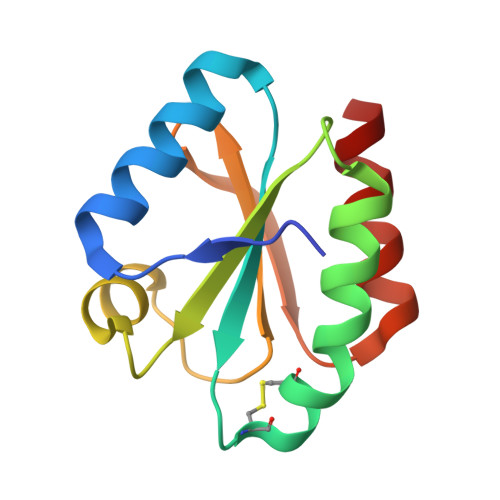
Cystatin C is a protein produced by many cells in the body, including all types of nucleated cells. It is a member of the cysteine protease inhibitor family and functions as an endogenous inhibitor of cathepsins, which are proteases involved in various physiological and pathological processes such as extracellular matrix degradation, antigen presentation, and cell death.
Cystatin C is freely filtered by the glomeruli in the kidneys and almost completely reabsorbed and catabolized by the proximal tubules. Therefore, its serum concentration is a reliable marker of glomerular filtration rate (GFR) and can be used to estimate kidney function.
Increased levels of cystatin C in the blood may indicate impaired kidney function or kidney disease, while decreased levels are less common and may be associated with hyperfiltration or overproduction of cystatin C. Measuring cystatin C levels can complement or supplement traditional methods for assessing kidney function, such as estimating GFR based on serum creatinine levels.
Micropore filters are medical devices used to filter or sterilize fluids and gases. They are made of materials like cellulose, mixed cellulose ester, or polyvinylidene fluoride with precise pore sizes, typically ranging from 0.1 to 10 micrometers in diameter. These filters are used to remove bacteria, fungi, and other particles from solutions in laboratory and medical settings, such as during the preparation of injectable drugs, tissue culture media, or sterile fluids for medical procedures. They come in various forms, including syringe filters, vacuum filters, and bottle-top filters, and are often used with the assistance of a vacuum or positive pressure to force the fluid through the filter material.
Hydrogen-ion concentration, also known as pH, is a measure of the acidity or basicity of a solution. It is defined as the negative logarithm (to the base 10) of the hydrogen ion activity in a solution. The standard unit of measurement is the pH unit. A pH of 7 is neutral, less than 7 is acidic, and greater than 7 is basic.
In medical terms, hydrogen-ion concentration is important for maintaining homeostasis within the body. For example, in the stomach, a high hydrogen-ion concentration (low pH) is necessary for the digestion of food. However, in other parts of the body such as blood, a high hydrogen-ion concentration can be harmful and lead to acidosis. Conversely, a low hydrogen-ion concentration (high pH) in the blood can lead to alkalosis. Both acidosis and alkalosis can have serious consequences on various organ systems if not corrected.
Chronic Renal Insufficiency (CRI) is a medical condition characterized by a gradual and progressive loss of kidney function over a period of months or years. It is also known as Chronic Kidney Disease (CKD). The main function of the kidneys is to filter waste products and excess fluids from the blood, which are then excreted in the urine. When the kidneys become insufficient, these waste products and fluids accumulate in the body, leading to various complications.
CRI is defined as a glomerular filtration rate (GFR) of less than 60 ml/min/1.73m2 for three months or more, regardless of cause. GFR is a measure of kidney function that estimates how well the kidneys are filtering waste products from the blood. The condition is classified into five stages based on the severity of the disease and the GFR value.
Stage 1: GFR greater than or equal to 90 ml/min/1.73m2
Stage 2: GFR between 60-89 ml/min/1.73m2
Stage 3: GFR between 30-59 ml/min/1.73m2
Stage 4: GFR between 15-29 ml/min/1.73m2
Stage 5: GFR less than 15 ml/min/1.73m2 or dialysis
CRI can be caused by various underlying conditions such as diabetes, hypertension, glomerulonephritis, polycystic kidney disease, and other genetic or acquired disorders. Symptoms of CRI may include fatigue, weakness, loss of appetite, swelling in the legs and ankles, shortness of breath, and changes in urination patterns. Treatment for CRI focuses on slowing down the progression of the disease, managing symptoms, and preventing complications. This may involve lifestyle modifications, medication, dialysis, or kidney transplantation.
Ultrafiltration is a medical process that separates fluids and dissolved solutes based on their size and charge. It's a type of membrane filtration that uses a semipermeable membrane with pores small enough to allow the passage of water and low molecular weight solutes, while retaining larger molecules and cells.
In clinical practice, ultrafiltration is often used in patients with acute or chronic kidney failure to remove excess fluid from the bloodstream, a process known as renal replacement therapy or dialysis. During this procedure, the patient's blood is passed through a hollow fiber membrane, and pressure differences across the membrane cause water and small solutes to move through the pores, while larger molecules such as proteins and cells are retained.
Ultrafiltration can also be used in other medical contexts, such as plasma exchange or therapeutic apheresis, where specific components of the blood are removed for therapeutic purposes.
Chromatography is a technique used in analytical chemistry for the separation, identification, and quantification of the components of a mixture. It is based on the differential distribution of the components of a mixture between a stationary phase and a mobile phase. The stationary phase can be a solid or liquid, while the mobile phase is a gas, liquid, or supercritical fluid that moves through the stationary phase carrying the sample components.
The interaction between the sample components and the stationary and mobile phases determines how quickly each component will move through the system. Components that interact more strongly with the stationary phase will move more slowly than those that interact more strongly with the mobile phase. This difference in migration rates allows for the separation of the components, which can then be detected and quantified.
There are many different types of chromatography, including paper chromatography, thin-layer chromatography (TLC), gas chromatography (GC), liquid chromatography (LC), and high-performance liquid chromatography (HPLC). Each type has its own strengths and weaknesses, and is best suited for specific applications.
In summary, chromatography is a powerful analytical technique used to separate, identify, and quantify the components of a mixture based on their differential distribution between a stationary phase and a mobile phase.
Affinity chromatography is a type of chromatography technique used in biochemistry and molecular biology to separate and purify proteins based on their biological characteristics, such as their ability to bind specifically to certain ligands or molecules. This method utilizes a stationary phase that is coated with a specific ligand (e.g., an antibody, antigen, receptor, or enzyme) that selectively interacts with the target protein in a sample.
The process typically involves the following steps:
1. Preparation of the affinity chromatography column: The stationary phase, usually a solid matrix such as agarose beads or magnetic beads, is modified by covalently attaching the ligand to its surface.
2. Application of the sample: The protein mixture is applied to the top of the affinity chromatography column, allowing it to flow through the stationary phase under gravity or pressure.
3. Binding and washing: As the sample flows through the column, the target protein selectively binds to the ligand on the stationary phase, while other proteins and impurities pass through. The column is then washed with a suitable buffer to remove any unbound proteins and contaminants.
4. Elution of the bound protein: The target protein can be eluted from the column using various methods, such as changing the pH, ionic strength, or polarity of the buffer, or by introducing a competitive ligand that displaces the bound protein.
5. Collection and analysis: The eluted protein fraction is collected and analyzed for purity and identity, often through techniques like SDS-PAGE or mass spectrometry.
Affinity chromatography is a powerful tool in biochemistry and molecular biology due to its high selectivity and specificity, enabling the efficient isolation of target proteins from complex mixtures. However, it requires careful consideration of the binding affinity between the ligand and the protein, as well as optimization of the elution conditions to minimize potential damage or denaturation of the purified protein.
Technetium Tc 99m Pentetate is a radioactive pharmaceutical preparation used as a radiopharmaceutical agent in medical imaging. It is a salt of technetium-99m, a metastable nuclear isomer of technetium-99, which emits gamma rays and has a half-life of 6 hours.
Technetium Tc 99m Pentetate is used in various diagnostic procedures, including renal imaging, brain scans, lung perfusion studies, and bone scans. It is distributed throughout the body after intravenous injection and is excreted primarily by the kidneys, making it useful for evaluating renal function and detecting abnormalities in the urinary tract.
The compound itself is a colorless, sterile, pyrogen-free solution that is typically supplied in a lead shielded container to protect against radiation exposure. It should be used promptly after preparation and handled with care to minimize radiation exposure to healthcare workers and patients.
Albuminuria is a medical condition that refers to the presence of albumin in the urine. Albumin is a type of protein normally found in the blood, but not in the urine. When the kidneys are functioning properly, they prevent large proteins like albumin from passing through into the urine. However, when the kidneys are damaged or not working correctly, such as in nephrotic syndrome or other kidney diseases, small amounts of albumin can leak into the urine.
The amount of albumin in the urine is often measured in milligrams per liter (mg/L) or in a spot urine sample, as the albumin-to-creatinine ratio (ACR). A small amount of albumin in the urine is called microalbuminuria, while a larger amount is called macroalbuminuria or proteinuria. The presence of albuminuria can indicate kidney damage and may be a sign of underlying medical conditions such as diabetes or high blood pressure. It is important to monitor and manage albuminuria to prevent further kidney damage and potential complications.
I'm sorry for any confusion, but "Iothalamic Acid" doesn't appear to be a recognized term in medical or physiological literature. It's possible there may be a spelling error or it could be a highly specialized or obscure term used only in specific research contexts.
If you meant "Iothalamate," that is a compound used as a contrast agent in medical imaging, specifically in radiology for procedures like intravenous pyelograms (IVPs) and computed tomography (CT) scans. Iothalamate is not typically referred to as an acid, though.
Please double-check the term you're looking for, and if there's any chance you meant "Iothalamate," let me know so I can provide a more accurate response!
Substrate specificity in the context of medical biochemistry and enzymology refers to the ability of an enzyme to selectively bind and catalyze a chemical reaction with a particular substrate (or a group of similar substrates) while discriminating against other molecules that are not substrates. This specificity arises from the three-dimensional structure of the enzyme, which has evolved to match the shape, charge distribution, and functional groups of its physiological substrate(s).
Substrate specificity is a fundamental property of enzymes that enables them to carry out highly selective chemical transformations in the complex cellular environment. The active site of an enzyme, where the catalysis takes place, has a unique conformation that complements the shape and charge distribution of its substrate(s). This ensures efficient recognition, binding, and conversion of the substrate into the desired product while minimizing unwanted side reactions with other molecules.
Substrate specificity can be categorized as:
1. Absolute specificity: An enzyme that can only act on a single substrate or a very narrow group of structurally related substrates, showing no activity towards any other molecule.
2. Group specificity: An enzyme that prefers to act on a particular functional group or class of compounds but can still accommodate minor structural variations within the substrate.
3. Broad or promiscuous specificity: An enzyme that can act on a wide range of structurally diverse substrates, albeit with varying catalytic efficiencies.
Understanding substrate specificity is crucial for elucidating enzymatic mechanisms, designing drugs that target specific enzymes or pathways, and developing biotechnological applications that rely on the controlled manipulation of enzyme activities.
Proteinuria is a medical term that refers to the presence of excess proteins, particularly albumin, in the urine. Under normal circumstances, only small amounts of proteins should be found in the urine because the majority of proteins are too large to pass through the glomeruli, which are the filtering units of the kidneys.
However, when the glomeruli become damaged or diseased, they may allow larger molecules such as proteins to leak into the urine. Persistent proteinuria is often a sign of kidney disease and can indicate damage to the glomeruli. It is usually detected through a routine urinalysis and may be confirmed with further testing.
The severity of proteinuria can vary, and it can be a symptom of various underlying conditions such as diabetes, hypertension, glomerulonephritis, and other kidney diseases. Treatment for proteinuria depends on the underlying cause and may include medications to control blood pressure, manage diabetes, or reduce protein loss in the urine.
Isoelectric focusing (IEF) is a technique used in electrophoresis, which is a method for separating proteins or other molecules based on their electrical charges. In IEF, a mixture of ampholytes (molecules that can carry both positive and negative charges) is used to create a pH gradient within a gel matrix. When an electric field is applied, the proteins or molecules migrate through the gel until they reach the point in the gradient where their net charge is zero, known as their isoelectric point (pI). At this point, they focus into a sharp band and stop moving, resulting in a highly resolved separation of the different components based on their pI. This technique is widely used in protein research for applications such as protein identification, characterization, and purification.
DEAE-cellulose chromatography is a method of purification and separation of biological molecules such as proteins, nucleic acids, and enzymes. DEAE stands for diethylaminoethyl, which is a type of charged functional group that is covalently bound to cellulose, creating a matrix with positive charges.
In this method, the mixture of biological molecules is applied to a column packed with DEAE-cellulose. The positively charged DEAE groups attract and bind negatively charged molecules in the mixture, such as nucleic acids and proteins, while allowing uncharged or neutrally charged molecules to pass through.
By adjusting the pH, ionic strength, or concentration of salt in the buffer solution used to elute the bound molecules from the column, it is possible to selectively elute specific molecules based on their charge and binding affinity to the DEAE-cellulose matrix. This makes DEAE-cellulose chromatography a powerful tool for purifying and separating biological molecules with high resolution and efficiency.
Podocytes are specialized cells that make up the visceral epithelial layer of the glomerular basement membrane in the kidney. They have long, interdigitating foot processes that wrap around the capillaries of the glomerulus and play a crucial role in maintaining the filtration barrier of the kidney. The slit diaphragms between the foot processes allow for the passage of small molecules while retaining larger proteins in the bloodstream. Podocytes also contribute to the maintenance and regulation of the glomerular filtration rate, making them essential for normal renal function. Damage or loss of podocytes can lead to proteinuria and kidney disease.
Chronic kidney failure, also known as chronic kidney disease (CKD) stage 5 or end-stage renal disease (ESRD), is a permanent loss of kidney function that occurs gradually over a period of months to years. It is defined as a glomerular filtration rate (GFR) of less than 15 ml/min, which means the kidneys are filtering waste and excess fluids at less than 15% of their normal capacity.
CKD can be caused by various underlying conditions such as diabetes, hypertension, glomerulonephritis, polycystic kidney disease, and recurrent kidney infections. Over time, the damage to the kidneys can lead to a buildup of waste products and fluids in the body, which can cause a range of symptoms including fatigue, weakness, shortness of breath, nausea, vomiting, and confusion.
Treatment for chronic kidney failure typically involves managing the underlying condition, making lifestyle changes such as following a healthy diet, and receiving supportive care such as dialysis or a kidney transplant to replace lost kidney function.
Kidney tubules are the structural and functional units of the kidney responsible for reabsorption, secretion, and excretion of various substances. They are part of the nephron, which is the basic unit of the kidney's filtration and reabsorption process.
There are three main types of kidney tubules:
1. Proximal tubule: This is the initial segment of the kidney tubule that receives the filtrate from the glomerulus. It is responsible for reabsorbing approximately 65% of the filtrate, including water, glucose, amino acids, and electrolytes.
2. Loop of Henle: This U-shaped segment of the tubule consists of a thin descending limb, a thin ascending limb, and a thick ascending limb. The loop of Henle helps to concentrate urine by creating an osmotic gradient that allows water to be reabsorbed in the collecting ducts.
3. Distal tubule: This is the final segment of the kidney tubule before it empties into the collecting duct. It is responsible for fine-tuning the concentration of electrolytes and pH balance in the urine by selectively reabsorbing or secreting substances such as sodium, potassium, chloride, and hydrogen ions.
Overall, kidney tubules play a critical role in maintaining fluid and electrolyte balance, regulating acid-base balance, and removing waste products from the body.
An amino acid sequence is the specific order of amino acids in a protein or peptide molecule, formed by the linking of the amino group (-NH2) of one amino acid to the carboxyl group (-COOH) of another amino acid through a peptide bond. The sequence is determined by the genetic code and is unique to each type of protein or peptide. It plays a crucial role in determining the three-dimensional structure and function of proteins.
Natriuresis is the process or condition of excreting an excessive amount of sodium (salt) through urine. It is a physiological response to high sodium levels in the body, which can be caused by various factors such as certain medical conditions (e.g., kidney disease, heart failure), medications, or dietary habits. The increased excretion of sodium helps regulate the body's water balance and maintain normal blood pressure. However, persistent natriuresis may indicate underlying health issues that require medical attention.
The isoelectric point (pI) is a term used in biochemistry and molecular biology to describe the pH at which a molecule, such as a protein or peptide, carries no net electrical charge. At this pH, the positive and negative charges on the molecule are equal and balanced. The pI of a protein can be calculated based on its amino acid sequence and is an important property that affects its behavior in various chemical and biological environments. Proteins with different pIs may have different solubilities, stabilities, and interactions with other molecules, which can impact their function and role in the body.
A nephron is the basic structural and functional unit of the kidney. It is responsible for filtering blood, reabsorbing necessary substances, and excreting waste products into the urine. Each human kidney contains approximately one million nephrons.
The structure of a nephron includes a glomerulus, which is a tuft of capillaries surrounded by Bowman's capsule. The glomerulus filters blood, allowing small molecules like water and solutes to pass through while keeping larger molecules like proteins and blood cells within the capillaries.
The filtrate then passes through the tubular portion of the nephron, which includes the proximal convoluted tubule, loop of Henle, distal convoluted tubule, and collecting duct. The tubular portion reabsorbs necessary substances like water, glucose, amino acids, and electrolytes back into the bloodstream while excreting waste products like urea and creatinine into the urine.
Overall, nephrons play a critical role in maintaining fluid and electrolyte balance, regulating blood pressure, and removing waste products from the body.
Molecular sequence data refers to the specific arrangement of molecules, most commonly nucleotides in DNA or RNA, or amino acids in proteins, that make up a biological macromolecule. This data is generated through laboratory techniques such as sequencing, and provides information about the exact order of the constituent molecules. This data is crucial in various fields of biology, including genetics, evolution, and molecular biology, allowing for comparisons between different organisms, identification of genetic variations, and studies of gene function and regulation.
Diuresis is a medical term that refers to an increased production of urine by the kidneys. It can occur as a result of various factors, including certain medications, medical conditions, or as a response to a physiological need, such as in the case of dehydration. Diuretics are a class of drugs that promote diuresis and are often used to treat conditions such as high blood pressure, heart failure, and edema.
Diuresis can be classified into several types based on its underlying cause or mechanism, including:
1. Osmotic diuresis: This occurs when the kidneys excrete large amounts of urine in response to a high concentration of solutes (such as glucose) in the tubular fluid. The high osmolarity of the tubular fluid causes water to be drawn out of the bloodstream and into the urine, leading to an increase in urine output.
2. Forced diuresis: This is a medical procedure in which large amounts of intravenous fluids are administered to promote diuresis. It is used in certain clinical situations, such as to enhance the excretion of toxic substances or to prevent kidney damage.
3. Natriuretic diuresis: This occurs when the kidneys excrete large amounts of sodium and water in response to the release of natriuretic peptides, which are hormones that regulate sodium balance and blood pressure.
4. Aquaresis: This is a type of diuresis that occurs in response to the ingestion of large amounts of water, leading to dilute urine production.
5. Pathological diuresis: This refers to increased urine production due to underlying medical conditions such as diabetes insipidus or pyelonephritis.
It is important to note that excessive diuresis can lead to dehydration and electrolyte imbalances, so it should be monitored carefully in clinical settings.
Blood pressure is the force exerted by circulating blood on the walls of the blood vessels. It is measured in millimeters of mercury (mmHg) and is given as two figures:
1. Systolic pressure: This is the pressure when the heart pushes blood out into the arteries.
2. Diastolic pressure: This is the pressure when the heart rests between beats, allowing it to fill with blood.
Normal blood pressure for adults is typically around 120/80 mmHg, although this can vary slightly depending on age, sex, and other factors. High blood pressure (hypertension) is generally considered to be a reading of 130/80 mmHg or higher, while low blood pressure (hypotension) is usually defined as a reading below 90/60 mmHg. It's important to note that blood pressure can fluctuate throughout the day and may be affected by factors such as stress, physical activity, and medication use.
Macromolecular substances, also known as macromolecules, are large, complex molecules made up of repeating subunits called monomers. These substances are formed through polymerization, a process in which many small molecules combine to form a larger one. Macromolecular substances can be naturally occurring, such as proteins, DNA, and carbohydrates, or synthetic, such as plastics and synthetic fibers.
In the context of medicine, macromolecular substances are often used in the development of drugs and medical devices. For example, some drugs are designed to bind to specific macromolecules in the body, such as proteins or DNA, in order to alter their function and produce a therapeutic effect. Additionally, macromolecular substances may be used in the creation of medical implants, such as artificial joints and heart valves, due to their strength and durability.
It is important for healthcare professionals to have an understanding of macromolecular substances and how they function in the body, as this knowledge can inform the development and use of medical treatments.
Renal insufficiency, also known as kidney failure, is a medical condition in which the kidneys are unable to properly filter waste products and excess fluids from the blood. This results in a buildup of these substances in the body, which can cause a variety of symptoms such as weakness, shortness of breath, and fluid retention. Renal insufficiency can be acute, meaning it comes on suddenly, or chronic, meaning it develops over time. It is typically diagnosed through blood tests, urine tests, and imaging studies. Treatment may include medications to control symptoms, dietary changes, and in severe cases, dialysis or a kidney transplant.
High-performance liquid chromatography (HPLC) is a type of chromatography that separates and analyzes compounds based on their interactions with a stationary phase and a mobile phase under high pressure. The mobile phase, which can be a gas or liquid, carries the sample mixture through a column containing the stationary phase.
In HPLC, the mobile phase is a liquid, and it is pumped through the column at high pressures (up to several hundred atmospheres) to achieve faster separation times and better resolution than other types of liquid chromatography. The stationary phase can be a solid or a liquid supported on a solid, and it interacts differently with each component in the sample mixture, causing them to separate as they travel through the column.
HPLC is widely used in analytical chemistry, pharmaceuticals, biotechnology, and other fields to separate, identify, and quantify compounds present in complex mixtures. It can be used to analyze a wide range of substances, including drugs, hormones, vitamins, pigments, flavors, and pollutants. HPLC is also used in the preparation of pure samples for further study or use.
I believe there may be some confusion in your question. "Rabbits" is a common name used to refer to the Lagomorpha species, particularly members of the family Leporidae. They are small mammals known for their long ears, strong legs, and quick reproduction.
However, if you're referring to "rabbits" in a medical context, there is a term called "rabbit syndrome," which is a rare movement disorder characterized by repetitive, involuntary movements of the fingers, resembling those of a rabbit chewing. It is also known as "finger-chewing chorea." This condition is usually associated with certain medications, particularly antipsychotics, and typically resolves when the medication is stopped or adjusted.
Renal plasma flow (RPF) is a medical term that refers to the volume of plasma delivered to and filtered through the kidneys per unit time. It is typically expressed in milliliters per minute (ml/min). The RPF is an important measure of renal function, as it reflects the ability of the kidneys to filter blood and remove waste products from the body.
RPF can be measured directly using various techniques, such as injecting a substance into the renal artery and measuring its concentration in the venous effluent from the kidney. However, RPF is often estimated indirectly based on the clearance of a substance that is freely filtered by the glomeruli but not reabsorbed or secreted by the tubules, such as para-aminohippuric acid (PAH). The clearance of PAH is proportional to the RPF, and can be used to calculate an estimate of RPF.
Renal plasma flow is affected by various factors, including blood pressure, renal vasodilation or vasoconstriction, and the presence of kidney disease or injury. Decreased RPF may indicate impaired renal function and may contribute to the development of kidney disease.
Ultracentrifugation is a medical and laboratory technique used for the separation of particles of different sizes, densities, or shapes from a mixture based on their sedimentation rates. This process involves the use of a specialized piece of equipment called an ultracentrifuge, which can generate very high centrifugal forces, much greater than those produced by a regular centrifuge.
In ultracentrifugation, a sample is placed in a special tube and spun at extremely high speeds, causing the particles within the sample to separate based on their size, shape, and density. The larger or denser particles will sediment faster and accumulate at the bottom of the tube, while smaller or less dense particles will remain suspended in the solution or sediment more slowly.
Ultracentrifugation is a valuable tool in various fields, including biochemistry, molecular biology, and virology. It can be used to purify and concentrate viruses, subcellular organelles, membrane fractions, ribosomes, DNA, and other macromolecules from complex mixtures. The technique can also provide information about the size, shape, and density of these particles, making it a crucial method for characterizing and studying their properties.
Carbohydrates are a major nutrient class consisting of organic compounds that primarily contain carbon, hydrogen, and oxygen atoms. They are classified as saccharides, which include monosaccharides (simple sugars), disaccharides (double sugars), oligosaccharides (short-chain sugars), and polysaccharides (complex carbohydrates).
Monosaccharides, such as glucose, fructose, and galactose, are the simplest form of carbohydrates. They consist of a single sugar molecule that cannot be broken down further by hydrolysis. Disaccharides, like sucrose (table sugar), lactose (milk sugar), and maltose (malt sugar), are formed from two monosaccharide units joined together.
Oligosaccharides contain a small number of monosaccharide units, typically less than 20, while polysaccharides consist of long chains of hundreds to thousands of monosaccharide units. Polysaccharides can be further classified into starch (found in plants), glycogen (found in animals), and non-starchy polysaccharides like cellulose, chitin, and pectin.
Carbohydrates play a crucial role in providing energy to the body, with glucose being the primary source of energy for most cells. They also serve as structural components in plants (cellulose) and animals (chitin), participate in various metabolic processes, and contribute to the taste, texture, and preservation of foods.
"Cattle" is a term used in the agricultural and veterinary fields to refer to domesticated animals of the genus *Bos*, primarily *Bos taurus* (European cattle) and *Bos indicus* (Zebu). These animals are often raised for meat, milk, leather, and labor. They are also known as bovines or cows (for females), bulls (intact males), and steers/bullocks (castrated males). However, in a strict medical definition, "cattle" does not apply to humans or other animals.
Capillary permeability refers to the ability of substances to pass through the walls of capillaries, which are the smallest blood vessels in the body. These tiny vessels connect the arterioles and venules, allowing for the exchange of nutrients, waste products, and gases between the blood and the surrounding tissues.
The capillary wall is composed of a single layer of endothelial cells that are held together by tight junctions. The permeability of these walls varies depending on the size and charge of the molecules attempting to pass through. Small, uncharged molecules such as water, oxygen, and carbon dioxide can easily diffuse through the capillary wall, while larger or charged molecules such as proteins and large ions have more difficulty passing through.
Increased capillary permeability can occur in response to inflammation, infection, or injury, allowing larger molecules and immune cells to enter the surrounding tissues. This can lead to swelling (edema) and tissue damage if not controlled. Decreased capillary permeability, on the other hand, can lead to impaired nutrient exchange and tissue hypoxia.
Overall, the permeability of capillaries is a critical factor in maintaining the health and function of tissues throughout the body.
Immunodiffusion is a laboratory technique used in immunology to detect and measure the presence of specific antibodies or antigens in a sample. It is based on the principle of diffusion, where molecules move from an area of high concentration to an area of low concentration until they reach equilibrium. In this technique, a sample containing an unknown quantity of antigen or antibody is placed in a gel or agar medium that contains a known quantity of antibody or antigen, respectively.
The two substances then diffuse towards each other and form a visible precipitate at the point where they meet and reach equivalence, which indicates the presence and quantity of the specific antigen or antibody in the sample. There are several types of immunodiffusion techniques, including radial immunodiffusion (RID) and double immunodiffusion (Ouchterlony technique). These techniques are widely used in diagnostic laboratories to identify and measure various antigens and antibodies, such as those found in infectious diseases, autoimmune disorders, and allergic reactions.
Cystatins are a group of proteins that inhibit cysteine proteases, which are enzymes that break down other proteins. Cystatins are found in various biological fluids and tissues, including tears, saliva, seminal plasma, and urine. They play an important role in regulating protein catabolism and protecting cells from excessive protease activity. There are three main types of cystatins: type 1 (cystatin C), type 2 (cystatin M, cystatin N, and fetuin), and type 3 (kininogens). Abnormal levels of cystatins have been associated with various pathological conditions, such as cancer, neurodegenerative diseases, and inflammatory disorders.
Diabetic nephropathy is a kidney disease that occurs as a complication of diabetes. It is also known as diabetic kidney disease (DKD). This condition affects the ability of the kidneys to filter waste and excess fluids from the blood, leading to their accumulation in the body.
Diabetic nephropathy is caused by damage to the small blood vessels in the kidneys, which can occur over time due to high levels of glucose in the blood. This damage can lead to scarring and thickening of the kidney's filtering membranes, reducing their ability to function properly.
Symptoms of diabetic nephropathy may include proteinuria (the presence of protein in the urine), edema (swelling in the legs, ankles, or feet due to fluid retention), and hypertension (high blood pressure). Over time, if left untreated, diabetic nephropathy can progress to end-stage kidney disease, which requires dialysis or a kidney transplant.
Preventing or delaying the onset of diabetic nephropathy involves maintaining good control of blood sugar levels, keeping blood pressure under control, and making lifestyle changes such as quitting smoking, eating a healthy diet, and getting regular exercise. Regular monitoring of kidney function through urine tests and blood tests is also important for early detection and treatment of this condition.
Water microbiology is not a formal medical term, but rather a branch of microbiology that deals with the study of microorganisms found in water. It involves the identification, enumeration, and characterization of bacteria, viruses, parasites, and other microscopic organisms present in water sources such as lakes, rivers, oceans, groundwater, drinking water, and wastewater.
In a medical context, water microbiology is relevant to public health because it helps to assess the safety of water supplies for human consumption and recreational activities. It also plays a critical role in understanding and preventing waterborne diseases caused by pathogenic microorganisms that can lead to illnesses such as diarrhea, skin infections, and respiratory problems.
Water microbiologists use various techniques to study water microorganisms, including culturing, microscopy, genetic analysis, and biochemical tests. They also investigate the ecology of these organisms, their interactions with other species, and their response to environmental factors such as temperature, pH, and nutrient availability.
Overall, water microbiology is a vital field that helps ensure the safety of our water resources and protects public health.
In the context of medicine, "chemistry" often refers to the field of study concerned with the properties, composition, and structure of elements and compounds, as well as their reactions with one another. It is a fundamental science that underlies much of modern medicine, including pharmacology (the study of drugs), toxicology (the study of poisons), and biochemistry (the study of the chemical processes that occur within living organisms).
In addition to its role as a basic science, chemistry is also used in medical testing and diagnosis. For example, clinical chemistry involves the analysis of bodily fluids such as blood and urine to detect and measure various substances, such as glucose, cholesterol, and electrolytes, that can provide important information about a person's health status.
Overall, chemistry plays a critical role in understanding the mechanisms of diseases, developing new treatments, and improving diagnostic tests and techniques.
Sodium is an essential mineral and electrolyte that is necessary for human health. In a medical context, sodium is often discussed in terms of its concentration in the blood, as measured by serum sodium levels. The normal range for serum sodium is typically between 135 and 145 milliequivalents per liter (mEq/L).
Sodium plays a number of important roles in the body, including:
* Regulating fluid balance: Sodium helps to regulate the amount of water in and around your cells, which is important for maintaining normal blood pressure and preventing dehydration.
* Facilitating nerve impulse transmission: Sodium is involved in the generation and transmission of electrical signals in the nervous system, which is necessary for proper muscle function and coordination.
* Assisting with muscle contraction: Sodium helps to regulate muscle contractions by interacting with other minerals such as calcium and potassium.
Low sodium levels (hyponatremia) can cause symptoms such as confusion, seizures, and coma, while high sodium levels (hypernatremia) can lead to symptoms such as weakness, muscle cramps, and seizures. Both conditions require medical treatment to correct.
'Escherichia coli' (E. coli) is a type of gram-negative, facultatively anaerobic, rod-shaped bacterium that commonly inhabits the intestinal tract of humans and warm-blooded animals. It is a member of the family Enterobacteriaceae and one of the most well-studied prokaryotic model organisms in molecular biology.
While most E. coli strains are harmless and even beneficial to their hosts, some serotypes can cause various forms of gastrointestinal and extraintestinal illnesses in humans and animals. These pathogenic strains possess virulence factors that enable them to colonize and damage host tissues, leading to diseases such as diarrhea, urinary tract infections, pneumonia, and sepsis.
E. coli is a versatile organism with remarkable genetic diversity, which allows it to adapt to various environmental niches. It can be found in water, soil, food, and various man-made environments, making it an essential indicator of fecal contamination and a common cause of foodborne illnesses. The study of E. coli has contributed significantly to our understanding of fundamental biological processes, including DNA replication, gene regulation, and protein synthesis.
Temperature, in a medical context, is a measure of the degree of hotness or coldness of a body or environment. It is usually measured using a thermometer and reported in degrees Celsius (°C), degrees Fahrenheit (°F), or kelvin (K). In the human body, normal core temperature ranges from about 36.5-37.5°C (97.7-99.5°F) when measured rectally, and can vary slightly depending on factors such as time of day, physical activity, and menstrual cycle. Elevated body temperature is a common sign of infection or inflammation, while abnormally low body temperature can indicate hypothermia or other medical conditions.
Chemical phenomena refer to the changes and interactions that occur at the molecular or atomic level when chemicals are involved. These phenomena can include chemical reactions, in which one or more substances (reactants) are converted into different substances (products), as well as physical properties that change as a result of chemical interactions, such as color, state of matter, and solubility. Chemical phenomena can be studied through various scientific disciplines, including chemistry, biochemistry, and physics.
In the field of medicine, "time factors" refer to the duration of symptoms or time elapsed since the onset of a medical condition, which can have significant implications for diagnosis and treatment. Understanding time factors is crucial in determining the progression of a disease, evaluating the effectiveness of treatments, and making critical decisions regarding patient care.
For example, in stroke management, "time is brain," meaning that rapid intervention within a specific time frame (usually within 4.5 hours) is essential to administering tissue plasminogen activator (tPA), a clot-busting drug that can minimize brain damage and improve patient outcomes. Similarly, in trauma care, the "golden hour" concept emphasizes the importance of providing definitive care within the first 60 minutes after injury to increase survival rates and reduce morbidity.
Time factors also play a role in monitoring the progression of chronic conditions like diabetes or heart disease, where regular follow-ups and assessments help determine appropriate treatment adjustments and prevent complications. In infectious diseases, time factors are crucial for initiating antibiotic therapy and identifying potential outbreaks to control their spread.
Overall, "time factors" encompass the significance of recognizing and acting promptly in various medical scenarios to optimize patient outcomes and provide effective care.
Aminohippuric acids are a type of organic compound that contain both an amino group and a hippuric acid group in their chemical structure. Hippuric acid is a derivative of benzoic acid, which is conjugated with glycine in the body. Aminohippuric acids are primarily known for their use as diagnostic agents in renal function tests.
The most common aminohippuric acid is p-aminohippuric acid (PAH), which is used as a marker to measure effective renal plasma flow (ERPF) in the kidneys. PAH is freely filtered by the glomeruli and then actively secreted by the proximal tubules of the nephrons, making it an ideal agent for measuring ERPF.
In a renal function test using PAH, a small dose of the compound is injected into the patient's bloodstream, and its concentration in the blood is measured over time. By analyzing the clearance rate of PAH from the blood, healthcare providers can estimate the ERPF and assess kidney function.
Overall, aminohippuric acids are important diagnostic tools for evaluating renal function and identifying potential kidney-related health issues.
Disc electrophoresis is a type of electrophoresis technique used to separate and analyze DNA, RNA, or proteins based on their size and electrical charge. In this method, the samples are placed in a gel matrix (usually agarose or polyacrylamide) and an electric field is applied. The smaller and/or more negatively charged molecules migrate faster through the gel and separate from larger and/or less charged molecules, creating a pattern of bands that can be visualized and analyzed.
The term "disc" refers to the characteristic disc-shaped pattern that is often seen in the separated protein bands when using this technique. This pattern is created by the interaction between the size, charge, and shape of the proteins, resulting in a distinct banding pattern that can be used for identification and analysis.
Disc electrophoresis is widely used in molecular biology and genetics research, as well as in diagnostic testing and forensic science.
Edetic acid, also known as ethylenediaminetetraacetic acid (EDTA), is not a medical term per se, but a chemical compound with various applications in medicine. EDTA is a synthetic amino acid that acts as a chelating agent, which means it can bind to metallic ions and form stable complexes.
In medicine, EDTA is primarily used in the treatment of heavy metal poisoning, such as lead or mercury toxicity. It works by binding to the toxic metal ions in the body, forming a stable compound that can be excreted through urine. This helps reduce the levels of harmful metals in the body and alleviate their toxic effects.
EDTA is also used in some diagnostic tests, such as the determination of calcium levels in blood. Additionally, it has been explored as a potential therapy for conditions like atherosclerosis and Alzheimer's disease, although its efficacy in these areas remains controversial and unproven.
It is important to note that EDTA should only be administered under medical supervision due to its potential side effects and the need for careful monitoring of its use.
Solubility is a fundamental concept in pharmaceutical sciences and medicine, which refers to the maximum amount of a substance (solute) that can be dissolved in a given quantity of solvent (usually water) at a specific temperature and pressure. Solubility is typically expressed as mass of solute per volume or mass of solvent (e.g., grams per liter, milligrams per milliliter). The process of dissolving a solute in a solvent results in a homogeneous solution where the solute particles are dispersed uniformly throughout the solvent.
Understanding the solubility of drugs is crucial for their formulation, administration, and therapeutic effectiveness. Drugs with low solubility may not dissolve sufficiently to produce the desired pharmacological effect, while those with high solubility might lead to rapid absorption and short duration of action. Therefore, optimizing drug solubility through various techniques like particle size reduction, salt formation, or solubilization is an essential aspect of drug development and delivery.
Radioisotope renography is a type of nuclear medicine test used to evaluate the function and anatomy of the kidneys. It involves the intravenous administration of a small amount of radioactive material, called a radiopharmaceutical or radioisotope, which is taken up by the kidneys and emits gamma rays that can be detected by a special camera.
The most commonly used radiopharmaceutical for renography is technetium-99m mercaptoacetyltriglycine (Tc-99m MAG3). The patient is positioned under the gamma camera, and images are taken at various intervals after the injection of the radioisotope.
The test provides information about the blood flow to the kidneys, the glomerular filtration rate (GFR), which measures how well the kidneys filter waste products from the blood, and the drainage of urine from the kidneys into the bladder. Renography can help diagnose conditions such as renal artery stenosis, hydronephrosis, and kidney obstruction.
It is important to note that while radioisotope renography involves exposure to a small amount of radiation, the benefits of the test in terms of diagnostic accuracy and patient management often outweigh the risks associated with the radiation exposure.
Protein binding, in the context of medical and biological sciences, refers to the interaction between a protein and another molecule (known as the ligand) that results in a stable complex. This process is often reversible and can be influenced by various factors such as pH, temperature, and concentration of the involved molecules.
In clinical chemistry, protein binding is particularly important when it comes to drugs, as many of them bind to proteins (especially albumin) in the bloodstream. The degree of protein binding can affect a drug's distribution, metabolism, and excretion, which in turn influence its therapeutic effectiveness and potential side effects.
Protein-bound drugs may be less available for interaction with their target tissues, as only the unbound or "free" fraction of the drug is active. Therefore, understanding protein binding can help optimize dosing regimens and minimize adverse reactions.
"Inbred strains of rats" are genetically identical rodents that have been produced through many generations of brother-sister mating. This results in a high degree of homozygosity, where the genes at any particular locus in the genome are identical in all members of the strain.
Inbred strains of rats are widely used in biomedical research because they provide a consistent and reproducible genetic background for studying various biological phenomena, including the effects of drugs, environmental factors, and genetic mutations on health and disease. Additionally, inbred strains can be used to create genetically modified models of human diseases by introducing specific mutations into their genomes.
Some commonly used inbred strains of rats include the Wistar Kyoto (WKY), Sprague-Dawley (SD), and Fischer 344 (F344) rat strains. Each strain has its own unique genetic characteristics, making them suitable for different types of research.
Iohexol is a non-ionic, water-soluble contrast medium primarily used in radiographic imaging procedures such as computed tomography (CT) scans and angiography. It belongs to a class of medications known as radiocontrast agents. Iohexol works by increasing the X-ray absorption of body tissues, making them more visible on X-ray images. This helps healthcare professionals to better diagnose and assess various medical conditions, including injuries, tumors, and vascular diseases.
The chemical structure of iohexol consists of an iodine atom surrounded by organic molecules, which makes it safe for intravenous administration. It is eliminatted from the body primarily through urinary excretion. Iohexol has a low risk of allergic reactions compared to ionic contrast media and is generally well-tolerated in patients with normal renal function. However, its use should be avoided or closely monitored in individuals with impaired kidney function, as it may increase the risk of nephrotoxicity.
Chemical precipitation is a process in which a chemical compound becomes a solid, insoluble form, known as a precipitate, from a liquid solution. This occurs when the concentration of the compound in the solution exceeds its solubility limit and forms a separate phase. The reaction that causes the formation of the precipitate can be a result of various factors such as changes in temperature, pH, or the addition of another chemical reagent.
In the medical field, chemical precipitation is used in diagnostic tests to detect and measure the presence of certain substances in body fluids, such as blood or urine. For example, a common test for kidney function involves adding a chemical reagent to a urine sample, which causes the excess protein in the urine to precipitate out of solution. The amount of precipitate formed can then be measured and used to diagnose and monitor kidney disease.
Chemical precipitation is also used in the treatment of certain medical conditions, such as heavy metal poisoning. In this case, a chelating agent is administered to bind with the toxic metal ions in the body, forming an insoluble compound that can be excreted through the urine or feces. This process helps to reduce the amount of toxic metals in the body and alleviate symptoms associated with poisoning.
In the context of medical research, "methods" refers to the specific procedures or techniques used in conducting a study or experiment. This includes details on how data was collected, what measurements were taken, and what statistical analyses were performed. The methods section of a medical paper allows other researchers to replicate the study if they choose to do so. It is considered one of the key components of a well-written research article, as it provides transparency and helps establish the validity of the findings.
Centrifugation, Density Gradient is a medical laboratory technique used to separate and purify different components of a mixture based on their size, density, and shape. This method involves the use of a centrifuge and a density gradient medium, such as sucrose or cesium chloride, to create a stable density gradient within a column or tube.
The sample is carefully layered onto the top of the gradient and then subjected to high-speed centrifugation. During centrifugation, the particles in the sample move through the gradient based on their size, density, and shape, with heavier particles migrating faster and further than lighter ones. This results in the separation of different components of the mixture into distinct bands or zones within the gradient.
This technique is commonly used to purify and concentrate various types of biological materials, such as viruses, organelles, ribosomes, and subcellular fractions, from complex mixtures. It allows for the isolation of pure and intact particles, which can then be collected and analyzed for further study or use in downstream applications.
In summary, Centrifugation, Density Gradient is a medical laboratory technique used to separate and purify different components of a mixture based on their size, density, and shape using a centrifuge and a density gradient medium.
A puncture, in medical terms, refers to a small hole or wound that is caused by a sharp object penetrating the skin or other body tissues. This can result in damage to underlying structures such as blood vessels, nerves, or organs, and may lead to complications such as bleeding, infection, or inflammation.
Punctures can occur accidentally, such as from stepping on a nail or getting pricked by a needle, or they can be inflicted intentionally, such as during medical procedures like injections or blood draws. In some cases, puncture wounds may require medical attention to clean and close the wound, prevent infection, and promote healing.
In a medical context, "hot temperature" is not a standard medical term with a specific definition. However, it is often used in relation to fever, which is a common symptom of illness. A fever is typically defined as a body temperature that is higher than normal, usually above 38°C (100.4°F) for adults and above 37.5-38°C (99.5-101.3°F) for children, depending on the source.
Therefore, when a medical professional talks about "hot temperature," they may be referring to a body temperature that is higher than normal due to fever or other causes. It's important to note that a high environmental temperature can also contribute to an elevated body temperature, so it's essential to consider both the body temperature and the environmental temperature when assessing a patient's condition.
Ammonium sulfate is a chemical compound with the formula (NH4)2SO4. It is a white crystalline solid that is highly soluble in water and is commonly used in fertilizers due to its high nitrogen content. In a medical context, it can be used as a laxative or for lowering the pH of the gastrointestinal tract in certain medical conditions. It may also be used in the treatment of metabolic alkalosis, a condition characterized by an excessively high pH in the blood. However, its use in medical treatments is less common than its use in agricultural and industrial applications.
Water purification is the process of removing or reducing contaminants in water to make it safe and suitable for specific uses, such as drinking, cooking, irrigation, or medical purposes. This is typically achieved through physical, chemical, or biological methods, or a combination thereof. The goal is to eliminate or reduce harmful substances like bacteria, viruses, parasites, heavy metals, pesticides, and other pollutants that can cause illness or negatively impact human health, aquatic life, or the environment.
The specific purification methods used may vary depending on the nature of the contaminants and the desired level of purity for the intended use. Common techniques include filtration (using various types of filters like activated carbon, ceramic, or reverse osmosis), disinfection (using chemicals like chlorine or UV light to kill microorganisms), sedimentation (allowing particles to settle and be removed), and distillation (heating water to create steam, which is then condensed back into pure water).
Nephrectomy is a surgical procedure in which all or part of a kidney is removed. It may be performed due to various reasons such as severe kidney damage, kidney cancer, or living donor transplantation. The type of nephrectomy depends on the reason for the surgery - a simple nephrectomy involves removing only the affected portion of the kidney, while a radical nephrectomy includes removal of the whole kidney along with its surrounding tissues like the adrenal gland and lymph nodes.
Chromatography, agarose is a type of chromatography technique that utilizes agarose gel as the stationary phase in the separation and analysis of biological molecules, such as DNA, RNA, and proteins. This method is commonly used in molecular biology for various applications, including DNA fragment separation, protein purification, and detection of specific nucleic acid sequences or proteins.
Agarose gel is a matrix made from agarose, a polysaccharide derived from seaweed. It has a porous structure with uniform pore size that allows for the size-based separation of molecules based on their ability to migrate through the gel under an electric field (in the case of electrophoresis) or by capillary action (in the case of capillary electrophoresis).
The charged molecules, such as DNA or proteins, interact with the agarose matrix and move through the gel at different rates depending on their size, charge, and shape. Smaller molecules can migrate more quickly through the pores of the gel, while larger molecules are retarded due to their inability to easily pass through the pores. This results in a separation of the molecules based on their physical properties, allowing for their analysis and characterization.
In summary, chromatography, agarose refers to the use of agarose gel as the stationary phase in the separation and analysis of biological molecules using various chromatography techniques, such as electrophoresis or capillary electrophoresis.
I believe there might be a misunderstanding in your question. "Dogs" is not a medical term or condition. It is the common name for a domesticated carnivore of the family Canidae, specifically the genus Canis, which includes wolves, foxes, and other extant and extinct species of mammals. Dogs are often kept as pets and companions, and they have been bred in a wide variety of forms and sizes for different purposes, such as hunting, herding, guarding, assisting police and military forces, and providing companionship and emotional support.
If you meant to ask about a specific medical condition or term related to dogs, please provide more context so I can give you an accurate answer.
A biological marker, often referred to as a biomarker, is a measurable indicator that reflects the presence or severity of a disease state, or a response to a therapeutic intervention. Biomarkers can be found in various materials such as blood, tissues, or bodily fluids, and they can take many forms, including molecular, histologic, radiographic, or physiological measurements.
In the context of medical research and clinical practice, biomarkers are used for a variety of purposes, such as:
1. Diagnosis: Biomarkers can help diagnose a disease by indicating the presence or absence of a particular condition. For example, prostate-specific antigen (PSA) is a biomarker used to detect prostate cancer.
2. Monitoring: Biomarkers can be used to monitor the progression or regression of a disease over time. For instance, hemoglobin A1c (HbA1c) levels are monitored in diabetes patients to assess long-term blood glucose control.
3. Predicting: Biomarkers can help predict the likelihood of developing a particular disease or the risk of a negative outcome. For example, the presence of certain genetic mutations can indicate an increased risk for breast cancer.
4. Response to treatment: Biomarkers can be used to evaluate the effectiveness of a specific treatment by measuring changes in the biomarker levels before and after the intervention. This is particularly useful in personalized medicine, where treatments are tailored to individual patients based on their unique biomarker profiles.
It's important to note that for a biomarker to be considered clinically valid and useful, it must undergo rigorous validation through well-designed studies, including demonstrating sensitivity, specificity, reproducibility, and clinical relevance.
Hydrostatic pressure is the pressure exerted by a fluid at equilibrium at a given point within the fluid, due to the force of gravity. In medical terms, hydrostatic pressure is often discussed in relation to body fluids and tissues. For example, the hydrostatic pressure in the capillaries (tiny blood vessels) is the force that drives the fluid out of the blood vessels and into the surrounding tissues. This helps to maintain the balance of fluids in the body. Additionally, abnormal increases in hydrostatic pressure can contribute to the development of edema (swelling) in the tissues.
Centrifugation is a laboratory technique that involves the use of a machine called a centrifuge to separate mixtures based on their differing densities or sizes. The mixture is placed in a rotor and spun at high speeds, causing the denser components to move away from the center of rotation and the less dense components to remain nearer the center. This separation allows for the recovery and analysis of specific particles, such as cells, viruses, or subcellular organelles, from complex mixtures.
The force exerted on the mixture during centrifugation is described in terms of relative centrifugal force (RCF) or g-force, which represents the number of times greater the acceleration due to centrifugation is than the acceleration due to gravity. The RCF is determined by the speed of rotation (revolutions per minute, or RPM), the radius of rotation, and the duration of centrifugation.
Centrifugation has numerous applications in various fields, including clinical laboratories, biochemistry, molecular biology, and virology. It is a fundamental technique for isolating and concentrating particles from solutions, enabling further analysis and characterization.
A peptide fragment is a short chain of amino acids that is derived from a larger peptide or protein through various biological or chemical processes. These fragments can result from the natural breakdown of proteins in the body during regular physiological processes, such as digestion, or they can be produced experimentally in a laboratory setting for research or therapeutic purposes.
Peptide fragments are often used in research to map the structure and function of larger peptides and proteins, as well as to study their interactions with other molecules. In some cases, peptide fragments may also have biological activity of their own and can be developed into drugs or diagnostic tools. For example, certain peptide fragments derived from hormones or neurotransmitters may bind to receptors in the body and mimic or block the effects of the full-length molecule.
Chromium radioisotopes are unstable isotopes or variants of the chemical element chromium that emit radiation as they decay into more stable forms. These isotopes have an excess of energy and particles, making them unstable and capable of emitting ionizing radiation in the form of gamma rays or subatomic particles such as alpha or beta particles.
Chromium has several radioisotopes, including chromium-50, chromium-51, and chromium-53, among others. Chromium-51 is one of the most commonly used radioisotopes in medical applications, particularly in diagnostic procedures such as red blood cell labeling and imaging studies.
It's important to note that handling and using radioisotopes require proper training and safety measures due to their potential radiation hazards.
Trypsin is a proteolytic enzyme, specifically a serine protease, that is secreted by the pancreas as an inactive precursor, trypsinogen. Trypsinogen is converted into its active form, trypsin, in the small intestine by enterokinase, which is produced by the intestinal mucosa.
Trypsin plays a crucial role in digestion by cleaving proteins into smaller peptides at specific arginine and lysine residues. This enzyme helps to break down dietary proteins into amino acids, allowing for their absorption and utilization by the body. Additionally, trypsin can activate other zymogenic pancreatic enzymes, such as chymotrypsinogen and procarboxypeptidases, thereby contributing to overall protein digestion.
Blood proteins, also known as serum proteins, are a group of complex molecules present in the blood that are essential for various physiological functions. These proteins include albumin, globulins (alpha, beta, and gamma), and fibrinogen. They play crucial roles in maintaining oncotic pressure, transporting hormones, enzymes, vitamins, and minerals, providing immune defense, and contributing to blood clotting.
Albumin is the most abundant protein in the blood, accounting for about 60% of the total protein mass. It functions as a transporter of various substances, such as hormones, fatty acids, and drugs, and helps maintain oncotic pressure, which is essential for fluid balance between the blood vessels and surrounding tissues.
Globulins are divided into three main categories: alpha, beta, and gamma globulins. Alpha and beta globulins consist of transport proteins like lipoproteins, hormone-binding proteins, and enzymes. Gamma globulins, also known as immunoglobulins or antibodies, are essential for the immune system's defense against pathogens.
Fibrinogen is a protein involved in blood clotting. When an injury occurs, fibrinogen is converted into fibrin, which forms a mesh to trap platelets and form a clot, preventing excessive bleeding.
Abnormal levels of these proteins can indicate various medical conditions, such as liver or kidney disease, malnutrition, infections, inflammation, or autoimmune disorders. Blood protein levels are typically measured through laboratory tests like serum protein electrophoresis (SPE) and immunoelectrophoresis (IEP).
Hemodynamics is the study of how blood flows through the cardiovascular system, including the heart and the vascular network. It examines various factors that affect blood flow, such as blood volume, viscosity, vessel length and diameter, and pressure differences between different parts of the circulatory system. Hemodynamics also considers the impact of various physiological and pathological conditions on these variables, and how they in turn influence the function of vital organs and systems in the body. It is a critical area of study in fields such as cardiology, anesthesiology, and critical care medicine.
The liver is a large, solid organ located in the upper right portion of the abdomen, beneath the diaphragm and above the stomach. It plays a vital role in several bodily functions, including:
1. Metabolism: The liver helps to metabolize carbohydrates, fats, and proteins from the food we eat into energy and nutrients that our bodies can use.
2. Detoxification: The liver detoxifies harmful substances in the body by breaking them down into less toxic forms or excreting them through bile.
3. Synthesis: The liver synthesizes important proteins, such as albumin and clotting factors, that are necessary for proper bodily function.
4. Storage: The liver stores glucose, vitamins, and minerals that can be released when the body needs them.
5. Bile production: The liver produces bile, a digestive juice that helps to break down fats in the small intestine.
6. Immune function: The liver plays a role in the immune system by filtering out bacteria and other harmful substances from the blood.
Overall, the liver is an essential organ that plays a critical role in maintaining overall health and well-being.
Acute kidney injury (AKI), also known as acute renal failure, is a rapid loss of kidney function that occurs over a few hours or days. It is defined as an increase in the serum creatinine level by 0.3 mg/dL within 48 hours or an increase in the creatinine level to more than 1.5 times baseline, which is known or presumed to have occurred within the prior 7 days, or a urine volume of less than 0.5 mL/kg per hour for six hours.
AKI can be caused by a variety of conditions, including decreased blood flow to the kidneys, obstruction of the urinary tract, exposure to toxic substances, and certain medications. Symptoms of AKI may include decreased urine output, fluid retention, electrolyte imbalances, and metabolic acidosis. Treatment typically involves addressing the underlying cause of the injury and providing supportive care, such as dialysis, to help maintain kidney function until the injury resolves.
Hydroxyapatite is a calcium phosphate mineral that makes up about 70% of the inorganic component of bone and teeth in humans and other animals. It has the chemical formula Ca10(PO4)6(OH)2. Hydroxyapatite is a naturally occurring mineral form of calcium apatite, with the idealized crystal structure consisting of alternating calcium and phosphate layers.
In addition to its natural occurrence in bone and teeth, hydroxyapatite has various medical applications due to its biocompatibility and osteoconductive properties. It is used as a coating on orthopedic implants to promote bone growth and integration with the implant, and it is also used in dental and oral healthcare products for remineralization of tooth enamel. Furthermore, hydroxyapatite has been studied for its potential use in drug delivery systems, tissue engineering, and other biomedical applications.
Sodium Chloride is defined as the inorganic compound with the chemical formula NaCl, representing a 1:1 ratio of sodium and chloride ions. It is commonly known as table salt or halite, and it is used extensively in food seasoning and preservation due to its ability to enhance flavor and inhibit bacterial growth. In medicine, sodium chloride is used as a balanced electrolyte solution for rehydration and as a topical wound irrigant and antiseptic. It is also an essential component of the human body's fluid balance and nerve impulse transmission.
Divalent cations are ions that carry a positive charge of +2. They are called divalent because they have two positive charges. Common examples of divalent cations include calcium (Ca²+), magnesium (Mg²+), and iron (Fe²+). These ions play important roles in various biological processes, such as muscle contraction, nerve impulse transmission, and bone metabolism. They can also interact with certain drugs and affect their absorption, distribution, and elimination in the body.
The proximal kidney tubule is the initial portion of the renal tubule in the nephron of the kidney. It is located in the renal cortex and is called "proximal" because it is closer to the glomerulus, compared to the distal tubule. The proximal tubule plays a crucial role in the reabsorption of water, electrolytes, and nutrients from the filtrate that has been formed by the glomerulus. It also helps in the secretion of waste products and other substances into the urine.
The proximal tubule is divided into two segments: the pars convoluta and the pars recta. The pars convoluta is the curved portion that receives filtrate from the Bowman's capsule, while the pars recta is the straight portion that extends deeper into the renal cortex.
The proximal tubule is lined with a simple cuboidal epithelium, and its cells are characterized by numerous mitochondria, which provide energy for active transport processes. The apical surface of the proximal tubular cells has numerous microvilli, forming a brush border that increases the surface area for reabsorption.
In summary, the proximal kidney tubule is a critical site for the reabsorption of water, electrolytes, and nutrients from the glomerular filtrate, contributing to the maintenance of fluid and electrolyte balance in the body.
Dialysis is a medical treatment that is used to remove waste and excess fluid from the blood when the kidneys are no longer able to perform these functions effectively. This life-sustaining procedure uses a specialized machine, called a dialyzer or artificial kidney, to filter the blood outside of the body and return clean, chemically balanced blood back into the body.
There are two main types of dialysis: hemodialysis and peritoneal dialysis.
1. Hemodialysis: In this method, a patient's blood is passed through an external filter (dialyzer) that removes waste products, toxins, and excess fluids. The cleaned blood is then returned to the body with the help of a specialized machine. Hemodialysis typically requires access to a large vein, often created by a surgical procedure called an arteriovenous (AV) fistula or graft. Hemodialysis sessions usually last for about 3-5 hours and are performed three times a week in a clinical setting, such as a dialysis center or hospital.
2. Peritoneal Dialysis: This method uses the lining of the patient's own abdomen (peritoneum) as a natural filter to clean the blood. A sterile dialysate solution is introduced into the peritoneal cavity via a permanently implanted catheter. The solution absorbs waste products and excess fluids from the blood vessels lining the peritoneum through a process called diffusion. After a dwell time, usually several hours, the used dialysate is drained out and replaced with fresh dialysate. This process is known as an exchange and is typically repeated multiple times throughout the day or night, depending on the specific type of peritoneal dialysis (continuous ambulatory peritoneal dialysis or automated peritoneal dialysis).
Both methods have their advantages and disadvantages, and the choice between them depends on various factors, such as a patient's overall health, lifestyle, and personal preferences. Dialysis is a life-saving treatment for people with end-stage kidney disease or severe kidney dysfunction, allowing them to maintain their quality of life and extend their lifespan until a kidney transplant becomes available or their kidney function improves.
Enzyme stability refers to the ability of an enzyme to maintain its structure and function under various environmental conditions, such as temperature, pH, and the presence of denaturants or inhibitors. A stable enzyme retains its activity and conformation over time and across a range of conditions, making it more suitable for industrial and therapeutic applications.
Enzymes can be stabilized through various methods, including chemical modification, immobilization, and protein engineering. Understanding the factors that affect enzyme stability is crucial for optimizing their use in biotechnology, medicine, and research.
Metabolic clearance rate is a term used in pharmacology to describe the volume of blood or plasma from which a drug is completely removed per unit time by metabolic processes. It is a measure of the body's ability to eliminate a particular substance and is usually expressed in units of volume (e.g., milliliters or liters) per time (e.g., minutes, hours, or days).
The metabolic clearance rate can be calculated by dividing the total amount of drug eliminated by the plasma concentration of the drug and the time over which it was eliminated. It provides important information about the pharmacokinetics of a drug, including its rate of elimination and the potential for drug-drug interactions that may affect metabolism.
It is worth noting that there are different types of clearance rates, such as renal clearance rate (which refers to the removal of a drug by the kidneys) or hepatic clearance rate (which refers to the removal of a drug by the liver). Metabolic clearance rate specifically refers to the elimination of a drug through metabolic processes, which can occur in various organs throughout the body.
Water-electrolyte balance refers to the regulation of water and electrolytes (sodium, potassium, chloride, bicarbonate) in the body to maintain homeostasis. This is crucial for various bodily functions such as nerve impulse transmission, muscle contraction, fluid balance, and pH regulation. The body maintains this balance through mechanisms that control water intake, excretion, and electrolyte concentration in various body fluids like blood and extracellular fluid. Disruptions in water-electrolyte balance can lead to dehydration or overhydration, and imbalances in electrolytes can cause conditions such as hyponatremia (low sodium levels) or hyperkalemia (high potassium levels).
Kidney transplantation is a surgical procedure where a healthy kidney from a deceased or living donor is implanted into a patient with end-stage renal disease (ESRD) or permanent kidney failure. The new kidney takes over the functions of filtering waste and excess fluids from the blood, producing urine, and maintaining the body's electrolyte balance.
The transplanted kidney is typically placed in the lower abdomen, with its blood vessels connected to the recipient's iliac artery and vein. The ureter of the new kidney is then attached to the recipient's bladder to ensure proper urine flow. Following the surgery, the patient will require lifelong immunosuppressive therapy to prevent rejection of the transplanted organ by their immune system.
Glycoproteins are complex proteins that contain oligosaccharide chains (glycans) covalently attached to their polypeptide backbone. These glycans are linked to the protein through asparagine residues (N-linked) or serine/threonine residues (O-linked). Glycoproteins play crucial roles in various biological processes, including cell recognition, cell-cell interactions, cell adhesion, and signal transduction. They are widely distributed in nature and can be found on the outer surface of cell membranes, in extracellular fluids, and as components of the extracellular matrix. The structure and composition of glycoproteins can vary significantly depending on their function and location within an organism.
Immunoelectrophoresis (IEP) is a laboratory technique used in the field of clinical pathology and immunology. It is a method for separating and identifying proteins, particularly immunoglobulins or antibodies, in a sample. This technique combines the principles of electrophoresis, which separates proteins based on their electric charge and size, with immunological reactions, which detect specific proteins using antigen-antibody interactions.
In IEP, a protein sample is first separated by electrophoresis in an agarose or agar gel matrix on a glass slide or in a test tube. After separation, an antibody specific to the protein of interest is layered on top of the gel and allowed to diffuse towards the separated proteins. This creates a reaction between the antigen (protein) and the antibody, forming a visible precipitate at the point where they meet. The precipitate line's position and intensity can then be analyzed to identify and quantify the protein of interest.
Immunoelectrophoresis is particularly useful in diagnosing various medical conditions, such as immunodeficiency disorders, monoclonal gammopathies (like multiple myeloma), and other plasma cell dyscrasias. It can help detect abnormal protein patterns, quantify specific immunoglobulins, and identify the presence of M-proteins or Bence Jones proteins, which are indicative of monoclonal gammopathies.
Electron microscopy (EM) is a type of microscopy that uses a beam of electrons to create an image of the sample being examined, resulting in much higher magnification and resolution than light microscopy. There are several types of electron microscopy, including transmission electron microscopy (TEM), scanning electron microscopy (SEM), and reflection electron microscopy (REM).
In TEM, a beam of electrons is transmitted through a thin slice of the sample, and the electrons that pass through the sample are focused to form an image. This technique can provide detailed information about the internal structure of cells, viruses, and other biological specimens, as well as the composition and structure of materials at the atomic level.
In SEM, a beam of electrons is scanned across the surface of the sample, and the electrons that are scattered back from the surface are detected to create an image. This technique can provide information about the topography and composition of surfaces, as well as the structure of materials at the microscopic level.
REM is a variation of SEM in which the beam of electrons is reflected off the surface of the sample, rather than scattered back from it. This technique can provide information about the surface chemistry and composition of materials.
Electron microscopy has a wide range of applications in biology, medicine, and materials science, including the study of cellular structure and function, disease diagnosis, and the development of new materials and technologies.
The Glomerular Basement Membrane (GBM) is a part of the filtration barrier in the nephron of the kidney. It is a thin, porous sheet of extracellular matrix that lies between the glomerular endothelial cells and the visceral epithelial cells (podocytes). The GBM plays a crucial role in the process of ultrafiltration, allowing the passage of water and small molecules while preventing the loss of larger proteins into the urine. It is composed mainly of type IV collagen, laminin, nidogen, and heparan sulfate proteoglycans. Certain kidney diseases, such as Goodpasture's disease and some forms of glomerulonephritis, can involve damage to the GBM.
A chronic disease is a long-term medical condition that often progresses slowly over a period of years and requires ongoing management and care. These diseases are typically not fully curable, but symptoms can be managed to improve quality of life. Common chronic diseases include heart disease, stroke, cancer, diabetes, arthritis, and COPD (chronic obstructive pulmonary disease). They are often associated with advanced age, although they can also affect children and younger adults. Chronic diseases can have significant impacts on individuals' physical, emotional, and social well-being, as well as on healthcare systems and society at large.
Ficoll is not a medical term itself, but it is a type of synthetic polymer that is often used in laboratory settings for various medical and scientific purposes. Ficoll is a high-molecular-weight coopolymer of sucrose and epichlorohydrin, which forms a highly flexible and soluble structure with unique physical properties.
In medicine and research, Ficoll is commonly used as a component in density gradient media for the separation and purification of biological cells, viruses, and other particles based on their size, density, or sedimentation rate. The most common application of Ficoll is in the preparation of peripheral blood mononuclear cells (PBMCs) from whole blood samples.
Ficoll-Paque is a commercially available density gradient medium that contains Ficoll and a high-density solution of sodium diatrizoate. When a blood sample is layered onto the Ficoll-Paque solution and centrifuged, the various cell types in the blood separate into distinct bands based on their densities. The PBMCs, which include lymphocytes, monocytes, and other immune cells, collect at the interface between the Ficoll layer and the plasma layer, allowing for easy isolation and further analysis.
Therefore, while not a medical term itself, Ficoll plays an essential role in many laboratory procedures used in medical research and diagnostics.
The kidney cortex is the outer region of the kidney where most of the functional units called nephrons are located. It plays a crucial role in filtering blood and regulating water, electrolyte, and acid-base balance in the body. The kidney cortex contains the glomeruli, proximal tubules, loop of Henle, and distal tubules, which work together to reabsorb necessary substances and excrete waste products into the urine.
Hypertension is a medical term used to describe abnormally high blood pressure in the arteries, often defined as consistently having systolic blood pressure (the top number in a blood pressure reading) over 130 mmHg and/or diastolic blood pressure (the bottom number) over 80 mmHg. It is also commonly referred to as high blood pressure.
Hypertension can be classified into two types: primary or essential hypertension, which has no identifiable cause and accounts for about 95% of cases, and secondary hypertension, which is caused by underlying medical conditions such as kidney disease, hormonal disorders, or use of certain medications.
If left untreated, hypertension can lead to serious health complications such as heart attack, stroke, heart failure, and chronic kidney disease. Therefore, it is important for individuals with hypertension to manage their condition through lifestyle modifications (such as healthy diet, regular exercise, stress management) and medication if necessary, under the guidance of a healthcare professional.
p-Aminohippuric acid (PAH) is a small organic compound that is primarily used as a diagnostic agent in measuring renal plasma flow. It is freely filtered by the glomeruli and almost completely secreted by the proximal tubules of the kidney. This makes it an ideal candidate for measuring effective renal plasma flow, as changes in its clearance can indicate alterations in renal function.
In a medical context, PAH is often used in conjunction with other tests to help diagnose and monitor kidney diseases or conditions that affect renal function. The compound is typically administered intravenously, and its clearance is then measured through blood or urine samples collected over a specific period. This information can be used to calculate the renal plasma flow and assess the overall health of the kidneys.
It's important to note that while PAH is a valuable tool in clinical nephrology, it should be used as part of a comprehensive diagnostic workup and interpreted in conjunction with other test results and clinical findings.
"Swine" is a common term used to refer to even-toed ungulates of the family Suidae, including domestic pigs and wild boars. However, in a medical context, "swine" often appears in the phrase "swine flu," which is a strain of influenza virus that typically infects pigs but can also cause illness in humans. The 2009 H1N1 pandemic was caused by a new strain of swine-origin influenza A virus, which was commonly referred to as "swine flu." It's important to note that this virus is not transmitted through eating cooked pork products; it spreads from person to person, mainly through respiratory droplets produced when an infected person coughs or sneezes.
Renin is a medically recognized term and it is defined as:
"A protein (enzyme) that is produced and released by specialized cells (juxtaglomerular cells) in the kidney. Renin is a key component of the renin-angiotensin-aldosterone system (RAAS), which helps regulate blood pressure and fluid balance in the body.
When the kidney detects a decrease in blood pressure or a reduction in sodium levels, it releases renin into the bloodstream. Renin then acts on a protein called angiotensinogen, converting it to angiotensin I. Angiotensin-converting enzyme (ACE) subsequently converts angiotensin I to angiotensin II, which is a potent vasoconstrictor that narrows blood vessels and increases blood pressure.
Additionally, angiotensin II stimulates the adrenal glands to release aldosterone, a hormone that promotes sodium reabsorption in the kidneys and increases water retention, further raising blood pressure.
Therefore, renin plays a critical role in maintaining proper blood pressure and electrolyte balance in the body."
Osmolar concentration is a measure of the total number of solute particles (such as ions or molecules) dissolved in a solution per liter of solvent (usually water), which affects the osmotic pressure. It is expressed in units of osmoles per liter (osmol/L). Osmolarity and osmolality are related concepts, with osmolarity referring to the number of osmoles per unit volume of solution, typically measured in liters, while osmolality refers to the number of osmoles per kilogram of solvent. In clinical contexts, osmolar concentration is often used to describe the solute concentration of bodily fluids such as blood or urine.
Medical Definition:
"Risk factors" are any attribute, characteristic or exposure of an individual that increases the likelihood of developing a disease or injury. They can be divided into modifiable and non-modifiable risk factors. Modifiable risk factors are those that can be changed through lifestyle choices or medical treatment, while non-modifiable risk factors are inherent traits such as age, gender, or genetic predisposition. Examples of modifiable risk factors include smoking, alcohol consumption, physical inactivity, and unhealthy diet, while non-modifiable risk factors include age, sex, and family history. It is important to note that having a risk factor does not guarantee that a person will develop the disease, but rather indicates an increased susceptibility.
Drug stability refers to the ability of a pharmaceutical drug product to maintain its physical, chemical, and biological properties during storage and use, under specified conditions. A stable drug product retains its desired quality, purity, strength, and performance throughout its shelf life. Factors that can affect drug stability include temperature, humidity, light exposure, and container compatibility. Maintaining drug stability is crucial to ensure the safety and efficacy of medications for patients.
I'm sorry for any confusion, but "water supply" is not a medical term per se. It is a general term used to describe the system or arrangement providing water for consumption or use, such as a public water supply system or a private well. However, in a medical context, it could refer to the source of water used in a healthcare facility for drinking, cooking, cleaning, and patient care, which must meet certain quality standards to prevent infection and ensure safety.
Peptide hydrolases, also known as proteases or peptidases, are a group of enzymes that catalyze the hydrolysis of peptide bonds in proteins and peptides. They play a crucial role in various biological processes such as protein degradation, digestion, cell signaling, and regulation of various physiological functions. Based on their catalytic mechanism and the specificity for the peptide bond, they are classified into several types, including serine proteases, cysteine proteases, aspartic proteases, and metalloproteases. These enzymes have important clinical applications in the diagnosis and treatment of various diseases, such as cancer, viral infections, and inflammatory disorders.
Recombinant proteins are artificially created proteins produced through the use of recombinant DNA technology. This process involves combining DNA molecules from different sources to create a new set of genes that encode for a specific protein. The resulting recombinant protein can then be expressed, purified, and used for various applications in research, medicine, and industry.
Recombinant proteins are widely used in biomedical research to study protein function, structure, and interactions. They are also used in the development of diagnostic tests, vaccines, and therapeutic drugs. For example, recombinant insulin is a common treatment for diabetes, while recombinant human growth hormone is used to treat growth disorders.
The production of recombinant proteins typically involves the use of host cells, such as bacteria, yeast, or mammalian cells, which are engineered to express the desired protein. The host cells are transformed with a plasmid vector containing the gene of interest, along with regulatory elements that control its expression. Once the host cells are cultured and the protein is expressed, it can be purified using various chromatography techniques.
Overall, recombinant proteins have revolutionized many areas of biology and medicine, enabling researchers to study and manipulate proteins in ways that were previously impossible.
Renal plasma flow (RPF) is the volume of plasma delivered to the renal capillaries per unit time. Effective renal plasma flow (ERPF) is the portion of the RPF that filters out solutes through the glomeruli and enters the renal tubules. ERPF provides an estimate of the functional capacity of the kidneys to filter blood and remove waste products. It is typically measured using radiocontrast agents or clearance techniques with substances like para-aminohippuric acid (PAH). A decrease in ERPF may indicate a reduction in renal function due to various causes, such as renal artery stenosis, glomerulonephritis, or other kidney diseases.
Dura Mater: The tough, outer membrane that covers the brain and spinal cord.
Hydroxyapatite: A naturally occurring mineral form of calcium apatite, also known as dahllite, with the formula Ca5(PO4)3(OH), is the primary mineral component of biological apatites found in bones and teeth.
Therefore, "Durapatite" isn't a recognized medical term, but it seems like it might be a combination of "dura mater" and "hydroxyapatite." If you meant to ask about a material used in medical or dental applications that combines properties of both dura mater and hydroxyapatite, please provide more context.
Oligosaccharides are complex carbohydrates composed of relatively small numbers (3-10) of monosaccharide units joined together by glycosidic linkages. They occur naturally in foods such as milk, fruits, vegetables, and legumes. In the body, oligosaccharides play important roles in various biological processes, including cell recognition, signaling, and protection against pathogens.
There are several types of oligosaccharides, classified based on their structures and functions. Some common examples include:
1. Disaccharides: These consist of two monosaccharide units, such as sucrose (glucose + fructose), lactose (glucose + galactose), and maltose (glucose + glucose).
2. Trisaccharides: These contain three monosaccharide units, like maltotriose (glucose + glucose + glucose) and raffinose (galactose + glucose + fructose).
3. Oligosaccharides found in human milk: Human milk contains unique oligosaccharides that serve as prebiotics, promoting the growth of beneficial bacteria in the gut. These oligosaccharides also help protect infants from pathogens by acting as decoy receptors and inhibiting bacterial adhesion to intestinal cells.
4. N-linked and O-linked glycans: These are oligosaccharides attached to proteins in the body, playing crucial roles in protein folding, stability, and function.
5. Plant-derived oligosaccharides: Fructooligosaccharides (FOS) and galactooligosaccharides (GOS) are examples of plant-derived oligosaccharides that serve as prebiotics, promoting the growth of beneficial gut bacteria.
Overall, oligosaccharides have significant impacts on human health and disease, particularly in relation to gastrointestinal function, immunity, and inflammation.
Glaucoma is a group of eye conditions that damage the optic nerve, often caused by an abnormally high pressure in the eye (intraocular pressure). This damage can lead to permanent vision loss or even blindness if left untreated. The most common type is open-angle glaucoma, which has no warning signs and progresses slowly. Angle-closure glaucoma, on the other hand, can cause sudden eye pain, redness, nausea, and vomiting, as well as rapid vision loss. Other less common types of glaucoma also exist. While there is no cure for glaucoma, early detection and treatment can help slow or prevent further vision loss.
Protease inhibitors are a class of antiviral drugs that are used to treat infections caused by retroviruses, such as the human immunodeficiency virus (HIV), which is responsible for causing AIDS. These drugs work by blocking the activity of protease enzymes, which are necessary for the replication and multiplication of the virus within infected cells.
Protease enzymes play a crucial role in the life cycle of retroviruses by cleaving viral polyproteins into functional units that are required for the assembly of new viral particles. By inhibiting the activity of these enzymes, protease inhibitors prevent the virus from replicating and spreading to other cells, thereby slowing down the progression of the infection.
Protease inhibitors are often used in combination with other antiretroviral drugs as part of highly active antiretroviral therapy (HAART) for the treatment of HIV/AIDS. Common examples of protease inhibitors include saquinavir, ritonavir, indinavir, and atazanavir. While these drugs have been successful in improving the outcomes of people living with HIV/AIDS, they can also cause side effects such as nausea, diarrhea, headaches, and lipodystrophy (changes in body fat distribution).
Hydrolysis is a chemical process, not a medical one. However, it is relevant to medicine and biology.
Hydrolysis is the breakdown of a chemical compound due to its reaction with water, often resulting in the formation of two or more simpler compounds. In the context of physiology and medicine, hydrolysis is a crucial process in various biological reactions, such as the digestion of food molecules like proteins, carbohydrates, and fats. Enzymes called hydrolases catalyze these hydrolysis reactions to speed up the breakdown process in the body.
Protein conformation refers to the specific three-dimensional shape that a protein molecule assumes due to the spatial arrangement of its constituent amino acid residues and their associated chemical groups. This complex structure is determined by several factors, including covalent bonds (disulfide bridges), hydrogen bonds, van der Waals forces, and ionic bonds, which help stabilize the protein's unique conformation.
Protein conformations can be broadly classified into two categories: primary, secondary, tertiary, and quaternary structures. The primary structure represents the linear sequence of amino acids in a polypeptide chain. The secondary structure arises from local interactions between adjacent amino acid residues, leading to the formation of recurring motifs such as α-helices and β-sheets. Tertiary structure refers to the overall three-dimensional folding pattern of a single polypeptide chain, while quaternary structure describes the spatial arrangement of multiple folded polypeptide chains (subunits) that interact to form a functional protein complex.
Understanding protein conformation is crucial for elucidating protein function, as the specific three-dimensional shape of a protein directly influences its ability to interact with other molecules, such as ligands, nucleic acids, or other proteins. Any alterations in protein conformation due to genetic mutations, environmental factors, or chemical modifications can lead to loss of function, misfolding, aggregation, and disease states like neurodegenerative disorders and cancer.
Plasmapheresis is a medical procedure where the liquid portion of the blood (plasma) is separated from the blood cells. The plasma, which may contain harmful substances such as antibodies or toxins, is then removed and replaced with fresh plasma or a plasma substitute. The remaining blood cells are mixed with the new plasma and returned to the body. This process is also known as therapeutic plasma exchange (TPE). It's used to treat various medical conditions including certain autoimmune diseases, poisonings, and neurological disorders.
Urine is a physiological excretory product that is primarily composed of water, urea, and various ions (such as sodium, potassium, chloride, and others) that are the byproducts of protein metabolism. It also contains small amounts of other substances like uric acid, creatinine, ammonia, and various organic compounds. Urine is produced by the kidneys through a process called urination or micturition, where it is filtered from the blood and then stored in the bladder until it is excreted from the body through the urethra. The color, volume, and composition of urine can provide important diagnostic information about various medical conditions.
Dextrans are a type of complex glucose polymers that are formed by the action of certain bacteria on sucrose. They are branched polysaccharides consisting of linear chains of α-1,6 linked D-glucopyranosyl units with occasional α-1,3 branches.
Dextrans have a wide range of applications in medicine and industry. In medicine, dextrans are used as plasma substitutes, volume expanders, and anticoagulants. They are also used as carriers for drugs and diagnostic agents, and in the manufacture of immunoadsorbents for the removal of toxins and pathogens from blood.
Dextrans can be derived from various bacterial sources, but the most common commercial source is Leuconostoc mesenteroides B-512(F) or L. dextranicum. The molecular weight of dextrans can vary widely, ranging from a few thousand to several million Daltons, depending on the method of preparation and purification.
Dextrans are generally biocompatible and non-toxic, but they can cause allergic reactions in some individuals. Therefore, their use as medical products requires careful monitoring and testing for safety and efficacy.
In the context of medical and biological sciences, a "binding site" refers to a specific location on a protein, molecule, or cell where another molecule can attach or bind. This binding interaction can lead to various functional changes in the original protein or molecule. The other molecule that binds to the binding site is often referred to as a ligand, which can be a small molecule, ion, or even another protein.
The binding between a ligand and its target binding site can be specific and selective, meaning that only certain ligands can bind to particular binding sites with high affinity. This specificity plays a crucial role in various biological processes, such as signal transduction, enzyme catalysis, or drug action.
In the case of drug development, understanding the location and properties of binding sites on target proteins is essential for designing drugs that can selectively bind to these sites and modulate protein function. This knowledge can help create more effective and safer therapeutic options for various diseases.
Peptides are short chains of amino acid residues linked by covalent bonds, known as peptide bonds. They are formed when two or more amino acids are joined together through a condensation reaction, which results in the elimination of a water molecule and the formation of an amide bond between the carboxyl group of one amino acid and the amino group of another.
Peptides can vary in length from two to about fifty amino acids, and they are often classified based on their size. For example, dipeptides contain two amino acids, tripeptides contain three, and so on. Oligopeptides typically contain up to ten amino acids, while polypeptides can contain dozens or even hundreds of amino acids.
Peptides play many important roles in the body, including serving as hormones, neurotransmitters, enzymes, and antibiotics. They are also used in medical research and therapeutic applications, such as drug delivery and tissue engineering.
Glycoside hydrolases are a class of enzymes that catalyze the hydrolysis of glycosidic bonds found in various substrates such as polysaccharides, oligosaccharides, and glycoproteins. These enzymes break down complex carbohydrates into simpler sugars by cleaving the glycosidic linkages that connect monosaccharide units.
Glycoside hydrolases are classified based on their mechanism of action and the type of glycosidic bond they hydrolyze. The classification system is maintained by the International Union of Biochemistry and Molecular Biology (IUBMB). Each enzyme in this class is assigned a unique Enzyme Commission (EC) number, which reflects its specificity towards the substrate and the type of reaction it catalyzes.
These enzymes have various applications in different industries, including food processing, biofuel production, pulp and paper manufacturing, and biomedical research. In medicine, glycoside hydrolases are used to diagnose and monitor certain medical conditions, such as carbohydrate-deficient glycoprotein syndrome, a rare inherited disorder affecting the structure of glycoproteins.
Proteins are complex, large molecules that play critical roles in the body's functions. They are made up of amino acids, which are organic compounds that are the building blocks of proteins. Proteins are required for the structure, function, and regulation of the body's tissues and organs. They are essential for the growth, repair, and maintenance of body tissues, and they play a crucial role in many biological processes, including metabolism, immune response, and cellular signaling. Proteins can be classified into different types based on their structure and function, such as enzymes, hormones, antibodies, and structural proteins. They are found in various foods, especially animal-derived products like meat, dairy, and eggs, as well as plant-based sources like beans, nuts, and grains.
Chemical fractionation is a process used in analytical chemistry to separate and isolate individual components or fractions from a mixture based on their chemical properties. This technique typically involves the use of various chemical reactions, such as precipitation, extraction, or chromatography, to selectively interact with specific components in the mixture and purify them.
In the context of medical research or clinical analysis, chemical fractionation may be used to isolate and identify individual compounds in a complex biological sample, such as blood, urine, or tissue. For example, fractionating a urine sample might involve separating out various metabolites, proteins, or other molecules based on their solubility, charge, or other chemical properties, allowing researchers to study the individual components and their roles in health and disease.
It's worth noting that while chemical fractionation can be a powerful tool for analyzing complex mixtures, it can also be time-consuming and technically challenging, requiring specialized equipment and expertise to perform accurately and reliably.
Furosemide is a loop diuretic medication that is primarily used to treat edema (fluid retention) associated with various medical conditions such as heart failure, liver cirrhosis, and kidney disease. It works by inhibiting the sodium-potassium-chloride cotransporter in the ascending loop of Henle in the kidneys, thereby promoting the excretion of water, sodium, and chloride ions. This increased urine output helps reduce fluid accumulation in the body and lower blood pressure.
Furosemide is also known by its brand names Lasix and Frusid. It can be administered orally or intravenously, depending on the patient's condition and the desired rate of diuresis. Common side effects include dehydration, electrolyte imbalances, hearing loss (in high doses), and increased blood sugar levels.
It is essential to monitor kidney function, electrolyte levels, and fluid balance while using furosemide to minimize potential adverse effects and ensure appropriate treatment.
Magnesium is an essential mineral that plays a crucial role in various biological processes in the human body. It is the fourth most abundant cation in the body and is involved in over 300 enzymatic reactions, including protein synthesis, muscle and nerve function, blood glucose control, and blood pressure regulation. Magnesium also contributes to the structural development of bones and teeth.
In medical terms, magnesium deficiency can lead to several health issues, such as muscle cramps, weakness, heart arrhythmias, and seizures. On the other hand, excessive magnesium levels can cause symptoms like diarrhea, nausea, and muscle weakness. Magnesium supplements or magnesium-rich foods are often recommended to maintain optimal magnesium levels in the body.
Some common dietary sources of magnesium include leafy green vegetables, nuts, seeds, legumes, whole grains, and dairy products. Magnesium is also available in various forms as a dietary supplement, including magnesium oxide, magnesium citrate, magnesium chloride, and magnesium glycinate.
Vascular resistance is a measure of the opposition to blood flow within a vessel or a group of vessels, typically expressed in units of mmHg/(mL/min) or sometimes as dynes*sec/cm^5. It is determined by the diameter and length of the vessels, as well as the viscosity of the blood flowing through them. In general, a decrease in vessel diameter, an increase in vessel length, or an increase in blood viscosity will result in an increase in vascular resistance, while an increase in vessel diameter, a decrease in vessel length, or a decrease in blood viscosity will result in a decrease in vascular resistance. Vascular resistance is an important concept in the study of circulation and cardiovascular physiology because it plays a key role in determining blood pressure and blood flow within the body.
Follow-up studies are a type of longitudinal research that involve repeated observations or measurements of the same variables over a period of time, in order to understand their long-term effects or outcomes. In medical context, follow-up studies are often used to evaluate the safety and efficacy of medical treatments, interventions, or procedures.
In a typical follow-up study, a group of individuals (called a cohort) who have received a particular treatment or intervention are identified and then followed over time through periodic assessments or data collection. The data collected may include information on clinical outcomes, adverse events, changes in symptoms or functional status, and other relevant measures.
The results of follow-up studies can provide important insights into the long-term benefits and risks of medical interventions, as well as help to identify factors that may influence treatment effectiveness or patient outcomes. However, it is important to note that follow-up studies can be subject to various biases and limitations, such as loss to follow-up, recall bias, and changes in clinical practice over time, which must be carefully considered when interpreting the results.
Prospective studies, also known as longitudinal studies, are a type of cohort study in which data is collected forward in time, following a group of individuals who share a common characteristic or exposure over a period of time. The researchers clearly define the study population and exposure of interest at the beginning of the study and follow up with the participants to determine the outcomes that develop over time. This type of study design allows for the investigation of causal relationships between exposures and outcomes, as well as the identification of risk factors and the estimation of disease incidence rates. Prospective studies are particularly useful in epidemiology and medical research when studying diseases with long latency periods or rare outcomes.
In medicine, "absorption" refers to the process by which substances, including nutrients, medications, or toxins, are taken up and assimilated into the body's tissues or bloodstream after they have been introduced into the body via various routes (such as oral, intravenous, or transdermal).
The absorption of a substance depends on several factors, including its chemical properties, the route of administration, and the presence of other substances that may affect its uptake. For example, some medications may be better absorbed when taken with food, while others may require an empty stomach for optimal absorption.
Once a substance is absorbed into the bloodstream, it can then be distributed to various tissues throughout the body, where it may exert its effects or be metabolized and eliminated by the body's detoxification systems. Understanding the process of absorption is crucial in developing effective medical treatments and determining appropriate dosages for medications.
Radioimmunoassay (RIA) is a highly sensitive analytical technique used in clinical and research laboratories to measure concentrations of various substances, such as hormones, vitamins, drugs, or tumor markers, in biological samples like blood, urine, or tissues. The method relies on the specific interaction between an antibody and its corresponding antigen, combined with the use of radioisotopes to quantify the amount of bound antigen.
In a typical RIA procedure, a known quantity of a radiolabeled antigen (also called tracer) is added to a sample containing an unknown concentration of the same unlabeled antigen. The mixture is then incubated with a specific antibody that binds to the antigen. During the incubation period, the antibody forms complexes with both the radiolabeled and unlabeled antigens.
After the incubation, the unbound (free) radiolabeled antigen is separated from the antibody-antigen complexes, usually through a precipitation or separation step involving centrifugation, filtration, or chromatography. The amount of radioactivity in the pellet (containing the antibody-antigen complexes) is then measured using a gamma counter or other suitable radiation detection device.
The concentration of the unlabeled antigen in the sample can be determined by comparing the ratio of bound to free radiolabeled antigen in the sample to a standard curve generated from known concentrations of unlabeled antigen and their corresponding bound/free ratios. The higher the concentration of unlabeled antigen in the sample, the lower the amount of radiolabeled antigen that will bind to the antibody, resulting in a lower bound/free ratio.
Radioimmunoassays offer high sensitivity, specificity, and accuracy, making them valuable tools for detecting and quantifying low levels of various substances in biological samples. However, due to concerns about radiation safety and waste disposal, alternative non-isotopic immunoassay techniques like enzyme-linked immunosorbent assays (ELISAs) have become more popular in recent years.
A trabeculectomy is a surgical procedure performed on the eye to treat glaucoma, an eye condition characterized by increased pressure within the eye that can lead to optic nerve damage and vision loss. The main goal of this operation is to create a new channel for the aqueous humor (the clear fluid inside the eye) to drain out, thus reducing the intraocular pressure (IOP).
During the trabeculectomy procedure, a small flap is made in the sclera (the white part of the eye), and a piece of the trabecular meshwork (a structure inside the eye that helps regulate the flow of aqueous humor) is removed. This opening allows the aqueous humor to bypass the obstructed drainage system and form a bleb, a small blister-like sac on the surface of the eye, which absorbs the fluid and reduces IOP.
The success of trabeculectomy depends on various factors, including the patient's age, type and severity of glaucoma, previous treatments, and overall health. Potential complications may include infection, bleeding, cataract formation, hypotony (abnormally low IOP), or failure to control IOP. Regular follow-up appointments with an ophthalmologist are necessary to monitor the eye's response to the surgery and manage any potential issues that may arise.
The conjunctiva is the mucous membrane that lines the inner surface of the eyelids and covers the front part of the eye, also known as the sclera. It helps to keep the eye moist and protected from irritants. The conjunctiva can become inflamed or infected, leading to conditions such as conjunctivitis (pink eye).
Endopeptidases are a type of enzyme that breaks down proteins by cleaving peptide bonds inside the polypeptide chain. They are also known as proteinases or endoproteinases. These enzymes work within the interior of the protein molecule, cutting it at specific points along its length, as opposed to exopeptidases, which remove individual amino acids from the ends of the protein chain.
Endopeptidases play a crucial role in various biological processes, such as digestion, blood coagulation, and programmed cell death (apoptosis). They are classified based on their catalytic mechanism and the structure of their active site. Some examples of endopeptidase families include serine proteases, cysteine proteases, aspartic proteases, and metalloproteases.
It is important to note that while endopeptidases are essential for normal physiological functions, they can also contribute to disease processes when their activity is unregulated or misdirected. For instance, excessive endopeptidase activity has been implicated in the pathogenesis of neurodegenerative disorders, cancer, and inflammatory conditions.
Capillaries are the smallest blood vessels in the body, with diameters that range from 5 to 10 micrometers. They form a network of tiny tubes that connect the arterioles (small branches of arteries) and venules (small branches of veins), allowing for the exchange of oxygen, carbon dioxide, nutrients, and waste products between the blood and the surrounding tissues.
Capillaries are composed of a single layer of endothelial cells that surround a hollow lumen through which blood flows. The walls of capillaries are extremely thin, allowing for easy diffusion of molecules between the blood and the surrounding tissue. This is essential for maintaining the health and function of all body tissues.
Capillaries can be classified into three types based on their structure and function: continuous, fenestrated, and sinusoidal. Continuous capillaries have a continuous layer of endothelial cells with tight junctions that restrict the passage of large molecules. Fenestrated capillaries have small pores or "fenestrae" in the endothelial cell walls that allow for the passage of larger molecules, such as proteins and lipids. Sinusoidal capillaries are found in organs with high metabolic activity, such as the liver and spleen, and have large, irregular spaces between the endothelial cells that allow for the exchange of even larger molecules.
Overall, capillaries play a critical role in maintaining the health and function of all body tissues by allowing for the exchange of nutrients, oxygen, and waste products between the blood and surrounding tissues.
Renal artery obstruction is a medical condition that refers to the blockage or restriction of blood flow in the renal artery, which is the main vessel that supplies oxygenated and nutrient-rich blood to the kidneys. This obstruction can be caused by various factors, such as blood clots, atherosclerosis (the buildup of fats, cholesterol, and other substances in and on the artery walls), emboli (tiny particles or air bubbles that travel through the bloodstream and lodge in smaller vessels), or compressive masses like tumors.
The obstruction can lead to reduced kidney function, hypertension, and even kidney failure in severe cases. Symptoms may include high blood pressure, proteinuria (the presence of protein in the urine), hematuria (blood in the urine), and a decrease in kidney function as measured by serum creatinine levels. Diagnosis typically involves imaging studies like Doppler ultrasound, CT angiography, or magnetic resonance angiography to visualize the renal artery and assess the extent of the obstruction. Treatment options may include medications to control blood pressure and reduce kidney damage, as well as invasive procedures like angioplasty and stenting or surgical intervention to remove the obstruction and restore normal blood flow to the kidneys.
Electrophoresis is a laboratory technique used in the field of molecular biology and chemistry to separate charged particles, such as DNA, RNA, or proteins, based on their size and charge. This technique uses an electric field to drive the movement of these charged particles through a medium, such as gel or liquid.
In electrophoresis, the sample containing the particles to be separated is placed in a matrix, such as a gel or a capillary tube, and an electric current is applied. The particles in the sample have a net charge, either positive or negative, which causes them to move through the matrix towards the oppositely charged electrode.
The rate at which the particles move through the matrix depends on their size and charge. Larger particles move more slowly than smaller ones, and particles with a higher charge-to-mass ratio move faster than those with a lower charge-to-mass ratio. By comparing the distance that each particle travels in the matrix, researchers can identify and quantify the different components of a mixture.
Electrophoresis has many applications in molecular biology and medicine, including DNA sequencing, genetic fingerprinting, protein analysis, and diagnosis of genetic disorders.
Cytosol refers to the liquid portion of the cytoplasm found within a eukaryotic cell, excluding the organelles and structures suspended in it. It is the site of various metabolic activities and contains a variety of ions, small molecules, and enzymes. The cytosol is where many biochemical reactions take place, including glycolysis, protein synthesis, and the regulation of cellular pH. It is also where some organelles, such as ribosomes and vesicles, are located. In contrast to the cytosol, the term "cytoplasm" refers to the entire contents of a cell, including both the cytosol and the organelles suspended within it.
Plasma volume refers to the total amount of plasma present in an individual's circulatory system. Plasma is the fluid component of blood, in which cells and chemical components are suspended. It is composed mainly of water, along with various dissolved substances such as nutrients, waste products, hormones, gases, and proteins.
Plasma volume is a crucial factor in maintaining proper blood flow, regulating body temperature, and facilitating the transportation of oxygen, carbon dioxide, and other essential components throughout the body. The average plasma volume for an adult human is approximately 3 liters, but it can vary depending on factors like age, sex, body weight, and overall health status.
Changes in plasma volume can have significant effects on an individual's cardiovascular function and fluid balance. For example, dehydration or blood loss can lead to a decrease in plasma volume, while conditions such as heart failure or liver cirrhosis may result in increased plasma volume due to fluid retention. Accurate measurement of plasma volume is essential for diagnosing various medical conditions and monitoring the effectiveness of treatments.
Carrier proteins, also known as transport proteins, are a type of protein that facilitates the movement of molecules across cell membranes. They are responsible for the selective and active transport of ions, sugars, amino acids, and other molecules from one side of the membrane to the other, against their concentration gradient. This process requires energy, usually in the form of ATP (adenosine triphosphate).
Carrier proteins have a specific binding site for the molecule they transport, and undergo conformational changes upon binding, which allows them to move the molecule across the membrane. Once the molecule has been transported, the carrier protein returns to its original conformation, ready to bind and transport another molecule.
Carrier proteins play a crucial role in maintaining the balance of ions and other molecules inside and outside of cells, and are essential for many physiological processes, including nerve impulse transmission, muscle contraction, and nutrient uptake.
Glycopeptides are a class of antibiotics that are characterized by their complex chemical structure, which includes both peptide and carbohydrate components. These antibiotics are produced naturally by certain types of bacteria and are effective against a range of Gram-positive bacterial infections, including methicillin-resistant Staphylococcus aureus (MRSA) and vancomycin-resistant Enterococci (VRE).
The glycopeptide antibiotics work by binding to the bacterial cell wall precursor, preventing the cross-linking of peptidoglycan chains that is necessary for the formation of a strong and rigid cell wall. This leads to the death of the bacteria.
Examples of glycopeptides include vancomycin, teicoplanin, and dalbavancin. While these antibiotics have been used successfully for many years, their use is often limited due to concerns about the emergence of resistance and potential toxicity.
Urea is not a medical condition but it is a medically relevant substance. Here's the definition:
Urea is a colorless, odorless solid that is the primary nitrogen-containing compound in the urine of mammals. It is a normal metabolic end product that is excreted by the kidneys and is also used as a fertilizer and in various industrial applications. Chemically, urea is a carbamide, consisting of two amino groups (NH2) joined by a carbon atom and having a hydrogen atom and a hydroxyl group (OH) attached to the carbon atom. Urea is produced in the liver as an end product of protein metabolism and is then eliminated from the body by the kidneys through urination. Abnormal levels of urea in the blood, known as uremia, can indicate impaired kidney function or other medical conditions.
Molecular cloning is a laboratory technique used to create multiple copies of a specific DNA sequence. This process involves several steps:
1. Isolation: The first step in molecular cloning is to isolate the DNA sequence of interest from the rest of the genomic DNA. This can be done using various methods such as PCR (polymerase chain reaction), restriction enzymes, or hybridization.
2. Vector construction: Once the DNA sequence of interest has been isolated, it must be inserted into a vector, which is a small circular DNA molecule that can replicate independently in a host cell. Common vectors used in molecular cloning include plasmids and phages.
3. Transformation: The constructed vector is then introduced into a host cell, usually a bacterial or yeast cell, through a process called transformation. This can be done using various methods such as electroporation or chemical transformation.
4. Selection: After transformation, the host cells are grown in selective media that allow only those cells containing the vector to grow. This ensures that the DNA sequence of interest has been successfully cloned into the vector.
5. Amplification: Once the host cells have been selected, they can be grown in large quantities to amplify the number of copies of the cloned DNA sequence.
Molecular cloning is a powerful tool in molecular biology and has numerous applications, including the production of recombinant proteins, gene therapy, functional analysis of genes, and genetic engineering.
Serum albumin is the most abundant protein in human blood plasma, synthesized by the liver. It plays a crucial role in maintaining the oncotic pressure or colloid osmotic pressure of blood, which helps to regulate the fluid balance between the intravascular and extravascular spaces.
Serum albumin has a molecular weight of around 66 kDa and is composed of a single polypeptide chain. It contains several binding sites for various endogenous and exogenous substances, such as bilirubin, fatty acids, hormones, and drugs, facilitating their transport throughout the body. Additionally, albumin possesses antioxidant properties, protecting against oxidative damage.
Albumin levels in the blood are often used as a clinical indicator of liver function, nutritional status, and overall health. Low serum albumin levels may suggest liver disease, malnutrition, inflammation, or kidney dysfunction.
Detergents are cleaning agents that are often used to remove dirt, grease, and stains from various surfaces. They contain one or more surfactants, which are compounds that lower the surface tension between two substances, such as water and oil, allowing them to mix more easily. This makes it possible for detergents to lift and suspend dirt particles in water so they can be rinsed away.
Detergents may also contain other ingredients, such as builders, which help to enhance the cleaning power of the surfactants by softening hard water or removing mineral deposits. Some detergents may also include fragrances, colorants, and other additives to improve their appearance or performance.
In a medical context, detergents are sometimes used as disinfectants or antiseptics, as they can help to kill bacteria, viruses, and other microorganisms on surfaces. However, it is important to note that not all detergents are effective against all types of microorganisms, and some may even be toxic or harmful if used improperly.
It is always important to follow the manufacturer's instructions when using any cleaning product, including detergents, to ensure that they are used safely and effectively.
Albumins are a type of protein found in various biological fluids, including blood plasma. The most well-known albumin is serum albumin, which is produced by the liver and is the most abundant protein in blood plasma. Serum albumin plays several important roles in the body, such as maintaining oncotic pressure (which helps to regulate fluid balance in the body), transporting various substances (such as hormones, fatty acids, and drugs), and acting as an antioxidant.
Albumins are soluble in water and have a molecular weight ranging from 65,000 to 69,000 daltons. They are composed of a single polypeptide chain that contains approximately 585 amino acid residues. The structure of albumin is characterized by a high proportion of alpha-helices and beta-sheets, which give it a stable, folded conformation.
In addition to their role in human physiology, albumins are also used as diagnostic markers in medicine. For example, low serum albumin levels may indicate liver disease, malnutrition, or inflammation, while high levels may be seen in dehydration or certain types of kidney disease. Albumins may also be used as a replacement therapy in patients with severe protein loss, such as those with nephrotic syndrome or burn injuries.
Retrospective studies, also known as retrospective research or looking back studies, are a type of observational study that examines data from the past to draw conclusions about possible causal relationships between risk factors and outcomes. In these studies, researchers analyze existing records, medical charts, or previously collected data to test a hypothesis or answer a specific research question.
Retrospective studies can be useful for generating hypotheses and identifying trends, but they have limitations compared to prospective studies, which follow participants forward in time from exposure to outcome. Retrospective studies are subject to biases such as recall bias, selection bias, and information bias, which can affect the validity of the results. Therefore, retrospective studies should be interpreted with caution and used primarily to generate hypotheses for further testing in prospective studies.
Renal hypertension, also known as renovascular hypertension, is a type of secondary hypertension (high blood pressure) that is caused by narrowing or obstruction of the renal arteries or veins, which supply blood to the kidneys. This can lead to decreased blood flow and oxygen delivery to the kidney tissue, activating the renin-angiotensin-aldosterone system (RAAS) and resulting in increased peripheral vascular resistance, sodium retention, and extracellular fluid volume, ultimately causing hypertension.
Renal hypertension can be classified into two types:
1. Renin-dependent renal hypertension: This is caused by a decrease in blood flow to the kidneys, leading to increased renin release from the juxtaglomerular cells of the kidney. Renin converts angiotensinogen to angiotensin I, which is then converted to angiotensin II by angiotensin-converting enzyme (ACE). Angiotensin II is a potent vasoconstrictor that causes an increase in peripheral vascular resistance and blood pressure.
2. Renin-independent renal hypertension: This is caused by increased sodium retention and extracellular fluid volume, leading to an increase in blood pressure. This can be due to various factors such as obstructive sleep apnea, primary aldosteronism, or pheochromocytoma.
Renal hypertension is often asymptomatic but can lead to serious complications such as kidney damage, heart failure, and stroke if left untreated. Diagnosis of renal hypertension involves imaging studies such as renal artery duplex ultrasound, CT angiography, or magnetic resonance angiography (MRA) to identify any narrowing or obstruction in the renal arteries or veins. Treatment options include medications such as ACE inhibitors, angiotensin receptor blockers (ARBs), calcium channel blockers, and diuretics, as well as interventions such as angioplasty and stenting to improve blood flow to the kidneys.
Culture media is a substance that is used to support the growth of microorganisms or cells in an artificial environment, such as a petri dish or test tube. It typically contains nutrients and other factors that are necessary for the growth and survival of the organisms being cultured. There are many different types of culture media, each with its own specific formulation and intended use. Some common examples include blood agar, which is used to culture bacteria; Sabouraud dextrose agar, which is used to culture fungi; and Eagle's minimum essential medium, which is used to culture animal cells.
Tissue extracts refer to the substances or compounds that are extracted from various types of biological tissues, such as plants, animals, or microorganisms. These extracts contain bioactive molecules, including proteins, peptides, lipids, carbohydrates, nucleic acids, and other small molecules, which can have therapeutic or diagnostic potential. The process of tissue extraction involves homogenizing the tissue, followed by separation and purification of the desired components using various techniques such as centrifugation, filtration, chromatography, or precipitation.
In medical research and clinical settings, tissue extracts are often used to study the biochemical and molecular properties of cells and tissues, investigate disease mechanisms, develop diagnostic tests, and identify potential drug targets. Examples of tissue extracts include cell lysates, subcellular fractions, organelle preparations, plasma membrane extracts, nuclear extracts, and various types of protein or nucleic acid extracts. It is important to note that the quality and purity of tissue extracts can significantly impact the accuracy and reproducibility of experimental results, and appropriate controls and validation methods should be employed to ensure their proper use.
Spectrophotometry is a technical analytical method used in the field of medicine and science to measure the amount of light absorbed or transmitted by a substance at specific wavelengths. This technique involves the use of a spectrophotometer, an instrument that measures the intensity of light as it passes through a sample.
In medical applications, spectrophotometry is often used in laboratory settings to analyze various biological samples such as blood, urine, and tissues. For example, it can be used to measure the concentration of specific chemicals or compounds in a sample by measuring the amount of light that is absorbed or transmitted at specific wavelengths.
In addition, spectrophotometry can also be used to assess the properties of biological tissues, such as their optical density and thickness. This information can be useful in the diagnosis and treatment of various medical conditions, including skin disorders, eye diseases, and cancer.
Overall, spectrophotometry is a valuable tool for medical professionals and researchers seeking to understand the composition and properties of various biological samples and tissues.
Electrolytes are substances that, when dissolved in water, break down into ions that can conduct electricity. In the body, electrolytes are responsible for regulating various important physiological functions, including nerve and muscle function, maintaining proper hydration and acid-base balance, and helping to repair tissue damage.
The major electrolytes found in the human body include sodium, potassium, chloride, bicarbonate, calcium, magnesium, and phosphate. These electrolytes are tightly regulated by various mechanisms, including the kidneys, which help to maintain their proper balance in the body.
When there is an imbalance of electrolytes in the body, it can lead to a range of symptoms and health problems. For example, low levels of sodium (hyponatremia) can cause confusion, seizures, and even coma, while high levels of potassium (hyperkalemia) can lead to heart arrhythmias and muscle weakness.
Electrolytes are also lost through sweat during exercise or illness, so it's important to replace them through a healthy diet or by drinking fluids that contain electrolytes, such as sports drinks or coconut water. In some cases, electrolyte imbalances may require medical treatment, such as intravenous (IV) fluids or medication.
The renal artery is a pair of blood vessels that originate from the abdominal aorta and supply oxygenated blood to each kidney. These arteries branch into several smaller vessels that provide blood to the various parts of the kidneys, including the renal cortex and medulla. The renal arteries also carry nutrients and other essential components needed for the normal functioning of the kidneys. Any damage or blockage to the renal artery can lead to serious consequences, such as reduced kidney function or even kidney failure.
Reference values, also known as reference ranges or reference intervals, are the set of values that are considered normal or typical for a particular population or group of people. These values are often used in laboratory tests to help interpret test results and determine whether a patient's value falls within the expected range.
The process of establishing reference values typically involves measuring a particular biomarker or parameter in a large, healthy population and then calculating the mean and standard deviation of the measurements. Based on these statistics, a range is established that includes a certain percentage of the population (often 95%) and excludes extreme outliers.
It's important to note that reference values can vary depending on factors such as age, sex, race, and other demographic characteristics. Therefore, it's essential to use reference values that are specific to the relevant population when interpreting laboratory test results. Additionally, reference values may change over time due to advances in measurement technology or changes in the population being studied.
Dimerization is a process in which two molecules, usually proteins or similar structures, bind together to form a larger complex. This can occur through various mechanisms, such as the formation of disulfide bonds, hydrogen bonding, or other non-covalent interactions. Dimerization can play important roles in cell signaling, enzyme function, and the regulation of gene expression.
In the context of medical research and therapy, dimerization is often studied in relation to specific proteins that are involved in diseases such as cancer. For example, some drugs have been developed to target and inhibit the dimerization of certain proteins, with the goal of disrupting their function and slowing or stopping the progression of the disease.
Sprague-Dawley rats are a strain of albino laboratory rats that are widely used in scientific research. They were first developed by researchers H.H. Sprague and R.C. Dawley in the early 20th century, and have since become one of the most commonly used rat strains in biomedical research due to their relatively large size, ease of handling, and consistent genetic background.
Sprague-Dawley rats are outbred, which means that they are genetically diverse and do not suffer from the same limitations as inbred strains, which can have reduced fertility and increased susceptibility to certain diseases. They are also characterized by their docile nature and low levels of aggression, making them easier to handle and study than some other rat strains.
These rats are used in a wide variety of research areas, including toxicology, pharmacology, nutrition, cancer, and behavioral studies. Because they are genetically diverse, Sprague-Dawley rats can be used to model a range of human diseases and conditions, making them an important tool in the development of new drugs and therapies.
Species specificity is a term used in the field of biology, including medicine, to refer to the characteristic of a biological entity (such as a virus, bacterium, or other microorganism) that allows it to interact exclusively or preferentially with a particular species. This means that the biological entity has a strong affinity for, or is only able to infect, a specific host species.
For example, HIV is specifically adapted to infect human cells and does not typically infect other animal species. Similarly, some bacterial toxins are species-specific and can only affect certain types of animals or humans. This concept is important in understanding the transmission dynamics and host range of various pathogens, as well as in developing targeted therapies and vaccines.
Sodium dodecyl sulfate (SDS) is not primarily used in medical contexts, but it is widely used in scientific research and laboratory settings within the field of biochemistry and molecular biology. Therefore, I will provide a definition related to its chemical and laboratory usage:
Sodium dodecyl sulfate (SDS) is an anionic surfactant, which is a type of detergent or cleansing agent. Its chemical formula is C12H25NaO4S. SDS is often used in the denaturation and solubilization of proteins for various analytical techniques such as sodium dodecyl sulfate-polyacrylamide gel electrophoresis (SDS-PAGE), a method used to separate and analyze protein mixtures based on their molecular weights.
When SDS interacts with proteins, it binds to the hydrophobic regions of the molecule, causing the protein to unfold or denature. This process disrupts the natural structure of the protein, exposing its constituent amino acids and creating a more uniform, negatively charged surface. The negative charge results from the sulfate group in SDS, which allows proteins to migrate through an electric field during electrophoresis based on their size rather than their native charge or conformation.
While not a medical definition per se, understanding the use of SDS and its role in laboratory techniques is essential for researchers working in biochemistry, molecular biology, and related fields.
Isoenzymes, also known as isoforms, are multiple forms of an enzyme that catalyze the same chemical reaction but differ in their amino acid sequence, structure, and/or kinetic properties. They are encoded by different genes or alternative splicing of the same gene. Isoenzymes can be found in various tissues and organs, and they play a crucial role in biological processes such as metabolism, detoxification, and cell signaling. Measurement of isoenzyme levels in body fluids (such as blood) can provide valuable diagnostic information for certain medical conditions, including tissue damage, inflammation, and various diseases.
A biological assay is a method used in biology and biochemistry to measure the concentration or potency of a substance (like a drug, hormone, or enzyme) by observing its effect on living cells or tissues. This type of assay can be performed using various techniques such as:
1. Cell-based assays: These involve measuring changes in cell behavior, growth, or viability after exposure to the substance being tested. Examples include proliferation assays, apoptosis assays, and cytotoxicity assays.
2. Protein-based assays: These focus on measuring the interaction between the substance and specific proteins, such as enzymes or receptors. Examples include enzyme-linked immunosorbent assays (ELISAs), radioimmunoassays (RIAs), and pull-down assays.
3. Genetic-based assays: These involve analyzing the effects of the substance on gene expression, DNA structure, or protein synthesis. Examples include quantitative polymerase chain reaction (qPCR) assays, reporter gene assays, and northern blotting.
Biological assays are essential tools in research, drug development, and diagnostic applications to understand biological processes and evaluate the potential therapeutic efficacy or toxicity of various substances.
A base sequence in the context of molecular biology refers to the specific order of nucleotides in a DNA or RNA molecule. In DNA, these nucleotides are adenine (A), guanine (G), cytosine (C), and thymine (T). In RNA, uracil (U) takes the place of thymine. The base sequence contains genetic information that is transcribed into RNA and ultimately translated into proteins. It is the exact order of these bases that determines the genetic code and thus the function of the DNA or RNA molecule.
Spectrophotometry, Ultraviolet (UV-Vis) is a type of spectrophotometry that measures how much ultraviolet (UV) and visible light is absorbed or transmitted by a sample. It uses a device called a spectrophotometer to measure the intensity of light at different wavelengths as it passes through a sample. The resulting data can be used to determine the concentration of specific components within the sample, identify unknown substances, or evaluate the physical and chemical properties of materials.
UV-Vis spectroscopy is widely used in various fields such as chemistry, biology, pharmaceuticals, and environmental science. It can detect a wide range of substances including organic compounds, metal ions, proteins, nucleic acids, and dyes. The technique is non-destructive, meaning that the sample remains unchanged after the measurement.
In UV-Vis spectroscopy, the sample is placed in a cuvette or other container, and light from a source is directed through it. The light then passes through a monochromator, which separates it into its component wavelengths. The monochromatic light is then directed through the sample, and the intensity of the transmitted or absorbed light is measured by a detector.
The resulting absorption spectrum can provide information about the concentration and identity of the components in the sample. For example, if a compound has a known absorption maximum at a specific wavelength, its concentration can be determined by measuring the absorbance at that wavelength and comparing it to a standard curve.
Overall, UV-Vis spectrophotometry is a versatile and powerful analytical technique for quantitative and qualitative analysis of various samples in different fields.
Angiotensin-Converting Enzyme (ACE) inhibitors are a class of medications that are commonly used to treat various cardiovascular conditions, such as hypertension (high blood pressure), heart failure, and diabetic nephropathy (kidney damage in people with diabetes).
ACE inhibitors work by blocking the action of angiotensin-converting enzyme, an enzyme that converts the hormone angiotensin I to angiotensin II. Angiotensin II is a potent vasoconstrictor, meaning it narrows blood vessels and increases blood pressure. By inhibiting the conversion of angiotensin I to angiotensin II, ACE inhibitors cause blood vessels to relax and widen, which lowers blood pressure and reduces the workload on the heart.
Some examples of ACE inhibitors include captopril, enalapril, lisinopril, ramipril, and fosinopril. These medications are generally well-tolerated, but they can cause side effects such as cough, dizziness, headache, and elevated potassium levels in the blood. It is important for patients to follow their healthcare provider's instructions carefully when taking ACE inhibitors and to report any unusual symptoms or side effects promptly.
Bacterial proteins are a type of protein that are produced by bacteria as part of their structural or functional components. These proteins can be involved in various cellular processes, such as metabolism, DNA replication, transcription, and translation. They can also play a role in bacterial pathogenesis, helping the bacteria to evade the host's immune system, acquire nutrients, and multiply within the host.
Bacterial proteins can be classified into different categories based on their function, such as:
1. Enzymes: Proteins that catalyze chemical reactions in the bacterial cell.
2. Structural proteins: Proteins that provide structural support and maintain the shape of the bacterial cell.
3. Signaling proteins: Proteins that help bacteria to communicate with each other and coordinate their behavior.
4. Transport proteins: Proteins that facilitate the movement of molecules across the bacterial cell membrane.
5. Toxins: Proteins that are produced by pathogenic bacteria to damage host cells and promote infection.
6. Surface proteins: Proteins that are located on the surface of the bacterial cell and interact with the environment or host cells.
Understanding the structure and function of bacterial proteins is important for developing new antibiotics, vaccines, and other therapeutic strategies to combat bacterial infections.
Hemofiltration is a type of renal replacement therapy used for treating acute or chronic renal failure. It is a convective process that utilizes a semipermeable membrane to remove waste solutes and water from the blood. In this process, blood is passed through a filter, called a hemofilter, which contains hollow fibers with tiny pores. The pressure gradient across the membrane causes fluid and solutes to move from the blood into the filtrate compartment, based on their size and charge.
The filtrate, which contains waste products and water, is then discarded, while a replacement solution is infused back into the patient's bloodstream to maintain adequate fluid volume and electrolyte balance. Hemofiltration can be performed continuously (continuous hemofiltration) or intermittently (intermittent hemofiltration), depending on the clinical situation and the patient's needs.
Hemofiltration is particularly useful in critically ill patients with fluid overload, electrolyte imbalances, or acute kidney injury, as it can effectively remove large volumes of water and solutes, including inflammatory mediators and toxins, from the blood. It is also used in the management of drug overdoses and poisonings, where rapid removal of toxic substances is required.
Circular dichroism (CD) is a technique used in physics and chemistry to study the structure of molecules, particularly large biological molecules such as proteins and nucleic acids. It measures the difference in absorption of left-handed and right-handed circularly polarized light by a sample. This difference in absorption can provide information about the three-dimensional structure of the molecule, including its chirality or "handedness."
In more technical terms, CD is a form of spectroscopy that measures the differential absorption of left and right circularly polarized light as a function of wavelength. The CD signal is measured in units of millidegrees (mdeg) and can be positive or negative, depending on the type of chromophore and its orientation within the molecule.
CD spectra can provide valuable information about the secondary and tertiary structure of proteins, as well as the conformation of nucleic acids. For example, alpha-helical proteins typically exhibit a strong positive band near 190 nm and two negative bands at around 208 nm and 222 nm, while beta-sheet proteins show a strong positive band near 195 nm and two negative bands at around 217 nm and 175 nm.
CD spectroscopy is a powerful tool for studying the structural changes that occur in biological molecules under different conditions, such as temperature, pH, or the presence of ligands or other molecules. It can also be used to monitor the folding and unfolding of proteins, as well as the binding of drugs or other small molecules to their targets.
Nephrotic syndrome is a group of symptoms that indicate kidney damage, specifically damage to the glomeruli—the tiny blood vessel clusters in the kidneys that filter waste and excess fluids from the blood. The main features of nephrotic syndrome are:
1. Proteinuria (excess protein in urine): Large amounts of a protein called albumin leak into the urine due to damaged glomeruli, which can't properly filter proteins. This leads to low levels of albumin in the blood, causing fluid buildup and swelling.
2. Hypoalbuminemia (low blood albumin levels): As albumin leaks into the urine, the concentration of albumin in the blood decreases, leading to hypoalbuminemia. This can cause edema (swelling), particularly in the legs, ankles, and feet.
3. Edema (fluid retention and swelling): With low levels of albumin in the blood, fluids move into the surrounding tissues, causing swelling or puffiness. The swelling is most noticeable around the eyes, face, hands, feet, and abdomen.
4. Hyperlipidemia (high lipid/cholesterol levels): The kidneys play a role in regulating lipid metabolism. Damage to the glomeruli can lead to increased lipid production and high cholesterol levels in the blood.
Nephrotic syndrome can result from various underlying kidney diseases, such as minimal change disease, membranous nephropathy, or focal segmental glomerulosclerosis. Treatment depends on the underlying cause and may include medications to control inflammation, manage high blood pressure, and reduce proteinuria. In some cases, dietary modifications and lifestyle changes are also recommended.
A cohort study is a type of observational study in which a group of individuals who share a common characteristic or exposure are followed up over time to determine the incidence of a specific outcome or outcomes. The cohort, or group, is defined based on the exposure status (e.g., exposed vs. unexposed) and then monitored prospectively to assess for the development of new health events or conditions.
Cohort studies can be either prospective or retrospective in design. In a prospective cohort study, participants are enrolled and followed forward in time from the beginning of the study. In contrast, in a retrospective cohort study, researchers identify a cohort that has already been assembled through medical records, insurance claims, or other sources and then look back in time to assess exposure status and health outcomes.
Cohort studies are useful for establishing causality between an exposure and an outcome because they allow researchers to observe the temporal relationship between the two. They can also provide information on the incidence of a disease or condition in different populations, which can be used to inform public health policy and interventions. However, cohort studies can be expensive and time-consuming to conduct, and they may be subject to bias if participants are not representative of the population or if there is loss to follow-up.
Focal segmental glomerulosclerosis (FSGS) is a pattern of kidney injury that involves scarring or sclerosis in some (segmental) areas of some (focal) glomeruli. Glomeruli are the tiny blood vessel clusters within the kidneys that filter waste and excess fluids from the blood.
In FSGS, the scarring occurs due to damage to the glomerular basement membrane, which can be caused by various factors such as genetic mutations, viral infections, or immune system disorders. The damage leads to the accumulation of extracellular matrix proteins and the formation of scar tissue, impairing the kidney's ability to filter blood effectively.
FSGS is characterized by proteinuria (protein in the urine), hematuria (blood in the urine), hypertension (high blood pressure), and declining kidney function, which can lead to end-stage renal disease if left untreated. The focal and segmental nature of the scarring means that not all glomeruli are affected, and only some areas of each affected glomerulus are damaged, making FSGS a highly variable condition with different clinical presentations and outcomes.
Osmotic pressure is a fundamental concept in the field of physiology and biochemistry. It refers to the pressure that is required to be applied to a solution to prevent the flow of solvent (like water) into it, through a semi-permeable membrane, when the solution is separated from a pure solvent or a solution of lower solute concentration.
In simpler terms, osmotic pressure is the force that drives the natural movement of solvent molecules from an area of lower solute concentration to an area of higher solute concentration, across a semi-permeable membrane. This process is crucial for maintaining the fluid balance and nutrient transport in living organisms.
The osmotic pressure of a solution can be determined by its solute concentration, temperature, and the ideal gas law. It is often expressed in units of atmospheres (atm), millimeters of mercury (mmHg), or pascals (Pa). In medical contexts, understanding osmotic pressure is essential for managing various clinical conditions such as dehydration, fluid and electrolyte imbalances, and dialysis treatments.
Pronase is not a medical term itself, but it is a proteolytic enzyme mixture derived from the bacterium Streptomyces griseus. The term "pronase" refers to a group of enzymes that can break down proteins into smaller peptides and individual amino acids by hydrolyzing their peptide bonds.
Pronase is used in various laboratory applications, including protein degradation, DNA and RNA isolation, and the removal of contaminating proteins from nucleic acid samples. It has also been used in some medical research contexts to study protein function and structure, as well as in certain therapeutic settings for its ability to break down proteins.
It is important to note that pronase is not a drug or a medical treatment itself but rather a laboratory reagent with potential applications in medical research and diagnostics.
A cell membrane, also known as the plasma membrane, is a thin semi-permeable phospholipid bilayer that surrounds all cells in animals, plants, and microorganisms. It functions as a barrier to control the movement of substances in and out of the cell, allowing necessary molecules such as nutrients, oxygen, and signaling molecules to enter while keeping out harmful substances and waste products. The cell membrane is composed mainly of phospholipids, which have hydrophilic (water-loving) heads and hydrophobic (water-fearing) tails. This unique structure allows the membrane to be flexible and fluid, yet selectively permeable. Additionally, various proteins are embedded in the membrane that serve as channels, pumps, receptors, and enzymes, contributing to the cell's overall functionality and communication with its environment.
A cross-sectional study is a type of observational research design that examines the relationship between variables at one point in time. It provides a snapshot or a "cross-section" of the population at a particular moment, allowing researchers to estimate the prevalence of a disease or condition and identify potential risk factors or associations.
In a cross-sectional study, data is collected from a sample of participants at a single time point, and the variables of interest are measured simultaneously. This design can be used to investigate the association between exposure and outcome, but it cannot establish causality because it does not follow changes over time.
Cross-sectional studies can be conducted using various data collection methods, such as surveys, interviews, or medical examinations. They are often used in epidemiology to estimate the prevalence of a disease or condition in a population and to identify potential risk factors that may contribute to its development. However, because cross-sectional studies only provide a snapshot of the population at one point in time, they cannot account for changes over time or determine whether exposure preceded the outcome.
Therefore, while cross-sectional studies can be useful for generating hypotheses and identifying potential associations between variables, further research using other study designs, such as cohort or case-control studies, is necessary to establish causality and confirm any findings.
Body fluids refer to the various liquids that can be found within and circulating throughout the human body. These fluids include, but are not limited to:
1. Blood: A fluid that carries oxygen, nutrients, hormones, and waste products throughout the body via the cardiovascular system. It is composed of red and white blood cells suspended in plasma.
2. Lymph: A clear-to-white fluid that circulates through the lymphatic system, helping to remove waste products, bacteria, and damaged cells from tissues while also playing a crucial role in the immune system.
3. Interstitial fluid: Also known as tissue fluid or extracellular fluid, it is the fluid that surrounds the cells in the body's tissues, allowing for nutrient exchange and waste removal between cells and blood vessels.
4. Cerebrospinal fluid (CSF): A clear, colorless fluid that circulates around the brain and spinal cord, providing protection, cushioning, and nutrients to these delicate structures while also removing waste products.
5. Pleural fluid: A small amount of lubricating fluid found in the pleural space between the lungs and the chest wall, allowing for smooth movement during respiration.
6. Pericardial fluid: A small amount of lubricating fluid found within the pericardial sac surrounding the heart, reducing friction during heart contractions.
7. Synovial fluid: A viscous, lubricating fluid found in joint spaces, allowing for smooth movement and protecting the articular cartilage from wear and tear.
8. Urine: A waste product produced by the kidneys, consisting of water, urea, creatinine, and various ions, which is excreted through the urinary system.
9. Gastrointestinal secretions: Fluids produced by the digestive system, including saliva, gastric juice, bile, pancreatic juice, and intestinal secretions, which aid in digestion, absorption, and elimination of food particles.
10. Reproductive fluids: Secretions from the male (semen) and female (cervical mucus, vaginal lubrication) reproductive systems that facilitate fertilization and reproduction.
Biological models, also known as physiological models or organismal models, are simplified representations of biological systems, processes, or mechanisms that are used to understand and explain the underlying principles and relationships. These models can be theoretical (conceptual or mathematical) or physical (such as anatomical models, cell cultures, or animal models). They are widely used in biomedical research to study various phenomena, including disease pathophysiology, drug action, and therapeutic interventions.
Examples of biological models include:
1. Mathematical models: These use mathematical equations and formulas to describe complex biological systems or processes, such as population dynamics, metabolic pathways, or gene regulation networks. They can help predict the behavior of these systems under different conditions and test hypotheses about their underlying mechanisms.
2. Cell cultures: These are collections of cells grown in a controlled environment, typically in a laboratory dish or flask. They can be used to study cellular processes, such as signal transduction, gene expression, or metabolism, and to test the effects of drugs or other treatments on these processes.
3. Animal models: These are living organisms, usually vertebrates like mice, rats, or non-human primates, that are used to study various aspects of human biology and disease. They can provide valuable insights into the pathophysiology of diseases, the mechanisms of drug action, and the safety and efficacy of new therapies.
4. Anatomical models: These are physical representations of biological structures or systems, such as plastic models of organs or tissues, that can be used for educational purposes or to plan surgical procedures. They can also serve as a basis for developing more sophisticated models, such as computer simulations or 3D-printed replicas.
Overall, biological models play a crucial role in advancing our understanding of biology and medicine, helping to identify new targets for therapeutic intervention, develop novel drugs and treatments, and improve human health.
Kidney concentrating ability refers to the capacity of the kidneys to increase the concentration of solutes, such as urea and minerals, and remove waste products while reabsorbing water to maintain fluid balance in the body. This is primarily regulated by the hormone vasopressin (ADH), which signals the collecting ducts in the nephrons of the kidneys to absorb more water, resulting in the production of concentrated urine. A decreased kidney concentrating ability may indicate a variety of renal disorders or diseases, such as diabetes insipidus or chronic kidney disease.
Sequence homology, amino acid, refers to the similarity in the order of amino acids in a protein or a portion of a protein between two or more species. This similarity can be used to infer evolutionary relationships and functional similarities between proteins. The higher the degree of sequence homology, the more likely it is that the proteins are related and have similar functions. Sequence homology can be determined through various methods such as pairwise alignment or multiple sequence alignment, which compare the sequences and calculate a score based on the number and type of matching amino acids.
Atrial natriuretic factor (ANF), also known as atrial natriuretic peptide (ANP), is a hormone that is primarily produced and secreted by the atria of the heart in response to stretching of the cardiac muscle cells due to increased blood volume. ANF plays a crucial role in regulating body fluid homeostasis, blood pressure, and cardiovascular function.
The main physiological action of ANF is to promote sodium and water excretion by the kidneys, which helps lower blood volume and reduce blood pressure. ANF also relaxes vascular smooth muscle, dilates blood vessels, and inhibits the renin-angiotensin-aldosterone system (RAAS), further contributing to its blood pressure-lowering effects.
Defects in ANF production or action have been implicated in several cardiovascular disorders, including heart failure, hypertension, and kidney disease. Therefore, ANF and its analogs are being investigated as potential therapeutic agents for the treatment of these conditions.
'Immune sera' refers to the serum fraction of blood that contains antibodies produced in response to an antigenic stimulus, such as a vaccine or an infection. These antibodies are proteins known as immunoglobulins, which are secreted by B cells (a type of white blood cell) and can recognize and bind to specific antigens. Immune sera can be collected from an immunized individual and used as a source of passive immunity to protect against infection or disease. It is often used in research and diagnostic settings to identify or measure the presence of specific antigens or antibodies.
Regional blood flow (RBF) refers to the rate at which blood flows through a specific region or organ in the body, typically expressed in milliliters per minute per 100 grams of tissue (ml/min/100g). It is an essential physiological parameter that reflects the delivery of oxygen and nutrients to tissues while removing waste products. RBF can be affected by various factors such as metabolic demands, neural regulation, hormonal influences, and changes in blood pressure or vascular resistance. Measuring RBF is crucial for understanding organ function, diagnosing diseases, and evaluating the effectiveness of treatments.
Nephelometry and turbidimetry are methods used in clinical laboratories to measure the amount of particles, such as proteins or cells, present in a liquid sample. The main difference between these two techniques lies in how they detect and quantify the particles.
1. Nephelometry: This is a laboratory method that measures the amount of light scattered by suspended particles in a liquid medium at a 90-degree angle to the path of the incident light. When light passes through a sample containing particles, some of the light is absorbed, while some is scattered in various directions. In nephelometry, a light beam is shone into the sample, and a detector measures the intensity of the scattered light at a right angle to the light source. The more particles present in the sample, the higher the intensity of scattered light, which correlates with the concentration of particles in the sample. Nephelometry is often used to measure the levels of immunoglobulins, complement components, and other proteins in serum or plasma.
2. Turbidimetry: This is another laboratory method that measures the amount of light blocked or absorbed by suspended particles in a liquid medium. In turbidimetry, a light beam is shone through the sample, and the intensity of the transmitted light is measured. The more particles present in the sample, the more light is absorbed or scattered, resulting in lower transmitted light intensity. Turbidimetric measurements are typically reported as percent transmittance, which is the ratio of the intensity of transmitted light to that of the incident light expressed as a percentage. Turbidimetry can be used to measure various substances, such as proteins, cells, and crystals, in body fluids like urine, serum, or plasma.
In summary, nephelometry measures the amount of scattered light at a 90-degree angle, while turbidimetry quantifies the reduction in transmitted light intensity due to particle presence. Both methods are useful for determining the concentration of particles in liquid samples and are commonly used in clinical laboratories for diagnostic purposes.
Phosphates, in a medical context, refer to the salts or esters of phosphoric acid. Phosphates play crucial roles in various biological processes within the human body. They are essential components of bones and teeth, where they combine with calcium to form hydroxyapatite crystals. Phosphates also participate in energy transfer reactions as phosphate groups attached to adenosine diphosphate (ADP) and adenosine triphosphate (ATP). Additionally, they contribute to buffer systems that help maintain normal pH levels in the body.
Abnormal levels of phosphates in the blood can indicate certain medical conditions. High phosphate levels (hyperphosphatemia) may be associated with kidney dysfunction, hyperparathyroidism, or excessive intake of phosphate-containing products. Low phosphate levels (hypophosphatemia) might result from malnutrition, vitamin D deficiency, or certain diseases affecting the small intestine or kidneys. Both hypophosphatemia and hyperphosphatemia can have significant impacts on various organ systems and may require medical intervention.
Erythrocytes, also known as red blood cells (RBCs), are the most common type of blood cell in circulating blood in mammals. They are responsible for transporting oxygen from the lungs to the body's tissues and carbon dioxide from the tissues to the lungs.
Erythrocytes are formed in the bone marrow and have a biconcave shape, which allows them to fold and bend easily as they pass through narrow blood vessels. They do not have a nucleus or mitochondria, which makes them more flexible but also limits their ability to reproduce or repair themselves.
In humans, erythrocytes are typically disc-shaped and measure about 7 micrometers in diameter. They contain the protein hemoglobin, which binds to oxygen and gives blood its red color. The lifespan of an erythrocyte is approximately 120 days, after which it is broken down in the liver and spleen.
Abnormalities in erythrocyte count or function can lead to various medical conditions, such as anemia, polycythemia, and sickle cell disease.
Membrane proteins are a type of protein that are embedded in the lipid bilayer of biological membranes, such as the plasma membrane of cells or the inner membrane of mitochondria. These proteins play crucial roles in various cellular processes, including:
1. Cell-cell recognition and signaling
2. Transport of molecules across the membrane (selective permeability)
3. Enzymatic reactions at the membrane surface
4. Energy transduction and conversion
5. Mechanosensation and signal transduction
Membrane proteins can be classified into two main categories: integral membrane proteins, which are permanently associated with the lipid bilayer, and peripheral membrane proteins, which are temporarily or loosely attached to the membrane surface. Integral membrane proteins can further be divided into three subcategories based on their topology:
1. Transmembrane proteins, which span the entire width of the lipid bilayer with one or more alpha-helices or beta-barrels.
2. Lipid-anchored proteins, which are covalently attached to lipids in the membrane via a glycosylphosphatidylinositol (GPI) anchor or other lipid modifications.
3. Monotopic proteins, which are partially embedded in the membrane and have one or more domains exposed to either side of the bilayer.
Membrane proteins are essential for maintaining cellular homeostasis and are targets for various therapeutic interventions, including drug development and gene therapy. However, their structural complexity and hydrophobicity make them challenging to study using traditional biochemical methods, requiring specialized techniques such as X-ray crystallography, nuclear magnetic resonance (NMR) spectroscopy, and single-particle cryo-electron microscopy (cryo-EM).
Pentetic Acid, also known as DTPA (Diethylenetriaminepentaacetic acid), is not a medication itself but a chelating agent used in the preparation of pharmaceutical products. A chelating agent is a compound that can form multiple bonds with metal ions, allowing them to be excreted from the body.
Pentetic Acid is used in medical treatments to remove or decrease the levels of certain toxic metals, such as lead, plutonium, americium, and curium, from the body. It can be given intravenously or orally, depending on the specific situation and the formulation of the medication.
It is important to note that the use of Pentetic Acid should be under the supervision of a healthcare professional, as it can also bind to essential metals like zinc, calcium, and iron, which can lead to deficiencies if not properly managed.
Perfusion, in medical terms, refers to the process of circulating blood through the body's organs and tissues to deliver oxygen and nutrients and remove waste products. It is a measure of the delivery of adequate blood flow to specific areas or tissues in the body. Perfusion can be assessed using various methods, including imaging techniques like computed tomography (CT) scans, magnetic resonance imaging (MRI), and perfusion scintigraphy.
Perfusion is critical for maintaining proper organ function and overall health. When perfusion is impaired or inadequate, it can lead to tissue hypoxia, acidosis, and cell death, which can result in organ dysfunction or failure. Conditions that can affect perfusion include cardiovascular disease, shock, trauma, and certain surgical procedures.
In the context of medicine, there is no specific medical definition for 'metals.' However, certain metals have significant roles in biological systems and are thus studied in physiology, pathology, and pharmacology. Some metals are essential to life, serving as cofactors for enzymatic reactions, while others are toxic and can cause harm at certain levels.
Examples of essential metals include:
1. Iron (Fe): It is a crucial component of hemoglobin, myoglobin, and various enzymes involved in energy production, DNA synthesis, and electron transport.
2. Zinc (Zn): This metal is vital for immune function, wound healing, protein synthesis, and DNA synthesis. It acts as a cofactor for over 300 enzymes.
3. Copper (Cu): Copper is essential for energy production, iron metabolism, antioxidant defense, and connective tissue formation. It serves as a cofactor for several enzymes.
4. Magnesium (Mg): Magnesium plays a crucial role in many biochemical reactions, including nerve and muscle function, protein synthesis, and blood pressure regulation.
5. Manganese (Mn): This metal is necessary for bone development, protein metabolism, and antioxidant defense. It acts as a cofactor for several enzymes.
6. Molybdenum (Mo): Molybdenum is essential for the function of certain enzymes involved in the metabolism of nucleic acids, proteins, and drugs.
7. Cobalt (Co): Cobalt is a component of vitamin B12, which plays a vital role in DNA synthesis, fatty acid metabolism, and nerve function.
Examples of toxic metals include:
1. Lead (Pb): Exposure to lead can cause neurological damage, anemia, kidney dysfunction, and developmental issues.
2. Mercury (Hg): Mercury is highly toxic and can cause neurological problems, kidney damage, and developmental issues.
3. Arsenic (As): Arsenic exposure can lead to skin lesions, cancer, neurological disorders, and cardiovascular diseases.
4. Cadmium (Cd): Cadmium is toxic and can cause kidney damage, bone demineralization, and lung irritation.
5. Chromium (Cr): Excessive exposure to chromium can lead to skin ulcers, respiratory issues, and kidney and liver damage.
Capillary resistance, in the context of physiology and medicine, refers to the resistance to blood flow that is offered by the small capillaries in the circulatory system. Capillaries are tiny blood vessels that connect the arteries and veins, and they play a critical role in the exchange of oxygen, nutrients, and waste products between the blood and the body's tissues.
The resistance provided by the capillaries is determined by several factors, including the diameter and length of the capillaries, as well as the viscosity of the blood that flows through them. Capillary resistance is an important factor in regulating blood flow and blood pressure throughout the body. In general, an increase in capillary resistance can lead to a decrease in blood flow and an increase in blood pressure, while a decrease in capillary resistance can have the opposite effect.
It's worth noting that the term "capillary resistance" is not commonly used in medical literature or clinical practice. Instead, physicians and researchers may use more specific terms to describe the resistance provided by different parts of the circulatory system, such as "total peripheral resistance," which refers to the resistance provided by all of the body's blood vessels excluding the heart and lungs.
Uric acid is a chemical compound that is formed when the body breaks down purines, which are substances that are found naturally in certain foods such as steak, organ meats and seafood, as well as in our own cells. After purines are broken down, they turn into uric acid and then get excreted from the body in the urine.
However, if there is too much uric acid in the body, it can lead to a condition called hyperuricemia. High levels of uric acid can cause gout, which is a type of arthritis that causes painful swelling and inflammation in the joints, especially in the big toe. Uric acid can also form crystals that can collect in the kidneys and lead to kidney stones.
It's important for individuals with gout or recurrent kidney stones to monitor their uric acid levels and follow a treatment plan prescribed by their healthcare provider, which may include medications to lower uric acid levels and dietary modifications.
Potassium is a essential mineral and an important electrolyte that is widely distributed in the human body. The majority of potassium in the body (approximately 98%) is found within cells, with the remaining 2% present in blood serum and other bodily fluids. Potassium plays a crucial role in various physiological processes, including:
1. Regulation of fluid balance and maintenance of normal blood pressure through its effects on vascular tone and sodium excretion.
2. Facilitation of nerve impulse transmission and muscle contraction by participating in the generation and propagation of action potentials.
3. Protein synthesis, enzyme activation, and glycogen metabolism.
4. Regulation of acid-base balance through its role in buffering systems.
The normal serum potassium concentration ranges from 3.5 to 5.0 mEq/L (milliequivalents per liter) or mmol/L (millimoles per liter). Potassium levels outside this range can have significant clinical consequences, with both hypokalemia (low potassium levels) and hyperkalemia (high potassium levels) potentially leading to serious complications such as cardiac arrhythmias, muscle weakness, and respiratory failure.
Potassium is primarily obtained through the diet, with rich sources including fruits (e.g., bananas, oranges, and apricots), vegetables (e.g., leafy greens, potatoes, and tomatoes), legumes, nuts, dairy products, and meat. In cases of deficiency or increased needs, potassium supplements may be recommended under the guidance of a healthcare professional.
Hematocrit is a medical term that refers to the percentage of total blood volume that is made up of red blood cells. It is typically measured as part of a complete blood count (CBC) test. A high hematocrit may indicate conditions such as dehydration, polycythemia, or living at high altitudes, while a low hematocrit may be a sign of anemia, bleeding, or overhydration. It is important to note that hematocrit values can vary depending on factors such as age, gender, and pregnancy status.
Chymotrypsin is a proteolytic enzyme, specifically a serine protease, that is produced in the pancreas and secreted into the small intestine as an inactive precursor called chymotrypsinogen. Once activated, chymotrypsin helps to digest proteins in food by breaking down specific peptide bonds in protein molecules. Its activity is based on the recognition of large hydrophobic side chains in amino acids like phenylalanine, tryptophan, and tyrosine. Chymotrypsin plays a crucial role in maintaining normal digestion and absorption processes in the human body.
Disease progression is the worsening or advancement of a medical condition over time. It refers to the natural course of a disease, including its development, the severity of symptoms and complications, and the impact on the patient's overall health and quality of life. Understanding disease progression is important for developing appropriate treatment plans, monitoring response to therapy, and predicting outcomes.
The rate of disease progression can vary widely depending on the type of medical condition, individual patient factors, and the effectiveness of treatment. Some diseases may progress rapidly over a short period of time, while others may progress more slowly over many years. In some cases, disease progression may be slowed or even halted with appropriate medical interventions, while in other cases, the progression may be inevitable and irreversible.
In clinical practice, healthcare providers closely monitor disease progression through regular assessments, imaging studies, and laboratory tests. This information is used to guide treatment decisions and adjust care plans as needed to optimize patient outcomes and improve quality of life.
In medical terms, pressure is defined as the force applied per unit area on an object or body surface. It is often measured in millimeters of mercury (mmHg) in clinical settings. For example, blood pressure is the force exerted by circulating blood on the walls of the arteries and is recorded as two numbers: systolic pressure (when the heart beats and pushes blood out) and diastolic pressure (when the heart rests between beats).
Pressure can also refer to the pressure exerted on a wound or incision to help control bleeding, or the pressure inside the skull or spinal canal. High or low pressure in different body systems can indicate various medical conditions and require appropriate treatment.
The extracellular space is the region outside of cells within a tissue or organ, where various biological molecules and ions exist in a fluid medium. This space is filled with extracellular matrix (ECM), which includes proteins like collagen and elastin, glycoproteins, and proteoglycans that provide structural support and biochemical cues to surrounding cells. The ECM also contains various ions, nutrients, waste products, signaling molecules, and growth factors that play crucial roles in cell-cell communication, tissue homeostasis, and regulation of cell behavior. Additionally, the extracellular space includes the interstitial fluid, which is the fluid component of the ECM, and the lymphatic and vascular systems, through which cells exchange nutrients, waste products, and signaling molecules with the rest of the body. Overall, the extracellular space is a complex and dynamic microenvironment that plays essential roles in maintaining tissue structure, function, and homeostasis.
Sulfhydryl reagents are chemical compounds that react with sulfhydryl groups (-SH), which are found in certain amino acids such as cysteine. These reagents can be used to modify or inhibit the function of proteins by forming disulfide bonds or adding functional groups to the sulfur atom. Examples of sulfhydryl reagents include N-ethylmaleimide (NEM), p-chloromercuribenzoate (PCMB), and iodoacetamide. These reagents are widely used in biochemistry and molecular biology research to study protein structure and function, as well as in the development of drugs and therapeutic agents.
Lymph is a colorless, transparent fluid that circulates throughout the lymphatic system, which is a part of the immune and circulatory systems. It consists of white blood cells called lymphocytes, proteins, lipids, glucose, electrolytes, hormones, and waste products. Lymph plays an essential role in maintaining fluid balance, absorbing fats from the digestive tract, and defending the body against infection by transporting immune cells to various tissues and organs. It is collected from tissues through lymph capillaries and flows through increasingly larger lymphatic vessels, ultimately returning to the bloodstream via the subclavian veins in the chest region.
Enzyme activation refers to the process by which an enzyme becomes biologically active and capable of carrying out its specific chemical or biological reaction. This is often achieved through various post-translational modifications, such as proteolytic cleavage, phosphorylation, or addition of cofactors or prosthetic groups to the enzyme molecule. These modifications can change the conformation or structure of the enzyme, exposing or creating a binding site for the substrate and allowing the enzymatic reaction to occur.
For example, in the case of proteolytic cleavage, an inactive precursor enzyme, known as a zymogen, is cleaved into its active form by a specific protease. This is seen in enzymes such as trypsin and chymotrypsin, which are initially produced in the pancreas as inactive precursors called trypsinogen and chymotrypsinogen, respectively. Once they reach the small intestine, they are activated by enteropeptidase, a protease that cleaves a specific peptide bond, releasing the active enzyme.
Phosphorylation is another common mechanism of enzyme activation, where a phosphate group is added to a specific serine, threonine, or tyrosine residue on the enzyme by a protein kinase. This modification can alter the conformation of the enzyme and create a binding site for the substrate, allowing the enzymatic reaction to occur.
Enzyme activation is a crucial process in many biological pathways, as it allows for precise control over when and where specific reactions take place. It also provides a mechanism for regulating enzyme activity in response to various signals and stimuli, such as hormones, neurotransmitters, or changes in the intracellular environment.
Diuretics are a type of medication that increase the production of urine and help the body eliminate excess fluid and salt. They work by interfering with the reabsorption of sodium in the kidney tubules, which in turn causes more water to be excreted from the body. Diuretics are commonly used to treat conditions such as high blood pressure, heart failure, liver cirrhosis, and kidney disease. There are several types of diuretics, including loop diuretics, thiazide diuretics, potassium-sparing diuretics, and osmotic diuretics, each with its own mechanism of action and potential side effects. It is important to use diuretics under the guidance of a healthcare professional, as they can interact with other medications and have an impact on electrolyte balance in the body.
"Physicochemical phenomena" is not a term that has a specific medical definition. However, in general terms, physicochemical phenomena refer to the physical and chemical interactions and processes that occur within living organisms or biological systems. These phenomena can include various properties and reactions such as pH levels, osmotic pressure, enzyme kinetics, and thermodynamics, among others.
In a broader context, physicochemical phenomena play an essential role in understanding the mechanisms of drug action, pharmacokinetics, and toxicity. For instance, the solubility, permeability, and stability of drugs are all physicochemical properties that can affect their absorption, distribution, metabolism, and excretion (ADME) within the body.
Therefore, while not a medical definition per se, an understanding of physicochemical phenomena is crucial to the study and practice of pharmacology, toxicology, and other related medical fields.
The Predictive Value of Tests, specifically the Positive Predictive Value (PPV) and Negative Predictive Value (NPV), are measures used in diagnostic tests to determine the probability that a positive or negative test result is correct.
Positive Predictive Value (PPV) is the proportion of patients with a positive test result who actually have the disease. It is calculated as the number of true positives divided by the total number of positive results (true positives + false positives). A higher PPV indicates that a positive test result is more likely to be a true positive, and therefore the disease is more likely to be present.
Negative Predictive Value (NPV) is the proportion of patients with a negative test result who do not have the disease. It is calculated as the number of true negatives divided by the total number of negative results (true negatives + false negatives). A higher NPV indicates that a negative test result is more likely to be a true negative, and therefore the disease is less likely to be present.
The predictive value of tests depends on the prevalence of the disease in the population being tested, as well as the sensitivity and specificity of the test. A test with high sensitivity and specificity will generally have higher predictive values than a test with low sensitivity and specificity. However, even a highly sensitive and specific test can have low predictive values if the prevalence of the disease is low in the population being tested.
Acetylglucosaminidase (ACG) is an enzyme that catalyzes the hydrolysis of N-acetyl-beta-D-glucosaminides, which are found in glycoproteins and glycolipids. This enzyme plays a crucial role in the degradation and recycling of these complex carbohydrates within the body.
Deficiency or malfunction of Acetylglucosaminidase can lead to various genetic disorders, such as mucolipidosis II (I-cell disease) and mucolipidosis III (pseudo-Hurler polydystrophy), which are characterized by the accumulation of glycoproteins and glycolipids in lysosomes, resulting in cellular dysfunction and progressive damage to multiple organs.
Physical chemistry is a branch of chemistry that deals with the fundamental principles and laws governing the behavior of matter and energy at the molecular and atomic levels. It combines elements of physics, chemistry, mathematics, and engineering to study the properties, composition, structure, and transformation of matter. Key areas of focus in physical chemistry include thermodynamics, kinetics, quantum mechanics, statistical mechanics, electrochemistry, and spectroscopy.
In essence, physical chemists aim to understand how and why chemical reactions occur, what drives them, and how they can be controlled or predicted. This knowledge is crucial for developing new materials, medicines, energy technologies, and other applications that benefit society.
Calcium is an essential mineral that is vital for various physiological processes in the human body. The medical definition of calcium is as follows:
Calcium (Ca2+) is a crucial cation and the most abundant mineral in the human body, with approximately 99% of it found in bones and teeth. It plays a vital role in maintaining structural integrity, nerve impulse transmission, muscle contraction, hormonal secretion, blood coagulation, and enzyme activation.
Calcium homeostasis is tightly regulated through the interplay of several hormones, including parathyroid hormone (PTH), calcitonin, and vitamin D. Dietary calcium intake, absorption, and excretion are also critical factors in maintaining optimal calcium levels in the body.
Hypocalcemia refers to low serum calcium levels, while hypercalcemia indicates high serum calcium levels. Both conditions can have detrimental effects on various organ systems and require medical intervention to correct.
Lipocalins are a family of small, mostly secreted proteins characterized by their ability to bind and transport small hydrophobic molecules, including lipids, steroids, retinoids, and odorants. They share a conserved tertiary structure consisting of a beta-barrel core with an internal ligand-binding pocket. Lipocalins are involved in various biological processes such as cell signaling, immune response, and metabolic regulation. Some well-known members of this family include tear lipocalin (TLSP), retinol-binding protein 4 (RBP4), and odorant-binding proteins (OBPs).
Lectins are a type of proteins that bind specifically to carbohydrates and have been found in various plant and animal sources. They play important roles in biological recognition events, such as cell-cell adhesion, and can also be involved in the immune response. Some lectins can agglutinate certain types of cells or precipitate glycoproteins, while others may have a more direct effect on cellular processes. In some cases, lectins from plants can cause adverse effects in humans if ingested, such as digestive discomfort or allergic reactions.
Iodine radioisotopes are radioactive isotopes of the element iodine, which decays and emits radiation in the form of gamma rays. Some commonly used iodine radioisotopes include I-123, I-125, I-131. These radioisotopes have various medical applications such as in diagnostic imaging, therapy for thyroid disorders, and cancer treatment.
For example, I-131 is commonly used to treat hyperthyroidism and differentiated thyroid cancer due to its ability to destroy thyroid tissue. On the other hand, I-123 is often used in nuclear medicine scans of the thyroid gland because it emits gamma rays that can be detected by a gamma camera, allowing for detailed images of the gland's structure and function.
It is important to note that handling and administering radioisotopes require specialized training and safety precautions due to their radiation-emitting properties.
"Fractional precipitation" is not a recognized medical term. However, in the field of chemistry and pharmaceutical sciences, fractional precipitation refers to a process used to separate or purify substances based on their different solubilities in various solvents. This technique involves changing the conditions such as temperature, pH, or solvent composition to cause some components of a mixture to precipitate (form a solid) while others remain in solution.
The precipitated fraction can then be separated from the remaining liquid, and further purification steps can be taken if necessary. While not a medical term per se, fractional precipitation may be used in the production or isolation of pharmaceutical compounds or in diagnostic tests that involve chemical separations.
A "carbohydrate sequence" refers to the specific arrangement or order of monosaccharides (simple sugars) that make up a carbohydrate molecule, such as a polysaccharide or an oligosaccharide. Carbohydrates are often composed of repeating units of monosaccharides, and the sequence in which these units are arranged can have important implications for the function and properties of the carbohydrate.
For example, in glycoproteins (proteins that contain carbohydrate chains), the specific carbohydrate sequence can affect how the protein is processed and targeted within the cell, as well as its stability and activity. Similarly, in complex carbohydrates like starch or cellulose, the sequence of glucose units can determine whether the molecule is branched or unbranched, which can have implications for its digestibility and other properties.
Therefore, understanding the carbohydrate sequence is an important aspect of studying carbohydrate structure and function in biology and medicine.
"Wistar rats" are a strain of albino rats that are widely used in laboratory research. They were developed at the Wistar Institute in Philadelphia, USA, and were first introduced in 1906. Wistar rats are outbred, which means that they are genetically diverse and do not have a fixed set of genetic characteristics like inbred strains.
Wistar rats are commonly used as animal models in biomedical research because of their size, ease of handling, and relatively low cost. They are used in a wide range of research areas, including toxicology, pharmacology, nutrition, cancer, cardiovascular disease, and behavioral studies. Wistar rats are also used in safety testing of drugs, medical devices, and other products.
Wistar rats are typically larger than many other rat strains, with males weighing between 500-700 grams and females weighing between 250-350 grams. They have a lifespan of approximately 2-3 years. Wistar rats are also known for their docile and friendly nature, making them easy to handle and work with in the laboratory setting.
I apologize, but "sheep" is not a term that has a medical definition. It is a common animal with the scientific name Ovis aries. If you have any medical or health-related questions, I would be happy to try and help answer those for you.
The basement membrane is a thin, specialized layer of extracellular matrix that provides structural support and separates epithelial cells (which line the outer surfaces of organs and blood vessels) from connective tissue. It is composed of two main layers: the basal lamina, which is produced by the epithelial cells, and the reticular lamina, which is produced by the connective tissue. The basement membrane plays important roles in cell adhesion, migration, differentiation, and survival.
The basal lamina is composed mainly of type IV collagen, laminins, nidogens, and proteoglycans, while the reticular lamina contains type III collagen, fibronectin, and other matrix proteins. The basement membrane also contains a variety of growth factors and cytokines that can influence cell behavior.
Defects in the composition or organization of the basement membrane can lead to various diseases, including kidney disease, eye disease, and skin blistering disorders.
Octoxynol is a type of surfactant, which is a compound that lowers the surface tension between two substances, such as oil and water. It is a synthetic chemical that is composed of repeating units of octylphenoxy polyethoxy ethanol.
Octoxynol is commonly used in medical applications as a spermicide, as it is able to disrupt the membrane of sperm cells and prevent them from fertilizing an egg. It is found in some contraceptive creams, gels, and films, and is also used as an ingredient in some personal care products such as shampoos and toothpastes.
In addition to its use as a spermicide, octoxynol has been studied for its potential antimicrobial properties, and has been shown to have activity against certain viruses, bacteria, and fungi. However, its use as an antimicrobial agent is not widely established.
It's important to note that octoxynol can cause irritation and allergic reactions in some people, and should be used with caution. Additionally, there is some concern about the potential for octoxynol to have harmful effects on the environment, as it has been shown to be toxic to aquatic organisms at high concentrations.
Iodohippuric acid is not typically defined in medical textbooks, but it is a radiocontrast agent used in imaging studies. Here's the definition from a chemistry perspective:
Iodohippuric acid, also known as iodine-131 hippuran or Hippuran, is an organic compound with the formula C6H5IO2 + . It is a derivative of hippuric acid, where one hydrogen atom has been replaced by radioactive iodine-131.
In medical imaging, it is used as a radiocontrast agent for renal function studies, such as renography, to assess the functioning and anatomy of the kidneys. The compound is excreted primarily by the kidneys, so its clearance rate can be used to estimate the glomerular filtration rate (GFR), which is a measure of kidney function.
Therefore, while not a medical definition per se, iodohippuric acid is an essential compound in nuclear medicine for evaluating renal function.
Artificial membranes are synthetic or man-made materials that possess properties similar to natural biological membranes, such as selective permeability and barrier functions. These membranes can be designed to control the movement of molecules, ions, or cells across them, making them useful in various medical and biotechnological applications.
Examples of artificial membranes include:
1. Dialysis membranes: Used in hemodialysis for patients with renal failure, these semi-permeable membranes filter waste products and excess fluids from the blood while retaining essential proteins and cells.
2. Hemofiltration membranes: Utilized in extracorporeal circuits to remove larger molecules, such as cytokines or inflammatory mediators, from the blood during critical illnesses or sepsis.
3. Drug delivery systems: Artificial membranes can be used to encapsulate drugs, allowing for controlled release and targeted drug delivery in specific tissues or cells.
4. Tissue engineering: Synthetic membranes serve as scaffolds for cell growth and tissue regeneration, guiding the formation of new functional tissues.
5. Biosensors: Artificial membranes can be integrated into biosensing devices to selectively detect and quantify biomolecules, such as proteins or nucleic acids, in diagnostic applications.
6. Microfluidics: Artificial membranes are used in microfluidic systems for lab-on-a-chip applications, enabling the manipulation and analysis of small volumes of fluids for various medical and biological purposes.
Manganese is not a medical condition, but it's an essential trace element that is vital for human health. Here is the medical definition of Manganese:
Manganese (Mn) is a trace mineral that is present in tiny amounts in the body. It is found mainly in bones, the liver, kidneys, and pancreas. Manganese helps the body form connective tissue, bones, blood clotting factors, and sex hormones. It also plays a role in fat and carbohydrate metabolism, calcium absorption, and blood sugar regulation. Manganese is also necessary for normal brain and nerve function.
The recommended dietary allowance (RDA) for manganese is 2.3 mg per day for adult men and 1.8 mg per day for adult women. Good food sources of manganese include nuts, seeds, legumes, whole grains, green leafy vegetables, and tea.
In some cases, exposure to high levels of manganese can cause neurological symptoms similar to Parkinson's disease, a condition known as manganism. However, this is rare and usually occurs in people who are occupationally exposed to manganese dust or fumes, such as welders.
Angiotensin II is a potent vasoactive peptide hormone that plays a critical role in the renin-angiotensin-aldosterone system (RAAS), which is a crucial regulator of blood pressure and fluid balance in the body. It is formed from angiotensin I through the action of an enzyme called angiotensin-converting enzyme (ACE).
Angiotensin II has several physiological effects on various organs, including:
1. Vasoconstriction: Angiotensin II causes contraction of vascular smooth muscle, leading to an increase in peripheral vascular resistance and blood pressure.
2. Aldosterone release: Angiotensin II stimulates the adrenal glands to release aldosterone, a hormone that promotes sodium reabsorption and potassium excretion in the kidneys, thereby increasing water retention and blood volume.
3. Sympathetic nervous system activation: Angiotensin II activates the sympathetic nervous system, leading to increased heart rate and contractility, further contributing to an increase in blood pressure.
4. Thirst regulation: Angiotensin II stimulates the hypothalamus to increase thirst, promoting water intake and helping to maintain intravascular volume.
5. Cell growth and fibrosis: Angiotensin II has been implicated in various pathological processes, such as cell growth, proliferation, and fibrosis, which can contribute to the development of cardiovascular and renal diseases.
Angiotensin-converting enzyme inhibitors (ACEIs) and angiotensin receptor blockers (ARBs) are two classes of medications commonly used in clinical practice to target the RAAS by blocking the formation or action of angiotensin II, respectively. These drugs have been shown to be effective in managing hypertension, heart failure, and chronic kidney disease.
Antihypertensive agents are a class of medications used to treat high blood pressure (hypertension). They work by reducing the force and rate of heart contractions, dilating blood vessels, or altering neurohormonal activation to lower blood pressure. Examples include diuretics, beta blockers, ACE inhibitors, ARBs, calcium channel blockers, and direct vasodilators. These medications may be used alone or in combination to achieve optimal blood pressure control.
A cell line is a culture of cells that are grown in a laboratory for use in research. These cells are usually taken from a single cell or group of cells, and they are able to divide and grow continuously in the lab. Cell lines can come from many different sources, including animals, plants, and humans. They are often used in scientific research to study cellular processes, disease mechanisms, and to test new drugs or treatments. Some common types of human cell lines include HeLa cells (which come from a cancer patient named Henrietta Lacks), HEK293 cells (which come from embryonic kidney cells), and HUVEC cells (which come from umbilical vein endothelial cells). It is important to note that cell lines are not the same as primary cells, which are cells that are taken directly from a living organism and have not been grown in the lab.
Polyethylene glycols (PEGs) are a family of synthetic, water-soluble polymers with a wide range of molecular weights. They are commonly used in the medical field as excipients in pharmaceutical formulations due to their ability to improve drug solubility, stability, and bioavailability. PEGs can also be used as laxatives to treat constipation or as bowel cleansing agents prior to colonoscopy examinations. Additionally, some PEG-conjugated drugs have been developed for use in targeted cancer therapies.
In a medical context, PEGs are often referred to by their average molecular weight, such as PEG 300, PEG 400, PEG 1500, and so on. Higher molecular weight PEGs tend to be more viscous and have longer-lasting effects in the body.
It's worth noting that while PEGs are generally considered safe for use in medical applications, some people may experience allergic reactions or hypersensitivity to these compounds. Prolonged exposure to high molecular weight PEGs has also been linked to potential adverse effects, such as decreased fertility and developmental toxicity in animal studies. However, more research is needed to fully understand the long-term safety of PEGs in humans.
Distal kidney tubules are the final segment of the renal tubule in the nephron of the kidney. The nephron is the basic unit of the kidney that filters blood and produces urine. After the filtrate leaves the glomerulus, it enters the proximal tubule where most of the reabsorption of water, electrolytes, and nutrients occurs.
The filtrate then moves into the loop of Henle, which is divided into a thin and thick descending limb and a thin and thick ascending limb. The loop of Henle helps to establish a concentration gradient in the medullary interstitium, allowing for the reabsorption of water in the collecting ducts.
The distal tubule is the last segment of the renal tubule before the filtrate enters the collecting duct. It is a relatively short structure that receives filtrate from the thick ascending limb of the loop of Henle. The distal tubule plays an important role in regulating electrolyte and water balance by actively transporting ions such as sodium, potassium, and chloride.
The distal tubule also contains specialized cells called principal cells and intercalated cells that are responsible for secreting or reabsorbing hydrogen and potassium ions to maintain acid-base balance. Additionally, the distal tubule is a site of action for several hormones, including aldosterone, which stimulates sodium reabsorption and potassium excretion, and vasopressin (antidiuretic hormone), which promotes water reabsorption in the collecting ducts.
Adenosine Triphosphate (ATP) is a high-energy molecule that stores and transports energy within cells. It is the main source of energy for most cellular processes, including muscle contraction, nerve impulse transmission, and protein synthesis. ATP is composed of a base (adenine), a sugar (ribose), and three phosphate groups. The bonds between these phosphate groups contain a significant amount of energy, which can be released when the bond between the second and third phosphate group is broken, resulting in the formation of adenosine diphosphate (ADP) and inorganic phosphate. This process is known as hydrolysis and can be catalyzed by various enzymes to drive a wide range of cellular functions. ATP can also be regenerated from ADP through various metabolic pathways, such as oxidative phosphorylation or substrate-level phosphorylation, allowing for the continuous supply of energy to cells.
Treatment outcome is a term used to describe the result or effect of medical treatment on a patient's health status. It can be measured in various ways, such as through symptoms improvement, disease remission, reduced disability, improved quality of life, or survival rates. The treatment outcome helps healthcare providers evaluate the effectiveness of a particular treatment plan and make informed decisions about future care. It is also used in clinical research to compare the efficacy of different treatments and improve patient care.
Mercaptoethanol, also known as β-mercaptoethanol or BME, is not a medical term itself but is commonly used in laboratories including medical research. It is a reducing agent and a powerful antioxidant with the chemical formula HOCH2CH2SH.
Medical Definition:
Mercaptoethanol (β-mercaptoethanol) is a colorless liquid with an unpleasant odor, used as a reducing agent in biochemical research and laboratory experiments. It functions by breaking disulfide bonds between cysteine residues in proteins, allowing them to unfold and denature. This property makes it useful for various applications such as protein purification, enzyme assays, and cell culture.
However, it is important to note that Mercaptoethanol has a high toxicity level and should be handled with caution in the laboratory setting.
Urodynamics is a medical test that measures the function and performance of the lower urinary tract, which includes the bladder, urethra, and sphincters. It involves the use of specialized equipment to record measurements such as bladder pressure, urine flow rate, and residual urine volume. The test can help diagnose various urinary problems, including incontinence, urinary retention, and overactive bladder.
During the test, a small catheter is inserted into the bladder through the urethra to measure bladder pressure while filling it with sterile water or saline solution. Another catheter may be placed in the rectum to record abdominal pressure. The patient is then asked to urinate, and the flow rate and any leaks are recorded.
Urodynamics can help identify the underlying cause of urinary symptoms and guide treatment decisions. It is often recommended for patients with complex or persistent urinary problems that have not responded to initial treatments.
Paper electrophoresis is a laboratory technique used to separate and analyze mixtures of charged particles, such as proteins or nucleic acids (DNA or RNA), based on their differing rates of migration in an electric field. In this method, the sample is applied to a strip of paper, usually made of cellulose, which is then placed in a bath of electrophoresis buffer.
An electric current is applied across the bath, creating an electric field that causes the charged particles in the sample to migrate along the length of the paper. The rate of migration depends on the charge and size of the particle: more highly charged particles move faster, while larger particles move more slowly. This allows for the separation of the individual components of the mixture based on their electrophoretic mobility.
After the electrophoresis is complete, the separated components can be visualized using various staining techniques, such as protein stains for proteins or dyes specific to nucleic acids. The resulting pattern of bands can then be analyzed to identify and quantify the individual components in the mixture.
Paper electrophoresis has been largely replaced by other methods, such as slab gel electrophoresis, due to its lower resolution and limited separation capabilities. However, it is still used in some applications where a simple, rapid, and low-cost method is desired.
A dose-response relationship in the context of drugs refers to the changes in the effects or symptoms that occur as the dose of a drug is increased or decreased. Generally, as the dose of a drug is increased, the severity or intensity of its effects also increases. Conversely, as the dose is decreased, the effects of the drug become less severe or may disappear altogether.
The dose-response relationship is an important concept in pharmacology and toxicology because it helps to establish the safe and effective dosage range for a drug. By understanding how changes in the dose of a drug affect its therapeutic and adverse effects, healthcare providers can optimize treatment plans for their patients while minimizing the risk of harm.
The dose-response relationship is typically depicted as a curve that shows the relationship between the dose of a drug and its effect. The shape of the curve may vary depending on the drug and the specific effect being measured. Some drugs may have a steep dose-response curve, meaning that small changes in the dose can result in large differences in the effect. Other drugs may have a more gradual dose-response curve, where larger changes in the dose are needed to produce significant effects.
In addition to helping establish safe and effective dosages, the dose-response relationship is also used to evaluate the potential therapeutic benefits and risks of new drugs during clinical trials. By systematically testing different doses of a drug in controlled studies, researchers can identify the optimal dosage range for the drug and assess its safety and efficacy.
I'm sorry for any confusion, but "Fresh Water" is not a medical term. It is a term used to describe water that contains low concentrations of dissolved salts and other dissolved minerals. It is distinguished from saline water, which includes saltwater found in the ocean and brackish water found in estuaries. Fresh water is essential for many biological processes and is the primary source of water for human consumption, agriculture, and industrial use.
Nephrosclerosis is a medical term that refers to the thickening and scarring (fibrosis) of the small arteries and arterioles in the kidneys, resulting in reduced blood flow and damage to the kidney tissue. This process can lead to decreased kidney function and ultimately result in chronic kidney disease or end-stage renal failure.
The two main types of nephrosclerosis are:
1. Hypertensive nephrosclerosis: This type is caused by long-term high blood pressure (hypertension), which damages the small blood vessels in the kidneys over time, leading to scarring and thickening of the arterial walls.
2. Ischemic nephrosclerosis: This type results from reduced blood flow to the kidneys due to atherosclerosis or other vascular diseases that cause narrowing or blockage of the renal arteries.
Nephrosclerosis is often asymptomatic in its early stages, but as the condition progresses, it may lead to symptoms such as proteinuria (protein in the urine), hematuria (blood in the urine), edema (swelling), and hypertension. Diagnosis typically involves a combination of medical history, physical examination, laboratory tests, and imaging studies. Treatment focuses on managing underlying conditions such as high blood pressure and diabetes, which can help slow or prevent further kidney damage.
Diabetes Mellitus, Type 2 is a metabolic disorder characterized by high blood glucose (or sugar) levels resulting from the body's inability to produce sufficient amounts of insulin or effectively use the insulin it produces. This form of diabetes usually develops gradually over several years and is often associated with older age, obesity, physical inactivity, family history of diabetes, and certain ethnicities.
In Type 2 diabetes, the body's cells become resistant to insulin, meaning they don't respond properly to the hormone. As a result, the pancreas produces more insulin to help glucose enter the cells. Over time, the pancreas can't keep up with the increased demand, leading to high blood glucose levels and diabetes.
Type 2 diabetes is managed through lifestyle modifications such as weight loss, regular exercise, and a healthy diet. Medications, including insulin therapy, may also be necessary to control blood glucose levels and prevent long-term complications associated with the disease, such as heart disease, nerve damage, kidney damage, and vision loss.
Tritium is not a medical term, but it is a term used in the field of nuclear physics and chemistry. Tritium (symbol: T or 3H) is a radioactive isotope of hydrogen with two neutrons and one proton in its nucleus. It is also known as heavy hydrogen or superheavy hydrogen.
Tritium has a half-life of about 12.3 years, which means that it decays by emitting a low-energy beta particle (an electron) to become helium-3. Due to its radioactive nature and relatively short half-life, tritium is used in various applications, including nuclear weapons, fusion reactors, luminous paints, and medical research.
In the context of medicine, tritium may be used as a radioactive tracer in some scientific studies or medical research, but it is not a term commonly used to describe a medical condition or treatment.
In the context of medicine and physiology, permeability refers to the ability of a tissue or membrane to allow the passage of fluids, solutes, or gases. It is often used to describe the property of the capillary walls, which control the exchange of substances between the blood and the surrounding tissues.
The permeability of a membrane can be influenced by various factors, including its molecular structure, charge, and the size of the molecules attempting to pass through it. A more permeable membrane allows for easier passage of substances, while a less permeable membrane restricts the movement of substances.
In some cases, changes in permeability can have significant consequences for health. For example, increased permeability of the blood-brain barrier (a specialized type of capillary that regulates the passage of substances into the brain) has been implicated in a number of neurological conditions, including multiple sclerosis, Alzheimer's disease, and traumatic brain injury.
Ferrocyanides are salts or complex ions containing the ferrocyanide ion (Fe(CN)2-4). The ferrocyanide ion is a stable, soluble, and brightly colored complex that contains iron in the +2 oxidation state coordinated to four cyanide ligands. Ferrocyanides are commonly used in various industrial applications such as water treatment, chemical synthesis, and photography due to their stability and reactivity. However, they can be toxic if ingested or inhaled in large quantities, so proper handling and disposal procedures should be followed.
"Pseudomonas" is a genus of Gram-negative, rod-shaped bacteria that are widely found in soil, water, and plants. Some species of Pseudomonas can cause disease in animals and humans, with P. aeruginosa being the most clinically relevant as it's an opportunistic pathogen capable of causing various types of infections, particularly in individuals with weakened immune systems.
P. aeruginosa is known for its remarkable ability to resist many antibiotics and disinfectants, making infections caused by this bacterium difficult to treat. It can cause a range of healthcare-associated infections, such as pneumonia, bloodstream infections, urinary tract infections, and surgical site infections. In addition, it can also cause external ear infections and eye infections.
Prompt identification and appropriate antimicrobial therapy are crucial for managing Pseudomonas infections, although the increasing antibiotic resistance poses a significant challenge in treatment.
Cross-linking reagents are chemical agents that are used to create covalent bonds between two or more molecules, creating a network of interconnected molecules known as a cross-linked structure. In the context of medical and biological research, cross-linking reagents are often used to stabilize protein structures, study protein-protein interactions, and develop therapeutic agents.
Cross-linking reagents work by reacting with functional groups on adjacent molecules, such as amino groups (-NH2) or sulfhydryl groups (-SH), to form a covalent bond between them. This can help to stabilize protein structures and prevent them from unfolding or aggregating.
There are many different types of cross-linking reagents, each with its own specificity and reactivity. Some common examples include glutaraldehyde, formaldehyde, disuccinimidyl suberate (DSS), and bis(sulfosuccinimidyl) suberate (BS3). The choice of cross-linking reagent depends on the specific application and the properties of the molecules being cross-linked.
It is important to note that cross-linking reagents can also have unintended effects, such as modifying or disrupting the function of the proteins they are intended to stabilize. Therefore, it is essential to use them carefully and with appropriate controls to ensure accurate and reliable results.
Glomerulonephritis is a medical condition that involves inflammation of the glomeruli, which are the tiny blood vessel clusters in the kidneys that filter waste and excess fluids from the blood. This inflammation can impair the kidney's ability to filter blood properly, leading to symptoms such as proteinuria (protein in the urine), hematuria (blood in the urine), edema (swelling), hypertension (high blood pressure), and eventually kidney failure.
Glomerulonephritis can be acute or chronic, and it may occur as a primary kidney disease or secondary to other medical conditions such as infections, autoimmune disorders, or vasculitis. The diagnosis of glomerulonephritis typically involves a combination of medical history, physical examination, urinalysis, blood tests, and imaging studies, with confirmation often requiring a kidney biopsy. Treatment depends on the underlying cause and severity of the disease but may include medications to suppress inflammation, control blood pressure, and manage symptoms.
Blood Urea Nitrogen (BUN) is a laboratory value that measures the amount of urea nitrogen in the blood. Urea nitrogen is a waste product that is formed when proteins are broken down in the liver. The kidneys filter urea nitrogen from the blood and excrete it as urine.
A high BUN level may indicate impaired kidney function, as the kidneys are not effectively removing urea nitrogen from the blood. However, BUN levels can also be affected by other factors such as dehydration, heart failure, or gastrointestinal bleeding. Therefore, BUN should be interpreted in conjunction with other laboratory values and clinical findings.
The normal range for BUN is typically between 7-20 mg/dL (milligrams per deciliter) or 2.5-7.1 mmol/L (millimoles per liter), but the reference range may vary depending on the laboratory.
Pepsin A is defined as a digestive enzyme that is primarily secreted by the chief cells in the stomach's fundic glands. It plays a crucial role in protein catabolism, helping to break down food proteins into smaller peptides during the digestive process. Pepsin A has an optimal pH range of 1.5-2.5 for its enzymatic activity and is activated from its inactive precursor, pepsinogen, upon exposure to acidic conditions in the stomach.
Enalapril is a medication that belongs to a class of drugs called angiotensin-converting enzyme (ACE) inhibitors. It works by blocking the action of a hormone in the body called angiotensin II, which causes blood vessels to narrow and tighten. By blocking this hormone, Enalapril helps relax and widen blood vessels, making it easier for the heart to pump blood and reducing the workload on the heart.
Enalapril is commonly used to treat high blood pressure (hypertension), congestive heart failure, and to improve survival after a heart attack. It may also be used to treat other conditions as determined by your doctor.
The medication comes in the form of tablets or capsules that are taken orally, usually once or twice a day with or without food. The dosage will depend on various factors such as the patient's age, weight, and medical condition. It is important to follow the instructions of your healthcare provider when taking Enalapril.
Like all medications, Enalapril can cause side effects, including dry cough, dizziness, headache, fatigue, and nausea. More serious side effects may include allergic reactions, kidney problems, and low blood pressure. If you experience any concerning symptoms while taking Enalapril, it is important to contact your healthcare provider right away.
Leukocyte reduction procedures are medical processes that aim to decrease the number of white blood cells (leukocytes) in a unit of blood or blood component, such as red blood cells or platelets. These procedures are often used during transfusions for patients who have heightened reactions to leukocytes, or to lower the risk of complications like febrile non-hemolytic transfusion reactions, allergic reactions, and transmission of certain infectious agents.
The most common method for leukocyte reduction is filtration, where the blood component passes through a specialized filter that captures and removes the white blood cells. This process can reduce the leukocyte count to less than 1 x 10^6 per unit, which is significantly lower than the typical 5-10 x 10^6 leukocytes per unit found in unprocessed components.
Leukocyte reduction procedures are beneficial for specific patient populations, such as those undergoing chemotherapy or bone marrow transplantation, and help improve overall transfusion safety and efficacy.
Cyanogen bromide is a solid compound with the chemical formula (CN)Br. It is a highly reactive and toxic substance that is used in research and industrial settings for various purposes, such as the production of certain types of resins and gels. Cyanogen bromide is an alkyl halide, which means it contains a bromine atom bonded to a carbon atom that is also bonded to a cyano group (a nitrogen atom bonded to a carbon atom with a triple bond).
Cyanogen bromide is classified as a class B poison, which means it can cause harm or death if swallowed, inhaled, or absorbed through the skin. It can cause irritation and burns to the eyes, skin, and respiratory tract, and prolonged exposure can lead to more serious health effects, such as damage to the nervous system and kidneys. Therefore, it is important to handle cyanogen bromide with care and to use appropriate safety precautions when working with it.
Renovascular hypertension is a type of secondary hypertension (high blood pressure) that is caused by renal artery stenosis or narrowing. This condition reduces blood flow to the kidneys, leading to the activation of the renin-angiotensin-aldosterone system (RAAS), which causes an increase in peripheral vascular resistance and blood volume, resulting in hypertension.
Renovascular hypertension is often seen in people with atherosclerosis or fibromuscular dysplasia, which are the most common causes of renal artery stenosis. Other conditions that can lead to renovascular hypertension include vasculitis, blood clots, and compression of the renal artery by nearby structures.
Diagnosis of renovascular hypertension typically involves imaging studies such as duplex ultrasound, CT angiography, or magnetic resonance angiography to visualize the renal arteries and assess for stenosis. Treatment may involve medications to control blood pressure, lifestyle modifications, and procedures such as angioplasty and stenting to open up the narrowed renal artery. In some cases, surgery may be necessary to restore blood flow to the kidney.
Renal dialysis is a medical procedure that is used to artificially remove waste products, toxins, and excess fluids from the blood when the kidneys are no longer able to perform these functions effectively. This process is also known as hemodialysis.
During renal dialysis, the patient's blood is circulated through a special machine called a dialyzer or an artificial kidney, which contains a semi-permeable membrane that filters out waste products and excess fluids from the blood. The cleaned blood is then returned to the patient's body.
Renal dialysis is typically recommended for patients with advanced kidney disease or kidney failure, such as those with end-stage renal disease (ESRD). It is a life-sustaining treatment that helps to maintain the balance of fluids and electrolytes in the body, prevent the buildup of waste products and toxins, and control blood pressure.
There are two main types of renal dialysis: hemodialysis and peritoneal dialysis. Hemodialysis is the most common type and involves using a dialyzer to filter the blood outside the body. Peritoneal dialysis, on the other hand, involves placing a catheter in the abdomen and using the lining of the abdomen (peritoneum) as a natural filter to remove waste products and excess fluids from the body.
Overall, renal dialysis is an essential treatment option for patients with kidney failure, helping them to maintain their quality of life and prolong their survival.
Phosphotransferases are a group of enzymes that catalyze the transfer of a phosphate group from a donor molecule to an acceptor molecule. This reaction is essential for various cellular processes, including energy metabolism, signal transduction, and biosynthesis.
The systematic name for this group of enzymes is phosphotransferase, which is derived from the general reaction they catalyze: D-donor + A-acceptor = D-donor minus phosphate + A-phosphate. The donor molecule can be a variety of compounds, such as ATP or a phosphorylated protein, while the acceptor molecule is typically a compound that becomes phosphorylated during the reaction.
Phosphotransferases are classified into several subgroups based on the type of donor and acceptor molecules they act upon. For example, kinases are a subgroup of phosphotransferases that transfer a phosphate group from ATP to a protein or other organic compound. Phosphatases, another subgroup, remove phosphate groups from molecules by transferring them to water.
Overall, phosphotransferases play a critical role in regulating many cellular functions and are important targets for drug development in various diseases, including cancer and neurological disorders.
Puromycin aminonucleoside is not a medical condition, but rather a laboratory reagent used in research. It is a synthetic antibiotic and analogue of the amino acid tyrosine, which specifically inhibits protein synthesis in eukaryotic cells by interacting with the peptidyl transferase center of the 60S ribosomal subunit. This compound has been widely used as a tool to study various cellular processes, including programmed cell death (apoptosis), autophagy, and lysosome biogenesis. Prolonged exposure to puromycin aminonucleoside can induce cytopathic effects, such as vacuolization and detachment of cells, which are often used as markers for its effectiveness in inhibiting protein synthesis.
I'm sorry for any confusion, but there seems to be a misunderstanding. Mathematics is not a medical term; it is a branch of science dedicated to the study of numbers, shapes, and structures. However, mathematics does have many applications in medicine, such as in modeling disease spread, analyzing medical images, or designing clinical trials. If you have any questions related to mathematics in a medical context, I'd be happy to help clarify those for you!
Hemoglobin (Hb or Hgb) is the main oxygen-carrying protein in the red blood cells, which are responsible for delivering oxygen throughout the body. It is a complex molecule made up of four globin proteins and four heme groups. Each heme group contains an iron atom that binds to one molecule of oxygen. Hemoglobin plays a crucial role in the transport of oxygen from the lungs to the body's tissues, and also helps to carry carbon dioxide back to the lungs for exhalation.
There are several types of hemoglobin present in the human body, including:
* Hemoglobin A (HbA): This is the most common type of hemoglobin, making up about 95-98% of total hemoglobin in adults. It consists of two alpha and two beta globin chains.
* Hemoglobin A2 (HbA2): This makes up about 1.5-3.5% of total hemoglobin in adults. It consists of two alpha and two delta globin chains.
* Hemoglobin F (HbF): This is the main type of hemoglobin present in fetal life, but it persists at low levels in adults. It consists of two alpha and two gamma globin chains.
* Hemoglobin S (HbS): This is an abnormal form of hemoglobin that can cause sickle cell disease when it occurs in the homozygous state (i.e., both copies of the gene are affected). It results from a single amino acid substitution in the beta globin chain.
* Hemoglobin C (HbC): This is another abnormal form of hemoglobin that can cause mild to moderate hemolytic anemia when it occurs in the homozygous state. It results from a different single amino acid substitution in the beta globin chain than HbS.
Abnormal forms of hemoglobin, such as HbS and HbC, can lead to various clinical disorders, including sickle cell disease, thalassemia, and other hemoglobinopathies.
Prognosis is a medical term that refers to the prediction of the likely outcome or course of a disease, including the chances of recovery or recurrence, based on the patient's symptoms, medical history, physical examination, and diagnostic tests. It is an important aspect of clinical decision-making and patient communication, as it helps doctors and patients make informed decisions about treatment options, set realistic expectations, and plan for future care.
Prognosis can be expressed in various ways, such as percentages, categories (e.g., good, fair, poor), or survival rates, depending on the nature of the disease and the available evidence. However, it is important to note that prognosis is not an exact science and may vary depending on individual factors, such as age, overall health status, and response to treatment. Therefore, it should be used as a guide rather than a definitive forecast.
Aminopeptidases are a group of enzymes that catalyze the removal of amino acids from the N-terminus of polypeptides and proteins. They play important roles in various biological processes, including protein degradation, processing, and activation. Aminopeptidases are classified based on their specificity for different types of amino acids and the mechanism of their action. Some of the well-known aminopeptidases include leucine aminopeptidase, alanyl aminopeptidase, and arginine aminopeptidase. They are widely distributed in nature and found in various tissues and organisms, including bacteria, plants, and animals. In humans, aminopeptidases are involved in several physiological functions, such as digestion, immune response, and blood pressure regulation.
Biopolymers are large molecules composed of repeating subunits known as monomers, which are derived from living organisms or synthesized by them. They can be natural or synthetic and are often classified based on their origin and structure. Some examples of biopolymers include proteins, nucleic acids (DNA and RNA), polysaccharides (such as cellulose and starch), and some types of polyesters (such as polyhydroxyalkanoates or PHAs). Biopolymers have a wide range of applications in various industries, including medicine, food, packaging, and biotechnology.
In the context of medicine and biology, sulfates are ions or compounds that contain the sulfate group (SO4−2). Sulfate is a polyatomic anion with the structure of a sphere. It consists of a central sulfur atom surrounded by four oxygen atoms in a tetrahedral arrangement.
Sulfates can be found in various biological molecules, such as glycosaminoglycans and proteoglycans, which are important components of connective tissue and the extracellular matrix. Sulfate groups play a crucial role in these molecules by providing negative charges that help maintain the structural integrity and hydration of tissues.
In addition to their biological roles, sulfates can also be found in various medications and pharmaceutical compounds. For example, some laxatives contain sulfate salts, such as magnesium sulfate (Epsom salt) or sodium sulfate, which work by increasing the water content in the intestines and promoting bowel movements.
It is important to note that exposure to high levels of sulfates can be harmful to human health, particularly in the form of sulfur dioxide (SO2), a common air pollutant produced by burning fossil fuels. Prolonged exposure to SO2 can cause respiratory problems and exacerbate existing lung conditions.
The Renin-Angiotensin System (RAS) is a complex hormonal system that regulates blood pressure, fluid and electrolyte balance, and vascular resistance. It plays a crucial role in the pathophysiology of hypertension, heart failure, and kidney diseases.
Here's a brief overview of how it works:
1. Renin is an enzyme that is released by the juxtaglomerular cells in the kidneys in response to decreased blood pressure or reduced salt delivery to the distal tubules.
2. Renin acts on a protein called angiotensinogen, which is produced by the liver, converting it into angiotensin I.
3. Angiotensin-converting enzyme (ACE), found in the lungs and other tissues, then converts angiotensin I into angiotensin II, a potent vasoconstrictor that narrows blood vessels and increases blood pressure.
4. Angiotensin II also stimulates the release of aldosterone from the adrenal glands, which promotes sodium and water reabsorption in the kidneys, further increasing blood volume and blood pressure.
5. Additionally, angiotensin II has direct effects on the heart, promoting hypertrophy and remodeling, which can contribute to heart failure.
6. The RAS can be modulated by various medications, such as ACE inhibitors, angiotensin receptor blockers (ARBs), and aldosterone antagonists, which are commonly used to treat hypertension, heart failure, and kidney diseases.
Body Surface Area (BSA) is a calculated value that is often used in medicine, pharmacology, and physiology to adjust dosages of medications or to estimate parameters based on body size. It is the total area of the exterior surface of the human body. The most widely used formula for estimating BSA in adults is the Mosteller formula:
BSA (m²) = √([height (cm)] x [weight (kg)] / 3600)
This formula uses the person's height and weight to estimate the body surface area. It's important to note that this formula, like all BSA formulas, is an approximation and may not be accurate for every individual. Other more complex formulas exist, such as the DuBois & DuBois formula or the Haycock formula, but the Mosteller formula is considered to be sufficiently accurate for most clinical purposes.
Organ size refers to the volume or physical measurement of an organ in the body of an individual. It can be described in terms of length, width, and height or by using specialized techniques such as imaging studies (like CT scans or MRIs) to determine the volume. The size of an organ can vary depending on factors such as age, sex, body size, and overall health status. Changes in organ size may indicate various medical conditions, including growths, inflammation, or atrophy.
Bacteria are single-celled microorganisms that are among the earliest known life forms on Earth. They are typically characterized as having a cell wall and no membrane-bound organelles. The majority of bacteria have a prokaryotic organization, meaning they lack a nucleus and other membrane-bound organelles.
Bacteria exist in diverse environments and can be found in every habitat on Earth, including soil, water, and the bodies of plants and animals. Some bacteria are beneficial to their hosts, while others can cause disease. Beneficial bacteria play important roles in processes such as digestion, nitrogen fixation, and biogeochemical cycling.
Bacteria reproduce asexually through binary fission or budding, and some species can also exchange genetic material through conjugation. They have a wide range of metabolic capabilities, with many using organic compounds as their source of energy, while others are capable of photosynthesis or chemosynthesis.
Bacteria are highly adaptable and can evolve rapidly in response to environmental changes. This has led to the development of antibiotic resistance in some species, which poses a significant public health challenge. Understanding the biology and behavior of bacteria is essential for developing strategies to prevent and treat bacterial infections and diseases.
"Cells, cultured" is a medical term that refers to cells that have been removed from an organism and grown in controlled laboratory conditions outside of the body. This process is called cell culture and it allows scientists to study cells in a more controlled and accessible environment than they would have inside the body. Cultured cells can be derived from a variety of sources, including tissues, organs, or fluids from humans, animals, or cell lines that have been previously established in the laboratory.
Cell culture involves several steps, including isolation of the cells from the tissue, purification and characterization of the cells, and maintenance of the cells in appropriate growth conditions. The cells are typically grown in specialized media that contain nutrients, growth factors, and other components necessary for their survival and proliferation. Cultured cells can be used for a variety of purposes, including basic research, drug development and testing, and production of biological products such as vaccines and gene therapies.
It is important to note that cultured cells may behave differently than they do in the body, and results obtained from cell culture studies may not always translate directly to human physiology or disease. Therefore, it is essential to validate findings from cell culture experiments using additional models and ultimately in clinical trials involving human subjects.
Drug contamination refers to the presence of impurities or foreign substances in a pharmaceutical drug or medication. These impurities can include things like bacteria, chemicals, or other drugs that are not intended to be present in the final product. Drug contamination can occur at any stage during the production, storage, or distribution of a medication and can potentially lead to reduced effectiveness, increased side effects, or serious health risks for patients. It is closely monitored and regulated by various health authorities to ensure the safety and efficacy of medications.
Medical definitions of water generally describe it as a colorless, odorless, tasteless liquid that is essential for all forms of life. It is a universal solvent, making it an excellent medium for transporting nutrients and waste products within the body. Water constitutes about 50-70% of an individual's body weight, depending on factors such as age, sex, and muscle mass.
In medical terms, water has several important functions in the human body:
1. Regulation of body temperature through perspiration and respiration.
2. Acting as a lubricant for joints and tissues.
3. Facilitating digestion by helping to break down food particles.
4. Transporting nutrients, oxygen, and waste products throughout the body.
5. Helping to maintain healthy skin and mucous membranes.
6. Assisting in the regulation of various bodily functions, such as blood pressure and heart rate.
Dehydration can occur when an individual does not consume enough water or loses too much fluid due to illness, exercise, or other factors. This can lead to a variety of symptoms, including dry mouth, fatigue, dizziness, and confusion. Severe dehydration can be life-threatening if left untreated.
"Competitive binding" is a term used in pharmacology and biochemistry to describe the behavior of two or more molecules (ligands) competing for the same binding site on a target protein or receptor. In this context, "binding" refers to the physical interaction between a ligand and its target.
When a ligand binds to a receptor, it can alter the receptor's function, either activating or inhibiting it. If multiple ligands compete for the same binding site, they will compete to bind to the receptor. The ability of each ligand to bind to the receptor is influenced by its affinity for the receptor, which is a measure of how strongly and specifically the ligand binds to the receptor.
In competitive binding, if one ligand is present in high concentrations, it can prevent other ligands with lower affinity from binding to the receptor. This is because the higher-affinity ligand will have a greater probability of occupying the binding site and blocking access to the other ligands. The competition between ligands can be described mathematically using equations such as the Langmuir isotherm, which describes the relationship between the concentration of ligand and the fraction of receptors that are occupied by the ligand.
Competitive binding is an important concept in drug development, as it can be used to predict how different drugs will interact with their targets and how they may affect each other's activity. By understanding the competitive binding properties of a drug, researchers can optimize its dosage and delivery to maximize its therapeutic effect while minimizing unwanted side effects.
Fluorescein-5-isothiocyanate (FITC) is not a medical term per se, but a chemical compound commonly used in biomedical research and clinical diagnostics. Therefore, I will provide a general definition of this term:
Fluorescein-5-isothiocyanate (FITC) is a fluorescent dye with an absorption maximum at approximately 492-495 nm and an emission maximum at around 518-525 nm. It is widely used as a labeling reagent for various biological molecules, such as antibodies, proteins, and nucleic acids, to study their structure, function, and interactions in techniques like flow cytometry, immunofluorescence microscopy, and western blotting. The isothiocyanate group (-N=C=S) in the FITC molecule reacts with primary amines (-NH2) present in biological molecules to form a stable thiourea bond, enabling specific labeling of target molecules for detection and analysis.
"Chickens" is a common term used to refer to the domesticated bird, Gallus gallus domesticus, which is widely raised for its eggs and meat. However, in medical terms, "chickens" is not a standard term with a specific definition. If you have any specific medical concern or question related to chickens, such as food safety or allergies, please provide more details so I can give a more accurate answer.
Trypsin inhibitors are substances that inhibit the activity of trypsin, an enzyme that helps digest proteins in the small intestine. Trypsin inhibitors can be found in various foods such as soybeans, corn, and raw egg whites. In the case of soybeans, trypsin inhibitors are denatured and inactivated during cooking and processing.
In a medical context, trypsin inhibitors may be used therapeutically to regulate excessive trypsin activity in certain conditions such as pancreatitis, where there is inflammation of the pancreas leading to the release of activated digestive enzymes, including trypsin, into the pancreas and surrounding tissues. By inhibiting trypsin activity, these inhibitors can help reduce tissue damage and inflammation.
Carboxypeptidases are a group of enzymes that catalyze the cleavage of peptide bonds at the carboxyl-terminal end of polypeptides or proteins. They specifically remove the last amino acid residue from the protein chain, provided that it has a free carboxyl group and is not blocked by another chemical group. Carboxypeptidases are classified into two main types based on their catalytic mechanism: serine carboxypeptidases and metallo-carboxypeptidases.
Serine carboxypeptidases, also known as chymotrypsin C or carboxypeptidase C, use a serine residue in their active site to catalyze the hydrolysis of peptide bonds. They are found in various organisms, including animals and bacteria.
Metallo-carboxypeptidases, on the other hand, require a metal ion (usually zinc) for their catalytic activity. They can be further divided into several subtypes based on their structure and substrate specificity. For example, carboxypeptidase A prefers to cleave hydrophobic amino acids from the carboxyl-terminal end of proteins, while carboxypeptidase B specifically removes basic residues (lysine or arginine).
Carboxypeptidases have important roles in various biological processes, such as protein maturation, digestion, and regulation of blood pressure. Dysregulation of these enzymes has been implicated in several diseases, including cancer, neurodegenerative disorders, and cardiovascular disease.
"Sclerostomy" is not a widely recognized or established medical term. However, based on its component parts - "sclero-" (meaning hardening or scarring) and "-stomy" (meaning creation of an opening or passage) - it could potentially be used to describe a surgical procedure that creates an opening in a hardened or scarred tissue.
However, in ophthalmology, "sclerostomy" is sometimes used to refer to a procedure where a small opening is made in the sclera (the white part of the eye) during glaucoma surgery to relieve pressure inside the eye. This is not a formal or widely recognized term, and its use may vary depending on the medical context.
A cicatrix is a medical term that refers to a scar or the process of scar formation. It is the result of the healing process following damage to body tissues, such as from an injury, wound, or surgery. During the healing process, specialized cells called fibroblasts produce collagen, which helps to reconnect and strengthen the damaged tissue. The resulting scar tissue may have a different texture, color, or appearance compared to the surrounding healthy tissue.
Cicatrix formation is a natural part of the body's healing response, but excessive scarring can sometimes cause functional impairment, pain, or cosmetic concerns. In such cases, various treatments may be used to minimize or improve the appearance of scars, including topical creams, steroid injections, laser therapy, and surgical revision.
Liquid chromatography (LC) is a type of chromatography technique used to separate, identify, and quantify the components in a mixture. In this method, the sample mixture is dissolved in a liquid solvent (the mobile phase) and then passed through a stationary phase, which can be a solid or a liquid that is held in place by a solid support.
The components of the mixture interact differently with the stationary phase and the mobile phase, causing them to separate as they move through the system. The separated components are then detected and measured using various detection techniques, such as ultraviolet (UV) absorbance or mass spectrometry.
Liquid chromatography is widely used in many areas of science and medicine, including drug development, environmental analysis, food safety testing, and clinical diagnostics. It can be used to separate and analyze a wide range of compounds, from small molecules like drugs and metabolites to large biomolecules like proteins and nucleic acids.
Thin-layer chromatography (TLC) is a type of chromatography used to separate, identify, and quantify the components of a mixture. In TLC, the sample is applied as a small spot onto a thin layer of adsorbent material, such as silica gel or alumina, which is coated on a flat, rigid support like a glass plate. The plate is then placed in a developing chamber containing a mobile phase, typically a mixture of solvents.
As the mobile phase moves up the plate by capillary action, it interacts with the stationary phase and the components of the sample. Different components of the mixture travel at different rates due to their varying interactions with the stationary and mobile phases, resulting in distinct spots on the plate. The distance each component travels can be measured and compared to known standards to identify and quantify the components of the mixture.
TLC is a simple, rapid, and cost-effective technique that is widely used in various fields, including forensics, pharmaceuticals, and research laboratories. It allows for the separation and analysis of complex mixtures with high resolution and sensitivity, making it an essential tool in many analytical applications.
Dithiothreitol (DTT) is a reducing agent, which is a type of chemical compound that breaks disulfide bonds between cysteine residues in proteins. DTT is commonly used in biochemistry and molecular biology research to prevent the formation of disulfide bonds during protein purification and manipulation.
Chemically, DTT is a small molecule with two sulfhydryl groups (-SH) that can donate electrons to oxidized cysteine residues in proteins, converting them to their reduced form (-S-H). This reaction reduces disulfide bonds and helps to maintain the solubility and stability of proteins.
DTT is also used as an antioxidant to prevent the oxidation of other molecules, such as DNA and enzymes, during experimental procedures. However, it should be noted that DTT can also reduce other types of bonds, including those in metal ions and certain chemical dyes, so its use must be carefully controlled and monitored.
Captopril is a medication that belongs to a class of drugs called ACE (angiotensin-converting enzyme) inhibitors. It works by blocking the action of a chemical in the body called angiotensin II, which causes blood vessels to narrow and release hormones that can increase blood pressure. By blocking the action of angiotensin II, captopril helps relax and widen blood vessels, which lowers blood pressure and improves blood flow.
Captopril is used to treat high blood pressure (hypertension), congestive heart failure, and to improve survival after a heart attack. It may also be used to protect the kidneys from damage due to diabetes or high blood pressure. The medication comes in the form of tablets that are taken by mouth, usually two to three times per day.
Common side effects of captopril include cough, dizziness, headache, and skin rash. More serious side effects may include allergic reactions, kidney problems, and changes in blood cell counts. It is important for patients taking captopril to follow their doctor's instructions carefully and report any unusual symptoms or side effects promptly.
Cell fractionation is a laboratory technique used to separate different cellular components or organelles based on their size, density, and other physical properties. This process involves breaking open the cell (usually through homogenization), and then separating the various components using various methods such as centrifugation, filtration, and ultracentrifugation.
The resulting fractions can include the cytoplasm, mitochondria, nuclei, endoplasmic reticulum, Golgi apparatus, lysosomes, peroxisomes, and other organelles. Each fraction can then be analyzed separately to study the biochemical and functional properties of the individual components.
Cell fractionation is a valuable tool in cell biology research, allowing scientists to study the structure, function, and interactions of various cellular components in a more detailed and precise manner.
Probenecid is a medication that is primarily used to treat gout and hyperuricemia (high levels of uric acid in the blood). It works by decreasing the production of uric acid in the body and increasing its excretion through the kidneys.
In medical terms, probenecid is a uricosuric agent, which means it increases the urinary excretion of urate, the salt form of uric acid. It does this by inhibiting the reabsorption of urate in the proximal tubules of the kidneys, thereby promoting its elimination in the urine.
Probenecid is also used in conjunction with certain antibiotics, such as penicillin and cephalosporins, to increase their concentration in the body by reducing their excretion by the kidneys. This is known as probenecid-antibiotic interaction.
It's important to note that probenecid should be used under the supervision of a healthcare provider, and its use may be contraindicated in certain medical conditions or in combination with specific medications.
Sensitivity and specificity are statistical measures used to describe the performance of a diagnostic test or screening tool in identifying true positive and true negative results.
* Sensitivity refers to the proportion of people who have a particular condition (true positives) who are correctly identified by the test. It is also known as the "true positive rate" or "recall." A highly sensitive test will identify most or all of the people with the condition, but may also produce more false positives.
* Specificity refers to the proportion of people who do not have a particular condition (true negatives) who are correctly identified by the test. It is also known as the "true negative rate." A highly specific test will identify most or all of the people without the condition, but may also produce more false negatives.
In medical testing, both sensitivity and specificity are important considerations when evaluating a diagnostic test. High sensitivity is desirable for screening tests that aim to identify as many cases of a condition as possible, while high specificity is desirable for confirmatory tests that aim to rule out the condition in people who do not have it.
It's worth noting that sensitivity and specificity are often influenced by factors such as the prevalence of the condition in the population being tested, the threshold used to define a positive result, and the reliability and validity of the test itself. Therefore, it's important to consider these factors when interpreting the results of a diagnostic test.
Edema is the medical term for swelling caused by excess fluid accumulation in the body tissues. It can affect any part of the body, but it's most commonly noticed in the hands, feet, ankles, and legs. Edema can be a symptom of various underlying medical conditions, such as heart failure, kidney disease, liver disease, or venous insufficiency.
The swelling occurs when the capillaries leak fluid into the surrounding tissues, causing them to become swollen and puffy. The excess fluid can also collect in the cavities of the body, leading to conditions such as pleural effusion (fluid around the lungs) or ascites (fluid in the abdominal cavity).
The severity of edema can vary from mild to severe, and it may be accompanied by other symptoms such as skin discoloration, stiffness, and pain. Treatment for edema depends on the underlying cause and may include medications, lifestyle changes, or medical procedures.
Bovine Serum Albumin (BSA) is not a medical term per se, but a biochemical term. It is widely used in medical and biological research. Here's the definition:
Bovine Serum Albumin is a serum albumin protein derived from cows. It is often used as a stabilizer, an emulsifier, or a protein source in various laboratory and industrial applications, including biochemical experiments, cell culture media, and diagnostic kits. BSA has a high solubility in water and can bind to many different types of molecules, making it useful for preventing unwanted interactions between components in a solution. It also has a consistent composition and is relatively inexpensive compared to human serum albumin, which are factors that contribute to its widespread use.
Intraocular pressure (IOP) is the fluid pressure within the eye, specifically within the anterior chamber, which is the space between the cornea and the iris. It is measured in millimeters of mercury (mmHg). The aqueous humor, a clear fluid that fills the anterior chamber, is constantly produced and drained, maintaining a balance that determines the IOP. Normal IOP ranges from 10-21 mmHg, with average values around 15-16 mmHg. Elevated IOP is a key risk factor for glaucoma, a group of eye conditions that can lead to optic nerve damage and vision loss if not treated promptly and effectively. Regular monitoring of IOP is essential in diagnosing and managing glaucoma and other ocular health issues.
Anuria is a medical condition characterized by the absence or near-absence of urine output, typically defined as less than 100 milliliters in 24 hours. This occurs when the kidneys are unable to produce urine due to a complete or nearly complete failure of both kidneys' function. Anuria can be caused by various underlying medical conditions such as severe dehydration, kidney damage, obstruction in the urinary tract, or certain medications that affect kidney function. It is considered a serious medical emergency and requires immediate evaluation and treatment to prevent further complications, including potential permanent kidney damage or even death.
Uremia is not a disease itself, but rather it's a condition that results from the buildup of waste products in the blood due to kidney failure. The term "uremia" comes from the word "urea," which is one of the waste products that accumulate when the kidneys are not functioning properly.
In uremia, the kidneys are unable to effectively filter waste and excess fluids from the blood, leading to a variety of symptoms such as nausea, vomiting, fatigue, itching, mental confusion, and ultimately, if left untreated, can lead to coma and death. It is a serious condition that requires immediate medical attention, often involving dialysis or a kidney transplant to manage the underlying kidney dysfunction.
Glucosidases are a group of enzymes that catalyze the hydrolysis of glycosidic bonds, specifically at the non-reducing end of an oligo- or poly saccharide, releasing a single sugar molecule, such as glucose. They play important roles in various biological processes, including digestion of carbohydrates and the breakdown of complex glycans in glycoproteins and glycolipids.
In the context of digestion, glucosidases are produced by the pancreas and intestinal brush border cells to help break down dietary polysaccharides (e.g., starch) into monosaccharides (glucose), which can then be absorbed by the body for energy production or storage.
There are several types of glucosidases, including:
1. α-Glucosidase: This enzyme is responsible for cleaving α-(1→4) and α-(1→6) glycosidic bonds in oligosaccharides and disaccharides, such as maltose, maltotriose, and isomaltose.
2. β-Glucosidase: This enzyme hydrolyzes β-(1→4) glycosidic bonds in cellobiose and other oligosaccharides derived from plant cell walls.
3. Lactase (β-Galactosidase): Although not a glucosidase itself, lactase is often included in this group because it hydrolyzes the β-(1→4) glycosidic bond between glucose and galactose in lactose, yielding free glucose and galactose.
Deficiencies or inhibition of these enzymes can lead to various medical conditions, such as congenital sucrase-isomaltase deficiency (an α-glucosidase deficiency), lactose intolerance (a lactase deficiency), and Gaucher's disease (a β-glucocerebrosidase deficiency).
I'm sorry for any confusion, but "Isoflurophate" does not appear to be a recognized term in medical or scientific literature. It is possible that there may be a spelling error or typo in the term you are looking for. If you meant "Isoflurane," which is a commonly used anesthetic in medical and surgical procedures, I can provide a definition for that.
Isoflurane: A volatile halogenated ether liquid used as an inhalational general anesthetic agent. It has a rapid onset and offset of action, making it useful for both induction and maintenance of anesthesia. Isoflurane is also known to have bronchodilatory properties, which can be beneficial in patients with reactive airway disease or asthma.
Protein denaturation is a process in which the native structure of a protein is altered, leading to loss of its biological activity. This can be caused by various factors such as changes in temperature, pH, or exposure to chemicals or radiation. The three-dimensional shape of a protein is crucial for its function, and denaturation causes the protein to lose this shape, resulting in impaired or complete loss of function. Denaturation is often irreversible and can lead to the aggregation of proteins, which can have negative effects on cellular function and can contribute to diseases such as Alzheimer's and Parkinson's.
Sulfhydryl compounds, also known as thiol compounds, are organic compounds that contain a functional group consisting of a sulfur atom bonded to a hydrogen atom (-SH). This functional group is also called a sulfhydryl group. Sulfhydryl compounds can be found in various biological systems and play important roles in maintaining the structure and function of proteins, enzymes, and other biomolecules. They can also act as antioxidants and help protect cells from damage caused by reactive oxygen species. Examples of sulfhydryl compounds include cysteine, glutathione, and coenzyme A.
Western blotting is a laboratory technique used in molecular biology to detect and quantify specific proteins in a mixture of many different proteins. This technique is commonly used to confirm the expression of a protein of interest, determine its size, and investigate its post-translational modifications. The name "Western" blotting distinguishes this technique from Southern blotting (for DNA) and Northern blotting (for RNA).
The Western blotting procedure involves several steps:
1. Protein extraction: The sample containing the proteins of interest is first extracted, often by breaking open cells or tissues and using a buffer to extract the proteins.
2. Separation of proteins by electrophoresis: The extracted proteins are then separated based on their size by loading them onto a polyacrylamide gel and running an electric current through the gel (a process called sodium dodecyl sulfate-polyacrylamide gel electrophoresis or SDS-PAGE). This separates the proteins according to their molecular weight, with smaller proteins migrating faster than larger ones.
3. Transfer of proteins to a membrane: After separation, the proteins are transferred from the gel onto a nitrocellulose or polyvinylidene fluoride (PVDF) membrane using an electric current in a process called blotting. This creates a replica of the protein pattern on the gel but now immobilized on the membrane for further analysis.
4. Blocking: The membrane is then blocked with a blocking agent, such as non-fat dry milk or bovine serum albumin (BSA), to prevent non-specific binding of antibodies in subsequent steps.
5. Primary antibody incubation: A primary antibody that specifically recognizes the protein of interest is added and allowed to bind to its target protein on the membrane. This step may be performed at room temperature or 4°C overnight, depending on the antibody's properties.
6. Washing: The membrane is washed with a buffer to remove unbound primary antibodies.
7. Secondary antibody incubation: A secondary antibody that recognizes the primary antibody (often coupled to an enzyme or fluorophore) is added and allowed to bind to the primary antibody. This step may involve using a horseradish peroxidase (HRP)-conjugated or alkaline phosphatase (AP)-conjugated secondary antibody, depending on the detection method used later.
8. Washing: The membrane is washed again to remove unbound secondary antibodies.
9. Detection: A detection reagent is added to visualize the protein of interest by detecting the signal generated from the enzyme-conjugated or fluorophore-conjugated secondary antibody. This can be done using chemiluminescent, colorimetric, or fluorescent methods.
10. Analysis: The resulting image is analyzed to determine the presence and quantity of the protein of interest in the sample.
Western blotting is a powerful technique for identifying and quantifying specific proteins within complex mixtures. It can be used to study protein expression, post-translational modifications, protein-protein interactions, and more. However, it requires careful optimization and validation to ensure accurate and reproducible results.
Fluorescence spectrometry is a type of analytical technique used to investigate the fluorescent properties of a sample. It involves the measurement of the intensity of light emitted by a substance when it absorbs light at a specific wavelength and then re-emits it at a longer wavelength. This process, known as fluorescence, occurs because the absorbed energy excites electrons in the molecules of the substance to higher energy states, and when these electrons return to their ground state, they release the excess energy as light.
Fluorescence spectrometry typically measures the emission spectrum of a sample, which is a plot of the intensity of emitted light versus the wavelength of emission. This technique can be used to identify and quantify the presence of specific fluorescent molecules in a sample, as well as to study their photophysical properties.
Fluorescence spectrometry has many applications in fields such as biochemistry, environmental science, and materials science. For example, it can be used to detect and measure the concentration of pollutants in water samples, to analyze the composition of complex biological mixtures, or to study the properties of fluorescent nanomaterials.
Lithium is not a medical term per se, but it is a chemical element with symbol Li and atomic number 3. In the field of medicine, lithium is most commonly referred to as a medication, specifically as "lithium carbonate" or "lithium citrate," which are used primarily to treat bipolar disorder. These medications work by stabilizing mood and reducing the severity and frequency of manic episodes.
Lithium is a naturally occurring substance, and it is an alkali metal. In its elemental form, lithium is highly reactive and flammable. However, when combined with carbonate or citrate ions to form lithium salts, it becomes more stable and safe for medical use.
It's important to note that lithium levels in the body must be closely monitored while taking this medication because too much lithium can lead to toxicity, causing symptoms such as tremors, nausea, diarrhea, and in severe cases, seizures, coma, or even death. Regular blood tests are necessary to ensure that lithium levels remain within the therapeutic range.
The renal veins are a pair of large veins that carry oxygen-depleted blood and waste products from the kidneys to the inferior vena cava, which is the largest vein in the body that returns blood to the heart. The renal veins are formed by the union of several smaller veins that drain blood from different parts of the kidney.
In humans, the right renal vein is shorter and passes directly into the inferior vena cava, while the left renal vein is longer and passes in front of the aorta before entering the inferior vena cava. The left renal vein also receives blood from the gonadal (testicular or ovarian) veins, suprarenal (adrenal) veins, and the lumbar veins.
It is important to note that the renal veins are vulnerable to compression by surrounding structures, such as the overlying artery or a tumor, which can lead to renal vein thrombosis, a serious condition that requires prompt medical attention.
Hemolysis is the destruction or breakdown of red blood cells, resulting in the release of hemoglobin into the surrounding fluid (plasma). This process can occur due to various reasons such as chemical agents, infections, autoimmune disorders, mechanical trauma, or genetic abnormalities. Hemolysis may lead to anemia and jaundice, among other complications. It is essential to monitor hemolysis levels in patients undergoing medical treatments that might cause this condition.
Mass spectrometry (MS) is an analytical technique used to identify and quantify the chemical components of a mixture or compound. It works by ionizing the sample, generating charged molecules or fragments, and then measuring their mass-to-charge ratio in a vacuum. The resulting mass spectrum provides information about the molecular weight and structure of the analytes, allowing for identification and characterization.
In simpler terms, mass spectrometry is a method used to determine what chemicals are present in a sample and in what quantities, by converting the chemicals into ions, measuring their masses, and generating a spectrum that shows the relative abundances of each ion type.
Biological transport refers to the movement of molecules, ions, or solutes across biological membranes or through cells in living organisms. This process is essential for maintaining homeostasis, regulating cellular functions, and enabling communication between cells. There are two main types of biological transport: passive transport and active transport.
Passive transport does not require the input of energy and includes:
1. Diffusion: The random movement of molecules from an area of high concentration to an area of low concentration until equilibrium is reached.
2. Osmosis: The diffusion of solvent molecules (usually water) across a semi-permeable membrane from an area of lower solute concentration to an area of higher solute concentration.
3. Facilitated diffusion: The assisted passage of polar or charged substances through protein channels or carriers in the cell membrane, which increases the rate of diffusion without consuming energy.
Active transport requires the input of energy (in the form of ATP) and includes:
1. Primary active transport: The direct use of ATP to move molecules against their concentration gradient, often driven by specific transport proteins called pumps.
2. Secondary active transport: The coupling of the movement of one substance down its electrochemical gradient with the uphill transport of another substance, mediated by a shared transport protein. This process is also known as co-transport or counter-transport.
Polysaccharides are complex carbohydrates consisting of long chains of monosaccharide units (simple sugars) bonded together by glycosidic linkages. They can be classified based on the type of monosaccharides and the nature of the bonds that connect them.
Polysaccharides have various functions in living organisms. For example, starch and glycogen serve as energy storage molecules in plants and animals, respectively. Cellulose provides structural support in plants, while chitin is a key component of fungal cell walls and arthropod exoskeletons.
Some polysaccharides also have important roles in the human body, such as being part of the extracellular matrix (e.g., hyaluronic acid) or acting as blood group antigens (e.g., ABO blood group substances).
Risk assessment in the medical context refers to the process of identifying, evaluating, and prioritizing risks to patients, healthcare workers, or the community related to healthcare delivery. It involves determining the likelihood and potential impact of adverse events or hazards, such as infectious diseases, medication errors, or medical devices failures, and implementing measures to mitigate or manage those risks. The goal of risk assessment is to promote safe and high-quality care by identifying areas for improvement and taking action to minimize harm.
Hemagglutination is a medical term that refers to the agglutination or clumping together of red blood cells (RBCs) in the presence of an agglutinin, which is typically a protein or a polysaccharide found on the surface of certain viruses, bacteria, or incompatible blood types.
In simpler terms, hemagglutination occurs when the agglutinin binds to specific antigens on the surface of RBCs, causing them to clump together and form visible clumps or aggregates. This reaction is often used in diagnostic tests to identify the presence of certain viruses or bacteria, such as influenza or HIV, by mixing a sample of blood or other bodily fluid with a known agglutinin and observing whether hemagglutination occurs.
Hemagglutination inhibition (HI) assays are also commonly used to measure the titer or concentration of antibodies in a serum sample, by adding serial dilutions of the serum to a fixed amount of agglutinin and observing the highest dilution that still prevents hemagglutination. This can help determine whether a person has been previously exposed to a particular pathogen and has developed immunity to it.
Cardiovascular diseases (CVDs) are a class of diseases that affect the heart and blood vessels. They are the leading cause of death globally, according to the World Health Organization (WHO). The term "cardiovascular disease" refers to a group of conditions that include:
1. Coronary artery disease (CAD): This is the most common type of heart disease and occurs when the arteries that supply blood to the heart become narrowed or blocked due to the buildup of cholesterol, fat, and other substances in the walls of the arteries. This can lead to chest pain, shortness of breath, or a heart attack.
2. Heart failure: This occurs when the heart is unable to pump blood efficiently to meet the body's needs. It can be caused by various conditions, including coronary artery disease, high blood pressure, and cardiomyopathy.
3. Stroke: A stroke occurs when the blood supply to a part of the brain is interrupted or reduced, often due to a clot or a ruptured blood vessel. This can cause brain damage or death.
4. Peripheral artery disease (PAD): This occurs when the arteries that supply blood to the limbs become narrowed or blocked, leading to pain, numbness, or weakness in the legs or arms.
5. Rheumatic heart disease: This is a complication of untreated strep throat and can cause damage to the heart valves, leading to heart failure or other complications.
6. Congenital heart defects: These are structural problems with the heart that are present at birth. They can range from mild to severe and may require medical intervention.
7. Cardiomyopathy: This is a disease of the heart muscle that makes it harder for the heart to pump blood efficiently. It can be caused by various factors, including genetics, infections, and certain medications.
8. Heart arrhythmias: These are abnormal heart rhythms that can cause the heart to beat too fast, too slow, or irregularly. They can lead to symptoms such as palpitations, dizziness, or fainting.
9. Valvular heart disease: This occurs when one or more of the heart valves become damaged or diseased, leading to problems with blood flow through the heart.
10. Aortic aneurysm and dissection: These are conditions that affect the aorta, the largest artery in the body. An aneurysm is a bulge in the aorta, while a dissection is a tear in the inner layer of the aorta. Both can be life-threatening if not treated promptly.
It's important to note that many of these conditions can be managed or treated with medical interventions such as medications, surgery, or lifestyle changes. If you have any concerns about your heart health, it's important to speak with a healthcare provider.
Sepharose is not a medical term itself, but it is a trade name for a type of gel that is often used in medical and laboratory settings. Sepharose is a type of cross-linked agarose gel, which is derived from seaweed. It is commonly used in chromatography, a technique used to separate and purify different components of a mixture based on their physical or chemical properties.
Sepharose gels are available in various forms, including beads and sheets, and they come in different sizes and degrees of cross-linking. These variations allow for the separation and purification of molecules with different sizes, charges, and other properties. Sepharose is known for its high porosity, mechanical stability, and low non-specific binding, making it a popular choice for many laboratory applications.
Nephrosis is an older term that was used to describe a group of kidney diseases, primarily characterized by the damage and loss of function in the glomeruli - the tiny filtering units within the kidneys. This results in the leakage of large amounts of protein (primarily albumin) into the urine, a condition known as proteinuria.
The term "nephrosis" was often used interchangeably with "minimal change nephropathy," which is a specific type of kidney disorder that demonstrates little to no changes in the glomeruli under a microscope, despite significant protein leakage. However, current medical terminology and classifications prefer the use of more precise terms to describe various kidney diseases, such as minimal change disease, focal segmental glomerulosclerosis, or membranous nephropathy, among others.
It is important to consult with a healthcare professional or refer to updated medical resources for accurate and current information regarding kidney diseases and their specific diagnoses.
Body water refers to the total amount of water present in the human body. It is an essential component of life and makes up about 60-70% of an adult's body weight. Body water is distributed throughout various fluid compartments within the body, including intracellular fluid (water inside cells), extracellular fluid (water outside cells), and transcellular fluid (water found in specific bodily spaces such as the digestive tract, eyes, and joints). Maintaining proper hydration and balance of body water is crucial for various physiological processes, including temperature regulation, nutrient transportation, waste elimination, and overall health.
Enzyme inhibitors are substances that bind to an enzyme and decrease its activity, preventing it from catalyzing a chemical reaction in the body. They can work by several mechanisms, including blocking the active site where the substrate binds, or binding to another site on the enzyme to change its shape and prevent substrate binding. Enzyme inhibitors are often used as drugs to treat various medical conditions, such as high blood pressure, abnormal heart rhythms, and bacterial infections. They can also be found naturally in some foods and plants, and can be used in research to understand enzyme function and regulation.
Renal blood flow (RBF) is the total volume of blood that flows through the kidneys per unit time. Effective renal plasma flow (ERPF) is the portion of the renal plasma flow that reaches and perfuses the functional units of the kidney, the nephrons. It is called "effective" because it is the fraction of the renal plasma flow that effectively takes part in the filtration process, ultimately forming urine. ERPF can be measured by determining the clearance of a substance that is freely filtered by the glomeruli and neither secreted nor absorbed by the tubules, such as para-aminohippuric acid (PAH). The normal ERPF in humans is approximately 625-675 mL/min per 1.73 m².
Immunochemistry is a branch of biochemistry and immunology that deals with the chemical basis of antigen-antibody interactions. It involves the application of chemical techniques and principles to the study of immune system components, particularly antibodies and antigens. Immunochemical methods are widely used in various fields such as clinical diagnostics, research, and forensic science for the detection, quantification, and characterization of different molecules, cells, and microorganisms. These methods include techniques like ELISA (Enzyme-Linked Immunosorbent Assay), Western blotting, immunoprecipitation, and immunohistochemistry.
Immunosuppressive agents are medications that decrease the activity of the immune system. They are often used to prevent the rejection of transplanted organs and to treat autoimmune diseases, where the immune system mistakenly attacks the body's own tissues. These drugs work by interfering with the immune system's normal responses, which helps to reduce inflammation and damage to tissues. However, because they suppress the immune system, people who take immunosuppressive agents are at increased risk for infections and other complications. Examples of immunosuppressive agents include corticosteroids, azathioprine, cyclophosphamide, mycophenolate mofetil, tacrolimus, and sirolimus.
Aldosterone is a hormone produced by the adrenal gland. It plays a key role in regulating sodium and potassium balance and maintaining blood pressure through its effects on the kidneys. Aldosterone promotes the reabsorption of sodium ions and the excretion of potassium ions in the distal tubules and collecting ducts of the nephrons in the kidneys. This increases the osmotic pressure in the blood, which in turn leads to water retention and an increase in blood volume and blood pressure.
Aldosterone is released from the adrenal gland in response to a variety of stimuli, including angiotensin II (a peptide hormone produced as part of the renin-angiotensin-aldosterone system), potassium ions, and adrenocorticotropic hormone (ACTH) from the pituitary gland. The production of aldosterone is regulated by a negative feedback mechanism involving sodium levels in the blood. High sodium levels inhibit the release of aldosterone, while low sodium levels stimulate its release.
In addition to its role in maintaining fluid and electrolyte balance and blood pressure, aldosterone has been implicated in various pathological conditions, including hypertension, heart failure, and primary hyperaldosteronism (a condition characterized by excessive production of aldosterone).
Air filters are devices used to remove contaminants and impurities from the air. They work by trapping particles that flow through them, such as dust, pollen, mold spores, and bacteria. Air filters are often used in heating, ventilation, and air conditioning (HVAC) systems to improve indoor air quality. They can also be found in portable air cleaners and vacuum cleaners.
Air filters are typically made of a porous material such as fiberglass, cotton, or paper, which is designed to trap particles of different sizes. The efficiency of an air filter is measured by its Minimum Efficiency Reporting Value (MERV) rating, which ranges from 1 to 16, with higher ratings indicating better filtration performance.
Medical-grade air filters, such as High Efficiency Particulate Air (HEPA) filters, are designed to remove at least 99.97% of particles that are 0.3 microns or larger in diameter. These filters are commonly used in hospitals and medical facilities to help prevent the spread of infectious diseases.
Hemolymph is not a term typically used in human medicine, but it is commonly used in the study of invertebrates, particularly arthropods such as insects and crustaceans. Hemolymph is the fluid that circulates within the open circulatory system of these animals, serving multiple functions similar to both blood and lymphatic systems in vertebrates.
In simpler terms, hemolymph is a combined fluid that performs the functions of both blood and lymph in invertebrates. It serves as a transport medium for nutrients, waste products, hormones, and immune cells (hemocytes) throughout the body. Hemolymph does not contain red and white blood cells like human blood; instead, hemocytes are the primary cellular components responsible for immune responses and wound healing in these animals.
Hemodiafiltration (HDF) is a type of renal replacement therapy used for patients with severe kidney failure. It combines elements of hemodialysis and hemofiltration to provide more efficient removal of waste products, toxins, and excess fluid from the blood.
During HDF, the patient's blood is passed through a semi-permeable membrane in a dialyzer or artificial kidney. The membrane allows for the passage of smaller molecules such as urea, creatinine, and electrolytes, while retaining larger molecules like proteins. A combination of diffusion (due to the concentration gradient) and convection (due to the application of a transmembrane pressure) leads to the removal of waste products and toxins from the blood.
In addition to this, a substitution fluid is infused into the extracorporeal circuit to replace the volume of fluid removed during convection. This substitution fluid can be tailored to match the patient's electrolyte and acid-base status, allowing for better control over their biochemical parameters.
HDF has been shown to provide better clearance of middle and large molecular weight uremic toxins compared to conventional hemodialysis, potentially leading to improved clinical outcomes such as reduced inflammation, oxidative stress, and cardiovascular risk. However, more research is needed to confirm these benefits and establish the optimal dosing and prescription for HDF.
Reproducibility of results in a medical context refers to the ability to obtain consistent and comparable findings when a particular experiment or study is repeated, either by the same researcher or by different researchers, following the same experimental protocol. It is an essential principle in scientific research that helps to ensure the validity and reliability of research findings.
In medical research, reproducibility of results is crucial for establishing the effectiveness and safety of new treatments, interventions, or diagnostic tools. It involves conducting well-designed studies with adequate sample sizes, appropriate statistical analyses, and transparent reporting of methods and findings to allow other researchers to replicate the study and confirm or refute the results.
The lack of reproducibility in medical research has become a significant concern in recent years, as several high-profile studies have failed to produce consistent findings when replicated by other researchers. This has led to increased scrutiny of research practices and a call for greater transparency, rigor, and standardization in the conduct and reporting of medical research.
In the context of medical and health sciences, particle size generally refers to the diameter or dimension of particles, which can be in the form of solid particles, droplets, or aerosols. These particles may include airborne pollutants, pharmaceutical drugs, or medical devices such as nanoparticles used in drug delivery systems.
Particle size is an important factor to consider in various medical applications because it can affect the behavior and interactions of particles with biological systems. For example, smaller particle sizes can lead to greater absorption and distribution throughout the body, while larger particle sizes may be filtered out by the body's natural defense mechanisms. Therefore, understanding particle size and its implications is crucial for optimizing the safety and efficacy of medical treatments and interventions.
Microbial collagenase is not a medical term per se, but it does refer to an enzyme that is used in various medical and research contexts. Collagenases are a group of enzymes that break down collagen, a structural protein found in connective tissues such as skin, tendons, and ligaments. Microbial collagenase is a type of collagenase that is produced by certain bacteria, such as Clostridium histolyticum.
In medical terms, microbial collagenase is used in various therapeutic and research applications, including:
1. Wound healing: Microbial collagenase can be used to break down and remove necrotic tissue from wounds, which can help promote healing and prevent infection.
2. Dental applications: Collagenases have been used in periodontal therapy to remove calculus and improve the effectiveness of root planing and scaling procedures.
3. Research: Microbial collagenase is a valuable tool for researchers studying the structure and function of collagen and other extracellular matrix proteins. It can be used to digest tissue samples, allowing scientists to study the individual components of the extracellular matrix.
It's important to note that while microbial collagenase has many useful applications, it must be used with care, as excessive or improper use can damage healthy tissues and cause adverse effects.
Uronic acids are a type of organic compound that are carboxylic acids derived from sugars (carbohydrates). They are formed by the oxidation of the primary alcohol group (-CH2OH) on a pentose sugar, resulting in a carboxyl group (-COOH) at that position.
The most common uronic acid is glucuronic acid, which is derived from glucose. Other examples include galacturonic acid (derived from galactose), iduronic acid (derived from glucose or galactose), and mannuronic acid (derived from mannose).
Uronic acids play important roles in various biological processes, such as the formation of complex carbohydrates like glycosaminoglycans, which are major components of connective tissues. They also serve as important intermediates in the metabolism of sugars and other carbohydrates.
I believe there might be a misunderstanding in your question. "Glutaral" does not seem to be a recognized medical term or abbreviation in healthcare and biomedical sciences. It is possible that you may be looking for information on "glutaraldehyde," which is a disinfectant and sterilizing agent used in medical settings.
Glutaraldehyde is a chemical compound with the formula C5H8O2, and it's often used as a 2% solution. It's an effective agent against bacteria, viruses, and fungi, making it useful for sterilizing medical equipment. However, glutaraldehyde can cause respiratory issues and skin irritation in some individuals, so proper handling and use are essential to minimize exposure.
If you meant to ask about a different term or if this answer does not address your question, please provide more context or clarify your request, and I will be happy to help further.
Glucosides are chemical compounds that consist of a glycosidic bond between a sugar molecule (typically glucose) and another non-sugar molecule, which can be an alcohol, phenol, or steroid. They occur naturally in various plants and some microorganisms.
Glucosides are not medical terms per se, but they do have significance in pharmacology and toxicology because some of them may release the sugar portion upon hydrolysis, yielding aglycone, which can have physiological effects when ingested or absorbed into the body. Some glucosides are used as medications or dietary supplements due to their therapeutic properties, while others can be toxic if consumed in large quantities.
Arginine is an α-amino acid that is classified as a semi-essential or conditionally essential amino acid, depending on the developmental stage and health status of the individual. The adult human body can normally synthesize sufficient amounts of arginine to meet its needs, but there are certain circumstances, such as periods of rapid growth or injury, where the dietary intake of arginine may become necessary.
The chemical formula for arginine is C6H14N4O2. It has a molecular weight of 174.20 g/mol and a pKa value of 12.48. Arginine is a basic amino acid, which means that it contains a side chain with a positive charge at physiological pH levels. The side chain of arginine is composed of a guanidino group, which is a functional group consisting of a nitrogen atom bonded to three methyl groups.
In the body, arginine plays several important roles. It is a precursor for the synthesis of nitric oxide, a molecule that helps regulate blood flow and immune function. Arginine is also involved in the detoxification of ammonia, a waste product produced by the breakdown of proteins. Additionally, arginine can be converted into other amino acids, such as ornithine and citrulline, which are involved in various metabolic processes.
Foods that are good sources of arginine include meat, poultry, fish, dairy products, nuts, seeds, and legumes. Arginine supplements are available and may be used for a variety of purposes, such as improving exercise performance, enhancing wound healing, and boosting immune function. However, it is important to consult with a healthcare provider before taking arginine supplements, as they can interact with certain medications and have potential side effects.
Ethylmaleimide is a chemical compound that is commonly used in research and scientific studies. Its chemical formula is C7H10N2S. It is known to modify proteins by forming covalent bonds with them, which can alter their function or structure. This property makes it a useful tool in the study of protein function and interactions.
In a medical context, Ethylmaleimide is not used as a therapeutic agent due to its reactivity and potential toxicity. However, it has been used in research to investigate various physiological processes, including the regulation of ion channels and the modulation of enzyme activity. It is important to note that the use of Ethylmaleimide in medical research should be carried out with appropriate precautions and safety measures due to its potential hazards.
Prevalence, in medical terms, refers to the total number of people in a given population who have a particular disease or condition at a specific point in time, or over a specified period. It is typically expressed as a percentage or a ratio of the number of cases to the size of the population. Prevalence differs from incidence, which measures the number of new cases that develop during a certain period.
Respiratory Protective Devices (RPDs) are personal protective equipment items designed to protect the user from inhalation of hazardous substances or harmful levels of airborne contaminants in the environment. These devices create a physical barrier between the user's respiratory system and the surrounding air, filtering out or purifying the air before it is breathed in.
RPDs can be categorized into two main types:
1. **Air-purifying Respirators (APRs):** These devices use filters, cartridges, or canisters to remove contaminants from the surrounding air. They are further divided into several subcategories, including filtering facepiece respirators, half-mask elastomeric respirators, full-facepiece elastomeric respirators, and powered air-purifying respirators (PAPRs).
2. **Supplied-Air Respirators (SARs):** These devices deliver clean breathing air from an external source, either through a compressor or compressed air cylinder. They are further divided into two subcategories: self-contained breathing apparatuses (SCBAs) and supplied-air respirators with escape provisions.
The choice of RPD depends on the nature and concentration of the airborne contaminants, the user's physiological and psychological capabilities, and the work environment. Proper selection, fitting, use, maintenance, and training are crucial to ensure the effectiveness and safety of Respiratory Protective Devices.
Cardio-renal syndrome (CRS) is a term used to describe the interplay between heart and kidney dysfunction, where acute or chronic damage in one organ can lead to dysfunction in the other. It is typically classified into five subtypes based on the primary organ dysfunction and the temporal relationship between cardiac and renal dysfunction.
The medical definition of CRS is:
A complex pathophysiological disorder involving heart and kidney interactions, where acute or chronic dysfunction in one organ can lead to dysfunction in the other. It is characterized by a spectrum of clinical presentations ranging from subtle biochemical changes to overt cardiac or renal failure. The syndrome encompasses five subtypes based on the primary organ dysfunction and the temporal relationship between heart and kidney involvement:
1. CRS Type 1 (Acute Cardio-Renal Syndrome): Acute worsening of heart function leading to acute kidney injury (AKI)
2. CRS Type 2 (Chronic Cardio-Renal Syndrome): Chronic abnormalities in cardiac function causing progressive and chronic kidney disease (CKD)
3. CRS Type 3 (Acute Reno-Cardiac Syndrome): Sudden worsening of renal function leading to acute cardiac injury or dysfunction
4. CRS Type 4 (Chronic Reno-Cardiac Syndrome): Chronic kidney disease contributing to decreased cardiac function, heart failure, and/or cardiovascular morbidity and mortality
5. CRS Type 5 (Secondary Cardio-Renal Syndrome): Systemic conditions causing simultaneous dysfunction in both the heart and kidneys
The pathophysiology of CRS involves complex interactions between neurohormonal, inflammatory, and hemodynamic factors that can lead to a vicious cycle of worsening organ function. Early recognition and management of CRS are crucial for improving patient outcomes.
Conjunctival diseases refer to a group of medical conditions that affect the conjunctiva, which is the thin, clear mucous membrane that covers the inner surface of the eyelids and the white part of the eye (known as the sclera). The conjunctiva helps to keep the eye moist and protected from irritants.
Conjunctival diseases can cause a range of symptoms, including redness, itching, burning, discharge, grittiness, and pain. Some common conjunctival diseases include:
1. Conjunctivitis (pink eye): This is an inflammation or infection of the conjunctiva that can be caused by viruses, bacteria, or allergies. Symptoms may include redness, itching, discharge, and watery eyes.
2. Pinguecula: This is a yellowish, raised bump that forms on the conjunctiva, usually near the corner of the eye. It is caused by an overgrowth of connective tissue and may be related to sun exposure or dry eye.
3. Pterygium: This is a fleshy growth that extends from the conjunctiva onto the cornea (the clear front part of the eye). It can cause redness, irritation, and vision problems if it grows large enough to cover the pupil.
4. Allergic conjunctivitis: This is an inflammation of the conjunctiva caused by an allergic reaction to substances such as pollen, dust mites, or pet dander. Symptoms may include redness, itching, watery eyes, and swelling.
5. Chemical conjunctivitis: This is an irritation or inflammation of the conjunctiva caused by exposure to chemicals such as chlorine, smoke, or fumes. Symptoms may include redness, burning, and tearing.
6. Giant papillary conjunctivitis (GPC): This is a type of allergic reaction that occurs in response to the presence of a foreign body in the eye, such as a contact lens. Symptoms may include itching, mucus discharge, and a gritty feeling in the eye.
Treatment for conjunctival diseases depends on the underlying cause. In some cases, over-the-counter medications or home remedies may be sufficient to relieve symptoms. However, more severe cases may require prescription medication or medical intervention. It is important to consult with a healthcare provider if you experience persistent or worsening symptoms of conjunctival disease.
Kallikreins are a group of serine proteases, which are enzymes that help to break down other proteins. They are found in various tissues and body fluids, including the pancreas, kidneys, and saliva. In the body, kallikreins play important roles in several physiological processes, such as blood pressure regulation, inflammation, and fibrinolysis (the breakdown of blood clots).
There are two main types of kallikreins: tissue kallikreins and plasma kallikreins. Tissue kallikreins are primarily involved in the activation of kininogen, a protein that leads to the production of bradykinin, a potent vasodilator that helps regulate blood pressure. Plasma kallikreins, on the other hand, play a key role in the coagulation cascade by activating factors XI and XII, which ultimately lead to the formation of a blood clot.
Abnormal levels or activity of kallikreins have been implicated in various diseases, including cancer, cardiovascular disease, and inflammatory disorders. For example, some studies suggest that certain tissue kallikreins may promote tumor growth and metastasis, while others indicate that they may have protective effects against cancer. Plasma kallikreins have also been linked to the development of thrombosis (blood clots) and inflammation in cardiovascular disease.
Overall, kallikreins are important enzymes with diverse functions in the body, and their dysregulation has been associated with various pathological conditions.
I'm sorry for any confusion, but "Tin" does not have a medical definition. Tin is a chemical element with the symbol Sn and atomic number 50. It is a malleable, ductile, silvery-white post-transition metal. It is found in nature mainly as tin oxides and is obtained from the mineral cassiterite through mining and processing.
Tin has no known biological role in humans, animals, or plants, and it is not considered an essential nutrient. Small amounts of tin can be found in some foods and drinking water, but these levels are generally low and not considered harmful. High levels of tin can be toxic to the human body, causing symptoms such as nausea, vomiting, and diarrhea.
If you have any questions about a medical condition or treatment, I would recommend consulting with a healthcare professional for accurate information and guidance.
Antibodies are proteins produced by the immune system in response to the presence of a foreign substance, such as a bacterium or virus. They are capable of identifying and binding to specific antigens (foreign substances) on the surface of these invaders, marking them for destruction by other immune cells. Antibodies are also known as immunoglobulins and come in several different types, including IgA, IgD, IgE, IgG, and IgM, each with a unique function in the immune response. They are composed of four polypeptide chains, two heavy chains and two light chains, that are held together by disulfide bonds. The variable regions of the heavy and light chains form the antigen-binding site, which is specific to a particular antigen.
In the context of medical definitions, polymers are large molecules composed of repeating subunits called monomers. These long chains of monomers can have various structures and properties, depending on the type of monomer units and how they are linked together. In medicine, polymers are used in a wide range of applications, including drug delivery systems, medical devices, and tissue engineering scaffolds. Some examples of polymers used in medicine include polyethylene, polypropylene, polystyrene, polyvinyl chloride (PVC), and biodegradable polymers such as polylactic acid (PLA) and polycaprolactone (PCL).
Paper chromatography is a type of chromatography technique that involves the separation and analysis of mixtures based on their components' ability to migrate differently upon capillary action on a paper medium. This simple and cost-effective method utilizes a paper, typically made of cellulose, as the stationary phase. The sample mixture is applied as a small spot near one end of the paper, and then the other end is dipped into a developing solvent or a mixture of solvents (mobile phase) in a shallow container.
As the mobile phase moves up the paper by capillary action, components within the sample mixture separate based on their partition coefficients between the stationary and mobile phases. The partition coefficient describes how much a component prefers to be in either the stationary or mobile phase. Components with higher partition coefficients in the mobile phase will move faster and further than those with lower partition coefficients.
Once separation is complete, the paper is dried and can be visualized under ultraviolet light or by using chemical reagents specific for the components of interest. The distance each component travels from the origin (point of application) and its corresponding solvent front position are measured, allowing for the calculation of Rf values (retardation factors). Rf is a dimensionless quantity calculated as the ratio of the distance traveled by the component to the distance traveled by the solvent front.
Rf = (distance traveled by component) / (distance traveled by solvent front)
Paper chromatography has been widely used in various applications, such as:
1. Identification and purity analysis of chemical compounds in pharmaceuticals, forensics, and research laboratories.
2. Separation and detection of amino acids, sugars, and other biomolecules in biological samples.
3. Educational purposes to demonstrate the principles of chromatography and separation techniques.
Despite its limitations, such as lower resolution compared to high-performance liquid chromatography (HPLC) and less compatibility with volatile or nonpolar compounds, paper chromatography remains a valuable tool for quick, qualitative analysis in various fields.
A Severity of Illness Index is a measurement tool used in healthcare to assess the severity of a patient's condition and the risk of mortality or other adverse outcomes. These indices typically take into account various physiological and clinical variables, such as vital signs, laboratory values, and co-morbidities, to generate a score that reflects the patient's overall illness severity.
Examples of Severity of Illness Indices include the Acute Physiology and Chronic Health Evaluation (APACHE) system, the Simplified Acute Physiology Score (SAPS), and the Mortality Probability Model (MPM). These indices are often used in critical care settings to guide clinical decision-making, inform prognosis, and compare outcomes across different patient populations.
It is important to note that while these indices can provide valuable information about a patient's condition, they should not be used as the sole basis for clinical decision-making. Rather, they should be considered in conjunction with other factors, such as the patient's overall clinical presentation, treatment preferences, and goals of care.
Catalysis is the process of increasing the rate of a chemical reaction by adding a substance known as a catalyst, which remains unchanged at the end of the reaction. A catalyst lowers the activation energy required for the reaction to occur, thereby allowing the reaction to proceed more quickly and efficiently. This can be particularly important in biological systems, where enzymes act as catalysts to speed up metabolic reactions that are essential for life.
Macroglobulins are high molecular weight immunoglobulins, specifically, IgM antibodies. They are called "macro" because of their large size, which is approximately 10 times larger than other types of immunoglobulins (IgG, IgA, and IgD). Macroglobulins are normally present in low concentrations in the blood, but their levels can increase in certain medical conditions such as macroglobulinemia, lymphoma, multiple myeloma, and other chronic inflammatory diseases.
Elevated levels of macroglobulins can cause various symptoms, including fatigue, weakness, bleeding, and neurological problems due to the increased viscosity of the blood. Macroglobulins can also interfere with laboratory tests, leading to false positive results for certain conditions. Treatment for elevated macroglobulins depends on the underlying cause and may include chemotherapy, radiation therapy, or other targeted therapies.
Analytical chemistry techniques are a collection of methods and tools used to identify and quantify the chemical composition of matter. These techniques can be used to analyze the presence and amount of various chemicals in a sample, including ions, molecules, and atoms. Some common analytical chemistry techniques include:
1. Spectroscopy: This technique uses the interaction between electromagnetic radiation and matter to identify and quantify chemical species. There are many different types of spectroscopy, including UV-Vis, infrared (IR), fluorescence, and nuclear magnetic resonance (NMR) spectroscopy.
2. Chromatography: This technique separates the components of a mixture based on their physical or chemical properties, such as size, charge, or polarity. Common types of chromatography include gas chromatography (GC), liquid chromatography (LC), and thin-layer chromatography (TLC).
3. Mass spectrometry: This technique uses the mass-to-charge ratio of ions to identify and quantify chemical species. It can be used in combination with other techniques, such as GC or LC, to provide structural information about unknown compounds.
4. Electrochemical methods: These techniques use the movement of electrons to measure the concentration of chemical species. Examples include potentiometry, voltammetry, and amperometry.
5. Thermal analysis: This technique uses changes in the physical or chemical properties of a sample as it is heated or cooled to identify and quantify chemical species. Examples include differential scanning calorimetry (DSC) and thermogravimetric analysis (TGA).
These are just a few examples of the many analytical chemistry techniques that are available. Each technique has its own strengths and limitations, and the choice of which to use will depend on the specific needs of the analysis.
Ion exchange resins are insoluble, cross-linked polymeric materials that contain functional groups which can exchange ions with surrounding solutions. These resins are typically used in water treatment and purification processes to remove unwanted dissolved ions, molecules, or gases. They operate through the principle of ion exchange, where ions held on the resin are exchanged for ions in the solution. The process can be used to soften water, remove heavy metals, treat wastewater, and deionize water, among other applications.
The resins consist of a three-dimensional network of cross-linked polymer chains, providing a large surface area for ion exchange. They are often made from styrene and divinylbenzene monomers, which form a rigid structure that can withstand repeated ion exchange cycles without losing its shape or functionality. The functional groups on the resins can be cationic (positively charged) or anionic (negatively charged), allowing them to attract and retain ions of opposite charge from the surrounding solution.
Cation exchange resins are used to remove positively charged ions, such as calcium, magnesium, sodium, and potassium, while anion exchange resins are used to remove negatively charged ions, such as chloride, sulfate, nitrate, and bicarbonate. The resins can be regenerated by washing them with a strong solution of the ion to be recovered, allowing them to be reused multiple times before they need to be replaced.
Beta-2 microglobulin (β2M) is a small protein that is a component of the major histocompatibility complex class I molecule, which plays a crucial role in the immune system. It is found on the surface of almost all nucleated cells in the body and is involved in presenting intracellular peptides to T-cells for immune surveillance.
β2M is produced at a relatively constant rate by cells throughout the body and is freely filtered by the glomeruli in the kidneys. Under normal circumstances, most of the filtrated β2M is reabsorbed and catabolized in the proximal tubules of the nephrons. However, when the glomerular filtration rate (GFR) is decreased, as in chronic kidney disease (CKD), the reabsorption capacity of the proximal tubules becomes overwhelmed, leading to increased levels of β2M in the blood and its subsequent appearance in the urine.
Elevated serum and urinary β2M levels have been associated with various clinical conditions, such as CKD, multiple myeloma, autoimmune disorders, and certain infectious diseases. Measuring β2M concentrations can provide valuable information for diagnostic, prognostic, and monitoring purposes in these contexts.
A lung is a pair of spongy, elastic organs in the chest that work together to enable breathing. They are responsible for taking in oxygen and expelling carbon dioxide through the process of respiration. The left lung has two lobes, while the right lung has three lobes. The lungs are protected by the ribcage and are covered by a double-layered membrane called the pleura. The trachea divides into two bronchi, which further divide into smaller bronchioles, leading to millions of tiny air sacs called alveoli, where the exchange of gases occurs.
I apologize for any confusion, but "plants" is not a term that has a medical definition. The term "plants" refers to a large and diverse group of organisms that obtain their energy through photosynthesis, which is the process of converting sunlight into chemical energy. Plants are typically characterized by having cells with cell walls containing cellulose, chloroplasts containing the pigment chlorophyll, and the ability to synthesize their own food through photosynthesis.
In a medical or biological context, you might be thinking of "plant-based" or "phytomedicine," which refer to the use of plants or plant extracts as a form of medicine or treatment. Phytomedicines have been used for thousands of years in many traditional systems of medicine, and some plant-derived compounds have been found to have therapeutic benefits in modern medicine as well. However, "plants" itself does not have a medical definition.
Sterilization, in a medical context, refers to the process of eliminating or removing all forms of microbial life, including fungi, bacteria, viruses, spores, and any other biological agents from a surface, object, or environment. This is typically achieved through various methods such as heat (using autoclaves), chemical processes, irradiation, or filtration.
In addition, sterilization can also refer to the surgical procedure that renders individuals unable to reproduce. This is often referred to as "permanent contraception" and can be performed through various methods such as vasectomy for men and tubal ligation for women. It's important to note that these procedures are typically permanent and not easily reversible.
Pregnancy is a physiological state or condition where a fertilized egg (zygote) successfully implants and grows in the uterus of a woman, leading to the development of an embryo and finally a fetus. This process typically spans approximately 40 weeks, divided into three trimesters, and culminates in childbirth. Throughout this period, numerous hormonal and physical changes occur to support the growing offspring, including uterine enlargement, breast development, and various maternal adaptations to ensure the fetus's optimal growth and well-being.
Pulmonary circulation refers to the process of blood flow through the lungs, where blood picks up oxygen and releases carbon dioxide. This is a vital part of the overall circulatory system, which delivers nutrients and oxygen to the body's cells while removing waste products like carbon dioxide.
In pulmonary circulation, deoxygenated blood from the systemic circulation returns to the right atrium of the heart via the superior and inferior vena cava. The blood then moves into the right ventricle through the tricuspid valve and gets pumped into the pulmonary artery when the right ventricle contracts.
The pulmonary artery divides into smaller vessels called arterioles, which further branch into a vast network of tiny capillaries in the lungs. Here, oxygen from the alveoli diffuses into the blood, binding to hemoglobin in red blood cells, while carbon dioxide leaves the blood and is exhaled through the nose or mouth.
The now oxygenated blood collects in venules, which merge to form pulmonary veins. These veins transport the oxygen-rich blood back to the left atrium of the heart, where it enters the systemic circulation once again. This continuous cycle enables the body's cells to receive the necessary oxygen and nutrients for proper functioning while disposing of waste products.
Animal disease models are specialized animals, typically rodents such as mice or rats, that have been genetically engineered or exposed to certain conditions to develop symptoms and physiological changes similar to those seen in human diseases. These models are used in medical research to study the pathophysiology of diseases, identify potential therapeutic targets, test drug efficacy and safety, and understand disease mechanisms.
The genetic modifications can include knockout or knock-in mutations, transgenic expression of specific genes, or RNA interference techniques. The animals may also be exposed to environmental factors such as chemicals, radiation, or infectious agents to induce the disease state.
Examples of animal disease models include:
1. Mouse models of cancer: Genetically engineered mice that develop various types of tumors, allowing researchers to study cancer initiation, progression, and metastasis.
2. Alzheimer's disease models: Transgenic mice expressing mutant human genes associated with Alzheimer's disease, which exhibit amyloid plaque formation and cognitive decline.
3. Diabetes models: Obese and diabetic mouse strains like the NOD (non-obese diabetic) or db/db mice, used to study the development of type 1 and type 2 diabetes, respectively.
4. Cardiovascular disease models: Atherosclerosis-prone mice, such as ApoE-deficient or LDLR-deficient mice, that develop plaque buildup in their arteries when fed a high-fat diet.
5. Inflammatory bowel disease models: Mice with genetic mutations affecting intestinal barrier function and immune response, such as IL-10 knockout or SAMP1/YitFc mice, which develop colitis.
Animal disease models are essential tools in preclinical research, but it is important to recognize their limitations. Differences between species can affect the translatability of results from animal studies to human patients. Therefore, researchers must carefully consider the choice of model and interpret findings cautiously when applying them to human diseases.
Viscosity is a physical property of a fluid that describes its resistance to flow. In medical terms, viscosity is often discussed in relation to bodily fluids such as blood or synovial fluid (found in joints). The unit of measurement for viscosity is the poise, although it is more commonly expressed in millipascals-second (mPa.s) in SI units. Highly viscous fluids flow more slowly than less viscous fluids. Changes in the viscosity of bodily fluids can have significant implications for health and disease; for example, increased blood viscosity has been associated with cardiovascular diseases, while decreased synovial fluid viscosity can contribute to joint pain and inflammation in conditions like osteoarthritis.
Mannitol is a type of sugar alcohol (a sugar substitute) used primarily as a diuretic to reduce brain swelling caused by traumatic brain injury or other causes that induce increased pressure in the brain. It works by drawing water out of the body through the urine. It's also used before surgeries in the heart, lungs, and kidneys to prevent fluid buildup.
In addition, mannitol is used in medical laboratories as a medium for growing bacteria and other microorganisms, and in some types of chemical research. In the clinic, it is also used as an osmotic agent in eye drops to reduce the pressure inside the eye in conditions such as glaucoma.
It's important to note that mannitol should be used with caution in patients with heart or kidney disease, as well as those who are dehydrated, because it can lead to electrolyte imbalances and other complications.
Mitosporic fungi, also known as asexual fungi or anamorphic fungi, are a group of fungi that produce mitospores (also called conidia) during their asexual reproduction. Mitospores are produced from the tip of specialized hyphae called conidiophores and are used for dispersal and survival of the fungi in various environments. These fungi do not have a sexual reproductive stage or it has not been observed, making their taxonomic classification challenging. They are commonly found in soil, decaying organic matter, and water, and some of them can cause diseases in humans, animals, and plants. Examples of mitosporic fungi include Aspergillus, Penicillium, and Fusarium species.
Phospholipases are a group of enzymes that catalyze the hydrolysis of phospholipids, which are major components of cell membranes. Phospholipases cleave specific ester bonds in phospholipids, releasing free fatty acids and other lipophilic molecules. Based on the site of action, phospholipases are classified into four types:
1. Phospholipase A1 (PLA1): This enzyme hydrolyzes the ester bond at the sn-1 position of a glycerophospholipid, releasing a free fatty acid and a lysophospholipid.
2. Phospholipase A2 (PLA2): PLA2 cleaves the ester bond at the sn-2 position of a glycerophospholipid, releasing a free fatty acid (often arachidonic acid) and a lysophospholipid. Arachidonic acid is a precursor for eicosanoids, which are signaling molecules involved in inflammation and other physiological processes.
3. Phospholipase C (PLC): PLC hydrolyzes the phosphodiester bond in the headgroup of a glycerophospholipid, releasing diacylglycerol (DAG) and a soluble head group, such as inositol trisphosphate (IP3). DAG acts as a secondary messenger in intracellular signaling pathways, while IP3 mediates the release of calcium ions from intracellular stores.
4. Phospholipase D (PLD): PLD cleaves the phosphoester bond between the headgroup and the glycerol moiety of a glycerophospholipid, releasing phosphatidic acid (PA) and a free head group. PA is an important signaling molecule involved in various cellular processes, including membrane trafficking, cytoskeletal reorganization, and cell survival.
Phospholipases have diverse roles in normal physiology and pathophysiological conditions, such as inflammation, immunity, and neurotransmission. Dysregulation of phospholipase activity can contribute to the development of various diseases, including cancer, cardiovascular disease, and neurological disorders.
Oxidoreductases are a class of enzymes that catalyze oxidation-reduction reactions, which involve the transfer of electrons from one molecule (the reductant) to another (the oxidant). These enzymes play a crucial role in various biological processes, including energy production, metabolism, and detoxification.
The oxidoreductase-catalyzed reaction typically involves the donation of electrons from a reducing agent (donor) to an oxidizing agent (acceptor), often through the transfer of hydrogen atoms or hydride ions. The enzyme itself does not undergo any permanent chemical change during this process, but rather acts as a catalyst to lower the activation energy required for the reaction to occur.
Oxidoreductases are classified and named based on the type of electron donor or acceptor involved in the reaction. For example, oxidoreductases that act on the CH-OH group of donors are called dehydrogenases, while those that act on the aldehyde or ketone groups are called oxidases. Other examples include reductases, peroxidases, and catalases.
Understanding the function and regulation of oxidoreductases is important for understanding various physiological processes and developing therapeutic strategies for diseases associated with impaired redox homeostasis, such as cancer, neurodegenerative disorders, and cardiovascular disease.
Diabetes Mellitus, Type 1 is a chronic autoimmune disease characterized by the destruction of insulin-producing beta cells in the pancreas, leading to an absolute deficiency of insulin. This results in an inability to regulate blood glucose levels, causing hyperglycemia (high blood sugar). Type 1 diabetes typically presents in childhood or early adulthood, although it can develop at any age. It is usually managed with regular insulin injections or the use of an insulin pump, along with monitoring of blood glucose levels and adjustments to diet and physical activity. Uncontrolled type 1 diabetes can lead to serious complications such as kidney damage, nerve damage, blindness, and cardiovascular disease.
Bacteriological techniques refer to the various methods and procedures used in the laboratory for the cultivation, identification, and study of bacteria. These techniques are essential in fields such as medicine, biotechnology, and research. Here are some common bacteriological techniques:
1. **Sterilization**: This is a process that eliminates or kills all forms of life, including bacteria, viruses, fungi, and spores. Common sterilization methods include autoclaving (using steam under pressure), dry heat (in an oven), chemical sterilants, and radiation.
2. **Aseptic Technique**: This refers to practices used to prevent contamination of sterile materials or environments with microorganisms. It includes the use of sterile equipment, gloves, and lab coats, as well as techniques such as flaming, alcohol swabbing, and using aseptic transfer devices.
3. **Media Preparation**: This involves the preparation of nutrient-rich substances that support bacterial growth. There are various types of media, including solid (agar), liquid (broth), and semi-solid (e.g., stab agar). The choice of medium depends on the type of bacteria being cultured and the purpose of the investigation.
4. **Inoculation**: This is the process of introducing a bacterial culture into a medium. It can be done using a loop, swab, or needle. The inoculum should be taken from a pure culture to avoid contamination.
5. **Incubation**: After inoculation, the bacteria are allowed to grow under controlled conditions of temperature, humidity, and atmospheric composition. This process is called incubation.
6. **Staining and Microscopy**: Bacteria are too small to be seen with the naked eye. Therefore, they need to be stained and observed under a microscope. Gram staining is a common method used to differentiate between two major groups of bacteria based on their cell wall composition.
7. **Biochemical Tests**: These are tests used to identify specific bacterial species based on their biochemical characteristics, such as their ability to ferment certain sugars, produce particular enzymes, or resist certain antibiotics.
8. **Molecular Techniques**: Advanced techniques like PCR and DNA sequencing can provide more precise identification of bacteria. They can also be used for genetic analysis and epidemiological studies.
Remember, handling microorganisms requires careful attention to biosafety procedures to prevent accidental infection or environmental contamination.
Subcellular fractions refer to the separation and collection of specific parts or components of a cell, including organelles, membranes, and other structures, through various laboratory techniques such as centrifugation and ultracentrifugation. These fractions can be used in further biochemical and molecular analyses to study the structure, function, and interactions of individual cellular components. Examples of subcellular fractions include nuclear extracts, mitochondrial fractions, microsomal fractions (membrane vesicles), and cytosolic fractions (cytoplasmic extracts).
Plant lectins are proteins or glycoproteins that are abundantly found in various plant parts such as seeds, leaves, stems, and roots. They have the ability to bind specifically to carbohydrate structures present on cell membranes, known as glycoconjugates. This binding property of lectins is reversible and non-catalytic, meaning it does not involve any enzymatic activity.
Lectins play several roles in plants, including defense against predators, pathogens, and herbivores. They can agglutinate red blood cells, stimulate the immune system, and have been implicated in various biological processes such as cell growth, differentiation, and apoptosis (programmed cell death). Some lectins also exhibit mitogenic activity, which means they can stimulate the proliferation of certain types of cells.
In the medical field, plant lectins have gained attention due to their potential therapeutic applications. For instance, some lectins have been shown to possess anti-cancer properties and are being investigated as potential cancer treatments. However, it is important to note that some lectins can be toxic or allergenic to humans and animals, so they must be used with caution.
Cobalt isotopes are variants of the chemical element Cobalt (Co) that have different numbers of neutrons in their atomic nuclei. This results in the different isotopes having slightly different masses and varying levels of stability.
The most naturally occurring stable cobalt isotope is Co-59, which contains 27 neutrons in its nucleus. However, there are also several radioactive isotopes of cobalt, including Co-60, which is a commonly used medical and industrial radioisotope.
Co-60 has 30 neutrons in its nucleus and undergoes beta decay, emitting gamma rays and becoming Nickel-60. It has a half-life of approximately 5.27 years, making it useful for a variety of applications, including cancer treatment, industrial radiography, and sterilization of medical equipment.
Other radioactive isotopes of cobalt include Co-57, which has a half-life of 271.8 days and is used in medical imaging, and Co-56, which has a half-life of just 77.2 seconds and is used in research.
Dehydration is a condition that occurs when your body loses more fluids than it takes in. It's normal to lose water throughout the day through activities like breathing, sweating, and urinating; however, if you don't replenish this lost fluid, your body can become dehydrated.
Mild to moderate dehydration can cause symptoms such as:
- Dry mouth
- Fatigue or weakness
- Dizziness or lightheadedness
- Headache
- Dark colored urine
- Muscle cramps
Severe dehydration can lead to more serious health problems, including heat injury, urinary and kidney problems, seizures, and even hypovolemic shock, a life-threatening condition that occurs when your blood volume is too low.
Dehydration can be caused by various factors such as illness (e.g., diarrhea, vomiting), excessive sweating, high fever, burns, alcohol consumption, and certain medications. It's essential to stay hydrated by drinking plenty of fluids, especially during hot weather, exercise, or when you're ill.
Contrast media are substances that are administered to a patient in order to improve the visibility of internal body structures or processes in medical imaging techniques such as X-rays, CT scans, MRI scans, and ultrasounds. These media can be introduced into the body through various routes, including oral, rectal, or intravenous administration.
Contrast media work by altering the appearance of bodily structures in imaging studies. For example, when a patient undergoes an X-ray examination, contrast media can be used to highlight specific organs, tissues, or blood vessels, making them more visible on the resulting images. In CT and MRI scans, contrast media can help to enhance the differences between normal and abnormal tissues, allowing for more accurate diagnosis and treatment planning.
There are several types of contrast media available, each with its own specific properties and uses. Some common examples include barium sulfate, which is used as a contrast medium in X-ray studies of the gastrointestinal tract, and iodinated contrast media, which are commonly used in CT scans to highlight blood vessels and other structures.
While contrast media are generally considered safe, they can sometimes cause adverse reactions, ranging from mild symptoms such as nausea or hives to more serious complications such as anaphylaxis or kidney damage. As a result, it is important for healthcare providers to carefully evaluate each patient's medical history and individual risk factors before administering contrast media.
A cell-free system is a biochemical environment in which biological reactions can occur outside of an intact living cell. These systems are often used to study specific cellular processes or pathways, as they allow researchers to control and manipulate the conditions in which the reactions take place. In a cell-free system, the necessary enzymes, substrates, and cofactors for a particular reaction are provided in a test tube or other container, rather than within a whole cell.
Cell-free systems can be derived from various sources, including bacteria, yeast, and mammalian cells. They can be used to study a wide range of cellular processes, such as transcription, translation, protein folding, and metabolism. For example, a cell-free system might be used to express and purify a specific protein, or to investigate the regulation of a particular metabolic pathway.
One advantage of using cell-free systems is that they can provide valuable insights into the mechanisms of cellular processes without the need for time-consuming and resource-intensive cell culture or genetic manipulation. Additionally, because cell-free systems are not constrained by the limitations of a whole cell, they offer greater flexibility in terms of reaction conditions and the ability to study complex or transient interactions between biological molecules.
Overall, cell-free systems are an important tool in molecular biology and biochemistry, providing researchers with a versatile and powerful means of investigating the fundamental processes that underlie life at the cellular level.
Glucosamine is a natural compound found in the body, primarily in the fluid around joints. It is a building block of cartilage, which is the tissue that cushions bones and allows for smooth joint movement. Glucosamine can also be produced in a laboratory and is commonly sold as a dietary supplement.
Medical definitions of glucosamine describe it as a type of amino sugar that plays a crucial role in the formation and maintenance of cartilage, ligaments, tendons, and other connective tissues. It is often used as a supplement to help manage osteoarthritis symptoms, such as pain, stiffness, and swelling in the joints, by potentially reducing inflammation and promoting cartilage repair.
There are different forms of glucosamine available, including glucosamine sulfate, glucosamine hydrochloride, and N-acetyl glucosamine. Glucosamine sulfate is the most commonly used form in supplements and has been studied more extensively than other forms. While some research suggests that glucosamine may provide modest benefits for osteoarthritis symptoms, its effectiveness remains a topic of ongoing debate among medical professionals.
Mitomycin is an antineoplastic antibiotic derived from Streptomyces caespitosus. It is primarily used in cancer chemotherapy, particularly in the treatment of various carcinomas including gastrointestinal tract malignancies and breast cancer. Mitomycin works by forming cross-links in DNA, thereby inhibiting its replication and transcription, which ultimately leads to cell death.
In addition to its systemic use, mitomycin is also used topically in ophthalmology for the treatment of certain eye conditions such as glaucoma and various ocular surface disorders. The topical application of mitomycin can help reduce scarring and fibrosis by inhibiting the proliferation of fibroblasts.
It's important to note that mitomycin has a narrow therapeutic index, meaning there is only a small range between an effective dose and a toxic one. Therefore, its use should be closely monitored to minimize side effects, which can include myelosuppression, mucositis, alopecia, and potential secondary malignancies.
Carbohydrate conformation refers to the three-dimensional shape and structure of a carbohydrate molecule. Carbohydrates, also known as sugars, can exist in various conformational states, which are determined by the rotation of their component bonds and the spatial arrangement of their functional groups.
The conformation of a carbohydrate molecule can have significant implications for its biological activity and recognition by other molecules, such as enzymes or antibodies. Factors that can influence carbohydrate conformation include the presence of intramolecular hydrogen bonds, steric effects, and intermolecular interactions with solvent molecules or other solutes.
In some cases, the conformation of a carbohydrate may be stabilized by the formation of cyclic structures, in which the hydroxyl group at one end of the molecule forms a covalent bond with the carbonyl carbon at the other end, creating a ring structure. The most common cyclic carbohydrates are monosaccharides, such as glucose and fructose, which can exist in various conformational isomers known as anomers.
Understanding the conformation of carbohydrate molecules is important for elucidating their biological functions and developing strategies for targeting them with drugs or other therapeutic agents.
Cyclosporine is a medication that belongs to a class of drugs called immunosuppressants. It is primarily used to prevent the rejection of transplanted organs, such as kidneys, livers, and hearts. Cyclosporine works by suppressing the activity of the immune system, which helps to reduce the risk of the body attacking the transplanted organ.
In addition to its use in organ transplantation, cyclosporine may also be used to treat certain autoimmune diseases, such as rheumatoid arthritis and psoriasis. It does this by suppressing the overactive immune response that contributes to these conditions.
Cyclosporine is available in capsule, oral solution, and injectable forms. Common side effects of the medication include kidney problems, high blood pressure, tremors, headache, and nausea. Long-term use of cyclosporine can also increase the risk of certain types of cancer and infections.
It is important to note that cyclosporine should only be used under the close supervision of a healthcare provider, as it requires regular monitoring of blood levels and kidney function.
Cross reactions, in the context of medical diagnostics and immunology, refer to a situation where an antibody or a immune response directed against one antigen also reacts with a different antigen due to similarities in their molecular structure. This can occur in allergy testing, where a person who is allergic to a particular substance may have a positive test result for a different but related substance because of cross-reactivity between them. For example, some individuals who are allergic to birch pollen may also have symptoms when eating certain fruits, such as apples, due to cross-reactive proteins present in both.
Microcirculation is the circulation of blood in the smallest blood vessels, including arterioles, venules, and capillaries. It's responsible for the delivery of oxygen and nutrients to the tissues and the removal of waste products. The microcirculation plays a crucial role in maintaining tissue homeostasis and is regulated by various physiological mechanisms such as autonomic nervous system activity, local metabolic factors, and hormones.
Impairment of microcirculation can lead to tissue hypoxia, inflammation, and organ dysfunction, which are common features in several diseases, including diabetes, hypertension, sepsis, and ischemia-reperfusion injury. Therefore, understanding the structure and function of the microcirculation is essential for developing new therapeutic strategies to treat these conditions.
Heart failure is a pathophysiological state in which the heart is unable to pump sufficient blood to meet the metabolic demands of the body or do so only at the expense of elevated filling pressures. It can be caused by various cardiac disorders, including coronary artery disease, hypertension, valvular heart disease, cardiomyopathy, and arrhythmias. Symptoms may include shortness of breath, fatigue, and fluid retention. Heart failure is often classified based on the ejection fraction (EF), which is the percentage of blood that is pumped out of the left ventricle during each contraction. A reduced EF (less than 40%) is indicative of heart failure with reduced ejection fraction (HFrEF), while a preserved EF (greater than or equal to 50%) is indicative of heart failure with preserved ejection fraction (HFpEF). There is also a category of heart failure with mid-range ejection fraction (HFmrEF) for those with an EF between 40-49%.
Carbonates are a class of chemical compounds that consist of a metal or metalloid combined with carbonate ions (CO32-). These compounds form when carbon dioxide (CO2) reacts with a base, such as a metal hydroxide. The reaction produces water (H2O), carbonic acid (H2CO3), and the corresponding carbonate.
Carbonates are important in many biological and geological processes. In the body, for example, calcium carbonate is a major component of bones and teeth. It also plays a role in maintaining pH balance by reacting with excess acid in the stomach to form carbon dioxide and water.
In nature, carbonates are common minerals found in rocks such as limestone and dolomite. They can also be found in mineral waters and in the shells of marine organisms. Carbonate rocks play an important role in the global carbon cycle, as they can dissolve or precipitate depending on environmental conditions, which affects the amount of carbon dioxide in the atmosphere.
Adsorption is a process in which atoms, ions, or molecules from a gas, liquid, or dissolved solid accumulate on the surface of a material. This occurs because the particles in the adsorbate (the substance being adsorbed) have forces that attract them to the surface of the adsorbent (the material that the adsorbate is adhering to).
In medical terms, adsorption can refer to the use of materials with adsorptive properties to remove harmful substances from the body. For example, activated charcoal is sometimes used in the treatment of poisoning because it can adsorb a variety of toxic substances and prevent them from being absorbed into the bloodstream.
It's important to note that adsorption is different from absorption, which refers to the process by which a substance is taken up and distributed throughout a material or tissue.
Ureteral obstruction is a medical condition characterized by the partial or complete blockage of the ureter, which is the tube that carries urine from the kidney to the bladder. This blockage can be caused by various factors such as kidney stones, tumors, blood clots, or scar tissue, leading to a backup of urine in the kidney (hydronephrosis). Ureteral obstruction can cause pain, infection, and potential kidney damage if not treated promptly.
IGA glomerulonephritis (also known as Berger's disease) is a type of glomerulonephritis, which is a condition characterized by inflammation of the glomeruli, the tiny filtering units in the kidneys. In IgA glomerulonephritis, the immune system produces an abnormal amount of IgA antibodies, which deposit in the glomeruli and cause inflammation. This can lead to symptoms such as blood in the urine, protein in the urine, and swelling in the legs and feet. In some cases, it can also lead to kidney failure. The exact cause of IgA glomerulonephritis is not known, but it is often associated with other conditions such as infections, autoimmune diseases, and certain medications.
Serine endopeptidases are a type of enzymes that cleave peptide bonds within proteins (endopeptidases) and utilize serine as the nucleophilic amino acid in their active site for catalysis. These enzymes play crucial roles in various biological processes, including digestion, blood coagulation, and programmed cell death (apoptosis). Examples of serine endopeptidases include trypsin, chymotrypsin, thrombin, and elastase.
Cellulose is a complex carbohydrate that is the main structural component of the cell walls of green plants, many algae, and some fungi. It is a polysaccharide consisting of long chains of beta-glucose molecules linked together by beta-1,4 glycosidic bonds. Cellulose is insoluble in water and most organic solvents, and it is resistant to digestion by humans and non-ruminant animals due to the lack of cellulase enzymes in their digestive systems. However, ruminants such as cows and sheep can digest cellulose with the help of microbes in their rumen that produce cellulase.
Cellulose has many industrial applications, including the production of paper, textiles, and building materials. It is also used as a source of dietary fiber in human food and animal feed. Cellulose-based materials are being explored for use in biomedical applications such as tissue engineering and drug delivery due to their biocompatibility and mechanical properties.
Epoxy resins are a type of synthetic polymer that are created through the reaction of an epoxide compound with a hardening agent or curing agent. These materials are known for their strong adhesive properties, chemical resistance, and durability. They are commonly used in coatings, adhesives, and composite materials for various industrial, commercial, and consumer applications.
In medical contexts, epoxy resins may be used to create durable and reliable components for medical devices or equipment. For example, they might be used to make housings for medical instruments, or to bond together different parts of a medical device. However, it's worth noting that epoxy resins are not typically used in direct contact with the body or as part of medical treatments.
It's important to note that while epoxy resins have many useful properties, they can also release potentially harmful chemicals during their production and disposal. As such, appropriate safety precautions should be taken when working with these materials.
Agaricales is an order of fungi that includes mushrooms, toadstools, and other gilled fungi. These fungi are characterized by their distinctive fruiting bodies, which have a cap (pileus) and stem (stipe), and gills (lamellae) on the underside of the cap where the spores are produced. Agaricales contains many well-known and economically important genera, such as Agaricus (which includes the common button mushroom), Amanita (which includes the deadly "death cap" mushroom), and Coprinus (which includes the inky cap mushrooms). The order was established by the Swedish mycologist Elias Magnus Fries in 1821.
Pulmonary edema is a medical condition characterized by the accumulation of fluid in the alveoli (air sacs) and interstitial spaces (the area surrounding the alveoli) within the lungs. This buildup of fluid can lead to impaired gas exchange, resulting in shortness of breath, coughing, and difficulty breathing, especially when lying down. Pulmonary edema is often a complication of heart failure, but it can also be caused by other conditions such as pneumonia, trauma, or exposure to certain toxins.
In the early stages of pulmonary edema, patients may experience mild symptoms such as shortness of breath during physical activity. However, as the condition progresses, symptoms can become more severe and include:
* Severe shortness of breath, even at rest
* Wheezing or coughing up pink, frothy sputum
* Rapid breathing and heart rate
* Anxiety or restlessness
* Bluish discoloration of the skin (cyanosis) due to lack of oxygen
Pulmonary edema can be diagnosed through a combination of physical examination, medical history, chest X-ray, and other diagnostic tests such as echocardiography or CT scan. Treatment typically involves addressing the underlying cause of the condition, as well as providing supportive care such as supplemental oxygen, diuretics to help remove excess fluid from the body, and medications to help reduce anxiety and improve breathing. In severe cases, mechanical ventilation may be necessary to support respiratory function.
Extracellular fluid (ECF) is the fluid that exists outside of the cells in the body. It makes up about 20-25% of the total body weight in a healthy adult. ECF can be further divided into two main components: interstitial fluid and intravascular fluid.
Interstitial fluid is the fluid that surrounds the cells and fills the spaces between them. It provides nutrients to the cells, removes waste products, and helps maintain a balanced environment around the cells.
Intravascular fluid, also known as plasma, is the fluid component of blood that circulates in the blood vessels. It carries nutrients, hormones, and waste products throughout the body, and helps regulate temperature, pH, and osmotic pressure.
Maintaining the proper balance of ECF is essential for normal bodily functions. Disruptions in this balance can lead to various medical conditions, such as dehydration, edema, and heart failure.
The Juxtaglomerular Apparatus (JGA) is a specialized region located at the junction between the afferent arteriole and the distal convoluted tubule in the nephron of the kidney. It plays a crucial role in regulating blood pressure and fluid balance within the body through the renin-angiotensin-aldosterone system (RAAS).
The JGA consists of three main components:
1. Juxtaglomerular Cells: These are specialized smooth muscle cells found in the media layer of the afferent arteriole. They have the ability to synthesize and release renin, an enzyme that initiates the RAAS cascade. When blood pressure decreases or when sodium levels in the distal convoluted tubule are low, these cells are stimulated to release renin.
2. Macula Densa: This is a group of specialized epithelial cells located within the wall of the distal convoluted tubule at its point of contact with the afferent arteriole. These cells monitor the concentration and flow rate of filtrate in the tubule and provide feedback to the juxtaglomerular cells regarding sodium levels and pressure changes in the nephron.
3. Lacis Cells: Also known as extraglomerular mesangial cells, lacis cells are located within the connective tissue surrounding the JGA. They help regulate blood flow to the glomerulus by contracting or relaxing in response to various stimuli.
In summary, the Juxtaglomerular Apparatus is a critical structure involved in maintaining homeostasis through its role in regulating blood pressure and fluid balance via the renin-angiotensin-aldosterone system.
A Structure-Activity Relationship (SAR) in the context of medicinal chemistry and pharmacology refers to the relationship between the chemical structure of a drug or molecule and its biological activity or effect on a target protein, cell, or organism. SAR studies aim to identify patterns and correlations between structural features of a compound and its ability to interact with a specific biological target, leading to a desired therapeutic response or undesired side effects.
By analyzing the SAR, researchers can optimize the chemical structure of lead compounds to enhance their potency, selectivity, safety, and pharmacokinetic properties, ultimately guiding the design and development of novel drugs with improved efficacy and reduced toxicity.
An immunoassay is a biochemical test that measures the presence or concentration of a specific protein, antibody, or antigen in a sample using the principles of antibody-antigen reactions. It is commonly used in clinical laboratories to diagnose and monitor various medical conditions such as infections, hormonal disorders, allergies, and cancer.
Immunoassays typically involve the use of labeled reagents, such as enzymes, radioisotopes, or fluorescent dyes, that bind specifically to the target molecule. The amount of label detected is proportional to the concentration of the target molecule in the sample, allowing for quantitative analysis.
There are several types of immunoassays, including enzyme-linked immunosorbent assay (ELISA), radioimmunoassay (RIA), fluorescence immunoassay (FIA), and chemiluminescent immunoassay (CLIA). Each type has its own advantages and limitations, depending on the sensitivity, specificity, and throughput required for a particular application.
A muscle is a soft tissue in our body that contracts to produce force and motion. It is composed mainly of specialized cells called muscle fibers, which are bound together by connective tissue. There are three types of muscles: skeletal (voluntary), smooth (involuntary), and cardiac. Skeletal muscles attach to bones and help in movement, while smooth muscles are found within the walls of organs and blood vessels, helping with functions like digestion and circulation. Cardiac muscle is the specific type that makes up the heart, allowing it to pump blood throughout the body.
"Evaluation studies" is a broad term that refers to the systematic assessment or examination of a program, project, policy, intervention, or product. The goal of an evaluation study is to determine its merits, worth, and value by measuring its effects, efficiency, and impact. There are different types of evaluation studies, including formative evaluations (conducted during the development or implementation of a program to provide feedback for improvement), summative evaluations (conducted at the end of a program to determine its overall effectiveness), process evaluations (focusing on how a program is implemented and delivered), outcome evaluations (assessing the short-term and intermediate effects of a program), and impact evaluations (measuring the long-term and broad consequences of a program).
In medical contexts, evaluation studies are often used to assess the safety, efficacy, and cost-effectiveness of new treatments, interventions, or technologies. These studies can help healthcare providers make informed decisions about patient care, guide policymakers in developing evidence-based policies, and promote accountability and transparency in healthcare systems. Examples of evaluation studies in medicine include randomized controlled trials (RCTs) that compare the outcomes of a new treatment to those of a standard or placebo treatment, observational studies that examine the real-world effectiveness and safety of interventions, and economic evaluations that assess the costs and benefits of different healthcare options.
The kidney medulla is the inner portion of the renal pyramids in the kidney, consisting of multiple conical structures found within the kidney. It is composed of loops of Henle and collecting ducts responsible for concentrating urine by reabsorbing water and producing a hyperosmotic environment. The kidney medulla has a unique blood supply and is divided into an inner and outer zone, with the inner zone having a higher osmolarity than the outer zone. This region of the kidney helps regulate electrolyte and fluid balance in the body.
Mannose is a simple sugar (monosaccharide) that is similar in structure to glucose. It is a hexose, meaning it contains six carbon atoms. Mannose is a stereoisomer of glucose, meaning it has the same chemical formula but a different structural arrangement of its atoms.
Mannose is not as commonly found in foods as other simple sugars, but it can be found in some fruits, such as cranberries, blueberries, and peaches, as well as in certain vegetables, like sweet potatoes and turnips. It is also found in some dietary fibers, such as those found in beans and whole grains.
In the body, mannose can be metabolized and used for energy, but it is also an important component of various glycoproteins and glycolipids, which are molecules that play critical roles in many biological processes, including cell recognition, signaling, and adhesion.
Mannose has been studied as a potential therapeutic agent for various medical conditions, including urinary tract infections (UTIs), because it can inhibit the attachment of certain bacteria to the cells lining the urinary tract. Additionally, mannose-binding lectins have been investigated for their potential role in the immune response to viral and bacterial infections.
Thermoluminescent dosimetry (TLD) is a passive dosimetry technique used to measure ionizing radiation exposure. It utilizes the property of certain materials, known as thermoluminescent materials or TLDs, to emit light when they are heated after being exposed to radiation.
The process involves exposing a TLD material, such as lithium fluoride (LiF) or calcium sulfate (CaSO4), to ionizing radiation. The radiation causes electrons in the material to become trapped in metastable energy levels. When the TLD material is subsequently heated, these trapped electrons are released and return to their ground state, emitting light in the process. The intensity of this thermoluminescent glow is proportional to the amount of radiation exposure the material has received.
TLDs offer several advantages over other dosimetry techniques. They can be used to measure both acute and chronic radiation exposures, are relatively insensitive to environmental factors such as temperature and humidity, and can be read out multiple times for comparison or calibration purposes. Additionally, TLD materials can be made into small, lightweight badges that can be worn by individuals to monitor their personal radiation exposure.
Overall, thermoluminescent dosimetry is a valuable tool in radiation protection, providing an accurate and reliable means of measuring ionizing radiation exposure for medical, industrial, and research applications.
Oxidation-Reduction (redox) reactions are a type of chemical reaction involving a transfer of electrons between two species. The substance that loses electrons in the reaction is oxidized, and the substance that gains electrons is reduced. Oxidation and reduction always occur together in a redox reaction, hence the term "oxidation-reduction."
In biological systems, redox reactions play a crucial role in many cellular processes, including energy production, metabolism, and signaling. The transfer of electrons in these reactions is often facilitated by specialized molecules called electron carriers, such as nicotinamide adenine dinucleotide (NAD+/NADH) and flavin adenine dinucleotide (FAD/FADH2).
The oxidation state of an element in a compound is a measure of the number of electrons that have been gained or lost relative to its neutral state. In redox reactions, the oxidation state of one or more elements changes as they gain or lose electrons. The substance that is oxidized has a higher oxidation state, while the substance that is reduced has a lower oxidation state.
Overall, oxidation-reduction reactions are fundamental to the functioning of living organisms and are involved in many important biological processes.
Oliguria is a medical term that refers to a condition where the urine output is significantly reduced, typically defined as less than 400 milliliters (or about 13 ounces) in 24 hours for an adult. This condition can be a sign of underlying kidney dysfunction or other medical conditions that affect urine production, such as dehydration, shock, or obstruction of the urinary tract. It is important to note that oliguria can be a serious symptom and requires prompt medical attention to determine the cause and initiate appropriate treatment.
Dimethyl suberimidate is a homobifunctional cross-linking agent that is used in molecular biology for protein-protein or protein-nucleic acid cross-linking. It is an imidoester with the chemical formula (CH3)2N-CO-[CH2]8-CO-N(CH3)2.
This reagent works by reacting with primary amines (-NH2) on proteins or nucleic acids, forming stable amide bonds between them. The length of the spacer arm (comprising eight methylene groups) provides sufficient distance and flexibility for the cross-linked molecules to maintain their native structures and functions.
Dimethyl suberimidate is used in various applications, such as studying protein-protein interactions, mapping protein domains, and analyzing protein complexes' structures. It is crucial to perform cross-linking reactions under controlled conditions to ensure specificity and minimize non-specific binding.
Sewage is not typically considered a medical term, but it does have relevance to public health and medicine. Sewage is the wastewater that is produced by households and industries, which contains a variety of contaminants including human waste, chemicals, and other pollutants. It can contain various pathogens such as bacteria, viruses, and parasites, which can cause diseases in humans if they come into contact with it or consume contaminated food or water. Therefore, the proper treatment and disposal of sewage is essential to prevent the spread of infectious diseases and protect public health.
"Age factors" refer to the effects, changes, or differences that age can have on various aspects of health, disease, and medical care. These factors can encompass a wide range of issues, including:
1. Physiological changes: As people age, their bodies undergo numerous physical changes that can affect how they respond to medications, illnesses, and medical procedures. For example, older adults may be more sensitive to certain drugs or have weaker immune systems, making them more susceptible to infections.
2. Chronic conditions: Age is a significant risk factor for many chronic diseases, such as heart disease, diabetes, cancer, and arthritis. As a result, age-related medical issues are common and can impact treatment decisions and outcomes.
3. Cognitive decline: Aging can also lead to cognitive changes, including memory loss and decreased decision-making abilities. These changes can affect a person's ability to understand and comply with medical instructions, leading to potential complications in their care.
4. Functional limitations: Older adults may experience physical limitations that impact their mobility, strength, and balance, increasing the risk of falls and other injuries. These limitations can also make it more challenging for them to perform daily activities, such as bathing, dressing, or cooking.
5. Social determinants: Age-related factors, such as social isolation, poverty, and lack of access to transportation, can impact a person's ability to obtain necessary medical care and affect their overall health outcomes.
Understanding age factors is critical for healthcare providers to deliver high-quality, patient-centered care that addresses the unique needs and challenges of older adults. By taking these factors into account, healthcare providers can develop personalized treatment plans that consider a person's age, physical condition, cognitive abilities, and social circumstances.
Glycosaminoglycans (GAGs) are long, unbranched polysaccharides composed of repeating disaccharide units. They are a major component of the extracellular matrix and connective tissues in the body. GAGs are negatively charged due to the presence of sulfate and carboxyl groups, which allows them to attract positively charged ions and water molecules, contributing to their ability to retain moisture and maintain tissue hydration and elasticity.
GAGs can be categorized into four main groups: heparin/heparan sulfate, chondroitin sulfate/dermatan sulfate, keratan sulfate, and hyaluronic acid. These different types of GAGs have varying structures and functions in the body, including roles in cell signaling, inflammation, and protection against enzymatic degradation.
Heparin is a highly sulfated form of heparan sulfate that is found in mast cells and has anticoagulant properties. Chondroitin sulfate and dermatan sulfate are commonly found in cartilage and contribute to its resiliency and ability to withstand compressive forces. Keratan sulfate is found in corneas, cartilage, and bone, where it plays a role in maintaining the structure and function of these tissues. Hyaluronic acid is a large, nonsulfated GAG that is widely distributed throughout the body, including in synovial fluid, where it provides lubrication and shock absorption for joints.
'Bacillus' is a genus of rod-shaped, gram-positive bacteria that are commonly found in soil, water, and the gastrointestinal tracts of animals. Many species of Bacillus are capable of forming endospores, which are highly resistant to heat, radiation, and chemicals, allowing them to survive for long periods in harsh environments. The most well-known species of Bacillus is B. anthracis, which causes anthrax in animals and humans. Other species of Bacillus have industrial or agricultural importance, such as B. subtilis, which is used in the production of enzymes and antibiotics.
Gamma-globulins are a type of protein found in the blood serum, specifically a class of immunoglobulins (antibodies) known as IgG. They are the most abundant type of antibody and provide long-term defense against bacterial and viral infections. Gamma-globulins can also be referred to as "gamma globulin" or "gamma immune globulins."
These proteins are produced by B cells, a type of white blood cell, in response to an antigen (a foreign substance that triggers an immune response). IgG gamma-globulins have the ability to cross the placenta and provide passive immunity to the fetus. They can be measured through various medical tests such as serum protein electrophoresis (SPEP) or immunoelectrophoresis, which are used to diagnose and monitor conditions related to immune system disorders, such as multiple myeloma or primary immunodeficiency diseases.
In addition, gamma-globulins can be administered therapeutically in the form of intravenous immunoglobulin (IVIG) to provide passive immunity for patients with immunodeficiencies, autoimmune disorders, or infectious diseases.
Tissue distribution, in the context of pharmacology and toxicology, refers to the way that a drug or xenobiotic (a chemical substance found within an organism that is not naturally produced by or expected to be present within that organism) is distributed throughout the body's tissues after administration. It describes how much of the drug or xenobiotic can be found in various tissues and organs, and is influenced by factors such as blood flow, lipid solubility, protein binding, and the permeability of cell membranes. Understanding tissue distribution is important for predicting the potential effects of a drug or toxin on different parts of the body, and for designing drugs with improved safety and efficacy profiles.
Transferases are a class of enzymes that facilitate the transfer of specific functional groups (like methyl, acetyl, or phosphate groups) from one molecule (the donor) to another (the acceptor). This transfer of a chemical group can alter the physical or chemical properties of the acceptor molecule and is a crucial process in various metabolic pathways. Transferases play essential roles in numerous biological processes, such as biosynthesis, detoxification, and catabolism.
The classification of transferases is based on the type of functional group they transfer:
1. Methyltransferases - transfer a methyl group (-CH3)
2. Acetyltransferases - transfer an acetyl group (-COCH3)
3. Aminotransferases or Transaminases - transfer an amino group (-NH2 or -NHR, where R is a hydrogen atom or a carbon-containing group)
4. Glycosyltransferases - transfer a sugar moiety (a glycosyl group)
5. Phosphotransferases - transfer a phosphate group (-PO3H2)
6. Sulfotransferases - transfer a sulfo group (-SO3H)
7. Acyltransferases - transfer an acyl group (a fatty acid or similar molecule)
These enzymes are identified and named according to the systematic nomenclature of enzymes developed by the Nomenclature Committee of the International Union of Biochemistry and Molecular Biology (IUBMB). The naming convention includes the class of enzyme, the specific group being transferred, and the molecules involved in the transfer reaction. For example, the enzyme that transfers a phosphate group from ATP to glucose is named "glucokinase."
Parathyroid hormone (PTH) is a polypeptide hormone that plays a crucial role in the regulation of calcium and phosphate levels in the body. It is produced and secreted by the parathyroid glands, which are four small endocrine glands located on the back surface of the thyroid gland.
The primary function of PTH is to maintain normal calcium levels in the blood by increasing calcium absorption from the gut, mobilizing calcium from bones, and decreasing calcium excretion by the kidneys. PTH also increases phosphate excretion by the kidneys, which helps to lower serum phosphate levels.
In addition to its role in calcium and phosphate homeostasis, PTH has been shown to have anabolic effects on bone tissue, stimulating bone formation and preventing bone loss. However, chronic elevations in PTH levels can lead to excessive bone resorption and osteoporosis.
Overall, Parathyroid Hormone is a critical hormone that helps maintain mineral homeostasis and supports healthy bone metabolism.
I must clarify that the term "Guinea Pigs" is not typically used in medical definitions. However, in colloquial or informal language, it may refer to people who are used as the first to try out a new medical treatment or drug. This is known as being a "test subject" or "in a clinical trial."
In the field of scientific research, particularly in studies involving animals, guinea pigs are small rodents that are often used as experimental subjects due to their size, cost-effectiveness, and ease of handling. They are not actually pigs from Guinea, despite their name's origins being unclear. However, they do not exactly fit the description of being used in human medical experiments.
Phospholipids are a major class of lipids that consist of a hydrophilic (water-attracting) head and two hydrophobic (water-repelling) tails. The head is composed of a phosphate group, which is often bound to an organic molecule such as choline, ethanolamine, serine or inositol. The tails are made up of two fatty acid chains.
Phospholipids are a key component of cell membranes and play a crucial role in maintaining the structural integrity and function of the cell. They form a lipid bilayer, with the hydrophilic heads facing outwards and the hydrophobic tails facing inwards, creating a barrier that separates the interior of the cell from the outside environment.
Phospholipids are also involved in various cellular processes such as signal transduction, intracellular trafficking, and protein function regulation. Additionally, they serve as emulsifiers in the digestive system, helping to break down fats in the diet.
A cation is a type of ion, which is a charged particle, that has a positive charge. In chemistry and biology, cations are formed when a neutral atom loses one or more electrons during chemical reactions. The removal of electrons results in the atom having more protons than electrons, giving it a net positive charge.
Cations are important in many biological processes, including nerve impulse transmission, muscle contraction, and enzyme function. For example, sodium (Na+), potassium (K+), calcium (Ca2+), and magnesium (Mg2+) are all essential cations that play critical roles in various physiological functions.
In medical contexts, cations can also be relevant in the diagnosis and treatment of various conditions. For instance, abnormal levels of certain cations, such as potassium or calcium, can indicate specific diseases or disorders. Additionally, medications used to treat various conditions may work by altering cation concentrations or activity within the body.
Leukocytes, also known as white blood cells (WBCs), are a crucial component of the human immune system. They are responsible for protecting the body against infections and foreign substances. Leukocytes are produced in the bone marrow and circulate throughout the body in the bloodstream and lymphatic system.
There are several types of leukocytes, including:
1. Neutrophils - These are the most abundant type of leukocyte and are primarily responsible for fighting bacterial infections. They contain enzymes that can destroy bacteria.
2. Lymphocytes - These are responsible for producing antibodies and destroying virus-infected cells, as well as cancer cells. There are two main types of lymphocytes: B-lymphocytes and T-lymphocytes.
3. Monocytes - These are the largest type of leukocyte and help to break down and remove dead or damaged tissues, as well as microorganisms.
4. Eosinophils - These play a role in fighting parasitic infections and are also involved in allergic reactions and inflammation.
5. Basophils - These release histamine and other chemicals that cause inflammation in response to allergens or irritants.
An abnormal increase or decrease in the number of leukocytes can indicate an underlying medical condition, such as an infection, inflammation, or a blood disorder.
Indicators and reagents are terms commonly used in the field of clinical chemistry and laboratory medicine. Here are their definitions:
1. Indicator: An indicator is a substance that changes its color or other physical properties in response to a chemical change, such as a change in pH, oxidation-reduction potential, or the presence of a particular ion or molecule. Indicators are often used in laboratory tests to monitor or signal the progress of a reaction or to indicate the end point of a titration. A familiar example is the use of phenolphthalein as a pH indicator in acid-base titrations, which turns pink in basic solutions and colorless in acidic solutions.
2. Reagent: A reagent is a substance that is added to a system (such as a sample or a reaction mixture) to bring about a chemical reaction, test for the presence or absence of a particular component, or measure the concentration of a specific analyte. Reagents are typically chemicals with well-defined and consistent properties, allowing them to be used reliably in analytical procedures. Examples of reagents include enzymes, antibodies, dyes, metal ions, and organic compounds. In laboratory settings, reagents are often prepared and standardized according to strict protocols to ensure their quality and performance in diagnostic tests and research applications.
Biological toxins are poisonous substances that are produced by living organisms such as bacteria, plants, and animals. They can cause harm to humans, animals, or the environment. Biological toxins can be classified into different categories based on their mode of action, such as neurotoxins (affecting the nervous system), cytotoxins (damaging cells), and enterotoxins (causing intestinal damage).
Examples of biological toxins include botulinum toxin produced by Clostridium botulinum bacteria, tetanus toxin produced by Clostridium tetani bacteria, ricin toxin from the castor bean plant, and saxitoxin produced by certain types of marine algae.
Biological toxins can cause a range of symptoms depending on the type and amount of toxin ingested or exposed to, as well as the route of exposure (e.g., inhalation, ingestion, skin contact). They can cause illnesses ranging from mild to severe, and some can be fatal if not treated promptly and effectively.
Prevention and control measures for biological toxins include good hygiene practices, vaccination against certain toxin-producing bacteria, avoidance of contaminated food or water sources, and personal protective equipment (PPE) when handling or working with potential sources of toxins.
Diatrizoate is a type of contrast medium that is used during X-ray examinations, such as CT scans and urography, to help improve the visibility of internal body structures. It is a type of iodinated compound, which means it contains iodine atoms. Diatrizoate works by blocking the absorption of X-rays, causing the areas where it is injected or introduced to appear white on X-ray images. This can help doctors to diagnose a variety of medical conditions, including problems with the urinary system and digestive tract.
Like all medications and contrast agents, diatrizoate can have side effects, including allergic reactions, kidney damage, and thyroid problems. It is important for patients to discuss any potential risks and benefits of using this agent with their healthcare provider before undergoing an X-ray examination.
Protein kinases are a group of enzymes that play a crucial role in many cellular processes by adding phosphate groups to other proteins, a process known as phosphorylation. This modification can activate or deactivate the target protein's function, thereby regulating various signaling pathways within the cell. Protein kinases are essential for numerous biological functions, including metabolism, signal transduction, cell cycle progression, and apoptosis (programmed cell death). Abnormal regulation of protein kinases has been implicated in several diseases, such as cancer, diabetes, and neurological disorders.
Dietary sodium is a mineral that is primarily found in table salt (sodium chloride) and many processed foods. It is an essential nutrient for human health, playing a crucial role in maintaining fluid balance, transmitting nerve impulses, and regulating muscle contractions. However, consuming too much dietary sodium can increase blood pressure and contribute to the development of hypertension, heart disease, stroke, and kidney problems.
The recommended daily intake of dietary sodium is less than 2,300 milligrams (mg) per day for most adults, but the American Heart Association recommends no more than 1,500 mg per day for optimal heart health. It's important to note that many processed and restaurant foods contain high levels of sodium, so it's essential to read food labels and choose fresh, whole foods whenever possible to help limit dietary sodium intake.
Sequence analysis in the context of molecular biology and genetics refers to the systematic examination and interpretation of DNA or protein sequences to understand their features, structures, functions, and evolutionary relationships. It involves using various computational methods and bioinformatics tools to compare, align, and analyze sequences to identify patterns, conserved regions, motifs, or mutations that can provide insights into molecular mechanisms, disease associations, or taxonomic classifications.
In a medical context, sequence analysis can be applied to diagnose genetic disorders, predict disease susceptibility, inform treatment decisions, and guide research in personalized medicine. For example, analyzing the sequence of a gene associated with a particular inherited condition can help identify the specific mutation responsible for the disorder, providing valuable information for genetic counseling and family planning. Similarly, comparing the sequences of pathogens from different patients can reveal drug resistance patterns or transmission dynamics, informing infection control strategies and therapeutic interventions.
Intravenous injections are a type of medical procedure where medication or fluids are administered directly into a vein using a needle and syringe. This route of administration is also known as an IV injection. The solution injected enters the patient's bloodstream immediately, allowing for rapid absorption and onset of action. Intravenous injections are commonly used to provide quick relief from symptoms, deliver medications that are not easily absorbed by other routes, or administer fluids and electrolytes in cases of dehydration or severe illness. It is important that intravenous injections are performed using aseptic technique to minimize the risk of infection.
An ostomy is a surgical procedure that creates an opening (a stoma) in the abdominal wall through which the function of an impaired digestive or urinary organ can be performed. This procedure is often necessary for patients with certain diseases such as cancer, inflammatory bowel disease, or birth defects that prevent normal bodily functions.
There are several types of ostomies, including colostomy, ileostomy, and urostomy. A colostomy involves creating a stoma from the colon (large intestine), an ileostomy involves creating a stoma from the ileum (the last part of the small intestine), and a urostomy involves creating a stoma for the urinary system.
After the ostomy procedure, patients will need to wear a pouching system to collect waste that is expelled through the stoma. With proper care and management, most people with an ostomy can lead active and fulfilling lives.
Microvilli are small, finger-like projections that line the apical surface (the side facing the lumen) of many types of cells, including epithelial and absorptive cells. They serve to increase the surface area of the cell membrane, which in turn enhances the cell's ability to absorb nutrients, transport ions, and secrete molecules.
Microvilli are typically found in high density and are arranged in a brush-like border called the "brush border." They contain a core of actin filaments that provide structural support and allow for their movement and flexibility. The membrane surrounding microvilli contains various transporters, channels, and enzymes that facilitate specific functions related to absorption and secretion.
In summary, microvilli are specialized structures on the surface of cells that enhance their ability to interact with their environment by increasing the surface area for transport and secretory processes.
An antigen-antibody complex is a type of immune complex that forms when an antibody binds to a specific antigen. An antigen is any substance that triggers an immune response, while an antibody is a protein produced by the immune system to neutralize or destroy foreign substances like antigens.
When an antibody binds to an antigen, it forms a complex that can be either soluble or insoluble. Soluble complexes are formed when the antigen is small and can move freely through the bloodstream. Insoluble complexes, on the other hand, are formed when the antigen is too large to move freely, such as when it is part of a bacterium or virus.
The formation of antigen-antibody complexes plays an important role in the immune response. Once formed, these complexes can be recognized and cleared by other components of the immune system, such as phagocytes, which help to prevent further damage to the body. However, in some cases, the formation of large numbers of antigen-antibody complexes can lead to inflammation and tissue damage, contributing to the development of certain autoimmune diseases.
The glycocalyx is a complex, thin layer of sugars, proteoglycans, and glycoproteins that covers the exterior surface of many cell types, including the endothelial cells that line the interior of blood vessels. It plays crucial roles in various biological processes such as cell adhesion, recognition, signaling, and protection against mechanical stress and pathogens. The glycocalyx also contributes to the regulation of vascular permeability, coagulation, and inflammation. Damage to the endothelial glycocalyx has been implicated in several diseases, including cardiovascular disorders and diabetes.
The sclera is the tough, white, fibrous outer coating of the eye in humans and other vertebrates, covering about five sixths of the eyeball's surface. It provides protection for the delicate inner structures of the eye and maintains its shape. The sclera is composed mainly of collagen and elastic fiber, making it strong and resilient. Its name comes from the Greek word "skleros," which means hard.
Water pollution is defined medically as the contamination of water sources by harmful or sufficient amounts of foreign substances (pathogens, chemicals, toxic compounds, etc.) which tend to interfere with its normal functioning and can have negative effects on human health. Such pollutants can find their way into water bodies through various means including industrial waste disposal, agricultural runoff, oil spills, sewage and wastewater discharges, and accidental chemical releases, among others.
Exposure to polluted water can lead to a range of health issues, from minor problems like skin irritation or stomach upset, to severe conditions such as neurological disorders, reproductive issues, cancer, and even death in extreme cases. It also poses significant risks to aquatic life, disrupting ecosystems and leading to the decline or extinction of various species. Therefore, maintaining clean and safe water supplies is critical for both human health and environmental preservation.
Monoclonal antibodies are a type of antibody that are identical because they are produced by a single clone of cells. They are laboratory-produced molecules that act like human antibodies in the immune system. They can be designed to attach to specific proteins found on the surface of cancer cells, making them useful for targeting and treating cancer. Monoclonal antibodies can also be used as a therapy for other diseases, such as autoimmune disorders and inflammatory conditions.
Monoclonal antibodies are produced by fusing a single type of immune cell, called a B cell, with a tumor cell to create a hybrid cell, or hybridoma. This hybrid cell is then able to replicate indefinitely, producing a large number of identical copies of the original antibody. These antibodies can be further modified and engineered to enhance their ability to bind to specific targets, increase their stability, and improve their effectiveness as therapeutic agents.
Monoclonal antibodies have several mechanisms of action in cancer therapy. They can directly kill cancer cells by binding to them and triggering an immune response. They can also block the signals that promote cancer growth and survival. Additionally, monoclonal antibodies can be used to deliver drugs or radiation directly to cancer cells, increasing the effectiveness of these treatments while minimizing their side effects on healthy tissues.
Monoclonal antibodies have become an important tool in modern medicine, with several approved for use in cancer therapy and other diseases. They are continuing to be studied and developed as a promising approach to treating a wide range of medical conditions.
Protein precursors, also known as proproteins or prohormones, are inactive forms of proteins that undergo post-translational modification to become active. These modifications typically include cleavage of the precursor protein by specific enzymes, resulting in the release of the active protein. This process allows for the regulation and control of protein activity within the body. Protein precursors can be found in various biological processes, including the endocrine system where they serve as inactive hormones that can be converted into their active forms when needed.
Hexosaminidases are a group of enzymes that play a crucial role in the breakdown of complex carbohydrates, specifically glycoproteins and glycolipids, in the human body. These enzymes are responsible for cleaving the terminal N-acetyl-D-glucosamine (GlcNAc) residues from these molecules during the process of glycosidase digestion.
There are several types of hexosaminidases, including Hexosaminidase A and Hexosaminidase B, which are encoded by different genes and have distinct functions. Deficiencies in these enzymes can lead to serious genetic disorders, such as Tay-Sachs disease and Sandhoff disease, respectively. These conditions are characterized by the accumulation of undigested glycolipids and glycoproteins in various tissues, leading to progressive neurological deterioration and other symptoms.
Hexosamines are amino sugars that are formed by the substitution of an amino group (-NH2) for a hydroxyl group (-OH) in a hexose sugar. The most common hexosamine is N-acetylglucosamine (GlcNAc), which is derived from glucose. Other hexosamines include galactosamine, mannosamine, and fucosamine.
Hexosamines play important roles in various biological processes, including the formation of glycosaminoglycans, proteoglycans, and glycoproteins. These molecules are involved in many cellular functions, such as cell signaling, cell adhesion, and protein folding. Abnormalities in hexosamine metabolism have been implicated in several diseases, including diabetes, cancer, and neurodegenerative disorders.
Esterases are a group of enzymes that catalyze the hydrolysis of ester bonds in esters, producing alcohols and carboxylic acids. They are widely distributed in plants, animals, and microorganisms and play important roles in various biological processes, such as metabolism, digestion, and detoxification.
Esterases can be classified into several types based on their substrate specificity, including carboxylesterases, cholinesterases, lipases, and phosphatases. These enzymes have different structures and mechanisms of action but all share the ability to hydrolyze esters.
Carboxylesterases are the most abundant and diverse group of esterases, with a wide range of substrate specificity. They play important roles in the metabolism of drugs, xenobiotics, and lipids. Cholinesterases, on the other hand, specifically hydrolyze choline esters, such as acetylcholine, which is an important neurotransmitter in the nervous system. Lipases are a type of esterase that preferentially hydrolyzes triglycerides and plays a crucial role in fat digestion and metabolism. Phosphatases are enzymes that remove phosphate groups from various molecules, including esters, and have important functions in signal transduction and other cellular processes.
Esterases can also be used in industrial applications, such as in the production of biodiesel, detergents, and food additives. They are often produced by microbial fermentation or extracted from plants and animals. The use of esterases in biotechnology is an active area of research, with potential applications in biofuel production, bioremediation, and medical diagnostics.
Cholic acids are a type of bile acid, which are naturally occurring steroid acids that play a crucial role in the digestion and absorption of fats and fat-soluble vitamins in the body. Cholic acid is the primary bile acid synthesized in the liver from cholesterol. It is then conjugated with glycine or taurine to form conjugated cholic acids, which are stored in the gallbladder and released into the small intestine during digestion to aid in fat emulsification and absorption.
Cholic acid and its derivatives have also been studied for their potential therapeutic benefits in various medical conditions, including liver diseases, gallstones, and bacterial infections. However, more research is needed to fully understand the mechanisms of action and potential side effects of cholic acids and their derivatives before they can be widely used as therapeutic agents.
I'm sorry for any confusion, but "Linear Models" is actually a term from the field of statistics and machine learning, rather than medicine. A linear model is a type of statistical model that is used to analyze the relationship between two or more variables. In a linear model, the relationship between the dependent variable (the outcome or result) and the independent variable(s) (the factors being studied) is assumed to be linear, meaning that it can be described by a straight line on a graph.
The equation for a simple linear model with one independent variable (x) and one dependent variable (y) looks like this:
y = β0 + β1*x + ε
In this equation, β0 is the y-intercept or the value of y when x equals zero, β1 is the slope or the change in y for each unit increase in x, and ε is the error term or the difference between the actual values of y and the predicted values of y based on the linear model.
Linear models are widely used in medical research to study the relationship between various factors (such as exposure to a risk factor or treatment) and health outcomes (such as disease incidence or mortality). They can also be used to adjust for confounding variables, which are factors that may influence both the independent variable and the dependent variable, and thus affect the observed relationship between them.
Saralasin is a synthetic analog of the natural hormone angiotensin II, which is used in research and medicine. It acts as an antagonist of the angiotensin II receptor, blocking its effects. Saralasin is primarily used in research to study the role of the renin-angiotensin system in various physiological processes. In clinical medicine, it has been used in the diagnosis and treatment of conditions such as hypertension and pheochromocytoma, although its use is not widespread due to the availability of more effective and selective drugs.
Guanidines are organic compounds that contain a guanidino group, which is a functional group with the formula -NH-C(=NH)-NH2. Guanidines can be found in various natural sources, including some animals, plants, and microorganisms. They also occur as byproducts of certain metabolic processes in the body.
In a medical context, guanidines are most commonly associated with the treatment of muscle weakness and neuromuscular disorders. The most well-known guanidine compound is probably guanidine hydrochloride, which has been used as a medication to treat conditions such as myasthenia gravis and Eaton-Lambert syndrome.
However, the use of guanidines as medications has declined in recent years due to their potential for toxicity and the development of safer and more effective treatments. Today, guanidines are mainly used in research settings to study various biological processes, including protein folding and aggregation, enzyme inhibition, and cell signaling.
Two-dimensional immunoelectrophoresis (2DE) is a specialized laboratory technique used in the field of clinical pathology and immunology. This technique is a refined version of traditional immunoelectrophoresis that adds an additional electrophoretic separation step, enhancing its resolution and allowing for more detailed analysis of complex protein mixtures.
In two-dimensional immunoelectrophoresis, proteins are first separated based on their isoelectric points (pI) in the initial dimension using isoelectric focusing (IEF). This process involves applying an electric field to a protein mixture contained within a gel matrix, where proteins will migrate and stop migrating once they reach the pH that matches their own isoelectric point.
Following IEF, the separated proteins are then subjected to a second electrophoretic separation in the perpendicular direction (second dimension) based on their molecular weights using sodium dodecyl sulfate-polyacrylamide gel electrophoresis (SDS-PAGE). SDS is a negatively charged molecule that binds to proteins, giving them a uniform negative charge and allowing for separation based solely on size.
Once the two-dimensional separation is complete, the gel is then overlaid with specific antisera to detect and identify proteins of interest. The resulting precipitin arcs formed at the intersection of the antibody and antigen are compared to known standards or patterns to determine the identity and quantity of the separated proteins.
Two-dimensional immunoelectrophoresis is particularly useful in identifying and quantifying proteins in complex mixtures, such as those found in body fluids like serum, urine, or cerebrospinal fluid (CSF). It can be applied to various clinical scenarios, including diagnosis and monitoring of monoclonal gammopathies, autoimmune disorders, and certain infectious diseases.
The brain is the central organ of the nervous system, responsible for receiving and processing sensory information, regulating vital functions, and controlling behavior, movement, and cognition. It is divided into several distinct regions, each with specific functions:
1. Cerebrum: The largest part of the brain, responsible for higher cognitive functions such as thinking, learning, memory, language, and perception. It is divided into two hemispheres, each controlling the opposite side of the body.
2. Cerebellum: Located at the back of the brain, it is responsible for coordinating muscle movements, maintaining balance, and fine-tuning motor skills.
3. Brainstem: Connects the cerebrum and cerebellum to the spinal cord, controlling vital functions such as breathing, heart rate, and blood pressure. It also serves as a relay center for sensory information and motor commands between the brain and the rest of the body.
4. Diencephalon: A region that includes the thalamus (a major sensory relay station) and hypothalamus (regulates hormones, temperature, hunger, thirst, and sleep).
5. Limbic system: A group of structures involved in emotional processing, memory formation, and motivation, including the hippocampus, amygdala, and cingulate gyrus.
The brain is composed of billions of interconnected neurons that communicate through electrical and chemical signals. It is protected by the skull and surrounded by three layers of membranes called meninges, as well as cerebrospinal fluid that provides cushioning and nutrients.
Iodine isotopes are different forms of the chemical element iodine, which have different numbers of neutrons in their nuclei. Iodine has a total of 53 protons in its nucleus, and its stable isotope, iodine-127, has 74 neutrons, giving it a mass number of 127. However, there are also radioactive isotopes of iodine, which have different numbers of neutrons and are therefore unstable.
Radioactive isotopes of iodine emit radiation as they decay towards a stable state. For example, iodine-131 is a commonly used isotope in medical imaging and therapy, with a half-life of about 8 days. It decays by emitting beta particles and gamma rays, making it useful for treating thyroid cancer and other conditions that involve overactive thyroid glands.
Other radioactive iodine isotopes include iodine-123, which has a half-life of about 13 hours and is used in medical imaging, and iodine-125, which has a half-life of about 60 days and is used in brachytherapy (a type of radiation therapy that involves placing radioactive sources directly into or near tumors).
It's important to note that exposure to radioactive iodine isotopes can be harmful, especially if it occurs through inhalation or ingestion. This is because the iodine can accumulate in the thyroid gland and cause damage over time. Therefore, appropriate safety measures must be taken when handling or working with radioactive iodine isotopes.
Multienzyme complexes are specialized protein structures that consist of multiple enzymes closely associated or bound together, often with other cofactors and regulatory subunits. These complexes facilitate the sequential transfer of substrates along a series of enzymatic reactions, also known as a metabolic pathway. By keeping the enzymes in close proximity, multienzyme complexes enhance reaction efficiency, improve substrate specificity, and maintain proper stoichiometry between different enzymes involved in the pathway. Examples of multienzyme complexes include the pyruvate dehydrogenase complex, the citrate synthase complex, and the fatty acid synthetase complex.
Magnetic Resonance Spectroscopy (MRS) is a non-invasive diagnostic technique that provides information about the biochemical composition of tissues, including their metabolic state. It is often used in conjunction with Magnetic Resonance Imaging (MRI) to analyze various metabolites within body tissues, such as the brain, heart, liver, and muscles.
During MRS, a strong magnetic field, radio waves, and a computer are used to produce detailed images and data about the concentration of specific metabolites in the targeted tissue or organ. This technique can help detect abnormalities related to energy metabolism, neurotransmitter levels, pH balance, and other biochemical processes, which can be useful for diagnosing and monitoring various medical conditions, including cancer, neurological disorders, and metabolic diseases.
There are different types of MRS, such as Proton (^1^H) MRS, Phosphorus-31 (^31^P) MRS, and Carbon-13 (^13^C) MRS, each focusing on specific elements or metabolites within the body. The choice of MRS technique depends on the clinical question being addressed and the type of information needed for diagnosis or monitoring purposes.
Chelating agents are substances that can bind and form stable complexes with certain metal ions, preventing them from participating in chemical reactions. In medicine, chelating agents are used to remove toxic or excessive amounts of metal ions from the body. For example, ethylenediaminetetraacetic acid (EDTA) is a commonly used chelating agent that can bind with heavy metals such as lead and mercury, helping to eliminate them from the body and reduce their toxic effects. Other chelating agents include dimercaprol (BAL), penicillamine, and deferoxamine. These agents are used to treat metal poisoning, including lead poisoning, iron overload, and copper toxicity.
Immunoglobulin G (IgG) is a type of antibody, which is a protective protein produced by the immune system in response to foreign substances like bacteria or viruses. IgG is the most abundant type of antibody in human blood, making up about 75-80% of all antibodies. It is found in all body fluids and plays a crucial role in fighting infections caused by bacteria, viruses, and toxins.
IgG has several important functions:
1. Neutralization: IgG can bind to the surface of bacteria or viruses, preventing them from attaching to and infecting human cells.
2. Opsonization: IgG coats the surface of pathogens, making them more recognizable and easier for immune cells like neutrophils and macrophages to phagocytose (engulf and destroy) them.
3. Complement activation: IgG can activate the complement system, a group of proteins that work together to help eliminate pathogens from the body. Activation of the complement system leads to the formation of the membrane attack complex, which creates holes in the cell membranes of bacteria, leading to their lysis (destruction).
4. Antibody-dependent cellular cytotoxicity (ADCC): IgG can bind to immune cells like natural killer (NK) cells and trigger them to release substances that cause target cells (such as virus-infected or cancerous cells) to undergo apoptosis (programmed cell death).
5. Immune complex formation: IgG can form immune complexes with antigens, which can then be removed from the body through various mechanisms, such as phagocytosis by immune cells or excretion in urine.
IgG is a critical component of adaptive immunity and provides long-lasting protection against reinfection with many pathogens. It has four subclasses (IgG1, IgG2, IgG3, and IgG4) that differ in their structure, function, and distribution in the body.
Blood is the fluid that circulates in the body of living organisms, carrying oxygen and nutrients to the cells and removing carbon dioxide and other waste products. It is composed of red and white blood cells suspended in a liquid called plasma. The main function of blood is to transport oxygen from the lungs to the body's tissues and carbon dioxide from the tissues to the lungs. It also transports nutrients, hormones, and other substances to the cells and removes waste products from them. Additionally, blood plays a crucial role in the body's immune system by helping to fight infection and disease.
Zinc is an essential mineral that is vital for the functioning of over 300 enzymes and involved in various biological processes in the human body, including protein synthesis, DNA synthesis, immune function, wound healing, and cell division. It is a component of many proteins and participates in the maintenance of structural integrity and functionality of proteins. Zinc also plays a crucial role in maintaining the sense of taste and smell.
The recommended daily intake of zinc varies depending on age, sex, and life stage. Good dietary sources of zinc include red meat, poultry, seafood, beans, nuts, dairy products, and fortified cereals. Zinc deficiency can lead to various health problems, including impaired immune function, growth retardation, and developmental delays in children. On the other hand, excessive intake of zinc can also have adverse effects on health, such as nausea, vomiting, and impaired immune function.
Crystallization is a process in which a substance transitions from a liquid or dissolved state to a solid state, forming a crystal lattice. In the medical context, crystallization can refer to the formation of crystals within the body, which can occur under certain conditions such as changes in pH, temperature, or concentration of solutes. These crystals can deposit in various tissues and organs, leading to the formation of crystal-induced diseases or disorders.
For example, in patients with gout, uric acid crystals can accumulate in joints, causing inflammation, pain, and swelling. Similarly, in nephrolithiasis (kidney stones), minerals in the urine can crystallize and form stones that can obstruct the urinary tract. Crystallization can also occur in other medical contexts, such as in the formation of dental calculus or plaque, and in the development of cataracts in the eye.
Streptomyces is a genus of Gram-positive, aerobic, saprophytic bacteria that are widely distributed in soil, water, and decaying organic matter. They are known for their complex morphology, forming branching filaments called hyphae that can differentiate into long chains of spores.
Streptomyces species are particularly notable for their ability to produce a wide variety of bioactive secondary metabolites, including antibiotics, antifungals, and other therapeutic compounds. In fact, many important antibiotics such as streptomycin, neomycin, tetracycline, and erythromycin are derived from Streptomyces species.
Because of their industrial importance in the production of antibiotics and other bioactive compounds, Streptomyces have been extensively studied and are considered model organisms for the study of bacterial genetics, biochemistry, and ecology.
Technetium is not a medical term itself, but it is a chemical element with the symbol Tc and atomic number 43. However, in the field of nuclear medicine, which is a branch of medicine that uses small amounts of radioactive material to diagnose or treat diseases, Technetium-99m (a radioisotope of technetium) is commonly used for various diagnostic procedures.
Technetium-99m is a metastable nuclear isomer of technetium-99, and it emits gamma rays that can be detected outside the body to create images of internal organs or tissues. It has a short half-life of about 6 hours, which makes it ideal for diagnostic imaging since it decays quickly and reduces the patient's exposure to radiation.
Technetium-99m is used in a variety of medical procedures, such as bone scans, lung scans, heart scans, liver-spleen scans, brain scans, and kidney scans, among others. It can be attached to different pharmaceuticals or molecules that target specific organs or tissues, allowing healthcare professionals to assess their function or identify any abnormalities.
Galactosidases are a group of enzymes that catalyze the hydrolysis of galactose-containing sugars, specifically at the beta-glycosidic bond. There are several types of galactosidases, including:
1. Beta-galactosidase: This is the most well-known type of galactosidase and it catalyzes the hydrolysis of lactose into glucose and galactose. It has important roles in various biological processes, such as lactose metabolism in animals and cell wall biosynthesis in plants.
2. Alpha-galactosidase: This enzyme catalyzes the hydrolysis of alpha-galactosides, which are found in certain plant-derived foods like legumes. A deficiency in this enzyme can lead to a genetic disorder called Fabry disease.
3. N-acetyl-beta-glucosaminidase: This enzyme is also known as hexosaminidase and it catalyzes the hydrolysis of N-acetyl-beta-D-glucosamine residues from glycoproteins, glycolipids, and other complex carbohydrates.
Galactosidases are widely used in various industrial applications, such as food processing, biotechnology, and biofuel production. They also have potential therapeutic uses, such as in the treatment of lysosomal storage disorders like Fabry disease.
Proteoglycans are complex, highly negatively charged macromolecules that are composed of a core protein covalently linked to one or more glycosaminoglycan (GAG) chains. They are a major component of the extracellular matrix (ECM) and play crucial roles in various biological processes, including cell signaling, regulation of growth factor activity, and maintenance of tissue structure and function.
The GAG chains, which can vary in length and composition, are long, unbranched polysaccharides that are composed of repeating disaccharide units containing a hexuronic acid (either glucuronic or iduronic acid) and a hexosamine (either N-acetylglucosamine or N-acetylgalactosamine). These GAG chains can be sulfated to varying degrees, which contributes to the negative charge of proteoglycans.
Proteoglycans are classified into four major groups based on their core protein structure and GAG composition: heparan sulfate/heparin proteoglycans, chondroitin/dermatan sulfate proteoglycans, keratan sulfate proteoglycans, and hyaluronan-binding proteoglycans. Each group has distinct functions and is found in specific tissues and cell types.
In summary, proteoglycans are complex macromolecules composed of a core protein and one or more GAG chains that play important roles in the ECM and various biological processes, including cell signaling, growth factor regulation, and tissue structure maintenance.
Ribonucleases (RNases) are a group of enzymes that catalyze the degradation of ribonucleic acid (RNA) molecules by hydrolyzing the phosphodiester bonds. These enzymes play crucial roles in various biological processes, such as RNA processing, turnover, and quality control. They can be classified into several types based on their specificities, mechanisms, and cellular localizations.
Some common classes of ribonucleases include:
1. Endoribonucleases: These enzymes cleave RNA internally, at specific sequences or structural motifs. Examples include RNase A, which targets single-stranded RNA; RNase III, which cuts double-stranded RNA at specific stem-loop structures; and RNase T1, which recognizes and cuts unpaired guanosine residues in RNA molecules.
2. Exoribonucleases: These enzymes remove nucleotides from the ends of RNA molecules. They can be further divided into 5'-3' exoribonucleases, which degrade RNA starting from the 5' end, and 3'-5' exoribonucleases, which start at the 3' end. Examples include Xrn1, a 5'-3' exoribonuclease involved in mRNA decay; and Dis3/RRP6, a 3'-5' exoribonuclease that participates in ribosomal RNA processing and degradation.
3. Specific ribonucleases: These enzymes target specific RNA molecules or regions with high precision. For example, RNase P is responsible for cleaving the 5' leader sequence of precursor tRNAs (pre-tRNAs) during their maturation; and RNase MRP is involved in the processing of ribosomal RNA and mitochondrial RNA molecules.
Dysregulation or mutations in ribonucleases have been implicated in various human diseases, such as neurological disorders, cancer, and viral infections. Therefore, understanding their functions and mechanisms is crucial for developing novel therapeutic strategies.
Hexoses are simple sugars (monosaccharides) that contain six carbon atoms. The most common hexoses include glucose, fructose, and galactose. These sugars play important roles in various biological processes, such as serving as energy sources or forming complex carbohydrates like starch and cellulose. Hexoses are essential for the structure and function of living organisms, including humans.
Colloids are a type of mixture that contains particles that are intermediate in size between those found in solutions and suspensions. These particles range in size from about 1 to 1000 nanometers in diameter, which is smaller than what can be seen with the naked eye, but larger than the molecules in a solution.
Colloids are created when one substance, called the dispersed phase, is dispersed in another substance, called the continuous phase. The dispersed phase can consist of particles such as proteins, emulsified fats, or finely divided solids, while the continuous phase is usually a liquid, but can also be a gas or a solid.
Colloids are important in many areas of medicine and biology, including drug delivery, diagnostic imaging, and tissue engineering. They are also found in nature, such as in milk, blood, and fog. The properties of colloids can be affected by factors such as pH, temperature, and the presence of other substances, which can influence their stability and behavior.
Chlorine is a chemical element with the symbol Cl and atomic number 17. It is a member of the halogen group of elements and is the second-lightest halogen after fluorine. In its pure form, chlorine is a yellow-green gas under standard conditions.
Chlorine is an important chemical compound that has many uses in various industries, including water treatment, disinfection, and bleaching. It is also used in the production of a wide range of products, such as plastics, solvents, and pesticides.
In medicine, chlorine compounds are sometimes used for their antimicrobial properties. For example, sodium hypochlorite (bleach) is a common disinfectant used to clean surfaces and equipment in healthcare settings. Chlorhexidine is another chlorine compound that is widely used as an antiseptic and disinfectant in medical and dental procedures.
However, it's important to note that exposure to high concentrations of chlorine gas can be harmful to human health, causing respiratory irritation, coughing, and shortness of breath. Long-term exposure to chlorine can also lead to more serious health effects, such as damage to the lungs and other organs.
In genetics, sequence alignment is the process of arranging two or more DNA, RNA, or protein sequences to identify regions of similarity or homology between them. This is often done using computational methods to compare the nucleotide or amino acid sequences and identify matching patterns, which can provide insight into evolutionary relationships, functional domains, or potential genetic disorders. The alignment process typically involves adjusting gaps and mismatches in the sequences to maximize the similarity between them, resulting in an aligned sequence that can be visually represented and analyzed.
I believe there may be a misunderstanding in your question. The term "fishes" is not typically used in a medical context. "Fish" or "fishes" refers to any aquatic organism belonging to the taxonomic class Actinopterygii (bony fish), Chondrichthyes (sharks and rays), or Agnatha (jawless fish).
However, if you are referring to a condition related to fish or consuming fish, there is a medical issue called scombroid fish poisoning. It's a foodborne illness caused by eating spoiled or improperly stored fish from the Scombridae family, which includes tuna, mackerel, and bonito, among others. The bacteria present in these fish can produce histamine, which can cause symptoms like skin flushing, headache, diarrhea, and itchy rash. But again, this is not related to the term "fishes" itself but rather a condition associated with consuming certain types of fish.
Regression analysis is a statistical technique used in medicine, as well as in other fields, to examine the relationship between one or more independent variables (predictors) and a dependent variable (outcome). It allows for the estimation of the average change in the outcome variable associated with a one-unit change in an independent variable, while controlling for the effects of other independent variables. This technique is often used to identify risk factors for diseases or to evaluate the effectiveness of medical interventions. In medical research, regression analysis can be used to adjust for potential confounding variables and to quantify the relationship between exposures and health outcomes. It can also be used in predictive modeling to estimate the probability of a particular outcome based on multiple predictors.
Urination, also known as micturition, is the physiological process of excreting urine from the urinary bladder through the urethra. It is a complex process that involves several systems in the body, including the urinary system, nervous system, and muscular system.
In medical terms, urination is defined as the voluntary or involuntary discharge of urine from the urethra, which is the final pathway for the elimination of waste products from the body. The process is regulated by a complex interplay between the detrusor muscle of the bladder, the internal and external sphincters of the urethra, and the nervous system.
During urination, the detrusor muscle contracts, causing the bladder to empty, while the sphincters relax to allow the urine to flow through the urethra and out of the body. The nervous system plays a crucial role in coordinating these actions, with sensory receptors in the bladder sending signals to the brain when it is time to urinate.
Urination is essential for maintaining the balance of fluids and electrolytes in the body, as well as eliminating waste products such as urea, creatinine, and other metabolic byproducts. Abnormalities in urination can indicate underlying medical conditions, such as urinary tract infections, bladder dysfunction, or neurological disorders.
Collagen is the most abundant protein in the human body, and it is a major component of connective tissues such as tendons, ligaments, skin, and bones. Collagen provides structure and strength to these tissues and helps them to withstand stretching and tension. It is made up of long chains of amino acids, primarily glycine, proline, and hydroxyproline, which are arranged in a triple helix structure. There are at least 16 different types of collagen found in the body, each with slightly different structures and functions. Collagen is important for maintaining the integrity and health of tissues throughout the body, and it has been studied for its potential therapeutic uses in various medical conditions.
In medical terms, membranes refer to thin layers of tissue that cover or line various structures in the body. They are composed of connective tissue and epithelial cells, and they can be found lining the outer surface of the body, internal organs, blood vessels, and nerves. There are several types of membranes in the human body, including:
1. Serous Membranes: These membranes line the inside of body cavities and cover the organs contained within them. They produce a lubricating fluid that reduces friction between the organ and the cavity wall. Examples include the pleura (lungs), pericardium (heart), and peritoneum (abdominal cavity).
2. Mucous Membranes: These membranes line the respiratory, gastrointestinal, and genitourinary tracts, as well as the inner surface of the eyelids and the nasal passages. They produce mucus to trap particles, bacteria, and other substances, which helps protect the body from infection.
3. Synovial Membranes: These membranes line the joint cavities and produce synovial fluid, which lubricates the joints and allows for smooth movement.
4. Meninges: These are three layers of membranes that cover and protect the brain and spinal cord. They include the dura mater (outermost layer), arachnoid mater (middle layer), and pia mater (innermost layer).
5. Amniotic Membrane: This is a thin, transparent membrane that surrounds and protects the fetus during pregnancy. It produces amniotic fluid, which provides a cushion for the developing baby and helps regulate its temperature.
Adenosine triphosphatases (ATPases) are a group of enzymes that catalyze the conversion of adenosine triphosphate (ATP) into adenosine diphosphate (ADP) and inorganic phosphate. This reaction releases energy, which is used to drive various cellular processes such as muscle contraction, transport of ions across membranes, and synthesis of proteins and nucleic acids.
ATPases are classified into several types based on their structure, function, and mechanism of action. Some examples include:
1. P-type ATPases: These ATPases form a phosphorylated intermediate during the reaction cycle and are involved in the transport of ions across membranes, such as the sodium-potassium pump and calcium pumps.
2. F-type ATPases: These ATPases are found in mitochondria, chloroplasts, and bacteria, and are responsible for generating a proton gradient across the membrane, which is used to synthesize ATP.
3. V-type ATPases: These ATPases are found in vacuolar membranes and endomembranes, and are involved in acidification of intracellular compartments.
4. A-type ATPases: These ATPases are found in the plasma membrane and are involved in various functions such as cell signaling and ion transport.
Overall, ATPases play a crucial role in maintaining the energy balance of cells and regulating various physiological processes.
The Loop of Henle, also known as the Henle's loop or nephron loop, is a hairpin-shaped structure in the nephrons of the mammalian kidney. It is a part of the renal tubule and plays a crucial role in concentrating urine and maintaining water-electrolyte balance in the body.
The Loop of Henle consists of two main segments: the thin descending limb, which dips into the medulla of the kidney, and the thick ascending limb, which returns to the cortex. The loop is responsible for creating a concentration gradient in the medullary interstitium, allowing for the reabsorption of water from the filtrate in the collecting ducts under the influence of antidiuretic hormone (ADH).
In summary, the Loop of Henle is a vital component of the kidney's nephron that facilitates urine concentration and helps regulate fluid balance in the body.
Flavin-Adenine Dinucleotide (FAD) is a coenzyme that plays a crucial role in various metabolic processes, particularly in the electron transport chain where it functions as an electron carrier in oxidation-reduction reactions. FAD is composed of a flavin moiety, riboflavin or vitamin B2, and adenine dinucleotide. It can exist in two forms: an oxidized form (FAD) and a reduced form (FADH2). The reduction of FAD to FADH2 involves the gain of two electrons and two protons, which is accompanied by a significant conformational change that allows FADH2 to donate its electrons to subsequent components in the electron transport chain, ultimately leading to the production of ATP, the main energy currency of the cell.
Air microbiology is the study of microorganisms, such as bacteria, fungi, and viruses, that are present in the air. These microorganisms can be suspended in the air as particles or carried within droplets of liquid, such as those produced when a person coughs or sneezes.
Air microbiology is an important field of study because it helps us understand how these microorganisms are transmitted and how they may affect human health. For example, certain airborne bacteria and fungi can cause respiratory infections, while airborne viruses can cause diseases such as the common cold and influenza.
Air microbiology involves various techniques for collecting and analyzing air samples, including culturing microorganisms on growth media, using molecular biology methods to identify specific types of microorganisms, and measuring the concentration of microorganisms in the air. This information can be used to develop strategies for controlling the spread of airborne pathogens and protecting public health.
Papain is defined as a proteolytic enzyme that is derived from the latex of the papaya tree (Carica papaya). It has the ability to break down other proteins into smaller peptides or individual amino acids. Papain is widely used in various industries, including the food industry for tenderizing meat and brewing beer, as well as in the medical field for its digestive and anti-inflammatory properties.
In medicine, papain is sometimes used topically to help heal burns, wounds, and skin ulcers. It can also be taken orally to treat indigestion, parasitic infections, and other gastrointestinal disorders. However, its use as a medical treatment is not widely accepted and more research is needed to establish its safety and efficacy.
The double-blind method is a study design commonly used in research, including clinical trials, to minimize bias and ensure the objectivity of results. In this approach, both the participants and the researchers are unaware of which group the participants are assigned to, whether it be the experimental group or the control group. This means that neither the participants nor the researchers know who is receiving a particular treatment or placebo, thus reducing the potential for bias in the evaluation of outcomes. The assignment of participants to groups is typically done by a third party not involved in the study, and the codes are only revealed after all data have been collected and analyzed.
Isotonic solutions are defined in the context of medical and physiological sciences as solutions that contain the same concentration of solutes (dissolved particles) as another solution, usually the bodily fluids like blood. This means that if you compare the concentration of solute particles in two isotonic solutions, they will be equal.
A common example is a 0.9% sodium chloride (NaCl) solution, also known as normal saline. The concentration of NaCl in this solution is approximately equal to the concentration found in the fluid portion of human blood, making it isotonic with blood.
Isotonic solutions are crucial in medical settings for various purposes, such as intravenous (IV) fluids replacement, wound care, and irrigation solutions. They help maintain fluid balance, prevent excessive water movement across cell membranes, and reduce the risk of damaging cells due to osmotic pressure differences between the solution and bodily fluids.
A blister is a small fluid-filled bubble that forms on the skin due to friction, burns, or contact with certain chemicals or irritants. Blisters are typically filled with a clear fluid called serum, which is a component of blood. They can also be filled with blood (known as blood blisters) if the blister is caused by a more severe injury.
Blisters act as a natural protective barrier for the underlying skin and tissues, preventing infection and promoting healing. It's generally recommended to leave blisters intact and avoid breaking them, as doing so can increase the risk of infection and delay healing. If a blister is particularly large or painful, medical attention may be necessary to prevent complications.
Quaternary protein structure refers to the arrangement and interaction of multiple folded protein molecules in a multi-subunit complex. These subunits can be identical or different forms of the same protein or distinctly different proteins that associate to form a functional complex. The quaternary structure is held together by non-covalent interactions, such as hydrogen bonds, ionic bonds, and van der Waals forces. Understanding quaternary structure is crucial for comprehending the function, regulation, and assembly of many protein complexes involved in various cellular processes.
Beta-glucosidase is an enzyme that breaks down certain types of complex sugars, specifically those that contain a beta-glycosidic bond. This enzyme is found in various organisms, including humans, and plays a role in the digestion of some carbohydrates, such as cellulose and other plant-based materials.
In the human body, beta-glucosidase is produced by the lysosomes, which are membrane-bound organelles found within cells that help break down and recycle various biological molecules. Beta-glucosidase is involved in the breakdown of glycolipids and gangliosides, which are complex lipids that contain sugar molecules.
Deficiencies in beta-glucosidase activity can lead to certain genetic disorders, such as Gaucher disease, in which there is an accumulation of glucocerebrosidase, a type of glycolipid, within the lysosomes. This can result in various symptoms, including enlargement of the liver and spleen, anemia, and bone pain.
An antigen is a substance (usually a protein) that is recognized as foreign by the immune system and stimulates an immune response, leading to the production of antibodies or activation of T-cells. Antigens can be derived from various sources, including bacteria, viruses, fungi, parasites, and tumor cells. They can also come from non-living substances such as pollen, dust mites, or chemicals.
Antigens contain epitopes, which are specific regions on the antigen molecule that are recognized by the immune system. The immune system's response to an antigen depends on several factors, including the type of antigen, its size, and its location in the body.
In general, antigens can be classified into two main categories:
1. T-dependent antigens: These require the help of T-cells to stimulate an immune response. They are typically larger, more complex molecules that contain multiple epitopes capable of binding to both MHC class II molecules on antigen-presenting cells and T-cell receptors on CD4+ T-cells.
2. T-independent antigens: These do not require the help of T-cells to stimulate an immune response. They are usually smaller, simpler molecules that contain repetitive epitopes capable of cross-linking B-cell receptors and activating them directly.
Understanding antigens and their properties is crucial for developing vaccines, diagnostic tests, and immunotherapies.
Hyperuricemia is a medical condition characterized by an excessively high level of uric acid in the blood. Uric acid is a waste product that's produced when the body breaks down purines, which are substances found in certain foods and drinks, such as red meat, seafood, and alcoholic beverages. Normally, uric acid is dissolved in the blood and then excreted by the kidneys through urine. However, if there's too much uric acid in the body or if the kidneys can't eliminate it efficiently, it can build up in the blood, leading to hyperuricemia.
Mild cases of hyperuricemia may not cause any symptoms and may not require treatment. However, high levels of uric acid can lead to the formation of uric acid crystals, which can accumulate in the joints and tissues, causing inflammation and pain. This condition is known as gout. Hyperuricemia can also increase the risk of developing kidney stones and kidney disease.
Hyperuricemia can be caused by several factors, including a diet high in purines, genetic factors, kidney disease, certain medications, and conditions that cause rapid cell turnover, such as cancer or psoriasis. Treatment for hyperuricemia typically involves lifestyle changes, such as reducing the intake of purine-rich foods and beverages, maintaining a healthy weight, and staying hydrated. Medications may also be prescribed to lower uric acid levels in the blood and prevent gout attacks.
An Enzyme-Linked Immunosorbent Assay (ELISA) is a type of analytical biochemistry assay used to detect and quantify the presence of a substance, typically a protein or peptide, in a liquid sample. It takes its name from the enzyme-linked antibodies used in the assay.
In an ELISA, the sample is added to a well containing a surface that has been treated to capture the target substance. If the target substance is present in the sample, it will bind to the surface. Next, an enzyme-linked antibody specific to the target substance is added. This antibody will bind to the captured target substance if it is present. After washing away any unbound material, a substrate for the enzyme is added. If the enzyme is present due to its linkage to the antibody, it will catalyze a reaction that produces a detectable signal, such as a color change or fluorescence. The intensity of this signal is proportional to the amount of target substance present in the sample, allowing for quantification.
ELISAs are widely used in research and clinical settings to detect and measure various substances, including hormones, viruses, and bacteria. They offer high sensitivity, specificity, and reproducibility, making them a reliable choice for many applications.
Cell surface receptors, also known as membrane receptors, are proteins located on the cell membrane that bind to specific molecules outside the cell, known as ligands. These receptors play a crucial role in signal transduction, which is the process of converting an extracellular signal into an intracellular response.
Cell surface receptors can be classified into several categories based on their structure and mechanism of action, including:
1. Ion channel receptors: These receptors contain a pore that opens to allow ions to flow across the cell membrane when they bind to their ligands. This ion flux can directly activate or inhibit various cellular processes.
2. G protein-coupled receptors (GPCRs): These receptors consist of seven transmembrane domains and are associated with heterotrimeric G proteins that modulate intracellular signaling pathways upon ligand binding.
3. Enzyme-linked receptors: These receptors possess an intrinsic enzymatic activity or are linked to an enzyme, which becomes activated when the receptor binds to its ligand. This activation can lead to the initiation of various signaling cascades within the cell.
4. Receptor tyrosine kinases (RTKs): These receptors contain intracellular tyrosine kinase domains that become activated upon ligand binding, leading to the phosphorylation and activation of downstream signaling molecules.
5. Integrins: These receptors are transmembrane proteins that mediate cell-cell or cell-matrix interactions by binding to extracellular matrix proteins or counter-receptors on adjacent cells. They play essential roles in cell adhesion, migration, and survival.
Cell surface receptors are involved in various physiological processes, including neurotransmission, hormone signaling, immune response, and cell growth and differentiation. Dysregulation of these receptors can contribute to the development of numerous diseases, such as cancer, diabetes, and neurological disorders.
A mutation is a permanent change in the DNA sequence of an organism's genome. Mutations can occur spontaneously or be caused by environmental factors such as exposure to radiation, chemicals, or viruses. They may have various effects on the organism, ranging from benign to harmful, depending on where they occur and whether they alter the function of essential proteins. In some cases, mutations can increase an individual's susceptibility to certain diseases or disorders, while in others, they may confer a survival advantage. Mutations are the driving force behind evolution, as they introduce new genetic variability into populations, which can then be acted upon by natural selection.
I'm sorry for any confusion, but "Molybdenum" is not a medical term. It is an element with the symbol Mo and atomic number 42 on the periodic table. Molybdenum is used in various industries, including medicine, for example in the production of surgical instruments and some prosthetics due to its strength and resistance to corrosion. However, it is not a term used to describe a medical condition or bodily process. If you have any questions related to elements and their uses in medicine, I'd be happy to help with those!
The placenta is an organ that develops in the uterus during pregnancy and provides oxygen and nutrients to the growing baby through the umbilical cord. It also removes waste products from the baby's blood. The placenta attaches to the wall of the uterus, and the baby's side of the placenta contains many tiny blood vessels that connect to the baby's circulatory system. This allows for the exchange of oxygen, nutrients, and waste between the mother's and baby's blood. After the baby is born, the placenta is usually expelled from the uterus in a process called afterbirth.
Carbon radioisotopes are radioactive isotopes of carbon, which is an naturally occurring chemical element with the atomic number 6. The most common and stable isotope of carbon is carbon-12 (^12C), but there are also several radioactive isotopes, including carbon-11 (^11C), carbon-14 (^14C), and carbon-13 (^13C). These radioisotopes have different numbers of neutrons in their nuclei, which makes them unstable and causes them to emit radiation.
Carbon-11 has a half-life of about 20 minutes and is used in medical imaging techniques such as positron emission tomography (PET) scans. It is produced by bombarding nitrogen-14 with protons in a cyclotron.
Carbon-14, also known as radiocarbon, has a half-life of about 5730 years and is used in archaeology and geology to date organic materials. It is produced naturally in the atmosphere by cosmic rays.
Carbon-13 is stable and has a natural abundance of about 1.1% in carbon. It is not radioactive, but it can be used as a tracer in medical research and in the study of metabolic processes.
Deoxyribonucleases (DNases) are a group of enzymes that cleave, or cut, the phosphodiester bonds in the backbone of deoxyribonucleic acid (DNA) molecules. DNases are classified based on their mechanism of action into two main categories: double-stranded DNases and single-stranded DNases.
Double-stranded DNases cleave both strands of the DNA duplex, while single-stranded DNases cleave only one strand. These enzymes play important roles in various biological processes, such as DNA replication, repair, recombination, and degradation. They are also used in research and clinical settings for applications such as DNA fragmentation analysis, DNA sequencing, and treatment of cystic fibrosis.
It's worth noting that there are many different types of DNases with varying specificities and activities, and the medical definition may vary depending on the context.
Oligopeptides are defined in medicine and biochemistry as short chains of amino acids, typically containing fewer than 20 amino acid residues. These small peptides are important components in various biological processes, such as serving as signaling molecules, enzyme inhibitors, or structural elements in some proteins. They can be found naturally in foods and may also be synthesized for use in medical research and therapeutic applications.
Flocculation is not a term that has a specific medical definition. However, it is a term that is used in various scientific and medical contexts to refer to the process of forming flocs or clumps. Flocs are aggregates of small particles that come together to form larger, visible clumps.
In medical contexts, flocculation may be used to describe the formation of clumps in biological samples such as urine or blood. For example, the presence of flocculent material in urine may indicate the presence of a protein abnormality or kidney disease. Similarly, flocculation of red blood cells may occur in certain medical conditions such as paroxysmal nocturnal hemoglobinuria (PNH), where red blood cells are susceptible to complement-mediated lysis and can form clumps in the blood.
Overall, while flocculation is not a term with a specific medical definition, it is a process that can have implications for various medical diagnoses and conditions.
Nephrology is a branch of medicine that deals with the study and treatment of kidney diseases. A nephrologist is a medical specialist who specializes in the diagnosis, management, and treatment of various kidney-related disorders such as chronic kidney disease (CKD), acute renal failure, glomerulonephritis, hypertension, kidney stones, electrolyte imbalances, and inherited kidney diseases. They also provide care for patients who require dialysis or transplantation due to end-stage renal disease (ESRD). Nephrologists work closely with other healthcare professionals including primary care physicians, surgeons, radiologists, and pathologists to develop individualized treatment plans for their patients.
I apologize for any confusion, but "Hippurates" is not a medical term or condition. It might refer to Hippocrates, who is often referred to as the "Father of Medicine." However, if you have a different context in mind, please provide it so I can give a more accurate response.
Deoxyribonucleic acid (DNA) is the genetic material present in the cells of organisms where it is responsible for the storage and transmission of hereditary information. DNA is a long molecule that consists of two strands coiled together to form a double helix. Each strand is made up of a series of four nucleotide bases - adenine (A), guanine (G), cytosine (C), and thymine (T) - that are linked together by phosphate and sugar groups. The sequence of these bases along the length of the molecule encodes genetic information, with A always pairing with T and C always pairing with G. This base-pairing allows for the replication and transcription of DNA, which are essential processes in the functioning and reproduction of all living organisms.
Proportional hazards models are a type of statistical analysis used in medical research to investigate the relationship between covariates (predictor variables) and survival times. The most common application of proportional hazards models is in the Cox regression model, which is named after its developer, Sir David Cox.
In a proportional hazards model, the hazard rate or risk of an event occurring at a given time is assumed to be proportional to the hazard rate of a reference group, after adjusting for the covariates. This means that the ratio of the hazard rates between any two individuals remains constant over time, regardless of their survival times.
Mathematically, the hazard function h(t) at time t for an individual with a set of covariates X can be expressed as:
h(t|X) = h0(t) \* exp(β1X1 + β2X2 + ... + βpXp)
where h0(t) is the baseline hazard function, X1, X2, ..., Xp are the covariates, and β1, β2, ..., βp are the regression coefficients that represent the effect of each covariate on the hazard rate.
The assumption of proportionality is crucial in the interpretation of the results from a Cox regression model. If the assumption is violated, then the estimated regression coefficients may be biased and misleading. Therefore, it is important to test for the proportional hazards assumption before interpreting the results of a Cox regression analysis.
Glucose is a simple monosaccharide (or single sugar) that serves as the primary source of energy for living organisms. It's a fundamental molecule in biology, often referred to as "dextrose" or "grape sugar." Glucose has the molecular formula C6H12O6 and is vital to the functioning of cells, especially those in the brain and nervous system.
In the body, glucose is derived from the digestion of carbohydrates in food, and it's transported around the body via the bloodstream to cells where it can be used for energy. Cells convert glucose into a usable form through a process called cellular respiration, which involves a series of metabolic reactions that generate adenosine triphosphate (ATP)—the main currency of energy in cells.
Glucose is also stored in the liver and muscles as glycogen, a polysaccharide (multiple sugar) that can be broken down back into glucose when needed for energy between meals or during physical activity. Maintaining appropriate blood glucose levels is crucial for overall health, and imbalances can lead to conditions such as diabetes mellitus.
Tetrazoles are a class of heterocyclic aromatic organic compounds that contain a five-membered ring with four nitrogen atoms and one carbon atom. They have the chemical formula of C2H2N4. Tetrazoles are stable under normal conditions, but can decompose explosively when heated or subjected to strong shock.
In the context of medicinal chemistry, tetrazoles are sometimes used as bioisosteres for carboxylic acids, as they can mimic some of their chemical and biological properties. This has led to the development of several drugs that contain tetrazole rings, such as the antiviral drug tenofovir and the anti-inflammatory drug celecoxib.
However, it's important to note that 'tetrazoles' is not a medical term per se, but rather a chemical term that can be used in the context of medicinal chemistry or pharmacology.
Caseins are a group of phosphoproteins found in the milk of mammals, including cows and humans. They are the major proteins in milk, making up about 80% of the total protein content. Caseins are characterized by their ability to form micelles, or tiny particles, in milk when it is mixed with calcium. This property allows caseins to help transport calcium and other minerals throughout the body.
Caseins are also known for their nutritional value, as they provide essential amino acids and are easily digestible. They are often used as ingredients in infant formula and other food products. Additionally, caseins have been studied for their potential health benefits, such as reducing the risk of cardiovascular disease and improving bone health. However, more research is needed to confirm these potential benefits.
Losartan is an angiotensin II receptor blocker (ARB) medication that is primarily used to treat hypertension (high blood pressure), but can also be used to manage chronic heart failure and protect against kidney damage in patients with type 2 diabetes. It works by blocking the action of angiotensin II, a hormone that causes blood vessels to narrow and blood pressure to rise. By blocking this hormone's effects, losartan helps relax and widen blood vessels, making it easier for the heart to pump blood and reducing the workload on the cardiovascular system.
The medical definition of losartan is: "A synthetic angiotensin II receptor antagonist used in the treatment of hypertension, chronic heart failure, and diabetic nephropathy. It selectively blocks the binding of angiotensin II to the AT1 receptor, leading to vasodilation, decreased aldosterone secretion, and increased renin activity."
Magnesium Chloride is an inorganic compound with the chemical formula MgCl2. It is a white, deliquescent solid that is highly soluble in water. Medically, magnesium chloride is used as a source of magnesium ions, which are essential for many biochemical reactions in the human body.
It can be administered orally, intravenously, or topically to treat or prevent magnesium deficiency, cardiac arrhythmias, seizures, and preterm labor. Topical application is also used as a mineral supplement and for skin care purposes due to its moisturizing properties. However, high doses of magnesium chloride can have side effects such as diarrhea, nausea, and muscle weakness, and should be used under medical supervision.
Tertiary protein structure refers to the three-dimensional arrangement of all the elements (polypeptide chains) of a single protein molecule. It is the highest level of structural organization and results from interactions between various side chains (R groups) of the amino acids that make up the protein. These interactions, which include hydrogen bonds, ionic bonds, van der Waals forces, and disulfide bridges, give the protein its unique shape and stability, which in turn determines its function. The tertiary structure of a protein can be stabilized by various factors such as temperature, pH, and the presence of certain ions. Any changes in these factors can lead to denaturation, where the protein loses its tertiary structure and thus its function.
Fibroblasts are specialized cells that play a critical role in the body's immune response and wound healing process. They are responsible for producing and maintaining the extracellular matrix (ECM), which is the non-cellular component present within all tissues and organs, providing structural support and biochemical signals for surrounding cells.
Fibroblasts produce various ECM proteins such as collagens, elastin, fibronectin, and laminins, forming a complex network of fibers that give tissues their strength and flexibility. They also help in the regulation of tissue homeostasis by controlling the turnover of ECM components through the process of remodeling.
In response to injury or infection, fibroblasts become activated and start to proliferate rapidly, migrating towards the site of damage. Here, they participate in the inflammatory response, releasing cytokines and chemokines that attract immune cells to the area. Additionally, they deposit new ECM components to help repair the damaged tissue and restore its functionality.
Dysregulation of fibroblast activity has been implicated in several pathological conditions, including fibrosis (excessive scarring), cancer (where they can contribute to tumor growth and progression), and autoimmune diseases (such as rheumatoid arthritis).
Intravenous (IV) infusion is a medical procedure in which liquids, such as medications, nutrients, or fluids, are delivered directly into a patient's vein through a needle or a catheter. This route of administration allows for rapid absorption and distribution of the infused substance throughout the body. IV infusions can be used for various purposes, including resuscitation, hydration, nutrition support, medication delivery, and blood product transfusion. The rate and volume of the infusion are carefully controlled to ensure patient safety and efficacy of treatment.
Basidiomycota is a phylum in the kingdom Fungi that consists of organisms commonly known as club fungi or club mushrooms. The name Basidiomycota is derived from the presence of a characteristic reproductive structure called a basidium, which is where spores are produced.
The basidiomycetes include many familiar forms such as mushrooms, toadstools, bracket fungi, and other types of polypores. They have a complex life cycle that involves both sexual and asexual reproduction. The sexual reproductive stage produces a characteristic fruiting body, which may be microscopic or highly visible, depending on the species.
Basidiomycota fungi play important ecological roles in decomposing organic matter, forming mutualistic relationships with plants, and acting as parasites on other organisms. Some species are economically important, such as edible mushrooms, while others can be harmful or even deadly to humans and animals.
Multivariate analysis is a statistical method used to examine the relationship between multiple independent variables and a dependent variable. It allows for the simultaneous examination of the effects of two or more independent variables on an outcome, while controlling for the effects of other variables in the model. This technique can be used to identify patterns, associations, and interactions among multiple variables, and is commonly used in medical research to understand complex health outcomes and disease processes. Examples of multivariate analysis methods include multiple regression, factor analysis, cluster analysis, and discriminant analysis.
Leukapheresis is a medical procedure that involves the separation and removal of white blood cells (leukocytes) from the blood. It is performed using a specialized machine called an apheresis instrument, which removes the desired component (in this case, leukocytes) and returns the remaining components (red blood cells, platelets, and plasma) back to the donor or patient. This procedure is often used in the treatment of certain blood disorders, such as leukemia and lymphoma, where high white blood cell counts can cause complications. It may also be used to collect stem cells for transplantation purposes. Leukapheresis is generally a safe procedure with minimal side effects, although it may cause temporary discomfort or bruising at the site of needle insertion.
Blood volume refers to the total amount of blood present in an individual's circulatory system at any given time. It is the combined volume of both the plasma (the liquid component of blood) and the formed elements (such as red and white blood cells and platelets) in the blood. In a healthy adult human, the average blood volume is approximately 5 liters (or about 1 gallon). However, blood volume can vary depending on several factors, including age, sex, body weight, and overall health status.
Blood volume plays a critical role in maintaining proper cardiovascular function, as it affects blood pressure, heart rate, and the delivery of oxygen and nutrients to tissues throughout the body. Changes in blood volume can have significant impacts on an individual's health and may be associated with various medical conditions, such as dehydration, hemorrhage, heart failure, and liver disease. Accurate measurement of blood volume is essential for diagnosing and managing these conditions, as well as for guiding treatment decisions in clinical settings.
Body weight is the measure of the force exerted on a scale or balance by an object's mass, most commonly expressed in units such as pounds (lb) or kilograms (kg). In the context of medical definitions, body weight typically refers to an individual's total weight, which includes their skeletal muscle, fat, organs, and bodily fluids.
Healthcare professionals often use body weight as a basic indicator of overall health status, as it can provide insights into various aspects of a person's health, such as nutritional status, metabolic function, and risk factors for certain diseases. For example, being significantly underweight or overweight can increase the risk of developing conditions like malnutrition, diabetes, heart disease, and certain types of cancer.
It is important to note that body weight alone may not provide a complete picture of an individual's health, as it does not account for factors such as muscle mass, bone density, or body composition. Therefore, healthcare professionals often use additional measures, such as body mass index (BMI), waist circumference, and blood tests, to assess overall health status more comprehensively.
Natriuretic agents are substances that increase the secretion of sodium in the urine (natriuresis), which subsequently leads to a decrease in extracellular fluid volume and blood pressure. These agents can be endogenous, such as atrial natriuretic peptide (ANP) and brain natriuretic peptide (BNP), which are produced by the heart in response to increased stretching of cardiac muscle cells due to volume or pressure overload. Excessive production of these endogenous natriuretic peptides can contribute to the pathophysiology of heart failure.
Exogenous natriuretic agents, such as loop diuretics (e.g., furosemide), thiazide diuretics, and potassium-sparing diuretics, are used in clinical practice to treat conditions associated with fluid overload, like heart failure, liver cirrhosis, and kidney diseases. These medications act on different parts of the nephron to inhibit sodium reabsorption, thereby promoting natriuresis and diuresis.
Ascites is an abnormal accumulation of fluid in the peritoneal cavity, which is the space between the lining of the abdominal wall and the organs within it. This buildup of fluid can cause the belly to swell and become distended. Ascites can be caused by various medical conditions, including liver cirrhosis, cancer, heart failure, and kidney disease. The accumulation of fluid in the peritoneal cavity can lead to complications such as infection, reduced mobility, and difficulty breathing. Treatment for ascites depends on the underlying cause and may include diuretics, paracentesis (a procedure to remove excess fluid from the abdomen), or treatment of the underlying medical condition.
A ureter is a thin, muscular tube that transports urine from the kidney to the bladder. In humans, there are two ureters, one for each kidney, and they are typically about 10-12 inches long. The ureters are lined with a special type of cells called transitional epithelium that can stretch and expand as urine passes through them. They are located in the retroperitoneal space, which is the area behind the peritoneum, the membrane that lines the abdominal cavity. The ureters play a critical role in the urinary system by ensuring that urine flows from the kidneys to the bladder for storage and eventual elimination from the body.
"Saccharomyces cerevisiae" is not typically considered a medical term, but it is a scientific name used in the field of microbiology. It refers to a species of yeast that is commonly used in various industrial processes, such as baking and brewing. It's also widely used in scientific research due to its genetic tractability and eukaryotic cellular organization.
However, it does have some relevance to medical fields like medicine and nutrition. For example, certain strains of S. cerevisiae are used as probiotics, which can provide health benefits when consumed. They may help support gut health, enhance the immune system, and even assist in the digestion of certain nutrients.
In summary, "Saccharomyces cerevisiae" is a species of yeast with various industrial and potential medical applications.
Disulfides are a type of organic compound that contains a sulfur-sulfur bond. In the context of biochemistry and medicine, disulfide bonds are often found in proteins, where they play a crucial role in maintaining their three-dimensional structure and function. These bonds form when two sulfhydryl groups (-SH) on cysteine residues within a protein molecule react with each other, releasing a molecule of water and creating a disulfide bond (-S-S-) between the two cysteines. Disulfide bonds can be reduced back to sulfhydryl groups by various reducing agents, which is an important process in many biological reactions. The formation and reduction of disulfide bonds are critical for the proper folding, stability, and activity of many proteins, including those involved in various physiological processes and diseases.
Deoxycholic acid is a bile acid, which is a natural molecule produced in the liver and released into the intestine to aid in the digestion of fats. It is also a secondary bile acid, meaning that it is formed from the metabolism of primary bile acids by bacteria in the gut.
Deoxycholic acid has a chemical formula of C~24~H~39~NO~4~ and a molecular weight of 391.57 g/mol. It is a white crystalline powder that is soluble in water and alcohol. In the body, deoxycholic acid acts as a detergent to help break down dietary fats into smaller droplets, which can then be absorbed by the intestines.
In addition to its role in digestion, deoxycholic acid has been investigated for its potential therapeutic uses. For example, it is approved by the US Food and Drug Administration (FDA) as an injectable treatment for reducing fat in the submental area (the region below the chin), under the brand name Kybella. When injected into this area, deoxycholic acid causes the destruction of fat cells, which are then naturally eliminated from the body over time.
It's important to note that while deoxycholic acid is a natural component of the human body, its therapeutic use can have potential side effects and risks, so it should only be used under the supervision of a qualified healthcare professional.
Immunosorbent techniques are a group of laboratory methods used in immunology and clinical chemistry to isolate or detect specific proteins, antibodies, or antigens from a complex mixture. These techniques utilize the specific binding properties of antibodies or antigens to capture and concentrate target molecules.
The most common immunosorbent technique is the Enzyme-Linked Immunosorbent Assay (ELISA), which involves coating a solid surface with a capture antibody, allowing the sample to bind, washing away unbound material, and then detecting bound antigens or antibodies using an enzyme-conjugated detection reagent. The enzyme catalyzes a colorimetric reaction that can be measured and quantified, providing a sensitive and specific assay for the target molecule.
Other immunosorbent techniques include Radioimmunoassay (RIA), Immunofluorescence Assay (IFA), and Lateral Flow Immunoassay (LFIA). These methods have wide-ranging applications in research, diagnostics, and drug development.
Alkalosis is a medical condition that refers to an excess of bases or a decrease in the concentration of hydrogen ions (H+) in the blood, leading to a higher than normal pH level. The normal range for blood pH is typically between 7.35 and 7.45. A pH above 7.45 indicates alkalosis.
Alkalosis can be caused by several factors, including:
1. Metabolic alkalosis: This type of alkalosis occurs due to an excess of bicarbonate (HCO3-) in the body, which can result from conditions such as excessive vomiting, hyperventilation, or the use of certain medications like diuretics.
2. Respiratory alkalosis: This form of alkalosis is caused by a decrease in carbon dioxide (CO2) levels in the blood due to hyperventilation or other conditions that affect breathing, such as high altitude, anxiety, or lung disease.
Symptoms of alkalosis can vary depending on its severity and underlying cause. Mild alkalosis may not produce any noticeable symptoms, while severe cases can lead to muscle twitching, cramps, tremors, confusion, and even seizures. Treatment for alkalosis typically involves addressing the underlying cause and restoring the body's normal pH balance through medications or other interventions as necessary.
Phosphoric monoester hydrolases are a class of enzymes that catalyze the hydrolysis of phosphoric monoesters into alcohol and phosphate. This class of enzymes includes several specific enzymes, such as phosphatases and nucleotidases, which play important roles in various biological processes, including metabolism, signal transduction, and regulation of cellular processes.
Phosphoric monoester hydrolases are classified under the EC number 3.1.3 by the Nomenclature Committee of the International Union of Biochemistry and Molecular Biology (IUBMB). The enzymes in this class share a common mechanism of action, which involves the nucleophilic attack on the phosphorus atom of the substrate by a serine or cysteine residue in the active site of the enzyme. This results in the formation of a covalent intermediate, which is then hydrolyzed to release the products.
Phosphoric monoester hydrolases are important therapeutic targets for the development of drugs that can modulate their activity. For example, inhibitors of phosphoric monoester hydrolases have been developed as potential treatments for various diseases, including cancer, neurodegenerative disorders, and infectious diseases.
Sialic acids are a family of nine-carbon sugars that are commonly found on the outermost surface of many cell types, particularly on the glycoconjugates of mucins in various secretions and on the glycoproteins and glycolipids of cell membranes. They play important roles in a variety of biological processes, including cell recognition, immune response, and viral and bacterial infectivity. Sialic acids can exist in different forms, with N-acetylneuraminic acid being the most common one in humans.
DEAE-Dextran is a water-soluble polymer that is often used in biochemistry and molecular biology research. The acronym "DEAE" stands for diethylaminoethyl, which is a type of charged group that can bind to and interact with negatively charged molecules such as DNA. Dextran is a type of sugar polymer that makes the DEAE groups more soluble in water.
In research settings, DEAE-Dextran is commonly used to precipitate DNA or to create complexes with DNA that can be used for various purposes, such as transfection (the process of introducing genetic material into cells). The positive charge of the DEAE groups allows them to interact strongly with the negative charges on the DNA molecule, forming a stable complex that can be taken up by cells.
It's important to note that DEAE-Dextran is not used in clinical medicine, but rather as a research tool in laboratory settings.
Hemagglutination tests are laboratory procedures used to detect the presence of antibodies or antigens in a sample, typically in blood serum. These tests rely on the ability of certain substances, such as viruses or bacteria, to agglutinate (clump together) red blood cells.
In a hemagglutination test, a small amount of the patient's serum is mixed with a known quantity of red blood cells that have been treated with a specific antigen. If the patient has antibodies against that antigen in their serum, they will bind to the antigens on the red blood cells and cause them to agglutinate. This clumping can be observed visually, indicating a positive test result.
Hemagglutination tests are commonly used to diagnose infectious diseases caused by viruses or bacteria that have hemagglutinating properties, such as influenza, parainfluenza, and HIV. They can also be used in blood typing and cross-matching before transfusions.
Nephritis is a medical term that refers to inflammation of the kidneys, specifically affecting the glomeruli - the tiny filtering units inside the kidneys. The condition can cause damage to the glomeruli, leading to impaired kidney function and the leakage of protein and blood into the urine.
Nephritis can result from a variety of causes, including infections, autoimmune disorders, and exposure to certain medications or toxins. Depending on the severity and underlying cause, nephritis may be treated with medications, dietary modifications, or other therapies aimed at reducing inflammation and preserving kidney function. In severe cases, hospitalization and more intensive treatments may be necessary.
Chlorides are simple inorganic ions consisting of a single chlorine atom bonded to a single charged hydrogen ion (H+). Chloride is the most abundant anion (negatively charged ion) in the extracellular fluid in the human body. The normal range for chloride concentration in the blood is typically between 96-106 milliequivalents per liter (mEq/L).
Chlorides play a crucial role in maintaining electrical neutrality, acid-base balance, and osmotic pressure in the body. They are also essential for various physiological processes such as nerve impulse transmission, maintenance of membrane potentials, and digestion (as hydrochloric acid in the stomach).
Chloride levels can be affected by several factors, including diet, hydration status, kidney function, and certain medical conditions. Increased or decreased chloride levels can indicate various disorders, such as dehydration, kidney disease, Addison's disease, or diabetes insipidus. Therefore, monitoring chloride levels is essential for assessing a person's overall health and diagnosing potential medical issues.
A precipitin test is a type of immunodiagnostic test used to detect and measure the presence of specific antibodies or antigens in a patient's serum. The test is based on the principle of antigen-antibody interaction, where the addition of an antigen to a solution containing its corresponding antibody results in the formation of an insoluble immune complex known as a precipitin.
In this test, a small amount of the patient's serum is added to a solution containing a known antigen or antibody. If the patient has antibodies or antigens that correspond to the added reagent, they will bind and form a visible precipitate. The size and density of the precipitate can be used to quantify the amount of antibody or antigen present in the sample.
Precipitin tests are commonly used in the diagnosis of various infectious diseases, autoimmune disorders, and allergies. They can also be used in forensic science to identify biological samples. However, they have largely been replaced by more modern immunological techniques such as enzyme-linked immunosorbent assays (ELISAs) and radioimmunoassays (RIAs).
Brain chemistry refers to the chemical processes that occur within the brain, particularly those involving neurotransmitters, neuromodulators, and neuropeptides. These chemicals are responsible for transmitting signals between neurons (nerve cells) in the brain, allowing for various cognitive, emotional, and physical functions.
Neurotransmitters are chemical messengers that transmit signals across the synapse (the tiny gap between two neurons). Examples of neurotransmitters include dopamine, serotonin, norepinephrine, GABA (gamma-aminobutyric acid), and glutamate. Each neurotransmitter has a specific role in brain function, such as regulating mood, motivation, attention, memory, and movement.
Neuromodulators are chemicals that modify the effects of neurotransmitters on neurons. They can enhance or inhibit the transmission of signals between neurons, thereby modulating brain activity. Examples of neuromodulators include acetylcholine, histamine, and substance P.
Neuropeptides are small protein-like molecules that act as neurotransmitters or neuromodulators. They play a role in various physiological functions, such as pain perception, stress response, and reward processing. Examples of neuropeptides include endorphins, enkephalins, and oxytocin.
Abnormalities in brain chemistry can lead to various neurological and psychiatric conditions, such as depression, anxiety disorders, schizophrenia, Parkinson's disease, and Alzheimer's disease. Understanding brain chemistry is crucial for developing effective treatments for these conditions.
Snake venoms are complex mixtures of bioactive compounds produced by specialized glands in snakes. They primarily consist of proteins and peptides, including enzymes, neurotoxins, hemotoxins, cytotoxins, and cardiotoxins. These toxins can cause a variety of pharmacological effects on the victim's body, such as disruption of the nervous system, blood coagulation, muscle function, and cell membrane integrity, ultimately leading to tissue damage and potentially death. The composition of snake venoms varies widely among different species, making each species' venom unique in its toxicity profile.
Rheology is not a term that is specific to medicine, but rather it is a term used in the field of physics to describe the flow and deformation of matter. It specifically refers to the study of how materials flow or deform under various stresses or strains. This concept can be applied to various medical fields such as studying the flow properties of blood (hematology), understanding the movement of tissues and organs during surgical procedures, or analyzing the mechanical behavior of biological materials like bones and cartilages.
Biochemical phenomena refer to the chemical processes and reactions that occur within living organisms. These phenomena are essential for the structure, function, and regulation of all cells and tissues in the body. They involve a wide range of molecular interactions, including enzyme-catalyzed reactions, signal transduction pathways, and gene expression regulatory mechanisms.
Biochemical phenomena can be studied at various levels, from individual molecules to complex biological systems. They are critical for understanding the underlying mechanisms of many physiological processes, as well as the basis of various diseases and medical conditions.
Examples of biochemical phenomena include:
1. Metabolism: the chemical reactions that occur within cells to maintain life, including the breakdown of nutrients to produce energy and the synthesis of new molecules.
2. Protein folding: the process by which a protein molecule assumes its three-dimensional structure, which is critical for its function.
3. Signal transduction: the molecular mechanisms by which cells respond to external signals, such as hormones or neurotransmitters, and convert them into intracellular responses.
4. Gene expression regulation: the complex network of molecular interactions that control the production of proteins from DNA, including transcription, RNA processing, and translation.
5. Cell-cell communication: the mechanisms by which cells communicate with each other to coordinate their functions and maintain tissue homeostasis.
6. Apoptosis: the programmed cell death pathway that eliminates damaged or unnecessary cells.
7. DNA repair: the molecular mechanisms that detect and correct damage to DNA, preventing mutations and maintaining genomic stability.
Radiation scattering is a physical process in which radiation particles or waves deviate from their original direction due to interaction with matter. This phenomenon can occur through various mechanisms such as:
1. Elastic Scattering: Also known as Thomson scattering or Rayleigh scattering, it occurs when the energy of the scattered particle or wave remains unchanged after the collision. In the case of electromagnetic radiation (e.g., light), this results in a change of direction without any loss of energy.
2. Inelastic Scattering: This type of scattering involves an exchange of energy between the scattered particle and the target medium, leading to a change in both direction and energy of the scattered particle or wave. An example is Compton scattering, where high-energy photons (e.g., X-rays or gamma rays) interact with charged particles (usually electrons), resulting in a decrease in photon energy and an increase in electron kinetic energy.
3. Coherent Scattering: In this process, the scattered radiation maintains its phase relationship with the incident radiation, leading to constructive and destructive interference patterns. An example is Bragg scattering, which occurs when X-rays interact with a crystal lattice, resulting in diffraction patterns that reveal information about the crystal structure.
In medical contexts, radiation scattering can have both beneficial and harmful effects. For instance, in diagnostic imaging techniques like computed tomography (CT) scans, radiation scattering contributes to image noise and reduces contrast resolution. However, in radiation therapy for cancer treatment, controlled scattering of therapeutic radiation beams can help ensure that the tumor receives a uniform dose while minimizing exposure to healthy tissues.
Arthrobacter is a genus of Gram-positive, catalase-positive, aerobic bacteria that are commonly found in soil and water. These bacteria are known for their ability to degrade various organic compounds, including hydrocarbons, and are often used in bioremediation applications. The cells of Arthrobacter species are typically rod-shaped and may appear slightly curved or irregular. They can form dormant structures called exospores that allow them to survive in harsh environments. Arthrobacter species are not considered human pathogens and do not cause disease in humans.
Alpha-macroglobulins are a type of large protein molecule found in blood plasma, which play a crucial role in the human body's immune system. They are called "macro" globulins because of their large size, and "alpha" refers to their electrophoretic mobility, which is a laboratory technique used to separate proteins based on their electrical charge.
Alpha-macroglobulins function as protease inhibitors, which means they help regulate the activity of enzymes called proteases that can break down other proteins in the body. By inhibiting these proteases, alpha-macroglobulins help protect tissues and organs from excessive protein degradation and also help maintain the balance of various biological processes.
One of the most well-known alpha-macroglobulins is alpha-1-antitrypsin, which helps protect the lungs from damage caused by inflammation and protease activity. Deficiencies in this protein have been linked to lung diseases such as emphysema and chronic obstructive pulmonary disease (COPD).
Overall, alpha-macroglobulins are an essential component of the human immune system and play a critical role in maintaining homeostasis and preventing excessive tissue damage.
Crotalid venoms are the toxic secretions produced by the members of the Crotalinae subfamily, also known as pit vipers. This group includes rattlesnakes, cottonmouths (or water moccasins), and copperheads, which are native to the Americas, as well as Old World vipers found in Asia and Europe, such as gaboon vipers and saw-scaled vipers.
Crotalid venoms are complex mixtures of various bioactive molecules, including enzymes, proteins, peptides, and other low molecular weight components. They typically contain a variety of pharmacologically active components, such as hemotoxic and neurotoxic agents, which can cause extensive local tissue damage, coagulopathy, cardiovascular dysfunction, and neuromuscular disorders in the victim.
The composition of crotalid venoms can vary significantly between different species and even among individual specimens within the same species. This variability is influenced by factors such as geographic location, age, sex, diet, and environmental conditions. As a result, the clinical manifestations of crotalid envenomation can be highly variable, ranging from mild local reactions to severe systemic effects that may require intensive medical treatment and supportive care.
Crotalid venoms have been the subject of extensive research in recent years due to their potential therapeutic applications. For example, certain components of crotalid venoms have shown promise as drugs for treating various medical conditions, such as cardiovascular diseases, pain, and inflammation. However, further studies are needed to fully understand the mechanisms of action of these venom components and to develop safe and effective therapies based on them.
Immunoblotting, also known as western blotting, is a laboratory technique used in molecular biology and immunogenetics to detect and quantify specific proteins in a complex mixture. This technique combines the electrophoretic separation of proteins by gel electrophoresis with their detection using antibodies that recognize specific epitopes (protein fragments) on the target protein.
The process involves several steps: first, the protein sample is separated based on size through sodium dodecyl sulfate-polyacrylamide gel electrophoresis (SDS-PAGE). Next, the separated proteins are transferred onto a nitrocellulose or polyvinylidene fluoride (PVDF) membrane using an electric field. The membrane is then blocked with a blocking agent to prevent non-specific binding of antibodies.
After blocking, the membrane is incubated with a primary antibody that specifically recognizes the target protein. Following this, the membrane is washed to remove unbound primary antibodies and then incubated with a secondary antibody conjugated to an enzyme such as horseradish peroxidase (HRP) or alkaline phosphatase (AP). The enzyme catalyzes a colorimetric or chemiluminescent reaction that allows for the detection of the target protein.
Immunoblotting is widely used in research and clinical settings to study protein expression, post-translational modifications, protein-protein interactions, and disease biomarkers. It provides high specificity and sensitivity, making it a valuable tool for identifying and quantifying proteins in various biological samples.
Bacterial polysaccharides are complex carbohydrates that consist of long chains of sugar molecules (monosaccharides) linked together by glycosidic bonds. They are produced and used by bacteria for various purposes such as:
1. Structural components: Bacterial polysaccharides, such as peptidoglycan and lipopolysaccharide (LPS), play a crucial role in maintaining the structural integrity of bacterial cells. Peptidoglycan is a major component of the bacterial cell wall, while LPS forms the outer layer of the outer membrane in gram-negative bacteria.
2. Nutrient storage: Some bacteria synthesize and store polysaccharides as an energy reserve, similar to how plants store starch. These polysaccharides can be broken down and utilized by the bacterium when needed.
3. Virulence factors: Bacterial polysaccharides can also function as virulence factors, contributing to the pathogenesis of bacterial infections. For example, certain bacteria produce capsular polysaccharides (CPS) that surround and protect the bacterial cells from host immune defenses, allowing them to evade phagocytosis and persist within the host.
4. Adhesins: Some polysaccharides act as adhesins, facilitating the attachment of bacteria to surfaces or host cells. This is important for biofilm formation, which helps bacteria resist environmental stresses and antibiotic treatments.
5. Antigenic properties: Bacterial polysaccharides can be highly antigenic, eliciting an immune response in the host. The antigenicity of these molecules can vary between different bacterial species or even strains within a species, making them useful as targets for vaccines and diagnostic tests.
In summary, bacterial polysaccharides are complex carbohydrates that serve various functions in bacteria, including structural support, nutrient storage, virulence factor production, adhesion, and antigenicity.
Phenylmethylsulfonyl Fluoride (PMSF) is not a medication or a treatment, but it is a chemical compound with the formula C8H9FO3S. It is commonly used in biochemistry and molecular biology research as a serine protease inhibitor.
Proteases are enzymes that break down other proteins by cleaving specific peptide bonds. Serine proteases are a class of proteases that use a serine residue in their active site to carry out the hydrolysis reaction. PMSF works by irreversibly modifying this serine residue, inhibiting the enzyme's activity.
PMSF is used in laboratory settings to prevent protein degradation during experiments such as protein purification or Western blotting. It is important to note that PMSF is highly toxic and must be handled with care, using appropriate personal protective equipment (PPE) and safety measures.
Venous pressure is the pressure exerted on the walls of a vein, which varies depending on several factors such as the volume and flow of blood within the vein, the contractile state of the surrounding muscles, and the position of the body. In clinical settings, venous pressure is often measured in the extremities (e.g., arms or legs) to assess the functioning of the cardiovascular system.
Central venous pressure (CVP) is a specific type of venous pressure that refers to the pressure within the large veins that enter the right atrium of the heart. CVP is an important indicator of right heart function and fluid status, as it reflects the amount of blood returning to the heart and the ability of the heart to pump it forward. Normal CVP ranges from 0 to 8 mmHg (millimeters of mercury) in adults.
Elevated venous pressure can be caused by various conditions such as heart failure, obstruction of blood flow, or fluid overload, while low venous pressure may indicate dehydration or blood loss. Accurate measurement and interpretation of venous pressure require specialized equipment and knowledge, and are typically performed by healthcare professionals in a clinical setting.
Parenteral infusions refer to the administration of fluids or medications directly into a patient's vein or subcutaneous tissue using a needle or catheter. This route bypasses the gastrointestinal tract and allows for rapid absorption and onset of action. Parenteral infusions can be used to correct fluid and electrolyte imbalances, administer medications that cannot be given orally, provide nutritional support, and deliver blood products. Common types of parenteral infusions include intravenous (IV) drips, IV push, and subcutaneous infusions. It is important that parenteral infusions are administered using aseptic technique to reduce the risk of infection.
Blood platelets, also known as thrombocytes, are small, colorless cell fragments in our blood that play an essential role in normal blood clotting. They are formed in the bone marrow from large cells called megakaryocytes and circulate in the blood in an inactive state until they are needed to help stop bleeding. When a blood vessel is damaged, platelets become activated and change shape, releasing chemicals that attract more platelets to the site of injury. These activated platelets then stick together to form a plug, or clot, that seals the wound and prevents further blood loss. In addition to their role in clotting, platelets also help to promote healing by releasing growth factors that stimulate the growth of new tissue.
Phosphorylation is the process of adding a phosphate group (a molecule consisting of one phosphorus atom and four oxygen atoms) to a protein or other organic molecule, which is usually done by enzymes called kinases. This post-translational modification can change the function, localization, or activity of the target molecule, playing a crucial role in various cellular processes such as signal transduction, metabolism, and regulation of gene expression. Phosphorylation is reversible, and the removal of the phosphate group is facilitated by enzymes called phosphatases.
Diabetes Mellitus is a chronic metabolic disorder characterized by elevated levels of glucose in the blood (hyperglycemia) due to absolute or relative deficiency in insulin secretion and/or insulin action. There are two main types: Type 1 diabetes, which results from the autoimmune destruction of pancreatic beta cells leading to insulin deficiency, and Type 2 diabetes, which is associated with insulin resistance and relative insulin deficiency.
Type 1 diabetes typically presents in childhood or young adulthood, while Type 2 diabetes tends to occur later in life, often in association with obesity and physical inactivity. Both types of diabetes can lead to long-term complications such as damage to the eyes, kidneys, nerves, and cardiovascular system if left untreated or not well controlled.
The diagnosis of diabetes is usually made based on fasting plasma glucose levels, oral glucose tolerance tests, or hemoglobin A1c (HbA1c) levels. Treatment typically involves lifestyle modifications such as diet and exercise, along with medications to lower blood glucose levels and manage associated conditions.
A sodium-restricted diet is a meal plan designed to limit the amount of sodium (salt) intake. The recommended daily sodium intake for adults is less than 2,300 milligrams (mg), but for those with certain medical conditions such as high blood pressure, heart failure, or chronic kidney disease, a lower daily sodium limit of 1,500 to 2,000 mg may be recommended.
A sodium-restricted diet typically involves avoiding processed and packaged foods, which are often high in sodium, and limiting the use of salt when cooking or at the table. Fresh fruits, vegetables, lean proteins, and whole grains are encouraged as they are naturally low in sodium. It is important to read food labels carefully, as some foods may contain hidden sources of sodium.
Adhering to a sodium-restricted diet can help manage blood pressure, reduce fluid retention, and decrease the risk of heart disease and stroke. However, it is important to consult with a healthcare provider or a registered dietitian before starting any new diet plan to ensure that it meets individual nutritional needs and medical conditions.
A fruiting body, in the context of mycology (the study of fungi), refers to the part of a fungus that produces spores for sexual or asexual reproduction. These structures are often what we typically think of as mushrooms or toadstools, although not all fungal fruiting bodies resemble these familiar forms.
Fungal fruiting bodies can vary greatly in size, shape, and color, depending on the species of fungus. They may be aboveground, like the caps and stalks of mushrooms, or underground, like the tiny, thread-like structures known as "corals" in some species.
The primary function of a fruiting body is to produce and disperse spores, which can give rise to new individuals when they germinate under favorable conditions. The development of a fruiting body is often triggered by environmental factors such as moisture, temperature, and nutrient availability.
Actin is a type of protein that forms part of the contractile apparatus in muscle cells, and is also found in various other cell types. It is a globular protein that polymerizes to form long filaments, which are important for many cellular processes such as cell division, cell motility, and the maintenance of cell shape. In muscle cells, actin filaments interact with another type of protein called myosin to enable muscle contraction. Actins can be further divided into different subtypes, including alpha-actin, beta-actin, and gamma-actin, which have distinct functions and expression patterns in the body.
Protamines are small, arginine-rich proteins that are found in the sperm cells of many organisms. They play a crucial role in the process of sperm maturation, also known as spermiogenesis. During this process, the DNA in the sperm cell is tightly packed and compacted by the protamines, which helps to protect the genetic material during its journey to fertilize an egg.
Protamines are typically composed of around 50-100 amino acids and have a high proportion of positively charged arginine residues, which allow them to interact strongly with the negatively charged DNA molecule. This interaction results in the formation of highly condensed chromatin structures that are resistant to enzymatic digestion and other forms of damage.
In addition to their role in sperm maturation, protamines have also been studied for their potential use in drug delivery and gene therapy applications. Their ability to bind strongly to DNA makes them attractive candidates for delivering drugs or genetic material directly to the nucleus of a cell. However, more research is needed to fully understand the potential benefits and risks associated with these applications.
I'm sorry for any confusion, but "spectrum analysis" is not a commonly used medical term. Spectrum analysis is a term that is more frequently used in the fields of physics, mathematics, and engineering to describe the process of breaking down a signal or a wave into its different frequencies and amplitudes, creating a visual representation called a spectrum.
If you have any concerns about a medical issue, I would recommend consulting with a healthcare professional for accurate information and guidance.
Mineral waters are naturally occurring waters that contain various minerals, including salts and gases. These waters can be still or sparkling, and they can vary in mineral content depending on the source. Some common minerals found in mineral waters include calcium, magnesium, sodium, bicarbonate, and sulfates.
Mineral waters are often used for therapeutic purposes, as drinking or bathing in them is believed to have various health benefits. For example, some studies suggest that drinking mineral water can help improve digestion, boost the immune system, and reduce inflammation. Bathing in mineral waters, on the other hand, has been shown to help relieve muscle pain, improve circulation, and promote relaxation.
It's important to note that while mineral waters can have potential health benefits, they should not be used as a substitute for medical treatment or advice from a healthcare professional. Additionally, some mineral waters may contain high levels of minerals like sodium, which may not be suitable for everyone, particularly those with certain medical conditions such as hypertension.
Phosphoric diester hydrolases are a class of enzymes that catalyze the hydrolysis of phosphoric diester bonds. These enzymes are also known as phosphatases or nucleotidases. They play important roles in various biological processes, such as signal transduction, metabolism, and regulation of cellular activities.
Phosphoric diester hydrolases can be further classified into several subclasses based on their substrate specificity and catalytic mechanism. For example, alkaline phosphatases (ALPs) are a group of phosphoric diester hydrolases that preferentially hydrolyze phosphomonoester bonds in a variety of organic molecules, releasing phosphate ions and alcohols. On the other hand, nucleotidases are a subclass of phosphoric diester hydrolases that specifically hydrolyze the phosphodiester bonds in nucleotides, releasing nucleosides and phosphate ions.
Overall, phosphoric diester hydrolases are essential for maintaining the balance of various cellular processes by regulating the levels of phosphorylated molecules and nucleotides.
Peptide mapping is a technique used in proteomics and analytical chemistry to analyze and identify the sequence and structure of peptides or proteins. This method involves breaking down a protein into smaller peptide fragments using enzymatic or chemical digestion, followed by separation and identification of these fragments through various analytical techniques such as liquid chromatography (LC) and mass spectrometry (MS).
The resulting peptide map serves as a "fingerprint" of the protein, providing information about its sequence, modifications, and structure. Peptide mapping can be used for a variety of applications, including protein identification, characterization of post-translational modifications, and monitoring of protein degradation or cleavage.
In summary, peptide mapping is a powerful tool in proteomics that enables the analysis and identification of proteins and their modifications at the peptide level.
Alcohol oxidoreductases are a class of enzymes that catalyze the oxidation of alcohols to aldehydes or ketones, while reducing nicotinamide adenine dinucleotide (NAD+) to NADH. These enzymes play an important role in the metabolism of alcohols and other organic compounds in living organisms.
The most well-known example of an alcohol oxidoreductase is alcohol dehydrogenase (ADH), which is responsible for the oxidation of ethanol to acetaldehyde in the liver during the metabolism of alcoholic beverages. Other examples include aldehyde dehydrogenases (ALDH) and sorbitol dehydrogenase (SDH).
These enzymes are important targets for the development of drugs used to treat alcohol use disorder, as inhibiting their activity can help to reduce the rate of ethanol metabolism and the severity of its effects on the body.
Seawater is not a medical term, but it is a type of water that covers more than 70% of the Earth's surface. Medically, seawater can be relevant in certain contexts, such as in discussions of marine biology, environmental health, or water safety. Seawater has a high salt content, with an average salinity of around 3.5%, which is much higher than that of freshwater. This makes it unsuitable for drinking or irrigation without desalination.
Exposure to seawater can also have medical implications, such as in cases of immersion injuries, marine envenomations, or waterborne illnesses. However, there is no single medical definition of seawater.
I'm sorry for any confusion, but "textiles" is not a term that is typically used in a medical context. In general, textiles refer to the broad category of flexible materials made by weaving, knitting, braiding, or felting fibers together. They include things like clothing, upholstery, and other soft goods.
In a medical setting, terms such as "medical textiles" or "healthcare textiles" might be used to refer to textile-based products that are specifically designed for use in medical applications, such as bandages, wound dressings, sutures, and implantable materials. These products must meet strict regulatory requirements to ensure their safety and effectiveness.
However, it's important to note that while some healthcare professionals may be familiar with the term "textiles" in this context, it is not a standard medical term and would not be used in a formal medical definition.
An algorithm is not a medical term, but rather a concept from computer science and mathematics. In the context of medicine, algorithms are often used to describe step-by-step procedures for diagnosing or managing medical conditions. These procedures typically involve a series of rules or decision points that help healthcare professionals make informed decisions about patient care.
For example, an algorithm for diagnosing a particular type of heart disease might involve taking a patient's medical history, performing a physical exam, ordering certain diagnostic tests, and interpreting the results in a specific way. By following this algorithm, healthcare professionals can ensure that they are using a consistent and evidence-based approach to making a diagnosis.
Algorithms can also be used to guide treatment decisions. For instance, an algorithm for managing diabetes might involve setting target blood sugar levels, recommending certain medications or lifestyle changes based on the patient's individual needs, and monitoring the patient's response to treatment over time.
Overall, algorithms are valuable tools in medicine because they help standardize clinical decision-making and ensure that patients receive high-quality care based on the latest scientific evidence.
Cryptosporidium is a genus of protozoan parasites that can cause the diarrheal disease known as cryptosporidiosis in humans and animals. These microscopic pathogens infect the epithelial cells of the gastrointestinal tract, primarily in the small intestine, leading to symptoms such as watery diarrhea, stomach cramps, nausea, vomiting, fever, and dehydration.
Cryptosporidium parasites have a complex life cycle, including several developmental stages within host cells. They are protected by an outer shell called oocyst, which allows them to survive outside the host's body for extended periods, making them resistant to chlorine-based disinfectants commonly used in water treatment.
Transmission of Cryptosporidium occurs through the fecal-oral route, often via contaminated water or food, or direct contact with infected individuals or animals. People at higher risk for severe illness include young children, elderly people, pregnant women, and those with weakened immune systems due to HIV/AIDS, cancer treatment, or organ transplantation.
Preventive measures include proper hand hygiene, avoiding consumption of untreated water or raw fruits and vegetables likely to be contaminated, and practicing safe sex. For immunocompromised individuals, antiparasitic medications such as nitazoxanide may help reduce the severity and duration of symptoms.
Potassium chloride is an essential electrolyte that is often used in medical settings as a medication. It's a white, crystalline salt that is highly soluble in water and has a salty taste. In the body, potassium chloride plays a crucial role in maintaining fluid and electrolyte balance, nerve function, and muscle contraction.
Medically, potassium chloride is commonly used to treat or prevent low potassium levels (hypokalemia) in the blood. Hypokalemia can occur due to various reasons such as certain medications, kidney diseases, vomiting, diarrhea, or excessive sweating. Potassium chloride is available in various forms, including tablets, capsules, and liquids, and it's usually taken by mouth.
It's important to note that potassium chloride should be used with caution and under the supervision of a healthcare provider, as high levels of potassium (hyperkalemia) can be harmful and even life-threatening. Hyperkalemia can cause symptoms such as muscle weakness, irregular heartbeat, and cardiac arrest.
I am not aware of a widely recognized or established medical term called "Blood-Air Barrier." It is possible that you may be referring to a concept or phenomenon that goes by a different name, or it could be a term that is specific to certain context or field within medicine.
In general, the terms "blood" and "air" refer to two distinct and separate compartments in the body, and there are various physiological barriers that prevent them from mixing with each other under normal circumstances. For example, the alveolar-capillary membrane in the lungs serves as a barrier that allows for the exchange of oxygen and carbon dioxide between the air in the alveoli and the blood in the capillaries, while preventing the two from mixing together.
If you could provide more context or clarify what you mean by "Blood-Air Barrier," I may be able to provide a more specific answer.
Amidohydrolases are a class of enzymes that catalyze the hydrolysis of amides and related compounds, resulting in the formation of an acid and an alcohol. This reaction is also known as amide hydrolysis or amide bond cleavage. Amidohydrolases play important roles in various biological processes, including the metabolism of xenobiotics (foreign substances) and endogenous compounds (those naturally produced within an organism).
The term "amidohydrolase" is a broad one that encompasses several specific types of enzymes, such as proteases, esterases, lipases, and nitrilases. These enzymes have different substrate specificities and catalytic mechanisms but share the common ability to hydrolyze amide bonds.
Proteases, for example, are a major group of amidohydrolases that specifically cleave peptide bonds in proteins. They are involved in various physiological processes, such as protein degradation, digestion, and regulation of biological pathways. Esterases and lipases hydrolyze ester bonds in various substrates, including lipids and other organic compounds. Nitrilases convert nitriles into carboxylic acids and ammonia by cleaving the nitrile bond (C≡N) through hydrolysis.
Amidohydrolases are found in various organisms, from bacteria to humans, and have diverse applications in industry, agriculture, and medicine. For instance, they can be used for the production of pharmaceuticals, biofuels, detergents, and other chemicals. Additionally, inhibitors of amidohydrolases can serve as therapeutic agents for treating various diseases, such as cancer, viral infections, and neurodegenerative disorders.
Affinity labels are chemical probes or reagents that can selectively and covalently bind to a specific protein or biomolecule based on its biological function or activity. These labels contain a functional group that interacts with the target molecule, often through non-covalent interactions such as hydrogen bonding, van der Waals forces, or ionic bonds. Once bound, the label then forms a covalent bond with the target molecule, allowing for its isolation and further study.
Affinity labels are commonly used in biochemistry and molecular biology research to identify and characterize specific proteins, enzymes, or receptors. They can be designed to bind to specific active sites, binding pockets, or other functional regions of a protein, allowing researchers to study the structure-function relationships of these molecules.
One example of an affinity label is a substrate analogue that contains a chemically reactive group. This type of affinity label can be used to identify and characterize enzymes by binding to their active sites and forming a covalent bond with the enzyme. The labeled enzyme can then be purified and analyzed to determine its structure, function, and mechanism of action.
Overall, affinity labels are valuable tools for studying the properties and functions of biological molecules in vitro and in vivo.
A "colony count" is a method used to estimate the number of viable microorganisms, such as bacteria or fungi, in a sample. In this technique, a known volume of the sample is spread onto the surface of a solid nutrient medium in a petri dish and then incubated under conditions that allow the microorganisms to grow and form visible colonies. Each colony that grows on the plate represents an individual cell (or small cluster of cells) from the original sample that was able to divide and grow under the given conditions. By counting the number of colonies that form, researchers can make a rough estimate of the concentration of microorganisms in the original sample.
The term "microbial" simply refers to microscopic organisms, such as bacteria, fungi, or viruses. Therefore, a "colony count, microbial" is a general term that encompasses the use of colony counting techniques to estimate the number of any type of microorganism in a sample.
Colony counts are used in various fields, including medical research, food safety testing, and environmental monitoring, to assess the levels of contamination or the effectiveness of disinfection procedures. However, it is important to note that colony counts may not always provide an accurate measure of the total number of microorganisms present in a sample, as some cells may be injured or unable to grow under the conditions used for counting. Additionally, some microorganisms may form clusters or chains that can appear as single colonies, leading to an overestimation of the true cell count.
Medicinal plants are defined as those plants that contain naturally occurring chemical compounds which can be used for therapeutic purposes, either directly or indirectly. These plants have been used for centuries in various traditional systems of medicine, such as Ayurveda, Chinese medicine, and Native American medicine, to prevent or treat various health conditions.
Medicinal plants contain a wide variety of bioactive compounds, including alkaloids, flavonoids, tannins, terpenes, and saponins, among others. These compounds have been found to possess various pharmacological properties, such as anti-inflammatory, analgesic, antimicrobial, antioxidant, and anticancer activities.
Medicinal plants can be used in various forms, including whole plant material, extracts, essential oils, and isolated compounds. They can be administered through different routes, such as oral, topical, or respiratory, depending on the desired therapeutic effect.
It is important to note that while medicinal plants have been used safely and effectively for centuries, they should be used with caution and under the guidance of a healthcare professional. Some medicinal plants can interact with prescription medications or have adverse effects if used inappropriately.
The mesentery is a continuous fold of the peritoneum, the double-layered serous membrane that lines the abdominal cavity, which attaches the stomach, small intestine, large intestine (colon), and rectum to the posterior wall of the abdomen. It provides blood vessels, nerves, and lymphatic vessels to these organs.
Traditionally, the mesentery was thought to consist of separate and distinct sections along the length of the intestines. However, recent research has shown that the mesentery is a continuous organ, with a single continuous tethering point to the posterior abdominal wall. This new understanding of the anatomy of the mesentery has implications for the study of various gastrointestinal diseases and disorders.
Teprotide is not a medical condition but rather a medication. It's a synthetic peptide that acts as an inhibitor of the enzyme angiotensin-converting enzyme (ACE). ACE plays a crucial role in regulating blood pressure and fluid balance by converting angiotensin I to angiotensin II, which is a potent vasoconstrictor. By blocking this conversion, teprotide can help lower blood pressure and reduce the workload on the heart.
Teprotide was initially used in clinical trials for the treatment of hypertension and heart failure but has since been largely replaced by other ACE inhibitors with more favorable pharmacokinetic properties. It is still occasionally used in research settings to study the renin-angiotensin system's role in various physiological processes.
Immunologic techniques are a group of laboratory methods that utilize the immune system's ability to recognize and respond to specific molecules, known as antigens. These techniques are widely used in medicine, biology, and research to detect, measure, or identify various substances, including proteins, hormones, viruses, bacteria, and other antigens.
Some common immunologic techniques include:
1. Enzyme-linked Immunosorbent Assay (ELISA): A sensitive assay used to detect and quantify antigens or antibodies in a sample. This technique uses an enzyme linked to an antibody or antigen, which reacts with a substrate to produce a colored product that can be measured and quantified.
2. Immunofluorescence: A microscopic technique used to visualize the location of antigens or antibodies in tissues or cells. This technique uses fluorescent dyes conjugated to antibodies, which bind to specific antigens and emit light when excited by a specific wavelength of light.
3. Western Blotting: A laboratory technique used to detect and identify specific proteins in a sample. This technique involves separating proteins based on their size using electrophoresis, transferring them to a membrane, and then probing the membrane with antibodies that recognize the protein of interest.
4. Immunoprecipitation: A laboratory technique used to isolate and purify specific antigens or antibodies from a complex mixture. This technique involves incubating the mixture with an antibody that recognizes the antigen or antibody of interest, followed by precipitation of the antigen-antibody complex using a variety of methods.
5. Radioimmunoassay (RIA): A sensitive assay used to detect and quantify antigens or antibodies in a sample. This technique uses radioactively labeled antigens or antibodies, which bind to specific antigens or antibodies in the sample, allowing for detection and quantification using a scintillation counter.
These techniques are important tools in medical diagnosis, research, and forensic science.
Sanitary engineering is not typically considered a medical definition, but rather it falls under the field of public health and environmental engineering. However, it is closely related to medicine and public health due to its focus on preventing disease transmission through the design and construction of safe water supplies, sanitary sewage disposal systems, and solid waste management facilities.
Here's a definition of sanitary engineering from the American Public Health Association (APHA):
"Sanitary engineering is the application of engineering principles to public health problems involving the control of environmental factors that affect human health. It includes the design, construction, and maintenance of systems for the collection, treatment, and disposal of wastewater and solid waste; the protection of water supplies from contamination; and the control of vectors of disease through the management of public facilities and environments."
In summary, sanitary engineering involves the application of engineering principles to prevent the spread of diseases by ensuring safe and adequate water supplies, proper sewage disposal, and effective solid waste management.
I'm sorry for any confusion, but "Oligochaeta" is not a medical term. It is a taxonomic category in biology, specifically referring to a class of segmented worms, including earthworms and related species. They are characterized by having a simple circulatory system, and most have separate sexes. They are not directly relevant to human medical definition or healthcare context.
Angiotensin receptor antagonists (ARAs), also known as angiotensin II receptor blockers (ARBs), are a class of medications used to treat hypertension, heart failure, and protect against kidney damage in patients with diabetes. They work by blocking the action of angiotensin II, a potent vasoconstrictor and hormone that increases blood pressure and promotes tissue fibrosis. By blocking the binding of angiotensin II to its receptors, ARAs cause relaxation of blood vessels, decreased sodium and water retention, and reduced cardiac remodeling, ultimately leading to improved cardiovascular function and reduced risk of organ damage. Examples of ARAs include losartan, valsartan, irbesartan, and candesartan.
Messenger RNA (mRNA) is a type of RNA (ribonucleic acid) that carries genetic information copied from DNA in the form of a series of three-base code "words," each of which specifies a particular amino acid. This information is used by the cell's machinery to construct proteins, a process known as translation. After being transcribed from DNA, mRNA travels out of the nucleus to the ribosomes in the cytoplasm where protein synthesis occurs. Once the protein has been synthesized, the mRNA may be degraded and recycled. Post-transcriptional modifications can also occur to mRNA, such as alternative splicing and addition of a 5' cap and a poly(A) tail, which can affect its stability, localization, and translation efficiency.
Graft rejection is an immune response that occurs when transplanted tissue or organ (the graft) is recognized as foreign by the recipient's immune system, leading to the activation of immune cells to attack and destroy the graft. This results in the failure of the transplant and the need for additional medical intervention or another transplant. There are three types of graft rejection: hyperacute, acute, and chronic. Hyperacute rejection occurs immediately or soon after transplantation due to pre-existing antibodies against the graft. Acute rejection typically occurs within weeks to months post-transplant and is characterized by the infiltration of T-cells into the graft. Chronic rejection, which can occur months to years after transplantation, is a slow and progressive process characterized by fibrosis and tissue damage due to ongoing immune responses against the graft.
Cell separation is a process used to separate and isolate specific cell types from a heterogeneous mixture of cells. This can be accomplished through various physical or biological methods, depending on the characteristics of the cells of interest. Some common techniques for cell separation include:
1. Density gradient centrifugation: In this method, a sample containing a mixture of cells is layered onto a density gradient medium and then centrifuged. The cells are separated based on their size, density, and sedimentation rate, with denser cells settling closer to the bottom of the tube and less dense cells remaining near the top.
2. Magnetic-activated cell sorting (MACS): This technique uses magnetic beads coated with antibodies that bind to specific cell surface markers. The labeled cells are then passed through a column placed in a magnetic field, which retains the magnetically labeled cells while allowing unlabeled cells to flow through.
3. Fluorescence-activated cell sorting (FACS): In this method, cells are stained with fluorochrome-conjugated antibodies that recognize specific cell surface or intracellular markers. The stained cells are then passed through a laser beam, which excites the fluorophores and allows for the detection and sorting of individual cells based on their fluorescence profile.
4. Filtration: This simple method relies on the physical size differences between cells to separate them. Cells can be passed through filters with pore sizes that allow smaller cells to pass through while retaining larger cells.
5. Enzymatic digestion: In some cases, cells can be separated by enzymatically dissociating tissues into single-cell suspensions and then using various separation techniques to isolate specific cell types.
These methods are widely used in research and clinical settings for applications such as isolating immune cells, stem cells, or tumor cells from biological samples.
Heparin is defined as a highly sulfated glycosaminoglycan (a type of polysaccharide) that is widely present in many tissues, but is most commonly derived from the mucosal tissues of mammalian lungs or intestinal mucosa. It is an anticoagulant that acts as an inhibitor of several enzymes involved in the blood coagulation cascade, primarily by activating antithrombin III which then neutralizes thrombin and other clotting factors.
Heparin is used medically to prevent and treat thromboembolic disorders such as deep vein thrombosis, pulmonary embolism, and certain types of heart attacks. It can also be used during hemodialysis, cardiac bypass surgery, and other medical procedures to prevent the formation of blood clots.
It's important to note that while heparin is a powerful anticoagulant, it does not have any fibrinolytic activity, meaning it cannot dissolve existing blood clots. Instead, it prevents new clots from forming and stops existing clots from growing larger.
The Fluorescent Antibody Technique (FAT) is a type of immunofluorescence assay used in laboratory medicine and pathology for the detection and localization of specific antigens or antibodies in tissues, cells, or microorganisms. In this technique, a fluorescein-labeled antibody is used to selectively bind to the target antigen or antibody, forming an immune complex. When excited by light of a specific wavelength, the fluorescein label emits light at a longer wavelength, typically visualized as green fluorescence under a fluorescence microscope.
The FAT is widely used in diagnostic microbiology for the identification and characterization of various bacteria, viruses, fungi, and parasites. It has also been applied in the diagnosis of autoimmune diseases and certain cancers by detecting specific antibodies or antigens in patient samples. The main advantage of FAT is its high sensitivity and specificity, allowing for accurate detection and differentiation of various pathogens and disease markers. However, it requires specialized equipment and trained personnel to perform and interpret the results.
Vasopressin, also known as antidiuretic hormone (ADH), is a hormone that helps regulate water balance in the body. It is produced by the hypothalamus and stored in the posterior pituitary gland. When the body is dehydrated or experiencing low blood pressure, vasopressin is released into the bloodstream, where it causes the kidneys to decrease the amount of urine they produce and helps to constrict blood vessels, thereby increasing blood pressure. This helps to maintain adequate fluid volume in the body and ensure that vital organs receive an adequate supply of oxygen-rich blood. In addition to its role in water balance and blood pressure regulation, vasopressin also plays a role in social behaviors such as pair bonding and trust.
Sialglycoproteins are a type of glycoprotein that have sialic acid as the terminal sugar in their oligosaccharide chains. These complex molecules are abundant on the surface of many cell types and play important roles in various biological processes, including cell recognition, cell-cell interactions, and protection against proteolytic degradation.
The presence of sialic acid on the outermost part of these glycoproteins makes them negatively charged, which can affect their interaction with other molecules such as lectins, antibodies, and enzymes. Sialglycoproteins are also involved in the regulation of various physiological functions, including blood coagulation, inflammation, and immune response.
Abnormalities in sialglycoprotein expression or structure have been implicated in several diseases, such as cancer, autoimmune disorders, and neurodegenerative conditions. Therefore, understanding the biology of sialoglycoproteins is important for developing new diagnostic and therapeutic strategies for these diseases.
A plasmid is a small, circular, double-stranded DNA molecule that is separate from the chromosomal DNA of a bacterium or other organism. Plasmids are typically not essential for the survival of the organism, but they can confer beneficial traits such as antibiotic resistance or the ability to degrade certain types of pollutants.
Plasmids are capable of replicating independently of the chromosomal DNA and can be transferred between bacteria through a process called conjugation. They often contain genes that provide resistance to antibiotics, heavy metals, and other environmental stressors. Plasmids have also been engineered for use in molecular biology as cloning vectors, allowing scientists to replicate and manipulate specific DNA sequences.
Plasmids are important tools in genetic engineering and biotechnology because they can be easily manipulated and transferred between organisms. They have been used to produce vaccines, diagnostic tests, and genetically modified organisms (GMOs) for various applications, including agriculture, medicine, and industry.
In medical terms, "seeds" are often referred to as a small amount of a substance, such as a radioactive material or drug, that is inserted into a tissue or placed inside a capsule for the purpose of treating a medical condition. This can include procedures like brachytherapy, where seeds containing radioactive materials are used in the treatment of cancer to kill cancer cells and shrink tumors. Similarly, in some forms of drug delivery, seeds containing medication can be used to gradually release the drug into the body over an extended period of time.
It's important to note that "seeds" have different meanings and applications depending on the medical context. In other cases, "seeds" may simply refer to small particles or structures found in the body, such as those present in the eye's retina.
Carboxylic ester hydrolases are a class of enzymes that catalyze the hydrolysis of ester bonds in carboxylic acid esters, producing alcohols and carboxylates. This group includes several subclasses of enzymes such as esterases, lipases, and thioesterases. These enzymes play important roles in various biological processes, including metabolism, detoxification, and signal transduction. They are widely used in industrial applications, such as the production of biodiesel, pharmaceuticals, and food ingredients.
Angiotensin II Type 1 Receptor Blockers (ARBs) are a class of medications used to treat hypertension, heart failure, and protect against kidney damage in patients with diabetes. They work by blocking the action of angiotensin II, a hormone that causes blood vessels to constrict and blood pressure to increase, at its type 1 receptor. By blocking this effect, ARBs cause blood vessels to dilate, reducing blood pressure and decreasing the workload on the heart. Examples of ARBs include losartan, valsartan, irbesartan, and candesartan.
Matrix-Assisted Laser Desorption/Ionization Mass Spectrometry (MALDI-MS) is a type of mass spectrometry that is used to analyze large biomolecules such as proteins and peptides. In this technique, the sample is mixed with a matrix compound, which absorbs laser energy and helps to vaporize and ionize the analyte molecules.
The matrix-analyte mixture is then placed on a target plate and hit with a laser beam, causing the matrix and analyte molecules to desorb from the plate and become ionized. The ions are then accelerated through an electric field and into a mass analyzer, which separates them based on their mass-to-charge ratio.
The separated ions are then detected and recorded as a mass spectrum, which can be used to identify and quantify the analyte molecules present in the sample. MALDI-MS is particularly useful for the analysis of complex biological samples, such as tissue extracts or biological fluids, because it allows for the detection and identification of individual components within those mixtures.
Scanning electron microscopy (SEM) is a type of electron microscopy that uses a focused beam of electrons to scan the surface of a sample and produce a high-resolution image. In SEM, a beam of electrons is scanned across the surface of a specimen, and secondary electrons are emitted from the sample due to interactions between the electrons and the atoms in the sample. These secondary electrons are then detected by a detector and used to create an image of the sample's surface topography. SEM can provide detailed images of the surface of a wide range of materials, including metals, polymers, ceramics, and biological samples. It is commonly used in materials science, biology, and electronics for the examination and analysis of surfaces at the micro- and nanoscale.
Nitric oxide (NO) is a molecule made up of one nitrogen atom and one oxygen atom. In the body, it is a crucial signaling molecule involved in various physiological processes such as vasodilation, immune response, neurotransmission, and inhibition of platelet aggregation. It is produced naturally by the enzyme nitric oxide synthase (NOS) from the amino acid L-arginine. Inhaled nitric oxide is used medically to treat pulmonary hypertension in newborns and adults, as it helps to relax and widen blood vessels, improving oxygenation and blood flow.
Hydrolases are a class of enzymes that help facilitate the breakdown of various types of chemical bonds through a process called hydrolysis, which involves the addition of water. These enzymes catalyze the cleavage of bonds in substrates by adding a molecule of water, leading to the formation of two or more smaller molecules.
Hydrolases play a crucial role in many biological processes, including digestion, metabolism, and detoxification. They can act on a wide range of substrates, such as proteins, lipids, carbohydrates, and nucleic acids, breaking them down into smaller units that can be more easily absorbed or utilized by the body.
Examples of hydrolases include:
1. Proteases: enzymes that break down proteins into smaller peptides or amino acids.
2. Lipases: enzymes that hydrolyze lipids, such as triglycerides, into fatty acids and glycerol.
3. Amylases: enzymes that break down complex carbohydrates, like starches, into simpler sugars, such as glucose.
4. Nucleases: enzymes that cleave nucleic acids, such as DNA or RNA, into smaller nucleotides or oligonucleotides.
5. Phosphatases: enzymes that remove phosphate groups from various substrates, including proteins and lipids.
6. Esterases: enzymes that hydrolyze ester bonds in a variety of substrates, such as those found in some drugs or neurotransmitters.
Hydrolases are essential for maintaining proper cellular function and homeostasis, and their dysregulation can contribute to various diseases and disorders.
Hyperkalemia is a medical condition characterized by an elevated level of potassium (K+) in the blood serum, specifically when the concentration exceeds 5.0-5.5 mEq/L (milliequivalents per liter). Potassium is a crucial intracellular ion that plays a significant role in various physiological processes, including nerve impulse transmission, muscle contraction, and heart rhythm regulation.
Mild to moderate hyperkalemia might not cause noticeable symptoms but can still have harmful effects on the body, particularly on the cardiovascular system. Severe cases of hyperkalemia (potassium levels > 6.5 mEq/L) can lead to potentially life-threatening arrhythmias and heart failure.
Hyperkalemia may result from various factors, such as kidney dysfunction, hormonal imbalances, medication side effects, trauma, or excessive potassium intake. Prompt identification and management of hyperkalemia are essential to prevent severe complications and ensure proper treatment.
In epidemiology, the incidence of a disease is defined as the number of new cases of that disease within a specific population over a certain period of time. It is typically expressed as a rate, with the number of new cases in the numerator and the size of the population at risk in the denominator. Incidence provides information about the risk of developing a disease during a given time period and can be used to compare disease rates between different populations or to monitor trends in disease occurrence over time.
NAD (Nicotinamide Adenine Dinucleotide) is a coenzyme found in all living cells. It plays an essential role in cellular metabolism, particularly in redox reactions, where it acts as an electron carrier. NAD exists in two forms: NAD+, which accepts electrons and becomes reduced to NADH. This pairing of NAD+/NADH is involved in many fundamental biological processes such as generating energy in the form of ATP during cellular respiration, and serving as a critical cofactor for various enzymes that regulate cellular functions like DNA repair, gene expression, and cell death.
Maintaining optimal levels of NAD+/NADH is crucial for overall health and longevity, as it declines with age and in certain disease states. Therefore, strategies to boost NAD+ levels are being actively researched for their potential therapeutic benefits in various conditions such as aging, neurodegenerative disorders, and metabolic diseases.
Disinfection is the process of eliminating or reducing harmful microorganisms from inanimate objects and surfaces through the use of chemicals, heat, or other methods. The goal of disinfection is to reduce the number of pathogens to a level that is considered safe for human health. Disinfection is an important step in preventing the spread of infectious diseases in healthcare settings, food processing facilities, and other environments where there is a risk of infection transmission.
It's important to note that disinfection is not the same as sterilization, which is the complete elimination of all microorganisms, including spores. Disinfection is generally less effective than sterilization but is often sufficient for most non-critical surfaces and objects. The choice between disinfection and sterilization depends on the level of risk associated with the item or surface being treated and the intended use of that item or surface.
"Freezing" is a term used in the medical field to describe a phenomenon that can occur in certain neurological conditions, most notably in Parkinson's disease. It refers to a sudden and temporary inability to move or initiate movement, often triggered by environmental factors such as narrow spaces, turning, or approaching a destination. This can increase the risk of falls and make daily activities challenging for affected individuals.
Freezing is also known as "freezing of gait" (FOG) when it specifically affects a person's ability to walk. During FOG episodes, the person may feel like their feet are glued to the ground, making it difficult to take steps forward. This can be very distressing and debilitating for those affected.
It is important to note that "freezing" has different meanings in different medical contexts, such as in the field of orthopedics, where it may refer to a loss of joint motion due to stiffness or inflammation. Always consult with a healthcare professional for accurate information tailored to your specific situation.
Chromium isotopes are different forms of the chemical element Chromium (Cr), which have different numbers of neutrons in their atomic nuclei. This results in each isotope having a different atomic mass, although they all have the same number of protons (24) and therefore share the same chemical properties.
The most common and stable chromium isotopes are Chromium-52 (Cr-52), Chromium-53 (Cr-53), Chromium-54 (Cr-54), and Chromium-56 (Cr-56). The other less abundant isotopes of Chromium, such as Chromium-50 (Cr-50) and Chromium-51 (Cr-51), are radioactive and undergo decay to become stable isotopes.
Chromium is an essential trace element for human health, playing a role in the metabolism of carbohydrates, lipids, and proteins. It is also used in various industrial applications, such as in the production of stainless steel and other alloys.
Erythrocyte deformability refers to the ability of red blood cells (erythrocytes) to change shape and bend without rupturing, which is crucial for their efficient movement through narrow blood vessels. This deformability is influenced by several factors including the cell membrane structure, hemoglobin concentration, and intracellular viscosity. A decrease in erythrocyte deformability can negatively impact blood flow and oxygen delivery to tissues, potentially contributing to various pathological conditions such as sickle cell disease, diabetes, and cardiovascular diseases.
Lipids are a broad group of organic compounds that are insoluble in water but soluble in nonpolar organic solvents. They include fats, waxes, sterols, fat-soluble vitamins (such as vitamins A, D, E, and K), monoglycerides, diglycerides, triglycerides, and phospholipids. Lipids serve many important functions in the body, including energy storage, acting as structural components of cell membranes, and serving as signaling molecules. High levels of certain lipids, particularly cholesterol and triglycerides, in the blood are associated with an increased risk of cardiovascular disease.
Protein multimerization refers to the process where multiple protein subunits assemble together to form a complex, repetitive structure called a multimer or oligomer. This can involve the association of identical or similar protein subunits through non-covalent interactions such as hydrogen bonding, ionic bonding, and van der Waals forces. The resulting multimeric structures can have various shapes, sizes, and functions, including enzymatic activity, transport, or structural support. Protein multimerization plays a crucial role in many biological processes and is often necessary for the proper functioning of proteins within cells.
Disaccharides are a type of carbohydrate that is made up of two monosaccharide units bonded together. Monosaccharides are simple sugars, such as glucose, fructose, or galactose. When two monosaccharides are joined together through a condensation reaction, they form a disaccharide.
The most common disaccharides include:
* Sucrose (table sugar), which is composed of one glucose molecule and one fructose molecule.
* Lactose (milk sugar), which is composed of one glucose molecule and one galactose molecule.
* Maltose (malt sugar), which is composed of two glucose molecules.
Disaccharides are broken down into their component monosaccharides during digestion by enzymes called disaccharidases, which are located in the brush border of the small intestine. These enzymes catalyze the hydrolysis of the glycosidic bond that links the two monosaccharides together, releasing them to be absorbed into the bloodstream and used for energy.
Disorders of disaccharide digestion and absorption can lead to various symptoms, such as bloating, diarrhea, and abdominal pain. For example, lactose intolerance is a common condition in which individuals lack sufficient levels of the enzyme lactase, leading to an inability to properly digest lactose and resulting in gastrointestinal symptoms.
Acid phosphatase is a type of enzyme that is found in various tissues and organs throughout the body, including the prostate gland, red blood cells, bone, liver, spleen, and kidneys. This enzyme plays a role in several biological processes, such as bone metabolism and the breakdown of molecules like nucleotides and proteins.
Acid phosphatase is classified based on its optimum pH level for activity. Acid phosphatases have an optimal activity at acidic pH levels (below 7.0), while alkaline phosphatases have an optimal activity at basic or alkaline pH levels (above 7.0).
In clinical settings, measuring the level of acid phosphatase in the blood can be useful as a tumor marker for prostate cancer. Elevated acid phosphatase levels may indicate the presence of metastatic prostate cancer or disease progression. However, it is important to note that acid phosphatase is not specific to prostate cancer and can also be elevated in other conditions, such as bone diseases, liver disorders, and some benign conditions. Therefore, acid phosphatase should be interpreted in conjunction with other diagnostic tests and clinical findings for a more accurate diagnosis.
Micelles are structures formed in a solution when certain substances, such as surfactants, reach a critical concentration called the critical micelle concentration (CMC). At this concentration, these molecules, which have both hydrophilic (water-attracting) and hydrophobic (water-repelling) components, arrange themselves in a spherical shape with the hydrophilic parts facing outward and the hydrophobic parts clustered inside. This formation allows the hydrophobic components to avoid contact with water while the hydrophilic components interact with it. Micelles are important in various biological and industrial processes, such as drug delivery, soil remediation, and the formation of emulsions.
Alpha-L-Fucosidase is an enzyme that catalyzes the hydrolysis of the terminal alpha-L-fucose residues from glycoproteins, glycolipids, and other substrates. This enzyme plays a crucial role in the degradation and recycling of complex carbohydrates found on the surface of cells and in various biological fluids. Deficiencies in alpha-L-fucosidase activity can lead to genetic disorders such as fucosidosis, which is characterized by the accumulation of fucose-containing glycoproteins and glycolipids in various tissues and organs, resulting in progressive neurological deterioration and other systemic manifestations.
Dental leakage, also known as "microleakage" in dental terminology, refers to the seepage or penetration of fluids, bacteria, or other substances between the walls of a dental restoration (such as a filling, crown, or bridge) and the prepared tooth structure. This occurs due to the presence of microscopic gaps or spaces at the interface of the restoration and the tooth.
Dental leakage can lead to several problems, including:
1. Recurrent decay: The seepage of fluids, bacteria, and sugars from the oral environment can cause secondary tooth decay around the margins of the restoration.
2. Sensitivity: Microleakage may result in temperature sensitivity or pain when consuming hot or cold foods and beverages due to fluid movement within the gap.
3. Discoloration: Over time, dental leakage might lead to staining of the tooth structure around the restoration, resulting in an unaesthetic appearance.
4. Failed restorations: Persistent dental leakage can weaken the bond between the restoration and the tooth, increasing the risk of restoration failure and the need for replacement.
To prevent dental leakage, dentists employ various techniques during restoration placement, such as using appropriate adhesives, following meticulous preparation protocols, and ensuring a tight seal around the margins of the restoration. Regular dental check-ups and professional cleanings are essential to monitor the condition of existing restorations and address any issues before they become more severe.
Semen is a complex, whitish fluid that is released from the male reproductive system during ejaculation. It is produced by several glands, including the seminal vesicles, prostate gland, and bulbourethral glands. Semen contains several components, including sperm (the male reproductive cells), as well as various proteins, enzymes, vitamins, and minerals. Its primary function is to transport sperm through the female reproductive tract during sexual intercourse, providing nutrients and aiding in the protection of the sperm as they travel toward the egg for fertilization.
In the context of pharmacology, "half-life" refers to the time it takes for the concentration or amount of a drug in the body to be reduced by half during its elimination phase. This is typically influenced by factors such as metabolism and excretion rates of the drug. It's a key factor in determining dosage intervals and therapeutic effectiveness of medications, as well as potential side effects or toxicity risks.
Fibrosis is a pathological process characterized by the excessive accumulation and/or altered deposition of extracellular matrix components, particularly collagen, in various tissues and organs. This results in the formation of fibrous scar tissue that can impair organ function and structure. Fibrosis can occur as a result of chronic inflammation, tissue injury, or abnormal repair mechanisms, and it is a common feature of many diseases, including liver cirrhosis, lung fibrosis, heart failure, and kidney disease.
In medical terms, fibrosis is defined as:
"The process of producing scar tissue (consisting of collagen) in response to injury or chronic inflammation in normal connective tissue. This can lead to the thickening and stiffening of affected tissues and organs, impairing their function."
Hydroxymercuribenzoates are a group of organic compounds that contain a mercury atom bonded to a hydroxyl group and a benzene ring. They were historically used in medicine as antiseptics and preservatives, but their use has been largely discontinued due to the toxicity of mercury.
The general structure of a hydroxymercuribenzoate is R-C6H4-COOH, where R represents a mercury atom bonded to a hydroxyl group (-OH). The most common example of this class of compounds is merbromin (also known as Mercurochrome), which has the chemical formula C9H9HgNaO2S.
It's important to note that due to the toxicity of mercury, these compounds are no longer used in modern medicine and have been replaced by safer alternatives.
I'm sorry for any confusion, but "Hyaluronoglucosaminidase" appears to be a made-up term or a typographical error. The correct term related to hyaluronic acid metabolism is "hyaluronidase," which is an enzyme that degrades hyaluronic acid, a component of the extracellular matrix in various tissues. If you meant to ask about this enzyme or its functions, I'd be happy to provide more information on that. However, if "Hyaluronoglucosaminidase" is intended to represent another medical term, could you please clarify so I can provide an accurate and helpful response?
Mucins are high molecular weight, heavily glycosylated proteins that are the major components of mucus. They are produced and secreted by specialized epithelial cells in various organs, including the respiratory, gastrointestinal, and urogenital tracts, as well as the eyes and ears.
Mucins have a characteristic structure consisting of a protein backbone with numerous attached oligosaccharide side chains, which give them their gel-forming properties and provide a protective barrier against pathogens, environmental insults, and digestive enzymes. They also play important roles in lubrication, hydration, and cell signaling.
Mucins can be classified into two main groups based on their structure and function: secreted mucins and membrane-bound mucins. Secreted mucins are released from cells and form a physical barrier on the surface of mucosal tissues, while membrane-bound mucins are integrated into the cell membrane and participate in cell adhesion and signaling processes.
Abnormalities in mucin production or function have been implicated in various diseases, including chronic inflammation, cancer, and cystic fibrosis.
Giardia is a genus of microscopic parasitic flagellates that cause giardiasis, a type of diarrheal disease. The most common species to infect humans is Giardia intestinalis (also known as Giardia lamblia or Giardia duodenalis). These microscopic parasites are found worldwide, particularly in areas with poor sanitation and unsafe water.
Giardia exists in two forms: the trophozoite, which is the actively feeding form that multiplies in the small intestine, and the cyst, which is the infective stage that is passed in feces and can survive outside the body for long periods under appropriate conditions. Infection occurs when a person ingests contaminated water or food, or comes into direct contact with an infected person's feces.
Once inside the body, the cysts transform into trophozoites, which attach to the lining of the small intestine and disrupt the normal function of the digestive system, leading to symptoms such as diarrhea, stomach cramps, nausea, dehydration, and weight loss. In some cases, giardiasis can cause long-term health problems, particularly in children, including malnutrition and developmental delays.
Preventing the spread of Giardia involves maintaining good hygiene practices, such as washing hands thoroughly after using the toilet or changing diapers, avoiding contaminated water sources, and practicing safe food handling and preparation. In cases where infection occurs, medication is usually effective in treating the illness.
Brain Natriuretic Peptide (BNP) is a type of natriuretic peptide that is primarily produced in the heart, particularly in the ventricles. Although it was initially identified in the brain, hence its name, it is now known that the cardiac ventricles are the main source of BNP in the body.
BNP is released into the bloodstream in response to increased stretching or distension of the heart muscle cells due to conditions such as heart failure, hypertension, and myocardial infarction (heart attack). Once released, BNP binds to specific receptors in the kidneys, causing an increase in urine production and excretion of sodium, which helps reduce fluid volume and decrease the workload on the heart.
BNP also acts as a hormone that regulates various physiological functions, including blood pressure, cardiac remodeling, and inflammation. Measuring BNP levels in the blood is a useful diagnostic tool for detecting and monitoring heart failure, as higher levels of BNP are associated with more severe heart dysfunction.
Gutta-Percha is defined in the medical field as a naturally occurring rubber derived from the sap of the Palaquium gutta tree, which is native to Malaysia. It has been historically used in various medical and dental applications due to its unique properties such as being malleable yet durable when heated, and remaining stable at room temperature.
In dentistry, gutta-percha is commonly utilized as a root canal filling material, as it can be easily shaped and compacted into the root canal space to seal off the tooth from bacteria and other infectious agents. It is often used in combination with a sealer cement to ensure a proper seal and prevent reinfection of the tooth.
Overall, gutta-percha is a valuable material in medical and dental applications due to its unique properties and versatility.
Technetium Tc 99m Dimercaptosuccinic Acid (DMSA) is a radiopharmaceutical agent used in nuclear medicine imaging procedures. The compound is made up of the radioisotope Technetium-99m, which emits gamma rays that can be detected by a gamma camera, and dimercaptosuccinic acid, which binds to certain types of metal ions in the body.
In medical imaging, Technetium Tc 99m DMSA is typically used to visualize the kidneys and detect any abnormalities such as inflammation, infection, or tumors. The compound is taken up by the renal tubules in the kidneys, allowing for detailed images of the kidney structure and function to be obtained.
It's important to note that the use of Technetium Tc 99m DMSA should be under the supervision of a trained medical professional, as with any radiopharmaceutical agent, due to the radiation exposure involved in its use.
Aging is a complex, progressive and inevitable process of bodily changes over time, characterized by the accumulation of cellular damage and degenerative changes that eventually lead to increased vulnerability to disease and death. It involves various biological, genetic, environmental, and lifestyle factors that contribute to the decline in physical and mental functions. The medical field studies aging through the discipline of gerontology, which aims to understand the underlying mechanisms of aging and develop interventions to promote healthy aging and extend the human healthspan.
Postoperative complications refer to any unfavorable condition or event that occurs during the recovery period after a surgical procedure. These complications can vary in severity and may include, but are not limited to:
1. Infection: This can occur at the site of the incision or inside the body, such as pneumonia or urinary tract infection.
2. Bleeding: Excessive bleeding (hemorrhage) can lead to a drop in blood pressure and may require further surgical intervention.
3. Blood clots: These can form in the deep veins of the legs (deep vein thrombosis) and can potentially travel to the lungs (pulmonary embolism).
4. Wound dehiscence: This is when the surgical wound opens up, which can lead to infection and further complications.
5. Pulmonary issues: These include atelectasis (collapsed lung), pneumonia, or respiratory failure.
6. Cardiovascular problems: These include abnormal heart rhythms (arrhythmias), heart attack, or stroke.
7. Renal failure: This can occur due to various reasons such as dehydration, blood loss, or the use of certain medications.
8. Pain management issues: Inadequate pain control can lead to increased stress, anxiety, and decreased mobility.
9. Nausea and vomiting: These can be caused by anesthesia, opioid pain medication, or other factors.
10. Delirium: This is a state of confusion and disorientation that can occur in the elderly or those with certain medical conditions.
Prompt identification and management of these complications are crucial to ensure the best possible outcome for the patient.
Ramipril is an angiotensin-converting enzyme (ACE) inhibitor, which is a type of medication used to treat various cardiovascular conditions. It works by blocking the conversion of angiotensin I to angiotensin II, a potent vasoconstrictor, thereby causing relaxation and widening of blood vessels, decreasing blood pressure, and increasing blood flow.
Ramipril is primarily used for the treatment of hypertension (high blood pressure), heart failure, and the prevention of major cardiovascular events such as myocardial infarction (heart attack) and stroke in high-risk patients. It may also be used to improve survival after a heart attack.
The medication is available in oral tablet form and is typically taken once or twice daily, depending on the prescribed dosage. Side effects of ramipril can include cough, dizziness, headache, fatigue, nausea, and taste changes. Serious side effects are rare but may include kidney problems, hyperkalemia (high potassium levels), and angioedema (swelling of the face, lips, tongue, or throat).
It is important to follow the prescribing physician's instructions carefully when taking ramipril and to report any unusual symptoms or side effects promptly. Regular monitoring of blood pressure, kidney function, and potassium levels may be necessary during treatment with this medication.
Cricetinae is a subfamily of rodents that includes hamsters, gerbils, and relatives. These small mammals are characterized by having short limbs, compact bodies, and cheek pouches for storing food. They are native to various parts of the world, particularly in Europe, Asia, and Africa. Some species are popular pets due to their small size, easy care, and friendly nature. In a medical context, understanding the biology and behavior of Cricetinae species can be important for individuals who keep them as pets or for researchers studying their physiology.
Periodic acid is not a medical term per se, but it is a chemical reagent that is used in some laboratory tests and staining procedures in the field of pathology, which is a medical specialty.
Periodic acid is an oxidizing agent with the chemical formula HIO4 or H5IO6. It is often used in histology (the study of the microscopic structure of tissues) to perform a special staining technique called the periodic acid-Schiff (PAS) reaction. This reaction is used to identify certain types of carbohydrates, such as glycogen and some types of mucins, in tissues.
The periodic acid first oxidizes the carbohydrate molecules, creating aldehydes. These aldehydes then react with a Schiff reagent, which results in a pink or magenta color. This reaction can help pathologists identify and diagnose various medical conditions, such as cancer, infection, and inflammation.
An injection is a medical procedure in which a medication, vaccine, or other substance is introduced into the body using a needle and syringe. The substance can be delivered into various parts of the body, including into a vein (intravenous), muscle (intramuscular), under the skin (subcutaneous), or into the spinal canal (intrathecal or spinal).
Injections are commonly used to administer medications that cannot be taken orally, have poor oral bioavailability, need to reach the site of action quickly, or require direct delivery to a specific organ or tissue. They can also be used for diagnostic purposes, such as drawing blood samples (venipuncture) or injecting contrast agents for imaging studies.
Proper technique and sterile conditions are essential when administering injections to prevent infection, pain, and other complications. The choice of injection site depends on the type and volume of the substance being administered, as well as the patient's age, health status, and personal preferences.
P-Chloromercuribenzoic acid (CMB) is not primarily considered a medical compound, but rather an organic chemical one. However, it has been used in some medical research and diagnostic procedures due to its ability to bind to proteins and enzymes. Here's the chemical definition:
P-Chloromercuribenzoic acid (CMB) is an organomercury compound with the formula C6H4ClHgO2. It is a white crystalline powder, soluble in water, and has a melting point of 208-210 °C. It is used as a reagent to study protein structure and function, as it can react with sulfhydryl groups (-SH) in proteins, forming a covalent bond and inhibiting their activity. This property has been exploited in various research and diagnostic applications. However, due to its toxicity and environmental concerns related to mercury, its use is now limited and regulated.
Ischemia is the medical term used to describe a lack of blood flow to a part of the body, often due to blocked or narrowed blood vessels. This can lead to a shortage of oxygen and nutrients in the tissues, which can cause them to become damaged or die. Ischemia can affect many different parts of the body, including the heart, brain, legs, and intestines. Symptoms of ischemia depend on the location and severity of the blockage, but they may include pain, cramping, numbness, weakness, or coldness in the affected area. In severe cases, ischemia can lead to tissue death (gangrene) or organ failure. Treatment for ischemia typically involves addressing the underlying cause of the blocked blood flow, such as through medication, surgery, or lifestyle changes.
NADP (Nicotinamide Adenine Dinucleotide Phosphate) is a coenzyme that plays a crucial role as an electron carrier in various redox reactions in the human body. It exists in two forms: NADP+, which functions as an oxidizing agent and accepts electrons, and NADPH, which serves as a reducing agent and donates electrons.
NADPH is particularly important in anabolic processes, such as lipid and nucleotide synthesis, where it provides the necessary reducing equivalents to drive these reactions forward. It also plays a critical role in maintaining the cellular redox balance by participating in antioxidant defense mechanisms that neutralize harmful reactive oxygen species (ROS).
In addition, NADP is involved in various metabolic pathways, including the pentose phosphate pathway and the Calvin cycle in photosynthesis. Overall, NADP and its reduced form, NADPH, are essential molecules for maintaining proper cellular function and energy homeostasis.
Neuraminidase is an enzyme that occurs on the surface of influenza viruses. It plays a crucial role in the life cycle of the virus by helping it to infect host cells and to spread from cell to cell within the body. Neuraminidase works by cleaving sialic acid residues from glycoproteins, allowing the virus to detach from infected cells and to move through mucus and other bodily fluids. This enzyme is a major target of antiviral drugs used to treat influenza, such as oseltamivir (Tamiflu) and zanamivir (Relenza). Inhibiting the activity of neuraminidase can help to prevent the spread of the virus within the body and reduce the severity of symptoms.
Phosphatidylcholines (PtdCho) are a type of phospholipids that are essential components of cell membranes in living organisms. They are composed of a hydrophilic head group, which contains a choline moiety, and two hydrophobic fatty acid chains. Phosphatidylcholines are crucial for maintaining the structural integrity and function of cell membranes, and they also serve as important precursors for the synthesis of signaling molecules such as acetylcholine. They can be found in various tissues and biological fluids, including blood, and are abundant in foods such as soybeans, eggs, and meat. Phosphatidylcholines have been studied for their potential health benefits, including their role in maintaining healthy lipid metabolism and reducing the risk of cardiovascular disease.
I'm sorry for any confusion, but "Japan" is not a medical term. Japan is the name of a country, officially known as Nippon-koku or Nihon-koku in Japanese, and is located in East Asia. It is an island nation in the Pacific Ocean with a population of about 126 million people.
If you have any medical questions or terms that you would like me to define, please let me know!
A lyase is a type of enzyme that catalyzes the breaking of various chemical bonds in a molecule, often resulting in the formation of two new molecules. Lyases differ from other types of enzymes, such as hydrolases and oxidoreductases, because they create double bonds or rings as part of their reaction mechanism.
In the context of medical terminology, lyases are not typically discussed on their own, but rather as a type of enzyme that can be involved in various biochemical reactions within the body. For example, certain lyases play a role in the metabolism of carbohydrates, lipids, and amino acids, among other molecules.
One specific medical application of lyase enzymes is in the diagnosis of certain genetic disorders. For instance, individuals with hereditary fructose intolerance (HFI) lack the enzyme aldolase B, which is a type of lyase that helps break down fructose in the liver. By measuring the activity of aldolase B in a patient's blood or tissue sample, doctors can diagnose HFI and recommend appropriate dietary restrictions to manage the condition.
Overall, while lyases are not a medical diagnosis or condition themselves, they play important roles in various biochemical processes within the body and can be useful in the diagnosis of certain genetic disorders.
Acrosin is a proteolytic enzyme that is found in the acrosome, which is a cap-like structure located on the anterior part of the sperm head. This enzyme plays an essential role in the fertilization process by helping the sperm to penetrate the zona pellucida, which is the glycoprotein coat surrounding the egg.
Acrosin is released from the acrosome when the sperm encounters the zona pellucida, and it begins to digest the glycoproteins in the zona pellucida, creating a path for the sperm to reach and fuse with the egg's plasma membrane. This enzyme is synthesized and stored in the acrosome during spermatogenesis and is activated during the acrosome reaction, which is a critical event in fertilization.
Defects in acrosin function or regulation have been implicated in male infertility, making it an important area of research in reproductive biology.
Bacterial DNA refers to the genetic material found in bacteria. It is composed of a double-stranded helix containing four nucleotide bases - adenine (A), thymine (T), guanine (G), and cytosine (C) - that are linked together by phosphodiester bonds. The sequence of these bases in the DNA molecule carries the genetic information necessary for the growth, development, and reproduction of bacteria.
Bacterial DNA is circular in most bacterial species, although some have linear chromosomes. In addition to the main chromosome, many bacteria also contain small circular pieces of DNA called plasmids that can carry additional genes and provide resistance to antibiotics or other environmental stressors.
Unlike eukaryotic cells, which have their DNA enclosed within a nucleus, bacterial DNA is present in the cytoplasm of the cell, where it is in direct contact with the cell's metabolic machinery. This allows for rapid gene expression and regulation in response to changing environmental conditions.
Heart rate is the number of heartbeats per unit of time, often expressed as beats per minute (bpm). It can vary significantly depending on factors such as age, physical fitness, emotions, and overall health status. A resting heart rate between 60-100 bpm is generally considered normal for adults, but athletes and individuals with high levels of physical fitness may have a resting heart rate below 60 bpm due to their enhanced cardiovascular efficiency. Monitoring heart rate can provide valuable insights into an individual's health status, exercise intensity, and response to various treatments or interventions.
Metalloproteins are proteins that contain one or more metal ions as a cofactor, which is required for their biological activity. These metal ions play crucial roles in the catalytic function, structural stability, and electron transfer processes of metalloproteins. The types of metals involved can include iron, zinc, copper, magnesium, calcium, or manganese, among others. Examples of metalloproteins are hemoglobin (contains heme-bound iron), cytochrome c (contains heme-bound iron and functions in electron transfer), and carbonic anhydrase (contains zinc and catalyzes the conversion between carbon dioxide and bicarbonate).
Insulin is a hormone produced by the beta cells of the pancreatic islets, primarily in response to elevated levels of glucose in the circulating blood. It plays a crucial role in regulating blood glucose levels and facilitating the uptake and utilization of glucose by peripheral tissues, such as muscle and adipose tissue, for energy production and storage. Insulin also inhibits glucose production in the liver and promotes the storage of excess glucose as glycogen or triglycerides.
Deficiency in insulin secretion or action leads to impaired glucose regulation and can result in conditions such as diabetes mellitus, characterized by chronic hyperglycemia and associated complications. Exogenous insulin is used as a replacement therapy in individuals with diabetes to help manage their blood glucose levels and prevent long-term complications.
Analysis of Variance (ANOVA) is a statistical technique used to compare the means of two or more groups and determine whether there are any significant differences between them. It is a way to analyze the variance in a dataset to determine whether the variability between groups is greater than the variability within groups, which can indicate that the groups are significantly different from one another.
ANOVA is based on the concept of partitioning the total variance in a dataset into two components: variance due to differences between group means (also known as "between-group variance") and variance due to differences within each group (also known as "within-group variance"). By comparing these two sources of variance, ANOVA can help researchers determine whether any observed differences between groups are statistically significant, or whether they could have occurred by chance.
ANOVA is a widely used technique in many areas of research, including biology, psychology, engineering, and business. It is often used to compare the means of two or more experimental groups, such as a treatment group and a control group, to determine whether the treatment had a significant effect. ANOVA can also be used to compare the means of different populations or subgroups within a population, to identify any differences that may exist between them.
Anemia is a medical condition characterized by a lower than normal number of red blood cells or lower than normal levels of hemoglobin in the blood. Hemoglobin is an important protein in red blood cells that carries oxygen from the lungs to the rest of the body. Anemia can cause fatigue, weakness, shortness of breath, and a pale complexion because the body's tissues are not getting enough oxygen.
Anemia can be caused by various factors, including nutritional deficiencies (such as iron, vitamin B12, or folate deficiency), blood loss, chronic diseases (such as kidney disease or rheumatoid arthritis), inherited genetic disorders (such as sickle cell anemia or thalassemia), and certain medications.
There are different types of anemia, classified based on the underlying cause, size and shape of red blood cells, and the level of hemoglobin in the blood. Treatment for anemia depends on the underlying cause and may include dietary changes, supplements, medication, or blood transfusions.
Galactosamine is not a medical condition but a chemical compound. Medically, it might be referred to in the context of certain medical tests or treatments. Here's the scientific definition:
Galactosamine is an amino sugar, a type of monosaccharide (simple sugar) that contains a functional amino group (-NH2) as well as a hydroxyl group (-OH). More specifically, galactosamine is a derivative of galactose, with the chemical formula C6H13NO5. It is an important component of many glycosaminoglycans (GAGs), which are complex carbohydrates found in animal tissues, particularly in connective tissue and cartilage.
In some medical applications, galactosamine has been used as a building block for the synthesis of GAG analogs or as a component of substrates for enzyme assays. It is also used in research to study various biological processes, such as cell growth and differentiation.
Glycosylation is the enzymatic process of adding a sugar group, or glycan, to a protein, lipid, or other organic molecule. This post-translational modification plays a crucial role in modulating various biological functions, such as protein stability, trafficking, and ligand binding. The structure and composition of the attached glycans can significantly influence the functional properties of the modified molecule, contributing to cell-cell recognition, signal transduction, and immune response regulation. Abnormal glycosylation patterns have been implicated in several disease states, including cancer, diabetes, and neurodegenerative disorders.
Blood protein electrophoresis (BPE) is a laboratory test that separates and measures the different proteins in the blood, such as albumin, alpha-1 globulins, alpha-2 globulins, beta globulins, and gamma globulins. This test is often used to help diagnose or monitor conditions related to abnormal protein levels, such as multiple myeloma, macroglobulinemia, and other plasma cell disorders.
In this test, a sample of the patient's blood is placed on a special gel and an electric current is applied. The proteins in the blood migrate through the gel based on their electrical charge and size, creating bands that can be visualized and measured. By comparing the band patterns to reference ranges, doctors can identify any abnormal protein levels or ratios, which may indicate underlying medical conditions.
It's important to note that while BPE is a useful diagnostic tool, it should be interpreted in conjunction with other clinical findings and laboratory tests for accurate diagnosis and management of the patient's condition.
Magnesium compounds refer to substances that contain magnesium (an essential mineral) combined with other elements. These compounds are formed when magnesium atoms chemically bond with atoms of other elements. Magnesium is an alkaline earth metal and it readily forms stable compounds with various elements due to its electron configuration.
Examples of magnesium compounds include:
1. Magnesium oxide (MgO): Also known as magnesia, it is formed by combining magnesium with oxygen. It has a high melting point and is used in various applications such as refractory materials, chemical production, and agricultural purposes.
2. Magnesium hydroxide (Mg(OH)2): Often called milk of magnesia, it is a common antacid and laxative. It is formed by combining magnesium with hydroxide ions.
3. Magnesium chloride (MgCl2): This compound is formed when magnesium reacts with chlorine gas. It has various uses, including as a de-icing agent, a component in fertilizers, and a mineral supplement.
4. Magnesium sulfate (MgSO4): Also known as Epsom salts, it is formed by combining magnesium with sulfur and oxygen. It is used as a bath salt, a laxative, and a fertilizer.
5. Magnesium carbonate (MgCO3): This compound is formed when magnesium reacts with carbon dioxide. It has various uses, including as a fire retardant, a food additive, and a dietary supplement.
These are just a few examples of the many different magnesium compounds that exist. Each compound has its unique properties and applications based on the elements it is combined with.
I apologize for any confusion, but "Polyporaceae" is not a medical term. It is a taxonomic category in mycology, which is the branch of biology that deals with the study of fungi.
Polyporaceae is a family of fungi that includes various types of polypores, which are characterized by having pores on the underside of their fruiting bodies (the above-ground reproductive structures) instead of gills. These fungi are mostly saprotrophic, meaning they decompose dead or decaying organic matter to obtain nutrients. Some species can also be parasitic and cause diseases in living trees.
Examples of genera within Polyporaceae include Trametes, Ganoderma, Fomes, and Irpex, among others. If you have any questions related to medical terminology or concepts, please feel free to ask!
The Kaplan-Meier estimate is a statistical method used to calculate the survival probability over time in a population. It is commonly used in medical research to analyze time-to-event data, such as the time until a patient experiences a specific event like disease progression or death. The Kaplan-Meier estimate takes into account censored data, which occurs when some individuals are lost to follow-up before experiencing the event of interest.
The method involves constructing a survival curve that shows the proportion of subjects still surviving at different time points. At each time point, the survival probability is calculated as the product of the conditional probabilities of surviving from one time point to the next. The Kaplan-Meier estimate provides an unbiased and consistent estimator of the survival function, even when censoring is present.
In summary, the Kaplan-Meier estimate is a crucial tool in medical research for analyzing time-to-event data and estimating survival probabilities over time while accounting for censored observations.
Anion exchange resins are a type of ion exchange resin that are positively charged and used to remove anions (negatively charged ions) from aqueous solutions. These resins contain functional groups such as quaternary ammonium or tertiary amine groups, which can attract and retain anions like chloride, sulfate, or nitrate ions.
Anion exchange resins are commonly used in water treatment to remove excess dissolved salts, heavy metals, and other impurities from drinking water, industrial wastewater, and process water. They can also be used in the pharmaceutical industry for the purification of drugs and biomolecules, as well as in research and analytical applications.
When anions come into contact with the resin, they are attracted to the positively charged functional groups and exchanged for hydroxide ions (OH-) present on the resin surface. This exchange results in the formation of water and the release of the anion from the resin. The resin can then be regenerated by washing it with a strong base, which replaces the hydroxide ions and restores its ability to exchange anions.
Overall, anion exchange resins are important tools for removing unwanted anions from various types of solutions, including water, biological samples, and industrial process streams.
"Plant proteins" refer to the proteins that are derived from plant sources. These can include proteins from legumes such as beans, lentils, and peas, as well as proteins from grains like wheat, rice, and corn. Other sources of plant proteins include nuts, seeds, and vegetables.
Plant proteins are made up of individual amino acids, which are the building blocks of protein. While animal-based proteins typically contain all of the essential amino acids that the body needs to function properly, many plant-based proteins may be lacking in one or more of these essential amino acids. However, by consuming a variety of plant-based foods throughout the day, it is possible to get all of the essential amino acids that the body needs from plant sources alone.
Plant proteins are often lower in calories and saturated fat than animal proteins, making them a popular choice for those following a vegetarian or vegan diet, as well as those looking to maintain a healthy weight or reduce their risk of chronic diseases such as heart disease and cancer. Additionally, plant proteins have been shown to have a number of health benefits, including improving gut health, reducing inflammation, and supporting muscle growth and repair.
Hypertrophy, in the context of physiology and pathology, refers to an increase in the size of an organ or tissue due to an enlargement of its constituent cells. It is often used to describe the growth of muscle cells (myocytes) in response to increased workload or hormonal stimulation, resulting in an increase in muscle mass. However, hypertrophy can also occur in other organs such as the heart (cardiac hypertrophy) in response to high blood pressure or valvular heart disease.
It is important to note that while hypertrophy involves an increase in cell size, hyperplasia refers to an increase in cell number. In some cases, both hypertrophy and hyperplasia can occur together, leading to a significant increase in the overall size and function of the organ or tissue.
Cysteine is a semi-essential amino acid, which means that it can be produced by the human body under normal circumstances, but may need to be obtained from external sources in certain conditions such as illness or stress. Its chemical formula is HO2CCH(NH2)CH2SH, and it contains a sulfhydryl group (-SH), which allows it to act as a powerful antioxidant and participate in various cellular processes.
Cysteine plays important roles in protein structure and function, detoxification, and the synthesis of other molecules such as glutathione, taurine, and coenzyme A. It is also involved in wound healing, immune response, and the maintenance of healthy skin, hair, and nails.
Cysteine can be found in a variety of foods, including meat, poultry, fish, dairy products, eggs, legumes, nuts, seeds, and some grains. It is also available as a dietary supplement and can be used in the treatment of various medical conditions such as liver disease, bronchitis, and heavy metal toxicity. However, excessive intake of cysteine may have adverse effects on health, including gastrointestinal disturbances, nausea, vomiting, and headaches.
Isopycnic centrifugation is a type of centrifugation technique used in medical and scientific research. The term "isopycnic" refers to the process of separating particles based on their density, where the density of the particles is equal to that of the surrounding medium. In this technique, a sample containing particles of different densities is placed in a gradient medium within a centrifuge tube and then subjected to high-speed centrifugation.
During centrifugation, the particles move through the gradient medium until they reach a layer where their density matches that of the surrounding medium. Once the particles reach this point, they will no longer continue to move, even if the centrifugation continues for an extended period. This results in the separation of particles based on their densities, with denser particles settling at lower levels and less dense particles settling at higher levels.
Isopycnic centrifugation is a useful technique for separating and purifying various types of biological particles, such as viruses, organelles, and subcellular structures. It can also be used to study the properties of these particles, including their density, size, and shape.
Cyclic guanosine monophosphate (cGMP) is a important second messenger molecule that plays a crucial role in various biological processes within the human body. It is synthesized from guanosine triphosphate (GTP) by the enzyme guanylyl cyclase.
Cyclic GMP is involved in regulating diverse physiological functions, such as smooth muscle relaxation, cardiovascular function, and neurotransmission. It also plays a role in modulating immune responses and cellular growth and differentiation.
In the medical field, changes in cGMP levels or dysregulation of cGMP-dependent pathways have been implicated in various disease states, including pulmonary hypertension, heart failure, erectile dysfunction, and glaucoma. Therefore, pharmacological agents that target cGMP signaling are being developed as potential therapeutic options for these conditions.
The submandibular glands are one of the major salivary glands in the human body. They are located beneath the mandible (jawbone) and produce saliva that helps in digestion, lubrication, and protection of the oral cavity. The saliva produced by the submandibular glands contains enzymes like amylase and mucin, which aid in the digestion of carbohydrates and provide moisture to the mouth and throat. Any medical condition or disease that affects the submandibular gland may impact its function and could lead to problems such as dry mouth (xerostomia), swelling, pain, or infection.
In the context of medicine, iron is an essential micromineral and key component of various proteins and enzymes. It plays a crucial role in oxygen transport, DNA synthesis, and energy production within the body. Iron exists in two main forms: heme and non-heme. Heme iron is derived from hemoglobin and myoglobin in animal products, while non-heme iron comes from plant sources and supplements.
The recommended daily allowance (RDA) for iron varies depending on age, sex, and life stage:
* For men aged 19-50 years, the RDA is 8 mg/day
* For women aged 19-50 years, the RDA is 18 mg/day
* During pregnancy, the RDA increases to 27 mg/day
* During lactation, the RDA for breastfeeding mothers is 9 mg/day
Iron deficiency can lead to anemia, characterized by fatigue, weakness, and shortness of breath. Excessive iron intake may result in iron overload, causing damage to organs such as the liver and heart. Balanced iron levels are essential for maintaining optimal health.
Medically, "milk" is not defined. However, it is important to note that human babies are fed with breast milk, which is the secretion from the mammary glands of humans. It is rich in nutrients like proteins, fats, carbohydrates (lactose), vitamins and minerals that are essential for growth and development.
Other mammals also produce milk to feed their young. These include cows, goats, and sheep, among others. Their milk is often consumed by humans as a source of nutrition, especially in dairy products. However, the composition of these milks can vary significantly from human breast milk.
Bicarbonates, also known as sodium bicarbonate or baking soda, is a chemical compound with the formula NaHCO3. In the context of medical definitions, bicarbonates refer to the bicarbonate ion (HCO3-), which is an important buffer in the body that helps maintain normal pH levels in blood and other bodily fluids.
The balance of bicarbonate and carbonic acid in the body helps regulate the acidity or alkalinity of the blood, a condition known as pH balance. Bicarbonates are produced by the body and are also found in some foods and drinking water. They work to neutralize excess acid in the body and help maintain the normal pH range of 7.35 to 7.45.
In medical testing, bicarbonate levels may be measured as part of an electrolyte panel or as a component of arterial blood gas (ABG) analysis. Low bicarbonate levels can indicate metabolic acidosis, while high levels can indicate metabolic alkalosis. Both conditions can have serious consequences if not treated promptly and appropriately.
Lysine is an essential amino acid, which means that it cannot be synthesized by the human body and must be obtained through the diet. Its chemical formula is (2S)-2,6-diaminohexanoic acid. Lysine is necessary for the growth and maintenance of tissues in the body, and it plays a crucial role in the production of enzymes, hormones, and antibodies. It is also essential for the absorption of calcium and the formation of collagen, which is an important component of bones and connective tissue. Foods that are good sources of lysine include meat, poultry, fish, eggs, and dairy products.
Adenosine diphosphate (ADP) is a chemical compound that plays a crucial role in energy transfer within cells. It is a nucleotide, which consists of a adenosine molecule (a sugar molecule called ribose attached to a nitrogenous base called adenine) and two phosphate groups.
In the cell, ADP functions as an intermediate in the conversion of energy from one form to another. When a high-energy phosphate bond in ADP is broken, energy is released and ADP is converted to adenosine triphosphate (ATP), which serves as the main energy currency of the cell. Conversely, when ATP donates a phosphate group to another molecule, it is converted back to ADP, releasing energy for the cell to use.
ADP also plays a role in blood clotting and other physiological processes. In the coagulation cascade, ADP released from damaged red blood cells can help activate platelets and initiate the formation of a blood clot.
Graft survival, in medical terms, refers to the success of a transplanted tissue or organ in continuing to function and integrate with the recipient's body over time. It is the opposite of graft rejection, which occurs when the recipient's immune system recognizes the transplanted tissue as foreign and attacks it, leading to its failure.
Graft survival depends on various factors, including the compatibility between the donor and recipient, the type and location of the graft, the use of immunosuppressive drugs to prevent rejection, and the overall health of the recipient. A successful graft survival implies that the transplanted tissue or organ has been accepted by the recipient's body and is functioning properly, providing the necessary physiological support for the recipient's survival and improved quality of life.
Phosphorus is an essential mineral that is required by every cell in the body for normal functioning. It is a key component of several important biomolecules, including adenosine triphosphate (ATP), which is the primary source of energy for cells, and deoxyribonucleic acid (DNA) and ribonucleic acid (RNA), which are the genetic materials in cells.
Phosphorus is also a major constituent of bones and teeth, where it combines with calcium to provide strength and structure. In addition, phosphorus plays a critical role in various metabolic processes, including energy production, nerve impulse transmission, and pH regulation.
The medical definition of phosphorus refers to the chemical element with the atomic number 15 and the symbol P. It is a highly reactive non-metal that exists in several forms, including white phosphorus, red phosphorus, and black phosphorus. In the body, phosphorus is primarily found in the form of organic compounds, such as phospholipids, phosphoproteins, and nucleic acids.
Abnormal levels of phosphorus in the body can lead to various health problems. For example, high levels of phosphorus (hyperphosphatemia) can occur in patients with kidney disease or those who consume large amounts of phosphorus-rich foods, and can contribute to the development of calcification of soft tissues and cardiovascular disease. On the other hand, low levels of phosphorus (hypophosphatemia) can occur in patients with malnutrition, vitamin D deficiency, or alcoholism, and can lead to muscle weakness, bone pain, and an increased risk of infection.
In the context of medical terminology, "solutions" refers to a homogeneous mixture of two or more substances, in which one substance (the solute) is uniformly distributed within another substance (the solvent). The solvent is typically the greater component of the solution and is capable of dissolving the solute.
Solutions can be classified based on the physical state of the solvent and solute. For instance, a solution in which both the solvent and solute are liquids is called a liquid solution or simply a solution. A solid solution is one where the solvent is a solid and the solute is either a gas, liquid, or solid. Similarly, a gas solution refers to a mixture where the solvent is a gas and the solute can be a gas, liquid, or solid.
In medical applications, solutions are often used as vehicles for administering medications, such as intravenous (IV) fluids, oral rehydration solutions, eye drops, and topical creams or ointments. The composition of these solutions is carefully controlled to ensure the appropriate concentration and delivery of the active ingredients.
In the context of medicine, "salts" often refers to ionic compounds that are formed when an acid and a base react together. The resulting product of this neutralization reaction is composed of cations (positively charged ions) and anions (negatively charged ions), which combine to form a salt.
Salts can also be formed from the reaction between a weak acid and a strong base, or between a strong acid and a weak base. The resulting salt will have properties that are different from those of the reactants, including its solubility in water, pH, and taste. In some cases, salts can be used for therapeutic purposes, such as potassium chloride (KCl) or sodium bicarbonate (NaHCO3), while others may be harmful and pose a risk to human health.
It's important to note that the term "salts" can also refer to organic compounds that contain a functional group consisting of a single bond between a carbon atom and a halogen atom, such as sodium chloride (NaCl) or potassium iodide (KI). These types of salts are not formed from acid-base reactions but rather through ionic bonding between a metal and a nonmetal.
Cytotoxins are substances that are toxic to cells. They can cause damage and death to cells by disrupting their membranes, interfering with their metabolism, or triggering programmed cell death (apoptosis). Cytotoxins can be produced by various organisms such as bacteria, fungi, plants, and animals, and they can also be synthesized artificially.
In medicine, cytotoxic drugs are used to treat cancer because they selectively target and kill rapidly dividing cells, including cancer cells. Examples of cytotoxic drugs include chemotherapy agents such as doxorubicin, cyclophosphamide, and methotrexate. However, these drugs can also damage normal cells, leading to side effects such as nausea, hair loss, and immune suppression.
It's important to note that cytotoxins are not the same as toxins, which are poisonous substances produced by living organisms that can cause harm to other organisms. While all cytotoxins are toxic to cells, not all toxins are cytotoxic. Some toxins may have systemic effects on organs or tissues rather than directly killing cells.
Retinol-binding proteins (RBPs) are a group of transport proteins found in plasma that bind and carry retinol (vitamin A alcohol) in the bloodstream. The major form of RBP in humans is known as RBP4, which is synthesized primarily in the liver and secreted into the bloodstream bound to retinol.
RBP4 plays a critical role in delivering retinol from the liver to peripheral tissues, where it is converted to retinal and then to retinoic acid, which are essential for various physiological functions such as vision, immune response, and cell differentiation. RBP4 is also considered a potential biomarker for insulin resistance and metabolic syndrome.
In summary, Retinol-Binding Proteins, Plasma refer to the proteins in the blood that bind and transport retinol (vitamin A alcohol) to peripheral tissues for further metabolism and physiological functions.
Liposomes are artificially prepared, small, spherical vesicles composed of one or more lipid bilayers that enclose an aqueous compartment. They can encapsulate both hydrophilic and hydrophobic drugs, making them useful for drug delivery applications in the medical field. The lipid bilayer structure of liposomes is similar to that of biological membranes, which allows them to merge with and deliver their contents into cells. This property makes liposomes a valuable tool in delivering drugs directly to targeted sites within the body, improving drug efficacy while minimizing side effects.
Oxo-acid lyases are a class of enzymes that catalyze the cleavage of a carbon-carbon bond in an oxo-acid to give a molecule with a carbonyl group and a carbanion, which then reacts non-enzymatically with a proton to form a new double bond. The reaction is reversible, and the enzyme can also catalyze the reverse reaction.
Oxo-acid lyases play important roles in various metabolic pathways, such as the citric acid cycle, glyoxylate cycle, and the degradation of certain amino acids. These enzymes are characterized by the presence of a conserved catalytic mechanism involving a nucleophilic attack on the carbonyl carbon atom of the oxo-acid substrate.
The International Union of Biochemistry and Molecular Biology (IUBMB) has classified oxo-acid lyases under EC 4.1.3, which includes enzymes that catalyze the formation of a carbon-carbon bond by means other than carbon-carbon bond formation to an enolate or carbonion, a carbanionic fragment, or a Michael acceptor.
Acetylgalactosamine (also known as N-acetyl-D-galactosamine or GalNAc) is a type of sugar molecule called a hexosamine that is commonly found in glycoproteins and proteoglycans, which are complex carbohydrates that are attached to proteins and lipids. It plays an important role in various biological processes, including cell-cell recognition, signal transduction, and protein folding.
In the context of medical research and biochemistry, Acetylgalactosamine is often used as a building block for synthesizing glycoconjugates, which are molecules that consist of a carbohydrate attached to a protein or lipid. These molecules play important roles in many biological processes, including cell-cell recognition, signaling, and immune response.
Acetylgalactosamine is also used as a target for enzymes called glycosyltransferases, which add sugar molecules to proteins and lipids. In particular, Acetylgalactosamine is the acceptor substrate for a class of glycosyltransferases known as galactosyltransferases, which add galactose molecules to Acetylgalactosamine-containing structures.
Defects in the metabolism of Acetylgalactosamine have been linked to various genetic disorders, including Schindler disease and Kanzaki disease, which are characterized by neurological symptoms and abnormal accumulation of glycoproteins in various tissues.
Microsomes are subcellular membranous vesicles that are obtained as a byproduct during the preparation of cellular homogenates. They are not naturally occurring structures within the cell, but rather formed due to fragmentation of the endoplasmic reticulum (ER) during laboratory procedures. Microsomes are widely used in various research and scientific studies, particularly in the fields of biochemistry and pharmacology.
Microsomes are rich in enzymes, including the cytochrome P450 system, which is involved in the metabolism of drugs, toxins, and other xenobiotics. These enzymes play a crucial role in detoxifying foreign substances and eliminating them from the body. As such, microsomes serve as an essential tool for studying drug metabolism, toxicity, and interactions, allowing researchers to better understand and predict the effects of various compounds on living organisms.
Triiodobenzoic acids are a group of organic compounds that contain a benzene ring substituted with three iodine atoms and a carboxyl group. They have the general formula C6H3I3CO2H. These compounds do not have a specific medical definition, but they may be used in medical or pharmaceutical applications due to their chemical properties. For instance, some triiodobenzoic acids can act as radioactive tracers in medical imaging or as precursors in the synthesis of certain drugs. However, direct exposure to these compounds should be avoided as they can be harmful if swallowed, inhaled, or absorbed through the skin.
Galactose is a simple sugar or monosaccharide that is a constituent of lactose, the disaccharide found in milk and dairy products. It's structurally similar to glucose but with a different chemical structure, and it plays a crucial role in various biological processes.
Galactose can be metabolized in the body through the action of enzymes such as galactokinase, galactose-1-phosphate uridylyltransferase, and UDP-galactose 4'-epimerase. Inherited deficiencies in these enzymes can lead to metabolic disorders like galactosemia, which can cause serious health issues if not diagnosed and treated promptly.
In summary, Galactose is a simple sugar that plays an essential role in lactose metabolism and other biological processes.
Succimer is an medication, specifically a chelating agent, that is used to treat heavy metal poisoning, such as lead or mercury. It works by binding to the metal ions in the body and allowing them to be excreted through urine. The chemical name for succimer is dimercaptosuccinic acid (DMSA). It is available in the form of oral capsules and is typically prescribed by a healthcare professional.
Intramolecular oxidoreductases are a specific class of enzymes that catalyze the transfer of electrons within a single molecule, hence the term "intramolecular." These enzymes are involved in oxidoreduction reactions, where one part of the molecule is oxidized (loses electrons) and another part is reduced (gains electrons). This process allows for the rearrangement or modification of functional groups within the molecule.
The term "oxidoreductase" refers to enzymes that catalyze oxidation-reduction reactions, which are also known as redox reactions. These enzymes play a crucial role in various biological processes, including energy metabolism, detoxification, and biosynthesis.
It's important to note that intramolecular oxidoreductases should not be confused with intermolecular oxidoreductases, which catalyze redox reactions between two separate molecules.
Biological transport, active is the process by which cells use energy to move materials across their membranes from an area of lower concentration to an area of higher concentration. This type of transport is facilitated by specialized proteins called transporters or pumps that are located in the cell membrane. These proteins undergo conformational changes to physically carry the molecules through the lipid bilayer of the membrane, often against their concentration gradient.
Active transport requires energy because it works against the natural tendency of molecules to move from an area of higher concentration to an area of lower concentration, a process known as diffusion. Cells obtain this energy in the form of ATP (adenosine triphosphate), which is produced through cellular respiration.
Examples of active transport include the uptake of glucose and amino acids into cells, as well as the secretion of hormones and neurotransmitters. The sodium-potassium pump, which helps maintain resting membrane potential in nerve and muscle cells, is a classic example of an active transporter.
Water pollutants refer to any substances or materials that contaminate water sources and make them unsafe or unsuitable for use. These pollutants can include a wide range of chemicals, microorganisms, and physical particles that can have harmful effects on human health, aquatic life, and the environment as a whole. Examples of water pollutants include heavy metals like lead and mercury, industrial chemicals such as polychlorinated biphenyls (PCBs) and dioxins, agricultural runoff containing pesticides and fertilizers, sewage and wastewater, oil spills, and microplastics. Exposure to water pollutants can cause a variety of health problems, ranging from minor irritations to serious illnesses or even death in extreme cases. Additionally, water pollution can have significant impacts on the environment, including harming or killing aquatic life, disrupting ecosystems, and reducing biodiversity.
A glaucoma drainage implant is a medical device used in the surgical management of glaucoma, a group of eye conditions that can lead to optic nerve damage and vision loss. The implant provides an alternative drainage pathway for the aqueous humor, the clear fluid inside the eye, to reduce intraocular pressure (IOP) when other treatment methods have been unsuccessful.
The glaucoma drainage implant typically consists of a small silicone or polypropylene plate with a tube attached. During surgery, the tube is carefully inserted into the anterior chamber of the eye, allowing the aqueous humor to flow through the tube and collect on the plate. The plate is placed underneath the conjunctiva, the clear membrane that covers the white part of the eye, where the fluid gets absorbed by the body.
There are various types of glaucoma drainage implants available, such as the Ahmed Glaucoma Valve, Baerveldt Glaucoma Implant, and Molteno Glaucoma Implant. Each type has its unique design features and may be more suitable for specific cases depending on the severity of glaucoma, previous surgical history, and individual patient factors.
Glaucoma drainage implant surgery is usually considered when other treatment options, such as medication or laser therapy, have failed to control IOP effectively. The procedure aims to prevent further optic nerve damage and preserve the patient's remaining vision. Potential complications of glaucoma drainage implant surgery include infection, bleeding, hypotony (abnormally low IOP), exposure of the tube, and failure of the device. Regular postoperative follow-up with an eye care professional is essential to monitor the implant's performance and manage any potential complications.
Cathepsins are a type of proteolytic enzymes, which are found in lysosomes and are responsible for breaking down proteins inside the cell. They are classified as papain-like cysteine proteases and play important roles in various physiological processes, including tissue remodeling, antigen presentation, and apoptosis (programmed cell death). There are several different types of cathepsins, including cathepsin B, C, D, F, H, K, L, S, V, and X/Z, each with distinct substrate specificities and functions.
Dysregulation of cathepsins has been implicated in various pathological conditions, such as cancer, neurodegenerative diseases, and inflammatory disorders. For example, overexpression or hyperactivation of certain cathepsins has been shown to contribute to tumor invasion and metastasis, while their inhibition has been explored as a potential therapeutic strategy in cancer treatment. Similarly, abnormal levels of cathepsins have been linked to the progression of neurodegenerative diseases like Alzheimer's and Parkinson's, making them attractive targets for drug development.
Glycosuria is a medical term that refers to the presence of glucose in the urine. Under normal circumstances, the kidneys are able to reabsorb all of the filtered glucose back into the bloodstream. However, when the blood glucose levels become excessively high, such as in uncontrolled diabetes mellitus, the kidneys may not be able to reabsorb all of the glucose, and some of it will spill over into the urine.
Glycosuria can also occur in other conditions that affect glucose metabolism or renal function, such as impaired kidney function, certain medications, pregnancy, and rare genetic disorders. It is important to note that glycosuria alone does not necessarily indicate diabetes, but it may be a sign of an underlying medical condition that requires further evaluation by a healthcare professional.
Equipment design, in the medical context, refers to the process of creating and developing medical equipment and devices, such as surgical instruments, diagnostic machines, or assistive technologies. This process involves several stages, including:
1. Identifying user needs and requirements
2. Concept development and brainstorming
3. Prototyping and testing
4. Design for manufacturing and assembly
5. Safety and regulatory compliance
6. Verification and validation
7. Training and support
The goal of equipment design is to create safe, effective, and efficient medical devices that meet the needs of healthcare providers and patients while complying with relevant regulations and standards. The design process typically involves a multidisciplinary team of engineers, clinicians, designers, and researchers who work together to develop innovative solutions that improve patient care and outcomes.
"Maleate" is not a medical term in and of itself, but it is a chemical compound that can be found in some medications. Maleic acid or its salts (maleates) are used as a keratolytic agent in topical medications, which means they help to break down and remove dead skin cells. They can also be used as a preservative or a buffering agent in various pharmaceutical preparations.
Maleic acid is a type of organic compound known as a dicarboxylic acid, which contains two carboxyl groups. In the case of maleic acid, these carboxyl groups are located on a single carbon atom, which makes it a cis-conjugated diacid. This structural feature gives maleic acid unique chemical properties that can be useful in various pharmaceutical and industrial applications.
It's worth noting that maleic acid and its salts should not be confused with "maleate" as a gender-specific term, which refers to something related to or characteristic of males.
Flavobacterium is a genus of Gram-negative, rod-shaped bacteria that are widely distributed in various environments such as water, soil, and associated with plants and animals. They are facultative anaerobes, which means they can grow in the presence or absence of oxygen. Some species of Flavobacterium are known to cause opportunistic infections in humans, particularly in individuals with compromised immune systems. These infections can include respiratory tract infections, wound infections, and bacteremia (bloodstream infections). However, Flavobacterium infections are relatively rare in healthy individuals.
It's worth noting that while some species of Flavobacterium have been associated with human disease, many others are important members of the microbial community in various environments and play beneficial roles in biogeochemical cycles and food webs.
Dithionitrobenzoic acid is not a medical term, as it is related to chemistry rather than medicine. It is an organic compound with the formula C6H4N2O4S2. This compound is a type of benzenediol that contains two sulfur atoms and two nitro groups. It is a white crystalline powder that is soluble in water and alcohol.
Dithionitrobenzoic acid is not used directly in medical applications, but it can be used as a reagent in chemical reactions that are relevant to medical research or analysis. For example, it can be used to determine the concentration of iron in biological samples through a reaction that produces a colored complex. However, if you have any specific questions related to its use or application in a medical context, I would recommend consulting with a medical professional or a researcher in the relevant field.
An epitope is a specific region on the surface of an antigen (a molecule that can trigger an immune response) that is recognized by an antibody, B-cell receptor, or T-cell receptor. It is also commonly referred to as an antigenic determinant. Epitopes are typically composed of linear amino acid sequences or conformational structures made up of discontinuous amino acids in the antigen. They play a crucial role in the immune system's ability to differentiate between self and non-self molecules, leading to the targeted destruction of foreign substances like viruses and bacteria. Understanding epitopes is essential for developing vaccines, diagnostic tests, and immunotherapies.
Surfactants, also known as surface-active agents, are amphiphilic compounds that reduce the surface tension between two liquids or between a liquid and a solid. They contain both hydrophilic (water-soluble) and hydrophobic (water-insoluble) components in their molecular structure. This unique property allows them to interact with and stabilize interfaces, making them useful in various medical and healthcare applications.
In the medical field, surfactants are commonly used in pulmonary medicine, particularly for treating respiratory distress syndrome (RDS) in premature infants. The lungs of premature infants often lack sufficient amounts of natural lung surfactant, which can lead to RDS and other complications. Exogenous surfactants, derived from animal sources or synthetically produced, are administered to replace the missing or dysfunctional lung surfactant, improving lung compliance and gas exchange.
Surfactants also have applications in topical formulations for dermatology, as they can enhance drug penetration into the skin, reduce irritation, and improve the spreadability of creams and ointments. Additionally, they are used in diagnostic imaging to enhance contrast between tissues and improve visualization during procedures such as ultrasound and X-ray examinations.
Blood preservation refers to the process of keeping blood viable and functional outside of the body for transfusion purposes. This is typically achieved through the addition of various chemical additives, such as anticoagulants and nutrients, to a storage solution in which the blood is contained. The preserved blood is then refrigerated or frozen until it is needed for transfusion.
The goal of blood preservation is to maintain the structural integrity and functional capacity of the red blood cells, white blood cells, and platelets, as well as the coagulation factors, in order to ensure that the transfused blood is safe and effective. Different storage conditions and additives are used for the preservation of different components of blood, depending on their specific requirements.
It's important to note that while blood preservation extends the shelf life of donated blood, it does not last indefinitely. The length of time that blood can be stored depends on several factors, including the type of blood component and the storage conditions. Regular testing is performed to ensure that the preserved blood remains safe and effective for transfusion.
Kinins are a group of endogenous inflammatory mediators that are involved in the body's response to injury or infection. They are derived from the decapeptide bradykinin and its related peptides, which are formed by the enzymatic cleavage of precursor proteins called kininogens.
Kinins exert their effects through the activation of specific G protein-coupled receptors, known as B1 and B2 receptors. These receptors are widely distributed throughout the body, including in the cardiovascular, respiratory, gastrointestinal, and nervous systems.
Activation of kinin receptors leads to a range of physiological responses, including vasodilation, increased vascular permeability, pain, and smooth muscle contraction. Kinins are also known to interact with other inflammatory mediators, such as prostaglandins and leukotrienes, to amplify the inflammatory response.
In addition to their role in inflammation, kinins have been implicated in a number of pathological conditions, including hypertension, asthma, arthritis, and pain. As such, kinin-targeted therapies are being explored as potential treatments for these and other diseases.
Medical survival rate is a statistical measure used to determine the percentage of patients who are still alive for a specific period of time after their diagnosis or treatment for a certain condition or disease. It is often expressed as a five-year survival rate, which refers to the proportion of people who are alive five years after their diagnosis. Survival rates can be affected by many factors, including the stage of the disease at diagnosis, the patient's age and overall health, the effectiveness of treatment, and other health conditions that the patient may have. It is important to note that survival rates are statistical estimates and do not necessarily predict an individual patient's prognosis.
I couldn't find a medical definition specifically for "Diabetes Mellitus, Experimental." However, I can provide you with information about experimental diabetes research and its relevance to understanding the disease.
Experimental diabetes models are used in biomedical research to study the pathophysiology of diabetes mellitus and to test potential therapies or treatments. These models can be broadly categorized into two types: chemically-induced and genetically modified.
1. Chemically-induced diabetes models: These involve administering chemicals, such as alloxan or streptozotocin, to animals (commonly mice or rats) to destroy their pancreatic β-cells, which produce insulin. This results in hyperglycemia and symptoms similar to those seen in type 1 diabetes in humans.
2. Genetically modified diabetes models: These involve altering the genes of animals (commonly mice) to create a diabetes phenotype. Examples include non-obese diabetic (NOD) mice, which develop an autoimmune form of diabetes similar to human type 1 diabetes, and various strains of obese mice with insulin resistance, such as ob/ob or db/db mice, which model aspects of type 2 diabetes.
These experimental models help researchers better understand the mechanisms behind diabetes development and progression, identify new therapeutic targets, and test potential treatments before moving on to human clinical trials. However, it's essential to recognize that these models may not fully replicate all aspects of human diabetes, so findings from animal studies should be interpreted with caution.
Guanosine triphosphate (GTP) is a nucleotide that plays a crucial role in various cellular processes, such as protein synthesis, signal transduction, and regulation of enzymatic activities. It serves as an energy currency, similar to adenosine triphosphate (ATP), and undergoes hydrolysis to guanosine diphosphate (GDP) or guanosine monophosphate (GMP) to release energy required for these processes. GTP is also a precursor for the synthesis of other essential molecules, including RNA and certain signaling proteins. Additionally, it acts as a molecular switch in many intracellular signaling pathways by binding and activating specific GTPase proteins.
The term "Asian Continental Ancestry Group" is a medical/ethnic classification used to describe a person's genetic background and ancestry. According to this categorization, individuals with origins in the Asian continent are grouped together. This includes populations from regions such as East Asia (e.g., China, Japan, Korea), South Asia (e.g., India, Pakistan, Bangladesh), Southeast Asia (e.g., Philippines, Indonesia, Thailand), and Central Asia (e.g., Kazakhstan, Uzbekistan, Tajikistan). It is important to note that this broad categorization may not fully capture the genetic diversity within these regions or accurately reflect an individual's specific ancestral origins.
"Eubacterium" is a genus of Gram-positive, obligately anaerobic, non-sporeforming bacteria that are commonly found in the human gastrointestinal tract. These bacteria are typically rod-shaped and can be either straight or curved. They play an important role in the breakdown of complex carbohydrates and the production of short-chain fatty acids in the gut, which are beneficial for host health. Some species of Eubacterium have also been shown to have probiotic properties and may provide health benefits when consumed in appropriate quantities. However, other species can be opportunistic pathogens and cause infections under certain circumstances.
Flavin Mononucleotide (FMN) is a coenzyme that plays a crucial role in biological oxidation-reduction reactions. It is derived from the vitamin riboflavin (also known as vitamin B2) and is composed of a flavin molecule bonded to a nucleotide. FMN functions as an electron carrier, accepting and donating electrons in various metabolic pathways, including the citric acid cycle and the electron transport chain, which are essential for energy production in cells. It also participates in the detoxification of harmful substances and contributes to the maintenance of cellular redox homeostasis. FMN can exist in two forms: the oxidized form (FMN) and the reduced form (FMNH2), depending on its involvement in redox reactions.
Neutrophils are a type of white blood cell that are part of the immune system's response to infection. They are produced in the bone marrow and released into the bloodstream where they circulate and are able to move quickly to sites of infection or inflammation in the body. Neutrophils are capable of engulfing and destroying bacteria, viruses, and other foreign substances through a process called phagocytosis. They are also involved in the release of inflammatory mediators, which can contribute to tissue damage in some cases. Neutrophils are characterized by the presence of granules in their cytoplasm, which contain enzymes and other proteins that help them carry out their immune functions.
Interstitial nephritis is a condition characterized by inflammation in the interstitium (the tissue between the kidney tubules) of one or both kidneys. This inflammation can be caused by various factors, including infections, autoimmune disorders, medications, and exposure to certain toxins.
The inflammation may lead to symptoms such as hematuria (blood in the urine), proteinuria (protein in the urine), decreased urine output, and kidney dysfunction. In some cases, interstitial nephritis can progress to chronic kidney disease or even end-stage renal failure if left untreated.
The diagnosis of interstitial nephritis typically involves a combination of medical history, physical examination, laboratory tests (such as urinalysis and blood tests), and imaging studies (such as ultrasound or CT scan). A kidney biopsy may also be performed to confirm the diagnosis and assess the severity of the inflammation.
Treatment for interstitial nephritis depends on the underlying cause, but may include corticosteroids, immunosuppressive medications, or discontinuation of any offending medications. In some cases, supportive care such as dialysis may be necessary to manage kidney dysfunction until the inflammation resolves.
Lipoid nephrosis is a historical term for a kidney disorder now more commonly referred to as minimal change disease (MCD). It is a type of glomerulonephritis which is characterized by the loss of proteins in the urine (proteinuria) due to damage to the glomeruli, the tiny filtering units within the kidneys.
The term "lipoid" refers to the presence of lipids or fats in the glomeruli, which can be observed under a microscope. However, it's worth noting that not all cases of MCD involve lipid accumulation in the glomeruli.
MCD is typically idiopathic, meaning its cause is unknown, but it can also occur as a secondary condition related to other medical disorders such as allergies, infections, or medications. It primarily affects children, but can also occur in adults. Treatment usually involves corticosteroids and other immunosuppressive therapies to control proteinuria and prevent kidney damage.
Recombinant fusion proteins are artificially created biomolecules that combine the functional domains or properties of two or more different proteins into a single protein entity. They are generated through recombinant DNA technology, where the genes encoding the desired protein domains are linked together and expressed as a single, chimeric gene in a host organism, such as bacteria, yeast, or mammalian cells.
The resulting fusion protein retains the functional properties of its individual constituent proteins, allowing for novel applications in research, diagnostics, and therapeutics. For instance, recombinant fusion proteins can be designed to enhance protein stability, solubility, or immunogenicity, making them valuable tools for studying protein-protein interactions, developing targeted therapies, or generating vaccines against infectious diseases or cancer.
Examples of recombinant fusion proteins include:
1. Etaglunatide (ABT-523): A soluble Fc fusion protein that combines the heavy chain fragment crystallizable region (Fc) of an immunoglobulin with the extracellular domain of the human interleukin-6 receptor (IL-6R). This fusion protein functions as a decoy receptor, neutralizing IL-6 and its downstream signaling pathways in rheumatoid arthritis.
2. Etanercept (Enbrel): A soluble TNF receptor p75 Fc fusion protein that binds to tumor necrosis factor-alpha (TNF-α) and inhibits its proinflammatory activity, making it a valuable therapeutic option for treating autoimmune diseases like rheumatoid arthritis, ankylosing spondylitis, and psoriasis.
3. Abatacept (Orencia): A fusion protein consisting of the extracellular domain of cytotoxic T-lymphocyte antigen 4 (CTLA-4) linked to the Fc region of an immunoglobulin, which downregulates T-cell activation and proliferation in autoimmune diseases like rheumatoid arthritis.
4. Belimumab (Benlysta): A monoclonal antibody that targets B-lymphocyte stimulator (BLyS) protein, preventing its interaction with the B-cell surface receptor and inhibiting B-cell activation in systemic lupus erythematosus (SLE).
5. Romiplostim (Nplate): A fusion protein consisting of a thrombopoietin receptor agonist peptide linked to an immunoglobulin Fc region, which stimulates platelet production in patients with chronic immune thrombocytopenia (ITP).
6. Darbepoetin alfa (Aranesp): A hyperglycosylated erythropoiesis-stimulating protein that functions as a longer-acting form of recombinant human erythropoietin, used to treat anemia in patients with chronic kidney disease or cancer.
7. Palivizumab (Synagis): A monoclonal antibody directed against the F protein of respiratory syncytial virus (RSV), which prevents RSV infection and is administered prophylactically to high-risk infants during the RSV season.
8. Ranibizumab (Lucentis): A recombinant humanized monoclonal antibody fragment that binds and inhibits vascular endothelial growth factor A (VEGF-A), used in the treatment of age-related macular degeneration, diabetic retinopathy, and other ocular disorders.
9. Cetuximab (Erbitux): A chimeric monoclonal antibody that binds to epidermal growth factor receptor (EGFR), used in the treatment of colorectal cancer and head and neck squamous cell carcinoma.
10. Adalimumab (Humira): A fully humanized monoclonal antibody that targets tumor necrosis factor-alpha (TNF-α), used in the treatment of various inflammatory diseases, including rheumatoid arthritis, psoriasis, and Crohn's disease.
11. Bevacizumab (Avastin): A recombinant humanized monoclonal antibody that binds to VEGF-A, used in the treatment of various cancers, including colorectal, lung, breast, and kidney cancer.
12. Trastuzumab (Herceptin): A humanized monoclonal antibody that targets HER2/neu receptor, used in the treatment of breast cancer.
13. Rituximab (Rituxan): A chimeric monoclonal antibody that binds to CD20 antigen on B cells, used in the treatment of non-Hodgkin's lymphoma and rheumatoid arthritis.
14. Palivizumab (Synagis): A humanized monoclonal antibody that binds to the F protein of respiratory syncytial virus, used in the prevention of respiratory syncytial virus infection in high-risk infants.
15. Infliximab (Remicade): A chimeric monoclonal antibody that targets TNF-α, used in the treatment of various inflammatory diseases, including Crohn's disease, ulcerative colitis, rheumatoid arthritis, and ankylosing spondylitis.
16. Natalizumab (Tysabri): A humanized monoclonal antibody that binds to α4β1 integrin, used in the treatment of multiple sclerosis and Crohn's disease.
17. Adalimumab (Humira): A fully human monoclonal antibody that targets TNF-α, used in the treatment of various inflammatory diseases, including rheumatoid arthritis, psoriatic arthritis, ankylosing spondylitis, Crohn's disease, and ulcerative colitis.
18. Golimumab (Simponi): A fully human monoclonal antibody that targets TNF-α, used in the treatment of rheumatoid arthritis, psoriatic arthritis, ankylosing spondylitis, and ulcerative colitis.
19. Certolizumab pegol (Cimzia): A PEGylated Fab' fragment of a humanized monoclonal antibody that targets TNF-α, used in the treatment of rheumatoid arthritis, psoriatic arthritis, ankylosing spondylitis, and Crohn's disease.
20. Ustekinumab (Stelara): A fully human monoclonal antibody that targets IL-12 and IL-23, used in the treatment of psoriasis, psoriatic arthritis, and Crohn's disease.
21. Secukinumab (Cosentyx): A fully human monoclonal antibody that targets IL-17A, used in the treatment of psoriasis, psoriatic arthritis, and ankylosing spondylitis.
22. Ixekizumab (Taltz): A fully human monoclonal antibody that targets IL-17A, used in the treatment of psoriasis and psoriatic arthritis.
23. Brodalumab (Siliq): A fully human monoclonal antibody that targets IL-17 receptor A, used in the treatment of psoriasis.
24. Sarilumab (Kevzara): A fully human monoclonal antibody that targets the IL-6 receptor, used in the treatment of rheumatoid arthritis.
25. Tocilizumab (Actemra): A humanized monoclonal antibody that targets the IL-6 receptor, used in the treatment of rheumatoid arthritis, systemic juvenile idiopathic arthritis, polyarticular juvenile idiopathic arthritis, giant cell arteritis, and chimeric antigen receptor T-cell-induced cytokine release syndrome.
26. Siltuximab (Sylvant): A chimeric monoclonal antibody that targets IL-6, used in the treatment of multicentric Castleman disease.
27. Satralizumab (Enspryng): A humanized monoclonal antibody that targets IL-6 receptor alpha, used in the treatment of neuromyelitis optica spectrum disorder.
28. Sirukumab (Plivensia): A human monoclonal antibody that targets IL-6, used in the treatment
Pentosyltransferases are a group of enzymes that catalyze the transfer of a pentose (a sugar containing five carbon atoms) molecule from one compound to another. These enzymes play important roles in various biochemical pathways, including the biosynthesis of nucleotides, glycoproteins, and other complex carbohydrates.
One example of a pentosyltransferase is the enzyme that catalyzes the addition of a ribose sugar to form a glycosidic bond with a purine or pyrimidine base during the biosynthesis of nucleotides, which are the building blocks of DNA and RNA.
Another example is the enzyme that adds xylose residues to proteins during the formation of glycoproteins, which are proteins that contain covalently attached carbohydrate chains. These enzymes are essential for many biological processes and have been implicated in various diseases, including cancer and neurodegenerative disorders.
The chemical element aluminum (or aluminium in British English) is a silvery-white, soft, non-magnetic, ductile metal. The atomic number of aluminum is 13 and its symbol on the periodic table is Al. It is the most abundant metallic element in the Earth's crust and is found in a variety of minerals such as bauxite.
Aluminum is resistant to corrosion due to the formation of a thin layer of aluminum oxide on its surface that protects it from further oxidation. It is lightweight, has good thermal and electrical conductivity, and can be easily formed and machined. These properties make aluminum a widely used metal in various industries such as construction, packaging, transportation, and electronics.
In the medical field, aluminum is used in some medications and medical devices. For example, aluminum hydroxide is commonly used as an antacid to neutralize stomach acid and treat heartburn, while aluminum salts are used as adjuvants in vaccines to enhance the immune response. However, excessive exposure to aluminum can be harmful and has been linked to neurological disorders such as Alzheimer's disease, although the exact relationship between aluminum and these conditions is not fully understood.
I'm sorry for any confusion, but "United States" is a geopolitical entity, specifically the name of the country consisting of 50 states, and it is not a medical term or concept. Therefore, I can't provide a medical definition for it. If you have any questions related to health, medicine, or biology, I would be happy to try to help answer those!
I'm sorry for any confusion, but "Iodopyracet" does not appear to be a recognized or established term in medical or pharmaceutical science. It's possible that you may have misspelled the name or it could be a term used in a specific context that I'm not aware of. If you intended to ask about a different term, please provide the correct spelling and I would be happy to help you find a definition for it.
In the field of medical imaging, "phantoms" refer to physical objects that are specially designed and used for calibration, quality control, and evaluation of imaging systems. These phantoms contain materials with known properties, such as attenuation coefficients or spatial resolution, which allow for standardized measurement and comparison of imaging parameters across different machines and settings.
Imaging phantoms can take various forms depending on the modality of imaging. For example, in computed tomography (CT), a common type of phantom is the "water-equivalent phantom," which contains materials with similar X-ray attenuation properties as water. This allows for consistent measurement of CT dose and image quality. In magnetic resonance imaging (MRI), phantoms may contain materials with specific relaxation times or magnetic susceptibilities, enabling assessment of signal-to-noise ratio, spatial resolution, and other imaging parameters.
By using these standardized objects, healthcare professionals can ensure the accuracy, consistency, and reliability of medical images, ultimately contributing to improved patient care and safety.
I'm sorry for any confusion, but "Silver" is not a medical term. It is a chemical element with the symbol Ag and atomic number 47. Silver is a soft, white, lustrous transition metal that occurs in its pure, free form (native silver) as an alloy with gold and other metals, and in minerals such as argentite and chlorargyrite.
In the medical field, silver compounds have been used for their antimicrobial properties. For example, silver sulfadiazine is a common topical cream used to prevent or treat wound infections. Colloidal silver, a suspension of silver particles in a liquid, has also been promoted as a dietary supplement and alternative treatment for various conditions, but its effectiveness and safety are not well-established.
Tacrolimus is an immunosuppressant drug that is primarily used to prevent the rejection of transplanted organs. It works by inhibiting the activity of T-cells, which are a type of white blood cell that plays a central role in the body's immune response. By suppressing the activity of these cells, tacrolimus helps to reduce the risk of an immune response being mounted against the transplanted organ.
Tacrolimus is often used in combination with other immunosuppressive drugs, such as corticosteroids and mycophenolate mofetil, to provide a comprehensive approach to preventing organ rejection. It is available in various forms, including capsules, oral solution, and intravenous injection.
The drug was first approved for use in the United States in 1994 and has since become a widely used immunosuppressant in transplant medicine. Tacrolimus is also being studied as a potential treatment for a variety of other conditions, including autoimmune diseases and cancer.
The endothelium is the thin, delicate tissue that lines the interior surface of blood vessels and lymphatic vessels. It is a single layer of cells called endothelial cells that are in contact with the blood or lymph fluid. The endothelium plays an essential role in maintaining vascular homeostasis by regulating blood flow, coagulation, platelet activation, immune function, and angiogenesis (the formation of new blood vessels). It also acts as a barrier between the vessel wall and the circulating blood or lymph fluid. Dysfunction of the endothelium has been implicated in various cardiovascular diseases, diabetes, inflammation, and cancer.
Urinalysis is a medical examination and analysis of urine. It's used to detect and manage a wide range of disorders, such as diabetes, kidney disease, and liver problems. A urinalysis can also help monitor medications and drug compliance. The test typically involves checking the color, clarity, and specific gravity (concentration) of urine. It may also include chemical analysis to detect substances like glucose, protein, blood, and white blood cells, which could indicate various medical conditions. In some cases, a microscopic examination is performed to identify any abnormal cells, casts, or crystals present in the urine.
Chondroitin sulfates are a type of complex carbohydrate molecules known as glycosaminoglycans (GAGs). They are a major component of cartilage, the tissue that cushions and protects the ends of bones in joints. Chondroitin sulfates are composed of repeating disaccharide units made up of glucuronic acid and N-acetylgalactosamine, which can be sulfated at various positions.
Chondroitin sulfates play a crucial role in the biomechanical properties of cartilage by attracting water and maintaining the resiliency and elasticity of the tissue. They also interact with other molecules in the extracellular matrix, such as collagen and proteoglycans, to form a complex network that provides structural support and regulates cell behavior.
Chondroitin sulfates have been studied for their potential therapeutic benefits in osteoarthritis, a degenerative joint disease characterized by the breakdown of cartilage. Supplementation with chondroitin sulfate has been shown to reduce pain and improve joint function in some studies, although the evidence is not consistent across all trials. The mechanism of action is thought to involve inhibition of enzymes that break down cartilage, as well as stimulation of cartilage repair and synthesis.
Alpha-globulins are a group of proteins present in blood plasma, which are classified based on their electrophoretic mobility. They migrate between albumin and beta-globulins during electrophoresis. Alpha-globulins include several proteins, such as alpha-1 antitrypsin, alpha-1 acid glycoprotein, and haptoglobin. These proteins play various roles in the body, including transporting and regulating other molecules, participating in immune responses, and maintaining oncotic pressure in blood vessels.
Hymecromone, also known as fladrafinic acid, is an antispasmodic and anti-inflammatory medication that is primarily used to treat biliary tract spasms and cholestasis (a condition in which the flow of bile from the liver is reduced or blocked). It works by relaxing the smooth muscles in the bile ducts, thereby reducing spasms and allowing for improved bile flow. Hymecromone has also been studied for its potential use in treating other conditions such as liver disease and cancer, but more research is needed to confirm its effectiveness in these areas. It's important to note that this medication should only be used under the supervision of a healthcare professional, as it can have side effects and interactions with other medications.
Glycosides are organic compounds that consist of a glycone (a sugar component) linked to a non-sugar component, known as an aglycone, via a glycosidic bond. They can be found in various plants, microorganisms, and some animals. Depending on the nature of the aglycone, glycosides can be classified into different types, such as anthraquinone glycosides, cardiac glycosides, and saponin glycosides.
These compounds have diverse biological activities and pharmacological effects. For instance:
* Cardiac glycosides, like digoxin and digitoxin, are used in the treatment of heart failure and certain cardiac arrhythmias due to their positive inotropic (contractility-enhancing) and negative chronotropic (heart rate-slowing) effects on the heart.
* Saponin glycosides have potent detergent properties and can cause hemolysis (rupture of red blood cells). They are used in various industries, including cosmetics and food processing, and have potential applications in drug delivery systems.
* Some glycosides, like amygdalin found in apricot kernels and bitter almonds, can release cyanide upon hydrolysis, making them potentially toxic.
It is important to note that while some glycosides have therapeutic uses, others can be harmful or even lethal if ingested or otherwise introduced into the body in large quantities.
Concanavalin A (Con A) is a type of protein known as a lectin, which is found in the seeds of the plant Canavalia ensiformis, also known as jack bean. It is often used in laboratory settings as a tool to study various biological processes, such as cell division and the immune response, due to its ability to bind specifically to certain sugars on the surface of cells. Con A has been extensively studied for its potential applications in medicine, including as a possible treatment for cancer and viral infections. However, more research is needed before these potential uses can be realized.
Blood glucose, also known as blood sugar, is the concentration of glucose in the blood. Glucose is a simple sugar that serves as the main source of energy for the body's cells. It is carried to each cell through the bloodstream and is absorbed into the cells with the help of insulin, a hormone produced by the pancreas.
The normal range for blood glucose levels in humans is typically between 70 and 130 milligrams per deciliter (mg/dL) when fasting, and less than 180 mg/dL after meals. Levels that are consistently higher than this may indicate diabetes or other metabolic disorders.
Blood glucose levels can be measured through a variety of methods, including fingerstick blood tests, continuous glucose monitoring systems, and laboratory tests. Regular monitoring of blood glucose levels is important for people with diabetes to help manage their condition and prevent complications.
Aldehyde oxidoreductases are a class of enzymes that catalyze the oxidation of aldehydes to carboxylic acids using NAD+ or FAD as cofactors. They play a crucial role in the detoxification of aldehydes generated from various metabolic processes, such as lipid peroxidation and alcohol metabolism. These enzymes are widely distributed in nature and have been identified in bacteria, yeast, plants, and animals.
The oxidation reaction catalyzed by aldehyde oxidoreductases involves the transfer of electrons from the aldehyde substrate to the cofactor, resulting in the formation of a carboxylic acid and reduced NAD+ or FAD. The enzymes are classified into several families based on their sequence similarity and cofactor specificity.
One of the most well-known members of this family is alcohol dehydrogenase (ADH), which catalyzes the oxidation of alcohols to aldehydes or ketones as part of the alcohol metabolism pathway. Another important member is aldehyde dehydrogenase (ALDH), which further oxidizes the aldehydes generated by ADH to carboxylic acids, thereby preventing the accumulation of toxic aldehydes in the body.
Deficiencies in ALDH enzymes have been linked to several human diseases, including alcoholism and certain types of cancer. Therefore, understanding the structure and function of aldehyde oxidoreductases is essential for developing new therapeutic strategies to treat these conditions.
Muramidase, also known as lysozyme, is an enzyme that hydrolyzes the glycosidic bond between N-acetylmuramic acid and N-acetylglucosamine in peptidoglycan, a polymer found in bacterial cell walls. This enzymatic activity plays a crucial role in the innate immune system by contributing to the destruction of invading bacteria. Muramidase is widely distributed in various tissues and bodily fluids, such as tears, saliva, and milk, and is also found in several types of white blood cells, including neutrophils and monocytes.
Butanones are a group of chemical compounds that contain a ketone functional group and have the molecular formula C4H8O. They are also known as methyl ethyl ketones or MEKs. The simplest butanone is called methyl ethyl ketone (MEK) or 2-butanone, which has a chain of four carbon atoms with a ketone group in the second position. Other butanones include diethyl ketone (3-pentanone), which has a ketone group in the third position, and methyl isobutyl ketone (MIBK) or 4-methyl-2-pentanone, which has a branched chain with a ketone group in the second position.
Butanones are commonly used as solvents in various industrial applications, such as paint thinners, adhesives, and cleaning agents. They have a characteristic odor and can be harmful if ingested or inhaled in large quantities. Exposure to butanones can cause irritation of the eyes, skin, and respiratory tract, and prolonged exposure may lead to neurological symptoms such as dizziness, headache, and nausea.
The Chi-square distribution is a continuous probability distribution that is often used in statistical hypothesis testing. It is the distribution of a sum of squares of k independent standard normal random variables. The resulting quantity follows a chi-square distribution with k degrees of freedom, denoted as χ²(k).
The probability density function (pdf) of the Chi-square distribution with k degrees of freedom is given by:
f(x; k) = (1/ (2^(k/2) * Γ(k/2))) \* x^((k/2)-1) \* e^(-x/2), for x > 0 and 0, otherwise.
Where Γ(k/2) is the gamma function evaluated at k/2. The mean and variance of a Chi-square distribution with k degrees of freedom are k and 2k, respectively.
The Chi-square distribution has various applications in statistical inference, including testing goodness-of-fit, homogeneity of variances, and independence in contingency tables.
"Vibrio" is a genus of Gram-negative, facultatively anaerobic, curved-rod bacteria that are commonly found in marine and freshwater environments. Some species of Vibrio can cause diseases in humans, the most notable being Vibrio cholerae, which is the causative agent of cholera, a severe diarrheal illness. Other pathogenic species include Vibrio vulnificus and Vibrio parahaemolyticus, which can cause gastrointestinal or wound infections. These bacteria are often transmitted through contaminated food or water and can lead to serious health complications, particularly in individuals with weakened immune systems.
In the context of medicine, plasma refers to the clear, yellowish fluid that is the liquid component of blood. It's composed of water, enzymes, hormones, antibodies, clotting factors, and other proteins. Plasma serves as a transport medium for cells, nutrients, waste products, gases, and other substances throughout the body. Additionally, it plays a crucial role in the immune response and helps regulate various bodily functions.
Plasma can be collected from blood donors and processed into various therapeutic products, such as clotting factors for people with hemophilia or immunoglobulins for patients with immune deficiencies. This process is called plasma fractionation.
Cytoplasm is the material within a eukaryotic cell (a cell with a true nucleus) that lies between the nuclear membrane and the cell membrane. It is composed of an aqueous solution called cytosol, in which various organelles such as mitochondria, ribosomes, endoplasmic reticulum, Golgi apparatus, lysosomes, and vacuoles are suspended. Cytoplasm also contains a variety of dissolved nutrients, metabolites, ions, and enzymes that are involved in various cellular processes such as metabolism, signaling, and transport. It is where most of the cell's metabolic activities take place, and it plays a crucial role in maintaining the structure and function of the cell.
Collodion is a clear, colorless, viscous solution that is used in medicine and photography. Medically, collodion is often used as a temporary protective dressing for wounds, burns, or skin abrasions. When applied to the skin, it dries to form a flexible, waterproof film that helps to prevent infection and promote healing. Collodion is typically made from a mixture of nitrocellulose, alcohol, and ether.
In photography, collodion was historically used as a medium for wet plate photography, which was popular in the mid-19th century. The photographer would coat a glass plate with a thin layer of collodion, then sensitize it with silver salts before exposing and developing the image while the collodion was still wet. This process required the photographer to carry a portable darkroom and develop the plates immediately after exposure. Despite its challenges, the wet plate collodion process was able to produce highly detailed images, making it a popular technique for portrait photography during its time.
Prostaglandins are naturally occurring, lipid-derived hormones that play various important roles in the human body. They are produced in nearly every tissue in response to injury or infection, and they have diverse effects depending on the site of release and the type of prostaglandin. Some of their functions include:
1. Regulation of inflammation: Prostaglandins contribute to the inflammatory response by increasing vasodilation, promoting fluid accumulation, and sensitizing pain receptors, which can lead to symptoms such as redness, heat, swelling, and pain.
2. Modulation of gastrointestinal functions: Prostaglandins protect the stomach lining from acid secretion and promote mucus production, maintaining the integrity of the gastric mucosa. They also regulate intestinal motility and secretion.
3. Control of renal function: Prostaglandins help regulate blood flow to the kidneys, maintain sodium balance, and control renin release, which affects blood pressure and fluid balance.
4. Regulation of smooth muscle contraction: Prostaglandins can cause both relaxation and contraction of smooth muscles in various tissues, such as the uterus, bronchioles, and vascular system.
5. Modulation of platelet aggregation: Some prostaglandins inhibit platelet aggregation, preventing blood clots from forming too quickly or becoming too large.
6. Reproductive system regulation: Prostaglandins are involved in the menstrual cycle, ovulation, and labor induction by promoting uterine contractions.
7. Neurotransmission: Prostaglandins can modulate neurotransmitter release and neuronal excitability, affecting pain perception, mood, and cognition.
Prostaglandins exert their effects through specific G protein-coupled receptors (GPCRs) found on the surface of target cells. There are several distinct types of prostaglandins (PGs), including PGD2, PGE2, PGF2α, PGI2 (prostacyclin), and thromboxane A2 (TXA2). Each type has unique functions and acts through specific receptors. Prostaglandins are synthesized from arachidonic acid, a polyunsaturated fatty acid derived from membrane phospholipids, by the action of cyclooxygenase (COX) enzymes. Nonsteroidal anti-inflammatory drugs (NSAIDs), such as aspirin and ibuprofen, inhibit COX activity, reducing prostaglandin synthesis and providing analgesic, anti-inflammatory, and antipyretic effects.
Comorbidity is the presence of one or more additional health conditions or diseases alongside a primary illness or condition. These co-occurring health issues can have an impact on the treatment plan, prognosis, and overall healthcare management of an individual. Comorbidities often interact with each other and the primary condition, leading to more complex clinical situations and increased healthcare needs. It is essential for healthcare professionals to consider and address comorbidities to provide comprehensive care and improve patient outcomes.
Radiation dosage, in the context of medical physics, refers to the amount of radiation energy that is absorbed by a material or tissue, usually measured in units of Gray (Gy), where 1 Gy equals an absorption of 1 Joule of radiation energy per kilogram of matter. In the clinical setting, radiation dosage is used to plan and assess the amount of radiation delivered to a patient during treatments such as radiotherapy. It's important to note that the biological impact of radiation also depends on other factors, including the type and energy level of the radiation, as well as the sensitivity of the irradiated tissues or organs.
In a medical context, feedback refers to the information or data about the results of a process, procedure, or treatment that is used to evaluate and improve its effectiveness. This can include both quantitative data (such as vital signs or laboratory test results) and qualitative data (such as patient-reported symptoms or satisfaction). Feedback can come from various sources, including patients, healthcare providers, medical equipment, and electronic health records. It is an essential component of quality improvement efforts, allowing healthcare professionals to make informed decisions about changes to care processes and treatments to improve patient outcomes.
I'm sorry for any confusion, but "Ytterbium" is not a medical term. It is a chemical element with the symbol "Yb" and atomic number 70. Ytterbium is a rare earth metal that is silvery white, soft, malleable, and has many uses in scientific research, particularly in the field of laser technology.
If you have any medical questions or terms you would like me to define, please let me know!
NG-Nitroarginine Methyl Ester (L-NAME) is not a medication, but rather a research chemical used in scientific studies. It is an inhibitor of nitric oxide synthase, an enzyme that synthesizes nitric oxide, a molecule involved in the relaxation of blood vessels.
Therefore, L-NAME is often used in experiments to investigate the role of nitric oxide in various physiological and pathophysiological processes. It is important to note that the use of L-NAME in humans is not approved for therapeutic purposes due to its potential side effects, which can include hypertension, decreased renal function, and decreased cerebral blood flow.
A living donor is a person who voluntarily donates an organ or part of an organ to another person while they are still alive. This can include donations such as a kidney, liver lobe, lung, or portion of the pancreas or intestines. The donor and recipient typically undergo medical evaluation and compatibility testing to ensure the best possible outcome for the transplantation procedure. Living donation is regulated by laws and ethical guidelines to ensure that donors are fully informed and making a voluntary decision.
An erythrocyte, also known as a red blood cell, is a type of cell that circulates in the blood and is responsible for transporting oxygen throughout the body. The erythrocyte membrane refers to the thin, flexible barrier that surrounds the erythrocyte and helps to maintain its shape and stability.
The erythrocyte membrane is composed of a lipid bilayer, which contains various proteins and carbohydrates. These components help to regulate the movement of molecules into and out of the erythrocyte, as well as provide structural support and protection for the cell.
The main lipids found in the erythrocyte membrane are phospholipids and cholesterol, which are arranged in a bilayer structure with the hydrophilic (water-loving) heads facing outward and the hydrophobic (water-fearing) tails facing inward. This arrangement helps to maintain the integrity of the membrane and prevent the leakage of cellular components.
The proteins found in the erythrocyte membrane include integral proteins, which span the entire width of the membrane, and peripheral proteins, which are attached to the inner or outer surface of the membrane. These proteins play a variety of roles, such as transporting molecules across the membrane, maintaining the shape of the erythrocyte, and interacting with other cells and proteins in the body.
The carbohydrates found in the erythrocyte membrane are attached to the outer surface of the membrane and help to identify the cell as part of the body's own immune system. They also play a role in cell-cell recognition and adhesion.
Overall, the erythrocyte membrane is a complex and dynamic structure that plays a critical role in maintaining the function and integrity of red blood cells.
Retinol-binding proteins (RBPs) are specialized transport proteins that bind and carry retinol (vitamin A alcohol) in the bloodstream. The most well-known and studied RBP is serum retinol-binding protein 4 (RBP4), which is primarily produced in the liver and circulates in the bloodstream.
RBP4 plays a crucial role in delivering retinol to target tissues, where it gets converted into active forms of vitamin A, such as retinal and retinoic acid, which are essential for various physiological functions, including vision, immune response, cell growth, and differentiation. RBP4 binds to retinol in a 1:1 molar ratio, forming a complex that is stable and soluble in the bloodstream.
Additionally, RBP4 has been identified as an adipokine, a protein hormone produced by adipose tissue, and has been associated with insulin resistance, metabolic syndrome, and type 2 diabetes. However, the precise mechanisms through which RBP4 contributes to these conditions are not yet fully understood.
Calcium-binding proteins (CaBPs) are a diverse group of proteins that have the ability to bind calcium ions (Ca^2+^) with high affinity and specificity. They play crucial roles in various cellular processes, including signal transduction, muscle contraction, neurotransmitter release, and protection against oxidative stress.
The binding of calcium ions to these proteins induces conformational changes that can either activate or inhibit their functions. Some well-known CaBPs include calmodulin, troponin C, S100 proteins, and parvalbumins. These proteins are essential for maintaining calcium homeostasis within cells and for mediating the effects of calcium as a second messenger in various cellular signaling pathways.
I'm sorry for any confusion, but "horses" is not a medical term. It is the common name given to the species *Equus ferus caballus*, which are large domesticated mammals used for transportation, work, and recreation. If you have any questions about horses or a related topic that you would like a medical perspective on, please let me know and I'd be happy to help!
Uromodulin, also known as Tamm-Horsfall protein, is a glycoprotein that is primarily produced in the thick ascending limb of the loop of Henle in the kidney. It is the most abundant protein found in normal urine. Uromodulin plays a role in the protection of the urinary tract by preventing the formation of calcium oxalate and brushite crystals, which can lead to kidney stones. Additionally, it has been implicated in various renal diseases, including chronic kidney disease and kidney transplant rejection.
Polysorbates are a type of nonionic surfactant (a compound that lowers the surface tension between two substances, such as oil and water) commonly used in pharmaceuticals, foods, and cosmetics. They are derived from sorbitol and reacted with ethylene oxide to create a polyoxyethylene structure. The most common types of polysorbates used in medicine are polysorbate 20, polysorbate 40, and polysorbate 60, which differ in the number of oxyethylene groups in their molecular structure.
Polysorbates are often added to pharmaceutical formulations as emulsifiers, solubilizers, or stabilizers. They help to improve the solubility and stability of drugs that are otherwise insoluble in water, allowing for better absorption and bioavailability. Polysorbates can also prevent the aggregation and precipitation of proteins in injectable formulations.
In addition to their use in pharmaceuticals, polysorbates are also used as emulsifiers in food products such as ice cream, salad dressings, and baked goods. They help to mix oil and water-based ingredients together and prevent them from separating. In cosmetics, polysorbates are used as surfactants, solubilizers, and stabilizers in a variety of personal care products.
It is important to note that some people may have allergic reactions to polysorbates, particularly those with sensitivities to sorbitol or other ingredients used in their production. Therefore, it is essential to carefully consider the potential risks and benefits of using products containing polysorbates in individuals who may be at risk for adverse reactions.
Arginine vasopressin (AVP), also known as antidiuretic hormone (ADH), is a hormone produced in the hypothalamus and stored in the posterior pituitary gland. It plays a crucial role in regulating water balance and blood pressure in the body.
AVP acts on the kidneys to promote water reabsorption, which helps maintain adequate fluid volume and osmotic balance in the body. It also constricts blood vessels, increasing peripheral vascular resistance and thereby helping to maintain blood pressure. Additionally, AVP has been shown to have effects on cognitive function, mood regulation, and pain perception.
Deficiencies or excesses of AVP can lead to a range of medical conditions, including diabetes insipidus (characterized by excessive thirst and urination), hyponatremia (low sodium levels in the blood), and syndrome of inappropriate antidiuretic hormone secretion (SIADH).
Iodoacetamide is not typically defined in a medical context, but it is a chemical compound with the formula CH3C(=NH)COI. It is used in laboratory settings as a reagent for various chemical reactions. In a biochemical context, iodoacetamide is an alkylating agent that can react with cysteine residues in proteins, modifying their structure and function. This property has made it useful in research applications such as the study of protein function and enzyme kinetics.
However, it's important to note that iodoacetamide is not used as a therapeutic agent in medicine due to its potential toxicity and reactivity with various biological molecules. Therefore, there is no medical definition for this compound.
'Clostridium' is a genus of gram-positive, rod-shaped bacteria that are widely distributed in nature, including in soil, water, and the gastrointestinal tracts of animals and humans. Many species of Clostridium are anaerobic, meaning they can grow and reproduce in environments with little or no oxygen. Some species of Clostridium are capable of producing toxins that can cause serious and sometimes life-threatening illnesses in humans and animals.
Some notable species of Clostridium include:
* Clostridium tetani, which causes tetanus (also known as lockjaw)
* Clostridium botulinum, which produces botulinum toxin, the most potent neurotoxin known and the cause of botulism
* Clostridium difficile, which can cause severe diarrhea and colitis, particularly in people who have recently taken antibiotics
* Clostridium perfringens, which can cause food poisoning and gas gangrene.
It is important to note that not all species of Clostridium are harmful, and some are even beneficial, such as those used in the production of certain fermented foods like sauerkraut and natto. However, due to their ability to produce toxins and cause illness, it is important to handle and dispose of materials contaminated with Clostridium species carefully, especially in healthcare settings.
Glucuronidase is an enzyme that catalyzes the hydrolysis of glucuronic acid from various substrates, including molecules that have been conjugated with glucuronic acid as part of the detoxification process in the body. This enzyme plays a role in the breakdown and elimination of certain drugs, toxins, and endogenous compounds, such as bilirubin. It is found in various tissues and organisms, including humans, bacteria, and insects. In clinical contexts, glucuronidase activity may be measured to assess liver function or to identify the presence of certain bacterial infections.
The pancreas is a glandular organ located in the abdomen, posterior to the stomach. It has both exocrine and endocrine functions. The exocrine portion of the pancreas consists of acinar cells that produce and secrete digestive enzymes into the duodenum via the pancreatic duct. These enzymes help in the breakdown of proteins, carbohydrates, and fats in food.
The endocrine portion of the pancreas consists of clusters of cells called islets of Langerhans, which include alpha, beta, delta, and F cells. These cells produce and secrete hormones directly into the bloodstream, including insulin, glucagon, somatostatin, and pancreatic polypeptide. Insulin and glucagon are critical regulators of blood sugar levels, with insulin promoting glucose uptake and storage in tissues and glucagon stimulating glycogenolysis and gluconeogenesis to raise blood glucose when it is low.
DNA primers are short single-stranded DNA molecules that serve as a starting point for DNA synthesis. They are typically used in laboratory techniques such as the polymerase chain reaction (PCR) and DNA sequencing. The primer binds to a complementary sequence on the DNA template through base pairing, providing a free 3'-hydroxyl group for the DNA polymerase enzyme to add nucleotides and synthesize a new strand of DNA. This allows for specific and targeted amplification or analysis of a particular region of interest within a larger DNA molecule.
Bacterial antigens are substances found on the surface or produced by bacteria that can stimulate an immune response in a host organism. These antigens can be proteins, polysaccharides, teichoic acids, lipopolysaccharides, or other molecules that are recognized as foreign by the host's immune system.
When a bacterial antigen is encountered by the host's immune system, it triggers a series of responses aimed at eliminating the bacteria and preventing infection. The host's immune system recognizes the antigen as foreign through the use of specialized receptors called pattern recognition receptors (PRRs), which are found on various immune cells such as macrophages, dendritic cells, and neutrophils.
Once a bacterial antigen is recognized by the host's immune system, it can stimulate both the innate and adaptive immune responses. The innate immune response involves the activation of inflammatory pathways, the recruitment of immune cells to the site of infection, and the production of antimicrobial peptides.
The adaptive immune response, on the other hand, involves the activation of T cells and B cells, which are specific to the bacterial antigen. These cells can recognize and remember the antigen, allowing for a more rapid and effective response upon subsequent exposures.
Bacterial antigens are important in the development of vaccines, as they can be used to stimulate an immune response without causing disease. By identifying specific bacterial antigens that are associated with virulence or pathogenicity, researchers can develop vaccines that target these antigens and provide protection against infection.
Lipoproteins are complex particles composed of multiple proteins and lipids (fats) that play a crucial role in the transport and metabolism of fat molecules in the body. They consist of an outer shell of phospholipids, free cholesterols, and apolipoproteins, enclosing a core of triglycerides and cholesteryl esters.
There are several types of lipoproteins, including:
1. Chylomicrons: These are the largest lipoproteins and are responsible for transporting dietary lipids from the intestines to other parts of the body.
2. Very-low-density lipoproteins (VLDL): Produced by the liver, VLDL particles carry triglycerides to peripheral tissues for energy storage or use.
3. Low-density lipoproteins (LDL): Often referred to as "bad cholesterol," LDL particles transport cholesterol from the liver to cells throughout the body. High levels of LDL in the blood can lead to plaque buildup in artery walls and increase the risk of heart disease.
4. High-density lipoproteins (HDL): Known as "good cholesterol," HDL particles help remove excess cholesterol from cells and transport it back to the liver for excretion or recycling. Higher levels of HDL are associated with a lower risk of heart disease.
Understanding lipoproteins and their roles in the body is essential for assessing cardiovascular health and managing risks related to heart disease and stroke.
Haplorhini is a term used in the field of primatology and physical anthropology to refer to a parvorder of simian primates, which includes humans, apes (both great and small), and Old World monkeys. The name "Haplorhini" comes from the Greek words "haploos," meaning single or simple, and "rhinos," meaning nose.
The defining characteristic of Haplorhini is the presence of a simple, dry nose, as opposed to the wet, fleshy noses found in other primates, such as New World monkeys and strepsirrhines (which include lemurs and lorises). The nostrils of haplorhines are located close together at the tip of the snout, and they lack the rhinarium or "wet nose" that is present in other primates.
Haplorhini is further divided into two infraorders: Simiiformes (which includes apes and Old World monkeys) and Tarsioidea (which includes tarsiers). These groups are distinguished by various anatomical and behavioral differences, such as the presence or absence of a tail, the structure of the hand and foot, and the degree of sociality.
Overall, Haplorhini is a group of primates that share a number of distinctive features related to their sensory systems, locomotion, and social behavior. Understanding the evolutionary history and diversity of this group is an important area of research in anthropology, biology, and psychology.
Metalloendopeptidases are a type of enzymes that cleave peptide bonds in proteins, specifically at interior positions within the polypeptide chain. They require metal ions as cofactors for their catalytic activity, typically zinc (Zn2+) or cobalt (Co2+). These enzymes play important roles in various biological processes such as protein degradation, processing, and signaling. Examples of metalloendopeptidases include thermolysin, matrix metalloproteinases (MMPs), and neutrophil elastase.
Longitudinal studies are a type of research design where data is collected from the same subjects repeatedly over a period of time, often years or even decades. These studies are used to establish patterns of changes and events over time, and can help researchers identify causal relationships between variables. They are particularly useful in fields such as epidemiology, psychology, and sociology, where the focus is on understanding developmental trends and the long-term effects of various factors on health and behavior.
In medical research, longitudinal studies can be used to track the progression of diseases over time, identify risk factors for certain conditions, and evaluate the effectiveness of treatments or interventions. For example, a longitudinal study might follow a group of individuals over several decades to assess their exposure to certain environmental factors and their subsequent development of chronic diseases such as cancer or heart disease. By comparing data collected at multiple time points, researchers can identify trends and correlations that may not be apparent in shorter-term studies.
Longitudinal studies have several advantages over other research designs, including their ability to establish temporal relationships between variables, track changes over time, and reduce the impact of confounding factors. However, they also have some limitations, such as the potential for attrition (loss of participants over time), which can introduce bias and affect the validity of the results. Additionally, longitudinal studies can be expensive and time-consuming to conduct, requiring significant resources and a long-term commitment from both researchers and study participants.
Neuraminic acids, also known as sialic acids, are a family of nine-carbon sugars that are commonly found on the outermost layer of many cell surfaces in animals. They play important roles in various biological processes, such as cell recognition, immune response, and viral and bacterial infection. Neuraminic acids can exist in several forms, with N-acetylneuraminic acid (NANA) being the most common one in mammals. They are often found attached to other sugars to form complex carbohydrates called glycoconjugates, which are involved in many cellular functions and interactions.
'Aspergillus oryzae' is a species of filamentous fungi belonging to the family Trichocomaceae. It is commonly known as koji mold and is widely used in the fermentation industry, particularly in Asian countries, for the production of various traditional foods and beverages such as soy sauce, miso, sake, and shochu. The fungus has the ability to produce a variety of enzymes, including amylases, proteases, and lipases, which make it useful in the breakdown and conversion of carbohydrates, proteins, and fats in food substrates.
In addition to its industrial applications, 'Aspergillus oryzae' has also been studied for its potential medicinal properties. Some research suggests that certain compounds produced by the fungus may have antimicrobial, antioxidant, and anti-inflammatory effects. However, more studies are needed to confirm these findings and determine the safety and efficacy of using 'Aspergillus oryzae' for medicinal purposes.
It is worth noting that while 'Aspergillus oryzae' is generally considered safe for food use, it can cause infections in people with weakened immune systems. Therefore, individuals who are at risk of invasive aspergillosis should avoid exposure to this and other species of Aspergillus.
A cell wall is a rigid layer found surrounding the plasma membrane of plant cells, fungi, and many types of bacteria. It provides structural support and protection to the cell, maintains cell shape, and acts as a barrier against external factors such as chemicals and mechanical stress. The composition of the cell wall varies among different species; for example, in plants, it is primarily made up of cellulose, hemicellulose, and pectin, while in bacteria, it is composed of peptidoglycan.
Chondroitin is a type of molecule known as a glycosaminoglycan, which is found in the connective tissues of the body, including cartilage. It is a major component of proteoglycans, which are complex molecules that provide structural support and help retain water within the cartilage, allowing it to function as a cushion between joints.
Chondroitin sulfate, a form of chondroitin, is commonly used in dietary supplements for osteoarthritis, a condition characterized by the breakdown of cartilage in joints. The idea behind using chondroitin sulfate as a treatment for osteoarthritis is that it may help to rebuild damaged cartilage and reduce inflammation in the affected joints. However, research on the effectiveness of chondroitin sulfate for osteoarthritis has had mixed results, with some studies showing modest benefits while others have found no significant effects.
It's important to note that dietary supplements containing chondroitin are not regulated by the U.S. Food and Drug Administration (FDA) in the same way that drugs are, so the quality and purity of these products can vary widely. As with any supplement, it's a good idea to talk to your doctor before starting to take chondroitin, especially if you have any medical conditions or are taking other medications.
A protein-restricted diet is a medical nutrition plan that limits the daily intake of protein. This type of diet may be recommended for individuals with certain kidney or liver disorders, as reducing protein intake can help decrease the workload on these organs and prevent further damage. The specific amount of protein restriction will depend on the individual's medical condition, overall health status, and prescribing healthcare professional's guidance.
It is essential to ensure that a protein-restricted diet is nutritionally adequate and balanced, providing sufficient calories, carbohydrates, fats, vitamins, and minerals. A registered dietitian or nutritionist should closely supervise the implementation of such a diet to prevent potential nutrient deficiencies and other related complications. In some cases, medical supplements may be necessary to meet the individual's nutritional requirements.
Individuals on a protein-restricted diet should avoid high-protein foods like meat, poultry, fish, eggs, dairy products, legumes, and nuts. Instead, they should focus on consuming low-protein or protein-free alternatives, such as fruits, vegetables, refined grains, and specific medical food products designed for individuals with special dietary needs.
It is crucial to consult a healthcare professional before starting any new diet, particularly one that restricts essential nutrients like protein. A healthcare provider can help determine if a protein-restricted diet is appropriate and ensure it is implemented safely and effectively.
Sulfur radioisotopes are unstable forms of the element sulfur that emit radiation as they decay into more stable forms. These isotopes can be used in medical imaging and treatment, such as in the detection and treatment of certain cancers. Common sulfur radioisotopes used in medicine include sulfur-35 and sulfur-32. Sulfur-35 is used in research and diagnostic applications, while sulfur-32 is used in brachytherapy, a type of internal radiation therapy. It's important to note that handling and usage of radioisotopes should be done by trained professionals due to the potential radiation hazards they pose.
Respiratory transport, in the context of physiology and medicine, refers to the process of gas exchange between the environment and an organism's body or between different parts of the body. Specifically, it involves the uptake of oxygen (O2) from the atmosphere into the lungs during inhalation, its transportation through the bloodstream to the body's cells, and the release of carbon dioxide (CO2), a waste product, from the cells to the lungs during exhalation.
The process of respiratory transport is essential for providing oxygen to the body's tissues and removing carbon dioxide, which is produced as a byproduct of cellular metabolism. The efficiency of respiratory transport can be affected by various factors, including lung function, cardiovascular health, and the presence of respiratory diseases or conditions that impair gas exchange.
Meclofenamic acid is a type of non-steroidal anti-inflammatory drug (NSAID) that is commonly used to relieve pain, reduce inflammation, and lower fever. It works by inhibiting the activity of certain enzymes in the body, such as cyclooxygenase (COX), which are involved in the production of prostaglandins, chemicals that contribute to inflammation and pain.
Meclofenamic acid is often used to treat a variety of conditions, including menstrual cramps, arthritis, and other types of musculoskeletal pain. It may also be used to reduce fever and relieve symptoms associated with colds and flu.
Like other NSAIDs, meclofenamic acid can have side effects, such as stomach ulcers, bleeding, and kidney or liver problems. It should be taken under the guidance of a healthcare provider, who can monitor for potential adverse effects and adjust the dosage accordingly.
Carbohydrate epimerases are a group of enzymes that catalyze the interconversion of specific stereoisomers (epimers) of carbohydrates by the reversible oxidation and reduction of carbon atoms, usually at the fourth or fifth position. These enzymes play important roles in the biosynthesis and modification of various carbohydrate-containing molecules, such as glycoproteins, proteoglycans, and glycolipids, which are involved in numerous biological processes including cell recognition, signaling, and adhesion.
The reaction catalyzed by carbohydrate epimerases involves the transfer of a hydrogen atom and a proton between two adjacent carbon atoms, leading to the formation of new stereochemical configurations at these positions. This process can result in the conversion of one epimer into another, thereby expanding the structural diversity of carbohydrates and their derivatives.
Carbohydrate epimerases are classified based on the type of substrate they act upon and the specific stereochemical changes they induce. Some examples include UDP-glucose 4-epimerase, which interconverts UDP-glucose and UDP-galactose; UDP-N-acetylglucosamine 2-epimerase, which converts UDP-N-acetylglucosamine to UDP-N-acetylmannosamine; and GDP-fucose synthase, which catalyzes the conversion of GDP-mannose to GDP-fucose.
Understanding the function and regulation of carbohydrate epimerases is crucial for elucidating their roles in various biological processes and developing strategies for targeting them in therapeutic interventions.
In medical terms, the skin is the largest organ of the human body. It consists of two main layers: the epidermis (outer layer) and dermis (inner layer), as well as accessory structures like hair follicles, sweat glands, and oil glands. The skin plays a crucial role in protecting us from external factors such as bacteria, viruses, and environmental hazards, while also regulating body temperature and enabling the sense of touch.
'Escherichia coli (E. coli) proteins' refer to the various types of proteins that are produced and expressed by the bacterium Escherichia coli. These proteins play a critical role in the growth, development, and survival of the organism. They are involved in various cellular processes such as metabolism, DNA replication, transcription, translation, repair, and regulation.
E. coli is a gram-negative, facultative anaerobe that is commonly found in the intestines of warm-blooded organisms. It is widely used as a model organism in scientific research due to its well-studied genetics, rapid growth, and ability to be easily manipulated in the laboratory. As a result, many E. coli proteins have been identified, characterized, and studied in great detail.
Some examples of E. coli proteins include enzymes involved in carbohydrate metabolism such as lactase, sucrase, and maltose; proteins involved in DNA replication such as the polymerases, single-stranded binding proteins, and helicases; proteins involved in transcription such as RNA polymerase and sigma factors; proteins involved in translation such as ribosomal proteins, tRNAs, and aminoacyl-tRNA synthetases; and regulatory proteins such as global regulators, two-component systems, and transcription factors.
Understanding the structure, function, and regulation of E. coli proteins is essential for understanding the basic biology of this important organism, as well as for developing new strategies for combating bacterial infections and improving industrial processes involving bacteria.
The lymphatic system is a complex network of organs, tissues, vessels, and cells that work together to defend the body against infectious diseases and also play a crucial role in the immune system. It is made up of:
1. Lymphoid Organs: These include the spleen, thymus, lymph nodes, tonsils, adenoids, and Peyer's patches (in the intestines). They produce and mature immune cells.
2. Lymphatic Vessels: These are thin tubes that carry clear fluid called lymph towards the heart.
3. Lymph: This is a clear-to-white fluid that contains white blood cells, mainly lymphocytes, which help fight infections.
4. Other tissues and cells: These include bone marrow where immune cells are produced, and lymphocytes (T cells and B cells) which are types of white blood cells that help protect the body from infection and disease.
The primary function of the lymphatic system is to transport lymph throughout the body, collecting waste products, bacteria, viruses, and other foreign substances from the tissues, and filtering them out through the lymph nodes. The lymphatic system also helps in the absorption of fats and fat-soluble vitamins from food in the digestive tract.
Hyaluronic acid is a glycosaminoglycan, a type of complex carbohydrate, that is naturally found in the human body. It is most abundant in the extracellular matrix of soft connective tissues, including the skin, eyes, and joints. Hyaluronic acid is known for its remarkable capacity to retain water, which helps maintain tissue hydration, lubrication, and elasticity. Its functions include providing structural support, promoting wound healing, and regulating cell growth and differentiation. In the medical field, hyaluronic acid is often used in various forms as a therapeutic agent for conditions like osteoarthritis, dry eye syndrome, and skin rejuvenation.
Molecular models are three-dimensional representations of molecular structures that are used in the field of molecular biology and chemistry to visualize and understand the spatial arrangement of atoms and bonds within a molecule. These models can be physical or computer-generated and allow researchers to study the shape, size, and behavior of molecules, which is crucial for understanding their function and interactions with other molecules.
Physical molecular models are often made up of balls (representing atoms) connected by rods or sticks (representing bonds). These models can be constructed manually using materials such as plastic or wooden balls and rods, or they can be created using 3D printing technology.
Computer-generated molecular models, on the other hand, are created using specialized software that allows researchers to visualize and manipulate molecular structures in three dimensions. These models can be used to simulate molecular interactions, predict molecular behavior, and design new drugs or chemicals with specific properties. Overall, molecular models play a critical role in advancing our understanding of molecular structures and their functions.
'Erythrina' is a botanical term, not a medical one. It refers to a genus of plants in the family Fabaceae, also known as the pea or legume family. These plants are commonly called coral trees due to their bright red flowers. While some parts of certain species can have medicinal uses, such as anti-inflammatory and analgesic properties, 'Erythrina' itself is not a medical term or condition.
Cadmium is a toxic heavy metal that is a byproduct of the mining and smelting of zinc, lead, and copper. It has no taste or smell and can be found in small amounts in air, water, and soil. Cadmium can also be found in some foods, such as kidneys, liver, and shellfish.
Exposure to cadmium can cause a range of health effects, including kidney damage, lung disease, fragile bones, and cancer. Cadmium is classified as a known human carcinogen by the International Agency for Research on Cancer (IARC) and the National Toxicology Program (NTP).
Occupational exposure to cadmium can occur in industries that produce or use cadmium, such as battery manufacturing, metal plating, and pigment production. Workers in these industries may be exposed to cadmium through inhalation of cadmium-containing dusts or fumes, or through skin contact with cadmium-containing materials.
The general population can also be exposed to cadmium through the environment, such as by eating contaminated food or breathing secondhand smoke. Smoking is a major source of cadmium exposure for smokers and those exposed to secondhand smoke.
Prevention measures include reducing occupational exposure to cadmium, controlling emissions from industrial sources, and reducing the use of cadmium in consumer products. Regular monitoring of air, water, and soil for cadmium levels can also help identify potential sources of exposure and prevent health effects.
I'm sorry for any confusion, but "snakes" is not a medical term. It refers to a group of legless reptiles that can be found on every continent except Antarctica. If you have any questions about snakes in a different context, please provide more information and I'll do my best to help!
Microspheres are tiny, spherical particles that range in size from 1 to 1000 micrometers in diameter. They are made of biocompatible and biodegradable materials such as polymers, glass, or ceramics. In medical terms, microspheres have various applications, including drug delivery systems, medical imaging, and tissue engineering.
In drug delivery, microspheres can be used to encapsulate drugs and release them slowly over time, improving the efficacy of the treatment while reducing side effects. They can also be used for targeted drug delivery, where the microspheres are designed to accumulate in specific tissues or organs.
In medical imaging, microspheres can be labeled with radioactive isotopes or magnetic materials and used as contrast agents to enhance the visibility of tissues or organs during imaging procedures such as X-ray, CT, MRI, or PET scans.
In tissue engineering, microspheres can serve as a scaffold for cell growth and differentiation, promoting the regeneration of damaged tissues or organs. Overall, microspheres have great potential in various medical applications due to their unique properties and versatility.
Scintillation counting is a method used in medical physics and nuclear medicine to detect and quantify radioactivity. It relies on the principle that certain materials, known as scintillators, emit light flashes (scintillations) when they absorb ionizing radiation. This light can then be detected and measured to determine the amount of radiation present.
In a scintillation counting system, the sample containing radioisotopes is placed in close proximity to the scintillator. When radiation is emitted from the sample, it interacts with the scintillator material, causing it to emit light. This light is then detected by a photomultiplier tube (PMT), which converts the light into an electrical signal that can be processed and counted by electronic circuits.
The number of counts recorded over a specific period of time is proportional to the amount of radiation emitted by the sample, allowing for the quantification of radioactivity. Scintillation counting is widely used in various applications such as measuring radioactive decay rates, monitoring environmental radiation levels, and analyzing radioisotopes in biological samples.
Adenosine A1 receptor antagonists are a class of pharmaceutical compounds that block the action of adenosine at A1 receptors. Adenosine is a naturally occurring purine nucleoside that acts as a neurotransmitter and modulator of various physiological processes, including cardiovascular function, neuronal excitability, and immune response.
Adenosine exerts its effects by binding to specific receptors on the surface of cells, including A1, A2A, A2B, and A3 receptors. The activation of A1 receptors leads to a variety of physiological responses, such as vasodilation, negative chronotropy (slowing of heart rate), and negative inotropy (reduced contractility) of the heart, as well as inhibition of neurotransmitter release in the brain.
Adenosine A1 receptor antagonists work by binding to and blocking the action of adenosine at A1 receptors, thereby preventing or reducing its effects on these physiological processes. These drugs have been investigated for their potential therapeutic uses in various conditions, such as heart failure, cardiac arrest, and neurological disorders.
Examples of adenosine A1 receptor antagonists include:
* Dipyridamole: a vasodilator used to treat peripheral arterial disease and to prevent blood clots.
* Caffeine: a natural stimulant found in coffee, tea, and chocolate, which acts as a weak A1 receptor antagonist.
* Rolofylline: an experimental drug that has been investigated for its potential use in treating acute ischemic stroke and traumatic brain injury.
* KW-3902: another experimental drug that has been studied for its potential therapeutic effects in heart failure, cardiac arrest, and neurodegenerative disorders.
It's important to note that adenosine A1 receptor antagonists may have side effects and potential risks, and their use should be monitored and managed by healthcare professionals.
Acetylglucosamine is a type of sugar that is commonly found in the body and plays a crucial role in various biological processes. It is a key component of glycoproteins and proteoglycans, which are complex molecules made up of protein and carbohydrate components.
More specifically, acetylglucosamine is an amino sugar that is formed by the addition of an acetyl group to glucosamine. It can be further modified in the body through a process called acetylation, which involves the addition of additional acetyl groups.
Acetylglucosamine is important for maintaining the structure and function of various tissues in the body, including cartilage, tendons, and ligaments. It also plays a role in the immune system and has been studied as a potential therapeutic target for various diseases, including cancer and inflammatory conditions.
In summary, acetylglucosamine is a type of sugar that is involved in many important biological processes in the body, and has potential therapeutic applications in various diseases.
Chondroitin lyases are a group of enzymes that breakdown chondroitin, which is a type of proteoglycan found in connective tissues such as cartilage. These enzymes cleave chondroitin at specific points by removing certain sugar units, thereby breaking down the large, complex molecule into smaller fragments. Chondroitin lyases are classified based on their site of action and the type of fragment they produce. They play important roles in various biological processes, including tissue remodeling, growth, and development. In some cases, chondroitin lyases may also be used in research and medical settings to study the structure and function of proteoglycans or for the production of smaller chondroitin fragments with therapeutic potential.
A biopsy is a medical procedure in which a small sample of tissue is taken from the body to be examined under a microscope for the presence of disease. This can help doctors diagnose and monitor various medical conditions, such as cancer, infections, or autoimmune disorders. The type of biopsy performed will depend on the location and nature of the suspected condition. Some common types of biopsies include:
1. Incisional biopsy: In this procedure, a surgeon removes a piece of tissue from an abnormal area using a scalpel or other surgical instrument. This type of biopsy is often used when the lesion is too large to be removed entirely during the initial biopsy.
2. Excisional biopsy: An excisional biopsy involves removing the entire abnormal area, along with a margin of healthy tissue surrounding it. This technique is typically employed for smaller lesions or when cancer is suspected.
3. Needle biopsy: A needle biopsy uses a thin, hollow needle to extract cells or fluid from the body. There are two main types of needle biopsies: fine-needle aspiration (FNA) and core needle biopsy. FNA extracts loose cells, while a core needle biopsy removes a small piece of tissue.
4. Punch biopsy: In a punch biopsy, a round, sharp tool is used to remove a small cylindrical sample of skin tissue. This type of biopsy is often used for evaluating rashes or other skin abnormalities.
5. Shave biopsy: During a shave biopsy, a thin slice of tissue is removed from the surface of the skin using a sharp razor-like instrument. This technique is typically used for superficial lesions or growths on the skin.
After the biopsy sample has been collected, it is sent to a laboratory where a pathologist will examine the tissue under a microscope and provide a diagnosis based on their findings. The results of the biopsy can help guide further treatment decisions and determine the best course of action for managing the patient's condition.
Peroxidases are a group of enzymes that catalyze the oxidation of various substrates using hydrogen peroxide (H2O2) as the electron acceptor. These enzymes contain a heme prosthetic group, which plays a crucial role in their catalytic activity. Peroxidases are widely distributed in nature and can be found in plants, animals, and microorganisms. They play important roles in various biological processes, including defense against oxidative stress, lignin degradation, and host-pathogen interactions. Some common examples of peroxidases include glutathione peroxidase, which helps protect cells from oxidative damage, and horseradish peroxidase, which is often used in laboratory research.
Biphenyl compounds, also known as diphenyls, are a class of organic compounds consisting of two benzene rings linked by a single carbon-carbon bond. The chemical structure of biphenyl compounds can be represented as C6H5-C6H5. These compounds are widely used in the industrial sector, including as intermediates in the synthesis of other chemicals, as solvents, and in the production of plastics and dyes. Some biphenyl compounds also have biological activity and can be found in natural products. For example, some plant-derived compounds that belong to this class have been shown to have anti-inflammatory, antioxidant, and anticancer properties.
The spleen is an organ in the upper left side of the abdomen, next to the stomach and behind the ribs. It plays multiple supporting roles in the body:
1. It fights infection by acting as a filter for the blood. Old red blood cells are recycled in the spleen, and platelets and white blood cells are stored there.
2. The spleen also helps to control the amount of blood in the body by removing excess red blood cells and storing platelets.
3. It has an important role in immune function, producing antibodies and removing microorganisms and damaged red blood cells from the bloodstream.
The spleen can be removed without causing any significant problems, as other organs take over its functions. This is known as a splenectomy and may be necessary if the spleen is damaged or diseased.
A case-control study is an observational research design used to identify risk factors or causes of a disease or health outcome. In this type of study, individuals with the disease or condition (cases) are compared with similar individuals who do not have the disease or condition (controls). The exposure history or other characteristics of interest are then compared between the two groups to determine if there is an association between the exposure and the disease.
Case-control studies are often used when it is not feasible or ethical to conduct a randomized controlled trial, as they can provide valuable insights into potential causes of diseases or health outcomes in a relatively short period of time and at a lower cost than other study designs. However, because case-control studies rely on retrospective data collection, they are subject to biases such as recall bias and selection bias, which can affect the validity of the results. Therefore, it is important to carefully design and conduct case-control studies to minimize these potential sources of bias.
C-reactive protein (CRP) is a protein produced by the liver in response to inflammation or infection in the body. It is named after its ability to bind to the C-polysaccharide of pneumococcus, a type of bacteria. CRP levels can be measured with a simple blood test and are often used as a marker of inflammation or infection. Elevated CRP levels may indicate a variety of conditions, including infections, tissue damage, and chronic diseases such as rheumatoid arthritis and cancer. However, it is important to note that CRP is not specific to any particular condition, so additional tests are usually needed to make a definitive diagnosis.
An antigen-antibody reaction is a specific immune response that occurs when an antigen (a foreign substance, such as a protein or polysaccharide on the surface of a bacterium or virus) comes into contact with a corresponding antibody (a protective protein produced by the immune system in response to the antigen). The antigen and antibody bind together, forming an antigen-antibody complex. This interaction can neutralize the harmful effects of the antigen, mark it for destruction by other immune cells, or activate complement proteins to help eliminate the antigen from the body. Antigen-antibody reactions are a crucial part of the adaptive immune response and play a key role in the body's defense against infection and disease.
Gene expression is the process by which the information encoded in a gene is used to synthesize a functional gene product, such as a protein or RNA molecule. This process involves several steps: transcription, RNA processing, and translation. During transcription, the genetic information in DNA is copied into a complementary RNA molecule, known as messenger RNA (mRNA). The mRNA then undergoes RNA processing, which includes adding a cap and tail to the mRNA and splicing out non-coding regions called introns. The resulting mature mRNA is then translated into a protein on ribosomes in the cytoplasm through the process of translation.
The regulation of gene expression is a complex and highly controlled process that allows cells to respond to changes in their environment, such as growth factors, hormones, and stress signals. This regulation can occur at various stages of gene expression, including transcriptional activation or repression, RNA processing, mRNA stability, and translation. Dysregulation of gene expression has been implicated in many diseases, including cancer, genetic disorders, and neurological conditions.
Xylosidases are a group of enzymes that catalyze the hydrolysis of xylosides, which are glycosides with a xylose sugar. Specifically, they cleave the terminal β-1,4-linked D-xylopyranoside residues from various substrates such as xylooligosaccharides and xylan. These enzymes play an important role in the breakdown and metabolism of plant-derived polysaccharides, particularly hemicelluloses, which are a major component of plant biomass. Xylosidases have potential applications in various industrial processes, including biofuel production and animal feed manufacturing.
Phenylmercury compounds are organic mercury salts that contain a phenyl group, which is a functional group consisting of a benzene ring with a hydroxyl group (-PHenyl-). These compounds were once used in various industrial and medical applications, such as antiseptics, preservatives, and vaccines. However, due to their toxicity and potential for bioaccumulation, the use of phenylmercury compounds has been largely discontinued.
Exposure to phenylmercury compounds can cause a range of adverse health effects, including neurological damage, kidney dysfunction, and developmental problems in children. Therefore, it is important to minimize exposure to these compounds and handle them with care if they are still used in certain applications.
Combination drug therapy is a treatment approach that involves the use of multiple medications with different mechanisms of action to achieve better therapeutic outcomes. This approach is often used in the management of complex medical conditions such as cancer, HIV/AIDS, and cardiovascular diseases. The goal of combination drug therapy is to improve efficacy, reduce the risk of drug resistance, decrease the likelihood of adverse effects, and enhance the overall quality of life for patients.
In combining drugs, healthcare providers aim to target various pathways involved in the disease process, which may help to:
1. Increase the effectiveness of treatment by attacking the disease from multiple angles.
2. Decrease the dosage of individual medications, reducing the risk and severity of side effects.
3. Slow down or prevent the development of drug resistance, a common problem in chronic diseases like HIV/AIDS and cancer.
4. Improve patient compliance by simplifying dosing schedules and reducing pill burden.
Examples of combination drug therapy include:
1. Antiretroviral therapy (ART) for HIV treatment, which typically involves three or more drugs from different classes to suppress viral replication and prevent the development of drug resistance.
2. Chemotherapy regimens for cancer treatment, where multiple cytotoxic agents are used to target various stages of the cell cycle and reduce the likelihood of tumor cells developing resistance.
3. Cardiovascular disease management, which may involve combining medications such as angiotensin-converting enzyme (ACE) inhibitors, beta-blockers, diuretics, and statins to control blood pressure, heart rate, fluid balance, and cholesterol levels.
4. Treatment of tuberculosis, which often involves a combination of several antibiotics to target different aspects of the bacterial life cycle and prevent the development of drug-resistant strains.
When prescribing combination drug therapy, healthcare providers must carefully consider factors such as potential drug interactions, dosing schedules, adverse effects, and contraindications to ensure safe and effective treatment. Regular monitoring of patients is essential to assess treatment response, manage side effects, and adjust the treatment plan as needed.
Transmission electron microscopy (TEM) is a type of microscopy in which an electron beam is transmitted through a ultra-thin specimen, interacting with it as it passes through. An image is formed from the interaction of the electrons with the specimen; the image is then magnified and visualized on a fluorescent screen or recorded on an electronic detector (or photographic film in older models).
TEM can provide high-resolution, high-magnification images that can reveal the internal structure of specimens including cells, viruses, and even molecules. It is widely used in biological and materials science research to investigate the ultrastructure of cells, tissues and materials. In medicine, TEM is used for diagnostic purposes in fields such as virology and bacteriology.
It's important to note that preparing a sample for TEM is a complex process, requiring specialized techniques to create thin (50-100 nm) specimens. These include cutting ultrathin sections of embedded samples using an ultramicrotome, staining with heavy metal salts, and positive staining or negative staining methods.
An oocyst is a thick-walled, environmentally resistant spore-like structure produced by some protozoan parasites, such as Cryptosporidium and Cyclospora, during their life cycle. These oocysts can survive for long periods in the environment and can infect a host when ingested, leading to infection and disease. The term "oocyst" is specific to certain groups of protozoan parasites and should not be confused with other types of spores produced by fungi or bacteria.
Reticulocytes are immature red blood cells that still contain remnants of organelles, such as ribosomes and mitochondria, which are typically found in developing cells. These organelles are involved in the process of protein synthesis and energy production, respectively. Reticulocytes are released from the bone marrow into the bloodstream, where they continue to mature into fully developed red blood cells called erythrocytes.
Reticulocytes can be identified under a microscope by their staining characteristics, which reveal a network of fine filaments or granules known as the reticular apparatus. This apparatus is composed of residual ribosomal RNA and other proteins that have not yet been completely eliminated during the maturation process.
The percentage of reticulocytes in the blood can be used as a measure of bone marrow function and erythropoiesis, or red blood cell production. An increased reticulocyte count may indicate an appropriate response to blood loss, hemolysis, or other conditions that cause anemia, while a decreased count may suggest impaired bone marrow function or a deficiency in erythropoietin, the hormone responsible for stimulating red blood cell production.
The small intestine is the portion of the gastrointestinal tract that extends from the pylorus of the stomach to the beginning of the large intestine (cecum). It plays a crucial role in the digestion and absorption of nutrients from food. The small intestine is divided into three parts: the duodenum, jejunum, and ileum.
1. Duodenum: This is the shortest and widest part of the small intestine, approximately 10 inches long. It receives chyme (partially digested food) from the stomach and begins the process of further digestion with the help of various enzymes and bile from the liver and pancreas.
2. Jejunum: The jejunum is the middle section, which measures about 8 feet in length. It has a large surface area due to the presence of circular folds (plicae circulares), finger-like projections called villi, and microvilli on the surface of the absorptive cells (enterocytes). These structures increase the intestinal surface area for efficient absorption of nutrients, electrolytes, and water.
3. Ileum: The ileum is the longest and final section of the small intestine, spanning about 12 feet. It continues the absorption process, mainly of vitamin B12, bile salts, and any remaining nutrients. At the end of the ileum, there is a valve called the ileocecal valve that prevents backflow of contents from the large intestine into the small intestine.
The primary function of the small intestine is to absorb the majority of nutrients, electrolytes, and water from ingested food. The mucosal lining of the small intestine contains numerous goblet cells that secrete mucus, which protects the epithelial surface and facilitates the movement of chyme through peristalsis. Additionally, the small intestine hosts a diverse community of microbiota, which contributes to various physiological functions, including digestion, immunity, and protection against pathogens.
I'm not able to provide a medical definition for "swimming pools" as it is not a term that is typically used in the medical field. However, swimming pools are bodies of water designed for swimming and other recreational activities. They can be found in various settings such as residential homes, community centers, hotels, and fitness clubs. Swimming pools can contain either chlorinated or saltwater and should be properly maintained to prevent the growth of harmful bacteria and algae that can cause illnesses.
If you have any medical concerns related to swimming or using swimming pools, I would recommend consulting with a healthcare professional for advice tailored to your specific situation.
Peritoneal dialysis is a type of renal replacement therapy used to treat patients with severe kidney dysfunction or end-stage renal disease. It is a process that utilizes the peritoneum, a membranous sac lining the abdominal cavity, as a natural semipermeable membrane for filtering waste products, excess fluids, and electrolytes from the bloodstream.
In peritoneal dialysis, a sterile dialysate solution is infused into the peritoneal cavity via a permanently implanted catheter. The dialysate contains various substances such as glucose or other osmotic agents, electrolytes, and buffer solutions that facilitate the diffusion of waste products and fluids from the blood vessels surrounding the peritoneum into the dialysate.
There are two primary types of peritoneal dialysis: continuous ambulatory peritoneal dialysis (CAPD) and automated peritoneal dialysis (APD). CAPD is performed manually, several times a day, while APD is carried out using a cycler machine overnight.
Peritoneal dialysis offers certain advantages over hemodialysis, such as better preservation of residual renal function, fewer dietary restrictions, and greater flexibility in scheduling treatments. However, it also has potential complications, including peritonitis (inflammation of the peritoneum), catheter-related infections, fluid imbalances, and membrane failure over time.
Adenine nucleotides are molecules that consist of a nitrogenous base called adenine, which is linked to a sugar molecule (ribose in the case of adenosine monophosphate or AMP, and deoxyribose in the case of adenosine diphosphate or ADP and adenosine triphosphate or ATP) and one, two, or three phosphate groups. These molecules play a crucial role in energy transfer and metabolism within cells.
AMP contains one phosphate group, while ADP contains two phosphate groups, and ATP contains three phosphate groups. When a phosphate group is removed from ATP, energy is released, which can be used to power various cellular processes such as muscle contraction, nerve impulse transmission, and protein synthesis. The reverse reaction, in which a phosphate group is added back to ADP or AMP to form ATP, requires energy input and often involves the breakdown of nutrients such as glucose or fatty acids.
In addition to their role in energy metabolism, adenine nucleotides also serve as precursors for other important molecules, including DNA and RNA, coenzymes, and signaling molecules.
Secondary protein structure refers to the local spatial arrangement of amino acid chains in a protein, typically described as regular repeating patterns held together by hydrogen bonds. The two most common types of secondary structures are the alpha-helix (α-helix) and the beta-pleated sheet (β-sheet). In an α-helix, the polypeptide chain twists around itself in a helical shape, with each backbone atom forming a hydrogen bond with the fourth amino acid residue along the chain. This forms a rigid rod-like structure that is resistant to bending or twisting forces. In β-sheets, adjacent segments of the polypeptide chain run parallel or antiparallel to each other and are connected by hydrogen bonds, forming a pleated sheet-like arrangement. These secondary structures provide the foundation for the formation of tertiary and quaternary protein structures, which determine the overall three-dimensional shape and function of the protein.
Fungi, in the context of medical definitions, are a group of eukaryotic organisms that include microorganisms such as yeasts and molds, as well as the more familiar mushrooms. The study of fungi is known as mycology.
Fungi can exist as unicellular organisms or as multicellular filamentous structures called hyphae. They are heterotrophs, which means they obtain their nutrients by decomposing organic matter or by living as parasites on other organisms. Some fungi can cause various diseases in humans, animals, and plants, known as mycoses. These infections range from superficial, localized skin infections to systemic, life-threatening invasive diseases.
Examples of fungal infections include athlete's foot (tinea pedis), ringworm (dermatophytosis), candidiasis (yeast infection), histoplasmosis, coccidioidomycosis, and aspergillosis. Fungal infections can be challenging to treat due to the limited number of antifungal drugs available and the potential for drug resistance.
Beta-globulins are a group of proteins found in the beta region of a serum protein electrophoresis, which is a laboratory test used to separate and identify different types of proteins in the blood. This group includes several important proteins such as:
1. Beta-lipoproteins: These are responsible for transporting fat molecules, including cholesterol, throughout the body.
2. Transferrin: A protein that binds and transports iron in the blood.
3. Complement components: These proteins play a crucial role in the immune system's response to infection and inflammation.
4. Beta-2 microglobulin: A protein involved in the functioning of the immune system, elevated levels of which can be found in various conditions such as kidney disease and autoimmune disorders.
5. Hemopexin: A protein that binds and transports heme (a component of hemoglobin) in the blood.
It is important to note that any significant increase or decrease in beta-globulins can indicate an underlying medical condition, such as liver disease, kidney disease, or an autoimmune disorder. Therefore, abnormal results should be further evaluated by a healthcare professional for proper diagnosis and treatment.
Growth substances, in the context of medical terminology, typically refer to natural hormones or chemically synthesized agents that play crucial roles in controlling and regulating cell growth, differentiation, and division. They are also known as "growth factors" or "mitogens." These substances include:
1. Proteins: Examples include insulin-like growth factors (IGFs), transforming growth factor-beta (TGF-β), platelet-derived growth factor (PDGF), and fibroblast growth factors (FGFs). They bind to specific receptors on the cell surface, activating intracellular signaling pathways that promote cell proliferation, differentiation, and survival.
2. Steroids: Certain steroid hormones, such as androgens and estrogens, can also act as growth substances by binding to nuclear receptors and influencing gene expression related to cell growth and division.
3. Cytokines: Some cytokines, like interleukins (ILs) and hematopoietic growth factors (HGFs), contribute to the regulation of hematopoiesis, immune responses, and inflammation, thus indirectly affecting cell growth and differentiation.
These growth substances have essential roles in various physiological processes, such as embryonic development, tissue repair, and wound healing. However, abnormal or excessive production or response to these growth substances can lead to pathological conditions, including cancer, benign tumors, and other proliferative disorders.
High-Density Lipoproteins (HDL) are a type of lipoprotein that play a crucial role in the transportation and metabolism of cholesterol in the body. They are often referred to as "good" cholesterol because they help remove excess cholesterol from cells and carry it back to the liver, where it can be broken down and removed from the body. This process is known as reverse cholesterol transport.
HDLs are composed of a lipid core containing cholesteryl esters and triglycerides, surrounded by a shell of phospholipids, free cholesterol, and apolipoproteins, primarily apoA-I. The size and composition of HDL particles can vary, leading to the classification of different subclasses of HDL with varying functions and metabolic fates.
Elevated levels of HDL have been associated with a lower risk of developing cardiovascular diseases, while low HDL levels increase the risk. However, it is essential to consider that HDL function and quality may be more important than just the quantity in determining cardiovascular risk.
Anti-bacterial agents, also known as antibiotics, are a type of medication used to treat infections caused by bacteria. These agents work by either killing the bacteria or inhibiting their growth and reproduction. There are several different classes of anti-bacterial agents, including penicillins, cephalosporins, fluoroquinolones, macrolides, and tetracyclines, among others. Each class of antibiotic has a specific mechanism of action and is used to treat certain types of bacterial infections. It's important to note that anti-bacterial agents are not effective against viral infections, such as the common cold or flu. Misuse and overuse of antibiotics can lead to antibiotic resistance, which is a significant global health concern.
Chondroitin sulfate proteoglycans (CSPGs) are complex molecules found in the extracellular matrix of various connective tissues, including cartilage. They are composed of a core protein covalently linked to one or more glycosaminoglycan (GAG) chains, such as chondroitin sulfate and dermatan sulfate.
CSPGs play important roles in the structure and function of tissues, including:
1. Regulating water content and providing resilience to tissues due to their high negative charge, which attracts cations and bound water molecules.
2. Interacting with other matrix components, such as collagen and elastin, to form a highly organized network that provides tensile strength and elasticity.
3. Modulating cell behavior by interacting with various growth factors, cytokines, and cell surface receptors, thereby influencing processes like cell adhesion, proliferation, differentiation, and migration.
4. Contributing to the maintenance of the extracellular matrix homeostasis through their involvement in matrix turnover and remodeling.
In articular cartilage, CSPGs are particularly abundant and contribute significantly to its load-bearing capacity and overall health. Dysregulation of CSPGs has been implicated in various pathological conditions, such as osteoarthritis, where altered proteoglycan composition and content can lead to cartilage degradation and joint dysfunction.
Ferritin is a protein in iron-metabolizing cells that stores iron in a water-soluble form. It is found inside the cells (intracellular) and is released into the bloodstream when the cells break down or die. Measuring the level of ferritin in the blood can help determine the amount of iron stored in the body. High levels of ferritin may indicate hemochromatosis, inflammation, liver disease, or other conditions. Low levels of ferritin may indicate anemia, iron deficiency, or other conditions.
The endothelium is a thin layer of simple squamous epithelial cells that lines the interior surface of blood vessels, lymphatic vessels, and heart chambers. The vascular endothelium, specifically, refers to the endothelial cells that line the blood vessels. These cells play a crucial role in maintaining vascular homeostasis by regulating vasomotor tone, coagulation, platelet activation, inflammation, and permeability of the vessel wall. They also contribute to the growth and repair of the vascular system and are involved in various pathological processes such as atherosclerosis, hypertension, and diabetes.
Creatine is a organic acid that is produced naturally in the liver, kidneys and pancreas. It is also found in small amounts in certain foods such as meat and fish. The chemical formula for creatine is C4H9N3O2. In the body, creatine is converted into creatine phosphate, which is used to help produce energy during high-intensity exercise, such as weightlifting or sprinting.
Creatine can also be taken as a dietary supplement, in the form of creatine monohydrate, with the goal of increasing muscle creatine and phosphocreatine levels, which may improve athletic performance and help with muscle growth. However, it is important to note that while some studies have found that creatine supplementation can improve exercise performance and muscle mass in certain populations, others have not found significant benefits.
Creatine supplements are generally considered safe when used as directed, but they can cause side effects such as weight gain, stomach discomfort, and muscle cramps in some people. It is always recommended to consult a healthcare professional before starting any new supplement regimen.
Mannosyl-glycoprotein endo-beta-N-acetylglucosaminidase (MGNAG) is an enzyme that is involved in the breakdown and recycling of glycoproteins, which are proteins that contain oligosaccharide chains attached to them. The enzyme's primary function is to cleave the beta-N-acetylglucosaminyl linkages in the chitobiose core of N-linked glycans, which are complex carbohydrates that are attached to many proteins in eukaryotic cells.
MGNAG is a lysosomal enzyme, meaning it is located within the lysosomes, which are membrane-bound organelles found in the cytoplasm of eukaryotic cells. Lysosomes contain hydrolytic enzymes that break down various biomolecules, including glycoproteins, lipids, and nucleic acids, into their constituent parts for recycling or disposal.
Deficiency in MGNAG activity can lead to a rare genetic disorder known as alpha-mannosidosis, which is characterized by the accumulation of mannose-rich oligosaccharides in various tissues and organs throughout the body. This condition can result in a range of symptoms, including developmental delays, intellectual disability, coarse facial features, skeletal abnormalities, hearing loss, and immune dysfunction.
Plethysmography is a non-invasive medical technique used to measure changes in volume or blood flow within an organ or body part, typically in the lungs or extremities. There are several types of plethysmography, including:
1. **Whole Body Plethysmography (WBP):** This type of plethysmography is used to assess lung function and volumes by measuring changes in pressure within a sealed chamber that contains the patient's entire body except for their head. The patient breathes normally while wearing a nose clip, allowing technicians to analyze respiratory patterns, airflow, and lung volume changes.
2. **Segmental or Local Plethysmography:** This technique measures volume or blood flow changes in specific body parts, such as the limbs or digits. It can help diagnose and monitor conditions affecting peripheral circulation, like deep vein thrombosis, arterial occlusive disease, or Raynaud's phenomenon.
3. **Impedance Plethysmography (IPG):** This non-invasive method uses electrical impedance to estimate changes in blood volume within an organ or body part. By applying a small electrical current and measuring the opposition to flow (impedance), technicians can determine variations in blood volume, which can help diagnose conditions like deep vein thrombosis or heart failure.
4. **Optical Plethysmography:** This technique uses light to measure changes in blood volume, typically in the skin or mucous membranes. By shining a light on the area and analyzing the reflected or transmitted light, technicians can detect variations in blood volume related to cardiac output, respiration, or other physiological factors.
Overall, plethysmography is an essential tool for diagnosing and monitoring various medical conditions affecting circulation, respiratory function, and organ volumes.
Aprotinin is a medication that belongs to a class of drugs called serine protease inhibitors. It works by inhibiting the activity of certain enzymes in the body that can cause tissue damage and bleeding. Aprotinin is used in medical procedures such as heart bypass surgery to reduce blood loss and the need for blood transfusions. It is administered intravenously and its use is typically stopped a few days after the surgical procedure.
Aprotinin was first approved for use in the United States in 1993, but its use has been restricted or withdrawn in many countries due to concerns about its safety. In 2006, a study found an increased risk of kidney damage and death associated with the use of aprotinin during heart bypass surgery, leading to its withdrawal from the market in Europe and Canada. However, it is still available for use in the United States under a restricted access program.
It's important to note that the use of aprotinin should be carefully considered and discussed with the healthcare provider, taking into account the potential benefits and risks of the medication.
Carbon isotopes are variants of the chemical element carbon that have different numbers of neutrons in their atomic nuclei. The most common and stable isotope of carbon is carbon-12 (^{12}C), which contains six protons and six neutrons. However, carbon can also come in other forms, known as isotopes, which contain different numbers of neutrons.
Carbon-13 (^{13}C) is a stable isotope of carbon that contains seven neutrons in its nucleus. It makes up about 1.1% of all carbon found on Earth and is used in various scientific applications, such as in tracing the metabolic pathways of organisms or in studying the age of fossilized materials.
Carbon-14 (^{14}C), also known as radiocarbon, is a radioactive isotope of carbon that contains eight neutrons in its nucleus. It is produced naturally in the atmosphere through the interaction of cosmic rays with nitrogen gas. Carbon-14 has a half-life of about 5,730 years, which makes it useful for dating organic materials, such as archaeological artifacts or fossils, up to around 60,000 years old.
Carbon isotopes are important in many scientific fields, including geology, biology, and medicine, and are used in a variety of applications, from studying the Earth's climate history to diagnosing medical conditions.
Vitamin B12, also known as cobalamin, is a water-soluble vitamin that plays a crucial role in the synthesis of DNA, formation of red blood cells, and maintenance of the nervous system. It is involved in the metabolism of every cell in the body, particularly affecting DNA regulation and neurological function.
Vitamin B12 is unique among vitamins because it contains a metal ion, cobalt, from which its name is derived. This vitamin can be synthesized only by certain types of bacteria and is not produced by plants or animals. The major sources of vitamin B12 in the human diet include animal-derived foods such as meat, fish, poultry, eggs, and dairy products, as well as fortified plant-based milk alternatives and breakfast cereals.
Deficiency in vitamin B12 can lead to various health issues, including megaloblastic anemia, fatigue, neurological symptoms such as numbness and tingling in the extremities, memory loss, and depression. Since vitamin B12 is not readily available from plant-based sources, vegetarians and vegans are at a higher risk of deficiency and may require supplementation or fortified foods to meet their daily requirements.
Complementary DNA (cDNA) is a type of DNA that is synthesized from a single-stranded RNA molecule through the process of reverse transcription. In this process, the enzyme reverse transcriptase uses an RNA molecule as a template to synthesize a complementary DNA strand. The resulting cDNA is therefore complementary to the original RNA molecule and is a copy of its coding sequence, but it does not contain non-coding regions such as introns that are present in genomic DNA.
Complementary DNA is often used in molecular biology research to study gene expression, protein function, and other genetic phenomena. For example, cDNA can be used to create cDNA libraries, which are collections of cloned cDNA fragments that represent the expressed genes in a particular cell type or tissue. These libraries can then be screened for specific genes or gene products of interest. Additionally, cDNA can be used to produce recombinant proteins in heterologous expression systems, allowing researchers to study the structure and function of proteins that may be difficult to express or purify from their native sources.
Diagnostic techniques in urology are methods used to identify and diagnose various urological conditions affecting the urinary tract and male reproductive system. These techniques include:
1. Urinalysis: A laboratory examination of a urine sample to detect abnormalities such as infection, kidney stones, or other underlying medical conditions.
2. Urine Culture: A test used to identify and grow bacteria from the urine to determine the type of bacterial infection present in the urinary tract.
3. Imaging Studies: Various imaging techniques such as X-rays, ultrasound, CT scans, and MRI scans are used to visualize the internal structures of the urinary tract and identify any abnormalities.
4. Cystoscopy: A procedure that involves inserting a thin tube with a camera into the bladder through the urethra to examine the bladder and urethra for signs of disease or abnormality.
5. Urodynamics: A series of tests used to evaluate bladder function, including measuring bladder pressure and urine flow rate.
6. Biopsy: The removal and examination of tissue from the urinary tract or male reproductive system to diagnose conditions such as cancer.
7. Prostate-Specific Antigen (PSA) Test: A blood test used to screen for prostate cancer by measuring the level of PSA, a protein produced by the prostate gland.
8. Voiding Diary: A record of urinary habits, including the frequency and volume of urination, that can help diagnose conditions such as overactive bladder or urinary incontinence.
Aminohydrolases are a class of enzymes that catalyze the hydrolysis of amide bonds and the breakdown of urea, converting it into ammonia and carbon dioxide. They are also known as amidases or urease. These enzymes play an essential role in various biological processes, including nitrogen metabolism and the detoxification of xenobiotics.
Aminohydrolases can be further classified into several subclasses based on their specificity for different types of amide bonds. For example, peptidases are a type of aminohydrolase that specifically hydrolyze peptide bonds in proteins and peptides. Other examples include ureases, which hydrolyze urea, and acylamidases, which hydrolyze acylamides.
Aminohydrolases are widely distributed in nature and can be found in various organisms, including bacteria, fungi, plants, and animals. They have important applications in biotechnology and medicine, such as in the production of pharmaceuticals, the treatment of wastewater, and the diagnosis of genetic disorders.
Complement fixation tests are a type of laboratory test used in immunology and serology to detect the presence of antibodies in a patient's serum. These tests are based on the principle of complement activation, which is a part of the immune response. The complement system consists of a group of proteins that work together to help eliminate pathogens from the body.
In a complement fixation test, the patient's serum is mixed with a known antigen and complement proteins. If the patient has antibodies against the antigen, they will bind to it and activate the complement system. This results in the consumption or "fixation" of the complement proteins, which are no longer available to participate in a secondary reaction.
A second step involves adding a fresh source of complement proteins and a dye-labeled antibody that recognizes a specific component of the complement system. If complement was fixed during the first step, it will not be available for this secondary reaction, and the dye-labeled antibody will remain unbound. Conversely, if no antibodies were present in the patient's serum, the complement proteins would still be available for the second reaction, leading to the binding of the dye-labeled antibody.
The mixture is then examined under a microscope or using a spectrophotometer to determine whether the dye-labeled antibody has bound. If it has not, this indicates that the patient's serum contains antibodies specific to the antigen used in the test, and a positive result is recorded.
Complement fixation tests have been widely used for the diagnosis of various infectious diseases, such as syphilis, measles, and influenza. However, they have largely been replaced by more modern serological techniques, like enzyme-linked immunosorbent assays (ELISAs) and nucleic acid amplification tests (NAATs), due to their increased sensitivity, specificity, and ease of use.
Culture techniques are methods used in microbiology to grow and multiply microorganisms, such as bacteria, fungi, or viruses, in a controlled laboratory environment. These techniques allow for the isolation, identification, and study of specific microorganisms, which is essential for diagnostic purposes, research, and development of medical treatments.
The most common culture technique involves inoculating a sterile growth medium with a sample suspected to contain microorganisms. The growth medium can be solid or liquid and contains nutrients that support the growth of the microorganisms. Common solid growth media include agar plates, while liquid growth media are used for broth cultures.
Once inoculated, the growth medium is incubated at a temperature that favors the growth of the microorganisms being studied. During incubation, the microorganisms multiply and form visible colonies on the solid growth medium or turbid growth in the liquid growth medium. The size, shape, color, and other characteristics of the colonies can provide important clues about the identity of the microorganism.
Other culture techniques include selective and differential media, which are designed to inhibit the growth of certain types of microorganisms while promoting the growth of others, allowing for the isolation and identification of specific pathogens. Enrichment cultures involve adding specific nutrients or factors to a sample to promote the growth of a particular type of microorganism.
Overall, culture techniques are essential tools in microbiology and play a critical role in medical diagnostics, research, and public health.
Alpha-galactosidase is an enzyme that breaks down complex carbohydrates, specifically those containing alpha-galactose molecules. This enzyme is found in humans, animals, and microorganisms. In humans, a deficiency of this enzyme can lead to a genetic disorder known as Fabry disease, which is characterized by the accumulation of these complex carbohydrates in various tissues and organs, leading to progressive damage. Alpha-galactosidase is also used as a medication for the treatment of Fabry disease, where it is administered intravenously to help break down the accumulated carbohydrates and alleviate symptoms.
Prostatic secretory proteins are a group of proteins that are produced and secreted by the prostate gland, which is a small gland that is part of the male reproductive system. These proteins play an important role in maintaining the health and function of the reproductive system.
One of the most well-known prostatic secretory proteins is prostate-specific antigen (PSA), which is often used as a biomarker for the early detection and monitoring of prostate cancer. PSA is a protein that is produced by the cells in the prostate gland and is normally found in low levels in the blood. However, when the prostate gland becomes enlarged or cancerous, the levels of PSA in the blood can increase, making it possible to detect these conditions through a simple blood test.
Other prostatic secretory proteins include prostate-specific acid phosphatase (PSAP), prostatein, and prolactin-inducible protein (PIP). These proteins are also produced by the prostate gland and have various functions, such as helping to liquefy semen and protecting sperm from the immune system.
It is important to note that while these proteins can provide valuable information about the health of the prostate gland, they are not foolproof indicators of disease. Other factors, such as age, inflammation, and benign prostatic hyperplasia (BPH), can also affect the levels of these proteins in the blood. Therefore, it is important to consult with a healthcare professional for proper interpretation and follow-up care.
In medical terms, "gels" are semi-solid colloidal systems in which a solid phase is dispersed in a liquid medium. They have a viscous consistency and can be described as a cross between a solid and a liquid. The solid particles, called the gel network, absorb and swell with the liquid component, creating a system that has properties of both solids and liquids.
Gels are widely used in medical applications such as wound dressings, drug delivery systems, and tissue engineering due to their unique properties. They can provide a moist environment for wounds to heal, control the release of drugs over time, and mimic the mechanical properties of natural tissues.
An anion is an ion that has a negative electrical charge because it has more electrons than protons. The term "anion" is derived from the Greek word "anion," which means "to go up" or "to move upward." This name reflects the fact that anions are attracted to positively charged electrodes, or anodes, and will move toward them during electrolysis.
Anions can be formed when a neutral atom or molecule gains one or more extra electrons. For example, if a chlorine atom gains an electron, it becomes a chloride anion (Cl-). Anions are important in many chemical reactions and processes, including the conduction of electricity through solutions and the formation of salts.
In medicine, anions may be relevant in certain physiological processes, such as acid-base balance. For example, the concentration of anions such as bicarbonate (HCO3-) and chloride (Cl-) in the blood can affect the pH of the body fluids and help maintain normal acid-base balance. Abnormal levels of anions may indicate the presence of certain medical conditions, such as metabolic acidosis or alkalosis.
Acrylates are a group of chemical compounds that are derived from acrylic acid. They are commonly used in various industrial and commercial applications, including the production of plastics, resins, paints, and adhesives. In the medical field, acrylates are sometimes used in the formation of dental restorations, such as fillings and dentures, due to their strong bonding properties and durability.
However, it is important to note that some people may have allergic reactions or sensitivities to acrylates, which can cause skin irritation, allergic contact dermatitis, or other adverse effects. Therefore, medical professionals must use caution when working with these materials and ensure that patients are informed of any potential risks associated with their use.
Iodoacetates are salts or esters of iodoacetic acid, an organic compound containing iodine. In medicine, iodoacetates have been used as topical antiseptics and anti-inflammatory agents. However, their use is limited due to potential skin irritation and the availability of safer alternatives.
In a broader context, iodoacetates are also known for their chemical properties. They can act as alkylating agents, which means they can react with proteins and enzymes in living organisms, disrupting their function. This property has been exploited in research to study various cellular processes.
Calcineurin is a calcium-calmodulin-activated serine/threonine protein phosphatase that plays a crucial role in signal transduction pathways involved in immune response and neuronal development. It consists of two subunits: the catalytic A subunit (calcineurin A) and the regulatory B subunit (calcineurin B). Calcineurin is responsible for dephosphorylating various substrates, including transcription factors, which leads to changes in their activity and ultimately affects gene expression. In the immune system, calcineurin plays a critical role in T-cell activation by dephosphorylating the nuclear factor of activated T-cells (NFAT), allowing it to translocate into the nucleus and induce the expression of cytokines and other genes involved in the immune response. Inhibitors of calcineurin, such as cyclosporine A and tacrolimus, are commonly used as immunosuppressive drugs to prevent organ rejection after transplantation.
Diphosphates, also known as pyrophosphates, are chemical compounds that contain two phosphate groups joined together by an oxygen atom. The general formula for a diphosphate is P~PO3~2-, where ~ represents a bond. Diphosphates play important roles in various biological processes, such as energy metabolism and cell signaling. In the context of nutrition, diphosphates can be found in some foods, including milk and certain vegetables.
Root canal obturation is the process of filling and sealing the root canal system of a tooth after it has been cleaned and shaped during endodontic treatment. The goal of obturation is to prevent reinfection or contamination of the root canal system by completely filling and sealing the space with an inert, biocompatible material such as gutta-percha and a suitable sealant. This procedure helps to preserve the natural tooth structure, alleviate pain, and maintain proper dental function.
Adenine is a purine nucleotide base that is a fundamental component of DNA and RNA, the genetic material of living organisms. In DNA, adenine pairs with thymine via double hydrogen bonds, while in RNA, it pairs with uracil. Adenine is essential for the structure and function of nucleic acids, as well as for energy transfer reactions in cells through its role in the formation of adenosine triphosphate (ATP), the primary energy currency of the cell.
Hydrochlorothiazide is a diuretic drug, which means it helps the body get rid of extra salt and water by increasing the amount of urine that is produced. The medical definition of Hydrochlorothiazide is:
A thiazide diuretic drug used to treat hypertension and edema associated with heart failure, liver cirrhosis, and kidney disorders. It works by inhibiting the reabsorption of sodium and chloride ions in the distal convoluted tubule of the nephron, which increases water excretion and decreases blood volume and pressure. Hydrochlorothiazide may be used alone or in combination with other antihypertensive agents. It is also used to treat conditions such as diabetes insipidus, renal tubular acidosis, and hypercalcemia.
The usual starting dose of hydrochlorothiazide for adults is 25 mg to 50 mg once a day, which may be increased gradually depending on the patient's response. The maximum recommended daily dose is 100 mg. Common side effects of hydrochlorothiazide include increased urination, headache, dizziness, and muscle cramps.
Immunoglobulin M (IgM) is a type of antibody that is primarily found in the blood and lymph fluid. It is the first antibody to be produced in response to an initial exposure to an antigen, making it an important part of the body's primary immune response. IgM antibodies are large molecules that are composed of five basic units, giving them a pentameric structure. They are primarily found on the surface of B cells as membrane-bound immunoglobulins (mlgM), where they function as receptors for antigens. Once an mlgM receptor binds to an antigen, it triggers the activation and differentiation of the B cell into a plasma cell that produces and secretes large amounts of soluble IgM antibodies.
IgM antibodies are particularly effective at agglutination (clumping) and complement activation, which makes them important in the early stages of an immune response to help clear pathogens from the bloodstream. However, they are not as stable or long-lived as other types of antibodies, such as IgG, and their levels tend to decline after the initial immune response has occurred.
In summary, Immunoglobulin M (IgM) is a type of antibody that plays a crucial role in the primary immune response to antigens by agglutination and complement activation. It is primarily found in the blood and lymph fluid, and it is produced by B cells after they are activated by an antigen.
Endotoxins are toxic substances that are associated with the cell walls of certain types of bacteria. They are released when the bacterial cells die or divide, and can cause a variety of harmful effects in humans and animals. Endotoxins are made up of lipopolysaccharides (LPS), which are complex molecules consisting of a lipid and a polysaccharide component.
Endotoxins are particularly associated with gram-negative bacteria, which have a distinctive cell wall structure that includes an outer membrane containing LPS. These toxins can cause fever, inflammation, and other symptoms when they enter the bloodstream or other tissues of the body. They are also known to play a role in the development of sepsis, a potentially life-threatening condition characterized by a severe immune response to infection.
Endotoxins are resistant to heat, acid, and many disinfectants, making them difficult to eliminate from contaminated environments. They can also be found in a variety of settings, including hospitals, industrial facilities, and agricultural operations, where they can pose a risk to human health.
Diabetes complications refer to a range of health issues that can develop as a result of poorly managed diabetes over time. These complications can affect various parts of the body and can be classified into two main categories: macrovascular and microvascular.
Macrovascular complications include:
* Cardiovascular disease (CVD): People with diabetes are at an increased risk of developing CVD, including coronary artery disease, peripheral artery disease, and stroke.
* Peripheral arterial disease (PAD): This condition affects the blood vessels that supply oxygen and nutrients to the limbs, particularly the legs. PAD can cause pain, numbness, or weakness in the legs and may increase the risk of amputation.
Microvascular complications include:
* Diabetic neuropathy: This is a type of nerve damage that can occur due to prolonged high blood sugar levels. It commonly affects the feet and legs, causing symptoms such as numbness, tingling, or pain.
* Diabetic retinopathy: This condition affects the blood vessels in the eye and can cause vision loss or blindness if left untreated.
* Diabetic nephropathy: This is a type of kidney damage that can occur due to diabetes. It can lead to kidney failure if not managed properly.
Other complications of diabetes include:
* Increased risk of infections, particularly skin and urinary tract infections.
* Slow healing of wounds, which can increase the risk of infection and amputation.
* Gum disease and other oral health problems.
* Hearing impairment.
* Sexual dysfunction.
Preventing or managing diabetes complications involves maintaining good blood sugar control, regular monitoring of blood glucose levels, following a healthy lifestyle, and receiving routine medical care.
Oxygenases are a class of enzymes that catalyze the incorporation of molecular oxygen (O2) into their substrates. They play crucial roles in various biological processes, including the biosynthesis of many natural products, as well as the detoxification and degradation of xenobiotics (foreign substances).
There are two main types of oxygenases: monooxygenases and dioxygenases. Monooxygenases introduce one atom of molecular oxygen into a substrate while reducing the other to water. An example of this type of enzyme is cytochrome P450, which is involved in drug metabolism and steroid hormone synthesis. Dioxygenases, on the other hand, incorporate both atoms of molecular oxygen into their substrates, often leading to the formation of new carbon-carbon bonds or the cleavage of existing ones.
It's important to note that while oxygenases are essential for many life-sustaining processes, they can also contribute to the production of harmful reactive oxygen species (ROS) during normal cellular metabolism. An imbalance in ROS levels can lead to oxidative stress and damage to cells and tissues, which has been linked to various diseases such as cancer, neurodegeneration, and cardiovascular disease.
Dipeptidyl-peptidases (DPPs) and tripeptidyl-peptidases (TPPs) are two types of enzymes that belong to the class of peptidases, which are proteins that help break down other proteins into smaller peptides or individual amino acids.
Dipeptidyl-peptidases cleave dipeptides (two-amino acid units) from the N-terminus (the end with a free amino group) of polypeptides and proteins, while tripeptidyl-peptidases cleave tripeptides (three-amino acid units) from the same location.
There are several different isoforms of DPPs and TPPs that have been identified in various organisms, including humans. These enzymes play important roles in regulating various physiological processes, such as digestion, immune function, and blood glucose homeostasis.
Inhibitors of DPP-4, one specific isoform of DPPs, have been developed for the treatment of type 2 diabetes, as they help increase the levels of incretin hormones that stimulate insulin secretion and suppress glucagon production.
Enterobacteriaceae is a family of gram-negative, rod-shaped bacteria that are commonly found in the intestines of humans and animals. Many species within this family are capable of causing various types of infections, particularly in individuals with weakened immune systems. Some common examples of Enterobacteriaceae include Escherichia coli (E. coli), Klebsiella pneumoniae, Proteus mirabilis, and Salmonella enterica.
These bacteria are typically characterized by their ability to ferment various sugars and produce acid and gas as byproducts. They can also be distinguished by their biochemical reactions, such as their ability to produce certain enzymes or resist specific antibiotics. Infections caused by Enterobacteriaceae can range from mild to severe, depending on the species involved and the overall health of the infected individual.
Some infections caused by Enterobacteriaceae include urinary tract infections, pneumonia, bloodstream infections, and foodborne illnesses. Proper hygiene, such as handwashing and safe food handling practices, can help prevent the spread of these bacteria and reduce the risk of infection.
Radiation protection, also known as radiation safety, is a field of study and practice that aims to protect people and the environment from harmful effects of ionizing radiation. It involves various measures and techniques used to minimize or eliminate exposure to ionizing radiation, such as:
1. Time: Reducing the amount of time spent near a radiation source.
2. Distance: Increasing the distance between oneself and a radiation source.
3. Shielding: Using materials that can absorb or block radiation to reduce exposure.
4. Containment: Preventing the release of radiation into the environment.
5. Training and education: Providing information and training to individuals who work with radiation sources.
6. Dosimetry and monitoring: Measuring and monitoring radiation doses received by individuals and populations.
7. Emergency planning and response: Developing plans and procedures for responding to radiation emergencies or accidents.
Radiation protection is an important consideration in various fields, including medicine, nuclear energy, research, and manufacturing, where ionizing radiation sources are used or produced.
Chlorothiazide is a medication that belongs to a class of diuretics known as thiazide diuretics. It works by increasing the excretion of salt and water from the body through urine, which helps to reduce blood pressure and decrease edema (swelling). Chlorothiazide is used to treat hypertension (high blood pressure), heart failure, and edema caused by various medical conditions.
The medical definition of Chlorothiazide is:
A thiazide diuretic drug used in the treatment of hypertension, heart failure, and edema. It acts by inhibiting the reabsorption of sodium and chloride ions in the distal convoluted tubule of the nephron, leading to increased excretion of salt and water in the urine. Chlorothiazide has a rapid onset of action and a short duration of effect, making it useful for acute situations requiring prompt diuresis. It is available in oral and injectable forms.
I'm sorry for any confusion, but "hagfishes" are not a medical term. They are a group of marine animals that are considered to be some of the most primitive and ancient of all living jawless fish. Hagfishes are well known for their ability to produce large amounts of slime when threatened, which can help them escape from predators.
If you have any questions about biology or zoology, I'd be happy to try to answer them for you!
Cholesterol is a type of lipid (fat) molecule that is an essential component of cell membranes and is also used to make certain hormones and vitamins in the body. It is produced by the liver and is also obtained from animal-derived foods such as meat, dairy products, and eggs.
Cholesterol does not mix with blood, so it is transported through the bloodstream by lipoproteins, which are particles made up of both lipids and proteins. There are two main types of lipoproteins that carry cholesterol: low-density lipoproteins (LDL), also known as "bad" cholesterol, and high-density lipoproteins (HDL), also known as "good" cholesterol.
High levels of LDL cholesterol in the blood can lead to a buildup of cholesterol in the walls of the arteries, increasing the risk of heart disease and stroke. On the other hand, high levels of HDL cholesterol are associated with a lower risk of these conditions because HDL helps remove LDL cholesterol from the bloodstream and transport it back to the liver for disposal.
It is important to maintain healthy levels of cholesterol through a balanced diet, regular exercise, and sometimes medication if necessary. Regular screening is also recommended to monitor cholesterol levels and prevent health complications.
Aqueous humor is a clear, watery fluid that fills the anterior and posterior chambers of the eye. It is produced by the ciliary processes in the posterior chamber and circulates through the pupil into the anterior chamber, where it provides nutrients to the cornea and lens, maintains intraocular pressure, and helps to shape the eye. The aqueous humor then drains out of the eye through the trabecular meshwork and into the canal of Schlemm, eventually reaching the venous system.
Copper is a chemical element with the symbol Cu (from Latin: *cuprum*) and atomic number 29. It is a soft, malleable, and ductile metal with very high thermal and electrical conductivity. Copper is found as a free element in nature, and it is also a constituent of many minerals such as chalcopyrite and bornite.
In the human body, copper is an essential trace element that plays a role in various physiological processes, including iron metabolism, energy production, antioxidant defense, and connective tissue synthesis. Copper is found in a variety of foods, such as shellfish, nuts, seeds, whole grains, and organ meats. The recommended daily intake of copper for adults is 900 micrograms (mcg) per day.
Copper deficiency can lead to anemia, neutropenia, impaired immune function, and abnormal bone development. Copper toxicity, on the other hand, can cause nausea, vomiting, abdominal pain, diarrhea, and in severe cases, liver damage and neurological symptoms. Therefore, it is important to maintain a balanced copper intake through diet and supplements if necessary.
A bacterial gene is a segment of DNA (or RNA in some viruses) that contains the genetic information necessary for the synthesis of a functional bacterial protein or RNA molecule. These genes are responsible for encoding various characteristics and functions of bacteria such as metabolism, reproduction, and resistance to antibiotics. They can be transmitted between bacteria through horizontal gene transfer mechanisms like conjugation, transformation, and transduction. Bacterial genes are often organized into operons, which are clusters of genes that are transcribed together as a single mRNA molecule.
It's important to note that the term "bacterial gene" is used to describe genetic elements found in bacteria, but not all genetic elements in bacteria are considered genes. For example, some DNA sequences may not encode functional products and are therefore not considered genes. Additionally, some bacterial genes may be plasmid-borne or phage-borne, rather than being located on the bacterial chromosome.
Fucose is a type of sugar molecule that is often found in complex carbohydrates known as glycans, which are attached to many proteins and lipids in the body. It is a hexose sugar, meaning it contains six carbon atoms, and is a type of L-sugar, which means that it rotates plane-polarized light in a counterclockwise direction.
Fucose is often found at the ends of glycan chains and plays important roles in various biological processes, including cell recognition, signaling, and interaction. It is also a component of some blood group antigens and is involved in the development and function of the immune system. Abnormalities in fucosylation (the addition of fucose to glycans) have been implicated in various diseases, including cancer, inflammation, and neurological disorders.
Logistic models, specifically logistic regression models, are a type of statistical analysis used in medical and epidemiological research to identify the relationship between the risk of a certain health outcome or disease (dependent variable) and one or more independent variables, such as demographic factors, exposure variables, or other clinical measurements.
In contrast to linear regression models, logistic regression models are used when the dependent variable is binary or dichotomous in nature, meaning it can only take on two values, such as "disease present" or "disease absent." The model uses a logistic function to estimate the probability of the outcome based on the independent variables.
Logistic regression models are useful for identifying risk factors and estimating the strength of associations between exposures and health outcomes, adjusting for potential confounders, and predicting the probability of an outcome given certain values of the independent variables. They can also be used to develop clinical prediction rules or scores that can aid in decision-making and patient care.
Osmosis is a physiological process in which solvent molecules move from an area of lower solute concentration to an area of higher solute concentration, through a semi-permeable membrane, with the goal of equalizing the solute concentrations on the two sides. This process occurs naturally and is essential for the functioning of cells and biological systems.
In medical terms, osmosis plays a crucial role in maintaining water balance and regulating the distribution of fluids within the body. For example, it helps to control the flow of water between the bloodstream and the tissues, and between the different fluid compartments within the body. Disruptions in osmotic balance can lead to various medical conditions, such as dehydration, swelling, and electrolyte imbalances.
Zonal centrifugation is a type of centrifugation technique used in laboratory settings, particularly in the field of molecular biology and biochemistry. It involves the use of a specialized rotor with a radial gradient that allows for the separation of particles based on their size, density, and shape.
In zonal centrifugation, a sample is placed in a zone or sector of the rotor, which is then spun at high speeds to generate centrifugal force. This force causes the particles within the sample to migrate through the radial gradient towards the outer edge of the rotor, where they are separated based on their physical properties.
Zonal centrifugation is often used to purify subcellular fractions, such as organelles or membrane fragments, from complex biological samples. It can also be used to separate and concentrate viruses, ribosomes, and other large macromolecular complexes. The technique allows for high resolution separation of particles, making it a valuable tool in many areas of research.
A virus is a small infectious agent that replicates inside the living cells of an organism. It is not considered to be a living organism itself, as it lacks the necessary components to independently maintain its own metabolic functions. Viruses are typically composed of genetic material, either DNA or RNA, surrounded by a protein coat called a capsid. Some viruses also have an outer lipid membrane known as an envelope.
Viruses can infect all types of organisms, from animals and plants to bacteria and archaea. They cause various diseases by invading the host cell, hijacking its machinery, and using it to produce numerous copies of themselves, which can then infect other cells. The resulting infection and the immune response it triggers can lead to a range of symptoms, depending on the virus and the host organism.
Viruses are transmitted through various means, such as respiratory droplets, bodily fluids, contaminated food or water, and vectors like insects. Prevention methods include vaccination, practicing good hygiene, using personal protective equipment, and implementing public health measures to control their spread.
Denervation is a medical term that refers to the loss or removal of nerve supply to an organ or body part. This can occur as a result of surgical intervention, injury, or disease processes that damage the nerves leading to the affected area. The consequences of denervation depend on the specific organ or tissue involved, but generally, it can lead to changes in function, sensation, and muscle tone. For example, denervation of a skeletal muscle can cause weakness, atrophy, and altered reflexes. Similarly, denervation of an organ such as the heart can lead to abnormalities in heart rate and rhythm. In some cases, denervation may be intentional, such as during surgical procedures aimed at treating chronic pain or spasticity.
"Renal agents" is not a standardized medical term with a single, widely accepted definition. However, in a general sense, renal agents could refer to medications or substances that have an effect on the kidneys or renal function. This can include drugs that are primarily used to treat kidney diseases or disorders (such as certain types of diuretics, ACE inhibitors, or ARBs), as well as chemicals or toxins that can negatively impact renal function if they are not properly eliminated from the body.
It's worth noting that the term "renal agent" is not commonly used in medical literature or clinical practice, and its meaning may vary depending on the context in which it is used. If you have any specific questions about a particular medication or substance and its effect on renal function, I would recommend consulting with a healthcare professional for more accurate information.
 Filtration
Cot filtration
Jantzen filtration
Offset filtration
Good filtration
Natural filtration
Bank filtration
Filtration (mathematics)
Gravity filtration
Adams filtration
Suction filtration
Filtration camp
Filtration fraction
Filtration coefficient
NKVD filtration camps
Vietoris-Rips Filtration
Cross-flow filtration
Glomerular filtration rate
Moy-Prasad filtration
Bacterial filtration efficiency
Inverted cone filtration
Filtration (probability theory)
Copper zinc water filtration
Abstraction-Filtration-Comparison test
NIOSH air filtration rating
Green bridge (filtration system)
Croton Water Filtration Plant
McMillan Sand Filtration Site
Russian filtration camps for Ukrainians
Air filtration in operating rooms
Filtration - Wikipedia
 Glomerular filtration rate: MedlinePlus Medical Encyclopedia
 Depth Filtration | Biopharmaceutical Manufacturing | 3M
Liquid and Air Filtration Products
 Best Water Filtration Systems - AskMen
 Filtration for Bioprocessing
 Glomerular filtration rate
Sample Preparation by Filtration
 HEPA Filtration Paks | Buildings
 Filtration Archives - Chart Attack
 Membranes & Filtration Applications | Solvay
 FILTRATION | greasecar
 AAF International Donates Air Filtration Products to Ten
 Uremia Workup: Approach Considerations, Glomerular Filtration Rate, Ultrasonography
 filtration Archives - Canadian Manufacturing
 Crossflow membrane filtration - Citizendium
 Filtration, Oil | American Machinist
 Shop Bypass Oil Filtration Systems & Mounts - AMSOIL
 Air Filtration Solution For Protection Of Critical ...
 EN
EN
 Argonne scientists create water filtration membranes that can clean themselves
 Coolant Filtration System Cuts Cleanup, Boosts Part Production | SME Media
 Elliotts Filtration | WALGA
 Bosnia (1993): The Water Filtration System | Online Ethics
 Global Thermal Treatment Air Filtration Market 2020 - Fives,
 Nomex® Filtration Contact Us - Thank You
 Reverse Osmosis Countertop Water Filtration System
 Freight train: Myo1c provides cellular transport for protein crucial to kidney filtration | ScienceDaily
 Interim Guidance for Ventilation, Filtration, and Air Quality in Indoor Environments
Glomerular7
- Glomerular filtration rate (GFR) is a test used to check how well the kidneys are working. (medlineplus.gov)
- Krishnan A, Levin A. Laboratory assessment of kidney disease: glomerular filtration rate, urinalysis, and proteinuria. (medlineplus.gov)
- The diagnosis of renal failure is based primarily on an abnormal glomerular filtration rate (GFR) or abnormal creatinine clearance, which is usually evident due to an elevated serum creatinine level. (medscape.com)
- In practice, the estimated glomerular filtration rate (eGFR) is most often used. (medscape.com)
- Faulty podocyte structure and filtration also characterize diabetic nephropathy and glomerular diseases (e.g., focal and segmental glomerular sclerosis), which were once considered orphan diseases but have become more prevalent in recent years as sedentary lifestyles and diets high in processed foods have become commonplace. (sciencedaily.com)
- Association between estimated glomerular filtration rate slope and cardiovascular disease among individuals with and without diabetes: a prospective cohort study. (bvsalud.org)
- Previous studies have reported an association between a significant decline in estimated glomerular filtration rate (eGFR) over time and an increased risk of cardiovascular disease (CVD). (bvsalud.org)
Membranes3
- Newswise - Scientists at the Department of Energy's (DOE) Argonne National Laboratory have developed a light-activated coating for filtration membranes - the kind used in water treatment facilities, at semiconductor manufacturing sites and within the food and beverage industry - to make them self-cleaning, eliminating the need to shut systems down in order to repair them. (newswise.com)
- Cheap and effective, water filtration membranes have been around for years but have always been vulnerable to clogging from organic and inorganic materials that stop up its pores over time, a phenomenon known as fouling. (newswise.com)
- While those who work in the area of water filtration have had some success in removing, cleaning and replacing fouled membranes, the process is far from ideal because it takes the membranes - and sometimes entire water treatment systems - out of service. (newswise.com)
Reverse osmosis2
- Your reverse osmosis water filtration system should include at least five stages of filtration. (askmen.com)
- So, when only the freshest sips will do, these reverse osmosis water filtration systems serve up the clean, contaminant-free hydration you need. (askmen.com)
Superior filtration performance3
- Ford 6.0/6.4L Single-Remote Bypass System provides superior filtration performance for Ford 6.0/6.4L engines while working in conjunction with the engine's full-flow oil filter. (amsoil.com)
- GM 6.6L Single-Remote Bypass System provides superior filtration performance for 2001 to 2016 GM 6.6L engines while working in conjunction with the engine's full-flow oil filter. (amsoil.com)
- The PSD air cleaner family offers two-stage filtration in a single, compact unit that delivers superior filtration performance using Donaldson PowerCore® filtration technology. (donaldson.com)
Separation8
- Filtration is a physical separation process that separates solid matter and fluid from a mixture using a filter medium that has a complex structure through which only the fluid can pass. (wikipedia.org)
- Filtration differs from sieving, where separation occurs at a single perforated layer (a sieve). (wikipedia.org)
- Filtration differs from adsorption, where separation relies on surface charge. (wikipedia.org)
- Filtration is a separation technique used to concentrate or purify substances based on their physical or chemical properties. (sigmaaldrich.com)
- Crossflow filtration is used in many applications of downstream processing including food processing, pharmaceutical production, and cell separation. (citizendium.org)
- Solutions for filtration, separation and purification are essential in power generation systems, whether coal or gas-fired power plants or renewable sources such as hydropower or nuclear fusion. (power-technology.com)
- With the growing concern for environmental protection and the introduction of strict pollution standards, power plant operators are increasingly seeking high-performing filtration, separation and purification equipment that is reliable, durable and cost-effective and requires little maintenance. (power-technology.com)
- Full-flow filtration with high-performance precoat filters guarantees 100 percent separation of contaminated and clean oil. (ctemag.com)
Coolant2
- From there, the dirty coolant passes under pressure through filtration canisters in the nearby Purge tank. (sme.org)
- When grinding tools, ultrafine coolant filtration is clearly a worthwhile proposition for the tool manufacturer or regrinder: The cleaner the grinding oil is and the less often it has to be replaced, the more it will save on coolant costs, minimize waste, and reduce tool costs and machine downtimes. (ctemag.com)
Renal1
- For example, in animals (including humans), renal filtration removes waste from the blood, and in water treatment and sewage treatment, undesirable constituents are removed by adsorption into a biological film grown on or in the filter medium, as in slow sand filtration. (wikipedia.org)
Comes to filtration2
- Customers are beginning to understand the value of being proactive when it comes to filtration activities," he said. (plantengineering.com)
- When it comes to filtration, our ceramic material products offer a massive amount of options, solutions and enhancements for users. (imerys.com)
Systems12
- Filtration is also used to describe biological and physical systems that not only separate solids from a fluid stream, but also remove chemical species and biological organisms by entrainment, phagocytosis, adsorption and absorption. (wikipedia.org)
- RO water filtration systems are installed directly under your sink so that you can have immediate access to fresh drinking water right on tap. (askmen.com)
- We have filtration products for every step of your process, including clarification depth filters, clarification systems, pretreatment solutions, and filters for media. (sigmaaldrich.com)
- Pre-sterilized, single-use filling systems offer speed and flexibility for critical final filtration and final fill operations. (sigmaaldrich.com)
- Additionally, because the fluid flow is parallel to the membrane, rather than perpendicular to it as in conventional filtration systems, shear along the membrane prevents continual cake buildup. (citizendium.org)
- Though fouling is significantly reduced in crossflow membrane systems, as compared with conventional filtration systems, it is still a problem. (citizendium.org)
- After suffering with this for months, Borch decided to try the Purge filtration system from MP Systems. (sme.org)
- The list includes companies providing state-of-the-art air, gas and water filtration and purification solutions, fly ash removal, exhaust and chimney systems, as well as filtration replacement and retrofitting specialists. (power-technology.com)
- Franke Filtration systems deliver high-quality filtered water through a complete suite of stylish, durable options for you, your home, and your family. (franke.com)
- Effective filtration is a must for newer systems that keep oil running longer, under more extreme conditions. (plantengineering.com)
- Discover our wide range of filtration systems, replacement filters & other products that support the energy sector. (aafintl.com)
- In order to achieve all this, VOMAT filtration systems come in a variety of different versions and are equipped with many special design features. (ctemag.com)
Membrane filtration3
- Crossflow membrane filtration is a term used to refer to a membrane filtration technique in which feed flows parallel to the membrane, rather than perpendicular to the membrane as in conventional filtration. (citizendium.org)
- One of the main factors in the operation of a crossflow membrane filtration is the operating pressure. (citizendium.org)
- Crossflow membrane filtration that involves suspended particles will generate a cake layer that is similar to the gel layer of an ultrafiltration system and is shown in Figure 1. (citizendium.org)
Filters6
- Some adsorption devices containing activated charcoal and ion-exchange resin are commercially called filters, although filtration is not their principal mechanical function. (wikipedia.org)
- Sterile filtration with disposable filters is a common operation in bioprocessing and part of every bioburden control strategy. (sigmaaldrich.com)
- Clarification filters are used for pre-filtration and particle analysis because they retain/remove large particles, aggregates, and debris based on size. (sigmaaldrich.com)
- This page discusses the importance of filtration and utility of hydrophilic PTFE Millex® syringe filters in dissolution testing. (sigmaaldrich.com)
- Franke four-stage filters combine advanced technologies to remove contaminants in the water, to deliver up to 600 gallons of fresh drinking water from the filtration faucet. (franke.com)
- Today our air filters are setting new standards in filtration quality, coverage and performance - with filters that fit our own air cleaners and those manufactured by others. (donaldson.com)
Particles5
- Filtration is used to separate particles and fluid in a suspension, where the fluid can be a liquid, a gas or a supercritical fluid. (wikipedia.org)
- In filtration, a multilayer lattice retains those particles that are unable to follow the tortuous channels of the filter. (wikipedia.org)
- Depending on the filtration method applied, particles or molecules are separated based on properties such as size, shape or charge. (sigmaaldrich.com)
- Microporous filtration (microfiltration) is used for particle retention/exclusion and sterilization because it separates/removes particles and biological entities, such as bacteria and cells, based on particle size. (sigmaaldrich.com)
- 6 Layers of HEPA Media filtration trap at least 99.97% of particles at .3 microns, including allergen matter, pollen, dander, dust and smoke: perfect for users with allergy problems in their home & for pet owners. (oreck.com)
Biopharmaceutical3
- Depth filtration has been the most prevalent single-use clarification technology in many biopharmaceutical manufacturing processes. (3m.com)
- We offer a variety of filtration-based solutions for DNA, RNA, and debris removal to help manufacturers produce high-quality and pure biopharmaceutical products. (sigmaaldrich.com)
- Pre-configured to meet all your biopharmaceutical process and filtration needs, our Series 4000™ line of single-round stainless steel filter housings offers a variety of T-Line and In-Line designs, as well as a wide range of tube sizes. (sigmaaldrich.com)
Products7
- AAF International provided truckloads of air filtration products to be used in Habitat homes built by these affiliates. (globenewswire.com)
- As a manufacturer of air filtration products, we understand the importance of clean air and how it relates to the well being of people. (globenewswire.com)
- We offer a broad range of products for filtration applications, so you can rest assured that there's a product ideally suited to your specific requirements. (bekaert.com)
- Elliotts Filtration has operated in the commercial irrigation contracting field since establishment in 1984, moving into a griculture, maintenance and supply and delivery of extensive irrigation products, being successful in every area. (walga.asn.au)
- Following the launch of liquid bag filtration media, Kimberly-Clark Filtration Products, of Roswell, Ga., introduces a new line of media for liquid cartridge filtration applications. (wcponline.com)
- Oxidation products that can deposit varnish on machine parts and pipes can be removed through flushing or specialized filtration techniques. (plantengineering.com)
- Steffen Strobel, technical sales manager of the filter manufacturer VOMAT from Treuen/Germany, said: 'Choosing the right filtration and cooling system for grinding oils has a direct effect on the manufacturing costs and the quality of the finished products. (ctemag.com)
Particulate2
- Cost-effective filtration for particulate reduction and bioburden control is vital in food and beverage production. (sigmaaldrich.com)
- HEPA Filtration Paks effectively collect particulate contaminants from the waste air stream, removing particulates 0.3 microns and larger at a 99.999% effective rate. (buildings.com)
System9
- Review resources about adding the Samplicity ® filtration system to your HPLC workflow. (sigmaaldrich.com)
- Watch our step-by-step video to understand how the Samplicity ® G2 filtration system works and use our application notes to learn more about how the Samplicity ® G2 filtration system has been applied for HPLC sample preparation. (sigmaaldrich.com)
- In an ideal crossflow filtration system, the flux of permeate through the membrane will reach a steady state. (citizendium.org)
- The membrane can continue to be in service, eliminating the need to, for example, shut down a filtration system in order to clean or replace fouled parts. (newswise.com)
- If the Bosnian officials allowed Cuny's water filtration and distribution system to operate, the Bosnian government would no longer control the precious water supply. (onlineethics.org)
- A Franke water filtration system reduces the need for single-use plastic bottles. (franke.com)
- Years back, someone gave me a filtration system. (homebrewersassociation.org)
- Xinxiang Shengda Filtration Technique Co., Ltd. is a professional company as a filter material, filter element, filter equipment, and system manufacturer. (afssociety.org)
- Therefore, the filtration system must maintain a constant temperature of the cooling medium. (ctemag.com)
Contaminants4
- Filtration differs from removal of magnetic contaminants from fluids with magnets (typically lubrication oil, coolants and fuel oils) because there is no filter medium. (wikipedia.org)
- Donaldson's filtration technology offers superior protection and filter life for a broad spectrum of contaminants that can degrade tools, affect critical processes and impact production yield. (donaldson.com)
- Franke filtration faucets are constructed with 100% lead-free stainless so you can be assured harmful contaminants will not be released into the water when it comes out of the faucet. (franke.com)
- Filtration is best for removing solid particulates, but other types of contaminants are harder to remove this way. (plantengineering.com)
Liquid2
- Power plant operators need a range of air and liquid filtration equipment to make their operations as safe and efficient as possible while keeping their performance within environmental parameters. (power-technology.com)
- With patented bi-component and melt-blown technologies, the media is available in polypropylene and polypropylene/polyethylene constructions that are chemically inert for most liquid filtration applications. (wcponline.com)
Ventilation2
- The following guidance supplements the Cal/OSHA regulations by recommending practical steps building operators can take to promote better ventilation, filtration, and air quality in indoor environments for the purpose of reducing the spread of COVID-19. (ca.gov)
- And we also haven't used mitigation practices that we know about, like high- quality masks and avoiding indoor, crowded gatherings, particularly without ventilation and air filtration. (medscape.com)
Vacuum4
- There are several laboratory filtration techniques depending on the desired outcome namely, hot, cold and vacuum filtration. (wikipedia.org)
- Damage to the product caused by the installation or use of non-Oreck® Certified Filtration bags or components shall not be covered under and is excluded from the vacuum warranty. (oreck.com)
- Reclaiming used oil can be as simple as running the oil through a filter cart or as complex as centrifugation, vacuum dehydration and several stages of filtration. (plantengineering.com)
- For operations on the scale of thousands of gallons, a service company's tractor trailer can bring a centrifuge, vacuum dehydrator, filtration panel, tanks, a generator and a testing lab to a customer's site. (plantengineering.com)
Filter4
- The concept, filtration method, and filter quality are all top notch," one reviewer shared. (askmen.com)
- Learn how to select the best filter for your cell culture media filtration. (sigmaaldrich.com)
- Flexible, easy-to-use filter holders are a key component for integration of the Viresolve ® Pro Solution, enabling scalable virus filtration that meets your manufacturing demands. (sigmaaldrich.com)
- Filtration methods and membrane filter selection in nanoparticle purification and production. (sigmaaldrich.com)
Donaldson2
- Our unrivaled product performance makes Donaldson the clear choice for demanding, high-efficiency filtration in complex environments - helping you meet increasingly stringent production process requirements. (donaldson.com)
- With refill stations located around the world, Donaldson can meet the filtration needs for your global business. (donaldson.com)
Downstream1
- Virus filtration of cell culture media minimizes the risk of bioreactor contamination, while downstream virus filtration assures viral clearance. (sigmaaldrich.com)
Depending2
- Methods of filtration vary depending on the location of the targeted material, i.e. whether it is dissolved in the fluid phase or suspended as a solid. (wikipedia.org)
- Standard operating conditions and calculations vary depending upon the feed used, and are affected by how the feed interacts with the solution, the physical size and quantity of the target(s) of filtration in the feed, and whether the targets are dissolved or simply suspended in solution. (citizendium.org)
Product3
- Their general organization review, major financial aspects, product portfolio, key advancements, SWOT examination, regional reach, developments, and processes are studied and have been competently mentioned in the Thermal Treatment Air Filtration market report. (openpr.com)
- The document includes detailed information on the power plant filtration equipment suppliers and their product lines, as well as contact details, to aid you in your purchasing decision. (power-technology.com)
- We build up Filtration Test Lab for product research and development. (afssociety.org)
Solutions5
- Power Technology has listed the top-rated power plant filtration equipment and solutions based on its intel, insights and decades of experience in the sector. (power-technology.com)
- Research and innovation in the filtration technology field is increasingly focused on creating innovative filtration mediums using high-performance fibres and membrane solutions that offer micro to ultra and nano-filtration capabilities. (power-technology.com)
- As a recognized and trusted leader within the semiconductor industry, we develop filtration solutions to meet the increasing concerns over the presence of gas phase molecular contamination at fab, tool, and point-of-use locations. (donaldson.com)
- Discover how air filtration solutions can minimize the effects of those processes on air quality. (aafintl.com)
- Manufacturer Details for Air Filtration Solutions Ltd. (cdc.gov)
Kidney2
- Researchers used small-angle X-ray scattering to determine the full structure of the motor protein Myo1c and its complex with Neph1, a protein crucial for kidney filtration. (sciencedaily.com)
- The motor protein Myo1c binds to Neph1, a protein crucial for ensuring effective filtration by the kidney, and serves as one mode of its cellular transport, according to findings by investigators at the Medical University of South Carolina (MUSC) and their collaborators reported in the May 16, 2016 issue of Molecular and Cellular Biology . (sciencedaily.com)
Reduces1
- Learn how virus filtration reduces risk and enhances viral safety. (sigmaaldrich.com)
Applications2
- Solvay's fluoropolymers and sulfone polymers are certified for food contact and are frequently used in applications using MF, UF, NF and RO filtration. (solvay.com)
- Fathom® meets requirements of 21CFR for food filtration and is chemically compatible with most applications. (wcponline.com)
Performance4
- Proper filtration is key to achieving not only environmental performance but also higher power output, increased fuel efficiency, reduce maintenance costs and a prolonged lifespan of turbines, generators, boilers and compressors. (power-technology.com)
- Improving filtration performance while maintaining appropriate air and gas flow rates has been the main challenge for power plant filtration equipment manufacturers and designers. (power-technology.com)
- For optimal performance, change filtration bag when maximum fill line is reached. (oreck.com)
- Only Oreck® Certified Filtration Bags should be used with the Oreck® vacuums to ensure optimal filtration & cleaning performance. (oreck.com)
Technology1
- With a family of stylish, timelessly designed filtration faucets and top-of-the-line home filtration technology, you can rest easy knowing wellness is at your fingertips. (franke.com)
Water2
- Provide your family an on-demand flow of clean, safe, filtered water directly from your kitchen filtration faucet. (franke.com)
- Water Purification Company in Charlotte NC, specializing in: residential and commercial water filtration and water softeners. (brownbook.net)
Prevents1
- Cell culture media filtration prevents bioreactor contamination. (sigmaaldrich.com)
Process2
- As a result, the apparatus and the solution used are heated to prevent the rapid decrease in temperature which in turn, would lead to the crystallization of the solids in the funnel and hinder the filtration process. (wikipedia.org)
- Filtration is used in dissolution testing to end the dissolution process and prepare the sample for HPLC analysis. (sigmaaldrich.com)
Effective2
- At any level of maintenance, Benton said, effective filtration is the central point. (plantengineering.com)
- Clean diesel kits provide simple, effective outlet filtration on low volume fuel dispensers to allow you to pump clean fuel into your vehicles and equipment. (donaldson.com)
Equipment3
- Customers needing filtration several days a week throughout the year might have filtration equipment installed permanently onsite, which saves wear and tear associated with transporting the filtration equipment, along with reducing labor costs. (plantengineering.com)
- JL Filtration is an Edmonton-based provider of industrial filtration equipment, consumables and services. (pehub.com)
- Mikisew Group's private investment group has acquired all ATB Private Equity's holdings in JL Filtration, an Edmonton-based provider of industrial filtration equipment, consumables and services. (pehub.com)
Beer1
- IMO, the beer I make may be worthy of filtration! (homebrewersassociation.org)