Collagen
Collagen Type I
Fibrillar Collagens
Amyloid
Collagen Type III
Collagen Type IV
Collagen Type II
Receptors, Collagen
Collagen Type V
Collagen Type VI
Extracellular Matrix
Microscopy, Electron
Procollagen
Tendons
Fibril-Associated Collagens
Collagen Type XI
Collagen Diseases
Amyloidosis
Hydroxyproline
Congo Red
Fibroblasts
Collagen Type XVIII
Microscopy, Atomic Force
Amyloid beta-Peptides
Microscopy, Electron, Transmission
Cartilage
Extracellular Matrix Proteins
Connective Tissue
Skin
Peptide Fragments
Basement Membrane
Cells, Cultured
Protein Structure, Secondary
Gels
Hydroxylysine
Fibronectins
Collagen Type VII
Amino Acid Sequence
Microscopy, Electron, Scanning Transmission
Microscopy, Electron, Scanning
Microbial Collagenase
Cattle
Molecular Sequence Data
Collagen Type X
Pepsin A
Corneal Stroma
Decorin
Microfibrils
Protein Conformation
Prealbumin
Cornea
Microscopy, Polarization
Circular Dichroism
Protein Multimerization
Cartilage, Articular
Thiazoles
Peptides
Collagen Type XII
Chick Embryo
Glycosaminoglycans
Tensile Strength
Osteogenesis Imperfecta
Elastin
Non-Fibrillar Collagens
RNA, Messenger
Laminin
Fibrosis
Protein Binding
beta 2-Microglobulin
Protein Denaturation
alpha-Synuclein
Islet Amyloid Polypeptide
Models, Molecular
Proline
Spectroscopy, Fourier Transform Infrared
Prions
Aminopropionitrile
Collagen Type VIII
Procollagen-Lysine, 2-Oxoglutarate 5-Dioxygenase
Stress, Mechanical
Bone and Bones
X-Ray Diffraction
Electrophoresis, Polyacrylamide Gel
Dermis
Integrin alpha1beta1
Biomechanical Phenomena
Protein Structure, Quaternary
Immunohistochemistry
Protein-Lysine 6-Oxidase
Transforming Growth Factor beta
Matrix Metalloproteinase 1
HSP47 Heat-Shock Proteins
Protein Structure, Tertiary
Gelatin
Integrins
Descemet Membrane
Solubility
Collagen Type XIII
Tissue Engineering
Fluorescent Antibody Technique
Calcification, Physiologic
Transforming Growth Factor beta1
Hydrogen-Ion Concentration
Nuclear Magnetic Resonance, Biomolecular
Cyanogen Bromide
Epidermolysis Bullosa Dystrophica
Rabbits
Protein Stability
Procollagen-Proline Dioxygenase
Platelet Adhesiveness
Elastic Tissue
PrP 27-30 Protein
Scattering, Radiation
Amino Acids
Chondroitin Sulfate Proteoglycans
Alzheimer Disease
Disease Models, Animal
Tissue Scaffolds
Microscopy, Immunoelectron
Mutation
Bone Morphogenetic Protein 1
Sclera
Integrin alpha2
Models, Biological
Cell Movement
Polymers
Matrix Metalloproteinases
Base Sequence
Blood Platelets
Platelet Aggregation
Temperature
Biglycan
Serum Amyloid A Protein
Matrix Metalloproteinase 2
Chickens
Tail
Ligaments
Gene Expression
Cross-Linking Reagents
Trifluoroethanol
Elastic Modulus
Macromolecular Substances
Keloid
Blotting, Western
Microscopy, Fluorescence
Gene Expression Regulation
Mice, Inbred C57BL
Binding Sites
Amyloid Neuropathies
Patellar Ligament
Coloring Agents
Pulmonary Fibrosis
Connective Tissue Cells
Biocompatible Materials
Aggrecans
Cell Differentiation
Antigens, CD29
Actins
Integrin alpha1
Tissue Inhibitor of Metalloproteinase-1
Birefringence
Synucleins
Phenotype
Matrix Metalloproteinase 8
Staining and Labeling
Antibodies
Thermodynamics
Mice, Knockout
Receptors, Mitogen
Osteonectin
Rats, Sprague-Dawley
Keratan Sulfate
Dentin
Reticulin
Cell Division
Histocytochemistry
DNA
Tissue Inhibitor of Metalloproteinases
Tropocollagen
Glycoproteins
Reverse Transcriptase Polymerase Chain Reaction
Proteinases of the bone morphogenetic protein-1 family convert procollagen VII to mature anchoring fibril collagen. (1/17)
Collagen VII is the major structural component of the anchoring fibrils at the dermal-epidermal junction in the skin. It is secreted by keratinocytes as a precursor, procollagen VII, and processed into mature collagen during polymerization of the anchoring fibrils. We show that bone morphogenetic protein-1 (BMP-1), which exhibits procollagen C-proteinase activity, cleaves the C-terminal propeptide from human procollagen VII. The cleavage occurs at the BMP-1 consensus cleavage site SYAA/DTAG within the NC-2 domain. Mammalian tolloid-like (mTLL)-1 and -2, two other proteases of the astacin enzyme family, were able to process procollagen VII at the same site in vitro. Immunohistochemical and genetic evidence supported the involvement of these enzymes in cleaving type VII procollagen in vivo. Both BMP-1 and mTLL-1 are expressed in the skin and in cultured cutaneous cells. A naturally occurring deletion in the human COL7A1 gene, 8523del14, which is associated with dystrophic epidermolysis bullosa and eliminates the BMP-1 consensus sequence, abolished processing of procollagen VII, and in mutant skin procollagen VII accumulated at the dermal-epidermal junction. On the other hand, deficiency of BMP-1 in the skin of knockout mouse embryos did not prevent processing of procollagen VII to mature collagen, suggesting that mTLL-1 and/or mTLL-2 can substitute for BMP-1 in the processing of procollagen VII in situ. (+info)Is there an evolutionary relationship between WARP (von Willebrand factor A-domain-related protein) and the FACIT and FACIT-like collagens? (2/17)
We suggest that there is an evolutionary relationship between von Willebrand factor A-domain-related protein (WARP), and the fibril-associated collagen with interrupted triple helix (FACIT) and FACIT-like subfamilies of collagens. Data from a comparison of amino acid sequences, domain organisation and chromosomal location are consistent with the hypothesis that WARP and these collagens share a common collagen ancestor. In support of this is the observation that the WARP 3' coding region is GC-rich suggesting that this may represent the remnant of a triple helix protein domain which WARP has 'lost' during evolution. (+info)Expression of FACIT collagens XII and XIV during bleomycin-induced pulmonary fibrosis in mice. (3/17)
Collagens XII and XIV are members of a subfamily of fibril-associated collagens with interrupted triple-helices (FACITs) that facilitate the interactions of adjacent collagen fibrils. Using immunohistochemistry and in situ hybridization, we analyzed the spatial and temporal expression pattern of collagens XII and XIV during bleomycin-induced pulmonary fibrosis. C57Bl mice were treated with bleomycin (1 U, i.p., every other day for 8 days) or saline (control), and lung tissue samples were analyzed 2-12 weeks later. Collagen I protein expression was increased in the lung 2 weeks post bleomycin treatment and persisted for at least 12 weeks. In contrast, collagen XII and XIV expression was low until 4 weeks after bleomycin treatment. Whereas collagen XII expression was greatest between 4 weeks and 8 weeks, expression of collagen XIV persisted from 4 to 12 weeks, which suggests that these two proteins may play distinct roles in the fibrotic process. The mRNA for lysyl oxidase (LOX), an enzyme for cross-linking of collagens, had a delayed increase in the lung after bleomycin administration. It reached a maximum after 8 weeks, and persisted throughout the 12 weeks of the study. These data support the hypothesis that fibrosis is a multistep process that involves both collagen accumulation and changes in the molecules that modulate the biomechanical properties of fibrils. (+info)Single amino acid substitutions in procollagen VII affect early stages of assembly of anchoring fibrils. (4/17)
Procollagen VII is a homotrimer of 350-kDa pro-alpha1(VII) chains, each consisting of a central collagenous domain flanked by the noncollagenous N-terminal NC1 domain and the C-terminal NC2 domain. After secretion from cells, procollagen VII molecules form anti-parallel dimers with a C-terminal 60-nm overlap. Characteristic alignment of procollagen VII monomers forming a dimer depends on site-specific binding between the NC2 domain and the triple-helical region adjacent to Cys-2634 of the interacting procollagen VII molecules. Formation of the intermolecular disulfide bonds between Cys-2634 and either Cys-2802 or Cys-2804 is promoted by the cleavage of the NC2 domain by procollagen C-proteinase. By employing recombinant procollagen VII variants harboring G2575R, R2622Q, or G2623C substitutions previously disclosed in patients with dystrophic epidermolysis bullosa, we studied how these amino acid substitutions affect intermolecular interactions. Binding assays utilizing an optical biosensor demonstrated that the G2575R substitution increased affinity between mutant molecules. In contrast, homotypic binding between the R2622Q or G2623C molecules was not detected. In addition, kinetics of heterotypic binding of all analyzed mutants to wild type collagen VII were different from those for binding between wild type molecules. Moreover, solid-state binding assays demonstrated that R2622Q and G2623C substitutions prevent formation of stable assemblies of procollagen C-proteinase-processed mutants. These results indicate that single amino acid substitutions in procollagen VII alter its self-assembly and provide a basis for understanding the pathomechanisms leading from mutations in the COL7A1 gene to fragility of the dermal-epidermal junction seen in patients with dystrophic forms of epidermolysis bullosa. (+info)Development of tendon structure and function: regulation of collagen fibrillogenesis. (5/17)
In the tendon, the development of mature mechanical properties is dependent on the assembly of a tendon-specific extracellular matrix. This matrix is synthesized by the tendon fibroblasts and composed of collagen fibrils organized as fibers, as well as fibril-associated collagenous and non-collagenous proteins. All of these components are integrated, during development and growth, to form a functional tissue. During tendon development, collagen fibrillogenesis and matrix assembly progress through multiple steps where each step is regulated independently, culminating in a structurally and functionally mature tissue. Collagen fibrillogenesis occurs in a series of extracellular compartments where fibril intermediates are assembled and mature fibrils grow through a process of post-depositional fusion of the intermediates. Linear and lateral fibril growth occurs after the immature fibril intermediates are incorporated into fibers. The processes are regulated by interactions of extracellular macromolecules with the fibrils. Interactions with quantitatively minor fibrillar collagens, fibril-associated collagens and proteoglycans influence different steps in fibrillogenesis and the extracellular microdomains provide a mechanism for the tendon fibroblasts to regulate these extracellular interactions. (+info)Effects of fibril- or fixed-collagen on matrix metalloproteinase-1 and tissue inhibitor of matrix metalloproteinase-1 production in the human hepatocyte cell line HLE. (6/17)
AIM: Matrix metalloproteinase-1 (MMP-1) and tissue inhibitor of matrix metalloproteinase-1 (TIMP-1) are central to the spontaneous resolution of liver fibrosis. The mechanisms involved have been investigated in hepatic stellate cells (HSC), but not in hepatocytes. We investigated the effects of fibril- and fixed-collagen on MMP-1 and TIMP-1 production in hepatocytes, using the HLE cell line. METHODS: Fibril type I and IV collagen were prepared by HCl digestion of type I and IV collagen, respectively. For fixed-collagen, culture dishes were coated with fibril type I or IV collagen and fixed by ultraviolet. Type I collagenase activity was measured using fluorescein isothiocyanate-labeled type I collagen. MMP-1 and TIMP-1 in HLE cells were measured by a one-step sandwich enzyme immunoassay. RESULTS: Both fibril type I and IV collagen significantly increased type I collagenase activity about two-fold compared with no fibril collagen. The effects of the fibril collagen were not affected by the coating condition. There was no significant difference in the effects on collagenase activity between cells cultured in medium containing fibril type I collagen and those cultured in the presence of type IV collagen. Both types of fibril collagen significantly increased MMP-1 production, and showed more than 10-fold higher levels of MMP-1 than the control. The enhanced MMP-1 production by fibril collagens was unaffected by the coating condition. By contrast, TIMP-1 production was not changed by the addition of fibril type I or IV collagen, and neither was it affected by the coating conditions. Coating with type I collagen significantly suppressed MMP-1 production by almost one-tenth compared with no coating. By contrast, TIMP-1 production was not affected by either the absence of a collagen coat or by increasing the concentration of the coating collagen. CONCLUSION: These results indicated that, in HLE cells, fibril- and fixed-collagen have opposite effects on MMP-1 production without affecting TIMP production. Fibril collagen induced collagenase activity by up-regulation of MMP-1 production without affecting TIMP-1 production. By contrast, fixed collagen reduced MMP-1 production. Our results suggest that hepatocytes might also play an important role in the regulation of the hepatic fibrosis alongside HSC. (+info)Deficiency of disulfide bonds facilitating fibrillogenesis of endostatin. (7/17)
Endostatin is an endogenous inhibitor of tumor angiogenesis and tumor growth. It has two pairs of disulfide bonds in a unique nested pattern, which play a key role in its native conformation, stability, and activity. Here, we constructed a disulfide-deficient variant of endostatin, endo-all-Ala, to examine the effects of the two disulfide bonds on fibrillogenesis of endostatin under nondenaturing conditions. Based on thioflavin T fluorescence, atomic force microscopy, far-UV circular dichroism, and Fourier transform infrared spectroscopy, we found that endo-all-Ala, which has a higher alpha-helical content compared with wild type, is prone to forming fibrils in a pH-dependent manner. Subsequently, more hydrophobic patches with a lower stability of endo-all-Ala were observed when compared with wild type, which possibly contributes to the propensity of amyloid formation of endo-all-Ala. To our surprise, the significant increase of the alpha-helical content in endostatin induced by trifluoroethanol can also facilitate fibril formation. In addition, the cytotoxicity of fibrillar aggregates of endo-all-Ala, which were generated at different stages of the fibril formation process, was evaluated by cell viability assay. The results indicate that the cytotoxicity is not due to the fibrils but rather due to the granular aggregates of endo-all-Ala. Moreover, endostatin was interestingly found to be reduced by glutathione at physiological concentrations. Our present work not only elucidates the correlation between the existence of disulfide bonds and the fibril formation of endostatin but also may provide some insights into the structural and functional basis of endostatin in Alzheimer disease brains. (+info)Ultrastructure of the reticular basement membrane in asthmatic adults, children and infants. (8/17)
Reticular basement membrane (RBM) thickening in asthma is considered to be the result of subepithelial fibrosis. Thus, the RBM in asthma should contain an excess of fibrils identified as interstitial collagen and the ratio of fibril to matrix should be increased above normal levels. Electron micrographs of the RBM were compared with those of interstitial collagen deeper in the bronchial wall using endobronchial biopsy specimens from adult asthmatics (aged 18-41 yrs (n = 10)), children with difficult asthma (aged 6-16 yrs (n = 10)), wheezy infants with reversible airflow limitation (aged 0.3-2 yrs (n = 10)) and age-matched nonasthmatic controls: 10 adults, nine children and nine symptomatic infants with normal lung function. Fibrils in the RBM were significantly thinner (median (range) width 39 (30-52) nm versus 59 (48-73) nm), and fewer fibrils were banded than in the interstitial collagen (ratio of banded to non-banded fibrils 0.08 (0-0.17) versus 0.22 (0-1.3)). The ratio of fibrils to matrix in the thickened RBM of asthmatics did not differ from that of their respective controls (1.34 (0.63-2.49) versus 1.18 (0.31-2.6)). The ratio of fibril to matrix in the thickened reticular basement membrane of asthmatics is normal, and, contrary to what is expected in fibrosis, the fibrils do not resemble those of interstitial collagen. (+info)Collagen is the most abundant protein in the human body, and it is a major component of connective tissues such as tendons, ligaments, skin, and bones. Collagen provides structure and strength to these tissues and helps them to withstand stretching and tension. It is made up of long chains of amino acids, primarily glycine, proline, and hydroxyproline, which are arranged in a triple helix structure. There are at least 16 different types of collagen found in the body, each with slightly different structures and functions. Collagen is important for maintaining the integrity and health of tissues throughout the body, and it has been studied for its potential therapeutic uses in various medical conditions.
Collagen Type I is the most abundant form of collagen in the human body, found in various connective tissues such as tendons, ligaments, skin, and bones. It is a structural protein that provides strength and integrity to these tissues. Collagen Type I is composed of three alpha chains, two alpha-1(I) chains, and one alpha-2(I) chain, arranged in a triple helix structure. This type of collagen is often used in medical research and clinical applications, such as tissue engineering and regenerative medicine, due to its excellent mechanical properties and biocompatibility.
Fibrillar collagens are a type of collagen that form rope-like fibrils in the extracellular matrix of connective tissues. They are composed of three polypeptide chains, called alpha chains, which are coiled together in a triple helix structure. The most common types of fibrillar collagens are Type I, II, III, V, and XI. These collagens provide strength and support to tissues such as tendons, ligaments, skin, and bones. They also play important roles in the regulation of cell behavior and tissue development. Mutations in genes encoding fibrillar collagens can lead to a variety of connective tissue disorders, including osteogenesis imperfecta, Ehlers-Danlos syndrome, and Marfan syndrome.
Amyloid is a term used in medicine to describe abnormally folded protein deposits that can accumulate in various tissues and organs of the body. These misfolded proteins can form aggregates known as amyloid fibrils, which have a characteristic beta-pleated sheet structure. Amyloid deposits can be composed of different types of proteins, depending on the specific disease associated with the deposit.
In some cases, amyloid deposits can cause damage to organs and tissues, leading to various clinical symptoms. Some examples of diseases associated with amyloidosis include Alzheimer's disease (where amyloid-beta protein accumulates in the brain), systemic amyloidosis (where amyloid fibrils deposit in various organs such as the heart, kidneys, and liver), and type 2 diabetes (where amyloid deposits form in the pancreas).
It's important to note that not all amyloid deposits are harmful or associated with disease. However, when they do cause problems, treatment typically involves managing the underlying condition that is leading to the abnormal protein accumulation.
Collagen Type III, also known as Collagen III Alpha 1 (COL3A1), is a type of collagen that is found in various connective tissues throughout the body. It is a fibrillar collagen that is produced by fibroblasts and is a major component of reticular fibers, which provide structural support to organs such as the liver, spleen, and lymph nodes. Collagen Type III is also found in the walls of blood vessels, the skin, and the intestinal tract.
Mutations in the COL3A1 gene can lead to a rare genetic disorder called Ehlers-Danlos syndrome type IV, which is characterized by fragile and elastic skin, easy bruising, and spontaneous rupture of blood vessels. Collagen Type III has been studied for its potential role in various other medical conditions, including fibrosis, cancer, and cardiovascular disease.
Collagen Type IV is a type of collagen that forms the structural basis of basement membranes, which are thin, sheet-like structures that separate and support cells in many types of tissues. It is a major component of the basement membrane's extracellular matrix and provides strength and flexibility to this structure. Collagen Type IV is composed of three chains that form a distinctive, mesh-like structure. Mutations in the genes encoding Collagen Type IV can lead to a variety of inherited disorders affecting the kidneys, eyes, and ears.
Collagen Type II is a specific type of collagen that is a major component of the extracellular matrix in articular cartilage, which is the connective tissue that covers and protects the ends of bones in joints. It is also found in other tissues such as the vitreous humor of the eye and the inner ear.
Collagen Type II is a triple helix molecule composed of three polypeptide chains that contain a high proportion of the amino acids proline and hydroxyproline. This type of collagen provides structural support and elasticity to tissues, and it also plays a role in the regulation of cell behavior and signaling.
Collagen Type II is a target for autoimmune responses in conditions such as rheumatoid arthritis, where the immune system mistakenly attacks the body's own collagen, leading to joint inflammation and damage. It is also a common component of various dietary supplements and therapies used to support joint health and treat osteoarthritis.
Collagen receptors are a type of cell surface receptor that bind to collagen molecules, which are the most abundant proteins in the extracellular matrix (ECM) of connective tissues. These receptors play important roles in various biological processes, including cell adhesion, migration, differentiation, and survival.
Collagen receptors can be classified into two major groups: integrins and discoidin domain receptors (DDRs). Integrins are heterodimeric transmembrane proteins that consist of an alpha and a beta subunit. They bind to collagens via their arginine-glycine-aspartic acid (RGD) motif, which is located in the triple-helical domain of collagen molecules. Integrins mediate cell-collagen interactions by clustering and forming focal adhesions, which are large protein complexes that connect the ECM to the cytoskeleton.
DDRs are receptor tyrosine kinases (RTKs) that contain a discoidin domain in their extracellular region, which is responsible for collagen binding. DDRs bind to collagens via their non-RGD motifs and induce intracellular signaling pathways that regulate cell behavior.
Abnormalities in collagen receptor function have been implicated in various diseases, including fibrosis, cancer, and inflammation. Therefore, understanding the structure and function of collagen receptors is crucial for developing novel therapeutic strategies to treat these conditions.
Collagen Type V is a specific type of collagen, which is a protein that provides structure and strength to connective tissues in the body. Collagen Type V is found in various tissues, including the cornea, blood vessels, and hair. It plays a crucial role in the formation of collagen fibers and helps regulate the diameter of collagen fibrils. Mutations in the genes that encode for Collagen Type V can lead to various connective tissue disorders, such as Ehlers-Danlos syndrome and osteogenesis imperfecta.
Collagen Type VI is a type of collagen that is widely expressed in various tissues, including skeletal muscle, skin, and blood vessels. It is a major component of the extracellular matrix and plays important roles in maintaining tissue structure and function. Collagen Type VI forms microfilaments that provide structural support to the basement membrane and regulate cell-matrix interactions. Mutations in the genes encoding collagen Type VI can lead to several inherited connective tissue disorders, such as Bethlem myopathy and Ullrich congenital muscular dystrophy.
The extracellular matrix (ECM) is a complex network of biomolecules that provides structural and biochemical support to cells in tissues and organs. It is composed of various proteins, glycoproteins, and polysaccharides, such as collagens, elastin, fibronectin, laminin, and proteoglycans. The ECM plays crucial roles in maintaining tissue architecture, regulating cell behavior, and facilitating communication between cells. It provides a scaffold for cell attachment, migration, and differentiation, and helps to maintain the structural integrity of tissues by resisting mechanical stresses. Additionally, the ECM contains various growth factors, cytokines, and chemokines that can influence cellular processes such as proliferation, survival, and differentiation. Overall, the extracellular matrix is essential for the normal functioning of tissues and organs, and its dysregulation can contribute to various pathological conditions, including fibrosis, cancer, and degenerative diseases.
Electron microscopy (EM) is a type of microscopy that uses a beam of electrons to create an image of the sample being examined, resulting in much higher magnification and resolution than light microscopy. There are several types of electron microscopy, including transmission electron microscopy (TEM), scanning electron microscopy (SEM), and reflection electron microscopy (REM).
In TEM, a beam of electrons is transmitted through a thin slice of the sample, and the electrons that pass through the sample are focused to form an image. This technique can provide detailed information about the internal structure of cells, viruses, and other biological specimens, as well as the composition and structure of materials at the atomic level.
In SEM, a beam of electrons is scanned across the surface of the sample, and the electrons that are scattered back from the surface are detected to create an image. This technique can provide information about the topography and composition of surfaces, as well as the structure of materials at the microscopic level.
REM is a variation of SEM in which the beam of electrons is reflected off the surface of the sample, rather than scattered back from it. This technique can provide information about the surface chemistry and composition of materials.
Electron microscopy has a wide range of applications in biology, medicine, and materials science, including the study of cellular structure and function, disease diagnosis, and the development of new materials and technologies.
Procollagen is the precursor protein of collagen, which is a major structural protein in the extracellular matrix of various connective tissues, such as tendons, ligaments, skin, and bones. Procollagen is synthesized inside the cell (in the rough endoplasmic reticulum) and then processed by enzymes to remove specific segments, resulting in the formation of tropocollagen, which are the basic units of collagen fibrils.
Procollagen consists of three polypeptide chains (two alpha-1 and one alpha-2 chain), each containing a central triple-helical domain flanked by non-helical regions at both ends. These non-helical regions, called propeptides, are cleaved off during the processing of procollagen to tropocollagen, allowing the individual collagen molecules to align and form fibrils through covalent cross-linking.
Abnormalities in procollagen synthesis or processing can lead to various connective tissue disorders, such as osteogenesis imperfecta (brittle bone disease) and Ehlers-Danlos syndrome (a group of disorders characterized by joint hypermobility, skin hyperextensibility, and tissue fragility).
A tendon is the strong, flexible band of tissue that connects muscle to bone. It helps transfer the force produced by the muscle to allow various movements of our body parts. Tendons are made up of collagen fibers arranged in parallel bundles and have a poor blood supply, making them prone to injuries and slow to heal. Examples include the Achilles tendon, which connects the calf muscle to the heel bone, and the patellar tendon, which connects the kneecap to the shinbone.
Fibril-Associated Collagens (also known as FACIT collagens) are a group of collagen proteins that are characterized by their association with the surface of collagen fibrils. They play a role in the organization, stability, and diameter regulation of collagen fibrils. These collagens include types XII, XIV, XVI, XIX, XXI, and XXII.
Type XII collagen is found in various tissues such as tendons, ligaments, skin, and cornea. It has a triple-helical domain that interacts with the surface of collagen fibrils and a non-collagenous domain that can bind to other extracellular matrix proteins.
Type XIV collagen is also found in various tissues and has a similar structure to type XII collagen, but it has a larger non-collagenous domain. It plays a role in regulating the diameter of collagen fibrils.
Type XVI collagen is primarily found in cartilage and has a unique structure with multiple interruptions in its triple-helical domain. It is involved in the regulation of collagen fibrillogenesis and may also have roles in cell adhesion and signaling.
Types XIX and XXI collagens are similar to each other and are found in various tissues, including skin, tendons, and blood vessels. They have a short triple-helical domain and large non-collagenous domains that contain multiple binding sites for other extracellular matrix proteins.
Type XXII collagen is primarily found in the cornea and has a similar structure to type XIX collagen. It plays a role in regulating the diameter of collagen fibrils and may also have roles in cell adhesion and signaling.
Collagen type XI is a fibrillar collagen that is found in the extracellular matrix of various tissues, including cartilage and the eye. It is a homotrimer made up of three identical alpha 1(XI) chains or a heterotrimer composed of two alpha 1(XI) chains and one alpha 2(XI) chain. Collagen type XI is closely associated with collagen type II fibrils and plays a role in regulating the diameter and organization of these fibrils. Mutations in the genes encoding collagen type XI can lead to skeletal disorders such as stiff skin syndrome and fibrodysplasia ossificans progressiva.
Collagen diseases, also known as collagen disorders or connective tissue diseases, refer to a group of medical conditions that affect the body's connective tissues. These tissues provide support and structure for various organs and systems in the body, including the skin, joints, muscles, and blood vessels.
Collagen is a major component of connective tissues, and it plays a crucial role in maintaining their strength and elasticity. In collagen diseases, the body's immune system mistakenly attacks healthy collagen, leading to inflammation, pain, and damage to the affected tissues.
There are several types of collagen diseases, including:
1. Systemic Lupus Erythematosus (SLE): This is a chronic autoimmune disease that can affect various organs and systems in the body, including the skin, joints, kidneys, heart, and lungs.
2. Rheumatoid Arthritis (RA): This is a chronic inflammatory disease that primarily affects the joints, causing pain, swelling, and stiffness.
3. Scleroderma: This is a rare autoimmune disorder that causes thickening and hardening of the skin and connective tissues, leading to restricted movement and organ damage.
4. Dermatomyositis: This is an inflammatory muscle disease that can also affect the skin, causing rashes and weakness.
5. Mixed Connective Tissue Disease (MCTD): This is a rare autoimmune disorder that combines symptoms of several collagen diseases, including SLE, RA, scleroderma, and dermatomyositis.
The exact cause of collagen diseases is not fully understood, but they are believed to be related to genetic, environmental, and hormonal factors. Treatment typically involves a combination of medications, lifestyle changes, and physical therapy to manage symptoms and prevent complications.
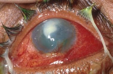
Amyloidosis is a medical condition characterized by the abnormal accumulation of insoluble proteins called amyloid in various tissues and organs throughout the body. These misfolded protein deposits can disrupt the normal function of affected organs, leading to a range of symptoms depending on the location and extent of the amyloid deposition.
There are different types of amyloidosis, classified based on the specific proteins involved:
1. Primary (AL) Amyloidosis: This is the most common form, accounting for around 80% of cases. It results from the overproduction and misfolding of immunoglobulin light chains, typically by clonal plasma cells in the bone marrow. The amyloid deposits can affect various organs, including the heart, kidneys, liver, and nervous system.
2. Secondary (AA) Amyloidosis: This form is associated with chronic inflammatory diseases, such as rheumatoid arthritis, tuberculosis, or familial Mediterranean fever. The amyloid fibrils are composed of serum amyloid A protein (SAA), an acute-phase reactant produced during the inflammatory response. The kidneys are commonly affected in this type of amyloidosis.
3. Hereditary or Familial Amyloidosis: These forms are caused by genetic mutations that result in the production of abnormal proteins prone to misfolding and amyloid formation. Examples include transthyretin (TTR) amyloidosis, fibrinogen amyloidosis, and apolipoprotein AI amyloidosis. These forms can affect various organs, including the heart, nerves, and kidneys.
4. Dialysis-Related Amyloidosis: This form is seen in patients undergoing long-term dialysis for chronic kidney disease. The amyloid fibrils are composed of beta-2 microglobulin, a protein that accumulates due to impaired clearance during dialysis. The joints and bones are commonly affected in this type of amyloidosis.
The diagnosis of amyloidosis typically involves a combination of clinical evaluation, imaging studies, and tissue biopsy with the demonstration of amyloid deposition using special stains (e.g., Congo red). Treatment depends on the specific type and extent of organ involvement and may include supportive care, medications to target the underlying cause (e.g., chemotherapy, immunomodulatory agents), and organ transplantation in some cases.
Hydroxyproline is not a medical term per se, but it is a significant component in the medical field, particularly in the study of connective tissues and collagen. Here's a scientific definition:
Hydroxyproline is a modified amino acid that is formed by the post-translational modification of the amino acid proline in collagen and some other proteins. This process involves the addition of a hydroxyl group (-OH) to the proline residue, which alters its chemical properties and contributes to the stability and structure of collagen fibers. Collagen is the most abundant protein in the human body and is a crucial component of connective tissues such as tendons, ligaments, skin, and bones. The presence and quantity of hydroxyproline can serve as a marker for collagen turnover and degradation, making it relevant to various medical and research contexts, including the study of diseases affecting connective tissues like osteoarthritis, rheumatoid arthritis, and Ehlers-Danlos syndrome.
Congo Red is a synthetic diazo dye that is commonly used in histology and pathology for stainings and tests. It is particularly useful in identifying amyloid deposits in tissues, which are associated with various diseases such as Alzheimer's disease, type 2 diabetes, and systemic amyloidosis.
When Congo Red binds to amyloid fibrils, it exhibits a characteristic apple-green birefringence under polarized light microscopy. Additionally, Congo Red stained amyloid deposits show a shift in their emission spectrum when excited with circularly polarized light, a phenomenon known as dichroism. These properties make Congo Red a valuable tool for the diagnosis and study of amyloidosis and other protein misfolding disorders.
It is important to note that Congo Red staining should be performed with care, as it can be toxic and carcinogenic if not handled properly.
Fibroblasts are specialized cells that play a critical role in the body's immune response and wound healing process. They are responsible for producing and maintaining the extracellular matrix (ECM), which is the non-cellular component present within all tissues and organs, providing structural support and biochemical signals for surrounding cells.
Fibroblasts produce various ECM proteins such as collagens, elastin, fibronectin, and laminins, forming a complex network of fibers that give tissues their strength and flexibility. They also help in the regulation of tissue homeostasis by controlling the turnover of ECM components through the process of remodeling.
In response to injury or infection, fibroblasts become activated and start to proliferate rapidly, migrating towards the site of damage. Here, they participate in the inflammatory response, releasing cytokines and chemokines that attract immune cells to the area. Additionally, they deposit new ECM components to help repair the damaged tissue and restore its functionality.
Dysregulation of fibroblast activity has been implicated in several pathological conditions, including fibrosis (excessive scarring), cancer (where they can contribute to tumor growth and progression), and autoimmune diseases (such as rheumatoid arthritis).
Collagen type XVIII is a type of collagen that is found in the basement membrane, which is a thin layer of extracellular matrix that separates and supports epithelial and endothelial cells. It is a heterotrimeric protein composed of three different chains, alpha1(XVIII), alpha2(XVIII), and alpha3(XVIII). Collagen XVIII is thought to play a role in the maintenance and organization of the basement membrane, as well as in cell adhesion and migration. It also contains a number of distinct domains that are involved in various biological processes, including angiogenesis, tissue repair, and tumor growth. Mutations in the gene that encodes collagen XVIII have been associated with eye diseases such as Knobloch syndrome and familial exudative vitreoretinopathy.
Atomic Force Microscopy (AFM) is a type of microscopy that allows visualization and measurement of surfaces at the atomic level. It works by using a sharp probe, called a tip, that is mounted on a flexible cantilever. The tip is brought very close to the surface of the sample and as the sample is scanned, the forces between the tip and the sample cause the cantilever to deflect. This deflection is measured and used to generate a topographic map of the surface with extremely high resolution, often on the order of fractions of a nanometer. AFM can be used to study both conductive and non-conductive samples, and can operate in various environments, including air and liquid. It has applications in fields such as materials science, biology, and chemistry.
Amyloid beta-peptides (Aβ) are small protein fragments that are crucially involved in the pathogenesis of Alzheimer's disease. They are derived from a larger transmembrane protein called the amyloid precursor protein (APP) through a series of proteolytic cleavage events.
The two primary forms of Aβ peptides are Aβ40 and Aβ42, which differ in length by two amino acids. While both forms can be harmful, Aβ42 is more prone to aggregation and is considered to be the more pathogenic form. These peptides have the tendency to misfold and accumulate into oligomers, fibrils, and eventually insoluble plaques that deposit in various areas of the brain, most notably the cerebral cortex and hippocampus.
The accumulation of Aβ peptides is believed to initiate a cascade of events leading to neuroinflammation, oxidative stress, synaptic dysfunction, and neuronal death, which are all hallmarks of Alzheimer's disease. Although the exact role of Aβ in the onset and progression of Alzheimer's is still under investigation, it is widely accepted that they play a central part in the development of this debilitating neurodegenerative disorder.
Transmission electron microscopy (TEM) is a type of microscopy in which an electron beam is transmitted through a ultra-thin specimen, interacting with it as it passes through. An image is formed from the interaction of the electrons with the specimen; the image is then magnified and visualized on a fluorescent screen or recorded on an electronic detector (or photographic film in older models).
TEM can provide high-resolution, high-magnification images that can reveal the internal structure of specimens including cells, viruses, and even molecules. It is widely used in biological and materials science research to investigate the ultrastructure of cells, tissues and materials. In medicine, TEM is used for diagnostic purposes in fields such as virology and bacteriology.
It's important to note that preparing a sample for TEM is a complex process, requiring specialized techniques to create thin (50-100 nm) specimens. These include cutting ultrathin sections of embedded samples using an ultramicrotome, staining with heavy metal salts, and positive staining or negative staining methods.
Cartilage is a type of connective tissue that is found throughout the body in various forms. It is made up of specialized cells called chondrocytes, which are embedded in a firm, flexible matrix composed of collagen fibers and proteoglycans. This unique structure gives cartilage its characteristic properties of being both strong and flexible.
There are three main types of cartilage in the human body: hyaline cartilage, elastic cartilage, and fibrocartilage.
1. Hyaline cartilage is the most common type and is found in areas such as the articular surfaces of bones (where they meet to form joints), the nose, trachea, and larynx. It has a smooth, glassy appearance and provides a smooth, lubricated surface for joint movement.
2. Elastic cartilage contains more elastin fibers than hyaline cartilage, which gives it greater flexibility and resilience. It is found in structures such as the external ear and parts of the larynx and epiglottis.
3. Fibrocartilage has a higher proportion of collagen fibers and fewer chondrocytes than hyaline or elastic cartilage. It is found in areas that require high tensile strength, such as the intervertebral discs, menisci (found in joints like the knee), and the pubic symphysis.
Cartilage plays a crucial role in supporting and protecting various structures within the body, allowing for smooth movement and providing a cushion between bones to absorb shock and prevent wear and tear. However, cartilage has limited capacity for self-repair and regeneration, making damage or degeneration of cartilage tissue a significant concern in conditions such as osteoarthritis.
Extracellular matrix (ECM) proteins are a group of structural and functional molecules that provide support, organization, and regulation to the cells in tissues and organs. The ECM is composed of a complex network of proteins, glycoproteins, and carbohydrates that are secreted by the cells and deposited outside of them.
ECM proteins can be classified into several categories based on their structure and function, including:
1. Collagens: These are the most abundant ECM proteins and provide strength and stability to tissues. They form fibrils that can withstand high tensile forces.
2. Proteoglycans: These are complex molecules made up of a core protein and one or more glycosaminoglycan (GAG) chains. The GAG chains attract water, making proteoglycans important for maintaining tissue hydration and resilience.
3. Elastin: This is an elastic protein that allows tissues to stretch and recoil, such as in the lungs and blood vessels.
4. Fibronectins: These are large glycoproteins that bind to cells and ECM components, providing adhesion, migration, and signaling functions.
5. Laminins: These are large proteins found in basement membranes, which provide structural support for epithelial and endothelial cells.
6. Tenascins: These are large glycoproteins that modulate cell adhesion and migration, and regulate ECM assembly and remodeling.
Together, these ECM proteins create a microenvironment that influences cell behavior, differentiation, and function. Dysregulation of ECM proteins has been implicated in various diseases, including fibrosis, cancer, and degenerative disorders.
Connective tissue is a type of biological tissue that provides support, strength, and protection to various structures in the body. It is composed of cells called fibroblasts, which produce extracellular matrix components such as collagen, elastin, and proteoglycans. These components give connective tissue its unique properties, including tensile strength, elasticity, and resistance to compression.
There are several types of connective tissue in the body, each with its own specific functions and characteristics. Some examples include:
1. Loose or Areolar Connective Tissue: This type of connective tissue is found throughout the body and provides cushioning and support to organs and other structures. It contains a large amount of ground substance, which allows for the movement and gliding of adjacent tissues.
2. Dense Connective Tissue: This type of connective tissue has a higher concentration of collagen fibers than loose connective tissue, making it stronger and less flexible. Dense connective tissue can be further divided into two categories: regular (or parallel) and irregular. Regular dense connective tissue, such as tendons and ligaments, has collagen fibers that run parallel to each other, providing great tensile strength. Irregular dense connective tissue, such as the dermis of the skin, has collagen fibers arranged in a more haphazard pattern, providing support and flexibility.
3. Adipose Tissue: This type of connective tissue is primarily composed of fat cells called adipocytes. Adipose tissue serves as an energy storage reservoir and provides insulation and cushioning to the body.
4. Cartilage: A firm, flexible type of connective tissue that contains chondrocytes within a matrix of collagen and proteoglycans. Cartilage is found in various parts of the body, including the joints, nose, ears, and trachea.
5. Bone: A specialized form of connective tissue that consists of an organic matrix (mainly collagen) and an inorganic mineral component (hydroxyapatite). Bone provides structural support to the body and serves as a reservoir for calcium and phosphate ions.
6. Blood: Although not traditionally considered connective tissue, blood does contain elements of connective tissue, such as plasma proteins and leukocytes (white blood cells). Blood transports nutrients, oxygen, hormones, and waste products throughout the body.
In medical terms, the skin is the largest organ of the human body. It consists of two main layers: the epidermis (outer layer) and dermis (inner layer), as well as accessory structures like hair follicles, sweat glands, and oil glands. The skin plays a crucial role in protecting us from external factors such as bacteria, viruses, and environmental hazards, while also regulating body temperature and enabling the sense of touch.
A peptide fragment is a short chain of amino acids that is derived from a larger peptide or protein through various biological or chemical processes. These fragments can result from the natural breakdown of proteins in the body during regular physiological processes, such as digestion, or they can be produced experimentally in a laboratory setting for research or therapeutic purposes.
Peptide fragments are often used in research to map the structure and function of larger peptides and proteins, as well as to study their interactions with other molecules. In some cases, peptide fragments may also have biological activity of their own and can be developed into drugs or diagnostic tools. For example, certain peptide fragments derived from hormones or neurotransmitters may bind to receptors in the body and mimic or block the effects of the full-length molecule.
The basement membrane is a thin, specialized layer of extracellular matrix that provides structural support and separates epithelial cells (which line the outer surfaces of organs and blood vessels) from connective tissue. It is composed of two main layers: the basal lamina, which is produced by the epithelial cells, and the reticular lamina, which is produced by the connective tissue. The basement membrane plays important roles in cell adhesion, migration, differentiation, and survival.
The basal lamina is composed mainly of type IV collagen, laminins, nidogens, and proteoglycans, while the reticular lamina contains type III collagen, fibronectin, and other matrix proteins. The basement membrane also contains a variety of growth factors and cytokines that can influence cell behavior.
Defects in the composition or organization of the basement membrane can lead to various diseases, including kidney disease, eye disease, and skin blistering disorders.
"Cells, cultured" is a medical term that refers to cells that have been removed from an organism and grown in controlled laboratory conditions outside of the body. This process is called cell culture and it allows scientists to study cells in a more controlled and accessible environment than they would have inside the body. Cultured cells can be derived from a variety of sources, including tissues, organs, or fluids from humans, animals, or cell lines that have been previously established in the laboratory.
Cell culture involves several steps, including isolation of the cells from the tissue, purification and characterization of the cells, and maintenance of the cells in appropriate growth conditions. The cells are typically grown in specialized media that contain nutrients, growth factors, and other components necessary for their survival and proliferation. Cultured cells can be used for a variety of purposes, including basic research, drug development and testing, and production of biological products such as vaccines and gene therapies.
It is important to note that cultured cells may behave differently than they do in the body, and results obtained from cell culture studies may not always translate directly to human physiology or disease. Therefore, it is essential to validate findings from cell culture experiments using additional models and ultimately in clinical trials involving human subjects.
Secondary protein structure refers to the local spatial arrangement of amino acid chains in a protein, typically described as regular repeating patterns held together by hydrogen bonds. The two most common types of secondary structures are the alpha-helix (α-helix) and the beta-pleated sheet (β-sheet). In an α-helix, the polypeptide chain twists around itself in a helical shape, with each backbone atom forming a hydrogen bond with the fourth amino acid residue along the chain. This forms a rigid rod-like structure that is resistant to bending or twisting forces. In β-sheets, adjacent segments of the polypeptide chain run parallel or antiparallel to each other and are connected by hydrogen bonds, forming a pleated sheet-like arrangement. These secondary structures provide the foundation for the formation of tertiary and quaternary protein structures, which determine the overall three-dimensional shape and function of the protein.
In medical terms, "gels" are semi-solid colloidal systems in which a solid phase is dispersed in a liquid medium. They have a viscous consistency and can be described as a cross between a solid and a liquid. The solid particles, called the gel network, absorb and swell with the liquid component, creating a system that has properties of both solids and liquids.
Gels are widely used in medical applications such as wound dressings, drug delivery systems, and tissue engineering due to their unique properties. They can provide a moist environment for wounds to heal, control the release of drugs over time, and mimic the mechanical properties of natural tissues.
Hydroxylysine is a modified form of the amino acid lysine, which is formed by the addition of a hydroxyl group (-OH) to the lysine molecule. This process is known as hydroxylation and is catalyzed by the enzyme lysyl hydroxylase.
In the human body, hydroxylysine is an important component of collagen, which is a protein that provides structure and strength to tissues such as skin, tendons, ligaments, and bones. Hydroxylysine helps to stabilize the triple-helix structure of collagen by forming cross-links between individual collagen molecules.
Abnormalities in hydroxylysine metabolism can lead to various connective tissue disorders, such as Ehlers-Danlos syndrome and osteogenesis imperfecta, which are characterized by joint hypermobility, skin fragility, and bone fractures.
Fibronectin is a high molecular weight glycoprotein that is found in many tissues and body fluids, including plasma, connective tissue, and the extracellular matrix. It is composed of two similar subunits that are held together by disulfide bonds. Fibronectin plays an important role in cell adhesion, migration, and differentiation by binding to various cell surface receptors, such as integrins, and other extracellular matrix components, such as collagen and heparan sulfate proteoglycans.
Fibronectin has several isoforms that are produced by alternative splicing of a single gene transcript. These isoforms differ in their biological activities and can be found in different tissues and developmental stages. Fibronectin is involved in various physiological processes, such as wound healing, tissue repair, and embryonic development, and has been implicated in several pathological conditions, including fibrosis, tumor metastasis, and thrombosis.
Collagen type VII is a type of collagen that is a major component of the anchoring fibrils, which are structures that help to attach the epidermis (the outermost layer of the skin) to the dermis (the layer of skin directly below the epidermis). Collagen type VII is composed of three identical chains that are encoded by the COL7A1 gene. Mutations in this gene can lead to a group of inherited blistering disorders known as autosomal recessive dystrophic epidermolysis bullosa, which is characterized by fragile skin and mucous membranes that blister and tear easily, often from minor trauma or friction.
An amino acid sequence is the specific order of amino acids in a protein or peptide molecule, formed by the linking of the amino group (-NH2) of one amino acid to the carboxyl group (-COOH) of another amino acid through a peptide bond. The sequence is determined by the genetic code and is unique to each type of protein or peptide. It plays a crucial role in determining the three-dimensional structure and function of proteins.
Proteoglycans are complex, highly negatively charged macromolecules that are composed of a core protein covalently linked to one or more glycosaminoglycan (GAG) chains. They are a major component of the extracellular matrix (ECM) and play crucial roles in various biological processes, including cell signaling, regulation of growth factor activity, and maintenance of tissue structure and function.
The GAG chains, which can vary in length and composition, are long, unbranched polysaccharides that are composed of repeating disaccharide units containing a hexuronic acid (either glucuronic or iduronic acid) and a hexosamine (either N-acetylglucosamine or N-acetylgalactosamine). These GAG chains can be sulfated to varying degrees, which contributes to the negative charge of proteoglycans.
Proteoglycans are classified into four major groups based on their core protein structure and GAG composition: heparan sulfate/heparin proteoglycans, chondroitin/dermatan sulfate proteoglycans, keratan sulfate proteoglycans, and hyaluronan-binding proteoglycans. Each group has distinct functions and is found in specific tissues and cell types.
In summary, proteoglycans are complex macromolecules composed of a core protein and one or more GAG chains that play important roles in the ECM and various biological processes, including cell signaling, growth factor regulation, and tissue structure maintenance.
Scanning transmission electron microscopy (STEM) is a type of electron microscopy that uses a focused beam of electrons to transmit through a specimen and create an image based on the interactions between the electrons and the sample. In STEM, the electron beam is scanned across the sample in a raster pattern, similar to how a television or computer monitor displays an image. As the electrons pass through the sample, they interact with the atoms in the material, causing scattering and energy loss. By detecting these scattered and energy-loss electrons, a high-resolution image of the sample can be created.
Scanning transmission electron microscopy is particularly useful for imaging thin specimens with high resolution, making it an important tool in materials science, biology, and other fields where detailed information about the structure and composition of materials is needed. The technique can provide information about the crystal structure, chemical composition, and electronic properties of materials at the atomic level.
Overall, scanning transmission electron microscopy is a powerful tool for characterizing materials and understanding their properties at the nanoscale and atomic level.
Scanning electron microscopy (SEM) is a type of electron microscopy that uses a focused beam of electrons to scan the surface of a sample and produce a high-resolution image. In SEM, a beam of electrons is scanned across the surface of a specimen, and secondary electrons are emitted from the sample due to interactions between the electrons and the atoms in the sample. These secondary electrons are then detected by a detector and used to create an image of the sample's surface topography. SEM can provide detailed images of the surface of a wide range of materials, including metals, polymers, ceramics, and biological samples. It is commonly used in materials science, biology, and electronics for the examination and analysis of surfaces at the micro- and nanoscale.
Microbial collagenase is not a medical term per se, but it does refer to an enzyme that is used in various medical and research contexts. Collagenases are a group of enzymes that break down collagen, a structural protein found in connective tissues such as skin, tendons, and ligaments. Microbial collagenase is a type of collagenase that is produced by certain bacteria, such as Clostridium histolyticum.
In medical terms, microbial collagenase is used in various therapeutic and research applications, including:
1. Wound healing: Microbial collagenase can be used to break down and remove necrotic tissue from wounds, which can help promote healing and prevent infection.
2. Dental applications: Collagenases have been used in periodontal therapy to remove calculus and improve the effectiveness of root planing and scaling procedures.
3. Research: Microbial collagenase is a valuable tool for researchers studying the structure and function of collagen and other extracellular matrix proteins. It can be used to digest tissue samples, allowing scientists to study the individual components of the extracellular matrix.
It's important to note that while microbial collagenase has many useful applications, it must be used with care, as excessive or improper use can damage healthy tissues and cause adverse effects.
"Cattle" is a term used in the agricultural and veterinary fields to refer to domesticated animals of the genus *Bos*, primarily *Bos taurus* (European cattle) and *Bos indicus* (Zebu). These animals are often raised for meat, milk, leather, and labor. They are also known as bovines or cows (for females), bulls (intact males), and steers/bullocks (castrated males). However, in a strict medical definition, "cattle" does not apply to humans or other animals.
Molecular sequence data refers to the specific arrangement of molecules, most commonly nucleotides in DNA or RNA, or amino acids in proteins, that make up a biological macromolecule. This data is generated through laboratory techniques such as sequencing, and provides information about the exact order of the constituent molecules. This data is crucial in various fields of biology, including genetics, evolution, and molecular biology, allowing for comparisons between different organisms, identification of genetic variations, and studies of gene function and regulation.
Collagen type X is a specific type of collagen that is primarily found in the hypertrophic zone of mature cartilage, which is located near the site of bone formation during endochondral ossification. It plays a crucial role in the mineralization process of the cartilage matrix and is essential for the formation of healthy bones. Collagen type X is composed of three identical alpha chains that form a triple helix structure, and it is synthesized by chondrocytes, which are the specialized cells found in cartilage tissue. Mutations in the gene that encodes collagen type X have been associated with certain skeletal disorders, such as Schmid metaphyseal chondrodysplasia.
Pepsin A is defined as a digestive enzyme that is primarily secreted by the chief cells in the stomach's fundic glands. It plays a crucial role in protein catabolism, helping to break down food proteins into smaller peptides during the digestive process. Pepsin A has an optimal pH range of 1.5-2.5 for its enzymatic activity and is activated from its inactive precursor, pepsinogen, upon exposure to acidic conditions in the stomach.
The corneal stroma, also known as the substantia propria, is the thickest layer of the cornea, which is the clear, dome-shaped surface at the front of the eye. The cornea plays a crucial role in focusing vision.
The corneal stroma makes up about 90% of the cornea's thickness and is composed of parallel bundles of collagen fibers that are arranged in regular, repeating patterns. These fibers give the cornea its strength and transparency. The corneal stroma also contains a small number of cells called keratocytes, which produce and maintain the collagen fibers.
Disorders that affect the corneal stroma can cause vision loss or other eye problems. For example, conditions such as keratoconus, in which the cornea becomes thin and bulges outward, can distort vision and make it difficult to see clearly. Other conditions, such as corneal scarring or infection, can also affect the corneal stroma and lead to vision loss or other eye problems.
Decorin is a small proteoglycan, a type of protein with a attached sugar chain, that is found in the extracellular matrix of connective tissues in the body. It is composed of a core protein and one or more glycosaminoglycan (GAG) chains, specifically dermatan sulfate. Decorin plays important roles in the organization and biomechanical properties of collagen fibrils, regulation of cell proliferation and migration, and modulation of growth factor activity. It has been studied for its potential role in various physiological and pathological processes, including wound healing, fibrosis, and cancer.
Microfibrils are tiny, thread-like structures that are found in the extracellular matrix (the material that surrounds and supports cells) of many types of biological tissues. They are made up of bundles of long, thin proteins called fibrillins, which are joined together by other proteins such as microfibril-associated glycoproteins (MAGPs).
Microfibrils play an important role in providing structural support and elasticity to tissues. They are particularly abundant in the connective tissue that surrounds blood vessels, where they help to regulate the diameter of the vessels and maintain blood pressure. Microfibrils are also found in the elastic fibers of the lungs, skin, and other tissues, where they contribute to the ability of these tissues to stretch and recoil.
In addition to their structural roles, microfibrils have been shown to play a role in regulating cell behavior and signaling. For example, they can bind to growth factors and other signaling molecules, helping to control the activity of these molecules and influence cellular processes such as proliferation, differentiation, and migration.
Abnormalities in microfibril structure or function have been linked to a number of diseases, including Marfan syndrome, Loeys-Dietz syndrome, and cutis laxa. These conditions are characterized by problems with connective tissue strength and elasticity, which can lead to a range of symptoms such as skeletal abnormalities, cardiovascular disease, and skin fragility.
Protein conformation refers to the specific three-dimensional shape that a protein molecule assumes due to the spatial arrangement of its constituent amino acid residues and their associated chemical groups. This complex structure is determined by several factors, including covalent bonds (disulfide bridges), hydrogen bonds, van der Waals forces, and ionic bonds, which help stabilize the protein's unique conformation.
Protein conformations can be broadly classified into two categories: primary, secondary, tertiary, and quaternary structures. The primary structure represents the linear sequence of amino acids in a polypeptide chain. The secondary structure arises from local interactions between adjacent amino acid residues, leading to the formation of recurring motifs such as α-helices and β-sheets. Tertiary structure refers to the overall three-dimensional folding pattern of a single polypeptide chain, while quaternary structure describes the spatial arrangement of multiple folded polypeptide chains (subunits) that interact to form a functional protein complex.
Understanding protein conformation is crucial for elucidating protein function, as the specific three-dimensional shape of a protein directly influences its ability to interact with other molecules, such as ligands, nucleic acids, or other proteins. Any alterations in protein conformation due to genetic mutations, environmental factors, or chemical modifications can lead to loss of function, misfolding, aggregation, and disease states like neurodegenerative disorders and cancer.
Prealbumin, also known as transthyretin, is a protein produced primarily in the liver and circulates in the blood. It plays a role in transporting thyroid hormones and vitamin A throughout the body. Prealbumin levels are often used as an indicator of nutritional status and liver function. Low prealbumin levels may suggest malnutrition or inflammation, while increased levels can be seen in certain conditions like hyperthyroidism. It is important to note that prealbumin levels should be interpreted in conjunction with other clinical findings and laboratory tests for a more accurate assessment of a patient's health status.
The cornea is the clear, dome-shaped surface at the front of the eye. It plays a crucial role in focusing vision. The cornea protects the eye from harmful particles and microorganisms, and it also serves as a barrier against UV light. Its transparency allows light to pass through and get focused onto the retina. The cornea does not contain blood vessels, so it relies on tears and the fluid inside the eye (aqueous humor) for nutrition and oxygen. Any damage or disease that affects its clarity and shape can significantly impact vision and potentially lead to blindness if left untreated.
Polarized light microscopy is a type of microscopy that uses polarized light to enhance contrast and reveal unique optical properties in specimens. In this technique, a polarizing filter is placed under the light source, which polarizes the light as it passes through. The specimen is then illuminated with this linearly polarized light. As the light travels through the specimen, its plane of polarization may be altered due to birefringence, a property of certain materials that causes the light to split into two separate rays with different refractive indices.
A second polarizing filter, called an analyzer, is placed in the light path between the objective and the eyepiece. The orientation of this filter can be adjusted to either allow or block the transmission of light through the microscope. When the polarizer and analyzer are aligned perpendicularly, no light will pass through if the specimen does not exhibit birefringence. However, if the specimen has birefringent properties, it will cause the plane of polarization to rotate, allowing some light to pass through the analyzer and create a contrasting image.
Polarized light microscopy is particularly useful for observing structures in minerals, crystals, and certain biological materials like collagen fibers, muscle proteins, and starch granules. It can also be used to study stress patterns in plastics and other synthetic materials.
Circular dichroism (CD) is a technique used in physics and chemistry to study the structure of molecules, particularly large biological molecules such as proteins and nucleic acids. It measures the difference in absorption of left-handed and right-handed circularly polarized light by a sample. This difference in absorption can provide information about the three-dimensional structure of the molecule, including its chirality or "handedness."
In more technical terms, CD is a form of spectroscopy that measures the differential absorption of left and right circularly polarized light as a function of wavelength. The CD signal is measured in units of millidegrees (mdeg) and can be positive or negative, depending on the type of chromophore and its orientation within the molecule.
CD spectra can provide valuable information about the secondary and tertiary structure of proteins, as well as the conformation of nucleic acids. For example, alpha-helical proteins typically exhibit a strong positive band near 190 nm and two negative bands at around 208 nm and 222 nm, while beta-sheet proteins show a strong positive band near 195 nm and two negative bands at around 217 nm and 175 nm.
CD spectroscopy is a powerful tool for studying the structural changes that occur in biological molecules under different conditions, such as temperature, pH, or the presence of ligands or other molecules. It can also be used to monitor the folding and unfolding of proteins, as well as the binding of drugs or other small molecules to their targets.
Protein multimerization refers to the process where multiple protein subunits assemble together to form a complex, repetitive structure called a multimer or oligomer. This can involve the association of identical or similar protein subunits through non-covalent interactions such as hydrogen bonding, ionic bonding, and van der Waals forces. The resulting multimeric structures can have various shapes, sizes, and functions, including enzymatic activity, transport, or structural support. Protein multimerization plays a crucial role in many biological processes and is often necessary for the proper functioning of proteins within cells.
Articular cartilage is the smooth, white tissue that covers the ends of bones where they come together to form joints. It provides a cushion between bones and allows for smooth movement by reducing friction. Articular cartilage also absorbs shock and distributes loads evenly across the joint, protecting the bones from damage. It is avascular, meaning it does not have its own blood supply, and relies on the surrounding synovial fluid for nutrients. Over time, articular cartilage can wear down or become damaged due to injury or disease, leading to conditions such as osteoarthritis.
Thiazoles are organic compounds that contain a heterocyclic ring consisting of a nitrogen atom and a sulfur atom, along with two carbon atoms and two hydrogen atoms. They have the chemical formula C3H4NS. Thiazoles are present in various natural and synthetic substances, including some vitamins, drugs, and dyes. In the context of medicine, thiazole derivatives have been developed as pharmaceuticals for their diverse biological activities, such as anti-inflammatory, antifungal, antibacterial, and antihypertensive properties. Some well-known examples include thiazide diuretics (e.g., hydrochlorothiazide) used to treat high blood pressure and edema, and the antidiabetic drug pioglitazone.
Peptides are short chains of amino acid residues linked by covalent bonds, known as peptide bonds. They are formed when two or more amino acids are joined together through a condensation reaction, which results in the elimination of a water molecule and the formation of an amide bond between the carboxyl group of one amino acid and the amino group of another.
Peptides can vary in length from two to about fifty amino acids, and they are often classified based on their size. For example, dipeptides contain two amino acids, tripeptides contain three, and so on. Oligopeptides typically contain up to ten amino acids, while polypeptides can contain dozens or even hundreds of amino acids.
Peptides play many important roles in the body, including serving as hormones, neurotransmitters, enzymes, and antibiotics. They are also used in medical research and therapeutic applications, such as drug delivery and tissue engineering.
Collagen type XII is a type of collagen that is found in the extracellular matrix of various tissues, including tendons, ligaments, and skin. It is a fibril-associated collagen that is closely associated with collagens type I and III. Collagen type XII has been shown to play a role in regulating the organization and diameter of collagen fibrils. Mutations in the gene for collagen type XII have been associated with certain types of muscular dystrophy and Bethlem myopathy, which are genetic disorders that affect muscle strength and tone. Additionally, it has been suggested to play a role in the development of osteoarthritis.
Collagenases are a group of enzymes that have the ability to break down collagen, which is a structural protein found in connective tissues such as tendons, ligaments, and skin. Collagen is an important component of the extracellular matrix, providing strength and support to tissues throughout the body.
Collagenases are produced by various organisms, including bacteria, animals, and humans. In humans, collagenases play a crucial role in normal tissue remodeling and repair processes, such as wound healing and bone resorption. However, excessive or uncontrolled activity of collagenases can contribute to the development of various diseases, including arthritis, periodontitis, and cancer metastasis.
Bacterial collagenases are often used in research and medical applications for their ability to digest collagen quickly and efficiently. For example, they may be used to study the structure and function of collagen or to isolate cells from tissues. However, the clinical use of bacterial collagenases is limited due to concerns about their potential to cause tissue damage and inflammation.
Overall, collagenases are important enzymes that play a critical role in maintaining the health and integrity of connective tissues throughout the body.
A chick embryo refers to the developing organism that arises from a fertilized chicken egg. It is often used as a model system in biological research, particularly during the stages of development when many of its organs and systems are forming and can be easily observed and manipulated. The study of chick embryos has contributed significantly to our understanding of various aspects of developmental biology, including gastrulation, neurulation, organogenesis, and pattern formation. Researchers may use various techniques to observe and manipulate the chick embryo, such as surgical alterations, cell labeling, and exposure to drugs or other agents.
Glycosaminoglycans (GAGs) are long, unbranched polysaccharides composed of repeating disaccharide units. They are a major component of the extracellular matrix and connective tissues in the body. GAGs are negatively charged due to the presence of sulfate and carboxyl groups, which allows them to attract positively charged ions and water molecules, contributing to their ability to retain moisture and maintain tissue hydration and elasticity.
GAGs can be categorized into four main groups: heparin/heparan sulfate, chondroitin sulfate/dermatan sulfate, keratan sulfate, and hyaluronic acid. These different types of GAGs have varying structures and functions in the body, including roles in cell signaling, inflammation, and protection against enzymatic degradation.
Heparin is a highly sulfated form of heparan sulfate that is found in mast cells and has anticoagulant properties. Chondroitin sulfate and dermatan sulfate are commonly found in cartilage and contribute to its resiliency and ability to withstand compressive forces. Keratan sulfate is found in corneas, cartilage, and bone, where it plays a role in maintaining the structure and function of these tissues. Hyaluronic acid is a large, nonsulfated GAG that is widely distributed throughout the body, including in synovial fluid, where it provides lubrication and shock absorption for joints.
Tensile strength is a material property that measures the maximum amount of tensile (pulling) stress that a material can withstand before failure, such as breaking or fracturing. It is usually measured in units of force per unit area, such as pounds per square inch (psi) or pascals (Pa). In the context of medical devices or biomaterials, tensile strength may be used to describe the mechanical properties of materials used in implants, surgical tools, or other medical equipment. High tensile strength is often desirable in these applications to ensure that the material can withstand the stresses and forces it will encounter during use.
Osteogenesis Imperfecta (OI), also known as brittle bone disease, is a group of genetic disorders that mainly affect the bones. It is characterized by bones that break easily, often from little or no apparent cause. This happens because the body produces an insufficient amount of collagen or poor quality collagen, which are crucial for the formation of healthy bones.
The severity of OI can vary greatly, even within the same family. Some people with OI have only a few fractures in their lifetime while others may have hundreds. Other symptoms can include blue or gray sclera (the white part of the eye), hearing loss, short stature, curved or bowed bones, loose joints, and a triangular face shape.
There are several types of OI, each caused by different genetic mutations. Most types of OI are inherited in an autosomal dominant pattern, meaning only one copy of the altered gene is needed to cause the condition. However, some types are inherited in an autosomal recessive pattern, which means that two copies of the altered gene must be present for the condition to occur.
There is no cure for OI, but treatment can help manage symptoms and prevent complications. Treatment may include medication to strengthen bones, physical therapy, bracing, and surgery.
Elastin is a protein that provides elasticity to tissues and organs, allowing them to resume their shape after stretching or contracting. It is a major component of the extracellular matrix in many tissues, including the skin, lungs, blood vessels, and ligaments. Elastin fibers can stretch up to 1.5 times their original length and then return to their original shape due to the unique properties of this protein. The elastin molecule is made up of cross-linked chains of the protein tropoelastin, which are produced by cells called fibroblasts and then assembled into larger elastin fibers by enzymes called lysyl oxidases. Elastin has a very long half-life, with some estimates suggesting that it can remain in the body for up to 70 years or more.
Non-fibrillar collagens are a type of collagen that do not form fibrous structures, unlike the more common fibrillar collagens. They are a group of structurally diverse collagens that play important roles in various biological processes such as cell adhesion, migration, and differentiation. Non-fibrillar collagens include types IV, VI, VIII, X, XII, XIV, XVI, XIX, XXI, and XXVIII. They are often found in basement membranes and other specialized extracellular matrix structures.
Type IV collagen is a major component of the basement membrane and forms a network-like structure that provides a scaffold for other matrix components. Type VI collagen has a beaded filament structure and is involved in the organization of the extracellular matrix. Type VIII collagen is found in the eyes and helps to maintain the structural integrity of the eye. Type X collagen is associated with cartilage development and bone formation. Type XII and XIV collagens are fibril-associated collagens that help to regulate the organization and diameter of fibrillar collagens. The other non-fibrillar collagens have various functions, including cell adhesion, migration, and differentiation.
Overall, non-fibrillar collagens are important structural components of the extracellular matrix and play critical roles in various biological processes.
Messenger RNA (mRNA) is a type of RNA (ribonucleic acid) that carries genetic information copied from DNA in the form of a series of three-base code "words," each of which specifies a particular amino acid. This information is used by the cell's machinery to construct proteins, a process known as translation. After being transcribed from DNA, mRNA travels out of the nucleus to the ribosomes in the cytoplasm where protein synthesis occurs. Once the protein has been synthesized, the mRNA may be degraded and recycled. Post-transcriptional modifications can also occur to mRNA, such as alternative splicing and addition of a 5' cap and a poly(A) tail, which can affect its stability, localization, and translation efficiency.
Laminin is a family of proteins that are an essential component of the basement membrane, which is a specialized type of extracellular matrix. Laminins are large trimeric molecules composed of three different chains: α, β, and γ. There are five different α chains, three different β chains, and three different γ chains that can combine to form at least 15 different laminin isoforms.
Laminins play a crucial role in maintaining the structure and integrity of basement membranes by interacting with other components of the extracellular matrix, such as collagen IV, and cell surface receptors, such as integrins. They are involved in various biological processes, including cell adhesion, differentiation, migration, and survival.
Laminin dysfunction has been implicated in several human diseases, including cancer, diabetic nephropathy, and muscular dystrophy.
Fibrosis is a pathological process characterized by the excessive accumulation and/or altered deposition of extracellular matrix components, particularly collagen, in various tissues and organs. This results in the formation of fibrous scar tissue that can impair organ function and structure. Fibrosis can occur as a result of chronic inflammation, tissue injury, or abnormal repair mechanisms, and it is a common feature of many diseases, including liver cirrhosis, lung fibrosis, heart failure, and kidney disease.
In medical terms, fibrosis is defined as:
"The process of producing scar tissue (consisting of collagen) in response to injury or chronic inflammation in normal connective tissue. This can lead to the thickening and stiffening of affected tissues and organs, impairing their function."
Protein binding, in the context of medical and biological sciences, refers to the interaction between a protein and another molecule (known as the ligand) that results in a stable complex. This process is often reversible and can be influenced by various factors such as pH, temperature, and concentration of the involved molecules.
In clinical chemistry, protein binding is particularly important when it comes to drugs, as many of them bind to proteins (especially albumin) in the bloodstream. The degree of protein binding can affect a drug's distribution, metabolism, and excretion, which in turn influence its therapeutic effectiveness and potential side effects.
Protein-bound drugs may be less available for interaction with their target tissues, as only the unbound or "free" fraction of the drug is active. Therefore, understanding protein binding can help optimize dosing regimens and minimize adverse reactions.
Beta-2 microglobulin (β2M) is a small protein that is a component of the major histocompatibility complex class I molecule, which plays a crucial role in the immune system. It is found on the surface of almost all nucleated cells in the body and is involved in presenting intracellular peptides to T-cells for immune surveillance.
β2M is produced at a relatively constant rate by cells throughout the body and is freely filtered by the glomeruli in the kidneys. Under normal circumstances, most of the filtrated β2M is reabsorbed and catabolized in the proximal tubules of the nephrons. However, when the glomerular filtration rate (GFR) is decreased, as in chronic kidney disease (CKD), the reabsorption capacity of the proximal tubules becomes overwhelmed, leading to increased levels of β2M in the blood and its subsequent appearance in the urine.
Elevated serum and urinary β2M levels have been associated with various clinical conditions, such as CKD, multiple myeloma, autoimmune disorders, and certain infectious diseases. Measuring β2M concentrations can provide valuable information for diagnostic, prognostic, and monitoring purposes in these contexts.
Protein denaturation is a process in which the native structure of a protein is altered, leading to loss of its biological activity. This can be caused by various factors such as changes in temperature, pH, or exposure to chemicals or radiation. The three-dimensional shape of a protein is crucial for its function, and denaturation causes the protein to lose this shape, resulting in impaired or complete loss of function. Denaturation is often irreversible and can lead to the aggregation of proteins, which can have negative effects on cellular function and can contribute to diseases such as Alzheimer's and Parkinson's.
Alpha-synuclein is a protein that is primarily found in neurons (nerve cells) in the brain. It is encoded by the SNCA gene and is abundantly expressed in presynaptic terminals, where it is believed to play a role in the regulation of neurotransmitter release.
In certain neurological disorders, including Parkinson's disease, dementia with Lewy bodies, and multiple system atrophy, alpha-synuclein can form aggregates known as Lewy bodies and Lewy neurites. These aggregates are a pathological hallmark of these diseases and are believed to contribute to the death of nerve cells, leading to the symptoms associated with these disorders.
The precise function of alpha-synuclein is not fully understood, but it is thought to be involved in various cellular processes such as maintaining the structure of the presynaptic terminal, regulating synaptic vesicle trafficking and neurotransmitter release, and protecting neurons from stress.
Islet Amyloid Polypeptide (IAPP), also known as amylin, is a 37-amino acid peptide co-secreted with insulin from pancreatic beta-cells in response to meals. It plays crucial roles in regulating glucose homeostasis by suppressing glucagon secretion, slowing gastric emptying, and promoting satiety. In type 2 diabetes, IAPP can form amyloid fibrils, which deposit in pancreatic islets, contributing to beta-cell dysfunction and death. This contributes to the progressive nature of type 2 diabetes.
Molecular models are three-dimensional representations of molecular structures that are used in the field of molecular biology and chemistry to visualize and understand the spatial arrangement of atoms and bonds within a molecule. These models can be physical or computer-generated and allow researchers to study the shape, size, and behavior of molecules, which is crucial for understanding their function and interactions with other molecules.
Physical molecular models are often made up of balls (representing atoms) connected by rods or sticks (representing bonds). These models can be constructed manually using materials such as plastic or wooden balls and rods, or they can be created using 3D printing technology.
Computer-generated molecular models, on the other hand, are created using specialized software that allows researchers to visualize and manipulate molecular structures in three dimensions. These models can be used to simulate molecular interactions, predict molecular behavior, and design new drugs or chemicals with specific properties. Overall, molecular models play a critical role in advancing our understanding of molecular structures and their functions.
Proline is an organic compound that is classified as a non-essential amino acid, meaning it can be produced by the human body and does not need to be obtained through the diet. It is encoded in the genetic code as the codon CCU, CCC, CCA, or CCG. Proline is a cyclic amino acid, containing an unusual secondary amine group, which forms a ring structure with its carboxyl group.
In proteins, proline acts as a structural helix breaker, disrupting the alpha-helix structure and leading to the formation of turns and bends in the protein chain. This property is important for the proper folding and function of many proteins. Proline also plays a role in the stability of collagen, a major structural protein found in connective tissues such as tendons, ligaments, and skin.
In addition to its role in protein structure, proline has been implicated in various cellular processes, including signal transduction, apoptosis, and oxidative stress response. It is also a precursor for the synthesis of other biologically important compounds such as hydroxyproline, which is found in collagen and elastin, and glutamate, an excitatory neurotransmitter in the brain.
Fourier Transform Infrared (FTIR) spectroscopy is a type of infrared spectroscopy that uses the Fourier transform mathematical technique to convert the raw data obtained from an interferometer into a more interpretable spectrum. This technique allows for the simultaneous collection of a wide range of wavelengths, resulting in increased sensitivity and speed compared to traditional dispersive infrared spectroscopy.
FTIR spectroscopy measures the absorption or transmission of infrared radiation by a sample as a function of frequency, providing information about the vibrational modes of the molecules present in the sample. This can be used for identification and quantification of chemical compounds, analysis of molecular structure, and investigation of chemical interactions and reactions.
In summary, FTIR spectroscopy is a powerful analytical technique that uses infrared radiation to study the vibrational properties of molecules, with increased sensitivity and speed due to the use of Fourier transform mathematical techniques and an interferometer.
Prions are misfolded proteins that can induce other normal proteins to also adopt the misfolded shape, leading to the formation of aggregates. These abnormal prion protein aggregates are associated with a group of progressive neurodegenerative diseases known as transmissible spongiform encephalopathies (TSEs). Examples of TSEs include bovine spongiform encephalopathy (BSE or "mad cow disease") in cattle, variant Creutzfeldt-Jakob disease (vCJD) in humans, and scrapie in sheep. The misfolded prion proteins are resistant to degradation by proteases, which contributes to their accumulation and subsequent neuronal damage, ultimately resulting in spongiform degeneration of the brain and other neurological symptoms associated with TSEs.
Aminopropionitrile is a chemical compound with the formula NPN(H2)CH2CH2CN. It is an irritant that can cause damage to the eyes, skin, and respiratory system. It is used in the manufacture of certain plastics and resins, and has also been studied for its potential effects on the human body. Some research suggests that aminopropionitrile may interfere with the normal functioning of collagen, a protein that helps to provide structure and support to tissues and organs in the body. This has led to interest in the use of aminopropionitrile as a potential treatment for certain conditions related to collagen, such as scleroderma. However, more research is needed to determine the safety and effectiveness of this use.
Collagen type VIII is a less common type of collagen that is found in the eyes, specifically in the basement membrane of the cornea and the blood vessels of the eye. It is a network-forming collagen and is believed to play a role in maintaining the structural integrity and stability of these tissues. Mutations in the genes encoding for collagen type VIII have been associated with certain eye disorders, such as Fuchs' endothelial corneal dystrophy.
Here is a medical definition from the US National Library of Medicine:
"Collagen, type VIII, alpha-1 (COL8A1) is a gene that provides instructions for making one component of a type VIII collagen protein called collagen VIII alpha-1 chain. Collagen proteins are important building blocks for many tissues in the body, including tendons, ligaments, and the cornea, which is the clear outer covering of the eye.
Collagen VIII is found in the basement membrane, a thin layer of protein that surrounds many types of cells and helps to anchor them to surrounding tissue. In the eye, collagen VIII is produced by cells called endothelial cells, which line the inside surface of the cornea. Collagen VIII forms networks with other proteins that help maintain the structural integrity and stability of the cornea.
Mutations in the COL8A1 gene can cause Fuchs' endothelial corneal dystrophy, a progressive eye disorder characterized by the gradual clouding of the cornea." ()
Cell adhesion refers to the binding of cells to extracellular matrices or to other cells, a process that is fundamental to the development, function, and maintenance of multicellular organisms. Cell adhesion is mediated by various cell surface receptors, such as integrins, cadherins, and immunoglobulin-like cell adhesion molecules (Ig-CAMs), which interact with specific ligands in the extracellular environment. These interactions lead to the formation of specialized junctions, such as tight junctions, adherens junctions, and desmosomes, that help to maintain tissue architecture and regulate various cellular processes, including proliferation, differentiation, migration, and survival. Disruptions in cell adhesion can contribute to a variety of diseases, including cancer, inflammation, and degenerative disorders.
In the context of medicine and pharmacology, "kinetics" refers to the study of how a drug moves throughout the body, including its absorption, distribution, metabolism, and excretion (often abbreviated as ADME). This field is called "pharmacokinetics."
1. Absorption: This is the process of a drug moving from its site of administration into the bloodstream. Factors such as the route of administration (e.g., oral, intravenous, etc.), formulation, and individual physiological differences can affect absorption.
2. Distribution: Once a drug is in the bloodstream, it gets distributed throughout the body to various tissues and organs. This process is influenced by factors like blood flow, protein binding, and lipid solubility of the drug.
3. Metabolism: Drugs are often chemically modified in the body, typically in the liver, through processes known as metabolism. These changes can lead to the formation of active or inactive metabolites, which may then be further distributed, excreted, or undergo additional metabolic transformations.
4. Excretion: This is the process by which drugs and their metabolites are eliminated from the body, primarily through the kidneys (urine) and the liver (bile).
Understanding the kinetics of a drug is crucial for determining its optimal dosing regimen, potential interactions with other medications or foods, and any necessary adjustments for special populations like pediatric or geriatric patients, or those with impaired renal or hepatic function.
Procollagen-Lysine, 2-Oxoglutarate 5-Dioxygenase is an enzyme that plays a crucial role in the biosynthesis of collagen. The medical definition of this enzyme is:
"An enzyme that catalyzes the post-translational modification of specific lysine residues in procollagens and related proteins. This enzyme requires Fe2+, 2-oxoglutarate, molecular oxygen, and ascorbic acid as cofactors. It hydroxylates certain lysine residues to form hydroxylysine, which is essential for the stabilization of collagen triple helices and for the formation of covalent cross-links between individual collagen molecules. Mutations in this gene have been associated with several types of Ehlers-Danlos syndrome."
The systematic name for this enzyme is "procollagen-lysine, 2-oxoglutarate 5-dioxygenase (hydroxylating)." It is also known as "procollagen-lysine, lysine hydroxylase," or simply "LH." This enzyme is responsible for the hydroxylation of specific lysine residues in procollagens and related proteins during their biosynthesis. The hydroxylation reaction catalyzed by this enzyme involves the incorporation of a hydroxyl group (-OH) into the lysine side chain, resulting in the formation of hydroxylysine. This modification is essential for the proper folding and stabilization of collagen molecules, as well as for their subsequent cross-linking and assembly into extracellular matrix structures.
Defects or mutations in the gene encoding this enzyme can lead to various types of Ehlers-Danlos syndrome (EDS), a group of heritable connective tissue disorders characterized by joint hypermobility, skin hyperextensibility, and tissue fragility.
Mechanical stress, in the context of physiology and medicine, refers to any type of force that is applied to body tissues or organs, which can cause deformation or displacement of those structures. Mechanical stress can be either external, such as forces exerted on the body during physical activity or trauma, or internal, such as the pressure changes that occur within blood vessels or other hollow organs.
Mechanical stress can have a variety of effects on the body, depending on the type, duration, and magnitude of the force applied. For example, prolonged exposure to mechanical stress can lead to tissue damage, inflammation, and chronic pain. Additionally, abnormal or excessive mechanical stress can contribute to the development of various musculoskeletal disorders, such as tendinitis, osteoarthritis, and herniated discs.
In order to mitigate the negative effects of mechanical stress, the body has a number of adaptive responses that help to distribute forces more evenly across tissues and maintain structural integrity. These responses include changes in muscle tone, joint positioning, and connective tissue stiffness, as well as the remodeling of bone and other tissues over time. However, when these adaptive mechanisms are overwhelmed or impaired, mechanical stress can become a significant factor in the development of various pathological conditions.
"Bone" is the hard, dense connective tissue that makes up the skeleton of vertebrate animals. It provides support and protection for the body's internal organs, and serves as a attachment site for muscles, tendons, and ligaments. Bone is composed of cells called osteoblasts and osteoclasts, which are responsible for bone formation and resorption, respectively, and an extracellular matrix made up of collagen fibers and mineral crystals.
Bones can be classified into two main types: compact bone and spongy bone. Compact bone is dense and hard, and makes up the outer layer of all bones and the shafts of long bones. Spongy bone is less dense and contains large spaces, and makes up the ends of long bones and the interior of flat and irregular bones.
The human body has 206 bones in total. They can be further classified into five categories based on their shape: long bones, short bones, flat bones, irregular bones, and sesamoid bones.
Protein folding is the process by which a protein molecule naturally folds into its three-dimensional structure, following the synthesis of its amino acid chain. This complex process is determined by the sequence and properties of the amino acids, as well as various environmental factors such as temperature, pH, and the presence of molecular chaperones. The final folded conformation of a protein is crucial for its proper function, as it enables the formation of specific interactions between different parts of the molecule, which in turn define its biological activity. Protein misfolding can lead to various diseases, including neurodegenerative disorders such as Alzheimer's and Parkinson's disease.
X-ray diffraction (XRD) is not strictly a medical definition, but it is a technique commonly used in the field of medical research and diagnostics. XRD is a form of analytical spectroscopy that uses the phenomenon of X-ray diffraction to investigate the crystallographic structure of materials. When a beam of X-rays strikes a crystal, it is scattered in specific directions and with specific intensities that are determined by the arrangement of atoms within the crystal. By measuring these diffraction patterns, researchers can determine the crystal structures of various materials, including biological macromolecules such as proteins and viruses.
In the medical field, XRD is often used to study the structure of drugs and drug candidates, as well as to analyze the composition and structure of tissues and other biological samples. For example, XRD can be used to investigate the crystal structures of calcium phosphate minerals in bone tissue, which can provide insights into the mechanisms of bone formation and disease. Additionally, XRD is sometimes used in the development of new medical imaging techniques, such as phase-contrast X-ray imaging, which has the potential to improve the resolution and contrast of traditional X-ray images.
In the field of medicine, "time factors" refer to the duration of symptoms or time elapsed since the onset of a medical condition, which can have significant implications for diagnosis and treatment. Understanding time factors is crucial in determining the progression of a disease, evaluating the effectiveness of treatments, and making critical decisions regarding patient care.
For example, in stroke management, "time is brain," meaning that rapid intervention within a specific time frame (usually within 4.5 hours) is essential to administering tissue plasminogen activator (tPA), a clot-busting drug that can minimize brain damage and improve patient outcomes. Similarly, in trauma care, the "golden hour" concept emphasizes the importance of providing definitive care within the first 60 minutes after injury to increase survival rates and reduce morbidity.
Time factors also play a role in monitoring the progression of chronic conditions like diabetes or heart disease, where regular follow-ups and assessments help determine appropriate treatment adjustments and prevent complications. In infectious diseases, time factors are crucial for initiating antibiotic therapy and identifying potential outbreaks to control their spread.
Overall, "time factors" encompass the significance of recognizing and acting promptly in various medical scenarios to optimize patient outcomes and provide effective care.
Wound healing is a complex and dynamic process that occurs after tissue injury, aiming to restore the integrity and functionality of the damaged tissue. It involves a series of overlapping phases: hemostasis, inflammation, proliferation, and remodeling.
1. Hemostasis: This initial phase begins immediately after injury and involves the activation of the coagulation cascade to form a clot, which stabilizes the wound and prevents excessive blood loss.
2. Inflammation: Activated inflammatory cells, such as neutrophils and monocytes/macrophages, infiltrate the wound site to eliminate pathogens, remove debris, and release growth factors that promote healing. This phase typically lasts for 2-5 days post-injury.
3. Proliferation: In this phase, various cell types, including fibroblasts, endothelial cells, and keratinocytes, proliferate and migrate to the wound site to synthesize extracellular matrix (ECM) components, form new blood vessels (angiogenesis), and re-epithelialize the wounded area. This phase can last up to several weeks depending on the size and severity of the wound.
4. Remodeling: The final phase of wound healing involves the maturation and realignment of collagen fibers, leading to the restoration of tensile strength in the healed tissue. This process can continue for months to years after injury, although the tissue may never fully regain its original structure and function.
It is important to note that wound healing can be compromised by several factors, including age, nutrition, comorbidities (e.g., diabetes, vascular disease), and infection, which can result in delayed healing or non-healing chronic wounds.
Electrophoresis, polyacrylamide gel (EPG) is a laboratory technique used to separate and analyze complex mixtures of proteins or nucleic acids (DNA or RNA) based on their size and electrical charge. This technique utilizes a matrix made of cross-linked polyacrylamide, a type of gel, which provides a stable and uniform environment for the separation of molecules.
In this process:
1. The polyacrylamide gel is prepared by mixing acrylamide monomers with a cross-linking agent (bis-acrylamide) and a catalyst (ammonium persulfate) in the presence of a buffer solution.
2. The gel is then poured into a mold and allowed to polymerize, forming a solid matrix with uniform pore sizes that depend on the concentration of acrylamide used. Higher concentrations result in smaller pores, providing better resolution for separating smaller molecules.
3. Once the gel has set, it is placed in an electrophoresis apparatus containing a buffer solution. Samples containing the mixture of proteins or nucleic acids are loaded into wells on the top of the gel.
4. An electric field is applied across the gel, causing the negatively charged molecules to migrate towards the positive electrode (anode) while positively charged molecules move toward the negative electrode (cathode). The rate of migration depends on the size, charge, and shape of the molecules.
5. Smaller molecules move faster through the gel matrix and will migrate farther from the origin compared to larger molecules, resulting in separation based on size. Proteins and nucleic acids can be selectively stained after electrophoresis to visualize the separated bands.
EPG is widely used in various research fields, including molecular biology, genetics, proteomics, and forensic science, for applications such as protein characterization, DNA fragment analysis, cloning, mutation detection, and quality control of nucleic acid or protein samples.
The dermis is the layer of skin located beneath the epidermis, which is the outermost layer of the skin. It is composed of connective tissue and provides structure and support to the skin. The dermis contains blood vessels, nerves, hair follicles, sweat glands, and oil glands. It is also responsible for the production of collagen and elastin, which give the skin its strength and flexibility. The dermis can be further divided into two layers: the papillary dermis, which is the upper layer and contains finger-like projections called papillae that extend upwards into the epidermis, and the reticular dermis, which is the lower layer and contains thicker collagen bundles. Together, the epidermis and dermis make up the true skin.
Chondrocytes are the specialized cells that produce and maintain the extracellular matrix of cartilage tissue. They are responsible for synthesizing and secreting the collagen fibers, proteoglycans, and other components that give cartilage its unique properties, such as elasticity, resiliency, and resistance to compression. Chondrocytes are located within lacunae, or small cavities, in the cartilage matrix, and they receive nutrients and oxygen through diffusion from the surrounding tissue fluid. They are capable of adapting to changes in mechanical stress by modulating the production and organization of the extracellular matrix, which allows cartilage to withstand various loads and maintain its structural integrity. Chondrocytes play a crucial role in the development, maintenance, and repair of cartilaginous tissues throughout the body, including articular cartilage, costal cartilage, and growth plate cartilage.
Integrin α1β1, also known as Very Late Antigen-1 (VLA-1) or CD49a/CD29, is a heterodimeric transmembrane receptor protein composed of α1 and β1 subunits. It belongs to the integrin family of adhesion molecules that play crucial roles in cell-cell and cell-extracellular matrix (ECM) interactions.
Integrin α1β1 is primarily expressed on various cell types, including fibroblasts, endothelial cells, smooth muscle cells, and some immune cells. This integrin binds to several ECM proteins, such as collagens (type I, II, III, IV), laminin, and fibronectin, mediating cell adhesion, migration, proliferation, differentiation, and survival. Additionally, α1β1 integrin has been implicated in various physiological and pathological processes, such as tissue repair, fibrosis, and tumor progression.
Biomechanics is the application of mechanical laws to living structures and systems, particularly in the field of medicine and healthcare. A biomechanical phenomenon refers to a observable event or occurrence that involves the interaction of biological tissues or systems with mechanical forces. These phenomena can be studied at various levels, from the molecular and cellular level to the tissue, organ, and whole-body level.
Examples of biomechanical phenomena include:
1. The way that bones and muscles work together to produce movement (known as joint kinematics).
2. The mechanical behavior of biological tissues such as bone, cartilage, tendons, and ligaments under various loads and stresses.
3. The response of cells and tissues to mechanical stimuli, such as the way that bone tissue adapts to changes in loading conditions (known as Wolff's law).
4. The biomechanics of injury and disease processes, such as the mechanisms of joint injury or the development of osteoarthritis.
5. The use of mechanical devices and interventions to treat medical conditions, such as orthopedic implants or assistive devices for mobility impairments.
Understanding biomechanical phenomena is essential for developing effective treatments and prevention strategies for a wide range of medical conditions, from musculoskeletal injuries to neurological disorders.
Quaternary protein structure refers to the arrangement and interaction of multiple folded protein molecules in a multi-subunit complex. These subunits can be identical or different forms of the same protein or distinctly different proteins that associate to form a functional complex. The quaternary structure is held together by non-covalent interactions, such as hydrogen bonds, ionic bonds, and van der Waals forces. Understanding quaternary structure is crucial for comprehending the function, regulation, and assembly of many protein complexes involved in various cellular processes.
Immunohistochemistry (IHC) is a technique used in pathology and laboratory medicine to identify specific proteins or antigens in tissue sections. It combines the principles of immunology and histology to detect the presence and location of these target molecules within cells and tissues. This technique utilizes antibodies that are specific to the protein or antigen of interest, which are then tagged with a detection system such as a chromogen or fluorophore. The stained tissue sections can be examined under a microscope, allowing for the visualization and analysis of the distribution and expression patterns of the target molecule in the context of the tissue architecture. Immunohistochemistry is widely used in diagnostic pathology to help identify various diseases, including cancer, infectious diseases, and immune-mediated disorders.
Protein-Lysine 6-Oxidase (PLOX) is an enzyme that belongs to the family of copper-containing oxidases. It catalyzes the oxidative deamination of specific lysine residues in proteins, resulting in the formation of lysine-6-aldehydes, ammonia, and hydrogen peroxide. This enzyme plays a crucial role in various biological processes, including the regulation of protein function, modification of extracellular matrices, and the maintenance of copper homeostasis. Mutations in the gene encoding PLOX have been associated with certain diseases, such as Menkes disease, a rare X-linked recessive disorder characterized by copper deficiency and neurological symptoms.
Transforming Growth Factor-beta (TGF-β) is a type of cytokine, which is a cell signaling protein involved in the regulation of various cellular processes, including cell growth, differentiation, and apoptosis (programmed cell death). TGF-β plays a critical role in embryonic development, tissue homeostasis, and wound healing. It also has been implicated in several pathological conditions such as fibrosis, cancer, and autoimmune diseases.
TGF-β exists in multiple isoforms (TGF-β1, TGF-β2, and TGF-β3) that are produced by many different cell types, including immune cells, epithelial cells, and fibroblasts. The protein is synthesized as a precursor molecule, which is cleaved to release the active TGF-β peptide. Once activated, TGF-β binds to its receptors on the cell surface, leading to the activation of intracellular signaling pathways that regulate gene expression and cell behavior.
In summary, Transforming Growth Factor-beta (TGF-β) is a multifunctional cytokine involved in various cellular processes, including cell growth, differentiation, apoptosis, embryonic development, tissue homeostasis, and wound healing. It has been implicated in several pathological conditions such as fibrosis, cancer, and autoimmune diseases.
Recombinant proteins are artificially created proteins produced through the use of recombinant DNA technology. This process involves combining DNA molecules from different sources to create a new set of genes that encode for a specific protein. The resulting recombinant protein can then be expressed, purified, and used for various applications in research, medicine, and industry.
Recombinant proteins are widely used in biomedical research to study protein function, structure, and interactions. They are also used in the development of diagnostic tests, vaccines, and therapeutic drugs. For example, recombinant insulin is a common treatment for diabetes, while recombinant human growth hormone is used to treat growth disorders.
The production of recombinant proteins typically involves the use of host cells, such as bacteria, yeast, or mammalian cells, which are engineered to express the desired protein. The host cells are transformed with a plasmid vector containing the gene of interest, along with regulatory elements that control its expression. Once the host cells are cultured and the protein is expressed, it can be purified using various chromatography techniques.
Overall, recombinant proteins have revolutionized many areas of biology and medicine, enabling researchers to study and manipulate proteins in ways that were previously impossible.
Medical Definition of Matrix Metalloproteinase 1 (MMP-1):
Matrix metalloproteinase 1, also known as collagenase-1 or fibroblast collagenase, is a member of the matrix metalloproteinase family of enzymes. These enzymes are involved in degrading and remodeling extracellular matrix components, such as collagens, gelatins, and other proteins. MMP-1 specifically targets interstitial collagens (types I, II, III, VII, and X) and plays a crucial role in tissue repair, wound healing, and pathological processes like tumor invasion and metastasis. It is secreted as an inactive proenzyme and requires activation before it can carry out its proteolytic functions. MMP-1 activity is regulated at various levels, including transcription, activation, and inhibition by endogenous tissue inhibitors of metalloproteinases (TIMPs). Dysregulation of MMP-1 has been implicated in several diseases, such as arthritis, cancer, and fibrosis.
HSP47 (Heat Shock Protein 47) is a type of molecular chaperone that assists in the proper folding and assembly of collagen molecules within the endoplasmic reticulum (ER) of eukaryotic cells. It is also known as SERPINH1, which stands for serine protease inhibitor, clade H (heat shock protein 47).
HSP47 binds to procollagen molecules in a highly specific manner and helps facilitate their correct folding and assembly into higher-order structures. Once the collagen molecules are properly assembled, HSP47 dissociates from them and allows for their transport out of the ER and further processing in the Golgi apparatus.
HSP47 is upregulated under conditions of cellular stress, such as heat shock or oxidative stress, which can lead to an accumulation of misfolded proteins within the ER. This upregulation helps to enhance the protein folding capacity of the ER and prevent the aggregation of misfolded proteins, thereby maintaining cellular homeostasis.
Defects in HSP47 function have been implicated in various connective tissue disorders, such as osteogenesis imperfecta and Ehlers-Danlos syndrome, which are characterized by abnormal collagen structure and function.
Tertiary protein structure refers to the three-dimensional arrangement of all the elements (polypeptide chains) of a single protein molecule. It is the highest level of structural organization and results from interactions between various side chains (R groups) of the amino acids that make up the protein. These interactions, which include hydrogen bonds, ionic bonds, van der Waals forces, and disulfide bridges, give the protein its unique shape and stability, which in turn determines its function. The tertiary structure of a protein can be stabilized by various factors such as temperature, pH, and the presence of certain ions. Any changes in these factors can lead to denaturation, where the protein loses its tertiary structure and thus its function.
Gelatin is not strictly a medical term, but it is often used in medical contexts. Medically, gelatin is recognized as a protein-rich substance that is derived from collagen, which is found in the skin, bones, and connective tissue of animals. It is commonly used in the production of various medical and pharmaceutical products such as capsules, wound dressings, and drug delivery systems due to its biocompatibility and ability to form gels.
In a broader sense, gelatin is a translucent, colorless, flavorless food ingredient that is derived from collagen through a process called hydrolysis. It is widely used in the food industry as a gelling agent, thickener, stabilizer, and texturizer in various foods such as candies, desserts, marshmallows, and yogurts.
It's worth noting that while gelatin has many uses, it may not be suitable for vegetarians or those with dietary restrictions since it is derived from animal products.
Integrins are a type of cell-adhesion molecule that play a crucial role in cell-cell and cell-extracellular matrix (ECM) interactions. They are heterodimeric transmembrane receptors composed of non-covalently associated α and β subunits, which form more than 24 distinct integrin heterodimers in humans.
Integrins bind to specific ligands, such as ECM proteins (e.g., collagen, fibronectin, laminin), cell surface molecules, and soluble factors, through their extracellular domains. The intracellular domains of integrins interact with the cytoskeleton and various signaling proteins, allowing them to transduce signals from the ECM into the cell (outside-in signaling) and vice versa (inside-out signaling).
These molecular interactions are essential for numerous biological processes, including cell adhesion, migration, proliferation, differentiation, survival, and angiogenesis. Dysregulation of integrin function has been implicated in various pathological conditions, such as cancer, fibrosis, inflammation, and autoimmune diseases.
The Descemet membrane is the thin, transparent basement membrane that is produced by the corneal endothelial cells. It is located between the corneal stroma and the corneal endothelium, which is the innermost layer of the cornea. The Descemet membrane provides structural support for the corneal endothelium and helps to maintain the proper hydration and clarity of the cornea. It is named after the French physician Jean Descemet, who first described it in 1752.
Solubility is a fundamental concept in pharmaceutical sciences and medicine, which refers to the maximum amount of a substance (solute) that can be dissolved in a given quantity of solvent (usually water) at a specific temperature and pressure. Solubility is typically expressed as mass of solute per volume or mass of solvent (e.g., grams per liter, milligrams per milliliter). The process of dissolving a solute in a solvent results in a homogeneous solution where the solute particles are dispersed uniformly throughout the solvent.
Understanding the solubility of drugs is crucial for their formulation, administration, and therapeutic effectiveness. Drugs with low solubility may not dissolve sufficiently to produce the desired pharmacological effect, while those with high solubility might lead to rapid absorption and short duration of action. Therefore, optimizing drug solubility through various techniques like particle size reduction, salt formation, or solubilization is an essential aspect of drug development and delivery.
Collagen type XIII, also known as alpha-1(XIII) collagen or COL13A1, is a type of collagen that is found in the extracellular matrix of various tissues, including skin, blood vessels, and the eye. It is a homotrimeric protein composed of three identical alpha-1(XIII) chains.
Collagen type XIII has a unique structure, with a short triple-helical domain and a large non-collagenous domain that contains several functional domains, including a von Willebrand factor A (vWA) domain, a thrombospondin type 1 (TSR) domain, and a C-terminal domain.
Collagen type XIII is involved in various biological processes, such as cell adhesion, migration, and differentiation. It has been shown to interact with other extracellular matrix proteins, such as collagens IV and VII, laminin-5, and fibronectin, as well as with integrins and growth factors.
Mutations in the COL13A1 gene have been associated with various human diseases, including dystrophic epidermolysis bullosa, a group of inherited skin fragility disorders characterized by blistering and scarring of the skin and mucous membranes.
Tissue engineering is a branch of biomedical engineering that combines the principles of engineering, materials science, and biological sciences to develop functional substitutes for damaged or diseased tissues and organs. It involves the creation of living, three-dimensional structures that can restore, maintain, or improve tissue function. This is typically accomplished through the use of cells, scaffolds (biodegradable matrices), and biologically active molecules. The goal of tissue engineering is to develop biological substitutes that can ultimately restore normal function and structure in damaged tissues or organs.
The Fluorescent Antibody Technique (FAT) is a type of immunofluorescence assay used in laboratory medicine and pathology for the detection and localization of specific antigens or antibodies in tissues, cells, or microorganisms. In this technique, a fluorescein-labeled antibody is used to selectively bind to the target antigen or antibody, forming an immune complex. When excited by light of a specific wavelength, the fluorescein label emits light at a longer wavelength, typically visualized as green fluorescence under a fluorescence microscope.
The FAT is widely used in diagnostic microbiology for the identification and characterization of various bacteria, viruses, fungi, and parasites. It has also been applied in the diagnosis of autoimmune diseases and certain cancers by detecting specific antibodies or antigens in patient samples. The main advantage of FAT is its high sensitivity and specificity, allowing for accurate detection and differentiation of various pathogens and disease markers. However, it requires specialized equipment and trained personnel to perform and interpret the results.
Physiologic calcification is the normal deposit of calcium salts in body tissues and organs. It is a natural process that occurs as part of the growth and development of the human body, as well as during the repair and remodeling of tissues.
Calcium is an essential mineral that plays a critical role in many bodily functions, including bone formation, muscle contraction, nerve impulse transmission, and blood clotting. In order to maintain proper levels of calcium in the body, excess calcium that is not needed for these functions may be deposited in various tissues as a normal part of the aging process.
Physiologic calcification typically occurs in areas such as the walls of blood vessels, the lungs, and the heart valves. While these calcifications are generally harmless, they can sometimes lead to complications, particularly if they occur in large amounts or in sensitive areas. For example, calcification of the coronary arteries can increase the risk of heart disease, while calcification of the lung tissue can cause respiratory symptoms.
It is important to note that pathologic calcification, on the other hand, refers to the abnormal deposit of calcium salts in tissues and organs, which can be caused by various medical conditions such as chronic kidney disease, hyperparathyroidism, and certain infections. Pathologic calcification is not a normal process and can lead to serious health complications if left untreated.
Transforming Growth Factor-beta 1 (TGF-β1) is a cytokine that belongs to the TGF-β superfamily. It is a multifunctional protein involved in various cellular processes, including cell growth, differentiation, apoptosis, and extracellular matrix production. TGF-β1 plays crucial roles in embryonic development, tissue homeostasis, and repair, as well as in pathological conditions such as fibrosis and cancer. It signals through a heteromeric complex of type I and type II serine/threonine kinase receptors, leading to the activation of intracellular signaling pathways, primarily the Smad-dependent pathway. TGF-β1 has context-dependent functions, acting as a tumor suppressor in normal and early-stage cancer cells but promoting tumor progression and metastasis in advanced cancers.
Hydrogen-ion concentration, also known as pH, is a measure of the acidity or basicity of a solution. It is defined as the negative logarithm (to the base 10) of the hydrogen ion activity in a solution. The standard unit of measurement is the pH unit. A pH of 7 is neutral, less than 7 is acidic, and greater than 7 is basic.
In medical terms, hydrogen-ion concentration is important for maintaining homeostasis within the body. For example, in the stomach, a high hydrogen-ion concentration (low pH) is necessary for the digestion of food. However, in other parts of the body such as blood, a high hydrogen-ion concentration can be harmful and lead to acidosis. Conversely, a low hydrogen-ion concentration (high pH) in the blood can lead to alkalosis. Both acidosis and alkalosis can have serious consequences on various organ systems if not corrected.
Nuclear Magnetic Resonance (NMR) Biomolecular is a research technique that uses magnetic fields and radio waves to study the structure and dynamics of biological molecules, such as proteins and nucleic acids. This technique measures the magnetic properties of atomic nuclei within these molecules, specifically their spin, which can be influenced by the application of an external magnetic field.
When a sample is placed in a strong magnetic field, the nuclei absorb and emit electromagnetic radiation at specific frequencies, known as resonance frequencies, which are determined by the molecular structure and environment of the nuclei. By analyzing these resonance frequencies and their interactions, researchers can obtain detailed information about the three-dimensional structure, dynamics, and interactions of biomolecules.
NMR spectroscopy is a non-destructive technique that allows for the study of biological molecules in solution, which makes it an important tool for understanding the function and behavior of these molecules in their natural environment. Additionally, NMR can be used to study the effects of drugs, ligands, and other small molecules on biomolecular structure and dynamics, making it a valuable tool in drug discovery and development.
Cyanogen bromide is a solid compound with the chemical formula (CN)Br. It is a highly reactive and toxic substance that is used in research and industrial settings for various purposes, such as the production of certain types of resins and gels. Cyanogen bromide is an alkyl halide, which means it contains a bromine atom bonded to a carbon atom that is also bonded to a cyano group (a nitrogen atom bonded to a carbon atom with a triple bond).
Cyanogen bromide is classified as a class B poison, which means it can cause harm or death if swallowed, inhaled, or absorbed through the skin. It can cause irritation and burns to the eyes, skin, and respiratory tract, and prolonged exposure can lead to more serious health effects, such as damage to the nervous system and kidneys. Therefore, it is important to handle cyanogen bromide with care and to use appropriate safety precautions when working with it.
Epidermolysis Bullosa Dystrophica (EBD) is a type of inherited skin disorder that belongs to the group of conditions known as Epidermolysis Bullosa. This condition is characterized by the development of fragile, blistering skin that can be caused by minor trauma or friction.
In EBD, the blisters form in the upper layer of the skin (epidermis) and the underlying layer (dermis), leading to scarring and tissue damage. The symptoms of EBD can range from mild to severe and may include:
* Blistering of the skin that can be triggered by friction, heat, or other factors
* Formation of scars, particularly on the hands and feet
* Thickening of the skin (hyperkeratosis)
* Nail abnormalities, such as ridged or brittle nails
* Mouth sores and blisters
* Dental problems, including tooth decay and gum disease
EBD is caused by mutations in the genes that provide instructions for making proteins that help to anchor the skin's layers together. As a result, the skin becomes fragile and prone to blistering.
There are several subtypes of EBD, each with its own specific genetic cause and symptoms. Treatment typically involves wound care, prevention of infection, and management of pain. In severe cases, surgery may be necessary to treat complications such as scarring or contractures.
I believe there may be some confusion in your question. "Rabbits" is a common name used to refer to the Lagomorpha species, particularly members of the family Leporidae. They are small mammals known for their long ears, strong legs, and quick reproduction.
However, if you're referring to "rabbits" in a medical context, there is a term called "rabbit syndrome," which is a rare movement disorder characterized by repetitive, involuntary movements of the fingers, resembling those of a rabbit chewing. It is also known as "finger-chewing chorea." This condition is usually associated with certain medications, particularly antipsychotics, and typically resolves when the medication is stopped or adjusted.
Protein stability refers to the ability of a protein to maintain its native structure and function under various physiological conditions. It is determined by the balance between forces that promote a stable conformation, such as intramolecular interactions (hydrogen bonds, van der Waals forces, and hydrophobic effects), and those that destabilize it, such as thermal motion, chemical denaturation, and environmental factors like pH and salt concentration. A protein with high stability is more resistant to changes in its structure and function, even under harsh conditions, while a protein with low stability is more prone to unfolding or aggregation, which can lead to loss of function or disease states, such as protein misfolding diseases.
Procollagen-proline dioxygenase is an enzyme that belongs to the family of oxidoreductases, specifically those acting on the CH-NH group of donors with oxygen as an acceptor. This enzyme is involved in the post-translational modification of procollagens, which are the precursors of collagen, a crucial protein found in connective tissues such as tendons, ligaments, and skin.
Procollagen-proline dioxygenase catalyzes the reaction that adds two hydroxyl groups to specific proline residues in the procollagen molecule, converting them into hydroxyprolines. This modification is essential for the proper folding and stabilization of the collagen triple helix structure, which provides strength and resilience to connective tissues.
The enzyme requires iron as a cofactor and molecular oxygen as a substrate, with vitamin C (ascorbic acid) acting as an essential cofactor in the reaction cycle. The proper functioning of procollagen-proline dioxygenase is critical for maintaining the integrity and health of connective tissues, and deficiencies or mutations in this enzyme can lead to various connective tissue disorders, such as scurvy (caused by vitamin C deficiency) or certain forms of osteogenesis imperfecta (a genetic disorder characterized by fragile bones).
Platelet adhesiveness refers to the ability of platelets, which are small blood cells that help your body form clots to prevent excessive bleeding, to stick to other cells or surfaces. This process is crucial in hemostasis, the process of stopping bleeding after injury to a blood vessel.
When the endothelium (the lining of blood vessels) is damaged, subendothelial structures are exposed, which can trigger platelet adhesion. Platelets then change shape and release chemical signals that cause other platelets to clump together, forming a platelet plug. This plug helps to seal the damaged vessel and prevent further bleeding.
Platelet adhesiveness is influenced by several factors, including the presence of von Willebrand factor (vWF), a protein in the blood that helps platelets bind to damaged vessels, and the expression of glycoprotein receptors on the surface of platelets. Abnormalities in platelet adhesiveness can lead to bleeding disorders or thrombotic conditions.
Elastic tissue is a type of connective tissue found in the body that is capable of returning to its original shape after being stretched or deformed. It is composed mainly of elastin fibers, which are protein molecules with a unique structure that allows them to stretch and recoil. Elastic tissue is found in many areas of the body, including the lungs, blood vessels, and skin, where it provides flexibility and resilience.
The elastin fibers in elastic tissue are intertwined with other types of connective tissue fibers, such as collagen, which provide strength and support. The combination of these fibers allows elastic tissue to stretch and recoil efficiently, enabling organs and tissues to function properly. For example, the elasticity of lung tissue allows the lungs to expand and contract during breathing, while the elasticity of blood vessels helps maintain blood flow and pressure.
Elastic tissue can become less flexible and resilient with age or due to certain medical conditions, such as emphysema or Marfan syndrome. This can lead to a variety of health problems, including respiratory difficulties, cardiovascular disease, and skin sagging.
PrP 27-30 protein is the protease-resistant core fragment of the prion protein (PrP), which is associated with transmissible spongiform encephalopathies (TSEs), also known as prion diseases. PrP is a normal cellular protein found in many tissues, including the brain, but in TSEs, it undergoes a conformational change and forms aggregates of an abnormal isoform called PrP scrapie (PrPSc). The PrP 27-30 fragment is resistant to protease digestion and has been used as a biochemical marker for prion diseases. It is typically detected in brain tissue from infected individuals or animals, and its presence is indicative of the accumulation of PrPSc in the central nervous system.
Radiation scattering is a physical process in which radiation particles or waves deviate from their original direction due to interaction with matter. This phenomenon can occur through various mechanisms such as:
1. Elastic Scattering: Also known as Thomson scattering or Rayleigh scattering, it occurs when the energy of the scattered particle or wave remains unchanged after the collision. In the case of electromagnetic radiation (e.g., light), this results in a change of direction without any loss of energy.
2. Inelastic Scattering: This type of scattering involves an exchange of energy between the scattered particle and the target medium, leading to a change in both direction and energy of the scattered particle or wave. An example is Compton scattering, where high-energy photons (e.g., X-rays or gamma rays) interact with charged particles (usually electrons), resulting in a decrease in photon energy and an increase in electron kinetic energy.
3. Coherent Scattering: In this process, the scattered radiation maintains its phase relationship with the incident radiation, leading to constructive and destructive interference patterns. An example is Bragg scattering, which occurs when X-rays interact with a crystal lattice, resulting in diffraction patterns that reveal information about the crystal structure.
In medical contexts, radiation scattering can have both beneficial and harmful effects. For instance, in diagnostic imaging techniques like computed tomography (CT) scans, radiation scattering contributes to image noise and reduces contrast resolution. However, in radiation therapy for cancer treatment, controlled scattering of therapeutic radiation beams can help ensure that the tumor receives a uniform dose while minimizing exposure to healthy tissues.
Amino acids are organic compounds that serve as the building blocks of proteins. They consist of a central carbon atom, also known as the alpha carbon, which is bonded to an amino group (-NH2), a carboxyl group (-COOH), a hydrogen atom (H), and a variable side chain (R group). The R group can be composed of various combinations of atoms such as hydrogen, oxygen, sulfur, nitrogen, and carbon, which determine the unique properties of each amino acid.
There are 20 standard amino acids that are encoded by the genetic code and incorporated into proteins during translation. These include:
1. Alanine (Ala)
2. Arginine (Arg)
3. Asparagine (Asn)
4. Aspartic acid (Asp)
5. Cysteine (Cys)
6. Glutamine (Gln)
7. Glutamic acid (Glu)
8. Glycine (Gly)
9. Histidine (His)
10. Isoleucine (Ile)
11. Leucine (Leu)
12. Lysine (Lys)
13. Methionine (Met)
14. Phenylalanine (Phe)
15. Proline (Pro)
16. Serine (Ser)
17. Threonine (Thr)
18. Tryptophan (Trp)
19. Tyrosine (Tyr)
20. Valine (Val)
Additionally, there are several non-standard or modified amino acids that can be incorporated into proteins through post-translational modifications, such as hydroxylation, methylation, and phosphorylation. These modifications expand the functional diversity of proteins and play crucial roles in various cellular processes.
Amino acids are essential for numerous biological functions, including protein synthesis, enzyme catalysis, neurotransmitter production, energy metabolism, and immune response regulation. Some amino acids can be synthesized by the human body (non-essential), while others must be obtained through dietary sources (essential).
Chondroitin sulfate proteoglycans (CSPGs) are complex molecules found in the extracellular matrix of various connective tissues, including cartilage. They are composed of a core protein covalently linked to one or more glycosaminoglycan (GAG) chains, such as chondroitin sulfate and dermatan sulfate.
CSPGs play important roles in the structure and function of tissues, including:
1. Regulating water content and providing resilience to tissues due to their high negative charge, which attracts cations and bound water molecules.
2. Interacting with other matrix components, such as collagen and elastin, to form a highly organized network that provides tensile strength and elasticity.
3. Modulating cell behavior by interacting with various growth factors, cytokines, and cell surface receptors, thereby influencing processes like cell adhesion, proliferation, differentiation, and migration.
4. Contributing to the maintenance of the extracellular matrix homeostasis through their involvement in matrix turnover and remodeling.
In articular cartilage, CSPGs are particularly abundant and contribute significantly to its load-bearing capacity and overall health. Dysregulation of CSPGs has been implicated in various pathological conditions, such as osteoarthritis, where altered proteoglycan composition and content can lead to cartilage degradation and joint dysfunction.
Alzheimer's disease is a progressive disorder that causes brain cells to waste away (degenerate) and die. It's the most common cause of dementia — a continuous decline in thinking, behavioral and social skills that disrupts a person's ability to function independently.
The early signs of the disease include forgetting recent events or conversations. As the disease progresses, a person with Alzheimer's disease will develop severe memory impairment and lose the ability to carry out everyday tasks.
Currently, there's no cure for Alzheimer's disease. However, treatments can temporarily slow the worsening of dementia symptoms and improve quality of life.
Animal disease models are specialized animals, typically rodents such as mice or rats, that have been genetically engineered or exposed to certain conditions to develop symptoms and physiological changes similar to those seen in human diseases. These models are used in medical research to study the pathophysiology of diseases, identify potential therapeutic targets, test drug efficacy and safety, and understand disease mechanisms.
The genetic modifications can include knockout or knock-in mutations, transgenic expression of specific genes, or RNA interference techniques. The animals may also be exposed to environmental factors such as chemicals, radiation, or infectious agents to induce the disease state.
Examples of animal disease models include:
1. Mouse models of cancer: Genetically engineered mice that develop various types of tumors, allowing researchers to study cancer initiation, progression, and metastasis.
2. Alzheimer's disease models: Transgenic mice expressing mutant human genes associated with Alzheimer's disease, which exhibit amyloid plaque formation and cognitive decline.
3. Diabetes models: Obese and diabetic mouse strains like the NOD (non-obese diabetic) or db/db mice, used to study the development of type 1 and type 2 diabetes, respectively.
4. Cardiovascular disease models: Atherosclerosis-prone mice, such as ApoE-deficient or LDLR-deficient mice, that develop plaque buildup in their arteries when fed a high-fat diet.
5. Inflammatory bowel disease models: Mice with genetic mutations affecting intestinal barrier function and immune response, such as IL-10 knockout or SAMP1/YitFc mice, which develop colitis.
Animal disease models are essential tools in preclinical research, but it is important to recognize their limitations. Differences between species can affect the translatability of results from animal studies to human patients. Therefore, researchers must carefully consider the choice of model and interpret findings cautiously when applying them to human diseases.
Tissue scaffolds, also known as bioactive scaffolds or synthetic extracellular matrices, refer to three-dimensional structures that serve as templates for the growth and organization of cells in tissue engineering and regenerative medicine. These scaffolds are designed to mimic the natural extracellular matrix (ECM) found in biological tissues, providing a supportive environment for cell attachment, proliferation, differentiation, and migration.
Tissue scaffolds can be made from various materials, including naturally derived biopolymers (e.g., collagen, alginate, chitosan, hyaluronic acid), synthetic polymers (e.g., polycaprolactone, polylactic acid, poly(lactic-co-glycolic acid)), or a combination of both. The choice of material depends on the specific application and desired properties, such as biocompatibility, biodegradability, mechanical strength, and porosity.
The primary functions of tissue scaffolds include:
1. Cell attachment: Providing surfaces for cells to adhere, spread, and form stable focal adhesions.
2. Mechanical support: Offering a structural framework that maintains the desired shape and mechanical properties of the engineered tissue.
3. Nutrient diffusion: Ensuring adequate transport of nutrients, oxygen, and waste products throughout the scaffold to support cell survival and function.
4. Guided tissue growth: Directing the organization and differentiation of cells through spatial cues and biochemical signals.
5. Biodegradation: Gradually degrading at a rate that matches tissue regeneration, allowing for the replacement of the scaffold with native ECM produced by the cells.
Tissue scaffolds have been used in various applications, such as wound healing, bone and cartilage repair, cardiovascular tissue engineering, and neural tissue regeneration. The design and fabrication of tissue scaffolds are critical aspects of tissue engineering, aiming to create functional substitutes for damaged or diseased tissues and organs.
Immunoelectron microscopy (IEM) is a specialized type of electron microscopy that combines the principles of immunochemistry and electron microscopy to detect and localize specific antigens within cells or tissues at the ultrastructural level. This technique allows for the visualization and identification of specific proteins, viruses, or other antigenic structures with a high degree of resolution and specificity.
In IEM, samples are first fixed, embedded, and sectioned to prepare them for electron microscopy. The sections are then treated with specific antibodies that have been labeled with electron-dense markers, such as gold particles or ferritin. These labeled antibodies bind to the target antigens in the sample, allowing for their visualization under an electron microscope.
There are several different methods of IEM, including pre-embedding and post-embedding techniques. Pre-embedding involves labeling the antigens before embedding the sample in resin, while post-embedding involves labeling the antigens after embedding. Post-embedding techniques are generally more commonly used because they allow for better preservation of ultrastructure and higher resolution.
IEM is a valuable tool in many areas of research, including virology, bacteriology, immunology, and cell biology. It can be used to study the structure and function of viruses, bacteria, and other microorganisms, as well as the distribution and localization of specific proteins and antigens within cells and tissues.
A mutation is a permanent change in the DNA sequence of an organism's genome. Mutations can occur spontaneously or be caused by environmental factors such as exposure to radiation, chemicals, or viruses. They may have various effects on the organism, ranging from benign to harmful, depending on where they occur and whether they alter the function of essential proteins. In some cases, mutations can increase an individual's susceptibility to certain diseases or disorders, while in others, they may confer a survival advantage. Mutations are the driving force behind evolution, as they introduce new genetic variability into populations, which can then be acted upon by natural selection.
Bone Morphogenetic Protein 1 (BMP-1) is a member of the transforming growth factor-beta (TGF-β) superfamily of proteins, which are signaling molecules involved in various biological processes such as cell growth, differentiation, and development. BMP-1 plays a crucial role in bone and cartilage formation during embryonic development and fracture healing in adults. It is also known to be involved in the regulation of extracellular matrix (ECM) remodeling and tissue homeostasis.
BMP-1 functions by binding to specific receptors on the cell surface, leading to the activation of intracellular signaling pathways that regulate gene expression and cell behavior. BMP-1 is synthesized as a preproprotein and undergoes proteolytic processing to generate the mature, active form of the protein.
Defects in BMP-1 function have been implicated in various human diseases, including skeletal disorders, fibrotic conditions, and cancer. Therefore, understanding the molecular mechanisms underlying BMP-1 signaling is important for developing therapeutic strategies to treat these conditions.
The sclera is the tough, white, fibrous outer coating of the eye in humans and other vertebrates, covering about five sixths of the eyeball's surface. It provides protection for the delicate inner structures of the eye and maintains its shape. The sclera is composed mainly of collagen and elastic fiber, making it strong and resilient. Its name comes from the Greek word "skleros," which means hard.
Integrin alpha2, also known as CD49b or ITGA2, is a type I transmembrane glycoprotein that forms a heterodimer with integrin beta1 to create the collagen receptor very late antigen-2 (VLA-2) or α2β1 integrin. This integrin plays crucial roles in various cellular processes such as adhesion, migration, and signaling during embryonic development, hemostasis, and tissue repair. It specifically binds to collagen types I, II, and IV, contributing to the regulation of cell-matrix interactions in several tissues, including bone, cartilage, and vascular systems. Integrin alpha2 also participates in immune responses by mediating lymphocyte adhesion and activation.
Biological models, also known as physiological models or organismal models, are simplified representations of biological systems, processes, or mechanisms that are used to understand and explain the underlying principles and relationships. These models can be theoretical (conceptual or mathematical) or physical (such as anatomical models, cell cultures, or animal models). They are widely used in biomedical research to study various phenomena, including disease pathophysiology, drug action, and therapeutic interventions.
Examples of biological models include:
1. Mathematical models: These use mathematical equations and formulas to describe complex biological systems or processes, such as population dynamics, metabolic pathways, or gene regulation networks. They can help predict the behavior of these systems under different conditions and test hypotheses about their underlying mechanisms.
2. Cell cultures: These are collections of cells grown in a controlled environment, typically in a laboratory dish or flask. They can be used to study cellular processes, such as signal transduction, gene expression, or metabolism, and to test the effects of drugs or other treatments on these processes.
3. Animal models: These are living organisms, usually vertebrates like mice, rats, or non-human primates, that are used to study various aspects of human biology and disease. They can provide valuable insights into the pathophysiology of diseases, the mechanisms of drug action, and the safety and efficacy of new therapies.
4. Anatomical models: These are physical representations of biological structures or systems, such as plastic models of organs or tissues, that can be used for educational purposes or to plan surgical procedures. They can also serve as a basis for developing more sophisticated models, such as computer simulations or 3D-printed replicas.
Overall, biological models play a crucial role in advancing our understanding of biology and medicine, helping to identify new targets for therapeutic intervention, develop novel drugs and treatments, and improve human health.
Cell movement, also known as cell motility, refers to the ability of cells to move independently and change their location within tissue or inside the body. This process is essential for various biological functions, including embryonic development, wound healing, immune responses, and cancer metastasis.
There are several types of cell movement, including:
1. **Crawling or mesenchymal migration:** Cells move by extending and retracting protrusions called pseudopodia or filopodia, which contain actin filaments. This type of movement is common in fibroblasts, immune cells, and cancer cells during tissue invasion and metastasis.
2. **Amoeboid migration:** Cells move by changing their shape and squeezing through tight spaces without forming protrusions. This type of movement is often observed in white blood cells (leukocytes) as they migrate through the body to fight infections.
3. **Pseudopodial extension:** Cells extend pseudopodia, which are temporary cytoplasmic projections containing actin filaments. These protrusions help the cell explore its environment and move forward.
4. **Bacterial flagellar motion:** Bacteria use a whip-like structure called a flagellum to propel themselves through their environment. The rotation of the flagellum is driven by a molecular motor in the bacterial cell membrane.
5. **Ciliary and ependymal movement:** Ciliated cells, such as those lining the respiratory tract and fallopian tubes, have hair-like structures called cilia that beat in coordinated waves to move fluids or mucus across the cell surface.
Cell movement is regulated by a complex interplay of signaling pathways, cytoskeletal rearrangements, and adhesion molecules, which enable cells to respond to environmental cues and navigate through tissues.
In the context of medical definitions, polymers are large molecules composed of repeating subunits called monomers. These long chains of monomers can have various structures and properties, depending on the type of monomer units and how they are linked together. In medicine, polymers are used in a wide range of applications, including drug delivery systems, medical devices, and tissue engineering scaffolds. Some examples of polymers used in medicine include polyethylene, polypropylene, polystyrene, polyvinyl chloride (PVC), and biodegradable polymers such as polylactic acid (PLA) and polycaprolactone (PCL).
Matrix metalloproteinases (MMPs) are a group of enzymes responsible for the degradation and remodeling of the extracellular matrix, the structural framework of most tissues in the body. These enzymes play crucial roles in various physiological processes such as tissue repair, wound healing, and embryonic development. They also participate in pathological conditions like tumor invasion, metastasis, and inflammatory diseases by breaking down the components of the extracellular matrix, including collagens, elastins, proteoglycans, and gelatins. MMPs are zinc-dependent endopeptidases that require activation from their proenzyme form to become fully functional. Their activity is tightly regulated at various levels, including gene expression, protein synthesis, and enzyme inhibition by tissue inhibitors of metalloproteinases (TIMPs). Dysregulation of MMPs has been implicated in several diseases, making them potential therapeutic targets for various clinical interventions.
A base sequence in the context of molecular biology refers to the specific order of nucleotides in a DNA or RNA molecule. In DNA, these nucleotides are adenine (A), guanine (G), cytosine (C), and thymine (T). In RNA, uracil (U) takes the place of thymine. The base sequence contains genetic information that is transcribed into RNA and ultimately translated into proteins. It is the exact order of these bases that determines the genetic code and thus the function of the DNA or RNA molecule.
Blood platelets, also known as thrombocytes, are small, colorless cell fragments in our blood that play an essential role in normal blood clotting. They are formed in the bone marrow from large cells called megakaryocytes and circulate in the blood in an inactive state until they are needed to help stop bleeding. When a blood vessel is damaged, platelets become activated and change shape, releasing chemicals that attract more platelets to the site of injury. These activated platelets then stick together to form a plug, or clot, that seals the wound and prevents further blood loss. In addition to their role in clotting, platelets also help to promote healing by releasing growth factors that stimulate the growth of new tissue.
Platelet aggregation is the clumping together of platelets (thrombocytes) in the blood, which is an essential step in the process of hemostasis (the stopping of bleeding) after injury to a blood vessel. When the inner lining of a blood vessel is damaged, exposure of subendothelial collagen and tissue factor triggers platelet activation. Activated platelets change shape, become sticky, and release the contents of their granules, which include ADP (adenosine diphosphate).
ADP then acts as a chemical mediator to attract and bind additional platelets to the site of injury, leading to platelet aggregation. This forms a plug that seals the damaged vessel and prevents further blood loss. Platelet aggregation is also a crucial component in the formation of blood clots (thrombosis) within blood vessels, which can have pathological consequences such as heart attacks and strokes if they obstruct blood flow to vital organs.
Temperature, in a medical context, is a measure of the degree of hotness or coldness of a body or environment. It is usually measured using a thermometer and reported in degrees Celsius (°C), degrees Fahrenheit (°F), or kelvin (K). In the human body, normal core temperature ranges from about 36.5-37.5°C (97.7-99.5°F) when measured rectally, and can vary slightly depending on factors such as time of day, physical activity, and menstrual cycle. Elevated body temperature is a common sign of infection or inflammation, while abnormally low body temperature can indicate hypothermia or other medical conditions.
A cell line is a culture of cells that are grown in a laboratory for use in research. These cells are usually taken from a single cell or group of cells, and they are able to divide and grow continuously in the lab. Cell lines can come from many different sources, including animals, plants, and humans. They are often used in scientific research to study cellular processes, disease mechanisms, and to test new drugs or treatments. Some common types of human cell lines include HeLa cells (which come from a cancer patient named Henrietta Lacks), HEK293 cells (which come from embryonic kidney cells), and HUVEC cells (which come from umbilical vein endothelial cells). It is important to note that cell lines are not the same as primary cells, which are cells that are taken directly from a living organism and have not been grown in the lab.
Biglycan is a type of small leucine-rich proteoglycan (SLRP) that is found in the extracellular matrix of various tissues, including bone, cartilage, and tendons. It plays important roles in the organization and stabilization of the extracellular matrix, as well as in the regulation of cell behavior and signaling pathways.
Biglycan is composed of a core protein and one or more glycosaminoglycan (GAG) chains, which are long, unbranched polysaccharides made up of repeating disaccharide units. The GAG chains attach to the core protein via specific serine residues, forming a proteoglycan.
In addition to its structural roles, biglycan has been shown to interact with various growth factors and cytokines, modulating their activity and influencing cellular responses such as proliferation, differentiation, and migration. Dysregulation of biglycan expression or function has been implicated in several diseases, including osteoarthritis, cancer, and fibrosis.
Serum Amyloid A (SAA) protein is an acute phase protein produced primarily in the liver, although it can also be produced by other cells in response to inflammation. It is a member of the apolipoprotein family and is found in high-density lipoproteins (HDL) in the blood. SAA protein levels increase rapidly during the acute phase response to infection, trauma, or tissue damage, making it a useful biomarker for inflammation.
In addition to its role as an acute phase protein, SAA has been implicated in several disease processes, including atherosclerosis and amyloidosis. In amyloidosis, SAA can form insoluble fibrils that deposit in various tissues, leading to organ dysfunction. There are four subtypes of SAA in humans (SAA1, SAA2, SAA3, and SAA4), with SAA1 and SAA2 being the most responsive to inflammatory stimuli.
Metalloendopeptidases are a type of enzymes that cleave peptide bonds in proteins, specifically at interior positions within the polypeptide chain. They require metal ions as cofactors for their catalytic activity, typically zinc (Zn2+) or cobalt (Co2+). These enzymes play important roles in various biological processes such as protein degradation, processing, and signaling. Examples of metalloendopeptidases include thermolysin, matrix metalloproteinases (MMPs), and neutrophil elastase.
In medicine, elasticity refers to the ability of a tissue or organ to return to its original shape after being stretched or deformed. This property is due to the presence of elastic fibers in the extracellular matrix of the tissue, which can stretch and recoil like rubber bands.
Elasticity is an important characteristic of many tissues, particularly those that are subjected to repeated stretching or compression, such as blood vessels, lungs, and skin. For example, the elasticity of the lungs allows them to expand and contract during breathing, while the elasticity of blood vessels helps maintain normal blood pressure by allowing them to expand and constrict in response to changes in blood flow.
In addition to its role in normal physiology, elasticity is also an important factor in the diagnosis and treatment of various medical conditions. For example, decreased elasticity in the lungs can be a sign of lung disease, while increased elasticity in the skin can be a sign of aging or certain genetic disorders. Medical professionals may use techniques such as pulmonary function tests or skin biopsies to assess elasticity and help diagnose these conditions.
Matrix metalloproteinase 2 (MMP-2), also known as gelatinase A, is an enzyme that belongs to the matrix metalloproteinase family. MMPs are involved in the breakdown of extracellular matrix components, and MMP-2 is responsible for degrading type IV collagen, a major component of the basement membrane. This enzyme plays a crucial role in various physiological processes, including tissue remodeling, wound healing, and angiogenesis. However, its dysregulation has been implicated in several pathological conditions, such as cancer, arthritis, and cardiovascular diseases. MMP-2 is synthesized as an inactive proenzyme and requires activation by other proteases or chemical modifications before it can exert its proteolytic activity.
"Chickens" is a common term used to refer to the domesticated bird, Gallus gallus domesticus, which is widely raised for its eggs and meat. However, in medical terms, "chickens" is not a standard term with a specific definition. If you have any specific medical concern or question related to chickens, such as food safety or allergies, please provide more details so I can give a more accurate answer.
In the context of human anatomy, the term "tail" is not used to describe any part of the body. Humans are considered tailless primates, and there is no structure or feature that corresponds directly to the tails found in many other animals.
However, there are some medical terms related to the lower end of the spine that might be confused with a tail:
1. Coccyx (Tailbone): The coccyx is a small triangular bone at the very bottom of the spinal column, formed by the fusion of several rudimentary vertebrae. It's also known as the tailbone because it resembles the end of an animal's tail in its location and appearance.
2. Cauda Equina (Horse's Tail): The cauda equina is a bundle of nerve roots at the lower end of the spinal cord, just above the coccyx. It got its name because it looks like a horse's tail due to the numerous rootlets radiating from the conus medullaris (the tapering end of the spinal cord).
These two structures are not tails in the traditional sense but rather medical terms related to the lower end of the human spine.
Ligaments are bands of dense, fibrous connective tissue that surround joints and provide support, stability, and limits the range of motion. They are made up primarily of collagen fibers arranged in a parallel pattern to withstand tension and stress. Ligaments attach bone to bone, and their function is to prevent excessive movement that could cause injury or dislocation.
There are two main types of ligaments: extracapsular and intracapsular. Extracapsular ligaments are located outside the joint capsule and provide stability to the joint by limiting its range of motion. Intracapsular ligaments, on the other hand, are found inside the joint capsule and help maintain the alignment of the joint surfaces.
Examples of common ligaments in the body include the anterior cruciate ligament (ACL) and posterior cruciate ligament (PCL) in the knee, the medial collateral ligament (MCL) and lateral collateral ligament (LCL) in the elbow, and the coracoacromial ligament in the shoulder.
Injuries to ligaments can occur due to sudden trauma or overuse, leading to sprains, strains, or tears. These injuries can cause pain, swelling, bruising, and limited mobility, and may require medical treatment such as immobilization, physical therapy, or surgery.
Gene expression is the process by which the information encoded in a gene is used to synthesize a functional gene product, such as a protein or RNA molecule. This process involves several steps: transcription, RNA processing, and translation. During transcription, the genetic information in DNA is copied into a complementary RNA molecule, known as messenger RNA (mRNA). The mRNA then undergoes RNA processing, which includes adding a cap and tail to the mRNA and splicing out non-coding regions called introns. The resulting mature mRNA is then translated into a protein on ribosomes in the cytoplasm through the process of translation.
The regulation of gene expression is a complex and highly controlled process that allows cells to respond to changes in their environment, such as growth factors, hormones, and stress signals. This regulation can occur at various stages of gene expression, including transcriptional activation or repression, RNA processing, mRNA stability, and translation. Dysregulation of gene expression has been implicated in many diseases, including cancer, genetic disorders, and neurological conditions.
Familial amyloidosis is a genetic disorder characterized by the buildup of abnormal protein deposits called amyloid fibrils in various tissues and organs throughout the body. These abnormal protein deposits can cause damage to the affected organs, leading to a variety of symptoms.
There are several types of familial amyloidosis, but the most common type is transthyretin-related hereditary amyloidosis (TTR-HA). This form of the disorder is caused by mutations in the TTR gene, which provides instructions for making a protein called transthyretin. Transthyretin is a transport protein that helps move thyroid hormones and vitamin A around the body. In TTR-HA, mutations in the TTR gene cause the transthyretin protein to misfold and form amyloid fibrils.
Symptoms of familial amyloidosis can vary widely depending on which organs are affected. Commonly affected organs include the heart, kidneys, nerves, and gastrointestinal tract. Symptoms may include:
* Heart problems such as arrhythmias (irregular heartbeat), heart failure, or cardiac conduction abnormalities
* Kidney problems such as proteinuria (protein in the urine) or kidney failure
* Nerve damage leading to numbness, tingling, or pain in the hands and feet, or autonomic nervous system dysfunction affecting digestion, bladder function, or blood pressure regulation
* Gastrointestinal problems such as diarrhea, constipation, nausea, vomiting, or abdominal pain
Familial amyloidosis is typically inherited in an autosomal dominant manner, meaning that a child has a 50% chance of inheriting the mutated gene from a parent with the disorder. However, some cases may be due to new (de novo) mutations and occur in people without a family history of the disorder.
Diagnosis of familial amyloidosis typically involves a combination of clinical evaluation, genetic testing, and tissue biopsy to confirm the presence of amyloid fibrils. Treatment may involve medications to manage symptoms, as well as liver transplantation or other experimental therapies aimed at reducing the production of abnormal proteins that form amyloid fibrils.
Cross-linking reagents are chemical agents that are used to create covalent bonds between two or more molecules, creating a network of interconnected molecules known as a cross-linked structure. In the context of medical and biological research, cross-linking reagents are often used to stabilize protein structures, study protein-protein interactions, and develop therapeutic agents.
Cross-linking reagents work by reacting with functional groups on adjacent molecules, such as amino groups (-NH2) or sulfhydryl groups (-SH), to form a covalent bond between them. This can help to stabilize protein structures and prevent them from unfolding or aggregating.
There are many different types of cross-linking reagents, each with its own specificity and reactivity. Some common examples include glutaraldehyde, formaldehyde, disuccinimidyl suberate (DSS), and bis(sulfosuccinimidyl) suberate (BS3). The choice of cross-linking reagent depends on the specific application and the properties of the molecules being cross-linked.
It is important to note that cross-linking reagents can also have unintended effects, such as modifying or disrupting the function of the proteins they are intended to stabilize. Therefore, it is essential to use them carefully and with appropriate controls to ensure accurate and reliable results.
Trifluoroethanol (TFE) is not a medical term, but it is a chemical compound with the formula CF3CH2OH. It is a colorless liquid that is used in various scientific and industrial applications. In the context of medical research, TFE has been used as a solvent for spectroscopic studies and as a reagent in organic synthesis.
TFE is known to have strong hydrogen bonding properties due to the electronegativity of the fluorine atoms, which makes it an excellent polar solvent. It can dissolve a wide range of organic compounds, including proteins and nucleic acids, making it useful for studying their structures and interactions.
While TFE is not used as a medication or therapeutic agent, it may have potential applications in medical research and drug development. For example, some studies have investigated the use of TFE as a cryoprotectant to prevent damage to cells and tissues during freezing and thawing. However, more research is needed to fully understand the potential benefits and risks of using TFE in medical contexts.
The Elastic Modulus, also known as Young's modulus, is a measure of the stiffness of a material. It is defined as the ratio of stress (force per unit area) to strain (partial deformation or change in length per unit length) in the elastic range of deformation of a material. In other words, it measures how much a material will deform (change in length or size) when subjected to a given amount of force. A higher elastic modulus indicates that a material is stiffer and less likely to deform, while a lower elastic modulus indicates that a material is more flexible and will deform more easily. The elastic modulus is typically expressed in units of Pascals (Pa) or Gigapascals (GPa).
Cell culture is a technique used in scientific research to grow and maintain cells from plants, animals, or humans in a controlled environment outside of their original organism. This environment typically consists of a sterile container called a cell culture flask or plate, and a nutrient-rich liquid medium that provides the necessary components for the cells' growth and survival, such as amino acids, vitamins, minerals, and hormones.
There are several different types of cell culture techniques used in research, including:
1. Adherent cell culture: In this technique, cells are grown on a flat surface, such as the bottom of a tissue culture dish or flask. The cells attach to the surface and spread out, forming a monolayer that can be observed and manipulated under a microscope.
2. Suspension cell culture: In suspension culture, cells are grown in liquid medium without any attachment to a solid surface. These cells remain suspended in the medium and can be agitated or mixed to ensure even distribution of nutrients.
3. Organoid culture: Organoids are three-dimensional structures that resemble miniature organs and are grown from stem cells or other progenitor cells. They can be used to study organ development, disease processes, and drug responses.
4. Co-culture: In co-culture, two or more different types of cells are grown together in the same culture dish or flask. This technique is used to study cell-cell interactions and communication.
5. Conditioned medium culture: In this technique, cells are grown in a medium that has been conditioned by previous cultures of other cells. The conditioned medium contains factors secreted by the previous cells that can influence the growth and behavior of the new cells.
Cell culture techniques are widely used in biomedical research to study cellular processes, develop drugs, test toxicity, and investigate disease mechanisms. However, it is important to note that cell cultures may not always accurately represent the behavior of cells in a living organism, and results from cell culture experiments should be validated using other methods.
Macromolecular substances, also known as macromolecules, are large, complex molecules made up of repeating subunits called monomers. These substances are formed through polymerization, a process in which many small molecules combine to form a larger one. Macromolecular substances can be naturally occurring, such as proteins, DNA, and carbohydrates, or synthetic, such as plastics and synthetic fibers.
In the context of medicine, macromolecular substances are often used in the development of drugs and medical devices. For example, some drugs are designed to bind to specific macromolecules in the body, such as proteins or DNA, in order to alter their function and produce a therapeutic effect. Additionally, macromolecular substances may be used in the creation of medical implants, such as artificial joints and heart valves, due to their strength and durability.
It is important for healthcare professionals to have an understanding of macromolecular substances and how they function in the body, as this knowledge can inform the development and use of medical treatments.
A keloid is a type of scar that results from an overgrowth of granulation tissue (collagen) at the site of a healed skin injury. Unlike normal scars, keloids extend beyond the borders of the original wound, invading surrounding tissues and forming smooth, hard, benign growths. They can be pink, red, or purple in color, and may become darker over time. Keloids can occur anywhere on the body, but they are most common on the earlobes, chest, shoulders, and back. They can cause itching, pain, and discomfort, and can sometimes interfere with movement. The exact cause of keloid formation is not fully understood, but it is thought to involve a combination of genetic, hormonal, and environmental factors. Treatment options for keloids include surgery, radiation therapy, corticosteroid injections, and silicone gel sheeting, although they can be difficult to eliminate completely.
Western blotting is a laboratory technique used in molecular biology to detect and quantify specific proteins in a mixture of many different proteins. This technique is commonly used to confirm the expression of a protein of interest, determine its size, and investigate its post-translational modifications. The name "Western" blotting distinguishes this technique from Southern blotting (for DNA) and Northern blotting (for RNA).
The Western blotting procedure involves several steps:
1. Protein extraction: The sample containing the proteins of interest is first extracted, often by breaking open cells or tissues and using a buffer to extract the proteins.
2. Separation of proteins by electrophoresis: The extracted proteins are then separated based on their size by loading them onto a polyacrylamide gel and running an electric current through the gel (a process called sodium dodecyl sulfate-polyacrylamide gel electrophoresis or SDS-PAGE). This separates the proteins according to their molecular weight, with smaller proteins migrating faster than larger ones.
3. Transfer of proteins to a membrane: After separation, the proteins are transferred from the gel onto a nitrocellulose or polyvinylidene fluoride (PVDF) membrane using an electric current in a process called blotting. This creates a replica of the protein pattern on the gel but now immobilized on the membrane for further analysis.
4. Blocking: The membrane is then blocked with a blocking agent, such as non-fat dry milk or bovine serum albumin (BSA), to prevent non-specific binding of antibodies in subsequent steps.
5. Primary antibody incubation: A primary antibody that specifically recognizes the protein of interest is added and allowed to bind to its target protein on the membrane. This step may be performed at room temperature or 4°C overnight, depending on the antibody's properties.
6. Washing: The membrane is washed with a buffer to remove unbound primary antibodies.
7. Secondary antibody incubation: A secondary antibody that recognizes the primary antibody (often coupled to an enzyme or fluorophore) is added and allowed to bind to the primary antibody. This step may involve using a horseradish peroxidase (HRP)-conjugated or alkaline phosphatase (AP)-conjugated secondary antibody, depending on the detection method used later.
8. Washing: The membrane is washed again to remove unbound secondary antibodies.
9. Detection: A detection reagent is added to visualize the protein of interest by detecting the signal generated from the enzyme-conjugated or fluorophore-conjugated secondary antibody. This can be done using chemiluminescent, colorimetric, or fluorescent methods.
10. Analysis: The resulting image is analyzed to determine the presence and quantity of the protein of interest in the sample.
Western blotting is a powerful technique for identifying and quantifying specific proteins within complex mixtures. It can be used to study protein expression, post-translational modifications, protein-protein interactions, and more. However, it requires careful optimization and validation to ensure accurate and reproducible results.
Fluorescence microscopy is a type of microscopy that uses fluorescent dyes or proteins to highlight and visualize specific components within a sample. In this technique, the sample is illuminated with high-energy light, typically ultraviolet (UV) or blue light, which excites the fluorescent molecules causing them to emit lower-energy, longer-wavelength light, usually visible light in the form of various colors. This emitted light is then collected by the microscope and detected to produce an image.
Fluorescence microscopy has several advantages over traditional brightfield microscopy, including the ability to visualize specific structures or molecules within a complex sample, increased sensitivity, and the potential for quantitative analysis. It is widely used in various fields of biology and medicine, such as cell biology, neuroscience, and pathology, to study the structure, function, and interactions of cells and proteins.
There are several types of fluorescence microscopy techniques, including widefield fluorescence microscopy, confocal microscopy, two-photon microscopy, and total internal reflection fluorescence (TIRF) microscopy, each with its own strengths and limitations. These techniques can provide valuable insights into the behavior of cells and proteins in health and disease.
'Gene expression regulation' refers to the processes that control whether, when, and where a particular gene is expressed, meaning the production of a specific protein or functional RNA encoded by that gene. This complex mechanism can be influenced by various factors such as transcription factors, chromatin remodeling, DNA methylation, non-coding RNAs, and post-transcriptional modifications, among others. Proper regulation of gene expression is crucial for normal cellular function, development, and maintaining homeostasis in living organisms. Dysregulation of gene expression can lead to various diseases, including cancer and genetic disorders.
A growth plate, also known as an epiphyseal plate or physis, is a layer of cartilaginous tissue found near the ends of long bones in children and adolescents. This region is responsible for the longitudinal growth of bones during development. The growth plate contains actively dividing cells that differentiate into chondrocytes, which produce and deposit new matrix, leading to bone elongation. Once growth is complete, usually in late adolescence or early adulthood, the growth plates ossify (harden) and are replaced by solid bone, transforming into the epiphyseal line.
C57BL/6 (C57 Black 6) is an inbred strain of laboratory mouse that is widely used in biomedical research. The term "inbred" refers to a strain of animals where matings have been carried out between siblings or other closely related individuals for many generations, resulting in a population that is highly homozygous at most genetic loci.
The C57BL/6 strain was established in 1920 by crossing a female mouse from the dilute brown (DBA) strain with a male mouse from the black strain. The resulting offspring were then interbred for many generations to create the inbred C57BL/6 strain.
C57BL/6 mice are known for their robust health, longevity, and ease of handling, making them a popular choice for researchers. They have been used in a wide range of biomedical research areas, including studies of cancer, immunology, neuroscience, cardiovascular disease, and metabolism.
One of the most notable features of the C57BL/6 strain is its sensitivity to certain genetic modifications, such as the introduction of mutations that lead to obesity or impaired glucose tolerance. This has made it a valuable tool for studying the genetic basis of complex diseases and traits.
Overall, the C57BL/6 inbred mouse strain is an important model organism in biomedical research, providing a valuable resource for understanding the genetic and molecular mechanisms underlying human health and disease.
In the context of medical and biological sciences, a "binding site" refers to a specific location on a protein, molecule, or cell where another molecule can attach or bind. This binding interaction can lead to various functional changes in the original protein or molecule. The other molecule that binds to the binding site is often referred to as a ligand, which can be a small molecule, ion, or even another protein.
The binding between a ligand and its target binding site can be specific and selective, meaning that only certain ligands can bind to particular binding sites with high affinity. This specificity plays a crucial role in various biological processes, such as signal transduction, enzyme catalysis, or drug action.
In the case of drug development, understanding the location and properties of binding sites on target proteins is essential for designing drugs that can selectively bind to these sites and modulate protein function. This knowledge can help create more effective and safer therapeutic options for various diseases.
Amyloid neuropathies are a group of peripheral nerve disorders caused by the abnormal accumulation of amyloid proteins in the nerves. Amyloid is a protein that can be produced in various diseases and can deposit in different organs, including nerves. When this occurs in the nerves, it can lead to damage and dysfunction, resulting in symptoms such as numbness, tingling, pain, and weakness in the affected limbs.
There are several types of amyloid neuropathies, with the two most common being:
1. Transthyretin (TTR)-related hereditary amyloidosis: This is an inherited disorder caused by mutations in the TTR gene, which leads to the production of abnormal TTR protein that can form amyloid deposits in various organs, including nerves.
2. Immunoglobulin light chain (AL) amyloidosis: This is a disorder in which abnormal plasma cells produce excessive amounts of immunoglobulin light chains, which can form amyloid deposits in various organs, including nerves.
The diagnosis of amyloid neuropathies typically involves a combination of clinical evaluation, nerve conduction studies, and tissue biopsy to confirm the presence of amyloid deposits. Treatment options depend on the underlying cause of the disorder and may include medications, chemotherapy, stem cell transplantation, or supportive care to manage symptoms.
The patellar ligament, also known as the patellar tendon, is a strong band of tissue that connects the bottom part of the kneecap (patella) to the top part of the shinbone (tibia). This ligament plays a crucial role in enabling the extension and straightening of the leg during activities such as walking, running, and jumping. Injuries to the patellar ligament, such as tendonitis or tears, can cause pain and difficulty with mobility.
Coloring agents, also known as food dyes or color additives, are substances that are added to foods, medications, and cosmetics to improve their appearance by giving them a specific color. These agents can be made from both synthetic and natural sources. They must be approved by regulatory agencies such as the U.S. Food and Drug Administration (FDA) before they can be used in products intended for human consumption.
Coloring agents are used for various reasons, including:
* To replace color lost during food processing or preparation
* To make foods more visually appealing
* To help consumers easily identify certain types of food
* To indicate the flavor of a product (e.g., fruit-flavored candies)
It's important to note that while coloring agents can enhance the appearance of products, they do not affect their taste or nutritional value. Some people may have allergic reactions to certain coloring agents, so it's essential to check product labels if you have any known allergies. Additionally, excessive consumption of some synthetic coloring agents has been linked to health concerns, so moderation is key.
Pulmonary fibrosis is a specific type of lung disease that results from the thickening and scarring of the lung tissues, particularly those in the alveoli (air sacs) and interstitium (the space around the air sacs). This scarring makes it harder for the lungs to properly expand and transfer oxygen into the bloodstream, leading to symptoms such as shortness of breath, coughing, fatigue, and eventually respiratory failure. The exact cause of pulmonary fibrosis can vary, with some cases being idiopathic (without a known cause) or related to environmental factors, medications, medical conditions, or genetic predisposition.
Molecular weight, also known as molecular mass, is the mass of a molecule. It is expressed in units of atomic mass units (amu) or daltons (Da). Molecular weight is calculated by adding up the atomic weights of each atom in a molecule. It is a useful property in chemistry and biology, as it can be used to determine the concentration of a substance in a solution, or to calculate the amount of a substance that will react with another in a chemical reaction.
Connective tissue cells are a type of cell that are responsible for the production and maintenance of the extracellular matrix (ECM), which provides structural support and separates different tissues in the body. There are several types of connective tissue cells, including:
1. Fibroblasts: These are the most common type of connective tissue cell. They produce and maintain the ECM by synthesizing and secreting collagen, elastin, and other proteins that give the matrix its strength and elasticity.
2. Chondrocytes: These cells are found in cartilage and are responsible for producing and maintaining the cartilaginous matrix, which is composed of collagen and proteoglycans.
3. Osteoblasts: These cells are responsible for the formation and mineralization of bone tissue. They produce and secrete type I collagen and other proteins that form the organic matrix of bone, and they also regulate the deposition of calcium salts that mineralize the matrix.
4. Adipocytes: These are fat cells that store energy in the form of lipids. They are found in adipose tissue, which is a type of connective tissue that provides insulation and cushioning to the body.
5. Macrophages: These are large, mobile phagocytic cells that play an important role in the immune system. They are derived from monocytes and are found in many types of connective tissue, where they help to remove foreign particles, debris, and microorganisms.
6. Mast cells: These are connective tissue cells that contain granules filled with histamine, heparin, and other substances that are involved in inflammation and allergic reactions. They play a role in the immune response by releasing these granules when activated by antigens or other stimuli.
Connective tissue cells are essential for maintaining the structure and function of the body's tissues and organs, and they play an important role in wound healing, tissue repair, and the immune response.
The Achilles tendon, also known as the calcaneal tendon, is a strong band of tissue that connects the calf muscles to the heel bone (calcaneus). It plays a crucial role in enabling activities such as walking, running, and jumping by facilitating the movement of the foot downward, which is called plantar flexion. Injuries to the Achilles tendon, such as tendinitis or ruptures, can be quite painful and impact mobility.
Oligopeptides are defined in medicine and biochemistry as short chains of amino acids, typically containing fewer than 20 amino acid residues. These small peptides are important components in various biological processes, such as serving as signaling molecules, enzyme inhibitors, or structural elements in some proteins. They can be found naturally in foods and may also be synthesized for use in medical research and therapeutic applications.
Biocompatible materials are non-toxic and non-reacting substances that can be used in medical devices, tissue engineering, and drug delivery systems without causing harm or adverse reactions to living tissues or organs. These materials are designed to mimic the properties of natural tissues and are able to integrate with biological systems without being rejected by the body's immune system.
Biocompatible materials can be made from a variety of substances, including metals, ceramics, polymers, and composites. The specific properties of these materials, such as their mechanical strength, flexibility, and biodegradability, are carefully selected to meet the requirements of their intended medical application.
Examples of biocompatible materials include titanium used in dental implants and joint replacements, polyethylene used in artificial hips, and hydrogels used in contact lenses and drug delivery systems. The use of biocompatible materials has revolutionized modern medicine by enabling the development of advanced medical technologies that can improve patient outcomes and quality of life.
Aggrecan is a large, complex proteoglycan molecule found in the extracellular matrix of articular cartilage and other connective tissues. It is a key component of the structural framework of these tissues, helping to provide resiliency, cushioning, and protection to the cells within. Aggrecan contains numerous glycosaminoglycan (GAG) chains, which are negatively charged molecules that attract water and ions, creating a swelling pressure that contributes to the tissue's load-bearing capacity.
The medical definition of 'Aggrecans' can be described as:
1. A large proteoglycan molecule found in articular cartilage and other connective tissues.
2. Composed of a core protein with attached glycosaminoglycan (GAG) chains, primarily chondroitin sulfate and keratan sulfate.
3. Plays a crucial role in the biomechanical properties of articular cartilage by attracting water and ions, creating a swelling pressure that contributes to the tissue's load-bearing capacity.
4. Aggrecan degradation or loss is associated with various joint diseases, such as osteoarthritis, due to reduced structural integrity and shock-absorbing capabilities of articular cartilage.
Cell differentiation is the process by which a less specialized cell, or stem cell, becomes a more specialized cell type with specific functions and structures. This process involves changes in gene expression, which are regulated by various intracellular signaling pathways and transcription factors. Differentiation results in the development of distinct cell types that make up tissues and organs in multicellular organisms. It is a crucial aspect of embryonic development, tissue repair, and maintenance of homeostasis in the body.
CD29, also known as integrin β1, is a type of cell surface protein called an integrin that forms heterodimers with various α subunits to form different integrin receptors. These integrin receptors play important roles in various biological processes such as cell adhesion, migration, and signaling.
CD29/integrin β1 is widely expressed on many types of cells including leukocytes, endothelial cells, epithelial cells, and fibroblasts. It can bind to several extracellular matrix proteins such as collagen, laminin, and fibronectin, and mediate cell-matrix interactions. CD29/integrin β1 also participates in intracellular signaling pathways that regulate cell survival, proliferation, differentiation, and migration.
CD29/integrin β1 can function as an antigen, which is a molecule capable of inducing an immune response. Antibodies against CD29/integrin β1 have been found in some autoimmune diseases such as rheumatoid arthritis and systemic lupus erythematosus (SLE). These antibodies can contribute to the pathogenesis of these diseases by activating complement, inducing inflammation, and damaging tissues.
Therefore, CD29/integrin β1 is an important molecule in both physiological and pathological processes, and its functions as an antigen have been implicated in some autoimmune disorders.
Actin is a type of protein that forms part of the contractile apparatus in muscle cells, and is also found in various other cell types. It is a globular protein that polymerizes to form long filaments, which are important for many cellular processes such as cell division, cell motility, and the maintenance of cell shape. In muscle cells, actin filaments interact with another type of protein called myosin to enable muscle contraction. Actins can be further divided into different subtypes, including alpha-actin, beta-actin, and gamma-actin, which have distinct functions and expression patterns in the body.
Integrin α1 (also known as ITGA1 or CD49a) is a subunit of a heterodimeric integrin receptor, specifically the collagen receptor α1β1. Integrins are transmembrane proteins that play crucial roles in cell-cell and cell-extracellular matrix (ECM) adhesion, signaling, migration, proliferation, and differentiation. The α1β1 integrin binds to various collagen types, such as collagens I, II, III, and V, and mediates cellular responses upon binding to these ECM components.
The gene encoding Integrin α1 is located on chromosome 5 (5q31) in humans. Mutations in the ITGA1 gene can lead to various diseases, including leukocyte adhesion deficiency type II and some forms of epidermolysis bullosa.
I'm sorry for any confusion, but "mechanical processes" is not a specific medical term. However, in a broader scientific context, mechanical processes refer to actions or events that involve forces and the movement or conversion of energy. This could include physical movements or manipulations of the body or bodily systems, such as surgical procedures, or the functioning of mechanical medical devices like pacemakers. If you have a more specific context in mind, I'd be happy to help further!
Tissue Inhibitor of Metalloproteinase-1 (TIMP-1) is a protein that inhibits the activity of matrix metalloproteinases (MMPs), which are enzymes responsible for breaking down extracellular matrix proteins. TIMP-1 plays a crucial role in regulating the balance between the synthesis and degradation of the extracellular matrix, thereby maintaining tissue homeostasis. It is involved in various biological processes, including cell growth, differentiation, and apoptosis (programmed cell death). An imbalance between MMPs and TIMPs has been implicated in several pathological conditions, such as cancer, fibrosis, and inflammatory diseases.
Birefringence is a property of certain materials, such as crystals and some plastics, to split a beam of light into two separate beams with different polarization states and refractive indices when the light passes through the material. This phenomenon arises due to the anisotropic structure of these materials, where their physical properties vary depending on the direction of measurement.
When a unpolarized or partially polarized light beam enters a birefringent material, it gets separated into two orthogonally polarized beams called the ordinary and extraordinary rays. These rays propagate through the material at different speeds due to their distinct refractive indices, resulting in a phase delay between them. Upon exiting the material, the recombination of these two beams can produce various optical effects, such as double refraction or interference patterns, depending on the thickness and orientation of the birefringent material and the polarization state of the incident light.
Birefringence has numerous applications in optics, including waveplates, polarizing filters, stress analysis, and microscopy techniques like phase contrast and differential interference contrast imaging.
Synucleins are a family of small, heat-stable, water-soluble proteins that are primarily expressed in neurons. They are involved in various cellular processes such as modulating synaptic plasticity, vesicle trafficking, and neurotransmitter release. The most well-known members of this family are alpha-synuclein, beta-synuclein, and gamma-synuclein.
Abnormal accumulation and aggregation of alpha-synuclein into insoluble fibrils called Lewy bodies and Lewy neurites are hallmark features of several neurodegenerative disorders, including Parkinson's disease, dementia with Lewy bodies, and multiple system atrophy. These conditions are collectively referred to as synucleinopathies. The dysfunction and aggregation of alpha-synuclein are thought to contribute to the progressive loss of dopaminergic neurons in the substantia nigra pars compacta, a region of the brain involved in motor control, leading to the characteristic symptoms observed in these disorders.
A phenotype is the physical or biochemical expression of an organism's genes, or the observable traits and characteristics resulting from the interaction of its genetic constitution (genotype) with environmental factors. These characteristics can include appearance, development, behavior, and resistance to disease, among others. Phenotypes can vary widely, even among individuals with identical genotypes, due to differences in environmental influences, gene expression, and genetic interactions.
Matrix Metalloproteinase 8 (MMP-8), also known as Collagenase-2 or Neutrophil Collagenase, is an enzyme that belongs to the Matrix Metalloproteinases family. MMP-8 is primarily produced by neutrophils and has the ability to degrade various components of the extracellular matrix (ECM), including collagens, gelatin, and elastin. It plays a crucial role in tissue remodeling, wound healing, and inflammatory responses. MMP-8 is also involved in the pathogenesis of several diseases, such as periodontitis, rheumatoid arthritis, and cancer, where it contributes to the breakdown of the ECM and promotes tissue destruction and invasion.
'Staining and labeling' are techniques commonly used in pathology, histology, cytology, and molecular biology to highlight or identify specific components or structures within tissues, cells, or molecules. These methods enable researchers and medical professionals to visualize and analyze the distribution, localization, and interaction of biological entities, contributing to a better understanding of diseases, cellular processes, and potential therapeutic targets.
Medical definitions for 'staining' and 'labeling' are as follows:
1. Staining: A process that involves applying dyes or stains to tissues, cells, or molecules to enhance their contrast and reveal specific structures or components. Stains can be categorized into basic stains (which highlight acidic structures) and acidic stains (which highlight basic structures). Common staining techniques include Hematoxylin and Eosin (H&E), which differentiates cell nuclei from the surrounding cytoplasm and extracellular matrix; special stains, such as PAS (Periodic Acid-Schiff) for carbohydrates or Masson's trichrome for collagen fibers; and immunostains, which use antibodies to target specific proteins.
2. Labeling: A process that involves attaching a detectable marker or tag to a molecule of interest, allowing its identification, quantification, or tracking within a biological system. Labels can be direct, where the marker is directly conjugated to the targeting molecule, or indirect, where an intermediate linker molecule is used to attach the label to the target. Common labeling techniques include fluorescent labels (such as FITC, TRITC, or Alexa Fluor), enzymatic labels (such as horseradish peroxidase or alkaline phosphatase), and radioactive labels (such as ³²P or ¹⁴C). Labeling is often used in conjunction with staining techniques to enhance the specificity and sensitivity of detection.
Together, staining and labeling provide valuable tools for medical research, diagnostics, and therapeutic development, offering insights into cellular and molecular processes that underlie health and disease.
Antibodies are proteins produced by the immune system in response to the presence of a foreign substance, such as a bacterium or virus. They are capable of identifying and binding to specific antigens (foreign substances) on the surface of these invaders, marking them for destruction by other immune cells. Antibodies are also known as immunoglobulins and come in several different types, including IgA, IgD, IgE, IgG, and IgM, each with a unique function in the immune response. They are composed of four polypeptide chains, two heavy chains and two light chains, that are held together by disulfide bonds. The variable regions of the heavy and light chains form the antigen-binding site, which is specific to a particular antigen.
I'm sorry for any confusion, but "thermodynamics" is not a term that has a specific medical definition. It is a branch of physics that deals with the relationships between heat and other forms of energy. However, the principles of thermodynamics can be applied to biological systems, including those in the human body, such as in the study of metabolism or muscle function. But in a medical context, "thermodynamics" would not be a term used independently as a diagnosis, treatment, or any medical condition.
A "knockout" mouse is a genetically engineered mouse in which one or more genes have been deleted or "knocked out" using molecular biology techniques. This allows researchers to study the function of specific genes and their role in various biological processes, as well as potential associations with human diseases. The mice are generated by introducing targeted DNA modifications into embryonic stem cells, which are then used to create a live animal. Knockout mice have been widely used in biomedical research to investigate gene function, disease mechanisms, and potential therapeutic targets.
Mitogen receptors are a type of cell surface receptor that become activated in response to the binding of mitogens, which are substances that stimulate mitosis (cell division) and therefore promote growth and proliferation of cells. The activation of mitogen receptors triggers a series of intracellular signaling events that ultimately lead to the transcription of genes involved in cell cycle progression and cell division.
Mitogen receptors include receptor tyrosine kinases (RTKs), G protein-coupled receptors (GPCRs), and cytokine receptors, among others. RTKs are transmembrane proteins that have an intracellular tyrosine kinase domain, which becomes activated upon ligand binding and phosphorylates downstream signaling molecules. GPCRs are seven-transmembrane domain proteins that activate heterotrimeric G proteins upon ligand binding, leading to the activation of various intracellular signaling pathways. Cytokine receptors are typically composed of multiple subunits and activate Janus kinases (JAKs) and signal transducer and activator of transcription (STAT) proteins upon ligand binding.
Abnormal activation of mitogen receptors has been implicated in the development and progression of various diseases, including cancer, autoimmune disorders, and inflammatory conditions. Therefore, understanding the mechanisms underlying mitogen receptor signaling is crucial for the development of targeted therapies for these diseases.
Osteonectin, also known as SPARC (Secreted Protein Acidic and Rich in Cysteine), is a non-collagenous protein found in the extracellular matrix of bone and other tissues. It plays a crucial role in bone mineralization, collagen fibrillogenesis, and tissue remodeling by interacting with various molecules such as collagens, growth factors, and integrins. Osteonectin is involved in regulating cell adhesion, proliferation, differentiation, and apoptosis during bone development, repair, and homeostasis.
Sprague-Dawley rats are a strain of albino laboratory rats that are widely used in scientific research. They were first developed by researchers H.H. Sprague and R.C. Dawley in the early 20th century, and have since become one of the most commonly used rat strains in biomedical research due to their relatively large size, ease of handling, and consistent genetic background.
Sprague-Dawley rats are outbred, which means that they are genetically diverse and do not suffer from the same limitations as inbred strains, which can have reduced fertility and increased susceptibility to certain diseases. They are also characterized by their docile nature and low levels of aggression, making them easier to handle and study than some other rat strains.
These rats are used in a wide variety of research areas, including toxicology, pharmacology, nutrition, cancer, and behavioral studies. Because they are genetically diverse, Sprague-Dawley rats can be used to model a range of human diseases and conditions, making them an important tool in the development of new drugs and therapies.
Keratan sulfate is a type of glycosaminoglycan (GAG), which is a complex carbohydrate found in connective tissues, including the cornea and cartilage. It is composed of repeating disaccharide units of galactose and N-acetylglucosamine, with sulfate groups attached to some of the sugar molecules.
Keratan sulfate is unique among GAGs because it contains a high proportion of non-sulfated sugars and is often found covalently linked to proteins in structures called proteoglycans. In the cornea, keratan sulfate plays important roles in maintaining transparency and regulating hydration. In cartilage, it contributes to the elasticity and resilience of the tissue.
Abnormalities in keratan sulfate metabolism have been associated with several genetic disorders, including corneal dystrophies and skeletal dysplasias.
Dentin is the hard, calcified tissue that lies beneath the enamel and cementum of a tooth. It forms the majority of the tooth's structure and is composed primarily of mineral salts (hydroxyapatite), collagenous proteins, and water. Dentin has a tubular structure, with microscopic channels called dentinal tubules that radiate outward from the pulp chamber (the center of the tooth containing nerves and blood vessels) to the exterior of the tooth. These tubules contain fluid and nerve endings that are responsible for the tooth's sensitivity to various stimuli such as temperature changes, pressure, or decay. Dentin plays a crucial role in protecting the dental pulp while also providing support and structure to the overlying enamel and cementum.
Reticulin is a type of protein fiber that forms part of the extracellular matrix in various connective tissues in the body. It is composed of collagenous and non-collagenous proteins, and it has a reticular or network-like structure when viewed under a microscope. In histology (the study of the microscopic structure of tissues), reticulin fibers are often stained to help identify certain types of cells or structures.
In particular, reticulin fibers are often found in close association with certain types of cells, such as hematopoietic stem cells and neurons. They provide structural support and help regulate the function of these cells. In addition, reticulin fibers play a role in the immune response, wound healing, and tissue repair.
Abnormal accumulations of reticulin fibers can be seen in various disease states, such as fibrosis (excessive scarring) and certain types of cancer. For example, increased reticulin fibers are often found in the liver in patients with cirrhosis, a condition characterized by extensive scarring and damage to the liver. Similarly, abnormal reticulin fiber deposition is seen in some forms of lymphoma, a type of cancer that affects the lymphatic system.
Cell division is the process by which a single eukaryotic cell (a cell with a true nucleus) divides into two identical daughter cells. This complex process involves several stages, including replication of DNA, separation of chromosomes, and division of the cytoplasm. There are two main types of cell division: mitosis and meiosis.
Mitosis is the type of cell division that results in two genetically identical daughter cells. It is a fundamental process for growth, development, and tissue repair in multicellular organisms. The stages of mitosis include prophase, prometaphase, metaphase, anaphase, and telophase, followed by cytokinesis, which divides the cytoplasm.
Meiosis, on the other hand, is a type of cell division that occurs in the gonads (ovaries and testes) during the production of gametes (sex cells). Meiosis results in four genetically unique daughter cells, each with half the number of chromosomes as the parent cell. This process is essential for sexual reproduction and genetic diversity. The stages of meiosis include meiosis I and meiosis II, which are further divided into prophase, prometaphase, metaphase, anaphase, and telophase.
In summary, cell division is the process by which a single cell divides into two daughter cells, either through mitosis or meiosis. This process is critical for growth, development, tissue repair, and sexual reproduction in multicellular organisms.
Histochemistry is the branch of pathology that deals with the microscopic localization of cellular or tissue components using specific chemical reactions. It involves the application of chemical techniques to identify and locate specific biomolecules within tissues, cells, and subcellular structures. This is achieved through the use of various staining methods that react with specific antigens or enzymes in the sample, allowing for their visualization under a microscope. Histochemistry is widely used in diagnostic pathology to identify different types of tissues, cells, and structures, as well as in research to study cellular and molecular processes in health and disease.
Deoxyribonucleic acid (DNA) is the genetic material present in the cells of organisms where it is responsible for the storage and transmission of hereditary information. DNA is a long molecule that consists of two strands coiled together to form a double helix. Each strand is made up of a series of four nucleotide bases - adenine (A), guanine (G), cytosine (C), and thymine (T) - that are linked together by phosphate and sugar groups. The sequence of these bases along the length of the molecule encodes genetic information, with A always pairing with T and C always pairing with G. This base-pairing allows for the replication and transcription of DNA, which are essential processes in the functioning and reproduction of all living organisms.
A Tissue Inhibitor of Metalloproteinases (TIMPs) is a group of four naturally occurring proteins that play a crucial role in the regulation of extracellular matrix (ECM) remodeling. They function by inhibiting Matrix Metalloproteinases (MMPs), which are a family of enzymes responsible for degrading various components of the ECM, such as collagen and elastin.
By controlling MMP activity, TIMPs help maintain the balance between ECM synthesis and degradation, thereby ensuring proper tissue structure and function. An imbalance in TIMPs and MMPs has been implicated in various pathological conditions, including fibrosis, cancer, and inflammatory diseases.
There are four known TIMPs: TIMP1, TIMP2, TIMP3, and TIMP4, each with distinct expression patterns and substrate specificities. They not only inhibit MMPs but also have other functions, such as promoting cell survival, modulating cell growth and differentiation, and regulating angiogenesis.
Tropocollagen is the fundamental unit of collagen, a protein that provides strength and structure to various tissues in the body. It is composed of three polypeptide chains coiled together in a triple helix structure. These chains are rich in the amino acids proline and hydroxyproline, which contribute to the stability of the helical structure. Tropocollagen molecules can further assemble into larger fibrils and fibers, providing tensile strength to tissues such as tendons, ligaments, and skin.
Glycoproteins are complex proteins that contain oligosaccharide chains (glycans) covalently attached to their polypeptide backbone. These glycans are linked to the protein through asparagine residues (N-linked) or serine/threonine residues (O-linked). Glycoproteins play crucial roles in various biological processes, including cell recognition, cell-cell interactions, cell adhesion, and signal transduction. They are widely distributed in nature and can be found on the outer surface of cell membranes, in extracellular fluids, and as components of the extracellular matrix. The structure and composition of glycoproteins can vary significantly depending on their function and location within an organism.
Fluorescence spectrometry is a type of analytical technique used to investigate the fluorescent properties of a sample. It involves the measurement of the intensity of light emitted by a substance when it absorbs light at a specific wavelength and then re-emits it at a longer wavelength. This process, known as fluorescence, occurs because the absorbed energy excites electrons in the molecules of the substance to higher energy states, and when these electrons return to their ground state, they release the excess energy as light.
Fluorescence spectrometry typically measures the emission spectrum of a sample, which is a plot of the intensity of emitted light versus the wavelength of emission. This technique can be used to identify and quantify the presence of specific fluorescent molecules in a sample, as well as to study their photophysical properties.
Fluorescence spectrometry has many applications in fields such as biochemistry, environmental science, and materials science. For example, it can be used to detect and measure the concentration of pollutants in water samples, to analyze the composition of complex biological mixtures, or to study the properties of fluorescent nanomaterials.
Reverse Transcriptase Polymerase Chain Reaction (RT-PCR) is a laboratory technique used in molecular biology to amplify and detect specific DNA sequences. This technique is particularly useful for the detection and quantification of RNA viruses, as well as for the analysis of gene expression.
The process involves two main steps: reverse transcription and polymerase chain reaction (PCR). In the first step, reverse transcriptase enzyme is used to convert RNA into complementary DNA (cDNA) by reading the template provided by the RNA molecule. This cDNA then serves as a template for the PCR amplification step.
In the second step, the PCR reaction uses two primers that flank the target DNA sequence and a thermostable polymerase enzyme to repeatedly copy the targeted cDNA sequence. The reaction mixture is heated and cooled in cycles, allowing the primers to anneal to the template, and the polymerase to extend the new strand. This results in exponential amplification of the target DNA sequence, making it possible to detect even small amounts of RNA or cDNA.
RT-PCR is a sensitive and specific technique that has many applications in medical research and diagnostics, including the detection of viruses such as HIV, hepatitis C virus, and SARS-CoV-2 (the virus that causes COVID-19). It can also be used to study gene expression, identify genetic mutations, and diagnose genetic disorders.
Dermatan sulfate is a type of glycosaminoglycan, which is a long, unbranched sugar chain found on the proteoglycan core protein in the extracellular matrix of animal tissues. It is composed of repeating disaccharide units of iduronic acid and N-acetylgalactosamine, with alternating sulfation at the 4-position of the iduronic acid and the 6-position of the galactosamine.
Dermatan sulfate is found in various tissues, including skin, heart valves, and blood vessels, where it plays important roles in regulating cell behavior, tissue development, and homeostasis. It also binds to a variety of growth factors, cytokines, and enzymes, modulating their activities and contributing to the regulation of various biological processes.
Abnormalities in dermatan sulfate metabolism can lead to several genetic disorders, such as Hunter syndrome and Hurler-Scheie syndrome, which are characterized by skeletal abnormalities, cardiac defects, and neurological impairment.
Confocal microscopy is a powerful imaging technique used in medical and biological research to obtain high-resolution, contrast-rich images of thick samples. This super-resolution technology provides detailed visualization of cellular structures and processes at various depths within a specimen.
In confocal microscopy, a laser beam focused through a pinhole illuminates a small spot within the sample. The emitted fluorescence or reflected light from this spot is then collected by a detector, passing through a second pinhole that ensures only light from the focal plane reaches the detector. This process eliminates out-of-focus light, resulting in sharp images with improved contrast compared to conventional widefield microscopy.
By scanning the laser beam across the sample in a raster pattern and collecting fluorescence at each point, confocal microscopy generates optical sections of the specimen. These sections can be combined to create three-dimensional reconstructions, allowing researchers to study cellular architecture and interactions within complex tissues.
Confocal microscopy has numerous applications in medical research, including studying protein localization, tracking intracellular dynamics, analyzing cell morphology, and investigating disease mechanisms at the cellular level. Additionally, it is widely used in clinical settings for diagnostic purposes, such as analyzing skin lesions or detecting pathogens in patient samples.
I apologize for any confusion, but "Myxococcus xanthus" is not a medical term or concept. It is actually the scientific name of a type of soil-dwelling bacteria that belongs to the family Myxococcaceae. These bacteria are known for their social behavior and complex life cycle, which includes the formation of multicellular structures under certain conditions. They have been studied extensively in the field of microbiology due to their unique biological characteristics.
Experimental arthritis refers to the induction of joint inflammation in animal models for the purpose of studying the disease process and testing potential treatments. This is typically achieved through the use of various methods such as injecting certain chemicals or proteins into the joints, genetically modifying animals to develop arthritis-like symptoms, or immunizing animals to induce an autoimmune response against their own joint tissues. These models are crucial for advancing our understanding of the underlying mechanisms of arthritis and for developing new therapies to treat this debilitating disease.
A lung is a pair of spongy, elastic organs in the chest that work together to enable breathing. They are responsible for taking in oxygen and expelling carbon dioxide through the process of respiration. The left lung has two lobes, while the right lung has three lobes. The lungs are protected by the ribcage and are covered by a double-layered membrane called the pleura. The trachea divides into two bronchi, which further divide into smaller bronchioles, leading to millions of tiny air sacs called alveoli, where the exchange of gases occurs.
Hydrophobic interactions: These are the interactions that occur between non-polar molecules or groups of atoms in an aqueous environment, leading to their association or aggregation. The term "hydrophobic" means "water-fearing" and describes the tendency of non-polar substances to repel water. When non-polar molecules or groups are placed in water, they tend to clump together to minimize contact with the polar water molecules. These interactions are primarily driven by the entropy increase of the system as a whole, rather than energy minimization. Hydrophobic interactions play crucial roles in various biological processes, such as protein folding, membrane formation, and molecular self-assembly.
Hydrophilic interactions: These are the interactions that occur between polar molecules or groups of atoms and water molecules. The term "hydrophilic" means "water-loving" and describes the attraction of polar substances to water. When polar molecules or groups are placed in water, they can form hydrogen bonds with the surrounding water molecules, which helps solvate them. Hydrophilic interactions contribute to the stability and functionality of various biological systems, such as protein structure, ion transport across membranes, and enzyme catalysis.
Familial amyloid neuropathies are a group of inherited disorders characterized by the accumulation of abnormal deposits of amyloid proteins in various tissues and organs of the body. These abnormal deposits can cause damage to nerves, leading to a peripheral neuropathy that affects sensation, movement, and organ function.
There are several types of familial amyloid neuropathies, each caused by different genetic mutations. The most common type is known as transthyretin-related hereditary amyloidosis (TTR-HA), which is caused by mutations in the TTR gene. Other types include apolipoprotein A1-related hereditary amyloidosis (APOA1-HA) and gelsolin-related amyloidosis (AGel-HA).
Symptoms of familial amyloid neuropathies can vary depending on the type and severity of the disorder. Common symptoms include:
* Numbness, tingling, or pain in the hands and feet
* Weakness or loss of muscle strength in the legs and arms
* Autonomic nervous system dysfunction, leading to problems with digestion, heart rate, blood pressure, and temperature regulation
* Carpal tunnel syndrome
* Eye abnormalities, such as vitreous opacities or retinal deposits
* Kidney disease
Familial amyloid neuropathies are typically inherited in an autosomal dominant manner, meaning that a child has a 50% chance of inheriting the mutated gene from an affected parent. Diagnosis is usually made through genetic testing and confirmation of the presence of amyloid deposits in tissue samples.
Treatment for familial amyloid neuropathies typically involves managing symptoms and slowing the progression of the disease. This may include medications to control pain, physical therapy to maintain muscle strength and mobility, and devices such as braces or wheelchairs to assist with mobility. In some cases, liver transplantation may be recommended to remove the source of the mutated transthyretin protein.
Hyaluronic acid is a glycosaminoglycan, a type of complex carbohydrate, that is naturally found in the human body. It is most abundant in the extracellular matrix of soft connective tissues, including the skin, eyes, and joints. Hyaluronic acid is known for its remarkable capacity to retain water, which helps maintain tissue hydration, lubrication, and elasticity. Its functions include providing structural support, promoting wound healing, and regulating cell growth and differentiation. In the medical field, hyaluronic acid is often used in various forms as a therapeutic agent for conditions like osteoarthritis, dry eye syndrome, and skin rejuvenation.
Matrilin proteins are a group of extracellular matrix (ECM) proteins that are predominantly found in cartilaginous tissues, such as articular cartilage, costal cartilage, and intervertebral discs. They belong to the von Willebrand factor A (vWF-A) domain-containing protein family and play important roles in maintaining the structural integrity and organization of the ECM.
Matrilin proteins are composed of multiple domains, including vWF-A domains, coiled-coil domains, and calcium-binding epidermal growth factor (cbEGF)-like domains. They can form multimeric complexes through their coiled-coil domains, which helps to stabilize the ECM network.
There are four known matrilin proteins in humans, designated as Matrilin-1, Matrilin-2, Matrilin-3, and Matrilin-4. Each of these proteins has distinct tissue distribution patterns and functions. For example, Matrilin-1 is primarily found in hyaline cartilage and is involved in regulating chondrocyte differentiation and matrix assembly. Matrilin-2 is widely expressed in various tissues, including cartilage, tendon, and ligament, and plays a role in maintaining the organization of collagen fibrils. Matrilin-3 is specifically expressed in articular cartilage and is involved in regulating the formation and maintenance of the cartilaginous matrix. Matrilin-4 is found in both hyaline and fibrocartilage, as well as in tendons and ligaments, and has been implicated in regulating collagen fibrillogenesis and tissue development.
Mutations in matrilin genes have been associated with various musculoskeletal disorders, such as multiple epiphyseal dysplasia (MED) and spondyloepimetaphyseal dysplasia (SEMD). These genetic defects can lead to abnormalities in the structure and organization of the ECM, resulting in joint pain, stiffness, and reduced mobility.
Monoclonal antibodies are a type of antibody that are identical because they are produced by a single clone of cells. They are laboratory-produced molecules that act like human antibodies in the immune system. They can be designed to attach to specific proteins found on the surface of cancer cells, making them useful for targeting and treating cancer. Monoclonal antibodies can also be used as a therapy for other diseases, such as autoimmune disorders and inflammatory conditions.
Monoclonal antibodies are produced by fusing a single type of immune cell, called a B cell, with a tumor cell to create a hybrid cell, or hybridoma. This hybrid cell is then able to replicate indefinitely, producing a large number of identical copies of the original antibody. These antibodies can be further modified and engineered to enhance their ability to bind to specific targets, increase their stability, and improve their effectiveness as therapeutic agents.
Monoclonal antibodies have several mechanisms of action in cancer therapy. They can directly kill cancer cells by binding to them and triggering an immune response. They can also block the signals that promote cancer growth and survival. Additionally, monoclonal antibodies can be used to deliver drugs or radiation directly to cancer cells, increasing the effectiveness of these treatments while minimizing their side effects on healthy tissues.
Monoclonal antibodies have become an important tool in modern medicine, with several approved for use in cancer therapy and other diseases. They are continuing to be studied and developed as a promising approach to treating a wide range of medical conditions.
Epithelium is the tissue that covers the outer surface of the body, lines the internal cavities and organs, and forms various glands. It is composed of one or more layers of tightly packed cells that have a uniform shape and size, and rest on a basement membrane. Epithelial tissues are avascular, meaning they do not contain blood vessels, and are supplied with nutrients by diffusion from the underlying connective tissue.
Epithelial cells perform a variety of functions, including protection, secretion, absorption, excretion, and sensation. They can be classified based on their shape and the number of cell layers they contain. The main types of epithelium are:
1. Squamous epithelium: composed of flat, scalelike cells that fit together like tiles on a roof. It forms the lining of blood vessels, air sacs in the lungs, and the outermost layer of the skin.
2. Cuboidal epithelium: composed of cube-shaped cells with equal height and width. It is found in glands, tubules, and ducts.
3. Columnar epithelium: composed of tall, rectangular cells that are taller than they are wide. It lines the respiratory, digestive, and reproductive tracts.
4. Pseudostratified epithelium: appears stratified or layered but is actually made up of a single layer of cells that vary in height. The nuclei of these cells appear at different levels, giving the tissue a stratified appearance. It lines the respiratory and reproductive tracts.
5. Transitional epithelium: composed of several layers of cells that can stretch and change shape to accommodate changes in volume. It is found in the urinary bladder and ureters.
Epithelial tissue provides a barrier between the internal and external environments, protecting the body from physical, chemical, and biological damage. It also plays a crucial role in maintaining homeostasis by regulating the exchange of substances between the body and its environment.
Lathyrism is a neurological disorder caused by the consumption of large amounts of food sources containing a toxin called β-N-oxalyl-L-α,β-diaminopropionic acid (ODAP), which is found in certain legumes of the genus Lathyrus, particularly in grass peas (L. sativus). This disorder is characterized by the irreversible spastic paralysis of lower limbs due to damage in the upper motor neurons of the spinal cord. The onset and severity of lathyrism depend on the amount and duration of ODAP-containing food intake, with higher doses and longer exposure leading to more severe symptoms. Lathyrism is more prevalent in regions where grass peas are a staple food and access to diverse nutrition is limited.
Phase-contrast microscopy is a type of optical microscopy that allows visualization of transparent or translucent specimens, such as living cells and their organelles, by increasing the contrast between areas with different refractive indices within the sample. This technique works by converting phase shifts in light passing through the sample into changes in amplitude, which can then be observed as differences in brightness and contrast.
In a phase-contrast microscope, a special condenser and objective are used to create an optical path difference between the direct and diffracted light rays coming from the specimen. The condenser introduces a phase shift for the diffracted light, while the objective contains a phase ring that compensates for this shift in the direct light. This results in the direct light appearing brighter than the diffracted light, creating contrast between areas with different refractive indices within the sample.
Phase-contrast microscopy is particularly useful for observing unstained living cells and their dynamic processes, such as cell division, motility, and secretion, without the need for stains or dyes that might affect their viability or behavior.
Hereditary nephritis is a genetic disorder that causes recurring inflammation of the kidneys' glomeruli, which are the tiny blood vessel clusters that filter waste from the blood. This condition is also known as hereditary glomerulonephritis.
The inherited form of nephritis is caused by mutations in specific genes, leading to abnormalities in the proteins responsible for maintaining the structural integrity and proper functioning of the glomeruli. As a result, affected individuals typically experience hematuria (blood in urine), proteinuria (protein in urine), hypertension (high blood pressure), and progressive kidney dysfunction that can ultimately lead to end-stage renal disease (ESRD).
There are different types of hereditary nephritis, such as Alport syndrome and thin basement membrane nephropathy. These conditions have distinct genetic causes, clinical presentations, and inheritance patterns. Early diagnosis and appropriate management can help slow the progression of kidney damage and improve long-term outcomes for affected individuals.
I'm happy to help! However, I must clarify that "materials testing" is not a medical term per se. It is a term used in various fields, including engineering and materials science, to describe the process of subjecting materials to different tests to evaluate their properties and performance under various conditions.
In the context of medical devices or implants, materials testing may refer to the evaluation of the physical and mechanical properties of materials used in their construction. These tests can include assessments of strength, durability, biocompatibility, and other factors that are critical to ensuring the safety and efficacy of medical devices.
Medical device manufacturers must comply with regulatory standards for materials testing to ensure that their products meet specific requirements for performance, safety, and quality. These standards may vary depending on the type of device, its intended use, and the country or region in which it will be marketed and sold.
A dose-response relationship in the context of drugs refers to the changes in the effects or symptoms that occur as the dose of a drug is increased or decreased. Generally, as the dose of a drug is increased, the severity or intensity of its effects also increases. Conversely, as the dose is decreased, the effects of the drug become less severe or may disappear altogether.
The dose-response relationship is an important concept in pharmacology and toxicology because it helps to establish the safe and effective dosage range for a drug. By understanding how changes in the dose of a drug affect its therapeutic and adverse effects, healthcare providers can optimize treatment plans for their patients while minimizing the risk of harm.
The dose-response relationship is typically depicted as a curve that shows the relationship between the dose of a drug and its effect. The shape of the curve may vary depending on the drug and the specific effect being measured. Some drugs may have a steep dose-response curve, meaning that small changes in the dose can result in large differences in the effect. Other drugs may have a more gradual dose-response curve, where larger changes in the dose are needed to produce significant effects.
In addition to helping establish safe and effective dosages, the dose-response relationship is also used to evaluate the potential therapeutic benefits and risks of new drugs during clinical trials. By systematically testing different doses of a drug in controlled studies, researchers can identify the optimal dosage range for the drug and assess its safety and efficacy.
Aging is a complex, progressive and inevitable process of bodily changes over time, characterized by the accumulation of cellular damage and degenerative changes that eventually lead to increased vulnerability to disease and death. It involves various biological, genetic, environmental, and lifestyle factors that contribute to the decline in physical and mental functions. The medical field studies aging through the discipline of gerontology, which aims to understand the underlying mechanisms of aging and develop interventions to promote healthy aging and extend the human healthspan.
Osteoarthritis (OA) is a type of joint disease that is characterized by the breakdown and eventual loss of cartilage - the tissue that cushions the ends of bones where they meet in the joints. This breakdown can cause the bones to rub against each other, causing pain, stiffness, and loss of mobility. OA can occur in any joint, but it most commonly affects the hands, knees, hips, and spine. It is often associated with aging and can be caused or worsened by obesity, injury, or overuse.
The medical definition of osteoarthritis is: "a degenerative, non-inflammatory joint disease characterized by the loss of articular cartilage, bone remodeling, and the formation of osteophytes (bone spurs). It is often associated with pain, stiffness, and decreased range of motion in the affected joint."
The myocardium is the middle layer of the heart wall, composed of specialized cardiac muscle cells that are responsible for pumping blood throughout the body. It forms the thickest part of the heart wall and is divided into two sections: the left ventricle, which pumps oxygenated blood to the rest of the body, and the right ventricle, which pumps deoxygenated blood to the lungs.
The myocardium contains several types of cells, including cardiac muscle fibers, connective tissue, nerves, and blood vessels. The muscle fibers are arranged in a highly organized pattern that allows them to contract in a coordinated manner, generating the force necessary to pump blood through the heart and circulatory system.
Damage to the myocardium can occur due to various factors such as ischemia (reduced blood flow), infection, inflammation, or genetic disorders. This damage can lead to several cardiac conditions, including heart failure, arrhythmias, and cardiomyopathy.
Osteogenesis is the process of bone formation or development. It involves the differentiation and maturation of osteoblasts, which are bone-forming cells that synthesize and deposit the organic matrix of bone tissue, composed mainly of type I collagen. This organic matrix later mineralizes to form the inorganic crystalline component of bone, primarily hydroxyapatite.
There are two primary types of osteogenesis: intramembranous and endochondral. Intramembranous osteogenesis occurs directly within connective tissue, where mesenchymal stem cells differentiate into osteoblasts and form bone tissue without an intervening cartilage template. This process is responsible for the formation of flat bones like the skull and clavicles.
Endochondral osteogenesis, on the other hand, involves the initial development of a cartilaginous model or template, which is later replaced by bone tissue. This process forms long bones, such as those in the limbs, and occurs through several stages involving chondrocyte proliferation, hypertrophy, and calcification, followed by invasion of blood vessels and osteoblasts to replace the cartilage with bone tissue.
Abnormalities in osteogenesis can lead to various skeletal disorders and diseases, such as osteogenesis imperfecta (brittle bone disease), achondroplasia (a form of dwarfism), and cleidocranial dysplasia (a disorder affecting skull and collarbone development).
Transgenic mice are genetically modified rodents that have incorporated foreign DNA (exogenous DNA) into their own genome. This is typically done through the use of recombinant DNA technology, where a specific gene or genetic sequence of interest is isolated and then introduced into the mouse embryo. The resulting transgenic mice can then express the protein encoded by the foreign gene, allowing researchers to study its function in a living organism.
The process of creating transgenic mice usually involves microinjecting the exogenous DNA into the pronucleus of a fertilized egg, which is then implanted into a surrogate mother. The offspring that result from this procedure are screened for the presence of the foreign DNA, and those that carry the desired genetic modification are used to establish a transgenic mouse line.
Transgenic mice have been widely used in biomedical research to model human diseases, study gene function, and test new therapies. They provide a valuable tool for understanding complex biological processes and developing new treatments for a variety of medical conditions.
Gel chromatography is a type of liquid chromatography that separates molecules based on their size or molecular weight. It uses a stationary phase that consists of a gel matrix made up of cross-linked polymers, such as dextran, agarose, or polyacrylamide. The gel matrix contains pores of various sizes, which allow smaller molecules to penetrate deeper into the matrix while larger molecules are excluded.
In gel chromatography, a mixture of molecules is loaded onto the top of the gel column and eluted with a solvent that moves down the column by gravity or pressure. As the sample components move down the column, they interact with the gel matrix and get separated based on their size. Smaller molecules can enter the pores of the gel and take longer to elute, while larger molecules are excluded from the pores and elute more quickly.
Gel chromatography is commonly used to separate and purify proteins, nucleic acids, and other biomolecules based on their size and molecular weight. It is also used in the analysis of polymers, colloids, and other materials with a wide range of applications in chemistry, biology, and medicine.
Molecular Dynamics (MD) simulation is a computational method used in the field of molecular modeling and molecular physics. It involves simulating the motions and interactions of atoms and molecules over time, based on classical mechanics or quantum mechanics. In MD simulations, the equations of motion for each atom are repeatedly solved, allowing researchers to study the dynamic behavior of molecular systems, such as protein folding, ligand-protein binding, and chemical reactions. These simulations provide valuable insights into the structural and functional properties of biological macromolecules at the atomic level, and have become an essential tool in modern drug discovery and development.
A cicatrix is a medical term that refers to a scar or the process of scar formation. It is the result of the healing process following damage to body tissues, such as from an injury, wound, or surgery. During the healing process, specialized cells called fibroblasts produce collagen, which helps to reconnect and strengthen the damaged tissue. The resulting scar tissue may have a different texture, color, or appearance compared to the surrounding healthy tissue.
Cicatrix formation is a natural part of the body's healing response, but excessive scarring can sometimes cause functional impairment, pain, or cosmetic concerns. In such cases, various treatments may be used to minimize or improve the appearance of scars, including topical creams, steroid injections, laser therapy, and surgical revision.
Platelet membrane glycoproteins are specialized proteins found on the surface of platelets, which are small blood cells responsible for clotting. These glycoproteins play crucial roles in various processes related to hemostasis and thrombosis, including platelet adhesion, activation, and aggregation.
There are several key platelet membrane glycoproteins, such as:
1. Glycoprotein (GP) Ia/IIa (also known as integrin α2β1): This glycoprotein mediates the binding of platelets to collagen fibers in the extracellular matrix, facilitating platelet adhesion and activation.
2. GP IIb/IIIa (also known as integrin αIIbβ3): This is the most abundant glycoprotein on the platelet surface and functions as a receptor for fibrinogen, von Willebrand factor, and other adhesive proteins. Upon activation, GP IIb/IIIa undergoes conformational changes that enable it to bind these ligands, leading to platelet aggregation and clot formation.
3. GPIb-IX-V: This glycoprotein complex is involved in the initial tethering and adhesion of platelets to von Willebrand factor (vWF) in damaged blood vessels. It consists of four subunits: GPIbα, GPIbβ, GPIX, and GPV.
4. GPVI: This glycoprotein is essential for platelet activation upon contact with collagen. It associates with the Fc receptor γ-chain (FcRγ) to form a signaling complex that triggers intracellular signaling pathways, leading to platelet activation and aggregation.
Abnormalities in these platelet membrane glycoproteins can lead to bleeding disorders or thrombotic conditions. For example, mutations in GPIIb/IIIa can result in Glanzmann's thrombasthenia, a severe bleeding disorder characterized by impaired platelet aggregation. On the other hand, increased expression or activation of these glycoproteins may contribute to the development of arterial thrombosis and cardiovascular diseases.
Gelatinases are a group of matrix metalloproteinases (MMPs) that have the ability to degrade gelatin, which is denatured collagen. There are two main types of gelatinases: MMP-2 (gelatinase A) and MMP-9 (gelatinase B). These enzymes play important roles in various physiological processes such as tissue remodeling and wound healing, but they have also been implicated in several pathological conditions, including cancer, cardiovascular diseases, and neurological disorders.
MMP-2 is produced by a variety of cells, including fibroblasts, endothelial cells, and immune cells. It plays a crucial role in angiogenesis (the formation of new blood vessels) and tumor cell invasion and metastasis. MMP-9 is primarily produced by inflammatory cells such as neutrophils and macrophages, and it has been associated with the degradation of the extracellular matrix during inflammation and tissue injury.
Both MMP-2 and MMP-9 are synthesized as inactive zymogens and require activation by other proteases or physicochemical factors before they can exert their enzymatic activity. The regulation of gelatinase activity is tightly controlled at multiple levels, including gene expression, protein synthesis, secretion, activation, and inhibition. Dysregulation of gelatinase activity has been linked to various diseases, making them attractive targets for therapeutic intervention.
Apatite is a group of phosphate minerals, primarily consisting of fluorapatite, chlorapatite, and hydroxylapatite. They are important constituents of rocks and bones, and they have a wide range of applications in various industries. In the context of medicine, apatites are most notable for their presence in human teeth and bones.
Hydroxylapatite is the primary mineral component of tooth enamel, making up about 97% of its weight. It provides strength and hardness to the enamel, enabling it to withstand the forces of biting and chewing. Fluorapatite, a related mineral that contains fluoride ions instead of hydroxyl ions, is also present in tooth enamel and helps to protect it from acid erosion caused by bacteria and dietary acids.
Chlorapatite has limited medical relevance but can be found in some pathological calcifications in the body.
In addition to their natural occurrence in teeth and bones, apatites have been synthesized for various medical applications, such as bone graft substitutes, drug delivery systems, and tissue engineering scaffolds. These synthetic apatites are biocompatible and can promote bone growth and regeneration, making them useful in dental and orthopedic procedures.
Osteoblasts are specialized bone-forming cells that are derived from mesenchymal stem cells. They play a crucial role in the process of bone formation and remodeling. Osteoblasts synthesize, secrete, and mineralize the organic matrix of bones, which is mainly composed of type I collagen.
These cells have receptors for various hormones and growth factors that regulate their activity, such as parathyroid hormone, vitamin D, and transforming growth factor-beta. When osteoblasts are not actively producing bone matrix, they can become trapped within the matrix they produce, where they differentiate into osteocytes, which are mature bone cells that play a role in maintaining bone structure and responding to mechanical stress.
Abnormalities in osteoblast function can lead to various bone diseases, such as osteoporosis, osteogenesis imperfecta, and Paget's disease of bone.
Biomimetics, also known as biomimicry, is the process of mimicking or taking inspiration from nature and biological systems to design materials, structures, or processes that solve human problems. It involves studying the models, systems, and elements of nature and then applying the knowledge gained to create new technologies and solutions.
In a medical context, biomimetics can be used to develop new therapies, medical devices, and diagnostic tools. For example, researchers might look to the structure of a spider's web to design a better surgical mesh or take inspiration from the way a gecko sticks to surfaces to create a new type of adhesive bandage.
Biomimetics is an interdisciplinary field that draws on knowledge from biology, chemistry, physics, engineering, and materials science. It has the potential to lead to innovative solutions in healthcare, sustainability, energy, transportation, and other areas.
Apolipoprotein C-II (ApoC-II) is a type of apolipoprotein, which are proteins that bind to lipids to form lipoprotein complexes. ApoC-II is a component of several lipoproteins, including very low-density lipoproteins (VLDL) and chylomicrons, which are responsible for the transport of fat molecules, such as triglycerides and cholesterol, in the bloodstream.
ApoC-II plays a crucial role in the activation of lipoprotein lipase, an enzyme that breaks down triglycerides in VLDL and chylomicrons into fatty acids, which can then be taken up by cells for energy production or storage. Therefore, ApoC-II deficiency can lead to hypertriglyceridemia, a condition characterized by high levels of triglycerides in the blood.
In addition to its role in lipid metabolism, ApoC-II has been implicated in the development and progression of atherosclerosis, a chronic inflammatory disease that affects the arteries and can lead to serious cardiovascular complications, such as heart attack and stroke.
A chemical model is a simplified representation or description of a chemical system, based on the laws of chemistry and physics. It is used to explain and predict the behavior of chemicals and chemical reactions. Chemical models can take many forms, including mathematical equations, diagrams, and computer simulations. They are often used in research, education, and industry to understand complex chemical processes and develop new products and technologies.
For example, a chemical model might be used to describe the way that atoms and molecules interact in a particular reaction, or to predict the properties of a new material. Chemical models can also be used to study the behavior of chemicals at the molecular level, such as how they bind to each other or how they are affected by changes in temperature or pressure.
It is important to note that chemical models are simplifications of reality and may not always accurately represent every aspect of a chemical system. They should be used with caution and validated against experimental data whenever possible.
Cell proliferation is the process by which cells increase in number, typically through the process of cell division. In the context of biology and medicine, it refers to the reproduction of cells that makes up living tissue, allowing growth, maintenance, and repair. It involves several stages including the transition from a phase of quiescence (G0 phase) to an active phase (G1 phase), DNA replication in the S phase, and mitosis or M phase, where the cell divides into two daughter cells.
Abnormal or uncontrolled cell proliferation is a characteristic feature of many diseases, including cancer, where deregulated cell cycle control leads to excessive and unregulated growth of cells, forming tumors that can invade surrounding tissues and metastasize to distant sites in the body.
Disulfides are a type of organic compound that contains a sulfur-sulfur bond. In the context of biochemistry and medicine, disulfide bonds are often found in proteins, where they play a crucial role in maintaining their three-dimensional structure and function. These bonds form when two sulfhydryl groups (-SH) on cysteine residues within a protein molecule react with each other, releasing a molecule of water and creating a disulfide bond (-S-S-) between the two cysteines. Disulfide bonds can be reduced back to sulfhydryl groups by various reducing agents, which is an important process in many biological reactions. The formation and reduction of disulfide bonds are critical for the proper folding, stability, and activity of many proteins, including those involved in various physiological processes and diseases.
Northern blotting is a laboratory technique used in molecular biology to detect and analyze specific RNA molecules (such as mRNA) in a mixture of total RNA extracted from cells or tissues. This technique is called "Northern" blotting because it is analogous to the Southern blotting method, which is used for DNA detection.
The Northern blotting procedure involves several steps:
1. Electrophoresis: The total RNA mixture is first separated based on size by running it through an agarose gel using electrical current. This separates the RNA molecules according to their length, with smaller RNA fragments migrating faster than larger ones.
2. Transfer: After electrophoresis, the RNA bands are denatured (made single-stranded) and transferred from the gel onto a nitrocellulose or nylon membrane using a technique called capillary transfer or vacuum blotting. This step ensures that the order and relative positions of the RNA fragments are preserved on the membrane, similar to how they appear in the gel.
3. Cross-linking: The RNA is then chemically cross-linked to the membrane using UV light or heat treatment, which helps to immobilize the RNA onto the membrane and prevent it from washing off during subsequent steps.
4. Prehybridization: Before adding the labeled probe, the membrane is prehybridized in a solution containing blocking agents (such as salmon sperm DNA or yeast tRNA) to minimize non-specific binding of the probe to the membrane.
5. Hybridization: A labeled nucleic acid probe, specific to the RNA of interest, is added to the prehybridization solution and allowed to hybridize (form base pairs) with its complementary RNA sequence on the membrane. The probe can be either a DNA or an RNA molecule, and it is typically labeled with a radioactive isotope (such as ³²P) or a non-radioactive label (such as digoxigenin).
6. Washing: After hybridization, the membrane is washed to remove unbound probe and reduce background noise. The washing conditions (temperature, salt concentration, and detergent concentration) are optimized based on the stringency required for specific hybridization.
7. Detection: The presence of the labeled probe is then detected using an appropriate method, depending on the type of label used. For radioactive probes, this typically involves exposing the membrane to X-ray film or a phosphorimager screen and analyzing the resulting image. For non-radioactive probes, detection can be performed using colorimetric, chemiluminescent, or fluorescent methods.
8. Data analysis: The intensity of the signal is quantified and compared to controls (such as housekeeping genes) to determine the relative expression level of the RNA of interest. This information can be used for various purposes, such as identifying differentially expressed genes in response to a specific treatment or comparing gene expression levels across different samples or conditions.
Magnetic Resonance Spectroscopy (MRS) is a non-invasive diagnostic technique that provides information about the biochemical composition of tissues, including their metabolic state. It is often used in conjunction with Magnetic Resonance Imaging (MRI) to analyze various metabolites within body tissues, such as the brain, heart, liver, and muscles.
During MRS, a strong magnetic field, radio waves, and a computer are used to produce detailed images and data about the concentration of specific metabolites in the targeted tissue or organ. This technique can help detect abnormalities related to energy metabolism, neurotransmitter levels, pH balance, and other biochemical processes, which can be useful for diagnosing and monitoring various medical conditions, including cancer, neurological disorders, and metabolic diseases.
There are different types of MRS, such as Proton (^1^H) MRS, Phosphorus-31 (^31^P) MRS, and Carbon-13 (^13^C) MRS, each focusing on specific elements or metabolites within the body. The choice of MRS technique depends on the clinical question being addressed and the type of information needed for diagnosis or monitoring purposes.
A biological marker, often referred to as a biomarker, is a measurable indicator that reflects the presence or severity of a disease state, or a response to a therapeutic intervention. Biomarkers can be found in various materials such as blood, tissues, or bodily fluids, and they can take many forms, including molecular, histologic, radiographic, or physiological measurements.
In the context of medical research and clinical practice, biomarkers are used for a variety of purposes, such as:
1. Diagnosis: Biomarkers can help diagnose a disease by indicating the presence or absence of a particular condition. For example, prostate-specific antigen (PSA) is a biomarker used to detect prostate cancer.
2. Monitoring: Biomarkers can be used to monitor the progression or regression of a disease over time. For instance, hemoglobin A1c (HbA1c) levels are monitored in diabetes patients to assess long-term blood glucose control.
3. Predicting: Biomarkers can help predict the likelihood of developing a particular disease or the risk of a negative outcome. For example, the presence of certain genetic mutations can indicate an increased risk for breast cancer.
4. Response to treatment: Biomarkers can be used to evaluate the effectiveness of a specific treatment by measuring changes in the biomarker levels before and after the intervention. This is particularly useful in personalized medicine, where treatments are tailored to individual patients based on their unique biomarker profiles.
It's important to note that for a biomarker to be considered clinically valid and useful, it must undergo rigorous validation through well-designed studies, including demonstrating sensitivity, specificity, reproducibility, and clinical relevance.
Biomimetic materials are synthetic or natural substances that mimic the chemical, physical, and biological properties of living systems or tissues. These materials are designed to interact with cells, tissues, and organs in ways that resemble the body's own structures and processes. They can be used in a variety of medical applications, including tissue engineering, drug delivery, and medical devices.
Biomimetic materials may be composed of polymers, ceramics, metals, or composites, and they can be designed to have specific properties such as mechanical strength, biocompatibility, and degradability. They may also incorporate bioactive molecules, such as growth factors or drugs, to promote healing or prevent infection.
The goal of using biomimetic materials is to create medical solutions that are more effective, safer, and more compatible with the body than traditional synthetic materials. By mimicking the body's own structures and processes, these materials can help to reduce inflammation, promote tissue regeneration, and improve overall patient outcomes.
Signal transduction is the process by which a cell converts an extracellular signal, such as a hormone or neurotransmitter, into an intracellular response. This involves a series of molecular events that transmit the signal from the cell surface to the interior of the cell, ultimately resulting in changes in gene expression, protein activity, or metabolism.
The process typically begins with the binding of the extracellular signal to a receptor located on the cell membrane. This binding event activates the receptor, which then triggers a cascade of intracellular signaling molecules, such as second messengers, protein kinases, and ion channels. These molecules amplify and propagate the signal, ultimately leading to the activation or inhibition of specific cellular responses.
Signal transduction pathways are highly regulated and can be modulated by various factors, including other signaling molecules, post-translational modifications, and feedback mechanisms. Dysregulation of these pathways has been implicated in a variety of diseases, including cancer, diabetes, and neurological disorders.
Medical Definition:
Matrix metalloproteinase 9 (MMP-9), also known as gelatinase B or 92 kDa type IV collagenase, is a member of the matrix metalloproteinase family. These enzymes are involved in degrading and remodeling the extracellular matrix (ECM) components, playing crucial roles in various physiological and pathological processes such as wound healing, tissue repair, and tumor metastasis.
MMP-9 is secreted as an inactive zymogen and activated upon removal of its propeptide domain. It can degrade several ECM proteins, including type IV collagen, elastin, fibronectin, and gelatin. MMP-9 has been implicated in numerous diseases, such as cancer, rheumatoid arthritis, neurological disorders, and cardiovascular diseases. Its expression is regulated at the transcriptional, translational, and post-translational levels, and its activity can be controlled by endogenous inhibitors called tissue inhibitors of metalloproteinases (TIMPs).
"Wistar rats" are a strain of albino rats that are widely used in laboratory research. They were developed at the Wistar Institute in Philadelphia, USA, and were first introduced in 1906. Wistar rats are outbred, which means that they are genetically diverse and do not have a fixed set of genetic characteristics like inbred strains.
Wistar rats are commonly used as animal models in biomedical research because of their size, ease of handling, and relatively low cost. They are used in a wide range of research areas, including toxicology, pharmacology, nutrition, cancer, cardiovascular disease, and behavioral studies. Wistar rats are also used in safety testing of drugs, medical devices, and other products.
Wistar rats are typically larger than many other rat strains, with males weighing between 500-700 grams and females weighing between 250-350 grams. They have a lifespan of approximately 2-3 years. Wistar rats are also known for their docile and friendly nature, making them easy to handle and work with in the laboratory setting.
Skin aging, also known as cutaneous aging, is a complex and multifactorial process characterized by various visible changes in the skin's appearance and function. It can be divided into two main types: intrinsic (chronological or natural) aging and extrinsic (environmental) aging.
Intrinsic aging is a genetically determined and time-dependent process that results from internal factors such as cellular metabolism, hormonal changes, and genetic predisposition. The primary features of intrinsic aging include gradual thinning of the epidermis and dermis, decreased collagen and elastin production, reduced skin cell turnover, and impaired wound healing. Clinically, these changes present as fine wrinkles, dryness, loss of elasticity, and increased fragility of the skin.
Extrinsic aging, on the other hand, is caused by external factors such as ultraviolet (UV) radiation, pollution, smoking, alcohol consumption, and poor nutrition. Exposure to these environmental elements leads to oxidative stress, inflammation, and DNA damage, which accelerate the aging process. The main features of extrinsic aging are coarse wrinkles, pigmentary changes (e.g., age spots, melasma), irregular texture, skin laxity, and increased risk of developing skin cancers.
It is important to note that intrinsic and extrinsic aging processes often interact and contribute to the overall appearance of aged skin. A comprehensive approach to skincare should address both types of aging to maintain healthy and youthful-looking skin.
Neurofibrils are thin, thread-like structures found within the cytoplasm of nerve cells (neurons). They are primarily composed of various proteins and are involved in maintaining the structure and function of neurons. Neurofibrils include two types: neurofilaments and microtubule-associated protein tau (TAU) proteins.
Neurofilaments are intermediate filaments that provide structural support to neurons, while TAU proteins are involved in microtubule assembly, stability, and intracellular transport. Abnormal accumulation and aggregation of these proteins can lead to neurodegenerative disorders, such as Alzheimer's disease, Parkinson's disease, and amyotrophic lateral sclerosis (ALS).
Glycine is a simple amino acid that plays a crucial role in the body. According to the medical definition, glycine is an essential component for the synthesis of proteins, peptides, and other biologically important compounds. It is also involved in various metabolic processes, such as the production of creatine, which supports muscle function, and the regulation of neurotransmitters, affecting nerve impulse transmission and brain function. Glycine can be found as a free form in the body and is also present in many dietary proteins.
Epidermolysis Bullosa Acquisita (EBA) is a rare autoimmune blistering disorder characterized by the production of autoantibodies against type VII collagen, a protein that plays a crucial role in anchoring the epidermis to the dermis. This results in the formation of blisters and erosions on the skin and mucous membranes, particularly in areas subjected to friction or trauma.
EBA can be classified into two main forms: the mechanobullous form and the inflammatory form. The mechanobullous form is characterized by spontaneous blistering and mechanical fragility of the skin, while the inflammatory form presents with inflammation and erosions in the mucous membranes.
The onset of EBA can occur at any age, but it is more common in adults, particularly those over 40 years old. The diagnosis of EBA is based on clinical presentation, direct immunofluorescence (DIF) studies, and detection of autoantibodies against type VII collagen.
Treatment of EBA typically involves a combination of wound care, prevention of infection, and immunosuppressive therapy to control the production of autoantibodies. The prognosis of EBA varies depending on the severity and extent of skin and mucous membrane involvement, as well as the response to treatment.
Platelet activation is the process by which platelets (also known as thrombocytes) become biologically active and change from their inactive discoid shape to a spherical shape with pseudopodia, resulting in the release of chemical mediators that are involved in hemostasis and thrombosis. This process is initiated by various stimuli such as exposure to subendothelial collagen, von Willebrand factor, or thrombin during vascular injury, leading to platelet aggregation and the formation of a platelet plug to stop bleeding. Platelet activation also plays a role in inflammation, immune response, and wound healing.
Collagen type IX is a type of collagen that is found in the extracellular matrix, particularly in the cartilage and vitreous humor of the eye. It is a heterotrimeric protein made up of three alpha chains (alpha1, alpha2, and alpha3), which are encoded by different genes (COL9A1, COL9A2, and COL9A3). Collagen type IX is thought to play a role in the organization and stability of collagen fibrils, as well as in the interaction between collagen and other extracellular matrix components. It has been implicated in various connective tissue disorders, such as Stickler syndrome and Marshall syndrome.
Ehlers-Danlos syndrome (EDS) is a group of inherited disorders that affect connective tissues, which are the proteins and chemicals in the body that provide structure and support for skin, bones, blood vessels, and other organs. People with EDS have stretching (elastic) skin and joints that are too loose (hypermobile). There are several types of EDS, each with its own set of symptoms and level of severity. Some of the more common types include:
* Classical EDS: This type is characterized by skin that can be stretched far beyond normal and bruises easily. Affected individuals may also have joints that dislocate easily.
* Hypermobile EDS: This type is marked by joint hypermobility, which can lead to frequent dislocations and subluxations (partial dislocations). Some people with this type of EDS also have Marfan syndrome-like features, such as long fingers and a curved spine.
* Vascular EDS: This type is caused by changes in the COL3A1 gene and is characterized by thin, fragile skin that tears or bruises easily. People with vascular EDS are at risk of serious complications, such as arterial rupture and organ perforation.
* Kyphoscoliosis EDS: This type is marked by severe kyphoscoliosis (a forward curvature of the spine) and joint laxity. Affected individuals may also have fragile skin that tears or bruises easily.
EDS is typically inherited in an autosomal dominant manner, meaning that a person only needs to inherit one copy of the altered gene from either parent to develop the condition. However, some types of EDS are inherited in an autosomal recessive manner, which means that a person must inherit two copies of the altered gene (one from each parent) to develop the condition.
There is no cure for EDS, and treatment is focused on managing symptoms and preventing complications. This may include physical therapy to strengthen muscles and improve joint stability, bracing to support joints, and surgery to repair damaged tissues or organs.
Biopolymers are large molecules composed of repeating subunits known as monomers, which are derived from living organisms or synthesized by them. They can be natural or synthetic and are often classified based on their origin and structure. Some examples of biopolymers include proteins, nucleic acids (DNA and RNA), polysaccharides (such as cellulose and starch), and some types of polyesters (such as polyhydroxyalkanoates or PHAs). Biopolymers have a wide range of applications in various industries, including medicine, food, packaging, and biotechnology.
The amnion is the innermost fetal membrane in mammals, forming a sac that contains and protects the developing embryo and later the fetus within the uterus. It is one of the extraembryonic membranes that are derived from the outer cell mass of the blastocyst during early embryonic development. The amnion is filled with fluid (amniotic fluid) that allows for the freedom of movement and protection of the developing fetus.
The primary function of the amnion is to provide a protective environment for the growing fetus, allowing for expansion and preventing physical damage from outside forces. Additionally, the amniotic fluid serves as a medium for the exchange of waste products and nutrients between the fetal membranes and the placenta. The amnion also contributes to the formation of the umbilical cord and plays a role in the initiation of labor during childbirth.
Corneal keratocytes are specialized cells located within the stroma, which is the thickest layer of the cornea, which is the clear front "window" of the eye. These cells play a crucial role in maintaining the transparency and structural integrity of the cornea. Keratocytes are star-shaped cells that produce and maintain the extracellular matrix (ECM) of the corneal stroma, which consists mainly of collagen fibrils and proteoglycans.
In a healthy cornea, keratocytes exist in a quiescent state, but they can become activated and undergo phenotypic changes in response to injury or disease. Activated keratocytes can differentiate into fibroblasts or myofibroblasts, which participate in the wound healing process by synthesizing ECM components and contracting to help close wounds. However, an overactive or dysregulated wound healing response can lead to corneal opacity, scarring, and visual impairment.
Therefore, understanding the behavior and regulation of corneal keratocytes is essential for developing effective therapies and treatments for various corneal disorders and diseases.
Dimerization is a process in which two molecules, usually proteins or similar structures, bind together to form a larger complex. This can occur through various mechanisms, such as the formation of disulfide bonds, hydrogen bonding, or other non-covalent interactions. Dimerization can play important roles in cell signaling, enzyme function, and the regulation of gene expression.
In the context of medical research and therapy, dimerization is often studied in relation to specific proteins that are involved in diseases such as cancer. For example, some drugs have been developed to target and inhibit the dimerization of certain proteins, with the goal of disrupting their function and slowing or stopping the progression of the disease.
Arthritis is a medical condition characterized by inflammation in one or more joints, leading to symptoms such as pain, stiffness, swelling, and reduced range of motion. There are many different types of arthritis, including osteoarthritis, rheumatoid arthritis, psoriatic arthritis, gout, and lupus, among others.
Osteoarthritis is the most common form of arthritis and is caused by wear and tear on the joints over time. Rheumatoid arthritis, on the other hand, is an autoimmune disorder in which the body's immune system mistakenly attacks the joint lining, causing inflammation and damage.
Arthritis can affect people of all ages, including children, although it is more common in older adults. Treatment for arthritis may include medications to manage pain and reduce inflammation, physical therapy, exercise, and in some cases, surgery.
Surface properties in the context of medical science refer to the characteristics and features of the outermost layer or surface of a biological material or structure, such as cells, tissues, organs, or medical devices. These properties can include physical attributes like roughness, smoothness, hydrophobicity or hydrophilicity, and electrical conductivity, as well as chemical properties like charge, reactivity, and composition.
In the field of biomaterials science, understanding surface properties is crucial for designing medical implants, devices, and drug delivery systems that can interact safely and effectively with biological tissues and fluids. Surface modifications, such as coatings or chemical treatments, can be used to alter surface properties and enhance biocompatibility, improve lubricity, reduce fouling, or promote specific cellular responses like adhesion, proliferation, or differentiation.
Similarly, in the field of cell biology, understanding surface properties is essential for studying cell-cell interactions, cell signaling, and cell behavior. Cells can sense and respond to changes in their environment, including variations in surface properties, which can influence cell shape, motility, and function. Therefore, characterizing and manipulating surface properties can provide valuable insights into the mechanisms of cellular processes and offer new strategies for developing therapies and treatments for various diseases.
Medical definitions of water generally describe it as a colorless, odorless, tasteless liquid that is essential for all forms of life. It is a universal solvent, making it an excellent medium for transporting nutrients and waste products within the body. Water constitutes about 50-70% of an individual's body weight, depending on factors such as age, sex, and muscle mass.
In medical terms, water has several important functions in the human body:
1. Regulation of body temperature through perspiration and respiration.
2. Acting as a lubricant for joints and tissues.
3. Facilitating digestion by helping to break down food particles.
4. Transporting nutrients, oxygen, and waste products throughout the body.
5. Helping to maintain healthy skin and mucous membranes.
6. Assisting in the regulation of various bodily functions, such as blood pressure and heart rate.
Dehydration can occur when an individual does not consume enough water or loses too much fluid due to illness, exercise, or other factors. This can lead to a variety of symptoms, including dry mouth, fatigue, dizziness, and confusion. Severe dehydration can be life-threatening if left untreated.
Trypsin is a proteolytic enzyme, specifically a serine protease, that is secreted by the pancreas as an inactive precursor, trypsinogen. Trypsinogen is converted into its active form, trypsin, in the small intestine by enterokinase, which is produced by the intestinal mucosa.
Trypsin plays a crucial role in digestion by cleaving proteins into smaller peptides at specific arginine and lysine residues. This enzyme helps to break down dietary proteins into amino acids, allowing for their absorption and utilization by the body. Additionally, trypsin can activate other zymogenic pancreatic enzymes, such as chymotrypsinogen and procarboxypeptidases, thereby contributing to overall protein digestion.
Rolitetracycline is not a recognized medication or drug in the medical field. It seems that there might be a spelling mistake or a confusion with the actual name of the antibiotic. The correct name of the antibiotic is likely Rolitetracycline, which is a semi-synthetic tetracycline derivative.
Tetracyclines are a class of broad-spectrum antibiotics that are effective against a wide range of bacteria. They work by inhibiting bacterial protein synthesis. Rolitetracycline has been used to treat various infections, including respiratory, urinary tract, and skin infections.
It's important to note that the use of tetracyclines during tooth development (last half of pregnancy, infancy and childhood up to the age of 8 years) may cause permanent discoloration of the teeth (yellow-gray-brown). Also, tetracyclines can phototoxic, which means they can increase the skin's sensitivity to sunlight, leading to sunburn.
It is always recommended to consult a healthcare professional for accurate information regarding medications and treatments.
Neurofibrillary tangles are a pathological hallmark of several neurodegenerative disorders, most notably Alzheimer's disease. They are intracellular inclusions composed of abnormally phosphorylated and aggregated tau protein, which forms paired helical filaments. These tangles accumulate within the neurons, leading to their dysfunction and eventual death. The presence and density of neurofibrillary tangles are strongly associated with cognitive decline and disease progression in Alzheimer's disease and other related dementias.
"Swine" is a common term used to refer to even-toed ungulates of the family Suidae, including domestic pigs and wild boars. However, in a medical context, "swine" often appears in the phrase "swine flu," which is a strain of influenza virus that typically infects pigs but can also cause illness in humans. The 2009 H1N1 pandemic was caused by a new strain of swine-origin influenza A virus, which was commonly referred to as "swine flu." It's important to note that this virus is not transmitted through eating cooked pork products; it spreads from person to person, mainly through respiratory droplets produced when an infected person coughs or sneezes.
Bone matrix refers to the non-cellular component of bone that provides structural support and functions as a reservoir for minerals, such as calcium and phosphate. It is made up of organic and inorganic components. The organic component consists mainly of type I collagen fibers, which provide flexibility and tensile strength to the bone. The inorganic component is primarily composed of hydroxyapatite crystals, which give bone its hardness and compressive strength. Bone matrix also contains other proteins, growth factors, and signaling molecules that regulate bone formation, remodeling, and repair.
Viscosity is a physical property of a fluid that describes its resistance to flow. In medical terms, viscosity is often discussed in relation to bodily fluids such as blood or synovial fluid (found in joints). The unit of measurement for viscosity is the poise, although it is more commonly expressed in millipascals-second (mPa.s) in SI units. Highly viscous fluids flow more slowly than less viscous fluids. Changes in the viscosity of bodily fluids can have significant implications for health and disease; for example, increased blood viscosity has been associated with cardiovascular diseases, while decreased synovial fluid viscosity can contribute to joint pain and inflammation in conditions like osteoarthritis.
Amyloidogenic proteins are misfolded proteins that can form amyloid fibrils, which are insoluble protein aggregates with a characteristic cross-beta sheet quaternary structure. These amyloid fibrils can accumulate in various tissues and organs, leading to the formation of amyloid deposits. The accumulation of amyloidogenic proteins and the resulting amyloid deposits have been associated with several neurodegenerative diseases, including Alzheimer's disease, Parkinson's disease, and Huntington's disease, as well as systemic amyloidoses.
In Alzheimer's disease, for example, the amyloidogenic protein is beta-amyloid, which is produced from the proteolytic processing of the amyloid precursor protein (APP). In Parkinson's disease, the amyloidogenic protein is alpha-synuclein, which forms the main component of Lewy bodies.
It's important to note that not all misfolded proteins are necessarily amyloidogenic, and not all amyloid fibrils are associated with disease. Some amyloid fibrils can have functional roles in normal physiological processes.
An Enzyme-Linked Immunosorbent Assay (ELISA) is a type of analytical biochemistry assay used to detect and quantify the presence of a substance, typically a protein or peptide, in a liquid sample. It takes its name from the enzyme-linked antibodies used in the assay.
In an ELISA, the sample is added to a well containing a surface that has been treated to capture the target substance. If the target substance is present in the sample, it will bind to the surface. Next, an enzyme-linked antibody specific to the target substance is added. This antibody will bind to the captured target substance if it is present. After washing away any unbound material, a substrate for the enzyme is added. If the enzyme is present due to its linkage to the antibody, it will catalyze a reaction that produces a detectable signal, such as a color change or fluorescence. The intensity of this signal is proportional to the amount of target substance present in the sample, allowing for quantification.
ELISAs are widely used in research and clinical settings to detect and measure various substances, including hormones, viruses, and bacteria. They offer high sensitivity, specificity, and reproducibility, making them a reliable choice for many applications.
In situ hybridization (ISH) is a molecular biology technique used to detect and localize specific nucleic acid sequences, such as DNA or RNA, within cells or tissues. This technique involves the use of a labeled probe that is complementary to the target nucleic acid sequence. The probe can be labeled with various types of markers, including radioisotopes, fluorescent dyes, or enzymes.
During the ISH procedure, the labeled probe is hybridized to the target nucleic acid sequence in situ, meaning that the hybridization occurs within the intact cells or tissues. After washing away unbound probe, the location of the labeled probe can be visualized using various methods depending on the type of label used.
In situ hybridization has a wide range of applications in both research and diagnostic settings, including the detection of gene expression patterns, identification of viral infections, and diagnosis of genetic disorders.
Molecular cloning is a laboratory technique used to create multiple copies of a specific DNA sequence. This process involves several steps:
1. Isolation: The first step in molecular cloning is to isolate the DNA sequence of interest from the rest of the genomic DNA. This can be done using various methods such as PCR (polymerase chain reaction), restriction enzymes, or hybridization.
2. Vector construction: Once the DNA sequence of interest has been isolated, it must be inserted into a vector, which is a small circular DNA molecule that can replicate independently in a host cell. Common vectors used in molecular cloning include plasmids and phages.
3. Transformation: The constructed vector is then introduced into a host cell, usually a bacterial or yeast cell, through a process called transformation. This can be done using various methods such as electroporation or chemical transformation.
4. Selection: After transformation, the host cells are grown in selective media that allow only those cells containing the vector to grow. This ensures that the DNA sequence of interest has been successfully cloned into the vector.
5. Amplification: Once the host cells have been selected, they can be grown in large quantities to amplify the number of copies of the cloned DNA sequence.
Molecular cloning is a powerful tool in molecular biology and has numerous applications, including the production of recombinant proteins, gene therapy, functional analysis of genes, and genetic engineering.
In the context of medical terminology, "solutions" refers to a homogeneous mixture of two or more substances, in which one substance (the solute) is uniformly distributed within another substance (the solvent). The solvent is typically the greater component of the solution and is capable of dissolving the solute.
Solutions can be classified based on the physical state of the solvent and solute. For instance, a solution in which both the solvent and solute are liquids is called a liquid solution or simply a solution. A solid solution is one where the solvent is a solid and the solute is either a gas, liquid, or solid. Similarly, a gas solution refers to a mixture where the solvent is a gas and the solute can be a gas, liquid, or solid.
In medical applications, solutions are often used as vehicles for administering medications, such as intravenous (IV) fluids, oral rehydration solutions, eye drops, and topical creams or ointments. The composition of these solutions is carefully controlled to ensure the appropriate concentration and delivery of the active ingredients.
Systemic Scleroderma, also known as Systemic Sclerosis (SSc), is a rare, chronic autoimmune disease that involves the abnormal growth and accumulation of collagen in various connective tissues, blood vessels, and organs throughout the body. This excessive collagen production leads to fibrosis or scarring, which can cause thickening, hardening, and tightening of the skin and damage to internal organs such as the heart, lungs, kidneys, and gastrointestinal tract.
Systemic Scleroderma is characterized by two main features: small blood vessel abnormalities (Raynaud's phenomenon) and fibrosis. The disease can be further classified into two subsets based on the extent of skin involvement: limited cutaneous systemic sclerosis (lcSSc) and diffuse cutaneous systemic sclerosis (dcSSc).
Limited cutaneous systemic sclerosis affects the skin distally, typically involving fingers, hands, forearms, feet, lower legs, and face. It is often associated with Raynaud's phenomenon, calcinosis, telangiectasias, and pulmonary arterial hypertension.
Diffuse cutaneous systemic sclerosis involves more extensive skin thickening and fibrosis that spreads proximally to affect the trunk, upper arms, thighs, and face. It is commonly associated with internal organ involvement, such as interstitial lung disease, heart disease, and kidney problems.
The exact cause of Systemic Scleroderma remains unknown; however, it is believed that genetic, environmental, and immunological factors contribute to its development. There is currently no cure for Systemic Scleroderma, but various treatments can help manage symptoms, slow disease progression, and improve quality of life.
Immunoglobulin light chains are the smaller protein subunits of an immunoglobulin, also known as an antibody. They are composed of two polypeptide chains, called kappa (κ) and lambda (λ), which are produced by B cells during the immune response. Each immunoglobulin molecule contains either two kappa or two lambda light chains, in association with two heavy chains.
Light chains play a crucial role in the antigen-binding site of an antibody, where they contribute to the specificity and affinity of the interaction between the antibody and its target antigen. In addition to their role in immune function, abnormal production or accumulation of light chains can lead to various diseases, such as multiple myeloma and amyloidosis.
Chondroitin is a type of molecule known as a glycosaminoglycan, which is found in the connective tissues of the body, including cartilage. It is a major component of proteoglycans, which are complex molecules that provide structural support and help retain water within the cartilage, allowing it to function as a cushion between joints.
Chondroitin sulfate, a form of chondroitin, is commonly used in dietary supplements for osteoarthritis, a condition characterized by the breakdown of cartilage in joints. The idea behind using chondroitin sulfate as a treatment for osteoarthritis is that it may help to rebuild damaged cartilage and reduce inflammation in the affected joints. However, research on the effectiveness of chondroitin sulfate for osteoarthritis has had mixed results, with some studies showing modest benefits while others have found no significant effects.
It's important to note that dietary supplements containing chondroitin are not regulated by the U.S. Food and Drug Administration (FDA) in the same way that drugs are, so the quality and purity of these products can vary widely. As with any supplement, it's a good idea to talk to your doctor before starting to take chondroitin, especially if you have any medical conditions or are taking other medications.
'DBA' is an abbreviation for 'Database of Genotypes and Phenotypes,' but in the context of "Inbred DBA mice," it refers to a specific strain of laboratory mice that have been inbred for many generations. The DBA strain is one of the oldest inbred strains, and it was established in 1909 by C.C. Little at the Bussey Institute of Harvard University.
The "Inbred DBA" mice are genetically identical mice that have been produced by brother-sister matings for more than 20 generations. This extensive inbreeding results in a homozygous population, where all members of the strain have the same genetic makeup. The DBA strain is further divided into several sub-strains, including DBA/1, DBA/2, and DBA/J, among others.
DBA mice are known for their black coat color, which can fade to gray with age, and they exhibit a range of phenotypic traits that make them useful for research purposes. For example, DBA mice have a high incidence of retinal degeneration, making them a valuable model for studying eye diseases. They also show differences in behavior, immune response, and susceptibility to various diseases compared to other inbred strains.
In summary, "Inbred DBA" mice are a specific strain of laboratory mice that have been inbred for many generations, resulting in a genetically identical population with distinct phenotypic traits. They are widely used in biomedical research to study various diseases and biological processes.
Tenascin is a large extracellular matrix protein that is involved in various biological processes, including cell adhesion, migration, and differentiation. It is found in high concentrations during embryonic development, tissue repair, and inflammation. Tenascin has a modular structure, consisting of multiple domains that can interact with various cell surface receptors and other extracellular matrix components. Its expression is regulated by a variety of growth factors, cytokines, and mechanical signals, making it an important player in the dynamic regulation of tissue architecture and function. In pathological conditions, abnormal tenascin expression has been implicated in various diseases, such as fibrosis, cancer, and autoimmune disorders.
Nephelometry and turbidimetry are methods used in clinical laboratories to measure the amount of particles, such as proteins or cells, present in a liquid sample. The main difference between these two techniques lies in how they detect and quantify the particles.
1. Nephelometry: This is a laboratory method that measures the amount of light scattered by suspended particles in a liquid medium at a 90-degree angle to the path of the incident light. When light passes through a sample containing particles, some of the light is absorbed, while some is scattered in various directions. In nephelometry, a light beam is shone into the sample, and a detector measures the intensity of the scattered light at a right angle to the light source. The more particles present in the sample, the higher the intensity of scattered light, which correlates with the concentration of particles in the sample. Nephelometry is often used to measure the levels of immunoglobulins, complement components, and other proteins in serum or plasma.
2. Turbidimetry: This is another laboratory method that measures the amount of light blocked or absorbed by suspended particles in a liquid medium. In turbidimetry, a light beam is shone through the sample, and the intensity of the transmitted light is measured. The more particles present in the sample, the more light is absorbed or scattered, resulting in lower transmitted light intensity. Turbidimetric measurements are typically reported as percent transmittance, which is the ratio of the intensity of transmitted light to that of the incident light expressed as a percentage. Turbidimetry can be used to measure various substances, such as proteins, cells, and crystals, in body fluids like urine, serum, or plasma.
In summary, nephelometry measures the amount of scattered light at a 90-degree angle, while turbidimetry quantifies the reduction in transmitted light intensity due to particle presence. Both methods are useful for determining the concentration of particles in liquid samples and are commonly used in clinical laboratories for diagnostic purposes.
Chondrogenesis is the process of cartilage formation during embryonic development and in the healing of certain types of injuries. It involves the differentiation of mesenchymal stem cells into chondrocytes, which are the specialized cells that produce and maintain the extracellular matrix of cartilage.
During chondrogenesis, the mesenchymal stem cells condense and form a template for the future cartilaginous tissue. These cells then differentiate into chondrocytes, which begin to produce and deposit collagen type II, proteoglycans, and other extracellular matrix components that give cartilage its unique biochemical and mechanical properties.
Chondrogenesis is a critical process for the development of various structures in the body, including the skeletal system, where it plays a role in the formation of articular cartilage, growth plates, and other types of cartilage. Understanding the molecular mechanisms that regulate chondrogenesis is important for developing therapies to treat cartilage injuries and degenerative diseases such as osteoarthritis.
Chondroitin sulfates are a type of complex carbohydrate molecules known as glycosaminoglycans (GAGs). They are a major component of cartilage, the tissue that cushions and protects the ends of bones in joints. Chondroitin sulfates are composed of repeating disaccharide units made up of glucuronic acid and N-acetylgalactosamine, which can be sulfated at various positions.
Chondroitin sulfates play a crucial role in the biomechanical properties of cartilage by attracting water and maintaining the resiliency and elasticity of the tissue. They also interact with other molecules in the extracellular matrix, such as collagen and proteoglycans, to form a complex network that provides structural support and regulates cell behavior.
Chondroitin sulfates have been studied for their potential therapeutic benefits in osteoarthritis, a degenerative joint disease characterized by the breakdown of cartilage. Supplementation with chondroitin sulfate has been shown to reduce pain and improve joint function in some studies, although the evidence is not consistent across all trials. The mechanism of action is thought to involve inhibition of enzymes that break down cartilage, as well as stimulation of cartilage repair and synthesis.
Rheology is not a term that is specific to medicine, but rather it is a term used in the field of physics to describe the flow and deformation of matter. It specifically refers to the study of how materials flow or deform under various stresses or strains. This concept can be applied to various medical fields such as studying the flow properties of blood (hematology), understanding the movement of tissues and organs during surgical procedures, or analyzing the mechanical behavior of biological materials like bones and cartilages.
Endostatin is a naturally occurring protein that inhibits the growth of new blood vessels, a process known as angiogenesis. It is derived from collagen type XVIII, which is found in the basement membrane of blood vessels. Endostatin has been studied for its potential use in treating various diseases, including cancer, because tumors need to form new blood vessels to grow and spread. By inhibiting this process, endostatin may be able to slow or stop tumor growth. It has also been investigated for its potential role in the treatment of age-related macular degeneration, a leading cause of blindness, due to its ability to inhibit the growth of new blood vessels in the eye.
Immunoblotting, also known as western blotting, is a laboratory technique used in molecular biology and immunogenetics to detect and quantify specific proteins in a complex mixture. This technique combines the electrophoretic separation of proteins by gel electrophoresis with their detection using antibodies that recognize specific epitopes (protein fragments) on the target protein.
The process involves several steps: first, the protein sample is separated based on size through sodium dodecyl sulfate-polyacrylamide gel electrophoresis (SDS-PAGE). Next, the separated proteins are transferred onto a nitrocellulose or polyvinylidene fluoride (PVDF) membrane using an electric field. The membrane is then blocked with a blocking agent to prevent non-specific binding of antibodies.
After blocking, the membrane is incubated with a primary antibody that specifically recognizes the target protein. Following this, the membrane is washed to remove unbound primary antibodies and then incubated with a secondary antibody conjugated to an enzyme such as horseradish peroxidase (HRP) or alkaline phosphatase (AP). The enzyme catalyzes a colorimetric or chemiluminescent reaction that allows for the detection of the target protein.
Immunoblotting is widely used in research and clinical settings to study protein expression, post-translational modifications, protein-protein interactions, and disease biomarkers. It provides high specificity and sensitivity, making it a valuable tool for identifying and quantifying proteins in various biological samples.
In the context of nutrition and health, minerals are inorganic elements that are essential for various bodily functions, such as nerve impulse transmission, muscle contraction, maintaining fluid and electrolyte balance, and bone structure. They are required in small amounts compared to macronutrients (carbohydrates, proteins, and fats) and are obtained from food and water.
Some of the major minerals include calcium, phosphorus, magnesium, sodium, potassium, and chloride, while trace minerals or microminerals are required in even smaller amounts and include iron, zinc, copper, manganese, iodine, selenium, and fluoride.
It's worth noting that the term "minerals" can also refer to geological substances found in the earth, but in medical terminology, it specifically refers to the essential inorganic elements required for human health.
Tendon injuries, also known as tendinopathies, refer to the damage or injury of tendons, which are strong bands of tissue that connect muscles to bones. Tendon injuries typically occur due to overuse or repetitive motion, causing micro-tears in the tendon fibers. The most common types of tendon injuries include tendinitis, which is inflammation of the tendon, and tendinosis, which is degeneration of the tendon's collagen.
Tendon injuries can cause pain, swelling, stiffness, and limited mobility in the affected area. The severity of the injury can vary from mild discomfort to severe pain that makes it difficult to move the affected joint. Treatment for tendon injuries may include rest, ice, compression, elevation (RICE) therapy, physical therapy, medication, or in some cases, surgery. Preventing tendon injuries involves warming up properly before exercise, using proper form and technique during physical activity, gradually increasing the intensity and duration of workouts, and taking regular breaks to rest and recover.
A kidney, in medical terms, is one of two bean-shaped organs located in the lower back region of the body. They are essential for maintaining homeostasis within the body by performing several crucial functions such as:
1. Regulation of water and electrolyte balance: Kidneys help regulate the amount of water and various electrolytes like sodium, potassium, and calcium in the bloodstream to maintain a stable internal environment.
2. Excretion of waste products: They filter waste products from the blood, including urea (a byproduct of protein metabolism), creatinine (a breakdown product of muscle tissue), and other harmful substances that result from normal cellular functions or external sources like medications and toxins.
3. Endocrine function: Kidneys produce several hormones with important roles in the body, such as erythropoietin (stimulates red blood cell production), renin (regulates blood pressure), and calcitriol (activated form of vitamin D that helps regulate calcium homeostasis).
4. pH balance regulation: Kidneys maintain the proper acid-base balance in the body by excreting either hydrogen ions or bicarbonate ions, depending on whether the blood is too acidic or too alkaline.
5. Blood pressure control: The kidneys play a significant role in regulating blood pressure through the renin-angiotensin-aldosterone system (RAAS), which constricts blood vessels and promotes sodium and water retention to increase blood volume and, consequently, blood pressure.
Anatomically, each kidney is approximately 10-12 cm long, 5-7 cm wide, and 3 cm thick, with a weight of about 120-170 grams. They are surrounded by a protective layer of fat and connected to the urinary system through the renal pelvis, ureters, bladder, and urethra.
Fibrin is defined as a protein that is formed from fibrinogen during the clotting of blood. It plays an essential role in the formation of blood clots, also known as a clotting or coagulation cascade. When an injury occurs and bleeding starts, fibrin threads form a net-like structure that entraps platelets and red blood cells to create a stable clot, preventing further loss of blood.
The process of forming fibrin from fibrinogen is initiated by thrombin, another protein involved in the coagulation cascade. Thrombin cleaves fibrinogen into fibrin monomers, which then polymerize to form long strands of fibrin. These strands cross-link with each other through a process catalyzed by factor XIIIa, forming a stable clot that protects the wound and promotes healing.
It is important to note that abnormalities in fibrin formation or breakdown can lead to bleeding disorders or thrombotic conditions, respectively. Proper regulation of fibrin production and degradation is crucial for maintaining healthy hemostasis and preventing excessive clotting or bleeding.
"Propanols" is a general term that refers to a class of alcohols containing a propanol group, which is a functional group made up of a carbon atom bonded to three hydrogen atoms and a hydroxyl group (-OH). There are two primary structures for propanols: 1-propanol (n-propyl alcohol) and 2-propanol (isopropyl alcohol), which differ in the arrangement of their carbon chain.
1-Propanol, also known as n-propyl alcohol, has a linear structure with the hydroxyl group attached to one end of the carbon chain: CH3CH2CH2OH. It is a colorless liquid that is used as a solvent and in the production of other chemicals.
2-Propanol, also known as isopropyl alcohol or isopropanol, has a branched structure with the hydroxyl group attached to a branch on the second carbon atom: (CH3)2CHOH. It is a colorless, flammable liquid that is widely used as a solvent and disinfectant.
Both 1-propanol and 2-propanol have applications in various industries, including pharmaceuticals, cosmetics, and cleaning products. However, they should be handled with care due to their flammability and potential health hazards, such as irritation of the eyes, skin, and respiratory tract.
Von Willebrand factor (vWF) is a large multimeric glycoprotein that plays a crucial role in hemostasis, the process which leads to the cessation of bleeding and the formation of a blood clot. It was named after Erik Adolf von Willebrand, a Finnish physician who first described the disorder associated with its deficiency, known as von Willebrand disease (vWD).
The primary functions of vWF include:
1. Platelet adhesion and aggregation: vWF mediates the initial attachment of platelets to damaged blood vessel walls by binding to exposed collagen fibers and then interacting with glycoprotein Ib (GPIb) receptors on the surface of platelets, facilitating platelet adhesion. Subsequently, vWF also promotes platelet-platelet interactions (aggregation) through its interaction with platelet glycoprotein IIb/IIIa (GPIIb/IIIa) receptors under high shear stress conditions found in areas of turbulent blood flow, such as arterioles and the capillary bed.
2. Transport and stabilization of coagulation factor VIII: vWF serves as a carrier protein for coagulation factor VIII (FVIII), protecting it from proteolytic degradation and maintaining its stability in circulation. This interaction between vWF and FVIII is essential for the proper functioning of the coagulation cascade, particularly in the context of vWD, where impaired FVIII function can lead to bleeding disorders.
3. Wound healing: vWF contributes to wound healing by promoting platelet adhesion and aggregation at the site of injury, which facilitates the formation of a provisional fibrin-based clot that serves as a scaffold for tissue repair and regeneration.
In summary, von Willebrand factor is a vital hemostatic protein involved in platelet adhesion, aggregation, coagulation factor VIII stabilization, and wound healing. Deficiencies or dysfunctions in vWF can lead to bleeding disorders such as von Willebrand disease.
Amyloid plaque is a pathological hallmark of several degenerative diseases, including Alzheimer's disease. It refers to extracellular deposits of misfolded proteins that accumulate in various tissues and organs, but are most commonly found in the brain. The main component of these plaques is an abnormally folded form of a protein called amyloid-beta (Aβ). This protein is produced through the normal processing of the amyloid precursor protein (APP), but in amyloid plaques, it aggregates into insoluble fibrils that form the core of the plaque.
The accumulation of amyloid plaques is thought to contribute to neurodegeneration and cognitive decline in Alzheimer's disease and other related disorders. The exact mechanisms by which this occurs are not fully understood, but it is believed that the aggregation of Aβ into plaques leads to the disruption of neuronal function and viability, as well as the activation of inflammatory responses that can further damage brain tissue.
It's important to note that while amyloid plaques are a key feature of Alzheimer's disease, they are not exclusive to this condition. Amyloid plaques have also been found in other neurodegenerative disorders, as well as in some normal aging brains, although their significance in these contexts is less clear.
A fascia is a band or sheet of connective tissue, primarily collagen, that covers, connects, and separates muscles, organs, and other structures in the body. It provides support and stability, allows for smooth movement between structures, and has the ability to transmit forces throughout the body. Fascia is found throughout the body, and there are several layers of it, including superficial fascia, deep fascia, and visceral fascia. Injury, inflammation, or strain to the fascia can cause pain and restriction of movement.
I'm sorry for any confusion, but "mechanical phenomena" is not a standard medical term. The term "mechanical phenomenon" is more commonly used in the fields of physics and engineering to refer to events or processes that involve physical forces and movements, such as the movement of a lever or the flow of a fluid due to pressure differences.
If you have any questions about a specific medical concept or condition, I would be happy to try to help you with that instead!
Lysine is an essential amino acid, which means that it cannot be synthesized by the human body and must be obtained through the diet. Its chemical formula is (2S)-2,6-diaminohexanoic acid. Lysine is necessary for the growth and maintenance of tissues in the body, and it plays a crucial role in the production of enzymes, hormones, and antibodies. It is also essential for the absorption of calcium and the formation of collagen, which is an important component of bones and connective tissue. Foods that are good sources of lysine include meat, poultry, fish, eggs, and dairy products.
Guanidine is not typically defined in the context of medical terminology, but rather, it is a chemical compound with the formula NH2(C=NH)NH2. However, guanidine and its derivatives do have medical relevance:
1. Guanidine is used as a medication in some neurological disorders, such as stiff-person syndrome, to reduce muscle spasms and rigidity. It acts on the central nervous system to decrease abnormal nerve impulses that cause muscle spasticity.
2. Guanidine derivatives are found in various medications used for treating diabetes, like metformin. These compounds help lower glucose production in the liver and improve insulin sensitivity in muscle cells.
3. In some cases, guanidine is used as a skin penetration enhancer in transdermal drug delivery systems to increase the absorption of certain medications through the skin.
It is essential to note that guanidine itself has limited medical use due to its potential toxicity and narrow therapeutic window. Its derivatives, like metformin, are more commonly used in medical practice.
The aorta is the largest artery in the human body, which originates from the left ventricle of the heart and carries oxygenated blood to the rest of the body. It can be divided into several parts, including the ascending aorta, aortic arch, and descending aorta. The ascending aorta gives rise to the coronary arteries that supply blood to the heart muscle. The aortic arch gives rise to the brachiocephalic, left common carotid, and left subclavian arteries, which supply blood to the head, neck, and upper extremities. The descending aorta travels through the thorax and abdomen, giving rise to various intercostal, visceral, and renal arteries that supply blood to the chest wall, organs, and kidneys.
Hydroxylation is a biochemical process that involves the addition of a hydroxyl group (-OH) to a molecule, typically a steroid or xenobiotic compound. This process is primarily catalyzed by enzymes called hydroxylases, which are found in various tissues throughout the body.
In the context of medicine and biochemistry, hydroxylation can have several important functions:
1. Drug metabolism: Hydroxylation is a common way that the liver metabolizes drugs and other xenobiotic compounds. By adding a hydroxyl group to a drug molecule, it becomes more polar and water-soluble, which facilitates its excretion from the body.
2. Steroid hormone biosynthesis: Hydroxylation is an essential step in the biosynthesis of many steroid hormones, including cortisol, aldosterone, and the sex hormones estrogen and testosterone. These hormones are synthesized from cholesterol through a series of enzymatic reactions that involve hydroxylation at various steps.
3. Vitamin D activation: Hydroxylation is also necessary for the activation of vitamin D in the body. In order to become biologically active, vitamin D must undergo two successive hydroxylations, first in the liver and then in the kidneys.
4. Toxin degradation: Some toxic compounds can be rendered less harmful through hydroxylation. For example, phenol, a toxic compound found in cigarette smoke and some industrial chemicals, can be converted to a less toxic form through hydroxylation by enzymes in the liver.
Overall, hydroxylation is an important biochemical process that plays a critical role in various physiological functions, including drug metabolism, hormone biosynthesis, and toxin degradation.
A kidney glomerulus is a functional unit in the nephron of the kidney. It is a tuft of capillaries enclosed within a structure called Bowman's capsule, which filters waste and excess fluids from the blood. The glomerulus receives blood from an afferent arteriole and drains into an efferent arteriole.
The process of filtration in the glomerulus is called ultrafiltration, where the pressure within the glomerular capillaries drives plasma fluid and small molecules (such as ions, glucose, amino acids, and waste products) through the filtration membrane into the Bowman's space. Larger molecules, like proteins and blood cells, are retained in the blood due to their larger size. The filtrate then continues down the nephron for further processing, eventually forming urine.
Lewy bodies are abnormal aggregates of alpha-synuclein protein that develop in nerve cells (neurons) in the brain. They are named after Frederick Lewy, a German-American neurologist who discovered them while working with Dr. Alois Alzheimer. The presence of Lewy bodies is a hallmark feature of Lewy body dementia, which includes both Parkinson's disease dementia and dementia with Lewy bodies.
Lewy bodies can lead to the dysfunction and death of neurons in areas of the brain that control movement, cognition, and behavior. This can result in a range of symptoms, including motor impairments, cognitive decline, visual hallucinations, and mood changes. The exact role of Lewy bodies in the development and progression of these disorders is not fully understood, but they are believed to contribute to the neurodegenerative process that underlies these conditions.
A fetus is the developing offspring in a mammal, from the end of the embryonic period (approximately 8 weeks after fertilization in humans) until birth. In humans, the fetal stage of development starts from the eleventh week of pregnancy and continues until childbirth, which is termed as full-term pregnancy at around 37 to 40 weeks of gestation. During this time, the organ systems become fully developed and the body grows in size. The fetus is surrounded by the amniotic fluid within the amniotic sac and is connected to the placenta via the umbilical cord, through which it receives nutrients and oxygen from the mother. Regular prenatal care is essential during this period to monitor the growth and development of the fetus and ensure a healthy pregnancy and delivery.
Anisotropy is a medical term that refers to the property of being directionally dependent, meaning that its properties or characteristics vary depending on the direction in which they are measured. In the context of medicine and biology, anisotropy can refer to various biological structures, tissues, or materials that exhibit different physical or chemical properties along different axes.
For example, certain types of collagen fibers in tendons and ligaments exhibit anisotropic behavior because they are stronger and stiffer when loaded along their long axis compared to being loaded perpendicular to it. Similarly, some brain tissues may show anisotropy due to the presence of nerve fibers that are organized in specific directions, leading to differences in electrical conductivity or diffusion properties depending on the orientation of the measurement.
Anisotropy is an important concept in various medical fields, including radiology, neurology, and materials science, as it can provide valuable information about the structure and function of biological tissues and help guide diagnostic and therapeutic interventions.
Heparin is defined as a highly sulfated glycosaminoglycan (a type of polysaccharide) that is widely present in many tissues, but is most commonly derived from the mucosal tissues of mammalian lungs or intestinal mucosa. It is an anticoagulant that acts as an inhibitor of several enzymes involved in the blood coagulation cascade, primarily by activating antithrombin III which then neutralizes thrombin and other clotting factors.
Heparin is used medically to prevent and treat thromboembolic disorders such as deep vein thrombosis, pulmonary embolism, and certain types of heart attacks. It can also be used during hemodialysis, cardiac bypass surgery, and other medical procedures to prevent the formation of blood clots.
It's important to note that while heparin is a powerful anticoagulant, it does not have any fibrinolytic activity, meaning it cannot dissolve existing blood clots. Instead, it prevents new clots from forming and stops existing clots from growing larger.
Alpha-Crystallin B chain is a protein that is a component of the eye lens. It is one of the two subunits of the alpha-crystallin protein, which is a major structural protein in the lens and helps to maintain the transparency and refractive properties of the lens. Alpha-Crystallin B chain is produced by the CRYAB gene and has chaperone-like properties, helping to prevent the aggregation of other proteins and contributing to the maintenance of lens clarity. Mutations in the CRYAB gene can lead to various eye disorders, including cataracts and certain types of glaucoma.
I believe there might be a misunderstanding in your question. "Glutaral" does not seem to be a recognized medical term or abbreviation in healthcare and biomedical sciences. It is possible that you may be looking for information on "glutaraldehyde," which is a disinfectant and sterilizing agent used in medical settings.
Glutaraldehyde is a chemical compound with the formula C5H8O2, and it's often used as a 2% solution. It's an effective agent against bacteria, viruses, and fungi, making it useful for sterilizing medical equipment. However, glutaraldehyde can cause respiratory issues and skin irritation in some individuals, so proper handling and use are essential to minimize exposure.
If you meant to ask about a different term or if this answer does not address your question, please provide more context or clarify your request, and I will be happy to help further.
Proteins are complex, large molecules that play critical roles in the body's functions. They are made up of amino acids, which are organic compounds that are the building blocks of proteins. Proteins are required for the structure, function, and regulation of the body's tissues and organs. They are essential for the growth, repair, and maintenance of body tissues, and they play a crucial role in many biological processes, including metabolism, immune response, and cellular signaling. Proteins can be classified into different types based on their structure and function, such as enzymes, hormones, antibodies, and structural proteins. They are found in various foods, especially animal-derived products like meat, dairy, and eggs, as well as plant-based sources like beans, nuts, and grains.
An epitope is a specific region on the surface of an antigen (a molecule that can trigger an immune response) that is recognized by an antibody, B-cell receptor, or T-cell receptor. It is also commonly referred to as an antigenic determinant. Epitopes are typically composed of linear amino acid sequences or conformational structures made up of discontinuous amino acids in the antigen. They play a crucial role in the immune system's ability to differentiate between self and non-self molecules, leading to the targeted destruction of foreign substances like viruses and bacteria. Understanding epitopes is essential for developing vaccines, diagnostic tests, and immunotherapies.
Compressive strength is a measure of the maximum compressive load that a material or structure can withstand before failure or deformation. It is typically expressed in units of pressure, such as pounds per square inch (psi) or megapascals (MPa). Compressive strength is an important property in the design and analysis of structures and materials, as it helps to ensure their safety and durability under compressive loads.
In medical terminology, compressive strength may refer to the ability of biological tissues, such as bone or cartilage, to withstand compressive forces without deforming or failing. For example, osteoporosis is a condition characterized by reduced bone density and compressive strength, which can increase the risk of fractures in affected individuals. Similarly, degenerative changes in articular cartilage can lead to decreased compressive strength and joint pain or stiffness.
Granulation tissue is the pinkish, bumpy material that forms on the surface of a healing wound. It's composed of tiny blood vessels (capillaries), white blood cells, and fibroblasts - cells that produce collagen, which is a protein that helps to strengthen and support the tissue.
Granulation tissue plays a crucial role in the wound healing process by filling in the wound space, contracting the wound, and providing a foundation for the growth of new skin cells (epithelialization). It's typically formed within 3-5 days after an injury and continues to develop until the wound is fully healed.
It's important to note that while granulation tissue is a normal part of the healing process, excessive or overgrowth of granulation tissue can lead to complications such as delayed healing, infection, or the formation of hypertrophic scars or keloids. In these cases, medical intervention may be necessary to manage the excess tissue and promote proper healing.
Alkaline phosphatase (ALP) is an enzyme found in various body tissues, including the liver, bile ducts, digestive system, bones, and kidneys. It plays a role in breaking down proteins and minerals, such as phosphate, in the body.
The medical definition of alkaline phosphatase refers to its function as a hydrolase enzyme that removes phosphate groups from molecules at an alkaline pH level. In clinical settings, ALP is often measured through blood tests as a biomarker for various health conditions.
Elevated levels of ALP in the blood may indicate liver or bone diseases, such as hepatitis, cirrhosis, bone fractures, or cancer. Therefore, physicians may order an alkaline phosphatase test to help diagnose and monitor these conditions. However, it is essential to interpret ALP results in conjunction with other diagnostic tests and clinical findings for accurate diagnosis and treatment.
Matrix metalloproteinase 3 (MMP-3), also known as stromelysin-1, is a member of the matrix metalloproteinase family. These are a group of enzymes involved in the degradation of the extracellular matrix, the network of proteins and other molecules that provides structural and biochemical support to surrounding cells. MMP-3 is secreted by various cell types, including fibroblasts, synovial cells, and chondrocytes, in response to inflammatory cytokines.
MMP-3 has the ability to degrade several extracellular matrix components, such as proteoglycans, laminin, fibronectin, and various types of collagen. It also plays a role in processing and activating other MMPs, thereby contributing to the overall breakdown of the extracellular matrix. This activity is crucial during processes like tissue remodeling, wound healing, and embryonic development; however, uncontrolled or excessive MMP-3 activation can lead to pathological conditions, including arthritis, cancer, and cardiovascular diseases.
In summary, Matrix metalloproteinase 3 (MMP-3) is a proteolytic enzyme involved in the degradation of the extracellular matrix and the activation of other MMPs. Its dysregulation has been implicated in several diseases.
A computer simulation is a process that involves creating a model of a real-world system or phenomenon on a computer and then using that model to run experiments and make predictions about how the system will behave under different conditions. In the medical field, computer simulations are used for a variety of purposes, including:
1. Training and education: Computer simulations can be used to create realistic virtual environments where medical students and professionals can practice their skills and learn new procedures without risk to actual patients. For example, surgeons may use simulation software to practice complex surgical techniques before performing them on real patients.
2. Research and development: Computer simulations can help medical researchers study the behavior of biological systems at a level of detail that would be difficult or impossible to achieve through experimental methods alone. By creating detailed models of cells, tissues, organs, or even entire organisms, researchers can use simulation software to explore how these systems function and how they respond to different stimuli.
3. Drug discovery and development: Computer simulations are an essential tool in modern drug discovery and development. By modeling the behavior of drugs at a molecular level, researchers can predict how they will interact with their targets in the body and identify potential side effects or toxicities. This information can help guide the design of new drugs and reduce the need for expensive and time-consuming clinical trials.
4. Personalized medicine: Computer simulations can be used to create personalized models of individual patients based on their unique genetic, physiological, and environmental characteristics. These models can then be used to predict how a patient will respond to different treatments and identify the most effective therapy for their specific condition.
Overall, computer simulations are a powerful tool in modern medicine, enabling researchers and clinicians to study complex systems and make predictions about how they will behave under a wide range of conditions. By providing insights into the behavior of biological systems at a level of detail that would be difficult or impossible to achieve through experimental methods alone, computer simulations are helping to advance our understanding of human health and disease.
Culture techniques are methods used in microbiology to grow and multiply microorganisms, such as bacteria, fungi, or viruses, in a controlled laboratory environment. These techniques allow for the isolation, identification, and study of specific microorganisms, which is essential for diagnostic purposes, research, and development of medical treatments.
The most common culture technique involves inoculating a sterile growth medium with a sample suspected to contain microorganisms. The growth medium can be solid or liquid and contains nutrients that support the growth of the microorganisms. Common solid growth media include agar plates, while liquid growth media are used for broth cultures.
Once inoculated, the growth medium is incubated at a temperature that favors the growth of the microorganisms being studied. During incubation, the microorganisms multiply and form visible colonies on the solid growth medium or turbid growth in the liquid growth medium. The size, shape, color, and other characteristics of the colonies can provide important clues about the identity of the microorganism.
Other culture techniques include selective and differential media, which are designed to inhibit the growth of certain types of microorganisms while promoting the growth of others, allowing for the isolation and identification of specific pathogens. Enrichment cultures involve adding specific nutrients or factors to a sample to promote the growth of a particular type of microorganism.
Overall, culture techniques are essential tools in microbiology and play a critical role in medical diagnostics, research, and public health.
Keratoconus is a degenerative non-inflammatory disorder of the eye, primarily affecting the cornea. It is characterized by a progressive thinning and steepening of the central or paracentral cornea, causing it to assume a conical shape. This results in irregular astigmatism, myopia, and scattering of light leading to blurred vision, visual distortions, and sensitivity to glare. The exact cause of keratoconus is unknown, but it may be associated with genetics, eye rubbing, and certain medical conditions. It typically starts in the teenage years and progresses into the third or fourth decade of life. Treatment options include glasses, contact lenses, cross-linking, and corneal transplantation in advanced cases.
Corneal opacity refers to a condition in which the cornea, the clear front part of the eye, becomes cloudy or opaque. This can occur due to various reasons such as injury, infection, degenerative changes, or inherited disorders. As a result, light is not properly refracted and vision becomes blurred or distorted. In some cases, corneal opacity can lead to complete loss of vision in the affected eye. Treatment options depend on the underlying cause and may include medication, corneal transplantation, or other surgical procedures.
Cell shape refers to the physical form or configuration of a cell, which is determined by the cytoskeleton (the internal framework of the cell) and the extracellular matrix (the external environment surrounding the cell). The shape of a cell can vary widely depending on its type and function. For example, some cells are spherical, such as red blood cells, while others are elongated or irregularly shaped. Changes in cell shape can be indicative of various physiological or pathological processes, including development, differentiation, migration, and disease.
Hydrogels are defined in the medical and biomedical fields as cross-linked, hydrophilic polymer networks that have the ability to swell and retain a significant amount of water or biological fluids while maintaining their structure. They can be synthesized from natural, synthetic, or hybrid polymers.
Hydrogels are known for their biocompatibility, high water content, and soft consistency, which resemble natural tissues, making them suitable for various medical applications such as contact lenses, drug delivery systems, tissue engineering, wound dressing, and biosensors. The physical and chemical properties of hydrogels can be tailored to specific uses by adjusting the polymer composition, cross-linking density, and network structure.
Fluorescence microscopy is a type of optical microscopy that uses fluorescent probes to highlight and visualize specific components or structures within a sample. When these probes are excited by light of a specific wavelength, they emit light at longer wavelengths, creating a bright contrast against the dark background. This allows for high-resolution imaging of cells, tissues, and subcellular structures.
Multiphoton microscopy is a type of fluorescence microscopy that uses multiple photons of lower energy to excite the fluorophores, rather than a single high-energy photon. This technique offers several advantages over traditional fluorescence microscopy, including reduced photodamage and improved depth penetration in thick samples. Additionally, multiphoton microscopy can be used for techniques such as second harmonic generation (SHG) and third harmonic generation (THG), which provide additional contrast mechanisms for imaging.
In summary, fluorescence multiphoton microscopy is a powerful tool for high-resolution imaging of biological samples, offering improved depth penetration, reduced photodamage, and additional contrast mechanisms compared to traditional fluorescence microscopy.
Experimental liver cirrhosis refers to a controlled research setting where various factors and substances are intentionally introduced to induce liver cirrhosis in animals or cell cultures. The purpose is to study the mechanisms, progression, potential treatments, and prevention strategies for liver cirrhosis. This could involve administering chemicals, drugs, alcohol, viruses, or manipulating genes associated with liver damage and fibrosis. It's important to note that results from experimental models may not directly translate to human conditions, but they can provide valuable insights into disease pathophysiology and therapeutic development.
The liver is a large, solid organ located in the upper right portion of the abdomen, beneath the diaphragm and above the stomach. It plays a vital role in several bodily functions, including:
1. Metabolism: The liver helps to metabolize carbohydrates, fats, and proteins from the food we eat into energy and nutrients that our bodies can use.
2. Detoxification: The liver detoxifies harmful substances in the body by breaking them down into less toxic forms or excreting them through bile.
3. Synthesis: The liver synthesizes important proteins, such as albumin and clotting factors, that are necessary for proper bodily function.
4. Storage: The liver stores glucose, vitamins, and minerals that can be released when the body needs them.
5. Bile production: The liver produces bile, a digestive juice that helps to break down fats in the small intestine.
6. Immune function: The liver plays a role in the immune system by filtering out bacteria and other harmful substances from the blood.
Overall, the liver is an essential organ that plays a critical role in maintaining overall health and well-being.
Transfection is a term used in molecular biology that refers to the process of deliberately introducing foreign genetic material (DNA, RNA or artificial gene constructs) into cells. This is typically done using chemical or physical methods, such as lipofection or electroporation. Transfection is widely used in research and medical settings for various purposes, including studying gene function, producing proteins, developing gene therapies, and creating genetically modified organisms. It's important to note that transfection is different from transduction, which is the process of introducing genetic material into cells using viruses as vectors.
Fluorescent dyes are substances that emit light upon excitation by absorbing light of a shorter wavelength. In a medical context, these dyes are often used in various diagnostic tests and procedures to highlight or mark certain structures or substances within the body. For example, fluorescent dyes may be used in imaging techniques such as fluorescence microscopy or fluorescence angiography to help visualize cells, tissues, or blood vessels. These dyes can also be used in flow cytometry to identify and sort specific types of cells. The choice of fluorescent dye depends on the specific application and the desired properties, such as excitation and emission spectra, quantum yield, and photostability.
Proteolysis is the biological process of breaking down proteins into smaller polypeptides or individual amino acids by the action of enzymes called proteases. This process is essential for various physiological functions, including digestion, protein catabolism, cell signaling, and regulation of numerous biological activities. Dysregulation of proteolysis can contribute to several pathological conditions, such as cancer, neurodegenerative diseases, and inflammatory disorders.
Tissue culture techniques refer to the methods used to maintain and grow cells, tissues or organs from multicellular organisms in an artificial environment outside of the living body, called an in vitro culture. These techniques are widely used in various fields such as biology, medicine, and agriculture for research, diagnostics, and therapeutic purposes.
The basic components of tissue culture include a sterile growth medium that contains nutrients, growth factors, and other essential components to support the growth of cells or tissues. The growth medium is often supplemented with antibiotics to prevent contamination by microorganisms. The cells or tissues are cultured in specialized containers called culture vessels, which can be plates, flasks, or dishes, depending on the type and scale of the culture.
There are several types of tissue culture techniques, including:
1. Monolayer Culture: In this technique, cells are grown as a single layer on a flat surface, allowing for easy observation and manipulation of individual cells.
2. Organoid Culture: This method involves growing three-dimensional structures that resemble the organization and function of an organ in vivo.
3. Co-culture: In co-culture, two or more cell types are grown together to study their interactions and communication.
4. Explant Culture: In this technique, small pieces of tissue are cultured to maintain the original structure and organization of the cells within the tissue.
5. Primary Culture: This refers to the initial culture of cells directly isolated from a living organism. These cells can be further subcultured to generate immortalized cell lines.
Tissue culture techniques have numerous applications, such as studying cell behavior, drug development and testing, gene therapy, tissue engineering, and regenerative medicine.
Sequence homology, amino acid, refers to the similarity in the order of amino acids in a protein or a portion of a protein between two or more species. This similarity can be used to infer evolutionary relationships and functional similarities between proteins. The higher the degree of sequence homology, the more likely it is that the proteins are related and have similar functions. Sequence homology can be determined through various methods such as pairwise alignment or multiple sequence alignment, which compare the sequences and calculate a score based on the number and type of matching amino acids.
Spectrophotometry, Infrared is a scientific analytical technique used to measure the absorption or transmission of infrared light by a sample. It involves the use of an infrared spectrophotometer, which directs infrared radiation through a sample and measures the intensity of the radiation that is transmitted or absorbed by the sample at different wavelengths within the infrared region of the electromagnetic spectrum.
Infrared spectroscopy can be used to identify and quantify functional groups and chemical bonds present in a sample, as well as to study the molecular structure and composition of materials. The resulting infrared spectrum provides a unique "fingerprint" of the sample, which can be compared with reference spectra to aid in identification and characterization.
Infrared spectrophotometry is widely used in various fields such as chemistry, biology, pharmaceuticals, forensics, and materials science for qualitative and quantitative analysis of samples.
Myofibroblasts are specialized cells that are present in various tissues throughout the body. They play a crucial role in wound healing and tissue repair, but they can also contribute to the development of fibrosis or scarring when their activation and proliferation persist beyond the normal healing process. Here is a medical definition of myofibroblasts:
Medical Definition of Myofibroblasts:
Myofibroblasts are modified fibroblasts that exhibit features of both smooth muscle cells and fibroblasts, including the expression of alpha-smooth muscle actin stress fibers. They are involved in the contraction of wounds, tissue remodeling, and the production of extracellular matrix components such as collagen, elastin, and fibronectin. Myofibroblasts can differentiate from various cell types, including resident fibroblasts, epithelial cells (epithelial-mesenchymal transition), endothelial cells (endothelial-mesenchymal transition), and circulating fibrocytes. Persistent activation of myofibroblasts can lead to excessive scarring and fibrosis in various organs, such as the lungs, liver, kidneys, and heart.
Cell survival refers to the ability of a cell to continue living and functioning normally, despite being exposed to potentially harmful conditions or treatments. This can include exposure to toxins, radiation, chemotherapeutic drugs, or other stressors that can damage cells or interfere with their normal processes.
In scientific research, measures of cell survival are often used to evaluate the effectiveness of various therapies or treatments. For example, researchers may expose cells to a particular drug or treatment and then measure the percentage of cells that survive to assess its potential therapeutic value. Similarly, in toxicology studies, measures of cell survival can help to determine the safety of various chemicals or substances.
It's important to note that cell survival is not the same as cell proliferation, which refers to the ability of cells to divide and multiply. While some treatments may promote cell survival, they may also inhibit cell proliferation, making them useful for treating diseases such as cancer. Conversely, other treatments may be designed to specifically target and kill cancer cells, even if it means sacrificing some healthy cells in the process.
Serum Amyloid P-component (SAP) is a protein that is normally present in the blood and other bodily fluids. It is a part of the larger family of pentraxin proteins, which are involved in the innate immune response, meaning they provide immediate defense against foreign invaders without needing to adapt over time. SAP plays a role in inflammation, immune complex clearance, and complement activation.
In the context of amyloidosis, SAP binds to misfolded proteins called amyloid fibrils, which can deposit in various tissues and organs, leading to their dysfunction and failure. The accumulation of these amyloid fibrils with SAP is a hallmark of systemic amyloidosis.
It's important to note that while SAP plays a role in the pathogenesis of amyloidosis, it is not directly responsible for causing the disease. Instead, its presence can serve as a useful marker for diagnosing and monitoring the progression of amyloidosis.
Ascorbic acid is the chemical name for Vitamin C. It is a water-soluble vitamin that is essential for human health. Ascorbic acid is required for the synthesis of collagen, a protein that plays a role in the structure of bones, tendons, ligaments, and blood vessels. It also functions as an antioxidant, helping to protect cells from damage caused by free radicals.
Ascorbic acid cannot be produced by the human body and must be obtained through diet or supplementation. Good food sources of vitamin C include citrus fruits, strawberries, bell peppers, broccoli, and spinach.
In the medical field, ascorbic acid is used to treat or prevent vitamin C deficiency and related conditions, such as scurvy. It may also be used in the treatment of various other health conditions, including common cold, cancer, and cardiovascular disease, although its effectiveness for these uses is still a matter of scientific debate.
Exons are the coding regions of DNA that remain in the mature, processed mRNA after the removal of non-coding intronic sequences during RNA splicing. These exons contain the information necessary to encode proteins, as they specify the sequence of amino acids within a polypeptide chain. The arrangement and order of exons can vary between different genes and even between different versions of the same gene (alternative splicing), allowing for the generation of multiple protein isoforms from a single gene. This complexity in exon structure and usage significantly contributes to the diversity and functionality of the proteome.
Hepatic stellate cells, also known as Ito cells or lipocytes, are specialized perisinusoidal cells located in the space of Disse in the liver. They play a crucial role in maintaining the normal architecture and function of the liver. In response to liver injury or disease, these cells can become activated and transform into myofibroblasts, which produce extracellular matrix components and contribute to fibrosis and scarring in the liver. This activation process is regulated by various signaling pathways and mediators, including cytokines, growth factors, and oxidative stress. Hepatic stellate cells also have the ability to store vitamin A and lipids, which they can release during activation to support hepatocyte function and regeneration.
Tissue distribution, in the context of pharmacology and toxicology, refers to the way that a drug or xenobiotic (a chemical substance found within an organism that is not naturally produced by or expected to be present within that organism) is distributed throughout the body's tissues after administration. It describes how much of the drug or xenobiotic can be found in various tissues and organs, and is influenced by factors such as blood flow, lipid solubility, protein binding, and the permeability of cell membranes. Understanding tissue distribution is important for predicting the potential effects of a drug or toxin on different parts of the body, and for designing drugs with improved safety and efficacy profiles.
An intervertebral disc is a fibrocartilaginous structure found between the vertebrae of the spinal column in humans and other animals. It functions as a shock absorber, distributes mechanical stress during weight-bearing activities, and allows for varying degrees of mobility between adjacent vertebrae.
The disc is composed of two parts: the annulus fibrosus, which forms the tough, outer layer; and the nucleus pulposus, which is a gel-like substance in the center that contains proteoglycans and water. The combination of these components provides the disc with its unique ability to distribute forces and allow for movement.
The intervertebral discs are essential for the normal functioning of the spine, providing stability, flexibility, and protection to the spinal cord and nerves. However, they can also be subject to degeneration and injury, which may result in conditions such as herniated discs or degenerative disc disease.
A cell membrane, also known as the plasma membrane, is a thin semi-permeable phospholipid bilayer that surrounds all cells in animals, plants, and microorganisms. It functions as a barrier to control the movement of substances in and out of the cell, allowing necessary molecules such as nutrients, oxygen, and signaling molecules to enter while keeping out harmful substances and waste products. The cell membrane is composed mainly of phospholipids, which have hydrophilic (water-loving) heads and hydrophobic (water-fearing) tails. This unique structure allows the membrane to be flexible and fluid, yet selectively permeable. Additionally, various proteins are embedded in the membrane that serve as channels, pumps, receptors, and enzymes, contributing to the cell's overall functionality and communication with its environment.
Connective Tissue Growth Factor (CTGF) is a cysteine-rich peptide growth factor that belongs to the CCN family of proteins. It plays an important role in various biological processes, including cell adhesion, migration, proliferation, and extracellular matrix production. CTGF is involved in wound healing, tissue repair, and fibrosis, as well as in the pathogenesis of several diseases such as cancer, diabetic nephropathy, and systemic sclerosis. It is expressed in response to various stimuli, including growth factors, cytokines, and mechanical stress. CTGF interacts with a variety of signaling molecules and integrins to regulate cellular responses and tissue homeostasis.
Procollagen N-Endopeptidase, also known as ADAMTS2 (A Disintegrin And Metalloproteinase with Thrombospondin type 1 motif, member 2), is an enzyme involved in the processing and maturation of procollagens. Specifically, it cleaves off the N-terminal extension peptides from procollagen types I, II, and III, allowing for the formation of stable collagen fibrils. Mutations in the ADAMTS2 gene can lead to various connective tissue disorders, such as Ehlers-Danlos syndrome and dermatosparaxis type of cutis laxa.
DNA primers are short single-stranded DNA molecules that serve as a starting point for DNA synthesis. They are typically used in laboratory techniques such as the polymerase chain reaction (PCR) and DNA sequencing. The primer binds to a complementary sequence on the DNA template through base pairing, providing a free 3'-hydroxyl group for the DNA polymerase enzyme to add nucleotides and synthesize a new strand of DNA. This allows for specific and targeted amplification or analysis of a particular region of interest within a larger DNA molecule.
Chemical precipitation is a process in which a chemical compound becomes a solid, insoluble form, known as a precipitate, from a liquid solution. This occurs when the concentration of the compound in the solution exceeds its solubility limit and forms a separate phase. The reaction that causes the formation of the precipitate can be a result of various factors such as changes in temperature, pH, or the addition of another chemical reagent.
In the medical field, chemical precipitation is used in diagnostic tests to detect and measure the presence of certain substances in body fluids, such as blood or urine. For example, a common test for kidney function involves adding a chemical reagent to a urine sample, which causes the excess protein in the urine to precipitate out of solution. The amount of precipitate formed can then be measured and used to diagnose and monitor kidney disease.
Chemical precipitation is also used in the treatment of certain medical conditions, such as heavy metal poisoning. In this case, a chelating agent is administered to bind with the toxic metal ions in the body, forming an insoluble compound that can be excreted through the urine or feces. This process helps to reduce the amount of toxic metals in the body and alleviate symptoms associated with poisoning.
Matrix metalloproteinase 14 (MMP-14), also known as membrane-type 1 matrix metalloproteinase (MT1-MMP), is a type of enzyme that belongs to the matrix metalloproteinase (MMP) family. MMPs are involved in the breakdown and remodeling of extracellular matrix (ECM) components, such as collagens, elastins, and proteoglycans.
MMP-14 is unique among MMPs because it is membrane-bound and can be found on the cell surface. It plays a crucial role in the activation of other MMPs, including proMMP-2, by cleaving their prodomains. Additionally, MMP-14 can degrade various ECM components directly, such as collagen types I, II, III, and IV, gelatin, fibronectin, and laminin.
The regulation of MMP-14 is complex and involves transcriptional, post-transcriptional, and post-translational mechanisms. Its expression can be induced by various growth factors, cytokines, and oncogenes, and it can be regulated by tissue inhibitors of metalloproteinases (TIMPs).
MMP-14 has been implicated in several physiological processes, including wound healing, angiogenesis, and cell migration. However, its overexpression or dysregulation has also been associated with various pathological conditions, such as cancer, arthritis, and cardiovascular diseases.
The cytoskeleton is a complex network of various protein filaments that provides structural support, shape, and stability to the cell. It plays a crucial role in maintaining cellular integrity, intracellular organization, and enabling cell movement. The cytoskeleton is composed of three major types of protein fibers: microfilaments (actin filaments), intermediate filaments, and microtubules. These filaments work together to provide mechanical support, participate in cell division, intracellular transport, and help maintain the cell's architecture. The dynamic nature of the cytoskeleton allows cells to adapt to changing environmental conditions and respond to various stimuli.
I believe there might be a misunderstanding in your question. "Dogs" is not a medical term or condition. It is the common name for a domesticated carnivore of the family Canidae, specifically the genus Canis, which includes wolves, foxes, and other extant and extinct species of mammals. Dogs are often kept as pets and companions, and they have been bred in a wide variety of forms and sizes for different purposes, such as hunting, herding, guarding, assisting police and military forces, and providing companionship and emotional support.
If you meant to ask about a specific medical condition or term related to dogs, please provide more context so I can give you an accurate answer.
Heparan sulfate proteoglycans (HSPGs) are complex molecules composed of a core protein to which one or more heparan sulfate (HS) glycosaminoglycan chains are covalently attached. They are widely distributed in animal tissues and play crucial roles in various biological processes, including cell-cell communication, growth factor signaling, viral infection, and cancer metastasis.
The HS chains are long, linear polysaccharides composed of repeating disaccharide units of glucosamine and uronic acid (either glucuronic or iduronic acid). These chains contain sulfate groups at various positions, which give them a negative charge and allow them to interact with numerous proteins, growth factors, and enzymes.
HSPGs can be found on the cell surface (syndecans and glypicans) or in the extracellular matrix (perlecans and agrin). They act as co-receptors for many signaling molecules, such as fibroblast growth factors (FGFs), wingless-type MMTV integration site family members (WNTs), and hedgehog proteins. By modulating the activity of these signaling pathways, HSPGs help regulate various cellular functions, including proliferation, differentiation, migration, and adhesion.
Dysregulation of HSPGs has been implicated in several diseases, such as cancer, fibrosis, and viral infections (e.g., HIV and herpes simplex virus). Therefore, understanding the structure and function of HSPGs is essential for developing new therapeutic strategies to target these diseases.
High-performance liquid chromatography (HPLC) is a type of chromatography that separates and analyzes compounds based on their interactions with a stationary phase and a mobile phase under high pressure. The mobile phase, which can be a gas or liquid, carries the sample mixture through a column containing the stationary phase.
In HPLC, the mobile phase is a liquid, and it is pumped through the column at high pressures (up to several hundred atmospheres) to achieve faster separation times and better resolution than other types of liquid chromatography. The stationary phase can be a solid or a liquid supported on a solid, and it interacts differently with each component in the sample mixture, causing them to separate as they travel through the column.
HPLC is widely used in analytical chemistry, pharmaceuticals, biotechnology, and other fields to separate, identify, and quantify compounds present in complex mixtures. It can be used to analyze a wide range of substances, including drugs, hormones, vitamins, pigments, flavors, and pollutants. HPLC is also used in the preparation of pure samples for further study or use.
Borohydrides are a class of chemical compounds that contain boron and hydrogen ions (H-). The most common borohydride is sodium borohydride (NaBH4), which is a white, solid compound often used in chemistry as a reducing agent. Borohydrides are known for their ability to donate hydride ions (H:-) in chemical reactions, making them useful for reducing various organic and inorganic compounds. Other borohydrides include lithium borohydride (LiBH4), potassium borohydride (KBH4), and calcium borohydride (Ca(BH4)2).
Heparin sulfate is not exactly referred to as "heparitin sulfate" in medical terminology. The correct term is heparan sulfate, which is a type of glycosaminoglycan (GAG), a long unbranched chain of repeating disaccharide units composed of a hexuronic acid and a hexosamine.
Heparan sulfate is found on the cell surface and in the extracellular matrix, where it plays crucial roles in various biological processes, including cell signaling, regulation of growth factor activity, and control of blood coagulation. It is also an important component of the proteoglycans, which are complex molecules that help to maintain the structural integrity and function of tissues and organs.
Like heparin, heparan sulfate has a high negative charge due to the presence of sulfate groups, which allows it to bind to and interact with various proteins and growth factors. However, heparan sulfate has a more diverse structure than heparin, with variations in the pattern of sulfation along the chain, which leads to specificity in its interactions with different proteins.
Defects in heparan sulfate biosynthesis or function have been implicated in various human diseases, including certain forms of cancer, developmental disorders, and infectious diseases.
Organ culture techniques refer to the methods used to maintain or grow intact organs or pieces of organs under controlled conditions in vitro, while preserving their structural and functional characteristics. These techniques are widely used in biomedical research to study organ physiology, pathophysiology, drug development, and toxicity testing.
Organ culture can be performed using a variety of methods, including:
1. Static organ culture: In this method, the organs or tissue pieces are placed on a porous support in a culture dish and maintained in a nutrient-rich medium. The medium is replaced periodically to ensure adequate nutrition and removal of waste products.
2. Perfusion organ culture: This method involves perfusing the organ with nutrient-rich media, allowing for better distribution of nutrients and oxygen throughout the tissue. This technique is particularly useful for studying larger organs such as the liver or kidney.
3. Microfluidic organ culture: In this approach, microfluidic devices are used to create a controlled microenvironment for organ cultures. These devices allow for precise control over the flow of nutrients and waste products, as well as the application of mechanical forces.
Organ culture techniques can be used to study various aspects of organ function, including metabolism, secretion, and response to drugs or toxins. Additionally, these methods can be used to generate three-dimensional tissue models that better recapitulate the structure and function of intact organs compared to traditional two-dimensional cell cultures.
Epithelial cells are types of cells that cover the outer surfaces of the body, line the inner surfaces of organs and glands, and form the lining of blood vessels and body cavities. They provide a protective barrier against the external environment, regulate the movement of materials between the internal and external environments, and are involved in the sense of touch, temperature, and pain. Epithelial cells can be squamous (flat and thin), cuboidal (square-shaped and of equal height), or columnar (tall and narrow) in shape and are classified based on their location and function.
Spiroplasma is a genus of wall-less, helical-shaped bacteria belonging to the class Mollicutes. These microorganisms lack a cell wall and have a unique method of movement through a characteristic corkscrew-like motion. Spiroplasmas are primarily known as insect symbionts, often living within the cells of their hosts without causing apparent disease. However, some species can be pathogenic to insects, plants, and even animals, including humans. They are transmitted through insect vectors or via plant sap.
In medical contexts, Spiroplasma spp. have been associated with certain animal diseases, such as citrus stubborn disease in plants and bruscellosis-like syndrome in sheep and goats. In humans, there is some evidence suggesting that Spiroplasma may be involved in the development of arthritis, although more research is needed to establish a definitive link.
To diagnose Spiroplasma infections, specific molecular techniques such as PCR (polymerase chain reaction) or serological methods like ELISA (enzyme-linked immunosorbent assay) are typically employed. Treatment options for Spiroplasma infections are limited due to their atypical cell structure and resistance to many antibiotics, but tetracyclines have shown some efficacy in treating these infections.
A smooth muscle within the vascular system refers to the involuntary, innervated muscle that is found in the walls of blood vessels. These muscles are responsible for controlling the diameter of the blood vessels, which in turn regulates blood flow and blood pressure. They are called "smooth" muscles because their individual muscle cells do not have the striations, or cross-striped patterns, that are observed in skeletal and cardiac muscle cells. Smooth muscle in the vascular system is controlled by the autonomic nervous system and by hormones, and can contract or relax slowly over a period of time.
Biophysics is a interdisciplinary field that combines the principles and methods of physics with those of biology to study biological systems and phenomena. It involves the use of physical theories, models, and techniques to understand and explain the properties, functions, and behaviors of living organisms and their constituents, such as cells, proteins, and DNA.
Biophysics can be applied to various areas of biology, including molecular biology, cell biology, neuroscience, and physiology. It can help elucidate the mechanisms of biological processes at the molecular and cellular levels, such as protein folding, ion transport, enzyme kinetics, gene expression, and signal transduction. Biophysical methods can also be used to develop diagnostic and therapeutic tools for medical applications, such as medical imaging, drug delivery, and gene therapy.
Examples of biophysical techniques include X-ray crystallography, nuclear magnetic resonance (NMR) spectroscopy, electron microscopy, fluorescence microscopy, atomic force microscopy, and computational modeling. These methods allow researchers to probe the structure, dynamics, and interactions of biological molecules and systems with high precision and resolution, providing insights into their functions and behaviors.
Inbred strains of mice are defined as lines of mice that have been brother-sister mated for at least 20 consecutive generations. This results in a high degree of homozygosity, where the mice of an inbred strain are genetically identical to one another, with the exception of spontaneous mutations.
Inbred strains of mice are widely used in biomedical research due to their genetic uniformity and stability, which makes them useful for studying the genetic basis of various traits, diseases, and biological processes. They also provide a consistent and reproducible experimental system, as compared to outbred or genetically heterogeneous populations.
Some commonly used inbred strains of mice include C57BL/6J, BALB/cByJ, DBA/2J, and 129SvEv. Each strain has its own unique genetic background and phenotypic characteristics, which can influence the results of experiments. Therefore, it is important to choose the appropriate inbred strain for a given research question.
Autoantigens are substances that are typically found in an individual's own body, but can stimulate an immune response because they are recognized as foreign by the body's own immune system. In autoimmune diseases, the immune system mistakenly attacks and damages healthy tissues and organs because it recognizes some of their components as autoantigens. These autoantigens can be proteins, DNA, or other molecules that are normally present in the body but have become altered or exposed due to various factors such as infection, genetics, or environmental triggers. The immune system then produces antibodies and activates immune cells to attack these autoantigens, leading to tissue damage and inflammation.
Genetic transcription is the process by which the information in a strand of DNA is used to create a complementary RNA molecule. This process is the first step in gene expression, where the genetic code in DNA is converted into a form that can be used to produce proteins or functional RNAs.
During transcription, an enzyme called RNA polymerase binds to the DNA template strand and reads the sequence of nucleotide bases. As it moves along the template, it adds complementary RNA nucleotides to the growing RNA chain, creating a single-stranded RNA molecule that is complementary to the DNA template strand. Once transcription is complete, the RNA molecule may undergo further processing before it can be translated into protein or perform its functional role in the cell.
Transcription can be either "constitutive" or "regulated." Constitutive transcription occurs at a relatively constant rate and produces essential proteins that are required for basic cellular functions. Regulated transcription, on the other hand, is subject to control by various intracellular and extracellular signals, allowing cells to respond to changing environmental conditions or developmental cues.
Tissue Inhibitor of Metalloproteinase-2 (TIMP-2) is a protein that inhibits the activity of matrix metalloproteinases (MMPs), which are enzymes involved in breaking down and remodeling extracellular matrix (ECM) components. TIMP-2 specifically inhibits MMP-2, also known as gelatinase A, by forming a 1:1 complex with it.
TIMP-2 is produced by various cell types, including fibroblasts, endothelial cells, and smooth muscle cells. It plays important roles in regulating ECM turnover, tissue remodeling, and wound healing. Imbalances between MMPs and TIMPs have been implicated in several pathological conditions, such as cancer, fibrosis, and cardiovascular diseases.
In the context of cancer, increased MMP-2 activity has been associated with tumor invasion and metastasis. TIMP-2 can counteract this effect by inhibiting MMP-2, thus potentially reducing tumor progression. However, the precise role of TIMP-2 in cancer is complex and may depend on various factors, including the type of cancer and the stage of disease progression.
Up-regulation is a term used in molecular biology and medicine to describe an increase in the expression or activity of a gene, protein, or receptor in response to a stimulus. This can occur through various mechanisms such as increased transcription, translation, or reduced degradation of the molecule. Up-regulation can have important functional consequences, for example, enhancing the sensitivity or response of a cell to a hormone, neurotransmitter, or drug. It is a normal physiological process that can also be induced by disease or pharmacological interventions.
Post-translational protein processing refers to the modifications and changes that proteins undergo after their synthesis on ribosomes, which are complex molecular machines responsible for protein synthesis. These modifications occur through various biochemical processes and play a crucial role in determining the final structure, function, and stability of the protein.
The process begins with the translation of messenger RNA (mRNA) into a linear polypeptide chain, which is then subjected to several post-translational modifications. These modifications can include:
1. Proteolytic cleavage: The removal of specific segments or domains from the polypeptide chain by proteases, resulting in the formation of mature, functional protein subunits.
2. Chemical modifications: Addition or modification of chemical groups to the side chains of amino acids, such as phosphorylation (addition of a phosphate group), glycosylation (addition of sugar moieties), methylation (addition of a methyl group), acetylation (addition of an acetyl group), and ubiquitination (addition of a ubiquitin protein).
3. Disulfide bond formation: The oxidation of specific cysteine residues within the polypeptide chain, leading to the formation of disulfide bonds between them. This process helps stabilize the three-dimensional structure of proteins, particularly in extracellular environments.
4. Folding and assembly: The acquisition of a specific three-dimensional conformation by the polypeptide chain, which is essential for its function. Chaperone proteins assist in this process to ensure proper folding and prevent aggregation.
5. Protein targeting: The directed transport of proteins to their appropriate cellular locations, such as the nucleus, mitochondria, endoplasmic reticulum, or plasma membrane. This is often facilitated by specific signal sequences within the protein that are recognized and bound by transport machinery.
Collectively, these post-translational modifications contribute to the functional diversity of proteins in living organisms, allowing them to perform a wide range of cellular processes, including signaling, catalysis, regulation, and structural support.
Fluorescence is not a medical term per se, but it is widely used in the medical field, particularly in diagnostic tests, medical devices, and research. Fluorescence is a physical phenomenon where a substance absorbs light at a specific wavelength and then emits light at a longer wavelength. This process, often referred to as fluorescing, results in the emission of visible light that can be detected and measured.
In medical terms, fluorescence is used in various applications such as:
1. In-vivo imaging: Fluorescent dyes or probes are introduced into the body to highlight specific structures, cells, or molecules during imaging procedures. This technique can help doctors detect and diagnose diseases such as cancer, inflammation, or infection.
2. Microscopy: Fluorescence microscopy is a powerful tool for visualizing biological samples at the cellular and molecular level. By labeling specific proteins, nucleic acids, or other molecules with fluorescent dyes, researchers can observe their distribution, interactions, and dynamics within cells and tissues.
3. Surgical guidance: Fluorescence-guided surgery is a technique where surgeons use fluorescent markers to identify critical structures such as blood vessels, nerves, or tumors during surgical procedures. This helps ensure precise and safe surgical interventions.
4. Diagnostic tests: Fluorescence-based assays are used in various diagnostic tests to detect and quantify specific biomarkers or analytes. These assays can be performed using techniques such as enzyme-linked immunosorbent assay (ELISA), polymerase chain reaction (PCR), or flow cytometry.
In summary, fluorescence is a physical process where a substance absorbs and emits light at different wavelengths. In the medical field, this phenomenon is harnessed for various applications such as in-vivo imaging, microscopy, surgical guidance, and diagnostic tests.
The sternum, also known as the breastbone, is a long, flat bone located in the central part of the chest. It serves as the attachment point for several muscles and tendons, including those involved in breathing. The sternum has three main parts: the manubrium at the top, the body in the middle, and the xiphoid process at the bottom. The upper seven pairs of ribs connect to the sternum via costal cartilages.
The vitreous body, also known simply as the vitreous, is the clear, gel-like substance that fills the space between the lens and the retina in the eye. It is composed mainly of water, but also contains collagen fibers, hyaluronic acid, and other proteins. The vitreous helps to maintain the shape of the eye and provides a transparent medium for light to pass through to reach the retina. With age, the vitreous can become more liquefied and may eventually separate from the retina, leading to symptoms such as floaters or flashes of light.
Bleomycin is a type of chemotherapeutic agent used to treat various types of cancer, including squamous cell carcinoma, testicular cancer, and lymphomas. It works by causing DNA damage in rapidly dividing cells, which can inhibit the growth and proliferation of cancer cells.
Bleomycin is an antibiotic derived from Streptomyces verticillus and is often administered intravenously or intramuscularly. While it can be effective in treating certain types of cancer, it can also have serious side effects, including lung toxicity, which can lead to pulmonary fibrosis and respiratory failure. Therefore, bleomycin should only be used under the close supervision of a healthcare professional who is experienced in administering chemotherapy drugs.
Morphogenesis is a term used in developmental biology and refers to the process by which cells give rise to tissues and organs with specific shapes, structures, and patterns during embryonic development. This process involves complex interactions between genes, cells, and the extracellular environment that result in the coordinated movement and differentiation of cells into specialized functional units.
Morphogenesis is a dynamic and highly regulated process that involves several mechanisms, including cell proliferation, death, migration, adhesion, and differentiation. These processes are controlled by genetic programs and signaling pathways that respond to environmental cues and regulate the behavior of individual cells within a developing tissue or organ.
The study of morphogenesis is important for understanding how complex biological structures form during development and how these processes can go awry in disease states such as cancer, birth defects, and degenerative disorders.
Culture media is a substance that is used to support the growth of microorganisms or cells in an artificial environment, such as a petri dish or test tube. It typically contains nutrients and other factors that are necessary for the growth and survival of the organisms being cultured. There are many different types of culture media, each with its own specific formulation and intended use. Some common examples include blood agar, which is used to culture bacteria; Sabouraud dextrose agar, which is used to culture fungi; and Eagle's minimum essential medium, which is used to culture animal cells.
Nasal cartilages are the flexible, supportive structures in the nose that contribute to its shape and structure. They are made up of tough, but elastic tissue called cartilage. There are several nasal cartilages, including:
1. The septal cartilage, which is a thin, flat strip that forms the dividing wall between the two sides of the nose.
2. The upper and lower lateral cartilages, which are located on either side of the nostrils and help to shape them.
3. The sesamoid cartilages, which are small, round pieces of cartilage that can be found near the nasal opening.
These cartilages work together to provide support and flexibility to the nose, allowing it to withstand the forces of breathing and other facial movements while maintaining its shape.
Surface Plasmon Resonance (SPR) is a physical phenomenon that occurs at the interface between a metal and a dielectric material, when electromagnetic radiation (usually light) is shone on it. It involves the collective oscillation of free electrons in the metal, known as surface plasmons, which are excited by the incident light. The resonance condition is met when the momentum and energy of the photons match those of the surface plasmons, leading to a strong absorption of light and an evanescent wave that extends into the dielectric material.
In the context of medical diagnostics and research, SPR is often used as a sensitive and label-free detection technique for biomolecular interactions. By immobilizing one binding partner (e.g., a receptor or antibody) onto the metal surface and flowing the other partner (e.g., a ligand or antigen) over it, changes in the refractive index at the interface can be measured in real-time as the plasmons are disturbed by the presence of bound molecules. This allows for the quantification of binding affinities, kinetics, and specificity with high sensitivity and selectivity.
Cricetinae is a subfamily of rodents that includes hamsters, gerbils, and relatives. These small mammals are characterized by having short limbs, compact bodies, and cheek pouches for storing food. They are native to various parts of the world, particularly in Europe, Asia, and Africa. Some species are popular pets due to their small size, easy care, and friendly nature. In a medical context, understanding the biology and behavior of Cricetinae species can be important for individuals who keep them as pets or for researchers studying their physiology.
Prolyl-hydroxylase inhibitors (PHI) are a class of pharmaceutical compounds that function as inhibitors of prolyl-hydroxylase enzymes. These enzymes play a crucial role in the regulation of hypoxia-inducible factors (HIF), which are transcription factors that respond to changes in oxygen levels in the body.
Under normal conditions, prolyl-hydroxylase enzymes hydroxylate specific residues on HIF proteins, leading to their degradation by other proteins. However, when oxygen levels are low (hypoxia), these enzymes become less active, allowing HIF proteins to accumulate and activate genes that help the body adapt to low-oxygen conditions.
PHIs work by inhibiting prolyl-hydroxylase enzymes, which in turn leads to an increase in HIF protein levels and activation of HIF-dependent genes. This effect can be useful in treating certain medical conditions, such as anemia associated with chronic kidney disease (CKD). In CKD, the kidneys are unable to produce enough erythropoietin, a hormone that stimulates red blood cell production. By increasing HIF protein levels, PHIs can stimulate the production of erythropoietin and improve anemia symptoms in patients with CKD.
Examples of PHIs include roxadustat, vadadustat, and daprodustat, which are currently under investigation for the treatment of anemia associated with CKD.
Proteostasis is the process by which cells regulate the proper functioning and folding of proteins within the body to maintain cellular homeostasis. A deficiency in proteostasis refers to an impairment in this regulatory process, leading to the accumulation of misfolded or aggregated proteins. This can result in various diseases, such as neurodegenerative disorders, cancer, and metabolic conditions.
Proteostasis deficiencies can occur due to genetic mutations, environmental factors, or aging, which can affect the function of protein quality control systems, including chaperones, the ubiquitin-proteasome system, and autophagy. These systems are responsible for recognizing and disposing of misfolded proteins, preventing their accumulation and subsequent toxicity.
In summary, proteostasis deficiencies refer to impairments in the regulation of protein homeostasis within cells, leading to the accumulation of misfolded or aggregated proteins and contributing to various diseases.
Endopeptidases are a type of enzyme that breaks down proteins by cleaving peptide bonds inside the polypeptide chain. They are also known as proteinases or endoproteinases. These enzymes work within the interior of the protein molecule, cutting it at specific points along its length, as opposed to exopeptidases, which remove individual amino acids from the ends of the protein chain.
Endopeptidases play a crucial role in various biological processes, such as digestion, blood coagulation, and programmed cell death (apoptosis). They are classified based on their catalytic mechanism and the structure of their active site. Some examples of endopeptidase families include serine proteases, cysteine proteases, aspartic proteases, and metalloproteases.
It is important to note that while endopeptidases are essential for normal physiological functions, they can also contribute to disease processes when their activity is unregulated or misdirected. For instance, excessive endopeptidase activity has been implicated in the pathogenesis of neurodegenerative disorders, cancer, and inflammatory conditions.
'Adhesiveness' is a term used in medicine and biology to describe the ability of two surfaces to stick or adhere to each other. In medical terms, it often refers to the property of tissues or cells to adhere to one another, as in the case of scar tissue formation where healing tissue adheres to adjacent structures.
In the context of microbiology, adhesiveness can refer to the ability of bacteria or other microorganisms to attach themselves to surfaces, such as medical devices or human tissues, which can lead to infection and other health problems. Adhesives used in medical devices, such as bandages or wound dressings, also have adhesiveness properties that allow them to stick to the skin or other surfaces.
Overall, adhesiveness is an important property in many areas of medicine and biology, with implications for wound healing, infection control, and the design and function of medical devices.
Crotalid venoms are the toxic secretions produced by the members of the Crotalinae subfamily, also known as pit vipers. This group includes rattlesnakes, cottonmouths (or water moccasins), and copperheads, which are native to the Americas, as well as Old World vipers found in Asia and Europe, such as gaboon vipers and saw-scaled vipers.
Crotalid venoms are complex mixtures of various bioactive molecules, including enzymes, proteins, peptides, and other low molecular weight components. They typically contain a variety of pharmacologically active components, such as hemotoxic and neurotoxic agents, which can cause extensive local tissue damage, coagulopathy, cardiovascular dysfunction, and neuromuscular disorders in the victim.
The composition of crotalid venoms can vary significantly between different species and even among individual specimens within the same species. This variability is influenced by factors such as geographic location, age, sex, diet, and environmental conditions. As a result, the clinical manifestations of crotalid envenomation can be highly variable, ranging from mild local reactions to severe systemic effects that may require intensive medical treatment and supportive care.
Crotalid venoms have been the subject of extensive research in recent years due to their potential therapeutic applications. For example, certain components of crotalid venoms have shown promise as drugs for treating various medical conditions, such as cardiovascular diseases, pain, and inflammation. However, further studies are needed to fully understand the mechanisms of action of these venom components and to develop safe and effective therapies based on them.
Muramidase, also known as lysozyme, is an enzyme that hydrolyzes the glycosidic bond between N-acetylmuramic acid and N-acetylglucosamine in peptidoglycan, a polymer found in bacterial cell walls. This enzymatic activity plays a crucial role in the innate immune system by contributing to the destruction of invading bacteria. Muramidase is widely distributed in various tissues and bodily fluids, such as tears, saliva, and milk, and is also found in several types of white blood cells, including neutrophils and monocytes.
Microscopy is a technical field in medicine that involves the use of microscopes to observe structures and phenomena that are too small to be seen by the naked eye. It allows for the examination of samples such as tissues, cells, and microorganisms at high magnifications, enabling the detection and analysis of various medical conditions, including infections, diseases, and cellular abnormalities.
There are several types of microscopy used in medicine, including:
1. Light Microscopy: This is the most common type of microscopy, which uses visible light to illuminate and magnify samples. It can be used to examine a wide range of biological specimens, such as tissue sections, blood smears, and bacteria.
2. Electron Microscopy: This type of microscopy uses a beam of electrons instead of light to produce highly detailed images of samples. It is often used in research settings to study the ultrastructure of cells and tissues.
3. Fluorescence Microscopy: This technique involves labeling specific molecules within a sample with fluorescent dyes, allowing for their visualization under a microscope. It can be used to study protein interactions, gene expression, and cell signaling pathways.
4. Confocal Microscopy: This type of microscopy uses a laser beam to scan a sample point by point, producing high-resolution images with reduced background noise. It is often used in medical research to study the structure and function of cells and tissues.
5. Scanning Probe Microscopy: This technique involves scanning a sample with a physical probe, allowing for the measurement of topography, mechanical properties, and other characteristics at the nanoscale. It can be used in medical research to study the structure and function of individual molecules and cells.
Hydrogen bonding is not a medical term per se, but it is a fundamental concept in chemistry and biology that is relevant to the field of medicine. Here's a general definition:
Hydrogen bonding is a type of attractive force between molecules or within a molecule, which occurs when a hydrogen atom is bonded to a highly electronegative atom (like nitrogen, oxygen, or fluorine) and is then attracted to another electronegative atom. This attraction results in the formation of a partially covalent bond known as a "hydrogen bond."
In biological systems, hydrogen bonding plays a crucial role in the structure and function of many biomolecules, such as DNA, proteins, and carbohydrates. For example, the double helix structure of DNA is stabilized by hydrogen bonds between complementary base pairs (adenine-thymine and guanine-cytosine). Similarly, the three-dimensional structure of proteins is maintained by a network of hydrogen bonds that help to determine their function.
In medical contexts, hydrogen bonding can be relevant in understanding drug-receptor interactions, where hydrogen bonds between a drug molecule and its target protein can enhance the binding affinity and specificity of the interaction, leading to more effective therapeutic outcomes.
"Skin physiological phenomena" is not a standard medical term with a specific definition. However, I can provide some information about the general concepts that might be encompassed by this term.
Physiological phenomena refer to the functions and processes that occur in living organisms. When it comes to the skin, there are many different physiological phenomena that take place, including:
1. Barrier function: The skin acts as a barrier to protect the body from external elements such as bacteria, viruses, chemicals, and UV radiation.
2. Temperature regulation: The skin helps regulate body temperature through sweat production and blood flow.
3. Sensation: The skin contains nerve endings that allow us to feel touch, pressure, pain, and temperature.
4. Vitamin D synthesis: The skin can produce vitamin D when exposed to sunlight.
5. Moisture regulation: The skin helps maintain the body's moisture balance by producing sweat and preventing water loss.
6. Immunological function: The skin plays a role in the immune system by providing a physical barrier and containing immune cells that help fight off infections.
7. Excretion: The skin eliminates waste products through sweat.
8. Wound healing: The skin has the ability to repair itself after injury, through a complex process involving inflammation, tissue regeneration, and remodeling.
Therefore, "skin physiological phenomena" could refer to any or all of these functions and processes that take place in the skin.
Endopeptidase K is a type of enzyme that belongs to the family of peptidases, which are proteins that help break down other proteins into smaller molecules called peptides or individual amino acids. Specifically, endopeptidase K is an intracellular serine protease that cleaves peptide bonds within a protein's interior, rather than at its ends.
Endopeptidase K was initially identified as a component of the proteasome, a large protein complex found in the nucleus and cytoplasm of eukaryotic cells. The proteasome plays a critical role in regulating protein turnover and degrading damaged or misfolded proteins. Endopeptidase K is one of several enzymes that make up the proteasome's catalytic core, where it helps cleave proteins into smaller peptides for further processing and eventual destruction.
Endopeptidase K has also been found to be involved in other cellular processes, such as regulating the activity of certain signaling molecules and contributing to the immune response. However, its precise functions and substrates are still being studied and elucidated.
Medical Definition of "Multiprotein Complexes" :
Multiprotein complexes are large molecular assemblies composed of two or more proteins that interact with each other to carry out specific cellular functions. These complexes can range from relatively simple dimers or trimers to massive structures containing hundreds of individual protein subunits. They are formed through a process known as protein-protein interaction, which is mediated by specialized regions on the protein surface called domains or motifs.
Multiprotein complexes play critical roles in many cellular processes, including signal transduction, gene regulation, DNA replication and repair, protein folding and degradation, and intracellular transport. The formation of these complexes is often dynamic and regulated in response to various stimuli, allowing for precise control of their function.
Disruption of multiprotein complexes can lead to a variety of diseases, including cancer, neurodegenerative disorders, and infectious diseases. Therefore, understanding the structure, composition, and regulation of these complexes is an important area of research in molecular biology and medicine.
The tibia, also known as the shin bone, is the larger of the two bones in the lower leg and part of the knee joint. It supports most of the body's weight and is a major insertion point for muscles that flex the foot and bend the leg. The tibia articulates with the femur at the knee joint and with the fibula and talus bone at the ankle joint. Injuries to the tibia, such as fractures, are common in sports and other activities that put stress on the lower leg.
Spectrum analysis in the context of Raman spectroscopy refers to the measurement and interpretation of the Raman scattering spectrum of a material or sample. Raman spectroscopy is a non-destructive analytical technique that uses the inelastic scattering of light to examine the vibrational modes of molecules.
When a monochromatic light source, typically a laser, illuminates a sample, a small fraction of the scattered light undergoes a shift in frequency due to interactions with the molecular vibrations of the sample. This shift in frequency is known as the Raman shift and is unique to each chemical bond or functional group within a molecule.
In a Raman spectrum, the intensity of the scattered light is plotted against the Raman shift, which is expressed in wavenumbers (cm-1). The resulting spectrum provides a "fingerprint" of the sample's molecular structure and composition, allowing for the identification and characterization of various chemical components within the sample.
Spectrum analysis in Raman spectroscopy can reveal valuable information about the sample's crystallinity, phase transitions, polymorphism, molecular orientation, and other properties. This technique is widely used across various fields, including materials science, chemistry, biology, pharmaceuticals, and forensics, to analyze a diverse range of samples, from simple liquids and solids to complex biological tissues and nanomaterials.
In a medical context, "hot temperature" is not a standard medical term with a specific definition. However, it is often used in relation to fever, which is a common symptom of illness. A fever is typically defined as a body temperature that is higher than normal, usually above 38°C (100.4°F) for adults and above 37.5-38°C (99.5-101.3°F) for children, depending on the source.
Therefore, when a medical professional talks about "hot temperature," they may be referring to a body temperature that is higher than normal due to fever or other causes. It's important to note that a high environmental temperature can also contribute to an elevated body temperature, so it's essential to consider both the body temperature and the environmental temperature when assessing a patient's condition.
Trifluoroacetic acid (TFA) is not typically considered a medical term, but rather a chemical one. However, it does have relevance to the medical field in certain contexts, such as in laboratory settings or pharmaceutical manufacturing. Here's a definition of TFA:
Trifluoroacetic acid (C2HF3O2) is an inorganic compound that is a colorless liquid at room temperature. It has a strong, pungent odor and is highly corrosive. In the chemical industry, it is commonly used as a reagent or solvent due to its ability to dissolve a wide range of organic compounds.
In the medical field, TFA may be encountered in laboratory settings where it can be used for various purposes such as peptide synthesis, chromatography, and other chemical reactions. It is also sometimes used as an ingredient in certain pharmaceutical formulations, although its use is generally limited due to its potential toxicity.
It's worth noting that TFA is not a medication or drug, but rather a chemical compound with various industrial and laboratory applications.
Anti-glomerular basement membrane (anti-GBM) disease, also known as Goodpasture's disease, is a rare autoimmune disorder in which the body produces antibodies that attack the glomerular basement membrane (GBM), a component of the filtering units (glomeruli) in the kidneys. This leads to inflammation and damage to the glomeruli, causing hematuria (blood in urine), proteinuria (protein in urine), and potentially kidney failure. In some cases, anti-GBM disease may also affect the lungs, leading to coughing up blood (hemoptysis). The exact cause of anti-GBM disease is not fully understood, but it is believed to be related to both genetic and environmental factors. Treatment typically involves a combination of immunosuppressive therapy and plasma exchange.
The endothelium of the cornea is the thin, innermost layer of cells that lines the inner surface of the cornea, which is the clear, dome-shaped structure at the front of the eye. This single layer of specialized cells is essential for maintaining the transparency and proper hydration of the cornea, allowing light to pass through it and focus on the retina.
The endothelial cells are hexagonal in shape and have tight junctions between them, creating a semi-permeable barrier that controls the movement of water and solutes between the corneal stroma (the middle layer of the cornea) and the anterior chamber (the space between the cornea and the iris). The endothelial cells actively pump excess fluid out of the cornea, maintaining a delicate balance of hydration that is critical for corneal clarity.
Damage to or dysfunction of the corneal endothelium can result in corneal edema (swelling), cloudiness, and loss of vision. Factors contributing to endothelial damage include aging, eye trauma, intraocular surgery, and certain diseases such as Fuchs' dystrophy and glaucoma.
A Structure-Activity Relationship (SAR) in the context of medicinal chemistry and pharmacology refers to the relationship between the chemical structure of a drug or molecule and its biological activity or effect on a target protein, cell, or organism. SAR studies aim to identify patterns and correlations between structural features of a compound and its ability to interact with a specific biological target, leading to a desired therapeutic response or undesired side effects.
By analyzing the SAR, researchers can optimize the chemical structure of lead compounds to enhance their potency, selectivity, safety, and pharmacokinetic properties, ultimately guiding the design and development of novel drugs with improved efficacy and reduced toxicity.
Keratinocytes are the predominant type of cells found in the epidermis, which is the outermost layer of the skin. These cells are responsible for producing keratin, a tough protein that provides structural support and protection to the skin. Keratinocytes undergo constant turnover, with new cells produced in the basal layer of the epidermis and older cells moving upward and eventually becoming flattened and filled with keratin as they reach the surface of the skin, where they are then shed. They also play a role in the immune response and can release cytokines and other signaling molecules to help protect the body from infection and injury.
Ventricular remodeling is a structural adaptation process of the heart in response to stress or injury, such as myocardial infarction (heart attack) or pressure overload. This process involves changes in size, shape, and function of the ventricles (the lower chambers of the heart).
In ventricular remodeling, the heart muscle may thicken, enlarge, or become more stiff, leading to alterations in the pumping ability of the heart. These changes can ultimately result in cardiac dysfunction, heart failure, and an increased risk of arrhythmias (irregular heart rhythms).
Ventricular remodeling is often classified into two types:
1. Concentric remodeling: This occurs when the ventricular wall thickens (hypertrophy) without a significant increase in chamber size, leading to a decrease in the cavity volume and an increase in the thickness of the ventricular wall.
2. Eccentric remodeling: This involves an increase in both the ventricular chamber size and wall thickness due to the addition of new muscle cells (hyperplasia) or enlargement of existing muscle cells (hypertrophy). As a result, the overall shape of the ventricle becomes more spherical and less elliptical.
Both types of remodeling can negatively impact heart function and contribute to the development of heart failure. Close monitoring and appropriate treatment are essential for managing ventricular remodeling and preventing further complications.
Freeze etching is not a medical term per se, but it is a technique used in scientific research and analysis, including some medical fields such as microbiology and cell biology. Here's a brief explanation:
Freeze etching (also known as freeze-fracture replication) is a preparation technique for electron microscopy that allows the observation of biological specimens at high resolution. This method involves rapid freezing of a sample to preserve its natural structure, followed by fracturing it at low temperatures to expose internal surfaces. The exposed surface is then etched, or lightly bombarded with ions to remove thin layers of ice and reveal more detail. A layer of metal (usually platinum or gold) is then evaporated onto the surface at an oblique angle, creating a replica of the surface structure. This replica can be examined in a transmission electron microscope (TEM).
This technique is particularly useful for studying cell membranes and their associated structures, as it allows researchers to observe the distribution and organization of proteins and lipids within these membranes at high resolution.
The glomerular mesangium is a part of the nephron in the kidney. It is the region located in the middle of the glomerular tuft, where the capillary loops of the glomerulus are surrounded by a network of extracellular matrix and mesangial cells. These cells and matrix play an important role in maintaining the structure and function of the filtration barrier in the glomerulus, which helps to filter waste products from the blood.
The mesangial cells have contractile properties and can regulate the flow of blood through the capillaries by constricting or dilating the diameter of the glomerular capillary loops. They also play a role in immune responses, as they can phagocytize immune complexes and release cytokines and growth factors that modulate inflammation and tissue repair.
Abnormalities in the mesangium can lead to various kidney diseases, such as glomerulonephritis, mesangial proliferative glomerulonephritis, and diabetic nephropathy.
Immunoenzyme techniques are a group of laboratory methods used in immunology and clinical chemistry that combine the specificity of antibody-antigen reactions with the sensitivity and amplification capabilities of enzyme reactions. These techniques are primarily used for the detection, quantitation, or identification of various analytes (such as proteins, hormones, drugs, viruses, or bacteria) in biological samples.
In immunoenzyme techniques, an enzyme is linked to an antibody or antigen, creating a conjugate. This conjugate then interacts with the target analyte in the sample, forming an immune complex. The presence and amount of this immune complex can be visualized or measured by detecting the enzymatic activity associated with it.
There are several types of immunoenzyme techniques, including:
1. Enzyme-linked Immunosorbent Assay (ELISA): A widely used method for detecting and quantifying various analytes in a sample. In ELISA, an enzyme is attached to either the capture antibody or the detection antibody. After the immune complex formation, a substrate is added that reacts with the enzyme, producing a colored product that can be measured spectrophotometrically.
2. Immunoblotting (Western blot): A method used for detecting specific proteins in a complex mixture, such as a protein extract from cells or tissues. In this technique, proteins are separated by gel electrophoresis and transferred to a membrane, where they are probed with an enzyme-conjugated antibody directed against the target protein.
3. Immunohistochemistry (IHC): A method used for detecting specific antigens in tissue sections or cells. In IHC, an enzyme-conjugated primary or secondary antibody is applied to the sample, and the presence of the antigen is visualized using a chromogenic substrate that produces a colored product at the site of the antigen-antibody interaction.
4. Immunofluorescence (IF): A method used for detecting specific antigens in cells or tissues by employing fluorophore-conjugated antibodies. The presence of the antigen is visualized using a fluorescence microscope.
5. Enzyme-linked immunosorbent assay (ELISA): A method used for detecting and quantifying specific antigens or antibodies in liquid samples, such as serum or culture supernatants. In ELISA, an enzyme-conjugated detection antibody is added after the immune complex formation, and a substrate is added that reacts with the enzyme to produce a colored product that can be measured spectrophotometrically.
These techniques are widely used in research and diagnostic laboratories for various applications, including protein characterization, disease diagnosis, and monitoring treatment responses.
Bacterial adhesion is the initial and crucial step in the process of bacterial colonization, where bacteria attach themselves to a surface or tissue. This process involves specific interactions between bacterial adhesins (proteins, fimbriae, or pili) and host receptors (glycoproteins, glycolipids, or extracellular matrix components). The attachment can be either reversible or irreversible, depending on the strength of interaction. Bacterial adhesion is a significant factor in initiating biofilm formation, which can lead to various infectious diseases and medical device-associated infections.
Dura Mater: The tough, outer membrane that covers the brain and spinal cord.
Hydroxyapatite: A naturally occurring mineral form of calcium apatite, also known as dahllite, with the formula Ca5(PO4)3(OH), is the primary mineral component of biological apatites found in bones and teeth.
Therefore, "Durapatite" isn't a recognized medical term, but it seems like it might be a combination of "dura mater" and "hydroxyapatite." If you meant to ask about a material used in medical or dental applications that combines properties of both dura mater and hydroxyapatite, please provide more context.
The Fluorescent Antibody Technique (FAT), Indirect is a type of immunofluorescence assay used to detect the presence of specific antigens in a sample. In this method, the sample is first incubated with a primary antibody that binds to the target antigen. After washing to remove unbound primary antibodies, a secondary fluorescently labeled antibody is added, which recognizes and binds to the primary antibody. This indirect labeling approach allows for amplification of the signal, making it more sensitive than direct methods. The sample is then examined under a fluorescence microscope to visualize the location and amount of antigen based on the emitted light from the fluorescent secondary antibody. It's commonly used in diagnostic laboratories for detection of various bacteria, viruses, and other antigens in clinical specimens.
Solvents, in a medical context, are substances that are capable of dissolving or dispersing other materials, often used in the preparation of medications and solutions. They are commonly organic chemicals that can liquefy various substances, making it possible to administer them in different forms, such as oral solutions, topical creams, or injectable drugs.
However, it is essential to recognize that solvents may pose health risks if mishandled or misused, particularly when they contain volatile organic compounds (VOCs). Prolonged exposure to these VOCs can lead to adverse health effects, including respiratory issues, neurological damage, and even cancer. Therefore, it is crucial to handle solvents with care and follow safety guidelines to minimize potential health hazards.
Alcian Blue is a type of dye that is commonly used in histology, which is the study of the microscopic structure of tissues. It is particularly useful for staining acidic mucopolysaccharides and proteoglycans, which are important components of the extracellular matrix in many tissues.
Alcian Blue binds to these negatively charged molecules through ionic interactions, forming a complex that can be visualized under a microscope. The dye is often used in combination with other stains to provide contrast and highlight specific structures within tissues.
The intensity of the Alcian Blue stain can also provide information about the degree of sulfation or carboxylation of the mucopolysaccharides, which can be useful in diagnosing certain diseases or abnormalities. For example, changes in the staining pattern of proteoglycans have been associated with various types of arthritis and other joint disorders.
Overall, Alcian Blue is an important tool in the field of histology and has contributed significantly to our understanding of tissue structure and function.
An amino acid substitution is a type of mutation in which one amino acid in a protein is replaced by another. This occurs when there is a change in the DNA sequence that codes for a particular amino acid in a protein. The genetic code is redundant, meaning that most amino acids are encoded by more than one codon (a sequence of three nucleotides). As a result, a single base pair change in the DNA sequence may not necessarily lead to an amino acid substitution. However, if a change does occur, it can have a variety of effects on the protein's structure and function, depending on the nature of the substituted amino acids. Some substitutions may be harmless, while others may alter the protein's activity or stability, leading to disease.
Sepharose is not a medical term itself, but it is a trade name for a type of gel that is often used in medical and laboratory settings. Sepharose is a type of cross-linked agarose gel, which is derived from seaweed. It is commonly used in chromatography, a technique used to separate and purify different components of a mixture based on their physical or chemical properties.
Sepharose gels are available in various forms, including beads and sheets, and they come in different sizes and degrees of cross-linking. These variations allow for the separation and purification of molecules with different sizes, charges, and other properties. Sepharose is known for its high porosity, mechanical stability, and low non-specific binding, making it a popular choice for many laboratory applications.
Structural models in medicine and biology are theoretical or physical representations used to explain the arrangement, organization, and relationship of various components or parts of a living organism or its systems. These models can be conceptual, graphical, mathematical, or computational and are used to understand complex biological structures and processes, such as molecular interactions, cell signaling pathways, organ system functions, and whole-body physiology. Structural models help researchers and healthcare professionals form hypotheses, design experiments, interpret data, and develop interventions for various medical conditions and diseases.
Protein isoforms are different forms or variants of a protein that are produced from a single gene through the process of alternative splicing, where different exons (or parts of exons) are included in the mature mRNA molecule. This results in the production of multiple, slightly different proteins that share a common core structure but have distinct sequences and functions. Protein isoforms can also arise from genetic variations such as single nucleotide polymorphisms or mutations that alter the protein-coding sequence of a gene. These differences in protein sequence can affect the stability, localization, activity, or interaction partners of the protein isoform, leading to functional diversity and specialization within cells and organisms.
Melanosomes are membrane-bound organelles found in melanocytes, the pigment-producing cells in the skin, hair, and eyes. They contain the pigment melanin, which is responsible for giving color to these tissues. Melanosomes are produced in the melanocyte and then transferred to surrounding keratinocytes in the epidermis via a process called cytocrinesis. There are four stages of melanosome development: stage I (immature), stage II (developing), stage III (mature), and stage IV (degrading). The amount and type of melanin in the melanosomes determine the color of an individual's skin, hair, and eyes. Mutations in genes involved in melanosome biogenesis or function can lead to various pigmentation disorders, such as albinism.
The epiphyses are the rounded ends of long bones in the body, which articulate with other bones to form joints. They are separated from the main shaft of the bone (diaphysis) by a growth plate called the physis or epiphyseal plate. The epiphyses are made up of spongy bone and covered with articular cartilage, which allows for smooth movement between bones. During growth, the epiphyseal plates produce new bone cells that cause the bone to lengthen until they eventually fuse during adulthood, at which point growth stops.
Complementary DNA (cDNA) is a type of DNA that is synthesized from a single-stranded RNA molecule through the process of reverse transcription. In this process, the enzyme reverse transcriptase uses an RNA molecule as a template to synthesize a complementary DNA strand. The resulting cDNA is therefore complementary to the original RNA molecule and is a copy of its coding sequence, but it does not contain non-coding regions such as introns that are present in genomic DNA.
Complementary DNA is often used in molecular biology research to study gene expression, protein function, and other genetic phenomena. For example, cDNA can be used to create cDNA libraries, which are collections of cloned cDNA fragments that represent the expressed genes in a particular cell type or tissue. These libraries can then be screened for specific genes or gene products of interest. Additionally, cDNA can be used to produce recombinant proteins in heterologous expression systems, allowing researchers to study the structure and function of proteins that may be difficult to express or purify from their native sources.
Bone development, also known as ossification, is the process by which bone tissue is formed and grows. This complex process involves several different types of cells, including osteoblasts, which produce new bone matrix, and osteoclasts, which break down and resorb existing bone tissue.
There are two main types of bone development: intramembranous and endochondral ossification. Intramembranous ossification occurs when bone tissue forms directly from connective tissue, while endochondral ossification involves the formation of a cartilage model that is later replaced by bone.
During fetal development, most bones develop through endochondral ossification, starting as a cartilage template that is gradually replaced by bone tissue. However, some bones, such as those in the skull and clavicles, develop through intramembranous ossification.
Bone development continues after birth, with new bone tissue being laid down and existing tissue being remodeled throughout life. This ongoing process helps to maintain the strength and integrity of the skeleton, allowing it to adapt to changing mechanical forces and repair any damage that may occur.
Norleucine is not typically defined in a medical context, but it is a chemical compound used in research and biochemistry. It is an unnatural amino acid that is sometimes used as a substitute for the naturally occurring amino acid methionine in scientific studies. Norleucine has a different side chain than methionine, which can affect the properties of proteins when it is substituted for methionine.
In terms of its chemical structure, norleucine is a straight-chain aliphatic amino acid with a four-carbon backbone and a carboxyl group at one end and an amino group at the other end. It has a branched side chain consisting of a methyl group and an ethyl group.
While norleucine is not typically used as a therapeutic agent in medicine, it may have potential applications in the development of new drugs or in understanding the functions of proteins in the body.
Osteocalcin is a protein that is produced by osteoblasts, which are the cells responsible for bone formation. It is one of the most abundant non-collagenous proteins found in bones and plays a crucial role in the regulation of bone metabolism. Osteocalcin contains a high affinity for calcium ions, making it essential for the mineralization of the bone matrix.
Once synthesized, osteocalcin is secreted into the extracellular matrix, where it binds to hydroxyapatite crystals, helping to regulate their growth and contributing to the overall strength and integrity of the bones. Osteocalcin also has been found to play a role in other physiological processes outside of bone metabolism, such as modulating insulin sensitivity, energy metabolism, and male fertility.
In summary, osteocalcin is a protein produced by osteoblasts that plays a critical role in bone formation, mineralization, and turnover, and has been implicated in various other physiological processes.
A joint is the location at which two or more bones make contact. They are constructed to allow movement and provide support and stability to the body during motion. Joints can be classified in several ways, including structure, function, and the type of tissue that forms them. The three main types of joints based on structure are fibrous (or fixed), cartilaginous, and synovial (or diarthrosis). Fibrous joints do not have a cavity and have limited movement, while cartilaginous joints allow for some movement and are connected by cartilage. Synovial joints, the most common and most movable type, have a space between the articular surfaces containing synovial fluid, which reduces friction and wear. Examples of synovial joints include hinge, pivot, ball-and-socket, saddle, and condyloid joints.
'Tumor cells, cultured' refers to the process of removing cancerous cells from a tumor and growing them in controlled laboratory conditions. This is typically done by isolating the tumor cells from a patient's tissue sample, then placing them in a nutrient-rich environment that promotes their growth and multiplication.
The resulting cultured tumor cells can be used for various research purposes, including the study of cancer biology, drug development, and toxicity testing. They provide a valuable tool for researchers to better understand the behavior and characteristics of cancer cells outside of the human body, which can lead to the development of more effective cancer treatments.
It is important to note that cultured tumor cells may not always behave exactly the same way as they do in the human body, so findings from cell culture studies must be validated through further research, such as animal models or clinical trials.
Species specificity is a term used in the field of biology, including medicine, to refer to the characteristic of a biological entity (such as a virus, bacterium, or other microorganism) that allows it to interact exclusively or preferentially with a particular species. This means that the biological entity has a strong affinity for, or is only able to infect, a specific host species.
For example, HIV is specifically adapted to infect human cells and does not typically infect other animal species. Similarly, some bacterial toxins are species-specific and can only affect certain types of animals or humans. This concept is important in understanding the transmission dynamics and host range of various pathogens, as well as in developing targeted therapies and vaccines.
The knee joint, also known as the tibiofemoral joint, is the largest and one of the most complex joints in the human body. It is a synovial joint that connects the thighbone (femur) to the shinbone (tibia). The patella (kneecap), which is a sesamoid bone, is located in front of the knee joint and helps in the extension of the leg.
The knee joint is made up of three articulations: the femorotibial joint between the femur and tibia, the femoropatellar joint between the femur and patella, and the tibiofibular joint between the tibia and fibula. These articulations are surrounded by a fibrous capsule that encloses the synovial membrane, which secretes synovial fluid to lubricate the joint.
The knee joint is stabilized by several ligaments, including the medial and lateral collateral ligaments, which provide stability to the sides of the joint, and the anterior and posterior cruciate ligaments, which prevent excessive forward and backward movement of the tibia relative to the femur. The menisci, which are C-shaped fibrocartilaginous structures located between the femoral condyles and tibial plateaus, also help to stabilize the joint by absorbing shock and distributing weight evenly across the articular surfaces.
The knee joint allows for flexion, extension, and a small amount of rotation, making it essential for activities such as walking, running, jumping, and sitting.
In the context of medicine, "chemistry" often refers to the field of study concerned with the properties, composition, and structure of elements and compounds, as well as their reactions with one another. It is a fundamental science that underlies much of modern medicine, including pharmacology (the study of drugs), toxicology (the study of poisons), and biochemistry (the study of the chemical processes that occur within living organisms).
In addition to its role as a basic science, chemistry is also used in medical testing and diagnosis. For example, clinical chemistry involves the analysis of bodily fluids such as blood and urine to detect and measure various substances, such as glucose, cholesterol, and electrolytes, that can provide important information about a person's health status.
Overall, chemistry plays a critical role in understanding the mechanisms of diseases, developing new treatments, and improving diagnostic tests and techniques.
Tau proteins are a type of microtubule-associated protein (MAP) found primarily in neurons of the central nervous system. They play a crucial role in maintaining the stability and structure of microtubules, which are essential components of the cell's cytoskeleton. Tau proteins bind to and stabilize microtubules, helping to regulate their assembly and disassembly.
In Alzheimer's disease and other neurodegenerative disorders known as tauopathies, tau proteins can become abnormally hyperphosphorylated, leading to the formation of insoluble aggregates called neurofibrillary tangles (NFTs) within neurons. These aggregates disrupt the normal function of microtubules and contribute to the degeneration and death of nerve cells, ultimately leading to cognitive decline and other symptoms associated with these disorders.
A blister is a small fluid-filled bubble that forms on the skin due to friction, burns, or contact with certain chemicals or irritants. Blisters are typically filled with a clear fluid called serum, which is a component of blood. They can also be filled with blood (known as blood blisters) if the blister is caused by a more severe injury.
Blisters act as a natural protective barrier for the underlying skin and tissues, preventing infection and promoting healing. It's generally recommended to leave blisters intact and avoid breaking them, as doing so can increase the risk of infection and delay healing. If a blister is particularly large or painful, medical attention may be necessary to prevent complications.
"Podospora" is not a term that has a medical definition. It is a genus of fungi in the order of Hypocreales, which are commonly found in soil and decaying organic matter. Some species of Podospora are known to produce perithecia, a type of sexual fruiting body, and ascospores, which are used for reproduction.
While Podospora fungi themselves do not have direct medical relevance, it is worth noting that some fungi can cause infections in humans, particularly in individuals with weakened immune systems. However, Podospora species are not typically associated with human disease.
Epidermolysis Bullosa (EB) is a group of rare inherited skin disorders that are characterized by the development of blisters, erosions, and scarring following minor trauma or friction. The condition results from a genetic defect that affects the structural proteins responsible for anchoring the epidermis (outer layer of the skin) to the dermis (inner layer of the skin).
There are several types of EB, which vary in severity and clinical presentation. These include:
1. Epidermolysis Bullosa Simplex (EBS): This is the most common form of EB, and it typically affects the skin's superficial layers. Blistering tends to occur after minor trauma or friction, and healing usually occurs without scarring. There are several subtypes of EBS, which vary in severity.
2. Junctional Epidermolysis Bullosa (JEB): This form of EB affects the deeper layers of the skin, and blistering can occur spontaneously or following minor trauma. Healing often results in scarring, and affected individuals may also experience nail loss, dental abnormalities, and fragile mucous membranes.
3. Dystrophic Epidermolysis Bullosa (DEB): DEB affects the deeper layers of the skin, and blistering can lead to significant scarring, contractures, and fusion of fingers and toes. There are two main subtypes of DEB: recessive DEB (RDEB), which is more severe and associated with a higher risk of skin cancer, and dominant DEB (DDEB), which tends to be milder.
4. Kindler Syndrome: This is a rare form of EB that affects both the epidermis and dermis. Blistering can occur spontaneously or following minor trauma, and affected individuals may experience photosensitivity, poikiloderma (a mottled skin appearance), and oral and gastrointestinal abnormalities.
Treatment for EB typically focuses on managing symptoms, preventing blister formation and infection, and promoting wound healing. There is currently no cure for EB, but research is ongoing to develop new therapies and treatments.
Polymerase Chain Reaction (PCR) is a laboratory technique used to amplify specific regions of DNA. It enables the production of thousands to millions of copies of a particular DNA sequence in a rapid and efficient manner, making it an essential tool in various fields such as molecular biology, medical diagnostics, forensic science, and research.
The PCR process involves repeated cycles of heating and cooling to separate the DNA strands, allow primers (short sequences of single-stranded DNA) to attach to the target regions, and extend these primers using an enzyme called Taq polymerase, resulting in the exponential amplification of the desired DNA segment.
In a medical context, PCR is often used for detecting and quantifying specific pathogens (viruses, bacteria, fungi, or parasites) in clinical samples, identifying genetic mutations or polymorphisms associated with diseases, monitoring disease progression, and evaluating treatment effectiveness.
A biopsy is a medical procedure in which a small sample of tissue is taken from the body to be examined under a microscope for the presence of disease. This can help doctors diagnose and monitor various medical conditions, such as cancer, infections, or autoimmune disorders. The type of biopsy performed will depend on the location and nature of the suspected condition. Some common types of biopsies include:
1. Incisional biopsy: In this procedure, a surgeon removes a piece of tissue from an abnormal area using a scalpel or other surgical instrument. This type of biopsy is often used when the lesion is too large to be removed entirely during the initial biopsy.
2. Excisional biopsy: An excisional biopsy involves removing the entire abnormal area, along with a margin of healthy tissue surrounding it. This technique is typically employed for smaller lesions or when cancer is suspected.
3. Needle biopsy: A needle biopsy uses a thin, hollow needle to extract cells or fluid from the body. There are two main types of needle biopsies: fine-needle aspiration (FNA) and core needle biopsy. FNA extracts loose cells, while a core needle biopsy removes a small piece of tissue.
4. Punch biopsy: In a punch biopsy, a round, sharp tool is used to remove a small cylindrical sample of skin tissue. This type of biopsy is often used for evaluating rashes or other skin abnormalities.
5. Shave biopsy: During a shave biopsy, a thin slice of tissue is removed from the surface of the skin using a sharp razor-like instrument. This technique is typically used for superficial lesions or growths on the skin.
After the biopsy sample has been collected, it is sent to a laboratory where a pathologist will examine the tissue under a microscope and provide a diagnosis based on their findings. The results of the biopsy can help guide further treatment decisions and determine the best course of action for managing the patient's condition.
Chemical phenomena refer to the changes and interactions that occur at the molecular or atomic level when chemicals are involved. These phenomena can include chemical reactions, in which one or more substances (reactants) are converted into different substances (products), as well as physical properties that change as a result of chemical interactions, such as color, state of matter, and solubility. Chemical phenomena can be studied through various scientific disciplines, including chemistry, biochemistry, and physics.
Chondroitinases and chondroitin lyases are enzymes that break down chondroitin sulfate, a type of glycosaminoglycan (GAG) found in connective tissues such as cartilage. Glycosaminoglycans are long, unbranched polysaccharides made up of repeating disaccharide units. In the case of chondroitin sulfate, the disaccharide unit consists of a glucuronic acid residue and a N-acetylgalactosamine residue that may be sulfated at various positions.
Chondroitinases are enzymes that cleave the linkage between the two sugars in the chondroitin sulfate chain, specifically between the carbon atom in the fourth position of the glucuronic acid and the nitrogen atom in the first position of the N-acetylgalactosamine. This results in the formation of unsaturated disaccharides. Chondroitinases are produced by certain bacteria and are used in research to study the structure and function of chondroitin sulfate and other GAGs.
Chondroitin lyases, on the other hand, are enzymes that cleave the same linkage but in the opposite direction, resulting in the formation of 4,5-unsaturated disaccharides. Chondroitin lyases are also produced by certain bacteria and are used in research to study the structure and function of chondroitin sulfate and other GAGs.
It is important to note that while both chondroitinases and chondroitin lyases break down chondroitin sulfate, they do so through different mechanisms and produce different products.
Polymerization is not exclusively a medical term, but it is widely used in the field of medical sciences, particularly in areas such as biochemistry and materials science. In a broad sense, polymerization refers to the process by which small molecules, known as monomers, chemically react and join together to form larger, more complex structures called polymers.
In the context of medical definitions:
Polymerization is the chemical reaction where multiple repeating monomer units bind together covalently (through strong chemical bonds) to create a long, chain-like molecule known as a polymer. This process can occur naturally or be induced artificially through various methods, depending on the type of monomers and desired polymer properties.
In biochemistry, polymerization plays an essential role in forming important biological macromolecules such as DNA, RNA, proteins, and polysaccharides. These natural polymers are built from specific monomer units—nucleotides for nucleic acids (DNA and RNA), amino acids for proteins, and sugars for polysaccharides—that polymerize in a highly regulated manner to create the final functional structures.
In materials science, synthetic polymers are often created through polymerization for various medical applications, such as biocompatible materials, drug delivery systems, and medical devices. These synthetic polymers can be tailored to have specific properties, such as degradation rates, mechanical strength, or hydrophilicity/hydrophobicity, depending on the desired application.
Phosphorylation is the process of adding a phosphate group (a molecule consisting of one phosphorus atom and four oxygen atoms) to a protein or other organic molecule, which is usually done by enzymes called kinases. This post-translational modification can change the function, localization, or activity of the target molecule, playing a crucial role in various cellular processes such as signal transduction, metabolism, and regulation of gene expression. Phosphorylation is reversible, and the removal of the phosphate group is facilitated by enzymes called phosphatases.
An animal model in medicine refers to the use of non-human animals in experiments to understand, predict, and test responses and effects of various biological and chemical interactions that may also occur in humans. These models are used when studying complex systems or processes that cannot be easily replicated or studied in human subjects, such as genetic manipulation or exposure to harmful substances. The choice of animal model depends on the specific research question being asked and the similarities between the animal's and human's biological and physiological responses. Examples of commonly used animal models include mice, rats, rabbits, guinea pigs, and non-human primates.
Smooth muscle myocytes are specialized cells that make up the contractile portion of non-striated, or smooth, muscles. These muscles are found in various organs and structures throughout the body, including the walls of blood vessels, the digestive system, the respiratory system, and the reproductive system.
Smooth muscle myocytes are smaller than their striated counterparts (skeletal and cardiac muscle cells) and have a single nucleus. They lack the distinctive banding pattern seen in striated muscles and instead have a uniform appearance of actin and myosin filaments. Smooth muscle myocytes are controlled by the autonomic nervous system, which allows them to contract and relax involuntarily.
These cells play an essential role in many physiological processes, such as regulating blood flow, moving food through the digestive tract, and facilitating childbirth. They can also contribute to various pathological conditions, including hypertension, atherosclerosis, and gastrointestinal disorders.
Advanced Glycosylation End Products (AGEs) are formed through the non-enzymatic glycation and oxidative modification of proteins, lipids, and nucleic acids. This process occurs when a sugar molecule, such as glucose, binds to a protein or lipid without the regulation of an enzyme, leading to the formation of a Schiff base. This then rearranges to form a more stable ketoamine, known as an Amadori product. Over time, these Amadori products can undergo further reactions, including oxidation, fragmentation, and cross-linking, resulting in the formation of AGEs.
AGEs can alter the structure and function of proteins and lipids, leading to damage in tissues and organs. They have been implicated in the development and progression of several age-related diseases, including diabetes, atherosclerosis, kidney disease, and Alzheimer's disease. AGEs can also contribute to inflammation and oxidative stress, which can further exacerbate tissue damage.
In summary, Advanced Glycosylation End Products (AGEs) are the result of non-enzymatic glycation and oxidation of proteins, lipids, and nucleic acids, leading to structural and functional changes in tissues and organs, and contributing to the development and progression of several age-related diseases.
Liver cirrhosis is a chronic, progressive disease characterized by the replacement of normal liver tissue with scarred (fibrotic) tissue, leading to loss of function. The scarring is caused by long-term damage from various sources such as hepatitis, alcohol abuse, nonalcoholic fatty liver disease, and other causes. As the disease advances, it can lead to complications like portal hypertension, fluid accumulation in the abdomen (ascites), impaired brain function (hepatic encephalopathy), and increased risk of liver cancer. It is generally irreversible, but early detection and treatment of underlying causes may help slow down its progression.
Comparative histology is a branch of pathology that deals with the comparison of tissue structures and organization across different species. It involves the microscopic examination and study of tissues from various organisms to understand the similarities and differences in their cellular and extracellular components, as well as their functions. By comparing and contrasting tissues from diverse species, researchers can gain insights into evolutionary relationships, adaptations, and the development of diseases across different taxa. This field is particularly important for understanding the fundamental principles of tissue organization and function, and has applications in areas such as veterinary medicine, comparative medical research, and evolutionary biology.
In medical and embryological terms, the mesoderm is one of the three primary germ layers in the very early stages of embryonic development. It forms between the ectoderm and endoderm during gastrulation, and it gives rise to a wide variety of cell types, tissues, and organs in the developing embryo.
The mesoderm contributes to the formation of structures such as:
1. The connective tissues (including tendons, ligaments, and most of the bones)
2. Muscular system (skeletal, smooth, and cardiac muscles)
3. Circulatory system (heart, blood vessels, and blood cells)
4. Excretory system (kidneys and associated structures)
5. Reproductive system (gonads, including ovaries and testes)
6. Dermis of the skin
7. Parts of the eye and inner ear
8. Several organs in the urogenital system
Dysfunctions or abnormalities in mesoderm development can lead to various congenital disorders and birth defects, highlighting its importance during embryogenesis.
Protease inhibitors are a class of antiviral drugs that are used to treat infections caused by retroviruses, such as the human immunodeficiency virus (HIV), which is responsible for causing AIDS. These drugs work by blocking the activity of protease enzymes, which are necessary for the replication and multiplication of the virus within infected cells.
Protease enzymes play a crucial role in the life cycle of retroviruses by cleaving viral polyproteins into functional units that are required for the assembly of new viral particles. By inhibiting the activity of these enzymes, protease inhibitors prevent the virus from replicating and spreading to other cells, thereby slowing down the progression of the infection.
Protease inhibitors are often used in combination with other antiretroviral drugs as part of highly active antiretroviral therapy (HAART) for the treatment of HIV/AIDS. Common examples of protease inhibitors include saquinavir, ritonavir, indinavir, and atazanavir. While these drugs have been successful in improving the outcomes of people living with HIV/AIDS, they can also cause side effects such as nausea, diarrhea, headaches, and lipodystrophy (changes in body fat distribution).
In the context of medical terminology, "light" doesn't have a specific or standardized definition on its own. However, it can be used in various medical terms and phrases. For example, it could refer to:
1. Visible light: The range of electromagnetic radiation that can be detected by the human eye, typically between wavelengths of 400-700 nanometers. This is relevant in fields such as ophthalmology and optometry.
2. Therapeutic use of light: In some therapies, light is used to treat certain conditions. An example is phototherapy, which uses various wavelengths of ultraviolet (UV) or visible light for conditions like newborn jaundice, skin disorders, or seasonal affective disorder.
3. Light anesthesia: A state of reduced consciousness in which the patient remains responsive to verbal commands and physical stimulation. This is different from general anesthesia where the patient is completely unconscious.
4. Pain relief using light: Certain devices like transcutaneous electrical nerve stimulation (TENS) units have a 'light' setting, indicating lower intensity or frequency of electrical impulses used for pain management.
Without more context, it's hard to provide a precise medical definition of 'light'.
Thrombin is a serine protease enzyme that plays a crucial role in the coagulation cascade, which is a complex series of biochemical reactions that leads to the formation of a blood clot (thrombus) to prevent excessive bleeding during an injury. Thrombin is formed from its precursor protein, prothrombin, through a process called activation, which involves cleavage by another enzyme called factor Xa.
Once activated, thrombin converts fibrinogen, a soluble plasma protein, into fibrin, an insoluble protein that forms the structural framework of a blood clot. Thrombin also activates other components of the coagulation cascade, such as factor XIII, which crosslinks and stabilizes the fibrin network, and platelets, which contribute to the formation and growth of the clot.
Thrombin has several regulatory mechanisms that control its activity, including feedback inhibition by antithrombin III, a plasma protein that inactivates thrombin and other serine proteases, and tissue factor pathway inhibitor (TFPI), which inhibits the activation of factor Xa, thereby preventing further thrombin formation.
Overall, thrombin is an essential enzyme in hemostasis, the process that maintains the balance between bleeding and clotting in the body. However, excessive or uncontrolled thrombin activity can lead to pathological conditions such as thrombosis, atherosclerosis, and disseminated intravascular coagulation (DIC).
Glycosylation is the enzymatic process of adding a sugar group, or glycan, to a protein, lipid, or other organic molecule. This post-translational modification plays a crucial role in modulating various biological functions, such as protein stability, trafficking, and ligand binding. The structure and composition of the attached glycans can significantly influence the functional properties of the modified molecule, contributing to cell-cell recognition, signal transduction, and immune response regulation. Abnormal glycosylation patterns have been implicated in several disease states, including cancer, diabetes, and neurodegenerative disorders.
I'm sorry for any confusion, but static electricity is not a term that has a specific medical definition. Static electricity is an electrical charge that builds up on the surface of objects. This occurs when there is an imbalance of electric charges within or on the surface of a material. It can be caused by certain conditions, such as friction, which can build up an electric charge.
While not a medical term, static electricity can have various effects in different settings, including medical ones. For instance, it can cause issues with electronic equipment used in healthcare settings. Additionally, some people may experience a shock or spark when they touch a conductive object that has been charged with static electricity. However, these occurrences are not typically considered medical conditions or issues.
The synovial membrane, also known as the synovium, is the soft tissue that lines the inner surface of the capsule of a synovial joint, which is a type of joint that allows for smooth movement between bones. This membrane secretes synovial fluid, a viscous substance that lubricates and nourishes the cartilage and helps to reduce friction within the joint during movement.
The synovial membrane has a highly specialized structure, consisting of two layers: the intima and the subintima. The intima is a thin layer of cells that are in direct contact with the synovial fluid, while the subintima is a more fibrous layer that contains blood vessels and nerves.
The main function of the synovial membrane is to produce and regulate the production of synovial fluid, as well as to provide nutrients to the articular cartilage. It also plays a role in the immune response within the joint, helping to protect against infection and inflammation. However, abnormalities in the synovial membrane can lead to conditions such as rheumatoid arthritis, where the membrane becomes inflamed and produces excess synovial fluid, leading to pain, swelling, and joint damage.
I'm sorry for any confusion, but "Triturus" is not a medical term. It is actually the genus name for newts that are native to Europe. These newts belong to the family Salamandridae and are known for their ability to regenerate various body parts. If you have any questions related to medical terminology or healthcare, I would be happy to try and help answer those for you.
Fibronectin receptors are a type of cell surface adhesion molecule that bind to the extracellular matrix protein fibronectin. These receptors are composed of transmembrane glycoproteins called integrins, which consist of non-covalently associated α and β subunits. The binding of fibronectin to its receptor triggers a range of intracellular signaling events that regulate various cellular functions, including cell adhesion, migration, proliferation, differentiation, and survival.
Fibronectin receptors play critical roles in many physiological processes, such as embryonic development, tissue repair, and hemostasis. They also contribute to the pathogenesis of various diseases, including fibrosis, cancer, and cardiovascular disease. In cancer, for example, increased expression of fibronectin receptors has been associated with tumor progression, metastasis, and drug resistance. Therefore, targeting fibronectin receptors has emerged as a promising therapeutic strategy for treating various diseases.
'Immune sera' refers to the serum fraction of blood that contains antibodies produced in response to an antigenic stimulus, such as a vaccine or an infection. These antibodies are proteins known as immunoglobulins, which are secreted by B cells (a type of white blood cell) and can recognize and bind to specific antigens. Immune sera can be collected from an immunized individual and used as a source of passive immunity to protect against infection or disease. It is often used in research and diagnostic settings to identify or measure the presence of specific antigens or antibodies.
"Newborn animals" refers to the very young offspring of animals that have recently been born. In medical terminology, newborns are often referred to as "neonates," and they are classified as such from birth until about 28 days of age. During this time period, newborn animals are particularly vulnerable and require close monitoring and care to ensure their survival and healthy development.
The specific needs of newborn animals can vary widely depending on the species, but generally, they require warmth, nutrition, hydration, and protection from harm. In many cases, newborns are unable to regulate their own body temperature or feed themselves, so they rely heavily on their mothers for care and support.
In medical settings, newborn animals may be examined and treated by veterinarians to ensure that they are healthy and receiving the care they need. This can include providing medical interventions such as feeding tubes, antibiotics, or other treatments as needed to address any health issues that arise. Overall, the care and support of newborn animals is an important aspect of animal medicine and conservation efforts.
'Hyalin' is not a medical condition or disease, but rather a histological term used to describe a particular type of tissue structure. Hyalin refers to the homogeneous, translucent, and eosinophilic (pink) appearance of a tissue under a microscope due to the accumulation of an amorphous, acellular, and protein-rich matrix.
Hyalinization can occur in various tissues, including blood vessels, cardiac valves, cartilage, and other connective tissues. It is often associated with aging, injury, inflammation, or degenerative changes, such as those seen in hyaline membrane disease (a respiratory disorder in premature infants) or hypertrophic cardiomyopathy (thickening of the heart muscle).
In summary, Hyalin is a histological term used to describe the appearance of tissue under a microscope due to the accumulation of an amorphous, acellular, and protein-rich matrix.
I'm sorry for any confusion, but "Hyaluronoglucosaminidase" appears to be a made-up term or a typographical error. The correct term related to hyaluronic acid metabolism is "hyaluronidase," which is an enzyme that degrades hyaluronic acid, a component of the extracellular matrix in various tissues. If you meant to ask about this enzyme or its functions, I'd be happy to provide more information on that. However, if "Hyaluronoglucosaminidase" is intended to represent another medical term, could you please clarify so I can provide an accurate and helpful response?
Deuterium exchange measurement is a technique used in physical chemistry and biochemistry to study the structure, dynamics, and interactions of proteins, peptides, and other biological macromolecules. This method involves the exchange of deuterium (a heavy isotope of hydrogen) for hydrogen atoms in the molecule of interest.
The process typically begins with the preparation of a sample containing the macromolecule, which is then exposed to an environment with a high concentration of deuterated solvent, such as heavy water (D2O). Over time, some or all of the exchangeable hydrogen atoms in the molecule will be replaced by deuterium atoms through a series of chemical reactions.
The rate and extent of this deuterium exchange can provide valuable information about various aspects of the macromolecule's structure and behavior, including:
1. Solvent accessibility: Regions of the molecule that are exposed to solvent will typically undergo faster deuterium exchange than those that are buried within the protein's core or shielded by other structures. This allows researchers to identify which parts of the molecule are accessible to the solvent and infer information about its overall shape and conformation.
2. Dynamics: The rate of deuterium exchange can also be used to study the flexibility and dynamics of different regions of the macromolecule. Flexible or disordered regions will typically exhibit faster exchange rates than more rigid or structured ones, providing insights into the molecule's internal motions and conformational changes.
3. Interactions: Deuterium exchange measurements can also be used to study how the macromolecule interacts with other molecules, such as ligands, drugs, or other proteins. By comparing the deuterium exchange patterns in the presence and absence of these interaction partners, researchers can identify which regions of the molecule are involved in binding and learn more about the nature of these interactions.
There are several experimental methods for measuring deuterium exchange, including nuclear magnetic resonance (NMR) spectroscopy, mass spectrometry (MS), and infrared spectroscopy (IR). Each method has its advantages and limitations, but all provide valuable information that can help researchers better understand the structure, dynamics, and function of biological macromolecules.
Periodic acid is not a medical term per se, but it is a chemical reagent that is used in some laboratory tests and staining procedures in the field of pathology, which is a medical specialty.
Periodic acid is an oxidizing agent with the chemical formula HIO4 or H5IO6. It is often used in histology (the study of the microscopic structure of tissues) to perform a special staining technique called the periodic acid-Schiff (PAS) reaction. This reaction is used to identify certain types of carbohydrates, such as glycogen and some types of mucins, in tissues.
The periodic acid first oxidizes the carbohydrate molecules, creating aldehydes. These aldehydes then react with a Schiff reagent, which results in a pink or magenta color. This reaction can help pathologists identify and diagnose various medical conditions, such as cancer, infection, and inflammation.
Molecular conformation, also known as spatial arrangement or configuration, refers to the specific three-dimensional shape and orientation of atoms that make up a molecule. It describes the precise manner in which bonds between atoms are arranged around a molecular framework, taking into account factors such as bond lengths, bond angles, and torsional angles.
Conformational isomers, or conformers, are different spatial arrangements of the same molecule that can interconvert without breaking chemical bonds. These isomers may have varying energies, stability, and reactivity, which can significantly impact a molecule's biological activity and function. Understanding molecular conformation is crucial in fields such as drug design, where small changes in conformation can lead to substantial differences in how a drug interacts with its target.
Sonication is a medical and laboratory term that refers to the use of ultrasound waves to agitate particles in a liquid. This process is often used in medical and scientific research to break down or disrupt cells, tissue, or other substances that are being studied. The high-frequency sound waves create standing waves that cause the particles in the liquid to vibrate, which can lead to cavitation (the formation and collapse of bubbles) and ultimately result in the disruption of the cell membranes or other structures. This technique is commonly used in procedures such as sonication of blood cultures to release microorganisms from clots, enhancing their growth in culture media and facilitating their identification.
Chromatography is a technique used in analytical chemistry for the separation, identification, and quantification of the components of a mixture. It is based on the differential distribution of the components of a mixture between a stationary phase and a mobile phase. The stationary phase can be a solid or liquid, while the mobile phase is a gas, liquid, or supercritical fluid that moves through the stationary phase carrying the sample components.
The interaction between the sample components and the stationary and mobile phases determines how quickly each component will move through the system. Components that interact more strongly with the stationary phase will move more slowly than those that interact more strongly with the mobile phase. This difference in migration rates allows for the separation of the components, which can then be detected and quantified.
There are many different types of chromatography, including paper chromatography, thin-layer chromatography (TLC), gas chromatography (GC), liquid chromatography (LC), and high-performance liquid chromatography (HPLC). Each type has its own strengths and weaknesses, and is best suited for specific applications.
In summary, chromatography is a powerful analytical technique used to separate, identify, and quantify the components of a mixture based on their differential distribution between a stationary phase and a mobile phase.
Nerve tissue proteins are specialized proteins found in the nervous system that provide structural and functional support to nerve cells, also known as neurons. These proteins include:
1. Neurofilaments: These are type IV intermediate filaments that provide structural support to neurons and help maintain their shape and size. They are composed of three subunits - NFL (light), NFM (medium), and NFH (heavy).
2. Neuronal Cytoskeletal Proteins: These include tubulins, actins, and spectrins that provide structural support to the neuronal cytoskeleton and help maintain its integrity.
3. Neurotransmitter Receptors: These are specialized proteins located on the postsynaptic membrane of neurons that bind neurotransmitters released by presynaptic neurons, triggering a response in the target cell.
4. Ion Channels: These are transmembrane proteins that regulate the flow of ions across the neuronal membrane and play a crucial role in generating and transmitting electrical signals in neurons.
5. Signaling Proteins: These include enzymes, receptors, and adaptor proteins that mediate intracellular signaling pathways involved in neuronal development, differentiation, survival, and death.
6. Adhesion Proteins: These are cell surface proteins that mediate cell-cell and cell-matrix interactions, playing a crucial role in the formation and maintenance of neural circuits.
7. Extracellular Matrix Proteins: These include proteoglycans, laminins, and collagens that provide structural support to nerve tissue and regulate neuronal migration, differentiation, and survival.
Mass spectrometry (MS) is an analytical technique used to identify and quantify the chemical components of a mixture or compound. It works by ionizing the sample, generating charged molecules or fragments, and then measuring their mass-to-charge ratio in a vacuum. The resulting mass spectrum provides information about the molecular weight and structure of the analytes, allowing for identification and characterization.
In simpler terms, mass spectrometry is a method used to determine what chemicals are present in a sample and in what quantities, by converting the chemicals into ions, measuring their masses, and generating a spectrum that shows the relative abundances of each ion type.
Hypertrophy, in the context of physiology and pathology, refers to an increase in the size of an organ or tissue due to an enlargement of its constituent cells. It is often used to describe the growth of muscle cells (myocytes) in response to increased workload or hormonal stimulation, resulting in an increase in muscle mass. However, hypertrophy can also occur in other organs such as the heart (cardiac hypertrophy) in response to high blood pressure or valvular heart disease.
It is important to note that while hypertrophy involves an increase in cell size, hyperplasia refers to an increase in cell number. In some cases, both hypertrophy and hyperplasia can occur together, leading to a significant increase in the overall size and function of the organ or tissue.
Enzyme precursors are typically referred to as zymogens or proenzymes. These are inactive forms of enzymes that can be activated under specific conditions. When the need for the enzyme's function arises, the proenzyme is converted into its active form through a process called proteolysis, where it is cleaved by another enzyme. This mechanism helps control and regulate the activation of certain enzymes in the body, preventing unwanted or premature reactions. A well-known example of an enzyme precursor is trypsinogen, which is converted into its active form, trypsin, in the digestive system.
Freeze fracturing is not a medical term itself, but it is a technique used in the field of electron microscopy, which is a type of imaging commonly used in scientific research and medical fields to visualize structures at a very small scale, such as cells and cellular components.
In freeze fracturing, a sample is rapidly frozen to preserve its structure and then fractured or split along a plane of weakness, often along the membrane of a cell. The freshly exposed surface is then shadowed with a thin layer of metal, such as platinum or gold, to create a replica of the surface. This replica can then be examined using an electron microscope to reveal details about the structure and organization of the sample at the molecular level.
Freeze fracturing is particularly useful for studying membrane structures, such as lipid bilayers and protein complexes, because it allows researchers to visualize these structures in their native state, without the need for staining or other chemical treatments that can alter or damage the samples.
A "mutant strain of mice" in a medical context refers to genetically engineered mice that have specific genetic mutations introduced into their DNA. These mutations can be designed to mimic certain human diseases or conditions, allowing researchers to study the underlying biological mechanisms and test potential therapies in a controlled laboratory setting.
Mutant strains of mice are created through various techniques, including embryonic stem cell manipulation, gene editing technologies such as CRISPR-Cas9, and radiation-induced mutagenesis. These methods allow scientists to introduce specific genetic changes into the mouse genome, resulting in mice that exhibit altered physiological or behavioral traits.
These strains of mice are widely used in biomedical research because their short lifespan, small size, and high reproductive rate make them an ideal model organism for studying human diseases. Additionally, the mouse genome has been well-characterized, and many genetic tools and resources are available to researchers working with these animals.
Examples of mutant strains of mice include those that carry mutations in genes associated with cancer, neurodegenerative disorders, metabolic diseases, and immunological conditions. These mice provide valuable insights into the pathophysiology of human diseases and help advance our understanding of potential therapeutic interventions.
Corneal diseases are a group of disorders that affect the cornea, which is the clear, dome-shaped surface at the front of the eye. The cornea plays an important role in focusing vision, and any damage or disease can cause significant visual impairment or loss. Some common types of corneal diseases include:
1. Keratoconus: A progressive disorder in which the cornea thins and bulges outward into a cone shape, causing distorted vision.
2. Fuchs' dystrophy: A genetic disorder that affects the inner layer of the cornea called the endothelium, leading to swelling, cloudiness, and decreased vision.
3. Dry eye syndrome: A condition in which the eyes do not produce enough tears or the tears evaporate too quickly, causing discomfort, redness, and blurred vision.
4. Corneal ulcers: Open sores on the cornea that can be caused by infection, trauma, or other factors.
5. Herpes simplex keratitis: A viral infection of the cornea that can cause recurrent episodes of inflammation, scarring, and vision loss.
6. Corneal dystrophies: Inherited disorders that affect the structure and clarity of the cornea, leading to visual impairment or blindness.
7. Bullous keratopathy: A condition in which the endothelium fails to pump fluid out of the cornea, causing it to swell and form blisters.
8. Corneal trauma: Injury to the cornea caused by foreign objects, chemicals, or other factors that can lead to scarring, infection, and vision loss.
Treatment for corneal diseases varies depending on the specific condition and severity of the disease. Options may include eyedrops, medications, laser surgery, corneal transplantation, or other treatments.
Cryo-electron microscopy (Cryo-EM) is a type of electron microscopy where the sample is studied at cryogenic temperatures, typically liquid nitrogen temperatures. This technique is used to investigate the structure and shape of biological molecules and complexes, viruses, and other nanoscale particles.
In Cryo-EM, the sample is rapidly frozen to preserve its natural structure and then imaged using a beam of electrons. The images are collected at different angles and then computationally combined to generate a 3D reconstruction of the sample. This technique allows researchers to visualize biological structures in their native environment with near-atomic resolution, providing valuable insights into their function and behavior.
Cryo-EM has become an increasingly popular tool in structural biology due to its ability to image large and complex structures that are difficult or impossible to crystallize for X-ray crystallography. It has been used to determine the structures of many important biological molecules, including membrane proteins, ribosomes, viruses, and protein complexes involved in various cellular processes.
Hydrolysis is a chemical process, not a medical one. However, it is relevant to medicine and biology.
Hydrolysis is the breakdown of a chemical compound due to its reaction with water, often resulting in the formation of two or more simpler compounds. In the context of physiology and medicine, hydrolysis is a crucial process in various biological reactions, such as the digestion of food molecules like proteins, carbohydrates, and fats. Enzymes called hydrolases catalyze these hydrolysis reactions to speed up the breakdown process in the body.
I'm sorry for any confusion, but "horses" is not a medical term. It is the common name given to the species *Equus ferus caballus*, which are large domesticated mammals used for transportation, work, and recreation. If you have any questions about horses or a related topic that you would like a medical perspective on, please let me know and I'd be happy to help!
Peptide hydrolases, also known as proteases or peptidases, are a group of enzymes that catalyze the hydrolysis of peptide bonds in proteins and peptides. They play a crucial role in various biological processes such as protein degradation, digestion, cell signaling, and regulation of various physiological functions. Based on their catalytic mechanism and the specificity for the peptide bond, they are classified into several types, including serine proteases, cysteine proteases, aspartic proteases, and metalloproteases. These enzymes have important clinical applications in the diagnosis and treatment of various diseases, such as cancer, viral infections, and inflammatory disorders.
Rheumatoid arthritis (RA) is a systemic autoimmune disease that primarily affects the joints. It is characterized by persistent inflammation, synovial hyperplasia, and subsequent damage to the articular cartilage and bone. The immune system mistakenly attacks the body's own tissues, specifically targeting the synovial membrane lining the joint capsule. This results in swelling, pain, warmth, and stiffness in affected joints, often most severely in the hands and feet.
RA can also have extra-articular manifestations, affecting other organs such as the lungs, heart, skin, eyes, and blood vessels. The exact cause of RA remains unknown, but it is believed to involve a complex interplay between genetic susceptibility and environmental triggers. Early diagnosis and treatment are crucial in managing rheumatoid arthritis to prevent joint damage, disability, and systemic complications.
Smad3 protein is a transcription factor that plays a crucial role in the TGF-β (transforming growth factor-beta) signaling pathway. When TGF-β binds to its receptor, it activates Smad3 through phosphorylation. Activated Smad3 then forms a complex with other Smad proteins and translocates into the nucleus where it regulates the transcription of target genes involved in various cellular processes such as proliferation, differentiation, apoptosis, and migration.
Mutations in the SMAD3 gene or dysregulation of the TGF-β/Smad3 signaling pathway have been implicated in several human diseases, including fibrotic disorders, cancer, and Marfan syndrome. Therefore, Smad3 protein is an important target for therapeutic interventions in these conditions.
The gp100 melanoma antigen, also known as Pmel17 or gp100, is a protein found on the surface of melanocytes, which are the pigment-producing cells in the skin. It is overexpressed in melanoma cells and can be recognized by the immune system as a foreign target, making it an attractive candidate for cancer immunotherapy. The gp100 protein plays a role in the formation and transport of melanosomes, which are organelles involved in the production and distribution of melanin. In melanoma, mutations or abnormal regulation of gp100 can contribute to uncontrolled cell growth and survival, leading to the development of cancer. The gp100 protein is used as a target for various immunotherapeutic approaches, such as vaccines and monoclonal antibodies, to stimulate an immune response against melanoma cells.
Histological techniques are a set of laboratory methods and procedures used to study the microscopic structure of tissues, also known as histology. These techniques include:
1. Tissue fixation: The process of preserving tissue specimens to maintain their structural integrity and prevent decomposition. This is typically done using formaldehyde or other chemical fixatives.
2. Tissue processing: The preparation of fixed tissues for embedding by removing water, fat, and other substances that can interfere with sectioning and staining. This is usually accomplished through a series of dehydration, clearing, and infiltration steps.
3. Embedding: The placement of processed tissue specimens into a solid support medium, such as paraffin or plastic, to facilitate sectioning.
4. Sectioning: The cutting of thin slices (usually 4-6 microns thick) from embedded tissue blocks using a microtome.
5. Staining: The application of dyes or stains to tissue sections to highlight specific structures or components. This can be done through a variety of methods, including hematoxylin and eosin (H&E) staining, immunohistochemistry, and special stains for specific cell types or molecules.
6. Mounting: The placement of stained tissue sections onto glass slides and covering them with a mounting medium to protect the tissue from damage and improve microscopic visualization.
7. Microscopy: The examination of stained tissue sections using a light or electron microscope to observe and analyze their structure and composition.
These techniques are essential for the diagnosis and study of various diseases, including cancer, neurological disorders, and infections. They allow pathologists and researchers to visualize and understand the cellular and molecular changes that occur in tissues during disease processes.
"Plastics" is not a term that has a specific medical definition. However, in a broader context, plastics can refer to a wide range of synthetic or semi-synthetic materials that are used in various medical applications due to their durability, flexibility, and ability to be molded into different shapes. Some examples include:
1. Medical devices such as catheters, implants, and surgical instruments.
2. Packaging for medical supplies and pharmaceuticals.
3. Protective barriers like gloves and gowns used in medical settings.
4. Intraocular lenses and other ophthalmic applications.
It's important to note that the term "plastics" is not a medical term per se, but rather a general category of materials with diverse uses across different industries, including healthcare.
Carrier proteins, also known as transport proteins, are a type of protein that facilitates the movement of molecules across cell membranes. They are responsible for the selective and active transport of ions, sugars, amino acids, and other molecules from one side of the membrane to the other, against their concentration gradient. This process requires energy, usually in the form of ATP (adenosine triphosphate).
Carrier proteins have a specific binding site for the molecule they transport, and undergo conformational changes upon binding, which allows them to move the molecule across the membrane. Once the molecule has been transported, the carrier protein returns to its original conformation, ready to bind and transport another molecule.
Carrier proteins play a crucial role in maintaining the balance of ions and other molecules inside and outside of cells, and are essential for many physiological processes, including nerve impulse transmission, muscle contraction, and nutrient uptake.
Sodium dodecyl sulfate (SDS) is not primarily used in medical contexts, but it is widely used in scientific research and laboratory settings within the field of biochemistry and molecular biology. Therefore, I will provide a definition related to its chemical and laboratory usage:
Sodium dodecyl sulfate (SDS) is an anionic surfactant, which is a type of detergent or cleansing agent. Its chemical formula is C12H25NaO4S. SDS is often used in the denaturation and solubilization of proteins for various analytical techniques such as sodium dodecyl sulfate-polyacrylamide gel electrophoresis (SDS-PAGE), a method used to separate and analyze protein mixtures based on their molecular weights.
When SDS interacts with proteins, it binds to the hydrophobic regions of the molecule, causing the protein to unfold or denature. This process disrupts the natural structure of the protein, exposing its constituent amino acids and creating a more uniform, negatively charged surface. The negative charge results from the sulfate group in SDS, which allows proteins to migrate through an electric field during electrophoresis based on their size rather than their native charge or conformation.
While not a medical definition per se, understanding the use of SDS and its role in laboratory techniques is essential for researchers working in biochemistry, molecular biology, and related fields.
Calcium is an essential mineral that is vital for various physiological processes in the human body. The medical definition of calcium is as follows:
Calcium (Ca2+) is a crucial cation and the most abundant mineral in the human body, with approximately 99% of it found in bones and teeth. It plays a vital role in maintaining structural integrity, nerve impulse transmission, muscle contraction, hormonal secretion, blood coagulation, and enzyme activation.
Calcium homeostasis is tightly regulated through the interplay of several hormones, including parathyroid hormone (PTH), calcitonin, and vitamin D. Dietary calcium intake, absorption, and excretion are also critical factors in maintaining optimal calcium levels in the body.
Hypocalcemia refers to low serum calcium levels, while hypercalcemia indicates high serum calcium levels. Both conditions can have detrimental effects on various organ systems and require medical intervention to correct.
The temporomandibular joint (TMJ) is the articulation between the mandible (lower jaw) and the temporal bone of the skull. It's a complex joint that involves the movement of two bones, several muscles, and various ligaments. The TMJ allows for movements like rotation and translation, enabling us to open and close our mouth, chew, speak, and yawn. Dysfunction in this joint can lead to temporomandibular joint disorders (TMD), which can cause pain, discomfort, and limited jaw movement.
Enzyme activation refers to the process by which an enzyme becomes biologically active and capable of carrying out its specific chemical or biological reaction. This is often achieved through various post-translational modifications, such as proteolytic cleavage, phosphorylation, or addition of cofactors or prosthetic groups to the enzyme molecule. These modifications can change the conformation or structure of the enzyme, exposing or creating a binding site for the substrate and allowing the enzymatic reaction to occur.
For example, in the case of proteolytic cleavage, an inactive precursor enzyme, known as a zymogen, is cleaved into its active form by a specific protease. This is seen in enzymes such as trypsin and chymotrypsin, which are initially produced in the pancreas as inactive precursors called trypsinogen and chymotrypsinogen, respectively. Once they reach the small intestine, they are activated by enteropeptidase, a protease that cleaves a specific peptide bond, releasing the active enzyme.
Phosphorylation is another common mechanism of enzyme activation, where a phosphate group is added to a specific serine, threonine, or tyrosine residue on the enzyme by a protein kinase. This modification can alter the conformation of the enzyme and create a binding site for the substrate, allowing the enzymatic reaction to occur.
Enzyme activation is a crucial process in many biological pathways, as it allows for precise control over when and where specific reactions take place. It also provides a mechanism for regulating enzyme activity in response to various signals and stimuli, such as hormones, neurotransmitters, or changes in the intracellular environment.
Mesenchymal Stromal Cells (MSCs) are a type of adult stem cells found in various tissues, including bone marrow, adipose tissue, and umbilical cord blood. They have the ability to differentiate into multiple cell types, such as osteoblasts, chondrocytes, and adipocytes, under specific conditions. MSCs also possess immunomodulatory properties, making them a promising tool in regenerative medicine and therapeutic strategies for various diseases, including autoimmune disorders and tissue injuries. It is important to note that the term "Mesenchymal Stem Cells" has been replaced by "Mesenchymal Stromal Cells" in the scientific community to better reflect their biological characteristics and potential functions.
Vitronectin is a glycoprotein found in various biological fluids, including blood plasma. It has multiple functions in the body, such as participating in blood clotting (as a cofactor for the protease thrombin), inhibiting the complement system, and binding to cell surfaces and the extracellular matrix. Vitronectin can also interact with several other molecules, including heparin, collagen, and the cytoskeleton. It is involved in various biological processes, such as cell adhesion, migration, and protection against apoptosis (programmed cell death).
Calcium phosphates are a group of minerals that are important components of bones and teeth. They are also found in some foods and are used in dietary supplements and medical applications. Chemically, calcium phosphates are salts of calcium and phosphoric acid, and they exist in various forms, including hydroxyapatite, which is the primary mineral component of bone tissue. Other forms of calcium phosphates include monocalcium phosphate, dicalcium phosphate, and tricalcium phosphate, which are used as food additives and dietary supplements. Calcium phosphates are important for maintaining strong bones and teeth, and they also play a role in various physiological processes, such as nerve impulse transmission and muscle contraction.
The extracellular space is the region outside of cells within a tissue or organ, where various biological molecules and ions exist in a fluid medium. This space is filled with extracellular matrix (ECM), which includes proteins like collagen and elastin, glycoproteins, and proteoglycans that provide structural support and biochemical cues to surrounding cells. The ECM also contains various ions, nutrients, waste products, signaling molecules, and growth factors that play crucial roles in cell-cell communication, tissue homeostasis, and regulation of cell behavior. Additionally, the extracellular space includes the interstitial fluid, which is the fluid component of the ECM, and the lymphatic and vascular systems, through which cells exchange nutrients, waste products, and signaling molecules with the rest of the body. Overall, the extracellular space is a complex and dynamic microenvironment that plays essential roles in maintaining tissue structure, function, and homeostasis.
Azo compounds are organic compounds characterized by the presence of one or more azo groups (-N=N-) in their molecular structure. The term "azo" is derived from the Greek word "azō," meaning "to boil" or "to sparkle," which refers to the brightly colored nature of many azo compounds.
These compounds are synthesized by the reaction between aromatic amines and nitrous acid or its derivatives, resulting in the formation of diazonium salts, which then react with another aromatic compound containing an active methylene group to form azo compounds.
Azo compounds have diverse applications across various industries, including dyes, pigments, pharmaceuticals, and agrochemicals. They are known for their vibrant colors, making them widely used as colorants in textiles, leather, paper, and food products. In addition, some azo compounds exhibit unique chemical properties, such as solubility, stability, and reactivity, which make them valuable intermediates in the synthesis of various organic compounds.
However, certain azo compounds have been found to pose health risks due to their potential carcinogenicity and mutagenicity. As a result, regulations have been imposed on their use in consumer products, particularly those intended for oral consumption or direct skin contact.
Differential scanning calorimetry (DSC) is a thermoanalytical technique used to measure the difference in the amount of heat required to increase the temperature of a sample and a reference as a function of temperature. It is commonly used to study phase transitions, such as melting, crystallization, and glass transition, as well as chemical reactions, in a wide range of materials, including polymers, pharmaceuticals, and biological samples.
In DSC, the sample and reference are placed in separate pans and heated at a constant rate. The heat flow required to maintain this heating rate is continuously measured for both the sample and the reference. As the temperature of the sample changes during a phase transition or chemical reaction, the heat flow required to maintain the same heating rate will change relative to the reference. This allows for the measurement of the enthalpy change (ΔH) associated with the transition or reaction.
Differential scanning calorimetry is a powerful tool in materials science and research as it can provide information about the thermal behavior, stability, and composition of materials. It can also be used to study the kinetics of reactions and phase transitions, making it useful for optimizing processing conditions and developing new materials.
SOX9 (SRY-related HMG-box gene 9) is a transcription factor that belongs to the SOX family of proteins, which are characterized by a high mobility group (HMG) box DNA-binding domain. SOX9 plays crucial roles in various developmental processes, including sex determination, chondrogenesis, and neurogenesis.
As a transcription factor, SOX9 binds to specific DNA sequences in the promoter or enhancer regions of its target genes and regulates their expression. In the context of sex determination, SOX9 is essential for the development of Sertoli cells in the male gonad, which are responsible for supporting sperm production. SOX9 also plays a role in maintaining the undifferentiated state of stem cells and promoting cell differentiation in various tissues.
Mutations in the SOX9 gene have been associated with several human genetic disorders, including campomelic dysplasia, a severe skeletal disorder characterized by bowed legs, and sex reversal in individuals with XY chromosomes.
Inclusion bodies are abnormal, intracellular accumulations or aggregations of various misfolded proteins, protein complexes, or other materials within the cells of an organism. They can be found in various tissues and cell types and are often associated with several pathological conditions, including infectious diseases, neurodegenerative disorders, and genetic diseases.
Inclusion bodies can vary in size, shape, and location depending on the specific disease or condition. Some inclusion bodies have a characteristic appearance under the microscope, such as eosinophilic (pink) staining with hematoxylin and eosin (H&E) histological stain, while others may require specialized stains or immunohistochemical techniques to identify the specific misfolded proteins involved.
Examples of diseases associated with inclusion bodies include:
1. Infectious diseases: Some viral infections, such as HIV, hepatitis B and C, and herpes simplex virus, can lead to the formation of inclusion bodies within infected cells.
2. Neurodegenerative disorders: Several neurodegenerative diseases are characterized by the presence of inclusion bodies, including Alzheimer's disease (amyloid-beta plaques and tau tangles), Parkinson's disease (Lewy bodies), Huntington's disease (Huntingtin aggregates), and amyotrophic lateral sclerosis (TDP-43 and SOD1 inclusions).
3. Genetic diseases: Certain genetic disorders, such as Danon disease, neuronal intranuclear inclusion disease, and some lysosomal storage disorders, can also present with inclusion bodies due to the accumulation of abnormal proteins or metabolic products within cells.
The exact role of inclusion bodies in disease pathogenesis remains unclear; however, they are often associated with cellular dysfunction, oxidative stress, and increased inflammation, which can contribute to disease progression and neurodegeneration.
A mutant protein is a protein that has undergone a genetic mutation, resulting in an altered amino acid sequence and potentially changed structure and function. These changes can occur due to various reasons such as errors during DNA replication, exposure to mutagenic substances, or inherited genetic disorders. The alterations in the protein's structure and function may have no significant effects, lead to benign phenotypic variations, or cause diseases, depending on the type and location of the mutation. Some well-known examples of diseases caused by mutant proteins include cystic fibrosis, sickle cell anemia, and certain types of cancer.
Autoantibodies are defined as antibodies that are produced by the immune system and target the body's own cells, tissues, or organs. These antibodies mistakenly identify certain proteins or molecules in the body as foreign invaders and attack them, leading to an autoimmune response. Autoantibodies can be found in various autoimmune diseases such as rheumatoid arthritis, lupus, and thyroiditis. The presence of autoantibodies can also be used as a diagnostic marker for certain conditions.
Bone Morphogenetic Proteins (BMPs) are a group of growth factors that play crucial roles in the development, growth, and repair of bones and other tissues. They belong to the Transforming Growth Factor-β (TGF-β) superfamily and were first discovered when researchers found that certain proteins extracted from demineralized bone matrix had the ability to induce new bone formation.
BMPs stimulate the differentiation of mesenchymal stem cells into osteoblasts, which are the cells responsible for bone formation. They also promote the recruitment and proliferation of these cells, enhancing the overall process of bone regeneration. In addition to their role in bone biology, BMPs have been implicated in various other biological processes, including embryonic development, wound healing, and the regulation of fat metabolism.
There are several types of BMPs (BMP-2, BMP-4, BMP-7, etc.) that exhibit distinct functions and expression patterns. Due to their ability to stimulate bone formation, recombinant human BMPs have been used in clinical applications, such as spinal fusion surgery and non-healing fracture treatment. However, the use of BMPs in medicine has been associated with certain risks and complications, including uncontrolled bone growth, inflammation, and cancer development, which necessitates further research to optimize their therapeutic potential.
'Cucumaria' is a genus of sea cucumbers, which are marine animals belonging to the class Holothuroidea in the phylum Echinodermata. They are found in oceans worldwide and are known for their elongated, cylindrical bodies and leathery skin. Sea cucumbers, including those of the genus Cucumaria, play an important role in marine ecosystems by helping to recycle nutrients and maintain water quality.
The term 'Cucumaria' is not typically used as a medical term, but rather as a scientific name for this group of animals. However, some species of sea cucumbers have been investigated for their potential medicinal properties, such as anti-inflammatory, antioxidant, and antimicrobial effects. These studies are still in the early stages, and more research is needed to determine the safety and efficacy of using these animals or their extracts as medical treatments.
The endothelium is the thin, delicate tissue that lines the interior surface of blood vessels and lymphatic vessels. It is a single layer of cells called endothelial cells that are in contact with the blood or lymph fluid. The endothelium plays an essential role in maintaining vascular homeostasis by regulating blood flow, coagulation, platelet activation, immune function, and angiogenesis (the formation of new blood vessels). It also acts as a barrier between the vessel wall and the circulating blood or lymph fluid. Dysfunction of the endothelium has been implicated in various cardiovascular diseases, diabetes, inflammation, and cancer.
Pancreatic elastase is a type of elastase that is specifically produced by the pancreas. It is an enzyme that helps in digesting proteins found in the food we eat. Pancreatic elastase breaks down elastin, a protein that provides elasticity to tissues and organs in the body.
In clinical practice, pancreatic elastase is often measured in stool samples as a diagnostic tool to assess exocrine pancreatic function. Low levels of pancreatic elastase in stool may indicate malabsorption or exocrine pancreatic insufficiency, which can be caused by various conditions such as chronic pancreatitis, cystic fibrosis, or pancreatic cancer.
RNA (Ribonucleic Acid) is a single-stranded, linear polymer of ribonucleotides. It is a nucleic acid present in the cells of all living organisms and some viruses. RNAs play crucial roles in various biological processes such as protein synthesis, gene regulation, and cellular signaling. There are several types of RNA including messenger RNA (mRNA), ribosomal RNA (rRNA), transfer RNA (tRNA), small nuclear RNA (snRNA), microRNA (miRNA), and long non-coding RNA (lncRNA). These RNAs differ in their structure, function, and location within the cell.
"Inbred strains of rats" are genetically identical rodents that have been produced through many generations of brother-sister mating. This results in a high degree of homozygosity, where the genes at any particular locus in the genome are identical in all members of the strain.
Inbred strains of rats are widely used in biomedical research because they provide a consistent and reproducible genetic background for studying various biological phenomena, including the effects of drugs, environmental factors, and genetic mutations on health and disease. Additionally, inbred strains can be used to create genetically modified models of human diseases by introducing specific mutations into their genomes.
Some commonly used inbred strains of rats include the Wistar Kyoto (WKY), Sprague-Dawley (SD), and Fischer 344 (F344) rat strains. Each strain has its own unique genetic characteristics, making them suitable for different types of research.
Peptide termination factors, also known as release factors, are proteins involved in the process of protein biosynthesis in cells. Specifically, they play a crucial role in the termination step of translation, which is the process by which the genetic code in messenger RNA (mRNA) is translated into a specific sequence of amino acids to form a protein.
During translation, ribosomes move along the mRNA and read the codons (three-nucleotide sequences) to add the corresponding amino acids to the growing polypeptide chain. When the ribosome encounters a stop codon (UAA, UAG, or UGA), peptide termination factors recognize it and bind to the ribosome. The specific factor that recognizes each stop codon is called a class 1 release factor.
In eukaryotic cells, there are two main class 1 release factors: eRF1 (eukaryotic release factor 1) and eRF3. eRF1 recognizes all three stop codons and promotes the hydrolysis of the peptidyl-tRNA bond, releasing the completed polypeptide chain from the ribosome. eRF3 acts as a GTPase and interacts with eRF1 to facilitate its binding to the ribosome.
Once the polypeptide is released, the ribosome dissociates from the mRNA, allowing for another round of translation or degradation of the mRNA. Peptide termination factors are essential for accurate protein synthesis and preventing errors due to premature termination or readthrough of stop codons.
Fibrinogen is a soluble protein present in plasma, synthesized by the liver. It plays an essential role in blood coagulation. When an injury occurs, fibrinogen gets converted into insoluble fibrin by the action of thrombin, forming a fibrin clot that helps to stop bleeding from the injured site. Therefore, fibrinogen is crucial for hemostasis, which is the process of stopping bleeding and starting the healing process after an injury.
Recombinant fusion proteins are artificially created biomolecules that combine the functional domains or properties of two or more different proteins into a single protein entity. They are generated through recombinant DNA technology, where the genes encoding the desired protein domains are linked together and expressed as a single, chimeric gene in a host organism, such as bacteria, yeast, or mammalian cells.
The resulting fusion protein retains the functional properties of its individual constituent proteins, allowing for novel applications in research, diagnostics, and therapeutics. For instance, recombinant fusion proteins can be designed to enhance protein stability, solubility, or immunogenicity, making them valuable tools for studying protein-protein interactions, developing targeted therapies, or generating vaccines against infectious diseases or cancer.
Examples of recombinant fusion proteins include:
1. Etaglunatide (ABT-523): A soluble Fc fusion protein that combines the heavy chain fragment crystallizable region (Fc) of an immunoglobulin with the extracellular domain of the human interleukin-6 receptor (IL-6R). This fusion protein functions as a decoy receptor, neutralizing IL-6 and its downstream signaling pathways in rheumatoid arthritis.
2. Etanercept (Enbrel): A soluble TNF receptor p75 Fc fusion protein that binds to tumor necrosis factor-alpha (TNF-α) and inhibits its proinflammatory activity, making it a valuable therapeutic option for treating autoimmune diseases like rheumatoid arthritis, ankylosing spondylitis, and psoriasis.
3. Abatacept (Orencia): A fusion protein consisting of the extracellular domain of cytotoxic T-lymphocyte antigen 4 (CTLA-4) linked to the Fc region of an immunoglobulin, which downregulates T-cell activation and proliferation in autoimmune diseases like rheumatoid arthritis.
4. Belimumab (Benlysta): A monoclonal antibody that targets B-lymphocyte stimulator (BLyS) protein, preventing its interaction with the B-cell surface receptor and inhibiting B-cell activation in systemic lupus erythematosus (SLE).
5. Romiplostim (Nplate): A fusion protein consisting of a thrombopoietin receptor agonist peptide linked to an immunoglobulin Fc region, which stimulates platelet production in patients with chronic immune thrombocytopenia (ITP).
6. Darbepoetin alfa (Aranesp): A hyperglycosylated erythropoiesis-stimulating protein that functions as a longer-acting form of recombinant human erythropoietin, used to treat anemia in patients with chronic kidney disease or cancer.
7. Palivizumab (Synagis): A monoclonal antibody directed against the F protein of respiratory syncytial virus (RSV), which prevents RSV infection and is administered prophylactically to high-risk infants during the RSV season.
8. Ranibizumab (Lucentis): A recombinant humanized monoclonal antibody fragment that binds and inhibits vascular endothelial growth factor A (VEGF-A), used in the treatment of age-related macular degeneration, diabetic retinopathy, and other ocular disorders.
9. Cetuximab (Erbitux): A chimeric monoclonal antibody that binds to epidermal growth factor receptor (EGFR), used in the treatment of colorectal cancer and head and neck squamous cell carcinoma.
10. Adalimumab (Humira): A fully humanized monoclonal antibody that targets tumor necrosis factor-alpha (TNF-α), used in the treatment of various inflammatory diseases, including rheumatoid arthritis, psoriasis, and Crohn's disease.
11. Bevacizumab (Avastin): A recombinant humanized monoclonal antibody that binds to VEGF-A, used in the treatment of various cancers, including colorectal, lung, breast, and kidney cancer.
12. Trastuzumab (Herceptin): A humanized monoclonal antibody that targets HER2/neu receptor, used in the treatment of breast cancer.
13. Rituximab (Rituxan): A chimeric monoclonal antibody that binds to CD20 antigen on B cells, used in the treatment of non-Hodgkin's lymphoma and rheumatoid arthritis.
14. Palivizumab (Synagis): A humanized monoclonal antibody that binds to the F protein of respiratory syncytial virus, used in the prevention of respiratory syncytial virus infection in high-risk infants.
15. Infliximab (Remicade): A chimeric monoclonal antibody that targets TNF-α, used in the treatment of various inflammatory diseases, including Crohn's disease, ulcerative colitis, rheumatoid arthritis, and ankylosing spondylitis.
16. Natalizumab (Tysabri): A humanized monoclonal antibody that binds to α4β1 integrin, used in the treatment of multiple sclerosis and Crohn's disease.
17. Adalimumab (Humira): A fully human monoclonal antibody that targets TNF-α, used in the treatment of various inflammatory diseases, including rheumatoid arthritis, psoriatic arthritis, ankylosing spondylitis, Crohn's disease, and ulcerative colitis.
18. Golimumab (Simponi): A fully human monoclonal antibody that targets TNF-α, used in the treatment of rheumatoid arthritis, psoriatic arthritis, ankylosing spondylitis, and ulcerative colitis.
19. Certolizumab pegol (Cimzia): A PEGylated Fab' fragment of a humanized monoclonal antibody that targets TNF-α, used in the treatment of rheumatoid arthritis, psoriatic arthritis, ankylosing spondylitis, and Crohn's disease.
20. Ustekinumab (Stelara): A fully human monoclonal antibody that targets IL-12 and IL-23, used in the treatment of psoriasis, psoriatic arthritis, and Crohn's disease.
21. Secukinumab (Cosentyx): A fully human monoclonal antibody that targets IL-17A, used in the treatment of psoriasis, psoriatic arthritis, and ankylosing spondylitis.
22. Ixekizumab (Taltz): A fully human monoclonal antibody that targets IL-17A, used in the treatment of psoriasis and psoriatic arthritis.
23. Brodalumab (Siliq): A fully human monoclonal antibody that targets IL-17 receptor A, used in the treatment of psoriasis.
24. Sarilumab (Kevzara): A fully human monoclonal antibody that targets the IL-6 receptor, used in the treatment of rheumatoid arthritis.
25. Tocilizumab (Actemra): A humanized monoclonal antibody that targets the IL-6 receptor, used in the treatment of rheumatoid arthritis, systemic juvenile idiopathic arthritis, polyarticular juvenile idiopathic arthritis, giant cell arteritis, and chimeric antigen receptor T-cell-induced cytokine release syndrome.
26. Siltuximab (Sylvant): A chimeric monoclonal antibody that targets IL-6, used in the treatment of multicentric Castleman disease.
27. Satralizumab (Enspryng): A humanized monoclonal antibody that targets IL-6 receptor alpha, used in the treatment of neuromyelitis optica spectrum disorder.
28. Sirukumab (Plivensia): A human monoclonal antibody that targets IL-6, used in the treatment
Molecular structure, in the context of biochemistry and molecular biology, refers to the arrangement and organization of atoms and chemical bonds within a molecule. It describes the three-dimensional layout of the constituent elements, including their spatial relationships, bond lengths, and angles. Understanding molecular structure is crucial for elucidating the functions and reactivities of biological macromolecules such as proteins, nucleic acids, lipids, and carbohydrates. Various experimental techniques, like X-ray crystallography, nuclear magnetic resonance (NMR) spectroscopy, and cryo-electron microscopy (cryo-EM), are employed to determine molecular structures at atomic resolution, providing valuable insights into their biological roles and potential therapeutic targets.
Prion diseases, also known as transmissible spongiform encephalopathies (TSEs), are a group of progressive neurodegenerative disorders that affect both humans and animals. They are unique in that they are caused by prions, which are misfolded proteins rather than infectious agents like bacteria or viruses. These abnormal prions can cause other normal proteins to misfold and accumulate in the brain, leading to brain damage and neurodegeneration.
Prion diseases can be sporadic, inherited, or acquired. Sporadic forms occur without a known cause and are the most common type. Inherited prion diseases are caused by mutations in the PRNP gene and are often associated with a family history of the disease. Acquired prion diseases can result from exposure to contaminated food (as in variant Creutzfeldt-Jakob disease), medical procedures (iatrogenic Creutzfeldt-Jakob disease), or inherited forms of the disease that cause abnormal prions to be secreted in body fluids (like kuru).
Common prion diseases in humans include:
1. Creutzfeldt-Jakob disease (CJD) - sporadic, inherited, and acquired forms
2. Variant Creutzfeldt-Jakob disease (vCJD) - acquired form linked to consumption of contaminated beef products
3. Gerstmann-Sträussler-Scheinker syndrome (GSS) - inherited form
4. Fatal familial insomnia (FFI) - inherited form
5. Kuru - an acquired form that occurred in a isolated tribe due to cannibalistic practices, now eradicated
Prion diseases are characterized by rapidly progressing dementia, neurological symptoms, and motor dysfunction. There is no known cure for these diseases, and they are universally fatal.
Organoids are 3D tissue cultures grown from stem cells that mimic the structure and function of specific organs. They are used in research to study development, disease, and potential treatments. The term "organoid" refers to the fact that these cultures can organize themselves into structures that resemble rudimentary organs, with differentiated cell types arranged in a pattern similar to their counterparts in the body. Organoids can be derived from various sources, including embryonic stem cells, induced pluripotent stem cells (iPSCs), or adult stem cells, and they provide a valuable tool for studying complex biological processes in a controlled laboratory setting.
Thrombospondins (TSPs) are a family of multifunctional glycoproteins that are involved in various biological processes, including cell adhesion, migration, proliferation, differentiation, and angiogenesis. They were initially identified as calcium-binding proteins that are secreted by platelets during blood clotting (thrombosis), hence the name thrombospondin.
There are five members in the TSP family, designated as TSP-1 to TSP-5, and they share a common structure consisting of several domains, including an N-terminal domain, a series of type 1 repeats, a type 2 (von Willebrand factor C) repeat, a type 3 repeat, and a C-terminal domain.
TSP-1 and TSP-2 are secreted proteins that have been extensively studied for their roles in the regulation of angiogenesis, the process of new blood vessel formation. They bind to various extracellular matrix components, growth factors, and cell surface receptors, and can either promote or inhibit angiogenesis depending on the context.
TSP-3 to TSP-5 are expressed in a variety of tissues and play roles in cell adhesion, migration, and differentiation. They have been implicated in various pathological conditions, including cancer, fibrosis, and neurodegenerative diseases.
Overall, thrombospondins are important regulators of extracellular matrix dynamics and cell-matrix interactions, and their dysregulation has been associated with a variety of diseases.
A cell line that is derived from tumor cells and has been adapted to grow in culture. These cell lines are often used in research to study the characteristics of cancer cells, including their growth patterns, genetic changes, and responses to various treatments. They can be established from many different types of tumors, such as carcinomas, sarcomas, and leukemias. Once established, these cell lines can be grown and maintained indefinitely in the laboratory, allowing researchers to conduct experiments and studies that would not be feasible using primary tumor cells. It is important to note that tumor cell lines may not always accurately represent the behavior of the original tumor, as they can undergo genetic changes during their time in culture.
"Weight-bearing" is a term used in the medical field to describe the ability of a body part or limb to support the weight or pressure exerted upon it, typically while standing, walking, or performing other physical activities. In a clinical setting, healthcare professionals often use the term "weight-bearing exercise" to refer to physical activities that involve supporting one's own body weight, such as walking, jogging, or climbing stairs. These exercises can help improve bone density, muscle strength, and overall physical function, particularly in individuals with conditions affecting the bones, joints, or muscles.
In addition, "weight-bearing" is also used to describe the positioning of a body part during medical imaging studies, such as X-rays or MRIs. For example, a weight-bearing X-ray of the foot or ankle involves taking an image while the patient stands on the affected limb, allowing healthcare providers to assess any alignment or stability issues that may not be apparent in a non-weight-bearing position.
The femur is the medical term for the thigh bone, which is the longest and strongest bone in the human body. It connects the hip bone to the knee joint and plays a crucial role in supporting the weight of the body and allowing movement during activities such as walking, running, and jumping. The femur is composed of a rounded head, a long shaft, and two condyles at the lower end that articulate with the tibia and patella to form the knee joint.
Bacterial adhesins are proteins or structures on the surface of bacterial cells that allow them to attach to other cells or surfaces. This ability to adhere to host tissues is an important first step in the process of bacterial infection and colonization. Adhesins can recognize and bind to specific receptors on host cells, such as proteins or sugars, enabling the bacteria to establish a close relationship with the host and evade immune responses.
There are several types of bacterial adhesins, including fimbriae, pili, and non-fimbrial adhesins. Fimbriae and pili are thin, hair-like structures that extend from the bacterial surface and can bind to a variety of host cell receptors. Non-fimbrial adhesins are proteins that are directly embedded in the bacterial cell wall and can also mediate attachment to host cells.
Bacterial adhesins play a crucial role in the pathogenesis of many bacterial infections, including urinary tract infections, respiratory tract infections, and gastrointestinal infections. Understanding the mechanisms of bacterial adhesion is important for developing new strategies to prevent and treat bacterial infections.
The Bowman membrane, also known as the Bowman's capsule, is a part of the nephron in the kidney. It is the outermost layer of the renal corpuscle and surrounds the glomerulus. The primary function of the Bowman membrane is to filter blood and produce urine.
The Bowman membrane is composed of two layers: an inner visceral layer, which is closely applied to the glomerular capillaries, and an outer parietal layer, which forms the inner lining of the Bowman's capsule. The space between these two layers is called the urinary space or Bowman's space.
The filtration process in the Bowman membrane allows for the passage of small molecules such as water, glucose, and amino acids from the blood into the urinary space, while larger molecules like proteins and blood cells are retained in the bloodstream. The fluid that passes through the Bowman membrane then flows into the tubular part of the nephron, where it is further modified before being excreted as urine.
Peptide mapping is a technique used in proteomics and analytical chemistry to analyze and identify the sequence and structure of peptides or proteins. This method involves breaking down a protein into smaller peptide fragments using enzymatic or chemical digestion, followed by separation and identification of these fragments through various analytical techniques such as liquid chromatography (LC) and mass spectrometry (MS).
The resulting peptide map serves as a "fingerprint" of the protein, providing information about its sequence, modifications, and structure. Peptide mapping can be used for a variety of applications, including protein identification, characterization of post-translational modifications, and monitoring of protein degradation or cleavage.
In summary, peptide mapping is a powerful tool in proteomics that enables the analysis and identification of proteins and their modifications at the peptide level.
Biophysical phenomena refer to the observable events and processes that occur in living organisms, which can be explained and studied using the principles and methods of physics. These phenomena can include a wide range of biological processes at various levels of organization, from molecular interactions to whole-organism behaviors. Examples of biophysical phenomena include the mechanics of muscle contraction, the electrical activity of neurons, the transport of molecules across cell membranes, and the optical properties of biological tissues. By applying physical theories and techniques to the study of living systems, biophysicists seek to better understand the fundamental principles that govern life and to develop new approaches for diagnosing and treating diseases.
Sodium Chloride is defined as the inorganic compound with the chemical formula NaCl, representing a 1:1 ratio of sodium and chloride ions. It is commonly known as table salt or halite, and it is used extensively in food seasoning and preservation due to its ability to enhance flavor and inhibit bacterial growth. In medicine, sodium chloride is used as a balanced electrolyte solution for rehydration and as a topical wound irrigant and antiseptic. It is also an essential component of the human body's fluid balance and nerve impulse transmission.
Enzyme inhibitors are substances that bind to an enzyme and decrease its activity, preventing it from catalyzing a chemical reaction in the body. They can work by several mechanisms, including blocking the active site where the substrate binds, or binding to another site on the enzyme to change its shape and prevent substrate binding. Enzyme inhibitors are often used as drugs to treat various medical conditions, such as high blood pressure, abnormal heart rhythms, and bacterial infections. They can also be found naturally in some foods and plants, and can be used in research to understand enzyme function and regulation.
"Cell count" is a medical term that refers to the process of determining the number of cells present in a given volume or sample of fluid or tissue. This can be done through various laboratory methods, such as counting individual cells under a microscope using a specialized grid called a hemocytometer, or using automated cell counters that use light scattering and electrical impedance techniques to count and classify different types of cells.
Cell counts are used in a variety of medical contexts, including hematology (the study of blood and blood-forming tissues), microbiology (the study of microscopic organisms), and pathology (the study of diseases and their causes). For example, a complete blood count (CBC) is a routine laboratory test that includes a white blood cell (WBC) count, red blood cell (RBC) count, hemoglobin level, hematocrit value, and platelet count. Abnormal cell counts can indicate the presence of various medical conditions, such as infections, anemia, or leukemia.
Mesangial cells are specialized cells that are found in the mesangium, which is the middle layer of the glomerulus in the kidney. The glomerulus is a network of capillaries where blood filtration occurs. Mesangial cells play an important role in maintaining the structure and function of the glomerulus. They help regulate the size of the filtration slits between the capillary endothelial cells and the podocytes (specialized epithelial cells) by contracting and relaxing, similar to smooth muscle cells. Additionally, mesangial cells can phagocytize immune complexes and other debris in the glomerulus, contributing to the body's immune response. They also produce extracellular matrix components that provide structural support for the glomerulus. Mesangial cell dysfunction or injury can contribute to kidney diseases such as glomerulonephritis and diabetic nephropathy.
Cathepsin K is a proteolytic enzyme, which belongs to the family of papain-like cysteine proteases. It is primarily produced by osteoclasts, which are specialized cells responsible for bone resorption. Cathepsin K plays a crucial role in the degradation and remodeling of the extracellular matrix, particularly in bone tissue.
This enzyme is capable of breaking down various proteins, including collagen, elastin, and proteoglycans, which are major components of the bone matrix. By doing so, cathepsin K helps osteoclasts to dissolve and remove mineralized and non-mineralized bone matrix during the process of bone resorption.
Apart from its function in bone metabolism, cathepsin K has also been implicated in several pathological conditions, such as osteoporosis, rheumatoid arthritis, and tumor metastasis to bones. Inhibitors of cathepsin K are being investigated as potential therapeutic agents for the treatment of these disorders.
Amino acid motifs are recurring patterns or sequences of amino acids in a protein molecule. These motifs can be identified through various sequence analysis techniques and often have functional or structural significance. They can be as short as two amino acids in length, but typically contain at least three to five residues.
Some common examples of amino acid motifs include:
1. Active site motifs: These are specific sequences of amino acids that form the active site of an enzyme and participate in catalyzing chemical reactions. For example, the catalytic triad in serine proteases consists of three residues (serine, histidine, and aspartate) that work together to hydrolyze peptide bonds.
2. Signal peptide motifs: These are sequences of amino acids that target proteins for secretion or localization to specific organelles within the cell. For example, a typical signal peptide consists of a positively charged n-region, a hydrophobic h-region, and a polar c-region that directs the protein to the endoplasmic reticulum membrane for translocation.
3. Zinc finger motifs: These are structural domains that contain conserved sequences of amino acids that bind zinc ions and play important roles in DNA recognition and regulation of gene expression.
4. Transmembrane motifs: These are sequences of hydrophobic amino acids that span the lipid bilayer of cell membranes and anchor transmembrane proteins in place.
5. Phosphorylation sites: These are specific serine, threonine, or tyrosine residues that can be phosphorylated by protein kinases to regulate protein function.
Understanding amino acid motifs is important for predicting protein structure and function, as well as for identifying potential drug targets in disease-associated proteins.
Cartilage oligomeric matrix protein (COMP) is a extracellular matrix protein that is found in high concentrations in cartilaginous tissues, such as articular cartilage and intervertebral discs. It is a member of the thrombospondin family and plays a role in the organization and stability of the extracellular matrix.
It is also known to be involved in the process of osteoarthritis, a degenerative joint disease. High levels of COMP are found in the synovial fluid of patients with osteoarthritis, and it is thought to contribute to the breakdown of cartilage. Additionally, genetic variations in the COMP gene have been associated with an increased risk of developing osteoarthritis.
It also plays a role in bone development and repair, as well as in the regulation of cell growth and differentiation.
Nucleic acid hybridization is a process in molecular biology where two single-stranded nucleic acids (DNA, RNA) with complementary sequences pair together to form a double-stranded molecule through hydrogen bonding. The strands can be from the same type of nucleic acid or different types (i.e., DNA-RNA or DNA-cDNA). This process is commonly used in various laboratory techniques, such as Southern blotting, Northern blotting, polymerase chain reaction (PCR), and microarray analysis, to detect, isolate, and analyze specific nucleic acid sequences. The hybridization temperature and conditions are critical to ensure the specificity of the interaction between the two strands.
Antlers are defined in medical terminology as the continuously growing, branched bony appendages that arise from the skull of members of the Cervidae family, which includes deer, elk, and moose. Antler growth and development are unique to this group of animals and are under the control of hormones and genetics. They serve as a means of defense, dominance display, and sexual selection.
During the growth phase, antlers are covered with highly vascular skin called "velvet," which provides nutrients for the rapid growth of bone. Once growth is complete, typically in late summer, the velvet is shed, revealing the hard, bony antler structure. The antlers are then used by males during the mating season as a tool to compete for females and establish dominance hierarchies.
In some species, only males grow antlers, while in others, both males and females develop them. After the mating season, the animals shed their antlers, which regenerate the following year. The study of antlers is known as palynology.
Protein biosynthesis is the process by which cells generate new proteins. It involves two major steps: transcription and translation. Transcription is the process of creating a complementary RNA copy of a sequence of DNA. This RNA copy, or messenger RNA (mRNA), carries the genetic information to the site of protein synthesis, the ribosome. During translation, the mRNA is read by transfer RNA (tRNA) molecules, which bring specific amino acids to the ribosome based on the sequence of nucleotides in the mRNA. The ribosome then links these amino acids together in the correct order to form a polypeptide chain, which may then fold into a functional protein. Protein biosynthesis is essential for the growth and maintenance of all living organisms.
Regeneration in a medical context refers to the process of renewal, restoration, and growth that replaces damaged or missing cells, tissues, organs, or even whole limbs in some organisms. This complex biological process involves various cellular and molecular mechanisms, such as cell proliferation, differentiation, and migration, which work together to restore the structural and functional integrity of the affected area.
In human medicine, regeneration has attracted significant interest due to its potential therapeutic applications in treating various conditions, including degenerative diseases, trauma, and congenital disorders. Researchers are actively studying the underlying mechanisms of regeneration in various model organisms to develop novel strategies for promoting tissue repair and regeneration in humans.
Examples of regeneration in human medicine include liver regeneration after partial hepatectomy, where the remaining liver lobes can grow back to their original size within weeks, and skin wound healing, where keratinocytes migrate and proliferate to close the wound and restore the epidermal layer. However, the regenerative capacity of humans is limited compared to some other organisms, such as planarians and axolotls, which can regenerate entire body parts or even their central nervous system.
The periodontal ligament, also known as the "PDL," is the soft tissue that connects the tooth root to the alveolar bone within the dental alveolus (socket). It consists of collagen fibers organized into groups called principal fibers and accessory fibers. These fibers are embedded into both the cementum of the tooth root and the alveolar bone, providing shock absorption during biting and chewing forces, allowing for slight tooth movement, and maintaining the tooth in its position within the socket.
The periodontal ligament plays a crucial role in the health and maintenance of the periodontium, which includes the gingiva (gums), cementum, alveolar bone, and the periodontal ligament itself. Inflammation or infection of the periodontal ligament can lead to periodontal disease, potentially causing tooth loss if not treated promptly and appropriately.
Electron microscope tomography (EMT) is a 3D imaging technique used in electron microscopy. It involves collecting a series of images of a sample at different tilt angles, and then using computational algorithms to reconstruct the 3D structure of the sample from these images.
In EMT, a sample is prepared and placed in an electron microscope, where it is exposed to a beam of electrons. The electrons interact with the atoms in the sample, producing contrast that allows the features of the sample to be visualized. By tilting the sample and collecting images at multiple angles, a range of perspectives can be obtained, which are then used to create a 3D reconstruction of the sample.
EMT is a powerful tool for studying the ultrastructure of cells and tissues, as it allows researchers to visualize structures that may not be visible using other imaging techniques. It has been used to study a wide range of biological systems, including viruses, bacteria, organelles, and cells.
EMT is a complex technique that requires specialized equipment and expertise to perform. However, it can provide valuable insights into the structure and function of biological systems, making it an important tool in the field of biology and medicine.
The nasal septum is the thin, flat wall of bone and cartilage that separates the two sides (nostrils) of the nose. Its primary function is to support the structures of the nose, divide the nostrils, and regulate airflow into the nasal passages. The nasal septum should be relatively centered, but it's not uncommon for a deviated septum to occur, where the septum is displaced to one side, which can sometimes cause blockage or breathing difficulties in the more affected nostril.
Promoter regions in genetics refer to specific DNA sequences located near the transcription start site of a gene. They serve as binding sites for RNA polymerase and various transcription factors that regulate the initiation of gene transcription. These regulatory elements help control the rate of transcription and, therefore, the level of gene expression. Promoter regions can be composed of different types of sequences, such as the TATA box and CAAT box, and their organization and composition can vary between different genes and species.
Metalloproteases are a group of enzymes that require a metal ion as a cofactor for their enzymatic activity. They are also known as matrix metalloproteinases (MMPs) or extracellular proteinases, and they play important roles in various biological processes such as tissue remodeling, wound healing, and cell migration. These enzymes are capable of degrading various types of extracellular matrix proteins, including collagens, gelatins, and proteoglycans. The metal ion cofactor is usually zinc, although other ions such as calcium or cobalt can also be involved. Metalloproteases are implicated in several diseases, including cancer, cardiovascular disease, and neurodegenerative disorders. Inhibitors of metalloproteases have been developed for therapeutic purposes.
Conditioned culture media refers to a type of growth medium that has been previously used to culture and maintain the cells of an organism. The conditioned media contains factors secreted by those cells, such as hormones, nutrients, and signaling molecules, which can affect the behavior and growth of other cells that are introduced into the media later on.
When the conditioned media is used for culturing a new set of cells, it can provide a more physiologically relevant environment than traditional culture media, as it contains factors that are specific to the original cell type. This can be particularly useful in studies that aim to understand cell-cell interactions and communication, or to mimic the natural microenvironment of cells in the body.
It's important to note that conditioned media should be handled carefully and used promptly after preparation, as the factors it contains can degrade over time and affect the quality of the results.
A missense mutation is a type of point mutation in which a single nucleotide change results in the substitution of a different amino acid in the protein that is encoded by the affected gene. This occurs when the altered codon (a sequence of three nucleotides that corresponds to a specific amino acid) specifies a different amino acid than the original one. The function and/or stability of the resulting protein may be affected, depending on the type and location of the missense mutation. Missense mutations can have various effects, ranging from benign to severe, depending on the importance of the changed amino acid for the protein's structure or function.
Antibody specificity refers to the ability of an antibody to bind to a specific epitope or antigenic determinant on an antigen. Each antibody has a unique structure that allows it to recognize and bind to a specific region of an antigen, typically a small portion of the antigen's surface made up of amino acids or sugar residues. This highly specific binding is mediated by the variable regions of the antibody's heavy and light chains, which form a pocket that recognizes and binds to the epitope.
The specificity of an antibody is determined by its unique complementarity-determining regions (CDRs), which are loops of amino acids located in the variable domains of both the heavy and light chains. The CDRs form a binding site that recognizes and interacts with the epitope on the antigen. The precise fit between the antibody's binding site and the epitope is critical for specificity, as even small changes in the structure of either can prevent binding.
Antibody specificity is important in immune responses because it allows the immune system to distinguish between self and non-self antigens. This helps to prevent autoimmune reactions where the immune system attacks the body's own cells and tissues. Antibody specificity also plays a crucial role in diagnostic tests, such as ELISA assays, where antibodies are used to detect the presence of specific antigens in biological samples.
Nanofibers are defined in the medical field as fibrous structures with extremely small diameters, typically measuring between 100 nanometers to 1 micrometer. They can be made from various materials such as polymers, ceramics, or composites and have a high surface area-to-volume ratio, which makes them useful in a variety of biomedical applications. These include tissue engineering, drug delivery, wound healing, and filtration. Nanofibers can be produced using different techniques such as electrospinning, self-assembly, and phase separation.
In the context of medical and health sciences, particle size generally refers to the diameter or dimension of particles, which can be in the form of solid particles, droplets, or aerosols. These particles may include airborne pollutants, pharmaceutical drugs, or medical devices such as nanoparticles used in drug delivery systems.
Particle size is an important factor to consider in various medical applications because it can affect the behavior and interactions of particles with biological systems. For example, smaller particle sizes can lead to greater absorption and distribution throughout the body, while larger particle sizes may be filtered out by the body's natural defense mechanisms. Therefore, understanding particle size and its implications is crucial for optimizing the safety and efficacy of medical treatments and interventions.
Urea is not a medical condition but it is a medically relevant substance. Here's the definition:
Urea is a colorless, odorless solid that is the primary nitrogen-containing compound in the urine of mammals. It is a normal metabolic end product that is excreted by the kidneys and is also used as a fertilizer and in various industrial applications. Chemically, urea is a carbamide, consisting of two amino groups (NH2) joined by a carbon atom and having a hydrogen atom and a hydroxyl group (OH) attached to the carbon atom. Urea is produced in the liver as an end product of protein metabolism and is then eliminated from the body by the kidneys through urination. Abnormal levels of urea in the blood, known as uremia, can indicate impaired kidney function or other medical conditions.
The medial collateral ligament (MCL) of the knee is a band-like structure located on the inner side of the knee joint. It connects the end of the femur (thighbone) to the top of the tibia (shinbone) and helps stabilize the knee by controlling side-to-side movement and preventing excessive separation of the bones. The MCL provides resistance to valgus force, which is a pushing or pulling force that attempts to push the bones apart in a direction away from the midline of the body. MCL injuries often occur due to direct impact to the outer knee or sudden changes in direction that strain the ligament.
Localized scleroderma, also known as morphea, is a rare autoimmune disorder that affects the skin and connective tissues. It is characterized by thickening and hardening (sclerosis) of the skin in patches or bands, usually on the trunk, limbs, or face. Unlike systemic scleroderma, localized scleroderma does not affect internal organs, although it can cause significant disfigurement and disability in some cases.
There are two main types of localized scleroderma: plaque morphea and generalized morphea. Plaque morphea typically presents as oval or circular patches of thickened, hard skin that are often white or pale in the center and surrounded by a purple or darker border. Generalized morphea, on the other hand, is characterized by larger areas of sclerosis that can cover much of the body surface.
The exact cause of localized scleroderma is not fully understood, but it is thought to involve an overactive immune system response that leads to inflammation and scarring of the skin and underlying tissues. Treatment typically involves a combination of topical therapies (such as corticosteroids or calcineurin inhibitors), phototherapy, and systemic medications (such as methotrexate or mycophenolate mofetil) in more severe cases.
A gene is a specific sequence of nucleotides in DNA that carries genetic information. Genes are the fundamental units of heredity and are responsible for the development and function of all living organisms. They code for proteins or RNA molecules, which carry out various functions within cells and are essential for the structure, function, and regulation of the body's tissues and organs.
Each gene has a specific location on a chromosome, and each person inherits two copies of every gene, one from each parent. Variations in the sequence of nucleotides in a gene can lead to differences in traits between individuals, including physical characteristics, susceptibility to disease, and responses to environmental factors.
Medical genetics is the study of genes and their role in health and disease. It involves understanding how genes contribute to the development and progression of various medical conditions, as well as identifying genetic risk factors and developing strategies for prevention, diagnosis, and treatment.
Drug stability refers to the ability of a pharmaceutical drug product to maintain its physical, chemical, and biological properties during storage and use, under specified conditions. A stable drug product retains its desired quality, purity, strength, and performance throughout its shelf life. Factors that can affect drug stability include temperature, humidity, light exposure, and container compatibility. Maintaining drug stability is crucial to ensure the safety and efficacy of medications for patients.
Pregnancy is a physiological state or condition where a fertilized egg (zygote) successfully implants and grows in the uterus of a woman, leading to the development of an embryo and finally a fetus. This process typically spans approximately 40 weeks, divided into three trimesters, and culminates in childbirth. Throughout this period, numerous hormonal and physical changes occur to support the growing offspring, including uterine enlargement, breast development, and various maternal adaptations to ensure the fetus's optimal growth and well-being.
In medical terms, pressure is defined as the force applied per unit area on an object or body surface. It is often measured in millimeters of mercury (mmHg) in clinical settings. For example, blood pressure is the force exerted by circulating blood on the walls of the arteries and is recorded as two numbers: systolic pressure (when the heart beats and pushes blood out) and diastolic pressure (when the heart rests between beats).
Pressure can also refer to the pressure exerted on a wound or incision to help control bleeding, or the pressure inside the skull or spinal canal. High or low pressure in different body systems can indicate various medical conditions and require appropriate treatment.
The skull is the bony structure that encloses and protects the brain, the eyes, and the ears. It is composed of two main parts: the cranium, which contains the brain, and the facial bones. The cranium is made up of several fused flat bones, while the facial bones include the upper jaw (maxilla), lower jaw (mandible), cheekbones, nose bones, and eye sockets (orbits).
The skull also provides attachment points for various muscles that control chewing, moving the head, and facial expressions. Additionally, it contains openings for blood vessels, nerves, and the spinal cord to pass through. The skull's primary function is to protect the delicate and vital structures within it from injury and trauma.
The endothelium is a thin layer of simple squamous epithelial cells that lines the interior surface of blood vessels, lymphatic vessels, and heart chambers. The vascular endothelium, specifically, refers to the endothelial cells that line the blood vessels. These cells play a crucial role in maintaining vascular homeostasis by regulating vasomotor tone, coagulation, platelet activation, inflammation, and permeability of the vessel wall. They also contribute to the growth and repair of the vascular system and are involved in various pathological processes such as atherosclerosis, hypertension, and diabetes.
BALB/c is an inbred strain of laboratory mouse that is widely used in biomedical research. The strain was developed at the Institute of Cancer Research in London by Henry Baldwin and his colleagues in the 1920s, and it has since become one of the most commonly used inbred strains in the world.
BALB/c mice are characterized by their black coat color, which is determined by a recessive allele at the tyrosinase locus. They are also known for their docile and friendly temperament, making them easy to handle and work with in the laboratory.
One of the key features of BALB/c mice that makes them useful for research is their susceptibility to certain types of tumors and immune responses. For example, they are highly susceptible to developing mammary tumors, which can be induced by chemical carcinogens or viral infection. They also have a strong Th2-biased immune response, which makes them useful models for studying allergic diseases and asthma.
BALB/c mice are also commonly used in studies of genetics, neuroscience, behavior, and infectious diseases. Because they are an inbred strain, they have a uniform genetic background, which makes it easier to control for genetic factors in experiments. Additionally, because they have been bred in the laboratory for many generations, they are highly standardized and reproducible, making them ideal subjects for scientific research.
Down-regulation is a process that occurs in response to various stimuli, where the number or sensitivity of cell surface receptors or the expression of specific genes is decreased. This process helps maintain homeostasis within cells and tissues by reducing the ability of cells to respond to certain signals or molecules.
In the context of cell surface receptors, down-regulation can occur through several mechanisms:
1. Receptor internalization: After binding to their ligands, receptors can be internalized into the cell through endocytosis. Once inside the cell, these receptors may be degraded or recycled back to the cell surface in smaller numbers.
2. Reduced receptor synthesis: Down-regulation can also occur at the transcriptional level, where the expression of genes encoding for specific receptors is decreased, leading to fewer receptors being produced.
3. Receptor desensitization: Prolonged exposure to a ligand can lead to a decrease in receptor sensitivity or affinity, making it more difficult for the cell to respond to the signal.
In the context of gene expression, down-regulation refers to the decreased transcription and/or stability of specific mRNAs, leading to reduced protein levels. This process can be induced by various factors, including microRNA (miRNA)-mediated regulation, histone modification, or DNA methylation.
Down-regulation is an essential mechanism in many physiological processes and can also contribute to the development of several diseases, such as cancer and neurodegenerative disorders.
"Competitive binding" is a term used in pharmacology and biochemistry to describe the behavior of two or more molecules (ligands) competing for the same binding site on a target protein or receptor. In this context, "binding" refers to the physical interaction between a ligand and its target.
When a ligand binds to a receptor, it can alter the receptor's function, either activating or inhibiting it. If multiple ligands compete for the same binding site, they will compete to bind to the receptor. The ability of each ligand to bind to the receptor is influenced by its affinity for the receptor, which is a measure of how strongly and specifically the ligand binds to the receptor.
In competitive binding, if one ligand is present in high concentrations, it can prevent other ligands with lower affinity from binding to the receptor. This is because the higher-affinity ligand will have a greater probability of occupying the binding site and blocking access to the other ligands. The competition between ligands can be described mathematically using equations such as the Langmuir isotherm, which describes the relationship between the concentration of ligand and the fraction of receptors that are occupied by the ligand.
Competitive binding is an important concept in drug development, as it can be used to predict how different drugs will interact with their targets and how they may affect each other's activity. By understanding the competitive binding properties of a drug, researchers can optimize its dosage and delivery to maximize its therapeutic effect while minimizing unwanted side effects.
C-type lectins are a family of proteins that contain one or more carbohydrate recognition domains (CRDs) with a characteristic pattern of conserved sequence motifs. These proteins are capable of binding to specific carbohydrate structures in a calcium-dependent manner, making them important in various biological processes such as cell adhesion, immune recognition, and initiation of inflammatory responses.
C-type lectins can be further classified into several subfamilies based on their structure and function, including selectins, collectins, and immunoglobulin-like receptors. They play a crucial role in the immune system by recognizing and binding to carbohydrate structures on the surface of pathogens, facilitating their clearance by phagocytic cells. Additionally, C-type lectins are involved in various physiological processes such as cell development, tissue repair, and cancer progression.
It is important to note that some C-type lectins can also bind to self-antigens and contribute to autoimmune diseases. Therefore, understanding the structure and function of these proteins has important implications for developing new therapeutic strategies for various diseases.
I apologize, but "sheep" is not a term that has a medical definition. It is a common animal with the scientific name Ovis aries. If you have any medical or health-related questions, I would be happy to try and help answer those for you.
Myofibrils are the basic contractile units of muscle fibers, composed of highly organized arrays of thick and thin filaments. They are responsible for generating the force necessary for muscle contraction. The thick filaments are primarily made up of the protein myosin, while the thin filaments are mainly composed of actin. Myofibrils are surrounded by a membrane called the sarcolemma and are organized into repeating sections called sarcomeres, which are the functional units of muscle contraction.
Substrate specificity in the context of medical biochemistry and enzymology refers to the ability of an enzyme to selectively bind and catalyze a chemical reaction with a particular substrate (or a group of similar substrates) while discriminating against other molecules that are not substrates. This specificity arises from the three-dimensional structure of the enzyme, which has evolved to match the shape, charge distribution, and functional groups of its physiological substrate(s).
Substrate specificity is a fundamental property of enzymes that enables them to carry out highly selective chemical transformations in the complex cellular environment. The active site of an enzyme, where the catalysis takes place, has a unique conformation that complements the shape and charge distribution of its substrate(s). This ensures efficient recognition, binding, and conversion of the substrate into the desired product while minimizing unwanted side reactions with other molecules.
Substrate specificity can be categorized as:
1. Absolute specificity: An enzyme that can only act on a single substrate or a very narrow group of structurally related substrates, showing no activity towards any other molecule.
2. Group specificity: An enzyme that prefers to act on a particular functional group or class of compounds but can still accommodate minor structural variations within the substrate.
3. Broad or promiscuous specificity: An enzyme that can act on a wide range of structurally diverse substrates, albeit with varying catalytic efficiencies.
Understanding substrate specificity is crucial for elucidating enzymatic mechanisms, designing drugs that target specific enzymes or pathways, and developing biotechnological applications that rely on the controlled manipulation of enzyme activities.
Macrophages are a type of white blood cell that are an essential part of the immune system. They are large, specialized cells that engulf and destroy foreign substances, such as bacteria, viruses, parasites, and fungi, as well as damaged or dead cells. Macrophages are found throughout the body, including in the bloodstream, lymph nodes, spleen, liver, lungs, and connective tissues. They play a critical role in inflammation, immune response, and tissue repair and remodeling.
Macrophages originate from monocytes, which are a type of white blood cell produced in the bone marrow. When monocytes enter the tissues, they differentiate into macrophages, which have a larger size and more specialized functions than monocytes. Macrophages can change their shape and move through tissues to reach sites of infection or injury. They also produce cytokines, chemokines, and other signaling molecules that help coordinate the immune response and recruit other immune cells to the site of infection or injury.
Macrophages have a variety of surface receptors that allow them to recognize and respond to different types of foreign substances and signals from other cells. They can engulf and digest foreign particles, bacteria, and viruses through a process called phagocytosis. Macrophages also play a role in presenting antigens to T cells, which are another type of immune cell that helps coordinate the immune response.
Overall, macrophages are crucial for maintaining tissue homeostasis, defending against infection, and promoting wound healing and tissue repair. Dysregulation of macrophage function has been implicated in a variety of diseases, including cancer, autoimmune disorders, and chronic inflammatory conditions.
Apolipoprotein A-II (ApoA-II) is a protein component of high-density lipoproteins (HDL), often referred to as "good cholesterol." It is one of the major apolipoproteins in HDL and plays a role in the structure, metabolism, and function of HDL particles. ApoA-II is produced primarily in the liver and intestine and helps facilitate the transport of cholesterol from tissues to the liver for excretion. Additionally, ApoA-II has been shown to have anti-inflammatory properties and may play a role in the regulation of the immune response.
Nanostructures, in the context of medical and biomedical research, refer to materials or devices with structural features that have at least one dimension ranging between 1-100 nanometers (nm). At this size scale, the properties of these structures can differ significantly from bulk materials, exhibiting unique phenomena that are often influenced by quantum effects.
Nanostructures have attracted considerable interest in biomedicine due to their potential applications in various areas such as drug delivery, diagnostics, regenerative medicine, and tissue engineering. They can be fabricated from a wide range of materials including metals, polymers, ceramics, and carbon-based materials.
Some examples of nanostructures used in biomedicine include:
1. Nanoparticles: These are tiny particles with at least one dimension in the nanoscale range. They can be made from various materials like metals, polymers, or lipids and have applications in drug delivery, imaging, and diagnostics.
2. Quantum dots: These are semiconductor nanocrystals that exhibit unique optical properties due to quantum confinement effects. They are used as fluorescent labels for bioimaging and biosensing applications.
3. Carbon nanotubes: These are hollow, cylindrical structures made of carbon atoms arranged in a hexagonal lattice. They have exceptional mechanical strength, electrical conductivity, and thermal stability, making them suitable for various biomedical applications such as drug delivery, tissue engineering, and biosensors.
4. Nanofibers: These are elongated nanostructures with high aspect ratios (length much greater than width). They can be fabricated from various materials like polymers, ceramics, or composites and have applications in tissue engineering, wound healing, and drug delivery.
5. Dendrimers: These are highly branched, nanoscale polymers with a well-defined structure and narrow size distribution. They can be used as drug carriers, gene delivery vehicles, and diagnostic agents.
6. Nanoshells: These are hollow, spherical nanoparticles consisting of a dielectric core covered by a thin metallic shell. They exhibit unique optical properties that make them suitable for applications such as photothermal therapy, biosensing, and imaging.
Cell size refers to the volume or spatial dimensions of a cell, which can vary widely depending on the type and function of the cell. In general, eukaryotic cells (cells with a true nucleus) tend to be larger than prokaryotic cells (cells without a true nucleus). The size of a cell is determined by various factors such as genetic makeup, the cell's role in the organism, and its environment.
The study of cell size and its relationship to cell function is an active area of research in biology, with implications for our understanding of cellular processes, evolution, and disease. For example, changes in cell size have been linked to various pathological conditions, including cancer and neurodegenerative disorders. Therefore, measuring and analyzing cell size can provide valuable insights into the health and function of cells and tissues.
Cysteine is a semi-essential amino acid, which means that it can be produced by the human body under normal circumstances, but may need to be obtained from external sources in certain conditions such as illness or stress. Its chemical formula is HO2CCH(NH2)CH2SH, and it contains a sulfhydryl group (-SH), which allows it to act as a powerful antioxidant and participate in various cellular processes.
Cysteine plays important roles in protein structure and function, detoxification, and the synthesis of other molecules such as glutathione, taurine, and coenzyme A. It is also involved in wound healing, immune response, and the maintenance of healthy skin, hair, and nails.
Cysteine can be found in a variety of foods, including meat, poultry, fish, dairy products, eggs, legumes, nuts, seeds, and some grains. It is also available as a dietary supplement and can be used in the treatment of various medical conditions such as liver disease, bronchitis, and heavy metal toxicity. However, excessive intake of cysteine may have adverse effects on health, including gastrointestinal disturbances, nausea, vomiting, and headaches.
Reference values, also known as reference ranges or reference intervals, are the set of values that are considered normal or typical for a particular population or group of people. These values are often used in laboratory tests to help interpret test results and determine whether a patient's value falls within the expected range.
The process of establishing reference values typically involves measuring a particular biomarker or parameter in a large, healthy population and then calculating the mean and standard deviation of the measurements. Based on these statistics, a range is established that includes a certain percentage of the population (often 95%) and excludes extreme outliers.
It's important to note that reference values can vary depending on factors such as age, sex, race, and other demographic characteristics. Therefore, it's essential to use reference values that are specific to the relevant population when interpreting laboratory test results. Additionally, reference values may change over time due to advances in measurement technology or changes in the population being studied.
Ion exchange chromatography is a type of chromatography technique used to separate and analyze charged molecules (ions) based on their ability to exchange bound ions in a solid resin or gel with ions of similar charge in the mobile phase. The stationary phase, often called an ion exchanger, contains fixed ated functional groups that can attract counter-ions of opposite charge from the sample mixture.
In this technique, the sample is loaded onto an ion exchange column containing the charged resin or gel. As the sample moves through the column, ions in the sample compete for binding sites on the stationary phase with ions already present in the column. The ions that bind most strongly to the stationary phase will elute (come off) slower than those that bind more weakly.
Ion exchange chromatography can be performed using either cation exchangers, which exchange positive ions (cations), or anion exchangers, which exchange negative ions (anions). The pH and ionic strength of the mobile phase can be adjusted to control the binding and elution of specific ions.
Ion exchange chromatography is widely used in various applications such as water treatment, protein purification, and chemical analysis.
A point mutation is a type of genetic mutation where a single nucleotide base (A, T, C, or G) in DNA is altered, deleted, or substituted with another nucleotide. Point mutations can have various effects on the organism, depending on the location of the mutation and whether it affects the function of any genes. Some point mutations may not have any noticeable effect, while others might lead to changes in the amino acids that make up proteins, potentially causing diseases or altering traits. Point mutations can occur spontaneously due to errors during DNA replication or be inherited from parents.
Transforming Growth Factor beta2 (TGF-β2) is a type of cytokine, specifically a growth factor, that plays a role in cell growth, division, and apoptosis (programmed cell death). It belongs to the TGF-β family of proteins. TGF-β2 is involved in various biological processes such as embryonic development, tissue homeostasis, wound healing, and immune regulation. In particular, it has been implicated in the regulation of extracellular matrix production and fibrosis, making it an important factor in diseases that involve excessive scarring or fibrotic changes, such as glaucoma, Marfan syndrome, and systemic sclerosis.
Computer-assisted image processing is a medical term that refers to the use of computer systems and specialized software to improve, analyze, and interpret medical images obtained through various imaging techniques such as X-ray, CT (computed tomography), MRI (magnetic resonance imaging), ultrasound, and others.
The process typically involves several steps, including image acquisition, enhancement, segmentation, restoration, and analysis. Image processing algorithms can be used to enhance the quality of medical images by adjusting contrast, brightness, and sharpness, as well as removing noise and artifacts that may interfere with accurate diagnosis. Segmentation techniques can be used to isolate specific regions or structures of interest within an image, allowing for more detailed analysis.
Computer-assisted image processing has numerous applications in medical imaging, including detection and characterization of lesions, tumors, and other abnormalities; assessment of organ function and morphology; and guidance of interventional procedures such as biopsies and surgeries. By automating and standardizing image analysis tasks, computer-assisted image processing can help to improve diagnostic accuracy, efficiency, and consistency, while reducing the potential for human error.
A muscle is a soft tissue in our body that contracts to produce force and motion. It is composed mainly of specialized cells called muscle fibers, which are bound together by connective tissue. There are three types of muscles: skeletal (voluntary), smooth (involuntary), and cardiac. Skeletal muscles attach to bones and help in movement, while smooth muscles are found within the walls of organs and blood vessels, helping with functions like digestion and circulation. Cardiac muscle is the specific type that makes up the heart, allowing it to pump blood throughout the body.
Glycosides are organic compounds that consist of a glycone (a sugar component) linked to a non-sugar component, known as an aglycone, via a glycosidic bond. They can be found in various plants, microorganisms, and some animals. Depending on the nature of the aglycone, glycosides can be classified into different types, such as anthraquinone glycosides, cardiac glycosides, and saponin glycosides.
These compounds have diverse biological activities and pharmacological effects. For instance:
* Cardiac glycosides, like digoxin and digitoxin, are used in the treatment of heart failure and certain cardiac arrhythmias due to their positive inotropic (contractility-enhancing) and negative chronotropic (heart rate-slowing) effects on the heart.
* Saponin glycosides have potent detergent properties and can cause hemolysis (rupture of red blood cells). They are used in various industries, including cosmetics and food processing, and have potential applications in drug delivery systems.
* Some glycosides, like amygdalin found in apricot kernels and bitter almonds, can release cyanide upon hydrolysis, making them potentially toxic.
It is important to note that while some glycosides have therapeutic uses, others can be harmful or even lethal if ingested or otherwise introduced into the body in large quantities.
Densitometry is a medical technique used to measure the density or degree of opacity of various structures, particularly bones and tissues. It is often used in the diagnosis and monitoring of osteoporosis, a condition characterized by weak and brittle bones. Bone densitometry measures the amount of calcium and other minerals in a segment of bone to determine its strength and density. This information can help doctors assess a patient's risk of fractures and make treatment recommendations. Densitometry is also used in other medical fields, such as mammography, where it is used to measure the density of breast tissue to detect abnormalities and potential signs of cancer.
Fibrocartilage is a type of tough, dense connective tissue that contains both collagen fibers and cartilaginous matrix. It is composed of fibroblasts embedded in a extracellular matrix rich in collagen types I and II, proteoglycans and elastin. Fibrocartilage is found in areas of the body where strong, flexible support is required, such as intervertebral discs, menisci (knee cartilage), labrum (shoulder and hip cartilage) and pubic symphysis. It has both the elasticity and flexibility of cartilage and the strength and durability of fibrous tissue. Fibrocartilage can withstand high compressive loads and provides cushioning, shock absorption and stability to the joints and spine.
Silver nitrate is defined as an inorganic compound with the chemical formula AgNO3. It is a white or colorless crystalline solid that is highly soluble in water. Silver nitrate is commonly used in medicine as a topical antiseptic and caustic, particularly for the treatment of wounds, ulcers, and warts. When applied to skin or mucous membranes, it can help to destroy bacteria, viruses, and fungi, and promote healing. However, it can also cause irritation and tissue damage if used inappropriately, so it should be used with caution and under the guidance of a healthcare professional.
A hydrogel is a biomaterial that is composed of a three-dimensional network of crosslinked polymers, which are able to absorb and retain a significant amount of water or biological fluids while maintaining their structure. Hydrogels are similar to natural tissues in their water content, making them suitable for various medical applications such as contact lenses, wound dressings, drug delivery systems, tissue engineering, and regenerative medicine.
Hydrogels can be synthesized from a variety of materials, including synthetic polymers like polyethylene glycol (PEG) or natural polymers like collagen, hyaluronic acid, or chitosan. The properties of hydrogels, such as their mechanical strength, degradation rate, and biocompatibility, can be tailored to specific applications by adjusting the type and degree of crosslinking, the molecular weight of the polymers, and the addition of functional groups or drugs.
Hydrogels have shown great potential in medical research and clinical practice due to their ability to mimic the natural environment of cells and tissues, provide sustained drug release, and promote tissue regeneration.
Molecular mimicry is a phenomenon in immunology where structurally similar molecules from different sources can induce cross-reactivity of the immune system. This means that an immune response against one molecule also recognizes and responds to another molecule due to their structural similarity, even though they may be from different origins.
In molecular mimicry, a foreign molecule (such as a bacterial or viral antigen) shares sequence or structural homology with self-antigens present in the host organism. The immune system might not distinguish between these two similar molecules, leading to an immune response against both the foreign and self-antigens. This can potentially result in autoimmune diseases, where the immune system attacks the body's own tissues or organs.
Molecular mimicry has been implicated as a possible mechanism for the development of several autoimmune disorders, including rheumatic fever, Guillain-Barré syndrome, and multiple sclerosis. However, it is essential to note that molecular mimicry alone may not be sufficient to trigger an autoimmune response; other factors like genetic predisposition and environmental triggers might also play a role in the development of these conditions.
Bacterial proteins are a type of protein that are produced by bacteria as part of their structural or functional components. These proteins can be involved in various cellular processes, such as metabolism, DNA replication, transcription, and translation. They can also play a role in bacterial pathogenesis, helping the bacteria to evade the host's immune system, acquire nutrients, and multiply within the host.
Bacterial proteins can be classified into different categories based on their function, such as:
1. Enzymes: Proteins that catalyze chemical reactions in the bacterial cell.
2. Structural proteins: Proteins that provide structural support and maintain the shape of the bacterial cell.
3. Signaling proteins: Proteins that help bacteria to communicate with each other and coordinate their behavior.
4. Transport proteins: Proteins that facilitate the movement of molecules across the bacterial cell membrane.
5. Toxins: Proteins that are produced by pathogenic bacteria to damage host cells and promote infection.
6. Surface proteins: Proteins that are located on the surface of the bacterial cell and interact with the environment or host cells.
Understanding the structure and function of bacterial proteins is important for developing new antibiotics, vaccines, and other therapeutic strategies to combat bacterial infections.
The term "stifle" is commonly used in veterinary medicine to refer to the joint in the leg of animals, specifically the knee joint in quadrupeds such as dogs and horses. In human anatomy, this joint is called the patellofemoral joint or knee joint. The stifle is a complex joint made up of several bones, including the femur, tibia, and patella (kneecap), as well as various ligaments, tendons, and cartilage that provide stability and support. Injuries or diseases affecting the stifle can cause lameness, pain, and decreased mobility in animals.
Spectrophotometry, Ultraviolet (UV-Vis) is a type of spectrophotometry that measures how much ultraviolet (UV) and visible light is absorbed or transmitted by a sample. It uses a device called a spectrophotometer to measure the intensity of light at different wavelengths as it passes through a sample. The resulting data can be used to determine the concentration of specific components within the sample, identify unknown substances, or evaluate the physical and chemical properties of materials.
UV-Vis spectroscopy is widely used in various fields such as chemistry, biology, pharmaceuticals, and environmental science. It can detect a wide range of substances including organic compounds, metal ions, proteins, nucleic acids, and dyes. The technique is non-destructive, meaning that the sample remains unchanged after the measurement.
In UV-Vis spectroscopy, the sample is placed in a cuvette or other container, and light from a source is directed through it. The light then passes through a monochromator, which separates it into its component wavelengths. The monochromatic light is then directed through the sample, and the intensity of the transmitted or absorbed light is measured by a detector.
The resulting absorption spectrum can provide information about the concentration and identity of the components in the sample. For example, if a compound has a known absorption maximum at a specific wavelength, its concentration can be determined by measuring the absorbance at that wavelength and comparing it to a standard curve.
Overall, UV-Vis spectrophotometry is a versatile and powerful analytical technique for quantitative and qualitative analysis of various samples in different fields.
X-ray crystallography is a technique used in structural biology to determine the three-dimensional arrangement of atoms in a crystal lattice. In this method, a beam of X-rays is directed at a crystal and diffracts, or spreads out, into a pattern of spots called reflections. The intensity and angle of each reflection are measured and used to create an electron density map, which reveals the position and type of atoms in the crystal. This information can be used to determine the molecular structure of a compound, including its shape, size, and chemical bonds. X-ray crystallography is a powerful tool for understanding the structure and function of biological macromolecules such as proteins and nucleic acids.
Affinity chromatography is a type of chromatography technique used in biochemistry and molecular biology to separate and purify proteins based on their biological characteristics, such as their ability to bind specifically to certain ligands or molecules. This method utilizes a stationary phase that is coated with a specific ligand (e.g., an antibody, antigen, receptor, or enzyme) that selectively interacts with the target protein in a sample.
The process typically involves the following steps:
1. Preparation of the affinity chromatography column: The stationary phase, usually a solid matrix such as agarose beads or magnetic beads, is modified by covalently attaching the ligand to its surface.
2. Application of the sample: The protein mixture is applied to the top of the affinity chromatography column, allowing it to flow through the stationary phase under gravity or pressure.
3. Binding and washing: As the sample flows through the column, the target protein selectively binds to the ligand on the stationary phase, while other proteins and impurities pass through. The column is then washed with a suitable buffer to remove any unbound proteins and contaminants.
4. Elution of the bound protein: The target protein can be eluted from the column using various methods, such as changing the pH, ionic strength, or polarity of the buffer, or by introducing a competitive ligand that displaces the bound protein.
5. Collection and analysis: The eluted protein fraction is collected and analyzed for purity and identity, often through techniques like SDS-PAGE or mass spectrometry.
Affinity chromatography is a powerful tool in biochemistry and molecular biology due to its high selectivity and specificity, enabling the efficient isolation of target proteins from complex mixtures. However, it requires careful consideration of the binding affinity between the ligand and the protein, as well as optimization of the elution conditions to minimize potential damage or denaturation of the purified protein.
Coculture techniques refer to a type of experimental setup in which two or more different types of cells or organisms are grown and studied together in a shared culture medium. This method allows researchers to examine the interactions between different cell types or species under controlled conditions, and to study how these interactions may influence various biological processes such as growth, gene expression, metabolism, and signal transduction.
Coculture techniques can be used to investigate a wide range of biological phenomena, including the effects of host-microbe interactions on human health and disease, the impact of different cell types on tissue development and homeostasis, and the role of microbial communities in shaping ecosystems. These techniques can also be used to test the efficacy and safety of new drugs or therapies by examining their effects on cells grown in coculture with other relevant cell types.
There are several different ways to establish cocultures, depending on the specific research question and experimental goals. Some common methods include:
1. Mixed cultures: In this approach, two or more cell types are simply mixed together in a culture dish or flask and allowed to grow and interact freely.
2. Cell-layer cultures: Here, one cell type is grown on a porous membrane or other support structure, while the second cell type is grown on top of it, forming a layered coculture.
3. Conditioned media cultures: In this case, one cell type is grown to confluence and its culture medium is collected and then used to grow a second cell type. This allows the second cell type to be exposed to any factors secreted by the first cell type into the medium.
4. Microfluidic cocultures: These involve growing cells in microfabricated channels or chambers, which allow for precise control over the spatial arrangement and flow of nutrients, waste products, and signaling molecules between different cell types.
Overall, coculture techniques provide a powerful tool for studying complex biological systems and gaining insights into the mechanisms that underlie various physiological and pathological processes.
Quinazolinones are a class of organic compounds that contain a quinazolinone core structure. Quinazolinone is a heterocyclic compound made up of a quinazoline ring fused to a ketone group. This structure contains nitrogen atoms at positions 1, 3, and 9 of the fused benzene and pyridine rings.
Quinazolinones have various biological activities, including anti-cancer, anti-malarial, anti-inflammatory, and kinase inhibitor properties. They are used as building blocks in the synthesis of pharmaceuticals and other organic compounds. Some drugs containing quinazolinone moieties include the chemotherapy agent gefitinib (Iressa) and the antimalarial drug chloroquine.
It is important to note that Quinazolinones are not a medication themselves, but rather a class of organic compounds with various potential medical applications.
I'd be happy to help! However, it seems there might be a slight confusion in your question. Hydrocarbons and iodinated compounds are two different types of chemical substances.
Hydrocarbons refer to organic compounds consisting entirely of hydrogen and carbon atoms. They can be categorized into several types, such as alkanes, alkenes, alkynes, and aromatic hydrocarbons, based on their structure and properties.
On the other hand, iodinated compounds are organic or inorganic molecules containing iodine atoms. These compounds have various applications, especially in medical imaging and therapy, such as radioactive iodine therapy for thyroid cancer and the use of iodinated contrast agents in X-ray and CT scans.
There isn't a specific category called "iodinated hydrocarbons" since hydrocarbons don't inherently contain iodine. However, it is possible to create molecules that combine both hydrocarbon structures and iodine atoms. An example of such a compound would be iodinated alkanes, where iodine atoms replace some hydrogen atoms in an alkane molecule.
So, if you're looking for a medical definition related to iodinated compounds, I can provide that. If you meant something else, please let me know!
Smooth muscle, also known as involuntary muscle, is a type of muscle that is controlled by the autonomic nervous system and functions without conscious effort. These muscles are found in the walls of hollow organs such as the stomach, intestines, bladder, and blood vessels, as well as in the eyes, skin, and other areas of the body.
Smooth muscle fibers are shorter and narrower than skeletal muscle fibers and do not have striations or sarcomeres, which give skeletal muscle its striped appearance. Smooth muscle is controlled by the autonomic nervous system through the release of neurotransmitters such as acetylcholine and norepinephrine, which bind to receptors on the smooth muscle cells and cause them to contract or relax.
Smooth muscle plays an important role in many physiological processes, including digestion, circulation, respiration, and elimination. It can also contribute to various medical conditions, such as hypertension, gastrointestinal disorders, and genitourinary dysfunction, when it becomes overactive or underactive.
The Haversian system, also known as the osteon, is the basic unit of structure in compact bone. It was first described by Clopton Havers in 1691. The Haversian system consists of a central canal called the Haversian canal, which contains blood vessels and nerve fibers. Surrounding the Haversian canal are concentric lamellae, which are layers of mineralized matrix. These lamellae are composed of collagen fibrils arranged in a parallel pattern.
Lacunae, or small spaces, are located between the lamellae and contain osteocytes, which are bone cells that help maintain bone health by regulating the exchange of nutrients and waste products between the bone tissue and the bloodstream. Canaliculi, or tiny channels, connect the lacunae to one another and to the Haversian canal, allowing for the movement of fluids and the exchange of nutrients and waste products.
The Haversian system is responsible for the strength and resilience of compact bone. It allows for the distribution of mechanical stresses and strains throughout the bone tissue, helping to prevent fractures. The Haversian systems are interconnected with one another through Volkmann's canals, which are perpendicular to the Haversian canals and allow for the exchange of fluids and nutrients between adjacent Haversian systems.
Bence Jones protein is a type of immunoglobulin light chain that can be detected in the urine or blood of some patients with certain diseases, most notably multiple myeloma. It's named after Henry Bence Jones, a 19th-century English physician who first described it.
These proteins are produced by malignant plasma cells, which are a type of white blood cell found in the bone marrow. In multiple myeloma, these cancerous cells multiply and produce abnormal amounts of immunoglobulins, leading to the overproduction of Bence Jones proteins.
When these proteins are excreted in the urine, they can cause damage to the kidneys, leading to kidney dysfunction or failure. Therefore, the detection of Bence Jones protein in the urine can be a sign of multiple myeloma or other related diseases. However, it's important to note that not all patients with multiple myeloma will have Bence Jones proteins in their urine.
Physiologic neovascularization is the natural and controlled formation of new blood vessels in the body, which occurs as a part of normal growth and development, as well as in response to tissue repair and wound healing. This process involves the activation of endothelial cells, which line the interior surface of blood vessels, and their migration, proliferation, and tube formation to create new capillaries. Physiologic neovascularization is tightly regulated by a balance of pro-angiogenic and anti-angiogenic factors, ensuring that it occurs only when and where it is needed. It plays crucial roles in various physiological processes, such as embryonic development, tissue regeneration, and wound healing.
A precipitin test is a type of immunodiagnostic test used to detect and measure the presence of specific antibodies or antigens in a patient's serum. The test is based on the principle of antigen-antibody interaction, where the addition of an antigen to a solution containing its corresponding antibody results in the formation of an insoluble immune complex known as a precipitin.
In this test, a small amount of the patient's serum is added to a solution containing a known antigen or antibody. If the patient has antibodies or antigens that correspond to the added reagent, they will bind and form a visible precipitate. The size and density of the precipitate can be used to quantify the amount of antibody or antigen present in the sample.
Precipitin tests are commonly used in the diagnosis of various infectious diseases, autoimmune disorders, and allergies. They can also be used in forensic science to identify biological samples. However, they have largely been replaced by more modern immunological techniques such as enzyme-linked immunosorbent assays (ELISAs) and radioimmunoassays (RIAs).
Cell communication, also known as cell signaling, is the process by which cells exchange and transmit signals between each other and their environment. This complex system allows cells to coordinate their functions and maintain tissue homeostasis. Cell communication can occur through various mechanisms including:
1. Autocrine signaling: When a cell releases a signal that binds to receptors on the same cell, leading to changes in its behavior or function.
2. Paracrine signaling: When a cell releases a signal that binds to receptors on nearby cells, influencing their behavior or function.
3. Endocrine signaling: When a cell releases a hormone into the bloodstream, which then travels to distant target cells and binds to specific receptors, triggering a response.
4. Synaptic signaling: In neurons, communication occurs through the release of neurotransmitters that cross the synapse and bind to receptors on the postsynaptic cell, transmitting electrical or chemical signals.
5. Contact-dependent signaling: When cells physically interact with each other, allowing for the direct exchange of signals and information.
Cell communication is essential for various physiological processes such as growth, development, differentiation, metabolism, immune response, and tissue repair. Dysregulation in cell communication can contribute to diseases, including cancer, diabetes, and neurological disorders.
Membrane proteins are a type of protein that are embedded in the lipid bilayer of biological membranes, such as the plasma membrane of cells or the inner membrane of mitochondria. These proteins play crucial roles in various cellular processes, including:
1. Cell-cell recognition and signaling
2. Transport of molecules across the membrane (selective permeability)
3. Enzymatic reactions at the membrane surface
4. Energy transduction and conversion
5. Mechanosensation and signal transduction
Membrane proteins can be classified into two main categories: integral membrane proteins, which are permanently associated with the lipid bilayer, and peripheral membrane proteins, which are temporarily or loosely attached to the membrane surface. Integral membrane proteins can further be divided into three subcategories based on their topology:
1. Transmembrane proteins, which span the entire width of the lipid bilayer with one or more alpha-helices or beta-barrels.
2. Lipid-anchored proteins, which are covalently attached to lipids in the membrane via a glycosylphosphatidylinositol (GPI) anchor or other lipid modifications.
3. Monotopic proteins, which are partially embedded in the membrane and have one or more domains exposed to either side of the bilayer.
Membrane proteins are essential for maintaining cellular homeostasis and are targets for various therapeutic interventions, including drug development and gene therapy. However, their structural complexity and hydrophobicity make them challenging to study using traditional biochemical methods, requiring specialized techniques such as X-ray crystallography, nuclear magnetic resonance (NMR) spectroscopy, and single-particle cryo-electron microscopy (cryo-EM).
Osteopontin (OPN) is a phosphorylated glycoprotein that is widely distributed in many tissues, including bone, teeth, and mineralized tissues. It plays important roles in various biological processes such as bone remodeling, immune response, wound healing, and tissue repair. In the skeletal system, osteopontin is involved in the regulation of bone formation and resorption by modulating the activity of osteoclasts and osteoblasts. It also plays a role in the development of chronic inflammatory diseases such as rheumatoid arthritis, atherosclerosis, and cancer metastasis to bones. Osteopontin is considered a potential biomarker for various disease states, including bone turnover, cardiovascular disease, and cancer progression.
Matrix metalloproteinase 16 (MMP-16), also known as matrilysin-2 or MT2-MMP, is a member of the matrix metalloproteinase family. MMPs are a group of zinc-dependent endopeptidases that play crucial roles in degrading and remodeling the extracellular matrix (ECM) and regulating various cellular processes, such as cell proliferation, differentiation, apoptosis, and angiogenesis.
MMP-16 is a membrane-type MMP that is primarily expressed in various tissues, including the heart, brain, lungs, kidneys, and reproductive organs. It is synthesized as an inactive proenzyme (pro-MMP-16) and requires proteolytic activation to become fully functional.
The primary function of MMP-16 is to cleave and degrade various ECM components, such as collagens, gelatins, and proteoglycans. It also has the ability to process and activate other MMPs, including MMP-2 (gelatinase A) and MMP-13 (collagenase 3).
Dysregulation of MMP-16 expression and activity have been implicated in several pathological conditions, such as cardiovascular diseases, neurodegenerative disorders, fibrosis, and cancer. Therefore, understanding the molecular mechanisms underlying MMP-16 regulation and function is essential for developing novel therapeutic strategies to treat these diseases.
Heart valves are specialized structures in the heart that ensure unidirectional flow of blood through its chambers during the cardiac cycle. There are four heart valves: the tricuspid valve and the mitral (bicuspid) valve, located between the atria and ventricles, and the pulmonic (pulmonary) valve and aortic valve, located between the ventricles and the major blood vessels leaving the heart.
The heart valves are composed of thin flaps of tissue called leaflets or cusps, which are supported by a fibrous ring. The aortic and pulmonic valves have three cusps each, while the tricuspid and mitral valves have three and two cusps, respectively.
The heart valves open and close in response to pressure differences across them, allowing blood to flow forward into the ventricles during diastole (filling phase) and preventing backflow of blood into the atria during systole (contraction phase). A properly functioning heart valve ensures efficient pumping of blood by the heart and maintains normal blood circulation throughout the body.
Crystallization is a process in which a substance transitions from a liquid or dissolved state to a solid state, forming a crystal lattice. In the medical context, crystallization can refer to the formation of crystals within the body, which can occur under certain conditions such as changes in pH, temperature, or concentration of solutes. These crystals can deposit in various tissues and organs, leading to the formation of crystal-induced diseases or disorders.
For example, in patients with gout, uric acid crystals can accumulate in joints, causing inflammation, pain, and swelling. Similarly, in nephrolithiasis (kidney stones), minerals in the urine can crystallize and form stones that can obstruct the urinary tract. Crystallization can also occur in other medical contexts, such as in the formation of dental calculus or plaque, and in the development of cataracts in the eye.
Cell surface receptors, also known as membrane receptors, are proteins located on the cell membrane that bind to specific molecules outside the cell, known as ligands. These receptors play a crucial role in signal transduction, which is the process of converting an extracellular signal into an intracellular response.
Cell surface receptors can be classified into several categories based on their structure and mechanism of action, including:
1. Ion channel receptors: These receptors contain a pore that opens to allow ions to flow across the cell membrane when they bind to their ligands. This ion flux can directly activate or inhibit various cellular processes.
2. G protein-coupled receptors (GPCRs): These receptors consist of seven transmembrane domains and are associated with heterotrimeric G proteins that modulate intracellular signaling pathways upon ligand binding.
3. Enzyme-linked receptors: These receptors possess an intrinsic enzymatic activity or are linked to an enzyme, which becomes activated when the receptor binds to its ligand. This activation can lead to the initiation of various signaling cascades within the cell.
4. Receptor tyrosine kinases (RTKs): These receptors contain intracellular tyrosine kinase domains that become activated upon ligand binding, leading to the phosphorylation and activation of downstream signaling molecules.
5. Integrins: These receptors are transmembrane proteins that mediate cell-cell or cell-matrix interactions by binding to extracellular matrix proteins or counter-receptors on adjacent cells. They play essential roles in cell adhesion, migration, and survival.
Cell surface receptors are involved in various physiological processes, including neurotransmission, hormone signaling, immune response, and cell growth and differentiation. Dysregulation of these receptors can contribute to the development of numerous diseases, such as cancer, diabetes, and neurological disorders.
Metabolic skin diseases are a group of cutaneous disorders that result from abnormalities in the metabolism of cells or systemic substances affecting the skin. These conditions can be caused by genetic defects, hormonal imbalances, nutritional deficiencies, or other underlying medical issues. Examples of metabolic skin diseases include:
1. Diabetic dermopathy: A condition characterized by the appearance of brown, scaly patches on the shins due to changes in small blood vessels caused by diabetes.
2. Porphyria cutanea tarda: A genetic disorder affecting heme biosynthesis, leading to blistering and scarring of sun-exposed skin.
3. Xanthomas: Deposits of fatty material (lipids) under the skin or in other tissues, often associated with high cholesterol levels or other lipid metabolism disorders.
4. Calciphylaxis: A rare condition characterized by calcification of small blood vessels and subsequent tissue death, typically affecting patients with chronic kidney disease.
5. Erythropoietic protoporphyria: A genetic disorder affecting heme biosynthesis, leading to photosensitivity, burning, itching, and scarring after sun exposure.
6. Gout: A form of arthritis caused by the buildup of uric acid crystals in joints, which can also lead to the formation of tophi (nodules) on the skin.
7. Amyloidosis: A group of diseases characterized by the abnormal accumulation of amyloid proteins in various organs and tissues, including the skin.
Treatment for metabolic skin diseases often involves addressing the underlying metabolic issue or disorder, as well as managing symptoms and preventing complications.
Carbon tetrachloride is a colorless, heavy, and nonflammable liquid with a mild ether-like odor. Its chemical formula is CCl4. It was previously used as a solvent and refrigerant, but its use has been largely phased out due to its toxicity and ozone-depleting properties.
Inhalation, ingestion, or skin contact with carbon tetrachloride can cause harmful health effects. Short-term exposure can lead to symptoms such as dizziness, headache, nausea, and vomiting. Long-term exposure has been linked to liver and kidney damage, as well as an increased risk of cancer.
Carbon tetrachloride is also a potent greenhouse gas and contributes to climate change. Its production and use are regulated by international agreements aimed at protecting human health and the environment.
Membrane glycoproteins are proteins that contain oligosaccharide chains (glycans) covalently attached to their polypeptide backbone. They are integral components of biological membranes, spanning the lipid bilayer and playing crucial roles in various cellular processes.
The glycosylation of these proteins occurs in the endoplasmic reticulum (ER) and Golgi apparatus during protein folding and trafficking. The attached glycans can vary in structure, length, and composition, which contributes to the diversity of membrane glycoproteins.
Membrane glycoproteins can be classified into two main types based on their orientation within the lipid bilayer:
1. Type I (N-linked): These glycoproteins have a single transmembrane domain and an extracellular N-terminus, where the oligosaccharides are predominantly attached via asparagine residues (Asn-X-Ser/Thr sequon).
2. Type II (C-linked): These glycoproteins possess two transmembrane domains and an intracellular C-terminus, with the oligosaccharides linked to tryptophan residues via a mannose moiety.
Membrane glycoproteins are involved in various cellular functions, such as:
* Cell adhesion and recognition
* Receptor-mediated signal transduction
* Enzymatic catalysis
* Transport of molecules across membranes
* Cell-cell communication
* Immunological responses
Some examples of membrane glycoproteins include cell surface receptors (e.g., growth factor receptors, cytokine receptors), adhesion molecules (e.g., integrins, cadherins), and transporters (e.g., ion channels, ABC transporters).
Small angle scattering (SAS) in the context of medical physics refers to a technique used to study the structure of non-crystalline materials at the nanoscale. It is called "small angle" because the scattering angles are very small, typically less than a few degrees. This occurs when X-rays, neutrons, or electrons interact with a sample and are scattered in various directions. The intensity of the scattered radiation is measured as a function of the scattering angle, which provides information about the size, shape, and spatial distribution of the nanostructures within the sample. SAS can be used to study a wide range of biological and materials science samples, including proteins, polymers, colloids, and porous materials.
Apoptosis is a programmed and controlled cell death process that occurs in multicellular organisms. It is a natural process that helps maintain tissue homeostasis by eliminating damaged, infected, or unwanted cells. During apoptosis, the cell undergoes a series of morphological changes, including cell shrinkage, chromatin condensation, and fragmentation into membrane-bound vesicles called apoptotic bodies. These bodies are then recognized and engulfed by neighboring cells or phagocytic cells, preventing an inflammatory response. Apoptosis is regulated by a complex network of intracellular signaling pathways that involve proteins such as caspases, Bcl-2 family members, and inhibitors of apoptosis (IAPs).
Multiple hereditary exostoses (MHE) is a genetic condition characterized by the growth of multiple benign tumors known as osteochondromas. These tumors typically develop at the ends of long bones near the growth plates and can cause various skeletal deformities, limitations in mobility, and other health issues.
MHE is usually inherited in an autosomal dominant pattern, meaning that a child has a 50% chance of inheriting the condition if one parent has it. However, some cases may result from spontaneous mutations. The condition typically becomes apparent during childhood or adolescence and can affect both sexes equally.
The primary diagnostic feature of MHE is the presence of multiple osteochondromas, which are made up of bone and cartilage. These growths can cause a range of symptoms, including pain, swelling, decreased mobility, and an increased risk of fractures. In some cases, they may also lead to complications such as nerve compression or vascular damage.
Treatment for MHE typically involves surgical removal of the osteochondromas, particularly if they are causing significant symptoms or complications. Regular monitoring is also important to detect any new growths and assess their potential impact on health. In addition, physical therapy and other supportive measures may be recommended to help manage symptoms and maintain mobility.
Organ specificity, in the context of immunology and toxicology, refers to the phenomenon where a substance (such as a drug or toxin) or an immune response primarily affects certain organs or tissues in the body. This can occur due to various reasons such as:
1. The presence of specific targets (like antigens in the case of an immune response or receptors in the case of drugs) that are more abundant in these organs.
2. The unique properties of certain cells or tissues that make them more susceptible to damage.
3. The way a substance is metabolized or cleared from the body, which can concentrate it in specific organs.
For example, in autoimmune diseases, organ specificity describes immune responses that are directed against antigens found only in certain organs, such as the thyroid gland in Hashimoto's disease. Similarly, some toxins or drugs may have a particular affinity for liver cells, leading to liver damage or specific drug interactions.
Neurodegenerative diseases are a group of disorders characterized by progressive and persistent loss of neuronal structure and function, often leading to cognitive decline, functional impairment, and ultimately death. These conditions are associated with the accumulation of abnormal protein aggregates, mitochondrial dysfunction, oxidative stress, chronic inflammation, and genetic mutations in the brain. Examples of neurodegenerative diseases include Alzheimer's disease, Parkinson's disease, Huntington's disease, Amyotrophic Lateral Sclerosis (ALS), and Spinal Muscular Atrophy (SMA). The underlying causes and mechanisms of these diseases are not fully understood, and there is currently no cure for most neurodegenerative disorders. Treatment typically focuses on managing symptoms and slowing disease progression.
Bone Morphogenetic Protein 2 (BMP-2) is a growth factor that belongs to the transforming growth factor-beta (TGF-β) superfamily. It plays a crucial role in bone and cartilage formation, as well as in the regulation of wound healing and embryonic development. BMP-2 stimulates the differentiation of mesenchymal stem cells into osteoblasts, which are cells responsible for bone formation.
BMP-2 has been approved by the US Food and Drug Administration (FDA) as a medical device to promote bone growth in certain spinal fusion surgeries and in the treatment of open fractures that have not healed properly. It is usually administered in the form of a collagen sponge soaked with recombinant human BMP-2 protein, which is a laboratory-produced version of the natural protein.
While BMP-2 has shown promising results in some clinical applications, its use is not without risks and controversies. Some studies have reported adverse effects such as inflammation, ectopic bone formation, and increased rates of cancer, which have raised concerns about its safety and efficacy. Therefore, it is essential to weigh the benefits and risks of BMP-2 therapy on a case-by-case basis and under the guidance of a qualified healthcare professional.
Analysis of Variance (ANOVA) is a statistical technique used to compare the means of two or more groups and determine whether there are any significant differences between them. It is a way to analyze the variance in a dataset to determine whether the variability between groups is greater than the variability within groups, which can indicate that the groups are significantly different from one another.
ANOVA is based on the concept of partitioning the total variance in a dataset into two components: variance due to differences between group means (also known as "between-group variance") and variance due to differences within each group (also known as "within-group variance"). By comparing these two sources of variance, ANOVA can help researchers determine whether any observed differences between groups are statistically significant, or whether they could have occurred by chance.
ANOVA is a widely used technique in many areas of research, including biology, psychology, engineering, and business. It is often used to compare the means of two or more experimental groups, such as a treatment group and a control group, to determine whether the treatment had a significant effect. ANOVA can also be used to compare the means of different populations or subgroups within a population, to identify any differences that may exist between them.
Inflammation is a complex biological response of tissues to harmful stimuli, such as pathogens, damaged cells, or irritants. It is characterized by the following signs: rubor (redness), tumor (swelling), calor (heat), dolor (pain), and functio laesa (loss of function). The process involves the activation of the immune system, recruitment of white blood cells, and release of inflammatory mediators, which contribute to the elimination of the injurious stimuli and initiation of the healing process. However, uncontrolled or chronic inflammation can also lead to tissue damage and diseases.
The Glomerular Basement Membrane (GBM) is a part of the filtration barrier in the nephron of the kidney. It is a thin, porous sheet of extracellular matrix that lies between the glomerular endothelial cells and the visceral epithelial cells (podocytes). The GBM plays a crucial role in the process of ultrafiltration, allowing the passage of water and small molecules while preventing the loss of larger proteins into the urine. It is composed mainly of type IV collagen, laminin, nidogen, and heparan sulfate proteoglycans. Certain kidney diseases, such as Goodpasture's disease and some forms of glomerulonephritis, can involve damage to the GBM.
Platelet-Derived Growth Factor (PDGF) is a dimeric protein with potent mitogenic and chemotactic properties that plays an essential role in wound healing, blood vessel growth, and cellular proliferation and differentiation. It is released from platelets during the process of blood clotting and binds to specific receptors on the surface of target cells, including fibroblasts, smooth muscle cells, and glial cells. PDGF exists in several isoforms, which are generated by alternative splicing of a single gene, and have been implicated in various physiological and pathological processes, such as tissue repair, atherosclerosis, and tumor growth.
Methyl Green is not typically considered a medical term, but it is a chemical compound that has been used in various medical and laboratory contexts. Here's the general definition:
Methyl Green is a basic dye, which is a type of organic compound with positively charged ions (cations). It is commonly used as a biological stain to selectively color certain structures in cells or tissues, such as nucleic acids (DNA and RNA), during microscopic examination.
In the medical field, Methyl Green has been used as a component of some topical ointments for treating superficial bacterial infections. However, its use is not widespread due to the availability of more effective antibiotics.
It's important to note that Methyl Green should not be confused with Methylene Blue, another basic dye that has broader medical applications, such as treating methemoglobinemia and used as a marker in some diagnostic tests.
Alkalies are a type of basic compound that has a pH level greater than 7. They are also known as bases and can neutralize acids. Alkalies can react with acids to form salts and water. Some common alkalies include sodium hydroxide (lye), potassium hydroxide, and calcium hydroxide. When in solution, alkalies can increase the pH level of a substance, making it more basic or alkaline. They are widely used in various industries for different purposes such as cleaning, manufacturing, and processing.
Intercellular signaling peptides and proteins are molecules that mediate communication and interaction between different cells in living organisms. They play crucial roles in various biological processes, including cell growth, differentiation, migration, and apoptosis (programmed cell death). These signals can be released into the extracellular space, where they bind to specific receptors on the target cell's surface, triggering intracellular signaling cascades that ultimately lead to a response.
Peptides are short chains of amino acids, while proteins are larger molecules made up of one or more polypeptide chains. Both can function as intercellular signaling molecules by acting as ligands for cell surface receptors or by being cleaved from larger precursor proteins and released into the extracellular space. Examples of intercellular signaling peptides and proteins include growth factors, cytokines, chemokines, hormones, neurotransmitters, and their respective receptors.
These molecules contribute to maintaining homeostasis within an organism by coordinating cellular activities across tissues and organs. Dysregulation of intercellular signaling pathways has been implicated in various diseases, such as cancer, autoimmune disorders, and neurodegenerative conditions. Therefore, understanding the mechanisms underlying intercellular signaling is essential for developing targeted therapies to treat these disorders.
Immunoglobulin G (IgG) is a type of antibody, which is a protective protein produced by the immune system in response to foreign substances like bacteria or viruses. IgG is the most abundant type of antibody in human blood, making up about 75-80% of all antibodies. It is found in all body fluids and plays a crucial role in fighting infections caused by bacteria, viruses, and toxins.
IgG has several important functions:
1. Neutralization: IgG can bind to the surface of bacteria or viruses, preventing them from attaching to and infecting human cells.
2. Opsonization: IgG coats the surface of pathogens, making them more recognizable and easier for immune cells like neutrophils and macrophages to phagocytose (engulf and destroy) them.
3. Complement activation: IgG can activate the complement system, a group of proteins that work together to help eliminate pathogens from the body. Activation of the complement system leads to the formation of the membrane attack complex, which creates holes in the cell membranes of bacteria, leading to their lysis (destruction).
4. Antibody-dependent cellular cytotoxicity (ADCC): IgG can bind to immune cells like natural killer (NK) cells and trigger them to release substances that cause target cells (such as virus-infected or cancerous cells) to undergo apoptosis (programmed cell death).
5. Immune complex formation: IgG can form immune complexes with antigens, which can then be removed from the body through various mechanisms, such as phagocytosis by immune cells or excretion in urine.
IgG is a critical component of adaptive immunity and provides long-lasting protection against reinfection with many pathogens. It has four subclasses (IgG1, IgG2, IgG3, and IgG4) that differ in their structure, function, and distribution in the body.
Bone resorption is the process by which bone tissue is broken down and absorbed into the body. It is a normal part of bone remodeling, in which old or damaged bone tissue is removed and new tissue is formed. However, excessive bone resorption can lead to conditions such as osteoporosis, in which bones become weak and fragile due to a loss of density. This process is carried out by cells called osteoclasts, which break down the bone tissue and release minerals such as calcium into the bloodstream.
Arginine is an α-amino acid that is classified as a semi-essential or conditionally essential amino acid, depending on the developmental stage and health status of the individual. The adult human body can normally synthesize sufficient amounts of arginine to meet its needs, but there are certain circumstances, such as periods of rapid growth or injury, where the dietary intake of arginine may become necessary.
The chemical formula for arginine is C6H14N4O2. It has a molecular weight of 174.20 g/mol and a pKa value of 12.48. Arginine is a basic amino acid, which means that it contains a side chain with a positive charge at physiological pH levels. The side chain of arginine is composed of a guanidino group, which is a functional group consisting of a nitrogen atom bonded to three methyl groups.
In the body, arginine plays several important roles. It is a precursor for the synthesis of nitric oxide, a molecule that helps regulate blood flow and immune function. Arginine is also involved in the detoxification of ammonia, a waste product produced by the breakdown of proteins. Additionally, arginine can be converted into other amino acids, such as ornithine and citrulline, which are involved in various metabolic processes.
Foods that are good sources of arginine include meat, poultry, fish, dairy products, nuts, seeds, and legumes. Arginine supplements are available and may be used for a variety of purposes, such as improving exercise performance, enhancing wound healing, and boosting immune function. However, it is important to consult with a healthcare provider before taking arginine supplements, as they can interact with certain medications and have potential side effects.
Phosphoric acids are a group of mineral acids known chemically as orthophosphoric acid and its salts or esters. The chemical formula for orthophosphoric acid is H3PO4. It is a weak acid that partially dissociates in solution to release hydrogen ions (H+), making it acidic. Phosphoric acid has many uses in various industries, including food additives, fertilizers, and detergents.
In the context of medical definitions, phosphoric acids are not typically referred to directly. However, they can be relevant in certain medical contexts, such as:
* In dentistry, phosphoric acid is used as an etching agent to prepare tooth enamel for bonding with dental materials.
* In nutrition, phosphorus is an essential mineral that plays a crucial role in many bodily functions, including energy metabolism, bone and teeth formation, and nerve function. Phosphoric acid is one form of phosphorus found in some foods and beverages.
* In medical research, phosphoric acids can be used as buffers to maintain a stable pH in laboratory experiments or as reagents in various analytical techniques.
Endomyocardial fibrosis is a rare heart condition characterized by the thickening and scarring (fibrosis) of the inner layer of the heart muscle (endocardium) and the muscular walls of the lower chambers of the heart (ventricles). This process can restrict the heart's ability to fill properly with blood, leading to symptoms such as shortness of breath, fatigue, and fluid retention. The exact cause of endomyocardial fibrosis is not fully understood, but it is believed to involve an abnormal immune response or inflammation. It is more commonly found in tropical regions of Africa and Asia. Treatment typically involves medications to manage symptoms and improve heart function, as well as potentially surgical interventions to remove the scar tissue and restore normal heart function.
Developmental gene expression regulation refers to the processes that control the activation or repression of specific genes during embryonic and fetal development. These regulatory mechanisms ensure that genes are expressed at the right time, in the right cells, and at appropriate levels to guide proper growth, differentiation, and morphogenesis of an organism.
Developmental gene expression regulation is a complex and dynamic process involving various molecular players, such as transcription factors, chromatin modifiers, non-coding RNAs, and signaling molecules. These regulators can interact with cis-regulatory elements, like enhancers and promoters, to fine-tune the spatiotemporal patterns of gene expression during development.
Dysregulation of developmental gene expression can lead to various congenital disorders and developmental abnormalities. Therefore, understanding the principles and mechanisms governing developmental gene expression regulation is crucial for uncovering the etiology of developmental diseases and devising potential therapeutic strategies.
Corneal edema is a medical condition characterized by the accumulation of fluid in the cornea, which is the clear, dome-shaped surface at the front of the eye. This buildup of fluid causes the cornea to swell and thicken, resulting in blurry or distorted vision. Corneal edema can be caused by various factors, including eye injuries, certain medications, eye surgeries, and diseases that affect the eye's ability to pump fluids out of the cornea. In some cases, corneal edema may resolve on its own or with treatment, but in severe cases, it may require a corneal transplant.
An amide is a functional group or a compound that contains a carbonyl group (a double-bonded carbon atom) and a nitrogen atom. The nitrogen atom is connected to the carbonyl carbon atom by a single bond, and it also has a lone pair of electrons. Amides are commonly found in proteins and peptides, where they form amide bonds (also known as peptide bonds) between individual amino acids.
The general structure of an amide is R-CO-NHR', where R and R' can be alkyl or aryl groups. Amides can be classified into several types based on the nature of R and R' substituents:
* Primary amides: R-CO-NH2
* Secondary amides: R-CO-NHR'
* Tertiary amides: R-CO-NR''R'''
Amides have several important chemical properties. They are generally stable and resistant to hydrolysis under neutral or basic conditions, but they can be hydrolyzed under acidic conditions or with strong bases. Amides also exhibit a characteristic infrared absorption band around 1650 cm-1 due to the carbonyl stretching vibration.
In addition to their prevalence in proteins and peptides, amides are also found in many natural and synthetic compounds, including pharmaceuticals, dyes, and polymers. They have a wide range of applications in chemistry, biology, and materials science.
Site-directed mutagenesis is a molecular biology technique used to introduce specific and targeted changes to a specific DNA sequence. This process involves creating a new variant of a gene or a specific region of interest within a DNA molecule by introducing a planned, deliberate change, or mutation, at a predetermined site within the DNA sequence.
The methodology typically involves the use of molecular tools such as PCR (polymerase chain reaction), restriction enzymes, and/or ligases to introduce the desired mutation(s) into a plasmid or other vector containing the target DNA sequence. The resulting modified DNA molecule can then be used to transform host cells, allowing for the production of large quantities of the mutated gene or protein for further study.
Site-directed mutagenesis is a valuable tool in basic research, drug discovery, and biotechnology applications where specific changes to a DNA sequence are required to understand gene function, investigate protein structure/function relationships, or engineer novel biological properties into existing genes or proteins.
Papain is defined as a proteolytic enzyme that is derived from the latex of the papaya tree (Carica papaya). It has the ability to break down other proteins into smaller peptides or individual amino acids. Papain is widely used in various industries, including the food industry for tenderizing meat and brewing beer, as well as in the medical field for its digestive and anti-inflammatory properties.
In medicine, papain is sometimes used topically to help heal burns, wounds, and skin ulcers. It can also be taken orally to treat indigestion, parasitic infections, and other gastrointestinal disorders. However, its use as a medical treatment is not widely accepted and more research is needed to establish its safety and efficacy.
Disc electrophoresis is a type of electrophoresis technique used to separate and analyze DNA, RNA, or proteins based on their size and electrical charge. In this method, the samples are placed in a gel matrix (usually agarose or polyacrylamide) and an electric field is applied. The smaller and/or more negatively charged molecules migrate faster through the gel and separate from larger and/or less charged molecules, creating a pattern of bands that can be visualized and analyzed.
The term "disc" refers to the characteristic disc-shaped pattern that is often seen in the separated protein bands when using this technique. This pattern is created by the interaction between the size, charge, and shape of the proteins, resulting in a distinct banding pattern that can be used for identification and analysis.
Disc electrophoresis is widely used in molecular biology and genetics research, as well as in diagnostic testing and forensic science.
Matrix-Assisted Laser Desorption/Ionization Mass Spectrometry (MALDI-MS) is a type of mass spectrometry that is used to analyze large biomolecules such as proteins and peptides. In this technique, the sample is mixed with a matrix compound, which absorbs laser energy and helps to vaporize and ionize the analyte molecules.
The matrix-analyte mixture is then placed on a target plate and hit with a laser beam, causing the matrix and analyte molecules to desorb from the plate and become ionized. The ions are then accelerated through an electric field and into a mass analyzer, which separates them based on their mass-to-charge ratio.
The separated ions are then detected and recorded as a mass spectrum, which can be used to identify and quantify the analyte molecules present in the sample. MALDI-MS is particularly useful for the analysis of complex biological samples, such as tissue extracts or biological fluids, because it allows for the detection and identification of individual components within those mixtures.
Bone remodeling is the normal and continuous process by which bone tissue is removed from the skeleton (a process called resorption) and new bone tissue is formed (a process called formation). This ongoing cycle allows bones to repair microdamage, adjust their size and shape in response to mechanical stress, and maintain mineral homeostasis. The cells responsible for bone resorption are osteoclasts, while the cells responsible for bone formation are osteoblasts. These two cell types work together to maintain the structural integrity and health of bones throughout an individual's life.
During bone remodeling, the process can be divided into several stages:
1. Activation: The initiation of bone remodeling is triggered by various factors such as microdamage, hormonal changes, or mechanical stress. This leads to the recruitment and activation of osteoclast precursor cells.
2. Resorption: Osteoclasts attach to the bone surface and create a sealed compartment called a resorption lacuna. They then secrete acid and enzymes that dissolve and digest the mineralized matrix, creating pits or cavities on the bone surface. This process helps remove old or damaged bone tissue and releases calcium and phosphate ions into the bloodstream.
3. Reversal: After resorption is complete, the osteoclasts undergo apoptosis (programmed cell death), and mononuclear cells called reversal cells appear on the resorbed surface. These cells prepare the bone surface for the next stage by cleaning up debris and releasing signals that attract osteoblast precursors.
4. Formation: Osteoblasts, derived from mesenchymal stem cells, migrate to the resorbed surface and begin producing a new organic matrix called osteoid. As the osteoid mineralizes, it forms a hard, calcified structure that gradually replaces the resorbed bone tissue. The osteoblasts may become embedded within this newly formed bone as they differentiate into osteocytes, which are mature bone cells responsible for maintaining bone homeostasis and responding to mechanical stress.
5. Mineralization: Over time, the newly formed bone continues to mineralize, becoming stronger and more dense. This process helps maintain the structural integrity of the skeleton and ensures adequate calcium storage.
Throughout this continuous cycle of bone remodeling, hormones, growth factors, and mechanical stress play crucial roles in regulating the balance between resorption and formation. Disruptions to this delicate equilibrium can lead to various bone diseases, such as osteoporosis, where excessive resorption results in weakened bones and increased fracture risk.
In the context of medicine, "salts" often refers to ionic compounds that are formed when an acid and a base react together. The resulting product of this neutralization reaction is composed of cations (positively charged ions) and anions (negatively charged ions), which combine to form a salt.
Salts can also be formed from the reaction between a weak acid and a strong base, or between a strong acid and a weak base. The resulting salt will have properties that are different from those of the reactants, including its solubility in water, pH, and taste. In some cases, salts can be used for therapeutic purposes, such as potassium chloride (KCl) or sodium bicarbonate (NaHCO3), while others may be harmful and pose a risk to human health.
It's important to note that the term "salts" can also refer to organic compounds that contain a functional group consisting of a single bond between a carbon atom and a halogen atom, such as sodium chloride (NaCl) or potassium iodide (KI). These types of salts are not formed from acid-base reactions but rather through ionic bonding between a metal and a nonmetal.
Guided Tissue Regeneration (GTR) is a surgical procedure used in periodontics and implant dentistry that aims to regenerate lost periodontal tissues, such as the alveolar bone, cementum, and periodontal ligament, which have been destroyed due to periodontal disease or trauma. The goal of GTR is to restore the architectural and functional relationship between the teeth and their supporting structures.
The procedure involves placing a barrier membrane between the tooth root and the surrounding soft tissues, creating a protected space that allows the periodontal tissues to regenerate. The membrane acts as a physical barrier, preventing the rapid growth of epithelial cells and fibroblasts from the soft tissue into the defect area, while allowing the slower-growing cells derived from the periodontal ligament and bone to repopulate the space.
There are two main types of membranes used in GTR: resorbable and non-resorbable. Resorbable membranes are made of materials that degrade over time, eliminating the need for a second surgical procedure to remove them. Non-resorbable membranes, on the other hand, must be removed after a period of healing.
GTR has been shown to be effective in treating intrabony defects, furcation involvements, and ridge augmentations, among other applications. However, the success of GTR depends on various factors, including the patient's overall health, the size and location of the defect, and the surgeon's skill and experience.
The epidermis is the outermost layer of the skin, composed mainly of stratified squamous epithelium. It forms a protective barrier that prevents water loss and inhibits the entry of microorganisms. The epidermis contains no blood vessels, and its cells are nourished by diffusion from the underlying dermis. The bottom-most layer of the epidermis, called the stratum basale, is responsible for generating new skin cells that eventually move up to replace dead cells on the surface. This process of cell turnover takes about 28 days in adults.
The most superficial part of the epidermis consists of dead cells called squames, which are constantly shed and replaced. The exact rate at which this happens varies depending on location; for example, it's faster on the palms and soles than elsewhere. Melanocytes, the pigment-producing cells, are also located in the epidermis, specifically within the stratum basale layer.
In summary, the epidermis is a vital part of our integumentary system, providing not only physical protection but also playing a crucial role in immunity and sensory perception through touch receptors called Pacinian corpuscles.
Cathepsins are a type of proteolytic enzymes, which are found in lysosomes and are responsible for breaking down proteins inside the cell. They are classified as papain-like cysteine proteases and play important roles in various physiological processes, including tissue remodeling, antigen presentation, and apoptosis (programmed cell death). There are several different types of cathepsins, including cathepsin B, C, D, F, H, K, L, S, V, and X/Z, each with distinct substrate specificities and functions.
Dysregulation of cathepsins has been implicated in various pathological conditions, such as cancer, neurodegenerative diseases, and inflammatory disorders. For example, overexpression or hyperactivation of certain cathepsins has been shown to contribute to tumor invasion and metastasis, while their inhibition has been explored as a potential therapeutic strategy in cancer treatment. Similarly, abnormal levels of cathepsins have been linked to the progression of neurodegenerative diseases like Alzheimer's and Parkinson's, making them attractive targets for drug development.
Electrophoresis is a laboratory technique used in the field of molecular biology and chemistry to separate charged particles, such as DNA, RNA, or proteins, based on their size and charge. This technique uses an electric field to drive the movement of these charged particles through a medium, such as gel or liquid.
In electrophoresis, the sample containing the particles to be separated is placed in a matrix, such as a gel or a capillary tube, and an electric current is applied. The particles in the sample have a net charge, either positive or negative, which causes them to move through the matrix towards the oppositely charged electrode.
The rate at which the particles move through the matrix depends on their size and charge. Larger particles move more slowly than smaller ones, and particles with a higher charge-to-mass ratio move faster than those with a lower charge-to-mass ratio. By comparing the distance that each particle travels in the matrix, researchers can identify and quantify the different components of a mixture.
Electrophoresis has many applications in molecular biology and medicine, including DNA sequencing, genetic fingerprinting, protein analysis, and diagnosis of genetic disorders.
A synchrotron is not a medical term, but rather a type of particle accelerator used in physics and related fields. Therefore, it doesn't have a specific medical definition. However, synchrotrons do have important applications in medicine, particularly in the field of medical imaging and radiation therapy.
In brief, a synchrotron is a large circular accelerator that uses magnetic fields to bend and focus a beam of charged particles (such as electrons) into a narrow, intense beam. The particles are then accelerated to very high speeds using electric fields. As the particles pass through special devices called insertion devices, they emit light in the form of X-rays or other forms of electromagnetic radiation. These X-rays can be used for a variety of scientific and medical applications, including:
1. Medical imaging: Synchrotron X-rays can produce high-resolution images of the body's internal structures, such as bones, tissues, and organs. This is particularly useful in the study of complex anatomical structures or diseases that affect them.
2. Radiation therapy: Synchrotron radiation can be used to deliver highly targeted doses of radiation to cancer cells while minimizing damage to surrounding healthy tissue. This technique, known as synchrotron-based radiotherapy, is still in the experimental stage but shows promise for improving the effectiveness and safety of radiation therapy.
3. Biomedical research: Synchrotron X-rays can be used to study the structure and function of biological molecules, such as proteins and DNA, at a molecular level. This information can help researchers better understand the mechanisms of diseases and develop new drugs and therapies.
In summary, while synchrotrons are not medical terms themselves, they have important applications in medicine, particularly in medical imaging, radiation therapy, and biomedical research.
Diflunisal is a non-steroidal anti-inflammatory drug (NSAID) that is used to relieve pain, reduce inflammation, and lower fever. It works by inhibiting the production of prostaglandins, which are hormone-like substances that cause pain and inflammation in the body.
Diflunisal is available in immediate-release and extended-release tablets, and it is typically taken two to four times a day, depending on the severity of symptoms and the dosage prescribed by a healthcare provider. Common side effects of diflunisal include stomach pain, nausea, vomiting, diarrhea, headache, dizziness, and skin rash.
Like other NSAIDs, diflunisal carries a risk of serious side effects, including gastrointestinal bleeding, ulcers, and perforations; kidney damage; and increased risk of heart attack and stroke. It should be used with caution in people with a history of stomach ulcers or bleeding disorders, kidney or liver disease, high blood pressure, or heart disease.
It is important to follow the dosage instructions provided by a healthcare provider and to discuss any potential risks and benefits of taking diflunisal before starting therapy.
"Mesocricetus" is a genus of rodents, more commonly known as hamsters. It includes several species of hamsters that are native to various parts of Europe and Asia. The best-known member of this genus is the Syrian hamster, also known as the golden hamster or Mesocricetus auratus, which is a popular pet due to its small size and relatively easy care. These hamsters are burrowing animals and are typically solitary in the wild.
A hindlimb, also known as a posterior limb, is one of the pair of extremities that are located distally to the trunk in tetrapods (four-legged vertebrates) and include mammals, birds, reptiles, and amphibians. In humans and other primates, hindlimbs are equivalent to the lower limbs, which consist of the thigh, leg, foot, and toes.
The primary function of hindlimbs is locomotion, allowing animals to move from one place to another. However, they also play a role in other activities such as balance, support, and communication. In humans, the hindlimbs are responsible for weight-bearing, standing, walking, running, and jumping.
In medical terminology, the term "hindlimb" is not commonly used to describe human anatomy. Instead, healthcare professionals use terms like lower limbs or lower extremities to refer to the same region of the body. However, in comparative anatomy and veterinary medicine, the term hindlimb is still widely used to describe the corresponding structures in non-human animals.
Caseins are a group of phosphoproteins found in the milk of mammals, including cows and humans. They are the major proteins in milk, making up about 80% of the total protein content. Caseins are characterized by their ability to form micelles, or tiny particles, in milk when it is mixed with calcium. This property allows caseins to help transport calcium and other minerals throughout the body.
Caseins are also known for their nutritional value, as they provide essential amino acids and are easily digestible. They are often used as ingredients in infant formula and other food products. Additionally, caseins have been studied for their potential health benefits, such as reducing the risk of cardiovascular disease and improving bone health. However, more research is needed to confirm these potential benefits.
Autoradiography is a medical imaging technique used to visualize and localize the distribution of radioactively labeled compounds within tissues or organisms. In this process, the subject is first exposed to a radioactive tracer that binds to specific molecules or structures of interest. The tissue is then placed in close contact with a radiation-sensitive film or detector, such as X-ray film or an imaging plate.
As the radioactive atoms decay, they emit particles (such as beta particles) that interact with the film or detector, causing chemical changes and leaving behind a visible image of the distribution of the labeled compound. The resulting autoradiogram provides information about the location, quantity, and sometimes even the identity of the molecules or structures that have taken up the radioactive tracer.
Autoradiography has been widely used in various fields of biology and medical research, including pharmacology, neuroscience, genetics, and cell biology, to study processes such as protein-DNA interactions, gene expression, drug metabolism, and neuronal connectivity. However, due to the use of radioactive materials and potential hazards associated with them, this technique has been gradually replaced by non-radioactive alternatives like fluorescence in situ hybridization (FISH) or immunofluorescence techniques.
I'm sorry for any confusion, but "Microtechnology" is not a term that has a specific medical definition. Microtechnology generally refers to the development and application of technologies on a microscopic or tiny scale. It is used in various fields including engineering, physics, electronics, and materials science.
In the context of medicine, microtechnologies can be used in the development of medical devices, diagnostic tools, drug delivery systems, and other healthcare applications. For example, microfabrication techniques are used to create microfluidic devices for lab-on-a-chip applications, which can perform complex biochemical analyses for disease diagnosis or drug screening.
However, it's important to note that the application of microtechnologies in medicine is constantly evolving, and new developments and techniques are being explored all the time.
Immunologic techniques are a group of laboratory methods that utilize the immune system's ability to recognize and respond to specific molecules, known as antigens. These techniques are widely used in medicine, biology, and research to detect, measure, or identify various substances, including proteins, hormones, viruses, bacteria, and other antigens.
Some common immunologic techniques include:
1. Enzyme-linked Immunosorbent Assay (ELISA): A sensitive assay used to detect and quantify antigens or antibodies in a sample. This technique uses an enzyme linked to an antibody or antigen, which reacts with a substrate to produce a colored product that can be measured and quantified.
2. Immunofluorescence: A microscopic technique used to visualize the location of antigens or antibodies in tissues or cells. This technique uses fluorescent dyes conjugated to antibodies, which bind to specific antigens and emit light when excited by a specific wavelength of light.
3. Western Blotting: A laboratory technique used to detect and identify specific proteins in a sample. This technique involves separating proteins based on their size using electrophoresis, transferring them to a membrane, and then probing the membrane with antibodies that recognize the protein of interest.
4. Immunoprecipitation: A laboratory technique used to isolate and purify specific antigens or antibodies from a complex mixture. This technique involves incubating the mixture with an antibody that recognizes the antigen or antibody of interest, followed by precipitation of the antigen-antibody complex using a variety of methods.
5. Radioimmunoassay (RIA): A sensitive assay used to detect and quantify antigens or antibodies in a sample. This technique uses radioactively labeled antigens or antibodies, which bind to specific antigens or antibodies in the sample, allowing for detection and quantification using a scintillation counter.
These techniques are important tools in medical diagnosis, research, and forensic science.
Protein precursors, also known as proproteins or prohormones, are inactive forms of proteins that undergo post-translational modification to become active. These modifications typically include cleavage of the precursor protein by specific enzymes, resulting in the release of the active protein. This process allows for the regulation and control of protein activity within the body. Protein precursors can be found in various biological processes, including the endocrine system where they serve as inactive hormones that can be converted into their active forms when needed.
Insulin is a hormone produced by the beta cells of the pancreatic islets, primarily in response to elevated levels of glucose in the circulating blood. It plays a crucial role in regulating blood glucose levels and facilitating the uptake and utilization of glucose by peripheral tissues, such as muscle and adipose tissue, for energy production and storage. Insulin also inhibits glucose production in the liver and promotes the storage of excess glucose as glycogen or triglycerides.
Deficiency in insulin secretion or action leads to impaired glucose regulation and can result in conditions such as diabetes mellitus, characterized by chronic hyperglycemia and associated complications. Exogenous insulin is used as a replacement therapy in individuals with diabetes to help manage their blood glucose levels and prevent long-term complications.
Cystatin B is a type of protease inhibitor that belongs to the cystatin superfamily. It is primarily produced in the central nervous system and is found in various body fluids, including cerebrospinal fluid and urine. Cystatin B plays a crucial role in regulating protein catabolism by inhibiting lysosomal cysteine proteases, which are enzymes that break down proteins.
Defects or mutations in the gene that encodes for cystatin B have been associated with a rare inherited neurodegenerative disorder known as Uner Tan Syndrome (UTS). UTS is characterized by language impairment, mental retardation, and distinctive facial features. The exact mechanism by which cystatin B deficiency leads to this disorder is not fully understood, but it is thought to involve the dysregulation of protein catabolism in neurons, leading to neurotoxicity and neurodegeneration.
A buffer in the context of physiology and medicine refers to a substance or system that helps to maintain stable or neutral conditions, particularly in relation to pH levels, within the body or biological fluids.
Buffers are weak acids or bases that can react with strong acids or bases to minimize changes in the pH level. They do this by taking up excess hydrogen ions (H+) when acidity increases or releasing hydrogen ions when alkalinity increases, thereby maintaining a relatively constant pH.
In the human body, some of the key buffer systems include:
1. Bicarbonate buffer system: This is the major buffer in blood and extracellular fluids. It consists of bicarbonate ions (HCO3-) and carbonic acid (H2CO3). When there is an increase in acidity, the bicarbonate ion accepts a hydrogen ion to form carbonic acid, which then dissociates into water and carbon dioxide. The carbon dioxide can be exhaled, helping to remove excess acid from the body.
2. Phosphate buffer system: This is primarily found within cells. It consists of dihydrogen phosphate (H2PO4-) and monohydrogen phosphate (HPO42-) ions. When there is an increase in alkalinity, the dihydrogen phosphate ion donates a hydrogen ion to form monohydrogen phosphate, helping to neutralize the excess base.
3. Protein buffer system: Proteins, particularly histidine-rich proteins, can also act as buffers due to the presence of ionizable groups on their surfaces. These groups can bind or release hydrogen ions in response to changes in pH, thus maintaining a stable environment within cells and organelles.
Maintaining appropriate pH levels is crucial for various biological processes, including enzyme function, cell membrane stability, and overall homeostasis. Buffers play a vital role in preserving these balanced conditions despite internal or external challenges that might disrupt them.
The notochord is a flexible, rod-shaped structure that is present in the embryos of chordates, including humans. It is composed of cells called chordocytes and is surrounded by a sheath. The notochord runs along the length of the body, providing support and flexibility. In human embryos, the notochord eventually becomes part of the discs between the vertebrae in the spine. An abnormal or absent notochord can lead to developmental problems with the spine and nervous system.
Centrifugation is a laboratory technique that involves the use of a machine called a centrifuge to separate mixtures based on their differing densities or sizes. The mixture is placed in a rotor and spun at high speeds, causing the denser components to move away from the center of rotation and the less dense components to remain nearer the center. This separation allows for the recovery and analysis of specific particles, such as cells, viruses, or subcellular organelles, from complex mixtures.
The force exerted on the mixture during centrifugation is described in terms of relative centrifugal force (RCF) or g-force, which represents the number of times greater the acceleration due to centrifugation is than the acceleration due to gravity. The RCF is determined by the speed of rotation (revolutions per minute, or RPM), the radius of rotation, and the duration of centrifugation.
Centrifugation has numerous applications in various fields, including clinical laboratories, biochemistry, molecular biology, and virology. It is a fundamental technique for isolating and concentrating particles from solutions, enabling further analysis and characterization.
Smad2 protein is a transcription factor that plays a critical role in the TGF-β (transforming growth factor-beta) signaling pathway, which regulates various cellular processes such as proliferation, differentiation, and apoptosis. Smad2 is primarily localized in the cytoplasm and becomes phosphorylated upon TGF-β receptor activation. Once phosphorylated, it forms a complex with Smad4 and translocates to the nucleus where it regulates the transcription of target genes. Mutations in the Smad2 gene have been associated with various human diseases, including cancer and fibrotic disorders.
Osteoarthritis (OA) of the knee is a degenerative joint disease that affects the articular cartilage and subchondral bone in the knee joint. It is characterized by the breakdown and eventual loss of the smooth, cushioning cartilage that covers the ends of bones and allows for easy movement within joints. As the cartilage wears away, the bones rub against each other, causing pain, stiffness, and limited mobility. Osteoarthritis of the knee can also lead to the formation of bone spurs (osteophytes) and cysts in the joint. This condition is most commonly found in older adults, but it can also occur in younger people as a result of injury or overuse. Risk factors include obesity, family history, previous joint injuries, and repetitive stress on the knee joint. Treatment options typically include pain management, physical therapy, and in some cases, surgery.
Experimental implants refer to medical devices that are not yet approved by regulatory authorities for general use in medical practice. These are typically being tested in clinical trials to evaluate their safety and efficacy. The purpose of experimental implants is to determine whether they can be used as a viable treatment option for various medical conditions. They may include, but are not limited to, devices such as artificial joints, heart valves, or spinal cord stimulators that are still in the developmental or testing stage. Participation in clinical trials involving experimental implants is voluntary and usually requires informed consent from the patient.
Growth substances, in the context of medical terminology, typically refer to natural hormones or chemically synthesized agents that play crucial roles in controlling and regulating cell growth, differentiation, and division. They are also known as "growth factors" or "mitogens." These substances include:
1. Proteins: Examples include insulin-like growth factors (IGFs), transforming growth factor-beta (TGF-β), platelet-derived growth factor (PDGF), and fibroblast growth factors (FGFs). They bind to specific receptors on the cell surface, activating intracellular signaling pathways that promote cell proliferation, differentiation, and survival.
2. Steroids: Certain steroid hormones, such as androgens and estrogens, can also act as growth substances by binding to nuclear receptors and influencing gene expression related to cell growth and division.
3. Cytokines: Some cytokines, like interleukins (ILs) and hematopoietic growth factors (HGFs), contribute to the regulation of hematopoiesis, immune responses, and inflammation, thus indirectly affecting cell growth and differentiation.
These growth substances have essential roles in various physiological processes, such as embryonic development, tissue repair, and wound healing. However, abnormal or excessive production or response to these growth substances can lead to pathological conditions, including cancer, benign tumors, and other proliferative disorders.
Organ size refers to the volume or physical measurement of an organ in the body of an individual. It can be described in terms of length, width, and height or by using specialized techniques such as imaging studies (like CT scans or MRIs) to determine the volume. The size of an organ can vary depending on factors such as age, sex, body size, and overall health status. Changes in organ size may indicate various medical conditions, including growths, inflammation, or atrophy.
Molecular chaperones are a group of proteins that assist in the proper folding and assembly of other protein molecules, helping them achieve their native conformation. They play a crucial role in preventing protein misfolding and aggregation, which can lead to the formation of toxic species associated with various neurodegenerative diseases. Molecular chaperones are also involved in protein transport across membranes, degradation of misfolded proteins, and protection of cells under stress conditions. Their function is generally non-catalytic and ATP-dependent, and they often interact with their client proteins in a transient manner.
"Spirochaeta" is a genus of spirochete bacteria, characterized by their long, spiral-shaped bodies. These bacteria are gram-negative, meaning they do not retain crystal violet dye in the Gram staining method, and are typically motile, moving by means of endoflagella located within their outer membrane. Members of this genus are found in various environments, including freshwater, marine, and terrestrial habitats. Some species are free-living, while others are parasitic or symbiotic with animals. It is important to note that the medical significance of "Spirochaeta" species is limited compared to other spirochete genera like "Treponema," which includes the bacterium causing syphilis.
Gerstmann-Straussler-Scheinker disease (GSS) is a rare, inherited, progressive neurodegenerative disorder characterized by cerebellar ataxia, pyramidal signs, and distinctive histopathological features in the brain. It is caused by mutations in the PRNP gene, which encodes the prion protein. The disease is transmitted in an autosomal dominant pattern, meaning that a single copy of the mutated gene from either parent is sufficient to cause the disorder.
GSS typically begins in mid-adulthood and progresses over several years to a decade, leading to severe disability and death. The symptoms of GSS include cerebellar ataxia (difficulty with coordination and balance), pyramidal signs (stiffness, spasticity, and hyperreflexia in the limbs), and various other neurological features such as dementia, visual disturbances, and speech difficulties.
Histopathologically, GSS is characterized by the accumulation of abnormal prion protein aggregates in the brain, which can be detected using special staining techniques. These aggregates are thought to be responsible for the neurodegeneration and clinical symptoms of the disease. Currently, there is no cure for GSS and treatment is focused on managing the symptoms of the disorder.
Gamma-crystallins are a type of structural protein found in the lens of the eye. They are part of the crystallin family, which also includes alpha- and beta-crystallins. These proteins are responsible for maintaining the transparency and refractive properties of the lens, allowing light to pass through and focus on the retina. Mutations in the genes that encode gamma-crystallins have been associated with various forms of cataracts, which are clouding of the lens that can impair vision. Gamma-crystallins are primarily expressed during embryonic development and decrease in expression after birth.
Disease progression is the worsening or advancement of a medical condition over time. It refers to the natural course of a disease, including its development, the severity of symptoms and complications, and the impact on the patient's overall health and quality of life. Understanding disease progression is important for developing appropriate treatment plans, monitoring response to therapy, and predicting outcomes.
The rate of disease progression can vary widely depending on the type of medical condition, individual patient factors, and the effectiveness of treatment. Some diseases may progress rapidly over a short period of time, while others may progress more slowly over many years. In some cases, disease progression may be slowed or even halted with appropriate medical interventions, while in other cases, the progression may be inevitable and irreversible.
In clinical practice, healthcare providers closely monitor disease progression through regular assessments, imaging studies, and laboratory tests. This information is used to guide treatment decisions and adjust care plans as needed to optimize patient outcomes and improve quality of life.
The crystalline lens is a biconvex transparent structure in the eye that helps to refract (bend) light rays and focus them onto the retina. It is located behind the iris and pupil and is suspended by small fibers called zonules that connect it to the ciliary body. The lens can change its shape to accommodate and focus on objects at different distances, a process known as accommodation. With age, the lens may become cloudy or opaque, leading to cataracts.
In the context of medicine and biology, sulfates are ions or compounds that contain the sulfate group (SO4−2). Sulfate is a polyatomic anion with the structure of a sphere. It consists of a central sulfur atom surrounded by four oxygen atoms in a tetrahedral arrangement.
Sulfates can be found in various biological molecules, such as glycosaminoglycans and proteoglycans, which are important components of connective tissue and the extracellular matrix. Sulfate groups play a crucial role in these molecules by providing negative charges that help maintain the structural integrity and hydration of tissues.
In addition to their biological roles, sulfates can also be found in various medications and pharmaceutical compounds. For example, some laxatives contain sulfate salts, such as magnesium sulfate (Epsom salt) or sodium sulfate, which work by increasing the water content in the intestines and promoting bowel movements.
It is important to note that exposure to high levels of sulfates can be harmful to human health, particularly in the form of sulfur dioxide (SO2), a common air pollutant produced by burning fossil fuels. Prolonged exposure to SO2 can cause respiratory problems and exacerbate existing lung conditions.
Microfilament proteins are a type of structural protein that form part of the cytoskeleton in eukaryotic cells. They are made up of actin monomers, which polymerize to form long, thin filaments. These filaments are involved in various cellular processes such as muscle contraction, cell division, and cell motility. Microfilament proteins also interact with other cytoskeletal components like intermediate filaments and microtubules to maintain the overall shape and integrity of the cell. Additionally, they play a crucial role in the formation of cell-cell junctions and cell-matrix adhesions, which are essential for tissue structure and function.
Cartilage diseases refer to conditions that affect the cartilaginous tissues in the body. Cartilage is a firm, flexible connective tissue found in many areas of the body, including the joints, ribcage, ears, and nose. It provides structure and support, allows for smooth movement between bones, and protects the ends of bones from friction.
There are several types of cartilage diseases, including:
1. Osteoarthritis (OA): This is a degenerative joint disease that occurs when the protective cartilage that cushions the ends of your bones wears down over time. It can cause pain, stiffness, and loss of mobility in the affected joints.
2. Rheumatoid arthritis (RA): This is an autoimmune disorder that causes inflammation in the lining of the joints, leading to cartilage damage and bone erosion.
3. Traumatic arthritis: This occurs when a joint is injured, causing damage to the cartilage and resulting in pain, stiffness, and loss of mobility.
4. Infectious arthritis: This occurs when a joint becomes infected, leading to inflammation and potential damage to the cartilage.
5. Chondromalacia patellae: This is a condition that affects the cartilage on the back of the kneecap, causing pain and stiffness in the knee.
6. Costochondritis: This is an inflammation of the cartilage in the ribcage, causing chest pain and discomfort.
7. Nasal septal deviation: This is a condition where the cartilage that separates the nostrils is crooked or off-center, causing difficulty breathing through the nose.
8. Osteochondritis dissecans (OCD): This is a joint condition that occurs when a piece of cartilage and bone in a joint becomes detached, causing pain and stiffness.
9. Synovial chondromatosis: This is a rare condition where nodules made up of cartilage form in the lining of a joint, causing pain, swelling, and limited mobility.
Treatment for cartilage diseases varies depending on the specific condition and severity, but may include medication, physical therapy, surgery, or a combination of these.
Tritium is not a medical term, but it is a term used in the field of nuclear physics and chemistry. Tritium (symbol: T or 3H) is a radioactive isotope of hydrogen with two neutrons and one proton in its nucleus. It is also known as heavy hydrogen or superheavy hydrogen.
Tritium has a half-life of about 12.3 years, which means that it decays by emitting a low-energy beta particle (an electron) to become helium-3. Due to its radioactive nature and relatively short half-life, tritium is used in various applications, including nuclear weapons, fusion reactors, luminous paints, and medical research.
In the context of medicine, tritium may be used as a radioactive tracer in some scientific studies or medical research, but it is not a term commonly used to describe a medical condition or treatment.
In the context of medical terminology, "porosity" is not a term that is frequently used to describe human tissues or organs. However, in dermatology and cosmetics, porosity refers to the ability of the skin to absorb and retain moisture or topical treatments.
A skin with high porosity has larger pores and can absorb more products, while a skin with low porosity has smaller pores and may have difficulty absorbing products. It is important to note that this definition of porosity is not a medical one but is instead used in the beauty industry.
Osmolar concentration is a measure of the total number of solute particles (such as ions or molecules) dissolved in a solution per liter of solvent (usually water), which affects the osmotic pressure. It is expressed in units of osmoles per liter (osmol/L). Osmolarity and osmolality are related concepts, with osmolarity referring to the number of osmoles per unit volume of solution, typically measured in liters, while osmolality refers to the number of osmoles per kilogram of solvent. In clinical contexts, osmolar concentration is often used to describe the solute concentration of bodily fluids such as blood or urine.
3T3 cells are a type of cell line that is commonly used in scientific research. The name "3T3" is derived from the fact that these cells were developed by treating mouse embryo cells with a chemical called trypsin and then culturing them in a flask at a temperature of 37 degrees Celsius.
Specifically, 3T3 cells are a type of fibroblast, which is a type of cell that is responsible for producing connective tissue in the body. They are often used in studies involving cell growth and proliferation, as well as in toxicity tests and drug screening assays.
One particularly well-known use of 3T3 cells is in the 3T3-L1 cell line, which is a subtype of 3T3 cells that can be differentiated into adipocytes (fat cells) under certain conditions. These cells are often used in studies of adipose tissue biology and obesity.
It's important to note that because 3T3 cells are a type of immortalized cell line, they do not always behave exactly the same way as primary cells (cells that are taken directly from a living organism). As such, researchers must be careful when interpreting results obtained using 3T3 cells and consider any potential limitations or artifacts that may arise due to their use.
"Serum-free culture media" refers to a type of nutrient medium used in cell culture and tissue engineering that does not contain fetal bovine serum (FBS) or other animal serums. Instead, it is supplemented with defined, chemically-defined components such as hormones, growth factors, vitamins, and amino acids.
The use of serum-free media offers several advantages over traditional media formulations that contain serum. For example, it reduces the risk of contamination with adventitious agents, such as viruses and prions, that may be present in animal serums. Additionally, it allows for greater control over the culture environment, as the concentration and composition of individual components can be carefully regulated. This is particularly important in applications where precise control over cell behavior is required, such as in the production of therapeutic proteins or in stem cell research.
However, serum-free media may not be suitable for all cell types, as some cells require the complex mixture of growth factors and other components found in animal serums to survive and proliferate. Therefore, it is important to carefully evaluate the needs of each specific cell type when selecting a culture medium.
Protein unfolding, also known as protein denaturation, refers to the loss of a protein's native structure, leading to a random or disordered conformation. Proteins are complex molecules that fold into specific three-dimensional shapes, allowing them to perform their biological functions. Various factors, such as heat, changes in pH, chemical denaturants, or mechanical forces, can disrupt the delicate balance of interactions that maintain this folded structure, causing the protein to unfold. Unfolded proteins may lose their functionality and can aggregate, forming insoluble aggregates, which can be harmful to cells and contribute to various diseases, including neurodegenerative disorders.
Synovial fluid is a viscous, clear, and straw-colored fluid found in the cavities of synovial joints, bursae, and tendon sheaths. It is produced by the synovial membrane, which lines the inner surface of the capsule surrounding these structures.
The primary function of synovial fluid is to reduce friction between articulating surfaces, providing lubrication for smooth and painless movement. It also acts as a shock absorber, protecting the joints from external forces during physical activities. Synovial fluid contains nutrients that nourish the articular cartilage, hyaluronic acid, which provides its viscoelastic properties, and lubricin, a protein responsible for boundary lubrication.
Abnormalities in synovial fluid composition or volume can indicate joint-related disorders, such as osteoarthritis, rheumatoid arthritis, gout, infection, or trauma. Analysis of synovial fluid is often used diagnostically to determine the underlying cause of joint pain, inflammation, or dysfunction.
I'm sorry for any confusion, but "Glass" is not a medical term. Glass is a non-crystalline amorphous solid that is often transparent and has various uses in everyday life, including medical devices and equipment. If you have any questions related to medical terminology or concepts, please provide them, and I'll be happy to help.
Endothelial cells are the type of cells that line the inner surface of blood vessels, lymphatic vessels, and heart chambers. They play a crucial role in maintaining vascular homeostasis by controlling vasomotor tone, coagulation, platelet activation, and inflammation. Endothelial cells also regulate the transport of molecules between the blood and surrounding tissues, and contribute to the maintenance of the structural integrity of the vasculature. They are flat, elongated cells with a unique morphology that allows them to form a continuous, nonthrombogenic lining inside the vessels. Endothelial cells can be isolated from various tissues and cultured in vitro for research purposes.
Restriction mapping is a technique used in molecular biology to identify the location and arrangement of specific restriction endonuclease recognition sites within a DNA molecule. Restriction endonucleases are enzymes that cut double-stranded DNA at specific sequences, producing fragments of various lengths. By digesting the DNA with different combinations of these enzymes and analyzing the resulting fragment sizes through techniques such as agarose gel electrophoresis, researchers can generate a restriction map - a visual representation of the locations and distances between recognition sites on the DNA molecule. This information is crucial for various applications, including cloning, genome analysis, and genetic engineering.
Refractometry is a medical laboratory technique used to measure the refractive index of a substance, typically a liquid. The refractive index is the ratio of the speed of light in a vacuum to its speed in the substance being measured. In a clinical setting, refractometry is often used to determine the concentration of total solids in a fluid, such as urine or serum, by measuring the angle at which light passes through the sample. This information can be useful in the diagnosis and monitoring of various medical conditions, including dehydration, kidney disease, and diabetes. Refractometry is also used in the field of optometry to measure the refractive error of the eye, or the amount and type of correction needed to provide clear vision.
Edetic acid, also known as ethylenediaminetetraacetic acid (EDTA), is not a medical term per se, but a chemical compound with various applications in medicine. EDTA is a synthetic amino acid that acts as a chelating agent, which means it can bind to metallic ions and form stable complexes.
In medicine, EDTA is primarily used in the treatment of heavy metal poisoning, such as lead or mercury toxicity. It works by binding to the toxic metal ions in the body, forming a stable compound that can be excreted through urine. This helps reduce the levels of harmful metals in the body and alleviate their toxic effects.
EDTA is also used in some diagnostic tests, such as the determination of calcium levels in blood. Additionally, it has been explored as a potential therapy for conditions like atherosclerosis and Alzheimer's disease, although its efficacy in these areas remains controversial and unproven.
It is important to note that EDTA should only be administered under medical supervision due to its potential side effects and the need for careful monitoring of its use.
Stromal cells, also known as stromal/stroma cells, are a type of cell found in various tissues and organs throughout the body. They are often referred to as the "connective tissue" or "supporting framework" of an organ because they play a crucial role in maintaining the structure and function of the tissue. Stromal cells include fibroblasts, adipocytes (fat cells), and various types of progenitor/stem cells. They produce and maintain the extracellular matrix, which is the non-cellular component of tissues that provides structural support and biochemical cues for other cells. Stromal cells also interact with immune cells and participate in the regulation of the immune response. In some contexts, "stromal cells" can also refer to cells found in the microenvironment of tumors, which can influence cancer growth and progression.
Extraembryonic membranes are specialized structures that form around the developing embryo in utero and provide vital support and protection during fetal development. There are three main extraembryonic membranes: the amnion, the chorion, and the allantois.
The amnion is the innermost membrane that surrounds the embryo itself, forming a fluid-filled sac known as the amniotic cavity. This sac provides a protective cushion for the developing embryo and helps to regulate its temperature and moisture levels.
The chorion is the outermost of the extraembryonic membranes, and it forms the boundary between the developing fetus and the mother's uterine wall. The chorion contains blood vessels that exchange nutrients and waste products with the mother's circulation, allowing for the growth and development of the fetus.
The allantois is a small membranous sac that arises from the developing fetal gut and eventually becomes part of the umbilical cord. It serves as a reservoir for fetal urine and helps to exchange waste products between the fetal and maternal circulations.
Together, these extraembryonic membranes play a critical role in supporting fetal development and ensuring a healthy pregnancy.
Phosphotungstic acid is not typically defined in a medical context as it is a chemical compound with the formula H3PW12O40. It is a complex polyoxometalate anion consisting of 12 tungsten atoms and one phosphorus atom, all in the +5 or +6 oxidation state, surrounded by 40 oxygen atoms.
In medicine, phosphotungstic acid is sometimes used as a negative stain for electron microscopy to enhance contrast and visualization of biological specimens. However, it is not a medication or a therapeutic agent, so it does not have a medical definition per se.
Field flow fractionation (FFF) is a type of separation technique used in laboratory settings, particularly in the field of biophysics and analytical chemistry. It is not a medical term per se, but it can have applications in medical research and diagnostics. Here's a brief explanation:
Field flow fractionation separates particles or molecules based on their size, shape, charge, or other physical properties by applying a field (like a gravitational, electric, or magnetic field) within a narrow channel. The sample is introduced at one end of the channel and the field causes the particles to move along the channel at different speeds, depending on their properties. Larger or denser particles will move more slowly than smaller or lighter ones, allowing for separation based on size or density.
In the context of medical research, FFF can be used to separate and analyze biological molecules like proteins, DNA, or nanoparticles, which can have important implications in fields such as drug development, diagnostics, and environmental monitoring.
Cell adhesion molecules (CAMs) are a type of protein found on the surface of cells that mediate the attachment or adhesion of cells to either other cells or to the extracellular matrix (ECM), which is the network of proteins and carbohydrates that provides structural and biochemical support to surrounding cells.
CAMs play crucial roles in various biological processes, including tissue development, differentiation, repair, and maintenance of tissue architecture and function. They are also involved in cell signaling, migration, and regulation of the immune response.
There are several types of CAMs, classified based on their structure and function, such as immunoglobulin-like CAMs (IgCAMs), cadherins, integrins, and selectins. Dysregulation of CAMs has been implicated in various diseases, including cancer, inflammation, and neurological disorders.
Chymotrypsin is a proteolytic enzyme, specifically a serine protease, that is produced in the pancreas and secreted into the small intestine as an inactive precursor called chymotrypsinogen. Once activated, chymotrypsin helps to digest proteins in food by breaking down specific peptide bonds in protein molecules. Its activity is based on the recognition of large hydrophobic side chains in amino acids like phenylalanine, tryptophan, and tyrosine. Chymotrypsin plays a crucial role in maintaining normal digestion and absorption processes in the human body.
Tropoelastin is the soluble precursor protein of elastin, which is a key component of the extracellular matrix in various tissues. It has the ability to stretch and recoil, providing elasticity to tissues such as lungs, blood vessels, and skin. Tropoelastin is synthesized and secreted by cells, and it undergoes spontaneous self-assembly to form insoluble elastin fibers through the process of cross-linking. The protein contains hydrophobic domains that allow for its elastic properties, as well as binding sites for other matrix proteins.
In genetics, sequence alignment is the process of arranging two or more DNA, RNA, or protein sequences to identify regions of similarity or homology between them. This is often done using computational methods to compare the nucleotide or amino acid sequences and identify matching patterns, which can provide insight into evolutionary relationships, functional domains, or potential genetic disorders. The alignment process typically involves adjusting gaps and mismatches in the sequences to maximize the similarity between them, resulting in an aligned sequence that can be visually represented and analyzed.
Bleeding time is a medical test that measures the time it takes for a small blood vessel to stop bleeding after being cut. It's used to evaluate platelet function and the effectiveness of blood clotting. The most common method used to measure bleeding time is the Ivy method, which involves making a standardized incision on the forearm and measuring the time it takes for the bleeding to stop. A normal bleeding time ranges from 2 to 9 minutes, but this can vary depending on the specific method used. Prolonged bleeding time may indicate an impairment in platelet function or clotting factor deficiency.
Absorbable implants are medical devices that are designed to be placed inside the body during a surgical procedure, where they provide support, stabilization, or other functions, and then gradually break down and are absorbed by the body over time. These implants are typically made from materials such as polymers, proteins, or ceramics that have been engineered to degrade at a controlled rate, allowing them to be resorbed and eliminated from the body without the need for a second surgical procedure to remove them.
Absorbable implants are often used in orthopedic, dental, and plastic surgery applications, where they can help promote healing and support tissue regeneration. For example, absorbable screws or pins may be used to stabilize fractured bones during the healing process, after which they will gradually dissolve and be absorbed by the body. Similarly, absorbable membranes may be used in dental surgery to help guide the growth of new bone and gum tissue around an implant, and then be resorbed over time.
It's important to note that while absorbable implants offer several advantages over non-absorbable materials, such as reduced risk of infection and improved patient comfort, they may also have some limitations. For example, the mechanical properties of absorbable materials may not be as strong as those of non-absorbable materials, which could affect their performance in certain applications. Additionally, the degradation products of absorbable implants may cause local inflammation or other adverse reactions in some patients. As with any medical device, the use of absorbable implants should be carefully considered and discussed with a qualified healthcare professional.
Actinomyces is a genus of gram-positive, rod-shaped bacteria that are normal inhabitants of the human mouth, colon, and urogenital tract. Under certain conditions, such as poor oral hygiene or tissue trauma, these bacteria can cause infections known as actinomycosis. These infections often involve the formation of abscesses or granulomas and can affect various tissues, including the lungs, mouth, and female reproductive organs. Actinomyces species are also known to form complex communities called biofilms, which can contribute to their ability to cause infection.
The brain is the central organ of the nervous system, responsible for receiving and processing sensory information, regulating vital functions, and controlling behavior, movement, and cognition. It is divided into several distinct regions, each with specific functions:
1. Cerebrum: The largest part of the brain, responsible for higher cognitive functions such as thinking, learning, memory, language, and perception. It is divided into two hemispheres, each controlling the opposite side of the body.
2. Cerebellum: Located at the back of the brain, it is responsible for coordinating muscle movements, maintaining balance, and fine-tuning motor skills.
3. Brainstem: Connects the cerebrum and cerebellum to the spinal cord, controlling vital functions such as breathing, heart rate, and blood pressure. It also serves as a relay center for sensory information and motor commands between the brain and the rest of the body.
4. Diencephalon: A region that includes the thalamus (a major sensory relay station) and hypothalamus (regulates hormones, temperature, hunger, thirst, and sleep).
5. Limbic system: A group of structures involved in emotional processing, memory formation, and motivation, including the hippocampus, amygdala, and cingulate gyrus.
The brain is composed of billions of interconnected neurons that communicate through electrical and chemical signals. It is protected by the skull and surrounded by three layers of membranes called meninges, as well as cerebrospinal fluid that provides cushioning and nutrients.
The corneal epithelium is the outermost layer of the cornea, which is the clear, dome-shaped surface at the front of the eye. It is a stratified squamous epithelium, consisting of several layers of flat, scale-like cells that are tightly packed together. The corneal epithelium serves as a barrier to protect the eye from microorganisms, dust, and other foreign particles. It also provides a smooth surface for the refraction of light, contributes to the maintenance of corneal transparency, and plays a role in the eye's sensitivity to touch and pain. The corneal epithelium is constantly being renewed through the process of cell division and shedding, with new cells produced by stem cells located at the limbus, the border between the cornea and the conjunctiva.
Protein refolding is the process by which a denatured or misfolded protein reverts to its native, functional three-dimensional structure. Proteins are complex molecules that perform a wide range of functions within living organisms. Their function is heavily dependent on their unique three-dimensional shape, which is determined by the specific sequence of amino acids that make up the protein.
When proteins are exposed to certain environmental conditions, such as changes in temperature, pH, or the presence of denaturing agents, they can lose their native conformation and become denatured or misfolded. This can result in the loss of protein function and, in some cases, the formation of aggregates that can be toxic to cells.
Protein refolding is a crucial process for maintaining proper protein function within cells. It involves several steps:
1. Unfolding: The denatured or misfolded protein must first be unfolded into its linear amino acid sequence. This can be accomplished through various methods, such as exposure to chemical denaturants or changes in pH.
2. Renaturation: Once the protein is unfolded, it can begin to refold into its native conformation. This process is often facilitated by chaperone proteins, which help to stabilize the protein and prevent aggregation during the refolding process.
3. Folding: The protein must then fold into its correct three-dimensional structure. This is a complex process that involves the formation of specific bonds between amino acids, as well as the interaction with other molecules in the cell.
4. Quality control: Once the protein has folded, it must be checked for correct folding and function. Misfolded proteins may be targeted for degradation by the cell's quality control mechanisms.
Protein refolding is a critical process that occurs naturally within cells, but it can also be studied in vitro (outside of the cell) using various techniques. Understanding the mechanisms of protein refolding is important for developing therapies for diseases caused by protein misfolding and aggregation, such as Alzheimer's disease and Parkinson's disease.
Dupuytren contracture is a medical condition that affects the hand, specifically the fascia, which is a layer of connective tissue beneath the skin of the palm. In this condition, the fascia thickens and shortens, causing one or more fingers to bend towards the palm and making it difficult to straighten them. The ring finger and little finger are most commonly affected, but the middle finger and thumb can also be involved.
The exact cause of Dupuytren contracture is not known, but it is more common in men than women and tends to run in families. It is also associated with certain medical conditions such as diabetes, seizures, and alcoholism. There is no cure for Dupuytren contracture, but treatments such as surgery or needle aponeurotomy can help relieve symptoms and improve hand function.
 Collagen receptor
Dermal fibroblast
MEMS for in situ mechanical characterization
Complement component 1q
Type V collagen
Collagen
Type II collagen
Catch connective tissue
Collagen, type XII, alpha 1
Dinosaur
Anchoring fibrils
Cell junction
Night monkey
Menkes disease
Biomaterial
ADAMTS2
Collagen, type VIII, alpha 1
Collagen, type XXV, alpha 1
Cornea
Alpha collagen
Collagen, type XIV, alpha 1
Tenascin X
Collagen, type V, alpha 3
Collagen, type VII, alpha 1
Second-harmonic imaging microscopy
Natural fiber
Role of skin in locomotion
Wrinkly skin syndrome
Osteogenesis imperfecta
Cardiac amyloidosis
 Investigation of the 3D orientation of mineralized collagen fibrils in human lamellar bone using synchrotron X-ray phase nano...
![Reactome | Collagen type X fibril [extracellular region]](data:image/png;base64,iVBORw0KGgoAAAANSUhEUgAAABAAAAAQCAYAAAAf8/9hAAACDklEQVQ4jW2ST0sbURTFf3dmMon/qYiWVtyF7hKXXYW4tHTrFygEiq78BNIPUSkEpPu6KqUuSkmaRTduTLoqpWDC2Ooolkw0GTOZuV1Ep4mdAw8e571z7r3nPUhA3fWou16x7nqVpPNRWGPCMw+EoqI7AkUQAMr11jLwtJRf2b9vYIxWRLSCUhGkCEI3CCnXW6+Bn8AGQOmgkSkdNBZjgyO3XQAqwyVFBLpByNFZm0/H5wCbgB1Gmil9bGwCP0ALALm9L0jd9fTOrReEfL+8pul1iW7Zh1Npml6XluczaZkAqOrG4Wn7AtiJDc67fb6eXMbCMFKaXo+rYIA/iJiwDB6kU3T6A47bPccPo2UgikPsh1Esdjo9vp138AcRj2cyqCrXQcjZ9Q2dfgiwDKDgGElPc9Lx8QcRADeDkIten99XsRjAAbYEslaSAYBtGszaJhe9IOZShvBoOsN8JrX99vnqPtz7B0OhsDBpM/wDcb7M2BZP5qcwZJwfHcFZmEixMGGTNg1EwDRkrPpQjA/ijhpUgbWTK387bZmICKYIc7bF0mQ6NlDFR9kFsuX1XC02UNW1/OJs9bYrB2UL+DBtW/yrD39ugvflZ7mt8nrOGR3ZWF2aG+6EGpAtra7sRooP9IE3CfmOIQ6xlF9xR/h3wPbhafsX8BLVYXcJkCTyDvm9mgF8Bl4pVBsvCv/d+Qt+deSvq9D1CQAAAABJRU5ErkJggg==) Reactome | Collagen type X fibril [extracellular region]
Reactome | Collagen type X fibril [extracellular region]
 Nature Cell Biology
 Dielectric Relaxation of Hydration Water in Native Collagen Fibrils<...
Type VII collagen forms an extended network of anchoring fibrils<...
Collagen receptor - Wikipedia
IUCr) Acta Crystallographica Section A Volume 34, Part 1, January 1978
![COL2A1 collagen type II alpha 1 chain [Homo sapiens (human)] - Gene - NCBI](data:image/png;base64,iVBORw0KGgoAAAANSUhEUgAAABAAAAAQCAYAAAAf8/9hAAAB1ElEQVQ4jaWSPWgTcRjGf/ehuWhobO2JxGJRY3taTTRV2yoqSpW6iIWO4iAoUsRBioNDKUWKLU7i4KA4OfhVREQnETRia03k7IdiS0LaQYKJQg3mLtfc30GySNUDn/V5nx/vy/vAf0pqad3db2xquiBJku93s2Tb2eEHdw1rTcsxol23sObTjN7oIp9KVmaU9kMdTxcLAyiqGtA0bfms+XKQULSdQG2EmnUx0q9ughAA8p/CFW0IN3Sv0vUI5p2zIMpUrd5JeP/Jii//80ZJUlrb9lyV8qn3zI5dB8A4MoBWtcITAKBmZe3eRmPzccYf9uIUsyzx6zQd7fMMAIjFdgxpkuPy4clFANbu6qa6fouybXtznxeAoqoBn0/zz5kvBqVQ5DBasJ5gXaPnDQAWFpwCkiwLZekyAMp2wTPAsqy5d8nEZcIHThPQo7jlIua9854BibdvekqKX8PouARAOn6F+c8pT4Bc7svz6U8f77O1cwDVV439PcPU4yHw8AUhhDPyOn4OfWOMuuZfBZp41INTLACorhC2/Jc2zsxMX8vl8lMcPBUHFL5mnpEZGa748sS42esKYS8WLtl2NjE22s/6fScIhtr48W2S5O0zIFwvp3vST6Z+myCvkaonAAAAAElFTkSuQmCC) COL2A1 collagen type II alpha 1 chain [Homo sapiens (human)] - Gene - NCBI
COL2A1 collagen type II alpha 1 chain [Homo sapiens (human)] - Gene - NCBI
 Establishment of a targeted proteomics method for the quantification of collagen chain: Revealing the chain stoichiometry of...
 JCI -
Adipocyte-derived endotrophin promotes malignant tumor progression
JCI -
Adipocyte-derived endotrophin promotes malignant tumor progression
 The effect of the collagen fibrils network on the damage behaviour of articular cartilage: a comp... - RWTH AACHEN UNIVERSITY...
 Computation and theory features, January 2016 - Materials Today
Materials features, January 2016 - Materials Today
 Nicolas RIVIER | Prof (emeritus) | PhD | Institut de Physique et Chimie des Matériaux de Strasbourg, Strasbourg | IPCMS | DMONS...
 Congenital stromal corneal dystrophy: MedlinePlus Genetics
 Frontiers | Three-Dimensional Quantification of Collagen Microstructure During Tensile Mechanical Loading of Skin
NIOSHTIC-2 Search Results - Full View
 Biomarkers, type II collagen, glucosamine and chondroitin sulfate in osteoarthritis follow-up: the "Magenta osteoarthritis...
 Epidermolysis Bullosa: Practice Essentials, Pathophysiology, Etiology
 Bioengineering | Free Full-Text | Application of Metal Nanoparticle-Hydrogel Composites in Tissue Regeneration
 2023 Collagen (GRS) Seminar GRC
 Dieter P. Reinhardt | Anatomy and Cell Biology - McGill University
![Recombinant Anti-Collagen I antibody [EPR7785] - BSA and Azide free KO Tested (ab215969)](data:image/png;base64,iVBORw0KGgoAAAANSUhEUgAAABAAAAAQCAYAAAAf8/9hAAABm0lEQVQ4jaWTv0tbURTHP/cl75lqTIiNRFyEJIiUxNB2qf+D6NIuDg7WwcXFxU2yOznYte2klFIqpXVqoXQqgTYZKhURUURTlajJy++Xdx1eeJrmTc/vcuHc8/3cc869V8ileBZkClcSOcW9GUCmFPdmS16n4PfjKp/2KxQbJsmwxnwywOs/RUoNCcB0rI+xAbUb0DQls9tnbO7qHcC3OyWOigbn1RYA0aDXGbD8o8Dmro6qCOYS/Tx6qPHztMbGXx3Zzll5FuJppKe7hULNZD17jQA+TA0xGe21Nh4HmRj2sfjtAoDno36iQdUG2EPM5Gs0WpJ4SL01t7UwHqBPdZ63HTVMaxWOaWBK6Ri3AU8iPSgC9i6bfDmodCS9yhWpGs4AT3piIA3Qrykc6y1+ndV5v1cmX25xWGqy9vua1cyVbRh84GEk4CXk81gVy6WYja4YkpnP/9jaL3eckghrnOgGhZrV57vJCC9G/cB/19jrFXycHuLrUZXtgwpXdZPUoMbLZIA3dx5SMnx7jR0VuNG9/4ICIufaLWT2BlLHjkWr+SchAAAAAElFTkSuQmCC) Recombinant Anti-Collagen I antibody [EPR7785] - BSA and Azide free KO Tested (ab215969)
Recombinant Anti-Collagen I antibody [EPR7785] - BSA and Azide free KO Tested (ab215969)
 CIL advanced search
Fabrication and deformation of three-dimensional hollow ceramic nanostructures | Nature Materials
 Functionalised type-I collagen as a hydrogel building block for bio-orthogonal tissue engineering applications - Journal of...
 Page 1 | Search Results | Caries Research | Karger Publishers
 Tiny rifts create fragility of brittle bone disease - cee.mit.edu
 Lifeboat Foundation News Blog - Page 9191
 The Evolution of Three-Dimensional Cell Cultures Towards Unimpeded Regenerative Medicine and Tissue Engineering | IntechOpen
Fibers9
- This extended network is capable of entrapping a large number of banded collagen fibers, microfibrils, and other stromal matrix components. (elsevierpure.com)
- Instead of visualizing individual fibers, however, these methods infer the average orientation and degree of alignment of thousands to millions of fibrils with macro-scale resolutions on the order of millimeters. (frontiersin.org)
- In contrast, morphological analysis indicated that collagen fibers were increased in CeO2 or aSiO2/CeO2 at 3.5 mg/kg, but not at 0.15mg/kg, at 28 days post-exposure. (cdc.gov)
- It has a special configuration that gives particular elastic properties to protein: collagen fibers are located in the extracellular matrix and have the capacity to increase or reduce their volume according to the degree of compression to which they are subjected. (springer.com)
- Dermal collagen fibers are disorderly arranged, with a whorled appearance. (medscape.com)
- Electron microscopy reveals defects in the striations of the collagen fibers, with large or small fibrils. (medscape.com)
- With age, vitreous collagen fibrils aggregate together into fibers. (utah.edu)
- Such extraordinary complex induces distraction hydration shell of collagen triple helix, stabilization hydration shell of ds-DNA, stability of DNA against the nucleases and this complex between collagen and DNA determines unusual properties of collagen fibers. (scialert.net)
- Individual collagen protein molecules are naturally organized into crimped fibrils that aggregate further into fibers by chemically cross-linking themselves. (sciencedaily.com)
Fibrillar5
- Integrin α11β1 is expressed by mesenchymal cells in some parts of embryo during its development and also in muscles in adults: it preferably binds fibrillar collagen. (wikipedia.org)
- They function as receptors for different collagen types, they recognize many fibrillar collagens and they are capable of binding some nonfibrillar collagens as well. (wikipedia.org)
- This gene encodes the alpha-1 chain of type II collagen, a fibrillar collagen found in cartilage and the vitreous humor of the eye. (nih.gov)
- Herein, four representative collagen α-chains in sea cucumber including two clade A fibrillar collagens , one clade B fibrillar collagen , and one fibril-associated collagen with interrupted triple helices were selected. (bvsalud.org)
- network-forming non fibrillar collagens (type IV), that composed a non-fibril network. (premiumbeautynews.com)
Tendon5
- The weak tendons and fragile bones characteristic of osteogenesis imperfecta, or brittle bone disease, stem from a genetic mutation that causes the incorrect substitution of a single amino acid in the chain of thousands of amino acids making up a collagen molecule, the basic building block of bone and tendon. (mit.edu)
- Research has been conducted on the deformation mechanisms of positional tendons and single fibrils, but knowledge about the coupling between the whole tendon and nanoscale deformation mechanisms of more commonly injured energy-storing tendons, such as Achilles tendons, remains sparse. (lu.se)
- In conclusion, this study clearly displays the simultaneous structural response and adaption of the collagen fibrils to the applied tissue loads and provide novel information about the transition of loads between length scales in the Achilles tendon. (lu.se)
- Tendon comprises collagen rich fibrils within which reside tenocytes that possess no specific cellular marker. (ectsoc.org)
- A key pathological feature remains a ' matrix switch' from collagen I to collagen III predominance: the latter is biomechanically inferior and thus functionally linked to tendon degeneration 6 . (ectsoc.org)
Morphology3
- Articular cartilage is divided into four zones-superficial, intermediate, deep and calcified-according to morphology, the orientation of collagen fiber, and the proteoglycan content. (springer.com)
- The structural morphology reveals a lamellar structure mimicking native collagen fibrils. (rsc.org)
- In the general accepted model of the morphology of cartilage, the collagen fibrils associate into columnar structures that extend outward from the bone. (lu.se)
Lamellar2
- Experimental mice, i.e., wild-type, Col5a1 f/f and Kera-Cre/Col5a1 f/f ( Col5a1 ∆st/∆st , collagen V null in the corneal stroma) mice in a C57BL/6J genetic background, were subjected to a lamellar keratectomy, and treated with or without UMSC (10 4 cells/cornea) transplantation via an intrastromal injection or a fibrin plug. (molvis.org)
- Second harmonic confocal microscopy revealed the improved collagen fibril lamellar architecture in the UMSC-transplanted cornea in comparison to the control keratectomized corneas. (molvis.org)
Tissue20
- Collagen VI (COL6) is highly expressed in adipose tissue, upregulated in the obese state, and enriched in breast cancer lesions and is a stimulator of mammary tumor growth. (jci.org)
- The development of these computational models has outpaced experimental measurements of the collagen microstructure under mechanical loading, and many assumptions regarding the fiber and tissue kinematics at the microscale remain untested, which limits our understanding of tissue structure-function relationships ( Fang and Lake, 2016 ). (frontiersin.org)
- The dominant collagen of this tissue is type II collagen (COLLII), together with smaller quantities of other collagens (IX, XII). (springer.com)
- Collagens are a big family of proteins, the main one forming connective tissue in all higher animals. (springer.com)
- In connective tissue, native COLLII is arranged in fibrils. (springer.com)
- ab138492 works in western blot in samples with high level of collagen I, like HFF-1, MRC-5, skin tissue etc. (abcam.com)
- For example, if healthy collagen tissue looked like a sheet of paper, diseased collagen tissue would look more like a sheet of paper full of tiny perforations. (mit.edu)
- We wanted to see how a single-point genetic mutation in a collagen molecule could cause entire tissue to become brittle, soft and even fail. (mit.edu)
- He sees the application of this approach to collagen-based diseases as a starting point that could lead to a similar analysis of the mechanical properties of tissue involved in other protein-based diseases. (mit.edu)
- Three years ago, Buehler used atomistic-based multi-scale modeling to describe in detail the hierarchical structure of collagen, the tissue comprising most structural material in mammalian bodies. (mit.edu)
- The molecules are arranged in packets called fibrils that collectively make up whole tissue. (mit.edu)
- In new research, Buehler and Sebastien Uzel , a graduate student at MIT, and Alfonso Gautieri, Alberto Redaelli and Simone Vesentini of Politecnico di Milano modeled type I collagen's behavior at the atomistic level all the way up to the scale of the fibrils that make up whole tissue. (mit.edu)
- Using atomistic modeling, the researchers demonstrate exactly how the substitution of eight different amino acids in place of glycine changes the electrochemical behavior of the collagen molecules and affects the mechanical properties of the collagen tissue. (mit.edu)
- In lab experiments, they demonstrated that collagen, once viewed as inert, forms structures that regulate how certain enzymes break down and remodel body tissue. (lifeboat.com)
- Ehlers-Danlos syndromes (EDS), the name given to a group of more than 10 different inherited, clinically and genetically heterogeneous group of connective-tissue disorders, involves a genetic defect in collagen and connective-tissue synthesis and structure. (medscape.com)
- However, during some pathological processes, in particular, during collagen diseases, DNA is released from the nucleus and is placed in the matrix between the cells-in the connective tissue (Poverenny, 1968). (scialert.net)
- The fibril strain was substantially lower than the applied tissue strain. (lu.se)
- The fibrils strained linearly in the elastic region of the tissue, but also exhibited viscoelastic properties, such as an increased stretchability and recovery during cyclic loading and fibril strain relaxation during tissue stress relaxation. (lu.se)
- Type XV collagen has a wide tissue distribution but the strongest expression is localized to basement membrane zones so it may function to adhere basement membranes to underlying connective tissue stroma. (nih.gov)
- Ascorbic acid is used for collagen synthesis and tissue repair. (medscape.com)
Corneal4
- Collagen fibril diameter in the human corneal stroma. (mpg.de)
- The corneal stroma is composed of primarily collagen fibrils, small leucine-rich proteoglycans (SLRPs), as well as sparsely distributed cells called keratocytes. (molvis.org)
- The purpose of this study is to examine the therapeutic efficacy of human umbilical cord mesenchymal stem/stromal cells (UMSCs) in treating congenital and acquired corneal opacity associated with the loss of collagen V. (molvis.org)
- The production of collagen V by transplanted UMSCs may account for the regeneration of corneal transparency, as exemplified by better collagen fiber organization, as revealed with SHG signals. (molvis.org)
Extracellular2
- Collagen receptors are membrane proteins that bind the extracellular matrix protein collagen, the most abundant protein in mammals. (wikipedia.org)
- Olsson PO, Gustafsson R, In 't Zandt R, Friman T, Maccarana M, Tykesson E, Oldberg Å, Rubin K, Kalamajski S. The Tyrosine Kinase Inhibitor Imatinib Augments Extracellular Fluid Exchange and Reduces Average Collagen Fibril Diameter in Experimental Carcinoma. (lu.se)
Protein8
- In addition, defects in processing chondrocalcin, a calcium binding protein that is the C-propeptide of this collagen molecule, are also associated with chondrodysplasia. (nih.gov)
- This gene provides instructions for making a protein called decorin, which is involved in the organization of collagens. (medlineplus.gov)
- This abnormal protein interferes with the organization of collagen fibrils in the cornea. (medlineplus.gov)
- This approach to the study of disease, referred to as "materiomics" by the lead researcher on the project, Professor Markus Buehler of MIT's Department of Civil and Environmental Engineering, could prove valuable in the study of other diseases - particularly collagen- and other protein-based diseases - where a material's behavior and breakdown play a critical role. (mit.edu)
- WHAT: Scientists at the National Institutes of Health are reporting new, unexpected details about the fundamental structure of collagen, the most abundant protein in the human body. (lifeboat.com)
- The proteolytically produced C-terminal fragment of type XV collagen is restin, a potentially antiangiogenic protein that is closely related to endostatin. (nih.gov)
- Collagen is a major ECM protein secreted by almost all cells in the body. (sciencedaily.com)
- As an example of protein therapy, a completed phase I/II trial associated recombinant human collagen with wound healing and pain reduction in RDEB. (medscape.com)
Tissues4
- These observations lead to the postulation of a multilayered network of anchoring fibrils and anchoring plaques which underlies the basal lamina of several anchoring fibril-containing tissues. (elsevierpure.com)
- How does the interaction of water with collagen lead to pretension in our connective tissues? (mpg.de)
- Key to their discoveries was the team's engineering of a tunable ECM model, in which they focused on a type of collagen that they found to be key to dictating the mechanical behavior of different tissues. (sciencedaily.com)
- Collagen V is a quantitatively minor component of predominantly type I collagen fibrils in most non-cartilaginous tissues. (molvis.org)
Binds to collagen2
- Integrin α1β1 binds to collagen via the MIDAS motif in the α subunit I domain. (wikipedia.org)
- DNA binds to collagen directly to form DNA-collagen complex. (scialert.net)
Organization2
- Analysis of undeformed collagen fiber organization and volume fraction revealed a length scale dependency for both metrics. (frontiersin.org)
- Accordingly it is very important study the principles of organization of the molecular complex between collagen and DNA. (scialert.net)
Uniformly3
- Decorin ensures that collagen fibrils in the cornea are uniformly sized and regularly spaced. (medlineplus.gov)
- Additionally, the relative amount of measured strain did not significantly change between length scales, suggesting that the collagen fiber network is uniformly distributing applied strains. (frontiersin.org)
- The stroma consists of uniformly arranged collagen fibrils with an average diameter of 25 nm arranged in orthogonal lamellae. (molvis.org)
Helices2
- This gene encodes the alpha chain of type XV collagen, a member of the FACIT collagen family (fibril-associated collagens with interrupted helices). (nih.gov)
- The magnetic relaxation dispersion agarose gel network is built from fibrils, formed by lateral aggregation of six or more double helices (13). (lu.se)
Fratzl1
- Jäger, I. & Fratzl, P. Mineralized collagen fibrils: A mechanical model with a staggered arrangement of mineral particles. (nature.com)
Aggregate1
- Mrevlishvili and Svintradze (2005a, b) is demonstrated that one example of biological important and molecular self-assembly complex system is the collagen-DNA ordered aggregate which is spontaneously formed in aqueous solution. (scialert.net)
Structural6
- 0.2), the hydration shell around the collagen triple helixes presents a spatial H-bonded network consisting of structural water bridges and cleft water channels. (iucc.ac.il)
- A variety of evidence, including ultrastructural immunolocalization, has previously shown that type VII collagen is a major structural component of anchoring fibrils, found immediately beneath the lamina densa of many epithelia. (elsevierpure.com)
- Collagen is the most abundant and important structural biomacromolecule in sea cucumbers . (bvsalud.org)
- The purpose of the present study was to determine relationship between disease activity, systemic markers of cartilage degradation, urinary C-terminal cross-linking telopeptides of type II collagen (uCTX-II), and bone degradation, urinary C-terminal cross-linking telopeptides of type I collagen (uCTX-I), structural progression of osteoarthritis (OA) and potential therapeutic efficacy of type II collagen (COLLII) in combination with glucosamine and chondroitin sulfate (GC). (springer.com)
- The specific viscoelastic mechanical properties of Achilles tendons are highly dependent on the structural characteristics of collagen at and between all hierarchical levels. (lu.se)
- By exploiting the highly periodic arrangement of tendons at the nanoscale, in situ loading of rat Achilles tendons during small-angle X-ray scattering acquisition was used to investigate the collagen structural response during load to rupture, cyclic loading and stress relaxation. (lu.se)
Orientation2
Triple helical2
- In this study, we derivatized type I collagen without altering its triple helical conformation to allow for facile hydrogel formation via the Michael addition of thiols to methacrylates without the addition of other crosslinking agents. (rsc.org)
- 21). Gelatin gels are built from collagen-like triple-helical junction zones, 100 -200 residues in length, connected by flexible single chains (22). (lu.se)
Induce1
- Vitamins with the ability to induce collagen fibril synthesis are used. (medscape.com)
Type11
- Type VII collagen is one of the newly identified members of the collagen family. (elsevierpure.com)
- In the present study, ultrastructural immunolocalization with monoclonal and monospecific polyclonal antibodies to type VII collagen and with a monoclonal antibody to type IV collagen indicates that amorphous electron-dense structures which we term 'anchoring plaques' are normal features of the basement membrane zone of skin and cornea. (elsevierpure.com)
- These plaques contain type IV collagen and the carboxyl-terminal domain of type VII collagen. (elsevierpure.com)
- It preferentially binds collagens IV, VI and type XIII collagen, but also fibril-forming collagens. (wikipedia.org)
- 2000), include integrin α10β2, which is situated on monocytes and binds type I collagen. (wikipedia.org)
- It binds fibril-forming collagens and primarily type IV collagen, but also collagen of types I, VI, VIII. (wikipedia.org)
- An observational retrospective study, 1-year follow-up, on 104 patients with OA (nodular osteoarthritis of the hand, erosive osteoarthritis of the hand, EOA, osteoarthritis of the knee or hip) who were treated with GC or glucosamine, chondroitin sulfate and collagen type II (GCC). (springer.com)
- fibril-associated collagens (types IX, XII), and others (type VI). (premiumbeautynews.com)
- Stultz CM., Insight into the Degradation of Type-I Collagen Fibrils by MMP-8. (mit.edu)
- The organic part of matrix is mainly Type I collagen . (kids.net.au)
- The exact etiology of epidermolysis bullosa acquisita is not known, but data suggest it involves autoimmune antibodies directed against type VII collagen, a key component of the anchoring fibrils within the dermal-epithelial junction. (msdmanuals.com)
Tendons2
- In the NIH study, the scientists isolated individual, nano-sized collagen fibrils from rat-tail tendons. (lifeboat.com)
- In the presence of tension, such as when tendons stretch, defects are likely eliminated, preventing enzymes from breaking down collagen that is loaded by physical force, the researchers suggest. (lifeboat.com)
Cartilage1
- The effect of the collagen fibrils network on the damage behaviour of articular cartilage: a comp. (rwth-aachen.de)
Proteins1
- The GRS brings together early career researchers involved in diverse areas of collagen research and provides a supportive and stimulating environment to share and discuss emerging research on collagen proteins. (grc.org)
Abstract1
- abstract = "The dielectric relaxation of hydrated collagen powders was studied over a wide temperature and frequency range. (iucc.ac.il)
Poorly2
- The sea cucumber collagen fibrils were previously confirmed to be heterotypic, nevertheless, the stoichiometry of collagen α-chains governing the complexity of collagen fibrils is still poorly understood. (bvsalud.org)
- As poorly arranged collagen fibrils accumulate, the cornea becomes cloudy. (medlineplus.gov)
Recombinant1
- Recombinant human collagen XV regulates cell adhesion and migration. (nih.gov)
Quantification2
- Establishment of a targeted proteomics method for the quantification of collagen chain: Revealing the chain stoichiometry of heterotypic collagen fibrils in sea cucumber. (bvsalud.org)
- This study facilitated the quantification of collagen and understanding of the collagen constituents in sea cucumber . (bvsalud.org)
Hydrogel1
- These aggregates are entangled with each other and with the collagen fibrils forming what can best be described as a hydrogel that sits within the collagen network. (lu.se)
Molecule1
- According to researchers at MIT, that minuscule encoding error creates a defective collagen molecule that, at the site of the amino acid substitution, repels rather than attracts the collagen molecule alongside it. (mit.edu)
Tumor1
- Tumor suppression by collagen XV is independent of the restin domain. (nih.gov)
Conformation1
- Nevertheless, the native conformation of collagen is a requirement for receptor binding, denatured collagen is not bound. (wikipedia.org)
Growth3
- Scientists have known for years that collagen remodeling plays an important role in a wide variety of biological processes ranging from wound healing to cancer growth. (lifeboat.com)
- From a molecular biology point of view this nano-scale self-assembling superstructure could increase the stability of DNA against the nucleases during collagen diseases and the growth of collagen fibrils in the presence of DNA. (scialert.net)
- 2005) growth of collagen fibrils in the presence of DNA was more rapid than in the absence of DNA. (scialert.net)
Bone1
- About 1 in 10,000 people are diagnosed with brittle bone disease annually, and defective collagen is implicated in many other medical conditions, including Alport syndrome (kidney disease) and Ehlers-Danlos syndrome (overly-flexible skin and joints). (mit.edu)
Molecular7
- The focus of the 2023 Collagen GRS is to highlight interdisciplinary topics in collagen research related, but not limited to, the broad range of physiological processes collagens are involved in and how this knowledge can aid in our understanding of collagen molecular properties, and the development of novel treatments and therapies. (grc.org)
- Indirect measurements, analyze the assessment of parameters that are linked to the biomechanical properties of the skin and give information on: Collagen, Face analysis and volume, molecular composition, proteomics and metagenomics, dermis size and shape, Skin aspect and structure. (premiumbeautynews.com)
- The present study demonstrated the one example of biologically important and molecular self-assembly system is a collagen-DNA ordered aggregates which spontaneously forms in aqueous solution. (scialert.net)
- In this paper is demonstrated that one example of the biologically important and molecular self-assembling complex system is a collagen-DNA ordered aggregates spontaneously formed in aqueous solutions. (scialert.net)
- According to molecular biology data, the assembly of the complex between collagen and DNA determines the stability of DNA against the nucleases and indicates a development of autoimmune reaction (Poverenny, 1968). (scialert.net)
- According to the molecular model (Mrevlishvili and Svintradze, 2005b) DNA containing well-arranged phosphate groups helps to make this complex and these groups and water layers play key roles during collagen-DNA ordered aggregation in aqueous solution. (scialert.net)
- Tenocytes are spindle shaped fibroblast-like cells that adhere to collagen fibres via ill-defined molecular ligand pairs, probably members of the integrin family. (ectsoc.org)
Consists2
- Its ectodomain consists of a collagen-binding discoidin domain followed by ~200 residues of unknown structure. (wikipedia.org)
- His model incorporates a bottom-up description of collagen, accounting for the hierarchical assembly of molecules, each of which consists of three helical threads of amino acids. (mit.edu)
Hydration shell2
- 0.2) and in the hydrated collagen samples after the dehydrothermal treatment, the hydration shell presents localized individual water compartments not connected to one another. (iucc.ac.il)
- Interaction between the collagen and DNA leads to destruction of the hydration shell of the triple helix and stabilization of the double helix structure. (scialert.net)
Spontaneously1
- These observations revealed collagen fibrils have defects that spontaneously form and heal. (lifeboat.com)
Aggregates1
- The model suggests that DNA, containing well-arranged phosphate groups, helps the collagen to make ordered aggregates-extraordinary twisted fiber complex. (scialert.net)
Fibroblast1
- Summarizing progress in each, she described improvement in levels of collagen VII, an important deficit in most types of EB, that were achieved with fibroblast injections that improved levels of collagen VII and anchoring fibrils in a study published in 2008 . (medscape.com)
Human4
- There are at least eight human collagen receptors. (wikipedia.org)
- They then exposed the collagen fibrils to fluorescently-labeled human MMP enzymes. (lifeboat.com)
- Human collagen XV is a prominent histopathological component of sinusoidal capillarization in hepatocellular carcinogenesis. (nih.gov)
- Crystal structure of the human collagen XV trimerization domain: a potent trimerizing unit common to multiplexin collagens. (nih.gov)
Primarily2
- Collagen binding is primarily provided by integrins α1β1, α2β1, α10β1 and α11β1. (wikipedia.org)
- Glycoprotein VI belongs to collagen receptors that are primarily expressed on platelets surface. (wikipedia.org)
Methods1
- The Collagen GRS provides a unique forum for young doctoral and post-doctoral researchers to present their work, discuss new methods, cutting edge ideas, and pre-published data, as well as to build collaborative relationships with their peers. (grc.org)
Adhesion2
- These observations support the hypothesis that anchoring fibrils provide additional adhesion of the lamina densa to its underlying stroma. (elsevierpure.com)
- Platelet membrane glycoproteins, mainly GPIa/IIa, GPVI and probably GPIV as well, function as receptors engaged in platelet adhesion to collagen. (wikipedia.org)
Extend1
- Banded anchoring fibrils extend from both the lamina densa and from these plaques, and can be seen bridging the plaques with the lamina densa and with other anchoring plaques. (elsevierpure.com)
Structure1
- The goal of the laboratory is to understand the specific ways in which vitreous collagen fiber structure differs in diseased eyes compared to normal individuals and to identify the factors that regulate this abnormal structure. (utah.edu)
Resin4
- In fact, this is due to the fact that dentin bonding collagen matrix as the scaffold for resin infiltration, to relies on organic components which are not remarkable in produce a hybrid layer that couples the adhesives, the resin enamel bonding4,5,6,8. (bvsalud.org)
- However, is essential for successful bonding using etch-and- rinse the permeation of resin monomers within exposed collagen adhesives, it also affects the long-term bonding stability9,10. (bvsalud.org)
- and posterior impregnation with an adhesive resin, which propagates among the collagen net, forming the hybrid layer 25 . (bvsalud.org)
- Due to its composition of 98% of water content, chlorhexidine may also act in the expansion maintenance of the demineralized collagen fibril net, which is a necessary requisite for resin monomer infiltration, consequently forming the hybrid layer 19 . (bvsalud.org)
Types4
- Binds fibril-forming collagens, collagen of types I, II, III and X. A specific binding site in collagen II has been identified. (wikipedia.org)
- The following information was collected at entry: demographics, BMI, characteristics of OA, patient global assessment (VAS), C-terminal cross-linking telopeptides of collagen types I (uCTX-I) and II (uCTX-II) and radiographs. (springer.com)
- At the moment there are 28 different identified types of collagen. (springer.com)
- Gram-for-gram, some types of collagen fibrils are stronger than steel. (greenmedinfo.com)
Attachment1
- Unexpectedly, the scientists observed that the enzymes preferred to attach at certain sites along the fibril, and over time these attachment sites slowly moved, or disappeared and reappeared in other positions. (lifeboat.com)