Extrachromosomal Inheritance
Inheritance Patterns
DNA, Circular
Plasmids
Recombination, Genetic
Pedigree
Base Sequence
Gene Amplification
Blotting, Southern
Tenericutes
Transformation, Genetic
Molecular Sequence Data
Chromosomes
Mutation
Crosses, Genetic
Genes, Dominant
Repetitive Sequences, Nucleic Acid
Phenotype
Models, Genetic
Chromosome Mapping
DNA
Multifactorial Inheritance
DNA Restriction Enzymes
Leishmania tropica
DNA, Ribosomal
Genetic Linkage
Nucleic Acid Hybridization
Ethidium
Chromosomes, Bacterial
Restriction Mapping
DNA Transposable Elements
Alleles
Physarum
Conjugation, Genetic
Chrysanthemum cinerariifolium
Replication Origin
RNA, Ribosomal
DNA, Mitochondrial
Spiroplasma
Saccharomyces cerevisiae
Sequence Homology, Nucleic Acid
Replicon
Cloning, Molecular
Genetic Markers
DNA, Recombinant
Genes
Genetic Vectors
Genotype
Deoxyribonucleases, Type II Site-Specific
Escherichia coli
Bromides
Telomere
Virus Integration
Sequence Analysis, DNA
Polymerase Chain Reaction
Chromosomes, Human
Drug Resistance
DNA, Satellite
Chromosomes, Archaeal
Deoxyribonuclease BamHI
Caenorhabditis
DNA-Binding Proteins
Retinitis pigmentosa and progressive sensorineural hearing loss caused by a C12258A mutation in the mitochondrial MTTS2 gene. (1/852)
Family ZMK is a large Irish kindred that segregates progressive sensorineural hearing loss and retinitis pigmentosa. The symptoms in the family are almost identical to those observed in Usher syndrome type III. Unlike that in Usher syndrome type III, the inheritance pattern in this family is compatible with dominant, X-linked dominant, or maternal inheritance. Prior linkage studies had resulted in exclusion of most candidate loci and >90% of the genome. A tentative location for a causative nuclear gene had been established on 9q; however, it is notable that no markers were found at zero recombination with respect to the disease gene. The marked variability in symptoms, together with the observation of subclinical muscle abnormalities in a single muscle biopsy, stimulated sequencing of the entire mtDNA in affected and unaffected individuals. This revealed a number of previously reported polymorphisms and/or silent substitutions. However, a C-->A transversion at position 12258 in the gene encoding the second mitochondrial serine tRNA, MTTS2, was heteroplasmic and was found in family members only. This sequence change was not present in 270 normal individuals from the same ethnic background. The consensus C at this position is highly conserved and is present in species as divergent from Homo sapiens as vulture and platypus. The mutation probably disrupts the amino acid-acceptor stem of the tRNA molecule, affecting aminoacylation of the tRNA and thereby reducing the efficiency and accuracy of mitochondrial translation. In summary, the data presented provide substantial evidence that the C12258A mtDNA mutation is causative of the disease phenotype in family ZMK. (+info)Effect of plasmid carriage on the virulence of staphylococcus aureus. (2/852)
The possession of any of eight different plasmids by Staphylococcus aureus strain 649--either singly or simultaneously (in no. 649MR)--caused changes in growth kinetics. Six of the plasmids caused an increase in exponential doubling time (by 8-25%), and most also altered the duration of the lag period. Strain 649MR was significantly less virulent for 10-day chick embryos than the corresponding plasmid-negative culture (no. 649N). The avirulence persisted even after loss of the plasmids from no. 649MR. The presence of a single plasmid specifying tetracycline resistance produced a moderate reduction in virulence, but chromosomal tetracycline resistance had an insignificant effect on it. The decrease in virulence could not be attributed to reduced formation of soluble products. It probably resulted from alterations in the cell surface, but membrane-polypeptide profiles of virulent and avirulent cells lacking plasmids were similar. Survival of strains 649MR and 649N on glass was identical. Therefore, reduction in the incidence of staphylococcal sepsis may be due in part to loss of virulence that has resulted from plasmid carriage. (+info)Evaluation of parental mitochondrial inheritance in neonates born after intracytoplasmic sperm injection. (3/852)
Intracytoplasmic sperm injection (ICSI) is now used when severe male-factor infertility has been documented. Since defective mitochondrial functions may result in male hypofertility, it is of prime importance to evaluate the risk of paternal transmission of an mtDNA defect to neonates. DNA samples from the blood of 21 infertile couples and their 27 neonates born after ICSI were studied. The highly polymorphic mtDNA D-loop region was analyzed by four PCR-based approaches. With denaturing gradient gel electrophoresis (DGGE), which allows 2% of a minor mtDNA species to be detected, the 27 newborns had a DGGE pattern identical to that of their mother but different from that of their father. Heteroplasmy documented in several parents and children supported an exclusive maternal inheritance of mtDNA. The parental origin of the children's mtDNA molecules also was studied by more-sensitive assays: restriction-endonuclease analysis (REA) of alpha[32P]-radiolabeled PCR products; paternal-specific PCR assay; and depletion of maternal mtDNA, followed by REA. We did not detect paternal mtDNA in nine neonates, with a sensitivity level of 0.01% in five children, 0.1% in two children, and 1% in two children. The estimated ratio of sperm-to-oocyte mtDNA molecules in humans is 0.1%-1.5%. Thus, we conclude that, in these families, the ICSI procedure performed with mature spermatozoa did not alter the uniparental pattern of inheritance of mtDNA. (+info)Mitochondrial genetic analyses suggest selection against maternal lineages in bipolar affective disorder. (4/852)
Previous reports of preferential transmission of bipolar affective disorder (BP) from the maternal versus the paternal lines in families suggested that this disorder may be caused by mitochondrial DNA mutations. We have sequenced the mitochondrial genome in 25 BP patients with family histories of psychiatric disorder that suggest matrilineal inheritance. No polymorphism identified more than once in this sequencing showed any significant association with BP in association studies using 94 cases and 94 controls. To determine whether our BP sample showed evidence of selection against the maternal lineage, we determined genetic distances between all possible pairwise comparisons within the BP and control groups, based on multilocus mitochondrial polymorphism haplotypes. These analyses revealed fewer closely related haplotypes in the BP group than in the matched control group, suggesting selection against maternal lineages in this disease. Such selection is compatible with recurrent mitochondrial mutations, which are associated with slightly decreased fitness. Although such mismatch distribution comparisons have been used previously for analyses of population histories, this is, as far as we are aware, the first report of this method being used to study disease. (+info)Association of a penicillin resistance gene with a tetracycline resistance plasmid (PTP-2) in Staphylococcus aureus. (5/852)
On transduction with a lysogenic strain of Straphylococcus aureus isolated from a clinical specimen and having tetracycline (TC)-penicillin (PC)-chloramphenicol (CP)-resistant plasmids, the three-drug-resistant strain was frequently obtained. By repeatedly transducing from this strain, a strain (TP-2) having stable resistance to TC and PC could be obtained. In transformation with the deoxyribonucleic acid (DNA) of TP-2 as donor, all of the transformants obtained by selecting with either TC or PC were both TC and PC resistant. According to electron microscopy study of the covalently closed circular DNA of TP-2, the plasmid DNA size was 1.37 +/- 0.03 mum (2.84 x 10(6) daltons). The plasmid (P(TP-2)) is presumed to be a new plasmid in which the PC resistance gene was integrated into the TC-resistant plasmid. (+info)Genetic and molecular characterisation of resistance determinants in methicillin-resistant Staphylococcus-aureus. (6/852)
A genetic analysis of resistance to antibiotics in methicillin-resistant Staphylococcus aureus was performed. Demonstration of plasmid-specific DNA either in transductants that had received antibiotic-resistance markers from multiply-resistant strains, or in segregants of methicillin-resistant strains that had lost unstable determinants except the one under study, indicated that markers of resistance to penicillin, chloramphenicol and neomycin are present on separate, mutually compatible plasmids. Absence of covalently closed circular DNA was demonstrated in transductants that were resistant to methicillin, tetracycline, erythromycin and streptomycin, as well as in segregants that had lost the penicillinase, chloramphenicol and neomycin plasmid, but were still resistant to methicillin, tetracycline, erythromycin, streptomycin and the sulphonamides. Analysis of plasmid DNA either in a 5-20% neutral sucrose gradient or by electron microscopy revealed the presence of three readily distinguishable plasmids. The molecular weights of these plasmids were estimated by comparing the sedimentation rate constants with those of known reference plasmids and by contour-length measurements. The molecular weight of the penicillinase plasmid was estimated to be 20 X 10(6) daltons, that of the chloramphenicol plasmid 3 X 10(6) daltons and that of the plasmid carrying the neomycin resistance marker 37 X 10(6) daltons. (+info)The Drosophila pumilio gene encodes two functional protein isoforms that play multiple roles in germline development, gonadogenesis, oogenesis and embryogenesis. (7/852)
The pumilio (pum) gene plays an essential role in embryonic patterning and germline stem cell (GSC) maintenance during oogenesis in Drosophila. Here we report on a phenotypic analysis using pum(ovarette) mutations, which reveals multiple functions of pum in primordial germ cell proliferation, larval ovary formation, GSC division, and subsequent oogenic processes, as well as in oviposition. Specifically, by inducing pum(-) GSC clones at the onset of oogenesis, we show that pum is directly involved in GSC division, a function that is distinct from its requirement in primordial germ cells. Furthermore, we show that pum encodes 156- and 130-kD proteins, both of which are functional isoforms. Among pum(ovarette) mutations, pum(1688) specifically eliminates the 156-kD isoform but not the 130-kD isoform, while pum(2003) and pum(4277) specifically affect the 130-kD isoform but not the 156-kD isoform. Normal doses of both isoforms are required for the zygotic function of pum, yet either isoform alone at a normal dose is sufficient for the maternal effect function of pum. A pum cDNA transgene that contains the known open reading frame encodes only the 156-kD isoform and rescues the phenotype of both pum(1688) and pum(2003) mutants. These observations suggest that the 156- and 130-kD isoforms can compensate for each other's function in a dosage-dependent manner. Finally, we present molecular evidence suggesting that the two PUM isoforms share some of their primary structures. (+info)Roles of the C terminus of Armadillo in Wingless signaling in Drosophila. (8/852)
Drosophila melanogaster Armadillo and its vertebrate homolog beta-catenin play multiple roles during development. Both are components of cell-cell adherens junctions and both transduce Wingless (Wg)/Wnt intercellular signals. The current model for Wingless signaling proposes that Armadillo binds the DNA-binding protein dTCF, forming a bipartite transcription factor that activates Wingless-responsive genes. In this model, Armadillo's C-terminal domain is proposed to serve an essential role as a transcriptional activation domain. In Xenopus, however, overexpression of C-terminally truncated beta-catenin activates Wnt signaling, suggesting that the C-terminal domain might not be essential. We reexamined the function of Armadillo's C terminus in Wingless signaling. We found that C-terminally truncated mutant Armadillo has a deficit in Wg-signaling activity, even when corrected for reduced protein levels. However, we also found that Armadillo proteins lacking all or part of the C terminus retain some signaling ability if overexpressed, and that mutants lacking different portions of the C-terminal domain differ in their level of signaling ability. Finally, we found that the C terminus plays a role in Armadillo protein stability in response to Wingless signal and that the C-terminal domain can physically interact with the Arm repeat region. These data suggest that the C-terminal domain plays a complex role in Wingless signaling and that Armadillo recruits the transcriptional machinery via multiple contact sites, which act in an additive fashion. (+info)Extrachromosomal inheritance refers to the transmission of genetic information that occurs outside of the chromosomes, which are the structures in the cell nucleus that typically contain and transmit genetic material. This type of inheritance is relatively rare and can involve various types of genetic elements, such as plasmids or transposons.
In extrachromosomal inheritance, these genetic elements can replicate independently of the chromosomes and be passed on to offspring through mechanisms other than traditional Mendelian inheritance. This can lead to non-Mendelian patterns of inheritance, where traits do not follow the expected dominant or recessive patterns.
One example of extrachromosomal inheritance is the transmission of mitochondrial DNA (mtDNA), which occurs in the cytoplasm of the cell rather than on the chromosomes. Mitochondria are organelles that produce energy for the cell, and they contain their own small circular genome that is inherited maternally. Mutations in mtDNA can lead to a variety of genetic disorders, including mitochondrial diseases.
Overall, extrachromosomal inheritance is an important area of study in genetics, as it can help researchers better understand the complex ways in which genetic information is transmitted and expressed in living organisms.
Inheritance patterns refer to the way in which a particular genetic trait or disorder is passed down from one generation to the next, following the rules of Mendelian genetics. There are several different inheritance patterns, including:
1. Autosomal dominant: A single copy of the altered gene in each cell is sufficient to cause the disorder. An affected parent has a 50% chance of passing on the altered gene to each offspring.
2. Autosomal recessive: Two copies of the altered gene in each cell are necessary for the disorder to occur. Both parents must be carriers of the altered gene and have a 25% chance of passing on the altered gene to each offspring, who may then develop the disorder.
3. X-linked dominant: The altered gene is located on the X chromosome, and one copy of the altered gene in each cell is sufficient to cause the disorder. Females are more likely to be affected than males, and an affected female has a 50% chance of passing on the altered gene to each offspring.
4. X-linked recessive: The altered gene is located on the X chromosome, and two copies of the altered gene in each cell are necessary for the disorder to occur. Males are more likely to be affected than females, and an affected male will pass on the altered gene to all of his daughters (who will be carriers) but none of his sons.
5. Mitochondrial inheritance: The altered gene is located in the mitochondria, the energy-producing structures in cells. Both males and females can pass on mitochondrial genetic disorders, but only through the female line because offspring inherit their mother's mitochondria.
Understanding inheritance patterns helps medical professionals predict the likelihood of a genetic disorder occurring in families and provides information about how a disorder may be passed down through generations.
Circular DNA is a type of DNA molecule that forms a closed loop, rather than the linear double helix structure commonly associated with DNA. This type of DNA is found in some viruses, plasmids (small extrachromosomal DNA molecules found in bacteria), and mitochondria and chloroplasts (organelles found in plant and animal cells).
Circular DNA is characterized by the absence of telomeres, which are the protective caps found on linear chromosomes. Instead, circular DNA has a specific sequence where the two ends join together, known as the origin of replication and the replication terminus. This structure allows for the DNA to be replicated efficiently and compactly within the cell.
Because of its circular nature, circular DNA is more resistant to degradation by enzymes that cut linear DNA, making it more stable in certain environments. Additionally, the ability to easily manipulate and clone circular DNA has made it a valuable tool in molecular biology and genetic engineering.
A plasmid is a small, circular, double-stranded DNA molecule that is separate from the chromosomal DNA of a bacterium or other organism. Plasmids are typically not essential for the survival of the organism, but they can confer beneficial traits such as antibiotic resistance or the ability to degrade certain types of pollutants.
Plasmids are capable of replicating independently of the chromosomal DNA and can be transferred between bacteria through a process called conjugation. They often contain genes that provide resistance to antibiotics, heavy metals, and other environmental stressors. Plasmids have also been engineered for use in molecular biology as cloning vectors, allowing scientists to replicate and manipulate specific DNA sequences.
Plasmids are important tools in genetic engineering and biotechnology because they can be easily manipulated and transferred between organisms. They have been used to produce vaccines, diagnostic tests, and genetically modified organisms (GMOs) for various applications, including agriculture, medicine, and industry.
Genetic recombination is the process by which genetic material is exchanged between two similar or identical molecules of DNA during meiosis, resulting in new combinations of genes on each chromosome. This exchange occurs during crossover, where segments of DNA are swapped between non-sister homologous chromatids, creating genetic diversity among the offspring. It is a crucial mechanism for generating genetic variability and facilitating evolutionary change within populations. Additionally, recombination also plays an essential role in DNA repair processes through mechanisms such as homologous recombinational repair (HRR) and non-homologous end joining (NHEJ).
I must clarify that the term "pedigree" is not typically used in medical definitions. Instead, it is often employed in genetics and breeding, where it refers to the recorded ancestry of an individual or a family, tracing the inheritance of specific traits or diseases. In human genetics, a pedigree can help illustrate the pattern of genetic inheritance in families over multiple generations. However, it is not a medical term with a specific clinical definition.
A base sequence in the context of molecular biology refers to the specific order of nucleotides in a DNA or RNA molecule. In DNA, these nucleotides are adenine (A), guanine (G), cytosine (C), and thymine (T). In RNA, uracil (U) takes the place of thymine. The base sequence contains genetic information that is transcribed into RNA and ultimately translated into proteins. It is the exact order of these bases that determines the genetic code and thus the function of the DNA or RNA molecule.
Gene amplification is a process in molecular biology where a specific gene or set of genes are copied multiple times, leading to an increased number of copies of that gene within the genome. This can occur naturally in cells as a response to various stimuli, such as stress or exposure to certain chemicals, but it can also be induced artificially through laboratory techniques for research purposes.
In cancer biology, gene amplification is often associated with tumor development and progression, where the amplified genes can contribute to increased cell growth, survival, and drug resistance. For example, the overamplification of the HER2/neu gene in breast cancer has been linked to more aggressive tumors and poorer patient outcomes.
In diagnostic and research settings, gene amplification techniques like polymerase chain reaction (PCR) are commonly used to detect and analyze specific genes or genetic sequences of interest. These methods allow researchers to quickly and efficiently generate many copies of a particular DNA sequence, facilitating downstream analysis and detection of low-abundance targets.
Southern blotting is a type of membrane-based blotting technique that is used in molecular biology to detect and locate specific DNA sequences within a DNA sample. This technique is named after its inventor, Edward M. Southern.
In Southern blotting, the DNA sample is first digested with one or more restriction enzymes, which cut the DNA at specific recognition sites. The resulting DNA fragments are then separated based on their size by gel electrophoresis. After separation, the DNA fragments are denatured to convert them into single-stranded DNA and transferred onto a nitrocellulose or nylon membrane.
Once the DNA has been transferred to the membrane, it is hybridized with a labeled probe that is complementary to the sequence of interest. The probe can be labeled with radioactive isotopes, fluorescent dyes, or chemiluminescent compounds. After hybridization, the membrane is washed to remove any unbound probe and then exposed to X-ray film (in the case of radioactive probes) or scanned (in the case of non-radioactive probes) to detect the location of the labeled probe on the membrane.
The position of the labeled probe on the membrane corresponds to the location of the specific DNA sequence within the original DNA sample. Southern blotting is a powerful tool for identifying and characterizing specific DNA sequences, such as those associated with genetic diseases or gene regulation.
Tenericutes is a taxonomic class of bacteria that lack a cell wall and have a reduced genome. They were previously classified as a subphylum within the phylum Firmicutes but are now considered a separate phylum. The most well-known member of this group is the genus Mycoplasma, which includes several species that can cause diseases in humans, animals, and plants.
Mycoplasmas are known for their small size, simple structure, and ability to exist as parasites or commensals in various host organisms. They lack a cell wall, which makes them resistant to many antibiotics that target the cell wall synthesis of other bacteria. Mycoplasma species can cause a variety of diseases, including respiratory tract infections, urinary tract infections, and sexually transmitted infections in humans. In animals, they can cause pneumonia, mastitis, and arthritis, among other conditions.
It's worth noting that the classification of Tenericutes has been debated, as some researchers argue that they should be considered a group of wall-less bacteria rather than a distinct phylum. Nonetheless, Tenericutes remains a widely recognized and studied taxonomic class in bacteriology.
Genetic transformation is the process by which an organism's genetic material is altered or modified, typically through the introduction of foreign DNA. This can be achieved through various techniques such as:
* Gene transfer using vectors like plasmids, phages, or artificial chromosomes
* Direct uptake of naked DNA using methods like electroporation or chemically-mediated transfection
* Use of genome editing tools like CRISPR-Cas9 to introduce precise changes into the organism's genome.
The introduced DNA may come from another individual of the same species (cisgenic), from a different species (transgenic), or even be synthetically designed. The goal of genetic transformation is often to introduce new traits, functions, or characteristics that do not exist naturally in the organism, or to correct genetic defects.
This technique has broad applications in various fields, including molecular biology, biotechnology, and medical research, where it can be used to study gene function, develop genetically modified organisms (GMOs), create cell lines for drug screening, and even potentially treat genetic diseases through gene therapy.
Molecular sequence data refers to the specific arrangement of molecules, most commonly nucleotides in DNA or RNA, or amino acids in proteins, that make up a biological macromolecule. This data is generated through laboratory techniques such as sequencing, and provides information about the exact order of the constituent molecules. This data is crucial in various fields of biology, including genetics, evolution, and molecular biology, allowing for comparisons between different organisms, identification of genetic variations, and studies of gene function and regulation.
DNA replication is the biological process by which DNA makes an identical copy of itself during cell division. It is a fundamental mechanism that allows genetic information to be passed down from one generation of cells to the next. During DNA replication, each strand of the double helix serves as a template for the synthesis of a new complementary strand. This results in the creation of two identical DNA molecules. The enzymes responsible for DNA replication include helicase, which unwinds the double helix, and polymerase, which adds nucleotides to the growing strands.
Chromosomes are thread-like structures that exist in the nucleus of cells, carrying genetic information in the form of genes. They are composed of DNA and proteins, and are typically present in pairs in the nucleus, with one set inherited from each parent. In humans, there are 23 pairs of chromosomes for a total of 46 chromosomes. Chromosomes come in different shapes and forms, including sex chromosomes (X and Y) that determine the biological sex of an individual. Changes or abnormalities in the number or structure of chromosomes can lead to genetic disorders and diseases.
A mutation is a permanent change in the DNA sequence of an organism's genome. Mutations can occur spontaneously or be caused by environmental factors such as exposure to radiation, chemicals, or viruses. They may have various effects on the organism, ranging from benign to harmful, depending on where they occur and whether they alter the function of essential proteins. In some cases, mutations can increase an individual's susceptibility to certain diseases or disorders, while in others, they may confer a survival advantage. Mutations are the driving force behind evolution, as they introduce new genetic variability into populations, which can then be acted upon by natural selection.
"Genetic crosses" refer to the breeding of individuals with different genetic characteristics to produce offspring with specific combinations of traits. This process is commonly used in genetics research to study the inheritance patterns and function of specific genes.
There are several types of genetic crosses, including:
1. Monohybrid cross: A cross between two individuals that differ in the expression of a single gene or trait.
2. Dihybrid cross: A cross between two individuals that differ in the expression of two genes or traits.
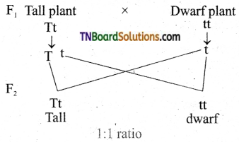
3. Backcross: A cross between an individual from a hybrid population and one of its parental lines.
4. Testcross: A cross between an individual with unknown genotype and a homozygous recessive individual.
5. Reciprocal cross: A cross in which the male and female parents are reversed to determine if there is any effect of sex on the expression of the trait.
These genetic crosses help researchers to understand the mode of inheritance, linkage, recombination, and other genetic phenomena.
Recessive genes refer to the alleles (versions of a gene) that will only be expressed when an individual has two copies of that particular allele, one inherited from each parent. If an individual inherits one recessive allele and one dominant allele for a particular gene, the dominant allele will be expressed and the recessive allele will have no effect on the individual's phenotype (observable traits).
Recessive genes can still play a role in determining an individual's genetic makeup and can be passed down through generations even if they are not expressed. If two carriers of a recessive gene have children, there is a 25% chance that their offspring will inherit two copies of the recessive allele and exhibit the associated recessive trait.
Examples of genetic disorders caused by recessive genes include cystic fibrosis, sickle cell anemia, and albinism.
Dominant genes refer to the alleles (versions of a gene) that are fully expressed in an individual's phenotype, even if only one copy of the gene is present. In dominant inheritance patterns, an individual needs only to receive one dominant allele from either parent to express the associated trait. This is in contrast to recessive genes, where both copies of the gene must be the recessive allele for the trait to be expressed. Dominant genes are represented by uppercase letters (e.g., 'A') and recessive genes by lowercase letters (e.g., 'a'). If an individual inherits one dominant allele (A) from either parent, they will express the dominant trait (A).
Heredity, in medical terms, refers to the passing on of genetic characteristics from parents to their offspring through the transmission of genes. These genes carry the information that determines many traits, such as eye color, hair color, height, and certain health conditions. Heredity plays a significant role in understanding the causes of various diseases and disorders, as some are strongly influenced by genetic factors. However, it's important to note that environmental factors can also interact with genetic predispositions to influence the expression of these traits.
Repetitive sequences in nucleic acid refer to repeated stretches of DNA or RNA nucleotide bases that are present in a genome. These sequences can vary in length and can be arranged in different patterns such as direct repeats, inverted repeats, or tandem repeats. In some cases, these repetitive sequences do not code for proteins and are often found in non-coding regions of the genome. They can play a role in genetic instability, regulation of gene expression, and evolutionary processes. However, certain types of repeat expansions have been associated with various neurodegenerative disorders and other human diseases.
A phenotype is the physical or biochemical expression of an organism's genes, or the observable traits and characteristics resulting from the interaction of its genetic constitution (genotype) with environmental factors. These characteristics can include appearance, development, behavior, and resistance to disease, among others. Phenotypes can vary widely, even among individuals with identical genotypes, due to differences in environmental influences, gene expression, and genetic interactions.
Genetic models are theoretical frameworks used in genetics to describe and explain the inheritance patterns and genetic architecture of traits, diseases, or phenomena. These models are based on mathematical equations and statistical methods that incorporate information about gene frequencies, modes of inheritance, and the effects of environmental factors. They can be used to predict the probability of certain genetic outcomes, to understand the genetic basis of complex traits, and to inform medical management and treatment decisions.
There are several types of genetic models, including:
1. Mendelian models: These models describe the inheritance patterns of simple genetic traits that follow Mendel's laws of segregation and independent assortment. Examples include autosomal dominant, autosomal recessive, and X-linked inheritance.
2. Complex trait models: These models describe the inheritance patterns of complex traits that are influenced by multiple genes and environmental factors. Examples include heart disease, diabetes, and cancer.
3. Population genetics models: These models describe the distribution and frequency of genetic variants within populations over time. They can be used to study evolutionary processes, such as natural selection and genetic drift.
4. Quantitative genetics models: These models describe the relationship between genetic variation and phenotypic variation in continuous traits, such as height or IQ. They can be used to estimate heritability and to identify quantitative trait loci (QTLs) that contribute to trait variation.
5. Statistical genetics models: These models use statistical methods to analyze genetic data and infer the presence of genetic associations or linkage. They can be used to identify genetic risk factors for diseases or traits.
Overall, genetic models are essential tools in genetics research and medical genetics, as they allow researchers to make predictions about genetic outcomes, test hypotheses about the genetic basis of traits and diseases, and develop strategies for prevention, diagnosis, and treatment.
Chromosome mapping, also known as physical mapping, is the process of determining the location and order of specific genes or genetic markers on a chromosome. This is typically done by using various laboratory techniques to identify landmarks along the chromosome, such as restriction enzyme cutting sites or patterns of DNA sequence repeats. The resulting map provides important information about the organization and structure of the genome, and can be used for a variety of purposes, including identifying the location of genes associated with genetic diseases, studying evolutionary relationships between organisms, and developing genetic markers for use in breeding or forensic applications.
Bacterial DNA refers to the genetic material found in bacteria. It is composed of a double-stranded helix containing four nucleotide bases - adenine (A), thymine (T), guanine (G), and cytosine (C) - that are linked together by phosphodiester bonds. The sequence of these bases in the DNA molecule carries the genetic information necessary for the growth, development, and reproduction of bacteria.
Bacterial DNA is circular in most bacterial species, although some have linear chromosomes. In addition to the main chromosome, many bacteria also contain small circular pieces of DNA called plasmids that can carry additional genes and provide resistance to antibiotics or other environmental stressors.
Unlike eukaryotic cells, which have their DNA enclosed within a nucleus, bacterial DNA is present in the cytoplasm of the cell, where it is in direct contact with the cell's metabolic machinery. This allows for rapid gene expression and regulation in response to changing environmental conditions.
Tetrahymena is not a medical term itself, but it is a genus of unicellular organisms known as ciliates. They are commonly found in freshwater environments and can be studied in the field of biology and microbiology. Some species of Tetrahymena have been used in scientific research, including studies on genetics, cell division, and protein function. It is not a term that would typically be used in a medical context.
Deoxyribonucleic acid (DNA) is the genetic material present in the cells of organisms where it is responsible for the storage and transmission of hereditary information. DNA is a long molecule that consists of two strands coiled together to form a double helix. Each strand is made up of a series of four nucleotide bases - adenine (A), guanine (G), cytosine (C), and thymine (T) - that are linked together by phosphate and sugar groups. The sequence of these bases along the length of the molecule encodes genetic information, with A always pairing with T and C always pairing with G. This base-pairing allows for the replication and transcription of DNA, which are essential processes in the functioning and reproduction of all living organisms.
Multifactorial inheritance is a type of genetic inheritance that involves the interaction of multiple genes (two or more) along with environmental factors in the development of a particular trait, disorder, or disease. Each gene can slightly increase or decrease the risk of developing the condition, and the combined effects of these genes, along with environmental influences, determine the ultimate outcome.
Examples of multifactorial inheritance include height, skin color, and many common diseases such as heart disease, diabetes, and mental disorders like schizophrenia and autism. These conditions tend to run in families but do not follow simple Mendelian patterns of inheritance (dominant or recessive). Instead, they show complex inheritance patterns that are influenced by multiple genetic and environmental factors.
It is important to note that having a family history of a multifactorial disorder does not guarantee that an individual will develop the condition. However, it does increase the likelihood, and the risk may be further modified by lifestyle choices, environmental exposures, and other health factors.
DNA restriction enzymes, also known as restriction endonucleases, are a type of enzyme that cut double-stranded DNA at specific recognition sites. These enzymes are produced by bacteria and archaea as a defense mechanism against foreign DNA, such as that found in bacteriophages (viruses that infect bacteria).
Restriction enzymes recognize specific sequences of nucleotides (the building blocks of DNA) and cleave the phosphodiester bonds between them. The recognition sites for these enzymes are usually palindromic, meaning that the sequence reads the same in both directions when facing the opposite strands of DNA.
Restriction enzymes are widely used in molecular biology research for various applications such as genetic engineering, genome mapping, and DNA fingerprinting. They allow scientists to cut DNA at specific sites, creating precise fragments that can be manipulated and analyzed. The use of restriction enzymes has been instrumental in the development of recombinant DNA technology and the Human Genome Project.
'Leishmania tropica' is a species of parasitic protozoan that causes cutaneous leishmaniasis, a skin infection commonly known as "Old World" or Middle Eastern form of the disease. The parasite is transmitted to humans through the bite of infected female sandflies, primarily of the genus Phlebotomus in the Old World.
The infection often results in skin ulcers, typically on exposed parts of the body such as the face, arms, and legs. These lesions can be disfiguring and may take several months to heal, leaving scars. In some cases, the infection can spread to other parts of the body, leading to more severe forms of the disease.
The incubation period for cutaneous leishmaniasis caused by Leishmania tropica can range from a few weeks to several months after the sandfly bite. The severity and duration of the disease can vary widely depending on various factors, including the immune status of the infected individual and the specific strain of the parasite.
Preventive measures include using insect repellent, wearing protective clothing, and sleeping under insecticide-treated bed nets in areas where sandflies are prevalent. There is no vaccine available for cutaneous leishmaniasis, but several treatment options are available, including topical treatments, intralesional injections, and systemic medications, depending on the severity of the infection and the patient's overall health condition.
Fungal DNA refers to the genetic material present in fungi, which are a group of eukaryotic organisms that include microorganisms such as yeasts and molds, as well as larger organisms like mushrooms. The DNA of fungi, like that of all living organisms, is made up of nucleotides that are arranged in a double helix structure.
Fungal DNA contains the genetic information necessary for the growth, development, and reproduction of fungi. This includes the instructions for making proteins, which are essential for the structure and function of cells, as well as other important molecules such as enzymes and nucleic acids.
Studying fungal DNA can provide valuable insights into the biology and evolution of fungi, as well as their potential uses in medicine, agriculture, and industry. For example, researchers have used genetic engineering techniques to modify the DNA of fungi to produce drugs, biofuels, and other useful products. Additionally, understanding the genetic makeup of pathogenic fungi can help scientists develop new strategies for preventing and treating fungal infections.
Ribosomal DNA (rDNA) refers to the specific regions of DNA in a cell that contain the genes for ribosomal RNA (rRNA). Ribosomes are complex structures composed of proteins and rRNA, which play a crucial role in protein synthesis by translating messenger RNA (mRNA) into proteins.
In humans, there are four types of rRNA molecules: 18S, 5.8S, 28S, and 5S. These rRNAs are encoded by multiple copies of rDNA genes that are organized in clusters on specific chromosomes. In humans, the majority of rDNA genes are located on the short arms of acrocentric chromosomes 13, 14, 15, 21, and 22.
Each cluster of rDNA genes contains both transcribed and non-transcribed spacer regions. The transcribed regions contain the genes for the four types of rRNA, while the non-transcribed spacers contain regulatory elements that control the transcription of the rRNA genes.
The number of rDNA copies varies between species and even within individuals of the same species. The copy number can also change during development and in response to environmental factors. Variations in rDNA copy number have been associated with various diseases, including cancer and neurological disorders.
Genetic linkage is the phenomenon where two or more genetic loci (locations on a chromosome) tend to be inherited together because they are close to each other on the same chromosome. This occurs during the process of sexual reproduction, where homologous chromosomes pair up and exchange genetic material through a process called crossing over.
The closer two loci are to each other on a chromosome, the lower the probability that they will be separated by a crossover event. As a result, they are more likely to be inherited together and are said to be linked. The degree of linkage between two loci can be measured by their recombination frequency, which is the percentage of meiotic events in which a crossover occurs between them.
Linkage analysis is an important tool in genetic research, as it allows researchers to identify and map genes that are associated with specific traits or diseases. By analyzing patterns of linkage between markers (identifiable DNA sequences) and phenotypes (observable traits), researchers can infer the location of genes that contribute to those traits or diseases on chromosomes.
Nucleic acid hybridization is a process in molecular biology where two single-stranded nucleic acids (DNA, RNA) with complementary sequences pair together to form a double-stranded molecule through hydrogen bonding. The strands can be from the same type of nucleic acid or different types (i.e., DNA-RNA or DNA-cDNA). This process is commonly used in various laboratory techniques, such as Southern blotting, Northern blotting, polymerase chain reaction (PCR), and microarray analysis, to detect, isolate, and analyze specific nucleic acid sequences. The hybridization temperature and conditions are critical to ensure the specificity of the interaction between the two strands.
Ethidium is a fluorescent, intercalating compound that is often used in molecular biology to stain DNA. When ethidium bromide, a common form of ethidium, binds to DNA, it causes the DNA to fluoresce brightly under ultraviolet light. This property makes it useful for visualizing DNA bands on gels, such as agarose or polyacrylamide gels, during techniques like gel electrophoresis.
It is important to note that ethidium bromide is a mutagen and should be handled with care. It can cause damage to DNA, which can lead to mutations, and it can also be harmful if inhaled or ingested. Therefore, appropriate safety precautions must be taken when working with this compound.
Bacterial chromosomes are typically circular, double-stranded DNA molecules that contain the genetic material of bacteria. Unlike eukaryotic cells, which have their DNA housed within a nucleus, bacterial chromosomes are located in the cytoplasm of the cell, often associated with the bacterial nucleoid.
Bacterial chromosomes can vary in size and structure among different species, but they typically contain all of the genetic information necessary for the survival and reproduction of the organism. They may also contain plasmids, which are smaller circular DNA molecules that can carry additional genes and can be transferred between bacteria through a process called conjugation.
One important feature of bacterial chromosomes is their ability to replicate rapidly, allowing bacteria to divide quickly and reproduce in large numbers. The replication of the bacterial chromosome begins at a specific origin point and proceeds in opposite directions until the entire chromosome has been copied. This process is tightly regulated and coordinated with cell division to ensure that each daughter cell receives a complete copy of the genetic material.
Overall, the study of bacterial chromosomes is an important area of research in microbiology, as understanding their structure and function can provide insights into bacterial genetics, evolution, and pathogenesis.
Restriction mapping is a technique used in molecular biology to identify the location and arrangement of specific restriction endonuclease recognition sites within a DNA molecule. Restriction endonucleases are enzymes that cut double-stranded DNA at specific sequences, producing fragments of various lengths. By digesting the DNA with different combinations of these enzymes and analyzing the resulting fragment sizes through techniques such as agarose gel electrophoresis, researchers can generate a restriction map - a visual representation of the locations and distances between recognition sites on the DNA molecule. This information is crucial for various applications, including cloning, genome analysis, and genetic engineering.
Viral DNA refers to the genetic material present in viruses that consist of DNA as their core component. Deoxyribonucleic acid (DNA) is one of the two types of nucleic acids that are responsible for storing and transmitting genetic information in living organisms. Viruses are infectious agents much smaller than bacteria that can only replicate inside the cells of other organisms, called hosts.
Viral DNA can be double-stranded (dsDNA) or single-stranded (ssDNA), depending on the type of virus. Double-stranded DNA viruses have a genome made up of two complementary strands of DNA, while single-stranded DNA viruses contain only one strand of DNA.
Examples of dsDNA viruses include Adenoviruses, Herpesviruses, and Poxviruses, while ssDNA viruses include Parvoviruses and Circoviruses. Viral DNA plays a crucial role in the replication cycle of the virus, encoding for various proteins necessary for its multiplication and survival within the host cell.
DNA transposable elements, also known as transposons or jumping genes, are mobile genetic elements that can change their position within a genome. They are composed of DNA sequences that include genes encoding the enzymes required for their own movement (transposase) and regulatory elements. When activated, the transposase recognizes specific sequences at the ends of the element and catalyzes the excision and reintegration of the transposable element into a new location in the genome. This process can lead to genetic variation, as the insertion of a transposable element can disrupt the function of nearby genes or create new combinations of gene regulatory elements. Transposable elements are widespread in both prokaryotic and eukaryotic genomes and are thought to play a significant role in genome evolution.
An allele is a variant form of a gene that is located at a specific position on a specific chromosome. Alleles are alternative forms of the same gene that arise by mutation and are found at the same locus or position on homologous chromosomes.
Each person typically inherits two copies of each gene, one from each parent. If the two alleles are identical, a person is said to be homozygous for that trait. If the alleles are different, the person is heterozygous.
For example, the ABO blood group system has three alleles, A, B, and O, which determine a person's blood type. If a person inherits two A alleles, they will have type A blood; if they inherit one A and one B allele, they will have type AB blood; if they inherit two B alleles, they will have type B blood; and if they inherit two O alleles, they will have type O blood.
Alleles can also influence traits such as eye color, hair color, height, and other physical characteristics. Some alleles are dominant, meaning that only one copy of the allele is needed to express the trait, while others are recessive, meaning that two copies of the allele are needed to express the trait.
"Physarum" is not a term that has a specific medical definition. It is a genus of slime molds, which are single-celled organisms that can behave as multicellular entities under certain conditions. They are often studied in biological research for their unique behaviors and abilities, but they do not have direct relevance to human medicine.
Genetic conjugation is a type of genetic transfer that occurs between bacterial cells. It involves the process of one bacterium (the donor) transferring a piece of its DNA to another bacterium (the recipient) through direct contact or via a bridge-like connection called a pilus. This transferred DNA may contain genes that provide the recipient cell with new traits, such as antibiotic resistance or virulence factors, which can make the bacteria more harmful or difficult to treat. Genetic conjugation is an important mechanism for the spread of antibiotic resistance and other traits among bacterial populations.
Bovine papillomavirus 1 (BPV-1) is a species of papillomavirus that primarily infects cattle, causing benign warts or papillomas in the skin and mucous membranes. It is not known to infect humans or cause disease in humans. BPV-1 is closely related to other papillomaviruses that can cause cancer in animals, but its role in human cancer is unclear.
BPV-1 is a double-stranded DNA virus that replicates in the nucleus of infected cells. It encodes several early and late proteins that are involved in viral replication and the transformation of host cells. BPV-1 has been extensively studied as a model system for understanding the molecular mechanisms of papillomavirus infection and oncogenesis.
In addition to its role in animal health, BPV-1 has also been used as a tool in biomedical research. For example, it can be used to transform cells in culture, providing a valuable resource for studying the properties of cancer cells and testing potential therapies. However, it is important to note that BPV-1 is not known to cause human disease and should not be used in any therapeutic context involving humans.
Chrysanthemum cinerariifolium is a specific species of chrysanthemum flower that is native to Asia. It is also known as the "Pyrethrum daisy" or "Dalmatian chrysanthemum." This plant is most well-known for its production of pyrethrin, a natural insecticide. The dried flowers of this species contain high concentrations of pyrethrins, which are potent neurotoxins to insects but considered low in toxicity to mammals and birds.
The medical definition of Chrysanthemum cinerariifolium is related to its use as a traditional herbal medicine in some cultures. The flowers are used to make teas and tinctures, which have been used to treat various conditions such as fever, headache, respiratory infections, and skin diseases. However, it's important to note that the scientific evidence supporting these uses is limited, and more research is needed before any definitive medical claims can be made.
It's also worth noting that Chrysanthemum cinerariifolium extracts and pyrethrins are used in some commercial insecticides and pesticides. These products are used to control a wide variety of pests, including mosquitoes, fleas, ticks, and agricultural pests. Pyrethrin-based insecticides are considered to be relatively safe for use around humans and animals, but they can be toxic to fish and other aquatic organisms, so they must be used with caution in or near bodies of water.
A replication origin is a specific location in a DNA molecule where the process of DNA replication is initiated. It serves as the starting point for the synthesis of new strands of DNA during cell division. The origin of replication contains regulatory elements and sequences that are recognized by proteins, which then recruit and assemble the necessary enzymes to start the replication process. In eukaryotic cells, replication origins are often found in clusters, with multiple origins scattered throughout each chromosome.
Ribosomal RNA (rRNA) is a type of RNA molecule that is a key component of ribosomes, which are the cellular structures where protein synthesis occurs in cells. In ribosomes, rRNA plays a crucial role in the process of translation, where genetic information from messenger RNA (mRNA) is translated into proteins.
Ribosomal RNA is synthesized in the nucleus and then transported to the cytoplasm, where it assembles with ribosomal proteins to form ribosomes. Within the ribosome, rRNA provides a structural framework for the assembly of the ribosome and also plays an active role in catalyzing the formation of peptide bonds between amino acids during protein synthesis.
There are several different types of rRNA molecules, including 5S, 5.8S, 18S, and 28S rRNA, which vary in size and function. These rRNA molecules are highly conserved across different species, indicating their essential role in protein synthesis and cellular function.
Mitochondrial DNA (mtDNA) is the genetic material present in the mitochondria, which are specialized structures within cells that generate energy. Unlike nuclear DNA, which is present in the cell nucleus and inherited from both parents, mtDNA is inherited solely from the mother.
MtDNA is a circular molecule that contains 37 genes, including 13 genes that encode for proteins involved in oxidative phosphorylation, a process that generates energy in the form of ATP. The remaining genes encode for rRNAs and tRNAs, which are necessary for protein synthesis within the mitochondria.
Mutations in mtDNA can lead to a variety of genetic disorders, including mitochondrial diseases, which can affect any organ system in the body. These mutations can also be used in forensic science to identify individuals and establish biological relationships.
Spiroplasma is a genus of wall-less, helical-shaped bacteria belonging to the class Mollicutes. These microorganisms lack a cell wall and have a unique method of movement through a characteristic corkscrew-like motion. Spiroplasmas are primarily known as insect symbionts, often living within the cells of their hosts without causing apparent disease. However, some species can be pathogenic to insects, plants, and even animals, including humans. They are transmitted through insect vectors or via plant sap.
In medical contexts, Spiroplasma spp. have been associated with certain animal diseases, such as citrus stubborn disease in plants and bruscellosis-like syndrome in sheep and goats. In humans, there is some evidence suggesting that Spiroplasma may be involved in the development of arthritis, although more research is needed to establish a definitive link.
To diagnose Spiroplasma infections, specific molecular techniques such as PCR (polymerase chain reaction) or serological methods like ELISA (enzyme-linked immunosorbent assay) are typically employed. Treatment options for Spiroplasma infections are limited due to their atypical cell structure and resistance to many antibiotics, but tetracyclines have shown some efficacy in treating these infections.
"Saccharomyces cerevisiae" is not typically considered a medical term, but it is a scientific name used in the field of microbiology. It refers to a species of yeast that is commonly used in various industrial processes, such as baking and brewing. It's also widely used in scientific research due to its genetic tractability and eukaryotic cellular organization.
However, it does have some relevance to medical fields like medicine and nutrition. For example, certain strains of S. cerevisiae are used as probiotics, which can provide health benefits when consumed. They may help support gut health, enhance the immune system, and even assist in the digestion of certain nutrients.
In summary, "Saccharomyces cerevisiae" is a species of yeast with various industrial and potential medical applications.
Sequence homology in nucleic acids refers to the similarity or identity between the nucleotide sequences of two or more DNA or RNA molecules. It is often used as a measure of biological relationship between genes, organisms, or populations. High sequence homology suggests a recent common ancestry or functional constraint, while low sequence homology may indicate a more distant relationship or different functions.
Nucleic acid sequence homology can be determined by various methods such as pairwise alignment, multiple sequence alignment, and statistical analysis. The degree of homology is typically expressed as a percentage of identical or similar nucleotides in a given window of comparison.
It's important to note that the interpretation of sequence homology depends on the biological context and the evolutionary distance between the sequences compared. Therefore, functional and experimental validation is often necessary to confirm the significance of sequence homology.
I'm sorry for any confusion, but "replicon" is not a term that has a widely accepted or specific medical definition. It is a term that is used in the field of molecular biology, where it refers to a segment of DNA that contains an origin of replication. The origin of replication is the site on the DNA molecule where the process of DNA replication is initiated. This concept is important in the fields of genetics and virology, but it is not a term that is commonly used in clinical medicine.
If you have any questions related to the medical field, I would be happy to try to help answer them for you!
Molecular cloning is a laboratory technique used to create multiple copies of a specific DNA sequence. This process involves several steps:
1. Isolation: The first step in molecular cloning is to isolate the DNA sequence of interest from the rest of the genomic DNA. This can be done using various methods such as PCR (polymerase chain reaction), restriction enzymes, or hybridization.
2. Vector construction: Once the DNA sequence of interest has been isolated, it must be inserted into a vector, which is a small circular DNA molecule that can replicate independently in a host cell. Common vectors used in molecular cloning include plasmids and phages.
3. Transformation: The constructed vector is then introduced into a host cell, usually a bacterial or yeast cell, through a process called transformation. This can be done using various methods such as electroporation or chemical transformation.
4. Selection: After transformation, the host cells are grown in selective media that allow only those cells containing the vector to grow. This ensures that the DNA sequence of interest has been successfully cloned into the vector.
5. Amplification: Once the host cells have been selected, they can be grown in large quantities to amplify the number of copies of the cloned DNA sequence.
Molecular cloning is a powerful tool in molecular biology and has numerous applications, including the production of recombinant proteins, gene therapy, functional analysis of genes, and genetic engineering.
Genetic markers are specific segments of DNA that are used in genetic mapping and genotyping to identify specific genetic locations, diseases, or traits. They can be composed of short tandem repeats (STRs), single nucleotide polymorphisms (SNPs), restriction fragment length polymorphisms (RFLPs), or variable number tandem repeats (VNTRs). These markers are useful in various fields such as genetic research, medical diagnostics, forensic science, and breeding programs. They can help to track inheritance patterns, identify genetic predispositions to diseases, and solve crimes by linking biological evidence to suspects or victims.
Recombinant DNA is a term used in molecular biology to describe DNA that has been created by combining genetic material from more than one source. This is typically done through the use of laboratory techniques such as molecular cloning, in which fragments of DNA are inserted into vectors (such as plasmids or viruses) and then introduced into a host organism where they can replicate and produce many copies of the recombinant DNA molecule.
Recombinant DNA technology has numerous applications in research, medicine, and industry, including the production of recombinant proteins for use as therapeutics, the creation of genetically modified organisms (GMOs) for agricultural or industrial purposes, and the development of new tools for genetic analysis and manipulation.
It's important to note that while recombinant DNA technology has many potential benefits, it also raises ethical and safety concerns, and its use is subject to regulation and oversight in many countries.
A gene is a specific sequence of nucleotides in DNA that carries genetic information. Genes are the fundamental units of heredity and are responsible for the development and function of all living organisms. They code for proteins or RNA molecules, which carry out various functions within cells and are essential for the structure, function, and regulation of the body's tissues and organs.
Each gene has a specific location on a chromosome, and each person inherits two copies of every gene, one from each parent. Variations in the sequence of nucleotides in a gene can lead to differences in traits between individuals, including physical characteristics, susceptibility to disease, and responses to environmental factors.
Medical genetics is the study of genes and their role in health and disease. It involves understanding how genes contribute to the development and progression of various medical conditions, as well as identifying genetic risk factors and developing strategies for prevention, diagnosis, and treatment.
A genetic vector is a vehicle, often a plasmid or a virus, that is used to introduce foreign DNA into a host cell as part of genetic engineering or gene therapy techniques. The vector contains the desired gene or genes, along with regulatory elements such as promoters and enhancers, which are needed for the expression of the gene in the target cells.
The choice of vector depends on several factors, including the size of the DNA to be inserted, the type of cell to be targeted, and the efficiency of uptake and expression required. Commonly used vectors include plasmids, adenoviruses, retroviruses, and lentiviruses.
Plasmids are small circular DNA molecules that can replicate independently in bacteria. They are often used as cloning vectors to amplify and manipulate DNA fragments. Adenoviruses are double-stranded DNA viruses that infect a wide range of host cells, including human cells. They are commonly used as gene therapy vectors because they can efficiently transfer genes into both dividing and non-dividing cells.
Retroviruses and lentiviruses are RNA viruses that integrate their genetic material into the host cell's genome. This allows for stable expression of the transgene over time. Lentiviruses, a subclass of retroviruses, have the advantage of being able to infect non-dividing cells, making them useful for gene therapy applications in post-mitotic tissues such as neurons and muscle cells.
Overall, genetic vectors play a crucial role in modern molecular biology and medicine, enabling researchers to study gene function, develop new therapies, and modify organisms for various purposes.
Genotype, in genetics, refers to the complete heritable genetic makeup of an individual organism, including all of its genes. It is the set of instructions contained in an organism's DNA for the development and function of that organism. The genotype is the basis for an individual's inherited traits, and it can be contrasted with an individual's phenotype, which refers to the observable physical or biochemical characteristics of an organism that result from the expression of its genes in combination with environmental influences.
It is important to note that an individual's genotype is not necessarily identical to their genetic sequence. Some genes have multiple forms called alleles, and an individual may inherit different alleles for a given gene from each parent. The combination of alleles that an individual inherits for a particular gene is known as their genotype for that gene.
Understanding an individual's genotype can provide important information about their susceptibility to certain diseases, their response to drugs and other treatments, and their risk of passing on inherited genetic disorders to their offspring.
A cell line is a culture of cells that are grown in a laboratory for use in research. These cells are usually taken from a single cell or group of cells, and they are able to divide and grow continuously in the lab. Cell lines can come from many different sources, including animals, plants, and humans. They are often used in scientific research to study cellular processes, disease mechanisms, and to test new drugs or treatments. Some common types of human cell lines include HeLa cells (which come from a cancer patient named Henrietta Lacks), HEK293 cells (which come from embryonic kidney cells), and HUVEC cells (which come from umbilical vein endothelial cells). It is important to note that cell lines are not the same as primary cells, which are cells that are taken directly from a living organism and have not been grown in the lab.
Deoxyribonucleases, Type II Site-Specific are a type of enzymes that cleave phosphodiester bonds in DNA molecules at specific recognition sites. They are called "site-specific" because they cut DNA at particular sequences, rather than at random or nonspecific locations. These enzymes belong to the class of endonucleases and play crucial roles in various biological processes such as DNA recombination, repair, and restriction.
Type II deoxyribonucleases are further classified into several subtypes based on their cofactor requirements, recognition site sequences, and cleavage patterns. The most well-known examples of Type II deoxyribonucleases are the restriction endonucleases, which recognize specific DNA motifs in double-stranded DNA and cleave them, generating sticky ends or blunt ends. These enzymes are widely used in molecular biology research for various applications such as genetic engineering, cloning, and genome analysis.
It is important to note that the term "Deoxyribonucleases, Type II Site-Specific" refers to a broad category of enzymes with similar properties and functions, rather than a specific enzyme or family of enzymes. Therefore, providing a concise medical definition for this term can be challenging, as it covers a wide range of enzymes with distinct characteristics and applications.
'Escherichia coli' (E. coli) is a type of gram-negative, facultatively anaerobic, rod-shaped bacterium that commonly inhabits the intestinal tract of humans and warm-blooded animals. It is a member of the family Enterobacteriaceae and one of the most well-studied prokaryotic model organisms in molecular biology.
While most E. coli strains are harmless and even beneficial to their hosts, some serotypes can cause various forms of gastrointestinal and extraintestinal illnesses in humans and animals. These pathogenic strains possess virulence factors that enable them to colonize and damage host tissues, leading to diseases such as diarrhea, urinary tract infections, pneumonia, and sepsis.
E. coli is a versatile organism with remarkable genetic diversity, which allows it to adapt to various environmental niches. It can be found in water, soil, food, and various man-made environments, making it an essential indicator of fecal contamination and a common cause of foodborne illnesses. The study of E. coli has contributed significantly to our understanding of fundamental biological processes, including DNA replication, gene regulation, and protein synthesis.
In medical terms, "bromides" refer to salts or compounds that contain bromine, a chemical element. Historically, potassium bromide was used as a sedative and anticonvulsant in the 19th and early 20th centuries. However, its use has largely been discontinued due to side effects such as neurotoxicity and kidney damage.
In modern medical language, "bromides" can also refer to something that is unoriginal, dull, or lacking in creativity, often used to describe ideas or expressions that are trite or clichéd. This usage comes from the fact that bromide salts were once commonly used as a sedative and were associated with a lack of excitement or energy.
A telomere is a region of repetitive DNA sequences found at the end of chromosomes, which protects the genetic data from damage and degradation during cell division. Telomeres naturally shorten as cells divide, and when they become too short, the cell can no longer divide and becomes senescent or dies. This natural process is associated with aging and various age-related diseases. The length of telomeres can also be influenced by various genetic and environmental factors, including stress, diet, and lifestyle.
Virus integration, in the context of molecular biology and virology, refers to the insertion of viral genetic material into the host cell's genome. This process is most commonly associated with retroviruses, such as HIV (Human Immunodeficiency Virus), which have an enzyme called reverse transcriptase that converts their RNA genome into DNA. This DNA can then integrate into the host's chromosomal DNA, becoming a permanent part of the host's genetic material.
This integration is a crucial step in the retroviral life cycle, allowing the virus to persist within the host cell and evade detection by the immune system. It also means that the viral genome can be passed on to daughter cells when the host cell divides.
However, it's important to note that not all viruses integrate their genetic material into the host's genome. Some viruses, like influenza, exist as separate entities within the host cell and do not become part of the host's DNA.
DNA Sequence Analysis is the systematic determination of the order of nucleotides in a DNA molecule. It is a critical component of modern molecular biology, genetics, and genetic engineering. The process involves determining the exact order of the four nucleotide bases - adenine (A), guanine (G), cytosine (C), and thymine (T) - in a DNA molecule or fragment. This information is used in various applications such as identifying gene mutations, studying evolutionary relationships, developing molecular markers for breeding, and diagnosing genetic diseases.
The process of DNA Sequence Analysis typically involves several steps, including DNA extraction, PCR amplification (if necessary), purification, sequencing reaction, and electrophoresis. The resulting data is then analyzed using specialized software to determine the exact sequence of nucleotides.
In recent years, high-throughput DNA sequencing technologies have revolutionized the field of genomics, enabling the rapid and cost-effective sequencing of entire genomes. This has led to an explosion of genomic data and new insights into the genetic basis of many diseases and traits.
Polymerase Chain Reaction (PCR) is a laboratory technique used to amplify specific regions of DNA. It enables the production of thousands to millions of copies of a particular DNA sequence in a rapid and efficient manner, making it an essential tool in various fields such as molecular biology, medical diagnostics, forensic science, and research.
The PCR process involves repeated cycles of heating and cooling to separate the DNA strands, allow primers (short sequences of single-stranded DNA) to attach to the target regions, and extend these primers using an enzyme called Taq polymerase, resulting in the exponential amplification of the desired DNA segment.
In a medical context, PCR is often used for detecting and quantifying specific pathogens (viruses, bacteria, fungi, or parasites) in clinical samples, identifying genetic mutations or polymorphisms associated with diseases, monitoring disease progression, and evaluating treatment effectiveness.
Chromosomes are thread-like structures that contain genetic material, i.e., DNA and proteins, present in the nucleus of human cells. In humans, there are 23 pairs of chromosomes, for a total of 46 chromosomes, in each diploid cell. Twenty-two of these pairs are called autosomal chromosomes, which come in identical pairs and contain genes that determine various traits unrelated to sex.
The last pair is referred to as the sex chromosomes (X and Y), which determines a person's biological sex. Females have two X chromosomes (46, XX), while males possess one X and one Y chromosome (46, XY). Chromosomes vary in size, with the largest being chromosome 1 and the smallest being the Y chromosome.
Human chromosomes are typically visualized during mitosis or meiosis using staining techniques that highlight their banding patterns, allowing for identification of specific regions and genes. Chromosomal abnormalities can lead to various genetic disorders, including Down syndrome (trisomy 21), Turner syndrome (monosomy X), and Klinefelter syndrome (XXY).
Drug resistance, also known as antimicrobial resistance, is the ability of a microorganism (such as bacteria, viruses, fungi, or parasites) to withstand the effects of a drug that was originally designed to inhibit or kill it. This occurs when the microorganism undergoes genetic changes that allow it to survive in the presence of the drug. As a result, the drug becomes less effective or even completely ineffective at treating infections caused by these resistant organisms.
Drug resistance can develop through various mechanisms, including mutations in the genes responsible for producing the target protein of the drug, alteration of the drug's target site, modification or destruction of the drug by enzymes produced by the microorganism, and active efflux of the drug from the cell.
The emergence and spread of drug-resistant microorganisms pose significant challenges in medical treatment, as they can lead to increased morbidity, mortality, and healthcare costs. The overuse and misuse of antimicrobial agents, as well as poor infection control practices, contribute to the development and dissemination of drug-resistant strains. To address this issue, it is crucial to promote prudent use of antimicrobials, enhance surveillance and monitoring of resistance patterns, invest in research and development of new antimicrobial agents, and strengthen infection prevention and control measures.
Satellite DNA is a type of DNA sequence that is repeated in a tandem arrangement in the genome. These repeats are usually relatively short, ranging from 2 to 10 base pairs, and are often present in thousands to millions of copies arranged in head-to-tail fashion. Satellite DNA can be found in centromeric and pericentromeric regions of chromosomes, as well as at telomeres and other heterochromatic regions of the genome.
Due to their repetitive nature, satellite DNAs are often excluded from the main part of the genome during DNA sequencing projects, and therefore have been referred to as "satellite" DNA. However, recent studies suggest that satellite DNA may play important roles in chromosome structure, function, and evolution.
It's worth noting that not all repetitive DNA sequences are considered satellite DNA. For example, microsatellites and minisatellites are also repetitive DNA sequences, but they have different repeat lengths and arrangements than satellite DNA.
There doesn't seem to be a specific medical definition for "DNA, protozoan" as it is simply a reference to the DNA found in protozoa. Protozoa are single-celled eukaryotic organisms that can be found in various environments such as soil, water, and the digestive tracts of animals.
Protozoan DNA refers to the genetic material present in these organisms. It is composed of nucleic acids, including deoxyribonucleic acid (DNA) and ribonucleic acid (RNA), which contain the instructions for the development, growth, and reproduction of the protozoan.
The DNA in protozoa, like in other organisms, is made up of two strands of nucleotides that coil together to form a double helix. The four nucleotide bases that make up protozoan DNA are adenine (A), thymine (T), guanine (G), and cytosine (C). These bases pair with each other to form the rungs of the DNA ladder, with A always pairing with T and G always pairing with C.
The genetic information stored in protozoan DNA is encoded in the sequence of these nucleotide bases. This information is used to synthesize proteins, which are essential for the structure and function of the organism's cells. Protozoan DNA also contains other types of genetic material, such as regulatory sequences that control gene expression and repetitive elements with no known function.
Understanding the DNA of protozoa is important for studying their biology, evolution, and pathogenicity. It can help researchers develop new treatments for protozoan diseases and gain insights into the fundamental principles of genetics and cellular function.
Archaeal chromosomes refer to the genetic material present in Archaea, a domain of single-celled microorganisms. Like bacteria and eukaryotes, Archaea have their genetic material organized into a single circular chromosome, which is typically smaller than bacterial chromosomes. The archaeal chromosome contains all the genetic information necessary for the organism's survival, including genes coding for proteins, RNA molecules, and regulatory elements that control gene expression.
Archaeal chromosomes are structurally similar to bacterial chromosomes, with a histone-like protein called histone-like protein A (HLP) that helps compact the DNA into a more condensed form. However, archaeal chromosomes also share some features with eukaryotic chromosomes, such as the presence of nucleosome-like structures and the use of similar mechanisms for DNA replication and repair.
Overall, archaeal chromosomes are an important area of study in molecular biology, as they provide insights into the evolution and diversity of life on Earth.
Deoxyribonuclease BamHI is a type of enzyme that belongs to the class of restriction endonucleases. These enzymes are capable of cutting double-stranded DNA molecules at specific recognition sites, and BamHI recognizes the sequence 5'-G|GATCC-3'. The vertical line indicates the point of cleavage, where the phosphodiester bond is broken, resulting in sticky ends that can reattach to other complementary sticky ends.
BamHI restriction endonuclease is derived from the bacterium Bacillus amyloliquefaciens H and is widely used in molecular biology research for various applications such as DNA fragmentation, cloning, and genetic engineering. It is essential to note that the activity of this enzyme can be affected by several factors, including temperature, pH, and the presence of inhibitors or activators.
"Caenorhabditis" is a genus of nematode (roundworm) animals, which are commonly used as model organisms in scientific research. The most widely studied species within this genus is "Caenorhabditis elegans," which has been extensively researched due to its simple anatomy, short lifespan, and fully sequenced genome. These nematodes are found in various environments, including soil and decaying organic matter, and play a crucial role in the decomposition process. The term "Caenorhabditis" itself is derived from Greek roots, with "caeno" meaning "recent" or "new," and "rhabditis" referring to the shape of their tails.
DNA-binding proteins are a type of protein that have the ability to bind to DNA (deoxyribonucleic acid), the genetic material of organisms. These proteins play crucial roles in various biological processes, such as regulation of gene expression, DNA replication, repair and recombination.
The binding of DNA-binding proteins to specific DNA sequences is mediated by non-covalent interactions, including electrostatic, hydrogen bonding, and van der Waals forces. The specificity of binding is determined by the recognition of particular nucleotide sequences or structural features of the DNA molecule.
DNA-binding proteins can be classified into several categories based on their structure and function, such as transcription factors, histones, and restriction enzymes. Transcription factors are a major class of DNA-binding proteins that regulate gene expression by binding to specific DNA sequences in the promoter region of genes and recruiting other proteins to modulate transcription. Histones are DNA-binding proteins that package DNA into nucleosomes, the basic unit of chromatin structure. Restriction enzymes are DNA-binding proteins that recognize and cleave specific DNA sequences, and are widely used in molecular biology research and biotechnology applications.
Consanguinity is a medical and genetic term that refers to the degree of genetic relationship between two individuals who share common ancestors. Consanguineous relationships exist when people are related by blood, through a common ancestor or siblings who have children together. The closer the relationship between the two individuals, the higher the degree of consanguinity.
The degree of consanguinity is typically expressed as a percentage or fraction, with higher values indicating a closer genetic relationship. For example, first-degree relatives, such as parents and children or full siblings, share approximately 50% of their genes and have a consanguinity coefficient of 0.25 (or 25%).
Consanguinity can increase the risk of certain genetic disorders and birth defects in offspring due to the increased likelihood of sharing harmful recessive genes. The risks depend on the degree of consanguinity, with closer relationships carrying higher risks. It is important for individuals who are planning to have children and have a history of consanguinity to consider genetic counseling and testing to assess their risk of passing on genetic disorders.
Genetic variation refers to the differences in DNA sequences among individuals and populations. These variations can result from mutations, genetic recombination, or gene flow between populations. Genetic variation is essential for evolution by providing the raw material upon which natural selection acts. It can occur within a single gene, between different genes, or at larger scales, such as differences in the number of chromosomes or entire sets of chromosomes. The study of genetic variation is crucial in understanding the genetic basis of diseases and traits, as well as the evolutionary history and relationships among species.
 Extrachromosomal DNA
 Discordant inheritance of chromosomal and extrachromosomal DNA elements contributes to dynamic disease evolution in...
 Extrachromosomal DNA in the cancerous transformation of Barrett's oesophagus | Nature
 GATE Syllabus 2022 for Botany - GATE Exam Syllabus
 2021 : Modiwlau , Prifysgol Aberystwyth
 Newly Diagnosed Glioblastoma: A Review on Clinical Management
 Biology, B.S. | Degrees | New York Tech
 Asha S. Multani | MD Anderson Cancer Center
 Talking Glossary of Genetic Terms | NHGRI
 ENGLISH | Cbcszoology
 CSIR NET Life Science Syllabus
 System-based approaches as prognostic tools for glioblastoma | BMC Cancer | Full Text
 Buy Real Glipizide/Metformin No Prescription - Harish Kumar
 CSIR UGC NET - Life Science - Syllabus 2022 | Pathfinder Academy
 GATE Syllabus for Botany (XL-J) - Section 1 to 9
 Genomics week in brief: Week ending 24th September
 FCMS2 - FMM
 Bedste Genetics-podcasts (2023)
 Pesquisa | Portal Regional da BVS
 MDU Entrance Exam - (M.Sc. Environmental Sciences / Energy and Environmental Science / Environmental Biotechnology) 2018: Exam...