Esophageal Cyst
Infected esophageal duplication cyst simulating empyema. (1/18)
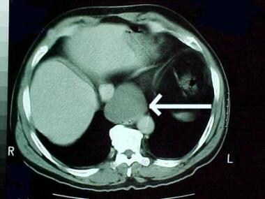
Duplications of the esophagus are the second most common duplication of the gastrointestinal tract. The children with esophageal duplication cyst usually present with respiratory distress or as asymptomatic thoracic mass found on incidental chest x-ray. We report a case of infected esophageal duplication cyst initially confused with empyema in a two years old boy. (+info)Intra-abdominal esophageal duplication cyst in an adult. (2/18)
Esophageal duplication cysts are congenital anomalies of the foregut that are rarely found in the abdomen. An accurate preoperative diagnosis is not always possible, so the definitive diagnosis can be made by histologic examination of the surgical specimen. We experienced a case of Intra-abdominal esophageal duplication cyst in a 52-year-old female, who initially presented with an esophageal submucosal tumor on upper gastrointestinal endoscopy. She did not have any gastrointestinal symptoms. Barium esophagography, chest computed tomography scan and endoscopic ultrasonography demonstrated the cystic lesion in the intra-abdominal esophagus. Transhiatal enucleation of the lesion was performed successfully via the abdominal approach with no postoperative complications. Histologic study showed that the cyst wall contained a two-layered muscle coat and the surface of the lumen was lined by pseudo-ciliated columnar epithelium. The patient has been doing well without any complaints for 3 months of follow-up period. (+info)Unusual bronchopulmonary foregut malformation associated with pericardial defect: bronchogenic cyst communicating with tubular esophageal duplication. (3/18)
We report a case of unusual bronchopulmonary foregut malformation composed of a mediastinal bronchogenic cyst with sequestrated lung tissue and communicating tubular esophageal duplication associated with complete pericardial defect. A 18-yr. old man, who had suffered from dry cough and mild dyspnea, was admitted because of an incidentally detected chest mass. A computed tomography scan demonstrated a cystic mass with an air fluid level connected with esophagus in the middle mediastinum. The surgically resected mass was a pleural invested accessory lobe of the lung (8.0 x 7.0 x 4.5 cm) connected with the esophageal wall by a tubular structure (3.0 cm in length and 2.0 cm in diameter). A complete left pericardial defect was also identified. Histologically, the cystic wall was composed of fibrovascular connective tissue with a smooth muscle layer, mixed seromucous glands and cartilage, and the inner surface of the cyst was lined by ciliated pseudostratified columnar epithelium. The inner surface of the tubular structure was lined by non-keratinizing or keratinizing squamous epithelium, and the wall contained submucosal mucous glands, muscularis mucosa, and duplicated muscularis propria. This case is important in understanding the embryological pathogenesis of the variable spectrum of the bronchopulmonary foregut malformation. (+info)Symptomatic mucocele after esophageal exclusion. (4/18)
(+info)Oesophageal duplication cyst: an unusual cause of retrosternal pain and dysphagia in an adult. (5/18)
Oesophageal duplication cysts in adults are a rare entity and are mostly asymptomatic. We describe the imaging findings in a rare case of oesophageal duplication cyst simulating cold abscess, causing retrosternal pain and dysphagia in a 25-year-old man. (+info)Esophageal cyst producing CA19-9 and CA125. (6/18)
(+info)Bronchogenic and esophageal cyst with laryngeal malformations in a thoroughbred foal. (7/18)
(+info)Duplication of cervical oesophagus: a case report and review of literatures. (8/18)
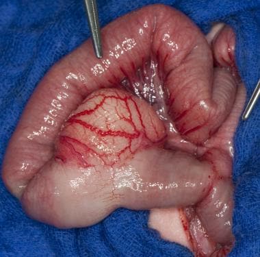
(+info)An esophageal cyst is a rare, abnormal growth that forms in the wall of the esophagus, which is the muscular tube that connects the throat to the stomach. These cysts are typically filled with fluid and can vary in size. They are usually congenital, meaning they are present at birth and develop as a result of abnormal embryonic development.
Esophageal cysts are typically asymptomatic and may not cause any problems until they become large enough to compress nearby structures, such as the trachea or other parts of the digestive system. In some cases, esophageal cysts may cause difficulty swallowing, coughing, or breathing.
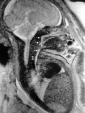
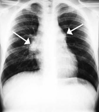
Diagnosis of an esophageal cyst is typically made through imaging tests, such as a CT scan or MRI, which can help to visualize the cyst and determine its size and location. Treatment usually involves surgical removal of the cyst, which is typically performed using minimally invasive techniques such as endoscopy or thoracoscopy.
It's important to note that while I strive to provide accurate information, my responses should not be used as a substitute for professional medical advice, diagnosis or treatment. If you have any concerns about your health, it is always best to consult with a healthcare provider.
A cyst is a closed sac, having a distinct membrane and division between the sac and its surrounding tissue, that contains fluid, air, or semisolid material. Cysts can occur in various parts of the body, including the skin, internal organs, and bones. They can be caused by various factors, such as infection, genetic predisposition, or blockage of a duct or gland. Some cysts may cause symptoms, such as pain or discomfort, while others may not cause any symptoms at all. Treatment for cysts depends on the type and location of the cyst, as well as whether it is causing any problems. Some cysts may go away on their own, while others may need to be drained or removed through a surgical procedure.
 Esophageal Cysts: Practice Essentials, Anatomy, Pathophysiology
 Dr. David Davtyan, MD - Bariatric Surgery Specialist in Beverly Hills, CA | Healthgrades
 Farrokh Saidi - Wikipedia
 Gintare Naujokaite
- Aalborg Universitets forskningsportal
Gintare Naujokaite
- Aalborg Universitets forskningsportal Serum Tumor Markers | AAFP
 Publication : USDA ARS
 Thoracic Surgery | GW Medical Faculty Associates
 Best Hospitals & Doctors in Abu Dhabi, UAE | NMC Healthcare
 Volume 28 Issue 1 | Journal of Avian Medicine and Surgery
 Tara Semenkovich | CTSNet
 Gastroenterology professional society guidance on endoscopic procedures during the COVID-19 pandemic - American...
 Throat Cancers In Dogs: Larynx, Tracheal & Esophageal Oncology - The National Canine Cancer Foundation
 Diagnostic Pathology Nonneoplastic Pediatrics, 2nd edition - Angelica R. Putnam - 1020 - ELSEVIER HEALTH SCIENCES -...
 Esophageal Dysmotility in Idiopathic Pulmonary Fibrosis | PracticeUpdate
 食管癌 - 医生与科室 - 妙佑医疗国际
 Samuel Matthew Alaish, M.D., Associate Professor of Surgery | Johns Hopkins Medicine
 Maraviroc: Package Insert - Drugs.com
 Harry Aslanian, MD Gastroenterology | Yale New Haven Hospital
 Ivacaftor and symptoms of extra-oesophageal reflux in patients with cystic fibrosis and G551D mutation
 Bronchoesophageal Fistula • APPLIED RADIOLOGY
 Black Tea: MedlinePlus Supplements
 statistiche ricercatore
 Digestive Health Services in Spirit Lake Iowa - Lakes Regional Healthcare