Retinal Degeneration
Retina
Retinal Diseases
Photoreceptor Cells, Vertebrate
Fundus Oculi
Dark Adaptation
Retinitis Pigmentosa
Night Blindness
Fluorescein Angiography
Eye Proteins
Visual Acuity
Color Vision Defects
Retinal Cone Photoreceptor Cells
Electrooculography
Vitreous Body
Tomography, Optical Coherence
Retinoschisis
Retinal Rod Photoreceptor Cells
Peripherins
Eye Diseases, Hereditary
Retinal Dystrophies
Opsins
Retinal Pigment Epithelium
Visual Fields
Rhodopsin
Visual Field Tests
Fluorescent Antibody Technique, Indirect
Lipoxygenases
Pedigree
Cyclic Nucleotide Phosphodiesterases, Type 6
Macular Degeneration
Endophthalmitis
Vision, Ocular
Choroideremia
Retinal Bipolar Cells
Electrodes
Disease Models, Animal
Retinal Photoreceptor Cell Inner Segment
Lipofuscin
Intravitreal Injections
Vision Disorders
Retinal Vein Occlusion
Genetic Diseases, X-Linked
Macula Lutea
Radiation Injuries, Experimental
Color Vision
Eye Infections, Bacterial
Pigment Epithelium of Eye
Photoreceptor Cells
Evoked Potentials, Visual
Retinal Ganglion Cells
Mice, Inbred C57BL
Phenotype
Rod Opsins
Mutation
Choroid
Optic Nerve
Dependovirus
Ischemic Preconditioning
Mice, Knockout
Genes, Dominant
Cell Count
Intermediate Filament Proteins
Photic Stimulation
Heterozygote
Reperfusion Injury
In Situ Nick-End Labeling
Rabbits
Cornea
Fluorescence
Diabetic Retinopathy
Glial Fibrillary Acidic Protein
Mutation, Missense
Blotting, Western
Dogs
Genotype
Exons
Genetic Linkage
Ischemia
Polymerase Chain Reaction
Riluzole improves functional recovery after ischemia in the rat retina. (1/2711)
PURPOSE: Retinal ischemia leads to neuronal death. The effects of riluzole, a drug that protects against the deleterious effect of cerebral ischemia by acting on several types of ion channels and blocking glutamatergic neurotransmission, were investigated in a rat model of retinal ischemic injury. METHODS: Retinal ischemia was induced by increasing intraocular pressure above systolic blood pressure for 30 minutes. Electroretinograms were recorded before ischemia and at different periods of reperfusion. Riluzole was injected or topically applied to the eye before or after ischemia and twice daily during the reperfusion period. Retinas were harvested for histopathology (toluidine blue and silver-impregnation stainings, Tdt-dUTP terminal nick-end labeling [TUNEL] method) and immunohistochemistry for cytoskeletal glial fibrillary acid protein and c-jun NH2-terminal kinase (p-JNK). RESULTS: Ischemia for 30 minutes caused a reduction of a- and b-waves of the electroretinogram. Systemic and topical treatments with riluzole significantly enhanced the recovery of the reduced a- and b-waves after defined reperfusion times. Riluzole also prevented or attenuated ischemia-induced retinal cell death (necrosis and apoptosis) and reduced the activation of p-JNK, c-jun phosphorylation, and the increase of cytoskeletal proteins induced by ischemic injury. CONCLUSIONS: Riluzole acted in vivo as a potent neuroprotective agent against pressure-induced ischemia. Therefore, riluzole may be a major drug for use in protection against retinal injury. (+info)Formate-induced inhibition of photoreceptor function in methanol intoxication. (2/2711)
Formic acid is the toxic metabolite responsible for the retinal and optic nerve toxicity produced in methanol intoxication. Previous studies in our laboratory have documented formate-induced retinal dysfunction and histopathology in a rodent model of methanol intoxication. The present studies define the time and concentration dependence of formate-induced retinal toxicity in methanol-intoxicated rats. Retinal function was assessed 24, 48, and 72 h after the initial dose of methanol by flicker electroretinographic measurements. Retinal histopathology was assessed at the same time intervals. Rod- and cone-mediated electroretinogram (ERG) responses were attenuated in a formate concentration- and time-dependent manner, and both retinal sensitivity and maximal responsiveness to light were diminished. Attenuation of UV-cone-mediated responses was temporally delayed in comparison to the functional deficits observed in the 15 Hz/510 nm responses, which have a rod-mediated component and occurred at significantly higher formate concentrations. Both 15 Hz/510 nm and UV-cone-mediated ERG responses were undetectable by 72 h; however, if light intensity was increased, a retinal ERG response could be recorded, indicating that photoreceptor function was profoundly attenuated, but not abolished, under these intoxication conditions. Functional changes preceded structural alterations. Histopathological changes were most pronounced in the outer retina with evidence of inner segment swelling, photoreceptor mitochondrial disruption, and the appearance of fragmented photoreceptor nuclei in the outer nuclear layer. The nature of both the functional and structural alterations observed are consistent with formate-induced inhibition of mitochondrial energy production, resulting in photoreceptor dysfunction and pathology. (+info)Sensitivity and kinetics of mouse rod flash responses determined in vivo from paired-flash electroretinograms. (3/2711)
1. Electroretinograms (ERGs) were recorded corneally from C57BL/6J mice using a paired-flash procedure in which a brief test flash at time zero was followed at time tprobe by a bright probe flash of fixed strength, and in which the probe response amplitude was determined at time t = tprobe + 6 ms. Probe responses obtained in a series of paired-flash trials were analysed to derive A(t), a family of amplitudes that putatively represents the massed response of the rod photoreceptors to the test flash. A central aim was to obtain a mathematical description of the normalized derived response A(t)/Amo as a function of Itest, the test flash strength. 2. With fixed tprobe (80 <= tprobe <= 1200 ms), A(t)/Amo was described by the saturating exponential function [1 - exp(-ktItest)], where kt is a time-dependent sensitivity parameter. For t = 86 ms, a time near the peak of A(t), k86 was 7.0 +/- 1.2 (scotopic cd s m-2)-1 (mean +/- s. d.; n = 4). 3. A(t)/Amo data were analysed in relation to the equation below, a time-generalized form of the above exponential function in which (k86Itest) is replaced by the product [k86Itestu(t)], and where u(t) is independent of the test flash strength. The function u(t) was modelled as the product of a scaling factor gamma, an activation term 1 - exp[-alpha(t - td)2]), and a decay term exp(-t/tauomega): A(t)/Amo = 1 - exp[-k86Itestu(t)]; u(t) = gamma(1 - exp[-alpha(t - td)2](exp(-t/tauomega) where td is a brief delay, tauomega is an exponential time constant, and alpha characterizes the acceleration of the activation term. For Itest up to approximately 2.57 scotopic cd s m-2, the overall time course of A(t) was well described by the above equation with gamma = 2.21, td = 3.1 ms, tauomega = 132 ms and alpha = 2.32 x 10⁻⁴ ms-2. An approximate halving of alpha improved the fit of the above equation to ERG a-wave and A(t)/Amo data obtained at t about 0-20 ms. 4. Kinetic and sensitivity properties of A(t) suggest that it approximates the in vivo massed photocurrent response of the rods to a test flash, and imply that u(t) in the above equation is the approximate kinetic description of a unit, i.e. single photon, response. (+info)Cone signal contributions to electroretinograms [correction of electrograms] in dichromats and trichromats. (4/2711)
PURPOSE: To find out how the different cone types contribute to the electroretinogram (ERG) by quantifying the contribution of the signal pathways originating in the long (L-) and the middle (M-) wavelength-sensitive cones to the total ERG response amplitude and phase. METHODS: ERG response amplitudes and phases were measured to cone-isolating stimuli and to different combinations of L- and M-cone modulation. Conditions were chosen to exclude any contribution of the short wavelength-sensitive (S-) cones. The sensitivity of the ERG to the L and the M cones was defined as the cone contrast gain. RESULTS: In the present paper, a model is provided that describes the ERG contrast gains and ERG thresholds in dichromats and color normal trichromats. For the X-chromosome-linked dichromats, the contrast gains of only one cone type (either the L or the M cones) sufficed to describe the ERG thresholds for all stimulus conditions. Data suggest that the M-cone contrast gains of protanopes are larger than the L-cone contrast gains of deuteranopes. The response thresholds of the trichromats are modeled by assuming a vector summation of signals originating in the L and the M cones. Their L- and M-cone contrast gains are close to a linear interpolation of the data obtained from the dichromats. Nearly all trichromats had larger L- than M-cone contrast gains. Data from a large population of trichromats were examined to study the individual variations in cone weightings and in the phases of the cone pathway responses. CONCLUSIONS: The data strongly suggest that the missing cone type in dichromats is replaced by the remaining cone type. The mean L-cone to M-cone weighting ratio in trichromats was found to be approximately 4:1. But there is a substantial interindividual variability between trichromats. The response phases of the L- and the M-cone pathways can be reliably quantified using the response phases to the cone-isolating stimuli or using a vector addition of L- and M-cone signals. (+info)Hypersensitivity in the anterior median eye of a jumping spider. (5/2711)
Changes in sensitivity of the photoreceptor cells of the anterior median eye of the jumping spider Menemerus confusus Boes. et Str. have been studied by recording electroretinograms (ERGs) and receptor potentials. The amplitudes of the responses (ERGs and receptor potentials) increase during repetitive stimulation, with a maximum increase at 3-5 s intervals. The sensitivity of the photoreceptor cell is greater for about 60 s following illumination (maximum magnitude at 3-5 s) than it is during complete dark adaptation. This phenomenon, which we call 'hypersensitivity', is lost within one day following surgery in physiological saline. Upon loss of hypersensitivity, the sensitivity decrease during light adaptation is greater than for the normal eye and the small increase of sensitivity following the onset of illumination observed for the normal eye is lost. (+info)Human cone pigment expressed in transgenic mice yields altered vision. (6/2711)
Genetically driven alterations in the complement of retinal photopigments are fundamental steps in the evolution of vision. We sought to determine how a newly added photopigment might impact vision by studying a transgenic mouse that expresses a human cone photopigment. Electroretinogram (ERG) measurements indicate that the added pigment works well, significantly changing spectral sensitivity without deleteriously affecting the operation of the native cone pigments. Visual capacities of the transgenic mice were established in behavioral tests. The new pigment was found to provide a significant expansion of the spectral range over which mice can perceive light, thus underlining the immediate utility of acquiring a new photopigment. The transgenic mouse also has the receptor basis for a novel color vision capacity, but tests show that potential was not realized. This failure likely reflects limitations in the organizational arrangement of the mouse retina. (+info)Mid-peripheral pattern electrical retinal responses in normals, glaucoma suspects, and glaucoma patients. (7/2711)
AIMS: Reliance on intraocular pressure, optic nerve cupping changes, nerve fibre layer integrity, and visual field changes may delay treatment of glaucoma since irreversible changes may have already occurred at the time of diagnosis. Abnormal pattern electrical retinal responses (PERR or PERG) have been demonstrated in patients with ocular hypertension (no visual field changes) and glaucoma when visual stimulation was presented to the central field. Since glaucomatous visual field changes tend to occur first in the mid-periphery, the use of PERR outside of the central field may offer an earlier indication of glaucomatous involvement. METHODS: Glaucoma suspects and glaucoma patients were derived from a university practice. Normal subjects were recruited from non-patient volunteers. Alternating bar gratings were presented in the supranasal, supratemporal, infratemporal, and infranasal visual field. Six spatial frequencies, from 0.25 to 6.0 cycles per degree, were used for normal volunteers; three spatial frequencies, from 0.38 to 1.5 cycles per degree, were presented to suspects and glaucoma patients. Time of onset of the first negative (N35) and first positive peak (P50) and the amplitude consisting of the absolute difference between the first negative peak and first positive peak (P50 amplitude) are reported. Age corrected values were determined for normals, suspects, and glaucoma patients for each spatial frequency and for each quadrant in the visual field. RESULTS: Mean P50 amplitudes from normal subjects showed spatial tuning in all quadrants with reduced low frequency attenuation. Normals demonstrated a small decline in amplitude with age. Glaucoma patients demonstrated an age corrected reduction in amplitude and early implicit times. Glaucoma suspects had values between those of normal and glaucoma subjects. P50 amplitudes were weakly correlated with increasing cup to disc diameter ratio. A glaucoma patient with asymmetric visual field loss demonstrated significant diminution of the PERR bilaterally. CONCLUSION: The PERR, using mid-peripheral stimulation, may be a sensitive tool for the early detection of glaucoma. Further refinements can speed clinical data acquisition and enhance signal to noise ratio. (+info)ERG phenotype of a dystrophin mutation in heterozygous female carriers of Duchenne muscular dystrophy. (8/2711)
PURPOSE: Mutations in the dystrophin gene result in Duchenne muscular dystrophy (DMD). DMD is associated with an abnormal electroretinogram (ERG) if the mutation disrupts the translation of retinal dystrophin (Dp260). Our aim was to determine if incomplete ERG abnormalities would be associated with heterozygous carriers of dystrophin gene mutations. METHODS: Ganzfeld ERGs were obtained under scotopic and photopic testing conditions from a family which includes the heterozygous maternal grandmother, the heterozygous mother, and her children, two affected boys and dizygotic twin sibs, an unaffected male and heterozygous female. Southern blot analyses were done to characterise the dystrophin mutation. RESULTS: The dystrophin gene was found to contain a deletion encompassing exon 50. The ERGs in the two affected boys were abnormal, consistent with the DMD ERG phenotype. Serial ERGs of the heterozygous females were abnormal; however, they were less severely affected than the DMD boys. The ERG of the female sib showed a greater abnormality than her mother and maternal grandmother. The unaffected twin had a normal ERG. CONCLUSIONS: The ERG shows abnormalities associated with carrier status in this family with a single exon deletion. A large study of confirmed obligate carriers is planned to clarify further the value of the ERG in detecting female heterozygous carriers of dystrophin gene mutations. (+info)Electroretinography (ERG) is a medical test used to evaluate the functioning of the retina, which is the light-sensitive tissue located at the back of the eye. The test measures the electrical responses of the retina to light stimulation.
During the procedure, a special contact lens or electrode is placed on the surface of the eye to record the electrical activity generated by the retina's light-sensitive cells (rods and cones) and other cells in the retina. The test typically involves presenting different levels of flashes of light to the eye while the electrical responses are recorded.
The resulting ERG waveform provides information about the overall health and function of the retina, including the condition of the photoreceptors, the integrity of the inner retinal layers, and the health of the retinal ganglion cells. This test is often used to diagnose and monitor various retinal disorders, such as retinitis pigmentosa, macular degeneration, and diabetic retinopathy.
Retinal degeneration is a broad term that refers to the progressive loss of photoreceptor cells (rods and cones) in the retina, which are responsible for converting light into electrical signals that are sent to the brain. This process can lead to vision loss or blindness. There are many different types of retinal degeneration, including age-related macular degeneration, retinitis pigmentosa, and Stargardt's disease, among others. These conditions can have varying causes, such as genetic mutations, environmental factors, or a combination of both. Treatment options vary depending on the specific type and progression of the condition.
The retina is the innermost, light-sensitive layer of tissue in the eye of many vertebrates and some cephalopods. It receives light that has been focused by the cornea and lens, converts it into neural signals, and sends these to the brain via the optic nerve. The retina contains several types of photoreceptor cells including rods (which handle vision in low light) and cones (which are active in bright light and are capable of color vision).
In medical terms, any pathological changes or diseases affecting the retinal structure and function can lead to visual impairment or blindness. Examples include age-related macular degeneration, diabetic retinopathy, retinal detachment, and retinitis pigmentosa among others.
Retinal diseases refer to a group of conditions that affect the retina, which is the light-sensitive tissue located at the back of the eye. The retina is responsible for converting light into electrical signals that are sent to the brain and interpreted as visual images. Retinal diseases can cause vision loss or even blindness, depending on their severity and location in the retina.
Some common retinal diseases include:
1. Age-related macular degeneration (AMD): A progressive disease that affects the central part of the retina called the macula, causing blurred or distorted vision.
2. Diabetic retinopathy: A complication of diabetes that can damage the blood vessels in the retina, leading to vision loss.
3. Retinal detachment: A serious condition where the retina becomes separated from its underlying tissue, requiring immediate medical attention.
4. Macular edema: Swelling or thickening of the macula due to fluid accumulation, which can cause blurred vision.
5. Retinitis pigmentosa: A group of inherited eye disorders that affect the retina's ability to respond to light, causing progressive vision loss.
6. Macular hole: A small break in the macula that can cause distorted or blurry vision.
7. Retinal vein occlusion: Blockage of the retinal veins that can lead to bleeding, swelling, and potential vision loss.
Treatment for retinal diseases varies depending on the specific condition and its severity. Some treatments include medication, laser therapy, surgery, or a combination of these options. Regular eye exams are essential for early detection and treatment of retinal diseases.
Photoreceptor cells in vertebrates are specialized types of neurons located in the retina of the eye that are responsible for converting light stimuli into electrical signals. These cells are primarily responsible for the initial process of vision and have two main types: rods and cones.
Rods are more numerous and are responsible for low-light vision or scotopic vision, enabling us to see in dimly lit conditions. They do not contribute to color vision but provide information about the shape and movement of objects.
Cones, on the other hand, are less numerous and are responsible for color vision and high-acuity vision or photopic vision. There are three types of cones, each sensitive to different wavelengths of light: short (S), medium (M), and long (L) wavelengths, which correspond to blue, green, and red, respectively. The combination of signals from these three types of cones allows us to perceive a wide range of colors.
Both rods and cones contain photopigments that consist of a protein called opsin and a light-sensitive chromophore called retinal. When light hits the photopigment, it triggers a series of chemical reactions that ultimately lead to the generation of an electrical signal that is transmitted to the brain via the optic nerve. This process enables us to see and perceive our visual world.
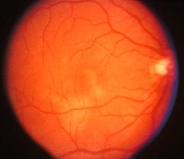
"Fundus Oculi" is a medical term that refers to the back part of the interior of the eye, including the optic disc, macula, fovea, retinal vasculature, and peripheral retina. It is the area where light is focused and then transmitted to the brain via the optic nerve, forming visual images. Examinations of the fundus oculi are crucial for detecting various eye conditions such as diabetic retinopathy, macular degeneration, glaucoma, and other retinal diseases. The examination is typically performed using an ophthalmoscope or a specialized camera called a retinal camera.
Dark adaptation is the process by which the eyes adjust to low levels of light. This process allows the eyes to become more sensitive to light and see better in the dark. It involves the dilation of the pupils, as well as chemical changes in the rods and cones (photoreceptor cells) of the retina. These changes allow the eye to detect even small amounts of light and improve visual acuity in low-light conditions. Dark adaptation typically takes several minutes to occur fully, but can be faster or slower depending on various factors such as age, prior exposure to light, and certain medical conditions. It is an important process for maintaining good vision in a variety of lighting conditions.
Retinitis pigmentosa (RP) is a group of rare, genetic disorders that involve a breakdown and loss of cells in the retina - a light-sensitive tissue located at the back of the eye. The retina converts light into electrical signals which are then sent to the brain and interpreted as visual images.
In RP, the cells that detect light (rods and cones) degenerate more slowly than other cells in the retina, leading to a progressive loss of vision. Symptoms typically begin in childhood with night blindness (difficulty seeing in low light), followed by a gradual narrowing of the visual field (tunnel vision). Over time, this can lead to significant vision loss and even blindness.
The condition is usually inherited and there are several different genes that have been associated with RP. The diagnosis is typically made based on a combination of genetic testing, family history, and clinical examination. Currently, there is no cure for RP, but researchers are actively working to develop new treatments that may help slow or stop the progression of the disease.
Night blindness, also known as nyctalopia, is a visual impairment characterized by the inability to see well in low light or darkness. It's not an eye condition itself but rather a symptom of various underlying eye disorders, most commonly vitamin A deficiency and retinal diseases like retinitis pigmentosa.
In a healthy eye, a molecule called rhodopsin is present in the rods (special light-sensitive cells in our eyes responsible for vision in low light conditions). This rhodopsin requires sufficient amounts of vitamin A to function properly. When there's a deficiency of vitamin A or damage to the rods, the ability to see in dim light gets affected, leading to night blindness.
People with night blindness often have difficulty adjusting to changes in light levels, such as when entering a dark room from bright sunlight. They may also experience trouble seeing stars at night, driving at dusk or dawn, and navigating in poorly lit areas. If you suspect night blindness, it's essential to consult an eye care professional for proper diagnosis and treatment of the underlying cause.
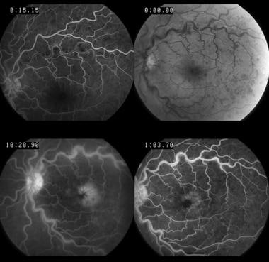
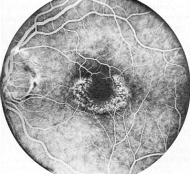
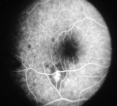
Fluorescein angiography is a medical diagnostic procedure used in ophthalmology to examine the blood flow in the retina and choroid, which are the inner layers of the eye. This test involves injecting a fluorescent dye, Fluorescein, into a patient's arm vein. As the dye reaches the blood vessels in the eye, a specialized camera takes rapid sequences of photographs to capture the dye's circulation through the retina and choroid.
The images produced by fluorescein angiography can help doctors identify any damage to the blood vessels, leakage, or abnormal growth of new blood vessels. This information is crucial in diagnosing and managing various eye conditions such as age-related macular degeneration, diabetic retinopathy, retinal vein occlusions, and inflammatory eye diseases.
It's important to note that while fluorescein angiography is a valuable diagnostic tool, it does carry some risks, including temporary side effects like nausea, vomiting, or allergic reactions to the dye. In rare cases, severe adverse reactions can occur, so patients should discuss these potential risks with their healthcare provider before undergoing the procedure.
Ophthalmoscopy is a medical examination technique used by healthcare professionals to observe the interior structures of the eye, including the retina, optic disc, and vitreous humor. This procedure typically involves using an ophthalmoscope, a handheld device that consists of a light and magnifying lenses. The healthcare provider looks through the ophthalmoscope and directly observes the internal structures of the eye by illuminating them.
There are several types of ophthalmoscopy, including direct ophthalmoscopy, indirect ophthalmoscopy, and slit-lamp biomicroscopy. Each type has its own advantages and disadvantages, and they may be used in different situations depending on the specific clinical situation and the information needed.
Ophthalmoscopy is an important diagnostic tool for detecting and monitoring a wide range of eye conditions, including diabetic retinopathy, glaucoma, age-related macular degeneration, and other retinal disorders. It can also provide valuable information about the overall health of the individual, as changes in the appearance of the retina or optic nerve may indicate the presence of systemic diseases such as hypertension or diabetes.
Eye proteins, also known as ocular proteins, are specific proteins that are found within the eye and play crucial roles in maintaining proper eye function and health. These proteins can be found in various parts of the eye, including the cornea, iris, lens, retina, and other structures. They perform a wide range of functions, such as:
1. Structural support: Proteins like collagen and elastin provide strength and flexibility to the eye's tissues, enabling them to maintain their shape and withstand mechanical stress.
2. Light absorption and transmission: Proteins like opsins and crystallins are involved in capturing and transmitting light signals within the eye, which is essential for vision.
3. Protection against damage: Some eye proteins, such as antioxidant enzymes and heat shock proteins, help protect the eye from oxidative stress, UV radiation, and other environmental factors that can cause damage.
4. Regulation of eye growth and development: Various growth factors and signaling molecules, which are protein-based, contribute to the proper growth, differentiation, and maintenance of eye tissues during embryonic development and throughout adulthood.
5. Immune defense: Proteins involved in the immune response, such as complement components and immunoglobulins, help protect the eye from infection and inflammation.
6. Maintenance of transparency: Crystallin proteins in the lens maintain its transparency, allowing light to pass through unobstructed for clear vision.
7. Neuroprotection: Certain eye proteins, like brain-derived neurotrophic factor (BDNF), support the survival and function of neurons within the retina, helping to preserve vision.
Dysfunction or damage to these eye proteins can contribute to various eye disorders and diseases, such as cataracts, age-related macular degeneration, glaucoma, diabetic retinopathy, and others.
Visual acuity is a measure of the sharpness or clarity of vision. It is usually tested by reading an eye chart from a specific distance, such as 20 feet (6 meters). The standard eye chart used for this purpose is called the Snellen chart, which contains rows of letters that decrease in size as you read down the chart.
Visual acuity is typically expressed as a fraction, with the numerator representing the testing distance and the denominator indicating the smallest line of type that can be read clearly. For example, if a person can read the line on the eye chart that corresponds to a visual acuity of 20/20, it means they have normal vision at 20 feet. If their visual acuity is 20/40, it means they must be as close as 20 feet to see what someone with normal vision can see at 40 feet.
It's important to note that visual acuity is just one aspect of overall vision and does not necessarily reflect other important factors such as peripheral vision, depth perception, color vision, or contrast sensitivity.
Color vision defects, also known as color blindness, are conditions in which a person has difficulty distinguishing between certain colors. The most common types of color vision defects involve the inability to distinguish between red and green or blue and yellow. These deficiencies result from an alteration or absence of one or more of the three types of cone cells in the retina that are responsible for normal color vision.
In red-green color vision defects, there is a problem with either the red or green cones, or both. This results in difficulty distinguishing between these two colors and their shades. Protanopia is a type of red-green color vision defect where there is an absence of red cone cells, making it difficult to distinguish between red and green as well as between red and black or green and black. Deuteranopia is another type of red-green color vision defect where there is an absence of green cone cells, resulting in similar difficulties distinguishing between red and green, as well as between blue and yellow.
Blue-yellow color vision defects are less common than red-green color vision defects. Tritanopia is a type of blue-yellow color vision defect where there is an absence of blue cone cells, making it difficult to distinguish between blue and yellow, as well as between blue and purple or yellow and pink.
Color vision defects are usually inherited and present from birth, but they can also result from eye diseases, chemical exposure, aging, or medication side effects. They affect both men and women, although red-green color vision defects are more common in men than in women. People with color vision defects may have difficulty with tasks that require color discrimination, such as matching clothes, selecting ripe fruit, reading colored maps, or identifying warning signals. However, most people with mild to moderate color vision defects can adapt and function well in daily life.
Retinal cone photoreceptor cells are specialized neurons located in the retina of the eye, responsible for visual phototransduction and color vision. They are one of the two types of photoreceptors, with the other being rods, which are more sensitive to low light levels. Cones are primarily responsible for high-acuity, color vision during daylight or bright-light conditions.
There are three types of cone cells, each containing different photopigments that absorb light at distinct wavelengths: short (S), medium (M), and long (L) wavelengths, which correspond to blue, green, and red light, respectively. The combination of signals from these three types of cones allows the human visual system to perceive a wide range of colors and discriminate between them. Cones are densely packed in the central region of the retina, known as the fovea, which provides the highest visual acuity.
Electrooculography (EOG) is a technique for measuring the resting potential of the eye and the changes in this potential that occur with eye movements. It involves placing electrodes near the eyes to detect the small electric fields generated by the movement of the eyeball within the surrounding socket. This technique is used in research and clinical settings to study eye movements and their control, as well as in certain diagnostic applications such as assessing the function of the oculomotor system in patients with neurological disorders.
The vitreous body, also known simply as the vitreous, is the clear, gel-like substance that fills the space between the lens and the retina in the eye. It is composed mainly of water, but also contains collagen fibers, hyaluronic acid, and other proteins. The vitreous helps to maintain the shape of the eye and provides a transparent medium for light to pass through to reach the retina. With age, the vitreous can become more liquefied and may eventually separate from the retina, leading to symptoms such as floaters or flashes of light.
Optical coherence tomography (OCT) is a non-invasive imaging technique that uses low-coherence light to capture high-resolution cross-sectional images of biological tissues, particularly the retina and other ocular structures. OCT works by measuring the echo time delay of light scattered back from different depths within the tissue, creating a detailed map of the tissue's structure. This technique is widely used in ophthalmology to diagnose and monitor various eye conditions such as macular degeneration, diabetic retinopathy, and glaucoma.
Retinoschisis is a medical term that refers to a specific eye condition where there is a separation (schisis) of the retinal layers, particularly the neurosensory retina. This condition often affects the peripheral retina and can be classified as congenital or acquired. Congenital retinoschisis is usually present at birth or develops during early childhood, while acquired retinoschisis occurs later in life due to various reasons such as trauma, inflammation, or degenerative changes.
In retinoschisis, the inner layers of the retina split apart, creating a cavity filled with fluid. This separation can lead to visual symptoms like blurred vision, shadows, or blind spots in the affected area of the visual field. However, it is important to note that many cases of retinoschisis do not cause significant visual impairment and may only require monitoring by an eye care professional.
Retinoschisis can be diagnosed through a comprehensive eye examination, including a dilated fundus exam, which allows the eye care professional to examine the retina thoroughly. In some cases, additional diagnostic tests like optical coherence tomography (OCT) or fluorescein angiography may be used to confirm the diagnosis and assess the extent of the condition.
Treatment for retinoschisis depends on the severity and location of the separation. Mild cases may not require any treatment, while more severe cases may need surgical intervention to prevent complications such as retinal detachment or bleeding in the eye. Regular follow-up appointments with an eye care professional are essential to monitor the condition and ensure appropriate management.
Color perception tests are a type of examination used to evaluate an individual's ability to perceive and distinguish different colors. These tests typically consist of a series of plates or images that contain various patterns or shapes displayed in different colors. The person being tested is then asked to identify or match the colors based on specific instructions.
There are several types of color perception tests, including:
1. Ishihara Test: This is a commonly used test for red-green color deficiency. It consists of a series of plates with circles made up of dots in different sizes and colors. Within these circles, there may be a number or symbol visible only to those with normal color vision or to those with specific types of color blindness.
2. Farnsworth D-15 Test: This test measures an individual's ability to arrange colored caps in a specific order based on their hue. It is often used to diagnose and monitor the progression of color vision deficiencies.
3. Hardy-Rand-Rittler (HRR) Test: This is another type of color arrangement test that measures an individual's ability to distinguish between different colors based on their hue, saturation, and brightness.
4. Color Discrimination Tests: These tests measure an individual's ability to distinguish between two similar colors that are presented side by side or in close proximity.
5. Anomaloscope Test: This is a more sophisticated test that measures the degree of color vision deficiency by asking the person to match the brightness and hue of two lights.
Color perception tests are often used in occupational settings, such as aviation, military, and manufacturing, where color discrimination is critical for safety and performance. They may also be used in educational and clinical settings to diagnose and monitor color vision deficiencies.
Retinal rod photoreceptor cells are specialized neurons in the retina of the eye that are primarily responsible for vision in low light conditions. They contain a light-sensitive pigment called rhodopsin, which undergoes a chemical change when struck by a single photon of light. This triggers a cascade of biochemical reactions that ultimately leads to the generation of electrical signals, which are then transmitted to the brain via the optic nerve.
Rod cells do not provide color vision or fine detail, but they allow us to detect motion and see in dim light. They are more sensitive to light than cone cells, which are responsible for color vision and detailed sight in bright light conditions. Rod cells are concentrated at the outer edges of the retina, forming a crescent-shaped region called the peripheral retina, with fewer rod cells located in the central region of the retina known as the fovea.
Peripherins are a family of neuron-specific type III intermediate filament proteins that are expressed in the peripheral nervous system. They play crucial roles in maintaining the structural integrity and stability of nerve cells, particularly during development and regeneration. Peripherins have also been implicated in various neurodegenerative disorders, including amyotrophic lateral sclerosis (ALS) and Charcot-Marie-Tooth disease (CMT). There are several isoforms of peripherins, with peripherin 2 being the most widely studied. Mutations in the gene encoding peripherin 2 have been linked to certain forms of CMT.
Hereditary eye diseases refer to conditions that affect the eyes and are passed down from parents to their offspring through genetics. These diseases are caused by mutations or changes in an individual's DNA that are inherited from their parents. The mutations can occur in any of the genes associated with eye development, function, or health.
There are many different types of hereditary eye diseases, some of which include:
1. Retinitis Pigmentosa - a group of rare, genetic disorders that involve a breakdown and loss of cells in the retina.
2. Macular Degeneration - a progressive disease that damages the central portion of the retina, impairing vision.
3. Glaucoma - a group of eye conditions that damage the optic nerve, often caused by an increase in pressure inside the eye.
4. Cataracts - clouding of the lens inside the eye, which can lead to blurry vision and blindness.
5. Keratoconus - a progressive eye disease that causes the cornea to thin and bulge outward into a cone shape.
6. Color Blindness - a condition where an individual has difficulty distinguishing between certain colors.
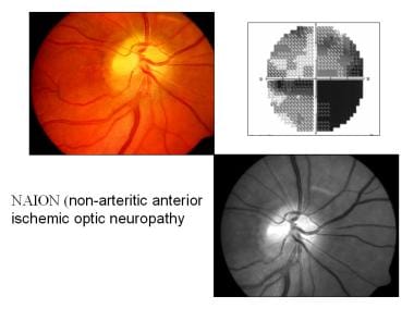
7. Optic Neuropathy - damage to the optic nerve, which can result in vision loss.
The symptoms and severity of hereditary eye diseases can vary widely depending on the specific condition and the individual's genetic makeup. Some conditions may be present at birth or develop in early childhood, while others may not appear until later in life. Treatment options for these conditions may include medication, surgery, or lifestyle changes, and are often most effective when started early.
Retinal dystrophies are a group of genetic eye disorders that primarily affect the retina, a light-sensitive layer at the back of the eye. These conditions are characterized by progressive degeneration and death of photoreceptor cells (rods and cones) in the retina, leading to vision loss.
The term "dystrophy" refers to a condition that results from the abnormal or defective development and function of tissues or organs. In the case of retinal dystrophies, the photoreceptor cells do not develop or function properly, resulting in visual impairment.
Retinal dystrophies can present at any age, from infancy to adulthood, and can have varying degrees of severity. Some common symptoms include night blindness, decreased visual acuity, loss of peripheral vision, light sensitivity, and color vision abnormalities.
Examples of retinal dystrophies include retinitis pigmentosa, Stargardt disease, Usher syndrome, and Leber congenital amaurosis, among others. These conditions are typically inherited and can be caused by mutations in various genes that play a role in the development and function of the retina.
There is currently no cure for retinal dystrophies, but research is ongoing to develop treatments that may slow or halt the progression of these conditions, such as gene therapy and stem cell transplantation.
Opsins are a type of protein that are sensitive to light and play a crucial role in vision. They are found in the photoreceptor cells of the retina, which are the specialized cells in the eye that detect light. Opsins are activated by light, which triggers a series of chemical reactions that ultimately result in the transmission of a signal to the brain, allowing us to see.
There are several different types of opsins, including rhodopsin and the cone pigments, which are found in the rods and cones of the retina, respectively. Rhodopsin is responsible for dim-light vision, while the cone pigments are involved in color vision and bright-light vision.
Opsins belong to a larger family of proteins called G protein-coupled receptors (GPCRs), which are involved in many different physiological processes in the body. In addition to their role in vision, opsins have also been found to be involved in other light-dependent processes, such as the regulation of circadian rhythms and the entrainment of the biological clock.
The retinal pigment epithelium (RPE) is a single layer of cells located between the photoreceptor cells of the retina and the choroid, which is a part of the eye containing blood vessels. The RPE plays a crucial role in maintaining the health and function of the photoreceptors by providing them with nutrients, removing waste products, and helping to regulate the light-sensitive visual pigments within the photoreceptors.
The RPE cells contain pigment granules that absorb excess light to prevent scattering within the eye and improve visual acuity. They also help to form the blood-retina barrier, which restricts the movement of certain molecules between the retina and the choroid, providing an important protective function for the retina.
Damage to the RPE can lead to a variety of eye conditions, including age-related macular degeneration (AMD), which is a leading cause of vision loss in older adults.
Visual fields refer to the total area in which objects can be seen while keeping the eyes focused on a central point. It is the entire area that can be observed using peripheral (side) vision while the eye gazes at a fixed point. A visual field test is used to detect blind spots or gaps (scotomas) in a person's vision, which could indicate various medical conditions such as glaucoma, retinal damage, optic nerve disease, brain tumors, or strokes. The test measures both the central and peripheral vision and maps the entire area that can be seen when focusing on a single point.
Rhodopsin, also known as visual purple, is a light-sensitive pigment found in the rods of the vertebrate retina. It is a complex protein molecule made up of two major components: an opsin protein and retinal, a form of vitamin A. When light hits the retinal in rhodopsin, it changes shape, which initiates a series of chemical reactions leading to the activation of the visual pathway and ultimately results in vision. This process is known as phototransduction. Rhodopsin plays a crucial role in low-light vision or scotopic vision.
A visual field test is a method used to measure an individual's entire scope of vision, which includes what can be seen straight ahead and in peripheral (or side) vision. During the test, the person being tested is asked to focus on a central point while gradually identifying the appearance of objects moving into their peripheral vision. The visual field test helps detect blind spots (scotomas) or gaps in the visual field, which can be caused by various conditions such as glaucoma, brain injury, optic nerve damage, or retinal disorders. It's an essential tool for diagnosing and monitoring eye-related diseases and conditions.
The Fluorescent Antibody Technique (FAT), Indirect is a type of immunofluorescence assay used to detect the presence of specific antigens in a sample. In this method, the sample is first incubated with a primary antibody that binds to the target antigen. After washing to remove unbound primary antibodies, a secondary fluorescently labeled antibody is added, which recognizes and binds to the primary antibody. This indirect labeling approach allows for amplification of the signal, making it more sensitive than direct methods. The sample is then examined under a fluorescence microscope to visualize the location and amount of antigen based on the emitted light from the fluorescent secondary antibody. It's commonly used in diagnostic laboratories for detection of various bacteria, viruses, and other antigens in clinical specimens.
Retinal vessels refer to the blood vessels that are located in the retina, which is the light-sensitive tissue that lines the inner surface of the eye. The retina contains two types of blood vessels: arteries and veins.
The central retinal artery supplies oxygenated blood to the inner layers of the retina, while the central retinal vein drains deoxygenated blood from the retina. These vessels can be visualized during a routine eye examination using an ophthalmoscope, which allows healthcare professionals to assess their health and any potential abnormalities.
Retinal vessels are essential for maintaining the health and function of the retina, and any damage or changes to these vessels can affect vision and lead to various eye conditions such as diabetic retinopathy, retinal vein occlusion, and hypertensive retinopathy.
Cis-trans isomeres are molecules that have the same molecular formula and skeletal structure, but differ in the arrangement of their atoms around a double bond. In a cis isomer, the two larger groups or atoms are on the same side of the double bond, while in a trans isomer, they are on opposite sides.
Cis-trans isomerases are enzymes that catalyze the interconversion between cis and trans isomers of various molecules, such as fatty acids, steroids, and retinals. These enzymes play important roles in various biological processes, including membrane fluidity, vision, and the biosynthesis of hormones and other signaling molecules.
Examples of cis-trans isomerases include:
* Fatty acid desaturases, which introduce double bonds into fatty acids and can convert trans isomers to cis isomers
* Retinal isomerases, which interconvert the cis and trans isomers of retinal, a molecule involved in vision
* Steroid isomerases, which catalyze the interconversion of various steroids, including cholesterol and its derivatives.
Lipoxygenases (LOX) are a group of enzymes that catalyze the dioxygenation of polyunsaturated fatty acids, forming hydroperoxides. These enzymes play a role in various physiological and pathophysiological processes, including inflammation, immunity, and cancer. They are widely distributed in nature and can be found in animals, plants, and microorganisms. In humans, LOXs are involved in the biosynthesis of leukotrienes and lipoxins, which are important mediators of inflammation and resolution of inflammation, respectively.
I must clarify that the term "pedigree" is not typically used in medical definitions. Instead, it is often employed in genetics and breeding, where it refers to the recorded ancestry of an individual or a family, tracing the inheritance of specific traits or diseases. In human genetics, a pedigree can help illustrate the pattern of genetic inheritance in families over multiple generations. However, it is not a medical term with a specific clinical definition.
Cyclic nucleotide phosphodiesterases (PDEs) are a family of enzymes that play a crucial role in regulating intracellular levels of cyclic nucleotides, which are important second messengers in various cellular signaling pathways. Among the different types of PDEs, type 6 (PDE6) is specifically expressed in the photoreceptor cells of the retina and is involved in the visual signal transduction cascade.
PDE6 is composed of two catalytic subunits, PDE6α and PDE6β, which are arranged in a heterodimeric complex. These subunits have distinct roles in the enzyme's activity: PDE6α contains the catalytic site that hydrolyzes cyclic guanosine monophosphate (cGMP) to GMP, while PDE6β regulates the activity of PDE6α through its inhibitory γ subunit.
In the visual signal transduction pathway, light stimulation leads to the activation of rhodopsin, which triggers a cascade of events that ultimately results in the hydrolysis of cGMP by PDE6. This reduction in cGMP levels causes the closure of cyclic nucleotide-gated channels in the plasma membrane, leading to hyperpolarization of the photoreceptor cells and the transmission of visual signals to the brain.
Defects in PDE6 have been implicated in various retinal disorders, including congenital stationary night blindness, retinitis pigmentosa, and age-related macular degeneration. Therefore, understanding the structure and function of PDE6 is essential for developing novel therapeutic strategies to treat these vision-threatening diseases.
Macular degeneration, also known as age-related macular degeneration (AMD), is a medical condition that affects the central part of the retina, called the macula. The macula is responsible for sharp, detailed vision, which is necessary for activities such as reading, driving, and recognizing faces.
In AMD, there is a breakdown or deterioration of the macula, leading to gradual loss of central vision. There are two main types of AMD: dry (atrophic) and wet (exudative). Dry AMD is more common and progresses more slowly, while wet AMD is less common but can cause rapid and severe vision loss if left untreated.
The exact causes of AMD are not fully understood, but risk factors include age, smoking, family history, high blood pressure, obesity, and exposure to sunlight. While there is no cure for AMD, treatments such as vitamin supplements, laser therapy, and medication injections can help slow its progression and reduce the risk of vision loss.
Endophthalmitis is a serious inflammatory eye condition that occurs when an infection develops inside the eyeball, specifically within the vitreous humor (the clear, gel-like substance that fills the space between the lens and the retina). This condition can be caused by bacteria, fungi, or other microorganisms that enter the eye through various means, such as trauma, surgery, or spread from another infected part of the body.
Endophthalmitis is often characterized by symptoms like sudden onset of pain, redness, decreased vision, and increased sensitivity to light (photophobia). If left untreated, it can lead to severe complications, including blindness. Treatment typically involves administering antibiotics or antifungal medications, either systemically or directly into the eye, and sometimes even requiring surgical intervention to remove infected tissues and relieve intraocular pressure.
An injection is a medical procedure in which a medication, vaccine, or other substance is introduced into the body using a needle and syringe. The substance can be delivered into various parts of the body, including into a vein (intravenous), muscle (intramuscular), under the skin (subcutaneous), or into the spinal canal (intrathecal or spinal).
Injections are commonly used to administer medications that cannot be taken orally, have poor oral bioavailability, need to reach the site of action quickly, or require direct delivery to a specific organ or tissue. They can also be used for diagnostic purposes, such as drawing blood samples (venipuncture) or injecting contrast agents for imaging studies.
Proper technique and sterile conditions are essential when administering injections to prevent infection, pain, and other complications. The choice of injection site depends on the type and volume of the substance being administered, as well as the patient's age, health status, and personal preferences.
Ocular vision refers to the ability to process and interpret visual information that is received by the eyes. This includes the ability to see clearly and make sense of the shapes, colors, and movements of objects in the environment. The ocular system, which includes the eye and related structures such as the optic nerve and visual cortex of the brain, works together to enable vision.
There are several components of ocular vision, including:
* Visual acuity: the clarity or sharpness of vision
* Field of vision: the extent of the visual world that is visible at any given moment
* Color vision: the ability to distinguish different colors
* Depth perception: the ability to judge the distance of objects in three-dimensional space
* Contrast sensitivity: the ability to distinguish an object from its background based on differences in contrast
Disorders of ocular vision can include refractive errors such as nearsightedness or farsightedness, as well as more serious conditions such as cataracts, glaucoma, and macular degeneration. These conditions can affect one or more aspects of ocular vision and may require medical treatment to prevent further vision loss.
Choroideremia is a rare inherited eye disorder that causes progressive loss of vision. It primarily affects the choroid, which is the layer of blood vessels that provides oxygen and nutrients to the outer layers of the retina. The disease also damages the retina and the optic nerve over time.
The condition is caused by mutations in the CHM gene, which provides instructions for making a protein called REP-1 that is essential for maintaining the health of the light-sensitive cells in the retina (rods and cones). Without this protein, these cells gradually deteriorate and die, leading to vision loss.
Choroideremia typically affects males more severely than females, and it usually begins in childhood with night blindness (nyctalopia) and decreased visual acuity. Over time, the field of vision becomes narrower (tunnel vision), and eventually, complete blindness can occur. Currently, there is no cure for choroideremia, but research is ongoing to develop potential treatments such as gene therapy.
Retinal bipolar cells are a type of neuron located in the inner nuclear layer of the retina, an light-sensitive tissue that lines the interior of the eye. These cells play a crucial role in the visual system by transmitting visual signals from photoreceptors (rods and cones) to ganglion cells, which then relay this information to the brain via the optic nerve.
Bipolar cells have two processes or "arms" that connect to either photoreceptors or ganglion cells: one process receives input from photoreceptors and the other transmits output to ganglion cells. They are called "bipolar" because of this dual connection. These cells can be classified into different types based on their morphology, neurotransmitter usage, and synaptic connections with photoreceptors and ganglion cells.
There are two primary types of retinal bipolar cells: rod bipolar cells and cone bipolar cells. Rod bipolar cells mainly transmit signals from rod photoreceptors, which are responsible for low-light vision, while cone bipolar cells connect to cone photoreceptors that handle color vision and high visual acuity in bright light conditions.
Retinal bipolar cells help process and encode visual information based on contrast, spatial patterns, and temporal changes in light intensity. Their output contributes significantly to the formation of visual perceptions such as brightness, contrast, and motion detection. Dysfunction or damage to retinal bipolar cells can lead to various visual impairments and diseases, including some forms of vision loss.
An electrode is a medical device that can conduct electrical currents and is used to transmit or receive electrical signals, often in the context of medical procedures or treatments. In a medical setting, electrodes may be used for a variety of purposes, such as:
1. Recording electrical activity in the body: Electrodes can be attached to the skin or inserted into body tissues to measure electrical signals produced by the heart, brain, muscles, or nerves. This information can be used to diagnose medical conditions, monitor the effectiveness of treatments, or guide medical procedures.
2. Stimulating nerve or muscle activity: Electrodes can be used to deliver electrical impulses to nerves or muscles, which can help to restore function or alleviate symptoms in people with certain medical conditions. For example, electrodes may be used to stimulate the nerves that control bladder function in people with spinal cord injuries, or to stimulate muscles in people with muscle weakness or paralysis.
3. Administering treatments: Electrodes can also be used to deliver therapeutic treatments, such as transcranial magnetic stimulation (TMS) for depression or deep brain stimulation (DBS) for movement disorders like Parkinson's disease. In these procedures, electrodes are implanted in specific areas of the brain and connected to a device that generates electrical impulses, which can help to regulate abnormal brain activity and improve symptoms.
Overall, electrodes play an important role in many medical procedures and treatments, allowing healthcare professionals to diagnose and treat a wide range of conditions that affect the body's electrical systems.
Animal disease models are specialized animals, typically rodents such as mice or rats, that have been genetically engineered or exposed to certain conditions to develop symptoms and physiological changes similar to those seen in human diseases. These models are used in medical research to study the pathophysiology of diseases, identify potential therapeutic targets, test drug efficacy and safety, and understand disease mechanisms.
The genetic modifications can include knockout or knock-in mutations, transgenic expression of specific genes, or RNA interference techniques. The animals may also be exposed to environmental factors such as chemicals, radiation, or infectious agents to induce the disease state.
Examples of animal disease models include:
1. Mouse models of cancer: Genetically engineered mice that develop various types of tumors, allowing researchers to study cancer initiation, progression, and metastasis.
2. Alzheimer's disease models: Transgenic mice expressing mutant human genes associated with Alzheimer's disease, which exhibit amyloid plaque formation and cognitive decline.
3. Diabetes models: Obese and diabetic mouse strains like the NOD (non-obese diabetic) or db/db mice, used to study the development of type 1 and type 2 diabetes, respectively.
4. Cardiovascular disease models: Atherosclerosis-prone mice, such as ApoE-deficient or LDLR-deficient mice, that develop plaque buildup in their arteries when fed a high-fat diet.
5. Inflammatory bowel disease models: Mice with genetic mutations affecting intestinal barrier function and immune response, such as IL-10 knockout or SAMP1/YitFc mice, which develop colitis.
Animal disease models are essential tools in preclinical research, but it is important to recognize their limitations. Differences between species can affect the translatability of results from animal studies to human patients. Therefore, researchers must carefully consider the choice of model and interpret findings cautiously when applying them to human diseases.
A patent, in the context of medicine and healthcare, generally refers to a government-granted exclusive right for an inventor to manufacture, use, or sell their invention for a certain period of time, typically 20 years from the filing date. In the medical field, patents may cover a wide range of inventions, including new drugs, medical devices, diagnostic methods, and even genetic sequences.
The purpose of patents is to provide incentives for innovation by allowing inventors to profit from their inventions. However, patents can also have significant implications for access to medical technologies and healthcare costs. For example, a patent on a life-saving drug may give the patent holder the exclusive right to manufacture and sell the drug, potentially limiting access and driving up prices.
It's worth noting that the patent system is complex and varies from country to country. In some cases, there may be ways to challenge or circumvent patents in order to increase access to medical technologies, such as through compulsory licensing or generic substitution.
Contact lenses are thin, curved plastic or silicone hydrogel devices that are placed on the eye to correct vision, replace a missing or damaged cornea, or for cosmetic purposes. They rest on the surface of the eye, called the cornea, and conform to its shape. Contact lenses are designed to float on a thin layer of tears and move with each blink.
There are two main types of contact lenses: soft and rigid gas permeable (RGP). Soft contact lenses are made of flexible hydrophilic (water-absorbing) materials that allow oxygen to pass through the lens to the cornea. RGP lenses are made of harder, more oxygen-permeable materials.
Contact lenses can be used to correct various vision problems, including nearsightedness, farsightedness, astigmatism, and presbyopia. They come in different shapes, sizes, and powers to suit individual needs and preferences. Proper care, handling, and regular check-ups with an eye care professional are essential for maintaining good eye health and preventing complications associated with contact lens wear.
The inner segment of a retinal photoreceptor cell, also known as the inner segment of a rod or cone cell, is the portion of the cell that contains the majority of its metabolic and energy-generating components. It is responsible for providing the energy needed for the outer segment, which is the part of the cell that contains the visual pigments and is responsible for phototransduction, or the conversion of light into electrical signals.
The inner segment is divided into two main parts: the ellipsoid and the myoid. The ellipsoid contains a high concentration of mitochondria, which provide energy to the cell through the process of oxidative phosphorylation. The myoid contains the endoplasmic reticulum and the Golgi apparatus, which are involved in protein synthesis and transport.
Damage to the inner segment of the retinal photoreceptor cells can lead to vision loss or impairment, as it can affect the ability of the outer segment to function properly and transmit visual signals to the brain.
Lipofuscin is a type of pigment that accumulates in the lysosomes (membrane-bound organelles found inside cells) of various tissues, particularly in nerve cells and heart muscle cells. It consists of cross-linked proteins and lipids that are resistant to degradation by enzymes. The accumulation of lipofuscin is a normal part of aging but can also be associated with certain diseases such as neurodegenerative disorders.
It's often referred to as "age pigment" because it tends to increase in amount with age, and its presence in tissues has been linked to oxidative stress and cellular damage caused by free radicals. Lipofuscin is autofluorescent, meaning that it emits light when excited by certain wavelengths of light, which can be useful for its detection and quantification in research and diagnostic settings.
An intravitreal injection is a medical procedure in which medication is delivered directly into the vitreous cavity of the eye, which is the clear, gel-like substance that fills the space between the lens and the retina. This type of injection is typically used to treat various eye conditions such as age-related macular degeneration, diabetic retinopathy, retinal vein occlusion, and uveitis. The medication administered in intravitreal injections can help to reduce inflammation, inhibit the growth of new blood vessels, or prevent the formation of abnormal blood vessels in the eye.
Intravitreal injections are usually performed in an outpatient setting, and the procedure typically takes only a few minutes. Before the injection, the eye is numbed with anesthetic drops to minimize discomfort. The medication is then injected into the vitreous cavity using a small needle. After the injection, patients may experience some mild discomfort or a scratchy sensation in the eye, but this usually resolves within a few hours.
While intravitreal injections are generally safe, there are some potential risks and complications associated with the procedure, including infection, bleeding, retinal detachment, and increased intraocular pressure. Patients who undergo intravitreal injections should be closely monitored by their eye care provider to ensure that any complications are promptly identified and treated.
Recessive genes refer to the alleles (versions of a gene) that will only be expressed when an individual has two copies of that particular allele, one inherited from each parent. If an individual inherits one recessive allele and one dominant allele for a particular gene, the dominant allele will be expressed and the recessive allele will have no effect on the individual's phenotype (observable traits).
Recessive genes can still play a role in determining an individual's genetic makeup and can be passed down through generations even if they are not expressed. If two carriers of a recessive gene have children, there is a 25% chance that their offspring will inherit two copies of the recessive allele and exhibit the associated recessive trait.
Examples of genetic disorders caused by recessive genes include cystic fibrosis, sickle cell anemia, and albinism.
DNA Mutational Analysis is a laboratory test used to identify genetic variations or changes (mutations) in the DNA sequence of a gene. This type of analysis can be used to diagnose genetic disorders, predict the risk of developing certain diseases, determine the most effective treatment for cancer, or assess the likelihood of passing on an inherited condition to offspring.
The test involves extracting DNA from a patient's sample (such as blood, saliva, or tissue), amplifying specific regions of interest using polymerase chain reaction (PCR), and then sequencing those regions to determine the precise order of nucleotide bases in the DNA molecule. The resulting sequence is then compared to reference sequences to identify any variations or mutations that may be present.
DNA Mutational Analysis can detect a wide range of genetic changes, including single-nucleotide polymorphisms (SNPs), insertions, deletions, duplications, and rearrangements. The test is often used in conjunction with other diagnostic tests and clinical evaluations to provide a comprehensive assessment of a patient's genetic profile.
It is important to note that not all mutations are pathogenic or associated with disease, and the interpretation of DNA Mutational Analysis results requires careful consideration of the patient's medical history, family history, and other relevant factors.
Vision disorders refer to a wide range of conditions that affect the visual system and result in various symptoms, such as blurry vision, double vision, distorted vision, impaired depth perception, and difficulty with visual tracking or focusing. These disorders can be categorized into several types, including:
1. Refractive errors: These occur when the shape of the eye prevents light from focusing directly on the retina, resulting in blurry vision. Examples include myopia (nearsightedness), hyperopia (farsightedness), astigmatism, and presbyopia (age-related loss of near vision).
2. Strabismus: Also known as crossed eyes or walleye, strabismus is a misalignment of the eyes where they point in different directions, which can lead to double vision or loss of depth perception.
3. Amblyopia: Often called lazy eye, amblyopia is a condition where one eye has reduced vision due to lack of proper visual development during childhood. It may be caused by strabismus, refractive errors, or other factors that interfere with normal visual development.
4. Accommodative disorders: These involve problems with the focusing ability of the eyes, such as convergence insufficiency (difficulty focusing on close objects) and accommodative dysfunction (inability to maintain clear vision at different distances).
5. Binocular vision disorders: These affect how the eyes work together as a team, leading to issues like poor depth perception, eye strain, and headaches. Examples include convergence insufficiency, divergence excess, and suppression.
6. Ocular motility disorders: These involve problems with eye movement, such as nystagmus (involuntary eye movements), strabismus, or restricted extraocular muscle function.
7. Visual processing disorders: These affect the brain's ability to interpret and make sense of visual information, even when the eyes themselves are healthy. Symptoms may include difficulty with reading, recognizing shapes and objects, and understanding spatial relationships.
8. Low vision: This term refers to significant visual impairment that cannot be fully corrected with glasses, contact lenses, medication, or surgery. It includes conditions like macular degeneration, diabetic retinopathy, glaucoma, and cataracts.
9. Blindness: Complete loss of sight in both eyes, which can be caused by various factors such as injury, disease, or genetic conditions.
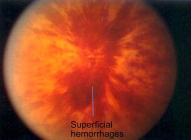
Retinal vein occlusion (RVO) is a medical condition that occurs when one of the retinal veins, which drains blood from the retina, becomes blocked by a blood clot or atherosclerotic plaque. This blockage can cause hemorrhages, fluid accumulation, and damage to the retinal tissue, leading to vision loss.
There are two types of RVO: branch retinal vein occlusion (BRVO) and central retinal vein occlusion (CRVO). BRVO affects a smaller branch retinal vein, while CRVO affects the main retinal vein. CRVO is generally associated with more severe vision loss than BRVO.
Risk factors for RVO include hypertension, diabetes, high cholesterol levels, smoking, and glaucoma. Age is also a significant risk factor, as RVO becomes more common with increasing age. Treatment options for RVO may include controlling underlying medical conditions, laser therapy, intravitreal injections of anti-VEGF agents or steroids, and surgery in some cases.
X-linked genetic diseases refer to a group of disorders caused by mutations in genes located on the X chromosome. These conditions primarily affect males since they have only one X chromosome and therefore don't have a second normal copy of the gene to compensate for the mutated one. Females, who have two X chromosomes, are typically less affected because they usually have one normal copy of the gene on their other X chromosome.
Examples of X-linked genetic diseases include Duchenne and Becker muscular dystrophy, hemophilia A and B, color blindness, and fragile X syndrome. Symptoms and severity can vary widely depending on the specific condition and the nature of the genetic mutation involved. Treatment options depend on the particular disease but may include physical therapy, medication, or in some cases, gene therapy.
The macula lutea, often simply referred to as the macula or fovea centralis, is a part of the eye that is responsible for central vision and color perception. It's located in the center of the retina, the light-sensitive tissue at the back of the eye. The macula contains a high concentration of pigments called xanthophylls, which give it a yellowish color and protect the photoreceptor cells in this area from damage by blue light.
The central part of the macula is called the fovea, which is a small depression that contains only cones, the photoreceptor cells responsible for color vision and high visual acuity. The fovea is surrounded by the parafovea and the perifovea, which contain both cones and rods, the photoreceptor cells responsible for low-light vision and peripheral vision.
Damage to the macula can result in a loss of central vision and color perception, a condition known as age-related macular degeneration (AMD), which is a leading cause of blindness in older adults. Other conditions that can affect the macula include macular edema, macular holes, and macular pucker.
'Radiation injuries, experimental' is not a widely recognized medical term. However, in the field of radiation biology and medicine, it may refer to the study and understanding of radiation-induced damage using various experimental models (e.g., cell cultures, animal models) before applying this knowledge to human health situations. These experiments aim to investigate the effects of ionizing radiation on living organisms' biological processes, tissue responses, and potential therapeutic interventions. The findings from these studies contribute to the development of medical countermeasures, diagnostic tools, and treatment strategies for accidental or intentional radiation exposures in humans.
Color vision is the ability to perceive and differentiate colors, which is a result of the way that our eyes and brain process different wavelengths of light. In the eye, there are two types of photoreceptor cells called rods and cones. While rods are more sensitive to low levels of light and help us see in dim conditions, cones are responsible for color vision.
There are three types of cone cells in the human eye, each containing a different type of pigment that is sensitive to specific wavelengths of light. One type of cone cell is most sensitive to short wavelengths (blue light), another is most sensitive to medium wavelengths (green light), and the third is most sensitive to long wavelengths (red light). When light enters the eye, it is absorbed by these pigments in the cones, which then send signals to the brain. The brain interprets these signals and translates them into the perception of color.
People with normal color vision can distinguish between millions of different colors based on the specific combinations of wavelengths that are present in a given scene. However, some people have deficiencies or abnormalities in their color vision, which can make it difficult or impossible to distinguish between certain colors. These conditions are known as color vision deficiencies or color blindness.
Bacterial eye infections, also known as bacterial conjunctivitis or bacterial keratitis, are caused by the invasion of bacteria into the eye. The most common types of bacteria that cause these infections include Staphylococcus aureus, Streptococcus pneumoniae, and Haemophilus influenzae.
Bacterial conjunctivitis is an inflammation of the conjunctiva, the thin membrane that covers the white part of the eye and the inner surface of the eyelids. Symptoms include redness, swelling, pain, discharge, and a gritty feeling in the eye. Bacterial keratitis is an infection of the cornea, the clear front part of the eye. Symptoms include severe pain, sensitivity to light, tearing, and decreased vision.
Bacterial eye infections are typically treated with antibiotic eye drops or ointments. It is important to seek medical attention promptly if you suspect a bacterial eye infection, as untreated infections can lead to serious complications such as corneal ulcers and vision loss. Preventive measures include good hygiene practices, such as washing your hands frequently and avoiding touching or rubbing your eyes.
The pigment epithelium of the eye, also known as the retinal pigment epithelium (RPE), is a layer of cells located between the photoreceptor cells of the retina and the choroid, which is the vascular layer of the eye. The RPE plays a crucial role in maintaining the health and function of the photoreceptors by providing them with nutrients, removing waste products, and helping to regulate the light that enters the eye.
The RPE cells contain pigment granules that absorb excess light, preventing it from scattering within the eye and improving visual acuity. They also help to create a barrier between the retina and the choroid, which is important for maintaining the proper functioning of the photoreceptors. Additionally, the RPE plays a role in the regeneration of visual pigments in the photoreceptor cells, allowing us to see in different light conditions.
Damage to the RPE can lead to various eye diseases and conditions, including age-related macular degeneration (AMD), which is a leading cause of vision loss in older adults.
Photoreceptor cells are specialized neurons in the retina of the eye that convert light into electrical signals. These cells consist of two types: rods and cones. Rods are responsible for vision at low light levels and provide black-and-white, peripheral, and motion sensitivity. Cones are active at higher light levels and are capable of color discrimination and fine detail vision. Both types of photoreceptor cells contain light-sensitive pigments that undergo chemical changes when exposed to light, triggering a series of electrical signals that ultimately reach the brain and contribute to visual perception.
Evoked potentials, visual, also known as visually evoked potentials (VEPs), are electrical responses recorded from the brain following the presentation of a visual stimulus. These responses are typically measured using electroencephalography (EEG) and can provide information about the functioning of the visual pathways in the brain.
There are several types of VEPs, including pattern-reversal VEPs and flash VEPs. Pattern-reversal VEPs are elicited by presenting alternating checkerboard patterns, while flash VEPs are elicited by flashing a light. The responses are typically analyzed in terms of their latency (the time it takes for the response to occur) and amplitude (the size of the response).
VEPs are often used in clinical settings to help diagnose and monitor conditions that affect the visual system, such as multiple sclerosis, optic neuritis, and brainstem tumors. They can also be used in research to study the neural mechanisms underlying visual perception.
Retinal Ganglion Cells (RGCs) are a type of neuron located in the innermost layer of the retina, the light-sensitive tissue at the back of the eye. These cells receive visual information from photoreceptors (rods and cones) via intermediate cells called bipolar cells. RGCs then send this visual information through their long axons to form the optic nerve, which transmits the signals to the brain for processing and interpretation as vision.
There are several types of RGCs, each with distinct morphological and functional characteristics. Some RGCs are specialized in detecting specific features of the visual scene, such as motion, contrast, color, or brightness. The diversity of RGCs allows for a rich and complex representation of the visual world in the brain.
Damage to RGCs can lead to various visual impairments, including loss of vision, reduced visual acuity, and altered visual fields. Conditions associated with RGC damage or degeneration include glaucoma, optic neuritis, ischemic optic neuropathy, and some inherited retinal diseases.
In the context of medical terminology, "light" doesn't have a specific or standardized definition on its own. However, it can be used in various medical terms and phrases. For example, it could refer to:
1. Visible light: The range of electromagnetic radiation that can be detected by the human eye, typically between wavelengths of 400-700 nanometers. This is relevant in fields such as ophthalmology and optometry.
2. Therapeutic use of light: In some therapies, light is used to treat certain conditions. An example is phototherapy, which uses various wavelengths of ultraviolet (UV) or visible light for conditions like newborn jaundice, skin disorders, or seasonal affective disorder.
3. Light anesthesia: A state of reduced consciousness in which the patient remains responsive to verbal commands and physical stimulation. This is different from general anesthesia where the patient is completely unconscious.
4. Pain relief using light: Certain devices like transcutaneous electrical nerve stimulation (TENS) units have a 'light' setting, indicating lower intensity or frequency of electrical impulses used for pain management.
Without more context, it's hard to provide a precise medical definition of 'light'.
C57BL/6 (C57 Black 6) is an inbred strain of laboratory mouse that is widely used in biomedical research. The term "inbred" refers to a strain of animals where matings have been carried out between siblings or other closely related individuals for many generations, resulting in a population that is highly homozygous at most genetic loci.
The C57BL/6 strain was established in 1920 by crossing a female mouse from the dilute brown (DBA) strain with a male mouse from the black strain. The resulting offspring were then interbred for many generations to create the inbred C57BL/6 strain.
C57BL/6 mice are known for their robust health, longevity, and ease of handling, making them a popular choice for researchers. They have been used in a wide range of biomedical research areas, including studies of cancer, immunology, neuroscience, cardiovascular disease, and metabolism.
One of the most notable features of the C57BL/6 strain is its sensitivity to certain genetic modifications, such as the introduction of mutations that lead to obesity or impaired glucose tolerance. This has made it a valuable tool for studying the genetic basis of complex diseases and traits.
Overall, the C57BL/6 inbred mouse strain is an important model organism in biomedical research, providing a valuable resource for understanding the genetic and molecular mechanisms underlying human health and disease.
A phenotype is the physical or biochemical expression of an organism's genes, or the observable traits and characteristics resulting from the interaction of its genetic constitution (genotype) with environmental factors. These characteristics can include appearance, development, behavior, and resistance to disease, among others. Phenotypes can vary widely, even among individuals with identical genotypes, due to differences in environmental influences, gene expression, and genetic interactions.
Rhodopsin, also known as visual purple, is a light-sensitive protein found in the rods of the eye's retina. It is a type of opsin, a class of proteins that are activated by light and play a crucial role in vision. Rhodopsin is composed of two parts: an apoprotein called opsin and a chromophore called 11-cis-retinal. When light hits the retina, it changes the shape of the 11-cis-retinal, which in turn activates the rhodopsin protein. This activation triggers a series of chemical reactions that ultimately lead to the transmission of a visual signal to the brain. Rhodopsin is highly sensitive to light and allows for vision in low-light conditions.
A mutation is a permanent change in the DNA sequence of an organism's genome. Mutations can occur spontaneously or be caused by environmental factors such as exposure to radiation, chemicals, or viruses. They may have various effects on the organism, ranging from benign to harmful, depending on where they occur and whether they alter the function of essential proteins. In some cases, mutations can increase an individual's susceptibility to certain diseases or disorders, while in others, they may confer a survival advantage. Mutations are the driving force behind evolution, as they introduce new genetic variability into populations, which can then be acted upon by natural selection.
The choroid is a layer of the eye that contains blood vessels that supply oxygen and nutrients to the outer layers of the retina. It lies between the sclera (the white, protective coat of the eye) and the retina (the light-sensitive tissue at the back of the eye). The choroid is essential for maintaining the health and function of the retina, particularly the photoreceptor cells that detect light and transmit visual signals to the brain. Damage to the choroid can lead to vision loss or impairment.
The optic nerve, also known as the second cranial nerve, is the nerve that transmits visual information from the retina to the brain. It is composed of approximately one million nerve fibers that carry signals related to vision, such as light intensity and color, from the eye's photoreceptor cells (rods and cones) to the visual cortex in the brain. The optic nerve is responsible for carrying this visual information so that it can be processed and interpreted by the brain, allowing us to see and perceive our surroundings. Damage to the optic nerve can result in vision loss or impairment.
Consanguinity is a medical and genetic term that refers to the degree of genetic relationship between two individuals who share common ancestors. Consanguineous relationships exist when people are related by blood, through a common ancestor or siblings who have children together. The closer the relationship between the two individuals, the higher the degree of consanguinity.
The degree of consanguinity is typically expressed as a percentage or fraction, with higher values indicating a closer genetic relationship. For example, first-degree relatives, such as parents and children or full siblings, share approximately 50% of their genes and have a consanguinity coefficient of 0.25 (or 25%).
Consanguinity can increase the risk of certain genetic disorders and birth defects in offspring due to the increased likelihood of sharing harmful recessive genes. The risks depend on the degree of consanguinity, with closer relationships carrying higher risks. It is important for individuals who are planning to have children and have a history of consanguinity to consider genetic counseling and testing to assess their risk of passing on genetic disorders.
Intraocular pressure (IOP) is the fluid pressure within the eye, specifically within the anterior chamber, which is the space between the cornea and the iris. It is measured in millimeters of mercury (mmHg). The aqueous humor, a clear fluid that fills the anterior chamber, is constantly produced and drained, maintaining a balance that determines the IOP. Normal IOP ranges from 10-21 mmHg, with average values around 15-16 mmHg. Elevated IOP is a key risk factor for glaucoma, a group of eye conditions that can lead to optic nerve damage and vision loss if not treated promptly and effectively. Regular monitoring of IOP is essential in diagnosing and managing glaucoma and other ocular health issues.
A dependovirus, also known as a dependent adenovirus or satellite adenovirus, is a type of virus that requires the presence of another virus, specifically an adenovirus, to replicate. Dependoviruses are small, non-enveloped viruses with a double-stranded DNA genome. They cannot complete their replication cycle without the help of an adenovirus, which provides necessary functions for the dependovirus to replicate.
Dependoviruses are clinically significant because they can cause disease in humans, particularly in individuals with weakened immune systems. In some cases, dependoviruses may also affect the severity and outcome of adenovirus infections. However, it is important to note that not all adenovirus infections are associated with dependovirus co-infections.
A "mutant strain of mice" in a medical context refers to genetically engineered mice that have specific genetic mutations introduced into their DNA. These mutations can be designed to mimic certain human diseases or conditions, allowing researchers to study the underlying biological mechanisms and test potential therapies in a controlled laboratory setting.
Mutant strains of mice are created through various techniques, including embryonic stem cell manipulation, gene editing technologies such as CRISPR-Cas9, and radiation-induced mutagenesis. These methods allow scientists to introduce specific genetic changes into the mouse genome, resulting in mice that exhibit altered physiological or behavioral traits.
These strains of mice are widely used in biomedical research because their short lifespan, small size, and high reproductive rate make them an ideal model organism for studying human diseases. Additionally, the mouse genome has been well-characterized, and many genetic tools and resources are available to researchers working with these animals.
Examples of mutant strains of mice include those that carry mutations in genes associated with cancer, neurodegenerative disorders, metabolic diseases, and immunological conditions. These mice provide valuable insights into the pathophysiology of human diseases and help advance our understanding of potential therapeutic interventions.
Ischemic preconditioning is a phenomenon in which brief, non-lethal episodes of ischemia (restriction or interruption of blood supply to an organ or tissue) render the tissue more resistant to subsequent prolonged ischemia and reperfusion injury. This adaptive response involves a complex series of cellular and molecular changes that protect the myocardium, brain, kidney, or other organs from ischemic damage. The underlying mechanisms include the activation of various signaling pathways, such as adenosine, opioid, and kinase pathways, which lead to the production of protective factors and the modulation of cellular responses to ischemia and reperfusion injury. Ischemic preconditioning has been extensively studied in the context of cardiovascular medicine, where it has been shown to reduce infarct size and improve cardiac function after myocardial infarction. However, this protective phenomenon has also been observed in other organs and systems, including the brain, kidney, liver, and skeletal muscle.
A "knockout" mouse is a genetically engineered mouse in which one or more genes have been deleted or "knocked out" using molecular biology techniques. This allows researchers to study the function of specific genes and their role in various biological processes, as well as potential associations with human diseases. The mice are generated by introducing targeted DNA modifications into embryonic stem cells, which are then used to create a live animal. Knockout mice have been widely used in biomedical research to investigate gene function, disease mechanisms, and potential therapeutic targets.
Dominant genes refer to the alleles (versions of a gene) that are fully expressed in an individual's phenotype, even if only one copy of the gene is present. In dominant inheritance patterns, an individual needs only to receive one dominant allele from either parent to express the associated trait. This is in contrast to recessive genes, where both copies of the gene must be the recessive allele for the trait to be expressed. Dominant genes are represented by uppercase letters (e.g., 'A') and recessive genes by lowercase letters (e.g., 'a'). If an individual inherits one dominant allele (A) from either parent, they will express the dominant trait (A).
"Cell count" is a medical term that refers to the process of determining the number of cells present in a given volume or sample of fluid or tissue. This can be done through various laboratory methods, such as counting individual cells under a microscope using a specialized grid called a hemocytometer, or using automated cell counters that use light scattering and electrical impedance techniques to count and classify different types of cells.
Cell counts are used in a variety of medical contexts, including hematology (the study of blood and blood-forming tissues), microbiology (the study of microscopic organisms), and pathology (the study of diseases and their causes). For example, a complete blood count (CBC) is a routine laboratory test that includes a white blood cell (WBC) count, red blood cell (RBC) count, hemoglobin level, hematocrit value, and platelet count. Abnormal cell counts can indicate the presence of various medical conditions, such as infections, anemia, or leukemia.
Intermediate filament proteins (IFPs) are a type of cytoskeletal protein that form the intermediate filaments (IFs), which are one of the three major components of the cytoskeleton in eukaryotic cells, along with microtubules and microfilaments. These proteins have a unique structure, characterized by an alpha-helical rod domain flanked by non-helical head and tail domains.
Intermediate filament proteins are classified into six major types based on their amino acid sequence: Type I (acidic) and Type II (basic) keratins, Type III (desmin, vimentin, glial fibrillary acidic protein, and peripherin), Type IV (neurofilaments), Type V (lamins), and Type VI (nestin). Each type of IFP has a distinct pattern of expression in different tissues and cell types.
Intermediate filament proteins play important roles in maintaining the structural integrity and mechanical strength of cells, providing resilience to mechanical stress, and regulating various cellular processes such as cell division, migration, and signal transduction. Mutations in IFP genes have been associated with several human diseases, including cancer, neurodegenerative disorders, and genetic skin fragility disorders.
Photic stimulation is a medical term that refers to the exposure of the eyes to light, specifically repetitive pulses of light, which is used as a method in various research and clinical settings. In neuroscience, it's often used in studies related to vision, circadian rhythms, and brain function.
In a clinical context, photic stimulation is sometimes used in the diagnosis of certain medical conditions such as seizure disorders (like epilepsy). By observing the response of the brain to this light stimulus, doctors can gain valuable insights into the functioning of the brain and the presence of any neurological disorders.
However, it's important to note that photic stimulation should be conducted under the supervision of a trained healthcare professional, as improper use can potentially trigger seizures in individuals who are susceptible to them.
A heterozygote is an individual who has inherited two different alleles (versions) of a particular gene, one from each parent. This means that the individual's genotype for that gene contains both a dominant and a recessive allele. The dominant allele will be expressed phenotypically (outwardly visible), while the recessive allele may or may not have any effect on the individual's observable traits, depending on the specific gene and its function. Heterozygotes are often represented as 'Aa', where 'A' is the dominant allele and 'a' is the recessive allele.
Reperfusion injury is a complex pathophysiological process that occurs when blood flow is restored to previously ischemic tissues, leading to further tissue damage. This phenomenon can occur in various clinical settings such as myocardial infarction (heart attack), stroke, or peripheral artery disease after an intervention aimed at restoring perfusion.
The restoration of blood flow leads to the generation of reactive oxygen species (ROS) and inflammatory mediators, which can cause oxidative stress, cellular damage, and activation of the immune system. This results in a cascade of events that may lead to microvascular dysfunction, capillary leakage, and tissue edema, further exacerbating the injury.
Reperfusion injury is an important consideration in the management of ischemic events, as interventions aimed at restoring blood flow must be carefully balanced with potential harm from reperfusion injury. Strategies to mitigate reperfusion injury include ischemic preconditioning (exposing the tissue to short periods of ischemia before a prolonged ischemic event), ischemic postconditioning (applying brief periods of ischemia and reperfusion after restoring blood flow), remote ischemic preconditioning (ischemia applied to a distant organ or tissue to protect the target organ), and pharmacological interventions that scavenge ROS, reduce inflammation, or improve microvascular function.
In situ nick-end labeling (ISEL, also known as TUNEL) is a technique used in pathology and molecular biology to detect DNA fragmentation, which is a characteristic of apoptotic cells (cells undergoing programmed cell death). The method involves labeling the 3'-hydroxyl termini of double or single stranded DNA breaks in situ (within tissue sections or individual cells) using modified nucleotides that are coupled to a detectable marker, such as a fluorophore or an enzyme. This technique allows for the direct visualization and quantification of apoptotic cells within complex tissues or cell populations.
I believe there may be some confusion in your question. "Rabbits" is a common name used to refer to the Lagomorpha species, particularly members of the family Leporidae. They are small mammals known for their long ears, strong legs, and quick reproduction.
However, if you're referring to "rabbits" in a medical context, there is a term called "rabbit syndrome," which is a rare movement disorder characterized by repetitive, involuntary movements of the fingers, resembling those of a rabbit chewing. It is also known as "finger-chewing chorea." This condition is usually associated with certain medications, particularly antipsychotics, and typically resolves when the medication is stopped or adjusted.
The cornea is the clear, dome-shaped surface at the front of the eye. It plays a crucial role in focusing vision. The cornea protects the eye from harmful particles and microorganisms, and it also serves as a barrier against UV light. Its transparency allows light to pass through and get focused onto the retina. The cornea does not contain blood vessels, so it relies on tears and the fluid inside the eye (aqueous humor) for nutrition and oxygen. Any damage or disease that affects its clarity and shape can significantly impact vision and potentially lead to blindness if left untreated.
Fluorescence is not a medical term per se, but it is widely used in the medical field, particularly in diagnostic tests, medical devices, and research. Fluorescence is a physical phenomenon where a substance absorbs light at a specific wavelength and then emits light at a longer wavelength. This process, often referred to as fluorescing, results in the emission of visible light that can be detected and measured.
In medical terms, fluorescence is used in various applications such as:
1. In-vivo imaging: Fluorescent dyes or probes are introduced into the body to highlight specific structures, cells, or molecules during imaging procedures. This technique can help doctors detect and diagnose diseases such as cancer, inflammation, or infection.
2. Microscopy: Fluorescence microscopy is a powerful tool for visualizing biological samples at the cellular and molecular level. By labeling specific proteins, nucleic acids, or other molecules with fluorescent dyes, researchers can observe their distribution, interactions, and dynamics within cells and tissues.
3. Surgical guidance: Fluorescence-guided surgery is a technique where surgeons use fluorescent markers to identify critical structures such as blood vessels, nerves, or tumors during surgical procedures. This helps ensure precise and safe surgical interventions.
4. Diagnostic tests: Fluorescence-based assays are used in various diagnostic tests to detect and quantify specific biomarkers or analytes. These assays can be performed using techniques such as enzyme-linked immunosorbent assay (ELISA), polymerase chain reaction (PCR), or flow cytometry.
In summary, fluorescence is a physical process where a substance absorbs and emits light at different wavelengths. In the medical field, this phenomenon is harnessed for various applications such as in-vivo imaging, microscopy, surgical guidance, and diagnostic tests.
Diabetic retinopathy is a diabetes complication that affects the eyes. It's caused by damage to the blood vessels of the light-sensitive tissue at the back of the eye (retina).
At first, diabetic retinopathy may cause no symptoms or only mild vision problems. Eventually, it can cause blindness. The condition usually affects both eyes.
There are two main stages of diabetic retinopathy:
1. Early diabetic retinopathy. This is when the blood vessels in the eye start to leak fluid or bleed. You might not notice any changes in your vision at this stage, but it's still important to get treatment because it can prevent the condition from getting worse.
2. Advanced diabetic retinopathy. This is when new, abnormal blood vessels grow on the surface of the retina. These vessels can leak fluid and cause severe vision problems, including blindness.
Diabetic retinopathy can be treated with laser surgery, injections of medication into the eye, or a vitrectomy (a surgical procedure to remove the gel-like substance that fills the center of the eye). It's important to get regular eye exams to detect diabetic retinopathy early and get treatment before it causes serious vision problems.
Glial Fibrillary Acidic Protein (GFAP) is a type of intermediate filament protein that is primarily found in astrocytes, which are a type of star-shaped glial cells in the central nervous system (CNS). These proteins play an essential role in maintaining the structural integrity and stability of astrocytes. They also participate in various cellular processes such as responding to injury, providing support to neurons, and regulating the extracellular environment.
GFAP is often used as a marker for astrocytic activation or reactivity, which can occur in response to CNS injuries, neuroinflammation, or neurodegenerative diseases. Elevated GFAP levels in cerebrospinal fluid (CSF) or blood can indicate astrocyte damage or dysfunction and are associated with several neurological conditions, including traumatic brain injury, stroke, multiple sclerosis, Alzheimer's disease, and Alexander's disease.
A homozygote is an individual who has inherited the same allele (version of a gene) from both parents and therefore possesses two identical copies of that allele at a specific genetic locus. This can result in either having two dominant alleles (homozygous dominant) or two recessive alleles (homozygous recessive). In contrast, a heterozygote has inherited different alleles from each parent for a particular gene.
The term "homozygote" is used in genetics to describe the genetic makeup of an individual at a specific locus on their chromosomes. Homozygosity can play a significant role in determining an individual's phenotype (observable traits), as having two identical alleles can strengthen the expression of certain characteristics compared to having just one dominant and one recessive allele.
A missense mutation is a type of point mutation in which a single nucleotide change results in the substitution of a different amino acid in the protein that is encoded by the affected gene. This occurs when the altered codon (a sequence of three nucleotides that corresponds to a specific amino acid) specifies a different amino acid than the original one. The function and/or stability of the resulting protein may be affected, depending on the type and location of the missense mutation. Missense mutations can have various effects, ranging from benign to severe, depending on the importance of the changed amino acid for the protein's structure or function.
Western blotting is a laboratory technique used in molecular biology to detect and quantify specific proteins in a mixture of many different proteins. This technique is commonly used to confirm the expression of a protein of interest, determine its size, and investigate its post-translational modifications. The name "Western" blotting distinguishes this technique from Southern blotting (for DNA) and Northern blotting (for RNA).
The Western blotting procedure involves several steps:
1. Protein extraction: The sample containing the proteins of interest is first extracted, often by breaking open cells or tissues and using a buffer to extract the proteins.
2. Separation of proteins by electrophoresis: The extracted proteins are then separated based on their size by loading them onto a polyacrylamide gel and running an electric current through the gel (a process called sodium dodecyl sulfate-polyacrylamide gel electrophoresis or SDS-PAGE). This separates the proteins according to their molecular weight, with smaller proteins migrating faster than larger ones.
3. Transfer of proteins to a membrane: After separation, the proteins are transferred from the gel onto a nitrocellulose or polyvinylidene fluoride (PVDF) membrane using an electric current in a process called blotting. This creates a replica of the protein pattern on the gel but now immobilized on the membrane for further analysis.
4. Blocking: The membrane is then blocked with a blocking agent, such as non-fat dry milk or bovine serum albumin (BSA), to prevent non-specific binding of antibodies in subsequent steps.
5. Primary antibody incubation: A primary antibody that specifically recognizes the protein of interest is added and allowed to bind to its target protein on the membrane. This step may be performed at room temperature or 4°C overnight, depending on the antibody's properties.
6. Washing: The membrane is washed with a buffer to remove unbound primary antibodies.
7. Secondary antibody incubation: A secondary antibody that recognizes the primary antibody (often coupled to an enzyme or fluorophore) is added and allowed to bind to the primary antibody. This step may involve using a horseradish peroxidase (HRP)-conjugated or alkaline phosphatase (AP)-conjugated secondary antibody, depending on the detection method used later.
8. Washing: The membrane is washed again to remove unbound secondary antibodies.
9. Detection: A detection reagent is added to visualize the protein of interest by detecting the signal generated from the enzyme-conjugated or fluorophore-conjugated secondary antibody. This can be done using chemiluminescent, colorimetric, or fluorescent methods.
10. Analysis: The resulting image is analyzed to determine the presence and quantity of the protein of interest in the sample.
Western blotting is a powerful technique for identifying and quantifying specific proteins within complex mixtures. It can be used to study protein expression, post-translational modifications, protein-protein interactions, and more. However, it requires careful optimization and validation to ensure accurate and reproducible results.
I believe there might be a misunderstanding in your question. "Dogs" is not a medical term or condition. It is the common name for a domesticated carnivore of the family Canidae, specifically the genus Canis, which includes wolves, foxes, and other extant and extinct species of mammals. Dogs are often kept as pets and companions, and they have been bred in a wide variety of forms and sizes for different purposes, such as hunting, herding, guarding, assisting police and military forces, and providing companionship and emotional support.
If you meant to ask about a specific medical condition or term related to dogs, please provide more context so I can give you an accurate answer.
Genotype, in genetics, refers to the complete heritable genetic makeup of an individual organism, including all of its genes. It is the set of instructions contained in an organism's DNA for the development and function of that organism. The genotype is the basis for an individual's inherited traits, and it can be contrasted with an individual's phenotype, which refers to the observable physical or biochemical characteristics of an organism that result from the expression of its genes in combination with environmental influences.
It is important to note that an individual's genotype is not necessarily identical to their genetic sequence. Some genes have multiple forms called alleles, and an individual may inherit different alleles for a given gene from each parent. The combination of alleles that an individual inherits for a particular gene is known as their genotype for that gene.
Understanding an individual's genotype can provide important information about their susceptibility to certain diseases, their response to drugs and other treatments, and their risk of passing on inherited genetic disorders to their offspring.
Exons are the coding regions of DNA that remain in the mature, processed mRNA after the removal of non-coding intronic sequences during RNA splicing. These exons contain the information necessary to encode proteins, as they specify the sequence of amino acids within a polypeptide chain. The arrangement and order of exons can vary between different genes and even between different versions of the same gene (alternative splicing), allowing for the generation of multiple protein isoforms from a single gene. This complexity in exon structure and usage significantly contributes to the diversity and functionality of the proteome.
Genetic linkage is the phenomenon where two or more genetic loci (locations on a chromosome) tend to be inherited together because they are close to each other on the same chromosome. This occurs during the process of sexual reproduction, where homologous chromosomes pair up and exchange genetic material through a process called crossing over.
The closer two loci are to each other on a chromosome, the lower the probability that they will be separated by a crossover event. As a result, they are more likely to be inherited together and are said to be linked. The degree of linkage between two loci can be measured by their recombination frequency, which is the percentage of meiotic events in which a crossover occurs between them.
Linkage analysis is an important tool in genetic research, as it allows researchers to identify and map genes that are associated with specific traits or diseases. By analyzing patterns of linkage between markers (identifiable DNA sequences) and phenotypes (observable traits), researchers can infer the location of genes that contribute to those traits or diseases on chromosomes.
Ischemia is the medical term used to describe a lack of blood flow to a part of the body, often due to blocked or narrowed blood vessels. This can lead to a shortage of oxygen and nutrients in the tissues, which can cause them to become damaged or die. Ischemia can affect many different parts of the body, including the heart, brain, legs, and intestines. Symptoms of ischemia depend on the location and severity of the blockage, but they may include pain, cramping, numbness, weakness, or coldness in the affected area. In severe cases, ischemia can lead to tissue death (gangrene) or organ failure. Treatment for ischemia typically involves addressing the underlying cause of the blocked blood flow, such as through medication, surgery, or lifestyle changes.
Polymerase Chain Reaction (PCR) is a laboratory technique used to amplify specific regions of DNA. It enables the production of thousands to millions of copies of a particular DNA sequence in a rapid and efficient manner, making it an essential tool in various fields such as molecular biology, medical diagnostics, forensic science, and research.
The PCR process involves repeated cycles of heating and cooling to separate the DNA strands, allow primers (short sequences of single-stranded DNA) to attach to the target regions, and extend these primers using an enzyme called Taq polymerase, resulting in the exponential amplification of the desired DNA segment.
In a medical context, PCR is often used for detecting and quantifying specific pathogens (viruses, bacteria, fungi, or parasites) in clinical samples, identifying genetic mutations or polymorphisms associated with diseases, monitoring disease progression, and evaluating treatment effectiveness.
 Electroretinography
 Electroretinography: MedlinePlus Medical Encyclopedia
 Electroretinography in Fairfax, VA | VCA SouthPaws Veterinary Specialists & Emergency Center
 Effects of Mesenchymal Stem Cell-Derived Exosomes on Experimental Autoimmune Uveitis | Scientific Reports
 2020-2021 BCSC Basic and Clinical Science Course™
 IJMS | Free Full-Text | Nicotinamide Mononucleotide Prevents Retinal Dysfunction in a Mouse Model of Retinal Ischemia...
 Molecular Vision: Exendin-4 protects retinal cells from early diabetes in Goto-Kakizaki rats by increasing the Bcl-2/Bax and...
 Electrophysiology Tests for the Eyes | Cigna
 ISCEV - ARVO SIG: Electroretinography - Current use, future needs and new developments
 Retinitis pigmentosa: Symptoms, causes, treatment, and more
 ISCEV Standards, Guidelines and Extended Protocols | SpringerLink
 Mucopolysaccharidosis: Practice Essentials, Pathophysiology, Etiology
 Whole-Exome Sequencing Identifies Biallelic IDH3A Variants as a Cause of Retinitis Pigmentosa Accompanied by Pseudocoloboma
 The study of relation between pre-eclampsia and retinal disorders using electroretinography - Medical Science Journal of...
 NEWS - esvo
 Phenominer
 Ophthalmology - College of Veterinary Medicine - Purdue University
 Frontiers | The Eye As a Biomarker for Alzheimer's Disease
 Browsing Optometry and Vision Science by Title