Electron Transport Complex III
Electron Transport Complex I
Electron Transport
Mitochondria
Electron Transport Complex II
Electron Transport Complex IV
Electron Transport Chain Complex Proteins
Antimycin A
Maltose
Biological Transport
Maltose-Binding Proteins
Biological Transport, Active
Electrons
Periplasmic Binding Proteins
Microscopy, Electron
ATP-Binding Cassette Transporters
Ubiquinone
Coat Protein Complex I
Monosaccharide Transport Proteins
Quinone Reductases
Oxidation-Reduction
Cytochromes b
Oxidative Phosphorylation
Membrane Transport Proteins
Microscopy, Electron, Scanning
Photosynthesis
Carrier Proteins
Cytochromes
Molecular Sequence Data
Adenosine Triphosphate
Protein Transport
Cytochrome b Group
Reactive Oxygen Species
Cytochromes c1
Models, Biological
Uncoupling Agents
Mutation
Oxygen Consumption
Hydroxyquinolines
Amino Acid Sequence
Membrane Proteins
Cell Membrane
NADH, NADPH Oxidoreductases
Cell Respiration
Potassium Cyanide
Cytochrome c Group
Oxidoreductases
Cytochrome Reductases
Protein Binding
NADH Dehydrogenase
Adenosine Triphosphatases
Methacrylates
Succinic Acid
Escherichia coli
Rhodothermus
Succinate Dehydrogenase
Electron Spin Resonance Spectroscopy
Axonal Transport
Oxygen
Chlorophyll
Mitochondrial Diseases
Flagella
Models, Molecular
Multienzyme Complexes
Ion Transport
Mitochondria, Muscle
Mitochondrial Proteins
Microscopy, Electron, Transmission
Iron-Sulfur Proteins
Plastoquinone
Cardiolipins
Saccharomyces cerevisiae Proteins
Submitochondrial Particles
NAD
Hydrogen Peroxide
Cyanides
Mitochondria, Liver
Photosystem II Protein Complex
Chloroplasts
Succinates
Photosystem I Protein Complex
Spectrophotometry
Succinate Cytochrome c Oxidoreductase
Thylakoids
Cytochrome b6f Complex
Membrane Potential, Mitochondrial
NAD(P)H Dehydrogenase (Quinone)
DNA, Mitochondrial
Protein Structure, Tertiary
Photosynthetic Reaction Center Complex Proteins
Saccharomyces cerevisiae
Superoxides
Cattle
Carbodiimides
Mitochondrial Membranes
Intracellular Membranes
Oligomycins
Amobarbital
Hydrogen-Ion Concentration
Dicyclohexylcarbodiimide
Cytochromes c
Oxidative Stress
Malates
Quinones
Ferredoxins
Heme
Plant Leaves
Brain Diseases, Metabolic, Inborn
Energy Metabolism
Thiazoles
ATP Synthetase Complexes
Methylphenazonium Methosulfate
Dibromothymoquinone
Hydrogen
Plastocyanin
Base Sequence
Protons
Anaerobiosis
Photophosphorylation
Cells, Cultured
Wolinella
Cyanobacteria
Ascorbic Acid
Chloroflexus
Glucose
Sodium
Plants
Binding Sites
NADP
Shewanella
Transport Vesicles
Dicumarol
Cytochromes f
Temperature
Light-Harvesting Protein Complexes
Carbonyl Cyanide p-Trifluoromethoxyphenylhydrazone
Aconitate Hydratase
Sulfanilic Acids
Synechocystis
Fluorescence
Plant Proteins
Carbonyl Cyanide m-Chlorophenyl Hydrazone
Membrane Potentials
Membranes
Ferredoxin-NADP Reductase
Electrophoresis, Polyacrylamide Gel
Carbon Dioxide
Protein Conformation
Evidence for the head domain movement of the rieske iron-sulfur protein in electron transfer reaction of the cytochrome bc1 complex. (1/979)
The three-dimensional structure of the mitochondrial cytochrome bc1 complex suggests that movement of the extramembrane domain (head) of the Rieske iron-sulfur protein (ISP) may play an important role in electron transfer. Such movement requires flexibility in the neck region of ISP, since the head and transmembrane domains of the protein are rather rigid. To test this hypothesis, Rhodobacter sphaeroides mutants expressing His-tagged cytochrome bc1 complexes with cysteine substitution at various positions in the ISP neck (residues 39-48) were generated and characterized. The mutants with a single cysteine substitution at Ala42 or Val44 and a double cysteine substitution at Val44 and Ala46 (VQA-CQC) or at Ala42 and Ala46 (ADVQA-CDVQC) have photosynthetic growth rates comparable with that of complement cells. Chromatophore membrane and intracytoplasmic membrane (ICM) prepared from these mutants have cytochrome bc1 complex activity similar to that in the complement membranes, indicating that flexibility of the neck region of ISP was not affected by these cysteine substitutions. Mutants with a double cysteine substitution at Ala42 and Val44 (ADV-CDC) or at Pro40 and Ala42 (PSA-CSC) have a retarded (50%) or no photosynthetic growth rate, respectively. The ADV-CDC or PSA-CSC mutant ICM contains 20 or 0% of the cytochrome bc1 complex activity found in the complement ICM. However, activity can be restored by the treatment with beta-mercaptoethanol (beta-ME). The restored activity is diminished upon removal of beta-ME but is retained if the beta-ME-treated membrane is treated with the sulfhydryl reagent N-ethylmaleimide or p-chloromercuribenzoic acid. These results indicate that the loss of bc1 complex activity in the ADV-CDC or PSA-CSC mutant membranes is due to disulfide bond formation, which increases the rigidity of ISP neck and, in turn, decreases the mobility of the head domain. Using the conditions developed for the isolation of His-tagged complement cytochrome bc1 complex, a two-subunit complex (cytochromes b and c1) is obtained from all of the double cysteine-substituted mutants. This suggests that introduction of two cysteines in the neck region of ISP weakens the interactions between cytochromes b, ISP, and subunit IV. (+info)Aromatic amino acids in the Rieske iron-sulfur protein do not form an obligatory conduit for electron transfer from the iron-sulfur cluster to the heme of cytochrome c1 in the cytochrome bc1 complex. (2/979)
We have changed nine conserved aromatic amino acids by site-directed mutagenesis of the cloned iron-sulfur protein gene to determine if any of these residues form an obligatory conduit for electron transfer within the iron-sulfur protein of the yeast cytochrome bc1 complex. The residues include W111, F117, W152, F173, W176, F177, H184, Y205 and F207. Greater than 70% of the catalytic activity was retained for all of the mutated iron-sulfur proteins, except for those containing a W152L and a W176L-F177L double mutation, for which the activity was approximately 45%. The crystal structures of the bc1 complex indicate that F177 and H184 are at the surface of the iron-sulfur protein near the surface of cytochrome c1, but not directly in a linear pathway between the iron-sulfur cluster and the c1 heme. The pre-steady-state rates of reduction of cytochromes b and c1 in mutants in which F177 and H184 were changed to non-aromatic residues were approximately 70-85% of the wild-type rates. There was a large decrease in iron-sulfur protein levels in mitochondrial membranes resulting from the W152L mutation and the W176L-F177L double mutation, and a small decrease for the Y205L, W176L and F177L mutations. This indicates that the decreases in activity resulting from these amino acid changes are due to instability of the altered proteins. These results show that these aromatic amino acids are unnecessary for electron transfer, but several are required for structural stability. (+info)Q-Band resonance Raman investigation of turnip cytochrome f and Rhodobacter capsulatus cytochrome c1. (3/979)
The results of a comprehensive Q-band resonance Raman investigation of cytochrome c1 and cytochrome f subunits of bc1 and b6f complexes are presented. Q-band excitation provides a particularly effective probe of the local heme environments of these species. The effects of protein conformation (particularly axial ligation) on heme structure and function were further investigated by comparison of spectra obtained from native subunits to those of a site directed c1 mutant (M183L) and various pH-dependent species of horse heart cytochrome c. In general, all species examined displayed variability in their axial amino acid ligation that suggests a good deal of flexibility in their hemepocket conformations. Surprisingly, the large scale protein rearrangements that accompany axial ligand replacement have little or no effect on macrocycle geometry in these species. This indicates the identity and/or conformation of the peptide linkage between the two cysteines that are covalently linked to the heme periphery may determine heme geometry. (+info)Intermediate length Rieske iron-sulfur protein is present and functionally active in the cytochrome bc1 complex of Saccharomyces cerevisiae. (4/979)
To investigate the relationship between post-translational processing of the Rieske iron-sulfur protein of Saccharomyces cerevisiae and its assembly into the mitochondrial cytochrome bc1 complex we used iron-sulfur proteins in which the presequences had been changed by site-directed mutagenesis of the cloned iron-sulfur protein gene, so that the recognition sites for the matrix processing peptidase or the mitochondrial intermediate peptidase (MIP) had been destroyed. When yeast strain JPJ1, in which the gene for the iron-sulfur protein is deleted, was transformed with these constructs on a single copy expression vector, mitochondrial membranes and bc1 complexes isolated from these strains accumulated intermediate length iron-sulfur proteins in vivo. The cytochrome bc1 complex activities of these membranes and bc1 complexes indicate that intermediate iron-sulfur protein (i-ISP) has full activity when compared with that of mature sized iron-sulfur protein (m-ISP). Therefore the iron-sulfur cluster must have been inserted before processing of i-ISP to m-ISP by MIP. When iron-sulfur protein is imported into mitochondria in vitro, i-ISP interacts with components of the bc1 complex before it is processed to m-ISP. These results establish that the iron-sulfur cluster is inserted into the apoprotein before MIP cleaves off the second part of the presequence and that this second processing step takes place after i-ISP has been assembled into the bc1 complex. (+info)Ubiquinol:cytochrome c oxidoreductase. Effects of inhibitors on reverse electron transfer from the iron-sulfur protein to cytochrome b. (5/979)
The effects of inhibitors on the reduction of the bis-heme cytochrome b of ubiquinol: cytochrome c oxidoreductase (complex III, bc1 complex) has been studied in bovine heart submitochondrial particles (SMP) when cytochrome b was reduced by NADH and succinate via the ubiquinone (Q) pool or by ascorbate plus N,N,N', N'-tetramethyl-p-phenylenediamine via cytochrome c1 and the iron-sulfur protein of complex III (ISP). The inhibitors used were antimycin (an N-side inhibitor), beta-methoxyacrylate derivatives, stigmatellin (P-side inhibitors), and ethoxyformic anhydride, which modifies essential histidyl residues in ISP. In agreement with our previous findings, the following results were obtained: (i) When ISP/cytochrome c1 were prereduced or SMP were treated with a P-side inhibitor, the high potential heme bH was fully and rapidly reduced by NADH or succinate, whereas the low potential heme bL was only partially reduced. (ii) Reverse electron transfer from ISP/c1 to cytochrome b was inhibited more by antimycin than by the P-side inhibitors. This reverse electron transfer was unaffected when, instead of normal SMP, Q-extracted SMP containing 200-fold less Q (0. 06 mol Q/mol cytochrome b or c1) were used. (iii) The cytochrome b reduced by reverse electron transfer through the leak of a P-side inhibitor was rapidly oxidized upon subsequent addition of antimycin. This antimycin-induced reoxidation did not happen when Q-extracted SMP were used. The implications of these results on the path of electrons in complex III, on oxidant-induced extra cytochrome b reduction, and on the inhibition of forward electron transfer to cytochrome b by a P-side plus an N-side inhibitor have been discussed. (+info)The cytochrome bc1 complex from Rhodovulum sulfidophilum is a dimer with six quinones per monomer and an additional 6-kDa component. (6/979)
A highly active, large-scale preparation of cytochrome bc1 complex has been obtained from the photosynthetic purple bacterium Rhodovulum (Rhv.) sulfidophilum. It has been characterized using mass spectrometry, quinone and lipid analysis as well as inhibitor binding. About 35 mg of pure complex can be obtained from 1 g of membrane protein. EPR spectroscopy and optical titrations have been used to obtain the redox midpoint potentials of the cofactors. The Em-value of 310 mV for the Rieske protein is the most positive midpoint potential for this protein in a bc1 complex so far. The bc1 complex from Rhv. sulfidophilum is very stable and consists of three subunits and a 6-kDa polypeptide. The complex appears as a dimer in solution and contains six quinone molecules per monomer which are tightly bound. EPR spectroscopy shows that the Q(o) site is highly occupied. High detergent concentrations convert the complex into an inactive, monomeric form that has lost the Rieske protein as well as the quinones and the 6-kDa component. (+info)The cleaved presequence is not required for import of subunit 6 of the cytochrome bc1 complex into yeast mitochondria or assembly into the complex. (7/979)
Subunit 6 of the yeast cytochrome bc1 complex contains a 25 amino acid presequence that is not present in the mature form of the protein in the bc1 complex. The presequence of subunit 6 is atypical of presequences responsible for targeting proteins to mitochondria. Whereas mitochondrial targeting sequences rarely contain acidic residues and typically contain basic residues that can potentially form an amphiphilic structure, the presequence of subunit 6 contains only one basic amino acid and is enriched in acidic amino acids. If the 25 amino acid presequence is deleted, subunit 6 is imported into mitochondria and assembled into the cytochrome bc1 complex and the activity of the bc1 complex is identical to that from a wild-type yeast strain. However, if the C-terminal 45 amino acids are truncated from the protein, subunit 6 is not present in the mitochondria and the activity of the bc1 complex is diminished by half, identical to that of the bc1 complex from a yeast strain in which the QCR6 gene is deleted. These results indicate that the presequence of subunit 6 is not required for targeting to mitochondria or assembly of the subunit into the bc1 complex and that information necessary for targeting and import into mitochondria may be present in the C-terminus of the protein. (+info)Redox components of cytochrome bc-type enzymes in acidophilic prokaryotes. I. Characterization of the cytochrome bc1-type complex of the acidophilic ferrous ion-oxidizing bacterium Thiobacillus ferrooxidans. (8/979)
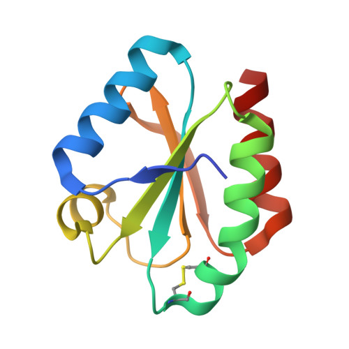
The redox components of the cytochrome bc1 complex from the acidophilic chemolithotrophic organism Thiobacillus ferrooxidans were investigated by potentiometric and spectroscopic techniques. Optical redox titrations demonstrated the presence of two b-type hemes with differing redox midpoint potentials at pH 7.4 (-169 and + 20 mV for bL and bH, respectively). At pH 3.5, by contrast, both hemes appeared to titrate at about +20 mV. Antimycin A, 2-heptyl-4-hydroxyquinoline N-oxide, and stigmatellin induced distinguishable shifts of the b hemes' alpha-bands, providing evidence for the binding of antimycin A and 2-heptyl-4-hydroxyquinoline N-oxide near heme bH (located on the cytosolic side of the membrane) and of stigmatellin near heme bL (located on the periplasmic side of the membrane). The inhibitors stigmatellin, 5-(n-undecyl)-6-hydroxy-4,7-dioxobenzothiazole, and 2, 5-dibromo-3-methyl-6-isopropyl-p-benzoquinone affected the EPR spectrum of the Rieske iron-sulfur center in a way that differs from what has been observed for cytochrome bc1 or b6f complexes. The results obtained demonstrate that the T. ferrooxidans complex, although showing most of the features characteristic for bc1 complexes, contains unique properties that are most probably related to the chemolithotrophicity and/or acidophilicity of its parent organism. A speculative model for reverse electron transfer through the T. ferrooxidans complex is proposed. (+info)Electron Transport Complex III, also known as cytochrome bc1 complex or ubiquinol-cytochrome c reductase, is a protein complex located in the inner mitochondrial membrane of eukaryotic cells and the cytoplasmic membrane of prokaryotic cells. It plays a crucial role in the electron transport chain (ETC), a series of complexes that generate energy in the form of ATP through a process called oxidative phosphorylation.
In ETC, Electron Transport Complex III accepts electrons from ubiquinol and transfers them to cytochrome c. This electron transfer is coupled with the translocation of protons (H+ ions) across the membrane, creating an electrochemical gradient. The energy stored in this gradient drives the synthesis of ATP by ATP synthase.
Electron Transport Complex III consists of several subunits, including cytochrome b, cytochrome c1, and the Rieske iron-sulfur protein. These subunits work together to facilitate the electron transfer and proton translocation processes.
Electron Transport Complex I, also known as NADH:ubiquinone oxidoreductase, is a large protein complex located in the inner mitochondrial membrane of eukaryotic cells and the cytoplasmic membrane of prokaryotic cells. It is the first complex in the electron transport chain, a series of protein complexes that transfer electrons from NADH to oxygen, driving the synthesis of ATP through chemiosmosis.
Complex I consists of multiple subunits, including a flavin mononucleotide (FMN) cofactor and several iron-sulfur clusters, which facilitate the oxidation of NADH and the reduction of ubiquinone (coenzyme Q). The energy released during this electron transfer process is used to pump protons across the membrane, creating a proton gradient that drives ATP synthesis.
Defects in Complex I can lead to various mitochondrial diseases, including neurological disorders and muscle weakness.
The Electron Transport Chain (ETC) is a series of complexes in the inner mitochondrial membrane that are involved in the process of cellular respiration. It is the final pathway for electrons derived from the oxidation of nutrients such as glucose, fatty acids, and amino acids to be transferred to molecular oxygen. This transfer of electrons drives the generation of a proton gradient across the inner mitochondrial membrane, which is then used by ATP synthase to produce ATP, the main energy currency of the cell.
The electron transport chain consists of four complexes (I-IV) and two mobile electron carriers (ubiquinone and cytochrome c). Electrons from NADH and FADH2 are transferred to Complex I and Complex II respectively, which then pass them along to ubiquinone. Ubiquinone then transfers the electrons to Complex III, which passes them on to cytochrome c. Finally, cytochrome c transfers the electrons to Complex IV, where they combine with oxygen and protons to form water.
The transfer of electrons through the ETC is accompanied by the pumping of protons from the mitochondrial matrix to the intermembrane space, creating a proton gradient. The flow of protons back across the inner membrane through ATP synthase drives the synthesis of ATP from ADP and inorganic phosphate.
Overall, the electron transport chain is a crucial process for generating energy in the form of ATP in the cell, and it plays a key role in many metabolic pathways.
Mitochondria are specialized structures located inside cells that convert the energy from food into ATP (adenosine triphosphate), which is the primary form of energy used by cells. They are often referred to as the "powerhouses" of the cell because they generate most of the cell's supply of chemical energy. Mitochondria are also involved in various other cellular processes, such as signaling, differentiation, and apoptosis (programmed cell death).
Mitochondria have their own DNA, known as mitochondrial DNA (mtDNA), which is inherited maternally. This means that mtDNA is passed down from the mother to her offspring through the egg cells. Mitochondrial dysfunction has been linked to a variety of diseases and conditions, including neurodegenerative disorders, diabetes, and aging.
Electron Transport Complex II, also known as succinate-Q oxidoreductase, is a key component of the electron transport chain in the inner mitochondrial membrane. It plays a crucial role in the process of cellular respiration, where it facilitates the transfer of electrons from succinate to ubiquinone (Q), thereby generating a proton gradient across the membrane. This gradient drives the synthesis of ATP, which is the primary source of energy for the cell.
The complex is composed of four core subunits: flavoprotein (Fp), iron-sulfur protein (Ip), cytochrome b (Cyb), and ubiquinone-binding protein (Qp). Electrons from succinate are accepted by FAD in the Fp subunit, and then transferred to the Ip subunit containing iron-sulfur clusters. From there, the electrons are moved to heme groups in the Cyb subunit, and finally passed on to ubiquinone at the Qp subunit.
In addition to its role in the electron transport chain, Complex II has been implicated in various cellular processes such as regulation of reactive oxygen species (ROS) production and modulation of apoptosis. Mutations in genes encoding Complex II subunits have been associated with several human diseases, including neurodegenerative disorders and cancer.
Electron Transport Complex IV is also known as Cytochrome c oxidase. It is the last complex in the electron transport chain, located in the inner mitochondrial membrane of eukaryotic cells and the plasma membrane of prokaryotic cells. This complex contains 13 subunits, two heme groups (a and a3), and three copper centers (A, B, and C).
In the electron transport chain, Complex IV receives electrons from cytochrome c and transfers them to molecular oxygen, reducing it to water. This process is accompanied by the pumping of protons across the membrane, contributing to the generation of a proton gradient that drives ATP synthesis via ATP synthase (Complex V). The overall reaction catalyzed by Complex IV can be summarized as follows:
4e- + 4H+ + O2 → 2H2O
Defects in Cytochrome c oxidase can lead to various diseases, including mitochondrial encephalomyopathies and neurodegenerative disorders.
The Electron Transport Chain (ETC) is a series of complexes in the inner mitochondrial membrane that are involved in the process of cellular respiration, through which the majority of energy is generated for the cell. The ETC complex proteins are a group of transmembrane protein complexes that facilitate the transfer of electrons from electron donors to electron acceptors via redox reactions. This transfer of electrons drives the generation of a proton gradient across the inner mitochondrial membrane, which is then used by ATP synthase to generate ATP, the primary energy currency of the cell.
The ETC complex proteins consist of four main complexes: Complex I (NADH-Q oxidoreductase), Complex II (succinate-Q oxidoreductase), Complex III (cytochrome bc1 complex or CoQ:cytochrome c oxidoreductase), and Complex IV (cytochrome c oxidase). Each complex contains a number of subunits, many of which are encoded by both the nuclear and mitochondrial genomes.
In summary, Electron Transport Chain Complex Proteins are a group of transmembrane protein complexes located in the inner mitochondrial membrane that facilitate the transfer of electrons from electron donors to electron acceptors, driving the generation of a proton gradient and ultimately ATP synthesis during cellular respiration.
Antimycin A is an antibiotic substance produced by various species of Streptomyces bacteria. It is known to inhibit the electron transport chain in mitochondria, which can lead to cellular dysfunction and death. Antimycin A has been used in research to study the mechanisms of cellular respiration and oxidative phosphorylation.
In a medical context, antimycin A is not used as a therapeutic agent due to its toxicity to mammalian cells. However, it may be used in laboratory settings to investigate various biological processes or to develop new therapies for diseases related to mitochondrial dysfunction.
Maltose is a disaccharide made up of two glucose molecules joined by an alpha-1,4 glycosidic bond. It is commonly found in malted barley and is created during the germination process when amylase breaks down starches into simpler sugars. Maltose is less sweet than sucrose (table sugar) and is broken down into glucose by the enzyme maltase during digestion.
Biological transport refers to the movement of molecules, ions, or solutes across biological membranes or through cells in living organisms. This process is essential for maintaining homeostasis, regulating cellular functions, and enabling communication between cells. There are two main types of biological transport: passive transport and active transport.
Passive transport does not require the input of energy and includes:
1. Diffusion: The random movement of molecules from an area of high concentration to an area of low concentration until equilibrium is reached.
2. Osmosis: The diffusion of solvent molecules (usually water) across a semi-permeable membrane from an area of lower solute concentration to an area of higher solute concentration.
3. Facilitated diffusion: The assisted passage of polar or charged substances through protein channels or carriers in the cell membrane, which increases the rate of diffusion without consuming energy.
Active transport requires the input of energy (in the form of ATP) and includes:
1. Primary active transport: The direct use of ATP to move molecules against their concentration gradient, often driven by specific transport proteins called pumps.
2. Secondary active transport: The coupling of the movement of one substance down its electrochemical gradient with the uphill transport of another substance, mediated by a shared transport protein. This process is also known as co-transport or counter-transport.
Maltose-binding proteins (MBPs) are a type of protein that are capable of binding to maltose, a disaccharide made up of two glucose molecules. MBPs are found in many organisms, including bacteria and plants. In bacteria such as Escherichia coli, MBPs play a role in the transport and metabolism of maltose and maltodextrins, which are polymers of glucose.
MBPs are often used in laboratory research as model systems for studying protein folding and stability. They have a well-characterized three-dimensional structure and are relatively small, making them easy to produce and study. MBPs are also known for their high binding affinity and specificity for maltose, making them useful for purifying and detecting this sugar in various applications.
Biological transport, active is the process by which cells use energy to move materials across their membranes from an area of lower concentration to an area of higher concentration. This type of transport is facilitated by specialized proteins called transporters or pumps that are located in the cell membrane. These proteins undergo conformational changes to physically carry the molecules through the lipid bilayer of the membrane, often against their concentration gradient.
Active transport requires energy because it works against the natural tendency of molecules to move from an area of higher concentration to an area of lower concentration, a process known as diffusion. Cells obtain this energy in the form of ATP (adenosine triphosphate), which is produced through cellular respiration.
Examples of active transport include the uptake of glucose and amino acids into cells, as well as the secretion of hormones and neurotransmitters. The sodium-potassium pump, which helps maintain resting membrane potential in nerve and muscle cells, is a classic example of an active transporter.
An electron is a subatomic particle, symbol e-, with a negative electric charge. Electrons are fundamental components of atoms and are responsible for the chemical bonding between atoms to form molecules. They are located in an atom's electron cloud, which is the outermost region of an atom and contains negatively charged electrons that surround the positively charged nucleus.
Electrons have a mass that is much smaller than that of protons or neutrons, making them virtually weightless on the atomic scale. They are also known to exhibit both particle-like and wave-like properties, which is a fundamental concept in quantum mechanics. Electrons play a crucial role in various physical phenomena, such as electricity, magnetism, and chemical reactions.
Periplasmic binding proteins (PBPs) are a type of water-soluble protein found in the periplasmic space of gram-negative bacteria. They play a crucial role in the bacterial uptake of specific nutrients, such as amino acids, sugars, and ions, through a process known as active transport.
PBPs function by specifically binding to their target substrates in the extracellular environment and then shuttling them across the inner membrane into the cytoplasm. This is achieved through a complex series of interactions with other proteins, including transmembrane permeases and ATP-binding cassette (ABC) transporters.
The binding of PBPs to their substrates typically results in a conformational change that allows for the transport of the substrate across the inner membrane. Once inside the cytoplasm, the substrate can be used for various metabolic processes, such as energy production or biosynthesis.
PBPs are often used as targets for the development of new antibiotics, as they play a critical role in bacterial survival and virulence. Inhibiting their function can disrupt essential physiological processes and lead to bacterial death.
Electron microscopy (EM) is a type of microscopy that uses a beam of electrons to create an image of the sample being examined, resulting in much higher magnification and resolution than light microscopy. There are several types of electron microscopy, including transmission electron microscopy (TEM), scanning electron microscopy (SEM), and reflection electron microscopy (REM).
In TEM, a beam of electrons is transmitted through a thin slice of the sample, and the electrons that pass through the sample are focused to form an image. This technique can provide detailed information about the internal structure of cells, viruses, and other biological specimens, as well as the composition and structure of materials at the atomic level.
In SEM, a beam of electrons is scanned across the surface of the sample, and the electrons that are scattered back from the surface are detected to create an image. This technique can provide information about the topography and composition of surfaces, as well as the structure of materials at the microscopic level.
REM is a variation of SEM in which the beam of electrons is reflected off the surface of the sample, rather than scattered back from it. This technique can provide information about the surface chemistry and composition of materials.
Electron microscopy has a wide range of applications in biology, medicine, and materials science, including the study of cellular structure and function, disease diagnosis, and the development of new materials and technologies.
ATP-binding cassette (ABC) transporters are a family of membrane proteins that utilize the energy from ATP hydrolysis to transport various substrates across extra- and intracellular membranes. These transporters play crucial roles in several biological processes, including detoxification, drug resistance, nutrient uptake, and regulation of cellular cholesterol homeostasis.
The structure of ABC transporters consists of two nucleotide-binding domains (NBDs) that bind and hydrolyze ATP, and two transmembrane domains (TMDs) that form the substrate-translocation pathway. The NBDs are typically located adjacent to each other in the cytoplasm, while the TMDs can be either integral membrane domains or separate structures associated with the membrane.
The human genome encodes 48 distinct ABC transporters, which are classified into seven subfamilies (ABCA-ABCG) based on their sequence similarity and domain organization. Some well-known examples of ABC transporters include P-glycoprotein (ABCB1), multidrug resistance protein 1 (ABCC1), and breast cancer resistance protein (ABCG2).
Dysregulation or mutations in ABC transporters have been implicated in various diseases, such as cystic fibrosis, neurological disorders, and cancer. In cancer, overexpression of certain ABC transporters can contribute to drug resistance by actively effluxing chemotherapeutic agents from cancer cells, making them less susceptible to treatment.
Ubiquinone, also known as coenzyme Q10 (CoQ10), is a lipid-soluble benzoquinone that plays a crucial role in the mitochondrial electron transport chain as an essential component of Complexes I, II, and III. It functions as an electron carrier, assisting in the transfer of electrons from reduced nicotinamide adenine dinucleotide (NADH) and flavin adenine dinucleotide (FADH2) to molecular oxygen during oxidative phosphorylation, thereby contributing to the generation of adenosine triphosphate (ATP), the primary energy currency of the cell.
Additionally, ubiquinone acts as a potent antioxidant in both membranes and lipoproteins, protecting against lipid peroxidation and oxidative damage to proteins and DNA. Its antioxidant properties stem from its ability to donate electrons and regenerate other antioxidants like vitamin E. Ubiquinone is synthesized endogenously in all human cells, with the highest concentrations found in tissues with high energy demands, such as the heart, liver, kidneys, and skeletal muscles.
Deficiency in ubiquinone can result from genetic disorders, aging, or certain medications (such as statins), leading to impaired mitochondrial function and increased oxidative stress. Supplementation with ubiquinone has been explored as a potential therapeutic strategy for various conditions associated with mitochondrial dysfunction and oxidative stress, including cardiovascular diseases, neurodegenerative disorders, and cancer.
Coat Protein Complex I (CPCI or COPI) is a protein complex involved in the intracellular transport of proteins within eukaryotic cells. It functions primarily in the retrograde transport of proteins from the Golgi apparatus to the endoplasmic reticulum (ER). The complex is composed of seven subunits, known as alpha, beta, gamma, delta, epsilon, zeta, and eta COPs (coat proteins), which form a cage-like structure around transport vesicles. This coat assists in the selection of cargo proteins, vesicle budding, and subsequent fusion with target membranes during the recycling of ER-derived proteins.
Monosaccharide transport proteins are a type of membrane transport protein that facilitate the passive or active transport of monosaccharides, such as glucose, fructose, and galactose, across cell membranes. These proteins play a crucial role in the absorption, distribution, and metabolism of carbohydrates in the body.
There are two main types of monosaccharide transport proteins: facilitated diffusion transporters and active transporters. Facilitated diffusion transporters, also known as glucose transporters (GLUTs), passively transport monosaccharides down their concentration gradient without the need for energy. In contrast, active transporters, such as the sodium-glucose cotransporter (SGLT), use energy in the form of ATP to actively transport monosaccharides against their concentration gradient.
Monosaccharide transport proteins are found in various tissues throughout the body, including the intestines, kidneys, liver, and brain. They play a critical role in maintaining glucose homeostasis by regulating the uptake and release of glucose into and out of cells. Dysfunction of these transporters has been implicated in several diseases, such as diabetes, cancer, and neurological disorders.
Quinone reductases are a group of enzymes that catalyze the reduction of quinones to hydroquinones, using NADH or NADPH as an electron donor. This reaction is important in the detoxification of quinones, which are potentially toxic compounds produced during the metabolism of certain drugs, chemicals, and endogenous substances.
There are two main types of quinone reductases: NQO1 (NAD(P)H:quinone oxidoreductase 1) and NQO2 (NAD(P)H:quinone oxidoreductase 2). NQO1 is a cytosolic enzyme that can reduce a wide range of quinones, while NQO2 is a mitochondrial enzyme with a narrower substrate specificity.
Quinone reductases have been studied for their potential role in cancer prevention and treatment, as they may help to protect cells from oxidative stress and DNA damage caused by quinones and other toxic compounds. Additionally, some quinone reductase inhibitors have been developed as chemotherapeutic agents, as they can enhance the cytotoxicity of certain drugs that require quinone reduction for activation.
Oxidation-Reduction (redox) reactions are a type of chemical reaction involving a transfer of electrons between two species. The substance that loses electrons in the reaction is oxidized, and the substance that gains electrons is reduced. Oxidation and reduction always occur together in a redox reaction, hence the term "oxidation-reduction."
In biological systems, redox reactions play a crucial role in many cellular processes, including energy production, metabolism, and signaling. The transfer of electrons in these reactions is often facilitated by specialized molecules called electron carriers, such as nicotinamide adenine dinucleotide (NAD+/NADH) and flavin adenine dinucleotide (FAD/FADH2).
The oxidation state of an element in a compound is a measure of the number of electrons that have been gained or lost relative to its neutral state. In redox reactions, the oxidation state of one or more elements changes as they gain or lose electrons. The substance that is oxidized has a higher oxidation state, while the substance that is reduced has a lower oxidation state.
Overall, oxidation-reduction reactions are fundamental to the functioning of living organisms and are involved in many important biological processes.
Cytochromes b are a group of electron transport proteins that contain a heme c group, which is the prosthetic group responsible for their redox activity. They play a crucial role in the electron transport chain (ETC) located in the inner mitochondrial membrane of eukaryotic cells and in the plasma membrane of prokaryotic cells.
The cytochromes b are part of Complex III, also known as the cytochrome bc1 complex or ubiquinol-cytochrome c reductase, in the ETC. In this complex, they function as electron carriers between ubiquinone (Q) and cytochrome c, participating in the process of oxidative phosphorylation to generate ATP.
There are multiple isoforms of cytochromes b found in various organisms, with different numbers of subunits and structures. However, they all share a common function as essential components of the electron transport chain, facilitating the transfer of electrons during cellular respiration and energy production.
Oxidative phosphorylation is the metabolic process by which cells use enzymes to generate energy in the form of adenosine triphosphate (ATP) from the oxidation of nutrients, such as glucose or fatty acids. This process occurs in the inner mitochondrial membrane of eukaryotic cells and is facilitated by the electron transport chain, which consists of a series of protein complexes that transfer electrons from donor molecules to acceptor molecules. As the electrons are passed along the chain, they release energy that is used to pump protons across the membrane, creating a gradient. The ATP synthase enzyme then uses the flow of protons back across the membrane to generate ATP, which serves as the main energy currency for cellular processes.
Membrane transport proteins are specialized biological molecules, specifically integral membrane proteins, that facilitate the movement of various substances across the lipid bilayer of cell membranes. They are responsible for the selective and regulated transport of ions, sugars, amino acids, nucleotides, and other molecules into and out of cells, as well as within different cellular compartments. These proteins can be categorized into two main types: channels and carriers (or pumps). Channels provide a passive transport mechanism, allowing ions or small molecules to move down their electrochemical gradient, while carriers actively transport substances against their concentration gradient, requiring energy usually in the form of ATP. Membrane transport proteins play a crucial role in maintaining cell homeostasis, signaling processes, and many other physiological functions.
Scanning electron microscopy (SEM) is a type of electron microscopy that uses a focused beam of electrons to scan the surface of a sample and produce a high-resolution image. In SEM, a beam of electrons is scanned across the surface of a specimen, and secondary electrons are emitted from the sample due to interactions between the electrons and the atoms in the sample. These secondary electrons are then detected by a detector and used to create an image of the sample's surface topography. SEM can provide detailed images of the surface of a wide range of materials, including metals, polymers, ceramics, and biological samples. It is commonly used in materials science, biology, and electronics for the examination and analysis of surfaces at the micro- and nanoscale.
Photosynthesis is not strictly a medical term, but it is a fundamental biological process with significant implications for medicine, particularly in understanding energy production in cells and the role of oxygen in sustaining life. Here's a general biological definition:
Photosynthesis is a process by which plants, algae, and some bacteria convert light energy, usually from the sun, into chemical energy in the form of organic compounds, such as glucose (or sugar), using water and carbon dioxide. This process primarily takes place in the chloroplasts of plant cells, specifically in structures called thylakoids. The overall reaction can be summarized as:
6 CO2 + 6 H2O + light energy → C6H12O6 + 6 O2
In this equation, carbon dioxide (CO2) and water (H2O) are the reactants, while glucose (C6H12O6) and oxygen (O2) are the products. Photosynthesis has two main stages: the light-dependent reactions and the light-independent reactions (Calvin cycle). The light-dependent reactions occur in the thylakoid membrane and involve the conversion of light energy into ATP and NADPH, which are used to power the Calvin cycle. The Calvin cycle takes place in the stroma of chloroplasts and involves the synthesis of glucose from CO2 and water using the ATP and NADPH generated during the light-dependent reactions.
Understanding photosynthesis is crucial for understanding various biological processes, including cellular respiration, plant metabolism, and the global carbon cycle. Additionally, research into artificial photosynthesis has potential applications in renewable energy production and environmental remediation.
Carrier proteins, also known as transport proteins, are a type of protein that facilitates the movement of molecules across cell membranes. They are responsible for the selective and active transport of ions, sugars, amino acids, and other molecules from one side of the membrane to the other, against their concentration gradient. This process requires energy, usually in the form of ATP (adenosine triphosphate).
Carrier proteins have a specific binding site for the molecule they transport, and undergo conformational changes upon binding, which allows them to move the molecule across the membrane. Once the molecule has been transported, the carrier protein returns to its original conformation, ready to bind and transport another molecule.
Carrier proteins play a crucial role in maintaining the balance of ions and other molecules inside and outside of cells, and are essential for many physiological processes, including nerve impulse transmission, muscle contraction, and nutrient uptake.
Cytochromes are a type of hemeprotein found in the mitochondria and other cellular membranes of organisms. They contain a heme group, which is a prosthetic group composed of an iron atom surrounded by a porphyrin ring. This structure allows cytochromes to participate in redox reactions, acting as electron carriers in various biological processes.
There are several types of cytochromes, classified based on the type of heme they contain and their absorption spectra. Some of the most well-known cytochromes include:
* Cytochrome c: a small, mobile protein found in the inner mitochondrial membrane that plays a crucial role in the electron transport chain during cellular respiration.
* Cytochrome P450: a large family of enzymes involved in the metabolism of drugs, toxins, and other xenobiotics. They are found in various tissues, including the liver, lungs, and skin.
* Cytochrome b: a component of several electron transport chains, including those found in mitochondria, bacteria, and chloroplasts.
Cytochromes play essential roles in energy production, detoxification, and other metabolic processes, making them vital for the survival and function of living organisms.
Rotenone is not strictly a medical term, but it is a pesticide that is used in some medical situations. According to the National Pesticide Information Center, rotenone is a pesticide derived from the roots and stems of several plants, including Derris Eliptica, Lonchocarpus utilis, and Tephrosia vogelii. It is used as a pesticide to control insects, mites, and fish in both agricultural and residential settings.
In medical contexts, rotenone has been studied for its potential effects on human health, particularly in relation to Parkinson's disease. Some research suggests that exposure to rotenone may increase the risk of developing Parkinson's disease, although more studies are needed to confirm this link. Rotenone works by inhibiting the mitochondria in cells, which can lead to cell death and neurodegeneration.
It is important to note that rotenone is highly toxic and should be handled with care. It can cause skin and eye irritation, respiratory problems, and gastrointestinal symptoms if ingested or inhaled. Therefore, it is recommended to use personal protective equipment when handling rotenone and to follow all label instructions carefully.
Molecular sequence data refers to the specific arrangement of molecules, most commonly nucleotides in DNA or RNA, or amino acids in proteins, that make up a biological macromolecule. This data is generated through laboratory techniques such as sequencing, and provides information about the exact order of the constituent molecules. This data is crucial in various fields of biology, including genetics, evolution, and molecular biology, allowing for comparisons between different organisms, identification of genetic variations, and studies of gene function and regulation.
Adenosine Triphosphate (ATP) is a high-energy molecule that stores and transports energy within cells. It is the main source of energy for most cellular processes, including muscle contraction, nerve impulse transmission, and protein synthesis. ATP is composed of a base (adenine), a sugar (ribose), and three phosphate groups. The bonds between these phosphate groups contain a significant amount of energy, which can be released when the bond between the second and third phosphate group is broken, resulting in the formation of adenosine diphosphate (ADP) and inorganic phosphate. This process is known as hydrolysis and can be catalyzed by various enzymes to drive a wide range of cellular functions. ATP can also be regenerated from ADP through various metabolic pathways, such as oxidative phosphorylation or substrate-level phosphorylation, allowing for the continuous supply of energy to cells.
Protein transport, in the context of cellular biology, refers to the process by which proteins are actively moved from one location to another within or between cells. This is a crucial mechanism for maintaining proper cell function and regulation.
Intracellular protein transport involves the movement of proteins within a single cell. Proteins can be transported across membranes (such as the nuclear envelope, endoplasmic reticulum, Golgi apparatus, or plasma membrane) via specialized transport systems like vesicles and transport channels.
Intercellular protein transport refers to the movement of proteins from one cell to another, often facilitated by exocytosis (release of proteins in vesicles) and endocytosis (uptake of extracellular substances via membrane-bound vesicles). This is essential for communication between cells, immune response, and other physiological processes.
It's important to note that any disruption in protein transport can lead to various diseases, including neurological disorders, cancer, and metabolic conditions.
Cytochrome b is a type of cytochrome, which is a class of proteins that contain heme as a cofactor and are involved in electron transfer. Cytochromes are classified based on the type of heme they contain and their absorption spectra.
The cytochrome b group includes several subfamilies of cytochromes, including cytochrome b5, cytochrome b2, and cytochrome bc1 (also known as complex III). These cytochromes are involved in various biological processes, such as fatty acid desaturation, steroid metabolism, and the electron transport chain.
The electron transport chain is a series of protein complexes in the inner mitochondrial membrane that generates most of the ATP (adenosine triphosphate) required for cellular energy production. Cytochrome bc1 is a key component of the electron transport chain, where it functions as a dimer and catalyzes the transfer of electrons from ubiquinol to cytochrome c while simultaneously pumping protons across the membrane. This creates an electrochemical gradient that drives ATP synthesis.
Deficiencies or mutations in cytochrome b genes can lead to various diseases, such as mitochondrial disorders and cancer.
I'm happy to help! However, it seems like there are two terms combined into one request: "Mitochondria" and "Heart." I will provide separate definitions for each.
Mitochondrion (singular) or Mitochondria (plural): These are specialized structures found in the cytoplasm of most eukaryotic cells (cells with a true nucleus), including human cells. They are often referred to as the "powerhouse" of the cell because they generate energy in the form of ATP (adenosine triphosphate) through a process called oxidative phosphorylation. Mitochondria contain their own DNA, which is distinct from the nuclear DNA, and are believed to have originated from ancient bacteria that established a symbiotic relationship with primitive eukaryotic cells.
Heart: In human anatomy, the heart is a muscular organ responsible for pumping blood throughout the body. It is located in the thoracic cavity, slightly left of the center, and is enclosed by the pericardium, a double-walled sac that provides protection and lubrication for the heart's movement. The human heart is divided into four chambers: two atria on the top and two ventricles on the bottom. The right side of the heart receives deoxygenated blood from the body and pumps it to the lungs, while the left side receives oxygenated blood from the lungs and pumps it to the rest of the body. The heart's pumping action is regulated by electrical signals that originate in a group of specialized cardiac muscle cells called the sinoatrial node (SA node).
Reactive Oxygen Species (ROS) are highly reactive molecules containing oxygen, including peroxides, superoxide, hydroxyl radical, and singlet oxygen. They are naturally produced as byproducts of normal cellular metabolism in the mitochondria, and can also be generated by external sources such as ionizing radiation, tobacco smoke, and air pollutants. At low or moderate concentrations, ROS play important roles in cell signaling and homeostasis, but at high concentrations, they can cause significant damage to cell structures, including lipids, proteins, and DNA, leading to oxidative stress and potential cell death.
Cytochrome c1 is a protein that is a part of the electron transport chain in the inner mitochondrial membrane. It is a component of Complex III, also known as the cytochrome bc1 complex. Cytochrome c1 contains a heme group and plays a role in the transfer of electrons from ubiquinol to cytochrome c during oxidative phosphorylation, which is the process by which cells generate energy in the form of ATP. Defects in cytochrome c1 can lead to mitochondrial disorders and have been implicated in the development of certain diseases, such as neurodegenerative disorders and cancer.
Biological models, also known as physiological models or organismal models, are simplified representations of biological systems, processes, or mechanisms that are used to understand and explain the underlying principles and relationships. These models can be theoretical (conceptual or mathematical) or physical (such as anatomical models, cell cultures, or animal models). They are widely used in biomedical research to study various phenomena, including disease pathophysiology, drug action, and therapeutic interventions.
Examples of biological models include:
1. Mathematical models: These use mathematical equations and formulas to describe complex biological systems or processes, such as population dynamics, metabolic pathways, or gene regulation networks. They can help predict the behavior of these systems under different conditions and test hypotheses about their underlying mechanisms.
2. Cell cultures: These are collections of cells grown in a controlled environment, typically in a laboratory dish or flask. They can be used to study cellular processes, such as signal transduction, gene expression, or metabolism, and to test the effects of drugs or other treatments on these processes.
3. Animal models: These are living organisms, usually vertebrates like mice, rats, or non-human primates, that are used to study various aspects of human biology and disease. They can provide valuable insights into the pathophysiology of diseases, the mechanisms of drug action, and the safety and efficacy of new therapies.
4. Anatomical models: These are physical representations of biological structures or systems, such as plastic models of organs or tissues, that can be used for educational purposes or to plan surgical procedures. They can also serve as a basis for developing more sophisticated models, such as computer simulations or 3D-printed replicas.
Overall, biological models play a crucial role in advancing our understanding of biology and medicine, helping to identify new targets for therapeutic intervention, develop novel drugs and treatments, and improve human health.
Uncoupling agents are chemicals that interfere with the normal process of oxidative phosphorylation in cells. In this process, the energy from food is converted into ATP (adenosine triphosphate), which is the main source of energy for cellular functions. Uncouplers disrupt this process by preventing the transfer of high-energy electrons to oxygen, which normally drives the production of ATP.
Instead, the energy from these electrons is released as heat, leading to an increase in body temperature. This effect is similar to what happens during shivering or exercise, when the body generates heat to maintain its core temperature. Uncoupling agents are therefore also known as "mitochondrial protonophores" because they allow protons to leak across the inner mitochondrial membrane, bypassing the ATP synthase enzyme that would normally use the energy from this proton gradient to produce ATP.
Uncoupling agents have been studied for their potential therapeutic uses, such as in weight loss and the treatment of metabolic disorders. However, they can also be toxic at high doses, and their long-term effects on health are not well understood.
A mutation is a permanent change in the DNA sequence of an organism's genome. Mutations can occur spontaneously or be caused by environmental factors such as exposure to radiation, chemicals, or viruses. They may have various effects on the organism, ranging from benign to harmful, depending on where they occur and whether they alter the function of essential proteins. In some cases, mutations can increase an individual's susceptibility to certain diseases or disorders, while in others, they may confer a survival advantage. Mutations are the driving force behind evolution, as they introduce new genetic variability into populations, which can then be acted upon by natural selection.
Oxygen consumption, also known as oxygen uptake, is the amount of oxygen that is consumed or utilized by the body during a specific period of time, usually measured in liters per minute (L/min). It is a common measurement used in exercise physiology and critical care medicine to assess an individual's aerobic metabolism and overall health status.
In clinical settings, oxygen consumption is often measured during cardiopulmonary exercise testing (CPET) to evaluate cardiovascular function, pulmonary function, and exercise capacity in patients with various medical conditions such as heart failure, chronic obstructive pulmonary disease (COPD), and other respiratory or cardiac disorders.
During exercise, oxygen is consumed by the muscles to generate energy through a process called oxidative phosphorylation. The amount of oxygen consumed during exercise can provide important information about an individual's fitness level, exercise capacity, and overall health status. Additionally, measuring oxygen consumption can help healthcare providers assess the effectiveness of treatments and rehabilitation programs in patients with various medical conditions.
Hydroxyquinolines are a group of synthetic antimicrobial agents that contain a hydroxyl group (-OH) attached to a quinoline ring. They have been used in the treatment of various bacterial, fungal, and parasitic infections. Some common examples of hydroxyquinolines include chloroquine, hydroxychloroquine, and quinacrine. These agents work by inhibiting the growth and multiplication of microorganisms, although their exact mechanisms of action may vary. Chloroquine and hydroxychloroquine, for example, are known to interfere with the replication of the malaria parasite within red blood cells, while quinacrine has been used to treat certain types of protozoal infections.
It is important to note that the use of hydroxyquinolines is associated with a number of potential side effects and risks, including gastrointestinal disturbances, visual disturbances, and cardiac toxicity. As such, they should only be used under the close supervision of a healthcare professional.
An amino acid sequence is the specific order of amino acids in a protein or peptide molecule, formed by the linking of the amino group (-NH2) of one amino acid to the carboxyl group (-COOH) of another amino acid through a peptide bond. The sequence is determined by the genetic code and is unique to each type of protein or peptide. It plays a crucial role in determining the three-dimensional structure and function of proteins.
Membrane proteins are a type of protein that are embedded in the lipid bilayer of biological membranes, such as the plasma membrane of cells or the inner membrane of mitochondria. These proteins play crucial roles in various cellular processes, including:
1. Cell-cell recognition and signaling
2. Transport of molecules across the membrane (selective permeability)
3. Enzymatic reactions at the membrane surface
4. Energy transduction and conversion
5. Mechanosensation and signal transduction
Membrane proteins can be classified into two main categories: integral membrane proteins, which are permanently associated with the lipid bilayer, and peripheral membrane proteins, which are temporarily or loosely attached to the membrane surface. Integral membrane proteins can further be divided into three subcategories based on their topology:
1. Transmembrane proteins, which span the entire width of the lipid bilayer with one or more alpha-helices or beta-barrels.
2. Lipid-anchored proteins, which are covalently attached to lipids in the membrane via a glycosylphosphatidylinositol (GPI) anchor or other lipid modifications.
3. Monotopic proteins, which are partially embedded in the membrane and have one or more domains exposed to either side of the bilayer.
Membrane proteins are essential for maintaining cellular homeostasis and are targets for various therapeutic interventions, including drug development and gene therapy. However, their structural complexity and hydrophobicity make them challenging to study using traditional biochemical methods, requiring specialized techniques such as X-ray crystallography, nuclear magnetic resonance (NMR) spectroscopy, and single-particle cryo-electron microscopy (cryo-EM).
A cell membrane, also known as the plasma membrane, is a thin semi-permeable phospholipid bilayer that surrounds all cells in animals, plants, and microorganisms. It functions as a barrier to control the movement of substances in and out of the cell, allowing necessary molecules such as nutrients, oxygen, and signaling molecules to enter while keeping out harmful substances and waste products. The cell membrane is composed mainly of phospholipids, which have hydrophilic (water-loving) heads and hydrophobic (water-fearing) tails. This unique structure allows the membrane to be flexible and fluid, yet selectively permeable. Additionally, various proteins are embedded in the membrane that serve as channels, pumps, receptors, and enzymes, contributing to the cell's overall functionality and communication with its environment.
'Escherichia coli (E. coli) proteins' refer to the various types of proteins that are produced and expressed by the bacterium Escherichia coli. These proteins play a critical role in the growth, development, and survival of the organism. They are involved in various cellular processes such as metabolism, DNA replication, transcription, translation, repair, and regulation.
E. coli is a gram-negative, facultative anaerobe that is commonly found in the intestines of warm-blooded organisms. It is widely used as a model organism in scientific research due to its well-studied genetics, rapid growth, and ability to be easily manipulated in the laboratory. As a result, many E. coli proteins have been identified, characterized, and studied in great detail.
Some examples of E. coli proteins include enzymes involved in carbohydrate metabolism such as lactase, sucrase, and maltose; proteins involved in DNA replication such as the polymerases, single-stranded binding proteins, and helicases; proteins involved in transcription such as RNA polymerase and sigma factors; proteins involved in translation such as ribosomal proteins, tRNAs, and aminoacyl-tRNA synthetases; and regulatory proteins such as global regulators, two-component systems, and transcription factors.
Understanding the structure, function, and regulation of E. coli proteins is essential for understanding the basic biology of this important organism, as well as for developing new strategies for combating bacterial infections and improving industrial processes involving bacteria.
NADH, NADPH oxidoreductases are a class of enzymes that catalyze the redox reaction between NADH or NADPH and various electron acceptors. These enzymes play a crucial role in cellular metabolism by transferring electrons from NADH or NADPH to other molecules, which is essential for many biochemical reactions.
NADH (nicotinamide adenine dinucleotide hydrogen) and NADPH (nicotinamide adenine dinucleotide phosphate hydrogen) are coenzymes that act as electron carriers in redox reactions. They consist of a nicotinamide ring, which undergoes reduction or oxidation by accepting or donating electrons and a proton (H+).
NADH, NADPH oxidoreductases are classified based on their structure and mechanism of action. Some examples include:
1. Dehydrogenases: These enzymes catalyze the oxidation of NADH or NADPH to NAD+ or NADP+ while reducing an organic substrate. Examples include lactate dehydrogenase, alcohol dehydrogenase, and malate dehydrogenase.
2. Oxidases: These enzymes catalyze the oxidation of NADH or NADPH to NAD+ or NADP+ while reducing molecular oxygen (O2) to water (H2O). Examples include NADH oxidase and NADPH oxidase.
3. Reductases: These enzymes catalyze the reduction of various electron acceptors using NADH or NADPH as a source of electrons. Examples include glutathione reductase, thioredoxin reductase, and nitrate reductase.
Overall, NADH, NADPH oxidoreductases are essential for maintaining the redox balance in cells and play a critical role in various metabolic pathways, including energy production, detoxification, and biosynthesis.
Cell respiration is the process by which cells convert biochemical energy from nutrients into adenosine triphosphate (ATP), and then release waste products. The three main stages of cell respiration are glycolysis, the citric acid cycle (also known as the Krebs cycle), and the electron transport chain.
During glycolysis, which takes place in the cytoplasm, glucose is broken down into two molecules of pyruvate, producing a small amount of ATP and reducing power in the form of NADH.
The citric acid cycle occurs in the mitochondria and involves the breakdown of acetyl-CoA (formed from pyruvate) to produce more ATP, NADH, and FADH2.
Finally, the electron transport chain, also located in the mitochondria, uses the energy from NADH and FADH2 to pump protons across the inner mitochondrial membrane, creating a proton gradient. The flow of protons back across the membrane drives the synthesis of ATP, which is used as a source of energy by the cell.
Cell respiration is a crucial process that allows cells to generate the energy they need to perform various functions and maintain homeostasis.
In the context of medicine and pharmacology, "kinetics" refers to the study of how a drug moves throughout the body, including its absorption, distribution, metabolism, and excretion (often abbreviated as ADME). This field is called "pharmacokinetics."
1. Absorption: This is the process of a drug moving from its site of administration into the bloodstream. Factors such as the route of administration (e.g., oral, intravenous, etc.), formulation, and individual physiological differences can affect absorption.
2. Distribution: Once a drug is in the bloodstream, it gets distributed throughout the body to various tissues and organs. This process is influenced by factors like blood flow, protein binding, and lipid solubility of the drug.
3. Metabolism: Drugs are often chemically modified in the body, typically in the liver, through processes known as metabolism. These changes can lead to the formation of active or inactive metabolites, which may then be further distributed, excreted, or undergo additional metabolic transformations.
4. Excretion: This is the process by which drugs and their metabolites are eliminated from the body, primarily through the kidneys (urine) and the liver (bile).
Understanding the kinetics of a drug is crucial for determining its optimal dosing regimen, potential interactions with other medications or foods, and any necessary adjustments for special populations like pediatric or geriatric patients, or those with impaired renal or hepatic function.
Diuron is a pesticide and herbicide that is used to control weeds in various settings, such as agriculture, landscaping, and forestry. Its chemical name is 3-(3,4-dichlorophenyl)-1,1-dimethylurea. Diuron works by inhibiting photosynthesis in plants, which prevents them from growing and eventually kills them.
While diuron is effective at controlling weeds, it can also have harmful effects on non-target organisms, including aquatic life and pollinators. Additionally, there are concerns about the potential for diuron to contaminate water sources and pose risks to human health. As a result, its use is regulated in many countries, and there are restrictions on how it can be applied and disposed of.
It's worth noting that Diuron is not a medical term or a drug used for treating any medical condition in humans or animals.
Bacterial proteins are a type of protein that are produced by bacteria as part of their structural or functional components. These proteins can be involved in various cellular processes, such as metabolism, DNA replication, transcription, and translation. They can also play a role in bacterial pathogenesis, helping the bacteria to evade the host's immune system, acquire nutrients, and multiply within the host.
Bacterial proteins can be classified into different categories based on their function, such as:
1. Enzymes: Proteins that catalyze chemical reactions in the bacterial cell.
2. Structural proteins: Proteins that provide structural support and maintain the shape of the bacterial cell.
3. Signaling proteins: Proteins that help bacteria to communicate with each other and coordinate their behavior.
4. Transport proteins: Proteins that facilitate the movement of molecules across the bacterial cell membrane.
5. Toxins: Proteins that are produced by pathogenic bacteria to damage host cells and promote infection.
6. Surface proteins: Proteins that are located on the surface of the bacterial cell and interact with the environment or host cells.
Understanding the structure and function of bacterial proteins is important for developing new antibiotics, vaccines, and other therapeutic strategies to combat bacterial infections.
Potassium Cyanide (C6H5KN) is defined as a white, water-soluble, crystalline salt that is highly toxic. It is used in fumigation, electroplating, and metal cleaning. When combined with acids, it releases the deadly gas hydrogen cyanide. It can cause immediate death by inhibiting cellular respiration. It is also known as Cyanide of Potassium or Potassium Salt of Hydrocyanic Acid.
Cytochrome c is a small protein that is involved in the electron transport chain, a key part of cellular respiration in which cells generate energy in the form of ATP. Cytochrome c contains a heme group, which binds to and transports electrons. The cytochrome c group refers to a class of related cytochromes that have similar structures and functions. These proteins are found in the mitochondria of eukaryotic cells (such as those of plants and animals) and in the inner membranes of bacteria. They play a crucial role in the production of energy within the cell, and are also involved in certain types of programmed cell death (apoptosis).
Oxidoreductases are a class of enzymes that catalyze oxidation-reduction reactions, which involve the transfer of electrons from one molecule (the reductant) to another (the oxidant). These enzymes play a crucial role in various biological processes, including energy production, metabolism, and detoxification.
The oxidoreductase-catalyzed reaction typically involves the donation of electrons from a reducing agent (donor) to an oxidizing agent (acceptor), often through the transfer of hydrogen atoms or hydride ions. The enzyme itself does not undergo any permanent chemical change during this process, but rather acts as a catalyst to lower the activation energy required for the reaction to occur.
Oxidoreductases are classified and named based on the type of electron donor or acceptor involved in the reaction. For example, oxidoreductases that act on the CH-OH group of donors are called dehydrogenases, while those that act on the aldehyde or ketone groups are called oxidases. Other examples include reductases, peroxidases, and catalases.
Understanding the function and regulation of oxidoreductases is important for understanding various physiological processes and developing therapeutic strategies for diseases associated with impaired redox homeostasis, such as cancer, neurodegenerative disorders, and cardiovascular disease.
Cytochrome reductases are a group of enzymes that play a crucial role in the electron transport chain, a process that occurs in the mitochondria of cells and is responsible for generating energy in the form of ATP (adenosine triphosphate). Specifically, cytochrome reductases are responsible for transferring electrons from one component of the electron transport chain to another, specifically to cytochromes.
There are several types of cytochrome reductases, including NADH dehydrogenase (also known as Complex I), succinate dehydrogenase (also known as Complex II), and ubiquinone-cytochrome c reductase (also known as Complex III). These enzymes help to facilitate the flow of electrons through the electron transport chain, which is essential for the production of ATP and the maintenance of cellular homeostasis.
Defects in cytochrome reductases can lead to a variety of mitochondrial diseases, which can affect multiple organ systems and may be associated with symptoms such as muscle weakness, developmental delays, and cardiac dysfunction.
Protein binding, in the context of medical and biological sciences, refers to the interaction between a protein and another molecule (known as the ligand) that results in a stable complex. This process is often reversible and can be influenced by various factors such as pH, temperature, and concentration of the involved molecules.
In clinical chemistry, protein binding is particularly important when it comes to drugs, as many of them bind to proteins (especially albumin) in the bloodstream. The degree of protein binding can affect a drug's distribution, metabolism, and excretion, which in turn influence its therapeutic effectiveness and potential side effects.
Protein-bound drugs may be less available for interaction with their target tissues, as only the unbound or "free" fraction of the drug is active. Therefore, understanding protein binding can help optimize dosing regimens and minimize adverse reactions.
NADH dehydrogenase, also known as Complex I, is an enzyme complex in the electron transport chain located in the inner mitochondrial membrane. It catalyzes the oxidation of NADH to NAD+ and the reduction of coenzyme Q to ubiquinol, playing a crucial role in cellular respiration and energy production. The reaction involves the transfer of electrons from NADH to coenzyme Q, which contributes to the generation of a proton gradient across the membrane, ultimately leading to ATP synthesis. Defects in NADH dehydrogenase can result in various mitochondrial diseases and disorders.
Adenosine triphosphatases (ATPases) are a group of enzymes that catalyze the conversion of adenosine triphosphate (ATP) into adenosine diphosphate (ADP) and inorganic phosphate. This reaction releases energy, which is used to drive various cellular processes such as muscle contraction, transport of ions across membranes, and synthesis of proteins and nucleic acids.
ATPases are classified into several types based on their structure, function, and mechanism of action. Some examples include:
1. P-type ATPases: These ATPases form a phosphorylated intermediate during the reaction cycle and are involved in the transport of ions across membranes, such as the sodium-potassium pump and calcium pumps.
2. F-type ATPases: These ATPases are found in mitochondria, chloroplasts, and bacteria, and are responsible for generating a proton gradient across the membrane, which is used to synthesize ATP.
3. V-type ATPases: These ATPases are found in vacuolar membranes and endomembranes, and are involved in acidification of intracellular compartments.
4. A-type ATPases: These ATPases are found in the plasma membrane and are involved in various functions such as cell signaling and ion transport.
Overall, ATPases play a crucial role in maintaining the energy balance of cells and regulating various physiological processes.
Methacrylates are a group of chemical compounds that contain the methacrylate functional group, which is a vinyl group (CH2=CH-) with a carbonyl group (C=O) at the β-position. This structure gives them unique chemical and physical properties, such as low viscosity, high reactivity, and resistance to heat and chemicals.
In medical terms, methacrylates are used in various biomedical applications, such as dental restorative materials, bone cements, and drug delivery systems. For example, methacrylate-based resins are commonly used in dentistry for fillings, crowns, and bridges due to their excellent mechanical properties and adhesion to tooth structures.
However, there have been concerns about the potential toxicity of methacrylates, particularly their ability to release monomers that can cause allergic reactions, irritation, or even mutagenic effects in some individuals. Therefore, it is essential to use these materials with caution and follow proper handling and safety protocols.
Succinic acid, also known as butanedioic acid, is an organic compound with the chemical formula HOOC(CH2)2COOH. It is a white crystalline powder that is soluble in water and has a slightly acerbic taste. In medicine, succinic acid is not used as a treatment for any specific condition. However, it is a naturally occurring substance found in the body and plays a role in the citric acid cycle, which is a key process in energy production within cells. It can also be found in some foods and is used in the manufacturing of various products such as pharmaceuticals, resins, and perfumes.
'Escherichia coli' (E. coli) is a type of gram-negative, facultatively anaerobic, rod-shaped bacterium that commonly inhabits the intestinal tract of humans and warm-blooded animals. It is a member of the family Enterobacteriaceae and one of the most well-studied prokaryotic model organisms in molecular biology.
While most E. coli strains are harmless and even beneficial to their hosts, some serotypes can cause various forms of gastrointestinal and extraintestinal illnesses in humans and animals. These pathogenic strains possess virulence factors that enable them to colonize and damage host tissues, leading to diseases such as diarrhea, urinary tract infections, pneumonia, and sepsis.
E. coli is a versatile organism with remarkable genetic diversity, which allows it to adapt to various environmental niches. It can be found in water, soil, food, and various man-made environments, making it an essential indicator of fecal contamination and a common cause of foodborne illnesses. The study of E. coli has contributed significantly to our understanding of fundamental biological processes, including DNA replication, gene regulation, and protein synthesis.
"Rhodothermus" is not a medical term, but rather a genus name in the domain Bacteria. It belongs to the family Rhodothermaceae and is characterized by its ability to thrive in high-temperature environments, with an optimum growth temperature of around 65-70°C. These bacteria are typically found in marine hot springs and deep-sea hydrothermal vents. They play a role in the global carbon cycle by breaking down complex organic matter into simpler compounds, which can then be used by other organisms.
Therefore, "Rhodothermus" is not directly related to medical terminology or human health. However, understanding the biology and ecology of these extremophilic bacteria can provide insights into the fundamental principles of life and may have potential applications in biotechnology and industrial processes.
Succinate dehydrogenase (SDH) is an enzyme complex that plays a crucial role in the process of cellular respiration, specifically in the citric acid cycle (also known as the Krebs cycle) and the electron transport chain. It is located in the inner mitochondrial membrane of eukaryotic cells.
SDH catalyzes the oxidation of succinate to fumarate, converting it into a molecule of fadaquate in the process. During this reaction, two electrons are transferred from succinate to the FAD cofactor within the SDH enzyme complex, reducing it to FADH2. These electrons are then passed on to ubiquinone (CoQ), which is a mobile electron carrier in the electron transport chain, leading to the generation of ATP, the main energy currency of the cell.
SDH is also known as mitochondrial complex II because it is the second complex in the electron transport chain. Mutations in the genes encoding SDH subunits or associated proteins have been linked to various human diseases, including hereditary paragangliomas, pheochromocytomas, gastrointestinal stromal tumors (GISTs), and some forms of neurodegenerative disorders.
Electron Spin Resonance (ESR) Spectroscopy, also known as Electron Paramagnetic Resonance (EPR) Spectroscopy, is a technique used to investigate materials with unpaired electrons. It is based on the principle of absorption of energy by the unpaired electrons when they are exposed to an external magnetic field and microwave radiation.
In this technique, a sample is placed in a magnetic field and microwave radiation is applied. The unpaired electrons in the sample absorb energy and change their spin state when the energy of the microwaves matches the energy difference between the spin states. This absorption of energy is recorded as a function of the magnetic field strength, producing an ESR spectrum.
ESR spectroscopy can provide information about the number, type, and behavior of unpaired electrons in a sample, as well as the local environment around the electron. It is widely used in physics, chemistry, and biology to study materials such as free radicals, transition metal ions, and defects in solids.
Axonal transport is the controlled movement of materials and organelles within axons, which are the nerve fibers of neurons (nerve cells). This intracellular transport system is essential for maintaining the structural and functional integrity of axons, particularly in neurons with long axonal processes. There are two types of axonal transport: anterograde transport, which moves materials from the cell body toward the synaptic terminals, and retrograde transport, which transports materials from the synaptic terminals back to the cell body. Anterograde transport is typically slower than retrograde transport and can be divided into fast and slow components based on velocity. Fast anterograde transport moves vesicles containing neurotransmitters and their receptors, as well as mitochondria and other organelles, at speeds of up to 400 mm/day. Slow anterograde transport moves cytoskeletal elements, proteins, and RNA at speeds of 1-10 mm/day. Retrograde transport is primarily responsible for recycling membrane components, removing damaged organelles, and transmitting signals from the axon terminal to the cell body. Dysfunctions in axonal transport have been implicated in various neurodegenerative disorders, such as Alzheimer's disease, Parkinson's disease, and amyotrophic lateral sclerosis (ALS).
Oxygen is a colorless, odorless, tasteless gas that constitutes about 21% of the earth's atmosphere. It is a crucial element for human and most living organisms as it is vital for respiration. Inhaled oxygen enters the lungs and binds to hemoglobin in red blood cells, which carries it to tissues throughout the body where it is used to convert nutrients into energy and carbon dioxide, a waste product that is exhaled.
Medically, supplemental oxygen therapy may be provided to patients with conditions such as chronic obstructive pulmonary disease (COPD), pneumonia, heart failure, or other medical conditions that impair the body's ability to extract sufficient oxygen from the air. Oxygen can be administered through various devices, including nasal cannulas, face masks, and ventilators.
Chlorophyll is a green pigment found in the chloroplasts of photosynthetic plants, algae, and some bacteria. It plays an essential role in light-dependent reactions of photosynthesis by absorbing light energy, primarily from the blue and red parts of the electromagnetic spectrum, and converting it into chemical energy to fuel the synthesis of carbohydrates from carbon dioxide and water. The structure of chlorophyll includes a porphyrin ring, which binds a central magnesium ion, and a long phytol tail. There are several types of chlorophyll, including chlorophyll a and chlorophyll b, which have distinct absorption spectra and slightly different structures. Chlorophyll is crucial for the process of photosynthesis, enabling the conversion of sunlight into chemical energy and the release of oxygen as a byproduct.
Mitochondrial diseases are a group of disorders caused by dysfunctions in the mitochondria, which are the energy-producing structures in cells. These diseases can affect people of any age and can manifest in various ways, depending on which organs or systems are affected. Common symptoms include muscle weakness, neurological problems, cardiac disease, diabetes, and vision/hearing loss. Mitochondrial diseases can be inherited from either the mother's or father's side, or they can occur spontaneously due to genetic mutations. They can range from mild to severe and can even be life-threatening in some cases.
Flagella are long, thin, whip-like structures that some types of cells use to move themselves around. They are made up of a protein called tubulin and are surrounded by a membrane. In bacteria, flagella rotate like a propeller to push the cell through its environment. In eukaryotic cells (cells with a true nucleus), such as sperm cells or certain types of algae, flagella move in a wave-like motion to achieve locomotion. The ability to produce flagella is called flagellation.
Molecular models are three-dimensional representations of molecular structures that are used in the field of molecular biology and chemistry to visualize and understand the spatial arrangement of atoms and bonds within a molecule. These models can be physical or computer-generated and allow researchers to study the shape, size, and behavior of molecules, which is crucial for understanding their function and interactions with other molecules.
Physical molecular models are often made up of balls (representing atoms) connected by rods or sticks (representing bonds). These models can be constructed manually using materials such as plastic or wooden balls and rods, or they can be created using 3D printing technology.
Computer-generated molecular models, on the other hand, are created using specialized software that allows researchers to visualize and manipulate molecular structures in three dimensions. These models can be used to simulate molecular interactions, predict molecular behavior, and design new drugs or chemicals with specific properties. Overall, molecular models play a critical role in advancing our understanding of molecular structures and their functions.
Multienzyme complexes are specialized protein structures that consist of multiple enzymes closely associated or bound together, often with other cofactors and regulatory subunits. These complexes facilitate the sequential transfer of substrates along a series of enzymatic reactions, also known as a metabolic pathway. By keeping the enzymes in close proximity, multienzyme complexes enhance reaction efficiency, improve substrate specificity, and maintain proper stoichiometry between different enzymes involved in the pathway. Examples of multienzyme complexes include the pyruvate dehydrogenase complex, the citrate synthase complex, and the fatty acid synthetase complex.
Ion transport refers to the active or passive movement of ions, such as sodium (Na+), potassium (K+), chloride (Cl-), and calcium (Ca2+) ions, across cell membranes. This process is essential for various physiological functions, including nerve impulse transmission, muscle contraction, and maintenance of resting membrane potential.
Ion transport can occur through several mechanisms, including:
1. Diffusion: the passive movement of ions down their concentration gradient, from an area of high concentration to an area of low concentration.
2. Facilitated diffusion: the passive movement of ions through specialized channels or transporters in the cell membrane.
3. Active transport: the energy-dependent movement of ions against their concentration gradient, requiring the use of ATP. This process is often mediated by ion pumps, such as the sodium-potassium pump (Na+/K+-ATPase).
4. Co-transport or symport: the coupled transport of two or more different ions or molecules in the same direction, often driven by an electrochemical gradient.
5. Counter-transport or antiport: the coupled transport of two or more different ions or molecules in opposite directions, also often driven by an electrochemical gradient.
Abnormalities in ion transport can lead to various medical conditions, such as cystic fibrosis (which involves defective chloride channel function), hypertension (which may be related to altered sodium transport), and certain forms of heart disease (which can result from abnormal calcium handling).
Mitochondria in muscle, also known as the "powerhouses" of the cell, are organelles that play a crucial role in generating energy for muscle cells through a process called cellular respiration. They convert the chemical energy found in glucose and oxygen into ATP (adenosine triphosphate), which is the main source of energy used by cells.
Muscle cells contain a high number of mitochondria due to their high energy demands for muscle contraction and relaxation. The number and size of mitochondria in muscle fibers can vary depending on the type of muscle fiber, with slow-twitch, aerobic fibers having more numerous and larger mitochondria than fast-twitch, anaerobic fibers.
Mitochondrial dysfunction has been linked to various muscle disorders, including mitochondrial myopathies, which are characterized by muscle weakness, exercise intolerance, and other symptoms related to impaired energy production in the muscle cells.
Mitochondrial proteins are any proteins that are encoded by the nuclear genome or mitochondrial genome and are located within the mitochondria, an organelle found in eukaryotic cells. These proteins play crucial roles in various cellular processes including energy production, metabolism of lipids, amino acids, and steroids, regulation of calcium homeostasis, and programmed cell death or apoptosis.
Mitochondrial proteins can be classified into two main categories based on their origin:
1. Nuclear-encoded mitochondrial proteins (NEMPs): These are proteins that are encoded by genes located in the nucleus, synthesized in the cytoplasm, and then imported into the mitochondria through specific import pathways. NEMPs make up about 99% of all mitochondrial proteins and are involved in various functions such as oxidative phosphorylation, tricarboxylic acid (TCA) cycle, fatty acid oxidation, and mitochondrial dynamics.
2. Mitochondrial DNA-encoded proteins (MEPs): These are proteins that are encoded by the mitochondrial genome, synthesized within the mitochondria, and play essential roles in the electron transport chain (ETC), a key component of oxidative phosphorylation. The human mitochondrial genome encodes only 13 proteins, all of which are subunits of complexes I, III, IV, and V of the ETC.
Defects in mitochondrial proteins can lead to various mitochondrial disorders, which often manifest as neurological, muscular, or metabolic symptoms due to impaired energy production. These disorders are usually caused by mutations in either nuclear or mitochondrial genes that encode mitochondrial proteins.
Transmission electron microscopy (TEM) is a type of microscopy in which an electron beam is transmitted through a ultra-thin specimen, interacting with it as it passes through. An image is formed from the interaction of the electrons with the specimen; the image is then magnified and visualized on a fluorescent screen or recorded on an electronic detector (or photographic film in older models).
TEM can provide high-resolution, high-magnification images that can reveal the internal structure of specimens including cells, viruses, and even molecules. It is widely used in biological and materials science research to investigate the ultrastructure of cells, tissues and materials. In medicine, TEM is used for diagnostic purposes in fields such as virology and bacteriology.
It's important to note that preparing a sample for TEM is a complex process, requiring specialized techniques to create thin (50-100 nm) specimens. These include cutting ultrathin sections of embedded samples using an ultramicrotome, staining with heavy metal salts, and positive staining or negative staining methods.
Iron-sulfur proteins are a group of metalloproteins that contain iron and sulfur atoms in their active centers. These clusters of iron and sulfur atoms, also known as iron-sulfur clusters, can exist in various forms, including Fe-S, 2Fe-2S, 3Fe-4S, and 4Fe-4S structures. The iron atoms are coordinated to the protein through cysteine residues, while the sulfur atoms can be in the form of sulfide (S2-) or sulfane (-S-).
These proteins play crucial roles in many biological processes, such as electron transfer, redox reactions, and enzyme catalysis. They are found in various organisms, from bacteria to humans, and are involved in a wide range of cellular functions, including energy metabolism, photosynthesis, nitrogen fixation, and DNA repair.
Iron-sulfur proteins can be classified into several categories based on their structure and function, such as ferredoxins, Rieske proteins, high-potential iron-sulfur proteins (HiPIPs), and radical SAM enzymes. Dysregulation or mutations in iron-sulfur protein genes have been linked to various human diseases, including neurodegenerative disorders, cancer, and mitochondrial disorders.
Plastoquinone is a lipid-soluble electron carrier in the photosynthetic electron transport chain located in the thylakoid membrane of chloroplasts. It plays a crucial role in both the light-dependent reactions of photosynthesis and cyclic photophosphorylation.
In more detail, plastoquinone exists in an oxidized (PQ) and reduced form (PQH2). In its oxidized state, it accepts electrons from cytochrome b6f complex during the transfer of electrons from photosystem II to photosystem I. Once plastoquinone accepts two electrons and two protons, it converts into its reduced form, plastoquinol (PQH2). Plastoquinol then donates the electrons to the cytochrome b6f complex, which in turn passes them on to the next carrier in the electron transport chain.
Plastoquinone is a member of the quinone family and is synthesized via the methylerythritol 4-phosphate (MEP) pathway, also known as the non-mevalonate pathway.
Cardiolipins are a type of phospholipid that are primarily found in the inner mitochondrial membrane of cells. They play a crucial role in several important cellular processes, including energy production, apoptosis (programmed cell death), and maintenance of the structural integrity of the mitochondria.
Cardiolipins are unique because they contain four fatty acid chains, whereas most other phospholipids contain only two. This gives cardiolipins a distinctive conical shape that is important for their function in maintaining the curvature and stability of the inner mitochondrial membrane.
Cardiolipins have also been implicated in various diseases, including neurodegenerative disorders, cancer, and bacterial infections. For example, changes in cardiolipin composition or distribution have been linked to mitochondrial dysfunction in Parkinson's disease and other neurological conditions. Additionally, certain bacteria, such as Neisseria gonorrhoeae and Chlamydia trachomatis, can manipulate host cell cardiolipins to facilitate their own survival and replication.
In summary, cardiolipins are essential phospholipids found in the inner mitochondrial membrane that play a critical role in several cellular processes, and have been implicated in various diseases.
Saccharomyces cerevisiae proteins are the proteins that are produced by the budding yeast, Saccharomyces cerevisiae. This organism is a single-celled eukaryote that has been widely used as a model organism in scientific research for many years due to its relatively simple genetic makeup and its similarity to higher eukaryotic cells.
The genome of Saccharomyces cerevisiae has been fully sequenced, and it is estimated to contain approximately 6,000 genes that encode proteins. These proteins play a wide variety of roles in the cell, including catalyzing metabolic reactions, regulating gene expression, maintaining the structure of the cell, and responding to environmental stimuli.
Many Saccharomyces cerevisiae proteins have human homologs and are involved in similar biological processes, making this organism a valuable tool for studying human disease. For example, many of the proteins involved in DNA replication, repair, and recombination in yeast have human counterparts that are associated with cancer and other diseases. By studying these proteins in yeast, researchers can gain insights into their function and regulation in humans, which may lead to new treatments for disease.
Submitochondrial particles, also known as "submitochondrial vesicles" or "inner membrane fragments," are small particles that consist of the inner mitochondrial membrane and the associated components. They are obtained through sonication or other methods of disrupting mitochondria, which results in breaking down the outer membrane while leaving the inner membrane intact. These particles can be used in various biochemical studies to investigate the structure, function, and composition of the inner mitochondrial membrane and its components, such as the electron transport chain and ATP synthase complexes.
In the context of medical terminology, "light" doesn't have a specific or standardized definition on its own. However, it can be used in various medical terms and phrases. For example, it could refer to:
1. Visible light: The range of electromagnetic radiation that can be detected by the human eye, typically between wavelengths of 400-700 nanometers. This is relevant in fields such as ophthalmology and optometry.
2. Therapeutic use of light: In some therapies, light is used to treat certain conditions. An example is phototherapy, which uses various wavelengths of ultraviolet (UV) or visible light for conditions like newborn jaundice, skin disorders, or seasonal affective disorder.
3. Light anesthesia: A state of reduced consciousness in which the patient remains responsive to verbal commands and physical stimulation. This is different from general anesthesia where the patient is completely unconscious.
4. Pain relief using light: Certain devices like transcutaneous electrical nerve stimulation (TENS) units have a 'light' setting, indicating lower intensity or frequency of electrical impulses used for pain management.
Without more context, it's hard to provide a precise medical definition of 'light'.
NAD (Nicotinamide Adenine Dinucleotide) is a coenzyme found in all living cells. It plays an essential role in cellular metabolism, particularly in redox reactions, where it acts as an electron carrier. NAD exists in two forms: NAD+, which accepts electrons and becomes reduced to NADH. This pairing of NAD+/NADH is involved in many fundamental biological processes such as generating energy in the form of ATP during cellular respiration, and serving as a critical cofactor for various enzymes that regulate cellular functions like DNA repair, gene expression, and cell death.
Maintaining optimal levels of NAD+/NADH is crucial for overall health and longevity, as it declines with age and in certain disease states. Therefore, strategies to boost NAD+ levels are being actively researched for their potential therapeutic benefits in various conditions such as aging, neurodegenerative disorders, and metabolic diseases.
Hydrogen peroxide (H2O2) is a colorless, odorless, clear liquid with a slightly sweet taste, although drinking it is harmful and can cause poisoning. It is a weak oxidizing agent and is used as an antiseptic and a bleaching agent. In diluted form, it is used to disinfect wounds and kill bacteria and viruses on the skin; in higher concentrations, it can be used to bleach hair or remove stains from clothing. It is also used as a propellant in rocketry and in certain industrial processes. Chemically, hydrogen peroxide is composed of two hydrogen atoms and two oxygen atoms, and it is structurally similar to water (H2O), with an extra oxygen atom. This gives it its oxidizing properties, as the additional oxygen can be released and used to react with other substances.
Cyanides are a group of chemical compounds that contain the cyano group, -CN, which consists of a carbon atom triple-bonded to a nitrogen atom. They are highly toxic and can cause rapid death due to the inhibition of cellular respiration. Cyanide ions (CN-) bind to the ferric iron in cytochrome c oxidase, a crucial enzyme in the electron transport chain, preventing the flow of electrons and the production of ATP, leading to cellular asphyxiation.
Common sources of cyanides include industrial chemicals such as hydrogen cyanide (HCN) and potassium cyanide (KCN), as well as natural sources like certain fruits, nuts, and plants. Exposure to high levels of cyanides can occur through inhalation, ingestion, or skin absorption, leading to symptoms such as headache, dizziness, nausea, vomiting, rapid heartbeat, seizures, coma, and ultimately death. Treatment for cyanide poisoning typically involves the use of antidotes that bind to cyanide ions and convert them into less toxic forms, such as thiosulfate and rhodanese.
I'd be happy to help! However, I believe there might be a slight misunderstanding in your question. Mitochondria are organelles found in the cells of all complex living organisms, including humans, while the liver is a large, solid organ located in the upper right portion of the abdomen. They don't have a medical definition together. I can certainly provide you with separate definitions for each:
1. Mitochondria: These are double-membrane-bound cellular organelles that generate most of the chemical energy needed to power the cell's biochemical reactions. Commonly known as the "powerhouse of the cell," mitochondria convert organic substrates, such as glucose, fatty acids, and amino acids, into adenosine triphosphate (ATP) through a process called oxidative phosphorylation. Mitochondria are dynamic structures that can change their shape, size, and number through fission (division) and fusion (merging) processes. They play essential roles in various cellular functions, including calcium signaling, apoptosis (programmed cell death), and the regulation of cellular metabolism.
2. Liver: The liver is a large, lobulated organ that lies mainly in the upper right portion of the abdominal cavity, just below the diaphragm. It plays a crucial role in various physiological functions, such as detoxification, protein synthesis, metabolism, and nutrient storage. The liver is responsible for removing toxins from the bloodstream, producing bile to aid in digestion, regulating glucose levels, synthesizing plasma proteins, and storing glycogen, vitamins, and minerals. It also contributes to the metabolism of carbohydrates, lipids, and amino acids, helping maintain energy homeostasis in the body.
I hope this clarifies any confusion! If you have any further questions or need more information, please don't hesitate to ask.
Photosystem II Protein Complex is a crucial component of the photosynthetic apparatus in plants, algae, and cyanobacteria. It is a multi-subunit protein complex located in the thylakoid membrane of the chloroplasts. Photosystem II plays a vital role in light-dependent reactions of photosynthesis, where it absorbs sunlight and uses its energy to drive the oxidation of water molecules into oxygen, electrons, and protons.
The protein complex consists of several subunits, including the D1 and D2 proteins, which form the reaction center, and several antenna proteins that capture light energy and transfer it to the reaction center. Photosystem II also contains various cofactors, such as pigments (chlorophylls and carotenoids), redox-active metal ions (manganese and calcium), and quinones, which facilitate the charge separation and electron transfer processes during photosynthesis.
Photosystem II Protein Complex is responsible for the initial charge separation event in photosynthesis, which sets off a series of redox reactions that ultimately lead to the reduction of NADP+ to NADPH and the synthesis of ATP, providing energy for the carbon fixation reactions in the Calvin cycle. Additionally, Photosystem II Protein Complex is involved in oxygen evolution, contributing to the Earth's atmosphere's oxygen levels and making it an essential component of global carbon fixation and oxygen production.
Chloroplasts are specialized organelles found in the cells of green plants, algae, and some protists. They are responsible for carrying out photosynthesis, which is the process by which these organisms convert light energy from the sun into chemical energy in the form of organic compounds, such as glucose.
Chloroplasts contain the pigment chlorophyll, which absorbs light energy from the sun. They also contain a system of membranes and enzymes that convert carbon dioxide and water into glucose and oxygen through a series of chemical reactions known as the Calvin cycle. This process not only provides energy for the organism but also releases oxygen as a byproduct, which is essential for the survival of most life forms on Earth.
Chloroplasts are believed to have originated from ancient cyanobacteria that were engulfed by early eukaryotic cells and eventually became integrated into their host's cellular machinery through a process called endosymbiosis. Over time, chloroplasts evolved to become an essential component of plant and algal cells, contributing to their ability to carry out photosynthesis and thrive in a wide range of environments.
Succinates, in a medical context, most commonly refer to the salts or esters of succinic acid. Succinic acid is a dicarboxylic acid that is involved in the Krebs cycle, which is a key metabolic pathway in cells that generates energy through the oxidation of acetyl-CoA derived from carbohydrates, fats, and proteins.
Succinates can also be used as a buffer in medical solutions and as a pharmaceutical intermediate in the synthesis of various drugs. In some cases, succinate may be used as a nutritional supplement or as a component of parenteral nutrition formulations to provide energy and help maintain acid-base balance in patients who are unable to eat normally.
It's worth noting that there is also a condition called "succinic semialdehyde dehydrogenase deficiency" which is a genetic disorder that affects the metabolism of the amino acid gamma-aminobutyric acid (GABA). This condition can lead to an accumulation of succinic semialdehyde and other metabolic byproducts, which can cause neurological symptoms such as developmental delay, hypotonia, and seizures.
Photosystem I Protein Complex, also known as PsaA/B-Protein or Photosystem I reaction center, is a large protein complex found in the thylakoid membrane of plant chloroplasts and cyanobacteria. It plays a crucial role in light-dependent reactions of photosynthesis, where it absorbs light energy and converts it into chemical energy in the form of NADPH.
The complex is composed of several subunits, including PsaA and PsaB, which are the core components that bind to chlorophyll a and bacteriochlorophyll a pigments. These pigments absorb light energy and transfer it to the reaction center, where it is used to drive the electron transport chain and generate a proton gradient across the membrane. This gradient is then used to produce ATP, which provides energy for the carbon fixation reactions in photosynthesis.
Photosystem I Protein Complex is also involved in cyclic electron flow, where electrons are recycled within the complex to generate additional ATP without producing NADPH. This process helps regulate the balance between ATP and NADPH production in the chloroplast and optimizes the efficiency of photosynthesis.
Spectrophotometry is a technical analytical method used in the field of medicine and science to measure the amount of light absorbed or transmitted by a substance at specific wavelengths. This technique involves the use of a spectrophotometer, an instrument that measures the intensity of light as it passes through a sample.
In medical applications, spectrophotometry is often used in laboratory settings to analyze various biological samples such as blood, urine, and tissues. For example, it can be used to measure the concentration of specific chemicals or compounds in a sample by measuring the amount of light that is absorbed or transmitted at specific wavelengths.
In addition, spectrophotometry can also be used to assess the properties of biological tissues, such as their optical density and thickness. This information can be useful in the diagnosis and treatment of various medical conditions, including skin disorders, eye diseases, and cancer.
Overall, spectrophotometry is a valuable tool for medical professionals and researchers seeking to understand the composition and properties of various biological samples and tissues.
Succinate cytochrome c oxidoreductase, also known as complex II or succinate-Q-reductase, is an enzyme complex in the electron transport chain that plays a crucial role in cellular respiration. It is located in the inner mitochondrial membrane of eukaryotic cells and the cytoplasmic membrane of prokaryotic cells.
Complex II consists of four subunits ( flavoprotein, iron-sulfur protein, and two cytochromes ) that catalyze the oxidation of succinate to fumarate, reducing FAD to FADH2 in the process. The FADH2 then transfers its electrons to the iron-sulfur protein and subsequently to ubiquinone (Q), reducing it to ubiquinol (QH2). This transfer of electrons drives the proton pumping across the membrane, contributing to the formation of a proton gradient that is used for ATP synthesis.
Complex II is unique among the electron transport chain complexes because it can operate independently of the other complexes and does not span the entire width of the inner mitochondrial membrane. It also plays a role in the regulation of reactive oxygen species (ROS) production, making it an important target for understanding various diseases, including neurodegenerative disorders and cancer.
Thylakoids are membrane-bound structures located in the chloroplasts of plant cells and some protists. They are the site of the light-dependent reactions of photosynthesis, where light energy is converted into chemical energy in the form of ATP (adenosine triphosphate) and NADPH (nicotinamide adenine dinucleotide phosphate). Thylakoids have a characteristic stacked or disc-like structure, called grana, and are interconnected by unstacked regions called stroma lamellae. The arrangement of thylakoids in grana increases the surface area for absorption of light energy, allowing for more efficient photosynthesis.
The cytochrome b6f complex is a protein complex in the thylakoid membrane of the chloroplasts in plants, algae, and cyanobacteria. It plays a crucial role in the light-dependent reactions of photosynthesis by facilitating the transfer of electrons from photosystem II to photosystem I.
The complex is composed of four subunits: cytochrome b6, subunit IV, and two Rieske iron-sulfur proteins. Cytochrome b6 is a heme protein that contains two heme groups, while subunit IV helps anchor the complex in the thylakoid membrane. The Rieske iron-sulfur proteins contain a 2Fe-2S cluster and are responsible for transferring electrons between cytochrome b6 and plastoquinone, a mobile electron carrier.
The cytochrome b6f complex functions in the Q-cycle, which is a mechanism that increases the efficiency of electron transfer and generates a proton gradient across the thylakoid membrane. This proton gradient drives the synthesis of ATP, an essential energy currency for the cell. Overall, the cytochrome b6f complex is a vital component of the photosynthetic machinery, enabling the conversion of light energy into chemical energy in the form of ATP and NADPH.
Mitochondrial membrane potential is the electric potential difference (voltage) across the inner mitochondrial membrane. It is negative inside the mitochondria and positive outside. This electrical gradient is established by the active transport of hydrogen ions (protons) out of the mitochondrial matrix and into the intermembrane space by complexes in the electron transport chain during oxidative phosphorylation. The energy stored in this electrochemical gradient is used to generate ATP, which is the main source of energy for cellular metabolism.
Mitochondrial DNA (mtDNA) is the genetic material present in the mitochondria, which are specialized structures within cells that generate energy. Unlike nuclear DNA, which is present in the cell nucleus and inherited from both parents, mtDNA is inherited solely from the mother.
MtDNA is a circular molecule that contains 37 genes, including 13 genes that encode for proteins involved in oxidative phosphorylation, a process that generates energy in the form of ATP. The remaining genes encode for rRNAs and tRNAs, which are necessary for protein synthesis within the mitochondria.
Mutations in mtDNA can lead to a variety of genetic disorders, including mitochondrial diseases, which can affect any organ system in the body. These mutations can also be used in forensic science to identify individuals and establish biological relationships.
Tertiary protein structure refers to the three-dimensional arrangement of all the elements (polypeptide chains) of a single protein molecule. It is the highest level of structural organization and results from interactions between various side chains (R groups) of the amino acids that make up the protein. These interactions, which include hydrogen bonds, ionic bonds, van der Waals forces, and disulfide bridges, give the protein its unique shape and stability, which in turn determines its function. The tertiary structure of a protein can be stabilized by various factors such as temperature, pH, and the presence of certain ions. Any changes in these factors can lead to denaturation, where the protein loses its tertiary structure and thus its function.
Photosynthetic Reaction Center (RC) Complex Proteins are specialized protein-pigment structures that play a crucial role in the primary process of light-driven electron transport during photosynthesis. They are present in the thylakoid membranes of cyanobacteria, algae, and higher plants.
The Photosynthetic Reaction Center Complex Proteins are composed of two major components: the light-harvesting complex (LHC) and the reaction center (RC). The LHC contains antenna pigments like chlorophylls and carotenoids that absorb sunlight and transfer the excitation energy to the RC. The RC is a multi-subunit protein complex containing cofactors such as bacteriochlorophyll, pheophytin, quinones, and iron-sulfur clusters.
When a photon of light is absorbed by the antenna pigments in the LHC, the energy is transferred to the RC, where it initiates a charge separation event. This results in the transfer of an electron from a donor molecule to an acceptor molecule, creating a flow of electrical charge and generating a transmembrane electrochemical gradient. The energy stored in this gradient is then used to synthesize ATP and reduce NADP+, which are essential for carbon fixation and other metabolic processes in the cell.
In summary, Photosynthetic Reaction Center Complex Proteins are specialized protein structures involved in capturing light energy and converting it into chemical energy during photosynthesis, ultimately driving the synthesis of ATP and NADPH for use in carbon fixation and other metabolic processes.
"Saccharomyces cerevisiae" is not typically considered a medical term, but it is a scientific name used in the field of microbiology. It refers to a species of yeast that is commonly used in various industrial processes, such as baking and brewing. It's also widely used in scientific research due to its genetic tractability and eukaryotic cellular organization.
However, it does have some relevance to medical fields like medicine and nutrition. For example, certain strains of S. cerevisiae are used as probiotics, which can provide health benefits when consumed. They may help support gut health, enhance the immune system, and even assist in the digestion of certain nutrients.
In summary, "Saccharomyces cerevisiae" is a species of yeast with various industrial and potential medical applications.
Superoxides are partially reduced derivatives of oxygen that contain one extra electron, giving them an overall charge of -1. They are highly reactive and unstable, with the most common superoxide being the hydroxyl radical (•OH-) and the superoxide anion (O2-). Superoxides are produced naturally in the body during metabolic processes, particularly within the mitochondria during cellular respiration. They play a role in various physiological processes, but when produced in excess or not properly neutralized, they can contribute to oxidative stress and damage to cells and tissues, potentially leading to the development of various diseases such as cancer, atherosclerosis, and neurodegenerative disorders.
"Cattle" is a term used in the agricultural and veterinary fields to refer to domesticated animals of the genus *Bos*, primarily *Bos taurus* (European cattle) and *Bos indicus* (Zebu). These animals are often raised for meat, milk, leather, and labor. They are also known as bovines or cows (for females), bulls (intact males), and steers/bullocks (castrated males). However, in a strict medical definition, "cattle" does not apply to humans or other animals.
Ferricyanides are a class of chemical compounds that contain the ferricyanide ion (Fe(CN)6−3). The ferricyanide ion is composed of a central iron atom in the +3 oxidation state, surrounded by six cyanide ligands. Ferricyanides are strong oxidizing agents and are used in various chemical reactions, including analytical chemistry and as reagents in organic synthesis.
It's important to note that while ferricyanides themselves are not highly toxic, they can release cyanide ions if they are decomposed or reduced under certain conditions. Therefore, they should be handled with care and used in well-ventilated areas.
Carbodiimides are a class of chemical compounds with the general formula R-N=C=N-R, where R can be an organic group. They are widely used in the synthesis of various chemical and biological products due to their ability to act as dehydrating agents, promoting the formation of amide bonds between carboxylic acids and amines.
In the context of medical research and biochemistry, carbodiimides are often used to modify proteins, peptides, and other biological molecules for various purposes, such as labeling, cross-linking, or functionalizing. For example, the carbodiimide cross-linker EDC (1-ethyl-3-(3-dimethylaminopropyl)carbodiimide) is commonly used to create stable amide bonds between proteins and other molecules in a process known as "EDC coupling."
It's important to note that carbodiimides can be potentially toxic and should be handled with care. They can cause irritation to the skin, eyes, and respiratory tract, and prolonged exposure can lead to more serious health effects. Therefore, appropriate safety precautions should be taken when working with these compounds in a laboratory setting.
Mitochondrial membranes refer to the double-layered structure that surrounds the mitochondrion, an organelle found in the cells of most eukaryotes. The outer mitochondrial membrane is a smooth, porous membrane that allows small molecules and ions to pass through freely, while the inner mitochondrial membrane is highly folded and selectively permeable, controlling the movement of larger molecules and maintaining the electrochemical gradient necessary for ATP synthesis. The space between the two membranes is called the intermembrane space, and the space within the inner membrane is called the matrix. Together, these membranes play a crucial role in energy production, metabolism, and cellular homeostasis.
Intracellular membranes refer to the membrane structures that exist within a eukaryotic cell (excluding bacteria and archaea, which are prokaryotic and do not have intracellular membranes). These membranes compartmentalize the cell, creating distinct organelles or functional regions with specific roles in various cellular processes.
Major types of intracellular membranes include:
1. Nuclear membrane (nuclear envelope): A double-membraned structure that surrounds and protects the genetic material within the nucleus. It consists of an outer and inner membrane, perforated by nuclear pores that regulate the transport of molecules between the nucleus and cytoplasm.
2. Endoplasmic reticulum (ER): An extensive network of interconnected tubules and sacs that serve as a major site for protein folding, modification, and lipid synthesis. The ER has two types: rough ER (with ribosomes on its surface) and smooth ER (without ribosomes).
3. Golgi apparatus/Golgi complex: A series of stacked membrane-bound compartments that process, sort, and modify proteins and lipids before they are transported to their final destinations within the cell or secreted out of the cell.
4. Lysosomes: Membrane-bound organelles containing hydrolytic enzymes for breaking down various biomolecules (proteins, carbohydrates, lipids, and nucleic acids) in the process called autophagy or from outside the cell via endocytosis.
5. Peroxisomes: Single-membrane organelles involved in various metabolic processes, such as fatty acid oxidation and detoxification of harmful substances like hydrogen peroxide.
6. Vacuoles: Membrane-bound compartments that store and transport various molecules, including nutrients, waste products, and enzymes. Plant cells have a large central vacuole for maintaining turgor pressure and storing metabolites.
7. Mitochondria: Double-membraned organelles responsible for generating energy (ATP) through oxidative phosphorylation and other metabolic processes, such as the citric acid cycle and fatty acid synthesis.
8. Chloroplasts: Double-membraned organelles found in plant cells that convert light energy into chemical energy during photosynthesis, producing oxygen and organic compounds (glucose) from carbon dioxide and water.
9. Endoplasmic reticulum (ER): A network of interconnected membrane-bound tubules involved in protein folding, modification, and transport; it is divided into two types: rough ER (with ribosomes on the surface) and smooth ER (without ribosomes).
10. Nucleus: Double-membraned organelle containing genetic material (DNA) and associated proteins involved in replication, transcription, RNA processing, and DNA repair. The nuclear membrane separates the nucleoplasm from the cytoplasm and contains nuclear pores for transporting molecules between the two compartments.
Oligomycins are a group of antibiotics produced by various species of Streptomyces bacteria. They are characterized by their ability to inhibit the function of ATP synthase, an enzyme that plays a crucial role in energy production within cells. By binding to the F1 component of ATP synthase, oligomycins prevent the synthesis of ATP, which is a key source of energy for cellular processes.
These antibiotics have been used in research to study the mechanisms of ATP synthase and mitochondrial function. However, their therapeutic use as antibiotics is limited due to their toxicity to mammalian cells. Oligomycin A is one of the most well-known and studied members of this group of antibiotics.
Amobarbital is a barbiturate drug that is primarily used as a sedative and sleep aid. It works by depressing the central nervous system, which can lead to relaxation, drowsiness, and reduced anxiety. Amobarbital is also sometimes used as an anticonvulsant to help control seizures.
Like other barbiturates, amobarbital has a high potential for abuse and addiction, and it can be dangerous or even fatal when taken in large doses or mixed with alcohol or other drugs. It is typically prescribed only for short-term use due to the risk of tolerance and dependence.
It's important to note that the use of barbiturates like amobarbital has declined in recent years due to the development of safer and more effective alternatives, such as benzodiazepines and non-benzodiazepine sleep aids.
Hydrogen-ion concentration, also known as pH, is a measure of the acidity or basicity of a solution. It is defined as the negative logarithm (to the base 10) of the hydrogen ion activity in a solution. The standard unit of measurement is the pH unit. A pH of 7 is neutral, less than 7 is acidic, and greater than 7 is basic.
In medical terms, hydrogen-ion concentration is important for maintaining homeostasis within the body. For example, in the stomach, a high hydrogen-ion concentration (low pH) is necessary for the digestion of food. However, in other parts of the body such as blood, a high hydrogen-ion concentration can be harmful and lead to acidosis. Conversely, a low hydrogen-ion concentration (high pH) in the blood can lead to alkalosis. Both acidosis and alkalosis can have serious consequences on various organ systems if not corrected.
Dicyclohexylcarbodiimide (DCC) is a chemical compound with the formula (C6H11)2NCO. It is a white to off-white solid that is used as a dehydrating agent in organic synthesis, particularly in the formation of peptide bonds. DCC works by activating carboxylic acids to form an active ester intermediate, which can then react with amines to form amides.
It's important to note that Dicyclohexylcarbodiimide is a hazardous chemical and should be handled with appropriate safety precautions, including the use of personal protective equipment (PPE) such as gloves, lab coats, and eye protection. It can cause skin and eye irritation, and prolonged exposure can lead to respiratory problems. Additionally, it can react violently with water and strong oxidizing agents.
It's also important to note that Dicyclohexylcarbodiimide is not a medical term or a substance used in medical treatment, but rather a chemical reagent used in laboratory settings for research purposes.
Cytochromes c are a group of small heme proteins found in the mitochondria of cells, involved in the electron transport chain and play a crucial role in cellular respiration. They accept and donate electrons during the process of oxidative phosphorylation, which generates ATP, the main energy currency of the cell. Cytochromes c contain a heme group, an organic compound that includes iron, which facilitates the transfer of electrons. The "c" in cytochromes c refers to the type of heme group they contain (cyt c has heme c). They are highly conserved across species and have been widely used as a molecular marker for evolutionary studies.
Oxidative stress is defined as an imbalance between the production of reactive oxygen species (free radicals) and the body's ability to detoxify them or repair the damage they cause. This imbalance can lead to cellular damage, oxidation of proteins, lipids, and DNA, disruption of cellular functions, and activation of inflammatory responses. Prolonged or excessive oxidative stress has been linked to various health conditions, including cancer, cardiovascular diseases, neurodegenerative disorders, and aging-related diseases.
I'm sorry for any confusion, but "Malates" is not a recognized term in medical terminology. It's possible there may be a spelling mistake or it could be a slang term or an abbreviation that is not widely recognized. If you have more context or information, I'd be happy to try and help further.
Quinones are a class of organic compounds that contain a fully conjugated diketone structure. This structure consists of two carbonyl groups (C=O) separated by a double bond (C=C). Quinones can be found in various biological systems and synthetic compounds. They play important roles in many biochemical processes, such as electron transport chains and redox reactions. Some quinones are also known for their antimicrobial and anticancer properties. However, some quinones can be toxic or mutagenic at high concentrations.
Ferredoxins are iron-sulfur proteins that play a crucial role in electron transfer reactions in various biological systems, particularly in photosynthesis and nitrogen fixation. They contain one or more clusters of iron and sulfur atoms (known as the iron-sulfur cluster) that facilitate the movement of electrons between different molecules during metabolic processes.
Ferredoxins have a relatively simple structure, consisting of a polypeptide chain that binds to the iron-sulfur cluster. This simple structure allows ferredoxins to participate in a wide range of redox reactions and makes them versatile electron carriers in biological systems. They can accept electrons from various donors and transfer them to different acceptors, depending on the needs of the cell.
In photosynthesis, ferredoxins play a critical role in the light-dependent reactions by accepting electrons from photosystem I and transferring them to NADP+, forming NADPH. This reduced form of nicotinamide adenine dinucleotide phosphate (NADPH) is then used in the Calvin cycle for carbon fixation and the production of glucose.
In nitrogen fixation, ferredoxins help transfer electrons to the nitrogenase enzyme complex, which reduces atmospheric nitrogen gas (N2) into ammonia (NH3), making it available for assimilation by plants and other organisms.
Overall, ferredoxins are essential components of many metabolic pathways, facilitating electron transfer and energy conversion in various biological systems.
Heme is not a medical term per se, but it is a term used in the field of medicine and biology. Heme is a prosthetic group found in hemoproteins, which are proteins that contain a heme iron complex. This complex plays a crucial role in various biological processes, including oxygen transport (in hemoglobin), electron transfer (in cytochromes), and chemical catalysis (in peroxidases and catalases).
The heme group consists of an organic component called a porphyrin ring, which binds to a central iron atom. The iron atom can bind or release electrons, making it essential for redox reactions in the body. Heme is also vital for the formation of hemoglobin and myoglobin, proteins responsible for oxygen transport and storage in the blood and muscles, respectively.
In summary, heme is a complex organic-inorganic structure that plays a critical role in several biological processes, particularly in electron transfer and oxygen transport.
I believe there may be a slight misunderstanding in your question. "Plant leaves" are not a medical term, but rather a general biological term referring to a specific organ found in plants.
Leaves are organs that are typically flat and broad, and they are the primary site of photosynthesis in most plants. They are usually green due to the presence of chlorophyll, which is essential for capturing sunlight and converting it into chemical energy through photosynthesis.
While leaves do not have a direct medical definition, understanding their structure and function can be important in various medical fields, such as pharmacognosy (the study of medicinal plants) or environmental health. For example, certain plant leaves may contain bioactive compounds that have therapeutic potential, while others may produce allergens or toxins that can impact human health.
Metabolic brain diseases are a group of disorders caused by genetic defects that affect the body's metabolism and result in abnormal accumulation of harmful substances in the brain. These conditions are present at birth (inborn) or develop during infancy or early childhood. Examples of metabolic brain diseases that are present at birth include:
1. Phenylketonuria (PKU): A disorder caused by a deficiency of the enzyme phenylalanine hydroxylase, which leads to an accumulation of phenylalanine in the brain and can cause intellectual disability, seizures, and behavioral problems if left untreated.
2. Maple syrup urine disease (MSUD): A disorder caused by a deficiency of the enzyme branched-chain ketoacid dehydrogenase, which leads to an accumulation of branched-chain amino acids in the body and can cause intellectual disability, seizures, and metabolic crisis if left untreated.
3. Urea cycle disorders: A group of disorders caused by defects in enzymes that help remove ammonia from the body. Accumulation of ammonia in the blood can lead to brain damage, coma, or death if not treated promptly.
4. Organic acidemias: A group of disorders caused by defects in enzymes that help break down certain amino acids and other organic compounds. These conditions can cause metabolic acidosis, seizures, and developmental delays if left untreated.
Early diagnosis and treatment of these conditions are crucial to prevent irreversible brain damage and other complications. Treatment typically involves dietary restrictions, supplements, and medications to manage the underlying metabolic imbalance. In some cases, enzyme replacement therapy or liver transplantation may be necessary.
Energy metabolism is the process by which living organisms produce and consume energy to maintain life. It involves a series of chemical reactions that convert nutrients from food, such as carbohydrates, fats, and proteins, into energy in the form of adenosine triphosphate (ATP).
The process of energy metabolism can be divided into two main categories: catabolism and anabolism. Catabolism is the breakdown of nutrients to release energy, while anabolism is the synthesis of complex molecules from simpler ones using energy.
There are three main stages of energy metabolism: glycolysis, the citric acid cycle (also known as the Krebs cycle), and oxidative phosphorylation. Glycolysis occurs in the cytoplasm of the cell and involves the breakdown of glucose into pyruvate, producing a small amount of ATP and nicotinamide adenine dinucleotide (NADH). The citric acid cycle takes place in the mitochondria and involves the further breakdown of pyruvate to produce more ATP, NADH, and carbon dioxide. Oxidative phosphorylation is the final stage of energy metabolism and occurs in the inner mitochondrial membrane. It involves the transfer of electrons from NADH and other electron carriers to oxygen, which generates a proton gradient across the membrane. This gradient drives the synthesis of ATP, producing the majority of the cell's energy.
Overall, energy metabolism is a complex and essential process that allows organisms to grow, reproduce, and maintain their bodily functions. Disruptions in energy metabolism can lead to various diseases, including diabetes, obesity, and neurodegenerative disorders.
Thiazoles are organic compounds that contain a heterocyclic ring consisting of a nitrogen atom and a sulfur atom, along with two carbon atoms and two hydrogen atoms. They have the chemical formula C3H4NS. Thiazoles are present in various natural and synthetic substances, including some vitamins, drugs, and dyes. In the context of medicine, thiazole derivatives have been developed as pharmaceuticals for their diverse biological activities, such as anti-inflammatory, antifungal, antibacterial, and antihypertensive properties. Some well-known examples include thiazide diuretics (e.g., hydrochlorothiazide) used to treat high blood pressure and edema, and the antidiabetic drug pioglitazone.
ATP synthetase complexes are molecular machines found in the inner membrane of mitochondria and the bacterial cell membrane that catalyze the synthesis of Adenosine Triphosphate (ATP) from ADP and inorganic phosphate. They are also known as F1F0-ATPases or complex V of the electron transport chain.
The complex is composed of two main parts: the F1 portion, which is located on the matrix side of the inner mitochondrial membrane and contains the catalytic site for ATP synthesis; and the F0 portion, which is embedded in the membrane and acts as a proton channel.
The process of ATP synthesis is coupled to the flow of protons across the membrane, driven by the electrochemical gradient generated by the electron transport chain. As protons flow through the F0 portion, they cause the F1 portion to rotate, which in turn drives the synthesis of ATP from ADP and Pi at the catalytic site.
ATP synthetase complexes are essential for cellular energy production and are conserved across all kingdoms of life.
Methylphenazonium methosulfate is not a medication itself, but rather a reagent used in the production and pharmacological research of certain medications. It's commonly used as a redox mediator, which means it helps to facilitate electron transfer in chemical reactions. In medical contexts, it may be used in the laboratory synthesis or testing of some drugs.
It's important to note that methylphenazonium methosulfate is not intended for direct medical use in humans or animals. Always consult with a healthcare professional or trusted medical source for information regarding specific medications and their uses.
Dibromothymoquinone is not a medical term, but a chemical compound with the formula C10H8Br2O2. It is an orange crystalline powder that is slightly soluble in water and more soluble in organic solvents. Dibromothymoquinone is used as a intermediate in the synthesis of various pharmaceuticals and other chemical products.
It does not have any specific medical use or indication, but it may have some biological activities that could be explored for potential therapeutic applications. For example, some studies suggest that dibromothymoquinone has anticancer, anti-inflammatory, and antioxidant properties, although more research is needed to confirm these findings and determine the safety and efficacy of this compound in humans.
Therefore, it is important to consult with a healthcare professional or a chemist for more information about dibromothymoquinone and its potential uses or risks.
I'm sorry for any confusion, but "Hydrogen" is not a medical term. It is a chemical element with the symbol H and atomic number 1. It is the lightest and most abundant chemical element in the universe, making up about 75% of its elemental mass.
In a medical context, hydrogen can be discussed in terms of molecular hydrogen (H2) which has been studied for potential therapeutic benefits. Some research explores its use as an antioxidant and anti-inflammatory agent, but more studies are needed to confirm these effects and understand the mechanisms behind them.
Plastocyanin is a small, copper-containing protein that plays a crucial role in the photosynthetic electron transport chain. It functions as an electron carrier, facilitating the movement of electrons between two key protein complexes (cytochrome b6f and photosystem I) located in the thylakoid membrane of chloroplasts. Plastocyanin is a soluble protein found in the lumen of the thylakoids, and its copper ion serves as the site for electron transfer. The oxidized form of plastocyanin accepts an electron from cytochrome b6f and then donates it to photosystem I, helping to maintain the flow of electrons during light-dependent reactions in photosynthesis.
A base sequence in the context of molecular biology refers to the specific order of nucleotides in a DNA or RNA molecule. In DNA, these nucleotides are adenine (A), guanine (G), cytosine (C), and thymine (T). In RNA, uracil (U) takes the place of thymine. The base sequence contains genetic information that is transcribed into RNA and ultimately translated into proteins. It is the exact order of these bases that determines the genetic code and thus the function of the DNA or RNA molecule.
In the context of medicine, particularly in relation to cancer treatment, protons refer to positively charged subatomic particles found in the nucleus of an atom. Proton therapy, a type of radiation therapy, uses a beam of protons to target and destroy cancer cells with high precision, minimizing damage to surrounding healthy tissue. The concentrated dose of radiation is delivered directly to the tumor site, reducing side effects and improving quality of life during treatment.
Anaerobiosis is a state in which an organism or a portion of an organism is able to live and grow in the absence of molecular oxygen (O2). In biological contexts, "anaerobe" refers to any organism that does not require oxygen for growth, and "aerobe" refers to an organism that does require oxygen for growth.
There are two types of anaerobes: obligate anaerobes, which cannot tolerate the presence of oxygen and will die if exposed to it; and facultative anaerobes, which can grow with or without oxygen but prefer to grow in its absence. Some organisms are able to switch between aerobic and anaerobic metabolism depending on the availability of oxygen, a process known as "facultative anaerobiosis."
Anaerobic respiration is a type of metabolic process that occurs in the absence of molecular oxygen. In this process, organisms use alternative electron acceptors other than oxygen to generate energy through the transfer of electrons during cellular respiration. Examples of alternative electron acceptors include nitrate, sulfate, and carbon dioxide.
Anaerobic metabolism is less efficient than aerobic metabolism in terms of energy production, but it allows organisms to survive in environments where oxygen is not available or is toxic. Anaerobic bacteria are important decomposers in many ecosystems, breaking down organic matter and releasing nutrients back into the environment. In the human body, anaerobic bacteria can cause infections and other health problems if they proliferate in areas with low oxygen levels, such as the mouth, intestines, or deep tissue wounds.
In the field of medicine, "time factors" refer to the duration of symptoms or time elapsed since the onset of a medical condition, which can have significant implications for diagnosis and treatment. Understanding time factors is crucial in determining the progression of a disease, evaluating the effectiveness of treatments, and making critical decisions regarding patient care.
For example, in stroke management, "time is brain," meaning that rapid intervention within a specific time frame (usually within 4.5 hours) is essential to administering tissue plasminogen activator (tPA), a clot-busting drug that can minimize brain damage and improve patient outcomes. Similarly, in trauma care, the "golden hour" concept emphasizes the importance of providing definitive care within the first 60 minutes after injury to increase survival rates and reduce morbidity.
Time factors also play a role in monitoring the progression of chronic conditions like diabetes or heart disease, where regular follow-ups and assessments help determine appropriate treatment adjustments and prevent complications. In infectious diseases, time factors are crucial for initiating antibiotic therapy and identifying potential outbreaks to control their spread.
Overall, "time factors" encompass the significance of recognizing and acting promptly in various medical scenarios to optimize patient outcomes and provide effective care.
Photophosphorylation is the process by which ATP (adenosine triphosphate) is produced during photosynthesis, utilizing light energy to add a phosphate group to ADP (adenosine diphosphate). This process occurs in the chloroplasts of plant cells and cyanobacteria, in a series of steps that are catalyzed by several complexes of proteins. There are two types of photophosphorylation: cyclic and non-cyclic. Cyclic photophosphorylation involves the use of only one photosystem and results in the production of ATP, while non-cyclic photophosphorylation involves the use of two photosystems and leads to the production of both ATP and NADPH, as well as the reduction of NADP+ to NADPH. Overall, photophosphorylation plays a crucial role in providing energy for various metabolic processes in plant cells and is essential for life on Earth.
Mitochondrial myopathies are a group of genetic disorders caused by mutations in the mitochondrial DNA or nuclear DNA that affect the function of the mitochondria, which are the energy-producing structures in cells. These mutations can result in impaired muscle function and other symptoms, depending on the specific type and severity of the disorder.
Mitochondrial myopathies can present at any age and can cause a range of symptoms, including muscle weakness, exercise intolerance, fatigue, muscle pain, and difficulty with coordination and balance. Some people with mitochondrial myopathies may also experience neurological symptoms such as seizures, developmental delays, and hearing or vision loss.
The diagnosis of mitochondrial myopathies typically involves a combination of clinical evaluation, muscle biopsy, genetic testing, and other diagnostic tests to assess mitochondrial function. Treatment is generally supportive and may include physical therapy, medications to manage symptoms, and nutritional support. In some cases, specific therapies such as vitamin or coenzyme Q10 supplementation may be recommended based on the underlying genetic defect.
"Cells, cultured" is a medical term that refers to cells that have been removed from an organism and grown in controlled laboratory conditions outside of the body. This process is called cell culture and it allows scientists to study cells in a more controlled and accessible environment than they would have inside the body. Cultured cells can be derived from a variety of sources, including tissues, organs, or fluids from humans, animals, or cell lines that have been previously established in the laboratory.
Cell culture involves several steps, including isolation of the cells from the tissue, purification and characterization of the cells, and maintenance of the cells in appropriate growth conditions. The cells are typically grown in specialized media that contain nutrients, growth factors, and other components necessary for their survival and proliferation. Cultured cells can be used for a variety of purposes, including basic research, drug development and testing, and production of biological products such as vaccines and gene therapies.
It is important to note that cultured cells may behave differently than they do in the body, and results obtained from cell culture studies may not always translate directly to human physiology or disease. Therefore, it is essential to validate findings from cell culture experiments using additional models and ultimately in clinical trials involving human subjects.
I'm sorry for any confusion, but "Wolinella" is not a medical term or a specific condition in human medicine. It is actually the name of a genus of bacteria that are commonly found in the digestive tracts of animals, including humans. These bacteria are named after the Polish microbiologist Michaił Łaszkowski, whose last name was transcribed as "Wolin" in German scientific literature.
The bacteria in this genus are gram-negative, non-spore forming, and can be curved or spiral-shaped. They are often associated with gastrointestinal diseases in animals, but their role in human health and disease is not fully understood. Some research suggests that they may be involved in certain digestive disorders, such as inflammatory bowel disease, but more study is needed to confirm these associations and understand their significance.
Cyanobacteria, also known as blue-green algae, are a type of bacteria that obtain their energy through photosynthesis, similar to plants. They can produce oxygen and contain chlorophyll a, which gives them a greenish color. Some species of cyanobacteria can produce toxins that can be harmful to humans and animals if ingested or inhaled. They are found in various aquatic environments such as freshwater lakes, ponds, and oceans, as well as in damp soil and on rocks. Cyanobacteria are important contributors to the Earth's oxygen-rich atmosphere and play a significant role in the global carbon cycle.
"Mycobacterium phlei" is not a recognized medical condition or disease. Mycobacterium phlei is actually a species of non-tuberculous mycobacteria (NTM) that is commonly found in the environment, such as in soil and water. It is often used in laboratory settings as a reference strain for mycobacterial identification and research. This bacterium is not known to cause disease in humans and is generally considered to be non-pathogenic.
A cell line is a culture of cells that are grown in a laboratory for use in research. These cells are usually taken from a single cell or group of cells, and they are able to divide and grow continuously in the lab. Cell lines can come from many different sources, including animals, plants, and humans. They are often used in scientific research to study cellular processes, disease mechanisms, and to test new drugs or treatments. Some common types of human cell lines include HeLa cells (which come from a cancer patient named Henrietta Lacks), HEK293 cells (which come from embryonic kidney cells), and HUVEC cells (which come from umbilical vein endothelial cells). It is important to note that cell lines are not the same as primary cells, which are cells that are taken directly from a living organism and have not been grown in the lab.
Ascorbic acid is the chemical name for Vitamin C. It is a water-soluble vitamin that is essential for human health. Ascorbic acid is required for the synthesis of collagen, a protein that plays a role in the structure of bones, tendons, ligaments, and blood vessels. It also functions as an antioxidant, helping to protect cells from damage caused by free radicals.
Ascorbic acid cannot be produced by the human body and must be obtained through diet or supplementation. Good food sources of vitamin C include citrus fruits, strawberries, bell peppers, broccoli, and spinach.
In the medical field, ascorbic acid is used to treat or prevent vitamin C deficiency and related conditions, such as scurvy. It may also be used in the treatment of various other health conditions, including common cold, cancer, and cardiovascular disease, although its effectiveness for these uses is still a matter of scientific debate.
Chloroflexus is a genus of bacteria that belongs to the phylum Chloroflexi. These bacteria are known for their unique photosynthetic ability, which involves both oxygenic and anoxygenic processes. They possess flexible filamentous morphology and can form multicellular aggregates or mats in various environments such as hot springs, freshwater, and marine habitats.
The name "Chloroflexus" comes from two Greek words - "chloros," meaning green, and "flexus," meaning flexible. This refers to the green color and filamentous shape of these bacteria. Chloroflexus species are important members of microbial communities in various ecosystems and play a significant role in carbon cycling and energy flow.
It is essential to note that 'Chloroflexus' is not a medical term but rather a taxonomic name for a group of bacteria with unique physiological and ecological characteristics.
Glucose is a simple monosaccharide (or single sugar) that serves as the primary source of energy for living organisms. It's a fundamental molecule in biology, often referred to as "dextrose" or "grape sugar." Glucose has the molecular formula C6H12O6 and is vital to the functioning of cells, especially those in the brain and nervous system.
In the body, glucose is derived from the digestion of carbohydrates in food, and it's transported around the body via the bloodstream to cells where it can be used for energy. Cells convert glucose into a usable form through a process called cellular respiration, which involves a series of metabolic reactions that generate adenosine triphosphate (ATP)—the main currency of energy in cells.
Glucose is also stored in the liver and muscles as glycogen, a polysaccharide (multiple sugar) that can be broken down back into glucose when needed for energy between meals or during physical activity. Maintaining appropriate blood glucose levels is crucial for overall health, and imbalances can lead to conditions such as diabetes mellitus.
Sodium is an essential mineral and electrolyte that is necessary for human health. In a medical context, sodium is often discussed in terms of its concentration in the blood, as measured by serum sodium levels. The normal range for serum sodium is typically between 135 and 145 milliequivalents per liter (mEq/L).
Sodium plays a number of important roles in the body, including:
* Regulating fluid balance: Sodium helps to regulate the amount of water in and around your cells, which is important for maintaining normal blood pressure and preventing dehydration.
* Facilitating nerve impulse transmission: Sodium is involved in the generation and transmission of electrical signals in the nervous system, which is necessary for proper muscle function and coordination.
* Assisting with muscle contraction: Sodium helps to regulate muscle contractions by interacting with other minerals such as calcium and potassium.
Low sodium levels (hyponatremia) can cause symptoms such as confusion, seizures, and coma, while high sodium levels (hypernatremia) can lead to symptoms such as weakness, muscle cramps, and seizures. Both conditions require medical treatment to correct.
I apologize for any confusion, but "plants" is not a term that has a medical definition. The term "plants" refers to a large and diverse group of organisms that obtain their energy through photosynthesis, which is the process of converting sunlight into chemical energy. Plants are typically characterized by having cells with cell walls containing cellulose, chloroplasts containing the pigment chlorophyll, and the ability to synthesize their own food through photosynthesis.
In a medical or biological context, you might be thinking of "plant-based" or "phytomedicine," which refer to the use of plants or plant extracts as a form of medicine or treatment. Phytomedicines have been used for thousands of years in many traditional systems of medicine, and some plant-derived compounds have been found to have therapeutic benefits in modern medicine as well. However, "plants" itself does not have a medical definition.
In the context of medical and biological sciences, a "binding site" refers to a specific location on a protein, molecule, or cell where another molecule can attach or bind. This binding interaction can lead to various functional changes in the original protein or molecule. The other molecule that binds to the binding site is often referred to as a ligand, which can be a small molecule, ion, or even another protein.
The binding between a ligand and its target binding site can be specific and selective, meaning that only certain ligands can bind to particular binding sites with high affinity. This specificity plays a crucial role in various biological processes, such as signal transduction, enzyme catalysis, or drug action.
In the case of drug development, understanding the location and properties of binding sites on target proteins is essential for designing drugs that can selectively bind to these sites and modulate protein function. This knowledge can help create more effective and safer therapeutic options for various diseases.
NADP (Nicotinamide Adenine Dinucleotide Phosphate) is a coenzyme that plays a crucial role as an electron carrier in various redox reactions in the human body. It exists in two forms: NADP+, which functions as an oxidizing agent and accepts electrons, and NADPH, which serves as a reducing agent and donates electrons.
NADPH is particularly important in anabolic processes, such as lipid and nucleotide synthesis, where it provides the necessary reducing equivalents to drive these reactions forward. It also plays a critical role in maintaining the cellular redox balance by participating in antioxidant defense mechanisms that neutralize harmful reactive oxygen species (ROS).
In addition, NADP is involved in various metabolic pathways, including the pentose phosphate pathway and the Calvin cycle in photosynthesis. Overall, NADP and its reduced form, NADPH, are essential molecules for maintaining proper cellular function and energy homeostasis.
Shewanella is a genus of gram-negative, facultatively anaerobic bacteria that are widely distributed in various environments such as aquatic habitats, sediments, and occasionally in association with animals or humans. The bacteria are known for their ability to reduce a variety of substances, including metals, which can have implications in bioremediation and corrosion processes. Some species of Shewanella have been associated with human infections, typically occurring in individuals with underlying health conditions or compromised immune systems. However, these cases are relatively rare.
Transport vesicles are membrane-bound sacs or containers within cells that are responsible for the intracellular transport of proteins, lipids, and other cargo. These vesicles form when a portion of a donor membrane buds off, enclosing the cargo inside. There are different types of transport vesicles, including:
1. Endoplasmic reticulum (ER) vesicles: These vesicles form from the ER and transport proteins to the Golgi apparatus for further processing.
2. Golgi-derived vesicles: After proteins have been processed in the Golgi, they are packaged into transport vesicles that can deliver them to their final destinations within the cell or to the plasma membrane for secretion.
3. Endocytic vesicles: These vesicles form when a portion of the plasma membrane invaginates and pinches off, engulfing extracellular material or fluid. Examples include clathrin-coated vesicles and caveolae.
4. Lysosomal vesicles: These vesicles transport materials to lysosomes for degradation.
5. Secretory vesicles: These vesicles store proteins and other molecules that will be secreted from the cell. When stimulated, these vesicles fuse with the plasma membrane, releasing their contents to the extracellular space.
Dicumarol is an anticoagulant medication that belongs to a class of compounds known as coumarins. It works by inhibiting the action of vitamin K, which is necessary for the production of certain clotting factors in the liver. This results in a decrease in blood clotting ability and helps prevent the formation of harmful blood clots.
Dicumarol is primarily used to treat and prevent deep vein thrombosis (DVT), pulmonary embolism, and other conditions that may require anticoagulation therapy. It is also used in the management of atrial fibrillation, valvular heart disease, and certain types of heart attacks.
It's important to note that dicumarol has a narrow therapeutic index, meaning that the difference between an effective dose and a toxic dose is relatively small. Therefore, it requires careful monitoring of blood clotting times (INR) to ensure that the drug is working effectively without causing excessive bleeding.
Dicumarol is available in oral form and is typically taken once or twice daily. Common side effects include nausea, vomiting, diarrhea, skin rash, and abnormal liver function tests. Rare but serious side effects include severe bleeding, necrosis of the skin and other tissues, and allergic reactions.
Dicumarol is a prescription medication that should only be used under the guidance of a healthcare professional. It interacts with many other medications and foods, so it's important to inform your doctor about all the drugs you are taking and any dietary changes you may make while on this medication.
Cytochrome f is a type of cytochrome protein that contains heme as a cofactor and plays a role in the electron transport chain during photosynthesis. It is specifically located in the cytochrome b6f complex, which is found in the thylakoid membrane of chloroplasts in plants and algae.
Cytochrome f functions as a ubiquinol-plastoquinone oxidoreductase, accepting electrons from ubiquinol and transferring them to plastoquinone. This electron transfer process is an essential step in the generation of a proton gradient across the thylakoid membrane, which drives the synthesis of ATP during photosynthesis.
Deficiency or mutation in cytochrome f can lead to impaired photosynthetic efficiency and reduced growth in plants.
Temperature, in a medical context, is a measure of the degree of hotness or coldness of a body or environment. It is usually measured using a thermometer and reported in degrees Celsius (°C), degrees Fahrenheit (°F), or kelvin (K). In the human body, normal core temperature ranges from about 36.5-37.5°C (97.7-99.5°F) when measured rectally, and can vary slightly depending on factors such as time of day, physical activity, and menstrual cycle. Elevated body temperature is a common sign of infection or inflammation, while abnormally low body temperature can indicate hypothermia or other medical conditions.
Light-harvesting protein complexes are specialized structures in photosynthetic organisms, such as plants, algae, and some bacteria, that capture and transfer light energy to the reaction centers where the initial chemical reactions of photosynthesis occur. These complexes consist of proteins and pigments (primarily chlorophylls and carotenoids) arranged in a way that allows them to absorb light most efficiently. The absorbed light energy is then converted into electrical charges, which are transferred to the reaction centers for further chemical reactions leading to the production of organic compounds and oxygen. The light-harvesting protein complexes play a crucial role in initiating the process of photosynthesis and optimizing its efficiency by capturing and distributing light energy.
Carbonyl cyanide p-trifluoromethoxyphenylhydrazone (CCP) is a chemical compound that functions as an ionophore, which is a type of molecule that can transport ions across biological membranes. CCP is specifically known to transport protons (H+) and has been used in research as a tool to study the role of proton transport in various cellular processes.
CCP is also a potent mitochondrial uncoupler, which means that it disrupts the normal functioning of the mitochondria, the energy-producing structures in cells. By doing so, CCP can cause a rapid and irreversible decline in ATP (adenosine triphosphate) production, leading to cell death.
Due to its potent toxicity, CCP is not used as a therapeutic agent but rather as a research tool to study mitochondrial function and cellular metabolism. It is important to handle this compound with care and follow appropriate safety protocols when working with it in the laboratory.
Aconitate hydratase is an enzyme that catalyzes the reversible conversion of citrate to isocitrate in the Krebs cycle (also known as the tricarboxylic acid cycle or TCA cycle), which is a central metabolic pathway in the cell. This enzyme is also called aconitase or aconitate dehydratase.
The reaction catalyzed by aconitate hydratase involves two steps: first, the removal of a water molecule from citrate to form cis-aconitate; and second, the addition of a water molecule to cis-aconitate to form isocitrate. The enzyme binds to the substrate in such a way that it stabilizes the transition state between citrate and cis-aconitate, making the reaction more favorable.
Aconitate hydratase plays an important role in energy metabolism, as it helps generate NADH and FADH2, which are used to produce ATP through oxidative phosphorylation. Additionally, aconitate hydratase has been implicated in various diseases, including neurodegenerative disorders, cancer, and bacterial infections.
I'm sorry for any confusion, but "Sulfanilic Acids" is not a recognized medical term or a specific medical condition. Sulfanilic acid is a chemical compound with the formula H2SO3NCH2COOH. It is used in various industrial applications such as dye and drug synthesis, but it is not a term that would be commonly used in medical contexts.
If you have any questions related to medical topics or conditions, I'd be happy to help! Please provide more information so I can give you a relevant and accurate response.
Fumarates are the salts or esters of fumaric acid, a naturally occurring organic compound with the formula HO2C-CH=CH-CO2H. In the context of medical therapy, fumarates are used as medications for the treatment of psoriasis and multiple sclerosis.
One such medication is dimethyl fumarate (DMF), which is a stable salt of fumaric acid. DMF has anti-inflammatory and immunomodulatory properties, and it's used to treat relapsing forms of multiple sclerosis (MS) and moderate-to-severe plaque psoriasis.
The exact mechanism of action of fumarates in these conditions is not fully understood, but they are thought to modulate the immune system and have antioxidant effects. Common side effects of fumarate therapy include gastrointestinal symptoms such as diarrhea, nausea, and abdominal pain, as well as flushing and skin reactions.
I'm sorry for any confusion, but "Synechocystis" is not a medical term. It is actually a genus of cyanobacteria (also known as blue-green algae), which are gram-negative bacteria capable of photosynthesis. The most studied species, Synechocystis sp. PCC 6803, is often used as a model organism in research related to photosynthesis, carbon metabolism, and bioenergy.
If you have any medical terms or concepts that you would like me to define or explain, please let me know!
Fluorescence is not a medical term per se, but it is widely used in the medical field, particularly in diagnostic tests, medical devices, and research. Fluorescence is a physical phenomenon where a substance absorbs light at a specific wavelength and then emits light at a longer wavelength. This process, often referred to as fluorescing, results in the emission of visible light that can be detected and measured.
In medical terms, fluorescence is used in various applications such as:
1. In-vivo imaging: Fluorescent dyes or probes are introduced into the body to highlight specific structures, cells, or molecules during imaging procedures. This technique can help doctors detect and diagnose diseases such as cancer, inflammation, or infection.
2. Microscopy: Fluorescence microscopy is a powerful tool for visualizing biological samples at the cellular and molecular level. By labeling specific proteins, nucleic acids, or other molecules with fluorescent dyes, researchers can observe their distribution, interactions, and dynamics within cells and tissues.
3. Surgical guidance: Fluorescence-guided surgery is a technique where surgeons use fluorescent markers to identify critical structures such as blood vessels, nerves, or tumors during surgical procedures. This helps ensure precise and safe surgical interventions.
4. Diagnostic tests: Fluorescence-based assays are used in various diagnostic tests to detect and quantify specific biomarkers or analytes. These assays can be performed using techniques such as enzyme-linked immunosorbent assay (ELISA), polymerase chain reaction (PCR), or flow cytometry.
In summary, fluorescence is a physical process where a substance absorbs and emits light at different wavelengths. In the medical field, this phenomenon is harnessed for various applications such as in-vivo imaging, microscopy, surgical guidance, and diagnostic tests.
"Plant proteins" refer to the proteins that are derived from plant sources. These can include proteins from legumes such as beans, lentils, and peas, as well as proteins from grains like wheat, rice, and corn. Other sources of plant proteins include nuts, seeds, and vegetables.
Plant proteins are made up of individual amino acids, which are the building blocks of protein. While animal-based proteins typically contain all of the essential amino acids that the body needs to function properly, many plant-based proteins may be lacking in one or more of these essential amino acids. However, by consuming a variety of plant-based foods throughout the day, it is possible to get all of the essential amino acids that the body needs from plant sources alone.
Plant proteins are often lower in calories and saturated fat than animal proteins, making them a popular choice for those following a vegetarian or vegan diet, as well as those looking to maintain a healthy weight or reduce their risk of chronic diseases such as heart disease and cancer. Additionally, plant proteins have been shown to have a number of health benefits, including improving gut health, reducing inflammation, and supporting muscle growth and repair.
Dinitrophenols (DNP) are a class of chemical compounds that contain two nitro groups (-NO2) attached to a phenol group. Dinitrophenols have been used in the past as industrial dyes, wood preservatives, and pesticides. However, they have also been misused as weight loss supplements due to their ability to increase metabolic rate and cause weight loss.
The use of DNP for weight loss is dangerous and has been linked to several fatalities. DNP works by disrupting the normal functioning of the mitochondria in cells, which are responsible for producing energy. This disruption causes an increase in metabolic rate, leading to a rapid breakdown of fat and carbohydrates, and ultimately weight loss. However, this increased metabolism can also produce excessive heat, leading to hyperthermia, dehydration, and damage to organs such as the heart, liver, and kidneys.
Due to their potential for serious harm, DNP-containing products are banned in many countries, including the United States. Medical professionals should be aware of the dangers associated with DNP use and advise patients accordingly.
Aerobiosis is the process of living, growing, and functioning in the presence of oxygen. It refers to the metabolic processes that require oxygen to break down nutrients and produce energy in cells. This is in contrast to anaerobiosis, which is the ability to live and grow in the absence of oxygen.
In medical terms, aerobiosis is often used to describe the growth of microorganisms, such as bacteria and fungi, that require oxygen to survive and multiply. These organisms are called aerobic organisms, and they play an important role in many biological processes, including decomposition and waste breakdown.
However, some microorganisms are unable to grow in the presence of oxygen and are instead restricted to environments where oxygen is absent or limited. These organisms are called anaerobic organisms, and their growth and metabolism are referred to as anaerobiosis.
Carbonyl cyanide m-chlorophenyl hydrazone (CCCP) is a chemical compound that is often used in research and scientific studies. It is an ionophore, which is a type of molecule that can transport ions across biological membranes. CCCP specifically transports protons (H+ ions) across membranes.
In biochemistry and cell biology, CCCP is commonly used as an uncoupler of oxidative phosphorylation. This is a process by which cells generate energy in the form of ATP (adenosine triphosphate) using the energy from the electron transport chain. By disrupting the proton gradient across the inner mitochondrial membrane, CCCP prevents the synthesis of ATP and causes a rapid depletion of cellular energy stores.
The medical relevance of CCCP is primarily limited to its use as a research tool in laboratory studies. It is not used as a therapeutic agent in clinical medicine.
Membrane potential is the electrical potential difference across a cell membrane, typically for excitable cells such as nerve and muscle cells. It is the difference in electric charge between the inside and outside of a cell, created by the selective permeability of the cell membrane to different ions. The resting membrane potential of a typical animal cell is around -70 mV, with the interior being negative relative to the exterior. This potential is generated and maintained by the active transport of ions across the membrane, primarily through the action of the sodium-potassium pump. Membrane potentials play a crucial role in many physiological processes, including the transmission of nerve impulses and the contraction of muscle cells.
In medical terms, membranes refer to thin layers of tissue that cover or line various structures in the body. They are composed of connective tissue and epithelial cells, and they can be found lining the outer surface of the body, internal organs, blood vessels, and nerves. There are several types of membranes in the human body, including:
1. Serous Membranes: These membranes line the inside of body cavities and cover the organs contained within them. They produce a lubricating fluid that reduces friction between the organ and the cavity wall. Examples include the pleura (lungs), pericardium (heart), and peritoneum (abdominal cavity).
2. Mucous Membranes: These membranes line the respiratory, gastrointestinal, and genitourinary tracts, as well as the inner surface of the eyelids and the nasal passages. They produce mucus to trap particles, bacteria, and other substances, which helps protect the body from infection.
3. Synovial Membranes: These membranes line the joint cavities and produce synovial fluid, which lubricates the joints and allows for smooth movement.
4. Meninges: These are three layers of membranes that cover and protect the brain and spinal cord. They include the dura mater (outermost layer), arachnoid mater (middle layer), and pia mater (innermost layer).
5. Amniotic Membrane: This is a thin, transparent membrane that surrounds and protects the fetus during pregnancy. It produces amniotic fluid, which provides a cushion for the developing baby and helps regulate its temperature.
Ferredoxin-NADP Reductase (FDNR) is an enzyme that catalyzes the electron transfer from ferredoxin to NADP+, reducing it to NADPH. This reaction plays a crucial role in several metabolic pathways, including photosynthesis and nitrogen fixation.
In photosynthesis, FDNR is located in the stroma of chloroplasts and receives electrons from ferredoxin, which is reduced by photosystem I. The enzyme then transfers these electrons to NADP+, generating NADPH, which is used in the Calvin cycle for carbon fixation.
In nitrogen fixation, FDNR is found in the nitrogen-fixing bacteria and receives electrons from ferredoxin, which is reduced by nitrogenase. The enzyme then transfers these electrons to NADP+, generating NADPH, which is used in the reduction of nitrogen gas (N2) to ammonia (NH3).
FDNR is a flavoprotein that contains a FAD cofactor and an iron-sulfur cluster. The enzyme catalyzes the electron transfer through a series of conformational changes that bring ferredoxin and NADP+ in close proximity, allowing for efficient electron transfer.
Electrophoresis, polyacrylamide gel (EPG) is a laboratory technique used to separate and analyze complex mixtures of proteins or nucleic acids (DNA or RNA) based on their size and electrical charge. This technique utilizes a matrix made of cross-linked polyacrylamide, a type of gel, which provides a stable and uniform environment for the separation of molecules.
In this process:
1. The polyacrylamide gel is prepared by mixing acrylamide monomers with a cross-linking agent (bis-acrylamide) and a catalyst (ammonium persulfate) in the presence of a buffer solution.
2. The gel is then poured into a mold and allowed to polymerize, forming a solid matrix with uniform pore sizes that depend on the concentration of acrylamide used. Higher concentrations result in smaller pores, providing better resolution for separating smaller molecules.
3. Once the gel has set, it is placed in an electrophoresis apparatus containing a buffer solution. Samples containing the mixture of proteins or nucleic acids are loaded into wells on the top of the gel.
4. An electric field is applied across the gel, causing the negatively charged molecules to migrate towards the positive electrode (anode) while positively charged molecules move toward the negative electrode (cathode). The rate of migration depends on the size, charge, and shape of the molecules.
5. Smaller molecules move faster through the gel matrix and will migrate farther from the origin compared to larger molecules, resulting in separation based on size. Proteins and nucleic acids can be selectively stained after electrophoresis to visualize the separated bands.
EPG is widely used in various research fields, including molecular biology, genetics, proteomics, and forensic science, for applications such as protein characterization, DNA fragment analysis, cloning, mutation detection, and quality control of nucleic acid or protein samples.
Carbon dioxide (CO2) is a colorless, odorless gas that is naturally present in the Earth's atmosphere. It is a normal byproduct of cellular respiration in humans, animals, and plants, and is also produced through the combustion of fossil fuels such as coal, oil, and natural gas.
In medical terms, carbon dioxide is often used as a respiratory stimulant and to maintain the pH balance of blood. It is also used during certain medical procedures, such as laparoscopic surgery, to insufflate (inflate) the abdominal cavity and create a working space for the surgeon.
Elevated levels of carbon dioxide in the body can lead to respiratory acidosis, a condition characterized by an increased concentration of carbon dioxide in the blood and a decrease in pH. This can occur in conditions such as chronic obstructive pulmonary disease (COPD), asthma, or other lung diseases that impair breathing and gas exchange. Symptoms of respiratory acidosis may include shortness of breath, confusion, headache, and in severe cases, coma or death.
Protein conformation refers to the specific three-dimensional shape that a protein molecule assumes due to the spatial arrangement of its constituent amino acid residues and their associated chemical groups. This complex structure is determined by several factors, including covalent bonds (disulfide bridges), hydrogen bonds, van der Waals forces, and ionic bonds, which help stabilize the protein's unique conformation.
Protein conformations can be broadly classified into two categories: primary, secondary, tertiary, and quaternary structures. The primary structure represents the linear sequence of amino acids in a polypeptide chain. The secondary structure arises from local interactions between adjacent amino acid residues, leading to the formation of recurring motifs such as α-helices and β-sheets. Tertiary structure refers to the overall three-dimensional folding pattern of a single polypeptide chain, while quaternary structure describes the spatial arrangement of multiple folded polypeptide chains (subunits) that interact to form a functional protein complex.
Understanding protein conformation is crucial for elucidating protein function, as the specific three-dimensional shape of a protein directly influences its ability to interact with other molecules, such as ligands, nucleic acids, or other proteins. Any alterations in protein conformation due to genetic mutations, environmental factors, or chemical modifications can lead to loss of function, misfolding, aggregation, and disease states like neurodegenerative disorders and cancer.
Metalloproteins are proteins that contain one or more metal ions as a cofactor, which is required for their biological activity. These metal ions play crucial roles in the catalytic function, structural stability, and electron transfer processes of metalloproteins. The types of metals involved can include iron, zinc, copper, magnesium, calcium, or manganese, among others. Examples of metalloproteins are hemoglobin (contains heme-bound iron), cytochrome c (contains heme-bound iron and functions in electron transfer), and carbonic anhydrase (contains zinc and catalyzes the conversion between carbon dioxide and bicarbonate).
 UQCRQ
UQCRQ Mitochondrial respiratory chain complex III | SGD
 Acequinocyl | IRAC
 IJMS | Free Full-Text | Possible Pathogenesis and Prevention of Long COVID: SARS-CoV-2-Induced Mitochondrial Disorder
 Light-driven activation of mitochondrial proton-motive force improves motor behaviors in a Drosophila model of Parkinson's...
 Profile | Biosciences | University of Exeter
 Frontiers | Metatranscriptomic Analysis Reveals Unexpectedly Diverse Microbial Metabolism in a Biogeochemical Hot Spot in an...
 Analysis of Archived Tissue from Parkin, DJ-1, PINK1 and LRRK2 KO Pre-Clinical Models | Parkinson's Disease
 BioChem Flashcards
 Mitochondrial Medicine - Research output
- Lund University
Mitochondrial Medicine - Research output
- Lund University News - Pharmacology & Physiology - University of Rochester Medical Center
 Use of mitochondrial electron transport mutants to evaluate the effects of redox state on photosynthesis, stress tolerance and...
 Spectral and reconstitution properties of cytochromes b in complexes II and 3
 DailyMed - ATOVAQUONE suspension
 Metabolism and Cancer in La Jolla | Cancer Research | American Association for Cancer Research
 Advances in metformin‑based metabolic therapy for non‑small cell lung cancer (Review)
 electron transport chain
![How does cyanide lead to cell death? [Expert Guide!]](data:image/png;base64,iVBORw0KGgoAAAANSUhEUgAAABAAAAAQCAMAAAAoLQ9TAAAAyVBMVEVHcExXl58yWFs8Z2w8b3dIfIRLho5Sm6RvcHo8TUk2Zm1Odn9Ab3JQk580anNSmaNVmqNJg45YkZdkhoA6X2IuVVgxVVg9Zmg4amxAc3suXGM4WFxem6JRkZRanqYxXGFFf4VFfYRTnaNSnqVIf4ZLhY5RkpdamJ5Yl6BQkpdQl6NPh45KgIhimqJIe4I1ZGpId34xW2I9bXY3ZmtJiJI9Z29DcXlRhpQyZWtCho9QkJxokptDcX42c3lxwr07eIRFiJVLlaI/gI2IvVxaAAAAP3RSTlMAr6iiTEyRpAQQzB6zvf3rkc5LCGifuFltsfY5QzxngKDDd8l3um5StpnYqKyEXuapzdH01ZZXMLLu8CVk7Au7GDd0AAAAu0lEQVQYlU2P5w6CQBCER2l3gHQEey9g713A938o78xp3GTnx5fs7AzARyb4H7rJKoAyn/kCVI29Kq2XICUBymxrNSaaACGwlY+9H2jUpV0sy8NL1z59HIIyORCpiwTamLmEFNEKdet1hd2E4XBHqlMrywukBFRDEKNjGtarsG9au5P1gcjUYUJ1MMXAzVvcVld7rpqkFWcg3j6eGLa88S/HOWeHLmsDEd0f3ZWRN5l67cW3rqJwnTS5vgEhnw6dqPgcZQAAAABJRU5ErkJggg==) How does cyanide lead to cell death? [Expert Guide!]
How does cyanide lead to cell death? [Expert Guide!] DeCS 2004 - Changed terms