Dentigerous Cyst
Maxillary Diseases
Radicular Cyst
Odontogenic Cysts
Mandibular Diseases
Tooth, Unerupted
Tooth, Impacted
Paranasal Sinus Diseases
Maxillary Sinus
Maxillary Sinusitis
Cysts
Radiography, Panoramic
Ameloblastoma
Orthodontic Extrusion
Cuspid
Jaw Diseases
Tooth, Deciduous
Bicuspid
Dentition, Mixed
Dental Sac
Oral Surgical Procedures
Incisor
Molar
Ameloblastic fibroma of the anterior maxilla presenting as a complication of tooth eruption: a case report. (1/55)
Ameloblastic fibroma is a rare mixed odontogenic tumour, which is extremely uncommon in the anterior maxillary region. A case report is presented where failure of eruption of an upper central incisor was the presenting feature. (+info)3D-CT imaging processing for qualitative and quantitative analysis of maxillofacial cysts and tumors. (2/55)
The objective of this study was to evaluate spiral-computed tomography (3D-CT) images of 20 patients presenting with cysts and tumors in the maxillofacial complex, in order to compare the surface and volume techniques of image rendering. The qualitative and quantitative appraisal indicated that the volume technique allowed a more precise and accurate observation than the surface method. On the average, the measurements obtained by means of the 3D volume-rendering technique were 6.28% higher than those obtained by means of the surface method. The sensitivity of the 3D surface technique was lower than that of the 3D volume technique for all conditions stipulated in the diagnosis and evaluation of lesions. We concluded that the 3D-CT volume rendering technique was more reproducible and sensitive than the 3D-CT surface method, in the diagnosis, treatment planning and evaluation of maxillofacial lesions, especially those with intra-osseous involvement. (+info)Dentigerous cyst of the maxillary sinus in a child. (3/55)
Dentigerous cyst in maxillary sinus, especially in children, is uncommon in Malaysia. Few cases of dentigerous cyst in maxillary sinus in children have been reported in the medical literature. According to Tay AB et al, dentigerous cyst was accounted for 2.3% of the 20 most common diagnosed oral tumors in Singapore form year 1993--1997. This report illustrates a case of the dentigerous cyst in the maxillary sinus, resulting in significant facial swelling, which was managed by endoscopic marsupialization of the cyst. (+info)Dentigerous cyst associated with permanent central incisor: a rare entity. (4/55)
Dentigerous cyst is one of the most prevalent types of odontogenic cyst and is associated with crown of an unerupted or developing tooth. Dentigerous cyst is more commonly seen with mandibular third molar and maxillary canine and rarely other teeth are involved. Here we report a case of dentigerous cyst involving permanent maxillary central incisor. (+info)Pulmonary edema following phenylephrine intranasal spray administration during the induction of general anesthesia in a child. (5/55)
Topical phenylephrine, an agent used to facilitate nasotracheal intubation and prevent nasal mucosal bleeding, can cause severe hypertension in some patients, secondary to its stimulation of alpha-adrenergic receptors. Moreover, a high incidence of pulmonary edema is found in patients whose phenylephrine administration is followed by treatment with beta-blocking agents. We report a case of acute pulmonary edema in a pediatric patient who developed severe hypertension after the inadvertent administration of a large dose of topical nasal phenylephrine, followed by beta-adrenergic antagonists (esmolol). (+info)Mucous and ciliated cell metaplasia in epithelial linings of odontogenic inflammatory and developmental cysts. (6/55)
The incidence of mucous and ciliated cells in epithelial linings was examined among odontogenic inflammatory cysts (radicular cysts) and developmental cysts (dentigerous and primordial cysts). Mucous cells were found in 20.8% of all cysts examined, while ciliated cells were found in 11.4%; however, ciliated cells were always accompanied by mucous cells. The incidence of mucous cells in radicular cysts and dentigerous cysts and that of ciliated cells in radicular cysts was higher in the maxilla than in the mandible, while the incidence of mucous cells in primordial cysts and that of ciliated cells in dentigerous cysts and primordial cysts was higher in the mandible than in the maxilla. The present results regarding mucous cells and ciliated cells in the epithelial linings of intraosseous odontogenic cysts indicate a metaplasic origin, but the cause and biological significance of this phenomenon is not known. Mucous cells were present in the surface layer of epithelial linings, and intraepithelial gland-like structures lined with mucous cells were observed in the hyperplastic regions of epithelial linings of several radicular and dentigerous cysts. Such gland-like structures lined by mucous cells in the thickened epithelial lining, which have not been demonstrated previously, resembled the glandular structures of "glandular odontogenic cysts". (+info)Inflammatory dentigerous cysts of children treated by tooth extraction and decompression--report of four cases. (7/55)
Inflammatory dentigerous cysts are only found in the mixed dentition. The four cases presented here illustrate the uncomplicated behavior of these cysts when properly treated. By extracting the infected primary teeth, opening the cyst and ensuring continuous drainage, it is possible to achieve spontaneous eruption of the involved permanent teeth into the dental arch even if they are badly dislocated. Simultaneous with the eruption of the permanent teeth, ossification of the bony defect can take place. The reparatory process is completed in one to two years. (+info)X-linked hypophosphatemia: dental and histologic findings. (8/55)
The recurrent spontaneous formation of abscesses affecting multiple noncarious primary as well as permanent teeth is the principle clinical dental feature in cases of hypophosphatemia, a condition inherited through the X chromosome. Patients often have high pulp horns, large pulp chambers and dentinal clefts. We report a case of hypophosphatemic vitamin D-resistant rickets in a patient who reported to our department on multiple occasions with spontaneous abscesses in relation to his primary teeth. The aim of this article is to review the features of this disorder and to discuss the risks and benefits of the treatment options suggested in the literature. (+info)A dentigerous cyst is a type of odontogenic cyst that forms around the crown of an unerupted tooth. It is typically slow-growing and often asymptomatic, but it can cause displacement or resorption of adjacent teeth if it becomes large enough. Dentigerous cysts are more common in permanent teeth than primary teeth, and they are more likely to occur in the mandible (lower jaw) than the maxilla (upper jaw). They are usually diagnosed through radiographic examination and can be treated by surgical removal of the cyst along with the affected tooth. If left untreated, dentigerous cysts can continue to grow and may eventually develop into a tumor or cancer.
Maxillary diseases refer to conditions that affect the maxilla, which is the upper bone of the jaw. This bone plays an essential role in functions such as biting, chewing, and speaking, and also forms the upper part of the oral cavity, houses the upper teeth, and supports the nose and the eyes.
Maxillary diseases can be caused by various factors, including infections, trauma, tumors, congenital abnormalities, or systemic conditions. Some common maxillary diseases include:
1. Maxillary sinusitis: Inflammation of the maxillary sinuses, which are air-filled cavities located within the maxilla, can cause symptoms such as nasal congestion, facial pain, and headaches.
2. Periodontal disease: Infection and inflammation of the tissues surrounding the teeth, including the gums and the alveolar bone (which is part of the maxilla), can lead to tooth loss and other complications.
3. Maxillary fractures: Trauma to the face can result in fractures of the maxilla, which can cause pain, swelling, and difficulty breathing or speaking.
4. Maxillary cysts and tumors: Abnormal growths in the maxilla can be benign or malignant and may require surgical intervention.
5. Oral cancer: Cancerous lesions in the oral cavity, including the maxilla, can cause pain, swelling, and difficulty swallowing or speaking.
Treatment for maxillary diseases depends on the specific condition and its severity. Treatment options may include antibiotics, surgery, radiation therapy, or chemotherapy. Regular dental check-ups and good oral hygiene practices can help prevent many maxillary diseases.
A radicular cyst is a type of dental cyst that forms around the root of a tooth, usually as a result of chronic infection or inflammation. It is also known as a periapical cyst. The cyst develops from the accumulation of fluid and cells in the periodontal ligament, which is the tissue that connects the tooth to the jawbone.
Radicular cysts are often caused by untreated dental caries or trauma to the tooth that allows bacteria to enter the pulp chamber of the tooth and cause an infection. Over time, the infection can spread to the surrounding tissues, leading to the formation of a cyst. Symptoms of a radicular cyst may include pain, swelling, and tenderness in the affected area. Treatment typically involves removing the affected tooth and the cyst through a surgical procedure.
Odontogenic cysts are a type of cyst that originates from the dental tissues or odontogenic apparatus. They are typically found in the jawbones, and can be classified as developmental or inflammatory in origin. Developmental odontogenic cysts arise from remnants of the tooth-forming structures, while inflammatory odontogenic cysts result from an infection or injury to a tooth.
The most common types of odontogenic cysts include:
1. Periapical cyst - an inflammatory cyst that forms at the tip of the root of a dead or non-vital tooth.
2. Dentigerous cyst - a developmental cyst that surrounds the crown of an unerupted or impacted tooth.
3. Follicular cyst - a type of dentigerous cyst that forms around the crown of an unerupted wisdom tooth.
4. Odontogenic keratocyst - a developmental cyst that arises from the dental lamina and has a high recurrence rate.
5. Lateral periodontal cyst - a rare, developmental cyst that forms in the periodontal ligament of a vital tooth.
Odontogenic cysts can cause various symptoms such as swelling, pain, or numbness in the affected area. They may also displace or resorb adjacent teeth. Diagnosis is typically made through radiographic imaging and histopathological examination of tissue samples obtained through biopsy. Treatment options include surgical excision, marsupialization (a procedure that creates an opening between the cyst and oral cavity), or enucleation (removal of the cyst lining).
Mandibular diseases refer to conditions that affect the mandible, or lower jawbone. These diseases can be classified as congenital (present at birth) or acquired (developing after birth). They can also be categorized based on the tissues involved, such as bone, muscle, or cartilage. Some examples of mandibular diseases include:
1. Mandibular fractures: These are breaks in the lower jawbone that can result from trauma or injury.
2. Osteomyelitis: This is an infection of the bone and surrounding tissues, which can affect the mandible.
3. Temporomandibular joint (TMJ) disorders: These are conditions that affect the joint that connects the jawbone to the skull, causing pain and limited movement.
4. Mandibular tumors: These are abnormal growths that can be benign or malignant, and can develop in any of the tissues of the mandible.
5. Osteonecrosis: This is a condition where the bone tissue dies due to lack of blood supply, which can affect the mandible.
6. Cleft lip and palate: This is a congenital deformity that affects the development of the face and mouth, including the lower jawbone.
7. Mandibular hypoplasia: This is a condition where the lower jawbone does not develop properly, leading to a small or recessed chin.
8. Developmental disorders: These are conditions that affect the growth and development of the mandible, such as condylar hyperplasia or hemifacial microsomia.
A tooth is classified as "unerupted" when it has not yet penetrated through the gums and entered the oral cavity. This can apply to both primary (baby) teeth and permanent (adult) teeth. The reasons for a tooth's failure to erupt can vary, including crowding of teeth, lack of sufficient space, or anatomical barriers such as bone or soft tissue. In some cases, unerupted teeth may need to be monitored or treated, depending on the specific situation and any symptoms experienced by the individual.
An impacted tooth is a condition where a tooth fails to erupt into the oral cavity within its expected time frame, resulting in its partial or complete entrapment within the jawbone or soft tissues. This commonly occurs with wisdom teeth (third molars) but can affect any tooth. Impacted teeth may cause problems such as infection, decay of adjacent teeth, gum disease, or cyst formation, and they may require surgical removal.
Paranasal sinus diseases refer to a group of medical conditions that affect the paranasal sinuses, which are air-filled cavities located within the skull near the nasal cavity. These sinuses include the maxillary, frontal, ethmoid, and sphenoid sinuses.
Paranasal sinus diseases can be caused by a variety of factors, including viral, bacterial, or fungal infections, allergies, structural abnormalities, or autoimmune disorders. Some common paranasal sinus diseases include:
1. Sinusitis: Inflammation or infection of the sinuses, which can cause symptoms such as nasal congestion, thick nasal discharge, facial pain or pressure, and reduced sense of smell.
2. Nasal polyps: Soft, benign growths that develop in the lining of the nasal passages or sinuses, which can obstruct airflow and cause difficulty breathing through the nose.
3. Sinonasal tumors: Abnormal growths that can be benign or malignant, which can cause symptoms such as nasal congestion, facial pain, and bleeding from the nose.
4. Sinus cysts: Fluid-filled sacs that form in the sinuses, which can cause symptoms similar to those of sinusitis.
5. Fungal sinusitis: Infection of the sinuses with fungi, which can cause symptoms such as nasal congestion, facial pain, and thick, discolored mucus.
Treatment for paranasal sinus diseases depends on the underlying cause and severity of the condition. Treatment options may include medications, such as antibiotics, antihistamines, or corticosteroids, as well as surgical intervention in more severe cases.
The maxillary sinuses, also known as the antrums of Highmore, are the largest of the four pairs of paranasal sinuses located in the maxilla bones. They are air-filled cavities that surround the nasolacrimal duct and are situated superior to the upper teeth and lateral to the nasal cavity. Each maxillary sinus is lined with a mucous membrane, which helps to warm, humidify, and filter the air we breathe. Inflammation or infection of the maxillary sinuses can result in conditions such as sinusitis, leading to symptoms like facial pain, headaches, and nasal congestion.
Maxillary sinusitis is a medical condition characterized by inflammation or infection of the maxillary sinuses, which are air-filled cavities located in the upper part of the cheekbones. These sinuses are lined with mucous membranes that produce mucus to help filter and humidify the air we breathe.
When the maxillary sinuses become inflamed or infected, they can fill with fluid and pus, leading to symptoms such as:
* Pain or pressure in the cheeks, upper teeth, or behind the eyes
* Nasal congestion or stuffiness
* Runny nose or postnasal drip
* Reduced sense of smell or taste
* Headache or facial pain
* Fatigue or fever (in cases of bacterial infection)
Maxillary sinusitis can be caused by viruses, bacteria, or fungi, and may also result from allergies, structural abnormalities, or exposure to environmental irritants such as smoke or pollution. Treatment typically involves managing symptoms with over-the-counter remedies or prescription medications, such as decongestants, antihistamines, or antibiotics. In some cases, more invasive treatments such as sinus surgery may be necessary.
Odontogenic tumors are a group of neoplasms that originate from the dental tissues or their remnants, including the odontogenic epithelium, ectomesenchyme, and/or their derivatives. These tumors can be benign or malignant and may affect the jaw bones and surrounding structures. They can cause various symptoms, such as swelling, pain, loosening of teeth, and altered bite. The classification of odontogenic tumors includes a wide range of entities with different biological behaviors, clinical features, and treatment approaches. Accurate diagnosis is essential for proper management and prognosis.
A cyst is a closed sac, having a distinct membrane and division between the sac and its surrounding tissue, that contains fluid, air, or semisolid material. Cysts can occur in various parts of the body, including the skin, internal organs, and bones. They can be caused by various factors, such as infection, genetic predisposition, or blockage of a duct or gland. Some cysts may cause symptoms, such as pain or discomfort, while others may not cause any symptoms at all. Treatment for cysts depends on the type and location of the cyst, as well as whether it is causing any problems. Some cysts may go away on their own, while others may need to be drained or removed through a surgical procedure.
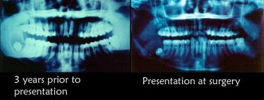
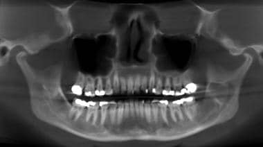
Panoramic radiography is a specialized type of dental X-ray imaging that captures a panoramic view of the entire mouth, including the teeth, upper and lower jaws, and surrounding structures. It uses a special machine that rotates around the head, capturing images as it moves. This technique provides a two-dimensional image that is helpful in diagnosing and planning treatment for various dental conditions such as impacted teeth, bone abnormalities, and jaw disorders.
The panoramic radiograph can also be used to assess the development and positioning of wisdom teeth, detect cysts or tumors in the jaws, and evaluate the effects of trauma or injury to the mouth. It is a valuable tool for dental professionals as it allows them to see a comprehensive view of the oral structures, which may not be visible with traditional X-ray techniques.
It's important to note that while panoramic radiography provides valuable information, it should be used in conjunction with other diagnostic tools and clinical examinations to ensure accurate diagnosis and treatment planning.
A third molar is the most posterior of the three molars present in an adult human dental arch. They are also commonly known as wisdom teeth, due to their late eruption period which usually occurs between the ages of 17-25, a time traditionally associated with gaining maturity and wisdom.
Anatomically, third molars have four cusps, making them the largest of all the teeth. However, not everyone develops third molars; some people may have one, two, three or no third molars at all. In many cases, third molars do not have enough space to fully erupt and align properly with the rest of the teeth, leading to impaction, infection, or other dental health issues. As a result, third molars are often extracted if they cause problems or if there is a risk they will cause problems in the future.
Ameloblastoma is a slow-growing, non-cancerous tumor that develops in the jawbone, typically in the lower jaw. It originates from the cells that form the enamel (the hard, outer surface of the teeth). This tumor can cause swelling, pain, and displacement or loosening of teeth. In some cases, it may also lead to fractures of the jawbone.
There are different types of ameloblastomas, including solid or multicystic, unicystic, and peripheral ameloblastoma. Treatment usually involves surgical removal of the tumor, with careful monitoring to ensure that it does not recur. In rare cases, more aggressive treatment may be necessary if the tumor is large or has invaded surrounding tissues.
It's important to note that while ameloblastomas are generally benign, they can still cause significant morbidity and should be treated promptly by an oral and maxillofacial surgeon or other qualified healthcare professional.
Orthodontic extrusion is a dental treatment procedure that involves the deliberate and controlled vertical movement of a tooth out of its socket with the use of orthodontic appliances. This technique is often used in orthodontics to align teeth, correct their position, or prepare them for other procedures such as crowns or bridges.
During the extrusion process, gentle force is applied to the tooth using specific orthodontic appliances, like a spring or an elastic band, which causes the tooth to move slowly in an upward direction. The movement is usually slow and gradual, taking several weeks or even months to achieve the desired result.
Orthodontic extrusion has various clinical applications, such as intruding deep overerupted teeth, uprighting tilted teeth, creating space for restorative work, or aiding in the eruption of impacted teeth. It is essential to maintain good oral hygiene and have regular check-ups with an orthodontist during the treatment to ensure proper healing and avoid any potential complications.
A cuspid, also known as a canine tooth or cuspid tooth, is a type of tooth in mammals. It is the pointiest tooth in the dental arch and is located between the incisors and bicuspids (or premolars). Cuspids have a single cusp or pointed tip that is used for tearing and grasping food. In humans, there are four cuspids, two on the upper jaw and two on the lower jaw, one on each side of the dental arch.
Ectopic tooth eruption is a condition where a tooth fails to erupt into its normal position in the dental arch. Instead, it emerupts in an abnormal location, such as in the wrong direction or through another tissue like the gums, palate, or jawbone. This can occur due to various reasons, including genetics, crowding of teeth, or trauma. Ectopic tooth eruption may cause problems with oral function and dental health, and treatment options depend on the severity and location of the ectopic tooth.
Jaw diseases refer to a variety of conditions that affect the temporomandibular joint (TMJ) and the surrounding muscles, as well as dental disorders that can impact the jaw. Some common examples include:
1. Temporomandibular Joint Disorders (TMD): These are problems with the TMJ and the muscles that control jaw movement. Symptoms may include pain, clicking or popping sounds, and limited movement of the jaw.
2. Osteonecrosis of the Jaw: This is a condition where bone in the jaw dies due to lack of blood supply. It can be caused by radiation therapy, chemotherapy, or certain medications.
3. Dental Cavities: These are holes in the teeth caused by bacteria. If left untreated, they can cause pain, infection, and damage to the jawbone.
4. Periodontal Disease: This is an infection of the gums and bones that support the teeth. Advanced periodontal disease can lead to loss of teeth and damage to the jawbone.
5. Jaw Fractures: These are breaks in the jawbone, often caused by trauma.
6. Oral Cancer: This is a type of cancer that starts in the mouth or throat. If not treated early, it can spread to the jaw and other parts of the body.
7. Cysts and Tumors: These are abnormal growths in the jawbone or surrounding tissues. While some are benign (non-cancerous), others can be malignant (cancerous).
8. Osteomyelitis: This is an infection of the bone, often occurring in the lower jaw. It can cause pain, swelling, and fever.
9. Oral Thrush: This is a fungal infection that causes white patches on the inside of the mouth. If left untreated, it can spread to the jaw and other parts of the body.
10. Sinusitis: Inflammation of the sinuses can sometimes cause pain in the upper jaw.
A deciduous tooth, also known as a baby tooth or primary tooth, is a type of temporary tooth that humans and some other mammals develop during childhood. They are called "deciduous" because they are eventually shed and replaced by permanent teeth, much like how leaves on a deciduous tree fall off and are replaced by new growth.
Deciduous teeth begin to form in the womb and start to erupt through the gums when a child is around six months old. By the time a child reaches age three, they typically have a full set of 20 deciduous teeth, including incisors, canines, and molars. These teeth are smaller and less durable than permanent teeth, but they serve important functions such as helping children chew food properly, speak clearly, and maintain space in the jaw for the permanent teeth to grow into.
Deciduous teeth usually begin to fall out around age six or seven, starting with the lower central incisors. This process continues until all of the deciduous teeth have been shed, typically by age 12 or 13. At this point, the permanent teeth will have grown in and taken their place, with the exception of the wisdom teeth, which may not erupt until later in adolescence or early adulthood.
A bicuspid valve, also known as a mitral valve in the heart, is a heart valve that has two leaflets or cusps. It lies between the left atrium and the left ventricle and helps to regulate blood flow between these two chambers of the heart. In a healthy heart, the bicuspid valve opens to allow blood to flow from the left atrium into the left ventricle and closes tightly to prevent blood from flowing back into the left atrium during contraction of the ventricle.
A congenital heart defect known as a bicuspid aortic valve occurs when the aortic valve, which normally has three leaflets or cusps, only has two. This can lead to narrowing of the valve (aortic stenosis) or leakage of the valve (aortic regurgitation), which can cause symptoms and may require medical treatment.
Mixed dentition is a stage of dental development in which both primary (deciduous) teeth and permanent teeth are present in the mouth. This phase typically begins when the first permanent molars erupt, around the age of 6, and continues until all of the primary teeth have been replaced by permanent teeth, usually around the age of 12-13.
During this stage, a person will have a mix of smaller, temporary teeth and larger, more durable permanent teeth. Proper care and management of mixed dentition is essential for maintaining good oral health, as it can help to prevent issues such as crowding, misalignment, and decay. Regular dental check-ups and proper brushing and flossing techniques are crucial during this stage to ensure the best possible outcomes for long-term oral health.
The dental sac, also known as the dental follicle, is a soft tissue structure that surrounds the developing tooth crown during odontogenesis, which is the process of tooth development. It is derived from the ectoderm and mesenchyme of the embryonic oral cavity. The dental sac gives rise to several important structures associated with the tooth, including the periodontal ligament, cementum, and the alveolar bone that surrounds and supports the tooth in the jaw.
The dental sac plays a critical role in tooth development by regulating the mineralization of the tooth crown and providing a protective environment for the developing tooth. It also contains cells called odontoblasts, which are responsible for producing dentin, one of the hard tissues that make up the tooth. Abnormalities in the development or growth of the dental sac can lead to various dental anomalies, such as impacted teeth, dilacerated roots, and other developmental disorders.
Oral surgical procedures refer to various types of surgeries performed in the oral cavity and maxillofacial region, which includes the mouth, jaws, face, and skull. These procedures are typically performed by oral and maxillofacial surgeons, who are dental specialists with extensive training in surgical procedures involving the mouth, jaws, and face.
Some common examples of oral surgical procedures include:
1. Tooth extractions: This involves removing a tooth that is damaged beyond repair or causing problems for the surrounding teeth. Wisdom tooth removal is a common type of tooth extraction.
2. Dental implant placement: This procedure involves placing a small titanium post in the jawbone to serve as a replacement root for a missing tooth. A dental crown is then attached to the implant, creating a natural-looking and functional replacement tooth.
3. Jaw surgery: Also known as orthognathic surgery, this procedure involves repositioning the jaws to correct bite problems or facial asymmetry.
4. Biopsy: This procedure involves removing a small sample of tissue from the oral cavity for laboratory analysis, often to diagnose suspicious lesions or growths.
5. Lesion removal: This procedure involves removing benign or malignant growths from the oral cavity, such as tumors or cysts.
6. Temporomandibular joint (TMJ) surgery: This procedure involves treating disorders of the TMJ, which connects the jawbone to the skull and allows for movement when eating, speaking, and yawning.
7. Facial reconstruction: This procedure involves rebuilding or reshaping the facial bones after trauma, cancer surgery, or other conditions that affect the face.
Overall, oral surgical procedures are an important part of dental and medical care, helping to diagnose and treat a wide range of conditions affecting the mouth, jaws, and face.
An incisor is a type of tooth that is primarily designed for biting off food pieces rather than chewing or grinding. They are typically chisel-shaped, flat, and have a sharp cutting edge. In humans, there are eight incisors - four on the upper jaw and four on the lower jaw, located at the front of the mouth. Other animals such as dogs, cats, and rodents also have incisors that they use for different purposes like tearing or gnawing.
Tooth extraction is a dental procedure in which a tooth that is damaged or poses a threat to oral health is removed from its socket in the jawbone. This may be necessary due to various reasons such as severe tooth decay, gum disease, fractured teeth, crowded teeth, or for orthodontic treatment purposes. The procedure is performed by a dentist or an oral surgeon, under local anesthesia to numb the area around the tooth, ensuring minimal discomfort during the extraction process.
In the context of dentistry, a molar is a type of tooth found in the back of the mouth. They are larger and wider than other types of teeth, such as incisors or canines, and have a flat biting surface with multiple cusps. Molars are primarily used for grinding and chewing food into smaller pieces that are easier to swallow. Humans typically have twelve molars in total, including the four wisdom teeth.
In medical terminology outside of dentistry, "molar" can also refer to a unit of mass in the apothecaries' system of measurement, which is equivalent to 4.08 grams. However, this usage is less common and not related to dental or medical anatomy.
Cyst fluid refers to the fluid accumulated within a cyst, which is a closed sac-like or capsular structure, typically filled with liquid or semi-solid material. Cysts can develop in various parts of the body for different reasons, and the composition of cyst fluid may vary depending on the type of cyst and its location.
In some cases, cyst fluid might contain proteins, sugars, hormones, or even cells from the surrounding tissue. Infected cysts may have pus-like fluid, while cancerous or precancerous cysts might contain abnormal cells or tumor markers. The analysis of cyst fluid can help medical professionals diagnose and manage various medical conditions, including infections, inflammatory diseases, genetic disorders, and cancers.
It is important to note that the term 'cyst fluid' generally refers to the liquid content within a cyst, but the specific composition and appearance of this fluid may vary significantly depending on the underlying cause and type of cyst.
An ovarian cyst is a sac or pouch filled with fluid that forms on the ovary. Ovarian cysts are quite common in women during their childbearing years, and they often cause no symptoms. In most cases, ovarian cysts disappear without treatment over a few months. However, larger or persistent cysts may require medical intervention, including surgical removal.
There are various types of ovarian cysts, such as functional cysts (follicular and corpus luteum cysts), which develop during the menstrual cycle due to hormonal changes, and non-functional cysts (dermoid cysts, endometriomas, and cystadenomas), which can form due to different causes.
While many ovarian cysts are benign, some may have malignant potential or indicate an underlying medical condition like polycystic ovary syndrome (PCOS). Regular gynecological check-ups, including pelvic examinations and ultrasounds, can help detect and monitor ovarian cysts.
An epidermal cyst is a common benign skin condition characterized by the growth of a sac-like structure filled with keratin, a protein found in the outermost layer of the skin (epidermis). These cysts typically appear as round, firm bumps just under the surface of the skin, often on the face, neck, trunk, or scalp. They can vary in size from a few millimeters to several centimeters in diameter.
Epidermal cysts usually develop as a result of the accumulation of dead skin cells that become trapped within a hair follicle or a pilosebaceous unit (a structure that contains a hair follicle and an oil gland). The keratin produced by the skin cells then collects inside the sac, causing it to expand gradually.
These cysts are generally slow-growing, painless, and rarely cause any symptoms. However, they may become infected or inflamed, leading to redness, tenderness, pain, or pus formation. In such cases, medical attention might be necessary to drain the cyst or administer antibiotics to treat the infection.
Epidermal cysts can be removed surgically if they cause cosmetic concerns or become frequently infected. The procedure typically involves making an incision in the skin and removing the entire sac along with its contents to prevent recurrence.
 Dentigerous cyst
Index of oral health and dental articles
Odontogenic cyst
Odontoma
Adenomatoid odontogenic tumor
Calcifying odontogenic cyst
Cysts of the jaws
Dental follicle
Periapical cyst
Dental emergency
Ameloblastoma
Glandular odontogenic cyst
Gastrointestinal disease
Wisdom tooth
Ameloblastic fibroma
Malocclusion
Homo antecessor
Follicular cyst
Odontogenic keratocyst
Ectopic tooth
Tooth impaction
Marsupialization
Eruption cyst
List of MeSH codes (C07)
List of MeSH codes (C05)
List of MeSH codes (C04)
Theropod paleopathology
Dentigerous cyst - Wikipedia
 Dentigerous Cyst | Harvard Catalyst Profiles | Harvard Catalyst
 Dentigerous cyst | MyPathologyReport.ca
 dentigerous cyst Archives - District Dentistry
The Endo Blog: Dentigerous Cyst
 Mandibular Cysts and Odontogenic Tumors: Overview, Odontogenic Mandibular Cysts, Nonodontogenic Mandibular Cysts
 Decompression of a maxillary dentigerous cyst
 Dentigerous Cyst | Calcaterra Family Dentistry | Follicular Cyst
 Odontogenic myxofibroma resembling dentigerous cyst - IIUM Repository (IRep)
 Clinical Features, Symptoms and Treatment of Dentigerous Cyst | Scitechnol
Dentigerous cyst maxilla arising from unerupted tooth - Otolaryngology Online
Bilateral dentigerous cyst in suprarenal tumor child: a case report
 Multiple bilateral dentigerous cysts are seen in: ? - MCQs For Tests
Maxillary dentigerous cyst showing squamous odontogenic tumor-like proliferation: surgical approach and literature review
 Dr. Balagopal Varma R. - Amrita Vishwa Vidyapeetham
 SURGERY CENTER: SECTION III MILITARY COMBAT SURGERY, NEUROSUREGERY OPHTHALMIC (EYE) SURGERY, ORTHOPAEDIC SURGERY - Martindale...
 Rank, rankl, and opg in dentigerous cyst, odontogenic keratocyst, and ameloblastoma: a meta-analysis | Braz. dent. j;32(1): 16...
 Clinicopathological Features and Expression of Ki-67 in Odontogenic Keratocyst, Dentigerous Cyst and Radicular Cyst
|...
Clinicopathological Features and Expression of Ki-67 in Odontogenic Keratocyst, Dentigerous Cyst and Radicular Cyst
|...
Multidisciplinary management of delayed eruption of permanent mandibular first molar associated with dentigerous cyst -...
"Forced Eruption of Multiple Impacted Teeth with Dentigerous Cyst - A C" by Tzu-Ying Wu, Chia-Chen Li et al.
Significant association of high-grade inflammation and thick lining epithelium with the increased number of Langerhans cells in...
 Weaving, Headshaking, Cribbing, and Other Stereotypies | IVIS
 References | Chronic Inflammation on the Right Side of the Maxilla | DentalCare.com
 Ameloblastoma
Summary Report | CureHunter
Ameloblastoma
Summary Report | CureHunter
Equine Surgery: Advanced Techniques Second Edition by C. Mcilwraith
 The Journal of Contemporary Dental Practice
 Are wisdom teeth (third molars) vestiges of human evolution
What Is Your Diagnosis? in: Journal of the American Veterinary Medical Association Volume 255 Issue 1 ()
 Bone grafting in oral surgery - Veterinary Practice News
 Search - JCO Online - Journal of Clinical Orthodontics
Radicular cysts7
- Secondly, radicular cysts developed at the apices of non vital primary teeth. (wikipedia.org)
- These radicular cysts may fuse with the follicles of the unerupted successors, causing the eruption of the successors into the cyst cavity. (wikipedia.org)
- Squamous odontogenic tumor-like proliferations in radicular cysts: a clinicopathologic study of forty-two cases. (autopsyandcasereports.org)
- Inflammatory radicular cysts were observed more in male gender, younger age at diagnosis and anterior maxilla as site of presentation. (unipa.it)
- Unlike dentigerous cysts, the frequency of radicular cysts decreased from 10.4% in 1986-1995 to about 8% in 1996-2005 (P (unipa.it)
- Inflammatory radicular cysts are the most represented group among OCs in our area with a higher prevalence than that reported in other countries. (unipa.it)
- Common odontogenic cysts are dentigerous cysts , and radicular cysts . (librepathology.org)
Ameloblastoma3
- There has been some discussion of dentigerous cysts that have undergone neoplastic transformation to ameloblastoma or other neoplasm. (theendoblog.com)
- Abstract The aim of this study was to assess and compare RANK, RANKL, and OPG immunoexpression in dentigerous cyst , odontogenic keratocyst , and ameloblastoma . (bvsalud.org)
- Most common odontogenic cyst and tumor reported was dentigerous cyst and ameloblastoma respectively. (oldcitypublishing.com)
Reduced enamel epi5
- A dentigerous cyst, also known as a follicular cyst, is an epithelial-lined developmental cyst formed by accumulation of fluid between the reduced enamel epithelium and the crown of an unerupted tooth. (wikipedia.org)
- The accumulation of fluid either between the reduced enamel epithelium and enamel or in between the layers of enamel organ seems to be the key to the formation of dentigerous cysts. (wikipedia.org)
- The inflammatory exudate causes separation of reduced enamel epithelium from the enamel with resultant cyst formation. (wikipedia.org)
- The cyst enlarges in size due to accumulation of fluid between reduced enamel epithelium and the crown. (drtbalu.co)
- A dentigerous cyst originates from the enamel organ remnant or reduced enamel epithelium. (dentalknowledge.in)
Tooth28
- Ectopic tooth eruption may result due to pathological process, such as a tumor or cyst or developmental disturbance. (wikipedia.org)
- The exact histogenesis of dentigerous cysts remains unknown, but most authors favor a developmental origin from the tooth follicle. (wikipedia.org)
- In 1928, Bloch-Jorgensen suggested that the overlying necrotic deciduous tooth is the origin of all dentigerous cysts. (wikipedia.org)
- He reported 22 cases of follicular cysts and stated that in each case a deciduous tooth or the remnants thereof was found in direct contact with the cyst cavity and that the related deciduous tooth always was diseased. (wikipedia.org)
- The dentigerous cyst commonly involves a single tooth and rarely affects multiple teeth. (wikipedia.org)
- Occurs in relation to a partially erupted or unerupted tooth with at least the crown of the tooth to which the cyst is attached protruding into the cystic cavity. (harvard.edu)
- Dentigerous cyst associated with a displaced tooth in the maxillary sinus: an unusual cause of recurrent sinusitis in an adolescent. (harvard.edu)
- A dentigerous cyst is a noncancerous growth that forms around the crown of an unerupted tooth. (mypathologyreport.ca)
- A dentigerous cyst is caused by the build-up of fluid in the tissue surrounding an unerupted tooth. (mypathologyreport.ca)
- It is a cyst that comes from the separation of the follicle from an uneruped tooth. (theendoblog.com)
- This cyst encloses the crown of an unerupted tooth and is attached to the tooth at the cemento-enamel junction. (theendoblog.com)
- These cysts can grow quite large and can cause painless expansion of the bone and also displace an involved tooth. (theendoblog.com)
- Treatment includes enucleation of the cyst with the removal of the unerupted tooth. (theendoblog.com)
- Many cysts resolve with endodontic therapy of the involved tooth. (medscape.com)
- The second most common odontogenic cyst is the dentigerous cyst, which develops within the normal dental follicle that surrounds an unerupted tooth. (medscape.com)
- A dentigerous or follicular cyst is defined as a cyst that originates from the separation of two specific components of a developing tooth. (orangectdentist.com)
- Dentigerous Cyst on the lower right with an impacted wisdom tooth. (orangectdentist.com)
- The red circle shows an unerupted lower right wisdom tooth with a cyst around it. (orangectdentist.com)
- The tooth along with the cyst was removed surgically and the patient recovered quite well with no significant long term complications. (orangectdentist.com)
- This type of cyst is the most common type of developmental odontogenic (tooth associated) cyst, making up 20% of all of these types of cysts of the jaw. (orangectdentist.com)
- The most common treatment for these cysts is enucleation (removal of the object without cutting directly into it) along with removal of the enerupted tooth/teeth. (orangectdentist.com)
- Unerupted tooth could be seen within the maxillary sinus after removal of the cyst. (drtbalu.co)
- Fluid sacs called dentigerous cysts or non-cancerous tumors like odontogenic keratocysts may arise from impacted wisdom tooth follicles. (cdhp.org)
- Dentigerous cyst represents is the most common developmental odontogenic cyst usually associated with an impacted tooth after complete formation of the crown. (oldcitypublishing.com)
- A dentigerous cyst encloses is seen attached to the neck of the tooth at the cementoenamel junction associated with an unerupted/ impacted tooth and grows by expansion of the dental follicles. (oldcitypublishing.com)
- By definition, a dentigerous cyst is attached to the tooth cervix (enamel-cementum junction) and encloses the crown of the unerupted tooth. (dentalknowledge.in)
- Radiographically, a dentigerous cyst manifests as a well-defined, unilocular or sometimes multilocular radiolucency with corticated margins in attached with the crown of an unerupted tooth. (dentalknowledge.in)
- Biopsy revealed squamous cell carcinoma arising from a dentigerous cyst associated with the impacted lower right wisdom tooth. (dentalmal.com)
Follicular cysts2
- Showing posts with label Follicular cysts . (dentalknowledge.in)
- Dentigerous (Follicular) Cysts are the second most commonly occurring odontogenic cysts after periapical cyst and the most common developmental cysts of the jaws. (dentalknowledge.in)
Tumor7
- In rare cases, squamous odontogenic tumor-like proliferation (SOT-LP) can be observed arising from odontogenic cysts (SOT-LPOC). (autopsyandcasereports.org)
- Squamous odontogenic tumor and squamous odontogenic tumor-like proliferations in odontogenic cysts: an updated analysis of 170 cases reported in the literature. (autopsyandcasereports.org)
- Squamous odontogenic tumor-like proliferations in periapical cysts. (autopsyandcasereports.org)
- 9 Sala-Pérez S, Marco-Molina V, Gay-Escoda C. Squamous odontogenic tumor-like proliferation in a radicular cyst: a case report. (autopsyandcasereports.org)
- 11 Unal T, Gomel M, Gunel O. Squamous odontogenic tumor-like islands in a radicular cyst: report of a case. (autopsyandcasereports.org)
- 14 Oliveira JA, Costa IM, Loyola AM. Squamous odontogenic tumor-like proliferations (SOT-LP) versus intraosseous squamous cell carcinoma in residual cyst. (autopsyandcasereports.org)
- RANKL ratio in this tumor , while it was lower for dentigerous cyst and odontogenic keratocyst . (bvsalud.org)
Periapical Cyst1
- Biological behaviour of Odontogenic Keratocyst (OKC) is aggressiveness than others Odontogenic Cysts (OCs) like Dentigerous Cyst (DCs) and Periapical Cyst/Residual Cysts (RCs). (org.pk)
Pathogenesis of dentigerous cyst1
- The pathogenesis of dentigerous cyst is still controversial. (wikipedia.org)
Maxilla6
- A decompression surgery was performed and an incisional biopsy revealed the presence of a dentigerous cyst in the maxilla. (bvsalud.org)
- CT scan image showing cyst involving the anterolateral wall of left maxilla. (drtbalu.co)
- To the best of our knowledge, this is the first report of a dentigerous cyst showing SOT-LP features in the maxilla. (autopsyandcasereports.org)
- This case report presented a11 y/o Chinese female with Class I skeletal pattern and normal mandibular plane angle.Multiple teeth were impacteddue to a dentigerous cyst over anterior region in the maxilla. (tjo.org.tw)
- Hemangioma of the maxilla with associated dentigerous cyst: report of a case. (dentalcare.com)
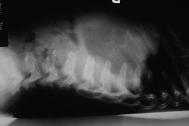
- Although resolution is not as detailed as intra-oral radiographs for examination of the teeth, gross changes in calcification of the dental structures, and changes in ossification of the underlying mandible and maxilla can aid in identification of dental disease such as caries (decay), periodontal bone loss, and abscess and cyst formation. (racgp.org.au)
Tumors8
- Volumetric analysis of keratocystic odontogenic tumors and non-neoplastic jaw cysts - Comparison and its clinical relevance. (harvard.edu)
- instead, it confines itself to an overview of the major odontogenic cysts and tumors with a brief discussion of other mandibular lesions that are often called cysts but are not true cystic lesions. (medscape.com)
- Squamous odontogenic tumors arising in odontogenic cysts. (autopsyandcasereports.org)
- The frequency and incidence of odontogenic cysts and tumors are controversial and depends in the geographic location. (oldcitypublishing.com)
- The purpose of this study was to determine the prevalence of odontogenic cysts and tumors over a period of 10 years and to compare with other data reported around the world. (oldcitypublishing.com)
- Data for the study were obtained from the archives of the Department of Oral and Maxillofacial Pathology, diagnosed as the cases of Odontogenic cysts and tumors histopathologically, reported from January 2007 to March 2016. (oldcitypublishing.com)
- This study provides epidemiological information on odontogenic cyst and tumors at an institutional level. (oldcitypublishing.com)
- The relative frequency of these cysts and tumors can be analyzed at a global level to understand their prevalence, incidence, biological behaviour, and distribution. (oldcitypublishing.com)
Large dentigerous cyst2
- Decompression, enucleation, and implant placement in the management of a large dentigerous cyst. (harvard.edu)
- I have seen a case where a large dentigerous cyst resulted in a pathologic fracture of the mandible in a dog. (veterinarypracticenews.com)
Normal dental follicle2
- Radiologically it may be difficult to differentiate cyst from normal dental follicle if the cyst is small. (drtbalu.co)
- When it is small, it is difficult to differentiate a dentigerous cyst from a large but normal dental follicle. (dentalknowledge.in)
Inflammatory4
- The resultant periapical inflammation might spread to involve the follicle of the unerupted permanent successor, an inflammatory exudate ensued with resultant dentigerous cyst formation. (wikipedia.org)
- A dentigerous cyst is described as inflamed if inflammatory cells such as histiocytes and multinucleated giant cells are seen within the cyst. (mypathologyreport.ca)
- 5. Kumar R, Singh RK, Pandey RK, Mohammad S, Ram H. Inflammatory dentigerous cyst in a ten-year-old child. (archivesheadnecksurgery.com)
- It is a well-established fact that all inflammatory and developmental cysts of odontogenic origin have squamous epithelial linings. (oldcitypublishing.com)
Squamous7
- When examined under the microscope, the inside surface of a dentigerous cyst is typically covered by a thin layer of squamous cells . (mypathologyreport.ca)
- The inside surface of the cyst is lined by a thin layer of pink squamous cells. (mypathologyreport.ca)
- Although these cysts arise from a mature resting epithelium and thus have a relatively low growth potential, a squamous cell carcinoma occasionally may arise de novo in a radicular cyst, thus the recommendation for histopathologic examination of all tissues removed. (medscape.com)
- 5 Wright JM Jr. Squamous odontogenic tumorlike proliferations in odontogenic cysts. (autopsyandcasereports.org)
- Squamous cell carcinoma arising in a residual odontogenic cyst: case report. (autopsyandcasereports.org)
- Squamous cell carcinomas arising in a dentigerous cyst. (dentalcare.com)
- The cyst is lined by stratified squamous epithelium. (dentalknowledge.in)
Teeth11
- This may trigger and hasten the formation of a dentigerous cyst developing around the permanent teeth. (wikipedia.org)
- The primary teeth were not in direct contact with the underlying dentigerous cyst. (wikipedia.org)
- Firstly, surrounding the crowns of affected teeth, the intrafollicular developmental dentigerous cysts may be formed. (wikipedia.org)
- Regezi and Sciubba stated that the impacted teeth were most commonly seen in the third molar and maxillary canine teeth, and hence dentigerous cysts occur most frequently in these teeth. (wikipedia.org)
- Patient with nonsyndromic bilateral and multiple impacted teeth and dentigerous cysts. (harvard.edu)
- This cyst usually surrounds the crown of impacted teeth, odontoma, or supernumerary teeth. (drtbalu.co)
- Prevalence rate of supernumerary teeth causing dentigerous cyst is about 0.8% in permanent dentition. (drtbalu.co)
- Teeth associated with the cysts were removed to avoid postoperative infections. (archivesheadnecksurgery.com)
- The cyst includes or is attached to the roots of the right second and third premolar teeth. (avma.org)
- Jaw destruction from expanding cysts can affect other teeth. (cdhp.org)
- The anterior teeth, consisting of the incisors and odontogenic tumours and cyst. (bvsalud.org)
Lesions3
- What factors differentiate dentigerous cysts from other pericoronal lesions? (harvard.edu)
- Next to the radicular cyst dentigerous cyst appears to the frequently appearing cystic lesions involving jaw bones. (drtbalu.co)
- The dentigerous cyst is one of the most common pathological entities found in dentistry, accounting for about 20% of all mandibular cystic lesions.1 It is usually associated with the crowns of permane. (jco-online.com)
Type of odontogenic2
- Dentigerous cysts are the second most prevalent type of odontogenic cysts after radicular cyst. (wikipedia.org)
- It is considered a type of odontogenic cyst because it develops from cells normally found in the bones of the jaw. (mypathologyreport.ca)
Right mandibular third2
- A panoramic radiograph revealed a well-defined unilocular radiolucency with corticated border associated with the unerupted right mandibular third molar resembling a dentigerous cyst. (iium.edu.my)
- Photograph1: Dentigerous cyst surrounding the crown of right mandibular third molar and going upward in ascending ramus. (dentalknowledge.in)
Jaws2
- Cabrini RL, Barros RE, Albano H. Cysts of the jaws: a statistical analysis. (dentalcare.com)
- http://www.ncbi.nlm.nih.gov/pubmed?term=Cysts of the jaws: a statistical analysis. (dentalcare.com)
Pathology2
- This article covers odontogenic tumours and cysts , which is a subset of oral pathology and can be grouped under the heading of head and neck pathology . (librepathology.org)
- This article provides a guide to identifying key anatomical features on the radiograph and outlines its use in identifying pathology such as dental disease, cysts and traumatic injuries An orthopantomogram to the hard tissues of the face. (racgp.org.au)
Differentiate1
- Dentigerous cyst - history is the key to differentiate. (librepathology.org)
Mandibular dentigerous1
- 4. Sindi AM. Bilateral mandibular dentigerous cysts presenting as an incidental finding: a case report. (archivesheadnecksurgery.com)
Mandible1
- When cysts affect significant portions of the mandible, exteriorization or marsupialization of the cyst is done to allow for decompression and subsequent shrinkage of the lesion followed by surgical enucleation. (dentalknowledge.in)
Enucleation of the cyst1
- After the enucleation of the cyst, the crown portion of 11 was exposed but with improper position and angulation. (tjo.org.tw)
Third molars2
- These cysts are most commonly involve the Md third molars and secondly the Mx canines. (theendoblog.com)
- Dentigerous cysts are most commonly seen associated with third molars and maxillary Canines. (dentalknowledge.in)
Lesion4
- The lesion was diangosed as a dentigerous cyst. (theendoblog.com)
- The cyst portion is so important because folks can misdiagnose it for and endo/perio lesion. (theendoblog.com)
- Radiographically, distinguishing between a granuloma and a cyst is impossible, although some say that if the lesion is quite large it is more likely to be a cyst. (medscape.com)
- This study describes the case of a dentigerous cyst in a six-year-old patient treated by decompression of the lesion. (bvsalud.org)
Prevalence1
- The objective of this study was to assess the prevalence of odontogenic cysts (OCs) in Sicily and evaluate their distribution during a 20-year period. (unipa.it)
Neoplasms1
- Odontogenic cysts and neoplasms. (archivesheadnecksurgery.com)
Types of cysts1
- While cases of cancers developing in these types of cysts have been described in the scientific literature, these cases are very rare. (mypathologyreport.ca)
Marsupialization1
- Marsupialization occasionally done with very large cysts to decompress the cyst. (theendoblog.com)
Neoplastic3
- However, granulomas and cysts are not neoplastic. (medscape.com)
- The dentigerous cyst is not thought to be neoplastic. (medscape.com)
- Neoplastic potential of odontogenic cysts. (librepathology.org)
Pathologic fracture1
- This cyst can achieve significant size, occasionally causes cortical bone expansion but rarely reaches a size that predisposes the patient to a pathologic fracture. (dentalknowledge.in)
Derived from odontogenic1
- Odontogenic cysts are defined as epithelial-lined structures derived from odontogenic epithelium. (medscape.com)
Bilateral1
- Bilateral dentigerous cyst in patients with no syndrome diagnosis is rare. (archivesheadnecksurgery.com)
Inflammation1
- These cysts may be secondarily inflamed and infected as a result of periapical inflammation spreading from non vital deciduous predecessors. (wikipedia.org)
Developmental odontogenic1
- Dentigerous Cyst is the most common developmental odontogenic cyst. (theendoblog.com)
Diagnosis2
- The diagnosis can also be made after the cyst is removed and sent to a pathologist for examination under the microscope. (mypathologyreport.ca)
- As dentigerous cysts have been documented to occasionally transform into a malignant carcinoma, placement of a bone graft prior to obtaining a definitive diagnosis may add fuel to the fire if neoplasia is present. (veterinarypracticenews.com)
Bone7
- Larger cysts can erode (damage) the surrounding bone and make the jaw appear larger than normal. (mypathologyreport.ca)
- bone cyst. (archivesheadnecksurgery.com)
- Nadimi H, Bronny AT, Sbigoli A, Gatti WM, Hasiakos P. Aneurysmal bone cyst associated with a dentigerous cyst: report of a case. (dentalcare.com)
- http://www.ncbi.nlm.nih.gov/pubmed?term=Aneurysmal bone cyst associated with a dentigerous cyst: report of a case. (dentalcare.com)
- Dentigerous cysts can be expansive and cause a large amount of bone loss. (veterinarypracticenews.com)
- Similarly, some veterinary dentists prefer not to place bone graft into the site of an enucleated dentigerous cyst, feeling that the blood clot that remains after closure of the site will have the ability to act as a scaffold. (veterinarypracticenews.com)
- The expansion of the dentigerous cyst is related to epithelial proliferation, release of bone-resorbing factors, and an increase in cyst fluid osmolality. (dentalknowledge.in)
Eruption2
- On the contrary, Toiler suggested that the breakdown of proliferating cells of the follicle after impeded eruption is likely to be the origin of the dentigerous cyst. (wikipedia.org)
- The delayed eruption is the most common indication of dentigerous cyst formation. (dentalknowledge.in)
Fluid1
- A cyst is basically an enclosed sac within tissue containing either fluid, air, or semi-solid materials. (orangectdentist.com)
Maxillary sinus1
- In maxillary dentigerous cysts in the canine region, extension into the maxillary sinus or to the orbital floor may be seen. (dentalknowledge.in)
Complications1
- Can cyst volume be used to stratify risk of complications following cyst defect reconstruction with iliac crest graft? (harvard.edu)
Incidence1
- The peak incidence of dentigerous cysts occurs between twenty to 40 years. (dentalknowledge.in)
Oral cavity1
- The aim of the study was to determine clinicopathological features and expression of Ki-67 in Odontogenic Cysts of the oral cavity. (org.pk)
Tomographic1
- Physical and tomographic assessments suggested odontogenic cysts. (archivesheadnecksurgery.com)