Dental Cementum
Dental Care
Students, Dental
Dental Caries
Periodontal Ligament
Tooth Cervix
Dental Care for Chronically Ill
Dental Care for Children
Periodontium
Tooth Calcification
Dental Pulp
Integrin-Binding Sialoprotein
Dental Hygienists
Root Resorption
Faculty, Dental
Dental Care for Disabled
Tooth Apex
Alveolar Process
Dental Anxiety
Dental Research
Dental Care for Aged
Dental Arch
Dental Plaque
Age Determination by Teeth
Dental Offices
Dental Records
Dental Staff
Dental Enamel
Dental Equipment
General Practice, Dental
Dental Amalgam
Odontogenesis
Education, Dental, Continuing
Dental Implants
Phosphate Transport Proteins
Education, Dental, Graduate
Ethics, Dental
Tooth Eruption
Tooth Resorption
Tooth Migration
Technology, Dental
Cementoma
Dental Health Surveys
Fluorosis, Dental
Dental Cavity Preparation
Laboratories, Dental
Furcation Defects
Dental Materials
Periodontal Diseases
Guided Tissue Regeneration, Periodontal
Dental Occlusion
Incisor
Practice Management, Dental
Hardness
Dental Sac
Tooth, Deciduous
Decalcification, Pathologic
Dentistry
Esthetics, Dental
Epithelial Attachment
Comprehensive Dental Care
Osteopontin
Health Education, Dental
Mandible
Tooth Socket
Microspectrophotometry
Alveolar Bone Loss
Dentinogenesis
Infection Control, Dental
Dental Papilla
Dental Prosthesis
Amelogenin
Tooth Diseases
Dental Leakage
Dental Audit
Oral Health
Dental Instruments
Dental Waste
Dental Implantation
Ameloblasts
Dental Marginal Adaptation
Microscopy, Electron, Scanning
Dental Alloys
Dental Informatics
Oral Hygiene
Dental Scaling
Felidae
Microscopy, Polarization
Dentistry, Operative
Dental Facilities
Dental Devices, Home Care
Preventive Dentistry
Photography, Dental
Bone Regeneration
Tolonium Chloride
Molar
Dental Porcelain
Radiography, Dental, Digital
Methylmethacrylates
The root surface in human teeth: a microradiographic study. (1/170)
In an attempt to clarify the nature of the human cemento-dentinal junction, ground sections of incompletely formed and fully formed extracted teeth were prepared and their histology compared with their microradiographic appearances. The results showed that incompletely formed teeth possess distinctive surface layers outside the granular layer of Tomes. The evidence indicates that these layers are of dentinal origin; their presence during development supports previous explanations by the author of the hyaline layer of Hopewell-Smith and of so-called intermediate cementum. The results also indicate that the granular layer of Tomes does not represent the outer limit of root dentine. The relationship of these surface layers to the definitive cementum which is present in fully formed teeth was studied in both young and older patients. From the results it was concluded that cementum formation begins in the more apical region of the teeth at a time when root formation is well advanced, and that it spreads towards the crown rather than in the generally accepted reverse direction. (+info)Histological and histochemical quantification of root resorption incident to the application of intrusive force to rat molars. (2/170)
This study was conducted to investigate the nature of root resorption resulting from intrusive forces applied to the rat lower molars, by means of histological and histochemical techniques with tartrate resistant acid phosphatase (TRAP). Thirty-eight 13-week-old Wistar strain male rats were used. Intrusive force was created by a fixed appliance which was adjusted to exert an initial force of 50 g for the duration of 1, 2, and 3 weeks. The degree of root resorption and distribution of TRAP positive cells were evaluated. On the root surface, the TRAP positive scores were low in the apical regions. Significant differences in the scores were found in the inter-radicular region of the roots between the experimental and control groups for the 2- and 3-week groups. More active resorption of bone occurred during the experimental period, as denoted by greater TRAP positive scores on the bone than on the root surface. Root resorption scores in the apical root region were larger in the 2- and 3-week groups than in the 1-week group. Significant differences in the root resorption scores were also found between the 1- and 3-week groups in the inter-radicular region, indicating that intrusive force application of a longer duration may lead to a higher frequency of root resorption. It is shown that, irrespective of the level of TRAP positive cells and root resorption scores, the degree of root resorption activity is higher in the apical root region than in the inter-radicular area. These results indicate that cellular cementum may be resorbed more easily because of its richer organic components and low mineralized structure. (+info)Evolution of periodontal regeneration: from the roots' point of view. (3/170)
Tissues lost as a consequence of periodontal diseases, i.e. bone, cementum and a functional periodontal ligament (PDL), can be restored to some degree. Nevertheless, results are often disappointing. There is a need to develop new paradigms for regenerating periodontal tissues that are based on an understanding of the cellular and molecular mechanisms regulating the development and regeneration of periodontal tissues. As one approach we have developed strategies for maintaining cementoblasts in culture by first determining the gene profile for these cells in situ. Next, cells were immortalized in vitro using SV 40 large T antigen (SV40 Tag) or by using mice containing transgenes enabling cellular immortality in vitro. Cementoblasts in vitro retained expression of genes associated with mineralized tissues, bone sialoprotein and osteocalcin, that were not linked with periodontal fibroblasts either in situ or in vitro. Further, cementoblasts promoted mineralization in vitro as measured by von Kossa and ex vivo using a severely compromised immunodeficient (SCID) mouse model. These cells responded to growth factors by eliciting changes in gene profile and mitogenesis and to osteotropic hormones by evoking changes in gene profile and ability to induce mineral nodule formation in vitro. The ultimate goal of these studies is to provide the knowledge base required for designing improved modalities for use in periodontal regenerative therapies. (+info)Growth factors regulate expression of mineral associated genes in cementoblasts. (4/170)
BACKGROUND: Knowledge of the responsiveness of cells within the periodontal region to specific bioactive agents is important for improving regenerative therapies. The aim of this study was to determine the effect of specific growth factors, insulin-like growth factor-I (IGF-I), platelet-derived growth factor-BB (PDGF-BB), and transforming growth factor-beta (TGF-beta) on cementoblasts in vitro and ex vivo. METHODS: Osteocalcin (OC) promoter driven SV40 transgenic mice were used to obtain immortalized cementoblasts. Growth factor effects on DNA synthesis were assayed by [3H]-thymidine incorporation. Northern analysis was used to determine the effects of growth factors on gene expression profile. Effects of growth factors on cementoblast induced biomineralization were determined in vitro (von Kossa stain) and ex vivo (re-implantation of cells in immunodeficient (SCID) mice). RESULTS: All growth factors stimulated DNA synthesis compared to control. Twenty-four hour exposure of cells to PDGF-BB or TGF-beta resulted in a decrease in bone sialoprotein (BSP) and osteocalcin (OCN) mRNAs while PDGF-BB also increased osteopontin (OPN) mRNA. Cells exposed to IGF-I for 24 hours exhibited decreased transcripts for OCN and OPN with an upregulation of BSP mRNA noted at 72 hours. In vitro mineralization was inhibited by continuous application of PDGF-BB or TGF-beta, while cells exposed to these factors prior to implantation into SCID mice still promoted biomineralization. CONCLUSIONS: These data indicate IGF-I, PDGF-BB, and TGF-beta influence mitogenesis, phenotypic gene expression profile, and biomineralization potential of cementoblasts suggesting that such factors alone or in combination with other agents may provide trigger factors required for regenerating periodontal tissues. (+info)Cell-specific patterns of Cbfa1 mRNA and protein expression in postnatal murine dental tissues. (5/170)
Cbfa1 (core binding factor alpha 1) is a transcription factor that is a key determinant of the osteoblastic lineage. Recent data showed that Cbfa1 is also highly expressed in early stages of tooth development and is involved in crown morphogenesis and cytodifferentiation of odontoblasts. Here we report the mRNA expression and protein localization of Cbfa1 in the mouse dentition in (later) stages of crown and root development. In addition to osteoblasts, osteocytes, chondrocytes, odontoblasts, dental follicle cells, cementoblasts and periodontal ligament cells, we report also Cbfa1 expression in dental epithelial cells (secretory and maturation ameloblasts) and several non-mineralizing cell types (hair follicles, ducts of salivary glands, and junctional epithelium of the gingiva). (+info)Ultrastructure of cementum and periodontal ligament after continuous intrusion in humans: a transmission electron microscopy study. (6/170)
An ultrastructural study of the cementum and periodontal ligament (PDL) changes after continuous intrusion with two different and controlled forces in humans was carried out. Twelve first upper premolars, at stage 10 of Nolla, orthodontically indicated for extraction from six patients (mean age 15.3) were used. They were divided into three experimental groups, distributed intra-individually as follows: control (not moved), continuously intruded for 4 weeks with 50 or 100 cN force, utilizing a precise biomechanical model with nickel titanium super-elastic wires (NiTi-SE), which were developed and calibrated individually. The teeth were extracted, fixed, decalcified, and conventionally processed for examination in a Jeol 100 CX II transmission electron microscope. Evident signs of degeneration of cell structures, vascular components, and extracellular matrix (EM) of cementum and PDL were observed in all the intruded teeth, with more severe changes towards an apical direction and in proportion to the magnitude of force applied. Resorptive areas and an irregular root surface of the intruded teeth were noticed, according to the same pattern described above. Concomitant, areas of repair were also revealed in the cementum and PDL although the magnitude of forces remained the same throughout the experimental period. Thus, a reduction of continuous force magnitude should be considered to preserve the integrity of tissues. (+info)Platelet-derived growth factor (PDGF) gene delivery for application in periodontal tissue engineering. (7/170)
BACKGROUND: A challenge in the reconstruction of periodontal structures is the targeted delivery of growth-promoting molecules to the tooth root surface. Polypeptide growth factors such as platelet-derived growth factor (PDGF) stimulate both cementogenesis and osteogenesis. Recent advances in gene therapy offer the advantage of delivering recombinant proteins to tissues for extended periods of time in vivo. METHODS: Recombinant adenoviral vectors encoding for the PDGF-A gene were constructed to allow delivery of PDGF transgenes to cells. The recombinant adenoviruses were assembled using the viral backbone of Ad2/CMV/EGFP and replacing GFP (reporter gene encoding green fluorescent protein driven by the cytomegalovirus promoter [CMV] within adenovirus type 2) with the PDGF-A gene. Root lining cells (cloned cementoblasts) were transduced with Ad2/PDGF-A and evaluated for gene expression, DNA synthesis, and cell proliferation. PDGF-inducible genes, c-myc and osteopontin, were also evaluated following gene delivery of Ad2/PDGF-A. RESULTS: The results revealed high level transduction of cementoblasts by gene transfer for 7 days as evidenced by flow cytometry and Northern blotting. Cementoblast DNA synthesis and subsequent proliferation were stimulated by Ad2/PDGF-A at levels equal to or greater than continuous rhPDGF-AA application. Strong message for the PDGF-A gene and protein as evidenced by Northern blotting and immunocytochemistry was noted. Furthermore, the potent induction of c-myc and osteopontin mRNA was found after PDGF gene delivery to cementoblasts. CONCLUSIONS: These findings demonstrate that gene delivery of platelet-derived growth factor stimulates cementoblast activity that is sustained above that of rhPDGF-AA application. The use of gene therapy as a mode of growth factor delivery offers a novel approach to periodontal tissue engineering. (+info)The developmental biology of cementum. (8/170)
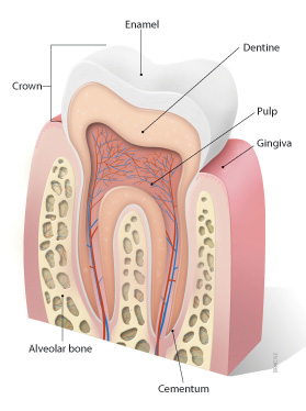
In conclusion, we have reviewed an extensive literature on early cementogenesis and performed a detailed morphological and molecular analysis to illustrate and verify key issues in the current debate about epithelial and mesenchymal contributions to root cementum. We have demonstrated that prior to cementogenesis, Hertwig's epithelial root sheath disintegrates and dental follicle cells penetrate the epithelial layer to invade the root surface. Our studies confirmed that HERS became disrupted or disintegrated prior to cementum deposition. We visualized how mesenchymal cells from the dental follicle penetrated the HERS bilayer and deposited initial cementum, while immediately adjacent epithelial cells were separated from the root surface by a basal lamina and did not secrete any cementum. Human specimen from the Gottlieb collection indicated that HERS was removed from the root surface prior to cementum deposition. Our in situ hybridization and immolocalization data revealed that both amelogenin mRNAs and enamel proteins were restricted to the crown enamel and were absent from the root surface and from the cervical-most ameloblasts adjacent to the root margin. On Western blots, cementum protein extracts did not cross-react with amelogenin antibodies. Our studies in conjunction with our literature review together confirmed the classical theory of cementum as a dental follicle derived connective tissue that forms subsequent to HERS disintegration. (+info)Dental cementum is a type of hard connective tissue that covers the root of a tooth. It is primarily composed of calcium salts and collagen fibers, and it serves to attach the periodontal ligaments (the fibers that help secure the tooth in its socket) to the tooth's root. Cementum also helps protect the root of the tooth and contributes to the maintenance of tooth stability. It continues to grow and deposit new layers throughout an individual's life, which can be seen as incremental lines called "cementum annulations."
Dental care refers to the practice of maintaining and improving the oral health of the teeth and gums. It involves regular check-ups, cleanings, and treatments by dental professionals such as dentists, hygienists, and dental assistants. Dental care also includes personal habits and practices, such as brushing and flossing, that help prevent tooth decay and gum disease.
Regular dental care is important for preventing common dental problems like cavities, gingivitis, and periodontal disease. It can also help detect early signs of more serious health issues, such as oral cancer or diabetes, which can have symptoms that appear in the mouth.
Dental care may involve a range of treatments, from routine cleanings and fillings to more complex procedures like root canals, crowns, bridges, and implants. Dental professionals use various tools and techniques to diagnose and treat dental problems, including X-rays, dental impressions, and local anesthesia.
Overall, dental care is a critical component of overall health and wellness, as poor oral health has been linked to a range of systemic health issues, including heart disease, stroke, and respiratory infections.
Dental education refers to the process of teaching, training, and learning in the field of dentistry. It involves a curriculum of academic and clinical instruction that prepares students to become licensed dental professionals, such as dentists, dental hygienists, and dental assistants. Dental education typically takes place in accredited dental schools or programs and includes classroom study, laboratory work, and supervised clinical experience. The goal of dental education is to provide students with the knowledge, skills, and values necessary to deliver high-quality oral health care to patients and promote overall health and wellness.
"Schools, Dental" is not a recognized medical term or concept. It seems that there might be some confusion in the terminology used. If you are referring to "Dental Schools," they are educational institutions that offer professional training programs in dentistry, leading to a degree in dental surgery (DDS) or dental medicine (DMD).
If you meant something else, please clarify the term or concept, and I would be happy to provide more information.
I'm happy to help! However, I believe there may be a slight misunderstanding in your question. "Students, Dental" is not a medical term or definition. Instead, it likely refers to individuals who are studying to become dental professionals, such as dentists, dental hygienists, or dental assistants.
If you're looking for information about dental education or the field of dentistry, I would be happy to provide some resources or answer any questions you may have!
Dental caries, also known as tooth decay or cavities, refers to the damage or breakdown of the hard tissues of the teeth (enamel, dentin, and cementum) due to the activity of acid-producing bacteria. These bacteria ferment sugars from food and drinks, producing acids that dissolve and weaken the tooth structure, leading to cavities.
The process of dental caries development involves several stages:
1. Demineralization: The acidic environment created by bacterial activity causes minerals (calcium and phosphate) to be lost from the tooth surface, making it weaker and more susceptible to decay.
2. Formation of a white spot lesion: As demineralization progresses, a chalky white area appears on the tooth surface, indicating early caries development.
3. Cavity formation: If left untreated, the demineralization process continues, leading to the breakdown and loss of tooth structure, resulting in a cavity or hole in the tooth.
4. Infection and pulp involvement: As the decay progresses deeper into the tooth, it can reach the dental pulp (the soft tissue containing nerves and blood vessels), causing infection, inflammation, and potentially leading to toothache, abscess, or even tooth loss.
Preventing dental caries involves maintaining good oral hygiene, reducing sugar intake, using fluoride toothpaste and mouthwash, and having regular dental check-ups and cleanings. Early detection and treatment of dental caries can help prevent further progression and more severe complications.
The periodontal ligament, also known as the "PDL," is the soft tissue that connects the tooth root to the alveolar bone within the dental alveolus (socket). It consists of collagen fibers organized into groups called principal fibers and accessory fibers. These fibers are embedded into both the cementum of the tooth root and the alveolar bone, providing shock absorption during biting and chewing forces, allowing for slight tooth movement, and maintaining the tooth in its position within the socket.
The periodontal ligament plays a crucial role in the health and maintenance of the periodontium, which includes the gingiva (gums), cementum, alveolar bone, and the periodontal ligament itself. Inflammation or infection of the periodontal ligament can lead to periodontal disease, potentially causing tooth loss if not treated promptly and appropriately.
The term "tooth cervix" is not commonly used in medical dentistry with a specific technical definition. However, if you are referring to the "cervical region of a tooth," it generally refers to the area where the crown (the visible part of the tooth) meets the root (the portion of the tooth that is below the gum line). This region is also sometimes referred to as the "cementoenamel junction" (CEJ), where the enamel covering of the crown meets the cementum covering of the root. Dental issues such as tooth decay, receding gums, or abrasion can affect this area and may require professional dental treatment.
Dental care for chronically ill refers to the oral health management and treatment provided to individuals who have chronic medical conditions. These patients often require specialized dental care due to their increased risk of developing oral health problems as a result of their underlying medical condition or its treatment. The goal of dental care for the chronically ill is to prevent and manage dental diseases, such as tooth decay and gum disease, in order to maintain overall health and quality of life. This may involve close collaboration between dental professionals, physicians, and other healthcare providers to ensure that the patient's oral health needs are being met in a comprehensive and coordinated manner.
Dental care for children, also known as pediatric dentistry, is a branch of dentistry that focuses on the oral health of children from infancy through adolescence. The medical definition of dental care for children includes:
1. Preventive Dentistry: This involves regular dental check-ups, professional cleaning, fluoride treatments, and sealants to prevent tooth decay and other dental diseases. Parents are also educated on proper oral hygiene practices for their children, including brushing, flossing, and dietary habits.
2. Restorative Dentistry: If a child develops cavities or other dental problems, restorative treatments such as fillings, crowns, or pulpotomies (baby root canals) may be necessary to restore the health and function of their teeth.
3. Orthodontic Treatment: Many children require orthodontic treatment to correct misaligned teeth or jaws. Early intervention can help guide proper jaw development and prevent more severe issues from developing later on.
4. Habit Counseling: Dental care for children may also involve habit counseling, such as helping a child stop thumb sucking or pacifier use, which can negatively impact their oral health.
5. Sedation and Anesthesia: For children who are anxious about dental procedures or have special needs, sedation or anesthesia may be used to ensure their comfort and safety during treatment.
6. Emergency Care: Dental care for children also includes emergency care for injuries such as knocked-out teeth, broken teeth, or severe toothaches. Prompt attention is necessary to prevent further damage and alleviate pain.
7. Education and Prevention: Finally, dental care for children involves educating parents and children about the importance of good oral hygiene practices and regular dental check-ups to maintain optimal oral health throughout their lives.
A dental clinic is a healthcare facility that is primarily focused on providing oral health services to patients. These services may include preventative care, such as dental cleanings and exams, as well as restorative treatments like fillings, crowns, and bridges. Dental clinics may also offer specialized services, such as orthodontics, periodontics, or endodontics.
In a dental clinic, patients are typically seen by licensed dentists who have completed dental school and received additional training in their chosen area of specialty. Dental hygienists, dental assistants, and other support staff may also work in the clinic to provide care and assistance to patients.
Dental clinics can be found in a variety of settings, including hospitals, community health centers, private practices, and educational institutions. Some dental clinics may specialize in treating certain populations, such as children, elderly individuals, or low-income patients. Others may offer specialized services, such as oral surgery or cosmetic dentistry.
Overall, dental clinics play an important role in promoting oral health and preventing dental diseases and conditions. By providing access to high-quality dental care, dental clinics can help patients maintain healthy teeth and gums, prevent tooth decay and gum disease, and improve their overall quality of life.
The periodontium is a complex structure in the oral cavity that surrounds and supports the teeth. It consists of four main components:
1. Gingiva (gums): The pink, soft tissue that covers the crown of the tooth and extends down to the neck of the tooth, where it meets the cementum.
2. Cementum: A specialized, calcified tissue that covers the root of the tooth and provides a surface for the periodontal ligament fibers to attach.
3. Periodontal ligament (PDL): A highly vascular and cell-rich connective tissue that attaches the cementum of the tooth root to the alveolar bone, allowing for tooth mobility and absorption of forces during chewing.
4. Alveolar bone: The portion of the jawbone that contains the sockets (alveoli) for the teeth. It is a spongy bone with a rich blood supply that responds to mechanical stresses from biting and chewing, undergoing remodeling throughout life.
Periodontal diseases, such as gingivitis and periodontitis, affect the health and integrity of the periodontium, leading to inflammation, bleeding, pocket formation, bone loss, and ultimately tooth loss if left untreated.
Tooth calcification, also known as dental calculus or tartar formation, refers to the hardening of plaque on the surface of teeth. This process occurs when minerals from saliva combine with bacterial deposits and dental plaque, resulting in a hard, calcified substance that adheres to the tooth surface. Calcification can occur both above and below the gum line, and if not removed through professional dental cleanings, it can lead to periodontal disease, tooth decay, and other oral health issues.
Dental pulp is the soft tissue located in the center of a tooth, surrounded by the dentin. It contains nerves, blood vessels, and connective tissue, and plays a vital role in the development and health of the tooth. The dental pulp helps to form dentin during tooth development and continues to provide nourishment to the tooth throughout its life. It also serves as a sensory organ, allowing the tooth to detect hot and cold temperatures and transmit pain signals to the brain. Injury or infection of the dental pulp can lead to serious dental problems, such as tooth decay or abscesses, and may require root canal treatment to remove the damaged tissue and save the tooth.
Integrin-binding sialoprotein (IBSP) is a non-collagenous protein found in bones and teeth. It is also known as bone sialoprotein II or acidic glycoprotein 34. IBSP plays a role in the regulation of biomineralization, which is the process by which minerals are deposited in biological tissues.
IBSP contains several functional domains that allow it to interact with other proteins and molecules. One such domain is an arginine-glycine-aspartic acid (RGD) motif, which can bind to integrin receptors on the surface of cells. This interaction helps regulate the attachment and behavior of cells in bone tissue.
IBSP also contains a large number of sialic acid residues, which give it its name and contribute to its negative charge. These residues may play a role in protecting the protein from degradation and helping it interact with other molecules in the extracellular matrix.
Overall, IBSP is an important component of bone tissue and plays a key role in regulating the formation and maintenance of bones and teeth.
A dental hygienist is a licensed healthcare professional who works as part of the dental team, providing educational, clinical, and therapeutic services to prevent and control oral diseases. They are trained and authorized to perform various duties such as:
1. Cleaning and polishing teeth (prophylaxis) to remove plaque, calculus, and stains.
2. Applying fluoride and sealants to protect tooth surfaces from decay.
3. Taking dental radiographs (x-rays) to help diagnose dental issues.
4. Providing oral health education, including proper brushing, flossing techniques, and nutrition counseling.
5. Performing screenings for oral cancer and other diseases.
6. Documenting patient care and treatment plans in medical records.
7. Collaborating with dentists to develop individualized treatment plans for patients.
8. Managing infection control protocols and maintaining a safe, clean dental environment.
9. Providing supportive services, such as applying anesthetics or administering nitrous oxide, under the direct supervision of a dentist (depending on state regulations).
Dental hygienists typically work in private dental offices but can also be found in hospitals, clinics, public health settings, educational institutions, and research facilities. They must complete an accredited dental hygiene program and pass written and clinical exams to obtain licensure in their state of practice. Continuing education is required to maintain licensure and stay current with advancements in the field.
Root resorption is a process that occurs when the body's own cells, called odontoclasts, break down and destroy the hard tissue of the tooth root. This can occur as a result of various factors such as trauma, infection, or orthodontic treatment. In some cases, it may be a normal part of the tooth development and eruption process in children. However, excessive or pathological root resorption can lead to weakening and loss of the tooth. It is often asymptomatic and discovered during routine dental x-rays.
The Faculty of Dental Surgery (FDS) is a division or department within a medical or dental school that focuses on the study, research, and practice of dental surgery. The faculty may be responsible for providing undergraduate and postgraduate education and training in dental surgery, as well as conducting research in this field.
Dental surgery encompasses various procedures related to the diagnosis, treatment, and prevention of diseases and disorders that affect the teeth, gums, and other structures of the mouth and jaw. This may include procedures such as tooth extractions, root canals, dental implants, and oral cancer surgery, among others.
The Faculty of Dental Surgery is typically composed of a group of dental surgeons who are experts in their field and have a commitment to advancing the practice of dental surgery through education, research, and clinical excellence. Members of the faculty may include professors, researchers, clinicians, and other professionals who are involved in the delivery of dental care.
Dental care for disabled refers to the specialized oral health services and treatments provided to individuals with physical, cognitive, or developmental disabilities. This type of dental care aims to prevent and manage dental diseases and conditions that can be more prevalent and challenging to treat in this population due to factors such as limited mobility, difficulty communicating, behavioral challenges, and the need for specialized equipment and techniques. Dental care for disabled may include routine cleanings, fillings, extractions, and other procedures, as well as education and counseling on oral hygiene and dietary habits. It may also involve collaboration with other healthcare providers to manage overall health and well-being.
The tooth apex is the tip or the narrowed end of the root of a tooth. It is the portion that is located deepest within the jawbone and it contains dental pulp tissue, which includes nerves and blood vessels. The apex plays an essential role in the development and maintenance of a tooth, as well as in the process of root canal treatment, where instruments and materials are introduced through it to clean and fill the root canals. It is also a crucial landmark in endodontic surgery and dental imaging.
The alveolar process is the curved part of the jawbone (mandible or maxilla) that contains sockets or hollow spaces (alveoli) for the teeth to be embedded. These processes are covered with a specialized mucous membrane called the gingiva, which forms a tight seal around the teeth to help protect the periodontal tissues and maintain oral health.
The alveolar process is composed of both compact and spongy bone tissue. The compact bone forms the outer layer, while the spongy bone is found inside the alveoli and provides support for the teeth. When a tooth is lost or extracted, the alveolar process begins to resorb over time due to the lack of mechanical stimulation from the tooth's chewing forces. This can lead to changes in the shape and size of the jawbone, which may require bone grafting procedures before dental implant placement.
A tooth is a hard, calcified structure found in the jaws (upper and lower) of many vertebrates and used for biting and chewing food. In humans, a typical tooth has a crown, one or more roots, and three layers: the enamel (the outermost layer, hardest substance in the body), the dentin (the layer beneath the enamel), and the pulp (the innermost layer, containing nerves and blood vessels). Teeth are essential for proper nutrition, speech, and aesthetics. There are different types of teeth, including incisors, canines, premolars, and molars, each designed for specific functions in the mouth.
Dental anxiety is a common feeling of fear or apprehension associated with dental appointments, treatments, or procedures. It can range from mild feelings of unease to severe phobias that cause people to avoid dental care altogether. Dental anxiety may stem from various factors such as negative past experiences, fear of pain, needles, or loss of control. In some cases, dental anxiety may lead to physical symptoms like sweating, rapid heartbeat, and difficulty breathing. It is important for individuals with dental anxiety to communicate their feelings with their dentist so that they can receive appropriate care and support.
Dental insurance is a type of health insurance specifically designed to cover the costs associated with dental care. It typically helps pay for preventive, basic, and major restorative procedures, including routine checkups, cleanings, fillings, extractions, root canals, crowns, bridges, and in some cases, orthodontic treatment.
Dental insurance plans often have a network of participating dentists who agree to provide services at pre-negotiated rates, helping to keep costs down for both the insured individual and the insurance company. The plan may cover a certain percentage of the cost of each procedure or have set copayments and deductibles that apply.
Like other forms of insurance, dental insurance plans come with annual maximum coverage limits, which is the most the plan will pay for dental care within a given year. It's essential to understand the terms and conditions of your dental insurance policy to make informed decisions about your oral health care and maximize the benefits available to you.
Dental auxiliaries are healthcare professionals who provide support to dentists in the delivery of oral healthcare services. They work under the supervision of a licensed dentist and perform tasks that require specific technical skills and knowledge. Examples of dental auxiliaries include dental hygienists, dental assistants, and dental lab technicians.
Dental hygienists are responsible for providing preventive dental care to patients, including cleaning teeth, taking x-rays, and educating patients on oral hygiene practices. They may also perform certain clinical procedures under the direct supervision of a dentist.
Dental assistants work closely with dentists during dental procedures, preparing instruments, mixing materials, and providing patient care. They may also perform administrative tasks such as scheduling appointments and managing patient records.
Dental lab technicians create dental restorations such as crowns, bridges, and dentures based on impressions taken by the dentist. They use a variety of materials and techniques to fabricate these devices with precision and accuracy.
It's important to note that the specific roles and responsibilities of dental auxiliaries may vary depending on the jurisdiction and local regulations.
Dental health services refer to medical care and treatment provided for the teeth and mouth. This can include preventative care, such as dental cleanings and exams, as well as restorative treatments like fillings, crowns, and root canals. Dental health services may also include cosmetic procedures, such as teeth whitening or orthodontic treatment to straighten crooked teeth. In addition to these services, dental health professionals may provide education on oral hygiene and the importance of maintaining good dental health. These services are typically provided by dentists, dental hygienists, and other dental professionals in a variety of settings, including private dental practices, community health clinics, and hospitals.
Dental research is a scientific discipline that focuses on the study of teeth, oral health, and related diseases. It involves various aspects of dental sciences such as oral biology, microbiology, biochemistry, genetics, epidemiology, biomaterials, and biotechnology. The main aim of dental research is to improve oral health care, develop new diagnostic tools, prevent dental diseases, and create better treatment options for various dental conditions. Dental researchers may study topics such as tooth development, oral cancer, periodontal disease, dental caries (cavities), saliva composition, and the effects of nutrition on oral health. The findings from dental research can help improve dental care practices, inform public health policies, and advance our understanding of overall human health.
Dental care for the elderly, also known as geriatric dentistry, refers to the dental care services provided to meet the specific needs and challenges of older adults. As people age, they may experience various oral health issues such as:
* Dry mouth due to medication side effects or medical conditions
* Gum disease and periodontitis
* Tooth loss and decay
* Oral cancer
* Uneven jawbone or ill-fitting dentures
Dental care for the aged may include routine dental exams, cleanings, fillings, extractions, denture fittings, oral surgery, and education on proper oral hygiene. It is important for elderly individuals to maintain good oral health as it can impact their overall health and quality of life. Regular dental check-ups and good oral hygiene practices can help prevent or manage these common oral health problems in the elderly.
The dental arch refers to the curved shape formed by the upper or lower teeth when they come together. The dental arch follows the curve of the jaw and is important for proper bite alignment and overall oral health. The dental arches are typically described as having a U-shaped appearance, with the front teeth forming a narrower section and the back teeth forming a wider section. The shape and size of the dental arch can vary from person to person, and any significant deviations from the typical shape or size may indicate an underlying orthodontic issue that requires treatment.
Dental enamel is the hard, outermost layer of a tooth that protects the dentin and pulp inside. It is primarily made up of minerals, mainly hydroxyapatite, and contains very little organic material. However, during the formation of dental enamel, proteins are synthesized and secreted by ameloblast cells, which help in the development and mineralization of the enamel. These proteins play a crucial role in the proper formation and structure of the enamel.
Some of the main dental enamel proteins include:
1. Amelogenin: This is the most abundant protein found in developing enamel, accounting for about 90% of the organic matrix. Amelogenin helps regulate the growth and organization of hydroxyapatite crystals during mineralization. It also plays a role in determining the final hardness and structure of the enamel.
2. Enamelin: This protein is the second most abundant protein in developing enamel, accounting for about 5-10% of the organic matrix. Enamelin is involved in the elongation and thickening of hydroxyapatite crystals during mineralization. It also helps maintain the stability of the enamel structure.
3. Ameloblastin: This protein is produced by ameloblast cells and is essential for proper enamel formation. Ameloblastin plays a role in regulating crystal growth, promoting adhesion between crystals, and maintaining the structural integrity of the enamel.
4. Tuftelin: This protein is found in both dentin and enamel but is more abundant in enamel. Tuftelin is involved in the initiation of mineralization and helps regulate crystal growth during this process.
5. Dentin sialophosphoprotein (DSPP): Although primarily associated with dentin formation, DSPP is also found in developing enamel. It plays a role in regulating crystal growth and promoting adhesion between crystals during mineralization.
After the formation of dental enamel is complete, these proteins are largely degraded and removed, leaving behind the highly mineralized and hard tissue that characterizes mature enamel. However, traces of these proteins may still be present in the enamel and could potentially play a role in its structure and properties.
Dental plaque is a biofilm or mass of bacteria that accumulates on the surface of the teeth, restorative materials, and prosthetic devices such as dentures. It is initiated when bacterial colonizers attach to the smooth surfaces of teeth through van der Waals forces and specific molecular adhesion mechanisms.
The microorganisms within the dental plaque produce extracellular polysaccharides that help to stabilize and strengthen the biofilm, making it resistant to removal by simple brushing or rinsing. Over time, if not regularly removed through oral hygiene practices such as brushing and flossing, dental plaque can mineralize and harden into tartar or calculus.
The bacteria in dental plaque can cause tooth decay (dental caries) by metabolizing sugars and producing acid that demineralizes the tooth enamel. Additionally, certain types of bacteria in dental plaque can cause periodontal disease, an inflammation of the gums that can lead to tissue damage and bone loss around the teeth. Regular professional dental cleanings and good oral hygiene practices are essential for preventing the buildup of dental plaque and maintaining good oral health.
"Age determination by teeth" is a method used in forensic dentistry to estimate the age of an individual based on the development and wear of their teeth. This process involves examining various features such as tooth eruption, crown and root formation, and dental attrition or wear.
The developmental stages of teeth can provide a rough estimate of age during childhood and adolescence, while dental wear patterns can offer insights into an individual's age during adulthood. However, it is important to note that there can be significant variation in tooth development and wear between individuals, making this method somewhat imprecise.
In addition to forensic applications, age determination by teeth can also be useful in archaeology and anthropology for studying past populations and their lifestyles.
A dental office is a healthcare facility where dental professionals, such as dentists, oral surgeons, and orthodontists, provide various dental treatments and services to patients. These services may include routine check-ups, teeth cleaning, fillings, extractions, root canals, crowns, bridges, implants, and orthodontic treatments like braces.
Dental offices typically have examination rooms equipped with dental chairs, dental instruments, and X-ray machines to diagnose and treat dental issues. They may also have a reception area where patients can schedule appointments, make payments, and complete paperwork.
In addition to clinical services, dental offices may also provide patient education on oral hygiene practices, nutrition, and lifestyle habits that can affect dental health. Some dental offices may specialize in certain areas of dentistry, such as pediatric dentistry or cosmetic dentistry.
Dental records are a collection of detailed documentation related to a patient's dental history and treatment. These records typically include:
1. Patient demographics: This includes the patient's name, date of birth, contact information, and other identifying details.
2. Dental charts: These are graphic representations of the patient's teeth and gums, noting any existing restorations, decay, periodontal disease, or other oral health conditions.
3. Radiographs (x-rays): These images help dentists visualize structures that aren't visible during a clinical examination, such as between teeth, below the gum line, and inside the jaw bones.
4. Treatment plans: This includes proposed dental procedures, their estimated costs, and the rationale behind them.
5. Progress notes: These are ongoing records of each dental appointment, detailing the treatments performed, the patient's response to treatment, and any home care instructions given.
6. Medical history: This includes any systemic health conditions that could impact dental treatment, such as diabetes or heart disease, as well as medications being taken.
7. Consent forms: These are documents signed by the patient (or their legal guardian) giving permission for specific treatments.
8. Communication notes: Any correspondence between dental professionals regarding the patient's care.
Dental records play a crucial role in continuity of care, allowing dentists to track changes in a patient's oral health over time and make informed treatment decisions. They are also important for medicolegal reasons, providing evidence in case of malpractice claims or other disputes.
The term "dental staff" generally refers to the group of professionals who work together in a dental practice or setting to provide oral health care services to patients. The composition of a dental staff can vary depending on the size and type of the practice, but it typically includes:
1. Dentists: These are medical doctors who specialize in oral health. They diagnose and treat dental diseases, conditions, and disorders, and perform various procedures such as fillings, root canals, extractions, and crowns.
2. Dental Hygienists: These are licensed healthcare professionals who provide preventive dental care services to patients. They clean teeth, remove plaque and tartar, apply fluoride and sealants, take X-rays, and educate patients on proper oral hygiene practices.
3. Dental Assistants: These are trained professionals who assist dentists during procedures and perform various administrative tasks in a dental practice. They prepare patients for treatment, sterilize instruments, take impressions, and schedule appointments.
4. Front Office Staff: These are the receptionists, schedulers, and billing specialists who manage the administrative aspects of a dental practice. They handle patient inquiries, schedule appointments, process insurance claims, and maintain patient records.
5. Other Specialists: Depending on the needs of the practice, other dental professionals such as orthodontists, oral surgeons, endodontists, periodontists, or prosthodontists may also be part of the dental staff. These specialists have advanced training in specific areas of dentistry and provide specialized care to patients.
Overall, a well-functioning dental staff is essential for providing high-quality oral health care services to patients in a safe, efficient, and patient-centered manner.
Dental enamel is the hard, white, outermost layer of a tooth. It is a highly mineralized and avascular tissue, meaning it contains no living cells or blood vessels. Enamel is primarily composed of calcium and phosphate minerals and serves as the protective covering for the crown of a tooth, which is the portion visible above the gum line.
Enamel is the hardest substance in the human body, and its primary function is to provide structural support and protection to the underlying dentin and pulp tissues of the tooth. It also plays a crucial role in chewing and biting by helping to distribute forces evenly across the tooth surface during these activities.
Despite its hardness, dental enamel can still be susceptible to damage from factors such as tooth decay, erosion, and abrasion. Once damaged or lost, enamel cannot regenerate or repair itself, making it essential to maintain good oral hygiene practices and seek regular dental checkups to prevent enamel damage and protect overall oral health.
Dental equipment refers to the various instruments and devices used by dental professionals to perform oral health examinations, diagnose dental conditions, and provide treatment to patients. Here are some examples:
1. Dental chair: A specially designed chair that allows patients to recline while receiving dental care.
2. Examination light: A bright light used to illuminate the oral cavity during examinations and procedures.
3. Dental mirror: A small, angled mirror used to help dentists see hard-to-reach areas of the mouth.
4. Explorer: A sharp instrument used to probe teeth for signs of decay or other dental problems.
5. Dental probe: A blunt instrument used to measure the depth of periodontal pockets and assess gum health.
6. Scaler: A handheld instrument or ultrasonic device used to remove tartar and calculus from teeth.
7. Suction device: A vacuum-like tool that removes saliva, water, and debris from the mouth during procedures.
8. Dental drill: A high-speed instrument used to remove decayed or damaged tooth structure and prepare teeth for fillings, crowns, or other restorations.
9. Rubber dam: A thin sheet of rubber used to isolate individual teeth during procedures, keeping them dry and free from saliva.
10. Dental X-ray machine: A device that uses radiation to capture images of the teeth and surrounding structures, helping dentists diagnose conditions such as decay, infection, and bone loss.
11. Curing light: A special light used to harden dental materials, such as composite fillings and crowns, after they have been placed in the mouth.
12. Air/water syringe: A handheld device that delivers a stream of air and water to clean teeth and rinse away debris during procedures.
"General practice dentistry" is a term used to describe the provision of primary dental care to patients of all ages. A general practice dentist provides a wide range of dental services, including preventative care (such as cleanings and fluoride treatments), restorative care (fillings, crowns, bridges), endodontics (root canals), oral surgery (extractions), periodontics (treatment of gum disease), prosthodontics (dentures, implants), and orthodontics (braces). They also diagnose and manage dental diseases and provide advice on oral health. General practice dentists aim to provide comprehensive and continuous care to their patients, coordinating with other dental and medical professionals as needed.
Dental amalgam is a commonly used dental filling material that consists of a mixture of metals, including silver, tin, copper, and mercury. The mercury binds the other metals together to form a strong, durable, and stable restoration that is resistant to wear and tear. Dental amalgam has been used for over 150 years to fill cavities and repair damaged teeth, and it remains a popular choice among dentists due to its strength, durability, and affordability.
However, there has been some controversy surrounding the use of dental amalgam due to concerns about the potential health effects of mercury exposure. While the majority of scientific evidence suggests that dental amalgam is safe for most people, some individuals may be more sensitive to mercury and may experience adverse reactions. As a result, some dentists may recommend alternative filling materials, such as composite resin or gold, for certain patients.
Overall, dental amalgam is a safe and effective option for filling cavities and restoring damaged teeth, but it is important to discuss any concerns or questions with a qualified dental professional.
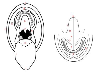
Odontogenesis is the process of tooth development that involves the formation and calcification of teeth. It is a complex process that requires the interaction of several types of cells, including epithelial cells, mesenchymal cells, and odontoblasts. The process begins during embryonic development with the formation of dental lamina, which gives rise to the tooth bud. As the tooth bud grows and differentiates, it forms the various structures of the tooth, including the enamel, dentin, cementum, and pulp. Odontogenesis is completed when the tooth erupts into the oral cavity. Abnormalities in odontogenesis can result in developmental dental anomalies such as tooth agenesis, microdontia, or odontomas.
A dental assistant is a healthcare professional who works under the direction of a dentist and provides patient care, takes and develops x-rays, assists the dentist during procedures, performs infection control procedures, and helps with office management. They may also provide education to patients on oral hygiene and other dental health topics. Dental assistants must be trained and certified in many states and are an important part of the dental care team.
Continuing dental education (CDE) refers to the ongoing education and training that dentists and other oral health professionals engage in after completing their initial professional degrees. The purpose of CDE is to help these professionals stay current with advances in dental technology, research, and patient care so they can continue to provide the highest quality of care to their patients.
CDE programs may cover a wide range of topics, including new techniques for treating oral diseases, advances in dental materials and equipment, ethical issues in dental practice, and strategies for managing a successful dental practice. These programs may take many forms, such as lectures, workshops, seminars, online courses, or hands-on training sessions.
In most states, dentists are required to complete a certain number of CDE credits each year in order to maintain their licensure. This helps ensure that all dental professionals are up-to-date on the latest research and best practices in their field, which ultimately benefits patients by promoting better oral health outcomes.
Dental anesthesia is a type of local or regional anesthesia that is specifically used in dental procedures to block the transmission of pain impulses from the teeth and surrounding tissues to the brain. The most common types of dental anesthesia include:
1. Local anesthesia: This involves the injection of a local anesthetic drug, such as lidocaine or prilocaine, into the gum tissue near the tooth that is being treated. This numbs the area and prevents the patient from feeling pain during the procedure.
2. Conscious sedation: This is a type of minimal sedation that is used to help patients relax during dental procedures. The patient remains conscious and can communicate with the dentist, but may not remember the details of the procedure. Common methods of conscious sedation include nitrous oxide (laughing gas) or oral sedatives.
3. Deep sedation or general anesthesia: This is rarely used in dental procedures, but may be necessary for patients who are extremely anxious or have special needs. It involves the administration of drugs that cause a state of unconsciousness and prevent the patient from feeling pain during the procedure.
Dental anesthesia is generally safe when administered by a qualified dentist or oral surgeon. However, as with any medical procedure, there are risks involved, including allergic reactions to the anesthetic drugs, nerve damage, and infection. Patients should discuss any concerns they have with their dentist before undergoing dental anesthesia.
Dental implants are artificial tooth roots that are surgically placed into the jawbone to replace missing or extracted teeth. They are typically made of titanium, a biocompatible material that can fuse with the bone over time in a process called osseointegration. Once the implant has integrated with the bone, a dental crown, bridge, or denture can be attached to it to restore function and aesthetics to the mouth.
Dental implants are a popular choice for tooth replacement because they offer several advantages over traditional options like dentures or bridges. They are more stable and comfortable, as they do not rely on adjacent teeth for support and do not slip or move around in the mouth. Additionally, dental implants can help to preserve jawbone density and prevent facial sagging that can occur when teeth are missing.
The process of getting dental implants typically involves several appointments with a dental specialist called a prosthodontist or an oral surgeon. During the first appointment, the implant is placed into the jawbone, and the gum tissue is stitched closed. Over the next few months, the implant will fuse with the bone. Once this process is complete, a second surgery may be necessary to expose the implant and attach an abutment, which connects the implant to the dental restoration. Finally, the crown, bridge, or denture is attached to the implant, providing a natural-looking and functional replacement for the missing tooth.
Phosphate transport proteins are membrane-bound proteins responsible for the active transport of phosphate ions across cell membranes. They play a crucial role in maintaining appropriate phosphate concentrations within cells and between intracellular compartments, which is essential for various biological processes such as energy metabolism, signal transduction, and bone formation.
These proteins utilize the energy derived from ATP hydrolysis or other sources to move phosphate ions against their concentration gradient, thereby facilitating cellular uptake of phosphate even when extracellular concentrations are low. Phosphate transport proteins can be classified based on their structure, function, and localization into different types, including sodium-dependent and sodium-independent transporters, secondary active transporters, and channels.
Dysregulation of phosphate transport proteins has been implicated in several pathological conditions, such as renal Fanconi syndrome, tumoral calcinosis, and hypophosphatemic rickets. Therefore, understanding the molecular mechanisms underlying phosphate transport protein function is essential for developing targeted therapies to treat these disorders.
Dental radiography is a specific type of imaging that uses radiation to produce detailed images of the teeth, bones, and soft tissues surrounding them. It is a crucial tool in dental diagnostics and treatment planning. There are several types of dental radiographs, including:
1. Intraoral Radiographs: These are taken inside the mouth and provide detailed images of individual teeth or small groups of teeth. They can help detect cavities, assess periodontal health, plan for restorations, and monitor tooth development in children. Common types of intraoral radiographs include bitewing, periapical, and occlusal radiographs.
2. Extraoral Radiographs: These are taken outside the mouth and provide images of larger areas, such as the entire jaw or skull. They can help diagnose issues related to the temporomandibular joint (TMJ), detect impacted teeth, assess bone health, and identify any abnormalities in the facial structure. Common types of extraoral radiographs include panoramic, cephalometric, and sialography radiographs.
3. Cone Beam Computed Tomography (CBCT): This is a specialized type of dental radiography that uses a cone-shaped X-ray beam to create detailed 3D images of the teeth, bones, and soft tissues. It is particularly useful in planning complex treatments such as dental implants, orthodontic treatment, and oral surgery.
Dental radiographs are typically taken using a specialized machine that emits a low dose of radiation. Patients are provided with protective lead aprons to minimize exposure to radiation. The frequency of dental radiographs depends on the patient's individual needs and medical history. Dentists follow strict guidelines to ensure that dental radiography is safe and effective for their patients.
Dental models are replicas of a patient's teeth and surrounding oral structures, used in dental practice and education. They are typically created using plaster or other materials that harden to accurately reproduce the shape and position of each tooth, as well as the contours of the gums and palate. Dental models may be used for a variety of purposes, including treatment planning, creating custom-fitted dental appliances, and teaching dental students about oral anatomy and various dental procedures. They provide a tactile and visual representation that can aid in understanding and communication between dentists, patients, and other dental professionals.
"Dental, Graduate Education" refers to the post-baccalaureate programs of study and training that lead to an advanced degree in the field of dentistry. These programs are designed to prepare students for specialized dental practice, research, or teaching careers. Examples of graduate dental degrees include:
1. Doctor of Dental Surgery (DDS): A professional doctoral degree that qualifies the graduate to practice general dentistry.
2. Doctor of Medical Dentistry (DMD): A professional doctoral degree equivalent to the DDS; awarded by some universities in the United States and several other countries.
3. Master of Science (MS) in Dentistry: An academic master's degree focused on research, teaching, or advanced clinical practice in a specific dental discipline.
4. Doctor of Philosophy (PhD) in Dental Sciences: A research-oriented doctoral degree that prepares students for careers in academia, research institutions, or the dental industry.
5. Specialty Training Programs: Postgraduate residency programs that provide advanced training in one of the nine recognized dental specialties, such as orthodontics, oral and maxillofacial surgery, or pediatric dentistry. These programs typically lead to a certificate or a master's degree in the respective specialty area.
Graduate dental education usually involves a combination of classroom instruction, laboratory work, clinical experience, and research. Admission to these programs typically requires a DDS or DMD degree from an accredited dental school and satisfactory scores on the Dental Admission Test (DAT).
Dental ethics refers to the principles and rules that guide the conduct of dental professionals in their interactions with patients, colleagues, and society. These ethical standards are designed to promote trust, respect, and fairness in dental care, and they are often based on fundamental ethical principles such as autonomy, beneficence, non-maleficence, and justice.
Autonomy refers to the patient's right to make informed decisions about their own health care, free from coercion or manipulation. Dental professionals have an obligation to provide patients with accurate information about their dental conditions and treatment options, so that they can make informed choices about their care.
Beneficence means acting in the best interests of the patient, and doing what is medically necessary and appropriate to promote their health and well-being. Dental professionals have a duty to provide high-quality care that meets accepted standards of practice, and to use evidence-based treatments that are likely to be effective.
Non-maleficence means avoiding harm to the patient. Dental professionals must take reasonable precautions to prevent injuries or complications during treatment, and they should avoid providing unnecessary or harmful treatments.
Justice refers to fairness and equity in the distribution of dental resources and services. Dental professionals have an obligation to provide care that is accessible, affordable, and culturally sensitive, and to advocate for policies and practices that promote health equity and social justice.
Dental ethics also encompasses issues related to patient confidentiality, informed consent, research integrity, professional competence, and boundary violations. Dental professionals are expected to adhere to ethical guidelines established by their professional organizations, such as the American Dental Association (ADA) or the British Dental Association (BDA), and to comply with relevant laws and regulations governing dental practice.
Tooth eruption is the process by which a tooth emerges from the gums and becomes visible in the oral cavity. It is a normal part of dental development that occurs in a predictable sequence and timeframe. Primary or deciduous teeth, also known as baby teeth, begin to erupt around 6 months of age and continue to emerge until approximately 2-3 years of age. Permanent or adult teeth start to erupt around 6 years of age and can continue to emerge until the early twenties.
The process of tooth eruption involves several stages, including the formation of the tooth within the jawbone, the movement of the tooth through the bone and surrounding tissues, and the final emergence of the tooth into the mouth. Proper tooth eruption is essential for normal oral function, including chewing, speaking, and smiling. Any abnormalities in the tooth eruption process, such as delayed or premature eruption, can indicate underlying dental or medical conditions that require further evaluation and treatment.
Tooth resorption is a process in which there is an abnormal loss or breakdown of tooth structure, either internally (internal resorption) or externally (external resorption), due to the action of specialized cells called odontoclasts. This can lead to weakening and destruction of the tooth, potentially causing sensitivity, pain, or even tooth loss if left untreated. The causes of tooth resorption can vary, including trauma, orthodontic treatment, periodontal disease, and certain systemic conditions. It is important to diagnose and treat tooth resorption early to prevent further damage and preserve the tooth structure.
A "Dental Service, Hospital" is a specialized department or unit within a hospital that provides comprehensive dental care services to patients. This type of service is typically equipped with advanced dental technology and staffed by oral health professionals such as dentists, oral surgeons, orthodontists, endodontists, periodontists, and dental hygienists.
The dental services offered in a hospital setting may include preventive care, restorative treatments, oral surgery, prosthodontics (dentures and implants), periodontal therapy, endodontic treatment (root canals), orthodontic treatment, and specialized care for patients with medical conditions that affect their oral health.
Hospital dental services often provide care to patients who require complex or extensive dental treatments, have medical conditions that make it difficult to receive dental care in a traditional dental office setting, or those who are recovering from surgery or other medical procedures. They may also provide emergency dental care for patients with severe dental pain, infection, or trauma.
In summary, a "Dental Service, Hospital" is a specialized unit within a hospital that provides comprehensive dental care services to patients, typically offering advanced technology and staffed by oral health professionals.
Tooth migration, in a dental or medical context, refers to the movement or shifting of teeth from their normal position within the dental arch. This phenomenon can occur due to various reasons such as:
1. Loss of adjacent teeth: When a tooth is lost, the surrounding teeth may drift or tilt into the empty space, causing other teeth to migrate out of their original positions.
2. Periodontal disease: Advanced periodontitis (severe gum disease) can lead to bone loss and ligament damage around the teeth, allowing them to move and potentially migrate.
3. Orthodontic treatment: Although controlled tooth movement is the goal of orthodontics, improper or unfinished treatment may result in undesirable tooth migration.
4. Aging: As people age, the supportive structures around teeth (bone and ligaments) can weaken, leading to tooth mobility and potential migration.
5. Tooth wear: Excessive tooth wear due to bruxism (grinding) or abrasion may alter the vertical dimension of the mouth, causing tooth migration over time.
It is essential to address tooth migration promptly to prevent further complications such as difficulty in chewing, speaking, and maintaining oral hygiene, which could lead to additional dental issues like decay and periodontal disease. Dental professionals may recommend various treatments, including orthodontic therapy, dental restorations, or even implants, depending on the cause and severity of tooth migration.
A dentist is a healthcare professional who specializes in the diagnosis, prevention, and treatment of diseases and conditions that affect the oral cavity and maxillofacial region. This includes the teeth, gums, jaw, and related structures. Dentists are trained to provide a wide range of services, including:
1. Routine dental exams and cleanings
2. Fillings, crowns, and other restorative treatments
3. Root canals and extractions
4. Dental implants and dentures
5. Orthodontic treatment (braces, aligners)
6. Treatment of gum disease
7. Oral cancer screenings
8. Cosmetic dental procedures (teeth whitening, veneers)
9. Management of temporomandibular joint disorders (TMJ)
10. Emergency dental care
To become a dentist, one must complete a Doctor of Dental Surgery (DDS) or Doctor of Medical Dentistry (DMD) degree from an accredited dental school and pass written and clinical exams to obtain licensure in their state. Many dentists also choose to specialize in a particular area of dentistry, such as orthodontics, oral surgery, or pediatric dentistry, by completing additional training and residency programs.
A dental society is a professional organization composed of dentists who have come together to promote and advance the practice of dentistry. These societies can be local, regional, national or international in scope and may include general dentists as well as specialists in various fields of dentistry. The members of dental societies often engage in continuing education, advocacy, research, and community service activities to improve oral health and the delivery of dental care. Additionally, dental societies may establish guidelines for ethical practice and provide resources and support for their members.
Dental technology refers to the application of science and engineering in dentistry to prevent, diagnose, and treat dental diseases and conditions. It involves the use of various equipment, materials, and techniques to improve oral health and enhance the delivery of dental care. Some examples of dental technology include:
1. Digital radiography: This technology uses digital sensors instead of traditional X-ray films to produce images of the teeth and supporting structures. It provides higher quality images, reduces radiation exposure, and allows for easier storage and sharing of images.
2. CAD/CAM dentistry: Computer-aided design and computer-aided manufacturing (CAD/CAM) technology is used to design and fabricate dental restorations such as crowns, bridges, and veneers in a single appointment. This technology allows for more precise and efficient production of dental restorations.
3. Dental implants: These are artificial tooth roots that are placed into the jawbone to replace missing teeth. They provide a stable foundation for dental restorations such as crowns, bridges, and dentures.
4. Intraoral cameras: These are small cameras that can be inserted into the mouth to capture detailed images of the teeth and gums. These images can be used for diagnosis, treatment planning, and patient education.
5. Laser dentistry: Dental lasers are used to perform a variety of procedures such as cavity preparation, gum contouring, and tooth whitening. They provide more precise and less invasive treatments compared to traditional dental tools.
6. 3D printing: This technology is used to create dental models, surgical guides, and custom-made dental restorations. It allows for more accurate and efficient production of dental products.
Overall, dental technology plays a crucial role in modern dentistry by improving the accuracy, efficiency, and quality of dental care.
Cementoma is a benign (non-cancerous) tumor that primarily affects the jaw bones, particularly the lower jaw (mandible). It is characterized by the growth of abnormal cementum-like tissue within the bone. Cementum is a hard tissue that covers the roots of teeth and helps anchor them to the jawbone.
There are different types of cementomas, including:
1. Periapical cemental dysplasia (PCD): This type of cementoma usually affects the anterior region of the lower jaw and is often associated with non-vital teeth. It typically presents as a small, radiopaque (dark) area on an X-ray.
2. Florid cemento-osseous dysplasia (FCOD): FCOD is a more widespread form of cementoma that affects multiple areas of the jawbones. It primarily affects middle-aged women and can cause significant bone remodeling, leading to radiopaque lesions on X-rays.
3. Gigantiform cementoma: This rare, aggressive type of cementoma typically affects children and adolescents. It can cause rapid bone growth and expansion, resulting in facial deformities and functional impairments.
4. Ossifying fibroma: Although not strictly a cementoma, ossifying fibroma shares some similarities with these tumors. It is characterized by the formation of both bone and cementum-like tissue within the lesion.
Treatment for cementomas depends on their size, location, and growth rate. Small, asymptomatic lesions may not require treatment, while larger or symptomatic ones might need surgical removal to prevent complications such as tooth displacement, infection, or pathological fractures. Regular follow-ups with dental X-rays are essential to monitor the progression of these lesions.
Dental health surveys are epidemiological studies that aim to assess the oral health status and related behaviors of a defined population at a particular point in time. These surveys collect data on various aspects of oral health, including the prevalence and severity of dental diseases such as caries (tooth decay), periodontal disease (gum disease), and oral cancer. They also gather information on factors that influence oral health, such as dietary habits, oral hygiene practices, access to dental care, and socioeconomic status.
The data collected in dental health surveys are used to identify trends and patterns in oral health, plan and evaluate public health programs and policies, and allocate resources for oral health promotion and disease prevention. Dental health surveys may be conducted at the local, regional, or national level, and they can target specific populations such as children, adolescents, adults, or older adults.
The methods used in dental health surveys include clinical examinations, interviews, questionnaires, and focus groups. Clinical examinations are conducted by trained dentists or dental hygienists who follow standardized protocols to assess the oral health status of participants. Interviews and questionnaires are used to collect information on demographic characteristics, oral health behaviors, and attitudes towards oral health. Focus groups can provide insights into the perceptions and experiences of participants regarding oral health issues.
Overall, dental health surveys play a critical role in monitoring and improving the oral health of populations and reducing oral health disparities.
Dental fluorosis is a developmental disturbance of dental enamel caused by excessive exposure to fluoride during tooth development. It is characterized by hypomineralization of the enamel, resulting in various appearances ranging from barely noticeable white spots to brown staining and pitting of the teeth. The severity depends on the amount, duration, and timing of fluoride intake, as well as individual susceptibility. Mild dental fluorosis is typically asymptomatic but can affect the appearance of teeth, while severe cases may cause tooth sensitivity and increased susceptibility to tooth decay.
Dental cavity preparation is the process of removing decayed and damaged tissue from a tooth and shaping the remaining healthy structure in order to prepare it for the placement of a filling or a crown. The goal of cavity preparation is to remove all traces of decay and create a clean, stable surface for the restoration to bond with, while also maintaining as much of the natural tooth structure as possible.
The process typically involves the use of dental drills and other tools to remove the decayed tissue and shape the tooth. The size and depth of the preparation will depend on the extent of the decay and the type of restoration that will be used. After the preparation is complete, the dentist will place the filling or crown, restoring the function and integrity of the tooth.
Dental licensure is the process by which a state or jurisdiction grants a dental professional the authority to practice dentistry within its borders. In order to obtain a dental license, individuals must meet certain education, examination, and other requirements established by the licensing body. These requirements typically include graduation from an accredited dental school, passing written and clinical examinations, and completion of continuing education courses.
The purpose of dental licensure is to protect the public by ensuring that dental professionals have the necessary knowledge, skills, and abilities to provide safe and effective dental care. Licensing boards are responsible for enforcing standards of practice and disciplining dentists who engage in unprofessional or unethical conduct.
It's important to note that dental licensure requirements may vary from state to state, so it's essential for dental professionals to familiarize themselves with the specific requirements of the state(s) in which they intend to practice.
Dental laboratories are specialized facilities where dental technicians create and manufacture various dental restorations and appliances based on the specific measurements, models, and instructions provided by dentists. These custom-made dental products are designed to restore or replace damaged, missing, or decayed teeth, improve oral function, and enhance the overall appearance of a patient's smile.
Some common dental restorations and appliances produced in dental laboratories include:
1. Dental crowns: Artificial caps that cover and protect damaged or weakened teeth, often made from ceramics, porcelain, metal alloys, or a combination of materials.
2. Dental bridges: Fixed or removable appliances used to replace one or more missing teeth by connecting artificial teeth (pontics) to adjacent natural teeth or dental implants.
3. Dentures: Removable prosthetic devices that replace all or most of the upper and/or lower teeth, providing improved chewing function, speech clarity, and aesthetics.
4. Orthodontic appliances: Devices used to correct malocclusions (improper bites) and misaligned teeth, such as traditional braces, clear aligners, palatal expanders, and retainers.
5. Custom dental implant components: Specialized parts designed for specific implant systems, which are used in conjunction with dental implants to replace missing teeth permanently.
6. Night guards and occlusal splints: Protective devices worn during sleep to prevent or manage bruxism (teeth grinding) and temporomandibular joint disorders (TMD).
7. Anti-snoring devices: Mandibular advancement devices that help reduce snoring by holding the lower jaw in a slightly forward position, preventing airway obstruction during sleep.
8. Dental whitening trays: Custom-fitted trays used to hold bleaching gel against tooth surfaces for professional teeth whitening treatments.
9. Specialty restorations: Including aesthetic veneers, inlays, onlays, and other customized dental solutions designed to meet specific patient needs.
Dental laboratories may be standalone facilities or part of a larger dental practice. They are typically staffed by skilled technicians who specialize in various aspects of dental technology, such as ceramics, orthodontics, implantology, and prosthodontics. Collaboration between dentists, dental specialists, and laboratory technicians ensures the highest quality results for patients undergoing restorative or cosmetic dental treatments.
A furcation defect in dental terminology refers to the loss or destruction of supporting bone in the area where the roots of a multi-rooted tooth, such as a molar, diverge or branch out. This condition is typically caused by periodontal disease, which results in inflammation and infection of the gums and surrounding tissues.
Furcation defects are classified into three categories based on their severity:
1. Class I: The furcation involvement is limited to the function groove, and the bone loss does not extend beyond this area. Treatment usually involves thorough cleaning and root planing of the affected area.
2. Class II: The bone loss extends halfway or more beneath the furcation, but not reaching the bottom of the furcation. This type of defect may require surgical treatment to promote bone regeneration.
3. Class III: The bone loss is so extensive that it reaches the bottom of the furcation and possibly beyond. In such cases, tooth extraction may be necessary if the tooth cannot be saved through regenerative procedures or other treatments.
It's important to note that early detection and treatment of periodontal disease can help prevent furcation defects from developing or worsening. Regular dental checkups and cleanings are essential for maintaining good oral health and preventing periodontal issues.
Dental materials are substances that are used in restorative dentistry, prosthodontics, endodontics, orthodontics, and preventive dentistry to restore or replace missing tooth structure, improve the function and esthetics of teeth, and protect the oral tissues from decay and disease. These materials can be classified into various categories based on their physical and chemical properties, including metals, ceramics, polymers, composites, cements, and alloys.
Some examples of dental materials include:
1. Amalgam: a metal alloy used for dental fillings that contains silver, tin, copper, and mercury. It is strong, durable, and resistant to wear but has been controversial due to concerns about the toxicity of mercury.
2. Composite: a tooth-colored restorative material made of a mixture of glass or ceramic particles and a bonding agent. It is used for fillings, veneers, and other esthetic dental treatments.
3. Glass ionomer cement: a type of cement used for dental restorations that releases fluoride ions and helps prevent tooth decay. It is often used for fillings in children's teeth or as a base under crowns and bridges.
4. Porcelain: a ceramic material used for dental crowns, veneers, and other esthetic restorations. It is strong, durable, and resistant to staining but can be brittle and prone to fracture.
5. Gold alloy: a metal alloy used for dental restorations that contains gold, copper, and other metals. It is highly biocompatible, corrosion-resistant, and malleable but can be expensive and less esthetic than other materials.
6. Acrylic resin: a type of polymer used for dental appliances such as dentures, night guards, and orthodontic retainers. It is lightweight, flexible, and easy to modify but can be less durable than other materials.
The choice of dental material depends on various factors, including the location and extent of the restoration, the patient's oral health status, their esthetic preferences, and their budget. Dental professionals must consider these factors carefully when selecting the appropriate dental material for each individual case.
According to the American Academy of Periodontology, periodontal diseases are chronic inflammatory conditions that affect the tissues surrounding and supporting the teeth. These tissues include the gums, periodontal ligament, and alveolar bone. The primary cause of periodontal disease is bacterial plaque, a sticky film that constantly forms on our teeth.
There are two major stages of periodontal disease:
1. Gingivitis: This is the milder form of periodontal disease, characterized by inflammation of the gums (gingiva) without loss of attachment to the teeth. The gums may appear red, swollen, and bleed easily during brushing or flossing. At this stage, the damage can be reversed with proper dental care and improved oral hygiene.
2. Periodontitis: If left untreated, gingivitis can progress to periodontitis, a more severe form of periodontal disease. In periodontitis, the inflammation extends beyond the gums and affects the deeper periodontal tissues, leading to loss of bone support around the teeth. Pockets filled with infection-causing bacteria form between the teeth and gums, causing further damage and potential tooth loss if not treated promptly.
Risk factors for developing periodontal disease include poor oral hygiene, smoking or using smokeless tobacco, genetic predisposition, diabetes, hormonal changes (such as pregnancy or menopause), certain medications, and systemic diseases like AIDS or cancer. Regular dental check-ups and good oral hygiene practices are crucial for preventing periodontal disease and maintaining overall oral health.
Dental specialties are recognized areas of expertise in dental practice that require additional training and education beyond the general dentist degree. The American Dental Association (ADA) recognizes nine dental specialties:
1. Dental Public Health: This specialty focuses on preventing oral diseases and promoting oral health through population-level interventions, research, and policy development.
2. Endodontics: Endodontists are experts in diagnosing and treating tooth pain and performing root canal treatments to save infected or damaged teeth.
3. Oral and Maxillofacial Pathology: This specialty involves the diagnosis and management of diseases that affect the oral cavity, jaws, and face, using clinical, radiographic, and microscopic examination techniques.
4. Oral and Maxillofacial Radiology: Oral and maxillofacial radiologists use advanced imaging technologies to diagnose and manage conditions affecting the head and neck region.
5. Oral and Maxillofacial Surgery: Oral surgeons perform surgical procedures on the face, jaws, and mouth, including tooth extractions, jaw alignment surgeries, and cancer treatments.
6. Orthodontics and Dentofacial Orthopedics: Orthodontists specialize in diagnosing and treating dental and facial irregularities, using appliances such as braces and aligners to straighten teeth and correct bite problems.
7. Pediatric Dentistry: Pediatric dentists are trained to care for the oral health needs of children, including those with special health care needs.
8. Periodontics: Periodontists diagnose and treat gum diseases, place dental implants, and perform surgical procedures to regenerate lost tissue and bone support around teeth.
9. Prosthodontics: Prosthodontists are experts in replacing missing teeth and restoring damaged or worn-out teeth using crowns, bridges, dentures, and implant-supported restorations.
Dental fees refer to the charges that dentists or dental professionals bill for their services, procedures, or treatments. These fees can vary based on several factors such as:
1. Location: Dental fees may differ depending on the region or country where the dental practice is located due to differences in cost of living and local market conditions.
2. Type of procedure: The complexity and duration of a dental treatment will impact the fee charged for that service. For example, a simple teeth cleaning will have a lower fee compared to more complex procedures like root canals or dental implants.
3. Dental professional's expertise and experience: Highly skilled and experienced dentists may charge higher fees due to their superior level of knowledge and proficiency in performing various dental treatments.
4. Type of dental practice: Fees for dental services at a private practice may differ from those charged by a community health center or non-profit organization.
5. Dental insurance coverage: The amount of coverage provided by a patient's dental insurance plan can also affect the final out-of-pocket cost for dental care, which in turn influences the fees that dentists charge.
Dental fee schedules are typically established by individual dental practices based on these factors and may be periodically updated to reflect changes in costs or market conditions. Patients should consult their dental providers to understand the specific fees associated with any recommended treatments or procedures.
Guided Tissue Regeneration (GTR) in periodontics is a surgical procedure that aims to regenerate lost periodontal tissues, including the alveolar bone, cementum, and periodontal ligament, which have been destroyed due to periodontal disease. The goal of GTR is to restore the architectural relationship between these supporting structures and the tooth, thereby improving its prognosis and function.
The procedure involves placing a barrier membrane between the tooth root and the surrounding soft tissues, creating a protected space that allows for the selective growth of periodontal cells. The membrane acts as a physical barrier to prevent the ingrowth of epithelial cells and fibroblasts from the oral mucosa, which can interfere with the regeneration process.
The membrane can be either resorbable or non-resorbable, depending on the clinical situation and surgeon's preference. Resorbable membranes are made of materials that degrade over time, while non-resorbable membranes require a second surgical procedure for removal. The choice of membrane material and configuration depends on various factors such as the size and location of the defect, patient's medical history, and surgeon's experience.
GTR has been shown to be effective in treating intrabony defects, furcation involvements, and class II function defects, among others. However, its success depends on various factors such as patient selection, surgical technique, membrane type and placement, and postoperative care.
Dental occlusion refers to the alignment and contact between the upper and lower teeth when the jaws are closed. It is the relationship between the maxillary (upper) and mandibular (lower) teeth when they approach each other, as occurs during chewing or biting.
A proper dental occlusion, also known as a balanced occlusion, ensures that the teeth and jaw joints function harmoniously, reducing the risk of tooth wear, damage, and temporomandibular disorders (TMD). Malocclusion, on the other hand, refers to improper alignment or contact between the upper and lower teeth, which may require orthodontic treatment or dental restorations to correct.
An incisor is a type of tooth that is primarily designed for biting off food pieces rather than chewing or grinding. They are typically chisel-shaped, flat, and have a sharp cutting edge. In humans, there are eight incisors - four on the upper jaw and four on the lower jaw, located at the front of the mouth. Other animals such as dogs, cats, and rodents also have incisors that they use for different purposes like tearing or gnawing.
A dental technician is a healthcare professional who designs, fabricates, and repairs custom-made dental devices, such as dentures, crowns, bridges, orthodontic appliances, and implant restorations. They work closely with dentists and other oral health professionals to meet the individual needs of each patient. Dental technicians typically have an associate's degree or certificate in dental technology and may be certified by a professional organization. Their work requires a strong understanding of dental materials, fabrication techniques, and the latest advances in dental technology.
Practice management in dentistry refers to the administration and operation of a dental practice. It involves various aspects such as:
1. Business Operations: This includes financial management, billing and coding, human resources, and office management.
2. Patient Care: This includes scheduling appointments, managing patient records, treatment planning, and ensuring quality care.
3. Marketing and Promotion: This includes advertising the practice, attracting new patients, and maintaining relationships with existing ones.
4. Compliance: This includes adhering to laws and regulations related to dental practices, such as HIPAA for patient privacy and OSHA for workplace safety.
5. Continuous Improvement: This involves regularly assessing the practice's performance, implementing changes to improve efficiency and effectiveness, and keeping up-to-date with advancements in dentistry and healthcare management.
The goal of dental practice management is to ensure the smooth running of the practice, provide high-quality patient care, and maintain a successful and profitable business.
Tooth movement, in a dental and orthodontic context, refers to the physical change in position or alignment of one or more teeth within the jaw bone as a result of controlled forces applied through various orthodontic appliances such as braces, aligners, or other orthodontic devices. The purposeful manipulation of these forces encourages the periodontal ligament (the tissue that connects the tooth to the bone) to remodel, allowing the tooth to move gradually over time into the desired position. This process is crucial in achieving proper bite alignment, correcting malocclusions, and enhancing overall oral function and aesthetics.
In the context of medical terminology, "hardness" is not a term that has a specific or standardized definition. It may be used in various ways to describe the firmness or consistency of a tissue, such as the hardness of an artery or tumor, but it does not have a single authoritative medical definition.
In some cases, healthcare professionals may use subjective terms like "hard," "firm," or "soft" to describe their tactile perception during a physical examination. For example, they might describe the hardness of an enlarged liver or spleen by comparing it to the feel of their knuckles when gently pressed against the abdomen.
However, in other contexts, healthcare professionals may use more objective measures of tissue stiffness or elasticity, such as palpation durometry or shear wave elastography, which provide quantitative assessments of tissue hardness. These techniques can be useful for diagnosing and monitoring conditions that affect the mechanical properties of tissues, such as liver fibrosis or cancer.
Therefore, while "hardness" may be a term used in medical contexts to describe certain physical characteristics of tissues, it does not have a single, universally accepted definition.
The dental sac, also known as the dental follicle, is a soft tissue structure that surrounds the developing tooth crown during odontogenesis, which is the process of tooth development. It is derived from the ectoderm and mesenchyme of the embryonic oral cavity. The dental sac gives rise to several important structures associated with the tooth, including the periodontal ligament, cementum, and the alveolar bone that surrounds and supports the tooth in the jaw.
The dental sac plays a critical role in tooth development by regulating the mineralization of the tooth crown and providing a protective environment for the developing tooth. It also contains cells called odontoblasts, which are responsible for producing dentin, one of the hard tissues that make up the tooth. Abnormalities in the development or growth of the dental sac can lead to various dental anomalies, such as impacted teeth, dilacerated roots, and other developmental disorders.
A deciduous tooth, also known as a baby tooth or primary tooth, is a type of temporary tooth that humans and some other mammals develop during childhood. They are called "deciduous" because they are eventually shed and replaced by permanent teeth, much like how leaves on a deciduous tree fall off and are replaced by new growth.
Deciduous teeth begin to form in the womb and start to erupt through the gums when a child is around six months old. By the time a child reaches age three, they typically have a full set of 20 deciduous teeth, including incisors, canines, and molars. These teeth are smaller and less durable than permanent teeth, but they serve important functions such as helping children chew food properly, speak clearly, and maintain space in the jaw for the permanent teeth to grow into.
Deciduous teeth usually begin to fall out around age six or seven, starting with the lower central incisors. This process continues until all of the deciduous teeth have been shed, typically by age 12 or 13. At this point, the permanent teeth will have grown in and taken their place, with the exception of the wisdom teeth, which may not erupt until later in adolescence or early adulthood.
Pathologic decalcification is a process that occurs when there is a loss of calcium salts from the bones or teeth. This can lead to weakening and structural damage in the affected area. It is often seen in conditions such as osteoporosis, Paget's disease, and tumors that involve bone. In dental contexts, decalcification can also refer to the loss of minerals from tooth enamel, which can lead to cavities and tooth decay. This is often caused by poor oral hygiene and a diet high in sugars.
Dentistry is the branch of medicine that is concerned with the examination, diagnosis, prevention, and treatment of diseases, disorders, and conditions of the oral cavity (mouth), including the teeth, gums, and other supporting structures. Dentists use a variety of treatments and procedures to help patients maintain good oral health and prevent dental problems from developing or worsening. These may include:
* Routine cleanings and checkups to remove plaque and tartar and detect any potential issues early on
* Fillings, crowns, and other restorative treatments to repair damaged teeth
* Root canal therapy to treat infected or inflamed tooth pulp
* Extractions of severely decayed or impacted teeth
* Dentures, bridges, and implants to replace missing teeth
* Orthodontic treatment to align crooked or misaligned teeth
* Treatment for temporomandibular joint (TMJ) disorders and other issues affecting the jaw and surrounding muscles
Dental health is an important part of overall health and well-being. Poor oral health has been linked to a variety of systemic conditions, including heart disease, diabetes, and respiratory infections. Regular dental checkups and good oral hygiene practices can help prevent these and other dental problems from developing.
A gold colloid is not a medical term per se, but it is often used in the context of medical applications. It refers to a suspension of sub-nanometer to nanometer-sized gold particles in a fluid, usually water. These particles are small enough to remain suspended and not settle at the bottom due to Brownian motion. Gold colloids have been used in various medical applications, such as diagnostic tests, drug delivery systems, and photothermal therapies, due to their unique optical properties and biocompatibility.
Dental esthetics refers to the branch of dentistry concerned with the aesthetic appearance of teeth and smile. It involves the use of various dental treatments and procedures to improve the color, shape, alignment, and position of teeth, thereby enhancing the overall facial appearance and self-confidence of a person. Some common dental esthetic treatments include tooth whitening, dental veneers, composite bonding, orthodontic treatment (braces), and dental implants. It is important to note that dental esthetics not only focuses on improving the appearance but also maintaining or improving oral health and function.
Epithelial attachment is a general term that refers to the point where epithelial cells, which are the cells that line the outer surfaces of organs and blood vessels, adhere or attach to an underlying structure. In the context of the mouth and teeth, epithelial attachment is often used to describe the connection between the gum tissue (gingiva) and the tooth surface.
In a healthy mouth, the gingival tissue fits tightly around each tooth, forming a protective seal that helps prevent bacteria and other harmful substances from entering the spaces between the teeth and gums. This tight seal is maintained by specialized epithelial cells called junctional epithelial cells, which form a barrier between the oral environment and the underlying connective tissue.
When the gingival tissue becomes inflamed due to factors such as poor oral hygiene or certain medical conditions, the epithelial attachment can become compromised, leading to a condition known as gingivitis. If left untreated, gingivitis can progress to periodontal disease, which is characterized by the destruction of the tissues that support the teeth, including the bone and connective tissue.
In summary, epithelial attachment refers to the point where epithelial cells adhere to an underlying structure, and in the context of oral health, it describes the connection between the gum tissue and the tooth surface.
Comprehensive dental care is a broad term that refers to a dental approach that involves the prevention, diagnosis, and treatment of a wide range of oral health issues. It aims to provide patients with complete and optimal oral health care, including:
1. Oral examination and assessment: This includes a thorough examination of the patient's oral cavity, head, and neck to identify any existing dental problems or potential issues that may arise in the future.
2. Preventive care: Comprehensive dental care emphasizes preventive measures such as regular dental cleanings, fluoride treatments, and sealants to help protect against tooth decay and gum disease.
3. Restorative dentistry: If dental problems are identified, comprehensive dental care includes restorative treatments like fillings, crowns, bridges, or implants to restore the function and appearance of damaged teeth.
4. Periodontal (gum) treatment: Comprehensive dental care also addresses periodontal health through deep cleanings, scaling and root planing, and other therapies to manage gum disease.
5. Oral surgery: In some cases, comprehensive dental care may involve oral surgery procedures like tooth extractions or jaw realignment.
6. Endodontic (root canal) treatment: If the pulp of a tooth becomes infected or inflamed, endodontic treatment may be necessary to save the tooth and alleviate pain.
7. Prosthodontics: This includes the replacement of missing teeth with dentures, bridges, or implants.
8. Orthodontic care: Comprehensive dental care can also involve orthodontic treatments like braces or aligners to straighten misaligned teeth and improve bite.
9. Oral cancer screening: Regular oral cancer screenings are an essential part of comprehensive dental care, as early detection significantly increases the chances of successful treatment.
10. Patient education: Comprehensive dental care also focuses on educating patients about proper oral hygiene practices, nutrition, and lifestyle choices that can impact their oral health. This helps empower patients to take an active role in maintaining their oral health between appointments.
In summary, comprehensive dental care is a holistic approach to dental care that aims to provide complete and personalized oral health solutions for each patient, addressing all aspects of their oral health and promoting long-term wellbeing.
Osteopontin (OPN) is a phosphorylated glycoprotein that is widely distributed in many tissues, including bone, teeth, and mineralized tissues. It plays important roles in various biological processes such as bone remodeling, immune response, wound healing, and tissue repair. In the skeletal system, osteopontin is involved in the regulation of bone formation and resorption by modulating the activity of osteoclasts and osteoblasts. It also plays a role in the development of chronic inflammatory diseases such as rheumatoid arthritis, atherosclerosis, and cancer metastasis to bones. Osteopontin is considered a potential biomarker for various disease states, including bone turnover, cardiovascular disease, and cancer progression.
Health education in the context of dentistry refers to the process of educating and informing individuals, families, and communities about oral health-related topics, including proper oral hygiene practices, the importance of regular dental checkups and cleanings, the risks and consequences of poor oral health, and the relationship between oral health and overall health. The goal of dental health education is to empower individuals to take control of their own oral health and make informed decisions about their dental care. This can be achieved through various methods such as lectures, demonstrations, printed materials, and interactive activities. Dental health education may also cover topics related to nutrition, tobacco and alcohol use, and the prevention and treatment of oral diseases and conditions.
Dentist-patient relations refer to the professional relationship between a licensed dentist and their patient. This relationship is based on trust, communication, and ethical obligations. The dentist is responsible for providing competent and appropriate dental care while considering the patient's needs, preferences, and values. The patient, on the other hand, should be honest with their dentist regarding their medical history, oral health habits, and any concerns they may have. Effective dentist-patient relations are crucial in ensuring positive dental experiences, treatment compliance, and overall satisfaction with dental care.
The mandible, also known as the lower jaw, is the largest and strongest bone in the human face. It forms the lower portion of the oral cavity and plays a crucial role in various functions such as mastication (chewing), speaking, and swallowing. The mandible is a U-shaped bone that consists of a horizontal part called the body and two vertical parts called rami.
The mandible articulates with the skull at the temporomandibular joints (TMJs) located in front of each ear, allowing for movements like opening and closing the mouth, protrusion, retraction, and side-to-side movement. The mandible contains the lower teeth sockets called alveolar processes, which hold the lower teeth in place.
In medical terminology, the term "mandible" refers specifically to this bone and its associated structures.
A tooth socket, also known as an alveolus (plural: alveoli), refers to the hollow cavity or space in the jawbone where a tooth is anchored. The tooth socket is part of the alveolar process, which is the curved part of the maxilla or mandible that contains multiple tooth sockets for the upper and lower teeth, respectively.
Each tooth socket has a specialized tissue called the periodontal ligament, which attaches the root of the tooth to the surrounding bone. This ligament helps absorb forces generated during biting and chewing, allowing for comfortable and efficient mastication while also maintaining the tooth's position within the jawbone. The tooth socket is responsible for providing support, stability, and nourishment to the tooth through its blood vessels and nerves.
Microspectrophotometry (MSP) is a microanalytical technique that combines microspectroscopy and photometry to measure the absorption, reflection, or fluorescence spectra of extremely small samples, typically in the range of micrometers to sub-micrometers. This technique is often used in biomedical research and clinical settings for the analysis of cellular and subcellular structures, such as organelles, inclusion bodies, and single molecules.
MSP can provide detailed information about the chemical composition, molecular structure, and spatial distribution of biological samples, making it a valuable tool for studying various physiological and pathological processes, including gene expression, protein function, and cell-cell interactions. Additionally, MSP has been used in diagnostic applications to identify abnormalities in tissues and cells, such as cancerous or precancerous lesions, and to monitor the efficacy of therapeutic interventions.
The technique involves using a microscope equipped with a high-resolution objective lens and a spectrophotometer to measure the intensity of light transmitted through or reflected from a sample at different wavelengths. The resulting spectra can be used to identify specific chemical components or molecular structures based on their characteristic absorption, reflection, or fluorescence patterns.
MSP is a powerful tool for studying biological systems at the microscopic level and has contributed significantly to our understanding of cellular and molecular biology. However, it requires specialized equipment and expertise to perform and interpret the data, making it a relatively complex and sophisticated technique.
X-ray microtomography, often referred to as micro-CT, is a non-destructive imaging technique used to visualize and analyze the internal structure of objects with high spatial resolution. It is based on the principles of computed tomography (CT), where multiple X-ray images are acquired at different angles and then reconstructed into cross-sectional slices using specialized software. These slices can be further processed to create 3D visualizations, allowing researchers and clinicians to examine the internal structure and composition of samples in great detail. Micro-CT is widely used in materials science, biology, medicine, and engineering for various applications such as material characterization, bone analysis, and defect inspection.
Alveolar bone loss refers to the breakdown and resorption of the alveolar process of the jawbone, which is the part of the jaw that contains the sockets of the teeth. This type of bone loss is often caused by periodontal disease, a chronic inflammation of the gums and surrounding tissues that can lead to the destruction of the structures that support the teeth.
In advanced stages of periodontal disease, the alveolar bone can become severely damaged or destroyed, leading to tooth loss. Alveolar bone loss can also occur as a result of other conditions, such as osteoporosis, trauma, or tumors. Dental X-rays and other imaging techniques are often used to diagnose and monitor alveolar bone loss. Treatment may include deep cleaning of the teeth and gums, medications, surgery, or tooth extraction in severe cases.
Dentinogenesis is the process of dentin formation, which is one of the main components of teeth. Dentin is a hard, calcified tissue that lies beneath the tooth's enamel and cementum layers, providing structural support and protection to the pulp tissue containing nerves and blood vessels. The process of dentinogenesis involves the differentiation and activation of odontoblasts, which are specialized cells that synthesize and secrete the organic and inorganic components of dentin matrix. These components include collagenous proteins and hydroxyapatite crystals, which form a highly mineralized tissue that is both strong and flexible. Dentinogenesis continues throughout life as new layers of dentin are formed in response to various stimuli such as tooth wear, dental caries, or injury.
'Infection Control, Dental' refers to the practices and procedures implemented in dental settings to prevent the transmission of infectious agents from person to person, or from contaminated instruments, equipment, or environmental surfaces to patients or dental personnel. It includes a range of measures such as hand hygiene, use of personal protective equipment (e.g., gloves, masks, eyewear), sterilization and disinfection of instruments and equipment, safe injection practices, and environmental cleaning and disinfection. The goal of infection control in dentistry is to eliminate or minimize the risk of infectious diseases, such as HIV, hepatitis B and C, and tuberculosis, among others, being transmitted in dental settings.
The dental papilla is a type of tissue found in the developing tooth within the jawbone. It is composed of cells that will eventually differentiate into odontoblasts, which are the cells responsible for producing dentin, one of the main hard tissues that make up the tooth. The dental papilla is located in the center of the tooth germ and is surrounded by the dental follicle, another type of tissue that helps to form the tooth. As the tooth develops, the dental papilla becomes smaller and eventually forms the pulp chamber, which contains the blood vessels, nerves, and connective tissue that support and nourish the tooth.
A dental prosthesis is a device that replaces one or more missing teeth or parts of teeth to correct deficiencies in chewing ability, speech, and aesthetics. It can be removable or fixed (permanent) and can be made from various materials such as acrylic resin, porcelain, metal alloys, or a combination of these. Examples of dental prostheses include dentures, bridges, crowns, and implants.
Amelogenin is a protein that plays a crucial role in the formation and mineralization of enamel, which is the hard, calcified tissue that covers the outer surface of teeth. It is expressed during tooth development and is secreted by ameloblasts, the cells responsible for producing enamel.
Amelogenin makes up approximately 90% of the organic matrix of developing enamel and guides the growth and organization of hydroxyapatite crystals, which are the primary mineral component of enamel. The protein is subsequently degraded and removed as the enamel matures and becomes fully mineralized.
Mutations in the gene that encodes amelogenin (AMELX on the X chromosome) can lead to various inherited enamel defects, such as amelogenesis imperfecta, which is characterized by thin, soft, or poorly formed enamel. Additionally, because of its high expression in developing teeth and unique size and structure, amelogenin has been widely used as a marker in forensic dentistry for human identification and sex determination.
Tooth diseases are conditions that affect the teeth and can cause discomfort, pain, and even loss of teeth if left untreated. These diseases can be caused by various factors such as poor oral hygiene, bacterial infections, trauma, genetics, and certain medical conditions. Some common tooth diseases include:
1. Dental caries (tooth decay): This is a breakdown of the tooth enamel due to the action of acid-producing bacteria that feed on sugars and starches in the mouth. Over time, this can lead to cavities or holes in the teeth.
2. Gingivitis: This is an inflammation of the gums caused by the buildup of plaque and tartar at the gum line. If left untreated, gingivitis can progress to periodontitis, a more serious form of gum disease that can cause tooth loss.
3. Periodontitis: This is a severe infection of the gums and bones that support the teeth. It is caused by the buildup of plaque and tartar, which leads to the destruction of the tissue and bone that hold the teeth in place.
4. Abscess: This is a pocket of pus that forms in the tooth or gum due to a bacterial infection. An abscess can cause pain, swelling, and fever, and may require antibiotics or surgical drainage.
5. Tooth erosion: This is the loss of tooth structure due to acid wear, which can be caused by factors such as diet, stomach acid, and teeth grinding.
6. Hypersensitivity: This is a condition in which the teeth become sensitive to hot, cold, or sweet foods and drinks. It can be caused by factors such as gum recession, tooth decay, and tooth wear.
7. Oral cancer: This is a type of cancer that affects the mouth, lips, tongue, or throat. It can cause symptoms such as sores, lumps, or difficulty swallowing, and may require surgery, radiation therapy, or chemotherapy for treatment.
Dental leakage, also known as "microleakage" in dental terminology, refers to the seepage or penetration of fluids, bacteria, or other substances between the walls of a dental restoration (such as a filling, crown, or bridge) and the prepared tooth structure. This occurs due to the presence of microscopic gaps or spaces at the interface of the restoration and the tooth.
Dental leakage can lead to several problems, including:
1. Recurrent decay: The seepage of fluids, bacteria, and sugars from the oral environment can cause secondary tooth decay around the margins of the restoration.
2. Sensitivity: Microleakage may result in temperature sensitivity or pain when consuming hot or cold foods and beverages due to fluid movement within the gap.
3. Discoloration: Over time, dental leakage might lead to staining of the tooth structure around the restoration, resulting in an unaesthetic appearance.
4. Failed restorations: Persistent dental leakage can weaken the bond between the restoration and the tooth, increasing the risk of restoration failure and the need for replacement.
To prevent dental leakage, dentists employ various techniques during restoration placement, such as using appropriate adhesives, following meticulous preparation protocols, and ensuring a tight seal around the margins of the restoration. Regular dental check-ups and professional cleanings are essential to monitor the condition of existing restorations and address any issues before they become more severe.
A dental audit is a systematic review and evaluation of the dental records, procedures, and care provided by a dentist or dental practice. The purpose of a dental audit is to assess the quality of care, identify any areas for improvement, and ensure that appropriate policies and procedures are being followed. This can include reviews of patient records, treatment plans, billing practices, and adherence to infection control guidelines.
The results of a dental audit may be used to improve the quality of care provided to patients, reduce the risk of errors or complications, and ensure compliance with regulatory requirements. Dental audits may be conducted internally by dental practices themselves, or externally by dental organizations, insurance companies, or government agencies.
Oral health is the scientific term used to describe the overall health status of the oral and related tissues, including the teeth, gums, palate, tongue, and mucosal lining. It involves the absence of chronic mouth and facial pain, oral and pharyngeal (throat) cancers, oral soft tissue lesions, birth defects such as cleft lip and palate, and other diseases and disorders that affect the oral cavity.
Good oral health also means being free of decay, gum disease, and other oral infections that can damage the teeth, gums, and bones of the mouth. It is essential to maintain good oral hygiene through regular brushing, flossing, and dental check-ups to prevent dental caries (cavities) and periodontal disease (gum disease).
Additionally, oral health is closely linked to overall health and well-being. Poor oral health has been associated with various systemic diseases, including diabetes, cardiovascular disease, respiratory infections, and stroke. Therefore, maintaining good oral health can contribute to improved general health and quality of life.
Dental instruments are specialized tools that dentists, dental hygienists, and other oral healthcare professionals use to examine, clean, and treat teeth and gums. These instruments come in various shapes and sizes, and each one is designed for a specific purpose. Here are some common dental instruments and their functions:
1. Mouth mirror: A small, handheld mirror used to help the dentist see hard-to-reach areas of the mouth and reflect light onto the teeth and gums.
2. Explorer: A sharp, hooked instrument used to probe teeth and detect cavities, tartar, or other dental problems.
3. Sickle scaler: A curved, sharp-edged instrument used to remove calculus (tartar) from the tooth surface.
4. Periodontal probe: A blunt, calibrated instrument used to measure the depth of periodontal pockets and assess gum health.
5. Dental syringe: A device used to inject local anesthesia into the gums before dental procedures.
6. High-speed handpiece: Also known as a dental drill, it is used to remove decay, shape teeth, or prepare them for fillings and other restorations.
7. Low-speed handpiece: A slower, quieter drill used for various procedures, such as placing crowns or veneers.
8. Suction tip: A thin tube that removes saliva, water, and debris from the mouth during dental procedures.
9. Cotton rolls: Small squares of cotton used to isolate teeth, absorb fluids, and protect soft tissues during dental treatments.
10. Dental forceps: Specialized pliers used to remove teeth or hold them in place while restorations are being placed.
11. Elevators: Curved, wedge-shaped instruments used to loosen or lift teeth out of their sockets.
12. Rubber dam: A thin sheet of rubber or latex that isolates a specific tooth or area during dental treatment, keeping it dry and free from saliva and debris.
These are just a few examples of the many dental instruments used in modern dentistry. Each one plays an essential role in maintaining oral health and providing effective dental care.
Odontometry is a term used in dentistry that refers to the measurement of teeth, particularly the size and length of teeth or tooth roots. It is often used in forensic dentistry for identification purposes, such as in age estimation, sex determination, or individual identification of human remains. The measurements can be taken using various methods, including radiographs (x-rays), calipers, or specialized software.
In some contexts, odontometry may also refer to the process of measuring the amount of dental work required for a particular treatment plan, although this usage is less common.
Dental waste refers to the byproducts and discarded materials generated from dental treatments and procedures. This can include:
1. Amalgam waste: This consists of a mixture of metals, including mercury, used to fill dental cavities.
2. Sharps waste: Includes needles, scalpel blades, and other sharp instruments used in dental procedures.
3. Infectious waste: Materials that have been contaminated with blood or other bodily fluids during dental treatments, such as gloves, gauze, and used dental bibs.
4. Pharmaceutical waste: Unused or expired medications, including analgesics, antibiotics, and anesthetics.
5. Chemical waste: Includes fixer and developer solutions used in developing X-rays, as well as disinfectants and other chemicals used in dental practices.
6. Radioactive waste: Dental X-ray film packets and lead foil from X-ray processing.
Proper management and disposal of dental waste is essential to protect public health and the environment. Regulations governing dental waste disposal vary by location, so it's important for dental practices to be aware of and comply with local requirements.
Dental implantation is a surgical procedure in which a titanium post or frame is inserted into the jawbone beneath the gum line to replace the root of a missing tooth. Once the implant has integrated with the bone, a replacement tooth (crown) is attached to the top of the implant, providing a stable and durable restoration that looks, feels, and functions like a natural tooth. Dental implants can also be used to support dental bridges or dentures, providing added stability and comfort for patients who are missing multiple teeth.
Ameloblasts are the specialized epithelial cells that are responsible for the formation of enamel, which is the hard, outermost layer of a tooth. These cells are a part of the dental lamina and are present in the developing tooth's crown region. They align themselves along the surface of the developing tooth and secrete enamel proteins and minerals to form the enamel rods and interrod enamel. Once the enamel formation is complete, ameloblasts undergo programmed cell death, leaving behind the hard, mineralized enamel matrix. Any damage or abnormality in the functioning of ameloblasts can lead to developmental defects in the enamel, such as hypoplasia or hypocalcification, which may affect the tooth's structure and function.
Dental economics is a branch of economics that focuses on the financial aspects of oral health and dental care. It involves the study of various economic factors that influence the provision, accessibility, affordability, and utilization of dental services. This includes analyzing the costs of dental treatments, pricing strategies, financing options, and insurance policies related to dental care. Additionally, dental economics also examines the impact of government policies, regulations, and market dynamics on dental care delivery and oral health outcomes. The ultimate goal of dental economics is to improve the efficiency, effectiveness, and equity of dental care systems, ultimately leading to better oral health for individuals and populations.
Dental marginal adaptation refers to the way in which a dental restoration, such as a filling or crown, fits precisely and accurately along the margin or edge where it meets the tooth structure. The term "marginal" describes the border between the restoration and the tooth. Ideally, this junction should be tight and smooth, without any gaps or spaces that could allow for the accumulation of bacteria, food debris, or dental plaque.
Achieving good marginal adaptation is crucial to ensure the longevity and success of a dental restoration. When the margin is well-adapted, it helps prevent microleakage, secondary tooth decay, and sensitivity. It also contributes to the overall seal and integrity of the restoration, minimizing the risk of recurrent caries or other complications.
The process of achieving optimal marginal adaptation involves careful preparation of the tooth structure, precise impression-taking techniques, and meticulous fabrication of the dental restoration. The use of high-quality materials and modern technologies, such as digital impressions and CAD/CAM systems, can further enhance the accuracy and predictability of the marginal adaptation.
Scanning electron microscopy (SEM) is a type of electron microscopy that uses a focused beam of electrons to scan the surface of a sample and produce a high-resolution image. In SEM, a beam of electrons is scanned across the surface of a specimen, and secondary electrons are emitted from the sample due to interactions between the electrons and the atoms in the sample. These secondary electrons are then detected by a detector and used to create an image of the sample's surface topography. SEM can provide detailed images of the surface of a wide range of materials, including metals, polymers, ceramics, and biological samples. It is commonly used in materials science, biology, and electronics for the examination and analysis of surfaces at the micro- and nanoscale.
I'm not aware of a medical definition for "DMF Index." The abbreviation "DMF" could potentially stand for many things, as it is used in various contexts across different fields. In the field of dentistry, DMF stands for Decayed, Missing, and Filled teeth/surfaces, which is a method for measuring dental caries or tooth decay. However, there is no standard medical definition for "DMF Index." If you could provide more context or specify the field of study or practice, I would be happy to help further!
Dental alloys are materials made by combining two or more metals to be used in dental restorations, such as crowns, bridges, fillings, and orthodontic appliances. These alloys can be classified into three main categories based on their composition:
1. Precious Alloys: Predominantly composed of precious metals like gold, platinum, palladium, and silver. They are highly corrosion-resistant, biocompatible, and durable, making them suitable for long-term use in dental restorations. Common examples include high noble (gold) alloys and noble alloys.
2. Base Metal Alloys: Contain primarily non-precious metals like nickel, chromium, cobalt, and beryllium. They are more affordable than precious alloys but may cause allergic reactions or sensitivities in some patients. Common examples include nickel-chromium alloys and cobalt-chromium alloys.
3. Castable Glass Ionomer Alloys: A combination of glass ionomer cement (GIC) powder and metal liquid, which can be cast into various dental restorations. They have the advantage of being both strong and adhesive to tooth structure but may not be as durable as other alloy types.
Each type of dental alloy has its unique properties and applications, depending on the specific clinical situation and patient needs. Dental professionals consider factors like cost, biocompatibility, mechanical properties, and esthetics when selecting an appropriate alloy for a dental restoration.
Dental caries susceptibility refers to the likelihood or predisposition of an individual to develop dental caries, also known as tooth decay or cavities. It is influenced by various factors such as oral hygiene practices, dietary habits, saliva composition, and the presence of certain bacteria in the mouth, particularly mutans streptococci and lactobacilli.
People with a higher dental caries susceptibility may have thinner or softer enamel, reduced saliva flow, or a greater concentration of cavity-causing bacteria in their mouths. Regular dental check-ups and good oral hygiene practices, such as brushing twice a day, flossing daily, and using fluoride toothpaste, can help reduce the risk of developing dental caries. Additionally, a balanced diet that limits sugary and starchy foods and beverages can also help lower the likelihood of tooth decay.
Dental Informatics is a branch of health informatics that deals with the application of information technology and computer systems to improve dental care delivery, oral health education, research, and management. It involves the development, implementation, and evaluation of information systems that support dental practice, including electronic health records (EHRs), imaging systems, decision support tools, and data analytics. The goal of dental informatics is to enhance patient care, improve clinical outcomes, increase efficiency, and reduce costs in dental care. It also includes the study of the structure, processing, and dissemination of biomedical and health data, information, and knowledge as it relates to dentistry.
Oral hygiene is the practice of keeping the mouth and teeth clean to prevent dental issues such as cavities, gum disease, bad breath, and other oral health problems. It involves regular brushing, flossing, and using mouthwash to remove plaque and food particles that can lead to tooth decay and gum disease. Regular dental check-ups and cleanings are also an essential part of maintaining good oral hygiene. Poor oral hygiene can lead to a range of health problems, including heart disease, diabetes, and respiratory infections, so it is important to prioritize oral health as part of overall health and wellbeing.
Dental scaling is a professional dental cleaning procedure that involves the removal of plaque, tartar (calculus), and stains from the tooth surfaces. This is typically performed by a dentist or dental hygienist using specialized instruments called scalers and curettes. The procedure helps to prevent gum disease and tooth decay by removing bacterial deposits that can cause inflammation and infection of the gums. Dental scaling may be recommended as part of a routine dental check-up or if there are signs of periodontal disease, such as red, swollen, or bleeding gums. In some cases, local anesthesia may be used to numb the area and make the procedure more comfortable for the patient.
Felidae is the biological family that includes all extant (living) members of the cat group, also known as felids. This family consists of big cats such as lions, tigers, and leopards, as well as small cats like domestic cats, cheetahs, and pumas. Felidae is part of the order Carnivora and is characterized by specialized adaptations for hunting and stalking prey, including retractile claws, sharp teeth, and flexible bodies. The family has a worldwide distribution, with species found in various habitats across all continents except Antarctica.
Polarized light microscopy is a type of microscopy that uses polarized light to enhance contrast and reveal unique optical properties in specimens. In this technique, a polarizing filter is placed under the light source, which polarizes the light as it passes through. The specimen is then illuminated with this linearly polarized light. As the light travels through the specimen, its plane of polarization may be altered due to birefringence, a property of certain materials that causes the light to split into two separate rays with different refractive indices.
A second polarizing filter, called an analyzer, is placed in the light path between the objective and the eyepiece. The orientation of this filter can be adjusted to either allow or block the transmission of light through the microscope. When the polarizer and analyzer are aligned perpendicularly, no light will pass through if the specimen does not exhibit birefringence. However, if the specimen has birefringent properties, it will cause the plane of polarization to rotate, allowing some light to pass through the analyzer and create a contrasting image.
Polarized light microscopy is particularly useful for observing structures in minerals, crystals, and certain biological materials like collagen fibers, muscle proteins, and starch granules. It can also be used to study stress patterns in plastics and other synthetic materials.
Operative dentistry is a branch of dental medicine that involves the diagnosis, treatment, and management of teeth with structural or functional damage due to decay, trauma, or other causes. It primarily focuses on restoring the function, form, and health of damaged teeth through various operative procedures such as fillings, crowns, inlays, onlays, and root canal treatments. The goal is to preserve natural tooth structure, alleviate pain, prevent further decay or damage, and restore the patient's oral health and aesthetics.
Here are some of the key aspects and procedures involved in operative dentistry:
1. Diagnosis: Operative dentists use various diagnostic tools and techniques to identify and assess tooth damage, including visual examination, dental X-rays, and special tests like pulp vitality testing. This helps them determine the most appropriate treatment approach for each case.
2. Preparation: Before performing any operative procedure, the dentist must prepare the tooth by removing decayed or damaged tissue, as well as any existing restorations that may be compromised or failing. This process is called tooth preparation and involves using specialized dental instruments like burs and excavators to shape the tooth and create a stable foundation for the new restoration.
3. Restoration: Operative dentistry encompasses various techniques and materials used to restore damaged teeth, including:
a. Fillings: Direct fillings are placed directly into the prepared cavity using materials like amalgam (silver), composite resin (tooth-colored), glass ionomer, or gold foil. The choice of filling material depends on factors such as the location and extent of the damage, patient's preferences, and cost considerations.
b. Indirect restorations: These are fabricated outside the mouth, usually in a dental laboratory, and then cemented or bonded to the prepared tooth. Examples include inlays, onlays, and crowns, which can be made from materials like gold, porcelain, ceramic, or resin composites.
c. Endodontic treatments: Operative dentistry also includes root canal therapy, which involves removing infected or inflamed pulp tissue from within the tooth's root canals, cleaning and shaping the canals, and then filling and sealing them to prevent reinfection.
d. Veneers: These are thin layers of porcelain or composite resin that are bonded to the front surfaces of teeth to improve their appearance, shape, or alignment.
4. Follow-up care: After placing a restoration, patients should maintain good oral hygiene practices and have regular dental checkups to ensure the long-term success of the treatment. In some cases, additional adjustments or repairs may be necessary over time due to wear, fracture, or secondary decay.
Dental facilities refer to establishments that provide dental care and treatment. These facilities can include private dental practices, community health centers, hospital dental departments, and specialized dental clinics. They are equipped with the necessary dental equipment and staffed by dental professionals such as dentists, dental hygienists, and dental assistants. Dental facilities offer a range of services including routine check-ups, cleanings, fillings, extractions, root canals, orthodontic treatment, and oral surgery. Some dental facilities may also offer specialized services such as periodontics, prosthodontics, and endodontics.
Dental devices for home care are products designed for use by individuals or their caregivers in a home setting to maintain oral hygiene, manage dental health issues, and promote overall oral health. These devices can include:
1. Toothbrushes: Manual, electric, or battery-operated toothbrushes used to clean teeth and remove plaque and food debris.
2. Dental floss: A thin string used to remove food particles and plaque from between the teeth and under the gum line.
3. Interdental brushes: Small brushes designed to clean between the teeth and around dental appliances, such as braces or implants.
4. Water flossers/oral irrigators: Devices that use a stream of water to remove food particles and plaque from between the teeth and under the gum line.
5. Tongue scrapers: Tools used to clean the tongue's surface, removing bacteria and reducing bad breath.
6. Rubber tips/gum stimulators: Devices used to massage and stimulate the gums, promoting blood circulation and helping to maintain gum health.
7. Dental picks/sticks: Pointed tools used to remove food particles and plaque from between the teeth and under the gum line.
8. Mouthguards: Protective devices worn over the teeth to prevent damage from grinding, clenching, or sports-related injuries.
9. Night guards: Similar to mouthguards, these are designed to protect the teeth from damage caused by nighttime teeth grinding (bruxism).
10. Dental retainers: Devices used to maintain the alignment of teeth after orthodontic treatment.
11. Whitening trays and strips: At-home products used to whiten teeth by applying a bleaching agent to the tooth surface.
12. Fluoride mouth rinses: Anticavity rinses containing fluoride, which help strengthen tooth enamel and prevent decay.
13. Oral pain relievers: Topical gels or creams used to alleviate oral pain, such as canker sores or denture irritation.
Proper use of these dental devices, along with regular dental check-ups and professional cleanings, can help maintain good oral health and prevent dental issues.
Preventive dentistry is a branch of dental medicine that focuses on preventing the occurrence or progression of oral diseases and maintaining optimal oral health. It encompasses a set of practices, behaviors, and interventions aimed at preserving the integrity and functionality of teeth and gums through early detection, intervention, and patient education.
The primary goal of preventive dentistry is to minimize the risk of dental caries (tooth decay), periodontal disease (gum disease), oral cancer, and other oral health conditions. This is achieved through a combination of professional dental care, personal oral hygiene habits, and lifestyle modifications.
Professional dental care includes regular dental examinations, cleanings, fluoride treatments, and sealants to protect tooth surfaces from decay. Patient education plays a crucial role in preventive dentistry, as it empowers individuals to take an active part in their oral health by teaching them proper brushing and flossing techniques, nutritional counseling, and the importance of regular dental visits.
Preventive dentistry also emphasizes the significance of risk assessment and early intervention for high-risk populations, such as children, elderly individuals, and those with medical conditions that may impact oral health. By promoting a proactive approach to dental care, preventive dentistry aims to improve overall quality of life, reduce healthcare costs, and enhance patient satisfaction.
Dental photography is a type of clinical photography that focuses on documenting the condition and treatment of teeth and oral structures. It involves using specialized cameras, lenses, and lighting to capture high-quality images of the mouth and related areas. These images can be used for diagnostic purposes, patient education, treatment planning, communication with other dental professionals, and monitoring progress over time. Dental photography may include various types of shots, such as extraoral (outside the mouth) and intraoral (inside the mouth) views, close-ups of individual teeth or restorations, and full-face portraits. It requires a strong understanding of dental anatomy, lighting techniques, and image composition to produce accurate and informative images.
Tooth extraction is a dental procedure in which a tooth that is damaged or poses a threat to oral health is removed from its socket in the jawbone. This may be necessary due to various reasons such as severe tooth decay, gum disease, fractured teeth, crowded teeth, or for orthodontic treatment purposes. The procedure is performed by a dentist or an oral surgeon, under local anesthesia to numb the area around the tooth, ensuring minimal discomfort during the extraction process.
Bone regeneration is the biological process of new bone formation that occurs after an injury or removal of a portion of bone. This complex process involves several stages, including inflammation, migration and proliferation of cells, matrix deposition, and mineralization, leading to the restoration of the bone's structure and function.
The main cells involved in bone regeneration are osteoblasts, which produce new bone matrix, and osteoclasts, which resorb damaged or old bone tissue. The process is tightly regulated by various growth factors, hormones, and signaling molecules that promote the recruitment, differentiation, and activity of these cells.
Bone regeneration can occur naturally in response to injury or surgical intervention, such as fracture repair or dental implant placement. However, in some cases, bone regeneration may be impaired due to factors such as age, disease, or trauma, leading to delayed healing or non-union of the bone. In these situations, various strategies and techniques, including the use of bone grafts, scaffolds, and growth factors, can be employed to enhance and support the bone regeneration process.
Tolonium Chloride, also known as Toluidine Blue O, is a basic thiazine metachromatic dye that is used in medical and research settings. It is often used as a diagnostic agent in procedures such as the Toluidine Blue Test for identifying cancerous or precancerous cells in the cervix, oral mucosa, and other tissues. The dye selectively binds to acidic components in the extracellular matrix of neoplastic cells, making them more visible under a microscope. It is also used in research to study cell membrane permeability and lysosomal function. Please note that the use of Tolonium Chloride should be under medical supervision and professional guidance.
In the context of dentistry, a molar is a type of tooth found in the back of the mouth. They are larger and wider than other types of teeth, such as incisors or canines, and have a flat biting surface with multiple cusps. Molars are primarily used for grinding and chewing food into smaller pieces that are easier to swallow. Humans typically have twelve molars in total, including the four wisdom teeth.
In medical terminology outside of dentistry, "molar" can also refer to a unit of mass in the apothecaries' system of measurement, which is equivalent to 4.08 grams. However, this usage is less common and not related to dental or medical anatomy.
Dental porcelain is a type of biocompatible ceramic material that is commonly used in restorative and cosmetic dentistry to create tooth-colored restorations such as crowns, veneers, inlays, onlays, and bridges. It is made from a mixture of powdered porcelain and water, which is heated to high temperatures to form a hard, glass-like substance. Dental porcelain has several desirable properties for dental restorations, including:
1. High strength and durability: Dental porcelain is strong enough to withstand the forces of biting and chewing, making it suitable for use in load-bearing restorations such as crowns and bridges.
2. Natural appearance: Dental porcelain can be matched closely to the color, translucency, and texture of natural teeth, allowing for highly aesthetic restorations that blend seamlessly with the surrounding dentition.
3. Biocompatibility: Dental porcelain is biologically inert and does not cause adverse reactions or toxicity in the body, making it a safe choice for dental restorations.
4. Chemical resistance: Dental porcelain is resistant to staining and chemical attack from substances such as coffee, tea, red wine, and acidic foods and drinks.
5. Low thermal conductivity: Dental porcelain has low thermal conductivity, which means it does not transmit heat or cold readily, reducing the risk of temperature sensitivity in dental restorations.
Overall, dental porcelain is a versatile and reliable material for creating high-quality, natural-looking, and durable dental restorations.
Dental digital radiography is a type of medical imaging that uses digital sensors instead of traditional X-ray film to produce highly detailed images of the teeth, gums, and surrounding structures. This technology offers several advantages over conventional dental radiography, including:
1. Lower radiation exposure: Digital sensors require less radiation to produce an image compared to traditional film, making it a safer option for patients.
2. Instant results: The images captured by digital sensors are immediately displayed on a computer screen, allowing dentists to quickly assess the patient's oral health and discuss any findings with them during the appointment.
3. Improved image quality: Digital radiography produces clearer and more precise images compared to traditional film, enabling dentists to better detect issues such as cavities, fractures, or tumors.
4. Enhanced communication: The ability to easily manipulate and enhance digital images allows for better communication between dental professionals and improved patient education.
5. Environmentally friendly: Digital radiography eliminates the need for chemical processing and disposal of used film, making it a more environmentally conscious choice.
6. Easy storage and retrieval: Digital images can be stored electronically and accessed easily for future reference or consultation with other dental professionals.
7. Remote consultations: Digital images can be shared remotely with specialists or insurance companies, facilitating faster diagnoses and treatment planning.
Methyl Methacrylates (MMA) are a family of synthetic materials that are commonly used in the medical field, particularly in orthopedic and dental applications. Medically, MMA is often used as a bone cement to fix prosthetic implants, such as artificial hips or knees, into place during surgeries.
Methyl methacrylates consist of a type of acrylic resin that hardens when mixed with a liquid catalyst. This property allows it to be easily molded and shaped before it sets, making it ideal for use in surgical procedures where precise positioning is required. Once hardened, MMA forms a strong, stable bond with the bone, helping to secure the implant in place.
It's important to note that while MMA is widely used in medical applications, there have been concerns about its safety in certain situations. For example, some studies have suggested that high levels of methyl methacrylate fumes released during the setting process may be harmful to both patients and surgical staff. Therefore, appropriate precautions should be taken when using MMA-based products in medical settings.
Community dentistry, also known as public health dentistry, is a branch of dental science that focuses on the prevention and control of oral diseases and promoting oral health within communities and populations. It involves the application of epidemiological, social, behavioral, and administrative sciences to improve the oral health of populations. The goal of community dentistry is to reduce oral health disparities by providing accessible, affordable, and culturally competent dental care to all members of a community, particularly those who are underserved or vulnerable.
Community dentistry programs may include school-based dental sealant programs, fluoridation initiatives, oral health education campaigns, and policy advocacy efforts to improve access to dental care. Dental public health professionals work in a variety of settings, including public health departments, community health centers, academic institutions, and non-profit organizations. They collaborate with other healthcare providers, policymakers, and community stakeholders to promote oral health and prevent oral diseases.
 Cementochronology
 Cementum - Green Dental & Orthodontics
 First fossil chimpanzee | Nature
 Bioengineered periodontal tissue formed on titanium dental implants
 Cementum is key to periodontal tissue regeneration: A review on apatite microstructures for creation of novel cementum-based...
 Advanced Search Results - Public Health Image Library(PHIL)
 New evidence is revealing the ages of death, birth, and menarche in Neandertals
 Mandibular Cysts and Odontogenic Tumors: Overview, Odontogenic Mandibular Cysts, Nonodontogenic Mandibular Cysts
 Medicare Dental Coverage | CMS
 Nathanael Leung | University of Surrey
 Caries - wikidoc
![Atlas of Human Embryos [by: RF Gasser, PhD.] - Ch.8](data:image/png;base64,iVBORw0KGgoAAAANSUhEUgAAABAAAAAQCAYAAAAf8/9hAAAAh0lEQVQ4jd2Suw2EMBBEHxZV+C7dPkDUQXWuA9l9OAXaWCIjn7GOwAESLx3NaPYDT9OpqrYEmNYGPUCMO+MwX8R1WwD4fqaL5oNDxGJysw/uNNVYtwUfHADjMBPjjsnNIva2soj9CemTkILyBrXqJWdArcG/HSRMOdMd5c46VdWWKzz/SC/gACJUSba7VMpqAAAAAElFTkSuQmCC) Atlas of Human Embryos [by: RF Gasser, PhD.] - Ch.8
Atlas of Human Embryos [by: RF Gasser, PhD.] - Ch.8 Differences in Morphological Age-Related Dental Changes Depending on Postmortem Interval
 Root cementum ultrastructure in healthy and periodontally diseased teeth | Slazhneva | Пародонтология
 New Study Shows MRI can Perform Forensic Age Estimation on Human Teeth | Bruker
 The Journal of Contemporary Dental Practice
 Mammals from Solna Jama Cave
 Dentosphere : World of Dentistry: MCQs on Oral Histology - Cementum
 PDF) Periodontal Disease in Dogs
 Recognising and Managing Apical Infection | IVIS
 Clearances
 Flashcards - Embryology Chapter Six
 Whether they need it ...or not! | Registered Dental Hygienists
 Periodontitis - Dental Disorders - Merck Manuals Professional Edition
 Illustrated Dental Embryology, Histology, and Anatomy Elsevier eBook on VitalSource (Retail Access Card), 5th Edition -...