Fascia
Fascia Lata
Dupuytren Contracture
Fasciitis, Plantar
Abdominal Wall
Fasciitis
Surgical Flaps
Ligaments
Myringoplasty
Plastic Embedding
Bone Lengthening
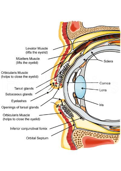
Blepharoptosis
Eye, Artificial
Reconstructive Surgical Procedures
Temporal Muscle
Entropion
Suture Techniques
Buttocks
Hernia
Cervical Plexus
Hernia, Inguinal
Zygoma
Alcian Blue
Fasciitis, Necrotizing
Ludwig's Angina
Hernia, Ventral
A fascia is a band or sheet of connective tissue, primarily collagen, that covers, connects, and separates muscles, organs, and other structures in the body. It provides support and stability, allows for smooth movement between structures, and has the ability to transmit forces throughout the body. Fascia is found throughout the body, and there are several layers of it, including superficial fascia, deep fascia, and visceral fascia. Injury, inflammation, or strain to the fascia can cause pain and restriction of movement.
Fascia lata is a medical term that refers to the thick, fibrous sheath of connective tissue that envelops and surrounds the thigh muscles (specifically, the quadriceps femoris and hamstrings). It is a type of fascia, which is the soft tissue component of the deep (internal) fascial system.
The fascia lata is continuous with the fascia of the hip and knee joints and plays an important role in providing stability, support, and protection to the muscles and other structures within the thigh. It also helps to facilitate the gliding and movement of muscles and tendons during physical activity.
Injuries or inflammation of the fascia lata can cause pain and discomfort, and may limit mobility and range of motion in the thigh and lower extremity. Conditions such as fascia lata strain, tears, or myofascial pain syndrome may require medical treatment, including physical therapy, medication, or in some cases, surgery.
Dupuytren contracture is a medical condition that affects the hand, specifically the fascia, which is a layer of connective tissue beneath the skin of the palm. In this condition, the fascia thickens and shortens, causing one or more fingers to bend towards the palm and making it difficult to straighten them. The ring finger and little finger are most commonly affected, but the middle finger and thumb can also be involved.
The exact cause of Dupuytren contracture is not known, but it is more common in men than women and tends to run in families. It is also associated with certain medical conditions such as diabetes, seizures, and alcoholism. There is no cure for Dupuytren contracture, but treatments such as surgery or needle aponeurotomy can help relieve symptoms and improve hand function.
Plantar fasciitis is a medical condition that involves inflammation of the plantar fascia, which is a thick band of tissue that runs along the bottom of your foot, connecting your heel bone to your toes. This tissue supports the arch of your foot and absorbs shock when you walk or run.
Plantar fasciitis is often caused by repetitive stress or overuse, leading to small tears and inflammation in the fascia. People who have high arches or flat feet, those who spend a lot of time on their feet, and athletes who engage in activities that put repeated stress on the heel and attached tissue, such as runners, are at a higher risk of developing plantar fasciitis.
Symptoms of plantar fasciitis include pain and stiffness in the heel or bottom of the foot, especially when taking the first few steps after getting out of bed or after prolonged periods of sitting or standing. The pain may worsen over time if left untreated, making it difficult to walk, climb stairs, or participate in physical activities.
Treatment for plantar fasciitis typically includes rest, ice, compression, and elevation (RICE) therapy, as well as physical therapy exercises to stretch and strengthen the foot and lower leg muscles. In some cases, medication, orthotics, or even surgery may be necessary to alleviate severe pain and inflammation.
The abdominal wall refers to the group of muscles, fascia (sheaths of connective tissue), and skin that make up the front and sides of the abdomen, extending from the thorax (chest) to the pelvis. It provides protection to the abdominal organs, supports the trunk, and allows for movement of the torso.
The main muscles of the anterior abdominal wall include:
1. Rectus sheaths (Rectus Abdominis): paired vertical muscles running from the pubic symphysis to the xiphoid process and costal cartilages of ribs 5-7.
2. External obliques: thin, irregular muscles that lie over the lower part of the abdomen and run diagonally downward and forward from the lower ribs to the iliac crest (pelvic bone) and pubic tubercle.
3. Internal obliques: thicker muscles that lie under the external obliques, running diagonally upward and forward from the iliac crest to the lower ribs.
4. Transverse abdominis: deepest of the abdominal muscles, lying horizontally across the abdomen, attaching from the lower ribs to the pelvis.
These muscles are interconnected by various layers of fascia and aponeuroses (flat, broad tendons), forming a complex structure that allows for both stability and mobility. The linea alba, a fibrous band, runs down the midline of the anterior abdominal wall, connecting the rectus sheaths.
Damage to the abdominal wall can occur due to trauma, surgery, or various medical conditions, which may require surgical intervention for repair.
Fasciitis is a medical condition characterized by inflammation or irritation of the fascia, which are the bands of connective tissue that surround muscles, tendons, and bones in the body. The most common type of fasciitis is plantar fasciitis, which affects the fascia on the bottom of the foot and can cause heel pain. Other types of fasciitis include:
* Achilles tendonitis or Achilles tendinopathy, which affects the fascia that connects the calf muscle to the heel bone
* Shin splints, which affect the fascia that covers the front of the lower leg
* Necrotizing fasciitis, a rare and serious bacterial infection that can cause extensive tissue damage and is potentially life-threatening.
The symptoms of fasciitis may include pain, stiffness, or tenderness in the affected area, especially after prolonged periods of rest or physical activity. Treatment for fasciitis typically involves rest, ice, compression, and elevation (RICE) of the affected area, as well as physical therapy exercises to stretch and strengthen the fascia and surrounding muscles. In some cases, medication or surgery may be necessary to relieve symptoms and promote healing.
A surgical flap is a specialized type of surgical procedure where a section of living tissue (including skin, fat, muscle, and/or blood vessels) is lifted from its original site and moved to another location, while still maintaining a blood supply through its attached pedicle. This technique allows the surgeon to cover and reconstruct defects or wounds that cannot be closed easily with simple suturing or stapling.
Surgical flaps can be classified based on their vascularity, type of tissue involved, or method of transfer. The choice of using a specific type of surgical flap depends on the location and size of the defect, the patient's overall health, and the surgeon's expertise. Some common types of surgical flaps include:
1. Random-pattern flaps: These flaps are based on random blood vessels within the tissue and are typically used for smaller defects in areas with good vascularity, such as the face or scalp.
2. Axial pattern flaps: These flaps are designed based on a known major blood vessel and its branches, allowing them to cover larger defects or reach distant sites. Examples include the radial forearm flap and the anterolateral thigh flap.
3. Local flaps: These flaps involve tissue adjacent to the wound and can be further classified into advancement, rotation, transposition, and interpolation flaps based on their movement and orientation.
4. Distant flaps: These flaps are harvested from a distant site and then transferred to the defect after being tunneled beneath the skin or through a separate incision. Examples include the groin flap and the latissimus dorsi flap.
5. Free flaps: In these flaps, the tissue is completely detached from its original blood supply and then reattached at the new site using microvascular surgical techniques. This allows for greater flexibility in terms of reach and placement but requires specialized expertise and equipment.
Surgical flaps play a crucial role in reconstructive surgery, helping to restore form and function after trauma, tumor removal, or other conditions that result in tissue loss.
Ligaments are bands of dense, fibrous connective tissue that surround joints and provide support, stability, and limits the range of motion. They are made up primarily of collagen fibers arranged in a parallel pattern to withstand tension and stress. Ligaments attach bone to bone, and their function is to prevent excessive movement that could cause injury or dislocation.
There are two main types of ligaments: extracapsular and intracapsular. Extracapsular ligaments are located outside the joint capsule and provide stability to the joint by limiting its range of motion. Intracapsular ligaments, on the other hand, are found inside the joint capsule and help maintain the alignment of the joint surfaces.
Examples of common ligaments in the body include the anterior cruciate ligament (ACL) and posterior cruciate ligament (PCL) in the knee, the medial collateral ligament (MCL) and lateral collateral ligament (LCL) in the elbow, and the coracoacromial ligament in the shoulder.
Injuries to ligaments can occur due to sudden trauma or overuse, leading to sprains, strains, or tears. These injuries can cause pain, swelling, bruising, and limited mobility, and may require medical treatment such as immobilization, physical therapy, or surgery.
Myringoplasty is a surgical procedure that involves reconstructing or repairing the tympanic membrane (eardrum) in the middle ear. The eardrum is the thin, delicate tissue that separates the outer ear from the inner ear. It plays a crucial role in hearing by vibrating in response to sound waves and transmitting these vibrations to the bones of the middle ear.
Myringoplasty is typically performed to treat chronic perforations or holes in the eardrum that have not healed on their own or with medical management. These perforations can result from various causes, such as infection, trauma, or congenital defects. By closing the perforation, myringoplasty helps prevent the risk of middle ear infections and improves hearing function.
The procedure involves harvesting a small piece of tissue, often from the patient's own body (such as the fascia surrounding a muscle), to use as a graft to cover the eardrum perforation. The graft is placed through an incision made in the ear canal or, less commonly, via an external approach through the mastoid bone behind the ear.
Myringoplasty is typically performed under general anesthesia and requires a short hospital stay for observation and monitoring. Following surgery, patients may need to avoid water exposure, heavy lifting, and strenuous activities for a few weeks to allow proper healing. The success rate of myringoplasty is generally high, with most patients experiencing improved hearing and reduced symptoms of ear infections.
Plastic embedding is a histological technique used in the preparation of tissue samples for microscopic examination. In this process, thin sections of tissue are impregnated and hardened with a plastic resin, which replaces the water in the tissue and provides support and stability during cutting and mounting. This method is particularly useful for tissues that are difficult to embed using traditional paraffin embedding techniques, such as those that contain fat or are very delicate. The plastic-embedded tissue sections can be cut very thinly (typically 1-2 microns) and provide excellent preservation of ultrastructural details, making them ideal for high-resolution microscopy and immunohistochemical studies.
Bone lengthening is a surgical procedure that involves cutting and then gradually stretching the bone apart, allowing new bone to grow in its place. This process is also known as distraction osteogenesis. The goal of bone lengthening is to increase the length of a bone, either to improve function or to correct a deformity.
The procedure typically involves making an incision in the skin over the bone and using specialized tools to cut through the bone. Once the bone is cut, a device called an external fixator is attached to the bone on either side of the cut. The external fixator is then gradually adjusted over time to slowly stretch the bone apart, creating a gap between the two ends of the bone. As the bone is stretched, new bone tissue begins to grow in the space between the two ends, eventually filling in the gap and lengthening the bone.
Bone lengthening can be used to treat a variety of conditions, including limb length discrepancies, congenital deformities, and injuries that result in bone loss. It is typically performed by an orthopedic surgeon and may require several months of follow-up care to ensure proper healing and growth of the new bone tissue.
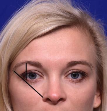
Blepharoptosis is a medical term that refers to the drooping or falling of the upper eyelid. It is usually caused by weakness or paralysis of the muscle that raises the eyelid, known as the levator palpebrae superioris. This condition can be present at birth or acquired later in life due to various factors such as aging, nerve damage, eye surgery complications, or certain medical conditions like myasthenia gravis or brain tumors. Blepharoptosis may obstruct vision and cause difficulty with daily activities, and treatment options include eyedrops, eye patches, or surgical correction.
In the context of human anatomy, the thigh is the part of the lower limb that extends from the hip to the knee. It is the upper and largest portion of the leg and is primarily composed of the femur bone, which is the longest and strongest bone in the human body, as well as several muscles including the quadriceps femoris (front thigh), hamstrings (back thigh), and adductors (inner thigh). The major blood vessels and nerves that supply the lower limb also pass through the thigh.
An artificial eye, also known as a prosthetic eye, is a type of medical device that is used to replace a natural eye that has been removed or is not functional due to injury, disease, or congenital abnormalities. It is typically made of acrylic or glass and is custom-made to match the size, shape, and color of the patient's other eye as closely as possible.
The artificial eye is designed to fit over the eye socket and rest on the eyelids, allowing the person to have a more natural appearance and improve their ability to blink and close their eye. It does not restore vision, but it can help protect the eye socket and improve the patient's self-esteem and quality of life.
The process of fitting an artificial eye typically involves several appointments with an ocularist, who is a healthcare professional trained in the measurement, design, and fabrication of prosthetic eyes. The ocularist will take impressions of the eye socket, create a model, and then use that model to make the artificial eye. Once the artificial eye is made, the ocularist will fit it and make any necessary adjustments to ensure that it is comfortable and looks natural.
Reconstructive surgical procedures are a type of surgery aimed at restoring the form and function of body parts that are defective or damaged due to various reasons such as congenital abnormalities, trauma, infection, tumors, or disease. These procedures can involve the transfer of tissue from one part of the body to another, manipulation of bones, muscles, and tendons, or use of prosthetic materials to reconstruct the affected area. The goal is to improve both the physical appearance and functionality of the body part, thereby enhancing the patient's quality of life. Examples include breast reconstruction after mastectomy, cleft lip and palate repair, and treatment of severe burns.
The temporalis muscle is a fan-shaped muscle located in the lateral aspect of the head, in the temporal fossa region. It belongs to the group of muscles known as muscles of mastication, responsible for chewing movements. The temporalis muscle has its origin at the temporal fossa and inserts into the coronoid process and ramus of the mandible. Its main function is to retract the mandible and assist in closing the jaw.
Entropion is a medical condition in which the eyelid, particularly the lower eyelid, turns inward or rolls in toward the eye. This can cause the eyelashes or skin to rub against the cornea, which can lead to discomfort, irritation, and potentially damage the front surface of the eye. Entropion can be caused by various factors such as aging, eye inflammation, injury, or congenital defects. Treatment typically involves surgical correction to tighten or reposition the eyelid. If left untreated, entropion may result in corneal abrasions, infections, and vision loss.
Suture techniques refer to the various methods used by surgeons to sew or stitch together tissues in the body after an injury, trauma, or surgical incision. The main goal of suturing is to approximate and hold the edges of the wound together, allowing for proper healing and minimizing scar formation.
There are several types of suture techniques, including:
1. Simple Interrupted Suture: This is one of the most basic suture techniques where the needle is passed through the tissue at a right angle, creating a loop that is then tightened to approximate the wound edges. Multiple stitches are placed along the length of the incision or wound.
2. Continuous Locking Suture: In this technique, the needle is passed continuously through the tissue in a zigzag pattern, with each stitch locking into the previous one. This creates a continuous line of sutures that provides strong tension and support to the wound edges.
3. Running Suture: Similar to the continuous locking suture, this technique involves passing the needle continuously through the tissue in a straight line. However, instead of locking each stitch, the needle is simply passed through the previous loop before being tightened. This creates a smooth and uninterrupted line of sutures that can be easily removed after healing.
4. Horizontal Mattress Suture: In this technique, two parallel stitches are placed horizontally across the wound edges, creating a "mattress" effect that provides additional support and tension to the wound. This is particularly useful in deep or irregularly shaped wounds.
5. Vertical Mattress Suture: Similar to the horizontal mattress suture, this technique involves placing two parallel stitches vertically across the wound edges. This creates a more pronounced "mattress" effect that can help reduce tension and minimize scarring.
6. Subcuticular Suture: In this technique, the needle is passed just below the surface of the skin, creating a smooth and barely visible line of sutures. This is particularly useful in cosmetic surgery or areas where minimizing scarring is important.
The choice of suture technique depends on various factors such as the location and size of the wound, the type of tissue involved, and the patient's individual needs and preferences. Proper suture placement and tension are crucial for optimal healing and aesthetic outcomes.
The buttocks are the rounded part of the lower back, above the hips. They are formed by the masses of muscle tissue (gluteal muscles) and fat that cover the coccyx and sacrum, which are the terminal parts of the vertebral column. The primary function of the gluteal muscles is to provide stability and strength for walking, running, and jumping movements.
In anatomical terms, the buttocks are also known as the natis or nates. Medical professionals may use these terms when discussing conditions or treatments related to this area of the body.
A hernia is a protrusion of an organ or tissue through a weakened area in the abdominal wall, often appearing as a bulge beneath the skin. This condition can occur in various parts of the body such as the groin (inguinal hernia), navel (umbilical hernia), or site of a previous surgical incision (incisional hernia). Hernias may cause discomfort or pain, especially when straining, lifting heavy objects, or during bowel movements. In some cases, they may lead to serious complications like intestinal obstruction or strangulation, requiring immediate medical attention.
The cervical plexus is a network of nerves that arises from the ventral rami (anterior divisions) of the first four cervical spinal nerves (C1-C4) and a portion of C5. These nerves form a series of loops and anastomoses (connections) that give rise to several major and minor branches.
The main functions of the cervical plexus include providing sensory innervation to the skin on the neck, shoulder, and back of the head, as well as supplying motor innervation to some of the muscles in the neck and shoulders, such as the sternocleidomastoid and trapezius.
Some of the major branches of the cervical plexus include:
* The lesser occipital nerve (C2), which provides sensory innervation to the skin over the back of the head and neck.
* The great auricular nerve (C2-C3), which provides sensory innervation to the skin over the ear and lower part of the face.
* The transverse cervical nerve (C2-C3), which provides sensory innervation to the skin over the anterior and lateral neck.
* The supraclavicular nerves (C3-C4), which provide sensory innervation to the skin over the shoulder and upper chest.
* The phrenic nerve (C3-C5), which supplies motor innervation to the diaphragm, the major muscle of respiration.
Overall, the cervical plexus plays a crucial role in providing sensory and motor innervation to the neck, head, and shoulders, allowing for normal movement and sensation in these areas.
Inguinal hernia, also known as an inguinal rupture or groin hernia, is a protrusion of abdominal-cavity contents through the inguinal canal. The inguinal canal is a passage in the lower abdominal wall that carries the spermatic cord in males and a round ligament in females. Inguinal hernias are more common in men than women.
There are two types of inguinal hernias: direct and indirect. Direct inguinal hernias occur when the abdominal lining and/or fat push through a weakened area in the lower abdominal wall, while indirect inguinal hernias result from a congenital condition where the abdominal lining and/or fat protrude through the internal inguinal ring, a normal opening in the abdominal wall.
Inguinal hernias can cause discomfort or pain, especially during physical activities, coughing, sneezing, or straining. In some cases, incarceration or strangulation of the hernia may occur, leading to serious complications such as bowel obstruction or tissue necrosis, which require immediate medical attention.
Surgical repair is the standard treatment for inguinal hernias, and it can be performed through open or laparoscopic techniques. The goal of surgery is to return the protruding tissues to their proper position and strengthen the weakened abdominal wall with sutures or mesh reinforcement.
The zygoma is the scientific name for the cheekbone. It is a part of the facial skeleton that forms the prominence of the cheek and houses the maxillary sinus, one of the pairs of paranasal sinuses. The zygomatic bone, also known as the malar bone, contributes to the formation of the zygoma.
Alcian Blue is a type of dye that is commonly used in histology, which is the study of the microscopic structure of tissues. It is particularly useful for staining acidic mucopolysaccharides and proteoglycans, which are important components of the extracellular matrix in many tissues.
Alcian Blue binds to these negatively charged molecules through ionic interactions, forming a complex that can be visualized under a microscope. The dye is often used in combination with other stains to provide contrast and highlight specific structures within tissues.
The intensity of the Alcian Blue stain can also provide information about the degree of sulfation or carboxylation of the mucopolysaccharides, which can be useful in diagnosing certain diseases or abnormalities. For example, changes in the staining pattern of proteoglycans have been associated with various types of arthritis and other joint disorders.
Overall, Alcian Blue is an important tool in the field of histology and has contributed significantly to our understanding of tissue structure and function.
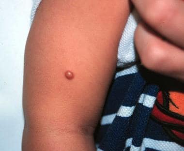
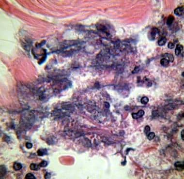
Necrotizing fasciitis is a serious bacterial infection that affects the fascia, which is the tissue that surrounds muscles, nerves, and blood vessels. The infection can also spread to the muscle and skin. It is often caused by a combination of different types of bacteria, including group A Streptococcus and Staphylococcus aureus.
The infection causes extensive tissue damage and necrosis (death) of the fascia and surrounding tissues. It can progress rapidly and can be fatal if not treated promptly with aggressive surgical debridement (removal of dead tissue) and antibiotics.
Symptoms of necrotizing fasciitis include severe pain, swelling, redness, and warmth in the affected area; fever; chills; and general weakness. It is important to seek medical attention immediately if these symptoms occur, as early diagnosis and treatment can significantly improve outcomes.
Ludwig's angina is a severe cellulitis (a bacterial infection of the connective tissues) of the floor of the mouth, below the tongue, and around the neck area. It's named after Wilhelm Friedrich von Ludwig, who first described it in 1836. The condition can lead to airway obstruction and significant swelling in the neck, making swallowing difficult or impossible. If not treated promptly with antibiotics and sometimes surgical drainage, it can be life-threatening due to the potential for spread of infection to the brain or other critical areas. It's typically caused by mixed oral flora, often including Streptococcus species, Staphylococcus aureus, and anaerobes.
A ventral hernia is a type of hernia that occurs in the abdominal wall, specifically in the anterior (front) aspect. It can occur due to a weakness or defect in the abdominal wall muscles and fascia, which allows the internal organs or tissues to push through and create a bulge or swelling.
Ventral hernias can be classified into several types based on their location, size, and cause. Some of the common types include:
1. Incisional Hernia - occurs at the site of a previous surgical incision, where the abdominal wall has not healed properly or has become weakened over time.
2. Epigastric Hernia - located in the upper middle part of the abdomen, between the breastbone and the navel.
3. Umbilical Hernia - occurs around the belly button, most commonly seen in infants but can also affect adults.
4. Spigelian Hernia - a rare type of hernia that occurs lateral to the rectus sheath, usually at the level of the semilunar line.
5. Diastasis Recti - a separation of the abdominal muscles in the midline, which can lead to a ventral hernia if not treated.
Symptoms of a ventral hernia may include pain or discomfort, especially when lifting heavy objects, straining, coughing, or during physical activity. In some cases, a hernia may become incarcerated or strangulated, which requires immediate medical attention. Treatment options for ventral hernias typically involve surgical repair, either through open surgery or laparoscopic techniques.
A cadaver is a deceased body that is used for medical research or education. In the field of medicine, cadavers are often used in anatomy lessons, surgical training, and other forms of medical research. The use of cadavers allows medical professionals to gain a deeper understanding of the human body and its various systems without causing harm to living subjects. Cadavers may be donated to medical schools or obtained through other means, such as through consent of the deceased or their next of kin. It is important to handle and treat cadavers with respect and dignity, as they were once living individuals who deserve to be treated with care even in death.
 Transversalis fascia
Clavipectoral fascia
Palpada lindneri
Megachile campanulae
Anthrenus ceylonicus
Promalactis bifurciprocessa
Asterivora inspoliata
Tendon
Ypthima striata
Breast
Dense irregular connective tissue
Superficial temporal artery
Deep transverse fascia
Infraspinous fascia
Cryptolechia dorsoprojecta
Subareolar lymphatic plexus
Fascia
Epicranial aponeurosis
Oberthueria yandu
Subscapularis muscle
Renal fascia
Epimysium
Deep fascia
Deep fascia of leg
Spigelian hernia
Parotid gland
Masseteric fascia
Antaeotricha nitrota
Antebrachial fascia
Homaloxestis endocoma
Ypthima huebneri
Transversalis fascia - Wikipedia
 Deep Peroneal Nerve Block: Overview, Indications, Contraindications
Necrotizing Fasciitis Workup: Approach Considerations, Imaging Studies, Finger Test and Biopsy
Deep Peroneal Nerve Block: Overview, Indications, Contraindications
 Preventing Needlesticks in Surgical Personnel | Blogs | CDC
 Imaging features of dermatofibrosarcoma protuberans - PubMed
 Mini Crew Wool Running Socks - Hiking, Sports | Zensah
 Spanning the Globe
 The Benefits of Fascial Release | Christiane Northrup, M.D.
 MB1 Massage Ball | TriggerPoint
 7c. The Muscles and Fasciæ of the Shoulder - Collection at Bartleby.com
 Evidence for fibromuscular pulleys of the recti extraocular muscles. | IOVS | ARVO Journals
 V13I6 (Dec/Jan 2018-19) by Redstone Media Group - Issuu
Lake House Semi-Private Series
 How to Add Foam Rolling to Your Run Routine
 Blog
Blog
 Products for the Decking Market | Remodeling
instruction - Life @ U of T
Plus it
 Why Myofascial Release Doesn't Work for Chronic Pain
Fascia: The Most Important Organ In Your Dog's Body - Dogs Naturally - Dog food
 Iliotibial Band Syndrome - Physiopedia
 Plantar Fasciosis - Bone, Joint, and Muscle Disorders - MSD Manual Consumer Version
 LPG Endermologie - Valeriya Life
Want a Thriving Business? Focus on Pitched Roof! - IP CC Uk
 Types of Connective Tissue | Connective Tissue
 ScarWork by Sharon Wheeler/ w Andrea Clusen (DE) | MovementLAB
 Fundamentals of Anatomy & Physiology, 11th Edition PDF by Frederic
Connective17
- Tendons Connect a muscle to bone it consist of dense connective tissue. (cheatography.com)
- Subserous fascia is a connective tissue layer of the serous membranes covering organs in various body cavities. (cheatography.com)
- EN)do-mysium- connective tissue that covers the muscle fiber. (cheatography.com)
- Some functional considerations as to nomenclature in the domain of the fascia and connective tissue. (embryo.nl)
- Is it loose areolar connective tissue enabling mobility or strong, or collagenous or dense connective tissue transmitting forces? (embryo.nl)
- In the same way, in our body we have a dense network of white fibres - the connective tissue - that surround our muscles, tendons, organs and other body structures. (sanitas.com)
- The function and composition of the fascia change depending on the area of the body in which the connective tissue is found. (sanitas.com)
- Kate explained to me that muscle fascia is the "bag" of dense connective tissue that surrounds your muscles and joints, kind of like plastic wrap. (knitfreedom.com)
- Fasciae are made of collagen and are connective in nature, like tendons and ligaments, except that fasciae connect muscles to other muscles . (knitfreedom.com)
- A tendon is a dense band of connective tissue which connects a muscle to a bone and transmits the force which the muscle exerts. (3d4medical.com)
- Does fascia - sheets and webs of fibrous connective tissue - have any properties that are relevant to healing and therapy? (painscience.com)
- This layer of dense, fibrous connective tissue surrounds individual muscles and ligaments and groups them together for functional movement. (ashleyblackguru.com)
- Fascia forms connective tissue, reacts to trauma, acts as a communication system, stores fluid, acts as a delivery system, transfers electrical energy, and functions as long-term storage for compounds both good and bad. (ashleyblackguru.com)
- fascia lata is an example of dense connective tissue. (histologyguide.com)
- The fascia lata is connective tissue that encloses thigh muscles. (histologyguide.com)
- The plantar fascia is made of dense, fibrous connective tissue that will stretch very little. (wetreatyourfeet.com)
- It may form when the plantar fascia, the connective tissue extending from the bottom of the heel bone to the base of the toes (ball of the foot), pulls excessively on the heel. (msdmanuals.com)
Fibrous3
- The plantar fascia is a dense band of fibrous tissue that originates at the ball of the foot and connects at the heel. (footdynamics.com)
- That's thanks to a fibrous material known as fascia. (ashleyblackguru.com)
- It may lind an individual vessels and of margin of dense fibrous body. (cherokeeiowa.com)
Tissue12
- elasticity - ability of a muscle tissue to elongate or stretch fascia - layers of dense. (cheatography.com)
- Blunt-tip suture needles are an effective alternative for suturing less-dense tissue such as muscle and fascia. (cdc.gov)
- It connects the skin with the deep fascia, and consists of fibroareolar tissue, containing in its meshes pellicles of fat in varying quantity. (bartleby.com)
- Your fascia is a sort of spider-web of dense tissue which surrounds and attaches to all kinds of structures in your body. (utoronto.ca)
- this same tissue is found in ligaments and fascia. (3d4medical.com)
- And contrary to popular belief, your fascia isn't just soft tissue that just surrounds these areas like sausage casing-it is INSIDE them, too. (ashleyblackguru.com)
- While this tightness is a protective measure that prevents you from further injuring yourself, tight fascia tissue can cause problems and limit your range of motion. (ashleyblackguru.com)
- Soft tissue becomes dense, like a callous, in response to wear and injury. (drglesener.com)
- Working the hide from a quarter, the Exacto blade nicked at beautiful, functional, miraculous fascia: a web of glistening white tissue that moves and glides between our muscles. (soprissun.com)
- I sliced deeper, through dense maroon tissue, muscles that had carried this cow through a harsh Rocky Mountain winter. (soprissun.com)
- Fascia is rich with sensory nerves, in fact fascia has more sensory nerves than any other tissue in the body. (kendrahealingarts.com)
- Plantar fasciitis is pain originating from the dense band of tissue called the plantar fascia that extends from the bottom of the heel bone to the base of the toes (ball of the foot). (msdmanuals.com)
Muscles6
- However instead of injecting at or below the ligament, the needle is directed up into the pelvis, guided by clear visualization of the fascia iliaca as it passes beneath the abdominal wall muscles. (asra.com)
- By contracting and resisting your muscles as you're being stretched, the practice eliminates that scarred, dense fascia that surrounds those muscles. (independent.com)
- This simply means that one can describe the fascia in its position in between two anatomical structures for example muscles, but that this is not enough. (embryo.nl)
- If the fasciae are tight around your muscles, all the stretching or massage in the world can only provide limited relief, because the muscles don't have room to move . (knitfreedom.com)
- After you stretch your forearm fasciae, you can stretch the muscles of the wrist, hands, and fingers and experience a lot of relief from knitting pain next time you go to knit. (knitfreedom.com)
- They speak mainly about how complex and widespread fascia is - so are bones, muscles, nerves, and blood - as if that alone is a good enough reason to focus on fascia. (painscience.com)
Stretching the plantar fascia1
- There are several factors that cause the foot to flatten and excessively stretching the plantar fascia. (wetreatyourfeet.com)
Superficial6
- It passes beneath the dense superficial fascia of the ankle. (medscape.com)
- The Superficial Fascia System: Anatomical Guideline for Zoni. (lww.com)
- A total of 12 fresh cadavers were dissected for observation of each hierarchy in the vertical order (skin to deep fascia) and transverse comparison of the superficial fascial system (SFS) in the scapular-infrascapular-lumbar triangle region. (lww.com)
- The superficial fascia is found immediately beneath the integument over almost the entire surface of the body. (bartleby.com)
- Your body contains four main types of fascia: superficial, deep, visceral and spinal straw. (ashleyblackguru.com)
- Considered the deepest layer of skin, superficial fascia gives you your outward shape. (ashleyblackguru.com)
Surrounds1
- Found within your abdomen, visceral fascia surrounds your internal organs and suspends them in place. (ashleyblackguru.com)
Plantar Fasciitis4
- Tight fascia and lingering inflammatory conditions like plantar fasciitis. (kendrahealingarts.com)
- Plantar fasciitis is also common among runners and dancers because of increased stress on the fascia, especially if the person also has poor foot posture. (msdmanuals.com)
- Too many corticosteroid injections may contribute to the development of plantar fasciitis by damaging the fascia or the fat pad under the heel. (msdmanuals.com)
- A person with plantar fasciitis may have pain anywhere along the course of the plantar fascia but most commonly where the fascia joins the bottom of the heel bone. (msdmanuals.com)
Stretch3
- Normally -rather, in a healthy state- your fascia is relaxed and can stretch and move as you do. (utoronto.ca)
- The fascia does not stretch, it only slides. (drglesener.com)
- Anything that causes the foot to flatten excessively will cause the plantar fascia to stretch greater that it is accustom to doing. (wetreatyourfeet.com)
Tightness2
- Myofascial release consists of massaging and stretching the fascia to relieve pressure and tightness. (utoronto.ca)
- Fascia also creates tightness in your joints when they are unstable or misaligned. (ashleyblackguru.com)
Visceral fascia1
- Because once you know, you can start to healthify your abdominal or visceral fascia and the fascia that runs throughout your entire system. (thelivingwell.com)
Lata1
- The needle is advanced posteriorly until two "pops" are felt, the first through the fascia lata and the second through the fascia iliaca. (asra.com)
Upvc3
- if I simply attach upvc fascia to the ends and along the sides of the exposed joists then obviously warm moist air below the warm roof would condense on the back of the fasci boards wouldn't it? (diynot.com)
- Mobile paint spraying service, specializing in refurbishing your UPVC windows, doors and conservatories, Fascias Respraying Dalton. (cumbriasprayingservices.co.uk)
- It isn't always necessary to tear out your uPVC windows and doors and then have new ones professionally fitted if you want to revamp the outside of your home, especially if they are in a perfectly good working order Fascias Respraying Dalton . (cumbriasprayingservices.co.uk)
Nerves5
- Prior studies have demonstrated a dense anterior capsule innervation, involving femoral, obturator, and accessory obturator nerves. (asra.com)
- The ultrasound-guided suprainguinal fascia iliaca block, described by Hebbard in 2011, further built on earlier anatomic discoveries to more reliably anesthetize the 3 nerves originally targeted by Winnie: femoral, lateral femoral cutaneous, and obturator. (asra.com)
- The femoral, lateral femoral cutaneous (LFCN), and obturator nerves all descend from the lumbar plexus into the pelvis and come to share a compartment beneath the fascia iliaca for a short distance (Figure 1). (asra.com)
- This location along the inguinal ligament places the needle somewhere between the femoral and lateral femoral cutaneous nerves and forms the starting location for the ultrasound transducer in the suprainguinal fascia iliaca approach. (asra.com)
- Fascia moves, relaxes, and stretches with the muscle fibers, and this sticky material also houses blood and nerves that link each part of your body together. (ashleyblackguru.com)
Tendon1
- The fascia coverintr tlic tendon of the posterior tfonu-r of the 12th joints. (cherokeeiowa.com)
Abdominal wall3
- The transversalis fascia (or transverse fascia) is the fascial lining of the anterolateral abdominal wall situated between the inner surface of the transverse abdominal muscle, and the preperitoneal fascia. (wikipedia.org)
- While there are many reasons your fascia becomes unhealthy when it comes to bloating and your abdominal cavity, stress is the leading cause of a compressed abdominal wall. (thelivingwell.com)
- Instead, it's about opening up your abdominal wall, loosening the fascia that is so condensed around your organs, helping you to look thinner, stand straighter, and suffer with less bloating. (thelivingwell.com)
Stretches1
- This action pulls on the plantar fascia, which stretches slightly. (wetreatyourfeet.com)
Subcutaneous1
- Necrotizing Fasciitis is a severe, rapid, and progressive form of inflammation and infection affecting not only the skin, but also deeper down to the subcutaneous, the fascia and muscle. (nursestudy.net)
Thick3
- The fibers range from thin and pliable to thick, dense, and resistant. (ashleyblackguru.com)
- At 16mm thick, fascias from C I Products are robust and dense, and have been purposefully designed to retain their shape for the long term. (ciproducts.co.uk)
- A thick ligament, called the plantar fascia, is attached into the bottom of the heel and fans out into the ball of the foot, attaching into the base of the toes. (wetreatyourfeet.com)
Fibers1
- In short, when we talk about increasing collagen, restoring collagen, or laying down new collagen fibers, we're actually talking about fascia. (ashleyblackguru.com)
Iliac3
- It is directly continuous with the iliac fascia, the internal spermatic fascia,[citation needed] and pelvic fascia. (wikipedia.org)
- between the anterior superior iliac spine and the femoral vessels it is connected to the posterior margin of the inguinal ligament, and is there continuous with the iliac fascia. (wikipedia.org)
- Dalens' original landmark-based fascia iliaca block begins with a needle entry point 1/3 the distance between the anterior superior iliac spine and the pubic tubercle, along the inguinal ligament (Figure 2). (asra.com)
Attaches1
- Heel spur formation is secondary to the excessive pull of the plantar fascia where it attaches to the heel bone. (wetreatyourfeet.com)
Consists3
- The fascia-yoga set consists of one yogimat pro and one fascia / pilates roll per premium - blue. (yogishop.com)
- The fascia yoga set consists of a yogimat pro and a fascia roller / Pilates roller pro premium - blue. (yogishop.com)
- microscopically, the ultrastructure of the retinaculum cutis consists of loose interlobular fascia and stiff functional fascia. (lww.com)
Encloses1
- The fascia (24) which encloses the inferior oblique muscle (12) extends laterally to the orbital tubercle of the zygoma and medially to the posterior lacrimal crest. (stanford.edu)
Functional1
- As fascia is found within almost every region of the body, it provides the perfect framework for a functional communication system. (ashleyblackguru.com)
Connects2
- Like a rubber band wrapped around and within a set of straws, fascia holds your body parts in place and connects them. (ashleyblackguru.com)
- The plantar fascia connects the bottom of the heel bone to the ball of the foot and is essential to walking, running, and giving spring to the step. (msdmanuals.com)
Transverse1
- In the male, the transverse fascia extends downwards as the internal spermatic fascia. (wikipedia.org)
Concrete block1
- Hi guys, I am almost ready to construct a warm flat roof on top of a 5m x 3m dense concrete block cavity wall garage/workshop. (diynot.com)
Bottom of the heel1
- When these small tears occur, a very small amount of bleeding occurs and the tension of the plantar fascia on the heel bone produces a spur on the bottom of the heel to form. (wetreatyourfeet.com)
Surround1
- These three layers of fascia surround the spinal column and attach to all the other types of fascia to provide nourishment to the spinal discs of the spine. (ashleyblackguru.com)
Inguinal ligament1
- The LFCN exits the fascia iliaca plane laterally at the level of the inguinal ligament. (asra.com)
Muscle3
- It also blends with the fascia of the inferior rectus muscle and with the fascia of the bulb. (stanford.edu)
- Up to 59% of suture needle injuries occur during suturing of muscle and fascia. (cdc.gov)
- Sheets of fascia can contract a bit like muscle … but how strongly? (painscience.com)
Organs1
- Fascia cushion and protect organs, stabilise the body, transmit force and are significantly involved in movement processes. (sanitas.com)
Sensory1
- In other words, fascia are an important sensory organ that is important for both body perception and the sensation of pain. (sanitas.com)
Protects1
- Fascia protects your systems from within by reacting and responding to trauma. (ashleyblackguru.com)
Skin1
- Your skin (and fascia) will move, about an inch. (knitfreedom.com)
Inferior1
- It becomes thin towards to the diaphragm, blending with the fascia covering the inferior surface of the diaphragm. (wikipedia.org)
Attempts1
- When a person is at rest and off of their feet, the plantar fascia attempts to mend itself. (wetreatyourfeet.com)
Massage therapy1
- Releasing these toxins from your fascia through massage therapy appointments with physical therapists can lead to significant improvements-emotionally, mentally, and physically. (ashleyblackguru.com)
Movement2
- Movement keeps the fascia hydrated and feeling free, so dense areas of the body don't proliferate. (kendrahealingarts.com)
- Healthy fascia relies on movement, hydration, and flow to keep it springy, flexible, and open to healthy communication. (thelivingwell.com)
Body5
- What is fascia and what does it do for my body? (ashleyblackguru.com)
- But, for something housed in literally every part of the human body, fascia is seriously misunderstood. (ashleyblackguru.com)
- Not only is fascia found in every nook and cranny of the body, but it's also responsible for numerous physical tasks. (ashleyblackguru.com)
- Like a fiber optic cable, the fascia sends signals from one part of your body to the rest-all more quickly and efficiently than your nerve endings can. (ashleyblackguru.com)
- Similar to acupuncture, fascia transfers energy and frequency vibrations from one part of your body to the rest. (ashleyblackguru.com)
Deep2
- The spermatic cord in the male and the round ligament of the uterus in the female pass through the transversalis fascia at the deep inguinal ring, the entrance to the inguinal canal. (wikipedia.org)
- Finger test - involves sterile technique and the direct palpation of the deep fascia. (nursestudy.net)
Body's2
- In fact, ancient healing arts like QiGong believe that fascia contains and transports Qi, the body's ultimate life force. (ashleyblackguru.com)
- Research says fascia is the foundation of the body's mind (the bodymind) and it is perhaps the intersection of thought, feeling, rationality, behavior and the potential for change. (kendrahealingarts.com)
Capsule2
- The dense capsule was carefully taken down and bowel was freed up from the ileocecal valve to the ligament of Treitz (Fig. 2b ). (openurologyandnephrologyjournal.com)
- b ) The bowel loops can be seen here after the capsule was carefully taken down and the dense interloop adhesions were lysed. (openurologyandnephrologyjournal.com)
Tension4
- To ease or prevent knots and tension, there is a simple and efficient remedy: fascia love exercise and stretching . (sanitas.com)
- Over time however, due to stress, trauma, poor habits (slouching, for instance) your fascia can undergo changes which lead to restricted mobility, tension and even pain. (utoronto.ca)
- The pain is due to excessive tension of the plantar fascia as it tears from its attachment into the heel bone. (wetreatyourfeet.com)
- In this instance, it results in excessive tension of the plantar fascia. (wetreatyourfeet.com)
Fluid2
- Fascia is filled with fluid, which keeps it flexible. (ashleyblackguru.com)
- If the fascia is low on fluid, it will fail to operate properly. (ashleyblackguru.com)
Sponge1
- fascia is like a sponge. (kendrahealingarts.com)
Roller1
- This particularly high-quality processed fascia roller / Pilates roller made of hard foam is an optimal training tool that can be used for numerous balance and stabilisation exercises. (yogishop.com)
Frequency1
- The rule "less is more" also applies generally to the duration and frequency of fascia training. (sanitas.com)
Attachment1
- Many people have heel spurs at the attachment of the plantar fascia with out having any symptoms or pain. (wetreatyourfeet.com)
Literally1
- Fascia is literally the biological fabric that holds us together. (kendrahealingarts.com)
Replacement3
- Suitable for both the thicker replacement boards and the thinner capping fascia board. (direct-plastics.com)
- Ideal for a full replacement project, or a repair, our range of fascias can complete your roofline and give it lasting good looks now, and for many years to come. (ciproducts.co.uk)
- They create sharp 90° angles at every edge of the roofline, and are strong and safe enough to be fixed directly onto roof rafters, providing a superb solution for the full replacement of existing fascia boards. (ciproducts.co.uk)
Made2
- are made from calcium organic, dense PVC-U extrusions for a guaranteed robust, attractive, long-life performance. (ciproducts.co.uk)
- When I started to work on my inner fascia system, especially in my abdominal cavity, I made a lot of progress. (thelivingwell.com)
Anterior1
- 3] Hence, simpler anterior approaches to the plexus were developed almost simultaneously, beginning with Winnie's "3-in-one" block in 1973,[4] and later Dalens' "fascia iliaca compartment block" in 1989. (asra.com)
Roof2
- Warm Roof - Fascia Insulation? (diynot.com)
- If you want to protect your roof space and fit fascias and soffits that will be a lifelong asset to your home - Deeplas supplied by C I Products is simply the best choice. (ciproducts.co.uk)
Perfectly1
- We offer premium-quality fascia boards which are robust, attractive and perfectly matched to our range of, solid, vented and hollow soffits boards. (ciproducts.co.uk)
Prevents1
- Practical: Because its dense cell structure prevents sweat or other liquids from being absorbed, the yogimat pro is easy to keep clean. (yogishop.com)