Cranial Fossa, Middle
Cranial Fossa, Anterior
Cranial Fossa, Posterior
Skull Base
Arachnoid Cysts
Cranial Nerve Neoplasms
Trigeminal Nerve Diseases
Temporal Bone
Skull Base Neoplasms
Facial Nerve Diseases
Ear, Middle
Dura Mater
Central Nervous System Vascular Malformations
Arnold-Chiari Malformation
Ethmoid Bone
Osteoma
Ethmoid Sinus
Sphenoid Bone
Petrous Bone
Tomography, X-Ray Computed
Mucocele
Neurilemmoma
Hematoma, Subdural
Zygoma
Infratentorial Neoplasms
Chondromatosis, Synovial
Hematoma, Subdural, Intracranial
Subdural Effusion
Magnetic Resonance Imaging
Arachnoid
Frontal Sinus
Facial Paralysis
Meningeal Neoplasms
Hematoma, Epidural, Cranial
Sphenoid Sinus
Pterygopalatine Fossa
Arteriovenous Fistula
Cavernous Sinus
Cerebral Angiography
Middle Cerebral Artery
Imaging, Three-Dimensional
Infarction, Middle Cerebral Artery
Dandy-Walker Syndrome
Cerebellar Neoplasms
Cerebellar Diseases
Management of traumatic dislocation of the mandibular condyle into the middle cranial fossa. (1/44)
Dislocation of the mandibular condyle into the middle cranial fossa is a rare complication of facial trauma that can have neurological and life-threatening implications. This article discusses the anatomic features that predispose patients to this type of injury, as well as the clinical features and mechanism of injury for this rare type of condylar deformity, to help practitioners recognize this easily overlooked injury and avoid disastrous complications. The article summarizes previously published case reports of this rare complication of condylar trauma and presents a case for which initial diagnosis and a management protocol are described. (+info)Orbit deformities in craniofacial neurofibromatosis type 1. (2/44)
BACKGROUND AND PURPOSE: The possible relationship of orbit deformities in neurofibromatosis type 1 (NF1) to plexiform neurofibromas (PNFs) have not been fully elucidated. Our purpose was to review orbital changes in patients with craniofacial NF1. METHODS: We retrospectively reviewed CT and MR imaging abnormalities of the orbit in 31 patients (18 male, 13 female; mean age, 14 years; age range 1-40 years) with craniofacial NF1. RESULTS: Orbital abnormalities were documented in 24 patients. Six had optic nerve gliomas with enlarged optic canals. Twenty had PNFs in the orbit or contiguous to the anterior skull. The posterior orbit was distorted by encroachment from an expanded middle cranial fossa in 13 patients, and 18 had enlargement of the orbital rim. Other changes included focal decalcification or remodeling of orbital walls adjacent to PNFs in 18 patients and enlargement of cranial foramina resulting from tumor infiltration of sensory nerves in 16. These orbital deformities were sometimes progressive and always associated with orbital infiltration by PNFs. CONCLUSION: In our patients with craniofacial neurofibromatosis, bony orbital deformity occurred frequently and always with an optic nerve glioma or orbital PNF. PNFs were associated with orbital-bone changes in four patterns: expansion of the middle cranial fossa into the posterior orbit, enlargement of the orbital rim, bone erosion and decalcification by contiguous tumor, and enlargement of the cranial foramina. Orbital changes support the concept of secondary dysplasia, in which interaction of PNFs with the developing skull is a major component of the multifaceted craniofacial changes possible with NF1. (+info)The sphenoparietal sinus of breschet: does it exist? An anatomic study. (3/44)
BACKGROUND AND PURPOSE: The termination of the superficial middle cerebral vein is classically assimilated to the sphenoid portion of the sphenoparietal sinus. This notion has, however, been challenged in a sometimes confusing literature. The purpose of the present study was to evaluate the actual anatomic relationship existing between the sphenoparietal sinus and the superficial middle cerebral vein. METHODS: The cranial venous system of 15 nonfixed human specimens was evaluated by the corrosion cast technique (12 cases) and by classic anatomic dissection (three cases). Angiographic correlation was provided by use of the digital subtraction technique. RESULTS: The parietal portion of the sphenoparietal sinus was found to correspond to the parietal portion of the anterior branch of the middle meningeal veins. The sphenoid portion of the sphenoparietal sinus was found to be an independent venous sinus coursing under the lesser sphenoid wing, the sinus of the lesser sphenoid wing, which was connected medially to the cavernous sinus and laterally to the anterior middle meningeal veins. The superficial middle cerebral vein drained into a paracavernous sinus, a laterocavernous sinus, or a cavernous sinus but was never connected to the sphenoparietal sinus. All these venous structures were demonstrated angiographically. CONCLUSION: The sphenoparietal sinus corresponds to the artificial combination of two venous structures, the parietal portion of the anterior branch of the middle meningeal veins and a dural channel located under the lesser sphenoid wing, the sinus of the lesser sphenoid wing. The classic notion that the superficial middle cerebral vein drains into or is partially equivalent to the sphenoparietal sinus is erroneous. Our study showed these structures to be independent of each other; we found no instance in which the superficial middle cerebral vein was connected to the anterior branch of the middle meningeal veins or the sinus of the lesser sphenoid wing. The clinical implications of these anatomic findings are discussed in relation to dural arteriovenous fistulas in the region of the lesser sphenoid wing. (+info)MR imaging of orbital inflammatory pseudotumors with extraorbital extension. (4/44)
OBJECTIVE: To demonstrate a variety of MR imaging findings of orbital inflammatory pseudotumors with extraorbital extension. MATERIALS AND METHODS: We retrospectively reviewed the MR features of five patients, who were diagnosed clinically and radiologically as having an orbital inflammatory pseudotumor with extraorbital extension. RESULTS: The types of orbital pseudotumors were a mass in the orbital apex (n = 3), diffuse form (n = 2), and myositis (n = 1). The extraorbital extension of the orbital pseudotumor passed through the superior orbital fissure in all cases, through the inferior orbital fissure in two cases, and through the optic canal in one case. The orbital lesions extended into the following areas: the cavernous sinus (n = 4), the middle cranial fossa (n = 4), Meckel's cave (n = 2), the petrous apex (n = 2), the clivus (n = 2), the pterygopalatine fossa and infratemporal fossa (n = 2), the foramen rotundum (n = 1), the paranasal sinus (n = 1), and the infraorbital foramen (n = 1). On MR imaging, the lesions appeared as an isosignal intensity with gray matter on the T1-weighted images, as a low signal intensity on the T2-weighted images and showed a marked enhancement on the post-gadoliniumdiethylene triamine pentaacetic acid (post-Gd-DTPA) T1-sequences. The symptoms of all of the patients improved when they were given high doses of steroids. Three of the five patients experienced a recurrence. CONCLUSION: MR imaging is useful for demonstrating the presence of a variety of extraorbital extensions of orbital inflammatory pseudotumors. (+info)Rapidly growing microcystic meningioma of the middle fossa floor. Case report. (5/44)
A 74-year-old woman presented with a microcystic meningioma which manifested as mental disturbance. A rapidly growing tumor in the left middle fossa had not been detected by examination 10 months before. The tumor was remarkably enhanced by contrast medium on both computed tomography and magnetic resonance imaging and was associated with massive perifocal edema. Cerebral angiography revealed that the tumor was mainly fed by the left middle meningeal artery, which was embolized preoperatively. The tumor was completely removed and no postoperative adjuvant therapy was administered. The histological diagnosis was microcystic meningioma with many mitotic figures and a MIB-1 labeling index of 12.8%. Four months later, the tumor recurred and invaded the paranasal sinus. Focal irradiation successfully controlled further regrowth. This case suggests that microcystic meningioma may have aggressive features, and close observation is necessary even after gross total removal. (+info)Synovial chondromatosis of the temporomandibular joint with extension to the middle cranial fossa. (6/44)
A rare case of synovial chondromatosis with extension to the middle cranial fossa is reported. Synovial chondromatosis, a benign disorder characterized by multiple cartilaginous, free-floating nodules that originate from the synovial membrane is not exclusive to the temporomandibular joint (TMJ). This condition is commonly seen in the axial skeleton and can involve multiple joints. In this case, synovial chondromatosis of the TMJ led to complete bony erosion of the glenoid fossa extending into the middle cranial fossa. Although plain radiographs showed the involvement of the joint, Computed Tomography (CT) and Magnetic Resonance Imaging (MRI) provided more detailed information about the lesion in all three dimensions. This case demonstrates the value of CT and MRI in both the diagnosis and treatment planning. A review of previously reported cases of synovial chondromatosis with cranial extensions is included. (+info)Assessment of the anatomical relationship between the arcuate eminence and superior semicircular canal by computed tomography. (7/44)
The anatomical relationship between the arcuate eminence (AE) and the superior semicircular canal (SSC) was examined by computed tomography (CT) in 52 petrous bones of 26 patients. After acquiring volume data by multidetector CT, 1-mm thick oblique bone window images perpendicular to the SSC were obtained from the axial images. The distances between the AE and the SSC, and the SSC and the superior surface of the petrous bone were measured. The AE corresponded exactly with the SSC in only 2/52 petrous bones, and corresponded well in 7/52. The AE was lateral to the SSC in 25/52 cases, medial to the SSC in 6/52 cases, intersected in 3/52 cases, and was indiscernible in 9/52 cases. The distance between the SSC and the petrous surface was 0 mm in 45/52 petrous bones, 1 mm in 5/52, 2 mm in 1/52, and 3 mm in 1/52. The SSC typically does not correspond exactly with the AE, and is generally located just under the surface of the petrous bone. Planning of the middle cranial fossa approach requires location of the SSC by CT. (+info)Dura-based giant intracranial schwannoma in the middle fossa. (8/44)
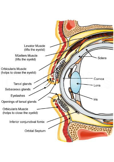
A 49-year-old female presented with a rare giant schwannoma arising from the dura mater of the middle fossa manifesting as loss of left visual acuity. Magnetic resonance imaging revealed a heterogeneously enhanced giant mass in the left middle fossa. Surgery via the transsylvian approach confirmed the origin of the tumor between the left internal carotid artery and the trigeminal nerve in the lateral wall of the cavernous sinus. Elongated abducens nerve was confirmed, but no tumor adhesion to the abducens nerve was found. The tumor was closely attached to the dura mater of the middle fossa and the lateral wall of the cavernous sinus. The histological diagnosis was schwannoma. Both left oculomotor and abducens nerve pareses occurred immediately after the operation but gradually resolved over 3 months. The operative findings indicated that this schwannoma may have arisen from the meningeal branch of the trigeminal nerve in the dura mater of the middle fossa. (+info)The middle cranial fossa is a depression or hollow in the skull that forms the upper and central portion of the cranial cavity. It is located between the anterior cranial fossa (which lies anteriorly) and the posterior cranial fossa (which lies posteriorly). The middle cranial fossa contains several important structures, including the temporal lobes of the brain, the pituitary gland, the optic chiasm, and the cavernous sinuses. It is also where many of the cranial nerves pass through on their way to the brain.
The middle cranial fossa can be further divided into two parts: the anterior and posterior fossae. The anterior fossa contains the optic chiasm and the pituitary gland, while the posterior fossa contains the temporal lobes of the brain and the cavernous sinuses.
The middle cranial fossa is formed by several bones of the skull, including the sphenoid bone, the temporal bone, and the parietal bone. The shape and size of the middle cranial fossa can vary from person to person, and abnormalities in its structure can be associated with various medical conditions, such as pituitary tumors or aneurysms.
The anterior cranial fossa is a term used in anatomy to refer to the portion of the skull that forms the upper part of the orbits (eye sockets) and the roof of the nasal cavity. It is located at the front of the skull, and is formed by several bones including the frontal bone, sphenoid bone, and ethmoid bone.
The anterior cranial fossa contains several important structures, including the olfactory bulbs (which are responsible for our sense of smell), as well as the optic nerves and parts of the pituitary gland. This region of the skull also provides protection for the brain, particularly the frontal lobes, which are involved in higher cognitive functions such as decision-making, problem-solving, and emotional regulation.
Abnormalities or injuries to the anterior cranial fossa can have serious consequences, including damage to the olfactory bulbs, optic nerves, and pituitary gland, as well as potential injury to the frontal lobes of the brain.
The posterior cranial fossa is a term used in anatomy to refer to the portion of the skull that forms the lower, back part of the cranial cavity. It is located between the occipital bone and the temporal bones, and it contains several important structures including the cerebellum, pons, medulla oblongata, and the lower cranial nerves (IX-XII). The posterior fossa also contains the foramen magnum, which is a large opening through which the spinal cord connects to the brainstem. This region of the skull is protected by the occipital bone, which forms the base of the skull and provides attachment for several neck muscles.
The skull base is the lower part of the skull that forms the floor of the cranial cavity and the roof of the facial skeleton. It is a complex anatomical region composed of several bones, including the frontal, sphenoid, temporal, occipital, and ethmoid bones. The skull base supports the brain and contains openings for blood vessels and nerves that travel between the brain and the face or neck. The skull base can be divided into three regions: the anterior cranial fossa, middle cranial fossa, and posterior cranial fossa, which house different parts of the brain.
An Arachnoid cyst is a type of abnormal fluid-filled sac that develops between the brain or spinal cord and the arachnoid membrane, which is one of the three layers that cover and protect the central nervous system. These cysts are filled with cerebrospinal fluid (CSF), which is the same fluid that surrounds and cushions the brain and spinal cord.
Arachnoid cysts can vary in size and may be present at birth or develop later in life due to trauma, infection, or other factors. While many arachnoid cysts are asymptomatic and do not cause any problems, larger cysts or those that grow or shift over time can put pressure on the brain or spinal cord, leading to a range of neurological symptoms such as headaches, seizures, hearing or vision changes, balance or coordination difficulties, and cognitive impairments.
Treatment for arachnoid cysts depends on their size, location, and associated symptoms. In some cases, observation and monitoring may be sufficient, while in others, surgical intervention may be necessary to drain the cyst or create a connection between it and the surrounding CSF space to relieve pressure.
Otologic surgical procedures refer to a range of surgeries performed on the ear or its related structures. These procedures are typically conducted by otologists, who are specialists trained in diagnosing and treating conditions that affect the ears, balance system, and related nerves. The goal of otologic surgery can vary from repairing damaged bones in the middle ear to managing hearing loss, tumors, or chronic infections. Some common otologic surgical procedures include:
1. Stapedectomy/Stapedotomy: These are procedures used to treat otosclerosis, a condition where the stapes bone in the middle ear becomes fixed and causes conductive hearing loss. The surgeon creates an opening in the stapes footplate (stapedotomy) or removes the entire stapes bone (stapedectomy) and replaces it with a prosthetic device to improve sound conduction.
2. Myringoplasty/Tympanoplasty: These are surgeries aimed at repairing damaged eardrums (tympanic membrane). A myringoplasty involves grafting a piece of tissue over the perforation in the eardrum, while a tympanoplasty includes both eardrum repair and reconstruction of the middle ear bones if necessary.
3. Mastoidectomy: This procedure involves removing the mastoid air cells, which are located in the bony prominence behind the ear. A mastoidectomy is often performed to treat chronic mastoiditis, cholesteatoma, or complications from middle ear infections.
4. Ossiculoplasty: This procedure aims to reconstruct and improve the function of the ossicles (middle ear bones) when they are damaged due to various reasons such as infection, trauma, or congenital conditions. The surgeon uses prosthetic devices made from plastic, metal, or even bone to replace or support the damaged ossicles.
5. Cochlear implantation: This is a surgical procedure that involves placing an electronic device inside the inner ear to help individuals with severe to profound hearing loss. The implant consists of an external processor and internal components that directly stimulate the auditory nerve, bypassing the damaged hair cells in the cochlea.
6. Labyrinthectomy: This procedure involves removing the balance-sensing structures (vestibular system) inside the inner ear to treat severe vertigo or dizziness caused by conditions like Meniere's disease when other treatments have failed.
7. Acoustic neuroma removal: An acoustic neuroma is a benign tumor that grows on the vestibulocochlear nerve, which connects the inner ear to the brain. Surgical removal of the tumor is necessary to prevent hearing loss, balance problems, and potential neurological complications.
These are just a few examples of the various surgical procedures performed by otolaryngologists (ear, nose, and throat specialists) to treat conditions affecting the ear and surrounding structures. Each procedure has its specific indications, benefits, risks, and postoperative care requirements. Patients should consult with their healthcare providers to discuss the most appropriate treatment options for their individual needs.
Cranial nerve neoplasms refer to abnormal growths or tumors that develop within or near the cranial nerves. These nerves are responsible for transmitting sensory and motor information between the brain and various parts of the head, neck, and trunk. There are 12 pairs of cranial nerves, each with a specific function and location in the skull.
Cranial nerve neoplasms can be benign or malignant and may arise from the nerve itself (schwannoma, neurofibroma) or from surrounding tissues that invade the nerve (meningioma, epidermoid cyst). The growth of these tumors can cause various symptoms depending on their size, location, and rate of growth. Common symptoms include:
* Facial weakness or numbness
* Double vision or other visual disturbances
* Hearing loss or tinnitus (ringing in the ears)
* Difficulty swallowing or speaking
* Loss of smell or taste
* Uncontrollable eye movements or drooping eyelids
Treatment for cranial nerve neoplasms depends on several factors, including the type, size, location, and extent of the tumor, as well as the patient's overall health. Treatment options may include surgery, radiation therapy, chemotherapy, or a combination of these approaches. Regular follow-up care is essential to monitor for recurrence or complications.
Trigeminal nerve diseases refer to conditions that affect the trigeminal nerve, which is one of the cranial nerves responsible for sensations in the face and motor functions such as biting and chewing. The trigeminal nerve has three branches: ophthalmic, maxillary, and mandibular, which innervate different parts of the face and head.
Trigeminal nerve diseases can cause various symptoms, including facial pain, numbness, tingling, or weakness. Some common trigeminal nerve diseases include:
1. Trigeminal neuralgia: A chronic pain condition that affects the trigeminal nerve, causing intense, stabbing, or electric shock-like pain in the face.
2. Hemifacial spasm: A neuromuscular disorder that causes involuntary muscle spasms on one side of the face, often affecting the muscles around the eye and mouth.
3. Trigeminal neuropathy: Damage or injury to the trigeminal nerve, which can result in numbness, tingling, or weakness in the face.
4. Herpes zoster oticus (Ramsay Hunt syndrome): A viral infection that affects the facial nerve and geniculate ganglion of the trigeminal nerve, causing facial paralysis, ear pain, and a rash around the ear.
5. Microvascular compression: Compression of the trigeminal nerve by a blood vessel, which can cause symptoms similar to trigeminal neuralgia.
Treatment for trigeminal nerve diseases depends on the specific condition and its severity. Treatment options may include medication, surgery, or radiation therapy.
A craniotomy is a surgical procedure where a bone flap is temporarily removed from the skull to access the brain. This procedure is typically performed to treat various neurological conditions, such as brain tumors, aneurysms, arteriovenous malformations, or traumatic brain injuries. After the underlying brain condition is addressed, the bone flap is usually replaced and secured back in place with plates and screws. The purpose of a craniotomy is to provide access to the brain for diagnostic or therapeutic interventions while minimizing potential damage to surrounding tissues.
The temporal bone is a paired bone that is located on each side of the skull, forming part of the lateral and inferior walls of the cranial cavity. It is one of the most complex bones in the human body and has several important structures associated with it. The main functions of the temporal bone include protecting the middle and inner ear, providing attachment for various muscles of the head and neck, and forming part of the base of the skull.
The temporal bone is divided into several parts, including the squamous part, the petrous part, the tympanic part, and the styloid process. The squamous part forms the lateral portion of the temporal bone and articulates with the parietal bone. The petrous part is the most medial and superior portion of the temporal bone and contains the inner ear and the semicircular canals. The tympanic part forms the lower and anterior portions of the temporal bone and includes the external auditory meatus or ear canal. The styloid process is a long, slender projection that extends downward from the inferior aspect of the temporal bone and serves as an attachment site for various muscles and ligaments.
The temporal bone plays a crucial role in hearing and balance, as it contains the structures of the middle and inner ear, including the oval window, round window, cochlea, vestibule, and semicircular canals. The stapes bone, one of the three bones in the middle ear, is entirely encased within the petrous portion of the temporal bone. Additionally, the temporal bone contains important structures for facial expression and sensation, including the facial nerve, which exits the skull through the stylomastoid foramen, a small opening in the temporal bone.
Skull base neoplasms refer to abnormal growths or tumors located in the skull base, which is the region where the skull meets the spine and where the brain connects with the blood vessels and nerves that supply the head and neck. These neoplasms can be benign (non-cancerous) or malignant (cancerous), and they can arise from various types of cells in this area, including bone, nerve, glandular, and vascular tissue.
Skull base neoplasms can cause a range of symptoms depending on their size, location, and growth rate. Some common symptoms include headaches, vision changes, hearing loss, facial numbness or weakness, difficulty swallowing, and balance problems. Treatment options for skull base neoplasms may include surgery, radiation therapy, chemotherapy, or a combination of these approaches. The specific treatment plan will depend on the type, size, location, and stage of the tumor, as well as the patient's overall health and medical history.
Facial nerve diseases refer to a group of medical conditions that affect the function of the facial nerve, also known as the seventh cranial nerve. This nerve is responsible for controlling the muscles of facial expression, and it also carries sensory information from the taste buds in the front two-thirds of the tongue, and regulates saliva flow and tear production.
Facial nerve diseases can cause a variety of symptoms, depending on the specific location and extent of the nerve damage. Common symptoms include:
* Facial weakness or paralysis on one or both sides of the face
* Drooping of the eyelid and corner of the mouth
* Difficulty closing the eye or keeping it closed
* Changes in taste sensation or dryness of the mouth and eyes
* Abnormal sensitivity to sound (hyperacusis)
* Twitching or spasms of the facial muscles
Facial nerve diseases can be caused by a variety of factors, including:
* Infections such as Bell's palsy, Ramsay Hunt syndrome, and Lyme disease
* Trauma or injury to the face or skull
* Tumors that compress or invade the facial nerve
* Neurological conditions such as multiple sclerosis or Guillain-Barre syndrome
* Genetic disorders such as Moebius syndrome or hemifacial microsomia
Treatment for facial nerve diseases depends on the underlying cause and severity of the symptoms. In some cases, medication, physical therapy, or surgery may be necessary to restore function and relieve symptoms.
The middle ear is the middle of the three parts of the ear, located between the outer ear and inner ear. It contains three small bones called ossicles (the malleus, incus, and stapes) that transmit and amplify sound vibrations from the eardrum to the inner ear. The middle ear also contains the Eustachian tube, which helps regulate air pressure in the middle ear and protects against infection by allowing fluid to drain from the middle ear into the back of the throat.
The cerebellopontine angle (CPA) is a narrow space located at the junction of the brainstem and the cerebellum, where the pons and cerebellum meet. This region is filled with several important nerves, blood vessels, and membranous coverings called meninges. The CPA is a common site for various neurological disorders because it contains critical structures such as:
1. Cerebellum: A part of the brain responsible for coordinating muscle movements, maintaining balance, and fine-tuning motor skills.
2. Pons: A portion of the brainstem that plays a role in several vital functions, including facial movements, taste sensation, sleep regulation, and respiration.
3. Cranial nerves: The CPA is home to the following cranial nerves:
* Vestibulocochlear nerve (CN VIII): This nerve has two components - cochlear and vestibular. The cochlear part is responsible for hearing, while the vestibular part contributes to balance and eye movement.
* Facial nerve (CN VII): This nerve controls facial expressions, taste sensation in the anterior two-thirds of the tongue, salivary gland function, and lacrimation (tear production).
4. Blood vessels: The CPA contains critical blood vessels like the anterior inferior cerebellar artery (AICA), which supplies blood to various parts of the brainstem, cerebellum, and cranial nerves.
5. Meninges: These are protective membranes surrounding the brain and spinal cord. In the CPA, the meninges include the dura mater, arachnoid mater, and pia mater.
Disorders that can affect the structures in the cerebellopontine angle include acoustic neuromas (vestibular schwannomas), meningiomas, epidermoids, and arteriovenous malformations. These conditions may cause symptoms such as hearing loss, tinnitus (ringing in the ears), vertigo (dizziness), facial weakness or numbness, difficulty swallowing, and imbalance.
Dura Mater is the thickest and outermost of the three membranes (meninges) that cover the brain and spinal cord. It provides protection and support to these delicate structures. The other two layers are called the Arachnoid Mater and the Pia Mater, which are thinner and more delicate than the Dura Mater. Together, these three layers form a protective barrier around the central nervous system.
Ear diseases are medical conditions that affect the ear and its various components, including the outer ear, middle ear, and inner ear. These diseases can cause a range of symptoms, such as hearing loss, tinnitus (ringing in the ears), vertigo (dizziness), ear pain, and discharge. Some common ear diseases include:
1. Otitis externa (swimmer's ear) - an infection or inflammation of the outer ear and ear canal.
2. Otitis media - an infection or inflammation of the middle ear, often caused by a cold or flu.
3. Cholesteatoma - a skin growth that develops in the middle ear behind the eardrum.
4. Meniere's disease - a disorder of the inner ear that can cause vertigo, hearing loss, and tinnitus.
5. Temporomandibular joint (TMJ) disorders - problems with the joint that connects the jawbone to the skull, which can cause ear pain and other symptoms.
6. Acoustic neuroma - a noncancerous tumor that grows on the nerve that connects the inner ear to the brain.
7. Presbycusis - age-related hearing loss.
Treatment for ear diseases varies depending on the specific condition and its severity. It may include medication, surgery, or other therapies. If you are experiencing symptoms of an ear disease, it is important to seek medical attention from a healthcare professional, such as an otolaryngologist (ear, nose, and throat specialist).
Central nervous system (CNS) vascular malformations are abnormal tangles or masses of blood vessels in the brain or spinal cord. These malformations can be congenital (present at birth) or acquired (develop later in life). They can vary in size, location, and symptoms, which may include headaches, seizures, weakness, numbness, difficulty speaking or understanding speech, and vision problems.
There are several types of CNS vascular malformations, including:
1. Arteriovenous malformations (AVMs): These are tangles of arteries and veins with a direct connection between them, bypassing the capillary network. AVMs can cause bleeding in the brain or spinal cord, leading to stroke or neurological deficits.
2. Cavernous malformations: These are clusters of dilated, thin-walled blood vessels that form a sac-like structure. They can rupture and bleed, causing symptoms such as seizures, headaches, or neurological deficits.
3. Developmental venous anomalies (DVAs): These are benign vascular malformations characterized by an abnormal pattern of veins that drain blood from the brain. DVAs are usually asymptomatic but can be associated with other vascular malformations.
4. Capillary telangiectasias: These are small clusters of dilated capillaries in the brain or spinal cord. They are usually asymptomatic and found incidentally during imaging studies.
5. Moyamoya disease: This is a rare, progressive cerebrovascular disorder characterized by the narrowing or blockage of the internal carotid arteries and their branches. This can lead to decreased blood flow to the brain, causing symptoms such as headaches, seizures, and strokes.
The diagnosis of CNS vascular malformations typically involves imaging studies such as MRI or CT scans, and sometimes angiography. Treatment options may include observation, medication, surgery, or endovascular procedures, depending on the type, location, and severity of the malformation.
Arnold-Chiari malformation is a structural abnormality of the brain and skull base, specifically the cerebellum and brainstem. It is characterized by the descent of the cerebellar tonsils and sometimes parts of the brainstem through the foramen magnum (the opening at the base of the skull) into the upper spinal canal. This can cause pressure on the brainstem and cerebellum, potentially leading to a range of symptoms such as headaches, neck pain, unsteady gait, swallowing difficulties, hearing or balance problems, and in severe cases, neurological deficits. There are four types of Arnold-Chiari malformations, with type I being the most common and least severe form. Types II, III, and IV are progressively more severe and involve varying degrees of hindbrain herniation and associated neural tissue damage. Surgical intervention is often required to alleviate symptoms and prevent further neurological deterioration.
The ethmoid bone is a paired, thin, and lightweight bone that forms part of the skull's anterior cranial fossa and contributes to the formation of the orbit and nasal cavity. It is located between the frontal bone above and the maxilla and palatine bones below. The ethmoid bone has several important features:
1. Cribriform plate: This is the horizontal, sieve-like portion that forms part of the anterior cranial fossa and serves as the roof of the nasal cavity. It contains small openings (foramina) through which olfactory nerves pass.
2. Perpendicular plate: The perpendicular plate is a vertical structure that projects downward from the cribriform plate, forming part of the nasal septum and separating the left and right nasal cavities.
3. Superior and middle nasal conchae: These are curved bony projections within the lateral walls of the nasal cavity that help to warm, humidify, and filter incoming air.
4. Lacrimal bone: The ethmoid bone articulates with the lacrimal bone, forming part of the medial wall of the orbit.
5. Frontal process: This is a thin, vertical plate that articulates with the frontal bone above the orbit.
6. Sphenoidal process: The sphenoidal process connects the ethmoid bone to the sphenoid bone posteriorly.
The ethmoid bone plays a crucial role in protecting the brain and providing structural support for the eyes, as well as facilitating respiration by warming, humidifying, and filtering incoming air.
Osteoma is a benign (noncancerous) tumor that is made up of mature bone tissue. It usually grows slowly over a period of years and is most commonly found in the skull or jaw, although it can occur in other bones of the body as well. Osteomas are typically small, but they can grow to be several centimeters in size. They may cause symptoms if they press on nearby tissues or structures, such as nerves or blood vessels. In some cases, osteomas may not cause any symptoms and may only be discovered during routine imaging studies. Treatment for osteoma is typically not necessary unless it is causing problems or growing rapidly. If treatment is needed, it may involve surgical removal of the tumor.
The ethmoid sinuses are a pair of air-filled spaces located in the ethmoid bone, which is a part of the skull that forms the upper portion of the nasal cavity and the inner eye socket. These sinuses are divided into anterior and posterior groups and are present in adults, but not at birth. They continue to grow and develop until early adulthood.
The ethmoid sinuses are lined with mucous membrane, which helps to warm, humidify, and filter the air we breathe. They are surrounded by a network of blood vessels and nerves, making them susceptible to inflammation and infection. Inflammation of the ethmoid sinuses can lead to conditions such as sinusitis, which can cause symptoms such as nasal congestion, headache, and facial pain.
The sphenoid bone is a complex, irregularly shaped bone located in the middle cranial fossa and forms part of the base of the skull. It articulates with several other bones, including the frontal, parietal, temporal, ethmoid, palatine, and zygomatic bones. The sphenoid bone has two main parts: the body and the wings.
The body of the sphenoid bone is roughly cuboid in shape and contains several important structures, such as the sella turcica, which houses the pituitary gland, and the sphenoid sinuses, which are air-filled cavities within the bone. The greater wings of the sphenoid bone extend laterally from the body and form part of the skull's lateral walls. They contain the superior orbital fissure, through which important nerves and blood vessels pass between the cranial cavity and the orbit of the eye.
The lesser wings of the sphenoid bone are thin, blade-like structures that extend anteriorly from the body and form part of the floor of the anterior cranial fossa. They contain the optic canal, which transmits the optic nerve and ophthalmic artery between the brain and the orbit of the eye.
Overall, the sphenoid bone plays a crucial role in protecting several important structures within the skull, including the pituitary gland, optic nerves, and ophthalmic arteries.
The mastoid is a term used in anatomy and refers to the bony prominence located at the base of the skull, posterior to the ear. More specifically, it's part of the temporal bone, one of the bones that forms the side and base of the skull. The mastoid process provides attachment for various muscles involved in chewing and moving the head.
In a medical context, "mastoid" can also refer to conditions or procedures related to this area. For example, mastoiditis is an infection of the mastoid process, while a mastoidectomy is a surgical procedure that involves removing part or all of the mastoid process.
The petrous bone is a part of the temporal bone, one of the 22 bones in the human skull. It is a thick and irregularly shaped bone located at the base of the skull and forms part of the ear and the cranial cavity. The petrous bone contains the cochlea, vestibule, and semicircular canals of the inner ear, which are responsible for hearing and balance. It also helps protect the brain from injury by forming part of the bony structure surrounding the brain.
The term "petrous" comes from the Latin word "petrosus," meaning "stony" or "rock-like," which describes the hard and dense nature of this bone. The petrous bone is one of the densest bones in the human body, making it highly resistant to fractures and other forms of damage.
In medical terminology, the term "petrous" may also be used to describe any structure that resembles a rock or is hard and dense, such as the petrous apex, which refers to the portion of the petrous bone that points towards the sphenoid bone.
X-ray computed tomography (CT or CAT scan) is a medical imaging method that uses computer-processed combinations of many X-ray images taken from different angles to produce cross-sectional (tomographic) images (virtual "slices") of the body. These cross-sectional images can then be used to display detailed internal views of organs, bones, and soft tissues in the body.
The term "computed tomography" is used instead of "CT scan" or "CAT scan" because the machines take a series of X-ray measurements from different angles around the body and then use a computer to process these data to create detailed images of internal structures within the body.
CT scanning is a noninvasive, painless medical test that helps physicians diagnose and treat medical conditions. CT imaging provides detailed information about many types of tissue including lung, bone, soft tissue and blood vessels. CT examinations can be performed on every part of the body for a variety of reasons including diagnosis, surgical planning, and monitoring of therapeutic responses.
In computed tomography (CT), an X-ray source and detector rotate around the patient, measuring the X-ray attenuation at many different angles. A computer uses this data to construct a cross-sectional image by the process of reconstruction. This technique is called "tomography". The term "computed" refers to the use of a computer to reconstruct the images.
CT has become an important tool in medical imaging and diagnosis, allowing radiologists and other physicians to view detailed internal images of the body. It can help identify many different medical conditions including cancer, heart disease, lung nodules, liver tumors, and internal injuries from trauma. CT is also commonly used for guiding biopsies and other minimally invasive procedures.
In summary, X-ray computed tomography (CT or CAT scan) is a medical imaging technique that uses computer-processed combinations of many X-ray images taken from different angles to produce cross-sectional images of the body. It provides detailed internal views of organs, bones, and soft tissues in the body, allowing physicians to diagnose and treat medical conditions.
A mucocele is a mucus-containing cystic lesion that results from the accumulation of mucin within a damaged minor salivary gland duct or mucous gland. It is typically caused by trauma, injury, or blockage of the duct. Mucocele appears as a round, dome-shaped, fluid-filled swelling, which may be bluish or clear in color. They are most commonly found on the lower lip but can also occur on other areas of the oral cavity. Mucocele is generally painless unless it becomes secondarily infected; however, it can cause discomfort during speaking, chewing, or swallowing, and may affect aesthetics. Treatment usually involves surgical excision of the mucocele to prevent recurrence.
A neurilemmoma, also known as schwannoma or peripheral nerve sheath tumor, is a benign, slow-growing tumor that arises from the Schwann cells, which produce the myelin sheath that surrounds and insulates peripheral nerves. These tumors can occur anywhere along the course of a peripheral nerve, but they most commonly affect the acoustic nerve (vestibulocochlear nerve), leading to a type of tumor called vestibular schwannoma or acoustic neuroma. Neurilemmomas are typically encapsulated and do not invade the surrounding tissue, although larger ones may cause pressure-related symptoms due to compression of nearby structures. Rarely, these tumors can undergo malignant transformation, leading to a condition called malignant peripheral nerve sheath tumor or neurofibrosarcoma.
A subdural hematoma is a type of hematoma (a collection of blood) that occurs between the dura mater, which is the outermost protective covering of the brain, and the brain itself. It is usually caused by bleeding from the veins located in this potential space, often as a result of a head injury or trauma.
Subdural hematomas can be classified as acute, subacute, or chronic based on their rate of symptom progression and the time course of their appearance on imaging studies. Acute subdural hematomas typically develop and cause symptoms rapidly, often within hours of the head injury. Subacute subdural hematomas have a more gradual onset of symptoms, which can occur over several days to a week after the trauma. Chronic subdural hematomas may take weeks to months to develop and are often seen in older adults or individuals with chronic alcohol abuse, even after minor head injuries.
Symptoms of a subdural hematoma can vary widely depending on the size and location of the hematoma, as well as the patient's age and overall health. Common symptoms include headache, altered mental status, confusion, memory loss, weakness or numbness, seizures, and in severe cases, coma or even death. Treatment typically involves surgical evacuation of the hematoma, along with management of any underlying conditions that may have contributed to its development.
Cerebral veins are the blood vessels that carry deoxygenated blood from the brain to the dural venous sinuses, which are located between the layers of tissue covering the brain. The largest cerebral vein is the superior sagittal sinus, which runs along the top of the brain. Other major cerebral veins include the straight sinus, transverse sinus, sigmoid sinus, and cavernous sinus. These veins receive blood from smaller veins called venules that drain the surface and deep structures of the brain. The cerebral veins play an important role in maintaining normal circulation and pressure within the brain.
The zygoma is the scientific name for the cheekbone. It is a part of the facial skeleton that forms the prominence of the cheek and houses the maxillary sinus, one of the pairs of paranasal sinuses. The zygomatic bone, also known as the malar bone, contributes to the formation of the zygoma.
Infratentorial neoplasms refer to tumors that originate in the region of the brain called the posterior fossa, which is located below the tentorium cerebelli (a membranous structure that separates the cerebrum from the cerebellum). This area contains several important structures such as the cerebellum, pons, medulla oblongata, and fourth ventricle. Infratentorial neoplasms can be benign or malignant and can arise from various cell types including nerve cells, glial cells, or supportive tissues. They can cause a variety of symptoms depending on their location and size, such as headache, vomiting, unsteady gait, weakness, numbness, vision changes, hearing loss, and difficulty swallowing or speaking. Treatment options may include surgery, radiation therapy, and chemotherapy.
Synovial chondromatosis is a rare condition that affects the synovial membrane, which is the lining of joints, bursae (fluid-filled sacs that cushion bones), and tendon sheaths. In this condition, nodules made up of cartilage form in the synovial membrane. These nodules can detach from the synovial membrane and float freely in the synovial fluid, which lubricates the joint. If they become numerous, they can cause joint pain, stiffness, and decreased range of motion. In some cases, the loose bodies may also cause locking or catching sensations in the joint. Surgery is typically required to remove the cartilaginous nodules and relieve symptoms. If left untreated, synovial chondromatosis can lead to osteoarthritis and other joint problems.
A subdural hematoma is a type of intracranial hemorrhage, which means it involves bleeding within the skull. More specifically, a subdural hematoma occurs between the dura mater (the outermost layer of the meninges that covers the brain) and the brain itself. This condition is usually caused by trauma or injury to the head, which results in the rupture of blood vessels in the brain. The bleeding then forms a collection of blood in the subdural space, which can compress the brain and lead to various neurological symptoms.
Subdural hematomas can be acute, subacute, or chronic, depending on the time course of symptom onset and the rate of blood accumulation. Acute subdural hematomas typically result from severe head trauma and require immediate medical attention due to their rapid progression and potential for causing significant brain damage or even death. Chronic subdural hematomas, on the other hand, may develop more slowly over time and can sometimes be asymptomatic, although they still have the potential to cause long-term neurological problems if left untreated.
Treatment options for subdural hematomas depend on various factors, including the patient's age, overall health status, the severity of symptoms, and the size and location of the hematoma. In some cases, conservative management with close monitoring may be appropriate, while in other situations, surgical intervention may be necessary to alleviate pressure on the brain and prevent further damage.
The occipital bone is the single, posterior cranial bone that forms the base of the skull and encloses the brain. It articulates with the parietal bones anteriorly and the temporal bones laterally. The occipital bone also contains several important structures such as the foramen magnum, through which the spinal cord connects to the brain, and the external and internal occipital protuberances, which serve as attachment points for neck muscles.
A subdural effusion is an abnormal accumulation of fluid in the potential space between the dura mater (the outermost layer of the meninges that covers the brain and spinal cord) and the arachnoid membrane (one of the three layers of the meninges that surround the brain and spinal cord) in the subdural space.
Subdural effusions can occur due to various reasons, including head trauma, infection, or complications from neurosurgical procedures. The fluid accumulation may result from bleeding (subdural hematoma), inflammation, or increased cerebrospinal fluid pressure. Depending on the underlying cause and the amount of fluid accumulated, subdural effusions can cause various symptoms, such as headaches, altered mental status, or neurological deficits.
Subdural effusions are often asymptomatic and may resolve independently; however, in some cases, medical intervention might be necessary to alleviate the pressure on the brain or address the underlying condition. Imaging techniques like computed tomography (CT) or magnetic resonance imaging (MRI) scans are typically used to diagnose and monitor subdural effusions.
Medical Definition:
Magnetic Resonance Imaging (MRI) is a non-invasive diagnostic imaging technique that uses a strong magnetic field and radio waves to create detailed cross-sectional or three-dimensional images of the internal structures of the body. The patient lies within a large, cylindrical magnet, and the scanner detects changes in the direction of the magnetic field caused by protons in the body. These changes are then converted into detailed images that help medical professionals to diagnose and monitor various medical conditions, such as tumors, injuries, or diseases affecting the brain, spinal cord, heart, blood vessels, joints, and other internal organs. MRI does not use radiation like computed tomography (CT) scans.
The arachnoid is one of the three membranes that cover the brain and the spinal cord, known as the meninges. It is located between the dura mater (the outermost layer) and the pia mater (the innermost layer). The arachnoid is a thin, delicate membrane that is filled with cerebrospinal fluid, which provides protection and nutrition to the central nervous system.
The arachnoid has a spider-web like appearance, hence its name, and it is composed of several layers of collagen fibers and elastic tissue. It is highly vascularized, meaning that it contains many blood vessels, and it plays an important role in regulating the flow of cerebrospinal fluid around the brain and spinal cord.
In some cases, the arachnoid can become inflamed or irritated, leading to a condition called arachnoiditis. This can cause a range of symptoms, including pain, muscle weakness, and sensory changes, and it may require medical treatment to manage.
A frontal sinus is a paired, air-filled paranasal sinus located in the frontal bone of the skull, above the eyes and behind the forehead. It is one of the four pairs of sinuses found in the human head. The frontal sinuses are lined with mucous membrane and are interconnected with the nasal cavity through small openings called ostia. They help to warm, humidify, and filter the air we breathe, and contribute to the resonance of our voice. Variations in size, shape, and asymmetry of frontal sinuses are common among individuals.
Skull neoplasms refer to abnormal growths or tumors that develop within the skull. These growths can be benign (non-cancerous) or malignant (cancerous). They can originate from various types of cells, such as bone cells, nerve cells, or soft tissues. Skull neoplasms can cause various symptoms depending on their size and location, including headaches, seizures, vision problems, hearing loss, and neurological deficits. Treatment options include surgery, radiation therapy, and chemotherapy. It is important to note that a neoplasm in the skull can also refer to metastatic cancer, which has spread from another part of the body to the skull.
Facial paralysis is a loss of facial movement due to damage or dysfunction of the facial nerve (cranial nerve VII). This nerve controls the muscles involved in facial expressions, such as smiling, frowning, and closing the eyes. Damage to one side of the facial nerve can cause weakness or paralysis on that side of the face.
Facial paralysis can result from various conditions, including:
1. Bell's palsy - an idiopathic (unknown cause) inflammation of the facial nerve
2. Trauma - skull fractures, facial injuries, or surgical trauma to the facial nerve
3. Infections - Lyme disease, herpes zoster (shingles), HIV/AIDS, or bacterial infections like meningitis
4. Tumors - benign or malignant growths that compress or invade the facial nerve
5. Stroke - damage to the brainstem where the facial nerve originates
6. Congenital conditions - some people are born with facial paralysis due to genetic factors or birth trauma
Symptoms of facial paralysis may include:
* Inability to move one or more parts of the face, such as the eyebrows, eyelids, mouth, or cheeks
* Drooping of the affected side of the face
* Difficulty closing the eye on the affected side
* Changes in saliva and tear production
* Altered sense of taste
* Pain around the ear or jaw
* Speech difficulties due to weakened facial muscles
Treatment for facial paralysis depends on the underlying cause. In some cases, such as Bell's palsy, spontaneous recovery may occur within a few weeks to months. However, physical therapy, medications, and surgical interventions might be necessary in other situations to improve function and minimize complications.
Meningeal neoplasms, also known as malignant meningitis or leptomeningeal carcinomatosis, refer to cancerous tumors that originate in the meninges, which are the membranes covering the brain and spinal cord. These tumors can arise primarily from the meningeal cells themselves, although they more commonly result from the spread (metastasis) of cancer cells from other parts of the body, such as breast, lung, or melanoma.
Meningeal neoplasms can cause a variety of symptoms, including headaches, nausea and vomiting, mental status changes, seizures, and focal neurological deficits. Diagnosis typically involves imaging studies (such as MRI) and analysis of cerebrospinal fluid obtained through a spinal tap. Treatment options may include radiation therapy, chemotherapy, or surgery, depending on the type and extent of the tumor. The prognosis for patients with meningeal neoplasms is generally poor, with a median survival time of several months to a year.
Paranasal sinus neoplasms refer to abnormal growths or tumors that develop within the paranasal sinuses, which are air-filled cavities located inside the skull near the nasal cavity. These tumors can be benign (noncancerous) or malignant (cancerous), and they can arise from various types of tissue within the sinuses, such as the lining of the sinuses (mucosa), bone, or other soft tissues.
Paranasal sinus neoplasms can cause a variety of symptoms, including nasal congestion, nosebleeds, facial pain or numbness, and visual disturbances. The diagnosis of these tumors typically involves a combination of imaging studies (such as CT or MRI scans) and biopsy to determine the type and extent of the tumor. Treatment options may include surgery, radiation therapy, chemotherapy, or a combination of these approaches, depending on the specific type and stage of the neoplasm.
An epidural cranial hematoma is a specific type of hematoma, which is defined as an abnormal accumulation of blood in a restricted space, occurring between the dura mater (the outermost layer of the meninges that covers the brain and spinal cord) and the skull in the cranial region. This condition is often caused by trauma or head injury, which results in the rupture of blood vessels, allowing blood to collect in the epidural space. The accumulation of blood can compress the brain tissue and cause various neurological symptoms, potentially leading to serious complications if not promptly diagnosed and treated.
The sphenoid sinuses are air-filled spaces located within the sphenoid bone, which is one of the bones that make up the skull base. These sinuses are located deep inside the skull, behind the eyes and nasal cavity. They are paired and separated by a thin bony septum, and each one opens into the corresponding nasal cavity through a small opening called the sphenoethmoidal recess. The sphenoid sinuses vary greatly in size and shape between individuals. They develop during childhood and continue to grow until early adulthood. The function of the sphenoid sinuses, like other paranasal sinuses, is not entirely clear, but they may contribute to reducing the weight of the skull, resonating voice during speech, and insulating the brain from trauma.
The pterygopalatine fossa is a small, irregularly shaped space located in the skull, lateral to the nasal cavity and inferior to the orbit. It serves as a critical communications center for several important nerves, arteries, and veins that provide sensory innervation, vasomotor control, and blood supply to various structures in the head and neck region.
The following are some key components of the pterygopalatine fossa:
1. Nerves: The pterygopalatine ganglion is a major component of this fossa, which contains postganglionic parasympathetic fibers, sympathetic fibers, and sensory fibers from various nerves, including the maxillary nerve (V2), greater petrosal nerve, deep petrosal nerve, and nerve of the pterygoid canal.
2. Arteries: The maxillary artery, a branch of the external carotid artery, enters the fossa through the foramen rotundum and divides into several branches that supply various structures in the head and neck region, such as the sphenopalatine artery, posterior superior alveolar artery, infraorbital artery, and greater palatine artery.
3. Veins: The pterygoid venous plexus is a complex network of veins located in and around the fossa that communicates with various venous systems, including the facial vein, cavernous sinus, and inferior ophthalmic vein.
The pterygopalatine fossa plays an essential role in several physiological functions, such as lacrimation, salivation, and vasodilation of blood vessels in the nasal cavity and paranasal sinuses. Additionally, it is a potential site for the spread of infection or neoplasm from the oral cavity, nasal cavity, or paranasal sinuses to other regions of the head and neck.
Neurosurgical procedures are operations that are performed on the brain, spinal cord, and peripheral nerves. These procedures are typically carried out by neurosurgeons, who are medical doctors with specialized training in the diagnosis and treatment of disorders of the nervous system. Neurosurgical procedures can be used to treat a wide range of conditions, including traumatic injuries, tumors, aneurysms, vascular malformations, infections, degenerative diseases, and congenital abnormalities.
Some common types of neurosurgical procedures include:
* Craniotomy: A procedure in which a bone flap is temporarily removed from the skull to gain access to the brain. This type of procedure may be performed to remove a tumor, repair a blood vessel, or relieve pressure on the brain.
* Spinal fusion: A procedure in which two or more vertebrae in the spine are fused together using bone grafts and metal hardware. This is often done to stabilize the spine and alleviate pain caused by degenerative conditions or spinal deformities.
* Microvascular decompression: A procedure in which a blood vessel that is causing pressure on a nerve is repositioned or removed. This type of procedure is often used to treat trigeminal neuralgia, a condition that causes severe facial pain.
* Deep brain stimulation: A procedure in which electrodes are implanted in specific areas of the brain and connected to a battery-operated device called a neurostimulator. The neurostimulator sends electrical impulses to the brain to help alleviate symptoms of movement disorders such as Parkinson's disease or dystonia.
* Stereotactic radiosurgery: A non-invasive procedure that uses focused beams of radiation to treat tumors, vascular malformations, and other abnormalities in the brain or spine. This type of procedure is often used for patients who are not good candidates for traditional surgery due to age, health status, or location of the lesion.
Neurosurgical procedures can be complex and require a high degree of skill and expertise. Patients considering neurosurgical treatment should consult with a qualified neurosurgeon to discuss their options and determine the best course of action for their individual situation.
An arteriovenous fistula is an abnormal connection or passageway between an artery and a vein. This connection causes blood to flow directly from the artery into the vein, bypassing the capillary network that would normally distribute the oxygen-rich blood to the surrounding tissues.
Arteriovenous fistulas can occur as a result of trauma, disease, or as a planned surgical procedure for patients who require hemodialysis, a treatment for advanced kidney failure. In hemodialysis, the arteriovenous fistula serves as a site for repeated access to the bloodstream, allowing for efficient removal of waste products and excess fluids.
The medical definition of an arteriovenous fistula is:
"An abnormal communication between an artery and a vein, usually created by surgical means for hemodialysis access or occurring as a result of trauma, congenital defects, or disease processes such as vasculitis or neoplasm."
Orbital diseases refer to a group of medical conditions that affect the orbit, which is the bony cavity in the skull that contains the eye, muscles, nerves, fat, and blood vessels. These diseases can cause various symptoms such as eyelid swelling, protrusion or displacement of the eyeball, double vision, pain, and limited extraocular muscle movement.
Orbital diseases can be broadly classified into inflammatory, infectious, neoplastic (benign or malignant), vascular, traumatic, and congenital categories. Some examples of orbital diseases include:
* Orbital cellulitis: a bacterial or fungal infection that causes swelling and inflammation in the orbit
* Graves' disease: an autoimmune disorder that affects the thyroid gland and can cause protrusion of the eyeballs (exophthalmos)
* Orbital tumors: benign or malignant growths that develop in the orbit, such as optic nerve gliomas, lacrimal gland tumors, and lymphomas
* Carotid-cavernous fistulas: abnormal connections between the carotid artery and cavernous sinus, leading to pulsatile proptosis and other symptoms
* Orbital fractures: breaks in the bones surrounding the orbit, often caused by trauma
* Congenital anomalies: structural abnormalities present at birth, such as craniofacial syndromes or dermoid cysts.
Proper diagnosis and management of orbital diseases require a multidisciplinary approach involving ophthalmologists, neurologists, radiologists, and other specialists.
The skull is the bony structure that encloses and protects the brain, the eyes, and the ears. It is composed of two main parts: the cranium, which contains the brain, and the facial bones. The cranium is made up of several fused flat bones, while the facial bones include the upper jaw (maxilla), lower jaw (mandible), cheekbones, nose bones, and eye sockets (orbits).
The skull also provides attachment points for various muscles that control chewing, moving the head, and facial expressions. Additionally, it contains openings for blood vessels, nerves, and the spinal cord to pass through. The skull's primary function is to protect the delicate and vital structures within it from injury and trauma.
The cavernous sinus is a venous structure located in the middle cranial fossa, which is a depression in the skull that houses several important nerves and blood vessels. The cavernous sinus is situated on either side of the sphenoid bone, near the base of the skull, and it contains several important structures:
* The internal carotid artery, which supplies oxygenated blood to the brain
* The abducens nerve (cranial nerve VI), which controls lateral movement of the eye
* The oculomotor nerve (cranial nerve III), which controls most of the muscles that move the eye
* The trochlear nerve (cranial nerve IV), which controls one of the muscles that moves the eye
* The ophthalmic and maxillary divisions of the trigeminal nerve (cranial nerve V), which transmit sensory information from the face and head
The cavernous sinus is an important structure because it serves as a conduit for several critical nerves and blood vessels. However, it is also vulnerable to various pathological conditions such as thrombosis (blood clots), infection, tumors, or aneurysms, which can lead to serious neurological deficits or even death.
Orbital neoplasms refer to abnormal growths or tumors that develop in the orbit, which is the bony cavity that contains the eyeball, muscles, nerves, fat, and blood vessels. These neoplasms can be benign (non-cancerous) or malignant (cancerous), and they can arise from various types of cells within the orbit.
Orbital neoplasms can cause a variety of symptoms depending on their size, location, and rate of growth. Common symptoms include protrusion or displacement of the eyeball, double vision, limited eye movement, pain, swelling, and numbness in the face. In some cases, orbital neoplasms may not cause any noticeable symptoms, especially if they are small and slow-growing.
There are many different types of orbital neoplasms, including:
1. Optic nerve glioma: a rare tumor that arises from the optic nerve's supportive tissue.
2. Orbital meningioma: a tumor that originates from the membranes covering the brain and extends into the orbit.
3. Lacrimal gland tumors: benign or malignant growths that develop in the lacrimal gland, which produces tears.
4. Orbital lymphangioma: a non-cancerous tumor that arises from the lymphatic vessels in the orbit.
5. Rhabdomyosarcoma: a malignant tumor that develops from the skeletal muscle cells in the orbit.
6. Metastatic tumors: cancerous growths that spread to the orbit from other parts of the body, such as the breast, lung, or prostate.
The diagnosis and treatment of orbital neoplasms depend on several factors, including the type, size, location, and extent of the tumor. Imaging tests, such as CT scans and MRI, are often used to visualize the tumor and determine its extent. A biopsy may also be performed to confirm the diagnosis and determine the tumor's type and grade. Treatment options include surgery, radiation therapy, chemotherapy, or a combination of these approaches.
Cerebral angiography is a medical procedure that involves taking X-ray images of the blood vessels in the brain after injecting a contrast dye into them. This procedure helps doctors to diagnose and treat various conditions affecting the blood vessels in the brain, such as aneurysms, arteriovenous malformations, and stenosis (narrowing of the blood vessels).
During the procedure, a catheter is inserted into an artery in the leg and threaded through the body to the blood vessels in the neck or brain. The contrast dye is then injected through the catheter, and X-ray images are taken to visualize the blood flow through the brain's blood vessels.
Cerebral angiography provides detailed images of the blood vessels in the brain, allowing doctors to identify any abnormalities or blockages that may be causing symptoms or increasing the risk of stroke. Based on the results of the cerebral angiography, doctors can develop a treatment plan to address these issues and prevent further complications.
The Middle Cerebral Artery (MCA) is one of the main blood vessels that supplies oxygenated blood to the brain. It arises from the internal carotid artery and divides into several branches, which supply the lateral surface of the cerebral hemisphere, including the frontal, parietal, and temporal lobes.
The MCA is responsible for providing blood flow to critical areas of the brain, such as the primary motor and sensory cortices, Broca's area (associated with speech production), Wernicke's area (associated with language comprehension), and the visual association cortex.
Damage to the MCA or its branches can result in a variety of neurological deficits, depending on the specific location and extent of the injury. These may include weakness or paralysis on one side of the body, sensory loss, language impairment, and visual field cuts.
Three-dimensional (3D) imaging in medicine refers to the use of technologies and techniques that generate a 3D representation of internal body structures, organs, or tissues. This is achieved by acquiring and processing data from various imaging modalities such as X-ray computed tomography (CT), magnetic resonance imaging (MRI), ultrasound, or confocal microscopy. The resulting 3D images offer a more detailed visualization of the anatomy and pathology compared to traditional 2D imaging techniques, allowing for improved diagnostic accuracy, surgical planning, and minimally invasive interventions.
In 3D imaging, specialized software is used to reconstruct the acquired data into a volumetric model, which can be manipulated and viewed from different angles and perspectives. This enables healthcare professionals to better understand complex anatomical relationships, detect abnormalities, assess disease progression, and monitor treatment response. Common applications of 3D imaging include neuroimaging, orthopedic surgery planning, cancer staging, dental and maxillofacial reconstruction, and interventional radiology procedures.
Middle Cerebral Artery (MCA) infarction is a type of ischemic stroke that occurs when there is an obstruction in the blood supply to the middle cerebral artery, which is one of the major blood vessels that supplies oxygenated blood to the brain. The MCA supplies blood to a large portion of the brain, including the motor and sensory cortex, parts of the temporal and parietal lobes, and the basal ganglia.
An infarction is the death of tissue due to the lack of blood supply, which can lead to damage or loss of function in the affected areas of the brain. Symptoms of MCA infarction may include weakness or numbness on one side of the body, difficulty speaking or understanding speech, vision problems, and altered levels of consciousness.
MCA infarctions can be caused by various factors, including embolism (a blood clot that travels to the brain from another part of the body), thrombosis (a blood clot that forms in the MCA itself), or stenosis (narrowing of the artery due to atherosclerosis or other conditions). Treatment for MCA infarction may include medications to dissolve blood clots, surgery to remove the obstruction, or rehabilitation to help regain lost function.
Dandy-Walker Syndrome is a congenital brain malformation characterized by the absence or underdevelopment of the cerebellar vermis (the part of the brain that helps coordinate movement) and an enlarged fluid-filled space (fourth ventricle) surrounding it. This condition can also be associated with an upward bulging of the back of the skull (occipital bone), and in some cases, hydrocephalus (excessive accumulation of cerebrospinal fluid in the brain). The syndrome can vary in severity, and symptoms may include problems with balance, coordination, developmental delays, and increased intracranial pressure. It is usually diagnosed through imaging tests such as ultrasound, CT scan, or MRI. Treatment typically involves managing symptoms and addressing complications, which may include surgical procedures to relieve hydrocephalus if present.
Cerebellar neoplasms refer to abnormal growths or tumors that develop in the cerebellum, which is the part of the brain responsible for coordinating muscle movements and maintaining balance. These tumors can be benign (non-cancerous) or malignant (cancerous), and they can arise from various types of cells within the cerebellum.
The most common type of cerebellar neoplasm is a medulloblastoma, which arises from primitive nerve cells in the cerebellum. Other types of cerebellar neoplasms include astrocytomas, ependymomas, and brain stem gliomas. Symptoms of cerebellar neoplasms may include headaches, vomiting, unsteady gait, coordination problems, and visual disturbances. Treatment options depend on the type, size, and location of the tumor, as well as the patient's overall health and age. Treatment may involve surgery, radiation therapy, chemotherapy, or a combination of these approaches.
Cerebellar diseases refer to a group of medical conditions that affect the cerebellum, which is the part of the brain located at the back of the head, below the occipital lobe and above the brainstem. The cerebellum plays a crucial role in motor control, coordination, balance, and some cognitive functions.
Cerebellar diseases can be caused by various factors, including genetics, infections, tumors, stroke, trauma, or degenerative processes. These conditions can result in a wide range of symptoms, such as:
1. Ataxia: Loss of coordination and unsteady gait
2. Dysmetria: Inability to judge distance and force while performing movements
3. Intention tremors: Shaking or trembling that worsens during purposeful movements
4. Nystagmus: Rapid, involuntary eye movement
5. Dysarthria: Speech difficulty due to muscle weakness or incoordination
6. Hypotonia: Decreased muscle tone
7. Titubation: Rhythmic, involuntary oscillations of the head and neck
8. Cognitive impairment: Problems with memory, attention, and executive functions
Some examples of cerebellar diseases include:
1. Ataxia-telangiectasia
2. Friedrich's ataxia
3. Multiple system atrophy (MSA)
4. Spinocerebellar ataxias (SCAs)
5. Cerebellar tumors, such as medulloblastomas or astrocytomas
6. Infarctions or hemorrhages in the cerebellum due to stroke or trauma
7. Infections, such as viral encephalitis or bacterial meningitis
8. Autoimmune disorders, like multiple sclerosis (MS) or paraneoplastic syndromes
9. Metabolic disorders, such as Wilson's disease or phenylketonuria (PKU)
10. Chronic alcoholism and withdrawal
Treatment for cerebellar diseases depends on the underlying cause and may involve medications, physical therapy, surgery, or supportive care to manage symptoms and improve quality of life.
Treatment outcome is a term used to describe the result or effect of medical treatment on a patient's health status. It can be measured in various ways, such as through symptoms improvement, disease remission, reduced disability, improved quality of life, or survival rates. The treatment outcome helps healthcare providers evaluate the effectiveness of a particular treatment plan and make informed decisions about future care. It is also used in clinical research to compare the efficacy of different treatments and improve patient care.
 Middle cranial fossa
 How to: Positioning for Middle Cranial Fossa Repair of Superior Semicircular Canal Dehiscence - ENTtoday
 Endoscopic Extended Minipterional Craniotomy Versus Transorbital Endoscopic Approach to the Anterior and Middle Cranial Fossae:...
 Foramina of Middle Cranial Fossa : Mnemonics | Epomedicine
 Sphenoid Bone Anatomy Print Medical Art Cranium Bones Poster Middle Cranial Fossa Bones Sphenoid Poster Anatomy Art Gift
 Horner Syndrome: Overview, Anatomy, Pathophysiology
 Avaliação neurológica e de neuroimagem em pacientes com mucopolissacaridoses
 Guimarães AS - Search Results - PubMed
 Myles L. Pensak, MD,FACS
 click to copy a shareable link to this record
 Dr Redleaf | Chicago Medicine
 How I Do It: Gasserian Ganglion Block for Trigeminal Neuralgia
 Arachnoid cyst | Radiology Case | Radiopaedia.org
 Thieme E-Journals - Asian Journal of Neurosurgery / Abstract
 Alphabetical Browse | Britannica
 Physiology, Cerebral Cortex Functions - StatPearls - NCBI Bookshelf
 Nikolas Blevins, MD's Profile | Stanford Profiles
 Franca WAGNER | Consultant | PD Dr. med. | Inselspital, Universitätsspital Bern, Bern | University Institute of Diagnostic and...
 Does right hemisphere compensate for the left in school-age children with large left middle fossa arachnoid cysts? | BMC...
 Frontiers | Paravian Phylogeny and the Dinosaur-Bird Transition: An Overview