Laparoscopy
Conversion to Open Surgery
Treatment Outcome
Surgical Procedures, Minimally Invasive
Postoperative Complications
Gene Conversion
Blood Vessel Prosthesis Implantation
Aortic Aneurysm, Abdominal
Retrospective Studies
Aortic Rupture
Reoperation
Endovascular Procedures
Colorectal Surgery
Follow-Up Studies
Blood Vessel Prosthesis
Trigger Finger Disorder
Intraoperative Complications
Thoracic Surgery, Video-Assisted
Prospective Studies
Cholecystectomy, Laparoscopic
Robotics
Bariatric Surgery
Stents
Video-Assisted Surgery
Surgical Procedures, Elective
Aortic Aneurysm, Thoracic
Urologic Surgical Procedures
Aortography
General Surgery
Surgery, Computer-Assisted
Open Reading Frames
Orthopedic Procedures
Tomography, X-Ray Computed
Angioplasty
Thoracic Surgery
Patient Selection
Ureteral Calculi
Retroperitoneal Space
Risk Assessment
Surgical Instruments
Feasibility Studies
Endoscopy
Conversion Disorder
Urinary Bladder Calculi
Anastomosis, Surgical
Hand-Assisted Laparoscopy
Chi-Square Distribution
Suture Techniques
Risk Factors
Surgical Equipment
Surgery, Plastic
Learning Curve
Ureter
Herniorrhaphy
Diskectomy, Percutaneous
Paraparesis
Prosthesis Failure
Molecular Sequence Data
Aneurysm, Dissecting
Hernia, Inguinal
Obesity, Morbid
Preoperative Care
Cholecystitis, Acute
Paraplegia
Surgical Procedures, Operative
Postoperative Care
Surgical Mesh
Gastrectomy
Lithotripsy
Aorta, Abdominal
Aneurysm, Ruptured
Iliac Artery
Survival Rate
Coronary Artery Bypass
Fundoplication
Kaplan-Meier Estimate
Surgery, Oral
Lumbar Vertebrae
Decompression, Surgical
Ischemia
Mesenteric Artery, Superior
Emergency Treatment
Hospital Mortality
Organ Sparing Treatments
Recovery of Function
Base Sequence
Cardiopulmonary Bypass
Celiac Artery
Aorta, Thoracic
Mesenteric Artery, Inferior
Amino Acid Sequence
Colonic Diseases
Endarterectomy
Survival Analysis
Ambulatory Surgical Procedures
Spinal Fusion
Intraoperative Care
Romania
Logistic Models
Mesenteric Vascular Occlusion
Femoracetabular Impingement
Severity of Illness Index
Gallstones
Neoplasm Recurrence, Local
Statistics, Nonparametric
Surgery Department, Hospital
Thoracic Surgical Procedures
Intervertebral Disc Displacement
Treatment Failure
Lower Extremity
Aneurysm
Kidney Calculi
Emergencies
Pain Measurement
Cholecystitis
Peptic Ulcer Perforation
Cohort Studies
Hernia, Ventral
Fractures, Open
Lymph Node Excision
Colorectal Neoplasms
Prognosis
Radiography, Interventional
Neoplasm Staging
Age Factors
Spinal Diseases
Limb Salvage
Renal Insufficiency
Monitoring, Intraoperative
Multivariate Analysis
Aneurysm, False
Prostatectomy
Arterial Occlusive Diseases
Magnetic Resonance Imaging
Catheterization
Quality of Life
Anesthesia, General
Cloning, Molecular
Ureteral Obstruction
Gastric Bypass
Incidence
Ultrasonography, Doppler, Duplex
Range of Motion, Articular
Odds Ratio
Embolization, Therapeutic
Evidence-Based Medicine
Randomized Controlled Trials as Topic
Appendicitis
Clinical Competence
Comorbidity
Combined Modality Therapy
Constriction, Pathologic
A novel method for troubleshooting vascular injury during anatomic thoracoscopic pulmonary resection without conversion to thoracotomy. (1/26)
(+info)Laparoscopic nephrectomy for xanthogranulomatous pyelonephritis--are there predictive factors for success? (2/26)
(+info)Early outcomes of colon laparoscopic resection in the elderly patients compared with the younger. (3/26)
(+info)Systematic review of robotic liver resection. (4/26)
(+info)Population-based study of laparoscopic colorectal cancer surgery 2006-2008. (5/26)
(+info)Laparoscopic surgery for the treatment of rectal cancer: short-term results. (6/26)
(+info)Open or laparoscopic treatment for hydatid disease of the liver? A 10-year single-institution experience. (7/26)
(+info)The value of percutaneous ultrasound in predicting conversion from laparoscopic to open cholecystectomy due to acute cholecystitis. (8/26)
(+info)Laparoscopy is a surgical procedure that involves the insertion of a laparoscope, which is a thin tube with a light and camera attached to it, through small incisions in the abdomen. This allows the surgeon to view the internal organs without making large incisions. It's commonly used to diagnose and treat various conditions such as endometriosis, ovarian cysts, infertility, and appendicitis. The advantages of laparoscopy over traditional open surgery include smaller incisions, less pain, shorter hospital stays, and quicker recovery times.
"Conversion to open surgery" is a medical term that refers to the situation when a surgical procedure, which was initially being performed using minimally invasive techniques (such as laparoscopy or thoracoscopy), needs to be changed to an open approach during the operation. This conversion may be necessary due to various reasons such as unforeseen technical difficulties, excessive bleeding, or discovery of unexpected surgical findings that cannot be safely managed using the minimally invasive approach. The decision to convert to an open surgery is typically made by the operating surgeon in order to ensure the safety and well-being of the patient.
Treatment outcome is a term used to describe the result or effect of medical treatment on a patient's health status. It can be measured in various ways, such as through symptoms improvement, disease remission, reduced disability, improved quality of life, or survival rates. The treatment outcome helps healthcare providers evaluate the effectiveness of a particular treatment plan and make informed decisions about future care. It is also used in clinical research to compare the efficacy of different treatments and improve patient care.
Minimally invasive surgical procedures are a type of surgery that is performed with the assistance of specialized equipment and techniques to minimize trauma to the patient's body. This approach aims to reduce blood loss, pain, and recovery time as compared to traditional open surgeries. The most common minimally invasive surgical procedure is laparoscopy, which involves making small incisions (usually 0.5-1 cm) in the abdomen or chest and inserting a thin tube with a camera (laparoscope) to visualize the internal organs.
The surgeon then uses long, slender instruments inserted through separate incisions to perform the necessary surgical procedures, such as cutting, coagulation, or suturing. Other types of minimally invasive surgical procedures include arthroscopy (for joint surgery), thoracoscopy (for chest surgery), and hysteroscopy (for uterine surgery). The benefits of minimally invasive surgical procedures include reduced postoperative pain, shorter hospital stays, quicker return to normal activities, and improved cosmetic results. However, not all surgeries can be performed using minimally invasive techniques, and the suitability of a particular procedure depends on various factors, including the patient's overall health, the nature and extent of the surgical problem, and the surgeon's expertise.
Postoperative complications refer to any unfavorable condition or event that occurs during the recovery period after a surgical procedure. These complications can vary in severity and may include, but are not limited to:
1. Infection: This can occur at the site of the incision or inside the body, such as pneumonia or urinary tract infection.
2. Bleeding: Excessive bleeding (hemorrhage) can lead to a drop in blood pressure and may require further surgical intervention.
3. Blood clots: These can form in the deep veins of the legs (deep vein thrombosis) and can potentially travel to the lungs (pulmonary embolism).
4. Wound dehiscence: This is when the surgical wound opens up, which can lead to infection and further complications.
5. Pulmonary issues: These include atelectasis (collapsed lung), pneumonia, or respiratory failure.
6. Cardiovascular problems: These include abnormal heart rhythms (arrhythmias), heart attack, or stroke.
7. Renal failure: This can occur due to various reasons such as dehydration, blood loss, or the use of certain medications.
8. Pain management issues: Inadequate pain control can lead to increased stress, anxiety, and decreased mobility.
9. Nausea and vomiting: These can be caused by anesthesia, opioid pain medication, or other factors.
10. Delirium: This is a state of confusion and disorientation that can occur in the elderly or those with certain medical conditions.
Prompt identification and management of these complications are crucial to ensure the best possible outcome for the patient.
Gene conversion is a process in genetics that involves the non-reciprocal transfer of genetic information from one region of a chromosome to a corresponding region on its homologous chromosome. This process results in a segment of DNA on one chromosome being replaced with a corresponding segment from the other chromosome, leading to a change in the genetic sequence and potentially the phenotype.
Gene conversion can occur during meiosis, as a result of homologous recombination between two similar or identical sequences. It is a natural process that helps maintain genetic diversity within populations and can also play a role in the evolution of genes and genomes. However, gene conversion can also lead to genetic disorders if it occurs in an important gene and results in a deleterious mutation.
Blood vessel prosthesis implantation is a surgical procedure in which an artificial blood vessel, also known as a vascular graft or prosthetic graft, is inserted into the body to replace a damaged or diseased native blood vessel. The prosthetic graft can be made from various materials such as Dacron (polyester), PTFE (polytetrafluoroethylene), or bovine/human tissue.
The implantation of a blood vessel prosthesis is typically performed to treat conditions that cause narrowing or blockage of the blood vessels, such as atherosclerosis, aneurysms, or traumatic injuries. The procedure may be used to bypass blocked arteries in the legs (peripheral artery disease), heart (coronary artery bypass surgery), or neck (carotid endarterectomy). It can also be used to replace damaged veins for hemodialysis access in patients with kidney failure.
The success of blood vessel prosthesis implantation depends on various factors, including the patient's overall health, the location and extent of the vascular disease, and the type of graft material used. Possible complications include infection, bleeding, graft thrombosis (clotting), and graft failure, which may require further surgical intervention or endovascular treatments.
An abdominal aortic aneurysm (AAA) is a localized dilatation or bulging of the abdominal aorta, which is the largest artery in the body that supplies oxygenated blood to the trunk and lower extremities. Normally, the diameter of the abdominal aorta measures about 2 centimeters (cm) in adults. However, when the diameter of the aorta exceeds 3 cm, it is considered an aneurysm.
AAA can occur anywhere along the length of the abdominal aorta, but it most commonly occurs below the renal arteries and above the iliac bifurcation. The exact cause of AAA remains unclear, but several risk factors have been identified, including smoking, hypertension, advanced age, male gender, family history, and certain genetic disorders such as Marfan syndrome and Ehlers-Danlos syndrome.
The main concern with AAA is the risk of rupture, which can lead to life-threatening internal bleeding. The larger the aneurysm, the greater the risk of rupture. Symptoms of AAA may include abdominal or back pain, a pulsating mass in the abdomen, or symptoms related to compression of surrounding structures such as the kidneys, ureters, or nerves. However, many AAAs are asymptomatic and are discovered incidentally during imaging studies performed for other reasons.
Diagnosis of AAA typically involves imaging tests such as ultrasound, computed tomography (CT) scan, or magnetic resonance imaging (MRI). Treatment options depend on the size and location of the aneurysm, as well as the patient's overall health status. Small AAAs that are not causing symptoms may be monitored with regular imaging studies to assess for growth. Larger AAAs or those that are growing rapidly may require surgical repair, either through open surgery or endovascular repair using a stent graft.
Retrospective studies, also known as retrospective research or looking back studies, are a type of observational study that examines data from the past to draw conclusions about possible causal relationships between risk factors and outcomes. In these studies, researchers analyze existing records, medical charts, or previously collected data to test a hypothesis or answer a specific research question.
Retrospective studies can be useful for generating hypotheses and identifying trends, but they have limitations compared to prospective studies, which follow participants forward in time from exposure to outcome. Retrospective studies are subject to biases such as recall bias, selection bias, and information bias, which can affect the validity of the results. Therefore, retrospective studies should be interpreted with caution and used primarily to generate hypotheses for further testing in prospective studies.
Vascular surgical procedures are operations that are performed to treat conditions and diseases related to the vascular system, which includes the arteries, veins, and capillaries. These procedures can be invasive or minimally invasive and are often used to treat conditions such as peripheral artery disease, carotid artery stenosis, aortic aneurysms, and venous insufficiency.
Some examples of vascular surgical procedures include:
* Endarterectomy: a procedure to remove plaque buildup from the inside of an artery
* Bypass surgery: creating a new path for blood to flow around a blocked or narrowed artery
* Angioplasty and stenting: using a balloon to open a narrowed artery and placing a stent to keep it open
* Aneurysm repair: surgically repairing an aneurysm, a weakened area in the wall of an artery that has bulged out and filled with blood
* Embolectomy: removing a blood clot from a blood vessel
* Thrombectomy: removing a blood clot from a vein
These procedures are typically performed by vascular surgeons, who are trained in the diagnosis and treatment of vascular diseases.
Aortic rupture is a medical emergency that refers to the tearing or splitting of the aorta, which is the largest and main artery in the body. The aorta carries oxygenated blood from the heart to the rest of the body. An aortic rupture can lead to life-threatening internal bleeding and requires immediate medical attention.
There are two types of aortic ruptures:
1. Aortic dissection: This occurs when there is a tear in the inner lining of the aorta, allowing blood to flow between the layers of the aortic wall. This can cause the aorta to bulge or split, leading to a rupture.
2. Thoracic aortic aneurysm rupture: An aneurysm is a weakened and bulging area in the aortic wall. When an aneurysm in the thoracic aorta (the part of the aorta that runs through the chest) ruptures, it can cause severe bleeding and other complications.
Risk factors for aortic rupture include high blood pressure, smoking, aging, family history of aortic disease, and certain genetic conditions such as Marfan syndrome or Ehlers-Danlos syndrome. Symptoms of an aortic rupture may include sudden severe chest or back pain, difficulty breathing, weakness, sweating, and loss of consciousness. Treatment typically involves emergency surgery to repair the aorta and control bleeding.
A reoperation is a surgical procedure that is performed again on a patient who has already undergone a previous operation for the same or related condition. Reoperations may be required due to various reasons, such as inadequate initial treatment, disease recurrence, infection, or complications from the first surgery. The nature and complexity of a reoperation can vary widely depending on the specific circumstances, but it often carries higher risks and potential complications compared to the original operation.
In the field of medicine, "time factors" refer to the duration of symptoms or time elapsed since the onset of a medical condition, which can have significant implications for diagnosis and treatment. Understanding time factors is crucial in determining the progression of a disease, evaluating the effectiveness of treatments, and making critical decisions regarding patient care.
For example, in stroke management, "time is brain," meaning that rapid intervention within a specific time frame (usually within 4.5 hours) is essential to administering tissue plasminogen activator (tPA), a clot-busting drug that can minimize brain damage and improve patient outcomes. Similarly, in trauma care, the "golden hour" concept emphasizes the importance of providing definitive care within the first 60 minutes after injury to increase survival rates and reduce morbidity.
Time factors also play a role in monitoring the progression of chronic conditions like diabetes or heart disease, where regular follow-ups and assessments help determine appropriate treatment adjustments and prevent complications. In infectious diseases, time factors are crucial for initiating antibiotic therapy and identifying potential outbreaks to control their spread.
Overall, "time factors" encompass the significance of recognizing and acting promptly in various medical scenarios to optimize patient outcomes and provide effective care.
A colectomy is a surgical procedure in which all or part of the large intestine (colon) is removed. This surgery may be performed to treat or prevent various medical conditions, including colon cancer, inflammatory bowel disease, diverticulitis, and severe obstructions or injuries of the colon.
There are several types of colectomies, depending on how much of the colon is removed:
* Total colectomy: Removal of the entire colon.
* Partial colectomy: Removal of a portion of the colon.
* Hemicolectomy: Removal of one half of the colon.
* Sigmoidectomy: Removal of the sigmoid colon, which is the part of the colon that is closest to the rectum.
After the affected portion of the colon is removed, the remaining ends of the intestine are reconnected, allowing stool to pass through the digestive system as usual. In some cases, a temporary or permanent colostomy may be necessary, in which a surgical opening (stoma) is created in the abdominal wall and the end of the colon is attached to it, allowing stool to be collected in a pouch outside the body.
Colectomies are major surgeries that require general anesthesia and hospitalization. The recovery time can vary depending on the type of colectomy performed and the individual's overall health, but typically ranges from several weeks to a few months. Complications of colectomy may include bleeding, infection, leakage from the surgical site, bowel obstruction, and changes in bowel habits or function.
Endovascular procedures are minimally invasive medical treatments that involve accessing and repairing blood vessels or other interior parts of the body through small incisions or punctures. These procedures typically use specialized catheters, wires, and other tools that are inserted into the body through an artery or vein, usually in the leg or arm.
Endovascular procedures can be used to treat a wide range of conditions, including aneurysms, atherosclerosis, peripheral artery disease, carotid artery stenosis, and other vascular disorders. Some common endovascular procedures include angioplasty, stenting, embolization, and thrombectomy.
The benefits of endovascular procedures over traditional open surgery include smaller incisions, reduced trauma to surrounding tissues, faster recovery times, and lower risks of complications such as infection and bleeding. However, endovascular procedures may not be appropriate for all patients or conditions, and careful evaluation and consideration are necessary to determine the best treatment approach.
"Length of Stay" (LOS) is a term commonly used in healthcare to refer to the amount of time a patient spends receiving care in a hospital, clinic, or other healthcare facility. It is typically measured in hours, days, or weeks and can be used as a metric for various purposes such as resource planning, quality assessment, and reimbursement. The length of stay can vary depending on the type of illness or injury, the severity of the condition, the patient's response to treatment, and other factors. It is an important consideration in healthcare management and can have significant implications for both patients and providers.
Operative time, in medical terms, refers to the duration from when an incision is made in the surgical procedure until the closure of the incision. This period includes any additional time needed for re-exploration or reopening during the same operation. It does not include any time spent performing other procedures that may be necessary but are carried out at a later stage. Operative time is an essential metric used in surgery to assess efficiency, plan resources, and determine costs.
Colorectal surgery is a medical specialty that deals with the diagnosis and treatment of disorders affecting the colon, rectum, and anus. This can include conditions such as colorectal cancer, inflammatory bowel disease (such as Crohn's disease or ulcerative colitis), diverticulitis, and anal fistulas or fissures.
The surgical procedures performed by colorectal surgeons may involve minimally invasive techniques, such as laparoscopic or robotic-assisted surgery, or more traditional open surgery. These procedures can range from removing polyps during a colonoscopy to complex resections of the colon, rectum, or anus.
Colorectal surgeons also work closely with other medical specialists, such as gastroenterologists, oncologists, and radiologists, to provide comprehensive care for their patients.
A laparotomy is a surgical procedure that involves making an incision in the abdominal wall to gain access to the abdominal cavity. This procedure is typically performed to diagnose and treat various conditions such as abdominal trauma, tumors, infections, or inflammatory diseases. The size of the incision can vary depending on the reason for the surgery and the extent of the condition being treated. Once the procedure is complete, the incision is closed with sutures or staples.
The term "laparotomy" comes from the Greek words "lapara," which means "flank" or "side," and "tome," which means "to cut." Together, they describe the surgical procedure that involves cutting into the abdomen to examine its contents.
Follow-up studies are a type of longitudinal research that involve repeated observations or measurements of the same variables over a period of time, in order to understand their long-term effects or outcomes. In medical context, follow-up studies are often used to evaluate the safety and efficacy of medical treatments, interventions, or procedures.
In a typical follow-up study, a group of individuals (called a cohort) who have received a particular treatment or intervention are identified and then followed over time through periodic assessments or data collection. The data collected may include information on clinical outcomes, adverse events, changes in symptoms or functional status, and other relevant measures.
The results of follow-up studies can provide important insights into the long-term benefits and risks of medical interventions, as well as help to identify factors that may influence treatment effectiveness or patient outcomes. However, it is important to note that follow-up studies can be subject to various biases and limitations, such as loss to follow-up, recall bias, and changes in clinical practice over time, which must be carefully considered when interpreting the results.
A blood vessel prosthesis is a medical device that is used as a substitute for a damaged or diseased natural blood vessel. It is typically made of synthetic materials such as polyester, Dacron, or ePTFE (expanded polytetrafluoroethylene) and is designed to mimic the function of a native blood vessel by allowing the flow of blood through it.
Blood vessel prostheses are used in various surgical procedures, including coronary artery bypass grafting, peripheral arterial reconstruction, and the creation of arteriovenous fistulas for dialysis access. The choice of material and size of the prosthesis depends on several factors, such as the location and diameter of the vessel being replaced, the patient's age and overall health status, and the surgeon's preference.
It is important to note that while blood vessel prostheses can be effective in restoring blood flow, they may also carry risks such as infection, thrombosis (blood clot formation), and graft failure over time. Therefore, careful patient selection, surgical technique, and postoperative management are crucial for the success of these procedures.
Trigger finger, also known as stenosing tenosynovitis, is a condition where one of the fingers or thumbs becomes stuck in a bent position and then straightens with a snap, much like pulling and releasing the trigger on a gun. The ring finger is most commonly affected, but it can occur in other fingers and thumbs as well.
In this disorder, the tendon sheath that surrounds the flexor tendons in the finger becomes inflamed and thickened, making it difficult for the tendon to glide smoothly through it. This results in the finger catching or locking in a bent position, which can be painful to straighten out.
The exact cause of trigger finger is not always known, but it is more common in women than men, and people with certain medical conditions such as diabetes and rheumatoid arthritis are at higher risk. Treatment options may include rest, splinting, medication, or surgery, depending on the severity of the condition.
Intraoperative complications refer to any unforeseen problems or events that occur during the course of a surgical procedure, once it has begun and before it is completed. These complications can range from minor issues, such as bleeding or an adverse reaction to anesthesia, to major complications that can significantly impact the patient's health and prognosis.
Examples of intraoperative complications include:
1. Bleeding (hemorrhage) - This can occur due to various reasons such as injury to blood vessels or organs during surgery.
2. Infection - Surgical site infections can develop if the surgical area becomes contaminated during the procedure.
3. Anesthesia-related complications - These include adverse reactions to anesthesia, difficulty maintaining the patient's airway, or cardiovascular instability.
4. Organ injury - Accidental damage to surrounding organs can occur during surgery, leading to potential long-term consequences.
5. Equipment failure - Malfunctioning surgical equipment can lead to complications and compromise the safety of the procedure.
6. Allergic reactions - Patients may have allergies to certain medications or materials used during surgery, causing an adverse reaction.
7. Prolonged operative time - Complications may arise if a surgical procedure takes longer than expected, leading to increased risk of infection and other issues.
Intraoperative complications require prompt identification and management by the surgical team to minimize their impact on the patient's health and recovery.
Thoracic surgery, video-assisted (VATS) is a minimally invasive surgical technique used to diagnose and treat various conditions related to the chest cavity, including the lungs, pleura, mediastinum, esophagus, and diaphragm. In VATS, a thoracoscope, a type of endoscope with a camera and light source, is inserted through small incisions in the chest wall to provide visualization of the internal structures. The surgeon then uses specialized instruments to perform the necessary surgical procedures, such as biopsies, lung resections, or esophageal repairs. Compared to traditional open thoracic surgery, VATS typically results in less postoperative pain, shorter hospital stays, and quicker recoveries for patients.
Prospective studies, also known as longitudinal studies, are a type of cohort study in which data is collected forward in time, following a group of individuals who share a common characteristic or exposure over a period of time. The researchers clearly define the study population and exposure of interest at the beginning of the study and follow up with the participants to determine the outcomes that develop over time. This type of study design allows for the investigation of causal relationships between exposures and outcomes, as well as the identification of risk factors and the estimation of disease incidence rates. Prospective studies are particularly useful in epidemiology and medical research when studying diseases with long latency periods or rare outcomes.
Laparoscopic cholecystectomy is a surgical procedure to remove the gallbladder using a laparoscope, a thin tube with a camera, which allows the surgeon to view the internal structures on a video monitor. The surgery is performed through several small incisions in the abdomen, rather than a single large incision used in open cholecystectomy. This approach results in less postoperative pain, fewer complications, and shorter recovery time compared to open cholecystectomy.
The procedure is typically indicated for symptomatic gallstones or chronic inflammation of the gallbladder (cholecystitis), which can cause severe abdominal pain, nausea, vomiting, and fever. Laparoscopic cholecystectomy has become the standard of care for gallbladder removal due to its minimally invasive nature and excellent outcomes.
Robotics, in the medical context, refers to the branch of technology that deals with the design, construction, operation, and application of robots in medical fields. These machines are capable of performing a variety of tasks that can aid or replicate human actions, often with high precision and accuracy. They can be used for various medical applications such as surgery, rehabilitation, prosthetics, patient care, and diagnostics. Surgical robotics, for example, allows surgeons to perform complex procedures with increased dexterity, control, and reduced fatigue, while minimizing invasiveness and improving patient outcomes.
Bariatric surgery is a branch of medicine that involves the surgical alteration of the stomach, intestines, or both to induce weight loss in individuals with severe obesity. The primary goal of bariatric surgery is to reduce the size of the stomach, leading to decreased food intake and absorption, which ultimately results in significant weight loss.
There are several types of bariatric surgeries, including:
1. Roux-en-Y gastric bypass (RYGB): This procedure involves creating a small pouch at the top of the stomach and connecting it directly to the middle portion of the small intestine, bypassing the rest of the stomach and the upper part of the small intestine.
2. Sleeve gastrectomy: In this procedure, a large portion of the stomach is removed, leaving behind a narrow sleeve-shaped pouch that restricts food intake.
3. Adjustable gastric banding (AGB): This surgery involves placing an adjustable band around the upper part of the stomach to create a small pouch and limit food intake.
4. Biliopancreatic diversion with duodenal switch (BPD/DS): This is a more complex procedure that involves both restricting the size of the stomach and rerouting the small intestine to reduce nutrient absorption.
Bariatric surgery can lead to significant weight loss, improvement in obesity-related health conditions such as diabetes, high blood pressure, sleep apnea, and reduced risk of mortality. However, it is not without risks and complications, including infection, bleeding, nutrient deficiencies, and dumping syndrome. Therefore, careful consideration and evaluation by a multidisciplinary team are necessary before undergoing bariatric surgery.
A stent is a small mesh tube that's used to treat narrow or weak arteries. Arteries are blood vessels that carry blood away from your heart to other parts of your body. A stent is placed in an artery as part of a procedure called angioplasty. Angioplasty restores blood flow through narrowed or blocked arteries by inflating a tiny balloon inside the blocked artery to widen it.
The stent is then inserted into the widened artery to keep it open. The stent is usually made of metal, but some are coated with medication that is slowly and continuously released to help prevent the formation of scar tissue in the artery. This can reduce the chance of the artery narrowing again.
Stents are also used in other parts of the body, such as the neck (carotid artery) and kidneys (renal artery), to help maintain blood flow and prevent blockages. They can also be used in the urinary system to treat conditions like ureteropelvic junction obstruction or narrowing of the urethra.
Video-assisted surgery, also known as video-assisted thoracic surgery (VATS), is a type of minimally invasive surgical procedure that uses a video camera and specialized instruments to perform the operation. A small incision is made in the body, and the surgeon inserts a thin tube with a camera on the end, known as a thoracoscope, into the chest cavity. The camera transmits images of the internal organs onto a video monitor, allowing the surgeon to visualize and perform the surgery. This type of surgery often results in smaller incisions, less pain, and faster recovery times compared to traditional open surgery. It is commonly used for procedures such as lung biopsies, lobectomies, and esophageal surgeries.
Surgical blood loss is the amount of blood that is lost during a surgical procedure. It can occur through various routes such as incisions, punctures or during the removal of organs or tissues. The amount of blood loss can vary widely depending on the type and complexity of the surgery being performed.
Surgical blood loss can be classified into three categories:
1. Insensible losses: These are small amounts of blood that are lost through the skin, respiratory tract, or gastrointestinal tract during surgery. They are not usually significant enough to cause any clinical effects.
2. Visible losses: These are larger amounts of blood that can be seen and measured directly during surgery. They may require transfusion or other interventions to prevent hypovolemia (low blood volume) and its complications.
3. Hidden losses: These are internal bleeding that cannot be easily seen or measured during surgery. They can occur in the abdominal cavity, retroperitoneal space, or other areas of the body. They may require further exploration or imaging studies to diagnose and manage.
Surgical blood loss can lead to several complications such as hypovolemia, anemia, coagulopathy (disorders of blood clotting), and organ dysfunction. Therefore, it is essential to monitor and manage surgical blood loss effectively to ensure optimal patient outcomes.
Elective surgical procedures are operations that are scheduled in advance because they do not involve a medical emergency. These surgeries are chosen or "elective" based on the patient's and doctor's decision to improve the patient's quality of life or to treat a non-life-threatening condition. Examples include but are not limited to:
1. Aesthetic or cosmetic surgery such as breast augmentation, rhinoplasty, etc.
2. Orthopedic surgeries like knee or hip replacements
3. Cataract surgery
4. Some types of cancer surgeries where the tumor is not spreading or causing severe symptoms
5. Gastric bypass for weight loss
It's important to note that while these procedures are planned, they still require thorough preoperative evaluation and preparation, and carry risks and benefits that need to be carefully considered by both the patient and the healthcare provider.
The digestive system is a series of organs that work together to convert food into nutrients and energy. Digestive system surgical procedures involve operations on any part of the digestive system, including the esophagus, stomach, small intestine, large intestine, liver, pancreas, and gallbladder. These procedures can be performed for a variety of reasons, such as to treat diseases, repair damage, or remove cancerous growths.
Some common digestive system surgical procedures include:
1. Gastric bypass surgery: A procedure in which the stomach is divided into two parts and the smaller part is connected directly to the small intestine, bypassing a portion of the stomach and upper small intestine. This procedure is used to treat severe obesity.
2. Colonoscopy: A procedure in which a flexible tube with a camera on the end is inserted into the rectum and colon to examine the lining for polyps, cancer, or other abnormalities.
3. Colectomy: A procedure in which all or part of the colon is removed, often due to cancer, inflammatory bowel disease, or diverticulitis.
4. Gastrostomy: A procedure in which a hole is made through the abdominal wall and into the stomach to create an opening for feeding. This is often done for patients who have difficulty swallowing.
5. Esophagectomy: A procedure in which all or part of the esophagus is removed, often due to cancer. The remaining esophagus is then reconnected to the stomach or small intestine.
6. Liver resection: A procedure in which a portion of the liver is removed, often due to cancer or other diseases.
7. Pancreatectomy: A procedure in which all or part of the pancreas is removed, often due to cancer or chronic pancreatitis.
8. Cholecystectomy: A procedure in which the gallbladder is removed, often due to gallstones or inflammation.
These are just a few examples of digestive system surgical procedures. There are many other types of operations that can be performed on the digestive system depending on the specific needs and condition of each patient.
A thoracic aortic aneurysm is a localized dilatation or bulging of the thoracic aorta, which is the part of the aorta that runs through the chest cavity. The aorta is the largest artery in the body, and it carries oxygenated blood from the heart to the rest of the body.
Thoracic aortic aneurysms can occur anywhere along the thoracic aorta, but they are most commonly found in the aortic arch or the descending thoracic aorta. These aneurysms can vary in size, and they are considered significant when they are 50% larger than the expected normal diameter of the aorta.
The exact cause of thoracic aortic aneurysms is not fully understood, but several factors can contribute to their development, including:
* Atherosclerosis (hardening and narrowing of the arteries)
* High blood pressure
* Genetic disorders such as Marfan syndrome or Ehlers-Danlos syndrome
* Infections or inflammation of the aorta
* Trauma to the chest
Thoracic aortic aneurysms can be asymptomatic and found incidentally on imaging studies, or they may present with symptoms such as chest pain, cough, difficulty swallowing, or hoarseness. If left untreated, thoracic aortic aneurysms can lead to serious complications, including aortic dissection (tearing of the inner layer of the aorta) or rupture, which can be life-threatening.
Treatment options for thoracic aortic aneurysms include medical management with blood pressure control and cholesterol-lowering medications, as well as surgical repair or endovascular stenting, depending on the size, location, and growth rate of the aneurysm. Regular follow-up imaging is necessary to monitor the size and progression of the aneurysm over time.
Urologic surgical procedures refer to various types of surgeries that are performed on the urinary system and male reproductive system. These surgeries can be invasive (requiring an incision) or minimally invasive (using small incisions or scopes). They may be performed to treat a range of conditions, including but not limited to:
1. Kidney stones: Procedures such as shock wave lithotripsy, ureteroscopy, and percutaneous nephrolithotomy are used to remove or break up kidney stones.
2. Urinary tract obstructions: Surgeries like pyeloplasty and urethral dilation can be done to correct blockages in the urinary tract.
3. Prostate gland issues: Transurethral resection of the prostate (TURP), simple prostatectomy, and robotic-assisted laparoscopic radical prostatectomy are some procedures used for benign prostatic hyperplasia (BPH) or prostate cancer.
4. Bladder problems: Procedures such as cystectomy (removal of the bladder), bladder augmentation, and implantation of an artificial urinary sphincter can be done for conditions like bladder cancer or incontinence.
5. Kidney diseases: Nephrectomy (removal of a kidney) may be necessary for severe kidney damage or cancer.
6. Testicular issues: Orchiectomy (removal of one or both testicles) can be performed for testicular cancer.
7. Pelvic organ prolapse: Surgeries like sacrocolpopexy and vaginal vault suspension can help correct this condition in women.
These are just a few examples; there are many other urologic surgical procedures available to treat various conditions affecting the urinary and reproductive systems.
Aortography is a medical procedure that involves taking X-ray images of the aorta, which is the largest blood vessel in the body. The procedure is usually performed to diagnose or assess various conditions related to the aorta, such as aneurysms, dissections, or blockages.
To perform an aortography, a contrast dye is injected into the aorta through a catheter that is inserted into an artery, typically in the leg or arm. The contrast dye makes the aorta visible on X-ray images, allowing doctors to see its structure and any abnormalities that may be present.
The procedure is usually performed in a hospital or outpatient setting and may require sedation or anesthesia. While aortography can provide valuable diagnostic information, it also carries some risks, such as allergic reactions to the contrast dye, damage to blood vessels, or infection. Therefore, it is typically reserved for situations where other diagnostic tests have been inconclusive or where more invasive treatment may be required.
General surgery is a surgical specialty that focuses on the abdominal organs, including the esophagus, stomach, small intestine, large intestine, liver, pancreas, gallbladder and bile ducts, and often the thyroid gland. General surgeons may also deal with diseases involving the skin, breast, soft tissue, and hernias. They employ a wide range of surgical procedures, using both traditional and laparoscopic techniques.
This definition is consistent with the guidelines provided by professional medical organizations such as the American College of Surgeons and the Royal College of Surgeons. However, it's important to note that specific practices can vary based on factors like geographical location, training, and individual expertise.
Computer-assisted surgery (CAS) refers to the use of computer systems and technologies to assist and enhance surgical procedures. These systems can include a variety of tools such as imaging software, robotic systems, and navigation devices that help surgeons plan, guide, and perform surgeries with greater precision and accuracy.
In CAS, preoperative images such as CT scans or MRI images are used to create a three-dimensional model of the surgical site. This model can be used to plan the surgery, identify potential challenges, and determine the optimal approach. During the surgery, the surgeon can use the computer system to navigate and guide instruments with real-time feedback, allowing for more precise movements and reduced risk of complications.
Robotic systems can also be used in CAS to perform minimally invasive procedures with smaller incisions and faster recovery times. The surgeon controls the robotic arms from a console, allowing for greater range of motion and accuracy than traditional hand-held instruments.
Overall, computer-assisted surgery provides a number of benefits over traditional surgical techniques, including improved precision, reduced risk of complications, and faster recovery times for patients.
An open reading frame (ORF) is a continuous stretch of DNA or RNA sequence that has the potential to be translated into a protein. It begins with a start codon (usually "ATG" in DNA, which corresponds to "AUG" in RNA) and ends with a stop codon ("TAA", "TAG", or "TGA" in DNA; "UAA", "UAG", or "UGA" in RNA). The sequence between these two points is called a coding sequence (CDS), which, when transcribed into mRNA and translated into amino acids, forms a polypeptide chain.
In eukaryotic cells, ORFs can be located in either protein-coding genes or non-coding regions of the genome. In prokaryotic cells, multiple ORFs may be present on a single strand of DNA, often organized into operons that are transcribed together as a single mRNA molecule.
It's important to note that not all ORFs necessarily represent functional proteins; some may be pseudogenes or result from errors in genome annotation. Therefore, additional experimental evidence is typically required to confirm the expression and functionality of a given ORF.
The postoperative period is the time following a surgical procedure during which the patient's response to the surgery and anesthesia is monitored, and any complications or adverse effects are managed. This period can vary in length depending on the type of surgery and the individual patient's needs, but it typically includes the immediate recovery phase in the post-anesthesia care unit (PACU) or recovery room, as well as any additional time spent in the hospital for monitoring and management of pain, wound healing, and other aspects of postoperative care.
The goals of postoperative care are to ensure the patient's safety and comfort, promote optimal healing and rehabilitation, and minimize the risk of complications such as infection, bleeding, or other postoperative issues. The specific interventions and treatments provided during this period will depend on a variety of factors, including the type and extent of surgery performed, the patient's overall health and medical history, and any individualized care plans developed in consultation with the patient and their healthcare team.
Orthopedic procedures are surgical or nonsurgical methods used to treat musculoskeletal conditions, including injuries, deformities, or diseases of the bones, joints, muscles, ligaments, and tendons. These procedures can range from simple splinting or casting to complex surgeries such as joint replacements, spinal fusions, or osteotomies (cutting and repositioning bones). The primary goal of orthopedic procedures is to restore function, reduce pain, and improve the quality of life for patients.
A laparoscope is a type of medical instrument called an endoscope, which is used to examine the interior of a body cavity or organ. Specifically, a laparoscope is a long, thin tube with a high-intensity light and a high-resolution camera attached to it. This device allows surgeons to view the abdominal cavity through small incisions, without having to make large, invasive cuts.
During a laparoscopic procedure, the surgeon will insert the laparoscope through a small incision in the abdomen, typically near the navel. The camera sends images back to a monitor, giving the surgeon a clear view of the organs and tissues inside the body. This allows for more precise and less invasive surgical procedures, often resulting in faster recovery times and fewer complications compared to traditional open surgery.
Laparoscopes are commonly used in a variety of surgical procedures, including:
1. Gynecological surgeries (e.g., hysterectomies, ovarian cyst removals)
2. Gallbladder removal (cholecystectomy)
3. Gastrointestinal surgeries (e.g., removing benign or malignant tumors)
4. Hernia repairs
5. Bariatric surgeries for weight loss (e.g., gastric bypass, sleeve gastrectomy)
While laparoscopes provide numerous benefits over open surgery, they still require specialized training and expertise to use effectively and safely.
X-ray computed tomography (CT or CAT scan) is a medical imaging method that uses computer-processed combinations of many X-ray images taken from different angles to produce cross-sectional (tomographic) images (virtual "slices") of the body. These cross-sectional images can then be used to display detailed internal views of organs, bones, and soft tissues in the body.
The term "computed tomography" is used instead of "CT scan" or "CAT scan" because the machines take a series of X-ray measurements from different angles around the body and then use a computer to process these data to create detailed images of internal structures within the body.
CT scanning is a noninvasive, painless medical test that helps physicians diagnose and treat medical conditions. CT imaging provides detailed information about many types of tissue including lung, bone, soft tissue and blood vessels. CT examinations can be performed on every part of the body for a variety of reasons including diagnosis, surgical planning, and monitoring of therapeutic responses.
In computed tomography (CT), an X-ray source and detector rotate around the patient, measuring the X-ray attenuation at many different angles. A computer uses this data to construct a cross-sectional image by the process of reconstruction. This technique is called "tomography". The term "computed" refers to the use of a computer to reconstruct the images.
CT has become an important tool in medical imaging and diagnosis, allowing radiologists and other physicians to view detailed internal images of the body. It can help identify many different medical conditions including cancer, heart disease, lung nodules, liver tumors, and internal injuries from trauma. CT is also commonly used for guiding biopsies and other minimally invasive procedures.
In summary, X-ray computed tomography (CT or CAT scan) is a medical imaging technique that uses computer-processed combinations of many X-ray images taken from different angles to produce cross-sectional images of the body. It provides detailed internal views of organs, bones, and soft tissues in the body, allowing physicians to diagnose and treat medical conditions.
Angioplasty is a medical procedure used to open narrowed or blocked blood vessels, often referred to as coronary angioplasty when it involves the heart's blood vessels (coronary arteries). The term "angio" refers to an angiogram, which is a type of X-ray image that reveals the inside of blood vessels.
The procedure typically involves the following steps:
1. A thin, flexible catheter (tube) is inserted into a blood vessel, usually through a small incision in the groin or arm.
2. The catheter is guided to the narrowed or blocked area using real-time X-ray imaging.
3. Once in place, a tiny balloon attached to the tip of the catheter is inflated to widen the blood vessel and compress any plaque buildup against the artery walls.
4. A stent (a small mesh tube) may be inserted to help keep the blood vessel open and prevent it from narrowing again.
5. The balloon is deflated, and the catheter is removed.
Angioplasty helps improve blood flow, reduce symptoms such as chest pain or shortness of breath, and lower the risk of heart attack in patients with blocked arteries. It's important to note that angioplasty is not a permanent solution for coronary artery disease, and lifestyle changes, medications, and follow-up care are necessary to maintain long-term cardiovascular health.
Thoracic surgery, also known as cardiothoracic surgery, is a branch of medicine that specializes in the surgical treatment of diseases affecting the organs inside the thorax (chest), specifically the heart, lungs, esophagus, and major blood vessels. This can include procedures such as lung biopsies, lobectomies, pneumonectomies, esophagectomies, heart valve repairs or replacements, coronary artery bypass grafting, and treatment of chest injuries. Thoracic surgeons are medical doctors who have completed extensive training in this field, including a general surgery residency followed by a fellowship in thoracic surgery.
Patient selection, in the context of medical treatment or clinical research, refers to the process of identifying and choosing appropriate individuals who are most likely to benefit from a particular medical intervention or who meet specific criteria to participate in a study. This decision is based on various factors such as the patient's diagnosis, stage of disease, overall health status, potential risks, and expected benefits. The goal of patient selection is to ensure that the selected individuals will receive the most effective and safe care possible while also contributing to meaningful research outcomes.
Ureteral calculi, also known as ureteric stones or ureteral stones, refer to the presence of solid mineral deposits (calculi) within the ureters, the tubes that transport urine from the kidneys to the bladder. These calculi can vary in size and composition, and their formation is often associated with conditions such as dehydration, urinary tract infections, or metabolic disorders. Ureteral calculi may cause symptoms like severe pain, hematuria (blood in the urine), and obstruction of urine flow, potentially leading to serious complications if left untreated.
Nephrectomy is a surgical procedure in which all or part of a kidney is removed. It may be performed due to various reasons such as severe kidney damage, kidney cancer, or living donor transplantation. The type of nephrectomy depends on the reason for the surgery - a simple nephrectomy involves removing only the affected portion of the kidney, while a radical nephrectomy includes removal of the whole kidney along with its surrounding tissues like the adrenal gland and lymph nodes.
The retroperitoneal space refers to the area within the abdominal cavity that is located behind (retro) the peritoneum, which is the smooth serous membrane that lines the inner wall of the abdomen and covers the abdominal organs. This space is divided into several compartments and contains vital structures such as the kidneys, adrenal glands, pancreas, duodenum, aorta, and vena cava.
The retroperitoneal space can be further categorized into two regions:
1. The posterior pararenal space, which is lateral to the psoas muscle and contains fat tissue.
2. The perirenal space, which surrounds the kidneys and adrenal glands and is filled with fatty connective tissue.
Disorders or conditions affecting the retroperitoneal space may include infections, tumors, hematomas, or inflammation, which can lead to various symptoms depending on the specific structures involved. Imaging techniques such as CT scans or MRI are commonly used to diagnose and assess retroperitoneal pathologies.
Risk assessment in the medical context refers to the process of identifying, evaluating, and prioritizing risks to patients, healthcare workers, or the community related to healthcare delivery. It involves determining the likelihood and potential impact of adverse events or hazards, such as infectious diseases, medication errors, or medical devices failures, and implementing measures to mitigate or manage those risks. The goal of risk assessment is to promote safe and high-quality care by identifying areas for improvement and taking action to minimize harm.
Cardiac surgical procedures are operations that are performed on the heart or great vessels (the aorta and vena cava) by cardiothoracic surgeons. These surgeries are often complex and require a high level of skill and expertise. Some common reasons for cardiac surgical procedures include:
1. Coronary artery bypass grafting (CABG): This is a surgery to improve blood flow to the heart in patients with coronary artery disease. During the procedure, a healthy blood vessel from another part of the body is used to create a detour around the blocked or narrowed portion of the coronary artery.
2. Valve repair or replacement: The heart has four valves that control blood flow through and out of the heart. If one or more of these valves become damaged or diseased, they may need to be repaired or replaced. This can be done using artificial valves or valves from animal or human donors.
3. Aneurysm repair: An aneurysm is a weakened area in the wall of an artery that can bulge out and potentially rupture. If an aneurysm occurs in the aorta, it may require surgical repair to prevent rupture.
4. Heart transplantation: In some cases, heart failure may be so severe that a heart transplant is necessary. This involves removing the diseased heart and replacing it with a healthy donor heart.
5. Arrhythmia surgery: Certain types of abnormal heart rhythms (arrhythmias) may require surgical treatment. One such procedure is called the Maze procedure, which involves creating a pattern of scar tissue in the heart to disrupt the abnormal electrical signals that cause the arrhythmia.
6. Congenital heart defect repair: Some people are born with structural problems in their hearts that require surgical correction. These may include holes between the chambers of the heart or abnormal blood vessels.
Cardiac surgical procedures carry risks, including bleeding, infection, stroke, and death. However, for many patients, these surgeries can significantly improve their quality of life and longevity.
Ureteral diseases refer to a range of conditions that affect the ureters, which are the thin tubes that carry urine from the kidneys to the bladder. These diseases can cause various symptoms such as pain in the side or back, fever, and changes in urinary patterns. Here are some examples of ureteral diseases:
1. Ureteral stricture: A narrowing of the ureter that can be caused by scarring, inflammation, or tumors. This can lead to a backup of urine, which can cause kidney damage or infection.
2. Ureteral stones: Small, hard mineral deposits that form in the ureters and can cause pain, nausea, and blood in the urine.
3. Ureteral cancer: A rare type of cancer that affects the ureters and can cause symptoms such as abdominal pain, weight loss, and bloody urine.
4. Ureteral reflux: A condition in which urine flows backward from the bladder into the ureters, causing infection and kidney damage.
5. Ureteral trauma: Injury to the ureters can occur due to accidents, surgeries, or other medical procedures. This can lead to bleeding, scarring, or blockages in the ureters.
Treatment for ureteral diseases depends on the specific condition and its severity. Treatment options may include medications, surgery, or minimally invasive procedures such as stenting or balloon dilation.
Surgical instruments are specialized tools or devices that are used by medical professionals during surgical procedures to assist in various tasks such as cutting, dissecting, grasping, holding, retracting, clamping, and suturing body tissues. These instruments are designed to be safe, precise, and effective, with a variety of shapes, sizes, and materials used depending on the specific surgical application. Some common examples of surgical instruments include scalpels, forceps, scissors, hemostats, retractors, and needle holders. Proper sterilization and maintenance of these instruments are crucial to ensure patient safety and prevent infection.
A feasibility study is a preliminary investigation or analysis conducted to determine the viability of a proposed project, program, or product. In the medical field, feasibility studies are often conducted before implementing new treatments, procedures, equipment, or facilities. These studies help to assess the practicality and effectiveness of the proposed intervention, as well as its potential benefits and risks.
Feasibility studies in healthcare typically involve several steps:
1. Problem identification: Clearly define the problem that the proposed project, program, or product aims to address.
2. Objectives setting: Establish specific, measurable, achievable, relevant, and time-bound (SMART) objectives for the study.
3. Literature review: Conduct a thorough review of existing research and best practices related to the proposed intervention.
4. Methodology development: Design a methodology for data collection and analysis that will help answer the research questions and achieve the study's objectives.
5. Resource assessment: Evaluate the availability and adequacy of resources, including personnel, time, and finances, required to carry out the proposed intervention.
6. Risk assessment: Identify potential risks and challenges associated with the implementation of the proposed intervention and develop strategies to mitigate them.
7. Cost-benefit analysis: Estimate the costs and benefits of the proposed intervention, including direct and indirect costs, as well as short-term and long-term benefits.
8. Stakeholder engagement: Engage relevant stakeholders, such as patients, healthcare providers, administrators, and policymakers, to gather their input and support for the proposed intervention.
9. Decision-making: Based on the findings of the feasibility study, make an informed decision about whether or not to proceed with the proposed project, program, or product.
Feasibility studies are essential in healthcare as they help ensure that resources are allocated efficiently and effectively, and that interventions are evidence-based, safe, and beneficial for patients.
Postoperative pain is defined as the pain or discomfort experienced by patients following a surgical procedure. It can vary in intensity and duration depending on the type of surgery performed, individual pain tolerance, and other factors. The pain may be caused by tissue trauma, inflammation, or nerve damage resulting from the surgical intervention. Proper assessment and management of postoperative pain is essential to promote recovery, prevent complications, and improve patient satisfaction.
Endoscopy is a medical procedure that involves the use of an endoscope, which is a flexible tube with a light and camera at the end, to examine the interior of a body cavity or organ. The endoscope is inserted through a natural opening in the body, such as the mouth or anus, or through a small incision. The images captured by the camera are transmitted to a monitor, allowing the physician to visualize the internal structures and detect any abnormalities, such as inflammation, ulcers, or tumors. Endoscopy can also be used for diagnostic purposes, such as taking tissue samples for biopsy, or for therapeutic purposes, such as removing polyps or performing minimally invasive surgeries.
An esophagostomy is a surgical opening created between the esophagus and the skin of the neck or chest. It is typically performed as an emergency procedure in cases where there is an obstruction or injury to the esophagus that cannot be managed through less invasive means. The esophagostomy provides a temporary or permanent access point for feeding, medication administration, or decompression of the esophagus.
The procedure involves creating an incision in the neck or chest and exposing the esophagus. A small opening is then made in the esophageal wall, and a tube is inserted through the opening and brought out through the skin. The tube may be secured in place with sutures or staples, and a dressing is applied to protect the site from infection.
After surgery, patients with an esophagostomy will require close monitoring and care to ensure proper healing and prevent complications such as infection, bleeding, or leakage of digestive fluids. The tube may be removed once the underlying condition has been treated and the esophagus has healed.
Conversion disorder is a mental health condition that is characterized by the presence of neurological symptoms, such as blindness, paralysis, or difficulty swallowing, that cannot be explained by a medical condition. These symptoms are thought to be caused by psychological factors, such as stress or trauma, and may be a way for the individual to express emotional distress or avoid certain situations.
The symptoms of conversion disorder are typically dramatic and can interfere significantly with a person's daily life. They may include:
* Loss of or alteration in physical senses (such as blindness, deafness, or loss of touch)
* Weakness or paralysis in a part or all of the body
* Difficulty swallowing or speaking
* Seizures or convulsions
* Inability to move certain parts of the body
* Tremors or shaking
* Loss of consciousness
It is important to note that conversion disorder is not a fake or intentional condition. Rather, it is a genuine medical condition that requires treatment. Treatment typically involves addressing any underlying psychological issues and helping the individual develop more effective ways of coping with stress and emotional distress.
Gynecologic surgical procedures refer to the operations that are performed on the female reproductive system and related organs. These surgeries can be either minimally invasive or open procedures, depending on the condition and the patient's health status.
The indications for gynecologic surgical procedures may include but are not limited to:
1. Diagnosis and treatment of various benign and malignant conditions such as uterine fibroids, ovarian cysts, endometriosis, and cancers of the reproductive organs.
2. Management of abnormal uterine bleeding, pelvic pain, and infertility.
3. Treatment of ectopic pregnancies and miscarriages.
4. Pelvic organ prolapse repair.
5. Sterilization procedures such as tubal ligation.
6. Investigation and treatment of suspicious lesions or abnormal Pap smears.
Some common gynecologic surgical procedures include hysterectomy (removal of the uterus), oophorectomy (removal of the ovary), salpingectomy (removal of the fallopian tube), cystectomy (removal of a cyst), myomectomy (removal of fibroids while preserving the uterus), and endometrial ablation (destruction of the lining of the uterus).
Minimally invasive surgical techniques such as laparoscopy and hysteroscopy have gained popularity in recent years due to their advantages over traditional open surgeries, including smaller incisions, less postoperative pain, quicker recovery times, and reduced risk of complications.
Urinary bladder calculi, also known as bladder stones, refer to the formation of solid mineral deposits within the urinary bladder. These calculi develop when urine becomes concentrated, allowing minerals to crystallize and stick together, forming a stone. Bladder stones can vary in size, ranging from tiny sand-like particles to larger ones that can occupy a significant portion of the bladder's volume.
Bladder stones typically form as a result of underlying urinary tract issues, such as bladder infection, enlarged prostate, nerve damage, or urinary retention. Symptoms may include lower abdominal pain, difficulty urinating, frequent urination, blood in the urine, and sudden, strong urges to urinate. If left untreated, bladder stones can lead to complications like urinary tract infections and kidney damage. Treatment usually involves surgical removal of the stones or using other minimally invasive procedures to break them up and remove the fragments.
Surgical anastomosis is a medical procedure that involves the connection of two tubular structures, such as blood vessels or intestines, to create a continuous passage. This technique is commonly used in various types of surgeries, including vascular, gastrointestinal, and orthopedic procedures.
During a surgical anastomosis, the ends of the two tubular structures are carefully prepared by removing any damaged or diseased tissue. The ends are then aligned and joined together using sutures, staples, or other devices. The connection must be secure and leak-free to ensure proper function and healing.
The success of a surgical anastomosis depends on several factors, including the patient's overall health, the location and condition of the structures being joined, and the skill and experience of the surgeon. Complications such as infection, bleeding, or leakage can occur, which may require additional medical intervention or surgery.
Proper postoperative care is also essential to ensure the success of a surgical anastomosis. This may include monitoring for signs of complications, administering medications to prevent infection and promote healing, and providing adequate nutrition and hydration.
Hand-assisted laparoscopy (HAL) is a surgical technique that combines the principles of traditional open surgery and minimally invasive laparoscopic surgery. In HAL, a small incision is made, typically in the abdomen, through which the surgeon's hand can be introduced into the abdominal cavity while maintaining a pneumoperitoneum (insufflation of carbon dioxide gas to create a working space). This approach allows the surgeon to use their hands to perform complex surgical procedures with the aid of laparoscopic instruments, which are inserted through other small incisions.
The hand-assisted technique provides several advantages over traditional laparoscopy, including improved tactile feedback, enhanced dexterity, and more precise dissection and manipulation of tissues. This approach is often used in complex urological, gynecological, and general surgical procedures, such as nephrectomy (removal of the kidney), colectomy (removal of part of the colon), and gastrectomy (removal of part of the stomach).
Hand-assisted laparoscopy offers several benefits over traditional open surgery, including smaller incisions, reduced postoperative pain, shorter hospital stays, quicker recovery times, and improved cosmetic outcomes. However, HAL still requires general anesthesia and carries the risks associated with any surgical procedure, such as infection, bleeding, and injury to surrounding tissues or organs.
The Chi-square distribution is a continuous probability distribution that is often used in statistical hypothesis testing. It is the distribution of a sum of squares of k independent standard normal random variables. The resulting quantity follows a chi-square distribution with k degrees of freedom, denoted as χ²(k).
The probability density function (pdf) of the Chi-square distribution with k degrees of freedom is given by:
f(x; k) = (1/ (2^(k/2) * Γ(k/2))) \* x^((k/2)-1) \* e^(-x/2), for x > 0 and 0, otherwise.
Where Γ(k/2) is the gamma function evaluated at k/2. The mean and variance of a Chi-square distribution with k degrees of freedom are k and 2k, respectively.
The Chi-square distribution has various applications in statistical inference, including testing goodness-of-fit, homogeneity of variances, and independence in contingency tables.
Suture techniques refer to the various methods used by surgeons to sew or stitch together tissues in the body after an injury, trauma, or surgical incision. The main goal of suturing is to approximate and hold the edges of the wound together, allowing for proper healing and minimizing scar formation.
There are several types of suture techniques, including:
1. Simple Interrupted Suture: This is one of the most basic suture techniques where the needle is passed through the tissue at a right angle, creating a loop that is then tightened to approximate the wound edges. Multiple stitches are placed along the length of the incision or wound.
2. Continuous Locking Suture: In this technique, the needle is passed continuously through the tissue in a zigzag pattern, with each stitch locking into the previous one. This creates a continuous line of sutures that provides strong tension and support to the wound edges.
3. Running Suture: Similar to the continuous locking suture, this technique involves passing the needle continuously through the tissue in a straight line. However, instead of locking each stitch, the needle is simply passed through the previous loop before being tightened. This creates a smooth and uninterrupted line of sutures that can be easily removed after healing.
4. Horizontal Mattress Suture: In this technique, two parallel stitches are placed horizontally across the wound edges, creating a "mattress" effect that provides additional support and tension to the wound. This is particularly useful in deep or irregularly shaped wounds.
5. Vertical Mattress Suture: Similar to the horizontal mattress suture, this technique involves placing two parallel stitches vertically across the wound edges. This creates a more pronounced "mattress" effect that can help reduce tension and minimize scarring.
6. Subcuticular Suture: In this technique, the needle is passed just below the surface of the skin, creating a smooth and barely visible line of sutures. This is particularly useful in cosmetic surgery or areas where minimizing scarring is important.
The choice of suture technique depends on various factors such as the location and size of the wound, the type of tissue involved, and the patient's individual needs and preferences. Proper suture placement and tension are crucial for optimal healing and aesthetic outcomes.
Medical Definition:
"Risk factors" are any attribute, characteristic or exposure of an individual that increases the likelihood of developing a disease or injury. They can be divided into modifiable and non-modifiable risk factors. Modifiable risk factors are those that can be changed through lifestyle choices or medical treatment, while non-modifiable risk factors are inherent traits such as age, gender, or genetic predisposition. Examples of modifiable risk factors include smoking, alcohol consumption, physical inactivity, and unhealthy diet, while non-modifiable risk factors include age, sex, and family history. It is important to note that having a risk factor does not guarantee that a person will develop the disease, but rather indicates an increased susceptibility.
Aortic diseases refer to conditions that affect the aorta, which is the largest and main artery in the body. The aorta carries oxygenated blood from the heart to the rest of the body. Aortic diseases can weaken or damage the aorta, leading to various complications. Here are some common aortic diseases with their medical definitions:
1. Aortic aneurysm: A localized dilation or bulging of the aortic wall, which can occur in any part of the aorta but is most commonly found in the abdominal aorta (abdominal aortic aneurysm) or the thoracic aorta (thoracic aortic aneurysm). Aneurysms can increase the risk of rupture, leading to life-threatening bleeding.
2. Aortic dissection: A separation of the layers of the aortic wall due to a tear in the inner lining, allowing blood to flow between the layers and potentially cause the aorta to rupture. This is a medical emergency that requires immediate treatment.
3. Aortic stenosis: A narrowing of the aortic valve opening, which restricts blood flow from the heart to the aorta. This can lead to shortness of breath, chest pain, and other symptoms. Severe aortic stenosis may require surgical or transcatheter intervention to replace or repair the aortic valve.
4. Aortic regurgitation: Also known as aortic insufficiency, this condition occurs when the aortic valve does not close properly, allowing blood to leak back into the heart. This can lead to symptoms such as fatigue, shortness of breath, and palpitations. Treatment may include medication or surgical repair or replacement of the aortic valve.
5. Aortitis: Inflammation of the aorta, which can be caused by various conditions such as infections, autoimmune diseases, or vasculitides. Aortitis can lead to aneurysms, dissections, or stenosis and may require medical treatment with immunosuppressive drugs or surgical intervention.
6. Marfan syndrome: A genetic disorder that affects the connective tissue, including the aorta. People with Marfan syndrome are at risk of developing aortic aneurysms and dissections, and may require close monitoring and prophylactic surgery to prevent complications.
Artificial pneumoperitoneum is a medical condition that refers to the presence of air or gas in the peritoneal cavity, which is the space between the lining of the abdominal wall and the organs within the abdomen. This condition is typically created intentionally during surgical procedures, such as laparoscopy, to provide a working space for the surgeon to perform the operation.
During laparoscopic surgery, a thin tube called a trocar is inserted through a small incision in the abdominal wall, and carbon dioxide gas is pumped into the peritoneal cavity to create a pneumoperitoneum. This allows the surgeon to insert specialized instruments through other small incisions and perform the surgery while visualizing the operative field with a camera.
While artificial pneumoperitoneum is generally safe, there are potential complications that can arise, such as injury to surrounding organs or blood vessels during trocar insertion, subcutaneous emphysema (air trapped under the skin), or gas embolism (gas in the bloodstream). These risks are typically minimized through careful technique and monitoring during the procedure.
Arthroscopy is a minimally invasive surgical procedure where an orthopedic surgeon uses an arthroscope (a thin tube with a light and camera on the end) to diagnose and treat problems inside a joint. The surgeon makes a small incision, inserts the arthroscope into the joint, and then uses the attached camera to view the inside of the joint on a monitor. They can then insert other small instruments through additional incisions to repair or remove damaged tissue.
Arthroscopy is most commonly used for joints such as the knee, shoulder, hip, ankle, and wrist. It offers several advantages over traditional open surgery, including smaller incisions, less pain and bleeding, faster recovery time, and reduced risk of infection. The procedure can be used to diagnose and treat a wide range of conditions, including torn ligaments or cartilage, inflamed synovial tissue, loose bone or cartilage fragments, and joint damage caused by arthritis.
Tissue adhesions, also known as scar tissue adhesions, are abnormal bands of fibrous tissue that form between two or more internal organs, or between organs and the walls of the chest or abdominal cavity. These adhesions can develop after surgery, infection, injury, radiation, or prolonged inflammation. The fibrous bands can cause pain, restrict movement of the organs, and potentially lead to complications such as bowel obstruction. Treatment options for tissue adhesions may include medication, physical therapy, or surgical intervention to remove the adhesions.
Surgical equipment refers to the specialized tools and instruments used by medical professionals during surgical procedures. These devices are designed to assist in various aspects of surgery, such as cutting, grasping, retraction, clamping, and suturing. Surgical equipment can be categorized into several types based on their function and use:
1. Cutting instruments: These include scalpels, scissors, and surgical blades designed to cut through tissues with precision and minimal trauma.
2. Grasping forceps: Forceps are used to hold, manipulate, or retrieve tissue, organs, or other surgical tools. Examples include Babcock forceps, Kelly forceps, and Allis tissue forceps.
3. Retractors: These devices help to expose deeper structures by holding open body cavities or tissues during surgery. Common retractors include Weitlaner retractors, Army-Navy retractors, and self-retaining retractors like the Bookwalter system.
4. Clamps: Used for occluding blood vessels, controlling bleeding, or approximating tissue edges before suturing. Examples of clamps are hemostats, bulldog clips, and Satinsky clamps.
5. Suction devices: These tools help remove fluids, debris, and smoke from the surgical site, improving visibility for the surgeon. Examples include Yankauer suctions and Frazier tip suctions.
6. Needle holders: Specialized forceps designed to hold suture needles securely during the process of suturing or approximating tissue edges.
7. Surgical staplers: Devices that place linear staple lines in tissues, used for quick and efficient closure of surgical incisions or anastomoses (joining two structures together).
8. Cautery devices: Electrosurgical units that use heat generated by electrical current to cut tissue and coagulate bleeding vessels.
9. Implants and prosthetics: Devices used to replace or reinforce damaged body parts, such as artificial joints, heart valves, or orthopedic implants.
10. Monitoring and navigation equipment: Advanced tools that provide real-time feedback on patient physiology, surgical site anatomy, or instrument positioning during minimally invasive procedures.
These are just a few examples of the diverse range of instruments and devices used in modern surgery. The choice of tools depends on various factors, including the type of procedure, patient characteristics, and surgeon preference.
Plastic surgery is a medical specialty that involves the restoration, reconstruction, or alteration of the human body. It can be divided into two main categories: reconstructive surgery and cosmetic surgery.
Reconstructive surgery is performed to correct functional impairments caused by burns, trauma, birth defects, or disease. The goal is to improve function, but may also involve improving appearance.
Cosmetic (or aesthetic) surgery is performed to reshape normal structures of the body in order to improve the patient's appearance and self-esteem. This includes procedures such as breast augmentation, rhinoplasty, facelifts, and tummy tucks.
Plastic surgeons use a variety of techniques, including skin grafts, tissue expansion, flap surgery, and fat grafting, to achieve their goals. They must have a thorough understanding of anatomy, as well as excellent surgical skills and aesthetic judgment.
A "learning curve" is not a medical term per se, but rather a general concept that is used in various fields including medicine. It refers to the process of acquiring new skills or knowledge in a specific task or activity, and the improvement in performance that comes with experience and practice over time.
In a medical context, a learning curve may refer to the rate at which healthcare professionals acquire proficiency in a new procedure, technique, or technology. It can also describe how quickly patients learn to manage their own health conditions or treatments. The term is often used to evaluate the effectiveness of training programs and to identify areas where additional education or practice may be necessary.
It's important to note that individuals may have different learning curves depending on factors such as prior experience, innate abilities, motivation, and access to resources. Therefore, it's essential to tailor training and support to the needs of each learner to ensure optimal outcomes.
A ureter is a thin, muscular tube that transports urine from the kidney to the bladder. In humans, there are two ureters, one for each kidney, and they are typically about 10-12 inches long. The ureters are lined with a special type of cells called transitional epithelium that can stretch and expand as urine passes through them. They are located in the retroperitoneal space, which is the area behind the peritoneum, the membrane that lines the abdominal cavity. The ureters play a critical role in the urinary system by ensuring that urine flows from the kidneys to the bladder for storage and eventual elimination from the body.
Herniorrhaphy is a surgical procedure where the herniated tissue or organ is placed back into its original position, and the weakened or damaged muscle wall is repaired. This is typically done to correct a hernia, which is a protrusion of an organ or tissue through a weakened area in the abdominal wall. The surgical incision may be closed with sutures or staples, and sometimes a mesh patch is used to reinforce the repair.
A percutaneous diskectomy is a minimally invasive surgical procedure to remove herniated disc material that is causing pain or neurological symptoms. "Percutaneous" means that the surgery is performed through a small incision or needle stick, rather than through a larger incision that requires muscle dissection.
During the procedure, the surgeon uses imaging guidance, such as fluoroscopy or computed tomography (CT), to guide a needle or series of needles into the affected disc space. Once the needle is in place, the surgeon uses small instruments or lasers to remove the herniated disc material that is pressing on nearby nerves or the spinal cord.
Percutaneous diskectomy is typically recommended for patients who have not responded to conservative treatments such as physical therapy, medication, and rest, and who have symptoms that are severe or worsening. The procedure is usually performed on an outpatient basis and has a shorter recovery time compared to traditional open discectomy. However, it may not be appropriate for all cases of herniated discs, particularly those with significant nerve compression or spinal instability.
Cholecystectomy is a medical procedure to remove the gallbladder, a small pear-shaped organ located on the right side of the abdomen, just beneath the liver. The primary function of the gallbladder is to store and concentrate bile, a digestive fluid produced by the liver. During a cholecystectomy, the surgeon removes the gallbladder, usually due to the presence of gallstones or inflammation that can cause pain, infection, or other complications.
There are two primary methods for performing a cholecystectomy:
1. Open Cholecystectomy: In this traditional surgical approach, the surgeon makes an incision in the abdomen to access and remove the gallbladder. This method is typically used when there are complications or unique circumstances that make laparoscopic surgery difficult or risky.
2. Laparoscopic Cholecystectomy: This is a minimally invasive surgical procedure where the surgeon makes several small incisions in the abdomen, through which a thin tube with a camera (laparoscope) and specialized surgical instruments are inserted. The surgeon then guides these tools to remove the gallbladder while viewing the internal structures on a video monitor.
After the gallbladder is removed, bile flows directly from the liver into the small intestine through the common bile duct, and the body continues to function normally without any significant issues.
The intraoperative period is the phase of surgical treatment that refers to the time during which the surgery is being performed. It begins when the anesthesia is administered and the patient is prepared for the operation, and it ends when the surgery is completed, the anesthesia is discontinued, and the patient is transferred to the recovery room or intensive care unit (ICU).
During the intraoperative period, the surgical team, including surgeons, anesthesiologists, nurses, and other healthcare professionals, work together to carry out the surgical procedure safely and effectively. The anesthesiologist monitors the patient's vital signs, such as heart rate, blood pressure, oxygen saturation, and body temperature, throughout the surgery to ensure that the patient remains stable and does not experience any complications.
The surgeon performs the operation, using various surgical techniques and instruments to achieve the desired outcome. The surgical team also takes measures to prevent infection, control bleeding, and manage pain during and after the surgery.
Overall, the intraoperative period is a critical phase of surgical treatment that requires close collaboration and communication among members of the healthcare team to ensure the best possible outcomes for the patient.
Ureteroscopy is a medical procedure that involves the use of a ureteroscope, which is a thin, flexible or rigid fiber-optic tube with a light and camera at the end, to visualize the inside of the ureters and kidneys. The ureteroscope is inserted through the urethra and bladder, and then up into the ureter to examine it for any abnormalities such as stones, tumors, or structural issues.
During the procedure, the doctor can also remove any small stones or take a biopsy of any suspicious tissue. Ureteroscopy is typically performed under general or regional anesthesia and may require hospitalization depending on the complexity of the procedure. It is a minimally invasive alternative to traditional open surgery for diagnosing and treating ureteral and kidney conditions.
Prosthesis design is a specialized field in medical device technology that involves creating and developing artificial substitutes to replace a missing body part, such as a limb, tooth, eye, or internal organ. The design process typically includes several stages: assessment of the patient's needs, selection of appropriate materials, creation of a prototype, testing and refinement, and final fabrication and fitting of the prosthesis.
The goal of prosthesis design is to create a device that functions as closely as possible to the natural body part it replaces, while also being comfortable, durable, and aesthetically pleasing for the patient. The design process may involve collaboration between medical professionals, engineers, and designers, and may take into account factors such as the patient's age, lifestyle, occupation, and overall health.
Prosthesis design can be highly complex, particularly for advanced devices such as robotic limbs or implantable organs. These devices often require sophisticated sensors, actuators, and control systems to mimic the natural functions of the body part they replace. As a result, prosthesis design is an active area of research and development in the medical field, with ongoing efforts to improve the functionality, comfort, and affordability of these devices for patients.
Paraparesis is a medical term that refers to a mild to moderate form of paralysis affecting the lower limbs, specifically the legs. It is characterized by partial loss of strength and mobility, which may result in difficulty walking or maintaining balance. Paraparesis can be caused by various conditions such as spinal cord injuries, multiple sclerosis, spina bifida, or other neurological disorders affecting the spinal cord.
The term "para" means "two," and "paresis" comes from the Greek word "paresis," which means "loosening" or "relaxation." Therefore, paraparesis implies weakness or partial paralysis in two lower extremities. It is important to note that while paraparesis can impact a person's ability to walk and perform daily activities, it does not necessarily lead to complete loss of movement or sensation in the affected limbs.
Proper diagnosis and management of the underlying cause are crucial for improving symptoms and preventing further progression of paraparesis. Treatment options may include physical therapy, medications, assistive devices, or surgical interventions depending on the specific condition causing the paraparesis.
Prosthesis failure is a term used to describe a situation where a prosthetic device, such as an artificial joint or limb, has stopped functioning or failed to meet its intended purpose. This can be due to various reasons, including mechanical failure, infection, loosening of the device, or a reaction to the materials used in the prosthesis.
Mechanical failure can occur due to wear and tear, manufacturing defects, or improper use of the prosthetic device. Infection can also lead to prosthesis failure, particularly in cases where the prosthesis is implanted inside the body. The immune system may react to the presence of the foreign material, leading to inflammation and infection.
Loosening of the prosthesis can also cause it to fail over time, as the device becomes less stable and eventually stops working properly. Additionally, some people may have a reaction to the materials used in the prosthesis, leading to tissue damage or other complications that can result in prosthesis failure.
In general, prosthesis failure can lead to decreased mobility, pain, and the need for additional surgeries or treatments to correct the problem. It is important for individuals with prosthetic devices to follow their healthcare provider's instructions carefully to minimize the risk of prosthesis failure and ensure that the device continues to function properly over time.
Molecular sequence data refers to the specific arrangement of molecules, most commonly nucleotides in DNA or RNA, or amino acids in proteins, that make up a biological macromolecule. This data is generated through laboratory techniques such as sequencing, and provides information about the exact order of the constituent molecules. This data is crucial in various fields of biology, including genetics, evolution, and molecular biology, allowing for comparisons between different organisms, identification of genetic variations, and studies of gene function and regulation.
A dissecting aneurysm is a serious and potentially life-threatening condition that occurs when there is a tear in the inner layer of the artery wall, allowing blood to flow between the layers of the artery wall. This can cause the artery to bulge or balloon out, leading to a dissection aneurysm.
Dissecting aneurysms can occur in any artery, but they are most commonly found in the aorta, which is the largest artery in the body. When a dissecting aneurysm occurs in the aorta, it is often referred to as a "dissecting aortic aneurysm."
Dissecting aneurysms can be caused by various factors, including high blood pressure, atherosclerosis (hardening and narrowing of the arteries), genetic disorders that affect the connective tissue, trauma, or illegal drug use (such as cocaine).
Symptoms of a dissecting aneurysm may include sudden severe chest or back pain, which can feel like ripping or tearing, shortness of breath, sweating, lightheadedness, or loss of consciousness. If left untreated, a dissecting aneurysm can lead to serious complications, such as rupture of the artery, stroke, or even death.
Treatment for a dissecting aneurysm typically involves surgery or endovascular repair to prevent further damage and reduce the risk of rupture. The specific treatment approach will depend on various factors, including the location and size of the aneurysm, the patient's overall health, and their medical history.
Inguinal hernia, also known as an inguinal rupture or groin hernia, is a protrusion of abdominal-cavity contents through the inguinal canal. The inguinal canal is a passage in the lower abdominal wall that carries the spermatic cord in males and a round ligament in females. Inguinal hernias are more common in men than women.
There are two types of inguinal hernias: direct and indirect. Direct inguinal hernias occur when the abdominal lining and/or fat push through a weakened area in the lower abdominal wall, while indirect inguinal hernias result from a congenital condition where the abdominal lining and/or fat protrude through the internal inguinal ring, a normal opening in the abdominal wall.
Inguinal hernias can cause discomfort or pain, especially during physical activities, coughing, sneezing, or straining. In some cases, incarceration or strangulation of the hernia may occur, leading to serious complications such as bowel obstruction or tissue necrosis, which require immediate medical attention.
Surgical repair is the standard treatment for inguinal hernias, and it can be performed through open or laparoscopic techniques. The goal of surgery is to return the protruding tissues to their proper position and strengthen the weakened abdominal wall with sutures or mesh reinforcement.
Morbid obesity is a severe form of obesity, defined by a body mass index (BMI) of 40 or higher or a BMI of 35 or higher in the presence of at least one serious obesity-related health condition, such as diabetes, high blood pressure, or sleep apnea. It is called "morbid" because it significantly increases the risk of various life-threatening health problems and reduces life expectancy.
Morbid obesity is typically associated with significant excess body weight, often characterized by a large amount of abdominal fat, that can strain the body's organs and lead to serious medical complications, such as:
* Type 2 diabetes
* High blood pressure (hypertension)
* Heart disease
* Stroke
* Sleep apnea and other respiratory problems
* Nonalcoholic fatty liver disease (NAFLD)
* Osteoarthritis
* Certain types of cancer, such as breast, colon, and endometrial cancer
Morbid obesity can also have significant negative impacts on a person's quality of life, including mobility issues, difficulty with daily activities, and increased risk of mental health problems, such as depression and anxiety. Treatment for morbid obesity typically involves a combination of lifestyle changes, medication, and in some cases, surgery.
Preoperative care refers to the series of procedures, interventions, and preparations that are conducted before a surgical operation. The primary goal of preoperative care is to ensure the patient's well-being, optimize their physical condition, reduce potential risks, and prepare them mentally and emotionally for the upcoming surgery.
Preoperative care typically includes:
1. Preoperative assessment: A thorough evaluation of the patient's overall health status, including medical history, physical examination, laboratory tests, and diagnostic imaging, to identify any potential risk factors or comorbidities that may impact the surgical procedure and postoperative recovery.
2. Informed consent: The process of ensuring the patient understands the nature of the surgery, its purpose, associated risks, benefits, and alternative treatment options. The patient signs a consent form indicating they have been informed and voluntarily agree to undergo the surgery.
3. Preoperative instructions: Guidelines provided to the patient regarding their diet, medication use, and other activities in the days leading up to the surgery. These instructions may include fasting guidelines, discontinuing certain medications, or arranging for transportation after the procedure.
4. Anesthesia consultation: A meeting with the anesthesiologist to discuss the type of anesthesia that will be used during the surgery and address any concerns related to anesthesia risks, side effects, or postoperative pain management.
5. Preparation of the surgical site: Cleaning and shaving the area where the incision will be made, as well as administering appropriate antimicrobial agents to minimize the risk of infection.
6. Medical optimization: Addressing any underlying medical conditions or correcting abnormalities that may negatively impact the surgical outcome. This may involve adjusting medications, treating infections, or managing chronic diseases such as diabetes.
7. Emotional and psychological support: Providing counseling, reassurance, and education to help alleviate anxiety, fear, or emotional distress related to the surgery.
8. Preoperative holding area: The patient is transferred to a designated area near the operating room where they are prepared for surgery by changing into a gown, having intravenous (IV) lines inserted, and receiving monitoring equipment.
By following these preoperative care guidelines, healthcare professionals aim to ensure that patients undergo safe and successful surgical procedures with optimal outcomes.
Recurrence, in a medical context, refers to the return of symptoms or signs of a disease after a period of improvement or remission. It indicates that the condition has not been fully eradicated and may require further treatment. Recurrence is often used to describe situations where a disease such as cancer comes back after initial treatment, but it can also apply to other medical conditions. The likelihood of recurrence varies depending on the type of disease and individual patient factors.
Acute cholecystitis is a medical condition characterized by inflammation of the gallbladder (cholecystitis) that develops suddenly (acute). The gallbladder is a small pear-shaped organ located in the upper right part of the abdomen, beneath the liver. It stores bile, a digestive juice produced by the liver, which helps break down fats in the food we eat.
Acute cholecystitis occurs when the gallbladder becomes inflamed and irritated, often due to the presence of gallstones that block the cystic duct, the tube that carries bile from the gallbladder into the small intestine. When the cystic duct is obstructed, bile builds up in the gallbladder, causing it to become swollen, inflamed, and infected.
Symptoms of acute cholecystitis may include sudden and severe abdominal pain, often located in the upper right or middle part of the abdomen, that may radiate to the back or shoulder blade area. Other symptoms may include fever, nausea, vomiting, loss of appetite, and abdominal tenderness or swelling.
Acute cholecystitis is typically diagnosed through a combination of medical history, physical examination, laboratory tests, and imaging studies such as ultrasound or CT scan. Treatment may involve hospitalization, antibiotics to treat infection, pain relief medications, and surgery to remove the gallbladder (cholecystectomy). In some cases, nonsurgical treatments such as endoscopic sphincterotomy or percutaneous cholecystostomy may be used to relieve obstruction and inflammation.
Thoracotomy is a surgical procedure that involves making an incision on the chest wall to gain access to the thoracic cavity, which contains the lungs, heart, esophagus, trachea, and other vital organs. The incision can be made on the side (lateral thoracotomy), back (posterolateral thoracotomy), or front (median sternotomy) of the chest wall, depending on the specific surgical indication.
Thoracotomy is performed for various indications, including lung biopsy, lung resection, esophagectomy, heart surgery, and mediastinal mass removal. The procedure allows the surgeon to directly visualize and access the organs within the thoracic cavity, perform necessary procedures, and control bleeding if needed.
After the procedure, the incision is typically closed with sutures or staples, and a chest tube may be placed to drain any accumulated fluid or air from the pleural space around the lungs. The patient will require postoperative care and monitoring in a hospital setting until their condition stabilizes.
Paraplegia is a medical condition characterized by partial or complete loss of motor function and sensation in the lower extremities, typically affecting both legs. This results from damage to the spinal cord, often due to trauma such as accidents, falls, or gunshot wounds, or from diseases like spina bifida, polio, or tumors. The specific area and extent of the injury on the spinal cord determine the severity and location of paralysis. Individuals with paraplegia may require assistive devices for mobility, such as wheelchairs, and may face various health challenges, including pressure sores, urinary tract infections, and chronic pain.
Cataract extraction is a surgical procedure that involves removing the cloudy lens (cataract) from the eye. This procedure is typically performed to restore vision impairment caused by cataracts and improve overall quality of life. There are two primary methods for cataract extraction:
1. Phacoemulsification: This is the most common method used today. It involves making a small incision in the front part of the eye (cornea), inserting an ultrasonic probe to break up the cloudy lens into tiny pieces, and then removing those pieces with suction. After removing the cataract, an artificial intraocular lens (IOL) is inserted to replace the natural lens and help focus light onto the retina.
2. Extracapsular Cataract Extraction: In this method, a larger incision is made on the side of the cornea, allowing the surgeon to remove the cloudy lens in one piece without breaking it up. The back part of the lens capsule is left intact to support the IOL. This technique is less common and typically reserved for more advanced cataracts or when phacoemulsification cannot be performed.
Recovery from cataract extraction usually involves using eye drops to prevent infection and inflammation, as well as protecting the eye with a shield or glasses during sleep for a few weeks after surgery. Most people experience improved vision within a few days to a week following the procedure.
Operative surgical procedures refer to medical interventions that involve manual manipulation of tissues, structures, or organs in the body, typically performed in an operating room setting under sterile conditions. These procedures are carried out with the use of specialized instruments, such as scalpels, forceps, and scissors, and may require regional or general anesthesia to ensure patient comfort and safety.
Operative surgical procedures can range from relatively minor interventions, such as a biopsy or the removal of a small lesion, to more complex and extensive surgeries, such as open heart surgery or total joint replacement. The specific goals of operative surgical procedures may include the diagnosis and treatment of medical conditions, the repair or reconstruction of damaged tissues or organs, or the prevention of further disease progression.
Regardless of the type or complexity of the procedure, all operative surgical procedures require careful planning, execution, and postoperative management to ensure the best possible outcomes for patients.
An appendectomy is a surgical procedure in which the vermiform appendix is removed. This procedure is performed when a patient has appendicitis, which is an inflammation of the appendix that can lead to serious complications such as peritonitis or sepsis if not treated promptly. The surgery can be done as an open procedure, in which a single incision is made in the lower right abdomen, or as a laparoscopic procedure, in which several small incisions are made and specialized instruments are used to remove the appendix. In some cases, if the appendix has burst, a more extensive surgery may be required to clean out the abdominal cavity.
Postoperative care refers to the comprehensive medical treatment and nursing attention provided to a patient following a surgical procedure. The goal of postoperative care is to facilitate the patient's recovery, prevent complications, manage pain, ensure proper healing of the incision site, and maintain overall health and well-being until the patient can resume their normal activities.
This type of care includes monitoring vital signs, managing pain through medication or other techniques, ensuring adequate hydration and nutrition, helping the patient with breathing exercises to prevent lung complications, encouraging mobility to prevent blood clots, monitoring for signs of infection or other complications, administering prescribed medications, providing wound care, and educating the patient about postoperative care instructions.
The duration of postoperative care can vary depending on the type and complexity of the surgical procedure, as well as the individual patient's needs and overall health status. It may be provided in a hospital setting, an outpatient surgery center, or in the patient's home, depending on the level of care required.
Surgical mesh is a medical device that is used in various surgical procedures, particularly in reconstructive surgery, to provide additional support to weakened or damaged tissues. It is typically made from synthetic materials such as polypropylene or polyester, or from biological materials such as animal tissue or human cadaveric tissue.
The mesh is designed to be implanted into the body, where it can help to reinforce and repair damaged tissues. For example, it may be used in hernia repairs to support the weakened abdominal wall, or in pelvic floor reconstruction surgery to treat conditions such as pelvic organ prolapse or stress urinary incontinence.
Surgical mesh can come in different forms, including sheets, plugs, and patches, and may be either absorbable or non-absorbable. The choice of mesh material and type will depend on the specific surgical indication and the patient's individual needs. It is important for patients to discuss the risks and benefits of surgical mesh with their healthcare provider before undergoing any surgical procedure that involves its use.
A Gastrectomy is a surgical procedure involving the removal of all or part of the stomach. This procedure can be total (complete resection of the stomach), partial (removal of a portion of the stomach), or sleeve (removal of a portion of the stomach to create a narrow sleeve-shaped pouch).
Gastrectomies are typically performed to treat conditions such as gastric cancer, benign tumors, severe peptic ulcers, and in some cases, for weight loss in individuals with morbid obesity. The type of gastrectomy performed depends on the patient's medical condition and the extent of the disease.
Following a gastrectomy, patients may require adjustments to their diet and lifestyle, as well as potential supplementation of vitamins and minerals that would normally be absorbed in the stomach. In some cases, further reconstructive surgery might be necessary to reestablish gastrointestinal continuity.
Thoracoscopy is a surgical procedure in which a thoracoscope, a type of endoscope, is inserted through a small incision between the ribs to examine the lungs and pleural space (the space surrounding the lungs). It allows the surgeon to directly view the chest cavity, take biopsies, and perform various operations. This procedure is often used in the diagnosis and treatment of pleural effusions, lung cancer, and other chest conditions.
Lithotripsy is a medical procedure that uses shock waves or other high-energy sound waves to break down and remove calculi (stones) in the body, particularly in the kidneys, ureters, or gallbladder. The procedure is typically performed on an outpatient basis and does not require any incisions.
During lithotripsy, the patient lies on a cushioned table while a lithotripter, a device that generates shock waves, is positioned around the area of the stone. As the shock waves pass through the body, they break the stone into tiny fragments that can then be easily passed out of the body in urine.
Lithotripsy is generally a safe and effective procedure, but it may not be suitable for everyone. Patients with certain medical conditions, such as bleeding disorders or pregnancy, may not be able to undergo lithotripsy. Additionally, some stones may be too large or too dense to be effectively treated with lithotripsy. In these cases, other treatment options, such as surgery, may be necessary.
The abdominal aorta is the portion of the aorta, which is the largest artery in the body, that runs through the abdomen. It originates from the thoracic aorta at the level of the diaphragm and descends through the abdomen, where it branches off into several smaller arteries that supply blood to the pelvis, legs, and various abdominal organs. The abdominal aorta is typically divided into four segments: the suprarenal, infrarenal, visceral, and parietal portions. Disorders of the abdominal aorta can include aneurysms, atherosclerosis, and dissections, which can have serious consequences if left untreated.
A ruptured aneurysm is a serious medical condition that occurs when the wall of an artery or a blood vessel weakens and bulges out, forming an aneurysm, which then bursts, causing bleeding into the surrounding tissue. This can lead to internal hemorrhage, organ damage, and even death, depending on the location and severity of the rupture.
Ruptured aneurysms are often caused by factors such as high blood pressure, smoking, aging, and genetic predisposition. They can occur in any part of the body but are most common in the aorta (the largest artery in the body) and the cerebral arteries (in the brain).
Symptoms of a ruptured aneurysm may include sudden and severe pain, weakness or paralysis, difficulty breathing, confusion, loss of consciousness, and shock. Immediate medical attention is required to prevent further complications and increase the chances of survival. Treatment options for a ruptured aneurysm may include surgery, endovascular repair, or medication to manage symptoms and prevent further bleeding.
The iliac arteries are major branches of the abdominal aorta, the large artery that carries oxygen-rich blood from the heart to the rest of the body. The iliac arteries divide into two branches, the common iliac arteries, which further bifurcate into the internal and external iliac arteries.
The internal iliac artery supplies blood to the lower abdomen, pelvis, and the reproductive organs, while the external iliac artery provides blood to the lower extremities, including the legs and feet. Together, the iliac arteries play a crucial role in circulating blood throughout the body, ensuring that all tissues and organs receive the oxygen and nutrients they need to function properly.
Microsurgery is a surgical technique that requires the use of an operating microscope and fine instruments to perform precise surgical manipulations. It is commonly used in various fields such as ophthalmology, neurosurgery, orthopedic surgery, and plastic and reconstructive surgery. The magnification provided by the microscope allows surgeons to work on small structures like nerves, blood vessels, and tiny bones. Some of the most common procedures that fall under microsurgery include nerve repair, replantation of amputated parts, and various types of reconstructions such as free tissue transfer for cancer reconstruction or coverage of large wounds.
Medical survival rate is a statistical measure used to determine the percentage of patients who are still alive for a specific period of time after their diagnosis or treatment for a certain condition or disease. It is often expressed as a five-year survival rate, which refers to the proportion of people who are alive five years after their diagnosis. Survival rates can be affected by many factors, including the stage of the disease at diagnosis, the patient's age and overall health, the effectiveness of treatment, and other health conditions that the patient may have. It is important to note that survival rates are statistical estimates and do not necessarily predict an individual patient's prognosis.
Coronary artery bypass surgery, also known as coronary artery bypass grafting (CABG), is a surgical procedure used to improve blood flow to the heart in patients with severe coronary artery disease. This condition occurs when the coronary arteries, which supply oxygen-rich blood to the heart muscle, become narrowed or blocked due to the buildup of fatty deposits, called plaques.
During CABG surgery, a healthy blood vessel from another part of the body is grafted, or attached, to the coronary artery, creating a new pathway for oxygen-rich blood to flow around the blocked or narrowed portion of the artery and reach the heart muscle. This bypass helps to restore normal blood flow and reduce the risk of angina (chest pain), shortness of breath, and other symptoms associated with coronary artery disease.
There are different types of CABG surgery, including traditional on-pump CABG, off-pump CABG, and minimally invasive CABG. The choice of procedure depends on various factors, such as the patient's overall health, the number and location of blocked arteries, and the presence of other medical conditions.
It is important to note that while CABG surgery can significantly improve symptoms and quality of life in patients with severe coronary artery disease, it does not cure the underlying condition. Lifestyle modifications, such as regular exercise, a healthy diet, smoking cessation, and medication therapy, are essential for long-term management and prevention of further progression of the disease.
The kidney pelvis, also known as the renal pelvis, is the funnel-shaped part of the upper end of the ureter in the kidney. It receives urine from the minor and major calyces, which are extensions of the renal collecting tubules, and then drains it into the ureter, which carries it to the bladder for storage and eventual elimination from the body. The kidney pelvis is lined with transitional epithelium, which is designed to stretch and accommodate changes in urine volume.
Equipment design, in the medical context, refers to the process of creating and developing medical equipment and devices, such as surgical instruments, diagnostic machines, or assistive technologies. This process involves several stages, including:
1. Identifying user needs and requirements
2. Concept development and brainstorming
3. Prototyping and testing
4. Design for manufacturing and assembly
5. Safety and regulatory compliance
6. Verification and validation
7. Training and support
The goal of equipment design is to create safe, effective, and efficient medical devices that meet the needs of healthcare providers and patients while complying with relevant regulations and standards. The design process typically involves a multidisciplinary team of engineers, clinicians, designers, and researchers who work together to develop innovative solutions that improve patient care and outcomes.
Fundoplication is a surgical procedure in which the upper part of the stomach (the fundus) is wrapped around the lower esophagus and then stitched into place. This procedure strengthens the lower esophageal sphincter, which helps prevent acid reflux from the stomach into the esophagus. It is commonly used to treat gastroesophageal reflux disease (GERD) and paraesophageal hernias.
The Kaplan-Meier estimate is a statistical method used to calculate the survival probability over time in a population. It is commonly used in medical research to analyze time-to-event data, such as the time until a patient experiences a specific event like disease progression or death. The Kaplan-Meier estimate takes into account censored data, which occurs when some individuals are lost to follow-up before experiencing the event of interest.
The method involves constructing a survival curve that shows the proportion of subjects still surviving at different time points. At each time point, the survival probability is calculated as the product of the conditional probabilities of surviving from one time point to the next. The Kaplan-Meier estimate provides an unbiased and consistent estimator of the survival function, even when censoring is present.
In summary, the Kaplan-Meier estimate is a crucial tool in medical research for analyzing time-to-event data and estimating survival probabilities over time while accounting for censored observations.
Oral surgery is a specialized branch of dentistry that focuses on the diagnosis and surgical treatment of various conditions related to the mouth, teeth, jaws, and facial structures. Some of the common procedures performed by oral surgeons include:
1. Tooth extractions: Removal of severely decayed, damaged, or impacted teeth, such as wisdom teeth.
2. Dental implant placement: Surgical insertion of titanium posts that serve as artificial tooth roots to support dental restorations like crowns, bridges, or dentures.
3. Jaw surgery (orthognathic surgery): Corrective procedures for misaligned jaws, uneven bite, or sleep apnea caused by structural jaw abnormalities.
4. Oral pathology: Diagnosis and treatment of benign and malignant growths or lesions in the oral cavity, including biopsies and removal of tumors.
5. Temporomandibular joint (TMJ) disorders: Surgical intervention for issues related to the joint that connects the jawbone to the skull, such as arthroscopy, open joint surgery, or total joint replacement.
6. Facial trauma reconstruction: Repair of fractured facial bones, soft tissue injuries, and lacerations resulting from accidents, sports injuries, or interpersonal violence.
7. Cleft lip and palate repair: Surgical correction of congenital deformities affecting the upper lip and hard/soft palate.
8. Sleep apnea treatment: Surgical reduction or removal of excess tissue in the throat to alleviate airway obstruction and improve breathing during sleep.
9. Cosmetic procedures: Enhancement of facial aesthetics through various techniques, such as chin or cheekbone augmentation, lip reshaping, or scar revision.
Oral surgeons typically complete a four-year dental school program followed by an additional four to six years of specialized surgical training in a hospital-based residency program. They are qualified to administer general anesthesia and often perform procedures in a hospital setting or outpatient surgical center.
Rectal neoplasms refer to abnormal growths in the tissues of the rectum, which can be benign or malignant. They are characterized by uncontrolled cell division and can invade nearby tissues or spread to other parts of the body (metastasis). The most common type of rectal neoplasm is rectal cancer, which often begins as a small polyp or growth in the lining of the rectum. Other types of rectal neoplasms include adenomas, carcinoids, and gastrointestinal stromal tumors (GISTs). Regular screenings are recommended for early detection and treatment of rectal neoplasms.
The lumbar vertebrae are the five largest and strongest vertebrae in the human spine, located in the lower back region. They are responsible for bearing most of the body's weight and providing stability during movement. The lumbar vertebrae have a characteristic shape, with a large body in the front, which serves as the main weight-bearing structure, and a bony ring in the back, formed by the pedicles, laminae, and processes. This ring encloses and protects the spinal cord and nerves. The lumbar vertebrae are numbered L1 to L5, starting from the uppermost one. They allow for flexion, extension, lateral bending, and rotation movements of the trunk.
The abdomen refers to the portion of the body that lies between the thorax (chest) and the pelvis. It is a musculo-fascial cavity containing the digestive, urinary, and reproductive organs. The abdominal cavity is divided into several regions and quadrants for medical description and examination purposes. These include the upper and lower abdomen, as well as nine quadrants formed by the intersection of the midline and a horizontal line drawn at the level of the umbilicus (navel).
The major organs located within the abdominal cavity include:
1. Stomach - muscular organ responsible for initial digestion of food
2. Small intestine - long, coiled tube where most nutrient absorption occurs
3. Large intestine - consists of the colon and rectum; absorbs water and stores waste products
4. Liver - largest internal organ, involved in protein synthesis, detoxification, and metabolism
5. Pancreas - secretes digestive enzymes and hormones such as insulin
6. Spleen - filters blood and removes old red blood cells
7. Kidneys - pair of organs responsible for filtering waste products from the blood and producing urine
8. Adrenal glands - sit atop each kidney, produce hormones that regulate metabolism, immune response, and stress response
The abdomen is an essential part of the human body, playing a crucial role in digestion, absorption, and elimination of food and waste materials, as well as various metabolic processes.
Surgical decompression is a medical procedure that involves relieving pressure on a nerve or tissue by creating additional space. This is typically accomplished through the removal of a portion of bone or other tissue that is causing the compression. The goal of surgical decompression is to alleviate symptoms such as pain, numbness, tingling, or weakness caused by the compression.
In the context of spinal disorders, surgical decompression is often used to treat conditions such as herniated discs, spinal stenosis, or bone spurs that are compressing nerves in the spine. The specific procedure used may vary depending on the location and severity of the compression, but common techniques include laminectomy, discectomy, and foraminotomy.
It's important to note that surgical decompression is a significant medical intervention that carries risks such as infection, bleeding, and injury to surrounding tissues. As with any surgery, it should be considered as a last resort after other conservative treatments have been tried and found to be ineffective. A thorough evaluation by a qualified medical professional is necessary to determine whether surgical decompression is appropriate in a given case.
An aortic aneurysm is a medical condition characterized by the abnormal widening or bulging of the wall of the aorta, which is the largest artery in the body. The aorta carries oxygenated blood from the heart to the rest of the body. When the aortic wall weakens, it can stretch and balloon out, forming an aneurysm.
Aortic aneurysms can occur anywhere along the aorta but are most commonly found in the abdominal section (abdominal aortic aneurysm) or the chest area (thoracic aortic aneurysm). The size and location of the aneurysm, as well as the patient's overall health, determine the risk of rupture and associated complications.
Aneurysms often do not cause symptoms until they become large or rupture. Symptoms may include:
* Pain in the chest, back, or abdomen
* Pulsating sensation in the abdomen
* Difficulty breathing
* Hoarseness
* Coughing or vomiting
Risk factors for aortic aneurysms include age, smoking, high blood pressure, family history, and certain genetic conditions. Treatment options depend on the size and location of the aneurysm and may include monitoring, medication, or surgical repair.
Ischemia is the medical term used to describe a lack of blood flow to a part of the body, often due to blocked or narrowed blood vessels. This can lead to a shortage of oxygen and nutrients in the tissues, which can cause them to become damaged or die. Ischemia can affect many different parts of the body, including the heart, brain, legs, and intestines. Symptoms of ischemia depend on the location and severity of the blockage, but they may include pain, cramping, numbness, weakness, or coldness in the affected area. In severe cases, ischemia can lead to tissue death (gangrene) or organ failure. Treatment for ischemia typically involves addressing the underlying cause of the blocked blood flow, such as through medication, surgery, or lifestyle changes.
The superior mesenteric artery (SMA) is a major artery that supplies oxygenated blood to the intestines, specifically the lower part of the duodenum, jejunum, ileum, cecum, ascending colon, and the first and second parts of the transverse colon. It originates from the abdominal aorta, located just inferior to the pancreas, and passes behind the neck of the pancreas before dividing into several branches to supply the intestines. The SMA is an essential vessel in the digestive system, providing blood flow for nutrient absorption and overall gut function.
Emergency treatment refers to the urgent medical interventions and care provided to individuals who are experiencing a severe injury, illness, or life-threatening condition. The primary aim of emergency treatment is to stabilize the patient's condition, prevent further harm, and provide immediate medical attention to save the patient's life or limb.
Emergency treatment may include various medical procedures, such as cardiopulmonary resuscitation (CPR), airway management, administering medications, controlling bleeding, treating burns, immobilizing fractures, and providing pain relief. The specific emergency treatment provided will depend on the nature and severity of the patient's condition.
Emergency treatment is typically delivered in an emergency department (ED) or a similar setting, such as an urgent care center, ambulance, or helicopter transport. Healthcare professionals who provide emergency treatment include emergency physicians, nurses, paramedics, and other specialists trained in emergency medicine.
It's important to note that emergency treatment is different from routine medical care, which is usually provided on a scheduled basis and focuses on preventing, diagnosing, and managing chronic or ongoing health conditions. Emergency treatment, on the other hand, is provided in response to an acute event or crisis that requires immediate attention and action.
Hospital mortality is a term used to describe the number or rate of deaths that occur in a hospital setting during a specific period. It is often used as a measure of the quality of healthcare provided by a hospital, as a higher hospital mortality rate may indicate poorer care or more complex cases being treated. However, it's important to note that hospital mortality rates can be influenced by many factors, including the severity of illness of the patients being treated, patient demographics, and the availability of resources and specialized care. Therefore, hospital mortality rates should be interpreted with caution and in the context of other quality metrics.
Drainage, in medical terms, refers to the removal of excess fluid or accumulated collections of fluids from various body parts or spaces. This is typically accomplished through the use of medical devices such as catheters, tubes, or drains. The purpose of drainage can be to prevent the buildup of fluids that may cause discomfort, infection, or other complications, or to treat existing collections of fluid such as abscesses, hematomas, or pleural effusions. Drainage may also be used as a diagnostic tool to analyze the type and composition of the fluid being removed.
Organ sparing treatments refer to medical interventions that are designed to preserve the structure and function of an organ, while still effectively treating the underlying disease or condition. These treatments can include surgical techniques, radiation therapy, or medications that aim to target specific cells or processes involved in the disease, while minimizing damage to healthy tissues.
Organ sparing treatments may be used in a variety of medical contexts, such as cancer treatment, where the goal is to eliminate malignant cells while preserving as much normal tissue as possible. For example, radiation therapy may be delivered with precise techniques that limit exposure to surrounding organs, or medications may be used to target specific receptors on cancer cells, reducing the need for more extensive surgical interventions.
Similarly, in the context of kidney disease, organ sparing treatments may include medications that help control blood pressure and reduce proteinuria (protein in the urine), which can help slow the progression of kidney damage and potentially delay or prevent the need for dialysis or transplantation.
Overall, organ sparing treatments represent an important area of medical research and practice, as they offer the potential to improve patient outcomes, reduce treatment-related morbidity, and maintain quality of life.
"Recovery of function" is a term used in medical rehabilitation to describe the process in which an individual regains the ability to perform activities or tasks that were previously difficult or impossible due to injury, illness, or disability. This can involve both physical and cognitive functions. The goal of recovery of function is to help the person return to their prior level of independence and participation in daily activities, work, and social roles as much as possible.
Recovery of function may be achieved through various interventions such as physical therapy, occupational therapy, speech-language therapy, and other rehabilitation strategies. The specific approach used will depend on the individual's needs and the nature of their impairment. Recovery of function can occur spontaneously as the body heals, or it may require targeted interventions to help facilitate the process.
It is important to note that recovery of function does not always mean a full return to pre-injury or pre-illness levels of ability. Instead, it often refers to the person's ability to adapt and compensate for any remaining impairments, allowing them to achieve their maximum level of functional independence and quality of life.
A base sequence in the context of molecular biology refers to the specific order of nucleotides in a DNA or RNA molecule. In DNA, these nucleotides are adenine (A), guanine (G), cytosine (C), and thymine (T). In RNA, uracil (U) takes the place of thymine. The base sequence contains genetic information that is transcribed into RNA and ultimately translated into proteins. It is the exact order of these bases that determines the genetic code and thus the function of the DNA or RNA molecule.
Cardiopulmonary bypass (CPB) is a medical procedure that temporarily takes over the functions of the heart and lungs during major heart surgery. It allows the surgeon to operate on a still, bloodless heart.
During CPB, the patient's blood is circulated outside the body with the help of a heart-lung machine. The machine pumps the blood through a oxygenator, where it is oxygenated and then returned to the body. This bypasses the heart and lungs, hence the name "cardiopulmonary bypass."
CPB involves several components, including a pump, oxygenator, heat exchanger, and tubing. The patient's blood is drained from the heart through cannulas (tubes) and passed through the oxygenator, where it is oxygenated and carbon dioxide is removed. The oxygenated blood is then warmed to body temperature in a heat exchanger before being pumped back into the body.
While on CPB, the patient's heart is stopped with the help of cardioplegia solution, which is infused directly into the coronary arteries. This helps to protect the heart muscle during surgery. The surgeon can then operate on a still and bloodless heart, allowing for more precise surgical repair.
After the surgery is complete, the patient is gradually weaned off CPB, and the heart is restarted with the help of electrical stimulation or medication. The patient's condition is closely monitored during this time to ensure that their heart and lungs are functioning properly.
While CPB has revolutionized heart surgery and allowed for more complex procedures to be performed, it is not without risks. These include bleeding, infection, stroke, kidney damage, and inflammation. However, with advances in technology and technique, the risks associated with CPB have been significantly reduced over time.
The celiac artery, also known as the anterior abdominal aortic trunk, is a major artery that originates from the abdominal aorta and supplies oxygenated blood to the foregut, which includes the stomach, liver, spleen, pancreas, and upper part of the duodenum. It branches into three main branches: the left gastric artery, the splenic artery, and the common hepatic artery. The celiac artery plays a crucial role in providing blood to these vital organs, and any disruption or damage to it can lead to serious health consequences.
The thoracic aorta is the segment of the largest artery in the human body (the aorta) that runs through the chest region (thorax). The thoracic aorta begins at the aortic arch, where it branches off from the ascending aorta, and extends down to the diaphragm, where it becomes the abdominal aorta.
The thoracic aorta is divided into three parts: the ascending aorta, the aortic arch, and the descending aorta. The ascending aorta rises from the left ventricle of the heart and is about 2 inches (5 centimeters) long. The aortic arch curves backward and to the left, giving rise to the brachiocephalic trunk, the left common carotid artery, and the left subclavian artery. The descending thoracic aorta runs downward through the chest, passing through the diaphragm to become the abdominal aorta.
The thoracic aorta supplies oxygenated blood to the upper body, including the head, neck, arms, and chest. It plays a critical role in maintaining blood flow and pressure throughout the body.
The Inferior Mesenteric Artery (IMA) is a major artery that supplies blood to the distal portion of the large intestine, including the descending colon, sigmoid colon, and rectum. It originates from the abdominal aorta, typically at the level of the third lumbar vertebra (L3), and descends anteriorly to the left psoas major muscle before crossing the iliac crest and entering the pelvis.
Once in the pelvis, the IMA divides into several branches, including the left colic artery, which supplies the descending colon; the sigmoidal branches, which supply the sigmoid colon; and the superior rectal artery, which supplies the upper part of the rectum. The inferior mesenteric artery plays a crucial role in maintaining blood flow to the distal gut and is often evaluated during surgical procedures involving the abdomen or pelvis.
An amino acid sequence is the specific order of amino acids in a protein or peptide molecule, formed by the linking of the amino group (-NH2) of one amino acid to the carboxyl group (-COOH) of another amino acid through a peptide bond. The sequence is determined by the genetic code and is unique to each type of protein or peptide. It plays a crucial role in determining the three-dimensional structure and function of proteins.
Colonic diseases refer to a group of medical conditions that affect the colon, also known as the large intestine or large bowel. The colon is the final segment of the digestive system, responsible for absorbing water and electrolytes, and storing and eliminating waste products.
Some common colonic diseases include:
1. Inflammatory bowel disease (IBD): This includes conditions such as Crohn's disease and ulcerative colitis, which cause inflammation and irritation in the lining of the digestive tract.
2. Diverticular disease: This occurs when small pouches called diverticula form in the walls of the colon, leading to symptoms such as abdominal pain, bloating, and changes in bowel movements.
3. Colorectal cancer: This is a type of cancer that develops in the colon or rectum, often starting as benign polyps that grow and become malignant over time.
4. Irritable bowel syndrome (IBS): This is a functional gastrointestinal disorder characterized by abdominal pain, bloating, and changes in bowel movements, but without any underlying structural or inflammatory causes.
5. Constipation: This is a common condition characterized by infrequent bowel movements, difficulty passing stools, or both.
6. Infectious colitis: This occurs when the colon becomes infected with bacteria, viruses, or parasites, leading to symptoms such as diarrhea, abdominal cramps, and fever.
Treatment for colonic diseases varies depending on the specific condition and its severity. Treatment options may include medications, lifestyle changes, surgery, or a combination of these approaches.
Endarterectomy is a surgical procedure in which the inner lining of an artery (the endothelium) that has become thickened, damaged, or narrowed due to the buildup of fatty deposits, called plaques, is removed. This process helps restore normal blood flow through the artery and reduces the risk of serious complications such as stroke or limb loss.
The procedure typically involves making an incision in the affected artery, carefully removing the plaque and inner lining, and then closing the artery with sutures or a patch graft. Endarterectomy is most commonly performed on the carotid arteries in the neck, but it can also be done on other arteries throughout the body, including the femoral artery in the leg and the iliac artery in the pelvis.
Endarterectomy is usually recommended for patients with significant narrowing of their arteries who are experiencing symptoms such as pain, numbness, or weakness in their limbs, or who have a high risk of stroke due to carotid artery disease. The procedure is generally safe and effective, but like any surgery, it carries risks such as bleeding, infection, and damage to nearby nerves or tissues.
Survival analysis is a branch of statistics that deals with the analysis of time to event data. It is used to estimate the time it takes for a certain event of interest to occur, such as death, disease recurrence, or treatment failure. The event of interest is called the "failure" event, and survival analysis estimates the probability of not experiencing the failure event until a certain point in time, also known as the "survival" probability.
Survival analysis can provide important information about the effectiveness of treatments, the prognosis of patients, and the identification of risk factors associated with the event of interest. It can handle censored data, which is common in medical research where some participants may drop out or be lost to follow-up before the event of interest occurs.
Survival analysis typically involves estimating the survival function, which describes the probability of surviving beyond a certain time point, as well as hazard functions, which describe the instantaneous rate of failure at a given time point. Other important concepts in survival analysis include median survival times, restricted mean survival times, and various statistical tests to compare survival curves between groups.
Ambulatory surgical procedures, also known as outpatient or same-day surgery, refer to medical operations that do not require an overnight hospital stay. These procedures are typically performed in a specialized ambulatory surgery center (ASC) or in a hospital-based outpatient department. Patients undergoing ambulatory surgical procedures receive anesthesia, undergo the operation, and recover enough to be discharged home on the same day of the procedure.
Examples of common ambulatory surgical procedures include:
1. Arthroscopy (joint scope examination and repair)
2. Cataract surgery
3. Colonoscopy and upper endoscopy
4. Dental surgery, such as wisdom tooth extraction
5. Gallbladder removal (cholecystectomy)
6. Hernia repair
7. Hysteroscopy (examination of the uterus)
8. Minor skin procedures, like biopsies and lesion removals
9. Orthopedic procedures, such as carpal tunnel release or joint injections
10. Pain management procedures, including epidural steroid injections and nerve blocks
11. Podiatric (foot and ankle) surgery
12. Tonsillectomy and adenoidectomy
Advancements in medical technology, minimally invasive surgical techniques, and improved anesthesia methods have contributed to the growth of ambulatory surgical procedures, offering patients a more convenient and cost-effective alternative to traditional inpatient surgeries.
Spinal fusion is a surgical procedure where two or more vertebrae in the spine are fused together to create a solid bone. The purpose of this procedure is to restrict movement between the fused vertebrae, which can help reduce pain and stabilize the spine. This is typically done using bone grafts or bone graft substitutes, along with hardware such as rods, screws, or cages to hold the vertebrae in place while they heal together. The procedure may be recommended for various spinal conditions, including degenerative disc disease, spinal stenosis, spondylolisthesis, scoliosis, or fractures.
Intraoperative care refers to the medical care and interventions provided to a patient during a surgical procedure. This care is typically administered by a team of healthcare professionals, including anesthesiologists, surgeons, nurses, and other specialists as needed. The goal of intraoperative care is to maintain the patient's physiological stability throughout the surgery, minimize complications, and ensure the best possible outcome.
Intraoperative care may include:
1. Anesthesia management: Administering and monitoring anesthetic drugs to keep the patient unconscious and free from pain during the surgery.
2. Monitoring vital signs: Continuously tracking the patient's heart rate, blood pressure, oxygen saturation, body temperature, and other key physiological parameters to ensure they remain within normal ranges.
3. Fluid and blood product administration: Maintaining adequate intravascular volume and oxygen-carrying capacity through the infusion of fluids and blood products as needed.
4. Intraoperative imaging: Utilizing real-time imaging techniques, such as X-ray, ultrasound, or CT scans, to guide the surgical procedure and ensure accurate placement of implants or other devices.
5. Neuromonitoring: Using electrophysiological methods to monitor the functional integrity of nerves and neural structures during surgery, particularly in procedures involving the brain, spine, or peripheral nerves.
6. Intraoperative medication management: Administering various medications as needed for pain control, infection prophylaxis, or the treatment of medical conditions that may arise during the surgery.
7. Temperature management: Regulating the patient's body temperature to prevent hypothermia or hyperthermia, which can have adverse effects on surgical outcomes and overall patient health.
8. Communication and coordination: Ensuring effective communication among the members of the surgical team to optimize patient care and safety.
I'm sorry for any confusion, but "Romania" is not a medical term. It is a country located in southeastern Europe. If you have any questions about medical terminology or health-related topics, I would be happy to help. Could you please clarify your question?
Logistic models, specifically logistic regression models, are a type of statistical analysis used in medical and epidemiological research to identify the relationship between the risk of a certain health outcome or disease (dependent variable) and one or more independent variables, such as demographic factors, exposure variables, or other clinical measurements.
In contrast to linear regression models, logistic regression models are used when the dependent variable is binary or dichotomous in nature, meaning it can only take on two values, such as "disease present" or "disease absent." The model uses a logistic function to estimate the probability of the outcome based on the independent variables.
Logistic regression models are useful for identifying risk factors and estimating the strength of associations between exposures and health outcomes, adjusting for potential confounders, and predicting the probability of an outcome given certain values of the independent variables. They can also be used to develop clinical prediction rules or scores that can aid in decision-making and patient care.
Mesenteric vascular occlusion refers to the blockage or obstruction of the blood vessels that supply the intestines, specifically the mesenteric arteries and veins. This condition can result in insufficient blood flow to the intestines, leading to ischemia (inadequate oxygen supply) and potential necrosis (tissue death).
There are two primary types of mesenteric vascular occlusion:
1. Mesenteric arterial occlusion: This occurs when the mesenteric artery, which carries oxygenated blood from the heart to the intestines, becomes blocked. The most common causes include atherosclerosis (plaque buildup in the arteries), embolism (a clot or particle that travels from another part of the body and lodges in the artery), and thrombosis (a blood clot forming directly in the artery).
2. Mesenteric venous occlusion: This happens when the mesenteric vein, which returns deoxygenated blood from the intestines to the heart, becomes obstructed. The most common causes include thrombophlebitis (inflammation and clot formation in the vein), tumors, or abdominal trauma.
Symptoms of mesenteric vascular occlusion may include severe abdominal pain, nausea, vomiting, diarrhea, and bloody stools. Rapid diagnosis and treatment are crucial to prevent intestinal tissue damage and potential life-threatening complications such as sepsis or shock. Treatment options typically involve surgical intervention, anticoagulation therapy, or endovascular procedures to restore blood flow.
Femoroacetabular impingement (FAI) is a medical condition that affects the hip joint. It occurs when there is abnormal contact between the femoral head (the ball at the top of the thigh bone) and the acetabulum (the socket in the pelvis) during normal movement of the hip. This abnormal contact can cause damage to the cartilage and labrum (a ring of cartilage that helps to stabilize the hip joint) leading to pain, stiffness and decreased range of motion.
FAI is classified into two types: cam impingement and pincer impingement. Cam impingement occurs when there is an abnormal shape of the femoral head or neck, which leads to abnormal contact with the acetabulum during hip flexion and internal rotation. Pincer impingement occurs when there is overcoverage of the acetabulum, leading to abnormal contact with the femoral head or neck.
In some cases, both cam and pincer impingement can be present, which is referred to as mixed impingement. Symptoms of FAI may include hip pain, stiffness, limping, and reduced range of motion. Treatment options for FAI may include physical therapy, activity modification, medications, and in some cases, surgery.
A Severity of Illness Index is a measurement tool used in healthcare to assess the severity of a patient's condition and the risk of mortality or other adverse outcomes. These indices typically take into account various physiological and clinical variables, such as vital signs, laboratory values, and co-morbidities, to generate a score that reflects the patient's overall illness severity.
Examples of Severity of Illness Indices include the Acute Physiology and Chronic Health Evaluation (APACHE) system, the Simplified Acute Physiology Score (SAPS), and the Mortality Probability Model (MPM). These indices are often used in critical care settings to guide clinical decision-making, inform prognosis, and compare outcomes across different patient populations.
It is important to note that while these indices can provide valuable information about a patient's condition, they should not be used as the sole basis for clinical decision-making. Rather, they should be considered in conjunction with other factors, such as the patient's overall clinical presentation, treatment preferences, and goals of care.
Perioperative care is a multidisciplinary approach to the management of patients before, during, and after surgery with the goal of optimizing outcomes and minimizing complications. It encompasses various aspects such as preoperative evaluation and preparation, intraoperative monitoring and management, and postoperative recovery and rehabilitation. The perioperative period begins when a decision is made to pursue surgical intervention and ends when the patient has fully recovered from the procedure. This care is typically provided by a team of healthcare professionals including anesthesiologists, surgeons, nurses, physical therapists, and other specialists as needed.
Gallstones are small, hard deposits that form in the gallbladder, a small organ located under the liver. They can range in size from as small as a grain of sand to as large as a golf ball. Gallstones can be made of cholesterol, bile pigments, or calcium salts, or a combination of these substances.
There are two main types of gallstones: cholesterol stones and pigment stones. Cholesterol stones are the most common type and are usually yellow-green in color. They form when there is too much cholesterol in the bile, which causes it to become saturated and form crystals that eventually grow into stones. Pigment stones are smaller and darker in color, ranging from brown to black. They form when there is an excess of bilirubin, a waste product produced by the breakdown of red blood cells, in the bile.
Gallstones can cause symptoms such as abdominal pain, nausea, vomiting, and bloating, especially after eating fatty foods. In some cases, gallstones can lead to serious complications, such as inflammation of the gallbladder (cholecystitis), infection, or blockage of the bile ducts, which can cause jaundice, a yellowing of the skin and eyes.
The exact cause of gallstones is not fully understood, but risk factors include being female, older age, obesity, a family history of gallstones, rapid weight loss, diabetes, and certain medical conditions such as cirrhosis or sickle cell anemia. Treatment for gallstones may involve medication to dissolve the stones, shock wave therapy to break them up, or surgery to remove the gallbladder.
Local neoplasm recurrence is the return or regrowth of a tumor in the same location where it was originally removed or treated. This means that cancer cells have survived the initial treatment and started to grow again in the same area. It's essential to monitor and detect any local recurrence as early as possible, as it can affect the prognosis and may require additional treatment.
Nonparametric statistics is a branch of statistics that does not rely on assumptions about the distribution of variables in the population from which the sample is drawn. In contrast to parametric methods, nonparametric techniques make fewer assumptions about the data and are therefore more flexible in their application. Nonparametric tests are often used when the data do not meet the assumptions required for parametric tests, such as normality or equal variances.
Nonparametric statistical methods include tests such as the Wilcoxon rank-sum test (also known as the Mann-Whitney U test) for comparing two independent groups, the Wilcoxon signed-rank test for comparing two related groups, and the Kruskal-Wallis test for comparing more than two independent groups. These tests use the ranks of the data rather than the actual values to make comparisons, which allows them to be used with ordinal or continuous data that do not meet the assumptions of parametric tests.
Overall, nonparametric statistics provide a useful set of tools for analyzing data in situations where the assumptions of parametric methods are not met, and can help researchers draw valid conclusions from their data even when the data are not normally distributed or have other characteristics that violate the assumptions of parametric tests.
The Surgery Department in a hospital is a specialized unit where surgical procedures are performed. It is typically staffed by surgeons, anesthesiologists, nurse anesthetists, registered nurses, surgical technologists, and other healthcare professionals who work together to provide surgical care for patients. The department may include various sub-specialties such as cardiovascular surgery, neurosurgery, orthopedic surgery, pediatric surgery, plastic surgery, and trauma surgery, among others.
The Surgery Department is responsible for the preoperative evaluation and preparation of patients, the performance of surgical procedures, and the postoperative care and management of patients. This includes ordering and interpreting diagnostic tests, developing treatment plans, obtaining informed consent from patients, performing surgeries, managing complications, providing postoperative pain control and wound care, and coordinating with other healthcare providers to ensure continuity of care.
The Surgery Department is equipped with operating rooms that contain specialized equipment and instruments necessary for performing surgical procedures. These may include microscopes, endoscopes, imaging equipment, and other technology used to assist in the performance of surgeries. The department may also have dedicated recovery areas, such as post-anesthesia care units (PACUs) or intensive care units (ICUs), where patients can be monitored and cared for immediately after surgery.
Overall, the Surgery Department plays a critical role in the delivery of healthcare services in a hospital setting, providing specialized surgical care to patients with a wide range of medical conditions and injuries.
Vascular patency is a term used in medicine to describe the state of a blood vessel (such as an artery or vein) being open, unobstructed, and allowing for the normal flow of blood. It is an important concept in the treatment and management of various cardiovascular conditions, such as peripheral artery disease, coronary artery disease, and deep vein thrombosis.
Maintaining vascular patency can help prevent serious complications like tissue damage, organ dysfunction, or even death. This may involve medical interventions such as administering blood-thinning medications to prevent clots, performing procedures to remove blockages, or using devices like stents to keep vessels open. Regular monitoring of vascular patency is also crucial for evaluating the effectiveness of treatments and adjusting care plans accordingly.
Thoracic surgical procedures refer to the operations that are performed on the thorax, which is the part of the body that lies between the neck and the abdomen and includes the chest cage, lungs, heart, great blood vessels, esophagus, diaphragm, and other organs in the chest cavity. These surgical procedures can be either open or minimally invasive (using small incisions and specialized instruments) and are performed to diagnose, treat, or manage various medical conditions affecting the thoracic organs, such as:
1. Lung cancer: Thoracic surgeons perform lung resections (lobectomy, segmentectomy, wedge resection) to remove cancerous lung tissue. They may also perform mediastinal lymph node dissection to assess the spread of the disease.
2. Esophageal surgery: Surgeries like esophagectomy are performed to treat esophageal cancer or other conditions affecting the esophagus, such as severe GERD (gastroesophageal reflux disease).
3. Chest wall surgery: This includes procedures to repair or replace damaged ribs, sternum, or chest wall muscles and treat conditions like pectus excavatum or tumors in the chest wall.
4. Heart surgery: Thoracic surgeons collaborate with cardiac surgeons to perform surgeries on the heart, such as coronary artery bypass grafting (CABG), valve repair/replacement, and procedures for treating aneurysms or dissections of the aorta.
5. Diaphragm surgery: Procedures like diaphragm plication are performed to treat paralysis or weakness of the diaphragm that can lead to respiratory insufficiency.
6. Mediastinal surgery: This involves operating on the mediastinum, the area between the lungs, to remove tumors, cysts, or other abnormal growths.
7. Pleural surgery: Procedures like pleurodesis or decortication are performed to manage conditions affecting the pleura (the membrane surrounding the lungs), such as pleural effusions, pneumothorax, or empyema.
8. Lung surgery: Thoracic surgeons perform procedures on the lungs, including lobectomy, segmentectomy, or pneumonectomy to treat lung cancer, benign tumors, or other lung diseases.
9. Tracheal surgery: This includes procedures to repair or reconstruct damaged trachea or remove tumors and growths in the airway.
10. Esophageal surgery: Collaborating with general surgeons, thoracic surgeons perform esophagectomy and other procedures to treat esophageal cancer, benign tumors, or other conditions affecting the esophagus.
Intervertebral disc displacement, also known as a slipped disc or herniated disc, is a medical condition where the inner, softer material (nucleus pulposus) of the intervertebral disc bulges or ruptures through its outer, tougher ring (annulus fibrosus). This can put pressure on nearby nerves and cause pain, numbness, tingling, or weakness in the affected area, often in the lower back or neck. The displacement may also lead to inflammation and irritation of the surrounding spinal structures, further exacerbating the symptoms. The condition is typically caused by age-related wear and tear (degenerative disc disease) or sudden trauma.
Angiography is a medical procedure in which an x-ray image is taken to visualize the internal structure of blood vessels, arteries, or veins. This is done by injecting a radiopaque contrast agent (dye) into the blood vessel using a thin, flexible catheter. The dye makes the blood vessels visible on an x-ray image, allowing doctors to diagnose and treat various medical conditions such as blockages, narrowing, or malformations of the blood vessels.
There are several types of angiography, including:
* Cardiac angiography (also called coronary angiography) - used to examine the blood vessels of the heart
* Cerebral angiography - used to examine the blood vessels of the brain
* Peripheral angiography - used to examine the blood vessels in the limbs or other parts of the body.
Angiography is typically performed by a radiologist, cardiologist, or vascular surgeon in a hospital setting. It can help diagnose conditions such as coronary artery disease, aneurysms, and peripheral arterial disease, among others.
Treatment failure is a term used in medicine to describe the situation when a prescribed treatment or intervention is not achieving the desired therapeutic goals or objectives. This may occur due to various reasons, such as:
1. Development of drug resistance by the pathogen or disease being treated.
2. Inadequate dosage or frequency of the medication.
3. Poor adherence or compliance to the treatment regimen by the patient.
4. The presence of underlying conditions or comorbidities that may affect the efficacy of the treatment.
5. The severity or progression of the disease despite appropriate treatment.
When treatment failure occurs, healthcare providers may need to reassess the patient's condition and modify the treatment plan accordingly, which may include adjusting the dosage, changing the medication, adding new medications, or considering alternative treatments.
The term "lower extremity" is used in the medical field to refer to the portion of the human body that includes the structures below the hip joint. This includes the thigh, lower leg, ankle, and foot. The lower extremities are responsible for weight-bearing and locomotion, allowing individuals to stand, walk, run, and jump. They contain many important structures such as bones, muscles, tendons, ligaments, nerves, and blood vessels.
An aneurysm is a localized, balloon-like bulge in the wall of a blood vessel. It occurs when the pressure inside the vessel causes a weakened area to swell and become enlarged. Aneurysms can develop in any blood vessel, but they are most common in arteries at the base of the brain (cerebral aneurysm) and the main artery carrying blood from the heart to the rest of the body (aortic aneurysm).
Aneurysms can be classified as saccular or fusiform, depending on their shape. A saccular aneurysm is a round or oval bulge that projects from the side of a blood vessel, while a fusiform aneurysm is a dilated segment of a blood vessel that is uniform in width and involves all three layers of the arterial wall.
The size and location of an aneurysm can affect its risk of rupture. Generally, larger aneurysms are more likely to rupture than smaller ones. Aneurysms located in areas with high blood pressure or where the vessel branches are also at higher risk of rupture.
Ruptured aneurysms can cause life-threatening bleeding and require immediate medical attention. Symptoms of a ruptured aneurysm may include sudden severe headache, neck stiffness, nausea, vomiting, blurred vision, or loss of consciousness. Unruptured aneurysms may not cause any symptoms and are often discovered during routine imaging tests for other conditions.
Treatment options for aneurysms depend on their size, location, and risk of rupture. Small, unruptured aneurysms may be monitored with regular imaging tests to check for growth or changes. Larger or symptomatic aneurysms may require surgical intervention, such as clipping or coiling, to prevent rupture and reduce the risk of complications.
Neurosurgical procedures are operations that are performed on the brain, spinal cord, and peripheral nerves. These procedures are typically carried out by neurosurgeons, who are medical doctors with specialized training in the diagnosis and treatment of disorders of the nervous system. Neurosurgical procedures can be used to treat a wide range of conditions, including traumatic injuries, tumors, aneurysms, vascular malformations, infections, degenerative diseases, and congenital abnormalities.
Some common types of neurosurgical procedures include:
* Craniotomy: A procedure in which a bone flap is temporarily removed from the skull to gain access to the brain. This type of procedure may be performed to remove a tumor, repair a blood vessel, or relieve pressure on the brain.
* Spinal fusion: A procedure in which two or more vertebrae in the spine are fused together using bone grafts and metal hardware. This is often done to stabilize the spine and alleviate pain caused by degenerative conditions or spinal deformities.
* Microvascular decompression: A procedure in which a blood vessel that is causing pressure on a nerve is repositioned or removed. This type of procedure is often used to treat trigeminal neuralgia, a condition that causes severe facial pain.
* Deep brain stimulation: A procedure in which electrodes are implanted in specific areas of the brain and connected to a battery-operated device called a neurostimulator. The neurostimulator sends electrical impulses to the brain to help alleviate symptoms of movement disorders such as Parkinson's disease or dystonia.
* Stereotactic radiosurgery: A non-invasive procedure that uses focused beams of radiation to treat tumors, vascular malformations, and other abnormalities in the brain or spine. This type of procedure is often used for patients who are not good candidates for traditional surgery due to age, health status, or location of the lesion.
Neurosurgical procedures can be complex and require a high degree of skill and expertise. Patients considering neurosurgical treatment should consult with a qualified neurosurgeon to discuss their options and determine the best course of action for their individual situation.
Kidney calculi, also known as kidney stones, are hard deposits made of minerals and salts that form inside your kidneys. They can range in size from a grain of sand to a golf ball. When they're small enough, they can be passed through your urine without causing too much discomfort. However, larger stones may block the flow of urine, causing severe pain and potentially leading to serious complications such as urinary tract infections or kidney damage if left untreated.
The formation of kidney calculi is often associated with factors like dehydration, high levels of certain minerals in your urine, family history, obesity, and certain medical conditions such as gout or inflammatory bowel disease. Symptoms of kidney stones typically include severe pain in the back, side, lower abdomen, or groin; nausea and vomiting; fever and chills if an infection is present; and blood in the urine. Treatment options depend on the size and location of the stone but may include medications to help pass the stone, shock wave lithotripsy to break up the stone, or surgical removal of the stone in severe cases.
An emergency is a sudden, unexpected situation that requires immediate medical attention to prevent serious harm, permanent disability, or death. Emergencies can include severe injuries, trauma, cardiac arrest, stroke, difficulty breathing, severe allergic reactions, and other life-threatening conditions. In such situations, prompt medical intervention is necessary to stabilize the patient's condition, diagnose the underlying problem, and provide appropriate treatment.
Emergency medical services (EMS) are responsible for providing emergency care to patients outside of a hospital setting, such as in the home, workplace, or public place. EMS personnel include emergency medical technicians (EMTs), paramedics, and other first responders who are trained to assess a patient's condition, provide basic life support, and transport the patient to a hospital for further treatment.
In a hospital setting, an emergency department (ED) is a specialized unit that provides immediate care to patients with acute illnesses or injuries. ED staff includes physicians, nurses, and other healthcare professionals who are trained to handle a wide range of medical emergencies. The ED is equipped with advanced medical technology and resources to provide prompt diagnosis and treatment for critically ill or injured patients.
Overall, the goal of emergency medical care is to stabilize the patient's condition, prevent further harm, and provide timely and effective treatment to improve outcomes and save lives.
Amputation is defined as the surgical removal of all or part of a limb or extremity such as an arm, leg, foot, hand, toe, or finger. This procedure is typically performed to remove damaged or dead tissue due to various reasons like severe injury, infection, tumors, or chronic conditions that impair circulation, such as diabetes or peripheral arterial disease. The goal of amputation is to alleviate pain, prevent further complications, and improve the patient's quality of life. Following the surgery, patients may require rehabilitation and prosthetic devices to help them adapt to their new physical condition.
Pain measurement, in a medical context, refers to the quantification or evaluation of the intensity and/or unpleasantness of a patient's subjective pain experience. This is typically accomplished through the use of standardized self-report measures such as numerical rating scales (NRS), visual analog scales (VAS), or categorical scales (mild, moderate, severe). In some cases, physiological measures like heart rate, blood pressure, and facial expressions may also be used to supplement self-reported pain ratings. The goal of pain measurement is to help healthcare providers better understand the nature and severity of a patient's pain in order to develop an effective treatment plan.
Cholecystitis is a medical condition characterized by inflammation of the gallbladder, a small pear-shaped organ located under the liver that stores and concentrates bile produced by the liver. Bile is a digestive fluid that helps break down fats in the small intestine during digestion.
Acute cholecystitis is a sudden inflammation of the gallbladder, often caused by the presence of gallstones that block the cystic duct, the tube that carries bile from the gallbladder to the common bile duct. This blockage can cause bile to build up in the gallbladder, leading to inflammation, swelling, and pain.
Chronic cholecystitis is a long-term inflammation of the gallbladder, often caused by repeated attacks of acute cholecystitis or the presence of gallstones that cause ongoing irritation and damage to the gallbladder wall. Over time, chronic cholecystitis can lead to thickening and scarring of the gallbladder wall, which can reduce its ability to function properly.
Symptoms of cholecystitis may include sudden and severe abdominal pain, often in the upper right or center of the abdomen, that may worsen after eating fatty foods; fever; nausea and vomiting; bloating and gas; and clay-colored stools. Treatment for cholecystitis typically involves antibiotics to treat any infection present, pain relief, and surgery to remove the gallbladder (cholecystectomy). In some cases, a nonsurgical procedure called endoscopic retrograde cholangiopancreatography (ERCP) may be used to remove gallstones from the bile duct.
Peptic ulcer perforation is a serious and sightful gastrointestinal complication characterized by the penetration or erosion of an acid-peptic ulcer through the full thickness of the stomach or duodenal wall, resulting in spillage of gastric or duodenal contents into the peritoneal cavity. This leads to chemical irritation and/or bacterial infection of the abdominal cavity, causing symptoms such as sudden severe abdominal pain, tenderness, rigidity, and potentially life-threatening sepsis if not promptly diagnosed and treated with surgical intervention, antibiotics, and supportive care.
A cohort study is a type of observational study in which a group of individuals who share a common characteristic or exposure are followed up over time to determine the incidence of a specific outcome or outcomes. The cohort, or group, is defined based on the exposure status (e.g., exposed vs. unexposed) and then monitored prospectively to assess for the development of new health events or conditions.
Cohort studies can be either prospective or retrospective in design. In a prospective cohort study, participants are enrolled and followed forward in time from the beginning of the study. In contrast, in a retrospective cohort study, researchers identify a cohort that has already been assembled through medical records, insurance claims, or other sources and then look back in time to assess exposure status and health outcomes.
Cohort studies are useful for establishing causality between an exposure and an outcome because they allow researchers to observe the temporal relationship between the two. They can also provide information on the incidence of a disease or condition in different populations, which can be used to inform public health policy and interventions. However, cohort studies can be expensive and time-consuming to conduct, and they may be subject to bias if participants are not representative of the population or if there is loss to follow-up.
A ventral hernia is a type of hernia that occurs in the abdominal wall, specifically in the anterior (front) aspect. It can occur due to a weakness or defect in the abdominal wall muscles and fascia, which allows the internal organs or tissues to push through and create a bulge or swelling.
Ventral hernias can be classified into several types based on their location, size, and cause. Some of the common types include:
1. Incisional Hernia - occurs at the site of a previous surgical incision, where the abdominal wall has not healed properly or has become weakened over time.
2. Epigastric Hernia - located in the upper middle part of the abdomen, between the breastbone and the navel.
3. Umbilical Hernia - occurs around the belly button, most commonly seen in infants but can also affect adults.
4. Spigelian Hernia - a rare type of hernia that occurs lateral to the rectus sheath, usually at the level of the semilunar line.
5. Diastasis Recti - a separation of the abdominal muscles in the midline, which can lead to a ventral hernia if not treated.
Symptoms of a ventral hernia may include pain or discomfort, especially when lifting heavy objects, straining, coughing, or during physical activity. In some cases, a hernia may become incarcerated or strangulated, which requires immediate medical attention. Treatment options for ventral hernias typically involve surgical repair, either through open surgery or laparoscopic techniques.
An open fracture, also known as a compound fracture, is a type of bone injury in which the bone breaks and penetrates through the skin, creating an open wound. This condition exposes the fractured bone to the external environment, increasing the risk of infection and complicating the healing process. Open fractures can result from high-energy trauma such as car accidents, falls from significant heights, or industrial incidents. Immediate medical attention is crucial for proper treatment and prevention of infection.
Lymph node excision is a surgical procedure in which one or more lymph nodes are removed from the body for the purpose of examination. This procedure is often conducted to help diagnose or stage various types of cancer, as malignant cells may spread to the lymphatic system and eventually accumulate within nearby lymph nodes.
During a lymph node excision, an incision is made in the skin overlying the affected lymph node(s). The surgeon carefully dissects the tissue surrounding the lymph node(s) to isolate them from adjacent structures before removing them. In some cases, a sentinel lymph node biopsy may be performed instead, where only the sentinel lymph node (the first lymph node to which cancer cells are likely to spread) is removed and examined.
The excised lymph nodes are then sent to a laboratory for histopathological examination, which involves staining and microscopic evaluation of the tissue to determine whether it contains any malignant cells. The results of this examination can help guide further treatment decisions and provide valuable prognostic information.
Adrenalectomy is a surgical procedure in which one or both adrenal glands are removed. The adrenal glands are small, triangular-shaped glands located on top of each kidney that produce hormones such as cortisol, aldosterone, and adrenaline (epinephrine).
There are several reasons why an adrenalectomy may be necessary. For example, the procedure may be performed to treat tumors or growths on the adrenal glands, such as pheochromocytomas, which can cause high blood pressure and other symptoms. Adrenalectomy may also be recommended for patients with Cushing's syndrome, a condition in which the body is exposed to too much cortisol, or for those with adrenal cancer.
During an adrenalectomy, the surgeon makes an incision in the abdomen or back and removes the affected gland or glands. In some cases, laparoscopic surgery may be used, which involves making several small incisions and using specialized instruments to remove the gland. After the procedure, patients may need to take hormone replacement therapy to compensate for the loss of adrenal gland function.
In the context of medicine and pharmacology, "kinetics" refers to the study of how a drug moves throughout the body, including its absorption, distribution, metabolism, and excretion (often abbreviated as ADME). This field is called "pharmacokinetics."
1. Absorption: This is the process of a drug moving from its site of administration into the bloodstream. Factors such as the route of administration (e.g., oral, intravenous, etc.), formulation, and individual physiological differences can affect absorption.
2. Distribution: Once a drug is in the bloodstream, it gets distributed throughout the body to various tissues and organs. This process is influenced by factors like blood flow, protein binding, and lipid solubility of the drug.
3. Metabolism: Drugs are often chemically modified in the body, typically in the liver, through processes known as metabolism. These changes can lead to the formation of active or inactive metabolites, which may then be further distributed, excreted, or undergo additional metabolic transformations.
4. Excretion: This is the process by which drugs and their metabolites are eliminated from the body, primarily through the kidneys (urine) and the liver (bile).
Understanding the kinetics of a drug is crucial for determining its optimal dosing regimen, potential interactions with other medications or foods, and any necessary adjustments for special populations like pediatric or geriatric patients, or those with impaired renal or hepatic function.
Colorectal neoplasms refer to abnormal growths in the colon or rectum, which can be benign or malignant. These growths can arise from the inner lining (mucosa) of the colon or rectum and can take various forms such as polyps, adenomas, or carcinomas.
Benign neoplasms, such as hyperplastic polyps and inflammatory polyps, are not cancerous but may need to be removed to prevent the development of malignant tumors. Adenomas, on the other hand, are precancerous lesions that can develop into colorectal cancer if left untreated.
Colorectal cancer is a malignant neoplasm that arises from the uncontrolled growth and division of cells in the colon or rectum. It is one of the most common types of cancer worldwide and can spread to other parts of the body through the bloodstream or lymphatic system.
Regular screening for colorectal neoplasms is recommended for individuals over the age of 50, as early detection and removal of precancerous lesions can significantly reduce the risk of developing colorectal cancer.
Prognosis is a medical term that refers to the prediction of the likely outcome or course of a disease, including the chances of recovery or recurrence, based on the patient's symptoms, medical history, physical examination, and diagnostic tests. It is an important aspect of clinical decision-making and patient communication, as it helps doctors and patients make informed decisions about treatment options, set realistic expectations, and plan for future care.
Prognosis can be expressed in various ways, such as percentages, categories (e.g., good, fair, poor), or survival rates, depending on the nature of the disease and the available evidence. However, it is important to note that prognosis is not an exact science and may vary depending on individual factors, such as age, overall health status, and response to treatment. Therefore, it should be used as a guide rather than a definitive forecast.
Interventional radiography is a subspecialty of radiology that uses imaging guidance (such as X-ray fluoroscopy, ultrasound, CT, or MRI) to perform minimally invasive diagnostic and therapeutic procedures. These procedures typically involve the insertion of needles, catheters, or other small instruments through the skin or a natural body opening, allowing for targeted treatment with reduced risk, trauma, and recovery time compared to traditional open surgeries.
Examples of interventional radiography procedures include:
1. Angiography: Imaging of blood vessels to diagnose and treat conditions like blockages, narrowing, or aneurysms.
2. Biopsy: The removal of tissue samples for diagnostic purposes.
3. Drainage: The removal of fluid accumulations (e.g., abscesses, cysts) or the placement of catheters to drain fluids continuously.
4. Embolization: The blocking of blood vessels to control bleeding, tumor growth, or reduce the size of an aneurysm.
5. Stenting and angioplasty: The widening of narrowed or blocked vessels using stents (small mesh tubes) or balloon catheters.
6. Radiofrequency ablation: The use of heat to destroy tumors or abnormal tissues.
7. Cryoablation: The use of extreme cold to destroy tumors or abnormal tissues.
Interventional radiologists are medical doctors who have completed specialized training in both diagnostic imaging and interventional procedures, allowing them to provide comprehensive care for patients requiring image-guided treatments.
Ophthalmologic surgical procedures refer to various types of surgeries performed on the eye and its surrounding structures by trained medical professionals called ophthalmologists. These procedures aim to correct or improve vision, diagnose and treat eye diseases or injuries, and enhance the overall health and functionality of the eye. Some common examples of ophthalmologic surgical procedures include:
1. Cataract Surgery: This procedure involves removing a cloudy lens (cataract) from the eye and replacing it with an artificial intraocular lens (IOL).
2. LASIK (Laser-Assisted In Situ Keratomileusis): A type of refractive surgery that uses a laser to reshape the cornea, correcting nearsightedness, farsightedness, and astigmatism.
3. Glaucoma Surgery: Several surgical options are available for treating glaucoma, including laser trabeculoplasty, traditional trabeculectomy, and various drainage device implantations. These procedures aim to reduce intraocular pressure (IOP) and prevent further optic nerve damage.
4. Corneal Transplant: This procedure involves replacing a damaged or diseased cornea with a healthy donor cornea to restore vision and improve the eye's appearance.
5. Vitreoretinal Surgery: These procedures focus on treating issues within the vitreous humor (gel-like substance filling the eye) and the retina, such as retinal detachment, macular holes, or diabetic retinopathy.
6. Strabismus Surgery: This procedure aims to correct misalignment of the eyes (strabismus) by adjusting the muscles responsible for eye movement.
7. Oculoplastic Surgery: These procedures involve reconstructive, cosmetic, and functional surgeries around the eye, such as eyelid repair, removal of tumors, or orbital fracture repairs.
8. Pediatric Ophthalmologic Procedures: Various surgical interventions are performed on children to treat conditions like congenital cataracts, amblyopia (lazy eye), or blocked tear ducts.
These are just a few examples of ophthalmic surgical procedures. The specific treatment plan will depend on the individual's condition and overall health.
Neoplasm staging is a systematic process used in medicine to describe the extent of spread of a cancer, including the size and location of the original (primary) tumor and whether it has metastasized (spread) to other parts of the body. The most widely accepted system for this purpose is the TNM classification system developed by the American Joint Committee on Cancer (AJCC) and the Union for International Cancer Control (UICC).
In this system, T stands for tumor, and it describes the size and extent of the primary tumor. N stands for nodes, and it indicates whether the cancer has spread to nearby lymph nodes. M stands for metastasis, and it shows whether the cancer has spread to distant parts of the body.
Each letter is followed by a number that provides more details about the extent of the disease. For example, a T1N0M0 cancer means that the primary tumor is small and has not spread to nearby lymph nodes or distant sites. The higher the numbers, the more advanced the cancer.
Staging helps doctors determine the most appropriate treatment for each patient and estimate the patient's prognosis. It is an essential tool for communication among members of the healthcare team and for comparing outcomes of treatments in clinical trials.
The femoral artery is the major blood vessel that supplies oxygenated blood to the lower extremity of the human body. It is a continuation of the external iliac artery and becomes the popliteal artery as it passes through the adductor hiatus in the adductor magnus muscle of the thigh.
The femoral artery is located in the femoral triangle, which is bound by the sartorius muscle anteriorly, the adductor longus muscle medially, and the biceps femoris muscle posteriorly. It can be easily palpated in the groin region, making it a common site for taking blood samples, measuring blood pressure, and performing surgical procedures such as femoral artery catheterization and bypass grafting.
The femoral artery gives off several branches that supply blood to the lower limb, including the deep femoral artery, the superficial femoral artery, and the profunda femoris artery. These branches provide blood to the muscles, bones, skin, and other tissues of the leg, ankle, and foot.
"Age factors" refer to the effects, changes, or differences that age can have on various aspects of health, disease, and medical care. These factors can encompass a wide range of issues, including:
1. Physiological changes: As people age, their bodies undergo numerous physical changes that can affect how they respond to medications, illnesses, and medical procedures. For example, older adults may be more sensitive to certain drugs or have weaker immune systems, making them more susceptible to infections.
2. Chronic conditions: Age is a significant risk factor for many chronic diseases, such as heart disease, diabetes, cancer, and arthritis. As a result, age-related medical issues are common and can impact treatment decisions and outcomes.
3. Cognitive decline: Aging can also lead to cognitive changes, including memory loss and decreased decision-making abilities. These changes can affect a person's ability to understand and comply with medical instructions, leading to potential complications in their care.
4. Functional limitations: Older adults may experience physical limitations that impact their mobility, strength, and balance, increasing the risk of falls and other injuries. These limitations can also make it more challenging for them to perform daily activities, such as bathing, dressing, or cooking.
5. Social determinants: Age-related factors, such as social isolation, poverty, and lack of access to transportation, can impact a person's ability to obtain necessary medical care and affect their overall health outcomes.
Understanding age factors is critical for healthcare providers to deliver high-quality, patient-centered care that addresses the unique needs and challenges of older adults. By taking these factors into account, healthcare providers can develop personalized treatment plans that consider a person's age, physical condition, cognitive abilities, and social circumstances.
Spinal diseases refer to a range of medical conditions that affect the spinal column, which is made up of vertebrae (bones), intervertebral discs, facet joints, nerves, ligaments, and muscles. These diseases can cause pain, discomfort, stiffness, numbness, weakness, or even paralysis, depending on the severity and location of the condition. Here are some examples of spinal diseases:
1. Degenerative disc disease: This is a condition where the intervertebral discs lose their elasticity and height, leading to stiffness, pain, and decreased mobility.
2. Herniated disc: This occurs when the inner material of the intervertebral disc bulges or herniates out through a tear in the outer layer, causing pressure on the spinal nerves and resulting in pain, numbness, tingling, or weakness in the affected area.
3. Spinal stenosis: This is a narrowing of the spinal canal or the neural foramen (the openings where the spinal nerves exit the spinal column), which can cause pressure on the spinal cord or nerves and result in pain, numbness, tingling, or weakness.
4. Scoliosis: This is a curvature of the spine that can occur in children or adults, leading to an abnormal posture, back pain, and decreased lung function.
5. Osteoarthritis: This is a degenerative joint disease that affects the facet joints in the spine, causing pain, stiffness, and decreased mobility.
6. Ankylosing spondylitis: This is a chronic inflammatory disease that affects the spine and sacroiliac joints, leading to pain, stiffness, and fusion of the vertebrae.
7. Spinal tumors: These are abnormal growths that can occur in the spinal column, which can be benign or malignant, causing pain, neurological symptoms, or even paralysis.
8. Infections: Bacterial or viral infections can affect the spine, leading to pain, fever, and other systemic symptoms.
9. Trauma: Fractures, dislocations, or sprains of the spine can occur due to accidents, falls, or sports injuries, causing pain, neurological deficits, or even paralysis.
Limb salvage is a medical term used to describe the surgical procedures and treatments aimed at preserving and restoring the functionality of a severely injured or diseased limb, rather than amputating it. The goal of limb salvage is to improve the patient's quality of life by maintaining their mobility, independence, and overall well-being.
Limb salvage may involve various surgical techniques such as vascular reconstruction, bone realignment, muscle flap coverage, and external fixation. These procedures aim to restore blood flow, stabilize bones, cover exposed tissues, and prevent infection. Additionally, adjuvant therapies like hyperbaric oxygen treatment, physical therapy, and pain management may be employed to support the healing process and improve functional outcomes.
Limb salvage is typically considered when a limb is threatened by conditions such as severe trauma, tumors, infections, or peripheral arterial disease. The decision to pursue limb salvage over amputation depends on factors like the patient's overall health, age, and personal preferences, as well as the extent of the injury or disease, potential for recovery, and likelihood of successful rehabilitation.
Renal insufficiency, also known as kidney failure, is a medical condition in which the kidneys are unable to properly filter waste products and excess fluids from the blood. This results in a buildup of these substances in the body, which can cause a variety of symptoms such as weakness, shortness of breath, and fluid retention. Renal insufficiency can be acute, meaning it comes on suddenly, or chronic, meaning it develops over time. It is typically diagnosed through blood tests, urine tests, and imaging studies. Treatment may include medications to control symptoms, dietary changes, and in severe cases, dialysis or a kidney transplant.
Intraoperative monitoring (IOM) is the practice of using specialized techniques to monitor physiological functions or neural structures in real-time during surgical procedures. The primary goal of IOM is to provide continuous information about the patient's status and the effects of surgery on neurological function, allowing surgeons to make informed decisions and minimize potential risks.
IOM can involve various methods such as:
1. Electrophysiological monitoring: This includes techniques like somatosensory evoked potentials (SSEP), motor evoked potentials (MEP), and electroencephalography (EEG) to assess the integrity of neural pathways and brain function during surgery.
2. Neuromonitoring: Direct electrical stimulation of nerves or spinal cord structures can help identify critical neuroanatomical structures, evaluate their functional status, and guide surgical interventions.
3. Hemodynamic monitoring: Measuring blood pressure, heart rate, cardiac output, and oxygen saturation helps assess the patient's overall physiological status during surgery.
4. Imaging modalities: Intraoperative imaging techniques like ultrasound, computed tomography (CT), or magnetic resonance imaging (MRI) can provide real-time visualization of anatomical structures and surgical progress.
The specific IOM methods employed depend on the type of surgery, patient characteristics, and potential risks involved. Intraoperative monitoring is particularly crucial in procedures where there is a risk of neurological injury, such as spinal cord or brain surgeries, vascular interventions, or tumor resections near critical neural structures.
Multivariate analysis is a statistical method used to examine the relationship between multiple independent variables and a dependent variable. It allows for the simultaneous examination of the effects of two or more independent variables on an outcome, while controlling for the effects of other variables in the model. This technique can be used to identify patterns, associations, and interactions among multiple variables, and is commonly used in medical research to understand complex health outcomes and disease processes. Examples of multivariate analysis methods include multiple regression, factor analysis, cluster analysis, and discriminant analysis.
A false aneurysm, also known as a pseudoaneurysm, is a type of aneurysm that occurs when there is a leakage or rupture of blood from a blood vessel into the surrounding tissues, creating a pulsating hematoma or collection of blood. Unlike true aneurysms, which involve a localized dilation or bulging of the blood vessel wall, false aneurysms do not have a complete covering of all three layers of the arterial wall (intima, media, and adventitia). Instead, they are typically covered by only one or two layers, such as the intima and adventitia, or by surrounding tissues like connective tissue or fascia.
False aneurysms can result from various factors, including trauma, infection, iatrogenic causes (such as medical procedures), or degenerative changes in the blood vessel wall. They are more common in arteries than veins and can occur in any part of the body. If left untreated, false aneurysms can lead to serious complications such as rupture, thrombosis, distal embolization, or infection. Treatment options for false aneurysms include surgical repair, endovascular procedures, or observation with regular follow-up imaging.
A prostatectomy is a surgical procedure where all or part of the prostate gland is removed. This surgery can be performed through various approaches such as open surgery, laparoscopic surgery, or robotic-assisted surgery. The type of prostatectomy performed depends on the reason for the surgery and the patient's individual circumstances.
There are two main types of prostatectomies: radical and simple. A radical prostatectomy is a surgical procedure to remove the entire prostate gland, seminal vesicles, and surrounding lymph nodes. This type of prostatectomy is typically performed as a treatment for prostate cancer.
A simple prostatectomy, on the other hand, involves removing only the inner part of the prostate gland that is causing symptoms such as difficulty urinating or bladder obstruction. Simple prostatectomies are usually performed to alleviate benign prostatic hyperplasia (BPH), which is a non-cancerous enlargement of the prostate gland.
Regardless of the type of prostatectomy, potential risks and complications include bleeding, infection, urinary incontinence, erectile dysfunction, and changes in sexual function. It is important for patients to discuss these risks with their healthcare provider before undergoing surgery.
Adrenal gland neoplasms refer to abnormal growths or tumors in the adrenal glands. These glands are located on top of each kidney and are responsible for producing hormones that regulate various bodily functions such as metabolism, blood pressure, and stress response. Adrenal gland neoplasms can be benign (non-cancerous) or malignant (cancerous).
Benign adrenal tumors are called adenomas and are usually small and asymptomatic. However, some adenomas may produce excessive amounts of hormones, leading to symptoms such as high blood pressure, weight gain, and mood changes.
Malignant adrenal tumors are called adrenocortical carcinomas and are rare but aggressive cancers that can spread to other parts of the body. Symptoms of adrenocortical carcinoma may include abdominal pain, weight loss, and hormonal imbalances.
It is important to diagnose and treat adrenal gland neoplasms early to prevent complications and improve outcomes. Diagnostic tests may include imaging studies such as CT scans or MRIs, as well as hormone level testing and biopsy. Treatment options may include surgery, radiation therapy, chemotherapy, or a combination of these approaches.
Arterial occlusive diseases are medical conditions characterized by the blockage or narrowing of the arteries, which can lead to a reduction in blood flow to various parts of the body. This reduction in blood flow can cause tissue damage and may result in serious complications such as tissue death (gangrene), organ dysfunction, or even death.
The most common cause of arterial occlusive diseases is atherosclerosis, which is the buildup of plaque made up of fat, cholesterol, calcium, and other substances in the inner lining of the artery walls. Over time, this plaque can harden and narrow the arteries, restricting blood flow. Other causes of arterial occlusive diseases include blood clots, emboli (tiny particles that travel through the bloodstream and lodge in smaller vessels), inflammation, trauma, and certain inherited conditions.
Symptoms of arterial occlusive diseases depend on the location and severity of the blockage. Common symptoms include:
* Pain, cramping, or fatigue in the affected limb, often triggered by exercise and relieved by rest (claudication)
* Numbness, tingling, or weakness in the affected limb
* Coldness or discoloration of the skin in the affected area
* Slow-healing sores or wounds on the toes, feet, or legs
* Erectile dysfunction in men
Treatment for arterial occlusive diseases may include lifestyle changes such as quitting smoking, exercising regularly, and eating a healthy diet. Medications to lower cholesterol, control blood pressure, prevent blood clots, or manage pain may also be prescribed. In severe cases, surgical procedures such as angioplasty, stenting, or bypass surgery may be necessary to restore blood flow.
Medical Definition:
Magnetic Resonance Imaging (MRI) is a non-invasive diagnostic imaging technique that uses a strong magnetic field and radio waves to create detailed cross-sectional or three-dimensional images of the internal structures of the body. The patient lies within a large, cylindrical magnet, and the scanner detects changes in the direction of the magnetic field caused by protons in the body. These changes are then converted into detailed images that help medical professionals to diagnose and monitor various medical conditions, such as tumors, injuries, or diseases affecting the brain, spinal cord, heart, blood vessels, joints, and other internal organs. MRI does not use radiation like computed tomography (CT) scans.
In medical terms, shock is a life-threatening condition that occurs when the body is not getting enough blood flow or when the circulatory system is not functioning properly to distribute oxygen and nutrients to the tissues and organs. This results in a state of hypoxia (lack of oxygen) and cellular dysfunction, which can lead to multiple organ failure and death if left untreated.
Shock can be caused by various factors such as severe blood loss, infection, trauma, heart failure, allergic reactions, and severe burns. The symptoms of shock include low blood pressure, rapid pulse, cool and clammy skin, rapid and shallow breathing, confusion, weakness, and a bluish color to the lips and nails. Immediate medical attention is required for proper diagnosis and treatment of shock.
Catheterization is a medical procedure in which a catheter (a flexible tube) is inserted into the body to treat various medical conditions or for diagnostic purposes. The specific definition can vary depending on the area of medicine and the particular procedure being discussed. Here are some common types of catheterization:
1. Urinary catheterization: This involves inserting a catheter through the urethra into the bladder to drain urine. It is often performed to manage urinary retention, monitor urine output in critically ill patients, or assist with surgical procedures.
2. Cardiac catheterization: A procedure where a catheter is inserted into a blood vessel, usually in the groin or arm, and guided to the heart. This allows for various diagnostic tests and treatments, such as measuring pressures within the heart chambers, assessing blood flow, or performing angioplasty and stenting of narrowed coronary arteries.
3. Central venous catheterization: A catheter is inserted into a large vein, typically in the neck, chest, or groin, to administer medications, fluids, or nutrition, or to monitor central venous pressure.
4. Peritoneal dialysis catheterization: A catheter is placed into the abdominal cavity for individuals undergoing peritoneal dialysis, a type of kidney replacement therapy.
5. Neurological catheterization: In some cases, a catheter may be inserted into the cerebrospinal fluid space (lumbar puncture) or the brain's ventricular system (ventriculostomy) to diagnose or treat various neurological conditions.
These are just a few examples of catheterization procedures in medicine. The specific definition and purpose will depend on the medical context and the particular organ or body system involved.
Quality of Life (QOL) is a broad, multidimensional concept that usually includes an individual's physical health, psychological state, level of independence, social relationships, personal beliefs, and their relationship to salient features of their environment. It reflects the impact of disease and treatment on a patient's overall well-being and ability to function in daily life.
The World Health Organization (WHO) defines QOL as "an individual's perception of their position in life in the context of the culture and value systems in which they live and in relation to their goals, expectations, standards and concerns." It is a subjective concept, meaning it can vary greatly from person to person.
In healthcare, QOL is often used as an outcome measure in clinical trials and other research studies to assess the impact of interventions or treatments on overall patient well-being.
General anesthesia is a state of controlled unconsciousness, induced by administering various medications, that eliminates awareness, movement, and pain sensation during medical procedures. It involves the use of a combination of intravenous and inhaled drugs to produce a reversible loss of consciousness, allowing patients to undergo surgical or diagnostic interventions safely and comfortably. The depth and duration of anesthesia are carefully monitored and adjusted throughout the procedure by an anesthesiologist or certified registered nurse anesthetist (CRNA) to ensure patient safety and optimize recovery. General anesthesia is typically used for more extensive surgical procedures, such as open-heart surgery, major orthopedic surgeries, and neurosurgery.
Molecular cloning is a laboratory technique used to create multiple copies of a specific DNA sequence. This process involves several steps:
1. Isolation: The first step in molecular cloning is to isolate the DNA sequence of interest from the rest of the genomic DNA. This can be done using various methods such as PCR (polymerase chain reaction), restriction enzymes, or hybridization.
2. Vector construction: Once the DNA sequence of interest has been isolated, it must be inserted into a vector, which is a small circular DNA molecule that can replicate independently in a host cell. Common vectors used in molecular cloning include plasmids and phages.
3. Transformation: The constructed vector is then introduced into a host cell, usually a bacterial or yeast cell, through a process called transformation. This can be done using various methods such as electroporation or chemical transformation.
4. Selection: After transformation, the host cells are grown in selective media that allow only those cells containing the vector to grow. This ensures that the DNA sequence of interest has been successfully cloned into the vector.
5. Amplification: Once the host cells have been selected, they can be grown in large quantities to amplify the number of copies of the cloned DNA sequence.
Molecular cloning is a powerful tool in molecular biology and has numerous applications, including the production of recombinant proteins, gene therapy, functional analysis of genes, and genetic engineering.
Ureteral obstruction is a medical condition characterized by the partial or complete blockage of the ureter, which is the tube that carries urine from the kidney to the bladder. This blockage can be caused by various factors such as kidney stones, tumors, blood clots, or scar tissue, leading to a backup of urine in the kidney (hydronephrosis). Ureteral obstruction can cause pain, infection, and potential kidney damage if not treated promptly.
Gastric bypass is a surgical procedure that involves creating a small pouch in the stomach and rerouting the small intestine to connect to this pouch, thereby bypassing the majority of the stomach and the first part of the small intestine (duodenum). This procedure is typically performed as a treatment for morbid obesity and related health conditions such as type 2 diabetes, sleep apnea, and high blood pressure.
The smaller stomach pouch restricts food intake, while the rerouting of the small intestine reduces the amount of calories and nutrients that are absorbed, leading to weight loss. Gastric bypass can also result in hormonal changes that help regulate appetite and metabolism, further contributing to weight loss and improved health outcomes.
There are different types of gastric bypass procedures, including Roux-en-Y gastric bypass and laparoscopic gastric bypass. The choice of procedure depends on various factors such as the patient's overall health, medical history, and personal preferences. Gastric bypass is generally considered a safe and effective treatment for morbid obesity, but like any surgical procedure, it carries risks and requires careful consideration and preparation.
In epidemiology, the incidence of a disease is defined as the number of new cases of that disease within a specific population over a certain period of time. It is typically expressed as a rate, with the number of new cases in the numerator and the size of the population at risk in the denominator. Incidence provides information about the risk of developing a disease during a given time period and can be used to compare disease rates between different populations or to monitor trends in disease occurrence over time.
Ultrasonography, Doppler, and Duplex are diagnostic medical techniques that use sound waves to create images of internal body structures and assess their function. Here are the definitions for each:
1. Ultrasonography: Also known as ultrasound, this is a non-invasive imaging technique that uses high-frequency sound waves to produce images of internal organs and tissues. A small handheld device called a transducer is placed on the skin surface, which emits and receives sound waves. The returning echoes are then processed to create real-time visual images of the internal structures.
2. Doppler: This is a type of ultrasound that measures the velocity and direction of blood flow in the body by analyzing the frequency shift of the reflected sound waves. It can be used to assess blood flow in various parts of the body, such as the heart, arteries, and veins.
3. Duplex: Duplex ultrasonography is a combination of both gray-scale ultrasound and Doppler ultrasound. It provides detailed images of internal structures, as well as information about blood flow velocity and direction. This technique is often used to evaluate conditions such as deep vein thrombosis, carotid artery stenosis, and peripheral arterial disease.
In summary, ultrasonography is a diagnostic imaging technique that uses sound waves to create images of internal structures, Doppler is a type of ultrasound that measures blood flow velocity and direction, and duplex is a combination of both techniques that provides detailed images and information about blood flow.
Articular Range of Motion (AROM) is a term used in physiotherapy and orthopedics to describe the amount of movement available in a joint, measured in degrees of a circle. It refers to the range through which synovial joints can actively move without causing pain or injury. AROM is assessed by measuring the degree of motion achieved by active muscle contraction, as opposed to passive range of motion (PROM), where the movement is generated by an external force.
Assessment of AROM is important in evaluating a patient's functional ability and progress, planning treatment interventions, and determining return to normal activities or sports participation. It is also used to identify any restrictions in joint mobility that may be due to injury, disease, or surgery, and to monitor the effectiveness of rehabilitation programs.
The odds ratio (OR) is a statistical measure used in epidemiology and research to estimate the association between an exposure and an outcome. It represents the odds that an event will occur in one group versus the odds that it will occur in another group, assuming that all other factors are held constant.
In medical research, the odds ratio is often used to quantify the strength of the relationship between a risk factor (exposure) and a disease outcome. An OR of 1 indicates no association between the exposure and the outcome, while an OR greater than 1 suggests that there is a positive association between the two. Conversely, an OR less than 1 implies a negative association.
It's important to note that the odds ratio is not the same as the relative risk (RR), which compares the incidence rates of an outcome in two groups. While the OR can approximate the RR when the outcome is rare, they are not interchangeable and can lead to different conclusions about the association between an exposure and an outcome.
Therapeutic embolization is a medical procedure that involves intentionally blocking or obstructing blood vessels to stop excessive bleeding or block the flow of blood to a tumor or abnormal tissue. This is typically accomplished by injecting small particles, such as microspheres or coils, into the targeted blood vessel through a catheter, which is inserted into a larger blood vessel and guided to the desired location using imaging techniques like X-ray or CT scanning. The goal of therapeutic embolization is to reduce the size of a tumor, control bleeding, or block off abnormal blood vessels that are causing problems.
Evidence-Based Medicine (EBM) is a medical approach that combines the best available scientific evidence with clinical expertise and patient values to make informed decisions about diagnosis, treatment, and prevention of diseases. It emphasizes the use of systematic research, including randomized controlled trials and meta-analyses, to guide clinical decision making. EBM aims to provide the most effective and efficient care while minimizing variations in practice, reducing errors, and improving patient outcomes.
A randomized controlled trial (RCT) is a type of clinical study in which participants are randomly assigned to receive either the experimental intervention or the control condition, which may be a standard of care, placebo, or no treatment. The goal of an RCT is to minimize bias and ensure that the results are due to the intervention being tested rather than other factors. This design allows for a comparison between the two groups to determine if there is a significant difference in outcomes. RCTs are often considered the gold standard for evaluating the safety and efficacy of medical interventions, as they provide a high level of evidence for causal relationships between the intervention and health outcomes.
A surgical wound infection, also known as a surgical site infection (SSI), is defined by the Centers for Disease Control and Prevention (CDC) as an infection that occurs within 30 days after surgery (or within one year if an implant is left in place) and involves either:
1. Purulent drainage from the incision;
2. Organisms isolated from an aseptically obtained culture of fluid or tissue from the incision;
3. At least one of the following signs or symptoms of infection: pain or tenderness, localized swelling, redness, or heat; and
4. Diagnosis of surgical site infection by the surgeon or attending physician.
SSIs can be classified as superficial incisional, deep incisional, or organ/space infections, depending on the depth and extent of tissue involvement. They are a common healthcare-associated infection and can lead to increased morbidity, mortality, and healthcare costs.
Appendicitis is a medical condition characterized by inflammation of the appendix, a small finger-like structure that projects from the colon located in the lower right abdomen. The appendix doesn't have a known function, and its removal (appendectomy) does not appear to affect a person's health.
The inflammation of the appendix can be caused by various factors, such as obstruction due to hardened stool, foreign bodies, or tumors. The blockage can lead to increased pressure within the appendix, reduced blood flow, and bacterial growth, resulting in infection and inflammation. If left untreated, appendicitis can progress to peritonitis (inflammation of the lining of the abdominal cavity) or even sepsis, a life-threatening condition.
Common symptoms of appendicitis include:
* Sudden onset of pain in the lower right abdomen, which may start around the navel and shift to the lower right side over several hours
* Pain that worsens with movement, coughing, or sneezing
* Nausea and vomiting
* Loss of appetite
* Fever and chills
* Constipation or diarrhea
* Abdominal swelling or bloating
If you suspect appendicitis, it's essential to seek immediate medical attention. The standard treatment for appendicitis is surgical removal of the appendix (appendectomy), which can be performed as an open surgery or laparoscopically. Antibiotics are also administered to treat any existing infection. Delaying treatment can lead to serious complications, so it's crucial not to ignore symptoms and seek medical help promptly.
Clinical competence is the ability of a healthcare professional to provide safe and effective patient care, demonstrating the knowledge, skills, and attitudes required for the job. It involves the integration of theoretical knowledge with practical skills, judgment, and decision-making abilities in real-world clinical situations. Clinical competence is typically evaluated through various methods such as direct observation, case studies, simulations, and feedback from peers and supervisors.
A clinically competent healthcare professional should be able to:
1. Demonstrate a solid understanding of the relevant medical knowledge and its application in clinical practice.
2. Perform essential clinical skills proficiently and safely.
3. Communicate effectively with patients, families, and other healthcare professionals.
4. Make informed decisions based on critical thinking and problem-solving abilities.
5. Exhibit professionalism, ethical behavior, and cultural sensitivity in patient care.
6. Continuously evaluate and improve their performance through self-reflection and ongoing learning.
Maintaining clinical competence is essential for healthcare professionals to ensure the best possible outcomes for their patients and stay current with advances in medical science and technology.
Comorbidity is the presence of one or more additional health conditions or diseases alongside a primary illness or condition. These co-occurring health issues can have an impact on the treatment plan, prognosis, and overall healthcare management of an individual. Comorbidities often interact with each other and the primary condition, leading to more complex clinical situations and increased healthcare needs. It is essential for healthcare professionals to consider and address comorbidities to provide comprehensive care and improve patient outcomes.
Combined modality therapy (CMT) is a medical treatment approach that utilizes more than one method or type of therapy simultaneously or in close succession, with the goal of enhancing the overall effectiveness of the treatment. In the context of cancer care, CMT often refers to the combination of two or more primary treatment modalities, such as surgery, radiation therapy, and systemic therapies (chemotherapy, immunotherapy, targeted therapy, etc.).
The rationale behind using combined modality therapy is that each treatment method can target cancer cells in different ways, potentially increasing the likelihood of eliminating all cancer cells and reducing the risk of recurrence. The specific combination and sequence of treatments will depend on various factors, including the type and stage of cancer, patient's overall health, and individual preferences.
For example, a common CMT approach for locally advanced rectal cancer may involve preoperative (neoadjuvant) chemoradiation therapy, followed by surgery to remove the tumor, and then postoperative (adjuvant) chemotherapy. This combined approach allows for the reduction of the tumor size before surgery, increases the likelihood of complete tumor removal, and targets any remaining microscopic cancer cells with systemic chemotherapy.
It is essential to consult with a multidisciplinary team of healthcare professionals to determine the most appropriate CMT plan for each individual patient, considering both the potential benefits and risks associated with each treatment method.
Pathological constriction refers to an abnormal narrowing or tightening of a body passage or organ, which can interfere with the normal flow of blood, air, or other substances through the area. This constriction can occur due to various reasons such as inflammation, scarring, or abnormal growths, and can affect different parts of the body, including blood vessels, airways, intestines, and ureters. Pathological constriction can lead to a range of symptoms and complications depending on its location and severity, and may require medical intervention to correct.
Kidney neoplasms refer to abnormal growths or tumors in the kidney tissues that can be benign (non-cancerous) or malignant (cancerous). These growths can originate from various types of kidney cells, including the renal tubules, glomeruli, and the renal pelvis.
Malignant kidney neoplasms are also known as kidney cancers, with renal cell carcinoma being the most common type. Benign kidney neoplasms include renal adenomas, oncocytomas, and angiomyolipomas. While benign neoplasms are generally not life-threatening, they can still cause problems if they grow large enough to compromise kidney function or if they undergo malignant transformation.
Early detection and appropriate management of kidney neoplasms are crucial for improving patient outcomes and overall prognosis. Regular medical check-ups, imaging studies, and urinalysis can help in the early identification of these growths, allowing for timely intervention and treatment.
An infected aneurysm, also known as a mycotic aneurysm, is a localized dilation or bulging of the wall of a blood vessel that has been invaded and damaged by infectious organisms. This type of aneurysm can occur in any blood vessel, but they are most commonly found in the aorta and cerebral arteries.
Infected aneurysms are usually caused by bacterial or fungal infections that spread through the bloodstream from another part of the body, such as endocarditis (infection of the heart valves), pneumonia, or skin infections. The infection weakens the vessel wall, causing it to bulge and potentially rupture, which can lead to serious complications such as hemorrhage, stroke, or even death.
Symptoms of infected aneurysm may include fever, chills, fatigue, weakness, weight loss, and localized pain or tenderness in the area of the aneurysm. Diagnosis is typically made through imaging tests such as CT angiography, MRI, or ultrasound, along with blood cultures to identify the causative organism. Treatment usually involves a combination of antibiotics to eliminate the infection and surgical intervention to repair or remove the aneurysm.
Stapes surgery, also known as stapedectomy or stapedotomy, is a surgical procedure performed to correct hearing loss caused by otosclerosis. Otosclerosis is a condition in which the stapes bone in the middle ear becomes fixed and unable to vibrate properly, leading to conductive hearing loss.
During stapes surgery, the surgeon makes an incision behind the ear and creates a small opening in the eardrum. The fixed stapes bone is then removed or modified, and a prosthetic device is inserted in its place to allow sound vibrations to be transmitted to the inner ear. In some cases, a piece of tissue or artificial material may be used to fill the space left by the removed bone.
Stapedectomy involves complete removal of the stapes bone, while stapedotomy involves making a small hole in the stapes bone and inserting a prosthetic device through it. Both procedures are typically performed on an outpatient basis and have a high success rate in restoring hearing. However, as with any surgical procedure, there are risks involved, including infection, permanent hearing loss, and balance problems.
A mutation is a permanent change in the DNA sequence of an organism's genome. Mutations can occur spontaneously or be caused by environmental factors such as exposure to radiation, chemicals, or viruses. They may have various effects on the organism, ranging from benign to harmful, depending on where they occur and whether they alter the function of essential proteins. In some cases, mutations can increase an individual's susceptibility to certain diseases or disorders, while in others, they may confer a survival advantage. Mutations are the driving force behind evolution, as they introduce new genetic variability into populations, which can then be acted upon by natural selection.
In the context of medicine and healthcare, 'probability' does not have a specific medical definition. However, in general terms, probability is a branch of mathematics that deals with the study of numerical quantities called probabilities, which are assigned to events or sets of events. Probability is a measure of the likelihood that an event will occur. It is usually expressed as a number between 0 and 1, where 0 indicates that the event is impossible and 1 indicates that the event is certain to occur.
In medical research and statistics, probability is often used to quantify the uncertainty associated with statistical estimates or hypotheses. For example, a p-value is a probability that measures the strength of evidence against a hypothesis. A small p-value (typically less than 0.05) suggests that the observed data are unlikely under the assumption of the null hypothesis, and therefore provides evidence in favor of an alternative hypothesis.
Probability theory is also used to model complex systems and processes in medicine, such as disease transmission dynamics or the effectiveness of medical interventions. By quantifying the uncertainty associated with these models, researchers can make more informed decisions about healthcare policies and practices.
A registry in the context of medicine is a collection or database of standardized information about individuals who share a certain condition or attribute, such as a disease, treatment, exposure, or demographic group. These registries are used for various purposes, including:
* Monitoring and tracking the natural history of diseases and conditions
* Evaluating the safety and effectiveness of medical treatments and interventions
* Conducting research and generating hypotheses for further study
* Providing information to patients, clinicians, and researchers
* Informing public health policy and decision-making
Registries can be established for a wide range of purposes, including disease-specific registries (such as cancer or diabetes registries), procedure-specific registries (such as joint replacement or cardiac surgery registries), and population-based registries (such as birth defects or cancer registries). Data collected in registries may include demographic information, clinical data, laboratory results, treatment details, and outcomes.
Registries can be maintained by a variety of organizations, including hospitals, clinics, academic medical centers, professional societies, government agencies, and industry. Participation in registries is often voluntary, although some registries may require informed consent from participants. Data collected in registries are typically de-identified to protect the privacy of individuals.
The subclavian artery is a major blood vessel that supplies the upper limb and important structures in the neck and head. It arises from the brachiocephalic trunk (in the case of the right subclavian artery) or directly from the aortic arch (in the case of the left subclavian artery).
The subclavian artery has several branches, including:
1. The vertebral artery, which supplies blood to the brainstem and cerebellum.
2. The internal thoracic artery (also known as the mammary artery), which supplies blood to the chest wall, breast, and anterior mediastinum.
3. The thyrocervical trunk, which gives rise to several branches that supply the neck, including the inferior thyroid artery, the suprascapular artery, and the transverse cervical artery.
4. The costocervical trunk, which supplies blood to the neck and upper back, including the posterior chest wall and the lower neck muscles.
The subclavian artery is a critical vessel in maintaining adequate blood flow to the upper limb, and any blockage or damage to this vessel can lead to significant morbidity, including arm pain, numbness, weakness, or even loss of function.
The preoperative period is the time period before a surgical procedure during which various preparations are made to ensure the best possible outcome for the surgery. This includes evaluating the patient's overall health status, identifying and managing any underlying medical conditions that could increase the risk of complications, obtaining informed consent from the patient, and providing preoperative instructions regarding medications, food and drink intake, and other aspects of preparation for the surgery.
The specific activities that occur during the preoperative period may vary depending on the type and complexity of the surgical procedure, as well as the individual needs and medical history of the patient. However, some common elements of the preoperative period include:
* A thorough medical history and physical examination to assess the patient's overall health status and identify any potential risk factors for complications
* Diagnostic tests such as blood tests, imaging studies, or electrocardiograms (ECGs) to provide additional information about the patient's health status
* Consultation with anesthesia providers to determine the appropriate type and dosage of anesthesia for the procedure
* Preoperative teaching to help the patient understand what to expect before, during, and after the surgery
* Management of any underlying medical conditions such as diabetes, heart disease, or lung disease to reduce the risk of complications
* Administration of medications such as antibiotics or anti-coagulants to prevent infection or bleeding
* Fasting instructions to ensure that the stomach is empty during the surgery and reduce the risk of aspiration (inhalation of stomach contents into the lungs)
Overall, the preoperative period is a critical time for ensuring the safety and success of surgical procedures. By taking a thorough and systematic approach to preparing patients for surgery, healthcare providers can help to minimize the risks of complications and ensure the best possible outcomes for their patients.
I'm sorry for any confusion, but "Netherlands" is not a medical term. It is a country located in Western Europe, known for its artistic heritage, elaborate canal system, and legalized marijuana and prostitution. If you have any questions about medical terms or concepts, I would be happy to help with those!
Pheochromocytoma is a rare type of tumor that develops in the adrenal glands, which are triangular-shaped glands located on top of each kidney. These tumors produce excessive amounts of hormones called catecholamines, including adrenaline and noradrenaline. This can lead to a variety of symptoms such as high blood pressure, sweating, headaches, rapid heartbeat, and anxiety.
Pheochromocytomas are typically slow-growing and can be benign or malignant (cancerous). While the exact cause of these tumors is not always known, some genetic factors have been identified that may increase a person's risk. Treatment usually involves surgical removal of the tumor, along with medications to manage symptoms and control blood pressure before and after surgery.
Clinical protocols, also known as clinical practice guidelines or care paths, are systematically developed statements that assist healthcare professionals and patients in making decisions about the appropriate healthcare for specific clinical circumstances. They are based on a thorough evaluation of the available scientific evidence and consist of a set of recommendations that are designed to optimize patient outcomes, improve the quality of care, and reduce unnecessary variations in practice. Clinical protocols may cover a wide range of topics, including diagnosis, treatment, follow-up, and disease prevention, and are developed by professional organizations, government agencies, and other groups with expertise in the relevant field.
I'm sorry for any confusion, but "United States" is a geopolitical entity, specifically the name of the country consisting of 50 states, and it is not a medical term or concept. Therefore, I can't provide a medical definition for it. If you have any questions related to health, medicine, or biology, I would be happy to try to help answer those!
Reconstructive surgical procedures are a type of surgery aimed at restoring the form and function of body parts that are defective or damaged due to various reasons such as congenital abnormalities, trauma, infection, tumors, or disease. These procedures can involve the transfer of tissue from one part of the body to another, manipulation of bones, muscles, and tendons, or use of prosthetic materials to reconstruct the affected area. The goal is to improve both the physical appearance and functionality of the body part, thereby enhancing the patient's quality of life. Examples include breast reconstruction after mastectomy, cleft lip and palate repair, and treatment of severe burns.
Adenocarcinoma is a type of cancer that arises from glandular epithelial cells. These cells line the inside of many internal organs, including the breasts, prostate, colon, and lungs. Adenocarcinomas can occur in any of these organs, as well as in other locations where glands are present.
The term "adenocarcinoma" is used to describe a cancer that has features of glandular tissue, such as mucus-secreting cells or cells that produce hormones. These cancers often form glandular structures within the tumor mass and may produce mucus or other substances.
Adenocarcinomas are typically slow-growing and tend to spread (metastasize) to other parts of the body through the lymphatic system or bloodstream. They can be treated with surgery, radiation therapy, chemotherapy, targeted therapy, or a combination of these treatments. The prognosis for adenocarcinoma depends on several factors, including the location and stage of the cancer, as well as the patient's overall health and age.
A pneumonectomy is a surgical procedure in which an entire lung is removed. This type of surgery is typically performed as a treatment for certain types of lung cancer, although it may also be used to treat other conditions such as severe damage or infection in the lung that does not respond to other treatments. The surgery requires general anesthesia and can be quite complex, with potential risks including bleeding, infection, pneumonia, and air leaks. Recovery from a pneumonectomy can take several weeks, and patients may require ongoing rehabilitation to regain strength and mobility.
Phacoemulsification is a surgical procedure used in cataract removal. It involves using an ultrasonic device to emulsify (break up) the cloudy lens (cataract) into small pieces, which are then aspirated or sucked out through a small incision. This procedure allows for smaller incisions and faster recovery times compared to traditional cataract surgery methods. After the cataract is removed, an artificial intraocular lens (IOL) is typically implanted to replace the natural lens and restore vision.
Oral surgical procedures refer to various types of surgeries performed in the oral cavity and maxillofacial region, which includes the mouth, jaws, face, and skull. These procedures are typically performed by oral and maxillofacial surgeons, who are dental specialists with extensive training in surgical procedures involving the mouth, jaws, and face.
Some common examples of oral surgical procedures include:
1. Tooth extractions: This involves removing a tooth that is damaged beyond repair or causing problems for the surrounding teeth. Wisdom tooth removal is a common type of tooth extraction.
2. Dental implant placement: This procedure involves placing a small titanium post in the jawbone to serve as a replacement root for a missing tooth. A dental crown is then attached to the implant, creating a natural-looking and functional replacement tooth.
3. Jaw surgery: Also known as orthognathic surgery, this procedure involves repositioning the jaws to correct bite problems or facial asymmetry.
4. Biopsy: This procedure involves removing a small sample of tissue from the oral cavity for laboratory analysis, often to diagnose suspicious lesions or growths.
5. Lesion removal: This procedure involves removing benign or malignant growths from the oral cavity, such as tumors or cysts.
6. Temporomandibular joint (TMJ) surgery: This procedure involves treating disorders of the TMJ, which connects the jawbone to the skull and allows for movement when eating, speaking, and yawning.
7. Facial reconstruction: This procedure involves rebuilding or reshaping the facial bones after trauma, cancer surgery, or other conditions that affect the face.
Overall, oral surgical procedures are an important part of dental and medical care, helping to diagnose and treat a wide range of conditions affecting the mouth, jaws, and face.
A chronic disease is a long-term medical condition that often progresses slowly over a period of years and requires ongoing management and care. These diseases are typically not fully curable, but symptoms can be managed to improve quality of life. Common chronic diseases include heart disease, stroke, cancer, diabetes, arthritis, and COPD (chronic obstructive pulmonary disease). They are often associated with advanced age, although they can also affect children and younger adults. Chronic diseases can have significant impacts on individuals' physical, emotional, and social well-being, as well as on healthcare systems and society at large.
'Escherichia coli' (E. coli) is a type of gram-negative, facultatively anaerobic, rod-shaped bacterium that commonly inhabits the intestinal tract of humans and warm-blooded animals. It is a member of the family Enterobacteriaceae and one of the most well-studied prokaryotic model organisms in molecular biology.
While most E. coli strains are harmless and even beneficial to their hosts, some serotypes can cause various forms of gastrointestinal and extraintestinal illnesses in humans and animals. These pathogenic strains possess virulence factors that enable them to colonize and damage host tissues, leading to diseases such as diarrhea, urinary tract infections, pneumonia, and sepsis.
E. coli is a versatile organism with remarkable genetic diversity, which allows it to adapt to various environmental niches. It can be found in water, soil, food, and various man-made environments, making it an essential indicator of fecal contamination and a common cause of foodborne illnesses. The study of E. coli has contributed significantly to our understanding of fundamental biological processes, including DNA replication, gene regulation, and protein synthesis.
Cardiovascular surgical procedures refer to a range of surgeries performed on the heart and blood vessels to treat or manage various cardiovascular conditions. These surgeries can be open or minimally invasive, and they aim to correct structural abnormalities, improve blood flow, or replace damaged or diseased parts of the cardiovascular system.
Some common types of cardiovascular surgical procedures include:
1. Coronary artery bypass grafting (CABG): This surgery involves taking a healthy blood vessel from another part of the body and using it to create a detour around a blocked or narrowed coronary artery, improving blood flow to the heart muscle.
2. Heart valve repair or replacement: When one or more heart valves become damaged or diseased, they may not open or close properly, leading to reduced blood flow or leakage of blood backward through the valve. In these cases, surgeons may repair or replace the affected valve with a mechanical or biological prosthetic valve.
3. Aneurysm repair: An aneurysm is a weakened area in the wall of an artery that can bulge and potentially rupture, causing severe bleeding. Surgeons can repair an aneurysm by reinforcing the weakened area with a graft or by replacing the affected section of the blood vessel.
4. Heart transplant: In cases where heart failure is irreversible and all other treatment options have been exhausted, a heart transplant may be necessary. This procedure involves removing the damaged heart and replacing it with a healthy donor heart.
5. Ventricular assist devices (VADs): These are mechanical pumps that can be implanted to help support heart function in patients with advanced heart failure who are not candidates for heart transplants. VADs can help improve blood flow, reduce symptoms, and increase the patient's quality of life.
6. Minimally invasive procedures: Advances in technology have led to the development of several minimally invasive cardiovascular surgical procedures, such as robotic-assisted heart surgery, video-assisted thoracoscopic surgery (VATS), and transcatheter aortic valve replacement (TAVR). These techniques typically involve smaller incisions, reduced blood loss, shorter hospital stays, and faster recovery times compared to traditional open-heart surgeries.
Abdominal pain is defined as discomfort or painful sensation in the abdomen. The abdomen is the region of the body between the chest and the pelvis, and contains many important organs such as the stomach, small intestine, large intestine, liver, gallbladder, pancreas, and spleen. Abdominal pain can vary in intensity from mild to severe, and can be acute or chronic depending on the underlying cause.
Abdominal pain can have many different causes, ranging from benign conditions such as gastritis, indigestion, or constipation, to more serious conditions such as appendicitis, inflammatory bowel disease, or abdominal aortic aneurysm. The location, quality, and duration of the pain can provide important clues about its cause. For example, sharp, localized pain in the lower right quadrant of the abdomen may indicate appendicitis, while crampy, diffuse pain in the lower abdomen may suggest irritable bowel syndrome.
It is important to seek medical attention if you experience severe or persistent abdominal pain, especially if it is accompanied by other symptoms such as fever, vomiting, or bloody stools. A thorough physical examination, including a careful history and a focused abdominal exam, can help diagnose the underlying cause of the pain and guide appropriate treatment.
Angioplasty, balloon refers to a medical procedure used to widen narrowed or obstructed blood vessels, particularly the coronary arteries that supply blood to the heart muscle. This procedure is typically performed using a catheter-based technique, where a thin, flexible tube called a catheter is inserted into an artery, usually through the groin or wrist, and guided to the site of the narrowing or obstruction in the coronary artery.
Once the catheter reaches the affected area, a small balloon attached to the tip of the catheter is inflated, which compresses the plaque against the artery wall and stretches the artery, thereby restoring blood flow. The balloon is then deflated and removed, along with the catheter.
Balloon angioplasty is often combined with the placement of a stent, a small metal mesh tube that helps to keep the artery open and prevent it from narrowing again. This procedure is known as percutaneous coronary intervention (PCI) or coronary angioplasty and stenting.
Overall, balloon angioplasty is a relatively safe and effective treatment for coronary artery disease, although complications such as bleeding, infection, or re-narrowing of the artery can occur in some cases.
An acute disease is a medical condition that has a rapid onset, develops quickly, and tends to be short in duration. Acute diseases can range from minor illnesses such as a common cold or flu, to more severe conditions such as pneumonia, meningitis, or a heart attack. These types of diseases often have clear symptoms that are easy to identify, and they may require immediate medical attention or treatment.
Acute diseases are typically caused by an external agent or factor, such as a bacterial or viral infection, a toxin, or an injury. They can also be the result of a sudden worsening of an existing chronic condition. In general, acute diseases are distinct from chronic diseases, which are long-term medical conditions that develop slowly over time and may require ongoing management and treatment.
Examples of acute diseases include:
* Acute bronchitis: a sudden inflammation of the airways in the lungs, often caused by a viral infection.
* Appendicitis: an inflammation of the appendix that can cause severe pain and requires surgical removal.
* Gastroenteritis: an inflammation of the stomach and intestines, often caused by a viral or bacterial infection.
* Migraine headaches: intense headaches that can last for hours or days, and are often accompanied by nausea, vomiting, and sensitivity to light and sound.
* Myocardial infarction (heart attack): a sudden blockage of blood flow to the heart muscle, often caused by a buildup of plaque in the coronary arteries.
* Pneumonia: an infection of the lungs that can cause coughing, chest pain, and difficulty breathing.
* Sinusitis: an inflammation of the sinuses, often caused by a viral or bacterial infection.
It's important to note that while some acute diseases may resolve on their own with rest and supportive care, others may require medical intervention or treatment to prevent complications and promote recovery. If you are experiencing symptoms of an acute disease, it is always best to seek medical attention to ensure proper diagnosis and treatment.
A database, in the context of medical informatics, is a structured set of data organized in a way that allows for efficient storage, retrieval, and analysis. Databases are used extensively in healthcare to store and manage various types of information, including patient records, clinical trials data, research findings, and genetic data.
As a topic, "Databases" in medicine can refer to the design, implementation, management, and use of these databases. It may also encompass issues related to data security, privacy, and interoperability between different healthcare systems and databases. Additionally, it can involve the development and application of database technologies for specific medical purposes, such as clinical decision support, outcomes research, and personalized medicine.
Overall, databases play a critical role in modern healthcare by enabling evidence-based practice, improving patient care, advancing medical research, and informing health policy decisions.
The renal artery is a pair of blood vessels that originate from the abdominal aorta and supply oxygenated blood to each kidney. These arteries branch into several smaller vessels that provide blood to the various parts of the kidneys, including the renal cortex and medulla. The renal arteries also carry nutrients and other essential components needed for the normal functioning of the kidneys. Any damage or blockage to the renal artery can lead to serious consequences, such as reduced kidney function or even kidney failure.
Orthopedics is a branch of medicine that deals with the prevention, diagnosis, and treatment of disorders of the musculoskeletal system, which includes the bones, joints, muscles, ligaments, tendons, and nerves. The goal of orthopedic care is to help patients maintain or restore their mobility, function, and quality of life through a variety of treatments, including medication, physical therapy, bracing, and surgery. Orthopedic surgeons are medical doctors who have completed additional training in the diagnosis and treatment of musculoskeletal conditions, and they may specialize in specific areas such as sports medicine, spine care, joint replacement, or pediatric orthopedics.
A cataract is a clouding of the natural lens in the eye that affects vision. This clouding can cause vision to become blurry, faded, or dim, making it difficult to see clearly. Cataracts are a common age-related condition, but they can also be caused by injury, disease, or medication use. In most cases, cataracts develop gradually over time and can be treated with surgery to remove the cloudy lens and replace it with an artificial one.
Mohs surgery, also known as Mohs micrographic surgery, is a precise surgical technique used to treat common types of skin cancer. It's primarily used for basal cell carcinomas and squamous cell carcinomas that have recurred, are large, aggressive, or in critical areas where preservation of healthy tissue is important, such as the face.
The procedure involves removing the visible tumor along with a thin layer of surrounding tissue. This layer is then processed and examined under a microscope while the patient waits. If cancer cells are found in the margin of the removed tissue, another layer of tissue is taken from that specific area and examined. This process continues until no cancer cells are found in the margins, ensuring complete removal of the tumor while minimizing the removal of healthy tissue.
The main advantage of Mohs surgery is its ability to accurately assess the depth and extent of the cancer, leading to high cure rates and improved cosmetic outcomes. However, it's a specialized procedure that requires extensive training and should be performed by a fellowship-trained Mohs surgeon.
An open bite, in dental terminology, refers to a type of malocclusion (or misalignment) where the upper and lower teeth do not make contact with each other when the jaw is closed. More specifically, the front teeth of both the upper and lower jaws fail to meet or overlap normally, creating an opening in the bite. This condition can lead to various problems such as difficulty in biting, chewing, speaking clearly, and even cause temporomandibular joint disorders (TMD). Open bite can be caused by several factors including thumb sucking, tongue thrusting, genetic factors, or abnormal jaw development. Treatment usually involves orthodontic intervention, possibly with the use of appliances or even surgery in severe cases.
The Predictive Value of Tests, specifically the Positive Predictive Value (PPV) and Negative Predictive Value (NPV), are measures used in diagnostic tests to determine the probability that a positive or negative test result is correct.
Positive Predictive Value (PPV) is the proportion of patients with a positive test result who actually have the disease. It is calculated as the number of true positives divided by the total number of positive results (true positives + false positives). A higher PPV indicates that a positive test result is more likely to be a true positive, and therefore the disease is more likely to be present.
Negative Predictive Value (NPV) is the proportion of patients with a negative test result who do not have the disease. It is calculated as the number of true negatives divided by the total number of negative results (true negatives + false negatives). A higher NPV indicates that a negative test result is more likely to be a true negative, and therefore the disease is less likely to be present.
The predictive value of tests depends on the prevalence of the disease in the population being tested, as well as the sensitivity and specificity of the test. A test with high sensitivity and specificity will generally have higher predictive values than a test with low sensitivity and specificity. However, even a highly sensitive and specific test can have low predictive values if the prevalence of the disease is low in the population being tested.
Colonic neoplasms refer to abnormal growths in the large intestine, also known as the colon. These growths can be benign (non-cancerous) or malignant (cancerous). The two most common types of colonic neoplasms are adenomas and carcinomas.
Adenomas are benign tumors that can develop into cancer over time if left untreated. They are often found during routine colonoscopies and can be removed during the procedure.
Carcinomas, on the other hand, are malignant tumors that invade surrounding tissues and can spread to other parts of the body. Colorectal cancer is the third leading cause of cancer-related deaths in the United States, and colonic neoplasms are a significant risk factor for developing this type of cancer.
Regular screenings for colonic neoplasms are recommended for individuals over the age of 50 or those with a family history of colorectal cancer or other risk factors. Early detection and removal of colonic neoplasms can significantly reduce the risk of developing colorectal cancer.
Visual acuity is a measure of the sharpness or clarity of vision. It is usually tested by reading an eye chart from a specific distance, such as 20 feet (6 meters). The standard eye chart used for this purpose is called the Snellen chart, which contains rows of letters that decrease in size as you read down the chart.
Visual acuity is typically expressed as a fraction, with the numerator representing the testing distance and the denominator indicating the smallest line of type that can be read clearly. For example, if a person can read the line on the eye chart that corresponds to a visual acuity of 20/20, it means they have normal vision at 20 feet. If their visual acuity is 20/40, it means they must be as close as 20 feet to see what someone with normal vision can see at 40 feet.
It's important to note that visual acuity is just one aspect of overall vision and does not necessarily reflect other important factors such as peripheral vision, depth perception, color vision, or contrast sensitivity.
Failed Back Surgery Syndrome (FBSS) is not a formally recognized medical diagnosis, but rather a term that is used to describe the condition of patients who continue to experience chronic pain in the spine or legs after having undergone one or more spinal surgeries. FBSS does not necessarily mean that the surgery was performed incorrectly, but rather that it did not achieve the desired outcome of relieving the patient's pain.
The symptoms of FBSS can vary from person to person, but often include chronic pain in the back or legs, numbness or tingling sensations, muscle weakness, and decreased mobility. The exact cause of FBSS is not always clear, but it may be due to a variety of factors, such as nerve damage, scar tissue formation, or continued spinal instability.
Treatment for FBSS typically involves a multidisciplinary approach that may include medication, physical therapy, injections, and psychological support. In some cases, additional surgery may be recommended, but this is usually considered a last resort due to the risks involved and the fact that previous surgeries have not been successful.
Sequence homology, amino acid, refers to the similarity in the order of amino acids in a protein or a portion of a protein between two or more species. This similarity can be used to infer evolutionary relationships and functional similarities between proteins. The higher the degree of sequence homology, the more likely it is that the proteins are related and have similar functions. Sequence homology can be determined through various methods such as pairwise alignment or multiple sequence alignment, which compare the sequences and calculate a score based on the number and type of matching amino acids.
A surgical flap is a specialized type of surgical procedure where a section of living tissue (including skin, fat, muscle, and/or blood vessels) is lifted from its original site and moved to another location, while still maintaining a blood supply through its attached pedicle. This technique allows the surgeon to cover and reconstruct defects or wounds that cannot be closed easily with simple suturing or stapling.
Surgical flaps can be classified based on their vascularity, type of tissue involved, or method of transfer. The choice of using a specific type of surgical flap depends on the location and size of the defect, the patient's overall health, and the surgeon's expertise. Some common types of surgical flaps include:
1. Random-pattern flaps: These flaps are based on random blood vessels within the tissue and are typically used for smaller defects in areas with good vascularity, such as the face or scalp.
2. Axial pattern flaps: These flaps are designed based on a known major blood vessel and its branches, allowing them to cover larger defects or reach distant sites. Examples include the radial forearm flap and the anterolateral thigh flap.
3. Local flaps: These flaps involve tissue adjacent to the wound and can be further classified into advancement, rotation, transposition, and interpolation flaps based on their movement and orientation.
4. Distant flaps: These flaps are harvested from a distant site and then transferred to the defect after being tunneled beneath the skin or through a separate incision. Examples include the groin flap and the latissimus dorsi flap.
5. Free flaps: In these flaps, the tissue is completely detached from its original blood supply and then reattached at the new site using microvascular surgical techniques. This allows for greater flexibility in terms of reach and placement but requires specialized expertise and equipment.
Surgical flaps play a crucial role in reconstructive surgery, helping to restore form and function after trauma, tumor removal, or other conditions that result in tissue loss.
Local anesthesia is a type of anesthesia that numbs a specific area of the body, blocking pain signals from that particular region while allowing the person to remain conscious and alert. It is typically achieved through the injection or application of a local anesthetic drug, which works by temporarily inhibiting the function of nerve fibers carrying pain sensations. Common examples of local anesthetics include lidocaine, prilocaine, and bupivacaine.
Local anesthesia is commonly used for minor surgical procedures, dental work, or other medical interventions where only a small area needs to be numbed. It can also be employed as part of a combined anesthetic technique, such as in conjunction with sedation or regional anesthesia, to provide additional pain relief and increase patient comfort during more extensive surgeries.
The duration of local anesthesia varies depending on the type and dosage of the anesthetic agent used; some last for just a few hours, while others may provide numbness for up to several days. Overall, local anesthesia is considered a safe and effective method for managing pain during various medical procedures.
Proportional hazards models are a type of statistical analysis used in medical research to investigate the relationship between covariates (predictor variables) and survival times. The most common application of proportional hazards models is in the Cox regression model, which is named after its developer, Sir David Cox.
In a proportional hazards model, the hazard rate or risk of an event occurring at a given time is assumed to be proportional to the hazard rate of a reference group, after adjusting for the covariates. This means that the ratio of the hazard rates between any two individuals remains constant over time, regardless of their survival times.
Mathematically, the hazard function h(t) at time t for an individual with a set of covariates X can be expressed as:
h(t|X) = h0(t) \* exp(β1X1 + β2X2 + ... + βpXp)
where h0(t) is the baseline hazard function, X1, X2, ..., Xp are the covariates, and β1, β2, ..., βp are the regression coefficients that represent the effect of each covariate on the hazard rate.
The assumption of proportionality is crucial in the interpretation of the results from a Cox regression model. If the assumption is violated, then the estimated regression coefficients may be biased and misleading. Therefore, it is important to test for the proportional hazards assumption before interpreting the results of a Cox regression analysis.
Stomach neoplasms refer to abnormal growths in the stomach that can be benign or malignant. They include a wide range of conditions such as:
1. Gastric adenomas: These are benign tumors that develop from glandular cells in the stomach lining.
2. Gastrointestinal stromal tumors (GISTs): These are rare tumors that can be found in the stomach and other parts of the digestive tract. They originate from the stem cells in the wall of the digestive tract.
3. Leiomyomas: These are benign tumors that develop from smooth muscle cells in the stomach wall.
4. Lipomas: These are benign tumors that develop from fat cells in the stomach wall.
5. Neuroendocrine tumors (NETs): These are tumors that develop from the neuroendocrine cells in the stomach lining. They can be benign or malignant.
6. Gastric carcinomas: These are malignant tumors that develop from the glandular cells in the stomach lining. They are the most common type of stomach neoplasm and include adenocarcinomas, signet ring cell carcinomas, and others.
7. Lymphomas: These are malignant tumors that develop from the immune cells in the stomach wall.
Stomach neoplasms can cause various symptoms such as abdominal pain, nausea, vomiting, weight loss, and difficulty swallowing. The diagnosis of stomach neoplasms usually involves a combination of imaging tests, endoscopy, and biopsy. Treatment options depend on the type and stage of the neoplasm and may include surgery, chemotherapy, radiation therapy, or targeted therapy.
Heart valve prosthesis implantation is a surgical procedure where an artificial heart valve is inserted to replace a damaged or malfunctioning native heart valve. This can be necessary for patients with valvular heart disease, including stenosis (narrowing) or regurgitation (leaking), who do not respond to medical management and are at risk of heart failure or other complications.
There are two main types of artificial heart valves used in prosthesis implantation: mechanical valves and biological valves. Mechanical valves are made of synthetic materials, such as carbon and metal, and can last a long time but require lifelong anticoagulation therapy to prevent blood clots from forming. Biological valves, on the other hand, are made from animal or human tissue and typically do not require anticoagulation therapy but may have a limited lifespan and may need to be replaced in the future.
The decision to undergo heart valve prosthesis implantation is based on several factors, including the patient's age, overall health, type and severity of valvular disease, and personal preferences. The procedure can be performed through traditional open-heart surgery or minimally invasive techniques, such as robotic-assisted surgery or transcatheter aortic valve replacement (TAVR). Recovery time varies depending on the approach used and individual patient factors.
Liver neoplasms refer to abnormal growths in the liver that can be benign or malignant. Benign liver neoplasms are non-cancerous tumors that do not spread to other parts of the body, while malignant liver neoplasms are cancerous tumors that can invade and destroy surrounding tissue and spread to other organs.
Liver neoplasms can be primary, meaning they originate in the liver, or secondary, meaning they have metastasized (spread) to the liver from another part of the body. Primary liver neoplasms can be further classified into different types based on their cell of origin and behavior, including hepatocellular carcinoma, cholangiocarcinoma, and hepatic hemangioma.
The diagnosis of liver neoplasms typically involves a combination of imaging studies, such as ultrasound, CT scan, or MRI, and biopsy to confirm the type and stage of the tumor. Treatment options depend on the type and extent of the neoplasm and may include surgery, radiation therapy, chemotherapy, or liver transplantation.
Natural Orifice Endoscopic Surgery (NOES) is a surgical technique that uses natural body openings, such as the mouth, anus, or vagina, to perform minimally invasive surgery. This approach eliminates the need for external incisions and can result in less postoperative pain, reduced risk of infection, faster recovery times, and improved cosmetic outcomes compared to traditional surgical methods.
In NOES, a flexible endoscope is inserted through a natural orifice and guided to the target organ or structure within the body. Specialized instruments are then passed through the endoscope to perform the necessary surgical procedures, such as removing organs, taking biopsies, or repairing tissues.
NOES has been used in various surgical procedures, including gallbladder removal, gastric bypass surgery, and hysterectomy. However, this technique is still considered experimental and requires further research and development to fully establish its safety and efficacy compared to traditional surgical methods.
Anesthesia is a medical term that refers to the loss of sensation or awareness, usually induced by the administration of various drugs. It is commonly used during surgical procedures to prevent pain and discomfort. There are several types of anesthesia, including:
1. General anesthesia: This type of anesthesia causes a complete loss of consciousness and is typically used for major surgeries.
2. Regional anesthesia: This type of anesthesia numbs a specific area of the body, such as an arm or leg, while the patient remains conscious.
3. Local anesthesia: This type of anesthesia numbs a small area of the body, such as a cut or wound, and is typically used for minor procedures.
Anesthesia can be administered through various routes, including injection, inhalation, or topical application. The choice of anesthesia depends on several factors, including the type and duration of the procedure, the patient's medical history, and their overall health. Anesthesiologists are medical professionals who specialize in administering anesthesia and monitoring patients during surgical procedures to ensure their safety and comfort.
An operating room, also known as an operating theatre or surgery suite, is a specially equipped and staffed hospital department where surgical procedures are performed. It is a sterile environment with controlled temperature, humidity, and air quality to minimize the risk of infection during surgeries. The room is typically equipped with medical equipment such as an operating table, surgical lights, anesthesia machines, monitoring equipment, and various surgical instruments. Access to the operating room is usually restricted to trained medical personnel to maintain a sterile environment and ensure patient safety.
Catheter ablation is a medical procedure in which specific areas of heart tissue that are causing arrhythmias (irregular heartbeats) are destroyed or ablated using heat energy (radiofrequency ablation), cold energy (cryoablation), or other methods. The procedure involves threading one or more catheters through the blood vessels to the heart, where the tip of the catheter can be used to selectively destroy the problematic tissue. Catheter ablation is often used to treat atrial fibrillation, atrial flutter, and other types of arrhythmias that originate in the heart's upper chambers (atria). It may also be used to treat certain types of arrhythmias that originate in the heart's lower chambers (ventricles), such as ventricular tachycardia.
The goal of catheter ablation is to eliminate or reduce the frequency and severity of arrhythmias, thereby improving symptoms and quality of life. In some cases, it may also help to reduce the risk of stroke and other complications associated with arrhythmias. Catheter ablation is typically performed by a specialist in heart rhythm disorders (electrophysiologist) in a hospital or outpatient setting under local anesthesia and sedation. The procedure can take several hours to complete, depending on the complexity of the arrhythmia being treated.
It's important to note that while catheter ablation is generally safe and effective, it does carry some risks, such as bleeding, infection, damage to nearby structures, and the possibility of recurrent arrhythmias. Patients should discuss the potential benefits and risks of the procedure with their healthcare provider before making a decision about treatment.
Minor surgical procedures are defined as surgical interventions that are relatively simple, performed using local anesthesia or conscious sedation, and have minimal impact on the patient's overall health. These procedures typically involve a small incision, excision, or removal of tissue, and may be performed in a variety of settings, including physician offices, clinics, or ambulatory surgery centers. Examples of minor surgical procedures include:
1. Excision of skin lesions (e.g., moles, cysts, lipomas)
2. Incision and drainage of abscesses
3. Removal of foreign bodies from the skin or soft tissues
4. Repair of simple lacerations or wounds
5. Insertion of ear tubes for recurrent otitis media (ear infections)
6. Biopsy of superficial tissue or organs
7. Cauterization of bleeding vessels
8. Cryotherapy for the removal of warts or other benign growths
9. Injection of therapeutic agents into joints or soft tissues
10. Placement of peripheral intravenous catheters or central lines in certain cases.
While these procedures are considered minor, they still require careful planning, sterile technique, and postoperative care to minimize complications and ensure optimal outcomes for patients.
The double-blind method is a study design commonly used in research, including clinical trials, to minimize bias and ensure the objectivity of results. In this approach, both the participants and the researchers are unaware of which group the participants are assigned to, whether it be the experimental group or the control group. This means that neither the participants nor the researchers know who is receiving a particular treatment or placebo, thus reducing the potential for bias in the evaluation of outcomes. The assignment of participants to groups is typically done by a third party not involved in the study, and the codes are only revealed after all data have been collected and analyzed.
Patient satisfaction is a concept in healthcare quality measurement that reflects the patient's perspective and evaluates their experience with the healthcare services they have received. It is a multidimensional construct that includes various aspects such as interpersonal mannerisms of healthcare providers, technical competence, accessibility, timeliness, comfort, and communication.
Patient satisfaction is typically measured through standardized surveys or questionnaires that ask patients to rate their experiences on various aspects of care. The results are often used to assess the quality of care provided by healthcare organizations, identify areas for improvement, and inform policy decisions. However, it's important to note that patient satisfaction is just one aspect of healthcare quality and should be considered alongside other measures such as clinical outcomes and patient safety.
Crossing over, genetic is a process that occurs during meiosis, where homologous chromosomes exchange genetic material with each other. It is a crucial mechanism for generating genetic diversity in sexually reproducing organisms.
Here's a more detailed explanation:
During meiosis, homologous chromosomes pair up and align closely with each other. At this point, sections of the chromosomes can break off and reattach to the corresponding section on the homologous chromosome. This exchange of genetic material is called crossing over or genetic recombination.
The result of crossing over is that the two resulting chromosomes are no longer identical to each other or to the original chromosomes. Instead, they contain a unique combination of genetic material from both parents. Crossing over can lead to new combinations of alleles (different forms of the same gene) and can increase genetic diversity in the population.
Crossing over is a random process, so the location and frequency of crossover events vary between individuals and between chromosomes. The number and position of crossovers can affect the likelihood that certain genes will be inherited together or separated, which is an important consideration in genetic mapping and breeding studies.
The perioperative period is a term used to describe the time frame surrounding a surgical procedure, encompassing the preoperative (before surgery), intraoperative (during surgery), and postoperative (after surgery) phases. This period begins with the initial decision for surgery, continues through the surgical intervention itself, and extends until the patient has fully recovered from the effects of the surgery and anesthesia. The perioperative period involves a multidisciplinary approach to patient care, involving surgeons, anesthesiologists, nurses, and other healthcare professionals working together to optimize patient outcomes, minimize complications, and ensure a smooth transition back to normal daily activities.
Genetic recombination is the process by which genetic material is exchanged between two similar or identical molecules of DNA during meiosis, resulting in new combinations of genes on each chromosome. This exchange occurs during crossover, where segments of DNA are swapped between non-sister homologous chromatids, creating genetic diversity among the offspring. It is a crucial mechanism for generating genetic variability and facilitating evolutionary change within populations. Additionally, recombination also plays an essential role in DNA repair processes through mechanisms such as homologous recombinational repair (HRR) and non-homologous end joining (NHEJ).
Diskectomy is a surgical procedure in which all or part of an intervertebral disc (the cushion between two vertebrae) is removed. This procedure is typically performed to alleviate pressure on nerve roots or the spinal cord caused by a herniated or degenerative disc. In a diskectomy, the surgeon accesses the damaged disc through an incision in the back or neck and removes the portion of the disc that is causing the compression. This can help to relieve pain, numbness, tingling, or weakness in the affected limb. Diskectomy may be performed as an open surgery or using minimally invasive techniques, depending on the individual case.
The sternum, also known as the breastbone, is a long, flat bone located in the central part of the chest. It serves as the attachment point for several muscles and tendons, including those involved in breathing. The sternum has three main parts: the manubrium at the top, the body in the middle, and the xiphoid process at the bottom. The upper seven pairs of ribs connect to the sternum via costal cartilages.
Surgical specialties are branches of medical practice in which surgeons perform surgical procedures to treat various diseases, injuries, or deformities. These specialties require advanced training, knowledge, and skills beyond general surgery. Here are some examples of surgical specialties:
1. Cardiothoracic Surgery: This specialty focuses on the surgical treatment of conditions related to the heart, lungs, and other structures in the chest.
2. Neurosurgery: Neurosurgeons specialize in the diagnosis and treatment of disorders of the nervous system, including the brain, spinal cord, and peripheral nerves.
3. Orthopedic Surgery: Orthopedic surgeons treat conditions related to the musculoskeletal system, including bones, joints, ligaments, tendons, and muscles.
4. Ophthalmology: Ophthalmologists specialize in medical and surgical treatment of eye disorders and diseases.
5. Otolaryngology (ENT): Otolaryngologists treat conditions related to the ear, nose, throat, head, and neck.
6. Plastic Surgery: Plastic surgeons perform cosmetic and reconstructive procedures to improve the appearance or function of various parts of the body.
7. Urology: Urologists specialize in the diagnosis and treatment of conditions related to the urinary system and male reproductive organs.
8. Vascular Surgery: Vascular surgeons treat disorders of the circulatory system, including arteries and veins.
9. Pediatric Surgery: Pediatric surgeons specialize in the surgical care of children, from infants to adolescents.
10. Surgical Oncology: Surgical oncologists focus on the surgical removal of tumors and other cancerous growths.
Surgical specialists must complete a residency program in their chosen specialty after completing medical school. Some may also pursue fellowship training to gain further expertise in a subspecialty area.
Biological models, also known as physiological models or organismal models, are simplified representations of biological systems, processes, or mechanisms that are used to understand and explain the underlying principles and relationships. These models can be theoretical (conceptual or mathematical) or physical (such as anatomical models, cell cultures, or animal models). They are widely used in biomedical research to study various phenomena, including disease pathophysiology, drug action, and therapeutic interventions.
Examples of biological models include:
1. Mathematical models: These use mathematical equations and formulas to describe complex biological systems or processes, such as population dynamics, metabolic pathways, or gene regulation networks. They can help predict the behavior of these systems under different conditions and test hypotheses about their underlying mechanisms.
2. Cell cultures: These are collections of cells grown in a controlled environment, typically in a laboratory dish or flask. They can be used to study cellular processes, such as signal transduction, gene expression, or metabolism, and to test the effects of drugs or other treatments on these processes.
3. Animal models: These are living organisms, usually vertebrates like mice, rats, or non-human primates, that are used to study various aspects of human biology and disease. They can provide valuable insights into the pathophysiology of diseases, the mechanisms of drug action, and the safety and efficacy of new therapies.
4. Anatomical models: These are physical representations of biological structures or systems, such as plastic models of organs or tissues, that can be used for educational purposes or to plan surgical procedures. They can also serve as a basis for developing more sophisticated models, such as computer simulations or 3D-printed replicas.
Overall, biological models play a crucial role in advancing our understanding of biology and medicine, helping to identify new targets for therapeutic intervention, develop novel drugs and treatments, and improve human health.
Premedication is the administration of medication before a medical procedure or surgery to prevent or manage pain, reduce anxiety, minimize side effects of anesthesia, or treat existing medical conditions. The goal of premedication is to improve the safety and outcomes of the medical procedure by preparing the patient's body in advance. Common examples of premedication include administering antibiotics before surgery to prevent infection, giving sedatives to help patients relax before a procedure, or providing medication to control acid reflux during surgery.
Heart valve diseases are a group of conditions that affect the function of one or more of the heart's four valves (tricuspid, pulmonic, mitral, and aortic). These valves are responsible for controlling the direction and flow of blood through the heart. Heart valve diseases can cause the valves to become narrowed (stenosis), leaky (regurgitation or insufficiency), or improperly closed (prolapse), leading to disrupted blood flow within the heart and potentially causing symptoms such as shortness of breath, fatigue, chest pain, and irregular heart rhythms. The causes of heart valve diseases can include congenital defects, age-related degenerative changes, infections, rheumatic heart disease, and high blood pressure. Treatment options may include medications, surgical repair or replacement of the affected valve(s), or transcatheter procedures.
Congenital heart defects (CHDs) are structural abnormalities in the heart that are present at birth. They can affect any part of the heart's structure, including the walls of the heart, the valves inside the heart, and the major blood vessels that lead to and from the heart.
Congenital heart defects can range from mild to severe and can cause various symptoms depending on the type and severity of the defect. Some common symptoms of CHDs include cyanosis (a bluish tint to the skin, lips, and fingernails), shortness of breath, fatigue, poor feeding, and slow growth in infants and children.
There are many different types of congenital heart defects, including:
1. Septal defects: These are holes in the walls that separate the four chambers of the heart. The two most common septal defects are atrial septal defect (ASD) and ventricular septal defect (VSD).
2. Valve abnormalities: These include narrowed or leaky valves, which can affect blood flow through the heart.
3. Obstruction defects: These occur when blood flow is blocked or restricted due to narrowing or absence of a part of the heart's structure. Examples include pulmonary stenosis and coarctation of the aorta.
4. Cyanotic heart defects: These cause a lack of oxygen in the blood, leading to cyanosis. Examples include tetralogy of Fallot and transposition of the great arteries.
The causes of congenital heart defects are not fully understood, but genetic factors and environmental influences during pregnancy may play a role. Some CHDs can be detected before birth through prenatal testing, while others may not be diagnosed until after birth or later in childhood. Treatment for CHDs may include medication, surgery, or other interventions to improve blood flow and oxygenation of the body's tissues.
Analysis of Variance (ANOVA) is a statistical technique used to compare the means of two or more groups and determine whether there are any significant differences between them. It is a way to analyze the variance in a dataset to determine whether the variability between groups is greater than the variability within groups, which can indicate that the groups are significantly different from one another.
ANOVA is based on the concept of partitioning the total variance in a dataset into two components: variance due to differences between group means (also known as "between-group variance") and variance due to differences within each group (also known as "within-group variance"). By comparing these two sources of variance, ANOVA can help researchers determine whether any observed differences between groups are statistically significant, or whether they could have occurred by chance.
ANOVA is a widely used technique in many areas of research, including biology, psychology, engineering, and business. It is often used to compare the means of two or more experimental groups, such as a treatment group and a control group, to determine whether the treatment had a significant effect. ANOVA can also be used to compare the means of different populations or subgroups within a population, to identify any differences that may exist between them.
Intraocular lens (IOL) implantation is a surgical procedure that involves placing a small artificial lens inside the eye to replace the natural lens that has been removed. This procedure is typically performed during cataract surgery, where the cloudy natural lens is removed and replaced with an IOL to restore clear vision.
During the procedure, a small incision is made in the eye, and the cloudy lens is broken up and removed using ultrasound waves or laser energy. Then, the folded IOL is inserted through the same incision and positioned in the correct place inside the eye. Once in place, the IOL unfolds and is secured into position.
There are several types of IOLs available, including monofocal, multifocal, toric, and accommodating lenses. Monofocal lenses provide clear vision at one distance, while multifocal lenses offer clear vision at multiple distances. Toric lenses correct astigmatism, and accommodating lenses can change shape to focus on objects at different distances.
Overall, intraocular lens implantation is a safe and effective procedure that can help restore clear vision in patients with cataracts or other eye conditions that require the removal of the natural lens.
Bacterial proteins are a type of protein that are produced by bacteria as part of their structural or functional components. These proteins can be involved in various cellular processes, such as metabolism, DNA replication, transcription, and translation. They can also play a role in bacterial pathogenesis, helping the bacteria to evade the host's immune system, acquire nutrients, and multiply within the host.
Bacterial proteins can be classified into different categories based on their function, such as:
1. Enzymes: Proteins that catalyze chemical reactions in the bacterial cell.
2. Structural proteins: Proteins that provide structural support and maintain the shape of the bacterial cell.
3. Signaling proteins: Proteins that help bacteria to communicate with each other and coordinate their behavior.
4. Transport proteins: Proteins that facilitate the movement of molecules across the bacterial cell membrane.
5. Toxins: Proteins that are produced by pathogenic bacteria to damage host cells and promote infection.
6. Surface proteins: Proteins that are located on the surface of the bacterial cell and interact with the environment or host cells.
Understanding the structure and function of bacterial proteins is important for developing new antibiotics, vaccines, and other therapeutic strategies to combat bacterial infections.
Surgical hemostasis refers to the methods and techniques used during surgical procedures to stop bleeding or prevent hemorrhage. This can be achieved through various means, including the use of surgical instruments such as clamps, ligatures, or staples to physically compress blood vessels and stop the flow of blood. Electrosurgical tools like cautery may also be used to coagulate and seal off bleeding vessels using heat. Additionally, topical hemostatic agents can be applied to promote clotting and control bleeding in wounded tissues. Effective surgical hemostasis is crucial for ensuring a successful surgical outcome and minimizing the risk of complications such as excessive blood loss, infection, or delayed healing.
Hydrogen-ion concentration, also known as pH, is a measure of the acidity or basicity of a solution. It is defined as the negative logarithm (to the base 10) of the hydrogen ion activity in a solution. The standard unit of measurement is the pH unit. A pH of 7 is neutral, less than 7 is acidic, and greater than 7 is basic.
In medical terms, hydrogen-ion concentration is important for maintaining homeostasis within the body. For example, in the stomach, a high hydrogen-ion concentration (low pH) is necessary for the digestion of food. However, in other parts of the body such as blood, a high hydrogen-ion concentration can be harmful and lead to acidosis. Conversely, a low hydrogen-ion concentration (high pH) in the blood can lead to alkalosis. Both acidosis and alkalosis can have serious consequences on various organ systems if not corrected.
A cell line is a culture of cells that are grown in a laboratory for use in research. These cells are usually taken from a single cell or group of cells, and they are able to divide and grow continuously in the lab. Cell lines can come from many different sources, including animals, plants, and humans. They are often used in scientific research to study cellular processes, disease mechanisms, and to test new drugs or treatments. Some common types of human cell lines include HeLa cells (which come from a cancer patient named Henrietta Lacks), HEK293 cells (which come from embryonic kidney cells), and HUVEC cells (which come from umbilical vein endothelial cells). It is important to note that cell lines are not the same as primary cells, which are cells that are taken directly from a living organism and have not been grown in the lab.
Hip arthroplasty, also known as hip replacement surgery, is a medical procedure where the damaged or diseased joint surfaces of the hip are removed and replaced with artificial components. These components typically include a metal or ceramic ball that replaces the head of the femur (thigh bone), and a polyethylene or ceramic socket that replaces the acetabulum (hip socket) in the pelvis.
The goal of hip arthroplasty is to relieve pain, improve joint mobility, and restore function to the hip joint. This procedure is commonly performed in patients with advanced osteoarthritis, rheumatoid arthritis, hip fractures, or other conditions that cause significant damage to the hip joint.
There are several types of hip replacement surgeries, including traditional total hip arthroplasty, partial (hemi) hip arthroplasty, and resurfacing hip arthroplasty. The choice of procedure depends on various factors, such as the patient's age, activity level, overall health, and the extent of joint damage.
After surgery, patients typically require rehabilitation to regain strength, mobility, and function in the affected hip. With proper care and follow-up, most patients can expect significant pain relief and improved quality of life following hip arthroplasty.
High-performance liquid chromatography (HPLC) is a type of chromatography that separates and analyzes compounds based on their interactions with a stationary phase and a mobile phase under high pressure. The mobile phase, which can be a gas or liquid, carries the sample mixture through a column containing the stationary phase.
In HPLC, the mobile phase is a liquid, and it is pumped through the column at high pressures (up to several hundred atmospheres) to achieve faster separation times and better resolution than other types of liquid chromatography. The stationary phase can be a solid or a liquid supported on a solid, and it interacts differently with each component in the sample mixture, causing them to separate as they travel through the column.
HPLC is widely used in analytical chemistry, pharmaceuticals, biotechnology, and other fields to separate, identify, and quantify compounds present in complex mixtures. It can be used to analyze a wide range of substances, including drugs, hormones, vitamins, pigments, flavors, and pollutants. HPLC is also used in the preparation of pure samples for further study or use.
A blood transfusion is a medical procedure in which blood or its components are transferred from one individual (donor) to another (recipient) through a vein. The donated blood can be fresh whole blood, packed red blood cells, platelets, plasma, or cryoprecipitate, depending on the recipient's needs. Blood transfusions are performed to replace lost blood due to severe bleeding, treat anemia, support patients undergoing major surgeries, or manage various medical conditions such as hemophilia, thalassemia, and leukemia. The donated blood must be carefully cross-matched with the recipient's blood type to minimize the risk of transfusion reactions.
Local anesthetics are a type of medication that is used to block the sensation of pain in a specific area of the body. They work by temporarily numbing the nerves in that area, preventing them from transmitting pain signals to the brain. Local anesthetics can be administered through various routes, including topical application (such as creams or gels), injection (such as into the skin or tissues), or regional nerve blocks (such as epidural or spinal anesthesia).
Some common examples of local anesthetics include lidocaine, prilocaine, bupivacaine, and ropivacaine. These medications can be used for a variety of medical procedures, ranging from minor surgeries (such as dental work or skin biopsies) to more major surgeries (such as joint replacements or hernia repairs).
Local anesthetics are generally considered safe when used appropriately, but they can have side effects and potential complications. These may include allergic reactions, toxicity (if too much is administered), and nerve damage (if the medication is injected into a nerve). It's important to follow your healthcare provider's instructions carefully when using local anesthetics, and to report any unusual symptoms or side effects promptly.
A laminectomy is a surgical procedure that involves the removal of the lamina, which is the back part of the vertebra that covers the spinal canal. This procedure is often performed to relieve pressure on the spinal cord or nerves caused by conditions such as herniated discs, spinal stenosis, or tumors. By removing the lamina, the surgeon can access the affected area and alleviate the compression on the spinal cord or nerves, thereby reducing pain, numbness, or weakness in the back, legs, or arms.
Laminectomy may be performed as a standalone procedure or in combination with other surgical techniques such as discectomy, foraminotomy, or spinal fusion. The specific approach and extent of the surgery will depend on the patient's individual condition and symptoms.
The liver is a large, solid organ located in the upper right portion of the abdomen, beneath the diaphragm and above the stomach. It plays a vital role in several bodily functions, including:
1. Metabolism: The liver helps to metabolize carbohydrates, fats, and proteins from the food we eat into energy and nutrients that our bodies can use.
2. Detoxification: The liver detoxifies harmful substances in the body by breaking them down into less toxic forms or excreting them through bile.
3. Synthesis: The liver synthesizes important proteins, such as albumin and clotting factors, that are necessary for proper bodily function.
4. Storage: The liver stores glucose, vitamins, and minerals that can be released when the body needs them.
5. Bile production: The liver produces bile, a digestive juice that helps to break down fats in the small intestine.
6. Immune function: The liver plays a role in the immune system by filtering out bacteria and other harmful substances from the blood.
Overall, the liver is an essential organ that plays a critical role in maintaining overall health and well-being.
DNA Sequence Analysis is the systematic determination of the order of nucleotides in a DNA molecule. It is a critical component of modern molecular biology, genetics, and genetic engineering. The process involves determining the exact order of the four nucleotide bases - adenine (A), guanine (G), cytosine (C), and thymine (T) - in a DNA molecule or fragment. This information is used in various applications such as identifying gene mutations, studying evolutionary relationships, developing molecular markers for breeding, and diagnosing genetic diseases.
The process of DNA Sequence Analysis typically involves several steps, including DNA extraction, PCR amplification (if necessary), purification, sequencing reaction, and electrophoresis. The resulting data is then analyzed using specialized software to determine the exact sequence of nucleotides.
In recent years, high-throughput DNA sequencing technologies have revolutionized the field of genomics, enabling the rapid and cost-effective sequencing of entire genomes. This has led to an explosion of genomic data and new insights into the genetic basis of many diseases and traits.
Laser therapy, also known as phototherapy or laser photobiomodulation, is a medical treatment that uses low-intensity lasers or light-emitting diodes (LEDs) to stimulate healing, reduce pain, and decrease inflammation. It works by promoting the increase of cellular metabolism, blood flow, and tissue regeneration through the process of photobiomodulation.
The therapy can be used on patients suffering from a variety of acute and chronic conditions, including musculoskeletal injuries, arthritis, neuropathic pain, and wound healing complications. The wavelength and intensity of the laser light are precisely controlled to ensure a safe and effective treatment.
During the procedure, the laser or LED device is placed directly on the skin over the area of injury or discomfort. The non-ionizing light penetrates the tissue without causing heat or damage, interacting with chromophores in the cells to initiate a series of photochemical reactions. This results in increased ATP production, modulation of reactive oxygen species, and activation of transcription factors that lead to improved cellular function and reduced pain.
In summary, laser therapy is a non-invasive, drug-free treatment option for various medical conditions, providing patients with an alternative or complementary approach to traditional therapies.
Benchmarking in the medical context refers to the process of comparing healthcare services, practices, or outcomes against a widely recognized standard or within best practice recommendations, with the aim of identifying areas for improvement and implementing changes to enhance the quality and efficiency of care. This can involve comparing data on various metrics such as patient satisfaction, clinical outcomes, costs, and safety measures. The goal is to continuously monitor and improve the quality of healthcare services provided to patients.
A vitrectomy is a surgical procedure that involves the removal of some or all of the vitreous humor, which is the clear gel-like substance filling the center of the eye. This surgery is often performed to treat various retinal disorders such as diabetic retinopathy, retinal detachment, macular hole, and vitreous hemorrhage.
During a vitrectomy, the ophthalmologist makes small incisions in the sclera (the white part of the eye) to access the vitreous cavity. The surgeon then uses specialized instruments to remove the cloudy or damaged vitreous and may also repair any damage to the retina or surrounding tissues. Afterward, a clear saline solution is injected into the eye to maintain its shape and help facilitate healing.
In some cases, a gas bubble or silicone oil may be placed in the eye after the vitrectomy to help hold the retina in place while it heals. These substances will gradually be absorbed or removed during follow-up appointments. The body naturally produces a new, clear vitreous to replace the removed material over time.
Vitrectomy is typically performed under local anesthesia and may require hospitalization or outpatient care depending on the individual case. Potential risks and complications include infection, bleeding, cataract formation, retinal detachment, and increased eye pressure. However, with proper care and follow-up, most patients experience improved vision after a successful vitrectomy procedure.
Substrate specificity in the context of medical biochemistry and enzymology refers to the ability of an enzyme to selectively bind and catalyze a chemical reaction with a particular substrate (or a group of similar substrates) while discriminating against other molecules that are not substrates. This specificity arises from the three-dimensional structure of the enzyme, which has evolved to match the shape, charge distribution, and functional groups of its physiological substrate(s).
Substrate specificity is a fundamental property of enzymes that enables them to carry out highly selective chemical transformations in the complex cellular environment. The active site of an enzyme, where the catalysis takes place, has a unique conformation that complements the shape and charge distribution of its substrate(s). This ensures efficient recognition, binding, and conversion of the substrate into the desired product while minimizing unwanted side reactions with other molecules.
Substrate specificity can be categorized as:
1. Absolute specificity: An enzyme that can only act on a single substrate or a very narrow group of structurally related substrates, showing no activity towards any other molecule.
2. Group specificity: An enzyme that prefers to act on a particular functional group or class of compounds but can still accommodate minor structural variations within the substrate.
3. Broad or promiscuous specificity: An enzyme that can act on a wide range of structurally diverse substrates, albeit with varying catalytic efficiencies.
Understanding substrate specificity is crucial for elucidating enzymatic mechanisms, designing drugs that target specific enzymes or pathways, and developing biotechnological applications that rely on the controlled manipulation of enzyme activities.
Reproducibility of results in a medical context refers to the ability to obtain consistent and comparable findings when a particular experiment or study is repeated, either by the same researcher or by different researchers, following the same experimental protocol. It is an essential principle in scientific research that helps to ensure the validity and reliability of research findings.
In medical research, reproducibility of results is crucial for establishing the effectiveness and safety of new treatments, interventions, or diagnostic tools. It involves conducting well-designed studies with adequate sample sizes, appropriate statistical analyses, and transparent reporting of methods and findings to allow other researchers to replicate the study and confirm or refute the results.
The lack of reproducibility in medical research has become a significant concern in recent years, as several high-profile studies have failed to produce consistent findings when replicated by other researchers. This has led to increased scrutiny of research practices and a call for greater transparency, rigor, and standardization in the conduct and reporting of medical research.
A hysterectomy is a surgical procedure that involves the removal of the uterus (womb). Depending on the specific medical condition and necessity, a hysterectomy may also include the removal of the ovaries, fallopian tubes, and surrounding tissues. There are different types of hysterectomies, including:
1. Total hysterectomy: The uterus and cervix are removed.
2. Supracervical (or subtotal) hysterectomy: Only the upper part of the uterus is removed, leaving the cervix intact.
3. Radical hysterectomy: This procedure involves removing the uterus, cervix, surrounding tissues, and the upper part of the vagina. It is typically performed in cases of cervical cancer.
4. Oophorectomy: The removal of one or both ovaries can be performed along with a hysterectomy depending on the patient's medical condition and age.
5. Salpingectomy: The removal of one or both fallopian tubes can also be performed along with a hysterectomy if needed.
The reasons for performing a hysterectomy may include but are not limited to: uterine fibroids, heavy menstrual bleeding, endometriosis, adenomyosis, pelvic prolapse, cervical or uterine cancer, and chronic pelvic pain. The choice of the type of hysterectomy depends on the patient's medical condition, age, and personal preferences.
In genetics, sequence alignment is the process of arranging two or more DNA, RNA, or protein sequences to identify regions of similarity or homology between them. This is often done using computational methods to compare the nucleotide or amino acid sequences and identify matching patterns, which can provide insight into evolutionary relationships, functional domains, or potential genetic disorders. The alignment process typically involves adjusting gaps and mismatches in the sequences to maximize the similarity between them, resulting in an aligned sequence that can be visually represented and analyzed.
A dose-response relationship in the context of drugs refers to the changes in the effects or symptoms that occur as the dose of a drug is increased or decreased. Generally, as the dose of a drug is increased, the severity or intensity of its effects also increases. Conversely, as the dose is decreased, the effects of the drug become less severe or may disappear altogether.
The dose-response relationship is an important concept in pharmacology and toxicology because it helps to establish the safe and effective dosage range for a drug. By understanding how changes in the dose of a drug affect its therapeutic and adverse effects, healthcare providers can optimize treatment plans for their patients while minimizing the risk of harm.
The dose-response relationship is typically depicted as a curve that shows the relationship between the dose of a drug and its effect. The shape of the curve may vary depending on the drug and the specific effect being measured. Some drugs may have a steep dose-response curve, meaning that small changes in the dose can result in large differences in the effect. Other drugs may have a more gradual dose-response curve, where larger changes in the dose are needed to produce significant effects.
In addition to helping establish safe and effective dosages, the dose-response relationship is also used to evaluate the potential therapeutic benefits and risks of new drugs during clinical trials. By systematically testing different doses of a drug in controlled studies, researchers can identify the optimal dosage range for the drug and assess its safety and efficacy.
Molecular models are three-dimensional representations of molecular structures that are used in the field of molecular biology and chemistry to visualize and understand the spatial arrangement of atoms and bonds within a molecule. These models can be physical or computer-generated and allow researchers to study the shape, size, and behavior of molecules, which is crucial for understanding their function and interactions with other molecules.
Physical molecular models are often made up of balls (representing atoms) connected by rods or sticks (representing bonds). These models can be constructed manually using materials such as plastic or wooden balls and rods, or they can be created using 3D printing technology.
Computer-generated molecular models, on the other hand, are created using specialized software that allows researchers to visualize and manipulate molecular structures in three dimensions. These models can be used to simulate molecular interactions, predict molecular behavior, and design new drugs or chemicals with specific properties. Overall, molecular models play a critical role in advancing our understanding of molecular structures and their functions.
A bacterial gene is a segment of DNA (or RNA in some viruses) that contains the genetic information necessary for the synthesis of a functional bacterial protein or RNA molecule. These genes are responsible for encoding various characteristics and functions of bacteria such as metabolism, reproduction, and resistance to antibiotics. They can be transmitted between bacteria through horizontal gene transfer mechanisms like conjugation, transformation, and transduction. Bacterial genes are often organized into operons, which are clusters of genes that are transcribed together as a single mRNA molecule.
It's important to note that the term "bacterial gene" is used to describe genetic elements found in bacteria, but not all genetic elements in bacteria are considered genes. For example, some DNA sequences may not encode functional products and are therefore not considered genes. Additionally, some bacterial genes may be plasmid-borne or phage-borne, rather than being located on the bacterial chromosome.
Gastroplasty is a surgical procedure that involves reducing the size of the stomach to treat morbid obesity. It is also known as vertical banded gastroplasty or stomach stapling. In this procedure, a part of the stomach is permanently stapled vertically to create a small pouch at the top of the stomach. This restricts the amount of food that can be eaten at one time and causes a feeling of fullness with smaller amounts of food.
The goal of gastroplasty is to help patients lose weight by reducing their calorie intake, promoting weight loss, and improving overall health. However, it is important to note that gastroplasty requires significant lifestyle changes, including regular exercise and healthy eating habits, to maintain long-term weight loss success.
As with any surgical procedure, there are risks associated with gastroplasty, such as infection, bleeding, and complications related to anesthesia. It is important for patients to discuss these risks with their healthcare provider before deciding whether or not to undergo the procedure.
Osteotomy is a surgical procedure in which a bone is cut to shorten, lengthen, or change its alignment. It is often performed to correct deformities or to realign bones that have been damaged by trauma or disease. The bone may be cut straight across (transverse osteotomy) or at an angle (oblique osteotomy). After the bone is cut, it can be realigned and held in place with pins, plates, or screws until it heals. This procedure is commonly performed on bones in the leg, such as the femur or tibia, but can also be done on other bones in the body.
Protein conformation refers to the specific three-dimensional shape that a protein molecule assumes due to the spatial arrangement of its constituent amino acid residues and their associated chemical groups. This complex structure is determined by several factors, including covalent bonds (disulfide bridges), hydrogen bonds, van der Waals forces, and ionic bonds, which help stabilize the protein's unique conformation.
Protein conformations can be broadly classified into two categories: primary, secondary, tertiary, and quaternary structures. The primary structure represents the linear sequence of amino acids in a polypeptide chain. The secondary structure arises from local interactions between adjacent amino acid residues, leading to the formation of recurring motifs such as α-helices and β-sheets. Tertiary structure refers to the overall three-dimensional folding pattern of a single polypeptide chain, while quaternary structure describes the spatial arrangement of multiple folded polypeptide chains (subunits) that interact to form a functional protein complex.
Understanding protein conformation is crucial for elucidating protein function, as the specific three-dimensional shape of a protein directly influences its ability to interact with other molecules, such as ligands, nucleic acids, or other proteins. Any alterations in protein conformation due to genetic mutations, environmental factors, or chemical modifications can lead to loss of function, misfolding, aggregation, and disease states like neurodegenerative disorders and cancer.
Messenger RNA (mRNA) is a type of RNA (ribonucleic acid) that carries genetic information copied from DNA in the form of a series of three-base code "words," each of which specifies a particular amino acid. This information is used by the cell's machinery to construct proteins, a process known as translation. After being transcribed from DNA, mRNA travels out of the nucleus to the ribosomes in the cytoplasm where protein synthesis occurs. Once the protein has been synthesized, the mRNA may be degraded and recycled. Post-transcriptional modifications can also occur to mRNA, such as alternative splicing and addition of a 5' cap and a poly(A) tail, which can affect its stability, localization, and translation efficiency.
Breast neoplasms refer to abnormal growths in the breast tissue that can be benign or malignant. Benign breast neoplasms are non-cancerous tumors or growths, while malignant breast neoplasms are cancerous tumors that can invade surrounding tissues and spread to other parts of the body.
Breast neoplasms can arise from different types of cells in the breast, including milk ducts, milk sacs (lobules), or connective tissue. The most common type of breast cancer is ductal carcinoma, which starts in the milk ducts and can spread to other parts of the breast and nearby structures.
Breast neoplasms are usually detected through screening methods such as mammography, ultrasound, or MRI, or through self-examination or clinical examination. Treatment options for breast neoplasms depend on several factors, including the type and stage of the tumor, the patient's age and overall health, and personal preferences. Treatment may include surgery, radiation therapy, chemotherapy, hormone therapy, or targeted therapy.
Orthognathic surgical procedures are a type of surgery used to correct jaw misalignments and improve the bite and function of the jaws. The term "orthognathic" comes from the Greek words "orthos," meaning straight or correct, and "gnathos," meaning jaw. These surgeries are typically performed by oral and maxillofacial surgeons in conjunction with orthodontic treatment to achieve proper alignment of the teeth and jaws.
Orthognathic surgical procedures may be recommended for patients who have significant discrepancies between the size and position of their upper and lower jaws, which can result in problems with chewing, speaking, breathing, and sleeping. These procedures can also improve facial aesthetics by correcting jaw deformities and imbalances.
The specific surgical procedure used will depend on the nature and extent of the jaw misalignment. Common orthognathic surgical procedures include:
1. Maxillary osteotomy: This procedure involves making cuts in the upper jawbone (maxilla) and moving it forward or backward to correct a misalignment.
2. Mandibular osteotomy: This procedure involves making cuts in the lower jawbone (mandible) and moving it forward or backward to correct a misalignment.
3. Genioplasty: This procedure involves reshaping or repositioning the chin bone (mentum) to improve facial aesthetics and jaw function.
4. Orthognathic surgery for sleep apnea: This procedure involves repositioning the upper and/or lower jaws to open up the airway and improve breathing during sleep.
Orthognathic surgical procedures require careful planning and coordination between the surgeon, orthodontist, and patient. The process typically involves taking detailed measurements and images of the jaw and teeth, creating a surgical plan, and undergoing orthodontic treatment to align the teeth prior to surgery. After surgery, patients may need to wear braces or other appliances to maintain the alignment of their teeth and jaws during healing.
The spine, also known as the vertebral column, is a complex structure in the human body that is part of the axial skeleton. It is composed of 33 individual vertebrae (except in some people where there are fewer due to fusion of certain vertebrae), intervertebral discs, facet joints, ligaments, muscles, and nerves.
The spine has several important functions:
1. Protection: The spine protects the spinal cord, which is a major component of the nervous system, by enclosing it within a bony canal.
2. Support: The spine supports the head and upper body, allowing us to maintain an upright posture and facilitating movement of the trunk and head.
3. Movement: The spine enables various movements such as flexion (bending forward), extension (bending backward), lateral flexion (bending sideways), and rotation (twisting).
4. Weight-bearing: The spine helps distribute weight and pressure evenly across the body, reducing stress on individual vertebrae and other structures.
5. Blood vessel and nerve protection: The spine protects vital blood vessels and nerves that pass through it, including the aorta, vena cava, and spinal nerves.
The spine is divided into five regions: cervical (7 vertebrae), thoracic (12 vertebrae), lumbar (5 vertebrae), sacrum (5 fused vertebrae), and coccyx (4 fused vertebrae, also known as the tailbone). Each region has unique characteristics that allow for specific functions and adaptations to the body's needs.
Catalysis is the process of increasing the rate of a chemical reaction by adding a substance known as a catalyst, which remains unchanged at the end of the reaction. A catalyst lowers the activation energy required for the reaction to occur, thereby allowing the reaction to proceed more quickly and efficiently. This can be particularly important in biological systems, where enzymes act as catalysts to speed up metabolic reactions that are essential for life.
Arthroplasty, replacement, knee is a surgical procedure where the damaged or diseased joint surface of the knee is removed and replaced with an artificial joint or prosthesis. The procedure involves resurfacing the worn-out ends of the femur (thigh bone) and tibia (shin bone) with metal components, and the back of the kneecap with a plastic button. This surgery is usually performed to relieve pain and restore function in patients with severe knee osteoarthritis, rheumatoid arthritis, or traumatic injuries that have damaged the joint beyond repair. The goal of knee replacement surgery is to improve mobility, reduce pain, and enhance the quality of life for the patient.
Neurosurgery, also known as neurological surgery, is a medical specialty that involves the diagnosis, surgical treatment, and rehabilitation of disorders of the nervous system. This includes the brain, spinal cord, peripheral nerves, and extra-cranial cerebrovascular system. Neurosurgeons use both traditional open and minimally invasive techniques to treat various conditions such as tumors, trauma, vascular disorders, infections, stroke, epilepsy, pain, and congenital anomalies. They work closely with other healthcare professionals including neurologists, radiologists, oncologists, and critical care specialists to provide comprehensive patient care.
Oxidation-Reduction (redox) reactions are a type of chemical reaction involving a transfer of electrons between two species. The substance that loses electrons in the reaction is oxidized, and the substance that gains electrons is reduced. Oxidation and reduction always occur together in a redox reaction, hence the term "oxidation-reduction."
In biological systems, redox reactions play a crucial role in many cellular processes, including energy production, metabolism, and signaling. The transfer of electrons in these reactions is often facilitated by specialized molecules called electron carriers, such as nicotinamide adenine dinucleotide (NAD+/NADH) and flavin adenine dinucleotide (FAD/FADH2).
The oxidation state of an element in a compound is a measure of the number of electrons that have been gained or lost relative to its neutral state. In redox reactions, the oxidation state of one or more elements changes as they gain or lose electrons. The substance that is oxidized has a higher oxidation state, while the substance that is reduced has a lower oxidation state.
Overall, oxidation-reduction reactions are fundamental to the functioning of living organisms and are involved in many important biological processes.
A questionnaire in the medical context is a standardized, systematic, and structured tool used to gather information from individuals regarding their symptoms, medical history, lifestyle, or other health-related factors. It typically consists of a series of written questions that can be either self-administered or administered by an interviewer. Questionnaires are widely used in various areas of healthcare, including clinical research, epidemiological studies, patient care, and health services evaluation to collect data that can inform diagnosis, treatment planning, and population health management. They provide a consistent and organized method for obtaining information from large groups or individual patients, helping to ensure accurate and comprehensive data collection while minimizing bias and variability in the information gathered.
Strabismus is a condition of the ocular muscles where the eyes are not aligned properly and point in different directions. One eye may turn inward, outward, upward, or downward while the other one remains fixed and aligns normally. This misalignment can occur occasionally or constantly. Strabismus is also commonly referred to as crossed eyes or walleye. The condition can lead to visual impairments such as amblyopia (lazy eye) and depth perception problems if not treated promptly and effectively, usually through surgery, glasses, or vision therapy.
A plasmid is a small, circular, double-stranded DNA molecule that is separate from the chromosomal DNA of a bacterium or other organism. Plasmids are typically not essential for the survival of the organism, but they can confer beneficial traits such as antibiotic resistance or the ability to degrade certain types of pollutants.
Plasmids are capable of replicating independently of the chromosomal DNA and can be transferred between bacteria through a process called conjugation. They often contain genes that provide resistance to antibiotics, heavy metals, and other environmental stressors. Plasmids have also been engineered for use in molecular biology as cloning vectors, allowing scientists to replicate and manipulate specific DNA sequences.
Plasmids are important tools in genetic engineering and biotechnology because they can be easily manipulated and transferred between organisms. They have been used to produce vaccines, diagnostic tests, and genetically modified organisms (GMOs) for various applications, including agriculture, medicine, and industry.
"Cells, cultured" is a medical term that refers to cells that have been removed from an organism and grown in controlled laboratory conditions outside of the body. This process is called cell culture and it allows scientists to study cells in a more controlled and accessible environment than they would have inside the body. Cultured cells can be derived from a variety of sources, including tissues, organs, or fluids from humans, animals, or cell lines that have been previously established in the laboratory.
Cell culture involves several steps, including isolation of the cells from the tissue, purification and characterization of the cells, and maintenance of the cells in appropriate growth conditions. The cells are typically grown in specialized media that contain nutrients, growth factors, and other components necessary for their survival and proliferation. Cultured cells can be used for a variety of purposes, including basic research, drug development and testing, and production of biological products such as vaccines and gene therapies.
It is important to note that cultured cells may behave differently than they do in the body, and results obtained from cell culture studies may not always translate directly to human physiology or disease. Therefore, it is essential to validate findings from cell culture experiments using additional models and ultimately in clinical trials involving human subjects.
Heart disease is a broad term for a class of diseases that involve the heart or blood vessels. It's often used to refer to conditions that include:
1. Coronary artery disease (CAD): This is the most common type of heart disease. It occurs when the arteries that supply blood to the heart become hardened and narrowed due to the buildup of cholesterol and other substances, which can lead to chest pain (angina), shortness of breath, or a heart attack.
2. Heart failure: This condition occurs when the heart is unable to pump blood efficiently to meet the body's needs. It can be caused by various conditions, including coronary artery disease, high blood pressure, and cardiomyopathy.
3. Arrhythmias: These are abnormal heart rhythms, which can be too fast, too slow, or irregular. They can lead to symptoms such as palpitations, dizziness, and fainting.
4. Valvular heart disease: This involves damage to one or more of the heart's four valves, which control blood flow through the heart. Damage can be caused by various conditions, including infection, rheumatic fever, and aging.
5. Cardiomyopathy: This is a disease of the heart muscle that makes it harder for the heart to pump blood efficiently. It can be caused by various factors, including genetics, viral infections, and drug abuse.
6. Pericardial disease: This involves inflammation or other problems with the sac surrounding the heart (pericardium). It can cause chest pain and other symptoms.
7. Congenital heart defects: These are heart conditions that are present at birth, such as a hole in the heart or abnormal blood vessels. They can range from mild to severe and may require medical intervention.
8. Heart infections: The heart can become infected by bacteria, viruses, or parasites, leading to various symptoms and complications.
It's important to note that many factors can contribute to the development of heart disease, including genetics, lifestyle choices, and certain medical conditions. Regular check-ups and a healthy lifestyle can help reduce the risk of developing heart disease.
Adjuvant chemotherapy is a medical treatment that is given in addition to the primary therapy, such as surgery or radiation, to increase the chances of a cure or to reduce the risk of recurrence in patients with cancer. It involves the use of chemicals (chemotherapeutic agents) to destroy any remaining cancer cells that may not have been removed by the primary treatment. This type of chemotherapy is typically given after the main treatment has been completed, and its goal is to kill any residual cancer cells that may be present in the body and reduce the risk of the cancer coming back. The specific drugs used and the duration of treatment will depend on the type and stage of cancer being treated.
Adjuvant radiotherapy is a type of cancer treatment that uses radiation therapy as an adjunct to a primary surgical procedure. The goal of adjuvant radiotherapy is to eliminate any remaining microscopic cancer cells that may be present in the surrounding tissues after surgery, thereby reducing the risk of local recurrence and improving the chances of cure.
Radiotherapy involves the use of high-energy radiation to destroy cancer cells and shrink tumors. In adjuvant radiotherapy, the radiation is usually delivered to the tumor bed and regional lymph nodes in order to target any potential sites of residual disease. The timing and dosing of adjuvant radiotherapy may vary depending on the type and stage of cancer being treated, as well as other factors such as patient age and overall health status.
Adjuvant radiotherapy is commonly used in the treatment of various types of cancer, including breast, colorectal, lung, head and neck, and gynecologic cancers. Its use has been shown to improve survival rates and reduce the risk of recurrence in many cases, making it an important component of comprehensive cancer care.
Biotransformation is the metabolic modification of a chemical compound, typically a xenobiotic (a foreign chemical substance found within an living organism), by a biological system. This process often involves enzymatic conversion of the parent compound to one or more metabolites, which may be more or less active, toxic, or mutagenic than the original substance.
In the context of pharmacology and toxicology, biotransformation is an important aspect of drug metabolism and elimination from the body. The liver is the primary site of biotransformation, but other organs such as the kidneys, lungs, and gastrointestinal tract can also play a role.
Biotransformation can occur in two phases: phase I reactions involve functionalization of the parent compound through oxidation, reduction, or hydrolysis, while phase II reactions involve conjugation of the metabolite with endogenous molecules such as glucuronic acid, sulfate, or acetate to increase its water solubility and facilitate excretion.
Sensitivity and specificity are statistical measures used to describe the performance of a diagnostic test or screening tool in identifying true positive and true negative results.
* Sensitivity refers to the proportion of people who have a particular condition (true positives) who are correctly identified by the test. It is also known as the "true positive rate" or "recall." A highly sensitive test will identify most or all of the people with the condition, but may also produce more false positives.
* Specificity refers to the proportion of people who do not have a particular condition (true negatives) who are correctly identified by the test. It is also known as the "true negative rate." A highly specific test will identify most or all of the people without the condition, but may also produce more false negatives.
In medical testing, both sensitivity and specificity are important considerations when evaluating a diagnostic test. High sensitivity is desirable for screening tests that aim to identify as many cases of a condition as possible, while high specificity is desirable for confirmatory tests that aim to rule out the condition in people who do not have it.
It's worth noting that sensitivity and specificity are often influenced by factors such as the prevalence of the condition in the population being tested, the threshold used to define a positive result, and the reliability and validity of the test itself. Therefore, it's important to consider these factors when interpreting the results of a diagnostic test.
The anesthesia recovery period, also known as the post-anesthetic care unit (PACU) or recovery room stay, is the time immediately following anesthesia and surgery during which a patient's vital signs are closely monitored as they emerge from the effects of anesthesia.
During this period, the patient is typically observed for adequate ventilation, oxygenation, circulation, level of consciousness, pain control, and any potential complications. The length of stay in the recovery room can vary depending on the type of surgery, the anesthetic used, and the individual patient's needs.
The anesthesia recovery period is a critical time for ensuring patient safety and comfort as they transition from the surgical setting to full recovery. Nurses and other healthcare providers in the recovery room are specially trained to monitor and manage patients during this vulnerable period.
Restriction mapping is a technique used in molecular biology to identify the location and arrangement of specific restriction endonuclease recognition sites within a DNA molecule. Restriction endonucleases are enzymes that cut double-stranded DNA at specific sequences, producing fragments of various lengths. By digesting the DNA with different combinations of these enzymes and analyzing the resulting fragment sizes through techniques such as agarose gel electrophoresis, researchers can generate a restriction map - a visual representation of the locations and distances between recognition sites on the DNA molecule. This information is crucial for various applications, including cloning, genome analysis, and genetic engineering.
Scoliosis is a medical condition characterized by an abnormal lateral curvature of the spine, which most often occurs in the thoracic or lumbar regions. The curvature can be "C" or "S" shaped and may also include rotation of the vertebrae. Mild scoliosis doesn't typically cause problems, but severe cases can interfere with breathing and other bodily functions.
The exact cause of most scoliosis is unknown, but it may be related to genetic factors. It often develops in the pre-teen or teenage years, particularly in girls, and is more commonly found in individuals with certain neuromuscular disorders such as cerebral palsy and muscular dystrophy.
Treatment for scoliosis depends on the severity of the curve, its location, and the age and expected growth of the individual. Mild cases may only require regular monitoring to ensure the curve doesn't worsen. More severe cases may require bracing or surgery to correct the curvature and prevent it from getting worse.
In the context of medical and biological sciences, a "binding site" refers to a specific location on a protein, molecule, or cell where another molecule can attach or bind. This binding interaction can lead to various functional changes in the original protein or molecule. The other molecule that binds to the binding site is often referred to as a ligand, which can be a small molecule, ion, or even another protein.
The binding between a ligand and its target binding site can be specific and selective, meaning that only certain ligands can bind to particular binding sites with high affinity. This specificity plays a crucial role in various biological processes, such as signal transduction, enzyme catalysis, or drug action.
In the case of drug development, understanding the location and properties of binding sites on target proteins is essential for designing drugs that can selectively bind to these sites and modulate protein function. This knowledge can help create more effective and safer therapeutic options for various diseases.
Pulmonary surgical procedures refer to the operations that are performed on the lungs and the surrounding structures, typically to treat or diagnose various respiratory conditions. These procedures can range from minimally invasive techniques to more complex surgeries, depending on the nature and severity of the condition. Here are some examples of pulmonary surgical procedures:
1. Thoracotomy: This is an open surgical procedure where a surgeon makes a large incision in the chest wall to access the lungs. It's typically used to remove lung tumors, repair damaged lung tissue, or perform a lobectomy (removal of a lobe of the lung).
2. Video-assisted thoracoscopic surgery (VATS): This is a minimally invasive procedure where a surgeon makes several small incisions in the chest wall and uses a camera and special instruments to perform the operation. VATS can be used for lung biopsies, lobectomies, and other procedures.
3. Lung biopsy: This is a procedure where a small piece of lung tissue is removed and examined under a microscope to diagnose various conditions such as infections, interstitial lung diseases, or cancer. A biopsy can be performed through a thoracotomy, VATS, or bronchoscopy (a procedure that involves inserting a thin tube with a camera into the airways).
4. Bullectomy: This is a procedure where a surgeon removes large air-filled sacs in the lungs called bullae, which can cause shortness of breath and other symptoms.
5. Lung transplant: This is a complex surgical procedure where a diseased lung is removed and replaced with a healthy one from a donor. It's typically performed on patients with end-stage lung disease such as cystic fibrosis or chronic obstructive pulmonary disease (COPD).
6. Pleurodesis: This is a procedure where the space between the lungs and chest wall is irritated to prevent fluid from accumulating in that space, which can cause shortness of breath and other symptoms. It's typically performed on patients with recurrent pleural effusions (fluid buildup in the pleural space).
These are just a few examples of the many procedures that can be performed to treat various lung conditions.
A segmental mastectomy, also known as a partial mastectomy, is a surgical procedure that involves the removal of a portion of the breast tissue. This type of mastectomy is typically used to treat breast cancer that is limited to a specific area of the breast. During the procedure, the surgeon removes the cancerous tumor along with some surrounding healthy tissue, as well as the lining of the chest wall below the tumor and the lymph nodes in the underarm area.
In a segmental mastectomy, the goal is to remove the cancer while preserving as much of the breast tissue as possible. This approach can help to achieve a more cosmetic outcome compared to a total or simple mastectomy, which involves removing the entire breast. However, the extent of the surgery will depend on the size and location of the tumor, as well as other factors such as the patient's overall health and personal preferences.
It is important to note that while a segmental mastectomy can be an effective treatment option for breast cancer, it may not be appropriate for all patients or tumors. The decision to undergo this procedure should be made in consultation with a healthcare provider, taking into account the individual patient's medical history, diagnosis, and treatment goals.
I believe there may be some confusion in your question. "Rabbits" is a common name used to refer to the Lagomorpha species, particularly members of the family Leporidae. They are small mammals known for their long ears, strong legs, and quick reproduction.
However, if you're referring to "rabbits" in a medical context, there is a term called "rabbit syndrome," which is a rare movement disorder characterized by repetitive, involuntary movements of the fingers, resembling those of a rabbit chewing. It is also known as "finger-chewing chorea." This condition is usually associated with certain medications, particularly antipsychotics, and typically resolves when the medication is stopped or adjusted.
Deoxyribonucleic acid (DNA) is the genetic material present in the cells of organisms where it is responsible for the storage and transmission of hereditary information. DNA is a long molecule that consists of two strands coiled together to form a double helix. Each strand is made up of a series of four nucleotide bases - adenine (A), guanine (G), cytosine (C), and thymine (T) - that are linked together by phosphate and sugar groups. The sequence of these bases along the length of the molecule encodes genetic information, with A always pairing with T and C always pairing with G. This base-pairing allows for the replication and transcription of DNA, which are essential processes in the functioning and reproduction of all living organisms.
A nerve block is a medical procedure in which an anesthetic or neurolytic agent is injected near a specific nerve or bundle of nerves to block the transmission of pain signals from that area to the brain. This technique can be used for both diagnostic and therapeutic purposes, such as identifying the source of pain, providing temporary or prolonged relief, or facilitating surgical procedures in the affected region.
The injection typically contains a local anesthetic like lidocaine or bupivacaine, which numbs the nerve, preventing it from transmitting pain signals. In some cases, steroids may also be added to reduce inflammation and provide longer-lasting relief. Depending on the type of nerve block and its intended use, the injection might be administered close to the spine (neuraxial blocks), at peripheral nerves (peripheral nerve blocks), or around the sympathetic nervous system (sympathetic nerve blocks).
While nerve blocks are generally safe, they can have side effects such as infection, bleeding, nerve damage, or in rare cases, systemic toxicity from the anesthetic agent. It is essential to consult with a qualified medical professional before undergoing this procedure to ensure proper evaluation, technique, and post-procedure care.
PrPc proteins, also known as cellular prion proteins, are a type of protein found on the surface of many types of cells in the body, including neurons in the brain. The normal function of PrPc proteins is not entirely clear, but they are believed to play a role in various physiological processes such as protecting nerve cells from damage, regulating metal ion homeostasis, and participating in cell signaling pathways.
PrPc proteins are composed of 253 amino acids and have a molecular weight of approximately 35 kDa. They contain a highly conserved domain called the prion protein domain (PRD), which is rich in alpha-helices and contains a copper-binding site. The PRD is necessary for the normal function of PrPc proteins, but it is also the region that undergoes conformational changes to form the abnormal, disease-associated form of the protein called PrPSc.
PrPSc proteins are misfolded and aggregated forms of PrPc proteins that are associated with a group of neurodegenerative diseases known as transmissible spongiform encephalopathies (TSEs), including bovine spongiform encephalopathy (BSE or "mad cow disease"), scrapie in sheep, and variant Creutzfeldt-Jakob disease (vCJD) in humans. The misfolded PrPSc proteins can cause other normal PrPc proteins to also misfold and aggregate, leading to the formation of amyloid fibrils that accumulate in the brain and cause neurodegeneration.
"Cattle" is a term used in the agricultural and veterinary fields to refer to domesticated animals of the genus *Bos*, primarily *Bos taurus* (European cattle) and *Bos indicus* (Zebu). These animals are often raised for meat, milk, leather, and labor. They are also known as bovines or cows (for females), bulls (intact males), and steers/bullocks (castrated males). However, in a strict medical definition, "cattle" does not apply to humans or other animals.
Sequence homology in nucleic acids refers to the similarity or identity between the nucleotide sequences of two or more DNA or RNA molecules. It is often used as a measure of biological relationship between genes, organisms, or populations. High sequence homology suggests a recent common ancestry or functional constraint, while low sequence homology may indicate a more distant relationship or different functions.
Nucleic acid sequence homology can be determined by various methods such as pairwise alignment, multiple sequence alignment, and statistical analysis. The degree of homology is typically expressed as a percentage of identical or similar nucleotides in a given window of comparison.
It's important to note that the interpretation of sequence homology depends on the biological context and the evolutionary distance between the sequences compared. Therefore, functional and experimental validation is often necessary to confirm the significance of sequence homology.
Heart valves are specialized structures in the heart that ensure unidirectional flow of blood through its chambers during the cardiac cycle. There are four heart valves: the tricuspid valve and the mitral (bicuspid) valve, located between the atria and ventricles, and the pulmonic (pulmonary) valve and aortic valve, located between the ventricles and the major blood vessels leaving the heart.
The heart valves are composed of thin flaps of tissue called leaflets or cusps, which are supported by a fibrous ring. The aortic and pulmonic valves have three cusps each, while the tricuspid and mitral valves have three and two cusps, respectively.
The heart valves open and close in response to pressure differences across them, allowing blood to flow forward into the ventricles during diastole (filling phase) and preventing backflow of blood into the atria during systole (contraction phase). A properly functioning heart valve ensures efficient pumping of blood by the heart and maintains normal blood circulation throughout the body.
A sternotomy is a surgical procedure that involves making an incision through the sternum, also known as the breastbone. This type of incision allows surgeons to access the thoracic cavity, which contains the heart and lungs. Sternotomies are often performed during open-heart surgery or other procedures that require access to the heart or major blood vessels. After the procedure, the sternum is typically wired or stapled back together to allow for proper healing.
Wound healing is a complex and dynamic process that occurs after tissue injury, aiming to restore the integrity and functionality of the damaged tissue. It involves a series of overlapping phases: hemostasis, inflammation, proliferation, and remodeling.
1. Hemostasis: This initial phase begins immediately after injury and involves the activation of the coagulation cascade to form a clot, which stabilizes the wound and prevents excessive blood loss.
2. Inflammation: Activated inflammatory cells, such as neutrophils and monocytes/macrophages, infiltrate the wound site to eliminate pathogens, remove debris, and release growth factors that promote healing. This phase typically lasts for 2-5 days post-injury.
3. Proliferation: In this phase, various cell types, including fibroblasts, endothelial cells, and keratinocytes, proliferate and migrate to the wound site to synthesize extracellular matrix (ECM) components, form new blood vessels (angiogenesis), and re-epithelialize the wounded area. This phase can last up to several weeks depending on the size and severity of the wound.
4. Remodeling: The final phase of wound healing involves the maturation and realignment of collagen fibers, leading to the restoration of tensile strength in the healed tissue. This process can continue for months to years after injury, although the tissue may never fully regain its original structure and function.
It is important to note that wound healing can be compromised by several factors, including age, nutrition, comorbidities (e.g., diabetes, vascular disease), and infection, which can result in delayed healing or non-healing chronic wounds.
Temperature, in a medical context, is a measure of the degree of hotness or coldness of a body or environment. It is usually measured using a thermometer and reported in degrees Celsius (°C), degrees Fahrenheit (°F), or kelvin (K). In the human body, normal core temperature ranges from about 36.5-37.5°C (97.7-99.5°F) when measured rectally, and can vary slightly depending on factors such as time of day, physical activity, and menstrual cycle. Elevated body temperature is a common sign of infection or inflammation, while abnormally low body temperature can indicate hypothermia or other medical conditions.
Vitreoretinal surgery is a specialized ophthalmic surgical procedure that deals with the treatment of various conditions related to the vitreous humor (the clear gel-like substance filling the space between the lens and the retina) and the retina (the light-sensitive tissue lining the inner surface of the eye). This type of surgery is typically performed by a vitreoretinal surgeon, who has additional fellowship training in this subspecialty.
The main indications for vitreoretinal surgery include:
1. Retinal detachment: A separation of the retina from its underlying tissue, which can lead to vision loss if not treated promptly.
2. Macular holes: Small breaks or tears in the center of the macula (the part of the retina responsible for sharp, central vision).
3. Epiretinal membranes: Thin layers of scar tissue that form on the surface of the retina and can distort vision.
4. Vitreous hemorrhage: Bleeding into the vitreous humor, often caused by diabetic retinopathy or other retinal vascular diseases.
5. Intraocular foreign bodies: The removal of objects that have accidentally entered the eye.
6. Advanced cases of age-related macular degeneration (AMD) and other retinal disorders.
During vitreoretinal surgery, the surgeon makes small incisions in the eye to access the vitreous cavity and the retina. The vitreous humor is removed using specialized instruments, such as a vitrectomy cutter or forceps. Then, the surgeon can perform various procedures to address the underlying condition, like repairing retinal tears, removing scar tissue, or applying a gas or oil bubble to help reattach the retina. Finally, the eye is often filled with a saline solution, air, or a special type of gas or oil to maintain the proper shape and pressure inside the eye.
Vitreoretinal surgery requires advanced technical skills and expertise, as well as a thorough understanding of the anatomy and pathophysiology of the vitreous and retina. The primary goal of this procedure is to preserve or improve vision by addressing the underlying condition and preventing further damage to the delicate structures of the eye.
Stereoisomerism is a type of isomerism (structural arrangement of atoms) in which molecules have the same molecular formula and sequence of bonded atoms, but differ in the three-dimensional orientation of their atoms in space. This occurs when the molecule contains asymmetric carbon atoms or other rigid structures that prevent free rotation, leading to distinct spatial arrangements of groups of atoms around a central point. Stereoisomers can have different chemical and physical properties, such as optical activity, boiling points, and reactivities, due to differences in their shape and the way they interact with other molecules.
There are two main types of stereoisomerism: enantiomers (mirror-image isomers) and diastereomers (non-mirror-image isomers). Enantiomers are pairs of stereoisomers that are mirror images of each other, but cannot be superimposed on one another. Diastereomers, on the other hand, are non-mirror-image stereoisomers that have different physical and chemical properties.
Stereoisomerism is an important concept in chemistry and biology, as it can affect the biological activity of molecules, such as drugs and natural products. For example, some enantiomers of a drug may be active, while others are inactive or even toxic. Therefore, understanding stereoisomerism is crucial for designing and synthesizing effective and safe drugs.
Prions are misfolded proteins that can induce other normal proteins to also adopt the misfolded shape, leading to the formation of aggregates. These abnormal prion protein aggregates are associated with a group of progressive neurodegenerative diseases known as transmissible spongiform encephalopathies (TSEs). Examples of TSEs include bovine spongiform encephalopathy (BSE or "mad cow disease") in cattle, variant Creutzfeldt-Jakob disease (vCJD) in humans, and scrapie in sheep. The misfolded prion proteins are resistant to degradation by proteases, which contributes to their accumulation and subsequent neuronal damage, ultimately resulting in spongiform degeneration of the brain and other neurological symptoms associated with TSEs.
Thyroidectomy is a surgical procedure where all or part of the thyroid gland is removed. The thyroid gland is a butterfly-shaped endocrine gland located in the neck, responsible for producing hormones that regulate metabolism, growth, and development.
There are different types of thyroidectomy procedures, including:
1. Total thyroidectomy: Removal of the entire thyroid gland.
2. Partial (or subtotal) thyroidectomy: Removal of a portion of the thyroid gland.
3. Hemithyroidectomy: Removal of one lobe of the thyroid gland, often performed to treat benign solitary nodules or differentiated thyroid cancer.
Thyroidectomy may be recommended for various reasons, such as treating thyroid nodules, goiter, hyperthyroidism (overactive thyroid), or thyroid cancer. Potential risks and complications of the procedure include bleeding, infection, damage to nearby structures like the parathyroid glands and recurrent laryngeal nerve, and hypoparathyroidism or hypothyroidism due to removal of or damage to the parathyroid glands or thyroid gland, respectively. Close postoperative monitoring and management are essential to minimize these risks and ensure optimal patient outcomes.
Coronary artery bypass, off-pump refers to a surgical procedure used to treat coronary artery disease (CAD), which is the narrowing or blockage of the coronary arteries due to the buildup of fatty deposits called plaques. This procedure is also known as off-pump coronary artery bypass (OPCAB) or beating heart bypass surgery.
In a coronary artery bypass, off-pump procedure, the surgeon creates a new pathway for blood to flow around the blocked or narrowed portion of the coronary artery using a healthy blood vessel from another part of the body, such as the chest wall (internal mammary artery) or the leg (saphenous vein). This allows oxygen-rich blood to bypass the blockage and reach the heart muscle directly.
The key difference between on-pump and off-pump coronary artery bypass surgery is that in an off-pump procedure, the heart continues to beat during the operation, and no heart-lung machine (cardiopulmonary bypass) is used. This approach has several potential advantages over on-pump CABG, including reduced risks of bleeding, stroke, and kidney failure. However, it may not be suitable for all patients, particularly those with complex or extensive coronary artery disease.
Overall, coronary artery bypass, off-pump surgery is a safe and effective treatment option for many patients with CAD, and can help improve symptoms, quality of life, and long-term outcomes.
A mastectomy is a surgical procedure where the entire breast tissue along with the nipple and areola is removed. This is usually performed to treat or prevent breast cancer. There are different types of mastectomies, such as simple (total) mastectomy, skin-sparing mastectomy, and nipple-sparing mastectomy. The choice of procedure depends on various factors including the type and stage of cancer, patient's preference, and the recommendation of the surgical team.
Hemodynamics is the study of how blood flows through the cardiovascular system, including the heart and the vascular network. It examines various factors that affect blood flow, such as blood volume, viscosity, vessel length and diameter, and pressure differences between different parts of the circulatory system. Hemodynamics also considers the impact of various physiological and pathological conditions on these variables, and how they in turn influence the function of vital organs and systems in the body. It is a critical area of study in fields such as cardiology, anesthesiology, and critical care medicine.
A craniotomy is a surgical procedure where a bone flap is temporarily removed from the skull to access the brain. This procedure is typically performed to treat various neurological conditions, such as brain tumors, aneurysms, arteriovenous malformations, or traumatic brain injuries. After the underlying brain condition is addressed, the bone flap is usually replaced and secured back in place with plates and screws. The purpose of a craniotomy is to provide access to the brain for diagnostic or therapeutic interventions while minimizing potential damage to surrounding tissues.
A phenotype is the physical or biochemical expression of an organism's genes, or the observable traits and characteristics resulting from the interaction of its genetic constitution (genotype) with environmental factors. These characteristics can include appearance, development, behavior, and resistance to disease, among others. Phenotypes can vary widely, even among individuals with identical genotypes, due to differences in environmental influences, gene expression, and genetic interactions.
Atrial fibrillation (A-tre-al fi-bru-la'shun) is a type of abnormal heart rhythm characterized by rapid and irregular beating of the atria, the upper chambers of the heart. In this condition, the electrical signals that coordinate heartbeats don't function properly, causing the atria to quiver instead of contracting effectively. As a result, blood may not be pumped efficiently into the ventricles, which can lead to blood clots, stroke, and other complications. Atrial fibrillation is a common type of arrhythmia and can cause symptoms such as palpitations, shortness of breath, fatigue, and dizziness. It can be caused by various factors, including heart disease, high blood pressure, age, and genetics. Treatment options include medications, electrical cardioversion, and surgical procedures to restore normal heart rhythm.
"Device Removal" in a medical context generally refers to the surgical or nonsurgical removal of a medical device that has been previously implanted in a patient's body. The purpose of removing the device may vary, depending on the individual case. Some common reasons for device removal include infection, malfunction, rejection, or when the device is no longer needed.
Examples of medical devices that may require removal include pacemakers, implantable cardioverter-defibrillators (ICDs), artificial joints, orthopedic hardware, breast implants, cochlear implants, and intrauterine devices (IUDs). The procedure for device removal will depend on the type of device, its location in the body, and the reason for its removal.
It is important to note that device removal carries certain risks, such as bleeding, infection, damage to surrounding tissues, or complications related to anesthesia. Therefore, the decision to remove a medical device should be made carefully, considering both the potential benefits and risks of the procedure.
Neoadjuvant therapy is a treatment regimen that is administered to patients before they undergo definitive or curative surgery for their cancer. The main goal of neoadjuvant therapy is to reduce the size and extent of the tumor, making it easier to remove surgically and increasing the likelihood of complete resection. This type of therapy often involves the use of chemotherapy, radiation therapy, or targeted therapy, and it can help improve treatment outcomes by reducing the risk of recurrence and improving overall survival rates. Neoadjuvant therapy is commonly used in the treatment of various types of cancer, including breast, lung, esophageal, rectal, and bladder cancer.
"Second-look surgery" is a medical term that refers to a second surgical procedure performed after an initial operation, usually to evaluate the effectiveness of treatment or to check for any potential complications. This type of surgery is often used in cancer treatment, where it can help determine if the tumor has responded to chemotherapy or radiation therapy. During the second-look surgery, surgeons may remove additional tissue or tumor cells, or they may perform other procedures to manage any complications that have arisen since the first surgery.
It's worth noting that the use of second-look surgery is not always necessary or appropriate, and the decision to perform this type of procedure will depend on a variety of factors, including the patient's overall health, the type and stage of cancer, and the specific goals of treatment. As with any surgical procedure, there are risks associated with second-look surgery, and patients should discuss these risks thoroughly with their healthcare provider before making a decision about treatment.
Corneal surgery, laser refers to a type of surgical procedure performed on the cornea (the clear, dome-shaped surface at the front of the eye) using a laser. The most common type of laser used in corneal surgery is an excimer laser, which can be used to reshape the cornea and correct refractive errors such as nearsightedness, farsightedness, and astigmatism. This procedure is commonly known as LASIK (Laser-Assisted In Situ Keratomileusis).
Another type of laser corneal surgery is PRK (Photorefractive Keratectomy) which uses a laser to reshape the surface of the cornea. This procedure is typically used for patients who have thin corneas or other conditions that make them ineligible for LASIK.
Additionally, there are other types of laser corneal surgeries such as LASEK (Laser Epithelial Keratomileusis), Epi-LASIK (Epithelial Laser-Assisted Keratomileusis) and SBK (Sub Bowman's Keratomileusis) which are variations of the above procedures.
It is important to note that, as with any surgical procedure, laser corneal surgery has risks and potential complications, including dry eye, infection, and visual symptoms such as glare or halos around lights. It is essential for patients to have a thorough examination and consultation with an ophthalmologist before deciding if laser corneal surgery is the right choice for them.
The aortic valve is the valve located between the left ventricle (the lower left chamber of the heart) and the aorta (the largest artery in the body, which carries oxygenated blood from the heart to the rest of the body). It is made up of three thin flaps or leaflets that open and close to regulate blood flow. During a heartbeat, the aortic valve opens to allow blood to be pumped out of the left ventricle into the aorta, and then closes to prevent blood from flowing back into the ventricle when it relaxes. Any abnormality or damage to this valve can lead to various cardiovascular conditions such as aortic stenosis, aortic regurgitation, or infective endocarditis.
Esophagectomy is a surgical procedure in which part or all of the esophagus (the muscular tube that connects the throat to the stomach) is removed. This surgery is typically performed as a treatment for esophageal cancer, although it may also be used to treat other conditions such as severe damage to the esophagus from acid reflux or benign tumors.
During an esophagectomy, the surgeon will make incisions in the neck, chest, and/or abdomen to access the esophagus. The affected portion of the esophagus is then removed, and the remaining ends are reconnected, often using a section of the stomach or colon to create a new conduit for food to pass from the throat to the stomach.
Esophagectomy is a complex surgical procedure that requires significant expertise and experience on the part of the surgeon. It carries risks such as bleeding, infection, and complications related to anesthesia. Additionally, patients who undergo esophagectomy may experience difficulty swallowing, chronic pain, and other long-term complications. However, for some patients with esophageal cancer or other serious conditions affecting the esophagus, esophagectomy may be the best available treatment option.
A biological marker, often referred to as a biomarker, is a measurable indicator that reflects the presence or severity of a disease state, or a response to a therapeutic intervention. Biomarkers can be found in various materials such as blood, tissues, or bodily fluids, and they can take many forms, including molecular, histologic, radiographic, or physiological measurements.
In the context of medical research and clinical practice, biomarkers are used for a variety of purposes, such as:
1. Diagnosis: Biomarkers can help diagnose a disease by indicating the presence or absence of a particular condition. For example, prostate-specific antigen (PSA) is a biomarker used to detect prostate cancer.
2. Monitoring: Biomarkers can be used to monitor the progression or regression of a disease over time. For instance, hemoglobin A1c (HbA1c) levels are monitored in diabetes patients to assess long-term blood glucose control.
3. Predicting: Biomarkers can help predict the likelihood of developing a particular disease or the risk of a negative outcome. For example, the presence of certain genetic mutations can indicate an increased risk for breast cancer.
4. Response to treatment: Biomarkers can be used to evaluate the effectiveness of a specific treatment by measuring changes in the biomarker levels before and after the intervention. This is particularly useful in personalized medicine, where treatments are tailored to individual patients based on their unique biomarker profiles.
It's important to note that for a biomarker to be considered clinically valid and useful, it must undergo rigorous validation through well-designed studies, including demonstrating sensitivity, specificity, reproducibility, and clinical relevance.
Preanesthetic medication, also known as premedication, refers to the administration of medications before anesthesia to help prepare the patient for the upcoming procedure. These medications can serve various purposes, such as:
1. Anxiolysis: Reducing anxiety and promoting relaxation in patients before surgery.
2. Amnesia: Causing temporary memory loss to help patients forget the events leading up to the surgery.
3. Analgesia: Providing pain relief to minimize discomfort during and after the procedure.
4. Antisialagogue: Decreasing saliva production to reduce the risk of aspiration during intubation.
5. Bronchodilation: Relaxing bronchial smooth muscles, which can help improve respiratory function in patients with obstructive lung diseases.
6. Antiemetic: Preventing or reducing the likelihood of postoperative nausea and vomiting.
7. Sedation: Inducing a state of calmness and drowsiness to facilitate a smooth induction of anesthesia.
Common preanesthetic medications include benzodiazepines (e.g., midazolam), opioids (e.g., fentanyl), anticholinergics (e.g., glycopyrrolate), and H1-antihistamines (e.g., diphenhydramine). The choice of preanesthetic medication depends on the patient's medical history, comorbidities, and the type of anesthesia to be administered.
Analgesics, opioid are a class of drugs used for the treatment of pain. They work by binding to specific receptors in the brain and spinal cord, blocking the transmission of pain signals to the brain. Opioids can be synthetic or natural, and include drugs such as morphine, codeine, oxycodone, hydrocodone, hydromorphone, fentanyl, and methadone. They are often used for moderate to severe pain, such as that resulting from injury, surgery, or chronic conditions like cancer. However, opioids can also produce euphoria, physical dependence, and addiction, so they are tightly regulated and carry a risk of misuse.
Intravenous anesthetics are a type of medication that is administered directly into a vein to cause a loss of consciousness and provide analgesia (pain relief) during medical procedures. They work by depressing the central nervous system, inhibiting nerve impulse transmission and ultimately preventing the patient from feeling pain or discomfort during surgery or other invasive procedures.
There are several different types of intravenous anesthetics, each with its own specific properties and uses. Some common examples include propofol, etomidate, ketamine, and barbiturates. These drugs may be used alone or in combination with other medications to provide a safe and effective level of anesthesia for the patient.
The choice of intravenous anesthetic depends on several factors, including the patient's medical history, the type and duration of the procedure, and the desired depth and duration of anesthesia. Anesthesiologists must carefully consider these factors when selecting an appropriate medication regimen for each individual patient.
While intravenous anesthetics are generally safe and effective, they can have side effects and risks, such as respiratory depression, hypotension, and allergic reactions. Anesthesia providers must closely monitor patients during and after the administration of these medications to ensure their safety and well-being.
Refractive surgical procedures are a type of ophthalmic surgery aimed at improving the refractive state of the eye and reducing or eliminating the need for corrective eyewear. These procedures reshape the cornea or alter the lens of the eye to correct nearsightedness (myopia), farsightedness (hyperopia), presbyopia, or astigmatism.
Examples of refractive surgical procedures include:
1. Laser-assisted in situ keratomileusis (LASIK): A laser is used to create a thin flap in the cornea, which is then lifted to allow reshaping of the underlying tissue with another laser. The flap is replaced, and the procedure is completed.
2. Photorefractive keratectomy (PRK): This procedure involves removing the outer layer of the cornea (epithelium) and using a laser to reshape the underlying tissue. A bandage contact lens is placed over the eye to protect it during healing.
3. LASEK (laser-assisted subepithelial keratomileusis): Similar to LASIK, but instead of creating a flap, the epithelium is loosened with an alcohol solution and moved aside. The laser treatment is applied, and the epithelium is replaced.
4. Small Incision Lenticule Extraction (SMILE): A femtosecond laser creates a small lenticule within the cornea, which is then removed through a tiny incision. This procedure reshapes the cornea to correct refractive errors.
5. Refractive lens exchange (RLE): The eye's natural lens is removed and replaced with an artificial intraocular lens (IOL) to correct refractive errors, similar to cataract surgery.
6. Implantable contact lenses: A thin, foldable lens is placed between the iris and the natural lens or behind the iris to improve the eye's focusing power.
These procedures are typically performed on an outpatient basis and may require topical anesthesia (eye drops) or local anesthesia. Potential risks and complications include infection, dry eye, visual disturbances, and changes in night vision. It is essential to discuss these potential risks with your ophthalmologist before deciding on a refractive surgery procedure.
The mitral valve, also known as the bicuspid valve, is a two-leaflet valve located between the left atrium and left ventricle in the heart. Its function is to ensure unidirectional flow of blood from the left atrium into the left ventricle during the cardiac cycle. The mitral valve consists of two leaflets (anterior and posterior), the chordae tendineae, papillary muscles, and the left atrial and ventricular myocardium. Dysfunction of the mitral valve can lead to various heart conditions such as mitral regurgitation or mitral stenosis.
A heart valve prosthesis is a medical device that is implanted in the heart to replace a damaged or malfunctioning heart valve. The prosthetic valve can be made of biological tissue (such as from a pig or cow) or artificial materials (such as carbon or polyester). Its function is to allow for the proper directional flow of blood through the heart, opening and closing with each heartbeat to prevent backflow of blood.
There are several types of heart valve prostheses, including:
1. Mechanical valves: These are made entirely of artificial materials and have a longer lifespan than biological valves. However, they require the patient to take blood-thinning medication for the rest of their life to prevent blood clots from forming on the valve.
2. Bioprosthetic valves: These are made of biological tissue and typically last 10-15 years before needing replacement. They do not require the patient to take blood-thinning medication, but there is a higher risk of reoperation due to degeneration of the tissue over time.
3. Homografts or allografts: These are human heart valves that have been donated and preserved for transplantation. They have similar longevity to bioprosthetic valves and do not require blood-thinning medication.
4. Autografts: In this case, the patient's own pulmonary valve is removed and used to replace the damaged aortic valve. This procedure is called the Ross procedure and has excellent long-term results, but it requires advanced surgical skills and is not widely available.
The choice of heart valve prosthesis depends on various factors, including the patient's age, overall health, lifestyle, and personal preferences.
Fentanyl is a potent synthetic opioid analgesic, which is similar to morphine but is 50 to 100 times more potent. It is a schedule II prescription drug, typically used to treat patients with severe pain or to manage pain after surgery. It works by binding to the body's opioid receptors, which are found in the brain, spinal cord, and other areas of the body.
Fentanyl can be administered in several forms, including transdermal patches, lozenges, injectable solutions, and tablets that dissolve in the mouth. Illegally manufactured and distributed fentanyl has also become a major public health concern, as it is often mixed with other drugs such as heroin, cocaine, and counterfeit pills, leading to an increase in overdose deaths.
Like all opioids, fentanyl carries a risk of dependence, addiction, and overdose, especially when used outside of medical supervision or in combination with other central nervous system depressants such as alcohol or benzodiazepines. It is important to use fentanyl only as directed by a healthcare provider and to be aware of the potential risks associated with its use.
A multigene family is a group of genetically related genes that share a common ancestry and have similar sequences or structures. These genes are arranged in clusters on a chromosome and often encode proteins with similar functions. They can arise through various mechanisms, including gene duplication, recombination, and transposition. Multigene families play crucial roles in many biological processes, such as development, immunity, and metabolism. Examples of multigene families include the globin genes involved in oxygen transport, the immune system's major histocompatibility complex (MHC) genes, and the cytochrome P450 genes associated with drug metabolism.
I'm sorry for any confusion, but "pilot projects" is not a medical term per se. Instead, it is a general term used in various fields, including healthcare and medicine, to describe a small-scale initiative that is implemented on a temporary basis to evaluate its feasibility, effectiveness, or impact before deciding whether to expand or continue it.
In the context of healthcare, pilot projects might involve testing new treatment protocols, implementing innovative care models, or introducing technology solutions in a limited setting to assess their potential benefits and drawbacks. The results of these projects can help inform decisions about broader implementation and provide valuable insights for improving the quality and efficiency of healthcare services.
A newborn infant is a baby who is within the first 28 days of life. This period is also referred to as the neonatal period. Newborns require specialized care and attention due to their immature bodily systems and increased vulnerability to various health issues. They are closely monitored for signs of well-being, growth, and development during this critical time.
Recombinant proteins are artificially created proteins produced through the use of recombinant DNA technology. This process involves combining DNA molecules from different sources to create a new set of genes that encode for a specific protein. The resulting recombinant protein can then be expressed, purified, and used for various applications in research, medicine, and industry.
Recombinant proteins are widely used in biomedical research to study protein function, structure, and interactions. They are also used in the development of diagnostic tests, vaccines, and therapeutic drugs. For example, recombinant insulin is a common treatment for diabetes, while recombinant human growth hormone is used to treat growth disorders.
The production of recombinant proteins typically involves the use of host cells, such as bacteria, yeast, or mammalian cells, which are engineered to express the desired protein. The host cells are transformed with a plasmid vector containing the gene of interest, along with regulatory elements that control its expression. Once the host cells are cultured and the protein is expressed, it can be purified using various chromatography techniques.
Overall, recombinant proteins have revolutionized many areas of biology and medicine, enabling researchers to study and manipulate proteins in ways that were previously impossible.
Protein binding, in the context of medical and biological sciences, refers to the interaction between a protein and another molecule (known as the ligand) that results in a stable complex. This process is often reversible and can be influenced by various factors such as pH, temperature, and concentration of the involved molecules.
In clinical chemistry, protein binding is particularly important when it comes to drugs, as many of them bind to proteins (especially albumin) in the bloodstream. The degree of protein binding can affect a drug's distribution, metabolism, and excretion, which in turn influence its therapeutic effectiveness and potential side effects.
Protein-bound drugs may be less available for interaction with their target tissues, as only the unbound or "free" fraction of the drug is active. Therefore, understanding protein binding can help optimize dosing regimens and minimize adverse reactions.
Species specificity is a term used in the field of biology, including medicine, to refer to the characteristic of a biological entity (such as a virus, bacterium, or other microorganism) that allows it to interact exclusively or preferentially with a particular species. This means that the biological entity has a strong affinity for, or is only able to infect, a specific host species.
For example, HIV is specifically adapted to infect human cells and does not typically infect other animal species. Similarly, some bacterial toxins are species-specific and can only affect certain types of animals or humans. This concept is important in understanding the transmission dynamics and host range of various pathogens, as well as in developing targeted therapies and vaccines.
I'm sorry for any confusion, but "Solar Energy" is not a term typically used in medical definitions. Solar energy refers to the energy that comes from the sun and can be captured and converted into thermal or electrical energy. It is not a medical concept or treatment. If you have any questions related to health or medicine, I'd be happy to try to help answer those for you!
Disease-free survival (DFS) is a term used in medical research and clinical practice, particularly in the field of oncology. It refers to the length of time after primary treatment for a cancer during which no evidence of the disease can be found. This means that the patient shows no signs or symptoms of the cancer, and any imaging studies or other tests do not reveal any tumors or other indications of the disease.
DFS is often used as an important endpoint in clinical trials to evaluate the effectiveness of different treatments for cancer. By measuring the length of time until the cancer recurs or a new cancer develops, researchers can get a better sense of how well a particular treatment is working and whether it is improving patient outcomes.
It's important to note that DFS is not the same as overall survival (OS), which refers to the length of time from primary treatment until death from any cause. While DFS can provide valuable information about the effectiveness of cancer treatments, it does not necessarily reflect the impact of those treatments on patients' overall survival.
A trabeculectomy is a surgical procedure performed on the eye to treat glaucoma, an eye condition characterized by increased pressure within the eye that can lead to optic nerve damage and vision loss. The main goal of this operation is to create a new channel for the aqueous humor (the clear fluid inside the eye) to drain out, thus reducing the intraocular pressure (IOP).
During the trabeculectomy procedure, a small flap is made in the sclera (the white part of the eye), and a piece of the trabecular meshwork (a structure inside the eye that helps regulate the flow of aqueous humor) is removed. This opening allows the aqueous humor to bypass the obstructed drainage system and form a bleb, a small blister-like sac on the surface of the eye, which absorbs the fluid and reduces IOP.
The success of trabeculectomy depends on various factors, including the patient's age, type and severity of glaucoma, previous treatments, and overall health. Potential complications may include infection, bleeding, cataract formation, hypotony (abnormally low IOP), or failure to control IOP. Regular follow-up appointments with an ophthalmologist are necessary to monitor the eye's response to the surgery and manage any potential issues that may arise.
The cervical vertebrae are the seven vertebrae that make up the upper part of the spine, also known as the neck region. They are labeled C1 to C7, with C1 being closest to the skull and C7 connecting to the thoracic vertebrae in the chest region. The cervical vertebrae have unique structures to allow for a wide range of motion in the neck while also protecting the spinal cord and providing attachment points for muscles and ligaments.
Oxygen is a colorless, odorless, tasteless gas that constitutes about 21% of the earth's atmosphere. It is a crucial element for human and most living organisms as it is vital for respiration. Inhaled oxygen enters the lungs and binds to hemoglobin in red blood cells, which carries it to tissues throughout the body where it is used to convert nutrients into energy and carbon dioxide, a waste product that is exhaled.
Medically, supplemental oxygen therapy may be provided to patients with conditions such as chronic obstructive pulmonary disease (COPD), pneumonia, heart failure, or other medical conditions that impair the body's ability to extract sufficient oxygen from the air. Oxygen can be administered through various devices, including nasal cannulas, face masks, and ventilators.
Fracture fixation, internal, is a surgical procedure where a fractured bone is fixed using metal devices such as plates, screws, or rods that are implanted inside the body. This technique helps to maintain the alignment and stability of the broken bone while it heals. The implants may be temporarily or permanently left inside the body, depending on the nature and severity of the fracture. Internal fixation allows for early mobilization and rehabilitation, which can result in a faster recovery and improved functional outcome.
Surgical stapling is a medical technique that uses specialized staplers to place linear staple lines to close surgical incisions, connect or remove organs and tissues during surgical procedures. Surgical staples are made of titanium or stainless steel and can be absorbable or non-absorbable. They provide secure, fast, and accurate wound closure, reducing the risk of infection and promoting faster healing compared to traditional suturing methods.
The surgical stapler consists of a handle, an anvil, and a cartridge containing multiple staples. The device is loaded with staple cartridges and used to approximate tissue edges before deploying the staples. Once the staples are placed, the stapler is removed, leaving the staple line in place.
Surgical stapling has various applications, including gastrointestinal anastomosis, lung resection, vascular anastomosis, and skin closure. It is widely used in different types of surgeries, such as open, laparoscopic, and robotic-assisted procedures. The use of surgical stapling requires proper training and expertise to ensure optimal patient outcomes.
Extracorporeal circulation (ECC) is a term used in medicine to describe the process of temporarily taking over the functions of the heart and lungs by using a machine. This allows the surgeon to perform certain types of surgery, such as open-heart surgery, on a still and bloodless operating field.
During ECC, the patient's blood is circulated outside the body through a pump and oxygenator. The pump helps to maintain blood flow and pressure, while the oxygenator adds oxygen to the blood and removes carbon dioxide. This allows the surgeon to stop the heart and arrest its motion, making it easier to perform delicate procedures on the heart and surrounding structures.
Extracorporeal circulation is a complex and high-risk procedure that requires careful monitoring and management by a team of healthcare professionals. It carries risks such as bleeding, infection, and injury to blood vessels or organs. However, when performed correctly, it can be a life-saving measure for patients undergoing certain types of surgery.
Debridement is a medical procedure that involves the removal of dead, damaged, or infected tissue to improve the healing process or prevent further infection. This can be done through various methods such as surgical debridement (removal of tissue using scalpel or scissors), mechanical debridement (use of wound irrigation or high-pressure water jet), autolytic debridement (using the body's own enzymes to break down and reabsorb dead tissue), and enzymatic debridement (application of topical enzymes to dissolve necrotic tissue). The goal of debridement is to promote healthy tissue growth, reduce the risk of infection, and improve overall wound healing.
Induced heart arrest, also known as controlled cardiac arrest or planned cardiac arrest, is a deliberate medical intervention where cardiac activity is temporarily stopped through the use of medications or electrical disruption. This procedure is typically carried out during a surgical procedure, such as open-heart surgery, where the heart needs to be stilled to allow surgeons to work on it safely.
The most common method used to induce heart arrest is by administering a medication called potassium chloride, which stops the heart's electrical activity. Alternatively, an electrical shock may be delivered to the heart to achieve the same effect. Once the procedure is complete, the heart can be restarted using various resuscitation techniques, such as defibrillation or medication administration.
It's important to note that induced heart arrest is a carefully monitored and controlled medical procedure carried out by trained healthcare professionals in a hospital setting. It should not be confused with sudden cardiac arrest, which is an unexpected and often unpredictable event that occurs outside of a medical setting.
Postoperative nausea and vomiting (PONV) are common complications following surgical procedures. It is defined as nausea, vomiting, or both that occurs within the first 24 hours after surgery. PONV can lead to dehydration, electrolyte imbalances, wound dehiscence, and impaired patient satisfaction. Risk factors for PONV include female gender, non-smoking status, history of motion sickness or PONV, use of opioids, and longer duration of surgery. Preventive measures and treatments include antiemetic medications, fluid therapy, and acupuncture or acupressure.
Conduction anesthesia is a type of local anesthesia in which an anesthetic agent is administered near a peripheral nerve to block the transmission of painful stimuli. It is called "conduction" anesthesia because it works by blocking the conduction of nerve impulses along the nerve fibers.
There are several types of conduction anesthesia, including:
1. Infiltration anesthesia: In this technique, the anesthetic agent is injected directly into the tissue where the surgical procedure will be performed. This type of anesthesia can be used for minor surgeries such as wound closure or repair of simple lacerations.
2. Nerve block anesthesia: In this technique, the anesthetic agent is injected near a specific nerve or bundle of nerves to block sensation in a larger area of the body. For example, a brachial plexus block can be used to numb the arm and hand for procedures such as shoulder surgery or fracture reduction.
3. Field block anesthesia: In this technique, the anesthetic agent is injected around the periphery of the surgical site to create a "field" of anesthesia that blocks sensation in the area. This type of anesthesia is often used for procedures such as hernia repair or circumcision.
Conduction anesthesia has several advantages over general anesthesia, including reduced risk of complications, faster recovery time, and lower cost. However, it may not be appropriate for all types of surgical procedures or patients, and its effectiveness can vary depending on the skill of the practitioner and the individual patient's response to the anesthetic agent.
Tooth extraction is a dental procedure in which a tooth that is damaged or poses a threat to oral health is removed from its socket in the jawbone. This may be necessary due to various reasons such as severe tooth decay, gum disease, fractured teeth, crowded teeth, or for orthodontic treatment purposes. The procedure is performed by a dentist or an oral surgeon, under local anesthesia to numb the area around the tooth, ensuring minimal discomfort during the extraction process.
A factual database in the medical context is a collection of organized and structured data that contains verified and accurate information related to medicine, healthcare, or health sciences. These databases serve as reliable resources for various stakeholders, including healthcare professionals, researchers, students, and patients, to access evidence-based information for making informed decisions and enhancing knowledge.
Examples of factual medical databases include:
1. PubMed: A comprehensive database of biomedical literature maintained by the US National Library of Medicine (NLM). It contains citations and abstracts from life sciences journals, books, and conference proceedings.
2. MEDLINE: A subset of PubMed, MEDLINE focuses on high-quality, peer-reviewed articles related to biomedicine and health. It is the primary component of the NLM's database and serves as a critical resource for healthcare professionals and researchers worldwide.
3. Cochrane Library: A collection of systematic reviews and meta-analyses focused on evidence-based medicine. The library aims to provide unbiased, high-quality information to support clinical decision-making and improve patient outcomes.
4. OVID: A platform that offers access to various medical and healthcare databases, including MEDLINE, Embase, and PsycINFO. It facilitates the search and retrieval of relevant literature for researchers, clinicians, and students.
5. ClinicalTrials.gov: A registry and results database of publicly and privately supported clinical studies conducted around the world. The platform aims to increase transparency and accessibility of clinical trial data for healthcare professionals, researchers, and patients.
6. UpToDate: An evidence-based, physician-authored clinical decision support resource that provides information on diagnosis, treatment, and prevention of medical conditions. It serves as a point-of-care tool for healthcare professionals to make informed decisions and improve patient care.
7. TRIP Database: A search engine designed to facilitate evidence-based medicine by providing quick access to high-quality resources, including systematic reviews, clinical guidelines, and practice recommendations.
8. National Guideline Clearinghouse (NGC): A database of evidence-based clinical practice guidelines and related documents developed through a rigorous review process. The NGC aims to provide clinicians, healthcare providers, and policymakers with reliable guidance for patient care.
9. DrugBank: A comprehensive, freely accessible online database containing detailed information about drugs, their mechanisms, interactions, and targets. It serves as a valuable resource for researchers, healthcare professionals, and students in the field of pharmacology and drug discovery.
10. Genetic Testing Registry (GTR): A database that provides centralized information about genetic tests, test developers, laboratories offering tests, and clinical validity and utility of genetic tests. It serves as a resource for healthcare professionals, researchers, and patients to make informed decisions regarding genetic testing.
Hemostatics are substances or agents that promote bleeding cessation or prevent the spread of bleeding. They can act in various ways, such as by stimulating the body's natural clotting mechanisms, constricting blood vessels to reduce blood flow, or forming a physical barrier to block the bleeding site.
Hemostatics are often used in medical settings to manage wounds, injuries, and surgical procedures. They can be applied directly to the wound as a powder, paste, or gauze, or they can be administered systemically through intravenous injection. Examples of hemostatic agents include fibrin sealants, collagen-based products, thrombin, and oxidized regenerated cellulose.
It's important to note that while hemostatics can be effective in controlling bleeding, they should be used with caution and only under the guidance of a healthcare professional. Inappropriate use or overuse of hemostatic agents can lead to complications such as excessive clotting, thrombosis, or tissue damage.
Polymerase Chain Reaction (PCR) is a laboratory technique used to amplify specific regions of DNA. It enables the production of thousands to millions of copies of a particular DNA sequence in a rapid and efficient manner, making it an essential tool in various fields such as molecular biology, medical diagnostics, forensic science, and research.
The PCR process involves repeated cycles of heating and cooling to separate the DNA strands, allow primers (short sequences of single-stranded DNA) to attach to the target regions, and extend these primers using an enzyme called Taq polymerase, resulting in the exponential amplification of the desired DNA segment.
In a medical context, PCR is often used for detecting and quantifying specific pathogens (viruses, bacteria, fungi, or parasites) in clinical samples, identifying genetic mutations or polymorphisms associated with diseases, monitoring disease progression, and evaluating treatment effectiveness.
Bupivacaine is a long-acting local anesthetic drug, which is used to cause numbness or loss of feeling in a specific area of the body during certain medical procedures such as surgery, dental work, or childbirth. It works by blocking the nerves that transmit pain signals to the brain.
Bupivacaine is available as a solution for injection and is usually administered directly into the tissue surrounding the nerve to be blocked (nerve block) or into the spinal fluid (epidural). The onset of action of bupivacaine is relatively slow, but its duration of action is long, making it suitable for procedures that require prolonged pain relief.
Like all local anesthetics, bupivacaine carries a risk of side effects such as allergic reactions, nerve damage, and systemic toxicity if accidentally injected into a blood vessel or given in excessive doses. It should be used with caution in patients with certain medical conditions, including heart disease, liver disease, and neurological disorders.
Prosthesis implantation is a surgical procedure where an artificial device or component, known as a prosthesis, is placed inside the body to replace a missing or damaged body part. The prosthesis can be made from various materials such as metal, plastic, or ceramic and is designed to perform the same function as the original body part.
The implantation procedure involves making an incision in the skin to create a pocket where the prosthesis will be placed. The prosthesis is then carefully positioned and secured in place using screws, cement, or other fixation methods. In some cases, tissue from the patient's own body may be used to help anchor the prosthesis.
Once the prosthesis is in place, the incision is closed with sutures or staples, and the area is bandaged. The patient will typically need to undergo rehabilitation and physical therapy to learn how to use the new prosthesis and regain mobility and strength.
Prosthesis implantation is commonly performed for a variety of reasons, including joint replacement due to arthritis or injury, dental implants to replace missing teeth, and breast reconstruction after mastectomy. The specific procedure and recovery time will depend on the type and location of the prosthesis being implanted.
Hepatectomy is a surgical procedure that involves the removal of part or all of the liver. This procedure can be performed for various reasons, such as removing cancerous or non-cancerous tumors, treating liver trauma, or donating a portion of the liver to another person in need of a transplant (live donor hepatectomy). The extent of the hepatectomy depends on the medical condition and overall health of the patient. It is a complex procedure that requires significant expertise and experience from the surgical team due to the liver's unique anatomy, blood supply, and regenerative capabilities.
Prostheses: Artificial substitutes or replacements for missing body parts, such as limbs, eyes, or teeth. They are designed to restore the function, appearance, or mobility of the lost part. Prosthetic devices can be categorized into several types, including:
1. External prostheses: Devices that are attached to the outside of the body, like artificial arms, legs, hands, and feet. These may be further classified into:
a. Cosmetic or aesthetic prostheses: Primarily designed to improve the appearance of the affected area.
b. Functional prostheses: Designed to help restore the functionality and mobility of the lost limb.
2. Internal prostheses: Implanted artificial parts that replace missing internal organs, bones, or tissues, such as heart valves, hip joints, or intraocular lenses.
Implants: Medical devices or substances that are intentionally placed inside the body to replace or support a missing or damaged biological structure, deliver medication, monitor physiological functions, or enhance bodily functions. Examples of implants include:
1. Orthopedic implants: Devices used to replace or reinforce damaged bones, joints, or cartilage, such as knee or hip replacements.
2. Cardiovascular implants: Devices that help support or regulate heart function, like pacemakers, defibrillators, and artificial heart valves.
3. Dental implants: Artificial tooth roots that are placed into the jawbone to support dental prostheses, such as crowns, bridges, or dentures.
4. Neurological implants: Devices used to stimulate nerves, brain structures, or spinal cord tissues to treat various neurological conditions, like deep brain stimulators for Parkinson's disease or cochlear implants for hearing loss.
5. Ophthalmic implants: Artificial lenses that are placed inside the eye to replace a damaged or removed natural lens, such as intraocular lenses used in cataract surgery.
Varicose veins are defined as enlarged, swollen, and twisting veins often appearing blue or dark purple, which usually occur in the legs. They are caused by weakened valves and vein walls that can't effectively push blood back toward the heart. This results in a buildup of blood, causing the veins to bulge and become varicose.
The condition is generally harmless but may cause symptoms like aching, burning, muscle cramp, or a feeling of heaviness in the legs. In some cases, varicose veins can lead to more serious problems, such as skin ulcers, blood clots, or chronic venous insufficiency. Treatment options include lifestyle changes, compression stockings, and medical procedures like sclerotherapy, laser surgery, or endovenous ablation.
"Swine" is a common term used to refer to even-toed ungulates of the family Suidae, including domestic pigs and wild boars. However, in a medical context, "swine" often appears in the phrase "swine flu," which is a strain of influenza virus that typically infects pigs but can also cause illness in humans. The 2009 H1N1 pandemic was caused by a new strain of swine-origin influenza A virus, which was commonly referred to as "swine flu." It's important to note that this virus is not transmitted through eating cooked pork products; it spreads from person to person, mainly through respiratory droplets produced when an infected person coughs or sneezes.
Genetic transcription is the process by which the information in a strand of DNA is used to create a complementary RNA molecule. This process is the first step in gene expression, where the genetic code in DNA is converted into a form that can be used to produce proteins or functional RNAs.
During transcription, an enzyme called RNA polymerase binds to the DNA template strand and reads the sequence of nucleotide bases. As it moves along the template, it adds complementary RNA nucleotides to the growing RNA chain, creating a single-stranded RNA molecule that is complementary to the DNA template strand. Once transcription is complete, the RNA molecule may undergo further processing before it can be translated into protein or perform its functional role in the cell.
Transcription can be either "constitutive" or "regulated." Constitutive transcription occurs at a relatively constant rate and produces essential proteins that are required for basic cellular functions. Regulated transcription, on the other hand, is subject to control by various intracellular and extracellular signals, allowing cells to respond to changing environmental conditions or developmental cues.
An algorithm is not a medical term, but rather a concept from computer science and mathematics. In the context of medicine, algorithms are often used to describe step-by-step procedures for diagnosing or managing medical conditions. These procedures typically involve a series of rules or decision points that help healthcare professionals make informed decisions about patient care.
For example, an algorithm for diagnosing a particular type of heart disease might involve taking a patient's medical history, performing a physical exam, ordering certain diagnostic tests, and interpreting the results in a specific way. By following this algorithm, healthcare professionals can ensure that they are using a consistent and evidence-based approach to making a diagnosis.
Algorithms can also be used to guide treatment decisions. For instance, an algorithm for managing diabetes might involve setting target blood sugar levels, recommending certain medications or lifestyle changes based on the patient's individual needs, and monitoring the patient's response to treatment over time.
Overall, algorithms are valuable tools in medicine because they help standardize clinical decision-making and ensure that patients receive high-quality care based on the latest scientific evidence.
A "Drug Administration Schedule" refers to the plan for when and how a medication should be given to a patient. It includes details such as the dose, frequency (how often it should be taken), route (how it should be administered, such as orally, intravenously, etc.), and duration (how long it should be taken) of the medication. This schedule is often created and prescribed by healthcare professionals, such as doctors or pharmacists, to ensure that the medication is taken safely and effectively. It may also include instructions for missed doses or changes in the dosage.
Molecular structure, in the context of biochemistry and molecular biology, refers to the arrangement and organization of atoms and chemical bonds within a molecule. It describes the three-dimensional layout of the constituent elements, including their spatial relationships, bond lengths, and angles. Understanding molecular structure is crucial for elucidating the functions and reactivities of biological macromolecules such as proteins, nucleic acids, lipids, and carbohydrates. Various experimental techniques, like X-ray crystallography, nuclear magnetic resonance (NMR) spectroscopy, and cryo-electron microscopy (cryo-EM), are employed to determine molecular structures at atomic resolution, providing valuable insights into their biological roles and potential therapeutic targets.
Disease progression is the worsening or advancement of a medical condition over time. It refers to the natural course of a disease, including its development, the severity of symptoms and complications, and the impact on the patient's overall health and quality of life. Understanding disease progression is important for developing appropriate treatment plans, monitoring response to therapy, and predicting outcomes.
The rate of disease progression can vary widely depending on the type of medical condition, individual patient factors, and the effectiveness of treatment. Some diseases may progress rapidly over a short period of time, while others may progress more slowly over many years. In some cases, disease progression may be slowed or even halted with appropriate medical interventions, while in other cases, the progression may be inevitable and irreversible.
In clinical practice, healthcare providers closely monitor disease progression through regular assessments, imaging studies, and laboratory tests. This information is used to guide treatment decisions and adjust care plans as needed to optimize patient outcomes and improve quality of life.
Epidural anesthesia is a type of regional anesthesia that involves the injection of local anesthetic medication into the epidural space in the spine, which is the space surrounding the dura mater, a membrane that covers the spinal cord. The injection is typically administered through a catheter placed in the lower back using a needle.
The local anesthetic drug blocks nerve impulses from the affected area, numbing it and relieving pain. Epidural anesthesia can be used for various surgical procedures, such as cesarean sections, knee or hip replacements, and hernia repairs. It is also commonly used during childbirth to provide pain relief during labor and delivery.
The effects of epidural anesthesia can vary depending on the dose and type of medication used, as well as the individual's response to the drug. The anesthetic may take several minutes to start working, and its duration of action can range from a few hours to a day or more. Epidural anesthesia is generally considered safe when administered by trained medical professionals, but like any medical procedure, it carries some risks, including infection, bleeding, nerve damage, and respiratory depression.
Intraocular lenses (IOLs) are artificial lens implants that are placed inside the eye during ophthalmic surgery, such as cataract removal. These lenses are designed to replace the natural lens of the eye that has become clouded or damaged, thereby restoring vision impairment caused by cataracts or other conditions.
There are several types of intraocular lenses available, including monofocal, multifocal, toric, and accommodative lenses. Monofocal IOLs provide clear vision at a single fixed distance, while multifocal IOLs offer clear vision at multiple distances. Toric IOLs are designed to correct astigmatism, and accommodative IOLs can change shape and position within the eye to allow for a range of vision.
The selection of the appropriate type of intraocular lens depends on various factors, including the patient's individual visual needs, lifestyle, and ocular health. The implantation procedure is typically performed on an outpatient basis and involves minimal discomfort or recovery time. Overall, intraocular lenses have become a safe and effective treatment option for patients with vision impairment due to cataracts or other eye conditions.
Bacterial DNA refers to the genetic material found in bacteria. It is composed of a double-stranded helix containing four nucleotide bases - adenine (A), thymine (T), guanine (G), and cytosine (C) - that are linked together by phosphodiester bonds. The sequence of these bases in the DNA molecule carries the genetic information necessary for the growth, development, and reproduction of bacteria.
Bacterial DNA is circular in most bacterial species, although some have linear chromosomes. In addition to the main chromosome, many bacteria also contain small circular pieces of DNA called plasmids that can carry additional genes and provide resistance to antibiotics or other environmental stressors.
Unlike eukaryotic cells, which have their DNA enclosed within a nucleus, bacterial DNA is present in the cytoplasm of the cell, where it is in direct contact with the cell's metabolic machinery. This allows for rapid gene expression and regulation in response to changing environmental conditions.
Propofol is a short-acting medication that is primarily used for the induction and maintenance of general anesthesia during procedures such as surgery. It belongs to a class of drugs called hypnotics or sedatives, which work by depressing the central nervous system to produce a calming effect. Propofol can also be used for sedation in mechanically ventilated patients in intensive care units and for procedural sedation in various diagnostic and therapeutic procedures outside the operating room.
The medical definition of Propofol is:
A rapid-onset, short-duration intravenous anesthetic agent that produces a hypnotic effect and is used for induction and maintenance of general anesthesia, sedation in mechanically ventilated patients, and procedural sedation. It acts by enhancing the inhibitory effects of gamma-aminobutyric acid (GABA) in the brain, leading to a decrease in neuronal activity and a reduction in consciousness. Propofol has a rapid clearance and distribution, allowing for quick recovery after discontinuation of its administration.
Molecular weight, also known as molecular mass, is the mass of a molecule. It is expressed in units of atomic mass units (amu) or daltons (Da). Molecular weight is calculated by adding up the atomic weights of each atom in a molecule. It is a useful property in chemistry and biology, as it can be used to determine the concentration of a substance in a solution, or to calculate the amount of a substance that will react with another in a chemical reaction.
Carbon dioxide (CO2) is a colorless, odorless gas that is naturally present in the Earth's atmosphere. It is a normal byproduct of cellular respiration in humans, animals, and plants, and is also produced through the combustion of fossil fuels such as coal, oil, and natural gas.
In medical terms, carbon dioxide is often used as a respiratory stimulant and to maintain the pH balance of blood. It is also used during certain medical procedures, such as laparoscopic surgery, to insufflate (inflate) the abdominal cavity and create a working space for the surgeon.
Elevated levels of carbon dioxide in the body can lead to respiratory acidosis, a condition characterized by an increased concentration of carbon dioxide in the blood and a decrease in pH. This can occur in conditions such as chronic obstructive pulmonary disease (COPD), asthma, or other lung diseases that impair breathing and gas exchange. Symptoms of respiratory acidosis may include shortness of breath, confusion, headache, and in severe cases, coma or death.
Retinal detachment is a serious eye condition that occurs when the retina, a thin layer of tissue at the back of the eye responsible for processing light and sending visual signals to the brain, pulls away from its normal position. This can lead to significant vision loss or even blindness if not promptly treated. Retinal detachment can be caused by various factors such as aging, trauma, eye disease, or an inflammatory condition. Symptoms of retinal detachment may include sudden flashes of light, floaters, a shadow in the peripheral vision, or a curtain-like covering over part of the visual field. Immediate medical attention is necessary to prevent further damage and preserve vision.
Glaucoma is a group of eye conditions that damage the optic nerve, often caused by an abnormally high pressure in the eye (intraocular pressure). This damage can lead to permanent vision loss or even blindness if left untreated. The most common type is open-angle glaucoma, which has no warning signs and progresses slowly. Angle-closure glaucoma, on the other hand, can cause sudden eye pain, redness, nausea, and vomiting, as well as rapid vision loss. Other less common types of glaucoma also exist. While there is no cure for glaucoma, early detection and treatment can help slow or prevent further vision loss.
Magnetic Resonance Spectroscopy (MRS) is a non-invasive diagnostic technique that provides information about the biochemical composition of tissues, including their metabolic state. It is often used in conjunction with Magnetic Resonance Imaging (MRI) to analyze various metabolites within body tissues, such as the brain, heart, liver, and muscles.
During MRS, a strong magnetic field, radio waves, and a computer are used to produce detailed images and data about the concentration of specific metabolites in the targeted tissue or organ. This technique can help detect abnormalities related to energy metabolism, neurotransmitter levels, pH balance, and other biochemical processes, which can be useful for diagnosing and monitoring various medical conditions, including cancer, neurological disorders, and metabolic diseases.
There are different types of MRS, such as Proton (^1^H) MRS, Phosphorus-31 (^31^P) MRS, and Carbon-13 (^13^C) MRS, each focusing on specific elements or metabolites within the body. The choice of MRS technique depends on the clinical question being addressed and the type of information needed for diagnosis or monitoring purposes.
The pelvis is the lower part of the trunk, located between the abdomen and the lower limbs. It is formed by the fusion of several bones: the ilium, ischium, and pubis (which together form the hip bone on each side), and the sacrum and coccyx in the back. The pelvis has several functions including supporting the weight of the upper body when sitting, protecting the lower abdominal organs, and providing attachment for muscles that enable movement of the lower limbs. In addition, it serves as a bony canal through which the reproductive and digestive tracts pass. The pelvic cavity contains several vital organs such as the bladder, parts of the large intestine, and in females, the uterus, ovaries, and fallopian tubes.
A waiting list, in the context of healthcare and medicine, refers to a list of patients who are awaiting a particular medical service or procedure, such as surgery, consultation with a specialist, or therapy. These lists are often established when the demand for certain services exceeds the immediate supply of resources, including physician time, hospital beds, or specialized equipment.
Patients on waiting lists are typically ranked based on factors like the severity of their condition, the urgency of their need for treatment, and the date they were placed on the list. The goal is to ensure that those with the most pressing medical needs receive care as soon as possible, while also providing a fair and transparent system for allocating limited resources.
However, it's important to note that extended waiting times can have negative consequences for patients, including worsening of symptoms, decreased quality of life, and potential complications. As such, healthcare systems strive to minimize wait times through various strategies, such as increasing resource allocation, improving efficiency, and implementing alternative service delivery models.
Intravenous anesthesia, also known as IV anesthesia, is a type of anesthesia that involves the administration of one or more drugs into a patient's vein to achieve a state of unconsciousness and analgesia (pain relief) during medical procedures. The drugs used in intravenous anesthesia can include sedatives, hypnotics, analgesics, and muscle relaxants, which are carefully selected and dosed based on the patient's medical history, physical status, and the type and duration of the procedure.
The administration of IV anesthesia is typically performed by a trained anesthesiologist or nurse anesthetist, who monitors the patient's vital signs and adjusts the dosage of the drugs as needed to ensure the patient's safety and comfort throughout the procedure. The onset of action for IV anesthesia is relatively rapid, usually within minutes, and the depth and duration of anesthesia can be easily titrated to meet the needs of the individual patient.
Compared to general anesthesia, which involves the administration of inhaled gases or vapors to achieve a state of unconsciousness, intravenous anesthesia is associated with fewer adverse effects on respiratory and cardiovascular function, and may be preferred for certain types of procedures or patients. However, like all forms of anesthesia, IV anesthesia carries risks and potential complications, including allergic reactions, infection, bleeding, and respiratory depression, and requires careful monitoring and management by trained medical professionals.
DNA primers are short single-stranded DNA molecules that serve as a starting point for DNA synthesis. They are typically used in laboratory techniques such as the polymerase chain reaction (PCR) and DNA sequencing. The primer binds to a complementary sequence on the DNA template through base pairing, providing a free 3'-hydroxyl group for the DNA polymerase enzyme to add nucleotides and synthesize a new strand of DNA. This allows for specific and targeted amplification or analysis of a particular region of interest within a larger DNA molecule.
The brain is the central organ of the nervous system, responsible for receiving and processing sensory information, regulating vital functions, and controlling behavior, movement, and cognition. It is divided into several distinct regions, each with specific functions:
1. Cerebrum: The largest part of the brain, responsible for higher cognitive functions such as thinking, learning, memory, language, and perception. It is divided into two hemispheres, each controlling the opposite side of the body.
2. Cerebellum: Located at the back of the brain, it is responsible for coordinating muscle movements, maintaining balance, and fine-tuning motor skills.
3. Brainstem: Connects the cerebrum and cerebellum to the spinal cord, controlling vital functions such as breathing, heart rate, and blood pressure. It also serves as a relay center for sensory information and motor commands between the brain and the rest of the body.
4. Diencephalon: A region that includes the thalamus (a major sensory relay station) and hypothalamus (regulates hormones, temperature, hunger, thirst, and sleep).
5. Limbic system: A group of structures involved in emotional processing, memory formation, and motivation, including the hippocampus, amygdala, and cingulate gyrus.
The brain is composed of billions of interconnected neurons that communicate through electrical and chemical signals. It is protected by the skull and surrounded by three layers of membranes called meninges, as well as cerebrospinal fluid that provides cushioning and nutrients.
Health facility planning is a specialized area of healthcare architecture and design that involves the careful analysis, programming, and design of physical facilities to meet the current and future needs of healthcare providers and patients. The goal of health facility planning is to create efficient, functional, safe, and healing environments that support high-quality patient care, promote staff productivity and satisfaction, and optimize operational workflows.
Health facility planning typically involves a multidisciplinary team of professionals, including architects, interior designers, engineers, construction managers, and healthcare administrators, who work together to develop a comprehensive plan for the facility. This plan may include an assessment of the current facility's strengths and weaknesses, identification of future space needs, development of functional program requirements, selection of appropriate building systems and technologies, and creation of a detailed design and construction schedule.
Effective health facility planning requires a deep understanding of the unique needs and challenges of healthcare delivery, as well as a commitment to evidence-based design principles that are informed by research and best practices. The ultimate goal is to create healing environments that support positive patient outcomes, enhance the overall patient experience, and promote the health and well-being of all who use the facility.
Spinal stenosis is a narrowing of the spinal canal or the neural foramina (the openings through which nerves exit the spinal column), typically in the lower back (lumbar) or neck (cervical) regions. This can put pressure on the spinal cord and/or nerve roots, causing pain, numbness, tingling, or weakness in the affected areas, often in the legs, arms, or hands. It's most commonly caused by age-related wear and tear, but can also be due to degenerative changes, herniated discs, tumors, or spinal injuries.
Surgical sponges are absorbent, sterile materials used in medical procedures to soak up bodily fluids and help maintain a clean surgical field. They are typically made from gauze material and come in various sizes and shapes to accommodate different surgical needs. Surgical sponges are carefully counted before and after a procedure to ensure that none are accidentally left inside the patient's body.
The saphenous vein is a term used in anatomical description to refer to the great or small saphenous veins, which are superficial veins located in the lower extremities of the human body.
The great saphenous vein (GSV) is the longest vein in the body and originates from the medial aspect of the foot, ascending along the medial side of the leg and thigh, and drains into the femoral vein at the saphenofemoral junction, located in the upper third of the thigh.
The small saphenous vein (SSV) is a shorter vein that originates from the lateral aspect of the foot, ascends along the posterior calf, and drains into the popliteal vein at the saphenopopliteal junction, located in the popliteal fossa.
These veins are often used as conduits for coronary artery bypass grafting (CABG) surgery due to their consistent anatomy and length.
Antibiotic prophylaxis refers to the use of antibiotics to prevent infection from occurring in the first place, rather than treating an existing infection. This practice is commonly used before certain medical procedures or surgeries that have a high risk of infection, such as joint replacements, heart valve surgery, or organ transplants. The goal of antibiotic prophylaxis is to reduce the risk of infection by introducing antibiotics into the body before bacteria have a chance to multiply and cause an infection.
The choice of antibiotic for prophylaxis depends on several factors, including the type of procedure being performed, the patient's medical history and allergies, and the most common types of bacteria that can cause infection in that particular situation. The antibiotic is typically given within one hour before the start of the procedure, and may be continued for up to 24 hours afterward, depending on the specific guidelines for that procedure.
It's important to note that antibiotic prophylaxis should only be used when it is truly necessary, as overuse of antibiotics can contribute to the development of antibiotic-resistant bacteria. Therefore, the decision to use antibiotic prophylaxis should be made carefully and in consultation with a healthcare provider.
Bone transplantation, also known as bone grafting, is a surgical procedure in which bone or bone-like material is transferred from one part of the body to another or from one person to another. The graft may be composed of cortical (hard outer portion) bone, cancellous (spongy inner portion) bone, or a combination of both. It can be taken from different sites in the same individual (autograft), from another individual of the same species (allograft), or from an animal source (xenograft). The purpose of bone transplantation is to replace missing bone, provide structural support, and stimulate new bone growth. This procedure is commonly used in orthopedic, dental, and maxillofacial surgeries to repair bone defects caused by trauma, tumors, or congenital conditions.
Intraocular pressure (IOP) is the fluid pressure within the eye, specifically within the anterior chamber, which is the space between the cornea and the iris. It is measured in millimeters of mercury (mmHg). The aqueous humor, a clear fluid that fills the anterior chamber, is constantly produced and drained, maintaining a balance that determines the IOP. Normal IOP ranges from 10-21 mmHg, with average values around 15-16 mmHg. Elevated IOP is a key risk factor for glaucoma, a group of eye conditions that can lead to optic nerve damage and vision loss if not treated promptly and effectively. Regular monitoring of IOP is essential in diagnosing and managing glaucoma and other ocular health issues.
Genetic models are theoretical frameworks used in genetics to describe and explain the inheritance patterns and genetic architecture of traits, diseases, or phenomena. These models are based on mathematical equations and statistical methods that incorporate information about gene frequencies, modes of inheritance, and the effects of environmental factors. They can be used to predict the probability of certain genetic outcomes, to understand the genetic basis of complex traits, and to inform medical management and treatment decisions.
There are several types of genetic models, including:
1. Mendelian models: These models describe the inheritance patterns of simple genetic traits that follow Mendel's laws of segregation and independent assortment. Examples include autosomal dominant, autosomal recessive, and X-linked inheritance.
2. Complex trait models: These models describe the inheritance patterns of complex traits that are influenced by multiple genes and environmental factors. Examples include heart disease, diabetes, and cancer.
3. Population genetics models: These models describe the distribution and frequency of genetic variants within populations over time. They can be used to study evolutionary processes, such as natural selection and genetic drift.
4. Quantitative genetics models: These models describe the relationship between genetic variation and phenotypic variation in continuous traits, such as height or IQ. They can be used to estimate heritability and to identify quantitative trait loci (QTLs) that contribute to trait variation.
5. Statistical genetics models: These models use statistical methods to analyze genetic data and infer the presence of genetic associations or linkage. They can be used to identify genetic risk factors for diseases or traits.
Overall, genetic models are essential tools in genetics research and medical genetics, as they allow researchers to make predictions about genetic outcomes, test hypotheses about the genetic basis of traits and diseases, and develop strategies for prevention, diagnosis, and treatment.
The rectum is the lower end of the digestive tract, located between the sigmoid colon and the anus. It serves as a storage area for feces before they are eliminated from the body. The rectum is about 12 cm long in adults and is surrounded by layers of muscle that help control defecation. The mucous membrane lining the rectum allows for the detection of stool, which triggers the reflex to have a bowel movement.
A kidney, in medical terms, is one of two bean-shaped organs located in the lower back region of the body. They are essential for maintaining homeostasis within the body by performing several crucial functions such as:
1. Regulation of water and electrolyte balance: Kidneys help regulate the amount of water and various electrolytes like sodium, potassium, and calcium in the bloodstream to maintain a stable internal environment.
2. Excretion of waste products: They filter waste products from the blood, including urea (a byproduct of protein metabolism), creatinine (a breakdown product of muscle tissue), and other harmful substances that result from normal cellular functions or external sources like medications and toxins.
3. Endocrine function: Kidneys produce several hormones with important roles in the body, such as erythropoietin (stimulates red blood cell production), renin (regulates blood pressure), and calcitriol (activated form of vitamin D that helps regulate calcium homeostasis).
4. pH balance regulation: Kidneys maintain the proper acid-base balance in the body by excreting either hydrogen ions or bicarbonate ions, depending on whether the blood is too acidic or too alkaline.
5. Blood pressure control: The kidneys play a significant role in regulating blood pressure through the renin-angiotensin-aldosterone system (RAAS), which constricts blood vessels and promotes sodium and water retention to increase blood volume and, consequently, blood pressure.
Anatomically, each kidney is approximately 10-12 cm long, 5-7 cm wide, and 3 cm thick, with a weight of about 120-170 grams. They are surrounded by a protective layer of fat and connected to the urinary system through the renal pelvis, ureters, bladder, and urethra.
Iatrogenic disease refers to any condition or illness that is caused, directly or indirectly, by medical treatment or intervention. This can include adverse reactions to medications, infections acquired during hospitalization, complications from surgical procedures, or injuries caused by medical equipment. It's important to note that iatrogenic diseases are unintended and often preventable with proper care and precautions.
Neuronavigation is a surgical technique that uses imaging technology, such as MRI or CT scans, to create a 3D map of the patient's brain in real-time during surgery. This allows surgeons to accurately locate and navigate to specific areas of the brain with greater precision and less invasiveness, improving surgical outcomes and reducing the risk of complications.
The neuronavigation system typically consists of a computer workstation, tracking systems, and instruments that are equipped with sensors. The system is able to track the position and orientation of these instruments relative to the patient's brain, allowing the surgeon to visualize the location of the instruments on the 3D map in real-time.
Neuronavigation has become an essential tool in many neurosurgical procedures, including tumor resection, functional neurosurgery, and deep brain stimulation. It enables surgeons to perform more complex surgeries with increased safety and efficacy, ultimately improving the quality of care for patients undergoing these procedures.
The oculomotor muscles are a group of extraocular muscles that control the movements of the eye. They include:
1. Superior rectus: This muscle is responsible for elevating the eye and helping with inward rotation (intorsion) when looking downwards.
2. Inferior rectus: It depresses the eye and helps with outward rotation (extorsion) when looking upwards.
3. Medial rectus: This muscle adducts, or moves, the eye towards the midline of the face.
4. Inferior oblique: The inferior oblique muscle intorts and elevates the eye.
5. Superior oblique: It extorts and depresses the eye.
These muscles work together to allow for smooth and precise movements of the eyes, enabling tasks such as tracking moving objects, reading, and maintaining visual fixation on a single point in space.
Autologous blood transfusion is a medical procedure in which a patient receives their own blood that has been collected and stored prior to surgery or a medical treatment that may cause significant blood loss. The blood is drawn from the patient, typically in the days or weeks leading up to the scheduled procedure, and then stored until it is needed during or after the surgery.
The primary advantage of autologous blood transfusion is that it eliminates the risk of transfusion reactions, infectious disease transmission, and immunomodulation associated with allogeneic (donor) blood transfusions. However, not all patients are candidates for this type of transfusion due to various factors such as medical conditions, low hemoglobin levels, or insufficient time to collect and store the blood before the procedure.
Autologous blood transfusion can be performed using several methods, including preoperative blood donation, acute normovolemic hemodilution, intraoperative cell salvage, and postoperative blood collection. The choice of method depends on various factors, such as the patient's medical condition, the type and extent of surgery, and the availability of resources.
In summary, autologous blood transfusion is a safe and effective way to reduce the need for allogeneic blood transfusions during or after surgical procedures, but it may not be suitable for all patients.
Tertiary protein structure refers to the three-dimensional arrangement of all the elements (polypeptide chains) of a single protein molecule. It is the highest level of structural organization and results from interactions between various side chains (R groups) of the amino acids that make up the protein. These interactions, which include hydrogen bonds, ionic bonds, van der Waals forces, and disulfide bridges, give the protein its unique shape and stability, which in turn determines its function. The tertiary structure of a protein can be stabilized by various factors such as temperature, pH, and the presence of certain ions. Any changes in these factors can lead to denaturation, where the protein loses its tertiary structure and thus its function.
Weight loss is a reduction in body weight attributed to loss of fluid, fat, muscle, or bone mass. It can be intentional through dieting and exercise or unintentional due to illness or disease. Unintentional weight loss is often a cause for concern and should be evaluated by a healthcare professional to determine the underlying cause and develop an appropriate treatment plan. Rapid or significant weight loss can also have serious health consequences, so it's important to approach any weight loss plan in a healthy and sustainable way.
Carbon isotopes are variants of the chemical element carbon that have different numbers of neutrons in their atomic nuclei. The most common and stable isotope of carbon is carbon-12 (^{12}C), which contains six protons and six neutrons. However, carbon can also come in other forms, known as isotopes, which contain different numbers of neutrons.
Carbon-13 (^{13}C) is a stable isotope of carbon that contains seven neutrons in its nucleus. It makes up about 1.1% of all carbon found on Earth and is used in various scientific applications, such as in tracing the metabolic pathways of organisms or in studying the age of fossilized materials.
Carbon-14 (^{14}C), also known as radiocarbon, is a radioactive isotope of carbon that contains eight neutrons in its nucleus. It is produced naturally in the atmosphere through the interaction of cosmic rays with nitrogen gas. Carbon-14 has a half-life of about 5,730 years, which makes it useful for dating organic materials, such as archaeological artifacts or fossils, up to around 60,000 years old.
Carbon isotopes are important in many scientific fields, including geology, biology, and medicine, and are used in a variety of applications, from studying the Earth's climate history to diagnosing medical conditions.
The sphenoid bone is a complex, irregularly shaped bone located in the middle cranial fossa and forms part of the base of the skull. It articulates with several other bones, including the frontal, parietal, temporal, ethmoid, palatine, and zygomatic bones. The sphenoid bone has two main parts: the body and the wings.
The body of the sphenoid bone is roughly cuboid in shape and contains several important structures, such as the sella turcica, which houses the pituitary gland, and the sphenoid sinuses, which are air-filled cavities within the bone. The greater wings of the sphenoid bone extend laterally from the body and form part of the skull's lateral walls. They contain the superior orbital fissure, through which important nerves and blood vessels pass between the cranial cavity and the orbit of the eye.
The lesser wings of the sphenoid bone are thin, blade-like structures that extend anteriorly from the body and form part of the floor of the anterior cranial fossa. They contain the optic canal, which transmits the optic nerve and ophthalmic artery between the brain and the orbit of the eye.
Overall, the sphenoid bone plays a crucial role in protecting several important structures within the skull, including the pituitary gland, optic nerves, and ophthalmic arteries.
Orthognathic surgery, also known as corrective jaw surgery, is a surgical procedure performed to correct and realign the bones of the jaws and face to improve their function and appearance. The surgery is typically recommended when there are significant skeletal discrepancies or dental malocclusions that cannot be corrected with orthodontic treatment alone.
Orthognathic surgery involves making precise cuts in the jawbones, repositioning them, and securing them in their new position using plates, screws, or wires. The procedure can be performed on the upper jaw (maxilla), lower jaw (mandible), or both, depending on the nature of the problem.
The goals of orthognathic surgery include improving bite function, chewing and swallowing ability, speech, breathing, and facial aesthetics. Patients who undergo this surgery often experience significant improvements in their quality of life and self-confidence. However, it is important to note that orthognathic surgery requires careful planning, coordination between the oral surgeon and orthodontist, and a commitment to post-surgical recovery and rehabilitation.
In medical terms, the leg refers to the lower portion of the human body that extends from the knee down to the foot. It includes the thigh (femur), lower leg (tibia and fibula), foot, and ankle. The leg is primarily responsible for supporting the body's weight and enabling movements such as standing, walking, running, and jumping.
The leg contains several important structures, including bones, muscles, tendons, ligaments, blood vessels, nerves, and joints. These structures work together to provide stability, support, and mobility to the lower extremity. Common medical conditions that can affect the leg include fractures, sprains, strains, infections, peripheral artery disease, and neurological disorders.
Anti-bacterial agents, also known as antibiotics, are a type of medication used to treat infections caused by bacteria. These agents work by either killing the bacteria or inhibiting their growth and reproduction. There are several different classes of anti-bacterial agents, including penicillins, cephalosporins, fluoroquinolones, macrolides, and tetracyclines, among others. Each class of antibiotic has a specific mechanism of action and is used to treat certain types of bacterial infections. It's important to note that anti-bacterial agents are not effective against viral infections, such as the common cold or flu. Misuse and overuse of antibiotics can lead to antibiotic resistance, which is a significant global health concern.
Lung neoplasms refer to abnormal growths or tumors in the lung tissue. These tumors can be benign (non-cancerous) or malignant (cancerous). Malignant lung neoplasms are further classified into two main types: small cell lung carcinoma and non-small cell lung carcinoma. Lung neoplasms can cause symptoms such as cough, chest pain, shortness of breath, and weight loss. They are often caused by smoking or exposure to secondhand smoke, but can also occur due to genetic factors, radiation exposure, and other environmental carcinogens. Early detection and treatment of lung neoplasms is crucial for improving outcomes and survival rates.
Pituitary neoplasms refer to abnormal growths or tumors in the pituitary gland, a small endocrine gland located at the base of the brain. These neoplasms can be benign (non-cancerous) or malignant (cancerous), with most being benign. They can vary in size and may cause various symptoms depending on their location, size, and hormonal activity.
Pituitary neoplasms can produce and secrete excess hormones, leading to a variety of endocrine disorders such as Cushing's disease (caused by excessive ACTH production), acromegaly (caused by excessive GH production), or prolactinoma (caused by excessive PRL production). They can also cause local compression symptoms due to their size, leading to headaches, vision problems, and cranial nerve palsies.
The exact causes of pituitary neoplasms are not fully understood, but genetic factors, radiation exposure, and certain inherited conditions may increase the risk of developing these tumors. Treatment options for pituitary neoplasms include surgical removal, radiation therapy, and medical management with drugs that can help control hormonal imbalances.
A third molar is the most posterior of the three molars present in an adult human dental arch. They are also commonly known as wisdom teeth, due to their late eruption period which usually occurs between the ages of 17-25, a time traditionally associated with gaining maturity and wisdom.
Anatomically, third molars have four cusps, making them the largest of all the teeth. However, not everyone develops third molars; some people may have one, two, three or no third molars at all. In many cases, third molars do not have enough space to fully erupt and align properly with the rest of the teeth, leading to impaction, infection, or other dental health issues. As a result, third molars are often extracted if they cause problems or if there is a risk they will cause problems in the future.
Cost-benefit analysis (CBA) is a systematic process used to compare the costs and benefits of different options to determine which one provides the greatest net benefit. In a medical context, CBA can be used to evaluate the value of medical interventions, treatments, or policies by estimating and monetizing all the relevant costs and benefits associated with each option.
The costs included in a CBA may include direct costs such as the cost of the intervention or treatment itself, as well as indirect costs such as lost productivity or time away from work. Benefits may include improved health outcomes, reduced morbidity or mortality, and increased quality of life.
Once all the relevant costs and benefits have been identified and quantified, they are typically expressed in monetary terms to allow for a direct comparison. The option with the highest net benefit (i.e., the difference between total benefits and total costs) is considered the most cost-effective.
It's important to note that CBA has some limitations and can be subject to various biases and assumptions, so it should be used in conjunction with other evaluation methods to ensure a comprehensive understanding of the value of medical interventions or policies.
"Esthetics" is a term that refers to the branch of knowledge dealing with the principles of beauty and artistic taste, particularly as they relate to the appreciation of beauty in the visual arts. However, it is important to note that "esthetics" is not typically used as a medical term.
In the context of healthcare and medicine, the term that is more commonly used is "aesthetics," which refers to the study and theory of beauty and taste, but in relation to medical treatments or procedures that aim to improve or restore physical appearance. Aesthetic medicine includes procedures such as cosmetic surgery, dermatology, and other treatments aimed at enhancing or restoring physical appearance for reasons that are not related to medical necessity.
Therefore, the term "esthetics" is more appropriately used in the context of art, beauty, and culture rather than medicine.
Gene expression is the process by which the information encoded in a gene is used to synthesize a functional gene product, such as a protein or RNA molecule. This process involves several steps: transcription, RNA processing, and translation. During transcription, the genetic information in DNA is copied into a complementary RNA molecule, known as messenger RNA (mRNA). The mRNA then undergoes RNA processing, which includes adding a cap and tail to the mRNA and splicing out non-coding regions called introns. The resulting mature mRNA is then translated into a protein on ribosomes in the cytoplasm through the process of translation.
The regulation of gene expression is a complex and highly controlled process that allows cells to respond to changes in their environment, such as growth factors, hormones, and stress signals. This regulation can occur at various stages of gene expression, including transcriptional activation or repression, RNA processing, mRNA stability, and translation. Dysregulation of gene expression has been implicated in many diseases, including cancer, genetic disorders, and neurological conditions.
The thoracic vertebrae are the 12 vertebrae in the thoracic region of the spine, which is the portion between the cervical and lumbar regions. These vertebrae are numbered T1 to T12, with T1 being closest to the skull and T12 connecting to the lumbar region.
The main function of the thoracic vertebrae is to provide stability and support for the chest region, including protection for the vital organs within, such as the heart and lungs. Each thoracic vertebra has costal facets on its sides, which articulate with the heads of the ribs, forming the costovertebral joints. This connection between the spine and the ribcage allows for a range of movements while maintaining stability.
The thoracic vertebrae have a unique structure compared to other regions of the spine. They are characterized by having long, narrow bodies, small bony processes, and prominent spinous processes that point downwards. This particular shape and orientation of the thoracic vertebrae contribute to their role in limiting excessive spinal movement and providing overall trunk stability.
Oxidoreductases are a class of enzymes that catalyze oxidation-reduction reactions, which involve the transfer of electrons from one molecule (the reductant) to another (the oxidant). These enzymes play a crucial role in various biological processes, including energy production, metabolism, and detoxification.
The oxidoreductase-catalyzed reaction typically involves the donation of electrons from a reducing agent (donor) to an oxidizing agent (acceptor), often through the transfer of hydrogen atoms or hydride ions. The enzyme itself does not undergo any permanent chemical change during this process, but rather acts as a catalyst to lower the activation energy required for the reaction to occur.
Oxidoreductases are classified and named based on the type of electron donor or acceptor involved in the reaction. For example, oxidoreductases that act on the CH-OH group of donors are called dehydrogenases, while those that act on the aldehyde or ketone groups are called oxidases. Other examples include reductases, peroxidases, and catalases.
Understanding the function and regulation of oxidoreductases is important for understanding various physiological processes and developing therapeutic strategies for diseases associated with impaired redox homeostasis, such as cancer, neurodegenerative disorders, and cardiovascular disease.
Animal disease models are specialized animals, typically rodents such as mice or rats, that have been genetically engineered or exposed to certain conditions to develop symptoms and physiological changes similar to those seen in human diseases. These models are used in medical research to study the pathophysiology of diseases, identify potential therapeutic targets, test drug efficacy and safety, and understand disease mechanisms.
The genetic modifications can include knockout or knock-in mutations, transgenic expression of specific genes, or RNA interference techniques. The animals may also be exposed to environmental factors such as chemicals, radiation, or infectious agents to induce the disease state.
Examples of animal disease models include:
1. Mouse models of cancer: Genetically engineered mice that develop various types of tumors, allowing researchers to study cancer initiation, progression, and metastasis.
2. Alzheimer's disease models: Transgenic mice expressing mutant human genes associated with Alzheimer's disease, which exhibit amyloid plaque formation and cognitive decline.
3. Diabetes models: Obese and diabetic mouse strains like the NOD (non-obese diabetic) or db/db mice, used to study the development of type 1 and type 2 diabetes, respectively.
4. Cardiovascular disease models: Atherosclerosis-prone mice, such as ApoE-deficient or LDLR-deficient mice, that develop plaque buildup in their arteries when fed a high-fat diet.
5. Inflammatory bowel disease models: Mice with genetic mutations affecting intestinal barrier function and immune response, such as IL-10 knockout or SAMP1/YitFc mice, which develop colitis.
Animal disease models are essential tools in preclinical research, but it is important to recognize their limitations. Differences between species can affect the translatability of results from animal studies to human patients. Therefore, researchers must carefully consider the choice of model and interpret findings cautiously when applying them to human diseases.
"Sex factors" is a term used in medicine and epidemiology to refer to the differences in disease incidence, prevalence, or response to treatment that are observed between males and females. These differences can be attributed to biological differences such as genetics, hormones, and anatomy, as well as social and cultural factors related to gender.
For example, some conditions such as autoimmune diseases, depression, and osteoporosis are more common in women, while others such as cardiovascular disease and certain types of cancer are more prevalent in men. Additionally, sex differences have been observed in the effectiveness and side effects of various medications and treatments.
It is important to consider sex factors in medical research and clinical practice to ensure that patients receive appropriate and effective care.
Ion channel gating refers to the process by which ion channels in cell membranes open and close in response to various stimuli, allowing ions such as sodium, potassium, and calcium to flow into or out of the cell. This movement of ions is crucial for many physiological processes, including the generation and transmission of electrical signals in nerve cells, muscle contraction, and the regulation of hormone secretion.
Ion channel gating can be regulated by various factors, including voltage changes across the membrane (voltage-gated channels), ligand binding (ligand-gated channels), mechanical stress (mechanosensitive channels), or other intracellular signals (second messenger-gated channels). The opening and closing of ion channels are highly regulated and coordinated processes that play a critical role in maintaining the proper functioning of cells and organ systems.
Surgical wound dehiscence is a medical condition that refers to the partial or complete separation of layers of a surgical incision after a surgical procedure, leading to the disruption of the wound closure. This can occur due to various factors such as infection, poor nutrition, increased tension on the sutures, hematoma or seroma formation, and patient's underlying health conditions like diabetes or immunodeficiency. Dehiscence may result in the exposure of internal tissues and organs, potentially causing severe complications such as infection, bleeding, or organ dysfunction. Immediate medical attention is required to manage this condition and prevent further complications.
Scleral buckling is a surgical procedure used to treat retinal detachment, a serious eye condition that can cause vision loss. In this procedure, the sclera (the white outer coat of the eye) is "buckled" or indented with a piece of silicone rubber or sponge material. This brings the detached retina into contact with the wall of the eye, allowing the retina to reattach and heal. The buckle is usually left in place permanently. Scleral buckling has been a standard treatment for retinal detachment for many years and is often combined with vitrectomy or cryotherapy to improve outcomes.
Sprague-Dawley rats are a strain of albino laboratory rats that are widely used in scientific research. They were first developed by researchers H.H. Sprague and R.C. Dawley in the early 20th century, and have since become one of the most commonly used rat strains in biomedical research due to their relatively large size, ease of handling, and consistent genetic background.
Sprague-Dawley rats are outbred, which means that they are genetically diverse and do not suffer from the same limitations as inbred strains, which can have reduced fertility and increased susceptibility to certain diseases. They are also characterized by their docile nature and low levels of aggression, making them easier to handle and study than some other rat strains.
These rats are used in a wide variety of research areas, including toxicology, pharmacology, nutrition, cancer, and behavioral studies. Because they are genetically diverse, Sprague-Dawley rats can be used to model a range of human diseases and conditions, making them an important tool in the development of new drugs and therapies.
Immunohistochemistry (IHC) is a technique used in pathology and laboratory medicine to identify specific proteins or antigens in tissue sections. It combines the principles of immunology and histology to detect the presence and location of these target molecules within cells and tissues. This technique utilizes antibodies that are specific to the protein or antigen of interest, which are then tagged with a detection system such as a chromogen or fluorophore. The stained tissue sections can be examined under a microscope, allowing for the visualization and analysis of the distribution and expression patterns of the target molecule in the context of the tissue architecture. Immunohistochemistry is widely used in diagnostic pathology to help identify various diseases, including cancer, infectious diseases, and immune-mediated disorders.
Spinal anesthesia is a type of regional anesthesia that involves injecting local anesthetic medication into the cerebrospinal fluid in the subarachnoid space, which is the space surrounding the spinal cord. This procedure is typically performed by introducing a needle into the lower back, between the vertebrae, to reach the subarachnoid space.
Once the local anesthetic is introduced into this space, it spreads to block nerve impulses from the corresponding levels of the spine, resulting in numbness and loss of sensation in specific areas of the body below the injection site. The extent and level of anesthesia depend on the amount and type of medication used, as well as the patient's individual response.
Spinal anesthesia is often used for surgeries involving the lower abdomen, pelvis, or lower extremities, such as cesarean sections, hernia repairs, hip replacements, and knee arthroscopies. It can also be utilized for procedures like epidural steroid injections to manage chronic pain conditions affecting the spine and lower limbs.
While spinal anesthesia provides effective pain relief during and after surgery, it may cause side effects such as low blood pressure, headache, or difficulty urinating. These potential complications should be discussed with the healthcare provider before deciding on this type of anesthesia.
Electrophoresis, polyacrylamide gel (EPG) is a laboratory technique used to separate and analyze complex mixtures of proteins or nucleic acids (DNA or RNA) based on their size and electrical charge. This technique utilizes a matrix made of cross-linked polyacrylamide, a type of gel, which provides a stable and uniform environment for the separation of molecules.
In this process:
1. The polyacrylamide gel is prepared by mixing acrylamide monomers with a cross-linking agent (bis-acrylamide) and a catalyst (ammonium persulfate) in the presence of a buffer solution.
2. The gel is then poured into a mold and allowed to polymerize, forming a solid matrix with uniform pore sizes that depend on the concentration of acrylamide used. Higher concentrations result in smaller pores, providing better resolution for separating smaller molecules.
3. Once the gel has set, it is placed in an electrophoresis apparatus containing a buffer solution. Samples containing the mixture of proteins or nucleic acids are loaded into wells on the top of the gel.
4. An electric field is applied across the gel, causing the negatively charged molecules to migrate towards the positive electrode (anode) while positively charged molecules move toward the negative electrode (cathode). The rate of migration depends on the size, charge, and shape of the molecules.
5. Smaller molecules move faster through the gel matrix and will migrate farther from the origin compared to larger molecules, resulting in separation based on size. Proteins and nucleic acids can be selectively stained after electrophoresis to visualize the separated bands.
EPG is widely used in various research fields, including molecular biology, genetics, proteomics, and forensic science, for applications such as protein characterization, DNA fragment analysis, cloning, mutation detection, and quality control of nucleic acid or protein samples.
Eyelashes are defined in medical terms as the slender, hair-like growths that originate from the edges of the eyelids. They are made up of keratin and follicles, and their primary function is to protect the eyes from debris, sweat, and other irritants by acting as a physical barrier. Additionally, they play a role in enhancing the aesthetic appeal of the eyes and can also serve as a sensory organ, helping to detect potential threats near the eye area.
Site-directed mutagenesis is a molecular biology technique used to introduce specific and targeted changes to a specific DNA sequence. This process involves creating a new variant of a gene or a specific region of interest within a DNA molecule by introducing a planned, deliberate change, or mutation, at a predetermined site within the DNA sequence.
The methodology typically involves the use of molecular tools such as PCR (polymerase chain reaction), restriction enzymes, and/or ligases to introduce the desired mutation(s) into a plasmid or other vector containing the target DNA sequence. The resulting modified DNA molecule can then be used to transform host cells, allowing for the production of large quantities of the mutated gene or protein for further study.
Site-directed mutagenesis is a valuable tool in basic research, drug discovery, and biotechnology applications where specific changes to a DNA sequence are required to understand gene function, investigate protein structure/function relationships, or engineer novel biological properties into existing genes or proteins.
PrP^Sc (prion protein scrapie) is a misfolded, abnormal conformational isoform of the prion protein (PrP), which is associated with a group of progressive neurodegenerative disorders known as transmissible spongiform encephalopathies (TSEs). These diseases affect both humans and animals and include conditions like bovine spongiform encephalopathy (BSE or "mad cow disease") in cattle, scrapie in sheep, and variant Creutzfeldt-Jakob disease (vCJD) in humans.
The PrP protein is a naturally occurring, normal cellular protein found primarily in the brain and central nervous system. It has a predominantly alpha-helical structure under physiological conditions. However, during the development of prion diseases, PrP^Sc forms through a conformational change where the alpha-helical regions are replaced by beta-sheet structures. This misfolded protein can aggregate and form amyloid fibrils, which deposit in various brain regions leading to neurodegeneration, spongiform changes, gliosis, and neuronal loss.
Importantly, PrP^Sc is thought to have self-propagating properties, as it can induce the conversion of normal PrP into more PrP^Sc through a process called seeded polymerization or templated misfolding. This mechanism is believed to underlie the infectious nature and transmissibility of prion diseases.
Molecular evolution is the process of change in the DNA sequence or protein structure over time, driven by mechanisms such as mutation, genetic drift, gene flow, and natural selection. It refers to the evolutionary study of changes in DNA, RNA, and proteins, and how these changes accumulate and lead to new species and diversity of life. Molecular evolution can be used to understand the history and relationships among different organisms, as well as the functional consequences of genetic changes.
An endoscope is a medical device used for examining the interior of a body cavity or organ. It consists of a long, thin, flexible (or rigid) tube with a light and a camera at one end. The other end is connected to a video monitor that displays the images captured by the camera. Endoscopes can be inserted through natural openings in the body, such as the mouth or anus, or through small incisions. They are used for diagnostic purposes, as well as for performing various medical procedures, including biopsies and surgeries. Different types of endoscopes include gastroscopes, colonoscopes, bronchoscopes, and arthroscopes, among others.
Trichiasis is a medical condition where the eyelashes are abnormally positioned and grow inward, so that they rub against the cornea or the inner surface of the eyelid. This can cause irritation, discomfort, and potentially lead to corneal abrasions, scarring, or infection if left untreated. It is often caused by inflammation, injury, or an aging process that affects the eyelids. Treatment options include epilation (removal of the lashes), electrolysis, or surgery to reposition or remove the misdirected lashes and prevent recurrence.
The hip joint, also known as the coxal joint, is a ball-and-socket type synovial joint that connects the femur (thigh bone) to the pelvis. The "ball" is the head of the femur, while the "socket" is the acetabulum, a concave surface on the pelvic bone.
The hip joint is surrounded by a strong fibrous capsule and is reinforced by several ligaments, including the iliofemoral, ischiofemoral, and pubofemoral ligaments. The joint allows for flexion, extension, abduction, adduction, medial and lateral rotation, and circumduction movements, making it one of the most mobile joints in the body.
The hip joint is also supported by various muscles, including the gluteus maximus, gluteus medius, gluteus minimus, iliopsoas, and other hip flexors and extensors. These muscles provide stability and strength to the joint, allowing for weight-bearing activities such as walking, running, and jumping.
Spinal neoplasms refer to abnormal growths or tumors found within the spinal column, which can be benign (non-cancerous) or malignant (cancerous). These tumors can originate in the spine itself, called primary spinal neoplasms, or they can spread to the spine from other parts of the body, known as secondary or metastatic spinal neoplasms. Spinal neoplasms can cause various symptoms, such as back pain, neurological deficits, and even paralysis, depending on their location and size. Early diagnosis and treatment are crucial to prevent or minimize long-term complications and improve the patient's prognosis.
Bone screws are medical devices used in orthopedic and trauma surgery to affix bone fracture fragments or to attach bones to other bones or to metal implants such as plates, rods, or artificial joints. They are typically made of stainless steel or titanium alloys and have a threaded shaft that allows for purchase in the bone when tightened. The head of the screw may have a hexagonal or star-shaped design to allow for precise tightening with a screwdriver. Bone screws come in various shapes, sizes, and designs, including fully threaded, partially threaded, cannulated (hollow), and headless types, depending on their intended use and location in the body.
Combined anesthetics refer to the use of two or more types of anesthetic agents together during a medical procedure to produce a desired level of sedation, amnesia, analgesia, and muscle relaxation. This approach can allow for lower doses of individual anesthetic drugs, which may reduce the risk of adverse effects associated with each drug. Common combinations include using a general anesthetic in combination with a regional or local anesthetic technique. The specific choice of combined anesthetics depends on various factors such as the type and duration of the procedure, patient characteristics, and the desired outcomes.
A chemical model is a simplified representation or description of a chemical system, based on the laws of chemistry and physics. It is used to explain and predict the behavior of chemicals and chemical reactions. Chemical models can take many forms, including mathematical equations, diagrams, and computer simulations. They are often used in research, education, and industry to understand complex chemical processes and develop new products and technologies.
For example, a chemical model might be used to describe the way that atoms and molecules interact in a particular reaction, or to predict the properties of a new material. Chemical models can also be used to study the behavior of chemicals at the molecular level, such as how they bind to each other or how they are affected by changes in temperature or pressure.
It is important to note that chemical models are simplifications of reality and may not always accurately represent every aspect of a chemical system. They should be used with caution and validated against experimental data whenever possible.
Intubation, intratracheal is a medical procedure in which a flexible plastic or rubber tube called an endotracheal tube (ETT) is inserted through the mouth or nose, passing through the vocal cords and into the trachea (windpipe). This procedure is performed to establish and maintain a patent airway, allowing for the delivery of oxygen and the removal of carbon dioxide during mechanical ventilation in various clinical scenarios, such as:
1. Respiratory failure or arrest
2. Procedural sedation
3. Surgery under general anesthesia
4. Neuromuscular disorders
5. Ingestion of toxic substances
6. Head and neck trauma
7. Critical illness or injury affecting the airway
The process of intubation is typically performed by trained medical professionals, such as anesthesiologists, emergency medicine physicians, or critical care specialists, using direct laryngoscopy or video laryngoscopy to visualize the vocal cords and guide the ETT into the correct position. Once placed, the ETT is secured to prevent dislodgement, and the patient's respiratory status is continuously monitored to ensure proper ventilation and oxygenation.
A case-control study is an observational research design used to identify risk factors or causes of a disease or health outcome. In this type of study, individuals with the disease or condition (cases) are compared with similar individuals who do not have the disease or condition (controls). The exposure history or other characteristics of interest are then compared between the two groups to determine if there is an association between the exposure and the disease.
Case-control studies are often used when it is not feasible or ethical to conduct a randomized controlled trial, as they can provide valuable insights into potential causes of diseases or health outcomes in a relatively short period of time and at a lower cost than other study designs. However, because case-control studies rely on retrospective data collection, they are subject to biases such as recall bias and selection bias, which can affect the validity of the results. Therefore, it is important to carefully design and conduct case-control studies to minimize these potential sources of bias.
Otologic surgical procedures refer to a range of surgeries performed on the ear or its related structures. These procedures are typically conducted by otologists, who are specialists trained in diagnosing and treating conditions that affect the ears, balance system, and related nerves. The goal of otologic surgery can vary from repairing damaged bones in the middle ear to managing hearing loss, tumors, or chronic infections. Some common otologic surgical procedures include:
1. Stapedectomy/Stapedotomy: These are procedures used to treat otosclerosis, a condition where the stapes bone in the middle ear becomes fixed and causes conductive hearing loss. The surgeon creates an opening in the stapes footplate (stapedotomy) or removes the entire stapes bone (stapedectomy) and replaces it with a prosthetic device to improve sound conduction.
2. Myringoplasty/Tympanoplasty: These are surgeries aimed at repairing damaged eardrums (tympanic membrane). A myringoplasty involves grafting a piece of tissue over the perforation in the eardrum, while a tympanoplasty includes both eardrum repair and reconstruction of the middle ear bones if necessary.
3. Mastoidectomy: This procedure involves removing the mastoid air cells, which are located in the bony prominence behind the ear. A mastoidectomy is often performed to treat chronic mastoiditis, cholesteatoma, or complications from middle ear infections.
4. Ossiculoplasty: This procedure aims to reconstruct and improve the function of the ossicles (middle ear bones) when they are damaged due to various reasons such as infection, trauma, or congenital conditions. The surgeon uses prosthetic devices made from plastic, metal, or even bone to replace or support the damaged ossicles.
5. Cochlear implantation: This is a surgical procedure that involves placing an electronic device inside the inner ear to help individuals with severe to profound hearing loss. The implant consists of an external processor and internal components that directly stimulate the auditory nerve, bypassing the damaged hair cells in the cochlea.
6. Labyrinthectomy: This procedure involves removing the balance-sensing structures (vestibular system) inside the inner ear to treat severe vertigo or dizziness caused by conditions like Meniere's disease when other treatments have failed.
7. Acoustic neuroma removal: An acoustic neuroma is a benign tumor that grows on the vestibulocochlear nerve, which connects the inner ear to the brain. Surgical removal of the tumor is necessary to prevent hearing loss, balance problems, and potential neurological complications.
These are just a few examples of the various surgical procedures performed by otolaryngologists (ear, nose, and throat specialists) to treat conditions affecting the ear and surrounding structures. Each procedure has its specific indications, benefits, risks, and postoperative care requirements. Patients should consult with their healthcare providers to discuss the most appropriate treatment options for their individual needs.
Chromosome mapping, also known as physical mapping, is the process of determining the location and order of specific genes or genetic markers on a chromosome. This is typically done by using various laboratory techniques to identify landmarks along the chromosome, such as restriction enzyme cutting sites or patterns of DNA sequence repeats. The resulting map provides important information about the organization and structure of the genome, and can be used for a variety of purposes, including identifying the location of genes associated with genetic diseases, studying evolutionary relationships between organisms, and developing genetic markers for use in breeding or forensic applications.
Medical societies are professional organizations composed of physicians, surgeons, and other healthcare professionals who share a common purpose of promoting medical research, education, and patient care. These societies can focus on specific medical specialties, such as the American Society of Clinical Oncology (ASCO) for cancer specialists or the American College of Surgeons (ACS) for surgeons. They may also address broader issues related to healthcare policy, advocacy, and ethics. Medical societies often provide resources for continuing medical education, publish scientific journals, establish clinical practice guidelines, and offer networking opportunities for members.
An allele is a variant form of a gene that is located at a specific position on a specific chromosome. Alleles are alternative forms of the same gene that arise by mutation and are found at the same locus or position on homologous chromosomes.
Each person typically inherits two copies of each gene, one from each parent. If the two alleles are identical, a person is said to be homozygous for that trait. If the alleles are different, the person is heterozygous.
For example, the ABO blood group system has three alleles, A, B, and O, which determine a person's blood type. If a person inherits two A alleles, they will have type A blood; if they inherit one A and one B allele, they will have type AB blood; if they inherit two B alleles, they will have type B blood; and if they inherit two O alleles, they will have type O blood.
Alleles can also influence traits such as eye color, hair color, height, and other physical characteristics. Some alleles are dominant, meaning that only one copy of the allele is needed to express the trait, while others are recessive, meaning that two copies of the allele are needed to express the trait.
A medical audit is a systematic review and evaluation of the quality of medical care against established standards to see if it is being delivered efficiently, effectively, and equitably. It is a quality improvement process that aims to improve patient care and outcomes by identifying gaps between actual and desired practice, and implementing changes to close those gaps. Medical audits can focus on various aspects of healthcare delivery, including diagnosis, treatment, medication use, and follow-up care. The ultimate goal of medical audits is to ensure that patients receive the best possible care based on current evidence and best practices.
A "health facility merger" is not explicitly defined in medical terminology. However, it generally refers to the process where two or more healthcare facilities combine their operations and resources to form a single, integrated entity. This can include hospitals, clinics, long-term care facilities, and other types of healthcare providers.
The goal of a health facility merger is often to improve operational efficiency, expand access to care, enhance the quality of care, and reduce costs for patients and payers. Mergers may also allow healthcare facilities to invest in new technologies, services, and infrastructure that might be difficult or impossible to do as standalone entities.
It's important to note that health facility mergers are subject to regulatory oversight and must meet certain requirements to ensure that they serve the public interest and do not result in anticompetitive practices. The specific regulations governing health facility mergers vary by jurisdiction, but typically include reviews by state and federal authorities to assess their potential impact on healthcare markets, quality of care, and patient access.
Thin-layer chromatography (TLC) is a type of chromatography used to separate, identify, and quantify the components of a mixture. In TLC, the sample is applied as a small spot onto a thin layer of adsorbent material, such as silica gel or alumina, which is coated on a flat, rigid support like a glass plate. The plate is then placed in a developing chamber containing a mobile phase, typically a mixture of solvents.
As the mobile phase moves up the plate by capillary action, it interacts with the stationary phase and the components of the sample. Different components of the mixture travel at different rates due to their varying interactions with the stationary and mobile phases, resulting in distinct spots on the plate. The distance each component travels can be measured and compared to known standards to identify and quantify the components of the mixture.
TLC is a simple, rapid, and cost-effective technique that is widely used in various fields, including forensics, pharmaceuticals, and research laboratories. It allows for the separation and analysis of complex mixtures with high resolution and sensitivity, making it an essential tool in many analytical applications.
Proprietary hospitals, also known as private for-profit hospitals, are healthcare institutions that are owned and operated by a private company or individual with the primary goal of generating a profit. These hospitals are funded through patient fees, investments, and other sources of revenue. They are required to meet state and federal regulations regarding patient care and safety but may have more flexibility in making business decisions compared to non-profit or government-owned hospitals.
Foreign-body migration is a medical condition that occurs when a foreign object, such as a surgical implant, tissue graft, or trauma-induced fragment, moves from its original position within the body to a different location. This displacement can cause various complications and symptoms depending on the type of foreign body, the location it migrated to, and the individual's specific physiological response.
Foreign-body migration may result from insufficient fixation or anchoring of the object during implantation, inadequate wound healing, infection, or an inflammatory reaction. Symptoms can include pain, swelling, redness, or infection at the new location, as well as potential damage to surrounding tissues and organs. Diagnosis typically involves imaging techniques like X-rays, CT scans, or MRIs to locate the foreign body, followed by a surgical procedure to remove it and address any resulting complications.
Spinal cord compression is a medical condition that refers to the narrowing of the spinal canal, which puts pressure on the spinal cord and the nerves that branch out from it. This can occur due to various reasons such as degenerative changes in the spine, herniated discs, bone spurs, tumors, or fractures. The compression can lead to a range of symptoms including pain, numbness, tingling, weakness, or loss of bladder and bowel control. In severe cases, it can cause paralysis. Treatment options depend on the underlying cause and may include physical therapy, medication, surgery, or radiation therapy.
Antifibrinolytic agents are a class of medications that inhibit the breakdown of blood clots. They work by blocking the action of enzymes called plasminogen activators, which convert plasminogen to plasmin, the main enzyme responsible for breaking down fibrin, a protein that forms the framework of a blood clot.
By preventing the conversion of plasminogen to plasmin, antifibrinolytic agents help to stabilize existing blood clots and prevent their premature dissolution. These medications are often used in clinical settings where excessive bleeding is a concern, such as during or after surgery, childbirth, or trauma.
Examples of antifibrinolytic agents include tranexamic acid, aminocaproic acid, and epsilon-aminocaproic acid. While these medications can be effective in reducing bleeding, they also carry the risk of thromboembolic events, such as deep vein thrombosis or pulmonary embolism, due to their pro-coagulant effects. Therefore, they should be used with caution and only under the close supervision of a healthcare provider.
I believe there might be a misunderstanding in your question. "Dogs" is not a medical term or condition. It is the common name for a domesticated carnivore of the family Canidae, specifically the genus Canis, which includes wolves, foxes, and other extant and extinct species of mammals. Dogs are often kept as pets and companions, and they have been bred in a wide variety of forms and sizes for different purposes, such as hunting, herding, guarding, assisting police and military forces, and providing companionship and emotional support.
If you meant to ask about a specific medical condition or term related to dogs, please provide more context so I can give you an accurate answer.
Arthroplasty, replacement, is a surgical procedure where a damaged or diseased joint surface is removed and replaced with an artificial implant or device. The goal of this surgery is to relieve pain, restore function, and improve the quality of life for patients who have severe joint damage due to arthritis or other conditions.
During the procedure, the surgeon removes the damaged cartilage and bone from the joint and replaces them with a metal, plastic, or ceramic component that replicates the shape and function of the natural joint surface. The most common types of joint replacement surgery are hip replacement, knee replacement, and shoulder replacement.
The success rate of joint replacement surgery is generally high, with many patients experiencing significant pain relief and improved mobility. However, as with any surgical procedure, there are risks involved, including infection, blood clots, implant loosening or failure, and nerve damage. Therefore, it's essential to discuss the potential benefits and risks of joint replacement surgery with a healthcare provider before making a decision.
Therapeutic irrigation, also known as lavage, is a medical procedure that involves the introduction of fluids or other agents into a body cavity or natural passageway for therapeutic purposes. This technique is used to cleanse, flush out, or introduce medication into various parts of the body, such as the bladder, lungs, stomach, or colon.
The fluid used in therapeutic irrigation can be sterile saline solution, distilled water, or a medicated solution, depending on the specific purpose of the procedure. The flow and pressure of the fluid are carefully controlled to ensure that it reaches the desired area without causing damage to surrounding tissues.
Therapeutic irrigation is used to treat a variety of medical conditions, including infections, inflammation, obstructions, and toxic exposures. It can also be used as a diagnostic tool to help identify abnormalities or lesions within body cavities.
Overall, therapeutic irrigation is a valuable technique in modern medicine that allows healthcare providers to deliver targeted treatment directly to specific areas of the body, improving patient outcomes and quality of life.
Aprotinin is a medication that belongs to a class of drugs called serine protease inhibitors. It works by inhibiting the activity of certain enzymes in the body that can cause tissue damage and bleeding. Aprotinin is used in medical procedures such as heart bypass surgery to reduce blood loss and the need for blood transfusions. It is administered intravenously and its use is typically stopped a few days after the surgical procedure.
Aprotinin was first approved for use in the United States in 1993, but its use has been restricted or withdrawn in many countries due to concerns about its safety. In 2006, a study found an increased risk of kidney damage and death associated with the use of aprotinin during heart bypass surgery, leading to its withdrawal from the market in Europe and Canada. However, it is still available for use in the United States under a restricted access program.
It's important to note that the use of aprotinin should be carefully considered and discussed with the healthcare provider, taking into account the potential benefits and risks of the medication.
Eyelids are the thin folds of skin that cover and protect the front surface (cornea) of the eye when closed. They are composed of several layers, including the skin, muscle, connective tissue, and a mucous membrane called the conjunctiva. The upper and lower eyelids meet at the outer corner of the eye (lateral canthus) and the inner corner of the eye (medial canthus).
The main function of the eyelids is to protect the eye from foreign particles, light, and trauma. They also help to distribute tears evenly over the surface of the eye through blinking, which helps to keep the eye moist and healthy. Additionally, the eyelids play a role in facial expressions and non-verbal communication.
A retinal perforation is a full-thickness break or hole in the retina, which is the light-sensitive tissue that lines the inner surface of the eye. This condition can lead to a serious complication called retinal detachment, where the retina separates from the underlying tissue, potentially resulting in vision loss if not promptly treated. Retinal perforations may be caused by trauma, certain eye conditions, or invasive eye procedures. Immediate medical attention is required for retinal perforations to prevent further damage and preserve vision.
Paranasal sinuses are air-filled cavities in the skull that surround the nasal cavity. There are four pairs of paranasal sinuses, including the maxillary, frontal, ethmoid, and sphenoid sinuses. These sinuses help to warm, humidify, and filter the air we breathe. They also contribute to our voice resonance and provide a slight cushioning effect for the skull. The openings of the paranasal sinuses lead directly into the nasal cavity, allowing mucus produced in the sinuses to drain into the nose. Infections or inflammation of the paranasal sinuses can result in conditions such as sinusitis.
Ligation, in the context of medical terminology, refers to the process of tying off a part of the body, usually blood vessels or tissue, with a surgical suture or another device. The goal is to stop the flow of fluids such as blood or other substances within the body. It is commonly used during surgeries to control bleeding or to block the passage of fluids, gases, or solids in various parts of the body.
Pregnancy is a physiological state or condition where a fertilized egg (zygote) successfully implants and grows in the uterus of a woman, leading to the development of an embryo and finally a fetus. This process typically spans approximately 40 weeks, divided into three trimesters, and culminates in childbirth. Throughout this period, numerous hormonal and physical changes occur to support the growing offspring, including uterine enlargement, breast development, and various maternal adaptations to ensure the fetus's optimal growth and well-being.
Intravenous (IV) infusion is a medical procedure in which liquids, such as medications, nutrients, or fluids, are delivered directly into a patient's vein through a needle or a catheter. This route of administration allows for rapid absorption and distribution of the infused substance throughout the body. IV infusions can be used for various purposes, including resuscitation, hydration, nutrition support, medication delivery, and blood product transfusion. The rate and volume of the infusion are carefully controlled to ensure patient safety and efficacy of treatment.
Hydrolysis is a chemical process, not a medical one. However, it is relevant to medicine and biology.
Hydrolysis is the breakdown of a chemical compound due to its reaction with water, often resulting in the formation of two or more simpler compounds. In the context of physiology and medicine, hydrolysis is a crucial process in various biological reactions, such as the digestion of food molecules like proteins, carbohydrates, and fats. Enzymes called hydrolases catalyze these hydrolysis reactions to speed up the breakdown process in the body.
A single-blind method in medical research is a study design where the participants are unaware of the group or intervention they have been assigned to, but the researchers conducting the study know which participant belongs to which group. This is done to prevent bias from the participants' expectations or knowledge of their assignment, while still allowing the researchers to control the study conditions and collect data.
In a single-blind trial, the participants do not know whether they are receiving the active treatment or a placebo (a sham treatment that looks like the real thing but has no therapeutic effect), whereas the researcher knows which participant is receiving which intervention. This design helps to ensure that the participants' responses and outcomes are not influenced by their knowledge of the treatment assignment, while still allowing the researchers to assess the effectiveness or safety of the intervention being studied.
Single-blind methods are commonly used in clinical trials and other medical research studies where it is important to minimize bias and control for confounding variables that could affect the study results.
Patient-controlled analgesia (PCA) is a method of pain management that allows patients to self-administer doses of analgesic medication through a controlled pump system. With PCA, the patient can press a button to deliver a predetermined dose of pain medication, usually an opioid, directly into their intravenous (IV) line.
The dosage and frequency of the medication are set by the healthcare provider based on the patient's individual needs and medical condition. The PCA pump is designed to prevent overinfusion by limiting the amount of medication that can be delivered within a specific time frame.
PCA provides several benefits, including improved pain control, increased patient satisfaction, and reduced sedation compared to traditional methods of opioid administration. It also allows patients to take an active role in managing their pain and provides them with a sense of control during their hospital stay. However, it is essential to monitor patients closely while using PCA to ensure safe and effective use.
X-ray crystallography is a technique used in structural biology to determine the three-dimensional arrangement of atoms in a crystal lattice. In this method, a beam of X-rays is directed at a crystal and diffracts, or spreads out, into a pattern of spots called reflections. The intensity and angle of each reflection are measured and used to create an electron density map, which reveals the position and type of atoms in the crystal. This information can be used to determine the molecular structure of a compound, including its shape, size, and chemical bonds. X-ray crystallography is a powerful tool for understanding the structure and function of biological macromolecules such as proteins and nucleic acids.
A biopsy is a medical procedure in which a small sample of tissue is taken from the body to be examined under a microscope for the presence of disease. This can help doctors diagnose and monitor various medical conditions, such as cancer, infections, or autoimmune disorders. The type of biopsy performed will depend on the location and nature of the suspected condition. Some common types of biopsies include:
1. Incisional biopsy: In this procedure, a surgeon removes a piece of tissue from an abnormal area using a scalpel or other surgical instrument. This type of biopsy is often used when the lesion is too large to be removed entirely during the initial biopsy.
2. Excisional biopsy: An excisional biopsy involves removing the entire abnormal area, along with a margin of healthy tissue surrounding it. This technique is typically employed for smaller lesions or when cancer is suspected.
3. Needle biopsy: A needle biopsy uses a thin, hollow needle to extract cells or fluid from the body. There are two main types of needle biopsies: fine-needle aspiration (FNA) and core needle biopsy. FNA extracts loose cells, while a core needle biopsy removes a small piece of tissue.
4. Punch biopsy: In a punch biopsy, a round, sharp tool is used to remove a small cylindrical sample of skin tissue. This type of biopsy is often used for evaluating rashes or other skin abnormalities.
5. Shave biopsy: During a shave biopsy, a thin slice of tissue is removed from the surface of the skin using a sharp razor-like instrument. This technique is typically used for superficial lesions or growths on the skin.
After the biopsy sample has been collected, it is sent to a laboratory where a pathologist will examine the tissue under a microscope and provide a diagnosis based on their findings. The results of the biopsy can help guide further treatment decisions and determine the best course of action for managing the patient's condition.
Adenosine Triphosphate (ATP) is a high-energy molecule that stores and transports energy within cells. It is the main source of energy for most cellular processes, including muscle contraction, nerve impulse transmission, and protein synthesis. ATP is composed of a base (adenine), a sugar (ribose), and three phosphate groups. The bonds between these phosphate groups contain a significant amount of energy, which can be released when the bond between the second and third phosphate group is broken, resulting in the formation of adenosine diphosphate (ADP) and inorganic phosphate. This process is known as hydrolysis and can be catalyzed by various enzymes to drive a wide range of cellular functions. ATP can also be regenerated from ADP through various metabolic pathways, such as oxidative phosphorylation or substrate-level phosphorylation, allowing for the continuous supply of energy to cells.
Voluntary hospitals, also known as non-profit or private hospitals, are medical institutions that are privately owned and operated, typically by a charitable organization or community group. They are called "voluntary" because they are not run by the government and rely on donations, grants, and other forms of financial support from the community to operate.
Voluntary hospitals can be religious or secular in nature and often have a mission to serve specific populations or provide care for underserved communities. They may offer a range of medical services, including emergency care, inpatient and outpatient care, diagnostic testing, and specialized treatments.
These hospitals are typically governed by a board of directors made up of community members and are required to operate on a non-profit basis, meaning that any revenue generated must be reinvested into the hospital's operations or mission rather than distributed to shareholders or owners. Voluntary hospitals may also receive funding from government sources such as Medicare and Medicaid, but they are not owned or operated by the government.
Medical education, graduate refers to the post-baccalaureate programs of study leading to a doctoral degree in medicine (MD) or osteopathic medicine (DO). These programs typically include rigorous coursework in the basic medical sciences, clinical training, and research experiences. The goal of medical education at this level is to prepare students to become competent, caring physicians who are able to provide high-quality medical care to patients, conduct research to advance medical knowledge, and contribute to the improvement of health care systems.
Graduate medical education (GME) typically includes residency programs, which are postgraduate training programs that provide specialized clinical training in a particular field of medicine. Residency programs typically last three to seven years, depending on the specialty, and provide hands-on experience in diagnosing and treating patients under the supervision of experienced physicians.
Medical education at the graduate level is designed to build upon the foundational knowledge and skills acquired during undergraduate medical education (UME) and to prepare students for licensure and certification as practicing physicians. Graduates of GME programs are eligible to take licensing exams and apply for certification in their chosen specialty through professional organizations such as the American Board of Medical Specialties (ABMS).
Coronary artery disease, often simply referred to as coronary disease, is a condition in which the blood vessels that supply oxygen-rich blood to the heart become narrowed or blocked due to the buildup of fatty deposits called plaques. This can lead to chest pain (angina), shortness of breath, or in severe cases, a heart attack.
The medical definition of coronary artery disease is:
A condition characterized by the accumulation of atheromatous plaques in the walls of the coronary arteries, leading to decreased blood flow and oxygen supply to the myocardium (heart muscle). This can result in symptoms such as angina pectoris, shortness of breath, or arrhythmias, and may ultimately lead to myocardial infarction (heart attack) or heart failure.
Risk factors for coronary artery disease include age, smoking, high blood pressure, high cholesterol, diabetes, obesity, physical inactivity, and a family history of the condition. Lifestyle changes such as quitting smoking, exercising regularly, eating a healthy diet, and managing stress can help reduce the risk of developing coronary artery disease. Medical treatments may include medications to control blood pressure, cholesterol levels, or irregular heart rhythms, as well as procedures such as angioplasty or bypass surgery to improve blood flow to the heart.
Prosthesis-related infections, also known as prosthetic joint infections (PJIs), are infections that occur around or within a prosthetic device, such as an artificial joint. These infections can be caused by bacteria, fungi, or other microorganisms and can lead to serious complications if not treated promptly and effectively.
Prosthesis-related infections can occur soon after the implantation of the prosthetic device (early infection) or months or even years later (late infection). Early infections are often caused by bacteria that enter the surgical site during the procedure, while late infections may be caused by hematogenous seeding (i.e., when bacteria from another source spread through the bloodstream and settle in the prosthetic device) or by contamination during a subsequent medical procedure.
Symptoms of prosthesis-related infections can include pain, swelling, redness, warmth, and drainage around the affected area. In some cases, patients may also experience fever, chills, or fatigue. Diagnosis typically involves a combination of clinical evaluation, laboratory tests (such as blood cultures, joint fluid analysis, and tissue biopsy), and imaging studies (such as X-rays, CT scans, or MRI).
Treatment of prosthesis-related infections usually involves a combination of antibiotics and surgical intervention. The specific treatment approach will depend on the type and severity of the infection, as well as the patient's overall health status. In some cases, it may be necessary to remove or replace the affected prosthetic device.
Squamous cell carcinoma is a type of skin cancer that begins in the squamous cells, which are flat, thin cells that form the outer layer of the skin (epidermis). It commonly occurs on sun-exposed areas such as the face, ears, lips, and backs of the hands. Squamous cell carcinoma can also develop in other areas of the body including the mouth, lungs, and cervix.
This type of cancer usually develops slowly and may appear as a rough or scaly patch of skin, a red, firm nodule, or a sore or ulcer that doesn't heal. While squamous cell carcinoma is not as aggressive as some other types of cancer, it can metastasize (spread) to other parts of the body if left untreated, making early detection and treatment important.
Risk factors for developing squamous cell carcinoma include prolonged exposure to ultraviolet (UV) radiation from the sun or tanning beds, fair skin, a history of sunburns, a weakened immune system, and older age. Prevention measures include protecting your skin from the sun by wearing protective clothing, using a broad-spectrum sunscreen with an SPF of at least 30, avoiding tanning beds, and getting regular skin examinations.
A "University Hospital" is a type of hospital that is often affiliated with a medical school or university. These hospitals serve as major teaching institutions where medical students, residents, and fellows receive their training and education. They are equipped with advanced medical technology and resources to provide specialized and tertiary care services. University hospitals also conduct research and clinical trials to advance medical knowledge and practices. Additionally, they often treat complex and rare cases and provide a wide range of medical services to the community.
Analgesia is defined as the absence or relief of pain in a patient, achieved through various medical means. It is derived from the Greek word "an-" meaning without and "algein" meaning to feel pain. Analgesics are medications that are used to reduce pain without causing loss of consciousness, and they work by blocking the transmission of pain signals to the brain.
Examples of analgesics include over-the-counter medications such as acetaminophen (Tylenol) and nonsteroidal anti-inflammatory drugs (NSAIDs) like ibuprofen (Advil, Motrin) and naproxen (Aleve). Prescription opioid painkillers, such as oxycodone (OxyContin, Percocet) and hydrocodone (Vicodin), are also used for pain relief but carry a higher risk of addiction and abuse.
Analgesia can also be achieved through non-pharmacological means, such as through nerve blocks, spinal cord stimulation, acupuncture, and other complementary therapies. The choice of analgesic therapy depends on the type and severity of pain, as well as the patient's medical history and individual needs.
"Random allocation," also known as "random assignment" or "randomization," is a process used in clinical trials and other research studies to distribute participants into different intervention groups (such as experimental group vs. control group) in a way that minimizes selection bias and ensures the groups are comparable at the start of the study.
In random allocation, each participant has an equal chance of being assigned to any group, and the assignment is typically made using a computer-generated randomization schedule or other objective methods. This process helps to ensure that any differences between the groups are due to the intervention being tested rather than pre-existing differences in the participants' characteristics.
I'm sorry for any confusion, but "England" is not a medical term or concept. It is one of the four constituent countries that make up the United Kingdom, along with Wales, Scotland, and Northern Ireland. England is located in the southern part of Great Britain, which is the largest island of the British Isles.
If you have any questions related to medicine or healthcare, I would be happy to try to help answer them for you!
Lymphatic metastasis is the spread of cancer cells from a primary tumor to distant lymph nodes through the lymphatic system. It occurs when malignant cells break away from the original tumor, enter the lymphatic vessels, and travel to nearby or remote lymph nodes. Once there, these cancer cells can multiply and form new tumors, leading to further progression of the disease. Lymphatic metastasis is a common way for many types of cancer to spread and can have significant implications for prognosis and treatment strategies.
A pancreatectomy is a surgical procedure in which all or part of the pancreas is removed. There are several types of pancreatectomies, including:
* **Total pancreatectomy:** Removal of the entire pancreas, as well as the spleen and nearby lymph nodes. This type of pancreatectomy is usually done for patients with cancer that has spread throughout the pancreas or for those who have had multiple surgeries to remove pancreatic tumors.
* **Distal pancreatectomy:** Removal of the body and tail of the pancreas, as well as nearby lymph nodes. This type of pancreatectomy is often done for patients with tumors in the body or tail of the pancreas.
* **Partial (or segmental) pancreatectomy:** Removal of a portion of the head or body of the pancreas, as well as nearby lymph nodes. This type of pancreatectomy is often done for patients with tumors in the head or body of the pancreas that can be removed without removing the entire organ.
* **Pylorus-preserving pancreaticoduodenectomy (PPPD):** A type of surgery used to treat tumors in the head of the pancreas, as well as other conditions such as chronic pancreatitis. In this procedure, the head of the pancreas, duodenum, gallbladder, and bile duct are removed, but the stomach and lower portion of the esophagus (pylorus) are left in place.
After a pancreatectomy, patients may experience problems with digestion and blood sugar regulation, as the pancreas plays an important role in these functions. Patients may need to take enzyme supplements to help with digestion and may require insulin therapy to manage their blood sugar levels.
Protein precursors, also known as proproteins or prohormones, are inactive forms of proteins that undergo post-translational modification to become active. These modifications typically include cleavage of the precursor protein by specific enzymes, resulting in the release of the active protein. This process allows for the regulation and control of protein activity within the body. Protein precursors can be found in various biological processes, including the endocrine system where they serve as inactive hormones that can be converted into their active forms when needed.
Arthroplasty is a surgical procedure to restore the integrity and function of a joint. The term is derived from two Greek words: "arthro" meaning joint, and "plasty" meaning to mold or form. There are several types of arthroplasty, but most involve resurfacing the damaged joint cartilage with artificial materials such as metal, plastic, or ceramic.
The goal of arthroplasty is to relieve pain, improve mobility, and restore function in a joint that has been damaged by arthritis, injury, or other conditions. The most common types of arthroplasty are total joint replacement (TJR) and partial joint replacement (PJR).
In TJR, the surgeon removes the damaged ends of the bones in the joint and replaces them with artificial components called prostheses. These prostheses can be made of metal, plastic, or ceramic materials, and are designed to mimic the natural movement and function of the joint.
In PJR, only one side of the joint is resurfaced, typically because the damage is less extensive. This procedure is less invasive than TJR and may be recommended for younger patients who are still active or have a higher risk of complications from a full joint replacement.
Other types of arthroplasty include osteotomy, in which the surgeon cuts and reshapes the bone to realign the joint; arthrodesis, in which the surgeon fuses two bones together to create a stable joint; and resurfacing, in which the damaged cartilage is removed and replaced with a smooth, artificial surface.
Arthroplasty is typically recommended for patients who have tried other treatments, such as physical therapy, medication, or injections, but have not found relief from their symptoms. While arthroplasty can be highly effective in relieving pain and improving mobility, it is not without risks, including infection, blood clots, and implant failure. Patients should discuss the benefits and risks of arthroplasty with their healthcare provider to determine if it is the right treatment option for them.
Tranexamic acid is an antifibrinolytic medication that is used to reduce or prevent bleeding. It works by inhibiting the activation of plasminogen to plasmin, which is a protease that degrades fibrin clots. By preventing the breakdown of blood clots, tranexamic acid helps to reduce bleeding and promote clot formation.
Tranexamic acid is available in various forms, including tablets, capsules, and injectable solutions. It is used in a variety of clinical settings, such as surgery, trauma, and heavy menstrual bleeding. The medication can be taken orally or administered intravenously, depending on the severity of the bleeding and the patient's medical condition.
Common side effects of tranexamic acid include nausea, vomiting, diarrhea, and headache. Less commonly, the medication may cause allergic reactions, visual disturbances, or seizures. It is important to follow the prescribing physician's instructions carefully when taking tranexamic acid to minimize the risk of side effects and ensure its safe and effective use.
Mammaplasty is a surgical procedure performed on the breast tissue. It involves various techniques to alter the size, shape, or position of the breasts. This can include breast augmentation using implants or fat transfer, breast reduction, or mastopexy (breast lift). The specific goal of the mammaplasty will depend on the individual patient's needs and desires.
Breast augmentation is performed to increase the size of the breasts, while breast reduction decreases the size of overly large breasts. Mastopexy or breast lift surgery raises sagging breasts by removing excess skin and tightening the surrounding tissue. These procedures can be done individually or in combination, depending on the patient's goals.
It is essential to consult a board-certified plastic surgeon who can provide detailed information about the different mammaplasty techniques and help determine which approach is best suited for an individual's needs and expectations.
In medical terms, suction refers to the process of creating and maintaining a partial vacuum in order to remove fluids or gases from a body cavity or wound. This is typically accomplished using specialized medical equipment such as a suction machine, which uses a pump to create the vacuum, and a variety of different suction tips or catheters that can be inserted into the area being treated.
Suction is used in a wide range of medical procedures and treatments, including wound care, surgical procedures, respiratory therapy, and diagnostic tests. It can help to remove excess fluids such as blood or pus from a wound, clear secretions from the airways during mechanical ventilation, or provide a means of visualizing internal structures during endoscopic procedures.
It is important to use proper technique when performing suctioning, as excessive or improperly applied suction can cause tissue damage or bleeding. Medical professionals are trained in the safe and effective use of suction equipment and techniques to minimize risks and ensure optimal patient outcomes.
A fatal outcome is a term used in medical context to describe a situation where a disease, injury, or illness results in the death of an individual. It is the most severe and unfortunate possible outcome of any medical condition, and is often used as a measure of the severity and prognosis of various diseases and injuries. In clinical trials and research, fatal outcome may be used as an endpoint to evaluate the effectiveness and safety of different treatments or interventions.
Carotid endarterectomy is a surgical procedure to remove plaque buildup (atherosclerosis) from the carotid arteries, which are the major blood vessels that supply oxygen-rich blood to the brain. The surgery involves making an incision in the neck, opening the carotid artery, and removing the plaque from the inside of the artery wall. The goal of the procedure is to restore normal blood flow to the brain and reduce the risk of stroke caused by the narrowing or blockage of the carotid arteries.
Regression analysis is a statistical technique used in medicine, as well as in other fields, to examine the relationship between one or more independent variables (predictors) and a dependent variable (outcome). It allows for the estimation of the average change in the outcome variable associated with a one-unit change in an independent variable, while controlling for the effects of other independent variables. This technique is often used to identify risk factors for diseases or to evaluate the effectiveness of medical interventions. In medical research, regression analysis can be used to adjust for potential confounding variables and to quantify the relationship between exposures and health outcomes. It can also be used in predictive modeling to estimate the probability of a particular outcome based on multiple predictors.
Hospital costs are the total amount of money that is expended by a hospital to provide medical and healthcare services to patients. These costs can include expenses related to:
* Hospital staff salaries and benefits
* Supplies, such as medications, medical devices, and surgical equipment
* Utilities, such as electricity, water, and heating
* Facility maintenance and renovation
* Equipment maintenance and purchase
* Administrative costs, such as billing and insurance processing
Hospital costs can also be classified into fixed and variable costs. Fixed costs are those that do not change with the volume of services provided, such as rent or depreciation of equipment. Variable costs are those that change with the volume of services provided, such as supplies and medications.
It's important to note that hospital costs can vary widely depending on factors such as the complexity of care provided, the geographic location of the hospital, and the patient population served. Additionally, hospital costs may not always align with charges or payments for healthcare services, which can be influenced by factors such as negotiated rates with insurance companies and government reimbursement policies.
Fungal genes refer to the genetic material present in fungi, which are eukaryotic organisms that include microorganisms such as yeasts and molds, as well as larger organisms like mushrooms. The genetic material of fungi is composed of DNA, just like in other eukaryotes, and is organized into chromosomes located in the nucleus of the cell.
Fungal genes are segments of DNA that contain the information necessary to produce proteins and RNA molecules required for various cellular functions. These genes are transcribed into messenger RNA (mRNA) molecules, which are then translated into proteins by ribosomes in the cytoplasm.
Fungal genomes have been sequenced for many species, revealing a diverse range of genes that encode proteins involved in various cellular processes such as metabolism, signaling, and regulation. Comparative genomic analyses have also provided insights into the evolutionary relationships among different fungal lineages and have helped to identify unique genetic features that distinguish fungi from other eukaryotes.
Understanding fungal genes and their functions is essential for advancing our knowledge of fungal biology, as well as for developing new strategies to control fungal pathogens that can cause diseases in humans, animals, and plants.
Endophthalmitis is a serious inflammatory eye condition that occurs when an infection develops inside the eyeball, specifically within the vitreous humor (the clear, gel-like substance that fills the space between the lens and the retina). This condition can be caused by bacteria, fungi, or other microorganisms that enter the eye through various means, such as trauma, surgery, or spread from another infected part of the body.
Endophthalmitis is often characterized by symptoms like sudden onset of pain, redness, decreased vision, and increased sensitivity to light (photophobia). If left untreated, it can lead to severe complications, including blindness. Treatment typically involves administering antibiotics or antifungal medications, either systemically or directly into the eye, and sometimes even requiring surgical intervention to remove infected tissues and relieve intraocular pressure.
Graft occlusion in the context of vascular surgery refers to the complete or partial blockage of a blood vessel that has been surgically replaced or repaired with a graft. The graft can be made from either synthetic materials or autologous tissue (taken from another part of the patient's body).
Graft occlusion can occur due to various reasons, including:
1. Thrombosis: Formation of a blood clot within the graft, which can obstruct blood flow.
2. Intimal hyperplasia: Overgrowth of the inner lining (intima) of the graft or the adjacent native vessel, causing narrowing of the lumen and reducing blood flow.
3. Atherosclerosis: Deposition of cholesterol and other substances in the walls of the graft, leading to hardening and narrowing of the vessel.
4. Infection: Bacterial or fungal infection of the graft can cause inflammation, weakening, and ultimately occlusion of the graft.
5. Mechanical factors: Kinking, twisting, or compression of the graft can lead to obstruction of blood flow.
Graft occlusion is a significant complication following vascular surgery, as it can result in reduced perfusion to downstream tissues and organs, leading to ischemia (lack of oxygen supply) and potential tissue damage or loss.
Cricetinae is a subfamily of rodents that includes hamsters, gerbils, and relatives. These small mammals are characterized by having short limbs, compact bodies, and cheek pouches for storing food. They are native to various parts of the world, particularly in Europe, Asia, and Africa. Some species are popular pets due to their small size, easy care, and friendly nature. In a medical context, understanding the biology and behavior of Cricetinae species can be important for individuals who keep them as pets or for researchers studying their physiology.
A prodrug is a pharmacologically inactive substance that, once administered, is metabolized into a drug that is active. Prodrugs are designed to improve the bioavailability or delivery of a drug, to minimize adverse effects, or to target the drug to specific sites in the body. The conversion of a prodrug to its active form typically occurs through enzymatic reactions in the liver or other tissues.
Prodrugs can offer several advantages over traditional drugs, including:
* Improved absorption: Some drugs have poor bioavailability due to their chemical properties, which make them difficult to absorb from the gastrointestinal tract. Prodrugs can be designed with improved absorption characteristics, allowing for more efficient delivery of the active drug to the body.
* Reduced toxicity: By masking the active drug's chemical structure, prodrugs can reduce its interactions with sensitive tissues and organs, thereby minimizing adverse effects.
* Targeted delivery: Prodrugs can be designed to selectively release the active drug in specific areas of the body, such as tumors or sites of infection, allowing for more precise and effective therapy.
Examples of prodrugs include:
* Aspirin (acetylsalicylic acid), which is metabolized to salicylic acid in the liver.
* Enalapril, an angiotensin-converting enzyme (ACE) inhibitor used to treat hypertension and heart failure, which is metabolized to enalaprilat in the liver.
* Codeine, an opioid analgesic, which is metabolized to morphine in the liver by the enzyme CYP2D6.
It's important to note that not all prodrugs are successful, and some may even have unintended consequences. For example, if a patient has a genetic variation that affects the activity of the enzyme responsible for converting the prodrug to its active form, the drug may not be effective or may produce adverse effects. Therefore, it's essential to consider individual genetic factors when prescribing prodrugs.
Antineoplastic combined chemotherapy protocols refer to a treatment plan for cancer that involves the use of more than one antineoplastic (chemotherapy) drug given in a specific sequence and schedule. The combination of drugs is used because they may work better together to destroy cancer cells compared to using a single agent alone. This approach can also help to reduce the likelihood of cancer cells becoming resistant to the treatment.
The choice of drugs, dose, duration, and frequency are determined by various factors such as the type and stage of cancer, patient's overall health, and potential side effects. Combination chemotherapy protocols can be used in various settings, including as a primary treatment, adjuvant therapy (given after surgery or radiation to kill any remaining cancer cells), neoadjuvant therapy (given before surgery or radiation to shrink the tumor), or palliative care (to alleviate symptoms and prolong survival).
It is important to note that while combined chemotherapy protocols can be effective in treating certain types of cancer, they can also cause significant side effects, including nausea, vomiting, hair loss, fatigue, and an increased risk of infection. Therefore, patients undergoing such treatment should be closely monitored and managed by a healthcare team experienced in administering chemotherapy.
A computer simulation is a process that involves creating a model of a real-world system or phenomenon on a computer and then using that model to run experiments and make predictions about how the system will behave under different conditions. In the medical field, computer simulations are used for a variety of purposes, including:
1. Training and education: Computer simulations can be used to create realistic virtual environments where medical students and professionals can practice their skills and learn new procedures without risk to actual patients. For example, surgeons may use simulation software to practice complex surgical techniques before performing them on real patients.
2. Research and development: Computer simulations can help medical researchers study the behavior of biological systems at a level of detail that would be difficult or impossible to achieve through experimental methods alone. By creating detailed models of cells, tissues, organs, or even entire organisms, researchers can use simulation software to explore how these systems function and how they respond to different stimuli.
3. Drug discovery and development: Computer simulations are an essential tool in modern drug discovery and development. By modeling the behavior of drugs at a molecular level, researchers can predict how they will interact with their targets in the body and identify potential side effects or toxicities. This information can help guide the design of new drugs and reduce the need for expensive and time-consuming clinical trials.
4. Personalized medicine: Computer simulations can be used to create personalized models of individual patients based on their unique genetic, physiological, and environmental characteristics. These models can then be used to predict how a patient will respond to different treatments and identify the most effective therapy for their specific condition.
Overall, computer simulations are a powerful tool in modern medicine, enabling researchers and clinicians to study complex systems and make predictions about how they will behave under a wide range of conditions. By providing insights into the behavior of biological systems at a level of detail that would be difficult or impossible to achieve through experimental methods alone, computer simulations are helping to advance our understanding of human health and disease.
Lidocaine is a type of local anesthetic that numbs painful areas and is used to prevent pain during certain medical procedures. It works by blocking the nerves that transmit pain signals to the brain. In addition to its use as an anesthetic, lidocaine can also be used to treat irregular heart rates and relieve itching caused by allergic reactions or skin conditions such as eczema.
Lidocaine is available in various forms, including creams, gels, ointments, sprays, solutions, and injectable preparations. It can be applied directly to the skin or mucous membranes, or it can be administered by injection into a muscle or vein. The specific dosage and method of administration will depend on the reason for its use and the individual patient's medical history and current health status.
Like all medications, lidocaine can have side effects, including allergic reactions, numbness that lasts too long, and in rare cases, heart problems or seizures. It is important to follow the instructions of a healthcare provider carefully when using lidocaine to minimize the risk of adverse effects.
Oral administration is a route of giving medications or other substances by mouth. This can be in the form of tablets, capsules, liquids, pastes, or other forms that can be swallowed. Once ingested, the substance is absorbed through the gastrointestinal tract and enters the bloodstream to reach its intended target site in the body. Oral administration is a common and convenient route of medication delivery, but it may not be appropriate for all substances or in certain situations, such as when rapid onset of action is required or when the patient has difficulty swallowing.
The knee joint, also known as the tibiofemoral joint, is the largest and one of the most complex joints in the human body. It is a synovial joint that connects the thighbone (femur) to the shinbone (tibia). The patella (kneecap), which is a sesamoid bone, is located in front of the knee joint and helps in the extension of the leg.
The knee joint is made up of three articulations: the femorotibial joint between the femur and tibia, the femoropatellar joint between the femur and patella, and the tibiofibular joint between the tibia and fibula. These articulations are surrounded by a fibrous capsule that encloses the synovial membrane, which secretes synovial fluid to lubricate the joint.
The knee joint is stabilized by several ligaments, including the medial and lateral collateral ligaments, which provide stability to the sides of the joint, and the anterior and posterior cruciate ligaments, which prevent excessive forward and backward movement of the tibia relative to the femur. The menisci, which are C-shaped fibrocartilaginous structures located between the femoral condyles and tibial plateaus, also help to stabilize the joint by absorbing shock and distributing weight evenly across the articular surfaces.
The knee joint allows for flexion, extension, and a small amount of rotation, making it essential for activities such as walking, running, jumping, and sitting.
A Structure-Activity Relationship (SAR) in the context of medicinal chemistry and pharmacology refers to the relationship between the chemical structure of a drug or molecule and its biological activity or effect on a target protein, cell, or organism. SAR studies aim to identify patterns and correlations between structural features of a compound and its ability to interact with a specific biological target, leading to a desired therapeutic response or undesired side effects.
By analyzing the SAR, researchers can optimize the chemical structure of lead compounds to enhance their potency, selectivity, safety, and pharmacokinetic properties, ultimately guiding the design and development of novel drugs with improved efficacy and reduced toxicity.
Glucose is a simple monosaccharide (or single sugar) that serves as the primary source of energy for living organisms. It's a fundamental molecule in biology, often referred to as "dextrose" or "grape sugar." Glucose has the molecular formula C6H12O6 and is vital to the functioning of cells, especially those in the brain and nervous system.
In the body, glucose is derived from the digestion of carbohydrates in food, and it's transported around the body via the bloodstream to cells where it can be used for energy. Cells convert glucose into a usable form through a process called cellular respiration, which involves a series of metabolic reactions that generate adenosine triphosphate (ATP)—the main currency of energy in cells.
Glucose is also stored in the liver and muscles as glycogen, a polysaccharide (multiple sugar) that can be broken down back into glucose when needed for energy between meals or during physical activity. Maintaining appropriate blood glucose levels is crucial for overall health, and imbalances can lead to conditions such as diabetes mellitus.
Carbon radioisotopes are radioactive isotopes of carbon, which is an naturally occurring chemical element with the atomic number 6. The most common and stable isotope of carbon is carbon-12 (^12C), but there are also several radioactive isotopes, including carbon-11 (^11C), carbon-14 (^14C), and carbon-13 (^13C). These radioisotopes have different numbers of neutrons in their nuclei, which makes them unstable and causes them to emit radiation.
Carbon-11 has a half-life of about 20 minutes and is used in medical imaging techniques such as positron emission tomography (PET) scans. It is produced by bombarding nitrogen-14 with protons in a cyclotron.
Carbon-14, also known as radiocarbon, has a half-life of about 5730 years and is used in archaeology and geology to date organic materials. It is produced naturally in the atmosphere by cosmic rays.
Carbon-13 is stable and has a natural abundance of about 1.1% in carbon. It is not radioactive, but it can be used as a tracer in medical research and in the study of metabolic processes.
Pain is an unpleasant sensory and emotional experience associated with actual or potential tissue damage, or described in terms of such damage. It is a complex phenomenon that can result from various stimuli, such as thermal, mechanical, or chemical irritation, and it can be acute or chronic. The perception of pain involves the activation of specialized nerve cells called nociceptors, which transmit signals to the brain via the spinal cord. These signals are then processed in different regions of the brain, leading to the conscious experience of pain. It's important to note that pain is a highly individual and subjective experience, and its perception can vary widely among individuals.
Ophthalmology is a branch of medicine that deals with the diagnosis, treatment, and prevention of diseases and disorders of the eye and visual system. It is a surgical specialty, and ophthalmologists are medical doctors who complete additional years of training to become experts in eye care. They are qualified to perform eye exams, diagnose and treat eye diseases, prescribe glasses and contact lenses, and perform eye surgery. Some subspecialties within ophthalmology include cornea and external disease, glaucoma, neuro-ophthalmology, pediatric ophthalmology, retina and vitreous, and oculoplastics.
Esophageal neoplasms refer to abnormal growths in the tissue of the esophagus, which is the muscular tube that connects the throat to the stomach. These growths can be benign (non-cancerous) or malignant (cancerous). Malignant esophageal neoplasms are typically classified as either squamous cell carcinomas or adenocarcinomas, depending on the type of cell from which they originate.
Esophageal cancer is a serious and often life-threatening condition that can cause symptoms such as difficulty swallowing, chest pain, weight loss, and coughing. Risk factors for esophageal neoplasms include smoking, heavy alcohol consumption, gastroesophageal reflux disease (GERD), and Barrett's esophagus. Treatment options may include surgery, radiation therapy, chemotherapy, or a combination of these approaches.
Cell differentiation is the process by which a less specialized cell, or stem cell, becomes a more specialized cell type with specific functions and structures. This process involves changes in gene expression, which are regulated by various intracellular signaling pathways and transcription factors. Differentiation results in the development of distinct cell types that make up tissues and organs in multicellular organisms. It is a crucial aspect of embryonic development, tissue repair, and maintenance of homeostasis in the body.
A tourniquet is a device or material used to apply pressure around an extremity, typically an arm or leg, with the goal of controlling severe bleeding (hemorrhage) by compressing blood vessels and limiting arterial flow. Tourniquets are usually applied as a last resort when direct pressure and elevation have failed to stop life-threatening bleeding. They should be used cautiously because they can cause tissue damage, nerve injury, or even amputation if left on for too long. In a medical setting, tourniquets are often applied by healthcare professionals in emergency situations; however, there are also specialized tourniquets available for use by trained individuals in the military, first responder communities, and civilians who have undergone proper training.
Brain neoplasms, also known as brain tumors, are abnormal growths of cells within the brain. These growths can be benign (non-cancerous) or malignant (cancerous). Benign brain tumors typically grow slowly and do not spread to other parts of the body. However, they can still cause serious problems if they press on sensitive areas of the brain. Malignant brain tumors, on the other hand, are cancerous and can grow quickly, invading surrounding brain tissue and spreading to other parts of the brain or spinal cord.
Brain neoplasms can arise from various types of cells within the brain, including glial cells (which provide support and insulation for nerve cells), neurons (nerve cells that transmit signals in the brain), and meninges (the membranes that cover the brain and spinal cord). They can also result from the spread of cancer cells from other parts of the body, known as metastatic brain tumors.
Symptoms of brain neoplasms may vary depending on their size, location, and growth rate. Common symptoms include headaches, seizures, weakness or paralysis in the limbs, difficulty with balance and coordination, changes in speech or vision, confusion, memory loss, and changes in behavior or personality.
Treatment for brain neoplasms depends on several factors, including the type, size, location, and grade of the tumor, as well as the patient's age and overall health. Treatment options may include surgery, radiation therapy, chemotherapy, targeted therapy, or a combination of these approaches. Regular follow-up care is essential to monitor for recurrence and manage any long-term effects of treatment.
Morbidity, in medical terms, refers to the state or condition of being diseased or unhealthy. It is used to describe the incidence or prevalence of a particular disease or health condition within a population, or the presence of multiple diseases or health conditions in an individual. Morbidity can also refer to the complications or symptoms associated with a disease or injury. In clinical settings, morbidity may be used to assess a patient's overall health status and their response to treatment.
Tritium is not a medical term, but it is a term used in the field of nuclear physics and chemistry. Tritium (symbol: T or 3H) is a radioactive isotope of hydrogen with two neutrons and one proton in its nucleus. It is also known as heavy hydrogen or superheavy hydrogen.
Tritium has a half-life of about 12.3 years, which means that it decays by emitting a low-energy beta particle (an electron) to become helium-3. Due to its radioactive nature and relatively short half-life, tritium is used in various applications, including nuclear weapons, fusion reactors, luminous paints, and medical research.
In the context of medicine, tritium may be used as a radioactive tracer in some scientific studies or medical research, but it is not a term commonly used to describe a medical condition or treatment.
A cicatrix is a medical term that refers to a scar or the process of scar formation. It is the result of the healing process following damage to body tissues, such as from an injury, wound, or surgery. During the healing process, specialized cells called fibroblasts produce collagen, which helps to reconnect and strengthen the damaged tissue. The resulting scar tissue may have a different texture, color, or appearance compared to the surrounding healthy tissue.
Cicatrix formation is a natural part of the body's healing response, but excessive scarring can sometimes cause functional impairment, pain, or cosmetic concerns. In such cases, various treatments may be used to minimize or improve the appearance of scars, including topical creams, steroid injections, laser therapy, and surgical revision.
Roux-en-Y anastomosis is a type of surgical connection between two parts of the gastrointestinal tract, typically performed during gastric bypass surgery for weight loss. In this procedure, a small pouch is created from the upper stomach, and the remaining portion of the stomach is bypassed. The Roux limb, a segment of the small intestine, is then connected to both the pouch and the bypassed stomach, creating two separate channels for food and digestive juices to mix. This surgical technique helps to reduce the amount of food that can be consumed and absorbed, leading to weight loss.
Bone neoplasms are abnormal growths or tumors that develop in the bone. They can be benign (non-cancerous) or malignant (cancerous). Benign bone neoplasms do not spread to other parts of the body and are rarely a threat to life, although they may cause problems if they grow large enough to press on surrounding tissues or cause fractures. Malignant bone neoplasms, on the other hand, can invade and destroy nearby tissue and may spread (metastasize) to other parts of the body.
There are many different types of bone neoplasms, including:
1. Osteochondroma - a benign tumor that develops from cartilage and bone
2. Enchondroma - a benign tumor that forms in the cartilage that lines the inside of the bones
3. Chondrosarcoma - a malignant tumor that develops from cartilage
4. Osteosarcoma - a malignant tumor that develops from bone cells
5. Ewing sarcoma - a malignant tumor that develops in the bones or soft tissues around the bones
6. Giant cell tumor of bone - a benign or occasionally malignant tumor that develops from bone tissue
7. Fibrosarcoma - a malignant tumor that develops from fibrous tissue in the bone
The symptoms of bone neoplasms vary depending on the type, size, and location of the tumor. They may include pain, swelling, stiffness, fractures, or limited mobility. Treatment options depend on the type and stage of the tumor but may include surgery, radiation therapy, chemotherapy, or a combination of these treatments.
A hernia is a protrusion of an organ or tissue through a weakened area in the abdominal wall, often appearing as a bulge beneath the skin. This condition can occur in various parts of the body such as the groin (inguinal hernia), navel (umbilical hernia), or site of a previous surgical incision (incisional hernia). Hernias may cause discomfort or pain, especially when straining, lifting heavy objects, or during bowel movements. In some cases, they may lead to serious complications like intestinal obstruction or strangulation, requiring immediate medical attention.
Promoter regions in genetics refer to specific DNA sequences located near the transcription start site of a gene. They serve as binding sites for RNA polymerase and various transcription factors that regulate the initiation of gene transcription. These regulatory elements help control the rate of transcription and, therefore, the level of gene expression. Promoter regions can be composed of different types of sequences, such as the TATA box and CAAT box, and their organization and composition can vary between different genes and species.
Secondary protein structure refers to the local spatial arrangement of amino acid chains in a protein, typically described as regular repeating patterns held together by hydrogen bonds. The two most common types of secondary structures are the alpha-helix (α-helix) and the beta-pleated sheet (β-sheet). In an α-helix, the polypeptide chain twists around itself in a helical shape, with each backbone atom forming a hydrogen bond with the fourth amino acid residue along the chain. This forms a rigid rod-like structure that is resistant to bending or twisting forces. In β-sheets, adjacent segments of the polypeptide chain run parallel or antiparallel to each other and are connected by hydrogen bonds, forming a pleated sheet-like arrangement. These secondary structures provide the foundation for the formation of tertiary and quaternary protein structures, which determine the overall three-dimensional shape and function of the protein.
A hematoma is defined as a localized accumulation of blood in a tissue, organ, or body space caused by a break in the wall of a blood vessel. This can result from various causes such as trauma, surgery, or certain medical conditions that affect coagulation. The severity and size of a hematoma may vary depending on the location and extent of the bleeding. Symptoms can include swelling, pain, bruising, and decreased mobility in the affected area. Treatment options depend on the size and location of the hematoma but may include observation, compression, ice, elevation, or in some cases, surgical intervention.
Gallbladder diseases refer to a range of conditions that affect the function and structure of the gallbladder, a small pear-shaped organ located beneath the liver. The primary role of the gallbladder is to store, concentrate, and release bile into the small intestine to aid in digesting fats. Gallbladder diseases can be chronic or acute and may cause various symptoms, discomfort, or complications if left untreated. Here are some common gallbladder diseases with brief definitions:
1. Cholelithiasis: The presence of gallstones within the gallbladder. Gallstones are small, hard deposits made of cholesterol, bilirubin, or a combination of both, which can vary in size from tiny grains to several centimeters.
2. Cholecystitis: Inflammation of the gallbladder, often caused by obstruction of the cystic duct (the tube connecting the gallbladder and the common bile duct) due to a gallstone. This condition can be acute or chronic and may cause abdominal pain, fever, and tenderness in the right upper quadrant of the abdomen.
3. Choledocholithiasis: The presence of gallstones within the common bile duct, which can lead to obstruction, jaundice, and potential infection of the biliary system (cholangitis).
4. Acalculous gallbladder disease: Gallbladder dysfunction or inflammation without the presence of gallstones. This condition is often seen in critically ill patients and can lead to similar symptoms as cholecystitis.
5. Gallbladder polyps: Small growths attached to the inner wall of the gallbladder. While most polyps are benign, some may have malignant potential, especially if they are larger than 1 cm in size or associated with certain risk factors.
6. Gallbladder cancer: A rare form of cancer that originates in the gallbladder tissue. It is often asymptomatic in its early stages and can be challenging to diagnose. Symptoms may include abdominal pain, jaundice, or a palpable mass in the right upper quadrant of the abdomen.
It is essential to consult with a healthcare professional if experiencing symptoms related to gallbladder disease for proper diagnosis and treatment.
"Saccharomyces cerevisiae" is not typically considered a medical term, but it is a scientific name used in the field of microbiology. It refers to a species of yeast that is commonly used in various industrial processes, such as baking and brewing. It's also widely used in scientific research due to its genetic tractability and eukaryotic cellular organization.
However, it does have some relevance to medical fields like medicine and nutrition. For example, certain strains of S. cerevisiae are used as probiotics, which can provide health benefits when consumed. They may help support gut health, enhance the immune system, and even assist in the digestion of certain nutrients.
In summary, "Saccharomyces cerevisiae" is a species of yeast with various industrial and potential medical applications.
Sinusitis, also known as rhinosinusitis, is a medical condition characterized by inflammation of the paranasal sinuses, which are air-filled cavities located within the skull near the nose. The inflammation can be caused by viral, bacterial, or fungal infections, as well as allergies, structural issues, or autoimmune disorders.
In sinusitis, the mucous membranes lining the sinuses become swollen and may produce excess mucus, leading to symptoms such as nasal congestion, thick green or yellow nasal discharge, facial pain or pressure, reduced sense of smell, cough, fatigue, and fever.
Sinusitis can be classified into acute (lasting less than 4 weeks), subacute (lasting 4-12 weeks), chronic (lasting more than 12 weeks), or recurrent (multiple episodes within a year). Treatment options depend on the underlying cause and severity of symptoms, and may include antibiotics, nasal corticosteroids, decongestants, saline irrigation, and in some cases, surgery.
Biofuels are defined as fuels derived from organic materials such as plants, algae, and animal waste. These fuels can be produced through various processes, including fermentation, esterification, and transesterification. The most common types of biofuels include biodiesel, ethanol, and biogas.
Biodiesel is a type of fuel that is produced from vegetable oils or animal fats through a process called transesterification. It can be used in diesel engines with little or no modification and can significantly reduce greenhouse gas emissions compared to traditional fossil fuels.
Ethanol is a type of alcohol that is produced through the fermentation of sugars found in crops such as corn, sugarcane, and switchgrass. It is typically blended with gasoline to create a fuel known as E85, which contains 85% ethanol and 15% gasoline.
Biogas is a type of fuel that is produced through the anaerobic digestion of organic materials such as food waste, sewage sludge, and agricultural waste. It is composed primarily of methane and carbon dioxide and can be used to generate electricity or heat.
Overall, biofuels offer a renewable and more sustainable alternative to traditional fossil fuels, helping to reduce greenhouse gas emissions and decrease dependence on non-renewable resources.
Exotropia is a type of ocular misalignment or strabismus, where one eye turns outward (towards the ear) while the other eye remains aligned straight ahead. This condition can be constant or intermittent and may result in limited or absent depth perception, double vision, and in some cases, amblyopia (lazy eye). Exotropia is typically diagnosed during childhood through a comprehensive eye examination by an optometrist or ophthalmologist. Treatment options include eyeglasses, prism lenses, vision therapy, or surgery, depending on the severity and frequency of the misalignment.
Stereotaxic techniques are minimally invasive surgical procedures used in neuroscience and neurology that allow for precise targeting and manipulation of structures within the brain. These methods use a stereotactic frame, which is attached to the skull and provides a three-dimensional coordinate system to guide the placement of instruments such as electrodes, cannulas, or radiation sources. The main goal is to reach specific brain areas with high precision and accuracy, minimizing damage to surrounding tissues. Stereotaxic techniques are widely used in research, diagnosis, and treatment of various neurological disorders, including movement disorders, pain management, epilepsy, and psychiatric conditions.
Tissue adhesives, also known as surgical glues or tissue sealants, are medical devices used to approximate and hold together tissues or wounds in place of traditional sutures or staples. They work by creating a bond between the tissue surfaces, helping to promote healing and reduce the risk of infection. Tissue adhesives can be synthetic or biologically derived and are often used in various surgical procedures, including ophthalmic, dermatological, and pediatric surgeries. Some common types of tissue adhesives include cyanoacrylate-based glues, fibrin sealants, and collagen-based sealants.
Physiological monitoring is the continuous or intermittent observation and measurement of various body functions or parameters in a patient, with the aim of evaluating their health status, identifying any abnormalities or changes, and guiding clinical decision-making and treatment. This may involve the use of specialized medical equipment, such as cardiac monitors, pulse oximeters, blood pressure monitors, and capnographs, among others. The data collected through physiological monitoring can help healthcare professionals assess the effectiveness of treatments, detect complications early, and make timely adjustments to patient care plans.
An adenoma is a benign (noncancerous) tumor that develops from glandular epithelial cells. These types of cells are responsible for producing and releasing fluids, such as hormones or digestive enzymes, into the surrounding tissues. Adenomas can occur in various organs and glands throughout the body, including the thyroid, pituitary, adrenal, and digestive systems.
Depending on their location, adenomas may cause different symptoms or remain asymptomatic. Some common examples of adenomas include:
1. Colorectal adenoma (also known as a polyp): These growths occur in the lining of the colon or rectum and can develop into colorectal cancer if left untreated. Regular screenings, such as colonoscopies, are essential for early detection and removal of these polyps.
2. Thyroid adenoma: This type of adenoma affects the thyroid gland and may result in an overproduction or underproduction of hormones, leading to conditions like hyperthyroidism (overactive thyroid) or hypothyroidism (underactive thyroid).
3. Pituitary adenoma: These growths occur in the pituitary gland, which is located at the base of the brain and controls various hormonal functions. Depending on their size and location, pituitary adenomas can cause vision problems, headaches, or hormonal imbalances that affect growth, reproduction, and metabolism.
4. Liver adenoma: These rare benign tumors develop in the liver and may not cause any symptoms unless they become large enough to press on surrounding organs or structures. In some cases, liver adenomas can rupture and cause internal bleeding.
5. Adrenal adenoma: These growths occur in the adrenal glands, which are located above the kidneys and produce hormones that regulate stress responses, metabolism, and blood pressure. Most adrenal adenomas are nonfunctioning, meaning they do not secrete excess hormones. However, functioning adrenal adenomas can lead to conditions like Cushing's syndrome or Conn's syndrome, depending on the type of hormone being overproduced.
It is essential to monitor and manage benign tumors like adenomas to prevent potential complications, such as rupture, bleeding, or hormonal imbalances. Treatment options may include surveillance with imaging studies, medication to manage hormonal issues, or surgical removal of the tumor in certain cases.
Mass spectrometry (MS) is an analytical technique used to identify and quantify the chemical components of a mixture or compound. It works by ionizing the sample, generating charged molecules or fragments, and then measuring their mass-to-charge ratio in a vacuum. The resulting mass spectrum provides information about the molecular weight and structure of the analytes, allowing for identification and characterization.
In simpler terms, mass spectrometry is a method used to determine what chemicals are present in a sample and in what quantities, by converting the chemicals into ions, measuring their masses, and generating a spectrum that shows the relative abundances of each ion type.
Polymerization is not exclusively a medical term, but it is widely used in the field of medical sciences, particularly in areas such as biochemistry and materials science. In a broad sense, polymerization refers to the process by which small molecules, known as monomers, chemically react and join together to form larger, more complex structures called polymers.
In the context of medical definitions:
Polymerization is the chemical reaction where multiple repeating monomer units bind together covalently (through strong chemical bonds) to create a long, chain-like molecule known as a polymer. This process can occur naturally or be induced artificially through various methods, depending on the type of monomers and desired polymer properties.
In biochemistry, polymerization plays an essential role in forming important biological macromolecules such as DNA, RNA, proteins, and polysaccharides. These natural polymers are built from specific monomer units—nucleotides for nucleic acids (DNA and RNA), amino acids for proteins, and sugars for polysaccharides—that polymerize in a highly regulated manner to create the final functional structures.
In materials science, synthetic polymers are often created through polymerization for various medical applications, such as biocompatible materials, drug delivery systems, and medical devices. These synthetic polymers can be tailored to have specific properties, such as degradation rates, mechanical strength, or hydrophilicity/hydrophobicity, depending on the desired application.
Rectal diseases refer to conditions that affect the structure or function of the rectum, which is the lower end of the large intestine, just above the anus. The rectum serves as a storage area for stool before it is eliminated from the body. Some common rectal diseases include:
1. Hemorrhoids: Swollen veins in the rectum or anus that can cause pain, itching, bleeding, and discomfort.
2. Rectal cancer: Abnormal growth of cells in the rectum that can invade and destroy nearby tissue and spread to other parts of the body.
3. Anal fissures: Small tears in the lining of the anus that can cause pain, bleeding, and itching.
4. Rectal prolapse: A condition where the rectum slips outside the anus, causing discomfort, fecal incontinence, and other symptoms.
5. Inflammatory bowel disease (IBD): A group of chronic inflammatory conditions that affect the digestive tract, including the rectum, such as Crohn's disease and ulcerative colitis.
6. Rectal abscess: A collection of pus in the rectum caused by an infection, which can cause pain, swelling, and fever.
7. Fistula-in-ano: An abnormal connection between the rectum and the skin around the anus, which can cause drainage of pus or stool.
8. Rectal foreign bodies: Objects that are accidentally or intentionally inserted into the rectum and can cause injury, infection, or obstruction.
These are just a few examples of rectal diseases, and there are many other conditions that can affect the rectum. If you experience any symptoms related to the rectum, it is important to seek medical attention from a healthcare professional for proper diagnosis and treatment.
Rhinoplasty is a surgical procedure performed on the nose to reshape its structure or improve its function. This may involve altering the bone, cartilage, or soft tissues of the nose to change its appearance, straighten its bridge, reduce or increase its size, narrow its width at the nostrils, or change the angle between the nose and upper lip. It can also be done to correct birth defects, injuries, or help relieve breathing problems. The procedure is usually performed by an otolaryngologist (ear, nose, and throat specialist) or a plastic surgeon, and it requires a thorough understanding of nasal anatomy and function.
Parathyroidectomy is a surgical procedure for the removal of one or more of the parathyroid glands. These glands are located in the neck and are responsible for producing parathyroid hormone (PTH), which helps regulate the levels of calcium and phosphorus in the body.
Parathyroidectomy is typically performed to treat conditions such as hyperparathyroidism, where one or more of the parathyroid glands become overactive and produce too much PTH. This can lead to high levels of calcium in the blood, which can cause symptoms such as weakness, fatigue, bone pain, kidney stones, and mental confusion.
There are different types of parathyroidectomy procedures, including:
* Partial parathyroidectomy: removal of one or more, but not all, of the parathyroid glands.
* Total parathyroidectomy: removal of all four parathyroid glands.
* Subtotal parathyroidectomy: removal of three and a half of the four parathyroid glands, leaving a small portion of one gland to prevent hypoparathyroidism (a condition where the body produces too little PTH).
The choice of procedure depends on the underlying condition and its severity. After the surgery, patients may need to have their calcium levels monitored and may require calcium and vitamin D supplements to maintain normal calcium levels in the blood.
Fracture fixation is a surgical procedure in orthopedic trauma surgery where a fractured bone is stabilized using various devices and techniques to promote proper healing and alignment. The goal of fracture fixation is to maintain the broken bone ends in correct anatomical position and length, allowing for adequate stability during the healing process.
There are two main types of fracture fixation:
1. Internal fixation: In this method, metal implants like plates, screws, or intramedullary rods are inserted directly into the bone to hold the fragments in place. These implants can be either removed or left in the body once healing is complete, depending on the type and location of the fracture.
2. External fixation: This technique involves placing pins or screws through the skin and into the bone above and below the fracture site. These pins are then connected to an external frame that maintains alignment and stability. External fixators are typically used when there is significant soft tissue damage, infection, or when internal fixation is not possible due to the complexity of the fracture.
The choice between internal and external fixation depends on various factors such as the type and location of the fracture, patient's age and overall health, surgeon's preference, and potential complications. Both methods aim to provide a stable environment for bone healing while minimizing the risk of malunion, nonunion, or deformity.
Mitral valve insufficiency, also known as mitral regurgitation, is a cardiac condition in which the mitral valve located between the left atrium and left ventricle of the heart does not close properly, causing blood to flow backward into the atrium during contraction of the ventricle. This leads to an increased volume load on the left heart chamber and can result in symptoms such as shortness of breath, fatigue, and fluid retention. The condition can be caused by various factors including valve damage due to degenerative changes, infective endocarditis, rheumatic heart disease, or trauma. Treatment options include medication, mitral valve repair, or replacement surgery depending on the severity and underlying cause of the insufficiency.
Transfection is a term used in molecular biology that refers to the process of deliberately introducing foreign genetic material (DNA, RNA or artificial gene constructs) into cells. This is typically done using chemical or physical methods, such as lipofection or electroporation. Transfection is widely used in research and medical settings for various purposes, including studying gene function, producing proteins, developing gene therapies, and creating genetically modified organisms. It's important to note that transfection is different from transduction, which is the process of introducing genetic material into cells using viruses as vectors.
Epidural analgesia is a type of regional anesthesia used to manage pain, most commonly during childbirth and after surgery. The term "epidural" refers to the location of the injection, which is in the epidural space of the spinal column.
In this procedure, a small amount of local anesthetic or narcotic medication is injected into the epidural space using a thin catheter. This medication blocks nerve impulses from the lower body, reducing or eliminating pain sensations without causing complete loss of feeling or muscle movement.
Epidural analgesia can be used for both short-term and long-term pain management. It is often preferred in situations where patients require prolonged pain relief, such as during labor and delivery or after major surgery. The medication can be administered continuously or intermittently, depending on the patient's needs and the type of procedure being performed.
While epidural analgesia is generally safe and effective, it can have side effects, including low blood pressure, headache, and difficulty urinating. In rare cases, it may also cause nerve damage or infection. Patients should discuss the risks and benefits of this procedure with their healthcare provider before deciding whether to undergo epidural analgesia.
Veins are blood vessels that carry deoxygenated blood from the tissues back to the heart. They have a lower pressure than arteries and contain valves to prevent the backflow of blood. Veins have a thin, flexible wall with a larger lumen compared to arteries, allowing them to accommodate more blood volume. The color of veins is often blue or green due to the absorption characteristics of light and the reduced oxygen content in the blood they carry.
Cholelithiasis is a medical term that refers to the presence of gallstones in the gallbladder. The gallbladder is a small pear-shaped organ located beneath the liver that stores bile, a digestive fluid produced by the liver. Gallstones are hardened deposits that can form in the gallbladder when substances in the bile, such as cholesterol or bilirubin, crystallize.
Gallstones can vary in size and may be as small as a grain of sand or as large as a golf ball. Some people with gallstones may not experience any symptoms, while others may have severe abdominal pain, nausea, vomiting, fever, and jaundice (yellowing of the skin and eyes) if the gallstones block the bile ducts.
Cholelithiasis is a common condition that affects millions of people worldwide, particularly women over the age of 40 and those with certain medical conditions such as obesity, diabetes, and rapid weight loss. If left untreated, gallstones can lead to serious complications such as inflammation of the gallbladder (cholecystitis), infection, or pancreatitis (inflammation of the pancreas). Treatment options for cholelithiasis include medication, shock wave lithotripsy (breaking up the gallstones with sound waves), and surgery to remove the gallbladder (cholecystectomy).
Myocardial infarction (MI), also known as a heart attack, is a medical condition characterized by the death of a segment of heart muscle (myocardium) due to the interruption of its blood supply. This interruption is most commonly caused by the blockage of a coronary artery by a blood clot formed on the top of an atherosclerotic plaque, which is a buildup of cholesterol and other substances in the inner lining of the artery.
The lack of oxygen and nutrients supply to the heart muscle tissue results in damage or death of the cardiac cells, causing the affected area to become necrotic. The extent and severity of the MI depend on the size of the affected area, the duration of the occlusion, and the presence of collateral circulation.
Symptoms of a myocardial infarction may include chest pain or discomfort, shortness of breath, nausea, lightheadedness, and sweating. Immediate medical attention is necessary to restore blood flow to the affected area and prevent further damage to the heart muscle. Treatment options for MI include medications, such as thrombolytics, antiplatelet agents, and pain relievers, as well as procedures such as percutaneous coronary intervention (PCI) or coronary artery bypass grafting (CABG).
The conjunctiva is the mucous membrane that lines the inner surface of the eyelids and covers the front part of the eye, also known as the sclera. It helps to keep the eye moist and protected from irritants. The conjunctiva can become inflamed or infected, leading to conditions such as conjunctivitis (pink eye).
Transesophageal echocardiography (TEE) is a type of echocardiogram, which is a medical test that uses sound waves to create detailed images of the heart. In TEE, a special probe containing a transducer is passed down the esophagus (the tube that connects the mouth to the stomach) to obtain views of the heart from behind. This allows for more detailed images of the heart structures and function compared to a standard echocardiogram, which uses a probe placed on the chest. TEE is often used in patients with poor image quality from a standard echocardiogram or when more detailed images are needed to diagnose or monitor certain heart conditions. It is typically performed by a trained cardiologist or sonographer under the direction of a cardiologist.
I am not aware of a widely accepted medical definition for the term "software," as it is more commonly used in the context of computer science and technology. Software refers to programs, data, and instructions that are used by computers to perform various tasks. It does not have direct relevance to medical fields such as anatomy, physiology, or clinical practice. If you have any questions related to medicine or healthcare, I would be happy to try to help with those instead!
In the context of medical research, "methods" refers to the specific procedures or techniques used in conducting a study or experiment. This includes details on how data was collected, what measurements were taken, and what statistical analyses were performed. The methods section of a medical paper allows other researchers to replicate the study if they choose to do so. It is considered one of the key components of a well-written research article, as it provides transparency and helps establish the validity of the findings.
Tissue and organ harvesting is the surgical removal of healthy tissues or organs from a living or deceased donor for the purpose of transplantation into another person in need of a transplant. This procedure is performed with great care, adhering to strict medical standards and ethical guidelines, to ensure the safety and well-being of both the donor and the recipient.
In the case of living donors, the harvested tissue or organ is typically removed from a site that can be safely spared, such as a kidney, a portion of the liver, or a segment of the lung. The donor must undergo extensive medical evaluation to ensure they are physically and psychologically suitable for the procedure.
For deceased donors, tissue and organ harvesting is performed in a manner that respects their wishes and those of their family, as well as adheres to legal and ethical requirements. Organs and tissues must be recovered promptly after death to maintain their viability for transplantation.
Tissue and organ harvesting is an essential component of the transplant process, allowing individuals with terminal illnesses or severe injuries to receive life-saving or life-enhancing treatments. It is a complex and highly regulated medical practice that requires specialized training, expertise, and coordination among healthcare professionals, donor families, and recipients.
Proinsulin is the precursor protein to insulin, produced in the beta cells of the pancreas. It has a molecular weight of around 9,000 daltons and is composed of three distinct regions: the A-chain, the B-chain, and the C-peptide. The A-chain and B-chain are linked together by disulfide bonds and will eventually become the insulin molecule after a series of enzymatic cleavages. The C-peptide is removed during this process and is released into the bloodstream in equimolar amounts to insulin. Proinsulin levels can be measured in the blood and are sometimes used as a marker for beta cell function in certain clinical settings, such as diagnosing or monitoring insulinoma (a tumor of the pancreas that produces insulin) or assessing the risk of diabetes-related complications.
A gene is a specific sequence of nucleotides in DNA that carries genetic information. Genes are the fundamental units of heredity and are responsible for the development and function of all living organisms. They code for proteins or RNA molecules, which carry out various functions within cells and are essential for the structure, function, and regulation of the body's tissues and organs.
Each gene has a specific location on a chromosome, and each person inherits two copies of every gene, one from each parent. Variations in the sequence of nucleotides in a gene can lead to differences in traits between individuals, including physical characteristics, susceptibility to disease, and responses to environmental factors.
Medical genetics is the study of genes and their role in health and disease. It involves understanding how genes contribute to the development and progression of various medical conditions, as well as identifying genetic risk factors and developing strategies for prevention, diagnosis, and treatment.
A fascia is a band or sheet of connective tissue, primarily collagen, that covers, connects, and separates muscles, organs, and other structures in the body. It provides support and stability, allows for smooth movement between structures, and has the ability to transmit forces throughout the body. Fascia is found throughout the body, and there are several layers of it, including superficial fascia, deep fascia, and visceral fascia. Injury, inflammation, or strain to the fascia can cause pain and restriction of movement.
A Receiver Operating Characteristic (ROC) curve is a graphical representation used in medical decision-making and statistical analysis to illustrate the performance of a binary classifier system, such as a diagnostic test or a machine learning algorithm. It's a plot that shows the tradeoff between the true positive rate (sensitivity) and the false positive rate (1 - specificity) for different threshold settings.
The x-axis of an ROC curve represents the false positive rate (the proportion of negative cases incorrectly classified as positive), while the y-axis represents the true positive rate (the proportion of positive cases correctly classified as positive). Each point on the curve corresponds to a specific decision threshold, with higher points indicating better performance.
The area under the ROC curve (AUC) is a commonly used summary measure that reflects the overall performance of the classifier. An AUC value of 1 indicates perfect discrimination between positive and negative cases, while an AUC value of 0.5 suggests that the classifier performs no better than chance.
ROC curves are widely used in healthcare to evaluate diagnostic tests, predictive models, and screening tools for various medical conditions, helping clinicians make informed decisions about patient care based on the balance between sensitivity and specificity.
I'm sorry for any confusion, but the term "Great Britain" is not a medical concept or condition. It is a geographical and political term referring to the largest island in the British Isles, on which the majority of England, Scotland, and Wales are located. It's also used to refer to the political union of these three countries, which is called the United Kingdom. Therefore, it doesn't have a medical definition.
Morphine is a potent opioid analgesic (pain reliever) derived from the opium poppy. It works by binding to opioid receptors in the brain and spinal cord, blocking the transmission of pain signals and reducing the perception of pain. Morphine is used to treat moderate to severe pain, including pain associated with cancer, myocardial infarction, and other conditions. It can also be used as a sedative and cough suppressant.
Morphine has a high potential for abuse and dependence, and its use should be closely monitored by healthcare professionals. Common side effects of morphine include drowsiness, respiratory depression, constipation, nausea, and vomiting. Overdose can result in respiratory failure, coma, and death.
A suburethral sling is a type of surgical mesh used in the treatment of stress urinary incontinence (SUI) in women. It is a narrow strip of synthetic material or tissue that is placed under the urethra, the tube that carries urine from the bladder out of the body, to provide support and restore normal function.
The sling helps to keep the urethra in its proper position during physical activities, such as coughing, sneezing, or exercising, which can put pressure on the bladder and cause urine leakage in women with SUI. Suburethral slings are typically made of non-absorbable synthetic materials, such as polypropylene or polyester, and can be attached to surrounding tissue or bone for added support.
The procedure to implant a suburethral sling is usually performed on an outpatient basis, and most women are able to return to their normal activities within a few weeks. While suburethral slings have been shown to be effective in treating SUI, they are not without risks, including infection, bleeding, pain during sexual intercourse, and in rare cases, erosion of the mesh into surrounding tissues.
Hemorrhage is defined in the medical context as an excessive loss of blood from the circulatory system, which can occur due to various reasons such as injury, surgery, or underlying health conditions that affect blood clotting or the integrity of blood vessels. The bleeding may be internal, external, visible, or concealed, and it can vary in severity from minor to life-threatening, depending on the location and extent of the bleeding. Hemorrhage is a serious medical emergency that requires immediate attention and treatment to prevent further blood loss, organ damage, and potential death.
Reverse Transcriptase Polymerase Chain Reaction (RT-PCR) is a laboratory technique used in molecular biology to amplify and detect specific DNA sequences. This technique is particularly useful for the detection and quantification of RNA viruses, as well as for the analysis of gene expression.
The process involves two main steps: reverse transcription and polymerase chain reaction (PCR). In the first step, reverse transcriptase enzyme is used to convert RNA into complementary DNA (cDNA) by reading the template provided by the RNA molecule. This cDNA then serves as a template for the PCR amplification step.
In the second step, the PCR reaction uses two primers that flank the target DNA sequence and a thermostable polymerase enzyme to repeatedly copy the targeted cDNA sequence. The reaction mixture is heated and cooled in cycles, allowing the primers to anneal to the template, and the polymerase to extend the new strand. This results in exponential amplification of the target DNA sequence, making it possible to detect even small amounts of RNA or cDNA.
RT-PCR is a sensitive and specific technique that has many applications in medical research and diagnostics, including the detection of viruses such as HIV, hepatitis C virus, and SARS-CoV-2 (the virus that causes COVID-19). It can also be used to study gene expression, identify genetic mutations, and diagnose genetic disorders.
Culture media is a substance that is used to support the growth of microorganisms or cells in an artificial environment, such as a petri dish or test tube. It typically contains nutrients and other factors that are necessary for the growth and survival of the organisms being cultured. There are many different types of culture media, each with its own specific formulation and intended use. Some common examples include blood agar, which is used to culture bacteria; Sabouraud dextrose agar, which is used to culture fungi; and Eagle's minimum essential medium, which is used to culture animal cells.
Peritoneal neoplasms refer to tumors or cancerous growths that develop in the peritoneum, which is the thin, transparent membrane that lines the inner wall of the abdomen and covers the organs within it. These neoplasms can be benign (non-cancerous) or malignant (cancerous). Malignant peritoneal neoplasms are often associated with advanced stages of gastrointestinal, ovarian, or uterine cancers and can spread (metastasize) to other parts of the abdomen.
Peritoneal neoplasms can cause various symptoms such as abdominal pain, bloating, nausea, vomiting, loss of appetite, and weight loss. Diagnosis typically involves imaging tests like CT scans or MRIs, followed by a biopsy to confirm the presence of cancerous cells. Treatment options may include surgery, chemotherapy, radiation therapy, or a combination of these approaches, depending on the type, stage, and location of the neoplasm.
Amino acids are organic compounds that serve as the building blocks of proteins. They consist of a central carbon atom, also known as the alpha carbon, which is bonded to an amino group (-NH2), a carboxyl group (-COOH), a hydrogen atom (H), and a variable side chain (R group). The R group can be composed of various combinations of atoms such as hydrogen, oxygen, sulfur, nitrogen, and carbon, which determine the unique properties of each amino acid.
There are 20 standard amino acids that are encoded by the genetic code and incorporated into proteins during translation. These include:
1. Alanine (Ala)
2. Arginine (Arg)
3. Asparagine (Asn)
4. Aspartic acid (Asp)
5. Cysteine (Cys)
6. Glutamine (Gln)
7. Glutamic acid (Glu)
8. Glycine (Gly)
9. Histidine (His)
10. Isoleucine (Ile)
11. Leucine (Leu)
12. Lysine (Lys)
13. Methionine (Met)
14. Phenylalanine (Phe)
15. Proline (Pro)
16. Serine (Ser)
17. Threonine (Thr)
18. Tryptophan (Trp)
19. Tyrosine (Tyr)
20. Valine (Val)
Additionally, there are several non-standard or modified amino acids that can be incorporated into proteins through post-translational modifications, such as hydroxylation, methylation, and phosphorylation. These modifications expand the functional diversity of proteins and play crucial roles in various cellular processes.
Amino acids are essential for numerous biological functions, including protein synthesis, enzyme catalysis, neurotransmitter production, energy metabolism, and immune response regulation. Some amino acids can be synthesized by the human body (non-essential), while others must be obtained through dietary sources (essential).
Blood pressure is the force exerted by circulating blood on the walls of the blood vessels. It is measured in millimeters of mercury (mmHg) and is given as two figures:
1. Systolic pressure: This is the pressure when the heart pushes blood out into the arteries.
2. Diastolic pressure: This is the pressure when the heart rests between beats, allowing it to fill with blood.
Normal blood pressure for adults is typically around 120/80 mmHg, although this can vary slightly depending on age, sex, and other factors. High blood pressure (hypertension) is generally considered to be a reading of 130/80 mmHg or higher, while low blood pressure (hypotension) is usually defined as a reading below 90/60 mmHg. It's important to note that blood pressure can fluctuate throughout the day and may be affected by factors such as stress, physical activity, and medication use.
Western blotting is a laboratory technique used in molecular biology to detect and quantify specific proteins in a mixture of many different proteins. This technique is commonly used to confirm the expression of a protein of interest, determine its size, and investigate its post-translational modifications. The name "Western" blotting distinguishes this technique from Southern blotting (for DNA) and Northern blotting (for RNA).
The Western blotting procedure involves several steps:
1. Protein extraction: The sample containing the proteins of interest is first extracted, often by breaking open cells or tissues and using a buffer to extract the proteins.
2. Separation of proteins by electrophoresis: The extracted proteins are then separated based on their size by loading them onto a polyacrylamide gel and running an electric current through the gel (a process called sodium dodecyl sulfate-polyacrylamide gel electrophoresis or SDS-PAGE). This separates the proteins according to their molecular weight, with smaller proteins migrating faster than larger ones.
3. Transfer of proteins to a membrane: After separation, the proteins are transferred from the gel onto a nitrocellulose or polyvinylidene fluoride (PVDF) membrane using an electric current in a process called blotting. This creates a replica of the protein pattern on the gel but now immobilized on the membrane for further analysis.
4. Blocking: The membrane is then blocked with a blocking agent, such as non-fat dry milk or bovine serum albumin (BSA), to prevent non-specific binding of antibodies in subsequent steps.
5. Primary antibody incubation: A primary antibody that specifically recognizes the protein of interest is added and allowed to bind to its target protein on the membrane. This step may be performed at room temperature or 4°C overnight, depending on the antibody's properties.
6. Washing: The membrane is washed with a buffer to remove unbound primary antibodies.
7. Secondary antibody incubation: A secondary antibody that recognizes the primary antibody (often coupled to an enzyme or fluorophore) is added and allowed to bind to the primary antibody. This step may involve using a horseradish peroxidase (HRP)-conjugated or alkaline phosphatase (AP)-conjugated secondary antibody, depending on the detection method used later.
8. Washing: The membrane is washed again to remove unbound secondary antibodies.
9. Detection: A detection reagent is added to visualize the protein of interest by detecting the signal generated from the enzyme-conjugated or fluorophore-conjugated secondary antibody. This can be done using chemiluminescent, colorimetric, or fluorescent methods.
10. Analysis: The resulting image is analyzed to determine the presence and quantity of the protein of interest in the sample.
Western blotting is a powerful technique for identifying and quantifying specific proteins within complex mixtures. It can be used to study protein expression, post-translational modifications, protein-protein interactions, and more. However, it requires careful optimization and validation to ensure accurate and reproducible results.
Pneumothorax is a medical condition that refers to the presence of air in the pleural space, which is the potential space between the lungs and the chest wall. This collection of air can result in a partial or complete collapse of the lung. The symptoms of pneumothorax may include sudden chest pain, shortness of breath, cough, and rapid heartbeat.
The two main types of pneumothorax are spontaneous pneumothorax, which occurs without any apparent cause or underlying lung disease, and secondary pneumothorax, which is caused by an underlying lung condition such as chronic obstructive pulmonary disease (COPD), asthma, or lung cancer.
Treatment for pneumothorax may include observation, oxygen therapy, needle aspiration, or chest tube insertion to remove the excess air from the pleural space and allow the lung to re-expand. In severe cases, surgery may be required to prevent recurrence.
"Wistar rats" are a strain of albino rats that are widely used in laboratory research. They were developed at the Wistar Institute in Philadelphia, USA, and were first introduced in 1906. Wistar rats are outbred, which means that they are genetically diverse and do not have a fixed set of genetic characteristics like inbred strains.
Wistar rats are commonly used as animal models in biomedical research because of their size, ease of handling, and relatively low cost. They are used in a wide range of research areas, including toxicology, pharmacology, nutrition, cancer, cardiovascular disease, and behavioral studies. Wistar rats are also used in safety testing of drugs, medical devices, and other products.
Wistar rats are typically larger than many other rat strains, with males weighing between 500-700 grams and females weighing between 250-350 grams. They have a lifespan of approximately 2-3 years. Wistar rats are also known for their docile and friendly nature, making them easy to handle and work with in the laboratory setting.
An anastomotic leak is a medical condition that occurs after a surgical procedure where two hollow organs or vessels are connected (anastomosed). It refers to the failure of the connection, resulting in a communication between the inside of the connected structures and the outside, which can lead to the escape of fluids, such as digestive contents or blood, into the surrounding tissues.
Anastomotic leaks can occur in various parts of the body where anastomoses are performed, including the gastrointestinal tract, vasculature, and respiratory system. The leakage can cause localized or systemic infection, inflammation, sepsis, organ failure, or even death if not promptly diagnosed and treated.
The risk of anastomotic leaks depends on several factors, such as the patient's overall health, the type and location of the surgery, the quality of the surgical technique, and the presence of any underlying medical conditions that may affect wound healing. Treatment options for anastomotic leaks vary depending on the severity and location of the leak, ranging from conservative management with antibiotics and bowel rest to surgical intervention, such as drainage, revision of the anastomosis, or resection of the affected segment.
The mandible, also known as the lower jaw, is the largest and strongest bone in the human face. It forms the lower portion of the oral cavity and plays a crucial role in various functions such as mastication (chewing), speaking, and swallowing. The mandible is a U-shaped bone that consists of a horizontal part called the body and two vertical parts called rami.
The mandible articulates with the skull at the temporomandibular joints (TMJs) located in front of each ear, allowing for movements like opening and closing the mouth, protrusion, retraction, and side-to-side movement. The mandible contains the lower teeth sockets called alveolar processes, which hold the lower teeth in place.
In medical terminology, the term "mandible" refers specifically to this bone and its associated structures.
Prion diseases, also known as transmissible spongiform encephalopathies (TSEs), are a group of progressive neurodegenerative disorders that affect both humans and animals. They are unique in that they are caused by prions, which are misfolded proteins rather than infectious agents like bacteria or viruses. These abnormal prions can cause other normal proteins to misfold and accumulate in the brain, leading to brain damage and neurodegeneration.
Prion diseases can be sporadic, inherited, or acquired. Sporadic forms occur without a known cause and are the most common type. Inherited prion diseases are caused by mutations in the PRNP gene and are often associated with a family history of the disease. Acquired prion diseases can result from exposure to contaminated food (as in variant Creutzfeldt-Jakob disease), medical procedures (iatrogenic Creutzfeldt-Jakob disease), or inherited forms of the disease that cause abnormal prions to be secreted in body fluids (like kuru).
Common prion diseases in humans include:
1. Creutzfeldt-Jakob disease (CJD) - sporadic, inherited, and acquired forms
2. Variant Creutzfeldt-Jakob disease (vCJD) - acquired form linked to consumption of contaminated beef products
3. Gerstmann-Sträussler-Scheinker syndrome (GSS) - inherited form
4. Fatal familial insomnia (FFI) - inherited form
5. Kuru - an acquired form that occurred in a isolated tribe due to cannibalistic practices, now eradicated
Prion diseases are characterized by rapidly progressing dementia, neurological symptoms, and motor dysfunction. There is no known cure for these diseases, and they are universally fatal.
Photoinitiators in dental materials are substances that initiate polymerization reactions when exposed to light. They are a critical component of dental resin-based composites and other light-cured materials, as they enable the material to harden and set rapidly upon exposure to a dental curing light.
The most commonly used photoinitiator in dental materials is camphorquinone (CQ), which absorbs light in the blue region of the visible spectrum (around 468 nm) and generates free radicals that initiate the polymerization reaction. However, due to its yellowish color and limited depth of cure, alternative photoinitiators or co-initiator systems have been developed, such as phenylpropanedione (PPD), Lucirin TPO-L, and Ivocerin.
These photoinitiators are chosen for their ability to absorb light at specific wavelengths that correspond to the emission spectrum of dental curing lights, their efficiency in generating free radicals, and their low toxicity profile. The use of photoinitiators in dental materials has significantly improved the physical properties, handling characteristics, and clinical performance of these materials.
Practice guidelines, also known as clinical practice guidelines, are systematically developed statements that aim to assist healthcare professionals and patients in making informed decisions about appropriate health care for specific clinical circumstances. They are based on a thorough evaluation of the available scientific evidence, consensus of expert opinion, and consideration of patient preferences. Practice guidelines can cover a wide range of topics, including diagnosis, management, prevention, and treatment options for various medical conditions. They are intended to improve the quality and consistency of care, reduce unnecessary variations in practice, and promote evidence-based medicine. However, they should not replace clinical judgment or individualized patient care.
Fluorouracil is a antineoplastic medication, which means it is used to treat cancer. It is a type of chemotherapy drug known as an antimetabolite. Fluorouracil works by interfering with the growth of cancer cells and ultimately killing them. It is often used to treat colon, esophageal, stomach, and breast cancers, as well as skin conditions such as actinic keratosis and superficial basal cell carcinoma. Fluorouracil may be given by injection or applied directly to the skin in the form of a cream.
It is important to note that fluorouracil can have serious side effects, including suppression of bone marrow function, mouth sores, stomach and intestinal ulcers, and nerve damage. It should only be used under the close supervision of a healthcare professional.
Laser In Situ Keratomileusis (LASIK) is a type of refractive surgery used to correct vision issues such as myopia (nearsightedness), hyperopia (farsightedness), and astigmatism. The procedure involves reshaping the cornea, which is the clear, dome-shaped surface at the front of the eye, using an excimer laser.
In LASIK, a thin flap is created on the surface of the cornea using a femtosecond or microkeratome laser. The flap is then lifted, and the excimer laser is used to reshape the underlying tissue. After the reshaping is complete, the flap is replaced, allowing for quicker healing and visual recovery compared to other refractive surgery procedures.
LASIK is an outpatient procedure that typically takes about 30 minutes or less per eye. Most people can expect to see improved vision within a few days of the procedure, although it may take several weeks for vision to fully stabilize. LASIK has a high success rate and is generally considered safe when performed by a qualified surgeon. However, as with any surgical procedure, there are risks involved, including dry eye, infection, and visual complications such as glare or halos around lights.
Mediastinitis is a medical condition that refers to the inflammation of the mediastinum, which is the area in the chest that separates the lungs and contains various vital structures such as the heart, esophagus, trachea, thymus gland, and major blood vessels. Mediastinitis can be caused by bacterial or fungal infections, trauma, or complications from medical procedures such as esophageal surgery or heart catheterization.
The symptoms of mediastinitis may include chest pain, fever, difficulty swallowing, shortness of breath, cough, and neck stiffness. The diagnosis is typically made through imaging tests such as X-rays, CT scans, or MRI scans, and confirmed with laboratory tests that identify the causative organism. Treatment usually involves antibiotics or antifungal medications to eliminate the infection, along with supportive care such as pain management, fluids, and nutrition. In severe cases, surgery may be necessary to drain infected fluid or remove damaged tissue.
Carcinoma is a type of cancer that develops from epithelial cells, which are the cells that line the inner and outer surfaces of the body. These cells cover organs, glands, and other structures within the body. Carcinomas can occur in various parts of the body, including the skin, lungs, breasts, prostate, colon, and pancreas. They are often characterized by the uncontrolled growth and division of abnormal cells that can invade surrounding tissues and spread to other parts of the body through a process called metastasis. Carcinomas can be further classified based on their appearance under a microscope, such as adenocarcinoma, squamous cell carcinoma, and basal cell carcinoma.
Paranasal sinus diseases refer to a group of medical conditions that affect the paranasal sinuses, which are air-filled cavities located within the skull near the nasal cavity. These sinuses include the maxillary, frontal, ethmoid, and sphenoid sinuses.
Paranasal sinus diseases can be caused by a variety of factors, including viral, bacterial, or fungal infections, allergies, structural abnormalities, or autoimmune disorders. Some common paranasal sinus diseases include:
1. Sinusitis: Inflammation or infection of the sinuses, which can cause symptoms such as nasal congestion, thick nasal discharge, facial pain or pressure, and reduced sense of smell.
2. Nasal polyps: Soft, benign growths that develop in the lining of the nasal passages or sinuses, which can obstruct airflow and cause difficulty breathing through the nose.
3. Sinonasal tumors: Abnormal growths that can be benign or malignant, which can cause symptoms such as nasal congestion, facial pain, and bleeding from the nose.
4. Sinus cysts: Fluid-filled sacs that form in the sinuses, which can cause symptoms similar to those of sinusitis.
5. Fungal sinusitis: Infection of the sinuses with fungi, which can cause symptoms such as nasal congestion, facial pain, and thick, discolored mucus.
Treatment for paranasal sinus diseases depends on the underlying cause and severity of the condition. Treatment options may include medications, such as antibiotics, antihistamines, or corticosteroids, as well as surgical intervention in more severe cases.
Post-translational protein processing refers to the modifications and changes that proteins undergo after their synthesis on ribosomes, which are complex molecular machines responsible for protein synthesis. These modifications occur through various biochemical processes and play a crucial role in determining the final structure, function, and stability of the protein.
The process begins with the translation of messenger RNA (mRNA) into a linear polypeptide chain, which is then subjected to several post-translational modifications. These modifications can include:
1. Proteolytic cleavage: The removal of specific segments or domains from the polypeptide chain by proteases, resulting in the formation of mature, functional protein subunits.
2. Chemical modifications: Addition or modification of chemical groups to the side chains of amino acids, such as phosphorylation (addition of a phosphate group), glycosylation (addition of sugar moieties), methylation (addition of a methyl group), acetylation (addition of an acetyl group), and ubiquitination (addition of a ubiquitin protein).
3. Disulfide bond formation: The oxidation of specific cysteine residues within the polypeptide chain, leading to the formation of disulfide bonds between them. This process helps stabilize the three-dimensional structure of proteins, particularly in extracellular environments.
4. Folding and assembly: The acquisition of a specific three-dimensional conformation by the polypeptide chain, which is essential for its function. Chaperone proteins assist in this process to ensure proper folding and prevent aggregation.
5. Protein targeting: The directed transport of proteins to their appropriate cellular locations, such as the nucleus, mitochondria, endoplasmic reticulum, or plasma membrane. This is often facilitated by specific signal sequences within the protein that are recognized and bound by transport machinery.
Collectively, these post-translational modifications contribute to the functional diversity of proteins in living organisms, allowing them to perform a wide range of cellular processes, including signaling, catalysis, regulation, and structural support.
C57BL/6 (C57 Black 6) is an inbred strain of laboratory mouse that is widely used in biomedical research. The term "inbred" refers to a strain of animals where matings have been carried out between siblings or other closely related individuals for many generations, resulting in a population that is highly homozygous at most genetic loci.
The C57BL/6 strain was established in 1920 by crossing a female mouse from the dilute brown (DBA) strain with a male mouse from the black strain. The resulting offspring were then interbred for many generations to create the inbred C57BL/6 strain.
C57BL/6 mice are known for their robust health, longevity, and ease of handling, making them a popular choice for researchers. They have been used in a wide range of biomedical research areas, including studies of cancer, immunology, neuroscience, cardiovascular disease, and metabolism.
One of the most notable features of the C57BL/6 strain is its sensitivity to certain genetic modifications, such as the introduction of mutations that lead to obesity or impaired glucose tolerance. This has made it a valuable tool for studying the genetic basis of complex diseases and traits.
Overall, the C57BL/6 inbred mouse strain is an important model organism in biomedical research, providing a valuable resource for understanding the genetic and molecular mechanisms underlying human health and disease.
A tibial fracture is a medical term that refers to a break in the shin bone, which is called the tibia. The tibia is the larger of the two bones in the lower leg and is responsible for supporting much of your body weight. Tibial fractures can occur in various ways, such as from high-energy trauma like car accidents or falls, or from low-energy trauma in individuals with weakened bones due to osteoporosis or other medical conditions.
Tibial fractures can be classified into different types based on the location, pattern, and severity of the break. Some common types of tibial fractures include:
1. Transverse fracture: A straight break that goes across the bone.
2. Oblique fracture: A diagonal break that slopes across the bone.
3. Spiral fracture: A break that spirals around the bone, often caused by twisting or rotational forces.
4. Comminuted fracture: A break where the bone is shattered into multiple pieces.
5. Open fracture: A break in which the bone pierces through the skin, increasing the risk of infection.
6. Closed fracture: A break in which the bone does not pierce through the skin.
Tibial fractures can cause symptoms such as pain, swelling, bruising, deformity, and difficulty walking or bearing weight on the affected leg. Treatment for tibial fractures may include immobilization with a cast or brace, surgery to realign and stabilize the bone with plates, screws, or rods, and rehabilitation to restore strength, mobility, and function to the injured limb.
The skull base is the lower part of the skull that forms the floor of the cranial cavity and the roof of the facial skeleton. It is a complex anatomical region composed of several bones, including the frontal, sphenoid, temporal, occipital, and ethmoid bones. The skull base supports the brain and contains openings for blood vessels and nerves that travel between the brain and the face or neck. The skull base can be divided into three regions: the anterior cranial fossa, middle cranial fossa, and posterior cranial fossa, which house different parts of the brain.
Epilepsy is a chronic neurological disorder characterized by recurrent, unprovoked seizures. These seizures are caused by abnormal electrical activity in the brain, which can result in a wide range of symptoms, including convulsions, loss of consciousness, and altered sensations or behaviors. Epilepsy can have many different causes, including genetic factors, brain injury, infection, or stroke. In some cases, the cause may be unknown.
There are many different types of seizures that can occur in people with epilepsy, and the specific type of seizure will depend on the location and extent of the abnormal electrical activity in the brain. Some people may experience only one type of seizure, while others may have several different types. Seizures can vary in frequency, from a few per year to dozens or even hundreds per day.
Epilepsy is typically diagnosed based on the patient's history of recurrent seizures and the results of an electroencephalogram (EEG), which measures the electrical activity in the brain. Imaging tests such as MRI or CT scans may also be used to help identify any structural abnormalities in the brain that may be contributing to the seizures.
While there is no cure for epilepsy, it can often be effectively managed with medication. In some cases, surgery may be recommended to remove the area of the brain responsible for the seizures. With proper treatment and management, many people with epilepsy are able to lead normal, productive lives.
Genetic variation refers to the differences in DNA sequences among individuals and populations. These variations can result from mutations, genetic recombination, or gene flow between populations. Genetic variation is essential for evolution by providing the raw material upon which natural selection acts. It can occur within a single gene, between different genes, or at larger scales, such as differences in the number of chromosomes or entire sets of chromosomes. The study of genetic variation is crucial in understanding the genetic basis of diseases and traits, as well as the evolutionary history and relationships among species.
Mixed Function Oxygenases (MFOs) are a type of enzyme that catalyze the addition of one atom each from molecular oxygen (O2) to a substrate, while reducing the other oxygen atom to water. These enzymes play a crucial role in the metabolism of various endogenous and exogenous compounds, including drugs, carcinogens, and environmental pollutants.
MFOs are primarily located in the endoplasmic reticulum of cells and consist of two subunits: a flavoprotein component that contains FAD or FMN as a cofactor, and an iron-containing heme protein. The most well-known example of MFO is cytochrome P450, which is involved in the oxidation of xenobiotics and endogenous compounds such as steroids, fatty acids, and vitamins.
MFOs can catalyze a variety of reactions, including hydroxylation, epoxidation, dealkylation, and deamination, among others. These reactions often lead to the activation or detoxification of xenobiotics, making MFOs an important component of the body's defense system against foreign substances. However, in some cases, these reactions can also produce reactive intermediates that may cause toxicity or contribute to the development of diseases such as cancer.
Electrocoagulation is a medical procedure that uses heat generated from an electrical current to cause coagulation (clotting) of tissue. This procedure is often used to treat a variety of medical conditions, such as:
* Gastrointestinal bleeding: Electrocoagulation can be used to control bleeding in the stomach or intestines by applying an electrical current to the affected blood vessels, causing them to shrink and clot.
* Skin lesions: Electrocoagulation can be used to remove benign or malignant skin lesions, such as warts, moles, or skin tags, by applying an electrical current to the growth, which causes it to dehydrate and eventually fall off.
* Vascular malformations: Electrocoagulation can be used to treat vascular malformations (abnormal blood vessels) by applying an electrical current to the affected area, causing the abnormal vessels to shrink and clot.
The procedure is typically performed using a specialized device that delivers an electrical current through a needle or probe. The intensity and duration of the electrical current can be adjusted to achieve the desired effect. Electrocoagulation may be used alone or in combination with other treatments, such as surgery or medication.
It's important to note that electrocoagulation is not without risks, including burns, infection, and scarring. It should only be performed by a qualified medical professional who has experience with the procedure.
Coronary artery disease (CAD) is a medical condition in which the coronary arteries, which supply oxygen-rich blood to the heart muscle, become narrowed or blocked due to the buildup of cholesterol, fatty deposits, and other substances, known as plaque. Over time, this buildup can cause the arteries to harden and narrow (a process called atherosclerosis), reducing blood flow to the heart muscle.
The reduction in blood flow can lead to various symptoms and complications, including:
1. Angina (chest pain or discomfort) - This occurs when the heart muscle doesn't receive enough oxygen-rich blood, causing pain, pressure, or discomfort in the chest, arms, neck, jaw, or back.
2. Shortness of breath - When the heart isn't receiving adequate blood flow, it can't pump blood efficiently to meet the body's demands, leading to shortness of breath during physical activities or at rest.
3. Heart attack - If a piece of plaque ruptures or breaks off in a coronary artery, a blood clot can form and block the artery, causing a heart attack (myocardial infarction). This can damage or destroy part of the heart muscle.
4. Heart failure - Chronic reduced blood flow to the heart muscle can weaken it over time, leading to heart failure, a condition in which the heart can't pump blood efficiently to meet the body's needs.
5. Arrhythmias - Reduced blood flow and damage to the heart muscle can lead to abnormal heart rhythms (arrhythmias), which can be life-threatening if not treated promptly.
Coronary artery disease is typically diagnosed through a combination of medical history, physical examination, and diagnostic tests such as electrocardiograms (ECGs), stress testing, cardiac catheterization, and imaging studies like coronary computed tomography angiography (CCTA). Treatment options for CAD include lifestyle modifications, medications, medical procedures, and surgery.
Fluid therapy, in a medical context, refers to the administration of fluids into a patient's circulatory system for various therapeutic purposes. This can be done intravenously (through a vein), intraosseously (through a bone), or subcutaneously (under the skin). The goal of fluid therapy is to correct or prevent imbalances in the body's fluids and electrolytes, maintain or restore blood volume, and support organ function.
The types of fluids used in fluid therapy can include crystalloids (which contain electrolytes and water) and colloids (which contain larger molecules like proteins). The choice of fluid depends on the patient's specific needs and condition. Fluid therapy is commonly used in the treatment of dehydration, shock, sepsis, trauma, surgery, and other medical conditions that can affect the body's fluid balance.
Proper administration of fluid therapy requires careful monitoring of the patient's vital signs, urine output, electrolyte levels, and overall clinical status to ensure that the therapy is effective and safe.
Bone plates are medical devices used in orthopedic surgery to stabilize and hold together fractured or broken bones during the healing process. They are typically made of surgical-grade stainless steel, titanium, or other biocompatible materials. The plate is shaped to fit the contour of the bone and is held in place with screws that are inserted through the plate and into the bone on either side of the fracture. This provides stability and alignment to the broken bones, allowing them to heal properly. Bone plates can be used to treat a variety of fractures, including those that are complex or unstable. After healing is complete, the bone plate may be left in place or removed, depending on the individual's needs and the surgeon's recommendation.
Inhalational anesthetics are a type of general anesthetic that is administered through the person's respiratory system. They are typically delivered in the form of vapor or gas, which is inhaled through a mask or breathing tube. Commonly used inhalational anesthetics include sevoflurane, desflurane, isoflurane, and nitrous oxide. These agents work by depressing the central nervous system, leading to a loss of consciousness and an inability to feel pain. They are often used for their rapid onset and offset of action, making them useful for both induction and maintenance of anesthesia during surgical procedures.
Recombinant fusion proteins are artificially created biomolecules that combine the functional domains or properties of two or more different proteins into a single protein entity. They are generated through recombinant DNA technology, where the genes encoding the desired protein domains are linked together and expressed as a single, chimeric gene in a host organism, such as bacteria, yeast, or mammalian cells.
The resulting fusion protein retains the functional properties of its individual constituent proteins, allowing for novel applications in research, diagnostics, and therapeutics. For instance, recombinant fusion proteins can be designed to enhance protein stability, solubility, or immunogenicity, making them valuable tools for studying protein-protein interactions, developing targeted therapies, or generating vaccines against infectious diseases or cancer.
Examples of recombinant fusion proteins include:
1. Etaglunatide (ABT-523): A soluble Fc fusion protein that combines the heavy chain fragment crystallizable region (Fc) of an immunoglobulin with the extracellular domain of the human interleukin-6 receptor (IL-6R). This fusion protein functions as a decoy receptor, neutralizing IL-6 and its downstream signaling pathways in rheumatoid arthritis.
2. Etanercept (Enbrel): A soluble TNF receptor p75 Fc fusion protein that binds to tumor necrosis factor-alpha (TNF-α) and inhibits its proinflammatory activity, making it a valuable therapeutic option for treating autoimmune diseases like rheumatoid arthritis, ankylosing spondylitis, and psoriasis.
3. Abatacept (Orencia): A fusion protein consisting of the extracellular domain of cytotoxic T-lymphocyte antigen 4 (CTLA-4) linked to the Fc region of an immunoglobulin, which downregulates T-cell activation and proliferation in autoimmune diseases like rheumatoid arthritis.
4. Belimumab (Benlysta): A monoclonal antibody that targets B-lymphocyte stimulator (BLyS) protein, preventing its interaction with the B-cell surface receptor and inhibiting B-cell activation in systemic lupus erythematosus (SLE).
5. Romiplostim (Nplate): A fusion protein consisting of a thrombopoietin receptor agonist peptide linked to an immunoglobulin Fc region, which stimulates platelet production in patients with chronic immune thrombocytopenia (ITP).
6. Darbepoetin alfa (Aranesp): A hyperglycosylated erythropoiesis-stimulating protein that functions as a longer-acting form of recombinant human erythropoietin, used to treat anemia in patients with chronic kidney disease or cancer.
7. Palivizumab (Synagis): A monoclonal antibody directed against the F protein of respiratory syncytial virus (RSV), which prevents RSV infection and is administered prophylactically to high-risk infants during the RSV season.
8. Ranibizumab (Lucentis): A recombinant humanized monoclonal antibody fragment that binds and inhibits vascular endothelial growth factor A (VEGF-A), used in the treatment of age-related macular degeneration, diabetic retinopathy, and other ocular disorders.
9. Cetuximab (Erbitux): A chimeric monoclonal antibody that binds to epidermal growth factor receptor (EGFR), used in the treatment of colorectal cancer and head and neck squamous cell carcinoma.
10. Adalimumab (Humira): A fully humanized monoclonal antibody that targets tumor necrosis factor-alpha (TNF-α), used in the treatment of various inflammatory diseases, including rheumatoid arthritis, psoriasis, and Crohn's disease.
11. Bevacizumab (Avastin): A recombinant humanized monoclonal antibody that binds to VEGF-A, used in the treatment of various cancers, including colorectal, lung, breast, and kidney cancer.
12. Trastuzumab (Herceptin): A humanized monoclonal antibody that targets HER2/neu receptor, used in the treatment of breast cancer.
13. Rituximab (Rituxan): A chimeric monoclonal antibody that binds to CD20 antigen on B cells, used in the treatment of non-Hodgkin's lymphoma and rheumatoid arthritis.
14. Palivizumab (Synagis): A humanized monoclonal antibody that binds to the F protein of respiratory syncytial virus, used in the prevention of respiratory syncytial virus infection in high-risk infants.
15. Infliximab (Remicade): A chimeric monoclonal antibody that targets TNF-α, used in the treatment of various inflammatory diseases, including Crohn's disease, ulcerative colitis, rheumatoid arthritis, and ankylosing spondylitis.
16. Natalizumab (Tysabri): A humanized monoclonal antibody that binds to α4β1 integrin, used in the treatment of multiple sclerosis and Crohn's disease.
17. Adalimumab (Humira): A fully human monoclonal antibody that targets TNF-α, used in the treatment of various inflammatory diseases, including rheumatoid arthritis, psoriatic arthritis, ankylosing spondylitis, Crohn's disease, and ulcerative colitis.
18. Golimumab (Simponi): A fully human monoclonal antibody that targets TNF-α, used in the treatment of rheumatoid arthritis, psoriatic arthritis, ankylosing spondylitis, and ulcerative colitis.
19. Certolizumab pegol (Cimzia): A PEGylated Fab' fragment of a humanized monoclonal antibody that targets TNF-α, used in the treatment of rheumatoid arthritis, psoriatic arthritis, ankylosing spondylitis, and Crohn's disease.
20. Ustekinumab (Stelara): A fully human monoclonal antibody that targets IL-12 and IL-23, used in the treatment of psoriasis, psoriatic arthritis, and Crohn's disease.
21. Secukinumab (Cosentyx): A fully human monoclonal antibody that targets IL-17A, used in the treatment of psoriasis, psoriatic arthritis, and ankylosing spondylitis.
22. Ixekizumab (Taltz): A fully human monoclonal antibody that targets IL-17A, used in the treatment of psoriasis and psoriatic arthritis.
23. Brodalumab (Siliq): A fully human monoclonal antibody that targets IL-17 receptor A, used in the treatment of psoriasis.
24. Sarilumab (Kevzara): A fully human monoclonal antibody that targets the IL-6 receptor, used in the treatment of rheumatoid arthritis.
25. Tocilizumab (Actemra): A humanized monoclonal antibody that targets the IL-6 receptor, used in the treatment of rheumatoid arthritis, systemic juvenile idiopathic arthritis, polyarticular juvenile idiopathic arthritis, giant cell arteritis, and chimeric antigen receptor T-cell-induced cytokine release syndrome.
26. Siltuximab (Sylvant): A chimeric monoclonal antibody that targets IL-6, used in the treatment of multicentric Castleman disease.
27. Satralizumab (Enspryng): A humanized monoclonal antibody that targets IL-6 receptor alpha, used in the treatment of neuromyelitis optica spectrum disorder.
28. Sirukumab (Plivensia): A human monoclonal antibody that targets IL-6, used in the treatment
Stress Urinary Incontinence (SUI) is a type of urinary incontinence that occurs when physical activities or movements, such as coughing, sneezing, laughing, exercising, or lifting heavy objects, put pressure on the bladder, causing unintentional leakage of urine. It is caused by weakened pelvic floor muscles and/or a malfunctioning urethral sphincter, which normally help maintain urinary continence. SUI is more common in women than men, especially those who have gone through pregnancy, childbirth, or menopause, but it can also affect older men with prostate gland issues.
An injection is a medical procedure in which a medication, vaccine, or other substance is introduced into the body using a needle and syringe. The substance can be delivered into various parts of the body, including into a vein (intravenous), muscle (intramuscular), under the skin (subcutaneous), or into the spinal canal (intrathecal or spinal).
Injections are commonly used to administer medications that cannot be taken orally, have poor oral bioavailability, need to reach the site of action quickly, or require direct delivery to a specific organ or tissue. They can also be used for diagnostic purposes, such as drawing blood samples (venipuncture) or injecting contrast agents for imaging studies.
Proper technique and sterile conditions are essential when administering injections to prevent infection, pain, and other complications. The choice of injection site depends on the type and volume of the substance being administered, as well as the patient's age, health status, and personal preferences.
Ovarian neoplasms refer to abnormal growths or tumors in the ovary, which can be benign (non-cancerous) or malignant (cancerous). These growths can originate from various cell types within the ovary, including epithelial cells, germ cells, and stromal cells. Ovarian neoplasms are often classified based on their cell type of origin, histological features, and potential for invasive or metastatic behavior.
Epithelial ovarian neoplasms are the most common type and can be further categorized into several subtypes, such as serous, mucinous, endometrioid, clear cell, and Brenner tumors. Some of these epithelial tumors have a higher risk of becoming malignant and spreading to other parts of the body.
Germ cell ovarian neoplasms arise from the cells that give rise to eggs (oocytes) and can include teratomas, dysgerminomas, yolk sac tumors, and embryonal carcinomas. Stromal ovarian neoplasms develop from the connective tissue cells supporting the ovary and can include granulosa cell tumors, thecomas, and fibromas.
It is essential to diagnose and treat ovarian neoplasms promptly, as some malignant forms can be aggressive and potentially life-threatening if not managed appropriately. Regular gynecological exams, imaging studies, and tumor marker tests are often used for early detection and monitoring of ovarian neoplasms. Treatment options may include surgery, chemotherapy, or radiation therapy, depending on the type, stage, and patient's overall health condition.
A surgicenter, also known as an ambulatory surgery center (ASC), is a specialized healthcare facility that provides same-day surgical procedures. These facilities are equipped with operating rooms and recovery rooms but do not have beds for overnight stays. Surgicenters primarily focus on providing outpatient surgeries, which allow patients to recover at home instead of being admitted to a hospital.
Procedures performed at surgicenters typically include minor to intermediate-complexity surgeries such as:
1. Orthopedic procedures (e.g., arthroscopy, joint repairs)
2. Ophthalmologic procedures (e.g., cataract surgery, LASIK)
3. Pain management procedures (e.g., epidural steroid injections)
4. Dental surgery
5. Endoscopies and colonoscopies
6. Plastic and reconstructive surgeries
7. Gynecologic procedures
Surgicenters offer several advantages, including lower costs compared to hospital-based surgeries, increased convenience for patients, reduced risk of infection due to shorter stays, and a more personalized care experience. They are often affiliated with hospitals or medical groups and must adhere to strict regulations and accreditation standards to ensure patient safety and quality of care.
"Pseudomonas" is a genus of Gram-negative, rod-shaped bacteria that are widely found in soil, water, and plants. Some species of Pseudomonas can cause disease in animals and humans, with P. aeruginosa being the most clinically relevant as it's an opportunistic pathogen capable of causing various types of infections, particularly in individuals with weakened immune systems.
P. aeruginosa is known for its remarkable ability to resist many antibiotics and disinfectants, making infections caused by this bacterium difficult to treat. It can cause a range of healthcare-associated infections, such as pneumonia, bloodstream infections, urinary tract infections, and surgical site infections. In addition, it can also cause external ear infections and eye infections.
Prompt identification and appropriate antimicrobial therapy are crucial for managing Pseudomonas infections, although the increasing antibiotic resistance poses a significant challenge in treatment.
Internal fixators are medical devices that are implanted into the body through surgery to stabilize and hold broken or fractured bones in the correct position while they heal. These devices can be made from various materials, such as metal (stainless steel or titanium) or bioabsorbable materials. Internal fixators can take many forms, including plates, screws, rods, nails, wires, or cages, depending on the type and location of the fracture.
The main goal of using internal fixators is to promote bone healing by maintaining accurate reduction and alignment of the fractured bones, allowing for early mobilization and rehabilitation. This can help reduce the risk of complications such as malunion, nonunion, or deformity. Internal fixators are typically removed once the bone has healed, although some bioabsorbable devices may not require a second surgery for removal.
It is important to note that while internal fixators provide stability and support for fractured bones, they do not replace the need for proper immobilization, protection, or rehabilitation during the healing process. Close follow-up with an orthopedic surgeon is essential to ensure appropriate healing and address any potential complications.
Anticoagulants are a class of medications that work to prevent the formation of blood clots in the body. They do this by inhibiting the coagulation cascade, which is a series of chemical reactions that lead to the formation of a clot. Anticoagulants can be given orally, intravenously, or subcutaneously, depending on the specific drug and the individual patient's needs.
There are several different types of anticoagulants, including:
1. Heparin: This is a naturally occurring anticoagulant that is often used in hospitalized patients who require immediate anticoagulation. It works by activating an enzyme called antithrombin III, which inhibits the formation of clots.
2. Low molecular weight heparin (LMWH): LMWH is a form of heparin that has been broken down into smaller molecules. It has a longer half-life than standard heparin and can be given once or twice daily by subcutaneous injection.
3. Direct oral anticoagulants (DOACs): These are newer oral anticoagulants that work by directly inhibiting specific clotting factors in the coagulation cascade. Examples include apixaban, rivaroxaban, and dabigatran.
4. Vitamin K antagonists: These are older oral anticoagulants that work by inhibiting the action of vitamin K, which is necessary for the formation of clotting factors. Warfarin is an example of a vitamin K antagonist.
Anticoagulants are used to prevent and treat a variety of conditions, including deep vein thrombosis (DVT), pulmonary embolism (PE), atrial fibrillation, and prosthetic heart valve thrombosis. It is important to note that anticoagulants can increase the risk of bleeding, so they must be used with caution and regular monitoring of blood clotting times may be required.
Calcium is an essential mineral that is vital for various physiological processes in the human body. The medical definition of calcium is as follows:
Calcium (Ca2+) is a crucial cation and the most abundant mineral in the human body, with approximately 99% of it found in bones and teeth. It plays a vital role in maintaining structural integrity, nerve impulse transmission, muscle contraction, hormonal secretion, blood coagulation, and enzyme activation.
Calcium homeostasis is tightly regulated through the interplay of several hormones, including parathyroid hormone (PTH), calcitonin, and vitamin D. Dietary calcium intake, absorption, and excretion are also critical factors in maintaining optimal calcium levels in the body.
Hypocalcemia refers to low serum calcium levels, while hypercalcemia indicates high serum calcium levels. Both conditions can have detrimental effects on various organ systems and require medical intervention to correct.
The crystalline lens of the eye is covered by a transparent, elastic capsule known as the lens capsule. This capsule is made up of collagen and forms the continuous outer layer of the lens. It is highly resistant to both physical and chemical insults, which allows it to protect the lens fibers within. The lens capsule is important for maintaining the shape and transparency of the lens, which are essential for proper focusing of light onto the retina.
A "Teaching Hospital" is a healthcare institution that provides medical education and training to future healthcare professionals, such as medical students, residents, and fellows. These hospitals are often affiliated with medical schools or universities and have a strong focus on research and innovation in addition to patient care. They typically have a larger staff of specialized doctors and medical professionals who can provide comprehensive care for complex and rare medical conditions. Teaching hospitals also serve as important resources for their communities, providing access to advanced medical treatments and contributing to the development of new healthcare technologies and practices.
Pseudophakia is a medical term that refers to the condition where a person's natural lens in the eye has been replaced with an artificial one. This procedure is typically performed during cataract surgery, where the cloudy, natural lens is removed and replaced with a clear, artificial lens to improve vision. The prefix "pseudo" means false or fake, and "phakia" refers to the natural lens of the eye, hence the term "Pseudophakia" implies a false or artificial lens.
In the context of medical terminology, "light" doesn't have a specific or standardized definition on its own. However, it can be used in various medical terms and phrases. For example, it could refer to:
1. Visible light: The range of electromagnetic radiation that can be detected by the human eye, typically between wavelengths of 400-700 nanometers. This is relevant in fields such as ophthalmology and optometry.
2. Therapeutic use of light: In some therapies, light is used to treat certain conditions. An example is phototherapy, which uses various wavelengths of ultraviolet (UV) or visible light for conditions like newborn jaundice, skin disorders, or seasonal affective disorder.
3. Light anesthesia: A state of reduced consciousness in which the patient remains responsive to verbal commands and physical stimulation. This is different from general anesthesia where the patient is completely unconscious.
4. Pain relief using light: Certain devices like transcutaneous electrical nerve stimulation (TENS) units have a 'light' setting, indicating lower intensity or frequency of electrical impulses used for pain management.
Without more context, it's hard to provide a precise medical definition of 'light'.
I apologize, but "sheep" is not a term that has a medical definition. It is a common animal with the scientific name Ovis aries. If you have any medical or health-related questions, I would be happy to try and help answer those for you.
Isoenzymes, also known as isoforms, are multiple forms of an enzyme that catalyze the same chemical reaction but differ in their amino acid sequence, structure, and/or kinetic properties. They are encoded by different genes or alternative splicing of the same gene. Isoenzymes can be found in various tissues and organs, and they play a crucial role in biological processes such as metabolism, detoxification, and cell signaling. Measurement of isoenzyme levels in body fluids (such as blood) can provide valuable diagnostic information for certain medical conditions, including tissue damage, inflammation, and various diseases.
Three-dimensional (3D) imaging in medicine refers to the use of technologies and techniques that generate a 3D representation of internal body structures, organs, or tissues. This is achieved by acquiring and processing data from various imaging modalities such as X-ray computed tomography (CT), magnetic resonance imaging (MRI), ultrasound, or confocal microscopy. The resulting 3D images offer a more detailed visualization of the anatomy and pathology compared to traditional 2D imaging techniques, allowing for improved diagnostic accuracy, surgical planning, and minimally invasive interventions.
In 3D imaging, specialized software is used to reconstruct the acquired data into a volumetric model, which can be manipulated and viewed from different angles and perspectives. This enables healthcare professionals to better understand complex anatomical relationships, detect abnormalities, assess disease progression, and monitor treatment response. Common applications of 3D imaging include neuroimaging, orthopedic surgery planning, cancer staging, dental and maxillofacial reconstruction, and interventional radiology procedures.
I'm sorry for any confusion, but "History, 20th Century" is a broad and complex topic that refers to the events, developments, and transformations that occurred throughout the world during the 1900s. It is not a medical term or concept. If you're interested in learning more about this historical period, I would recommend consulting a history textbook, reputable online resources, or speaking with a historian. They can provide detailed information about the political, social, economic, and cultural changes that took place during the 20th century.
Protein biosynthesis is the process by which cells generate new proteins. It involves two major steps: transcription and translation. Transcription is the process of creating a complementary RNA copy of a sequence of DNA. This RNA copy, or messenger RNA (mRNA), carries the genetic information to the site of protein synthesis, the ribosome. During translation, the mRNA is read by transfer RNA (tRNA) molecules, which bring specific amino acids to the ribosome based on the sequence of nucleotides in the mRNA. The ribosome then links these amino acids together in the correct order to form a polypeptide chain, which may then fold into a functional protein. Protein biosynthesis is essential for the growth and maintenance of all living organisms.
**Referral:**
A referral in the medical context is the process where a healthcare professional (such as a general practitioner or primary care physician) sends or refers a patient to another healthcare professional who has specialized knowledge and skills to address the patient's specific health condition or concern. This could be a specialist, a consultant, or a facility that provides specialized care. The referral may involve transferring the patient's care entirely to the other professional or may simply be for a consultation and advice.
**Consultation:**
A consultation in healthcare is a process where a healthcare professional seeks the opinion or advice of another professional regarding a patient's medical condition. This can be done in various ways, such as face-to-face meetings, phone calls, or written correspondence. The consulting professional provides their expert opinion to assist in the diagnosis, treatment plan, or management of the patient's condition. The ultimate decision and responsibility for the patient's care typically remain with the referring or primary healthcare provider.
Clinical trials are research studies that involve human participants and are designed to evaluate the safety and efficacy of new medical treatments, drugs, devices, or behavioral interventions. The purpose of clinical trials is to determine whether a new intervention is safe, effective, and beneficial for patients, as well as to compare it with currently available treatments. Clinical trials follow a series of phases, each with specific goals and criteria, before a new intervention can be approved by regulatory authorities for widespread use.
Clinical trials are conducted according to a protocol, which is a detailed plan that outlines the study's objectives, design, methodology, statistical analysis, and ethical considerations. The protocol is developed and reviewed by a team of medical experts, statisticians, and ethicists, and it must be approved by an institutional review board (IRB) before the trial can begin.
Participation in clinical trials is voluntary, and participants must provide informed consent before enrolling in the study. Informed consent involves providing potential participants with detailed information about the study's purpose, procedures, risks, benefits, and alternatives, as well as their rights as research subjects. Participants can withdraw from the study at any time without penalty or loss of benefits to which they are entitled.
Clinical trials are essential for advancing medical knowledge and improving patient care. They help researchers identify new treatments, diagnostic tools, and prevention strategies that can benefit patients and improve public health. However, clinical trials also pose potential risks to participants, including adverse effects from experimental interventions, time commitment, and inconvenience. Therefore, it is important for researchers to carefully design and conduct clinical trials to minimize risks and ensure that the benefits outweigh the risks.
"Chickens" is a common term used to refer to the domesticated bird, Gallus gallus domesticus, which is widely raised for its eggs and meat. However, in medical terms, "chickens" is not a standard term with a specific definition. If you have any specific medical concern or question related to chickens, such as food safety or allergies, please provide more details so I can give a more accurate answer.
A colostomy is a surgical procedure that involves creating an opening, or stoma, through the abdominal wall to divert the flow of feces from the colon (large intestine) through this opening and into a pouch or bag worn outside the body. This procedure is typically performed when a portion of the colon has been removed due to disease or injury, such as cancer, inflammatory bowel disease, or trauma.
There are several types of colostomies, including end colostomy, loop colostomy, and double-barrel colostomy, which differ in terms of the location and configuration of the stoma. The type of colostomy performed will depend on the individual's medical condition and the specific goals of the surgery.
After a colostomy, patients will need to learn how to care for their stoma and manage their bowel movements using specialized equipment and techniques. With proper care and management, most people are able to lead active and fulfilling lives after a colostomy.
Intensive care is a specialized level of medical care that is provided to critically ill patients. It's usually given in a dedicated unit of a hospital called the Intensive Care Unit (ICU) or Critical Care Unit (CCU). The goal of intensive care is to closely monitor and manage life-threatening conditions, stabilize vital functions, and support organs until they recover or the patient can be moved to a less acute level of care.
Intensive care involves advanced medical equipment and technologies, such as ventilators to assist with breathing, dialysis machines for kidney support, intravenous lines for medication administration, and continuous monitoring devices for heart rate, blood pressure, oxygen levels, and other vital signs.
The ICU team typically includes intensive care specialists (intensivists), critical care nurses, respiratory therapists, and other healthcare professionals who work together to provide comprehensive, round-the-clock care for critically ill patients.
Fracture healing is the natural process by which a broken bone repairs itself. When a fracture occurs, the body responds by initiating a series of biological and cellular events aimed at restoring the structural integrity of the bone. This process involves the formation of a hematoma (a collection of blood) around the fracture site, followed by the activation of inflammatory cells that help to clean up debris and prepare the area for repair.
Over time, specialized cells called osteoblasts begin to lay down new bone matrix, or osteoid, along the edges of the broken bone ends. This osteoid eventually hardens into new bone tissue, forming a bridge between the fracture fragments. As this process continues, the callus (a mass of newly formed bone and connective tissue) gradually becomes stronger and more compact, eventually remodeling itself into a solid, unbroken bone.
The entire process of fracture healing can take several weeks to several months, depending on factors such as the severity of the injury, the patient's age and overall health, and the location of the fracture. In some cases, medical intervention may be necessary to help promote healing or ensure proper alignment of the bone fragments. This may include the use of casts, braces, or surgical implants such as plates, screws, or rods.
Chest tubes are medical devices that are inserted into the chest cavity to drain fluid, air, or blood. They are typically used to treat conditions such as pneumothorax (collapsed lung), hemothorax (blood in the chest cavity), pleural effusion (excess fluid in the chest cavity), and chylothorax (milky fluid in the chest cavity).
Chest tubes are usually inserted between the ribs and directed into the chest cavity, allowing for drainage of the affected area. The tubes are connected to a collection system that creates negative pressure, which helps to remove the air or fluid from the chest cavity.
The size and number of chest tubes used may vary depending on the severity and location of the condition being treated. Chest tubes are typically removed once the underlying condition has been resolved and the drainage has decreased to a minimal amount.
Oxygenases are a class of enzymes that catalyze the incorporation of molecular oxygen (O2) into their substrates. They play crucial roles in various biological processes, including the biosynthesis of many natural products, as well as the detoxification and degradation of xenobiotics (foreign substances).
There are two main types of oxygenases: monooxygenases and dioxygenases. Monooxygenases introduce one atom of molecular oxygen into a substrate while reducing the other to water. An example of this type of enzyme is cytochrome P450, which is involved in drug metabolism and steroid hormone synthesis. Dioxygenases, on the other hand, incorporate both atoms of molecular oxygen into their substrates, often leading to the formation of new carbon-carbon bonds or the cleavage of existing ones.
It's important to note that while oxygenases are essential for many life-sustaining processes, they can also contribute to the production of harmful reactive oxygen species (ROS) during normal cellular metabolism. An imbalance in ROS levels can lead to oxidative stress and damage to cells and tissues, which has been linked to various diseases such as cancer, neurodegeneration, and cardiovascular disease.
Dermatologic surgical procedures refer to various types of surgeries performed by dermatologists, which are aimed at treating and managing conditions related to the skin, hair, nails, and mucous membranes. These procedures can be divided into several categories, including:
1. Excisional surgery: This involves removing a lesion or growth by cutting it out with a scalpel. The resulting wound is then closed with stitches, sutures, or left to heal on its own.
2. Incisional biopsy: This is a type of excisional surgery where only a portion of the lesion is removed for diagnostic purposes.
3. Cryosurgery: This involves using extreme cold (usually liquid nitrogen) to destroy abnormal tissue, such as warts or precancerous growths.
4. Electrosurgical procedures: These use heat generated by an electric current to remove or destroy skin lesions. Examples include electrodessication and curettage (ED&C), which involves scraping away the affected tissue with a sharp instrument and then applying heat to seal the wound.
5. Laser surgery: Dermatologic surgeons use various types of lasers to treat a wide range of conditions, such as removing tattoos, reducing wrinkles, or treating vascular lesions.
6. Mohs micrographic surgery: This is a specialized surgical technique used to treat certain types of skin cancer, particularly basal cell carcinomas and squamous cell carcinomas. It involves removing the tumor in thin layers and examining each layer under a microscope until no cancer cells remain.
7. Scar revision surgery: Dermatologic surgeons can perform procedures to improve the appearance of scars, such as excising the scar and reclosing the wound or using laser therapy to minimize redness and thickness.
8. Hair transplantation: This involves removing hair follicles from one area of the body (usually the back of the head) and transplanting them to another area where hair is thinning or absent, such as the scalp or eyebrows.
9. Flap surgery: In this procedure, a piece of tissue with its own blood supply is moved from one part of the body to another and then reattached. This can be used for reconstructive purposes after skin cancer removal or trauma.
10. Liposuction: Dermatologic surgeons may perform liposuction to remove excess fat from various areas of the body, such as the abdomen, thighs, or chin.
Fibroblasts are specialized cells that play a critical role in the body's immune response and wound healing process. They are responsible for producing and maintaining the extracellular matrix (ECM), which is the non-cellular component present within all tissues and organs, providing structural support and biochemical signals for surrounding cells.
Fibroblasts produce various ECM proteins such as collagens, elastin, fibronectin, and laminins, forming a complex network of fibers that give tissues their strength and flexibility. They also help in the regulation of tissue homeostasis by controlling the turnover of ECM components through the process of remodeling.
In response to injury or infection, fibroblasts become activated and start to proliferate rapidly, migrating towards the site of damage. Here, they participate in the inflammatory response, releasing cytokines and chemokines that attract immune cells to the area. Additionally, they deposit new ECM components to help repair the damaged tissue and restore its functionality.
Dysregulation of fibroblast activity has been implicated in several pathological conditions, including fibrosis (excessive scarring), cancer (where they can contribute to tumor growth and progression), and autoimmune diseases (such as rheumatoid arthritis).
DNA-binding proteins are a type of protein that have the ability to bind to DNA (deoxyribonucleic acid), the genetic material of organisms. These proteins play crucial roles in various biological processes, such as regulation of gene expression, DNA replication, repair and recombination.
The binding of DNA-binding proteins to specific DNA sequences is mediated by non-covalent interactions, including electrostatic, hydrogen bonding, and van der Waals forces. The specificity of binding is determined by the recognition of particular nucleotide sequences or structural features of the DNA molecule.
DNA-binding proteins can be classified into several categories based on their structure and function, such as transcription factors, histones, and restriction enzymes. Transcription factors are a major class of DNA-binding proteins that regulate gene expression by binding to specific DNA sequences in the promoter region of genes and recruiting other proteins to modulate transcription. Histones are DNA-binding proteins that package DNA into nucleosomes, the basic unit of chromatin structure. Restriction enzymes are DNA-binding proteins that recognize and cleave specific DNA sequences, and are widely used in molecular biology research and biotechnology applications.
Dura Mater is the thickest and outermost of the three membranes (meninges) that cover the brain and spinal cord. It provides protection and support to these delicate structures. The other two layers are called the Arachnoid Mater and the Pia Mater, which are thinner and more delicate than the Dura Mater. Together, these three layers form a protective barrier around the central nervous system.
Mitomycin is an antineoplastic antibiotic derived from Streptomyces caespitosus. It is primarily used in cancer chemotherapy, particularly in the treatment of various carcinomas including gastrointestinal tract malignancies and breast cancer. Mitomycin works by forming cross-links in DNA, thereby inhibiting its replication and transcription, which ultimately leads to cell death.
In addition to its systemic use, mitomycin is also used topically in ophthalmology for the treatment of certain eye conditions such as glaucoma and various ocular surface disorders. The topical application of mitomycin can help reduce scarring and fibrosis by inhibiting the proliferation of fibroblasts.
It's important to note that mitomycin has a narrow therapeutic index, meaning there is only a small range between an effective dose and a toxic one. Therefore, its use should be closely monitored to minimize side effects, which can include myelosuppression, mucositis, alopecia, and potential secondary malignancies.
Electrocardiography (ECG or EKG) is a medical procedure that records the electrical activity of the heart. It provides a graphic representation of the electrical changes that occur during each heartbeat. The resulting tracing, called an electrocardiogram, can reveal information about the heart's rate and rhythm, as well as any damage to its cells or abnormalities in its conduction system.
During an ECG, small electrodes are placed on the skin of the chest, arms, and legs. These electrodes detect the electrical signals produced by the heart and transmit them to a machine that amplifies and records them. The procedure is non-invasive, painless, and quick, usually taking only a few minutes.
ECGs are commonly used to diagnose and monitor various heart conditions, including arrhythmias, coronary artery disease, heart attacks, and electrolyte imbalances. They can also be used to evaluate the effectiveness of certain medications or treatments.
The abdominal wall refers to the group of muscles, fascia (sheaths of connective tissue), and skin that make up the front and sides of the abdomen, extending from the thorax (chest) to the pelvis. It provides protection to the abdominal organs, supports the trunk, and allows for movement of the torso.
The main muscles of the anterior abdominal wall include:
1. Rectus sheaths (Rectus Abdominis): paired vertical muscles running from the pubic symphysis to the xiphoid process and costal cartilages of ribs 5-7.
2. External obliques: thin, irregular muscles that lie over the lower part of the abdomen and run diagonally downward and forward from the lower ribs to the iliac crest (pelvic bone) and pubic tubercle.
3. Internal obliques: thicker muscles that lie under the external obliques, running diagonally upward and forward from the iliac crest to the lower ribs.
4. Transverse abdominis: deepest of the abdominal muscles, lying horizontally across the abdomen, attaching from the lower ribs to the pelvis.
These muscles are interconnected by various layers of fascia and aponeuroses (flat, broad tendons), forming a complex structure that allows for both stability and mobility. The linea alba, a fibrous band, runs down the midline of the anterior abdominal wall, connecting the rectus sheaths.
Damage to the abdominal wall can occur due to trauma, surgery, or various medical conditions, which may require surgical intervention for repair.
A fibrin tissue adhesive is a type of surgical glue that is used to approximate and secure together cut or wounded tissues in the body during surgical procedures. It is made from fibrin, a protein involved in blood clotting, and is often combined with other substances like thrombin and calcium chloride to promote clot formation and enhance adhesion.
Fibrin tissue adhesives work by mimicking the body's natural clotting process. When applied to the wound site, the fibrinogen component of the adhesive is converted into fibrin by the thrombin component, creating a stable fibrin clot that holds the edges of the wound together. This helps to promote healing and reduce the risk of complications such as bleeding or infection.
Fibrin tissue adhesives are commonly used in various surgical procedures, including dermatologic, ophthalmic, orthopedic, and neurologic surgeries. They offer several advantages over traditional suturing methods, such as reduced operation time, less trauma to the tissues, and improved cosmetic outcomes. However, they may not be suitable for all types of wounds or surgical sites, and their use should be determined by a qualified healthcare professional based on individual patient needs and circumstances.
A rupture, in medical terms, refers to the breaking or tearing of an organ, tissue, or structure in the body. This can occur due to various reasons such as trauma, injury, increased pressure, or degeneration. A ruptured organ or structure can lead to serious complications, including internal bleeding, infection, and even death, if not treated promptly and appropriately. Examples of ruptures include a ruptured appendix, ruptured eardrum, or a ruptured disc in the spine.
Aortic valve stenosis is a cardiac condition characterized by the narrowing or stiffening of the aortic valve, which separates the left ventricle (the heart's main pumping chamber) from the aorta (the large artery that carries oxygen-rich blood to the rest of the body). This narrowing or stiffening prevents the aortic valve from opening fully, resulting in reduced blood flow from the left ventricle to the aorta and the rest of the body.
The narrowing can be caused by several factors, including congenital heart defects, calcification (hardening) of the aortic valve due to aging, or scarring of the valve due to rheumatic fever or other inflammatory conditions. As a result, the left ventricle must work harder to pump blood through the narrowed valve, which can lead to thickening and enlargement of the left ventricular muscle (left ventricular hypertrophy).
Symptoms of aortic valve stenosis may include chest pain or tightness, shortness of breath, fatigue, dizziness or fainting, and heart palpitations. Severe aortic valve stenosis can lead to serious complications such as heart failure, arrhythmias, or even sudden cardiac death. Treatment options may include medications to manage symptoms, lifestyle changes, or surgical intervention such as aortic valve replacement.
Southern blotting is a type of membrane-based blotting technique that is used in molecular biology to detect and locate specific DNA sequences within a DNA sample. This technique is named after its inventor, Edward M. Southern.
In Southern blotting, the DNA sample is first digested with one or more restriction enzymes, which cut the DNA at specific recognition sites. The resulting DNA fragments are then separated based on their size by gel electrophoresis. After separation, the DNA fragments are denatured to convert them into single-stranded DNA and transferred onto a nitrocellulose or nylon membrane.
Once the DNA has been transferred to the membrane, it is hybridized with a labeled probe that is complementary to the sequence of interest. The probe can be labeled with radioactive isotopes, fluorescent dyes, or chemiluminescent compounds. After hybridization, the membrane is washed to remove any unbound probe and then exposed to X-ray film (in the case of radioactive probes) or scanned (in the case of non-radioactive probes) to detect the location of the labeled probe on the membrane.
The position of the labeled probe on the membrane corresponds to the location of the specific DNA sequence within the original DNA sample. Southern blotting is a powerful tool for identifying and characterizing specific DNA sequences, such as those associated with genetic diseases or gene regulation.
Phylogeny is the evolutionary history and relationship among biological entities, such as species or genes, based on their shared characteristics. In other words, it refers to the branching pattern of evolution that shows how various organisms have descended from a common ancestor over time. Phylogenetic analysis involves constructing a tree-like diagram called a phylogenetic tree, which depicts the inferred evolutionary relationships among organisms or genes based on molecular sequence data or other types of characters. This information is crucial for understanding the diversity and distribution of life on Earth, as well as for studying the emergence and spread of diseases.
Intravenous injections are a type of medical procedure where medication or fluids are administered directly into a vein using a needle and syringe. This route of administration is also known as an IV injection. The solution injected enters the patient's bloodstream immediately, allowing for rapid absorption and onset of action. Intravenous injections are commonly used to provide quick relief from symptoms, deliver medications that are not easily absorbed by other routes, or administer fluids and electrolytes in cases of dehydration or severe illness. It is important that intravenous injections are performed using aseptic technique to minimize the risk of infection.
A lung is a pair of spongy, elastic organs in the chest that work together to enable breathing. They are responsible for taking in oxygen and expelling carbon dioxide through the process of respiration. The left lung has two lobes, while the right lung has three lobes. The lungs are protected by the ribcage and are covered by a double-layered membrane called the pleura. The trachea divides into two bronchi, which further divide into smaller bronchioles, leading to millions of tiny air sacs called alveoli, where the exchange of gases occurs.
Spectrophotometry is a technical analytical method used in the field of medicine and science to measure the amount of light absorbed or transmitted by a substance at specific wavelengths. This technique involves the use of a spectrophotometer, an instrument that measures the intensity of light as it passes through a sample.
In medical applications, spectrophotometry is often used in laboratory settings to analyze various biological samples such as blood, urine, and tissues. For example, it can be used to measure the concentration of specific chemicals or compounds in a sample by measuring the amount of light that is absorbed or transmitted at specific wavelengths.
In addition, spectrophotometry can also be used to assess the properties of biological tissues, such as their optical density and thickness. This information can be useful in the diagnosis and treatment of various medical conditions, including skin disorders, eye diseases, and cancer.
Overall, spectrophotometry is a valuable tool for medical professionals and researchers seeking to understand the composition and properties of various biological samples and tissues.
Gas Chromatography-Mass Spectrometry (GC-MS) is a powerful analytical technique that combines the separating power of gas chromatography with the identification capabilities of mass spectrometry. This method is used to separate, identify, and quantify different components in complex mixtures.
In GC-MS, the mixture is first vaporized and carried through a long, narrow column by an inert gas (carrier gas). The various components in the mixture interact differently with the stationary phase inside the column, leading to their separation based on their partition coefficients between the mobile and stationary phases. As each component elutes from the column, it is then introduced into the mass spectrometer for analysis.
The mass spectrometer ionizes the sample, breaks it down into smaller fragments, and measures the mass-to-charge ratio of these fragments. This information is used to generate a mass spectrum, which serves as a unique "fingerprint" for each compound. By comparing the generated mass spectra with reference libraries or known standards, analysts can identify and quantify the components present in the original mixture.
GC-MS has wide applications in various fields such as forensics, environmental analysis, drug testing, and research laboratories due to its high sensitivity, specificity, and ability to analyze volatile and semi-volatile compounds.
A jejunoileal bypass is a surgical procedure that was once used to treat morbid obesity, but it is now rarely performed due to the high risk of serious complications. This procedure involves dividing the small intestine into two parts: the proximal jejunum and the distal ileum. The proximal jejunum is then connected to the colon, bypassing a significant portion of the small intestine where nutrient absorption occurs.
The goal of this surgery was to reduce the amount of food and nutrients that could be absorbed, leading to weight loss. However, it was found that patients who underwent jejunoileal bypass were at risk for developing severe malnutrition, vitamin deficiencies, bone disease, kidney stones, and liver problems. Additionally, many patients experienced unpleasant side effects such as diarrhea, bloating, and foul-smelling stools. Due to these significant risks and limited benefits, jejunoileal bypass has largely been replaced by other weight loss surgeries such as gastric bypass and sleeve gastrectomy.
An amide is a functional group or a compound that contains a carbonyl group (a double-bonded carbon atom) and a nitrogen atom. The nitrogen atom is connected to the carbonyl carbon atom by a single bond, and it also has a lone pair of electrons. Amides are commonly found in proteins and peptides, where they form amide bonds (also known as peptide bonds) between individual amino acids.
The general structure of an amide is R-CO-NHR', where R and R' can be alkyl or aryl groups. Amides can be classified into several types based on the nature of R and R' substituents:
* Primary amides: R-CO-NH2
* Secondary amides: R-CO-NHR'
* Tertiary amides: R-CO-NR''R'''
Amides have several important chemical properties. They are generally stable and resistant to hydrolysis under neutral or basic conditions, but they can be hydrolyzed under acidic conditions or with strong bases. Amides also exhibit a characteristic infrared absorption band around 1650 cm-1 due to the carbonyl stretching vibration.
In addition to their prevalence in proteins and peptides, amides are also found in many natural and synthetic compounds, including pharmaceuticals, dyes, and polymers. They have a wide range of applications in chemistry, biology, and materials science.
Alcohol oxidoreductases are a class of enzymes that catalyze the oxidation of alcohols to aldehydes or ketones, while reducing nicotinamide adenine dinucleotide (NAD+) to NADH. These enzymes play an important role in the metabolism of alcohols and other organic compounds in living organisms.
The most well-known example of an alcohol oxidoreductase is alcohol dehydrogenase (ADH), which is responsible for the oxidation of ethanol to acetaldehyde in the liver during the metabolism of alcoholic beverages. Other examples include aldehyde dehydrogenases (ALDH) and sorbitol dehydrogenase (SDH).
These enzymes are important targets for the development of drugs used to treat alcohol use disorder, as inhibiting their activity can help to reduce the rate of ethanol metabolism and the severity of its effects on the body.
A peptide fragment is a short chain of amino acids that is derived from a larger peptide or protein through various biological or chemical processes. These fragments can result from the natural breakdown of proteins in the body during regular physiological processes, such as digestion, or they can be produced experimentally in a laboratory setting for research or therapeutic purposes.
Peptide fragments are often used in research to map the structure and function of larger peptides and proteins, as well as to study their interactions with other molecules. In some cases, peptide fragments may also have biological activity of their own and can be developed into drugs or diagnostic tools. For example, certain peptide fragments derived from hormones or neurotransmitters may bind to receptors in the body and mimic or block the effects of the full-length molecule.
In medical terms, the "groin" refers to the area where the lower abdomen meets the thigh. It is located on both sides of the body, in front of the upper part of each leg. The groin contains several important structures such as the inguinal canal, which contains blood vessels and nerves, and the femoral artery and vein, which supply blood to and from the lower extremities. Issues in this region, such as pain or swelling, may indicate a variety of medical conditions, including muscle strains, hernias, or infections.
Peptides are short chains of amino acid residues linked by covalent bonds, known as peptide bonds. They are formed when two or more amino acids are joined together through a condensation reaction, which results in the elimination of a water molecule and the formation of an amide bond between the carboxyl group of one amino acid and the amino group of another.
Peptides can vary in length from two to about fifty amino acids, and they are often classified based on their size. For example, dipeptides contain two amino acids, tripeptides contain three, and so on. Oligopeptides typically contain up to ten amino acids, while polypeptides can contain dozens or even hundreds of amino acids.
Peptides play many important roles in the body, including serving as hormones, neurotransmitters, enzymes, and antibiotics. They are also used in medical research and therapeutic applications, such as drug delivery and tissue engineering.
In a medical context, "hot temperature" is not a standard medical term with a specific definition. However, it is often used in relation to fever, which is a common symptom of illness. A fever is typically defined as a body temperature that is higher than normal, usually above 38°C (100.4°F) for adults and above 37.5-38°C (99.5-101.3°F) for children, depending on the source.
Therefore, when a medical professional talks about "hot temperature," they may be referring to a body temperature that is higher than normal due to fever or other causes. It's important to note that a high environmental temperature can also contribute to an elevated body temperature, so it's essential to consider both the body temperature and the environmental temperature when assessing a patient's condition.
Pancreaticoduodenectomy, also known as the Whipple procedure, is a complex surgical operation that involves the removal of the head of the pancreas, the duodenum (the first part of the small intestine), the gallbladder, and the distal common bile duct. In some cases, a portion of the stomach may also be removed. The remaining parts of the pancreas, bile duct, and intestines are then reconnected to allow for the digestion of food and drainage of bile.
This procedure is typically performed as a treatment for various conditions affecting the pancreas, such as tumors (including pancreatic cancer), chronic pancreatitis, or traumatic injuries. It is a major surgical operation that requires significant expertise and experience to perform safely and effectively.
A cell membrane, also known as the plasma membrane, is a thin semi-permeable phospholipid bilayer that surrounds all cells in animals, plants, and microorganisms. It functions as a barrier to control the movement of substances in and out of the cell, allowing necessary molecules such as nutrients, oxygen, and signaling molecules to enter while keeping out harmful substances and waste products. The cell membrane is composed mainly of phospholipids, which have hydrophilic (water-loving) heads and hydrophobic (water-fearing) tails. This unique structure allows the membrane to be flexible and fluid, yet selectively permeable. Additionally, various proteins are embedded in the membrane that serve as channels, pumps, receptors, and enzymes, contributing to the cell's overall functionality and communication with its environment.
In the context of medicine, "chemistry" often refers to the field of study concerned with the properties, composition, and structure of elements and compounds, as well as their reactions with one another. It is a fundamental science that underlies much of modern medicine, including pharmacology (the study of drugs), toxicology (the study of poisons), and biochemistry (the study of the chemical processes that occur within living organisms).
In addition to its role as a basic science, chemistry is also used in medical testing and diagnosis. For example, clinical chemistry involves the analysis of bodily fluids such as blood and urine to detect and measure various substances, such as glucose, cholesterol, and electrolytes, that can provide important information about a person's health status.
Overall, chemistry plays a critical role in understanding the mechanisms of diseases, developing new treatments, and improving diagnostic tests and techniques.
Isomerism is a term used in chemistry and biochemistry, including the field of medicine, to describe the existence of molecules that have the same molecular formula but different structural formulas. This means that although these isomers contain the same number and type of atoms, they differ in the arrangement of these atoms in space.
There are several types of isomerism, including constitutional isomerism (also known as structural isomerism) and stereoisomerism. Constitutional isomers have different arrangements of atoms, while stereoisomers have the same arrangement of atoms but differ in the spatial arrangement of their atoms in three-dimensional space.
Stereoisomerism can be further divided into subcategories such as enantiomers (mirror-image stereoisomers), diastereomers (non-mirror-image stereoisomers), and conformational isomers (stereoisomers that can interconvert by rotating around single bonds).
In the context of medicine, isomerism can be important because different isomers of a drug may have different pharmacological properties. For example, some drugs may exist as pairs of enantiomers, and one enantiomer may be responsible for the desired therapeutic effect while the other enantiomer may be inactive or even harmful. In such cases, it may be important to develop methods for producing pure enantiomers of the drug in order to maximize its efficacy and minimize its side effects.
Deep hypothermic circulatory arrest (DHCA) is a medical procedure in which the body temperature is lowered to around 15-20°C (59-68°F), and the circulation of blood is temporarily stopped. This technique is often used during complex cardiac surgeries, such as aortic arch reconstruction or repair of congenital heart defects, to reduce the body's metabolic demand for oxygen and allow surgeons to operate in a still and bloodless field.
During DHCA, the patient is connected to a heart-lung machine that takes over the function of pumping blood and oxygenating it. The blood is then cooled down using a cooling device before being returned to the body. Once the body temperature reaches the desired level, the circulation is stopped for a short period, usually no more than 30 minutes, during which time the surgeon can work on the heart or great vessels.
After the surgical procedure is complete, the patient is gradually rewarmed, and the circulation is restarted. DHCA carries some risks, including neurological complications such as stroke, cognitive impairment, or delirium, but it remains an important tool in complex cardiac surgery.
Viral proteins are the proteins that are encoded by the viral genome and are essential for the viral life cycle. These proteins can be structural or non-structural and play various roles in the virus's replication, infection, and assembly process. Structural proteins make up the physical structure of the virus, including the capsid (the protein shell that surrounds the viral genome) and any envelope proteins (that may be present on enveloped viruses). Non-structural proteins are involved in the replication of the viral genome and modulation of the host cell environment to favor viral replication. Overall, a thorough understanding of viral proteins is crucial for developing antiviral therapies and vaccines.
Combination drug therapy is a treatment approach that involves the use of multiple medications with different mechanisms of action to achieve better therapeutic outcomes. This approach is often used in the management of complex medical conditions such as cancer, HIV/AIDS, and cardiovascular diseases. The goal of combination drug therapy is to improve efficacy, reduce the risk of drug resistance, decrease the likelihood of adverse effects, and enhance the overall quality of life for patients.
In combining drugs, healthcare providers aim to target various pathways involved in the disease process, which may help to:
1. Increase the effectiveness of treatment by attacking the disease from multiple angles.
2. Decrease the dosage of individual medications, reducing the risk and severity of side effects.
3. Slow down or prevent the development of drug resistance, a common problem in chronic diseases like HIV/AIDS and cancer.
4. Improve patient compliance by simplifying dosing schedules and reducing pill burden.
Examples of combination drug therapy include:
1. Antiretroviral therapy (ART) for HIV treatment, which typically involves three or more drugs from different classes to suppress viral replication and prevent the development of drug resistance.
2. Chemotherapy regimens for cancer treatment, where multiple cytotoxic agents are used to target various stages of the cell cycle and reduce the likelihood of tumor cells developing resistance.
3. Cardiovascular disease management, which may involve combining medications such as angiotensin-converting enzyme (ACE) inhibitors, beta-blockers, diuretics, and statins to control blood pressure, heart rate, fluid balance, and cholesterol levels.
4. Treatment of tuberculosis, which often involves a combination of several antibiotics to target different aspects of the bacterial life cycle and prevent the development of drug-resistant strains.
When prescribing combination drug therapy, healthcare providers must carefully consider factors such as potential drug interactions, dosing schedules, adverse effects, and contraindications to ensure safe and effective treatment. Regular monitoring of patients is essential to assess treatment response, manage side effects, and adjust the treatment plan as needed.
Membrane proteins are a type of protein that are embedded in the lipid bilayer of biological membranes, such as the plasma membrane of cells or the inner membrane of mitochondria. These proteins play crucial roles in various cellular processes, including:
1. Cell-cell recognition and signaling
2. Transport of molecules across the membrane (selective permeability)
3. Enzymatic reactions at the membrane surface
4. Energy transduction and conversion
5. Mechanosensation and signal transduction
Membrane proteins can be classified into two main categories: integral membrane proteins, which are permanently associated with the lipid bilayer, and peripheral membrane proteins, which are temporarily or loosely attached to the membrane surface. Integral membrane proteins can further be divided into three subcategories based on their topology:
1. Transmembrane proteins, which span the entire width of the lipid bilayer with one or more alpha-helices or beta-barrels.
2. Lipid-anchored proteins, which are covalently attached to lipids in the membrane via a glycosylphosphatidylinositol (GPI) anchor or other lipid modifications.
3. Monotopic proteins, which are partially embedded in the membrane and have one or more domains exposed to either side of the bilayer.
Membrane proteins are essential for maintaining cellular homeostasis and are targets for various therapeutic interventions, including drug development and gene therapy. However, their structural complexity and hydrophobicity make them challenging to study using traditional biochemical methods, requiring specialized techniques such as X-ray crystallography, nuclear magnetic resonance (NMR) spectroscopy, and single-particle cryo-electron microscopy (cryo-EM).
Signal transduction is the process by which a cell converts an extracellular signal, such as a hormone or neurotransmitter, into an intracellular response. This involves a series of molecular events that transmit the signal from the cell surface to the interior of the cell, ultimately resulting in changes in gene expression, protein activity, or metabolism.
The process typically begins with the binding of the extracellular signal to a receptor located on the cell membrane. This binding event activates the receptor, which then triggers a cascade of intracellular signaling molecules, such as second messengers, protein kinases, and ion channels. These molecules amplify and propagate the signal, ultimately leading to the activation or inhibition of specific cellular responses.
Signal transduction pathways are highly regulated and can be modulated by various factors, including other signaling molecules, post-translational modifications, and feedback mechanisms. Dysregulation of these pathways has been implicated in a variety of diseases, including cancer, diabetes, and neurological disorders.
A cell-free system is a biochemical environment in which biological reactions can occur outside of an intact living cell. These systems are often used to study specific cellular processes or pathways, as they allow researchers to control and manipulate the conditions in which the reactions take place. In a cell-free system, the necessary enzymes, substrates, and cofactors for a particular reaction are provided in a test tube or other container, rather than within a whole cell.
Cell-free systems can be derived from various sources, including bacteria, yeast, and mammalian cells. They can be used to study a wide range of cellular processes, such as transcription, translation, protein folding, and metabolism. For example, a cell-free system might be used to express and purify a specific protein, or to investigate the regulation of a particular metabolic pathway.
One advantage of using cell-free systems is that they can provide valuable insights into the mechanisms of cellular processes without the need for time-consuming and resource-intensive cell culture or genetic manipulation. Additionally, because cell-free systems are not constrained by the limitations of a whole cell, they offer greater flexibility in terms of reaction conditions and the ability to study complex or transient interactions between biological molecules.
Overall, cell-free systems are an important tool in molecular biology and biochemistry, providing researchers with a versatile and powerful means of investigating the fundamental processes that underlie life at the cellular level.
The sphenoid sinuses are air-filled spaces located within the sphenoid bone, which is one of the bones that make up the skull base. These sinuses are located deep inside the skull, behind the eyes and nasal cavity. They are paired and separated by a thin bony septum, and each one opens into the corresponding nasal cavity through a small opening called the sphenoethmoidal recess. The sphenoid sinuses vary greatly in size and shape between individuals. They develop during childhood and continue to grow until early adulthood. The function of the sphenoid sinuses, like other paranasal sinuses, is not entirely clear, but they may contribute to reducing the weight of the skull, resonating voice during speech, and insulating the brain from trauma.
Eyelid diseases refer to a variety of medical conditions that affect the function and/or appearance of the eyelids. These can include structural abnormalities, such as entropion (inward turning of the eyelid) or ectropion (outward turning of the eyelid), as well as functional issues like ptosis (drooping of the upper eyelid). Other common eyelid diseases include blepharitis (inflammation of the eyelid margin), chalazion (a blocked oil gland in the eyelid), and cancerous or benign growths on the eyelid. Symptoms of eyelid diseases can vary widely, but often include redness, swelling, pain, itching, tearing, and sensitivity to light. Treatment for these conditions depends on the specific diagnosis and may range from self-care measures and medications to surgical intervention.
Bacteria are single-celled microorganisms that are among the earliest known life forms on Earth. They are typically characterized as having a cell wall and no membrane-bound organelles. The majority of bacteria have a prokaryotic organization, meaning they lack a nucleus and other membrane-bound organelles.
Bacteria exist in diverse environments and can be found in every habitat on Earth, including soil, water, and the bodies of plants and animals. Some bacteria are beneficial to their hosts, while others can cause disease. Beneficial bacteria play important roles in processes such as digestion, nitrogen fixation, and biogeochemical cycling.
Bacteria reproduce asexually through binary fission or budding, and some species can also exchange genetic material through conjugation. They have a wide range of metabolic capabilities, with many using organic compounds as their source of energy, while others are capable of photosynthesis or chemosynthesis.
Bacteria are highly adaptable and can evolve rapidly in response to environmental changes. This has led to the development of antibiotic resistance in some species, which poses a significant public health challenge. Understanding the biology and behavior of bacteria is essential for developing strategies to prevent and treat bacterial infections and diseases.
Internship: In medical terms, an internship is a supervised program of hospital-based training for physicians and surgeons who have recently graduated from medical school. The duration of an internship typically ranges from one to three years, during which the intern engages in a variety of clinical rotations in different departments such as internal medicine, surgery, pediatrics, obstetrics and gynecology, psychiatry, and neurology. The primary aim of an internship is to provide newly graduated doctors with hands-on experience in patient care, diagnosis, treatment planning, and communication skills under the close supervision of experienced physicians.
Residency: A residency is a structured and intensive postgraduate medical training program that typically lasts between three and seven years, depending on the specialty. Residents are licensed physicians who have completed their internship and are now receiving advanced training in a specific area of medicine or surgery. During this period, residents work closely with experienced attending physicians to gain comprehensive knowledge and skills in their chosen field. They are responsible for managing patient care, performing surgical procedures, interpreting diagnostic tests, conducting research, teaching medical students, and participating in continuing education activities. Residency programs aim to prepare physicians for independent practice and board certification in their specialty.
Cysteine is a semi-essential amino acid, which means that it can be produced by the human body under normal circumstances, but may need to be obtained from external sources in certain conditions such as illness or stress. Its chemical formula is HO2CCH(NH2)CH2SH, and it contains a sulfhydryl group (-SH), which allows it to act as a powerful antioxidant and participate in various cellular processes.
Cysteine plays important roles in protein structure and function, detoxification, and the synthesis of other molecules such as glutathione, taurine, and coenzyme A. It is also involved in wound healing, immune response, and the maintenance of healthy skin, hair, and nails.
Cysteine can be found in a variety of foods, including meat, poultry, fish, dairy products, eggs, legumes, nuts, seeds, and some grains. It is also available as a dietary supplement and can be used in the treatment of various medical conditions such as liver disease, bronchitis, and heavy metal toxicity. However, excessive intake of cysteine may have adverse effects on health, including gastrointestinal disturbances, nausea, vomiting, and headaches.
Biliopancreatic diversion is a surgical procedure for the treatment of morbid obesity. It involves creating a small pouch from the lower part of the stomach and connecting it directly to the last portion of the small intestine (ileum), bypassing the majority of the stomach and duodenum. This results in a significant reduction in food intake, as well as malabsorption of nutrients such as fats, proteins, and vitamins.
The procedure is designed to promote weight loss through restriction and malabsorption. The small pouch restricts the amount of food that can be consumed at one time, while the bypassed portion of the intestine reduces the absorption of calories from food. This results in a significant reduction in calorie intake, leading to weight loss.
However, due to the malabsorption of nutrients, patients who undergo biliopancreatic diversion are at risk for nutrient deficiencies and require lifelong supplementation with vitamins and minerals. The procedure also carries a higher risk of complications such as dumping syndrome, ulcers, and malnutrition compared to other weight loss surgeries.
It is important to note that biliopancreatic diversion should only be considered in patients who are severely obese (with a body mass index or BMI greater than 50) and have not been successful with non-surgical weight loss methods. The decision to undergo this procedure should be made in consultation with a team of healthcare professionals, including a bariatric surgeon, dietitian, and mental health professional.
The heart atria are the upper chambers of the heart that receive blood from the veins and deliver it to the lower chambers, or ventricles. There are two atria in the heart: the right atrium receives oxygen-poor blood from the body and pumps it into the right ventricle, which then sends it to the lungs to be oxygenated; and the left atrium receives oxygen-rich blood from the lungs and pumps it into the left ventricle, which then sends it out to the rest of the body. The atria contract before the ventricles during each heartbeat, helping to fill the ventricles with blood and prepare them for contraction.
Acute kidney injury (AKI), also known as acute renal failure, is a rapid loss of kidney function that occurs over a few hours or days. It is defined as an increase in the serum creatinine level by 0.3 mg/dL within 48 hours or an increase in the creatinine level to more than 1.5 times baseline, which is known or presumed to have occurred within the prior 7 days, or a urine volume of less than 0.5 mL/kg per hour for six hours.
AKI can be caused by a variety of conditions, including decreased blood flow to the kidneys, obstruction of the urinary tract, exposure to toxic substances, and certain medications. Symptoms of AKI may include decreased urine output, fluid retention, electrolyte imbalances, and metabolic acidosis. Treatment typically involves addressing the underlying cause of the injury and providing supportive care, such as dialysis, to help maintain kidney function until the injury resolves.
Spontaneous rupture in medical terms refers to the sudden breaking or tearing of an organ, tissue, or structure within the body without any identifiable trauma or injury. This event can occur due to various reasons such as weakening of the tissue over time because of disease or degeneration, or excessive pressure on the tissue.
For instance, a spontaneous rupture of the appendix is called an "appendiceal rupture," which can lead to peritonitis, a serious inflammation of the abdominal cavity. Similarly, a spontaneous rupture of a blood vessel, like an aortic aneurysm, can result in life-threatening internal bleeding.
Spontaneous ruptures are often medical emergencies and require immediate medical attention for proper diagnosis and treatment.
Enzyme inhibitors are substances that bind to an enzyme and decrease its activity, preventing it from catalyzing a chemical reaction in the body. They can work by several mechanisms, including blocking the active site where the substrate binds, or binding to another site on the enzyme to change its shape and prevent substrate binding. Enzyme inhibitors are often used as drugs to treat various medical conditions, such as high blood pressure, abnormal heart rhythms, and bacterial infections. They can also be found naturally in some foods and plants, and can be used in research to understand enzyme function and regulation.
Gene expression regulation in bacteria refers to the complex cellular processes that control the production of proteins from specific genes. This regulation allows bacteria to adapt to changing environmental conditions and ensure the appropriate amount of protein is produced at the right time.
Bacteria have a variety of mechanisms for regulating gene expression, including:
1. Operon structure: Many bacterial genes are organized into operons, which are clusters of genes that are transcribed together as a single mRNA molecule. The expression of these genes can be coordinately regulated by controlling the transcription of the entire operon.
2. Promoter regulation: Transcription is initiated at promoter regions upstream of the gene or operon. Bacteria have regulatory proteins called sigma factors that bind to the promoter and recruit RNA polymerase, the enzyme responsible for transcribing DNA into RNA. The binding of sigma factors can be influenced by environmental signals, allowing for regulation of transcription.
3. Attenuation: Some operons have regulatory regions called attenuators that control transcription termination. These regions contain hairpin structures that can form in the mRNA and cause transcription to stop prematurely. The formation of these hairpins is influenced by the concentration of specific metabolites, allowing for regulation of gene expression based on the availability of those metabolites.
4. Riboswitches: Some bacterial mRNAs contain regulatory elements called riboswitches that bind small molecules directly. When a small molecule binds to the riboswitch, it changes conformation and affects transcription or translation of the associated gene.
5. CRISPR-Cas systems: Bacteria use CRISPR-Cas systems for adaptive immunity against viruses and plasmids. These systems incorporate short sequences from foreign DNA into their own genome, which can then be used to recognize and cleave similar sequences in invading genetic elements.
Overall, gene expression regulation in bacteria is a complex process that allows them to respond quickly and efficiently to changing environmental conditions. Understanding these regulatory mechanisms can provide insights into bacterial physiology and help inform strategies for controlling bacterial growth and behavior.
Genotype, in genetics, refers to the complete heritable genetic makeup of an individual organism, including all of its genes. It is the set of instructions contained in an organism's DNA for the development and function of that organism. The genotype is the basis for an individual's inherited traits, and it can be contrasted with an individual's phenotype, which refers to the observable physical or biochemical characteristics of an organism that result from the expression of its genes in combination with environmental influences.
It is important to note that an individual's genotype is not necessarily identical to their genetic sequence. Some genes have multiple forms called alleles, and an individual may inherit different alleles for a given gene from each parent. The combination of alleles that an individual inherits for a particular gene is known as their genotype for that gene.
Understanding an individual's genotype can provide important information about their susceptibility to certain diseases, their response to drugs and other treatments, and their risk of passing on inherited genetic disorders to their offspring.
Epidemiologic methods are systematic approaches used to investigate and understand the distribution, determinants, and outcomes of health-related events or diseases in a population. These methods are applied to study the patterns of disease occurrence and transmission, identify risk factors and causes, and evaluate interventions for prevention and control. The core components of epidemiologic methods include:
1. Descriptive Epidemiology: This involves the systematic collection and analysis of data on the who, what, when, and where of health events to describe their distribution in a population. It includes measures such as incidence, prevalence, mortality, and morbidity rates, as well as geographic and temporal patterns.
2. Analytical Epidemiology: This involves the use of statistical methods to examine associations between potential risk factors and health outcomes. It includes observational studies (cohort, case-control, cross-sectional) and experimental studies (randomized controlled trials). The goal is to identify causal relationships and quantify the strength of associations.
3. Experimental Epidemiology: This involves the design and implementation of interventions or experiments to test hypotheses about disease prevention and control. It includes randomized controlled trials, community trials, and other experimental study designs.
4. Surveillance and Monitoring: This involves ongoing systematic collection, analysis, and interpretation of health-related data for early detection, tracking, and response to health events or diseases.
5. Ethical Considerations: Epidemiologic studies must adhere to ethical principles such as respect for autonomy, beneficence, non-maleficence, and justice. This includes obtaining informed consent, ensuring confidentiality, and minimizing harm to study participants.
Overall, epidemiologic methods provide a framework for investigating and understanding the complex interplay between host, agent, and environmental factors that contribute to the occurrence of health-related events or diseases in populations.
In medical terms, dissection refers to the separation of the layers of a biological tissue or structure by cutting or splitting. It is often used to describe the process of surgically cutting through tissues, such as during an operation to separate organs or examine their internal structures.
However, "dissection" can also refer to a pathological condition in which there is a separation of the layers of a blood vessel wall by blood, creating a false lumen or aneurysm. This type of dissection is most commonly seen in the aorta and can be life-threatening if not promptly diagnosed and treated.
In summary, "dissection" has both surgical and pathological meanings related to the separation of tissue layers, and it's essential to consider the context in which the term is used.
The umbilicus, also known as the navel, is the scar left on the abdominal wall after the removal of the umbilical cord in a newborn. The umbilical cord connects the developing fetus to the placenta in the uterus during pregnancy, providing essential nutrients and oxygen while removing waste products. After birth, the cord is clamped and cut, leaving behind a small stump that eventually dries up and falls off, leaving the umbilicus. In adults, it typically appears as a slight depression or dimple on the abdomen.
Body weight is the measure of the force exerted on a scale or balance by an object's mass, most commonly expressed in units such as pounds (lb) or kilograms (kg). In the context of medical definitions, body weight typically refers to an individual's total weight, which includes their skeletal muscle, fat, organs, and bodily fluids.
Healthcare professionals often use body weight as a basic indicator of overall health status, as it can provide insights into various aspects of a person's health, such as nutritional status, metabolic function, and risk factors for certain diseases. For example, being significantly underweight or overweight can increase the risk of developing conditions like malnutrition, diabetes, heart disease, and certain types of cancer.
It is important to note that body weight alone may not provide a complete picture of an individual's health, as it does not account for factors such as muscle mass, bone density, or body composition. Therefore, healthcare professionals often use additional measures, such as body mass index (BMI), waist circumference, and blood tests, to assess overall health status more comprehensively.
I'm sorry for any confusion, but "Ontario" is not a medical term. It is the name of a province in Canada, similar to how "California" is the name of a state in the United States. If you have any questions related to medical terminology or health conditions, I would be happy to try and help answer those for you!
Analgesics are a class of drugs that are used to relieve pain. They work by blocking the transmission of pain signals in the nervous system, allowing individuals to manage their pain levels more effectively. There are many different types of analgesics available, including both prescription and over-the-counter options. Some common examples include acetaminophen (Tylenol), ibuprofen (Advil or Motrin), and opioids such as morphine or oxycodone.
The choice of analgesic will depend on several factors, including the type and severity of pain being experienced, any underlying medical conditions, potential drug interactions, and individual patient preferences. It is important to use these medications as directed by a healthcare provider, as misuse or overuse can lead to serious side effects and potential addiction.
In addition to their pain-relieving properties, some analgesics may also have additional benefits such as reducing inflammation (like in the case of nonsteroidal anti-inflammatory drugs or NSAIDs) or causing sedation (as with certain opioids). However, it is essential to weigh these potential benefits against the risks and side effects associated with each medication.
When used appropriately, analgesics can significantly improve a person's quality of life by helping them manage their pain effectively and allowing them to engage in daily activities more comfortably.
Urologic surgical procedures in males refer to various surgical operations performed on the male urinary system and reproductive organs. These may include:
1. Transurethral Resection of the Prostate (TURP): A procedure used to treat an enlarged prostate, where excess tissue is removed through the urethra using a specialized instrument.
2. Radical Prostatectomy: The surgical removal of the entire prostate gland and some surrounding tissues, usually performed as a treatment for prostate cancer.
3. Cystectomy: Surgical removal of the bladder, often due to bladder cancer. In males, this procedure may also involve removing the prostate and seminal vesicles.
4. Nephrectomy: The surgical removal of a kidney, usually performed due to kidney disease or cancer.
5. Pyeloplasty: A procedure to correct a blockage in the renal pelvis, the part of the kidney where urine collects before flowing into the ureter.
6. Ureterostomy: A surgical procedure that creates an opening from the ureter to the outside of the body, often performed when a portion of the urinary system needs to be bypassed or drained.
7. Orchiectomy: The surgical removal of one or both testicles, often performed as a treatment for testicular cancer.
8. Vasectomy: A minor surgical procedure for male sterilization, where the vas deferens are cut and sealed to prevent sperm from reaching the semen.
9. Testicular Sperm Extraction (TESE): A surgical procedure used to extract sperm directly from the testicles, often performed as part of assisted reproductive techniques for infertile couples.
These procedures may be performed using open surgery, laparoscopy, or robotic-assisted surgery, depending on the specific circumstances and patient factors.
Fermentation is a metabolic process in which an organism converts carbohydrates into alcohol or organic acids using enzymes. In the absence of oxygen, certain bacteria, yeasts, and fungi convert sugars into carbon dioxide, hydrogen, and various end products, such as alcohol, lactic acid, or acetic acid. This process is commonly used in food production, such as in making bread, wine, and beer, as well as in industrial applications for the production of biofuels and chemicals.
Mutagenesis is the process by which the genetic material (DNA or RNA) of an organism is changed in a way that can alter its phenotype, or observable traits. These changes, known as mutations, can be caused by various factors such as chemicals, radiation, or viruses. Some mutations may have no effect on the organism, while others can cause harm, including diseases and cancer. Mutagenesis is a crucial area of study in genetics and molecular biology, with implications for understanding evolution, genetic disorders, and the development of new medical treatments.
A codon is a sequence of three adjacent nucleotides in DNA or RNA that specifies the insertion of a particular amino acid during protein synthesis, or signals the beginning or end of translation. In DNA, these triplets are read during transcription to produce a complementary mRNA molecule, which is then translated into a polypeptide chain during translation. There are 64 possible codons in the standard genetic code, with 61 encoding for specific amino acids and three serving as stop codons that signal the termination of protein synthesis.
Blepharoplasty is a surgical procedure that involves the removal of excess skin, fat, and muscle from the upper and/or lower eyelids. The primary goal of blepharoplasty is to improve the appearance of the eyes by reducing signs of aging such as drooping eyelids, bags under the eyes, and wrinkles around the eyes.
In an upper blepharoplasty, an incision is made in the natural crease of the upper eyelid, allowing the surgeon to remove excess skin and fat, and sometimes tighten the muscle. In a lower blepharoplasty, an incision may be made just below the lashes or inside the lower lid, depending on whether skin or fat needs to be removed.
Blepharoplasty is typically performed as an outpatient procedure under local anesthesia with sedation or general anesthesia. Recovery time varies but usually includes some swelling and bruising for several days to a week or two. The results of blepharoplasty can be long-lasting, although they may not completely stop the aging process.
Obesity is a complex disease characterized by an excess accumulation of body fat to the extent that it negatively impacts health. It's typically defined using Body Mass Index (BMI), a measure calculated from a person's weight and height. A BMI of 30 or higher is indicative of obesity. However, it's important to note that while BMI can be a useful tool for identifying obesity in populations, it does not directly measure body fat and may not accurately reflect health status in individuals. Other factors such as waist circumference, blood pressure, cholesterol levels, and blood sugar levels should also be considered when assessing health risks associated with weight.
Chemical phenomena refer to the changes and interactions that occur at the molecular or atomic level when chemicals are involved. These phenomena can include chemical reactions, in which one or more substances (reactants) are converted into different substances (products), as well as physical properties that change as a result of chemical interactions, such as color, state of matter, and solubility. Chemical phenomena can be studied through various scientific disciplines, including chemistry, biochemistry, and physics.
A meningioma is a type of slow-growing tumor that forms on the membranes (meninges) surrounding the brain and spinal cord. It's usually benign, meaning it doesn't spread to other parts of the body, but it can still cause serious problems if it grows and presses on nearby tissues.
Meningiomas most commonly occur in adults, and are more common in women than men. They can cause various symptoms depending on their location and size, including headaches, seizures, vision or hearing problems, memory loss, and changes in personality or behavior. In some cases, they may not cause any symptoms at all and are discovered only during imaging tests for other conditions.
Treatment options for meningiomas include monitoring with regular imaging scans, surgery to remove the tumor, and radiation therapy to shrink or kill the tumor cells. The best treatment approach depends on factors such as the size and location of the tumor, the patient's age and overall health, and their personal preferences.
Kyphosis is a medical term used to describe an excessive curvature of the spine in the sagittal plane, leading to a rounded or humped back appearance. This condition often affects the thoracic region of the spine and can result from various factors such as age-related degenerative changes, congenital disorders, Scheuermann's disease, osteoporosis, or traumatic injuries. Mild kyphosis may not cause any significant symptoms; however, severe cases can lead to pain, respiratory difficulties, and decreased quality of life. Treatment options typically include physical therapy, bracing, and, in some cases, surgical intervention.
Longitudinal studies are a type of research design where data is collected from the same subjects repeatedly over a period of time, often years or even decades. These studies are used to establish patterns of changes and events over time, and can help researchers identify causal relationships between variables. They are particularly useful in fields such as epidemiology, psychology, and sociology, where the focus is on understanding developmental trends and the long-term effects of various factors on health and behavior.
In medical research, longitudinal studies can be used to track the progression of diseases over time, identify risk factors for certain conditions, and evaluate the effectiveness of treatments or interventions. For example, a longitudinal study might follow a group of individuals over several decades to assess their exposure to certain environmental factors and their subsequent development of chronic diseases such as cancer or heart disease. By comparing data collected at multiple time points, researchers can identify trends and correlations that may not be apparent in shorter-term studies.
Longitudinal studies have several advantages over other research designs, including their ability to establish temporal relationships between variables, track changes over time, and reduce the impact of confounding factors. However, they also have some limitations, such as the potential for attrition (loss of participants over time), which can introduce bias and affect the validity of the results. Additionally, longitudinal studies can be expensive and time-consuming to conduct, requiring significant resources and a long-term commitment from both researchers and study participants.
"History, 19th Century" is not a medical term or concept. It refers to the historical events, developments, and figures related to the 1800s in various fields, including politics, culture, science, and technology. However, if you are looking for medical advancements during the 19th century, here's a brief overview:
The 19th century was a period of significant progress in medicine, with numerous discoveries and innovations that shaped modern medical practices. Some notable developments include:
1. Edward Jenner's smallpox vaccine (1796): Although not strictly within the 19th century, Jenner's discovery laid the foundation for vaccination as a preventive measure against infectious diseases.
2. Germ theory of disease: The work of Louis Pasteur, Robert Koch, and others established that many diseases were caused by microorganisms, leading to the development of antiseptic practices and vaccines.
3. Anesthesia: In 1842, Crawford Long first used ether as an anesthetic during surgery, followed by the introduction of chloroform in 1847 by James Simpson.
4. Antisepsis and asepsis: Joseph Lister introduced antiseptic practices in surgery, significantly reducing postoperative infections. Later, the concept of asepsis (sterilization) was developed to prevent contamination during surgical procedures.
5. Microbiology: The development of techniques for culturing and staining bacteria allowed for better understanding and identification of pathogens.
6. Physiology: Claude Bernard's work on the regulation of internal body functions, or homeostasis, contributed significantly to our understanding of human physiology.
7. Neurology: Jean-Martin Charcot made significant contributions to the study of neurological disorders, including multiple sclerosis and Parkinson's disease.
8. Psychiatry: Sigmund Freud developed psychoanalysis, a new approach to understanding mental illnesses.
9. Public health: The 19th century saw the establishment of public health organizations and initiatives aimed at improving sanitation, water quality, and vaccination programs.
10. Medical education reforms: The Flexner Report in 1910 led to significant improvements in medical education standards and practices.
Gene deletion is a type of mutation where a segment of DNA, containing one or more genes, is permanently lost or removed from a chromosome. This can occur due to various genetic mechanisms such as homologous recombination, non-homologous end joining, or other types of genomic rearrangements.
The deletion of a gene can have varying effects on the organism, depending on the function of the deleted gene and its importance for normal physiological processes. If the deleted gene is essential for survival, the deletion may result in embryonic lethality or developmental abnormalities. However, if the gene is non-essential or has redundant functions, the deletion may not have any noticeable effects on the organism's phenotype.
Gene deletions can also be used as a tool in genetic research to study the function of specific genes and their role in various biological processes. For example, researchers may use gene deletion techniques to create genetically modified animal models to investigate the impact of gene deletion on disease progression or development.
Lactic acid, also known as 2-hydroxypropanoic acid, is a chemical compound that plays a significant role in various biological processes. In the context of medicine and biochemistry, lactic acid is primarily discussed in relation to muscle metabolism and cellular energy production. Here's a medical definition for lactic acid:
Lactic acid (LA): A carboxylic acid with the molecular formula C3H6O3 that plays a crucial role in anaerobic respiration, particularly during strenuous exercise or conditions of reduced oxygen availability. It is formed through the conversion of pyruvate, catalyzed by the enzyme lactate dehydrogenase (LDH), when there is insufficient oxygen to complete the final step of cellular respiration in the Krebs cycle. The accumulation of lactic acid can lead to acidosis and muscle fatigue. Additionally, lactic acid serves as a vital intermediary in various metabolic pathways and is involved in the production of glucose through gluconeogenesis in the liver.
Joint instability is a condition characterized by the loss of normal joint function and increased risk of joint injury due to impaired integrity of the supporting structures, such as ligaments, muscles, or cartilage. This can result in excessive movement or laxity within the joint, leading to decreased stability and increased susceptibility to dislocations or subluxations. Joint instability may cause pain, swelling, and limited range of motion, and it can significantly impact a person's mobility and quality of life. It is often caused by trauma, degenerative conditions, or congenital abnormalities and may require medical intervention, such as physical therapy, bracing, or surgery, to restore joint stability.
The brachial plexus is a network of nerves that originates from the spinal cord in the neck region and supplies motor and sensory innervation to the upper limb. It is formed by the ventral rami (branches) of the lower four cervical nerves (C5-C8) and the first thoracic nerve (T1). In some cases, contributions from C4 and T2 may also be included.
The brachial plexus nerves exit the intervertebral foramen, pass through the neck, and travel down the upper chest before branching out to form major peripheral nerves of the upper limb. These include the axillary, radial, musculocutaneous, median, and ulnar nerves, which further innervate specific muscles and sensory areas in the arm, forearm, and hand.
Damage to the brachial plexus can result in various neurological deficits, such as weakness or paralysis of the upper limb, numbness, or loss of sensation in the affected area, depending on the severity and location of the injury.
An amino acid substitution is a type of mutation in which one amino acid in a protein is replaced by another. This occurs when there is a change in the DNA sequence that codes for a particular amino acid in a protein. The genetic code is redundant, meaning that most amino acids are encoded by more than one codon (a sequence of three nucleotides). As a result, a single base pair change in the DNA sequence may not necessarily lead to an amino acid substitution. However, if a change does occur, it can have a variety of effects on the protein's structure and function, depending on the nature of the substituted amino acids. Some substitutions may be harmless, while others may alter the protein's activity or stability, leading to disease.
An intestinal fistula is an abnormal communication or connection between the intestines (or a portion of the intestine) and another organ or the skin surface. This connection forms a tract or passage, allowing the contents of the intestines, such as digestive enzymes, bacteria, and waste materials, to leak into other body areas or outside the body. Intestinal fistulas can develop due to various reasons, including inflammatory bowel diseases (like Crohn's disease), infections, complications from surgery, radiation therapy, or trauma. They can cause symptoms such as abdominal pain, diarrhea, skin irritation, and infection. Treatment of intestinal fistulas often involves a combination of medical management, nutritional support, and surgical intervention.
The cornea is the clear, dome-shaped surface at the front of the eye. It plays a crucial role in focusing vision. The cornea protects the eye from harmful particles and microorganisms, and it also serves as a barrier against UV light. Its transparency allows light to pass through and get focused onto the retina. The cornea does not contain blood vessels, so it relies on tears and the fluid inside the eye (aqueous humor) for nutrition and oxygen. Any damage or disease that affects its clarity and shape can significantly impact vision and potentially lead to blindness if left untreated.
Methyl ethers are a type of organic compound where a methyl group (CH3-) is attached to an oxygen atom, which in turn is connected to another carbon atom. They are formed by the process of methylation, where a methyl group replaces a hydrogen atom in another molecule.
Methyl ethers can be found in various natural and synthetic substances. For example, dimethyl ether (CH3-O-CH3) is a common fuel used in refrigeration systems and as a propellant in aerosol sprays. Anisole (CH3-O-C6H5), another methyl ether, is found in anise oil and is used as a flavoring agent and solvent.
It's worth noting that some methyl ethers have been associated with potential health risks, particularly when they are volatile and can be inhaled or ingested. For example, exposure to high levels of dimethyl ether can cause respiratory irritation, headaches, and dizziness. Therefore, it's important to handle these substances with care and follow appropriate safety guidelines.
"Inbred strains of rats" are genetically identical rodents that have been produced through many generations of brother-sister mating. This results in a high degree of homozygosity, where the genes at any particular locus in the genome are identical in all members of the strain.
Inbred strains of rats are widely used in biomedical research because they provide a consistent and reproducible genetic background for studying various biological phenomena, including the effects of drugs, environmental factors, and genetic mutations on health and disease. Additionally, inbred strains can be used to create genetically modified models of human diseases by introducing specific mutations into their genomes.
Some commonly used inbred strains of rats include the Wistar Kyoto (WKY), Sprague-Dawley (SD), and Fischer 344 (F344) rat strains. Each strain has its own unique genetic characteristics, making them suitable for different types of research.
I'm sorry for any confusion, but "Linear Models" is actually a term from the field of statistics and machine learning, rather than medicine. A linear model is a type of statistical model that is used to analyze the relationship between two or more variables. In a linear model, the relationship between the dependent variable (the outcome or result) and the independent variable(s) (the factors being studied) is assumed to be linear, meaning that it can be described by a straight line on a graph.
The equation for a simple linear model with one independent variable (x) and one dependent variable (y) looks like this:
y = β0 + β1*x + ε
In this equation, β0 is the y-intercept or the value of y when x equals zero, β1 is the slope or the change in y for each unit increase in x, and ε is the error term or the difference between the actual values of y and the predicted values of y based on the linear model.
Linear models are widely used in medical research to study the relationship between various factors (such as exposure to a risk factor or treatment) and health outcomes (such as disease incidence or mortality). They can also be used to adjust for confounding variables, which are factors that may influence both the independent variable and the dependent variable, and thus affect the observed relationship between them.
Biliary tract surgical procedures refer to a range of operations that involve the biliary system, which includes the liver, gallbladder, and bile ducts. These procedures can be performed for various reasons, including the treatment of gallstones, bile duct injuries, tumors, or other conditions affecting the biliary tract. Here are some examples of biliary tract surgical procedures:
1. Cholecystectomy: This is the surgical removal of the gallbladder, which is often performed to treat symptomatic gallstones or chronic cholecystitis (inflammation of the gallbladder). It can be done as an open procedure or laparoscopically.
2. Bile duct exploration: This procedure involves opening the common bile duct to remove stones, strictures, or tumors. It is often performed during a cholecystectomy if there is suspicion of common bile duct involvement.
3. Hepaticojejunostomy: This operation connects the liver's bile ducts directly to a portion of the small intestine called the jejunum, bypassing a damaged or obstructed segment of the biliary tract. It is often performed for benign or malignant conditions affecting the bile ducts.
4. Roux-en-Y hepaticojejunostomy: This procedure involves creating a Y-shaped limb of jejunum and connecting it to the liver's bile ducts, bypassing the common bile duct and duodenum. It is often performed for complex biliary tract injuries or malignancies.
5. Whipple procedure (pancreaticoduodenectomy): This extensive operation involves removing the head of the pancreas, the duodenum, a portion of the jejunum, the gallbladder, and the common bile duct. It is performed for malignancies involving the pancreas, bile duct, or duodenum.
6. Liver resection: This procedure involves removing a portion of the liver to treat primary liver tumors (hepatocellular carcinoma or cholangiocarcinoma) or metastatic cancer from other organs.
7. Biliary stenting or bypass: These minimally invasive procedures involve placing a stent or creating a bypass to relieve bile duct obstructions caused by tumors, strictures, or stones. They can be performed endoscopically (ERCP) or percutaneously (PTC).
8. Cholecystectomy: This procedure involves removing the gallbladder, often for symptomatic cholelithiasis (gallstones) or cholecystitis (inflammation of the gallbladder). It can be performed laparoscopically or open.
9. Biliary drainage: This procedure involves placing a catheter to drain bile from the liver or bile ducts, often for acute or chronic obstructions caused by tumors, strictures, or stones. It can be performed endoscopically (ERCP) or percutaneously (PTC).
10. Bilioenteric anastomosis: This procedure involves connecting the biliary tract to a portion of the small intestine, often for benign or malignant conditions affecting the bile ducts or pancreas. It can be performed open or laparoscopically.
Nitrous oxide, also known as laughing gas, is a colorless and non-flammable gas with a slightly sweet odor and taste. In medicine, it's commonly used for its anesthetic and pain reducing effects. It is often used in dental procedures, surgery, and childbirth to help reduce anxiety and provide mild sedation. Nitrous oxide works by binding to the hemoglobin in red blood cells, which reduces the oxygen-carrying capacity of the blood, but this effect is usually not significant at the low concentrations used for analgesia and anxiolysis. It's also considered relatively safe when administered by a trained medical professional because it does not cause depression of the respiratory system or cardiovascular function.
An epiretinal membrane, also known as a macular pucker or cellophane maculopathy, is a thin and transparent layer of tissue that forms over the macula (the central part of the retina responsible for sharp, detailed vision) in the eye. This membrane can contract and wrinkle the macula, distorting central vision.
Epiretinal membranes are typically caused by the migration and proliferation of glial cells or other cell types onto the surface of the retina following retinal injury, inflammation, or aging. In some cases, they may be associated with other eye conditions such as diabetic retinopathy, retinal vein occlusion, or age-related macular degeneration.
Mild epiretinal membranes may not require treatment, but if the distortion of vision is significant, a vitrectomy surgery may be recommended to remove the membrane and improve visual acuity.
Arginine is an α-amino acid that is classified as a semi-essential or conditionally essential amino acid, depending on the developmental stage and health status of the individual. The adult human body can normally synthesize sufficient amounts of arginine to meet its needs, but there are certain circumstances, such as periods of rapid growth or injury, where the dietary intake of arginine may become necessary.
The chemical formula for arginine is C6H14N4O2. It has a molecular weight of 174.20 g/mol and a pKa value of 12.48. Arginine is a basic amino acid, which means that it contains a side chain with a positive charge at physiological pH levels. The side chain of arginine is composed of a guanidino group, which is a functional group consisting of a nitrogen atom bonded to three methyl groups.
In the body, arginine plays several important roles. It is a precursor for the synthesis of nitric oxide, a molecule that helps regulate blood flow and immune function. Arginine is also involved in the detoxification of ammonia, a waste product produced by the breakdown of proteins. Additionally, arginine can be converted into other amino acids, such as ornithine and citrulline, which are involved in various metabolic processes.
Foods that are good sources of arginine include meat, poultry, fish, dairy products, nuts, seeds, and legumes. Arginine supplements are available and may be used for a variety of purposes, such as improving exercise performance, enhancing wound healing, and boosting immune function. However, it is important to consult with a healthcare provider before taking arginine supplements, as they can interact with certain medications and have potential side effects.
Artificial respiration is an emergency procedure that can be used to provide oxygen to a person who is not breathing or is breathing inadequately. It involves manually forcing air into the lungs, either by compressing the chest or using a device to deliver breaths. The goal of artificial respiration is to maintain adequate oxygenation of the body's tissues and organs until the person can breathe on their own or until advanced medical care arrives. Artificial respiration may be used in conjunction with cardiopulmonary resuscitation (CPR) in cases of cardiac arrest.
Polytetrafluoroethylene (PTFE) is not inherently a medical term, but it is a chemical compound with significant uses in the medical field. Medically, PTFE is often referred to by its brand name, Teflon. It is a synthetic fluoropolymer used in various medical applications due to its unique properties such as high resistance to heat, electrical and chemical interaction, and exceptional non-reactivity with body tissues.
PTFE can be found in medical devices like catheters, where it reduces friction, making insertion easier and minimizing trauma. It is also used in orthopedic and dental implants, drug delivery systems, and sutures due to its biocompatibility and non-adhesive nature.
A hip fracture is a medical condition referring to a break in the upper part of the femur (thigh) bone, which forms the hip joint. The majority of hip fractures occur due to falls or direct trauma to the area. They are more common in older adults, particularly those with osteoporosis, a condition that weakens bones and makes them more prone to breaking. Hip fractures can significantly impact mobility and quality of life, often requiring surgical intervention and rehabilitation.
Conjunctival diseases refer to a group of medical conditions that affect the conjunctiva, which is the thin, clear mucous membrane that covers the inner surface of the eyelids and the white part of the eye (known as the sclera). The conjunctiva helps to keep the eye moist and protected from irritants.
Conjunctival diseases can cause a range of symptoms, including redness, itching, burning, discharge, grittiness, and pain. Some common conjunctival diseases include:
1. Conjunctivitis (pink eye): This is an inflammation or infection of the conjunctiva that can be caused by viruses, bacteria, or allergies. Symptoms may include redness, itching, discharge, and watery eyes.
2. Pinguecula: This is a yellowish, raised bump that forms on the conjunctiva, usually near the corner of the eye. It is caused by an overgrowth of connective tissue and may be related to sun exposure or dry eye.
3. Pterygium: This is a fleshy growth that extends from the conjunctiva onto the cornea (the clear front part of the eye). It can cause redness, irritation, and vision problems if it grows large enough to cover the pupil.
4. Allergic conjunctivitis: This is an inflammation of the conjunctiva caused by an allergic reaction to substances such as pollen, dust mites, or pet dander. Symptoms may include redness, itching, watery eyes, and swelling.
5. Chemical conjunctivitis: This is an irritation or inflammation of the conjunctiva caused by exposure to chemicals such as chlorine, smoke, or fumes. Symptoms may include redness, burning, and tearing.
6. Giant papillary conjunctivitis (GPC): This is a type of allergic reaction that occurs in response to the presence of a foreign body in the eye, such as a contact lens. Symptoms may include itching, mucus discharge, and a gritty feeling in the eye.
Treatment for conjunctival diseases depends on the underlying cause. In some cases, over-the-counter medications or home remedies may be sufficient to relieve symptoms. However, more severe cases may require prescription medication or medical intervention. It is important to consult with a healthcare provider if you experience persistent or worsening symptoms of conjunctival disease.
In the context of medicine, risk is the probability or likelihood of an adverse health effect or the occurrence of a negative event related to treatment or exposure to certain hazards. It is usually expressed as a ratio or percentage and can be influenced by various factors such as age, gender, lifestyle, genetics, and environmental conditions. Risk assessment involves identifying, quantifying, and prioritizing risks to make informed decisions about prevention, mitigation, or treatment strategies.
An Intensive Care Unit (ICU) is a specialized hospital department that provides continuous monitoring and advanced life support for critically ill patients. The ICU is equipped with sophisticated technology and staffed by highly trained healthcare professionals, including intensivists, nurses, respiratory therapists, and other specialists.
Patients in the ICU may require mechanical ventilation, invasive monitoring, vasoactive medications, and other advanced interventions due to conditions such as severe infections, trauma, cardiac arrest, respiratory failure, or post-surgical complications. The goal of the ICU is to stabilize patients' condition, prevent further complications, and support organ function while the underlying illness is treated.
ICUs may be organized into different units based on the type of care provided, such as medical, surgical, cardiac, neurological, or pediatric ICUs. The length of stay in the ICU can vary widely depending on the patient's condition and response to treatment.
Pregnenolone is defined as a steroid hormone produced in the body from cholesterol. It's often referred to as the "mother hormone" since many other hormones, including cortisol, aldosterone, progesterone, testosterone, and estrogen, are synthesized from it.
Pregnenolone is primarily produced in the adrenal glands but can also be produced in smaller amounts in the brain, skin, and sex organs (ovaries and testes). It plays a crucial role in various physiological processes such as maintaining membrane fluidity, acting as an antioxidant, and contributing to cognitive function.
However, it's important to note that while pregnenolone is a hormone, over-the-counter supplements containing this compound are not approved by the FDA for any medical use or condition. As always, consult with a healthcare provider before starting any new supplement regimen.
I'm not aware of a medical term called "bone wires." The term "wiring" is used in orthopedic surgery to describe the use of metal wire to hold bones or fractures in place during healing. However, I couldn't find any specific medical definition or term related to "bone wires." It may be a colloquialism, a term used in a specific context, or a term from science fiction. If you could provide more context about where you encountered this term, I might be able to give a more accurate answer.
Prevalence, in medical terms, refers to the total number of people in a given population who have a particular disease or condition at a specific point in time, or over a specified period. It is typically expressed as a percentage or a ratio of the number of cases to the size of the population. Prevalence differs from incidence, which measures the number of new cases that develop during a certain period.
Enzyme activation refers to the process by which an enzyme becomes biologically active and capable of carrying out its specific chemical or biological reaction. This is often achieved through various post-translational modifications, such as proteolytic cleavage, phosphorylation, or addition of cofactors or prosthetic groups to the enzyme molecule. These modifications can change the conformation or structure of the enzyme, exposing or creating a binding site for the substrate and allowing the enzymatic reaction to occur.
For example, in the case of proteolytic cleavage, an inactive precursor enzyme, known as a zymogen, is cleaved into its active form by a specific protease. This is seen in enzymes such as trypsin and chymotrypsin, which are initially produced in the pancreas as inactive precursors called trypsinogen and chymotrypsinogen, respectively. Once they reach the small intestine, they are activated by enteropeptidase, a protease that cleaves a specific peptide bond, releasing the active enzyme.
Phosphorylation is another common mechanism of enzyme activation, where a phosphate group is added to a specific serine, threonine, or tyrosine residue on the enzyme by a protein kinase. This modification can alter the conformation of the enzyme and create a binding site for the substrate, allowing the enzymatic reaction to occur.
Enzyme activation is a crucial process in many biological pathways, as it allows for precise control over when and where specific reactions take place. It also provides a mechanism for regulating enzyme activity in response to various signals and stimuli, such as hormones, neurotransmitters, or changes in the intracellular environment.
Enzyme precursors are typically referred to as zymogens or proenzymes. These are inactive forms of enzymes that can be activated under specific conditions. When the need for the enzyme's function arises, the proenzyme is converted into its active form through a process called proteolysis, where it is cleaved by another enzyme. This mechanism helps control and regulate the activation of certain enzymes in the body, preventing unwanted or premature reactions. A well-known example of an enzyme precursor is trypsinogen, which is converted into its active form, trypsin, in the digestive system.
Pseudogenes are defined in medical and genetics terminology as non-functional segments of DNA that resemble functional genes, such as protein-coding genes or RNA genes, but have lost their ability to be expressed or produce a functional product. They are often characterized by the presence of mutations, such as frameshifts, premature stop codons, or deletions, that prevent them from being transcribed or translated into functional proteins or RNAs.
Pseudogenes can arise through various mechanisms, including gene duplication followed by degenerative mutations, retrotransposition of processed mRNA, and the insertion of transposable elements. While they were once considered "genomic fossils" with no biological relevance, recent research has shown that pseudogenes may play important roles in regulating gene expression, modulating protein function, and contributing to disease processes.
It's worth noting that there is ongoing debate in the scientific community about the precise definition and functional significance of pseudogenes, as some may still retain residual functions or regulatory potential.
Abdominal neoplasms refer to abnormal growths or tumors in the abdomen that can be benign (non-cancerous) or malignant (cancerous). These growths can occur in any of the organs within the abdominal cavity, including the stomach, small intestine, large intestine, liver, pancreas, spleen, and kidneys.
Abdominal neoplasms can cause various symptoms depending on their size, location, and type. Some common symptoms include abdominal pain or discomfort, bloating, changes in bowel habits, unexplained weight loss, fatigue, and fever. In some cases, abdominal neoplasms may not cause any symptoms until they have grown quite large or spread to other parts of the body.
The diagnosis of abdominal neoplasms typically involves a combination of physical exam, medical history, imaging studies such as CT scans or MRIs, and sometimes biopsy to confirm the type of tumor. Treatment options depend on the type, stage, and location of the neoplasm but may include surgery, radiation therapy, chemotherapy, targeted therapy, or a combination of these approaches.
Diplopia is a medical term that refers to the condition where a person sees two images of a single object. It is commonly known as double vision. This can occur due to various reasons, such as nerve damage or misalignment of the eyes. Diplopia can be temporary or chronic and can affect one or both eyes. If you're experiencing diplopia, it's essential to consult an eye care professional for proper evaluation and treatment.
I apologize, but "Canada" is not a medical term or concept. It is a country located in North America, known for its vast wilderness, multicultural cities, and significant natural resources. If you have any questions related to healthcare or medical terms, I would be happy to help answer those!
'Escherichia coli (E. coli) proteins' refer to the various types of proteins that are produced and expressed by the bacterium Escherichia coli. These proteins play a critical role in the growth, development, and survival of the organism. They are involved in various cellular processes such as metabolism, DNA replication, transcription, translation, repair, and regulation.
E. coli is a gram-negative, facultative anaerobe that is commonly found in the intestines of warm-blooded organisms. It is widely used as a model organism in scientific research due to its well-studied genetics, rapid growth, and ability to be easily manipulated in the laboratory. As a result, many E. coli proteins have been identified, characterized, and studied in great detail.
Some examples of E. coli proteins include enzymes involved in carbohydrate metabolism such as lactase, sucrase, and maltose; proteins involved in DNA replication such as the polymerases, single-stranded binding proteins, and helicases; proteins involved in transcription such as RNA polymerase and sigma factors; proteins involved in translation such as ribosomal proteins, tRNAs, and aminoacyl-tRNA synthetases; and regulatory proteins such as global regulators, two-component systems, and transcription factors.
Understanding the structure, function, and regulation of E. coli proteins is essential for understanding the basic biology of this important organism, as well as for developing new strategies for combating bacterial infections and improving industrial processes involving bacteria.
Biological evolution is the change in the genetic composition of populations of organisms over time, from one generation to the next. It is a process that results in descendants differing genetically from their ancestors. Biological evolution can be driven by several mechanisms, including natural selection, genetic drift, gene flow, and mutation. These processes can lead to changes in the frequency of alleles (variants of a gene) within populations, resulting in the development of new species and the extinction of others over long periods of time. Biological evolution provides a unifying explanation for the diversity of life on Earth and is supported by extensive evidence from many different fields of science, including genetics, paleontology, comparative anatomy, and biogeography.
DNA transposable elements, also known as transposons or jumping genes, are mobile genetic elements that can change their position within a genome. They are composed of DNA sequences that include genes encoding the enzymes required for their own movement (transposase) and regulatory elements. When activated, the transposase recognizes specific sequences at the ends of the element and catalyzes the excision and reintegration of the transposable element into a new location in the genome. This process can lead to genetic variation, as the insertion of a transposable element can disrupt the function of nearby genes or create new combinations of gene regulatory elements. Transposable elements are widespread in both prokaryotic and eukaryotic genomes and are thought to play a significant role in genome evolution.
Fungal DNA refers to the genetic material present in fungi, which are a group of eukaryotic organisms that include microorganisms such as yeasts and molds, as well as larger organisms like mushrooms. The DNA of fungi, like that of all living organisms, is made up of nucleotides that are arranged in a double helix structure.
Fungal DNA contains the genetic information necessary for the growth, development, and reproduction of fungi. This includes the instructions for making proteins, which are essential for the structure and function of cells, as well as other important molecules such as enzymes and nucleic acids.
Studying fungal DNA can provide valuable insights into the biology and evolution of fungi, as well as their potential uses in medicine, agriculture, and industry. For example, researchers have used genetic engineering techniques to modify the DNA of fungi to produce drugs, biofuels, and other useful products. Additionally, understanding the genetic makeup of pathogenic fungi can help scientists develop new strategies for preventing and treating fungal infections.
NADP (Nicotinamide Adenine Dinucleotide Phosphate) is a coenzyme that plays a crucial role as an electron carrier in various redox reactions in the human body. It exists in two forms: NADP+, which functions as an oxidizing agent and accepts electrons, and NADPH, which serves as a reducing agent and donates electrons.
NADPH is particularly important in anabolic processes, such as lipid and nucleotide synthesis, where it provides the necessary reducing equivalents to drive these reactions forward. It also plays a critical role in maintaining the cellular redox balance by participating in antioxidant defense mechanisms that neutralize harmful reactive oxygen species (ROS).
In addition, NADP is involved in various metabolic pathways, including the pentose phosphate pathway and the Calvin cycle in photosynthesis. Overall, NADP and its reduced form, NADPH, are essential molecules for maintaining proper cellular function and energy homeostasis.
Acetates, in a medical context, most commonly refer to compounds that contain the acetate group, which is an functional group consisting of a carbon atom bonded to two hydrogen atoms and an oxygen atom (-COO-). An example of an acetate is sodium acetate (CH3COONa), which is a salt formed from acetic acid (CH3COOH) and is often used as a buffering agent in medical solutions.
Acetates can also refer to a group of medications that contain acetate as an active ingredient, such as magnesium acetate, which is used as a laxative, or calcium acetate, which is used to treat high levels of phosphate in the blood.
In addition, acetates can also refer to a process called acetylation, which is the addition of an acetyl group (-COCH3) to a molecule. This process can be important in the metabolism and regulation of various substances within the body.
Incidental findings are diagnoses or conditions that are discovered unintentionally while evaluating a patient for a different condition or symptom. These findings are not related to the primary reason for the medical examination, investigation, or procedure. They can occur in various contexts such as radiology studies, laboratory tests, or physical examinations.
Incidental findings can sometimes lead to further evaluation and management, depending on their nature and potential clinical significance. However, they also pose challenges related to communication, informed consent, and potential patient anxiety or harm. Therefore, it is essential to have clear guidelines for managing incidental findings in clinical practice.
Diagnostic imaging is a medical specialty that uses various technologies to produce visual representations of the internal structures and functioning of the body. These images are used to diagnose injury, disease, or other abnormalities and to monitor the effectiveness of treatment. Common modalities of diagnostic imaging include:
1. Radiography (X-ray): Uses ionizing radiation to produce detailed images of bones, teeth, and some organs.
2. Computed Tomography (CT) Scan: Combines X-ray technology with computer processing to create cross-sectional images of the body.
3. Magnetic Resonance Imaging (MRI): Uses a strong magnetic field and radio waves to generate detailed images of soft tissues, organs, and bones.
4. Ultrasound: Employs high-frequency sound waves to produce real-time images of internal structures, often used for obstetrics and gynecology.
5. Nuclear Medicine: Involves the administration of radioactive tracers to assess organ function or detect abnormalities within the body.
6. Positron Emission Tomography (PET) Scan: Uses a small amount of radioactive material to produce detailed images of metabolic activity in the body, often used for cancer detection and monitoring treatment response.
7. Fluoroscopy: Utilizes continuous X-ray imaging to observe moving structures or processes within the body, such as swallowing studies or angiography.
Diagnostic imaging plays a crucial role in modern medicine, allowing healthcare providers to make informed decisions about patient care and treatment plans.
Anti-arrhythmia agents are a class of medications used to treat abnormal heart rhythms or arrhythmias. These drugs work by modifying the electrical activity of the heart to restore and maintain a normal heart rhythm. There are several types of anti-arrhythmia agents, including:
1. Sodium channel blockers: These drugs slow down the conduction of electrical signals in the heart, which helps to reduce rapid or irregular heartbeats. Examples include flecainide, propafenone, and quinidine.
2. Beta-blockers: These medications work by blocking the effects of adrenaline on the heart, which helps to slow down the heart rate and reduce the force of heart contractions. Examples include metoprolol, atenolol, and esmolol.
3. Calcium channel blockers: These drugs block the entry of calcium into heart muscle cells, which helps to slow down the heart rate and reduce the force of heart contractions. Examples include verapamil and diltiazem.
4. Potassium channel blockers: These medications work by prolonging the duration of the heart's electrical cycle, which helps to prevent abnormal rhythms. Examples include amiodarone and sotalol.
5. Digoxin: This drug increases the force of heart contractions and slows down the heart rate, which can help to restore a normal rhythm in certain types of arrhythmias.
It's important to note that anti-arrhythmia agents can have significant side effects and should only be prescribed by a healthcare professional who has experience in managing arrhythmias. Close monitoring is necessary to ensure the medication is working effectively and not causing any adverse effects.
The mammary arteries are a set of blood vessels that supply oxygenated blood to the mammary glands, which are the structures in female breasts responsible for milk production during lactation. The largest mammary artery, also known as the internal thoracic or internal mammary artery, originates from the subclavian artery and descends along the inner side of the chest wall. It then branches into several smaller arteries that supply blood to the breast tissue. These include the anterior and posterior intercostal arteries, lateral thoracic artery, and pectoral branches. The mammary arteries are crucial in maintaining the health and function of the breast tissue, and any damage or blockage to these vessels can lead to various breast-related conditions or diseases.
Antineoplastic agents are a class of drugs used to treat malignant neoplasms or cancer. These agents work by inhibiting the growth and proliferation of cancer cells, either by killing them or preventing their division and replication. Antineoplastic agents can be classified based on their mechanism of action, such as alkylating agents, antimetabolites, topoisomerase inhibitors, mitotic inhibitors, and targeted therapy agents.
Alkylating agents work by adding alkyl groups to DNA, which can cause cross-linking of DNA strands and ultimately lead to cell death. Antimetabolites interfere with the metabolic processes necessary for DNA synthesis and replication, while topoisomerase inhibitors prevent the relaxation of supercoiled DNA during replication. Mitotic inhibitors disrupt the normal functioning of the mitotic spindle, which is essential for cell division. Targeted therapy agents are designed to target specific molecular abnormalities in cancer cells, such as mutated oncogenes or dysregulated signaling pathways.
It's important to note that antineoplastic agents can also affect normal cells and tissues, leading to various side effects such as nausea, vomiting, hair loss, and myelosuppression (suppression of bone marrow function). Therefore, the use of these drugs requires careful monitoring and management of their potential adverse effects.
An erythrocyte transfusion, also known as a red blood cell (RBC) transfusion, is the process of transferring compatible red blood cells from a donor to a recipient. This procedure is typically performed to increase the recipient's oxygen-carrying capacity, usually in situations where there is significant blood loss, anemia, or impaired red blood cell production.
During the transfusion, the donor's red blood cells are collected, typed, and tested for compatibility with the recipient's blood to minimize the risk of a transfusion reaction. Once compatible units are identified, they are infused into the recipient's circulation through a sterile intravenous (IV) line. The recipient's body will eventually eliminate the donated red blood cells within 100-120 days as part of its normal turnover process.
Erythrocyte transfusions can be lifesaving in various clinical scenarios, such as trauma, surgery, severe anemia due to chronic diseases, and hematologic disorders. However, they should only be used when necessary, as there are potential risks associated with the procedure, including allergic reactions, transmission of infectious diseases, transfusion-related acute lung injury (TRALI), and iron overload in cases of multiple transfusions.
In medical terms, constriction refers to the narrowing or tightening of a body part or passageway. This can occur due to various reasons such as spasms of muscles, inflammation, or abnormal growths. It can lead to symptoms like difficulty in breathing, swallowing, or blood flow, depending on where it occurs. For example, constriction of the airways in asthma, constriction of blood vessels in hypertension, or constriction of the esophagus in certain digestive disorders.
Electroencephalography (EEG) is a medical procedure that records electrical activity in the brain. It uses small, metal discs called electrodes, which are attached to the scalp with paste or a specialized cap. These electrodes detect tiny electrical charges that result from the activity of brain cells, and the EEG machine then amplifies and records these signals.
EEG is used to diagnose various conditions related to the brain, such as seizures, sleep disorders, head injuries, infections, and degenerative diseases like Alzheimer's or Parkinson's. It can also be used during surgery to monitor brain activity and ensure that surgical procedures do not interfere with vital functions.
EEG is a safe and non-invasive procedure that typically takes about 30 minutes to an hour to complete, although longer recordings may be necessary in some cases. Patients are usually asked to relax and remain still during the test, as movement can affect the quality of the recording.
The sclera is the tough, white, fibrous outer coating of the eye in humans and other vertebrates, covering about five sixths of the eyeball's surface. It provides protection for the delicate inner structures of the eye and maintains its shape. The sclera is composed mainly of collagen and elastic fiber, making it strong and resilient. Its name comes from the Greek word "skleros," which means hard.
Otolaryngology is a specialized branch of medicine that deals with the diagnosis, management, and treatment of disorders related to the ear, nose, throat (ENT), and head and neck region. It's also known as ENT (Ear, Nose, Throat) specialty. Otolaryngologists are physicians trained in the medical and surgical management of conditions such as hearing and balance disorders, nasal congestion, sinusitis, allergies, sleep apnea, snoring, swallowing difficulties, voice and speech problems, and head and neck tumors.
Early ambulation, also known as early mobilization or early rehabilitation, refers to the practice of encouraging patients to get out of bed and start moving around as soon as possible after a surgical procedure or medical event such as a stroke. The goal of early ambulation is to prevent complications associated with prolonged bed rest, including muscle weakness, joint stiffness, blood clots, pneumonia, and pressure ulcers. It can also help improve patients' overall recovery, strength, and functional ability.
The specific timeline for early ambulation will depend on the individual patient's medical condition and healthcare provider's recommendations. However, in general, it is recommended to start mobilizing patients as soon as they are medically stable and able to do so safely, often within the first 24-48 hours after surgery or an event. This may involve sitting up in bed, standing, taking a few steps with assistance, or walking a short distance with the help of a walker or other assistive device.
Healthcare providers such as physicians, nurses, and physical therapists work together to develop a safe and effective early ambulation plan for each patient, taking into account their individual needs, abilities, and limitations.
Xanthine dehydrogenase (XDH) is an enzyme involved in the metabolism of purines, which are nitrogen-containing compounds that form part of DNA and RNA. Specifically, XDH helps to break down xanthine and hypoxanthine into uric acid, a waste product that is excreted in the urine.
XDH can exist in two interconvertible forms: a dehydrogenase form (XDH) and an oxidase form (XO). In its dehydrogenase form, XDH uses NAD+ as an electron acceptor to convert xanthine into uric acid. However, when XDH is converted to its oxidase form (XO), it can use molecular oxygen as an electron acceptor instead, producing superoxide and hydrogen peroxide as byproducts. These reactive oxygen species can contribute to oxidative stress and tissue damage in the body.
Abnormal levels or activity of XDH have been implicated in various diseases, including gout, cardiovascular disease, and neurodegenerative disorders.
The Cytochrome P-450 (CYP450) enzyme system is a group of enzymes found primarily in the liver, but also in other organs such as the intestines, lungs, and skin. These enzymes play a crucial role in the metabolism and biotransformation of various substances, including drugs, environmental toxins, and endogenous compounds like hormones and fatty acids.
The name "Cytochrome P-450" refers to the unique property of these enzymes to bind to carbon monoxide (CO) and form a complex that absorbs light at a wavelength of 450 nm, which can be detected spectrophotometrically.
The CYP450 enzyme system is involved in Phase I metabolism of xenobiotics, where it catalyzes oxidation reactions such as hydroxylation, dealkylation, and epoxidation. These reactions introduce functional groups into the substrate molecule, which can then undergo further modifications by other enzymes during Phase II metabolism.
There are several families and subfamilies of CYP450 enzymes, each with distinct substrate specificities and functions. Some of the most important CYP450 enzymes include:
1. CYP3A4: This is the most abundant CYP450 enzyme in the human liver and is involved in the metabolism of approximately 50% of all drugs. It also metabolizes various endogenous compounds like steroids, bile acids, and vitamin D.
2. CYP2D6: This enzyme is responsible for the metabolism of many psychotropic drugs, including antidepressants, antipsychotics, and beta-blockers. It also metabolizes some endogenous compounds like dopamine and serotonin.
3. CYP2C9: This enzyme plays a significant role in the metabolism of warfarin, phenytoin, and nonsteroidal anti-inflammatory drugs (NSAIDs).
4. CYP2C19: This enzyme is involved in the metabolism of proton pump inhibitors, antidepressants, and clopidogrel.
5. CYP2E1: This enzyme metabolizes various xenobiotics like alcohol, acetaminophen, and carbon tetrachloride, as well as some endogenous compounds like fatty acids and prostaglandins.
Genetic polymorphisms in CYP450 enzymes can significantly affect drug metabolism and response, leading to interindividual variability in drug efficacy and toxicity. Understanding the role of CYP450 enzymes in drug metabolism is crucial for optimizing pharmacotherapy and minimizing adverse effects.
A splenectomy is a surgical procedure in which the spleen is removed from the body. The spleen is an organ located in the upper left quadrant of the abdomen, near the stomach and behind the ribs. It plays several important roles in the body, including fighting certain types of infections, removing old or damaged red blood cells from the circulation, and storing platelets and white blood cells.
There are several reasons why a splenectomy may be necessary, including:
* Trauma to the spleen that cannot be repaired
* Certain types of cancer, such as Hodgkin's lymphoma or non-Hodgkin's lymphoma
* Sickle cell disease, which can cause the spleen to enlarge and become damaged
* A ruptured spleen, which can be life-threatening if not treated promptly
* Certain blood disorders, such as idiopathic thrombocytopenic purpura (ITP) or hemolytic anemia
A splenectomy is typically performed under general anesthesia and may be done using open surgery or laparoscopically. After the spleen is removed, the incision(s) are closed with sutures or staples. Recovery time varies depending on the individual and the type of surgery performed, but most people are able to return to their normal activities within a few weeks.
It's important to note that following a splenectomy, individuals may be at increased risk for certain types of infections, so it's recommended that they receive vaccinations to help protect against these infections. They should also seek medical attention promptly if they develop fever, chills, or other signs of infection.
Echocardiography is a medical procedure that uses sound waves to produce detailed images of the heart's structure, function, and motion. It is a non-invasive test that can help diagnose various heart conditions, such as valve problems, heart muscle damage, blood clots, and congenital heart defects.
During an echocardiogram, a transducer (a device that sends and receives sound waves) is placed on the chest or passed through the esophagus to obtain images of the heart. The sound waves produced by the transducer bounce off the heart structures and return to the transducer, which then converts them into electrical signals that are processed to create images of the heart.
There are several types of echocardiograms, including:
* Transthoracic echocardiography (TTE): This is the most common type of echocardiogram and involves placing the transducer on the chest.
* Transesophageal echocardiography (TEE): This type of echocardiogram involves passing a specialized transducer through the esophagus to obtain images of the heart from a closer proximity.
* Stress echocardiography: This type of echocardiogram is performed during exercise or medication-induced stress to assess how the heart functions under stress.
* Doppler echocardiography: This type of echocardiogram uses sound waves to measure blood flow and velocity in the heart and blood vessels.
Echocardiography is a valuable tool for diagnosing and managing various heart conditions, as it provides detailed information about the structure and function of the heart. It is generally safe, non-invasive, and painless, making it a popular choice for doctors and patients alike.
Inhalational anesthesia is a type of general anesthesia that is induced by the inhalation of gases or vapors. It is administered through a breathing system, which delivers the anesthetic agents to the patient via a face mask, laryngeal mask airway, or endotracheal tube.
The most commonly used inhalational anesthetics include nitrous oxide, sevoflurane, isoflurane, and desflurane. These agents work by depressing the central nervous system, causing a reversible loss of consciousness, amnesia, analgesia, and muscle relaxation.
The depth of anesthesia can be easily adjusted during the procedure by changing the concentration of the anesthetic agent. Once the procedure is complete, the anesthetic agents are eliminated from the body through exhalation, allowing for a rapid recovery.
Inhalational anesthesia is commonly used in a wide range of surgical procedures due to its ease of administration, quick onset and offset of action, and ability to rapidly adjust the depth of anesthesia. However, it requires careful monitoring and management by trained anesthesia providers to ensure patient safety and optimize outcomes.
Proteins are complex, large molecules that play critical roles in the body's functions. They are made up of amino acids, which are organic compounds that are the building blocks of proteins. Proteins are required for the structure, function, and regulation of the body's tissues and organs. They are essential for the growth, repair, and maintenance of body tissues, and they play a crucial role in many biological processes, including metabolism, immune response, and cellular signaling. Proteins can be classified into different types based on their structure and function, such as enzymes, hormones, antibodies, and structural proteins. They are found in various foods, especially animal-derived products like meat, dairy, and eggs, as well as plant-based sources like beans, nuts, and grains.
Hydrocortisone is a synthetic glucocorticoid, which is a class of steroid hormones. It is identical to the naturally occurring cortisol, a hormone produced by the adrenal gland that helps regulate metabolism and helps your body respond to stress. Hydrocortisone has anti-inflammatory effects and is used to treat various inflammatory conditions such as allergies, skin disorders, and autoimmune diseases. It works by suppressing the immune system's response to reduce swelling, redness, itching, and other symptoms caused by inflammation.
Hydrocortisone is available in different forms, including oral tablets, topical creams, lotions, gels, and ointments, as well as injectable solutions. The specific use and dosage depend on the condition being treated and the individual patient's medical history and current health status.
As with any medication, hydrocortisone can have side effects, especially when used in high doses or for extended periods. Common side effects include increased appetite, weight gain, mood changes, insomnia, and skin thinning. Long-term use of hydrocortisone may also increase the risk of developing osteoporosis, diabetes, cataracts, and other health problems. Therefore, it is essential to follow your healthcare provider's instructions carefully when using this medication.
In medical terms, pressure is defined as the force applied per unit area on an object or body surface. It is often measured in millimeters of mercury (mmHg) in clinical settings. For example, blood pressure is the force exerted by circulating blood on the walls of the arteries and is recorded as two numbers: systolic pressure (when the heart beats and pushes blood out) and diastolic pressure (when the heart rests between beats).
Pressure can also refer to the pressure exerted on a wound or incision to help control bleeding, or the pressure inside the skull or spinal canal. High or low pressure in different body systems can indicate various medical conditions and require appropriate treatment.
A thoracoscope is not a medical condition, but a medical device used in the field of thoracic surgery. It is a type of endoscope that allows surgeons to view the inside of the chest cavity (thorax) through small incisions. The thoracoscope has a light source and a camera at its tip, which transmits images to a video monitor. This enables the surgeon to inspect the lungs, pleura, mediastinum, and diaphragm, take biopsies, and perform various surgical procedures, such as pleurodesis or lung resection, minimizing invasiveness and promoting faster recovery compared to traditional open thoracotomy.
I'm happy to help! However, I must clarify that "materials testing" is not a medical term per se. It is a term used in various fields, including engineering and materials science, to describe the process of subjecting materials to different tests to evaluate their properties and performance under various conditions.
In the context of medical devices or implants, materials testing may refer to the evaluation of the physical and mechanical properties of materials used in their construction. These tests can include assessments of strength, durability, biocompatibility, and other factors that are critical to ensuring the safety and efficacy of medical devices.
Medical device manufacturers must comply with regulatory standards for materials testing to ensure that their products meet specific requirements for performance, safety, and quality. These standards may vary depending on the type of device, its intended use, and the country or region in which it will be marketed and sold.
Transcription factors are proteins that play a crucial role in regulating gene expression by controlling the transcription of DNA to messenger RNA (mRNA). They function by binding to specific DNA sequences, known as response elements, located in the promoter region or enhancer regions of target genes. This binding can either activate or repress the initiation of transcription, depending on the properties and interactions of the particular transcription factor. Transcription factors often act as part of a complex network of regulatory proteins that determine the precise spatiotemporal patterns of gene expression during development, differentiation, and homeostasis in an organism.
A needle biopsy is a medical procedure in which a thin, hollow needle is used to remove a small sample of tissue from a suspicious or abnormal area of the body. The tissue sample is then examined under a microscope to check for cancer cells or other abnormalities. Needle biopsies are often used to diagnose lumps or masses that can be felt through the skin, but they can also be guided by imaging techniques such as ultrasound, CT scan, or MRI to reach areas that cannot be felt. There are several types of needle biopsy procedures, including fine-needle aspiration (FNA) and core needle biopsy. FNA uses a thin needle and gentle suction to remove fluid and cells from the area, while core needle biopsy uses a larger needle to remove a small piece of tissue. The type of needle biopsy used depends on the location and size of the abnormal area, as well as the reason for the procedure.
An abscess is a localized collection of pus caused by an infection. It is typically characterized by inflammation, redness, warmth, pain, and swelling in the affected area. Abscesses can form in various parts of the body, including the skin, teeth, lungs, brain, and abdominal organs. They are usually treated with antibiotics to eliminate the infection and may require drainage if they are large or located in a critical area. If left untreated, an abscess can lead to serious complications such as sepsis or organ failure.
I'm sorry for any confusion, but "Japan" is not a medical term. Japan is the name of a country, officially known as Nippon-koku or Nihon-koku in Japanese, and is located in East Asia. It is an island nation in the Pacific Ocean with a population of about 126 million people.
If you have any medical questions or terms that you would like me to define, please let me know!
Repetitive sequences in nucleic acid refer to repeated stretches of DNA or RNA nucleotide bases that are present in a genome. These sequences can vary in length and can be arranged in different patterns such as direct repeats, inverted repeats, or tandem repeats. In some cases, these repetitive sequences do not code for proteins and are often found in non-coding regions of the genome. They can play a role in genetic instability, regulation of gene expression, and evolutionary processes. However, certain types of repeat expansions have been associated with various neurodegenerative disorders and other human diseases.
Medical definitions of water generally describe it as a colorless, odorless, tasteless liquid that is essential for all forms of life. It is a universal solvent, making it an excellent medium for transporting nutrients and waste products within the body. Water constitutes about 50-70% of an individual's body weight, depending on factors such as age, sex, and muscle mass.
In medical terms, water has several important functions in the human body:
1. Regulation of body temperature through perspiration and respiration.
2. Acting as a lubricant for joints and tissues.
3. Facilitating digestion by helping to break down food particles.
4. Transporting nutrients, oxygen, and waste products throughout the body.
5. Helping to maintain healthy skin and mucous membranes.
6. Assisting in the regulation of various bodily functions, such as blood pressure and heart rate.
Dehydration can occur when an individual does not consume enough water or loses too much fluid due to illness, exercise, or other factors. This can lead to a variety of symptoms, including dry mouth, fatigue, dizziness, and confusion. Severe dehydration can be life-threatening if left untreated.
Induced hypothermia is a medically controlled lowering of the core body temperature to around 89.6-93.2°F (32-34°C) for therapeutic purposes. It is intentionally induced to reduce the metabolic rate and oxygen demand of organs, thereby offering protection during periods of low blood flow or inadequate oxygenation, such as during cardiac bypass surgery, severe trauma, or after a cardiac arrest. The deliberate induction and maintenance of hypothermia can help minimize tissue damage and improve outcomes in specific clinical scenarios. Once the risk has passed, the body temperature is gradually rewarmed to normal levels under controlled conditions.
Hospital restructuring is a process that involves making significant changes to the organizational structure, operations, or financial management of a hospital or healthcare system. This can include mergers, acquisitions, partnerships, or consolidations with other hospitals or healthcare organizations, as well as changes to hospital services, staffing, or physical facilities. The goal of hospital restructuring is often to improve the quality and efficiency of care, reduce costs, and increase competitiveness in a rapidly changing healthcare environment. Restructuring may also be necessary in response to financial difficulties, regulatory changes, or shifts in patient demand.
Cardiac catheterization is a medical procedure used to diagnose and treat cardiovascular conditions. In this procedure, a thin, flexible tube called a catheter is inserted into a blood vessel in the arm or leg and threaded up to the heart. The catheter can be used to perform various diagnostic tests, such as measuring the pressure inside the heart chambers and assessing the function of the heart valves.
Cardiac catheterization can also be used to treat certain cardiovascular conditions, such as narrowed or blocked arteries. In these cases, a balloon or stent may be inserted through the catheter to open up the blood vessel and improve blood flow. This procedure is known as angioplasty or percutaneous coronary intervention (PCI).
Cardiac catheterization is typically performed in a hospital cardiac catheterization laboratory by a team of healthcare professionals, including cardiologists, radiologists, and nurses. The procedure may be done under local anesthesia with sedation or general anesthesia, depending on the individual patient's needs and preferences.
Overall, cardiac catheterization is a valuable tool in the diagnosis and treatment of various heart conditions, and it can help improve symptoms, reduce complications, and prolong life for many patients.
The anterior chamber is the front portion of the eye, located between the cornea (the clear front "window" of the eye) and the iris (the colored part of the eye). It is filled with a clear fluid called aqueous humor that provides nutrients to the structures inside the eye and helps maintain its shape. The anterior chamber plays an important role in maintaining the overall health and function of the eye.
NAD (Nicotinamide Adenine Dinucleotide) is a coenzyme found in all living cells. It plays an essential role in cellular metabolism, particularly in redox reactions, where it acts as an electron carrier. NAD exists in two forms: NAD+, which accepts electrons and becomes reduced to NADH. This pairing of NAD+/NADH is involved in many fundamental biological processes such as generating energy in the form of ATP during cellular respiration, and serving as a critical cofactor for various enzymes that regulate cellular functions like DNA repair, gene expression, and cell death.
Maintaining optimal levels of NAD+/NADH is crucial for overall health and longevity, as it declines with age and in certain disease states. Therefore, strategies to boost NAD+ levels are being actively researched for their potential therapeutic benefits in various conditions such as aging, neurodegenerative disorders, and metabolic diseases.
Astigmatism is a common eye condition that occurs when the cornea or lens has an irregular shape, causing blurred or distorted vision. The cornea and lens are typically smooth and curved uniformly in all directions, allowing light to focus clearly on the retina. However, if the cornea or lens is not smoothly curved and has a steeper curve in one direction than the other, it causes light to focus unevenly on the retina, leading to astigmatism.
Astigmatism can cause blurred vision at all distances, as well as eye strain, headaches, and fatigue. It is often present from birth and can be hereditary, but it can also develop later in life due to eye injuries or surgery. Astigmatism can be corrected with glasses, contact lenses, or refractive surgery such as LASIK.
Cognitive disorders are a category of mental health disorders that primarily affect cognitive abilities including learning, memory, perception, and problem-solving. These disorders can be caused by various factors such as brain injury, degenerative diseases, infection, substance abuse, or developmental disabilities. Examples of cognitive disorders include dementia, amnesia, delirium, and intellectual disability. It's important to note that the specific definition and diagnostic criteria for cognitive disorders may vary depending on the medical source or classification system being used.
A cyst is a closed sac, having a distinct membrane and division between the sac and its surrounding tissue, that contains fluid, air, or semisolid material. Cysts can occur in various parts of the body, including the skin, internal organs, and bones. They can be caused by various factors, such as infection, genetic predisposition, or blockage of a duct or gland. Some cysts may cause symptoms, such as pain or discomfort, while others may not cause any symptoms at all. Treatment for cysts depends on the type and location of the cyst, as well as whether it is causing any problems. Some cysts may go away on their own, while others may need to be drained or removed through a surgical procedure.
Streptomyces is a genus of Gram-positive, aerobic, saprophytic bacteria that are widely distributed in soil, water, and decaying organic matter. They are known for their complex morphology, forming branching filaments called hyphae that can differentiate into long chains of spores.
Streptomyces species are particularly notable for their ability to produce a wide variety of bioactive secondary metabolites, including antibiotics, antifungals, and other therapeutic compounds. In fact, many important antibiotics such as streptomycin, neomycin, tetracycline, and erythromycin are derived from Streptomyces species.
Because of their industrial importance in the production of antibiotics and other bioactive compounds, Streptomyces have been extensively studied and are considered model organisms for the study of bacterial genetics, biochemistry, and ecology.
An excimer laser is a type of laser that is used in various medical procedures, particularly in ophthalmology and dermatology. The term "excimer" is derived from "excited dimer," which refers to a short-lived molecule formed when two atoms combine in an excited state.
Excimer lasers emit light at a specific wavelength that is determined by the type of gas used in the laser. In medical applications, excimer lasers typically use noble gases such as argon, krypton, or xenon, combined with halogens such as fluorine or chlorine. The most commonly used excimer laser in medical procedures is the excimer laser that uses a mixture of argon and fluoride gas to produce light at a wavelength of 193 nanometers (nm).
In ophthalmology, excimer lasers are primarily used for refractive surgery, such as LASIK and PRK, to correct vision problems like myopia, hyperopia, and astigmatism. The laser works by vaporizing tiny amounts of tissue from the cornea, reshaping its curvature to improve the way light is focused onto the retina.
In dermatology, excimer lasers are used for various skin conditions, including psoriasis, vitiligo, and atopic dermatitis. The laser works by emitting high-energy ultraviolet (UV) light that selectively targets and destroys the abnormal cells responsible for these conditions while leaving surrounding healthy tissue intact.
Excimer lasers are known for their precision, accuracy, and minimal side effects, making them a popular choice in medical procedures where fine detail and tissue preservation are critical.
An abdominal hernia refers to the protrusion of an organ or tissue through a weakened area in the abdominal wall, resulting in a bulge. This condition can occur due to various factors such as congenital defects, aging, obesity, pregnancy, persistent coughing, or previous surgeries that have left behind weak spots in the abdominal wall.
There are several types of abdominal hernias, including:
1. Inguinal Hernia: This is the most common type of hernia, occurring when the intestine or bladder protrudes through the inguinal canal in the lower abdomen. Inguinal hernias are more prevalent in men than women.
2. Femoral Hernia: This type of hernia occurs when the intestine or fatty tissue pushes through a weakened area near the femoral artery, located in the upper thigh region. Femoral hernias are more common in women, especially those who are pregnant or obese.
3. Incisional Hernia: This type of hernia develops at the site of a previous abdominal surgery where the abdominal muscles have weakened or failed to heal properly.
4. Umbilical Hernia: An umbilical hernia occurs when the intestine protrudes through the abdominal wall near the navel, often visible as a bulge around the belly button. This type of hernia is more common in infants but can also affect adults, particularly those who are overweight or have had multiple pregnancies.
5. Epigastric Hernia: An epigastric hernia occurs when fatty tissue protrudes through a weakened area between the breastbone and the navel. These hernias are usually small and often painless but can cause discomfort or complications if they become incarcerated or strangulated.
Abdominal hernias can vary in size, from small and barely noticeable to large and severely painful. Symptoms may include a visible bulge, localized pain or discomfort, especially when lifting heavy objects, coughing, or straining during bowel movements. In some cases, hernias may become incarcerated (trapped) or strangulated (blood supply is cut off), which can lead to severe pain, nausea, vomiting, and require immediate medical attention.
Treatment for abdominal hernias typically involves surgical repair, either through open surgery or laparoscopic techniques. The choice of procedure depends on various factors, including the size and location of the hernia, the patient's overall health, and their personal preferences. In some cases, watchful waiting may be recommended for small, asymptomatic hernias, but it is essential to consult with a healthcare professional to determine the best course of action.
Physician's practice patterns refer to the individual habits and preferences of healthcare providers when it comes to making clinical decisions and managing patient care. These patterns can encompass various aspects, such as:
1. Diagnostic testing: The types and frequency of diagnostic tests ordered for patients with similar conditions.
2. Treatment modalities: The choice of treatment options, including medications, procedures, or referrals to specialists.
3. Patient communication: The way physicians communicate with their patients, including the amount and type of information shared, as well as the level of patient involvement in decision-making.
4. Follow-up care: The frequency and duration of follow-up appointments, as well as the monitoring of treatment effectiveness and potential side effects.
5. Resource utilization: The use of healthcare resources, such as hospitalizations, imaging studies, or specialist consultations, and the associated costs.
Physician practice patterns can be influenced by various factors, including medical training, clinical experience, personal beliefs, guidelines, and local availability of resources. Understanding these patterns is essential for evaluating the quality of care, identifying potential variations in care, and implementing strategies to improve patient outcomes and reduce healthcare costs.
Endocarditis is an inflammation of the inner layer of the heart chambers and heart valves, called the endocardium. This inflammation typically results from a bacterial or, less commonly, fungal infection that travels through the bloodstream and attaches to damaged areas of the heart.
There are two main types of endocarditis:
1. Acute Endocarditis: Develops quickly and can be severe, causing fever, chills, shortness of breath, fatigue, and heart murmurs. It may lead to serious complications like heart failure, embolism (blood clots that travel to other parts of the body), and damage to heart valves.
2. Subacute Endocarditis: Develops more slowly, often causing milder symptoms that can be mistaken for a cold or flu. Symptoms may include fatigue, weakness, fever, night sweats, weight loss, joint pain, and heart murmurs. Subacute endocarditis is more likely to affect people with previously damaged heart valves or congenital heart conditions.
Treatment usually involves several weeks of intravenous antibiotics or antifungal medications, depending on the cause of the infection. In some cases, surgery may be required to repair or replace damaged heart valves. Preventive measures include good oral hygiene and prompt treatment of infections, especially in individuals at a higher risk for endocarditis, such as those with congenital heart defects, artificial heart valves, or previous history of endocarditis.
The myocardium is the middle layer of the heart wall, composed of specialized cardiac muscle cells that are responsible for pumping blood throughout the body. It forms the thickest part of the heart wall and is divided into two sections: the left ventricle, which pumps oxygenated blood to the rest of the body, and the right ventricle, which pumps deoxygenated blood to the lungs.
The myocardium contains several types of cells, including cardiac muscle fibers, connective tissue, nerves, and blood vessels. The muscle fibers are arranged in a highly organized pattern that allows them to contract in a coordinated manner, generating the force necessary to pump blood through the heart and circulatory system.
Damage to the myocardium can occur due to various factors such as ischemia (reduced blood flow), infection, inflammation, or genetic disorders. This damage can lead to several cardiac conditions, including heart failure, arrhythmias, and cardiomyopathy.
In the context of medical definitions, 'carbon' is not typically used as a standalone term. Carbon is an element with the symbol C and atomic number 6, which is naturally abundant in the human body and the environment. It is a crucial component of all living organisms, forming the basis of organic compounds, such as proteins, carbohydrates, lipids, and nucleic acids (DNA and RNA).
Carbon forms strong covalent bonds with various elements, allowing for the creation of complex molecules that are essential to life. In this sense, carbon is a fundamental building block of life on Earth. However, it does not have a specific medical definition as an isolated term.
Radiotherapy, also known as radiation therapy, is a medical treatment that uses ionizing radiation to kill cancer cells, shrink tumors, and prevent the growth and spread of cancer. The radiation can be delivered externally using machines or internally via radioactive substances placed in or near the tumor. Radiotherapy works by damaging the DNA of cancer cells, which prevents them from dividing and growing. Normal cells are also affected by radiation, but they have a greater ability to repair themselves compared to cancer cells. The goal of radiotherapy is to destroy as many cancer cells as possible while minimizing damage to healthy tissue.
The shoulder joint, also known as the glenohumeral joint, is the most mobile joint in the human body. It is a ball and socket synovial joint that connects the head of the humerus (upper arm bone) to the glenoid cavity of the scapula (shoulder blade). The shoulder joint allows for a wide range of movements including flexion, extension, abduction, adduction, internal rotation, and external rotation. It is surrounded by a group of muscles and tendons known as the rotator cuff that provide stability and enable smooth movement of the joint.
Paper chromatography is a type of chromatography technique that involves the separation and analysis of mixtures based on their components' ability to migrate differently upon capillary action on a paper medium. This simple and cost-effective method utilizes a paper, typically made of cellulose, as the stationary phase. The sample mixture is applied as a small spot near one end of the paper, and then the other end is dipped into a developing solvent or a mixture of solvents (mobile phase) in a shallow container.
As the mobile phase moves up the paper by capillary action, components within the sample mixture separate based on their partition coefficients between the stationary and mobile phases. The partition coefficient describes how much a component prefers to be in either the stationary or mobile phase. Components with higher partition coefficients in the mobile phase will move faster and further than those with lower partition coefficients.
Once separation is complete, the paper is dried and can be visualized under ultraviolet light or by using chemical reagents specific for the components of interest. The distance each component travels from the origin (point of application) and its corresponding solvent front position are measured, allowing for the calculation of Rf values (retardation factors). Rf is a dimensionless quantity calculated as the ratio of the distance traveled by the component to the distance traveled by the solvent front.
Rf = (distance traveled by component) / (distance traveled by solvent front)
Paper chromatography has been widely used in various applications, such as:
1. Identification and purity analysis of chemical compounds in pharmaceuticals, forensics, and research laboratories.
2. Separation and detection of amino acids, sugars, and other biomolecules in biological samples.
3. Educational purposes to demonstrate the principles of chromatography and separation techniques.
Despite its limitations, such as lower resolution compared to high-performance liquid chromatography (HPLC) and less compatibility with volatile or nonpolar compounds, paper chromatography remains a valuable tool for quick, qualitative analysis in various fields.
The femur is the medical term for the thigh bone, which is the longest and strongest bone in the human body. It connects the hip bone to the knee joint and plays a crucial role in supporting the weight of the body and allowing movement during activities such as walking, running, and jumping. The femur is composed of a rounded head, a long shaft, and two condyles at the lower end that articulate with the tibia and patella to form the knee joint.
Costs refer to the total amount of resources, such as money, time, and labor, that are expended in the provision of a medical service or treatment. Costs can be categorized into direct costs, which include expenses directly related to patient care, such as medication, supplies, and personnel; and indirect costs, which include overhead expenses, such as rent, utilities, and administrative salaries.
Cost analysis is the process of estimating and evaluating the total cost of a medical service or treatment. This involves identifying and quantifying all direct and indirect costs associated with the provision of care, and analyzing how these costs may vary based on factors such as patient volume, resource utilization, and reimbursement rates.
Cost analysis is an important tool for healthcare organizations to understand the financial implications of their operations and make informed decisions about resource allocation, pricing strategies, and quality improvement initiatives. It can also help policymakers and payers evaluate the cost-effectiveness of different treatment options and develop evidence-based guidelines for clinical practice.
A hip prosthesis, also known as a total hip replacement, is a surgical implant designed to replace the damaged or diseased components of the human hip joint. The procedure involves replacing the femoral head (the ball at the top of the thigh bone) and the acetabulum (the socket in the pelvis) with artificial parts, typically made from materials such as metal, ceramic, or plastic.
The goal of a hip prosthesis is to relieve pain, improve joint mobility, and restore function, allowing patients to return to their normal activities and enjoy an improved quality of life. The procedure is most commonly performed in individuals with advanced osteoarthritis, rheumatoid arthritis, or other degenerative conditions that have caused significant damage to the hip joint.
There are several different types of hip prostheses available, each with its own unique design and set of benefits and risks. The choice of prosthesis will depend on a variety of factors, including the patient's age, activity level, overall health, and specific medical needs. In general, however, all hip prostheses are designed to provide a durable, long-lasting solution for patients suffering from debilitating joint pain and stiffness.
Viral DNA refers to the genetic material present in viruses that consist of DNA as their core component. Deoxyribonucleic acid (DNA) is one of the two types of nucleic acids that are responsible for storing and transmitting genetic information in living organisms. Viruses are infectious agents much smaller than bacteria that can only replicate inside the cells of other organisms, called hosts.
Viral DNA can be double-stranded (dsDNA) or single-stranded (ssDNA), depending on the type of virus. Double-stranded DNA viruses have a genome made up of two complementary strands of DNA, while single-stranded DNA viruses contain only one strand of DNA.
Examples of dsDNA viruses include Adenoviruses, Herpesviruses, and Poxviruses, while ssDNA viruses include Parvoviruses and Circoviruses. Viral DNA plays a crucial role in the replication cycle of the virus, encoding for various proteins necessary for its multiplication and survival within the host cell.
An animal model in medicine refers to the use of non-human animals in experiments to understand, predict, and test responses and effects of various biological and chemical interactions that may also occur in humans. These models are used when studying complex systems or processes that cannot be easily replicated or studied in human subjects, such as genetic manipulation or exposure to harmful substances. The choice of animal model depends on the specific research question being asked and the similarities between the animal's and human's biological and physiological responses. Examples of commonly used animal models include mice, rats, rabbits, guinea pigs, and non-human primates.
Interventional radiology (IR) is a subspecialty of radiology that uses minimally invasive image-guided procedures to diagnose and treat various medical conditions. The main goal of interventional radiology is to offer patients less invasive options for treatment, which can result in smaller incisions, reduced recovery time, and fewer complications compared to traditional open surgeries.
Interventional radiologists use a variety of imaging techniques, such as X-rays, fluoroscopy, computed tomography (CT), magnetic resonance imaging (MRI), and ultrasound, to guide catheters, wires, needles, and other small instruments through the body to target specific areas. These targeted interventions can be used for both diagnostic and therapeutic purposes, including:
1. Biopsies: Obtaining tissue samples from organs or tumors to determine a diagnosis.
2. Drainage procedures: Removing fluid from abscesses, cysts, or blocked areas to alleviate symptoms and promote healing.
3. Stent placements: Opening narrowed or obstructed blood vessels, bile ducts, or airways using small mesh tubes called stents.
4. Embolization: Blocking abnormal blood vessels or reducing blood flow to tumors, aneurysms, or other problematic areas.
5. Tumor ablation: Destroying tumors using heat (radiofrequency ablation, microwave ablation), cold (cryoablation), or other energy sources.
6. Pain management: Treating chronic pain by targeting specific nerves and blocking their transmission of pain signals.
7. Vascular access: Creating secure pathways to blood vessels for dialysis, chemotherapy, or other long-term treatments.
8. Aneurysm repair: Reinforcing weakened or bulging blood vessel walls using coils, stents, or flow diverters.
9. Vertebroplasty and kyphoplasty: Stabilizing fractured vertebrae in the spine to alleviate pain and improve mobility.
10. Uterine fibroid embolization: Reducing the size and symptoms of uterine fibroids by blocking their blood supply.
These are just a few examples of interventional radiology procedures. The field is constantly evolving, with new techniques and technologies being developed to improve patient care and outcomes. Interventional radiologists work closely with other medical specialists to provide minimally invasive treatment options for a wide range of conditions.
Biocatalysis is the use of living organisms or their components, such as enzymes, to accelerate chemical reactions. In other words, it is the process by which biological systems, including cells, tissues, and organs, catalyze chemical transformations. Biocatalysts, such as enzymes, can increase the rate of a reaction by lowering the activation energy required for the reaction to occur. They are highly specific and efficient, making them valuable tools in various industries, including pharmaceuticals, food and beverage, and biofuels.
In medicine, biocatalysis is used in the production of drugs, such as antibiotics and hormones, as well as in diagnostic tests. Enzymes are also used in medical treatments, such as enzyme replacement therapy for genetic disorders that affect enzyme function. Overall, biocatalysis plays a critical role in many areas of medicine and healthcare.
Coloring agents, also known as food dyes or color additives, are substances that are added to foods, medications, and cosmetics to improve their appearance by giving them a specific color. These agents can be made from both synthetic and natural sources. They must be approved by regulatory agencies such as the U.S. Food and Drug Administration (FDA) before they can be used in products intended for human consumption.
Coloring agents are used for various reasons, including:
* To replace color lost during food processing or preparation
* To make foods more visually appealing
* To help consumers easily identify certain types of food
* To indicate the flavor of a product (e.g., fruit-flavored candies)
It's important to note that while coloring agents can enhance the appearance of products, they do not affect their taste or nutritional value. Some people may have allergic reactions to certain coloring agents, so it's essential to check product labels if you have any known allergies. Additionally, excessive consumption of some synthetic coloring agents has been linked to health concerns, so moderation is key.
A laryngectomy is a surgical procedure that involves the removal of the larynx, also known as the voice box. This is typically performed in cases of advanced laryngeal cancer or other severe diseases of the larynx. After the surgery, the patient will have a permanent stoma (opening) in the neck to allow for breathing. The ability to speak after a total laryngectomy can be restored through various methods such as esophageal speech, tracheoesophageal puncture with a voice prosthesis, or electronic devices.
Pancreatic neoplasms refer to abnormal growths in the pancreas that can be benign or malignant. The pancreas is a gland located behind the stomach that produces hormones and digestive enzymes. Pancreatic neoplasms can interfere with the normal functioning of the pancreas, leading to various health complications.
Benign pancreatic neoplasms are non-cancerous growths that do not spread to other parts of the body. They are usually removed through surgery to prevent any potential complications, such as blocking the bile duct or causing pain.
Malignant pancreatic neoplasms, also known as pancreatic cancer, are cancerous growths that can invade and destroy surrounding tissues and organs. They can also spread (metastasize) to other parts of the body, such as the liver, lungs, or bones. Pancreatic cancer is often aggressive and difficult to treat, with a poor prognosis.
There are several types of pancreatic neoplasms, including adenocarcinomas, neuroendocrine tumors, solid pseudopapillary neoplasms, and cystic neoplasms. The specific type of neoplasm is determined through various diagnostic tests, such as imaging studies, biopsies, and blood tests. Treatment options depend on the type, stage, and location of the neoplasm, as well as the patient's overall health and preferences.
Peripheral Vascular Diseases (PVD) refer to a group of medical conditions that affect the blood vessels outside of the heart and brain. These diseases are characterized by a narrowing or blockage of the peripheral arteries, which can lead to reduced blood flow to the limbs, particularly the legs.
The primary cause of PVD is atherosclerosis, a buildup of fats, cholesterol, and other substances in and on the walls of the arteries, forming plaques that restrict blood flow. Other risk factors include smoking, diabetes, hypertension, high cholesterol levels, and a family history of vascular disease.
Symptoms of PVD can vary depending on the severity of the condition but may include leg pain or cramping during exercise (claudication), numbness or tingling in the legs, coldness or discoloration of the feet, sores or wounds that heal slowly or not at all, and in severe cases, gangrene.
PVD can increase the risk of heart attack and stroke, so it is essential to diagnose and treat the condition as early as possible. Treatment options include lifestyle changes such as quitting smoking, exercising regularly, and maintaining a healthy diet, medications to control symptoms and reduce the risk of complications, and surgical procedures such as angioplasty or bypass surgery to restore blood flow.
Electron microscopy (EM) is a type of microscopy that uses a beam of electrons to create an image of the sample being examined, resulting in much higher magnification and resolution than light microscopy. There are several types of electron microscopy, including transmission electron microscopy (TEM), scanning electron microscopy (SEM), and reflection electron microscopy (REM).
In TEM, a beam of electrons is transmitted through a thin slice of the sample, and the electrons that pass through the sample are focused to form an image. This technique can provide detailed information about the internal structure of cells, viruses, and other biological specimens, as well as the composition and structure of materials at the atomic level.
In SEM, a beam of electrons is scanned across the surface of the sample, and the electrons that are scattered back from the surface are detected to create an image. This technique can provide information about the topography and composition of surfaces, as well as the structure of materials at the microscopic level.
REM is a variation of SEM in which the beam of electrons is reflected off the surface of the sample, rather than scattered back from it. This technique can provide information about the surface chemistry and composition of materials.
Electron microscopy has a wide range of applications in biology, medicine, and materials science, including the study of cellular structure and function, disease diagnosis, and the development of new materials and technologies.
Membrane potential is the electrical potential difference across a cell membrane, typically for excitable cells such as nerve and muscle cells. It is the difference in electric charge between the inside and outside of a cell, created by the selective permeability of the cell membrane to different ions. The resting membrane potential of a typical animal cell is around -70 mV, with the interior being negative relative to the exterior. This potential is generated and maintained by the active transport of ions across the membrane, primarily through the action of the sodium-potassium pump. Membrane potentials play a crucial role in many physiological processes, including the transmission of nerve impulses and the contraction of muscle cells.
Heart rate is the number of heartbeats per unit of time, often expressed as beats per minute (bpm). It can vary significantly depending on factors such as age, physical fitness, emotions, and overall health status. A resting heart rate between 60-100 bpm is generally considered normal for adults, but athletes and individuals with high levels of physical fitness may have a resting heart rate below 60 bpm due to their enhanced cardiovascular efficiency. Monitoring heart rate can provide valuable insights into an individual's health status, exercise intensity, and response to various treatments or interventions.
A fistula is an abnormal connection or passage between two organs, vessels, or body parts that usually do not connect. It can form as a result of injury, infection, surgery, or disease. A fistula can occur anywhere in the body but commonly forms in the digestive system, genital area, or urinary system. The symptoms and treatment options for a fistula depend on its location and underlying cause.
Traumatology is a branch of medicine focused on the diagnosis, treatment, and management of injuries caused by external forces, such as accidents, violence, or sports. It involves the care of various types of traumas, including but not limited to:
1. Musculoskeletal trauma: Fractures, dislocations, sprains, strains, and soft tissue injuries affecting bones, joints, muscles, tendons, and ligaments.
2. Traumatic brain injury (TBI): Concussions, contusions, diffuse axonal injuries, and other head injuries that can lead to cognitive impairment, physical disability, or even death.
3. Spinal cord injury: Fractures, dislocations, or contusions of the spinal column leading to neurological deficits, paralysis, or loss of sensation.
4. Thoracic and abdominal trauma: Injuries affecting the chest and abdominal organs, such as lung contusions, rib fractures, liver lacerations, or splenic ruptures.
5. Facial trauma: Fractures, soft tissue injuries, or dental damage affecting the face, jaws, and eyes.
6. Burns and electrical injuries: Thermal, chemical, or electrical damage to the skin and underlying tissues.
7. Pediatric trauma: Injuries specific to children due to their unique anatomy, physiology, and developmental needs.
8. Geriatric trauma: Injuries in older adults who may have increased vulnerability due to age-related changes in bone density, balance, cognition, or comorbidities.
Traumatologists are healthcare professionals trained in the management of these injuries, often working closely with other specialists such as orthopedic surgeons, neurosurgeons, and critical care physicians to provide comprehensive care for trauma patients.
Venous thrombosis is a medical condition characterized by the formation of a blood clot (thrombus) in the deep veins, often in the legs (deep vein thrombosis or DVT), but it can also occur in other parts of the body such as the arms, pelvis, or lungs (pulmonary embolism).
The formation of a venous thrombus can be caused by various factors, including injury to the blood vessel wall, changes in blood flow, and alterations in the composition of the blood. These factors can lead to the activation of clotting factors and platelets, which can result in the formation of a clot that blocks the vein.
Symptoms of venous thrombosis may include swelling, pain, warmth, and redness in the affected area. In some cases, the clot can dislodge and travel to other parts of the body, causing potentially life-threatening complications such as pulmonary embolism.
Risk factors for venous thrombosis include advanced age, obesity, smoking, pregnancy, use of hormonal contraceptives or hormone replacement therapy, cancer, recent surgery or trauma, prolonged immobility, and a history of previous venous thromboembolism. Treatment typically involves the use of anticoagulant medications to prevent further clotting and dissolve existing clots.
The sacrum is a triangular-shaped bone in the lower portion of the human vertebral column, located between the lumbar spine and the coccyx (tailbone). It forms through the fusion of several vertebrae during fetal development. The sacrum's base articulates with the fifth lumbar vertebra, while its apex connects with the coccyx.
The sacrum plays an essential role in supporting the spine and transmitting weight from the upper body to the pelvis and lower limbs. It also serves as an attachment site for various muscles and ligaments. The sacral region is often a focus in medical and chiropractic treatments due to its importance in spinal stability, posture, and overall health.
Delirium is a serious disturbance in mental abilities that results in confused thinking and reduced awareness of the environment, which can cause people to be easily distracted and unable to focus on any one topic for very long. It can also lead to rapid changes in emotions, perception, behavior, sleep-wake cycle, and hallucinations. Delirium is caused by various underlying medical conditions, such as infection, illness, or medication side effects, and it can be a symptom of severe illness or brain disorder. It can develop quickly, often over the course of hours or days, and it may come and go.
Delirium is different from dementia, which is a chronic and progressive decline in cognitive abilities, although delirium can occur in people with dementia. Delirium is also different from a mental illness such as schizophrenia, which involves persistent disturbances in thinking and perception that are not caused by a medical condition or medication.
Delirium is a serious medical condition that requires immediate evaluation and treatment. If you suspect someone may have delirium, it's important to seek medical attention right away.
The esophagus is the muscular tube that connects the throat (pharynx) to the stomach. It is located in the midline of the neck and chest, passing through the diaphragm to enter the abdomen and join the stomach. The main function of the esophagus is to transport food and liquids from the mouth to the stomach for digestion.
The esophagus has a few distinct parts: the upper esophageal sphincter (a ring of muscle that separates the esophagus from the throat), the middle esophagus, and the lower esophageal sphincter (another ring of muscle that separates the esophagus from the stomach). The lower esophageal sphincter relaxes to allow food and liquids to enter the stomach and then contracts to prevent stomach contents from flowing back into the esophagus.
The walls of the esophagus are made up of several layers, including mucosa (a moist tissue that lines the inside of the tube), submucosa (a layer of connective tissue), muscle (both voluntary and involuntary types), and adventitia (an outer layer of connective tissue).
Common conditions affecting the esophagus include gastroesophageal reflux disease (GERD), Barrett's esophagus, esophageal cancer, esophageal strictures, and eosinophilic esophagitis.
I apologize for any confusion, but "plants" is not a term that has a medical definition. The term "plants" refers to a large and diverse group of organisms that obtain their energy through photosynthesis, which is the process of converting sunlight into chemical energy. Plants are typically characterized by having cells with cell walls containing cellulose, chloroplasts containing the pigment chlorophyll, and the ability to synthesize their own food through photosynthesis.
In a medical or biological context, you might be thinking of "plant-based" or "phytomedicine," which refer to the use of plants or plant extracts as a form of medicine or treatment. Phytomedicines have been used for thousands of years in many traditional systems of medicine, and some plant-derived compounds have been found to have therapeutic benefits in modern medicine as well. However, "plants" itself does not have a medical definition.
A tonsillectomy is a surgical procedure in which the tonsils, two masses of lymphoid tissue located on both sides of the back of the throat, are removed. This procedure is typically performed to treat recurrent or severe cases of tonsillitis (inflammation of the tonsils), sleep-disordered breathing such as obstructive sleep apnea, and other conditions where the tonsils are causing problems or complications. The surgery can be done under general anesthesia, and there are various methods for removing the tonsils, including traditional scalpel excision, electrocautery, and laser surgery. After a tonsillectomy, patients may experience pain, swelling, and difficulty swallowing, but these symptoms typically improve within 1-2 weeks post-surgery.
Blepharoptosis is a medical term that refers to the drooping or falling of the upper eyelid. It is usually caused by weakness or paralysis of the muscle that raises the eyelid, known as the levator palpebrae superioris. This condition can be present at birth or acquired later in life due to various factors such as aging, nerve damage, eye surgery complications, or certain medical conditions like myasthenia gravis or brain tumors. Blepharoptosis may obstruct vision and cause difficulty with daily activities, and treatment options include eyedrops, eye patches, or surgical correction.
A papilloma is a benign (noncancerous) tumor that grows on a stalk, often appearing as a small cauliflower-like growth. It can develop in various parts of the body, but when it occurs in the mucous membranes lining the respiratory, digestive, or genitourinary tracts, they are called squamous papillomas. The most common type is the skin papilloma, which includes warts. They are usually caused by human papillomavirus (HPV) infection and can be removed through various medical procedures if they become problematic or unsightly.
Health care costs refer to the expenses incurred for medical services, treatments, procedures, and products that are used to maintain or restore an individual's health. These costs can be categorized into several types:
1. Direct costs: These include payments made for doctor visits, hospital stays, medications, diagnostic tests, surgeries, and other medical treatments and services. Direct costs can be further divided into two subcategories:
* Out-of-pocket costs: Expenses paid directly by patients, such as co-payments, deductibles, coinsurance, and any uncovered medical services or products.
* Third-party payer costs: Expenses covered by insurance companies, government programs (like Medicare, Medicaid), or other entities that pay for health care services on behalf of patients.
2. Indirect costs: These are the expenses incurred as a result of illness or injury that indirectly impact an individual's ability to work and earn a living. Examples include lost productivity, absenteeism, reduced earning capacity, and disability benefits.
3. Non-medical costs: These are expenses related to caregiving, transportation, home modifications, assistive devices, and other non-medical services required for managing health conditions or disabilities.
Health care costs can vary significantly depending on factors such as the type of medical service, geographic location, insurance coverage, and individual health status. Understanding these costs is essential for patients, healthcare providers, policymakers, and researchers to make informed decisions about treatment options, resource allocation, and health system design.
A femoral fracture is a medical term that refers to a break in the thigh bone, which is the longest and strongest bone in the human body. The femur extends from the hip joint to the knee joint and is responsible for supporting the weight of the upper body and allowing movement of the lower extremity. Femoral fractures can occur due to various reasons such as high-energy trauma, low-energy trauma in individuals with weak bones (osteoporosis), or as a result of a direct blow to the thigh.
Femoral fractures can be classified into different types based on their location, pattern, and severity. Some common types of femoral fractures include:
1. Transverse fracture: A break that occurs straight across the bone.
2. Oblique fracture: A break that occurs at an angle across the bone.
3. Spiral fracture: A break that occurs in a helical pattern around the bone.
4. Comminuted fracture: A break that results in multiple fragments of the bone.
5. Open or compound fracture: A break in which the bone pierces through the skin.
6. Closed or simple fracture: A break in which the bone does not pierce through the skin.
Femoral fractures can cause severe pain, swelling, bruising, and difficulty walking or bearing weight on the affected leg. Diagnosis typically involves a physical examination, medical history, and imaging tests such as X-rays or CT scans. Treatment may involve surgical intervention, including the use of metal rods, plates, or screws to stabilize the bone, followed by rehabilitation and physical therapy to restore mobility and strength.
An intracranial aneurysm is a localized, blood-filled dilation or bulging in the wall of a cerebral artery within the skull (intracranial). These aneurysms typically occur at weak points in the arterial walls, often at branching points where the vessel divides into smaller branches. Over time, the repeated pressure from blood flow can cause the vessel wall to weaken and balloon out, forming a sac-like structure. Intracranial aneurysms can vary in size, ranging from a few millimeters to several centimeters in diameter.
There are three main types of intracranial aneurysms:
1. Saccular (berry) aneurysm: This is the most common type, characterized by a round or oval shape with a narrow neck and a bulging sac. They usually develop at branching points in the arteries due to congenital weaknesses in the vessel wall.
2. Fusiform aneurysm: These aneurysms have a dilated segment along the length of the artery, forming a cigar-shaped or spindle-like structure. They are often caused by atherosclerosis and can affect any part of the cerebral arteries.
3. Dissecting aneurysm: This type occurs when there is a tear in the inner lining (intima) of the artery, allowing blood to flow between the layers of the vessel wall. It can lead to narrowing or complete blockage of the affected artery and may cause subarachnoid hemorrhage if it ruptures.
Intracranial aneurysms can be asymptomatic and discovered incidentally during imaging studies for other conditions. However, when they grow larger or rupture, they can lead to severe complications such as subarachnoid hemorrhage, stroke, or even death. Treatment options include surgical clipping, endovascular coiling, or flow diversion techniques to prevent further growth and potential rupture of the aneurysm.
Vascular diseases are medical conditions that affect the circulatory system, specifically the blood vessels (arteries, veins, and capillaries). These diseases can include conditions such as:
1. Atherosclerosis: The buildup of fats, cholesterol, and other substances in and on the walls of the arteries, which can restrict blood flow.
2. Peripheral Artery Disease (PAD): A condition caused by atherosclerosis where there is narrowing or blockage of the peripheral arteries, most commonly in the legs. This can lead to pain, numbness, and cramping.
3. Coronary Artery Disease (CAD): Atherosclerosis of the coronary arteries that supply blood to the heart muscle. This can lead to chest pain, shortness of breath, or a heart attack.
4. Carotid Artery Disease: Atherosclerosis of the carotid arteries in the neck that supply blood to the brain. This can increase the risk of stroke.
5. Cerebrovascular Disease: Conditions that affect blood flow to the brain, including stroke and transient ischemic attack (TIA or "mini-stroke").
6. Aneurysm: A weakened area in the wall of a blood vessel that causes it to bulge outward and potentially rupture.
7. Deep Vein Thrombosis (DVT): A blood clot that forms in the deep veins, usually in the legs, which can cause pain, swelling, and increased risk of pulmonary embolism if the clot travels to the lungs.
8. Varicose Veins: Swollen, twisted, and often painful veins that have filled with an abnormal collection of blood, usually appearing in the legs.
9. Vasculitis: Inflammation of the blood vessels, which can cause damage and narrowing, leading to reduced blood flow.
10. Raynaud's Phenomenon: A condition where the small arteries that supply blood to the skin become narrowed, causing decreased blood flow, typically in response to cold temperatures or stress.
These are just a few examples of vascular conditions that fall under the umbrella term "cerebrovascular disease." Early diagnosis and treatment can significantly improve outcomes for many of these conditions.
In anatomical terms, the stomach is a muscular, J-shaped organ located in the upper left portion of the abdomen. It is part of the gastrointestinal tract and plays a crucial role in digestion. The stomach's primary functions include storing food, mixing it with digestive enzymes and hydrochloric acid to break down proteins, and slowly emptying the partially digested food into the small intestine for further absorption of nutrients.
The stomach is divided into several regions, including the cardia (the area nearest the esophagus), the fundus (the upper portion on the left side), the body (the main central part), and the pylorus (the narrowed region leading to the small intestine). The inner lining of the stomach, called the mucosa, is protected by a layer of mucus that prevents the digestive juices from damaging the stomach tissue itself.
In medical contexts, various conditions can affect the stomach, such as gastritis (inflammation of the stomach lining), peptic ulcers (sores in the stomach or duodenum), gastroesophageal reflux disease (GERD), and stomach cancer. Symptoms related to the stomach may include abdominal pain, bloating, nausea, vomiting, heartburn, and difficulty swallowing.
Palliative care is a type of medical care that focuses on relieving the pain, symptoms, and stress of serious illnesses. The goal is to improve quality of life for both the patient and their family. It is provided by a team of doctors, nurses, and other specialists who work together to address the physical, emotional, social, and spiritual needs of the patient. Palliative care can be provided at any stage of an illness, alongside curative treatments, and is not dependent on prognosis.
The World Health Organization (WHO) defines palliative care as: "an approach that improves the quality of life of patients and their families facing the problems associated with life-threatening illness, through the prevention and relief of suffering by means of early identification and impeccable assessment and treatment of pain and other problems, physical, psychological and spiritual."
I'm sorry for any confusion, but "Germany" is not a medical term or concept. It is the name of a country in central Europe. If you have any questions related to medical topics, I would be happy to try and help answer those for you!
Thrombosis is the formation of a blood clot (thrombus) inside a blood vessel, obstructing the flow of blood through the circulatory system. When a clot forms in an artery, it can cut off the supply of oxygen and nutrients to the tissues served by that artery, leading to damage or tissue death. If a thrombus forms in the heart, it can cause a heart attack. If a thrombus breaks off and travels through the bloodstream, it can lodge in a smaller vessel, causing blockage and potentially leading to damage in the organ that the vessel supplies. This is known as an embolism.
Thrombosis can occur due to various factors such as injury to the blood vessel wall, abnormalities in blood flow, or changes in the composition of the blood. Certain medical conditions, medications, and lifestyle factors can increase the risk of thrombosis. Treatment typically involves anticoagulant or thrombolytic therapy to dissolve or prevent further growth of the clot, as well as addressing any underlying causes.
In medical terms, the orbit refers to the bony cavity or socket in the skull that contains and protects the eye (eyeball) and its associated structures, including muscles, nerves, blood vessels, fat, and the lacrimal gland. The orbit is made up of several bones: the frontal bone, sphenoid bone, zygomatic bone, maxilla bone, and palatine bone. These bones form a pyramid-like shape that provides protection for the eye while also allowing for a range of movements.
The omentum, in anatomical terms, refers to a large apron-like fold of abdominal fatty tissue that hangs down from the stomach and loops over the intestines. It is divided into two portions: the greater omentum, which is larger and hangs down further, and the lesser omentum, which is smaller and connects the stomach to the liver.
The omentum has several functions in the body, including providing protection and cushioning for the abdominal organs, assisting with the immune response by containing a large number of immune cells, and helping to repair damaged tissues. It can also serve as a source of nutrients and energy for the body during times of starvation or other stressors.
In medical contexts, the omentum may be surgically mobilized and used to wrap around injured or inflamed tissues in order to promote healing and reduce the risk of infection. This technique is known as an "omentopexy" or "omentoplasty."
Ion channels are specialized transmembrane proteins that form hydrophilic pores or gaps in the lipid bilayer of cell membranes. They regulate the movement of ions (such as sodium, potassium, calcium, and chloride) across the cell membrane by allowing these charged particles to pass through selectively in response to various stimuli, including voltage changes, ligand binding, mechanical stress, or temperature changes. This ion movement is essential for many physiological processes, including electrical signaling, neurotransmission, muscle contraction, and maintenance of resting membrane potential. Ion channels can be categorized based on their activation mechanisms, ion selectivity, and structural features. Dysfunction of ion channels can lead to various diseases, making them important targets for drug development.
Isomerases are a class of enzymes that catalyze the interconversion of isomers of a single molecule. They do this by rearranging atoms within a molecule to form a new structural arrangement or isomer. Isomerases can act on various types of chemical bonds, including carbon-carbon and carbon-oxygen bonds.
There are several subclasses of isomerases, including:
1. Racemases and epimerases: These enzymes interconvert stereoisomers, which are molecules that have the same molecular formula but different spatial arrangements of their atoms in three-dimensional space.
2. Cis-trans isomerases: These enzymes interconvert cis and trans isomers, which differ in the arrangement of groups on opposite sides of a double bond.
3. Intramolecular oxidoreductases: These enzymes catalyze the transfer of electrons within a single molecule, resulting in the formation of different isomers.
4. Mutases: These enzymes catalyze the transfer of functional groups within a molecule, resulting in the formation of different isomers.
5. Tautomeres: These enzymes catalyze the interconversion of tautomers, which are isomeric forms of a molecule that differ in the location of a movable hydrogen atom and a double bond.
Isomerases play important roles in various biological processes, including metabolism, signaling, and regulation.
I'm sorry for any confusion, but "Italy" is not a medical term or concept. It is a country located in Southern Europe. If you have any questions related to medical terminology or health-related topics, I would be happy to try and help answer those for you.
Microsomes are subcellular membranous vesicles that are obtained as a byproduct during the preparation of cellular homogenates. They are not naturally occurring structures within the cell, but rather formed due to fragmentation of the endoplasmic reticulum (ER) during laboratory procedures. Microsomes are widely used in various research and scientific studies, particularly in the fields of biochemistry and pharmacology.
Microsomes are rich in enzymes, including the cytochrome P450 system, which is involved in the metabolism of drugs, toxins, and other xenobiotics. These enzymes play a crucial role in detoxifying foreign substances and eliminating them from the body. As such, microsomes serve as an essential tool for studying drug metabolism, toxicity, and interactions, allowing researchers to better understand and predict the effects of various compounds on living organisms.
Carrier proteins, also known as transport proteins, are a type of protein that facilitates the movement of molecules across cell membranes. They are responsible for the selective and active transport of ions, sugars, amino acids, and other molecules from one side of the membrane to the other, against their concentration gradient. This process requires energy, usually in the form of ATP (adenosine triphosphate).
Carrier proteins have a specific binding site for the molecule they transport, and undergo conformational changes upon binding, which allows them to move the molecule across the membrane. Once the molecule has been transported, the carrier protein returns to its original conformation, ready to bind and transport another molecule.
Carrier proteins play a crucial role in maintaining the balance of ions and other molecules inside and outside of cells, and are essential for many physiological processes, including nerve impulse transmission, muscle contraction, and nutrient uptake.
Steroids, also known as corticosteroids, are a type of hormone that the adrenal gland produces in your body. They have many functions, such as controlling the balance of salt and water in your body and helping to reduce inflammation. Steroids can also be synthetically produced and used as medications to treat a variety of conditions, including allergies, asthma, skin conditions, and autoimmune disorders.
Steroid medications are available in various forms, such as oral pills, injections, creams, and inhalers. They work by mimicking the effects of natural hormones produced by your body, reducing inflammation and suppressing the immune system's response to prevent or reduce symptoms. However, long-term use of steroids can have significant side effects, including weight gain, high blood pressure, osteoporosis, and increased risk of infections.
It is important to note that anabolic steroids are a different class of drugs that are sometimes abused for their muscle-building properties. These steroids are synthetic versions of the male hormone testosterone and can have serious health consequences when taken in large doses or without medical supervision.
Reference values, also known as reference ranges or reference intervals, are the set of values that are considered normal or typical for a particular population or group of people. These values are often used in laboratory tests to help interpret test results and determine whether a patient's value falls within the expected range.
The process of establishing reference values typically involves measuring a particular biomarker or parameter in a large, healthy population and then calculating the mean and standard deviation of the measurements. Based on these statistics, a range is established that includes a certain percentage of the population (often 95%) and excludes extreme outliers.
It's important to note that reference values can vary depending on factors such as age, sex, race, and other demographic characteristics. Therefore, it's essential to use reference values that are specific to the relevant population when interpreting laboratory test results. Additionally, reference values may change over time due to advances in measurement technology or changes in the population being studied.
Nucleic acid conformation refers to the three-dimensional structure that nucleic acids (DNA and RNA) adopt as a result of the bonding patterns between the atoms within the molecule. The primary structure of nucleic acids is determined by the sequence of nucleotides, while the conformation is influenced by factors such as the sugar-phosphate backbone, base stacking, and hydrogen bonding.
Two common conformations of DNA are the B-form and the A-form. The B-form is a right-handed helix with a diameter of about 20 Å and a pitch of 34 Å, while the A-form has a smaller diameter (about 18 Å) and a shorter pitch (about 25 Å). RNA typically adopts an A-form conformation.
The conformation of nucleic acids can have significant implications for their function, as it can affect their ability to interact with other molecules such as proteins or drugs. Understanding the conformational properties of nucleic acids is therefore an important area of research in molecular biology and medicine.
Lung diseases refer to a broad category of disorders that affect the lungs and other structures within the respiratory system. These diseases can impair lung function, leading to symptoms such as coughing, shortness of breath, chest pain, and wheezing. They can be categorized into several types based on the underlying cause and nature of the disease process. Some common examples include:
1. Obstructive lung diseases: These are characterized by narrowing or blockage of the airways, making it difficult to breathe out. Examples include chronic obstructive pulmonary disease (COPD), asthma, bronchiectasis, and cystic fibrosis.
2. Restrictive lung diseases: These involve stiffening or scarring of the lungs, which reduces their ability to expand and take in air. Examples include idiopathic pulmonary fibrosis, sarcoidosis, and asbestosis.
3. Infectious lung diseases: These are caused by bacteria, viruses, fungi, or parasites that infect the lungs. Examples include pneumonia, tuberculosis, and influenza.
4. Vascular lung diseases: These affect the blood vessels in the lungs, impairing oxygen exchange. Examples include pulmonary embolism, pulmonary hypertension, and chronic thromboembolic pulmonary hypertension (CTEPH).
5. Neoplastic lung diseases: These involve abnormal growth of cells within the lungs, leading to cancer. Examples include small cell lung cancer, non-small cell lung cancer, and mesothelioma.
6. Other lung diseases: These include interstitial lung diseases, pleural effusions, and rare disorders such as pulmonary alveolar proteinosis and lymphangioleiomyomatosis (LAM).
It is important to note that this list is not exhaustive, and there are many other conditions that can affect the lungs. Proper diagnosis and treatment of lung diseases require consultation with a healthcare professional, such as a pulmonologist or respiratory therapist.
A heart-lung machine, also known as a cardiopulmonary bypass machine, is a medical device that temporarily takes over the function of the heart and lungs during certain surgical procedures, such as open-heart surgery. The machine pumps blood through the body, oxygenates it, and removes carbon dioxide, allowing the surgeon to operate on a still and non-functioning heart.
The heart-lung machine consists of several components, including a pump, an oxygenator, a heat exchanger, and monitoring equipment. The pump is used to circulate the blood throughout the body, while the oxygenator adds oxygen and removes carbon dioxide from the blood. The heat exchanger is used to control the patient's body temperature during surgery.
The use of a heart-lung machine allows for more precise surgical techniques and can reduce the risk of complications during open-heart surgery. However, there are also potential risks associated with its use, including bleeding, stroke, and infection. Therefore, careful monitoring and management of the patient's condition is essential during and after the use of a heart-lung machine.
Viral genes refer to the genetic material present in viruses that contains the information necessary for their replication and the production of viral proteins. In DNA viruses, the genetic material is composed of double-stranded or single-stranded DNA, while in RNA viruses, it is composed of single-stranded or double-stranded RNA.
Viral genes can be classified into three categories: early, late, and structural. Early genes encode proteins involved in the replication of the viral genome, modulation of host cell processes, and regulation of viral gene expression. Late genes encode structural proteins that make up the viral capsid or envelope. Some viruses also have structural genes that are expressed throughout their replication cycle.
Understanding the genetic makeup of viruses is crucial for developing antiviral therapies and vaccines. By targeting specific viral genes, researchers can develop drugs that inhibit viral replication and reduce the severity of viral infections. Additionally, knowledge of viral gene sequences can inform the development of vaccines that stimulate an immune response to specific viral proteins.
Aminocaproic acid is an antifibrinolytic medication, which means it helps to prevent the breakdown of blood clots. It works by blocking plasmin, an enzyme in your body that dissolves blood clots.
This drug is used for the treatment of bleeding conditions due to various causes, such as:
1. Excessive menstrual bleeding (menorrhagia)
2. Bleeding after tooth extraction or surgery
3. Hematuria (blood in urine) due to certain medical procedures or conditions like kidney stones
4. Intracranial hemorrhage (bleeding inside the skull)
5. Hereditary angioedema, a genetic disorder that causes swelling of various parts of the body
Aminocaproic acid is available in oral and injectable forms. Common side effects include nausea, vomiting, diarrhea, and headache. Serious side effects are rare but may include allergic reactions, seizures, or vision changes. It's essential to use this medication under the supervision of a healthcare professional, as improper usage might lead to blood clots, stroke, or other severe complications.
Insulin is a hormone produced by the beta cells of the pancreatic islets, primarily in response to elevated levels of glucose in the circulating blood. It plays a crucial role in regulating blood glucose levels and facilitating the uptake and utilization of glucose by peripheral tissues, such as muscle and adipose tissue, for energy production and storage. Insulin also inhibits glucose production in the liver and promotes the storage of excess glucose as glycogen or triglycerides.
Deficiency in insulin secretion or action leads to impaired glucose regulation and can result in conditions such as diabetes mellitus, characterized by chronic hyperglycemia and associated complications. Exogenous insulin is used as a replacement therapy in individuals with diabetes to help manage their blood glucose levels and prevent long-term complications.
Cisplatin is a chemotherapeutic agent used to treat various types of cancers, including testicular, ovarian, bladder, head and neck, lung, and cervical cancers. It is an inorganic platinum compound that contains a central platinum atom surrounded by two chloride atoms and two ammonia molecules in a cis configuration.
Cisplatin works by forming crosslinks between DNA strands, which disrupts the structure of DNA and prevents cancer cells from replicating. This ultimately leads to cell death and slows down or stops the growth of tumors. However, cisplatin can also cause damage to normal cells, leading to side effects such as nausea, vomiting, hearing loss, and kidney damage. Therefore, it is essential to monitor patients closely during treatment and manage any adverse effects promptly.
Aphakia, postcataract is a medical condition that refers to the absence of the lens in the eye after cataract surgery. A cataract is a clouding of the natural lens inside the eye that can cause vision loss. During cataract surgery, the cloudy lens is removed and replaced with an artificial lens implant. However, if there is a complication during the procedure and the artificial lens is not placed in the eye or if it becomes dislocated after surgery, then the patient will develop aphakia, postcataract.
Patients with aphakia, postcataract have poor vision and may experience symptoms such as blurry vision, glare, and halos around lights. They are also at an increased risk of developing glaucoma and retinal detachment. To correct the vision in patients with aphakia, they can wear special contact lenses or glasses with high-powered lenses, or undergo a secondary surgical procedure to implant an artificial lens in the eye.
The colon, also known as the large intestine, is a part of the digestive system in humans and other vertebrates. It is an organ that eliminates waste from the body and is located between the small intestine and the rectum. The main function of the colon is to absorb water and electrolytes from digested food, forming and storing feces until they are eliminated through the anus.
The colon is divided into several regions, including the cecum, ascending colon, transverse colon, descending colon, sigmoid colon, rectum, and anus. The walls of the colon contain a layer of muscle that helps to move waste material through the organ by a process called peristalsis.
The inner surface of the colon is lined with mucous membrane, which secretes mucus to lubricate the passage of feces. The colon also contains a large population of bacteria, known as the gut microbiota, which play an important role in digestion and immunity.
Esotropia is a type of ocular misalignment, also known as strabismus, in which one eye turns inward toward the nose. This condition can be constant or intermittent and may result in double vision or loss of depth perception. Esotropia is often classified based on its cause, age of onset, and frequency. Common forms include congenital esotropia, acquired esotropia, and accommodative esotropia. Treatment typically involves corrective eyewear, eye exercises, or surgery to realign the eyes.
Medicare is a social insurance program in the United States, administered by the Centers for Medicare & Medicaid Services (CMS), that provides health insurance coverage to people who are aged 65 and over; or who have certain disabilities; or who have End-Stage Renal Disease (permanent kidney failure requiring dialysis or a transplant).
The program consists of four parts:
1. Hospital Insurance (Part A), which helps pay for inpatient care in hospitals, skilled nursing facilities, hospices, and home health care.
2. Medical Insurance (Part B), which helps pay for doctors' services, outpatient care, medical supplies, and preventive services.
3. Medicare Advantage Plans (Part C), which are private insurance plans that provide all of your Part A and Part B benefits, and may include additional benefits like dental, vision, and hearing coverage.
4. Prescription Drug Coverage (Part D), which helps pay for medications doctors prescribe for treatment.
Medicare is funded by payroll taxes, premiums paid by beneficiaries, and general revenue. Beneficiaries typically pay a monthly premium for Part B and Part D coverage, while Part A is generally free for those who have worked and paid Medicare taxes for at least 40 quarters.
Decision support techniques are methods used to help individuals or groups make informed and effective decisions in a medical context. These techniques can involve various approaches, such as:
1. **Clinical Decision Support Systems (CDSS):** Computerized systems that provide clinicians with patient-specific information and evidence-based recommendations to assist in decision-making. CDSS can be integrated into electronic health records (EHRs) or standalone applications.
2. **Evidence-Based Medicine (EBM):** A systematic approach to clinical decision-making that involves the integration of best available research evidence, clinician expertise, and patient values and preferences. EBM emphasizes the importance of using high-quality scientific studies to inform medical decisions.
3. **Diagnostic Reasoning:** The process of formulating a diagnosis based on history, physical examination, and diagnostic tests. Diagnostic reasoning techniques may include pattern recognition, hypothetico-deductive reasoning, or a combination of both.
4. **Predictive Modeling:** The use of statistical models to predict patient outcomes based on historical data and clinical variables. These models can help clinicians identify high-risk patients and inform treatment decisions.
5. **Cost-Effectiveness Analysis (CEA):** An economic evaluation technique that compares the costs and benefits of different medical interventions to determine which option provides the most value for money. CEA can assist decision-makers in allocating resources efficiently.
6. **Multicriteria Decision Analysis (MCDA):** A structured approach to decision-making that involves identifying, evaluating, and comparing multiple criteria or objectives. MCDA can help clinicians and patients make complex decisions by accounting for various factors, such as efficacy, safety, cost, and patient preferences.
7. **Shared Decision-Making (SDM):** A collaborative approach to decision-making that involves the clinician and patient working together to choose the best course of action based on the available evidence, clinical expertise, and patient values and preferences. SDM aims to empower patients to participate actively in their care.
These techniques can be used individually or in combination to support medical decision-making and improve patient outcomes.
Orthopedic fixation devices are medical implants used in orthopedic surgery to provide stability and promote the healing of fractured or broken bones, as well as joints or spinal segments. These devices can be internal or external and include a variety of products such as:
1. Intramedullary nails: Long rods that are inserted into the center of a bone to stabilize fractures in long bones like the femur or tibia.
2. Plates and screws: Metal plates are attached to the surface of a bone with screws to hold the fragments together while they heal.
3. Screws: Used alone or in combination with other devices, they can be used to stabilize small fractures or to fix implants like total joint replacements.
4. Wires: Used to hold bone fragments together, often in conjunction with other devices.
5. External fixators: A external frame attached to the bones using pins or wires that is placed outside the skin to provide stability and alignment of fractured bones.
6. Spinal fixation devices: These include pedicle screws, rods, hooks, and plates used to stabilize spinal fractures or deformities.
7. Orthopedic staples: Small metal staples used to stabilize small bone fragments or for joint fusion.
The choice of orthopedic fixation device depends on the location and severity of the injury or condition being treated. The primary goal of these devices is to provide stability, promote healing, and restore function.
Gastroesophageal reflux (GER) is the retrograde movement of stomach contents into the esophagus, which can cause discomfort and symptoms. It occurs when the lower esophageal sphincter (a ring of muscle between the esophagus and stomach) relaxes inappropriately, allowing the acidic or non-acidic gastric contents to flow back into the esophagus.
Gastroesophageal reflux becomes gastroesophageal reflux disease (GERD) when it is more severe, persistent, and/or results in complications such as esophagitis, strictures, or Barrett's esophagus. Common symptoms of GERD include heartburn, regurgitation, chest pain, difficulty swallowing, and chronic cough or hoarseness.
An ileostomy is a surgical procedure in which the end of the small intestine, called the ileum, is brought through an opening in the abdominal wall (stoma) to create a path for waste material to leave the body. This procedure is typically performed when there is damage or removal of the colon, rectum, or anal canal due to conditions such as inflammatory bowel disease (Crohn's disease or ulcerative colitis), cancer, or trauma.
After an ileostomy, waste material from the small intestine exits the body through the stoma and collects in a pouch worn outside the body. The patient needs to empty the pouch regularly, typically every few hours, as the output is liquid or semi-liquid. Ileostomies can be temporary or permanent, depending on the underlying condition and the planned course of treatment. Proper care and management of the stoma and pouch are essential for maintaining good health and quality of life after an ileostomy.
Intestinal perforation is a medical condition that refers to a hole or tear in the lining of the intestine. This can occur anywhere along the gastrointestinal tract, including the small intestine, large intestine (colon), or stomach. Intestinal perforation allows the contents of the intestines, such as digestive enzymes and bacteria, to leak into the abdominal cavity, which can lead to a serious inflammatory response known as peritonitis.
Intestinal perforation can be caused by various factors, including:
* Mechanical trauma (e.g., gunshot wounds, stab wounds)
* Inflammatory bowel disease (e.g., Crohn's disease, ulcerative colitis)
* Diverticulitis
* Appendicitis
* Intestinal obstruction
* Infections (e.g., typhoid fever, tuberculosis)
* Certain medications (e.g., nonsteroidal anti-inflammatory drugs, corticosteroids)
* Radiation therapy
* Ischemic bowel disease (lack of blood flow to the intestines)
Symptoms of intestinal perforation may include sudden abdominal pain, nausea, vomiting, fever, and decreased bowel movements. Treatment typically involves surgery to repair the perforation and remove any damaged tissue. Antibiotics are also administered to prevent infection. In severe cases, a temporary or permanent colostomy or ileostomy may be necessary.
A dislocation is a condition in which a bone slips out of its normal position in a joint. This can happen as a result of trauma or injury, such as a fall or direct blow to the body. Dislocations can cause pain, swelling, and limited mobility in the affected area. In some cases, a dislocation may also damage surrounding tissues, such as ligaments, tendons, and nerves.
Dislocations are typically treated by reducing the dislocation, which means putting the bone back into its normal position. This is usually done with the help of medication to relieve pain and relaxation techniques to help the person stay still during the reduction. In some cases, surgery may be necessary to repair damaged tissues or if the dislocation cannot be reduced through other methods. After the dislocation has been reduced, the joint may be immobilized with a splint or sling to allow it to heal properly.
It is important to seek medical attention promptly if you suspect that you have a dislocation. If left untreated, a dislocation can lead to further complications, such as joint instability and chronic pain.
A recovery room, also known as a post-anesthesia care unit (PACU), is a specialized area in a hospital or surgical center where patients are taken after a surgery or procedure to recover from the effects of anesthesia. In this room, patients receive continuous monitoring and care until they are stable enough to be discharged to their regular hospital room or to go home.
The recovery room is staffed with trained healthcare professionals, such as nurses, who have expertise in post-anesthesia care. They monitor the patient's vital signs, including heart rate, blood pressure, respiratory rate, and oxygen saturation, and assess their level of consciousness, pain, and comfort.
Patients in the recovery room may receive oxygen therapy, intravenous fluids, medications to manage pain or nausea, and other treatments as needed. The length of stay in the recovery room varies depending on the type of procedure, the patient's overall health, and their response to anesthesia.
Overall, the primary goal of a recovery room is to ensure that patients receive safe and effective care during the critical period after a surgical or procedural intervention.
Patient readmission refers to the event when a patient who was previously discharged from a hospital or healthcare facility returns for further treatment, often within a specified period. It is measured as a percentage of patients who are readmitted within a certain time frame, such as 30, 60, or 90 days after discharge. Readmissions may be planned or unplanned and can occur due to various reasons, including complications from the initial illness or treatment, inadequate post-discharge follow-up care, or the patient's inability to manage their health conditions effectively at home. High readmission rates are often considered an indicator of the quality of care provided during the initial hospitalization and may also signify potential issues with care coordination and transitions between healthcare settings.
Skin neoplasms refer to abnormal growths or tumors in the skin that can be benign (non-cancerous) or malignant (cancerous). They result from uncontrolled multiplication of skin cells, which can form various types of lesions. These growths may appear as lumps, bumps, sores, patches, or discolored areas on the skin.
Benign skin neoplasms include conditions such as moles, warts, and seborrheic keratoses, while malignant skin neoplasms are primarily classified into melanoma, squamous cell carcinoma, and basal cell carcinoma. These three types of cancerous skin growths are collectively known as non-melanoma skin cancers (NMSCs). Melanoma is the most aggressive and dangerous form of skin cancer, while NMSCs tend to be less invasive but more common.
It's essential to monitor any changes in existing skin lesions or the appearance of new growths and consult a healthcare professional for proper evaluation and treatment if needed.
A point mutation is a type of genetic mutation where a single nucleotide base (A, T, C, or G) in DNA is altered, deleted, or substituted with another nucleotide. Point mutations can have various effects on the organism, depending on the location of the mutation and whether it affects the function of any genes. Some point mutations may not have any noticeable effect, while others might lead to changes in the amino acids that make up proteins, potentially causing diseases or altering traits. Point mutations can occur spontaneously due to errors during DNA replication or be inherited from parents.
Cholesterol is a type of lipid (fat) molecule that is an essential component of cell membranes and is also used to make certain hormones and vitamins in the body. It is produced by the liver and is also obtained from animal-derived foods such as meat, dairy products, and eggs.
Cholesterol does not mix with blood, so it is transported through the bloodstream by lipoproteins, which are particles made up of both lipids and proteins. There are two main types of lipoproteins that carry cholesterol: low-density lipoproteins (LDL), also known as "bad" cholesterol, and high-density lipoproteins (HDL), also known as "good" cholesterol.
High levels of LDL cholesterol in the blood can lead to a buildup of cholesterol in the walls of the arteries, increasing the risk of heart disease and stroke. On the other hand, high levels of HDL cholesterol are associated with a lower risk of these conditions because HDL helps remove LDL cholesterol from the bloodstream and transport it back to the liver for disposal.
It is important to maintain healthy levels of cholesterol through a balanced diet, regular exercise, and sometimes medication if necessary. Regular screening is also recommended to monitor cholesterol levels and prevent health complications.
Venous Thromboembolism (VTE) is a medical condition that includes both deep vein thrombosis (DVT) and pulmonary embolism (PE). DVT is a blood clot that forms in the deep veins, usually in the legs, while PE occurs when a clot breaks off and travels to the lungs, blocking a pulmonary artery or one of its branches. This condition can be life-threatening if not diagnosed and treated promptly.
The medical definition of Venous Thromboembolism is:
"The formation of a blood clot (thrombus) in a deep vein, most commonly in the legs, which can then dislodge and travel to the lungs, causing a potentially life-threatening blockage of the pulmonary artery or one of its branches (pulmonary embolism). VTE is a complex disorder resulting from an interplay of genetic and environmental factors that affect the balance between thrombosis and fibrinolysis."
Some common risk factors for VTE include immobility, surgery, trauma, cancer, hormonal therapy, pregnancy, advanced age, and inherited or acquired thrombophilia. Symptoms of DVT may include swelling, pain, warmth, and redness in the affected limb, while symptoms of PE can range from shortness of breath and chest pain to coughing up blood or even sudden death. Diagnosis typically involves a combination of clinical assessment, imaging studies (such as ultrasound, CT scan, or MRI), and laboratory tests (such as D-dimer). Treatment usually includes anticoagulation therapy to prevent further clot formation and reduce the risk of recurrence.
Sufentanil is a potent, synthetic opioid analgesic that is approximately 5-10 times more potent than fentanyl and 1000 times more potent than morphine. It is primarily used for the treatment of moderate to severe pain in surgical settings, as an adjunct to anesthesia, or for obstetrical analgesia during labor and delivery.
Sufentanil works by binding to opioid receptors in the brain and spinal cord, which inhibits the transmission of pain signals to the brain. It has a rapid onset of action and a short duration of effect, making it useful for procedures that require intense analgesia for brief periods.
Like other opioids, sufentanil can cause respiratory depression, sedation, nausea, vomiting, and constipation. It should be used with caution in patients with compromised respiratory function or those who are taking other central nervous system depressants.
Fourier Transform Infrared (FTIR) spectroscopy is a type of infrared spectroscopy that uses the Fourier transform mathematical technique to convert the raw data obtained from an interferometer into a more interpretable spectrum. This technique allows for the simultaneous collection of a wide range of wavelengths, resulting in increased sensitivity and speed compared to traditional dispersive infrared spectroscopy.
FTIR spectroscopy measures the absorption or transmission of infrared radiation by a sample as a function of frequency, providing information about the vibrational modes of the molecules present in the sample. This can be used for identification and quantification of chemical compounds, analysis of molecular structure, and investigation of chemical interactions and reactions.
In summary, FTIR spectroscopy is a powerful analytical technique that uses infrared radiation to study the vibrational properties of molecules, with increased sensitivity and speed due to the use of Fourier transform mathematical techniques and an interferometer.
Meningeal neoplasms, also known as malignant meningitis or leptomeningeal carcinomatosis, refer to cancerous tumors that originate in the meninges, which are the membranes covering the brain and spinal cord. These tumors can arise primarily from the meningeal cells themselves, although they more commonly result from the spread (metastasis) of cancer cells from other parts of the body, such as breast, lung, or melanoma.
Meningeal neoplasms can cause a variety of symptoms, including headaches, nausea and vomiting, mental status changes, seizures, and focal neurological deficits. Diagnosis typically involves imaging studies (such as MRI) and analysis of cerebrospinal fluid obtained through a spinal tap. Treatment options may include radiation therapy, chemotherapy, or surgery, depending on the type and extent of the tumor. The prognosis for patients with meningeal neoplasms is generally poor, with a median survival time of several months to a year.
Ileus is a condition characterized by a lack of intestinal motility or paralysis of the bowel, leading to obstruction of the digestive tract. It is not caused by a physical blockage but rather by a disruption of the normal muscular contractions (peristalsis) that move food through the intestines. This can result in abdominal distention, vomiting, and absence of bowel movements or gas passage. Ileus can be a complication of various surgical procedures, intra-abdominal infections, or other medical conditions. It is essential to diagnose and treat ileus promptly to prevent further complications such as tissue damage, sepsis, or even death.
The abdominal cavity is the portion of the abdominothoracic cavity that lies between the diaphragm and the pelvic inlet. It contains the stomach, small intestine, colon, liver, pancreas, spleen, kidneys, adrenal glands, and associated blood vessels and nerves. The abdominal cavity is enclosed by the abdominal wall, which consists of muscles, fascia, and skin. It is divided into several compartments by various membranes, including the peritoneum, a serous membrane that lines the walls of the cavity and covers many of the organs within it. The abdominal cavity provides protection and support for the organs it contains, and also serves as a site for the absorption and digestion of food.
Arthrodesis is a surgical procedure to fuse together the bones of a joint, in order to restrict its movement and provide stability. This procedure is typically performed when a joint has been severely damaged by injury, arthritis, or other conditions, and non-surgical treatments have failed to relieve symptoms such as pain and instability.
During the surgery, the cartilage that normally cushions the ends of the bones is removed, and the bones are realigned and held in place with hardware such as plates, screws, or rods. Over time, the bones grow together, forming a solid fusion that restricts joint motion.
Arthrodesis can be performed on various joints throughout the body, including the spine, wrist, ankle, and knee. While this procedure can provide significant pain relief and improve function, it does limit the range of motion in the fused joint, which may impact mobility and daily activities. Therefore, arthrodesis is typically considered a last resort when other treatments have failed.
A hospital is a healthcare facility where patients receive medical treatment, diagnosis, and care for various health conditions, injuries, or diseases. It is typically staffed with medical professionals such as doctors, nurses, and other healthcare workers who provide round-the-clock medical services. Hospitals may offer inpatient (overnight) stays or outpatient (same-day) services, depending on the nature of the treatment required. They are equipped with various medical facilities like operating rooms, diagnostic equipment, intensive care units (ICUs), and emergency departments to handle a wide range of medical situations. Hospitals may specialize in specific areas of medicine, such as pediatrics, geriatrics, oncology, or trauma care.
Insertional mutagenesis is a process of introducing new genetic material into an organism's genome at a specific location, which can result in a change or disruption of the function of the gene at that site. This technique is often used in molecular biology research to study gene function and regulation. The introduction of the foreign DNA is typically accomplished through the use of mobile genetic elements, such as transposons or viruses, which are capable of inserting themselves into the genome.
The insertion of the new genetic material can lead to a loss or gain of function in the affected gene, resulting in a mutation. This type of mutagenesis is called "insertional" because the mutation is caused by the insertion of foreign DNA into the genome. The effects of insertional mutagenesis can range from subtle changes in gene expression to the complete inactivation of a gene.
This technique has been widely used in genetic research, including the study of developmental biology, cancer, and genetic diseases. It is also used in the development of genetically modified organisms (GMOs) for agricultural and industrial applications.
Fluoroscopy is a type of medical imaging that uses X-rays to obtain real-time moving images of the internal structures of the body. A continuous X-ray beam is passed through the body part being examined, and the resulting fluoroscopic images are transmitted to a monitor, allowing the medical professional to view the structure and movement of the internal organs and bones in real time.
Fluoroscopy is often used to guide minimally invasive procedures such as catheterization, stent placement, or joint injections. It can also be used to diagnose and monitor a variety of medical conditions, including gastrointestinal disorders, musculoskeletal injuries, and cardiovascular diseases.
It is important to note that fluoroscopy involves exposure to ionizing radiation, and the risks associated with this exposure should be carefully weighed against the benefits of the procedure. Medical professionals are trained to use the lowest possible dose of radiation necessary to obtain the desired diagnostic information.
Gene expression regulation, enzymologic refers to the biochemical processes and mechanisms that control the transcription and translation of specific genes into functional proteins or enzymes. This regulation is achieved through various enzymatic activities that can either activate or repress gene expression at different levels, such as chromatin remodeling, transcription factor activation, mRNA processing, and protein degradation.
Enzymologic regulation of gene expression involves the action of specific enzymes that catalyze chemical reactions involved in these processes. For example, histone-modifying enzymes can alter the structure of chromatin to make genes more or less accessible for transcription, while RNA polymerase and its associated factors are responsible for transcribing DNA into mRNA. Additionally, various enzymes are involved in post-transcriptional modifications of mRNA, such as splicing, capping, and tailing, which can affect the stability and translation of the transcript.
Overall, the enzymologic regulation of gene expression is a complex and dynamic process that allows cells to respond to changes in their environment and maintain proper physiological function.
Creatinine is a waste product that's produced by your muscles and removed from your body by your kidneys. Creatinine is a breakdown product of creatine, a compound found in meat and fish, as well as in the muscles of vertebrates, including humans.
In healthy individuals, the kidneys filter out most of the creatinine and eliminate it through urine. However, when the kidneys are not functioning properly, creatinine levels in the blood can rise. Therefore, measuring the amount of creatinine in the blood or urine is a common way to test how well the kidneys are working. High creatinine levels in the blood may indicate kidney damage or kidney disease.
Laryngoscopy is a medical procedure that involves the examination of the larynx, which is the upper part of the windpipe (trachea), and the vocal cords using a specialized instrument called a laryngoscope. The laryngoscope is inserted through the mouth or nose to provide a clear view of the larynx and surrounding structures. This procedure can be performed for diagnostic purposes, such as identifying abnormalities like growths, inflammation, or injuries, or for therapeutic reasons, such as removing foreign objects or taking tissue samples for biopsy. There are different types of laryngoscopes and techniques used depending on the reason for the examination and the patient's specific needs.
A syndrome, in medical terms, is a set of symptoms that collectively indicate or characterize a disease, disorder, or underlying pathological process. It's essentially a collection of signs and/or symptoms that frequently occur together and can suggest a particular cause or condition, even though the exact physiological mechanisms might not be fully understood.
For example, Down syndrome is characterized by specific physical features, cognitive delays, and other developmental issues resulting from an extra copy of chromosome 21. Similarly, metabolic syndromes like diabetes mellitus type 2 involve a group of risk factors such as obesity, high blood pressure, high blood sugar, and abnormal cholesterol or triglyceride levels that collectively increase the risk of heart disease, stroke, and diabetes.
It's important to note that a syndrome is not a specific diagnosis; rather, it's a pattern of symptoms that can help guide further diagnostic evaluation and management.
Hydroxylation is a biochemical process that involves the addition of a hydroxyl group (-OH) to a molecule, typically a steroid or xenobiotic compound. This process is primarily catalyzed by enzymes called hydroxylases, which are found in various tissues throughout the body.
In the context of medicine and biochemistry, hydroxylation can have several important functions:
1. Drug metabolism: Hydroxylation is a common way that the liver metabolizes drugs and other xenobiotic compounds. By adding a hydroxyl group to a drug molecule, it becomes more polar and water-soluble, which facilitates its excretion from the body.
2. Steroid hormone biosynthesis: Hydroxylation is an essential step in the biosynthesis of many steroid hormones, including cortisol, aldosterone, and the sex hormones estrogen and testosterone. These hormones are synthesized from cholesterol through a series of enzymatic reactions that involve hydroxylation at various steps.
3. Vitamin D activation: Hydroxylation is also necessary for the activation of vitamin D in the body. In order to become biologically active, vitamin D must undergo two successive hydroxylations, first in the liver and then in the kidneys.
4. Toxin degradation: Some toxic compounds can be rendered less harmful through hydroxylation. For example, phenol, a toxic compound found in cigarette smoke and some industrial chemicals, can be converted to a less toxic form through hydroxylation by enzymes in the liver.
Overall, hydroxylation is an important biochemical process that plays a critical role in various physiological functions, including drug metabolism, hormone biosynthesis, and toxin degradation.
Adipose tissue, also known as fatty tissue, is a type of connective tissue that is composed mainly of adipocytes (fat cells). It is found throughout the body, but is particularly abundant in the abdominal cavity, beneath the skin, and around organs such as the heart and kidneys.
Adipose tissue serves several important functions in the body. One of its primary roles is to store energy in the form of fat, which can be mobilized and used as an energy source during periods of fasting or exercise. Adipose tissue also provides insulation and cushioning for the body, and produces hormones that help regulate metabolism, appetite, and reproductive function.
There are two main types of adipose tissue: white adipose tissue (WAT) and brown adipose tissue (BAT). WAT is the more common form and is responsible for storing energy as fat. BAT, on the other hand, contains a higher number of mitochondria and is involved in heat production and energy expenditure.
Excessive accumulation of adipose tissue can lead to obesity, which is associated with an increased risk of various health problems such as diabetes, heart disease, and certain types of cancer.
Osteoarthritis (OA) of the hip is a degenerative joint disease that affects the articular cartilage and subchondral bone of the hip joint. It is characterized by the progressive loss of cartilage, remodeling of bone, osteophyte formation (bone spurs), cysts, and mild to moderate inflammation. The degenerative process can lead to pain, stiffness, limited range of motion, and crepitus (grating or crackling sound) during movement.
In the hip joint, OA typically affects the femoral head and acetabulum. As the articular cartilage wears away, the underlying bone becomes exposed and can lead to bone-on-bone contact, which is painful. The body responds by attempting to repair the damage through remodeling of the subchondral bone and formation of osteophytes. However, these changes can further limit joint mobility and exacerbate symptoms.
Risk factors for OA of the hip include age, obesity, genetics, previous joint injury or surgery, and repetitive stress on the joint. Treatment options may include pain management (such as NSAIDs, physical therapy, and injections), lifestyle modifications (such as weight loss and exercise), and, in severe cases, surgical intervention (such as hip replacement).
Carotid stenosis is a medical condition that refers to the narrowing or constriction of the lumen (inner space) of the carotid artery. The carotid arteries are major blood vessels that supply oxygenated blood to the head and neck. Carotid stenosis usually results from the buildup of plaque, made up of fat, cholesterol, calcium, and other substances, on the inner walls of the artery. This process is called atherosclerosis.
As the plaque accumulates, it causes the artery to narrow, reducing blood flow to the brain. Severe carotid stenosis can increase the risk of stroke, as a clot or debris from the plaque can break off and travel to the brain, blocking a smaller blood vessel and causing tissue damage or death.
Carotid stenosis is typically diagnosed through imaging tests such as ultrasound, CT angiography, or MRI angiography. Treatment options may include lifestyle modifications (such as quitting smoking, controlling blood pressure, and managing cholesterol levels), medications to reduce the risk of clots, or surgical procedures like endarterectomy or stenting to remove or bypass the blockage.
Spectrophotometry, Ultraviolet (UV-Vis) is a type of spectrophotometry that measures how much ultraviolet (UV) and visible light is absorbed or transmitted by a sample. It uses a device called a spectrophotometer to measure the intensity of light at different wavelengths as it passes through a sample. The resulting data can be used to determine the concentration of specific components within the sample, identify unknown substances, or evaluate the physical and chemical properties of materials.
UV-Vis spectroscopy is widely used in various fields such as chemistry, biology, pharmaceuticals, and environmental science. It can detect a wide range of substances including organic compounds, metal ions, proteins, nucleic acids, and dyes. The technique is non-destructive, meaning that the sample remains unchanged after the measurement.
In UV-Vis spectroscopy, the sample is placed in a cuvette or other container, and light from a source is directed through it. The light then passes through a monochromator, which separates it into its component wavelengths. The monochromatic light is then directed through the sample, and the intensity of the transmitted or absorbed light is measured by a detector.
The resulting absorption spectrum can provide information about the concentration and identity of the components in the sample. For example, if a compound has a known absorption maximum at a specific wavelength, its concentration can be determined by measuring the absorbance at that wavelength and comparing it to a standard curve.
Overall, UV-Vis spectrophotometry is a versatile and powerful analytical technique for quantitative and qualitative analysis of various samples in different fields.
Otosclerosis is a medical condition that affects the bones in the middle ear. It is characterized by the abnormal growth and hardening (sclerosis) of the otosclerotic bone near the stapes footplate, one of the tiny bones in the middle ear (ossicles). This abnormal bone growth can cause stiffness or fixation of the stapes bone, preventing it from vibrating properly and leading to conductive hearing loss. In some cases, otosclerosis may also result in sensorineural hearing loss due to involvement of the inner ear structures. The exact cause of otosclerosis is not fully understood, but it is believed to have a genetic component and can sometimes be associated with pregnancy. Treatment options for otosclerosis include hearing aids or surgical procedures like stapedectomy or stapedotomy to bypass or remove the affected bone and improve hearing.
Computer-assisted image processing is a medical term that refers to the use of computer systems and specialized software to improve, analyze, and interpret medical images obtained through various imaging techniques such as X-ray, CT (computed tomography), MRI (magnetic resonance imaging), ultrasound, and others.
The process typically involves several steps, including image acquisition, enhancement, segmentation, restoration, and analysis. Image processing algorithms can be used to enhance the quality of medical images by adjusting contrast, brightness, and sharpness, as well as removing noise and artifacts that may interfere with accurate diagnosis. Segmentation techniques can be used to isolate specific regions or structures of interest within an image, allowing for more detailed analysis.
Computer-assisted image processing has numerous applications in medical imaging, including detection and characterization of lesions, tumors, and other abnormalities; assessment of organ function and morphology; and guidance of interventional procedures such as biopsies and surgeries. By automating and standardizing image analysis tasks, computer-assisted image processing can help to improve diagnostic accuracy, efficiency, and consistency, while reducing the potential for human error.
Biocompatible materials are non-toxic and non-reacting substances that can be used in medical devices, tissue engineering, and drug delivery systems without causing harm or adverse reactions to living tissues or organs. These materials are designed to mimic the properties of natural tissues and are able to integrate with biological systems without being rejected by the body's immune system.
Biocompatible materials can be made from a variety of substances, including metals, ceramics, polymers, and composites. The specific properties of these materials, such as their mechanical strength, flexibility, and biodegradability, are carefully selected to meet the requirements of their intended medical application.
Examples of biocompatible materials include titanium used in dental implants and joint replacements, polyethylene used in artificial hips, and hydrogels used in contact lenses and drug delivery systems. The use of biocompatible materials has revolutionized modern medicine by enabling the development of advanced medical technologies that can improve patient outcomes and quality of life.
Biomass is defined in the medical field as a renewable energy source derived from organic materials, primarily plant matter, that can be burned or converted into fuel. This includes materials such as wood, agricultural waste, and even methane gas produced by landfills. Biomass is often used as a source of heat, electricity, or transportation fuels, and its use can help reduce greenhouse gas emissions and dependence on fossil fuels.
In the context of human health, biomass burning can have both positive and negative impacts. On one hand, biomass can provide a source of heat and energy for cooking and heating, which can improve living standards and reduce exposure to harmful pollutants from traditional cooking methods such as open fires. On the other hand, biomass burning can also produce air pollution, including particulate matter and toxic chemicals, that can have negative effects on respiratory health and contribute to climate change.
Therefore, while biomass has the potential to be a sustainable and low-carbon source of energy, it is important to consider the potential health and environmental impacts of its use and implement appropriate measures to minimize any negative effects.
Academic medical centers (AMCs) are institutions that combine medical care, research, and education in a single setting. They are typically affiliated with a medical school and often serve as teaching hospitals for medical students, residents, and fellows. AMCs are dedicated to providing high-quality patient care while also advancing medical knowledge through research and training the next generation of healthcare professionals.
AMCs often have a strong focus on cutting-edge medical technology, innovative treatments, and clinical trials. They may also be involved in community outreach programs and provide specialized care for complex medical conditions that may not be available at other hospitals or healthcare facilities. Additionally, AMCs often have robust research programs focused on developing new drugs, therapies, and medical devices to improve patient outcomes and advance the field of medicine.
Overall, academic medical centers play a critical role in advancing medical knowledge, improving patient care, and training future healthcare professionals.
Solubility is a fundamental concept in pharmaceutical sciences and medicine, which refers to the maximum amount of a substance (solute) that can be dissolved in a given quantity of solvent (usually water) at a specific temperature and pressure. Solubility is typically expressed as mass of solute per volume or mass of solvent (e.g., grams per liter, milligrams per milliliter). The process of dissolving a solute in a solvent results in a homogeneous solution where the solute particles are dispersed uniformly throughout the solvent.
Understanding the solubility of drugs is crucial for their formulation, administration, and therapeutic effectiveness. Drugs with low solubility may not dissolve sufficiently to produce the desired pharmacological effect, while those with high solubility might lead to rapid absorption and short duration of action. Therefore, optimizing drug solubility through various techniques like particle size reduction, salt formation, or solubilization is an essential aspect of drug development and delivery.
Magnesium is an essential mineral that plays a crucial role in various biological processes in the human body. It is the fourth most abundant cation in the body and is involved in over 300 enzymatic reactions, including protein synthesis, muscle and nerve function, blood glucose control, and blood pressure regulation. Magnesium also contributes to the structural development of bones and teeth.
In medical terms, magnesium deficiency can lead to several health issues, such as muscle cramps, weakness, heart arrhythmias, and seizures. On the other hand, excessive magnesium levels can cause symptoms like diarrhea, nausea, and muscle weakness. Magnesium supplements or magnesium-rich foods are often recommended to maintain optimal magnesium levels in the body.
Some common dietary sources of magnesium include leafy green vegetables, nuts, seeds, legumes, whole grains, and dairy products. Magnesium is also available in various forms as a dietary supplement, including magnesium oxide, magnesium citrate, magnesium chloride, and magnesium glycinate.
"Evaluation studies" is a broad term that refers to the systematic assessment or examination of a program, project, policy, intervention, or product. The goal of an evaluation study is to determine its merits, worth, and value by measuring its effects, efficiency, and impact. There are different types of evaluation studies, including formative evaluations (conducted during the development or implementation of a program to provide feedback for improvement), summative evaluations (conducted at the end of a program to determine its overall effectiveness), process evaluations (focusing on how a program is implemented and delivered), outcome evaluations (assessing the short-term and intermediate effects of a program), and impact evaluations (measuring the long-term and broad consequences of a program).
In medical contexts, evaluation studies are often used to assess the safety, efficacy, and cost-effectiveness of new treatments, interventions, or technologies. These studies can help healthcare providers make informed decisions about patient care, guide policymakers in developing evidence-based policies, and promote accountability and transparency in healthcare systems. Examples of evaluation studies in medicine include randomized controlled trials (RCTs) that compare the outcomes of a new treatment to those of a standard or placebo treatment, observational studies that examine the real-world effectiveness and safety of interventions, and economic evaluations that assess the costs and benefits of different healthcare options.
Northern blotting is a laboratory technique used in molecular biology to detect and analyze specific RNA molecules (such as mRNA) in a mixture of total RNA extracted from cells or tissues. This technique is called "Northern" blotting because it is analogous to the Southern blotting method, which is used for DNA detection.
The Northern blotting procedure involves several steps:
1. Electrophoresis: The total RNA mixture is first separated based on size by running it through an agarose gel using electrical current. This separates the RNA molecules according to their length, with smaller RNA fragments migrating faster than larger ones.
2. Transfer: After electrophoresis, the RNA bands are denatured (made single-stranded) and transferred from the gel onto a nitrocellulose or nylon membrane using a technique called capillary transfer or vacuum blotting. This step ensures that the order and relative positions of the RNA fragments are preserved on the membrane, similar to how they appear in the gel.
3. Cross-linking: The RNA is then chemically cross-linked to the membrane using UV light or heat treatment, which helps to immobilize the RNA onto the membrane and prevent it from washing off during subsequent steps.
4. Prehybridization: Before adding the labeled probe, the membrane is prehybridized in a solution containing blocking agents (such as salmon sperm DNA or yeast tRNA) to minimize non-specific binding of the probe to the membrane.
5. Hybridization: A labeled nucleic acid probe, specific to the RNA of interest, is added to the prehybridization solution and allowed to hybridize (form base pairs) with its complementary RNA sequence on the membrane. The probe can be either a DNA or an RNA molecule, and it is typically labeled with a radioactive isotope (such as ³²P) or a non-radioactive label (such as digoxigenin).
6. Washing: After hybridization, the membrane is washed to remove unbound probe and reduce background noise. The washing conditions (temperature, salt concentration, and detergent concentration) are optimized based on the stringency required for specific hybridization.
7. Detection: The presence of the labeled probe is then detected using an appropriate method, depending on the type of label used. For radioactive probes, this typically involves exposing the membrane to X-ray film or a phosphorimager screen and analyzing the resulting image. For non-radioactive probes, detection can be performed using colorimetric, chemiluminescent, or fluorescent methods.
8. Data analysis: The intensity of the signal is quantified and compared to controls (such as housekeeping genes) to determine the relative expression level of the RNA of interest. This information can be used for various purposes, such as identifying differentially expressed genes in response to a specific treatment or comparing gene expression levels across different samples or conditions.
"Forecasting" is not a term that has a specific medical definition. It is a general term used in various fields, including finance, economics, and meteorology, to describe the process of making predictions or estimates about future events or trends based on historical data, trends, and other relevant factors. In healthcare and public health, forecasting may be used to predict the spread of diseases, identify potential shortages of resources such as hospital beds or medical equipment, or plan for future health care needs. However, there is no medical definition for "forecasting" itself.
Aortic valve insufficiency, also known as aortic regurgitation or aortic incompetence, is a cardiac condition in which the aortic valve does not close properly during the contraction phase of the heart cycle. This allows blood to flow back into the left ventricle from the aorta, instead of being pumped out to the rest of the body. As a result, the left ventricle must work harder to maintain adequate cardiac output, which can lead to left ventricular enlargement and heart failure over time if left untreated.
The aortic valve is a trileaflet valve that lies between the left ventricle and the aorta. During systole (the contraction phase of the heart cycle), the aortic valve opens to allow blood to be pumped out of the left ventricle into the aorta and then distributed to the rest of the body. During diastole (the relaxation phase of the heart cycle), the aortic valve closes to prevent blood from flowing back into the left ventricle.
Aortic valve insufficiency can be caused by various conditions, including congenital heart defects, infective endocarditis, rheumatic heart disease, Marfan syndrome, and trauma. Symptoms of aortic valve insufficiency may include shortness of breath, fatigue, chest pain, palpitations, and edema (swelling). Diagnosis is typically made through physical examination, echocardiography, and other imaging studies. Treatment options depend on the severity of the condition and may include medication, surgery to repair or replace the aortic valve, or a combination of both.
Coronary balloon angioplasty is a minimally invasive medical procedure used to widen narrowed or obstructed coronary arteries (the blood vessels that supply oxygen-rich blood to the heart muscle) and improve blood flow to the heart. This procedure is typically performed in conjunction with the insertion of a stent, a small mesh tube that helps keep the artery open.
During coronary balloon angioplasty, a thin, flexible catheter with a deflated balloon at its tip is inserted into a blood vessel, usually through a small incision in the groin or arm. The catheter is then guided to the narrowed or obstructed section of the coronary artery. Once in position, the balloon is inflated to compress the plaque against the artery wall and widen the lumen (the inner space) of the artery. This helps restore blood flow to the heart muscle.
The procedure is typically performed under local anesthesia and conscious sedation to minimize discomfort. Coronary balloon angioplasty is a relatively safe and effective treatment for many people with coronary artery disease, although complications such as bleeding, infection, or re-narrowing of the artery (restenosis) can occur in some cases.
A catalytic domain is a portion or region within a protein that contains the active site, where the chemical reactions necessary for the protein's function are carried out. This domain is responsible for the catalysis of biological reactions, hence the name "catalytic domain." The catalytic domain is often composed of specific amino acid residues that come together to form the active site, creating a unique three-dimensional structure that enables the protein to perform its specific function.
In enzymes, for example, the catalytic domain contains the residues that bind and convert substrates into products through chemical reactions. In receptors, the catalytic domain may be involved in signal transduction or other regulatory functions. Understanding the structure and function of catalytic domains is crucial to understanding the mechanisms of protein function and can provide valuable insights for drug design and therapeutic interventions.
Endopeptidase K is a type of enzyme that belongs to the family of peptidases, which are proteins that help break down other proteins into smaller molecules called peptides or individual amino acids. Specifically, endopeptidase K is an intracellular serine protease that cleaves peptide bonds within a protein's interior, rather than at its ends.
Endopeptidase K was initially identified as a component of the proteasome, a large protein complex found in the nucleus and cytoplasm of eukaryotic cells. The proteasome plays a critical role in regulating protein turnover and degrading damaged or misfolded proteins. Endopeptidase K is one of several enzymes that make up the proteasome's catalytic core, where it helps cleave proteins into smaller peptides for further processing and eventual destruction.
Endopeptidase K has also been found to be involved in other cellular processes, such as regulating the activity of certain signaling molecules and contributing to the immune response. However, its precise functions and substrates are still being studied and elucidated.
A glaucoma drainage implant is a medical device used in the surgical management of glaucoma, a group of eye conditions that can lead to optic nerve damage and vision loss. The implant provides an alternative drainage pathway for the aqueous humor, the clear fluid inside the eye, to reduce intraocular pressure (IOP) when other treatment methods have been unsuccessful.
The glaucoma drainage implant typically consists of a small silicone or polypropylene plate with a tube attached. During surgery, the tube is carefully inserted into the anterior chamber of the eye, allowing the aqueous humor to flow through the tube and collect on the plate. The plate is placed underneath the conjunctiva, the clear membrane that covers the white part of the eye, where the fluid gets absorbed by the body.
There are various types of glaucoma drainage implants available, such as the Ahmed Glaucoma Valve, Baerveldt Glaucoma Implant, and Molteno Glaucoma Implant. Each type has its unique design features and may be more suitable for specific cases depending on the severity of glaucoma, previous surgical history, and individual patient factors.
Glaucoma drainage implant surgery is usually considered when other treatment options, such as medication or laser therapy, have failed to control IOP effectively. The procedure aims to prevent further optic nerve damage and preserve the patient's remaining vision. Potential complications of glaucoma drainage implant surgery include infection, bleeding, hypotony (abnormally low IOP), exposure of the tube, and failure of the device. Regular postoperative follow-up with an eye care professional is essential to monitor the implant's performance and manage any potential complications.
Postpericardiotomy Syndrome (PPS) is a clinical entity that can occur after cardiac surgical procedures. It is characterized by the presence of pericardial effusion, pleural effusion, and/or inflammation of the serosal surfaces lining the heart and chest cavity (pericardium and pleura). The symptoms typically develop within 1-6 weeks after surgery and include fever, chest pain, and signs of fluid accumulation in the pericardial or pleural spaces.
The exact cause of PPS is not fully understood, but it is thought to be related to an immune response to the surgical trauma, leading to inflammation and increased production of cytokines and other mediators. The diagnosis of PPS is typically made based on clinical criteria, including the presence of fever, pleural or pericardial effusion, and evidence of inflammation. Treatment may include nonsteroidal anti-inflammatory drugs (NSAIDs), corticosteroids, or colchicine to reduce inflammation and relieve symptoms. In severe cases, drainage of the effusions may be necessary.
In medical terms, the skin is the largest organ of the human body. It consists of two main layers: the epidermis (outer layer) and dermis (inner layer), as well as accessory structures like hair follicles, sweat glands, and oil glands. The skin plays a crucial role in protecting us from external factors such as bacteria, viruses, and environmental hazards, while also regulating body temperature and enabling the sense of touch.
The aorta is the largest artery in the human body, which originates from the left ventricle of the heart and carries oxygenated blood to the rest of the body. It can be divided into several parts, including the ascending aorta, aortic arch, and descending aorta. The ascending aorta gives rise to the coronary arteries that supply blood to the heart muscle. The aortic arch gives rise to the brachiocephalic, left common carotid, and left subclavian arteries, which supply blood to the head, neck, and upper extremities. The descending aorta travels through the thorax and abdomen, giving rise to various intercostal, visceral, and renal arteries that supply blood to the chest wall, organs, and kidneys.
Neuroendoscopy is a minimally invasive surgical technique that involves the use of an endoscope to access and treat various conditions within the brain and spinal column. An endoscope is a long, flexible tube with a light and camera at its tip, which allows surgeons to view and operate on internal structures through small incisions or natural openings in the body.
In neuroendoscopy, the surgeon uses the endoscope to navigate through the brain's ventricular system (fluid-filled spaces) or other narrow spaces within the skull or spine to diagnose and treat conditions such as hydrocephalus, brain tumors, arachnoid cysts, and intraventricular hemorrhage.
The benefits of neuroendoscopy include reduced trauma to surrounding tissues, shorter hospital stays, faster recovery times, and improved outcomes compared to traditional open surgical approaches. However, neuroendoscopic procedures require specialized training and expertise due to the complexity of the anatomy involved.
Blindness is a condition of complete or near-complete vision loss. It can be caused by various factors such as eye diseases, injuries, or birth defects. Total blindness means that a person cannot see anything at all, while near-complete blindness refers to having only light perception or the ability to perceive the direction of light, but not able to discern shapes or forms. Legal blindness is a term used to define a certain level of visual impairment that qualifies an individual for government assistance and benefits; it usually means best corrected visual acuity of 20/200 or worse in the better eye, or a visual field no greater than 20 degrees in diameter.
'Tumor cells, cultured' refers to the process of removing cancerous cells from a tumor and growing them in controlled laboratory conditions. This is typically done by isolating the tumor cells from a patient's tissue sample, then placing them in a nutrient-rich environment that promotes their growth and multiplication.
The resulting cultured tumor cells can be used for various research purposes, including the study of cancer biology, drug development, and toxicity testing. They provide a valuable tool for researchers to better understand the behavior and characteristics of cancer cells outside of the human body, which can lead to the development of more effective cancer treatments.
It is important to note that cultured tumor cells may not always behave exactly the same way as they do in the human body, so findings from cell culture studies must be validated through further research, such as animal models or clinical trials.
Anterior Temporal Lobectomy is a surgical procedure that involves the removal of a portion of the anterior (front) part of the temporal lobe of the brain. This procedure is often performed to treat certain types of epilepsy that are resistant to medication, as well as other conditions such as tumors or degenerative diseases that affect this area of the brain.
The temporal lobe is located on each side of the brain and is involved in several important functions, including hearing, memory, emotion, and language comprehension. The anterior portion of the temporal lobe contains structures such as the amygdala and hippocampus, which are critical for the formation and retrieval of memories.
During an anterior temporal lobectomy, a neurosurgeon will make an incision in the skull and remove a portion of the brain tissue that is causing seizures or other symptoms. The size and location of the resection will depend on the specific condition being treated and the individual patient's needs. After the surgery, patients may require rehabilitation to help them recover from any cognitive or physical deficits caused by the procedure.
Methionine is an essential amino acid, which means that it cannot be synthesized by the human body and must be obtained through the diet. It plays a crucial role in various biological processes, including:
1. Protein synthesis: Methionine is one of the building blocks of proteins, helping to create new proteins and maintain the structure and function of cells.
2. Methylation: Methionine serves as a methyl group donor in various biochemical reactions, which are essential for DNA synthesis, gene regulation, and neurotransmitter production.
3. Antioxidant defense: Methionine can be converted to cysteine, which is involved in the formation of glutathione, a potent antioxidant that helps protect cells from oxidative damage.
4. Homocysteine metabolism: Methionine is involved in the conversion of homocysteine back to methionine through a process called remethylation, which is essential for maintaining normal homocysteine levels and preventing cardiovascular disease.
5. Fat metabolism: Methionine helps facilitate the breakdown and metabolism of fats in the body.
Foods rich in methionine include meat, fish, dairy products, eggs, and some nuts and seeds.
The tricuspid valve is the heart valve that separates the right atrium and the right ventricle in the human heart. It is called "tricuspid" because it has three leaflets or cusps, which are also referred to as flaps or segments. These cusps are named anterior, posterior, and septal. The tricuspid valve's function is to prevent the backflow of blood from the ventricle into the atrium during systole, ensuring unidirectional flow of blood through the heart.
A muscle is a soft tissue in our body that contracts to produce force and motion. It is composed mainly of specialized cells called muscle fibers, which are bound together by connective tissue. There are three types of muscles: skeletal (voluntary), smooth (involuntary), and cardiac. Skeletal muscles attach to bones and help in movement, while smooth muscles are found within the walls of organs and blood vessels, helping with functions like digestion and circulation. Cardiac muscle is the specific type that makes up the heart, allowing it to pump blood throughout the body.
A "gene library" is not a recognized term in medical genetics or molecular biology. However, the closest concept that might be referred to by this term is a "genomic library," which is a collection of DNA clones that represent the entire genetic material of an organism. These libraries are used for various research purposes, such as identifying and studying specific genes or gene functions.
Tissue distribution, in the context of pharmacology and toxicology, refers to the way that a drug or xenobiotic (a chemical substance found within an organism that is not naturally produced by or expected to be present within that organism) is distributed throughout the body's tissues after administration. It describes how much of the drug or xenobiotic can be found in various tissues and organs, and is influenced by factors such as blood flow, lipid solubility, protein binding, and the permeability of cell membranes. Understanding tissue distribution is important for predicting the potential effects of a drug or toxin on different parts of the body, and for designing drugs with improved safety and efficacy profiles.
Urology is a surgical specialty that deals with the diagnosis and treatment of diseases and conditions related to the male and female urinary tract system and the male reproductive organs. This includes the kidneys, ureters, bladder, prostate gland, and testicles. Urologists are medical doctors who have completed specialized training in this field, and they may perform various surgical procedures such as cystoscopy, lithotripsy, and radical prostatectomy to treat conditions like kidney stones, urinary tract infections, bladder cancer, enlarged prostate, and infertility.
Environmental biodegradation is the breakdown of materials, especially man-made substances such as plastics and industrial chemicals, by microorganisms such as bacteria and fungi in order to use them as a source of energy or nutrients. This process occurs naturally in the environment and helps to break down organic matter into simpler compounds that can be more easily absorbed and assimilated by living organisms.
Biodegradation in the environment is influenced by various factors, including the chemical composition of the substance being degraded, the environmental conditions (such as temperature, moisture, and pH), and the type and abundance of microorganisms present. Some substances are more easily biodegraded than others, and some may even be resistant to biodegradation altogether.
Biodegradation is an important process for maintaining the health and balance of ecosystems, as it helps to prevent the accumulation of harmful substances in the environment. However, some man-made substances, such as certain types of plastics and industrial chemicals, may persist in the environment for long periods of time due to their resistance to biodegradation, leading to negative impacts on wildlife and ecosystems.
In recent years, there has been increasing interest in developing biodegradable materials that can break down more easily in the environment as a way to reduce waste and minimize environmental harm. These efforts have led to the development of various biodegradable plastics, coatings, and other materials that are designed to degrade under specific environmental conditions.
Fatty acids are carboxylic acids with a long aliphatic chain, which are important components of lipids and are widely distributed in living organisms. They can be classified based on the length of their carbon chain, saturation level (presence or absence of double bonds), and other structural features.
The two main types of fatty acids are:
1. Saturated fatty acids: These have no double bonds in their carbon chain and are typically solid at room temperature. Examples include palmitic acid (C16:0) and stearic acid (C18:0).
2. Unsaturated fatty acids: These contain one or more double bonds in their carbon chain and can be further classified into monounsaturated (one double bond) and polyunsaturated (two or more double bonds) fatty acids. Examples of unsaturated fatty acids include oleic acid (C18:1, monounsaturated), linoleic acid (C18:2, polyunsaturated), and alpha-linolenic acid (C18:3, polyunsaturated).
Fatty acids play crucial roles in various biological processes, such as energy storage, membrane structure, and cell signaling. Some essential fatty acids cannot be synthesized by the human body and must be obtained through dietary sources.
Ketorolac is a non-steroidal anti-inflammatory drug (NSAID) that is used to treat moderate to severe pain. It works by reducing the levels of prostaglandins, chemicals in the body that cause inflammation and trigger pain signals in the brain. By blocking the production of prostaglandins, ketorolac helps to reduce pain, swelling, and fever.
Ketorolac is available in several forms, including tablets, injection solutions, and suppositories. It is typically used for short-term pain relief, as it can increase the risk of serious side effects such as stomach ulcers, bleeding, and kidney problems with long-term use.
Like other NSAIDs, ketorolac may also increase the risk of heart attack and stroke, especially in people who already have cardiovascular disease or risk factors for it. It should be used with caution and only under the supervision of a healthcare provider.
The nasal septum is the thin, flat wall of bone and cartilage that separates the two sides (nostrils) of the nose. Its primary function is to support the structures of the nose, divide the nostrils, and regulate airflow into the nasal passages. The nasal septum should be relatively centered, but it's not uncommon for a deviated septum to occur, where the septum is displaced to one side, which can sometimes cause blockage or breathing difficulties in the more affected nostril.
Meningomyelocele is a type of neural tube defect that affects the development of the spinal cord and the surrounding membranes known as meninges. In this condition, a portion of the spinal cord and meninges protrude through an opening in the spine, creating a sac-like structure on the back. This sac is usually covered by skin, but it may be open in some cases.
Meningomyelocele can result in various neurological deficits, including muscle weakness, paralysis, and loss of sensation below the level of the lesion. It can also cause bladder and bowel dysfunction, as well as problems with sexual function. The severity of these symptoms depends on the location and extent of the spinal cord defect.
Early diagnosis and treatment are crucial for managing meningomyelocele and preventing further complications. Treatment typically involves surgical closure of the opening in the spine to protect the spinal cord and prevent infection. Physical therapy, occupational therapy, and other supportive care measures may also be necessary to help individuals with meningomyelocele achieve their full potential for mobility and independence.
The Anterior Cruciate Ligament (ACL) is a major stabilizing ligament in the knee. It is one of the four strong bands of tissue that connect the bones of the knee joint together. The ACL runs diagonally through the middle of the knee and helps to control the back and forth motion of the knee, as well as provide stability to the knee joint. Injuries to the ACL often occur during sports or physical activities that involve sudden stops, changes in direction, or awkward landings.
Radiopharmaceuticals are defined as pharmaceutical preparations that contain radioactive isotopes and are used for diagnosis or therapy in nuclear medicine. These compounds are designed to interact specifically with certain biological targets, such as cells, tissues, or organs, and emit radiation that can be detected and measured to provide diagnostic information or used to destroy abnormal cells or tissue in therapeutic applications.
The radioactive isotopes used in radiopharmaceuticals have carefully controlled half-lives, which determine how long they remain radioactive and how long the pharmaceutical preparation remains effective. The choice of radioisotope depends on the intended use of the radiopharmaceutical, as well as factors such as its energy, range of emission, and chemical properties.
Radiopharmaceuticals are used in a wide range of medical applications, including imaging, cancer therapy, and treatment of other diseases and conditions. Examples of radiopharmaceuticals include technetium-99m for imaging the heart, lungs, and bones; iodine-131 for treating thyroid cancer; and samarium-153 for palliative treatment of bone metastases.
The use of radiopharmaceuticals requires specialized training and expertise in nuclear medicine, as well as strict adherence to safety protocols to minimize radiation exposure to patients and healthcare workers.
Chemoradiotherapy, adjuvant is a medical treatment approach that involves the use of both chemotherapy and radiotherapy in combination to kill any remaining cancer cells after surgery. The goal of this therapy is to reduce the risk of recurrence or spread of the cancer. Adjuvant chemoradiotherapy may be recommended for certain types of cancers, such as colon, rectal, breast, head and neck, and lung cancer, among others.
Adjuvant chemotherapy involves the use of drugs that kill cancer cells throughout the body, while adjuvant radiotherapy uses high-energy radiation to target specific areas where the cancer was removed during surgery. The combination of these two treatments can be more effective than either treatment alone in preventing cancer recurrence and improving survival rates.
The timing and duration of chemoradiotherapy, as well as the specific drugs and doses used, may vary depending on the type and stage of cancer being treated, as well as the individual patient's overall health and medical history. It is important for patients to discuss their treatment options with their healthcare team to determine the best approach for their particular situation.
The term "hysteria" is an outdated and discredited concept in medicine, particularly in psychiatry and psychology. Originally, it was used to describe a condition characterized by dramatic, excessive emotional reactions and physical symptoms that couldn't be explained by a medical condition. These symptoms often included things like paralysis, blindness, or fits, which would sometimes be "hysterical" in nature - that is, they seemed to have no physical cause.
However, the concept of hysteria has been largely abandoned due to its lack of scientific basis and its use as a catch-all diagnosis for symptoms that doctors couldn't explain. Today, many of the symptoms once attributed to hysteria are now understood as manifestations of other medical or psychological conditions, such as conversion disorder, panic attacks, or malingering. It's important to note that using outdated and stigmatizing terms like "hysteria" can be harmful and misleading, so it's best to avoid them in favor of more precise and respectful language.
Psychosurgery is a surgical intervention aimed at modifying or altering brain functions to treat severe and disabling mental disorders. It involves the deliberate destruction or disconnection of specific areas of the brain, typically through procedures such as lobotomy or stereotactic neurosurgery. These interventions are usually considered a last resort when other treatments have failed, and they are reserved for individuals with extreme cases of mental illness, such as intractable depression, obsessive-compulsive disorder, or severe anxiety disorders.
It's important to note that psychosurgery is a highly controversial and stigmatized field, and its use has declined significantly since the mid-20th century due to concerns about its effectiveness, ethics, and potential for harm. Today, psychosurgery is tightly regulated and subject to strict ethical guidelines in most countries.
Biological transport refers to the movement of molecules, ions, or solutes across biological membranes or through cells in living organisms. This process is essential for maintaining homeostasis, regulating cellular functions, and enabling communication between cells. There are two main types of biological transport: passive transport and active transport.
Passive transport does not require the input of energy and includes:
1. Diffusion: The random movement of molecules from an area of high concentration to an area of low concentration until equilibrium is reached.
2. Osmosis: The diffusion of solvent molecules (usually water) across a semi-permeable membrane from an area of lower solute concentration to an area of higher solute concentration.
3. Facilitated diffusion: The assisted passage of polar or charged substances through protein channels or carriers in the cell membrane, which increases the rate of diffusion without consuming energy.
Active transport requires the input of energy (in the form of ATP) and includes:
1. Primary active transport: The direct use of ATP to move molecules against their concentration gradient, often driven by specific transport proteins called pumps.
2. Secondary active transport: The coupling of the movement of one substance down its electrochemical gradient with the uphill transport of another substance, mediated by a shared transport protein. This process is also known as co-transport or counter-transport.
Gastrointestinal (GI) hemorrhage is a term used to describe any bleeding that occurs in the gastrointestinal tract, which includes the esophagus, stomach, small intestine, large intestine, and rectum. The bleeding can range from mild to severe and can produce symptoms such as vomiting blood, passing black or tarry stools, or having low blood pressure.
GI hemorrhage can be classified as either upper or lower, depending on the location of the bleed. Upper GI hemorrhage refers to bleeding that occurs above the ligament of Treitz, which is a point in the small intestine where it becomes narrower and turns a corner. Common causes of upper GI hemorrhage include gastritis, ulcers, esophageal varices, and Mallory-Weiss tears.
Lower GI hemorrhage refers to bleeding that occurs below the ligament of Treitz. Common causes of lower GI hemorrhage include diverticulosis, colitis, inflammatory bowel disease, and vascular abnormalities such as angiodysplasia.
The diagnosis of GI hemorrhage is often made based on the patient's symptoms, medical history, physical examination, and diagnostic tests such as endoscopy, CT scan, or radionuclide scanning. Treatment depends on the severity and cause of the bleeding and may include medications, endoscopic procedures, surgery, or a combination of these approaches.
Skeletal muscle, also known as striated or voluntary muscle, is a type of muscle that is attached to bones by tendons or aponeuroses and functions to produce movements and support the posture of the body. It is composed of long, multinucleated fibers that are arranged in parallel bundles and are characterized by alternating light and dark bands, giving them a striped appearance under a microscope. Skeletal muscle is under voluntary control, meaning that it is consciously activated through signals from the nervous system. It is responsible for activities such as walking, running, jumping, and lifting objects.
Atrial flutter is a type of abnormal heart rhythm or arrhythmia that originates in the atria - the upper chambers of the heart. In atrial flutter, the atria beat too quickly, usually between 250 and 350 beats per minute, which is much faster than the normal resting rate of 60 to 100 beats per minute.
This rapid beating causes the atria to quiver or "flutter" instead of contracting effectively. As a result, blood may not be pumped efficiently into the ventricles - the lower chambers of the heart - which can lead to reduced cardiac output and symptoms such as palpitations, shortness of breath, fatigue, dizziness, or chest discomfort.
Atrial flutter is often caused by underlying heart conditions, such as coronary artery disease, hypertension, valvular heart disease, or congenital heart defects. It can also be a complication of cardiac surgery or other medical procedures. In some cases, atrial flutter may occur without any apparent underlying cause, which is known as lone atrial flutter.
Treatment for atrial flutter typically involves medications to control the heart rate and rhythm, electrical cardioversion to restore a normal heart rhythm, or catheter ablation to destroy the abnormal electrical pathways in the heart that are causing the arrhythmia. In some cases, surgical intervention may be necessary to treat atrial flutter.
Protein folding is the process by which a protein molecule naturally folds into its three-dimensional structure, following the synthesis of its amino acid chain. This complex process is determined by the sequence and properties of the amino acids, as well as various environmental factors such as temperature, pH, and the presence of molecular chaperones. The final folded conformation of a protein is crucial for its proper function, as it enables the formation of specific interactions between different parts of the molecule, which in turn define its biological activity. Protein misfolding can lead to various diseases, including neurodegenerative disorders such as Alzheimer's and Parkinson's disease.
Neoplasms are abnormal growths of cells or tissues in the body that serve no physiological function. They can be benign (non-cancerous) or malignant (cancerous). Benign neoplasms are typically slow growing and do not spread to other parts of the body, while malignant neoplasms are aggressive, invasive, and can metastasize to distant sites.
Neoplasms occur when there is a dysregulation in the normal process of cell division and differentiation, leading to uncontrolled growth and accumulation of cells. This can result from genetic mutations or other factors such as viral infections, environmental exposures, or hormonal imbalances.
Neoplasms can develop in any organ or tissue of the body and can cause various symptoms depending on their size, location, and type. Treatment options for neoplasms include surgery, radiation therapy, chemotherapy, immunotherapy, and targeted therapy, among others.
Disability Evaluation is the process of determining the nature and extent of a person's functional limitations or impairments, and assessing their ability to perform various tasks and activities in order to determine eligibility for disability benefits or accommodations. This process typically involves a medical examination and assessment by a licensed healthcare professional, such as a physician or psychologist, who evaluates the individual's symptoms, medical history, laboratory test results, and functional abilities. The evaluation may also involve input from other professionals, such as vocational experts, occupational therapists, or speech-language pathologists, who can provide additional information about the person's ability to perform specific tasks and activities in a work or daily living context. Based on this information, a determination is made about whether the individual meets the criteria for disability as defined by the relevant governing authority, such as the Social Security Administration or the Americans with Disabilities Act.
Pituitary ACTH hypersecretion, also known as Cushing's disease, is a condition characterized by the excessive production of adrenocorticotropic hormone (ACTH) from the pituitary gland. This results in an overproduction of cortisol, a steroid hormone produced by the adrenal glands, leading to a constellation of symptoms known as Cushing's syndrome.
In Cushing's disease, a benign tumor called an adenoma develops on the pituitary gland, causing it to release excess ACTH. This in turn stimulates the adrenal glands to produce more cortisol than necessary. The resulting high levels of cortisol can cause various symptoms such as weight gain, particularly around the trunk and face (central obesity), thinning of the skin, bruising, weakness, fatigue, mood changes, high blood pressure, and an increased risk of infections.
It is important to distinguish Cushing's disease from other causes of Cushing's syndrome, such as cortisol-producing adrenal tumors or exogenous sources of corticosteroid use, as the treatment approach may differ. Treatment for Cushing's disease typically involves surgical removal of the pituitary tumor, with additional medical management and/or radiation therapy in some cases.
A district hospital is a type of healthcare facility that provides medical services to a specific geographic area, or "district." These hospitals are typically smaller than regional or tertiary care facilities and offer a range of general and specialized medical services to the local population. They serve as the primary point of contact for many patients seeking medical care and may provide emergency services, inpatient and outpatient care, surgery, diagnostic imaging, laboratory services, and rehabilitation. District hospitals are an essential part of healthcare systems in many countries, particularly in rural or underserved areas where access to larger medical centers may be limited.
I'm sorry for any confusion, but "Hydrogen" is not a medical term. It is a chemical element with the symbol H and atomic number 1. It is the lightest and most abundant chemical element in the universe, making up about 75% of its elemental mass.
In a medical context, hydrogen can be discussed in terms of molecular hydrogen (H2) which has been studied for potential therapeutic benefits. Some research explores its use as an antioxidant and anti-inflammatory agent, but more studies are needed to confirm these effects and understand the mechanisms behind them.
Meiosis is a type of cell division that results in the formation of four daughter cells, each with half the number of chromosomes as the parent cell. It is a key process in sexual reproduction, where it generates gametes or sex cells (sperm and eggs).
The process of meiosis involves one round of DNA replication followed by two successive nuclear divisions, meiosis I and meiosis II. In meiosis I, homologous chromosomes pair, form chiasma and exchange genetic material through crossing over, then separate from each other. In meiosis II, sister chromatids separate, leading to the formation of four haploid cells. This process ensures genetic diversity in offspring by shuffling and recombining genetic information during the formation of gametes.
Vascular grafting is a surgical procedure where a vascular graft, which can be either a natural or synthetic tube, is used to replace or bypass a damaged or diseased portion of a blood vessel. The goal of this procedure is to restore normal blood flow to the affected area, thereby preventing tissue damage or necrosis due to insufficient oxygen and nutrient supply.
The vascular graft can be sourced from various locations in the body, such as the saphenous vein in the leg, or it can be made of synthetic materials like polytetrafluoroethylene (PTFE) or Dacron. The choice of graft depends on several factors, including the size and location of the damaged vessel, the patient's overall health, and the surgeon's preference.
Vascular grafting is commonly performed to treat conditions such as atherosclerosis, peripheral artery disease, aneurysms, and vasculitis. This procedure carries risks such as bleeding, infection, graft failure, and potential complications related to anesthesia. However, with proper postoperative care and follow-up, vascular grafting can significantly improve the patient's quality of life and overall prognosis.
Recurrent laryngeal nerve injuries refer to damages or trauma inflicted on the recurrent laryngeal nerve, which is a branch of the vagus nerve that supplies motor function to the intrinsic muscles of the larynx, except for the cricothyroid muscle. This nerve plays a crucial role in controlling vocal fold movement and swallowing.
Injuries to this nerve can result in voice changes, hoarseness, or even complete loss of voice, depending on the severity and location of the injury. Additionally, it may also lead to breathing difficulties, coughing, and choking while swallowing due to impaired laryngeal function.
Recurrent laryngeal nerve injuries can occur due to various reasons, such as surgical complications (particularly during thyroid or neck surgeries), tumors, infections, inflammation, or direct trauma to the neck region. In some cases, these injuries may be temporary and resolve on their own or through appropriate treatment; however, severe or prolonged injuries might require medical intervention, including possible surgical repair.
Paresthesia is a medical term that describes an abnormal sensation such as tingling, numbness, prickling, or burning, usually in the hands, feet, arms, or legs. These sensations can occur without any obvious cause, often described as "pins and needles" or falling asleep in a limb. However, persistent paresthesia can be a sign of an underlying medical condition, such as nerve damage, diabetes, multiple sclerosis, or a vitamin deficiency. It is important to consult with a healthcare professional if experiencing persistent paresthesia to determine the cause and appropriate treatment.
Thromboembolism is a medical condition that refers to the obstruction of a blood vessel by a thrombus (blood clot) that has formed elsewhere in the body and then been transported by the bloodstream to a narrower vessel, where it becomes lodged. This process can occur in various parts of the body, leading to different types of thromboembolisms:
1. Deep Vein Thrombosis (DVT): A thrombus forms in the deep veins, usually in the legs or pelvis, and then breaks off and travels to the lungs, causing a pulmonary embolism.
2. Pulmonary Embolism (PE): A thrombus formed elsewhere, often in the deep veins of the legs, dislodges and travels to the lungs, blocking one or more pulmonary arteries. This can lead to shortness of breath, chest pain, and potentially life-threatening complications if not treated promptly.
3. Cerebral Embolism: A thrombus formed in another part of the body, such as the heart or carotid artery, dislodges and travels to the brain, causing a stroke or transient ischemic attack (TIA).
4. Arterial Thromboembolism: A thrombus forms in an artery and breaks off, traveling to another part of the body and blocking blood flow to an organ or tissue, leading to potential damage or loss of function. Examples include mesenteric ischemia (intestinal damage due to blocked blood flow) and retinal artery occlusion (vision loss due to blocked blood flow in the eye).
Prevention, early detection, and appropriate treatment are crucial for managing thromboembolism and reducing the risk of severe complications.
Polymethacrylic acids are not typically referred to as a medical term, but rather as a chemical one. They are a type of synthetic polymer made up of repeating units of methacrylic acid (MAA). These polymers have various applications in different industries, including the medical field.
In medicine, polymethacrylates are often used in the formulation of controlled-release drug delivery systems, such as beads or microspheres, due to their ability to swell and shrink in response to changes in pH or temperature. This property allows for the gradual release of drugs encapsulated within these polymers over an extended period.
Polymethacrylates are also used in dental applications, such as in the production of artificial teeth and dentures, due to their durability and resistance to wear. Additionally, they can be found in some surgical sealants and adhesives.
While polymethacrylic acids themselves may not have a specific medical definition, their various forms and applications in medical devices and drug delivery systems contribute significantly to the field of medicine.
The intestines, also known as the bowel, are a part of the digestive system that extends from the stomach to the anus. They are responsible for the further breakdown and absorption of nutrients from food, as well as the elimination of waste products. The intestines can be divided into two main sections: the small intestine and the large intestine.
The small intestine is a long, coiled tube that measures about 20 feet in length and is lined with tiny finger-like projections called villi, which increase its surface area and enhance nutrient absorption. The small intestine is where most of the digestion and absorption of nutrients takes place.
The large intestine, also known as the colon, is a wider tube that measures about 5 feet in length and is responsible for absorbing water and electrolytes from digested food, forming stool, and eliminating waste products from the body. The large intestine includes several regions, including the cecum, colon, rectum, and anus.
Together, the intestines play a critical role in maintaining overall health and well-being by ensuring that the body receives the nutrients it needs to function properly.
Radiculopathy is a medical term that refers to the condition where there is damage or disturbance in the nerve roots as they exit the spinal column. These nerve roots, also known as radicles, can become damaged due to various reasons such as compression, inflammation, or injury, leading to a range of symptoms.
Radiculopathy may occur in any part of the spine, but it is most commonly found in the cervical (neck) and lumbar (lower back) regions. When the nerve roots in the cervical region are affected, it can result in symptoms such as neck pain, shoulder pain, arm pain, numbness, tingling, or weakness in the arms or fingers. On the other hand, when the nerve roots in the lumbar region are affected, it can cause lower back pain, leg pain, numbness, tingling, or weakness in the legs or feet.
The symptoms of radiculopathy can vary depending on the severity and location of the damage to the nerve roots. In some cases, the condition may resolve on its own with rest and conservative treatment. However, in more severe cases, medical intervention such as physical therapy, medication, or surgery may be necessary to alleviate the symptoms and prevent further damage.
L-Citrulline is a non-essential amino acid that plays a role in the urea cycle, which is the process by which the body eliminates toxic ammonia from the bloodstream. It is called "non-essential" because it can be synthesized by the body from other compounds, such as L-Ornithine and carbamoyl phosphate.
Citrulline is found in some foods, including watermelon, bitter melon, and certain types of sausage. It is also available as a dietary supplement. In the body, citrulline is converted to another amino acid called L-Arginine, which is involved in the production of nitric oxide, a molecule that helps dilate blood vessels and improve blood flow.
Citrulline has been studied for its potential benefits on various aspects of health, including exercise performance, cardiovascular function, and immune system function. However, more research is needed to confirm these potential benefits and establish safe and effective dosages.
Hemoglobin (Hb or Hgb) is the main oxygen-carrying protein in the red blood cells, which are responsible for delivering oxygen throughout the body. It is a complex molecule made up of four globin proteins and four heme groups. Each heme group contains an iron atom that binds to one molecule of oxygen. Hemoglobin plays a crucial role in the transport of oxygen from the lungs to the body's tissues, and also helps to carry carbon dioxide back to the lungs for exhalation.
There are several types of hemoglobin present in the human body, including:
* Hemoglobin A (HbA): This is the most common type of hemoglobin, making up about 95-98% of total hemoglobin in adults. It consists of two alpha and two beta globin chains.
* Hemoglobin A2 (HbA2): This makes up about 1.5-3.5% of total hemoglobin in adults. It consists of two alpha and two delta globin chains.
* Hemoglobin F (HbF): This is the main type of hemoglobin present in fetal life, but it persists at low levels in adults. It consists of two alpha and two gamma globin chains.
* Hemoglobin S (HbS): This is an abnormal form of hemoglobin that can cause sickle cell disease when it occurs in the homozygous state (i.e., both copies of the gene are affected). It results from a single amino acid substitution in the beta globin chain.
* Hemoglobin C (HbC): This is another abnormal form of hemoglobin that can cause mild to moderate hemolytic anemia when it occurs in the homozygous state. It results from a different single amino acid substitution in the beta globin chain than HbS.
Abnormal forms of hemoglobin, such as HbS and HbC, can lead to various clinical disorders, including sickle cell disease, thalassemia, and other hemoglobinopathies.
Hemostatic techniques refer to various methods used in medicine to stop bleeding or hemorrhage. The goal of these techniques is to promote the body's natural clotting process and prevent excessive blood loss. Some common hemostatic techniques include:
1. Mechanical compression: Applying pressure directly to the wound to physically compress blood vessels and stop the flow of blood. This can be done manually or with the use of medical devices such as clamps, tourniquets, or compression bandages.
2. Suturing or stapling: Closing a wound with stitches or staples to bring the edges of the wound together and allow the body's natural clotting process to occur.
3. Electrocautery: Using heat generated by an electrical current to seal off blood vessels and stop bleeding.
4. Hemostatic agents: Applying topical substances that promote clotting, such as fibrin glue, collagen, or gelatin sponges, to the wound site.
5. Vascular embolization: Inserting a catheter into a blood vessel and injecting a substance that blocks the flow of blood to a specific area, such as a bleeding tumor or aneurysm.
6. Surgical ligation: Tying off a bleeding blood vessel with suture material during surgery.
7. Arterial or venous repair: Repairing damaged blood vessels through surgical intervention to restore normal blood flow and prevent further bleeding.
Alanine is an alpha-amino acid that is used in the biosynthesis of proteins. The molecular formula for alanine is C3H7NO2. It is a non-essential amino acid, which means that it can be produced by the human body through the conversion of other nutrients, such as pyruvate, and does not need to be obtained directly from the diet.
Alanine is classified as an aliphatic amino acid because it contains a simple carbon side chain. It is also a non-polar amino acid, which means that it is hydrophobic and tends to repel water. Alanine plays a role in the metabolism of glucose and helps to regulate blood sugar levels. It is also involved in the transfer of nitrogen between tissues and helps to maintain the balance of nitrogen in the body.
In addition to its role as a building block of proteins, alanine is also used as a neurotransmitter in the brain and has been shown to have a calming effect on the nervous system. It is found in many foods, including meats, poultry, fish, eggs, dairy products, and legumes.
Neoplasm invasiveness is a term used in pathology and oncology to describe the aggressive behavior of cancer cells as they invade surrounding tissues and organs. This process involves the loss of cell-to-cell adhesion, increased motility and migration, and the ability of cancer cells to degrade the extracellular matrix (ECM) through the production of enzymes such as matrix metalloproteinases (MMPs).
Invasive neoplasms are cancers that have spread beyond the original site where they first developed and have infiltrated adjacent tissues or structures. This is in contrast to non-invasive or in situ neoplasms, which are confined to the epithelial layer where they originated and have not yet invaded the underlying basement membrane.
The invasiveness of a neoplasm is an important prognostic factor in cancer diagnosis and treatment, as it can indicate the likelihood of metastasis and the potential effectiveness of various therapies. In general, more invasive cancers are associated with worse outcomes and require more aggressive treatment approaches.
I'm sorry for any confusion, but "France" is not a medical term or concept. France is the largest country in Western Europe and the third-largest in Europe after Russia and Ukraine. It has been a major player in world affairs for centuries, with a significant cultural and artistic influence. If you have any questions about medical terms or concepts, I would be happy to help answer those for you.
I'm sorry for any confusion, but "thermodynamics" is not a term that has a specific medical definition. It is a branch of physics that deals with the relationships between heat and other forms of energy. However, the principles of thermodynamics can be applied to biological systems, including those in the human body, such as in the study of metabolism or muscle function. But in a medical context, "thermodynamics" would not be a term used independently as a diagnosis, treatment, or any medical condition.
I apologize, but "Brazil" is not a medical term or condition. It is the largest country in both South America and Latin America by land area and population. If you have any questions related to medical terminology or health concerns, please provide more information and I will do my best to help.
A pulmonary embolism (PE) is a medical condition that occurs when a blood clot, often formed in the deep veins of the legs (deep vein thrombosis), breaks off and travels to the lungs, blocking one or more pulmonary arteries. This blockage can lead to various symptoms such as shortness of breath, chest pain, rapid heart rate, and coughing up blood. In severe cases, it can cause life-threatening complications like low oxygen levels, hypotension, and even death if not promptly diagnosed and treated with anticoagulant medications or thrombolytic therapy to dissolve the clot.
In the context of healthcare, "safety" refers to the freedom from harm or injury that is intentionally designed into a process, system, or environment. It involves the prevention of adverse events or injuries, as well as the reduction of risk and the mitigation of harm when accidents do occur. Safety in healthcare aims to protect patients, healthcare workers, and other stakeholders from potential harm associated with medical care, treatments, or procedures. This is achieved through evidence-based practices, guidelines, protocols, training, and continuous quality improvement efforts.
Fungal proteins are a type of protein that is specifically produced and present in fungi, which are a group of eukaryotic organisms that include microorganisms such as yeasts and molds. These proteins play various roles in the growth, development, and survival of fungi. They can be involved in the structure and function of fungal cells, metabolism, pathogenesis, and other cellular processes. Some fungal proteins can also have important implications for human health, both in terms of their potential use as therapeutic targets and as allergens or toxins that can cause disease.
Fungal proteins can be classified into different categories based on their functions, such as enzymes, structural proteins, signaling proteins, and toxins. Enzymes are proteins that catalyze chemical reactions in fungal cells, while structural proteins provide support and protection for the cell. Signaling proteins are involved in communication between cells and regulation of various cellular processes, and toxins are proteins that can cause harm to other organisms, including humans.
Understanding the structure and function of fungal proteins is important for developing new treatments for fungal infections, as well as for understanding the basic biology of fungi. Research on fungal proteins has led to the development of several antifungal drugs that target specific fungal enzymes or other proteins, providing effective treatment options for a range of fungal diseases. Additionally, further study of fungal proteins may reveal new targets for drug development and help improve our ability to diagnose and treat fungal infections.
An operon is a genetic unit in prokaryotic organisms (like bacteria) consisting of a cluster of genes that are transcribed together as a single mRNA molecule, which then undergoes translation to produce multiple proteins. This genetic organization allows for the coordinated regulation of genes that are involved in the same metabolic pathway or functional process. The unit typically includes promoter and operator regions that control the transcription of the operon, as well as structural genes encoding the proteins. Operons were first discovered in bacteria, but similar genetic organizations have been found in some eukaryotic organisms, such as yeast.
A User-Computer Interface (also known as Human-Computer Interaction) refers to the point at which a person (user) interacts with a computer system. This can include both hardware and software components, such as keyboards, mice, touchscreens, and graphical user interfaces (GUIs). The design of the user-computer interface is crucial in determining the usability and accessibility of a computer system for the user. A well-designed interface should be intuitive, efficient, and easy to use, minimizing the cognitive load on the user and allowing them to effectively accomplish their tasks.
Oxidized cellulose is a type of modified cellulose that has undergone oxidation, resulting in the introduction of functional groups such as carboxylic acid or aldehyde groups along the cellulose chain. This process can alter the physical and chemical properties of cellulose, making it more soluble in water and capable of forming gels or films.
Oxidized cellulose is used in a variety of applications, including as a wound dressing material, where it can help to promote healing by providing a moist environment that supports tissue regeneration. It can also be used as a thickening or stabilizing agent in food and cosmetic products, or as a component in the manufacture of specialized papers and textiles.
RNA (Ribonucleic Acid) is a single-stranded, linear polymer of ribonucleotides. It is a nucleic acid present in the cells of all living organisms and some viruses. RNAs play crucial roles in various biological processes such as protein synthesis, gene regulation, and cellular signaling. There are several types of RNA including messenger RNA (mRNA), ribosomal RNA (rRNA), transfer RNA (tRNA), small nuclear RNA (snRNA), microRNA (miRNA), and long non-coding RNA (lncRNA). These RNAs differ in their structure, function, and location within the cell.
A research design in medical or healthcare research is a systematic plan that guides the execution and reporting of research to address a specific research question or objective. It outlines the overall strategy for collecting, analyzing, and interpreting data to draw valid conclusions. The design includes details about the type of study (e.g., experimental, observational), sampling methods, data collection techniques, data analysis approaches, and any potential sources of bias or confounding that need to be controlled for. A well-defined research design helps ensure that the results are reliable, generalizable, and relevant to the research question, ultimately contributing to evidence-based practice in medicine and healthcare.
A conserved sequence in the context of molecular biology refers to a pattern of nucleotides (in DNA or RNA) or amino acids (in proteins) that has remained relatively unchanged over evolutionary time. These sequences are often functionally important and are highly conserved across different species, indicating strong selection pressure against changes in these regions.
In the case of protein-coding genes, the corresponding amino acid sequence is deduced from the DNA sequence through the genetic code. Conserved sequences in proteins may indicate structurally or functionally important regions, such as active sites or binding sites, that are critical for the protein's activity. Similarly, conserved non-coding sequences in DNA may represent regulatory elements that control gene expression.
Identifying conserved sequences can be useful for inferring evolutionary relationships between species and for predicting the function of unknown genes or proteins.
Adenoidectomy is a surgical procedure in which the adenoids are removed. The adenoids are a patch of tissue located behind the nasal cavity, near the roof of the mouth. They help to filter out germs that are breathed in through the nose. However, sometimes the adenoids can become enlarged or infected, leading to problems such as difficulty breathing through the nose, recurrent ear infections, and sleep apnea. In these cases, an adenoidectomy may be recommended to remove the adenoids and alleviate these symptoms.
The procedure is typically performed on an outpatient basis, which means that the patient can go home the same day as the surgery. The surgeon will use a special instrument to remove the adenoids through the mouth, without making any external incisions. After the surgery, the patient may experience some discomfort, sore throat, and difficulty swallowing for a few days. However, these symptoms usually resolve within a week or two.
It is important to note that an adenoidectomy is not the same as a tonsillectomy, which is the surgical removal of the tonsils. While the tonsils and adenoids are both part of the immune system and located in the same area of the mouth, they serve different functions and may be removed separately or together depending on the individual's medical needs.
Knee injuries refer to damages or harm caused to the structures surrounding or within the knee joint, which may include the bones (femur, tibia, and patella), cartilage (meniscus and articular cartilage), ligaments (ACL, PCL, MCL, and LCL), tendons (patellar and quadriceps), muscles, bursae, and other soft tissues. These injuries can result from various causes, such as trauma, overuse, degeneration, or sports-related activities. Symptoms may include pain, swelling, stiffness, instability, reduced range of motion, and difficulty walking or bearing weight on the affected knee. Common knee injuries include fractures, dislocations, meniscal tears, ligament sprains or ruptures, and tendonitis. Proper diagnosis and treatment are crucial to ensure optimal recovery and prevent long-term complications.
Hyperthermia, induced, is a medically controlled increase in core body temperature beyond the normal range (36.5-37.5°C or 97.7-99.5°F) to a target temperature typically between 38-42°C (100.4-107.6°F). This therapeutic intervention is used in various medical fields, including oncology and critical care medicine. Induced hyperthermia can be achieved through different methods such as whole-body heating or localized heat application, often combined with chemotherapy or radiation therapy to enhance treatment efficacy.
In the context of oncology, hyperthermia is used as a sensitizer for cancer treatments by increasing blood flow to tumors, enhancing drug delivery, and directly damaging cancer cells through protein denaturation and apoptosis at higher temperatures. In critical care settings, induced hyperthermia may be applied in therapeutic hypothermia protocols to protect the brain after cardiac arrest or other neurological injuries by decreasing metabolic demand and reducing oxidative stress.
It is essential to closely monitor patients undergoing induced hyperthermia for potential adverse effects, including cardiovascular instability, electrolyte imbalances, and infections, and manage these complications promptly to ensure patient safety during the procedure.
'Unnecessary procedures' in a medical context refer to diagnostic or therapeutic interventions that are not indicated based on established guidelines, evidence-based medicine, or the individual patient's needs and preferences. These procedures may not provide any benefit to the patient, or the potential harm may outweigh the expected benefits. They can also include tests, treatments, or surgeries that are performed in excess of what is medically necessary, or when there are less invasive, cheaper, or safer alternatives available.
Unnecessary procedures can result from various factors, including defensive medicine (ordering extra tests or procedures to avoid potential malpractice claims), financial incentives (providers or institutions benefiting financially from performing more procedures), lack of knowledge or awareness of evidence-based guidelines, and patient pressure or anxiety. It is essential to promote evidence-based medicine and shared decision-making between healthcare providers and patients to reduce the frequency of unnecessary procedures.
Myocardial ischemia is a condition in which the blood supply to the heart muscle (myocardium) is reduced or blocked, leading to insufficient oxygen delivery and potential damage to the heart tissue. This reduction in blood flow typically results from the buildup of fatty deposits, called plaques, in the coronary arteries that supply the heart with oxygen-rich blood. The plaques can rupture or become unstable, causing the formation of blood clots that obstruct the artery and limit blood flow.
Myocardial ischemia may manifest as chest pain (angina pectoris), shortness of breath, fatigue, or irregular heartbeats (arrhythmias). In severe cases, it can lead to myocardial infarction (heart attack) if the oxygen supply is significantly reduced or cut off completely, causing permanent damage or death of the heart muscle. Early diagnosis and treatment of myocardial ischemia are crucial for preventing further complications and improving patient outcomes.
Sciatica is not a medical condition itself but rather a symptom of an underlying medical problem. It's typically described as pain that radiates along the sciatic nerve, which runs from your lower back through your hips and buttocks and down each leg.
The pain can vary widely, from a mild ache to a sharp, burning sensation or excruciating discomfort. Sometimes, the pain is severe enough to make moving difficult. Sciatica most commonly occurs when a herniated disk, bone spur on the spine, or narrowing of the spine (spinal stenosis) compresses part of the nerve.
While sciatica can be quite painful, it's not typically a sign of permanent nerve damage and can often be relieved with non-surgical treatments. However, if the pain is severe or persists for a long period, it's essential to seek medical attention as it could indicate a more serious underlying condition.
Genetic polymorphism refers to the occurrence of multiple forms (called alleles) of a particular gene within a population. These variations in the DNA sequence do not generally affect the function or survival of the organism, but they can contribute to differences in traits among individuals. Genetic polymorphisms can be caused by single nucleotide changes (SNPs), insertions or deletions of DNA segments, or other types of genetic rearrangements. They are important for understanding genetic diversity and evolution, as well as for identifying genetic factors that may contribute to disease susceptibility in humans.
Nervous system diseases, also known as neurological disorders, refer to a group of conditions that affect the nervous system, which includes the brain, spinal cord, nerves, and muscles. These diseases can affect various functions of the body, such as movement, sensation, cognition, and behavior. They can be caused by genetics, infections, injuries, degeneration, or tumors. Examples of nervous system diseases include Alzheimer's disease, Parkinson's disease, multiple sclerosis, epilepsy, migraine, stroke, and neuroinfections like meningitis and encephalitis. The symptoms and severity of these disorders can vary widely, ranging from mild to severe and debilitating.
A tendon transfer is a surgical procedure where a healthy tendon is moved to rebalance or reinforce a muscle that has become weak or paralyzed due to injury, disease, or nerve damage. The transferred tendon attaches to the bone in a new position, allowing it to power a different movement or stabilize a joint. This procedure helps restore function and improve mobility in the affected area.
Endopeptidases are a type of enzyme that breaks down proteins by cleaving peptide bonds inside the polypeptide chain. They are also known as proteinases or endoproteinases. These enzymes work within the interior of the protein molecule, cutting it at specific points along its length, as opposed to exopeptidases, which remove individual amino acids from the ends of the protein chain.
Endopeptidases play a crucial role in various biological processes, such as digestion, blood coagulation, and programmed cell death (apoptosis). They are classified based on their catalytic mechanism and the structure of their active site. Some examples of endopeptidase families include serine proteases, cysteine proteases, aspartic proteases, and metalloproteases.
It is important to note that while endopeptidases are essential for normal physiological functions, they can also contribute to disease processes when their activity is unregulated or misdirected. For instance, excessive endopeptidase activity has been implicated in the pathogenesis of neurodegenerative disorders, cancer, and inflammatory conditions.
Replantation is a surgical procedure in which a body part that has been completely detached or amputated is reattached to the body. This procedure involves careful reattachment of bones, muscles, tendons, nerves, and blood vessels to restore function and sensation to the greatest extent possible. The success of replantation depends on various factors such as the level of injury, the condition of the amputated part, and the patient's overall health.
Neoplasm seeding, also known as tumor seeding or iatrogenic implantation, is a rare complication that can occur during surgical procedures. It refers to the accidental spread of cancer cells from the primary tumor site to other locations in the body, usually along the path of a surgical incision or via bodily fluids. This can result in new tumor growths (metastases) at these sites, which may complicate treatment and worsen the patient's prognosis.
Neoplasm seeding is more commonly associated with certain types of surgeries, such as those involving the liver, pancreas, or other organs with highly vascular tumors. It can also occur during biopsy procedures, where a needle is used to remove tissue samples for diagnostic purposes. While neoplasm seeding is a known risk of these procedures, it is relatively uncommon and often outweighed by the benefits of timely and effective treatment.
Pelvic Organ Prolapse (POP) is a medical condition where the supporting muscles and ligaments in a woman's pelvis weaken, causing one or more of the pelvic organs - including the bladder, uterus, rectum, or small intestine - to drop or press into or out of the vagina. This can result in various symptoms such as a feeling of heaviness or fullness in the pelvis, pressure or pain in the lower back, painful intercourse, and problems with urination or bowel movements. POP is often associated with childbirth, menopause, aging, and certain medical conditions that increase abdominal pressure, like obesity or chronic coughing. Treatment options can range from lifestyle changes and physical therapy to surgery.
Hydrocephalus is a medical condition characterized by an abnormal accumulation of cerebrospinal fluid (CSF) within the brain, leading to an increase in intracranial pressure and potentially causing damage to the brain tissues. This excessive buildup of CSF can result from either overproduction or impaired absorption of the fluid, which typically causes the ventricles (fluid-filled spaces) inside the brain to expand and put pressure on surrounding brain structures.
The condition can be congenital, present at birth due to genetic factors or abnormalities during fetal development, or acquired later in life as a result of injuries, infections, tumors, or other disorders affecting the brain's ability to regulate CSF flow and absorption. Symptoms may vary depending on age, severity, and duration but often include headaches, vomiting, balance problems, vision issues, cognitive impairment, and changes in behavior or personality.
Treatment for hydrocephalus typically involves surgically implanting a shunt system that diverts the excess CSF from the brain to another part of the body where it can be absorbed, such as the abdominal cavity. In some cases, endoscopic third ventriculostomy (ETV) might be an alternative treatment option, creating a new pathway for CSF flow within the brain. Regular follow-ups with neurosurgeons and other healthcare professionals are essential to monitor the condition and make any necessary adjustments to the treatment plan.
Myocardial revascularization is a medical term that refers to the restoration of blood flow to the heart muscle (myocardium), typically through a surgical or interventional procedure. This is often performed in patients with coronary artery disease, where the buildup of plaque in the coronary arteries restricts blood flow to the heart muscle, causing symptoms such as chest pain (angina) or shortness of breath, and increasing the risk of a heart attack (myocardial infarction).
There are two main types of myocardial revascularization:
1. Coronary artery bypass grafting (CABG): This is a surgical procedure in which a healthy blood vessel from another part of the body is used to create a detour around the blocked or narrowed coronary artery, allowing blood to flow more freely to the heart muscle.
2. Percutaneous coronary intervention (PCI), also known as angioplasty and stenting: This is a minimally invasive procedure in which a thin catheter is inserted into an artery in the groin or arm and threaded up to the blocked or narrowed coronary artery. A balloon is then inflated to widen the artery, and a stent may be placed to keep it open.
Both procedures aim to improve symptoms, reduce the risk of heart attack, and prolong survival in appropriately selected patients with coronary artery disease.
Gel chromatography is a type of liquid chromatography that separates molecules based on their size or molecular weight. It uses a stationary phase that consists of a gel matrix made up of cross-linked polymers, such as dextran, agarose, or polyacrylamide. The gel matrix contains pores of various sizes, which allow smaller molecules to penetrate deeper into the matrix while larger molecules are excluded.
In gel chromatography, a mixture of molecules is loaded onto the top of the gel column and eluted with a solvent that moves down the column by gravity or pressure. As the sample components move down the column, they interact with the gel matrix and get separated based on their size. Smaller molecules can enter the pores of the gel and take longer to elute, while larger molecules are excluded from the pores and elute more quickly.
Gel chromatography is commonly used to separate and purify proteins, nucleic acids, and other biomolecules based on their size and molecular weight. It is also used in the analysis of polymers, colloids, and other materials with a wide range of applications in chemistry, biology, and medicine.
Wound closure techniques are methods used to bring the edges of a wound together, allowing for proper healing and minimizing the scar formation. The goal is to approximate the wound edges accurately while providing strength and support to the healing tissues. Several techniques can be employed depending on the type, location, and size of the wound. Some common wound closure techniques include:
1. Sutures (Stitches): A surgical thread is passed through the skin on either side of the wound and tied together to hold the edges in place. Sutures can be absorbable or non-absorbable, and various materials and needle types are used depending on the specific application.
2. Staples: Similar to sutures, staples are used to bring the wound edges together. They are typically faster to apply and remove than sutures, making them suitable for certain types of wounds, such as those on the scalp or torso.
3. Adhesive strips (Steri-Strips): These are thin adhesive bandages applied across the wound to keep the edges approximated. They are often used in conjunction with other closure techniques or for superficial wounds that do not require extensive support.
4. Tissue adhesives (Glues): A liquid adhesive is applied to the wound edges, which then hardens and forms a bond between them. This technique is typically used for minor wounds and can be less invasive than sutures or staples.
5. Skin closure tapes: These are specialized tapes that provide support to the healing wound while also protecting it from external factors. They can be used in combination with other closure techniques or on their own for superficial wounds.
6. Surgical sealants: These are medical-grade materials that create a barrier over the wound, helping to prevent infection and maintain moisture at the wound site. They can be used alongside other closure methods or as an alternative for certain types of wounds.
The choice of wound closure technique depends on various factors, including the location, size, and depth of the wound, patient preferences, and the healthcare provider's expertise. Proper wound care and follow-up are essential to ensure optimal healing and minimize scarring.
Cerebral revascularization is a surgical procedure aimed at restoring blood flow to the brain. This is often performed in cases where there is narrowing or blockage of the cerebral arteries, a condition known as cerebrovascular disease. The most common type of cerebral revascularization is called carotid endarterectomy, which involves removing plaque buildup from the carotid artery in the neck to improve blood flow to the brain. Another type is extracranial-intracranial bypass, where a new connection is created between an external carotid artery and an intracranial artery to bypass a blockage.
Embalming is a process used in mortuary science, where the preservation and disinfection of human remains are carried out for the purpose of delaying decomposition and preserving the appearance of the body. This procedure typically involves the removal of bodily fluids and replacement with chemical preservatives, such as formaldehyde, which help to prevent the decay of tissues.
The goal of embalming is to make it possible to view the deceased person during funerals or memorial services, allowing friends and family members an opportunity for closure and remembrance. It also enables the body to be transported over long distances without risking health hazards associated with decomposition.
There are different methods of embalming, but all share the common objective of maintaining the dignity and integrity of the deceased while providing a safe and respectful way to handle and display the body.
Ovariectomy is a surgical procedure in which one or both ovaries are removed. It is also known as "ovary removal" or "oophorectomy." This procedure is often performed as a treatment for various medical conditions, including ovarian cancer, endometriosis, uterine fibroids, and pelvic pain. Ovariectomy can also be part of a larger surgical procedure called an hysterectomy, in which the uterus is also removed.
In some cases, an ovariectomy may be performed as a preventative measure for individuals at high risk of developing ovarian cancer. This is known as a prophylactic ovariectomy. After an ovariectomy, a person will no longer have menstrual periods and will be unable to become pregnant naturally. Hormone replacement therapy may be recommended in some cases to help manage symptoms associated with the loss of hormones produced by the ovaries.
A Ventriculoperitoneal (VP) shunt is a surgical procedure that involves the insertion of a long, flexible tube (shunt) into the cerebral ventricles of the brain to drain excess cerebrospinal fluid (CSF). The other end of the shunt is directed into the peritoneal cavity, where the CSF can be absorbed.
The VP shunt is typically used to treat hydrocephalus, a condition characterized by an abnormal accumulation of CSF within the ventricles of the brain, which can cause increased intracranial pressure and damage to the brain. By diverting the excess CSF from the ventricles into the peritoneal cavity, the VP shunt helps to relieve the symptoms of hydrocephalus and prevent further neurological damage.
The shunt system consists of several components, including a ventricular catheter that is placed in the ventricle, a one-way valve that regulates the flow of CSF, and a distal catheter that is directed into the peritoneal cavity. The valve helps to prevent backflow of CSF into the brain and ensures that the fluid flows in only one direction, from the ventricles to the peritoneal cavity.
VP shunts are generally safe and effective, but they can be associated with complications such as infection, obstruction, or malfunction of the shunt system. Regular follow-up with a healthcare provider is necessary to monitor the function of the shunt and ensure that any potential issues are addressed promptly.
Photorefractive Keratectomy (PRK) is a type of refractive surgery used to correct vision issues such as nearsightedness, farsightedness, and astigmatism. It works by reshaping the cornea using a laser, which alters how light enters the eye and focuses on the retina.
In PRK, the surgeon removes the thin outer layer of the cornea (epithelium) with an alcohol solution or a blunt surgical instrument before using the laser to reshape the underlying stromal layer. The epithelium then grows back during the healing process, which can take several days.
Compared to LASIK (another type of refractive surgery), PRK has a longer recovery time and may cause more discomfort in the first few days after surgery. However, it is an option for people who are not good candidates for LASIK due to thin corneas or other eye conditions.
It's important to note that while refractive surgeries like PRK can significantly improve vision and reduce dependence on glasses or contact lenses, they may not completely eliminate the need for corrective eyewear in all cases. Additionally, as with any surgical procedure, there are potential risks and complications associated with PRK, including infection, dry eye, and visual disturbances such as glare or halos around lights.
A bandage is a medical dressing or covering applied to a wound, injury, or sore with the intention of promoting healing or preventing infection. Bandages can be made of a variety of materials such as gauze, cotton, elastic, or adhesive tape and come in different sizes and shapes to accommodate various body parts. They can also have additional features like fasteners, non-slip surfaces, or transparent windows for monitoring the condition of the wound.
Bandages serve several purposes, including:
1. Absorbing drainage or exudate from the wound
2. Protecting the wound from external contaminants and bacteria
3. Securing other medical devices such as catheters or splints in place
4. Reducing swelling or promoting immobilization of the affected area
5. Providing compression to control bleeding or prevent fluid accumulation
6. Relieving pain by reducing pressure on sensitive nerves or structures.
Proper application and care of bandages are essential for effective wound healing and prevention of complications such as infection or delayed recovery.
"Plant proteins" refer to the proteins that are derived from plant sources. These can include proteins from legumes such as beans, lentils, and peas, as well as proteins from grains like wheat, rice, and corn. Other sources of plant proteins include nuts, seeds, and vegetables.
Plant proteins are made up of individual amino acids, which are the building blocks of protein. While animal-based proteins typically contain all of the essential amino acids that the body needs to function properly, many plant-based proteins may be lacking in one or more of these essential amino acids. However, by consuming a variety of plant-based foods throughout the day, it is possible to get all of the essential amino acids that the body needs from plant sources alone.
Plant proteins are often lower in calories and saturated fat than animal proteins, making them a popular choice for those following a vegetarian or vegan diet, as well as those looking to maintain a healthy weight or reduce their risk of chronic diseases such as heart disease and cancer. Additionally, plant proteins have been shown to have a number of health benefits, including improving gut health, reducing inflammation, and supporting muscle growth and repair.
Blood glucose, also known as blood sugar, is the concentration of glucose in the blood. Glucose is a simple sugar that serves as the main source of energy for the body's cells. It is carried to each cell through the bloodstream and is absorbed into the cells with the help of insulin, a hormone produced by the pancreas.
The normal range for blood glucose levels in humans is typically between 70 and 130 milligrams per deciliter (mg/dL) when fasting, and less than 180 mg/dL after meals. Levels that are consistently higher than this may indicate diabetes or other metabolic disorders.
Blood glucose levels can be measured through a variety of methods, including fingerstick blood tests, continuous glucose monitoring systems, and laboratory tests. Regular monitoring of blood glucose levels is important for people with diabetes to help manage their condition and prevent complications.
A knee prosthesis, also known as a knee replacement or artificial knee joint, is a medical device used to replace the damaged or diseased weight-bearing surfaces of the knee joint. It typically consists of three components: the femoral component (made of metal) that fits over the end of the thighbone (femur), the tibial component (often made of metal and plastic) that fits into the top of the shinbone (tibia), and a patellar component (usually made of plastic) that replaces the damaged surface of the kneecap.
The primary goal of knee prosthesis is to relieve pain, restore function, and improve quality of life for individuals with advanced knee joint damage due to conditions such as osteoarthritis, rheumatoid arthritis, or traumatic injuries. The procedure to implant a knee prosthesis is called knee replacement surgery or total knee arthroplasty (TKA).
In anatomical terms, the shoulder refers to the complex joint of the human body that connects the upper limb to the trunk. It is formed by the union of three bones: the clavicle (collarbone), scapula (shoulder blade), and humerus (upper arm bone). The shoulder joint is a ball-and-socket type of synovial joint, allowing for a wide range of movements such as flexion, extension, abduction, adduction, internal rotation, and external rotation.
The shoulder complex includes not only the glenohumeral joint but also other structures that contribute to its movement and stability, including:
1. The acromioclavicular (AC) joint: where the clavicle meets the acromion process of the scapula.
2. The coracoclavicular (CC) ligament: connects the coracoid process of the scapula to the clavicle, providing additional stability to the AC joint.
3. The rotator cuff: a group of four muscles (supraspinatus, infraspinatus, teres minor, and subscapularis) that surround and reinforce the shoulder joint, contributing to its stability and range of motion.
4. The biceps tendon: originates from the supraglenoid tubercle of the scapula and passes through the shoulder joint, helping with flexion, supination, and stability.
5. Various ligaments and capsular structures that provide additional support and limit excessive movement in the shoulder joint.
The shoulder is a remarkable joint due to its wide range of motion, but this also makes it susceptible to injuries and disorders such as dislocations, subluxations, sprains, strains, tendinitis, bursitis, and degenerative conditions like osteoarthritis. Proper care, exercise, and maintenance are essential for maintaining shoulder health and function throughout one's life.
I'm sorry for any confusion, but "New York" is not a medical term or concept. New York refers to a state in the United States, as well as its largest city. If you have any questions about medical terminology or concepts, I would be happy to help with those!
Indocyanine green (ICG) is a sterile, water-soluble, tricarbocyanine dye that is used as a diagnostic agent in medical imaging. It is primarily used in ophthalmology for fluorescein angiography to examine blood flow in the retina and choroid, and in cardiac surgery to assess cardiac output and perfusion. When injected into the body, ICG binds to plasma proteins and fluoresces when exposed to near-infrared light, allowing for visualization of various tissues and structures. It is excreted primarily by the liver and has a half-life of approximately 3-4 minutes in the bloodstream.
Anaerobiosis is a state in which an organism or a portion of an organism is able to live and grow in the absence of molecular oxygen (O2). In biological contexts, "anaerobe" refers to any organism that does not require oxygen for growth, and "aerobe" refers to an organism that does require oxygen for growth.
There are two types of anaerobes: obligate anaerobes, which cannot tolerate the presence of oxygen and will die if exposed to it; and facultative anaerobes, which can grow with or without oxygen but prefer to grow in its absence. Some organisms are able to switch between aerobic and anaerobic metabolism depending on the availability of oxygen, a process known as "facultative anaerobiosis."
Anaerobic respiration is a type of metabolic process that occurs in the absence of molecular oxygen. In this process, organisms use alternative electron acceptors other than oxygen to generate energy through the transfer of electrons during cellular respiration. Examples of alternative electron acceptors include nitrate, sulfate, and carbon dioxide.
Anaerobic metabolism is less efficient than aerobic metabolism in terms of energy production, but it allows organisms to survive in environments where oxygen is not available or is toxic. Anaerobic bacteria are important decomposers in many ecosystems, breaking down organic matter and releasing nutrients back into the environment. In the human body, anaerobic bacteria can cause infections and other health problems if they proliferate in areas with low oxygen levels, such as the mouth, intestines, or deep tissue wounds.
Anxiety: A feeling of worry, nervousness, or unease, typically about an imminent event or something with an uncertain outcome. In a medical context, anxiety refers to a mental health disorder characterized by feelings of excessive and persistent worry, fear, or panic that interfere with daily activities. It can also be a symptom of other medical conditions, such as heart disease, diabetes, or substance abuse disorders. Anxiety disorders include generalized anxiety disorder, panic disorder, social anxiety disorder, and phobias.
Saccharomyces cerevisiae proteins are the proteins that are produced by the budding yeast, Saccharomyces cerevisiae. This organism is a single-celled eukaryote that has been widely used as a model organism in scientific research for many years due to its relatively simple genetic makeup and its similarity to higher eukaryotic cells.
The genome of Saccharomyces cerevisiae has been fully sequenced, and it is estimated to contain approximately 6,000 genes that encode proteins. These proteins play a wide variety of roles in the cell, including catalyzing metabolic reactions, regulating gene expression, maintaining the structure of the cell, and responding to environmental stimuli.
Many Saccharomyces cerevisiae proteins have human homologs and are involved in similar biological processes, making this organism a valuable tool for studying human disease. For example, many of the proteins involved in DNA replication, repair, and recombination in yeast have human counterparts that are associated with cancer and other diseases. By studying these proteins in yeast, researchers can gain insights into their function and regulation in humans, which may lead to new treatments for disease.
Glutamine is defined as a conditionally essential amino acid in humans, which means that it can be produced by the body under normal circumstances, but may become essential during certain conditions such as stress, illness, or injury. It is the most abundant free amino acid found in the blood and in the muscles of the body.
Glutamine plays a crucial role in various biological processes, including protein synthesis, energy production, and acid-base balance. It serves as an important fuel source for cells in the intestines, immune system, and skeletal muscles. Glutamine has also been shown to have potential benefits in wound healing, gut function, and immunity, particularly during times of physiological stress or illness.
In summary, glutamine is a vital amino acid that plays a critical role in maintaining the health and function of various tissues and organs in the body.
Neoplasm metastasis is the spread of cancer cells from the primary site (where the original or primary tumor formed) to other places in the body. This happens when cancer cells break away from the original (primary) tumor and enter the bloodstream or lymphatic system. The cancer cells can then travel to other parts of the body and form new tumors, called secondary tumors or metastases.
Metastasis is a key feature of malignant neoplasms (cancers), and it is one of the main ways that cancer can cause harm in the body. The metastatic tumors may continue to grow and may cause damage to the organs and tissues where they are located. They can also release additional cancer cells into the bloodstream or lymphatic system, leading to further spread of the cancer.
The metastatic tumors are named based on the location where they are found, as well as the type of primary cancer. For example, if a patient has a primary lung cancer that has metastasized to the liver, the metastatic tumor would be called a liver metastasis from lung cancer.
It is important to note that the presence of metastases can significantly affect a person's prognosis and treatment options. In general, metastatic cancer is more difficult to treat than cancer that has not spread beyond its original site. However, there are many factors that can influence a person's prognosis and response to treatment, so it is important for each individual to discuss their specific situation with their healthcare team.
Topical administration refers to a route of administering a medication or treatment directly to a specific area of the body, such as the skin, mucous membranes, or eyes. This method allows the drug to be applied directly to the site where it is needed, which can increase its effectiveness and reduce potential side effects compared to systemic administration (taking the medication by mouth or injecting it into a vein or muscle).
Topical medications come in various forms, including creams, ointments, gels, lotions, solutions, sprays, and patches. They may be used to treat localized conditions such as skin infections, rashes, inflammation, or pain, or to deliver medication to the eyes or mucous membranes for local or systemic effects.
When applying topical medications, it is important to follow the instructions carefully to ensure proper absorption and avoid irritation or other adverse reactions. This may include cleaning the area before application, covering the treated area with a dressing, or avoiding exposure to sunlight or water after application, depending on the specific medication and its intended use.
Parenteral infusions refer to the administration of fluids or medications directly into a patient's vein or subcutaneous tissue using a needle or catheter. This route bypasses the gastrointestinal tract and allows for rapid absorption and onset of action. Parenteral infusions can be used to correct fluid and electrolyte imbalances, administer medications that cannot be given orally, provide nutritional support, and deliver blood products. Common types of parenteral infusions include intravenous (IV) drips, IV push, and subcutaneous infusions. It is important that parenteral infusions are administered using aseptic technique to reduce the risk of infection.
Glycine is a simple amino acid that plays a crucial role in the body. According to the medical definition, glycine is an essential component for the synthesis of proteins, peptides, and other biologically important compounds. It is also involved in various metabolic processes, such as the production of creatine, which supports muscle function, and the regulation of neurotransmitters, affecting nerve impulse transmission and brain function. Glycine can be found as a free form in the body and is also present in many dietary proteins.
Thoracic radiography is a type of diagnostic imaging that involves using X-rays to produce images of the chest, including the lungs, heart, bronchi, great vessels, and the bones of the spine and chest wall. It is a commonly used tool in the diagnosis and management of various respiratory, cardiovascular, and thoracic disorders such as pneumonia, lung cancer, heart failure, and rib fractures.
During the procedure, the patient is positioned between an X-ray machine and a cassette containing a film or digital detector. The X-ray beam is directed at the chest, and the resulting image is captured on the film or detector. The images produced can help identify any abnormalities in the structure or function of the organs within the chest.
Thoracic radiography may be performed as a routine screening test for certain conditions, such as lung cancer, or it may be ordered when a patient presents with symptoms suggestive of a respiratory or cardiovascular disorder. It is a safe and non-invasive procedure that can provide valuable information to help guide clinical decision making and improve patient outcomes.
Biomechanics is the application of mechanical laws to living structures and systems, particularly in the field of medicine and healthcare. A biomechanical phenomenon refers to a observable event or occurrence that involves the interaction of biological tissues or systems with mechanical forces. These phenomena can be studied at various levels, from the molecular and cellular level to the tissue, organ, and whole-body level.
Examples of biomechanical phenomena include:
1. The way that bones and muscles work together to produce movement (known as joint kinematics).
2. The mechanical behavior of biological tissues such as bone, cartilage, tendons, and ligaments under various loads and stresses.
3. The response of cells and tissues to mechanical stimuli, such as the way that bone tissue adapts to changes in loading conditions (known as Wolff's law).
4. The biomechanics of injury and disease processes, such as the mechanisms of joint injury or the development of osteoarthritis.
5. The use of mechanical devices and interventions to treat medical conditions, such as orthopedic implants or assistive devices for mobility impairments.
Understanding biomechanical phenomena is essential for developing effective treatments and prevention strategies for a wide range of medical conditions, from musculoskeletal injuries to neurological disorders.
Liver transplantation is a surgical procedure in which a diseased or failing liver is replaced with a healthy one from a deceased donor or, less commonly, a portion of a liver from a living donor. The goal of the procedure is to restore normal liver function and improve the patient's overall health and quality of life.
Liver transplantation may be recommended for individuals with end-stage liver disease, acute liver failure, certain genetic liver disorders, or liver cancers that cannot be treated effectively with other therapies. The procedure involves complex surgery to remove the diseased liver and implant the new one, followed by a period of recovery and close medical monitoring to ensure proper function and minimize the risk of complications.
The success of liver transplantation has improved significantly in recent years due to advances in surgical techniques, immunosuppressive medications, and post-transplant care. However, it remains a major operation with significant risks and challenges, including the need for lifelong immunosuppression to prevent rejection of the new liver, as well as potential complications such as infection, bleeding, and organ failure.
Cost savings in a medical context generally refers to the reduction in expenses or resources expended in the delivery of healthcare services, treatments, or procedures. This can be achieved through various means such as implementing more efficient processes, utilizing less expensive treatment options when appropriate, preventing complications or readmissions, and negotiating better prices for drugs or supplies.
Cost savings can also result from comparative effectiveness research, which compares the relative benefits and harms of different medical interventions to help doctors and patients make informed decisions about which treatment is most appropriate and cost-effective for a given condition.
Ultimately, cost savings in healthcare aim to improve the overall value of care delivered by reducing unnecessary expenses while maintaining or improving quality outcomes for patients.
Heparin is defined as a highly sulfated glycosaminoglycan (a type of polysaccharide) that is widely present in many tissues, but is most commonly derived from the mucosal tissues of mammalian lungs or intestinal mucosa. It is an anticoagulant that acts as an inhibitor of several enzymes involved in the blood coagulation cascade, primarily by activating antithrombin III which then neutralizes thrombin and other clotting factors.
Heparin is used medically to prevent and treat thromboembolic disorders such as deep vein thrombosis, pulmonary embolism, and certain types of heart attacks. It can also be used during hemodialysis, cardiac bypass surgery, and other medical procedures to prevent the formation of blood clots.
It's important to note that while heparin is a powerful anticoagulant, it does not have any fibrinolytic activity, meaning it cannot dissolve existing blood clots. Instead, it prevents new clots from forming and stops existing clots from growing larger.
Acromegaly is a rare hormonal disorder that typically occurs in middle-aged adults. It results from the pituitary gland producing too much growth hormone (GH) during adulthood. The excessive production of GH leads to abnormal growth of body tissues, particularly in the hands, feet, and face.
The term "acromegaly" is derived from two Greek words: "akros," meaning extremities, and "megaly," meaning enlargement. In most cases, acromegaly is caused by a benign tumor (adenoma) of the pituitary gland, which results in overproduction of GH.
Common symptoms include enlarged hands and feet, coarse facial features, deepened voice, joint pain, and sweating. If left untreated, acromegaly can lead to serious complications such as diabetes, hypertension, heart disease, and arthritis. Treatment usually involves surgical removal of the tumor, radiation therapy, or medication to control GH production.
A confidence interval (CI) is a range of values that is likely to contain the true value of a population parameter with a certain level of confidence. It is commonly used in statistical analysis to express the uncertainty associated with estimates derived from sample data.
For example, if we calculate a 95% confidence interval for the mean height of a population based on a sample of individuals, we can say that we are 95% confident that the true population mean height falls within the calculated range. The width of the confidence interval gives us an idea of how precise our estimate is - narrower intervals indicate more precise estimates, while wider intervals suggest greater uncertainty.
Confidence intervals are typically calculated using statistical formulas that take into account the sample size, standard deviation, and level of confidence desired. They can be used to compare different groups or to evaluate the effectiveness of interventions in medical research.
A stroke, also known as cerebrovascular accident (CVA), is a serious medical condition that occurs when the blood supply to part of the brain is interrupted or reduced, leading to deprivation of oxygen and nutrients to brain cells. This can result in the death of brain tissue and cause permanent damage or temporary impairment to cognitive functions, speech, memory, movement, and other body functions controlled by the affected area of the brain.
Strokes can be caused by either a blockage in an artery that supplies blood to the brain (ischemic stroke) or the rupture of a blood vessel in the brain (hemorrhagic stroke). A transient ischemic attack (TIA), also known as a "mini-stroke," is a temporary disruption of blood flow to the brain that lasts only a few minutes and does not cause permanent damage.
Symptoms of a stroke may include sudden weakness or numbness in the face, arm, or leg; difficulty speaking or understanding speech; vision problems; loss of balance or coordination; severe headache with no known cause; and confusion or disorientation. Immediate medical attention is crucial for stroke patients to receive appropriate treatment and prevent long-term complications.
Isoflurane is a volatile halogenated ether used for induction and maintenance of general anesthesia. It is a colorless liquid with a pungent, sweet odor. Isoflurane is an agonist at the gamma-aminobutyric acid type A (GABAA) receptor and inhibits excitatory neurotransmission in the brain, leading to unconsciousness and immobility. It has a rapid onset and offset of action due to its low blood solubility, allowing for quick adjustments in anesthetic depth during surgery. Isoflurane is also known for its bronchodilator effects, making it useful in patients with reactive airway disease. However, it can cause dose-dependent decreases in heart rate and blood pressure, so careful hemodynamic monitoring is required during its use.
Gene duplication, in the context of genetics and genomics, refers to an event where a segment of DNA that contains a gene is copied, resulting in two identical copies of that gene. This can occur through various mechanisms such as unequal crossing over during meiosis, retrotransposition, or whole genome duplication. The duplicate genes are then passed on to the next generation.
Gene duplications can have several consequences. Often, one copy may continue to function normally while the other is free to mutate without affecting the organism's survival, potentially leading to new functions (neofunctionalization) or subfunctionalization where each copy takes on some of the original gene's roles.
Gene duplication plays a significant role in evolution by providing raw material for the creation of novel genes and genetic diversity. However, it can also lead to various genetic disorders if multiple copies of a gene become dysfunctional or if there are too many copies, leading to an overdose effect.
Patient positioning in a medical context refers to the arrangement and placement of a patient's body in a specific posture or alignment on a hospital bed, examination table, or other medical device during medical procedures, surgeries, or diagnostic imaging examinations. The purpose of patient positioning is to optimize the patient's comfort, ensure their safety, facilitate access to the surgical site or area being examined, enhance the effectiveness of medical interventions, and improve the quality of medical images in diagnostic tests.
Proper patient positioning can help prevent complications such as pressure ulcers, nerve injuries, and respiratory difficulties. It may involve adjusting the height and angle of the bed, using pillows, blankets, or straps to support various parts of the body, and communicating with the patient to ensure they are comfortable and aware of what to expect during the procedure.
In surgical settings, patient positioning is carefully planned and executed by a team of healthcare professionals, including surgeons, anesthesiologists, nurses, and surgical technicians, to optimize surgical outcomes and minimize risks. In diagnostic imaging examinations, such as X-rays, CT scans, or MRIs, patient positioning is critical for obtaining high-quality images that can aid in accurate diagnosis and treatment planning.
Life tables are statistical tools used in actuarial science, demography, and public health to estimate the mortality rate and survival rates of a population. They provide a data-driven representation of the probability that individuals of a certain age will die before their next birthday (the death rate) or live to a particular age (the survival rate).
Life tables are constructed using data on the number of deaths and the size of the population in specific age groups over a given period. These tables typically include several columns representing different variables, such as:
1. Age group or interval: The age range for which the data is being presented (e.g., 0-1 year, 1-5 years, 5-10 years, etc.).
2. Number of people in the population: The size of the population within each age group.
3. Number of deaths: The number of individuals who died during the study period within each age group.
4. Death rate: The probability that an individual in a given age group will die before their next birthday. It is calculated as the number of deaths divided by the size of the population for that age group.
5. Survival rate: The probability that an individual in a given age group will survive to a specific age or older. It is calculated using the death rates from earlier age groups.
6. Life expectancy: The average number of years a person is expected to live, based on their current age and mortality rates for each subsequent age group.
Life tables are essential in various fields, including insurance, pension planning, social security administration, and healthcare policy development. They help researchers and policymakers understand the health status and demographic trends of populations, allowing them to make informed decisions about resource allocation, program development, and public health interventions.
Restorative proctocolectomy, also known as ileal pouch-anal anastomosis (IPAA), is a surgical procedure used to treat ulcerative colitis and familial adenomatous polyposis. This procedure involves the removal of the colon, rectum, and anal canal while preserving the sphincter muscles that control fecal continence.
After removing the diseased tissues, the surgeon creates a pouch from the end of the small intestine (ileum) and attaches it to the anus, restoring the continuity of the gastrointestinal tract. The pouch serves as a reservoir for stool, allowing for more normal bowel movements compared to having a permanent ileostomy.
Restorative proctocolectomy can be performed in one or two stages, depending on the patient's condition and the surgeon's preference. In the two-stage procedure, an initial total colectomy with ileostomy is performed, followed by the creation of the pouch and closure of the ileostomy in a second operation. The single-stage procedure involves removing the colon, creating the pouch, and performing the anastomosis in one surgical setting.
While restorative proctocolectomy significantly improves quality of life for many patients with ulcerative colitis and familial adenomatous polyposis, potential complications include pouchitis (inflammation of the ileal pouch), anastomotic leakage, small bowel obstruction, and pelvic sepsis. Regular follow-up care is essential to monitor for these and other potential issues.
Non-steroidal anti-inflammatory agents (NSAIDs) are a class of medications that reduce pain, inflammation, and fever. They work by inhibiting the activity of cyclooxygenase (COX) enzymes, which are involved in the production of prostaglandins, chemicals that contribute to inflammation and cause blood vessels to dilate and become more permeable, leading to symptoms such as pain, redness, warmth, and swelling.
NSAIDs are commonly used to treat a variety of conditions, including arthritis, muscle strains and sprains, menstrual cramps, headaches, and fever. Some examples of NSAIDs include aspirin, ibuprofen, naproxen, and celecoxib.
While NSAIDs are generally safe and effective when used as directed, they can have side effects, particularly when taken in large doses or for long periods of time. Common side effects include stomach ulcers, gastrointestinal bleeding, and increased risk of heart attack and stroke. It is important to follow the recommended dosage and consult with a healthcare provider if you have any concerns about using NSAIDs.
Statistics, as a topic in the context of medicine and healthcare, refers to the scientific discipline that involves the collection, analysis, interpretation, and presentation of numerical data or quantifiable data in a meaningful and organized manner. It employs mathematical theories and models to draw conclusions, make predictions, and support evidence-based decision-making in various areas of medical research and practice.
Some key concepts and methods in medical statistics include:
1. Descriptive Statistics: Summarizing and visualizing data through measures of central tendency (mean, median, mode) and dispersion (range, variance, standard deviation).
2. Inferential Statistics: Drawing conclusions about a population based on a sample using hypothesis testing, confidence intervals, and statistical modeling.
3. Probability Theory: Quantifying the likelihood of events or outcomes in medical scenarios, such as diagnostic tests' sensitivity and specificity.
4. Study Designs: Planning and implementing various research study designs, including randomized controlled trials (RCTs), cohort studies, case-control studies, and cross-sectional surveys.
5. Sampling Methods: Selecting a representative sample from a population to ensure the validity and generalizability of research findings.
6. Multivariate Analysis: Examining the relationships between multiple variables simultaneously using techniques like regression analysis, factor analysis, or cluster analysis.
7. Survival Analysis: Analyzing time-to-event data, such as survival rates in clinical trials or disease progression.
8. Meta-Analysis: Systematically synthesizing and summarizing the results of multiple studies to provide a comprehensive understanding of a research question.
9. Biostatistics: A subfield of statistics that focuses on applying statistical methods to biological data, including medical research.
10. Epidemiology: The study of disease patterns in populations, which often relies on statistical methods for data analysis and interpretation.
Medical statistics is essential for evidence-based medicine, clinical decision-making, public health policy, and healthcare management. It helps researchers and practitioners evaluate the effectiveness and safety of medical interventions, assess risk factors and outcomes associated with diseases or treatments, and monitor trends in population health.
Thyroxine (T4) is a type of hormone produced and released by the thyroid gland, a small butterfly-shaped endocrine gland located in the front of your neck. It is one of two major hormones produced by the thyroid gland, with the other being triiodothyronine (T3).
Thyroxine plays a crucial role in regulating various metabolic processes in the body, including growth, development, and energy expenditure. Specifically, T4 helps to control the rate at which your body burns calories for energy, regulates protein, fat, and carbohydrate metabolism, and influences the body's sensitivity to other hormones.
T4 is produced by combining iodine and tyrosine, an amino acid found in many foods. Once produced, T4 circulates in the bloodstream and gets converted into its active form, T3, in various tissues throughout the body. Thyroxine has a longer half-life than T3, which means it remains active in the body for a more extended period.
Abnormal levels of thyroxine can lead to various medical conditions, such as hypothyroidism (underactive thyroid) or hyperthyroidism (overactive thyroid). These conditions can cause a range of symptoms, including weight gain or loss, fatigue, mood changes, and changes in heart rate and blood pressure.
Neuropsychological tests are a type of psychological assessment that measures cognitive functions, such as attention, memory, language, problem-solving, and perception. These tests are used to help diagnose and understand the cognitive impact of neurological conditions, including dementia, traumatic brain injury, stroke, Parkinson's disease, and other disorders that affect the brain.
The tests are typically administered by a trained neuropsychologist and can take several hours to complete. They may involve paper-and-pencil tasks, computerized tasks, or interactive activities. The results of the tests are compared to normative data to help identify any areas of cognitive weakness or strength.
Neuropsychological testing can provide valuable information for treatment planning, rehabilitation, and assessing response to treatment. It can also be used in research to better understand the neural basis of cognition and the impact of neurological conditions on cognitive function.
I'm sorry for any confusion, but "India" is not a medical term or concept. It is a country in South Asia, the second-most populous country in the world, known for its rich history, diverse culture, and numerous contributions to various fields including medicine. If you have any questions related to medical topics, I would be happy to help answer them!
Monoclonal antibodies are a type of antibody that are identical because they are produced by a single clone of cells. They are laboratory-produced molecules that act like human antibodies in the immune system. They can be designed to attach to specific proteins found on the surface of cancer cells, making them useful for targeting and treating cancer. Monoclonal antibodies can also be used as a therapy for other diseases, such as autoimmune disorders and inflammatory conditions.
Monoclonal antibodies are produced by fusing a single type of immune cell, called a B cell, with a tumor cell to create a hybrid cell, or hybridoma. This hybrid cell is then able to replicate indefinitely, producing a large number of identical copies of the original antibody. These antibodies can be further modified and engineered to enhance their ability to bind to specific targets, increase their stability, and improve their effectiveness as therapeutic agents.
Monoclonal antibodies have several mechanisms of action in cancer therapy. They can directly kill cancer cells by binding to them and triggering an immune response. They can also block the signals that promote cancer growth and survival. Additionally, monoclonal antibodies can be used to deliver drugs or radiation directly to cancer cells, increasing the effectiveness of these treatments while minimizing their side effects on healthy tissues.
Monoclonal antibodies have become an important tool in modern medicine, with several approved for use in cancer therapy and other diseases. They are continuing to be studied and developed as a promising approach to treating a wide range of medical conditions.
Osteoarthritis (OA) of the knee is a degenerative joint disease that affects the articular cartilage and subchondral bone in the knee joint. It is characterized by the breakdown and eventual loss of the smooth, cushioning cartilage that covers the ends of bones and allows for easy movement within joints. As the cartilage wears away, the bones rub against each other, causing pain, stiffness, and limited mobility. Osteoarthritis of the knee can also lead to the formation of bone spurs (osteophytes) and cysts in the joint. This condition is most commonly found in older adults, but it can also occur in younger people as a result of injury or overuse. Risk factors include obesity, family history, previous joint injuries, and repetitive stress on the knee joint. Treatment options typically include pain management, physical therapy, and in some cases, surgery.
Body Mass Index (BMI) is a measure used to assess whether a person has a healthy weight for their height. It's calculated by dividing a person's weight in kilograms by the square of their height in meters. Here is the medical definition:
Body Mass Index (BMI) = weight(kg) / [height(m)]^2
According to the World Health Organization, BMI categories are defined as follows:
* Less than 18.5: Underweight
* 18.5-24.9: Normal or healthy weight
* 25.0-29.9: Overweight
* 30.0 and above: Obese
It is important to note that while BMI can be a useful tool for identifying weight issues in populations, it does have limitations when applied to individuals. For example, it may not accurately reflect body fat distribution or muscle mass, which can affect health risks associated with excess weight. Therefore, BMI should be used as one of several factors when evaluating an individual's health status and risk for chronic diseases.
Equipment safety in a medical context refers to the measures taken to ensure that medical equipment is free from potential harm or risks to patients, healthcare providers, and others who may come into contact with the equipment. This includes:
1. Designing and manufacturing the equipment to meet safety standards and regulations.
2. Properly maintaining and inspecting the equipment to ensure it remains safe over time.
3. Providing proper training for healthcare providers on how to use the equipment safely.
4. Implementing safeguards, such as alarms and warnings, to alert users of potential hazards.
5. Conducting regular risk assessments to identify and address any potential safety concerns.
6. Reporting and investigating any incidents or accidents involving the equipment to determine their cause and prevent future occurrences.
The term "Area Under Curve" (AUC) is commonly used in the medical field, particularly in the analysis of diagnostic tests or pharmacokinetic studies. The AUC refers to the mathematical calculation of the area between a curve and the x-axis in a graph, typically representing a concentration-time profile.
In the context of diagnostic tests, the AUC is used to evaluate the performance of a test by measuring the entire two-dimensional area underneath the receiver operating characteristic (ROC) curve, which plots the true positive rate (sensitivity) against the false positive rate (1-specificity) at various threshold settings. The AUC ranges from 0 to 1, where a higher AUC indicates better test performance:
* An AUC of 0.5 suggests that the test is no better than chance.
* An AUC between 0.7 and 0.8 implies moderate accuracy.
* An AUC between 0.8 and 0.9 indicates high accuracy.
* An AUC greater than 0.9 signifies very high accuracy.
In pharmacokinetic studies, the AUC is used to assess drug exposure over time by calculating the area under a plasma concentration-time curve (AUC(0-t) or AUC(0-\∞)) following drug administration. This value can help determine dosing regimens and evaluate potential drug interactions:
* AUC(0-t): Represents the area under the plasma concentration-time curve from time zero to the last measurable concentration (t).
* AUC(0-\∞): Refers to the area under the plasma concentration-time curve from time zero to infinity, which estimates total drug exposure.
Amines are organic compounds that contain a basic nitrogen atom with a lone pair of electrons. They are derived from ammonia (NH3) by replacing one or more hydrogen atoms with alkyl or aryl groups. The nomenclature of amines follows the substitutive type, where the parent compound is named as an aliphatic or aromatic hydrocarbon, and the functional group "amine" is designated as a suffix or prefix.
Amines are classified into three types based on the number of carbon atoms attached to the nitrogen atom:
1. Primary (1°) amines: One alkyl or aryl group is attached to the nitrogen atom.
2. Secondary (2°) amines: Two alkyl or aryl groups are attached to the nitrogen atom.
3. Tertiary (3°) amines: Three alkyl or aryl groups are attached to the nitrogen atom.
Quaternary ammonium salts have four organic groups attached to the nitrogen atom and a positive charge, with anions balancing the charge.
Amines have a wide range of applications in the chemical industry, including pharmaceuticals, dyes, polymers, and solvents. They also play a significant role in biological systems as neurotransmitters, hormones, and cell membrane components.
Angiotensin I is a decapeptide (a peptide consisting of ten amino acids) that is generated by the action of an enzyme called renin on a protein called angiotensinogen. Renin cleaves angiotensinogen to produce angiotensin I, which is then converted to angiotensin II by the action of an enzyme called angiotensin-converting enzyme (ACE).
Angiotensin II is a potent vasoconstrictor, meaning it causes blood vessels to narrow and blood pressure to increase. It also stimulates the release of aldosterone from the adrenal glands, which leads to increased sodium and water reabsorption in the kidneys, further increasing blood volume and blood pressure.
Angiotensin I itself has little biological activity, but it is an important precursor to angiotensin II, which plays a key role in regulating blood pressure and fluid balance in the body.
Cyclosporine is a medication that belongs to a class of drugs called immunosuppressants. It is primarily used to prevent the rejection of transplanted organs, such as kidneys, livers, and hearts. Cyclosporine works by suppressing the activity of the immune system, which helps to reduce the risk of the body attacking the transplanted organ.
In addition to its use in organ transplantation, cyclosporine may also be used to treat certain autoimmune diseases, such as rheumatoid arthritis and psoriasis. It does this by suppressing the overactive immune response that contributes to these conditions.
Cyclosporine is available in capsule, oral solution, and injectable forms. Common side effects of the medication include kidney problems, high blood pressure, tremors, headache, and nausea. Long-term use of cyclosporine can also increase the risk of certain types of cancer and infections.
It is important to note that cyclosporine should only be used under the close supervision of a healthcare provider, as it requires regular monitoring of blood levels and kidney function.
I'm sorry for any confusion, but there seems to be a misunderstanding. Mathematics is not a medical term; it is a branch of science dedicated to the study of numbers, shapes, and structures. However, mathematics does have many applications in medicine, such as in modeling disease spread, analyzing medical images, or designing clinical trials. If you have any questions related to mathematics in a medical context, I'd be happy to help clarify those for you!
Compression stockings are a specialized type of hosiery that applies pressure to your legs, promoting better blood flow. They are tightest at the ankle and gradually become less constrictive up the leg. This gradient compression helps to counteract the force of gravity and promote venous return, reducing the pooling of blood in the lower extremities.
Compression stockings are often used to help prevent or treat various conditions related to poor circulation, including:
1. Varicose veins: Enlarged, swollen, and twisting veins that are easily visible just under the surface of the skin.
2. Deep vein thrombosis (DVT): A blood clot that forms in the deep veins, usually in the legs. Compression stockings can help reduce the risk of DVT after certain surgeries or during long periods of immobilization.
3. Edema: Swelling in the legs and ankles due to fluid buildup.
4. Chronic venous insufficiency: A condition where the veins have difficulty returning blood from the legs back to the heart, leading to symptoms like leg pain, swelling, and skin changes.
5. Post-thrombotic syndrome (PTS): A long-term complication of DVT characterized by chronic leg pain, swelling, and skin ulcers. Compression stockings can help manage symptoms and prevent further complications.
There are different levels of compression available, ranging from mild (15-20 mmHg) to extra firm (50-60 mmHg). Your healthcare provider will recommend the appropriate level based on your specific condition and needs. It is essential to wear compression stockings correctly for them to be effective and avoid skin irritation or other complications.
Pelvic neoplasms refer to abnormal growths or tumors located in the pelvic region. These growths can be benign (non-cancerous) or malignant (cancerous). They can originate from various tissues within the pelvis, including the reproductive organs (such as ovaries, uterus, cervix, vagina, and vulva in women; and prostate, testicles, and penis in men), the urinary system (kidneys, ureters, bladder, and urethra), the gastrointestinal tract (colon, rectum, and anus), as well as the muscles, nerves, blood vessels, and other connective tissues.
Malignant pelvic neoplasms can invade surrounding tissues and spread to distant parts of the body (metastasize). The symptoms of pelvic neoplasms may vary depending on their location, size, and type but often include abdominal or pelvic pain, bloating, changes in bowel or bladder habits, unusual vaginal bleeding or discharge, and unintentional weight loss. Early detection and prompt treatment are crucial for improving the prognosis of malignant pelvic neoplasms.
Tryptophan is an essential amino acid, meaning it cannot be synthesized by the human body and must be obtained through dietary sources. Its chemical formula is C11H12N2O2. Tryptophan plays a crucial role in various biological processes as it serves as a precursor to several important molecules, including serotonin, melatonin, and niacin (vitamin B3). Serotonin is a neurotransmitter involved in mood regulation, appetite control, and sleep-wake cycles, while melatonin is a hormone that regulates sleep-wake patterns. Niacin is essential for energy production and DNA repair.
Foods rich in tryptophan include turkey, chicken, fish, eggs, cheese, milk, nuts, seeds, and whole grains. In some cases, tryptophan supplementation may be recommended to help manage conditions related to serotonin imbalances, such as depression or insomnia, but this should only be done under the guidance of a healthcare professional due to potential side effects and interactions with other medications.
Kidney transplantation is a surgical procedure where a healthy kidney from a deceased or living donor is implanted into a patient with end-stage renal disease (ESRD) or permanent kidney failure. The new kidney takes over the functions of filtering waste and excess fluids from the blood, producing urine, and maintaining the body's electrolyte balance.
The transplanted kidney is typically placed in the lower abdomen, with its blood vessels connected to the recipient's iliac artery and vein. The ureter of the new kidney is then attached to the recipient's bladder to ensure proper urine flow. Following the surgery, the patient will require lifelong immunosuppressive therapy to prevent rejection of the transplanted organ by their immune system.
The heart ventricles are the two lower chambers of the heart that receive blood from the atria and pump it to the lungs or the rest of the body. The right ventricle pumps deoxygenated blood to the lungs, while the left ventricle pumps oxygenated blood to the rest of the body. Both ventricles have thick, muscular walls to generate the pressure necessary to pump blood through the circulatory system.
An impacted tooth is a condition where a tooth fails to erupt into the oral cavity within its expected time frame, resulting in its partial or complete entrapment within the jawbone or soft tissues. This commonly occurs with wisdom teeth (third molars) but can affect any tooth. Impacted teeth may cause problems such as infection, decay of adjacent teeth, gum disease, or cyst formation, and they may require surgical removal.
Thyroid neoplasms refer to abnormal growths or tumors in the thyroid gland, which can be benign (non-cancerous) or malignant (cancerous). These growths can vary in size and may cause a noticeable lump or nodule in the neck. Thyroid neoplasms can also affect the function of the thyroid gland, leading to hormonal imbalances and related symptoms. The exact causes of thyroid neoplasms are not fully understood, but risk factors include radiation exposure, family history, and certain genetic conditions. It is important to note that most thyroid nodules are benign, but a proper medical evaluation is necessary to determine the nature of the growth and develop an appropriate treatment plan.
Anesthesiology is a medical specialty concerned with providing anesthesia, which is the loss of sensation or awareness, to patients undergoing surgical, diagnostic, or therapeutic procedures. Anesthesiologists are responsible for administering various types of anesthetics, monitoring the patient's vital signs during the procedure, and managing any complications that may arise. They also play a critical role in pain management before, during, and after surgery, as well as in the treatment of chronic pain conditions.
Anesthesiologists work closely with other medical professionals, including surgeons, anesthetists, nurses, and respiratory therapists, to ensure that patients receive the best possible care. They must have a thorough understanding of human physiology, pharmacology, and anatomy, as well as excellent communication skills and the ability to make quick decisions under high pressure.
The primary goal of anesthesiology is to provide safe and effective anesthesia that minimizes pain and discomfort while maximizing patient safety and comfort. This requires a deep understanding of the risks and benefits associated with different types of anesthetics, as well as the ability to tailor the anesthetic plan to each individual patient's needs and medical history.
In summary, anesthesiology is a critical medical specialty focused on providing safe and effective anesthesia and pain management for patients undergoing surgical or other medical procedures.
Droperidol is a butyrophenone neuroleptic medication that is primarily used for its antiemetic (anti-nausea and vomiting) properties. It works by blocking dopamine receptors in the brain, which can help to reduce feelings of nausea and vomiting caused by various factors such as chemotherapy, surgery, or motion sickness.
Droperidol is also known for its sedative and anxiolytic (anxiety-reducing) effects, and has been used in the past as a premedication before surgery to help reduce anxiety and produce sedation. However, due to concerns about rare but serious side effects such as QT prolongation (a heart rhythm disorder), droperidol is now less commonly used for this purpose.
Droperidol is available in injectable form and is typically administered by healthcare professionals in a hospital or clinical setting. It should be used with caution and only under the close supervision of a healthcare provider, as it can cause a range of side effects including dizziness, drowsiness, dry mouth, and restlessness. More serious side effects such as seizures, irregular heartbeat, and neuroleptic malignant syndrome (a rare but potentially life-threatening condition characterized by muscle rigidity, fever, and autonomic instability) have also been reported with droperidol use.
Molecular conformation, also known as spatial arrangement or configuration, refers to the specific three-dimensional shape and orientation of atoms that make up a molecule. It describes the precise manner in which bonds between atoms are arranged around a molecular framework, taking into account factors such as bond lengths, bond angles, and torsional angles.
Conformational isomers, or conformers, are different spatial arrangements of the same molecule that can interconvert without breaking chemical bonds. These isomers may have varying energies, stability, and reactivity, which can significantly impact a molecule's biological activity and function. Understanding molecular conformation is crucial in fields such as drug design, where small changes in conformation can lead to substantial differences in how a drug interacts with its target.
A genetic vector is a vehicle, often a plasmid or a virus, that is used to introduce foreign DNA into a host cell as part of genetic engineering or gene therapy techniques. The vector contains the desired gene or genes, along with regulatory elements such as promoters and enhancers, which are needed for the expression of the gene in the target cells.
The choice of vector depends on several factors, including the size of the DNA to be inserted, the type of cell to be targeted, and the efficiency of uptake and expression required. Commonly used vectors include plasmids, adenoviruses, retroviruses, and lentiviruses.
Plasmids are small circular DNA molecules that can replicate independently in bacteria. They are often used as cloning vectors to amplify and manipulate DNA fragments. Adenoviruses are double-stranded DNA viruses that infect a wide range of host cells, including human cells. They are commonly used as gene therapy vectors because they can efficiently transfer genes into both dividing and non-dividing cells.
Retroviruses and lentiviruses are RNA viruses that integrate their genetic material into the host cell's genome. This allows for stable expression of the transgene over time. Lentiviruses, a subclass of retroviruses, have the advantage of being able to infect non-dividing cells, making them useful for gene therapy applications in post-mitotic tissues such as neurons and muscle cells.
Overall, genetic vectors play a crucial role in modern molecular biology and medicine, enabling researchers to study gene function, develop new therapies, and modify organisms for various purposes.
Patient discharge is a medical term that refers to the point in time when a patient is released from a hospital or other healthcare facility after receiving treatment. This process typically involves the physician or healthcare provider determining that the patient's condition has improved enough to allow them to continue their recovery at home or in another appropriate setting.
The discharge process may include providing the patient with instructions for ongoing care, such as medication regimens, follow-up appointments, and activity restrictions. The healthcare team may also provide educational materials and resources to help patients and their families manage their health conditions and prevent complications.
It is important for patients and their families to understand and follow the discharge instructions carefully to ensure a smooth transition back to home or another care setting and to promote continued recovery and good health.
"Age distribution" is a term used to describe the number of individuals within a population or sample that fall into different age categories. It is often presented in the form of a graph, table, or chart, and can provide important information about the demographic structure of a population.
The age distribution of a population can be influenced by a variety of factors, including birth rates, mortality rates, migration patterns, and aging. Public health officials and researchers use age distribution data to inform policies and programs related to healthcare, social services, and other areas that affect the well-being of populations.
For example, an age distribution graph might show a larger number of individuals in the younger age categories, indicating a population with a high birth rate. Alternatively, it might show a larger number of individuals in the older age categories, indicating a population with a high life expectancy or an aging population. Understanding the age distribution of a population can help policymakers plan for future needs and allocate resources more effectively.
In the context of medical definitions, polymers are large molecules composed of repeating subunits called monomers. These long chains of monomers can have various structures and properties, depending on the type of monomer units and how they are linked together. In medicine, polymers are used in a wide range of applications, including drug delivery systems, medical devices, and tissue engineering scaffolds. Some examples of polymers used in medicine include polyethylene, polypropylene, polystyrene, polyvinyl chloride (PVC), and biodegradable polymers such as polylactic acid (PLA) and polycaprolactone (PCL).
Introns are non-coding sequences of DNA that are present within the genes of eukaryotic organisms, including plants, animals, and humans. Introns are removed during the process of RNA splicing, in which the initial RNA transcript is cut and reconnected to form a mature, functional RNA molecule.
After the intron sequences are removed, the remaining coding sequences, known as exons, are joined together to create a continuous stretch of genetic information that can be translated into a protein or used to produce non-coding RNAs with specific functions. The removal of introns allows for greater flexibility in gene expression and regulation, enabling the generation of multiple proteins from a single gene through alternative splicing.
In summary, introns are non-coding DNA sequences within genes that are removed during RNA processing to create functional RNA molecules or proteins.
Negative-Pressure Wound Therapy (NPWT) is a medical treatment used to promote wound healing and prevent infection in acute or chronic wounds. It involves the application of controlled sub-atmospheric pressure to the surface of the wound, usually through the use of a vacuum-assisted device.
The negative pressure helps to remove excess fluid and infectious materials from the wound, while also promoting the growth of new tissue by increasing blood flow and stimulating cell proliferation. NPWT can be used in various types of wounds, including diabetic foot ulcers, pressure ulcers, surgical wounds, and traumatic injuries.
The therapy is typically administered through a sealed dressing that covers the wound and is connected to a vacuum pump. The negative pressure is applied continuously or intermittently, depending on the specific needs of the patient and the type of wound being treated. NPWT has been shown to be effective in reducing wound size, promoting healing, and improving overall clinical outcomes in many patients with complex wounds.
I believe you are referring to "bone pins" or "bone nails" rather than "bone nails." These terms are used in the medical field to describe surgical implants made of metal or biocompatible materials that are used to stabilize and hold together fractured bones during the healing process. They can also be used in spinal fusion surgery to provide stability and promote bone growth between vertebrae.
Bone pins or nails typically have a threaded or smooth shaft, with a small diameter that allows them to be inserted into the medullary canal of long bones such as the femur or tibia. They may also have a head or eyelet on one end that allows for attachment to external fixation devices or other surgical instruments.
The use of bone pins and nails has revolutionized orthopedic surgery, allowing for faster healing times, improved stability, and better functional outcomes for patients with fractures or spinal deformities.
Pulmonary emphysema is a chronic respiratory disease characterized by abnormal, permanent enlargement of the airspaces distal to the terminal bronchioles, accompanied by destruction of their walls and without obvious fibrosis. This results in loss of elastic recoil, which leads to trappling of air within the lungs and difficulty exhaling. It is often caused by cigarette smoking or long-term exposure to harmful pollutants. The disease is part of a group of conditions known as chronic obstructive pulmonary disease (COPD), which also includes chronic bronchitis.
A cell line that is derived from tumor cells and has been adapted to grow in culture. These cell lines are often used in research to study the characteristics of cancer cells, including their growth patterns, genetic changes, and responses to various treatments. They can be established from many different types of tumors, such as carcinomas, sarcomas, and leukemias. Once established, these cell lines can be grown and maintained indefinitely in the laboratory, allowing researchers to conduct experiments and studies that would not be feasible using primary tumor cells. It is important to note that tumor cell lines may not always accurately represent the behavior of the original tumor, as they can undergo genetic changes during their time in culture.
Deglutition disorders, also known as swallowing disorders, are conditions that affect the ability to move food or liquids from the mouth to the stomach safely and efficiently. These disorders can occur at any stage of the swallowing process, which includes oral preparation (chewing and manipulating food in the mouth), pharyngeal phase (activating muscles and structures in the throat to move food toward the esophagus), and esophageal phase (relaxing and contracting the esophagus to propel food into the stomach).
Symptoms of deglutition disorders may include coughing or choking during or after eating, difficulty initiating a swallow, food sticking in the throat or chest, regurgitation, unexplained weight loss, and aspiration (inhaling food or liquids into the lungs), which can lead to pneumonia.
Deglutition disorders can be caused by various factors, such as neurological conditions (e.g., stroke, Parkinson's disease, multiple sclerosis), structural abnormalities (e.g., narrowing or blockage of the esophagus), muscle weakness or dysfunction, and cognitive or behavioral issues. Treatment for deglutition disorders may involve dietary modifications, swallowing exercises, medications, or surgical interventions, depending on the underlying cause and severity of the condition.
Chylothorax is a medical condition characterized by the accumulation of lymphatic fluid called chyle in the pleural space, which is the space between the lungs and the chest wall. Chyle is a milky-white fluid that contains nutrients, electrolytes, and immune cells, and it is normally transported through the thoracic duct to the bloodstream.
Chylothorax can occur due to various reasons, such as trauma, surgery, tumors, or congenital abnormalities that disrupt the normal flow of chyle. As a result, chyle leaks into the pleural space, causing symptoms such as cough, chest pain, difficulty breathing, and fever.
The diagnosis of chylothorax is usually made through imaging studies such as chest X-ray or CT scan, and confirmed by analyzing the fluid for the presence of chylomicrons, which are lipid particles found in chyle. The treatment options for chylothorax include dietary modifications, such as a low-fat diet with medium-chain triglycerides, chest tube drainage, and surgical interventions such as thoracic duct ligation or pleurodesis.
Genetic selection, also known as natural selection, is a fundamental mechanism of evolution. It refers to the process by which certain heritable traits become more or less common in a population over successive generations due to differential reproduction of organisms with those traits.
In genetic selection, traits that increase an individual's fitness (its ability to survive and reproduce) are more likely to be passed on to the next generation, while traits that decrease fitness are less likely to be passed on. This results in a gradual change in the distribution of traits within a population over time, leading to adaptation to the environment and potentially speciation.
Genetic selection can occur through various mechanisms, including viability selection (differential survival), fecundity selection (differences in reproductive success), and sexual selection (choices made by individuals during mating). The process of genetic selection is driven by environmental pressures, such as predation, competition for resources, and changes in the availability of food or habitat.
The posterior cranial fossa is a term used in anatomy to refer to the portion of the skull that forms the lower, back part of the cranial cavity. It is located between the occipital bone and the temporal bones, and it contains several important structures including the cerebellum, pons, medulla oblongata, and the lower cranial nerves (IX-XII). The posterior fossa also contains the foramen magnum, which is a large opening through which the spinal cord connects to the brainstem. This region of the skull is protected by the occipital bone, which forms the base of the skull and provides attachment for several neck muscles.
Spondylolisthesis is a medical condition that affects the spine, specifically the vertebrae in the lower back (lumbar region). It occurs when one vertebra slips forward and onto the vertebra below it. This slippage can lead to narrowing of the spinal canal and compression of the nerves exiting the spine, causing pain and discomfort. The condition can be congenital, degenerative, or result from trauma or injury. Symptoms may include lower back pain, stiffness, and radiating pain down the legs. Treatment options range from physical therapy and pain management to surgical intervention in severe cases.
Absorbable implants are medical devices that are designed to be placed inside the body during a surgical procedure, where they provide support, stabilization, or other functions, and then gradually break down and are absorbed by the body over time. These implants are typically made from materials such as polymers, proteins, or ceramics that have been engineered to degrade at a controlled rate, allowing them to be resorbed and eliminated from the body without the need for a second surgical procedure to remove them.
Absorbable implants are often used in orthopedic, dental, and plastic surgery applications, where they can help promote healing and support tissue regeneration. For example, absorbable screws or pins may be used to stabilize fractured bones during the healing process, after which they will gradually dissolve and be absorbed by the body. Similarly, absorbable membranes may be used in dental surgery to help guide the growth of new bone and gum tissue around an implant, and then be resorbed over time.
It's important to note that while absorbable implants offer several advantages over non-absorbable materials, such as reduced risk of infection and improved patient comfort, they may also have some limitations. For example, the mechanical properties of absorbable materials may not be as strong as those of non-absorbable materials, which could affect their performance in certain applications. Additionally, the degradation products of absorbable implants may cause local inflammation or other adverse reactions in some patients. As with any medical device, the use of absorbable implants should be carefully considered and discussed with a qualified healthcare professional.
Neoplastic cell transformation is a process in which a normal cell undergoes genetic alterations that cause it to become cancerous or malignant. This process involves changes in the cell's DNA that result in uncontrolled cell growth and division, loss of contact inhibition, and the ability to invade surrounding tissues and metastasize (spread) to other parts of the body.
Neoplastic transformation can occur as a result of various factors, including genetic mutations, exposure to carcinogens, viral infections, chronic inflammation, and aging. These changes can lead to the activation of oncogenes or the inactivation of tumor suppressor genes, which regulate cell growth and division.
The transformation of normal cells into cancerous cells is a complex and multi-step process that involves multiple genetic and epigenetic alterations. It is characterized by several hallmarks, including sustained proliferative signaling, evasion of growth suppressors, resistance to cell death, enabling replicative immortality, induction of angiogenesis, activation of invasion and metastasis, reprogramming of energy metabolism, and evading immune destruction.
Neoplastic cell transformation is a fundamental concept in cancer biology and is critical for understanding the molecular mechanisms underlying cancer development and progression. It also has important implications for cancer diagnosis, prognosis, and treatment, as identifying the specific genetic alterations that underlie neoplastic transformation can help guide targeted therapies and personalized medicine approaches.
Endometriosis is a medical condition in which tissue similar to the lining of the uterus (endometrium) grows outside the uterine cavity, most commonly on the ovaries, fallopian tubes, and the pelvic peritoneum. This misplaced endometrial tissue continues to act as it would inside the uterus, thickening, breaking down, and bleeding with each menstrual cycle. However, because it is outside the uterus, this blood and tissue have no way to exit the body and can lead to inflammation, scarring, and the formation of adhesions (tissue bands that bind organs together).
The symptoms of endometriosis may include pelvic pain, heavy menstrual periods, painful intercourse, and infertility. The exact cause of endometriosis is not known, but several theories have been proposed, including retrograde menstruation (the backflow of menstrual blood through the fallopian tubes into the pelvic cavity), genetic factors, and immune system dysfunction.
Endometriosis can be diagnosed through a combination of methods, such as medical history, physical examination, imaging tests like ultrasound or MRI, and laparoscopic surgery with tissue biopsy. Treatment options for endometriosis include pain management, hormonal therapies, and surgical intervention to remove the misplaced endometrial tissue. In severe cases, a hysterectomy (removal of the uterus) may be recommended, but this is typically considered a last resort due to its impact on fertility and quality of life.
A bioprosthesis is a type of medical implant that is made from biological materials, such as heart valves or tendons taken from animals (xenografts) or humans (allografts). These materials are processed and sterilized to be used in surgical procedures to replace damaged or diseased tissues in the body.
Bioprosthetic implants are often used in cardiac surgery, such as heart valve replacement, because they are less likely to cause an immune response than synthetic materials. However, they may have a limited lifespan due to calcification and degeneration of the biological tissue over time. Therefore, bioprosthetic implants may need to be replaced after several years.
Bioprostheses can also be used in other types of surgical procedures, such as ligament or tendon repair, where natural tissue is needed to restore function and mobility. These prostheses are designed to mimic the properties of native tissues and provide a more physiological solution than synthetic materials.
Cell division is the process by which a single eukaryotic cell (a cell with a true nucleus) divides into two identical daughter cells. This complex process involves several stages, including replication of DNA, separation of chromosomes, and division of the cytoplasm. There are two main types of cell division: mitosis and meiosis.
Mitosis is the type of cell division that results in two genetically identical daughter cells. It is a fundamental process for growth, development, and tissue repair in multicellular organisms. The stages of mitosis include prophase, prometaphase, metaphase, anaphase, and telophase, followed by cytokinesis, which divides the cytoplasm.
Meiosis, on the other hand, is a type of cell division that occurs in the gonads (ovaries and testes) during the production of gametes (sex cells). Meiosis results in four genetically unique daughter cells, each with half the number of chromosomes as the parent cell. This process is essential for sexual reproduction and genetic diversity. The stages of meiosis include meiosis I and meiosis II, which are further divided into prophase, prometaphase, metaphase, anaphase, and telophase.
In summary, cell division is the process by which a single cell divides into two daughter cells, either through mitosis or meiosis. This process is critical for growth, development, tissue repair, and sexual reproduction in multicellular organisms.
Surgical staplers are medical devices used in various surgical procedures to create secure and precise connections between tissues, vessels, or organs. They function by placing sterile, disposable staple cartridges into the device that contain rows of stainless steel staples. The stapler then applies pressure to deform the staples, forming a B-shaped staple line that holds the tissue together.
These devices are often used in place of traditional suturing methods due to their speed, accuracy, and ability to reduce surgical trauma. They can be employed in various types of surgeries, including gastrointestinal, thoracic, gynecologic, and orthopedic procedures.
Surgical staplers come in different shapes and sizes, with some designed for specific applications such as linear or circular stapling. Linear staplers are used to create straight lines of staples, while circular staplers form a ring-shaped connection, often used in anastomosis procedures (the joining of two hollow organs or vessels).
It is essential to follow proper techniques and indications when using surgical staplers, as improper usage can lead to complications such as bleeding, infection, leakage, or even tissue necrosis.
Anesthetics are medications that are used to block or reduce feelings of pain and sensation, either locally in a specific area of the body or generally throughout the body. They work by depressing the nervous system, interrupting the communication between nerves and the brain. Anesthetics can be administered through various routes such as injection, inhalation, or topical application, depending on the type and the desired effect. There are several classes of anesthetics, including:
1. Local anesthetics: These numb a specific area of the body and are commonly used during minor surgical procedures, dental work, or to relieve pain from injuries. Examples include lidocaine, prilocaine, and bupivacaine.
2. Regional anesthetics: These block nerve impulses in a larger area of the body, such as an arm or leg, and can be used for more extensive surgical procedures. They are often administered through a catheter to provide continuous pain relief over a longer period. Examples include spinal anesthesia, epidural anesthesia, and peripheral nerve blocks.
3. General anesthetics: These cause a state of unconsciousness and are used for major surgical procedures or when the patient needs to be completely immobile during a procedure. They can be administered through inhalation or injection and affect the entire body. Examples include propofol, sevoflurane, and isoflurane.
Anesthetics are typically safe when used appropriately and under medical supervision. However, they can have side effects such as drowsiness, nausea, and respiratory depression. Proper dosing and monitoring by a healthcare professional are essential to minimize the risks associated with anesthesia.
Physiological stress is a response of the body to a demand or threat that disrupts homeostasis and activates the autonomic nervous system and hypothalamic-pituitary-adrenal (HPA) axis. This results in the release of stress hormones such as adrenaline, cortisol, and noradrenaline, which prepare the body for a "fight or flight" response. Increased heart rate, rapid breathing, heightened sensory perception, and increased alertness are some of the physiological changes that occur during this response. Chronic stress can have negative effects on various bodily functions, including the immune, cardiovascular, and nervous systems.
Intramolecular transferases are a specific class of enzymes that catalyze the transfer of a functional group from one part of a molecule to another within the same molecule. These enzymes play a crucial role in various biochemical reactions, including the modification of complex carbohydrates, lipids, and nucleic acids. By facilitating intramolecular transfers, these enzymes help regulate cellular processes, signaling pathways, and metabolic functions.
The systematic name for this class of enzymes is: [donor group]-transferring intramolecular transferases. The classification system developed by the Nomenclature Committee of the International Union of Biochemistry and Molecular Biology (NC-IUBMB) categorizes them under EC 2.5. This category includes enzymes that transfer alkyl or aryl groups, other than methyl groups; methyl groups; hydroxylyl groups, including glycosyl groups; and various other specific functional groups.
Examples of intramolecular transferases include:
1. Protein kinases (EC 2.7.11): Enzymes that catalyze the transfer of a phosphate group from ATP to a specific amino acid residue within a protein, thereby regulating protein function and cellular signaling pathways.
2. Glycosyltransferases (EC 2.4): Enzymes that facilitate the transfer of glycosyl groups between donor and acceptor molecules; some of these enzymes can catalyze intramolecular transfers, playing a role in the biosynthesis and modification of complex carbohydrates.
3. Methyltransferases (EC 2.1): Enzymes that transfer methyl groups between donor and acceptor molecules; some of these enzymes can catalyze intramolecular transfers, contributing to the regulation of gene expression and other cellular processes.
Understanding the function and regulation of intramolecular transferases is essential for elucidating their roles in various biological processes and developing targeted therapeutic strategies for diseases associated with dysregulation of these enzymes.
Patch-clamp techniques are a group of electrophysiological methods used to study ion channels and other electrical properties of cells. These techniques were developed by Erwin Neher and Bert Sakmann, who were awarded the Nobel Prize in Physiology or Medicine in 1991 for their work. The basic principle of patch-clamp techniques involves creating a high resistance seal between a glass micropipette and the cell membrane, allowing for the measurement of current flowing through individual ion channels or groups of channels.
There are several different configurations of patch-clamp techniques, including:
1. Cell-attached configuration: In this configuration, the micropipette is attached to the outer surface of the cell membrane, and the current flowing across a single ion channel can be measured. This configuration allows for the study of the properties of individual channels in their native environment.
2. Whole-cell configuration: Here, the micropipette breaks through the cell membrane, creating a low resistance electrical connection between the pipette and the inside of the cell. This configuration allows for the measurement of the total current flowing across all ion channels in the cell membrane.
3. Inside-out configuration: In this configuration, the micropipette is pulled away from the cell after establishing a seal, resulting in the exposure of the inner surface of the cell membrane to the solution in the pipette. This configuration allows for the study of the properties of ion channels in isolation from other cellular components.
4. Outside-out configuration: Here, the micropipette is pulled away from the cell after establishing a seal, resulting in the exposure of the outer surface of the cell membrane to the solution in the pipette. This configuration allows for the study of the properties of ion channels in their native environment, but with the ability to control the composition of the extracellular solution.
Patch-clamp techniques have been instrumental in advancing our understanding of ion channel function and have contributed to numerous breakthroughs in neuroscience, pharmacology, and physiology.
A lyase is a type of enzyme that catalyzes the breaking of various chemical bonds in a molecule, often resulting in the formation of two new molecules. Lyases differ from other types of enzymes, such as hydrolases and oxidoreductases, because they create double bonds or rings as part of their reaction mechanism.
In the context of medical terminology, lyases are not typically discussed on their own, but rather as a type of enzyme that can be involved in various biochemical reactions within the body. For example, certain lyases play a role in the metabolism of carbohydrates, lipids, and amino acids, among other molecules.
One specific medical application of lyase enzymes is in the diagnosis of certain genetic disorders. For instance, individuals with hereditary fructose intolerance (HFI) lack the enzyme aldolase B, which is a type of lyase that helps break down fructose in the liver. By measuring the activity of aldolase B in a patient's blood or tissue sample, doctors can diagnose HFI and recommend appropriate dietary restrictions to manage the condition.
Overall, while lyases are not a medical diagnosis or condition themselves, they play important roles in various biochemical processes within the body and can be useful in the diagnosis of certain genetic disorders.
I'm sorry for any confusion, but "Polyethylene Terephthalates" is not a medical term. It is a type of polymer used in the manufacturing of various products, such as plastic bottles and textile fibers. Medically, you might encounter the abbreviation "PET" or "PET scan," which stands for "Positron Emission Tomography." A PET scan is a type of medical imaging that provides detailed pictures of the body's interior. If you have any medical terms you would like defined, I'd be happy to help!
Analog-digital conversion, also known as analog-to-digital conversion (ADC) or digitization, is the process of converting a continuous physical quantity or analog signal into a discrete numerical representation or digital signal. This process typically involves sampling the analog signal at regular intervals and then quantizing each sample by assigning it to a specific numerical value within a range. The resulting digital signal can be processed, stored, and transmitted more easily than an analog signal. In medical settings, this type of conversion is often used in devices such as electrocardiograms (ECGs) and blood pressure monitors to convert physiological signals into digital data that can be analyzed and interpreted by healthcare professionals.
Osteosarcoma is defined as a type of cancerous tumor that arises from the cells that form bones (osteoblasts). It's the most common primary bone cancer, and it typically develops in the long bones of the body, such as the arms or legs, near the growth plates. Osteosarcoma can metastasize (spread) to other parts of the body, including the lungs, making it a highly malignant form of cancer. Symptoms may include bone pain, swelling, and fractures. Treatment usually involves a combination of surgery, chemotherapy, and/or radiation therapy.
Corneal topography is a non-invasive medical imaging technique used to create a detailed map of the surface curvature of the cornea, which is the clear, dome-shaped surface at the front of the eye. This procedure provides valuable information about the shape and condition of the cornea, helping eye care professionals assess various eye conditions such as astigmatism, keratoconus, and other corneal abnormalities. It can also be used in contact lens fitting, refractive surgery planning, and post-surgical evaluation.
Lactates, also known as lactic acid, are compounds that are produced by muscles during intense exercise or other conditions of low oxygen supply. They are formed from the breakdown of glucose in the absence of adequate oxygen to complete the full process of cellular respiration. This results in the production of lactate and a hydrogen ion, which can lead to a decrease in pH and muscle fatigue.
In a medical context, lactates may be measured in the blood as an indicator of tissue oxygenation and metabolic status. Elevated levels of lactate in the blood, known as lactic acidosis, can indicate poor tissue perfusion or hypoxia, and may be seen in conditions such as sepsis, cardiac arrest, and severe shock. It is important to note that lactates are not the primary cause of acidemia (low pH) in lactic acidosis, but rather a marker of the underlying process.
Dental anesthesia is a type of local or regional anesthesia that is specifically used in dental procedures to block the transmission of pain impulses from the teeth and surrounding tissues to the brain. The most common types of dental anesthesia include:
1. Local anesthesia: This involves the injection of a local anesthetic drug, such as lidocaine or prilocaine, into the gum tissue near the tooth that is being treated. This numbs the area and prevents the patient from feeling pain during the procedure.
2. Conscious sedation: This is a type of minimal sedation that is used to help patients relax during dental procedures. The patient remains conscious and can communicate with the dentist, but may not remember the details of the procedure. Common methods of conscious sedation include nitrous oxide (laughing gas) or oral sedatives.
3. Deep sedation or general anesthesia: This is rarely used in dental procedures, but may be necessary for patients who are extremely anxious or have special needs. It involves the administration of drugs that cause a state of unconsciousness and prevent the patient from feeling pain during the procedure.
Dental anesthesia is generally safe when administered by a qualified dentist or oral surgeon. However, as with any medical procedure, there are risks involved, including allergic reactions to the anesthetic drugs, nerve damage, and infection. Patients should discuss any concerns they have with their dentist before undergoing dental anesthesia.
Brain diseases, also known as neurological disorders, refer to a wide range of conditions that affect the brain and nervous system. These diseases can be caused by various factors such as genetics, infections, injuries, degeneration, or structural abnormalities. They can affect different parts of the brain, leading to a variety of symptoms and complications.
Some examples of brain diseases include:
1. Alzheimer's disease - a progressive degenerative disorder that affects memory and cognitive function.
2. Parkinson's disease - a movement disorder characterized by tremors, stiffness, and difficulty with coordination and balance.
3. Multiple sclerosis - a chronic autoimmune disease that affects the nervous system and can cause a range of symptoms such as vision loss, muscle weakness, and cognitive impairment.
4. Epilepsy - a neurological disorder characterized by recurrent seizures.
5. Brain tumors - abnormal growths in the brain that can be benign or malignant.
6. Stroke - a sudden interruption of blood flow to the brain, which can cause paralysis, speech difficulties, and other neurological symptoms.
7. Meningitis - an infection of the membranes surrounding the brain and spinal cord.
8. Encephalitis - an inflammation of the brain that can be caused by viruses, bacteria, or autoimmune disorders.
9. Huntington's disease - a genetic disorder that affects muscle coordination, cognitive function, and mental health.
10. Migraine - a neurological condition characterized by severe headaches, often accompanied by nausea, vomiting, and sensitivity to light and sound.
Brain diseases can range from mild to severe and may be treatable or incurable. They can affect people of all ages and backgrounds, and early diagnosis and treatment are essential for improving outcomes and quality of life.
A drug combination refers to the use of two or more drugs in combination for the treatment of a single medical condition or disease. The rationale behind using drug combinations is to achieve a therapeutic effect that is superior to that obtained with any single agent alone, through various mechanisms such as:
* Complementary modes of action: When different drugs target different aspects of the disease process, their combined effects may be greater than either drug used alone.
* Synergistic interactions: In some cases, the combination of two or more drugs can result in a greater-than-additive effect, where the total response is greater than the sum of the individual responses to each drug.
* Antagonism of adverse effects: Sometimes, the use of one drug can mitigate the side effects of another, allowing for higher doses or longer durations of therapy.
Examples of drug combinations include:
* Highly active antiretroviral therapy (HAART) for HIV infection, which typically involves a combination of three or more antiretroviral drugs to suppress viral replication and prevent the development of drug resistance.
* Chemotherapy regimens for cancer treatment, where combinations of cytotoxic agents are used to target different stages of the cell cycle and increase the likelihood of tumor cell death.
* Fixed-dose combination products, such as those used in the treatment of hypertension or type 2 diabetes, which combine two or more active ingredients into a single formulation for ease of administration and improved adherence to therapy.
However, it's important to note that drug combinations can also increase the risk of adverse effects, drug-drug interactions, and medication errors. Therefore, careful consideration should be given to the selection of appropriate drugs, dosing regimens, and monitoring parameters when using drug combinations in clinical practice.
Rhinitis is a medical condition characterized by inflammation and irritation of the nasal passages, leading to symptoms such as sneezing, runny nose, congestion, and postnasal drip. It can be caused by various factors, including allergies (such as pollen, dust mites, or pet dander), infections (viral or bacterial), environmental irritants (such as smoke or pollution), and hormonal changes. Depending on the cause, rhinitis can be classified as allergic rhinitis, non-allergic rhinitis, infectious rhinitis, or hormonal rhinitis. Treatment options vary depending on the underlying cause but may include medications such as antihistamines, decongestants, nasal sprays, and immunotherapy (allergy shots).
Conscious sedation, also known as procedural sedation and analgesia, is a minimally depressed level of consciousness that retains the patient's ability to maintain airway spontaneously and respond appropriately to physical stimulation and verbal commands. It is typically achieved through the administration of sedative and/or analgesic medications and is commonly used in medical procedures that do not require general anesthesia. The goal of conscious sedation is to provide a comfortable and anxiety-free experience for the patient while ensuring their safety throughout the procedure.
Spinal cord neoplasms refer to abnormal growths or tumors within the spinal cord. These can be benign (non-cancerous) or malignant (cancerous). They originate from the cells within the spinal cord itself (primary tumors), or they may spread to the spinal cord from other parts of the body (metastatic tumors). Spinal cord neoplasms can cause various symptoms depending on their location and size, including back pain, neurological deficits, and even paralysis. Treatment options include surgery, radiation therapy, and chemotherapy.
Biliary tract diseases refer to a group of medical conditions that affect the biliary system, which includes the gallbladder, bile ducts, and liver. Bile is a digestive juice produced by the liver, stored in the gallbladder, and released into the small intestine through the bile ducts to help digest fats.
Biliary tract diseases can cause various symptoms such as abdominal pain, jaundice, fever, nausea, vomiting, and changes in stool color. Some of the common biliary tract diseases include:
1. Gallstones: Small, hard deposits that form in the gallbladder or bile ducts made up of cholesterol or bilirubin.
2. Cholecystitis: Inflammation of the gallbladder, often caused by gallstones.
3. Cholangitis: Infection or inflammation of the bile ducts.
4. Biliary dyskinesia: A motility disorder that affects the contraction and relaxation of the muscles in the biliary system.
5. Primary sclerosing cholangitis: A chronic autoimmune disease that causes scarring and narrowing of the bile ducts.
6. Biliary tract cancer: Rare cancers that affect the gallbladder, bile ducts, or liver.
Treatment for biliary tract diseases varies depending on the specific condition and severity but may include medications, surgery, or a combination of both.
Tacrolimus is an immunosuppressant drug that is primarily used to prevent the rejection of transplanted organs. It works by inhibiting the activity of T-cells, which are a type of white blood cell that plays a central role in the body's immune response. By suppressing the activity of these cells, tacrolimus helps to reduce the risk of an immune response being mounted against the transplanted organ.
Tacrolimus is often used in combination with other immunosuppressive drugs, such as corticosteroids and mycophenolate mofetil, to provide a comprehensive approach to preventing organ rejection. It is available in various forms, including capsules, oral solution, and intravenous injection.
The drug was first approved for use in the United States in 1994 and has since become a widely used immunosuppressant in transplant medicine. Tacrolimus is also being studied as a potential treatment for a variety of other conditions, including autoimmune diseases and cancer.
Silicone oils are synthetic, polymerized forms of siloxane, which is a type of silicon-based compound. These oils are known for their stability, durability, and resistance to heat, chemicals, and aging. In the medical field, silicone oils are often used in various medical devices and procedures, such as:
1. Intraocular lenses: Silicone oils can be used as a temporary replacement for the vitreous humor (the gel-like substance that fills the eye) during vitreoretinal surgery, particularly when there is a retinal detachment or other serious eye conditions. The oil helps to reattach the retina and maintain its position until a permanent solution can be found.
2. Breast implants: Silicone oils are used as a filling material for breast implants due to their ability to mimic the feel of natural breast tissue. However, the use of silicone breast implants has been controversial due to concerns about potential health risks, including immune system disorders and cancer.
3. Drug delivery systems: Silicone oils can be used as a component in drug-eluting devices, which are designed to deliver medication slowly and consistently over an extended period. These devices can be used in various medical applications, such as wound healing or the treatment of chronic pain.
4. Medical adhesives: Silicone oils can be incorporated into medical adhesives to improve their flexibility, biocompatibility, and resistance to moisture and heat. These adhesives are often used in the manufacturing of medical devices and for securing bandages or dressings to the skin.
It is important to note that while silicone oils have many medical applications, they can also pose potential risks, such as migration, inflammation, or other complications. Therefore, their use should be carefully considered and monitored by healthcare professionals.
Drug substitution, also known as medication substitution, refers to the practice of replacing a prescribed medication with a different one that is therapeutically equivalent or similar. This may be done for various reasons such as:
* Cost: The substitute drug may be less expensive than the original medication.
* Availability: The substitute drug may be more readily available than the original medication.
* Adverse effects: The substitute drug may have fewer or less severe side effects than the original medication.
* Drug interactions: The substitute drug may have fewer or no interactions with other medications that the patient is taking.
* Efficacy: The substitute drug may be equally or more effective than the original medication.
It's important to note that any changes to a patient's medication regimen should be made in consultation with their healthcare provider, as substituting medications can have potential risks and benefits. Additionally, some states have laws and regulations that govern when and how drug substitution can be done.
Peritoneal diseases refer to a group of conditions that affect the peritoneum, which is the thin, transparent membrane that lines the inner wall of the abdomen and covers the organs within it. The peritoneum has several functions, including providing protection and support to the abdominal organs, producing and absorbing fluids, and serving as a site for the immune system's response to infections and other foreign substances.
Peritoneal diseases can be broadly classified into two categories: infectious and non-infectious. Infectious peritoneal diseases are caused by bacterial, viral, fungal, or parasitic infections that spread to the peritoneum from other parts of the body or through contaminated food, water, or medical devices. Non-infectious peritoneal diseases, on the other hand, are not caused by infections but rather by other factors such as autoimmune disorders, cancer, or chemical irritants.
Some examples of peritoneal diseases include:
1. Peritonitis: Inflammation of the peritoneum due to bacterial or fungal infections, often caused by a ruptured appendix, perforated ulcer, or other abdominal injuries or conditions.
2. Tuberculous peritonitis: A form of peritonitis caused by Mycobacterium tuberculosis, the bacterium that causes tuberculosis (TB).
3. Peritoneal dialysis-associated peritonitis: Infection of the peritoneum in patients undergoing peritoneal dialysis, a type of kidney replacement therapy for patients with end-stage renal disease.
4. Malignant peritoneal mesothelioma: A rare and aggressive form of cancer that affects the mesothelial cells lining the peritoneum, often caused by exposure to asbestos.
5. Systemic lupus erythematosus (SLE): An autoimmune disorder that can cause inflammation and scarring of the peritoneum.
6. Peritoneal carcinomatosis: The spread of cancer cells from other parts of the body to the peritoneum, often seen in patients with advanced ovarian or colorectal cancer.
7. Cirrhotic ascites: Fluid accumulation in the peritoneal cavity due to liver cirrhosis and portal hypertension.
8. Meigs' syndrome: A rare condition characterized by the presence of a benign ovarian tumor, ascites, and pleural effusion.
I'm sorry for any confusion, but "spectrum analysis" is not a commonly used medical term. Spectrum analysis is a term that is more frequently used in the fields of physics, mathematics, and engineering to describe the process of breaking down a signal or a wave into its different frequencies and amplitudes, creating a visual representation called a spectrum.
If you have any concerns about a medical issue, I would recommend consulting with a healthcare professional for accurate information and guidance.
Abdominal injuries refer to damages or traumas that occur in the abdomen, an area of the body that is located between the chest and the pelvis. This region contains several vital organs such as the stomach, liver, spleen, pancreas, small intestine, large intestine, kidneys, and reproductive organs. Abdominal injuries can range from minor bruises and cuts to severe internal bleeding and organ damage, depending on the cause and severity of the trauma.
Common causes of abdominal injuries include:
* Blunt force trauma, such as that caused by car accidents, falls, or physical assaults
* Penetrating trauma, such as that caused by gunshot wounds or stabbing
* Deceleration injuries, which occur when the body is moving at a high speed and suddenly stops, causing internal organs to continue moving and collide with each other or the abdominal wall
Symptoms of abdominal injuries may include:
* Pain or tenderness in the abdomen
* Swelling or bruising in the abdomen
* Nausea or vomiting
* Dizziness or lightheadedness
* Blood in the urine or stool
* Difficulty breathing or shortness of breath
* Rapid heartbeat or low blood pressure
Abdominal injuries can be life-threatening if left untreated, and immediate medical attention is necessary to prevent complications such as infection, internal bleeding, organ failure, or even death. Treatment may include surgery, medication, or other interventions depending on the severity and location of the injury.
Bile ducts are tubular structures that carry bile from the liver to the gallbladder for storage or directly to the small intestine to aid in digestion. There are two types of bile ducts: intrahepatic and extrahepatic. Intrahepatic bile ducts are located within the liver and drain bile from liver cells, while extrahepatic bile ducts are outside the liver and include the common hepatic duct, cystic duct, and common bile duct. These ducts can become obstructed or inflamed, leading to various medical conditions such as cholestasis, cholecystitis, and gallstones.
The maxillary sinuses, also known as the antrums of Highmore, are the largest of the four pairs of paranasal sinuses located in the maxilla bones. They are air-filled cavities that surround the nasolacrimal duct and are situated superior to the upper teeth and lateral to the nasal cavity. Each maxillary sinus is lined with a mucous membrane, which helps to warm, humidify, and filter the air we breathe. Inflammation or infection of the maxillary sinuses can result in conditions such as sinusitis, leading to symptoms like facial pain, headaches, and nasal congestion.
Joint diseases is a broad term that refers to various conditions affecting the joints, including but not limited to:
1. Osteoarthritis (OA): A degenerative joint disease characterized by the breakdown of cartilage and underlying bone, leading to pain, stiffness, and potential loss of function.
2. Rheumatoid Arthritis (RA): An autoimmune disorder causing inflammation in the synovial membrane lining the joints, resulting in swelling, pain, and joint damage if left untreated.
3. Infectious Arthritis: Joint inflammation caused by bacterial, viral, or fungal infections that spread through the bloodstream or directly enter the joint space.
4. Gout: A type of arthritis resulting from the buildup of uric acid crystals in the joints, typically affecting the big toe and characterized by sudden attacks of severe pain, redness, and swelling.
5. Psoriatic Arthritis (PsA): An inflammatory joint disease associated with psoriasis, causing symptoms such as pain, stiffness, and swelling in the joints and surrounding tissues.
6. Juvenile Idiopathic Arthritis (JIA): A group of chronic arthritis conditions affecting children, characterized by joint inflammation, pain, and stiffness.
7. Ankylosing Spondylitis: A form of arthritis primarily affecting the spine, causing inflammation, pain, and potential fusion of spinal vertebrae.
8. Bursitis: Inflammation of the fluid-filled sacs (bursae) that cushion joints, leading to pain and swelling.
9. Tendinitis: Inflammation or degeneration of tendons, which connect muscles to bones, often resulting in pain and stiffness near joints.
These conditions can impact the function and mobility of affected joints, causing discomfort and limiting daily activities. Proper diagnosis and treatment are essential for managing joint diseases and preserving joint health.
The ilium is the largest and broadest of the three parts that make up the hip bone or coxal bone. It is the uppermost portion of the pelvis and forms the side of the waist. The ilium has a curved, fan-like shape and articulates with the sacrum at the back to form the sacroiliac joint. The large, concave surface on the top of the ilium is called the iliac crest, which can be felt as a prominent ridge extending from the front of the hip to the lower back. This region is significant in orthopedics and physical examinations for its use in assessing various medical conditions and performing certain maneuvers during the physical examination.
A jejunostomy is a surgical procedure where an opening (stoma) is created in the lower part of the small intestine, called the jejunum. This stoma allows for the passage of nutrients and digestive enzymes from the small intestine into a tube or external pouch, bypassing the mouth, esophagus, stomach, and upper small intestine (duodenum).
Jejunostomy is typically performed to provide enteral nutrition support in patients who are unable to consume food or liquids by mouth due to various medical conditions such as dysphagia, gastroparesis, bowel obstruction, or after certain surgical procedures. The jejunostomy tube can be used for short-term or long-term nutritional support, depending on the patient's needs and underlying medical condition.
Antiemetics are a class of medications that are used to prevent and treat nausea and vomiting. They work by blocking or reducing the activity of dopamine, serotonin, and other neurotransmitters in the brain that can trigger these symptoms. Antiemetics can be prescribed for a variety of conditions, including motion sickness, chemotherapy-induced nausea and vomiting, postoperative nausea and vomiting, and pregnancy-related morning sickness. Some common examples of antiemetic medications include ondansetron (Zofran), promethazine (Phenergan), and metoclopramide (Reglan).
Troponin I is a protein that is found in the cardiac muscle cells (myocytes) of the heart. It is a component of the troponin complex, which also includes troponin C and troponin T, that regulates the calcium-mediated interaction between actin and myosin filaments during muscle contraction.
Troponin I is specific to the cardiac muscle tissue, making it a useful biomarker for detecting damage to the heart muscle. When there is injury or damage to the heart muscle cells, such as during a heart attack (myocardial infarction), troponin I is released into the bloodstream.
Measurement of cardiac troponin I levels in the blood is used in the diagnosis and management of acute coronary syndrome (ACS) and other conditions that cause damage to the heart muscle. Elevated levels of troponin I in the blood are indicative of myocardial injury, and the degree of elevation can help determine the severity of the injury.
Circular dichroism (CD) is a technique used in physics and chemistry to study the structure of molecules, particularly large biological molecules such as proteins and nucleic acids. It measures the difference in absorption of left-handed and right-handed circularly polarized light by a sample. This difference in absorption can provide information about the three-dimensional structure of the molecule, including its chirality or "handedness."
In more technical terms, CD is a form of spectroscopy that measures the differential absorption of left and right circularly polarized light as a function of wavelength. The CD signal is measured in units of millidegrees (mdeg) and can be positive or negative, depending on the type of chromophore and its orientation within the molecule.
CD spectra can provide valuable information about the secondary and tertiary structure of proteins, as well as the conformation of nucleic acids. For example, alpha-helical proteins typically exhibit a strong positive band near 190 nm and two negative bands at around 208 nm and 222 nm, while beta-sheet proteins show a strong positive band near 195 nm and two negative bands at around 217 nm and 175 nm.
CD spectroscopy is a powerful tool for studying the structural changes that occur in biological molecules under different conditions, such as temperature, pH, or the presence of ligands or other molecules. It can also be used to monitor the folding and unfolding of proteins, as well as the binding of drugs or other small molecules to their targets.
Coronary angiography is a medical procedure that uses X-ray imaging to visualize the coronary arteries, which supply blood to the heart muscle. During the procedure, a thin, flexible catheter is inserted into an artery in the arm or groin and threaded through the blood vessels to the heart. A contrast dye is then injected through the catheter, and X-ray images are taken as the dye flows through the coronary arteries. These images can help doctors diagnose and treat various heart conditions, such as blockages or narrowing of the arteries, that can lead to chest pain or heart attacks. It is also known as coronary arteriography or cardiac catheterization.
I apologize, but "video recording" is not a term that has a specific medical definition. It generally refers to the process of capturing and storing moving visual images on electronic media, which is used in various medical fields for different purposes such as surgical training, telemedicine consultations, or monitoring patient conditions. However, there is no unique medical meaning associated with this term.
Inflammation is a complex biological response of tissues to harmful stimuli, such as pathogens, damaged cells, or irritants. It is characterized by the following signs: rubor (redness), tumor (swelling), calor (heat), dolor (pain), and functio laesa (loss of function). The process involves the activation of the immune system, recruitment of white blood cells, and release of inflammatory mediators, which contribute to the elimination of the injurious stimuli and initiation of the healing process. However, uncontrolled or chronic inflammation can also lead to tissue damage and diseases.
Head and neck neoplasms refer to abnormal growths or tumors in the head and neck region, which can be benign (non-cancerous) or malignant (cancerous). These tumors can develop in various sites, including the oral cavity, nasopharynx, oropharynx, larynx, hypopharynx, paranasal sinuses, salivary glands, and thyroid gland.
Benign neoplasms are slow-growing and generally do not spread to other parts of the body. However, they can still cause problems if they grow large enough to press on surrounding tissues or structures. Malignant neoplasms, on the other hand, can invade nearby tissues and organs and may also metastasize (spread) to other parts of the body.
Head and neck neoplasms can have various symptoms depending on their location and size. Common symptoms include difficulty swallowing, speaking, or breathing; pain in the mouth, throat, or ears; persistent coughing or hoarseness; and swelling or lumps in the neck or face. Early detection and treatment of head and neck neoplasms are crucial for improving outcomes and reducing the risk of complications.
Cerebrospinal fluid (CSF) shunts are medical devices used to divert the flow of excess CSF from the brain and spinal cord to another part of the body, usually the abdominal cavity. The shunt consists of a catheter, a valve, and a reservoir.
The catheter is inserted into one of the ventricles in the brain or the subarachnoid space surrounding the spinal cord to drain the excess CSF. The valve regulates the flow of CSF to prevent over-drainage, which can cause complications such as low CSF pressure and brain sagging. The reservoir is a small chamber that allows for easy access to the shunt system for monitoring and adjusting the pressure settings.
CSF shunts are typically used to treat conditions associated with increased production or impaired absorption of CSF, such as hydrocephalus, communicating hydrocephalus, normal pressure hydrocephalus, and pseudotumor cerebri. By reducing the buildup of CSF in the brain, shunts can help alleviate symptoms such as headaches, nausea, vomiting, vision problems, and cognitive impairment.
It is important to note that while CSF shunts are effective in managing these conditions, they also carry risks of complications such as infection, obstruction, malfunction, and over-drainage. Regular monitoring and follow-up care are necessary to ensure proper functioning and minimize the risk of complications.
An external fixator is a type of orthopedic device used in the treatment of severe fractures or deformities of bones. It consists of an external frame that is attached to the bone with pins or wires that pass through the skin and into the bone. This provides stability to the injured area while allowing for alignment and adjustment of the bone during the healing process.
External fixators are typically used in cases where traditional casting or internal fixation methods are not feasible, such as when there is extensive soft tissue damage, infection, or when a limb needs to be gradually stretched or shortened. They can also be used in reconstructive surgery for bone defects or deformities.
The external frame of the fixator is made up of bars and clamps that are adjustable, allowing for precise positioning and alignment of the bones. The pins or wires that attach to the bone are carefully inserted through small incisions in the skin, and are held in place by the clamps on the frame.
External fixators can be used for a period of several weeks to several months, depending on the severity of the injury and the individual's healing process. During this time, the patient may require regular adjustments and monitoring by an orthopedic surgeon or other medical professional. Once the bone has healed sufficiently, the external fixator can be removed in a follow-up procedure.
Positron-Emission Tomography (PET) is a type of nuclear medicine imaging that uses small amounts of radioactive material, called a radiotracer, to produce detailed, three-dimensional images. This technique measures metabolic activity within the body, such as sugar metabolism, to help distinguish between healthy and diseased tissue, identify cancerous cells, or examine the function of organs.
During a PET scan, the patient is injected with a radiotracer, typically a sugar-based compound labeled with a positron-emitting radioisotope, such as fluorine-18 (^18^F). The radiotracer accumulates in cells that are metabolically active, like cancer cells. As the radiotracer decays, it emits positrons, which then collide with electrons in nearby tissue, producing gamma rays. A special camera, called a PET scanner, detects these gamma rays and uses this information to create detailed images of the body's internal structures and processes.
PET is often used in conjunction with computed tomography (CT) or magnetic resonance imaging (MRI) to provide both functional and anatomical information, allowing for more accurate diagnosis and treatment planning. Common applications include detecting cancer recurrence, staging and monitoring cancer, evaluating heart function, and assessing brain function in conditions like dementia and epilepsy.
Sex reassignment surgery (SRS), also known as gender confirmation surgery, is a surgical procedure (or series of procedures) that an individual may undergo as part of the process to change their physical sex to match their gender identity. It involves the alteration and reconstruction of the genitalia and secondary sex characteristics to resemble those of the desired sex.
For individuals transitioning from male to female, SRS typically includes orchiectomy (removal of the testicles), penectomy (removal of the penis), and vaginoplasty (creation of a vagina). For individuals transitioning from female to male, SRS may involve hysterectomy (removal of the uterus and ovaries), salpingo-oophorectomy (removal of the fallopian tubes and ovaries), vaginectomy (removal of the vagina), metoidioplasty or phalloplasty (creation of a penis), and scrotoplasty (creation of a scrotum).
It is important to note that SRS is just one aspect of gender affirming care, which may also include hormone therapy, mental health support, and social transition. The decision to undergo SRS is highly personal and depends on various factors, including the individual's physical and mental health, personal goals, and financial resources.
Tumor markers are substances that can be found in the body and their presence can indicate the presence of certain types of cancer or other conditions. Biological tumor markers refer to those substances that are produced by cancer cells or by other cells in response to cancer or certain benign (non-cancerous) conditions. These markers can be found in various bodily fluids such as blood, urine, or tissue samples.
Examples of biological tumor markers include:
1. Proteins: Some tumor markers are proteins that are produced by cancer cells or by other cells in response to the presence of cancer. For example, prostate-specific antigen (PSA) is a protein produced by normal prostate cells and in higher amounts by prostate cancer cells.
2. Genetic material: Tumor markers can also include genetic material such as DNA, RNA, or microRNA that are shed by cancer cells into bodily fluids. For example, circulating tumor DNA (ctDNA) is genetic material from cancer cells that can be found in the bloodstream.
3. Metabolites: Tumor markers can also include metabolic products produced by cancer cells or by other cells in response to cancer. For example, lactate dehydrogenase (LDH) is an enzyme that is released into the bloodstream when cancer cells break down glucose for energy.
It's important to note that tumor markers are not specific to cancer and can be elevated in non-cancerous conditions as well. Therefore, they should not be used alone to diagnose cancer but rather as a tool in conjunction with other diagnostic tests and clinical evaluations.
Oxygen consumption, also known as oxygen uptake, is the amount of oxygen that is consumed or utilized by the body during a specific period of time, usually measured in liters per minute (L/min). It is a common measurement used in exercise physiology and critical care medicine to assess an individual's aerobic metabolism and overall health status.
In clinical settings, oxygen consumption is often measured during cardiopulmonary exercise testing (CPET) to evaluate cardiovascular function, pulmonary function, and exercise capacity in patients with various medical conditions such as heart failure, chronic obstructive pulmonary disease (COPD), and other respiratory or cardiac disorders.
During exercise, oxygen is consumed by the muscles to generate energy through a process called oxidative phosphorylation. The amount of oxygen consumed during exercise can provide important information about an individual's fitness level, exercise capacity, and overall health status. Additionally, measuring oxygen consumption can help healthcare providers assess the effectiveness of treatments and rehabilitation programs in patients with various medical conditions.
Chromatography, gas (GC) is a type of chromatographic technique used to separate, identify, and analyze volatile compounds or vapors. In this method, the sample mixture is vaporized and carried through a column packed with a stationary phase by an inert gas (carrier gas). The components of the mixture get separated based on their partitioning between the mobile and stationary phases due to differences in their adsorption/desorption rates or solubility.
The separated components elute at different times, depending on their interaction with the stationary phase, which can be detected and quantified by various detection systems like flame ionization detector (FID), thermal conductivity detector (TCD), electron capture detector (ECD), or mass spectrometer (MS). Gas chromatography is widely used in fields such as chemistry, biochemistry, environmental science, forensics, and food analysis.
The vitreous body, also known simply as the vitreous, is the clear, gel-like substance that fills the space between the lens and the retina in the eye. It is composed mainly of water, but also contains collagen fibers, hyaluronic acid, and other proteins. The vitreous helps to maintain the shape of the eye and provides a transparent medium for light to pass through to reach the retina. With age, the vitreous can become more liquefied and may eventually separate from the retina, leading to symptoms such as floaters or flashes of light.
Povidone-Iodine is a broad-spectrum antimicrobial agent, which is a complex of iodine with polyvinylpyrrolidone (PVP). This complex allows for sustained release of iodine, providing persistent antimicrobial activity. It has been widely used in various clinical settings, including as a surgical scrub, wound disinfection, and skin preparation before invasive procedures. Povidone-Iodine is effective against bacteria, viruses, fungi, and spores. The mechanism of action involves the release of iodine ions, which oxidize cellular components and disrupt microbial membranes, leading to cell death.
Cerebrospinal fluid (CSF) rhinorrhea is a condition where the cerebrospinal fluid, which surrounds and protects the brain and spinal cord, leaks through the nasal cavity. This occurs due to a defect or opening in the skull base or the thin bone that separates the brain from the nasal cavity, known as the cribriform plate.
CSF rhinorrhea can result from trauma, surgery, or spontaneously due to increased pressure in the brain. It is important to diagnose and treat this condition promptly because it increases the risk of meningitis, an infection of the membranes covering the brain and spinal cord. Treatment options include bed rest, hydration, stool softeners, and sometimes surgical repair of the defect.
Bisphenol A-Glycidyl Methacrylate (BPAGM) is a type of chemical compound that belongs to the class of organic compounds known as glycidyl methacrylates. It is created by the reaction between bisphenol A and glycidyl methacrylate.
BPAGM is used in various industrial applications, including the production of coatings, adhesives, and resins. In the medical field, it has been used as a component in some dental materials, such as bonding agents and composite resins. However, due to concerns about its potential health effects, including its possible estrogenic activity and potential to cause reproductive toxicity, its use in dental materials has become more restricted in recent years.
It is important to note that exposure to BPAGM should be limited as much as possible, and appropriate safety measures should be taken when handling this chemical compound.
"Time" is not a medical term or concept. It is a fundamental concept in physics that refers to the ongoing sequence of events taking place. While there are medical terms that include the word "time," such as "reaction time" or "pregnancy due date," these refer to specific measurements or periods within a medical context, rather than the concept of time itself.
Radiotherapy dosage refers to the total amount of radiation energy that is absorbed by tissues or organs, typically measured in units of Gray (Gy), during a course of radiotherapy treatment. It is the product of the dose rate (the amount of radiation delivered per unit time) and the duration of treatment. The prescribed dosage for cancer treatments can range from a few Gray to more than 70 Gy, depending on the type and location of the tumor, the patient's overall health, and other factors. The goal of radiotherapy is to deliver a sufficient dosage to destroy the cancer cells while minimizing damage to surrounding healthy tissues.
Blood coagulation, also known as blood clotting, is a complex process that occurs in the body to prevent excessive bleeding when a blood vessel is damaged. This process involves several different proteins and chemical reactions that ultimately lead to the formation of a clot.
The coagulation cascade is initiated when blood comes into contact with tissue factor, which is exposed after damage to the blood vessel wall. This triggers a series of enzymatic reactions that activate clotting factors, leading to the formation of a fibrin clot. Fibrin is a protein that forms a mesh-like structure that traps platelets and red blood cells to form a stable clot.
Once the bleeding has stopped, the coagulation process is regulated and inhibited to prevent excessive clotting. The fibrinolytic system degrades the clot over time, allowing for the restoration of normal blood flow.
Abnormalities in the blood coagulation process can lead to bleeding disorders or thrombotic disorders such as deep vein thrombosis and pulmonary embolism.
Electrophysiology is a branch of medicine that deals with the electrical activities of the body, particularly the heart. In a medical context, electrophysiology studies (EPS) are performed to assess abnormal heart rhythms (arrhythmias) and to evaluate the effectiveness of certain treatments, such as medication or pacemakers.
During an EPS, electrode catheters are inserted into the heart through blood vessels in the groin or neck. These catheters can record the electrical activity of the heart and stimulate it to help identify the source of the arrhythmia. The information gathered during the study can help doctors determine the best course of treatment for each patient.
In addition to cardiac electrophysiology, there are also other subspecialties within electrophysiology, such as neuromuscular electrophysiology, which deals with the electrical activity of the nervous system and muscles.
Myopia, also known as nearsightedness, is a common refractive error of the eye. It occurs when the eye is either too long or the cornea (the clear front part of the eye) is too curved. As a result, light rays focus in front of the retina instead of directly on it, causing distant objects to appear blurry while close objects remain clear.
Myopia typically develops during childhood and can progress gradually or rapidly until early adulthood. It can be corrected with glasses, contact lenses, or refractive surgery such as LASIK. Regular eye examinations are essential for people with myopia to monitor any changes in their prescription and ensure proper correction.
While myopia is generally not a serious condition, high levels of nearsightedness can increase the risk of certain eye diseases, including cataracts, glaucoma, retinal detachment, and myopic degeneration. Therefore, it's crucial to manage myopia effectively and maintain regular follow-ups with an eye care professional.
Activities of Daily Living (ADL) are routine self-care activities that individuals usually do every day without assistance. These activities are widely used as a measure to determine the functional status and independence of a person, particularly in the elderly or those with disabilities or chronic illnesses. The basic ADLs include:
1. Personal hygiene: Bathing, washing hands and face, brushing teeth, grooming, and using the toilet.
2. Dressing: Selecting appropriate clothes and dressing oneself.
3. Eating: Preparing and consuming food, either independently or with assistive devices.
4. Mobility: Moving in and out of bed, chairs, or wheelchairs, walking independently or using mobility aids.
5. Transferring: Moving from one place to another, such as getting in and out of a car, bath, or bed.
There are also more complex Instrumental Activities of Daily Living (IADLs) that assess an individual's ability to manage their own life and live independently. These include managing finances, shopping for groceries, using the telephone, taking medications as prescribed, preparing meals, and housekeeping tasks.
The maxilla is a paired bone that forms the upper jaw in vertebrates. In humans, it is a major bone in the face and plays several important roles in the craniofacial complex. Each maxilla consists of a body and four processes: frontal process, zygomatic process, alveolar process, and palatine process.
The maxillae contribute to the formation of the eye sockets (orbits), nasal cavity, and the hard palate of the mouth. They also contain the upper teeth sockets (alveoli) and help form the lower part of the orbit and the cheekbones (zygomatic arches).
Here's a quick rundown of its key functions:
1. Supports the upper teeth and forms the upper jaw.
2. Contributes to the formation of the eye sockets, nasal cavity, and hard palate.
3. Helps shape the lower part of the orbit and cheekbones.
4. Partakes in the creation of important sinuses, such as the maxillary sinus, which is located within the body of the maxilla.
The anal canal is the terminal portion of the digestive tract, located between the rectum and the anus. It is a short tube-like structure that is about 1 to 1.5 inches long in adults. The main function of the anal canal is to provide a seal for the elimination of feces from the body while also preventing the leakage of intestinal contents.
The inner lining of the anal canal is called the mucosa, which is kept moist by the production of mucus. The walls of the anal canal contain specialized muscles that help control the passage of stool during bowel movements. These muscles include the internal and external sphincters, which work together to maintain continence and allow for the voluntary release of feces.
The anal canal is an important part of the digestive system and plays a critical role in maintaining bowel function and overall health.
The pericardium is the double-walled sac that surrounds the heart. It has an outer fibrous layer and an inner serous layer, which further divides into two parts: the parietal layer lining the fibrous pericardium and the visceral layer (epicardium) closely adhering to the heart surface.
The space between these two layers is filled with a small amount of lubricating serous fluid, allowing for smooth movement of the heart within the pericardial cavity. The pericardium provides protection, support, and helps maintain the heart's normal position within the chest while reducing friction during heart contractions.
Systemic Inflammatory Response Syndrome (SIRS) is not a specific disease, but rather a systemic response to various insults or injuries within the body. It is defined as a combination of clinical signs that indicate a widespread inflammatory response in the body. According to the American College of Chest Physicians/Society of Critical Care Medicine (ACCP/SCCM) consensus criteria, SIRS is characterized by the presence of at least two of the following conditions:
1. Body temperature >38°C (100.4°F) or 90 beats per minute
3. Respiratory rate >20 breaths per minute or arterial carbon dioxide tension (PaCO2) 12,000 cells/mm3, 10% bands (immature white blood cells)
SIRS can be caused by various factors, including infections (sepsis), trauma, burns, pancreatitis, and immune-mediated reactions. Prolonged SIRS may lead to organ dysfunction and failure, which can progress to severe sepsis or septic shock if not treated promptly and effectively.
Sarcoma is a type of cancer that develops from certain types of connective tissue (such as muscle, fat, fibrous tissue, blood vessels, or nerves) found throughout the body. It can occur in any part of the body, but it most commonly occurs in the arms, legs, chest, and abdomen.
Sarcomas are classified into two main groups: bone sarcomas and soft tissue sarcomas. Bone sarcomas develop in the bones, while soft tissue sarcomas develop in the soft tissues of the body, such as muscles, tendons, ligaments, fat, blood vessels, and nerves.
Sarcomas can be further classified into many subtypes based on their specific characteristics, such as the type of tissue they originate from, their genetic makeup, and their appearance under a microscope. The different subtypes of sarcoma have varying symptoms, prognoses, and treatment options.
Overall, sarcomas are relatively rare cancers, accounting for less than 1% of all cancer diagnoses in the United States each year. However, they can be aggressive and may require intensive treatment, such as surgery, radiation therapy, and chemotherapy.
Carboxy-lyases are a class of enzymes that catalyze the removal of a carboxyl group from a substrate, often releasing carbon dioxide in the process. These enzymes play important roles in various metabolic pathways, such as the biosynthesis and degradation of amino acids, sugars, and other organic compounds.
Carboxy-lyases are classified under EC number 4.2 in the Enzyme Commission (EC) system. They can be further divided into several subclasses based on their specific mechanisms and substrates. For example, some carboxy-lyases require a cofactor such as biotin or thiamine pyrophosphate to facilitate the decarboxylation reaction, while others do not.
Examples of carboxy-lyases include:
1. Pyruvate decarboxylase: This enzyme catalyzes the conversion of pyruvate to acetaldehyde and carbon dioxide during fermentation in yeast and other organisms.
2. Ribulose-1,5-bisphosphate carboxylase/oxygenase (RuBisCO): This enzyme is essential for photosynthesis in plants and some bacteria, as it catalyzes the fixation of carbon dioxide into an organic molecule during the Calvin cycle.
3. Phosphoenolpyruvate carboxylase: Found in plants, algae, and some bacteria, this enzyme plays a role in anaplerotic reactions that replenish intermediates in the citric acid cycle. It catalyzes the conversion of phosphoenolpyruvate to oxaloacetate and inorganic phosphate.
4. Aspartate transcarbamylase: This enzyme is involved in the biosynthesis of pyrimidines, a class of nucleotides. It catalyzes the transfer of a carboxyl group from carbamoyl aspartate to carbamoyl phosphate, forming cytidine triphosphate (CTP) and fumarate.
5. Urocanase: Found in animals, this enzyme is involved in histidine catabolism. It catalyzes the conversion of urocanate to formiminoglutamate and ammonia.
Cryosurgery is a medical procedure that uses extreme cold, such as liquid nitrogen or argon gas, to destroy abnormal or unwanted tissue. The intense cold causes the water inside the cells to freeze and form ice crystals, which can rupture the cell membrane and cause the cells to die. Cryosurgery is often used to treat a variety of conditions including skin growths such as warts and tumors, precancerous lesions, and some types of cancer. The procedure is typically performed in a doctor's office or outpatient setting and may require local anesthesia.
In the context of medicine, the term "ownership" is not typically used as a formal medical definition. However, it may be used informally to refer to the responsibility and authority that a healthcare provider has in managing a patient's care. For example, a physician may say that they "take ownership" of a patient's care, meaning that they will oversee and coordinate all aspects of the patient's medical treatment. Additionally, in medical research or clinical trials, "data ownership" refers to who has the rights to access, use, and share the data collected during the study.
Vascular system injuries refer to damages or disruptions to the body's vascular system, which is made up of the heart, arteries, veins, and capillaries. These injuries can occur due to various reasons such as trauma, disease, or surgical complications. They may result in bleeding, blockage of blood flow, or formation of blood clots, leading to serious consequences like tissue damage, organ failure, or even death if not treated promptly and appropriately.
Traumatic injuries to the vascular system can include cuts, tears, or bruises to the blood vessels, which can lead to internal or external bleeding. Blunt trauma can also cause damage to the blood vessels, leading to blockages or aneurysms.
Diseases such as atherosclerosis, diabetes, and inflammatory conditions can weaken the blood vessels and make them more prone to injury. Surgical complications, such as accidental cuts to blood vessels during operations, can also lead to vascular system injuries.
Treatment for vascular system injuries may include surgery, medication, or lifestyle changes, depending on the severity and location of the injury.
Patient care planning is a critical aspect of medical practice that involves the development, implementation, and evaluation of an individualized plan for patients to receive high-quality and coordinated healthcare services. It is a collaborative process between healthcare professionals, patients, and their families that aims to identify the patient's health needs, establish realistic goals, and determine the most effective interventions to achieve those goals.
The care planning process typically includes several key components, such as:
1. Assessment: A comprehensive evaluation of the patient's physical, psychological, social, and environmental status to identify their healthcare needs and strengths.
2. Diagnosis: The identification of the patient's medical condition(s) based on clinical findings and diagnostic tests.
3. Goal-setting: The establishment of realistic and measurable goals that address the patient's healthcare needs and align with their values, preferences, and lifestyle.
4. Intervention: The development and implementation of evidence-based strategies to achieve the identified goals, including medical treatments, therapies, and supportive services.
5. Monitoring and evaluation: The ongoing assessment of the patient's progress towards achieving their goals and adjusting the care plan as needed based on changes in their condition or response to treatment.
Patient care planning is essential for ensuring that patients receive comprehensive, coordinated, and personalized care that promotes their health, well-being, and quality of life. It also helps healthcare professionals to communicate effectively, make informed decisions, and provide safe and effective care that meets the needs and expectations of their patients.
Crohn's disease is a type of inflammatory bowel disease (IBD) that can affect any part of the gastrointestinal tract, from the mouth to the anus. It is characterized by chronic inflammation of the digestive tract, which can lead to symptoms such as abdominal pain, diarrhea, fatigue, weight loss, and malnutrition.
The specific causes of Crohn's disease are not fully understood, but it is believed to be related to a combination of genetic, environmental, and immune system factors. The disease can affect people of any age, but it is most commonly diagnosed in young adults between the ages of 15 and 35.
There is no cure for Crohn's disease, but treatments such as medications, lifestyle changes, and surgery can help manage symptoms and prevent complications. Treatment options depend on the severity and location of the disease, as well as the individual patient's needs and preferences.
Eye diseases are a range of conditions that affect the eye or visual system, causing damage to vision and, in some cases, leading to blindness. These diseases can be categorized into various types, including:
1. Refractive errors: These include myopia (nearsightedness), hyperopia (farsightedness), astigmatism, and presbyopia, which affect the way light is focused on the retina and can usually be corrected with glasses or contact lenses.
2. Cataracts: A clouding of the lens inside the eye that leads to blurry vision, glare, and decreased contrast sensitivity. Cataract surgery is the most common treatment for this condition.
3. Glaucoma: A group of diseases characterized by increased pressure in the eye, leading to damage to the optic nerve and potential blindness if left untreated. Treatment includes medications, laser therapy, or surgery.
4. Age-related macular degeneration (AMD): A progressive condition that affects the central part of the retina called the macula, causing blurry vision and, in advanced stages, loss of central vision. Treatment may include anti-VEGF injections, laser therapy, or nutritional supplements.
5. Diabetic retinopathy: A complication of diabetes that affects the blood vessels in the retina, leading to bleeding, leakage, and potential blindness if left untreated. Treatment includes laser therapy, anti-VEGF injections, or surgery.
6. Retinal detachment: A separation of the retina from its underlying tissue, which can lead to vision loss if not treated promptly with surgery.
7. Amblyopia (lazy eye): A condition where one eye does not develop normal vision, often due to a misalignment or refractive error in childhood. Treatment includes correcting the underlying problem and encouraging the use of the weaker eye through patching or other methods.
8. Strabismus (crossed eyes): A misalignment of the eyes that can lead to amblyopia if not treated promptly with surgery, glasses, or other methods.
9. Corneal diseases: Conditions that affect the transparent outer layer of the eye, such as keratoconus, Fuchs' dystrophy, and infectious keratitis, which can lead to vision loss if not treated promptly.
10. Uveitis: Inflammation of the middle layer of the eye, which can cause vision loss if not treated promptly with anti-inflammatory medications or surgery.
Dimerization is a process in which two molecules, usually proteins or similar structures, bind together to form a larger complex. This can occur through various mechanisms, such as the formation of disulfide bonds, hydrogen bonding, or other non-covalent interactions. Dimerization can play important roles in cell signaling, enzyme function, and the regulation of gene expression.
In the context of medical research and therapy, dimerization is often studied in relation to specific proteins that are involved in diseases such as cancer. For example, some drugs have been developed to target and inhibit the dimerization of certain proteins, with the goal of disrupting their function and slowing or stopping the progression of the disease.
Macromolecular substances, also known as macromolecules, are large, complex molecules made up of repeating subunits called monomers. These substances are formed through polymerization, a process in which many small molecules combine to form a larger one. Macromolecular substances can be naturally occurring, such as proteins, DNA, and carbohydrates, or synthetic, such as plastics and synthetic fibers.
In the context of medicine, macromolecular substances are often used in the development of drugs and medical devices. For example, some drugs are designed to bind to specific macromolecules in the body, such as proteins or DNA, in order to alter their function and produce a therapeutic effect. Additionally, macromolecular substances may be used in the creation of medical implants, such as artificial joints and heart valves, due to their strength and durability.
It is important for healthcare professionals to have an understanding of macromolecular substances and how they function in the body, as this knowledge can inform the development and use of medical treatments.
Corneal diseases are a group of disorders that affect the cornea, which is the clear, dome-shaped surface at the front of the eye. The cornea plays an important role in focusing vision, and any damage or disease can cause significant visual impairment or loss. Some common types of corneal diseases include:
1. Keratoconus: A progressive disorder in which the cornea thins and bulges outward into a cone shape, causing distorted vision.
2. Fuchs' dystrophy: A genetic disorder that affects the inner layer of the cornea called the endothelium, leading to swelling, cloudiness, and decreased vision.
3. Dry eye syndrome: A condition in which the eyes do not produce enough tears or the tears evaporate too quickly, causing discomfort, redness, and blurred vision.
4. Corneal ulcers: Open sores on the cornea that can be caused by infection, trauma, or other factors.
5. Herpes simplex keratitis: A viral infection of the cornea that can cause recurrent episodes of inflammation, scarring, and vision loss.
6. Corneal dystrophies: Inherited disorders that affect the structure and clarity of the cornea, leading to visual impairment or blindness.
7. Bullous keratopathy: A condition in which the endothelium fails to pump fluid out of the cornea, causing it to swell and form blisters.
8. Corneal trauma: Injury to the cornea caused by foreign objects, chemicals, or other factors that can lead to scarring, infection, and vision loss.
Treatment for corneal diseases varies depending on the specific condition and severity of the disease. Options may include eyedrops, medications, laser surgery, corneal transplantation, or other treatments.
Penetrating wounds are a type of traumatic injury that occurs when an object pierces through the skin and underlying tissues, creating a hole or cavity in the body. These wounds can vary in severity, depending on the size and shape of the object, as well as the location and depth of the wound.
Penetrating wounds are typically caused by sharp objects such as knives, bullets, or glass. They can damage internal organs, blood vessels, nerves, and bones, leading to serious complications such as bleeding, infection, organ failure, and even death if not treated promptly and properly.
The management of penetrating wounds involves a thorough assessment of the wound and surrounding tissues, as well as the identification and treatment of any associated injuries or complications. This may include wound cleaning and closure, antibiotics to prevent infection, pain management, and surgery to repair damaged structures. In some cases, hospitalization and close monitoring may be necessary to ensure proper healing and recovery.
Ethanol is the medical term for pure alcohol, which is a colorless, clear, volatile, flammable liquid with a characteristic odor and burning taste. It is the type of alcohol that is found in alcoholic beverages and is produced by the fermentation of sugars by yeasts.
In the medical field, ethanol is used as an antiseptic and disinfectant, and it is also used as a solvent for various medicinal preparations. It has central nervous system depressant properties and is sometimes used as a sedative or to induce sleep. However, excessive consumption of ethanol can lead to alcohol intoxication, which can cause a range of negative health effects, including impaired judgment, coordination, and memory, as well as an increased risk of accidents, injuries, and chronic diseases such as liver disease and addiction.
Uterine neoplasms refer to abnormal growths in the uterus, which can be benign (non-cancerous) or malignant (cancerous). These growths can originate from different types of cells within the uterus, leading to various types of uterine neoplasms. The two main categories of uterine neoplasms are endometrial neoplasms and uterine sarcomas.
Endometrial neoplasms develop from the endometrium, which is the inner lining of the uterus. Most endometrial neoplasms are classified as endometrioid adenocarcinomas, arising from glandular cells in the endometrium. Other types include serous carcinoma, clear cell carcinoma, and mucinous carcinoma.
Uterine sarcomas, on the other hand, are less common and originate from the connective tissue (stroma) or muscle (myometrium) of the uterus. Uterine sarcomas can be further divided into several subtypes, such as leiomyosarcoma, endometrial stromal sarcoma, and undifferentiated uterine sarcoma.
Uterine neoplasms can cause various symptoms, including abnormal vaginal bleeding or discharge, pelvic pain, and difficulty urinating or having bowel movements. The diagnosis typically involves a combination of imaging tests (such as ultrasound, CT, or MRI scans) and tissue biopsies to determine the type and extent of the neoplasm. Treatment options depend on the type, stage, and patient's overall health but may include surgery, radiation therapy, chemotherapy, or hormone therapy.
Electrosurgery is a surgical procedure that uses high-frequency electrical currents to cut, coagulate, or fulgurate tissue. It is often used in surgical procedures as an alternative to traditional scalpels and electrocautery. The electrical currents are delivered through a specialized instrument called an electrosurgical unit (ESU) that can be set to produce different forms of energy, including cutting, coagulation, or blended currents.
During the procedure, the ESU is used to apply electrical energy to the target tissue, which responds by heating up and vaporizing, allowing for precise cuts to be made. The heat generated during the procedure also helps to seal off blood vessels and nerve endings, reducing bleeding and minimizing post-operative pain.
Electrosurgery is commonly used in a variety of surgical procedures, including dermatology, gynecology, urology, orthopedics, and general surgery. It offers several advantages over traditional surgical techniques, such as reduced blood loss, shorter operating times, and faster recovery times for patients. However, it also requires specialized training and equipment to ensure safe and effective use.
In medical terms, "retreatment" refers to the process of providing additional treatment or courses of therapy to an individual who has previously undergone a medical intervention but has not achieved the desired outcomes or has experienced a recurrence of symptoms. This may apply to various medical conditions and treatments, including dental procedures, cancer therapies, mental health treatments, and more.
In the context of dentistry, specifically endodontics (root canal treatment), retreatment is the process of repeating the root canal procedure on a tooth that has already been treated before. This may be necessary if the initial treatment was not successful in eliminating infection or if reinfection has occurred. The goal of retreatment is to preserve the natural tooth and alleviate any persistent pain or discomfort.
In medical terms, the "neck" is defined as the portion of the body that extends from the skull/head to the thorax or chest region. It contains 7 cervical vertebrae, muscles, nerves, blood vessels, lymphatic vessels, and glands (such as the thyroid gland). The neck is responsible for supporting the head, allowing its movement in various directions, and housing vital structures that enable functions like respiration and circulation.
Statistical models are mathematical representations that describe the relationship between variables in a given dataset. They are used to analyze and interpret data in order to make predictions or test hypotheses about a population. In the context of medicine, statistical models can be used for various purposes such as:
1. Disease risk prediction: By analyzing demographic, clinical, and genetic data using statistical models, researchers can identify factors that contribute to an individual's risk of developing certain diseases. This information can then be used to develop personalized prevention strategies or early detection methods.
2. Clinical trial design and analysis: Statistical models are essential tools for designing and analyzing clinical trials. They help determine sample size, allocate participants to treatment groups, and assess the effectiveness and safety of interventions.
3. Epidemiological studies: Researchers use statistical models to investigate the distribution and determinants of health-related events in populations. This includes studying patterns of disease transmission, evaluating public health interventions, and estimating the burden of diseases.
4. Health services research: Statistical models are employed to analyze healthcare utilization, costs, and outcomes. This helps inform decisions about resource allocation, policy development, and quality improvement initiatives.
5. Biostatistics and bioinformatics: In these fields, statistical models are used to analyze large-scale molecular data (e.g., genomics, proteomics) to understand biological processes and identify potential therapeutic targets.
In summary, statistical models in medicine provide a framework for understanding complex relationships between variables and making informed decisions based on data-driven insights.
Immunosuppressive agents are medications that decrease the activity of the immune system. They are often used to prevent the rejection of transplanted organs and to treat autoimmune diseases, where the immune system mistakenly attacks the body's own tissues. These drugs work by interfering with the immune system's normal responses, which helps to reduce inflammation and damage to tissues. However, because they suppress the immune system, people who take immunosuppressive agents are at increased risk for infections and other complications. Examples of immunosuppressive agents include corticosteroids, azathioprine, cyclophosphamide, mycophenolate mofetil, tacrolimus, and sirolimus.
Plasma substitutes are fluids that are used to replace the plasma volume in conditions such as hypovolemia (low blood volume) or plasma loss, for example due to severe burns, trauma, or major surgery. They do not contain cells or clotting factors, but they help to maintain intravascular volume and tissue perfusion. Plasma substitutes can be divided into two main categories: crystalloids and colloids.
Crystalloid solutions contain small molecules that can easily move between intracellular and extracellular spaces. Examples include normal saline (0.9% sodium chloride) and lactated Ringer's solution. They are less expensive and have a lower risk of allergic reactions compared to colloids, but they may require larger volumes to achieve the same effect due to their rapid distribution in the body.
Colloid solutions contain larger molecules that tend to stay within the intravascular space for longer periods, thus increasing the oncotic pressure and helping to maintain fluid balance. Examples include albumin, fresh frozen plasma, and synthetic colloids such as hydroxyethyl starch (HES) and gelatin. Colloids may be more effective in restoring intravascular volume, but they carry a higher risk of allergic reactions and anaphylaxis, and some types have been associated with adverse effects such as kidney injury and coagulopathy.
The choice of plasma substitute depends on various factors, including the patient's clinical condition, the underlying cause of plasma loss, and any contraindications or potential side effects of the available products. It is important to monitor the patient's hemodynamic status, electrolyte balance, and coagulation profile during and after the administration of plasma substitutes to ensure appropriate resuscitation and avoid complications.
Triiodothyronine (T3) is a thyroid hormone, specifically the active form of thyroid hormone, that plays a critical role in the regulation of metabolism, growth, and development in the human body. It is produced by the thyroid gland through the iodination and coupling of the amino acid tyrosine with three atoms of iodine. T3 is more potent than its precursor, thyroxine (T4), which has four iodine atoms, as T3 binds more strongly to thyroid hormone receptors and accelerates metabolic processes at the cellular level.
In circulation, about 80% of T3 is bound to plasma proteins, while the remaining 20% is unbound or free, allowing it to enter cells and exert its biological effects. The primary functions of T3 include increasing the rate of metabolic reactions, promoting protein synthesis, enhancing sensitivity to catecholamines (e.g., adrenaline), and supporting normal brain development during fetal growth and early infancy. Imbalances in T3 levels can lead to various medical conditions, such as hypothyroidism or hyperthyroidism, which may require clinical intervention and management.
The common bile duct is a duct that results from the union of the cystic duct (which drains bile from the gallbladder) and the common hepatic duct (which drains bile from the liver). The common bile duct transports bile, a digestive enzyme, from the liver and gallbladder to the duodenum, which is the first part of the small intestine.
The common bile duct runs through the head of the pancreas before emptying into the second part of the duodenum, either alone or in conjunction with the pancreatic duct, via a small opening called the ampulla of Vater. The common bile duct plays a crucial role in the digestion of fats by helping to break them down into smaller molecules that can be absorbed by the body.
An Enzyme-Linked Immunosorbent Assay (ELISA) is a type of analytical biochemistry assay used to detect and quantify the presence of a substance, typically a protein or peptide, in a liquid sample. It takes its name from the enzyme-linked antibodies used in the assay.
In an ELISA, the sample is added to a well containing a surface that has been treated to capture the target substance. If the target substance is present in the sample, it will bind to the surface. Next, an enzyme-linked antibody specific to the target substance is added. This antibody will bind to the captured target substance if it is present. After washing away any unbound material, a substrate for the enzyme is added. If the enzyme is present due to its linkage to the antibody, it will catalyze a reaction that produces a detectable signal, such as a color change or fluorescence. The intensity of this signal is proportional to the amount of target substance present in the sample, allowing for quantification.
ELISAs are widely used in research and clinical settings to detect and measure various substances, including hormones, viruses, and bacteria. They offer high sensitivity, specificity, and reproducibility, making them a reliable choice for many applications.
A bone fracture is a medical condition in which there is a partial or complete break in the continuity of a bone due to external or internal forces. Fractures can occur in any bone in the body and can vary in severity from a small crack to a shattered bone. The symptoms of a bone fracture typically include pain, swelling, bruising, deformity, and difficulty moving the affected limb. Treatment for a bone fracture may involve immobilization with a cast or splint, surgery to realign and stabilize the bone, or medication to manage pain and prevent infection. The specific treatment approach will depend on the location, type, and severity of the fracture.
Stroke volume is a term used in cardiovascular physiology and medicine. It refers to the amount of blood that is pumped out of the left ventricle of the heart during each contraction (systole). Specifically, it is the difference between the volume of blood in the left ventricle at the end of diastole (when the ventricle is filled with blood) and the volume at the end of systole (when the ventricle has contracted and ejected its contents into the aorta).
Stroke volume is an important measure of heart function, as it reflects the ability of the heart to pump blood effectively to the rest of the body. A low stroke volume may indicate that the heart is not pumping efficiently, while a high stroke volume may suggest that the heart is working too hard. Stroke volume can be affected by various factors, including heart disease, high blood pressure, and physical fitness level.
The formula for calculating stroke volume is:
Stroke Volume = End-Diastolic Volume - End-Systolic Volume
Where end-diastolic volume (EDV) is the volume of blood in the left ventricle at the end of diastole, and end-systolic volume (ESV) is the volume of blood in the left ventricle at the end of systole.
Intramedullary fracture fixation is a surgical technique used to stabilize and align bone fractures. In this procedure, a metal rod or nail is inserted into the marrow cavity (intramedullary canal) of the affected bone, spanning the length of the fracture. The rod is then secured to the bone using screws or other fixation devices on either side of the fracture. This provides stability and helps maintain proper alignment during the healing process.
The benefits of intramedullary fixation include:
1. Load sharing: The intramedullary rod shares some of the load bearing capacity with the bone, which can help reduce stress on the healing bone.
2. Minimal soft tissue dissection: Since the implant is inserted through the medullary canal, there is less disruption to the surrounding muscles, tendons, and ligaments compared to other fixation methods.
3. Biomechanical stability: Intramedullary fixation provides rotational and bending stiffness, which helps maintain proper alignment of the fracture fragments during healing.
4. Early mobilization: Patients with intramedullary fixation can often begin weight bearing and rehabilitation exercises earlier than those with other types of fixation, leading to faster recovery times.
Common indications for intramedullary fracture fixation include long bone fractures in the femur, tibia, humerus, and fibula, as well as certain pelvic and spinal fractures. However, the choice of fixation method depends on various factors such as patient age, fracture pattern, location, and associated injuries.
A vaginal hysterectomy is a surgical procedure in which the uterus is removed through the vagina. During this type of hysterectomy, an incision is made in the vagina, and the uterus is separated from its attachments and then removed. The cervix may also be removed during a vaginal hysterectomy. This procedure is typically used to treat conditions such as uterine fibroids, endometriosis, pelvic support problems, and gynecologic cancer.
It's important to note that after a hysterectomy, a woman will no longer have menstrual periods or be able to become pregnant. Additionally, the removal of the ovaries (oophorectomy) during a hysterectomy can lead to surgical menopause and associated symptoms such as hot flashes, vaginal dryness, and mood changes.
As with any surgery, there are risks involved with vaginal hysterectomy, including infection, bleeding, injury to surrounding organs, and reactions to anesthesia. It is important for patients to discuss the potential benefits and risks of this procedure with their healthcare provider before making a decision about treatment.
The epidural space is the potential space located outside the dura mater, which is the outermost of the three membranes covering the brain and spinal cord (the meninges). This space runs the entire length of the spinal canal and contains fatty tissue, blood vessels, and nerve roots. It is often used as a route for administering anesthesia during childbirth or surgery, as well as for pain management in certain medical conditions. The injection of medications into this space is called an epidural block.
Microsomes, liver refers to a subcellular fraction of liver cells (hepatocytes) that are obtained during tissue homogenization and subsequent centrifugation. These microsomal fractions are rich in membranous structures known as the endoplasmic reticulum (ER), particularly the rough ER. They are involved in various important cellular processes, most notably the metabolism of xenobiotics (foreign substances) including drugs, toxins, and carcinogens.
The liver microsomes contain a variety of enzymes, such as cytochrome P450 monooxygenases, that are crucial for phase I drug metabolism. These enzymes help in the oxidation, reduction, or hydrolysis of xenobiotics, making them more water-soluble and facilitating their excretion from the body. Additionally, liver microsomes also host other enzymes involved in phase II conjugation reactions, where the metabolites from phase I are further modified by adding polar molecules like glucuronic acid, sulfate, or acetyl groups.
In summary, liver microsomes are a subcellular fraction of liver cells that play a significant role in the metabolism and detoxification of xenobiotics, contributing to the overall protection and maintenance of cellular homeostasis within the body.
A cadaver is a deceased body that is used for medical research or education. In the field of medicine, cadavers are often used in anatomy lessons, surgical training, and other forms of medical research. The use of cadavers allows medical professionals to gain a deeper understanding of the human body and its various systems without causing harm to living subjects. Cadavers may be donated to medical schools or obtained through other means, such as through consent of the deceased or their next of kin. It is important to handle and treat cadavers with respect and dignity, as they were once living individuals who deserve to be treated with care even in death.
Leiomyoma is a benign (non-cancerous) tumor that originates from the smooth muscle cells. It most commonly occurs in the uterus, where it is also known as a fibroid, but can also develop in other parts of the body such as the skin, gastrointestinal tract, and genitourinary system. Leiomyomas are typically slow-growing and often cause no symptoms, although they can lead to various complications depending on their size and location. Treatment options for leiomyomas include surveillance, medication, or surgical removal.
Energy metabolism is the process by which living organisms produce and consume energy to maintain life. It involves a series of chemical reactions that convert nutrients from food, such as carbohydrates, fats, and proteins, into energy in the form of adenosine triphosphate (ATP).
The process of energy metabolism can be divided into two main categories: catabolism and anabolism. Catabolism is the breakdown of nutrients to release energy, while anabolism is the synthesis of complex molecules from simpler ones using energy.
There are three main stages of energy metabolism: glycolysis, the citric acid cycle (also known as the Krebs cycle), and oxidative phosphorylation. Glycolysis occurs in the cytoplasm of the cell and involves the breakdown of glucose into pyruvate, producing a small amount of ATP and nicotinamide adenine dinucleotide (NADH). The citric acid cycle takes place in the mitochondria and involves the further breakdown of pyruvate to produce more ATP, NADH, and carbon dioxide. Oxidative phosphorylation is the final stage of energy metabolism and occurs in the inner mitochondrial membrane. It involves the transfer of electrons from NADH and other electron carriers to oxygen, which generates a proton gradient across the membrane. This gradient drives the synthesis of ATP, producing the majority of the cell's energy.
Overall, energy metabolism is a complex and essential process that allows organisms to grow, reproduce, and maintain their bodily functions. Disruptions in energy metabolism can lead to various diseases, including diabetes, obesity, and neurodegenerative disorders.
"Genetic crosses" refer to the breeding of individuals with different genetic characteristics to produce offspring with specific combinations of traits. This process is commonly used in genetics research to study the inheritance patterns and function of specific genes.
There are several types of genetic crosses, including:
1. Monohybrid cross: A cross between two individuals that differ in the expression of a single gene or trait.
2. Dihybrid cross: A cross between two individuals that differ in the expression of two genes or traits.
3. Backcross: A cross between an individual from a hybrid population and one of its parental lines.
4. Testcross: A cross between an individual with unknown genotype and a homozygous recessive individual.
5. Reciprocal cross: A cross in which the male and female parents are reversed to determine if there is any effect of sex on the expression of the trait.
These genetic crosses help researchers to understand the mode of inheritance, linkage, recombination, and other genetic phenomena.
The term "Theoretical Models" is used in various scientific fields, including medicine, to describe a representation of a complex system or phenomenon. It is a simplified framework that explains how different components of the system interact with each other and how they contribute to the overall behavior of the system. Theoretical models are often used in medical research to understand and predict the outcomes of diseases, treatments, or public health interventions.
A theoretical model can take many forms, such as mathematical equations, computer simulations, or conceptual diagrams. It is based on a set of assumptions and hypotheses about the underlying mechanisms that drive the system. By manipulating these variables and observing the effects on the model's output, researchers can test their assumptions and generate new insights into the system's behavior.
Theoretical models are useful for medical research because they allow scientists to explore complex systems in a controlled and systematic way. They can help identify key drivers of disease or treatment outcomes, inform the design of clinical trials, and guide the development of new interventions. However, it is important to recognize that theoretical models are simplifications of reality and may not capture all the nuances and complexities of real-world systems. Therefore, they should be used in conjunction with other forms of evidence, such as experimental data and observational studies, to inform medical decision-making.
Endocrine surgical procedures refer to the surgical removal or manipulation of endocrine glands or related structures. Endocrine glands are specialized organs that produce, store, and secrete hormones directly into the bloodstream. These hormones regulate various physiological processes in the body, including metabolism, growth, development, and reproduction.
Some common endocrine surgical procedures include:
1. Thyroidectomy: Surgical removal of all or part of the thyroid gland, which is located in the neck and produces hormones regulating metabolism. Indications for thyroidectomy may include thyroid cancer, benign thyroid nodules, hyperthyroidism, and Graves' disease.
2. Parathyroidectomy: Surgical removal of one or more parathyroid glands, which are located near the thyroid gland and regulate calcium levels in the blood. Indications for parathyroidectomy may include hyperparathyroidism, parathyroid tumors, and kidney stones caused by high calcium levels.
3. Adrenalectomy: Surgical removal of one or both adrenal glands, which are located on top of the kidneys and produce hormones regulating stress response, metabolism, and blood pressure. Indications for adrenalectomy may include adrenal cancer, pheochromocytoma, Cushing's syndrome, and Conn's syndrome.
4. Pancreatectomy: Surgical removal of all or part of the pancreas, which is a gland located behind the stomach and produces hormones regulating blood sugar levels (insulin and glucagon) and enzymes for digestion. Indications for pancreatectomy may include pancreatic cancer, chronic pancreatitis, and insulinoma.
5. Neuroendocrine tumor resection: Surgical removal of neuroendocrine tumors, which are rare tumors that arise from hormone-producing cells in various organs, including the pancreas, lung, and gastrointestinal tract. Indications for neuroendocrine tumor resection may include symptoms caused by hormone excess or risk of metastasis.
These surgical procedures are complex and require specialized training and expertise. Patients should consult with a qualified surgeon to discuss the risks and benefits of each procedure and determine the best course of treatment.
Photochemical processes refer to chemical reactions that are initiated or driven by the absorption of light. In these reactions, photons (light particles) interact with molecules, causing electrons in the molecules to become excited and leading to the formation of new chemical bonds or the breaking of existing ones. This results in the creation of different molecular structures or products.
In the context of human health and medicine, photochemical processes can occur both naturally and artificially. For instance, the body uses light-dependent reactions in the process of vision, where light is absorbed by rhodopsin in the retina, triggering a series of chemical events that ultimately lead to visual perception.
Additionally, photochemotherapy is a medical treatment that utilizes photochemical processes to achieve therapeutic effects. In this approach, a photosensitizing agent is administered to a patient, and then exposed to specific wavelengths of light. The light causes the photosensitizer to react with oxygen, generating reactive oxygen species that can destroy targeted cells or tissues, such as cancer cells or bacteria.
Overall, photochemical processes play an essential role in various biological and medical contexts, enabling critical functions like vision and offering promising therapeutic avenues for a range of conditions.
Cardiac care facilities are healthcare institutions specifically designed to diagnose, treat, and manage cardiovascular diseases and conditions. These facilities offer a range of services that cater to patients with various heart-related issues, including but not limited to, coronary artery disease, heart failure, arrhythmias, and valvular heart disorders.
There are different levels of cardiac care facilities, each providing specialized care based on the patient's needs:
1. Cardiac Catheterization Laboratories (Cath Labs): These facilities specialize in performing invasive diagnostic and interventional procedures such as coronary angiography, angioplasty, and stenting to diagnose and treat heart conditions.
2. Coronary Care Units (CCUs) or Cardiac Critical Care Units (CVICUs): These units provide intensive care for patients who have experienced acute cardiovascular events such as heart attacks, unstable angina, or life-threatening arrhythmias. They are equipped with advanced monitoring systems and specialized staff to manage critically ill patients.
3. Telemetry Units: These units provide continuous electrocardiogram (ECG) monitoring for patients who are at risk of developing cardiac complications but do not require intensive care. Patients in telemetry units typically have conditions such as stable angina, heart failure, or arrhythmias.
4. Inpatient Cardiology Units: These wards provide general care for patients admitted with various heart conditions. They offer diagnostic services, medical management, and rehabilitation under the supervision of cardiologists and specialized nursing staff.
5. Outpatient Cardiology Clinics: These clinics provide consultations, follow-up care, and diagnostic services for patients with known or suspected heart diseases. They may also offer preventive care and education to promote heart health.
6. Cardiac Rehabilitation Programs: These programs focus on helping patients recover from cardiovascular events or procedures by providing exercise training, risk factor modification, and psychosocial support.
It is important to note that the specific services offered may vary between different facilities, and patients should consult with their healthcare providers to determine the most appropriate care setting for their needs.
Jaw fixation techniques, also known as maxillomandibular fixation (MMF), are procedures used in dental and oral surgery to hold the jaw in a specific position. This is typically done by wiring the upper and lower teeth together or using elastic bands and other devices to keep the jaws aligned. The technique is often used after surgical procedures on the jaw, such as corrective jaw surgery (orthognathic surgery) or fracture repair, to help promote proper healing and alignment of the bones. It may also be used in the management of temporomandibular joint disorders or other conditions affecting the jaw. The duration of jaw fixation can vary depending on the specific procedure and individual patient needs, but it typically lasts several weeks.
DNA repair is the process by which cells identify and correct damage to the DNA molecules that encode their genome. DNA can be damaged by a variety of internal and external factors, such as radiation, chemicals, and metabolic byproducts. If left unrepaired, this damage can lead to mutations, which may in turn lead to cancer and other diseases.
There are several different mechanisms for repairing DNA damage, including:
1. Base excision repair (BER): This process repairs damage to a single base in the DNA molecule. An enzyme called a glycosylase removes the damaged base, leaving a gap that is then filled in by other enzymes.
2. Nucleotide excision repair (NER): This process repairs more severe damage, such as bulky adducts or crosslinks between the two strands of the DNA molecule. An enzyme cuts out a section of the damaged DNA, and the gap is then filled in by other enzymes.
3. Mismatch repair (MMR): This process repairs errors that occur during DNA replication, such as mismatched bases or small insertions or deletions. Specialized enzymes recognize the error and remove a section of the newly synthesized strand, which is then replaced by new nucleotides.
4. Double-strand break repair (DSBR): This process repairs breaks in both strands of the DNA molecule. There are two main pathways for DSBR: non-homologous end joining (NHEJ) and homologous recombination (HR). NHEJ directly rejoins the broken ends, while HR uses a template from a sister chromatid to repair the break.
Overall, DNA repair is a crucial process that helps maintain genome stability and prevent the development of diseases caused by genetic mutations.
A cross-sectional study is a type of observational research design that examines the relationship between variables at one point in time. It provides a snapshot or a "cross-section" of the population at a particular moment, allowing researchers to estimate the prevalence of a disease or condition and identify potential risk factors or associations.
In a cross-sectional study, data is collected from a sample of participants at a single time point, and the variables of interest are measured simultaneously. This design can be used to investigate the association between exposure and outcome, but it cannot establish causality because it does not follow changes over time.
Cross-sectional studies can be conducted using various data collection methods, such as surveys, interviews, or medical examinations. They are often used in epidemiology to estimate the prevalence of a disease or condition in a population and to identify potential risk factors that may contribute to its development. However, because cross-sectional studies only provide a snapshot of the population at one point in time, they cannot account for changes over time or determine whether exposure preceded the outcome.
Therefore, while cross-sectional studies can be useful for generating hypotheses and identifying potential associations between variables, further research using other study designs, such as cohort or case-control studies, is necessary to establish causality and confirm any findings.
Thoracostomy is a surgical procedure that involves the creation of an opening into the chest cavity to relieve excessive pressure, drain fluid or air accumulation, or provide access for surgery. It is commonly performed to treat conditions such as pneumothorax (collapsed lung), hemothorax (blood in the chest cavity), pleural effusion (excess fluid in the pleural space), and empyema (pus in the pleural space).
During a thoracostomy, a healthcare professional makes an incision on the chest wall and inserts a tube called a thoracostomy tube or chest tube. The tube is connected to a drainage system that helps remove the air, fluid, or blood from the chest cavity. This procedure can be performed as an emergency treatment or as a planned surgical intervention.
The medical definition of thoracostomy includes the following key components:
1. A surgical procedure
2. Involving the creation of an opening
3. Into the chest cavity (thorax)
4. To relieve pressure, drain fluids or air, or provide access for surgery
5. Often performed with the insertion of a thoracostomy tube or chest tube
6. Used to treat various conditions related to the pleural space and lungs
Carbohydrate epimerases are a group of enzymes that catalyze the interconversion of specific stereoisomers (epimers) of carbohydrates by the reversible oxidation and reduction of carbon atoms, usually at the fourth or fifth position. These enzymes play important roles in the biosynthesis and modification of various carbohydrate-containing molecules, such as glycoproteins, proteoglycans, and glycolipids, which are involved in numerous biological processes including cell recognition, signaling, and adhesion.
The reaction catalyzed by carbohydrate epimerases involves the transfer of a hydrogen atom and a proton between two adjacent carbon atoms, leading to the formation of new stereochemical configurations at these positions. This process can result in the conversion of one epimer into another, thereby expanding the structural diversity of carbohydrates and their derivatives.
Carbohydrate epimerases are classified based on the type of substrate they act upon and the specific stereochemical changes they induce. Some examples include UDP-glucose 4-epimerase, which interconverts UDP-glucose and UDP-galactose; UDP-N-acetylglucosamine 2-epimerase, which converts UDP-N-acetylglucosamine to UDP-N-acetylmannosamine; and GDP-fucose synthase, which catalyzes the conversion of GDP-mannose to GDP-fucose.
Understanding the function and regulation of carbohydrate epimerases is crucial for elucidating their roles in various biological processes and developing strategies for targeting them in therapeutic interventions.
Glycerol, also known as glycerine or glycerin, is a simple polyol (a sugar alcohol) with a sweet taste and a thick, syrupy consistency. It is a colorless, odorless, viscous liquid that is slightly soluble in water and freely miscible with ethanol and ether.
In the medical field, glycerol is often used as a medication or supplement. It can be used as a laxative to treat constipation, as a source of calories and energy for people who cannot eat by mouth, and as a way to prevent dehydration in people with certain medical conditions.
Glycerol is also used in the production of various medical products, such as medications, skin care products, and vaccines. It acts as a humectant, which means it helps to keep things moist, and it can also be used as a solvent or preservative.
In addition to its medical uses, glycerol is also widely used in the food industry as a sweetener, thickening agent, and moisture-retaining agent. It is generally recognized as safe (GRAS) by the U.S. Food and Drug Administration (FDA).
Urea is not a medical condition but it is a medically relevant substance. Here's the definition:
Urea is a colorless, odorless solid that is the primary nitrogen-containing compound in the urine of mammals. It is a normal metabolic end product that is excreted by the kidneys and is also used as a fertilizer and in various industrial applications. Chemically, urea is a carbamide, consisting of two amino groups (NH2) joined by a carbon atom and having a hydrogen atom and a hydroxyl group (OH) attached to the carbon atom. Urea is produced in the liver as an end product of protein metabolism and is then eliminated from the body by the kidneys through urination. Abnormal levels of urea in the blood, known as uremia, can indicate impaired kidney function or other medical conditions.
Prostatic neoplasms refer to abnormal growths in the prostate gland, which can be benign or malignant. The term "neoplasm" simply means new or abnormal tissue growth. When it comes to the prostate, neoplasms are often referred to as tumors.
Benign prostatic neoplasms, such as prostate adenomas, are non-cancerous overgrowths of prostate tissue. They usually grow slowly and do not spread to other parts of the body. While they can cause uncomfortable symptoms like difficulty urinating, they are generally not life-threatening.
Malignant prostatic neoplasms, on the other hand, are cancerous growths. The most common type of prostate cancer is adenocarcinoma, which arises from the glandular cells in the prostate. Prostate cancer often grows slowly and may not cause any symptoms for many years. However, some types of prostate cancer can be aggressive and spread quickly to other parts of the body, such as the bones or lymph nodes.
It's important to note that while prostate neoplasms can be concerning, early detection and treatment can significantly improve outcomes for many men. Regular check-ups with a healthcare provider are key to monitoring prostate health and catching any potential issues early on.
Transgenic mice are genetically modified rodents that have incorporated foreign DNA (exogenous DNA) into their own genome. This is typically done through the use of recombinant DNA technology, where a specific gene or genetic sequence of interest is isolated and then introduced into the mouse embryo. The resulting transgenic mice can then express the protein encoded by the foreign gene, allowing researchers to study its function in a living organism.
The process of creating transgenic mice usually involves microinjecting the exogenous DNA into the pronucleus of a fertilized egg, which is then implanted into a surrogate mother. The offspring that result from this procedure are screened for the presence of the foreign DNA, and those that carry the desired genetic modification are used to establish a transgenic mouse line.
Transgenic mice have been widely used in biomedical research to model human diseases, study gene function, and test new therapies. They provide a valuable tool for understanding complex biological processes and developing new treatments for a variety of medical conditions.
Nitrogen is not typically referred to as a medical term, but it is an element that is crucial to medicine and human life.
In a medical context, nitrogen is often mentioned in relation to gas analysis, respiratory therapy, or medical gases. Nitrogen (N) is a colorless, odorless, and nonreactive gas that makes up about 78% of the Earth's atmosphere. It is an essential element for various biological processes, such as the growth and maintenance of organisms, because it is a key component of amino acids, nucleic acids, and other organic compounds.
In some medical applications, nitrogen is used to displace oxygen in a mixture to create a controlled environment with reduced oxygen levels (hypoxic conditions) for therapeutic purposes, such as in certain types of hyperbaric chambers. Additionally, nitrogen gas is sometimes used in cryotherapy, where extremely low temperatures are applied to tissues to reduce pain, swelling, and inflammation.
However, it's important to note that breathing pure nitrogen can be dangerous, as it can lead to unconsciousness and even death due to lack of oxygen (asphyxiation) within minutes.
Genetic engineering, also known as genetic modification, is a scientific process where the DNA or genetic material of an organism is manipulated to bring about a change in its characteristics. This is typically done by inserting specific genes into the organism's genome using various molecular biology techniques. These new genes may come from the same species (cisgenesis) or a different species (transgenesis). The goal is to produce a desired trait, such as resistance to pests, improved nutritional content, or increased productivity. It's widely used in research, medicine, and agriculture. However, it's important to note that the use of genetically engineered organisms can raise ethical, environmental, and health concerns.
Complementary DNA (cDNA) is a type of DNA that is synthesized from a single-stranded RNA molecule through the process of reverse transcription. In this process, the enzyme reverse transcriptase uses an RNA molecule as a template to synthesize a complementary DNA strand. The resulting cDNA is therefore complementary to the original RNA molecule and is a copy of its coding sequence, but it does not contain non-coding regions such as introns that are present in genomic DNA.
Complementary DNA is often used in molecular biology research to study gene expression, protein function, and other genetic phenomena. For example, cDNA can be used to create cDNA libraries, which are collections of cloned cDNA fragments that represent the expressed genes in a particular cell type or tissue. These libraries can then be screened for specific genes or gene products of interest. Additionally, cDNA can be used to produce recombinant proteins in heterologous expression systems, allowing researchers to study the structure and function of proteins that may be difficult to express or purify from their native sources.
Chromatography is a technique used in analytical chemistry for the separation, identification, and quantification of the components of a mixture. It is based on the differential distribution of the components of a mixture between a stationary phase and a mobile phase. The stationary phase can be a solid or liquid, while the mobile phase is a gas, liquid, or supercritical fluid that moves through the stationary phase carrying the sample components.
The interaction between the sample components and the stationary and mobile phases determines how quickly each component will move through the system. Components that interact more strongly with the stationary phase will move more slowly than those that interact more strongly with the mobile phase. This difference in migration rates allows for the separation of the components, which can then be detected and quantified.
There are many different types of chromatography, including paper chromatography, thin-layer chromatography (TLC), gas chromatography (GC), liquid chromatography (LC), and high-performance liquid chromatography (HPLC). Each type has its own strengths and weaknesses, and is best suited for specific applications.
In summary, chromatography is a powerful analytical technique used to separate, identify, and quantify the components of a mixture based on their differential distribution between a stationary phase and a mobile phase.
Exons are the coding regions of DNA that remain in the mature, processed mRNA after the removal of non-coding intronic sequences during RNA splicing. These exons contain the information necessary to encode proteins, as they specify the sequence of amino acids within a polypeptide chain. The arrangement and order of exons can vary between different genes and even between different versions of the same gene (alternative splicing), allowing for the generation of multiple protein isoforms from a single gene. This complexity in exon structure and usage significantly contributes to the diversity and functionality of the proteome.
Hydroxyethyl starch derivatives are modified starches that are used as plasma expanders in medicine. They are created by chemically treating corn, potato, or wheat starch with hydroxylethyl groups, which makes the starch more soluble and less likely to be broken down by enzymes in the body. This results in a large molecule that can remain in the bloodstream for an extended period, increasing intravascular volume and improving circulation.
These derivatives are available in different molecular weights and substitution patterns, which affect their pharmacokinetics and pharmacodynamics. They are used to treat or prevent hypovolemia (low blood volume) due to various causes such as bleeding, burns, or dehydration. Common brand names include Hetastarch, Pentastarch, and Voluven.
It's important to note that the use of hydroxyethyl starch derivatives has been associated with adverse effects, including kidney injury, coagulopathy, and pruritus (severe itching). Therefore, their use should be carefully monitored and restricted to specific clinical situations.
Hemodilution is a medical term that refers to the reduction in the concentration of certain components in the blood, usually referring to red blood cells (RBCs) or hemoglobin. This occurs when an individual's plasma volume expands due to the infusion of intravenous fluids or the body's own production of fluid, such as during severe infection or inflammation. As a result, the number of RBCs per unit of blood decreases, leading to a lower hematocrit and hemoglobin level. It is important to note that while hemodilution reduces the concentration of RBCs in the blood, it does not necessarily indicate anemia or blood loss.
Venous insufficiency is a medical condition that occurs when the veins, particularly in the legs, have difficulty returning blood back to the heart due to impaired valve function or obstruction in the vein. This results in blood pooling in the veins, leading to symptoms such as varicose veins, swelling, skin changes, and ulcers. Prolonged venous insufficiency can cause chronic pain and affect the quality of life if left untreated.
The glottis is a medical term that refers to the opening between the vocal cords (the ligaments in the larynx that produce sound when air passes through them during speech) in the human throat or larynx. It is an important structure for breathing, swallowing, and producing sounds or speech. The glottis opens during inhalation to allow air into the lungs and closes during swallowing to prevent food or liquids from entering the trachea (windpipe) and lungs.
Aldehydes are a class of organic compounds characterized by the presence of a functional group consisting of a carbon atom bonded to a hydrogen atom and a double bonded oxygen atom, also known as a formyl or aldehyde group. The general chemical structure of an aldehyde is R-CHO, where R represents a hydrocarbon chain.
Aldehydes are important in biochemistry and medicine as they are involved in various metabolic processes and are found in many biological molecules. For example, glucose is converted to pyruvate through a series of reactions that involve aldehyde intermediates. Additionally, some aldehydes have been identified as toxicants or environmental pollutants, such as formaldehyde, which is a known carcinogen and respiratory irritant.
Formaldehyde is also commonly used in medical and laboratory settings for its disinfectant properties and as a fixative for tissue samples. However, exposure to high levels of formaldehyde can be harmful to human health, causing symptoms such as coughing, wheezing, and irritation of the eyes, nose, and throat. Therefore, appropriate safety measures must be taken when handling aldehydes in medical and laboratory settings.
Laryngeal neoplasms refer to abnormal growths or tumors in the larynx, also known as the voice box. These growths can be benign (non-cancerous) or malignant (cancerous). Laryngeal neoplasms can affect any part of the larynx, including the vocal cords, epiglottis, and the area around the vocal cords called the ventricle.
Benign laryngeal neoplasms may include papillomas, hemangiomas, or polyps. Malignant laryngeal neoplasms are typically squamous cell carcinomas, which account for more than 95% of all malignant laryngeal tumors. Other types of malignant laryngeal neoplasms include adenocarcinoma, sarcoma, and lymphoma.
Risk factors for developing laryngeal neoplasms include smoking, alcohol consumption, exposure to industrial chemicals, and a history of acid reflux. Symptoms may include hoarseness, difficulty swallowing, sore throat, ear pain, or a lump in the neck. Treatment options depend on the type, size, location, and stage of the neoplasm but may include surgery, radiation therapy, chemotherapy, or a combination of these treatments.
Thyroid diseases are a group of conditions that affect the function and structure of the thyroid gland, a small butterfly-shaped endocrine gland located in the base of the neck. The thyroid gland produces hormones that regulate many vital functions in the body, including metabolism, growth, and development.
Thyroid diseases can be classified into two main categories: hypothyroidism and hyperthyroidism. Hypothyroidism occurs when the thyroid gland does not produce enough hormones, leading to symptoms such as fatigue, weight gain, cold intolerance, constipation, and depression. Hyperthyroidism, on the other hand, occurs when the thyroid gland produces too much hormone, resulting in symptoms such as weight loss, heat intolerance, rapid heart rate, tremors, and anxiety.
Other common thyroid diseases include:
1. Goiter: an enlargement of the thyroid gland that can be caused by iodine deficiency or autoimmune disorders.
2. Thyroid nodules: abnormal growths on the thyroid gland that can be benign or malignant.
3. Thyroid cancer: a malignant tumor of the thyroid gland that requires medical treatment.
4. Hashimoto's disease: an autoimmune disorder that causes chronic inflammation of the thyroid gland, leading to hypothyroidism.
5. Graves' disease: an autoimmune disorder that causes hyperthyroidism and can also lead to eye problems and skin changes.
Thyroid diseases are diagnosed through a combination of physical examination, medical history, blood tests, and imaging studies such as ultrasound or CT scan. Treatment options depend on the specific type and severity of the disease and may include medication, surgery, or radioactive iodine therapy.
Testosterone is a steroid hormone that belongs to androsten class of hormones. It is primarily secreted by the Leydig cells in the testes of males and, to a lesser extent, by the ovaries and adrenal glands in females. Testosterone is the main male sex hormone and anabolic steroid. It plays a key role in the development of masculine characteristics, such as body hair and muscle mass, and contributes to bone density, fat distribution, red cell production, and sex drive. In females, testosterone contributes to sexual desire and bone health. Testosterone is synthesized from cholesterol and its production is regulated by luteinizing hormone (LH) and follicle-stimulating hormone (FSH).
Quaternary protein structure refers to the arrangement and interaction of multiple folded protein molecules in a multi-subunit complex. These subunits can be identical or different forms of the same protein or distinctly different proteins that associate to form a functional complex. The quaternary structure is held together by non-covalent interactions, such as hydrogen bonds, ionic bonds, and van der Waals forces. Understanding quaternary structure is crucial for comprehending the function, regulation, and assembly of many protein complexes involved in various cellular processes.
Diverticulitis, Colonic is a medical condition characterized by the inflammation or infection of one or more diverticula in the colon. Diverticula are small, bulging pouches that form in the wall of the colon, usually in older adults. They are caused by increased pressure on weakened areas of the colon wall, resulting in the formation of these sac-like protrusions.
When diverticula become inflamed or infected, it leads to the condition known as diverticulitis. Symptoms of colonic diverticulitis may include abdominal pain, fever, nausea, vomiting, constipation or diarrhea, and a decreased appetite. In severe cases, complications such as perforation, abscess formation, or peritonitis (inflammation of the lining of the abdominal cavity) may occur, requiring hospitalization and surgical intervention.
The exact cause of diverticulitis is not fully understood, but it is believed to be associated with a low-fiber diet, obesity, smoking, and lack of exercise. Treatment typically involves antibiotics to clear the infection, a liquid diet to allow the colon to rest, and over-the-counter or prescription pain medications to manage discomfort. In severe cases or in patients who experience recurrent episodes of diverticulitis, surgery may be necessary to remove the affected portion of the colon.
Carpal Tunnel Syndrome (CTS) is a common peripheral nerve disorder that affects the median nerve, which runs from the forearm into the hand through a narrow tunnel-like structure in the wrist called the carpal tunnel. The condition is caused by compression or pinching of the median nerve as it passes through this tunnel, leading to various symptoms such as numbness, tingling, and weakness in the hand and fingers.
The median nerve provides sensation to the thumb, index finger, middle finger, and half of the ring finger. It also controls some small muscles in the hand that allow for fine motor movements. When the median nerve is compressed or damaged due to CTS, it can result in a range of symptoms including:
1. Numbness, tingling, or burning sensations in the fingers (especially the thumb, index finger, middle finger, and half of the ring finger)
2. Pain or discomfort in the hand, wrist, or forearm
3. Weakness in the hand, leading to difficulty gripping objects or making a fist
4. A sensation of swelling or inflammation in the fingers, even if there is no visible swelling present
5. Nighttime symptoms that may disrupt sleep patterns
The exact cause of Carpal Tunnel Syndrome can vary from person to person, but some common risk factors include:
1. Repetitive hand and wrist motions (such as typing, writing, or using tools)
2. Prolonged exposure to vibrations (from machinery or power tools)
3. Wrist trauma or fractures
4. Pregnancy and hormonal changes
5. Certain medical conditions like diabetes, rheumatoid arthritis, and thyroid disorders
6. Obesity
7. Smoking
Diagnosis of Carpal Tunnel Syndrome typically involves a physical examination, medical history review, and sometimes specialized tests like nerve conduction studies or electromyography to confirm the diagnosis and assess the severity of the condition. Treatment options may include splinting, medication, corticosteroid injections, and in severe cases, surgery to relieve pressure on the median nerve.
Leucine is an essential amino acid, meaning it cannot be produced by the human body and must be obtained through the diet. It is one of the three branched-chain amino acids (BCAAs), along with isoleucine and valine. Leucine is critical for protein synthesis and muscle growth, and it helps to regulate blood sugar levels, promote wound healing, and produce growth hormones.
Leucine is found in various food sources such as meat, dairy products, eggs, and certain plant-based proteins like soy and beans. It is also available as a dietary supplement for those looking to increase their intake for athletic performance or muscle recovery purposes. However, it's important to consult with a healthcare professional before starting any new supplement regimen.
Health care surveys are research tools used to systematically collect information from a population or sample regarding their experiences, perceptions, and knowledge of health services, health outcomes, and various other health-related topics. These surveys typically consist of standardized questionnaires that cover specific aspects of healthcare, such as access to care, quality of care, patient satisfaction, health disparities, and healthcare costs. The data gathered from health care surveys are used to inform policy decisions, improve healthcare delivery, identify best practices, allocate resources, and monitor the health status of populations. Health care surveys can be conducted through various modes, including in-person interviews, telephone interviews, mail-in questionnaires, or online platforms.
Spondylosis is a general term that refers to degenerative changes in the spine, particularly in the joints (facets) between vertebrae and/or intervertebral discs. It's a common age-related condition, which can also be caused by stresses on the spine due to poor posture, repetitive movements, or injury.
The degenerative process often involves loss of hydration and elasticity in the intervertebral discs, leading to decreased disc height and potential disc herniation. This can cause narrowing of the spinal canal (spinal stenosis) or nerve root canal (foraminal stenosis), resulting in pressure on the spinal cord and/or nerves.
Spondylosis can occur throughout the spine, but it is most commonly found in the cervical (neck) and lumbar (lower back) regions. Symptoms may include pain, stiffness, numbness, tingling, or weakness in the neck, arms, legs, or back, depending on the location and severity of the degeneration. However, it's worth noting that many people with spondylosis might not experience any symptoms at all. Treatment options typically include pain management, physical therapy, and, in severe cases, surgery.
The larynx, also known as the voice box, is a complex structure in the neck that plays a crucial role in protection of the lower respiratory tract and in phonation. It is composed of cartilaginous, muscular, and soft tissue structures. The primary functions of the larynx include:
1. Airway protection: During swallowing, the larynx moves upward and forward to close the opening of the trachea (the glottis) and prevent food or liquids from entering the lungs. This action is known as the swallowing reflex.
2. Phonation: The vocal cords within the larynx vibrate when air passes through them, producing sound that forms the basis of human speech and voice production.
3. Respiration: The larynx serves as a conduit for airflow between the upper and lower respiratory tracts during breathing.
The larynx is located at the level of the C3-C6 vertebrae in the neck, just above the trachea. It consists of several important structures:
1. Cartilages: The laryngeal cartilages include the thyroid, cricoid, and arytenoid cartilages, as well as the corniculate and cuneiform cartilages. These form a framework for the larynx and provide attachment points for various muscles.
2. Vocal cords: The vocal cords are thin bands of mucous membrane that stretch across the glottis (the opening between the arytenoid cartilages). They vibrate when air passes through them, producing sound.
3. Muscles: There are several intrinsic and extrinsic muscles associated with the larynx. The intrinsic muscles control the tension and position of the vocal cords, while the extrinsic muscles adjust the position and movement of the larynx within the neck.
4. Nerves: The larynx is innervated by both sensory and motor nerves. The recurrent laryngeal nerve provides motor innervation to all intrinsic laryngeal muscles, except for one muscle called the cricothyroid, which is innervated by the external branch of the superior laryngeal nerve. Sensory innervation is provided by the internal branch of the superior laryngeal nerve and the recurrent laryngeal nerve.
The larynx plays a crucial role in several essential functions, including breathing, speaking, and protecting the airway during swallowing. Dysfunction or damage to the larynx can result in various symptoms, such as hoarseness, difficulty swallowing, shortness of breath, or stridor (a high-pitched sound heard during inspiration).
Intramolecular lyases are a type of enzyme that catalyzes the breakdown of a molecule by removing a group of atoms from within the same molecule, creating a new chemical bond in the process. These enzymes specifically cleave a molecule through an intramolecular mechanism, meaning they act on a single substrate molecule. Intramolecular lyases are involved in various biological processes, such as DNA replication, repair, and recombination. They play a crucial role in maintaining the integrity of genetic material by removing or adding specific groups of atoms to DNA or RNA molecules.
Edema is the medical term for swelling caused by excess fluid accumulation in the body tissues. It can affect any part of the body, but it's most commonly noticed in the hands, feet, ankles, and legs. Edema can be a symptom of various underlying medical conditions, such as heart failure, kidney disease, liver disease, or venous insufficiency.
The swelling occurs when the capillaries leak fluid into the surrounding tissues, causing them to become swollen and puffy. The excess fluid can also collect in the cavities of the body, leading to conditions such as pleural effusion (fluid around the lungs) or ascites (fluid in the abdominal cavity).
The severity of edema can vary from mild to severe, and it may be accompanied by other symptoms such as skin discoloration, stiffness, and pain. Treatment for edema depends on the underlying cause and may include medications, lifestyle changes, or medical procedures.
Atrial septal defect (ASD) is a type of congenital heart defect that involves the septum, which is the wall that separates the two upper chambers of the heart (atria). An ASD is a hole or abnormal opening in the atrial septum, allowing oxygen-rich blood to leak into the oxygen-poor blood chambers in the heart. This leads to an overload of blood in the right side of the heart, which can cause enlargement of the heart and increased work for the right ventricle.
ASDs can vary in size, and small defects may not cause any symptoms or require treatment. Larger defects, however, can result in symptoms such as shortness of breath, fatigue, and heart rhythm abnormalities. Over time, if left untreated, ASDs can lead to complications like pulmonary hypertension, atrial fibrillation, and stroke.
Treatment for ASD typically involves surgical closure of the defect or catheter-based procedures using devices to close the hole. The choice of treatment depends on factors such as the size and location of the defect, the patient's age and overall health, and the presence of any coexisting conditions.
DNA restriction enzymes, also known as restriction endonucleases, are a type of enzyme that cut double-stranded DNA at specific recognition sites. These enzymes are produced by bacteria and archaea as a defense mechanism against foreign DNA, such as that found in bacteriophages (viruses that infect bacteria).
Restriction enzymes recognize specific sequences of nucleotides (the building blocks of DNA) and cleave the phosphodiester bonds between them. The recognition sites for these enzymes are usually palindromic, meaning that the sequence reads the same in both directions when facing the opposite strands of DNA.
Restriction enzymes are widely used in molecular biology research for various applications such as genetic engineering, genome mapping, and DNA fingerprinting. They allow scientists to cut DNA at specific sites, creating precise fragments that can be manipulated and analyzed. The use of restriction enzymes has been instrumental in the development of recombinant DNA technology and the Human Genome Project.
A closed fracture, also known as a simple fracture, is a type of bone break where the skin remains intact and there is no open wound. The bone may be broken in such a way that it does not pierce the skin, but still requires medical attention for proper diagnosis, treatment, and healing. Closed fractures can range from hairline cracks to complete breaks and can occur due to various reasons, including trauma, overuse, or weakened bones. It is important to seek immediate medical care if a closed fracture is suspected, as improper healing can lead to long-term complications such as decreased mobility, chronic pain, or deformity.
A ligand, in the context of biochemistry and medicine, is a molecule that binds to a specific site on a protein or a larger biomolecule, such as an enzyme or a receptor. This binding interaction can modify the function or activity of the target protein, either activating it or inhibiting it. Ligands can be small molecules, like hormones or neurotransmitters, or larger structures, like antibodies. The study of ligand-protein interactions is crucial for understanding cellular processes and developing drugs, as many therapeutic compounds function by binding to specific targets within the body.
Hyperhidrosis is a medical condition characterized by excessive sweating beyond the normal requirement for thermoregulation. It can affect various parts of the body, but it primarily occurs in the palms, soles, underarms, and face. The sweating can be so profuse that it can interfere with daily activities and cause significant distress or embarrassment. Hyperhidrosis can be primary (idiopathic), meaning there is no underlying medical condition causing it, or secondary, due to a known cause such as anxiety, certain medications, infections, or medical conditions like diabetes or hyperthyroidism.
Nonpenetrating wounds are a type of trauma or injury to the body that do not involve a break in the skin or underlying tissues. These wounds can result from blunt force trauma, such as being struck by an object or falling onto a hard surface. They can also result from crushing injuries, where significant force is applied to a body part, causing damage to internal structures without breaking the skin.
Nonpenetrating wounds can cause a range of injuries, including bruising, swelling, and damage to internal organs, muscles, bones, and other tissues. The severity of the injury depends on the force of the trauma, the location of the impact, and the individual's overall health and age.
While nonpenetrating wounds may not involve a break in the skin, they can still be serious and require medical attention. If you have experienced blunt force trauma or suspect a nonpenetrating wound, it is important to seek medical care to assess the extent of the injury and receive appropriate treatment.
Chromosomes in fungi are thread-like structures that contain genetic material, composed of DNA and proteins, present in the nucleus of a cell. Unlike humans and other eukaryotes that have a diploid number of chromosomes in their somatic cells, fungal chromosome numbers can vary widely between and within species.
Fungal chromosomes are typically smaller and fewer in number compared to those found in plants and animals. The chromosomal organization in fungi is also different from other eukaryotes. In many fungi, the chromosomes are condensed throughout the cell cycle, whereas in other eukaryotes, chromosomes are only condensed during cell division.
Fungi can have linear or circular chromosomes, depending on the species. For example, the model organism Saccharomyces cerevisiae (budding yeast) has a set of 16 small circular chromosomes, while other fungi like Neurospora crassa (red bread mold) and Aspergillus nidulans (a filamentous fungus) have linear chromosomes.
Fungal chromosomes play an essential role in the growth, development, reproduction, and survival of fungi. They carry genetic information that determines various traits such as morphology, metabolism, pathogenicity, and resistance to environmental stresses. Advances in genomic technologies have facilitated the study of fungal chromosomes, leading to a better understanding of their structure, function, and evolution.
Piperidines are not a medical term per se, but they are a class of organic compounds that have important applications in the pharmaceutical industry. Medically relevant piperidines include various drugs such as some antihistamines, antidepressants, and muscle relaxants.
A piperidine is a heterocyclic amine with a six-membered ring containing five carbon atoms and one nitrogen atom. The structure can be described as a cyclic secondary amine. Piperidines are found in some natural alkaloids, such as those derived from the pepper plant (Piper nigrum), which gives piperidines their name.
In a medical context, it is more common to encounter specific drugs that belong to the class of piperidines rather than the term itself.
Scanning electron microscopy (SEM) is a type of electron microscopy that uses a focused beam of electrons to scan the surface of a sample and produce a high-resolution image. In SEM, a beam of electrons is scanned across the surface of a specimen, and secondary electrons are emitted from the sample due to interactions between the electrons and the atoms in the sample. These secondary electrons are then detected by a detector and used to create an image of the sample's surface topography. SEM can provide detailed images of the surface of a wide range of materials, including metals, polymers, ceramics, and biological samples. It is commonly used in materials science, biology, and electronics for the examination and analysis of surfaces at the micro- and nanoscale.
Radiosurgery is a non-invasive surgical procedure that uses precisely focused beams of radiation to treat various medical conditions, primarily in the field of neurosurgery and oncology. It allows for the destruction of targeted tissue while minimizing damage to surrounding healthy structures. Unlike traditional surgery, radiosurgery does not require any incisions, as it delivers radiation through the skin to reach the intended target.
The term "stereotactic" is often associated with radiosurgery, which refers to the use of a three-dimensional coordinate system to precisely locate and target the affected area. This technique enables high doses of radiation to be delivered accurately and efficiently, maximizing therapeutic effectiveness while minimizing side effects.
Radiosurgery can be used to treat various conditions such as brain tumors (both malignant and benign), arteriovenous malformations (AVMs), trigeminal neuralgia, acoustic neuromas, pituitary adenomas, and spinal cord tumors. Common radiosurgery platforms include the Gamma Knife, CyberKnife, and linear accelerator-based systems like Novalis Tx or TrueBeam.
It is essential to note that although it is called "surgery," radiosurgery does not involve any physical incisions or removal of tissue. Instead, it relies on the destructive effects of high-dose radiation to ablate or damage targeted cells over time, leading to their eventual death and resolution of symptoms or tumor control.
Cardiac output is a measure of the amount of blood that is pumped by the heart in one minute. It is defined as the product of stroke volume (the amount of blood pumped by the left ventricle during each contraction) and heart rate (the number of contractions per minute). Normal cardiac output at rest for an average-sized adult is about 5 to 6 liters per minute. Cardiac output can be increased during exercise or other conditions that require more blood flow, such as during illness or injury. It can be measured noninvasively using techniques such as echocardiography or invasively through a catheter placed in the heart.
Vocal cord paralysis is a medical condition characterized by the inability of one or both vocal cords to move or function properly due to nerve damage or disruption. The vocal cords are two bands of muscle located in the larynx (voice box) that vibrate to produce sound during speech, singing, and breathing. When the nerves that control the vocal cord movements are damaged or not functioning correctly, the vocal cords may become paralyzed or weakened, leading to voice changes, breathing difficulties, and other symptoms.
The causes of vocal cord paralysis can vary, including neurological disorders, trauma, tumors, surgery, or infections. The diagnosis typically involves a physical examination, including a laryngoscopy, to assess the movement and function of the vocal cords. Treatment options may include voice therapy, surgical procedures, or other interventions to improve voice quality and breathing functions.
Base composition in genetics refers to the relative proportion of the four nucleotide bases (adenine, thymine, guanine, and cytosine) in a DNA or RNA molecule. In DNA, adenine pairs with thymine, and guanine pairs with cytosine, so the base composition is often expressed in terms of the ratio of adenine + thymine (A-T) to guanine + cytosine (G-C). This ratio can vary between species and even between different regions of the same genome. The base composition can provide important clues about the function, evolution, and structure of genetic material.
Decision-making is the cognitive process of selecting a course of action from among multiple alternatives. In a medical context, decision-making refers to the process by which healthcare professionals and patients make choices about medical tests, treatments, or management options based on a thorough evaluation of available information, including the patient's preferences, values, and circumstances.
The decision-making process in medicine typically involves several steps:
1. Identifying the problem or issue that requires a decision.
2. Gathering relevant information about the patient's medical history, current condition, diagnostic test results, treatment options, and potential outcomes.
3. Considering the benefits, risks, and uncertainties associated with each option.
4. Evaluating the patient's preferences, values, and goals.
5. Selecting the most appropriate course of action based on a careful weighing of the available evidence and the patient's individual needs and circumstances.
6. Communicating the decision to the patient and ensuring that they understand the rationale behind it, as well as any potential risks or benefits.
7. Monitoring the outcomes of the decision and adjusting the course of action as needed based on ongoing evaluation and feedback.
Effective decision-making in medicine requires a thorough understanding of medical evidence, clinical expertise, and patient preferences. It also involves careful consideration of ethical principles, such as respect for autonomy, non-maleficence, beneficence, and justice. Ultimately, the goal of decision-making in healthcare is to promote the best possible outcomes for patients while minimizing harm and respecting their individual needs and values.
Tramadol is a centrally acting synthetic opioid analgesic, chemically unrelated to other opioids but with actions similar to those of morphine. It is used to manage moderate to moderately severe pain and is available in immediate-release and extended-release formulations. Tramadol has multiple mechanisms of action including binding to mu-opioid receptors, inhibiting the reuptake of norepinephrine and serotonin, and weakly inhibiting monoamine oxidase A and B. Common side effects include dizziness, headache, nausea, vomiting, and somnolence. Respiratory depression is less frequent compared to other opioids, but caution should still be exercised in patients at risk for respiratory compromise. Tramadol has a lower potential for abuse than traditional opioids, but it can still produce physical dependence and withdrawal symptoms upon discontinuation.
'Gene expression regulation' refers to the processes that control whether, when, and where a particular gene is expressed, meaning the production of a specific protein or functional RNA encoded by that gene. This complex mechanism can be influenced by various factors such as transcription factors, chromatin remodeling, DNA methylation, non-coding RNAs, and post-transcriptional modifications, among others. Proper regulation of gene expression is crucial for normal cellular function, development, and maintaining homeostasis in living organisms. Dysregulation of gene expression can lead to various diseases, including cancer and genetic disorders.
The small intestine is the portion of the gastrointestinal tract that extends from the pylorus of the stomach to the beginning of the large intestine (cecum). It plays a crucial role in the digestion and absorption of nutrients from food. The small intestine is divided into three parts: the duodenum, jejunum, and ileum.
1. Duodenum: This is the shortest and widest part of the small intestine, approximately 10 inches long. It receives chyme (partially digested food) from the stomach and begins the process of further digestion with the help of various enzymes and bile from the liver and pancreas.
2. Jejunum: The jejunum is the middle section, which measures about 8 feet in length. It has a large surface area due to the presence of circular folds (plicae circulares), finger-like projections called villi, and microvilli on the surface of the absorptive cells (enterocytes). These structures increase the intestinal surface area for efficient absorption of nutrients, electrolytes, and water.
3. Ileum: The ileum is the longest and final section of the small intestine, spanning about 12 feet. It continues the absorption process, mainly of vitamin B12, bile salts, and any remaining nutrients. At the end of the ileum, there is a valve called the ileocecal valve that prevents backflow of contents from the large intestine into the small intestine.
The primary function of the small intestine is to absorb the majority of nutrients, electrolytes, and water from ingested food. The mucosal lining of the small intestine contains numerous goblet cells that secrete mucus, which protects the epithelial surface and facilitates the movement of chyme through peristalsis. Additionally, the small intestine hosts a diverse community of microbiota, which contributes to various physiological functions, including digestion, immunity, and protection against pathogens.
Transaminases, also known as aminotransferases, are a group of enzymes found in various tissues of the body, particularly in the liver, heart, muscle, and kidneys. They play a crucial role in the metabolism of amino acids, the building blocks of proteins.
There are two major types of transaminases: aspartate aminotransferase (AST) and alanine aminotransferase (ALT). Both enzymes are normally present in low concentrations in the bloodstream. However, when tissues that contain these enzymes are damaged or injured, such as during liver disease or muscle damage, the levels of AST and ALT in the blood may significantly increase.
Measurement of serum transaminase levels is a common laboratory test used to assess liver function and detect liver injury or damage. Increased levels of these enzymes in the blood can indicate conditions such as hepatitis, liver cirrhosis, drug-induced liver injury, heart attack, and muscle disorders. It's important to note that while elevated transaminase levels may suggest liver disease, they do not specify the type or cause of the condition, and further diagnostic tests are often required for accurate diagnosis and treatment.
I'm sorry for any confusion, but "Internet" is a term that pertains to the global network of interconnected computers and servers that enable the transmission and reception of data via the internet protocol (IP). It is not a medical term and does not have a specific medical definition. If you have any questions related to medicine or health, I'd be happy to try to help answer them for you!
In the context of medicine, iron is an essential micromineral and key component of various proteins and enzymes. It plays a crucial role in oxygen transport, DNA synthesis, and energy production within the body. Iron exists in two main forms: heme and non-heme. Heme iron is derived from hemoglobin and myoglobin in animal products, while non-heme iron comes from plant sources and supplements.
The recommended daily allowance (RDA) for iron varies depending on age, sex, and life stage:
* For men aged 19-50 years, the RDA is 8 mg/day
* For women aged 19-50 years, the RDA is 18 mg/day
* During pregnancy, the RDA increases to 27 mg/day
* During lactation, the RDA for breastfeeding mothers is 9 mg/day
Iron deficiency can lead to anemia, characterized by fatigue, weakness, and shortness of breath. Excessive iron intake may result in iron overload, causing damage to organs such as the liver and heart. Balanced iron levels are essential for maintaining optimal health.
Pericardial effusion is an abnormal accumulation of fluid in the pericardial space, which is the potential space between the two layers of the pericardium - the fibrous and serous layers. The pericardium is a sac that surrounds the heart to provide protection and lubrication for the heart's movement during each heartbeat. Normally, there is only a small amount of fluid (5-15 mL) in this space to ensure smooth motion of the heart. However, when an excessive amount of fluid accumulates, it can cause increased pressure on the heart, leading to various complications such as decreased cardiac output and even cardiac tamponade, a life-threatening condition that requires immediate medical attention.
Pericardial effusion may result from several causes, including infections (viral, bacterial, or fungal), inflammatory conditions (such as rheumatoid arthritis, lupus, or cancer), trauma, heart surgery, kidney failure, or iatrogenic causes. The symptoms of pericardial effusion can vary depending on the rate and amount of fluid accumulation. Slowly developing effusions may not cause any symptoms, while rapid accumulations can lead to chest pain, shortness of breath, cough, palpitations, or even hypotension (low blood pressure). Diagnosis is usually confirmed through imaging techniques such as echocardiography, CT scan, or MRI. Treatment depends on the underlying cause and severity of the effusion, ranging from close monitoring to drainage procedures or medications to address the root cause.
Platelet aggregation inhibitors are a class of medications that prevent platelets (small blood cells involved in clotting) from sticking together and forming a clot. These drugs work by interfering with the ability of platelets to adhere to each other and to the damaged vessel wall, thereby reducing the risk of thrombosis (blood clot formation).
Platelet aggregation inhibitors are often prescribed for people who have an increased risk of developing blood clots due to various medical conditions such as atrial fibrillation, coronary artery disease, peripheral artery disease, stroke, or a history of heart attack. They may also be used in patients undergoing certain medical procedures, such as angioplasty and stenting, to prevent blood clot formation in the stents.
Examples of platelet aggregation inhibitors include:
1. Aspirin: A nonsteroidal anti-inflammatory drug (NSAID) that irreversibly inhibits the enzyme cyclooxygenase, which is involved in platelet activation and aggregation.
2. Clopidogrel (Plavix): A P2Y12 receptor antagonist that selectively blocks ADP-induced platelet activation and aggregation.
3. Prasugrel (Effient): A third-generation thienopyridine P2Y12 receptor antagonist, similar to clopidogrel but with faster onset and greater potency.
4. Ticagrelor (Brilinta): A direct-acting P2Y12 receptor antagonist that does not require metabolic activation and has a reversible binding profile.
5. Dipyridamole (Persantine): An antiplatelet agent that inhibits platelet aggregation by increasing cyclic adenosine monophosphate (cAMP) levels in platelets, which leads to decreased platelet reactivity.
6. Iloprost (Ventavis): A prostacyclin analogue that inhibits platelet aggregation and causes vasodilation, often used in the treatment of pulmonary arterial hypertension.
7. Cilostazol (Pletal): A phosphodiesterase III inhibitor that increases cAMP levels in platelets, leading to decreased platelet activation and aggregation, as well as vasodilation.
8. Ticlopidine (Ticlid): An older P2Y12 receptor antagonist with a slower onset of action and more frequent side effects compared to clopidogrel or prasugrel.
Gene expression profiling is a laboratory technique used to measure the activity (expression) of thousands of genes at once. This technique allows researchers and clinicians to identify which genes are turned on or off in a particular cell, tissue, or organism under specific conditions, such as during health, disease, development, or in response to various treatments.
The process typically involves isolating RNA from the cells or tissues of interest, converting it into complementary DNA (cDNA), and then using microarray or high-throughput sequencing technologies to determine which genes are expressed and at what levels. The resulting data can be used to identify patterns of gene expression that are associated with specific biological states or processes, providing valuable insights into the underlying molecular mechanisms of diseases and potential targets for therapeutic intervention.
In recent years, gene expression profiling has become an essential tool in various fields, including cancer research, drug discovery, and personalized medicine, where it is used to identify biomarkers of disease, predict patient outcomes, and guide treatment decisions.
Lysine is an essential amino acid, which means that it cannot be synthesized by the human body and must be obtained through the diet. Its chemical formula is (2S)-2,6-diaminohexanoic acid. Lysine is necessary for the growth and maintenance of tissues in the body, and it plays a crucial role in the production of enzymes, hormones, and antibodies. It is also essential for the absorption of calcium and the formation of collagen, which is an important component of bones and connective tissue. Foods that are good sources of lysine include meat, poultry, fish, eggs, and dairy products.
A Cardiology Service in a hospital is a specialized department that provides medical care and treatment for patients with conditions related to the heart and cardiovascular system. The service is typically staffed by cardiologists, who are doctors with additional training and expertise in diagnosing and treating heart diseases. They work closely with other healthcare professionals such as nurses, technicians, and support staff to provide comprehensive care to patients with various heart conditions, including coronary artery disease, heart failure, arrhythmias, valvular heart disease, and genetic disorders that affect the heart.
The Cardiology Service may offer a range of diagnostic tests and procedures such as electrocardiograms (ECGs), stress testing, echocardiography, cardiac catheterization, and coronary angioplasty. They may also provide interventional procedures such as implantation of pacemakers or defibrillators, as well as more invasive surgeries like coronary artery bypass grafting (CABG) or valve replacement surgery.
In addition to providing clinical care, Cardiology Services may also be involved in research and education, conducting studies to advance the understanding of heart disease and training medical students, residents, and fellows in the latest diagnostic and treatment techniques.
Capsulorhexis is a surgical procedure that is commonly performed during cataract surgery. It involves creating a circular opening in the front part of the lens capsule, which is a clear membrane that surrounds and holds the lens in place inside the eye. This opening allows the cloudy lens material (cataract) to be removed and replaced with an artificial intraocular lens (IOL).
The procedure is typically performed using a specialized instrument called a cystotome or a femtosecond laser, which creates a small tear in the capsule that can be carefully enlarged to the desired size. The capsulorhexis is crucial for the successful removal of the cataract and the proper placement of the IOL. If the capsulorhexis is not performed correctly, it can lead to complications such as posterior capsular opacification (PCO), which is a thickening and clouding of the back part of the lens capsule that can cause visual symptoms similar to those of a cataract.
A humeral fracture is a medical term that refers to a break in the humerus bone, which is the long bone located in the upper arm that runs from the shoulder to the elbow. Humeral fractures can occur anywhere along the length of the bone and can vary in severity, from small hairline cracks to complete breaks that separate the bone into several pieces.
These types of fractures can be caused by a variety of factors, including trauma, falls, sports injuries, or repetitive stress injuries. Symptoms of a humeral fracture may include pain, swelling, bruising, deformity, limited mobility, and difficulty moving the arm.
Humeral fractures are typically diagnosed through physical examination, medical history, and imaging tests such as X-rays or CT scans. Treatment options for humeral fractures depend on the severity and location of the break, and may include immobilization with a sling or cast, surgery to realign and stabilize the bone with plates, screws, or rods, or physical therapy to help restore mobility and strength to the arm.
In the context of healthcare, workload refers to the amount and complexity of tasks or responsibilities that a healthcare professional is expected to perform within a given period. This can include direct patient care activities such as physical assessments, treatments, and procedures, as well as indirect care activities like documentation, communication with other healthcare team members, and quality improvement initiatives.
Workload can be measured in various ways, including the number of patients assigned to a provider, the amount of time spent on direct patient care, or the complexity of the medical conditions being managed. High workloads can impact the quality of care provided, as well as healthcare professional burnout and job satisfaction. Therefore, it is essential to monitor and manage workload effectively to ensure safe and high-quality patient care.
The term "axilla" is used in anatomical context to refer to the armpit region, specifically the space located lateral to the upper part of the chest wall and medial to the upper arm. This area contains a number of important structures such as blood vessels, nerves, and lymph nodes, which play a critical role in the health and functioning of the upper limb. Understanding the anatomy of the axilla is essential for medical professionals performing various procedures, including surgeries and injections, in this region.
Isotope labeling is a scientific technique used in the field of medicine, particularly in molecular biology, chemistry, and pharmacology. It involves replacing one or more atoms in a molecule with a radioactive or stable isotope of the same element. This modified molecule can then be traced and analyzed to study its structure, function, metabolism, or interaction with other molecules within biological systems.
Radioisotope labeling uses unstable radioactive isotopes that emit radiation, allowing for detection and quantification of the labeled molecule using various imaging techniques, such as positron emission tomography (PET) or single-photon emission computed tomography (SPECT). This approach is particularly useful in tracking the distribution and metabolism of drugs, hormones, or other biomolecules in living organisms.
Stable isotope labeling, on the other hand, employs non-radioactive isotopes that do not emit radiation. These isotopes have different atomic masses compared to their natural counterparts and can be detected using mass spectrometry. Stable isotope labeling is often used in metabolic studies, protein turnover analysis, or for identifying the origin of specific molecules within complex biological samples.
In summary, isotope labeling is a versatile tool in medical research that enables researchers to investigate various aspects of molecular behavior and interactions within biological systems.
Methacrylates are a group of chemical compounds that contain the methacrylate functional group, which is a vinyl group (CH2=CH-) with a carbonyl group (C=O) at the β-position. This structure gives them unique chemical and physical properties, such as low viscosity, high reactivity, and resistance to heat and chemicals.
In medical terms, methacrylates are used in various biomedical applications, such as dental restorative materials, bone cements, and drug delivery systems. For example, methacrylate-based resins are commonly used in dentistry for fillings, crowns, and bridges due to their excellent mechanical properties and adhesion to tooth structures.
However, there have been concerns about the potential toxicity of methacrylates, particularly their ability to release monomers that can cause allergic reactions, irritation, or even mutagenic effects in some individuals. Therefore, it is essential to use these materials with caution and follow proper handling and safety protocols.
Patient safety is defined as the prevention, reduction, and elimination of errors, injuries, accidents, and other adverse events that can harm patients during the delivery of healthcare. It involves the creation of a healthcare environment that is safe for patients, where risks are minimized, and patient care is consistently delivered at a high quality level. Patient safety is an essential component of healthcare quality and is achieved through evidence-based practices, continuous improvement, education, and collaboration among healthcare professionals, patients, and their families.
Weight gain is defined as an increase in body weight over time, which can be attributed to various factors such as an increase in muscle mass, fat mass, or total body water. It is typically measured in terms of pounds or kilograms and can be intentional or unintentional. Unintentional weight gain may be a cause for concern if it's significant or accompanied by other symptoms, as it could indicate an underlying medical condition such as hypothyroidism, diabetes, or heart disease.
It is important to note that while body mass index (BMI) can be used as a general guideline for weight status, it does not differentiate between muscle mass and fat mass. Therefore, an increase in muscle mass through activities like strength training could result in a higher BMI, but this may not necessarily be indicative of increased health risks associated with excess body fat.
Cell transdifferentiation is a process in which one type of differentiated cell transforms into another type of differentiated cell, without going through the stage of pluripotent stem cells. This process involves a series of genetic and epigenetic changes that result in the activation of new genetic programs and repression of old ones, leading to the acquisition of a new cell identity.
Transdifferentiation is a rare event in nature, but it has been induced in the laboratory through various methods such as gene transfer, chemical treatment, or nuclear transplantation. This process has potential applications in regenerative medicine, tissue engineering, and disease modeling. However, it also raises ethical concerns related to the generation of chimeric organisms and the possibility of uncontrolled cell growth.
Nucleic acid hybridization is a process in molecular biology where two single-stranded nucleic acids (DNA, RNA) with complementary sequences pair together to form a double-stranded molecule through hydrogen bonding. The strands can be from the same type of nucleic acid or different types (i.e., DNA-RNA or DNA-cDNA). This process is commonly used in various laboratory techniques, such as Southern blotting, Northern blotting, polymerase chain reaction (PCR), and microarray analysis, to detect, isolate, and analyze specific nucleic acid sequences. The hybridization temperature and conditions are critical to ensure the specificity of the interaction between the two strands.
Osteomyelitis is a medical condition characterized by an infection that involves the bone or the bone marrow. It can occur as a result of a variety of factors, including bacterial or fungal infections that spread to the bone from another part of the body, or direct infection of the bone through trauma or surgery.
The symptoms of osteomyelitis may include pain and tenderness in the affected area, fever, chills, fatigue, and difficulty moving the affected limb. In some cases, there may also be redness, swelling, and drainage from the infected area. The diagnosis of osteomyelitis typically involves imaging tests such as X-rays, CT scans, or MRI scans, as well as blood tests and cultures to identify the underlying cause of the infection.
Treatment for osteomyelitis usually involves a combination of antibiotics or antifungal medications to eliminate the infection, as well as pain management and possibly surgical debridement to remove infected tissue. In severe cases, hospitalization may be necessary to monitor and manage the condition.
Dehydroepiandrosterone (DHEA) is a steroid hormone produced by the adrenal glands. It serves as a precursor to other hormones, including androgens such as testosterone and estrogens such as estradiol. DHEA levels typically peak during early adulthood and then gradually decline with age.
DHEA has been studied for its potential effects on various health conditions, including aging, cognitive function, sexual dysfunction, and certain chronic diseases. However, the evidence supporting its use for these purposes is generally limited and inconclusive. As with any supplement or medication, it's important to consult with a healthcare provider before taking DHEA to ensure safety and effectiveness.
Medical Definition of Vitamin A:
Vitamin A is a fat-soluble vitamin that is essential for normal vision, immune function, and cell growth. It is also an antioxidant that helps protect the body's cells from damage caused by free radicals. Vitamin A can be found in two main forms: preformed vitamin A, which is found in animal products such as dairy, fish, and meat, particularly liver; and provitamin A carotenoids, which are found in plant-based foods such as fruits, vegetables, and vegetable oils.
The most active form of vitamin A is retinoic acid, which plays a critical role in the development and maintenance of the heart, lungs, kidneys, and other organs. Vitamin A deficiency can lead to night blindness, dry skin, and increased susceptibility to infections. Chronic vitamin A toxicity can cause nausea, dizziness, headaches, coma, and even death.
Hydrolases are a class of enzymes that help facilitate the breakdown of various types of chemical bonds through a process called hydrolysis, which involves the addition of water. These enzymes catalyze the cleavage of bonds in substrates by adding a molecule of water, leading to the formation of two or more smaller molecules.
Hydrolases play a crucial role in many biological processes, including digestion, metabolism, and detoxification. They can act on a wide range of substrates, such as proteins, lipids, carbohydrates, and nucleic acids, breaking them down into smaller units that can be more easily absorbed or utilized by the body.
Examples of hydrolases include:
1. Proteases: enzymes that break down proteins into smaller peptides or amino acids.
2. Lipases: enzymes that hydrolyze lipids, such as triglycerides, into fatty acids and glycerol.
3. Amylases: enzymes that break down complex carbohydrates, like starches, into simpler sugars, such as glucose.
4. Nucleases: enzymes that cleave nucleic acids, such as DNA or RNA, into smaller nucleotides or oligonucleotides.
5. Phosphatases: enzymes that remove phosphate groups from various substrates, including proteins and lipids.
6. Esterases: enzymes that hydrolyze ester bonds in a variety of substrates, such as those found in some drugs or neurotransmitters.
Hydrolases are essential for maintaining proper cellular function and homeostasis, and their dysregulation can contribute to various diseases and disorders.
Indicators and reagents are terms commonly used in the field of clinical chemistry and laboratory medicine. Here are their definitions:
1. Indicator: An indicator is a substance that changes its color or other physical properties in response to a chemical change, such as a change in pH, oxidation-reduction potential, or the presence of a particular ion or molecule. Indicators are often used in laboratory tests to monitor or signal the progress of a reaction or to indicate the end point of a titration. A familiar example is the use of phenolphthalein as a pH indicator in acid-base titrations, which turns pink in basic solutions and colorless in acidic solutions.
2. Reagent: A reagent is a substance that is added to a system (such as a sample or a reaction mixture) to bring about a chemical reaction, test for the presence or absence of a particular component, or measure the concentration of a specific analyte. Reagents are typically chemicals with well-defined and consistent properties, allowing them to be used reliably in analytical procedures. Examples of reagents include enzymes, antibodies, dyes, metal ions, and organic compounds. In laboratory settings, reagents are often prepared and standardized according to strict protocols to ensure their quality and performance in diagnostic tests and research applications.
The pancreas is a glandular organ located in the abdomen, posterior to the stomach. It has both exocrine and endocrine functions. The exocrine portion of the pancreas consists of acinar cells that produce and secrete digestive enzymes into the duodenum via the pancreatic duct. These enzymes help in the breakdown of proteins, carbohydrates, and fats in food.
The endocrine portion of the pancreas consists of clusters of cells called islets of Langerhans, which include alpha, beta, delta, and F cells. These cells produce and secrete hormones directly into the bloodstream, including insulin, glucagon, somatostatin, and pancreatic polypeptide. Insulin and glucagon are critical regulators of blood sugar levels, with insulin promoting glucose uptake and storage in tissues and glucagon stimulating glycogenolysis and gluconeogenesis to raise blood glucose when it is low.
Hypnotics and sedatives are classes of medications that have depressant effects on the central nervous system, leading to sedation (calming or inducing sleep), reduction in anxiety, and in some cases, decreased awareness or memory. These agents work by affecting the neurotransmitter GABA (gamma-aminobutyric acid) in the brain, which results in inhibitory effects on neuronal activity.
Hypnotics are primarily used for the treatment of insomnia and other sleep disorders, while sedatives are often prescribed to manage anxiety or to produce a calming effect before medical procedures. Some medications can function as both hypnotics and sedatives, depending on the dosage and specific formulation. Common examples of these medications include benzodiazepines (such as diazepam and lorazepam), non-benzodiazepine hypnotics (such as zolpidem and eszopiclone), barbiturates, and certain antihistamines.
It is essential to use these medications under the guidance of a healthcare professional, as they can have potential side effects, such as drowsiness, dizziness, confusion, and impaired coordination. Additionally, long-term use or high doses may lead to tolerance, dependence, and withdrawal symptoms upon discontinuation.
In medical terms, the heart is a muscular organ located in the thoracic cavity that functions as a pump to circulate blood throughout the body. It's responsible for delivering oxygen and nutrients to the tissues and removing carbon dioxide and other wastes. The human heart is divided into four chambers: two atria on the top and two ventricles on the bottom. The right side of the heart receives deoxygenated blood from the body and pumps it to the lungs, while the left side receives oxygenated blood from the lungs and pumps it out to the rest of the body. The heart's rhythmic contractions and relaxations are regulated by a complex electrical conduction system.
Regional perfusion chemotherapy for cancer is a medical treatment in which a specific area or region of the body is infused with high concentrations of cancer-killing (cytotoxic) drugs via a temporary isolation and perfusion of that region. This technique is typically used to treat isolated areas of cancer that are locally advanced, recurrent, or cannot be removed surgically.
The procedure involves isolating the regional blood circulation by cannulating the artery and vein that supply blood to the target area, often the limbs (such as in melanoma or sarcoma) or the liver (for liver tumors). The chemotherapeutic drugs are then introduced into the isolated arterial circulation, allowing for a high concentration of the drug to be delivered directly to the cancerous tissue while minimizing systemic exposure and toxicity.
After the infusion, the region is rinsed with a blood-substitute solution to remove any residual chemotherapeutic agents before reconnecting the circulation. This procedure can be repeated multiple times if necessary.
Regional perfusion chemotherapy has been shown to improve local control and potentially increase survival rates in certain types of cancer, while reducing systemic side effects compared to traditional intravenous chemotherapy. However, it is a complex and invasive procedure that requires specialized medical expertise and facilities.
The rotator cuff is a group of four muscles and their tendons that attach to the shoulder blade (scapula) and help stabilize and move the shoulder joint. These muscles are the supraspinatus, infraspinatus, teres minor, and subscapularis. The rotator cuff helps to keep the head of the humerus (upper arm bone) centered in the glenoid fossa (shoulder socket), providing stability during shoulder movements. It also allows for rotation and elevation of the arm. Rotator cuff injuries or conditions, such as tears or tendinitis, can cause pain and limit shoulder function.
"Access to information," in a medical context, refers to the ability of individuals, patients, healthcare providers, and researchers to obtain, request, and disseminate health-related data, records, research findings, and other important information. This includes access to personal medical records, clinical trial results, evidence-based practices, and public health statistics.
Promoting access to information is crucial for informed decision-making, ensuring transparency, advancing medical research, improving patient care, and enhancing overall healthcare system performance. Various laws, regulations, and policies at the local, national, and international levels aim to protect and facilitate access to information while balancing privacy concerns, data security, and intellectual property rights.
Potassium channels are membrane proteins that play a crucial role in regulating the electrical excitability of cells, including cardiac, neuronal, and muscle cells. These channels facilitate the selective passage of potassium ions (K+) across the cell membrane, maintaining the resting membrane potential and shaping action potentials. They are composed of four or six subunits that assemble to form a central pore through which potassium ions move down their electrochemical gradient. Potassium channels can be modulated by various factors such as voltage, ligands, mechanical stimuli, or temperature, allowing cells to fine-tune their electrical properties and respond to different physiological demands. Dysfunction of potassium channels has been implicated in several diseases, including cardiac arrhythmias, epilepsy, and neurodegenerative disorders.
Fluorescence spectrometry is a type of analytical technique used to investigate the fluorescent properties of a sample. It involves the measurement of the intensity of light emitted by a substance when it absorbs light at a specific wavelength and then re-emits it at a longer wavelength. This process, known as fluorescence, occurs because the absorbed energy excites electrons in the molecules of the substance to higher energy states, and when these electrons return to their ground state, they release the excess energy as light.
Fluorescence spectrometry typically measures the emission spectrum of a sample, which is a plot of the intensity of emitted light versus the wavelength of emission. This technique can be used to identify and quantify the presence of specific fluorescent molecules in a sample, as well as to study their photophysical properties.
Fluorescence spectrometry has many applications in fields such as biochemistry, environmental science, and materials science. For example, it can be used to detect and measure the concentration of pollutants in water samples, to analyze the composition of complex biological mixtures, or to study the properties of fluorescent nanomaterials.
Titanium is not a medical term, but rather a chemical element (symbol Ti, atomic number 22) that is widely used in the medical field due to its unique properties. Medically, it is often referred to as a biocompatible material used in various medical applications such as:
1. Orthopedic implants: Titanium and its alloys are used for making joint replacements (hips, knees, shoulders), bone plates, screws, and rods due to their high strength-to-weight ratio, excellent corrosion resistance, and biocompatibility.
2. Dental implants: Titanium is also commonly used in dental applications like implants, crowns, and bridges because of its ability to osseointegrate, or fuse directly with bone tissue, providing a stable foundation for replacement teeth.
3. Cardiovascular devices: Titanium alloys are used in the construction of heart valves, pacemakers, and other cardiovascular implants due to their non-magnetic properties, which prevent interference with magnetic resonance imaging (MRI) scans.
4. Medical instruments: Due to its resistance to corrosion and high strength, titanium is used in the manufacturing of various medical instruments such as surgical tools, needles, and catheters.
In summary, Titanium is a chemical element with unique properties that make it an ideal material for various medical applications, including orthopedic and dental implants, cardiovascular devices, and medical instruments.
Computational biology is a branch of biology that uses mathematical and computational methods to study biological data, models, and processes. It involves the development and application of algorithms, statistical models, and computational approaches to analyze and interpret large-scale molecular and phenotypic data from genomics, transcriptomics, proteomics, metabolomics, and other high-throughput technologies. The goal is to gain insights into biological systems and processes, develop predictive models, and inform experimental design and hypothesis testing in the life sciences. Computational biology encompasses a wide range of disciplines, including bioinformatics, systems biology, computational genomics, network biology, and mathematical modeling of biological systems.
Phosphorylation is the process of adding a phosphate group (a molecule consisting of one phosphorus atom and four oxygen atoms) to a protein or other organic molecule, which is usually done by enzymes called kinases. This post-translational modification can change the function, localization, or activity of the target molecule, playing a crucial role in various cellular processes such as signal transduction, metabolism, and regulation of gene expression. Phosphorylation is reversible, and the removal of the phosphate group is facilitated by enzymes called phosphatases.
"High-volume hospitals" is a term used to describe healthcare facilities that treat a large number of patients or perform a high volume of specific procedures on an annual basis. While there isn't a universally accepted threshold for what constitutes a "high volume," some studies and organizations define it as the top 10-25% of hospitals based on the number of procedures performed.
Research has shown that high-volume hospitals often have better patient outcomes, such as lower mortality rates and fewer complications, for certain complex surgical procedures compared to low-volume hospitals. This is attributed to factors like greater expertise, specialized resources, and standardized processes that come with handling a higher volume of similar cases. Examples of high-volume procedures include cardiac bypass surgery, hip replacement, and major cancer surgeries.
However, it's important to note that being a high-volume hospital doesn't automatically guarantee better outcomes for all types of treatments or conditions. The relationship between volume and quality varies depending on the specific procedure or medical specialty.
Sepsis is a life-threatening condition that arises when the body's response to an infection injures its own tissues and organs. It is characterized by a whole-body inflammatory state (systemic inflammation) that can lead to blood clotting issues, tissue damage, and multiple organ failure.
Sepsis happens when an infection you already have triggers a chain reaction throughout your body. Infections that lead to sepsis most often start in the lungs, urinary tract, skin, or gastrointestinal tract.
Sepsis is a medical emergency. If you suspect sepsis, seek immediate medical attention. Early recognition and treatment of sepsis are crucial to improve outcomes. Treatment usually involves antibiotics, intravenous fluids, and may require oxygen, medication to raise blood pressure, and corticosteroids. In severe cases, surgery may be required to clear the infection.
Urinary retention is a medical condition in which the bladder cannot empty completely or at all, resulting in the accumulation of urine in the bladder. This can lead to discomfort, pain, and difficulty in passing urine. Urinary retention can be acute (sudden onset) or chronic (long-term). Acute urinary retention is a medical emergency that requires immediate attention, while chronic urinary retention may be managed with medications or surgery. The causes of urinary retention include nerve damage, bladder muscle weakness, prostate gland enlargement, and side effects of certain medications.
A lymphocele is a localized collection or sac filled with lymph fluid, which usually forms as a result of surgical dissection or injury to the lymphatic vessels. The accumulation of lymph fluid occurs due to the disruption of normal lymphatic drainage in the affected area.
Lymphoceles are most commonly found following surgeries involving the lymph nodes, such as pelvic, groin, or abdominal procedures. They can also occur after radiotherapy treatments that damage the lymphatic vessels. In some cases, lymphoceles may develop spontaneously due to underlying medical conditions affecting the lymphatic system.
While lymphoceles are generally not harmful on their own, they can cause complications such as infection, delayed wound healing, or impaired limb function if they become large enough to put pressure on surrounding tissues and organs. Treatment options for lymphoceles include compression garments, percutaneous drainage, sclerosis (the injection of a substance that causes the sac to stick together), or surgical removal in severe cases.
The mastoid is a term used in anatomy and refers to the bony prominence located at the base of the skull, posterior to the ear. More specifically, it's part of the temporal bone, one of the bones that forms the side and base of the skull. The mastoid process provides attachment for various muscles involved in chewing and moving the head.
In a medical context, "mastoid" can also refer to conditions or procedures related to this area. For example, mastoiditis is an infection of the mastoid process, while a mastoidectomy is a surgical procedure that involves removing part or all of the mastoid process.
Enzyme stability refers to the ability of an enzyme to maintain its structure and function under various environmental conditions, such as temperature, pH, and the presence of denaturants or inhibitors. A stable enzyme retains its activity and conformation over time and across a range of conditions, making it more suitable for industrial and therapeutic applications.
Enzymes can be stabilized through various methods, including chemical modification, immobilization, and protein engineering. Understanding the factors that affect enzyme stability is crucial for optimizing their use in biotechnology, medicine, and research.
Oximes are a class of chemical compounds that contain the functional group =N-O-, where two organic groups are attached to the nitrogen atom. In a clinical context, oximes are used as antidotes for nerve agent and pesticide poisoning. The most commonly used oxime in medicine is pralidoxime (2-PAM), which is used to reactivate acetylcholinesterase that has been inhibited by organophosphorus compounds, such as nerve agents and certain pesticides. These compounds work by forming a bond with the phosphoryl group of the inhibited enzyme, allowing for its reactivation and restoration of normal neuromuscular function.
Sulfites are a group of chemical compounds that contain the sulfite ion (SO3−2), which consists of one sulfur atom and three oxygen atoms. In medical terms, sulfites are often used as food additives or preservatives, serving to prevent bacterial growth and preserve the color of certain foods and drinks.
Sulfites can be found naturally in some foods, such as wine, dried fruits, and vegetables, but they are also added to a variety of processed products like potato chips, beer, and soft drinks. While sulfites are generally considered safe for most people, they can cause adverse reactions in some individuals, particularly those with asthma or a sensitivity to sulfites.
In the medical field, sulfites may also be used as medications to treat certain conditions. For example, they may be used as a vasodilator to widen blood vessels and improve blood flow during heart surgery or as an antimicrobial agent in some eye drops. However, their use as a medication is relatively limited due to the potential for adverse reactions.
Hyperparathyroidism is a condition in which the parathyroid glands produce excessive amounts of parathyroid hormone (PTH). There are four small parathyroid glands located in the neck, near or within the thyroid gland. They release PTH into the bloodstream to help regulate the levels of calcium and phosphorus in the body.
In hyperparathyroidism, overproduction of PTH can lead to an imbalance in these minerals, causing high blood calcium levels (hypercalcemia) and low phosphate levels (hypophosphatemia). This can result in various symptoms such as fatigue, weakness, bone pain, kidney stones, and cognitive issues.
There are two types of hyperparathyroidism: primary and secondary. Primary hyperparathyroidism occurs when there is a problem with one or more of the parathyroid glands, causing them to become overactive and produce too much PTH. Secondary hyperparathyroidism develops as a response to low calcium levels in the body due to conditions like vitamin D deficiency, chronic kidney disease, or malabsorption syndromes.
Treatment for hyperparathyroidism depends on the underlying cause and severity of symptoms. In primary hyperparathyroidism, surgery to remove the overactive parathyroid gland(s) is often recommended. For secondary hyperparathyroidism, treating the underlying condition and managing calcium levels with medications or dietary changes may be sufficient.
Cholecystolithiasis is the medical term for the presence of gallstones in the gallbladder. The gallbladder is a small pear-shaped organ located under the liver that stores and concentrates bile, a digestive fluid produced by the liver. Gallstones are hardened deposits that can form in the gallbladder when substances in the bile, such as cholesterol or bilirubin, become concentrated and crystallize.
Gallstones can vary in size, from tiny grains of sand to large stones several centimeters in diameter. Some people may have a single gallstone, while others may have many. Gallstones may cause no symptoms at all, but if they block the flow of bile out of the gallbladder, they can cause pain, inflammation, and infection.
Symptoms of cholecystolithiasis may include abdominal pain, often in the upper right or center of the abdomen, that may be sharp or crampy and may occur after eating fatty foods. Other symptoms may include nausea, vomiting, fever, and chills. If gallstones are left untreated, they can lead to serious complications such as cholecystitis (inflammation of the gallbladder), pancreatitis (inflammation of the pancreas), or cholangitis (infection of the bile ducts). Treatment for cholecystolithiasis may include medication to dissolve the gallstones, shock wave lithotripsy to break up the stones, or surgery to remove the gallbladder.
Isotonic solutions are defined in the context of medical and physiological sciences as solutions that contain the same concentration of solutes (dissolved particles) as another solution, usually the bodily fluids like blood. This means that if you compare the concentration of solute particles in two isotonic solutions, they will be equal.
A common example is a 0.9% sodium chloride (NaCl) solution, also known as normal saline. The concentration of NaCl in this solution is approximately equal to the concentration found in the fluid portion of human blood, making it isotonic with blood.
Isotonic solutions are crucial in medical settings for various purposes, such as intravenous (IV) fluids replacement, wound care, and irrigation solutions. They help maintain fluid balance, prevent excessive water movement across cell membranes, and reduce the risk of damaging cells due to osmotic pressure differences between the solution and bodily fluids.
Hospitalization is the process of admitting a patient to a hospital for the purpose of receiving medical treatment, surgery, or other health care services. It involves staying in the hospital as an inpatient, typically under the care of doctors, nurses, and other healthcare professionals. The length of stay can vary depending on the individual's medical condition and the type of treatment required. Hospitalization may be necessary for a variety of reasons, such as to receive intensive care, to undergo diagnostic tests or procedures, to recover from surgery, or to manage chronic illnesses or injuries.
The tibia, also known as the shin bone, is the larger of the two bones in the lower leg and part of the knee joint. It supports most of the body's weight and is a major insertion point for muscles that flex the foot and bend the leg. The tibia articulates with the femur at the knee joint and with the fibula and talus bone at the ankle joint. Injuries to the tibia, such as fractures, are common in sports and other activities that put stress on the lower leg.
Implanted electrodes are medical devices that are surgically placed inside the body to interface directly with nerves, neurons, or other electrically excitable tissue for various therapeutic purposes. These electrodes can be used to stimulate or record electrical activity from specific areas of the body, depending on their design and application.
There are several types of implanted electrodes, including:
1. Deep Brain Stimulation (DBS) electrodes: These are placed deep within the brain to treat movement disorders such as Parkinson's disease, essential tremor, and dystonia. DBS electrodes deliver electrical impulses that modulate abnormal neural activity in targeted brain regions.
2. Spinal Cord Stimulation (SCS) electrodes: These are implanted along the spinal cord to treat chronic pain syndromes. SCS electrodes emit low-level electrical pulses that interfere with pain signals traveling to the brain, providing relief for patients.
3. Cochlear Implant electrodes: These are surgically inserted into the cochlea of the inner ear to restore hearing in individuals with severe to profound hearing loss. The electrodes stimulate the auditory nerve directly, bypassing damaged hair cells within the cochlea.
4. Retinal Implant electrodes: These are implanted in the retina to treat certain forms of blindness caused by degenerative eye diseases like retinitis pigmentosa. The electrodes convert visual information from a camera into electrical signals, which stimulate remaining retinal cells and transmit the information to the brain via the optic nerve.
5. Sacral Nerve Stimulation (SNS) electrodes: These are placed near the sacral nerves in the lower back to treat urinary or fecal incontinence and overactive bladder syndrome. SNS electrodes deliver electrical impulses that regulate the function of the affected muscles and nerves.
6. Vagus Nerve Stimulation (VNS) electrodes: These are wrapped around the vagus nerve in the neck to treat epilepsy and depression. VNS electrodes provide intermittent electrical stimulation to the vagus nerve, which has connections to various regions of the brain involved in these conditions.
Overall, implanted electrodes serve as a crucial component in many neuromodulation therapies, offering an effective treatment option for numerous neurological and sensory disorders.
An ovarian cyst is a sac or pouch filled with fluid that forms on the ovary. Ovarian cysts are quite common in women during their childbearing years, and they often cause no symptoms. In most cases, ovarian cysts disappear without treatment over a few months. However, larger or persistent cysts may require medical intervention, including surgical removal.
There are various types of ovarian cysts, such as functional cysts (follicular and corpus luteum cysts), which develop during the menstrual cycle due to hormonal changes, and non-functional cysts (dermoid cysts, endometriomas, and cystadenomas), which can form due to different causes.
While many ovarian cysts are benign, some may have malignant potential or indicate an underlying medical condition like polycystic ovary syndrome (PCOS). Regular gynecological check-ups, including pelvic examinations and ultrasounds, can help detect and monitor ovarian cysts.
Alginates are a type of polysaccharide derived from brown algae or produced synthetically, which have gelling and thickening properties. In medical context, they are commonly used as a component in wound dressings, dental impressions, and bowel cleansing products. The gels formed by alginates can provide a protective barrier to wounds, help maintain a moist environment, and promote healing. They can also be used to create a mold of the mouth or other body parts in dental and medical applications. In bowel cleansing, sodium alginates are often combined with sodium bicarbonate and water to form a solution that expands and stimulates bowel movements, helping to prepare the colon for procedures such as colonoscopy.
A wound is a type of injury that occurs when the skin or other tissues are cut, pierced, torn, or otherwise broken. Wounds can be caused by a variety of factors, including accidents, violence, surgery, or certain medical conditions. There are several different types of wounds, including:
* Incisions: These are cuts that are made deliberately, often during surgery. They are usually straight and clean.
* Lacerations: These are tears in the skin or other tissues. They can be irregular and jagged.
* Abrasions: These occur when the top layer of skin is scraped off. They may look like a bruise or a scab.
* Punctures: These are wounds that are caused by sharp objects, such as needles or knives. They are usually small and deep.
* Avulsions: These occur when tissue is forcibly torn away from the body. They can be very serious and require immediate medical attention.
Injuries refer to any harm or damage to the body, including wounds. Injuries can range from minor scrapes and bruises to more severe injuries such as fractures, dislocations, and head trauma. It is important to seek medical attention for any injury that is causing significant pain, swelling, or bleeding, or if there is a suspected bone fracture or head injury.
In general, wounds and injuries should be cleaned and covered with a sterile bandage to prevent infection. Depending on the severity of the wound or injury, additional medical treatment may be necessary. This may include stitches for deep cuts, immobilization for broken bones, or surgery for more serious injuries. It is important to follow your healthcare provider's instructions carefully to ensure proper healing and to prevent complications.
A femoral neck fracture is a type of hip fracture that occurs in the narrow, vertical section of bone just below the ball of the femur (thigh bone) that connects to the hip socket. This area is called the femoral neck. Femoral neck fractures can be categorized into different types based on their location and the direction of the fractured bone.
These fractures are typically caused by high-energy trauma, such as car accidents or falls from significant heights, in younger individuals. However, in older adults, particularly those with osteoporosis, femoral neck fractures can also result from low-energy trauma, like a simple fall from standing height.
Femoral neck fractures are often serious and require prompt medical attention. Treatment usually involves surgery to realign and stabilize the broken bone fragments, followed by rehabilitation to help regain mobility and strength. Potential complications of femoral neck fractures include avascular necrosis (loss of blood flow to the femoral head), nonunion or malunion (improper healing), and osteoarthritis in the hip joint.
CHO cells, or Chinese Hamster Ovary cells, are a type of immortalized cell line that are commonly used in scientific research and biotechnology. They were originally derived from the ovaries of a female Chinese hamster (Cricetulus griseus) in the 1950s.
CHO cells have several characteristics that make them useful for laboratory experiments. They can grow and divide indefinitely under appropriate conditions, which allows researchers to culture large quantities of them for study. Additionally, CHO cells are capable of expressing high levels of recombinant proteins, making them a popular choice for the production of therapeutic drugs, vaccines, and other biologics.
In particular, CHO cells have become a workhorse in the field of biotherapeutics, with many approved monoclonal antibody-based therapies being produced using these cells. The ability to genetically modify CHO cells through various methods has further expanded their utility in research and industrial applications.
It is important to note that while CHO cells are widely used in scientific research, they may not always accurately represent human cell behavior or respond to drugs and other compounds in the same way as human cells do. Therefore, results obtained using CHO cells should be validated in more relevant systems when possible.
Lysogeny is a process in the life cycle of certain viruses, known as bacteriophages or phages, which can infect bacteria. In lysogeny, the viral DNA integrates into the chromosome of the host bacterium and replicates along with it, remaining dormant and not producing any new virus particles. This state is called lysogeny or the lysogenic cycle.
The integrated viral DNA is known as a prophage. The bacterial cell that contains a prophage is called a lysogen. The lysogen can continue to grow and divide normally, passing the prophage onto its daughter cells during reproduction. This dormant state can last for many generations of the host bacterium.
However, under certain conditions such as DNA damage or exposure to UV radiation, the prophage can be induced to excise itself from the bacterial chromosome and enter the lytic cycle. In the lytic cycle, the viral DNA replicates rapidly, producing many new virus particles, which eventually leads to the lysis (breaking open) of the host cell and the release of the newly formed virions.
Lysogeny is an important mechanism for the spread and survival of bacteriophages in bacterial populations. It also plays a role in horizontal gene transfer between bacteria, as genes carried by prophages can be transferred to other bacteria during transduction.
'Medical Staff, Hospital' is a general term that refers to the group of licensed physicians and other healthcare professionals who are responsible for providing medical care to patients in a hospital setting. The medical staff may include attending physicians, residents, interns, fellows, nurse practitioners, physician assistants, and other advanced practice providers.
The medical staff is typically governed by a set of bylaws that outline the structure, authority, and responsibilities of the group. They are responsible for establishing policies and procedures related to patient care, quality improvement, and safety. The medical staff also plays a key role in the hospital's credentialing and privileging process, which ensures that healthcare professionals meet certain standards and qualifications before they are allowed to practice in the hospital.
The medical staff may work in various departments or divisions within the hospital, such as internal medicine, surgery, pediatrics, obstetrics and gynecology, psychiatry, and radiology. They may also participate in teaching and research activities, as well as hospital committees and leadership roles.
Spinal cord diseases refer to a group of conditions that affect the spinal cord, which is a part of the central nervous system responsible for transmitting messages between the brain and the rest of the body. These diseases can cause damage to the spinal cord, leading to various symptoms such as muscle weakness, numbness, pain, bladder and bowel dysfunction, and difficulty with movement and coordination.
Spinal cord diseases can be congenital or acquired, and they can result from a variety of causes, including infections, injuries, tumors, degenerative conditions, autoimmune disorders, and genetic factors. Some examples of spinal cord diseases include multiple sclerosis, spina bifida, spinal cord injury, herniated discs, spinal stenosis, and motor neuron diseases such as amyotrophic lateral sclerosis (ALS).
The treatment for spinal cord diseases varies depending on the underlying cause and severity of the condition. Treatment options may include medication, physical therapy, surgery, and rehabilitation. In some cases, the damage to the spinal cord may be irreversible, leading to permanent disability or paralysis.
DNA replication is the biological process by which DNA makes an identical copy of itself during cell division. It is a fundamental mechanism that allows genetic information to be passed down from one generation of cells to the next. During DNA replication, each strand of the double helix serves as a template for the synthesis of a new complementary strand. This results in the creation of two identical DNA molecules. The enzymes responsible for DNA replication include helicase, which unwinds the double helix, and polymerase, which adds nucleotides to the growing strands.
The spinal canal is the bony, protective channel within the vertebral column that contains and houses the spinal cord. It extends from the foramen magnum at the base of the skull to the sacrum, where the spinal cord ends and forms the cauda equina. The spinal canal is formed by a series of vertebral bodies stacked on top of each other, intervertebral discs in between them, and the laminae and spinous processes that form the posterior elements of the vertebrae. The spinal canal provides protection to the spinal cord from external trauma and contains cerebrospinal fluid (CSF) that circulates around the cord, providing nutrients and cushioning. Any narrowing or compression of the spinal canal, known as spinal stenosis, can cause various neurological symptoms due to pressure on the spinal cord or nerve roots.
Neurons, also known as nerve cells or neurocytes, are specialized cells that constitute the basic unit of the nervous system. They are responsible for receiving, processing, and transmitting information and signals within the body. Neurons have three main parts: the dendrites, the cell body (soma), and the axon. The dendrites receive signals from other neurons or sensory receptors, while the axon transmits these signals to other neurons, muscles, or glands. The junction between two neurons is called a synapse, where neurotransmitters are released to transmit the signal across the gap (synaptic cleft) to the next neuron. Neurons vary in size, shape, and structure depending on their function and location within the nervous system.
Dacryocystorhinostomy (DCR) is a surgical procedure that creates a new passageway between the tear sac and the nasal cavity to allow for the drainage of tears. This procedure is typically performed to alleviate symptoms associated with blocked or obstructed tear ducts, such as watery eyes, chronic inflammation, or recurrent infections.
During a DCR procedure, an incision is made either externally on the side of the nose or endoscopically through the nasal passage. The surgeon then creates an opening between the tear sac and the nasal cavity, allowing tears to bypass any obstruction and drain directly into the nasal cavity.
There are two main types of DCR procedures: external DCR (EDCR) and endoscopic DCR (ENDCR). The choice of procedure depends on various factors, including the location and severity of the blockage, patient anatomy, and surgeon preference. Both procedures have been shown to be effective in relieving symptoms associated with blocked tear ducts, although ENDCR may result in fewer complications and a quicker recovery time.
RNA editing is a process that alters the sequence of a transcribed RNA molecule after it has been synthesized from DNA, but before it is translated into protein. This can result in changes to the amino acid sequence of the resulting protein or to the regulation of gene expression. The most common type of RNA editing in mammals is the hydrolytic deamination of adenosine (A) to inosine (I), catalyzed by a family of enzymes called adenosine deaminases acting on RNA (ADARs). Inosine is recognized as guanosine (G) by the translation machinery, leading to A-to-G changes in the RNA sequence. Other types of RNA editing include cytidine (C) to uridine (U) deamination and insertion/deletion of nucleotides. RNA editing is a crucial mechanism for generating diversity in gene expression and has been implicated in various biological processes, including development, differentiation, and disease.
In medical terms, 'air' is defined as the mixture of gases that make up the Earth's atmosphere. It primarily consists of nitrogen (78%), oxygen (21%), and small amounts of other gases such as argon, carbon dioxide, and trace amounts of neon, helium, and methane.
Air is essential for human life, as it provides the oxygen that our bodies need to produce energy through respiration. We inhale air into our lungs, where oxygen is absorbed into the bloodstream and transported to cells throughout the body. At the same time, carbon dioxide, a waste product of cellular metabolism, is exhaled out of the body through the lungs and back into the atmosphere.
In addition to its role in respiration, air also plays a critical role in regulating the Earth's climate and weather patterns, as well as serving as a medium for sound waves and other forms of energy transfer.
Multienzyme complexes are specialized protein structures that consist of multiple enzymes closely associated or bound together, often with other cofactors and regulatory subunits. These complexes facilitate the sequential transfer of substrates along a series of enzymatic reactions, also known as a metabolic pathway. By keeping the enzymes in close proximity, multienzyme complexes enhance reaction efficiency, improve substrate specificity, and maintain proper stoichiometry between different enzymes involved in the pathway. Examples of multienzyme complexes include the pyruvate dehydrogenase complex, the citrate synthase complex, and the fatty acid synthetase complex.
Biotechnology is defined in the medical field as a branch of technology that utilizes biological processes, organisms, or systems to create products that are technologically useful. This can include various methods and techniques such as genetic engineering, cell culture, fermentation, and others. The goal of biotechnology is to harness the power of biology to produce drugs, vaccines, diagnostic tests, biofuels, and other industrial products, as well as to advance our understanding of living systems for medical and scientific research.
The use of biotechnology has led to significant advances in medicine, including the development of new treatments for genetic diseases, improved methods for diagnosing illnesses, and the creation of vaccines to prevent infectious diseases. However, it also raises ethical and societal concerns related to issues such as genetic modification of organisms, cloning, and biosecurity.
Uterine prolapse is a condition where the uterus descends or slips down from its normal position in the pelvic cavity into or through the cervix and sometimes even outside the vaginal opening. This occurs due to the weakening of the muscles and ligaments that support the uterus, often as a result of childbirth, aging, menopause, obesity, or prior hysterectomy. Uterine prolapse can lead to various symptoms such as a feeling of heaviness in the pelvis, difficulty in urinating or having bowel movements, and uncomfortable sexual intercourse. The severity of the condition may vary from mild to severe, and treatment options range from lifestyle changes and physical therapy to surgery.
Cosmetic techniques refer to medical or surgical procedures that are performed with the primary goal of improving the appearance or aesthetics of an individual. These techniques can be non-invasive, minimally invasive, or surgical in nature and may involve various treatments such as:
1. Botulinum toxin (Botox) injections: used to reduce wrinkles and fine lines by temporarily paralyzing the underlying muscles.
2. Dermal fillers: injected beneath the skin to add volume, smooth out wrinkles, and enhance facial features.
3. Chemical peels: a chemical solution is applied to the skin to remove damaged outer layers, revealing smoother, more even-toned skin.
4. Microdermabrasion: a minimally abrasive procedure that uses fine crystals or diamond tips to exfoliate and remove dead skin cells, resulting in a refreshed appearance.
5. Laser resurfacing: using laser technology to improve the texture, tone, and overall appearance of the skin by removing damaged layers and stimulating collagen production.
6. Micro-needling: a minimally invasive treatment that involves puncturing the skin with fine needles to promote collagen production and skin rejuvenation.
7. Facelift surgery (rhytidectomy): a surgical procedure that tightens loose or sagging skin on the face and neck, restoring a more youthful appearance.
8. Blepharoplasty: cosmetic eyelid surgery that removes excess fat, muscle, and skin from the upper and/or lower eyelids to improve the appearance of tired or aging eyes.
9. Rhinoplasty: nose reshaping surgery that can correct various aesthetic concerns such as a bulbous tip, crooked bridge, or wide nostrils.
10. Breast augmentation: surgical enhancement of the breasts using implants or fat transfer to increase size, improve symmetry, or restore volume lost due to aging, pregnancy, or weight loss.
11. Liposuction: a surgical procedure that removes excess fat from various areas of the body, such as the abdomen, hips, thighs, and arms, to contour and shape the body.
These cosmetic techniques aim to enhance an individual's appearance, boost self-confidence, and help them feel more comfortable in their own skin.
Open-angle glaucoma is a chronic, progressive type of glaucoma characterized by the gradual loss of optic nerve fibers and resulting in visual field defects. It is called "open-angle" because the angle where the iris meets the cornea (trabecular meshwork) appears to be normal and open on examination. The exact cause of this condition is not fully understood, but it is associated with increased resistance to the outflow of aqueous humor within the trabecular meshwork, leading to an increase in intraocular pressure (IOP). This elevated IOP can cause damage to the optic nerve and result in vision loss.
The onset of open-angle glaucoma is often asymptomatic, making regular comprehensive eye examinations crucial for early detection and management. Treatment typically involves lowering IOP using medications, laser therapy, or surgery to prevent further optic nerve damage and preserve vision.
Interventional ultrasonography is a medical procedure that involves the use of real-time ultrasound imaging to guide minimally invasive diagnostic and therapeutic interventions. This technique combines the advantages of ultrasound, such as its non-ionizing nature (no radiation exposure), relatively low cost, and portability, with the ability to perform precise and targeted procedures.
In interventional ultrasonography, a specialized physician called an interventional radiologist or an interventional sonographer uses high-frequency sound waves to create detailed images of internal organs and tissues. These images help guide the placement of needles, catheters, or other instruments used during the procedure. Common interventions include biopsies (tissue sampling), fluid drainage, tumor ablation, and targeted drug delivery.
The real-time visualization provided by ultrasonography allows for increased accuracy and safety during these procedures, minimizing complications and reducing recovery time compared to traditional surgical approaches. Additionally, interventional ultrasonography can be performed on an outpatient basis, further contributing to its appeal as a less invasive alternative in many clinical scenarios.
Pyruvic acid, also known as 2-oxopropanoic acid, is a key metabolic intermediate in both anaerobic and aerobic respiration. It is a carboxylic acid with a ketone functional group, making it a β-ketoacid. In the cytosol, pyruvate is produced from glucose during glycolysis, where it serves as a crucial link between the anaerobic breakdown of glucose and the aerobic process of cellular respiration in the mitochondria.
During low oxygen availability or high energy demands, pyruvate can be converted into lactate through anaerobic glycolysis, allowing for the continued production of ATP (adenosine triphosphate) without oxygen. In the presence of adequate oxygen and functional mitochondria, pyruvate is transported into the mitochondrial matrix where it undergoes oxidative decarboxylation to form acetyl-CoA by the enzyme pyruvate dehydrogenase complex (PDC). This reaction also involves the reduction of NAD+ to NADH and the release of CO2. Acetyl-CoA then enters the citric acid cycle, where it is further oxidized to produce energy in the form of ATP, NADH, FADH2, and GTP (guanosine triphosphate) through a series of enzymatic reactions.
In summary, pyruvic acid is a vital metabolic intermediate that plays a significant role in energy production pathways, connecting glycolysis to both anaerobic and aerobic respiration.
Endoscopic retrograde cholangiopancreatography (ERCP) is a medical procedure that combines upper gastrointestinal (GI) endoscopy and fluoroscopy to diagnose and treat certain problems of the bile ducts and pancreas.
During ERCP, a flexible endoscope (a long, thin, lighted tube with a camera on the end) is passed through the patient's mouth and throat, then through the stomach and into the first part of the small intestine (duodenum). A narrow plastic tube (catheter) is then inserted through the endoscope and into the bile ducts and/or pancreatic duct. Contrast dye is injected through the catheter, and X-rays are taken to visualize the ducts.
ERCP can be used to diagnose a variety of conditions affecting the bile ducts and pancreas, including gallstones, tumors, strictures (narrowing of the ducts), and chronic pancreatitis. It can also be used to treat certain conditions, such as removing gallstones from the bile duct or placing stents to keep the ducts open in cases of stricture.
ERCP is an invasive procedure that carries a risk of complications, including pancreatitis, infection, bleeding, and perforation (a tear in the lining of the GI tract). It should only be performed by experienced medical professionals in a hospital setting.
Leukotriene A4 (LTA4) is a lipid mediator derived from arachidonic acid, which is released from membrane phospholipids by the action of phospholipase A2. LTA4 is synthesized in the cell through the 5-lipoxygenase pathway and serves as an intermediate in the production of other leukotrienes (LB4, LTC4, LTD4, LTE4) that are involved in inflammation, bronchoconstriction, increased vascular permeability, and recruitment of leukocytes.
Leukotriene A4 is an unstable compound with a short half-life, which can be converted to Leukotriene B4 (LTB4) by the enzyme LTA4 hydrolase or to Leukotriene C4 (LTC4) by the addition of glutathione through the action of LTC4 synthase. These leukotrienes play a significant role in the pathophysiology of asthma, allergies, and other inflammatory diseases.
A cystostomy is a surgical procedure that creates an opening through the wall of the bladder to allow urine to drain out. This opening, or stoma, is usually connected to a external collection device, such as a bag or a tube. The purpose of a cystostomy is to provide a stable and reliable way for urine to leave the body when a person is unable to urinate naturally due to injury, illness, or other medical conditions that affect bladder function.
There are several types of cystostomies, including temporary and permanent procedures. A temporary cystostomy may be performed as a short-term solution while a patient recovers from surgery or an injury, or when a person is unable to urinate temporarily due to an obstruction in the urinary tract. In these cases, the cystostomy can be closed once the underlying issue has been resolved.
A permanent cystostomy may be recommended for individuals who have irreversible bladder damage or dysfunction, such as those with spinal cord injuries, neurological disorders, or certain types of cancer. In these cases, a cystostomy can help improve quality of life by allowing for regular and reliable urinary drainage, reducing the risk of complications like urinary tract infections and kidney damage.
It's important to note that a cystostomy is a significant surgical procedure that carries risks and potential complications, such as bleeding, infection, and injury to surrounding tissues. As with any surgery, it's essential to discuss the benefits and risks of a cystostomy with a healthcare provider to determine whether it's the right option for an individual's specific medical needs.
I'm sorry for any confusion, but the term "Europe" is a geographical and political designation, rather than a medical one. It refers to the continent located entirely in the Northern Hemisphere and mostly in the Eastern Hemisphere. It is bordered by the Arctic Ocean to the north, the Atlantic Ocean to the west, and the Mediterranean Sea to the south. Europe is made up of approximately 50 countries, depending on how one defines a "country."
If you have any questions related to medical terminology or health-related topics, I'd be happy to help answer them!
Skull base neoplasms refer to abnormal growths or tumors located in the skull base, which is the region where the skull meets the spine and where the brain connects with the blood vessels and nerves that supply the head and neck. These neoplasms can be benign (non-cancerous) or malignant (cancerous), and they can arise from various types of cells in this area, including bone, nerve, glandular, and vascular tissue.
Skull base neoplasms can cause a range of symptoms depending on their size, location, and growth rate. Some common symptoms include headaches, vision changes, hearing loss, facial numbness or weakness, difficulty swallowing, and balance problems. Treatment options for skull base neoplasms may include surgery, radiation therapy, chemotherapy, or a combination of these approaches. The specific treatment plan will depend on the type, size, location, and stage of the tumor, as well as the patient's overall health and medical history.
Critical pathways, also known as clinical pathways or care maps, are specialized treatment plans for specific medical conditions. They are designed to standardize and improve the quality of care by providing evidence-based guidelines for each stage of a patient's treatment, from diagnosis to discharge. Critical pathways aim to reduce variations in care, promote efficient use of resources, and enhance communication among healthcare providers. These pathways may include recommendations for medications, tests, procedures, and follow-up care based on best practices and current research evidence. By following critical pathways, healthcare professionals can ensure that patients receive timely, effective, and coordinated care, which can lead to better outcomes and improved patient satisfaction.
A sequence deletion in a genetic context refers to the removal or absence of one or more nucleotides (the building blocks of DNA or RNA) from a specific region in a DNA or RNA molecule. This type of mutation can lead to the loss of genetic information, potentially resulting in changes in the function or expression of a gene. If the deletion involves a critical portion of the gene, it can cause diseases, depending on the role of that gene in the body. The size of the deleted sequence can vary, ranging from a single nucleotide to a large segment of DNA.
A propensity score is a statistical concept used in epidemiology and biostatistics to reduce bias and confounding in observational studies. It is a predicted probability of being exposed to a certain treatment or intervention, based on a set of observed covariates or characteristics.
The propensity score is calculated by estimating the probability of exposure (i.e., treatment or intervention) for each individual in the study sample, using logistic regression or other statistical models. The resulting scores are then used to match individuals with similar propensities for exposure, creating a balanced comparison group that more closely resembles a randomized controlled trial.
Propensity score methods can help to account for confounding variables and improve the internal validity of observational studies, making them a useful tool in medical research where randomized controlled trials may not be feasible or ethical. However, it is important to note that propensity score matching is not a panacea and has its own limitations, such as the potential for unmeasured confounding and the need for sufficient sample size.
The acetabulum is the cup-shaped cavity in the pelvic bone (specifically, the os coxa) where the head of the femur bone articulates to form the hip joint. It provides a stable and flexible connection between the lower limb and the trunk, allowing for a wide range of movements such as flexion, extension, abduction, adduction, rotation, and circumduction. The acetabulum is lined with articular cartilage, which facilitates smooth and frictionless movement of the hip joint. Its stability is further enhanced by various ligaments, muscles, and the labrum, a fibrocartilaginous rim that deepens the socket and increases its contact area with the femoral head.
Cardiotonic agents are a type of medication that have a positive inotropic effect on the heart, meaning they help to improve the contractility and strength of heart muscle contractions. These medications are often used to treat heart failure, as they can help to improve the efficiency of the heart's pumping ability and increase cardiac output.
Cardiotonic agents work by increasing the levels of calcium ions inside heart muscle cells during each heartbeat, which in turn enhances the force of contraction. Some common examples of cardiotonic agents include digitalis glycosides (such as digoxin), which are derived from the foxglove plant, and synthetic medications such as dobutamine and milrinone.
While cardiotonic agents can be effective in improving heart function, they can also have potentially serious side effects, including arrhythmias, electrolyte imbalances, and digestive symptoms. As a result, they are typically used under close medical supervision and their dosages may need to be carefully monitored to minimize the risk of adverse effects.
The ileum is the third and final segment of the small intestine, located between the jejunum and the cecum (the beginning of the large intestine). It plays a crucial role in nutrient absorption, particularly for vitamin B12 and bile salts. The ileum is characterized by its thin, lined walls and the presence of Peyer's patches, which are part of the immune system and help surveil for pathogens.
Mitochondria are specialized structures located inside cells that convert the energy from food into ATP (adenosine triphosphate), which is the primary form of energy used by cells. They are often referred to as the "powerhouses" of the cell because they generate most of the cell's supply of chemical energy. Mitochondria are also involved in various other cellular processes, such as signaling, differentiation, and apoptosis (programmed cell death).
Mitochondria have their own DNA, known as mitochondrial DNA (mtDNA), which is inherited maternally. This means that mtDNA is passed down from the mother to her offspring through the egg cells. Mitochondrial dysfunction has been linked to a variety of diseases and conditions, including neurodegenerative disorders, diabetes, and aging.
Ophthalmic solutions are sterile, single-use or multi-dose preparations in a liquid form that are intended for topical administration to the eye. These solutions can contain various types of medications, such as antibiotics, anti-inflammatory agents, antihistamines, or lubricants, which are used to treat or prevent ocular diseases and conditions.
The pH and osmolarity of ophthalmic solutions are carefully controlled to match the physiological environment of the eye and minimize any potential discomfort or irritation. The solutions may be packaged in various forms, including drops, sprays, or irrigations, depending on the intended use and administration route.
It is important to follow the instructions for use provided by a healthcare professional when administering ophthalmic solutions, as improper use can lead to eye injury or reduced effectiveness of the medication.
Adrenergic beta-antagonists, also known as beta blockers, are a class of medications that block the effects of adrenaline and noradrenaline (also known as epinephrine and norepinephrine) on beta-adrenergic receptors. These receptors are found in various tissues throughout the body, including the heart, lungs, and blood vessels.
Beta blockers work by binding to these receptors and preventing the activation of certain signaling pathways that lead to increased heart rate, force of heart contractions, and relaxation of blood vessels. As a result, beta blockers can lower blood pressure, reduce heart rate, and decrease the workload on the heart.
Beta blockers are used to treat a variety of medical conditions, including hypertension (high blood pressure), angina (chest pain), heart failure, irregular heart rhythms, migraines, and certain anxiety disorders. Some common examples of beta blockers include metoprolol, atenolol, propranolol, and bisoprolol.
It is important to note that while beta blockers can have many benefits, they can also cause side effects such as fatigue, dizziness, and shortness of breath. Additionally, sudden discontinuation of beta blocker therapy can lead to rebound hypertension or worsening chest pain. Therefore, it is important to follow the dosing instructions provided by a healthcare provider carefully when taking these medications.
Ultrasonography, also known as sonography, is a diagnostic medical procedure that uses high-frequency sound waves (ultrasound) to produce dynamic images of organs, tissues, or blood flow inside the body. These images are captured in real-time and can be used to assess the size, shape, and structure of various internal structures, as well as detect any abnormalities such as tumors, cysts, or inflammation.
During an ultrasonography procedure, a small handheld device called a transducer is placed on the patient's skin, which emits and receives sound waves. The transducer sends high-frequency sound waves into the body, and these waves bounce back off internal structures and are recorded by the transducer. The recorded data is then processed and transformed into visual images that can be interpreted by a medical professional.
Ultrasonography is a non-invasive, painless, and safe procedure that does not use radiation like other imaging techniques such as CT scans or X-rays. It is commonly used to diagnose and monitor conditions in various parts of the body, including the abdomen, pelvis, heart, blood vessels, and musculoskeletal system.
The popliteal artery is the continuation of the femoral artery that passes through the popliteal fossa, which is the area behind the knee. It is the major blood vessel that supplies oxygenated blood to the lower leg and foot. The popliteal artery divides into the anterior tibial artery and the tibioperoneal trunk at the lower border of the popliteus muscle. Any damage or blockage to this artery can result in serious health complications, including reduced blood flow to the leg and foot, which may lead to pain, cramping, numbness, or even tissue death (gangrene) if left untreated.
Ultrasonic therapy, also known as therapeutic ultrasound, is a treatment method used in physical therapy and rehabilitation that utilizes sound waves with frequencies higher than the upper limit of human hearing. In most cases, the frequency ranges from 800,000 to 2,000,000 Hz (cycles per second).
During ultrasonic therapy, a small device called a transducer is placed in direct contact with the patient's skin. The transducer emits ultrasonic waves that are primarily absorbed by soft tissues directly beneath the skin's surface, including muscles, tendons, and ligaments. These sound waves cause microscopic vibrations in the tissue molecules, which can produce various therapeutic effects:
1. Deep heating: The vibration of tissue molecules generates heat within the treated area, increasing local blood flow, reducing muscle tension, and promoting healing. This effect is particularly beneficial for treating chronic pain, muscle spasms, joint stiffness, and soft tissue injuries.
2. Cavitation: High-intensity ultrasonic waves can create tiny gas bubbles in the fluid surrounding the tissue cells. When these bubbles collapse (a process called cavitation), they generate intense localized pressure that may help break down scar tissue, reduce adhesions, and improve tissue mobility.
3. Non-thermal effects: Low-intensity ultrasonic waves can stimulate cellular processes without causing significant heating. These non-thermal effects include enhanced metabolism, increased collagen production, and improved nutrient exchange in the treated tissues, which may contribute to faster healing and tissue regeneration.
Ultrasonic therapy is generally considered safe when performed by a trained healthcare professional. However, it should be avoided in certain situations, such as over areas with malignant tumors, infected tissues, or near metal implants (due to the risk of heating). Pregnant women should also avoid therapeutic ultrasound, especially during the first trimester, due to potential risks to fetal development.
Nitric oxide (NO) is a molecule made up of one nitrogen atom and one oxygen atom. In the body, it is a crucial signaling molecule involved in various physiological processes such as vasodilation, immune response, neurotransmission, and inhibition of platelet aggregation. It is produced naturally by the enzyme nitric oxide synthase (NOS) from the amino acid L-arginine. Inhaled nitric oxide is used medically to treat pulmonary hypertension in newborns and adults, as it helps to relax and widen blood vessels, improving oxygenation and blood flow.
Aromatase is a enzyme that belongs to the cytochrome P450 superfamily, and it is responsible for converting androgens into estrogens through a process called aromatization. This enzyme plays a crucial role in the steroid hormone biosynthesis pathway, particularly in females where it is primarily expressed in adipose tissue, ovaries, brain, and breast tissue.
Aromatase inhibitors are used as a treatment for estrogen receptor-positive breast cancer in postmenopausal women, as they work by blocking the activity of aromatase and reducing the levels of circulating estrogens in the body.
Aspartic acid is an α-amino acid with the chemical formula HO2CCH(NH2)CO2H. It is one of the twenty standard amino acids, and it is a polar, negatively charged, and hydrophilic amino acid. In proteins, aspartic acid usually occurs in its ionized form, aspartate, which has a single negative charge.
Aspartic acid plays important roles in various biological processes, including metabolism, neurotransmitter synthesis, and energy production. It is also a key component of many enzymes and proteins, where it often contributes to the formation of ionic bonds and helps stabilize protein structure.
In addition to its role as a building block of proteins, aspartic acid is also used in the synthesis of other important biological molecules, such as nucleotides, which are the building blocks of DNA and RNA. It is also a component of the dipeptide aspartame, an artificial sweetener that is widely used in food and beverages.
Like other amino acids, aspartic acid is essential for human health, but it cannot be synthesized by the body and must be obtained through the diet. Foods that are rich in aspartic acid include meat, poultry, fish, dairy products, eggs, legumes, and some fruits and vegetables.
I'm sorry for any confusion, but "Hydro-Lyases" is not a recognized medical term or category in biochemistry. It seems like there might be a misunderstanding or a typo in the term.
In biochemistry, "lyases" are enzymes that catalyze the removal of groups from substrates by means other than hydrolysis or oxidation, often forming a double bond or a ring-forming reaction. They are classified and named based on the type of bond they break.
If you meant to ask about a specific enzyme or reaction, could you please provide more context or clarify the term? I'd be happy to help further with accurate information.
'Arabidopsis' is a genus of small flowering plants that are part of the mustard family (Brassicaceae). The most commonly studied species within this genus is 'Arabidopsis thaliana', which is often used as a model organism in plant biology and genetics research. This plant is native to Eurasia and Africa, and it has a small genome that has been fully sequenced. It is known for its short life cycle, self-fertilization, and ease of growth, making it an ideal subject for studying various aspects of plant biology, including development, metabolism, and response to environmental stresses.
Methyltransferases are a class of enzymes that catalyze the transfer of a methyl group (-CH3) from a donor molecule to an acceptor molecule, which is often a protein, DNA, or RNA. This transfer of a methyl group can modify the chemical and physical properties of the acceptor molecule, playing a crucial role in various cellular processes such as gene expression, signal transduction, and DNA repair.
In biochemistry, methyltransferases are classified based on the type of donor molecule they use for the transfer of the methyl group. The most common methyl donor is S-adenosylmethionine (SAM), a universal methyl group donor found in many organisms. Methyltransferases that utilize SAM as a cofactor are called SAM-dependent methyltransferases.
Abnormal regulation or function of methyltransferases has been implicated in several diseases, including cancer and neurological disorders. Therefore, understanding the structure, function, and regulation of these enzymes is essential for developing targeted therapies to treat these conditions.
Bacteriophages, often simply called phages, are viruses that infect and replicate within bacteria. They consist of a protein coat, called the capsid, that encases the genetic material, which can be either DNA or RNA. Bacteriophages are highly specific, meaning they only infect certain types of bacteria, and they reproduce by hijacking the bacterial cell's machinery to produce more viruses.
Once a phage infects a bacterium, it can either replicate its genetic material and create new phages (lytic cycle), or integrate its genetic material into the bacterial chromosome and replicate along with the bacterium (lysogenic cycle). In the lytic cycle, the newly formed phages are released by lysing, or breaking open, the bacterial cell.
Bacteriophages play a crucial role in shaping microbial communities and have been studied as potential alternatives to antibiotics for treating bacterial infections.
Glucocorticoids are a class of steroid hormones that are naturally produced in the adrenal gland, or can be synthetically manufactured. They play an essential role in the metabolism of carbohydrates, proteins, and fats, and have significant anti-inflammatory effects. Glucocorticoids suppress immune responses and inflammation by inhibiting the release of inflammatory mediators from various cells, such as mast cells, eosinophils, and lymphocytes. They are frequently used in medical treatment for a wide range of conditions, including allergies, asthma, rheumatoid arthritis, dermatological disorders, and certain cancers. Prolonged use or high doses of glucocorticoids can lead to several side effects, such as weight gain, mood changes, osteoporosis, and increased susceptibility to infections.
Colonic pouches, also known as pouch colon or reservoir, refer to an artificial structure created during a surgical procedure called restorative proctocolectomy. This is often performed in patients with certain types of inflammatory bowel disease like ulcerative colitis or familial adenomatous polyposis.
During the surgery, the entire colon and rectum are removed. A pouch is then created using the patient's own small intestine, which is folded back on itself and sewn together to form a reservoir. This pouch is connected to the anus, allowing the patient to have relatively normal bowel movements.
The most common type of colonic pouch is the J-pouch, so named because of its J-shaped design. Other types include the S-pouch and the W-pouch. The choice of pouch depends on various factors, including the patient's anatomy and the surgeon's preference.
The purpose of creating a colonic pouch is to restore intestinal continuity and function after removing the diseased colon and rectum, thereby improving the patient's quality of life. However, it's important to note that living with a colonic pouch also requires significant lifestyle adjustments and ongoing medical management.
"Foreign bodies" refer to any object or substance that is not normally present in a particular location within the body. These can range from relatively harmless items such as splinters or pieces of food in the skin or gastrointestinal tract, to more serious objects like bullets or sharp instruments that can cause significant damage and infection.
Foreign bodies can enter the body through various routes, including ingestion, inhalation, injection, or penetrating trauma. The location of the foreign body will determine the potential for harm and the necessary treatment. Some foreign bodies may pass through the body without causing harm, while others may require medical intervention such as removal or surgical extraction.
It is important to seek medical attention if a foreign body is suspected, as untreated foreign bodies can lead to complications such as infection, inflammation, and tissue damage.
Lymph nodes are small, bean-shaped organs that are part of the immune system. They are found throughout the body, especially in the neck, armpits, groin, and abdomen. Lymph nodes filter lymph fluid, which carries waste and unwanted substances such as bacteria, viruses, and cancer cells. They contain white blood cells called lymphocytes that help fight infections and diseases by attacking and destroying the harmful substances found in the lymph fluid. When an infection or disease is present, lymph nodes may swell due to the increased number of immune cells and fluid accumulation as they work to fight off the invaders.
Oxidoreductases acting on CH-CH group donors are a class of enzymes within the larger group of oxidoreductases, which are responsible for catalyzing oxidation-reduction reactions. Specifically, this subclass of enzymes acts upon donors containing a carbon-carbon (CH-CH) bond, where one atom or group of atoms is oxidized and another is reduced during the reaction process. These enzymes play crucial roles in various metabolic pathways, including the breakdown and synthesis of carbohydrates, lipids, and amino acids.
The reactions catalyzed by these enzymes involve the transfer of electrons and hydrogen atoms between the donor and an acceptor molecule. This process often results in the formation or cleavage of carbon-carbon bonds, making them essential for numerous biological processes. The systematic name for this class of enzymes is typically structured as "donor:acceptor oxidoreductase," where donor and acceptor represent the molecules involved in the electron transfer process.
Examples of enzymes that fall under this category include:
1. Aldehyde dehydrogenases (EC 1.2.1.3): These enzymes catalyze the oxidation of aldehydes to carboxylic acids, using NAD+ as an electron acceptor.
2. Dihydrodiol dehydrogenase (EC 1.3.1.14): This enzyme is responsible for the oxidation of dihydrodiols to catechols in the biodegradation of aromatic compounds.
3. Succinate dehydrogenase (EC 1.3.5.1): A key enzyme in the citric acid cycle, succinate dehydrogenase catalyzes the oxidation of succinate to fumarate and reduces FAD to FADH2.
4. Xylose reductase (EC 1.1.1.307): This enzyme is involved in the metabolism of pentoses, where it reduces xylose to xylitol using NADPH as a cofactor.
Perfusion, in medical terms, refers to the process of circulating blood through the body's organs and tissues to deliver oxygen and nutrients and remove waste products. It is a measure of the delivery of adequate blood flow to specific areas or tissues in the body. Perfusion can be assessed using various methods, including imaging techniques like computed tomography (CT) scans, magnetic resonance imaging (MRI), and perfusion scintigraphy.
Perfusion is critical for maintaining proper organ function and overall health. When perfusion is impaired or inadequate, it can lead to tissue hypoxia, acidosis, and cell death, which can result in organ dysfunction or failure. Conditions that can affect perfusion include cardiovascular disease, shock, trauma, and certain surgical procedures.
A neurological examination is a series of tests used to evaluate the functioning of the nervous system, including both the central nervous system (the brain and spinal cord) and peripheral nervous system (the nerves that extend from the brain and spinal cord to the rest of the body). It is typically performed by a healthcare professional such as a neurologist or a primary care physician with specialized training in neurology.
During a neurological examination, the healthcare provider will assess various aspects of neurological function, including:
1. Mental status: This involves evaluating a person's level of consciousness, orientation, memory, and cognitive abilities.
2. Cranial nerves: There are 12 cranial nerves that control functions such as vision, hearing, smell, taste, and movement of the face and neck. The healthcare provider will test each of these nerves to ensure they are functioning properly.
3. Motor function: This involves assessing muscle strength, tone, coordination, and reflexes. The healthcare provider may ask the person to perform certain movements or tasks to evaluate these functions.
4. Sensory function: The healthcare provider will test a person's ability to feel different types of sensations, such as touch, pain, temperature, vibration, and proprioception (the sense of where your body is in space).
5. Coordination and balance: The healthcare provider may assess a person's ability to perform coordinated movements, such as touching their finger to their nose or walking heel-to-toe.
6. Reflexes: The healthcare provider will test various reflexes throughout the body using a reflex hammer.
The results of a neurological examination can help healthcare providers diagnose and monitor conditions that affect the nervous system, such as stroke, multiple sclerosis, Parkinson's disease, or peripheral neuropathy.
I'm sorry for any confusion, but "photochemistry" is not a term typically used in medical definitions. Photochemistry is a branch of chemistry that deals with the chemical effects of light. It involves the absorption of light by a substance, which can lead to the promotion of an electron to a higher energy state, and subsequently result in various chemical reactions.
In a medical context, photochemical processes might be discussed in relation to certain therapies or diagnostic techniques, such as photodynamic therapy for cancer treatment, where a photosensitizing agent is used that reacts with light to produce singlet oxygen or other reactive species to destroy nearby cells. However, it's not a term used to define a specific medical condition or concept in the same way that one might define "inflammation" or "metabolism."
Eye injuries refer to any damage or trauma caused to the eye or its surrounding structures. These injuries can vary in severity and may include:
1. Corneal abrasions: A scratch or scrape on the clear surface of the eye (cornea).
2. Chemical burns: Occurs when chemicals come into contact with the eye, causing damage to the cornea and other structures.
3. Eyelid lacerations: Cuts or tears to the eyelid.
4. Subconjunctival hemorrhage: Bleeding under the conjunctiva, the clear membrane that covers the white part of the eye.
5. Hyphema: Accumulation of blood in the anterior chamber of the eye, which is the space between the cornea and iris.
6. Orbital fractures: Breaks in the bones surrounding the eye.
7. Retinal detachment: Separation of the retina from its underlying tissue, which can lead to vision loss if not treated promptly.
8. Traumatic uveitis: Inflammation of the uvea, the middle layer of the eye, caused by trauma.
9. Optic nerve damage: Damage to the optic nerve, which transmits visual information from the eye to the brain.
Eye injuries can result from a variety of causes, including accidents, sports-related injuries, violence, and chemical exposure. It is important to seek medical attention promptly for any suspected eye injury to prevent further damage and potential vision loss.
The Recurrent Laryngeal Nerve (RLN) is a branch of the vagus nerve (cranial nerve X), which is a mixed sensory, motor, and autonomic nerve. The RLN has important functions in providing motor innervation to the intrinsic muscles of the larynx, except for the cricothyroid muscle, which is supplied by the external branch of the superior laryngeal nerve.
The recurrent laryngeal nerve supplies all the muscles that are responsible for adduction (bringing together) of the vocal cords, including the vocalis muscle, lateral cricoarytenoid, thyroarytenoid, and interarytenoid muscles. These muscles play a crucial role in voice production, coughing, and swallowing.
The right recurrent laryngeal nerve has a longer course than the left one. It loops around the subclavian artery in the chest before ascending to the larynx, while the left RLN hooks around the arch of the aorta. This anatomical course makes them vulnerable to injury during various surgical procedures, such as thyroidectomy and neck dissection, leading to potential voice impairment or vocal cord paralysis.
Dexmedetomidine is a medication that belongs to a class of drugs called alpha-2 adrenergic agonists. It is used for sedation and analgesia (pain relief) in critically ill patients, as well as for procedural sedation in adults and children. Dexmedetomidine works by mimicking the effects of natural chemicals in the body that help to regulate sleep, wakefulness, and pain perception.
The medical definition of dexmedetomidine is: "A selective alpha-2 adrenergic agonist used for sedation and analgesia in critically ill patients, as well as for procedural sedation in adults and children. Dexmedetomidine has sedative, anxiolytic, analgesic, and sympatholytic properties, and its effects are mediated by activation of alpha-2 adrenergic receptors in the central nervous system."
It is important to note that dexmedetomidine should only be administered under the close supervision of a healthcare professional, as it can have significant effects on heart rate, blood pressure, and respiratory function.
The penis is a part of the male reproductive and urinary systems. It has three parts: the root, the body, and the glans. The root attaches to the pelvic bone and the body makes up the majority of the free-hanging portion. The glans is the cone-shaped end that protects the urethra, the tube inside the penis that carries urine from the bladder and semen from the testicles.
The penis has a dual function - it acts as a conduit for both urine and semen. During sexual arousal, the penis becomes erect when blood fills two chambers inside its shaft. This process is facilitated by the relaxation of the smooth muscles in the arterial walls and the trappping of blood in the corpora cavernosa. The stiffness of the penis enables sexual intercourse. After ejaculation, or when the sexual arousal passes, the muscles contract and the blood flows out of the penis back into the body, causing it to become flaccid again.
The foreskin, a layer of skin that covers the glans, is sometimes removed in a procedure called circumcision. Circumcision is often performed for religious or cultural reasons, or as a matter of family custom. In some countries, it's also done for medical reasons, such as to treat conditions like phimosis (an inability to retract the foreskin) or balanitis (inflammation of the glans).
It's important to note that any changes in appearance, size, or function of the penis should be evaluated by a healthcare professional, as they could indicate an underlying medical condition.
The Radioisotope Dilution Technique is a method used in nuclear medicine to measure the volume and flow rate of a particular fluid in the body. It involves introducing a known amount of a radioactive isotope, or radioisotope, into the fluid, such as blood. The isotope mixes with the fluid, and samples are then taken from the fluid at various time points.
By measuring the concentration of the radioisotope in each sample, it is possible to calculate the total volume of the fluid based on the amount of the isotope introduced and the dilution factor. The flow rate can also be calculated by measuring the concentration of the isotope over time and using the formula:
Flow rate = Volume/Time
This technique is commonly used in medical research and clinical settings to measure cardiac output, cerebral blood flow, and renal function, among other applications. It is a safe and reliable method that has been widely used for many years. However, it does require the use of radioactive materials and specialized equipment, so it should only be performed by trained medical professionals in appropriate facilities.
Aging is a complex, progressive and inevitable process of bodily changes over time, characterized by the accumulation of cellular damage and degenerative changes that eventually lead to increased vulnerability to disease and death. It involves various biological, genetic, environmental, and lifestyle factors that contribute to the decline in physical and mental functions. The medical field studies aging through the discipline of gerontology, which aims to understand the underlying mechanisms of aging and develop interventions to promote healthy aging and extend the human healthspan.
Surgical gloves are a form of personal protective equipment (PPE) used by healthcare professionals during medical procedures, particularly surgical procedures. They are designed to provide a barrier between the healthcare professional's hands and the patient's sterile field, helping to prevent contamination and reduce the risk of infection.
Surgical gloves are typically made of latex, nitrile rubber, or vinyl and come in various sizes to fit different hand shapes and sizes. They have a powder-free interior and an exterior that is coated with a substance to make them easier to put on and remove. The gloves are usually sterile and are packaged in pairs, often with a protective covering to maintain their sterility until they are ready to be used.
The use of surgical gloves is a critical component of standard precautions, which are measures taken to prevent the transmission of infectious agents from patients to healthcare professionals or from one patient to another. By wearing surgical gloves, healthcare professionals can protect themselves and their patients from potentially harmful bacteria, viruses, and other microorganisms that may be present during medical procedures.
Data collection in the medical context refers to the systematic gathering of information relevant to a specific research question or clinical situation. This process involves identifying and recording data elements, such as demographic characteristics, medical history, physical examination findings, laboratory results, and imaging studies, from various sources including patient interviews, medical records, and diagnostic tests. The data collected is used to support clinical decision-making, inform research hypotheses, and evaluate the effectiveness of treatments or interventions. It is essential that data collection is performed in a standardized and unbiased manner to ensure the validity and reliability of the results.
Empyema is a collection of pus in a body cavity. Pleural empyema refers to the presence of pus in the pleural space, which is the thin fluid-filled space that surrounds the lungs. This condition usually develops as a complication of pneumonia or lung infection, and it can cause symptoms such as chest pain, cough, fever, and difficulty breathing. Treatment typically involves antibiotics to treat the underlying infection, as well as drainage of the pus from the pleural space through procedures such as thoracentesis or chest tube placement. In severe cases, surgery may be necessary to remove the infected pleura and prevent recurrence.
Steroid 21-hydroxylase, also known as CYP21A2, is a crucial enzyme involved in the synthesis of steroid hormones in the adrenal gland. Specifically, it catalyzes the conversion of 17-hydroxyprogesterone to 11-deoxycortisol and progesterone to deoxycorticosterone in the glucocorticoid and mineralocorticoid pathways, respectively.
Deficiency or mutations in this enzyme can lead to a group of genetic disorders called congenital adrenal hyperplasia (CAH), which is characterized by impaired cortisol production and disrupted hormonal balance. Depending on the severity of the deficiency, CAH can result in various symptoms such as ambiguous genitalia, precocious puberty, sexual infantilism, infertility, and increased risk of adrenal crisis.
Aqueous humor is a clear, watery fluid that fills the anterior and posterior chambers of the eye. It is produced by the ciliary processes in the posterior chamber and circulates through the pupil into the anterior chamber, where it provides nutrients to the cornea and lens, maintains intraocular pressure, and helps to shape the eye. The aqueous humor then drains out of the eye through the trabecular meshwork and into the canal of Schlemm, eventually reaching the venous system.
DNA Mutational Analysis is a laboratory test used to identify genetic variations or changes (mutations) in the DNA sequence of a gene. This type of analysis can be used to diagnose genetic disorders, predict the risk of developing certain diseases, determine the most effective treatment for cancer, or assess the likelihood of passing on an inherited condition to offspring.
The test involves extracting DNA from a patient's sample (such as blood, saliva, or tissue), amplifying specific regions of interest using polymerase chain reaction (PCR), and then sequencing those regions to determine the precise order of nucleotide bases in the DNA molecule. The resulting sequence is then compared to reference sequences to identify any variations or mutations that may be present.
DNA Mutational Analysis can detect a wide range of genetic changes, including single-nucleotide polymorphisms (SNPs), insertions, deletions, duplications, and rearrangements. The test is often used in conjunction with other diagnostic tests and clinical evaluations to provide a comprehensive assessment of a patient's genetic profile.
It is important to note that not all mutations are pathogenic or associated with disease, and the interpretation of DNA Mutational Analysis results requires careful consideration of the patient's medical history, family history, and other relevant factors.
Cytosol refers to the liquid portion of the cytoplasm found within a eukaryotic cell, excluding the organelles and structures suspended in it. It is the site of various metabolic activities and contains a variety of ions, small molecules, and enzymes. The cytosol is where many biochemical reactions take place, including glycolysis, protein synthesis, and the regulation of cellular pH. It is also where some organelles, such as ribosomes and vesicles, are located. In contrast to the cytosol, the term "cytoplasm" refers to the entire contents of a cell, including both the cytosol and the organelles suspended within it.
Bacterial endocarditis is a medical condition characterized by the inflammation and infection of the inner layer of the heart, known as the endocardium. This infection typically occurs when bacteria enter the bloodstream and attach themselves to damaged or abnormal heart valves or other parts of the endocardium. The bacteria can then multiply and cause the formation of vegetations, which are clusters of infected tissue that can further damage the heart valves and lead to serious complications such as heart failure, stroke, or even death if left untreated.
Bacterial endocarditis is a relatively uncommon but potentially life-threatening condition that requires prompt medical attention. Risk factors for developing bacterial endocarditis include pre-existing heart conditions such as congenital heart defects, artificial heart valves, previous history of endocarditis, or other conditions that damage the heart valves. Intravenous drug use is also a significant risk factor for this condition.
Symptoms of bacterial endocarditis may include fever, chills, fatigue, muscle and joint pain, shortness of breath, chest pain, and a new or changing heart murmur. Diagnosis typically involves a combination of medical history, physical examination, blood cultures, and imaging tests such as echocardiography. Treatment usually involves several weeks of intravenous antibiotics to eradicate the infection, and in some cases, surgical intervention may be necessary to repair or replace damaged heart valves.
Ion exchange chromatography is a type of chromatography technique used to separate and analyze charged molecules (ions) based on their ability to exchange bound ions in a solid resin or gel with ions of similar charge in the mobile phase. The stationary phase, often called an ion exchanger, contains fixed ated functional groups that can attract counter-ions of opposite charge from the sample mixture.
In this technique, the sample is loaded onto an ion exchange column containing the charged resin or gel. As the sample moves through the column, ions in the sample compete for binding sites on the stationary phase with ions already present in the column. The ions that bind most strongly to the stationary phase will elute (come off) slower than those that bind more weakly.
Ion exchange chromatography can be performed using either cation exchangers, which exchange positive ions (cations), or anion exchangers, which exchange negative ions (anions). The pH and ionic strength of the mobile phase can be adjusted to control the binding and elution of specific ions.
Ion exchange chromatography is widely used in various applications such as water treatment, protein purification, and chemical analysis.
Corneal opacity refers to a condition in which the cornea, the clear front part of the eye, becomes cloudy or opaque. This can occur due to various reasons such as injury, infection, degenerative changes, or inherited disorders. As a result, light is not properly refracted and vision becomes blurred or distorted. In some cases, corneal opacity can lead to complete loss of vision in the affected eye. Treatment options depend on the underlying cause and may include medication, corneal transplantation, or other surgical procedures.
An iliac aneurysm is a localized dilation or bulging of the iliac artery, which are the main blood vessels that supply blood to the lower extremities. The iliac arteries branch off from the abdominal aorta and divide into the internal and external iliac arteries. An aneurysm occurs when the wall of the artery becomes weakened and balloons out, leading to an increased risk of rupture and serious complications such as bleeding and organ damage. Iliac aneurysms are often asymptomatic but can cause symptoms such as abdominal or back pain, leg pain, or a pulsating mass in the abdomen or groin. They are typically diagnosed through imaging tests such as ultrasound, CT scan, or MRI and may require surgical intervention to prevent rupture and other complications.
Acetone is a colorless, volatile, and flammable liquid organic compound with the chemical formula (CH3)2CO. It is the simplest and smallest ketone, and its molecules consist of a carbonyl group linked to two methyl groups. Acetone occurs naturally in the human body and is produced as a byproduct of normal metabolic processes, particularly during fat burning.
In clinical settings, acetone can be measured in breath or blood to assess metabolic status, such as in cases of diabetic ketoacidosis, where an excess production of acetone and other ketones occurs due to insulin deficiency and high levels of fatty acid breakdown. High concentrations of acetone can lead to a sweet, fruity odor on the breath, often described as "fruity acetone" or "acetone breath."
Gastrointestinal (GI) neoplasms refer to abnormal growths in the gastrointestinal tract, which can be benign or malignant. The gastrointestinal tract includes the mouth, esophagus, stomach, small intestine, large intestine, rectum, and anus.
Benign neoplasms are non-cancerous growths that do not invade nearby tissues or spread to other parts of the body. They can sometimes be removed completely and may not cause any further health problems.
Malignant neoplasms, on the other hand, are cancerous growths that can invade nearby tissues and organs and spread to other parts of the body through the bloodstream or lymphatic system. These types of neoplasms can be life-threatening if not diagnosed and treated promptly.
GI neoplasms can cause various symptoms, including abdominal pain, bloating, changes in bowel habits, nausea, vomiting, weight loss, and anemia. The specific symptoms may depend on the location and size of the neoplasm.
There are many types of GI neoplasms, including adenocarcinomas, gastrointestinal stromal tumors (GISTs), lymphomas, and neuroendocrine tumors. The diagnosis of GI neoplasms typically involves a combination of medical history, physical examination, imaging studies, and biopsy. Treatment options may include surgery, radiation therapy, chemotherapy, targeted therapy, or immunotherapy.
Levulinic acid is not specifically a medical term, but it is a chemical compound with the formula C5H8O2. It is a white crystalline solid that is used in the production of various chemicals and materials. However, I can provide you with some general information about levulinic acid:
Levulinic acid is a saturated carboxylic acid, which means it contains a carboxyl group (-COOH) and is fully saturated with hydrogen atoms. It is an alpha-beta unsaturated carboxylic acid due to the presence of a carbon-carbon double bond (C=C) between the second and third carbon atoms in its structure.
Levulinic acid can be found naturally in small amounts in various fruits, such as apples and grapes, and is also present in some fermented foods like beer and wine. It can be produced industrially from biomass sources, such as cellulose or lignocellulosic materials, through a process called acid hydrolysis.
In the medical field, levulinic acid may have potential applications as an antimicrobial agent due to its ability to inhibit the growth of certain bacteria and fungi. It is also used in the synthesis of pharmaceuticals and other chemical products. However, it is not a substance that is typically directly associated with medical treatment or diagnosis.
A ventriculostomy is a medical procedure in which an opening is made into one of the cerebral ventricles, the fluid-filled spaces within the brain, to relieve pressure or to obtain cerebrospinal fluid (CSF) for diagnostic testing. This is typically performed using a catheter known as an external ventricular drain (EVD). The EVD is inserted through a burr hole in the skull and into the ventricle, allowing CSF to drain out and be measured or tested. Ventriculostomy may be necessary in the management of various conditions that can cause increased intracranial pressure, such as hydrocephalus, brain tumors, or traumatic brain injuries.
The pulmonary artery is a large blood vessel that carries deoxygenated blood from the right ventricle of the heart to the lungs for oxygenation. It divides into two main branches, the right and left pulmonary arteries, which further divide into smaller vessels called arterioles, and then into a vast network of capillaries in the lungs where gas exchange occurs. The thin walls of these capillaries allow oxygen to diffuse into the blood and carbon dioxide to diffuse out, making the blood oxygen-rich before it is pumped back to the left side of the heart through the pulmonary veins. This process is crucial for maintaining proper oxygenation of the body's tissues and organs.
Glycoproteins are complex proteins that contain oligosaccharide chains (glycans) covalently attached to their polypeptide backbone. These glycans are linked to the protein through asparagine residues (N-linked) or serine/threonine residues (O-linked). Glycoproteins play crucial roles in various biological processes, including cell recognition, cell-cell interactions, cell adhesion, and signal transduction. They are widely distributed in nature and can be found on the outer surface of cell membranes, in extracellular fluids, and as components of the extracellular matrix. The structure and composition of glycoproteins can vary significantly depending on their function and location within an organism.
Amiodarone is a Class III antiarrhythmic medication used to treat and prevent various types of irregular heart rhythms (arrhythmias). It works by stabilizing the electrical activity of the heart and slowing down the nerve impulses in the heart tissue. Amiodarone is available in oral tablet and injection forms.
The medical definition of 'Amiodarone' is:
A benzofuran derivative with Class III antiarrhythmic properties, used for the treatment of ventricular arrhythmias. It has a relatively slow onset of action and is therefore not useful in acute situations. Additionally, it has negative inotropic effects and may exacerbate heart failure. The most serious adverse effect is pulmonary fibrosis, which occurs in approximately 1-2% of patients. Other important side effects include corneal microdeposits, hepatotoxicity, thyroid dysfunction, and photosensitivity. Amiodarone has a very long half-life (approximately 50 days) due to its extensive tissue distribution. It is metabolized by the liver and excreted in bile and urine.
Sources:
1. UpToDate - Amiodarone use in adults: Indications, dosing, and adverse effects.
2. Micromedex - Amiodarone.
3. Drugs.com - Amiodarone.
Metabolic networks and pathways refer to the complex interconnected series of biochemical reactions that occur within cells to maintain life. These reactions are catalyzed by enzymes and are responsible for the conversion of nutrients into energy, as well as the synthesis and breakdown of various molecules required for cellular function.
A metabolic pathway is a series of chemical reactions that occur in a specific order, with each reaction being catalyzed by a different enzyme. These pathways are often interconnected, forming a larger network of interactions known as a metabolic network.
Metabolic networks can be represented as complex diagrams or models, which show the relationships between different pathways and the flow of matter and energy through the system. These networks can help researchers to understand how cells regulate their metabolism in response to changes in their environment, and how disruptions to these networks can lead to disease.
Some common examples of metabolic pathways include glycolysis, the citric acid cycle (also known as the Krebs cycle), and the pentose phosphate pathway. Each of these pathways plays a critical role in maintaining cellular homeostasis and providing energy for cellular functions.
Beta-Carotene 15,15'-Monooxygenase is an enzyme that catalyzes the conversion of beta-carotene to retinal, which is a form of vitamin A. This enzyme adds a single oxygen atom to the beta-carotene molecule at the 15,15' position, creating two molecules of retinal.
Retinal is an essential nutrient that plays a critical role in vision, immune function, and cell growth and differentiation. Deficiency in vitamin A can lead to various health issues, including night blindness, impaired immunity, and growth retardation.
Beta-Carotene 15,15'-Monooxygenase is primarily found in the intestinal mucosa of humans and other mammals, where it helps convert dietary beta-carotene into a usable form of vitamin A. This enzyme is also present in some bacteria and fungi, where it plays a similar role in their metabolism of carotenoids.
The inguinal canal is a narrow passage in the lower abdominal wall. In males, it allows for the spermatic cord and blood vessels to travel from the abdomen to the scrotum. In females, it provides a pathway for the round ligament of the uterus to pass through. The inguinal canal is located in the groin region, and an inguinal hernia occurs when a portion of the intestine protrudes through this canal.
Dehydrocholesterols are a type of sterol that is derived from cholesterol through the process of oxidation and the removal of hydrogen atoms. These compounds are important intermediates in the biosynthesis of vitamin D and other steroid hormones in the body.
The most well-known dehydrocholesterol is 7-dehydrocholesterol, which is converted to vitamin D3 (cholecalciferol) through a reaction that involves exposure to ultraviolet B (UVB) radiation from sunlight. This conversion occurs in the skin and is an essential step in the production of vitamin D, which plays a critical role in maintaining healthy bones, teeth, and immune function.
Other dehydrocholesterols include 4-en-3-oxo-5α-cholest-8(14)-en-3β-ol (also known as Δ4-dehydrocholesterol) and 5,7,22,24-tetrahydroxycholesterol, which are also important intermediates in the biosynthesis of steroid hormones.
It is worth noting that dehydrocholesterols can be oxidized further to form other compounds known as oxysterols, which have been implicated in various disease processes such as atherosclerosis and neurodegeneration.
An endoleak is a complication that can occur following minimally invasive endovascular aortic repair (EVAR) for abdominal aortic aneurysms. It refers to the persistence or recurrence of blood flow outside the lumen of the endograft but within the aneurysm sac. Endoleaks are classified into different types based on their source and can be categorized as follows:
1. Type I endoleak: This type of endoleak occurs due to inadequate sealing at the attachment sites between the endograft and the aortic wall. It can further be divided into two subtypes - Type Ia (proximal) and Type Ib (distal).
2. Type II endoleak: This type of endoleak results from retrograde flow from branch vessels that enter the aneurysm sac, such as lumbar arteries or inferior mesenteric artery. Type II endoleaks are often asymptomatic and may not require immediate treatment.
3. Type III endoleak: This type of endoleak occurs due to a defect in the structural integrity of the endograft itself, leading to communication between different components of the graft or between the graft and another vessel.
4. Type IV endoleak: This type of endoleak is caused by porosity in the graft material, allowing for leakage through the graft wall itself. It typically resolves on its own within 30 days post-procedure.
5. Type V endoleak (also known as endotension): This type of endoleak is characterized by an increase in sac size without a demonstrable endoleak on imaging. The exact cause remains unclear, but it may be related to continued pressurization of the aneurysm sac due to transmission of systemic pressure through the graft fabric.
Endoleaks can lead to persistent enlargement of the aneurysm sac and potential rupture if not addressed promptly. Therefore, regular follow-up imaging is essential after EVAR to monitor for endoleak development and address any issues that arise.
Serine endopeptidases are a type of enzymes that cleave peptide bonds within proteins (endopeptidases) and utilize serine as the nucleophilic amino acid in their active site for catalysis. These enzymes play crucial roles in various biological processes, including digestion, blood coagulation, and programmed cell death (apoptosis). Examples of serine endopeptidases include trypsin, chymotrypsin, thrombin, and elastase.
Back pain is a common symptom characterized by discomfort or soreness in the back, often occurring in the lower region of the back (lumbago). It can range from a mild ache to a sharp stabbing or shooting pain, and it may be accompanied by stiffness, restricted mobility, and difficulty performing daily activities. Back pain is typically caused by strain or sprain to the muscles, ligaments, or spinal joints, but it can also result from degenerative conditions, disc herniation, spinal stenosis, osteoarthritis, or other medical issues affecting the spine. The severity and duration of back pain can vary widely, with some cases resolving on their own within a few days or weeks, while others may require medical treatment and rehabilitation.
Edetic acid, also known as ethylenediaminetetraacetic acid (EDTA), is not a medical term per se, but a chemical compound with various applications in medicine. EDTA is a synthetic amino acid that acts as a chelating agent, which means it can bind to metallic ions and form stable complexes.
In medicine, EDTA is primarily used in the treatment of heavy metal poisoning, such as lead or mercury toxicity. It works by binding to the toxic metal ions in the body, forming a stable compound that can be excreted through urine. This helps reduce the levels of harmful metals in the body and alleviate their toxic effects.
EDTA is also used in some diagnostic tests, such as the determination of calcium levels in blood. Additionally, it has been explored as a potential therapy for conditions like atherosclerosis and Alzheimer's disease, although its efficacy in these areas remains controversial and unproven.
It is important to note that EDTA should only be administered under medical supervision due to its potential side effects and the need for careful monitoring of its use.
Phosphates, in a medical context, refer to the salts or esters of phosphoric acid. Phosphates play crucial roles in various biological processes within the human body. They are essential components of bones and teeth, where they combine with calcium to form hydroxyapatite crystals. Phosphates also participate in energy transfer reactions as phosphate groups attached to adenosine diphosphate (ADP) and adenosine triphosphate (ATP). Additionally, they contribute to buffer systems that help maintain normal pH levels in the body.
Abnormal levels of phosphates in the blood can indicate certain medical conditions. High phosphate levels (hyperphosphatemia) may be associated with kidney dysfunction, hyperparathyroidism, or excessive intake of phosphate-containing products. Low phosphate levels (hypophosphatemia) might result from malnutrition, vitamin D deficiency, or certain diseases affecting the small intestine or kidneys. Both hypophosphatemia and hyperphosphatemia can have significant impacts on various organ systems and may require medical intervention.
An acetal is a chemical compound that contains two ether functional groups (-O-) bonded to two carbon atoms, which in turn are bonded to two other carbon atoms forming a six-membered ring. Acetals are formed by the reaction of an aldehyde with two equivalents of an alcohol under acid catalysis, followed by removal of water. They are stable compounds that do not easily hydrolyze back to their starting materials, making them useful in various chemical and industrial applications. In the context of organic chemistry, acetals are a subclass of hemiacetals, which contain only one ether functional group bonded to a carbon atom that is also bonded to another oxygen-containing group.
Health status indicators are measures used to assess and monitor the health and well-being of a population. They provide information about various aspects of health, such as mortality rates, morbidity rates, prevalence of chronic diseases, lifestyle factors, environmental exposures, and access to healthcare services. These indicators can be used to identify trends and disparities in health outcomes, inform policy decisions, allocate resources, and evaluate the effectiveness of public health interventions. Examples of health status indicators include life expectancy, infant mortality rate, prevalence of diabetes, smoking rates, and access to primary care.
Pancreatic diseases refer to a group of medical conditions that affect the structure and function of the pancreas, a vital organ located in the abdomen. The pancreas has two main functions: an exocrine function, which involves the production of digestive enzymes that help break down food in the small intestine, and an endocrine function, which involves the production of hormones such as insulin and glucagon that regulate blood sugar levels.
Pancreatic diseases can be broadly classified into two categories: inflammatory and non-inflammatory. Inflammatory pancreatic diseases include conditions such as acute pancreatitis, which is characterized by sudden inflammation of the pancreas, and chronic pancreatitis, which is a long-term inflammation that can lead to scarring and loss of function.
Non-inflammatory pancreatic diseases include conditions such as pancreatic cancer, which is a malignant tumor that can arise from the cells of the pancreas, and benign tumors such as cysts or adenomas. Other non-inflammatory conditions include pancreatic insufficiency, which can occur when the pancreas does not produce enough digestive enzymes, and diabetes mellitus, which can result from impaired insulin production or action.
Overall, pancreatic diseases can have serious consequences on a person's health and quality of life, and early diagnosis and treatment are essential for optimal outcomes.
Cardioplegic solutions are specially formulated liquids used in medical procedures to induce cardiac arrest and protect the heart muscle during open-heart surgery. These solutions typically contain a combination of electrolytes, such as potassium and magnesium, which stop the heart from beating by interrupting its electrical activity. They may also include energy substrates, buffers, and other components to maintain the health and function of the heart cells during the period of arrest. The specific formulation of cardioplegic solutions can vary depending on the needs of the patient and the preferences of the medical team.
Gastrointestinal endoscopy is a medical procedure that allows direct visualization of the inner lining of the digestive tract, which includes the esophagus, stomach, small intestine, large intestine (colon), and sometimes the upper part of the small intestine (duodenum). This procedure is performed using an endoscope, a long, thin, flexible tube with a light and camera at its tip. The endoscope is inserted through the mouth for upper endoscopy or through the rectum for lower endoscopy (colonoscopy), and the images captured by the camera are transmitted to a monitor for the physician to view.
Gastrointestinal endoscopy can help diagnose various conditions, such as inflammation, ulcers, tumors, polyps, or bleeding in the digestive tract. It can also be used for therapeutic purposes, such as removing polyps, taking tissue samples (biopsies), treating bleeding, and performing other interventions to manage certain digestive diseases.
There are different types of gastrointestinal endoscopy procedures, including:
1. Upper Endoscopy (Esophagogastroduodenoscopy or EGD): This procedure examines the esophagus, stomach, and duodenum.
2. Colonoscopy: This procedure examines the colon and rectum.
3. Sigmoidoscopy: A limited examination of the lower part of the colon (sigmoid colon) using a shorter endoscope.
4. Enteroscopy: An examination of the small intestine, which can be performed using various techniques, such as push enteroscopy, single-balloon enteroscopy, or double-balloon enteroscopy.
5. Capsule Endoscopy: A procedure that involves swallowing a small capsule containing a camera, which captures images of the digestive tract as it passes through.
Gastrointestinal endoscopy is generally considered safe when performed by experienced medical professionals. However, like any medical procedure, there are potential risks and complications, such as bleeding, infection, perforation, or adverse reactions to sedatives used during the procedure. Patients should discuss these risks with their healthcare provider before undergoing gastrointestinal endoscopy.
The "attitude of health personnel" refers to the overall disposition, behavior, and approach that healthcare professionals exhibit towards their patients or clients. This encompasses various aspects such as:
1. Interpersonal skills: The ability to communicate effectively, listen actively, and build rapport with patients.
2. Professionalism: Adherence to ethical principles, confidentiality, and maintaining a non-judgmental attitude.
3. Compassion and empathy: Showing genuine concern for the patient's well-being and understanding their feelings and experiences.
4. Cultural sensitivity: Respecting and acknowledging the cultural backgrounds, beliefs, and values of patients.
5. Competence: Demonstrating knowledge, skills, and expertise in providing healthcare services.
6. Collaboration: Working together with other healthcare professionals to ensure comprehensive care for the patient.
7. Patient-centeredness: Focusing on the individual needs, preferences, and goals of the patient in the decision-making process.
8. Commitment to continuous learning and improvement: Staying updated with the latest developments in the field and seeking opportunities to enhance one's skills and knowledge.
A positive attitude of health personnel contributes significantly to patient satisfaction, adherence to treatment plans, and overall healthcare outcomes.
The jejunum is the middle section of the small intestine, located between the duodenum and the ileum. It is responsible for the majority of nutrient absorption that occurs in the small intestine, particularly carbohydrates, proteins, and some fats. The jejunum is characterized by its smooth muscle structure, which allows it to contract and mix food with digestive enzymes and absorb nutrients through its extensive network of finger-like projections called villi.
The jejunum is also lined with microvilli, which further increase the surface area available for absorption. Additionally, the jejunum contains numerous lymphatic vessels called lacteals, which help to absorb fats and fat-soluble vitamins into the bloodstream. Overall, the jejunum plays a critical role in the digestion and absorption of nutrients from food.
Trypsin is a proteolytic enzyme, specifically a serine protease, that is secreted by the pancreas as an inactive precursor, trypsinogen. Trypsinogen is converted into its active form, trypsin, in the small intestine by enterokinase, which is produced by the intestinal mucosa.
Trypsin plays a crucial role in digestion by cleaving proteins into smaller peptides at specific arginine and lysine residues. This enzyme helps to break down dietary proteins into amino acids, allowing for their absorption and utilization by the body. Additionally, trypsin can activate other zymogenic pancreatic enzymes, such as chymotrypsinogen and procarboxypeptidases, thereby contributing to overall protein digestion.
An abdominal abscess is a localized collection of pus in the abdominal cavity, caused by an infection. It can occur as a result of complications from surgery, trauma, or inflammatory conditions such as appendicitis or diverticulitis. Symptoms may include abdominal pain, fever, and tenderness at the site of the abscess. Abdominal abscesses can be serious and require medical treatment, which may include antibiotics, drainage of the abscess, or surgery.
A gene in plants, like in other organisms, is a hereditary unit that carries genetic information from one generation to the next. It is a segment of DNA (deoxyribonucleic acid) that contains the instructions for the development and function of an organism. Genes in plants determine various traits such as flower color, plant height, resistance to diseases, and many others. They are responsible for encoding proteins and RNA molecules that play crucial roles in the growth, development, and reproduction of plants. Plant genes can be manipulated through traditional breeding methods or genetic engineering techniques to improve crop yield, enhance disease resistance, and increase nutritional value.
Iopanoic acid is a contrast medium, specifically a radiocontrast agent, that is used during imaging examinations such as X-rays and CT scans to help improve the visibility of internal body structures. It works by blocking the absorption of X-rays in the digestive tract, making it possible to visualize the gastrointestinal tract more clearly on imaging studies. Iopanoic acid is typically given orally before the examination.
It's important to note that the use of iopanoic acid and other radiocontrast agents should be carefully weighed against the potential risks, as they can cause allergic reactions, kidney damage, and other complications in some individuals. Therefore, it is usually reserved for situations where the benefits of improved imaging outweigh these potential risks.
Inosine is not a medical condition but a naturally occurring compound called a nucleoside, which is formed from the combination of hypoxanthine and ribose. It is an intermediate in the metabolic pathways of purine nucleotides, which are essential components of DNA and RNA. Inosine has been studied for its potential therapeutic benefits in various medical conditions, including neurodegenerative disorders, cardiovascular diseases, and cancer. However, more research is needed to fully understand its mechanisms and clinical applications.
Proline is an organic compound that is classified as a non-essential amino acid, meaning it can be produced by the human body and does not need to be obtained through the diet. It is encoded in the genetic code as the codon CCU, CCC, CCA, or CCG. Proline is a cyclic amino acid, containing an unusual secondary amine group, which forms a ring structure with its carboxyl group.
In proteins, proline acts as a structural helix breaker, disrupting the alpha-helix structure and leading to the formation of turns and bends in the protein chain. This property is important for the proper folding and function of many proteins. Proline also plays a role in the stability of collagen, a major structural protein found in connective tissues such as tendons, ligaments, and skin.
In addition to its role in protein structure, proline has been implicated in various cellular processes, including signal transduction, apoptosis, and oxidative stress response. It is also a precursor for the synthesis of other biologically important compounds such as hydroxyproline, which is found in collagen and elastin, and glutamate, an excitatory neurotransmitter in the brain.
Asepsis is a state or practice of being free from infection or contamination, especially by pathogenic microorganisms. It is a set of procedures and practices used in medicine and healthcare to prevent infection and the spread of disease-causing microorganisms. Aseptic techniques include the use of sterile equipment, barriers, and environmental controls to prevent the introduction of microorganisms into a susceptible host.
There are two types of asepsis: medical and surgical. Medical asepsis involves practices that reduce the number of microorganisms in the environment, such as hand hygiene, use of personal protective equipment (PPE), and cleaning and disinfection of surfaces and equipment. Surgical asepsis is a more stringent form of asepsis that aims to create a sterile field during surgical procedures, using sterilized instruments, drapes, gowns, gloves, and other materials to prevent the introduction of microorganisms into the surgical site.
Maintaining aseptic techniques is critical in healthcare settings to prevent the transmission of infectious agents and protect patients from harm. Failure to follow aseptic practices can result in healthcare-associated infections (HAIs), which can cause significant morbidity, mortality, and increased healthcare costs.
Pathologic dilatation refers to an abnormal and excessive widening or enlargement of a body cavity or organ, which can result from various medical conditions. This abnormal dilation can occur in different parts of the body, including the blood vessels, digestive tract, airways, or heart chambers.
In the context of the cardiovascular system, pathologic dilatation may indicate a weakening or thinning of the heart muscle, leading to an enlarged chamber that can no longer pump blood efficiently. This condition is often associated with various heart diseases, such as cardiomyopathy, valvular heart disease, or long-standing high blood pressure.
In the gastrointestinal tract, pathologic dilatation may occur due to mechanical obstruction, neuromuscular disorders, or inflammatory conditions that affect the normal motility of the intestines. Examples include megacolon in Hirschsprung's disease, toxic megacolon in ulcerative colitis, or volvulus (twisting) of the bowel.
Pathologic dilatation can lead to various complications, such as reduced organ function, impaired circulation, and increased risk of infection or perforation. Treatment depends on the underlying cause and may involve medications, surgery, or other interventions to address the root problem and prevent further enlargement.
Aerobiosis is the process of living, growing, and functioning in the presence of oxygen. It refers to the metabolic processes that require oxygen to break down nutrients and produce energy in cells. This is in contrast to anaerobiosis, which is the ability to live and grow in the absence of oxygen.
In medical terms, aerobiosis is often used to describe the growth of microorganisms, such as bacteria and fungi, that require oxygen to survive and multiply. These organisms are called aerobic organisms, and they play an important role in many biological processes, including decomposition and waste breakdown.
However, some microorganisms are unable to grow in the presence of oxygen and are instead restricted to environments where oxygen is absent or limited. These organisms are called anaerobic organisms, and their growth and metabolism are referred to as anaerobiosis.
The lumbosacral region is the lower part of the back where the lumbar spine (five vertebrae in the lower back) connects with the sacrum (a triangular bone at the base of the spine). This region is subject to various conditions such as sprains, strains, herniated discs, and degenerative disorders that can cause pain and discomfort. It's also a common site for surgical intervention when non-surgical treatments fail to provide relief.
A joint prosthesis, also known as an artificial joint or a replacement joint, is a surgical implant used to replace all or part of a damaged or diseased joint. The most common types of joint prostheses are total hip replacements and total knee replacements. These prostheses typically consist of a combination of metal, plastic, and ceramic components that are designed to replicate the movement and function of a natural joint.
Joint prostheses are usually recommended for patients who have severe joint pain or mobility issues that cannot be adequately managed with other treatments such as physical therapy, medication, or lifestyle changes. The goal of joint replacement surgery is to relieve pain, improve joint function, and enhance the patient's quality of life.
Joint prostheses are typically made from materials such as titanium, cobalt-chrome alloys, stainless steel, polyethylene plastic, and ceramics. The choice of material depends on a variety of factors, including the patient's age, activity level, weight, and overall health.
While joint replacement surgery is generally safe and effective, there are risks associated with any surgical procedure, including infection, blood clots, implant loosening or failure, and nerve damage. Patients who undergo joint replacement surgery typically require several weeks of rehabilitation and physical therapy to regain strength and mobility in the affected joint.
Thoracic diseases refer to a range of medical conditions that affect the thorax, which is the part of the body that includes the chest cage, lungs, pleura (lining of the lungs), mediastinum (the area between the lungs), and diaphragm. Examples of thoracic diseases include:
1. Chronic obstructive pulmonary disease (COPD): A progressive lung disease characterized by difficulty breathing, cough, and sputum production.
2. Asthma: A chronic inflammatory disorder of the airways that causes wheezing, shortness of breath, chest tightness, and coughing.
3. Lung cancer: A malignant tumor that forms in the tissues of the lungs, usually in the cells lining the air passages.
4. Pneumonia: An infection of the lung tissue that can be caused by bacteria, viruses, or fungi.
5. Tuberculosis (TB): A bacterial infection that mainly affects the lungs but can also affect other parts of the body.
6. Pleural effusion: An abnormal accumulation of fluid in the pleural space between the lungs and the chest wall.
7. Pneumothorax: A collection of air in the pleural space that causes the lung to collapse.
8. Lung fibrosis: A condition characterized by scarring and thickening of the lung tissue, leading to difficulty breathing.
9. Esophageal cancer: A malignant tumor that forms in the tissues of the esophagus, the tube that connects the throat to the stomach.
10. Thoracic aortic aneurysm: A bulging or weakened area in the wall of the thoracic aorta, the largest artery in the body.
These are just a few examples of thoracic diseases, and there are many others that can affect the chest and lungs.
Kidney disease, also known as nephropathy or renal disease, refers to any functional or structural damage to the kidneys that impairs their ability to filter blood, regulate electrolytes, produce hormones, and maintain fluid balance. This damage can result from a wide range of causes, including diabetes, hypertension, glomerulonephritis, polycystic kidney disease, lupus, infections, drugs, toxins, and congenital or inherited disorders.
Depending on the severity and progression of the kidney damage, kidney diseases can be classified into two main categories: acute kidney injury (AKI) and chronic kidney disease (CKD). AKI is a sudden and often reversible loss of kidney function that occurs over hours to days, while CKD is a progressive and irreversible decline in kidney function that develops over months or years.
Symptoms of kidney diseases may include edema, proteinuria, hematuria, hypertension, electrolyte imbalances, metabolic acidosis, anemia, and decreased urine output. Treatment options depend on the underlying cause and severity of the disease and may include medications, dietary modifications, dialysis, or kidney transplantation.
Genital neoplasms in females refer to abnormal growths or tumors that occur in the female reproductive organs. These can be benign (non-cancerous) or malignant (cancerous). The most common types of female genital neoplasms are:
1. Cervical cancer: This is a malignancy that arises from the cells lining the cervix, usually caused by human papillomavirus (HPV) infection.
2. Uterine cancer: Also known as endometrial cancer, this type of female genital neoplasm originates in the lining of the uterus (endometrium).
3. Ovarian cancer: This is a malignancy that develops from the cells in the ovaries, which can be difficult to detect at an early stage due to its location and lack of symptoms.
4. Vulvar cancer: A rare type of female genital neoplasm that affects the external female genital area (vulva).
5. Vaginal cancer: This is a malignancy that occurs in the vagina, often caused by HPV infection.
6. Gestational trophoblastic neoplasia: A rare group of tumors that develop from placental tissue and can occur during or after pregnancy.
Regular screening and early detection are crucial for successful treatment and management of female genital neoplasms.
"Acute abdomen" is a medical term used to describe a sudden and severe abdominal pain that requires immediate medical attention. This condition can be caused by various factors such as inflammation, infection, obstruction, or perforation of the abdominal organs. Common causes of acute abdomen include appendicitis, cholecystitis, diverticulitis, intestinal obstruction, and perforated ulcers.
The symptoms of acute abdomen may include severe and localized or generalized abdominal pain, tenderness, rigidity, rebound tenderness, fever, nausea, vomiting, and loss of appetite. The diagnosis of acute abdomen is usually made based on the patient's history, physical examination, laboratory tests, and imaging studies such as X-rays, ultrasound, or CT scan.
Treatment of acute abdomen depends on the underlying cause and may include antibiotics, intravenous fluids, pain management, and surgery in severe cases. Delayed diagnosis and treatment of acute abdomen can lead to serious complications such as sepsis, peritonitis, and even death.
The elbow joint, also known as the cubitus joint, is a hinge joint that connects the humerus bone of the upper arm to the radius and ulna bones of the forearm. It allows for flexion and extension movements of the forearm, as well as some degree of rotation. The main articulation occurs between the trochlea of the humerus and the trochlear notch of the ulna, while the radial head of the radius also contributes to the joint's stability and motion. Ligaments, muscles, and tendons surround and support the elbow joint, providing strength and protection during movement.
The fibula is a slender bone located in the lower leg of humans and other vertebrates. It runs parallel to the larger and more robust tibia, and together they are known as the bones of the leg or the anterior tibial segment. The fibula is the lateral bone in the leg, positioned on the outside of the tibia.
In humans, the fibula extends from the knee joint proximally to the ankle joint distally. Its proximal end, called the head of the fibula, articulates with the lateral condyle of the tibia and forms part of the inferior aspect of the knee joint. The narrowed portion below the head is known as the neck of the fibula.
The shaft of the fibula, also called the body of the fibula, is a long, thin structure that descends from the neck and serves primarily for muscle attachment rather than weight-bearing functions. The distal end of the fibula widens to form the lateral malleolus, which is an important bony landmark in the ankle region. The lateral malleolus articulates with the talus bone of the foot and forms part of the ankle joint.
The primary functions of the fibula include providing attachment sites for muscles that act on the lower leg, ankle, and foot, as well as contributing to the stability of the ankle joint through its articulation with the talus bone. Fractures of the fibula can occur due to various injuries, such as twisting or rotational forces applied to the ankle or direct trauma to the lateral aspect of the lower leg.
Pyruvate is a negatively charged ion or group of atoms, called anion, with the chemical formula C3H3O3-. It is formed from the decomposition of glucose and other sugars in the process of cellular respiration. Pyruvate plays a crucial role in the metabolic pathways that generate energy for cells.
In the cytoplasm, pyruvate is produced through glycolysis, where one molecule of glucose is broken down into two molecules of pyruvate, releasing energy and producing ATP (adenosine triphosphate) and NADH (reduced nicotinamide adenine dinucleotide).
In the mitochondria, pyruvate can be further metabolized through the citric acid cycle (also known as the Krebs cycle) to produce more ATP. The process involves the conversion of pyruvate into acetyl-CoA, which then enters the citric acid cycle and undergoes a series of reactions that generate energy in the form of ATP, NADH, and FADH2 (reduced flavin adenine dinucleotide).
Overall, pyruvate is an important intermediate in cellular respiration and plays a central role in the production of energy for cells.
Ketones are organic compounds that contain a carbon atom bound to two oxygen atoms and a central carbon atom bonded to two additional carbon groups through single bonds. In the context of human physiology, ketones are primarily produced as byproducts when the body breaks down fat for energy in a process called ketosis.
Specifically, under conditions of low carbohydrate availability or prolonged fasting, the liver converts fatty acids into ketone bodies, which can then be used as an alternative fuel source for the brain and other organs. The three main types of ketones produced in the human body are acetoacetate, beta-hydroxybutyrate, and acetone.
Elevated levels of ketones in the blood, known as ketonemia, can occur in various medical conditions such as diabetes, starvation, alcoholism, and high-fat/low-carbohydrate diets. While moderate levels of ketosis are generally considered safe, severe ketosis can lead to a life-threatening condition called diabetic ketoacidosis (DKA) in people with diabetes.
Sterols are a type of organic compound that is derived from steroids and found in the cell membranes of organisms. In animals, including humans, cholesterol is the most well-known sterol. Sterols help to maintain the structural integrity and fluidity of cell membranes, and they also play important roles as precursors for the synthesis of various hormones and other signaling molecules. Phytosterols are plant sterols that have been shown to have cholesterol-lowering effects in humans when consumed in sufficient amounts.
Epinephrine, also known as adrenaline, is a hormone and a neurotransmitter that is produced in the body. It is released by the adrenal glands in response to stress or excitement, and it prepares the body for the "fight or flight" response. Epinephrine works by binding to specific receptors in the body, which causes a variety of physiological effects, including increased heart rate and blood pressure, improved muscle strength and alertness, and narrowing of the blood vessels in the skin and intestines. It is also used as a medication to treat various medical conditions, such as anaphylaxis (a severe allergic reaction), cardiac arrest, and low blood pressure.
Professional practice in the context of medicine refers to the responsible and ethical application of medical knowledge, skills, and judgement in providing healthcare services to patients. It involves adhering to established standards, guidelines, and best practices within the medical community, while also considering individual patient needs and preferences. Professional practice requires ongoing learning, self-reflection, and improvement to maintain and enhance one's competence and expertise. Additionally, it encompasses effective communication, collaboration, and respect for colleagues, other healthcare professionals, and patients. Ultimately, professional practice is aimed at promoting the health, well-being, and autonomy of patients while also safeguarding their rights and dignity.
Furans are not a medical term, but a class of organic compounds that contain a four-membered ring with four atoms, usually carbon and oxygen. They can be found in some foods and have been used in the production of certain industrial chemicals. Some furan derivatives have been identified as potentially toxic or carcinogenic, but the effects of exposure to these substances depend on various factors such as the level and duration of exposure.
In a medical context, furans may be mentioned in relation to environmental exposures, food safety, or occupational health. For example, some studies have suggested that high levels of exposure to certain furan compounds may increase the risk of liver damage or cancer. However, more research is needed to fully understand the potential health effects of these substances.
It's worth noting that furans are not a specific medical condition or diagnosis, but rather a class of chemical compounds with potential health implications. If you have concerns about exposure to furans or other environmental chemicals, it's best to consult with a healthcare professional for personalized advice and recommendations.
I believe there might be a bit of confusion in your question. A "history" in medical terms usually refers to the detailed account of a patient's symptoms, illnesses, and treatments received, which is used by healthcare professionals to understand their health status and provide appropriate care. It is not typically associated with a specific century like the 17th century.
If you are asking for information about the medical practices or significant developments in the field of medicine during the 17th century, I would be happy to provide some insight into that. The 17th century was a time of great advancement in medical knowledge and practice, with several key figures and events shaping the course of medical history.
Some notable developments in medicine during the 17th century include:
1. William Harvey's discovery of the circulation of blood (1628): English physician William Harvey published his groundbreaking work "De Motu Cordis" (On the Motion of the Heart and Blood), which described the circulatory system and the role of the heart in pumping blood throughout the body. This discovery fundamentally changed our understanding of human anatomy and physiology.
2. The development of the microscope (1600s): The invention of the microscope allowed scientists to observe structures that were previously invisible to the naked eye, such as cells, bacteria, and other microorganisms. This technology opened up new avenues of research in anatomy, physiology, and pathology, paving the way for modern medical science.
3. The establishment of the Royal Society (1660): The Royal Society, a prominent scientific organization in the UK, was founded during this century to promote scientific inquiry and share knowledge among its members. Many notable scientists and physicians, including Robert Hooke and Christopher Wren, were part of the society and contributed significantly to the advancement of medical science.
4. The Smallpox Vaccination (1796): Although this occurred near the end of the 18th century, the groundwork for Edward Jenner's smallpox vaccine was laid during the 17th century. Smallpox was a significant public health issue during this time, and Jenner's development of an effective vaccine marked a major milestone in the history of medicine and public health.
5. The work of Sylvius de le Boe (1614-1672): A Dutch physician and scientist, Sylvius de le Boe made significant contributions to our understanding of human anatomy and physiology. He was the first to describe the circulation of blood in the lungs and identified the role of the liver in metabolism.
These are just a few examples of the many advancements that took place during the 17th century, shaping the course of medical history and laying the foundation for modern medicine.
Anatomic models are three-dimensional representations of body structures used for educational, training, or demonstration purposes. They can be made from various materials such as plastic, wax, or rubber and may depict the entire body or specific regions, organs, or systems. These models can be used to provide a visual aid for understanding anatomy, physiology, and pathology, and can be particularly useful in situations where actual human specimens are not available or practical to use. They may also be used for surgical planning and rehearsal, as well as in medical research and product development.
Gas lasers are a type of laser that uses a gas as the gain medium, or the material through which the laser beam is amplified. In a gas laser, the gas is excited electrically or through the use of a radio frequency (RF) generator, causing the atoms or molecules within the gas to emit light at specific wavelengths.
The most common type of gas laser is the helium-neon (HeNe) laser, which produces a red beam at a wavelength of 632.8 nanometers. Other types of gas lasers include the carbon dioxide (CO2) laser, which produces an infrared beam and is commonly used for industrial cutting and welding applications, and the nitrogen laser, which produces a ultraviolet beam.
Gas lasers are known for their high efficiency, stability, and long lifespan. They are also relatively easy to maintain and operate, making them popular choices for a variety of industrial, scientific, and medical applications. In medicine, gas lasers are used for procedures such as laser surgery, where they can be used to cut or coagulate tissue with high precision.
Protein isoforms are different forms or variants of a protein that are produced from a single gene through the process of alternative splicing, where different exons (or parts of exons) are included in the mature mRNA molecule. This results in the production of multiple, slightly different proteins that share a common core structure but have distinct sequences and functions. Protein isoforms can also arise from genetic variations such as single nucleotide polymorphisms or mutations that alter the protein-coding sequence of a gene. These differences in protein sequence can affect the stability, localization, activity, or interaction partners of the protein isoform, leading to functional diversity and specialization within cells and organisms.
Liver diseases refer to a wide range of conditions that affect the normal functioning of the liver. The liver is a vital organ responsible for various critical functions such as detoxification, protein synthesis, and production of biochemicals necessary for digestion.
Liver diseases can be categorized into acute and chronic forms. Acute liver disease comes on rapidly and can be caused by factors like viral infections (hepatitis A, B, C, D, E), drug-induced liver injury, or exposure to toxic substances. Chronic liver disease develops slowly over time, often due to long-term exposure to harmful agents or inherent disorders of the liver.
Common examples of liver diseases include hepatitis, cirrhosis (scarring of the liver tissue), fatty liver disease, alcoholic liver disease, autoimmune liver diseases, genetic/hereditary liver disorders (like Wilson's disease and hemochromatosis), and liver cancers. Symptoms may vary widely depending on the type and stage of the disease but could include jaundice, abdominal pain, fatigue, loss of appetite, nausea, and weight loss.
Early diagnosis and treatment are essential to prevent progression and potential complications associated with liver diseases.
Iodine radioisotopes are radioactive isotopes of the element iodine, which decays and emits radiation in the form of gamma rays. Some commonly used iodine radioisotopes include I-123, I-125, I-131. These radioisotopes have various medical applications such as in diagnostic imaging, therapy for thyroid disorders, and cancer treatment.
For example, I-131 is commonly used to treat hyperthyroidism and differentiated thyroid cancer due to its ability to destroy thyroid tissue. On the other hand, I-123 is often used in nuclear medicine scans of the thyroid gland because it emits gamma rays that can be detected by a gamma camera, allowing for detailed images of the gland's structure and function.
It is important to note that handling and administering radioisotopes require specialized training and safety precautions due to their radiation-emitting properties.
An intervertebral disc is a fibrocartilaginous structure found between the vertebrae of the spinal column in humans and other animals. It functions as a shock absorber, distributes mechanical stress during weight-bearing activities, and allows for varying degrees of mobility between adjacent vertebrae.
The disc is composed of two parts: the annulus fibrosus, which forms the tough, outer layer; and the nucleus pulposus, which is a gel-like substance in the center that contains proteoglycans and water. The combination of these components provides the disc with its unique ability to distribute forces and allow for movement.
The intervertebral discs are essential for the normal functioning of the spine, providing stability, flexibility, and protection to the spinal cord and nerves. However, they can also be subject to degeneration and injury, which may result in conditions such as herniated discs or degenerative disc disease.
I am not aware of a specific medical definition for the term "China." Generally, it is used to refer to:
1. The People's Republic of China (PRC), which is a country in East Asia. It is the most populous country in the world and the fourth largest by geographical area. Its capital city is Beijing.
2. In a historical context, "China" was used to refer to various dynasties and empires that existed in East Asia over thousands of years. The term "Middle Kingdom" or "Zhongguo" (中国) has been used by the Chinese people to refer to their country for centuries.
3. In a more general sense, "China" can also be used to describe products or goods that originate from or are associated with the People's Republic of China.
If you have a specific context in which you encountered the term "China" related to medicine, please provide it so I can give a more accurate response.
Ocular refraction is a medical term that refers to the bending of light as it passes through the optical media of the eye, including the cornea and lens. This process allows the eye to focus light onto the retina, creating a clear image. The refractive power of the eye is determined by the curvature and transparency of these structures.
In a normal eye, light rays are bent or refracted in such a way that they converge at a single point on the retina, producing a sharp and focused image. However, if the curvature of the cornea or lens is too steep or too flat, the light rays may not converge properly, resulting in a refractive error such as myopia (nearsightedness), hyperopia (farsightedness), or astigmatism.
Ocular refraction can be measured using a variety of techniques, including retinoscopy, automated refraction, and subjective refraction. These measurements are used to determine the appropriate prescription for corrective lenses such as eyeglasses or contact lenses. In some cases, ocular refractive errors may be corrected surgically through procedures such as LASIK or PRK.
A viral RNA (ribonucleic acid) is the genetic material found in certain types of viruses, as opposed to viruses that contain DNA (deoxyribonucleic acid). These viruses are known as RNA viruses. The RNA can be single-stranded or double-stranded and can exist as several different forms, such as positive-sense, negative-sense, or ambisense RNA. Upon infecting a host cell, the viral RNA uses the host's cellular machinery to translate the genetic information into proteins, leading to the production of new virus particles and the continuation of the viral life cycle. Examples of human diseases caused by RNA viruses include influenza, COVID-19 (SARS-CoV-2), hepatitis C, and polio.
A ventricular septal defect (VSD) is a type of congenital heart defect that involves a hole in the wall separating the two lower chambers of the heart, the ventricles. This defect allows oxygenated blood from the left ventricle to mix with deoxygenated blood in the right ventricle, leading to inefficient oxygenation of the body's tissues. The size and location of the hole can vary, and symptoms may range from none to severe, depending on the size of the defect and the amount of blood that is able to shunt between the ventricles. Small VSDs may close on their own over time, while larger defects usually require medical intervention, such as medication or surgery, to prevent complications like pulmonary hypertension and heart failure.
Xylose is a type of sugar that is commonly found in plants and wood. In the context of medical definitions, xylose is often used in tests to assess the function of the small intestine. The most common test is called the "xylose absorption test," which measures the ability of the small intestine to absorb this sugar.
In this test, a patient is given a small amount of xylose to drink, and then several blood and/or urine samples are collected over the next few hours. The amount of xylose that appears in these samples is measured and used to determine how well the small intestine is absorbing nutrients.
Abnormal results on a xylose absorption test can indicate various gastrointestinal disorders, such as malabsorption syndromes, celiac disease, or bacterial overgrowth in the small intestine.
Scrapie is a progressive, fatal, degenerative disease affecting the central nervous system of sheep and goats. It is one of the transmissible spongiform encephalopathies (TSEs), also known as prion diseases. The agent responsible for scrapie is thought to be an abnormal form of the prion protein, which can cause normal prion proteins in the brain to adopt the abnormal shape and accumulate, leading to brain damage and neurodegeneration.
Scrapie is characterized by several clinical signs, including changes in behavior, tremors, loss of coordination, itching, and excessive scraping of the fleece against hard surfaces, which gives the disease its name. The incubation period for scrapie can range from 2 to 5 years, and there is no known treatment or cure for the disease.
Scrapie is not considered a significant threat to human health, but it has served as a model for understanding other prion diseases, such as bovine spongiform encephalopathy (BSE) in cattle, which can cause variant Creutzfeldt-Jakob disease (vCJD) in humans.
Urologic diseases refer to a variety of conditions that affect the urinary tract, which includes the kidneys, ureters, bladder, and urethra in both males and females, as well as the male reproductive system. These diseases can range from relatively common conditions such as urinary tract infections (UTIs) and benign prostatic hyperplasia (BPH), to more complex diseases like kidney stones, bladder cancer, and prostate cancer.
Some of the common urologic diseases include:
1. Urinary Tract Infections (UTIs): These are infections that occur in any part of the urinary system, including the kidneys, ureters, bladder, and urethra. UTIs are more common in women than men.
2. Kidney Stones: These are small, hard mineral deposits that form inside the kidneys and can cause pain, nausea, and blood in the urine when passed.
3. Benign Prostatic Hyperplasia (BPH): This is a non-cancerous enlargement of the prostate gland that can cause difficulty urinating, frequent urination, and a weak urine stream.
4. Bladder Cancer: This is a type of cancer that begins in the bladder, usually in the lining of the bladder.
5. Prostate Cancer: This is a type of cancer that occurs in the prostate gland, which is a small walnut-shaped gland in men that produces seminal fluid.
6. Erectile Dysfunction (ED): This is a condition where a man has trouble achieving or maintaining an erection.
7. Overactive Bladder (OAB): This is a condition characterized by the sudden and strong need to urinate frequently, as well as involuntary loss of urine (incontinence).
Urologic diseases can affect people of all ages and genders, although some conditions are more common in certain age groups or among men or women. Treatment for urologic diseases varies depending on the specific condition and its severity, but may include medication, surgery, or lifestyle changes.
Anthraquinones are a type of organic compound that consists of an anthracene structure (a chemical compound made up of three benzene rings) with two carbonyl groups attached to the central ring. They are commonly found in various plants and have been used in medicine for their laxative properties. Some anthraquinones also exhibit antibacterial, antiviral, and anti-inflammatory activities. However, long-term use of anthraquinone-containing laxatives can lead to serious side effects such as electrolyte imbalances, muscle weakness, and liver damage.
Arachidonic acid is a type of polyunsaturated fatty acid that is found naturally in the body and in certain foods. It is an essential fatty acid, meaning that it cannot be produced by the human body and must be obtained through the diet. Arachidonic acid is a key component of cell membranes and plays a role in various physiological processes, including inflammation and blood clotting.
In the body, arachidonic acid is released from cell membranes in response to various stimuli, such as injury or infection. Once released, it can be converted into a variety of bioactive compounds, including prostaglandins, thromboxanes, and leukotrienes, which mediate various physiological responses, including inflammation, pain, fever, and blood clotting.
Arachidonic acid is found in high concentrations in animal products such as meat, poultry, fish, and eggs, as well as in some plant sources such as certain nuts and seeds. It is also available as a dietary supplement. However, it is important to note that excessive intake of arachidonic acid can contribute to the development of inflammation and other health problems, so it is recommended to consume this fatty acid in moderation as part of a balanced diet.
Oligodeoxyribonucleotides (ODNs) are relatively short, synthetic single-stranded DNA molecules. They typically contain 15 to 30 nucleotides, but can range from 2 to several hundred nucleotides in length. ODNs are often used as tools in molecular biology research for various applications such as:
1. Nucleic acid detection and quantification (e.g., real-time PCR)
2. Gene regulation (antisense, RNA interference)
3. Gene editing (CRISPR-Cas systems)
4. Vaccine development
5. Diagnostic purposes
Due to their specificity and affinity towards complementary DNA or RNA sequences, ODNs can be designed to target a particular gene or sequence of interest. This makes them valuable tools in understanding gene function, regulation, and interaction with other molecules within the cell.
Early diagnosis refers to the identification and detection of a medical condition or disease in its initial stages, before the appearance of significant symptoms or complications. This is typically accomplished through various screening methods, such as medical history reviews, physical examinations, laboratory tests, and imaging studies. Early diagnosis can allow for more effective treatment interventions, potentially improving outcomes and quality of life for patients, while also reducing the overall burden on healthcare systems.
A pterygium is a benign, triangular-shaped growth of the conjunctiva (the clear, thin tissue that covers the white part of the eye) that extends onto the cornea (the clear front "window" of the eye). It typically forms on the side of the eye closest to the nose and can sometimes grow large enough to interfere with vision.
Pterygium is believed to be caused by a combination of environmental factors, such as prolonged exposure to sunlight, wind, and dust, and genetic predisposition. Chronic inflammation and dry eye syndrome may also contribute to its development.
While pterygium is not cancerous, it can cause discomfort, redness, and irritation. In some cases, surgery may be recommended to remove the growth, especially if it affects vision or becomes cosmetically bothersome. However, recurrence of pterygium after surgery is relatively common.
Lipids are a broad group of organic compounds that are insoluble in water but soluble in nonpolar organic solvents. They include fats, waxes, sterols, fat-soluble vitamins (such as vitamins A, D, E, and K), monoglycerides, diglycerides, triglycerides, and phospholipids. Lipids serve many important functions in the body, including energy storage, acting as structural components of cell membranes, and serving as signaling molecules. High levels of certain lipids, particularly cholesterol and triglycerides, in the blood are associated with an increased risk of cardiovascular disease.
Polyethylene glycols (PEGs) are a family of synthetic, water-soluble polymers with a wide range of molecular weights. They are commonly used in the medical field as excipients in pharmaceutical formulations due to their ability to improve drug solubility, stability, and bioavailability. PEGs can also be used as laxatives to treat constipation or as bowel cleansing agents prior to colonoscopy examinations. Additionally, some PEG-conjugated drugs have been developed for use in targeted cancer therapies.
In a medical context, PEGs are often referred to by their average molecular weight, such as PEG 300, PEG 400, PEG 1500, and so on. Higher molecular weight PEGs tend to be more viscous and have longer-lasting effects in the body.
It's worth noting that while PEGs are generally considered safe for use in medical applications, some people may experience allergic reactions or hypersensitivity to these compounds. Prolonged exposure to high molecular weight PEGs has also been linked to potential adverse effects, such as decreased fertility and developmental toxicity in animal studies. However, more research is needed to fully understand the long-term safety of PEGs in humans.
A choroid hemorrhage is a type of hemorrhage that occurs in the choroid layer of the eye. The choroid is a part of the uveal tract, which is located between the retina and the sclera (the white outer coat of the eye). It contains numerous blood vessels that supply oxygen and nutrients to the retina.
A choroid hemorrhage occurs when there is bleeding in the choroid layer, which can cause sudden vision loss or other visual symptoms. The bleeding may result from various causes, such as trauma, hypertension, blood disorders, or inflammatory conditions affecting the eye. In some cases, the exact cause of a choroid hemorrhage may be difficult to determine.
Treatment for a choroid hemorrhage depends on the underlying cause and severity of the bleeding. In some cases, observation and monitoring may be sufficient, while in other cases, medical or surgical intervention may be necessary to manage the condition and prevent further vision loss.
Collagen is the most abundant protein in the human body, and it is a major component of connective tissues such as tendons, ligaments, skin, and bones. Collagen provides structure and strength to these tissues and helps them to withstand stretching and tension. It is made up of long chains of amino acids, primarily glycine, proline, and hydroxyproline, which are arranged in a triple helix structure. There are at least 16 different types of collagen found in the body, each with slightly different structures and functions. Collagen is important for maintaining the integrity and health of tissues throughout the body, and it has been studied for its potential therapeutic uses in various medical conditions.
The prone position is a body posture in which an individual lies on their stomach, with their face down and chest facing the floor or bed. This position is often used in medical settings for various purposes, such as during certain surgical procedures, respiratory support, or to alleviate pressure ulcers. It's also important to note that the prone position can have implications for patient safety, particularly in critically ill patients, and should be carefully monitored.
Quality-Adjusted Life Years (QALYs) is a measure of health outcomes that combines both the quality and quantity of life lived in a single metric. It is often used in economic evaluations of healthcare interventions to estimate their value for money. QALYs are calculated by multiplying the number of years of life gained by a weighting factor that reflects the quality of life experienced during those years, typically on a scale from 0 (representing death) to 1 (representing perfect health). For example, if a healthcare intervention extends a person's life by an additional five years but they experience only 80% of full health during that time, the QALY gain would be 4 (5 x 0.8). This measure allows for comparisons to be made between different interventions and their impact on both length and quality of life.
Plasminogen is a glycoprotein that is present in human plasma, and it is the inactive precursor of the enzyme plasmin. Plasmin is a serine protease that plays a crucial role in the dissolution of blood clots by degrading fibrin, one of the major components of a blood clot.
Plasminogen can be activated to form plasmin through the action of various activators, such as tissue plasminogen activator (tPA) and urokinase-type plasminogen activator (uPA). Once activated, plasmin can break down fibrin and other proteins, helping to prevent excessive clotting and promoting the normal turnover of extracellular matrix components.
Abnormalities in plasminogen activation have been implicated in various diseases, including thrombosis, fibrosis, and cancer. Therefore, understanding the regulation and function of plasminogen is important for developing therapies to treat these conditions.
Heterotopic ossification (HO) is a medical condition where bone tissue forms outside the skeleton, in locations where it does not typically exist. This process can occur in various soft tissues, such as muscles, tendons, ligaments, or even inside joint capsules. The abnormal bone growth can lead to pain, stiffness, limited range of motion, and, in some cases, loss of function in the affected area.
There are several types of heterotopic ossification, including:
1. Myositis ossificans - This form is often associated with trauma or injury, such as muscle damage from a fracture, surgery, or direct blow. It typically affects young, active individuals and usually resolves on its own within months to a few years.
2. Neurogenic heterotopic ossification (NHO) - Also known as "traumatic heterotopic ossification," this form is often linked to spinal cord injuries, brain injuries, or central nervous system damage. NHO can cause significant impairment and may require surgical intervention in some cases.
3. Fibrodysplasia ossificans progressiva (FOP) - This rare, genetic disorder causes progressive heterotopic ossification throughout the body, starting in early childhood. The condition significantly impacts mobility and quality of life, with no known cure.
The exact mechanisms behind heterotopic ossification are not fully understood, but it is believed that a combination of factors, including inflammation, tissue injury, and genetic predisposition, contribute to its development. Treatment options may include nonsteroidal anti-inflammatory drugs (NSAIDs), radiation therapy, physical therapy, or surgical removal of the abnormal bone growth, depending on the severity and location of the HO.
A biliary fistula is an abnormal connection or passage between the biliary system (which includes the gallbladder, bile ducts, and liver) and another organ or structure, usually in the abdominal cavity. This connection allows bile, which is a digestive fluid produced by the liver, to leak out of its normal pathway and into other areas of the body.
Biliary fistulas can occur as a result of trauma, surgery, infection, or inflammation in the biliary system. Symptoms may include abdominal pain, fever, jaundice (yellowing of the skin and eyes), nausea, vomiting, and clay-colored stools. Treatment typically involves addressing the underlying cause of the fistula, such as draining an infection or repairing damaged tissue, and diverting bile flow away from the site of the leak. In some cases, surgery may be necessary to repair the fistula.
A subarachnoid hemorrhage is a type of stroke that results from bleeding into the space surrounding the brain, specifically within the subarachnoid space which contains cerebrospinal fluid (CSF). This space is located between the arachnoid membrane and the pia mater, two of the three layers that make up the meninges, the protective covering of the brain and spinal cord.
The bleeding typically originates from a ruptured aneurysm, a weakened area in the wall of a cerebral artery, or less commonly from arteriovenous malformations (AVMs) or head trauma. The sudden influx of blood into the CSF-filled space can cause increased intracranial pressure, irritation to the brain, and vasospasms, leading to further ischemia and potential additional neurological damage.
Symptoms of a subarachnoid hemorrhage may include sudden onset of severe headache (often described as "the worst headache of my life"), neck stiffness, altered mental status, nausea, vomiting, photophobia, and focal neurological deficits. Rapid diagnosis and treatment are crucial to prevent further complications and improve the chances of recovery.
Nasal obstruction is a medical condition that refers to any blockage or restriction in the normal flow of air through the nasal passages. This can be caused by various factors such as inflammation, swelling, or physical abnormalities in the nasal cavity. Common causes of nasal obstruction include allergies, sinusitis, deviated septum, enlarged turbinates, and nasal polyps. Symptoms may include difficulty breathing through the nose, nasal congestion, and nasal discharge. Treatment options depend on the underlying cause and may include medications, surgery, or lifestyle changes.
A "knockout" mouse is a genetically engineered mouse in which one or more genes have been deleted or "knocked out" using molecular biology techniques. This allows researchers to study the function of specific genes and their role in various biological processes, as well as potential associations with human diseases. The mice are generated by introducing targeted DNA modifications into embryonic stem cells, which are then used to create a live animal. Knockout mice have been widely used in biomedical research to investigate gene function, disease mechanisms, and potential therapeutic targets.
Alkenes are unsaturated hydrocarbons that contain at least one carbon-carbon double bond in their molecular structure. The general chemical formula for alkenes is CnH2n, where n represents the number of carbon atoms in the molecule.
The double bond in alkenes can undergo various reactions, such as addition reactions, where different types of molecules can add across the double bond to form new compounds. The relative position of the double bond in the carbon chain and the presence of substituents on the carbon atoms can affect the physical and chemical properties of alkenes.
Alkenes are important industrial chemicals and are used as starting materials for the synthesis of a wide range of products, including plastics, resins, fibers, and other chemicals. They are also found in nature, occurring in some plants and animals, and can be produced by certain types of bacteria through fermentation processes.
Penetrating eye injuries are a type of ocular trauma where a foreign object or substance pierces the outer layers of the eye and damages the internal structures. This can result in serious harm to various parts of the eye, such as the cornea, iris, lens, or retina, and may potentially cause vision loss or blindness if not promptly treated.
The severity of a penetrating eye injury depends on several factors, including the type and size of the object that caused the injury, the location of the wound, and the extent of damage to the internal structures. Common causes of penetrating eye injuries include sharp objects, such as metal shards or glass fragments, projectiles, such as pellets or bullets, and explosive materials.
Symptoms of a penetrating eye injury may include pain, redness, sensitivity to light, blurred vision, floaters, or the presence of a foreign body in the eye. If you suspect that you have sustained a penetrating eye injury, it is essential to seek immediate medical attention from an ophthalmologist or other healthcare professional with experience in treating eye trauma.
Treatment for penetrating eye injuries may include removing any foreign objects or substances from the eye, repairing damaged tissues, and administering medications to prevent infection and reduce inflammation. In some cases, surgery may be necessary to repair the injury and restore vision. Preventing eye injuries is crucial, and appropriate protective eyewear should be worn when engaging in activities that pose a risk of eye trauma.
Soft tissue neoplasms refer to abnormal growths or tumors that develop in the soft tissues of the body. Soft tissues include muscles, tendons, ligaments, fascia, nerves, blood vessels, fat, and synovial membranes (the thin layer of cells that line joints and tendons). Neoplasms can be benign (non-cancerous) or malignant (cancerous), and their behavior and potential for spread depend on the specific type of neoplasm.
Benign soft tissue neoplasms are typically slow-growing, well-circumscribed, and rarely spread to other parts of the body. They can often be removed surgically with a low risk of recurrence. Examples of benign soft tissue neoplasms include lipomas (fat tumors), schwannomas (nerve sheath tumors), and hemangiomas (blood vessel tumors).
Malignant soft tissue neoplasms, on the other hand, can grow rapidly, invade surrounding tissues, and may metastasize (spread) to distant parts of the body. They are often more difficult to treat than benign neoplasms and require a multidisciplinary approach, including surgery, radiation therapy, and chemotherapy. Examples of malignant soft tissue neoplasms include sarcomas, such as rhabdomyosarcoma (arising from skeletal muscle), leiomyosarcoma (arising from smooth muscle), and angiosarcoma (arising from blood vessels).
It is important to note that soft tissue neoplasms can occur in any part of the body, and their diagnosis and treatment require a thorough evaluation by a healthcare professional with expertise in this area.
The parathyroid glands are four small endocrine glands located in the neck, usually near or behind the thyroid gland. They secrete parathyroid hormone (PTH), which plays a critical role in regulating calcium and phosphate levels in the blood and bones. PTH helps maintain the balance of these minerals by increasing the absorption of calcium from food in the intestines, promoting reabsorption of calcium in the kidneys, and stimulating the release of calcium from bones when needed. Additionally, PTH decreases the excretion of calcium through urine and reduces phosphate reabsorption in the kidneys, leading to increased phosphate excretion. Disorders of the parathyroid glands can result in conditions such as hyperparathyroidism (overactive glands) or hypoparathyroidism (underactive glands), which can have significant impacts on calcium and phosphate homeostasis and overall health.
A "General Hospital" is a type of hospital that provides a broad range of medical and surgical services to a diverse patient population. It typically offers general medical care, emergency services, intensive care, diagnostic services (such as laboratory testing and imaging), and inpatient and outpatient surgical services. General hospitals may also have specialized departments or units for specific medical conditions or populations, such as pediatrics, obstetrics and gynecology, geriatrics, oncology, and mental health. They are usually staffed by a variety of healthcare professionals, including physicians, nurses, pharmacists, therapists, and support personnel. General hospitals can be found in both urban and rural areas and may be operated by governmental, non-profit, or for-profit organizations.
Pteridines are a class of heterocyclic aromatic organic compounds that are structurally related to pterins, which contain a pyrimidine ring fused to a pyrazine ring. They are naturally occurring substances that can be found in various living organisms such as bacteria, fungi, plants, and animals.
Pteridines have several important biological functions. For instance, they play a crucial role in the synthesis of folate and biotin, which are essential cofactors for various metabolic reactions in the body. Additionally, some pteridines function as chromophores, contributing to the coloration of certain organisms such as butterflies and birds.
In medicine, pteridines have been studied for their potential therapeutic applications. For example, some synthetic pteridine derivatives have shown promising results in preclinical studies as antitumor, antiviral, and antibacterial agents. However, further research is needed to fully understand the medical implications of these compounds.
Paralysis is a loss of muscle function in part or all of your body. It can be localized, affecting only one specific area, or generalized, impacting multiple areas or even the entire body. Paralysis often occurs when something goes wrong with the way messages pass between your brain and muscles. In most cases, paralysis is caused by damage to the nervous system, especially the spinal cord. Other causes include stroke, trauma, infections, and various neurological disorders.
It's important to note that paralysis doesn't always mean a total loss of movement or feeling. Sometimes, it may just cause weakness or numbness in the affected area. The severity and extent of paralysis depend on the underlying cause and the location of the damage in the nervous system.
Traction, in medical terms, refers to the application of a pulling force to distract or align parts of the body, particularly bones, joints, or muscles, with the aim of immobilizing, reducing displacement, or realigning them. This is often achieved through the use of various devices such as tongs, pulleys, weights, or specialized traction tables. Traction may be applied manually or mechanically and can be continuous or intermittent, depending on the specific medical condition being treated. Common indications for traction include fractures, dislocations, spinal cord injuries, and certain neurological conditions.
Propionates, in a medical context, most commonly refer to a group of medications that are used as topical creams or gels to treat fungal infections of the skin. Propionic acid and its salts, such as propionate, are the active ingredients in these medications. They work by inhibiting the growth of fungi, which causes the infection. Common examples of propionate-containing medications include creams used to treat athlete's foot, ringworm, and jock itch.
It is important to note that there are many different types of medications and compounds that contain the word "propionate" in their name, as it refers to a specific chemical structure. However, in a medical context, it most commonly refers to antifungal creams or gels.
Mitral valve stenosis is a cardiac condition characterized by the narrowing or stiffening of the mitral valve, one of the four heart valves that regulate blood flow through the heart. This narrowing prevents the mitral valve from fully opening during diastole (relaxation phase of the heart cycle), leading to restricted flow of oxygenated blood from the left atrium into the left ventricle.
The narrowing or stiffening of the mitral valve can be caused by various factors, such as rheumatic heart disease, congenital heart defects, aging, or calcium deposits on the valve leaflets. As a result, the left atrium has to work harder to pump blood into the left ventricle, causing increased pressure in the left atrium and pulmonary veins. This can lead to symptoms such as shortness of breath, fatigue, coughing, and heart palpitations.
Mitral valve stenosis is typically diagnosed through a combination of medical history, physical examination, and imaging techniques like echocardiography or cardiac catheterization. Treatment options may include medications to manage symptoms and prevent complications, as well as surgical interventions such as mitral valve repair or replacement to alleviate the stenosis and improve heart function.
A residual neoplasm is a term used in pathology and oncology to describe the remaining abnormal tissue or cancer cells after a surgical procedure or treatment aimed at completely removing a tumor. This means that some cancer cells have been left behind and continue to persist in the body. The presence of residual neoplasm can increase the risk of recurrence or progression of the disease, as these remaining cells may continue to grow and divide.
Residual neoplasm is often assessed during follow-up appointments and monitoring, using imaging techniques like CT scans, MRIs, or PET scans, and sometimes through biopsies. The extent of residual neoplasm can influence the choice of further treatment options, such as additional surgery, radiation therapy, chemotherapy, or targeted therapies, to eliminate the remaining cancer cells and reduce the risk of recurrence.
Dexamethasone is a type of corticosteroid medication, which is a synthetic version of a natural hormone produced by the adrenal glands. It is often used to reduce inflammation and suppress the immune system in a variety of medical conditions, including allergies, asthma, rheumatoid arthritis, and certain skin conditions.
Dexamethasone works by binding to specific receptors in cells, which triggers a range of anti-inflammatory effects. These include reducing the production of chemicals that cause inflammation, suppressing the activity of immune cells, and stabilizing cell membranes.
In addition to its anti-inflammatory effects, dexamethasone can also be used to treat other medical conditions, such as certain types of cancer, brain swelling, and adrenal insufficiency. It is available in a variety of forms, including tablets, liquids, creams, and injectable solutions.
Like all medications, dexamethasone can have side effects, particularly if used for long periods of time or at high doses. These may include mood changes, increased appetite, weight gain, acne, thinning skin, easy bruising, and an increased risk of infections. It is important to follow the instructions of a healthcare provider when taking dexamethasone to minimize the risk of side effects.
Anti-inflammatory agents are a class of drugs or substances that reduce inflammation in the body. They work by inhibiting the production of inflammatory mediators, such as prostaglandins and leukotrienes, which are released during an immune response and contribute to symptoms like pain, swelling, redness, and warmth.
There are two main types of anti-inflammatory agents: steroidal and nonsteroidal. Steroidal anti-inflammatory drugs (SAIDs) include corticosteroids, which mimic the effects of hormones produced by the adrenal gland. Nonsteroidal anti-inflammatory drugs (NSAIDs) are a larger group that includes both prescription and over-the-counter medications, such as aspirin, ibuprofen, naproxen, and celecoxib.
While both types of anti-inflammatory agents can be effective in reducing inflammation and relieving symptoms, they differ in their mechanisms of action, side effects, and potential risks. Long-term use of NSAIDs, for example, can increase the risk of gastrointestinal bleeding, kidney damage, and cardiovascular events. Corticosteroids can have significant side effects as well, particularly with long-term use, including weight gain, mood changes, and increased susceptibility to infections.
It's important to use anti-inflammatory agents only as directed by a healthcare provider, and to be aware of potential risks and interactions with other medications or health conditions.
Antitubercular agents, also known as anti-tuberculosis drugs or simply TB drugs, are a category of medications specifically used for the treatment and prevention of tuberculosis (TB), a bacterial infection caused by Mycobacterium tuberculosis. These drugs target various stages of the bacteria's growth and replication process to eradicate it from the body or prevent its spread.
There are several first-line antitubercular agents, including:
1. Isoniazid (INH): This is a bactericidal drug that inhibits the synthesis of mycolic acids, essential components of the mycobacterial cell wall. It is primarily active against actively growing bacilli.
2. Rifampin (RIF) or Rifampicin: A bactericidal drug that inhibits DNA-dependent RNA polymerase, preventing the transcription of genetic information into mRNA. This results in the interruption of protein synthesis and ultimately leads to the death of the bacteria.
3. Ethambutol (EMB): A bacteriostatic drug that inhibits the arabinosyl transferase enzyme, which is responsible for the synthesis of arabinan, a crucial component of the mycobacterial cell wall. It is primarily active against actively growing bacilli.
4. Pyrazinamide (PZA): A bactericidal drug that inhibits the synthesis of fatty acids and mycolic acids in the mycobacterial cell wall, particularly under acidic conditions. PZA is most effective during the initial phase of treatment when the bacteria are in a dormant or slow-growing state.
These first-line antitubercular agents are often used together in a combination therapy to ensure complete eradication of the bacteria and prevent the development of drug-resistant strains. Treatment duration typically lasts for at least six months, with the initial phase consisting of daily doses of INH, RIF, EMB, and PZA for two months, followed by a continuation phase of INH and RIF for four months.
Second-line antitubercular agents are used when patients have drug-resistant TB or cannot tolerate first-line drugs. These include drugs like aminoglycosides (e.g., streptomycin, amikacin), fluoroquinolones (e.g., ofloxacin, moxifloxacin), and injectable bacteriostatic agents (e.g., capreomycin, ethionamide).
It is essential to closely monitor patients undergoing antitubercular therapy for potential side effects and ensure adherence to the treatment regimen to achieve optimal outcomes and prevent the development of drug-resistant strains.
Thoracic injuries refer to damages or traumas that occur in the thorax, which is the part of the body that contains the chest cavity. The thorax houses vital organs such as the heart, lungs, esophagus, trachea, and major blood vessels. Thoracic injuries can range from blunt trauma, caused by impacts or compressions, to penetrating trauma, resulting from stabbing or gunshot wounds. These injuries may cause various complications, including but not limited to:
1. Hemothorax - bleeding into the chest cavity
2. Pneumothorax - collapsed lung due to air accumulation in the chest cavity
3. Tension pneumothorax - a life-threatening condition where trapped air puts pressure on the heart and lungs, impairing their function
4. Cardiac tamponade - compression of the heart caused by blood or fluid accumulation in the pericardial sac
5. Rib fractures, which can lead to complications like punctured lungs or internal bleeding
6. Tracheobronchial injuries, causing air leaks and difficulty breathing
7. Great vessel injuries, potentially leading to massive hemorrhage and hemodynamic instability
Immediate medical attention is required for thoracic injuries, as they can quickly become life-threatening due to the vital organs involved. Treatment may include surgery, chest tubes, medications, or supportive care, depending on the severity and type of injury.
Thromboelastography (TEG) is a viscoelastic method used to assess the kinetics of clot formation, clot strength, and fibrinolysis in whole blood. It provides a global assessment of hemostasis by measuring the mechanical properties of a clot as it forms and dissolves over time. The TEG graph displays several parameters that reflect the different stages of clotting, including reaction time (R), clot formation time (K), angle of clot formation (α), maximum amplitude (MA), and percentage lysis at 30 minutes (LY30). These parameters can help guide transfusion therapy and inform decisions regarding the management of coagulopathy in various clinical settings, such as trauma, cardiac surgery, liver transplantation, and obstetrics.
Alpha-linolenic acid (ALA) is a type of essential fatty acid, which means that it cannot be produced by the human body and must be obtained through diet. It is an 18-carbon fatty acid with three cis double bonds, and its chemical formula is C18:3 n-3 or 9c,12c,15c-18:3.
ALA is one of the two essential omega-3 fatty acids, along with eicosapentaenoic acid (EPA) and docosahexaenoic acid (DHA). ALA is found in a variety of plant sources, including flaxseeds, chia seeds, hemp seeds, walnuts, soybeans, and some vegetable oils such as canola and soybean oil.
ALA is an important precursor to EPA and DHA, which have been shown to have numerous health benefits, including reducing inflammation, improving heart health, and supporting brain function. However, the conversion of ALA to EPA and DHA is limited in humans, and it is recommended to consume foods rich in EPA and DHA directly, such as fatty fish and fish oil supplements.
Medically speaking, a deficiency in ALA can lead to various health issues, including dry skin, hair loss, poor wound healing, and increased risk of heart disease. Therefore, it is important to include adequate amounts of ALA-rich foods in the diet to maintain optimal health.
Ammonia is a colorless, pungent-smelling gas with the chemical formula NH3. It is a compound of nitrogen and hydrogen and is a basic compound, meaning it has a pH greater than 7. Ammonia is naturally found in the environment and is produced by the breakdown of organic matter, such as animal waste and decomposing plants. In the medical field, ammonia is most commonly discussed in relation to its role in human metabolism and its potential toxicity.
In the body, ammonia is produced as a byproduct of protein metabolism and is typically converted to urea in the liver and excreted in the urine. However, if the liver is not functioning properly or if there is an excess of protein in the diet, ammonia can accumulate in the blood and cause a condition called hyperammonemia. Hyperammonemia can lead to serious neurological symptoms, such as confusion, seizures, and coma, and is treated by lowering the level of ammonia in the blood through medications, dietary changes, and dialysis.
The medical definition of "eating" refers to the process of consuming and ingesting food or nutrients into the body. This process typically involves several steps, including:
1. Food preparation: This may involve cleaning, chopping, cooking, or combining ingredients to make them ready for consumption.
2. Ingestion: The act of taking food or nutrients into the mouth and swallowing it.
3. Digestion: Once food is ingested, it travels down the esophagus and enters the stomach, where it is broken down by enzymes and acids to facilitate absorption of nutrients.
4. Absorption: Nutrients are absorbed through the walls of the small intestine and transported to cells throughout the body for use as energy or building blocks for growth and repair.
5. Elimination: Undigested food and waste products are eliminated from the body through the large intestine (colon) and rectum.
Eating is an essential function that provides the body with the nutrients it needs to maintain health, grow, and repair itself. Disorders of eating, such as anorexia nervosa or bulimia nervosa, can have serious consequences for physical and mental health.
Health services accessibility refers to the degree to which individuals and populations are able to obtain needed health services in a timely manner. It includes factors such as physical access (e.g., distance, transportation), affordability (e.g., cost of services, insurance coverage), availability (e.g., supply of providers, hours of operation), and acceptability (e.g., cultural competence, language concordance).
According to the World Health Organization (WHO), accessibility is one of the key components of health system performance, along with responsiveness and fair financing. Improving accessibility to health services is essential for achieving universal health coverage and ensuring that everyone has access to quality healthcare without facing financial hardship. Factors that affect health services accessibility can vary widely between and within countries, and addressing these disparities requires a multifaceted approach that includes policy interventions, infrastructure development, and community engagement.
Estradiol is a type of estrogen, which is a female sex hormone. It is the most potent and dominant form of estrogen in humans. Estradiol plays a crucial role in the development and maintenance of secondary sexual characteristics in women, such as breast development and regulation of the menstrual cycle. It also helps maintain bone density, protect the lining of the uterus, and is involved in cognition and mood regulation.
Estradiol is produced primarily by the ovaries, but it can also be synthesized in smaller amounts by the adrenal glands and fat cells. In men, estradiol is produced from testosterone through a process called aromatization. Abnormal levels of estradiol can contribute to various health issues, such as hormonal imbalances, infertility, osteoporosis, and certain types of cancer.
Arteries are blood vessels that carry oxygenated blood away from the heart to the rest of the body. They have thick, muscular walls that can withstand the high pressure of blood being pumped out of the heart. Arteries branch off into smaller vessels called arterioles, which further divide into a vast network of tiny capillaries where the exchange of oxygen, nutrients, and waste occurs between the blood and the body's cells. After passing through the capillary network, deoxygenated blood collects in venules, then merges into veins, which return the blood back to the heart.
Obstetric surgical procedures are operations that are performed on the female reproductive system during pregnancy, labor, delivery, or after childbirth to address various medical conditions and complications. Some common obstetric surgical procedures include:
1. Cesarean section (C-section): A surgical delivery of a baby through incisions in the abdomen and uterus.
2. Induction of labor: The use of medication or other methods to stimulate labor.
3. Dilation and curettage (D&C): A procedure to remove tissue from the uterus using a thin, sharp instrument called a curette.
4. Hysterectomy: The surgical removal of the uterus.
5. Myomectomy: The surgical removal of fibroids, which are noncancerous growths in the muscular wall of the uterus.
6. Ovarian cystectomy: The surgical removal of a cyst from the ovary.
7. Tubal ligation: A permanent form of birth control in which the fallopian tubes are tied, cut, or sealed to prevent pregnancy.
8. Ectopic pregnancy surgery: Removal of an ectopic pregnancy, which is a pregnancy that develops outside of the uterus, usually in the fallopian tube.
These procedures may be necessary to save the life of the mother or baby, to treat medical conditions, or to prevent future complications. They should only be performed by trained medical professionals in a hospital setting.
Vomiting is defined in medical terms as the forceful expulsion of stomach contents through the mouth. It is a violent, involuntary act that is usually accompanied by strong contractions of the abdominal muscles and retching. The body's vomiting reflex is typically triggered when the brain receives signals from the digestive system that something is amiss.
There are many potential causes of vomiting, including gastrointestinal infections, food poisoning, motion sickness, pregnancy, alcohol consumption, and certain medications or medical conditions. In some cases, vomiting can be a symptom of a more serious underlying condition, such as a brain injury, concussion, or chemical imbalance in the body.
Vomiting is generally not considered a serious medical emergency on its own, but it can lead to dehydration and other complications if left untreated. If vomiting persists for an extended period of time, or if it is accompanied by other concerning symptoms such as severe abdominal pain, fever, or difficulty breathing, it is important to seek medical attention promptly.
Nonprofit organizations in the medical context are private entities that operate on a nonprofit basis and are typically dedicated to furthering a particular social, healthcare-related, or advocacy mission. They are usually tax-exempt and rely on donations, grants, and sometimes membership fees to support their work. Examples of nonprofit organizations in the medical field include hospitals, clinics, research institutions, patient advocacy groups, and health-related foundations. Their primary goal is to provide services or conduct activities that benefit the community or a specific group, rather than generating profits for shareholders or owners.
Physical therapy modalities refer to the various forms of treatment that physical therapists use to help reduce pain, promote healing, and restore function to the body. These modalities can include:
1. Heat therapy: This includes the use of hot packs, paraffin baths, and infrared heat to increase blood flow, relax muscles, and relieve pain.
2. Cold therapy: Also known as cryotherapy, this involves the use of ice packs, cold compresses, or cooling gels to reduce inflammation, numb the area, and relieve pain.
3. Electrical stimulation: This uses electrical currents to stimulate nerves and muscles, which can help to reduce pain, promote healing, and improve muscle strength and function.
4. Ultrasound: This uses high-frequency sound waves to penetrate deep into tissues, increasing blood flow, reducing inflammation, and promoting healing.
5. Manual therapy: This includes techniques such as massage, joint mobilization, and stretching, which are used to improve range of motion, reduce pain, and promote relaxation.
6. Traction: This is a technique that uses gentle pulling on the spine or other joints to help relieve pressure and improve alignment.
7. Light therapy: Also known as phototherapy, this involves the use of low-level lasers or light-emitting diodes (LEDs) to promote healing and reduce pain and inflammation.
8. Therapeutic exercise: This includes a range of exercises that are designed to improve strength, flexibility, balance, and coordination, and help patients recover from injury or illness.
Physical therapy modalities are often used in combination with other treatments, such as manual therapy and therapeutic exercise, to provide a comprehensive approach to rehabilitation and pain management.
Fellowships and scholarships in the medical context are awards given to individuals to support their education, training, or research in a specific medical field. Here are the definitions for each:
1. Fellowship: A fellowship is a competitive award given to a highly qualified individual, usually a physician or researcher, to pursue advanced training, education, or research in a specialized area of medicine. Fellowships can last from one to several years and often involve working in an academic medical center or research institution. They may include a stipend, tuition support, and other benefits.
2. Scholarship: A scholarship is a financial award given to an individual to support their education, typically for undergraduate or graduate studies. In the medical context, scholarships are often granted to students who demonstrate academic excellence, leadership potential, and a commitment to a career in medicine. Scholarships can cover tuition, fees, books, and living expenses and may be awarded by universities, professional organizations, or other entities.
Both fellowships and scholarships can provide valuable opportunities for individuals to advance their knowledge, skills, and careers in the medical field. They are often highly competitive, with selection based on a variety of factors including academic achievement, research experience, leadership potential, and personal qualities.
The pelvic bones, also known as the hip bones, are a set of three irregularly shaped bones that connect to form the pelvic girdle in the lower part of the human body. They play a crucial role in supporting the spine and protecting the abdominal and pelvic organs.
The pelvic bones consist of three bones:
1. The ilium: This is the largest and uppermost bone, forming the majority of the hip bone and the broad, flaring part of the pelvis known as the wing of the ilium or the iliac crest, which can be felt on the side of the body.
2. The ischium: This is the lower and back portion of the pelvic bone that forms part of the sitting surface or the "sit bones."
3. The pubis: This is the front part of the pelvic bone, which connects to the other side at the pubic symphysis in the midline of the body.
The pelvic bones are joined together at the acetabulum, a cup-shaped socket that forms the hip joint and articulates with the head of the femur (thigh bone). The pelvic bones also have several openings for the passage of blood vessels, nerves, and reproductive and excretory organs.
The shape and size of the pelvic bones differ between males and females due to their different roles in childbirth and locomotion. Females typically have a wider and shallower pelvis than males to accommodate childbirth, while males usually have a narrower and deeper pelvis that is better suited for weight-bearing and movement.
Regional blood flow (RBF) refers to the rate at which blood flows through a specific region or organ in the body, typically expressed in milliliters per minute per 100 grams of tissue (ml/min/100g). It is an essential physiological parameter that reflects the delivery of oxygen and nutrients to tissues while removing waste products. RBF can be affected by various factors such as metabolic demands, neural regulation, hormonal influences, and changes in blood pressure or vascular resistance. Measuring RBF is crucial for understanding organ function, diagnosing diseases, and evaluating the effectiveness of treatments.
Thoracic neoplasms refer to abnormal growths or tumors that develop in the thorax, which is the area of the body that includes the chest and lungs. These neoplasms can be benign (non-cancerous) or malignant (cancerous). Malignant thoracic neoplasms are often referred to as lung cancer, but they can also include other types of cancer such as mesothelioma, thymoma, and esophageal cancer.
Thoracic neoplasms can cause various symptoms depending on their location and size. Common symptoms include coughing, chest pain, shortness of breath, hoarseness, and difficulty swallowing. Treatment options for thoracic neoplasms depend on the type, stage, and location of the tumor, as well as the patient's overall health. Treatment may include surgery, radiation therapy, chemotherapy, targeted therapy, or a combination of these approaches.
Cephalometry is a medical term that refers to the measurement and analysis of the skull, particularly the head face relations. It is commonly used in orthodontics and maxillofacial surgery to assess and plan treatment for abnormalities related to the teeth, jaws, and facial structures. The process typically involves taking X-ray images called cephalograms, which provide a lateral view of the head, and then using various landmarks and reference lines to make measurements and evaluate skeletal and dental relationships. This information can help clinicians diagnose problems, plan treatment, and assess treatment outcomes.
Methane is not a medical term, but it is a chemical compound that is often mentioned in the context of medicine and health. Medically, methane is significant because it is one of the gases produced by anaerobic microorganisms during the breakdown of organic matter in the gut, leading to conditions such as bloating, cramping, and diarrhea. Excessive production of methane can also be a symptom of certain digestive disorders like irritable bowel syndrome (IBS) and small intestinal bacterial overgrowth (SIBO).
In broader terms, methane is a colorless, odorless gas that is the primary component of natural gas. It is produced naturally by the decomposition of organic matter in anaerobic conditions, such as in landfills, wetlands, and the digestive tracts of animals like cows and humans. Methane is also a potent greenhouse gas with a global warming potential 25 times greater than carbon dioxide over a 100-year time frame.
Bone cements are medical-grade materials used in orthopedic and trauma surgery to fill gaps between bone surfaces and implants, such as artificial joints or screws. They serve to mechanically stabilize the implant and provide a smooth, load-bearing surface. The two most common types of bone cement are:
1. Polymethylmethacrylate (PMMA) cement: This is a two-component system consisting of powdered PMMA and liquid methyl methacrylate monomer. When mixed together, they form a dough-like consistency that hardens upon exposure to air. PMMA cement has been widely used for decades in joint replacement surgeries, such as hip or knee replacements.
2. Calcium phosphate (CP) cement: This is a two-component system consisting of a powdered CP compound and an aqueous solution. When mixed together, they form a paste that hardens through a chemical reaction at body temperature. CP cement has lower mechanical strength compared to PMMA but demonstrates better biocompatibility, bioactivity, and the ability to resorb over time.
Both types of bone cements have advantages and disadvantages, and their use depends on the specific surgical indication and patient factors.
Malunited fractures refer to a type of fracture where the bones do not heal in their proper alignment or position. This can occur due to various reasons such as inadequate reduction of the fracture fragments during initial treatment, improper casting or immobilization, or failure of the patient to follow proper immobilization instructions. Malunited fractures can result in deformity, limited range of motion, and decreased functionality of the affected limb. Additional treatments such as surgery may be required to correct the malunion and restore normal function.
Statistical data interpretation involves analyzing and interpreting numerical data in order to identify trends, patterns, and relationships. This process often involves the use of statistical methods and tools to organize, summarize, and draw conclusions from the data. The goal is to extract meaningful insights that can inform decision-making, hypothesis testing, or further research.
In medical contexts, statistical data interpretation is used to analyze and make sense of large sets of clinical data, such as patient outcomes, treatment effectiveness, or disease prevalence. This information can help healthcare professionals and researchers better understand the relationships between various factors that impact health outcomes, develop more effective treatments, and identify areas for further study.
Some common statistical methods used in data interpretation include descriptive statistics (e.g., mean, median, mode), inferential statistics (e.g., hypothesis testing, confidence intervals), and regression analysis (e.g., linear, logistic). These methods can help medical professionals identify patterns and trends in the data, assess the significance of their findings, and make evidence-based recommendations for patient care or public health policy.
Cytosine is one of the four nucleobases in the nucleic acid molecules DNA and RNA, along with adenine, guanine, and thymine (in DNA) or uracil (in RNA). The single-letter abbreviation for cytosine is "C."
Cytosine base pairs specifically with guanine through hydrogen bonding, forming a base pair. In DNA, the double helix consists of two complementary strands of nucleotides held together by these base pairs, such that the sequence of one strand determines the sequence of the other. This property is critical for DNA replication and transcription, processes that are essential for life.
Cytosine residues in DNA can undergo spontaneous deamination to form uracil, which can lead to mutations if not corrected by repair mechanisms. In RNA, cytosine can be methylated at the 5-carbon position to form 5-methylcytosine, a modification that plays a role in regulating gene expression and other cellular processes.
Deuterium is a stable and non-radioactive isotope of hydrogen. The atomic nucleus of deuterium, called a deuteron, contains one proton and one neutron, giving it an atomic weight of approximately 2.014 atomic mass units (amu). It is also known as heavy hydrogen or heavy water because its hydrogen atoms contain one neutron in addition to the usual one proton found in common hydrogen atoms.
Deuterium occurs naturally in trace amounts in water and other organic compounds, typically making up about 0.015% to 0.018% of all hydrogen atoms. It can be separated from regular hydrogen through various methods such as electrolysis or distillation, and it has many applications in scientific research, particularly in the fields of chemistry and physics.
In medical contexts, deuterium is sometimes used as a tracer to study metabolic processes in the body. By replacing hydrogen atoms in specific molecules with deuterium atoms, researchers can track the movement and transformation of those molecules within living organisms. This technique has been used to investigate various physiological processes, including drug metabolism, energy production, and lipid synthesis.
Dithiothreitol (DTT) is a reducing agent, which is a type of chemical compound that breaks disulfide bonds between cysteine residues in proteins. DTT is commonly used in biochemistry and molecular biology research to prevent the formation of disulfide bonds during protein purification and manipulation.
Chemically, DTT is a small molecule with two sulfhydryl groups (-SH) that can donate electrons to oxidized cysteine residues in proteins, converting them to their reduced form (-S-H). This reaction reduces disulfide bonds and helps to maintain the solubility and stability of proteins.
DTT is also used as an antioxidant to prevent the oxidation of other molecules, such as DNA and enzymes, during experimental procedures. However, it should be noted that DTT can also reduce other types of bonds, including those in metal ions and certain chemical dyes, so its use must be carefully controlled and monitored.
Coenzyme A, often abbreviated as CoA or sometimes holo-CoA, is a coenzyme that plays a crucial role in several important chemical reactions in the body, particularly in the metabolism of carbohydrates, fatty acids, and amino acids. It is composed of a pantothenic acid (vitamin B5) derivative called pantothenate, an adenosine diphosphate (ADP) molecule, and a terminal phosphate group.
Coenzyme A functions as a carrier molecule for acetyl groups, which are formed during the breakdown of carbohydrates, fatty acids, and some amino acids. The acetyl group is attached to the sulfur atom in CoA, forming acetyl-CoA, which can then be used as a building block for various biochemical pathways, such as the citric acid cycle (Krebs cycle) and fatty acid synthesis.
In summary, Coenzyme A is a vital coenzyme that helps facilitate essential metabolic processes by carrying and transferring acetyl groups in the body.
The breast is the upper ventral region of the human body in females, which contains the mammary gland. The main function of the breast is to provide nutrition to infants through the production and secretion of milk, a process known as lactation. The breast is composed of fibrous connective tissue, adipose (fatty) tissue, and the mammary gland, which is made up of 15-20 lobes that are arranged in a radial pattern. Each lobe contains many smaller lobules, where milk is produced during lactation. The milk is then transported through a network of ducts to the nipple, where it can be expressed by the infant.
In addition to its role in lactation, the breast also has important endocrine and psychological functions. It contains receptors for hormones such as estrogen and progesterone, which play a key role in sexual development and reproduction. The breast is also a source of sexual pleasure and can be an important symbol of femininity and motherhood.
It's worth noting that males also have breast tissue, although it is usually less developed than in females. Male breast tissue consists mainly of adipose tissue and does not typically contain functional mammary glands. However, some men may develop enlarged breast tissue due to conditions such as gynecomastia, which can be caused by hormonal imbalances or certain medications.
Enzyme induction is a process by which the activity or expression of an enzyme is increased in response to some stimulus, such as a drug, hormone, or other environmental factor. This can occur through several mechanisms, including increasing the transcription of the enzyme's gene, stabilizing the mRNA that encodes the enzyme, or increasing the translation of the mRNA into protein.
In some cases, enzyme induction can be a beneficial process, such as when it helps the body to metabolize and clear drugs more quickly. However, in other cases, enzyme induction can have negative consequences, such as when it leads to the increased metabolism of important endogenous compounds or the activation of harmful procarcinogens.
Enzyme induction is an important concept in pharmacology and toxicology, as it can affect the efficacy and safety of drugs and other xenobiotics. It is also relevant to the study of drug interactions, as the induction of one enzyme by a drug can lead to altered metabolism and effects of another drug that is metabolized by the same enzyme.
Gastroenterostomy is a surgical procedure that creates an anastomosis (a connection or junction) between the stomach and the small intestine, usually between the stomach's lesser curvature and the jejunum (the second part of the small intestine). This procedure is often performed to bypass a diseased or obstructed portion of the gastrointestinal tract, such as in the case of gastric ulcers, tumors, or other conditions that prevent normal digestion and absorption.
There are different types of gastroenterostomy procedures, including:
1. Billroth I (or "gastroduodenostomy"): The stomach is connected directly to the duodenum (the first part of the small intestine).
2. Billroth II (or "gastrojejunostomy"): The stomach is connected to the jejunum, bypassing the duodenum.
3. Roux-en-Y gastrojejunostomy: A more complex procedure in which a portion of the jejunum is separated and reconnected further down the small intestine, creating a Y-shaped configuration. This type of gastroenterostomy is often used in bariatric surgery for weight loss.
The choice of gastroenterostomy technique depends on the specific medical condition being treated and the patient's overall health status.
Naphthacenes are hydrocarbon compounds that consist of a naphthalene ring fused to two additional benzene rings. They belong to the class of polycyclic aromatic hydrocarbons (PAHs) and have been studied for their potential carcinogenic properties. Naphthacenes can be found in various environmental sources, including air pollution from vehicle emissions and cigarette smoke. However, it's important to note that specific medical definitions related to diseases or conditions are not typically associated with naphthacenes.
Tendon injuries, also known as tendinopathies, refer to the damage or injury of tendons, which are strong bands of tissue that connect muscles to bones. Tendon injuries typically occur due to overuse or repetitive motion, causing micro-tears in the tendon fibers. The most common types of tendon injuries include tendinitis, which is inflammation of the tendon, and tendinosis, which is degeneration of the tendon's collagen.
Tendon injuries can cause pain, swelling, stiffness, and limited mobility in the affected area. The severity of the injury can vary from mild discomfort to severe pain that makes it difficult to move the affected joint. Treatment for tendon injuries may include rest, ice, compression, elevation (RICE) therapy, physical therapy, medication, or in some cases, surgery. Preventing tendon injuries involves warming up properly before exercise, using proper form and technique during physical activity, gradually increasing the intensity and duration of workouts, and taking regular breaks to rest and recover.
Ortho-Aminobenzoates are chemical compounds that contain a benzene ring substituted with an amino group in the ortho position and an ester group in the form of a benzoate. They are often used as pharmaceutical intermediates, plastic additives, and UV stabilizers. In medical contexts, one specific ortho-aminobenzoate, para-aminosalicylic acid (PABA), is an antibiotic used in the treatment of tuberculosis. However, it's important to note that "ortho-aminobenzoates" in general do not have a specific medical definition and can refer to any compound with this particular substitution pattern on a benzene ring.
Manometry is a medical test that measures pressure inside various parts of the gastrointestinal tract. It is often used to help diagnose digestive disorders such as achalasia, gastroparesis, and irritable bowel syndrome. During the test, a thin, flexible tube called a manometer is inserted through the mouth or rectum and into the area being tested. The tube is connected to a machine that measures and records pressure readings. These readings can help doctors identify any abnormalities in muscle function or nerve reflexes within the digestive tract.
The nasal cavity is the air-filled space located behind the nose, which is divided into two halves by the nasal septum. It is lined with mucous membrane and is responsible for several functions including respiration, filtration, humidification, and olfaction (smell). The nasal cavity serves as an important part of the upper respiratory tract, extending from the nares (nostrils) to the choanae (posterior openings of the nasal cavity that lead into the pharynx). It contains specialized structures such as turbinate bones, which help to warm, humidify and filter incoming air.
Radiation dosage, in the context of medical physics, refers to the amount of radiation energy that is absorbed by a material or tissue, usually measured in units of Gray (Gy), where 1 Gy equals an absorption of 1 Joule of radiation energy per kilogram of matter. In the clinical setting, radiation dosage is used to plan and assess the amount of radiation delivered to a patient during treatments such as radiotherapy. It's important to note that the biological impact of radiation also depends on other factors, including the type and energy level of the radiation, as well as the sensitivity of the irradiated tissues or organs.
Hospital charges refer to the total amount that a hospital charges for providing medical and healthcare services, including room and board, surgery, laboratory tests, medications, and other related expenses. These charges are typically listed on a patient's bill or invoice and can vary widely depending on the type of care provided, the complexity of the treatment, and the specific hospital or healthcare facility. It is important to note that hospital charges may not reflect the actual cost of care, as many hospitals negotiate discounted rates with insurance companies and government payers. Additionally, patients may be responsible for paying a portion of these charges out-of-pocket, depending on their insurance coverage and other factors.
The uvula is a small, conical piece of soft tissue that hangs down from the middle part of the back of the soft palate (the rear-most portion of the roof of the mouth). It contains muscle fibers and mucous glands, and its function is associated with swallowing, speaking, and protecting the airway. During swallowing, the uvula helps to prevent food and liquids from entering the nasal cavity by blocking the opening between the oral and nasal cavities (the nasopharynx). In speech, it plays a role in shaping certain sounds like "a" and "u."
A seizure is an uncontrolled, abnormal firing of neurons (brain cells) that can cause various symptoms such as convulsions, loss of consciousness, altered awareness, or changes in behavior. Seizures can be caused by a variety of factors including epilepsy, brain injury, infection, toxic substances, or genetic disorders. They can also occur without any identifiable cause, known as idiopathic seizures. Seizures are a medical emergency and require immediate attention.
Technology Assessment, Biomedical is defined as the systematic evaluation of biomedical technologies and techniques for their scientific validity, efficacy, effectiveness, cost-benefit, and impact on patient care, health system, and society. It involves a multidisciplinary and systematic approach to examining the medical, social, ethical, and economic implications of the use of new and existing biomedical technologies. The goal is to provide unbiased, evidence-based information to healthcare providers, patients, policymakers, and other stakeholders to inform decision making about the adoption, implementation, and dissemination of these technologies in clinical practice and health policy.
The menisci are crescent-shaped fibrocartilaginous structures located in the knee joint. There are two menisci in each knee: the medial meniscus and the lateral meniscus. The tibial menisci, also known as the medial and lateral menisci, are named according to their location in the knee joint. They lie on the top surface of the tibia (shin bone) and provide shock absorption, stability, and lubrication to the knee joint.
The tibial menisci have a complex shape, with a wider outer portion called the peripheral rim and a narrower inner portion called the central portion or root attachment. The menisci are attached to the bones of the knee joint by ligaments and have a rich blood supply in their outer portions, which helps in healing after injury. However, the inner two-thirds of the menisci have a poor blood supply, making them more prone to degeneration and less likely to heal after injury.
Damage to the tibial menisci can occur due to trauma or degenerative changes, leading to symptoms such as pain, swelling, stiffness, and limited mobility of the knee joint. Treatment for meniscal injuries may include physical therapy, bracing, or surgery, depending on the severity and location of the injury.
'Animal behavior' refers to the actions or responses of animals to various stimuli, including their interactions with the environment and other individuals. It is the study of the actions of animals, whether they are instinctual, learned, or a combination of both. Animal behavior includes communication, mating, foraging, predator avoidance, and social organization, among other things. The scientific study of animal behavior is called ethology. This field seeks to understand the evolutionary basis for behaviors as well as their physiological and psychological mechanisms.
Enzymes are complex proteins that act as catalysts to speed up chemical reactions in the body. They help to lower activation energy required for reactions to occur, thereby enabling the reaction to happen faster and at lower temperatures. Enzymes work by binding to specific molecules, called substrates, and converting them into different molecules, called products. This process is known as catalysis.
Enzymes are highly specific and will only catalyze one particular reaction with a specific substrate. The shape of the enzyme's active site, where the substrate binds, determines this specificity. Enzymes can be regulated by various factors such as temperature, pH, and the presence of inhibitors or activators. They play a crucial role in many biological processes, including digestion, metabolism, and DNA replication.
Neck dissection is a surgical procedure that involves the removal of lymph nodes and other tissues from the neck. It is typically performed as part of cancer treatment, particularly in cases of head and neck cancer, to help determine the stage of the cancer, prevent the spread of cancer, or treat existing metastases. There are several types of neck dissections, including radical, modified radical, and selective neck dissection, which vary based on the extent of tissue removal. The specific type of neck dissection performed depends on the location and extent of the cancer.
Sirolimus is a medication that belongs to a class of drugs called immunosuppressants. It is also known as rapamycin. Sirolimus works by inhibiting the mammalian target of rapamycin (mTOR), which is a protein that plays a key role in cell growth and division.
Sirolimus is primarily used to prevent rejection of transplanted organs, such as kidneys, livers, and hearts. It works by suppressing the activity of the immune system, which can help to reduce the risk of the body rejecting the transplanted organ. Sirolimus is often used in combination with other immunosuppressive drugs, such as corticosteroids and calcineurin inhibitors.
Sirolimus is also being studied for its potential therapeutic benefits in a variety of other conditions, including cancer, tuberous sclerosis complex, and lymphangioleiomyomatosis. However, more research is needed to fully understand the safety and efficacy of sirolimus in these contexts.
It's important to note that sirolimus can have significant side effects, including increased risk of infections, mouth sores, high blood pressure, and kidney damage. Therefore, it should only be used under the close supervision of a healthcare provider.
Fibrinolysin is defined as a proteolytic enzyme that dissolves or breaks down fibrin, a protein involved in the clotting of blood. This enzyme is produced by certain cells, such as endothelial cells that line the interior surface of blood vessels, and is an important component of the body's natural mechanism for preventing excessive blood clotting and maintaining blood flow.
Fibrinolysin works by cleaving specific bonds in the fibrin molecule, converting it into soluble degradation products that can be safely removed from the body. This process is known as fibrinolysis, and it helps to maintain the balance between clotting and bleeding in the body.
In medical contexts, fibrinolysin may be used as a therapeutic agent to dissolve blood clots that have formed in the blood vessels, such as those that can occur in deep vein thrombosis or pulmonary embolism. It is often administered in combination with other medications that help to enhance its activity and specificity for fibrin.
Cyclohexanecarboxylic acids are a type of organic compound that consists of a cyclohexane ring, which is a six-carbon saturated hydrocarbon, substituted with a carboxylic acid group (-COOH). This group contains a carbon atom double bonded to an oxygen atom and single bonded to a hydroxyl group (-OH).
The cyclohexane ring can be in various forms, including the chair, boat, or twist-boat conformations, depending on the orientation of its constituent atoms. The carboxylic acid group can ionize to form a carboxylate anion, which is negatively charged and has a deprotonated hydroxyl group.
Cyclohexanecarboxylic acids have various applications in industry and research, including as intermediates in the synthesis of other chemicals, solvents, and pharmaceuticals. They can also be found naturally in some plants and microorganisms.
A multicenter study is a type of clinical research study that involves multiple centers or institutions. These studies are often conducted to increase the sample size and diversity of the study population, which can improve the generalizability of the study results. In a multicenter study, data is collected from participants at multiple sites and then analyzed together to identify patterns, trends, and relationships in the data. This type of study design can be particularly useful for researching rare diseases or conditions, or for testing new treatments or interventions that require a large number of participants.
Multicenter studies can be either interventional (where participants are randomly assigned to receive different treatments or interventions) or observational (where researchers collect data on participants' characteristics and outcomes without intervening). In both cases, it is important to ensure standardization of data collection and analysis procedures across all study sites to minimize bias and ensure the validity and reliability of the results.
Multicenter studies can provide valuable insights into the effectiveness and safety of new treatments or interventions, as well as contribute to our understanding of disease mechanisms and risk factors. However, they can also be complex and expensive to conduct, requiring careful planning, coordination, and management to ensure their success.
"Sex distribution" is a term used to describe the number of males and females in a study population or sample. It can be presented as a simple count, a percentage, or a ratio. This information is often used in research to identify any differences in health outcomes, disease prevalence, or response to treatment between males and females. Additionally, understanding sex distribution can help researchers ensure that their studies are representative of the general population and can inform the design of future studies.
Cholestanes are a type of steroid compound that are derived from cholesterol. They are characterized by a fully saturated steroid nucleus, which means that all of the double bonds in the cholesterol molecule have been reduced to single bonds through a process called hydrogenation.
Cholestanes are important intermediates in the biosynthesis of other steroids, such as bile acids and steroid hormones. They can also be found in some natural sources, including certain plants and fungi.
It's worth noting that cholestanes themselves do not have any specific medical significance, but they are important for understanding the biochemistry of steroids and their role in human health and disease.
Dura Mater: The tough, outer membrane that covers the brain and spinal cord.
Hydroxyapatite: A naturally occurring mineral form of calcium apatite, also known as dahllite, with the formula Ca5(PO4)3(OH), is the primary mineral component of biological apatites found in bones and teeth.
Therefore, "Durapatite" isn't a recognized medical term, but it seems like it might be a combination of "dura mater" and "hydroxyapatite." If you meant to ask about a material used in medical or dental applications that combines properties of both dura mater and hydroxyapatite, please provide more context.
Primary hyperparathyroidism is a medical condition characterized by excessive secretion of parathyroid hormone (PTH) from one or more of the parathyroid glands in the neck. These glands are normally responsible for regulating calcium levels in the body by releasing PTH, which helps to maintain an appropriate balance of calcium and phosphate in the bloodstream.
In primary hyperparathyroidism, the parathyroid gland(s) become overactive and produce too much PTH, leading to elevated calcium levels (hypercalcemia) in the blood. This can result in a variety of symptoms, such as fatigue, weakness, bone pain, kidney stones, and cognitive impairment, although some individuals may not experience any symptoms at all.
The most common cause of primary hyperparathyroidism is a benign tumor called an adenoma that develops in one or more of the parathyroid glands. In rare cases, primary hyperparathyroidism can be caused by cancer of the parathyroid gland(s) or by enlargement of all four glands (four-gland hyperplasia). Treatment typically involves surgical removal of the affected parathyroid gland(s), which is usually curative.
Diabetes complications refer to a range of health issues that can develop as a result of poorly managed diabetes over time. These complications can affect various parts of the body and can be classified into two main categories: macrovascular and microvascular.
Macrovascular complications include:
* Cardiovascular disease (CVD): People with diabetes are at an increased risk of developing CVD, including coronary artery disease, peripheral artery disease, and stroke.
* Peripheral arterial disease (PAD): This condition affects the blood vessels that supply oxygen and nutrients to the limbs, particularly the legs. PAD can cause pain, numbness, or weakness in the legs and may increase the risk of amputation.
Microvascular complications include:
* Diabetic neuropathy: This is a type of nerve damage that can occur due to prolonged high blood sugar levels. It commonly affects the feet and legs, causing symptoms such as numbness, tingling, or pain.
* Diabetic retinopathy: This condition affects the blood vessels in the eye and can cause vision loss or blindness if left untreated.
* Diabetic nephropathy: This is a type of kidney damage that can occur due to diabetes. It can lead to kidney failure if not managed properly.
Other complications of diabetes include:
* Increased risk of infections, particularly skin and urinary tract infections.
* Slow healing of wounds, which can increase the risk of infection and amputation.
* Gum disease and other oral health problems.
* Hearing impairment.
* Sexual dysfunction.
Preventing or managing diabetes complications involves maintaining good blood sugar control, regular monitoring of blood glucose levels, following a healthy lifestyle, and receiving routine medical care.
Autologous transplantation is a medical procedure where cells, tissues, or organs are removed from a person, stored and then returned back to the same individual at a later time. This is different from allogeneic transplantation where the tissue or organ is obtained from another donor. The term "autologous" is derived from the Greek words "auto" meaning self and "logos" meaning study.
In autologous transplantation, the patient's own cells or tissues are used to replace or repair damaged or diseased ones. This reduces the risk of rejection and eliminates the need for immunosuppressive drugs, which are required in allogeneic transplants to prevent the body from attacking the foreign tissue.
Examples of autologous transplantation include:
* Autologous bone marrow or stem cell transplantation, where stem cells are removed from the patient's blood or bone marrow, stored and then reinfused back into the same individual after high-dose chemotherapy or radiation therapy to treat cancer.
* Autologous skin grafting, where a piece of skin is taken from one part of the body and transplanted to another area on the same person.
* Autologous chondrocyte implantation, where cartilage cells are harvested from the patient's own knee, cultured in a laboratory and then implanted back into the knee to repair damaged cartilage.
A hiatal hernia is a type of hernia that occurs when a part of the stomach protrudes or squeezes through an opening (hiatus) in the diaphragm, the muscular partition between the chest and abdominal cavities. Normally, the esophagus passes through this opening to connect to the stomach, but in a hiatal hernia, a portion of the stomach also moves up into the chest cavity through the hiatus.
There are two main types of hiatal hernias: sliding and paraesophageal. In a sliding hiatal hernia, the junction between the esophagus and stomach (gastroesophageal junction) slides upward into the chest cavity, which is the most common type. Paraesophageal hiatal hernias are less common but can be more severe, as they involve the stomach herniating alongside the esophagus, potentially leading to complications like obstruction or strangulation of the blood supply to the stomach.
Many people with hiatal hernias do not experience symptoms, but some may have heartburn, acid reflux, regurgitation, difficulty swallowing, chest pain, or shortness of breath. Treatment depends on the severity and associated symptoms, ranging from lifestyle modifications and medications to surgical repair in severe cases.
Epithelial cells are types of cells that cover the outer surfaces of the body, line the inner surfaces of organs and glands, and form the lining of blood vessels and body cavities. They provide a protective barrier against the external environment, regulate the movement of materials between the internal and external environments, and are involved in the sense of touch, temperature, and pain. Epithelial cells can be squamous (flat and thin), cuboidal (square-shaped and of equal height), or columnar (tall and narrow) in shape and are classified based on their location and function.
Spontaneous remission in a medical context refers to the disappearance or significant improvement of symptoms of a disease or condition without any specific treatment being administered. In other words, it's a situation where the disease resolves on its own, without any apparent cause. While spontaneous remission can occur in various conditions, it is relatively rare and not well understood. It's important to note that just because a remission occurs without treatment doesn't mean that medical care should be avoided, as many conditions can worsen or lead to complications if left untreated.
Fibrinogen is a soluble protein present in plasma, synthesized by the liver. It plays an essential role in blood coagulation. When an injury occurs, fibrinogen gets converted into insoluble fibrin by the action of thrombin, forming a fibrin clot that helps to stop bleeding from the injured site. Therefore, fibrinogen is crucial for hemostasis, which is the process of stopping bleeding and starting the healing process after an injury.
Actuarial analysis is a process used in the field of actuarial science to evaluate and manage risk, typically for financial or insurance purposes. It involves the use of statistical modeling, mathematical calculations, and data analysis to estimate the probability and potential financial impact of various events or outcomes.
In a medical context, actuarial analysis may be used to assess the risks and costs associated with different health conditions, treatments, or patient populations. For example, an actuary might use data on morbidity rates, mortality rates, and healthcare utilization patterns to estimate the expected costs of providing coverage to a group of patients with a particular medical condition.
Actuarial analysis can help healthcare organizations, insurers, and policymakers make informed decisions about resource allocation, pricing, and risk management. It can also be used to develop predictive models that identify high-risk populations or forecast future trends in healthcare utilization and costs.
Medical illustration is a specialized field of visual art that involves the creation of accurate and detailed images to help communicate medical or scientific information. These illustrations are often used in textbooks, journal articles, educational materials, legal exhibits, and medical marketing materials to clearly and effectively convey complex concepts and procedures related to the human body, health, and disease.
Medical illustrators typically have a strong background in both art and science, with many holding advanced degrees in fields such as biology, anatomy, or medical illustration. They use a variety of traditional and digital media to create their work, including pencils, pens, paint, 3D modeling software, and graphic design tools.
Medical illustrations can depict a wide range of subjects, from the microscopic structure of cells and tissues to the intricate workings of medical devices and procedures. They may also be used to visualize surgical techniques, patient anatomy, or disease processes, making them an essential tool for medical education, research, and communication.
In medical terms, a hand is the part of the human body that is attached to the forearm and consists of the carpus (wrist), metacarpus, and phalanges. It is made up of 27 bones, along with muscles, tendons, ligaments, and other soft tissues. The hand is a highly specialized organ that is capable of performing a wide range of complex movements and functions, including grasping, holding, manipulating objects, and communicating through gestures. It is also richly innervated with sensory receptors that provide information about touch, temperature, pain, and proprioception (the sense of the position and movement of body parts).
Duodenostomy is a surgical procedure that creates an opening (stoma) into the duodenum, which is the first part of the small intestine. This procedure is typically performed to divert the flow of digestive secretions and contents away from a diseased or obstructed area of the gastrointestinal tract.
A duodenostomy may be created as a temporary measure to allow a portion of the intestine to heal or as a permanent solution for conditions such as chronic inflammatory bowel disease, cancer, or congenital abnormalities. The stoma can be located on the abdominal wall, allowing for the external drainage of digestive secretions and contents into a collection bag.
It is important to note that the specific medical definition and indications for duodenostomy may vary based on individual clinical context and patient needs. Therefore, it is always best to consult with a healthcare professional or medical expert for accurate information.
Iris diseases refer to a variety of conditions that affect the iris, which is the colored part of the eye that regulates the amount of light reaching the retina by adjusting the size of the pupil. Some common iris diseases include:
1. Iritis: This is an inflammation of the iris and the adjacent tissues in the eye. It can cause pain, redness, photophobia (sensitivity to light), and blurred vision.
2. Aniridia: A congenital condition characterized by the absence or underdevelopment of the iris. This can lead to decreased visual acuity, sensitivity to light, and an increased risk of glaucoma.
3. Iris cysts: These are fluid-filled sacs that form on the iris. They are usually benign but can cause vision problems if they grow too large or interfere with the function of the eye.
4. Iris melanoma: A rare type of eye cancer that develops in the pigmented cells of the iris. It can cause symptoms such as blurred vision, floaters, and changes in the appearance of the iris.
5. Iridocorneal endothelial syndrome (ICE): A group of rare eye conditions that affect the cornea and the iris. They are characterized by the growth of abnormal tissue on the back surface of the cornea and can lead to vision loss.
It is important to seek medical attention if you experience any symptoms of iris diseases, as early diagnosis and treatment can help prevent complications and preserve your vision.
Refractive errors are a group of vision conditions that include nearsightedness (myopia), farsightedness (hyperopia), astigmatism, and presbyopia. These conditions occur when the shape of the eye prevents light from focusing directly on the retina, causing blurred or distorted vision.
Myopia is a condition where distant objects appear blurry while close-up objects are clear. This occurs when the eye is too long or the cornea is too curved, causing light to focus in front of the retina instead of directly on it.
Hyperopia, on the other hand, is a condition where close-up objects appear blurry while distant objects are clear. This happens when the eye is too short or the cornea is not curved enough, causing light to focus behind the retina.
Astigmatism is a condition that causes blurred vision at all distances due to an irregularly shaped cornea or lens.
Presbyopia is a natural aging process that affects everyone as they get older, usually around the age of 40. It causes difficulty focusing on close-up objects and can be corrected with reading glasses, bifocals, or progressive lenses.
Refractive errors can be diagnosed through a comprehensive eye exam and are typically corrected with eyeglasses, contact lenses, or refractive surgery such as LASIK.
A genome is the complete set of genetic material (DNA, or in some viruses, RNA) present in a single cell of an organism. It includes all of the genes, both coding and noncoding, as well as other regulatory elements that together determine the unique characteristics of that organism. The human genome, for example, contains approximately 3 billion base pairs and about 20,000-25,000 protein-coding genes.
The term "genome" was first coined by Hans Winkler in 1920, derived from the word "gene" and the suffix "-ome," which refers to a complete set of something. The study of genomes is known as genomics.
Understanding the genome can provide valuable insights into the genetic basis of diseases, evolution, and other biological processes. With advancements in sequencing technologies, it has become possible to determine the entire genomic sequence of many organisms, including humans, and use this information for various applications such as personalized medicine, gene therapy, and biotechnology.
Graft survival, in medical terms, refers to the success of a transplanted tissue or organ in continuing to function and integrate with the recipient's body over time. It is the opposite of graft rejection, which occurs when the recipient's immune system recognizes the transplanted tissue as foreign and attacks it, leading to its failure.
Graft survival depends on various factors, including the compatibility between the donor and recipient, the type and location of the graft, the use of immunosuppressive drugs to prevent rejection, and the overall health of the recipient. A successful graft survival implies that the transplanted tissue or organ has been accepted by the recipient's body and is functioning properly, providing the necessary physiological support for the recipient's survival and improved quality of life.
Propafenone is an antiarrhythmic medication used to treat certain types of irregular heartbeats (such as atrial fibrillation, paroxysmal supraventricular tachycardia). It works by blocking certain electrical signals in the heart to help it beat regularly. Propafenone belongs to a class of drugs known as Class IC antiarrhythmics.
It is important to note that this definition provides an overview of what propafenone is and how it is used, but it does not cover all possible uses, precautions, side effects, and interactions related to the drug. For more detailed information about propafenone, including its specific indications, contraindications, and potential adverse effects, consult a reliable medical reference or speak with a healthcare professional.
Sodium Chloride is defined as the inorganic compound with the chemical formula NaCl, representing a 1:1 ratio of sodium and chloride ions. It is commonly known as table salt or halite, and it is used extensively in food seasoning and preservation due to its ability to enhance flavor and inhibit bacterial growth. In medicine, sodium chloride is used as a balanced electrolyte solution for rehydration and as a topical wound irrigant and antiseptic. It is also an essential component of the human body's fluid balance and nerve impulse transmission.
Curettage is a medical procedure that involves scraping or removing tissue from the lining of an organ or body cavity, typically performed using a curette, which is a long, thin surgical instrument with a looped or sharp end. In gynecology, curettage is often used to remove tissue from the uterus during a procedure called dilation and curettage (D&C) to diagnose or treat abnormal uterine bleeding, or to remove residual placental or fetal tissue following a miscarriage or abortion. Curettage may also be used in other medical specialties to remove damaged or diseased tissue from areas such as the nose, throat, or skin.
Subcellular fractions refer to the separation and collection of specific parts or components of a cell, including organelles, membranes, and other structures, through various laboratory techniques such as centrifugation and ultracentrifugation. These fractions can be used in further biochemical and molecular analyses to study the structure, function, and interactions of individual cellular components. Examples of subcellular fractions include nuclear extracts, mitochondrial fractions, microsomal fractions (membrane vesicles), and cytosolic fractions (cytoplasmic extracts).
An oocyte, also known as an egg cell or female gamete, is a large specialized cell found in the ovary of female organisms. It contains half the number of chromosomes as a normal diploid cell, as it is the product of meiotic division. Oocytes are surrounded by follicle cells and are responsible for the production of female offspring upon fertilization with sperm. The term "oocyte" specifically refers to the immature egg cell before it reaches full maturity and is ready for fertilization, at which point it is referred to as an ovum or egg.
Hallux Valgus is a medical condition that affects the foot, specifically the big toe joint. It is characterized by the deviation of the big toe (hallux) towards the second toe, resulting in a prominent bump on the inner side of the foot at the base of the big toe. This bump is actually the metatarsal head of the first bone in the foot that becomes exposed due to the angulation.
The deformity can lead to pain, stiffness, and difficulty wearing shoes. In severe cases, it can also cause secondary arthritis in the joint. Hallux Valgus is more common in women than men and can be caused by genetic factors, foot shape, or ill-fitting shoes that put pressure on the big toe joint.
"Sclerostomy" is not a widely recognized or established medical term. However, based on its component parts - "sclero-" (meaning hardening or scarring) and "-stomy" (meaning creation of an opening or passage) - it could potentially be used to describe a surgical procedure that creates an opening in a hardened or scarred tissue.
However, in ophthalmology, "sclerostomy" is sometimes used to refer to a procedure where a small opening is made in the sclera (the white part of the eye) during glaucoma surgery to relieve pressure inside the eye. This is not a formal or widely recognized term, and its use may vary depending on the medical context.
A puncture, in medical terms, refers to a small hole or wound that is caused by a sharp object penetrating the skin or other body tissues. This can result in damage to underlying structures such as blood vessels, nerves, or organs, and may lead to complications such as bleeding, infection, or inflammation.
Punctures can occur accidentally, such as from stepping on a nail or getting pricked by a needle, or they can be inflicted intentionally, such as during medical procedures like injections or blood draws. In some cases, puncture wounds may require medical attention to clean and close the wound, prevent infection, and promote healing.
A modified radical mastectomy is a surgical procedure that involves the removal of the whole breast tissue (including the nipple and areola), some of the axillary lymph nodes, and the lining over the chest muscles. However, unlike a radical mastectomy, the underlying major chest muscle (the pectoralis major) is left intact unless it is directly involved by cancer. This type of mastectomy is often performed for breast cancer staging, particularly in cases where there's confirmation or suspicion of cancer in the lymph nodes, but the tumor is too large to be treated with breast-conserving surgery (lumpectomy).
Femoral head necrosis, also known as avascular necrosis of the femoral head, is a medical condition that results from the interruption of blood flow to the femoral head, which is the rounded end of the thigh bone that fits into the hip joint. This lack of blood supply can cause the bone tissue to die, leading to the collapse of the femoral head and eventually resulting in hip joint damage or arthritis.
The condition can be caused by a variety of factors, including trauma, alcohol abuse, corticosteroid use, radiation therapy, and certain medical conditions such as sickle cell disease and lupus. Symptoms may include pain in the hip or groin, limited range of motion, and difficulty walking. Treatment options depend on the severity and progression of the necrosis and may include medication, physical therapy, or surgical intervention.
Urogenital surgical procedures refer to surgeries that are performed on the urinary and genital systems. The urinary system includes the kidneys, ureters, bladder, and urethra, while the genital system includes the reproductive organs such as the ovaries, fallopian tubes, uterus, vagina, testicles, epididymis, vas deferens, prostate, and penis.
Urogenital surgical procedures can be performed for various reasons, including the treatment of diseases, injuries, or congenital abnormalities. Some examples of urogenital surgical procedures include:
1. Cystectomy: the removal of the bladder.
2. Nephrectomy: the removal of a kidney.
3. Prostatectomy: the removal of all or part of the prostate gland.
4. Hysterectomy: the removal of the uterus and sometimes the ovaries and fallopian tubes.
5. Vasectomy: a surgical procedure for male sterilization, in which the vas deferens is cut and tied.
6. Vaginoplasty: a surgical procedure to construct or repair a vagina.
7. Urethroplasty: a surgical procedure to reconstruct or repair the urethra.
8. Pyeloplasty: a surgical procedure to correct a congenital or acquired narrowing of the renal pelvis, the area where urine collects before flowing into the ureter.
These procedures can be performed using various surgical techniques, including open surgery, laparoscopic surgery, and robotic-assisted surgery. The choice of technique depends on several factors, including the patient's overall health, the location and extent of the disease or injury, and the surgeon's expertise.
Heart failure is a pathophysiological state in which the heart is unable to pump sufficient blood to meet the metabolic demands of the body or do so only at the expense of elevated filling pressures. It can be caused by various cardiac disorders, including coronary artery disease, hypertension, valvular heart disease, cardiomyopathy, and arrhythmias. Symptoms may include shortness of breath, fatigue, and fluid retention. Heart failure is often classified based on the ejection fraction (EF), which is the percentage of blood that is pumped out of the left ventricle during each contraction. A reduced EF (less than 40%) is indicative of heart failure with reduced ejection fraction (HFrEF), while a preserved EF (greater than or equal to 50%) is indicative of heart failure with preserved ejection fraction (HFpEF). There is also a category of heart failure with mid-range ejection fraction (HFmrEF) for those with an EF between 40-49%.
Flow cytometry is a medical and research technique used to measure physical and chemical characteristics of cells or particles, one cell at a time, as they flow in a fluid stream through a beam of light. The properties measured include:
* Cell size (light scatter)
* Cell internal complexity (granularity, also light scatter)
* Presence or absence of specific proteins or other molecules on the cell surface or inside the cell (using fluorescent antibodies or other fluorescent probes)
The technique is widely used in cell counting, cell sorting, protein engineering, biomarker discovery and monitoring disease progression, particularly in hematology, immunology, and cancer research.
Ligaments are bands of dense, fibrous connective tissue that surround joints and provide support, stability, and limits the range of motion. They are made up primarily of collagen fibers arranged in a parallel pattern to withstand tension and stress. Ligaments attach bone to bone, and their function is to prevent excessive movement that could cause injury or dislocation.
There are two main types of ligaments: extracapsular and intracapsular. Extracapsular ligaments are located outside the joint capsule and provide stability to the joint by limiting its range of motion. Intracapsular ligaments, on the other hand, are found inside the joint capsule and help maintain the alignment of the joint surfaces.
Examples of common ligaments in the body include the anterior cruciate ligament (ACL) and posterior cruciate ligament (PCL) in the knee, the medial collateral ligament (MCL) and lateral collateral ligament (LCL) in the elbow, and the coracoacromial ligament in the shoulder.
Injuries to ligaments can occur due to sudden trauma or overuse, leading to sprains, strains, or tears. These injuries can cause pain, swelling, bruising, and limited mobility, and may require medical treatment such as immobilization, physical therapy, or surgery.
A nucleic acid heteroduplex is a double-stranded structure formed by the pairing of two complementary single strands of nucleic acids (DNA or RNA) that are derived from different sources. The term "hetero" refers to the fact that the two strands are not identical and come from different parents, genes, or organisms.
Heteroduplexes can form spontaneously during processes like genetic recombination, where DNA repair mechanisms may mistakenly pair complementary regions between two different double-stranded DNA molecules. They can also be generated intentionally in laboratory settings for various purposes, such as analyzing the similarity of DNA sequences or detecting mutations.
Heteroduplexes are often used in molecular biology techniques like polymerase chain reaction (PCR) and DNA sequencing, where they can help identify mismatches, insertions, deletions, or other sequence variations between the two parental strands. These variations can provide valuable information about genetic diversity, evolutionary relationships, and disease-causing mutations.
Erythrocytes, also known as red blood cells (RBCs), are the most common type of blood cell in circulating blood in mammals. They are responsible for transporting oxygen from the lungs to the body's tissues and carbon dioxide from the tissues to the lungs.
Erythrocytes are formed in the bone marrow and have a biconcave shape, which allows them to fold and bend easily as they pass through narrow blood vessels. They do not have a nucleus or mitochondria, which makes them more flexible but also limits their ability to reproduce or repair themselves.
In humans, erythrocytes are typically disc-shaped and measure about 7 micrometers in diameter. They contain the protein hemoglobin, which binds to oxygen and gives blood its red color. The lifespan of an erythrocyte is approximately 120 days, after which it is broken down in the liver and spleen.
Abnormalities in erythrocyte count or function can lead to various medical conditions, such as anemia, polycythemia, and sickle cell disease.
Potassium is a essential mineral and an important electrolyte that is widely distributed in the human body. The majority of potassium in the body (approximately 98%) is found within cells, with the remaining 2% present in blood serum and other bodily fluids. Potassium plays a crucial role in various physiological processes, including:
1. Regulation of fluid balance and maintenance of normal blood pressure through its effects on vascular tone and sodium excretion.
2. Facilitation of nerve impulse transmission and muscle contraction by participating in the generation and propagation of action potentials.
3. Protein synthesis, enzyme activation, and glycogen metabolism.
4. Regulation of acid-base balance through its role in buffering systems.
The normal serum potassium concentration ranges from 3.5 to 5.0 mEq/L (milliequivalents per liter) or mmol/L (millimoles per liter). Potassium levels outside this range can have significant clinical consequences, with both hypokalemia (low potassium levels) and hyperkalemia (high potassium levels) potentially leading to serious complications such as cardiac arrhythmias, muscle weakness, and respiratory failure.
Potassium is primarily obtained through the diet, with rich sources including fruits (e.g., bananas, oranges, and apricots), vegetables (e.g., leafy greens, potatoes, and tomatoes), legumes, nuts, dairy products, and meat. In cases of deficiency or increased needs, potassium supplements may be recommended under the guidance of a healthcare professional.
Androstenedione is a steroid hormone produced by the adrenal glands, ovaries, and testes. It is a precursor to both male and female sex hormones, including testosterone and estrogen. In the adrenal glands, it is produced from cholesterol through a series of biochemical reactions involving several enzymes. Androstenedione can also be converted into other steroid hormones, such as dehydroepiandrosterone (DHEA) and estrone.
In the body, androstenedione plays an important role in the development and maintenance of secondary sexual characteristics, such as facial hair and a deep voice in men, and breast development and menstrual cycles in women. It also contributes to bone density, muscle mass, and overall physical strength.
Androstenedione is available as a dietary supplement and has been marketed as a way to boost athletic performance and increase muscle mass. However, its effectiveness for these purposes is not supported by scientific evidence, and it may have harmful side effects when taken in high doses or for extended periods of time. Additionally, the use of androstenedione as a dietary supplement is banned by many sports organizations, including the International Olympic Committee and the National Collegiate Athletic Association.
Sequence homology is a term used in molecular biology to describe the similarity between the nucleotide or amino acid sequences of two or more genes or proteins. It is a measure of the degree to which the sequences are related, indicating a common evolutionary origin.
In other words, sequence homology implies that the compared sequences have a significant number of identical or similar residues in the same order, suggesting that they share a common ancestor and have diverged over time through processes such as mutation, insertion, deletion, or rearrangement. The higher the degree of sequence homology, the more closely related the sequences are likely to be.
Sequence homology is often used to identify similarities between genes or proteins from different species, which can provide valuable insights into their functions, structures, and evolutionary relationships. It is commonly assessed using various bioinformatics tools and algorithms, such as BLAST (Basic Local Alignment Search Tool), Clustal Omega, and multiple sequence alignment (MSA) methods.
Parathyroid neoplasms refer to abnormal growths in the parathyroid glands, which are small endocrine glands located in the neck, near or within the thyroid gland. These neoplasms can be benign (non-cancerous) or malignant (cancerous).
Benign parathyroid neoplasms are typically called parathyroid adenomas and are the most common type of parathyroid disorder. They result in overproduction of parathyroid hormone (PTH), leading to a condition known as primary hyperparathyroidism. Symptoms may include kidney stones, osteoporosis, fatigue, depression, and abdominal pain.
Malignant parathyroid neoplasms are called parathyroid carcinomas. They are rare but more aggressive than adenomas, with a higher risk of recurrence and metastasis. Symptoms are similar to those of benign neoplasms but may also include hoarseness, difficulty swallowing, and enlarged lymph nodes in the neck.
It is important to note that parathyroid neoplasms can only be definitively diagnosed through biopsy or surgical removal and subsequent histopathological examination.
Osteoarthritis (OA) is a type of joint disease that is characterized by the breakdown and eventual loss of cartilage - the tissue that cushions the ends of bones where they meet in the joints. This breakdown can cause the bones to rub against each other, causing pain, stiffness, and loss of mobility. OA can occur in any joint, but it most commonly affects the hands, knees, hips, and spine. It is often associated with aging and can be caused or worsened by obesity, injury, or overuse.
The medical definition of osteoarthritis is: "a degenerative, non-inflammatory joint disease characterized by the loss of articular cartilage, bone remodeling, and the formation of osteophytes (bone spurs). It is often associated with pain, stiffness, and decreased range of motion in the affected joint."
A "false negative" reaction in medical testing refers to a situation where a diagnostic test incorrectly indicates the absence of a specific condition or disease, when in fact it is present. This can occur due to various reasons such as issues with the sensitivity of the test, improper sample collection, or specimen handling and storage.
False negative results can have serious consequences, as they may lead to delayed treatment, misdiagnosis, or a false sense of security for the patient. Therefore, it is essential to interpret medical test results in conjunction with other clinical findings, patient history, and physical examination. In some cases, repeating the test or using a different diagnostic method may be necessary to confirm the initial result.
Organ size refers to the volume or physical measurement of an organ in the body of an individual. It can be described in terms of length, width, and height or by using specialized techniques such as imaging studies (like CT scans or MRIs) to determine the volume. The size of an organ can vary depending on factors such as age, sex, body size, and overall health status. Changes in organ size may indicate various medical conditions, including growths, inflammation, or atrophy.
The pleura is the medical term for the double-layered serous membrane that surrounds the lungs and lines the inside of the chest cavity. The two layers of the pleura are called the parietal pleura, which lines the chest cavity, and the visceral pleura, which covers the surface of the lungs.
The space between these two layers is called the pleural cavity, which contains a small amount of lubricating fluid that allows the lungs to move smoothly within the chest during breathing. The main function of the pleura is to protect the lungs and facilitate their movement during respiration.
Hydroxybenzoates are the salts or esters of hydroxybenzoic acids. They are commonly used as preservatives in food, cosmetics, and pharmaceutical products due to their antimicrobial and antifungal properties. The most common examples include methylparaben, ethylparaben, propylparaben, and butylparaben. These compounds work by inhibiting the growth of bacteria and fungi, thereby increasing the shelf life and safety of various products. However, there has been some concern about their potential health effects, including possible hormonal disruption, and their use in certain applications is being re-evaluated.
Pulse oximetry is a noninvasive method for monitoring a person's oxygen saturation (SO2) and pulse rate. It uses a device called a pulse oximeter, which measures the amount of oxygen-carrying hemoglobin in the blood compared to the amount of hemoglobin that is not carrying oxygen. This measurement is expressed as a percentage, known as oxygen saturation (SpO2). Normal oxygen saturation levels are generally 95% or above at sea level. Lower levels may indicate hypoxemia, a condition where there is not enough oxygen in the blood to meet the body's needs. Pulse oximetry is commonly used in hospitals and other healthcare settings to monitor patients during surgery, in intensive care units, and in sleep studies to detect conditions such as sleep apnea. It can also be used by individuals with certain medical conditions, such as chronic obstructive pulmonary disease (COPD), to monitor their oxygen levels at home.
Indole is not strictly a medical term, but it is a chemical compound that can be found in the human body and has relevance to medical and biological research. Indoles are organic compounds that contain a bicyclic structure consisting of a six-membered benzene ring fused to a five-membered pyrrole ring.
In the context of medicine, indoles are particularly relevant due to their presence in certain hormones and other biologically active molecules. For example, the neurotransmitter serotonin contains an indole ring, as does the hormone melatonin. Indoles can also be found in various plant-based foods, such as cruciferous vegetables (e.g., broccoli, kale), and have been studied for their potential health benefits.
Some indoles, like indole-3-carbinol and diindolylmethane, are found in these vegetables and can have anti-cancer properties by modulating estrogen metabolism, reducing inflammation, and promoting cell death (apoptosis) in cancer cells. However, it is essential to note that further research is needed to fully understand the potential health benefits and risks associated with indoles.
Medical records are organized, detailed collections of information about a patient's health history, including their symptoms, diagnoses, treatments, medications, test results, and any other relevant data. These records are created and maintained by healthcare professionals during the course of providing medical care and serve as an essential tool for continuity, communication, and decision-making in healthcare. They may exist in paper form, electronic health records (EHRs), or a combination of both. Medical records also play a critical role in research, quality improvement, public health, reimbursement, and legal proceedings.
Midazolam is a medication from the class of drugs known as benzodiazepines. It works by enhancing the effect of a neurotransmitter called gamma-aminobutyric acid (GABA), which has a calming effect on the brain and nervous system. Midazolam is often used for its sedative, hypnotic, anxiolytic, anticonvulsant, and muscle relaxant properties.
Medically, midazolam is used for various purposes, including:
1. Preoperative medication (sedation before surgery)
2. Procedural sedation (for minor surgical or diagnostic procedures)
3. Treatment of seizures (status epilepticus)
4. Sedation in critically ill patients
5. As an adjunct to anesthesia during surgeries
6. Treatment of alcohol withdrawal symptoms
7. To induce amnesia for certain medical or dental procedures
Midazolam is available in various forms, such as tablets, intravenous (IV) solutions, and intranasal sprays. It has a rapid onset of action and a short duration, making it suitable for brief, intermittent procedures. However, midazolam can cause side effects like drowsiness, confusion, respiratory depression, and memory impairment. Therefore, its use should be carefully monitored by healthcare professionals.
Bile is a digestive fluid that is produced by the liver and stored in the gallbladder. It plays an essential role in the digestion and absorption of fats and fat-soluble vitamins in the small intestine. Bile consists of bile salts, bilirubin, cholesterol, phospholipids, electrolytes, and water.
Bile salts are amphipathic molecules that help to emulsify fats into smaller droplets, increasing their surface area and allowing for more efficient digestion by enzymes such as lipase. Bilirubin is a breakdown product of hemoglobin from red blood cells and gives bile its characteristic greenish-brown color.
Bile is released into the small intestine in response to food, particularly fats, entering the digestive tract. It helps to break down large fat molecules into smaller ones that can be absorbed through the walls of the intestines and transported to other parts of the body for energy or storage.
Otorhinolaryngologic diseases, also known as ear, nose, and throat (ENT) diseases, refer to a group of medical conditions that affect the ears, nose, and/or throat. These specialized areas are closely related both anatomically and functionally, and disorders in one area can often have impacts on the others.
Here are some examples of otorhinolaryngologic diseases categorized by the affected area:
1. Otologic diseases - affecting the ear:
* Otitis media (ear infection)
* Otitis externa (swimmer's ear)
* Tinnitus (ringing in the ears)
* Hearing loss
* Meniere's disease (inner ear disorder causing vertigo, tinnitus, and hearing loss)
* Acoustic neuroma (noncancerous tumor on the vestibular nerve)
2. Rhinologic diseases - affecting the nose:
* Allergic rhinitis (hay fever)
* Non-allergic rhinitis
* Sinusitis (sinus infection)
* Deviated septum
* Nasal polyps
* Epistaxis (nosebleed)
3. Laryngologic diseases - affecting the throat and voice box:
* Laryngitis (inflammation of the larynx, causing hoarseness or voice loss)
* Vocal cord nodules or polyps
* Reflux laryngitis (acid reflux irritating the throat)
* Subglottic stenosis (narrowing of the airway below the vocal cords)
* Laryngeal cancer
4. Common otorhinolaryngologic diseases:
* Tonsillitis (inflammation of the tonsils, often causing sore throat and difficulty swallowing)
* Adenoiditis (inflammation of the adenoids, commonly seen in children)
* Obstructive sleep apnea (OSA, a disorder characterized by pauses in breathing during sleep)
* Pharyngitis (inflammation of the pharynx or throat)
Otorhinolaryngologists, also known as ENT specialists, diagnose and treat these conditions. They may use various methods such as physical examination, imaging studies, endoscopy, and laboratory tests to determine the best course of treatment for each individual patient.
Immunoblotting, also known as western blotting, is a laboratory technique used in molecular biology and immunogenetics to detect and quantify specific proteins in a complex mixture. This technique combines the electrophoretic separation of proteins by gel electrophoresis with their detection using antibodies that recognize specific epitopes (protein fragments) on the target protein.
The process involves several steps: first, the protein sample is separated based on size through sodium dodecyl sulfate-polyacrylamide gel electrophoresis (SDS-PAGE). Next, the separated proteins are transferred onto a nitrocellulose or polyvinylidene fluoride (PVDF) membrane using an electric field. The membrane is then blocked with a blocking agent to prevent non-specific binding of antibodies.
After blocking, the membrane is incubated with a primary antibody that specifically recognizes the target protein. Following this, the membrane is washed to remove unbound primary antibodies and then incubated with a secondary antibody conjugated to an enzyme such as horseradish peroxidase (HRP) or alkaline phosphatase (AP). The enzyme catalyzes a colorimetric or chemiluminescent reaction that allows for the detection of the target protein.
Immunoblotting is widely used in research and clinical settings to study protein expression, post-translational modifications, protein-protein interactions, and disease biomarkers. It provides high specificity and sensitivity, making it a valuable tool for identifying and quantifying proteins in various biological samples.
Creatine kinase (CK) is a muscle enzyme that is normally present in small amounts in the blood. It is primarily found in tissues that require a lot of energy, such as the heart, brain, and skeletal muscles. When these tissues are damaged or injured, CK is released into the bloodstream, causing the levels to rise.
Creatine kinase exists in several forms, known as isoenzymes, which can be measured in the blood to help identify the location of tissue damage. The three main isoenzymes are:
1. CK-MM: Found primarily in skeletal muscle
2. CK-MB: Found primarily in heart muscle
3. CK-BB: Found primarily in the brain
Elevated levels of creatine kinase, particularly CK-MB, can indicate damage to the heart muscle, such as occurs with a heart attack. Similarly, elevated levels of CK-BB may suggest brain injury or disease. Overall, measuring creatine kinase levels is a useful diagnostic tool for assessing tissue damage and determining the severity of injuries or illnesses.
Gastrointestinal diseases refer to a group of conditions that affect the gastrointestinal (GI) tract, which includes the organs from the mouth to the anus, responsible for food digestion, absorption, and elimination of waste. These diseases can affect any part of the GI tract, causing various symptoms such as abdominal pain, bloating, diarrhea, constipation, nausea, vomiting, and weight loss.
Common gastrointestinal diseases include:
1. Gastroesophageal reflux disease (GERD) - a condition where stomach acid flows back into the esophagus, causing heartburn and other symptoms.
2. Peptic ulcers - sores that develop in the lining of the stomach or duodenum, often caused by bacterial infection or long-term use of nonsteroidal anti-inflammatory drugs (NSAIDs).
3. Inflammatory bowel disease (IBD) - a group of chronic inflammatory conditions of the intestine, including Crohn's disease and ulcerative colitis.
4. Irritable bowel syndrome (IBS) - a functional gastrointestinal disorder characterized by abdominal pain, bloating, and altered bowel habits.
5. Celiac disease - an autoimmune disorder where the ingestion of gluten leads to damage in the small intestine.
6. Diverticular disease - a condition that affects the colon, causing diverticula (small pouches) to form and potentially become inflamed or infected.
7. Constipation - a common gastrointestinal symptom characterized by infrequent bowel movements, hard stools, and difficulty passing stools.
8. Diarrhea - a common gastrointestinal symptom characterized by loose, watery stools and frequent bowel movements.
9. Food intolerances and allergies - adverse reactions to specific foods or food components that can cause various gastrointestinal symptoms.
10. Gastrointestinal infections - caused by bacteria, viruses, parasites, or fungi that can lead to a range of symptoms, including diarrhea, vomiting, and abdominal pain.
The femoral vein is the large vein that runs through the thigh and carries oxygen-depleted blood from the lower limbs back to the heart. It is located in the femoral triangle, along with the femoral artery and nerve. The femoral vein begins at the knee as the popliteal vein, which then joins with the deep vein of the thigh to form the femoral vein. As it moves up the leg, it is joined by several other veins, including the great saphenous vein, before it becomes the external iliac vein at the inguinal ligament in the groin.
I believe there might be a bit of confusion in your question. A "history" in medical terms usually refers to the detailed account of a patient's symptoms, illnesses, and treatments over time. It is a crucial part of the medical record and helps healthcare professionals understand the patient's health status and inform their care plans.
On the other hand, "16th century" refers to a specific period in history, spanning from 1501 to 1600 AD.
There isn't a direct medical definition for 'History, 16th Century.' However, if you are interested in learning about the medical advancements and practices during that time, I would be happy to provide some information. The 16th century was marked by significant developments in anatomy, surgery, and pharmacology, thanks to pioneers like Andreas Vesalius, Ambroise Paré, and William Shakespeare, who incorporated medical themes into his plays.
Cerebral angiography is a medical procedure that involves taking X-ray images of the blood vessels in the brain after injecting a contrast dye into them. This procedure helps doctors to diagnose and treat various conditions affecting the blood vessels in the brain, such as aneurysms, arteriovenous malformations, and stenosis (narrowing of the blood vessels).
During the procedure, a catheter is inserted into an artery in the leg and threaded through the body to the blood vessels in the neck or brain. The contrast dye is then injected through the catheter, and X-ray images are taken to visualize the blood flow through the brain's blood vessels.
Cerebral angiography provides detailed images of the blood vessels in the brain, allowing doctors to identify any abnormalities or blockages that may be causing symptoms or increasing the risk of stroke. Based on the results of the cerebral angiography, doctors can develop a treatment plan to address these issues and prevent further complications.
BALB/c is an inbred strain of laboratory mouse that is widely used in biomedical research. The strain was developed at the Institute of Cancer Research in London by Henry Baldwin and his colleagues in the 1920s, and it has since become one of the most commonly used inbred strains in the world.
BALB/c mice are characterized by their black coat color, which is determined by a recessive allele at the tyrosinase locus. They are also known for their docile and friendly temperament, making them easy to handle and work with in the laboratory.
One of the key features of BALB/c mice that makes them useful for research is their susceptibility to certain types of tumors and immune responses. For example, they are highly susceptible to developing mammary tumors, which can be induced by chemical carcinogens or viral infection. They also have a strong Th2-biased immune response, which makes them useful models for studying allergic diseases and asthma.
BALB/c mice are also commonly used in studies of genetics, neuroscience, behavior, and infectious diseases. Because they are an inbred strain, they have a uniform genetic background, which makes it easier to control for genetic factors in experiments. Additionally, because they have been bred in the laboratory for many generations, they are highly standardized and reproducible, making them ideal subjects for scientific research.
A Patient Care Team is a group of healthcare professionals from various disciplines who work together to provide comprehensive, coordinated care to a patient. The team may include doctors, nurses, pharmacists, social workers, physical therapists, dietitians, and other specialists as needed, depending on the patient's medical condition and healthcare needs.
The Patient Care Team works collaboratively to develop an individualized care plan for the patient, taking into account their medical history, current health status, treatment options, and personal preferences. The team members communicate regularly to share information, coordinate care, and make any necessary adjustments to the care plan.
The goal of a Patient Care Team is to ensure that the patient receives high-quality, safe, and effective care that is tailored to their unique needs and preferences. By working together, the team can provide more comprehensive and coordinated care, which can lead to better outcomes for the patient.
Glutamates are the salt or ester forms of glutamic acid, which is a naturally occurring amino acid and the most abundant excitatory neurotransmitter in the central nervous system. Glutamate plays a crucial role in various brain functions, such as learning, memory, and cognition. However, excessive levels of glutamate can lead to neuronal damage or death, contributing to several neurological disorders, including stroke, epilepsy, and neurodegenerative diseases like Alzheimer's and Parkinson's.
Glutamates are also commonly found in food as a natural flavor enhancer, often listed under the name monosodium glutamate (MSG). While MSG has been extensively studied, its safety remains a topic of debate, with some individuals reporting adverse reactions after consuming foods containing this additive.
Squalene is a organic compound that is a polyunsaturated triterpene. It is a natural component of human skin surface lipids and sebum, where it plays a role in maintaining the integrity and permeability barrier of the stratum corneum. Squalene is also found in various plant and animal tissues, including olive oil, wheat germ oil, and shark liver oil.
In the body, squalene is an intermediate in the biosynthesis of cholesterol and other sterols. It is produced in the liver and transported to other tissues via low-density lipoproteins (LDLs). Squalene has been studied for its potential health benefits due to its antioxidant properties, as well as its ability to modulate immune function and reduce the risk of certain types of cancer. However, more research is needed to confirm these potential benefits.
Epilepsy, partial is a type of epilepsy characterized by recurrent, unprovoked seizures that originate in a specific, localized area of the brain. These seizures are also known as focal seizures and can vary in severity and symptoms depending on the location of the abnormal electrical activity in the brain.
Partial epilepsies can be further classified into two main categories: simple partial seizures and complex partial seizures. Simple partial seizures do not involve a loss of consciousness, while complex partial seizures are associated with impaired awareness or responsiveness during the seizure.
The causes of partial epilepsies can include brain injury, infection, stroke, tumors, genetic factors, or an unknown cause. Treatment typically involves anti-seizure medications, and in some cases, surgery may be recommended to remove the specific area of the brain responsible for the seizures.
An acoustic neuroma, also known as vestibular schwannoma, is not actually a neuroma but rather a benign (noncancerous) tumor that develops on the vestibular nerve. This nerve is one of the two nerves that transmit sound and balance information from the inner ear to the brain. The tumor arises from an overproduction of Schwann cells, which normally provide a protective covering for the nerve fibers. As the tumor grows, it can press against the hearing and balance nerves, causing symptoms such as hearing loss, ringing in the ear (tinnitus), unsteadiness, and disequilibrium. In some cases, acoustic neuromas can become quite large and cause additional symptoms by pressing on nearby cranial nerves. Treatment options include observation, radiation therapy, or surgical removal of the tumor.
Emission-Computed Tomography, Single-Photon (SPECT) is a type of nuclear medicine imaging procedure that generates detailed, three-dimensional images of the distribution of radioactive pharmaceuticals within the body. It uses gamma rays emitted by a radiopharmaceutical that is introduced into the patient's body, and a specialized gamma camera to detect these gamma rays and create tomographic images. The data obtained from the SPECT imaging can be used to diagnose various medical conditions, evaluate organ function, and guide treatment decisions. It is commonly used to image the heart, brain, and bones, among other organs and systems.
Aminohydrolases are a class of enzymes that catalyze the hydrolysis of amide bonds and the breakdown of urea, converting it into ammonia and carbon dioxide. They are also known as amidases or urease. These enzymes play an essential role in various biological processes, including nitrogen metabolism and the detoxification of xenobiotics.
Aminohydrolases can be further classified into several subclasses based on their specificity for different types of amide bonds. For example, peptidases are a type of aminohydrolase that specifically hydrolyze peptide bonds in proteins and peptides. Other examples include ureases, which hydrolyze urea, and acylamidases, which hydrolyze acylamides.
Aminohydrolases are widely distributed in nature and can be found in various organisms, including bacteria, fungi, plants, and animals. They have important applications in biotechnology and medicine, such as in the production of pharmaceuticals, the treatment of wastewater, and the diagnosis of genetic disorders.
The gallbladder is a small, pear-shaped organ located just under the liver in the right upper quadrant of the abdomen. Its primary function is to store and concentrate bile, a digestive enzyme produced by the liver, which helps in the breakdown of fats during the digestion process. When food, particularly fatty foods, enter the stomach and small intestine, the gallbladder contracts and releases bile through the common bile duct into the duodenum, the first part of the small intestine, to aid in fat digestion.
The gallbladder is made up of three main parts: the fundus, body, and neck. It has a muscular wall that allows it to contract and release bile. Gallstones, an inflammation of the gallbladder (cholecystitis), or other gallbladder diseases can cause pain, discomfort, and potentially serious health complications if left untreated.
A cutaneous fistula is a type of fistula that occurs when a tract or tunnel forms between the skin (cutaneous) and another organ or structure, such as the gastrointestinal tract, vagina, or urinary system. Cutaneous fistulas can result from various medical conditions, including infections, inflammatory diseases, surgical complications, trauma, or malignancies.
Cutaneous fistulas may present with symptoms such as drainage of fluid or pus from the skin, pain, redness, swelling, or irritation around the affected area. The treatment for cutaneous fistulas depends on their underlying cause and can range from conservative management with antibiotics and wound care to surgical intervention.
It is essential to seek medical attention if you suspect a cutaneous fistula, as untreated fistulas can lead to complications such as infection, sepsis, or tissue damage. A healthcare professional can provide an accurate diagnosis and develop an appropriate treatment plan based on the individual's needs.
Transmission electron microscopy (TEM) is a type of microscopy in which an electron beam is transmitted through a ultra-thin specimen, interacting with it as it passes through. An image is formed from the interaction of the electrons with the specimen; the image is then magnified and visualized on a fluorescent screen or recorded on an electronic detector (or photographic film in older models).
TEM can provide high-resolution, high-magnification images that can reveal the internal structure of specimens including cells, viruses, and even molecules. It is widely used in biological and materials science research to investigate the ultrastructure of cells, tissues and materials. In medicine, TEM is used for diagnostic purposes in fields such as virology and bacteriology.
It's important to note that preparing a sample for TEM is a complex process, requiring specialized techniques to create thin (50-100 nm) specimens. These include cutting ultrathin sections of embedded samples using an ultramicrotome, staining with heavy metal salts, and positive staining or negative staining methods.
Penetrating keratoplasty (PK) is a type of corneal transplant surgery where the entire thickness of the host's damaged or diseased cornea is removed and replaced with a similar full-thickness portion of a healthy donor's cornea. The procedure aims to restore visual function, alleviate pain, and improve the structural integrity of the eye. It is typically performed for conditions such as severe keratoconus, corneal scarring, or corneal ulcers that cannot be treated with other, less invasive methods. Following the surgery, patients may require extended recovery time and rigorous postoperative care to minimize the risk of complications and ensure optimal visual outcomes.
Graft rejection is an immune response that occurs when transplanted tissue or organ (the graft) is recognized as foreign by the recipient's immune system, leading to the activation of immune cells to attack and destroy the graft. This results in the failure of the transplant and the need for additional medical intervention or another transplant. There are three types of graft rejection: hyperacute, acute, and chronic. Hyperacute rejection occurs immediately or soon after transplantation due to pre-existing antibodies against the graft. Acute rejection typically occurs within weeks to months post-transplant and is characterized by the infiltration of T-cells into the graft. Chronic rejection, which can occur months to years after transplantation, is a slow and progressive process characterized by fibrosis and tissue damage due to ongoing immune responses against the graft.
Apoptosis is a programmed and controlled cell death process that occurs in multicellular organisms. It is a natural process that helps maintain tissue homeostasis by eliminating damaged, infected, or unwanted cells. During apoptosis, the cell undergoes a series of morphological changes, including cell shrinkage, chromatin condensation, and fragmentation into membrane-bound vesicles called apoptotic bodies. These bodies are then recognized and engulfed by neighboring cells or phagocytic cells, preventing an inflammatory response. Apoptosis is regulated by a complex network of intracellular signaling pathways that involve proteins such as caspases, Bcl-2 family members, and inhibitors of apoptosis (IAPs).
Clubfoot, also known as talipes equinovarus, is a congenital foot deformity where the foot is twisted inward and downward. The affected foot appears to be turned inward and downward, resembling a club or a bowling pin. This condition usually affects one foot but can occur in both feet as well.
The cause of clubfoot is not fully understood, but it is believed to be a combination of genetic and environmental factors. Clubfoot is often diagnosed at birth or during routine prenatal ultrasound exams. Treatment for clubfoot typically involves nonsurgical methods such as stretching, casting, and bracing to gradually correct the position of the foot over time. In some cases, surgery may be required to release tight tendons and realign the bones in the foot and ankle.
If left untreated, clubfoot can lead to significant mobility issues and difficulty walking or participating in activities. However, with early intervention and consistent treatment, most children with clubfoot are able to lead active and normal lives.
Dioxygenases are a class of enzymes that catalyze the incorporation of both atoms of molecular oxygen (O2) into their substrates. They are classified based on the type of reaction they catalyze and the number of iron atoms in their active site. The two main types of dioxygenases are:
1. Intradiol dioxygenases: These enzymes cleave an aromatic ring by inserting both atoms of O2 into a single bond between two carbon atoms, leading to the formation of an unsaturated diol (catechol) intermediate and the release of CO2. They contain a non-heme iron(III) center in their active site.
An example of intradiol dioxygenase is catechol 1,2-dioxygenase, which catalyzes the conversion of catechol to muconic acid.
2. Extradiol dioxygenases: These enzymes cleave an aromatic ring by inserting one atom of O2 at a position adjacent to the hydroxyl group and the other atom at a more distant position, leading to the formation of an unsaturated lactone or cyclic ether intermediate. They contain a non-heme iron(II) center in their active site.
An example of extradiol dioxygenase is homogentisate 1,2-dioxygenase, which catalyzes the conversion of homogentisate to maleylacetoacetate in the tyrosine degradation pathway.
Dioxygenases play important roles in various biological processes, including the metabolism of aromatic compounds, the biosynthesis of hormones and signaling molecules, and the detoxification of xenobiotics.
Diclofenac is a non-steroidal anti-inflammatory drug (NSAID) commonly used to treat pain, inflammation, and fever. It works by inhibiting the production of prostaglandins, which are hormone-like substances that cause pain and inflammation in the body. Diclofenac is available in various forms, including tablets, capsules, suppositories, topical creams, gels, and patches.
The medical definition of Diclofenac is:
Diclofenac sodium: A sodium salt of diclofenac, a phenylacetic acid derivative that is a potent inhibitor of prostaglandin synthesis. It is used in the treatment of inflammation and pain in rheumatoid arthritis, osteoarthritis, ankylosing spondylitis, and other conditions. Diclofenac sodium has also been used to treat actinic keratosis, a precancerous skin condition. It is available by prescription in various forms, including oral tablets, capsules, topical creams, gels, and patches.
Adipates are a group of chemical compounds that are esters of adipic acid. Adipic acid is a dicarboxylic acid with the formula (CH₂)₄(COOH)₂. Adipates are commonly used as plasticizers in the manufacture of polyvinyl chloride (PVC) products, such as pipes, cables, and flooring. They can also be found in cosmetics, personal care products, and some food additives.
Adipates are generally considered to be safe for use in consumer products, but like all chemicals, they should be used with caution and in accordance with recommended guidelines. Some adipates have been shown to have potential health effects, such as endocrine disruption and reproductive toxicity, at high levels of exposure. Therefore, it is important to follow proper handling and disposal procedures to minimize exposure.
Cell proliferation is the process by which cells increase in number, typically through the process of cell division. In the context of biology and medicine, it refers to the reproduction of cells that makes up living tissue, allowing growth, maintenance, and repair. It involves several stages including the transition from a phase of quiescence (G0 phase) to an active phase (G1 phase), DNA replication in the S phase, and mitosis or M phase, where the cell divides into two daughter cells.
Abnormal or uncontrolled cell proliferation is a characteristic feature of many diseases, including cancer, where deregulated cell cycle control leads to excessive and unregulated growth of cells, forming tumors that can invade surrounding tissues and metastasize to distant sites in the body.
"Motor activity" is a general term used in the field of medicine and neuroscience to refer to any kind of physical movement or action that is generated by the body's motor system. The motor system includes the brain, spinal cord, nerves, and muscles that work together to produce movements such as walking, talking, reaching for an object, or even subtle actions like moving your eyes.
Motor activity can be voluntary, meaning it is initiated intentionally by the individual, or involuntary, meaning it is triggered automatically by the nervous system without conscious control. Examples of voluntary motor activity include deliberately lifting your arm or kicking a ball, while examples of involuntary motor activity include heartbeat, digestion, and reflex actions like jerking your hand away from a hot stove.
Abnormalities in motor activity can be a sign of neurological or muscular disorders, such as Parkinson's disease, cerebral palsy, or multiple sclerosis. Assessment of motor activity is often used in the diagnosis and treatment of these conditions.
Blood coagulation tests, also known as coagulation studies or clotting tests, are a series of medical tests used to evaluate the blood's ability to clot. These tests measure the functioning of various clotting factors and regulatory proteins involved in the coagulation cascade, which is a complex process that leads to the formation of a blood clot to prevent excessive bleeding.
The most commonly performed coagulation tests include:
1. Prothrombin Time (PT): Measures the time it takes for a sample of plasma to clot after the addition of calcium and tissue factor, which activates the extrinsic pathway of coagulation. The PT is reported in seconds and can be converted to an International Normalized Ratio (INR) to monitor anticoagulant therapy.
2. Activated Partial Thromboplastin Time (aPTT): Measures the time it takes for a sample of plasma to clot after the addition of calcium, phospholipid, and a contact activator, which activates the intrinsic pathway of coagulation. The aPTT is reported in seconds and is used to monitor heparin therapy.
3. Thrombin Time (TT): Measures the time it takes for a sample of plasma to clot after the addition of thrombin, which directly converts fibrinogen to fibrin. The TT is reported in seconds and can be used to detect the presence of fibrin degradation products or abnormalities in fibrinogen function.
4. Fibrinogen Level: Measures the amount of fibrinogen, a protein involved in clot formation, present in the blood. The level is reported in grams per liter (g/L) and can be used to assess bleeding risk or the effectiveness of fibrinogen replacement therapy.
5. D-dimer Level: Measures the amount of D-dimer, a protein fragment produced during the breakdown of a blood clot, present in the blood. The level is reported in micrograms per milliliter (µg/mL) and can be used to diagnose or exclude venous thromboembolism (VTE), such as deep vein thrombosis (DVT) or pulmonary embolism (PE).
These tests are important for the diagnosis, management, and monitoring of various bleeding and clotting disorders. They can help identify the underlying cause of abnormal bleeding or clotting, guide appropriate treatment decisions, and monitor the effectiveness of therapy. It is essential to interpret these test results in conjunction with a patient's clinical presentation and medical history.
Genetic markers are specific segments of DNA that are used in genetic mapping and genotyping to identify specific genetic locations, diseases, or traits. They can be composed of short tandem repeats (STRs), single nucleotide polymorphisms (SNPs), restriction fragment length polymorphisms (RFLPs), or variable number tandem repeats (VNTRs). These markers are useful in various fields such as genetic research, medical diagnostics, forensic science, and breeding programs. They can help to track inheritance patterns, identify genetic predispositions to diseases, and solve crimes by linking biological evidence to suspects or victims.
Cystectomy is a surgical procedure in which all or part of the urinary bladder is removed. This procedure is often used to treat bladder cancer, but it may also be necessary in cases of severe bladder damage, infection, or inflammation that do not respond to other treatments.
There are several types of cystectomy, including:
1. Radical cystectomy: This is the most common type of cystectomy performed for bladder cancer. It involves removing the entire bladder, as well as nearby lymph nodes, the prostate gland in men, and the uterus, ovaries, fallopian tubes, and a portion of the vagina in women.
2. Partial cystectomy: In this procedure, only a part of the bladder is removed. This may be an option for patients with early-stage bladder cancer that has not spread deeply into the bladder muscle or to other parts of the body.
3. Urinary diversion: After a cystectomy, the surgeon must create a new way for urine to leave the body. This may involve creating a urostomy, in which a piece of intestine is used to form a stoma (an opening) on the abdominal wall, through which urine can be collected in a bag. Alternatively, the surgeon may create an internal pouch using a segment of intestine, which can then be connected to the ureters and allowed to drain into the rectum or vagina.
As with any surgical procedure, cystectomy carries risks such as bleeding, infection, and reactions to anesthesia. Patients may also experience long-term complications such as urinary incontinence, sexual dysfunction, and changes in bowel habits. However, for many patients with bladder cancer or other severe bladder conditions, cystectomy can be a life-saving procedure.
Up-regulation is a term used in molecular biology and medicine to describe an increase in the expression or activity of a gene, protein, or receptor in response to a stimulus. This can occur through various mechanisms such as increased transcription, translation, or reduced degradation of the molecule. Up-regulation can have important functional consequences, for example, enhancing the sensitivity or response of a cell to a hormone, neurotransmitter, or drug. It is a normal physiological process that can also be induced by disease or pharmacological interventions.
Cytoplasm is the material within a eukaryotic cell (a cell with a true nucleus) that lies between the nuclear membrane and the cell membrane. It is composed of an aqueous solution called cytosol, in which various organelles such as mitochondria, ribosomes, endoplasmic reticulum, Golgi apparatus, lysosomes, and vacuoles are suspended. Cytoplasm also contains a variety of dissolved nutrients, metabolites, ions, and enzymes that are involved in various cellular processes such as metabolism, signaling, and transport. It is where most of the cell's metabolic activities take place, and it plays a crucial role in maintaining the structure and function of the cell.
Uterine diseases refer to a range of medical conditions that affect the uterus, which is the reproductive organ in females where fetal development occurs. These diseases can be categorized into structural abnormalities, infectious diseases, and functional disorders. Here are some examples:
1. Structural abnormalities: These include congenital malformations such as septate uterus or bicornuate uterus, as well as acquired conditions like endometrial polyps, fibroids (benign tumors of the muscular wall), and adenomyosis (where the endometrial tissue grows into the muscular wall).
2. Infectious diseases: The uterus can be affected by various infections, including bacterial, viral, fungal, or parasitic agents. Examples include pelvic inflammatory disease (PID), tuberculosis, and candidiasis.
3. Functional disorders: These are conditions that affect the normal functioning of the uterus without any apparent structural abnormalities or infections. Examples include dysmenorrhea (painful periods), menorrhagia (heavy periods), and endometriosis (where the endometrial tissue grows outside the uterus).
4. Malignant diseases: Uterine cancer, including endometrial cancer and cervical cancer, are significant health concerns for women.
5. Other conditions: Miscarriage, ectopic pregnancy, and infertility can also be considered as uterine diseases since they involve the abnormal functioning or structural issues of the uterus.
Light-curing of dental adhesives refers to the process of using a special type of light to polymerize and harden the adhesive material used in dentistry. The light is typically a blue spectrum light, with a wavelength of approximately 460-490 nanometers, which activates a photoinitiator within the adhesive. This initiates a polymerization reaction that causes the adhesive to solidify and form a strong bond between the tooth surface and the dental restoration material, such as a filling or a crown.
The light-curing process is an important step in many dental procedures as it helps ensure the durability and longevity of the restoration. The intensity and duration of the light exposure are critical factors that can affect the degree of cure and overall strength of the bond. Therefore, it is essential to follow the manufacturer's instructions carefully when using dental adhesives and light-curing equipment.
Hexuronic acids are a type of uronic acid that contains six carbon atoms and is commonly found in various biological tissues and polysaccharides, such as pectins, heparin, and certain glycoproteins. The most common hexuronic acids are glucuronic acid and iduronic acid, which are formed from the oxidation of the corresponding hexoses, glucose and galactose, respectively. Hexuronic acids play important roles in various biological processes, including the detoxification and excretion of xenobiotics, the formation of proteoglycans, and the regulation of cell growth and differentiation.
Operative blood salvage, also known as intraoperative blood recovery or cell salvage, is a medical procedure that involves the collection, washing, and reinfusion of a patient's own blood during surgery. The blood is collected from the surgical site using a suction device and then processed to remove any debris, clots, and free hemoglobin. The resulting red blood cells are then washed and suspended in a sterile solution before being returned to the patient through a transfusion.
This technique is commonly used during surgeries where significant blood loss is expected, such as orthopedic, cardiovascular, and major cancer surgeries. It offers several advantages over allogeneic (donor) blood transfusions, including reduced exposure to potential transfusion reactions, decreased risk of infectious disease transmission, and lower costs. However, it may not be appropriate for all patients or surgical procedures, and its use should be carefully considered based on the individual patient's medical history and condition.
A glioma is a type of tumor that originates from the glial cells in the brain. Glial cells are non-neuronal cells that provide support and protection for nerve cells (neurons) within the central nervous system, including providing nutrients, maintaining homeostasis, and insulating neurons.
Gliomas can be classified into several types based on the specific type of glial cell from which they originate. The most common types include:
1. Astrocytoma: Arises from astrocytes, a type of star-shaped glial cells that provide structural support to neurons.
2. Oligodendroglioma: Develops from oligodendrocytes, which produce the myelin sheath that insulates nerve fibers.
3. Ependymoma: Originate from ependymal cells, which line the ventricles (fluid-filled spaces) in the brain and spinal cord.
4. Glioblastoma multiforme (GBM): A highly aggressive and malignant type of astrocytoma that tends to spread quickly within the brain.
Gliomas can be further classified based on their grade, which indicates how aggressive and fast-growing they are. Lower-grade gliomas tend to grow more slowly and may be less aggressive, while higher-grade gliomas are more likely to be aggressive and rapidly growing.
Symptoms of gliomas depend on the location and size of the tumor but can include headaches, seizures, cognitive changes, and neurological deficits such as weakness or paralysis in certain parts of the body. Treatment options for gliomas may include surgery, radiation therapy, chemotherapy, or a combination of these approaches.
"Pseudomonas aeruginosa" is a medically important, gram-negative, rod-shaped bacterium that is widely found in the environment, such as in soil, water, and on plants. It's an opportunistic pathogen, meaning it usually doesn't cause infection in healthy individuals but can cause severe and sometimes life-threatening infections in people with weakened immune systems, burns, or chronic lung diseases like cystic fibrosis.
P. aeruginosa is known for its remarkable ability to resist many antibiotics and disinfectants due to its intrinsic resistance mechanisms and the acquisition of additional resistance determinants. It can cause various types of infections, including respiratory tract infections, urinary tract infections, gastrointestinal infections, dermatitis, and severe bloodstream infections known as sepsis.
The bacterium produces a variety of virulence factors that contribute to its pathogenicity, such as exotoxins, proteases, and pigments like pyocyanin and pyoverdine, which aid in iron acquisition and help the organism evade host immune responses. Effective infection control measures, appropriate use of antibiotics, and close monitoring of high-risk patients are crucial for managing P. aeruginosa infections.
The mediastinum is the medical term for the area in the middle of the chest that separates the two lungs. It contains various vital organs and structures, including:
* The heart and its blood vessels
* The trachea (windpipe) and esophagus (tube connecting the throat to the stomach)
* The thymus gland
* Lymph nodes
* Nerves, including the vagus nerve and phrenic nerves
* Connective tissue and fat
The mediastinum is enclosed by the breastbone in front, the spine in back, and the lungs on either side. Abnormalities in the structures contained within the mediastinum can lead to various medical conditions, such as tumors or infections.
Urodynamics is a medical test that measures the function and performance of the lower urinary tract, which includes the bladder, urethra, and sphincters. It involves the use of specialized equipment to record measurements such as bladder pressure, urine flow rate, and residual urine volume. The test can help diagnose various urinary problems, including incontinence, urinary retention, and overactive bladder.
During the test, a small catheter is inserted into the bladder through the urethra to measure bladder pressure while filling it with sterile water or saline solution. Another catheter may be placed in the rectum to record abdominal pressure. The patient is then asked to urinate, and the flow rate and any leaks are recorded.
Urodynamics can help identify the underlying cause of urinary symptoms and guide treatment decisions. It is often recommended for patients with complex or persistent urinary problems that have not responded to initial treatments.
"Xenopus laevis" is not a medical term itself, but it refers to a specific species of African clawed frog that is often used in scientific research, including biomedical and developmental studies. Therefore, its relevance to medicine comes from its role as a model organism in laboratories.
In a broader sense, Xenopus laevis has contributed significantly to various medical discoveries, such as the understanding of embryonic development, cell cycle regulation, and genetic research. For instance, the Nobel Prize in Physiology or Medicine was awarded in 1963 to John R. B. Gurdon and Sir Michael J. Bishop for their discoveries concerning the genetic mechanisms of organism development using Xenopus laevis as a model system.
Aspirin is the common name for acetylsalicylic acid, which is a medication used to relieve pain, reduce inflammation, and lower fever. It works by inhibiting the activity of an enzyme called cyclooxygenase (COX), which is involved in the production of prostaglandins, hormone-like substances that cause inflammation and pain. Aspirin also has an antiplatelet effect, which means it can help prevent blood clots from forming. This makes it useful for preventing heart attacks and strokes.
Aspirin is available over-the-counter in various forms, including tablets, capsules, and chewable tablets. It is also available in prescription strengths for certain medical conditions. As with any medication, aspirin should be taken as directed by a healthcare provider, and its use should be avoided in children and teenagers with viral infections due to the risk of Reye's syndrome, a rare but serious condition that can affect the liver and brain.
Peptide hydrolases, also known as proteases or peptidases, are a group of enzymes that catalyze the hydrolysis of peptide bonds in proteins and peptides. They play a crucial role in various biological processes such as protein degradation, digestion, cell signaling, and regulation of various physiological functions. Based on their catalytic mechanism and the specificity for the peptide bond, they are classified into several types, including serine proteases, cysteine proteases, aspartic proteases, and metalloproteases. These enzymes have important clinical applications in the diagnosis and treatment of various diseases, such as cancer, viral infections, and inflammatory disorders.
Osmolar concentration is a measure of the total number of solute particles (such as ions or molecules) dissolved in a solution per liter of solvent (usually water), which affects the osmotic pressure. It is expressed in units of osmoles per liter (osmol/L). Osmolarity and osmolality are related concepts, with osmolarity referring to the number of osmoles per unit volume of solution, typically measured in liters, while osmolality refers to the number of osmoles per kilogram of solvent. In clinical contexts, osmolar concentration is often used to describe the solute concentration of bodily fluids such as blood or urine.
Coenzymes are small organic molecules that assist enzymes in catalyzing chemical reactions within cells. They typically act as carriers of specific atoms or groups of atoms during enzymatic reactions, facilitating the conversion of substrates into products. Coenzymes often bind temporarily to enzymes at the active site, forming an enzyme-coenzyme complex.
Coenzymes are usually derived from vitamins or minerals and are essential for maintaining proper metabolic functions in the body. Examples of coenzymes include nicotinamide adenine dinucleotide (NAD+), flavin adenine dinucleotide (FAD), and coenzyme A (CoA). When a coenzyme is used up in a reaction, it must be regenerated or replaced for the enzyme to continue functioning.
In summary, coenzymes are vital organic compounds that work closely with enzymes to facilitate biochemical reactions, ensuring the smooth operation of various metabolic processes within living organisms.
Phospholipids are a major class of lipids that consist of a hydrophilic (water-attracting) head and two hydrophobic (water-repelling) tails. The head is composed of a phosphate group, which is often bound to an organic molecule such as choline, ethanolamine, serine or inositol. The tails are made up of two fatty acid chains.
Phospholipids are a key component of cell membranes and play a crucial role in maintaining the structural integrity and function of the cell. They form a lipid bilayer, with the hydrophilic heads facing outwards and the hydrophobic tails facing inwards, creating a barrier that separates the interior of the cell from the outside environment.
Phospholipids are also involved in various cellular processes such as signal transduction, intracellular trafficking, and protein function regulation. Additionally, they serve as emulsifiers in the digestive system, helping to break down fats in the diet.
The spinal cord is a major part of the nervous system, extending from the brainstem and continuing down to the lower back. It is a slender, tubular bundle of nerve fibers (axons) and support cells (glial cells) that carries signals between the brain and the rest of the body. The spinal cord primarily serves as a conduit for motor information, which travels from the brain to the muscles, and sensory information, which travels from the body to the brain. It also contains neurons that can independently process and respond to information within the spinal cord without direct input from the brain.
The spinal cord is protected by the bony vertebral column (spine) and is divided into 31 segments: 8 cervical, 12 thoracic, 5 lumbar, 5 sacral, and 1 coccygeal. Each segment corresponds to a specific region of the body and gives rise to pairs of spinal nerves that exit through the intervertebral foramina at each level.
The spinal cord is responsible for several vital functions, including:
1. Reflexes: Simple reflex actions, such as the withdrawal reflex when touching a hot surface, are mediated by the spinal cord without involving the brain.
2. Muscle control: The spinal cord carries motor signals from the brain to the muscles, enabling voluntary movement and muscle tone regulation.
3. Sensory perception: The spinal cord transmits sensory information, such as touch, temperature, pain, and vibration, from the body to the brain for processing and awareness.
4. Autonomic functions: The sympathetic and parasympathetic divisions of the autonomic nervous system originate in the thoracolumbar and sacral regions of the spinal cord, respectively, controlling involuntary physiological responses like heart rate, blood pressure, digestion, and respiration.
Damage to the spinal cord can result in various degrees of paralysis or loss of sensation below the level of injury, depending on the severity and location of the damage.
Bilirubin is a yellowish pigment that is produced by the liver when it breaks down old red blood cells. It is a normal byproduct of hemoglobin metabolism and is usually conjugated (made water-soluble) in the liver before being excreted through the bile into the digestive system. Elevated levels of bilirubin can cause jaundice, a yellowing of the skin and eyes. Increased bilirubin levels may indicate liver disease or other medical conditions such as gallstones or hemolysis. It is also measured to assess liver function and to help diagnose various liver disorders.
Fibrin is defined as a protein that is formed from fibrinogen during the clotting of blood. It plays an essential role in the formation of blood clots, also known as a clotting or coagulation cascade. When an injury occurs and bleeding starts, fibrin threads form a net-like structure that entraps platelets and red blood cells to create a stable clot, preventing further loss of blood.
The process of forming fibrin from fibrinogen is initiated by thrombin, another protein involved in the coagulation cascade. Thrombin cleaves fibrinogen into fibrin monomers, which then polymerize to form long strands of fibrin. These strands cross-link with each other through a process catalyzed by factor XIIIa, forming a stable clot that protects the wound and promotes healing.
It is important to note that abnormalities in fibrin formation or breakdown can lead to bleeding disorders or thrombotic conditions, respectively. Proper regulation of fibrin production and degradation is crucial for maintaining healthy hemostasis and preventing excessive clotting or bleeding.
Industrial microbiology is not strictly a medical definition, but it is a branch of microbiology that deals with the use of microorganisms for the production of various industrial and commercial products. In a broader sense, it can include the study of microorganisms that are involved in diseases of animals, humans, and plants, as well as those that are beneficial in industrial processes.
In the context of medical microbiology, industrial microbiology may involve the use of microorganisms to produce drugs, vaccines, or other therapeutic agents. For example, certain bacteria and yeasts are used to ferment sugars and produce antibiotics, while other microorganisms are used to create vaccines through a process called attenuation.
Industrial microbiology may also involve the study of microorganisms that can cause contamination in medical settings, such as hospitals or pharmaceutical manufacturing facilities. These microorganisms can cause infections and pose a risk to patients or workers, so it is important to understand their behavior and develop strategies for controlling their growth and spread.
Overall, industrial microbiology plays an important role in the development of new medical technologies and therapies, as well as in ensuring the safety and quality of medical products and environments.
A pancreatic fistula is an abnormal connection or passage between the pancreas and another organ, often the digestive system. It usually occurs as a complication following trauma, surgery, or inflammation of the pancreas (such as pancreatitis). The pancreas secretes digestive enzymes, and when these enzymes escape the pancreas through a damaged or disrupted duct, they can cause irritation and inflammation in nearby tissues, leading to the formation of a fistula.
Pancreatic fistulas are typically characterized by the drainage of pancreatic fluid, which contains high levels of digestive enzymes, into other parts of the body. This can lead to various symptoms, including abdominal pain, swelling, fever, and malnutrition. Treatment may involve surgical repair of the fistula, as well as supportive care such as antibiotics, nutritional support, and drainage of any fluid collections.
The inferior vena cava (IVC) is the largest vein in the human body that carries deoxygenated blood from the lower extremities, pelvis, and abdomen to the right atrium of the heart. It is formed by the union of the left and right common iliac veins at the level of the fifth lumbar vertebra. The inferior vena cava is a retroperitoneal structure, meaning it lies behind the peritoneum, the lining that covers the abdominal cavity. It ascends through the posterior abdominal wall and passes through the central tendon of the diaphragm to enter the thoracic cavity.
The inferior vena cava is composed of three parts:
1. The infrarenal portion, which lies below the renal veins
2. The renal portion, which receives blood from the renal veins
3. The suprahepatic portion, which lies above the liver and receives blood from the hepatic veins before draining into the right atrium of the heart.
The inferior vena cava plays a crucial role in maintaining venous return to the heart and contributing to cardiovascular function.
Quality of health care is a term that refers to the degree to which health services for individuals and populations increase the likelihood of desired health outcomes and are consistent with current professional knowledge. It encompasses various aspects such as:
1. Clinical effectiveness: The use of best available evidence to make decisions about prevention, diagnosis, treatment, and care. This includes considering the benefits and harms of different options and making sure that the most effective interventions are used.
2. Safety: Preventing harm to patients and minimizing risks associated with healthcare. This involves identifying potential hazards, implementing measures to reduce errors, and learning from adverse events to improve systems and processes.
3. Patient-centeredness: Providing care that is respectful of and responsive to individual patient preferences, needs, and values. This includes ensuring that patients are fully informed about their condition and treatment options, involving them in decision-making, and providing emotional support throughout the care process.
4. Timeliness: Ensuring that healthcare services are delivered promptly and efficiently, without unnecessary delays. This includes coordinating care across different providers and settings to ensure continuity and avoid gaps in service.
5. Efficiency: Using resources wisely and avoiding waste, while still providing high-quality care. This involves considering the costs and benefits of different interventions, as well as ensuring that healthcare services are equitably distributed.
6. Equitability: Ensuring that all individuals have access to quality healthcare services, regardless of their socioeconomic status, race, ethnicity, gender, age, or other factors. This includes addressing disparities in health outcomes and promoting fairness and justice in healthcare.
Overall, the quality of health care is a multidimensional concept that requires ongoing evaluation and improvement to ensure that patients receive the best possible care.
In the context of medicine, particularly in relation to cancer treatment, protons refer to positively charged subatomic particles found in the nucleus of an atom. Proton therapy, a type of radiation therapy, uses a beam of protons to target and destroy cancer cells with high precision, minimizing damage to surrounding healthy tissue. The concentrated dose of radiation is delivered directly to the tumor site, reducing side effects and improving quality of life during treatment.
Fluorescence is not a medical term per se, but it is widely used in the medical field, particularly in diagnostic tests, medical devices, and research. Fluorescence is a physical phenomenon where a substance absorbs light at a specific wavelength and then emits light at a longer wavelength. This process, often referred to as fluorescing, results in the emission of visible light that can be detected and measured.
In medical terms, fluorescence is used in various applications such as:
1. In-vivo imaging: Fluorescent dyes or probes are introduced into the body to highlight specific structures, cells, or molecules during imaging procedures. This technique can help doctors detect and diagnose diseases such as cancer, inflammation, or infection.
2. Microscopy: Fluorescence microscopy is a powerful tool for visualizing biological samples at the cellular and molecular level. By labeling specific proteins, nucleic acids, or other molecules with fluorescent dyes, researchers can observe their distribution, interactions, and dynamics within cells and tissues.
3. Surgical guidance: Fluorescence-guided surgery is a technique where surgeons use fluorescent markers to identify critical structures such as blood vessels, nerves, or tumors during surgical procedures. This helps ensure precise and safe surgical interventions.
4. Diagnostic tests: Fluorescence-based assays are used in various diagnostic tests to detect and quantify specific biomarkers or analytes. These assays can be performed using techniques such as enzyme-linked immunosorbent assay (ELISA), polymerase chain reaction (PCR), or flow cytometry.
In summary, fluorescence is a physical process where a substance absorbs and emits light at different wavelengths. In the medical field, this phenomenon is harnessed for various applications such as in-vivo imaging, microscopy, surgical guidance, and diagnostic tests.
Contrast media are substances that are administered to a patient in order to improve the visibility of internal body structures or processes in medical imaging techniques such as X-rays, CT scans, MRI scans, and ultrasounds. These media can be introduced into the body through various routes, including oral, rectal, or intravenous administration.
Contrast media work by altering the appearance of bodily structures in imaging studies. For example, when a patient undergoes an X-ray examination, contrast media can be used to highlight specific organs, tissues, or blood vessels, making them more visible on the resulting images. In CT and MRI scans, contrast media can help to enhance the differences between normal and abnormal tissues, allowing for more accurate diagnosis and treatment planning.
There are several types of contrast media available, each with its own specific properties and uses. Some common examples include barium sulfate, which is used as a contrast medium in X-ray studies of the gastrointestinal tract, and iodinated contrast media, which are commonly used in CT scans to highlight blood vessels and other structures.
While contrast media are generally considered safe, they can sometimes cause adverse reactions, ranging from mild symptoms such as nausea or hives to more serious complications such as anaphylaxis or kidney damage. As a result, it is important for healthcare providers to carefully evaluate each patient's medical history and individual risk factors before administering contrast media.
Fluorescence microscopy is a type of microscopy that uses fluorescent dyes or proteins to highlight and visualize specific components within a sample. In this technique, the sample is illuminated with high-energy light, typically ultraviolet (UV) or blue light, which excites the fluorescent molecules causing them to emit lower-energy, longer-wavelength light, usually visible light in the form of various colors. This emitted light is then collected by the microscope and detected to produce an image.
Fluorescence microscopy has several advantages over traditional brightfield microscopy, including the ability to visualize specific structures or molecules within a complex sample, increased sensitivity, and the potential for quantitative analysis. It is widely used in various fields of biology and medicine, such as cell biology, neuroscience, and pathology, to study the structure, function, and interactions of cells and proteins.
There are several types of fluorescence microscopy techniques, including widefield fluorescence microscopy, confocal microscopy, two-photon microscopy, and total internal reflection fluorescence (TIRF) microscopy, each with its own strengths and limitations. These techniques can provide valuable insights into the behavior of cells and proteins in health and disease.
Angina pectoris is a medical term that describes chest pain or discomfort caused by an inadequate supply of oxygen-rich blood to the heart muscle. This condition often occurs due to coronary artery disease, where the coronary arteries become narrowed or blocked by the buildup of cholesterol, fatty deposits, and other substances, known as plaques. These blockages can reduce blood flow to the heart, causing ischemia (lack of oxygen) and leading to angina symptoms.
There are two primary types of angina: stable and unstable. Stable angina is predictable and usually occurs during physical exertion or emotional stress when the heart needs more oxygen-rich blood. The pain typically subsides with rest or after taking prescribed nitroglycerin medication, which helps widen the blood vessels and improve blood flow to the heart.
Unstable angina, on the other hand, is more severe and unpredictable. It can occur at rest, during sleep, or with minimal physical activity and may not be relieved by rest or nitroglycerin. Unstable angina is considered a medical emergency, as it could indicate an imminent heart attack.
Symptoms of angina pectoris include chest pain, pressure, tightness, or heaviness that typically radiates to the left arm, neck, jaw, or back. Shortness of breath, nausea, sweating, and fatigue may also accompany angina symptoms. Immediate medical attention is necessary if you experience chest pain or discomfort, especially if it's new, severe, or persistent, as it could be a sign of a more serious condition like a heart attack.
Interleukin-6 (IL-6) is a cytokine, a type of protein that plays a crucial role in communication between cells, especially in the immune system. It is produced by various cells including T-cells, B-cells, fibroblasts, and endothelial cells in response to infection, injury, or inflammation.
IL-6 has diverse effects on different cell types. In the immune system, it stimulates the growth and differentiation of B-cells into plasma cells that produce antibodies. It also promotes the activation and survival of T-cells. Moreover, IL-6 plays a role in fever induction by acting on the hypothalamus to raise body temperature during an immune response.
In addition to its functions in the immune system, IL-6 has been implicated in various physiological processes such as hematopoiesis (the formation of blood cells), bone metabolism, and neural development. However, abnormal levels of IL-6 have also been associated with several diseases, including autoimmune disorders, chronic inflammation, and cancer.
The term "appointments and schedules" is commonly used in the medical field to refer to the planned or designated times for patients to see healthcare professionals for medical services. Here are the definitions of each term:
1. Appointment: A prearranged meeting between a patient and a healthcare professional at a specific time and date. An appointment is typically made in advance, either by the patient or the healthcare professional's office staff, to ensure that both parties are available to meet at the designated time.
2. Schedule: A list of appointments or activities that are planned for a specific period, such as a day, week, or month. In a medical setting, a schedule may include appointments for patients to see their healthcare professionals, as well as times for procedures, tests, and other medical services.
Together, appointments and schedules help ensure that healthcare professionals can provide timely and efficient care to their patients. They also allow patients to plan their visits to the doctor's office or hospital around their own busy schedules.
Fibrinolysis is the natural process in the body that leads to the dissolution of blood clots. It is a vital part of hemostasis, the process that regulates bleeding and wound healing. Fibrinolysis occurs when plasminogen activators convert plasminogen to plasmin, an enzyme that breaks down fibrin, the insoluble protein mesh that forms the structure of a blood clot. This process helps to prevent excessive clotting and maintains the fluidity of the blood. In medical settings, fibrinolysis can also refer to the therapeutic use of drugs that stimulate this process to dissolve unwanted or harmful blood clots, such as those that cause deep vein thrombosis or pulmonary embolism.
Surface properties in the context of medical science refer to the characteristics and features of the outermost layer or surface of a biological material or structure, such as cells, tissues, organs, or medical devices. These properties can include physical attributes like roughness, smoothness, hydrophobicity or hydrophilicity, and electrical conductivity, as well as chemical properties like charge, reactivity, and composition.
In the field of biomaterials science, understanding surface properties is crucial for designing medical implants, devices, and drug delivery systems that can interact safely and effectively with biological tissues and fluids. Surface modifications, such as coatings or chemical treatments, can be used to alter surface properties and enhance biocompatibility, improve lubricity, reduce fouling, or promote specific cellular responses like adhesion, proliferation, or differentiation.
Similarly, in the field of cell biology, understanding surface properties is essential for studying cell-cell interactions, cell signaling, and cell behavior. Cells can sense and respond to changes in their environment, including variations in surface properties, which can influence cell shape, motility, and function. Therefore, characterizing and manipulating surface properties can provide valuable insights into the mechanisms of cellular processes and offer new strategies for developing therapies and treatments for various diseases.
An adjuvant in anesthesia refers to a substance or drug that is added to an anesthetic medication to enhance its effects, make it last longer, or improve the overall quality of anesthesia. Adjuvants do not produce analgesia or anesthesia on their own but work synergistically with other anesthetics to achieve better clinical outcomes.
There are several types of adjuvants used in anesthesia, including:
1. Opioids: These are commonly used adjuvants that enhance the analgesic effect of anesthetic drugs. Examples include fentanyl, sufentanil, and remifentanil.
2. Alpha-2 agonists: Drugs like clonidine and dexmedetomidine are used as adjuvants to provide sedation, analgesia, and anxiolysis. They also help reduce the requirement for other anesthetic drugs, thus minimizing side effects.
3. Ketamine: This NMDA receptor antagonist is used as an adjuvant to provide analgesia and amnesia. It can be used in subanesthetic doses to improve the quality of analgesia during general anesthesia or as a sole anesthetic for procedural sedation.
4. Local anesthetics: When used as an adjuvant, local anesthetics can prolong the duration of postoperative analgesia and reduce the requirement for opioids. Examples include bupivacaine, ropivacaine, and lidocaine.
5. Neostigmine: This cholinesterase inhibitor is used as an adjuvant to reverse the neuromuscular blockade produced by non-depolarizing muscle relaxants at the end of surgery.
6. Dexamethasone: A corticosteroid used as an adjuvant to reduce postoperative nausea and vomiting, inflammation, and pain.
7. Magnesium sulfate: This non-competitive NMDA receptor antagonist is used as an adjuvant to provide analgesia, reduce opioid consumption, and provide neuroprotection in certain surgical settings.
The choice of adjuvants depends on the type of surgery, patient factors, and the desired clinical effects.
A heterozygote is an individual who has inherited two different alleles (versions) of a particular gene, one from each parent. This means that the individual's genotype for that gene contains both a dominant and a recessive allele. The dominant allele will be expressed phenotypically (outwardly visible), while the recessive allele may or may not have any effect on the individual's observable traits, depending on the specific gene and its function. Heterozygotes are often represented as 'Aa', where 'A' is the dominant allele and 'a' is the recessive allele.
Bacterial infections are caused by the invasion and multiplication of bacteria in or on tissues of the body. These infections can range from mild, like a common cold, to severe, such as pneumonia, meningitis, or sepsis. The symptoms of a bacterial infection depend on the type of bacteria invading the body and the area of the body that is affected.
Bacteria are single-celled microorganisms that can live in many different environments, including in the human body. While some bacteria are beneficial to humans and help with digestion or protect against harmful pathogens, others can cause illness and disease. When bacteria invade the body, they can release toxins and other harmful substances that damage tissues and trigger an immune response.
Bacterial infections can be treated with antibiotics, which work by killing or inhibiting the growth of bacteria. However, it is important to note that misuse or overuse of antibiotics can lead to antibiotic resistance, making treatment more difficult. It is also essential to complete the full course of antibiotics as prescribed, even if symptoms improve, to ensure that all bacteria are eliminated and reduce the risk of recurrence or development of antibiotic resistance.
Equipment Failure Analysis is a process of identifying the cause of failure in medical equipment or devices. This involves a systematic examination and evaluation of the equipment, its components, and operational history to determine why it failed. The analysis may include physical inspection, chemical testing, and review of maintenance records, as well as assessment of design, manufacturing, and usage factors that may have contributed to the failure.
The goal of Equipment Failure Analysis is to identify the root cause of the failure, so that corrective actions can be taken to prevent similar failures in the future. This is important in medical settings to ensure patient safety and maintain the reliability and effectiveness of medical equipment.
Diabetes Mellitus is a chronic metabolic disorder characterized by elevated levels of glucose in the blood (hyperglycemia) due to absolute or relative deficiency in insulin secretion and/or insulin action. There are two main types: Type 1 diabetes, which results from the autoimmune destruction of pancreatic beta cells leading to insulin deficiency, and Type 2 diabetes, which is associated with insulin resistance and relative insulin deficiency.
Type 1 diabetes typically presents in childhood or young adulthood, while Type 2 diabetes tends to occur later in life, often in association with obesity and physical inactivity. Both types of diabetes can lead to long-term complications such as damage to the eyes, kidneys, nerves, and cardiovascular system if left untreated or not well controlled.
The diagnosis of diabetes is usually made based on fasting plasma glucose levels, oral glucose tolerance tests, or hemoglobin A1c (HbA1c) levels. Treatment typically involves lifestyle modifications such as diet and exercise, along with medications to lower blood glucose levels and manage associated conditions.
In medical terms, the arm refers to the upper limb of the human body, extending from the shoulder to the wrist. It is composed of three major bones: the humerus in the upper arm, and the radius and ulna in the lower arm. The arm contains several joints, including the shoulder joint, elbow joint, and wrist joint, which allow for a wide range of motion. The arm also contains muscles, blood vessels, nerves, and other soft tissues that are essential for normal function.
Fluorescent dyes are substances that emit light upon excitation by absorbing light of a shorter wavelength. In a medical context, these dyes are often used in various diagnostic tests and procedures to highlight or mark certain structures or substances within the body. For example, fluorescent dyes may be used in imaging techniques such as fluorescence microscopy or fluorescence angiography to help visualize cells, tissues, or blood vessels. These dyes can also be used in flow cytometry to identify and sort specific types of cells. The choice of fluorescent dye depends on the specific application and the desired properties, such as excitation and emission spectra, quantum yield, and photostability.
Adrenocorticotropic Hormone (ACTH) is a hormone produced and released by the anterior pituitary gland, a small endocrine gland located at the base of the brain. ACTH plays a crucial role in the regulation of the body's stress response and has significant effects on various physiological processes.
The primary function of ACTH is to stimulate the adrenal glands, which are triangular-shaped glands situated on top of the kidneys. The adrenal glands consist of two parts: the outer cortex and the inner medulla. ACTH specifically targets the adrenal cortex, where it binds to specific receptors and initiates a series of biochemical reactions leading to the production and release of steroid hormones, primarily cortisol (a glucocorticoid) and aldosterone (a mineralocorticoid).
Cortisol is involved in various metabolic processes, such as regulating blood sugar levels, modulating the immune response, and helping the body respond to stress. Aldosterone plays a vital role in maintaining electrolyte and fluid balance by promoting sodium reabsorption and potassium excretion in the kidneys.
ACTH release is controlled by the hypothalamus, another part of the brain, which produces corticotropin-releasing hormone (CRH). CRH stimulates the anterior pituitary gland to secrete ACTH, which in turn triggers cortisol production in the adrenal glands. This complex feedback system helps maintain homeostasis and ensures that appropriate amounts of cortisol are released in response to various physiological and psychological stressors.
Disorders related to ACTH can lead to hormonal imbalances, resulting in conditions such as Cushing's syndrome (excessive cortisol production) or Addison's disease (insufficient cortisol production). Proper diagnosis and management of these disorders typically involve assessing the function of the hypothalamic-pituitary-adrenal axis and addressing any underlying issues affecting ACTH secretion.
Electric countershock, also known as defibrillation, is a medical procedure that uses an electric current to restore normal heart rhythm in certain types of cardiac arrhythmias, such as ventricular fibrillation or pulseless ventricular tachycardia. The procedure involves delivering a therapeutic dose of electrical energy to the heart through electrodes placed on the chest wall or directly on the heart. This electric current helps to depolarize a large number of cardiac cells simultaneously, which can help to interrupt the abnormal electrical activity in the heart and allow the normal conduction system to regain control and restore a normal rhythm. Electric countershock is typically delivered using an automated external defibrillator (AED) or a manual defibrillator, and it is a critical component of advanced cardiac life support (ACLS).
Healthcare Quality Indicators (QIs) are measurable elements that can be used to assess the quality of healthcare services and outcomes. They are often based on evidence-based practices and guidelines, and are designed to help healthcare providers monitor and improve the quality of care they deliver to their patients. QIs may focus on various aspects of healthcare, such as patient safety, clinical effectiveness, patient-centeredness, timeliness, and efficiency. Examples of QIs include measures such as rates of hospital-acquired infections, adherence to recommended treatments for specific conditions, and patient satisfaction scores. By tracking these indicators over time, healthcare organizations can identify areas where they need to improve, make changes to their processes and practices, and ultimately provide better care to their patients.
Risk adjustment is a statistical method used in healthcare financing and delivery to account for differences in the health status and expected healthcare costs among groups of enrollees. It is a process that modifies payment rates or capitation amounts based on the relative risk of each enrollee, as measured by demographic factors such as age, sex, and chronic medical conditions. The goal of risk adjustment is to create a more level playing field for healthcare providers and insurers by reducing the financial impact of serving patients who are sicker or have greater healthcare needs. This allows for a more fair comparison of performance and payment across different populations and helps to ensure that resources are distributed equitably.
Glyoxylates are organic compounds that are intermediates in various metabolic pathways, including the glyoxylate cycle. The glyoxylate cycle is a modified version of the Krebs cycle (also known as the citric acid cycle) and is found in plants, bacteria, and some fungi.
Glyoxylates are formed from the breakdown of certain amino acids or from the oxidation of one-carbon units. They can be converted into glycine, an important amino acid involved in various metabolic processes. In the glyoxylate cycle, glyoxylates are combined with acetyl-CoA to form malate and succinate, which can then be used to synthesize glucose or other organic compounds.
Abnormal accumulation of glyoxylates in the body can lead to the formation of calcium oxalate crystals, which can cause kidney stones and other health problems. Certain genetic disorders, such as primary hyperoxaluria, can result in overproduction of glyoxylates and increased risk of kidney stone formation.
A spinal fracture, also known as a vertebral compression fracture, is a break in one or more bones (vertebrae) of the spine. This type of fracture often occurs due to weakened bones caused by osteoporosis, but it can also result from trauma such as a car accident or a fall.
In a spinal fracture, the front part of the vertebra collapses, causing the height of the vertebra to decrease, while the back part of the vertebra remains intact. This results in a wedge-shaped deformity of the vertebra. Multiple fractures can lead to a hunched forward posture known as kyphosis or dowager's hump.
Spinal fractures can cause pain, numbness, tingling, or weakness in the back, legs, or arms, depending on the location and severity of the fracture. In some cases, spinal cord compression may occur, leading to more severe symptoms such as paralysis or loss of bladder and bowel control.
Diverticulitis is a medical condition characterized by the inflammation or infection of one or more diverticula, which are small pouches that form in the wall of the colon (large intestine). The condition most commonly affects the sigmoid colon, which is the part of the colon located in the lower left abdomen.
Diverticulitis occurs when these pouches become inflamed or infected, often as a result of a small piece of stool or undigested food getting trapped inside them. This can cause symptoms such as:
* Severe abdominal pain and tenderness, particularly in the lower left side of the abdomen
* Fever and chills
* Nausea and vomiting
* Constipation or diarrhea
* Bloating and gas
* Loss of appetite
Diverticulitis can range from mild to severe, and in some cases, it may require hospitalization and surgery. Treatment typically involves antibiotics to clear the infection, as well as a liquid diet to allow the colon to rest and heal. In more severe cases, surgery may be necessary to remove the affected portion of the colon.
In chemistry, an alcohol is a broad term that refers to any organic compound characterized by the presence of a hydroxyl (-OH) functional group attached to a carbon atom. This means that alcohols are essentially hydrocarbons with a hydroxyl group. The simplest alcohol is methanol (CH3OH), and ethanol (C2H5OH), also known as ethyl alcohol, is the type of alcohol found in alcoholic beverages.
In the context of medical definitions, alcohol primarily refers to ethanol, which has significant effects on the human body when consumed. Ethanol can act as a central nervous system depressant, leading to various physiological and psychological changes depending on the dose and frequency of consumption. Excessive or prolonged use of ethanol can result in various health issues, including addiction, liver disease, neurological damage, and increased risk of injuries due to impaired judgment and motor skills.
It is important to note that there are other types of alcohols (e.g., methanol, isopropyl alcohol) with different chemical structures and properties, but they are not typically consumed by humans and can be toxic or even lethal in high concentrations.
A tendon is the strong, flexible band of tissue that connects muscle to bone. It helps transfer the force produced by the muscle to allow various movements of our body parts. Tendons are made up of collagen fibers arranged in parallel bundles and have a poor blood supply, making them prone to injuries and slow to heal. Examples include the Achilles tendon, which connects the calf muscle to the heel bone, and the patellar tendon, which connects the kneecap to the shinbone.
I believe there may be a slight misunderstanding in your question. "Plant leaves" are not a medical term, but rather a general biological term referring to a specific organ found in plants.
Leaves are organs that are typically flat and broad, and they are the primary site of photosynthesis in most plants. They are usually green due to the presence of chlorophyll, which is essential for capturing sunlight and converting it into chemical energy through photosynthesis.
While leaves do not have a direct medical definition, understanding their structure and function can be important in various medical fields, such as pharmacognosy (the study of medicinal plants) or environmental health. For example, certain plant leaves may contain bioactive compounds that have therapeutic potential, while others may produce allergens or toxins that can impact human health.
Necrosis is the premature death of cells or tissues due to damage or injury, such as from infection, trauma, infarction (lack of blood supply), or toxic substances. It's a pathological process that results in the uncontrolled and passive degradation of cellular components, ultimately leading to the release of intracellular contents into the extracellular space. This can cause local inflammation and may lead to further tissue damage if not treated promptly.
There are different types of necrosis, including coagulative, liquefactive, caseous, fat, fibrinoid, and gangrenous necrosis, each with distinct histological features depending on the underlying cause and the affected tissues or organs.
The duodenum is the first part of the small intestine, immediately following the stomach. It is a C-shaped structure that is about 10-12 inches long and is responsible for continuing the digestion process that begins in the stomach. The duodenum receives partially digested food from the stomach through the pyloric valve and mixes it with digestive enzymes and bile produced by the pancreas and liver, respectively. These enzymes help break down proteins, fats, and carbohydrates into smaller molecules, allowing for efficient absorption in the remaining sections of the small intestine.
A "torsion abnormality" is not a standard medical term, but I believe you are asking about torsional deformities or abnormalities related to torsion. Torsion refers to a twisting force or movement that can cause structures to rotate around their long axis. In the context of medical definitions:
Torsional abnormality could refer to a congenital or acquired condition where anatomical structures, such as blood vessels, muscles, tendons, or bones, are twisted or rotated in an abnormal way. This can lead to various complications depending on the structure involved and the degree of torsion.
For instance, in congenital torsional deformities of long bones (like tibia or femur), the rotation of the bone axis can cause issues with gait, posture, and joint function. In some cases, this may require surgical intervention to correct the abnormality.
In the context of vascular torsion abnormalities, such as mesenteric torsion, it could lead to bowel ischemia due to the twisting of blood vessels that supply the intestines. This can be a surgical emergency and requires immediate intervention to restore blood flow and prevent further damage.
It's essential to consult with a medical professional for a precise diagnosis and treatment options if you or someone else experiences symptoms related to torsional abnormalities.
Aldehyde oxidoreductases are a class of enzymes that catalyze the oxidation of aldehydes to carboxylic acids using NAD+ or FAD as cofactors. They play a crucial role in the detoxification of aldehydes generated from various metabolic processes, such as lipid peroxidation and alcohol metabolism. These enzymes are widely distributed in nature and have been identified in bacteria, yeast, plants, and animals.
The oxidation reaction catalyzed by aldehyde oxidoreductases involves the transfer of electrons from the aldehyde substrate to the cofactor, resulting in the formation of a carboxylic acid and reduced NAD+ or FAD. The enzymes are classified into several families based on their sequence similarity and cofactor specificity.
One of the most well-known members of this family is alcohol dehydrogenase (ADH), which catalyzes the oxidation of alcohols to aldehydes or ketones as part of the alcohol metabolism pathway. Another important member is aldehyde dehydrogenase (ALDH), which further oxidizes the aldehydes generated by ADH to carboxylic acids, thereby preventing the accumulation of toxic aldehydes in the body.
Deficiencies in ALDH enzymes have been linked to several human diseases, including alcoholism and certain types of cancer. Therefore, understanding the structure and function of aldehyde oxidoreductases is essential for developing new therapeutic strategies to treat these conditions.
An embolism is a medical condition that occurs when a substance, such as a blood clot or an air bubble, blocks a blood vessel. This can happen in any part of the body, but it is particularly dangerous when it affects the brain (causing a stroke) or the lungs (causing a pulmonary embolism). Embolisms can cause serious harm by preventing oxygen and nutrients from reaching the tissues and organs that need them. They are often the result of underlying medical conditions, such as heart disease or deep vein thrombosis, and may require immediate medical attention to prevent further complications.
17-Hydroxysteroid dehydrogenases (17-HSDs) are a group of enzymes that play a crucial role in steroid hormone biosynthesis. They are involved in the conversion of 17-ketosteroids to 17-hydroxy steroids or vice versa, by adding or removing a hydroxyl group (–OH) at the 17th carbon atom of the steroid molecule. This conversion is essential for the production of various steroid hormones, including cortisol, aldosterone, and sex hormones such as estrogen and testosterone.
There are several isoforms of 17-HSDs, each with distinct substrate specificities, tissue distributions, and functions:
1. 17-HSD type 1 (17-HSD1): This isoform primarily catalyzes the conversion of estrone (E1) to estradiol (E2), an active form of estrogen. It is mainly expressed in the ovary, breast, and adipose tissue.
2. 17-HSD type 2 (17-HSD2): This isoform catalyzes the reverse reaction, converting estradiol (E2) to estrone (E1). It is primarily expressed in the placenta, prostate, and breast tissue.
3. 17-HSD type 3 (17-HSD3): This isoform is responsible for the conversion of androstenedione to testosterone, an essential step in male sex hormone biosynthesis. It is predominantly expressed in the testis and adrenal gland.
4. 17-HSD type 4 (17-HSD4): This isoform catalyzes the conversion of dehydroepiandrosterone (DHEA) to androstenedione, an intermediate step in steroid hormone biosynthesis. It is primarily expressed in the placenta.
5. 17-HSD type 5 (17-HSD5): This isoform catalyzes the conversion of cortisone to cortisol, a critical step in glucocorticoid biosynthesis. It is predominantly expressed in the adrenal gland and liver.
6. 17-HSD type 6 (17-HSD6): This isoform catalyzes the conversion of androstenedione to testosterone, similar to 17-HSD3. However, it has a different substrate specificity and is primarily expressed in the ovary.
7. 17-HSD type 7 (17-HSD7): This isoform catalyzes the conversion of estrone (E1) to estradiol (E2), similar to 17-HSD1. However, it has a different substrate specificity and is primarily expressed in the ovary.
8. 17-HSD type 8 (17-HSD8): This isoform catalyzes the conversion of DHEA to androstenedione, similar to 17-HSD4. However, it has a different substrate specificity and is primarily expressed in the testis.
9. 17-HSD type 9 (17-HSD9): This isoform catalyzes the conversion of estrone (E1) to estradiol (E2), similar to 17-HSD1. However, it has a different substrate specificity and is primarily expressed in the placenta.
10. 17-HSD type 10 (17-HSD10): This isoform catalyzes the conversion of DHEA to androstenedione, similar to 17-HSD4. However, it has a different substrate specificity and is primarily expressed in the testis.
11. 17-HSD type 11 (17-HSD11): This isoform catalyzes the conversion of estrone (E1) to estradiol (E2), similar to 17-HSD1. However, it has a different substrate specificity and is primarily expressed in the placenta.
12. 17-HSD type 12 (17-HSD12): This isoform catalyzes the conversion of DHEA to androstenedione, similar to 17-HSD4. However, it has a different substrate specificity and is primarily expressed in the testis.
13. 17-HSD type 13 (17-HSD13): This isoform catalyzes the conversion of estrone (E1) to estradiol (E2), similar to 17-HSD1. However, it has a different substrate specificity and is primarily expressed in the placenta.
14. 17-HSD type 14 (17-HSD14): This isoform catalyzes the conversion of DHEA to androstenedione, similar to 17-HSD4. However, it has a different substrate specificity and is primarily expressed in the testis.
15. 17-HSD type 15 (17-HSD15): This isoform catalyzes the conversion of estrone (E1) to estradiol (E2), similar to 17-HSD1. However, it has a different substrate specificity and is primarily expressed in the placenta.
16. 17-HSD type 16 (17-HSD16): This isoform catalyzes the conversion of DHEA to androstenedione, similar to 17-HSD4. However, it has a different substrate specificity and is primarily expressed in the testis.
17. 17-HSD type 17 (17-HSD17): This isoform catalyzes the conversion of estrone (E1) to estradiol (E2), similar to 17-HSD1. However, it has a different substrate specificity and is primarily expressed in the placenta.
18. 17-HSD type 18 (17-HSD18): This isoform catalyzes the conversion of DHEA to androstenedione, similar to 17-HSD4. However, it has a different substrate specificity and is primarily expressed in the testis.
19. 17-HSD type 19 (17-HSD19): This isoform catalyzes the conversion of estrone (E1) to estradiol (E2), similar to 17-HSD1. However, it has a different substrate specificity and is primarily expressed in the placenta.
20. 17-HSD type 20 (17-HSD20): This isoform catalyzes the conversion of DHEA to androstenedione, similar to 17-HSD4. However, it has a different substrate specificity and is primarily expressed in the testis.
21. 17-HSD type 21 (17-HSD21): This isoform catalyzes the conversion of estrone (E1) to estradiol (E2), similar to 17-HSD1. However, it has a different substrate specificity and is primarily expressed in the placenta.
22. 17-HSD type 22 (17-HSD22): This isoform catalyzes the conversion of DHEA to androstenedione, similar to 17-HSD4. However, it has a different substrate specificity and is primarily expressed in the testis.
23. 17-HSD type 23 (17-HSD23): This isoform catalyzes the conversion of estrone (E1) to estradiol (E2), similar to 17-HSD1. However, it has a different substrate specificity and is primarily expressed in the placenta.
24. 17-HSD type 24 (17-HSD24): This isoform catalyzes the conversion of DHEA to androstenedione, similar to 17-HSD4. However, it has a different substrate specificity and is primarily expressed in the testis.
25. 17-HSD type 25 (17-HSD25): This isoform catalyzes the conversion of estrone (E1) to estradiol (E2), similar to 17-HSD1. However, it has a different substrate specificity and is primarily expressed in the placenta.
26. 17-HSD type 26 (17-HSD26): This isoform catalyzes the conversion of DHEA to androstenedione, similar to 17-HSD4. However
Alpha-ketoglutaric acid, also known as 2-oxoglutarate, is not an acid in the traditional sense but is instead a key molecule in the Krebs cycle (citric acid cycle), which is a central metabolic pathway involved in cellular respiration. Alpha-ketoglutaric acid is a crucial intermediate in the process of converting carbohydrates, fats, and proteins into energy through oxidation. It plays a vital role in amino acid synthesis and the breakdown of certain amino acids. Additionally, it serves as an essential cofactor for various enzymes involved in numerous biochemical reactions within the body. Any medical conditions or disorders related to alpha-ketoglutaric acid would typically be linked to metabolic dysfunctions or genetic defects affecting the Krebs cycle.
Thrombin is a serine protease enzyme that plays a crucial role in the coagulation cascade, which is a complex series of biochemical reactions that leads to the formation of a blood clot (thrombus) to prevent excessive bleeding during an injury. Thrombin is formed from its precursor protein, prothrombin, through a process called activation, which involves cleavage by another enzyme called factor Xa.
Once activated, thrombin converts fibrinogen, a soluble plasma protein, into fibrin, an insoluble protein that forms the structural framework of a blood clot. Thrombin also activates other components of the coagulation cascade, such as factor XIII, which crosslinks and stabilizes the fibrin network, and platelets, which contribute to the formation and growth of the clot.
Thrombin has several regulatory mechanisms that control its activity, including feedback inhibition by antithrombin III, a plasma protein that inactivates thrombin and other serine proteases, and tissue factor pathway inhibitor (TFPI), which inhibits the activation of factor Xa, thereby preventing further thrombin formation.
Overall, thrombin is an essential enzyme in hemostasis, the process that maintains the balance between bleeding and clotting in the body. However, excessive or uncontrolled thrombin activity can lead to pathological conditions such as thrombosis, atherosclerosis, and disseminated intravascular coagulation (DIC).
The skull is the bony structure that encloses and protects the brain, the eyes, and the ears. It is composed of two main parts: the cranium, which contains the brain, and the facial bones. The cranium is made up of several fused flat bones, while the facial bones include the upper jaw (maxilla), lower jaw (mandible), cheekbones, nose bones, and eye sockets (orbits).
The skull also provides attachment points for various muscles that control chewing, moving the head, and facial expressions. Additionally, it contains openings for blood vessels, nerves, and the spinal cord to pass through. The skull's primary function is to protect the delicate and vital structures within it from injury and trauma.
Transurethral Resection of Prostate (TURP) is a surgical procedure that involves the removal of the inner part of an enlarged prostate gland using a resectoscope, a tool with a tiny loop of wire at its end that is inserted into the urethra through the penis. The loop of wire is used to cut and remove the excess tissue, which is then washed out of the body. This procedure is typically performed to relieve moderate to severe urinary symptoms caused by an enlarged prostate, such as difficulty in beginning the flow of urine, a weak stream, and frequent urination, especially at night. It is one of the most common surgical procedures for treating benign prostatic hyperplasia (BPH).
The jugular veins are a pair of large, superficial veins that carry blood from the head and neck to the heart. They are located in the neck and are easily visible when looking at the side of a person's neck. The external jugular vein runs along the surface of the muscles in the neck, while the internal jugular vein runs within the carotid sheath along with the carotid artery and the vagus nerve.
The jugular veins are important in clinical examinations because they can provide information about a person's cardiovascular function and intracranial pressure. For example, distention of the jugular veins may indicate heart failure or increased intracranial pressure, while decreased venous pulsations may suggest a low blood pressure or shock.
It is important to note that medical conditions such as deep vein thrombosis (DVT) can also affect the jugular veins and can lead to serious complications if not treated promptly.
Silicones are not a medical term, but they are commonly used in the medical field, particularly in medical devices and healthcare products. Silicones are synthetic polymers made up of repeating units of siloxane, which is a chain of alternating silicon and oxygen atoms. They can exist in various forms such as oils, gels, rubbers, and resins.
In the medical context, silicones are often used for their unique properties, including:
1. Biocompatibility - Silicones have a low risk of causing an adverse reaction when they come into contact with living tissue.
2. Inertness - They do not react chemically with other substances, making them suitable for use in medical devices that need to remain stable over time.
3. Temperature resistance - Silicones can maintain their flexibility and elasticity even under extreme temperature conditions.
4. Gas permeability - Some silicone materials allow gases like oxygen and water vapor to pass through, which is useful in applications where maintaining a moist environment is essential.
5. Durability - Silicones have excellent resistance to aging, weathering, and environmental factors, ensuring long-lasting performance.
Examples of medical applications for silicones include:
1. Breast implants
2. Contact lenses
3. Catheters
4. Artificial joints and tendons
5. Bandages and wound dressings
6. Drug delivery systems
7. Medical adhesives
8. Infant care products (nipples, pacifiers)
Disulfides are a type of organic compound that contains a sulfur-sulfur bond. In the context of biochemistry and medicine, disulfide bonds are often found in proteins, where they play a crucial role in maintaining their three-dimensional structure and function. These bonds form when two sulfhydryl groups (-SH) on cysteine residues within a protein molecule react with each other, releasing a molecule of water and creating a disulfide bond (-S-S-) between the two cysteines. Disulfide bonds can be reduced back to sulfhydryl groups by various reducing agents, which is an important process in many biological reactions. The formation and reduction of disulfide bonds are critical for the proper folding, stability, and activity of many proteins, including those involved in various physiological processes and diseases.
Diabetes Mellitus, Type 2 is a metabolic disorder characterized by high blood glucose (or sugar) levels resulting from the body's inability to produce sufficient amounts of insulin or effectively use the insulin it produces. This form of diabetes usually develops gradually over several years and is often associated with older age, obesity, physical inactivity, family history of diabetes, and certain ethnicities.
In Type 2 diabetes, the body's cells become resistant to insulin, meaning they don't respond properly to the hormone. As a result, the pancreas produces more insulin to help glucose enter the cells. Over time, the pancreas can't keep up with the increased demand, leading to high blood glucose levels and diabetes.
Type 2 diabetes is managed through lifestyle modifications such as weight loss, regular exercise, and a healthy diet. Medications, including insulin therapy, may also be necessary to control blood glucose levels and prevent long-term complications associated with the disease, such as heart disease, nerve damage, kidney damage, and vision loss.
An ovary is a part of the female reproductive system in which ova or eggs are produced through the process of oogenesis. They are a pair of solid, almond-shaped structures located one on each side of the uterus within the pelvic cavity. Each ovary measures about 3 to 5 centimeters in length and weighs around 14 grams.
The ovaries have two main functions: endocrine (hormonal) function and reproductive function. They produce and release eggs (ovulation) responsible for potential fertilization and development of an embryo/fetus during pregnancy. Additionally, they are essential in the production of female sex hormones, primarily estrogen and progesterone, which regulate menstrual cycles, sexual development, and reproduction.
During each menstrual cycle, a mature egg is released from one of the ovaries into the fallopian tube, where it may be fertilized by sperm. If not fertilized, the egg, along with the uterine lining, will be shed, leading to menstruation.
Cerebrovascular circulation refers to the network of blood vessels that supply oxygenated blood and nutrients to the brain tissue, and remove waste products. It includes the internal carotid arteries, vertebral arteries, circle of Willis, and the intracranial arteries that branch off from them.
The internal carotid arteries and vertebral arteries merge to form the circle of Willis, a polygonal network of vessels located at the base of the brain. The anterior cerebral artery, middle cerebral artery, posterior cerebral artery, and communicating arteries are the major vessels that branch off from the circle of Willis and supply blood to different regions of the brain.
Interruptions or abnormalities in the cerebrovascular circulation can lead to various neurological conditions such as stroke, transient ischemic attack (TIA), and vascular dementia.
Carcinoma, renal cell (also known as renal cell carcinoma or RCC) is a type of cancer that originates in the lining of the tubules of the kidney. These tubules are small structures within the kidney that help filter waste and fluids from the blood to form urine.
Renal cell carcinoma is the most common type of kidney cancer in adults, accounting for about 80-85% of all cases. It can affect people of any age, but it is more commonly diagnosed in those over the age of 50.
There are several subtypes of renal cell carcinoma, including clear cell, papillary, chromophobe, and collecting duct carcinomas, among others. Each subtype has a different appearance under the microscope and may have a different prognosis and response to treatment.
Symptoms of renal cell carcinoma can vary but may include blood in the urine, flank pain, a lump or mass in the abdomen, unexplained weight loss, fatigue, and fever. Treatment options for renal cell carcinoma depend on the stage and grade of the cancer, as well as the patient's overall health and preferences. Treatment may include surgery, radiation therapy, chemotherapy, immunotherapy, or targeted therapy.
Posture is the position or alignment of body parts supported by the muscles, especially the spine and head in relation to the vertebral column. It can be described as static (related to a stationary position) or dynamic (related to movement). Good posture involves training your body to stand, walk, sit, and lie in positions where the least strain is placed on supporting muscles and ligaments during movement or weight-bearing activities. Poor posture can lead to various health issues such as back pain, neck pain, headaches, and respiratory problems.
Ultrasonography, Doppler, color is a type of diagnostic ultrasound technique that uses the Doppler effect to produce visual images of blood flow in vessels and the heart. The Doppler effect is the change in frequency or wavelength of a wave in relation to an observer who is moving relative to the source of the wave. In this context, it refers to the change in frequency of the ultrasound waves as they reflect off moving red blood cells.
In color Doppler ultrasonography, different colors are used to represent the direction and speed of blood flow. Red typically represents blood flowing toward the transducer (the device that sends and receives sound waves), while blue represents blood flowing away from the transducer. The intensity or brightness of the color is proportional to the velocity of blood flow.
Color Doppler ultrasonography is often used in conjunction with grayscale ultrasound imaging, which provides information about the structure and composition of tissues. Together, these techniques can help diagnose a wide range of conditions, including heart disease, blood clots, and abnormalities in blood flow.
Intra-aortic balloon pumping (IABP) is a form of short-term mechanical circulatory support that is used in patients with cardiogenic shock or acute complications of coronary artery disease, such as acute mitral regurgitation or papillary muscle rupture. It involves the insertion of a specialized catheter into the aorta, which contains a sausage-shaped balloon at its tip.
The IABP is synchronized with the patient's ECG and inflates the balloon during diastole (when the heart relaxes) and deflates it during systole (when the heart contracts). By inflating the balloon during diastole, the IABP increases the diastolic pressure in the aorta, which improves coronary perfusion and myocardial oxygen supply. By deflating the balloon during systole, the IABP reduces afterload, which decreases the work of the left ventricle and improves cardiac output.
Overall, IABP can help to stabilize patients with acute heart failure or cardiogenic shock while more definitive treatments are being planned or implemented. However, it is not a long-term solution and carries risks such as infection, bleeding, and limb ischemia.
Antibodies are proteins produced by the immune system in response to the presence of a foreign substance, such as a bacterium or virus. They are capable of identifying and binding to specific antigens (foreign substances) on the surface of these invaders, marking them for destruction by other immune cells. Antibodies are also known as immunoglobulins and come in several different types, including IgA, IgD, IgE, IgG, and IgM, each with a unique function in the immune response. They are composed of four polypeptide chains, two heavy chains and two light chains, that are held together by disulfide bonds. The variable regions of the heavy and light chains form the antigen-binding site, which is specific to a particular antigen.
Steroid hydroxylases are enzymes that catalyze the addition of a hydroxyl group (-OH) to a steroid molecule. These enzymes are located in the endoplasmic reticulum and play a crucial role in the biosynthesis of various steroid hormones, such as cortisol, aldosterone, and sex hormones. The hydroxylation reaction catalyzed by these enzymes increases the polarity and solubility of steroids, allowing them to be further metabolized and excreted from the body.
The most well-known steroid hydroxylases are part of the cytochrome P450 family, specifically CYP11A1, CYP11B1, CYP11B2, CYP17A1, CYP19A1, and CYP21A2. Each enzyme has a specific function in steroid biosynthesis, such as converting cholesterol to pregnenolone (CYP11A1), hydroxylating the 11-beta position of steroids (CYP11B1 and CYP11B2), or performing multiple hydroxylation reactions in the synthesis of sex hormones (CYP17A1, CYP19A1, and CYP21A2).
Defects in these enzymes can lead to various genetic disorders, such as congenital adrenal hyperplasia, which is characterized by impaired steroid hormone biosynthesis.
Salvage therapy, in the context of medical oncology, refers to the use of treatments that are typically considered less desirable or more aggressive, often due to greater side effects or lower efficacy, when standard treatment options have failed. These therapies are used to attempt to salvage a response or delay disease progression in patients with refractory or relapsed cancers.
In other words, salvage therapy is a last-resort treatment approach for patients who have not responded to first-line or subsequent lines of therapy. It may involve the use of different drug combinations, higher doses of chemotherapy, immunotherapy, targeted therapy, or radiation therapy. The goal of salvage therapy is to extend survival, improve quality of life, or achieve disease stabilization in patients with limited treatment options.
Silicone elastomers are a type of synthetic rubber made from silicone, which is a polymer composed primarily of silicon-oxygen bonds. They are known for their durability, flexibility, and resistance to heat, cold, and moisture. Silicone elastomers can be manufactured in various forms, including liquids, gels, and solids, and they are used in a wide range of medical applications such as:
1. Breast implants: Silicone elastomer shells filled with silicone gel are commonly used for breast augmentation and reconstruction.
2. Contact lenses: Some contact lenses are made from silicone elastomers due to their high oxygen permeability, which allows for better eye health.
3. Catheters: Silicone elastomer catheters are flexible and resistant to kinking, making them suitable for long-term use in various medical procedures.
4. Implantable drug delivery systems: Silicone elastomers can be used as a matrix for controlled release of drugs, allowing for sustained and targeted medication administration.
5. Medical adhesives: Silicone elastomer adhesives are biocompatible and can be used to attach medical devices to the skin or other tissues.
6. Sealants and coatings: Silicone elastomers can be used as sealants and coatings in medical devices to prevent leakage, improve durability, and reduce infection risk.
It is important to note that while silicone elastomers are generally considered safe for medical use, there have been concerns about the potential health risks associated with breast implants, such as capsular contracture, breast pain, and immune system reactions. However, these risks vary depending on the individual's health status and the specific type of silicone elastomer used.
Pneumocephalus is a medical condition characterized by the presence of air or gas within the intracranial cavity, specifically within the cranial vault (the space enclosed by the skull and containing the brain). This can occur due to various reasons such as trauma, neurosurgical procedures, tumors, or infection. The accumulation of air in the cranium can lead to symptoms like headache, altered mental status, nausea, vomiting, and neurological deficits. It is essential to diagnose and treat pneumocephalus promptly to prevent further complications, such as meningitis or brain abscess. Treatment options may include surgery, bed rest with head elevation, or administration of oxygen to facilitate the reabsorption of air.
Spectrum analysis in the context of Raman spectroscopy refers to the measurement and interpretation of the Raman scattering spectrum of a material or sample. Raman spectroscopy is a non-destructive analytical technique that uses the inelastic scattering of light to examine the vibrational modes of molecules.
When a monochromatic light source, typically a laser, illuminates a sample, a small fraction of the scattered light undergoes a shift in frequency due to interactions with the molecular vibrations of the sample. This shift in frequency is known as the Raman shift and is unique to each chemical bond or functional group within a molecule.
In a Raman spectrum, the intensity of the scattered light is plotted against the Raman shift, which is expressed in wavenumbers (cm-1). The resulting spectrum provides a "fingerprint" of the sample's molecular structure and composition, allowing for the identification and characterization of various chemical components within the sample.
Spectrum analysis in Raman spectroscopy can reveal valuable information about the sample's crystallinity, phase transitions, polymorphism, molecular orientation, and other properties. This technique is widely used across various fields, including materials science, chemistry, biology, pharmaceuticals, and forensics, to analyze a diverse range of samples, from simple liquids and solids to complex biological tissues and nanomaterials.
A seroma is an accumulation of sterile clear fluid, specifically serous fluid, that forms in a closed surgical space or dead space within the body after trauma, injury, or surgery. It is a common post-surgical complication and can occur following various types of surgeries, including but not limited to breast augmentation, mastectomy, lumpectomy, gynecologic procedures, and orthopedic surgeries.
Seromas form due to the disruption of lymphatic vessels during surgery, which results in the leakage of fluid into the surgical site. The body's natural response is to produce more fluid to fill the space, leading to the formation of a seroma. In some cases, seromas may resolve independently as the body reabsorbs the fluid over time. However, larger or persistent seromas might require medical intervention, such as aspiration (drainage) with a needle or surgical drain placement to facilitate healing and prevent complications like infection or delayed recovery.
I'm sorry for any confusion, but "Formates" is not a recognized term in medical terminology. It is possible that you may be referring to "formic acid" or "formate," which are terms used in chemistry and biochemistry. Formic acid is a colorless, pungent, and corrosive liquid with the chemical formula HCOOH. Its salts are called formates.
Formate is the anion (negatively charged ion) of formic acid, with the chemical formula HCOO-. Formate can be found in various biological systems and is involved in several metabolic processes. If you could provide more context or clarify your question, I would be happy to help further.
Glucuronic acid is a physiological important organic acid, which is a derivative of glucose. It is formed by the oxidation of the primary alcohol group of glucose to form a carboxyl group at the sixth position. Glucuronic acid plays a crucial role in the detoxification process in the body as it conjugates with toxic substances, making them water-soluble and facilitating their excretion through urine or bile. This process is known as glucuronidation. It is also a component of various polysaccharides, such as heparan sulfate and chondroitin sulfate, which are found in the extracellular matrix of connective tissues.
A splint is a device used to support, protect, and immobilize injured body parts, such as bones, joints, or muscles. It can be made from various materials like plastic, metal, or fiberglass. Splints are often used to keep the injured area in a stable position, reducing pain, swelling, and further damage while the injury heals. They come in different shapes and sizes, tailored to fit specific body parts and injuries. A splint can be adjustable or custom-made, depending on the patient's needs. It is essential to follow healthcare professionals' instructions for using and caring for a splint to ensure proper healing and prevent complications.
I'm sorry for any confusion, but "Turkey" is not a medical term. It is a common name for the country located in Eastern Europe and Western Asia, as well as a type of large bird native to North America that is often eaten as a holiday meal. If you have any questions about medical terminology or health-related topics, I'd be happy to try and help answer them!
Adenosine is a purine nucleoside that is composed of a sugar (ribose) and the base adenine. It plays several important roles in the body, including serving as a precursor for the synthesis of other molecules such as ATP, NAD+, and RNA.
In the medical context, adenosine is perhaps best known for its use as a pharmaceutical agent to treat certain cardiac arrhythmias. When administered intravenously, it can help restore normal sinus rhythm in patients with paroxysmal supraventricular tachycardia (PSVT) by slowing conduction through the atrioventricular node and interrupting the reentry circuit responsible for the arrhythmia.
Adenosine can also be used as a diagnostic tool to help differentiate between narrow-complex tachycardias of supraventricular origin and those that originate from below the ventricles (such as ventricular tachycardia). This is because adenosine will typically terminate PSVT but not affect the rhythm of VT.
It's worth noting that adenosine has a very short half-life, lasting only a few seconds in the bloodstream. This means that its effects are rapidly reversible and generally well-tolerated, although some patients may experience transient symptoms such as flushing, chest pain, or shortness of breath.
Guideline adherence, in the context of medicine, refers to the extent to which healthcare professionals follow established clinical practice guidelines or recommendations in their daily practice. These guidelines are systematically developed statements designed to assist practitioners and patient decisions about appropriate health care for specific clinical circumstances. Adherence to evidence-based guidelines can help improve the quality of care, reduce unnecessary variations in practice, and promote optimal patient outcomes. Factors that may influence guideline adherence include clinician awareness, familiarity, agreement, self-efficacy, outcome expectancy, and the complexity of the recommendation.
Adenosine diphosphate (ADP) is a chemical compound that plays a crucial role in energy transfer within cells. It is a nucleotide, which consists of a adenosine molecule (a sugar molecule called ribose attached to a nitrogenous base called adenine) and two phosphate groups.
In the cell, ADP functions as an intermediate in the conversion of energy from one form to another. When a high-energy phosphate bond in ADP is broken, energy is released and ADP is converted to adenosine triphosphate (ATP), which serves as the main energy currency of the cell. Conversely, when ATP donates a phosphate group to another molecule, it is converted back to ADP, releasing energy for the cell to use.
ADP also plays a role in blood clotting and other physiological processes. In the coagulation cascade, ADP released from damaged red blood cells can help activate platelets and initiate the formation of a blood clot.
General anesthetics are a type of medication used to render a person unconscious and insensible to pain during surgical procedures. They work by depressing the central nervous system, affecting the brain's ability to process information and transmit signals, including those related to pain and muscle movement. General anesthesia involves a combination of intravenous (IV) drugs and inhaled gases that produce a state of controlled unconsciousness, amnesia, analgesia, and immobility.
General anesthetics can be classified into several categories based on their chemical structure and mechanism of action, including:
1. Inhalation anesthetics: These are volatile liquids or gases that are vaporized and inhaled through a breathing circuit. Examples include sevoflurane, desflurane, isoflurane, and nitrous oxide.
2. Intravenous anesthetics: These are drugs that are administered directly into the bloodstream through an IV line. Examples include propofol, etomidate, and ketamine.
3. Dissociative anesthetics: These are drugs that produce a state of dissociation between the thalamus and the cerebral cortex, resulting in altered consciousness, analgesia, and amnesia. Ketamine is a commonly used example.
4. Neurodegenerative anesthetics: These are drugs that cause degeneration of neurons in specific areas of the brain, leading to loss of consciousness. Examples include barbiturates such as thiopental and methohexital.
The choice of general anesthetic depends on several factors, including the patient's medical history, the type and duration of surgery, and the anesthesiologist's preference. The administration of general anesthetics requires careful monitoring and management by a trained anesthesia provider to ensure the patient's safety and comfort throughout the procedure.
DNA-directed RNA polymerases are enzymes that synthesize RNA molecules using a DNA template in a process called transcription. These enzymes read the sequence of nucleotides in a DNA molecule and use it as a blueprint to construct a complementary RNA strand.
The RNA polymerase moves along the DNA template, adding ribonucleotides one by one to the growing RNA chain. The synthesis is directional, starting at the promoter region of the DNA and moving towards the terminator region.
In bacteria, there is a single type of RNA polymerase that is responsible for transcribing all types of RNA (mRNA, tRNA, and rRNA). In eukaryotic cells, however, there are three different types of RNA polymerases: RNA polymerase I, II, and III. Each type is responsible for transcribing specific types of RNA.
RNA polymerases play a crucial role in gene expression, as they link the genetic information encoded in DNA to the production of functional proteins. Inhibition or mutation of these enzymes can have significant consequences for cellular function and survival.
An oculocardiac reflex is a medical term that refers to a reflexive response that involves the eye and the heart. This reflex is elicited when there is pressure or traction applied to the eye or its surrounding structures, which can result in a decrease in heart rate.
The oculocardiac reflex is mediated by the ophthalmic division of the trigeminal nerve (cranial nerve V) and the vagus nerve (cranial nerve X). When the eye or its surrounding structures are stimulated, the impulses travel through the ophthalmic branch of the trigeminal nerve to the brainstem, where they synapse with neurons in the vagus nerve. The vagus nerve then carries these impulses to the sinoatrial node of the heart, which results in a decrease in heart rate.
The oculocardiac reflex is commonly seen during ophthalmic surgical procedures, particularly those that involve manipulation of the eye or its surrounding structures. It can also occur in response to other forms of stimulation, such as coughing, sneezing, or vomiting. In some cases, the oculocardiac reflex can lead to a significant decrease in heart rate, which may require medical intervention to prevent serious complications.
HeLa cells are a type of immortalized cell line used in scientific research. They are derived from a cancer that developed in the cervical tissue of Henrietta Lacks, an African-American woman, in 1951. After her death, cells taken from her tumor were found to be capable of continuous division and growth in a laboratory setting, making them an invaluable resource for medical research.
HeLa cells have been used in a wide range of scientific studies, including research on cancer, viruses, genetics, and drug development. They were the first human cell line to be successfully cloned and are able to grow rapidly in culture, doubling their population every 20-24 hours. This has made them an essential tool for many areas of biomedical research.
It is important to note that while HeLa cells have been instrumental in numerous scientific breakthroughs, the story of their origin raises ethical questions about informed consent and the use of human tissue in research.
A surgical stoma, also known simply as a stoma, is a surgically created opening on the surface of the body that allows for the passage of bodily waste. This procedure is typically performed when a person has a malfunctioning or diseased organ in the digestive or urinary system that cannot be effectively treated or repaired.
In a colostomy or ileostomy, which are common types of surgical stomas, a portion of the colon or small intestine is brought through an opening in the abdominal wall to create a new pathway for waste to exit the body. The stoma may be temporary or permanent, depending on the underlying condition and the success of any additional treatments.
After surgery, patients with a stoma will need to wear a pouching system to collect and contain the waste that is expelled through the stoma. This can take some getting used to, but with proper care and support, most people are able to adjust to life with a stoma and maintain a good quality of life.
Biocompatible coated materials refer to surfaces or substances that are treated or engineered with a layer or film designed to interact safely and effectively with living tissues or biological systems, without causing harm or adverse reactions. The coating material is typically composed of biomaterials that can withstand the conditions of the specific application while promoting a positive response from the body.
The purpose of these coatings may vary depending on the medical device or application. For example, they might be used to enhance the lubricity and wear resistance of implantable devices, reduce the risk of infection, promote integration with surrounding tissues, control drug release, or prevent the formation of biofilms.
Biocompatible coated materials must undergo rigorous testing and evaluation to ensure their safety and efficacy in various clinical settings. This includes assessing potential cytotoxicity, genotoxicity, sensitization, hemocompatibility, carcinogenicity, and other factors that could impact the body's response to the material.
Examples of biocompatible coating materials include:
1. Hydrogels: Cross-linked networks of hydrophilic polymers that can be used for drug delivery, tissue engineering, or as lubricious coatings on medical devices.
2. Self-assembling monolayers (SAMs): Organosilane or thiol-based molecules that form a stable, well-ordered film on surfaces, which can be further functionalized to promote specific biological interactions.
3. Poly(ethylene glycol) (PEG): A biocompatible polymer often used as a coating material due to its ability to reduce protein adsorption and cell attachment, making it useful for preventing biofouling or thrombosis on medical devices.
4. Bioactive glass: A type of biomaterial composed of silica-based glasses that can stimulate bone growth and healing when used as a coating material in orthopedic or dental applications.
5. Drug-eluting coatings: Biocompatible polymers impregnated with therapeutic agents, designed to release the drug over time to promote healing, prevent infection, or inhibit restenosis in various medical devices.
Intervertebral disc degeneration is a physiological and biochemical process that occurs in the spinal discs, which are located between each vertebra in the spine. These discs act as shock absorbers and allow for movement and flexibility of the spine.
The degenerative process involves changes in the structure and composition of the disc, including loss of water content, decreased production of proteoglycans (which help to maintain the disc's elasticity), and disorganization of the collagen fibers that make up the disc's outer layer (annulus fibrosus). These changes can lead to a decrease in the disc's height and mobility, as well as the development of tears or cracks in the annulus fibrosus.
In advanced stages of degeneration, the disc may herniate or bulge outward, causing pressure on nearby nerves and potentially leading to pain, numbness, tingling, or weakness in the affected area. It's worth noting that while intervertebral disc degeneration is a normal part of aging, certain factors such as injury, smoking, obesity, and repetitive stress can accelerate the process.
Coronary vessels refer to the network of blood vessels that supply oxygenated blood and nutrients to the heart muscle, also known as the myocardium. The two main coronary arteries are the left main coronary artery and the right coronary artery.
The left main coronary artery branches off into the left anterior descending artery (LAD) and the left circumflex artery (LCx). The LAD supplies blood to the front of the heart, while the LCx supplies blood to the side and back of the heart.
The right coronary artery supplies blood to the right lower part of the heart, including the right atrium and ventricle, as well as the back of the heart.
Coronary vessel disease (CVD) occurs when these vessels become narrowed or blocked due to the buildup of plaque, leading to reduced blood flow to the heart muscle. This can result in chest pain, shortness of breath, or a heart attack.
Alkanes are a group of saturated hydrocarbons, which are characterized by the presence of single bonds between carbon atoms in their molecular structure. The general formula for alkanes is CnH2n+2, where n represents the number of carbon atoms in the molecule.
The simplest and shortest alkane is methane (CH4), which contains one carbon atom and four hydrogen atoms. As the number of carbon atoms increases, the length and complexity of the alkane chain also increase. For example, ethane (C2H6) contains two carbon atoms and six hydrogen atoms, while propane (C3H8) contains three carbon atoms and eight hydrogen atoms.
Alkanes are important components of fossil fuels such as natural gas, crude oil, and coal. They are also used as starting materials in the production of various chemicals and materials, including plastics, fertilizers, and pharmaceuticals. In the medical field, alkanes may be used as anesthetics or as solvents for various medical applications.
Mitosis is a type of cell division in which the genetic material of a single cell, called the mother cell, is equally distributed into two identical daughter cells. It's a fundamental process that occurs in multicellular organisms for growth, maintenance, and repair, as well as in unicellular organisms for reproduction.
The process of mitosis can be broken down into several stages: prophase, prometaphase, metaphase, anaphase, and telophase. During prophase, the chromosomes condense and become visible, and the nuclear envelope breaks down. In prometaphase, the nuclear membrane is completely disassembled, and the mitotic spindle fibers attach to the chromosomes at their centromeres.
During metaphase, the chromosomes align at the metaphase plate, an imaginary line equidistant from the two spindle poles. In anaphase, sister chromatids are pulled apart by the spindle fibers and move toward opposite poles of the cell. Finally, in telophase, new nuclear envelopes form around each set of chromosomes, and the chromosomes decondense and become less visible.
Mitosis is followed by cytokinesis, a process that divides the cytoplasm of the mother cell into two separate daughter cells. The result of mitosis and cytokinesis is two genetically identical cells, each with the same number and kind of chromosomes as the original parent cell.
Tromethamine is a chemical compound with the formula (CH2OH)3CNH2. It is also known as tris(hydroxymethyl)aminomethane or THAM. Tromethamine is a tertiary amine that acts as a buffer, maintaining a stable pH in various solutions.
In medical terms, tromethamine is used as a medication to correct acid-base imbalances in the body. It works by binding hydrogen ions and converting them into water and carbon dioxide, which can then be eliminated from the body. Tromethamine is often used in critically ill patients who have severe metabolic acidosis, a condition characterized by an excess of acid in the body that can lead to organ dysfunction and failure.
Tromethamine is available as a sterile solution for injection or as a powder to be reconstituted with sterile water for injection. It may also be used as an additive to intravenous fluids to help maintain a stable pH. Common side effects of tromethamine include local irritation at the injection site, nausea, vomiting, and headache.
A testicular hydrocele is a type of fluid-filled sac that forms around the testicle (testis), typically in the scrotum. This sac, known as the tunica vaginalis, normally contains a small amount of fluid that helps to lubricate and protect the testicle. However, when an excessive amount of fluid accumulates in this sac, it results in the formation of a hydrocele.
Testicular hydroceles can be congenital (present at birth) or acquired later in life due to various reasons such as injury, inflammation, or infection in the scrotal area. They are usually painless but may cause discomfort or a feeling of heaviness in the scrotum, especially when they become large. In some cases, hydroceles may resolve on their own without treatment, while others may require surgical intervention to drain the fluid and repair the underlying issue.
It is essential to differentiate between hydroceles and other conditions with similar symptoms, such as hernias or tumors, which may require more urgent medical attention. A healthcare professional can perform a physical examination and possibly recommend further testing, like an ultrasound, to confirm the diagnosis of a testicular hydrocele.
Pericardiectomy is a surgical procedure that involves the removal of all or part of the pericardium, which is the sac-like membrane surrounding the heart. This surgery is typically performed to treat chronic or recurrent pericarditis, constrictive pericarditis, or pericardial effusions that do not respond to other treatments. Pericardiectomy can help reduce symptoms such as chest pain, shortness of breath, and fluid buildup around the heart, improving the patient's quality of life and overall prognosis.
Cell survival refers to the ability of a cell to continue living and functioning normally, despite being exposed to potentially harmful conditions or treatments. This can include exposure to toxins, radiation, chemotherapeutic drugs, or other stressors that can damage cells or interfere with their normal processes.
In scientific research, measures of cell survival are often used to evaluate the effectiveness of various therapies or treatments. For example, researchers may expose cells to a particular drug or treatment and then measure the percentage of cells that survive to assess its potential therapeutic value. Similarly, in toxicology studies, measures of cell survival can help to determine the safety of various chemicals or substances.
It's important to note that cell survival is not the same as cell proliferation, which refers to the ability of cells to divide and multiply. While some treatments may promote cell survival, they may also inhibit cell proliferation, making them useful for treating diseases such as cancer. Conversely, other treatments may be designed to specifically target and kill cancer cells, even if it means sacrificing some healthy cells in the process.
Gastroscopy is a medical procedure that involves the insertion of a gastroscope, which is a thin, flexible tube with a camera and light on the end, through the mouth and into the digestive tract. The gastroscope allows the doctor to visually examine the lining of the esophagus, stomach, and duodenum (the first part of the small intestine) for any abnormalities such as inflammation, ulcers, or tumors.
The procedure is usually performed under sedation to minimize discomfort, and it typically takes only a few minutes to complete. Gastroscopy can help diagnose various conditions, including gastroesophageal reflux disease (GERD), gastritis, stomach ulcers, and Barrett's esophagus. It can also be used to take tissue samples for biopsy or to treat certain conditions, such as bleeding or the removal of polyps.
"Cell count" is a medical term that refers to the process of determining the number of cells present in a given volume or sample of fluid or tissue. This can be done through various laboratory methods, such as counting individual cells under a microscope using a specialized grid called a hemocytometer, or using automated cell counters that use light scattering and electrical impedance techniques to count and classify different types of cells.
Cell counts are used in a variety of medical contexts, including hematology (the study of blood and blood-forming tissues), microbiology (the study of microscopic organisms), and pathology (the study of diseases and their causes). For example, a complete blood count (CBC) is a routine laboratory test that includes a white blood cell (WBC) count, red blood cell (RBC) count, hemoglobin level, hematocrit value, and platelet count. Abnormal cell counts can indicate the presence of various medical conditions, such as infections, anemia, or leukemia.
The digestive system, also known as the gastrointestinal (GI) tract, is a series of organs that process food and liquids into nutrients and waste. Digestive system diseases refer to any conditions that affect the normal functioning of this system, leading to impaired digestion, absorption, or elimination of food and fluids.
Some common examples of digestive system diseases include:
1. Gastroesophageal Reflux Disease (GERD): A condition where stomach acid flows back into the esophagus, causing symptoms such as heartburn, chest pain, and difficulty swallowing.
2. Peptic Ulcer Disease: Sores or ulcers that develop in the lining of the stomach or duodenum, often caused by bacterial infection or long-term use of nonsteroidal anti-inflammatory drugs (NSAIDs).
3. Inflammatory Bowel Disease (IBD): A group of chronic inflammatory conditions that affect the intestines, including Crohn's disease and ulcerative colitis.
4. Irritable Bowel Syndrome (IBS): A functional gastrointestinal disorder characterized by abdominal pain, bloating, and changes in bowel habits.
5. Celiac Disease: An autoimmune disorder where the ingestion of gluten leads to damage in the small intestine, impairing nutrient absorption.
6. Diverticular Disease: A condition that affects the colon, characterized by the formation of small pouches or sacs (diverticula) that can become inflamed or infected.
7. Constipation: A common digestive system issue where bowel movements occur less frequently than usual or are difficult to pass.
8. Diarrhea: Loose, watery stools that occur more frequently than normal, often accompanied by cramps and bloating.
9. Gallstones: Small, hard deposits that form in the gallbladder, causing pain, inflammation, and potential blockages of the bile ducts.
10. Hepatitis: Inflammation of the liver, often caused by viral infections or toxins, leading to symptoms such as jaundice, fatigue, and abdominal pain.
These are just a few examples of digestive system disorders that can affect overall health and quality of life. If you experience any persistent or severe digestive symptoms, it is important to seek medical attention from a healthcare professional.
Deep brain stimulation (DBS) is a surgical procedure that involves the implantation of a medical device called a neurostimulator, which sends electrical impulses to specific targets in the brain. The impulses help to regulate abnormal brain activity, and can be used to treat a variety of neurological conditions, including Parkinson's disease, essential tremor, dystonia, and obsessive-compulsive disorder.
During the procedure, electrodes are implanted into the brain and connected to the neurostimulator, which is typically implanted in the chest. The neurostimulator can be programmed to deliver electrical impulses at varying frequencies, amplitudes, and pulse widths, depending on the specific needs of the patient.
DBS is generally considered a safe and effective treatment option for many patients with neurological conditions, although it does carry some risks, such as infection, bleeding, and hardware complications. It is typically reserved for patients who have not responded well to other forms of treatment, or who experience significant side effects from medication.
In the context of pharmacology, "half-life" refers to the time it takes for the concentration or amount of a drug in the body to be reduced by half during its elimination phase. This is typically influenced by factors such as metabolism and excretion rates of the drug. It's a key factor in determining dosage intervals and therapeutic effectiveness of medications, as well as potential side effects or toxicity risks.
Tympanoplasty is a surgical procedure performed to reconstruct or repair the tympanic membrane (eardrum) and/or the small bones of the middle ear (ossicles). The primary goal of this surgery is to restore hearing, but it can also help manage chronic middle ear infections, traumatic eardrum perforations, or cholesteatoma (a skin growth in the middle ear).
During the procedure, a surgeon may use various techniques such as grafting tissue from another part of the body to rebuild the eardrum or using prosthetic materials to reconstruct the ossicles. The choice of technique depends on the extent and location of the damage. Tympanoplasty is typically an outpatient procedure, meaning patients can return home on the same day of the surgery.
"Pseudomonas putida" is a species of gram-negative, rod-shaped bacteria that is commonly found in soil and water environments. It is a non-pathogenic, opportunistic microorganism that is known for its versatile metabolism and ability to degrade various organic compounds. This bacterium has been widely studied for its potential applications in bioremediation and industrial biotechnology due to its ability to break down pollutants such as toluene, xylene, and other aromatic hydrocarbons. It is also known for its resistance to heavy metals and antibiotics, making it a valuable tool in the study of bacterial survival mechanisms and potential applications in bioremediation and waste treatment.
Catechols are a type of chemical compound that contain a benzene ring with two hydroxyl groups (-OH) attached to it in the ortho position. The term "catechol" is often used interchangeably with "ortho-dihydroxybenzene." Catechols are important in biology because they are produced through the metabolism of certain amino acids, such as phenylalanine and tyrosine, and are involved in the synthesis of various neurotransmitters and hormones. They also have antioxidant properties and can act as reducing agents. In chemistry, catechols can undergo various reactions, such as oxidation and polymerization, to form other classes of compounds.
Mevalonic acid is not a term that is typically used in medical definitions, but rather it is a biochemical concept. Mevalonic acid is a key intermediate in the biosynthetic pathway for cholesterol and other isoprenoids. It is formed from 3-hydroxy-3-methylglutaryl coenzyme A (HMG-CoA) by the enzyme HMG-CoA reductase, which is the target of cholesterol-lowering drugs known as statins.
In a medical context, mevalonic acid may be mentioned in relation to certain rare genetic disorders, such as mevalonate kinase deficiency (MKD) or hyperimmunoglobulinemia D and periodic fever syndrome (HIDS), which are caused by mutations in the gene encoding mevalonate kinase, an enzyme involved in the metabolism of mevalonic acid. These conditions can cause recurrent fevers, rashes, joint pain, and other symptoms.
Nuclear proteins are a category of proteins that are primarily found in the nucleus of a eukaryotic cell. They play crucial roles in various nuclear functions, such as DNA replication, transcription, repair, and RNA processing. This group includes structural proteins like lamins, which form the nuclear lamina, and regulatory proteins, such as histones and transcription factors, that are involved in gene expression. Nuclear localization signals (NLS) often help target these proteins to the nucleus by interacting with importin proteins during active transport across the nuclear membrane.
A blister is a small fluid-filled bubble that forms on the skin due to friction, burns, or contact with certain chemicals or irritants. Blisters are typically filled with a clear fluid called serum, which is a component of blood. They can also be filled with blood (known as blood blisters) if the blister is caused by a more severe injury.
Blisters act as a natural protective barrier for the underlying skin and tissues, preventing infection and promoting healing. It's generally recommended to leave blisters intact and avoid breaking them, as doing so can increase the risk of infection and delay healing. If a blister is particularly large or painful, medical attention may be necessary to prevent complications.
Genetic transformation is the process by which an organism's genetic material is altered or modified, typically through the introduction of foreign DNA. This can be achieved through various techniques such as:
* Gene transfer using vectors like plasmids, phages, or artificial chromosomes
* Direct uptake of naked DNA using methods like electroporation or chemically-mediated transfection
* Use of genome editing tools like CRISPR-Cas9 to introduce precise changes into the organism's genome.
The introduced DNA may come from another individual of the same species (cisgenic), from a different species (transgenic), or even be synthetically designed. The goal of genetic transformation is often to introduce new traits, functions, or characteristics that do not exist naturally in the organism, or to correct genetic defects.
This technique has broad applications in various fields, including molecular biology, biotechnology, and medical research, where it can be used to study gene function, develop genetically modified organisms (GMOs), create cell lines for drug screening, and even potentially treat genetic diseases through gene therapy.
Dihydrotestosterone (DHT) is a sex hormone and androgen that plays a critical role in the development and maintenance of male characteristics, such as facial hair, deep voice, and muscle mass. It is synthesized from testosterone through the action of the enzyme 5-alpha reductase. DHT is essential for the normal development of the male genitalia during fetal development and for the maturation of the sexual organs at puberty.
In addition to its role in sexual development, DHT also contributes to the growth of hair follicles, the health of the prostate gland, and the maintenance of bone density. However, an excess of DHT has been linked to certain medical conditions, such as benign prostatic hyperplasia (BPH) and androgenetic alopecia (male pattern baldness).
DHT exerts its effects by binding to androgen receptors in various tissues throughout the body. Once bound, DHT triggers a series of cellular responses that regulate gene expression and influence the growth and differentiation of cells. In some cases, these responses can lead to unwanted side effects, such as hair loss or prostate enlargement.
Medications that block the action of 5-alpha reductase, such as finasteride and dutasteride, are sometimes used to treat conditions associated with excess DHT production. These drugs work by reducing the amount of DHT available to bind to androgen receptors, thereby alleviating symptoms and slowing disease progression.
In summary, dihydrotestosterone is a potent sex hormone that plays a critical role in male sexual development and function. While it is essential for normal growth and development, an excess of DHT has been linked to certain medical conditions, such as BPH and androgenetic alopecia. Medications that block the action of 5-alpha reductase are sometimes used to treat these conditions by reducing the amount of DHT available to bind to androgen receptors.
In the context of medicine, and specifically in physiology and respiratory therapy, partial pressure (P or p) is a measure of the pressure exerted by an individual gas in a mixture of gases. It's commonly used to describe the concentrations of gases in the body, such as oxygen (PO2), carbon dioxide (PCO2), and nitrogen (PN2).
The partial pressure of a specific gas is calculated as the fraction of that gas in the total mixture multiplied by the total pressure of the mixture. This concept is based on Dalton's law, which states that the total pressure exerted by a mixture of gases is equal to the sum of the pressures exerted by each individual gas.
For example, in room air at sea level, the partial pressure of oxygen (PO2) is approximately 160 mmHg (mm of mercury), which represents about 21% of the total barometric pressure (760 mmHg). This concept is crucial for understanding gas exchange in the lungs and how gases move across membranes, such as from alveoli to blood and vice versa.
Left ventricular function refers to the ability of the left ventricle (the heart's lower-left chamber) to contract and relax, thereby filling with and ejecting blood. The left ventricle is responsible for pumping oxygenated blood to the rest of the body. Its function is evaluated by measuring several parameters, including:
1. Ejection fraction (EF): This is the percentage of blood that is pumped out of the left ventricle with each heartbeat. A normal ejection fraction ranges from 55% to 70%.
2. Stroke volume (SV): The amount of blood pumped by the left ventricle in one contraction. A typical SV is about 70 mL/beat.
3. Cardiac output (CO): The total volume of blood that the left ventricle pumps per minute, calculated as the product of stroke volume and heart rate. Normal CO ranges from 4 to 8 L/minute.
Assessment of left ventricular function is crucial in diagnosing and monitoring various cardiovascular conditions such as heart failure, coronary artery disease, valvular heart diseases, and cardiomyopathies.
Histidine is an essential amino acid, meaning it cannot be synthesized by the human body and must be obtained through dietary sources. Its chemical formula is C6H9N3O2. Histidine plays a crucial role in several physiological processes, including:
1. Protein synthesis: As an essential amino acid, histidine is required for the production of proteins, which are vital components of various tissues and organs in the body.
2. Hemoglobin synthesis: Histidine is a key component of hemoglobin, the protein in red blood cells responsible for carrying oxygen throughout the body. The imidazole side chain of histidine acts as a proton acceptor/donor, facilitating the release and uptake of oxygen by hemoglobin.
3. Acid-base balance: Histidine is involved in maintaining acid-base homeostasis through its role in the biosynthesis of histamine, which is a critical mediator of inflammatory responses and allergies. The decarboxylation of histidine results in the formation of histamine, which can increase vascular permeability and modulate immune responses.
4. Metal ion binding: Histidine has a high affinity for metal ions such as zinc, copper, and iron. This property allows histidine to participate in various enzymatic reactions and maintain the structural integrity of proteins.
5. Antioxidant defense: Histidine-containing dipeptides, like carnosine and anserine, have been shown to exhibit antioxidant properties by scavenging reactive oxygen species (ROS) and chelating metal ions. These compounds may contribute to the protection of proteins and DNA from oxidative damage.
Dietary sources of histidine include meat, poultry, fish, dairy products, and wheat germ. Histidine deficiency is rare but can lead to growth retardation, anemia, and impaired immune function.
Retroperitoneal neoplasms refer to abnormal growths or tumors that develop in the retroperitoneal space. This is the area located behind the peritoneum, which is the membrane that lines the abdominal cavity and covers the abdominal organs. The retroperitoneal space contains several vital structures such as the kidneys, adrenal glands, pancreas, aorta, and lymphatic vessels.
Retroperitoneal neoplasms can be benign or malignant (cancerous). Malignant retroperitoneal neoplasms are often aggressive and can invade surrounding tissues and organs, leading to various complications. Common types of retroperitoneal neoplasms include lymphomas, sarcomas, and metastatic tumors from other primary sites. Symptoms may vary depending on the size and location of the tumor but can include abdominal or back pain, weight loss, and swelling in the legs. Diagnosis typically involves imaging studies such as CT scans or MRI, followed by a biopsy to determine the type and grade of the tumor. Treatment options may include surgery, radiation therapy, chemotherapy, or a combination of these approaches.
In the context of medical terminology, "hardness" is not a term that has a specific or standardized definition. It may be used in various ways to describe the firmness or consistency of a tissue, such as the hardness of an artery or tumor, but it does not have a single authoritative medical definition.
In some cases, healthcare professionals may use subjective terms like "hard," "firm," or "soft" to describe their tactile perception during a physical examination. For example, they might describe the hardness of an enlarged liver or spleen by comparing it to the feel of their knuckles when gently pressed against the abdomen.
However, in other contexts, healthcare professionals may use more objective measures of tissue stiffness or elasticity, such as palpation durometry or shear wave elastography, which provide quantitative assessments of tissue hardness. These techniques can be useful for diagnosing and monitoring conditions that affect the mechanical properties of tissues, such as liver fibrosis or cancer.
Therefore, while "hardness" may be a term used in medical contexts to describe certain physical characteristics of tissues, it does not have a single, universally accepted definition.
Amyloid is a term used in medicine to describe abnormally folded protein deposits that can accumulate in various tissues and organs of the body. These misfolded proteins can form aggregates known as amyloid fibrils, which have a characteristic beta-pleated sheet structure. Amyloid deposits can be composed of different types of proteins, depending on the specific disease associated with the deposit.
In some cases, amyloid deposits can cause damage to organs and tissues, leading to various clinical symptoms. Some examples of diseases associated with amyloidosis include Alzheimer's disease (where amyloid-beta protein accumulates in the brain), systemic amyloidosis (where amyloid fibrils deposit in various organs such as the heart, kidneys, and liver), and type 2 diabetes (where amyloid deposits form in the pancreas).
It's important to note that not all amyloid deposits are harmful or associated with disease. However, when they do cause problems, treatment typically involves managing the underlying condition that is leading to the abnormal protein accumulation.
Uracil is not a medical term, but it is a biological molecule. Medically or biologically, uracil can be defined as one of the four nucleobases in the nucleic acid of RNA (ribonucleic acid) that is linked to a ribose sugar by an N-glycosidic bond. It forms base pairs with adenine in double-stranded RNA and DNA. Uracil is a pyrimidine derivative, similar to thymine found in DNA, but it lacks the methyl group (-CH3) that thymine has at the 5 position of its ring.
Peptic ulcer hemorrhage is a medical condition characterized by bleeding in the gastrointestinal tract due to a peptic ulcer. Peptic ulcers are open sores that develop on the lining of the stomach, lower esophagus, or small intestine. They are usually caused by infection with the bacterium Helicobacter pylori or long-term use of nonsteroidal anti-inflammatory drugs (NSAIDs).
When a peptic ulcer bleeds, it can cause symptoms such as vomiting blood or passing black, tarry stools. In severe cases, the bleeding can lead to shock, which is a life-threatening condition characterized by a rapid heartbeat, low blood pressure, and confusion. Peptic ulcer hemorrhage is a serious medical emergency that requires immediate treatment. Treatment may include medications to reduce stomach acid, antibiotics to eliminate H. pylori infection, and endoscopic procedures to stop the bleeding. In some cases, surgery may be necessary to repair the ulcer or remove damaged tissue.
Tetralogy of Fallot is a congenital heart defect that consists of four components: ventricular septal defect (a hole between the lower chambers of the heart), pulmonary stenosis (narrowing of the pulmonary valve and outflow tract), overriding aorta (the aorta lies directly over the ventricular septal defect), and right ventricular hypertrophy (thickening of the right ventricular muscle). This condition results in insufficient oxygenation of the blood, leading to cyanosis (bluish discoloration of the skin and mucous membranes) and other symptoms such as shortness of breath, fatigue, and poor growth. Treatment typically involves surgical repair, which is usually performed during infancy or early childhood.
Protease inhibitors are a class of antiviral drugs that are used to treat infections caused by retroviruses, such as the human immunodeficiency virus (HIV), which is responsible for causing AIDS. These drugs work by blocking the activity of protease enzymes, which are necessary for the replication and multiplication of the virus within infected cells.
Protease enzymes play a crucial role in the life cycle of retroviruses by cleaving viral polyproteins into functional units that are required for the assembly of new viral particles. By inhibiting the activity of these enzymes, protease inhibitors prevent the virus from replicating and spreading to other cells, thereby slowing down the progression of the infection.
Protease inhibitors are often used in combination with other antiretroviral drugs as part of highly active antiretroviral therapy (HAART) for the treatment of HIV/AIDS. Common examples of protease inhibitors include saquinavir, ritonavir, indinavir, and atazanavir. While these drugs have been successful in improving the outcomes of people living with HIV/AIDS, they can also cause side effects such as nausea, diarrhea, headaches, and lipodystrophy (changes in body fat distribution).
Lacrimal duct obstruction is a blockage in the lacrimal duct, which is the passageway that drains tears from the eye into the nose. This condition can cause excessive tearing, pain, and swelling in the affected eye. In some cases, it may also lead to recurrent eye infections or inflammation. The obstruction can be caused by various factors such as age-related changes, injury, infection, inflammation, or congenital abnormalities. Treatment options for lacrimal duct obstruction depend on the underlying cause and severity of the condition and may include medications, minor surgical procedures, or more invasive surgeries.
Peripheral catheterization is a medical procedure that involves the insertion of a thin, flexible tube (catheter) into a peripheral vein, which is a blood vessel located outside of the chest and abdomen. This type of catheterization is typically performed to administer medications, fluids, or nutritional support, or to monitor various physiological parameters such as central venous pressure.
Peripheral catheters are usually inserted into veins in the hands or arms, although they can also be placed in other peripheral veins. The procedure is typically performed using aseptic technique to minimize the risk of infection. Once the catheter is in place, it may be secured with a dressing or suture to prevent movement and dislodgement.
Peripheral catheterization is a relatively safe and common procedure that is routinely performed in hospitals, clinics, and other healthcare settings. However, like any medical procedure, it carries a small risk of complications such as infection, bleeding, or damage to the vein or surrounding tissues.
Hemiarthroplasty is a surgical procedure where only one half (hemi-) of a joint is replaced with an artificial component, usually a metal ball attached to a stem that fits into the bone. This procedure is most commonly performed on the shoulder or hip joints. In a hip hemiarthroplasty, it involves replacing the femoral head (the ball part of the thighbone) which has been damaged due to fracture or arthritis. The acetabulum (socket part of the pelvis) is not replaced and remains as it is. This procedure aims to relieve pain, restore mobility, and improve joint function.
Unsaturated fatty acids are a type of fatty acid that contain one or more double bonds in their carbon chain. These double bonds can be either cis or trans configurations, although the cis configuration is more common in nature. The presence of these double bonds makes unsaturated fatty acids more liquid at room temperature and less prone to spoilage than saturated fatty acids, which do not have any double bonds.
Unsaturated fatty acids can be further classified into two main categories: monounsaturated fatty acids (MUFAs) and polyunsaturated fatty acids (PUFAs). MUFAs contain one double bond in their carbon chain, while PUFAs contain two or more.
Examples of unsaturated fatty acids include oleic acid (a MUFA found in olive oil), linoleic acid (a PUFA found in vegetable oils), and alpha-linolenic acid (an omega-3 PUFA found in flaxseed and fish). Unsaturated fatty acids are essential nutrients for the human body, as they play important roles in various physiological processes such as membrane structure, inflammation, and blood clotting. It is recommended to consume a balanced diet that includes both MUFAs and PUFAs to maintain good health.
Crystallization is a process in which a substance transitions from a liquid or dissolved state to a solid state, forming a crystal lattice. In the medical context, crystallization can refer to the formation of crystals within the body, which can occur under certain conditions such as changes in pH, temperature, or concentration of solutes. These crystals can deposit in various tissues and organs, leading to the formation of crystal-induced diseases or disorders.
For example, in patients with gout, uric acid crystals can accumulate in joints, causing inflammation, pain, and swelling. Similarly, in nephrolithiasis (kidney stones), minerals in the urine can crystallize and form stones that can obstruct the urinary tract. Crystallization can also occur in other medical contexts, such as in the formation of dental calculus or plaque, and in the development of cataracts in the eye.
Fibrosis is a pathological process characterized by the excessive accumulation and/or altered deposition of extracellular matrix components, particularly collagen, in various tissues and organs. This results in the formation of fibrous scar tissue that can impair organ function and structure. Fibrosis can occur as a result of chronic inflammation, tissue injury, or abnormal repair mechanisms, and it is a common feature of many diseases, including liver cirrhosis, lung fibrosis, heart failure, and kidney disease.
In medical terms, fibrosis is defined as:
"The process of producing scar tissue (consisting of collagen) in response to injury or chronic inflammation in normal connective tissue. This can lead to the thickening and stiffening of affected tissues and organs, impairing their function."
Renal dialysis is a medical procedure that is used to artificially remove waste products, toxins, and excess fluids from the blood when the kidneys are no longer able to perform these functions effectively. This process is also known as hemodialysis.
During renal dialysis, the patient's blood is circulated through a special machine called a dialyzer or an artificial kidney, which contains a semi-permeable membrane that filters out waste products and excess fluids from the blood. The cleaned blood is then returned to the patient's body.
Renal dialysis is typically recommended for patients with advanced kidney disease or kidney failure, such as those with end-stage renal disease (ESRD). It is a life-sustaining treatment that helps to maintain the balance of fluids and electrolytes in the body, prevent the buildup of waste products and toxins, and control blood pressure.
There are two main types of renal dialysis: hemodialysis and peritoneal dialysis. Hemodialysis is the most common type and involves using a dialyzer to filter the blood outside the body. Peritoneal dialysis, on the other hand, involves placing a catheter in the abdomen and using the lining of the abdomen (peritoneum) as a natural filter to remove waste products and excess fluids from the body.
Overall, renal dialysis is an essential treatment option for patients with kidney failure, helping them to maintain their quality of life and prolong their survival.
Enteral nutrition refers to the delivery of nutrients to a person through a tube that is placed into the gastrointestinal tract, specifically into the stomach or small intestine. This type of nutrition is used when a person is unable to consume food or liquids by mouth due to various medical conditions such as swallowing difficulties, malabsorption, or gastrointestinal disorders.
Enteral nutrition can be provided through different types of feeding tubes, including nasogastric tubes, which are inserted through the nose and down into the stomach, and gastrostomy or jejunostomy tubes, which are placed directly into the stomach or small intestine through a surgical incision.
The nutrients provided through enteral nutrition may include commercially prepared formulas that contain a balance of carbohydrates, proteins, fats, vitamins, and minerals, or blenderized whole foods that are pureed and delivered through the feeding tube. The choice of formula or type of feed depends on the individual's nutritional needs, gastrointestinal function, and medical condition.
Enteral nutrition is a safe and effective way to provide nutrition support to people who are unable to meet their nutritional needs through oral intake alone. It can help prevent malnutrition, promote wound healing, improve immune function, and enhance overall health and quality of life.
Esters are organic compounds that are formed by the reaction between an alcohol and a carboxylic acid. They are widely found in nature and are used in various industries, including the production of perfumes, flavors, and pharmaceuticals. In the context of medical definitions, esters may be mentioned in relation to their use as excipients in medications or in discussions of organic chemistry and biochemistry. Esters can also be found in various natural substances such as fats and oils, which are triesters of glycerol and fatty acids.
"Time-to-Treatment" is a medical term that refers to the duration of time taken from the identification or diagnosis of a medical condition to the initiation of appropriate treatment. This interval is crucial in determining the prognosis and outcome of various medical conditions, particularly those that require prompt intervention such as stroke, myocardial infarction (heart attack), and sepsis.
Reducing time-to-treatment can significantly improve patient outcomes by minimizing the risk of complications, reducing morbidity and mortality, and enhancing the effectiveness of therapeutic interventions. Therefore, healthcare providers strive to optimize and streamline their processes to ensure timely and effective treatment for their patients.
The clavicle, also known as the collarbone, is a long, slender bone that lies horizontally between the breastbone (sternum) and the shoulder blade (scapula). It is part of the shoulder girdle and plays a crucial role in supporting the upper limb. The clavicle has two ends: the medial end, which articulates with the sternum, and the lateral end, which articulates with the acromion process of the scapula. It is a common site of fracture due to its superficial location and susceptibility to direct trauma.
Blood platelets, also known as thrombocytes, are small, colorless cell fragments in our blood that play an essential role in normal blood clotting. They are formed in the bone marrow from large cells called megakaryocytes and circulate in the blood in an inactive state until they are needed to help stop bleeding. When a blood vessel is damaged, platelets become activated and change shape, releasing chemicals that attract more platelets to the site of injury. These activated platelets then stick together to form a plug, or clot, that seals the wound and prevents further blood loss. In addition to their role in clotting, platelets also help to promote healing by releasing growth factors that stimulate the growth of new tissue.
Compartment syndromes refer to a group of conditions characterized by increased pressure within a confined anatomical space (compartment), leading to impaired circulation and nerve function. These compartments are composed of bones, muscles, tendons, blood vessels, and nerves, surrounded by a tough fibrous fascial covering that does not expand easily.
There are two main types of compartment syndromes: acute and chronic.
1. Acute Compartment Syndrome (ACS): This is a medical emergency that typically occurs after trauma, fractures, or prolonged compression of the affected limb. The increased pressure within the compartment reduces blood flow to the muscles and nerves, causing ischemia, pain, and potential muscle and nerve damage if not promptly treated with fasciotomy (surgical release of the fascial covering). Symptoms include severe pain disproportionate to the injury, pallor, paresthesia (abnormal sensation), pulselessness, and paralysis.
2. Chronic Compartment Syndrome (CCS) or Exertional Compartment Syndrome: This condition is caused by repetitive physical activities that lead to increased compartment pressure over time. The symptoms are usually reversible with rest and may include aching, cramping, tightness, or swelling in the affected limb during exercise. CCS rarely leads to permanent muscle or nerve damage if managed appropriately with activity modification, physical therapy, and occasionally surgical intervention (fasciotomy or fasciectomy).
Early recognition and appropriate management of compartment syndromes are crucial for preventing long-term complications such as muscle necrosis, contractures, and nerve damage.
Certification is the act of granting a formal warranty or guarantee (a certificate) that a product, process, or service conforms to specified requirements. In the medical field, certification often refers to the process by which a regulatory body or professional organization grants recognition to a healthcare professional, institution, or program that meets certain predetermined standards.
For example, in the United States, physicians can become certified in a particular medical specialty through the American Board of Medical Specialties (ABMS) after completing residency training and passing a rigorous examination. Similarly, hospitals and other healthcare facilities may be certified by organizations such as The Joint Commission to demonstrate that they meet established quality and safety standards.
Medical certification serves several purposes, including:
1. Ensuring competence: Certification helps establish that the certified individual or organization possesses the necessary knowledge, skills, and abilities to provide safe and effective care in their area of expertise.
2. Protecting patients: By setting and enforcing standards, certification organizations aim to protect patients from harm and ensure they receive high-quality care.
3. Promoting continuous improvement: Certification programs often require ongoing professional development and continuing education, encouraging healthcare professionals and institutions to stay current with best practices and advancements in their field.
4. Enhancing public trust: Certification can help build public confidence in the competence and expertise of healthcare providers and organizations, making it easier for patients to make informed decisions about their care.
'Bacillus' is a genus of rod-shaped, gram-positive bacteria that are commonly found in soil, water, and the gastrointestinal tracts of animals. Many species of Bacillus are capable of forming endospores, which are highly resistant to heat, radiation, and chemicals, allowing them to survive for long periods in harsh environments. The most well-known species of Bacillus is B. anthracis, which causes anthrax in animals and humans. Other species of Bacillus have industrial or agricultural importance, such as B. subtilis, which is used in the production of enzymes and antibiotics.
I believe there might be a bit of confusion in your question. A "history" in medical terms usually refers to the detailed account of a patient's symptoms, illnesses, and treatments received, which is used by healthcare professionals to understand their health status and provide appropriate care. It is not typically associated with a specific century like the 18th century.
If you are asking for information about the medical practices or significant developments in the field of medicine during the 18th century, I would be happy to provide some insight into that! The 18th century was a time of great advancement and change in the medical field, with many notable discoveries and innovations. Some examples include:
* The development of smallpox vaccination by Edward Jenner in 1796
* The discovery of oxygen by Joseph Priestley in 1774
* The invention of the thermometer by Gabriel Fahrenheit in 1714
* The publication of "An Inquiry into the Causes and Effects of the Variolae Vaccinae" by Edward Jenner in 1798, which helped to establish the concept of vaccination
* The founding of the Royal Society of Medicine in London in 1773
* The development of new surgical techniques and instruments, such as the use of tourniquets and catgut sutures.
Proliferative vitreoretinopathy (PVR) is a sight-threatening complication that can occur after open-globe eye injuries or retinal reattachment surgery. It is characterized by the abnormal growth and contraction of fibrous tissue on the surface of the retina and/or inside the vitreous cavity, which can cause distortion or detachment of the retina. This process can lead to visual impairment or even blindness if left untreated.
The term "proliferative" refers to the abnormal growth of cells (specifically, fibrous and inflammatory cells) on the retinal surface and within the vitreous cavity. These cells form membranes that can contract and cause traction on the retina, leading to distortion or detachment.
PVR is classified into three stages (A, B, and C) based on the extent of fibrous tissue formation and retinal changes. Stage A is characterized by the presence of cellular proliferation without any visible membranes or retinal changes. In stage B, fibrous membranes are present, but there is no retinal detachment. Finally, stage C involves the development of tractional retinal detachment due to the contraction of fibrous membranes.
Treatment for PVR typically involves additional surgical intervention to remove or release the fibrous tissue and reattach the retina. The prognosis for visual recovery depends on the severity and extent of the PVR, as well as the timing and success of treatment.
Analgesics, non-narcotic are a class of medications used to relieve pain that do not contain narcotics or opioids. They work by blocking the transmission of pain signals in the nervous system or by reducing inflammation and swelling. Examples of non-narcotic analgesics include acetaminophen (Tylenol), ibuprofen (Advil, Motrin), naproxen (Aleve), and aspirin. These medications are often used to treat mild to moderate pain, such as headaches, menstrual cramps, muscle aches, and arthritis symptoms. They can be obtained over-the-counter or by prescription, depending on the dosage and formulation. It is important to follow the recommended dosages and usage instructions carefully to avoid adverse effects.
In a medical or scientific context, "Primates" is a biological order that includes various species of mammals, such as humans, apes, monkeys, and prosimians (like lemurs and lorises). This group is characterized by several distinct features, including:
1. A forward-facing eye position, which provides stereoscopic vision and depth perception.
2. Nails instead of claws on most digits, except for the big toe in some species.
3. A rotating shoulder joint that allows for a wide range of motion in the arms.
4. A complex brain with a well-developed cortex, which is associated with higher cognitive functions like problem-solving and learning.
5. Social structures and behaviors, such as living in groups and exhibiting various forms of communication.
Understanding primates is essential for medical and biological research since many human traits, diseases, and behaviors have their origins within this group.
Organ specificity, in the context of immunology and toxicology, refers to the phenomenon where a substance (such as a drug or toxin) or an immune response primarily affects certain organs or tissues in the body. This can occur due to various reasons such as:
1. The presence of specific targets (like antigens in the case of an immune response or receptors in the case of drugs) that are more abundant in these organs.
2. The unique properties of certain cells or tissues that make them more susceptible to damage.
3. The way a substance is metabolized or cleared from the body, which can concentrate it in specific organs.
For example, in autoimmune diseases, organ specificity describes immune responses that are directed against antigens found only in certain organs, such as the thyroid gland in Hashimoto's disease. Similarly, some toxins or drugs may have a particular affinity for liver cells, leading to liver damage or specific drug interactions.
An air embolism is a medical condition that occurs when one or more air bubbles enter the bloodstream and block or obstruct blood vessels. This can lead to various symptoms depending on the severity and location of the obstruction, including shortness of breath, chest pain, confusion, stroke, or even death.
Air embolisms can occur in a variety of ways, such as during certain medical procedures (e.g., when air is accidentally introduced into a vein or artery), trauma to the lungs or blood vessels, scuba diving, or mountain climbing. Treatment typically involves administering oxygen and supportive care, as well as removing the source of the air bubbles if possible. In severe cases, hyperbaric oxygen therapy may be used to help reduce the size of the air bubbles and improve outcomes.
Heme is not a medical term per se, but it is a term used in the field of medicine and biology. Heme is a prosthetic group found in hemoproteins, which are proteins that contain a heme iron complex. This complex plays a crucial role in various biological processes, including oxygen transport (in hemoglobin), electron transfer (in cytochromes), and chemical catalysis (in peroxidases and catalases).
The heme group consists of an organic component called a porphyrin ring, which binds to a central iron atom. The iron atom can bind or release electrons, making it essential for redox reactions in the body. Heme is also vital for the formation of hemoglobin and myoglobin, proteins responsible for oxygen transport and storage in the blood and muscles, respectively.
In summary, heme is a complex organic-inorganic structure that plays a critical role in several biological processes, particularly in electron transfer and oxygen transport.
A placebo is a substance or treatment that has no inherent therapeutic effect. It is often used in clinical trials as a control against which the effects of a new drug or therapy can be compared. Placebos are typically made to resemble the active treatment, such as a sugar pill for a medication trial, so that participants cannot tell the difference between what they are receiving and the actual treatment.
The placebo effect refers to the phenomenon where patients experience real improvements in their symptoms or conditions even when given a placebo. This may be due to psychological factors such as belief in the effectiveness of the treatment, suggestion, or conditioning. The placebo effect is often used as a comparison group in clinical trials to help determine if the active treatment has a greater effect than no treatment at all.
Digital subtraction angiography (DSA) is a medical imaging technique used to visualize the blood vessels and blood flow within the body. It combines the use of X-ray technology with digital image processing to produce detailed images of the vascular system.
In DSA, a contrast agent is injected into the patient's bloodstream through a catheter, which is typically inserted into an artery in the leg and guided to the area of interest using fluoroscopy. As the contrast agent flows through the blood vessels, X-ray images are taken at multiple time points.
The digital subtraction process involves taking a baseline image without contrast and then subtracting it from subsequent images taken with contrast. This allows for the removal of background structures and noise, resulting in clearer images of the blood vessels. DSA can be used to diagnose and evaluate various vascular conditions, such as aneurysms, stenosis, and tumors, and can also guide interventional procedures such as angioplasty and stenting.
'Zea mays' is the biological name for corn or maize, which is not typically considered a medical term. However, corn or maize can have medical relevance in certain contexts. For example, cornstarch is sometimes used as a diluent for medications and is also a component of some skin products. Corn oil may be found in topical ointments and creams. In addition, some people may have allergic reactions to corn or corn-derived products. But generally speaking, 'Zea mays' itself does not have a specific medical definition.
A diet, in medical terms, refers to the planned and regular consumption of food and drinks. It is a balanced selection of nutrient-rich foods that an individual eats on a daily or periodic basis to meet their energy needs and maintain good health. A well-balanced diet typically includes a variety of fruits, vegetables, whole grains, lean proteins, and low-fat dairy products.
A diet may also be prescribed for therapeutic purposes, such as in the management of certain medical conditions like diabetes, hypertension, or obesity. In these cases, a healthcare professional may recommend specific restrictions or modifications to an individual's regular diet to help manage their condition and improve their overall health.
It is important to note that a healthy and balanced diet should be tailored to an individual's age, gender, body size, activity level, and any underlying medical conditions. Consulting with a healthcare professional, such as a registered dietitian or nutritionist, can help ensure that an individual's dietary needs are being met in a safe and effective way.
In medical terms, the jaw is referred to as the mandible (in humans and some other animals), which is the lower part of the face that holds the lower teeth in place. It's a large, horseshoe-shaped bone that forms the lower jaw and serves as a attachment point for several muscles that are involved in chewing and moving the lower jaw.
In addition to the mandible, the upper jaw is composed of two bones known as the maxillae, which fuse together at the midline of the face to form the upper jaw. The upper jaw holds the upper teeth in place and forms the roof of the mouth, as well as a portion of the eye sockets and nasal cavity.
Together, the mandible and maxillae allow for various functions such as speaking, eating, and breathing.
Nuclear Magnetic Resonance (NMR) Biomolecular is a research technique that uses magnetic fields and radio waves to study the structure and dynamics of biological molecules, such as proteins and nucleic acids. This technique measures the magnetic properties of atomic nuclei within these molecules, specifically their spin, which can be influenced by the application of an external magnetic field.
When a sample is placed in a strong magnetic field, the nuclei absorb and emit electromagnetic radiation at specific frequencies, known as resonance frequencies, which are determined by the molecular structure and environment of the nuclei. By analyzing these resonance frequencies and their interactions, researchers can obtain detailed information about the three-dimensional structure, dynamics, and interactions of biomolecules.
NMR spectroscopy is a non-destructive technique that allows for the study of biological molecules in solution, which makes it an important tool for understanding the function and behavior of these molecules in their natural environment. Additionally, NMR can be used to study the effects of drugs, ligands, and other small molecules on biomolecular structure and dynamics, making it a valuable tool in drug discovery and development.
"State Medicine" is not a term that has a widely accepted or specific medical definition. However, in general terms, it can refer to the organization, financing, and delivery of healthcare services and resources at the national or regional level, overseen and managed by the government or state. This can include public health initiatives, regulation of healthcare professionals and institutions, and the provision of healthcare services through publicly funded programs.
In some contexts, "State Medicine" may also refer to the practice of using medical treatments or interventions as a means of achieving political or social objectives, such as reducing crime rates or improving economic productivity. However, this usage is less common and more controversial.
Prothrombin is a protein present in blood plasma, and it's also known as coagulation factor II. It plays a crucial role in the coagulation cascade, which is a complex series of reactions that leads to the formation of a blood clot.
When an injury occurs, the coagulation cascade is initiated to prevent excessive blood loss. Prothrombin is converted into its active form, thrombin, by another factor called factor Xa in the presence of calcium ions, phospholipids, and factor Va. Thrombin then catalyzes the conversion of fibrinogen into fibrin, forming a stable clot.
Prothrombin levels can be measured through a blood test, which is often used to diagnose or monitor conditions related to bleeding or coagulation disorders, such as liver disease or vitamin K deficiency.
Ornithine is not a medical condition but a naturally occurring alpha-amino acid, which is involved in the urea cycle, a process that eliminates ammonia from the body. Here's a brief medical/biochemical definition of Ornithine:
Ornithine (NH₂-CH₂-CH₂-CH(NH₃)-COOH) is an α-amino acid without a carbon atom attached to the amino group, classified as a non-proteinogenic amino acid because it is not encoded by the standard genetic code and not commonly found in proteins. It plays a crucial role in the urea cycle, where it helps convert harmful ammonia into urea, which can then be excreted by the body through urine. Ornithine is produced from the breakdown of arginine, another amino acid, via the enzyme arginase. In some medical and nutritional contexts, ornithine supplementation may be recommended to support liver function, wound healing, or muscle growth, but its effectiveness for these uses remains a subject of ongoing research and debate.
Electron Spin Resonance (ESR) Spectroscopy, also known as Electron Paramagnetic Resonance (EPR) Spectroscopy, is a technique used to investigate materials with unpaired electrons. It is based on the principle of absorption of energy by the unpaired electrons when they are exposed to an external magnetic field and microwave radiation.
In this technique, a sample is placed in a magnetic field and microwave radiation is applied. The unpaired electrons in the sample absorb energy and change their spin state when the energy of the microwaves matches the energy difference between the spin states. This absorption of energy is recorded as a function of the magnetic field strength, producing an ESR spectrum.
ESR spectroscopy can provide information about the number, type, and behavior of unpaired electrons in a sample, as well as the local environment around the electron. It is widely used in physics, chemistry, and biology to study materials such as free radicals, transition metal ions, and defects in solids.
Arachidonic acids are a type of polyunsaturated fatty acid that is primarily found in the phospholipids of cell membranes. They contain 20 carbon atoms and four double bonds (20:4n-6), with the first double bond located at the sixth carbon atom from the methyl end.
Arachidonic acids are derived from linoleic acid, an essential fatty acid that cannot be synthesized by the human body and must be obtained through dietary sources such as meat, fish, and eggs. Once ingested, linoleic acid is converted to arachidonic acid in a series of enzymatic reactions.
Arachidonic acids play an important role in various physiological processes, including inflammation, immune response, and cell signaling. They serve as precursors for the synthesis of eicosanoids, which are signaling molecules that include prostaglandins, thromboxanes, and leukotrienes. These eicosanoids have diverse biological activities, such as modulating blood flow, platelet aggregation, and pain perception, among others.
However, excessive production of arachidonic acid-derived eicosanoids has been implicated in various pathological conditions, including inflammation, atherosclerosis, and cancer. Therefore, the regulation of arachidonic acid metabolism is an important area of research for the development of new therapeutic strategies.
Organophosphorus compounds are a class of chemical substances that contain phosphorus bonded to organic compounds. They are used in various applications, including as plasticizers, flame retardants, pesticides (insecticides, herbicides, and nerve gases), and solvents. In medicine, they are also used in the treatment of certain conditions such as glaucoma. However, organophosphorus compounds can be toxic to humans and animals, particularly those that affect the nervous system by inhibiting acetylcholinesterase, an enzyme that breaks down the neurotransmitter acetylcholine. Exposure to these compounds can cause symptoms such as nausea, vomiting, muscle weakness, and in severe cases, respiratory failure and death.
Phosphines are a class of organic compounds characterized by a phosphorus atom bonded to three organic groups and a hydrogen atom, with the general formula of PRR'R''H. They are important in various chemical reactions as reducing agents and catalysts. In medicine, phosphines have no direct medical application. However, certain phosphine compounds have been studied for their potential use as pharmaceuticals, such as phosphinic acids which have shown promise as protease inhibitors used in the treatment of diseases like HIV and HCV. It is important to note that some phosphines are highly toxic and should be handled with care.
Blue Cross Blue Shield (BCBS) is a federation of 36 separate health insurance organizations and companies in the United States. It provides healthcare coverage to over 100 million Americans, making it one of the largest health insurers in the country. The BCBS brand offers a variety of medical, dental, vision, and prescription drug plans for individuals, families, and businesses.
The "Blue Cross" and "Blue Shield" designations originated from two separate insurance organizations that emerged in the early 20th century. Blue Cross initially focused on hospital coverage, while Blue Shield concentrated on physician services. In 1982, these two entities merged to form the modern-day BCBS Association.
BCBS plans are known for their extensive provider networks, which typically include a wide range of hospitals, doctors, and other healthcare professionals. The specific benefits, costs, and coverage options vary by plan and region but generally offer comprehensive medical services, including preventive care, specialist visits, hospital stays, and prescription medications.
BCBS also participates in various federal and state health programs, such as Medicare Advantage plans, Medicaid managed care, and the Children's Health Insurance Program (CHIP). Additionally, BCBS offers international insurance options for individuals living or traveling abroad.
It is essential to research and compare different BCBS plans and offerings in your area to determine which one best suits your specific healthcare needs and budget.
Polysaccharides are complex carbohydrates consisting of long chains of monosaccharide units (simple sugars) bonded together by glycosidic linkages. They can be classified based on the type of monosaccharides and the nature of the bonds that connect them.
Polysaccharides have various functions in living organisms. For example, starch and glycogen serve as energy storage molecules in plants and animals, respectively. Cellulose provides structural support in plants, while chitin is a key component of fungal cell walls and arthropod exoskeletons.
Some polysaccharides also have important roles in the human body, such as being part of the extracellular matrix (e.g., hyaluronic acid) or acting as blood group antigens (e.g., ABO blood group substances).
Doxorubicin is a type of chemotherapy medication known as an anthracycline. It works by interfering with the DNA in cancer cells, which prevents them from growing and multiplying. Doxorubicin is used to treat a wide variety of cancers, including leukemia, lymphoma, breast cancer, lung cancer, ovarian cancer, and many others. It may be given alone or in combination with other chemotherapy drugs.
Doxorubicin is usually administered through a vein (intravenously) and can cause side effects such as nausea, vomiting, hair loss, mouth sores, and increased risk of infection. It can also cause damage to the heart muscle, which can lead to heart failure in some cases. For this reason, doctors may monitor patients' heart function closely while they are receiving doxorubicin treatment.
It is important for patients to discuss the potential risks and benefits of doxorubicin therapy with their healthcare provider before starting treatment.
Diploidy is a term used in genetics to describe the state of having two sets of chromosomes in each cell. In diploid organisms, one set of chromosomes is inherited from each parent, resulting in a total of 2 sets of chromosomes.
In humans, for example, most cells are diploid and contain 46 chromosomes arranged in 23 pairs. This includes 22 pairs of autosomal chromosomes and one pair of sex chromosomes (XX in females or XY in males). Diploidy is a characteristic feature of many complex organisms, including animals, plants, and fungi.
Diploid cells can undergo a process called meiosis, which results in the formation of haploid cells that contain only one set of chromosomes. These haploid cells can then combine with other haploid cells during fertilization to form a new diploid organism.
Abnormalities in diploidy can lead to genetic disorders, such as Down syndrome, which occurs when an individual has three copies of chromosome 21 instead of the typical two. This extra copy of the chromosome can result in developmental delays and intellectual disabilities.
Body temperature is the measure of heat produced by the body. In humans, the normal body temperature range is typically between 97.8°F (36.5°C) and 99°F (37.2°C), with an average oral temperature of 98.6°F (37°C). Body temperature can be measured in various ways, including orally, rectally, axillary (under the arm), and temporally (on the forehead).
Maintaining a stable body temperature is crucial for proper bodily functions, as enzymes and other biological processes depend on specific temperature ranges. The hypothalamus region of the brain regulates body temperature through feedback mechanisms that involve shivering to produce heat and sweating to release heat. Fever is a common medical sign characterized by an elevated body temperature above the normal range, often as a response to infection or inflammation.
Ocular hypertension is a medical condition characterized by elevated pressure within the eye (intraocular pressure or IOP), which is higher than normal but not necessarily high enough to cause any visible damage to the optic nerve or visual field loss. It serves as a significant risk factor for developing glaucoma, a sight-threatening disease.
The normal range of intraocular pressure is typically between 10-21 mmHg (millimeters of mercury). Ocular hypertension is often defined as an IOP consistently above 21 mmHg, although some studies suggest that even pressures between 22-30 mmHg may not cause damage in all individuals. Regular monitoring and follow-up with an ophthalmologist are essential for people diagnosed with ocular hypertension to ensure early detection and management of any potential glaucomatous changes. Treatment options include medications, laser therapy, or surgery to lower the IOP and reduce the risk of glaucoma onset.
Photosynthesis is not strictly a medical term, but it is a fundamental biological process with significant implications for medicine, particularly in understanding energy production in cells and the role of oxygen in sustaining life. Here's a general biological definition:
Photosynthesis is a process by which plants, algae, and some bacteria convert light energy, usually from the sun, into chemical energy in the form of organic compounds, such as glucose (or sugar), using water and carbon dioxide. This process primarily takes place in the chloroplasts of plant cells, specifically in structures called thylakoids. The overall reaction can be summarized as:
6 CO2 + 6 H2O + light energy → C6H12O6 + 6 O2
In this equation, carbon dioxide (CO2) and water (H2O) are the reactants, while glucose (C6H12O6) and oxygen (O2) are the products. Photosynthesis has two main stages: the light-dependent reactions and the light-independent reactions (Calvin cycle). The light-dependent reactions occur in the thylakoid membrane and involve the conversion of light energy into ATP and NADPH, which are used to power the Calvin cycle. The Calvin cycle takes place in the stroma of chloroplasts and involves the synthesis of glucose from CO2 and water using the ATP and NADPH generated during the light-dependent reactions.
Understanding photosynthesis is crucial for understanding various biological processes, including cellular respiration, plant metabolism, and the global carbon cycle. Additionally, research into artificial photosynthesis has potential applications in renewable energy production and environmental remediation.
The femoral nerve is a major nerve in the thigh region of the human body. It originates from the lumbar plexus, specifically from the ventral rami (anterior divisions) of the second, third, and fourth lumbar nerves (L2-L4). The femoral nerve provides motor and sensory innervation to various muscles and areas in the lower limb.
Motor Innervation:
The femoral nerve is responsible for providing motor innervation to several muscles in the anterior compartment of the thigh, including:
1. Iliacus muscle
2. Psoas major muscle
3. Quadriceps femoris muscle (consisting of four heads: rectus femoris, vastus lateralis, vastus medialis, and vastus intermedius)
These muscles are involved in hip flexion, knee extension, and stabilization of the hip joint.
Sensory Innervation:
The sensory distribution of the femoral nerve includes:
1. Anterior and medial aspects of the thigh
2. Skin over the anterior aspect of the knee and lower leg (via the saphenous nerve, a branch of the femoral nerve)
The saphenous nerve provides sensation to the skin on the inner side of the leg and foot, as well as the medial malleolus (the bony bump on the inside of the ankle).
In summary, the femoral nerve is a crucial component of the lumbar plexus that controls motor functions in the anterior thigh muscles and provides sensory innervation to the anterior and medial aspects of the thigh and lower leg.
Balloon occlusion is a medical procedure that involves the use of a small, deflated balloon at the end of a catheter, which can be inserted into a blood vessel or other tubular structure in the body. Once the balloon is in position, it is inflated with a fluid or gas to create a blockage or obstruction in the vessel. This can be used for various medical purposes, such as:
1. Controlling bleeding: By inflating the balloon in a blood vessel, doctors can temporarily stop the flow of blood to a specific area, allowing them to treat injuries or abnormalities that are causing excessive bleeding.
2. Vessel narrowing or blockage assessment: Balloon occlusion can be used to assess the severity of narrowing or blockages in blood vessels. By inflating the balloon and measuring the pressure differences upstream and downstream, doctors can determine the extent of the obstruction and plan appropriate treatment.
3. Embolization therapy: In some cases, balloon occlusion is used to deliver embolic agents (such as coils, particles, or glue) that block off blood flow to specific areas. This can be useful in treating conditions like tumors, arteriovenous malformations, or aneurysms.
4. Temporary vessel occlusion during surgery: During certain surgical procedures, it may be necessary to temporarily stop the flow of blood to a specific area. Balloon occlusion can be used to achieve this quickly and safely.
5. Assisting in the placement of stents or other devices: Balloon occlusion can help position and deploy stents or other medical devices by providing temporary support or blocking off blood flow during the procedure.
It is important to note that balloon occlusion procedures carry potential risks, such as vessel injury, infection, or embolism (the blockage of a blood vessel by a clot or foreign material). These risks should be carefully weighed against the benefits when considering this type of treatment.
DNA damage refers to any alteration in the structure or composition of deoxyribonucleic acid (DNA), which is the genetic material present in cells. DNA damage can result from various internal and external factors, including environmental exposures such as ultraviolet radiation, tobacco smoke, and certain chemicals, as well as normal cellular processes such as replication and oxidative metabolism.
Examples of DNA damage include base modifications, base deletions or insertions, single-strand breaks, double-strand breaks, and crosslinks between the two strands of the DNA helix. These types of damage can lead to mutations, genomic instability, and chromosomal aberrations, which can contribute to the development of diseases such as cancer, neurodegenerative disorders, and aging-related conditions.
The body has several mechanisms for repairing DNA damage, including base excision repair, nucleotide excision repair, mismatch repair, and double-strand break repair. However, if the damage is too extensive or the repair mechanisms are impaired, the cell may undergo apoptosis (programmed cell death) to prevent the propagation of potentially harmful mutations.
Beta-carotene is a type of carotenoid, which is a pigment found in plants that gives them their vibrant colors. It is commonly found in fruits and vegetables, such as carrots, sweet potatoes, and spinach.
Beta-carotene is converted into vitamin A in the body, which is an essential nutrient for maintaining healthy vision, immune function, and cell growth. It acts as an antioxidant, helping to protect cells from damage caused by free radicals.
According to the medical definition, beta-carotene is a provitamin A carotenoid that is converted into vitamin A in the body. It has a variety of health benefits, including supporting eye health, boosting the immune system, and reducing the risk of certain types of cancer. However, it is important to note that excessive consumption of beta-carotene supplements can lead to a condition called carotenemia, which causes the skin to turn yellow or orange.
The portal vein is the large venous trunk that carries blood from the gastrointestinal tract, spleen, pancreas, and gallbladder to the liver. It is formed by the union of the superior mesenteric vein (draining the small intestine and a portion of the large intestine) and the splenic vein (draining the spleen and pancreas). The portal vein then divides into right and left branches within the liver, where the blood flows through the sinusoids and gets enriched with oxygen and nutrients before being drained by the hepatic veins into the inferior vena cava. This unique arrangement allows the liver to process and detoxify the absorbed nutrients, remove waste products, and regulate metabolic homeostasis.
Intubation is a medical procedure in which a flexible plastic tube called an endotracheal tube (ETT) is inserted into the patient's windpipe (trachea) through the mouth or nose. This procedure is performed to maintain an open airway and ensure adequate ventilation and oxygenation of the lungs during surgery, critical illness, or trauma.
The ETT is connected to a breathing circuit and a ventilator, which delivers breaths and removes carbon dioxide from the lungs. Intubation allows healthcare professionals to manage the patient's airway, control their breathing, and administer anesthesia during surgical procedures. It is typically performed by trained medical personnel such as anesthesiologists, emergency medicine physicians, or critical care specialists.
There are two main types of intubation: oral and nasal. Oral intubation involves inserting the ETT through the patient's mouth, while nasal intubation involves passing the tube through the nostril and into the trachea. The choice of technique depends on various factors, including the patient's medical condition, anatomy, and the reason for intubation.
Heart neoplasms are abnormal growths or tumors that develop within the heart tissue. They can be benign (noncancerous) or malignant (cancerous). Benign tumors, such as myxomas and rhabdomyomas, are typically slower growing and less likely to spread, but they can still cause serious complications if they obstruct blood flow or damage heart valves. Malignant tumors, such as angiosarcomas and rhabdomyosarcomas, are fast-growing and have a higher risk of spreading to other parts of the body. Symptoms of heart neoplasms can include shortness of breath, chest pain, fatigue, and irregular heart rhythms. Treatment options depend on the type, size, and location of the tumor, and may include surgery, radiation therapy, or chemotherapy.
Sterigmatocystin is a mycotoxin, which is a toxic compound produced by certain types of fungi. It is a secondary metabolite produced by some species of Aspergillus, a genus of mold that is commonly found in soil, decaying vegetation, and other organic matter.
Sterigmatocystin has structural similarities to aflatoxins, which are another group of mycotoxins produced by some species of Aspergillus that are known to be highly toxic and carcinogenic. Sterigmatocystin is considered to be less potent than aflatoxins, but it is still thought to have harmful effects on human health.
Exposure to sterigmatocystin can occur through the ingestion of contaminated food or feed, as well as through inhalation of contaminated air. It has been shown to have genotoxic and carcinogenic effects in various animal studies, and it is classified as a possible human carcinogen (Group 2B) by the International Agency for Research on Cancer (IARC).
It's important to note that sterigmatocystin contamination can occur in a variety of food products, including cereals, nuts, spices, and dried fruits. Proper storage and handling of these foods can help prevent contamination and reduce the risk of exposure.
Radiation injuries refer to the damages that occur to living tissues as a result of exposure to ionizing radiation. These injuries can be acute, occurring soon after exposure to high levels of radiation, or chronic, developing over a longer period after exposure to lower levels of radiation. The severity and type of injury depend on the dose and duration of exposure, as well as the specific tissues affected.
Acute radiation syndrome (ARS), also known as radiation sickness, is the most severe form of acute radiation injury. It can cause symptoms such as nausea, vomiting, diarrhea, fatigue, fever, and skin burns. In more severe cases, it can lead to neurological damage, hemorrhage, infection, and death.
Chronic radiation injuries, on the other hand, may not appear until months or even years after exposure. They can cause a range of symptoms, including fatigue, weakness, skin changes, cataracts, reduced fertility, and an increased risk of cancer.
Radiation injuries can be treated with supportive care, such as fluids and electrolytes replacement, antibiotics, wound care, and blood transfusions. In some cases, surgery may be necessary to remove damaged tissue or control bleeding. Prevention is the best approach to radiation injuries, which includes limiting exposure through proper protective measures and monitoring radiation levels in the environment.
I'm sorry for any confusion, but "Polypropylenes" does not have a medical definition as it is not a medical term. Polypropylene is a type of plastic that is used in various medical and non-medical applications. It is a thermoplastic polymer resin that is produced by the polymerization of propylene.
In the medical field, polypropylene is sometimes used to make single-use surgical instruments, sutures, and medical devices due to its resistance to heat, chemicals, and electricity. It is also biocompatible, meaning it can be safely used in the body without causing adverse reactions. However, "Polypropylenes" as a medical term is not recognized or used in the medical community.
Pancreatitis is a medical condition characterized by inflammation of the pancreas, a gland located in the abdomen that plays a crucial role in digestion and regulating blood sugar levels. The inflammation can be acute (sudden and severe) or chronic (persistent and recurring), and it can lead to various complications if left untreated.
Acute pancreatitis often results from gallstones or excessive alcohol consumption, while chronic pancreatitis may be caused by long-term alcohol abuse, genetic factors, autoimmune conditions, or metabolic disorders like high triglyceride levels. Symptoms of acute pancreatitis include severe abdominal pain, nausea, vomiting, fever, and increased heart rate, while chronic pancreatitis may present with ongoing abdominal pain, weight loss, diarrhea, and malabsorption issues due to impaired digestive enzyme production. Treatment typically involves supportive care, such as intravenous fluids, pain management, and addressing the underlying cause. In severe cases, hospitalization and surgery may be necessary.
Amidohydrolases are a class of enzymes that catalyze the hydrolysis of amides and related compounds, resulting in the formation of an acid and an alcohol. This reaction is also known as amide hydrolysis or amide bond cleavage. Amidohydrolases play important roles in various biological processes, including the metabolism of xenobiotics (foreign substances) and endogenous compounds (those naturally produced within an organism).
The term "amidohydrolase" is a broad one that encompasses several specific types of enzymes, such as proteases, esterases, lipases, and nitrilases. These enzymes have different substrate specificities and catalytic mechanisms but share the common ability to hydrolyze amide bonds.
Proteases, for example, are a major group of amidohydrolases that specifically cleave peptide bonds in proteins. They are involved in various physiological processes, such as protein degradation, digestion, and regulation of biological pathways. Esterases and lipases hydrolyze ester bonds in various substrates, including lipids and other organic compounds. Nitrilases convert nitriles into carboxylic acids and ammonia by cleaving the nitrile bond (C≡N) through hydrolysis.
Amidohydrolases are found in various organisms, from bacteria to humans, and have diverse applications in industry, agriculture, and medicine. For instance, they can be used for the production of pharmaceuticals, biofuels, detergents, and other chemicals. Additionally, inhibitors of amidohydrolases can serve as therapeutic agents for treating various diseases, such as cancer, viral infections, and neurodegenerative disorders.
Endosonography, also known as endoscopic ultrasound (EUS), is a medical procedure that combines endoscopy and ultrasound to obtain detailed images and information about the digestive tract and surrounding organs. An endoscope, which is a flexible tube with a light and camera at its tip, is inserted through the mouth or rectum to reach the area of interest. A high-frequency ultrasound transducer at the tip of the endoscope generates sound waves that bounce off body tissues and create echoes, which are then translated into detailed images by a computer.
Endosonography allows doctors to visualize structures such as the esophageal, stomach, and intestinal walls, lymph nodes, blood vessels, and organs like the pancreas, liver, and gallbladder. It can help diagnose conditions such as tumors, inflammation, and infections, and it can also be used to guide biopsies or fine-needle aspirations of suspicious lesions.
Overall, endosonography is a valuable tool for the diagnosis and management of various gastrointestinal and related disorders.
The "chin" is the lower, prominent part of the front portion of the jaw in humans and other animals. In medical terms, it is often referred to as the mentum or the symphysis of the mandible. The chin helps in protecting the soft tissues of the mouth and throat during activities such as eating, speaking, and swallowing. It also plays a role in shaping the overall appearance of the face. Anatomically, the chin is formed by the fusion of the two halves of the mandible (lower jawbone) at the symphysis menti.
Epithelium is the tissue that covers the outer surface of the body, lines the internal cavities and organs, and forms various glands. It is composed of one or more layers of tightly packed cells that have a uniform shape and size, and rest on a basement membrane. Epithelial tissues are avascular, meaning they do not contain blood vessels, and are supplied with nutrients by diffusion from the underlying connective tissue.
Epithelial cells perform a variety of functions, including protection, secretion, absorption, excretion, and sensation. They can be classified based on their shape and the number of cell layers they contain. The main types of epithelium are:
1. Squamous epithelium: composed of flat, scalelike cells that fit together like tiles on a roof. It forms the lining of blood vessels, air sacs in the lungs, and the outermost layer of the skin.
2. Cuboidal epithelium: composed of cube-shaped cells with equal height and width. It is found in glands, tubules, and ducts.
3. Columnar epithelium: composed of tall, rectangular cells that are taller than they are wide. It lines the respiratory, digestive, and reproductive tracts.
4. Pseudostratified epithelium: appears stratified or layered but is actually made up of a single layer of cells that vary in height. The nuclei of these cells appear at different levels, giving the tissue a stratified appearance. It lines the respiratory and reproductive tracts.
5. Transitional epithelium: composed of several layers of cells that can stretch and change shape to accommodate changes in volume. It is found in the urinary bladder and ureters.
Epithelial tissue provides a barrier between the internal and external environments, protecting the body from physical, chemical, and biological damage. It also plays a crucial role in maintaining homeostasis by regulating the exchange of substances between the body and its environment.
"Newborn animals" refers to the very young offspring of animals that have recently been born. In medical terminology, newborns are often referred to as "neonates," and they are classified as such from birth until about 28 days of age. During this time period, newborn animals are particularly vulnerable and require close monitoring and care to ensure their survival and healthy development.
The specific needs of newborn animals can vary widely depending on the species, but generally, they require warmth, nutrition, hydration, and protection from harm. In many cases, newborns are unable to regulate their own body temperature or feed themselves, so they rely heavily on their mothers for care and support.
In medical settings, newborn animals may be examined and treated by veterinarians to ensure that they are healthy and receiving the care they need. This can include providing medical interventions such as feeding tubes, antibiotics, or other treatments as needed to address any health issues that arise. Overall, the care and support of newborn animals is an important aspect of animal medicine and conservation efforts.
Nasal polyps are benign (noncancerous) growths that originate from the lining of your nasal passages or sinuses. They most often occur in the area where the sinuses open into the nasal cavity. Small nasal polyps may not cause any problems. But if they grow large enough, they can block your nasal passages and lead to breathing issues, frequent infections and loss of smell.
Nasal polyps are associated with chronic inflammation due to conditions such as asthma, allergic rhinitis or chronic sinusitis. Treatment typically includes medication to reduce the size of the polyps or surgery to remove them. Even after successful treatment, nasal polyps often return.
Glutamic acid is an alpha-amino acid, which is one of the 20 standard amino acids in the genetic code. The systematic name for this amino acid is (2S)-2-Aminopentanedioic acid. Its chemical formula is HO2CCH(NH2)CH2CH2CO2H.
Glutamic acid is a crucial excitatory neurotransmitter in the human brain, and it plays an essential role in learning and memory. It's also involved in the metabolism of sugars and amino acids, the synthesis of proteins, and the removal of waste nitrogen from the body.
Glutamic acid can be found in various foods such as meat, fish, beans, eggs, dairy products, and vegetables. In the human body, glutamic acid can be converted into gamma-aminobutyric acid (GABA), another important neurotransmitter that has a calming effect on the nervous system.
The temporal bone is a paired bone that is located on each side of the skull, forming part of the lateral and inferior walls of the cranial cavity. It is one of the most complex bones in the human body and has several important structures associated with it. The main functions of the temporal bone include protecting the middle and inner ear, providing attachment for various muscles of the head and neck, and forming part of the base of the skull.
The temporal bone is divided into several parts, including the squamous part, the petrous part, the tympanic part, and the styloid process. The squamous part forms the lateral portion of the temporal bone and articulates with the parietal bone. The petrous part is the most medial and superior portion of the temporal bone and contains the inner ear and the semicircular canals. The tympanic part forms the lower and anterior portions of the temporal bone and includes the external auditory meatus or ear canal. The styloid process is a long, slender projection that extends downward from the inferior aspect of the temporal bone and serves as an attachment site for various muscles and ligaments.
The temporal bone plays a crucial role in hearing and balance, as it contains the structures of the middle and inner ear, including the oval window, round window, cochlea, vestibule, and semicircular canals. The stapes bone, one of the three bones in the middle ear, is entirely encased within the petrous portion of the temporal bone. Additionally, the temporal bone contains important structures for facial expression and sensation, including the facial nerve, which exits the skull through the stylomastoid foramen, a small opening in the temporal bone.
Nasal surgical procedures, also known as nasal surgery or rhinoplasty, refer to various surgical operations performed on the nose. These procedures can be either functional (to improve breathing) or cosmetic (to change the appearance of the nose). Some common nasal surgical procedures include:
1. Septoplasty: a surgical procedure to correct a deviated septum, which is the partition between the two nostrils. This procedure helps to improve airflow through the nose and alleviate breathing difficulties.
2. Turbinate reduction: a procedure that reduces the size of the turbinates (structures inside the nasal passages that help warm, humidify, and filter the air we breathe) to improve nasal breathing.
3. Rhinoplasty: a cosmetic procedure that reshapes or resizes the nose to achieve a more desirable appearance. This can involve changing the shape of the cartilage, bone, or soft tissue in the nose.
4. Nasal polyp removal: a procedure to remove nasal polyps, which are non-cancerous growths that can obstruct the nasal passages and cause breathing difficulties.
5. Sinus surgery: a procedure to open up blocked sinuses and improve drainage. This can be done through various techniques, including endoscopic sinus surgery, balloon sinuplasty, or traditional sinus surgery.
6. Nose reconstruction: a procedure to repair or reconstruct the nose after trauma, cancer, or other medical conditions that have caused damage to the nose.
These are just a few examples of nasal surgical procedures. The specific type of procedure will depend on the individual patient's needs and goals.
Hepatocellular carcinoma (HCC) is the most common type of primary liver cancer in adults. It originates from the hepatocytes, which are the main functional cells of the liver. This type of cancer is often associated with chronic liver diseases such as cirrhosis caused by hepatitis B or C virus infection, alcohol abuse, non-alcoholic fatty liver disease (NAFLD), and aflatoxin exposure.
The symptoms of HCC can vary but may include unexplained weight loss, lack of appetite, abdominal pain or swelling, jaundice, and fatigue. The diagnosis of HCC typically involves imaging tests such as ultrasound, CT scan, or MRI, as well as blood tests to measure alpha-fetoprotein (AFP) levels. Treatment options for Hepatocellular carcinoma depend on the stage and extent of the cancer, as well as the patient's overall health and liver function. Treatment options may include surgery, radiation therapy, chemotherapy, targeted therapy, or liver transplantation.
Respiratory insufficiency is a condition characterized by the inability of the respiratory system to maintain adequate gas exchange, resulting in an inadequate supply of oxygen and/or removal of carbon dioxide from the body. This can occur due to various causes, such as lung diseases (e.g., chronic obstructive pulmonary disease, pneumonia), neuromuscular disorders (e.g., muscular dystrophy, spinal cord injury), or other medical conditions that affect breathing mechanics and/or gas exchange.
Respiratory insufficiency can manifest as hypoxemia (low oxygen levels in the blood) and/or hypercapnia (high carbon dioxide levels in the blood). Symptoms of respiratory insufficiency may include shortness of breath, rapid breathing, fatigue, confusion, and in severe cases, loss of consciousness or even death. Treatment depends on the underlying cause and severity of the condition and may include oxygen therapy, mechanical ventilation, medications, and/or other supportive measures.
Bone substitutes are materials that are used to replace missing or damaged bone in the body. They can be made from a variety of materials, including natural bone from other parts of the body or from animals, synthetic materials, or a combination of both. The goal of using bone substitutes is to provide structural support and promote the growth of new bone tissue.
Bone substitutes are often used in dental, orthopedic, and craniofacial surgery to help repair defects caused by trauma, tumors, or congenital abnormalities. They can also be used to augment bone volume in procedures such as spinal fusion or joint replacement.
There are several types of bone substitutes available, including:
1. Autografts: Bone taken from another part of the patient's body, such as the hip or pelvis.
2. Allografts: Bone taken from a deceased donor and processed to remove any cells and infectious materials.
3. Xenografts: Bone from an animal source, typically bovine or porcine, that has been processed to remove any cells and infectious materials.
4. Synthetic bone substitutes: Materials such as calcium phosphate ceramics, bioactive glass, and polymer-based materials that are designed to mimic the properties of natural bone.
The choice of bone substitute material depends on several factors, including the size and location of the defect, the patient's medical history, and the surgeon's preference. It is important to note that while bone substitutes can provide structural support and promote new bone growth, they may not have the same strength or durability as natural bone. Therefore, they may not be suitable for all applications, particularly those that require high load-bearing capacity.
Medical oncology is a branch of medicine that deals with the prevention, diagnosis, and treatment of cancer using systemic medications, including chemotherapy, hormonal therapy, targeted therapy, and immunotherapy. Medical oncologists are specialized physicians who manage cancer patients throughout their illness, from diagnosis to survivorship or end-of-life care. They work closely with other healthcare professionals, such as surgeons, radiation oncologists, radiologists, pathologists, and nurses, to provide comprehensive cancer care for their patients. The primary goal of medical oncology is to improve the quality of life and overall survival of cancer patients while minimizing side effects and toxicities associated with cancer treatments.
The occipital bone is the single, posterior cranial bone that forms the base of the skull and encloses the brain. It articulates with the parietal bones anteriorly and the temporal bones laterally. The occipital bone also contains several important structures such as the foramen magnum, through which the spinal cord connects to the brain, and the external and internal occipital protuberances, which serve as attachment points for neck muscles.
Glossectomy is a surgical procedure that involves the partial or total removal of the tongue. This type of surgery may be performed for various reasons, such as treating certain types of cancer (like oral or tongue cancer) that have not responded to other forms of treatment, or removing a portion of the tongue that's severely damaged or injured due to trauma.
The extent of the glossectomy depends on the size and location of the tumor or lesion. A partial glossectomy refers to the removal of a part of the tongue, while a total glossectomy involves the complete excision of the tongue. In some cases, reconstructive surgery may be performed to help restore speech and swallowing functions after the procedure.
It is essential to note that a glossectomy can significantly impact a patient's quality of life, as the tongue plays crucial roles in speaking, swallowing, and taste sensation. Therefore, multidisciplinary care involving speech therapists, dietitians, and other healthcare professionals is often necessary to help patients adapt to their new conditions and optimize their recovery process.
Cyanoacrylates are a type of fast-acting adhesive that polymerize in the presence of moisture. They are commonly used in medical settings as tissue adhesives or surgical glues to close wounds and promote healing. The most well-known cyanoacrylate is probably "super glue," which is not intended for medical use.
In a medical context, cyanoacrylates are often used as an alternative to sutures or staples to close minor cuts and wounds. They can also be used in certain surgical procedures to help stop bleeding and hold tissue together while it heals. The adhesive forms a strong bond that helps to keep the wound closed and reduce the risk of infection.
It's important to note that cyanoacrylates should only be used under the direction of a healthcare professional, as improper use can lead to skin irritation or other complications. Additionally, cyanoacrylates are not suitable for all types of wounds, so it's important to follow your doctor's instructions carefully when using these products.
I'm sorry for any confusion, but "trees" is not a medical term. It is a common term used in various fields, including biology, ecology, and dendrology, to refer to a woody perennial plant with a single stem or trunk that supports branches and leaves in most species.
If you have any questions related to medical terminology or health-related topics, please provide more context so I can offer an accurate and helpful response.
Hydrogen peroxide (H2O2) is a colorless, odorless, clear liquid with a slightly sweet taste, although drinking it is harmful and can cause poisoning. It is a weak oxidizing agent and is used as an antiseptic and a bleaching agent. In diluted form, it is used to disinfect wounds and kill bacteria and viruses on the skin; in higher concentrations, it can be used to bleach hair or remove stains from clothing. It is also used as a propellant in rocketry and in certain industrial processes. Chemically, hydrogen peroxide is composed of two hydrogen atoms and two oxygen atoms, and it is structurally similar to water (H2O), with an extra oxygen atom. This gives it its oxidizing properties, as the additional oxygen can be released and used to react with other substances.
Nitriles, in a medical context, refer to a class of organic compounds that contain a cyano group (-CN) bonded to a carbon atom. They are widely used in the chemical industry and can be found in various materials, including certain plastics and rubber products.
In some cases, nitriles can pose health risks if ingested, inhaled, or come into contact with the skin. Short-term exposure to high levels of nitriles can cause irritation to the eyes, nose, throat, and respiratory tract. Prolonged or repeated exposure may lead to more severe health effects, such as damage to the nervous system, liver, and kidneys.
However, it's worth noting that the medical use of nitriles is not very common. Some nitrile gloves are used in healthcare settings due to their resistance to many chemicals and because they can provide a better barrier against infectious materials compared to latex or vinyl gloves. But beyond this application, nitriles themselves are not typically used as medications or therapeutic agents.
Repressor proteins are a type of regulatory protein in molecular biology that suppress the transcription of specific genes into messenger RNA (mRNA) by binding to DNA. They function as part of gene regulation processes, often working in conjunction with an operator region and a promoter region within the DNA molecule. Repressor proteins can be activated or deactivated by various signals, allowing for precise control over gene expression in response to changing cellular conditions.
There are two main types of repressor proteins:
1. DNA-binding repressors: These directly bind to specific DNA sequences (operator regions) near the target gene and prevent RNA polymerase from transcribing the gene into mRNA.
2. Allosteric repressors: These bind to effector molecules, which then cause a conformational change in the repressor protein, enabling it to bind to DNA and inhibit transcription.
Repressor proteins play crucial roles in various biological processes, such as development, metabolism, and stress response, by controlling gene expression patterns in cells.
A physician is a healthcare professional who practices medicine, providing medical care and treatment to patients. Physicians may specialize in various fields of medicine, such as internal medicine, surgery, pediatrics, psychiatry, or radiology, among others. They are responsible for diagnosing and treating illnesses, injuries, and disorders; prescribing medications; ordering and interpreting diagnostic tests; providing counseling and education to patients; and collaborating with other healthcare professionals to provide comprehensive care. Physicians may work in a variety of settings, including hospitals, clinics, private practices, and academic medical centers. To become a physician, one must complete a Doctor of Medicine (M.D.) or Doctor of Osteopathic Medicine (D.O.) degree program and pass licensing exams to practice medicine in their state.
Fetoscopy is a minimally invasive surgical procedure that allows direct visualization of the fetus and the intrauterine environment through the use of a fiber-optic scope. It is typically performed during the second trimester of pregnancy to diagnose or treat various fetal conditions, such as twin-to-twin transfusion syndrome, congenital diaphragmatic hernia, or spina bifida. The procedure involves inserting a thin tube called a fetoscope through the mother's abdomen and uterus to access the fetus. Fetoscopy can also be used for taking fetal tissue samples for genetic testing.
It is important to note that while fetoscopy can provide valuable information and treatment options, it does carry some risks, including preterm labor, premature rupture of membranes, infection, and bleeding. Therefore, the decision to undergo fetoscopy should be made carefully, in consultation with a medical professional, and based on a thorough evaluation of the potential benefits and risks.
Peritonitis is a medical condition characterized by inflammation of the peritoneum, which is the serous membrane that lines the inner wall of the abdominal cavity and covers the abdominal organs. The peritoneum has an important role in protecting the abdominal organs and providing a smooth surface for them to move against each other.
Peritonitis can occur as a result of bacterial or fungal infection, chemical irritation, or trauma to the abdomen. The most common cause of peritonitis is a rupture or perforation of an organ in the abdominal cavity, such as the appendix, stomach, or intestines, which allows bacteria from the gut to enter the peritoneal cavity.
Symptoms of peritonitis may include abdominal pain and tenderness, fever, nausea and vomiting, loss of appetite, and decreased bowel movements. In severe cases, peritonitis can lead to sepsis, a life-threatening condition characterized by widespread inflammation throughout the body.
Treatment for peritonitis typically involves antibiotics to treat the infection, as well as surgical intervention to repair any damage to the abdominal organs and remove any infected fluid or tissue from the peritoneal cavity. In some cases, a temporary or permanent drain may be placed in the abdomen to help remove excess fluid and promote healing.
A laryngeal mask is a type of supraglottic airway device that is used in anesthesia and critical care to secure the airway during procedures or respiratory support. It consists of an inflatable cuff that is inserted into the hypopharynx, behind the tongue, and above the laryngeal opening. The cuff forms a low-pressure seal around the laryngeal inlet, allowing for the delivery of ventilated gases to the lungs while minimizing the risk of aspiration.
Laryngeal masks are often used as an alternative to endotracheal intubation, especially in cases where intubation is difficult or contraindicated. They are also used in emergency situations for airway management and during resuscitation efforts. Laryngeal masks come in various sizes and designs, with some models allowing for the placement of a gastric tube to decompress the stomach and reduce the risk of regurgitation and aspiration.
Overall, laryngeal masks provide a safe and effective means of securing the airway while minimizing trauma and discomfort to the patient.
I must clarify that "Jehovah's Witnesses" is not a medical term or condition. It is a religious group with specific beliefs and practices, one of which is the refusal of blood transfusions, even in life-threatening situations, due to their interpretation of biblical passages. This can have significant implications for their healthcare and medical decision-making. However, it does not constitute a medical definition.
Culture techniques are methods used in microbiology to grow and multiply microorganisms, such as bacteria, fungi, or viruses, in a controlled laboratory environment. These techniques allow for the isolation, identification, and study of specific microorganisms, which is essential for diagnostic purposes, research, and development of medical treatments.
The most common culture technique involves inoculating a sterile growth medium with a sample suspected to contain microorganisms. The growth medium can be solid or liquid and contains nutrients that support the growth of the microorganisms. Common solid growth media include agar plates, while liquid growth media are used for broth cultures.
Once inoculated, the growth medium is incubated at a temperature that favors the growth of the microorganisms being studied. During incubation, the microorganisms multiply and form visible colonies on the solid growth medium or turbid growth in the liquid growth medium. The size, shape, color, and other characteristics of the colonies can provide important clues about the identity of the microorganism.
Other culture techniques include selective and differential media, which are designed to inhibit the growth of certain types of microorganisms while promoting the growth of others, allowing for the isolation and identification of specific pathogens. Enrichment cultures involve adding specific nutrients or factors to a sample to promote the growth of a particular type of microorganism.
Overall, culture techniques are essential tools in microbiology and play a critical role in medical diagnostics, research, and public health.
The thoracic wall refers to the anatomical structure that surrounds and protects the chest cavity or thorax, which contains the lungs, heart, and other vital organs. It is composed of several components:
1. Skeletal framework: This includes the 12 pairs of ribs, the sternum (breastbone) in the front, and the thoracic vertebrae in the back. The upper seven pairs of ribs are directly attached to the sternum in the front through costal cartilages. The lower five pairs of ribs are not directly connected to the sternum but are joined to the ribs above them.
2. Muscles: The thoracic wall contains several muscles, including the intercostal muscles (located between the ribs), the scalene muscles (at the side and back of the neck), and the serratus anterior muscle (on the sides of the chest). These muscles help in breathing by expanding and contracting the ribcage.
3. Soft tissues: The thoracic wall also contains various soft tissues, such as fascia, nerves, blood vessels, and fat. These structures support the functioning of the thoracic organs and contribute to the overall stability and protection of the chest cavity.
The primary function of the thoracic wall is to protect the vital organs within the chest cavity while allowing for adequate movement during respiration. Additionally, it provides a stable base for the attachment of various muscles involved in upper limb movement and posture.
Xanthine oxidase is an enzyme that catalyzes the oxidation of xanthine to uric acid, which is the last step in purine metabolism. It's a type of molybdenum-containing oxidoreductase that generates reactive oxygen species (ROS) during its reaction mechanism.
The enzyme exists in two interconvertible forms: an oxidized state and a reduced state. The oxidized form, called xanthine oxidase, reduces molecular oxygen to superoxide and hydrogen peroxide, while the reduced form, called xanthine dehydrogenase, reduces NAD+ to NADH.
Xanthine oxidase is found in various tissues, including the liver, intestines, and milk. An overproduction of uric acid due to increased activity of xanthine oxidase can lead to hyperuricemia, which may result in gout or kidney stones. Some medications and natural compounds are known to inhibit xanthine oxidase, such as allopurinol and febuxostat, which are used to treat gout and prevent the formation of uric acid stones in the kidneys.
"Trauma severity indices" refer to various scoring systems used by healthcare professionals to evaluate the severity of injuries in trauma patients. These tools help standardize the assessment and communication of injury severity among different members of the healthcare team, allowing for more effective and consistent treatment planning, resource allocation, and prognosis estimation.
There are several commonly used trauma severity indices, including:
1. Injury Severity Score (ISS): ISS is an anatomical scoring system that evaluates the severity of injuries based on the Abbreviated Injury Scale (AIS). The body is divided into six regions, and the square of the highest AIS score in each region is summed to calculate the ISS. Scores range from 0 to 75, with higher scores indicating more severe injuries.
2. New Injury Severity Score (NISS): NISS is a modification of the ISS that focuses on the three most severely injured body regions, regardless of their anatomical location. The three highest AIS scores are squared and summed to calculate the NISS. This scoring system tends to correlate better with mortality than the ISS in some studies.
3. Revised Trauma Score (RTS): RTS is a physiological scoring system that evaluates the patient's respiratory, cardiovascular, and neurological status upon arrival at the hospital. It uses variables such as Glasgow Coma Scale (GCS), systolic blood pressure, and respiratory rate to calculate a score between 0 and 7.84, with lower scores indicating more severe injuries.
4. Trauma and Injury Severity Score (TRISS): TRISS is a combined anatomical and physiological scoring system that estimates the probability of survival based on ISS or NISS, RTS, age, and mechanism of injury (blunt or penetrating). It uses logistic regression equations to calculate the predicted probability of survival.
5. Pediatric Trauma Score (PTS): PTS is a physiological scoring system specifically designed for children under 14 years old. It evaluates six variables, including respiratory rate, oxygen saturation, systolic blood pressure, capillary refill time, GCS, and temperature to calculate a score between -6 and +12, with lower scores indicating more severe injuries.
These scoring systems help healthcare professionals assess the severity of trauma, predict outcomes, allocate resources, and compare patient populations in research settings. However, they should not replace clinical judgment or individualized care for each patient.
In the context of medical terminology, "solutions" refers to a homogeneous mixture of two or more substances, in which one substance (the solute) is uniformly distributed within another substance (the solvent). The solvent is typically the greater component of the solution and is capable of dissolving the solute.
Solutions can be classified based on the physical state of the solvent and solute. For instance, a solution in which both the solvent and solute are liquids is called a liquid solution or simply a solution. A solid solution is one where the solvent is a solid and the solute is either a gas, liquid, or solid. Similarly, a gas solution refers to a mixture where the solvent is a gas and the solute can be a gas, liquid, or solid.
In medical applications, solutions are often used as vehicles for administering medications, such as intravenous (IV) fluids, oral rehydration solutions, eye drops, and topical creams or ointments. The composition of these solutions is carefully controlled to ensure the appropriate concentration and delivery of the active ingredients.
Blood coagulation disorders, also known as bleeding disorders or clotting disorders, refer to a group of medical conditions that affect the body's ability to form blood clots properly. Normally, when a blood vessel is injured, the body's coagulation system works to form a clot to stop the bleeding and promote healing.
In blood coagulation disorders, there can be either an increased tendency to bleed due to problems with the formation of clots (hemorrhagic disorder), or an increased tendency for clots to form inappropriately even without injury, leading to blockages in the blood vessels (thrombotic disorder).
Examples of hemorrhagic disorders include:
1. Hemophilia - a genetic disorder that affects the ability to form clots due to deficiencies in clotting factors VIII or IX.
2. Von Willebrand disease - another genetic disorder caused by a deficiency or abnormality of the von Willebrand factor, which helps platelets stick together to form a clot.
3. Liver diseases - can lead to decreased production of coagulation factors, increasing the risk of bleeding.
4. Disseminated intravascular coagulation (DIC) - a serious condition where clotting and bleeding occur simultaneously due to widespread activation of the coagulation system.
Examples of thrombotic disorders include:
1. Factor V Leiden mutation - a genetic disorder that increases the risk of inappropriate blood clot formation.
2. Antithrombin III deficiency - a genetic disorder that impairs the body's ability to break down clots, increasing the risk of thrombosis.
3. Protein C or S deficiencies - genetic disorders that lead to an increased risk of thrombosis due to impaired regulation of the coagulation system.
4. Antiphospholipid syndrome (APS) - an autoimmune disorder where the body produces antibodies against its own clotting factors, increasing the risk of thrombosis.
Treatment for blood coagulation disorders depends on the specific diagnosis and may include medications to manage bleeding or prevent clots, as well as lifestyle changes and monitoring to reduce the risk of complications.
The ethmoid sinuses are a pair of air-filled spaces located in the ethmoid bone, which is a part of the skull that forms the upper portion of the nasal cavity and the inner eye socket. These sinuses are divided into anterior and posterior groups and are present in adults, but not at birth. They continue to grow and develop until early adulthood.
The ethmoid sinuses are lined with mucous membrane, which helps to warm, humidify, and filter the air we breathe. They are surrounded by a network of blood vessels and nerves, making them susceptible to inflammation and infection. Inflammation of the ethmoid sinuses can lead to conditions such as sinusitis, which can cause symptoms such as nasal congestion, headache, and facial pain.
Carotenoids are a class of pigments that are naturally occurring in various plants and fruits. They are responsible for the vibrant colors of many vegetables and fruits, such as carrots, pumpkins, tomatoes, and leafy greens. There are over 600 different types of carotenoids, with beta-carotene, alpha-carotene, lycopene, lutein, and zeaxanthin being some of the most well-known.
Carotenoids have antioxidant properties, which means they can help protect the body's cells from damage caused by free radicals. Some carotenoids, such as beta-carotene, can be converted into vitamin A in the body, which is important for maintaining healthy vision, skin, and immune function. Other carotenoids, such as lycopene and lutein, have been studied for their potential role in preventing chronic diseases, including cancer and heart disease.
In addition to being found in plant-based foods, carotenoids can also be taken as dietary supplements. However, it is generally recommended to obtain nutrients from whole foods rather than supplements whenever possible, as food provides a variety of other beneficial compounds that work together to support health.
Urinary incontinence is defined as the involuntary loss or leakage of urine that is sufficient to be a social or hygienic problem. It can occur due to various reasons such as weak pelvic muscles, damage to nerves that control the bladder, certain medications, and underlying medical conditions like diabetes, multiple sclerosis, or Parkinson's disease.
There are different types of urinary incontinence, including stress incontinence (leakage of urine during physical activities like coughing, sneezing, or exercising), urge incontinence (a sudden and strong need to urinate that results in leakage), overflow incontinence (constant dribbling of urine due to a bladder that doesn't empty completely), functional incontinence (inability to reach the bathroom in time due to physical or mental impairments), and mixed incontinence (a combination of any two or more types of incontinence).
Urinary incontinence can significantly impact a person's quality of life, causing embarrassment, social isolation, and depression. However, it is a treatable condition, and various treatment options are available, including bladder training, pelvic floor exercises, medications, medical devices, and surgery.
I'm sorry for any confusion, but "Chorismic Acid" is not a recognized term in medical or clinical sciences. It appears that "chorismic acid" is a concept from biochemistry, specifically in the field of amino acid biosynthesis. It is an intermediate compound in the shikimate pathway, which is present in plants and microorganisms but not in animals.
Chorismic acid is a key branchpoint metabolite that leads to the formation of various aromatic amino acids and other important compounds. However, it's not typically mentioned in medical contexts or definitions. If you're looking for information related to its biochemical role, I would be happy to help with that!
The pituitary gland is a small, endocrine gland located at the base of the brain, in the sella turcica of the sphenoid bone. It is often called the "master gland" because it controls other glands and makes the hormones that trigger many body functions. The pituitary gland measures about 0.5 cm in height and 1 cm in width, and it weighs approximately 0.5 grams.
The pituitary gland is divided into two main parts: the anterior lobe (adenohypophysis) and the posterior lobe (neurohypophysis). The anterior lobe is further divided into three zones: the pars distalis, pars intermedia, and pars tuberalis. Each part of the pituitary gland has distinct functions and produces different hormones.
The anterior pituitary gland produces and releases several important hormones, including:
* Growth hormone (GH), which regulates growth and development in children and helps maintain muscle mass and bone strength in adults.
* Thyroid-stimulating hormone (TSH), which controls the production of thyroid hormones by the thyroid gland.
* Adrenocorticotropic hormone (ACTH), which stimulates the adrenal glands to produce cortisol and other steroid hormones.
* Follicle-stimulating hormone (FSH) and luteinizing hormone (LH), which regulate reproductive function in both males and females.
* Prolactin, which stimulates milk production in pregnant and lactating women.
The posterior pituitary gland stores and releases two hormones that are produced by the hypothalamus:
* Antidiuretic hormone (ADH), which helps regulate water balance in the body by controlling urine production.
* Oxytocin, which stimulates uterine contractions during childbirth and milk release during breastfeeding.
Overall, the pituitary gland plays a critical role in maintaining homeostasis and regulating various bodily functions, including growth, development, metabolism, and reproductive function.
Thymectomy is a surgical procedure that involves the removal of the thymus gland. The thymus gland is a part of the immune system located in the upper chest, behind the sternum (breastbone), and above the heart. It is responsible for producing white blood cells called T-lymphocytes, which help fight infections.
Thymectomy is often performed as a treatment option for patients with certain medical conditions, such as:
* Myasthenia gravis: an autoimmune disorder that causes muscle weakness and fatigue. In some cases, the thymus gland may contain abnormal cells that contribute to the development of myasthenia gravis. Removing the thymus gland can help improve symptoms in some patients with this condition.
* Thymomas: tumors that develop in the thymus gland. While most thymomas are benign (non-cancerous), some can be malignant (cancerous) and may require surgical removal.
* Myasthenic syndrome: a group of disorders characterized by muscle weakness and fatigue, similar to myasthenia gravis. In some cases, the thymus gland may be abnormal and contribute to the development of these conditions. Removing the thymus gland can help improve symptoms in some patients.
Thymectomy can be performed using various surgical approaches, including open surgery (through a large incision in the chest), video-assisted thoracoscopic surgery (VATS, using small incisions and a camera to guide the procedure), or robotic-assisted surgery (using a robot to perform the procedure through small incisions). The choice of surgical approach depends on several factors, including the size and location of the thymus gland, the patient's overall health, and the surgeon's expertise.
I believe there might be a bit of confusion in your question. "History" is a subject that refers to events, ideas, and developments of the past. It's not something that has a medical definition. However, if you're referring to the "21st century" in a historical context, it relates to the period from 2001 to the present. It's an era marked by significant advancements in technology, medicine, and society at large. But again, it doesn't have a medical definition. If you meant something else, please provide more context so I can give a more accurate response.
Nerve compression syndromes refer to a group of conditions characterized by the pressure or irritation of a peripheral nerve, causing various symptoms such as pain, numbness, tingling, and weakness in the affected area. This compression can occur due to several reasons, including injury, repetitive motion, bone spurs, tumors, or swelling. Common examples of nerve compression syndromes include carpal tunnel syndrome, cubital tunnel syndrome, radial nerve compression, and ulnar nerve entrapment at the wrist or elbow. Treatment options may include physical therapy, splinting, medications, injections, or surgery, depending on the severity and underlying cause of the condition.
Hematocrit is a medical term that refers to the percentage of total blood volume that is made up of red blood cells. It is typically measured as part of a complete blood count (CBC) test. A high hematocrit may indicate conditions such as dehydration, polycythemia, or living at high altitudes, while a low hematocrit may be a sign of anemia, bleeding, or overhydration. It is important to note that hematocrit values can vary depending on factors such as age, gender, and pregnancy status.
Abdominal radiography, also known as a KUB (kidneys, ureters, bladder) X-ray, is a medical imaging technique used to examine the abdominal cavity. It involves using ionizing radiation to produce images of the internal structures of the abdomen, including the bones, organs, and soft tissues.
The procedure typically involves the patient lying down on a table while a specialized X-ray machine captures images of the abdomen from different angles. The images produced can help doctors diagnose and monitor a variety of conditions, such as kidney stones, intestinal obstructions, and abnormalities in the spine or other bones.
Abdominal radiography is a quick, painless, and non-invasive procedure that requires little preparation on the part of the patient. However, it does involve exposure to radiation, so it is typically only used when necessary and when other imaging techniques are not appropriate.
"Endpoint determination" is a medical term that refers to the process of deciding when a clinical trial or study should be stopped or concluded based on the outcomes or results that have been observed. The endpoint of a study is the primary outcome or result that the study is designed to investigate and measure.
In endpoint determination, researchers use pre-specified criteria, such as statistical significance levels or safety concerns, to evaluate whether the study has met its objectives or if there are any significant benefits or risks associated with the intervention being studied. The decision to end a study early can be based on various factors, including the achievement of a predefined level of efficacy, the emergence of unexpected safety issues, or the realization that the study is unlikely to achieve its intended goals.
Endpoint determination is an important aspect of clinical trial design and conduct, as it helps ensure that studies are conducted in an ethical and scientifically rigorous manner, and that their results can be used to inform medical practice and policy.
Adenine is a purine nucleotide base that is a fundamental component of DNA and RNA, the genetic material of living organisms. In DNA, adenine pairs with thymine via double hydrogen bonds, while in RNA, it pairs with uracil. Adenine is essential for the structure and function of nucleic acids, as well as for energy transfer reactions in cells through its role in the formation of adenosine triphosphate (ATP), the primary energy currency of the cell.
Zinc is an essential mineral that is vital for the functioning of over 300 enzymes and involved in various biological processes in the human body, including protein synthesis, DNA synthesis, immune function, wound healing, and cell division. It is a component of many proteins and participates in the maintenance of structural integrity and functionality of proteins. Zinc also plays a crucial role in maintaining the sense of taste and smell.
The recommended daily intake of zinc varies depending on age, sex, and life stage. Good dietary sources of zinc include red meat, poultry, seafood, beans, nuts, dairy products, and fortified cereals. Zinc deficiency can lead to various health problems, including impaired immune function, growth retardation, and developmental delays in children. On the other hand, excessive intake of zinc can also have adverse effects on health, such as nausea, vomiting, and impaired immune function.
A tuberculin test is a medical procedure used to determine if someone has developed an immune response to the bacterium that causes tuberculosis (TB), Mycobacterium tuberculosis. The test involves injecting a small amount of purified protein derivative (PPD) from the TB bacteria under the skin, usually on the forearm. After 48-72 hours, the area is examined for signs of a reaction, such as swelling, redness, or hardness. A positive result suggests that the person has been infected with TB at some point in the past, although it does not necessarily mean that they have active TB disease. However, individuals who have a positive tuberculin test should be evaluated further to determine if they need treatment for latent TB infection or active TB disease.
Fructose is a simple monosaccharide, also known as "fruit sugar." It is a naturally occurring carbohydrate that is found in fruits, vegetables, and honey. Fructose has the chemical formula C6H12O6 and is a hexose, or six-carbon sugar.
Fructose is absorbed directly into the bloodstream during digestion and is metabolized primarily in the liver. It is sweeter than other sugars such as glucose and sucrose (table sugar), which makes it a popular sweetener in many processed foods and beverages. However, consuming large amounts of fructose can have negative health effects, including increasing the risk of obesity, diabetes, and heart disease.
I must clarify that the term "Guinea Pigs" is not typically used in medical definitions. However, in colloquial or informal language, it may refer to people who are used as the first to try out a new medical treatment or drug. This is known as being a "test subject" or "in a clinical trial."
In the field of scientific research, particularly in studies involving animals, guinea pigs are small rodents that are often used as experimental subjects due to their size, cost-effectiveness, and ease of handling. They are not actually pigs from Guinea, despite their name's origins being unclear. However, they do not exactly fit the description of being used in human medical experiments.
Carboxylic acids are organic compounds that contain a carboxyl group, which is a functional group made up of a carbon atom doubly bonded to an oxygen atom and single bonded to a hydroxyl group. The general formula for a carboxylic acid is R-COOH, where R represents the rest of the molecule.
Carboxylic acids can be found in various natural sources such as in fruits, vegetables, and animal products. Some common examples of carboxylic acids include formic acid (HCOOH), acetic acid (CH3COOH), propionic acid (C2H5COOH), and butyric acid (C3H7COOH).
Carboxylic acids have a variety of uses in industry, including as food additives, pharmaceuticals, and industrial chemicals. They are also important intermediates in the synthesis of other organic compounds. In the body, carboxylic acids play important roles in metabolism and energy production.
Osseointegration is a direct structural and functional connection between living bone and the surface of an implant. It's a process where the bone grows in and around the implant, which is typically made of titanium or another biocompatible material. This process provides a solid foundation for dental prosthetics, such as crowns, bridges, or dentures, or for orthopedic devices like artificial limbs. The success of osseointegration depends on various factors, including the patient's overall health, the quality and quantity of available bone, and the surgical technique used for implant placement.
Chromosomes are thread-like structures that exist in the nucleus of cells, carrying genetic information in the form of genes. They are composed of DNA and proteins, and are typically present in pairs in the nucleus, with one set inherited from each parent. In humans, there are 23 pairs of chromosomes for a total of 46 chromosomes. Chromosomes come in different shapes and forms, including sex chromosomes (X and Y) that determine the biological sex of an individual. Changes or abnormalities in the number or structure of chromosomes can lead to genetic disorders and diseases.
The peritoneum is the serous membrane that lines the abdominal cavity and covers the abdominal organs. It is composed of a mesothelial cell monolayer supported by a thin, loose connective tissue. The peritoneum has two layers: the parietal peritoneum, which lines the abdominal wall, and the visceral peritoneum, which covers the organs.
The potential space between these two layers is called the peritoneal cavity, which contains a small amount of serous fluid that allows for the smooth movement of the organs within the cavity. The peritoneum plays an important role in the absorption and secretion of fluids and electrolytes, as well as providing a surface for the circulation of immune cells.
In addition, it also provides a route for the spread of infection or malignant cells throughout the abdominal cavity, known as peritonitis. The peritoneum is highly vascularized and innervated, making it sensitive to pain and distention.
Methanol, also known as methyl alcohol or wood alcohol, is a volatile, colorless, flammable liquid with a distinctive odor similar to that of ethanol (drinking alcohol). It is used in various industrial applications such as the production of formaldehyde, acetic acid, and other chemicals. In the medical field, methanol is considered a toxic alcohol that can cause severe intoxication and metabolic disturbances when ingested or improperly consumed. Methanol poisoning can lead to neurological symptoms, blindness, and even death if not treated promptly and effectively.
Lymphedema is a chronic condition characterized by swelling in one or more parts of the body, usually an arm or leg, due to the accumulation of lymph fluid. This occurs when the lymphatic system is unable to properly drain the fluid, often as a result of damage or removal of lymph nodes, or because of a genetic abnormality that affects lymphatic vessel development.
The swelling can range from mild to severe and may cause discomfort, tightness, or a feeling of heaviness in the affected limb. In some cases, lymphedema can also lead to skin changes, recurrent infections, and reduced mobility. The condition is currently not curable but can be managed effectively with various treatments such as compression garments, manual lymphatic drainage, exercise, and skincare routines.
The eye is the organ of sight, primarily responsible for detecting and focusing on visual stimuli. It is a complex structure composed of various parts that work together to enable vision. Here are some of the main components of the eye:
1. Cornea: The clear front part of the eye that refracts light entering the eye and protects the eye from harmful particles and microorganisms.
2. Iris: The colored part of the eye that controls the amount of light reaching the retina by adjusting the size of the pupil.
3. Pupil: The opening in the center of the iris that allows light to enter the eye.
4. Lens: A biconvex structure located behind the iris that further refracts light and focuses it onto the retina.
5. Retina: A layer of light-sensitive cells (rods and cones) at the back of the eye that convert light into electrical signals, which are then transmitted to the brain via the optic nerve.
6. Optic Nerve: The nerve that carries visual information from the retina to the brain.
7. Vitreous: A clear, gel-like substance that fills the space between the lens and the retina, providing structural support to the eye.
8. Conjunctiva: A thin, transparent membrane that covers the front of the eye and the inner surface of the eyelids.
9. Extraocular Muscles: Six muscles that control the movement of the eye, allowing for proper alignment and focus.
The eye is a remarkable organ that allows us to perceive and interact with our surroundings. Various medical specialties, such as ophthalmology and optometry, are dedicated to the diagnosis, treatment, and management of various eye conditions and diseases.
In the context of medical terminology, "vacuum" is not typically used as a standalone term with a specific medical definition. However, it can be used in certain medical procedures or conditions in relation to creating a partial vacuum or absence of pressure. For example:
1. In surgical procedures, such as a vacuum-assisted closure, a vacuum is applied to help promote wound healing by removing fluids and infectious materials from the wound site.
2. In some cases, a therapeutic vacuum may be used to treat soft tissue injuries or conditions like lymphedema, where controlled negative pressure is applied to improve circulation, reduce swelling, and promote healing.
3. A rare medical condition called "spontaneous intracranial hypotension" can occur when there is a leak in the dura mater (the protective membrane surrounding the brain and spinal cord), causing cerebrospinal fluid to escape and creating a negative pressure or vacuum-like effect within the skull, which may result in headaches, neck pain, or other neurological symptoms.
In general, "vacuum" is not a commonly used medical term with a specific definition but can be found in relation to certain procedures or conditions where a partial vacuum or absence of pressure is involved.
Glycosylation is the enzymatic process of adding a sugar group, or glycan, to a protein, lipid, or other organic molecule. This post-translational modification plays a crucial role in modulating various biological functions, such as protein stability, trafficking, and ligand binding. The structure and composition of the attached glycans can significantly influence the functional properties of the modified molecule, contributing to cell-cell recognition, signal transduction, and immune response regulation. Abnormal glycosylation patterns have been implicated in several disease states, including cancer, diabetes, and neurodegenerative disorders.
Biosynthetic pathways refer to the series of biochemical reactions that occur within cells and living organisms, leading to the production (synthesis) of complex molecules from simpler precursors. These pathways involve a sequence of enzyme-catalyzed reactions, where each reaction builds upon the product of the previous one, ultimately resulting in the formation of a specific biomolecule.
Examples of biosynthetic pathways include:
1. The Krebs cycle (citric acid cycle) - an essential metabolic pathway that generates energy through the oxidation of acetyl-CoA derived from carbohydrates, fats, and proteins.
2. Glycolysis - a process that breaks down glucose into pyruvate to generate ATP and NADH.
3. Gluconeogenesis - the synthesis of glucose from non-carbohydrate precursors such as lactate, pyruvate, glycerol, and certain amino acids.
4. Fatty acid synthesis - a process that produces fatty acids from acetyl-CoA and malonyl-CoA through a series of reduction reactions.
5. Amino acid synthesis - the production of various amino acids from simpler precursors, often involving intermediates in central metabolic pathways like the Krebs cycle or glycolysis.
6. Steroid biosynthesis - the formation of steroids from simple precursors such as cholesterol and its derivatives.
7. Terpenoid biosynthesis - the production of terpenes, terpenoids, and sterols from isoprene units (isopentenyl pyrophosphate).
8. Nucleotide synthesis - the generation of nucleotides, the building blocks of DNA and RNA, through complex biochemical pathways involving various precursors and cofactors.
Understanding biosynthetic pathways is crucial for comprehending cellular metabolism, developing drugs that target specific metabolic processes, and engineering organisms with desired traits in synthetic biology and metabolic engineering applications.
Phosphotransferases are a group of enzymes that catalyze the transfer of a phosphate group from a donor molecule to an acceptor molecule. This reaction is essential for various cellular processes, including energy metabolism, signal transduction, and biosynthesis.
The systematic name for this group of enzymes is phosphotransferase, which is derived from the general reaction they catalyze: D-donor + A-acceptor = D-donor minus phosphate + A-phosphate. The donor molecule can be a variety of compounds, such as ATP or a phosphorylated protein, while the acceptor molecule is typically a compound that becomes phosphorylated during the reaction.
Phosphotransferases are classified into several subgroups based on the type of donor and acceptor molecules they act upon. For example, kinases are a subgroup of phosphotransferases that transfer a phosphate group from ATP to a protein or other organic compound. Phosphatases, another subgroup, remove phosphate groups from molecules by transferring them to water.
Overall, phosphotransferases play a critical role in regulating many cellular functions and are important targets for drug development in various diseases, including cancer and neurological disorders.
The perineum is the region between the anus and the genitals. In anatomical terms, it refers to the diamond-shaped area located in the lower part of the pelvis and extends from the coccyx (tailbone) to the pubic symphysis, which is the joint in the front where the two pubic bones meet. This region contains various muscles that support the pelvic floor and contributes to maintaining urinary and fecal continence. The perineum can be further divided into two triangular regions: the urogenital triangle (anterior) and the anal triangle (posterior).
The abdominal muscles, also known as the abdominals or abs, are a group of muscles in the anterior (front) wall of the abdominopelvic cavity. They play a crucial role in maintaining posture, supporting the trunk, and facilitating movement of the torso. The main abdominal muscles include:
1. Rectus Abdominis: These are the pair of long, flat muscles that run vertically along the middle of the anterior abdominal wall. They are often referred to as the "six-pack" muscles due to their visible, segmented appearance in well-trained individuals. The primary function of the rectus abdominis is to flex the spine, allowing for actions such as sitting up from a lying down position or performing a crunch exercise.
2. External Obliques: These are the largest and most superficial of the oblique muscles, located on the lateral (side) aspects of the abdominal wall. They run diagonally downward and forward from the lower ribs to the iliac crest (the upper part of the pelvis) and the pubic tubercle (a bony prominence at the front of the pelvis). The external obliques help rotate and flex the trunk, as well as assist in side-bending and exhalation.
3. Internal Obliques: These muscles lie deep to the external obliques and run diagonally downward and backward from the lower ribs to the iliac crest, pubic tubercle, and linea alba (the strong band of connective tissue that runs vertically along the midline of the abdomen). The internal obliques help rotate and flex the trunk, as well as assist in forced exhalation and increasing intra-abdominal pressure during actions such as coughing or lifting heavy objects.
4. Transversus Abdominis: This is the deepest of the abdominal muscles, located inner to both the internal obliques and the rectus sheath (a strong, fibrous covering that surrounds the rectus abdominis). The transversus abdominis runs horizontally around the abdomen, attaching to the lower six ribs, the thoracolumbar fascia (a broad sheet of connective tissue spanning from the lower back to the pelvis), and the pubic crest (the front part of the pelvic bone). The transversus abdominis helps maintain core stability by compressing the abdominal contents and increasing intra-abdominal pressure.
Together, these muscles form the muscular "corset" of the abdomen, providing support, stability, and flexibility to the trunk. They also play a crucial role in respiration, posture, and various movements such as bending, twisting, and lifting.
Fatty acid desaturases are enzymes that introduce double bonds into fatty acid molecules, thereby reducing their saturation level. These enzymes play a crucial role in the synthesis of unsaturated fatty acids, which are essential components of cell membranes and precursors for various signaling molecules.
The position of the introduced double bond is specified by the type of desaturase enzyme. For example, Δ-9 desaturases introduce a double bond at the ninth carbon atom from the methyl end of the fatty acid chain. This enzyme is responsible for converting saturated fatty acids like stearic acid (18:0) to monounsaturated fatty acids like oleic acid (18:1n-9).
In humans, there are several fatty acid desaturases, including Δ-5 and Δ-6 desaturases, which introduce double bonds at the fifth and sixth carbon atoms from the methyl end, respectively. These enzymes are essential for the synthesis of long-chain polyunsaturated fatty acids (LC-PUFAs) such as arachidonic acid (20:4n-6), eicosapentaenoic acid (EPA, 20:5n-3), and docosahexaenoic acid (DHA, 22:6n-3).
Disorders in fatty acid desaturase activity or expression have been linked to various diseases, including cardiovascular disease, cancer, and metabolic disorders. Therefore, understanding the regulation and function of these enzymes is crucial for developing strategies to modulate fatty acid composition in cells and tissues, which may have therapeutic potential.
Marfan syndrome is a genetic disorder that affects the body's connective tissue. Connective tissue helps to strengthen and support various structures in the body, including the skin, ligaments, blood vessels, and heart. In Marfan syndrome, the body produces an abnormal amount of a protein called fibrillin-1, which is a key component of connective tissue. This leads to problems with the formation and function of connective tissue throughout the body.
The most serious complications of Marfan syndrome typically involve the heart and blood vessels. The aorta, which is the large artery that carries blood away from the heart, can become weakened and stretched, leading to an increased risk of aortic dissection or rupture. Other common features of Marfan syndrome include long, thin fingers and toes; tall stature; a curved spine; and eye problems such as nearsightedness and lens dislocation.
Marfan syndrome is usually inherited in an autosomal dominant pattern, which means that a child has a 50% chance of inheriting the gene mutation from a parent who has the condition. However, about 25% of cases are the result of a new mutation and occur in people with no family history of the disorder. There is no cure for Marfan syndrome, but treatment can help to manage the symptoms and reduce the risk of complications.
In medical terms, the knee is referred to as the largest and one of the most complex joints in the human body. It is a hinge joint that connects the thigh bone (femur) to the shin bones (tibia and fibula), enabling movements like flexion, extension, and a small amount of rotation. The knee also contains several other components such as menisci, ligaments, tendons, and bursae, which provide stability, cushioning, and protection during movement.
In the context of medicine, there is no specific medical definition for 'metals.' However, certain metals have significant roles in biological systems and are thus studied in physiology, pathology, and pharmacology. Some metals are essential to life, serving as cofactors for enzymatic reactions, while others are toxic and can cause harm at certain levels.
Examples of essential metals include:
1. Iron (Fe): It is a crucial component of hemoglobin, myoglobin, and various enzymes involved in energy production, DNA synthesis, and electron transport.
2. Zinc (Zn): This metal is vital for immune function, wound healing, protein synthesis, and DNA synthesis. It acts as a cofactor for over 300 enzymes.
3. Copper (Cu): Copper is essential for energy production, iron metabolism, antioxidant defense, and connective tissue formation. It serves as a cofactor for several enzymes.
4. Magnesium (Mg): Magnesium plays a crucial role in many biochemical reactions, including nerve and muscle function, protein synthesis, and blood pressure regulation.
5. Manganese (Mn): This metal is necessary for bone development, protein metabolism, and antioxidant defense. It acts as a cofactor for several enzymes.
6. Molybdenum (Mo): Molybdenum is essential for the function of certain enzymes involved in the metabolism of nucleic acids, proteins, and drugs.
7. Cobalt (Co): Cobalt is a component of vitamin B12, which plays a vital role in DNA synthesis, fatty acid metabolism, and nerve function.
Examples of toxic metals include:
1. Lead (Pb): Exposure to lead can cause neurological damage, anemia, kidney dysfunction, and developmental issues.
2. Mercury (Hg): Mercury is highly toxic and can cause neurological problems, kidney damage, and developmental issues.
3. Arsenic (As): Arsenic exposure can lead to skin lesions, cancer, neurological disorders, and cardiovascular diseases.
4. Cadmium (Cd): Cadmium is toxic and can cause kidney damage, bone demineralization, and lung irritation.
5. Chromium (Cr): Excessive exposure to chromium can lead to skin ulcers, respiratory issues, and kidney and liver damage.
Surgical casts are medical devices used to immobilize and protect injured body parts, typically fractured or broken bones, during the healing process. They are usually made of plaster or fiberglass materials that harden when wet and conform to the shape of the affected area once applied. The purpose of a surgical cast is to restrict movement and provide stability to the injured site, allowing for proper alignment and healing of the bones.
The casting process involves first aligning the broken bone fragments into their correct positions, often through manual manipulation or surgical intervention. Once aligned, the cast material is applied in layers, with each layer being allowed to dry before adding the next. This creates a rigid structure that encases and supports the injured area. The cast must be kept dry during the healing process to prevent it from becoming weakened or damaged.
Surgical casts come in various shapes and sizes depending on the location and severity of the injury. They may also include additional components such as padding, Velcro straps, or window openings to allow for regular monitoring of the skin and underlying tissue. In some cases, removable splints or functional braces may be used instead of traditional casts, providing similar support while allowing for limited movement and easier adjustments.
It is essential to follow proper care instructions when wearing a surgical cast, including elevating the injured limb, avoiding excessive weight-bearing, and monitoring for signs of complications such as swelling, numbness, or infection. Regular check-ups with a healthcare provider are necessary to ensure proper healing and adjust the cast if needed.
Internal mammary-coronary artery anastomosis is a surgical procedure in which the internal mammary artery (IMA) is connected to the coronary artery of the heart. This type of surgery, also known as internal thoracic artery-coronary artery bypass grafting (ITA CABG), is performed to improve blood flow to the heart muscle and reduce symptoms of coronary artery disease such as angina and shortness of breath.
The IMA is a small artery that branches off the subclavian artery and runs along the inside of the chest wall. It has several advantages over other conduits used for bypass grafting, including its size, length, and excellent long-term patency rates. The procedure involves harvesting the IMA through a small incision in the chest wall and then sewing it to the coronary artery using fine sutures.
The internal mammary-coronary artery anastomosis can be performed as a single bypass graft or in combination with other conduits such as the saphenous vein. The choice of conduit and number of grafts depends on various factors, including the location and severity of coronary artery disease, patient's age and overall health status.
Overall, internal mammary-coronary artery anastomosis is a safe and effective surgical procedure that has been shown to improve symptoms, quality of life, and survival in patients with coronary artery disease.
Oligonucleotides are short sequences of nucleotides, the building blocks of DNA and RNA. They typically contain fewer than 100 nucleotides, and can be synthesized chemically to have specific sequences. Oligonucleotides are used in a variety of applications in molecular biology, including as probes for detecting specific DNA or RNA sequences, as inhibitors of gene expression, and as components of diagnostic tests and therapies. They can also be used in the study of protein-nucleic acid interactions and in the development of new drugs.
Cardiovascular diseases (CVDs) are a class of diseases that affect the heart and blood vessels. They are the leading cause of death globally, according to the World Health Organization (WHO). The term "cardiovascular disease" refers to a group of conditions that include:
1. Coronary artery disease (CAD): This is the most common type of heart disease and occurs when the arteries that supply blood to the heart become narrowed or blocked due to the buildup of cholesterol, fat, and other substances in the walls of the arteries. This can lead to chest pain, shortness of breath, or a heart attack.
2. Heart failure: This occurs when the heart is unable to pump blood efficiently to meet the body's needs. It can be caused by various conditions, including coronary artery disease, high blood pressure, and cardiomyopathy.
3. Stroke: A stroke occurs when the blood supply to a part of the brain is interrupted or reduced, often due to a clot or a ruptured blood vessel. This can cause brain damage or death.
4. Peripheral artery disease (PAD): This occurs when the arteries that supply blood to the limbs become narrowed or blocked, leading to pain, numbness, or weakness in the legs or arms.
5. Rheumatic heart disease: This is a complication of untreated strep throat and can cause damage to the heart valves, leading to heart failure or other complications.
6. Congenital heart defects: These are structural problems with the heart that are present at birth. They can range from mild to severe and may require medical intervention.
7. Cardiomyopathy: This is a disease of the heart muscle that makes it harder for the heart to pump blood efficiently. It can be caused by various factors, including genetics, infections, and certain medications.
8. Heart arrhythmias: These are abnormal heart rhythms that can cause the heart to beat too fast, too slow, or irregularly. They can lead to symptoms such as palpitations, dizziness, or fainting.
9. Valvular heart disease: This occurs when one or more of the heart valves become damaged or diseased, leading to problems with blood flow through the heart.
10. Aortic aneurysm and dissection: These are conditions that affect the aorta, the largest artery in the body. An aneurysm is a bulge in the aorta, while a dissection is a tear in the inner layer of the aorta. Both can be life-threatening if not treated promptly.
It's important to note that many of these conditions can be managed or treated with medical interventions such as medications, surgery, or lifestyle changes. If you have any concerns about your heart health, it's important to speak with a healthcare provider.
Cardiac tamponade is a serious medical condition that occurs when there is excessive fluid or blood accumulation in the pericardial sac, which surrounds the heart. This accumulation puts pressure on the heart, preventing it from filling properly and reducing its ability to pump blood effectively. As a result, cardiac output decreases, leading to symptoms such as low blood pressure, shortness of breath, chest pain, and a rapid pulse. If left untreated, cardiac tamponade can be life-threatening, requiring emergency medical intervention to drain the fluid and relieve the pressure on the heart.
Rheumatic Heart Disease (RHD) is defined as a chronic heart condition caused by damage to the heart valves due to untreated or inadequately treated streptococcal throat infection (strep throat). The immune system's response to this infection can mistakenly attack and damage the heart tissue, leading to inflammation and scarring of the heart valves. This damage can result in narrowing, leakage, or abnormal functioning of the heart valves, which can further lead to complications such as heart failure, stroke, or infective endocarditis.
RHD is a preventable and treatable condition if detected early and managed effectively. It primarily affects children and young adults in developing countries where access to healthcare and antibiotics for strep throat infections may be limited. Long-term management of RHD typically involves medications, regular monitoring, and sometimes surgical intervention to repair or replace damaged heart valves.
Confocal microscopy is a powerful imaging technique used in medical and biological research to obtain high-resolution, contrast-rich images of thick samples. This super-resolution technology provides detailed visualization of cellular structures and processes at various depths within a specimen.
In confocal microscopy, a laser beam focused through a pinhole illuminates a small spot within the sample. The emitted fluorescence or reflected light from this spot is then collected by a detector, passing through a second pinhole that ensures only light from the focal plane reaches the detector. This process eliminates out-of-focus light, resulting in sharp images with improved contrast compared to conventional widefield microscopy.
By scanning the laser beam across the sample in a raster pattern and collecting fluorescence at each point, confocal microscopy generates optical sections of the specimen. These sections can be combined to create three-dimensional reconstructions, allowing researchers to study cellular architecture and interactions within complex tissues.
Confocal microscopy has numerous applications in medical research, including studying protein localization, tracking intracellular dynamics, analyzing cell morphology, and investigating disease mechanisms at the cellular level. Additionally, it is widely used in clinical settings for diagnostic purposes, such as analyzing skin lesions or detecting pathogens in patient samples.
Metalloendopeptidases are a type of enzymes that cleave peptide bonds in proteins, specifically at interior positions within the polypeptide chain. They require metal ions as cofactors for their catalytic activity, typically zinc (Zn2+) or cobalt (Co2+). These enzymes play important roles in various biological processes such as protein degradation, processing, and signaling. Examples of metalloendopeptidases include thermolysin, matrix metalloproteinases (MMPs), and neutrophil elastase.
In medical terms, the iris refers to the colored portion of the eye that surrounds the pupil. It is a circular structure composed of thin, contractile muscle fibers (radial and circumferential) arranged in a regular pattern. These muscles are controlled by the autonomic nervous system and can adjust the size of the pupil in response to changes in light intensity or emotional arousal. By constricting or dilating the iris, the amount of light entering the eye can be regulated, which helps maintain optimal visual acuity under various lighting conditions.
The color of the iris is determined by the concentration and distribution of melanin pigments within the iris stroma. The iris also contains blood vessels, nerves, and connective tissue that support its structure and function. Anatomically, the iris is continuous with the ciliary body and the choroid, forming part of the uveal tract in the eye.
An arteriovenous fistula is an abnormal connection or passageway between an artery and a vein. This connection causes blood to flow directly from the artery into the vein, bypassing the capillary network that would normally distribute the oxygen-rich blood to the surrounding tissues.
Arteriovenous fistulas can occur as a result of trauma, disease, or as a planned surgical procedure for patients who require hemodialysis, a treatment for advanced kidney failure. In hemodialysis, the arteriovenous fistula serves as a site for repeated access to the bloodstream, allowing for efficient removal of waste products and excess fluids.
The medical definition of an arteriovenous fistula is:
"An abnormal communication between an artery and a vein, usually created by surgical means for hemodialysis access or occurring as a result of trauma, congenital defects, or disease processes such as vasculitis or neoplasm."
According to the medical definition, ultraviolet (UV) rays are invisible radiations that fall in the range of the electromagnetic spectrum between 100-400 nanometers. UV rays are further divided into three categories: UVA (320-400 nm), UVB (280-320 nm), and UVC (100-280 nm).
UV rays have various sources, including the sun and artificial sources like tanning beds. Prolonged exposure to UV rays can cause damage to the skin, leading to premature aging, eye damage, and an increased risk of skin cancer. UVA rays penetrate deeper into the skin and are associated with skin aging, while UVB rays primarily affect the outer layer of the skin and are linked to sunburns and skin cancer. UVC rays are the most harmful but fortunately, they are absorbed by the Earth's atmosphere and do not reach the surface.
Healthcare professionals recommend limiting exposure to UV rays, wearing protective clothing, using broad-spectrum sunscreen with an SPF of at least 30, and avoiding tanning beds to reduce the risk of UV-related health problems.
Purines are heterocyclic aromatic organic compounds that consist of a pyrimidine ring fused to an imidazole ring. They are fundamental components of nucleotides, which are the building blocks of DNA and RNA. In the body, purines can be synthesized endogenously or obtained through dietary sources such as meat, seafood, and certain vegetables.
Once purines are metabolized, they are broken down into uric acid, which is excreted by the kidneys. Elevated levels of uric acid in the body can lead to the formation of uric acid crystals, resulting in conditions such as gout or kidney stones. Therefore, maintaining a balanced intake of purine-rich foods and ensuring proper kidney function are essential for overall health.
Malocclusion, Angle Class III is a type of orthodontic problem characterized by a misalignment of the teeth and jaws. This classification was first described by Edward Angle, an American dentist who is considered the father of modern orthodontics. In Class III malocclusion, the lower jaw (mandible) protrudes forward beyond the upper jaw (maxilla), resulting in a misaligned bite.
In this condition, the lower front teeth are positioned in front of the upper front teeth when the jaws are closed. This can lead to various dental and skeletal problems, such as abnormal tooth wear, difficulty in chewing and speaking, and aesthetic concerns. Class III malocclusion can be mild, moderate, or severe and may require orthodontic treatment, including braces, appliances, or even surgery, to correct the problem.
Cellulose is a complex carbohydrate that is the main structural component of the cell walls of green plants, many algae, and some fungi. It is a polysaccharide consisting of long chains of beta-glucose molecules linked together by beta-1,4 glycosidic bonds. Cellulose is insoluble in water and most organic solvents, and it is resistant to digestion by humans and non-ruminant animals due to the lack of cellulase enzymes in their digestive systems. However, ruminants such as cows and sheep can digest cellulose with the help of microbes in their rumen that produce cellulase.
Cellulose has many industrial applications, including the production of paper, textiles, and building materials. It is also used as a source of dietary fiber in human food and animal feed. Cellulose-based materials are being explored for use in biomedical applications such as tissue engineering and drug delivery due to their biocompatibility and mechanical properties.
A hip dislocation is a medical emergency that occurs when the head of the femur (thighbone) slips out of its socket in the pelvis. This can happen due to high-energy trauma, such as a car accident or a severe fall. Hip dislocations can also occur in people with certain health conditions that make their hips more prone to displacement, such as developmental dysplasia of the hip.
There are two main types of hip dislocations: posterior and anterior. In a posterior dislocation, the femur head moves out of the back of the socket, which is the most common type. In an anterior dislocation, the femur head moves out of the front of the socket. Both types of hip dislocations can cause severe pain, swelling, and difficulty moving the affected leg.
Immediate medical attention is necessary for a hip dislocation to realign the bones and prevent further damage. Treatment typically involves sedation or anesthesia to relax the muscles around the joint, followed by a closed reduction procedure to gently guide the femur head back into the socket. In some cases, surgery may be required to repair any associated injuries, such as fractures or damaged ligaments. After treatment, physical therapy and rehabilitation are usually necessary to restore strength, mobility, and function to the affected hip joint.
Abdominal wound closure techniques refer to the methods used to close and repair surgical incisions in the abdomen. The goal of these techniques is to restore the integrity of the abdominal wall, minimize the risk of infection or dehiscence (wound separation), and promote optimal healing. Several abdominal wound closure techniques are available, and the choice of which one to use depends on various factors such as the size and location of the incision, the patient's individual needs and medical history, and the surgeon's preference. Here are some commonly used abdominal wound closure techniques:
1. Continuous running suture: This technique involves using a continuous strand of suture material to close the wound in a single pass. The suture is inserted through the full thickness of the abdominal wall, including the fascia (the strong connective tissue that surrounds the muscles), and then passed continuously along the length of the incision, pulling the edges of the wound together as it goes. This technique can be faster and more efficient than other methods, but it may increase the risk of infection or wound breakdown if not done properly.
2. Interrupted suture: In this technique, the surgeon uses individual stitches placed at regular intervals along the incision to close the wound. Each stitch is tied separately, which can make the closure more secure and reduce the risk of infection or wound breakdown. However, interrupted sutures can be more time-consuming than continuous running sutures.
3. Mass closure: This technique involves using a large, continuous suture to close the entire length of the incision in one pass. The suture is inserted through the full thickness of the abdominal wall and tied at both ends, pulling the edges of the wound together. Mass closure can be faster and more efficient than other methods, but it may increase the risk of infection or wound breakdown if not done properly.
4. Retention sutures: These are additional sutures that are placed deep within the abdominal wall to provide extra support and strength to the closure. They are often used in high-tension areas or in patients who are at increased risk of wound dehiscence, such as those with obesity or diabetes.
5. Layered closure: In this technique, the surgeon closes the incision in multiple layers, starting with the deepest layer of muscle and fascia and working outward to the skin. Each layer is closed separately using either interrupted or continuous sutures. Layered closure can provide added strength and stability to the closure, but it can be more time-consuming than other methods.
6. Skin closure: The final step in wound closure is to close the skin, which can be done using a variety of techniques, including staples, sutures, or surgical glue. The choice of closure method depends on several factors, including the size and location of the incision, the patient's individual needs and preferences, and the surgeon's experience and expertise.
Overall, the choice of wound closure technique depends on several factors, including the size and location of the incision, the patient's individual needs and preferences, and the surgeon's experience and expertise. The goal is to provide a strong, secure, and cosmetically appealing closure that minimizes the risk of infection, wound breakdown, or other complications.
A meningocele is a type of neural tube defect that results in the herniation of the meninges (the protective membranes covering the brain and spinal cord) through a defect in the vertebral column. The meninges protrude as a sac-like structure, which may be covered by skin or a thin layer of tissue. Meningoceles usually do not contain neural tissue, but cerebrospinal fluid is present within the sac. They are typically asymptomatic unless there is compression of surrounding structures or infection. Treatment generally involves surgical repair to prevent potential complications such as meningitis or neurological damage.
A bacterial genome is the complete set of genetic material, including both DNA and RNA, found within a single bacterium. It contains all the hereditary information necessary for the bacterium to grow, reproduce, and survive in its environment. The bacterial genome typically includes circular chromosomes, as well as plasmids, which are smaller, circular DNA molecules that can carry additional genes. These genes encode various functional elements such as enzymes, structural proteins, and regulatory sequences that determine the bacterium's characteristics and behavior.
Bacterial genomes vary widely in size, ranging from around 130 kilobases (kb) in Mycoplasma genitalium to over 14 megabases (Mb) in Sorangium cellulosum. The complete sequencing and analysis of bacterial genomes have provided valuable insights into the biology, evolution, and pathogenicity of bacteria, enabling researchers to better understand their roles in various diseases and potential applications in biotechnology.
Aldose-ketose isomerases are a group of enzymes that catalyze the interconversion between aldoses and ketoses, which are different forms of sugars. These enzymes play an essential role in carbohydrate metabolism by facilitating the reversible conversion of aldoses to ketoses and vice versa.
Aldoses are sugars that contain a carbonyl group (a functional group consisting of a carbon atom double-bonded to an oxygen atom) at the end of the carbon chain, while ketoses have their carbonyl group located in the middle of the chain. The isomerization process catalyzed by aldose-ketose isomerases helps maintain the balance between these two forms of sugars and enables cells to utilize them more efficiently for energy production and other metabolic processes.
There are several types of aldose-ketose isomerases, including:
1. Triose phosphate isomerase (TPI): This enzyme catalyzes the interconversion between dihydroxyacetone phosphate (a ketose) and D-glyceraldehyde 3-phosphate (an aldose), which are both trioses (three-carbon sugars). TPI plays a crucial role in glycolysis, the metabolic pathway that breaks down glucose to produce energy.
2. Xylulose kinase: This enzyme is involved in the pentose phosphate pathway, which is a metabolic route that generates reducing equivalents (NADPH) and pentoses for nucleic acid synthesis. Xylulose kinase catalyzes the conversion of D-xylulose (a ketose) to D-xylulose 5-phosphate, an important intermediate in the pentose phosphate pathway.
3. Ribulose-5-phosphate 3-epimerase: This enzyme is also part of the pentose phosphate pathway and catalyzes the interconversion between D-ribulose 5-phosphate (an aldose) and D-xylulose 5-phosphate (a ketose).
4. Phosphoglucomutase: This enzyme catalyzes the reversible conversion of glucose 1-phosphate (an aldose) to glucose 6-phosphate (an aldose), which is an important intermediate in both glycolysis and gluconeogenesis.
5. Phosphomannomutase: This enzyme catalyzes the reversible conversion of mannose 1-phosphate (a ketose) to mannose 6-phosphate (an aldose), which is involved in the biosynthesis of complex carbohydrates.
These are just a few examples of enzymes that catalyze the interconversion between aldoses and ketoses, highlighting their importance in various metabolic pathways.
Ileal diseases refer to conditions that primarily affect the ileum, which is the final portion of the small intestine. The ileum plays a crucial role in nutrient absorption, particularly vitamin B12 and bile salts. Ileal diseases can cause various symptoms, including diarrhea, abdominal pain, weight loss, and malnutrition, depending on their nature and extent. Some common ileal diseases include:
1. Crohn's disease: A type of inflammatory bowel disease (IBD) that can affect any part of the gastrointestinal tract, including the ileum. Crohn's disease causes chronic inflammation, which can lead to symptoms such as diarrhea, abdominal pain, and fatigue.
2. Celiac disease: An autoimmune disorder triggered by gluten ingestion in genetically susceptible individuals. In celiac disease, the immune system attacks the lining of the small intestine, including the ileum, causing inflammation and impaired nutrient absorption.
3. Intestinal tuberculosis: A bacterial infection caused by Mycobacterium tuberculosis that can affect any part of the gastrointestinal tract, including the ileum. Intestinal tuberculosis can cause symptoms such as abdominal pain, diarrhea, and weight loss.
4. Typhlitis: Also known as neutropenic enterocolitis, typhlitis is an inflammatory condition that affects the cecum and terminal ileum, typically in immunocompromised individuals. It can cause symptoms such as abdominal pain, fever, and diarrhea.
5. Meckel's diverticulum: A congenital condition characterized by a small pouch protruding from the wall of the ileum. While many people with Meckel's diverticulum do not experience symptoms, it can sometimes become inflamed or bleed, causing abdominal pain and rectal bleeding.
6. Lymphoma: A type of cancer that originates in the lymphatic system and can affect any part of the body, including the ileum. Ileal lymphoma can cause symptoms such as abdominal pain, diarrhea, and weight loss.
Ascomycota is a phylum in the kingdom Fungi, also known as sac fungi. This group includes both unicellular and multicellular organisms, such as yeasts, mold species, and morel mushrooms. Ascomycetes are characterized by their reproductive structures called ascus, which contain typically eight haploid spores produced sexually through a process called ascogony. Some members of this phylum have significant ecological and economic importance, as they can be decomposers, mutualistic symbionts, or plant pathogens causing various diseases. Examples include the baker's yeast Saccharomyces cerevisiae, ergot fungus Claviceps purpurea, and morel mushroom Morchella esculenta.
The radial artery is a key blood vessel in the human body, specifically a part of the peripheral arterial system. Originating from the brachial artery in the upper arm, the radial artery travels down the arm and crosses over the wrist, where it can be palpated easily. It then continues into the hand, dividing into several branches to supply blood to the hand's tissues and digits.
The radial artery is often used for taking pulse readings due to its easy accessibility at the wrist. Additionally, in medical procedures such as coronary angiography or bypass surgery, the radial artery can be utilized as a site for catheter insertion. This allows healthcare professionals to examine the heart's blood vessels and assess cardiovascular health.
Carbohydrate metabolism is the process by which the body breaks down carbohydrates into glucose, which is then used for energy or stored in the liver and muscles as glycogen. This process involves several enzymes and chemical reactions that convert carbohydrates from food into glucose, fructose, or galactose, which are then absorbed into the bloodstream and transported to cells throughout the body.
The hormones insulin and glucagon regulate carbohydrate metabolism by controlling the uptake and storage of glucose in cells. Insulin is released from the pancreas when blood sugar levels are high, such as after a meal, and promotes the uptake and storage of glucose in cells. Glucagon, on the other hand, is released when blood sugar levels are low and signals the liver to convert stored glycogen back into glucose and release it into the bloodstream.
Disorders of carbohydrate metabolism can result from genetic defects or acquired conditions that affect the enzymes or hormones involved in this process. Examples include diabetes, hypoglycemia, and galactosemia. Proper management of these disorders typically involves dietary modifications, medication, and regular monitoring of blood sugar levels.
Serine is an amino acid, which is a building block of proteins. More specifically, it is a non-essential amino acid, meaning that the body can produce it from other compounds, and it does not need to be obtained through diet. Serine plays important roles in the body, such as contributing to the formation of the protective covering of nerve fibers (myelin sheath), helping to synthesize another amino acid called tryptophan, and taking part in the metabolism of fatty acids. It is also involved in the production of muscle tissues, the immune system, and the forming of cell structures. Serine can be found in various foods such as soy, eggs, cheese, meat, peanuts, lentils, and many others.
Nitric Oxide Synthase (NOS) is a group of enzymes that catalyze the production of nitric oxide (NO) from L-arginine. There are three distinct isoforms of NOS, each with different expression patterns and functions:
1. Neuronal Nitric Oxide Synthase (nNOS or NOS1): This isoform is primarily expressed in the nervous system and plays a role in neurotransmission, synaptic plasticity, and learning and memory processes.
2. Inducible Nitric Oxide Synthase (iNOS or NOS2): This isoform is induced by various stimuli such as cytokines, lipopolysaccharides, and hypoxia in a variety of cells including immune cells, endothelial cells, and smooth muscle cells. iNOS produces large amounts of NO, which functions as a potent effector molecule in the immune response, particularly in the defense against microbial pathogens.
3. Endothelial Nitric Oxide Synthase (eNOS or NOS3): This isoform is constitutively expressed in endothelial cells and produces low levels of NO that play a crucial role in maintaining vascular homeostasis by regulating vasodilation, inhibiting platelet aggregation, and preventing smooth muscle cell proliferation.
Overall, NOS plays an essential role in various physiological processes, including neurotransmission, immune response, cardiovascular function, and respiratory regulation. Dysregulation of NOS activity has been implicated in several pathological conditions such as hypertension, atherosclerosis, neurodegenerative diseases, and inflammatory disorders.
The Injury Severity Score (ISS) is a medical scoring system used to assess the severity of trauma in patients with multiple injuries. It's based on the Abbreviated Injury Scale (AIS), which classifies each injury by body region on a scale from 1 (minor) to 6 (maximum severity).
The ISS is calculated by summing the squares of the highest AIS score in each of the three most severely injured body regions. The possible ISS ranges from 0 to 75, with higher scores indicating more severe injuries. An ISS over 15 is generally considered a significant injury, and an ISS over 25 is associated with a high risk of mortality. It's important to note that the ISS has limitations, as it doesn't consider the number or type of injuries within each body region, only the most severe one.
I'm sorry for any confusion, but "Spain" is not a medical term. It is a country located in southwestern Europe. If you have any questions about medical terms or topics, I would be happy to help answer them.
Terpenes are a large and diverse class of organic compounds produced by a variety of plants, including cannabis. They are responsible for the distinctive aromas and flavors found in different strains of cannabis. Terpenes have been found to have various therapeutic benefits, such as anti-inflammatory, analgesic, and antimicrobial properties. Some terpenes may also enhance the psychoactive effects of THC, the main psychoactive compound in cannabis. It's important to note that more research is needed to fully understand the potential medical benefits and risks associated with terpenes.
Lactones are not a medical term per se, but they are important in the field of pharmaceuticals and medicinal chemistry. Lactones are cyclic esters derived from hydroxy acids. They can be found naturally in various plants, fruits, and some insects. In medicine, lactones have been used in the synthesis of drugs, including certain antibiotics and antifungal agents. For instance, the penicillin family of antibiotics contains a beta-lactone ring in their structure, which is essential for their antibacterial activity.
Hypotension is a medical term that refers to abnormally low blood pressure, usually defined as a systolic blood pressure less than 90 millimeters of mercury (mm Hg) or a diastolic blood pressure less than 60 mm Hg. Blood pressure is the force exerted by the blood against the walls of the blood vessels as the heart pumps blood.
Hypotension can cause symptoms such as dizziness, lightheadedness, weakness, and fainting, especially when standing up suddenly. In severe cases, hypotension can lead to shock, which is a life-threatening condition characterized by multiple organ failure due to inadequate blood flow.
Hypotension can be caused by various factors, including certain medications, medical conditions such as heart disease, endocrine disorders, and dehydration. It is important to seek medical attention if you experience symptoms of hypotension, as it can indicate an underlying health issue that requires treatment.
"Mesocricetus" is a genus of rodents, more commonly known as hamsters. It includes several species of hamsters that are native to various parts of Europe and Asia. The best-known member of this genus is the Syrian hamster, also known as the golden hamster or Mesocricetus auratus, which is a popular pet due to its small size and relatively easy care. These hamsters are burrowing animals and are typically solitary in the wild.
Anticonvulsants are a class of drugs used primarily to treat seizure disorders, also known as epilepsy. These medications work by reducing the abnormal electrical activity in the brain that leads to seizures. In addition to their use in treating epilepsy, anticonvulsants are sometimes also prescribed for other conditions, such as neuropathic pain, bipolar disorder, and migraine headaches.
Anticonvulsants can work in different ways to reduce seizure activity. Some medications, such as phenytoin and carbamazepine, work by blocking sodium channels in the brain, which helps to stabilize nerve cell membranes and prevent excessive electrical activity. Other medications, such as valproic acid and gabapentin, increase the levels of a neurotransmitter called gamma-aminobutyric acid (GABA) in the brain, which has a calming effect on nerve cells and helps to reduce seizure activity.
While anticonvulsants are generally effective at reducing seizure frequency and severity, they can also have side effects, such as dizziness, drowsiness, and gastrointestinal symptoms. In some cases, these side effects may be managed by adjusting the dosage or switching to a different medication. It is important for individuals taking anticonvulsants to work closely with their healthcare provider to monitor their response to the medication and make any necessary adjustments.
A protein subunit refers to a distinct and independently folding polypeptide chain that makes up a larger protein complex. Proteins are often composed of multiple subunits, which can be identical or different, that come together to form the functional unit of the protein. These subunits can interact with each other through non-covalent interactions such as hydrogen bonds, ionic bonds, and van der Waals forces, as well as covalent bonds like disulfide bridges. The arrangement and interaction of these subunits contribute to the overall structure and function of the protein.
Controlled hypotension is a medical procedure in which the healthcare provider intentionally lowers the patient's blood pressure during surgery. This is done to reduce bleeding and improve surgical conditions. The goal is to maintain the patient's blood pressure at a level that is lower than their normal resting blood pressure, but high enough to ensure adequate blood flow to vital organs such as the heart and brain. Controlled hypotension is closely monitored and managed throughout the surgery to minimize risks and ensure the best possible outcomes for the patient.
Metabolism is the complex network of chemical reactions that occur within our bodies to maintain life. It involves two main types of processes: catabolism, which is the breaking down of molecules to release energy, and anabolism, which is the building up of molecules using energy. These reactions are necessary for the body to grow, reproduce, respond to environmental changes, and repair itself. Metabolism is a continuous process that occurs at the cellular level and is regulated by enzymes, hormones, and other signaling molecules. It is influenced by various factors such as age, genetics, diet, physical activity, and overall health status.
Protein multimerization refers to the process where multiple protein subunits assemble together to form a complex, repetitive structure called a multimer or oligomer. This can involve the association of identical or similar protein subunits through non-covalent interactions such as hydrogen bonding, ionic bonding, and van der Waals forces. The resulting multimeric structures can have various shapes, sizes, and functions, including enzymatic activity, transport, or structural support. Protein multimerization plays a crucial role in many biological processes and is often necessary for the proper functioning of proteins within cells.
The internal carotid artery is a major blood vessel that supplies oxygenated blood to the brain. It originates from the common carotid artery and passes through the neck, entering the skull via the carotid canal in the temporal bone. Once inside the skull, it branches into several smaller vessels that supply different parts of the brain with blood.
The internal carotid artery is divided into several segments: cervical, petrous, cavernous, clinoid, and supraclinoid. Each segment has distinct clinical significance in terms of potential injury or disease. The most common conditions affecting the internal carotid artery include atherosclerosis, which can lead to stroke or transient ischemic attack (TIA), and dissection, which can cause severe headache, neck pain, and neurological symptoms.
It's important to note that any blockage or damage to the internal carotid artery can have serious consequences, as it can significantly reduce blood flow to the brain and lead to permanent neurological damage or even death. Therefore, regular check-ups and screening tests are recommended for individuals at high risk of developing vascular diseases.
A bronchial fistula is an abnormal connection or passage between the bronchial tree (the airways in the lungs) and the surrounding tissues, such as the pleural space (the space between the lungs and the chest wall), blood vessels, or other organs. This condition can result from various causes, including lung injury, infection, surgery, or certain diseases such as cancer or tuberculosis.
Bronchial fistulas can lead to symptoms like coughing, wheezing, shortness of breath, and chest pain. They may also cause air leaks, pneumothorax (collapsed lung), or chronic infections. Treatment for bronchial fistulas depends on the underlying cause and severity of the condition but often involves surgical repair or closure of the abnormal connection.
Cholesteatoma, middle ear is a medical condition characterized by the abnormal growth of skin cells (keratinizing squamous epithelium) within the middle ear space. This skin cells accumulation forms a pearly, white, or gray mass that can erode and destroy surrounding structures such as the ossicles (the tiny bones in the middle ear), the mastoid process (a bony prominence behind the ear), and even the inner ear or brain.
Cholesteatomas can be congenital (present at birth) or acquired (develop later in life). Acquired cholesteatomas are more common and usually result from repeated middle ear infections that cause a retraction pocket of the eardrum, which then traps skin cells leading to their abnormal growth. Symptoms of cholesteatoma may include hearing loss, ear drainage, ear pain, vertigo, or facial weakness. Treatment typically involves surgical removal of the cholesteatoma and restoration of any damaged structures.
Hypertension is a medical term used to describe abnormally high blood pressure in the arteries, often defined as consistently having systolic blood pressure (the top number in a blood pressure reading) over 130 mmHg and/or diastolic blood pressure (the bottom number) over 80 mmHg. It is also commonly referred to as high blood pressure.
Hypertension can be classified into two types: primary or essential hypertension, which has no identifiable cause and accounts for about 95% of cases, and secondary hypertension, which is caused by underlying medical conditions such as kidney disease, hormonal disorders, or use of certain medications.
If left untreated, hypertension can lead to serious health complications such as heart attack, stroke, heart failure, and chronic kidney disease. Therefore, it is important for individuals with hypertension to manage their condition through lifestyle modifications (such as healthy diet, regular exercise, stress management) and medication if necessary, under the guidance of a healthcare professional.
A Vitreous Hemorrhage is a medical condition where there is bleeding into the vitreous cavity of the eye. The vitreous cavity is the space in the eye that is filled with a clear, gel-like substance called the vitreous humor. This substance helps to maintain the shape of the eye and transmit light to the retina.
When a vitreous hemorrhage occurs, blood cells from the bleeding mix with the vitreous humor, causing it to become cloudy or hazy. As a result, vision can become significantly impaired, ranging from mildly blurry to complete loss of vision depending on the severity of the bleed.
Vitreous hemorrhages can occur due to various reasons such as trauma, retinal tears or detachments, diabetic retinopathy, age-related macular degeneration, and other eye conditions that affect the blood vessels in the eye. Treatment for vitreous hemorrhage depends on the underlying cause and may include observation, laser surgery, or vitrectomy (a surgical procedure to remove the vitreous humor and stop the bleeding).
I couldn't find a medical definition specifically for "delayed-action preparations." However, in the context of pharmacology, it may refer to medications or treatments that have a delayed onset of action. These are designed to release the active drug slowly over an extended period, which can help to maintain a consistent level of the medication in the body and reduce the frequency of dosing.
Examples of delayed-action preparations include:
1. Extended-release (ER) or controlled-release (CR) formulations: These are designed to release the drug slowly over several hours, reducing the need for frequent dosing. Examples include extended-release tablets and capsules.
2. Transdermal patches: These deliver medication through the skin and can provide a steady rate of drug delivery over several days. Examples include nicotine patches for smoking cessation or fentanyl patches for pain management.
3. Injectable depots: These are long-acting injectable formulations that slowly release the drug into the body over weeks to months. An example is the use of long-acting antipsychotic injections for the treatment of schizophrenia.
4. Implantable devices: These are small, biocompatible devices placed under the skin or within a body cavity that release a steady dose of medication over an extended period. Examples include hormonal implants for birth control or drug-eluting stents used in cardiovascular procedures.
Delayed-action preparations can improve patient compliance and quality of life by reducing dosing frequency, minimizing side effects, and maintaining consistent therapeutic levels.
A mouth neoplasm refers to an abnormal growth or tumor in the oral cavity, which can be benign (non-cancerous) or malignant (cancerous). Malignant mouth neoplasms are also known as oral cancer. They can develop on the lips, gums, tongue, roof and floor of the mouth, inside the cheeks, and in the oropharynx (the middle part of the throat at the back of the mouth).
Mouth neoplasms can have various causes, including genetic factors, tobacco use, alcohol consumption, and infection with human papillomavirus (HPV). Symptoms may include a lump or thickening in the oral soft tissues, white or red patches, persistent mouth sores, difficulty swallowing or speaking, and numbness in the mouth. Early detection and treatment of mouth neoplasms are crucial for improving outcomes and preventing complications.
Osteonecrosis is a medical condition characterized by the death of bone tissue due to the disruption of blood supply. Also known as avascular necrosis, this process can lead to the collapse of the bone and adjacent joint surfaces, resulting in pain, limited mobility, and potential deformity if left untreated. Osteonecrosis most commonly affects the hips, shoulders, and knees, but it can occur in any bone. The condition may be caused by trauma, corticosteroid use, alcohol abuse, certain medical conditions (like sickle cell disease or lupus), or for no apparent reason (idiopathic).
The term "extremities" in a medical context refers to the most distant parts of the body, including the hands and feet (both fingers and toes), as well as the arms and legs. These are the farthest parts from the torso and head. Medical professionals may examine a patient's extremities for various reasons, such as checking circulation, assessing nerve function, or looking for injuries or abnormalities.
The endothelium is a thin layer of simple squamous epithelial cells that lines the interior surface of blood vessels, lymphatic vessels, and heart chambers. The vascular endothelium, specifically, refers to the endothelial cells that line the blood vessels. These cells play a crucial role in maintaining vascular homeostasis by regulating vasomotor tone, coagulation, platelet activation, inflammation, and permeability of the vessel wall. They also contribute to the growth and repair of the vascular system and are involved in various pathological processes such as atherosclerosis, hypertension, and diabetes.
A comminuted fracture is a type of bone break where the bone is shattered into three or more pieces. This type of fracture typically occurs after high-energy trauma, such as a car accident or a fall from a great height. Commminuted fractures can also occur in bones that are weakened by conditions like osteoporosis or cancer. Because of the severity and complexity of comminuted fractures, they often require extensive treatment, which may include surgery to realign and stabilize the bone fragments using metal screws, plates, or rods.
In medical terms, "seeds" are often referred to as a small amount of a substance, such as a radioactive material or drug, that is inserted into a tissue or placed inside a capsule for the purpose of treating a medical condition. This can include procedures like brachytherapy, where seeds containing radioactive materials are used in the treatment of cancer to kill cancer cells and shrink tumors. Similarly, in some forms of drug delivery, seeds containing medication can be used to gradually release the drug into the body over an extended period of time.
It's important to note that "seeds" have different meanings and applications depending on the medical context. In other cases, "seeds" may simply refer to small particles or structures found in the body, such as those present in the eye's retina.
Nucleotides are the basic structural units of nucleic acids, such as DNA and RNA. They consist of a nitrogenous base (adenine, guanine, cytosine, thymine or uracil), a pentose sugar (ribose in RNA and deoxyribose in DNA) and one to three phosphate groups. Nucleotides are linked together by phosphodiester bonds between the sugar of one nucleotide and the phosphate group of another, forming long chains known as polynucleotides. The sequence of these nucleotides determines the genetic information carried in DNA and RNA, which is essential for the functioning, reproduction and survival of all living organisms.
Congenital hip dislocation, also known as developmental dysplasia of the hip (DDH), is a condition where the hip joint fails to develop normally in utero or during early infancy. In a healthy hip, the head of the femur (thigh bone) fits snugly into the acetabulum (hip socket). However, in congenital hip dislocation, the femoral head is not held firmly in place within the acetabulum due to abnormal development or laxity of the ligaments that support the joint.
There are two types of congenital hip dislocations:
1. Teratologic dislocation: This type is present at birth and occurs due to abnormalities in the development of the hip joint during fetal growth. The femoral head may be completely outside the acetabulum or partially dislocated.
2. Developmental dysplasia: This type develops after birth, often within the first few months of life, as a result of ligamentous laxity and shallow acetabulum. In some cases, it can progress to a complete hip dislocation if left untreated.
Risk factors for congenital hip dislocation include family history, breech presentation during delivery, and female gender. Early diagnosis and treatment are crucial to prevent long-term complications such as pain, limited mobility, and osteoarthritis. Treatment options may include bracing, closed reduction, or surgical intervention, depending on the severity and age of the child at diagnosis.
Subtilisins are a group of serine proteases that are produced by certain bacteria, including Bacillus subtilis. They are named after the bacterium and the Latin word "subtilis," which means delicate or finely made. Subtilisins are alkaline proteases, meaning they work best in slightly basic conditions.
Subtilisins have a broad specificity for cleaving peptide bonds and can hydrolyze a wide range of protein substrates. They are widely used in industry for various applications such as detergents, food processing, leather treatment, and biotechnology due to their ability to function at high temperatures and in the presence of denaturing agents.
In medicine, subtilisins have been studied for their potential use in therapeutic applications, including as anti-inflammatory agents and in wound healing. However, more research is needed to fully understand their mechanisms of action and potential benefits.
Carcinoma, non-small-cell lung (NSCLC) is a type of lung cancer that includes several subtypes of malignant tumors arising from the epithelial cells of the lung. These subtypes are classified based on the appearance of the cancer cells under a microscope and include adenocarcinoma, squamous cell carcinoma, and large cell carcinoma. NSCLC accounts for about 85% of all lung cancers and tends to grow and spread more slowly than small-cell lung cancer (SCLC).
NSCLC is often asymptomatic in its early stages, but as the tumor grows, symptoms such as coughing, chest pain, shortness of breath, hoarseness, and weight loss may develop. Treatment options for NSCLC depend on the stage and location of the cancer, as well as the patient's overall health and lung function. Common treatments include surgery, radiation therapy, chemotherapy, targeted therapy, or a combination of these approaches.
The thyroid gland is a major endocrine gland located in the neck, anterior to the trachea and extends from the lower third of the Adams apple to the suprasternal notch. It has two lateral lobes, connected by an isthmus, and sometimes a pyramidal lobe. This gland plays a crucial role in the metabolism, growth, and development of the human body through the production of thyroid hormones (triiodothyronine/T3 and thyroxine/T4) and calcitonin. The thyroid hormones regulate body temperature, heart rate, and the production of protein, while calcitonin helps in controlling calcium levels in the blood. The function of the thyroid gland is controlled by the hypothalamus and pituitary gland through the thyroid-stimulating hormone (TSH).
Single-stranded DNA (ssDNA) is a form of DNA that consists of a single polynucleotide chain. In contrast, double-stranded DNA (dsDNA) consists of two complementary polynucleotide chains that are held together by hydrogen bonds.
In the double-helix structure of dsDNA, each nucleotide base on one strand pairs with a specific base on the other strand through hydrogen bonding: adenine (A) with thymine (T), and guanine (G) with cytosine (C). This base pairing provides stability to the double-stranded structure.
Single-stranded DNA, on the other hand, lacks this complementary base pairing and is therefore less stable than dsDNA. However, ssDNA can still form secondary structures through intrastrand base pairing, such as hairpin loops or cruciform structures.
Single-stranded DNA is found in various biological contexts, including viral genomes, transcription bubbles during gene expression, and in certain types of genetic recombination. It also plays a critical role in some laboratory techniques, such as polymerase chain reaction (PCR) and DNA sequencing.
Health resources refer to the personnel, facilities, equipment, and supplies that are used in the delivery of healthcare services. This includes:
1. Human resources: Healthcare professionals such as doctors, nurses, pharmacists, and allied health professionals.
2. Physical resources: Hospitals, clinics, laboratories, and other healthcare facilities.
3. Technological resources: Medical equipment and technology used for diagnosis and treatment, such as MRI machines, CT scanners, and electronic health records.
4. Financial resources: Funding for healthcare services, including public and private insurance, government funding, and out-of-pocket payments.
5. Informational resources: Research findings, evidence-based practices, and health education materials that inform healthcare decision-making.
The adequate availability, distribution, and utilization of these health resources are crucial for ensuring access to quality healthcare services and improving population health outcomes.
Solvents, in a medical context, are substances that are capable of dissolving or dispersing other materials, often used in the preparation of medications and solutions. They are commonly organic chemicals that can liquefy various substances, making it possible to administer them in different forms, such as oral solutions, topical creams, or injectable drugs.
However, it is essential to recognize that solvents may pose health risks if mishandled or misused, particularly when they contain volatile organic compounds (VOCs). Prolonged exposure to these VOCs can lead to adverse health effects, including respiratory issues, neurological damage, and even cancer. Therefore, it is crucial to handle solvents with care and follow safety guidelines to minimize potential health hazards.
In the context of medicine and healthcare, "movement" refers to the act or process of changing physical location or position. It involves the contraction and relaxation of muscles, which allows for the joints to move and the body to be in motion. Movement can also refer to the ability of a patient to move a specific body part or limb, which is assessed during physical examinations. Additionally, "movement" can describe the progression or spread of a disease within the body.
Blood flow velocity is the speed at which blood travels through a specific part of the vascular system. It is typically measured in units of distance per time, such as centimeters per second (cm/s) or meters per second (m/s). Blood flow velocity can be affected by various factors, including cardiac output, vessel diameter, and viscosity of the blood. Measuring blood flow velocity is important in diagnosing and monitoring various medical conditions, such as heart disease, stroke, and peripheral vascular disease.
Tricuspid valve insufficiency, also known as tricuspid regurgitation, is a cardiac condition in which the tricuspid valve located between the right atrium and right ventricle of the heart does not close properly, allowing blood to flow back into the right atrium during contraction of the right ventricle. This results in a portion of the blood being pumped inefficiently, which can lead to volume overload of the right side of the heart and potentially result in symptoms such as fatigue, weakness, shortness of breath, and fluid retention. The condition can be congenital or acquired, with common causes including dilated cardiomyopathy, infective endocarditis, rheumatic heart disease, and trauma.
In medical or clinical terms, "ethers" do not have a specific relevance as a single medical condition or diagnosis. However, in a broader chemical context, ethers are a class of organic compounds characterized by an oxygen atom connected to two alkyl or aryl groups. Ethers are not typically used as therapeutic agents but can be found in certain medications as solvents or as part of the drug's chemical structure.
An example of a medication with an ether group is the antihistamine diphenhydramine (Benadryl), which has a phenyl ether moiety in its chemical structure. Another example is the anesthetic sevoflurane, which is a fluorinated methyl isopropyl ether used for inducing and maintaining general anesthesia during surgeries.
It's important to note that 'ethers' as a term primarily belongs to the field of chemistry rather than medicine.
A wound infection is defined as the invasion and multiplication of microorganisms in a part of the body tissue, which has been damaged by a cut, blow, or other trauma, leading to inflammation, purulent discharge, and sometimes systemic toxicity. The symptoms may include redness, swelling, pain, warmth, and fever. Treatment typically involves the use of antibiotics and proper wound care. It's important to note that not all wounds will become infected, but those that are contaminated with bacteria, dirt, or other foreign substances, or those in which the skin's natural barrier has been significantly compromised, are at a higher risk for infection.
In the field of dentistry, braces are devices used to align and straighten teeth and improve jaw position. They are typically made of metal or ceramic brackets that are bonded to the teeth, along with wires and rubber bands that apply pressure and move the teeth into proper alignment over time. The length of treatment with braces can vary but typically lasts from 1-3 years. Regular adjustments are necessary to ensure effective movement of the teeth.
The purpose of wearing braces is to correct malocclusions, such as overbites, underbites, crossbites, and open bites, as well as crowded or crooked teeth. This can lead to improved dental health, better oral function, and a more aesthetically pleasing smile. It's important to maintain good oral hygiene while wearing braces to prevent issues like tooth decay and gum disease. After the braces are removed, retainers may be used to maintain the new alignment of the teeth.
A computer system is a collection of hardware and software components that work together to perform specific tasks. This includes the physical components such as the central processing unit (CPU), memory, storage devices, and input/output devices, as well as the operating system and application software that run on the hardware. Computer systems can range from small, embedded systems found in appliances and devices, to large, complex networks of interconnected computers used for enterprise-level operations.
In a medical context, computer systems are often used for tasks such as storing and retrieving electronic health records (EHRs), managing patient scheduling and billing, performing diagnostic imaging and analysis, and delivering telemedicine services. These systems must adhere to strict regulatory standards, such as the Health Insurance Portability and Accountability Act (HIPAA) in the United States, to ensure the privacy and security of sensitive medical information.
Acyl Coenzyme A (often abbreviated as Acetyl-CoA or Acyl-CoA) is a crucial molecule in metabolism, particularly in the breakdown and oxidation of fats and carbohydrates to produce energy. It is a thioester compound that consists of a fatty acid or an acetate group linked to coenzyme A through a sulfur atom.
Acyl CoA plays a central role in several metabolic pathways, including:
1. The citric acid cycle (Krebs cycle): In the mitochondria, Acyl-CoA is formed from the oxidation of fatty acids or the breakdown of certain amino acids. This Acyl-CoA then enters the citric acid cycle to produce high-energy electrons, which are used in the electron transport chain to generate ATP (adenosine triphosphate), the main energy currency of the cell.
2. Beta-oxidation: The breakdown of fatty acids occurs in the mitochondria through a process called beta-oxidation, where Acyl-CoA is sequentially broken down into smaller units, releasing acetyl-CoA, which then enters the citric acid cycle.
3. Ketogenesis: In times of low carbohydrate availability or during prolonged fasting, the liver can produce ketone bodies from acetyl-CoA to supply energy to other organs, such as the brain and heart.
4. Protein synthesis: Acyl-CoA is also involved in the modification of proteins by attaching fatty acid chains to them (a process called acetylation), which can influence protein function and stability.
In summary, Acyl Coenzyme A is a vital molecule in metabolism that connects various pathways related to energy production, fatty acid breakdown, and protein modification.
Prognathism is a dental and maxillofacial term that refers to a condition where the jaw, particularly the lower jaw (mandible), protrudes or sticks out beyond the normal range, resulting in the forward positioning of the chin and teeth. It can be classified as horizontal or vertical, depending on whether the protrusion is side-to-side or up-and-down.
This condition can be mild or severe and may affect one's appearance and dental health. In some cases, it can also cause issues with speaking, chewing, and breathing. Prognathism can be a result of genetic factors or certain medical conditions, such as acromegaly or gigantism. Treatment options for prognathism include orthodontic treatment, surgery, or a combination of both.
Multiple trauma, also known as polytrauma, is a medical term used to describe severe injuries to the body that are sustained in more than one place or region. It often involves damage to multiple organ systems and can be caused by various incidents such as traffic accidents, falls from significant heights, high-energy collisions, or violent acts.
The injuries sustained in multiple trauma may include fractures, head injuries, internal bleeding, chest and abdominal injuries, and soft tissue injuries. These injuries can lead to a complex medical situation requiring immediate and ongoing care from a multidisciplinary team of healthcare professionals, including emergency physicians, trauma surgeons, critical care specialists, nurses, rehabilitation therapists, and mental health providers.
Multiple trauma is a serious condition that can result in long-term disability or even death if not treated promptly and effectively.
Lipid metabolism is the process by which the body breaks down and utilizes lipids (fats) for various functions, such as energy production, cell membrane formation, and hormone synthesis. This complex process involves several enzymes and pathways that regulate the digestion, absorption, transport, storage, and consumption of fats in the body.
The main types of lipids involved in metabolism include triglycerides, cholesterol, phospholipids, and fatty acids. The breakdown of these lipids begins in the digestive system, where enzymes called lipases break down dietary fats into smaller molecules called fatty acids and glycerol. These molecules are then absorbed into the bloodstream and transported to the liver, which is the main site of lipid metabolism.
In the liver, fatty acids may be further broken down for energy production or used to synthesize new lipids. Excess fatty acids may be stored as triglycerides in specialized cells called adipocytes (fat cells) for later use. Cholesterol is also metabolized in the liver, where it may be used to synthesize bile acids, steroid hormones, and other important molecules.
Disorders of lipid metabolism can lead to a range of health problems, including obesity, diabetes, cardiovascular disease, and non-alcoholic fatty liver disease (NAFLD). These conditions may be caused by genetic factors, lifestyle habits, or a combination of both. Proper diagnosis and management of lipid metabolism disorders typically involves a combination of dietary changes, exercise, and medication.
Intra-articular injections refer to the administration of medication directly into a joint space. This route of administration is used for treating various joint conditions such as inflammation, pain, and arthritis. Commonly injected medications include corticosteroids, local anesthetics, and viscosupplementation agents. The procedure is usually performed using imaging guidance, like ultrasound or fluoroscopy, to ensure accurate placement of the medication within the joint.
Phenylalanine is an essential amino acid, meaning it cannot be produced by the human body and must be obtained through diet or supplementation. It's one of the building blocks of proteins and is necessary for the production of various molecules in the body, such as neurotransmitters (chemical messengers in the brain).
Phenylalanine has two forms: L-phenylalanine and D-phenylalanine. L-phenylalanine is the form found in proteins and is used by the body for protein synthesis, while D-phenylalanine has limited use in humans and is not involved in protein synthesis.
Individuals with a rare genetic disorder called phenylketonuria (PKU) must follow a low-phenylalanine diet or take special medical foods because they are unable to metabolize phenylalanine properly, leading to its buildup in the body and potential neurological damage.
Alkaline phosphatase (ALP) is an enzyme found in various body tissues, including the liver, bile ducts, digestive system, bones, and kidneys. It plays a role in breaking down proteins and minerals, such as phosphate, in the body.
The medical definition of alkaline phosphatase refers to its function as a hydrolase enzyme that removes phosphate groups from molecules at an alkaline pH level. In clinical settings, ALP is often measured through blood tests as a biomarker for various health conditions.
Elevated levels of ALP in the blood may indicate liver or bone diseases, such as hepatitis, cirrhosis, bone fractures, or cancer. Therefore, physicians may order an alkaline phosphatase test to help diagnose and monitor these conditions. However, it is essential to interpret ALP results in conjunction with other diagnostic tests and clinical findings for accurate diagnosis and treatment.
Gynecology is a branch of medicine that deals with the health of the female reproductive system. It includes the diagnosis, treatment, and management of conditions related to the female reproductive organs such as the vagina, cervix, uterus, ovaries, and fallopian tubes.
Gynecologists provide routine care for women, including Pap tests, breast exams, and family planning advice. They also treat a wide range of gynecological issues, from menstrual disorders and sexually transmitted infections to reproductive system cancers and hormonal imbalances. In addition, many gynecologists also provide obstetric care, making them both ob-gyns.
It's important for women to establish a relationship with a trusted gynecologist to ensure they receive regular checkups and are able to address any concerns or issues related to their reproductive health.
Low back pain is a common musculoskeletal disorder characterized by discomfort or pain in the lower part of the back, typically between the costal margin (bottom of the ribcage) and the gluteal folds (buttocks). It can be caused by several factors including strain or sprain of the muscles or ligaments, disc herniation, spinal stenosis, osteoarthritis, or other degenerative conditions affecting the spine. The pain can range from a dull ache to a sharp stabbing sensation and may be accompanied by stiffness, limited mobility, and radiating pain down the legs in some cases. Low back pain is often described as acute (lasting less than 6 weeks), subacute (lasting between 6-12 weeks), or chronic (lasting more than 12 weeks).
Troponin T is a subunit of the troponin complex, which is a protein complex that plays a crucial role in muscle contraction. In particular, Troponin T is responsible for binding the troponin complex to tropomyosin, another protein that helps regulate muscle contraction.
In the context of medical diagnostics, Troponin T is often measured as a biomarker for heart damage. When heart muscle cells are damaged or die, such as in a myocardial infarction (heart attack), troponin T is released into the bloodstream. Therefore, measuring the levels of Troponin T in the blood can help diagnose and assess the severity of heart damage.
It's important to note that Troponin T is specific to cardiac muscle cells, which makes it a more reliable biomarker for heart damage than other markers that may also be found in skeletal muscle cells. However, it's worth noting that Troponin T levels can also be elevated in conditions other than heart attacks, such as heart failure, myocarditis, and pulmonary embolism, so clinical context is important when interpreting test results.
'Mycobacterium tuberculosis' is a species of slow-growing, aerobic, gram-positive bacteria that demonstrates acid-fastness. It is the primary causative agent of tuberculosis (TB) in humans. This bacterium has a complex cell wall rich in lipids, including mycolic acids, which provides a hydrophobic barrier and makes it resistant to many conventional antibiotics. The ability of M. tuberculosis to survive within host macrophages and resist the immune response contributes to its pathogenicity and the difficulty in treating TB infections.
M. tuberculosis is typically transmitted through inhalation of infectious droplets containing the bacteria, which primarily targets the lungs but can spread to other parts of the body (extrapulmonary TB). The infection may result in a spectrum of clinical manifestations, ranging from latent TB infection (LTBI) to active disease. LTBI represents a dormant state where individuals are infected with M. tuberculosis but do not show symptoms and cannot transmit the bacteria. However, they remain at risk of developing active TB throughout their lifetime, especially if their immune system becomes compromised.
Effective prevention and control strategies for TB rely on early detection, treatment, and public health interventions to limit transmission. The current first-line treatments for drug-susceptible TB include a combination of isoniazid, rifampin, ethambutol, and pyrazinamide for at least six months. Multidrug-resistant (MDR) and extensively drug-resistant (XDR) strains of M. tuberculosis present significant challenges in TB control and require more complex treatment regimens.
Intestinal diseases refer to a wide range of conditions that affect the function or structure of the small intestine, large intestine (colon), or both. These diseases can cause various symptoms such as abdominal pain, diarrhea, constipation, bloating, nausea, vomiting, and weight loss. They can be caused by infections, inflammation, genetic disorders, or other factors. Some examples of intestinal diseases include inflammatory bowel disease (IBD), irritable bowel syndrome (IBS), celiac disease, Crohn's disease, ulcerative colitis, and intestinal infections. The specific medical definition may vary depending on the context and the specific condition being referred to.
A diaphragm is a thin, dome-shaped muscle that separates the chest cavity from the abdominal cavity. It plays a vital role in the process of breathing as it contracts and flattens to draw air into the lungs (inhalation) and relaxes and returns to its domed shape to expel air out of the lungs (exhalation).
In addition, a diaphragm is also a type of barrier method of birth control. It is a flexible dome-shaped device made of silicone that fits over the cervix inside the vagina. When used correctly and consistently, it prevents sperm from entering the uterus and fertilizing an egg, thereby preventing pregnancy.
Aortic coarctation is a narrowing of the aorta, the largest blood vessel in the body that carries oxygen-rich blood from the heart to the rest of the body. This condition usually occurs in the part of the aorta that is just beyond where it arises from the left ventricle and before it divides into the iliac arteries.
In aortic coarctation, the narrowing can vary from mild to severe, and it can cause a variety of symptoms depending on the severity of the narrowing and the age of the individual. In newborns and infants with severe coarctation, symptoms may include difficulty breathing, poor feeding, and weak or absent femoral pulses (located in the groin area). Older children and adults with mild to moderate coarctation may not experience any symptoms until later in life, when high blood pressure, headaches, nosebleeds, leg cramps, or heart failure develop.
Aortic coarctation is typically diagnosed through physical examination, imaging tests such as echocardiography, CT angiography, or MRI, and sometimes cardiac catheterization. Treatment options include surgical repair or balloon dilation (also known as balloon angioplasty) to open the narrowed section of the aorta. If left untreated, aortic coarctation can lead to serious complications such as high blood pressure, heart failure, stroke, and rupture or dissection of the aorta.
"Public hospitals" are defined as healthcare institutions that are owned, operated, and funded by government entities. They provide medical services to the general public, regardless of their ability to pay. Public hospitals can be found at the local, regional, or national level and may offer a wide range of services, including emergency care, inpatient and outpatient care, specialized clinics, and community health programs. These hospitals are accountable to the public and often have a mandate to serve vulnerable populations, such as low-income individuals, uninsured patients, and underserved communities. Public hospitals may receive additional funding from various sources, including patient fees, grants, and donations.
The retina is the innermost, light-sensitive layer of tissue in the eye of many vertebrates and some cephalopods. It receives light that has been focused by the cornea and lens, converts it into neural signals, and sends these to the brain via the optic nerve. The retina contains several types of photoreceptor cells including rods (which handle vision in low light) and cones (which are active in bright light and are capable of color vision).
In medical terms, any pathological changes or diseases affecting the retinal structure and function can lead to visual impairment or blindness. Examples include age-related macular degeneration, diabetic retinopathy, retinal detachment, and retinitis pigmentosa among others.
Treatment refusal, in a medical context, refers to the situation where a patient declines or denies recommended medical treatment or intervention for their health condition. This decision is made with full understanding and awareness of the potential consequences of not receiving the proposed medical care.
It's important to note that patients have the right to accept or refuse medical treatments based on their personal values, beliefs, and preferences. Healthcare providers must respect this right, while also ensuring that patients are well-informed about their health status, treatment options, and associated benefits, risks, and outcomes. In some cases, it might be necessary to explore the reasons behind the refusal and address any concerns or misconceptions the patient may have, in order to support informed decision-making.
Anatomic landmarks are specific, identifiable structures or features on the body that are used as references in medicine and surgery. These landmarks can include bones, muscles, joints, or other visible or palpable features that help healthcare professionals identify specific locations, orient themselves during procedures, or measure changes in the body.
Examples of anatomic landmarks include:
* The anterior iliac spine, a bony prominence on the front of the pelvis that can be used to locate the hip joint.
* The cubital fossa, a depression at the elbow where the median nerve and brachial artery can be palpated.
* The navel (umbilicus), which serves as a reference point for measuring distances in the abdomen.
* The xiphoid process, a small piece of cartilage at the bottom of the breastbone that can be used to locate the heart and other structures in the chest.
Anatomic landmarks are important for accurate diagnosis, treatment planning, and surgical procedures, as they provide reliable and consistent reference points that can help ensure safe and effective care.
In medicine, "absorption" refers to the process by which substances, including nutrients, medications, or toxins, are taken up and assimilated into the body's tissues or bloodstream after they have been introduced into the body via various routes (such as oral, intravenous, or transdermal).
The absorption of a substance depends on several factors, including its chemical properties, the route of administration, and the presence of other substances that may affect its uptake. For example, some medications may be better absorbed when taken with food, while others may require an empty stomach for optimal absorption.
Once a substance is absorbed into the bloodstream, it can then be distributed to various tissues throughout the body, where it may exert its effects or be metabolized and eliminated by the body's detoxification systems. Understanding the process of absorption is crucial in developing effective medical treatments and determining appropriate dosages for medications.
Tuberculosis (TB) of the spine, also known as Pott's disease, is a specific form of extrapulmonary tuberculosis that involves the vertebral column. It is caused by the Mycobacterium tuberculosis bacterium, which primarily affects the lungs but can spread through the bloodstream to other parts of the body, including the spine.
In Pott's disease, the infection leads to the destruction of the spongy bone (vertebral body) and the intervertebral disc space, resulting in vertebral collapse, kyphosis (hunchback deformity), and potential neurological complications due to spinal cord compression. Common symptoms include back pain, stiffness, fever, night sweats, and weight loss. Early diagnosis and treatment with a multidrug antibiotic regimen are crucial to prevent long-term disability and further spread of the infection.
Radioimmunoassay (RIA) is a highly sensitive analytical technique used in clinical and research laboratories to measure concentrations of various substances, such as hormones, vitamins, drugs, or tumor markers, in biological samples like blood, urine, or tissues. The method relies on the specific interaction between an antibody and its corresponding antigen, combined with the use of radioisotopes to quantify the amount of bound antigen.
In a typical RIA procedure, a known quantity of a radiolabeled antigen (also called tracer) is added to a sample containing an unknown concentration of the same unlabeled antigen. The mixture is then incubated with a specific antibody that binds to the antigen. During the incubation period, the antibody forms complexes with both the radiolabeled and unlabeled antigens.
After the incubation, the unbound (free) radiolabeled antigen is separated from the antibody-antigen complexes, usually through a precipitation or separation step involving centrifugation, filtration, or chromatography. The amount of radioactivity in the pellet (containing the antibody-antigen complexes) is then measured using a gamma counter or other suitable radiation detection device.
The concentration of the unlabeled antigen in the sample can be determined by comparing the ratio of bound to free radiolabeled antigen in the sample to a standard curve generated from known concentrations of unlabeled antigen and their corresponding bound/free ratios. The higher the concentration of unlabeled antigen in the sample, the lower the amount of radiolabeled antigen that will bind to the antibody, resulting in a lower bound/free ratio.
Radioimmunoassays offer high sensitivity, specificity, and accuracy, making them valuable tools for detecting and quantifying low levels of various substances in biological samples. However, due to concerns about radiation safety and waste disposal, alternative non-isotopic immunoassay techniques like enzyme-linked immunosorbent assays (ELISAs) have become more popular in recent years.
Remission induction is a treatment approach in medicine, particularly in the field of oncology and hematology. It refers to the initial phase of therapy aimed at reducing or eliminating the signs and symptoms of active disease, such as cancer or autoimmune disorders. The primary goal of remission induction is to achieve a complete response (disappearance of all detectable signs of the disease) or a partial response (a decrease in the measurable extent of the disease). This phase of treatment is often intensive and may involve the use of multiple drugs or therapies, including chemotherapy, immunotherapy, or targeted therapy. After remission induction, patients may receive additional treatments to maintain the remission and prevent relapse, known as consolidation or maintenance therapy.
Phlebography is a medical imaging technique used to visualize and assess the veins, particularly in the legs. It involves the injection of a contrast agent into the veins, followed by X-ray imaging to capture the flow of the contrast material through the veins. This allows doctors to identify any abnormalities such as blood clots, blockages, or malformations in the venous system.
There are different types of phlebography, including ascending phlebography (where the contrast agent is injected into a foot vein and travels up the leg) and descending phlebography (where the contrast agent is injected into a vein in the groin or neck and travels down the leg).
Phlebography is an invasive procedure that requires careful preparation and monitoring, and it is typically performed by radiologists or vascular specialists. It has largely been replaced by non-invasive imaging techniques such as ultrasound and CT angiography in many clinical settings.
Somatosensory evoked potentials (SEPs) are electrical signals generated in the brain and spinal cord in response to the stimulation of peripheral nerves. These responses are recorded and measured to assess the functioning of the somatosensory system, which is responsible for processing sensations such as touch, temperature, vibration, and proprioception (the sense of the position and movement of body parts).
SEPs are typically elicited by applying electrical stimuli to peripheral nerves in the arms or legs. The resulting neural responses are then recorded using electrodes placed on the scalp or other locations on the body. These recordings can provide valuable information about the integrity and function of the nervous system, and are often used in clinical settings to diagnose and monitor conditions such as nerve damage, spinal cord injury, multiple sclerosis, and other neurological disorders.
SEPs can be further categorized based on the specific type of stimulus used and the location of the recording electrodes. For example, short-latency SEPs (SLSEPs) are those that occur within the first 50 milliseconds after stimulation, and are typically recorded from the scalp over the primary sensory cortex. These responses reflect the earliest stages of sensory processing and can be used to assess the integrity of the peripheral nerves and the ascending sensory pathways in the spinal cord.
In contrast, long-latency SEPs (LLSEPs) occur after 50 milliseconds and are typically recorded from more posterior regions of the scalp over the parietal cortex. These responses reflect later stages of sensory processing and can be used to assess higher-level cognitive functions such as attention, memory, and perception.
Overall, SEPs provide a valuable tool for clinicians and researchers seeking to understand the functioning of the somatosensory system and diagnose or monitor neurological disorders.
Sclerotherapy is a medical procedure used to treat varicose veins and spider veins. It involves the injection of a solution (called a sclerosant) directly into the affected vein, which causes the vein to collapse and eventually fade away. The sclerosant works by irritating the lining of the vein, causing it to swell and stick together, which then leads to clotting and the eventual reabsorption of the vein by the body.
The procedure is typically performed in a doctor's office or outpatient setting and may require multiple sessions depending on the severity and number of veins being treated. Common side effects include bruising, swelling, and discomfort at the injection site, as well as the possibility of developing brownish pigmentation or small ulcers near the treatment area. However, these side effects are usually temporary and resolve on their own within a few weeks.
Sclerotherapy is considered a safe and effective treatment for varicose veins and spider veins, with high success rates and low complication rates. It is important to note that while sclerotherapy can improve the appearance of affected veins, it does not prevent new veins from developing in the future.
Creatine kinase (CK) is an enzyme found in various cells throughout the body, including heart muscle cells and skeletal muscle fibers. The CK enzyme exists in different forms, depending on the type of tissue where it is found. One such form is creatine kinase MB (CK-MB), which is primarily found in cardiac muscle cells.
An increase in the levels of CK-MB in the blood can indicate damage to the heart muscle, such as that caused by a heart attack or myocardial infarction. When heart muscle cells are damaged, they release their contents, including CK-MB, into the bloodstream. Therefore, measuring CK-MB levels is a useful diagnostic tool for detecting and monitoring heart muscle damage.
It's important to note that while an elevated CK-MB level can suggest heart muscle damage, it is not specific to the heart and can also be elevated in other conditions such as skeletal muscle damage or certain muscle disorders. Therefore, CK-MB levels should be interpreted in conjunction with other clinical findings and diagnostic tests.
Proprotein convertase 2 (PCSK2) is a type of enzyme known as a proprotein convertase. It plays a role in the activation of other proteins by cleaving off specific peptide sequences and allowing them to become biologically active. PCSK2 is primarily involved in the processing of hormones and neurotransmitters, including insulin, prolactin, and members of the bombesin family.
Defects in the gene that encodes PCSK2 have been associated with certain medical conditions, such as congenital hyperinsulinism, a disorder characterized by low blood sugar levels due to excessive insulin secretion. However, more research is needed to fully understand the relationship between PCSK2 and these conditions.
The Fontan procedure is a type of open-heart surgery used to treat specific types of complex congenital (present at birth) heart defects. It's typically performed on children with single ventricle hearts, where one of the heart's lower chambers (the right or left ventricle) is underdeveloped or missing.
In a normal heart, oxygen-poor (blue) blood returns from the body to the right atrium, then flows through the tricuspid valve into the right ventricle. The right ventricle pumps the blue blood to the lungs, where it picks up oxygen and turns red. Oxygen-rich (red) blood then returns from the lungs to the left atrium, flows through the mitral valve into the left ventricle, and the left ventricle pumps it out to the body through the aorta.
However, in a single ventricle heart, the underdeveloped or missing ventricle cannot effectively pump blood to the lungs and the body simultaneously. The Fontan procedure aims to separate the blue and red blood circulation to improve oxygenation of the body's tissues.
The Fontan procedure involves two stages:
1. In the first stage, usually performed in infancy, a shunt or a band is placed around the pulmonary artery (the blood vessel that carries blood from the heart to the lungs) to control the amount of blood flowing into the lungs. This helps prevent lung congestion due to excessive blood flow.
2. The second stage, the Fontan procedure itself, takes place when the child is between 18 months and 4 years old. During this surgery, the surgeon creates a connection between the inferior vena cava (the large vein that returns blue blood from the lower body to the heart) and the pulmonary artery. This allows oxygen-poor blood to flow directly into the lungs without passing through the underdeveloped ventricle.
The Fontan procedure significantly improves the quality of life for many children with single ventricle hearts, although they may still face long-term complications such as heart failure, arrhythmias, and protein-losing enteropathy (a condition where the body loses too much protein in the stool). Regular follow-up care with a pediatric cardiologist is essential to monitor their health and manage any potential issues.
Physiological adaptation refers to the changes or modifications that occur in an organism's biological functions or structures as a result of environmental pressures or changes. These adaptations enable the organism to survive and reproduce more successfully in its environment. They can be short-term, such as the constriction of blood vessels in response to cold temperatures, or long-term, such as the evolution of longer limbs in animals that live in open environments.
In the context of human physiology, examples of physiological adaptation include:
1. Acclimatization: The process by which the body adjusts to changes in environmental conditions, such as altitude or temperature. For example, when a person moves to a high-altitude location, their body may produce more red blood cells to compensate for the lower oxygen levels, leading to improved oxygen delivery to tissues.
2. Exercise adaptation: Regular physical activity can lead to various physiological adaptations, such as increased muscle strength and endurance, enhanced cardiovascular function, and improved insulin sensitivity.
3. Hormonal adaptation: The body can adjust hormone levels in response to changes in the environment or internal conditions. For instance, during prolonged fasting, the body releases stress hormones like cortisol and adrenaline to help maintain energy levels and prevent muscle wasting.
4. Sensory adaptation: Our senses can adapt to different stimuli over time. For example, when we enter a dark room after being in bright sunlight, it takes some time for our eyes to adjust to the new light level. This process is known as dark adaptation.
5. Aging-related adaptations: As we age, various physiological changes occur that help us adapt to the changing environment and maintain homeostasis. These include changes in body composition, immune function, and cognitive abilities.
Mandibular advancement is a treatment approach used in dentistry and sleep medicine, which involves the surgical or non-surgical forward movement of the mandible (lower jaw) to address certain medical conditions. The most common use of mandibular advancement is in the treatment of obstructive sleep apnea (OSA), where the tongue and soft tissues at the back of the throat can collapse into the airway during sleep, causing obstruction and breathing difficulties.
Mandibular advancement devices (MADs) are often used in non-surgical treatments. These custom-made oral appliances look similar to mouthguards or sports guards and are worn during sleep. They work by holding the lower jaw in a slightly forward position, which helps to keep the airway open and prevents the tongue and soft tissues from collapsing into it.
Surgical mandibular advancement is another option for patients with severe OSA who cannot tolerate or do not respond well to MADs or other treatments like continuous positive airway pressure (CPAP). In this procedure, the jaw is surgically moved forward and stabilized in that position using plates, screws, or wires. This creates more space in the airway and reduces the risk of obstruction during sleep.
In summary, mandibular advancement refers to the movement of the lower jaw forward, either through non-surgical means like MADs or surgical interventions, with the primary goal of treating obstructive sleep apnea by maintaining a patent airway during sleep.
Glutathione is a tripeptide composed of three amino acids: cysteine, glutamic acid, and glycine. It is a vital antioxidant that plays an essential role in maintaining cellular health and function. Glutathione helps protect cells from oxidative stress by neutralizing free radicals, which are unstable molecules that can damage cells and contribute to aging and diseases such as cancer, heart disease, and dementia. It also supports the immune system, detoxifies harmful substances, and regulates various cellular processes, including DNA synthesis and repair.
Glutathione is found in every cell of the body, with particularly high concentrations in the liver, lungs, and eyes. The body can produce its own glutathione, but levels may decline with age, illness, or exposure to toxins. As such, maintaining optimal glutathione levels through diet, supplementation, or other means is essential for overall health and well-being.
Curing lights, dental, are specialized devices used in dentistry to initiate the polymerization (hardening) of light-cured restorative materials, such as composite resins and sealants. These lights emit high-intensity, visible blue light with a wavelength range typically between 450-490 nanometers. This blue light activates photoinitiators within the dental material, which then undergo a chemical reaction that causes the material to harden and solidify.
There are two primary types of curing lights used in dental practice:
1. Quartz Tungsten Halogen (QTH) Lamps: These are traditional curing lights that use a halogen bulb to produce the necessary light intensity. They provide a broad spectrum of light, which allows them to cure a wide variety of materials. However, they tend to produce more heat and have a shorter lifespan compared to newer alternatives.
2. Light-Emitting Diodes (LED) Curing Lights: These are more modern curing lights that utilize LEDs as the light source. They offer several advantages over QTH lamps, including cooler operation, longer lifespan, and lower energy consumption. Additionally, some LED curing lights can emit higher light intensities, which may lead to shorter curing times and better polymerization of the restorative material.
Proper use of dental curing lights is essential for ensuring optimal physical and mechanical properties of the restored teeth, such as strength, wear resistance, and marginal seal.
Sputum is defined as a mixture of saliva and phlegm that is expelled from the respiratory tract during coughing, sneezing or deep breathing. It can be clear, mucoid, or purulent (containing pus) depending on the underlying cause of the respiratory issue. Examination of sputum can help diagnose various respiratory conditions such as infections, inflammation, or other lung diseases.
Copper is a chemical element with the symbol Cu (from Latin: *cuprum*) and atomic number 29. It is a soft, malleable, and ductile metal with very high thermal and electrical conductivity. Copper is found as a free element in nature, and it is also a constituent of many minerals such as chalcopyrite and bornite.
In the human body, copper is an essential trace element that plays a role in various physiological processes, including iron metabolism, energy production, antioxidant defense, and connective tissue synthesis. Copper is found in a variety of foods, such as shellfish, nuts, seeds, whole grains, and organ meats. The recommended daily intake of copper for adults is 900 micrograms (mcg) per day.
Copper deficiency can lead to anemia, neutropenia, impaired immune function, and abnormal bone development. Copper toxicity, on the other hand, can cause nausea, vomiting, abdominal pain, diarrhea, and in severe cases, liver damage and neurological symptoms. Therefore, it is important to maintain a balanced copper intake through diet and supplements if necessary.
Carbon monoxide (CO) is a colorless, odorless, and tasteless gas that is slightly less dense than air. It is toxic to hemoglobic animals when encountered in concentrations above about 35 ppm. This compound is a product of incomplete combustion of organic matter, and is a major component of automobile exhaust.
Carbon monoxide is poisonous because it binds to hemoglobin in red blood cells much more strongly than oxygen does, forming carboxyhemoglobin. This prevents the transport of oxygen throughout the body, which can lead to suffocation and death. Symptoms of carbon monoxide poisoning include headache, dizziness, weakness, nausea, vomiting, confusion, and disorientation. Prolonged exposure can lead to unconsciousness and death.
Carbon monoxide detectors are commonly used in homes and other buildings to alert occupants to the presence of this dangerous gas. It is important to ensure that these devices are functioning properly and that they are placed in appropriate locations throughout the building. Additionally, it is essential to maintain appliances and heating systems to prevent the release of carbon monoxide into living spaces.
Informed consent is a process in medical care where patients are provided with all relevant information about their health status, proposed treatments, potential risks and benefits, and alternative options. This allows patients to make informed decisions regarding their healthcare and understand the consequences of their choices. The process includes ensuring that the patient has adequate mental capacity to make such decisions, is fully aware of the implications, and gives their voluntary agreement for the proposed treatment or procedure. It's a fundamental principle in medical ethics and is required by law in many jurisdictions to protect patients' rights.
Protein engineering is a branch of molecular biology that involves the modification of proteins to achieve desired changes in their structure and function. This can be accomplished through various techniques, including site-directed mutagenesis, gene shuffling, directed evolution, and rational design. The goal of protein engineering may be to improve the stability, activity, specificity, or other properties of a protein for therapeutic, diagnostic, industrial, or research purposes. It is an interdisciplinary field that combines knowledge from genetics, biochemistry, structural biology, and computational modeling.
Calcineurin is a calcium-calmodulin-activated serine/threonine protein phosphatase that plays a crucial role in signal transduction pathways involved in immune response and neuronal development. It consists of two subunits: the catalytic A subunit (calcineurin A) and the regulatory B subunit (calcineurin B). Calcineurin is responsible for dephosphorylating various substrates, including transcription factors, which leads to changes in their activity and ultimately affects gene expression. In the immune system, calcineurin plays a critical role in T-cell activation by dephosphorylating the nuclear factor of activated T-cells (NFAT), allowing it to translocate into the nucleus and induce the expression of cytokines and other genes involved in the immune response. Inhibitors of calcineurin, such as cyclosporine A and tacrolimus, are commonly used as immunosuppressive drugs to prevent organ rejection after transplantation.
Reperfusion injury is a complex pathophysiological process that occurs when blood flow is restored to previously ischemic tissues, leading to further tissue damage. This phenomenon can occur in various clinical settings such as myocardial infarction (heart attack), stroke, or peripheral artery disease after an intervention aimed at restoring perfusion.
The restoration of blood flow leads to the generation of reactive oxygen species (ROS) and inflammatory mediators, which can cause oxidative stress, cellular damage, and activation of the immune system. This results in a cascade of events that may lead to microvascular dysfunction, capillary leakage, and tissue edema, further exacerbating the injury.
Reperfusion injury is an important consideration in the management of ischemic events, as interventions aimed at restoring blood flow must be carefully balanced with potential harm from reperfusion injury. Strategies to mitigate reperfusion injury include ischemic preconditioning (exposing the tissue to short periods of ischemia before a prolonged ischemic event), ischemic postconditioning (applying brief periods of ischemia and reperfusion after restoring blood flow), remote ischemic preconditioning (ischemia applied to a distant organ or tissue to protect the target organ), and pharmacological interventions that scavenge ROS, reduce inflammation, or improve microvascular function.
Genomics is the scientific study of genes and their functions. It involves the sequencing and analysis of an organism's genome, which is its complete set of DNA, including all of its genes. Genomics also includes the study of how genes interact with each other and with the environment. This field of study can provide important insights into the genetic basis of diseases and can lead to the development of new diagnostic tools and treatments.
In the field of medicine, ceramics are commonly referred to as inorganic, non-metallic materials that are made up of compounds such as oxides, carbides, and nitrides. These materials are often used in medical applications due to their biocompatibility, resistance to corrosion, and ability to withstand high temperatures. Some examples of medical ceramics include:
1. Bioceramics: These are ceramic materials that are used in medical devices and implants, such as hip replacements, dental implants, and bone grafts. They are designed to be biocompatible, which means they can be safely implanted into the body without causing an adverse reaction.
2. Ceramic coatings: These are thin layers of ceramic material that are applied to medical devices and implants to improve their performance and durability. For example, ceramic coatings may be used on orthopedic implants to reduce wear and tear, or on cardiovascular implants to prevent blood clots from forming.
3. Ceramic membranes: These are porous ceramic materials that are used in medical filtration systems, such as hemodialysis machines. They are designed to selectively filter out impurities while allowing essential molecules to pass through.
4. Ceramic scaffolds: These are three-dimensional structures made of ceramic material that are used in tissue engineering and regenerative medicine. They provide a framework for cells to grow and multiply, helping to repair or replace damaged tissues.
Overall, medical ceramics play an important role in modern healthcare, providing safe and effective solutions for a wide range of medical applications.
A fetus is the developing offspring in a mammal, from the end of the embryonic period (approximately 8 weeks after fertilization in humans) until birth. In humans, the fetal stage of development starts from the eleventh week of pregnancy and continues until childbirth, which is termed as full-term pregnancy at around 37 to 40 weeks of gestation. During this time, the organ systems become fully developed and the body grows in size. The fetus is surrounded by the amniotic fluid within the amniotic sac and is connected to the placenta via the umbilical cord, through which it receives nutrients and oxygen from the mother. Regular prenatal care is essential during this period to monitor the growth and development of the fetus and ensure a healthy pregnancy and delivery.
Gangrene is a serious and potentially life-threatening condition that occurs when there is a loss of blood flow to a specific area of the body, resulting in tissue death. It can be caused by various factors such as bacterial infections, trauma, diabetes, vascular diseases, and smoking. The affected tissues may become discolored, swollen, and emit a foul odor due to the accumulation of bacteria and toxins.
Gangrene can be classified into two main types: dry gangrene and wet (or moist) gangrene. Dry gangrene develops slowly and is often associated with peripheral arterial disease, which reduces blood flow to the extremities. The affected area turns black and shriveled as it dries out. Wet gangrene, on the other hand, progresses rapidly due to bacterial infections that cause tissue breakdown and pus formation. This type of gangrene can spread quickly throughout the body, leading to severe complications such as sepsis and organ failure if left untreated.
Treatment for gangrene typically involves surgical removal of the dead tissue (debridement), antibiotics to control infections, and sometimes revascularization procedures to restore blood flow to the affected area. In severe cases where the infection has spread or the damage is irreversible, amputation of the affected limb may be necessary to prevent further complications and save the patient's life.
Polymethyl methacrylate (PMMA) is a type of synthetic resin that is widely used in the medical field due to its biocompatibility and versatility. It is a transparent, rigid, and lightweight material that can be easily molded into different shapes and forms. Here are some of the medical definitions of PMMA:
1. A biocompatible acrylic resin used in various medical applications such as bone cement, intraocular lenses, dental restorations, and drug delivery systems.
2. A type of synthetic material that is used as a bone cement to fix prosthetic joint replacements and vertebroplasty for the treatment of spinal fractures.
3. A transparent and shatter-resistant material used in the manufacture of medical devices such as intravenous (IV) fluid bags, dialyzer housings, and oxygenators.
4. A drug delivery system that can be used to administer drugs locally or systemically, such as intraocular sustained-release drug implants for the treatment of chronic eye diseases.
5. A component of dental restorations such as fillings, crowns, and bridges due to its excellent mechanical properties and esthetic qualities.
Overall, PMMA is a versatile and valuable material in the medical field, with numerous applications that take advantage of its unique properties.
Intention-to-treat (ITT) analysis is a principle used in the design and interpretation of clinical trials, where all participants who are randomly assigned to a treatment group, regardless of whether they receive or complete the intended intervention, are included in the final analysis. The primary aim of ITT analysis is to mirror real-world scenarios as closely as possible by preserving the original randomization scheme and accounting for potential confounding factors, such as dropouts, protocol deviations, or crossovers between treatment groups.
In an ITT analysis, participants are analyzed in their originally assigned groups, even if they do not receive the intended intervention or switch to another group during the study. This approach helps maintain the balance of prognostic factors across treatment groups and reduces bias that may arise from selective exclusion of non-compliant or non-adherent individuals.
The ITT principle is particularly important in superiority trials, where the primary goal is to demonstrate a significant difference between two interventions. By including all participants in the analysis, researchers can minimize potential biases and maintain statistical power, ensuring that the results are more generalizable to the broader target population. However, it is essential to recognize that ITT analyses may underestimate treatment effects compared to per-protocol or as-treated analyses, which only include participants who adhere to their assigned intervention.
In summary, an intention-to-treat analysis is a medical research principle in clinical trials where all randomly assigned participants are analyzed in their original groups, regardless of whether they receive or complete the intended intervention. This approach helps maintain statistical power and reduce bias, providing results that are more generalizable to real-world scenarios.
Rhytidoplasty is a surgical procedure more commonly known as a facelift. According to the American Society of Plastic Surgeons, a facelift involves the "tightening of the muscles and removal of excess skin and fat from the face and neck." The goal of this cosmetic surgery is to create a more youthful appearance by reducing signs of aging such as wrinkles, sagging skin, and excess fat in the face and neck area.
It's important to note that facelifts are major surgeries with potential risks and complications, and they should only be performed by qualified, experienced plastic surgeons. Additionally, facelifts do not stop the aging process; they can simply help to reduce some of its visible signs temporarily.
Hypoxanthine is not a medical condition but a purine base that is a component of many organic compounds, including nucleotides and nucleic acids, which are the building blocks of DNA and RNA. In the body, hypoxanthine is produced as a byproduct of normal cellular metabolism and is converted to xanthine and then uric acid, which is excreted in the urine.
However, abnormally high levels of hypoxanthine in the body can indicate tissue damage or disease. For example, during intense exercise or hypoxia (low oxygen levels), cells may break down ATP (adenosine triphosphate) rapidly, releasing large amounts of hypoxanthine. Similarly, in some genetic disorders such as Lesch-Nyhan syndrome, there is an accumulation of hypoxanthine due to a deficiency of the enzyme that converts it to xanthine. High levels of hypoxanthine can lead to the formation of kidney stones and other complications.
Acetaminophen is a medication used to relieve pain and reduce fever. It is a commonly used over-the-counter drug and is also available in prescription-strength formulations. Acetaminophen works by inhibiting the production of prostaglandins, chemicals in the body that cause inflammation and trigger pain signals.
Acetaminophen is available in many different forms, including tablets, capsules, liquids, and suppositories. It is often found in combination with other medications, such as cough and cold products, sleep aids, and opioid pain relievers.
While acetaminophen is generally considered safe when used as directed, it can cause serious liver damage or even death if taken in excessive amounts. It is important to follow the dosing instructions carefully and avoid taking more than the recommended dose, especially if you are also taking other medications that contain acetaminophen.
If you have any questions about using acetaminophen or are concerned about potential side effects, it is always best to consult with a healthcare professional.
I'm sorry for any confusion, but "Sweden" is not a medical term. It is a country located in northern Europe. If you have any questions about medical terminology or concepts, I would be happy to help answer those!
The urinary bladder is a muscular, hollow organ in the pelvis that stores urine before it is released from the body. It expands as it fills with urine and contracts when emptying. The typical adult bladder can hold between 400 to 600 milliliters of urine for about 2-5 hours before the urge to urinate occurs. The wall of the bladder contains several layers, including a mucous membrane, a layer of smooth muscle (detrusor muscle), and an outer fibrous adventitia. The muscles of the bladder neck and urethra remain contracted to prevent leakage of urine during filling, and they relax during voiding to allow the urine to flow out through the urethra.
The testis, also known as the testicle, is a male reproductive organ that is part of the endocrine system. It is located in the scrotum, outside of the abdominal cavity. The main function of the testis is to produce sperm and testosterone, the primary male sex hormone.
The testis is composed of many tiny tubules called seminiferous tubules, where sperm are produced. These tubules are surrounded by a network of blood vessels, nerves, and supportive tissues. The sperm then travel through a series of ducts to the epididymis, where they mature and become capable of fertilization.
Testosterone is produced in the Leydig cells, which are located in the interstitial tissue between the seminiferous tubules. Testosterone plays a crucial role in the development and maintenance of male secondary sexual characteristics, such as facial hair, deep voice, and muscle mass. It also supports sperm production and sexual function.
Abnormalities in testicular function can lead to infertility, hormonal imbalances, and other health problems. Regular self-examinations and medical check-ups are recommended for early detection and treatment of any potential issues.
Bacterial transformation is a natural process by which exogenous DNA is taken up and incorporated into the genome of a bacterial cell. This process was first discovered in 1928 by Frederick Griffith, who observed that dead virulent bacteria could transfer genetic material to live avirulent bacteria, thereby conferring new properties such as virulence to the recipient cells.
The uptake of DNA by bacterial cells typically occurs through a process called "competence," which can be either naturally induced under certain environmental conditions or artificially induced in the laboratory using various methods. Once inside the cell, the exogenous DNA may undergo recombination with the host genome, resulting in the acquisition of new genes or the alteration of existing ones.
Bacterial transformation has important implications for both basic research and biotechnology. It is a powerful tool for studying gene function and for engineering bacteria with novel properties, such as the ability to produce valuable proteins or degrade environmental pollutants. However, it also poses potential risks in the context of genetic engineering and biocontainment, as transformed bacteria may be able to transfer their newly acquired genes to other organisms in the environment.
Blood proteins, also known as serum proteins, are a group of complex molecules present in the blood that are essential for various physiological functions. These proteins include albumin, globulins (alpha, beta, and gamma), and fibrinogen. They play crucial roles in maintaining oncotic pressure, transporting hormones, enzymes, vitamins, and minerals, providing immune defense, and contributing to blood clotting.
Albumin is the most abundant protein in the blood, accounting for about 60% of the total protein mass. It functions as a transporter of various substances, such as hormones, fatty acids, and drugs, and helps maintain oncotic pressure, which is essential for fluid balance between the blood vessels and surrounding tissues.
Globulins are divided into three main categories: alpha, beta, and gamma globulins. Alpha and beta globulins consist of transport proteins like lipoproteins, hormone-binding proteins, and enzymes. Gamma globulins, also known as immunoglobulins or antibodies, are essential for the immune system's defense against pathogens.
Fibrinogen is a protein involved in blood clotting. When an injury occurs, fibrinogen is converted into fibrin, which forms a mesh to trap platelets and form a clot, preventing excessive bleeding.
Abnormal levels of these proteins can indicate various medical conditions, such as liver or kidney disease, malnutrition, infections, inflammation, or autoimmune disorders. Blood protein levels are typically measured through laboratory tests like serum protein electrophoresis (SPE) and immunoelectrophoresis (IEP).
Dilation, also known as dilatation, refers to the process of expanding or enlarging a body passage or cavity. In medical terms, it typically refers to the widening of a bodily opening or hollow organ, allowing for increased flow or access. This can occur naturally, such as during childbirth when the cervix dilates to allow for the passage of a baby, or it can be induced through medical procedures or interventions.
For example, dilation of the pupils is a natural response to darkness or certain medications, while dilation of blood vessels is a common side effect of some drugs and can also occur in response to changes in temperature or emotional state. Dilation of the stomach or intestines may be necessary for medical procedures such as endoscopies or surgeries.
It's important to note that dilation can also refer to the abnormal enlargement of a body part, such as dilated cardiomyopathy, which refers to an enlarged and weakened heart muscle.
There seems to be a misunderstanding in your question. "Hospital Departments" is not a medical term or diagnosis, but rather an organizational structure used by hospitals to divide their services and facilities into different units based on medical specialties or patient populations. Examples of hospital departments include internal medicine, surgery, pediatrics, emergency medicine, radiology, and pathology. Each department typically has its own staff, equipment, and facilities to provide specialized care for specific types of patients or medical conditions.
Mitral valve annuloplasty is a surgical procedure that involves repairing and reinforcing the mitral valve in the heart, which helps control blood flow between the left atrium and left ventricle. The procedure typically aims to reduce the size of the mitral valve's dilated or stretched opening (annulus) by implanting a prosthetic ring or band around it. This reinforcement helps restore normal valve function, preventing regurgitation or backflow of blood into the atrium during heart contractions.
The procedure is often performed to treat mitral valve regurgitation, which can be caused by various factors such as age-related degenerative changes, infective endocarditis, rheumatic heart disease, or congenital abnormalities. Mitral valve annuloplasty may be done alone or in combination with other cardiac surgeries like mitral valve replacement or repair of the valve leaflets.
Ultrasonic surgical procedures, also known as ultrasonic surgery or ultrasonically activated device (USD) surgery, refer to the use of high-frequency sound waves in surgical applications. These procedures typically involve the use of specialized tools called ultrasonic dissectors or harmonic scalpels that cut and coagulate tissue using ultrasonic vibrations.
The ultrasonic dissector consists of a handpiece with a thin, vibrating blade that moves at a frequency of approximately 55,000 Hz. This rapid motion generates friction and heat, which allows the blade to cut through tissue while simultaneously sealing blood vessels up to 3-4 mm in diameter. The harmonic scalpel works on a similar principle but uses a different mechanism for coagulation. It produces a high-frequency vibration that causes the tissue to vibrate, leading to cavitation and the generation of heat. This heat is responsible for sealing blood vessels and cutting through tissues.
Ultrasonic surgical procedures offer several advantages over traditional surgical methods, including reduced blood loss, less thermal damage to surrounding tissues, and potentially shorter recovery times. They are commonly used in various surgical fields, such as general surgery, gynecology, urology, and orthopedics.
Hyperplasia is a medical term that refers to an abnormal increase in the number of cells in an organ or tissue, leading to an enlargement of the affected area. It's a response to various stimuli such as hormones, chronic irritation, or inflammation. Hyperplasia can be physiological, like the growth of breast tissue during pregnancy, or pathological, like in the case of benign or malignant tumors. The process is generally reversible if the stimulus is removed. It's important to note that hyperplasia itself is not cancerous, but some forms of hyperplasia can increase the risk of developing cancer over time.
Cross infection, also known as cross-contamination, is the transmission of infectious agents or diseases between patients in a healthcare setting. This can occur through various means such as contaminated equipment, surfaces, hands of healthcare workers, or the air. It is an important concern in medical settings and measures are taken to prevent its occurrence, including proper hand hygiene, use of personal protective equipment (PPE), environmental cleaning and disinfection, and safe injection practices.
Facial nerve diseases refer to a group of medical conditions that affect the function of the facial nerve, also known as the seventh cranial nerve. This nerve is responsible for controlling the muscles of facial expression, and it also carries sensory information from the taste buds in the front two-thirds of the tongue, and regulates saliva flow and tear production.
Facial nerve diseases can cause a variety of symptoms, depending on the specific location and extent of the nerve damage. Common symptoms include:
* Facial weakness or paralysis on one or both sides of the face
* Drooping of the eyelid and corner of the mouth
* Difficulty closing the eye or keeping it closed
* Changes in taste sensation or dryness of the mouth and eyes
* Abnormal sensitivity to sound (hyperacusis)
* Twitching or spasms of the facial muscles
Facial nerve diseases can be caused by a variety of factors, including:
* Infections such as Bell's palsy, Ramsay Hunt syndrome, and Lyme disease
* Trauma or injury to the face or skull
* Tumors that compress or invade the facial nerve
* Neurological conditions such as multiple sclerosis or Guillain-Barre syndrome
* Genetic disorders such as Moebius syndrome or hemifacial microsomia
Treatment for facial nerve diseases depends on the underlying cause and severity of the symptoms. In some cases, medication, physical therapy, or surgery may be necessary to restore function and relieve symptoms.
A living donor is a person who voluntarily donates an organ or part of an organ to another person while they are still alive. This can include donations such as a kidney, liver lobe, lung, or portion of the pancreas or intestines. The donor and recipient typically undergo medical evaluation and compatibility testing to ensure the best possible outcome for the transplantation procedure. Living donation is regulated by laws and ethical guidelines to ensure that donors are fully informed and making a voluntary decision.
Methotrexate is a medication used in the treatment of certain types of cancer and autoimmune diseases. It is an antimetabolite that inhibits the enzyme dihydrofolate reductase, which is necessary for the synthesis of purines and pyrimidines, essential components of DNA and RNA. By blocking this enzyme, methotrexate interferes with cell division and growth, making it effective in treating rapidly dividing cells such as cancer cells.
In addition to its use in cancer treatment, methotrexate is also used to manage autoimmune diseases such as rheumatoid arthritis, psoriasis, and inflammatory bowel disease. In these conditions, methotrexate modulates the immune system and reduces inflammation.
It's important to note that methotrexate can have significant side effects and should be used under the close supervision of a healthcare provider. Regular monitoring of blood counts, liver function, and kidney function is necessary during treatment with methotrexate.
Cerebrovascular disorders are a group of medical conditions that affect the blood vessels of the brain. These disorders can be caused by narrowing, blockage, or rupture of the blood vessels, leading to decreased blood flow and oxygen supply to the brain. The most common types of cerebrovascular disorders include:
1. Stroke: A stroke occurs when a blood vessel in the brain becomes blocked or bursts, causing a lack of oxygen and nutrients to reach brain cells. This can lead to permanent damage or death of brain tissue.
2. Transient ischemic attack (TIA): Also known as a "mini-stroke," a TIA occurs when blood flow to the brain is temporarily blocked, often by a blood clot. Symptoms may last only a few minutes to a few hours and typically resolve on their own. However, a TIA is a serious warning sign that a full-blown stroke may occur in the future.
3. Aneurysm: An aneurysm is a weakened or bulging area in the wall of a blood vessel. If left untreated, an aneurysm can rupture and cause bleeding in the brain.
4. Arteriovenous malformation (AVM): An AVM is a tangled mass of abnormal blood vessels that connect arteries and veins. This can lead to bleeding in the brain or stroke.
5. Carotid stenosis: Carotid stenosis occurs when the carotid arteries, which supply blood to the brain, become narrowed or blocked due to plaque buildup. This can increase the risk of stroke.
6. Vertebrobasilar insufficiency: This condition occurs when the vertebral and basilar arteries, which supply blood to the back of the brain, become narrowed or blocked. This can lead to symptoms such as dizziness, vertigo, and difficulty swallowing.
Cerebrovascular disorders are a leading cause of disability and death worldwide. Risk factors for these conditions include age, high blood pressure, smoking, diabetes, high cholesterol, and family history. Treatment may involve medications, surgery, or lifestyle changes to reduce the risk of further complications.
I'm sorry for any confusion, but "Renewable Energy" is not a term that has a medical definition. It is a concept in the field of energy policy and environmental science. Renewable energy refers to energy sources that are naturally replenished and can be harnessed without causing long-term damage to the environment. Examples include solar power, wind power, hydroelectric power, geothermal energy, and biomass. These energy sources are considered important for reducing greenhouse gas emissions and promoting sustainable development.
Ondansetron is a medication that is primarily used to prevent nausea and vomiting caused by chemotherapy, radiation therapy, or surgery. It is a selective antagonist of 5-HT3 receptors, which are found in the brain and gut and play a role in triggering the vomiting reflex. By blocking these receptors, ondansetron helps to reduce the frequency and severity of nausea and vomiting.
The drug is available in various forms, including tablets, oral solution, and injection, and is typically administered 30 minutes before chemotherapy or surgery, and then every 8 to 12 hours as needed. Common side effects of ondansetron include headache, constipation, and diarrhea.
It's important to note that ondansetron should be used under the supervision of a healthcare provider, and its use may be contraindicated in certain individuals, such as those with a history of allergic reactions to the drug or who have certain heart conditions.
Trephination, also known as trepanation or burr hole surgery, is a surgical procedure that involves making a circular hole in the skull. This ancient medical practice was used in various cultures throughout history for various purposes, such as relieving pressure on the brain, treating mental disorders, or releasing evil spirits. In modern medicine, it is rarely performed and usually reserved for severe conditions like subdural hematomas or infection inside the skull.
Anisoles are organic compounds that consist of a phenyl ring (a benzene ring with a hydroxyl group replaced by a hydrogen atom) attached to a methoxy group (-O-CH3). The molecular formula for anisole is C6H5OCH3. Anisoles are aromatic ethers and can be found in various natural sources, including anise plants and some essential oils. They have a wide range of applications, including as solvents, flavoring agents, and intermediates in the synthesis of other chemicals.
A urinary fistula is an abnormal connection or passage between the urinary tract and another organ or tissue, such as the bladder, ureter, or kidney, and the skin, vagina, or intestine. This condition can lead to urine leakage through the abnormal opening, causing discomfort, infection, and other complications if not treated promptly and effectively. Urinary fistulas can be caused by various factors, including surgery, injury, radiation therapy, inflammation, or cancer. The type and location of the fistula will determine the specific symptoms and treatment options.
A rectal fistula is an abnormal connection or tunnel that develops between the rectum, which is the lower end of the colon, and another organ or the skin surface surrounding the anus. This condition often results from inflammation, infection, trauma, or surgery in the anal area. The fistula can cause symptoms such as pain, discharge, irritation, and swelling around the anus. In some cases, it may also lead to complications like abscesses or recurrent infections if not treated promptly and effectively. Treatment options typically include surgical intervention to close the fistula and promote healing of the affected tissues.
Antibody diversity refers to the variety of different antibodies that an organism can produce in response to exposure to various antigens. This diversity is generated through a process called V(D)J recombination, which occurs during the development of B cells in the bone marrow.
The variable regions of heavy and light chains of antibody molecules are generated by the random selection and rearrangement of gene segments (V, D, and J) from different combinations. This results in a unique antigen-binding site for each antibody molecule, allowing the immune system to recognize and respond to a vast array of potential pathogens.
Further diversity is generated through the processes of somatic hypermutation and class switch recombination, which introduce additional changes in the variable regions of antibodies during an immune response. These processes allow for the affinity maturation of antibodies, where the binding strength between the antibody and antigen is increased over time, leading to a more effective immune response.
Overall, antibody diversity is critical for the adaptive immune system's ability to recognize and respond to a wide range of pathogens and protect against infection and disease.
The Sella Turcica, also known as the Turkish saddle, is a depression or fossa in the sphenoid bone located at the base of the skull. It forms a housing for the pituitary gland, which is a small endocrine gland often referred to as the "master gland" because it controls other glands and makes several essential hormones. The Sella Turcica has a saddle-like shape, with its anterior and posterior clinoids forming the front and back of the saddle, respectively. This region is of significant interest in neuroimaging and clinical settings, as various conditions such as pituitary tumors or other abnormalities may affect the size, shape, and integrity of the Sella Turcica.
A career choice refers to the decision or selection of a job or profession that an individual makes, typically based on their interests, skills, values, and personal goals. It involves considering various factors such as education and training requirements, job outlook, salary potential, and work-life balance. A well-informed career choice can lead to long-term job satisfaction, success, and fulfillment. It is essential to note that career choices can change over time due to various reasons, including personal growth, industry trends, or changes in life circumstances.
Gelatin is not strictly a medical term, but it is often used in medical contexts. Medically, gelatin is recognized as a protein-rich substance that is derived from collagen, which is found in the skin, bones, and connective tissue of animals. It is commonly used in the production of various medical and pharmaceutical products such as capsules, wound dressings, and drug delivery systems due to its biocompatibility and ability to form gels.
In a broader sense, gelatin is a translucent, colorless, flavorless food ingredient that is derived from collagen through a process called hydrolysis. It is widely used in the food industry as a gelling agent, thickener, stabilizer, and texturizer in various foods such as candies, desserts, marshmallows, and yogurts.
It's worth noting that while gelatin has many uses, it may not be suitable for vegetarians or those with dietary restrictions since it is derived from animal products.
Sulfonamides are a group of synthetic antibacterial drugs that contain the sulfonamide group (SO2NH2) in their chemical structure. They are bacteriostatic agents, meaning they inhibit bacterial growth rather than killing them outright. Sulfonamides work by preventing the bacteria from synthesizing folic acid, which is essential for their survival.
The first sulfonamide drug was introduced in the 1930s and since then, many different sulfonamides have been developed with varying chemical structures and pharmacological properties. They are used to treat a wide range of bacterial infections, including urinary tract infections, respiratory tract infections, skin and soft tissue infections, and ear infections.
Some common sulfonamide drugs include sulfisoxazole, sulfamethoxazole, and trimethoprim-sulfamethoxazole (a combination of a sulfonamide and another antibiotic called trimethoprim). While sulfonamides are generally safe and effective when used as directed, they can cause side effects such as rash, nausea, and allergic reactions. It is important to follow the prescribing physician's instructions carefully and to report any unusual symptoms or side effects promptly.
Vitamin K is a fat-soluble vitamin that plays a crucial role in blood clotting and bone metabolism. It is essential for the production of several proteins involved in blood clotting, including factor II (prothrombin), factor VII, factor IX, and factor X. Additionally, Vitamin K is necessary for the synthesis of osteocalcin, a protein that contributes to bone health by regulating the deposition of calcium in bones.
There are two main forms of Vitamin K: Vitamin K1 (phylloquinone), which is found primarily in green leafy vegetables and some vegetable oils, and Vitamin K2 (menaquinones), which is produced by bacteria in the intestines and is also found in some fermented foods.
Vitamin K deficiency can lead to bleeding disorders such as hemorrhage and excessive bruising. While Vitamin K deficiency is rare in adults, it can occur in newborns who have not yet developed sufficient levels of the vitamin. Therefore, newborns are often given a Vitamin K injection shortly after birth to prevent bleeding problems.
Hormones are defined as chemical messengers that are produced by endocrine glands or specialized cells and are transported through the bloodstream to tissues and organs, where they elicit specific responses. They play crucial roles in regulating various physiological processes such as growth, development, metabolism, reproduction, and mood. Examples of hormones include insulin, estrogen, testosterone, adrenaline, and thyroxine.
Liquid chromatography (LC) is a type of chromatography technique used to separate, identify, and quantify the components in a mixture. In this method, the sample mixture is dissolved in a liquid solvent (the mobile phase) and then passed through a stationary phase, which can be a solid or a liquid that is held in place by a solid support.
The components of the mixture interact differently with the stationary phase and the mobile phase, causing them to separate as they move through the system. The separated components are then detected and measured using various detection techniques, such as ultraviolet (UV) absorbance or mass spectrometry.
Liquid chromatography is widely used in many areas of science and medicine, including drug development, environmental analysis, food safety testing, and clinical diagnostics. It can be used to separate and analyze a wide range of compounds, from small molecules like drugs and metabolites to large biomolecules like proteins and nucleic acids.
Chronic kidney failure, also known as chronic kidney disease (CKD) stage 5 or end-stage renal disease (ESRD), is a permanent loss of kidney function that occurs gradually over a period of months to years. It is defined as a glomerular filtration rate (GFR) of less than 15 ml/min, which means the kidneys are filtering waste and excess fluids at less than 15% of their normal capacity.
CKD can be caused by various underlying conditions such as diabetes, hypertension, glomerulonephritis, polycystic kidney disease, and recurrent kidney infections. Over time, the damage to the kidneys can lead to a buildup of waste products and fluids in the body, which can cause a range of symptoms including fatigue, weakness, shortness of breath, nausea, vomiting, and confusion.
Treatment for chronic kidney failure typically involves managing the underlying condition, making lifestyle changes such as following a healthy diet, and receiving supportive care such as dialysis or a kidney transplant to replace lost kidney function.
Tracheal stenosis is a medical condition characterized by the abnormal narrowing of the trachea (windpipe), which can lead to difficulty breathing. This narrowing can be caused by various factors such as inflammation, scarring, or the growth of abnormal tissue in the airway. Symptoms may include wheezing, coughing, shortness of breath, and chest discomfort, particularly during physical activity. Treatment options for tracheal stenosis depend on the severity and underlying cause of the condition and may include medications, bronchodilators, corticosteroids, or surgical interventions such as laser surgery, stent placement, or tracheal reconstruction.
Fibrinolytic agents are medications that dissolve or break down blood clots by activating plasminogen, which is converted into plasmin. Plasmin is a proteolytic enzyme that degrades fibrin, the structural protein in blood clots. Fibrinolytic agents are used medically to treat conditions such as acute ischemic stroke, deep vein thrombosis, pulmonary embolism, and myocardial infarction (heart attack) by restoring blood flow in occluded vessels. Examples of fibrinolytic agents include alteplase, reteplase, and tenecteplase. It is important to note that these medications carry a risk of bleeding complications and should be administered with caution.
Caudal anesthesia is a type of regional anesthesia that involves injecting a local anesthetic into the caudal canal, which is the lower end of the spinal canal where it meets the tailbone or coccyx. This region contains nerve roots that provide sensation to the perineum, buttocks, and lower extremities.
Caudal anesthesia is typically administered through a single injection into the caudal space using a needle inserted through the sacrococcygeal ligament, which is a tough band of tissue that connects the sacrum (the triangular bone at the base of the spine) to the coccyx. Once the needle is in place, the anesthetic solution is injected into the caudal space, where it spreads to surround and numb the nearby nerve roots.
This type of anesthesia is often used for surgeries or procedures involving the lower abdomen, pelvis, or lower extremities, such as hernia repairs, hemorrhoidectomies, or hip replacements. It can also be used to provide postoperative pain relief or to manage chronic pain conditions affecting the lower body.
As with any medical procedure, caudal anesthesia carries some risks and potential complications, including infection, bleeding, nerve damage, and accidental injection of the anesthetic into a blood vessel. However, these complications are rare when the procedure is performed by a trained and experienced anesthesiologist.
A metastasectomy is a surgical procedure to remove metastatic tumors, which are cancerous growths that have spread from the primary site of the cancer to other locations in the body. This procedure is typically performed when there is a reasonable expectation that all of the metastatic lesions can be removed, and it is often used in conjunction with other treatments such as chemotherapy or radiation therapy.
The goal of a metastasectomy is to improve the patient's prognosis by reducing the overall burden of cancer in their body. This may help to alleviate symptoms caused by the metastatic tumors, improve quality of life, and potentially increase survival time. However, the decision to perform a metastasectomy must be carefully weighed against the risks of surgery and other factors such as the patient's overall health and the extent of their cancer.
Phenols, also known as phenolic acids or phenol derivatives, are a class of chemical compounds consisting of a hydroxyl group (-OH) attached to an aromatic hydrocarbon ring. In the context of medicine and biology, phenols are often referred to as a type of antioxidant that can be found in various foods and plants.
Phenols have the ability to neutralize free radicals, which are unstable molecules that can cause damage to cells and contribute to the development of chronic diseases such as cancer, heart disease, and neurodegenerative disorders. Some common examples of phenolic compounds include gallic acid, caffeic acid, ferulic acid, and ellagic acid, among many others.
Phenols can also have various pharmacological activities, including anti-inflammatory, antimicrobial, and analgesic effects. However, some phenolic compounds can also be toxic or irritating to the body in high concentrations, so their use as therapeutic agents must be carefully monitored and controlled.
Ankylosis is a medical term that refers to the abnormal joining or fusion of bones, typically in a joint. This can occur as a result of various conditions such as injury, infection, or inflammatory diseases like rheumatoid arthritis. The fusion of bones can restrict movement and cause stiffness in the affected joint. In some cases, ankylosis can lead to deformity and disability if not treated promptly and effectively.
There are different types of ankylosis depending on the location and extent of bone fusion. For instance, when it affects the spine, it is called "ankylosing spondylitis," which is a chronic inflammatory disease that can cause stiffness and pain in the joints between the vertebrae.
Treatment for ankylosis depends on the underlying cause and severity of the condition. In some cases, physical therapy or surgery may be necessary to restore mobility and function to the affected joint.
A suture anchor is a medical device used in surgical procedures, particularly in orthopedic and cardiovascular surgeries. It is typically made of biocompatible materials such as metal (titanium or absorbable steel) or polymer (absorbable or non-absorbable). The suture anchor serves to attach a suture to bone securely, providing a stable fixation point for soft tissue reattachment or repair.
Suture anchors come in various shapes and sizes, including screws, hooks, or buttons, designed to fit specific surgical needs. Surgeons insert the anchor into a predrilled hole in the bone, and then pass the suture through the eyelet or loop of the anchor. Once the anchor is securely in place, the surgeon can tie the suture to attach tendons, ligaments, or other soft tissues to the bone.
The use of suture anchors has revolutionized many surgical procedures by providing a more reliable and less invasive method for reattaching soft tissues to bones compared to traditional methods such as drill holes and staples.
I'm sorry for any confusion, but "Switzerland" is not a medical term or concept. Switzerland is a country in Europe, known officially as the Swiss Confederation. If you have any questions about medical terminology or concepts, I'd be happy to try and help answer those for you!
Sodium is an essential mineral and electrolyte that is necessary for human health. In a medical context, sodium is often discussed in terms of its concentration in the blood, as measured by serum sodium levels. The normal range for serum sodium is typically between 135 and 145 milliequivalents per liter (mEq/L).
Sodium plays a number of important roles in the body, including:
* Regulating fluid balance: Sodium helps to regulate the amount of water in and around your cells, which is important for maintaining normal blood pressure and preventing dehydration.
* Facilitating nerve impulse transmission: Sodium is involved in the generation and transmission of electrical signals in the nervous system, which is necessary for proper muscle function and coordination.
* Assisting with muscle contraction: Sodium helps to regulate muscle contractions by interacting with other minerals such as calcium and potassium.
Low sodium levels (hyponatremia) can cause symptoms such as confusion, seizures, and coma, while high sodium levels (hypernatremia) can lead to symptoms such as weakness, muscle cramps, and seizures. Both conditions require medical treatment to correct.
Pleural neoplasms refer to abnormal growths or tumors that develop in the pleura, which is the thin, double layered membrane that surrounds the lungs and lines the inside of the chest wall. These neoplasms can be benign (non-cancerous) or malignant (cancerous).
Malignant pleural neoplasms are often associated with lung cancer, mesothelioma, or metastasis from other types of cancer. They can cause symptoms such as chest pain, cough, shortness of breath, and weight loss. Diagnosis typically involves imaging tests like X-rays or CT scans, followed by biopsy to confirm the type of tumor. Treatment options may include surgery, radiation therapy, chemotherapy, or a combination of these approaches.
An electrode is a medical device that can conduct electrical currents and is used to transmit or receive electrical signals, often in the context of medical procedures or treatments. In a medical setting, electrodes may be used for a variety of purposes, such as:
1. Recording electrical activity in the body: Electrodes can be attached to the skin or inserted into body tissues to measure electrical signals produced by the heart, brain, muscles, or nerves. This information can be used to diagnose medical conditions, monitor the effectiveness of treatments, or guide medical procedures.
2. Stimulating nerve or muscle activity: Electrodes can be used to deliver electrical impulses to nerves or muscles, which can help to restore function or alleviate symptoms in people with certain medical conditions. For example, electrodes may be used to stimulate the nerves that control bladder function in people with spinal cord injuries, or to stimulate muscles in people with muscle weakness or paralysis.
3. Administering treatments: Electrodes can also be used to deliver therapeutic treatments, such as transcranial magnetic stimulation (TMS) for depression or deep brain stimulation (DBS) for movement disorders like Parkinson's disease. In these procedures, electrodes are implanted in specific areas of the brain and connected to a device that generates electrical impulses, which can help to regulate abnormal brain activity and improve symptoms.
Overall, electrodes play an important role in many medical procedures and treatments, allowing healthcare professionals to diagnose and treat a wide range of conditions that affect the body's electrical systems.
Health care rationing refers to the deliberate limitation or restriction of medical services, treatments, or resources provided to patients based on specific criteria or guidelines. These limitations can be influenced by various factors such as cost-effectiveness, scarcity of resources, evidence-based medicine, and clinical appropriateness. The primary goal of health care rationing is to ensure fair distribution and allocation of finite medical resources among a population while maximizing overall health benefits and minimizing harm.
Rationing can occur at different levels within the healthcare system, including individual patient care decisions, insurance coverage policies, and governmental resource allocation. Examples of rationing include prioritizing certain treatments based on their proven effectiveness, restricting access to high-cost procedures with limited clinical benefits, or setting age limits for specific interventions.
It is important to note that health care rationing remains a controversial topic due to ethical concerns about potential disparities in care and the balance between individual patient needs and societal resource constraints.
The adrenal glands are a pair of endocrine glands that are located on top of the kidneys. Each gland has two parts: the outer cortex and the inner medulla. The adrenal cortex produces hormones such as cortisol, aldosterone, and androgens, which regulate metabolism, blood pressure, and other vital functions. The adrenal medulla produces catecholamines, including epinephrine (adrenaline) and norepinephrine (noradrenaline), which help the body respond to stress by increasing heart rate, blood pressure, and alertness.
I'm not aware of any medical definition for the term "Florida." It is primarily used to refer to a state in the United States located in the southeastern region. If you have any specific medical context in which this term was used, please let me know and I will do my best to provide a relevant answer.
The Surveillance, Epidemiology, and End Results (SEER) Program is not a medical condition or diagnosis, but rather a research program run by the National Cancer Institute (NCI), which is part of the National Institutes of Health (NIH). The SEER Program collects and publishes cancer incidence and survival data from population-based cancer registries covering approximately 34.6% of the U.S. population.
The primary goal of the SEER Program is to provide reliable, up-to-date, and accessible information about cancer incidence and survival in the United States. This information is used by researchers, clinicians, policymakers, and the public to monitor cancer trends, identify factors that influence cancer risk, inform cancer prevention and control efforts, and improve cancer care.
The SEER Program collects data on patient demographics, primary tumor site, morphology, stage at diagnosis, first course of treatment, and survival. The program also supports research on the causes and effects of cancer, as well as the development of new methods for cancer surveillance and data analysis.
A neurilemmoma, also known as schwannoma or peripheral nerve sheath tumor, is a benign, slow-growing tumor that arises from the Schwann cells, which produce the myelin sheath that surrounds and insulates peripheral nerves. These tumors can occur anywhere along the course of a peripheral nerve, but they most commonly affect the acoustic nerve (vestibulocochlear nerve), leading to a type of tumor called vestibular schwannoma or acoustic neuroma. Neurilemmomas are typically encapsulated and do not invade the surrounding tissue, although larger ones may cause pressure-related symptoms due to compression of nearby structures. Rarely, these tumors can undergo malignant transformation, leading to a condition called malignant peripheral nerve sheath tumor or neurofibrosarcoma.
Echinococcosis is a parasitic infection caused by the larval stage of tapeworms belonging to the genus Echinococcus. There are several species of Echinococcus that can cause disease in humans, but the most common ones are Echinococcus granulosus (causing cystic echinococcosis) and Echinococcus multilocularis (causing alveolar echinococcosis).
Humans typically become infected with echinococcosis by accidentally ingesting eggs of the tapeworm, which are shed in the feces of infected animals such as dogs, foxes, and wolves. The eggs hatch in the small intestine and release larvae that migrate to various organs in the body, where they form cysts or hydatids.
The symptoms of echinococcosis depend on the location and size of the cysts. Cystic echinococcosis often affects the liver and lungs, causing symptoms such as abdominal pain, cough, and shortness of breath. Alveolar echinococcosis typically involves the liver and can cause chronic liver disease, abdominal pain, and jaundice.
Treatment of echinococcosis may involve surgery to remove the cysts, medication to kill the parasites, or both. Preventive measures include avoiding contact with dogs and other animals that may be infected with Echinococcus, practicing good hygiene, and cooking meat thoroughly before eating it.
Oligopeptides are defined in medicine and biochemistry as short chains of amino acids, typically containing fewer than 20 amino acid residues. These small peptides are important components in various biological processes, such as serving as signaling molecules, enzyme inhibitors, or structural elements in some proteins. They can be found naturally in foods and may also be synthesized for use in medical research and therapeutic applications.
Radial Keratotomy (RK) is a type of refractive surgery used to correct vision problems such as nearsightedness and astigmatism. The procedure involves making small, precise incisions in the cornea in a radial pattern, like the spokes of a wheel. These incisions cause the cornea to change shape, which can help to improve the way that light is focused onto the retina and reduce the need for corrective lenses.
During the procedure, the surgeon uses a specialized blade or laser to make the incisions in the cornea. The incisions are typically made at the periphery of the cornea, leaving the central portion of the cornea untouched. This helps to preserve the strength and stability of the cornea while still allowing it to change shape enough to improve vision.
Radial keratotomy was first developed in the 1970s and was widely used in the 1980s and 1990s. However, it has largely been replaced by newer procedures such as LASIK and PRK, which are considered to be safer and more effective. RK is still occasionally performed in cases where other procedures are not an option or when a patient prefers this type of surgery.
It's important to note that any surgical procedure carries risks, including infection, scarring, and changes in vision. Patients considering radial keratotomy should discuss the potential benefits and risks with their eye care provider before making a decision.
'Sus scrofa' is the scientific name for the wild boar, a species of suid that is native to much of Eurasia and North Africa. It is not a medical term or concept. If you have any questions related to medical terminology or health-related topics, I would be happy to help with those instead!
Airway extubation is a medical procedure in which an endotracheal tube is removed from a patient's airway. The endotracheal tube is typically inserted during intubation, which is performed to maintain a secure airway and ensure proper ventilation and oxygenation of the lungs during surgery or other medical procedures.
Extubation is usually done when the patient is able to breathe on their own and no longer requires mechanical ventilation. The procedure involves carefully removing the tube while ensuring that the patient's airway remains open and protected. This may involve suctioning secretions from the airway, providing oxygen supplementation, and monitoring the patient's vital signs closely.
Extubation can be a routine procedure in some cases, but it can also carry risks such as respiratory distress, laryngospasm, or aspiration of stomach contents into the lungs. As such, it is typically performed by trained medical professionals in a controlled setting, with appropriate monitoring and equipment available to manage any potential complications.
The adrenal cortex hormones are a group of steroid hormones produced and released by the outer portion (cortex) of the adrenal glands, which are located on top of each kidney. These hormones play crucial roles in regulating various physiological processes, including:
1. Glucose metabolism: Cortisol helps control blood sugar levels by increasing glucose production in the liver and reducing its uptake in peripheral tissues.
2. Protein and fat metabolism: Cortisol promotes protein breakdown and fatty acid mobilization, providing essential building blocks for energy production during stressful situations.
3. Immune response regulation: Cortisol suppresses immune function to prevent overactivation and potential damage to the body during stress.
4. Cardiovascular function: Aldosterone regulates electrolyte balance and blood pressure by promoting sodium reabsorption and potassium excretion in the kidneys.
5. Sex hormone production: The adrenal cortex produces small amounts of sex hormones, such as androgens and estrogens, which contribute to sexual development and function.
6. Growth and development: Cortisol plays a role in normal growth and development by influencing the activity of growth-promoting hormones like insulin-like growth factor 1 (IGF-1).
The main adrenal cortex hormones include:
1. Glucocorticoids: Cortisol is the primary glucocorticoid, responsible for regulating metabolism and stress response.
2. Mineralocorticoids: Aldosterone is the primary mineralocorticoid, involved in electrolyte balance and blood pressure regulation.
3. Androgens: Dehydroepiandrosterone (DHEA) and its sulfate derivative (DHEAS) are the most abundant adrenal androgens, contributing to sexual development and function.
4. Estrogens: Small amounts of estrogens are produced by the adrenal cortex, mainly in women.
Disorders related to impaired adrenal cortex hormone production or regulation can lead to various clinical manifestations, such as Addison's disease (adrenal insufficiency), Cushing's syndrome (hypercortisolism), and congenital adrenal hyperplasia (CAH).
Electric power supplies are devices that convert electrical energy from a source into a form suitable for powering various types of equipment or devices. They can include a wide range of products such as batteries, generators, transformers, and rectifiers. The main function of an electric power supply is to maintain a stable voltage and current to the load, despite variations in the input voltage or changes in the load's electrical characteristics.
In medical terminology, electric power supplies are used in various medical devices such as diagnostic equipment, therapeutic machines, and monitoring systems. They provide a reliable source of power to these devices, ensuring their proper functioning and enabling accurate measurements and treatments. In some cases, medical power supplies may also include features such as uninterruptible power supply (UPS) systems or emergency power-off functions to ensure patient safety in the event of a power failure or other electrical issues.
A nose, in a medical context, refers to the external part of the human body that is located on the face and serves as the primary organ for the sense of smell. It is composed of bone and cartilage, with a thin layer of skin covering it. The nose also contains nasal passages that are lined with mucous membranes and tiny hairs known as cilia. These structures help to filter, warm, and moisturize the air we breathe in before it reaches our lungs. Additionally, the nose plays an essential role in the process of verbal communication by shaping the sounds we make when we speak.
Freudian theory, also known as psychoanalytic theory, is a psychological clinical and theoretical framework proposed by Sigmund Freud and his followers. It focuses on the unconscious mind and the importance of childhood experiences in shaping an individual's behavior, thoughts, and feelings.
The theory posits that the human mind is divided into three parts: the id (primitive instincts), the ego (rational thought), and the superego (moral standards). According to Freudian theory, conflicts between these parts of the mind shape an individual's personality and behavior.
Freud also proposed several concepts such as the Oedipus complex, penis envy, and defense mechanisms, which are used to explain the development of personality and psychopathology. These concepts have been widely debated and criticized in the field of psychology, but Freudian theory has still had a significant impact on our understanding of human behavior and mental health.
Spectrophotometry, Infrared is a scientific analytical technique used to measure the absorption or transmission of infrared light by a sample. It involves the use of an infrared spectrophotometer, which directs infrared radiation through a sample and measures the intensity of the radiation that is transmitted or absorbed by the sample at different wavelengths within the infrared region of the electromagnetic spectrum.
Infrared spectroscopy can be used to identify and quantify functional groups and chemical bonds present in a sample, as well as to study the molecular structure and composition of materials. The resulting infrared spectrum provides a unique "fingerprint" of the sample, which can be compared with reference spectra to aid in identification and characterization.
Infrared spectrophotometry is widely used in various fields such as chemistry, biology, pharmaceuticals, forensics, and materials science for qualitative and quantitative analysis of samples.
Spinal nerve roots are the initial parts of spinal nerves that emerge from the spinal cord through the intervertebral foramen, which are small openings between each vertebra in the spine. These nerve roots carry motor, sensory, and autonomic fibers to and from specific regions of the body. There are 31 pairs of spinal nerve roots in total, with 8 cervical, 12 thoracic, 5 lumbar, 5 sacral, and 1 coccygeal pair. Each root has a dorsal (posterior) and ventral (anterior) ramus that branch off to form the peripheral nervous system. Irritation or compression of these nerve roots can result in pain, numbness, weakness, or loss of reflexes in the affected area.
Sensation disorders are conditions that affect the nervous system's ability to receive and interpret sensory information from the environment. These disorders can affect any of the five senses, including sight, hearing, touch, taste, and smell. They can result in symptoms such as numbness, tingling, pain, or loss of sensation in various parts of the body.
Some common types of sensation disorders include:
1. Neuropathy: A disorder that affects the nerves, often causing numbness, tingling, or pain in the hands and feet.
2. Central pain syndrome: A condition that results from damage to the brain or spinal cord, leading to chronic pain.
3. Tinnitus: A ringing or buzzing sound in the ears that can be a symptom of an underlying hearing disorder.
4. Ageusia: The loss of taste sensation, often caused by damage to the tongue or nerves that transmit taste information to the brain.
5. Anosmia: The loss of smell sensation, which can result from a variety of causes including injury, infection, or neurological disorders.
Sensation disorders can have significant impacts on a person's quality of life and ability to perform daily activities. Treatment may involve medication, physical therapy, or other interventions aimed at addressing the underlying cause of the disorder.
Porphyrins are complex organic compounds that contain four pyrrole rings joined together by methine bridges (=CH-). They play a crucial role in the biochemistry of many organisms, as they form the core structure of various heme proteins and other metalloproteins. Some examples of these proteins include hemoglobin, myoglobin, cytochromes, and catalases, which are involved in essential processes such as oxygen transport, electron transfer, and oxidative metabolism.
In the human body, porphyrins are synthesized through a series of enzymatic reactions known as the heme biosynthesis pathway. Disruptions in this pathway can lead to an accumulation of porphyrins or their precursors, resulting in various medical conditions called porphyrias. These disorders can manifest as neurological symptoms, skin lesions, and gastrointestinal issues, depending on the specific type of porphyria and the site of enzyme deficiency.
It is important to note that while porphyrins are essential for life, their accumulation in excessive amounts or at inappropriate locations can result in pathological conditions. Therefore, understanding the regulation and function of porphyrin metabolism is crucial for diagnosing and managing porphyrias and other related disorders.
Respiratory Function Tests (RFTs) are a group of medical tests that measure how well your lungs take in and exhale air, and how well they transfer oxygen and carbon dioxide into and out of your blood. They can help diagnose certain lung disorders, measure the severity of lung disease, and monitor response to treatment.
RFTs include several types of tests, such as:
1. Spirometry: This test measures how much air you can exhale and how quickly you can do it. It's often used to diagnose and monitor conditions like asthma, chronic obstructive pulmonary disease (COPD), and other lung diseases.
2. Lung volume testing: This test measures the total amount of air in your lungs. It can help diagnose restrictive lung diseases, such as pulmonary fibrosis or sarcoidosis.
3. Diffusion capacity testing: This test measures how well oxygen moves from your lungs into your bloodstream. It's often used to diagnose and monitor conditions like pulmonary fibrosis, interstitial lung disease, and other lung diseases that affect the ability of the lungs to transfer oxygen to the blood.
4. Bronchoprovocation testing: This test involves inhaling a substance that can cause your airways to narrow, such as methacholine or histamine. It's often used to diagnose and monitor asthma.
5. Exercise stress testing: This test measures how well your lungs and heart work together during exercise. It's often used to diagnose lung or heart disease.
Overall, Respiratory Function Tests are an important tool for diagnosing and managing a wide range of lung conditions.
Magnetic Resonance Angiography (MRA) is a non-invasive medical imaging technique that uses magnetic fields and radio waves to create detailed images of the blood vessels or arteries within the body. It is a type of Magnetic Resonance Imaging (MRI) that focuses specifically on the circulatory system.
MRA can be used to diagnose and evaluate various conditions related to the blood vessels, such as aneurysms, stenosis (narrowing of the vessel), or the presence of plaques or tumors. It can also be used to plan for surgeries or other treatments related to the vascular system. The procedure does not use radiation and is generally considered safe, although people with certain implants like pacemakers may not be able to have an MRA due to safety concerns.
The Fluorescent Antibody Technique (FAT) is a type of immunofluorescence assay used in laboratory medicine and pathology for the detection and localization of specific antigens or antibodies in tissues, cells, or microorganisms. In this technique, a fluorescein-labeled antibody is used to selectively bind to the target antigen or antibody, forming an immune complex. When excited by light of a specific wavelength, the fluorescein label emits light at a longer wavelength, typically visualized as green fluorescence under a fluorescence microscope.
The FAT is widely used in diagnostic microbiology for the identification and characterization of various bacteria, viruses, fungi, and parasites. It has also been applied in the diagnosis of autoimmune diseases and certain cancers by detecting specific antibodies or antigens in patient samples. The main advantage of FAT is its high sensitivity and specificity, allowing for accurate detection and differentiation of various pathogens and disease markers. However, it requires specialized equipment and trained personnel to perform and interpret the results.
Hydronephrosis is a medical condition characterized by the swelling of one or both kidneys due to the accumulation of urine. This occurs when the flow of urine from the kidney to the bladder is obstructed, causing urine to back up into the kidney. The obstruction can be caused by various factors such as kidney stones, tumors, or congenital abnormalities. If left untreated, hydronephrosis can lead to serious complications including kidney damage and infection. It is typically diagnosed through imaging tests such as ultrasound, CT scan, or MRI.
An oligonucleotide probe is a short, single-stranded DNA or RNA molecule that contains a specific sequence of nucleotides designed to hybridize with a complementary sequence in a target nucleic acid (DNA or RNA). These probes are typically 15-50 nucleotides long and are used in various molecular biology techniques, such as polymerase chain reaction (PCR), DNA sequencing, microarray analysis, and blotting methods.
Oligonucleotide probes can be labeled with various reporter molecules, like fluorescent dyes or radioactive isotopes, to enable the detection of hybridized targets. The high specificity of oligonucleotide probes allows for the precise identification and quantification of target nucleic acids in complex biological samples, making them valuable tools in diagnostic, research, and forensic applications.
Composite resins, also known as dental composites or filling materials, are a type of restorative material used in dentistry to restore the function, integrity, and morphology of missing tooth structure. They are called composite resins because they are composed of a combination of materials, including a resin matrix (usually made of bisphenol A-glycidyl methacrylate or urethane dimethacrylate) and filler particles (commonly made of silica, quartz, or glass).
The composite resins are widely used in modern dentistry due to their excellent esthetic properties, ease of handling, and ability to bond directly to tooth structure. They can be used for a variety of restorative procedures, including direct and indirect fillings, veneers, inlays, onlays, and crowns.
Composite resins are available in various shades and opacities, allowing dentists to match the color and translucency of natural teeth closely. They also have good wear resistance, strength, and durability, making them a popular choice for both anterior and posterior restorations. However, composite resins may be prone to staining over time and may require more frequent replacement compared to other types of restorative materials.
Left ventricular dysfunction (LVD) is a condition characterized by the impaired ability of the left ventricle of the heart to pump blood efficiently during contraction. The left ventricle is one of the four chambers of the heart and is responsible for pumping oxygenated blood to the rest of the body.
LVD can be caused by various underlying conditions, such as coronary artery disease, cardiomyopathy, valvular heart disease, or hypertension. These conditions can lead to structural changes in the left ventricle, including remodeling, hypertrophy, and dilation, which ultimately impair its contractile function.
The severity of LVD is often assessed by measuring the ejection fraction (EF), which is the percentage of blood that is pumped out of the left ventricle during each contraction. A normal EF ranges from 55% to 70%, while an EF below 40% is indicative of LVD.
LVD can lead to various symptoms, such as shortness of breath, fatigue, fluid retention, and decreased exercise tolerance. It can also increase the risk of complications, such as heart failure, arrhythmias, and cardiac arrest. Treatment for LVD typically involves managing the underlying cause, along with medications to improve contractility, reduce fluid buildup, and control heart rate. In severe cases, devices such as implantable cardioverter-defibrillators (ICDs) or left ventricular assist devices (LVADs) may be required.
Inbred strains of mice are defined as lines of mice that have been brother-sister mated for at least 20 consecutive generations. This results in a high degree of homozygosity, where the mice of an inbred strain are genetically identical to one another, with the exception of spontaneous mutations.
Inbred strains of mice are widely used in biomedical research due to their genetic uniformity and stability, which makes them useful for studying the genetic basis of various traits, diseases, and biological processes. They also provide a consistent and reproducible experimental system, as compared to outbred or genetically heterogeneous populations.
Some commonly used inbred strains of mice include C57BL/6J, BALB/cByJ, DBA/2J, and 129SvEv. Each strain has its own unique genetic background and phenotypic characteristics, which can influence the results of experiments. Therefore, it is important to choose the appropriate inbred strain for a given research question.
Adenine nucleotides are molecules that consist of a nitrogenous base called adenine, which is linked to a sugar molecule (ribose in the case of adenosine monophosphate or AMP, and deoxyribose in the case of adenosine diphosphate or ADP and adenosine triphosphate or ATP) and one, two, or three phosphate groups. These molecules play a crucial role in energy transfer and metabolism within cells.
AMP contains one phosphate group, while ADP contains two phosphate groups, and ATP contains three phosphate groups. When a phosphate group is removed from ATP, energy is released, which can be used to power various cellular processes such as muscle contraction, nerve impulse transmission, and protein synthesis. The reverse reaction, in which a phosphate group is added back to ADP or AMP to form ATP, requires energy input and often involves the breakdown of nutrients such as glucose or fatty acids.
In addition to their role in energy metabolism, adenine nucleotides also serve as precursors for other important molecules, including DNA and RNA, coenzymes, and signaling molecules.
Corneal transplantation, also known as keratoplasty, is a surgical procedure in which all or part of a damaged or diseased cornea is replaced with healthy corneal tissue from a deceased donor. The cornea is the clear, dome-shaped surface at the front of the eye that plays an important role in focusing vision. When it becomes cloudy or misshapen due to injury, infection, or inherited conditions, vision can become significantly impaired.
During the procedure, the surgeon carefully removes a circular section of the damaged cornea and replaces it with a similarly sized piece of donor tissue. The new cornea is then stitched into place using very fine sutures that are typically removed several months after surgery.
Corneal transplantation has a high success rate, with more than 90% of procedures resulting in improved vision. However, as with any surgical procedure, there are risks involved, including infection, rejection of the donor tissue, and bleeding. Regular follow-up care is essential to monitor for any signs of complications and ensure proper healing.
Trachoma is a chronic infectious disease caused by the bacterium Chlamydia trachomatis. It primarily affects the eyes, causing repeated infections that lead to scarring of the inner eyelid and eyelashes turning inward (trichiasis), which can result in damage to the cornea and blindness if left untreated.
The disease is spread through direct contact with eye or nose discharge from infected individuals, often through contaminated fingers, shared towels, or flies that have come into contact with the discharge. Trachoma is prevalent in areas with poor sanitation and limited access to clean water, making it a significant public health issue in many developing countries.
Preventive measures include improving personal hygiene, such as washing hands regularly, promoting facial cleanliness, and providing safe water and sanitation facilities. Treatment typically involves antibiotics to eliminate the infection and surgery for advanced cases with trichiasis or corneal damage.
Pectus Excavatum, commonly referred to as "Funnel Chest," is a congenital deformity of the chest wall where the sternum (breastbone) and rib cartilages grow inward, creating a sunken or caved-in appearance of the chest. This condition can vary in severity, from mild to severe, and may affect one's appearance, breathing, and overall health. In some cases, surgical intervention might be required to correct the deformity and improve related symptoms.
I'm sorry for any confusion, but "Poland" is not a medical term. It is a country in Central Europe. If you have any questions about medical conditions or terminology, I would be happy to help answer those!
Mechanical stress, in the context of physiology and medicine, refers to any type of force that is applied to body tissues or organs, which can cause deformation or displacement of those structures. Mechanical stress can be either external, such as forces exerted on the body during physical activity or trauma, or internal, such as the pressure changes that occur within blood vessels or other hollow organs.
Mechanical stress can have a variety of effects on the body, depending on the type, duration, and magnitude of the force applied. For example, prolonged exposure to mechanical stress can lead to tissue damage, inflammation, and chronic pain. Additionally, abnormal or excessive mechanical stress can contribute to the development of various musculoskeletal disorders, such as tendinitis, osteoarthritis, and herniated discs.
In order to mitigate the negative effects of mechanical stress, the body has a number of adaptive responses that help to distribute forces more evenly across tissues and maintain structural integrity. These responses include changes in muscle tone, joint positioning, and connective tissue stiffness, as well as the remodeling of bone and other tissues over time. However, when these adaptive mechanisms are overwhelmed or impaired, mechanical stress can become a significant factor in the development of various pathological conditions.
I'm sorry for any confusion, but "Nigeria" is not a medical term. It is a country located in West Africa, and it is the most populous country in Africa. If you have any questions about medical conditions or terms, I would be happy to help clarify those for you.
The lumbosacral plexus is a complex network of nerves that arises from the lower part of the spinal cord, specifically the lumbar (L1-L5) and sacral (S1-S4) roots. This plexus is responsible for providing innervation to the lower extremities, including the legs, feet, and some parts of the abdomen and pelvis.
The lumbosacral plexus can be divided into several major branches:
1. The femoral nerve: It arises from the L2-L4 roots and supplies motor innervation to the muscles in the anterior compartment of the thigh, as well as sensation to the anterior and medial aspects of the leg and thigh.
2. The obturator nerve: It originates from the L2-L4 roots and provides motor innervation to the adductor muscles of the thigh and sensation to the inner aspect of the thigh.
3. The sciatic nerve: This is the largest nerve in the body, formed by the union of the tibial and common fibular (peroneal) nerves. It arises from the L4-S3 roots and supplies motor innervation to the muscles of the lower leg and foot, as well as sensation to the posterior aspect of the leg and foot.
4. The pudendal nerve: It originates from the S2-S4 roots and is responsible for providing motor innervation to the pelvic floor muscles and sensory innervation to the genital region.
5. Other smaller nerves, such as the ilioinguinal, iliohypogastric, and genitofemoral nerves, also arise from the lumbosacral plexus and supply sensation to various regions in the lower abdomen and pelvis.
Damage or injury to the lumbosacral plexus can result in significant neurological deficits, including muscle weakness, numbness, and pain in the lower extremities.
The vagina is the canal that joins the cervix (the lower part of the uterus) to the outside of the body. It also is known as the birth canal because babies pass through it during childbirth. The vagina is where sexual intercourse occurs and where menstrual blood exits the body. It has a flexible wall that can expand and retract. During sexual arousal, the vaginal walls swell with blood to become more elastic in order to accommodate penetration.
It's important to note that sometimes people use the term "vagina" to refer to the entire female genital area, including the external structures like the labia and clitoris. But technically, these are considered part of the vulva, not the vagina.
Hydroxysteroid dehydrogenases (HSDs) are a group of enzymes that play a crucial role in steroid hormone metabolism. They catalyze the oxidation and reduction reactions of hydroxyl groups on the steroid molecule, which can lead to the activation or inactivation of steroid hormones. HSDs are involved in the conversion of various steroids, including sex steroids (e.g., androgens, estrogens) and corticosteroids (e.g., cortisol, cortisone). These enzymes can be found in different tissues throughout the body, and their activity is regulated by various factors, such as hormones, growth factors, and cytokines. Dysregulation of HSDs has been implicated in several diseases, including cancer, diabetes, and cardiovascular disease.
Homeostasis is a fundamental concept in the field of medicine and physiology, referring to the body's ability to maintain a stable internal environment, despite changes in external conditions. It is the process by which biological systems regulate their internal environment to remain in a state of dynamic equilibrium. This is achieved through various feedback mechanisms that involve sensors, control centers, and effectors, working together to detect, interpret, and respond to disturbances in the system.
For example, the body maintains homeostasis through mechanisms such as temperature regulation (through sweating or shivering), fluid balance (through kidney function and thirst), and blood glucose levels (through insulin and glucagon secretion). When homeostasis is disrupted, it can lead to disease or dysfunction in the body.
In summary, homeostasis is the maintenance of a stable internal environment within biological systems, through various regulatory mechanisms that respond to changes in external conditions.
Lipoxygenase is an enzyme that catalyzes the dioxygenation of polyunsaturated fatty acids containing a cis,cis-1,4-pentadiene structure, forming hydroperoxides. This reaction is important in the biosynthesis of leukotrienes and lipoxins, which are involved in various inflammatory responses and immune functions. There are several isoforms of lipoxygenase found in different tissues and organisms, including arachidonate 5-lipoxygenase, arachidonate 12-lipoxygenase, and arachidonate 15-lipoxygenase.
Adenosine triphosphatases (ATPases) are a group of enzymes that catalyze the conversion of adenosine triphosphate (ATP) into adenosine diphosphate (ADP) and inorganic phosphate. This reaction releases energy, which is used to drive various cellular processes such as muscle contraction, transport of ions across membranes, and synthesis of proteins and nucleic acids.
ATPases are classified into several types based on their structure, function, and mechanism of action. Some examples include:
1. P-type ATPases: These ATPases form a phosphorylated intermediate during the reaction cycle and are involved in the transport of ions across membranes, such as the sodium-potassium pump and calcium pumps.
2. F-type ATPases: These ATPases are found in mitochondria, chloroplasts, and bacteria, and are responsible for generating a proton gradient across the membrane, which is used to synthesize ATP.
3. V-type ATPases: These ATPases are found in vacuolar membranes and endomembranes, and are involved in acidification of intracellular compartments.
4. A-type ATPases: These ATPases are found in the plasma membrane and are involved in various functions such as cell signaling and ion transport.
Overall, ATPases play a crucial role in maintaining the energy balance of cells and regulating various physiological processes.
Benzoates are the salts and esters of benzoic acid. They are widely used as preservatives in foods, cosmetics, and pharmaceuticals to prevent the growth of microorganisms. The chemical formula for benzoic acid is C6H5COOH, and when it is combined with a base (like sodium or potassium), it forms a benzoate salt (e.g., sodium benzoate or potassium benzoate). When benzoic acid reacts with an alcohol, it forms a benzoate ester (e.g., methyl benzoate or ethyl benzoate).
Benzoates are generally considered safe for use in food and cosmetics in small quantities. However, some people may have allergies or sensitivities to benzoates, which can cause reactions such as hives, itching, or asthma symptoms. In addition, there is ongoing research into the potential health effects of consuming high levels of benzoates over time, particularly in relation to gut health and the development of certain diseases.
In a medical context, benzoates may also be used as a treatment for certain conditions. For example, sodium benzoate is sometimes given to people with elevated levels of ammonia in their blood (hyperammonemia) to help reduce those levels and prevent brain damage. This is because benzoates can bind with excess ammonia in the body and convert it into a form that can be excreted in urine.
Heart injuries, also known as cardiac injuries, refer to any damage or harm caused to the heart muscle, valves, or surrounding structures. This can result from various causes such as blunt trauma (e.g., car accidents, falls), penetrating trauma (e.g., gunshot wounds, stabbing), or medical conditions like heart attacks (myocardial infarction) and infections (e.g., myocarditis, endocarditis).
Some common types of heart injuries include:
1. Contusions: Bruising of the heart muscle due to blunt trauma.
2. Myocardial infarctions: Damage to the heart muscle caused by insufficient blood supply, often due to blocked coronary arteries.
3. Cardiac rupture: A rare but life-threatening condition where the heart muscle tears or breaks open, usually resulting from severe trauma or complications from a myocardial infarction.
4. Valvular damage: Disruption of the heart valves' function due to injury or infection, leading to leakage (regurgitation) or narrowing (stenosis).
5. Pericardial injuries: Damage to the pericardium, the sac surrounding the heart, which can result in fluid accumulation (pericardial effusion), inflammation (pericarditis), or tamponade (compression of the heart by excess fluid).
6. Arrhythmias: Irregular heart rhythms caused by damage to the heart's electrical conduction system.
Timely diagnosis and appropriate treatment are crucial for managing heart injuries, as they can lead to severe complications or even be fatal if left untreated.
Bacterial eye infections, also known as bacterial conjunctivitis or bacterial keratitis, are caused by the invasion of bacteria into the eye. The most common types of bacteria that cause these infections include Staphylococcus aureus, Streptococcus pneumoniae, and Haemophilus influenzae.
Bacterial conjunctivitis is an inflammation of the conjunctiva, the thin membrane that covers the white part of the eye and the inner surface of the eyelids. Symptoms include redness, swelling, pain, discharge, and a gritty feeling in the eye. Bacterial keratitis is an infection of the cornea, the clear front part of the eye. Symptoms include severe pain, sensitivity to light, tearing, and decreased vision.
Bacterial eye infections are typically treated with antibiotic eye drops or ointments. It is important to seek medical attention promptly if you suspect a bacterial eye infection, as untreated infections can lead to serious complications such as corneal ulcers and vision loss. Preventive measures include good hygiene practices, such as washing your hands frequently and avoiding touching or rubbing your eyes.
Infection is defined medically as the invasion and multiplication of pathogenic microorganisms such as bacteria, viruses, fungi, or parasites within the body, which can lead to tissue damage, illness, and disease. This process often triggers an immune response from the host's body in an attempt to eliminate the infectious agents and restore homeostasis. Infections can be transmitted through various routes, including airborne particles, direct contact with contaminated surfaces or bodily fluids, sexual contact, or vector-borne transmission. The severity of an infection may range from mild and self-limiting to severe and life-threatening, depending on factors such as the type and quantity of pathogen, the host's immune status, and any underlying health conditions.
A foreign-body reaction is an immune response that occurs when a non-native substance, or "foreign body," is introduced into the human body. This can include things like splinters, surgical implants, or even injected medications. The immune system recognizes these substances as foreign and mounts a response to try to eliminate them.
The initial response to a foreign body is often an acute inflammatory reaction, characterized by the release of chemical mediators that cause vasodilation, increased blood flow, and the migration of white blood cells to the site. This can result in symptoms such as redness, swelling, warmth, and pain.
If the foreign body is not eliminated, a chronic inflammatory response may develop, which can lead to the formation of granulation tissue, fibrosis, and encapsulation of the foreign body. In some cases, this reaction can cause significant tissue damage or impede proper healing.
It's worth noting that not all foreign bodies necessarily elicit a strong immune response. The nature and size of the foreign body, as well as its location in the body, can all influence the severity of the reaction.
Androstanes are a class of steroidal compounds that have a basic structure consisting of a four-ring core derived from cholesterol. Specifically, androstanes contain a 19-carbon skeleton with a chemical formula of C19H28O or C19H28O2, depending on whether they are alcohols (androgens) or ketones (androstanes), respectively.
The term "androstane" is often used to refer to the parent compound, which has a hydroxyl group (-OH) attached at the C3 position of the steroid nucleus. When this hydroxyl group is replaced by a keto group (-C=O), the resulting compound is called androstane-3,17-dione or simply "androstane."
Androstanes are important precursors in the biosynthesis of various steroid hormones, including testosterone, estrogen, and cortisol. They are also used as intermediates in the synthesis of certain drugs and pharmaceuticals.
Cyanosis is a medical term that refers to the bluish discoloration of the skin and mucous membranes due to an insufficient amount of oxygen in the blood. This occurs when the level of deoxygenated hemoglobin (the form of hemoglobin that has released its oxygen) in the blood is increased, causing a blue or purple tint to appear, especially in the lips, fingertips, and nail beds.
Cyanosis can be central or peripheral. Central cyanosis affects the entire body and results from low levels of oxygen in the arterial blood, often due to heart or lung conditions that impair oxygen exchange. Peripheral cyanosis is localized to the extremities, usually caused by poor circulation or cold exposure, which can lead to sluggish blood flow and slow oxygen uptake in the tissues.
It's important to note that cyanosis may not always be visually apparent, particularly in individuals with darker skin tones. In these cases, other signs of hypoxia (low oxygen levels) should be considered for proper diagnosis and treatment.
Carotid artery diseases refer to conditions that affect the carotid arteries, which are the major blood vessels that supply oxygen-rich blood to the head and neck. The most common type of carotid artery disease is atherosclerosis, which occurs when fatty deposits called plaques build up in the inner lining of the arteries.
These plaques can cause the arteries to narrow or become blocked, reducing blood flow to the brain and increasing the risk of stroke. Other carotid artery diseases include carotid artery dissection, which occurs when there is a tear in the inner lining of the artery, and fibromuscular dysplasia, which is a condition that affects the muscle and tissue in the walls of the artery.
Symptoms of carotid artery disease may include neck pain or pulsations, transient ischemic attacks (TIAs) or "mini-strokes," and strokes. Treatment options for carotid artery disease depend on the severity and type of the condition but may include lifestyle changes, medications, endarterectomy (a surgical procedure to remove plaque from the artery), or angioplasty and stenting (procedures to open blocked arteries using a balloon and stent).
Aldehyde-lyases are a class of enzymes that catalyze the breakdown or synthesis of molecules involving an aldehyde group through a reaction known as lyase cleavage. This type of reaction results in the removal of a molecule, typically water or carbon dioxide, from the substrate.
In the case of aldehyde-lyases, these enzymes specifically catalyze reactions that involve the conversion of an aldehyde into a carboxylic acid or vice versa. These enzymes are important in various metabolic pathways and play a crucial role in the biosynthesis and degradation of several biomolecules, including carbohydrates, amino acids, and lipids.
The systematic name for this class of enzymes is "ald(e)hyde-lyases." They are classified under EC number 4.3.1 in the Enzyme Commission (EC) system.
Bile acids and salts are naturally occurring steroidal compounds that play a crucial role in the digestion and absorption of lipids (fats) in the body. They are produced in the liver from cholesterol and then conjugated with glycine or taurine to form bile acids, which are subsequently converted into bile salts by the addition of a sodium or potassium ion.
Bile acids and salts are stored in the gallbladder and released into the small intestine during digestion, where they help emulsify fats, allowing them to be broken down into smaller molecules that can be absorbed by the body. They also aid in the elimination of waste products from the liver and help regulate cholesterol metabolism.
Abnormalities in bile acid synthesis or transport can lead to various medical conditions, such as cholestatic liver diseases, gallstones, and diarrhea. Therefore, understanding the role of bile acids and salts in the body is essential for diagnosing and treating these disorders.
"Saccharomyces" is a genus of fungi that are commonly known as baker's yeast or brewer's yeast. These organisms are single-celled and oval-shaped, and they reproduce through budding. They are widely used in the food industry for fermentation processes, such as making bread, beer, and wine.
In a medical context, Saccharomyces cerevisiae, one of the species within this genus, has been studied for its potential health benefits when taken orally. Some research suggests that it may help to support gut health and immune function, although more studies are needed to confirm these effects and establish appropriate dosages and safety guidelines.
It's worth noting that while Saccharomyces is generally considered safe for most people, there have been rare cases of infection in individuals with weakened immune systems or underlying medical conditions. As with any supplement, it's important to talk to your healthcare provider before starting to take Saccharomyces cerevisiae or any other probiotic strain.
The crystalline lens is a biconvex transparent structure in the eye that helps to refract (bend) light rays and focus them onto the retina. It is located behind the iris and pupil and is suspended by small fibers called zonules that connect it to the ciliary body. The lens can change its shape to accommodate and focus on objects at different distances, a process known as accommodation. With age, the lens may become cloudy or opaque, leading to cataracts.
Sigmoid neoplasms refer to abnormal growths or tumors in the sigmoid colon, which is the lower portion of the large intestine that extends from the descending colon to the rectum. These neoplasms can be benign (non-cancerous) or malignant (cancerous).
Benign neoplasms, such as adenomas, are typically removed through a polypectomy during a colonoscopy to prevent their potential transformation into malignant tumors. Malignant neoplasms, on the other hand, are often referred to as sigmoid colon cancers and can be classified into different types based on their cellular origin, such as adenocarcinomas, lymphomas, carcinoids, or sarcomas.
Adenocarcinomas are the most common type of sigmoid neoplasm, accounting for more than 95% of all cases. These tumors originate from the glandular cells lining the colon's inner surface and can invade surrounding tissues, leading to local spread or distant metastasis if left untreated. Early detection and removal of sigmoid neoplasms significantly improve treatment outcomes and overall prognosis.
A prolapse is a medical condition where an organ or tissue in the body slips from its normal position and drops down into a lower part of the body. This usually occurs when the muscles and ligaments that support the organ become weak or stretched. The most common types of prolapses include:
* Uterine prolapse: When the uterus slips down into or protrudes out of the vagina.
* Rectal prolapse: When the rectum (the lower end of the colon) slips outside the anus.
* Bladder prolapse (cystocele): When the bladder drops into the vagina.
* Small bowel prolapse (enterocele): When the small intestine bulges into the vagina.
Prolapses can cause various symptoms, such as discomfort, pain, pressure, and difficulty with urination or bowel movements. Treatment options depend on the severity of the prolapse and may include lifestyle changes, physical therapy, medication, or surgery.
Racemases and epimerases are two types of enzymes that are involved in the modification of the stereochemistry of molecules, particularly amino acids and sugars. Here is a brief definition for each:
1. Racemases: These are enzymes that catalyze the interconversion of D- and L-stereoisomers of amino acids or other chiral compounds. They do this by promoting the conversion of one stereoisomer to its mirror image, resulting in a racemic mixture (a 1:1 mixture of two enantiomers). Racemases are important in various biological processes, such as the biosynthesis of some amino acids and the degradation of certain carbohydrates.
Example: Alanine racemase is an enzyme that catalyzes the conversion of L-alanine to D-alanine, which is essential for bacterial cell wall biosynthesis.
2. Epimerases: These are enzymes that convert one stereoisomer (epimer) of a chiral compound into another stereoisomer by changing the configuration at a single asymmetric carbon atom while keeping the rest of the molecule unchanged. Unlike racemases, epimerases do not produce racemic mixtures but rather create specific stereoisomers.
Example: Glucose-1-phosphate epimerase is an enzyme that converts glucose-1-phosphate to galactose-1-phosphate during the Leloir pathway, which is the primary metabolic route for lactose digestion in mammals.
Both racemases and epimerases play crucial roles in various biochemical processes, including the synthesis and degradation of essential molecules like amino acids and carbohydrates.
Electric stimulation therapy, also known as neuromuscular electrical stimulation (NMES) or electromyostimulation, is a therapeutic treatment that uses electrical impulses to stimulate muscles and nerves. The electrical signals are delivered through electrodes placed on the skin near the target muscle group or nerve.
The therapy can be used for various purposes, including:
1. Pain management: Electric stimulation can help reduce pain by stimulating the release of endorphins, which are natural painkillers produced by the body. It can also help block the transmission of pain signals to the brain.
2. Muscle rehabilitation: NMES can be used to prevent muscle atrophy and maintain muscle tone in individuals who are unable to move their muscles due to injury or illness, such as spinal cord injuries or stroke.
3. Improving circulation: Electric stimulation can help improve blood flow and reduce swelling by contracting the muscles and promoting the movement of fluids in the body.
4. Wound healing: NMES can be used to promote wound healing by increasing blood flow, reducing swelling, and improving muscle function around the wound site.
5. Muscle strengthening: Electric stimulation can be used to strengthen muscles by causing them to contract and relax repeatedly, which can help improve muscle strength and endurance.
It is important to note that electric stimulation therapy should only be administered under the guidance of a trained healthcare professional, as improper use can cause harm or discomfort.
Oligonucleotide Array Sequence Analysis is a type of microarray analysis that allows for the simultaneous measurement of the expression levels of thousands of genes in a single sample. In this technique, oligonucleotides (short DNA sequences) are attached to a solid support, such as a glass slide, in a specific pattern. These oligonucleotides are designed to be complementary to specific target mRNA sequences from the sample being analyzed.
During the analysis, labeled RNA or cDNA from the sample is hybridized to the oligonucleotide array. The level of hybridization is then measured and used to determine the relative abundance of each target sequence in the sample. This information can be used to identify differences in gene expression between samples, which can help researchers understand the underlying biological processes involved in various diseases or developmental stages.
It's important to note that this technique requires specialized equipment and bioinformatics tools for data analysis, as well as careful experimental design and validation to ensure accurate and reproducible results.
Intraductal carcinoma, noninfiltrating is a medical term used to describe a type of breast cancer that is confined to the milk ducts of the breast. It is also sometimes referred to as ductal carcinoma in situ (DCIS). Noninfiltrating means that the cancer cells have not spread beyond the ducts into the surrounding breast tissue or elsewhere in the body.
In this type of cancer, abnormal cells line the milk ducts and fill the inside of the ducts. These abnormal cells may look like cancer cells under a microscope, but they have not grown through the walls of the ducts into the surrounding breast tissue. However, if left untreated, noninfiltrating intraductal carcinoma can progress to an invasive form of breast cancer where the cancer cells spread beyond the milk ducts and invade the surrounding breast tissue.
It is important to note that while noninfiltrating intraductal carcinoma is considered a precancerous condition, it still requires medical treatment to prevent the development of invasive breast cancer. Treatment options may include surgery, radiation therapy, or hormone therapy, depending on the size and location of the tumor and other individual factors.
An arteriovenous shunt is a surgically created connection between an artery and a vein. This procedure is typically performed to reroute blood flow or to provide vascular access for various medical treatments. In a surgical setting, the creation of an arteriovenous shunt involves connecting an artery directly to a vein, bypassing the capillary network in between.
There are different types of arteriovenous shunts used for specific medical purposes:
1. Arteriovenous Fistula (AVF): This is a surgical connection created between an artery and a vein, usually in the arm or leg. The procedure involves dissecting both the artery and vein, then suturing them directly together. Over time, the increased blood flow to the vein causes it to dilate and thicken, making it suitable for repeated needle punctures during hemodialysis treatments for patients with kidney failure.
2. Arteriovenous Graft (AVG): An arteriovenous graft is a synthetic tube used to connect an artery and a vein when a direct AVF cannot be created due to insufficient vessel size or poor quality. The graft can be made of various materials, such as polytetrafluoroethylene (PTFE) or Dacron. Grafts are more prone to infection and clotting compared to native AVFs but remain an essential option for patients requiring hemodialysis access.
3. Central Venous Catheter (CVC): A central venous catheter is a flexible tube inserted into a large vein, often in the neck or groin, and advanced towards the heart. CVCs can be used as temporary arteriovenous shunts for patients who require immediate hemodialysis access but do not have time to wait for an AVF or AVG to mature. However, they are associated with higher risks of infection and thrombosis compared to native AVFs and AVGs.
In summary, a surgical arteriovenous shunt is a connection between an artery and a vein established through a medical procedure. The primary purpose of these shunts is to provide vascular access for hemodialysis in patients with end-stage renal disease or to serve as temporary access when native AVFs or AVGs are not feasible.
3-Hydroxysteroid dehydrogenases (3-HSDs) are a group of enzymes that play a crucial role in steroid hormone biosynthesis. These enzymes catalyze the conversion of 3-beta-hydroxy steroids to 3-keto steroids, which is an essential step in the production of various steroid hormones, including progesterone, cortisol, aldosterone, and sex hormones such as testosterone and estradiol.
There are several isoforms of 3-HSDs that are expressed in different tissues and have distinct substrate specificities. For instance, 3-HSD type I is primarily found in the ovary and adrenal gland, where it catalyzes the conversion of pregnenolone to progesterone and 17-hydroxyprogesterone to 17-hydroxycortisol. On the other hand, 3-HSD type II is mainly expressed in the testes, adrenal gland, and placenta, where it catalyzes the conversion of dehydroepiandrosterone (DHEA) to androstenedione and androstenedione to testosterone.
Defects in 3-HSDs can lead to various genetic disorders that affect steroid hormone production and metabolism, resulting in a range of clinical manifestations such as adrenal insufficiency, ambiguous genitalia, and sexual development disorders.
Gluconeogenesis is a metabolic pathway that occurs in the liver, kidneys, and to a lesser extent in the small intestine. It involves the synthesis of glucose from non-carbohydrate precursors such as lactate, pyruvate, glycerol, and certain amino acids. This process becomes particularly important during periods of fasting or starvation when glucose levels in the body begin to drop, and there is limited carbohydrate intake to replenish them.
Gluconeogenesis helps maintain blood glucose homeostasis by providing an alternative source of glucose for use by various tissues, especially the brain, which relies heavily on glucose as its primary energy source. It is a complex process that involves several enzymatic steps, many of which are regulated to ensure an adequate supply of glucose while preventing excessive production, which could lead to hyperglycemia.
Benzaldehyde is an organic compound with the formula C6H5CHO. It is the simplest aromatic aldehyde, and it consists of a benzene ring attached to a formyl group. Benzaldehyde is a colorless liquid with a characteristic almond-like odor.
Benzaldehyde occurs naturally in various plants, including bitter almonds, cherries, peaches, and apricots. It is used in many industrial applications, such as in the production of perfumes, flavorings, and dyes. In addition, benzaldehyde has been used in medical research for its potential therapeutic effects, such as its anti-inflammatory and antimicrobial properties.
However, it is important to note that benzaldehyde can be toxic in high concentrations and may cause irritation to the skin, eyes, and respiratory system. Therefore, it should be handled with care and used in accordance with appropriate safety guidelines.
In the context of medicine, "needles" are thin, sharp, and typically hollow instruments used in various medical procedures to introduce or remove fluids from the body, administer medications, or perform diagnostic tests. They consist of a small-gauge metal tube with a sharp point on one end and a hub on the other, where a syringe is attached.
There are different types of needles, including:
1. Hypodermic needles: These are used for injections, such as intramuscular (IM), subcutaneous (SC), or intravenous (IV) injections, to deliver medications directly into the body. They come in various sizes and lengths depending on the type of injection and the patient's age and weight.
2. Blood collection needles: These are used for drawing blood samples for diagnostic tests. They have a special vacuum-assisted design that allows them to easily penetrate veins and collect the required amount of blood.
3. Surgical needles: These are used in surgeries for suturing (stitching) wounds or tissues together. They are typically curved and made from stainless steel, with a triangular or reverse cutting point to facilitate easy penetration through tissues.
4. Acupuncture needles: These are thin, solid needles used in traditional Chinese medicine for acupuncture therapy. They are inserted into specific points on the body to stimulate energy flow and promote healing.
It is essential to follow proper infection control procedures when handling and disposing of needles to prevent the spread of bloodborne pathogens and infectious diseases.
Central venous pressure (CVP) is the blood pressure measured in the large veins that enter the right atrium of the heart. It reflects the amount of blood returning to the heart and the ability of the heart to pump it effectively. CVP is used as an indicator of a person's intravascular volume status, cardiac function, and overall hemodynamic performance. The measurement is taken using a central venous catheter placed in a large vein such as the internal jugular or subclavian vein. Normal CVP values range from 0 to 8 mmHg (millimeters of mercury) in adults when measured at the level of the right atrium.
Femoral neoplasms refer to abnormal growths or tumors that develop in the femur, which is the long thigh bone in the human body. These neoplasms can be benign (non-cancerous) or malignant (cancerous). Benign femoral neoplasms are slow-growing and rarely spread to other parts of the body, while malignant neoplasms are aggressive and can invade nearby tissues and organs, as well as metastasize (spread) to distant sites.
There are various types of femoral neoplasms, including osteochondromas, enchondromas, chondrosarcomas, osteosarcomas, and Ewing sarcomas, among others. The specific type of neoplasm is determined by the cell type from which it arises and its behavior.
Symptoms of femoral neoplasms may include pain, swelling, stiffness, or weakness in the thigh, as well as a palpable mass or limited mobility. Diagnosis typically involves imaging studies such as X-rays, CT scans, or MRI, as well as biopsy to determine the type and grade of the tumor. Treatment options may include surgery, radiation therapy, chemotherapy, or a combination of these approaches, depending on the type, size, location, and stage of the neoplasm.
Bile duct neoplasms, also known as cholangiocarcinomas, refer to a group of malignancies that arise from the bile ducts. These are the tubes that carry bile from the liver to the gallbladder and small intestine. Bile duct neoplasms can be further classified based on their location as intrahepatic (within the liver), perihilar (at the junction of the left and right hepatic ducts), or distal (in the common bile duct).
These tumors are relatively rare, but their incidence has been increasing in recent years. They can cause a variety of symptoms, including jaundice, abdominal pain, weight loss, and fever. The diagnosis of bile duct neoplasms typically involves imaging studies such as CT or MRI scans, as well as blood tests to assess liver function. In some cases, a biopsy may be necessary to confirm the diagnosis.
Treatment options for bile duct neoplasms depend on several factors, including the location and stage of the tumor, as well as the patient's overall health. Surgical resection is the preferred treatment for early-stage tumors, while chemotherapy and radiation therapy may be used in more advanced cases. For patients who are not candidates for surgery, palliative treatments such as stenting or bypass procedures may be recommended to relieve symptoms and improve quality of life.
A choristoma is a type of growth that occurs when normally functioning tissue is found in an abnormal location within the body. It is not cancerous or harmful, but it can cause problems if it presses on surrounding structures or causes symptoms. Choristomas are typically congenital, meaning they are present at birth, and are thought to occur due to developmental errors during embryonic growth. They can be found in various organs and tissues throughout the body, including the brain, eye, skin, and gastrointestinal tract.
Continuing medical education (CME) refers to the process of ongoing learning and professional development that healthcare professionals engage in throughout their careers. The goal of CME is to enhance knowledge, skills, and performance in order to provide better patient care and improve health outcomes.
CME activities may include a variety of formats such as conferences, seminars, workshops, online courses, journal clubs, and self-study programs. These activities are designed to address specific learning needs and objectives related to clinical practice, research, or healthcare management.
Healthcare professionals are required to complete a certain number of CME credits on a regular basis in order to maintain their licensure, certification, or membership in professional organizations. The content and quality of CME activities are typically overseen by accreditation bodies such as the Accreditation Council for Continuing Medical Education (ACCME) in the United States.
Overall, continuing medical education is an essential component of maintaining competence and staying up-to-date with the latest developments in healthcare.
Glucosyltransferases (GTs) are a group of enzymes that catalyze the transfer of a glucose molecule from an activated donor to an acceptor molecule, resulting in the formation of a glycosidic bond. These enzymes play crucial roles in various biological processes, including the biosynthesis of complex carbohydrates, cell wall synthesis, and protein glycosylation. In some cases, GTs can also contribute to bacterial pathogenesis by facilitating the attachment of bacteria to host tissues through the formation of glucans, which are polymers of glucose molecules.
GTs can be classified into several families based on their sequence similarities and catalytic mechanisms. The donor substrates for GTs are typically activated sugars such as UDP-glucose, TDP-glucose, or GDP-glucose, which serve as the source of the glucose moiety that is transferred to the acceptor molecule. The acceptor can be a wide range of molecules, including other sugars, proteins, lipids, or small molecules.
In the context of human health and disease, GTs have been implicated in various pathological conditions, such as cancer, inflammation, and microbial infections. For example, some GTs can modify proteins on the surface of cancer cells, leading to increased cell proliferation, migration, and invasion. Additionally, GTs can contribute to bacterial resistance to antibiotics by modifying the structure of bacterial cell walls or by producing biofilms that protect bacteria from host immune responses and antimicrobial agents.
Overall, Glucosyltransferases are essential enzymes involved in various biological processes, and their dysregulation has been associated with several human diseases. Therefore, understanding the structure, function, and regulation of GTs is crucial for developing novel therapeutic strategies to target these enzymes and treat related pathological conditions.
Oral hemorrhage, also known as oral bleeding or mouth bleed, refers to the escape of blood from the blood vessels in the oral cavity, which includes the lips, gums, tongue, palate, and cheek lining. It can result from various causes such as trauma, dental procedures, inflammation, infection, tumors, or systemic disorders that affect blood clotting or cause bleeding tendencies. The bleeding may be minor and self-limiting, or it could be severe and life-threatening, depending on the underlying cause and extent of the bleed. Immediate medical attention is required for heavy oral hemorrhage to prevent airway obstruction, hypovolemia, and other complications.
A Zenker diverticulum is a small, pouch-like structure that forms in the back of the throat (pharynx), specifically in the area called the hypopharynx. It's an acquired condition, which means it develops over time due to increased pressure in the pharyngeal muscles, leading to the formation of the diverticulum. This condition is more common in older adults and can cause difficulty swallowing (dysphagia), regurgitation of undigested food, halitosis (bad breath), and occasionally coughing or choking, especially when lying down. The diagnosis is typically made through a barium swallow X-ray study or an endoscopic examination. Treatment usually involves surgical intervention to remove the diverticulum and relieve symptoms.
Laser coagulation, also known as laser photocoagulation, is a medical procedure that uses a laser to seal or destroy abnormal blood vessels or tissue. The laser produces a concentrated beam of light that can be precisely focused on the target area. When the laser energy is absorbed by the tissue, it causes the temperature to rise, which leads to coagulation (the formation of a clot) or destruction of the tissue.
In ophthalmology, laser coagulation is commonly used to treat conditions such as diabetic retinopathy, age-related macular degeneration, and retinal tears or holes. The procedure can help to seal leaking blood vessels, reduce fluid leakage, and prevent further vision loss. It is usually performed as an outpatient procedure and may be repeated if necessary.
In other medical specialties, laser coagulation may be used to control bleeding, destroy tumors, or remove unwanted tissue. The specific technique and parameters of the laser treatment will depend on the individual patient's needs and the condition being treated.
The Thoracic Arteries are branches of the aorta that supply oxygenated blood to the thoracic region of the body. The pair of arteries originate from the descending aorta and divide into several smaller branches, including intercostal arteries that supply blood to the muscles between the ribs, and posterior intercostal arteries that supply blood to the back and chest wall. Other branches of the thoracic arteries include the superior phrenic arteries, which supply blood to the diaphragm, and the bronchial arteries, which supply blood to the lungs. These arteries play a crucial role in maintaining the health and function of the chest and respiratory system.
An ostomy is a surgical procedure that creates an opening (a stoma) in the abdominal wall through which the function of an impaired digestive or urinary organ can be performed. This procedure is often necessary for patients with certain diseases such as cancer, inflammatory bowel disease, or birth defects that prevent normal bodily functions.
There are several types of ostomies, including colostomy, ileostomy, and urostomy. A colostomy involves creating a stoma from the colon (large intestine), an ileostomy involves creating a stoma from the ileum (the last part of the small intestine), and a urostomy involves creating a stoma for the urinary system.
After the ostomy procedure, patients will need to wear a pouching system to collect waste that is expelled through the stoma. With proper care and management, most people with an ostomy can lead active and fulfilling lives.
Single Nucleotide Polymorphism (SNP) is a type of genetic variation that occurs when a single nucleotide (A, T, C, or G) in the DNA sequence is altered. This alteration must occur in at least 1% of the population to be considered a SNP. These variations can help explain why some people are more susceptible to certain diseases than others and can also influence how an individual responds to certain medications. SNPs can serve as biological markers, helping scientists locate genes that are associated with disease. They can also provide information about an individual's ancestry and ethnic background.
Cyclization is a chemical process that involves forming a cyclic structure or ring-shaped molecule from a linear or open-chain compound. In the context of medicinal chemistry and drug design, cyclization reactions are often used to synthesize complex molecules, including drugs, by creating rings or fused ring systems within the molecule's structure.
Cyclization can occur through various mechanisms, such as intramolecular nucleophilic substitution, electrophilic addition, or radical reactions. The resulting cyclized compounds may exhibit different chemical and biological properties compared to their linear precursors, making them valuable targets for drug discovery and development.
In some cases, the cyclization process can lead to the formation of stereocenters within the molecule, which can impact its three-dimensional shape and how it interacts with biological targets. Therefore, controlling the stereochemistry during cyclization reactions is crucial in medicinal chemistry to optimize the desired biological activity.
Overall, cyclization plays a significant role in the design and synthesis of many pharmaceutical compounds, enabling the creation of complex structures that can interact specifically with biological targets for therapeutic purposes.
Esterases are a group of enzymes that catalyze the hydrolysis of ester bonds in esters, producing alcohols and carboxylic acids. They are widely distributed in plants, animals, and microorganisms and play important roles in various biological processes, such as metabolism, digestion, and detoxification.
Esterases can be classified into several types based on their substrate specificity, including carboxylesterases, cholinesterases, lipases, and phosphatases. These enzymes have different structures and mechanisms of action but all share the ability to hydrolyze esters.
Carboxylesterases are the most abundant and diverse group of esterases, with a wide range of substrate specificity. They play important roles in the metabolism of drugs, xenobiotics, and lipids. Cholinesterases, on the other hand, specifically hydrolyze choline esters, such as acetylcholine, which is an important neurotransmitter in the nervous system. Lipases are a type of esterase that preferentially hydrolyzes triglycerides and plays a crucial role in fat digestion and metabolism. Phosphatases are enzymes that remove phosphate groups from various molecules, including esters, and have important functions in signal transduction and other cellular processes.
Esterases can also be used in industrial applications, such as in the production of biodiesel, detergents, and food additives. They are often produced by microbial fermentation or extracted from plants and animals. The use of esterases in biotechnology is an active area of research, with potential applications in biofuel production, bioremediation, and medical diagnostics.
Sequence analysis in the context of molecular biology and genetics refers to the systematic examination and interpretation of DNA or protein sequences to understand their features, structures, functions, and evolutionary relationships. It involves using various computational methods and bioinformatics tools to compare, align, and analyze sequences to identify patterns, conserved regions, motifs, or mutations that can provide insights into molecular mechanisms, disease associations, or taxonomic classifications.
In a medical context, sequence analysis can be applied to diagnose genetic disorders, predict disease susceptibility, inform treatment decisions, and guide research in personalized medicine. For example, analyzing the sequence of a gene associated with a particular inherited condition can help identify the specific mutation responsible for the disorder, providing valuable information for genetic counseling and family planning. Similarly, comparing the sequences of pathogens from different patients can reveal drug resistance patterns or transmission dynamics, informing infection control strategies and therapeutic interventions.
A mutant protein is a protein that has undergone a genetic mutation, resulting in an altered amino acid sequence and potentially changed structure and function. These changes can occur due to various reasons such as errors during DNA replication, exposure to mutagenic substances, or inherited genetic disorders. The alterations in the protein's structure and function may have no significant effects, lead to benign phenotypic variations, or cause diseases, depending on the type and location of the mutation. Some well-known examples of diseases caused by mutant proteins include cystic fibrosis, sickle cell anemia, and certain types of cancer.
Bronchoscopy is a medical procedure that involves the examination of the inside of the airways and lungs with a flexible or rigid tube called a bronchoscope. This procedure allows healthcare professionals to directly visualize the airways, take tissue samples for biopsy, and remove foreign objects or secretions. Bronchoscopy can be used to diagnose and manage various respiratory conditions such as lung infections, inflammation, cancer, and bleeding. It is usually performed under local or general anesthesia to minimize discomfort and risks associated with the procedure.
Ferrous compounds are inorganic substances that contain iron (Fe) in its +2 oxidation state. The term "ferrous" is derived from the Latin word "ferrum," which means iron. Ferrous compounds are often used in medicine, particularly in the treatment of iron-deficiency anemia due to their ability to provide bioavailable iron to the body.
Examples of ferrous compounds include ferrous sulfate, ferrous gluconate, and ferrous fumarate. These compounds are commonly found in dietary supplements and multivitamins. Ferrous sulfate is one of the most commonly used forms of iron supplementation, as it has a high iron content and is relatively inexpensive.
It's important to note that ferrous compounds can be toxic in large doses, so they should be taken under the guidance of a healthcare professional. Overdose can lead to symptoms such as nausea, vomiting, diarrhea, abdominal pain, and potentially fatal consequences if left untreated.
Medical errors can be defined as the failure to complete a task (commission) or the use of an incorrect plan of action (omission) that results in harm to the patient. This can include mistakes made in diagnosis, treatment planning, medication dosage, health management, and other medical services. Medical errors can be caused by individual health care providers, system failures, communication breakdowns, or a combination of these factors. They are a significant source of preventable harm and can lead to patient death, injury, increased healthcare costs, and decreased trust in the medical profession.
Psychological stress is the response of an individual's mind and body to challenging or demanding situations. It can be defined as a state of emotional and physical tension resulting from adversity, demand, or change. This response can involve a variety of symptoms, including emotional, cognitive, behavioral, and physiological components.
Emotional responses may include feelings of anxiety, fear, anger, sadness, or frustration. Cognitive responses might involve difficulty concentrating, racing thoughts, or negative thinking patterns. Behaviorally, psychological stress can lead to changes in appetite, sleep patterns, social interactions, and substance use. Physiologically, the body's "fight-or-flight" response is activated, leading to increased heart rate, blood pressure, muscle tension, and other symptoms.
Psychological stress can be caused by a wide range of factors, including work or school demands, financial problems, relationship issues, traumatic events, chronic illness, and major life changes. It's important to note that what causes stress in one person may not cause stress in another, as individual perceptions and coping mechanisms play a significant role.
Chronic psychological stress can have negative effects on both mental and physical health, increasing the risk of conditions such as anxiety disorders, depression, heart disease, diabetes, and autoimmune diseases. Therefore, it's essential to identify sources of stress and develop effective coping strategies to manage and reduce its impact.
Watchful waiting is a medical approach where monitoring and careful observation are used in place of immediate treatment for certain conditions, such as slow-growing cancers or chronic diseases. The goal is to delay active treatment until there are signs that it's necessary, thus avoiding unnecessary side effects and costs associated with early intervention. Regular follow-ups and tests are conducted to track the progression of the condition and determine if and when treatment should be initiated.
Pulmonary tuberculosis (TB) is an infectious disease caused by the bacterium Mycobacterium tuberculosis. It primarily affects the lungs and can spread to other parts of the body through the bloodstream or lymphatic system. The infection typically enters the body when a person inhales droplets containing the bacteria, which are released into the air when an infected person coughs, sneezes, or talks.
The symptoms of pulmonary TB can vary but often include:
* Persistent cough that lasts for more than three weeks and may produce phlegm or blood-tinged sputum
* Chest pain or discomfort, particularly when breathing deeply or coughing
* Fatigue and weakness
* Unexplained weight loss
* Fever and night sweats
* Loss of appetite
Pulmonary TB can cause serious complications if left untreated, including damage to the lungs, respiratory failure, and spread of the infection to other parts of the body. Treatment typically involves a course of antibiotics that can last several months, and it is essential for patients to complete the full treatment regimen to ensure that the infection is fully eradicated.
Preventive measures include vaccination with the Bacillus Calmette-Guérin (BCG) vaccine, which can provide some protection against severe forms of TB in children, and measures to prevent the spread of the disease, such as covering the mouth and nose when coughing or sneezing, wearing a mask in public places, and avoiding close contact with people who have active TB.
A prolactinoma is a type of pituitary tumor that produces an excess amount of the hormone prolactin, leading to various symptoms. The pituitary gland, located at the base of the brain, is responsible for producing and releasing several hormones that regulate different bodily functions. Prolactin is one such hormone, primarily known for its role in stimulating milk production in women during lactation (breastfeeding).
Prolactinoma tumors can be classified into two types: microprolactinomas and macroprolactinomas. Microprolactinomas are smaller tumors, typically less than 10 millimeters in size, while macroprolactinomas are larger tumors, generally greater than 10 millimeters in size.
The overproduction of prolactin caused by these tumors can lead to several clinical manifestations, including:
1. Galactorrhea: Unusual and often spontaneous milk production or leakage from the nipples, which can occur in both men and women who do not have a recent history of pregnancy or breastfeeding.
2. Menstrual irregularities: In women, high prolactin levels can interfere with the normal functioning of other hormones, leading to menstrual irregularities such as infrequent periods (oligomenorrhea) or absent periods (amenorrhea), and sometimes infertility.
3. Sexual dysfunction: In both men and women, high prolactin levels can cause decreased libido and sexual desire. Men may also experience erectile dysfunction and reduced sperm production.
4. Bone loss: Over time, high prolactin levels can lead to decreased bone density and an increased risk of osteoporosis due to the disruption of other hormones that regulate bone health.
5. Headaches and visual disturbances: As the tumor grows, it may put pressure on surrounding structures in the brain, leading to headaches and potential vision problems such as blurred vision or decreased peripheral vision.
Diagnosis typically involves measuring prolactin levels in the blood and performing imaging tests like an MRI (magnetic resonance imaging) scan to assess the size of the tumor. Treatment usually consists of medication to lower prolactin levels, such as dopamine agonists (e.g., bromocriptine or cabergoline), which can also help shrink the tumor. In some cases, surgery may be necessary if medication is ineffective or if the tumor is large and causing severe symptoms.
In medical terms, observation refers to the close monitoring and recording of a patient's signs, symptoms, or biological parameters over time in order to evaluate their condition, response to treatment, or any changes that may occur. This can include continuous or intermittent monitoring of vital signs, behavior, appearance, laboratory results, or other relevant factors. The purpose is to gather data and assess the patient's status, which will help healthcare professionals make informed decisions about diagnosis, treatment, or further management. Observation can take place in various settings such as hospitals, clinics, long-term care facilities, or at home with the use of telemedicine technologies.
Staphylococcal infections are a type of infection caused by Staphylococcus bacteria, which are commonly found on the skin and nose of healthy people. However, if they enter the body through a cut, scratch, or other wound, they can cause an infection.
There are several types of Staphylococcus bacteria, but the most common one that causes infections is Staphylococcus aureus. These infections can range from minor skin infections such as pimples, boils, and impetigo to serious conditions such as pneumonia, bloodstream infections, and toxic shock syndrome.
Symptoms of staphylococcal infections depend on the type and severity of the infection. Treatment typically involves antibiotics, either topical or oral, depending on the severity and location of the infection. In some cases, hospitalization may be necessary for more severe infections. It is important to note that some strains of Staphylococcus aureus have developed resistance to certain antibiotics, making them more difficult to treat.
Hydrogen bonding is not a medical term per se, but it is a fundamental concept in chemistry and biology that is relevant to the field of medicine. Here's a general definition:
Hydrogen bonding is a type of attractive force between molecules or within a molecule, which occurs when a hydrogen atom is bonded to a highly electronegative atom (like nitrogen, oxygen, or fluorine) and is then attracted to another electronegative atom. This attraction results in the formation of a partially covalent bond known as a "hydrogen bond."
In biological systems, hydrogen bonding plays a crucial role in the structure and function of many biomolecules, such as DNA, proteins, and carbohydrates. For example, the double helix structure of DNA is stabilized by hydrogen bonds between complementary base pairs (adenine-thymine and guanine-cytosine). Similarly, the three-dimensional structure of proteins is maintained by a network of hydrogen bonds that help to determine their function.
In medical contexts, hydrogen bonding can be relevant in understanding drug-receptor interactions, where hydrogen bonds between a drug molecule and its target protein can enhance the binding affinity and specificity of the interaction, leading to more effective therapeutic outcomes.
Centrifugation, Density Gradient is a medical laboratory technique used to separate and purify different components of a mixture based on their size, density, and shape. This method involves the use of a centrifuge and a density gradient medium, such as sucrose or cesium chloride, to create a stable density gradient within a column or tube.
The sample is carefully layered onto the top of the gradient and then subjected to high-speed centrifugation. During centrifugation, the particles in the sample move through the gradient based on their size, density, and shape, with heavier particles migrating faster and further than lighter ones. This results in the separation of different components of the mixture into distinct bands or zones within the gradient.
This technique is commonly used to purify and concentrate various types of biological materials, such as viruses, organelles, ribosomes, and subcellular fractions, from complex mixtures. It allows for the isolation of pure and intact particles, which can then be collected and analyzed for further study or use in downstream applications.
In summary, Centrifugation, Density Gradient is a medical laboratory technique used to separate and purify different components of a mixture based on their size, density, and shape using a centrifuge and a density gradient medium.
Nervous system trauma, also known as neurotrauma, refers to damage or injury to the nervous system, including the brain and spinal cord. This type of trauma can result from various causes, such as vehicular accidents, sports injuries, falls, violence, or penetrating traumas. Nervous system trauma can lead to temporary or permanent impairments in sensory, motor, or cognitive functions, depending on the severity and location of the injury.
Traumatic brain injury (TBI) is a common form of nervous system trauma that occurs when an external force causes brain dysfunction. TBIs can be classified as mild, moderate, or severe, based on factors such as loss of consciousness, memory loss, and neurological deficits. Mild TBIs, also known as concussions, may not cause long-term damage but still require medical attention to ensure proper healing and prevent further complications.
Spinal cord injuries (SCI) are another form of nervous system trauma that can have severe consequences. SCI occurs when the spinal cord is damaged due to a sudden, traumatic blow or cut, causing loss of motor function, sensation, or autonomic function below the level of injury. The severity and location of the injury determine the extent of impairment, which can range from partial to complete paralysis.
Immediate medical intervention is crucial in cases of nervous system trauma to minimize secondary damage, prevent complications, and optimize recovery outcomes. Treatment options may include surgery, medication, rehabilitation, or a combination of these approaches.
Rhodococcus is a genus of gram-positive, aerobic, actinomycete bacteria that are widely distributed in the environment, including soil and water. Some species of Rhodococcus can cause opportunistic infections in humans and animals, particularly in individuals with weakened immune systems. These infections can affect various organs and tissues, such as the lungs, skin, and brain, and can range from mild to severe.
Rhodococcus species are known for their ability to degrade a wide variety of organic compounds, including hydrocarbons, making them important players in bioremediation processes. They also have complex cell walls that make them resistant to many antibiotics and disinfectants, which can complicate treatment of Rhodococcus infections.
Niacin, also known as vitamin B3 or nicotinic acid, is a water-soluble vitamin that is essential for human health. It is a crucial component of the coenzymes NAD (nicotinamide adenine dinucleotide) and NADP (nicotinamide adenine dinucleotide phosphate), which play key roles in energy production, DNA repair, and cellular signaling.
Niacin can be obtained from various dietary sources, including meat, poultry, fish, legumes, whole grains, and fortified foods. It is also available as a dietary supplement and prescription medication. Niacin deficiency can lead to a condition called pellagra, which is characterized by symptoms such as diarrhea, dermatitis, dementia, and, if left untreated, death.
In addition to its role in energy metabolism and DNA repair, niacin has been shown to have potential benefits for cardiovascular health, including lowering LDL (low-density lipoprotein) cholesterol and triglyceride levels while raising HDL (high-density lipoprotein) cholesterol levels. However, high-dose niacin therapy can also have adverse effects, such as flushing, itching, and liver toxicity, so it should be used under the guidance of a healthcare professional.
A genetic complementation test is a laboratory procedure used in molecular genetics to determine whether two mutated genes can complement each other's function, indicating that they are located at different loci and represent separate alleles. This test involves introducing a normal or wild-type copy of one gene into a cell containing a mutant version of the same gene, and then observing whether the presence of the normal gene restores the normal function of the mutated gene. If the introduction of the normal gene results in the restoration of the normal phenotype, it suggests that the two genes are located at different loci and can complement each other's function. However, if the introduction of the normal gene does not restore the normal phenotype, it suggests that the two genes are located at the same locus and represent different alleles of the same gene. This test is commonly used to map genes and identify genetic interactions in a variety of organisms, including bacteria, yeast, and animals.
A sentinel lymph node biopsy is a surgical procedure used in cancer staging to determine if the cancer has spread beyond the primary tumor to the lymphatic system. This procedure involves identifying and removing the sentinel lymph node(s), which are the first few lymph nodes to which cancer cells are most likely to spread from the primary tumor site.
The sentinel lymph node(s) are identified by injecting a tracer substance (usually a radioactive material and/or a blue dye) near the tumor site. The tracer substance is taken up by the lymphatic vessels and transported to the sentinel lymph node(s), allowing the surgeon to locate and remove them.
The removed sentinel lymph node(s) are then examined under a microscope for the presence of cancer cells. If no cancer cells are found, it is unlikely that the cancer has spread to other lymph nodes or distant sites in the body. However, if cancer cells are present, further lymph node dissection and/or additional treatment may be necessary.
Sentinel lymph node biopsy is commonly used in the staging of melanoma, breast cancer, and some types of head and neck cancer.
The sigmoid colon is a part of the large intestine that forms an "S"-shaped curve before it joins the rectum. It gets its name from its unique shape, which resembles the Greek letter sigma (σ). The main function of the sigmoid colon is to store stool temporarily and assist in the absorption of water and electrolytes from digestive waste before it is eliminated from the body.
Trismus is a term used in medicine to describe the inability to open the mouth fully due to spasm or prolonged stiffness of the muscles involved in jaw movement, specifically the masseter and temporalis muscles. This condition can result from various causes such as dental procedures, infections, tetanus, radiation therapy to the head and neck region, or trauma. In some cases, trismus can lead to complications like difficulty eating, speaking, and maintaining oral hygiene, which can negatively impact a person's quality of life. Treatment typically involves physical therapy, stretching exercises, medication, or in severe cases, surgery.
Virus replication is the process by which a virus produces copies or reproduces itself inside a host cell. This involves several steps:
1. Attachment: The virus attaches to a specific receptor on the surface of the host cell.
2. Penetration: The viral genetic material enters the host cell, either by invagination of the cell membrane or endocytosis.
3. Uncoating: The viral genetic material is released from its protective coat (capsid) inside the host cell.
4. Replication: The viral genetic material uses the host cell's machinery to produce new viral components, such as proteins and nucleic acids.
5. Assembly: The newly synthesized viral components are assembled into new virus particles.
6. Release: The newly formed viruses are released from the host cell, often through lysis (breaking) of the cell membrane or by budding off the cell membrane.
The specific mechanisms and details of virus replication can vary depending on the type of virus. Some viruses, such as DNA viruses, use the host cell's DNA polymerase to replicate their genetic material, while others, such as RNA viruses, use their own RNA-dependent RNA polymerase or reverse transcriptase enzymes. Understanding the process of virus replication is important for developing antiviral therapies and vaccines.
Special hospitals are medical facilities that provide specialized services and care for specific patient populations or medical conditions. These hospitals are designed to handle complex medical cases that require advanced technology, specialized equipment, and trained healthcare professionals with expertise in certain areas of medicine. Examples of special hospitals include:
1. Psychiatric Hospitals: Also known as mental health hospitals, these facilities focus on providing care for patients with mental illnesses, emotional disorders, or substance abuse issues. They offer various treatments, such as therapy, counseling, and medication management, to help patients manage their conditions.
2. Rehabilitation Hospitals: These hospitals specialize in helping patients recover from injuries, illnesses, or surgeries that have left them with temporary or permanent disabilities. They provide physical, occupational, and speech therapy, along with other supportive services, to assist patients in regaining their independence and improving their quality of life.
3. Children's Hospitals: These hospitals are dedicated to providing healthcare services specifically for children and adolescents. They have specialized pediatric departments, equipment, and trained staff to address the unique medical needs of this patient population.
4. Long-Term Acute Care Hospitals (LTACHs): LTACHs provide extended care for patients with chronic illnesses or severe injuries who require ongoing medical treatment and monitoring. They often have specialized units for specific conditions, such as ventilator weaning or wound care.
5. Cancer Hospitals: Also known as comprehensive cancer centers, these hospitals focus on the diagnosis, treatment, and research of various types of cancer. They typically have multidisciplinary teams of healthcare professionals, including oncologists, surgeons, radiologists, and researchers, working together to provide comprehensive care for cancer patients.
6. Teaching Hospitals: Although not a specific type of medical condition, teaching hospitals are affiliated with medical schools and serve as training grounds for future doctors, nurses, and allied healthcare professionals. They often have access to the latest research, technology, and treatments and may participate in clinical trials or innovative treatment approaches.
Tenotomy is a surgical procedure where a tight or contracted tendon is cut to help relieve tension, improve mobility, and treat various musculoskeletal conditions. Tendons are strong bands of tissue that connect muscles to bones. In some cases, tendons can become shortened, thickened, or stiff due to injury, disease, or overuse, leading to limited movement, pain, or deformity.
During a tenotomy, the surgeon locates the affected tendon and carefully incises it, allowing the attached muscle to lengthen gradually. This procedure can be performed on different tendons throughout the body, depending on the specific condition being addressed. Tenotomy is often used in conjunction with other treatments, such as physical therapy or casting, to ensure optimal recovery and functional improvement.
Common indications for tenotomy include:
1. Contractures in children with cerebral palsy or other neurological disorders
2. Shortening of the Achilles tendon (equinus deformity) in adults with foot drop or spasticity
3. Dupuytren's contracture, a thickening and shortening of the palmar fascia in the hand
4. Trigger finger, a condition where the flexor tendon in the finger becomes stuck due to nodule formation
5. Certain types of tendon injuries or tears that do not respond to non-surgical treatment.
Anti-infective agents, local, are medications that are applied directly to a specific area of the body to prevent or treat infections caused by bacteria, fungi, viruses, or parasites. These agents include topical antibiotics, antifungals, antivirals, and anti-parasitic drugs. They work by killing or inhibiting the growth of the infectious organisms, thereby preventing their spread and reducing the risk of infection. Local anti-infective agents are often used to treat skin infections, eye infections, and other localized infections, and can be administered as creams, ointments, gels, solutions, or drops.
Sesquiterpenes are a class of terpenes that consist of three isoprene units, hence the name "sesqui-" meaning "one and a half" in Latin. They are composed of 15 carbon atoms and have a wide range of chemical structures and biological activities. Sesquiterpenes can be found in various plants, fungi, and insects, and they play important roles in the defense mechanisms of these organisms. Some sesquiterpenes are also used in traditional medicine and have been studied for their potential therapeutic benefits.
Esophageal diseases refer to a range of medical conditions that affect the esophagus, which is the muscular tube that connects the throat to the stomach. Here are some common esophageal diseases with their brief definitions:
1. Gastroesophageal reflux disease (GERD): A chronic condition in which stomach acid or bile flows back into the esophagus, causing symptoms such as heartburn, chest pain, and difficulty swallowing.
2. Esophagitis: Inflammation of the esophageal lining, often caused by GERD, infection, or medication.
3. Esophageal stricture: Narrowing of the esophagus due to scarring or inflammation, which can make swallowing difficult.
4. Esophageal cancer: Cancer that forms in the tissues of the esophagus, often as a result of long-term GERD or smoking.
5. Esophageal motility disorders: Disorders that affect the normal movement and function of the esophagus, such as achalasia, diffuse spasm, and nutcracker esophagus.
6. Barrett's esophagus: A condition in which the lining of the lower esophagus changes, increasing the risk of esophageal cancer.
7. Esophageal diverticula: Small pouches that form in the esophageal wall, often causing difficulty swallowing or regurgitation.
8. Eosinophilic esophagitis (EoE): A chronic immune-mediated disorder characterized by inflammation of the esophagus due to an allergic reaction.
These are some of the common esophageal diseases, and their diagnosis and treatment may vary depending on the severity and underlying cause of the condition.
Trans-activators are proteins that increase the transcriptional activity of a gene or a set of genes. They do this by binding to specific DNA sequences and interacting with the transcription machinery, thereby enhancing the recruitment and assembly of the complexes needed for transcription. In some cases, trans-activators can also modulate the chromatin structure to make the template more accessible to the transcription machinery.
In the context of HIV (Human Immunodeficiency Virus) infection, the term "trans-activator" is often used specifically to refer to the Tat protein. The Tat protein is a viral regulatory protein that plays a critical role in the replication of HIV by activating the transcription of the viral genome. It does this by binding to a specific RNA structure called the Trans-Activation Response Element (TAR) located at the 5' end of all nascent HIV transcripts, and recruiting cellular cofactors that enhance the processivity and efficiency of RNA polymerase II, leading to increased viral gene expression.
Bone regeneration is the biological process of new bone formation that occurs after an injury or removal of a portion of bone. This complex process involves several stages, including inflammation, migration and proliferation of cells, matrix deposition, and mineralization, leading to the restoration of the bone's structure and function.
The main cells involved in bone regeneration are osteoblasts, which produce new bone matrix, and osteoclasts, which resorb damaged or old bone tissue. The process is tightly regulated by various growth factors, hormones, and signaling molecules that promote the recruitment, differentiation, and activity of these cells.
Bone regeneration can occur naturally in response to injury or surgical intervention, such as fracture repair or dental implant placement. However, in some cases, bone regeneration may be impaired due to factors such as age, disease, or trauma, leading to delayed healing or non-union of the bone. In these situations, various strategies and techniques, including the use of bone grafts, scaffolds, and growth factors, can be employed to enhance and support the bone regeneration process.
Macrophages are a type of white blood cell that are an essential part of the immune system. They are large, specialized cells that engulf and destroy foreign substances, such as bacteria, viruses, parasites, and fungi, as well as damaged or dead cells. Macrophages are found throughout the body, including in the bloodstream, lymph nodes, spleen, liver, lungs, and connective tissues. They play a critical role in inflammation, immune response, and tissue repair and remodeling.
Macrophages originate from monocytes, which are a type of white blood cell produced in the bone marrow. When monocytes enter the tissues, they differentiate into macrophages, which have a larger size and more specialized functions than monocytes. Macrophages can change their shape and move through tissues to reach sites of infection or injury. They also produce cytokines, chemokines, and other signaling molecules that help coordinate the immune response and recruit other immune cells to the site of infection or injury.
Macrophages have a variety of surface receptors that allow them to recognize and respond to different types of foreign substances and signals from other cells. They can engulf and digest foreign particles, bacteria, and viruses through a process called phagocytosis. Macrophages also play a role in presenting antigens to T cells, which are another type of immune cell that helps coordinate the immune response.
Overall, macrophages are crucial for maintaining tissue homeostasis, defending against infection, and promoting wound healing and tissue repair. Dysregulation of macrophage function has been implicated in a variety of diseases, including cancer, autoimmune disorders, and chronic inflammatory conditions.
Lens subluxation, also known as lens dislocation or ectopia lentis, is a condition where the lens of the eye becomes partially or completely displaced from its normal position. The lens is held in place by tiny fibers called zonules, which can become weakened or broken due to various reasons such as genetic disorders (like Marfan syndrome, homocystinuria, and Weill-Marchesani syndrome), trauma, inflammation, or cataract surgery complications. This displacement can lead to symptoms like blurry vision, double vision, sensitivity to light, or the appearance of a shadow in the peripheral vision. In some cases, lens subluxation may not cause any noticeable symptoms and can be discovered during routine eye examinations. Treatment options depend on the severity and underlying cause of the subluxation and may include eyeglasses, contact lenses, or surgical intervention to remove and replace the displaced lens with an intraocular lens (IOL).
Spinal curvatures refer to the normal or abnormal curvature patterns of the spine as viewed from the side. The human spine has four distinct curves that form an "S" shape when viewed from the side: cervical, thoracic, lumbar, and sacral. These natural curves provide strength, flexibility, and balance to the spine, allowing us to stand upright, maintain proper posture, and absorb shock during movement.
Abnormal spinal curvatures are often referred to as spinal deformities and can be classified into two main categories: hyperkyphosis (increased kyphosis) and hyperlordosis (increased lordosis). Examples of such conditions include:
1. Kyphosis: An excessive curvature in the thoracic or sacral regions, leading to a hunchback or rounded appearance. Mild kyphosis is common and usually not problematic, but severe cases can cause pain, breathing difficulties, and neurological issues.
2. Lordosis: An abnormal increase in the curvature of the lumbar or cervical spine, resulting in an exaggerated swayback posture. This can lead to lower back pain, muscle strain, and difficulty maintaining proper balance.
3. Scoliosis: A lateral (side-to-side) spinal curvature that causes the spine to twist and rotate, forming a C or S shape when viewed from behind. Most scoliosis cases are idiopathic (of unknown cause), but they can also be congenital (present at birth) or secondary to other medical conditions.
These abnormal spinal curvatures may require medical intervention, such as physical therapy, bracing, or surgery, depending on the severity and progression of the condition.
Heart-assist devices, also known as mechanical circulatory support devices, are medical equipment designed to help the heart function more efficiently. These devices can be used in patients with advanced heart failure who are not responding to medication or other treatments. They work by taking over some or all of the heart's pumping functions, reducing the workload on the heart and improving blood flow to the rest of the body.
There are several types of heart-assist devices, including:
1. Intra-aortic balloon pumps (IABPs): These devices are inserted into the aorta, the large artery that carries blood from the heart to the rest of the body. The IABP inflates and deflates in time with the heartbeat, helping to improve blood flow to the coronary arteries and reduce the workload on the heart.
2. Ventricular assist devices (VADs): These devices are more invasive than IABPs and are used to support the function of one or both ventricles, the lower chambers of the heart. VADs can be used to support the heart temporarily while a patient recovers from surgery or heart failure, or they can be used as a long-term solution for patients who are not candidates for a heart transplant.
3. Total artificial hearts (TAHs): These devices replace both ventricles and all four valves of the heart. TAHs are used in patients who are not candidates for a heart transplant and have severe biventricular failure, meaning that both ventricles are no longer functioning properly.
Heart-assist devices can be life-saving for some patients with advanced heart failure, but they also carry risks, such as infection, bleeding, and device malfunction. As with any medical treatment, the benefits and risks of using a heart-assist device must be carefully weighed for each individual patient.
Pseudomyxoma Peritonei (PMP) is a rare, slow-growing, and invasive cancer that typically starts in the appendix as a low-grade mucinous neoplasm, although it can also arise from other organs of the abdominal cavity. The primary characteristic of PMP is the accumulation of copious amounts of gelatinous ascites (peritoneal fluid containing mucin) within the peritoneal cavity, causing progressive abdominal distension and discomfort.
The condition is classified into three main histological subtypes: disseminated peritoneal adenomucinosis (DPAM), peritoneal mucinous carcinomatosis (PMCA), and hybrid tumors. DPAM is the least aggressive form, while PMCA is more invasive and has a worse prognosis.
The primary treatment for Pseudomyxoma Peritonei involves cytoreductive surgery (CRS) combined with hyperthermic intraperitoneal chemotherapy (HIPEC). This approach aims to remove all visible tumors and destroy any remaining cancer cells within the abdominal cavity. Early diagnosis and aggressive treatment can significantly improve the prognosis for patients with PMP, although long-term survival rates remain variable due to the disease's rarity and heterogeneity.
Antimetabolites are a class of drugs that interfere with the normal metabolic processes of cells, particularly those involved in DNA replication and cell division. They are commonly used as chemotherapeutic agents to treat various types of cancer because many cancer cells divide more rapidly than normal cells. Antimetabolites work by mimicking natural substances needed for cell growth and division, such as nucleotides or amino acids, and getting incorporated into the growing cells' DNA or protein structures, which ultimately leads to the termination of cell division and death of the cancer cells. Examples of antimetabolites include methotrexate, 5-fluorouracil, and capecitabine.
"Terminology as a topic" in the context of medical education and practice refers to the study and use of specialized language and terms within the field of medicine. This includes understanding the meaning, origins, and appropriate usage of medical terminology in order to effectively communicate among healthcare professionals and with patients. It may also involve studying the evolution and cultural significance of medical terminology. The importance of "terminology as a topic" lies in promoting clear and accurate communication, which is essential for providing safe and effective patient care.
Transposition of the Great Vessels is a congenital heart defect in which the two main vessels that carry blood from the heart to the rest of the body are switched in position. Normally, the aorta arises from the left ventricle and carries oxygenated blood to the body, while the pulmonary artery arises from the right ventricle and carries deoxygenated blood to the lungs. In transposition of the great vessels, the aorta arises from the right ventricle and the pulmonary artery arises from the left ventricle. This results in oxygen-poor blood being pumped to the body and oxygen-rich blood being recirculated back to the lungs, which can lead to serious health problems and is often fatal if not corrected through surgery soon after birth.
Low-molecular-weight heparin (LMWH) is a type of heparin used as an anticoagulant, which refers to a group of medications that prevent the formation of blood clots. Heparin is a naturally occurring substance in the body, and low-molecular-weight heparins are obtained through the depolymerization of standard heparin.
LMWH has a lower molecular weight than standard heparin, which results in several pharmacological differences. LMWHs have a more predictable dose response, longer half-life, and higher bioavailability when administered subcutaneously compared to standard heparin. They also exhibit greater anti-factor Xa activity relative to their antithrombin (anti-IIa) activity, which contributes to their anticoagulant effects.
LMWHs are used for the prevention and treatment of deep vein thrombosis (DVT), pulmonary embolism (PE), and other thromboembolic disorders. Common LMWHs include enoxaparin, dalteparin, tinzaparin, and nadroparin.
It is essential to monitor the patient's kidney function when using LMWH since they are primarily cleared by the kidneys. In patients with renal impairment, dose adjustments or alternative anticoagulants may be necessary to reduce the risk of bleeding complications.
Gunshot wounds are defined as traumatic injuries caused by the penetration of bullets or other projectiles fired from firearms into the body. The severity and extent of damage depend on various factors such as the type of firearm used, the distance between the muzzle and the victim, the size and shape of the bullet, and its velocity.
Gunshot wounds can be classified into two main categories:
1. Penetrating gunshot wounds: These occur when a bullet enters the body but does not exit, causing damage to the organs, tissues, and blood vessels along its path.
2. Perforating gunshot wounds: These happen when a bullet enters and exits the body, creating an entry and exit wound, causing damage to the structures it traverses.
Based on the mechanism of injury, gunshot wounds can also be categorized into low-velocity (less than 1000 feet per second) and high-velocity (greater than 1000 feet per second) injuries. High-velocity gunshot wounds are more likely to cause extensive tissue damage due to the transfer of kinetic energy from the bullet to the surrounding tissues.
Immediate medical attention is required for individuals with gunshot wounds, as they may experience significant blood loss, infection, and potential long-term complications such as organ dysfunction or disability. Treatment typically involves surgical intervention to control bleeding, remove foreign material, repair damaged structures, and manage infections if present.
Cefazolin is a type of antibiotic known as a cephalosporin, which is used to treat a variety of bacterial infections. It works by interfering with the bacteria's ability to form a cell wall, which is necessary for its survival. Without a functional cell wall, the bacteria are not able to grow and multiply, and are eventually destroyed by the body's immune system.
Cefazolin is commonly used to treat infections of the skin, bones, joints, heart, lungs, and urinary tract. It may also be used to prevent infection during surgery. Like all antibiotics, cefazolin is only effective against certain types of bacteria, so it is important to know the specific type of bacteria causing an infection before using this medication.
Cefazolin is usually given as an injection into a vein or muscle, and may be administered in a hospital setting or at home with proper training. The dosage and duration of treatment will depend on the severity and location of the infection, as well as the patient's overall health status.
As with any medication, cefazolin can cause side effects, including diarrhea, nausea, vomiting, headache, and rash. In rare cases, it may also cause serious side effects such as allergic reactions, kidney damage, or abnormal blood clotting. It is important to report any unusual symptoms to a healthcare provider promptly.
It is essential to complete the full course of treatment with cefazolin, even if symptoms improve, to ensure that the infection is fully treated and to reduce the risk of antibiotic resistance.
Research, in the context of medicine, is a systematic and rigorous process of collecting, analyzing, and interpreting information in order to increase our understanding, develop new knowledge, or evaluate current practices and interventions. It can involve various methodologies such as observational studies, experiments, surveys, or literature reviews. The goal of medical research is to advance health care by identifying new treatments, improving diagnostic techniques, and developing prevention strategies. Medical research is typically conducted by teams of researchers including clinicians, scientists, and other healthcare professionals. It is subject to ethical guidelines and regulations to ensure that it is conducted responsibly and with the best interests of patients in mind.
Epoxy compounds, also known as epoxy resins, are a type of thermosetting polymer characterized by the presence of epoxide groups in their molecular structure. An epoxide group is a chemical functional group consisting of an oxygen atom double-bonded to a carbon atom, which is itself bonded to another carbon atom.
Epoxy compounds are typically produced by reacting a mixture of epichlorohydrin and bisphenol-A or other similar chemicals under specific conditions. The resulting product is a two-part system consisting of a resin and a hardener, which must be mixed together before use.
Once the two parts are combined, a chemical reaction takes place that causes the mixture to cure or harden into a solid material. This curing process can be accelerated by heat, and once fully cured, epoxy compounds form a strong, durable, and chemically resistant material that is widely used in various industrial and commercial applications.
In the medical field, epoxy compounds are sometimes used as dental restorative materials or as adhesives for bonding medical devices or prosthetics. However, it's important to note that some people may have allergic reactions to certain components of epoxy compounds, so their use must be carefully evaluated and monitored in a medical context.
Prodromal symptoms refer to the early, often nonspecific signs or symptoms that appear before the onset of a particular disease or condition. These symptoms can vary widely depending on the specific illness, and they may include things like fatigue, headache, muscle aches, or changes in appetite or sleep patterns.
In some cases, prodromal symptoms may be mild and easily dismissed as simply feeling "under the weather." However, they can also serve as important warning signs that a more serious condition is on the horizon. For example, prodromal symptoms of a migraine headache might include mood changes, food cravings, or neck stiffness in the days leading up to the actual headache.
It's worth noting that not everyone who experiences prodromal symptoms will go on to develop the full-blown illness. However, if you notice any unusual or persistent symptoms that concern you, it's always a good idea to speak with your healthcare provider for further evaluation and guidance.
Genetically modified plants (GMPs) are plants that have had their DNA altered through genetic engineering techniques to exhibit desired traits. These modifications can be made to enhance certain characteristics such as increased resistance to pests, improved tolerance to environmental stresses like drought or salinity, or enhanced nutritional content. The process often involves introducing genes from other organisms, such as bacteria or viruses, into the plant's genome. Examples of GMPs include Bt cotton, which has a gene from the bacterium Bacillus thuringiensis that makes it resistant to certain pests, and golden rice, which is engineered to contain higher levels of beta-carotene, a precursor to vitamin A. It's important to note that genetically modified plants are subject to rigorous testing and regulation to ensure their safety for human consumption and environmental impact before they are approved for commercial use.
17-Ketosteroids are a group of steroid compounds that contain a ketone group at the 17th carbon position in their molecular structure. They are produced as metabolic byproducts of certain hormones, such as androgens and estrogens, in the human body.
The term "17-KS" or "17-ketosteroids" is often used to refer to a class of urinary steroid metabolites that can be measured in the urine to assess adrenal and gonadal function. The measurement of 17-KS is particularly useful in monitoring patients with certain endocrine disorders, such as congenital adrenal hyperplasia or adrenal tumors.
The two major 17-KS that are routinely measured in urine are androsterone and etiocholanolone, which are derived from the metabolism of testosterone and dehydroepiandrosterone (DHEA), respectively. Other 17-KS include tetrahydrocortisone, tetrahydrocortisol, and 5-androstene-3β,17β-diol.
It's worth noting that the measurement of 17-KS has largely been replaced by more specific tests, such as the measurement of individual steroid hormones or their metabolites using mass spectrometry-based methods.
Organometallic compounds are a type of chemical compound that contain at least one metal-carbon bond. This means that the metal is directly attached to carbon atom(s) from an organic molecule. These compounds can be synthesized through various methods, and they have found widespread use in industrial and medicinal applications, including catalysis, polymerization, and pharmaceuticals.
It's worth noting that while organometallic compounds contain metal-carbon bonds, not all compounds with metal-carbon bonds are considered organometallic. For example, in classical inorganic chemistry, simple salts of metal carbonyls (M(CO)n) are not typically classified as organometallic, but rather as metal carbonyl complexes. The distinction between these classes of compounds can sometimes be subtle and is a matter of ongoing debate among chemists.
Articular ligaments, also known as fibrous ligaments, are bands of dense, fibrous connective tissue that connect and stabilize bones to each other at joints. They help to limit the range of motion of a joint and provide support, preventing excessive movement that could cause injury. Articular ligaments are composed mainly of collagen fibers arranged in a parallel pattern, making them strong and flexible. They have limited blood supply and few nerve endings, which makes them less prone to injury but also slower to heal if damaged. Examples of articular ligaments include the anterior cruciate ligament (ACL) and posterior cruciate ligament (PCL) in the knee joint, and the medial collateral ligament (MCL) and lateral collateral ligament (LCL) in the elbow joint.
The humerus is the long bone in the upper arm that extends from the shoulder joint (glenohumeral joint) to the elbow joint. It articulates with the glenoid cavity of the scapula to form the shoulder joint and with the radius and ulna bones at the elbow joint. The proximal end of the humerus has a rounded head that provides for movement in multiple planes, making it one of the most mobile joints in the body. The greater and lesser tubercles are bony prominences on the humeral head that serve as attachment sites for muscles that move the shoulder and arm. The narrow shaft of the humerus provides stability and strength for weight-bearing activities, while the distal end forms two articulations: one with the ulna (trochlea) and one with the radius (capitulum). Together, these structures allow for a wide range of motion in the shoulder and elbow joints.
Subcutaneous injection is a route of administration where a medication or vaccine is delivered into the subcutaneous tissue, which lies between the skin and the muscle. This layer contains small blood vessels, nerves, and connective tissues that help to absorb the medication slowly and steadily over a period of time. Subcutaneous injections are typically administered using a short needle, at an angle of 45-90 degrees, and the dose is injected slowly to minimize discomfort and ensure proper absorption. Common sites for subcutaneous injections include the abdomen, thigh, or upper arm. Examples of medications that may be given via subcutaneous injection include insulin, heparin, and some vaccines.
A chick embryo refers to the developing organism that arises from a fertilized chicken egg. It is often used as a model system in biological research, particularly during the stages of development when many of its organs and systems are forming and can be easily observed and manipulated. The study of chick embryos has contributed significantly to our understanding of various aspects of developmental biology, including gastrulation, neurulation, organogenesis, and pattern formation. Researchers may use various techniques to observe and manipulate the chick embryo, such as surgical alterations, cell labeling, and exposure to drugs or other agents.
Ribose is a simple carbohydrate, specifically a monosaccharide, which means it is a single sugar unit. It is a type of sugar known as a pentose, containing five carbon atoms. Ribose is a vital component of ribonucleic acid (RNA), one of the essential molecules in all living cells, involved in the process of transcribing and translating genetic information from DNA to proteins. The term "ribose" can also refer to any sugar alcohol derived from it, such as D-ribose or Ribitol.
Transforming Growth Factor-beta (TGF-β) is a type of cytokine, which is a cell signaling protein involved in the regulation of various cellular processes, including cell growth, differentiation, and apoptosis (programmed cell death). TGF-β plays a critical role in embryonic development, tissue homeostasis, and wound healing. It also has been implicated in several pathological conditions such as fibrosis, cancer, and autoimmune diseases.
TGF-β exists in multiple isoforms (TGF-β1, TGF-β2, and TGF-β3) that are produced by many different cell types, including immune cells, epithelial cells, and fibroblasts. The protein is synthesized as a precursor molecule, which is cleaved to release the active TGF-β peptide. Once activated, TGF-β binds to its receptors on the cell surface, leading to the activation of intracellular signaling pathways that regulate gene expression and cell behavior.
In summary, Transforming Growth Factor-beta (TGF-β) is a multifunctional cytokine involved in various cellular processes, including cell growth, differentiation, apoptosis, embryonic development, tissue homeostasis, and wound healing. It has been implicated in several pathological conditions such as fibrosis, cancer, and autoimmune diseases.
Sulfhydryl compounds, also known as thiol compounds, are organic compounds that contain a functional group consisting of a sulfur atom bonded to a hydrogen atom (-SH). This functional group is also called a sulfhydryl group. Sulfhydryl compounds can be found in various biological systems and play important roles in maintaining the structure and function of proteins, enzymes, and other biomolecules. They can also act as antioxidants and help protect cells from damage caused by reactive oxygen species. Examples of sulfhydryl compounds include cysteine, glutathione, and coenzyme A.
Electric stimulation, also known as electrical nerve stimulation or neuromuscular electrical stimulation, is a therapeutic treatment that uses low-voltage electrical currents to stimulate nerves and muscles. It is often used to help manage pain, promote healing, and improve muscle strength and mobility. The electrical impulses can be delivered through electrodes placed on the skin or directly implanted into the body.
In a medical context, electric stimulation may be used for various purposes such as:
1. Pain management: Electric stimulation can help to block pain signals from reaching the brain and promote the release of endorphins, which are natural painkillers produced by the body.
2. Muscle rehabilitation: Electric stimulation can help to strengthen muscles that have become weak due to injury, illness, or surgery. It can also help to prevent muscle atrophy and improve range of motion.
3. Wound healing: Electric stimulation can promote tissue growth and help to speed up the healing process in wounds, ulcers, and other types of injuries.
4. Urinary incontinence: Electric stimulation can be used to strengthen the muscles that control urination and reduce symptoms of urinary incontinence.
5. Migraine prevention: Electric stimulation can be used as a preventive treatment for migraines by applying electrical impulses to specific nerves in the head and neck.
It is important to note that electric stimulation should only be administered under the guidance of a qualified healthcare professional, as improper use can cause harm or discomfort.
Swan-Ganz catheterization is a medical procedure in which a Swan-Ganz catheter, also known as a pulmonary artery catheter, is inserted into a patient's vein and guided through the heart to the pulmonary artery. The procedure is named after its inventors, Dr. Jeremy Swan and Dr. William Ganz.
The Swan-Ganz catheter is a thin, flexible tube that is equipped with sensors that measure various cardiac functions, such as blood pressure in the heart chambers and lungs, oxygen saturation of the blood, and cardiac output. This information helps doctors evaluate heart function, diagnose heart conditions, and monitor treatment effectiveness.
Swan-Ganz catheterization is typically performed in a hospital setting by trained medical professionals, such as cardiologists or critical care specialists. The procedure may be used to diagnose and manage various heart conditions, including heart failure, pulmonary hypertension, and shock. It may also be used during major surgeries or other medical procedures to monitor the patient's hemodynamic status.
Like any medical procedure, Swan-Ganz catheterization carries some risks, such as infection, bleeding, and damage to blood vessels or heart structures. However, these complications are relatively rare when the procedure is performed by experienced medical professionals.
"Aspergillus" is a genus of filamentous fungi (molds) that are widely distributed in the environment. These molds are commonly found in decaying organic matter such as leaf litter, compost piles, and rotting vegetation. They can also be found in indoor environments like air conditioning systems, dust, and building materials.
The medical relevance of Aspergillus comes from the fact that some species can cause a range of diseases in humans, particularly in individuals with weakened immune systems or underlying lung conditions. The most common disease caused by Aspergillus is called aspergillosis, which can manifest as allergic reactions, lung infections (like pneumonia), and invasive infections that can spread to other parts of the body.
Aspergillus species produce small, airborne spores called conidia, which can be inhaled into the lungs and cause infection. The severity of aspergillosis depends on various factors, including the individual's immune status, the specific Aspergillus species involved, and the extent of fungal invasion in the body.
Common Aspergillus species that can cause human disease include A. fumigatus, A. flavus, A. niger, and A. terreus. Preventing exposure to Aspergillus spores and maintaining a healthy immune system are crucial steps in minimizing the risk of aspergillosis.
Mammals are a group of warm-blooded vertebrates constituting the class Mammalia, characterized by the presence of mammary glands (which produce milk to feed their young), hair or fur, three middle ear bones, and a neocortex region in their brain. They are found in a diverse range of habitats and come in various sizes, from tiny shrews to large whales. Examples of mammals include humans, apes, monkeys, dogs, cats, bats, mice, raccoons, seals, dolphins, horses, and elephants.
Adenosine monophosphate (AMP) is a nucleotide that is the monophosphate ester of adenosine, consisting of the nitrogenous base adenine attached to the 1' carbon atom of ribose via a β-N9-glycosidic bond, which in turn is esterified to a phosphate group. It is an important molecule in biological systems as it plays a key role in cellular energy transfer and storage, serving as a precursor to other nucleotides such as ADP and ATP. AMP is also involved in various signaling pathways and can act as a neurotransmitter in the central nervous system.
Biological availability is a term used in pharmacology and toxicology that refers to the degree and rate at which a drug or other substance is absorbed into the bloodstream and becomes available at the site of action in the body. It is a measure of the amount of the substance that reaches the systemic circulation unchanged, after administration by any route (such as oral, intravenous, etc.).
The biological availability (F) of a drug can be calculated using the area under the curve (AUC) of the plasma concentration-time profile after extravascular and intravenous dosing, according to the following formula:
F = (AUCex/AUCiv) x (Doseiv/Doseex)
where AUCex is the AUC after extravascular dosing, AUCiv is the AUC after intravenous dosing, Doseiv is the intravenous dose, and Doseex is the extravascular dose.
Biological availability is an important consideration in drug development and therapy, as it can affect the drug's efficacy, safety, and dosage regimen. Drugs with low biological availability may require higher doses to achieve the desired therapeutic effect, while drugs with high biological availability may have a more rapid onset of action and require lower doses to avoid toxicity.
A femoral hernia is a type of hernia that occurs when a portion of the abdominal wall tissue or intestine protrudes through a weakened area in the lower part of the abdominal wall, specifically at the opening of the femoral canal. This canal is located near the groin region and contains blood vessels that pass from the abdomen to the leg.
Femoral hernias are more common in women than men, particularly those who are pregnant, obese, or have a history of multiple pregnancies. Symptoms may include a visible bulge in the inner thigh or groin area, especially when standing, coughing, or straining. Pain or discomfort in the lower abdomen or groin region, particularly during physical activities, is also common.
While some femoral hernias may not cause any symptoms and can be left untreated, they have a higher risk of becoming incarcerated or strangulated compared to other types of hernias. Incarceration occurs when the protruding tissue becomes trapped and cannot be pushed back in, while strangulation happens when the blood supply to the trapped tissue is cut off, leading to tissue death if not treated promptly with surgery.
Fetal therapies are medical interventions that are performed on fetuses before they are born to treat or prevent certain serious conditions that could affect their health and development. These therapies can include both surgical and nonsurgical procedures, and they are typically used when it is determined that the potential benefits of treatment outweigh the risks to both the mother and the fetus.
Some examples of fetal therapies include:
* Fetal surgery: This involves operating on the fetus while it is still in the uterus. Fetal surgery may be used to treat conditions such as spina bifida, congenital diaphragmatic hernia, and twin-to-twin transfusion syndrome.
* Intrauterine blood transfusions: This involves transfusing blood into the fetus through a needle that is inserted through the mother's abdomen and uterus. This may be done to treat conditions such as anemia caused by rhesus (Rh) sensitization or other causes.
* Medication therapy: Certain medications can be given to the mother during pregnancy to help treat or prevent fetal conditions. For example, steroids may be given to help mature the lungs of a premature fetus.
It is important to note that fetal therapies are typically only used in cases where the potential benefits of treatment are considered to outweigh the risks. The decision to undergo fetal therapy should be made carefully and with the guidance of medical professionals who have experience with these procedures.
Erectile dysfunction (ED) is the inability to achieve or maintain an erection sufficient for satisfactory sexual performance. It can have physical and psychological causes, such as underlying health conditions like diabetes, heart disease, obesity, and mental health issues like stress, anxiety, and depression. ED can also be a side effect of certain medications. Treatment options include lifestyle changes, medication, counseling, and in some cases, surgery.
Health status is a term used to describe the overall condition of an individual's health, including physical, mental, and social well-being. It is often assessed through various measures such as medical history, physical examination, laboratory tests, and self-reported health assessments. Health status can be used to identify health disparities, track changes in population health over time, and evaluate the effectiveness of healthcare interventions.
Neuroblastoma is defined as a type of cancer that develops from immature nerve cells found in the fetal or early postnatal period, called neuroblasts. It typically occurs in infants and young children, with around 90% of cases diagnosed before age five. The tumors often originate in the adrenal glands but can also arise in the neck, chest, abdomen, or spine. Neuroblastoma is characterized by its ability to spread (metastasize) to other parts of the body, including bones, bone marrow, lymph nodes, and skin. The severity and prognosis of neuroblastoma can vary widely, depending on factors such as the patient's age at diagnosis, stage of the disease, and specific genetic features of the tumor.
Respiratory mechanics refers to the biomechanical properties and processes that involve the movement of air through the respiratory system during breathing. It encompasses the mechanical behavior of the lungs, chest wall, and the muscles of respiration, including the diaphragm and intercostal muscles.
Respiratory mechanics includes several key components:
1. **Compliance**: The ability of the lungs and chest wall to expand and recoil during breathing. High compliance means that the structures can easily expand and recoil, while low compliance indicates greater resistance to expansion and recoil.
2. **Resistance**: The opposition to airflow within the respiratory system, primarily due to the friction between the air and the airway walls. Airway resistance is influenced by factors such as airway diameter, length, and the viscosity of the air.
3. **Lung volumes and capacities**: These are the amounts of air present in the lungs during different phases of the breathing cycle. They include tidal volume (the amount of air inspired or expired during normal breathing), inspiratory reserve volume (additional air that can be inspired beyond the tidal volume), expiratory reserve volume (additional air that can be exhaled beyond the tidal volume), and residual volume (the air remaining in the lungs after a forced maximum exhalation).
4. **Work of breathing**: The energy required to overcome the resistance and elastic forces during breathing. This work is primarily performed by the respiratory muscles, which contract to generate negative intrathoracic pressure and expand the chest wall, allowing air to flow into the lungs.
5. **Pressure-volume relationships**: These describe how changes in lung volume are associated with changes in pressure within the respiratory system. Important pressure components include alveolar pressure (the pressure inside the alveoli), pleural pressure (the pressure between the lungs and the chest wall), and transpulmonary pressure (the difference between alveolar and pleural pressures).
Understanding respiratory mechanics is crucial for diagnosing and managing various respiratory disorders, such as chronic obstructive pulmonary disease (COPD), asthma, and restrictive lung diseases.
Reverse Triiodothyronine (rT3) is a thyroid hormone that is chemically identical to triiodothyronine (T3), but has a reverse configuration at one end of the molecule. It is produced in smaller quantities compared to T3 and its function is not well understood. In some cases, increased levels of rT3 have been associated with decreased thyroid hormone action, such as in non-thyroidal illnesses or during calorie restriction. However, the clinical significance of rT3 levels remains a topic of ongoing research and debate.
In medical terms, ribs are the long, curved bones that make up the ribcage in the human body. They articulate with the thoracic vertebrae posteriorly and connect to the sternum anteriorly via costal cartilages. There are 12 pairs of ribs in total, and they play a crucial role in protecting the lungs and heart, allowing room for expansion and contraction during breathing. Ribs also provide attachment points for various muscles involved in respiration and posture.
In medical terms, turbinates refer to the curled bone shelves that are present inside the nasal passages. They are covered by a mucous membrane and are responsible for warming, humidifying, and filtering the air that we breathe in through our nose. There are three pairs of turbinates in each nasal passage: inferior, middle, and superior turbinates. The inferior turbinate is the largest and most significant contributor to nasal airflow resistance. Inflammation or enlargement of the turbinates can lead to nasal congestion and difficulty breathing through the nose.
Exudates and transudates are two types of bodily fluids that can accumulate in various body cavities or tissues as a result of injury, inflammation, or other medical conditions. Here are the medical definitions:
1. Exudates: These are fluids that accumulate due to an active inflammatory process. Exudates contain high levels of protein, white blood cells (such as neutrophils and macrophages), and sometimes other cells like red blood cells or cellular debris. They can be yellow, green, or brown in color and may have a foul odor due to the presence of dead cells and bacteria. Exudates are often seen in conditions such as abscesses, pneumonia, pleurisy, or wound infections.
Examples of exudative fluids include pus, purulent discharge, or inflammatory effusions.
2. Transudates: These are fluids that accumulate due to increased hydrostatic pressure or decreased oncotic pressure within the blood vessels. Transudates contain low levels of protein and cells compared to exudates. They are typically clear and pale yellow in color, with no odor. Transudates can be found in conditions such as congestive heart failure, liver cirrhosis, or nephrotic syndrome.
Examples of transudative fluids include ascites, pleural effusions, or pericardial effusions.
It is essential to differentiate between exudates and transudates because their underlying causes and treatment approaches may differ significantly. Medical professionals often use various tests, such as fluid analysis, to determine whether a fluid sample is an exudate or transudate.
I'm sorry for any confusion, but "Malates" is not a recognized term in medical terminology. It's possible there may be a spelling mistake or it could be a slang term or an abbreviation that is not widely recognized. If you have more context or information, I'd be happy to try and help further.
"Pan troglodytes" is the scientific name for a species of great apes known as the Common Chimpanzee. They are native to tropical rainforests in Western and Central Africa. Common Chimpanzees are our closest living relatives, sharing about 98.6% of our DNA. They are highly intelligent and social animals, capable of using tools, exhibiting complex behaviors, and displaying a range of emotions.
Here is a medical definition for 'Pan troglodytes':
The scientific name for the Common Chimpanzee species (genus Pan), a highly intelligent and social great ape native to tropical rainforests in Western and Central Africa. They are our closest living relatives, sharing approximately 98.6% of our DNA. Known for their complex behaviors, tool use, and emotional expression, Common Chimpanzees have been extensively studied in the fields of anthropology, psychology, and primatology to better understand human evolution and behavior.
Tamoxifen is a selective estrogen receptor modulator (SERM) medication that is primarily used in the treatment and prevention of breast cancer. It works by blocking the action of estrogen in the body, particularly in breast tissue. This can help to stop or slow the growth of hormone-sensitive tumors.
Tamoxifen has been approved by the U.S. Food and Drug Administration (FDA) for use in both men and women. It is often used as a part of adjuvant therapy, which is treatment given after surgery to reduce the risk of cancer recurrence. Tamoxifen may also be used to treat metastatic breast cancer that has spread to other parts of the body.
Common side effects of tamoxifen include hot flashes, vaginal discharge, and changes in mood or vision. Less commonly, tamoxifen can increase the risk of blood clots, stroke, and endometrial cancer (cancer of the lining of the uterus). However, for many women with breast cancer, the benefits of taking tamoxifen outweigh the risks.
It's important to note that while tamoxifen can be an effective treatment option for some types of breast cancer, it is not appropriate for all patients. A healthcare professional will consider a variety of factors when determining whether tamoxifen is the right choice for an individual patient.
A "false positive reaction" in medical testing refers to a situation where a diagnostic test incorrectly indicates the presence of a specific condition or disease in an individual who does not actually have it. This occurs when the test results give a positive outcome, while the true health status of the person is negative or free from the condition being tested for.
False positive reactions can be caused by various factors including:
1. Presence of unrelated substances that interfere with the test result (e.g., cross-reactivity between similar molecules).
2. Low specificity of the test, which means it may detect other conditions or irrelevant factors as positive.
3. Contamination during sample collection, storage, or analysis.
4. Human errors in performing or interpreting the test results.
False positive reactions can have significant consequences, such as unnecessary treatments, anxiety, and increased healthcare costs. Therefore, it is essential to confirm any positive test result with additional tests or clinical evaluations before making a definitive diagnosis.
C-reactive protein (CRP) is a protein produced by the liver in response to inflammation or infection in the body. It is named after its ability to bind to the C-polysaccharide of pneumococcus, a type of bacteria. CRP levels can be measured with a simple blood test and are often used as a marker of inflammation or infection. Elevated CRP levels may indicate a variety of conditions, including infections, tissue damage, and chronic diseases such as rheumatoid arthritis and cancer. However, it is important to note that CRP is not specific to any particular condition, so additional tests are usually needed to make a definitive diagnosis.
Diagnostic techniques, surgical refers to the use of surgical procedures or methods to diagnose and evaluate various medical conditions. These techniques are often used when non-invasive tests are inconclusive or when more detailed information is required. Here are some examples:
1. Biopsy: A small sample of tissue is removed from the body for examination under a microscope. This can help to confirm a diagnosis of cancer, infection, or other diseases.
2. Endoscopy: A flexible tube with a light and camera on the end is inserted into the body through a natural opening (such as the mouth or anus) or a small incision. This allows the doctor to visualize internal organs and tissues, and may also involve taking biopsy samples.
3. Imaging studies: Various imaging techniques such as X-rays, CT scans, MRI scans, and ultrasound can be used to produce detailed images of internal structures. These can help to diagnose a wide range of medical conditions, from broken bones to tumors.
4. Exploratory surgery: In some cases, a surgical incision may be made to directly visualize and examine an organ or tissue. This can help to diagnose conditions that are difficult to detect with non-invasive tests.
5. Functional testing: Some surgical techniques involve stimulating or measuring the function of an organ or system. For example, a cardiac stress test may be performed during surgery to assess heart function.
Overall, diagnostic techniques, surgical play an important role in the diagnosis and management of many medical conditions. They can provide valuable information that helps doctors to make informed decisions about treatment options and improve patient outcomes.
Exploratory behavior refers to the actions taken by an individual to investigate and gather information about their environment. This type of behavior is often driven by curiosity and a desire to understand new or unfamiliar situations, objects, or concepts. In a medical context, exploratory behavior may refer to a patient's willingness to learn more about their health condition, try new treatments, or engage in self-care activities. It can also refer to the behaviors exhibited by young children as they explore their world and develop their cognitive and motor skills. Exploratory behavior is an important aspect of learning and development, and it can have a positive impact on overall health and well-being.
I'm not aware of a specific medical definition for "consciousness monitors." The term "consciousness" generally refers to an individual's state of being awake and aware of their surroundings and experiences. In a medical context, healthcare professionals may monitor a person's level of consciousness as part of their overall assessment of the patient's neurological status.
There are several tools and scales that healthcare providers use to assess a person's level of consciousness, including:
1. The Glasgow Coma Scale (GCS): This is a widely used tool for assessing level of consciousness in patients with traumatic brain injury or other conditions that may affect consciousness. The GCS evaluates a patient's ability to open their eyes, speak, and move in response to stimuli.
2. The Alert, Voice, Pain, Unresponsive (AVPU) scale: This is another tool used to assess level of consciousness. It evaluates whether a patient is alert, responds to voice, responds to pain, or is unresponsive.
3. Pupillary response: Healthcare providers may also monitor the size and reactivity of a person's pupils as an indicator of their level of consciousness. Changes in pupil size or reactivity can be a sign of brainstem dysfunction or increased intracranial pressure.
It's important to note that while healthcare professionals may monitor a patient's level of consciousness, there is no single device or tool that can directly measure "consciousness" itself. Instead, these tools and assessments provide valuable information about a person's neurological status and help healthcare providers make informed decisions about their care.
Fasting is defined in medical terms as the abstinence from food or drink for a period of time. This practice is often recommended before certain medical tests or procedures, as it helps to ensure that the results are not affected by recent eating or drinking.
In some cases, fasting may also be used as a therapeutic intervention, such as in the management of seizures or other neurological conditions. Fasting can help to lower blood sugar and insulin levels, which can have a variety of health benefits. However, it is important to note that prolonged fasting can also have negative effects on the body, including malnutrition, dehydration, and electrolyte imbalances.
Fasting is also a spiritual practice in many religions, including Christianity, Islam, Buddhism, and Hinduism. In these contexts, fasting is often seen as a way to purify the mind and body, to focus on spiritual practices, or to express devotion or mourning.
Thermodilution is a method used to measure various hemodynamic parameters, such as cardiac output and intracardiac pressures. It is based on the principle that the change in temperature of a fluid can be used to determine its flow rate.
In thermodilution, a known amount of cold or room-temperature saline solution is injected into the right atrium of the heart, while a thermistor-tipped catheter placed in the pulmonary artery measures the change in blood temperature as the cool fluid mixes with the surrounding blood. The degree and duration of the temperature change are then used to calculate the cardiac output, which is the volume of blood pumped by the heart per minute.
Thermodilution is a widely used and well-established technique for measuring cardiac output and other hemodynamic parameters in clinical settings. However, it does have some limitations, such as the potential for errors due to variations in injection technique or patient factors, and the need for invasive catheterization.
Gastrostomy is a surgical procedure that creates an opening through the abdominal wall into the stomach. This opening, called a stoma or gastrostomy tract, allows for the passage of a tube (gastrostomy tube) that can be used to provide enteral nutrition and hydration directly into the stomach when a person is unable to consume food or fluids by mouth due to various medical conditions such as dysphagia, neurological disorders, or head and neck cancers.
Gastrostomy tubes come in different types and sizes, including percutaneous endoscopic gastrostomy (PEG) tubes, laparoscopic gastrostomy tubes, and open surgical gastrostomy tubes. The choice of the procedure depends on various factors such as the patient's medical condition, anatomy, and overall health status.
The primary purpose of a gastrostomy is to ensure adequate nutrition and hydration for individuals who have difficulty swallowing or are unable to consume enough food or fluids by mouth to meet their nutritional needs. It can also help prevent complications associated with prolonged fasting, such as malnutrition, dehydration, and weight loss.
A drug interaction is the effect of combining two or more drugs, or a drug and another substance (such as food or alcohol), which can alter the effectiveness or side effects of one or both of the substances. These interactions can be categorized as follows:
1. Pharmacodynamic interactions: These occur when two or more drugs act on the same target organ or receptor, leading to an additive, synergistic, or antagonistic effect. For example, taking a sedative and an antihistamine together can result in increased drowsiness due to their combined depressant effects on the central nervous system.
2. Pharmacokinetic interactions: These occur when one drug affects the absorption, distribution, metabolism, or excretion of another drug. For example, taking certain antibiotics with grapefruit juice can increase the concentration of the antibiotic in the bloodstream, leading to potential toxicity.
3. Food-drug interactions: Some drugs may interact with specific foods, affecting their absorption, metabolism, or excretion. An example is the interaction between warfarin (a blood thinner) and green leafy vegetables, which can increase the risk of bleeding due to enhanced vitamin K absorption from the vegetables.
4. Drug-herb interactions: Some herbal supplements may interact with medications, leading to altered drug levels or increased side effects. For instance, St. John's Wort can decrease the effectiveness of certain antidepressants and oral contraceptives by inducing their metabolism.
5. Drug-alcohol interactions: Alcohol can interact with various medications, causing additive sedative effects, impaired judgment, or increased risk of liver damage. For example, combining alcohol with benzodiazepines or opioids can lead to dangerous levels of sedation and respiratory depression.
It is essential for healthcare providers and patients to be aware of potential drug interactions to minimize adverse effects and optimize treatment outcomes.
An electron is a subatomic particle, symbol e-, with a negative electric charge. Electrons are fundamental components of atoms and are responsible for the chemical bonding between atoms to form molecules. They are located in an atom's electron cloud, which is the outermost region of an atom and contains negatively charged electrons that surround the positively charged nucleus.
Electrons have a mass that is much smaller than that of protons or neutrons, making them virtually weightless on the atomic scale. They are also known to exhibit both particle-like and wave-like properties, which is a fundamental concept in quantum mechanics. Electrons play a crucial role in various physical phenomena, such as electricity, magnetism, and chemical reactions.
Immunoenzyme techniques are a group of laboratory methods used in immunology and clinical chemistry that combine the specificity of antibody-antigen reactions with the sensitivity and amplification capabilities of enzyme reactions. These techniques are primarily used for the detection, quantitation, or identification of various analytes (such as proteins, hormones, drugs, viruses, or bacteria) in biological samples.
In immunoenzyme techniques, an enzyme is linked to an antibody or antigen, creating a conjugate. This conjugate then interacts with the target analyte in the sample, forming an immune complex. The presence and amount of this immune complex can be visualized or measured by detecting the enzymatic activity associated with it.
There are several types of immunoenzyme techniques, including:
1. Enzyme-linked Immunosorbent Assay (ELISA): A widely used method for detecting and quantifying various analytes in a sample. In ELISA, an enzyme is attached to either the capture antibody or the detection antibody. After the immune complex formation, a substrate is added that reacts with the enzyme, producing a colored product that can be measured spectrophotometrically.
2. Immunoblotting (Western blot): A method used for detecting specific proteins in a complex mixture, such as a protein extract from cells or tissues. In this technique, proteins are separated by gel electrophoresis and transferred to a membrane, where they are probed with an enzyme-conjugated antibody directed against the target protein.
3. Immunohistochemistry (IHC): A method used for detecting specific antigens in tissue sections or cells. In IHC, an enzyme-conjugated primary or secondary antibody is applied to the sample, and the presence of the antigen is visualized using a chromogenic substrate that produces a colored product at the site of the antigen-antibody interaction.
4. Immunofluorescence (IF): A method used for detecting specific antigens in cells or tissues by employing fluorophore-conjugated antibodies. The presence of the antigen is visualized using a fluorescence microscope.
5. Enzyme-linked immunosorbent assay (ELISA): A method used for detecting and quantifying specific antigens or antibodies in liquid samples, such as serum or culture supernatants. In ELISA, an enzyme-conjugated detection antibody is added after the immune complex formation, and a substrate is added that reacts with the enzyme to produce a colored product that can be measured spectrophotometrically.
These techniques are widely used in research and diagnostic laboratories for various applications, including protein characterization, disease diagnosis, and monitoring treatment responses.
Multiple primary neoplasms refer to the occurrence of more than one primary malignant tumor in an individual, where each tumor is unrelated to the other and originates from separate cells or organs. This differs from metastatic cancer, where a single malignancy spreads to multiple sites in the body. Multiple primary neoplasms can be synchronous (occurring at the same time) or metachronous (occurring at different times). The risk of developing multiple primary neoplasms increases with age and is associated with certain genetic predispositions, environmental factors, and lifestyle choices such as smoking and alcohol consumption.
A vaginal fistula is an abnormal opening or connection between the vagina and another organ, such as the bladder (resulting in a vesicovaginal fistula), the rectum (resulting in a rectovaginal fistula), or the colon (resulting in a colovaginal fistula). This condition can lead to various complications, including chronic urinary or fecal incontinence, infection, and difficulty with sexual intercourse.
Vaginal fistulas are often caused by obstetric trauma, such as prolonged labor, or may be the result of surgery, radiation therapy, injury, or infection. Symptoms can vary depending on the size and location of the fistula but typically include abnormal discharge, pain, and foul-smelling odor. Treatment usually involves surgical repair of the fistula, although smaller fistulas may sometimes heal on their own with proper care and management.
Echinococcosis, hepatic is a type of parasitic infection caused by the larval stage of the tapeworm Echinococcus granulosus. The infection typically occurs when a person accidentally ingests microscopic eggs of the tapeworm, which can be present in contaminated food, water, or soil.
Once inside the body, the eggs hatch and release larvae that can migrate to various organs, including the liver. In the liver, the larvae form hydatid cysts, which are fluid-filled sacs that can grow slowly over several years, causing symptoms such as abdominal pain, nausea, vomiting, and jaundice.
Hepatic echinococcosis is a serious condition that can lead to complications such as cyst rupture, infection, or organ damage if left untreated. Treatment options include surgery to remove the cysts, medication to kill the parasites, or a combination of both. Prevention measures include good hygiene practices, avoiding contact with contaminated soil or water, and cooking meat thoroughly before eating it.
Lipectomy is a surgical procedure that involves the removal of excess fat and skin from various parts of the body. It's often used to describe certain types of cosmetic surgeries, such as:
1. Abdominoplasty (Tummy Tuck): This procedure removes excess fat and skin from the abdomen, resulting in a flatter and more toned appearance.
2. Brachioplasty (Arm Lift): This procedure targets the upper arms, removing loose skin and fat to create a firmer and more defined look.
3. Thighplasty (Thigh Lift): This procedure focuses on the inner or outer thighs, eliminating excess tissue for smoother and slimmer thighs.
4. Belt Lipectomy: This is a circumferential lipectomy that removes excess fat and skin from the abdomen, hips, and back areas, typically performed on patients who have undergone massive weight loss.
The goal of lipectomy is to improve the contour and shape of the body by eliminating unwanted fat and sagging skin, which can result from aging, significant weight loss, or hereditary factors.
Human Growth Hormone (HGH), also known as somatotropin, is a peptide hormone produced in the pituitary gland. It plays a crucial role in human development and growth by stimulating the production of another hormone called insulin-like growth factor 1 (IGF-1). IGF-1 promotes the growth and reproduction of cells throughout the body, particularly in bones and other tissues. HGH also helps regulate body composition, body fluids, muscle and bone growth, sugar and fat metabolism, and possibly heart function. It is essential for human development and continues to have important effects throughout life. The secretion of HGH decreases with age, which is thought to contribute to the aging process.
Moyamoya Disease is a rare, progressive cerebrovascular disorder characterized by the narrowing or occlusion (blockage) of the internal carotid artery and its main branches. The name "moyamoya" means "puff of smoke" in Japanese and describes the look of the tangle of tiny vessels formed to compensate for the blockage. Over time, these fragile vessels can become less effective or rupture, leading to transient ischemic attacks (mini-strokes), strokes, bleeding in the brain, or cognitive decline. The exact cause of moyamoya disease is unknown, but it may be associated with genetic factors and certain medical conditions such as Down syndrome, neurofibromatosis type 1, and sickle cell anemia. Treatment options include surgical procedures to improve blood flow to the brain.
Gallbladder neoplasms refer to abnormal growths in the tissue of the gallbladder, which can be benign or malignant. Benign neoplasms are non-cancerous and typically do not spread to other parts of the body. Malignant neoplasms, also known as gallbladder cancer, can invade nearby tissues and organs and may metastasize (spread) to distant parts of the body. Gallbladder neoplasms can cause symptoms such as abdominal pain, jaundice, and nausea, but they are often asymptomatic until they have advanced to an advanced stage. The exact causes of gallbladder neoplasms are not fully understood, but risk factors include gallstones, chronic inflammation of the gallbladder, and certain inherited genetic conditions.
Mild Cognitive Impairment (MCI) is a medical term used to describe a stage between the cognitive changes seen in normal aging and the more serious decline of dementia. It's characterized by a slight but noticeable decline in cognitive abilities, such as memory or thinking skills, that are greater than expected for an individual's age and education level, but not significant enough to interfere with daily life.
People with MCI have an increased risk of developing dementia, particularly Alzheimer's disease, compared to those without MCI. However, it's important to note that not everyone with MCI will develop dementia; some may remain stable, and others may even improve over time.
The diagnosis of MCI is typically made through a comprehensive medical evaluation, including a detailed medical history, cognitive testing, and sometimes brain imaging or laboratory tests.
In the context of medicine, particularly in anatomy and physiology, "rotation" refers to the movement of a body part around its own axis or the long axis of another structure. This type of motion is three-dimensional and can occur in various planes. A common example of rotation is the movement of the forearm bones (radius and ulna) around each other during pronation and supination, which allows the hand to be turned palm up or down. Another example is the rotation of the head during mastication (chewing), where the mandible moves in a circular motion around the temporomandibular joint.
Allosteric regulation is a process that describes the way in which the binding of a molecule (known as a ligand) to an enzyme or protein at one site affects the ability of another molecule to bind to a different site on the same enzyme or protein. This interaction can either enhance (positive allosteric regulation) or inhibit (negative allosteric regulation) the activity of the enzyme or protein, depending on the nature of the ligand and its effect on the shape and/or conformation of the enzyme or protein.
In an allosteric regulatory system, the binding of the first molecule to the enzyme or protein causes a conformational change in the protein structure that alters the affinity of the second site for its ligand. This can result in changes in the activity of the enzyme or protein, allowing for fine-tuning of biochemical pathways and regulatory processes within cells.
Allosteric regulation is a fundamental mechanism in many biological systems, including metabolic pathways, signal transduction cascades, and gene expression networks. Understanding allosteric regulation can provide valuable insights into the mechanisms underlying various physiological and pathological processes, and can inform the development of novel therapeutic strategies for the treatment of disease.
Optical coherence tomography (OCT) is a non-invasive imaging technique that uses low-coherence light to capture high-resolution cross-sectional images of biological tissues, particularly the retina and other ocular structures. OCT works by measuring the echo time delay of light scattered back from different depths within the tissue, creating a detailed map of the tissue's structure. This technique is widely used in ophthalmology to diagnose and monitor various eye conditions such as macular degeneration, diabetic retinopathy, and glaucoma.
Sclerosis is a medical term that refers to the abnormal hardening or scarring of body tissues, particularly in the context of various degenerative diseases affecting the nervous system. The term "sclerosis" comes from the Greek word "skleros," which means hard. In these conditions, the normally flexible and adaptable nerve cells or their protective coverings (myelin sheath) become rigid and inflexible due to the buildup of scar tissue or abnormal protein deposits.
There are several types of sclerosis, but one of the most well-known is multiple sclerosis (MS). In MS, the immune system mistakenly attacks the myelin sheath surrounding nerve fibers in the brain and spinal cord, leading to scarring and damage that disrupts communication between the brain and the rest of the body. This results in a wide range of symptoms, such as muscle weakness, numbness, vision problems, balance issues, and cognitive impairment.
Other conditions that involve sclerosis include:
1. Amyotrophic lateral sclerosis (ALS): Also known as Lou Gehrig's disease, ALS is a progressive neurodegenerative disorder affecting motor neurons in the brain and spinal cord, leading to muscle weakness, stiffness, and atrophy.
2. Systemic sclerosis: A rare autoimmune connective tissue disorder characterized by thickening and hardening of the skin and internal organs due to excessive collagen deposition.
3. Plaque psoriasis: A chronic inflammatory skin condition marked by red, scaly patches (plaques) resulting from rapid turnover and accumulation of skin cells.
4. Adhesive capsulitis: Also known as frozen shoulder, this condition involves stiffening and thickening of the shoulder joint's capsule due to scarring or inflammation, leading to limited mobility and pain.
Observer variation, also known as inter-observer variability or measurement agreement, refers to the difference in observations or measurements made by different observers or raters when evaluating the same subject or phenomenon. It is a common issue in various fields such as medicine, research, and quality control, where subjective assessments are involved.
In medical terms, observer variation can occur in various contexts, including:
1. Diagnostic tests: Different radiologists may interpret the same X-ray or MRI scan differently, leading to variations in diagnosis.
2. Clinical trials: Different researchers may have different interpretations of clinical outcomes or adverse events, affecting the consistency and reliability of trial results.
3. Medical records: Different healthcare providers may document medical histories, physical examinations, or treatment plans differently, leading to inconsistencies in patient care.
4. Pathology: Different pathologists may have varying interpretations of tissue samples or laboratory tests, affecting diagnostic accuracy.
Observer variation can be minimized through various methods, such as standardized assessment tools, training and calibration of observers, and statistical analysis of inter-rater reliability.
Octreotide is a synthetic analogue of the natural hormone somatostatin, which is used in medical treatment. It is a octapeptide with similar effects to somatostatin, but with a longer duration of action. Octreotide is primarily used in the management of acromegaly, gastroenteropancreatic neuroendocrine tumors (GEP-NETs), and diarrhea and flushing associated with carcinoid syndrome.
It works by inhibiting the release of several hormones, including growth hormone, insulin, glucagon, and gastrin. This results in a decrease in symptoms caused by excessive hormone secretion, such as reduced growth hormone levels in acromegaly, decreased tumor size in some GEP-NETs, and improved diarrhea and flushing in carcinoid syndrome.
Octreotide is available in several forms, including short-acting subcutaneous injections (Sandostatin®), long-acting depot intramuscular injections (Sandostatin LAR®), and a slow-release formulation for the treatment of diarrhea associated with AIDS (Mycapssa™).
The medical definition of Octreotide is:
A synthetic octapeptide analogue of somatostatin, used in the management of acromegaly, gastroenteropancreatic neuroendocrine tumors (GEP-NETs), and diarrhea and flushing associated with carcinoid syndrome. Octreotide inhibits the release of several hormones, including growth hormone, insulin, glucagon, and gastrin, leading to symptomatic improvement in these conditions. It is available as short-acting subcutaneous injections, long-acting depot intramuscular injections, and a slow-release formulation for diarrhea associated with AIDS.
A heart aneurysm, also known as a ventricular aneurysm, is a localized bulging or ballooning of the heart muscle in the left ventricle, which is the main pumping chamber of the heart. This condition typically occurs following a myocardial infarction (heart attack), where blood flow to a portion of the heart muscle is blocked, leading to tissue death and weakness in the heart wall. As a result, the weakened area may stretch and form a sac-like bulge or aneurysm.
Heart aneurysms can vary in size and may cause complications such as blood clots, arrhythmias (irregular heartbeats), or heart failure. In some cases, they may be asymptomatic and discovered during routine imaging tests. The diagnosis of a heart aneurysm is typically made through echocardiography, cardiac MRI, or cardiac CT scans. Treatment options depend on the size, location, and symptoms of the aneurysm and may include medications, surgical repair, or implantation of a device to support heart function.
Thiophenes are organic compounds that contain a heterocyclic ring made up of four carbon atoms and one sulfur atom. The structure of thiophene is similar to benzene, with the benzene ring being replaced by a thiophene ring. Thiophenes are aromatic compounds, which means they have a stable, planar ring structure and delocalized electrons.
Thiophenes can be found in various natural sources such as coal tar, crude oil, and some foods like onions and garlic. They also occur in certain medications, dyes, and pesticides. Some thiophene derivatives have been synthesized and studied for their potential therapeutic uses, including anti-inflammatory, antiviral, and antitumor activities.
In the medical field, thiophenes are used in some pharmaceuticals as building blocks to create drugs with various therapeutic effects. For example, tipepidine, a cough suppressant, contains a thiophene ring. Additionally, some anesthetics and antipsychotic medications also contain thiophene moieties.
It is important to note that while thiophenes themselves are not typically considered medical terms, they play a role in the chemistry of various pharmaceuticals and other medical-related compounds.
Anterior cruciate ligament (ACL) reconstruction is a surgical procedure in which the damaged or torn ACL, a major stabilizing ligament in the knee, is replaced with a graft. The ACL is responsible for preventing excessive motion of the knee joint, and when it is injured, the knee may become unstable and prone to further damage.
During the procedure, the surgeon makes an incision in the knee to access the damaged ligament. The torn ends of the ACL are then removed, and a graft is taken from another part of the body (such as the patellar tendon or hamstring tendons) or from a donor. This graft is then positioned in the same location as the original ACL and fixed in place with screws or other devices.
The goal of ACL reconstruction is to restore stability and function to the knee joint, allowing the patient to return to their normal activities, including sports and exercise. Physical therapy is typically required after surgery to help strengthen the knee and improve range of motion.
Methylation, in the context of genetics and epigenetics, refers to the addition of a methyl group (CH3) to a molecule, usually to the nitrogenous base of DNA or to the side chain of amino acids in proteins. In DNA methylation, this process typically occurs at the 5-carbon position of cytosine residues that precede guanine residues (CpG sites) and is catalyzed by enzymes called DNA methyltransferases (DNMTs).
DNA methylation plays a crucial role in regulating gene expression, genomic imprinting, X-chromosome inactivation, and suppression of repetitive elements. Hypermethylation or hypomethylation of specific genes can lead to altered gene expression patterns, which have been associated with various human diseases, including cancer.
In summary, methylation is a fundamental epigenetic modification that influences genomic stability, gene regulation, and cellular function by introducing methyl groups to DNA or proteins.
Orchiectomy is a surgical procedure where one or both of the testicles are removed. It is also known as castration. This procedure can be performed for various reasons, including the treatment of testicular cancer, prostate cancer, or other conditions that may affect the testicles. It can also be done to reduce levels of male hormones in the body, such as in the case of transgender women undergoing gender affirming surgery. The specific medical definition may vary slightly depending on the context and the extent of the procedure.
In medical terms, the mouth is officially referred to as the oral cavity. It is the first part of the digestive tract and includes several structures: the lips, vestibule (the space enclosed by the lips and teeth), teeth, gingiva (gums), hard and soft palate, tongue, floor of the mouth, and salivary glands. The mouth is responsible for several functions including speaking, swallowing, breathing, and eating, as it is the initial point of ingestion where food is broken down through mechanical and chemical processes, beginning the digestive process.
A mandibular fracture is a break or crack in the lower jaw (mandible) bone. It can occur at any point along the mandible, but common sites include the condyle (the rounded end near the ear), the angle (the curved part of the jaw), and the symphysis (the area where the two halves of the jaw meet in the front). Mandibular fractures are typically caused by trauma, such as a direct blow to the face or a fall. Symptoms may include pain, swelling, bruising, difficulty chewing or speaking, and malocclusion (misalignment) of the teeth. Treatment usually involves immobilization with wires or screws to allow the bone to heal properly.
Cyclophosphamide is an alkylating agent, which is a type of chemotherapy medication. It works by interfering with the DNA of cancer cells, preventing them from dividing and growing. This helps to stop the spread of cancer in the body. Cyclophosphamide is used to treat various types of cancer, including lymphoma, leukemia, multiple myeloma, and breast cancer. It can be given orally as a tablet or intravenously as an injection.
Cyclophosphamide can also have immunosuppressive effects, which means it can suppress the activity of the immune system. This makes it useful in treating certain autoimmune diseases, such as rheumatoid arthritis and lupus. However, this immunosuppression can also increase the risk of infections and other side effects.
Like all chemotherapy medications, cyclophosphamide can cause a range of side effects, including nausea, vomiting, hair loss, fatigue, and increased susceptibility to infections. It is important for patients receiving cyclophosphamide to be closely monitored by their healthcare team to manage these side effects and ensure the medication is working effectively.
Pentose phosphates are monosaccharides that contain five carbon atoms and one phosphate group. They play a crucial role in various metabolic pathways, including the pentose phosphate pathway (PPP), which is a major source of NADPH and ribose-5-phosphate for the synthesis of nucleotides.
The pentose phosphate pathway involves two main phases: the oxidative phase and the non-oxidative phase. In the oxidative phase, glucose-6-phosphate is converted to ribulose-5-phosphate, producing NADPH and CO2 as byproducts. Ribulose-5-phosphate can then be further metabolized in the non-oxidative phase to produce other pentose phosphates or converted back to glucose-6-phosphate through a series of reactions.
Pentose phosphates are also important intermediates in the synthesis of nucleotides, coenzymes, and other metabolites. Abnormalities in pentose phosphate pathway enzymes can lead to various metabolic disorders, such as defects in erythrocyte function and increased susceptibility to oxidative stress.
Camphor is a waxy, flammable solid with a strong aroma, derived from the wood of the camphor laurel (Cinnamomum camphora). In a medical context, camphor is used topically as a skin protectant and a counterirritant, and in some over-the-counter products such as nasal decongestants and muscle rubs. It can also be found in some insect repellents and embalming fluids.
Camphor works by stimulating nerve endings and increasing blood flow to the area where it is applied. This can help to relieve pain, reduce inflammation, and alleviate congestion. However, camphor should be used with caution, as it can be toxic if ingested or absorbed in large amounts through the skin. It is important to follow the instructions on product labels carefully and avoid using camphor on broken or irritated skin.
Nitrates are chemical compounds that consist of a nitrogen atom bonded to three oxygen atoms (NO3-). In the context of medical science, nitrates are often discussed in relation to their use as medications or their presence in food and water.
As medications, nitrates are commonly used to treat angina (chest pain) caused by coronary artery disease. Nitrates work by relaxing and widening blood vessels, which improves blood flow and reduces the workload on the heart. Some examples of nitrate medications include nitroglycerin, isosorbide dinitrate, and isosorbide mononitrate.
In food and water, nitrates are naturally occurring compounds that can be found in a variety of vegetables, such as spinach, beets, and lettuce. They can also be present in fertilizers and industrial waste, which can contaminate groundwater and surface water sources. While nitrates themselves are not harmful, they can be converted into potentially harmful compounds called nitrites under certain conditions, particularly in the digestive system of young children or in the presence of bacteria such as those found in unpasteurized foods. Excessive levels of nitrites can react with hemoglobin in the blood to form methemoglobin, which cannot transport oxygen effectively and can lead to a condition called methemoglobinemia.
"Energy-generating resources" is a broad term that refers to various methods and technologies used to convert different forms of energy into electricity or other useful forms. While there isn't a specific medical definition for this term, it is often discussed in the context of public health and environmental medicine due to its impact on air quality, climate change, and human health. Here are some examples of energy-generating resources:
1. Fossil fuels: These include coal, oil, and natural gas, which are non-renewable resources. They are burned to produce heat, which is then converted into electricity. The combustion process releases greenhouse gases and pollutants, contributing to climate change and air pollution-related health issues.
2. Nuclear power: This energy source involves the fission of atomic nuclei to generate heat, which is used to produce steam and drive turbines for electricity generation. While nuclear power itself does not emit greenhouse gases, it poses potential risks associated with radioactive waste disposal, accidents, and proliferation.
3. Renewable resources: These are sustainable energy sources that can be replenished naturally over time. Examples include solar power (photovoltaic or concentrated), wind power, hydroelectric power, geothermal energy, and biomass. These resources have lower environmental impacts and contribute less to air pollution and climate change compared to fossil fuels.
4. Hydrogen fuel cells: These devices convert chemical energy from hydrogen into electricity through an electrochemical reaction with oxygen or another oxidizing agent. They are clean energy sources, as the only byproducts are water and heat. However, the production of hydrogen can have environmental impacts depending on the method used (e.g., steam methane reforming vs. electrolysis powered by renewable energy).
5. Energy storage systems: While not a primary source of energy generation, energy storage technologies like batteries and capacitors play an essential role in optimizing the use of energy-generating resources. They can store excess energy produced during periods of low demand or high resource availability (e.g., solar power during the day) and release it during peak demand or resource scarcity, improving overall system efficiency and reducing the need for backup generation from fossil fuels.
In summary, "energy-generating resources" refer to various methods used to convert different forms of energy into electricity or other useful forms. The environmental and health impacts of these resources vary significantly, with renewable sources generally having lower impacts compared to fossil fuel-based options.
Mitosporic fungi, also known as asexual fungi or anamorphic fungi, are a group of fungi that produce mitospores (also called conidia) during their asexual reproduction. Mitospores are produced from the tip of specialized hyphae called conidiophores and are used for dispersal and survival of the fungi in various environments. These fungi do not have a sexual reproductive stage or it has not been observed, making their taxonomic classification challenging. They are commonly found in soil, decaying organic matter, and water, and some of them can cause diseases in humans, animals, and plants. Examples of mitosporic fungi include Aspergillus, Penicillium, and Fusarium species.
Cholangiography is a medical procedure that involves taking X-ray images of the bile ducts (the tubes that carry bile from the liver to the small intestine). This is typically done by injecting a contrast dye into the bile ducts through an endoscope or a catheter that has been inserted into the body.
There are several types of cholangiography, including:
* Endoscopic retrograde cholangiopancreatography (ERCP): This procedure involves inserting an endoscope through the mouth and down the throat into the small intestine. A dye is then injected into the bile ducts through a small tube that is passed through the endoscope.
* Percutaneous transhepatic cholangiography (PTC): This procedure involves inserting a needle through the skin and into the liver to inject the contrast dye directly into the bile ducts.
* Operative cholangiography: This procedure is performed during surgery to examine the bile ducts for any abnormalities or blockages.
Cholangiography can help diagnose a variety of conditions that affect the bile ducts, such as gallstones, tumors, or inflammation. It can also be used to guide treatment decisions, such as whether surgery is necessary to remove a blockage.
Acrylates are a group of chemical compounds that are derived from acrylic acid. They are commonly used in various industrial and commercial applications, including the production of plastics, resins, paints, and adhesives. In the medical field, acrylates are sometimes used in the formation of dental restorations, such as fillings and dentures, due to their strong bonding properties and durability.
However, it is important to note that some people may have allergic reactions or sensitivities to acrylates, which can cause skin irritation, allergic contact dermatitis, or other adverse effects. Therefore, medical professionals must use caution when working with these materials and ensure that patients are informed of any potential risks associated with their use.
The "cause of death" is a medical determination of the disease, injury, or event that directly results in a person's death. This information is typically documented on a death certificate and may be used for public health surveillance, research, and legal purposes. The cause of death is usually determined by a physician based on their clinical judgment and any available medical evidence, such as laboratory test results, autopsy findings, or eyewitness accounts. In some cases, the cause of death may be uncertain or unknown, and the death may be classified as "natural," "accidental," "homicide," or "suicide" based on the available information.
I'm sorry for any confusion, but "North America" is not a medical term. It is a geographical term that refers to the third largest continent in the world, which includes countries such as the United States, Canada, and Mexico, among others. If you have any questions related to medical terminology or health topics, I'd be happy to help with those!
Carcinoma, ductal, breast is a type of breast cancer that begins in the milk ducts (the tubes that carry milk from the lobules of the breast to the nipple). It is called "ductal" because it starts in the cells that line the milk ducts. Ductal carcinoma can be further classified as either non-invasive or invasive, based on whether the cancer cells are confined to the ducts or have spread beyond them into the surrounding breast tissue.
Non-invasive ductal carcinoma (also known as intraductal carcinoma or ductal carcinoma in situ) is a condition where abnormal cells have been found in the lining of the milk ducts, but they have not spread outside of the ducts. These cells have the potential to become invasive and spread to other parts of the breast or body if left untreated.
Invasive ductal carcinoma (IDC) is a type of breast cancer that starts in a milk duct and then grows into the surrounding breast tissue. From there, it can spread to other parts of the body through the bloodstream and lymphatic system. IDC is the most common form of breast cancer, accounting for about 80% of all cases.
Symptoms of ductal carcinoma may include a lump or thickening in the breast, changes in the size or shape of the breast, dimpling or puckering of the skin on the breast, nipple discharge (especially if it is clear or bloody), and/or redness or scaling of the nipple or breast skin. However, many cases of ductal carcinoma are detected through mammography before any symptoms develop.
Treatment for ductal carcinoma depends on several factors, including the stage and grade of the cancer, as well as the patient's overall health and personal preferences. Treatment options may include surgery (such as a lumpectomy or mastectomy), radiation therapy, chemotherapy, hormone therapy, and/or targeted therapies.
An artificial eye, also known as a prosthetic eye, is a type of medical device that is used to replace a natural eye that has been removed or is not functional due to injury, disease, or congenital abnormalities. It is typically made of acrylic or glass and is custom-made to match the size, shape, and color of the patient's other eye as closely as possible.
The artificial eye is designed to fit over the eye socket and rest on the eyelids, allowing the person to have a more natural appearance and improve their ability to blink and close their eye. It does not restore vision, but it can help protect the eye socket and improve the patient's self-esteem and quality of life.
The process of fitting an artificial eye typically involves several appointments with an ocularist, who is a healthcare professional trained in the measurement, design, and fabrication of prosthetic eyes. The ocularist will take impressions of the eye socket, create a model, and then use that model to make the artificial eye. Once the artificial eye is made, the ocularist will fit it and make any necessary adjustments to ensure that it is comfortable and looks natural.
Laryngeal nerve injuries refer to damages or injuries to the recurrent laryngeal nerve (RLN) and/or the superior laryngeal nerve (SLN), which are the primary nerves that supply the larynx, or voice box. These nerves play crucial roles in controlling the vocal cord movements and protecting the airway during swallowing.
The recurrent laryngeal nerve provides motor function to all intrinsic muscles of the larynx, except for the cricothyroid muscle, which is innervated by the superior laryngeal nerve. The RLN also carries sensory fibers from a small area of the mucous membrane below the vocal folds.
Injuries to these nerves can result in voice changes, breathing difficulties, and swallowing problems. Depending on the severity and location of the injury, patients may experience hoarseness, weak voice, breathy voice, coughing while swallowing, or even complete airway obstruction in severe cases. Laryngeal nerve injuries can occur due to various reasons, such as surgical complications (e.g., thyroid, esophageal, and cardiovascular surgeries), neck trauma, tumors, infections, or iatrogenic causes.
The macula lutea, often simply referred to as the macula or fovea centralis, is a part of the eye that is responsible for central vision and color perception. It's located in the center of the retina, the light-sensitive tissue at the back of the eye. The macula contains a high concentration of pigments called xanthophylls, which give it a yellowish color and protect the photoreceptor cells in this area from damage by blue light.
The central part of the macula is called the fovea, which is a small depression that contains only cones, the photoreceptor cells responsible for color vision and high visual acuity. The fovea is surrounded by the parafovea and the perifovea, which contain both cones and rods, the photoreceptor cells responsible for low-light vision and peripheral vision.
Damage to the macula can result in a loss of central vision and color perception, a condition known as age-related macular degeneration (AMD), which is a leading cause of blindness in older adults. Other conditions that can affect the macula include macular edema, macular holes, and macular pucker.
Current Procedural Terminology (CPT) is a system of medical codes, developed and maintained by the American Medical Association (AMA), that are used to describe medical, surgical, and diagnostic services provided by healthcare professionals. The codes are used for administrative purposes, such as billing and insurance claims processing, and consist of a five-digit alphanumeric code that identifies the specific service or procedure performed.
The CPT code set is organized into three categories: Category I codes describe common medical, surgical, and diagnostic services; Category II codes are used for performance measurement and tracking of quality improvement initiatives; and Category III codes are used for emerging technologies, experimental procedures, and services that do not have a defined CPT code.
Healthcare professionals and facilities rely on the accuracy and specificity of CPT codes to ensure appropriate reimbursement for their services. The AMA regularly updates the CPT code set to reflect changes in medical practice and technology, and provides guidance and resources to help healthcare professionals navigate the complexities of coding and billing.
Near-infrared spectroscopy (NIRS) is a non-invasive optical technique that uses the near-infrared region of the electromagnetic spectrum (approximately 700-2500 nanometers) to analyze various chemical and physical properties of materials, primarily in the fields of biomedical research and industry. In medicine, NIRS is often used to measure tissue oxygenation, hemodynamics, and metabolism, providing valuable information about organ function and physiology. This technique is based on the principle that different molecules absorb and scatter near-infrared light differently, allowing for the identification and quantification of specific chromophores, such as oxyhemoglobin, deoxyhemoglobin, and cytochrome c oxidase. NIRS can be employed in a variety of clinical settings, including monitoring cerebral or muscle oxygenation during surgery, assessing tissue viability in wound healing, and studying brain function in neuroscience research.
Dental implantation is a surgical procedure in which a titanium post or frame is inserted into the jawbone beneath the gum line to replace the root of a missing tooth. Once the implant has integrated with the bone, a replacement tooth (crown) is attached to the top of the implant, providing a stable and durable restoration that looks, feels, and functions like a natural tooth. Dental implants can also be used to support dental bridges or dentures, providing added stability and comfort for patients who are missing multiple teeth.
Organizational efficiency is a management concept that refers to the ability of an organization to produce the desired output with minimal waste of resources such as time, money, and labor. It involves optimizing processes, structures, and systems within the organization to achieve its goals in the most effective and efficient manner possible. This can be achieved through various means, including the implementation of best practices, the use of technology to automate and streamline processes, and the continuous improvement of skills and knowledge among employees. Ultimately, organizational efficiency is about creating value for stakeholders while minimizing waste and maximizing returns on investment.
Neuromuscular blockade (NMB) is a pharmacological state in which the communication between nerves and muscles is interrupted by blocking the neuromuscular junction, thereby preventing muscle contraction. This condition can be achieved through the use of certain medications called neuromuscular blocking agents (NMBAs). These drugs are commonly used during surgical procedures to facilitate endotracheal intubation, mechanical ventilation, and to prevent patient movement and minimize potential injury during surgery. NMBs are classified into two main categories based on their mechanism of action: depolarizing and non-depolarizing agents.
Depolarizing neuromuscular blocking agents, such as succinylcholine, work by activating the nicotinic acetylcholine receptors at the neuromuscular junction, causing a sustained depolarization and muscle contraction followed by flaccid paralysis. Non-depolarizing neuromuscular blocking agents, such as rocuronium, vecuronium, pancuronium, and atracurium, bind to the receptors without activating them, thereby preventing acetylcholine from binding and transmitting the signal for muscle contraction.
Clinical monitoring of neuromuscular blockade is essential to ensure proper dosing and avoid complications such as residual curarization, which can lead to respiratory compromise in the postoperative period. Monitoring techniques include peripheral nerve stimulation and train-of-four (TOF) assessment to evaluate the depth of neuromuscular blockade and guide the administration of reversal agents when appropriate.
Cyclic adenosine monophosphate (cAMP) is a key secondary messenger in many biological processes, including the regulation of metabolism, gene expression, and cellular excitability. It is synthesized from adenosine triphosphate (ATP) by the enzyme adenylyl cyclase and is degraded by the enzyme phosphodiesterase.
In the body, cAMP plays a crucial role in mediating the effects of hormones and neurotransmitters on target cells. For example, when a hormone binds to its receptor on the surface of a cell, it can activate a G protein, which in turn activates adenylyl cyclase to produce cAMP. The increased levels of cAMP then activate various effector proteins, such as protein kinases, which go on to regulate various cellular processes.
Overall, the regulation of cAMP levels is critical for maintaining proper cellular function and homeostasis, and abnormalities in cAMP signaling have been implicated in a variety of diseases, including cancer, diabetes, and neurological disorders.
The term "developing countries" is a socio-economic classification used to describe nations that are in the process of industrialization and modernization. This term is often used interchangeably with "low and middle-income countries" or "Global South." The World Bank defines developing countries as those with a gross national income (GNI) per capita of less than US $12,695.
In the context of healthcare, developing countries face unique challenges including limited access to quality medical care, lack of resources and infrastructure, high burden of infectious diseases, and a shortage of trained healthcare professionals. These factors contribute to significant disparities in health outcomes between developing and developed nations.
Melanoma is defined as a type of cancer that develops from the pigment-containing cells known as melanocytes. It typically occurs in the skin but can rarely occur in other parts of the body, including the eyes and internal organs. Melanoma is characterized by the uncontrolled growth and multiplication of melanocytes, which can form malignant tumors that invade and destroy surrounding tissue.
Melanoma is often caused by exposure to ultraviolet (UV) radiation from the sun or tanning beds, but it can also occur in areas of the body not exposed to the sun. It is more likely to develop in people with fair skin, light hair, and blue or green eyes, but it can affect anyone, regardless of their skin type.
Melanoma can be treated effectively if detected early, but if left untreated, it can spread to other parts of the body and become life-threatening. Treatment options for melanoma include surgery, radiation therapy, chemotherapy, immunotherapy, and targeted therapy, depending on the stage and location of the cancer. Regular skin examinations and self-checks are recommended to detect any changes or abnormalities in moles or other pigmented lesions that may indicate melanoma.
Actin is a type of protein that forms part of the contractile apparatus in muscle cells, and is also found in various other cell types. It is a globular protein that polymerizes to form long filaments, which are important for many cellular processes such as cell division, cell motility, and the maintenance of cell shape. In muscle cells, actin filaments interact with another type of protein called myosin to enable muscle contraction. Actins can be further divided into different subtypes, including alpha-actin, beta-actin, and gamma-actin, which have distinct functions and expression patterns in the body.
Inosine nucleotides are chemical compounds that play a role in the metabolism of nucleic acids, which are the building blocks of DNA and RNA. Inosine is a purine nucleoside that is formed when adenosine (a normal component of DNA and RNA) is deaminated, or has an amino group (-NH2) removed from its structure.
Inosine nucleotides are important in the salvage pathway of nucleotide synthesis, which allows cells to recycle existing nucleotides rather than synthesizing them entirely from scratch. Inosine nucleotides can be converted back into adenosine nucleotides through a process called reversal of deamination.
Inosine nucleotides also have important functions in the regulation of gene expression and in the response to cellular stress. For example, they can act as signaling molecules that activate various enzymes and pathways involved in DNA repair, apoptosis (programmed cell death), and other cellular processes.
Inosine nucleotides have been studied for their potential therapeutic uses in a variety of conditions, including neurological disorders, cancer, and viral infections. However, more research is needed to fully understand their mechanisms of action and potential benefits.
Estrone is a type of estrogen, which is a female sex hormone. It's one of the three major naturally occurring estrogens in women, along with estradiol and estriol. Estrone is weaker than estradiol but has a longer half-life, meaning it remains active in the body for a longer period of time.
Estrone is produced primarily in the ovaries, adrenal glands, and fat tissue. In postmenopausal women, when the ovaries stop producing estradiol, estrone becomes the dominant form of estrogen. It plays a role in maintaining bone density, regulating the menstrual cycle, and supporting the development and maintenance of female sexual characteristics.
Like other forms of estrogen, estrone can also have effects on various tissues throughout the body, including the brain, heart, and breast tissue. Abnormal levels of estrone, either too high or too low, can contribute to a variety of health issues, such as osteoporosis, menstrual irregularities, and increased risk of certain types of cancer.
Succinates, in a medical context, most commonly refer to the salts or esters of succinic acid. Succinic acid is a dicarboxylic acid that is involved in the Krebs cycle, which is a key metabolic pathway in cells that generates energy through the oxidation of acetyl-CoA derived from carbohydrates, fats, and proteins.
Succinates can also be used as a buffer in medical solutions and as a pharmaceutical intermediate in the synthesis of various drugs. In some cases, succinate may be used as a nutritional supplement or as a component of parenteral nutrition formulations to provide energy and help maintain acid-base balance in patients who are unable to eat normally.
It's worth noting that there is also a condition called "succinic semialdehyde dehydrogenase deficiency" which is a genetic disorder that affects the metabolism of the amino acid gamma-aminobutyric acid (GABA). This condition can lead to an accumulation of succinic semialdehyde and other metabolic byproducts, which can cause neurological symptoms such as developmental delay, hypotonia, and seizures.
"Frozen sections" is a medical term that refers to the process of quickly preparing and examining a small piece of tissue during surgery. This procedure is typically performed by a pathologist in order to provide immediate diagnostic information to the surgeon, who can then make informed decisions about the course of the operation.
To create a frozen section, the surgical team first removes a small sample of tissue from the patient's body. This sample is then quickly frozen, typically using a special machine that can freeze the tissue in just a few seconds. Once the tissue is frozen, it can be cut into thin slices and stained with dyes to help highlight its cellular structures.
The stained slides are then examined under a microscope by a pathologist, who looks for any abnormalities or signs of disease. The results of this examination are typically available within 10-30 minutes, allowing the surgeon to make real-time decisions about whether to remove more tissue, change the surgical approach, or take other actions based on the findings.
Frozen sections are often used in cancer surgery to help ensure that all of the cancerous tissue has been removed, and to guide decisions about whether additional treatments such as radiation therapy or chemotherapy are necessary. They can also be used in other types of surgeries to help diagnose conditions and make treatment decisions during the procedure.
Estrogens are a group of steroid hormones that are primarily responsible for the development and regulation of female sexual characteristics and reproductive functions. They are also present in lower levels in males. The main estrogen hormone is estradiol, which plays a key role in promoting the growth and development of the female reproductive system, including the uterus, fallopian tubes, and breasts. Estrogens also help regulate the menstrual cycle, maintain bone density, and have important effects on the cardiovascular system, skin, hair, and cognitive function.
Estrogens are produced primarily by the ovaries in women, but they can also be produced in smaller amounts by the adrenal glands and fat cells. In men, estrogens are produced from the conversion of testosterone, the primary male sex hormone, through a process called aromatization.
Estrogen levels vary throughout a woman's life, with higher levels during reproductive years and lower levels after menopause. Estrogen therapy is sometimes used to treat symptoms of menopause, such as hot flashes and vaginal dryness, or to prevent osteoporosis in postmenopausal women. However, estrogen therapy also carries risks, including an increased risk of certain cancers, blood clots, and stroke, so it is typically recommended only for women who have a high risk of these conditions.
Vanillic Acid is not a medical term, but it is a chemical compound with the name 4-hydroxy-3-methoxybenzoic acid. It is a type of phenolic acid that occurs naturally in some foods and plants, including vanilla beans, pineapples, and certain types of mushrooms.
Vanillic Acid has been studied for its potential antioxidant, anti-inflammatory, and neuroprotective properties. However, it is not considered a medication or a medical treatment and does not have a specific medical definition.
Drug stability refers to the ability of a pharmaceutical drug product to maintain its physical, chemical, and biological properties during storage and use, under specified conditions. A stable drug product retains its desired quality, purity, strength, and performance throughout its shelf life. Factors that can affect drug stability include temperature, humidity, light exposure, and container compatibility. Maintaining drug stability is crucial to ensure the safety and efficacy of medications for patients.
A platelet transfusion is the process of medically administering platelets, which are small blood cells that help your body form clots to stop bleeding. Platelet transfusions are often given to patients with low platelet counts or dysfunctional platelets due to various reasons such as chemotherapy, bone marrow transplantation, disseminated intravascular coagulation (DIC), and other medical conditions leading to increased consumption or destruction of platelets. This procedure helps to prevent or treat bleeding complications in these patients. It's important to note that platelet transfusions should be given under the supervision of a healthcare professional, taking into account the patient's clinical condition, platelet count, and potential risks associated with transfusion reactions.
Neutrophils are a type of white blood cell that are part of the immune system's response to infection. They are produced in the bone marrow and released into the bloodstream where they circulate and are able to move quickly to sites of infection or inflammation in the body. Neutrophils are capable of engulfing and destroying bacteria, viruses, and other foreign substances through a process called phagocytosis. They are also involved in the release of inflammatory mediators, which can contribute to tissue damage in some cases. Neutrophils are characterized by the presence of granules in their cytoplasm, which contain enzymes and other proteins that help them carry out their immune functions.
Disposable equipment in a medical context refers to items that are designed to be used once and then discarded. These items are often patient-care products that come into contact with patients or bodily fluids, and are meant to help reduce the risk of infection transmission. Examples of disposable medical equipment include gloves, gowns, face masks, syringes, and bandages.
Disposable equipment is intended for single use only and should not be reused or cleaned for reuse. This helps ensure that the equipment remains sterile and free from potential contaminants that could cause harm to patients or healthcare workers. Proper disposal of these items is also important to prevent the spread of infection and maintain a safe and clean environment.
A pentose is a monosaccharide (simple sugar) that contains five carbon atoms. The name "pentose" comes from the Greek word "pente," meaning five, and "ose," meaning sugar. Pentoses play important roles in various biological processes, such as serving as building blocks for nucleic acids (DNA and RNA) and other biomolecules.
Some common pentoses include:
1. D-Ribose - A naturally occurring pentose found in ribonucleic acid (RNA), certain coenzymes, and energy-carrying molecules like adenosine triphosphate (ATP).
2. D-Deoxyribose - A pentose that lacks a hydroxyl (-OH) group on the 2' carbon atom, making it a key component of deoxyribonucleic acid (DNA).
3. Xylose - A naturally occurring pentose found in various plants and woody materials; it is used as a sweetener and food additive.
4. Arabinose - Another plant-derived pentose, arabinose can be found in various fruits, vegetables, and grains. It has potential applications in the production of biofuels and other bioproducts.
5. Lyxose - A less common pentose that can be found in some polysaccharides and glycoproteins.
Pentoses are typically less sweet than hexoses (six-carbon sugars) like glucose or fructose, but they still contribute to the overall sweetness of many foods and beverages.
Glucose phosphates are organic compounds that result from the reaction of glucose (a simple sugar) with phosphate groups. These compounds play a crucial role in various metabolic processes, particularly in energy metabolism within cells. The addition of phosphate groups to glucose makes it more reactive and enables it to undergo further reactions that lead to the formation of important molecules such as adenosine triphosphate (ATP), which is a primary source of energy for cellular functions.
One notable example of a glucose phosphate is glucose 1-phosphate, which is an intermediate in several metabolic pathways, including glycogenesis (the process of forming glycogen, a storage form of glucose) and glycolysis (the breakdown of glucose to release energy). Another example is glucose 6-phosphate, which is a key regulator of carbohydrate metabolism and serves as an important intermediate in the pentose phosphate pathway, a metabolic route that generates reducing equivalents (NADPH) and ribose sugars for nucleotide synthesis.
In summary, glucose phosphates are essential compounds in cellular metabolism, facilitating energy production, storage, and utilization.
The mesocolon is a peritoneal fold that attaches the colon to the posterior abdominal wall. It contains blood vessels, lymphatics, and nerves that supply the colon. The mesocolon allows for the mobility and flexibility of the colon within the abdominal cavity. There are several parts of the mesocolon, including the mesentery of the ascending colon (right mesocolon), the transverse mesocolon, and the mesentery of the descending and sigmoid colon (left mesocolon).
Quality Assurance in the context of healthcare refers to a systematic approach and set of activities designed to ensure that health care services and products consistently meet predetermined standards of quality and safety. It includes all the policies, procedures, and processes that are put in place to monitor, assess, and improve the quality of healthcare delivery.
The goal of quality assurance is to minimize variability in clinical practice, reduce medical errors, and ensure that patients receive evidence-based care that is safe, effective, timely, patient-centered, and equitable. Quality assurance activities may include:
1. Establishing standards of care based on best practices and clinical guidelines.
2. Developing and implementing policies and procedures to ensure compliance with these standards.
3. Providing education and training to healthcare professionals to improve their knowledge and skills.
4. Conducting audits, reviews, and evaluations of healthcare services and processes to identify areas for improvement.
5. Implementing corrective actions to address identified issues and prevent their recurrence.
6. Monitoring and measuring outcomes to evaluate the effectiveness of quality improvement initiatives.
Quality assurance is an ongoing process that requires continuous evaluation and improvement to ensure that healthcare delivery remains safe, effective, and patient-centered.
Acquired foot deformities refer to structural abnormalities of the foot that develop after birth, as opposed to congenital foot deformities which are present at birth. These deformities can result from various factors such as trauma, injury, infection, neurological conditions, or complications from a medical condition like diabetes or arthritis.
Examples of acquired foot deformities include:
1. Hammertoe - A deformity where the toe bends downward at the middle joint, resembling a hammer.
2. Claw toe - A more severe form of hammertoe where the toe also curls under, forming a claw-like shape.
3. Mallet toe - A condition where the end joint of a toe is bent downward, causing it to resemble a mallet.
4. Bunions - A bony bump that forms on the inside of the foot at the big toe joint, often causing pain and difficulty wearing shoes.
5. Tailor's bunion (bunionette) - A similar condition to a bunion, but it occurs on the outside of the foot near the little toe joint.
6. Charcot foot - A severe deformity that can occur in people with diabetes or other neurological conditions, characterized by the collapse and dislocation of joints in the foot.
7. Cavus foot - A condition where the arch of the foot is excessively high, causing instability and increasing the risk of ankle injuries.
8. Flatfoot (pes planus) - A deformity where the arch of the foot collapses, leading to pain and difficulty walking.
9. Pronation deformities - Abnormal rotation or tilting of the foot, often causing instability and increasing the risk of injury.
Treatment for acquired foot deformities varies depending on the severity and underlying cause but may include orthotics, physical therapy, medication, or surgery.
The atlanto-axial joint is the joint between the first and second cervical vertebrae, also known as C1 (atlas) and C2 (axis). It consists of two separate joints: the median atlanto-axial joint, which is a pivot joint that allows for rotation of the head, and the paired lateral atlanto-axial joints, which are plane joints that allow for limited gliding movements.
The atlanto-axial joint is surrounded by several ligaments that provide stability and limit excessive movement. The transverse ligament, located on the anterior aspect of the joint, is particularly important as it prevents excessive movement of the atlas on the axis and helps to protect the spinal cord.
Abnormalities or injuries to the atlanto-axial joint can result in instability and potentially serious neurological complications.
Aminolevulinic acid (ALA) is a naturally occurring compound in the human body and is a key precursor in the biosynthesis of heme, which is a component of hemoglobin in red blood cells. It is also used as a photosensitizer in dermatology for the treatment of certain types of skin conditions such as actinic keratosis and basal cell carcinoma.
In medical terms, ALA is classified as an α-keto acid and a porphyrin precursor. It is synthesized in the mitochondria from glycine and succinyl-CoA in a reaction catalyzed by the enzyme aminolevulinic acid synthase. After its synthesis, ALA is transported to the cytosol where it undergoes further metabolism to form porphyrins, which are then used for heme biosynthesis in the mitochondria.
In dermatology, topical application of ALA followed by exposure to a specific wavelength of light can lead to the production of reactive oxygen species that destroy abnormal cells in the skin while leaving healthy cells unharmed. This makes it an effective treatment for precancerous and cancerous lesions on the skin.
It is important to note that ALA can cause photosensitivity, which means that patients who have undergone ALA-based treatments should avoid exposure to sunlight or other sources of bright light for a period of time after the treatment to prevent adverse reactions.
Angiotensins are a group of hormones that play a crucial role in the body's cardiovascular system, particularly in regulating blood pressure and fluid balance. The most well-known angiotensins are Angiotensin I, Angiotensin II, and Angiotensin-(1-7).
Angiotensinogen is a protein produced mainly by the liver. When the body requires an increase in blood pressure, renin (an enzyme produced by the kidneys) cleaves angiotensinogen to form Angiotensin I. Then, another enzyme called angiotensin-converting enzyme (ACE), primarily found in the lungs, converts Angiotensin I into Angiotensin II.
Angiotensin II is a potent vasoconstrictor, causing blood vessels to narrow and increase blood pressure. It also stimulates the release of aldosterone from the adrenal glands, which leads to increased sodium reabsorption in the kidneys, further raising blood pressure and promoting fluid retention.
Angiotensin-(1-7) is a more recently discovered member of the angiotensin family. It has opposing effects to Angiotensin II, acting as a vasodilator and counterbalancing some of the negative consequences of Angiotensin II's actions.
Medications called ACE inhibitors and ARBs (angiotensin receptor blockers) are commonly used in clinical practice to target the renin-angiotensin system, lowering blood pressure and protecting against organ damage in various cardiovascular conditions.
Haploidy is a term used in genetics to describe the condition of having half the normal number of chromosomes in a cell or an organism. In humans, for example, a haploid cell contains 23 chromosomes, whereas a diploid cell has 46 chromosomes.
Haploid cells are typically produced through a process called meiosis, which is a type of cell division that occurs in the reproductive organs of sexually reproducing organisms. During meiosis, a diploid cell undergoes two rounds of division to produce four haploid cells, each containing only one set of chromosomes.
In humans, haploid cells are found in the sperm and egg cells, which fuse together during fertilization to create a diploid zygote with 46 chromosomes. Haploidy is important for maintaining the correct number of chromosomes in future generations and preventing genetic abnormalities that can result from having too many or too few chromosomes.
Gastroenterology is a branch of medicine that deals with the study, diagnosis, management, and treatment of disorders and diseases of the digestive system, also known as the gastrointestinal (GI) tract. This includes the esophagus, stomach, small intestine, large intestine (colon), liver, pancreas, gallbladder, and bile ducts.
Physicians who specialize in this field are called gastroenterologists. They undergo extensive training in internal medicine and then complete a fellowship in gastroenterology, where they gain expertise in using various diagnostic techniques such as endoscopy, colonoscopy, and radiologic imaging to evaluate GI tract disorders.
Gastroenterologists treat a wide range of conditions affecting the digestive system, including but not limited to:
1. Gastroesophageal reflux disease (GERD)
2. Inflammatory bowel disease (IBD), which includes Crohn's disease and ulcerative colitis
3. Irritable bowel syndrome (IBS)
4. Celiac disease
5. Hepatitis and other liver diseases
6. Pancreatic disorders, such as pancreatitis
7. Gastrointestinal cancers, like colon, rectal, and esophageal cancer
8. Functional gastrointestinal disorders (FGIDs), which include chronic abdominal pain, bloating, and difficulty with bowel movements
By focusing on the prevention, diagnosis, and treatment of digestive diseases, gastroenterologists play a crucial role in maintaining overall health and well-being for their patients.
A colonoscopy is a medical procedure used to examine the large intestine, also known as the colon and rectum. It is performed using a flexible tube with a tiny camera on the end, called a colonoscope, which is inserted into the rectum and gently guided through the entire length of the colon.
The procedure allows doctors to visually inspect the lining of the colon for any abnormalities such as polyps, ulcers, inflammation, or cancer. If any polyps are found during the procedure, they can be removed immediately using special tools passed through the colonoscope. Colonoscopy is an important tool in the prevention and early detection of colorectal cancer, which is one of the leading causes of cancer-related deaths worldwide.
Patients are usually given a sedative to help them relax during the procedure, which is typically performed on an outpatient basis in a hospital or clinic setting. The entire procedure usually takes about 30-60 minutes to complete, although patients should plan to spend several hours at the medical facility for preparation and recovery.
In a medical context, nitrites are typically referred to as organic compounds that contain a functional group with the formula R-N=O, where R represents an alkyl or aryl group. They are commonly used in medicine as vasodilators, which means they widen and relax blood vessels, improving blood flow and lowering blood pressure.
One example of a nitrite used medically is amyl nitrite, which was previously used to treat angina pectoris, a type of chest pain caused by reduced blood flow to the heart muscle. However, its use has largely been replaced by other medications due to safety concerns and the availability of more effective treatments.
It's worth noting that inorganic nitrites, such as sodium nitrite, are also used in medicine for various purposes, including as a preservative in food and as a medication to treat cyanide poisoning. However, these compounds have different chemical properties and uses than organic nitrites.
Central nervous system (CNS) cysts are abnormal fluid-filled sacs that develop in the brain or spinal cord. These cysts can be congenital, meaning they are present at birth and develop as a result of abnormal embryonic development, or they can be acquired later in life due to injury, infection, or disease.
CNS cysts can vary in size and may cause symptoms depending on their location and the amount of pressure they place on surrounding brain or spinal cord tissue. Symptoms may include headaches, seizures, weakness, numbness, or difficulty with coordination and balance. In some cases, CNS cysts may not cause any symptoms and may be discovered incidentally during imaging studies performed for other reasons.
There are several types of CNS cysts, including:
1. Arachnoid cysts: These are the most common type of CNS cyst and occur between the layers of the arachnoid membrane that covers the brain and spinal cord.
2. Colloid cysts: These cysts typically develop at the junction of the third and fourth ventricles in the brain and can obstruct the flow of cerebrospinal fluid (CSF), leading to increased intracranial pressure.
3. Ependymal cysts: These cysts arise from the ependymal cells that line the ventricular system of the brain and can cause symptoms by compressing surrounding brain tissue.
4. Neuroglial cysts: These cysts are composed of glial cells, which support and protect nerve cells in the CNS.
5. Pineal cysts: These cysts develop in the pineal gland, a small endocrine gland located near the center of the brain.
Treatment for CNS cysts depends on their size, location, and symptoms. In some cases, observation and monitoring may be all that is necessary. However, if the cyst is causing significant symptoms or is at risk of rupturing or obstructing CSF flow, surgical intervention may be required to remove or reduce the size of the cyst.
Tissue expansion is a surgical procedure that involves the gradual stretching and expansion of surrounding skin to repair or reconstruct defects, typically caused by trauma, burns, birth defects, or cancer removal. In this process, a silicone balloon expander is inserted under the skin near the area to be repaired and then gradually filled with saline solution over time, causing the skin to stretch and grow. This allows new, healthy tissue to grow, which can then be used to reconstruct the defective area. The expanded skin has a similar texture, color, and sensation to the surrounding skin, resulting in a more natural-looking repair.
Staphylococcus aureus is a type of gram-positive, round (coccal) bacterium that is commonly found on the skin and mucous membranes of warm-blooded animals and humans. It is a facultative anaerobe, which means it can grow in the presence or absence of oxygen.
Staphylococcus aureus is known to cause a wide range of infections, from mild skin infections such as pimples, impetigo, and furuncles (boils) to more severe and potentially life-threatening infections such as pneumonia, endocarditis, osteomyelitis, and sepsis. It can also cause food poisoning and toxic shock syndrome.
The bacterium is often resistant to multiple antibiotics, including methicillin, which has led to the emergence of methicillin-resistant Staphylococcus aureus (MRSA) strains that are difficult to treat. Proper hand hygiene and infection control practices are critical in preventing the spread of Staphylococcus aureus and MRSA.
In the context of medical terminology, 'color' is not defined specifically with a unique meaning. Instead, it generally refers to the characteristic or appearance of something, particularly in relation to the color that a person may observe visually. For instance, doctors may describe the color of a patient's skin, eyes, hair, or bodily fluids to help diagnose medical conditions or monitor their progression.
For example, jaundice is a yellowing of the skin and whites of the eyes that can indicate liver problems, while cyanosis refers to a bluish discoloration of the skin and mucous membranes due to insufficient oxygen in the blood. Similarly, doctors may describe the color of stool or urine to help diagnose digestive or kidney issues.
Therefore, 'color' is not a medical term with a specific definition but rather a general term used to describe various visual characteristics of the body and bodily fluids that can provide important diagnostic clues for healthcare professionals.
"Intralesional injection" is a medical term that refers to the administration of a medication directly into a lesion or skin abnormality, such as a tumor, cyst, or blister. This technique is used to deliver the medication directly to the site of action, allowing for higher local concentrations and potentially reducing systemic side effects. Common examples include the injection of corticosteroids into inflamed tissues to reduce swelling and pain, or the injection of chemotherapeutic agents directly into tumors to shrink them.
A cerebral hemorrhage, also known as an intracranial hemorrhage or intracerebral hemorrhage, is a type of stroke that results from bleeding within the brain tissue. It occurs when a weakened blood vessel bursts and causes localized bleeding in the brain. This bleeding can increase pressure in the skull, damage nearby brain cells, and release toxic substances that further harm brain tissues.
Cerebral hemorrhages are often caused by chronic conditions like hypertension (high blood pressure) or cerebral amyloid angiopathy, which weakens the walls of blood vessels over time. Other potential causes include trauma, aneurysms, arteriovenous malformations, illicit drug use, and brain tumors. Symptoms may include sudden headache, weakness, numbness, difficulty speaking or understanding speech, vision problems, loss of balance, and altered level of consciousness. Immediate medical attention is required to diagnose and manage cerebral hemorrhage through imaging techniques, supportive care, and possible surgical interventions.
A diaphragmatic hernia is a type of hernia that occurs when the abdominal organs (such as the stomach, intestines, or liver) protrude through an opening in the diaphragm, the thin muscle that separates the chest and abdominal cavities. This condition can be present at birth (congenital) or acquired due to injury or surgery.
There are two main types of diaphragmatic hernias:
1. Bochdalek hernia: This is a congenital defect that occurs when the posterior portion of the diaphragm fails to close properly during fetal development, creating an opening through which abdominal organs can move into the chest cavity. It is more common on the left side and can lead to pulmonary hypoplasia (underdevelopment of the lungs) and other complications if not detected and treated early.
2. Morgagni hernia: This is a less common type of congenital diaphragmatic hernia that occurs when there is an opening in the anterior portion of the diaphragm, allowing abdominal organs to move into the chest cavity near the sternum. It tends to be asymptomatic and may not be discovered until adulthood.
Acquired diaphragmatic hernias can result from trauma, such as a car accident or penetrating injury, which causes a tear in the diaphragm. In some cases, surgical procedures involving the abdomen or chest can also lead to a diaphragmatic hernia.
Symptoms of a diaphragmatic hernia may include difficulty breathing, chest pain, vomiting, and bowel obstruction. Treatment typically involves surgery to repair the defect in the diaphragm and return the abdominal organs to their proper position.
'Clostridium' is a genus of gram-positive, rod-shaped bacteria that are widely distributed in nature, including in soil, water, and the gastrointestinal tracts of animals and humans. Many species of Clostridium are anaerobic, meaning they can grow and reproduce in environments with little or no oxygen. Some species of Clostridium are capable of producing toxins that can cause serious and sometimes life-threatening illnesses in humans and animals.
Some notable species of Clostridium include:
* Clostridium tetani, which causes tetanus (also known as lockjaw)
* Clostridium botulinum, which produces botulinum toxin, the most potent neurotoxin known and the cause of botulism
* Clostridium difficile, which can cause severe diarrhea and colitis, particularly in people who have recently taken antibiotics
* Clostridium perfringens, which can cause food poisoning and gas gangrene.
It is important to note that not all species of Clostridium are harmful, and some are even beneficial, such as those used in the production of certain fermented foods like sauerkraut and natto. However, due to their ability to produce toxins and cause illness, it is important to handle and dispose of materials contaminated with Clostridium species carefully, especially in healthcare settings.
Progesterone is a steroid hormone that is primarily produced in the ovaries during the menstrual cycle and in pregnancy. It plays an essential role in preparing the uterus for implantation of a fertilized egg and maintaining the early stages of pregnancy. Progesterone works to thicken the lining of the uterus, creating a nurturing environment for the developing embryo.
During the menstrual cycle, progesterone is produced by the corpus luteum, a temporary structure formed in the ovary after an egg has been released from a follicle during ovulation. If pregnancy does not occur, the levels of progesterone will decrease, leading to the shedding of the uterine lining and menstruation.
In addition to its reproductive functions, progesterone also has various other effects on the body, such as helping to regulate the immune system, supporting bone health, and potentially influencing mood and cognition. Progesterone can be administered medically in the form of oral pills, intramuscular injections, or vaginal suppositories for various purposes, including hormone replacement therapy, contraception, and managing certain gynecological conditions.
Astrocytoma is a type of brain tumor that arises from astrocytes, which are star-shaped glial cells in the brain. These tumors can occur in various parts of the brain and can have different grades of malignancy, ranging from low-grade (I or II) to high-grade (III or IV). Low-grade astrocytomas tend to grow slowly and may not cause any symptoms for a long time, while high-grade astrocytomas are more aggressive and can grow quickly, causing neurological problems.
Symptoms of astrocytoma depend on the location and size of the tumor but may include headaches, seizures, weakness or numbness in the limbs, difficulty speaking or swallowing, changes in vision or behavior, and memory loss. Treatment options for astrocytomas include surgery, radiation therapy, chemotherapy, or a combination of these approaches. The prognosis for astrocytoma varies widely depending on the grade and location of the tumor, as well as the age and overall health of the patient.
The anterior eye segment refers to the front portion of the eye, which includes the cornea, iris, ciliary body, and lens. The cornea is the clear, dome-shaped surface at the front of the eye that refracts light entering the eye and provides protection. The iris is the colored part of the eye that controls the amount of light reaching the retina by adjusting the size of the pupil. The ciliary body is a muscle that changes the shape of the lens to focus on objects at different distances. The lens is a transparent structure located behind the iris that further refracts light to provide a clear image. Together, these structures work to focus light onto the retina and enable vision.
Lipase is an enzyme that is produced by the pancreas and found in the digestive system of most organisms. Its primary function is to catalyze the hydrolysis of fats (triglycerides) into smaller molecules, such as fatty acids and glycerol, which can then be absorbed by the intestines and utilized for energy or stored for later use.
In medical terms, lipase levels in the blood are often measured to diagnose or monitor conditions that affect the pancreas, such as pancreatitis (inflammation of the pancreas), pancreatic cancer, or cystic fibrosis. Elevated lipase levels may indicate damage to the pancreas and its ability to produce digestive enzymes.
Ketoprofen is a non-steroidal anti-inflammatory drug (NSAID) that is commonly used to treat pain, fever, and inflammation in the body. It works by inhibiting the production of prostaglandins, which are hormone-like substances that cause pain and inflammation in the body.
Ketoprofen has analgesic, anti-inflammatory, and antipyretic properties, making it a useful medication for managing various conditions such as arthritis, menstrual cramps, muscle pain, dental pain, and migraines. It is available in various forms, including oral capsules, tablets, suppositories, and topical creams or gels.
Like other NSAIDs, ketoprofen can cause side effects such as stomach ulcers, bleeding, and kidney damage if used in high doses or for extended periods. It is essential to follow the recommended dosage and consult with a healthcare provider before using this medication.
A fine-needle biopsy (FNB) is a medical procedure in which a thin, hollow needle is used to obtain a sample of cells or tissue from a suspicious or abnormal area in the body, such as a lump or mass. The needle is typically smaller than that used in a core needle biopsy, and it is guided into place using imaging techniques such as ultrasound, CT scan, or MRI.
The sample obtained during an FNB can be used to diagnose various medical conditions, including cancer, infection, or inflammation. The procedure is generally considered safe and well-tolerated, with minimal risks of complications such as bleeding, infection, or discomfort. However, the accuracy of the diagnosis depends on the skill and experience of the healthcare provider performing the biopsy, as well as the adequacy of the sample obtained.
Overall, FNB is a valuable diagnostic tool that can help healthcare providers make informed decisions about treatment options and improve patient outcomes.
3-Oxo-5-alpha-steroid 4-dehydrogenase is an enzyme that plays a role in steroid metabolism. It is involved in the conversion of certain steroids into others by removing hydrogen atoms and adding oxygen to create double bonds in the steroid molecule. Specifically, this enzyme catalyzes the dehydrogenation of 3-oxo-5-alpha-steroids at the 4th position, which results in the formation of a 4,5-double bond.
The enzyme is found in various tissues throughout the body and is involved in the metabolism of several important steroid hormones, including cortisol, aldosterone, and androgens. It helps to regulate the levels of these hormones in the body by converting them into their active or inactive forms as needed.
Deficiencies or mutations in the 3-oxo-5-alpha-steroid 4-dehydrogenase enzyme can lead to various medical conditions, such as congenital adrenal hyperplasia, which is characterized by abnormal hormone levels and development of sexual characteristics.
In clinical research, sample size refers to the number of participants or observations included in a study. It is a critical aspect of study design that can impact the validity and generalizability of research findings. A larger sample size typically provides more statistical power, which means that it is more likely to detect true effects if they exist. However, increasing the sample size also increases the cost and time required for a study. Therefore, determining an appropriate sample size involves balancing statistical power with practical considerations.
The calculation of sample size depends on several factors, including the expected effect size, the variability of the outcome measure, the desired level of statistical significance, and the desired power of the study. Statistical software programs are often used to calculate sample sizes that balance these factors while minimizing the overall sample size required to detect a meaningful effect.
It is important to note that a larger sample size does not necessarily mean that a study is more rigorous or well-designed. The quality of the study's methods, including the selection of participants, the measurement of outcomes, and the analysis of data, are also critical factors that can impact the validity and generalizability of research findings.
Bacterial RNA refers to the genetic material present in bacteria that is composed of ribonucleic acid (RNA). Unlike higher organisms, bacteria contain a single circular chromosome made up of DNA, along with smaller circular pieces of DNA called plasmids. These bacterial genetic materials contain the information necessary for the growth and reproduction of the organism.
Bacterial RNA can be divided into three main categories: messenger RNA (mRNA), ribosomal RNA (rRNA), and transfer RNA (tRNA). mRNA carries genetic information copied from DNA, which is then translated into proteins by the rRNA and tRNA molecules. rRNA is a structural component of the ribosome, where protein synthesis occurs, while tRNA acts as an adapter that brings amino acids to the ribosome during protein synthesis.
Bacterial RNA plays a crucial role in various cellular processes, including gene expression, protein synthesis, and regulation of metabolic pathways. Understanding the structure and function of bacterial RNA is essential for developing new antibiotics and other therapeutic strategies to combat bacterial infections.
Syringomyelia is a medical condition characterized by the formation of a fluid-filled cavity or cavities (syrinx) within the spinal cord. This syrinx can lead to various symptoms depending on its size and location, which may include pain, muscle weakness, numbness, and stiffness in the neck, back, shoulders, arms, or legs. In some cases, it may also affect bladder and bowel function, sexual performance, and the ability to maintain normal body temperature. Syringomyelia is often associated with Chiari malformation, a condition where the lower part of the brain extends into the spinal canal. However, other conditions such as spinal cord injuries, tumors, or infections may also cause syringomyelia.
Gastrointestinal agents are a class of pharmaceutical drugs that affect the gastrointestinal (GI) tract, which includes the organs involved in digestion such as the mouth, esophagus, stomach, small intestine, large intestine, and anus. These agents can have various effects on the GI tract, including:
1. Increasing gastric motility (promoting bowel movements) - laxatives, prokinetics
2. Decreasing gastric motility (reducing bowel movements) - antidiarrheal agents
3. Neutralizing gastric acid - antacids
4. Reducing gastric acid secretion - H2-blockers, proton pump inhibitors
5. Protecting the mucosal lining of the GI tract - sucralfate, misoprostol
6. Relieving symptoms associated with GI disorders such as bloating, abdominal pain, and nausea - antispasmodics, antiemetics
Examples of gastrointestinal agents include:
* Laxatives (e.g., psyllium, docusate)
* Prokinetics (e.g., metoclopramide)
* Antacids (e.g., calcium carbonate, aluminum hydroxide)
* H2-blockers (e.g., ranitidine, famotidine)
* Proton pump inhibitors (e.g., omeprazole, lansoprazole)
* Sucralfate
* Misoprostol
* Antispasmodics (e.g., hyoscyamine, dicyclomine)
* Antiemetics (e.g., ondansetron, promethazine)
It is important to note that gastrointestinal agents can have both therapeutic and adverse effects, and their use should be based on a careful evaluation of the patient's condition and medical history.
The petrous bone is a part of the temporal bone, one of the 22 bones in the human skull. It is a thick and irregularly shaped bone located at the base of the skull and forms part of the ear and the cranial cavity. The petrous bone contains the cochlea, vestibule, and semicircular canals of the inner ear, which are responsible for hearing and balance. It also helps protect the brain from injury by forming part of the bony structure surrounding the brain.
The term "petrous" comes from the Latin word "petrosus," meaning "stony" or "rock-like," which describes the hard and dense nature of this bone. The petrous bone is one of the densest bones in the human body, making it highly resistant to fractures and other forms of damage.
In medical terminology, the term "petrous" may also be used to describe any structure that resembles a rock or is hard and dense, such as the petrous apex, which refers to the portion of the petrous bone that points towards the sphenoid bone.
Family practice, also known as family medicine, is a medical specialty that provides comprehensive and continuous care to patients of all ages, genders, and stages of life. Family physicians are trained to provide a wide range of services, including preventive care, diagnosis and treatment of acute and chronic illnesses, management of complex medical conditions, and providing health education and counseling.
Family practice emphasizes the importance of building long-term relationships with patients and their families, and takes into account the physical, emotional, social, and psychological factors that influence a person's health. Family physicians often serve as the primary point of contact for patients within the healthcare system, coordinating care with other specialists and healthcare providers as needed.
Family practice is a broad and diverse field, encompassing various areas such as pediatrics, internal medicine, obstetrics and gynecology, geriatrics, and behavioral health. The goal of family practice is to provide high-quality, patient-centered care that meets the unique needs and preferences of each individual patient and their family.
I couldn't find a specific medical definition for "Personnel Staffing and Scheduling," but I can provide you with a general definition that is widely used in the healthcare industry.
"Personnel Staffing and Scheduling" refers to the process of managing and allocating healthcare personnel resources to ensure appropriate staffing levels and schedules to meet the demand for patient care services. It involves determining the right number and mix of qualified healthcare professionals, such as physicians, nurses, therapists, and other support staff, required to provide safe and high-quality patient care.
Effective personnel staffing and scheduling aim to match the supply of healthcare personnel with the demand for patient care services while considering factors such as patient acuity, skill mix, workload, and productivity. It also involves addressing issues related to employee satisfaction, work-life balance, fatigue management, and regulatory compliance.
Proper personnel staffing and scheduling are critical in ensuring that healthcare organizations can deliver safe, high-quality care to their patients while maintaining the well-being and job satisfaction of their employees.
Sphingomonas is a genus of gram-negative, aerobic bacteria that are widely distributed in the environment. They are known for their ability to degrade various organic compounds and are often found in water, soil, and air samples. The cells of Sphingomonas species are typically straight or slightly curved rods, and they do not form spores.
One distinctive feature of Sphingomonas species is the presence of a unique lipid called sphingolipid in their cell membranes. This lipid contains a long-chain base called sphingosine, which is not found in the cell membranes of other gram-negative bacteria. The genus Sphingomonas includes several species that have been associated with human infections, particularly in immunocompromised individuals. These infections can include bacteremia, pneumonia, and urinary tract infections. However, Sphingomonas species are generally considered to be of low virulence and are not typically regarded as major pathogens.
Borates are a group of minerals that contain boron, oxygen, and hydrogen in various combinations. They can also contain other elements such as sodium, calcium, or potassium. Borates have a wide range of uses, including as flame retardants, insecticides, and preservatives. In medicine, boric acid powder is sometimes used as a mild antiseptic to treat minor cuts, burns, and scrapes. However, it can be toxic if ingested or absorbed through the skin in large amounts, so it should be used with caution.
"Likelihood functions" is a statistical concept that is used in medical research and other fields to estimate the probability of obtaining a given set of data, given a set of assumptions or parameters. In other words, it is a function that describes how likely it is to observe a particular outcome or result, based on a set of model parameters.
More formally, if we have a statistical model that depends on a set of parameters θ, and we observe some data x, then the likelihood function is defined as:
L(θ | x) = P(x | θ)
This means that the likelihood function describes the probability of observing the data x, given a particular value of the parameter vector θ. By convention, the likelihood function is often expressed as a function of the parameters, rather than the data, so we might instead write:
L(θ) = P(x | θ)
The likelihood function can be used to estimate the values of the model parameters that are most consistent with the observed data. This is typically done by finding the value of θ that maximizes the likelihood function, which is known as the maximum likelihood estimator (MLE). The MLE has many desirable statistical properties, including consistency, efficiency, and asymptotic normality.
In medical research, likelihood functions are often used in the context of Bayesian analysis, where they are combined with prior distributions over the model parameters to obtain posterior distributions that reflect both the observed data and prior knowledge or assumptions about the parameter values. This approach is particularly useful when there is uncertainty or ambiguity about the true value of the parameters, as it allows researchers to incorporate this uncertainty into their analyses in a principled way.
The hepatic artery is a branch of the celiac trunk or abdominal aorta that supplies oxygenated blood to the liver. It typically divides into two main branches, the right and left hepatic arteries, which further divide into smaller vessels to supply different regions of the liver. The hepatic artery also gives off branches to supply other organs such as the gallbladder, pancreas, and duodenum.
It's worth noting that there is significant variability in the anatomy of the hepatic artery, with some individuals having additional branches or variations in the origin of the vessel. This variability can have implications for surgical procedures involving the liver and surrounding organs.
Cardiovascular agents are a class of medications that are used to treat various conditions related to the cardiovascular system, which includes the heart and blood vessels. These agents can be further divided into several subcategories based on their specific mechanisms of action and therapeutic effects. Here are some examples:
1. Antiarrhythmics: These drugs are used to treat abnormal heart rhythms or arrhythmias. They work by stabilizing the electrical activity of the heart and preventing irregular impulses from spreading through the heart muscle.
2. Antihypertensives: These medications are used to lower high blood pressure, also known as hypertension. There are several classes of antihypertensive drugs, including diuretics, beta-blockers, calcium channel blockers, and angiotensin-converting enzyme (ACE) inhibitors.
3. Anticoagulants: These drugs are used to prevent blood clots from forming or growing larger. They work by interfering with the coagulation cascade, which is a series of chemical reactions that lead to the formation of a blood clot.
4. Antiplatelet agents: These medications are used to prevent platelets in the blood from sticking together and forming clots. They work by inhibiting the aggregation of platelets, which are small cells in the blood that help form clots.
5. Lipid-lowering agents: These drugs are used to lower cholesterol and other fats in the blood. They work by reducing the production or absorption of cholesterol in the body or increasing the removal of cholesterol from the bloodstream. Examples include statins, bile acid sequestrants, and PCSK9 inhibitors.
6. Vasodilators: These medications are used to widen blood vessels and improve blood flow. They work by relaxing the smooth muscle in the walls of blood vessels, causing them to dilate or widen. Examples include nitrates, calcium channel blockers, and ACE inhibitors.
7. Inotropes: These drugs are used to increase the force of heart contractions. They work by increasing the sensitivity of heart muscle cells to calcium ions, which are necessary for muscle contraction.
These are just a few examples of cardiovascular medications that are used to treat various conditions related to the heart and blood vessels. It is important to note that these medications can have side effects and should be taken under the guidance of a healthcare provider.
I'm sorry for any confusion, but "Lignin" is not a medical term. It is a term used in the field of biology and chemistry, particularly in botany and wood science. Lignin is a complex organic polymer that binds cellulose fibers together, providing strength and rigidity to the cell walls of plants. It is a major component of wood and bark.
If you have any medical terms you would like defined or any other questions, please let me know!
Thiazoles are organic compounds that contain a heterocyclic ring consisting of a nitrogen atom and a sulfur atom, along with two carbon atoms and two hydrogen atoms. They have the chemical formula C3H4NS. Thiazoles are present in various natural and synthetic substances, including some vitamins, drugs, and dyes. In the context of medicine, thiazole derivatives have been developed as pharmaceuticals for their diverse biological activities, such as anti-inflammatory, antifungal, antibacterial, and antihypertensive properties. Some well-known examples include thiazide diuretics (e.g., hydrochlorothiazide) used to treat high blood pressure and edema, and the antidiabetic drug pioglitazone.
Ulcerative colitis is a type of inflammatory bowel disease (IBD) that affects the lining of the large intestine (colon) and rectum. In ulcerative colitis, the lining of the colon becomes inflamed and develops ulcers or open sores that produce pus and mucous. The symptoms of ulcerative colitis include diarrhea, abdominal pain, and rectal bleeding.
The exact cause of ulcerative colitis is not known, but it is thought to be related to an abnormal immune response in which the body's immune system attacks the cells in the digestive tract. The inflammation can be triggered by environmental factors such as diet, stress, and infections.
Ulcerative colitis is a chronic condition that can cause symptoms ranging from mild to severe. It can also lead to complications such as anemia, malnutrition, and colon cancer. There is no cure for ulcerative colitis, but treatment options such as medications, lifestyle changes, and surgery can help manage the symptoms and prevent complications.
Metabolic clearance rate is a term used in pharmacology to describe the volume of blood or plasma from which a drug is completely removed per unit time by metabolic processes. It is a measure of the body's ability to eliminate a particular substance and is usually expressed in units of volume (e.g., milliliters or liters) per time (e.g., minutes, hours, or days).
The metabolic clearance rate can be calculated by dividing the total amount of drug eliminated by the plasma concentration of the drug and the time over which it was eliminated. It provides important information about the pharmacokinetics of a drug, including its rate of elimination and the potential for drug-drug interactions that may affect metabolism.
It is worth noting that there are different types of clearance rates, such as renal clearance rate (which refers to the removal of a drug by the kidneys) or hepatic clearance rate (which refers to the removal of a drug by the liver). Metabolic clearance rate specifically refers to the elimination of a drug through metabolic processes, which can occur in various organs throughout the body.
I'm sorry for any confusion, but "Singapore" is not a medical term or concept. It is a sovereign city-state and island country in Southeast Asia. If you have any questions related to medical topics or definitions, I would be happy to try and help answer those for you!
Nerve tissue proteins are specialized proteins found in the nervous system that provide structural and functional support to nerve cells, also known as neurons. These proteins include:
1. Neurofilaments: These are type IV intermediate filaments that provide structural support to neurons and help maintain their shape and size. They are composed of three subunits - NFL (light), NFM (medium), and NFH (heavy).
2. Neuronal Cytoskeletal Proteins: These include tubulins, actins, and spectrins that provide structural support to the neuronal cytoskeleton and help maintain its integrity.
3. Neurotransmitter Receptors: These are specialized proteins located on the postsynaptic membrane of neurons that bind neurotransmitters released by presynaptic neurons, triggering a response in the target cell.
4. Ion Channels: These are transmembrane proteins that regulate the flow of ions across the neuronal membrane and play a crucial role in generating and transmitting electrical signals in neurons.
5. Signaling Proteins: These include enzymes, receptors, and adaptor proteins that mediate intracellular signaling pathways involved in neuronal development, differentiation, survival, and death.
6. Adhesion Proteins: These are cell surface proteins that mediate cell-cell and cell-matrix interactions, playing a crucial role in the formation and maintenance of neural circuits.
7. Extracellular Matrix Proteins: These include proteoglycans, laminins, and collagens that provide structural support to nerve tissue and regulate neuronal migration, differentiation, and survival.
Ultrasonography, Doppler refers to a non-invasive diagnostic medical procedure that uses high-frequency sound waves to create real-time images of the movement of blood flow through vessels, tissues, or heart valves. The Doppler effect is used to measure the frequency shift of the ultrasound waves as they bounce off moving red blood cells, which allows for the calculation of the speed and direction of blood flow. This technique is commonly used to diagnose and monitor various conditions such as deep vein thrombosis, carotid artery stenosis, heart valve abnormalities, and fetal heart development during pregnancy. It does not use radiation or contrast agents and is considered safe with minimal risks.
A tracheostomy is a surgically created opening through the neck into the trachea (windpipe). It is performed to provide an airway in cases where the upper airway is obstructed or access to the lower airway is required, such as in prolonged intubation, severe trauma, or chronic lung diseases. The procedure involves making an incision in the front of the neck and creating a direct opening into the trachea, through which a tracheostomy tube is inserted to maintain the patency of the airway. This allows for direct ventilation of the lungs, suctioning of secretions, and prevention of complications associated with upper airway obstruction.
Drug resistance, also known as antimicrobial resistance, is the ability of a microorganism (such as bacteria, viruses, fungi, or parasites) to withstand the effects of a drug that was originally designed to inhibit or kill it. This occurs when the microorganism undergoes genetic changes that allow it to survive in the presence of the drug. As a result, the drug becomes less effective or even completely ineffective at treating infections caused by these resistant organisms.
Drug resistance can develop through various mechanisms, including mutations in the genes responsible for producing the target protein of the drug, alteration of the drug's target site, modification or destruction of the drug by enzymes produced by the microorganism, and active efflux of the drug from the cell.
The emergence and spread of drug-resistant microorganisms pose significant challenges in medical treatment, as they can lead to increased morbidity, mortality, and healthcare costs. The overuse and misuse of antimicrobial agents, as well as poor infection control practices, contribute to the development and dissemination of drug-resistant strains. To address this issue, it is crucial to promote prudent use of antimicrobials, enhance surveillance and monitoring of resistance patterns, invest in research and development of new antimicrobial agents, and strengthen infection prevention and control measures.
"Pyrroles" is not a medical term in and of itself, but "pyrrole" is an organic compound that contains one nitrogen atom and four carbon atoms in a ring structure. In the context of human health, "pyrroles" often refers to a group of compounds called pyrrol derivatives or pyrrole metabolites.
In clinical settings, "pyrroles" is sometimes used to refer to a urinary metabolite called "pyrrole-protein conjugate," which contains a pyrrole ring and is excreted in the urine. Elevated levels of this compound have been associated with certain psychiatric and behavioral disorders, such as schizophrenia and mood disorders. However, the relationship between pyrroles and these conditions is not well understood, and more research is needed to establish a clear medical definition or diagnostic criteria for "pyrrole disorder" or "pyroluria."
I couldn't find a medical definition for "Depression, Chemical" as it is not a recognized medical term. However, I can provide you with information about chemical imbalances in the brain that are associated with depression.
Major depressive disorder (MDD), commonly referred to as depression, is believed to be caused by a combination of genetic, environmental, and physiological factors. While there is no definitive evidence that depression is solely caused by a "chemical imbalance," neurotransmitter irregularities in the brain are associated with depressive symptoms. Neurotransmitters are chemical messengers that transmit signals in the brain and other parts of the body. Some of the primary neurotransmitters involved in mood regulation include serotonin, norepinephrine, and dopamine.
In depression, it is thought that there may be alterations in the functioning of these neurotransmitter systems, leading to an imbalance. For example:
1. Serotonin: Low levels of serotonin are associated with depressive symptoms. Selective serotonin reuptake inhibitors (SSRIs), a common class of antidepressants, work by increasing the availability of serotonin in the synapse (the space between neurons) to improve communication between brain cells.
2. Norepinephrine: Imbalances in norepinephrine levels can contribute to depressive symptoms and anxiety. Norepinephrine reuptake inhibitors (NRIs), tricyclic antidepressants (TCAs), and monoamine oxidase inhibitors (MAOIs) are medications that target norepinephrine to help alleviate depression.
3. Dopamine: Deficiencies in dopamine can lead to depressive symptoms, anhedonia (the inability to feel pleasure), and motivation loss. Some antidepressants, like bupropion, work by increasing dopamine levels in the brain.
In summary, while "Chemical Depression" is not a recognized medical term, chemical imbalances in neurotransmitter systems are associated with depressive symptoms. However, depression is a complex disorder that cannot be solely attributed to a single cause or a simple chemical imbalance. It is essential to consider multiple factors when diagnosing and treating depression.
I'm sorry for any confusion, but "Minnesota" is not a medical term or concept. It is a state located in the Midwestern United States, known for its cold winters, beautiful lakes, and friendly people. If you have any questions about medical terms or concepts, I would be happy to help!
Rhizobium etli is a gram-negative, aerobic, motile, non-spore forming bacteria that belongs to the Rhizobiaceae family. It has the ability to fix atmospheric nitrogen in a symbiotic relationship with certain leguminous plants, particularly common bean (Phaseolus vulgaris). This bacterium infects the roots of these plants and forms nodules where it converts nitrogen gas into ammonia, a form that can be used by the plant for growth. The nitrogen-fixing ability of Rhizobium etli makes it an important bacteria in agriculture and environmental science.
Serum albumin is the most abundant protein in human blood plasma, synthesized by the liver. It plays a crucial role in maintaining the oncotic pressure or colloid osmotic pressure of blood, which helps to regulate the fluid balance between the intravascular and extravascular spaces.
Serum albumin has a molecular weight of around 66 kDa and is composed of a single polypeptide chain. It contains several binding sites for various endogenous and exogenous substances, such as bilirubin, fatty acids, hormones, and drugs, facilitating their transport throughout the body. Additionally, albumin possesses antioxidant properties, protecting against oxidative damage.
Albumin levels in the blood are often used as a clinical indicator of liver function, nutritional status, and overall health. Low serum albumin levels may suggest liver disease, malnutrition, inflammation, or kidney dysfunction.
Transferases are a class of enzymes that facilitate the transfer of specific functional groups (like methyl, acetyl, or phosphate groups) from one molecule (the donor) to another (the acceptor). This transfer of a chemical group can alter the physical or chemical properties of the acceptor molecule and is a crucial process in various metabolic pathways. Transferases play essential roles in numerous biological processes, such as biosynthesis, detoxification, and catabolism.
The classification of transferases is based on the type of functional group they transfer:
1. Methyltransferases - transfer a methyl group (-CH3)
2. Acetyltransferases - transfer an acetyl group (-COCH3)
3. Aminotransferases or Transaminases - transfer an amino group (-NH2 or -NHR, where R is a hydrogen atom or a carbon-containing group)
4. Glycosyltransferases - transfer a sugar moiety (a glycosyl group)
5. Phosphotransferases - transfer a phosphate group (-PO3H2)
6. Sulfotransferases - transfer a sulfo group (-SO3H)
7. Acyltransferases - transfer an acyl group (a fatty acid or similar molecule)
These enzymes are identified and named according to the systematic nomenclature of enzymes developed by the Nomenclature Committee of the International Union of Biochemistry and Molecular Biology (IUBMB). The naming convention includes the class of enzyme, the specific group being transferred, and the molecules involved in the transfer reaction. For example, the enzyme that transfers a phosphate group from ATP to glucose is named "glucokinase."
The femoral head is the rounded, ball-like top portion of the femur (thigh bone) that fits into the hip socket (acetabulum) to form the hip joint. It has a smooth, articular cartilage surface that allows for smooth and stable articulation with the pelvis. The femoral head is connected to the femoral neck, which is a narrower section of bone that angles downward and leads into the shaft of the femur. Together, the femoral head and neck provide stability and range of motion to the hip joint.
The hippocampus is a complex, curved formation in the brain that resembles a seahorse (hence its name, from the Greek word "hippos" meaning horse and "kampos" meaning sea monster). It's part of the limbic system and plays crucial roles in the formation of memories, particularly long-term ones.
This region is involved in spatial navigation and cognitive maps, allowing us to recognize locations and remember how to get to them. Additionally, it's one of the first areas affected by Alzheimer's disease, which often results in memory loss as an early symptom.
Anatomically, it consists of two main parts: the Ammon's horn (or cornu ammonis) and the dentate gyrus. These structures are made up of distinct types of neurons that contribute to different aspects of learning and memory.
The Achilles tendon, also known as the calcaneal tendon, is a strong band of tissue that connects the calf muscles to the heel bone (calcaneus). It plays a crucial role in enabling activities such as walking, running, and jumping by facilitating the movement of the foot downward, which is called plantar flexion. Injuries to the Achilles tendon, such as tendinitis or ruptures, can be quite painful and impact mobility.
COS cells are a type of cell line that are commonly used in molecular biology and genetic research. The name "COS" is an acronym for "CV-1 in Origin," as these cells were originally derived from the African green monkey kidney cell line CV-1. COS cells have been modified through genetic engineering to express high levels of a protein called SV40 large T antigen, which allows them to efficiently take up and replicate exogenous DNA.
There are several different types of COS cells that are commonly used in research, including COS-1, COS-3, and COS-7 cells. These cells are widely used for the production of recombinant proteins, as well as for studies of gene expression, protein localization, and signal transduction.
It is important to note that while COS cells have been a valuable tool in scientific research, they are not without their limitations. For example, because they are derived from monkey kidney cells, there may be differences in the way that human genes are expressed or regulated in these cells compared to human cells. Additionally, because COS cells express SV40 large T antigen, they may have altered cell cycle regulation and other phenotypic changes that could affect experimental results. Therefore, it is important to carefully consider the choice of cell line when designing experiments and interpreting results.
I'm sorry for any confusion, but "Pyridines" is not a medical term. It is a chemical term that refers to a class of organic compounds with the chemical structure of a six-membered ring containing one nitrogen atom and five carbon atoms (heterocyclic aromatic compound).
In a biological or medical context, pyridine derivatives can be found in various natural and synthetic substances. For example, some medications contain pyridine rings as part of their chemical structure. However, "Pyridines" itself is not a medical term or condition.
The uterus, also known as the womb, is a hollow, muscular organ located in the female pelvic cavity, between the bladder and the rectum. It has a thick, middle layer called the myometrium, which is composed of smooth muscle tissue, and an inner lining called the endometrium, which provides a nurturing environment for the fertilized egg to develop into a fetus during pregnancy.
The uterus is where the baby grows and develops until it is ready for birth through the cervix, which is the lower, narrow part of the uterus that opens into the vagina. The uterus plays a critical role in the menstrual cycle as well, by shedding its lining each month if pregnancy does not occur.
The esophagogastric junction (EGJ) is the region of the gastrointestinal tract where the esophagus (the tube that carries food from the mouth to the stomach) meets the stomach. It serves as a physiological sphincter, which helps control the direction of flow and prevent reflux of gastric contents back into the esophagus. The EGJ is also known as the gastroesophageal junction or cardia.
Diagnosis-Related Groups (DRGs) are a system of classifying hospital patients based on their severity of illness, resource utilization, and other factors. DRGs were developed by the US federal government to determine the relative cost of providing inpatient care for various types of diagnoses and procedures.
The DRG system categorizes patients into one of several hundred groups based on their diagnosis, treatment, and other clinical characteristics. Each DRG has a corresponding payment weight that reflects the average resource utilization and costs associated with caring for patients in that group. Hospitals are then reimbursed for inpatient services based on the DRG payment weights, providing an incentive to provide more efficient and cost-effective care.
DRGs have been widely adopted as a tool for managing healthcare costs and improving quality of care. They are used by Medicare, Medicaid, and many private insurers to determine payments for inpatient hospital services. DRGs can also be used to compare the performance of hospitals and healthcare providers, identify best practices, and support quality improvement initiatives.
A decision tree is a graphical representation of possible solutions to a decision based on certain conditions. It is a predictive modeling tool commonly used in statistics, data mining, and machine learning. In the medical field, decision trees can be used for clinical decision-making and predicting patient outcomes based on various factors such as symptoms, test results, or demographic information.
In a decision tree, each internal node represents a feature or attribute, and each branch represents a possible value or outcome of that feature. The leaves of the tree represent the final decisions or predictions. Decision trees are constructed by recursively partitioning the data into subsets based on the most significant attributes until a stopping criterion is met.
Decision trees can be used for both classification and regression tasks, making them versatile tools in medical research and practice. They can help healthcare professionals make informed decisions about patient care, identify high-risk patients, and develop personalized treatment plans. However, it's important to note that decision trees are only as good as the data they are trained on, and their accuracy may be affected by biases or limitations in the data.
Antibiotics are a type of medication used to treat infections caused by bacteria. They work by either killing the bacteria or inhibiting their growth.
Antineoplastics, also known as chemotherapeutic agents, are a class of drugs used to treat cancer. These medications target and destroy rapidly dividing cells, such as cancer cells, although they can also affect other quickly dividing cells in the body, such as those in the hair follicles or digestive tract, which can lead to side effects.
Antibiotics and antineoplastics are two different classes of drugs with distinct mechanisms of action and uses. It is important to use them appropriately and under the guidance of a healthcare professional.
Eyeglasses are a medical device used to correct vision problems. Also known as spectacles, they consist of frames that hold one or more lenses through which a person looks to see clearly. The lenses may be made of glass or plastic and are designed to compensate for various visual impairments such as nearsightedness, farsightedness, astigmatism, or presbyopia. Eyeglasses can be custom-made to fit an individual's face and prescription, and they come in a variety of styles, colors, and materials. Some people wear eyeglasses all the time, while others may only need to wear them for certain activities such as reading or driving.
Endoscopy of the digestive system, also known as gastrointestinal (GI) endoscopy, is a medical procedure that allows healthcare professionals to visually examine the inside lining of the digestive tract using a flexible tube with a light and camera attached to it, called an endoscope. This procedure can help diagnose and treat various conditions affecting the digestive system, including gastroesophageal reflux disease (GERD), ulcers, inflammatory bowel disease (IBD), and cancer.
There are several types of endoscopy procedures that focus on different parts of the digestive tract:
1. Esophagogastroduodenoscopy (EGD): This procedure examines the esophagus, stomach, and duodenum (the first part of the small intestine). It is often used to investigate symptoms such as difficulty swallowing, abdominal pain, or bleeding in the upper GI tract.
2. Colonoscopy: This procedure explores the large intestine (colon) and rectum. It is commonly performed to screen for colon cancer, as well as to diagnose and treat conditions like inflammatory bowel disease, diverticulosis, or polyps.
3. Sigmoidoscopy: Similar to a colonoscopy, this procedure examines the lower part of the colon (sigmoid colon) and rectum. It is often used as a screening tool for colon cancer and to investigate symptoms like rectal bleeding or changes in bowel habits.
4. Upper GI endoscopy: This procedure focuses on the esophagus, stomach, and duodenum, using a thin, flexible tube with a light and camera attached to it. It is used to diagnose and treat conditions such as GERD, ulcers, and difficulty swallowing.
5. Capsule endoscopy: This procedure involves swallowing a small capsule containing a camera that captures images of the digestive tract as it passes through. It can help diagnose conditions in the small intestine that may be difficult to reach with traditional endoscopes.
Endoscopy is typically performed under sedation or anesthesia to ensure patient comfort during the procedure. The images captured by the endoscope are displayed on a monitor, allowing the healthcare provider to assess the condition of the digestive tract and make informed treatment decisions.
I believe there might be a slight confusion in your question as intubation is a procedure typically related to the respiratory system rather than the gastrointestinal system.
Intubation generally refers to the process of inserting a tube into a specific part of the body. In the context of medical terminology, intubation usually means the placement of a flexible plastic tube through the mouth or nose and into the trachea (windpipe). This is done to secure and maintain an open airway during surgery or in emergency situations when a person cannot breathe on their own.
However, if you're referring to a procedure that involves the gastrointestinal tract, it might be "gastric lavage" or "nasogastric intubation."
Gastric lavage is a medical procedure where a tube is inserted through the mouth or nose, down the esophagus, and into the stomach to wash out its contents. This can help remove harmful substances from the stomach in case of poisoning.
Nasogastric intubation refers to the insertion of a thin, flexible tube through the nostril, down the back of the throat, and into the stomach. The tube can be used for various purposes, such as draining the stomach of fluids and air or administering nutrients and medications directly into the stomach.
I hope this clarifies any confusion. If you have further questions, please let me know!
Magnesium Sulfate is an inorganic salt with the chemical formula MgSO4. It is often encountered as the heptahydrate sulfate mineral epsomite (MgSO4·7H2O), commonly called Epsom salts. Magnesium sulfate is used medically as a vasodilator, to treat constipation, and as an antidote for magnesium overdose or poisoning. It is also used in the preparation of skin for esthetic procedures and in the treatment of eclampsia, a serious complication of pregnancy characterized by seizures.
Aspartate aminotransferases (ASTs) are a group of enzymes found in various tissues throughout the body, including the heart, liver, and muscles. They play a crucial role in the metabolic process of transferring amino groups between different molecules.
In medical terms, AST is often used as a blood test to measure the level of this enzyme in the serum. Elevated levels of AST can indicate damage or injury to tissues that contain this enzyme, such as the liver or heart. For example, liver disease, including hepatitis and cirrhosis, can cause elevated AST levels due to damage to liver cells. Similarly, heart attacks can also result in increased AST levels due to damage to heart muscle tissue.
It is important to note that an AST test alone cannot diagnose a specific medical condition, but it can provide valuable information when used in conjunction with other diagnostic tests and clinical evaluation.
An ion is an atom or molecule that has gained or lost one or more electrons, resulting in a net electric charge. Cations are positively charged ions, which have lost electrons, while anions are negatively charged ions, which have gained electrons. Ions can play a significant role in various physiological processes within the human body, including enzyme function, nerve impulse transmission, and maintenance of acid-base balance. They also contribute to the formation of salts and buffer systems that help regulate fluid composition and pH levels in different bodily fluids.
Triglycerides are the most common type of fat in the body, and they're found in the food we eat. They're carried in the bloodstream to provide energy to the cells in our body. High levels of triglycerides in the blood can increase the risk of heart disease, especially in combination with other risk factors such as high LDL (bad) cholesterol, low HDL (good) cholesterol, and high blood pressure.
It's important to note that while triglycerides are a type of fat, they should not be confused with cholesterol, which is a waxy substance found in the cells of our body. Both triglycerides and cholesterol are important for maintaining good health, but high levels of either can increase the risk of heart disease.
Triglyceride levels are measured through a blood test called a lipid panel or lipid profile. A normal triglyceride level is less than 150 mg/dL. Borderline-high levels range from 150 to 199 mg/dL, high levels range from 200 to 499 mg/dL, and very high levels are 500 mg/dL or higher.
Elevated triglycerides can be caused by various factors such as obesity, physical inactivity, excessive alcohol consumption, smoking, and certain medical conditions like diabetes, hypothyroidism, and kidney disease. Medications such as beta-blockers, steroids, and diuretics can also raise triglyceride levels.
Lifestyle changes such as losing weight, exercising regularly, eating a healthy diet low in saturated and trans fats, avoiding excessive alcohol consumption, and quitting smoking can help lower triglyceride levels. In some cases, medication may be necessary to reduce triglycerides to recommended levels.
Tubal sterilization, also known as female sterilization or tubal ligation, is a permanent form of birth control for women. It involves blocking, sealing, or removing the fallopian tubes, which prevents the sperm from reaching and fertilizing the egg. This procedure can be performed surgically through various methods such as cutting and tying the tubes, using clips or rings to block them, or removing a portion of the tube (known as a partial salpingectomy). Tubal sterilization is considered a highly effective form of contraception with a low failure rate. However, it does not protect against sexually transmitted infections and should be combined with condom use for that purpose. It's important to note that tubal sterilization is a permanent procedure and cannot be easily reversed.
Breast implants are medical devices that are inserted into the breast to enhance their size, shape, or fullness. They can also be used for breast reconstruction after a mastectomy or other medical treatments. Breast implants typically consist of a silicone shell filled with either saline (sterile saltwater) or silicone gel.
There are two main types of breast implants:
1. Saline-filled implants: These implants have a silicone outer shell that is filled with sterile saline solution after the implant has been inserted into the breast. This allows for some adjustment in the size and shape of the implant after surgery.
2. Silicone gel-filled implants: These implants have a silicone outer shell that is pre-filled with a cohesive silicone gel. The gel is designed to feel more like natural breast tissue than saline implants.
Breast implants come in various sizes, shapes, and textures, and the choice of implant will depend on several factors, including the patient's body type, desired outcome, and personal preference. It is important for patients considering breast implants to discuss their options with a qualified plastic surgeon who can help them make an informed decision based on their individual needs and goals.
Hydroxymethylglutaryl-CoA (HMG-CoA) reductase inhibitors, also known as statins, are a class of cholesterol-lowering medications. They work by inhibiting the enzyme HMG-CoA reductase, which plays a central role in the production of cholesterol in the liver. By blocking this enzyme, the liver is stimulated to take up more low-density lipoprotein (LDL) cholesterol from the bloodstream, leading to a decrease in LDL cholesterol levels and a reduced risk of cardiovascular disease.
Examples of HMG-CoA reductase inhibitors include atorvastatin, simvastatin, pravastatin, rosuvastatin, and fluvastatin. These medications are commonly prescribed to individuals with high cholesterol levels, particularly those who are at risk for or have established cardiovascular disease.
It's important to note that while HMG-CoA reductase inhibitors can be effective in reducing LDL cholesterol levels and the risk of cardiovascular events, they should be used as part of a comprehensive approach to managing high cholesterol, which may also include lifestyle modifications such as dietary changes, exercise, and weight management.
Ocular tonometry is a medical test used to measure the pressure inside the eye, also known as intraocular pressure (IOP). This test is an essential part of diagnosing and monitoring glaucoma, a group of eye conditions that can cause vision loss and blindness due to damage to the optic nerve from high IOP.
The most common method of ocular tonometry involves using a tonometer device that gently touches the front surface of the eye (cornea) with a small probe or prism. The device measures the amount of force required to flatten the cornea slightly, which correlates with the pressure inside the eye. Other methods of ocular tonometry include applanation tonometry, which uses a small amount of fluorescein dye and a blue light to measure the IOP, and rebound tonometry, which uses a lightweight probe that briefly touches the cornea and then bounces back to determine the IOP.
Regular ocular tonometry is important for detecting glaucoma early and preventing vision loss. It is typically performed during routine eye exams and may be recommended more frequently for individuals at higher risk of developing glaucoma, such as those with a family history of the condition or certain medical conditions like diabetes.
Bile duct diseases refer to a group of medical conditions that affect the bile ducts, which are tiny tubes that carry bile from the liver to the gallbladder and small intestine. Bile is a digestive juice produced by the liver that helps break down fats in food.
There are several types of bile duct diseases, including:
1. Choledocholithiasis: This occurs when stones form in the common bile duct, causing blockage and leading to symptoms such as abdominal pain, jaundice, and fever.
2. Cholangitis: This is an infection of the bile ducts that can cause inflammation, pain, and fever. It can occur due to obstruction of the bile ducts or as a complication of other medical procedures.
3. Primary Biliary Cirrhosis (PBC): This is a chronic autoimmune disease that affects the bile ducts in the liver, causing inflammation and scarring that can lead to cirrhosis and liver failure.
4. Primary Sclerosing Cholangitis (PSC): This is another autoimmune disease that causes inflammation and scarring of the bile ducts, leading to liver damage and potential liver failure.
5. Bile Duct Cancer: Also known as cholangiocarcinoma, this is a rare form of cancer that affects the bile ducts and can cause jaundice, abdominal pain, and weight loss.
6. Benign Strictures: These are narrowing of the bile ducts that can occur due to injury, inflammation, or surgery, leading to blockage and potential infection.
Symptoms of bile duct diseases may include jaundice, abdominal pain, fever, itching, dark urine, and light-colored stools. Treatment depends on the specific condition and may involve medication, surgery, or other medical interventions.
Prostaglandin-Endoperoxide Synthases (PTGS), also known as Cyclooxygenases (COX), are a group of enzymes that catalyze the conversion of arachidonic acid into prostaglandin G2 and H2, which are further metabolized to produce various prostaglandins and thromboxanes. These lipid mediators play crucial roles in several physiological processes such as inflammation, pain, fever, and blood clotting. There are two major isoforms of PTGS: PTGS-1 (COX-1) and PTGS-2 (COX-2). While COX-1 is constitutively expressed in most tissues and involved in homeostatic functions, COX-2 is usually induced during inflammation and tissue injury. Nonsteroidal anti-inflammatory drugs (NSAIDs) exert their therapeutic effects by inhibiting these enzymes, thereby reducing the production of prostaglandins and thromboxanes.
I'm sorry for any confusion, but "horses" is not a medical term. It is the common name given to the species *Equus ferus caballus*, which are large domesticated mammals used for transportation, work, and recreation. If you have any questions about horses or a related topic that you would like a medical perspective on, please let me know and I'd be happy to help!
The pectoralis muscles are a group of chest muscles that are primarily involved in the movement and stabilization of the shoulder joint. They consist of two individual muscles: the pectoralis major and the pectoralis minor.
1. Pectoralis Major: This is the larger and more superficial of the two muscles, lying just under the skin and fat of the chest wall. It has two heads of origin - the clavicular head arises from the medial half of the clavicle (collarbone), while the sternocostal head arises from the anterior surface of the sternum (breastbone) and the upper six costal cartilages. Both heads insert onto the lateral lip of the bicipital groove of the humerus (upper arm bone). The primary actions of the pectoralis major include flexion, adduction, and internal rotation of the shoulder joint.
2. Pectoralis Minor: This is a smaller, triangular muscle that lies deep to the pectoralis major. It originates from the third, fourth, and fifth ribs near their costal cartilages and inserts onto the coracoid process of the scapula (shoulder blade). The main function of the pectoralis minor is to pull the scapula forward and downward, helping to stabilize the shoulder joint and aiding in deep inspiration during breathing.
Together, these muscles play essential roles in various movements such as pushing, pulling, and hugging, making them crucial for daily activities and athletic performance.
"Energy transfer" is a general term used in the field of physics and physiology, including medical sciences, to describe the process by which energy is passed from one system, entity, or location to another. In the context of medicine, energy transfer often refers to the ways in which cells and organ systems exchange and utilize various forms of energy for proper functioning and maintenance of life.
In a more specific sense, "energy transfer" may refer to:
1. Bioenergetics: This is the study of energy flow through living organisms, including the conversion, storage, and utilization of energy in biological systems. Key processes include cellular respiration, photosynthesis, and metabolic pathways that transform energy into forms useful for growth, maintenance, and reproduction.
2. Electron transfer: In biochemistry, electrons are transferred between molecules during redox reactions, which play a crucial role in energy production and consumption within cells. Examples include the electron transport chain (ETC) in mitochondria, where high-energy electrons from NADH and FADH2 are passed along a series of protein complexes to generate an electrochemical gradient that drives ATP synthesis.
3. Heat transfer: This is the exchange of thermal energy between systems or objects due to temperature differences. In medicine, heat transfer can be relevant in understanding how body temperature is regulated and maintained, as well as in therapeutic interventions such as hyperthermia or cryotherapy.
4. Mechanical energy transfer: This refers to the transmission of mechanical force or motion from one part of the body to another. For instance, muscle contractions generate forces that are transmitted through tendons and bones to produce movement and maintain posture.
5. Radiation therapy: In oncology, ionizing radiation is used to treat cancer by transferring energy to malignant cells, causing damage to their DNA and leading to cell death or impaired function.
6. Magnetic resonance imaging (MRI): This non-invasive diagnostic technique uses magnetic fields and radio waves to excite hydrogen nuclei in the body, which then release energy as they return to their ground state. The resulting signals are used to generate detailed images of internal structures and tissues.
In summary, "energy transfer" is a broad term that encompasses various processes by which different forms of energy (thermal, mechanical, electromagnetic, etc.) are exchanged or transmitted between systems or objects in the context of medicine and healthcare.
Community hospitals are healthcare facilities that provide a range of medical services to the local population in a given geographic area. They are typically smaller than major teaching or tertiary care hospitals and offer a more personalized level of care. The services provided by community hospitals may include general medical, surgical, obstetrical, and pediatric care, as well as diagnostic and therapeutic services such as laboratory testing, imaging, and rehabilitation.
Community hospitals often play an important role in providing access to healthcare for underserved populations and may offer specialized programs to address the specific health needs of the communities they serve. They may also collaborate with other healthcare providers, such as primary care physicians, specialists, and long-term care facilities, to provide coordinated care and improve outcomes for patients.
Overall, community hospitals are an essential component of the healthcare system and play a vital role in providing high-quality, accessible care to local populations.
Indwelling catheters, also known as Foley catheters, are medical devices that are inserted into the bladder to drain urine. They have a small balloon at the tip that is inflated with water once the catheter is in the correct position in the bladder, allowing it to remain in place and continuously drain urine. Indwelling catheters are typically used for patients who are unable to empty their bladders on their own, such as those who are bedridden or have nerve damage that affects bladder function. They are also used during and after certain surgical procedures. Prolonged use of indwelling catheters can increase the risk of urinary tract infections and other complications.
Intramolecular oxidoreductases are a specific class of enzymes that catalyze the transfer of electrons within a single molecule, hence the term "intramolecular." These enzymes are involved in oxidoreduction reactions, where one part of the molecule is oxidized (loses electrons) and another part is reduced (gains electrons). This process allows for the rearrangement or modification of functional groups within the molecule.
The term "oxidoreductase" refers to enzymes that catalyze oxidation-reduction reactions, which are also known as redox reactions. These enzymes play a crucial role in various biological processes, including energy metabolism, detoxification, and biosynthesis.
It's important to note that intramolecular oxidoreductases should not be confused with intermolecular oxidoreductases, which catalyze redox reactions between two separate molecules.
"Xenopus" is not a medical term, but it is a genus of highly invasive aquatic frogs native to sub-Saharan Africa. They are often used in scientific research, particularly in developmental biology and genetics. The most commonly studied species is Xenopus laevis, also known as the African clawed frog.
In a medical context, Xenopus might be mentioned when discussing their use in research or as a model organism to study various biological processes or diseases.
Aflatoxins are toxic compounds produced by certain types of mold (Aspergillus flavus and Aspergillus parasiticus) that grow on crops such as grains, nuts, and spices. These toxins can contaminate food and animal feed, posing a serious health risk to both humans and animals. Aflatoxin exposure has been linked to various health problems, including liver damage, cancer, immune system suppression, and growth impairment in children. Regular monitoring and control measures are necessary to prevent aflatoxin contamination in food and feed supplies.
A pilonidal sinus is a small hole or tunnel in the skin that usually develops in the cleft at the top of the buttocks. It can be painful and may become infected, causing symptoms such as redness, swelling, pain, and pus discharge. The condition often affects young adults and is more common in men than women.
The term "pilonidal" comes from the Latin words "pilus," meaning hair, and "nidus," meaning nest. This refers to the fact that the sinus often contains hairs that have become embedded in the skin. The exact cause of pilonidal sinuses is not known, but they are thought to develop as a result of ingrown hairs or chronic irritation in the affected area.
Treatment for pilonidal sinuses typically involves surgical removal of the sinus and any associated hair follicles. In some cases, this may be done using a minor procedure that can be performed in a doctor's office. More complex cases may require hospitalization and a more extensive surgical procedure. After surgery, patients will need to take steps to prevent the sinus from recurring, such as keeping the area clean and avoiding prolonged periods of sitting or driving.
Ultrasonics is a branch of physics and acoustics that deals with the study and application of sound waves with frequencies higher than the upper limit of human hearing, typically 20 kilohertz or above. In the field of medicine, ultrasonics is commonly used in diagnostic and therapeutic applications through the use of medical ultrasound.
Diagnostic medical ultrasound, also known as sonography, uses high-frequency sound waves to produce images of internal organs, tissues, and bodily structures. A transducer probe emits and receives sound waves that bounce off body structures and reflect back to the probe, creating echoes that are then processed into an image. This technology is widely used in various medical specialties, such as obstetrics and gynecology, cardiology, radiology, and vascular medicine, to diagnose a range of conditions and monitor the health of organs and tissues.
Therapeutic ultrasound, on the other hand, uses lower-frequency sound waves to generate heat within body tissues, promoting healing, increasing local blood flow, and reducing pain and inflammation. This modality is often used in physical therapy and rehabilitation settings to treat soft tissue injuries, joint pain, and musculoskeletal disorders.
In summary, ultrasonics in medicine refers to the use of high-frequency sound waves for diagnostic and therapeutic purposes, providing valuable information about internal body structures and facilitating healing processes.
I'm sorry for any confusion, but "Models, Econometric" is not a medical term. Econometric models are statistical tools used in the field of economics to estimate economic relationships and forecast future outcomes. They are based on economic theory and use historical data to estimate the parameters of mathematical equations that describe these relationships.
If you have any questions about medical terms or concepts, I'd be happy to try to help! Just let me know what you're looking for.
Smoking is not a medical condition, but it's a significant health risk behavior. Here is the definition from a public health perspective:
Smoking is the act of inhaling and exhaling the smoke of burning tobacco that is commonly consumed through cigarettes, pipes, and cigars. The smoke contains over 7,000 chemicals, including nicotine, tar, carbon monoxide, and numerous toxic and carcinogenic substances. These toxins contribute to a wide range of diseases and health conditions, such as lung cancer, heart disease, stroke, chronic obstructive pulmonary disease (COPD), and various other cancers, as well as adverse reproductive outcomes and negative impacts on the developing fetus during pregnancy. Smoking is highly addictive due to the nicotine content, which makes quitting smoking a significant challenge for many individuals.
Intracranial arteriovenous malformations (AVMs) are abnormal, tangled connections between the arteries and veins in the brain. These connections bypass the capillary system, which can lead to high-flow shunting and potential complications such as hemorrhage, stroke, or neurological deficits. AVMs are congenital conditions, meaning they are present at birth, although symptoms may not appear until later in life. They are relatively rare, affecting approximately 0.1% of the population. Treatment options for AVMs include surgery, radiation therapy, and endovascular embolization, depending on the size, location, and specific characteristics of the malformation.
Carbohydrate dehydrogenases are a group of enzymes that catalyze the oxidation of carbohydrates, including sugars and sugar alcohols. These enzymes play a crucial role in cellular metabolism by helping to convert these molecules into forms that can be used for energy or as building blocks for other biological compounds.
During the oxidation process, carbohydrate dehydrogenases remove hydrogen atoms from the carbohydrate substrate and transfer them to an electron acceptor, such as NAD+ or FAD. This results in the formation of a ketone or aldehyde group on the carbohydrate molecule and the reduction of the electron acceptor to NADH or FADH2.
Carbohydrate dehydrogenases are classified into several subgroups based on their substrate specificity, cofactor requirements, and other factors. Some examples include glucose dehydrogenase, galactose dehydrogenase, and sorbitol dehydrogenase.
These enzymes have important applications in various fields, including biotechnology, medicine, and industry. For example, they can be used to detect or quantify specific carbohydrates in biological samples, or to produce valuable chemical compounds through the oxidation of renewable resources such as plant-derived sugars.
An Arachnoid cyst is a type of abnormal fluid-filled sac that develops between the brain or spinal cord and the arachnoid membrane, which is one of the three layers that cover and protect the central nervous system. These cysts are filled with cerebrospinal fluid (CSF), which is the same fluid that surrounds and cushions the brain and spinal cord.
Arachnoid cysts can vary in size and may be present at birth or develop later in life due to trauma, infection, or other factors. While many arachnoid cysts are asymptomatic and do not cause any problems, larger cysts or those that grow or shift over time can put pressure on the brain or spinal cord, leading to a range of neurological symptoms such as headaches, seizures, hearing or vision changes, balance or coordination difficulties, and cognitive impairments.
Treatment for arachnoid cysts depends on their size, location, and associated symptoms. In some cases, observation and monitoring may be sufficient, while in others, surgical intervention may be necessary to drain the cyst or create a connection between it and the surrounding CSF space to relieve pressure.
'Digestive System Neoplasms' refer to new and abnormal growths of tissue in the digestive system that can be benign or malignant. These growths are also known as tumors, and they can occur in any part of the digestive system, including the esophagus, stomach, small intestine, large intestine (colon and rectum), liver, bile ducts, pancreas, and gallbladder. Neoplasms in the digestive system can interfere with normal digestion and absorption of nutrients, cause bleeding, obstruct the digestive tract, and spread to other parts of the body (metastasis) if they are malignant.
Benign neoplasms are not cancerous and do not usually spread to other parts of the body. They can often be removed surgically and may not require further treatment. Malignant neoplasms, on the other hand, are cancerous and can invade nearby tissues and organs and spread to other parts of the body. Treatment for malignant neoplasms in the digestive system typically involves a combination of surgery, radiation therapy, and chemotherapy.
The causes of digestive system neoplasms are varied and include genetic factors, environmental exposures, lifestyle factors (such as diet and smoking), and infectious agents. Prevention strategies may include maintaining a healthy diet, avoiding tobacco and excessive alcohol consumption, practicing safe sex, getting vaccinated against certain viral infections, and undergoing regular screenings for certain types of neoplasms (such as colonoscopies for colorectal cancer).
Metabolic engineering is a branch of biotechnology that involves the modification and manipulation of metabolic pathways in organisms to enhance their production of specific metabolites or to alter their flow of energy and carbon. This field combines principles from genetics, molecular biology, biochemistry, and chemical engineering to design and construct novel metabolic pathways or modify existing ones with the goal of optimizing the production of valuable compounds or improving the properties of organisms for various applications.
Examples of metabolic engineering include the modification of microorganisms to produce biofuels, pharmaceuticals, or industrial chemicals; the enhancement of crop yields and nutritional value in agriculture; and the development of novel bioremediation strategies for environmental pollution control. The ultimate goal of metabolic engineering is to create organisms that can efficiently and sustainably produce valuable products while minimizing waste and reducing the impact on the environment.
Cytokines are a broad and diverse category of small signaling proteins that are secreted by various cells, including immune cells, in response to different stimuli. They play crucial roles in regulating the immune response, inflammation, hematopoiesis, and cellular communication.
Cytokines mediate their effects by binding to specific receptors on the surface of target cells, which triggers intracellular signaling pathways that ultimately result in changes in gene expression, cell behavior, and function. Some key functions of cytokines include:
1. Regulating the activation, differentiation, and proliferation of immune cells such as T cells, B cells, natural killer (NK) cells, and macrophages.
2. Coordinating the inflammatory response by recruiting immune cells to sites of infection or tissue damage and modulating their effector functions.
3. Regulating hematopoiesis, the process of blood cell formation in the bone marrow, by controlling the proliferation, differentiation, and survival of hematopoietic stem and progenitor cells.
4. Modulating the development and function of the nervous system, including neuroinflammation, neuroprotection, and neuroregeneration.
Cytokines can be classified into several categories based on their structure, function, or cellular origin. Some common types of cytokines include interleukins (ILs), interferons (IFNs), tumor necrosis factors (TNFs), chemokines, colony-stimulating factors (CSFs), and transforming growth factors (TGFs). Dysregulation of cytokine production and signaling has been implicated in various pathological conditions, such as autoimmune diseases, chronic inflammation, cancer, and neurodegenerative disorders.
A radius fracture is a break in the bone that runs from the wrist to the elbow, located on the thumb side of the forearm. Radius fractures can occur as a result of a fall, direct blow to the forearm, or a high-energy collision such as a car accident. There are various types of radius fractures, including:
1. Distal radius fracture: A break at the end of the radius bone, near the wrist joint, which is the most common type of radius fracture.
2. Radial shaft fracture: A break in the middle portion of the radius bone.
3. Radial head and neck fractures: Breaks in the upper part of the radius bone, near the elbow joint.
4. Comminuted fracture: A complex radius fracture where the bone is broken into multiple pieces.
5. Open (compound) fracture: A radius fracture with a wound or laceration in the skin, allowing for communication between the outside environment and the fractured bone.
6. Intra-articular fracture: A radius fracture that extends into the wrist joint or elbow joint.
7. Torus (buckle) fracture: A stable fracture where one side of the bone is compressed, causing it to buckle or bend, but not break completely through.
Symptoms of a radius fracture may include pain, swelling, tenderness, bruising, deformity, limited mobility, and in some cases, numbness or tingling in the fingers. Treatment options depend on the type and severity of the fracture but can range from casting to surgical intervention with implant fixation.
Anoxia is a medical condition that refers to the absence or complete lack of oxygen supply in the body or a specific organ, tissue, or cell. This can lead to serious health consequences, including damage or death of cells and tissues, due to the vital role that oxygen plays in supporting cellular metabolism and energy production.
Anoxia can occur due to various reasons, such as respiratory failure, cardiac arrest, severe blood loss, carbon monoxide poisoning, or high altitude exposure. Prolonged anoxia can result in hypoxic-ischemic encephalopathy, a serious condition that can cause brain damage and long-term neurological impairments.
Medical professionals use various diagnostic tests, such as blood gas analysis, pulse oximetry, and electroencephalography (EEG), to assess oxygen levels in the body and diagnose anoxia. Treatment for anoxia typically involves addressing the underlying cause, providing supplemental oxygen, and supporting vital functions, such as breathing and circulation, to prevent further damage.
Palpation is a medical examination technique in which a healthcare professional uses their hands to feel the size, shape, and consistency of body parts, including organs, tissues, and bones. It is used to assess the patient's overall health, identify any abnormalities or areas of pain, monitor healing and disease progression, and guide diagnostic and treatment decisions.
During palpation, the healthcare professional applies gentle pressure with their fingers or hands to specific areas of the body, feeling for any changes in texture, temperature, moisture, or movement. The technique can be used to assess various bodily systems, including the cardiovascular, respiratory, gastrointestinal, musculoskeletal, and nervous systems.
Palpation is a valuable tool in physical examinations because it is non-invasive, relatively quick, and cost-effective. It can provide important information that helps healthcare professionals make accurate diagnoses and develop effective treatment plans for their patients.
Patient education, as defined by the US National Library of Medicine's Medical Subject Headings (MeSH), is "the teaching or training of patients concerning their own health needs. It includes the patient's understanding of his or her condition and the necessary procedures for self, assisted, or professional care." This encompasses a wide range of activities and interventions aimed at helping patients and their families understand their medical conditions, treatment options, self-care skills, and overall health management. Effective patient education can lead to improved health outcomes, increased patient satisfaction, and better use of healthcare resources.
Insulinoma is a rare type of neuroendocrine tumor that originates from the beta cells of the pancreatic islets (islets of Langerhans). These tumors produce and secrete excessive amounts of insulin, leading to hypoglycemia (low blood sugar levels) even when the person hasn't eaten for a while. Insulinomas are typically slow-growing and benign (noncancerous), but about 10% of them can be malignant (cancerous) and may spread to other parts of the body. Common symptoms include sweating, confusion, dizziness, and weakness due to low blood sugar levels. The diagnosis is often confirmed through imaging tests like CT scans or MRI, and measuring insulin and C-peptide levels in the blood during a fasting test. Treatment usually involves surgical removal of the tumor.
Xanthophylls are a type of pigment known as carotenoids, which are naturally occurring in various plants and animals. They are characterized by their yellow to orange color and play an important role in photosynthesis. Unlike other carotenoids, xanthophylls contain oxygen in their chemical structure.
In the context of human health, xanthophylls are often studied for their potential antioxidant properties and their possible role in reducing the risk of age-related macular degeneration (AMD), a leading cause of vision loss in older adults. The two main dietary sources of xanthophylls are lutein and zeaxanthin, which are found in green leafy vegetables, such as spinach and kale, as well as in other fruits and vegetables.
It's important to note that while a healthy diet rich in fruits and vegetables has many benefits for overall health, including eye health, more research is needed to fully understand the specific role of xanthophylls in preventing or treating diseases.
Facial paralysis is a loss of facial movement due to damage or dysfunction of the facial nerve (cranial nerve VII). This nerve controls the muscles involved in facial expressions, such as smiling, frowning, and closing the eyes. Damage to one side of the facial nerve can cause weakness or paralysis on that side of the face.
Facial paralysis can result from various conditions, including:
1. Bell's palsy - an idiopathic (unknown cause) inflammation of the facial nerve
2. Trauma - skull fractures, facial injuries, or surgical trauma to the facial nerve
3. Infections - Lyme disease, herpes zoster (shingles), HIV/AIDS, or bacterial infections like meningitis
4. Tumors - benign or malignant growths that compress or invade the facial nerve
5. Stroke - damage to the brainstem where the facial nerve originates
6. Congenital conditions - some people are born with facial paralysis due to genetic factors or birth trauma
Symptoms of facial paralysis may include:
* Inability to move one or more parts of the face, such as the eyebrows, eyelids, mouth, or cheeks
* Drooping of the affected side of the face
* Difficulty closing the eye on the affected side
* Changes in saliva and tear production
* Altered sense of taste
* Pain around the ear or jaw
* Speech difficulties due to weakened facial muscles
Treatment for facial paralysis depends on the underlying cause. In some cases, such as Bell's palsy, spontaneous recovery may occur within a few weeks to months. However, physical therapy, medications, and surgical interventions might be necessary in other situations to improve function and minimize complications.
Phosphatidylcholines (PtdCho) are a type of phospholipids that are essential components of cell membranes in living organisms. They are composed of a hydrophilic head group, which contains a choline moiety, and two hydrophobic fatty acid chains. Phosphatidylcholines are crucial for maintaining the structural integrity and function of cell membranes, and they also serve as important precursors for the synthesis of signaling molecules such as acetylcholine. They can be found in various tissues and biological fluids, including blood, and are abundant in foods such as soybeans, eggs, and meat. Phosphatidylcholines have been studied for their potential health benefits, including their role in maintaining healthy lipid metabolism and reducing the risk of cardiovascular disease.
Benzene derivatives are chemical compounds that are derived from benzene, which is a simple aromatic hydrocarbon with the molecular formula C6H6. Benzene has a planar, hexagonal ring structure, and its derivatives are formed by replacing one or more of the hydrogen atoms in the benzene molecule with other functional groups.
Benzene derivatives have a wide range of applications in various industries, including pharmaceuticals, dyes, plastics, and explosives. Some common examples of benzene derivatives include toluene, xylene, phenol, aniline, and nitrobenzene. These compounds can have different physical and chemical properties depending on the nature and position of the substituents attached to the benzene ring.
It is important to note that some benzene derivatives are known to be toxic or carcinogenic, and their production, use, and disposal must be carefully regulated to ensure safety and protect public health.
L-Lactate Dehydrogenase (LDH) is an enzyme found in various tissues within the body, including the heart, liver, kidneys, muscles, and brain. It plays a crucial role in the process of energy production, particularly during anaerobic conditions when oxygen levels are low.
In the presence of the coenzyme NADH, LDH catalyzes the conversion of pyruvate to lactate, generating NAD+ as a byproduct. Conversely, in the presence of NAD+, LDH can convert lactate back to pyruvate using NADH. This reversible reaction is essential for maintaining the balance between lactate and pyruvate levels within cells.
Elevated blood levels of LDH may indicate tissue damage or injury, as this enzyme can be released into the circulation following cellular breakdown. As a result, LDH is often used as a nonspecific biomarker for various medical conditions, such as myocardial infarction (heart attack), liver disease, muscle damage, and certain types of cancer. However, it's important to note that an isolated increase in LDH does not necessarily pinpoint the exact location or cause of tissue damage, and further diagnostic tests are usually required for confirmation.
An exercise test, also known as a stress test or an exercise stress test, is a medical procedure used to evaluate the heart's function and response to physical exertion. It typically involves walking on a treadmill or pedaling a stationary bike while being monitored for changes in heart rate, blood pressure, electrocardiogram (ECG), and sometimes other variables such as oxygen consumption or gas exchange.
During the test, the patient's symptoms, such as chest pain or shortness of breath, are also closely monitored. The exercise test can help diagnose coronary artery disease, assess the severity of heart-related symptoms, and evaluate the effectiveness of treatments for heart conditions. It may also be used to determine a person's safe level of physical activity and fitness.
There are different types of exercise tests, including treadmill stress testing, stationary bike stress testing, nuclear stress testing, and stress echocardiography. The specific type of test used depends on the patient's medical history, symptoms, and overall health status.
A free tissue flap in medical terms refers to a surgical procedure where living tissue, along with its own blood supply, is lifted from its original location and moved to another part of the body for reconstruction or repair. The term "free" indicates that the tissue is completely detached from its original blood vessels and then reattached to new blood vessels at the recipient site using microvascular surgical techniques.
Free tissue flaps can be composed of various tissues, such as skin, muscle, fascia (the connective tissue beneath the skin), or bone. They are often used in reconstructive surgery following trauma, tumor removal, or for treating complex wounds that cannot heal on their own. The advantages of free tissue flaps include increased flexibility in choosing the type and size of tissue to be transferred, as well as improved blood flow to the transplanted tissue, which can enhance healing and overall surgical success.
Insufflation is a medical term that refers to the act of introducing a gas or vapor into a body cavity or passage, typically through a tube or surgical instrument. This procedure is often used in medical and surgical settings for various purposes, such as:
* To administer anesthesia during surgery (e.g., introducing nitrous oxide or other gases into the lungs)
* To introduce medication or other substances into the body (e.g., insufflating steroids into a joint)
* To perform diagnostic procedures (e.g., insufflating air or a contrast agent into the gastrointestinal tract to visualize it with X-rays)
* To clean out a body cavity (e.g., irrigating and insufflating the bladder during urological procedures).
It's important to note that insufflation should be performed under controlled conditions, as there are potential risks associated with introducing gases or vapors into the body, such as barotrauma (damage caused by changes in pressure) and infection.
Salicylates are a group of chemicals found naturally in certain fruits, vegetables, and herbs, as well as in some medications like aspirin. They are named after willow bark's active ingredient, salicin, from which they were derived. Salicylates have anti-inflammatory, analgesic (pain-relieving), and antipyretic (fever-reducing) properties.
In a medical context, salicylates are often used to relieve pain, reduce inflammation, and lower fever. High doses of salicylates can have blood thinning effects and may be used in the prevention of strokes or heart attacks. Commonly prescribed salicylate medications include aspirin, methylsalicylate, and sodium salicylate.
It is important to note that some people may have allergic reactions to salicylates, and overuse can lead to side effects such as stomach ulcers, ringing in the ears, and even kidney or liver damage.
A stab wound is a type of penetrating trauma to the body caused by a sharp object such as a knife or screwdriver. The injury may be classified as either a stabbing or a puncture wound, depending on the nature of the object and the manner in which it was inflicted. Stab wounds typically involve a forceful thrusting motion, which can result in damage to internal organs, blood vessels, and other structures.
The depth and severity of a stab wound depend on several factors, including the type and length of the weapon used, the angle and force of the strike, and the location of the wound on the body. Stab wounds to vital areas such as the chest or abdomen can be particularly dangerous due to the risk of internal bleeding and infection.
Immediate medical attention is required for stab wounds, even if they appear minor at first glance. Treatment may involve wound cleaning, suturing, antibiotics, and in some cases, surgery to repair damaged tissues or organs. In severe cases, stab wounds can lead to shock, organ failure, and even death if left untreated.
Chlorophyll is a green pigment found in the chloroplasts of photosynthetic plants, algae, and some bacteria. It plays an essential role in light-dependent reactions of photosynthesis by absorbing light energy, primarily from the blue and red parts of the electromagnetic spectrum, and converting it into chemical energy to fuel the synthesis of carbohydrates from carbon dioxide and water. The structure of chlorophyll includes a porphyrin ring, which binds a central magnesium ion, and a long phytol tail. There are several types of chlorophyll, including chlorophyll a and chlorophyll b, which have distinct absorption spectra and slightly different structures. Chlorophyll is crucial for the process of photosynthesis, enabling the conversion of sunlight into chemical energy and the release of oxygen as a byproduct.
In medical terms, "gels" are semi-solid colloidal systems in which a solid phase is dispersed in a liquid medium. They have a viscous consistency and can be described as a cross between a solid and a liquid. The solid particles, called the gel network, absorb and swell with the liquid component, creating a system that has properties of both solids and liquids.
Gels are widely used in medical applications such as wound dressings, drug delivery systems, and tissue engineering due to their unique properties. They can provide a moist environment for wounds to heal, control the release of drugs over time, and mimic the mechanical properties of natural tissues.
A critical illness is a serious condition that has the potential to cause long-term or permanent disability, or even death. It often requires intensive care and life support from medical professionals. Critical illnesses can include conditions such as:
1. Heart attack
2. Stroke
3. Organ failure (such as kidney, liver, or lung)
4. Severe infections (such as sepsis)
5. Coma or brain injury
6. Major trauma
7. Cancer that has spread to other parts of the body
These conditions can cause significant physical and emotional stress on patients and their families, and often require extensive medical treatment, rehabilitation, and long-term care. Critical illness insurance is a type of insurance policy that provides financial benefits to help cover the costs associated with treating these serious medical conditions.
The Electron Transport Chain (ETC) is a series of complexes in the inner mitochondrial membrane that are involved in the process of cellular respiration. It is the final pathway for electrons derived from the oxidation of nutrients such as glucose, fatty acids, and amino acids to be transferred to molecular oxygen. This transfer of electrons drives the generation of a proton gradient across the inner mitochondrial membrane, which is then used by ATP synthase to produce ATP, the main energy currency of the cell.
The electron transport chain consists of four complexes (I-IV) and two mobile electron carriers (ubiquinone and cytochrome c). Electrons from NADH and FADH2 are transferred to Complex I and Complex II respectively, which then pass them along to ubiquinone. Ubiquinone then transfers the electrons to Complex III, which passes them on to cytochrome c. Finally, cytochrome c transfers the electrons to Complex IV, where they combine with oxygen and protons to form water.
The transfer of electrons through the ETC is accompanied by the pumping of protons from the mitochondrial matrix to the intermembrane space, creating a proton gradient. The flow of protons back across the inner membrane through ATP synthase drives the synthesis of ATP from ADP and inorganic phosphate.
Overall, the electron transport chain is a crucial process for generating energy in the form of ATP in the cell, and it plays a key role in many metabolic pathways.
A laser is not a medical term per se, but a physical concept that has important applications in medicine. The term "LASER" stands for "Light Amplification by Stimulated Emission of Radiation." It refers to a device that produces and amplifies light with specific characteristics, such as monochromaticity (single wavelength), coherence (all waves moving in the same direction), and high intensity.
In medicine, lasers are used for various therapeutic and diagnostic purposes, including surgery, dermatology, ophthalmology, and dentistry. They can be used to cut, coagulate, or vaporize tissues with great precision, minimizing damage to surrounding structures. Additionally, lasers can be used to detect and measure physiological parameters, such as blood flow and oxygen saturation.
It's important to note that while lasers are powerful tools in medicine, they must be used by trained professionals to ensure safe and effective treatment.
A platelet count is a laboratory test that measures the number of platelets, also known as thrombocytes, in a sample of blood. Platelets are small, colorless cell fragments that circulate in the blood and play a crucial role in blood clotting. They help to stop bleeding by sticking together to form a plug at the site of an injured blood vessel.
A normal platelet count ranges from 150,000 to 450,000 platelets per microliter (µL) of blood. A lower than normal platelet count is called thrombocytopenia, while a higher than normal platelet count is known as thrombocytosis.
Abnormal platelet counts can be a sign of various medical conditions, including bleeding disorders, infections, certain medications, and some types of cancer. It is important to consult with a healthcare provider if you have any concerns about your platelet count or if you experience symptoms such as easy bruising, prolonged bleeding, or excessive menstrual flow.
A homozygote is an individual who has inherited the same allele (version of a gene) from both parents and therefore possesses two identical copies of that allele at a specific genetic locus. This can result in either having two dominant alleles (homozygous dominant) or two recessive alleles (homozygous recessive). In contrast, a heterozygote has inherited different alleles from each parent for a particular gene.
The term "homozygote" is used in genetics to describe the genetic makeup of an individual at a specific locus on their chromosomes. Homozygosity can play a significant role in determining an individual's phenotype (observable traits), as having two identical alleles can strengthen the expression of certain characteristics compared to having just one dominant and one recessive allele.
Developmental gene expression regulation refers to the processes that control the activation or repression of specific genes during embryonic and fetal development. These regulatory mechanisms ensure that genes are expressed at the right time, in the right cells, and at appropriate levels to guide proper growth, differentiation, and morphogenesis of an organism.
Developmental gene expression regulation is a complex and dynamic process involving various molecular players, such as transcription factors, chromatin modifiers, non-coding RNAs, and signaling molecules. These regulators can interact with cis-regulatory elements, like enhancers and promoters, to fine-tune the spatiotemporal patterns of gene expression during development.
Dysregulation of developmental gene expression can lead to various congenital disorders and developmental abnormalities. Therefore, understanding the principles and mechanisms governing developmental gene expression regulation is crucial for uncovering the etiology of developmental diseases and devising potential therapeutic strategies.
Soft tissue injuries refer to damages that occur in the body's connective tissues, such as ligaments, tendons, and muscles. These injuries can be caused by various events, including accidents, falls, or sports-related impacts. Common soft tissue injuries include sprains, strains, and contusions (bruises).
Sprains occur when the ligaments, which connect bones to each other, are stretched or torn. This usually happens in the joints like ankles, knees, or wrists. Strains, on the other hand, involve injuries to the muscles or tendons, often resulting from overuse or sudden excessive force. Contusions occur when blood vessels within the soft tissues get damaged due to a direct blow or impact, causing bleeding and subsequent bruising in the affected area.
Soft tissue injuries can cause pain, swelling, stiffness, and limited mobility. In some cases, these injuries may require medical treatment, including physical therapy, medication, or even surgery, depending on their severity and location. It is essential to seek proper medical attention for soft tissue injuries to ensure appropriate healing and prevent long-term complications or chronic pain.
A cation is a type of ion, which is a charged particle, that has a positive charge. In chemistry and biology, cations are formed when a neutral atom loses one or more electrons during chemical reactions. The removal of electrons results in the atom having more protons than electrons, giving it a net positive charge.
Cations are important in many biological processes, including nerve impulse transmission, muscle contraction, and enzyme function. For example, sodium (Na+), potassium (K+), calcium (Ca2+), and magnesium (Mg2+) are all essential cations that play critical roles in various physiological functions.
In medical contexts, cations can also be relevant in the diagnosis and treatment of various conditions. For instance, abnormal levels of certain cations, such as potassium or calcium, can indicate specific diseases or disorders. Additionally, medications used to treat various conditions may work by altering cation concentrations or activity within the body.
Polyglactin 910 is a type of synthetic absorbable suture made from copolymers of lactide and glycolide. It is designed to gradually break down and be absorbed by the body over time, typically within 56 to 70 days after being used in surgical wounds. This property makes it an ideal choice for soft tissue approximation and laceration repairs.
Polyglactin 910 sutures are often used in various surgical procedures, including orthopedic, ophthalmic, cardiovascular, and general surgery. They come in different sizes and forms, such as plain, reverse cutting, and braided, to suit various surgical needs.
The gradual absorption of Polyglactin 910 sutures helps minimize scarring and reduces the need for suture removal procedures. However, it is essential to note that inflammation may occur during the degradation process, which could potentially lead to adverse reactions in some individuals. Proper wound care and follow-up with healthcare professionals are crucial to ensure optimal healing and manage any potential complications.
Biophysics is a interdisciplinary field that combines the principles and methods of physics with those of biology to study biological systems and phenomena. It involves the use of physical theories, models, and techniques to understand and explain the properties, functions, and behaviors of living organisms and their constituents, such as cells, proteins, and DNA.
Biophysics can be applied to various areas of biology, including molecular biology, cell biology, neuroscience, and physiology. It can help elucidate the mechanisms of biological processes at the molecular and cellular levels, such as protein folding, ion transport, enzyme kinetics, gene expression, and signal transduction. Biophysical methods can also be used to develop diagnostic and therapeutic tools for medical applications, such as medical imaging, drug delivery, and gene therapy.
Examples of biophysical techniques include X-ray crystallography, nuclear magnetic resonance (NMR) spectroscopy, electron microscopy, fluorescence microscopy, atomic force microscopy, and computational modeling. These methods allow researchers to probe the structure, dynamics, and interactions of biological molecules and systems with high precision and resolution, providing insights into their functions and behaviors.
Cardiology is a branch of medicine that deals with the diagnosis and treatment of diseases and disorders of the heart and blood vessels. It encompasses the study of the normal functioning of the heart, the investigation and diagnosis of heart disease, and the treatment of various cardiovascular conditions through both surgical and non-surgical interventions. Cardiologists are medical professionals who specialize in this field, providing comprehensive care for patients with conditions such as coronary artery disease, congenital heart defects, valvular heart disease, electrophysiology disorders, and hypertension, among others. They work closely with other healthcare providers to manage cardiovascular risk factors, optimize overall cardiovascular health, and improve patients' quality of life.
Neoplasms are abnormal growths of cells or tissues that serve no purpose and can be benign (non-cancerous) or malignant (cancerous). Glandular and epithelial neoplasms refer to specific types of tumors that originate from the glandular and epithelial tissues, respectively.
Glandular neoplasms arise from the glandular tissue, which is responsible for producing and secreting substances such as hormones, enzymes, or other fluids. These neoplasms can be further classified into adenomas (benign) and adenocarcinomas (malignant).
Epithelial neoplasms, on the other hand, develop from the epithelial tissue that lines the outer surfaces of organs and the inner surfaces of cavities. These neoplasms can also be benign or malignant and are classified as papillomas (benign) and carcinomas (malignant).
It is important to note that while both glandular and epithelial neoplasms can become cancerous, not all of them do. However, if they do, the malignant versions can invade surrounding tissues and spread to other parts of the body, making them potentially life-threatening.
A diverticulum is a small sac or pouch that forms as a result of a weakness in the wall of a hollow organ, such as the intestine. These sacs can become inflamed or infected, leading to conditions like diverticulitis. Diverticula are common in the large intestine, particularly in the colon, and are more likely to develop with age. They are usually asymptomatic but can cause symptoms such as abdominal pain, bloating, constipation, or diarrhea if they become inflamed or infected.
A rare disease, also known as an orphan disease, is a health condition that affects fewer than 200,000 people in the United States or fewer than 1 in 2,000 people in Europe. There are over 7,000 rare diseases identified, and many of them are severe, chronic, and often life-threatening. The causes of rare diseases can be genetic, infectious, environmental, or degenerative. Due to their rarity, research on rare diseases is often underfunded, and treatments may not be available or well-studied. Additionally, the diagnosis of rare diseases can be challenging due to a lack of awareness and understanding among healthcare professionals.
Prostatic hyperplasia, also known as benign prostatic hyperplasia (BPH), is a noncancerous enlargement of the prostate gland. The prostate gland surrounds the urethra, the tube that carries urine and semen out of the body. When the prostate gland enlarges, it can squeeze or partially block the urethra, causing problems with urination, such as a weak stream, difficulty starting or stopping the flow, and more frequent urination, especially at night. Prostatic hyperplasia is a common condition as men age and does not necessarily lead to cancer. However, it can cause significant discomfort and decreased quality of life if left untreated. Treatment options include medications, minimally invasive procedures, and surgery.
Quaternary ammonium compounds (QACs) are a group of disinfectants and antiseptics that contain a nitrogen atom surrounded by four organic groups, resulting in a charged "quat" structure. They are widely used in healthcare settings due to their broad-spectrum activity against bacteria, viruses, fungi, and spores. QACs work by disrupting the cell membrane of microorganisms, leading to their death. Common examples include benzalkonium chloride and cetyltrimethylammonium bromide. It is important to note that some microorganisms have developed resistance to QACs, and they may not be effective against all types of pathogens.
The third ventricle is a narrow, fluid-filled cavity in the brain that is located between the thalamus and hypothalamus. It is one of the four ventricles in the ventricular system of the brain, which produces and circulates cerebrospinal fluid (CSF) around the brain and spinal cord.
The third ventricle is shaped like a slit and communicates with the lateral ventricles through the interventricular foramen (also known as the foramen of Monro), and with the fourth ventricle through the cerebral aqueduct (also known as the aqueduct of Sylvius).
The third ventricle contains choroid plexus tissue, which produces CSF. The fluid flows from the lateral ventricles into the third ventricle, then through the cerebral aqueduct and into the fourth ventricle, where it can circulate around the brainstem and spinal cord before being absorbed back into the bloodstream.
Abnormalities in the third ventricle, such as enlargement or obstruction of the cerebral aqueduct, can lead to hydrocephalus, a condition characterized by an accumulation of CSF in the brain.
Membrane glycoproteins are proteins that contain oligosaccharide chains (glycans) covalently attached to their polypeptide backbone. They are integral components of biological membranes, spanning the lipid bilayer and playing crucial roles in various cellular processes.
The glycosylation of these proteins occurs in the endoplasmic reticulum (ER) and Golgi apparatus during protein folding and trafficking. The attached glycans can vary in structure, length, and composition, which contributes to the diversity of membrane glycoproteins.
Membrane glycoproteins can be classified into two main types based on their orientation within the lipid bilayer:
1. Type I (N-linked): These glycoproteins have a single transmembrane domain and an extracellular N-terminus, where the oligosaccharides are predominantly attached via asparagine residues (Asn-X-Ser/Thr sequon).
2. Type II (C-linked): These glycoproteins possess two transmembrane domains and an intracellular C-terminus, with the oligosaccharides linked to tryptophan residues via a mannose moiety.
Membrane glycoproteins are involved in various cellular functions, such as:
* Cell adhesion and recognition
* Receptor-mediated signal transduction
* Enzymatic catalysis
* Transport of molecules across membranes
* Cell-cell communication
* Immunological responses
Some examples of membrane glycoproteins include cell surface receptors (e.g., growth factor receptors, cytokine receptors), adhesion molecules (e.g., integrins, cadherins), and transporters (e.g., ion channels, ABC transporters).
Skin transplantation, also known as skin grafting, is a surgical procedure that involves the removal of healthy skin from one part of the body (donor site) and its transfer to another site (recipient site) that has been damaged or lost due to various reasons such as burns, injuries, infections, or diseases. The transplanted skin can help in healing wounds, restoring functionality, and improving the cosmetic appearance of the affected area. There are different types of skin grafts, including split-thickness grafts, full-thickness grafts, and composite grafts, which vary in the depth and size of the skin removed and transplanted. The success of skin transplantation depends on various factors, including the size and location of the wound, the patient's overall health, and the availability of suitable donor sites.
A drug implant is a medical device that is specially designed to provide controlled release of a medication into the body over an extended period of time. Drug implants can be placed under the skin or in various body cavities, depending on the specific medical condition being treated. They are often used when other methods of administering medication, such as oral pills or injections, are not effective or practical.
Drug implants come in various forms, including rods, pellets, and small capsules. The medication is contained within the device and is released slowly over time, either through diffusion or erosion of the implant material. This allows for a steady concentration of the drug to be maintained in the body, which can help to improve treatment outcomes and reduce side effects.
Some common examples of drug implants include:
1. Hormonal implants: These are small rods that are inserted under the skin of the upper arm and release hormones such as progestin or estrogen over a period of several years. They are often used for birth control or to treat conditions such as endometriosis or uterine fibroids.
2. Intraocular implants: These are small devices that are placed in the eye during surgery to release medication directly into the eye. They are often used to treat conditions such as age-related macular degeneration or diabetic retinopathy.
3. Bone cement implants: These are specially formulated cements that contain antibiotics and are used to fill bone defects or joint spaces during surgery. The antibiotics are released slowly over time, helping to prevent infection.
4. Implantable pumps: These are small devices that are placed under the skin and deliver medication directly into a specific body cavity, such as the spinal cord or the peritoneal cavity. They are often used to treat chronic pain or cancer.
Overall, drug implants offer several advantages over other methods of administering medication, including improved compliance, reduced side effects, and more consistent drug levels in the body. However, they may also have some disadvantages, such as the need for surgical placement and the potential for infection or other complications. As with any medical treatment, it is important to discuss the risks and benefits of drug implants with a healthcare provider.
I apologize, but "Alberta" is not a medical term. It is the name of a province in Canada, located in western Canada, and is known for its vast prairies, mountains, and coniferous forests. It is bordered by the provinces of British Columbia to the west and Saskatchewan to the east, as well as the Northwest Territories to the north, and the US state of Montana to the south.
If you have any questions related to medical terminology or health-related topics, I would be happy to try to help answer them for you.
Binocular vision refers to the ability to use both eyes together to create a single, three-dimensional image of our surroundings. This is achieved through a process called binocular fusion, where the images from each eye are aligned and combined in the brain to form a unified perception.
The term "binocular vision" specifically refers to the way that our visual system integrates information from both eyes to create depth perception and enhance visual clarity. When we view an object with both eyes, they focus on the same point in space and send slightly different images to the brain due to their slightly different positions. The brain then combines these images to create a single, three-dimensional image that allows us to perceive depth and distance.
Binocular vision is important for many everyday activities, such as driving, reading, and playing sports. Disorders of binocular vision can lead to symptoms such as double vision, eye strain, and difficulty with depth perception.
A cross-over study is a type of experimental design in which participants receive two or more interventions in a specific order. After a washout period, each participant receives the opposite intervention(s). The primary advantage of this design is that it controls for individual variability by allowing each participant to act as their own control.
In medical research, cross-over studies are often used to compare the efficacy or safety of two treatments. For example, a researcher might conduct a cross-over study to compare the effectiveness of two different medications for treating high blood pressure. Half of the participants would be randomly assigned to receive one medication first and then switch to the other medication after a washout period. The other half of the participants would receive the opposite order of treatments.
Cross-over studies can provide valuable insights into the relative merits of different interventions, but they also have some limitations. For example, they may not be suitable for studying conditions that are chronic or irreversible, as it may not be possible to completely reverse the effects of the first intervention before administering the second one. Additionally, carryover effects from the first intervention can confound the results if they persist into the second treatment period.
Overall, cross-over studies are a useful tool in medical research when used appropriately and with careful consideration of their limitations.
Splanchnic circulation refers to the blood flow to the visceral organs, including the gastrointestinal tract, pancreas, spleen, and liver. These organs receive a significant portion of the cardiac output, with approximately 25-30% of the total restingly going to the splanchnic circulation. The splanchnic circulation is regulated by a complex interplay of neural and hormonal mechanisms that help maintain adequate blood flow to these vital organs while also allowing for the distribution of blood to other parts of the body as needed.
The splanchnic circulation is unique in its ability to vasodilate and increase blood flow significantly in response to meals or other stimuli, such as stress or hormonal changes. This increased blood flow helps support the digestive process and absorption of nutrients. At the same time, the body must carefully regulate this blood flow to prevent a significant drop in blood pressure or overloading the heart with too much work.
Overall, the splanchnic circulation plays a critical role in maintaining the health and function of the body's vital organs, and dysregulation of this system can contribute to various diseases, including digestive disorders, liver disease, and cardiovascular disease.
Uridine diphosphate sugars (UDP-sugars) are nucleotide sugars that play a crucial role in the biosynthesis of glycans, which are complex carbohydrates found on the surface of many cell types. UDP-sugars consist of a uridine diphosphate molecule linked to a sugar moiety, such as glucose, galactose, or xylose. These molecules serve as activated donor substrates for glycosyltransferases, enzymes that catalyze the transfer of sugar residues to acceptor molecules, including proteins and other carbohydrates. UDP-sugars are essential for various biological processes, such as cell recognition, signaling, and protein folding. Dysregulation of UDP-sugar metabolism has been implicated in several diseases, including cancer and congenital disorders of glycosylation.
Spinal injuries refer to damages or traumas that occur to the vertebral column, which houses and protects the spinal cord. These injuries can be caused by various factors such as trauma from accidents (motor vehicle, sports-related, falls, etc.), violence, or degenerative conditions like arthritis, disc herniation, or spinal stenosis.
Spinal injuries can result in bruising, fractures, dislocations, or compression of the vertebrae, which may then cause damage to the spinal cord and its surrounding tissues, nerves, and blood vessels. The severity of a spinal injury can range from mild, with temporary symptoms, to severe, resulting in permanent impairment or paralysis below the level of injury.
Symptoms of spinal injuries may include:
- Pain or stiffness in the neck or back
- Numbness, tingling, or weakness in the limbs
- Loss of bladder or bowel control
- Difficulty walking or maintaining balance
- Paralysis or loss of sensation below the level of injury
- In severe cases, respiratory problems and difficulty in breathing
Immediate medical attention is crucial for spinal injuries to prevent further damage and ensure proper treatment. Treatment options may include immobilization, surgery, medication, rehabilitation, and physical therapy.
Acrylic resins are a type of synthetic polymer made from methacrylate monomers. They are widely used in various industrial, commercial, and medical applications due to their unique properties such as transparency, durability, resistance to breakage, and ease of coloring or molding. In the medical field, acrylic resins are often used to make dental restorations like false teeth and fillings, medical devices like intraocular lenses, and surgical instruments. They can also be found in orthopedic implants, bone cement, and other medical-grade plastics. Acrylic resins are biocompatible, meaning they do not typically cause adverse reactions when in contact with living tissue. However, they may release small amounts of potentially toxic chemicals over time, so their long-term safety in certain applications is still a subject of ongoing research.
Medical fees are the charges for services provided by medical professionals and healthcare facilities. These fees can vary widely depending on the type of service, the provider, and the geographic location. They may include charges for office visits, procedures, surgeries, hospital stays, diagnostic tests, and prescribed medications. In some cases, medical fees may be covered in part or in full by health insurance, but in other cases patients may be responsible for paying these fees out of pocket. It is important for patients to understand the fees associated with their medical care and to ask questions about any charges that they do not understand.
A shoulder fracture refers to a break in one or more bones that make up the shoulder joint, which includes the humerus (upper arm bone), scapula (shoulder blade), and clavicle (collarbone). These types of fractures can occur due to various reasons such as high-energy trauma, falls, or degenerative conditions. Symptoms may include severe pain, swelling, bruising, limited range of motion, deformity, and in some cases, numbness or tingling sensations. Treatment options depend on the severity and location of the fracture but can include immobilization with a sling or brace, surgery, or physical therapy.
Health services research (HSR) is a multidisciplinary field of scientific investigation that studies how social factors, financing systems, organizational structures and processes, health technologies, and personal behaviors affect access to healthcare, the quality and cost of care, and ultimately, our health and well-being. The goal of HSR is to inform policy and practice, improve system performance, and enhance the health and well-being of individuals and communities. It involves the use of various research methods, including epidemiology, biostatistics, economics, sociology, management science, political science, and psychology, to answer questions about the healthcare system and how it can be improved.
Examples of HSR topics include:
* Evaluating the effectiveness and cost-effectiveness of different healthcare interventions and technologies
* Studying patient-centered care and patient experiences with the healthcare system
* Examining healthcare workforce issues, such as shortages of primary care providers or the impact of nurse-to-patient ratios on patient outcomes
* Investigating the impact of health insurance design and financing systems on access to care and health disparities
* Analyzing the organization and delivery of healthcare services in different settings, such as hospitals, clinics, and long-term care facilities
* Identifying best practices for improving healthcare quality and safety, reducing medical errors, and eliminating wasteful or unnecessary care.
Virulence, in the context of medicine and microbiology, refers to the degree or severity of damage or harm that a pathogen (like a bacterium, virus, fungus, or parasite) can cause to its host. It is often associated with the ability of the pathogen to invade and damage host tissues, evade or suppress the host's immune response, replicate within the host, and spread between hosts.
Virulence factors are the specific components or mechanisms that contribute to a pathogen's virulence, such as toxins, enzymes, adhesins, and capsules. These factors enable the pathogen to establish an infection, cause tissue damage, and facilitate its transmission between hosts. The overall virulence of a pathogen can be influenced by various factors, including host susceptibility, environmental conditions, and the specific strain or species of the pathogen.
The intestinal mucosa is the innermost layer of the intestines, which comes into direct contact with digested food and microbes. It is a specialized epithelial tissue that plays crucial roles in nutrient absorption, barrier function, and immune defense. The intestinal mucosa is composed of several cell types, including absorptive enterocytes, mucus-secreting goblet cells, hormone-producing enteroendocrine cells, and immune cells such as lymphocytes and macrophages.
The surface of the intestinal mucosa is covered by a single layer of epithelial cells, which are joined together by tight junctions to form a protective barrier against harmful substances and microorganisms. This barrier also allows for the selective absorption of nutrients into the bloodstream. The intestinal mucosa also contains numerous lymphoid follicles, known as Peyer's patches, which are involved in immune surveillance and defense against pathogens.
In addition to its role in absorption and immunity, the intestinal mucosa is also capable of producing hormones that regulate digestion and metabolism. Dysfunction of the intestinal mucosa can lead to various gastrointestinal disorders, such as inflammatory bowel disease, celiac disease, and food allergies.
A respiratory tract fistula is an abnormal connection or passage between the respiratory tract (which includes the nose, throat, windpipe, and lungs) and another organ or structure, such as the skin, digestive tract, or blood vessels. This condition can lead to complications such as air leakage, infection, and difficulty breathing. The causes of respiratory tract fistulas vary and can include trauma, surgery, infection, or cancer. Treatment depends on the location and severity of the fistula and may involve surgical repair, antibiotics, or other therapies.
'Cercopithecus aethiops' is the scientific name for the monkey species more commonly known as the green monkey. It belongs to the family Cercopithecidae and is native to western Africa. The green monkey is omnivorous, with a diet that includes fruits, nuts, seeds, insects, and small vertebrates. They are known for their distinctive greenish-brown fur and long tail. Green monkeys are also important animal models in biomedical research due to their susceptibility to certain diseases, such as SIV (simian immunodeficiency virus), which is closely related to HIV.
Imidazoles are a class of heterocyclic organic compounds that contain a double-bonded nitrogen atom and two additional nitrogen atoms in the ring. They have the chemical formula C3H4N2. In a medical context, imidazoles are commonly used as antifungal agents. Some examples of imidazole-derived antifungals include clotrimazole, miconazole, and ketoconazole. These medications work by inhibiting the synthesis of ergosterol, a key component of fungal cell membranes, leading to increased permeability and death of the fungal cells. Imidazoles may also have anti-inflammatory, antibacterial, and anticancer properties.
The cerebral cortex is the outermost layer of the brain, characterized by its intricate folded structure and wrinkled appearance. It is a region of great importance as it plays a key role in higher cognitive functions such as perception, consciousness, thought, memory, language, and attention. The cerebral cortex is divided into two hemispheres, each containing four lobes: the frontal, parietal, temporal, and occipital lobes. These areas are responsible for different functions, with some regions specializing in sensory processing while others are involved in motor control or associative functions. The cerebral cortex is composed of gray matter, which contains neuronal cell bodies, and is covered by a layer of white matter that consists mainly of myelinated nerve fibers.
Sodium channels are specialized protein structures that are embedded in the membranes of excitable cells, such as nerve and muscle cells. They play a crucial role in the generation and transmission of electrical signals in these cells. Sodium channels are responsible for the rapid influx of sodium ions into the cell during the initial phase of an action potential, which is the electrical signal that travels along the membrane of a neuron or muscle fiber. This sudden influx of sodium ions causes the membrane potential to rapidly reverse, leading to the depolarization of the cell. After the action potential, the sodium channels close and become inactivated, preventing further entry of sodium ions and helping to restore the resting membrane potential.
Sodium channels are composed of a large alpha subunit and one or two smaller beta subunits. The alpha subunit forms the ion-conducting pore, while the beta subunits play a role in modulating the function and stability of the channel. Mutations in sodium channel genes have been associated with various inherited diseases, including certain forms of epilepsy, cardiac arrhythmias, and muscle disorders.
Tobacco is not a medical term, but it refers to the leaves of the plant Nicotiana tabacum that are dried and fermented before being used in a variety of ways. Medically speaking, tobacco is often referred to in the context of its health effects. According to the World Health Organization (WHO), "tobacco" can also refer to any product prepared from the leaf of the tobacco plant for smoking, sucking, chewing or snuffing.
Tobacco use is a major risk factor for a number of diseases, including cancer, heart disease, stroke, lung disease, and various other medical conditions. The smoke produced by burning tobacco contains thousands of chemicals, many of which are toxic and can cause serious health problems. Nicotine, one of the primary active constituents in tobacco, is highly addictive and can lead to dependence.
The appendix is a small, tube-like structure that projects from the large intestine, located in the lower right quadrant of the abdomen. Its function in humans is not well understood and is often considered vestigial, meaning it no longer serves a necessary purpose. However, in some animals, the appendix plays a role in the immune system. Inflammation of the appendix, known as appendicitis, can cause severe abdominal pain and requires medical attention, often leading to surgical removal of the appendix (appendectomy).
Intermittent claudication is a medical condition characterized by pain or cramping in the legs, usually in the calf muscles, that occurs during exercise or walking and is relieved by rest. This symptom is caused by insufficient blood flow to the working muscles due to peripheral artery disease (PAD), a narrowing or blockage of the arteries in the limbs. As the individual walks, the muscle demands for oxygen and nutrients increase, but the restricted blood supply cannot meet these demands, leading to ischemia (lack of oxygen) and pain. The pain typically subsides after a few minutes of rest, as the muscle's demand for oxygen decreases, allowing the limited blood flow to compensate. Regular exercise and medications may help improve symptoms and reduce the risk of complications associated with PAD.
Mass spectrometry with electrospray ionization (ESI-MS) is an analytical technique used to identify and quantify chemical species in a sample based on the mass-to-charge ratio of charged particles. In ESI-MS, analytes are ionized through the use of an electrospray, where a liquid sample is introduced through a metal capillary needle at high voltage, creating an aerosol of charged droplets. As the solvent evaporates, the analyte molecules become charged and can be directed into a mass spectrometer for analysis.
ESI-MS is particularly useful for the analysis of large biomolecules such as proteins, peptides, and nucleic acids, due to its ability to gently ionize these species without fragmentation. The technique provides information about the molecular weight and charge state of the analytes, which can be used to infer their identity and structure. Additionally, ESI-MS can be interfaced with separation techniques such as liquid chromatography (LC) for further purification and characterization of complex samples.
Hydantoins are a class of chemical compounds that contain a five-membered ring containing two nitrogen atoms, with one of the nitrogens being part of a urea group. They are important in medicine as a specific group of anticonvulsant drugs used to treat seizures, known as hydantoin derivatives or hydantoins proper. The most well-known example is phenytoin (diphenylhydantoin), which has been widely used for this purpose since the 1930s.
The structure of hydantoins allows them to interact with and stabilize voltage-gated sodium channels in the brain, reducing their excitability and thus the likelihood of seizures. However, long-term use of hydantoin derivatives can lead to several side effects, including dizziness, unsteady gait, tremors, and behavioral changes. Regular monitoring of blood levels is necessary to ensure safe and effective treatment with these medications.
Parathyroid hormone (PTH) is a polypeptide hormone that plays a crucial role in the regulation of calcium and phosphate levels in the body. It is produced and secreted by the parathyroid glands, which are four small endocrine glands located on the back surface of the thyroid gland.
The primary function of PTH is to maintain normal calcium levels in the blood by increasing calcium absorption from the gut, mobilizing calcium from bones, and decreasing calcium excretion by the kidneys. PTH also increases phosphate excretion by the kidneys, which helps to lower serum phosphate levels.
In addition to its role in calcium and phosphate homeostasis, PTH has been shown to have anabolic effects on bone tissue, stimulating bone formation and preventing bone loss. However, chronic elevations in PTH levels can lead to excessive bone resorption and osteoporosis.
Overall, Parathyroid Hormone is a critical hormone that helps maintain mineral homeostasis and supports healthy bone metabolism.
Entropion is a medical condition in which the eyelid, particularly the lower eyelid, turns inward or rolls in toward the eye. This can cause the eyelashes or skin to rub against the cornea, which can lead to discomfort, irritation, and potentially damage the front surface of the eye. Entropion can be caused by various factors such as aging, eye inflammation, injury, or congenital defects. Treatment typically involves surgical correction to tighten or reposition the eyelid. If left untreated, entropion may result in corneal abrasions, infections, and vision loss.
Mediastinal neoplasms refer to abnormal growths or tumors located in the mediastinum, which is the central compartment of the thoracic cavity that lies between the lungs and contains various vital structures such as the heart, esophagus, trachea, blood vessels, lymph nodes, and nerves. Mediastinal neoplasms can be benign (non-cancerous) or malignant (cancerous), and they can arise from any of the tissues or organs within the mediastinum.
Benign mediastinal neoplasms may include thymomas, lipomas, neurofibromas, or teratomas, among others. These tumors are typically slow-growing and rarely spread to other parts of the body. However, they can still cause symptoms or complications by compressing adjacent structures within the mediastinum, such as the airways, blood vessels, or nerves.
Malignant mediastinal neoplasms are cancerous tumors that can invade and destroy surrounding tissues and may spread (metastasize) to other parts of the body. Common types of malignant mediastinal neoplasms include thymic carcinomas, lymphomas, germ cell tumors, and neuroendocrine tumors. These tumors often require aggressive treatment, such as surgery, radiation therapy, and chemotherapy, to control their growth and spread.
It is important to note that mediastinal neoplasms can present with various symptoms depending on their location, size, and type. Some patients may be asymptomatic, while others may experience cough, chest pain, difficulty breathing, hoarseness, or swallowing difficulties. A thorough diagnostic workup, including imaging studies and biopsies, is necessary to confirm the diagnosis and determine the best course of treatment for mediastinal neoplasms.
Methyl Methacrylates (MMA) are a family of synthetic materials that are commonly used in the medical field, particularly in orthopedic and dental applications. Medically, MMA is often used as a bone cement to fix prosthetic implants, such as artificial hips or knees, into place during surgeries.
Methyl methacrylates consist of a type of acrylic resin that hardens when mixed with a liquid catalyst. This property allows it to be easily molded and shaped before it sets, making it ideal for use in surgical procedures where precise positioning is required. Once hardened, MMA forms a strong, stable bond with the bone, helping to secure the implant in place.
It's important to note that while MMA is widely used in medical applications, there have been concerns about its safety in certain situations. For example, some studies have suggested that high levels of methyl methacrylate fumes released during the setting process may be harmful to both patients and surgical staff. Therefore, appropriate precautions should be taken when using MMA-based products in medical settings.
Electrochemistry is a branch of chemistry that deals with the interconversion of electrical energy and chemical energy. It involves the study of chemical processes that cause electrons to move, resulting in the transfer of electrical charge, and the reverse processes by which electrical energy can be used to drive chemical reactions. This field encompasses various phenomena such as the generation of electricity from chemical sources (as in batteries), the electrolysis of substances, and corrosion. Electrochemical reactions are fundamental to many technologies, including energy storage and conversion, environmental protection, and medical diagnostics.
Eye enucleation is a surgical procedure that involves the removal of the entire eyeball, leaving the eye muscles, eyelids, and orbital structures intact. This procedure is typically performed to treat severe eye conditions or injuries, such as uncontrollable pain, blindness, cancer, or trauma. After the eyeball is removed, an implant may be placed in the socket to help maintain its shape and appearance. The optic nerve and other surrounding tissues are cut during the enucleation procedure, which means that vision cannot be restored in the affected eye. However, the remaining eye structures can still function normally, allowing for regular blinking, tear production, and eyelid movement.
Sugar alcohols, also known as polyols, are carbohydrates that are chemically similar to sugar but have a different molecular structure. They occur naturally in some fruits and vegetables, but most sugar alcohols used in food products are manufactured.
The chemical structure of sugar alcohols contains a hydroxyl group (-OH) instead of a hydrogen and a ketone or aldehyde group, which makes them less sweet than sugar and have fewer calories. They are not completely absorbed by the body, so they do not cause a rapid increase in blood glucose levels, making them a popular sweetener for people with diabetes.
Common sugar alcohols used in food products include xylitol, sorbitol, mannitol, erythritol, and maltitol. They are often used as sweeteners in sugar-free and low-sugar foods such as candy, chewing gum, baked goods, and beverages.
However, consuming large amounts of sugar alcohols can cause digestive symptoms such as bloating, gas, and diarrhea, due to their partial absorption in the gut. Therefore, it is recommended to consume them in moderation.
"Competitive binding" is a term used in pharmacology and biochemistry to describe the behavior of two or more molecules (ligands) competing for the same binding site on a target protein or receptor. In this context, "binding" refers to the physical interaction between a ligand and its target.
When a ligand binds to a receptor, it can alter the receptor's function, either activating or inhibiting it. If multiple ligands compete for the same binding site, they will compete to bind to the receptor. The ability of each ligand to bind to the receptor is influenced by its affinity for the receptor, which is a measure of how strongly and specifically the ligand binds to the receptor.
In competitive binding, if one ligand is present in high concentrations, it can prevent other ligands with lower affinity from binding to the receptor. This is because the higher-affinity ligand will have a greater probability of occupying the binding site and blocking access to the other ligands. The competition between ligands can be described mathematically using equations such as the Langmuir isotherm, which describes the relationship between the concentration of ligand and the fraction of receptors that are occupied by the ligand.
Competitive binding is an important concept in drug development, as it can be used to predict how different drugs will interact with their targets and how they may affect each other's activity. By understanding the competitive binding properties of a drug, researchers can optimize its dosage and delivery to maximize its therapeutic effect while minimizing unwanted side effects.
Sympathectomy is a surgical procedure that involves interrupting the sympathetic nerve pathways. These nerves are part of the autonomic nervous system, which controls involuntary bodily functions such as heart rate, blood pressure, sweating, and digestion. The goal of sympathectomy is to manage conditions like hyperhidrosis (excessive sweating), Raynaud's phenomenon, and certain types of chronic pain.
There are different types of sympathectomy, including thoracic sympathectomy (which targets the sympathetic nerves in the chest), lumbar sympathectomy (which targets the sympathetic nerves in the lower back), and cervical sympathectomy (which targets the sympathetic nerves in the neck). The specific type of procedure depends on the location of the affected nerves and the condition being treated.
Sympathectomy is usually performed using minimally invasive techniques, such as endoscopic surgery, which involves making small incisions and using specialized instruments to access the nerves. While sympathectomy can be effective in managing certain conditions, it carries risks such as nerve damage, bleeding, infection, and chronic pain.
Cefuroxime is a type of antibiotic known as a cephalosporin, which is used to treat a variety of bacterial infections. It works by interfering with the bacteria's ability to form a cell wall, which is necessary for its survival. Without a functional cell wall, the bacteria are unable to grow and multiply, and are eventually destroyed by the body's immune system.
Cefuroxime is effective against many different types of bacteria, including both Gram-positive and Gram-negative organisms. It is often used to treat respiratory tract infections, urinary tract infections, skin and soft tissue infections, and bone and joint infections.
Like all antibiotics, cefuroxime should be used only under the direction of a healthcare provider, and it is important to take the full course of treatment as prescribed, even if symptoms improve before the medication is finished. Misuse of antibiotics can lead to the development of drug-resistant bacteria, which are more difficult to treat and can pose a serious threat to public health.
Ear diseases are medical conditions that affect the ear and its various components, including the outer ear, middle ear, and inner ear. These diseases can cause a range of symptoms, such as hearing loss, tinnitus (ringing in the ears), vertigo (dizziness), ear pain, and discharge. Some common ear diseases include:
1. Otitis externa (swimmer's ear) - an infection or inflammation of the outer ear and ear canal.
2. Otitis media - an infection or inflammation of the middle ear, often caused by a cold or flu.
3. Cholesteatoma - a skin growth that develops in the middle ear behind the eardrum.
4. Meniere's disease - a disorder of the inner ear that can cause vertigo, hearing loss, and tinnitus.
5. Temporomandibular joint (TMJ) disorders - problems with the joint that connects the jawbone to the skull, which can cause ear pain and other symptoms.
6. Acoustic neuroma - a noncancerous tumor that grows on the nerve that connects the inner ear to the brain.
7. Presbycusis - age-related hearing loss.
Treatment for ear diseases varies depending on the specific condition and its severity. It may include medication, surgery, or other therapies. If you are experiencing symptoms of an ear disease, it is important to seek medical attention from a healthcare professional, such as an otolaryngologist (ear, nose, and throat specialist).
Animal feed refers to any substance or mixture of substances, whether processed, unprocessed, or partially processed, which is intended to be used as food for animals, including fish, without further processing. It includes ingredients such as grains, hay, straw, oilseed meals, and by-products from the milling, processing, and manufacturing industries. Animal feed can be in the form of pellets, crumbles, mash, or other forms, and is used to provide nutrients such as energy, protein, fiber, vitamins, and minerals to support the growth, reproduction, and maintenance of animals. It's important to note that animal feed must be safe, nutritious, and properly labeled to ensure the health and well-being of the animals that consume it.
Hip injuries refer to damages or harm caused to the hip joint or its surrounding structures, including bones, muscles, tendons, ligaments, and cartilage. These injuries can occur due to various reasons such as falls, accidents, sports-related activities, or degenerative conditions. Common hip injuries include fractures, dislocations, strains, sprains, bursitis, and labral tears. Symptoms may include pain, swelling, bruising, stiffness, limited mobility, and inability to bear weight on the affected leg. Proper diagnosis and treatment are crucial to ensure optimal recovery and prevent long-term complications.
Glycolysis is a fundamental metabolic pathway that occurs in the cytoplasm of cells, consisting of a series of biochemical reactions. It's the process by which a six-carbon glucose molecule is broken down into two three-carbon pyruvate molecules. This process generates a net gain of two ATP molecules (the main energy currency in cells), two NADH molecules, and two water molecules.
Glycolysis can be divided into two stages: the preparatory phase (or 'energy investment' phase) and the payoff phase (or 'energy generation' phase). During the preparatory phase, glucose is phosphorylated twice to form glucose-6-phosphate and then converted to fructose-1,6-bisphosphate. These reactions consume two ATP molecules but set up the subsequent breakdown of fructose-1,6-bisphosphate into triose phosphates in the payoff phase. In this second stage, each triose phosphate is further oxidized and degraded to produce one pyruvate molecule, one NADH molecule, and one ATP molecule through substrate-level phosphorylation.
Glycolysis does not require oxygen to proceed; thus, it can occur under both aerobic (with oxygen) and anaerobic (without oxygen) conditions. In the absence of oxygen, the pyruvate produced during glycolysis is further metabolized through fermentation pathways such as lactic acid fermentation or alcohol fermentation to regenerate NAD+, which is necessary for glycolysis to continue.
In summary, glycolysis is a crucial process in cellular energy metabolism, allowing cells to convert glucose into ATP and other essential molecules while also serving as a starting point for various other biochemical pathways.
Ununited fracture is a medical term used to describe a fractured bone that has failed to heal properly. This condition is also known as a nonunion fracture. In a normal healing process, the broken ends of the bone will grow together, or "unite," over time as new bone tissue forms. However, in some cases, the bones may not reconnect due to various reasons such as infection, poor blood supply, excessive motion at the fracture site, or inadequate stabilization of the fracture.
Ununited fractures can cause significant pain, swelling, and deformity in the affected area. They may also lead to a decreased range of motion, weakness, and instability in the joint near the fracture. Treatment for ununited fractures typically involves surgical intervention to promote bone healing, such as bone grafting or internal fixation with screws or plates. In some cases, electrical stimulation or ultrasound therapy may also be used to help promote bone growth and healing.
Facial neoplasms refer to abnormal growths or tumors that develop in the tissues of the face. These growths can be benign (non-cancerous) or malignant (cancerous). Facial neoplasms can occur in any of the facial structures, including the skin, muscles, bones, nerves, and glands.
Benign facial neoplasms are typically slow-growing and do not spread to other parts of the body. Examples include papillomas, hemangiomas, and neurofibromas. While these tumors are usually harmless, they can cause cosmetic concerns or interfere with normal facial function.
Malignant facial neoplasms, on the other hand, can be aggressive and invasive. They can spread to other parts of the face, as well as to distant sites in the body. Common types of malignant facial neoplasms include basal cell carcinoma, squamous cell carcinoma, and melanoma.
Treatment for facial neoplasms depends on several factors, including the type, size, location, and stage of the tumor. Treatment options may include surgery, radiation therapy, chemotherapy, or a combination of these approaches. It is important to seek medical attention promptly if you notice any unusual growths or changes in the skin or tissues of your face.
Acyltransferases are a group of enzymes that catalyze the transfer of an acyl group (a functional group consisting of a carbon atom double-bonded to an oxygen atom and single-bonded to a hydrogen atom) from one molecule to another. This transfer involves the formation of an ester bond between the acyl group donor and the acyl group acceptor.
Acyltransferases play important roles in various biological processes, including the biosynthesis of lipids, fatty acids, and other metabolites. They are also involved in the detoxification of xenobiotics (foreign substances) by catalyzing the addition of an acyl group to these compounds, making them more water-soluble and easier to excrete from the body.
Examples of acyltransferases include serine palmitoyltransferase, which is involved in the biosynthesis of sphingolipids, and cholesteryl ester transfer protein (CETP), which facilitates the transfer of cholesteryl esters between lipoproteins.
Acyltransferases are classified based on the type of acyl group they transfer and the nature of the acyl group donor and acceptor molecules. They can be further categorized into subclasses based on their sequence similarities, three-dimensional structures, and evolutionary relationships.
A biological assay is a method used in biology and biochemistry to measure the concentration or potency of a substance (like a drug, hormone, or enzyme) by observing its effect on living cells or tissues. This type of assay can be performed using various techniques such as:
1. Cell-based assays: These involve measuring changes in cell behavior, growth, or viability after exposure to the substance being tested. Examples include proliferation assays, apoptosis assays, and cytotoxicity assays.
2. Protein-based assays: These focus on measuring the interaction between the substance and specific proteins, such as enzymes or receptors. Examples include enzyme-linked immunosorbent assays (ELISAs), radioimmunoassays (RIAs), and pull-down assays.
3. Genetic-based assays: These involve analyzing the effects of the substance on gene expression, DNA structure, or protein synthesis. Examples include quantitative polymerase chain reaction (qPCR) assays, reporter gene assays, and northern blotting.
Biological assays are essential tools in research, drug development, and diagnostic applications to understand biological processes and evaluate the potential therapeutic efficacy or toxicity of various substances.
I'm sorry for any confusion, but "London" is a place and not a medical term or condition. It is the capital city and largest metropolitan area in both England and the United Kingdom. If you have any questions related to medical definitions or health-related topics, I would be happy to help!
A cavernous hemangioma is a type of benign vascular tumor that is made up of large, dilated blood vessels. It is characterized by the presence of large, "cavernous" spaces or sacs filled with blood. These lesions can occur in various parts of the body, but when they occur in the skin or mucous membranes, they appear as well-circumscribed rubbery masses that are compressible and blanchable (turn pale when pressed).
Cavernous hemangiomas are most commonly found on the face and neck, but they can also occur in other parts of the body such as the liver. They typically grow slowly during infancy or early childhood and then stabilize or even regress spontaneously over time. However, if they are located in critical areas such as the airway or near vital organs, they may require treatment to prevent complications.
Histologically, cavernous hemangiomas are composed of large, irregularly shaped vascular spaces lined by a single layer of endothelial cells and surrounded by fibrous tissue. Treatment options for cavernous hemangiomas include observation, compression therapy, laser therapy, surgical excision, or embolization.
Gene expression regulation in fungi refers to the complex cellular processes that control the production of proteins and other functional gene products in response to various internal and external stimuli. This regulation is crucial for normal growth, development, and adaptation of fungal cells to changing environmental conditions.
In fungi, gene expression is regulated at multiple levels, including transcriptional, post-transcriptional, translational, and post-translational modifications. Key regulatory mechanisms include:
1. Transcription factors (TFs): These proteins bind to specific DNA sequences in the promoter regions of target genes and either activate or repress their transcription. Fungi have a diverse array of TFs that respond to various signals, such as nutrient availability, stress, developmental cues, and quorum sensing.
2. Chromatin remodeling: The organization and compaction of DNA into chromatin can influence gene expression. Fungi utilize ATP-dependent chromatin remodeling complexes and histone modifying enzymes to alter chromatin structure, thereby facilitating or inhibiting the access of transcriptional machinery to genes.
3. Non-coding RNAs: Small non-coding RNAs (sncRNAs) play a role in post-transcriptional regulation of gene expression in fungi. These sncRNAs can guide RNA-induced transcriptional silencing (RITS) complexes to specific target loci, leading to the repression of gene expression through histone modifications and DNA methylation.
4. Alternative splicing: Fungi employ alternative splicing mechanisms to generate multiple mRNA isoforms from a single gene, thereby increasing proteome diversity. This process can be regulated by RNA-binding proteins that recognize specific sequence motifs in pre-mRNAs and promote or inhibit splicing events.
5. Protein stability and activity: Post-translational modifications (PTMs) of proteins, such as phosphorylation, ubiquitination, and sumoylation, can influence their stability, localization, and activity. These PTMs play a crucial role in regulating various cellular processes, including signal transduction, stress response, and cell cycle progression.
Understanding the complex interplay between these regulatory mechanisms is essential for elucidating the molecular basis of fungal development, pathogenesis, and drug resistance. This knowledge can be harnessed to develop novel strategies for combating fungal infections and improving agricultural productivity.
Esophagoscopy is a medical procedure that involves the visual examination of the esophagus, which is the tube that connects the throat to the stomach. This procedure is typically carried out using an esophagogastroduodenoscope (EGD), a flexible tube with a camera and light on the end.
During the procedure, the EGD is inserted through the mouth and down the throat into the esophagus, allowing the medical professional to examine its lining for any abnormalities such as inflammation, ulcers, or tumors. The procedure may also involve taking tissue samples (biopsies) for further examination and testing.
Esophagoscopy is commonly used to diagnose and monitor conditions such as gastroesophageal reflux disease (GERD), Barrett's esophagus, esophageal cancer, and other disorders affecting the esophagus. It may also be used to treat certain conditions, such as removing polyps or foreign objects from the esophagus.
Antineoplastic agents, hormonal, are a class of drugs used to treat cancers that are sensitive to hormones. These agents work by interfering with the production or action of hormones in the body. They can be used to slow down or stop the growth of cancer cells and may also help to relieve symptoms caused by the spread of cancer.
Hormonal therapies can work in one of two ways: they can either block the production of hormones or prevent their action on cancer cells. For example, some hormonal therapies work by blocking the action of estrogen or testosterone, which are hormones that can stimulate the growth of certain types of cancer cells.
Examples of hormonal agents used to treat cancer include:
* Aromatase inhibitors (such as letrozole, anastrozole, and exemestane), which block the production of estrogen in postmenopausal women
* Selective estrogen receptor modulators (such as tamoxifen and raloxifene), which block the action of estrogen on cancer cells
* Luteinizing hormone-releasing hormone agonists (such as leuprolide, goserelin, and triptorelin), which block the production of testosterone in men
* Antiandrogens (such as bicalutamide, flutamide, and enzalutamide), which block the action of testosterone on cancer cells
Hormonal therapies are often used in combination with other treatments, such as surgery or radiation therapy. They may be used to shrink tumors before surgery, to kill any remaining cancer cells after surgery, or to help control the spread of cancer that cannot be removed by surgery. Hormonal therapies can also be used to relieve symptoms and improve quality of life in people with advanced cancer.
It's important to note that hormonal therapies are not effective for all types of cancer. They are most commonly used to treat breast, prostate, and endometrial cancers, which are known to be sensitive to hormones. Hormonal therapies may also be used to treat other types of cancer in certain situations.
Like all medications, hormonal therapies can have side effects. These can vary depending on the specific drug and the individual person. Common side effects of hormonal therapies include hot flashes, fatigue, mood changes, and sexual dysfunction. Some hormonal therapies can also cause more serious side effects, such as an increased risk of osteoporosis or blood clots. It's important to discuss the potential risks and benefits of hormonal therapy with a healthcare provider before starting treatment.
There is no medical definition for "dog diseases" as it is too broad a term. However, dogs can suffer from various health conditions and illnesses that are specific to their species or similar to those found in humans. Some common categories of dog diseases include:
1. Infectious Diseases: These are caused by viruses, bacteria, fungi, or parasites. Examples include distemper, parvovirus, kennel cough, Lyme disease, and heartworms.
2. Hereditary/Genetic Disorders: Some dogs may inherit certain genetic disorders from their parents. Examples include hip dysplasia, elbow dysplasia, progressive retinal atrophy (PRA), and degenerative myelopathy.
3. Age-Related Diseases: As dogs age, they become more susceptible to various health issues. Common age-related diseases in dogs include arthritis, dental disease, cancer, and cognitive dysfunction syndrome (CDS).
4. Nutritional Disorders: Malnutrition or improper feeding can lead to various health problems in dogs. Examples include obesity, malnutrition, and vitamin deficiencies.
5. Environmental Diseases: These are caused by exposure to environmental factors such as toxins, allergens, or extreme temperatures. Examples include heatstroke, frostbite, and toxicities from ingesting harmful substances.
6. Neurological Disorders: Dogs can suffer from various neurological conditions that affect their nervous system. Examples include epilepsy, intervertebral disc disease (IVDD), and vestibular disease.
7. Behavioral Disorders: Some dogs may develop behavioral issues due to various factors such as anxiety, fear, or aggression. Examples include separation anxiety, noise phobias, and resource guarding.
It's important to note that regular veterinary care, proper nutrition, exercise, and preventative measures can help reduce the risk of many dog diseases.
Neoplasm grading is a system used by pathologists to classify the degree of abnormality in cells that make up a tumor (neoplasm). It provides an assessment of how quickly the tumor is likely to grow and spread. The grade helps doctors predict the prognosis and determine the best treatment options.
Neoplasm grading typically involves evaluating certain cellular features under a microscope, such as:
1. Differentiation or degree of maturity: This refers to how closely the tumor cells resemble their normal counterparts in terms of size, shape, and organization. Well-differentiated tumors have cells that look more like normal cells and are usually slower growing. Poorly differentiated tumors have cells that appear very abnormal and tend to grow and spread more aggressively.
2. Mitotic count: This is the number of times the tumor cells divide (mitosis) within a given area. A higher mitotic count indicates a faster-growing tumor.
3. Necrosis: This refers to areas of dead tissue within the tumor. A significant amount of necrosis may suggest a more aggressive tumor.
Based on these and other factors, pathologists assign a grade to the tumor using a standardized system, such as the Bloom-Richardson or Scarff-Bloom-Richardson grading systems for breast cancer or the Fuhrman grading system for kidney cancer. The grade usually consists of a number or a range (e.g., G1, G2, G3, or G4) or a combination of grades (e.g., low grade, intermediate grade, and high grade).
In general, higher-grade tumors have a worse prognosis than lower-grade tumors because they are more likely to grow quickly, invade surrounding tissues, and metastasize (spread) to other parts of the body. However, neoplasm grading is just one aspect of cancer diagnosis and treatment planning. Other factors, such as the stage of the disease, location of the tumor, patient's overall health, and specific molecular markers, are also considered when making treatment decisions.
Hand injuries refer to any damage or harm caused to the structures of the hand, including the bones, joints, muscles, tendons, ligaments, nerves, blood vessels, and skin. These injuries can result from various causes such as trauma, overuse, or degenerative conditions. Examples of hand injuries include fractures, dislocations, sprains, strains, cuts, burns, and insect bites. Symptoms may vary depending on the type and severity of the injury, but they often include pain, swelling, stiffness, numbness, weakness, or loss of function in the hand. Proper diagnosis and treatment are crucial to ensure optimal recovery and prevent long-term complications.
Oxidative stress is defined as an imbalance between the production of reactive oxygen species (free radicals) and the body's ability to detoxify them or repair the damage they cause. This imbalance can lead to cellular damage, oxidation of proteins, lipids, and DNA, disruption of cellular functions, and activation of inflammatory responses. Prolonged or excessive oxidative stress has been linked to various health conditions, including cancer, cardiovascular diseases, neurodegenerative disorders, and aging-related diseases.
Mepivacaine is a local anesthetic drug, which is used to cause numbness or loss of feeling before and during surgical procedures. It works by blocking the nerve signals in your body. Mepivacaine has a faster onset of action compared to bupivacaine but has a shorter duration of action. It can be used for infiltration, peripheral nerve block, and epidural anesthesia.
The medical definition of Mepivacaine is:
A amide-type local anesthetic with fast onset and moderate duration of action. Its molar potency is similar to that of procaine, but its duration of action is approximately 50% longer. It has been used for infiltration anesthesia, peripheral nerve block, and epidural anesthesia. Mepivacaine is metabolized in the liver by hydrolysis.
It's important to note that mepivacaine, like any other medication, can have side effects and should be used under the supervision of a healthcare professional.
Hair diseases is a broad term that refers to various medical conditions affecting the hair shaft, follicle, or scalp. These conditions can be categorized into several types, including:
1. Hair shaft abnormalities: These are conditions that affect the structure and growth of the hair shaft. Examples include trichorrhexis nodosa, where the hair becomes weak and breaks easily, and pili torti, where the hair shaft is twisted and appears sparse and fragile.
2. Hair follicle disorders: These are conditions that affect the hair follicles, leading to hair loss or abnormal growth patterns. Examples include alopecia areata, an autoimmune disorder that causes patchy hair loss, and androgenetic alopecia, a genetic condition that leads to pattern baldness in both men and women.
3. Scalp disorders: These are conditions that affect the scalp, leading to symptoms such as itching, redness, scaling, or pain. Examples include seborrheic dermatitis, psoriasis, and tinea capitis (ringworm of the scalp).
4. Hair cycle abnormalities: These are conditions that affect the normal growth cycle of the hair, leading to excessive shedding or thinning. Examples include telogen effluvium, where a large number of hairs enter the resting phase and fall out, and anagen effluvium, which is typically caused by chemotherapy or radiation therapy.
5. Infectious diseases: Hair follicles can become infected with various bacteria, viruses, or fungi, leading to conditions such as folliculitis, furunculosis, and kerion.
6. Genetic disorders: Some genetic disorders can affect the hair, such as Menkes syndrome, which is a rare inherited disorder that affects copper metabolism and leads to kinky, sparse, and brittle hair.
Proper diagnosis and treatment of hair diseases require consultation with a healthcare professional, often a dermatologist or a trichologist who specializes in hair and scalp disorders.
Duodenal neoplasms refer to abnormal growths in the duodenum, which is the first part of the small intestine that receives digestive secretions from the pancreas and bile duct. These growths can be benign or malignant (cancerous).
Benign neoplasms include adenomas, leiomyomas, lipomas, and hamartomas. They are usually slow-growing and do not spread to other parts of the body. However, they may cause symptoms such as abdominal pain, bleeding, or obstruction of the intestine.
Malignant neoplasms include adenocarcinomas, neuroendocrine tumors (carcinoids), lymphomas, and sarcomas. They are more aggressive and can invade surrounding tissues and spread to other parts of the body. Symptoms may include abdominal pain, weight loss, jaundice, anemia, or bowel obstruction.
The diagnosis of duodenal neoplasms is usually made through imaging tests such as CT scans, MRI, or endoscopy with biopsy. Treatment depends on the type and stage of the tumor and may include surgery, chemotherapy, radiation therapy, or a combination of these modalities.
Adenocarcinoma, mucinous is a type of cancer that begins in the glandular cells that line certain organs and produce mucin, a substance that lubricates and protects tissues. This type of cancer is characterized by the presence of abundant pools of mucin within the tumor. It typically develops in organs such as the colon, rectum, lungs, pancreas, and ovaries.
Mucinous adenocarcinomas tend to have a distinct appearance under the microscope, with large pools of mucin pushing aside the cancer cells. They may also have a different clinical behavior compared to other types of adenocarcinomas, such as being more aggressive or having a worse prognosis in some cases.
It is important to note that while a diagnosis of adenocarcinoma, mucinous can be serious, the prognosis and treatment options may vary depending on several factors, including the location of the cancer, the stage at which it was diagnosed, and the individual's overall health.
Medical Definition of Respiration:
Respiration, in physiology, is the process by which an organism takes in oxygen and gives out carbon dioxide. It's also known as breathing. This process is essential for most forms of life because it provides the necessary oxygen for cellular respiration, where the cells convert biochemical energy from nutrients into adenosine triphosphate (ATP), and releases waste products, primarily carbon dioxide.
In humans and other mammals, respiration is a two-stage process:
1. Breathing (or external respiration): This involves the exchange of gases with the environment. Air enters the lungs through the mouth or nose, then passes through the pharynx, larynx, trachea, and bronchi, finally reaching the alveoli where the actual gas exchange occurs. Oxygen from the inhaled air diffuses into the blood, while carbon dioxide, a waste product of metabolism, diffuses from the blood into the alveoli to be exhaled.
2. Cellular respiration (or internal respiration): This is the process by which cells convert glucose and other nutrients into ATP, water, and carbon dioxide in the presence of oxygen. The carbon dioxide produced during this process then diffuses out of the cells and into the bloodstream to be exhaled during breathing.
In summary, respiration is a vital physiological function that enables organisms to obtain the necessary oxygen for cellular metabolism while eliminating waste products like carbon dioxide.
The ankle joint, also known as the talocrural joint, is the articulation between the bones of the lower leg (tibia and fibula) and the talus bone in the foot. It is a synovial hinge joint that allows for dorsiflexion and plantarflexion movements, which are essential for walking, running, and jumping. The ankle joint is reinforced by strong ligaments on both sides to provide stability during these movements.
The corneal stroma, also known as the substantia propria, is the thickest layer of the cornea, which is the clear, dome-shaped surface at the front of the eye. The cornea plays a crucial role in focusing vision.
The corneal stroma makes up about 90% of the cornea's thickness and is composed of parallel bundles of collagen fibers that are arranged in regular, repeating patterns. These fibers give the cornea its strength and transparency. The corneal stroma also contains a small number of cells called keratocytes, which produce and maintain the collagen fibers.
Disorders that affect the corneal stroma can cause vision loss or other eye problems. For example, conditions such as keratoconus, in which the cornea becomes thin and bulges outward, can distort vision and make it difficult to see clearly. Other conditions, such as corneal scarring or infection, can also affect the corneal stroma and lead to vision loss or other eye problems.
In the context of medicine, particularly in physical therapy and rehabilitation, "pliability" refers to the quality or state of being flexible or supple. It describes the ability of tissues, such as muscles or fascia (connective tissue), to stretch, deform, and adapt to forces applied upon them without resistance or injury. Improving pliability can help enhance range of motion, reduce muscle stiffness, promote circulation, and alleviate pain. Techniques like soft tissue mobilization, myofascial release, and stretching are often used to increase pliability in clinical settings.
Financial management in a hospital setting refers to the planning, organizing, directing, and controlling of financial resources in order to achieve the hospital's mission, vision, and strategic objectives. This includes developing financial strategies, preparing budget plans, managing revenue cycles, controlling costs, ensuring compliance with financial regulations, and making informed decisions about resource allocation. Effective financial management is critical for the sustainability and growth of hospitals, as it enables them to provide high-quality patient care while maintaining fiscal responsibility.
A "periodical" in the context of medicine typically refers to a type of publication that is issued regularly, such as on a monthly or quarterly basis. These publications include peer-reviewed journals, magazines, and newsletters that focus on medical research, education, and practice. They may contain original research articles, review articles, case reports, editorials, letters to the editor, and other types of content related to medical science and clinical practice.
As a "Topic," periodicals in medicine encompass various aspects such as their role in disseminating new knowledge, their impact on clinical decision-making, their quality control measures, and their ethical considerations. Medical periodicals serve as a crucial resource for healthcare professionals, researchers, students, and other stakeholders to stay updated on the latest developments in their field and to share their findings with others.
Fiber optic technology in the medical context refers to the use of thin, flexible strands of glass or plastic fibers that are designed to transmit light and images along their length. These fibers are used to create bundles, known as fiber optic cables, which can be used for various medical applications such as:
1. Illumination: Fiber optics can be used to deliver light to hard-to-reach areas during surgical procedures or diagnostic examinations.
2. Imaging: Fiber optics can transmit images from inside the body, enabling doctors to visualize internal structures and tissues. This is commonly used in medical imaging techniques such as endoscopy, colonoscopy, and laparoscopy.
3. Sensing: Fiber optic sensors can be used to measure various physiological parameters such as temperature, pressure, and strain within the body. These sensors can provide real-time data during surgical procedures or for monitoring patients' health status.
Fiber optic technology offers several advantages over traditional medical imaging techniques, including high resolution, flexibility, small diameter, and the ability to bend around corners without significant loss of image quality. Additionally, fiber optics are non-magnetic and can be used in MRI environments without causing interference.
Polyradiculopathy is a medical term that refers to a condition affecting multiple nerve roots. It's a type of neurological disorder where there is damage or injury to the nerve roots, which are the beginning portions of nerves as they exit the spinal cord. This damage can result in various symptoms such as weakness, numbness, tingling, and pain in the affected areas of the body, depending on the specific nerves involved.
Polyradiculopathy can be caused by a variety of factors, including trauma, infection, inflammation, compression, or degenerative changes in the spine. Some common causes include spinal cord tumors, herniated discs, spinal stenosis, and autoimmune disorders such as Guillain-Barre syndrome.
Diagnosing polyradiculopathy typically involves a thorough neurological examination, imaging studies such as MRI or CT scans, and sometimes nerve conduction studies or electromyography (EMG) to assess the function of the affected nerves. Treatment for polyradiculopathy depends on the underlying cause but may include medications, physical therapy, surgery, or a combination of these approaches.
Chlorides are simple inorganic ions consisting of a single chlorine atom bonded to a single charged hydrogen ion (H+). Chloride is the most abundant anion (negatively charged ion) in the extracellular fluid in the human body. The normal range for chloride concentration in the blood is typically between 96-106 milliequivalents per liter (mEq/L).
Chlorides play a crucial role in maintaining electrical neutrality, acid-base balance, and osmotic pressure in the body. They are also essential for various physiological processes such as nerve impulse transmission, maintenance of membrane potentials, and digestion (as hydrochloric acid in the stomach).
Chloride levels can be affected by several factors, including diet, hydration status, kidney function, and certain medical conditions. Increased or decreased chloride levels can indicate various disorders, such as dehydration, kidney disease, Addison's disease, or diabetes insipidus. Therefore, monitoring chloride levels is essential for assessing a person's overall health and diagnosing potential medical issues.
Down-regulation is a process that occurs in response to various stimuli, where the number or sensitivity of cell surface receptors or the expression of specific genes is decreased. This process helps maintain homeostasis within cells and tissues by reducing the ability of cells to respond to certain signals or molecules.
In the context of cell surface receptors, down-regulation can occur through several mechanisms:
1. Receptor internalization: After binding to their ligands, receptors can be internalized into the cell through endocytosis. Once inside the cell, these receptors may be degraded or recycled back to the cell surface in smaller numbers.
2. Reduced receptor synthesis: Down-regulation can also occur at the transcriptional level, where the expression of genes encoding for specific receptors is decreased, leading to fewer receptors being produced.
3. Receptor desensitization: Prolonged exposure to a ligand can lead to a decrease in receptor sensitivity or affinity, making it more difficult for the cell to respond to the signal.
In the context of gene expression, down-regulation refers to the decreased transcription and/or stability of specific mRNAs, leading to reduced protein levels. This process can be induced by various factors, including microRNA (miRNA)-mediated regulation, histone modification, or DNA methylation.
Down-regulation is an essential mechanism in many physiological processes and can also contribute to the development of several diseases, such as cancer and neurodegenerative disorders.
Tyrosine is an non-essential amino acid, which means that it can be synthesized by the human body from another amino acid called phenylalanine. Its name is derived from the Greek word "tyros," which means cheese, as it was first isolated from casein, a protein found in cheese.
Tyrosine plays a crucial role in the production of several important substances in the body, including neurotransmitters such as dopamine, norepinephrine, and epinephrine, which are involved in various physiological processes, including mood regulation, stress response, and cognitive functions. It also serves as a precursor to melanin, the pigment responsible for skin, hair, and eye color.
In addition, tyrosine is involved in the structure of proteins and is essential for normal growth and development. Some individuals may require tyrosine supplementation if they have a genetic disorder that affects tyrosine metabolism or if they are phenylketonurics (PKU), who cannot metabolize phenylalanine, which can lead to elevated tyrosine levels in the blood. However, it is important to consult with a healthcare professional before starting any supplementation regimen.
I apologize for any confusion, but "insects" are not a medical term. Insects are small air-breathing arthropods that have a segmented body with six legs and usually have wings. They make up the largest group of animals on Earth, with over a million described species.
If you're looking for information about a specific medical condition or topic, please provide more details so I can offer a relevant response.
Bacterial chromosomes are typically circular, double-stranded DNA molecules that contain the genetic material of bacteria. Unlike eukaryotic cells, which have their DNA housed within a nucleus, bacterial chromosomes are located in the cytoplasm of the cell, often associated with the bacterial nucleoid.
Bacterial chromosomes can vary in size and structure among different species, but they typically contain all of the genetic information necessary for the survival and reproduction of the organism. They may also contain plasmids, which are smaller circular DNA molecules that can carry additional genes and can be transferred between bacteria through a process called conjugation.
One important feature of bacterial chromosomes is their ability to replicate rapidly, allowing bacteria to divide quickly and reproduce in large numbers. The replication of the bacterial chromosome begins at a specific origin point and proceeds in opposite directions until the entire chromosome has been copied. This process is tightly regulated and coordinated with cell division to ensure that each daughter cell receives a complete copy of the genetic material.
Overall, the study of bacterial chromosomes is an important area of research in microbiology, as understanding their structure and function can provide insights into bacterial genetics, evolution, and pathogenesis.
An ACTH-secreting pituitary adenoma is a type of tumor that develops in the pituitary gland, a small gland located at the base of the brain. This type of tumor is also known as Cushing's disease.
ACTH stands for adrenocorticotropic hormone, which is a hormone produced and released by the pituitary gland. ACTH stimulates the adrenal glands (small glands located on top of the kidneys) to produce cortisol, a steroid hormone that helps regulate metabolism, helps the body respond to stress, and suppresses inflammation.
In an ACTH-secreting pituitary adenoma, the tumor cells produce and release excessive amounts of ACTH, leading to overproduction of cortisol by the adrenal glands. This can result in a constellation of symptoms known as Cushing's syndrome, which may include weight gain (especially around the trunk), fatigue, muscle weakness, mood changes, thinning of the skin, easy bruising, and increased susceptibility to infections.
Treatment for an ACTH-secreting pituitary adenoma typically involves surgical removal of the tumor, followed by medications to manage cortisol levels if necessary. Radiation therapy may also be used in some cases.
Butylene glycols are a type of organic compounds that belong to the class of diols, which are chemical compounds containing two hydroxyl groups. Specifically, butylene glycols are composed of a four-carbon chain with two hydroxyl groups located on adjacent carbon atoms.
There are two isomeric forms of butylene glycol: 1,2-butanediol and 1,3-butanediol.
* 1,2-Butanediol (also known as 1,2-butylene glycol) has the hydroxyl groups on the first and second carbon atoms of the chain. It is a colorless, viscous liquid that is used as a solvent, humectant, and antifreeze in various industrial and cosmetic applications.
* 1,3-Butanediol (also known as 1,3-butylene glycol) has the hydroxyl groups on the first and third carbon atoms of the chain. It is also a colorless, viscous liquid that is used as a solvent, humectant, and antifreeze in various industrial and cosmetic applications.
Butylene glycols are generally considered to be safe for use in cosmetics and other consumer products, although they may cause skin irritation or allergic reactions in some individuals. They are also used as intermediates in the synthesis of other chemicals, such as polyesters and polyurethanes.
Homologous transplantation is a type of transplant surgery where organs or tissues are transferred between two genetically non-identical individuals of the same species. The term "homologous" refers to the similarity in structure and function of the donated organ or tissue to the recipient's own organ or tissue.
For example, a heart transplant from one human to another is an example of homologous transplantation because both organs are hearts and perform the same function. Similarly, a liver transplant, kidney transplant, lung transplant, and other types of organ transplants between individuals of the same species are also considered homologous transplantations.
Homologous transplantation is in contrast to heterologous or xenogeneic transplantation, where organs or tissues are transferred from one species to another, such as a pig heart transplanted into a human. Homologous transplantation is more commonly performed than heterologous transplantation due to the increased risk of rejection and other complications associated with xenogeneic transplants.
Occlusive dressings are specialized bandages or coverings that form a barrier over the skin, preventing air and moisture from passing through. They are designed to create a moist environment that promotes healing by increasing local blood flow, reducing wound desiccation, and encouraging the growth of new tissue. Occlusive dressings can also help to minimize pain, scarring, and the risk of infection in wounds. These dressings are often used for dry, necrotic, or hard-to-heal wounds, such as pressure ulcers, diabetic foot ulcers, and burns. It is important to monitor the wound closely while using occlusive dressings, as they can sometimes lead to skin irritation or maceration if left in place for too long.
Histochemistry is the branch of pathology that deals with the microscopic localization of cellular or tissue components using specific chemical reactions. It involves the application of chemical techniques to identify and locate specific biomolecules within tissues, cells, and subcellular structures. This is achieved through the use of various staining methods that react with specific antigens or enzymes in the sample, allowing for their visualization under a microscope. Histochemistry is widely used in diagnostic pathology to identify different types of tissues, cells, and structures, as well as in research to study cellular and molecular processes in health and disease.
The renal veins are a pair of large veins that carry oxygen-depleted blood and waste products from the kidneys to the inferior vena cava, which is the largest vein in the body that returns blood to the heart. The renal veins are formed by the union of several smaller veins that drain blood from different parts of the kidney.
In humans, the right renal vein is shorter and passes directly into the inferior vena cava, while the left renal vein is longer and passes in front of the aorta before entering the inferior vena cava. The left renal vein also receives blood from the gonadal (testicular or ovarian) veins, suprarenal (adrenal) veins, and the lumbar veins.
It is important to note that the renal veins are vulnerable to compression by surrounding structures, such as the overlying artery or a tumor, which can lead to renal vein thrombosis, a serious condition that requires prompt medical attention.
In situ hybridization (ISH) is a molecular biology technique used to detect and localize specific nucleic acid sequences, such as DNA or RNA, within cells or tissues. This technique involves the use of a labeled probe that is complementary to the target nucleic acid sequence. The probe can be labeled with various types of markers, including radioisotopes, fluorescent dyes, or enzymes.
During the ISH procedure, the labeled probe is hybridized to the target nucleic acid sequence in situ, meaning that the hybridization occurs within the intact cells or tissues. After washing away unbound probe, the location of the labeled probe can be visualized using various methods depending on the type of label used.
In situ hybridization has a wide range of applications in both research and diagnostic settings, including the detection of gene expression patterns, identification of viral infections, and diagnosis of genetic disorders.
Flavin-Adenine Dinucleotide (FAD) is a coenzyme that plays a crucial role in various metabolic processes, particularly in the electron transport chain where it functions as an electron carrier in oxidation-reduction reactions. FAD is composed of a flavin moiety, riboflavin or vitamin B2, and adenine dinucleotide. It can exist in two forms: an oxidized form (FAD) and a reduced form (FADH2). The reduction of FAD to FADH2 involves the gain of two electrons and two protons, which is accompanied by a significant conformational change that allows FADH2 to donate its electrons to subsequent components in the electron transport chain, ultimately leading to the production of ATP, the main energy currency of the cell.
Hypothermia is a medically defined condition where the core body temperature drops below 35°C (95°F). It is often associated with exposure to cold environments, but can also occur in cases of severe illness, injury, or immersion in cold water. Symptoms may include shivering, confusion, slowed heart rate and breathing, and if not treated promptly, can lead to unconsciousness, cardiac arrest, and even death.
I'm sorry for any confusion, but "Scotland" is not a medical term or concept. It is one of the four constituent countries of the United Kingdom. If you have any questions related to medical terminology or health-related topics, I would be happy to try and help answer those for you.
A bioreactor is a device or system that supports and controls the conditions necessary for biological organisms, cells, or tissues to grow and perform their specific functions. It provides a controlled environment with appropriate temperature, pH, nutrients, and other factors required for the desired biological process to occur. Bioreactors are widely used in various fields such as biotechnology, pharmaceuticals, agriculture, and environmental science for applications like production of therapeutic proteins, vaccines, biofuels, enzymes, and wastewater treatment.
Cecal diseases refer to medical conditions that affect the cecum, which is a pouch-like structure located at the junction of the small and large intestines. The cecum plays an important role in digestion, particularly in the fermentation of certain types of food.
There are several different types of cecal diseases, including:
1. Cecal volvulus: This is a rare condition in which the cecum twists on itself, cutting off blood flow and causing severe pain and other symptoms.
2. Diverticulitis: This occurs when small pouches called diverticula form in the wall of the cecum and become inflamed or infected.
3. Appendicitis: Although not strictly a cecal disease, the appendix is a small tube-like structure that branches off from the cecum. Inflammation of the appendix (appendicitis) can cause severe pain in the lower right abdomen and may require surgical removal of the appendix.
4. Crohn's disease: This is a chronic inflammatory bowel disease that can affect any part of the digestive tract, including the cecum.
5. Tuberculosis: The cecum can also be affected by tuberculosis, which is a bacterial infection that primarily affects the lungs but can spread to other parts of the body.
6. Cancer: Although rare, cancer can also affect the cecum, leading to symptoms such as abdominal pain, bloating, and changes in bowel habits.
Treatment for cecal diseases depends on the specific condition and its severity. Treatment options may include antibiotics, surgery, or other medical interventions. If you are experiencing symptoms that may be related to a cecal disease, it is important to seek medical attention promptly.
Pyrrolidonecarboxylic acid, also known as Proline or Prolinic acid, is an organic compound with the formula N-pyrrolidinecarboxylic acid. It is a cyclic amino acid, which means that its side chain is bonded to the rest of the molecule in a ring structure.
Proline is an important constituent of many proteins and plays a crucial role in maintaining the structural integrity of the protein. It is classified as a non-essential amino acid because it can be synthesized by the human body from other amino acids, such as glutamic acid.
Pyrrolidonecarboxylic acid has a variety of uses in medicine and industry, including as a chiral auxiliary in organic synthesis, a building block for pharmaceuticals, and a component in cosmetics and personal care products. It is also used as a buffering agent and a stabilizer in various medical and industrial applications.
A thyroid nodule is a growth or lump that forms within the thyroid gland, a small butterfly-shaped endocrine gland located in the front of your neck. Thyroid nodules can be solid or fluid-filled (cystic) and vary in size. Most thyroid nodules are benign (noncancerous) and do not cause symptoms. However, some thyroid nodules may be cancerous or overproduce hormones, leading to hyperthyroidism. The exact cause of thyroid nodules is not always known, but factors such as iodine deficiency, Hashimoto's disease, and family history can increase the risk of developing them. A healthcare professional typically diagnoses a thyroid nodule through physical examination, imaging tests like ultrasound, or fine-needle aspiration biopsy to determine if further treatment is necessary.
**Ketamine** is a dissociative anesthetic medication primarily used for starting and maintaining anesthesia. It can lead to a state of altered perception, hallucinations, sedation, and memory loss. Ketamine is also used as a pain reliever in patients with chronic pain conditions and during certain medical procedures due to its strong analgesic properties.
It is available as a generic drug and is also sold under various brand names, such as Ketalar, Ketanest, and Ketamine HCl. It can be administered intravenously, intramuscularly, orally, or as a nasal spray.
In addition to its medical uses, ketamine has been increasingly used off-label for the treatment of mood disorders like depression, anxiety, and post-traumatic stress disorder (PTSD), owing to its rapid antidepressant effects. However, more research is needed to fully understand its long-term benefits and risks in these applications.
It's important to note that ketamine can be abused recreationally due to its dissociative and hallucinogenic effects, which may lead to addiction and severe psychological distress. Therefore, it should only be used under the supervision of a medical professional.
I'm afraid there seems to be a misunderstanding. Programming languages are a field of study in computer science and are not related to medicine. They are used to create computer programs, through the composition of symbols and words. Some popular programming languages include Python, Java, C++, and JavaScript. If you have any questions about programming or computer science, I'd be happy to try and help answer them!
Fumarates are the salts or esters of fumaric acid, a naturally occurring organic compound with the formula HO2C-CH=CH-CO2H. In the context of medical therapy, fumarates are used as medications for the treatment of psoriasis and multiple sclerosis.
One such medication is dimethyl fumarate (DMF), which is a stable salt of fumaric acid. DMF has anti-inflammatory and immunomodulatory properties, and it's used to treat relapsing forms of multiple sclerosis (MS) and moderate-to-severe plaque psoriasis.
The exact mechanism of action of fumarates in these conditions is not fully understood, but they are thought to modulate the immune system and have antioxidant effects. Common side effects of fumarate therapy include gastrointestinal symptoms such as diarrhea, nausea, and abdominal pain, as well as flushing and skin reactions.
Electricity is not a medical term, but rather a fundamental aspect of physics and science. It refers to the form of energy resulting from the existence of charged particles such as electrons or protons, either statically as an accumulation of charge or dynamically as a current.
However, in the context of medical procedures and treatments, electricity is often used to stimulate nerves or muscles, destroy tissue through processes like electrocoagulation, or generate images of internal structures using methods like electrocardiography (ECG) or electroencephalography (EEG). In these cases, a clear medical definition would be:
The use of electric currents or fields in medical procedures for therapeutic or diagnostic purposes.
Aggressive fibromatosis, also known as Desmoid tumor or Desmoid-type fibromatosis, is a rare, non-cancerous (benign) connective tissue neoplasm. It is characterized by the proliferation of fibroblasts and excessive deposition of collagen in the affected area.
Aggressive fibromatosis typically involves the deep soft tissues such as muscle, fascia, or aponeurosis. The tumor can grow aggressively, invading surrounding tissues but rarely metastasizing to distant organs. It can cause significant morbidity due to local invasion and destruction of adjacent structures.
The exact cause of aggressive fibromatosis is unknown, although it has been associated with genetic mutations in the beta-catenin gene (CTNNB1) or familial adenomatous polyposis (FAP). Treatment options for aggressive fibromatosis include surgical resection, radiation therapy, medical management with nonsteroidal anti-inflammatory drugs (NSAIDs), and targeted therapies such as tyrosine kinase inhibitors. The choice of treatment depends on the location, size, growth rate, and symptoms associated with the tumor.
Chondrosarcoma is a type of cancer that develops in the cartilaginous tissue, which is the flexible and smooth connective tissue found in various parts of the body such as the bones, ribs, and nose. It is characterized by the production of malignant cartilage cells that can invade surrounding tissues and spread to other parts of the body (metastasis).
Chondrosarcomas are typically slow-growing tumors but can be aggressive in some cases. They usually occur in adults over the age of 40, and men are more commonly affected than women. The most common sites for chondrosarcoma development include the bones of the pelvis, legs, and arms.
Treatment for chondrosarcoma typically involves surgical removal of the tumor, along with radiation therapy or chemotherapy in some cases. The prognosis for chondrosarcoma depends on several factors, including the size and location of the tumor, the grade of malignancy, and whether it has spread to other parts of the body.
Hexoses are simple sugars (monosaccharides) that contain six carbon atoms. The most common hexoses include glucose, fructose, and galactose. These sugars play important roles in various biological processes, such as serving as energy sources or forming complex carbohydrates like starch and cellulose. Hexoses are essential for the structure and function of living organisms, including humans.
Vasodilator agents are pharmacological substances that cause the relaxation or widening of blood vessels by relaxing the smooth muscle in the vessel walls. This results in an increase in the diameter of the blood vessels, which decreases vascular resistance and ultimately reduces blood pressure. Vasodilators can be further classified based on their site of action:
1. Systemic vasodilators: These agents cause a generalized relaxation of the smooth muscle in the walls of both arteries and veins, resulting in a decrease in peripheral vascular resistance and preload (the volume of blood returning to the heart). Examples include nitroglycerin, hydralazine, and calcium channel blockers.
2. Arterial vasodilators: These agents primarily affect the smooth muscle in arterial vessel walls, leading to a reduction in afterload (the pressure against which the heart pumps blood). Examples include angiotensin-converting enzyme (ACE) inhibitors, angiotensin receptor blockers (ARBs), and direct vasodilators like sodium nitroprusside.
3. Venous vasodilators: These agents primarily affect the smooth muscle in venous vessel walls, increasing venous capacitance and reducing preload. Examples include nitroglycerin and other organic nitrates.
Vasodilator agents are used to treat various cardiovascular conditions such as hypertension, heart failure, angina, and pulmonary arterial hypertension. It is essential to monitor their use carefully, as excessive vasodilation can lead to orthostatic hypotension, reflex tachycardia, or fluid retention.
Androgens are a class of hormones that are primarily responsible for the development and maintenance of male sexual characteristics and reproductive function. Testosterone is the most well-known androgen, but other androgens include dehydroepiandrosterone (DHEA), androstenedione, and dihydrotestosterone (DHT).
Androgens are produced primarily by the testes in men and the ovaries in women, although small amounts are also produced by the adrenal glands in both sexes. They play a critical role in the development of male secondary sexual characteristics during puberty, such as the growth of facial hair, deepening of the voice, and increased muscle mass.
In addition to their role in sexual development and function, androgens also have important effects on bone density, mood, and cognitive function. Abnormal levels of androgens can contribute to a variety of medical conditions, including infertility, erectile dysfunction, acne, hirsutism (excessive hair growth), and prostate cancer.
Manganese is not a medical condition, but it's an essential trace element that is vital for human health. Here is the medical definition of Manganese:
Manganese (Mn) is a trace mineral that is present in tiny amounts in the body. It is found mainly in bones, the liver, kidneys, and pancreas. Manganese helps the body form connective tissue, bones, blood clotting factors, and sex hormones. It also plays a role in fat and carbohydrate metabolism, calcium absorption, and blood sugar regulation. Manganese is also necessary for normal brain and nerve function.
The recommended dietary allowance (RDA) for manganese is 2.3 mg per day for adult men and 1.8 mg per day for adult women. Good food sources of manganese include nuts, seeds, legumes, whole grains, green leafy vegetables, and tea.
In some cases, exposure to high levels of manganese can cause neurological symptoms similar to Parkinson's disease, a condition known as manganism. However, this is rare and usually occurs in people who are occupationally exposed to manganese dust or fumes, such as welders.
Biochemistry is the branch of science that deals with the chemical processes and substances that occur within living organisms. It involves studying the structures, functions, and interactions of biological macromolecules such as proteins, nucleic acids, carbohydrates, and lipids, and how they work together to carry out cellular functions. Biochemistry also investigates the chemical reactions that transform energy and matter within cells, including metabolic pathways, signal transduction, and gene expression. Understanding biochemical processes is essential for understanding the functioning of biological systems and has important applications in medicine, agriculture, and environmental science.
Leucovorin is the pharmaceutical name for a form of folic acid, also known as folinic acid. It is used in medicine as a medication to reduce the toxic effects of certain chemotherapy drugs, such as methotrexate, that work by blocking the action of folic acid in the body. Leucovorin is able to bypass this blockage and restore some of the necessary functions of folic acid, helping to prevent or reduce the severity of side effects like nausea, vomiting, and damage to the mucous membranes.
Leucovorin may also be used in combination with fluorouracil chemotherapy to enhance its effectiveness in treating certain types of cancer. It is important to note that leucovorin should only be used under the supervision of a healthcare professional, as it can interact with other medications and have potentially serious side effects if not used properly.
Blood gas analysis is a medical test that measures the levels of oxygen and carbon dioxide in the blood, as well as the pH level, which indicates the acidity or alkalinity of the blood. This test is often used to evaluate lung function, respiratory disorders, and acid-base balance in the body. It can also be used to monitor the effectiveness of treatments for conditions such as chronic obstructive pulmonary disease (COPD), asthma, and other respiratory illnesses. The analysis is typically performed on a sample of arterial blood, although venous blood may also be used in some cases.
Pterins are a group of naturally occurring pigments that are derived from purines. They are widely distributed in various organisms, including bacteria, fungi, and animals. In humans, pterins are primarily found in the eye, skin, and hair. Some pterins have been found to play important roles as cofactors in enzymatic reactions and as electron carriers in metabolic pathways.
Abnormal levels of certain pterins can be indicative of genetic disorders or other medical conditions. For example, an excess of biopterin, a type of pterin, is associated with phenylketonuria (PKU), a genetic disorder that affects the body's ability to metabolize the amino acid phenylalanine. Similarly, low levels of neopterin, another type of pterin, can be indicative of immune system dysfunction or certain types of cancer.
Medical professionals may measure pterin levels in blood, urine, or other bodily fluids to help diagnose and monitor these conditions.
Angioscopy is a medical diagnostic procedure that uses a small fiber-optic scope, called an angioscope, to directly visualize the interior of blood vessels. The angioscope is inserted into the vessel through a small incision or catheter and allows physicians to examine the vessel walls for abnormalities such as plaque buildup, inflammation, or damage. This procedure can be used to diagnose and monitor conditions such as coronary artery disease, peripheral artery disease, and vasculitis. It can also be used during surgical procedures to assist with the placement of stents or other devices in the blood vessels.
Protein denaturation is a process in which the native structure of a protein is altered, leading to loss of its biological activity. This can be caused by various factors such as changes in temperature, pH, or exposure to chemicals or radiation. The three-dimensional shape of a protein is crucial for its function, and denaturation causes the protein to lose this shape, resulting in impaired or complete loss of function. Denaturation is often irreversible and can lead to the aggregation of proteins, which can have negative effects on cellular function and can contribute to diseases such as Alzheimer's and Parkinson's.
Pseudarthrosis is a medical term that refers to a false joint or a nonunion of bones, meaning that the broken bone ends do not heal properly and continue to move at the fracture site. This condition can cause pain, instability, and deformity in the affected limb. It may require additional treatment such as surgery to promote bone healing and stabilization.
I'm sorry for any confusion, but "photography" is not a term typically used in medical definitions. Photography refers to the art, application, or process of creating images by recording light or other electromagnetic radiation, either electronically by means of an image sensor, or chemically by means of a light-sensitive material such as photographic film.
If you're looking for a medical term related to imaging, there are several terms that might be relevant, such as:
1. Radiography: This is a technique using X-rays to visualize the internal structures of the body.
2. Ultrasonography: Also known as ultrasound, this is a diagnostic imaging technique using high-frequency sound waves to create images of the inside of the body.
3. Computed Tomography (CT): A type of imaging that uses X-rays to create detailed cross-sectional images of the body.
4. Magnetic Resonance Imaging (MRI): A type of imaging that uses magnetic fields and radio waves to create detailed images of the organs and tissues within the body.
5. Nuclear Medicine: This is a branch of medical imaging that uses small amounts of radioactive material to diagnose and treat diseases.
If you have any questions related to medical definitions or topics, feel free to ask!
Coronary stenosis is a medical condition that refers to the narrowing of the coronary arteries, which supply oxygen-rich blood to the heart muscle. This narrowing is typically caused by the buildup of plaque, made up of fat, cholesterol, and other substances, on the inner walls of the arteries. Over time, as the plaque hardens and calcifies, it can cause the artery to become narrowed or blocked, reducing blood flow to the heart muscle.
Coronary stenosis can lead to various symptoms and complications, including chest pain (angina), shortness of breath, irregular heart rhythms (arrhythmias), and heart attacks. Treatment options for coronary stenosis may include lifestyle changes, medications, medical procedures such as angioplasty or bypass surgery, or a combination of these approaches. Regular check-ups and diagnostic tests, such as stress testing or coronary angiography, can help detect and monitor coronary stenosis over time.
"Cricetulus" is a genus of rodents that includes several species of hamsters. These small, burrowing animals are native to Asia and have a body length of about 8-15 centimeters, with a tail that is usually shorter than the body. They are characterized by their large cheek pouches, which they use to store food. Some common species in this genus include the Chinese hamster (Cricetulus griseus) and the Daurian hamster (Cricetulus dauuricus). These animals are often kept as pets or used in laboratory research.
Economic models in the context of healthcare and medicine are theoretical frameworks used to analyze and predict the economic impact and cost-effectiveness of healthcare interventions, treatments, or policies. These models utilize clinical and epidemiological data, as well as information on resource use and costs, to estimate outcomes such as quality-adjusted life years (QALYs) gained, incremental cost-effectiveness ratios (ICERs), and budget impacts. The purpose of economic models is to inform decision-making and allocate resources in an efficient and evidence-based manner. Examples of economic models include decision tree analysis, Markov models, and simulation models.
Alkyl and aryl transferases are a group of enzymes that catalyze the transfer of alkyl or aryl groups from one molecule to another. These enzymes play a role in various biological processes, including the metabolism of drugs and other xenobiotics, as well as the biosynthesis of certain natural compounds.
Alkyl transferases typically catalyze the transfer of methyl or ethyl groups, while aryl transferases transfer larger aromatic rings. These enzymes often use cofactors such as S-adenosylmethionine (SAM) or acetyl-CoA to donate the alkyl or aryl group to a recipient molecule.
Examples of alkyl and aryl transferases include:
1. Methyltransferases: enzymes that transfer methyl groups from SAM to various acceptor molecules, such as DNA, RNA, proteins, and small molecules.
2. Histone methyltransferases: enzymes that methylate specific residues on histone proteins, which can affect chromatin structure and gene expression.
3. N-acyltransferases: enzymes that transfer acetyl or other acyl groups to amino groups in proteins or small molecules.
4. O-acyltransferases: enzymes that transfer acyl groups to hydroxyl groups in lipids, steroids, and other molecules.
5. Arylsulfatases: enzymes that remove sulfate groups from aromatic rings, releasing an alcohol and sulfate.
6. Glutathione S-transferases (GSTs): enzymes that transfer the tripeptide glutathione to electrophilic centers in xenobiotics and endogenous compounds, facilitating their detoxification and excretion.
A chromosome deletion is a type of genetic abnormality that occurs when a portion of a chromosome is missing or deleted. Chromosomes are thread-like structures located in the nucleus of cells that contain our genetic material, which is organized into genes.
Chromosome deletions can occur spontaneously during the formation of reproductive cells (eggs or sperm) or can be inherited from a parent. They can affect any chromosome and can vary in size, from a small segment to a large portion of the chromosome.
The severity of the symptoms associated with a chromosome deletion depends on the size and location of the deleted segment. In some cases, the deletion may be so small that it does not cause any noticeable symptoms. However, larger deletions can lead to developmental delays, intellectual disabilities, physical abnormalities, and various medical conditions.
Chromosome deletions are typically detected through a genetic test called karyotyping, which involves analyzing the number and structure of an individual's chromosomes. Other more precise tests, such as fluorescence in situ hybridization (FISH) or chromosomal microarray analysis (CMA), may also be used to confirm the diagnosis and identify the specific location and size of the deletion.
Urokinase-type plasminogen activator (uPA) is a serine protease enzyme that plays a crucial role in the degradation of the extracellular matrix and cell migration. It catalyzes the conversion of plasminogen to plasmin, which then breaks down various proteins in the extracellular matrix, leading to tissue remodeling and repair.
uPA is synthesized as a single-chain molecule, pro-uPA, which is activated by cleavage into two chains, forming the mature and active enzyme. uPA binds to its specific receptor, uPAR, on the cell surface, where it exerts its proteolytic activity.
Abnormal regulation of uPA and uPAR has been implicated in various pathological conditions, including cancer, where they contribute to tumor invasion and metastasis. Therefore, uPA is a potential target for therapeutic intervention in cancer and other diseases associated with excessive extracellular matrix degradation.
Cardiac valve annuloplasty is a surgical procedure that involves repairing and reinforcing the ring-like structure (annulus) surrounding the heart valves, primarily the mitral or tricuspid valves. This procedure is often performed to correct valve leaks or regurgitation caused by various conditions such as valve disease or dilated cardiomyopathy.
During the annuloplasty procedure, the surgeon typically uses an artificial ring-like device (annuloplasty ring) made of fabric, metal, or a combination of both to reshape and stabilize the damaged annulus. The ring is sewn in place, reducing the size of the valve opening and helping the valve leaflets to coapt properly, thereby preventing valve leaks and improving heart function.
Annuloplasty can be performed as a standalone procedure or in combination with other cardiac surgeries such as valve replacement or repair. The specific technique and approach may vary depending on the individual patient's needs and the surgeon's preference.
Hemorrhoids are swollen veins in the rectum or anus that can cause pain, itching, bleeding, and bulging. They can be internal (inside the rectum) or external (under the skin around the anus). Internal hemorrhoids may protrude through the anus and become irritated, leading to bleeding. External hemorrhoids are more likely to cause pain and swelling, especially if they become thrombosed (formed a blood clot). Hemorrhoids can be caused by straining during bowel movements, pregnancy, obesity, chronic constipation or diarrhea, aging, and sitting for long periods of time. They are often treated with over-the-counter creams, suppositories, or warm sitz baths, but severe cases may require surgical intervention.
Patient compliance, also known as medication adherence or patient adherence, refers to the degree to which a patient's behavior matches the agreed-upon recommendations from their healthcare provider. This includes taking medications as prescribed (including the correct dosage, frequency, and duration), following dietary restrictions, making lifestyle changes, and attending follow-up appointments. Poor patient compliance can negatively impact treatment outcomes and lead to worsening of symptoms, increased healthcare costs, and development of drug-resistant strains in the case of antibiotics. It is a significant challenge in healthcare and efforts are being made to improve patient education, communication, and support to enhance compliance.
Pleural effusion is a medical condition characterized by the abnormal accumulation of fluid in the pleural space, which is the thin, fluid-filled space that surrounds the lungs and lines the inside of the chest wall. This space typically contains a small amount of fluid to allow for smooth movement of the lungs during breathing. However, when an excessive amount of fluid accumulates, it can cause symptoms such as shortness of breath, coughing, and chest pain.
Pleural effusions can be caused by various underlying medical conditions, including pneumonia, heart failure, cancer, pulmonary embolism, and autoimmune disorders. The fluid that accumulates in the pleural space can be transudative or exudative, depending on the cause of the effusion. Transudative effusions are caused by increased pressure in the blood vessels or decreased protein levels in the blood, while exudative effusions are caused by inflammation, infection, or cancer.
Diagnosis of pleural effusion typically involves a physical examination, chest X-ray, and analysis of the fluid in the pleural space. Treatment depends on the underlying cause of the effusion and may include medications, drainage of the fluid, or surgery.
"Drosophila" is a genus of small flies, also known as fruit flies. The most common species used in scientific research is "Drosophila melanogaster," which has been a valuable model organism for many areas of biological and medical research, including genetics, developmental biology, neurobiology, and aging.
The use of Drosophila as a model organism has led to numerous important discoveries in genetics and molecular biology, such as the identification of genes that are associated with human diseases like cancer, Parkinson's disease, and obesity. The short reproductive cycle, large number of offspring, and ease of genetic manipulation make Drosophila a powerful tool for studying complex biological processes.
An "ecosystem" is not a term that has a specific medical definition, as it is a concept that is more commonly used in the fields of ecology and environmental science. However, I can provide you with the general ecological definition of an ecosystem:
An ecosystem is a community of living organisms interacting with each other and their non-living environment, including both biotic factors (plants, animals, microorganisms) and abiotic factors (climate, soil, water, and air). These interactions create a complex network of relationships that form the foundation of ecological processes, such as energy flow, nutrient cycling, and population dynamics.
While there is no direct medical definition for an ecosystem, understanding the principles of ecosystems can have important implications for human health. For example, healthy ecosystems can provide clean air and water, regulate climate, support food production, and offer opportunities for recreation and relaxation, all of which contribute to overall well-being. Conversely, degraded ecosystems can lead to increased exposure to environmental hazards, reduced access to natural resources, and heightened risks of infectious diseases. Therefore, maintaining the health and integrity of ecosystems is crucial for promoting human health and preventing disease.
Vision tests are a series of procedures used to assess various aspects of the visual system, including visual acuity, accommodation, convergence, divergence, stereopsis, color vision, and peripheral vision. These tests help healthcare professionals diagnose and manage vision disorders, such as nearsightedness, farsightedness, astigmatism, amblyopia, strabismus, and eye diseases like glaucoma, cataracts, and macular degeneration. Common vision tests include:
1. Visual acuity test (Snellen chart or letter chart): Measures the sharpness of a person's vision at different distances.
2. Refraction test: Determines the correct lens prescription for glasses or contact lenses by assessing how light is bent as it passes through the eye.
3. Color vision test: Evaluates the ability to distinguish between different colors and color combinations, often using pseudoisochromatic plates or Ishihara tests.
4. Stereopsis test: Assesses depth perception and binocular vision by presenting separate images to each eye that, when combined, create a three-dimensional effect.
5. Cover test: Examines eye alignment and the presence of strabismus (crossed eyes or turned eyes) by covering and uncovering each eye while observing eye movements.
6. Ocular motility test: Assesses the ability to move the eyes in various directions and coordinate both eyes during tracking and convergence/divergence movements.
7. Accommodation test: Evaluates the ability to focus on objects at different distances by using lenses, prisms, or dynamic retinoscopy.
8. Pupillary response test: Examines the size and reaction of the pupils to light and near objects.
9. Visual field test: Measures the peripheral (side) vision using automated perimetry or manual confrontation techniques.
10. Slit-lamp examination: Inspects the structures of the front part of the eye, such as the cornea, iris, lens, and anterior chamber, using a specialized microscope.
These tests are typically performed by optometrists, ophthalmologists, or other vision care professionals during routine eye examinations or when visual symptoms are present.
Desmosterol is a sterol, which is a type of lipid molecule similar to cholesterol. It is an intermediate in the biosynthetic pathway that leads to the production of cholesterol in the body. Specifically, desmosterol is produced from 7-dehydrocholesterol and is then converted to cholesterol through a series of additional steps.
Desmosterol is found in small amounts in various tissues throughout the body, including the brain, where it plays important roles in maintaining cell membrane structure and function. However, abnormal accumulations of desmosterol have been associated with certain genetic disorders, such as desmosterolosis and lathosterolosis, which are characterized by developmental delays, cataracts, and other neurological symptoms.
It's worth noting that while desmosterol is an important molecule in the body, it is not typically measured or monitored in a clinical setting unless there is a specific reason to suspect a problem with its metabolism.
A percutaneous nephrostomy is a medical procedure in which a tube (catheter) is inserted through the skin into the kidney to drain urine. "Percutaneous" means that the procedure is performed through the skin. The term "nephrostomy" refers specifically to the creation of an opening into the kidney.
This procedure is typically performed under local anesthesia and imaging guidance, such as ultrasound or fluoroscopy, to ensure accurate placement of the catheter. It may be used in cases where there is a blockage in the urinary tract that prevents the normal flow of urine, such as a kidney stone or tumor. By creating a nephrostomy, urine can be drained from the kidney, helping to alleviate pressure and prevent further complications.
Percutaneous nephrostomy is generally a safe procedure, but like any medical intervention, it carries some risks. These may include bleeding, infection, injury to surrounding organs, or failure to properly place the catheter. Patients who undergo this procedure will typically require follow-up care to manage the catheter and monitor their kidney function.
Pulmonary gas exchange is the process by which oxygen (O2) from inhaled air is transferred to the blood, and carbon dioxide (CO2), a waste product of metabolism, is removed from the blood and exhaled. This process occurs in the lungs, primarily in the alveoli, where the thin walls of the alveoli and capillaries allow for the rapid diffusion of gases between them. The partial pressure gradient between the alveolar air and the blood in the pulmonary capillaries drives this diffusion process. Oxygen-rich blood is then transported to the body's tissues, while CO2-rich blood returns to the lungs to be exhaled.
Feeding behavior refers to the various actions and mechanisms involved in the intake of food and nutrition for the purpose of sustaining life, growth, and health. This complex process encompasses a coordinated series of activities, including:
1. Food selection: The identification, pursuit, and acquisition of appropriate food sources based on sensory cues (smell, taste, appearance) and individual preferences.
2. Preparation: The manipulation and processing of food to make it suitable for consumption, such as chewing, grinding, or chopping.
3. Ingestion: The act of transferring food from the oral cavity into the digestive system through swallowing.
4. Digestion: The mechanical and chemical breakdown of food within the gastrointestinal tract to facilitate nutrient absorption and eliminate waste products.
5. Assimilation: The uptake and utilization of absorbed nutrients by cells and tissues for energy production, growth, repair, and maintenance.
6. Elimination: The removal of undigested material and waste products from the body through defecation.
Feeding behavior is regulated by a complex interplay between neural, hormonal, and psychological factors that help maintain energy balance and ensure adequate nutrient intake. Disruptions in feeding behavior can lead to various medical conditions, such as malnutrition, obesity, eating disorders, and gastrointestinal motility disorders.
A lipid bilayer is a thin membrane made up of two layers of lipid molecules, primarily phospholipids. The hydrophilic (water-loving) heads of the lipids face outwards, coming into contact with watery environments on both sides, while the hydrophobic (water-fearing) tails point inward, away from the aqueous surroundings. This unique structure allows lipid bilayers to form a stable barrier that controls the movement of molecules and ions in and out of cells and organelles, thus playing a crucial role in maintaining cellular compartmentalization and homeostasis.
Hyaluronic acid is a glycosaminoglycan, a type of complex carbohydrate, that is naturally found in the human body. It is most abundant in the extracellular matrix of soft connective tissues, including the skin, eyes, and joints. Hyaluronic acid is known for its remarkable capacity to retain water, which helps maintain tissue hydration, lubrication, and elasticity. Its functions include providing structural support, promoting wound healing, and regulating cell growth and differentiation. In the medical field, hyaluronic acid is often used in various forms as a therapeutic agent for conditions like osteoarthritis, dry eye syndrome, and skin rejuvenation.
I am not aware of a medical definition for the term "darkness." In general, darkness refers to the absence of light. It is not a term that is commonly used in the medical field, and it does not have a specific clinical meaning. If you have a question about a specific medical term or concept, I would be happy to try to help you understand it.
Microvascular decompression surgery (MVD) is a surgical procedure used to alleviate the symptoms of certain neurological conditions, such as trigeminal neuralgia and hemifacial spasm. The primary goal of MVD is to relieve pressure on the affected cranial nerve by placing a small pad or sponge between the nerve and the blood vessel that is causing compression. This procedure is typically performed under a microscope, hence the term "microvascular."
During the surgery, the neurosurgeon makes an incision behind the ear and creates a small opening in the skull (a craniotomy) to access the brain. The surgeon then identifies the affected nerve and the blood vessel that is compressing it. Using specialized instruments under the microscope, the surgeon carefully separates the blood vessel from the nerve and places a tiny pad or sponge between them to prevent further compression.
The benefits of MVD include its high success rate in relieving symptoms, minimal impact on surrounding brain tissue, and lower risk of complications compared to other surgical options for treating these conditions. However, as with any surgery, there are potential risks and complications associated with MVD, including infection, bleeding, cerebrospinal fluid leakage, facial numbness, hearing loss, balance problems, and very rarely, stroke or death.
It is essential to consult a qualified neurosurgeon for a thorough evaluation and discussion of the risks and benefits of microvascular decompression surgery before making a treatment decision.
Emergency Medical Services (EMS) is a system that provides immediate and urgent medical care, transportation, and treatment to patients who are experiencing an acute illness or injury that poses an immediate threat to their health, safety, or life. EMS is typically composed of trained professionals, such as emergency medical technicians (EMTs), paramedics, and first responders, who work together to assess a patient's condition, administer appropriate medical interventions, and transport the patient to a hospital or other medical facility for further treatment.
The goal of EMS is to quickly and effectively stabilize patients in emergency situations, prevent further injury or illness, and ensure that they receive timely and appropriate medical care. This may involve providing basic life support (BLS) measures such as cardiopulmonary resuscitation (CPR), controlling bleeding, and managing airway obstructions, as well as more advanced interventions such as administering medications, establishing intravenous lines, and performing emergency procedures like intubation or defibrillation.
EMS systems are typically organized and managed at the local or regional level, with coordination and oversight provided by public health agencies, hospitals, and other healthcare organizations. EMS providers may work for private companies, non-profit organizations, or government agencies, and they may be dispatched to emergencies via 911 or other emergency response systems.
In summary, Emergency Medical Services (EMS) is a critical component of the healthcare system that provides urgent medical care and transportation to patients who are experiencing acute illnesses or injuries. EMS professionals work together to quickly assess, stabilize, and transport patients to appropriate medical facilities for further treatment.
Utilization review (UR) is a comprehensive process used by healthcare insurance companies to evaluate the medical necessity, appropriateness, and efficiency of the healthcare services and treatments that have been rendered, are currently being provided, or are being recommended for members. The primary goal of utilization review is to ensure that patients receive clinically necessary and cost-effective care while avoiding unnecessary or excessive treatments.
The utilization review process may involve various steps, including:
1. Preauthorization (also known as precertification): A prospective review to approve or deny coverage for specific services, procedures, or treatments before they are provided. This step helps ensure that the planned care aligns with evidence-based guidelines and medical necessity criteria.
2. Concurrent review: An ongoing evaluation of a patient's treatment during their hospital stay or course of therapy to determine if the services remain medically necessary and consistent with established clinical pathways.
3. Retrospective review: A retrospective analysis of healthcare services already provided to assess their medical necessity, appropriateness, and quality. This step may lead to adjustments in reimbursement or require the provider to justify the rendered services.
Utilization review is typically conducted by a team of healthcare professionals, including physicians, nurses, and case managers, who apply their clinical expertise and adhere to established criteria and guidelines. The process aims to promote high-quality care, reduce wasteful spending, and safeguard patients from potential harm caused by inappropriate or unnecessary treatments.
Diphosphates, also known as pyrophosphates, are chemical compounds that contain two phosphate groups joined together by an oxygen atom. The general formula for a diphosphate is P~PO3~2-, where ~ represents a bond. Diphosphates play important roles in various biological processes, such as energy metabolism and cell signaling. In the context of nutrition, diphosphates can be found in some foods, including milk and certain vegetables.
Tertiary care centers are specialized healthcare facilities that provide complex medical and surgical services to patients with severe or rare conditions. These centers have advanced medical technology, specialized treatment options, and multidisciplinary teams of healthcare professionals who work together to manage the most challenging cases. Tertiary care centers often serve as referral centers for primary and secondary care providers, and they typically offer a wide range of services including cancer care, neurosurgery, cardiac surgery, transplantation, and other highly specialized treatments. Access to tertiary care centers is usually limited to patients who require advanced medical care that cannot be provided by community hospitals or smaller healthcare facilities.
Esophageal achalasia is a rare disorder of the esophagus, the tube that carries food from the mouth to the stomach. In this condition, the muscles at the lower end of the esophagus fail to relax properly during swallowing, making it difficult for food and liquids to pass into the stomach. This results in symptoms such as difficulty swallowing (dysphagia), regurgitation of food, chest pain, and weight loss. The cause of esophageal achalasia is not fully understood, but it is believed to be related to damage to the nerves that control the muscles of the esophagus. Treatment options include medications to relax the lower esophageal sphincter, botulinum toxin injections, and surgical procedures such as laparoscopic Heller myotomy or peroral endoscopic myotomy (POEM).
A haplotype is a group of genes or DNA sequences that are inherited together from a single parent. It refers to a combination of alleles (variant forms of a gene) that are located on the same chromosome and are usually transmitted as a unit. Haplotypes can be useful in tracing genetic ancestry, understanding the genetic basis of diseases, and developing personalized medical treatments.
In population genetics, haplotypes are often used to study patterns of genetic variation within and between populations. By comparing haplotype frequencies across populations, researchers can infer historical events such as migrations, population expansions, and bottlenecks. Additionally, haplotypes can provide information about the evolutionary history of genes and genomic regions.
In clinical genetics, haplotypes can be used to identify genetic risk factors for diseases or to predict an individual's response to certain medications. For example, specific haplotypes in the HLA gene region have been associated with increased susceptibility to certain autoimmune diseases, while other haplotypes in the CYP450 gene family can affect how individuals metabolize drugs.
Overall, haplotypes provide a powerful tool for understanding the genetic basis of complex traits and diseases, as well as for developing personalized medical treatments based on an individual's genetic makeup.
The tibial arteries are three major arteries that supply blood to the lower leg and foot. They are branches of the popliteal artery, which is a continuation of the femoral artery. The three tibial arteries are:
1. Anterior tibial artery: This artery runs down the front of the leg and supplies blood to the muscles in the anterior compartment of the leg, as well as to the foot. It becomes the dorsalis pedis artery as it approaches the ankle.
2. Posterior tibial artery: This artery runs down the back of the leg and supplies blood to the muscles in the posterior compartment of the leg. It then branches into the fibular (peroneal) artery and the medial and lateral plantar arteries, which supply blood to the foot.
3. Fibular (peroneal) artery: This artery runs down the outside of the leg and supplies blood to the muscles in the lateral compartment of the leg. It also provides branches that anastomose with the anterior and posterior tibial arteries, forming a network of vessels that helps ensure adequate blood flow to the foot.
Together, these arteries play a critical role in providing oxygenated blood and nutrients to the lower leg and foot, helping to maintain their health and function.
Cushing syndrome is a hormonal disorder that occurs when your body is exposed to high levels of the hormone cortisol for a long time. This can happen due to various reasons such as taking high doses of corticosteroid medications or tumors that produce cortisol or adrenocorticotropic hormone (ACTH).
The symptoms of Cushing syndrome may include:
* Obesity, particularly around the trunk and upper body
* Thinning of the skin, easy bruising, and purple or red stretch marks on the abdomen, thighs, breasts, and arms
* Weakened bones, leading to fractures
* High blood pressure
* High blood sugar
* Mental changes such as depression, anxiety, and irritability
* Increased fatigue and weakness
* Menstrual irregularities in women
* Decreased fertility in men
Cushing syndrome can be diagnosed through various tests, including urine and blood tests to measure cortisol levels, saliva tests, and imaging tests to locate any tumors. Treatment depends on the cause of the condition but may include surgery, radiation therapy, chemotherapy, or adjusting medication dosages.
Supratentorial neoplasms refer to tumors that originate in the region of the brain located above the tentorium cerebelli, which is a dual layer of dura mater (the protective outer covering of the brain) that separates the cerebrum from the cerebellum. This area includes the cerebral hemispheres, basal ganglia, thalamus, hypothalamus, and pineal gland. Supratentorial neoplasms can be benign or malignant and may arise from various cell types such as neurons, glial cells, meninges, or blood vessels. They can cause a variety of neurological symptoms depending on their size, location, and rate of growth.
Spinal cord ischemia refers to a reduction or interruption of blood flow to the spinal cord, leading to insufficient oxygen and nutrient supply. This condition can cause damage to the spinal cord tissue, potentially resulting in neurological deficits, such as muscle weakness, sensory loss, or autonomic dysfunction. Spinal cord ischemia may be caused by various factors, including atherosclerosis, embolism, spinal artery stenosis, or complications during surgery. The severity and extent of the neurological impairment depend on the duration and location of the ischemic event in the spinal cord.
Glycoside hydrolases are a class of enzymes that catalyze the hydrolysis of glycosidic bonds found in various substrates such as polysaccharides, oligosaccharides, and glycoproteins. These enzymes break down complex carbohydrates into simpler sugars by cleaving the glycosidic linkages that connect monosaccharide units.
Glycoside hydrolases are classified based on their mechanism of action and the type of glycosidic bond they hydrolyze. The classification system is maintained by the International Union of Biochemistry and Molecular Biology (IUBMB). Each enzyme in this class is assigned a unique Enzyme Commission (EC) number, which reflects its specificity towards the substrate and the type of reaction it catalyzes.
These enzymes have various applications in different industries, including food processing, biofuel production, pulp and paper manufacturing, and biomedical research. In medicine, glycoside hydrolases are used to diagnose and monitor certain medical conditions, such as carbohydrate-deficient glycoprotein syndrome, a rare inherited disorder affecting the structure of glycoproteins.
Fluorodeoxyglucose F18 (FDG-18) is not a medical condition, but a radiopharmaceutical used in medical imaging. It is a type of glucose (a simple sugar) that has been chemically combined with a small amount of a radioactive isotope called fluorine-18.
FDG-18 is used in positron emission tomography (PET) scans to help identify areas of the body where cells are using more energy than normal, such as cancerous tumors. The FDG-18 is injected into the patient's vein and travels throughout the body. Because cancer cells often use more glucose than normal cells, they tend to absorb more FDG-18.
Once inside the body, the FDG-18 emits positrons, which interact with electrons in nearby tissue, producing gamma rays that can be detected by a PET scanner. The resulting images can help doctors locate and assess the size and activity of cancerous tumors, as well as monitor the effectiveness of treatment.
Choline is an essential nutrient that is vital for the normal functioning of all cells, particularly those in the brain and liver. It is a water-soluble compound that is neither a vitamin nor a mineral, but is often grouped with vitamins because it has many similar functions. Choline is a precursor to the neurotransmitter acetylcholine, which plays an important role in memory, mood, and other cognitive processes. It also helps to maintain the structural integrity of cell membranes and is involved in the transport and metabolism of fats.
Choline can be synthesized by the body in small amounts, but it is also found in a variety of foods such as eggs, meat, fish, nuts, and cruciferous vegetables. Some people may require additional choline through supplementation, particularly if they follow a vegetarian or vegan diet, are pregnant or breastfeeding, or have certain medical conditions that affect choline metabolism.
Deficiency in choline can lead to a variety of health problems, including liver disease, muscle damage, and neurological disorders. On the other hand, excessive intake of choline can cause fishy body odor, sweating, and gastrointestinal symptoms such as diarrhea and vomiting. It is important to maintain adequate levels of choline through a balanced diet and, if necessary, supplementation under the guidance of a healthcare professional.
Beta-galactosidase is an enzyme that catalyzes the hydrolysis of beta-galactosides into monosaccharides. It is found in various organisms, including bacteria, yeast, and mammals. In humans, it plays a role in the breakdown and absorption of certain complex carbohydrates, such as lactose, in the small intestine. Deficiency of this enzyme in humans can lead to a disorder called lactose intolerance. In scientific research, beta-galactosidase is often used as a marker for gene expression and protein localization studies.
Untranslated regions (UTRs) are sections of an mRNA molecule that do not contain information for protein synthesis. There are two types of UTRs: 5' UTR, which is located at the 5' end of the mRNA molecule, and 3' UTR, which is located at the 3' end.
The 5' UTR typically contains regulatory elements that control the translation of the mRNA into protein. These elements can affect the efficiency and timing of translation, as well as the stability of the mRNA molecule. The 5' UTR may also contain upstream open reading frames (uORFs), which are short sequences that can be translated into small peptides and potentially regulate the translation of the main coding sequence.
The length and sequence composition of the 5' UTR can have significant impacts on gene expression, and variations in these regions have been associated with various diseases, including cancer and neurological disorders. Therefore, understanding the structure and function of 5' UTRs is an important area of research in molecular biology and genetics.
Patient admission in a medical context refers to the process by which a patient is formally accepted and registered into a hospital or healthcare facility for treatment or further medical care. This procedure typically includes the following steps:
1. Patient registration: The patient's personal information, such as name, address, contact details, and insurance coverage, are recorded in the hospital's system.
2. Clinical assessment: A healthcare professional evaluates the patient's medical condition to determine the appropriate level of care required and develop a plan for treatment. This may involve consulting with other healthcare providers, reviewing medical records, and performing necessary tests or examinations.
3. Bed assignment: Based on the clinical assessment, the hospital staff assigns an appropriate bed in a suitable unit (e.g., intensive care unit, step-down unit, general ward) for the patient's care.
4. Informed consent: The healthcare team explains the proposed treatment plan and associated risks to the patient or their legal representative, obtaining informed consent before proceeding with any invasive procedures or significant interventions.
5. Admission orders: The attending physician documents the admission orders in the medical chart, specifying the diagnostic tests, medications, treatments, and care plans for the patient during their hospital stay.
6. Notification of family members or caregivers: Hospital staff informs the patient's emergency contact or next of kin about their admission and provides relevant information regarding their condition, treatment plan, and any necessary follow-up instructions.
7. Patient education: The healthcare team educates the patient on what to expect during their hospital stay, including potential side effects, self-care strategies, and discharge planning.
The goal of patient admission is to ensure a smooth transition into the healthcare facility, providing timely and appropriate care while maintaining open communication with patients, families, and caregivers throughout the process.
Epidural injection is a medical procedure where a medication is injected into the epidural space of the spine. The epidural space is the area between the outer covering of the spinal cord (dura mater) and the vertebral column. This procedure is typically used to provide analgesia (pain relief) or anesthesia for surgical procedures, labor and delivery, or chronic pain management.
The injection usually contains a local anesthetic and/or a steroid medication, which can help reduce inflammation and swelling in the affected area. The medication is delivered through a thin needle that is inserted into the epidural space using the guidance of fluoroscopy or computed tomography (CT) scans.
Epidural injections are commonly used to treat various types of pain, including lower back pain, leg pain (sciatica), and neck pain. They can also be used to diagnose the source of pain by injecting a local anesthetic to numb the area and determine if it is the cause of the pain.
While epidural injections are generally safe, they do carry some risks, such as infection, bleeding, nerve damage, or allergic reactions to the medication. It's important to discuss these risks with your healthcare provider before undergoing the procedure.
Defecation is the medical term for the act of passing stools (feces) through the anus. It is a normal bodily function that involves the contraction of muscles in the colon and anal sphincter to release waste from the body. Defecation is usually a regular and daily occurrence, with the frequency varying from person to person.
The stool is made up of undigested food, bacteria, and other waste products that are eliminated from the body through the rectum and anus. The process of defecation is controlled by the autonomic nervous system, which regulates involuntary bodily functions such as heart rate and digestion.
Difficulties with defecation can occur due to various medical conditions, including constipation, irritable bowel syndrome, and inflammatory bowel disease. These conditions can cause symptoms such as hard or painful stools, straining during bowel movements, and a feeling of incomplete evacuation. If you are experiencing any problems with defecation, it is important to speak with your healthcare provider for proper diagnosis and treatment.
Colorimetry is the scientific measurement and quantification of color, typically using a colorimeter or spectrophotometer. In the medical field, colorimetry may be used in various applications such as:
1. Diagnosis and monitoring of skin conditions: Colorimeters can measure changes in skin color to help diagnose or monitor conditions like jaundice, cyanosis, or vitiligo. They can also assess the effectiveness of treatments for these conditions.
2. Vision assessment: Colorimetry is used in vision testing to determine the presence and severity of visual impairments such as color blindness or deficiencies. Special tests called anomaloscopes or color vision charts are used to measure an individual's ability to distinguish between different colors.
3. Environmental monitoring: In healthcare settings, colorimetry can be employed to monitor the cleanliness and sterility of surfaces or equipment by measuring the amount of contamination present. This is often done using ATP (adenosine triphosphate) bioluminescence assays, which emit light when they come into contact with microorganisms.
4. Medical research: Colorimetry has applications in medical research, such as studying the optical properties of tissues or developing new diagnostic tools and techniques based on color measurements.
In summary, colorimetry is a valuable tool in various medical fields for diagnosis, monitoring, and research purposes. It allows healthcare professionals to make more informed decisions about patient care and treatment plans.
A consensus sequence in genetics refers to the most common nucleotide (DNA or RNA) or amino acid at each position in a multiple sequence alignment. It is derived by comparing and analyzing several sequences of the same gene or protein from different individuals or organisms. The consensus sequence provides a general pattern or motif that is shared among these sequences and can be useful in identifying functional regions, conserved domains, or evolutionary relationships. However, it's important to note that not every sequence will exactly match the consensus sequence, as variations can occur naturally due to mutations or genetic differences among individuals.
Computer-assisted signal processing is a medical term that refers to the use of computer algorithms and software to analyze, interpret, and extract meaningful information from biological signals. These signals can include physiological data such as electrocardiogram (ECG) waves, electromyography (EMG) signals, electroencephalography (EEG) readings, or medical images.
The goal of computer-assisted signal processing is to automate the analysis of these complex signals and extract relevant features that can be used for diagnostic, monitoring, or therapeutic purposes. This process typically involves several steps, including:
1. Signal acquisition: Collecting raw data from sensors or medical devices.
2. Preprocessing: Cleaning and filtering the data to remove noise and artifacts.
3. Feature extraction: Identifying and quantifying relevant features in the signal, such as peaks, troughs, or patterns.
4. Analysis: Applying statistical or machine learning algorithms to interpret the extracted features and make predictions about the underlying physiological state.
5. Visualization: Presenting the results in a clear and intuitive way for clinicians to review and use.
Computer-assisted signal processing has numerous applications in healthcare, including:
* Diagnosing and monitoring cardiac arrhythmias or other heart conditions using ECG signals.
* Assessing muscle activity and function using EMG signals.
* Monitoring brain activity and diagnosing neurological disorders using EEG readings.
* Analyzing medical images to detect abnormalities, such as tumors or fractures.
Overall, computer-assisted signal processing is a powerful tool for improving the accuracy and efficiency of medical diagnosis and monitoring, enabling clinicians to make more informed decisions about patient care.
I'm sorry for any confusion, but "Prostaglandins H" is not a widely recognized or established medical term. Prostaglandins are a group of lipid compounds that are synthesized in the body from fatty acids and have diverse hormone-like effects in the body. They play roles in various physiological processes, including inflammation, blood flow regulation, and labor induction, among others.
Prostaglandin H is an intermediate in the conversion of arachidonic acid to prostaglandins through the cyclooxygenase pathway. However, it's not a final product or a commonly used term in medical definitions. If you have any specific questions about prostaglandins or their role in the body, I would be happy to help further clarify those topics for you!
Kidney function tests (KFTs) are a group of diagnostic tests that evaluate how well your kidneys are functioning by measuring the levels of various substances in the blood and urine. The tests typically assess the glomerular filtration rate (GFR), which is an indicator of how efficiently the kidneys filter waste from the blood, as well as the levels of electrolytes, waste products, and proteins in the body.
Some common KFTs include:
1. Serum creatinine: A waste product that's produced by normal muscle breakdown and is excreted by the kidneys. Elevated levels may indicate reduced kidney function.
2. Blood urea nitrogen (BUN): Another waste product that's produced when protein is broken down and excreted by the kidneys. Increased BUN levels can suggest impaired kidney function.
3. Estimated glomerular filtration rate (eGFR): A calculation based on serum creatinine, age, sex, and race that estimates the GFR and provides a more precise assessment of kidney function than creatinine alone.
4. Urinalysis: An examination of a urine sample to detect abnormalities such as protein, blood, or bacteria that may indicate kidney disease.
5. Electrolyte levels: Measurement of sodium, potassium, chloride, and bicarbonate in the blood to ensure they're properly balanced, which is essential for normal kidney function.
KFTs are often ordered as part of a routine check-up or when kidney disease is suspected based on symptoms or other diagnostic tests. Regular monitoring of kidney function can help detect and manage kidney disease early, potentially preventing or slowing down its progression.
A granuloma is a type of organized immune response that occurs when the body encounters a foreign substance that it cannot eliminate. A "foreign-body" granuloma specifically refers to this reaction in response to an exogenous material, such as a splinter, suture, or other types of medical implants.
Foreign-body granulomas are characterized by the formation of a collection of immune cells, including macrophages and lymphocytes, which surround and attempt to isolate the foreign material. Over time, this collection of immune cells can become walled off and form a well-circumscribed mass or nodule.
Foreign-body granulomas may cause localized symptoms such as pain, swelling, or inflammation, depending on their location and size. In some cases, they may also lead to complications such as infection or tissue damage. Treatment typically involves removing the foreign body, if possible, followed by anti-inflammatory therapy to manage any residual symptoms or complications.
The conservation of natural resources refers to the responsible use and management of natural resources, such as water, soil, minerals, forests, and wildlife, in a way that preserves their availability for future generations. This may involve measures such as reducing waste and pollution, promoting sustainable practices, protecting habitats and ecosystems, and engaging in careful planning and decision-making to ensure the long-term sustainability of these resources. The goal of conservation is to balance the needs of the present with the needs of the future, so that current and future generations can continue to benefit from the many goods and services that natural resources provide.
Laryngostenosis is a medical term that refers to a condition where the larynx (or voice box) becomes narrowed. This can occur due to various reasons such as scarring, swelling, or growths in the laryngeal area. The narrowing can cause difficulty with breathing, swallowing, and speaking. In severe cases, it may require medical intervention, such as surgery, to correct the problem.
Dental materials are substances that are used in restorative dentistry, prosthodontics, endodontics, orthodontics, and preventive dentistry to restore or replace missing tooth structure, improve the function and esthetics of teeth, and protect the oral tissues from decay and disease. These materials can be classified into various categories based on their physical and chemical properties, including metals, ceramics, polymers, composites, cements, and alloys.
Some examples of dental materials include:
1. Amalgam: a metal alloy used for dental fillings that contains silver, tin, copper, and mercury. It is strong, durable, and resistant to wear but has been controversial due to concerns about the toxicity of mercury.
2. Composite: a tooth-colored restorative material made of a mixture of glass or ceramic particles and a bonding agent. It is used for fillings, veneers, and other esthetic dental treatments.
3. Glass ionomer cement: a type of cement used for dental restorations that releases fluoride ions and helps prevent tooth decay. It is often used for fillings in children's teeth or as a base under crowns and bridges.
4. Porcelain: a ceramic material used for dental crowns, veneers, and other esthetic restorations. It is strong, durable, and resistant to staining but can be brittle and prone to fracture.
5. Gold alloy: a metal alloy used for dental restorations that contains gold, copper, and other metals. It is highly biocompatible, corrosion-resistant, and malleable but can be expensive and less esthetic than other materials.
6. Acrylic resin: a type of polymer used for dental appliances such as dentures, night guards, and orthodontic retainers. It is lightweight, flexible, and easy to modify but can be less durable than other materials.
The choice of dental material depends on various factors, including the location and extent of the restoration, the patient's oral health status, their esthetic preferences, and their budget. Dental professionals must consider these factors carefully when selecting the appropriate dental material for each individual case.
Chymases are a type of enzyme that belong to the family of serine proteases. They are found in various tissues and organs, including the heart, lungs, and immune cells called mast cells. Chymases play a role in several physiological and pathological processes, such as inflammation, tissue remodeling, and blood pressure regulation.
One of the most well-known chymases is found in the mast cells and is often referred to as "mast cell chymase." This enzyme can cleave and activate various proteins, including angiotensin I to angiotensin II, a potent vasoconstrictor that increases blood pressure. Chymases have also been implicated in the development of cardiovascular diseases, such as hypertension and heart failure, as well as respiratory diseases like asthma and chronic obstructive pulmonary disease (COPD).
In summary, chymases are a group of serine protease enzymes that play important roles in various physiological and pathological processes, particularly in inflammation, tissue remodeling, and blood pressure regulation.
Esophageal stenosis is a medical condition characterized by the narrowing or constriction of the esophagus, which is the muscular tube that connects the throat to the stomach. This narrowing can make it difficult to swallow food and liquids, leading to symptoms such as dysphagia (difficulty swallowing), pain or discomfort while swallowing, regurgitation, and weight loss.
Esophageal stenosis can be caused by a variety of factors, including:
1. Scarring or fibrosis due to prolonged acid reflux or gastroesophageal reflux disease (GERD)
2. Radiation therapy for cancer treatment
3. Ingestion of corrosive substances
4. Eosinophilic esophagitis, an allergic condition that affects the esophagus
5. Esophageal tumors or cancers
6. Surgical complications
Depending on the underlying cause and severity of the stenosis, treatment options may include medications to manage symptoms, dilation procedures to widen the narrowed area, or surgery to remove the affected portion of the esophagus. It is important to seek medical attention if you experience any difficulty swallowing or other symptoms related to esophageal stenosis.
Serine proteinase inhibitors, also known as serine protease inhibitors or serpins, are a group of proteins that inhibit serine proteases, which are enzymes that cut other proteins in a process called proteolysis. Serine proteinases are important in many biological processes such as blood coagulation, fibrinolysis, inflammation and cell death. The inhibition of these enzymes by serpin proteins is an essential regulatory mechanism to maintain the balance and prevent uncontrolled proteolytic activity that can lead to diseases.
Serpins work by forming a covalent complex with their target serine proteinases, irreversibly inactivating them. The active site of serpins contains a reactive center loop (RCL) that mimics the protease's target protein sequence and acts as a bait for the enzyme. When the protease cleaves the RCL, it gets trapped within the serpin structure, leading to its inactivation.
Serpin proteinase inhibitors play crucial roles in various physiological processes, including:
1. Blood coagulation and fibrinolysis regulation: Serpins such as antithrombin, heparin cofactor II, and protease nexin-2 control the activity of enzymes involved in blood clotting and dissolution to prevent excessive or insufficient clot formation.
2. Inflammation modulation: Serpins like α1-antitrypsin, α2-macroglobulin, and C1 inhibitor regulate the activity of proteases released during inflammation, protecting tissues from damage.
3. Cell death regulation: Some serpins, such as PI-9/SERPINB9, control apoptosis (programmed cell death) by inhibiting granzyme B, a protease involved in this process.
4. Embryonic development and tissue remodeling: Serpins like plasminogen activator inhibitor-1 (PAI-1) and PAI-2 regulate the activity of enzymes involved in extracellular matrix degradation during embryonic development and tissue remodeling.
5. Neuroprotection: Serpins such as neuroserpin protect neurons from damage by inhibiting proteases released during neuroinflammation or neurodegenerative diseases.
Dysregulation of serpins has been implicated in various pathological conditions, including thrombosis, emphysema, Alzheimer's disease, and cancer. Understanding the roles of serpins in these processes may provide insights into potential therapeutic strategies for treating these diseases.
I apologize for any confusion, but "agriculture" is not a term that has a medical definition. Agriculture refers to the cultivation and breeding of animals, plants, and fungi for food, fiber, biofuel, medicinal plants, and other products used to sustain and enhance human life. It is an important industry and practice that has been developed over thousands of years and continues to play a critical role in global food production and security.
Respiratory paralysis is a condition characterized by the inability to breathe effectively due to the failure or weakness of the muscles involved in respiration. This can include the diaphragm, intercostal muscles, and other accessory muscles.
In medical terms, it's often associated with conditions that affect the neuromuscular junction, such as botulism, myasthenia gravis, or spinal cord injuries. It can also occur as a complication of general anesthesia, sedative drugs, or certain types of poisoning.
Respiratory paralysis is a serious condition that requires immediate medical attention, as it can lead to lack of oxygen (hypoxia) and buildup of carbon dioxide (hypercapnia) in the body, which can be life-threatening if not treated promptly.
Arterial pressure is the pressure exerted by the blood on the walls of the arteries during its flow through them. It is usually measured in millimeters of mercury (mmHg) and is expressed as two numbers: systolic and diastolic pressures. Systolic pressure is the higher value, representing the pressure when the heart contracts and pushes blood into the arteries. Diastolic pressure is the lower value, representing the pressure when the heart relaxes and fills with blood. A normal resting blood pressure for adults is typically around 120/80 mmHg.
Affinity chromatography is a type of chromatography technique used in biochemistry and molecular biology to separate and purify proteins based on their biological characteristics, such as their ability to bind specifically to certain ligands or molecules. This method utilizes a stationary phase that is coated with a specific ligand (e.g., an antibody, antigen, receptor, or enzyme) that selectively interacts with the target protein in a sample.
The process typically involves the following steps:
1. Preparation of the affinity chromatography column: The stationary phase, usually a solid matrix such as agarose beads or magnetic beads, is modified by covalently attaching the ligand to its surface.
2. Application of the sample: The protein mixture is applied to the top of the affinity chromatography column, allowing it to flow through the stationary phase under gravity or pressure.
3. Binding and washing: As the sample flows through the column, the target protein selectively binds to the ligand on the stationary phase, while other proteins and impurities pass through. The column is then washed with a suitable buffer to remove any unbound proteins and contaminants.
4. Elution of the bound protein: The target protein can be eluted from the column using various methods, such as changing the pH, ionic strength, or polarity of the buffer, or by introducing a competitive ligand that displaces the bound protein.
5. Collection and analysis: The eluted protein fraction is collected and analyzed for purity and identity, often through techniques like SDS-PAGE or mass spectrometry.
Affinity chromatography is a powerful tool in biochemistry and molecular biology due to its high selectivity and specificity, enabling the efficient isolation of target proteins from complex mixtures. However, it requires careful consideration of the binding affinity between the ligand and the protein, as well as optimization of the elution conditions to minimize potential damage or denaturation of the purified protein.
Acetyl Coenzyme A, often abbreviated as Acetyl-CoA, is a key molecule in metabolism, particularly in the breakdown and oxidation of carbohydrates, fats, and proteins to produce energy. It is a coenzyme that plays a central role in the cellular process of transforming the energy stored in the chemical bonds of nutrients into a form that the cell can use.
Acetyl-CoA consists of an acetyl group (two carbon atoms) linked to coenzyme A, a complex organic molecule. This linkage is facilitated by an enzyme called acetyltransferase. Once formed, Acetyl-CoA can enter various metabolic pathways. In the citric acid cycle (also known as the Krebs cycle), Acetyl-CoA is further oxidized to release energy in the form of ATP, NADH, and FADH2, which are used in other cellular processes. Additionally, Acetyl-CoA is involved in the biosynthesis of fatty acids, cholesterol, and certain amino acids.
In summary, Acetyl Coenzyme A is a vital molecule in metabolism that connects various biochemical pathways for energy production and biosynthesis.
Divalent cations are ions that carry a positive charge of +2. They are called divalent because they have two positive charges. Common examples of divalent cations include calcium (Ca²+), magnesium (Mg²+), and iron (Fe²+). These ions play important roles in various biological processes, such as muscle contraction, nerve impulse transmission, and bone metabolism. They can also interact with certain drugs and affect their absorption, distribution, and elimination in the body.
Galactose is a simple sugar or monosaccharide that is a constituent of lactose, the disaccharide found in milk and dairy products. It's structurally similar to glucose but with a different chemical structure, and it plays a crucial role in various biological processes.
Galactose can be metabolized in the body through the action of enzymes such as galactokinase, galactose-1-phosphate uridylyltransferase, and UDP-galactose 4'-epimerase. Inherited deficiencies in these enzymes can lead to metabolic disorders like galactosemia, which can cause serious health issues if not diagnosed and treated promptly.
In summary, Galactose is a simple sugar that plays an essential role in lactose metabolism and other biological processes.
I'm sorry for any confusion, but "Israel" is not a medical term. It is a country located in the Middle East. If you have any medical questions or terms you would like defined, I would be happy to help!
A gastric fistula is an abnormal connection or passage between the stomach and another organ or the skin surface. This condition can occur as a result of complications from surgery, injury, infection, or certain diseases such as cancer. Symptoms may include persistent drainage from the site of the fistula, pain, malnutrition, and infection. Treatment typically involves surgical repair of the fistula and management of any underlying conditions.
Brain chemistry refers to the chemical processes that occur within the brain, particularly those involving neurotransmitters, neuromodulators, and neuropeptides. These chemicals are responsible for transmitting signals between neurons (nerve cells) in the brain, allowing for various cognitive, emotional, and physical functions.
Neurotransmitters are chemical messengers that transmit signals across the synapse (the tiny gap between two neurons). Examples of neurotransmitters include dopamine, serotonin, norepinephrine, GABA (gamma-aminobutyric acid), and glutamate. Each neurotransmitter has a specific role in brain function, such as regulating mood, motivation, attention, memory, and movement.
Neuromodulators are chemicals that modify the effects of neurotransmitters on neurons. They can enhance or inhibit the transmission of signals between neurons, thereby modulating brain activity. Examples of neuromodulators include acetylcholine, histamine, and substance P.
Neuropeptides are small protein-like molecules that act as neurotransmitters or neuromodulators. They play a role in various physiological functions, such as pain perception, stress response, and reward processing. Examples of neuropeptides include endorphins, enkephalins, and oxytocin.
Abnormalities in brain chemistry can lead to various neurological and psychiatric conditions, such as depression, anxiety disorders, schizophrenia, Parkinson's disease, and Alzheimer's disease. Understanding brain chemistry is crucial for developing effective treatments for these conditions.
Gastrointestinal Stromal Tumors (GISTs) are rare, but potentially aggressive neoplasms that arise from the interstitial cells of Cajal or their precursors in the gastrointestinal tract. These tumors can be found anywhere along the digestive tract, including the stomach, small intestine, colon, and rectum. They are usually characterized by the presence of specific genetic mutations, most commonly involving the KIT (CD117) or PDGFRA genes. GISTs can vary in size and may present with a range of symptoms, such as abdominal pain, bleeding, or obstruction, depending on their location and size. Treatment typically involves surgical resection, and in some cases, targeted therapy with kinase inhibitors.
Appendiceal neoplasms refer to various types of tumors that can develop in the appendix, a small tube-like structure attached to the large intestine. These neoplasms can be benign or malignant and can include:
1. Adenomas: These are benign tumors that arise from the glandular cells lining the appendix. They are usually slow-growing and may not cause any symptoms.
2. Carcinoids: These are neuroendocrine tumors that arise from the hormone-producing cells in the appendix. They are typically small and slow-growing, but some can be aggressive and spread to other parts of the body.
3. Mucinous neoplasms: These are tumors that produce mucin, a slippery substance that can cause the appendix to become distended and filled with mucus. They can be low-grade (less aggressive) or high-grade (more aggressive) and may spread to other parts of the abdomen.
4. Adenocarcinomas: These are malignant tumors that arise from the glandular cells lining the appendix. They are relatively rare but can be aggressive and spread to other parts of the body.
5. Pseudomyxoma peritonei: This is a condition in which mucin produced by an appendiceal neoplasm leaks into the abdominal cavity, causing a jelly-like accumulation of fluid and tissue. It can be caused by both benign and malignant tumors.
Treatment for appendiceal neoplasms depends on the type and stage of the tumor, as well as the patient's overall health. Treatment options may include surgery, chemotherapy, or radiation therapy.
Glomerular filtration rate (GFR) is a test used to check how well the kidneys are working. Specifically, it estimates how much blood passes through the glomeruli each minute. The glomeruli are the tiny fibers in the kidneys that filter waste from the blood. A lower GFR number means that the kidneys aren't working properly and may indicate kidney disease.
The GFR is typically calculated using a formula that takes into account the patient's serum creatinine level, age, sex, and race. The most commonly used formula is the CKD-EPI (Chronic Kidney Disease Epidemiology Collaboration) equation. A normal GFR is usually above 90 mL/min/1.73m2, but this can vary depending on the individual's age and other factors.
A blood component transfusion is the process of transferring a specific component of donated blood into a recipient's bloodstream. Blood components include red blood cells, plasma, platelets, and cryoprecipitate (a fraction of plasma that contains clotting factors). These components can be separated from whole blood and stored separately to allow for targeted transfusions based on the individual needs of the patient.
For example, a patient who is anemic may only require a red blood cell transfusion, while a patient with severe bleeding may need both red blood cells and plasma to replace lost volume and clotting factors. Platelet transfusions are often used for patients with low platelet counts or platelet dysfunction, and cryoprecipitate is used for patients with factor VIII or fibrinogen deficiencies.
Blood component transfusions must be performed under strict medical supervision to ensure compatibility between the donor and recipient blood types and to monitor for any adverse reactions. Proper handling, storage, and administration of blood components are also critical to ensure their safety and efficacy.
Cell culture is a technique used in scientific research to grow and maintain cells from plants, animals, or humans in a controlled environment outside of their original organism. This environment typically consists of a sterile container called a cell culture flask or plate, and a nutrient-rich liquid medium that provides the necessary components for the cells' growth and survival, such as amino acids, vitamins, minerals, and hormones.
There are several different types of cell culture techniques used in research, including:
1. Adherent cell culture: In this technique, cells are grown on a flat surface, such as the bottom of a tissue culture dish or flask. The cells attach to the surface and spread out, forming a monolayer that can be observed and manipulated under a microscope.
2. Suspension cell culture: In suspension culture, cells are grown in liquid medium without any attachment to a solid surface. These cells remain suspended in the medium and can be agitated or mixed to ensure even distribution of nutrients.
3. Organoid culture: Organoids are three-dimensional structures that resemble miniature organs and are grown from stem cells or other progenitor cells. They can be used to study organ development, disease processes, and drug responses.
4. Co-culture: In co-culture, two or more different types of cells are grown together in the same culture dish or flask. This technique is used to study cell-cell interactions and communication.
5. Conditioned medium culture: In this technique, cells are grown in a medium that has been conditioned by previous cultures of other cells. The conditioned medium contains factors secreted by the previous cells that can influence the growth and behavior of the new cells.
Cell culture techniques are widely used in biomedical research to study cellular processes, develop drugs, test toxicity, and investigate disease mechanisms. However, it is important to note that cell cultures may not always accurately represent the behavior of cells in a living organism, and results from cell culture experiments should be validated using other methods.
Optical rotation, also known as optical activity, is a property of certain substances to rotate the plane of polarization of linearly polarized light as it passes through the substance. This ability arises from the presence of optically active molecules, most commonly chiral molecules, which have a non-superimposable mirror image.
The angle and direction of rotation (either clockwise or counterclockwise) are specific to each optically active substance and can be used as a characteristic identification property. The measurement of optical rotation is an important tool in the determination of the enantiomeric purity of chiral compounds, such as drugs and natural products, in chemistry and pharmacology.
The optical rotation of a substance can be influenced by factors such as temperature, concentration, wavelength of light, and solvent used. The magnitude of the optical rotation is often reported as the specific rotation, which is the optical rotation per unit length (usually expressed in degrees) and per unit concentration (often given in grams per deciliter or g/dL).
Doppler echocardiography is a type of ultrasound test that uses high-frequency sound waves to produce detailed images of the heart and its blood vessels. It measures the direction and speed of blood flow in the heart and major blood vessels leading to and from the heart. This helps to evaluate various conditions such as valve problems, congenital heart defects, and heart muscle diseases.
In Doppler echocardiography, a small handheld device called a transducer is placed on the chest, which emits sound waves that bounce off the heart and blood vessels. The transducer then picks up the returning echoes, which are processed by a computer to create moving images of the heart.
The Doppler effect is used to measure the speed and direction of blood flow. This occurs when the frequency of the sound waves changes as they bounce off moving objects, such as red blood cells. By analyzing these changes, the ultrasound machine can calculate the velocity and direction of blood flow in different parts of the heart.
Doppler echocardiography is a non-invasive test that does not require any needles or dyes. It is generally safe and painless, although patients may experience some discomfort from the pressure applied by the transducer on the chest. The test usually takes about 30 to 60 minutes to complete.
The carotid arteries are a pair of vital blood vessels in the human body that supply oxygenated blood to the head and neck. Each person has two common carotid arteries, one on each side of the neck, which branch off from the aorta, the largest artery in the body.
The right common carotid artery originates from the brachiocephalic trunk, while the left common carotid artery arises directly from the aortic arch. As they ascend through the neck, they split into two main branches: the internal and external carotid arteries.
The internal carotid artery supplies oxygenated blood to the brain, eyes, and other structures within the skull, while the external carotid artery provides blood to the face, scalp, and various regions of the neck.
Maintaining healthy carotid arteries is crucial for overall cardiovascular health and preventing serious conditions like stroke, which can occur when the arteries become narrowed or blocked due to the buildup of plaque or fatty deposits (atherosclerosis). Regular check-ups with healthcare professionals may include monitoring carotid artery health through ultrasound or other imaging techniques.
Brachytherapy is a type of cancer treatment that involves placing radioactive material directly into or near the tumor site. The term "brachy" comes from the Greek word for "short," which refers to the short distance that the radiation travels. This allows for a high dose of radiation to be delivered directly to the tumor while minimizing exposure to healthy surrounding tissue.
There are two main types of brachytherapy:
1. Intracavitary brachytherapy: The radioactive material is placed inside a body cavity, such as the uterus or windpipe.
2. Interstitial brachytherapy: The radioactive material is placed directly into the tumor or surrounding tissue using needles, seeds, or catheters.
Brachytherapy can be used alone or in combination with other cancer treatments such as surgery, external beam radiation therapy, and chemotherapy. It may be recommended for a variety of cancers, including prostate, cervical, vaginal, vulvar, head and neck, and skin cancers. The specific type of brachytherapy used will depend on the size, location, and stage of the tumor.
The advantages of brachytherapy include its ability to deliver a high dose of radiation directly to the tumor while minimizing exposure to healthy tissue, which can result in fewer side effects compared to other forms of radiation therapy. Additionally, brachytherapy is often a shorter treatment course than external beam radiation therapy, with some treatments lasting only a few minutes or hours.
However, there are also potential risks and side effects associated with brachytherapy, including damage to nearby organs and tissues, bleeding, infection, and pain. Patients should discuss the benefits and risks of brachytherapy with their healthcare provider to determine if it is an appropriate treatment option for them.
A "reporter gene" is a type of gene that is linked to a gene of interest in order to make the expression or activity of that gene detectable. The reporter gene encodes for a protein that can be easily measured and serves as an indicator of the presence and activity of the gene of interest. Commonly used reporter genes include those that encode for fluorescent proteins, enzymes that catalyze colorimetric reactions, or proteins that bind to specific molecules.
In the context of genetics and genomics research, a reporter gene is often used in studies involving gene expression, regulation, and function. By introducing the reporter gene into an organism or cell, researchers can monitor the activity of the gene of interest in real-time or after various experimental treatments. The information obtained from these studies can help elucidate the role of specific genes in biological processes and diseases, providing valuable insights for basic research and therapeutic development.
A leukocyte count, also known as a white blood cell (WBC) count, is a laboratory test that measures the number of leukocytes in a sample of blood. Leukocytes are a vital part of the body's immune system and help fight infection and inflammation. A high or low leukocyte count may indicate an underlying medical condition, such as an infection, inflammation, or a bone marrow disorder. The normal range for a leukocyte count in adults is typically between 4,500 and 11,000 cells per microliter (mcL) of blood. However, the normal range can vary slightly depending on the laboratory and the individual's age and sex.
Facial muscles, also known as facial nerves or cranial nerve VII, are a group of muscles responsible for various expressions and movements of the face. These muscles include:
1. Orbicularis oculi: muscle that closes the eyelid and raises the upper eyelid
2. Corrugator supercilii: muscle that pulls the eyebrows down and inward, forming wrinkles on the forehead
3. Frontalis: muscle that raises the eyebrows and forms horizontal wrinkles on the forehead
4. Procerus: muscle that pulls the medial ends of the eyebrows downward, forming vertical wrinkles between the eyebrows
5. Nasalis: muscle that compresses or dilates the nostrils
6. Depressor septi: muscle that pulls down the tip of the nose
7. Levator labii superioris alaeque nasi: muscle that raises the upper lip and flares the nostrils
8. Levator labii superioris: muscle that raises the upper lip
9. Zygomaticus major: muscle that raises the corner of the mouth, producing a smile
10. Zygomaticus minor: muscle that raises the nasolabial fold and corner of the mouth
11. Risorius: muscle that pulls the angle of the mouth laterally, producing a smile
12. Depressor anguli oris: muscle that pulls down the angle of the mouth
13. Mentalis: muscle that raises the lower lip and forms wrinkles on the chin
14. Buccinator: muscle that retracts the cheek and helps with chewing
15. Platysma: muscle that depresses the corner of the mouth and wrinkles the skin of the neck.
These muscles are innervated by the facial nerve, which arises from the brainstem and exits the skull through the stylomastoid foramen. Damage to the facial nerve can result in facial paralysis or weakness on one or both sides of the face.
Chemoradiotherapy is a medical treatment that combines chemotherapy and radiotherapy. Chemotherapy involves the use of drugs to kill or damage cancer cells, while radiotherapy uses ionizing radiation to achieve the same goal. In chemoradiotherapy, these two modalities are used simultaneously or sequentially to treat a malignancy.
The aim of chemoradiotherapy is to increase the effectiveness of treatment by targeting cancer cells with both chemotherapy and radiation therapy. This approach can be particularly effective in treating certain types of cancer, such as head and neck cancer, lung cancer, esophageal cancer, cervical cancer, anal cancer, and rectal cancer.
The specific drugs used in chemoradiotherapy and the doses and schedules of both chemotherapy and radiotherapy vary depending on the type and stage of cancer being treated. The side effects of chemoradiotherapy can be significant and may include fatigue, skin reactions, mucositis, nausea, vomiting, diarrhea, and myelosuppression. However, these side effects are usually manageable with appropriate supportive care.
Glycerol-3-phosphate dehydrogenase (GPD) is an enzyme that plays a crucial role in the metabolism of glucose and lipids. It catalyzes the conversion of dihydroxyacetone phosphate (DHAP) to glycerol-3-phosphate (G3P), which is a key intermediate in the synthesis of triglycerides, phospholipids, and other glycerophospholipids.
There are two main forms of GPD: a cytoplasmic form (GPD1) and a mitochondrial form (GPD2). The cytoplasmic form is involved in the production of NADH, which is used in various metabolic processes, while the mitochondrial form is involved in the production of ATP, the main energy currency of the cell.
Deficiencies or mutations in GPD can lead to a variety of metabolic disorders, including glycerol kinase deficiency and congenital muscular dystrophy. Elevated levels of GPD have been observed in certain types of cancer, suggesting that it may play a role in tumor growth and progression.
Shoulder dislocation is a medical condition where the head of the humerus (upper arm bone) gets displaced from its normal position in the glenoid fossa of the scapula (shoulder blade). This can occur anteriorly, posteriorly, or inferiorly, with anterior dislocations being the most common. It is usually caused by trauma or forceful movement and can result in pain, swelling, bruising, and limited range of motion in the shoulder joint. Immediate medical attention is required to relocate the joint and prevent further damage.
Jaw abnormalities, also known as maxillofacial abnormalities, refer to any structural or functional deviations from the normal anatomy and physiology of the jaw bones (mandible and maxilla) and the temporomandibular joint (TMJ). These abnormalities can be present at birth (congenital) or acquired later in life due to various factors such as trauma, infection, tumors, or degenerative diseases.
Examples of jaw abnormalities include:
1. Micrognathia: a condition where the lower jaw is underdeveloped and appears recessed or small.
2. Prognathism: a condition where the lower jaw protrudes forward beyond the normal position.
3. Maxillary hypoplasia/aplasia: a condition where the upper jaw is underdeveloped or absent.
4. Mandibular hypoplasia/aplasia: a condition where the lower jaw is underdeveloped or absent.
5. Condylar hyperplasia: a condition where one or both of the condyles (the rounded ends of the mandible that articulate with the skull) continue to grow abnormally, leading to an asymmetrical jaw and facial deformity.
6. TMJ disorders: conditions affecting the temporomandibular joint, causing pain, stiffness, and limited movement.
7. Jaw tumors or cysts: abnormal growths that can affect the function and structure of the jaw bones.
Jaw abnormalities can cause various problems, including difficulty with chewing, speaking, breathing, and swallowing, as well as aesthetic concerns. Treatment options may include orthodontic treatment, surgery, or a combination of both, depending on the severity and nature of the abnormality.
A missense mutation is a type of point mutation in which a single nucleotide change results in the substitution of a different amino acid in the protein that is encoded by the affected gene. This occurs when the altered codon (a sequence of three nucleotides that corresponds to a specific amino acid) specifies a different amino acid than the original one. The function and/or stability of the resulting protein may be affected, depending on the type and location of the missense mutation. Missense mutations can have various effects, ranging from benign to severe, depending on the importance of the changed amino acid for the protein's structure or function.
A catheter is a flexible tube that can be inserted into the body to treat various medical conditions or to perform certain medical procedures. Catheters are used to drain fluids, deliver medications, or provide access to different parts of the body for diagnostic or therapeutic purposes. They come in various sizes and materials, depending on their intended use.
In a general sense, catheters can be classified into two main categories:
1. **External catheters:** These are applied to the outside of the body and are commonly used for urinary drainage. For example, a condom catheter is an external collection device that fits over the penis to drain urine into a bag. Similarly, a Texas or Foley catheter can be used in females, where a small tube is inserted into the urethra and inflated with a balloon to keep it in place.
2. **Internal catheters:** These are inserted into the body through various openings or surgical incisions. They have different applications based on their placement:
* **Urinary catheters:** Used for bladder drainage, similar to external catheters but inserted through the urethra.
* **Vascular catheters:** Inserted into veins or arteries to administer medication, fluids, or to perform diagnostic tests like angiography.
* **Cardiovascular catheters:** Used in procedures such as cardiac catheterization to diagnose and treat heart conditions.
* **Neurological catheters:** Placed in the cerebrospinal fluid spaces of the brain or spinal cord for diagnostic or therapeutic purposes, like draining excess fluid or delivering medication.
* **Gastrointestinal catheters:** Used to provide enteral nutrition, drain fluids, or perform procedures within the gastrointestinal tract.
Proper care and maintenance of catheters are crucial to prevent infection and other complications. Patients with indwelling catheters should follow their healthcare provider's instructions for cleaning, handling, and monitoring the catheter site.
Duodenal diseases refer to a range of medical conditions that affect the duodenum, which is the first part of the small intestine. Here are some examples of duodenal diseases:
1. Duodenitis: This is inflammation of the duodenum, which can cause symptoms such as abdominal pain, nausea, vomiting, and bloating. Duodenitis can be caused by bacterial or viral infections, excessive use of nonsteroidal anti-inflammatory drugs (NSAIDs), or chronic inflammation due to conditions like Crohn's disease.
2. Peptic ulcers: These are sores that develop in the lining of the duodenum, usually as a result of infection with Helicobacter pylori bacteria or long-term use of NSAIDs. Symptoms can include abdominal pain, bloating, and heartburn.
3. Duodenal cancer: This is a rare type of cancer that affects the duodenum. Symptoms can include abdominal pain, weight loss, and blood in the stool.
4. Celiac disease: This is an autoimmune disorder that causes the immune system to attack the lining of the small intestine in response to gluten, a protein found in wheat, barley, and rye. This can lead to inflammation and damage to the duodenum.
5. Duodenal diverticulosis: This is a condition in which small pouches form in the lining of the duodenum. While many people with duodenal diverticulosis do not experience symptoms, some may develop complications such as inflammation or infection.
6. Duodenal atresia: This is a congenital condition in which the duodenum does not form properly, leading to blockage of the intestine. This can cause symptoms such as vomiting and difficulty feeding in newborns.
Ovarian diseases refer to a range of conditions that affect the function and health of the ovaries, which are the female reproductive organs responsible for producing eggs (oocytes) and female hormones estrogen and progesterone. These diseases can be categorized into functional disorders, infectious and inflammatory diseases, neoplastic diseases, and other conditions that impact ovarian function. Here's a brief overview of some common ovarian diseases:
1. Functional Disorders: These are conditions where the ovaries experience hormonal imbalances or abnormal functioning, leading to issues such as:
* Polycystic Ovary Syndrome (PCOS): A condition characterized by hormonal imbalances that can cause irregular periods, cysts in the ovaries, and symptoms like acne, weight gain, and infertility.
* Functional Cysts: Fluid-filled sacs that develop within the ovary, usually as a result of normal ovulation (follicular or corpus luteum cysts). They're typically harmless and resolve on their own within a few weeks or months.
2. Infectious and Inflammatory Diseases: These conditions are caused by infections or inflammation affecting the ovaries, such as:
* Pelvic Inflammatory Disease (PID): An infection that spreads to the reproductive organs, including the ovaries, fallopian tubes, and uterus. It's often caused by sexually transmitted bacteria like Chlamydia trachomatis or Neisseria gonorrhoeae.
* Tuberculosis (TB): A bacterial infection that can spread to the ovaries and cause inflammation, abscesses, or scarring.
3. Neoplastic Diseases: These are conditions where abnormal growths or tumors develop in the ovaries, which can be benign (non-cancerous) or malignant (cancerous). Examples include:
* Ovarian Cysts: While some cysts are functional and harmless, others can be neoplastic. Benign tumors like fibromas, dermoids, or cystadenomas can grow significantly larger and cause symptoms like pain or bloating. Malignant tumors include epithelial ovarian cancer, germ cell tumors, and sex cord-stromal tumors.
4. Other Conditions: Various other conditions can affect the ovaries, such as:
* Polycystic Ovary Syndrome (PCOS): A hormonal disorder that causes enlarged ovaries with small cysts. It's associated with irregular periods, infertility, and increased risk of diabetes, high blood pressure, and heart disease.
* Premature Ovarian Failure (POF): Also known as primary ovarian insufficiency, it occurs when the ovaries stop functioning before age 40, leading to menstrual irregularities, infertility, and early onset of menopause.
It's essential to consult a healthcare professional if you experience any symptoms related to your reproductive system or suspect an issue with your ovaries. Early detection and treatment can significantly improve the prognosis for many conditions affecting the ovaries.
The urethra is the tube that carries urine from the bladder out of the body. In males, it also serves as the conduit for semen during ejaculation. The male urethra is longer than the female urethra and is divided into sections: the prostatic, membranous, and spongy (or penile) urethra. The female urethra extends from the bladder to the external urethral orifice, which is located just above the vaginal opening.
The patella, also known as the kneecap, is a sesamoid bone located at the front of the knee joint. It is embedded in the tendon of the quadriceps muscle and serves to protect the knee joint and increase the leverage of the extensor mechanism, allowing for greater extension force of the lower leg. The patella moves within a groove on the femur called the trochlea during flexion and extension of the knee.
 Minimally invasive strabismus surgery
Robot-assisted surgery
Tito Lara
Daniel Mojon
2005 World Club Challenge
Tubal ligation
Adrenocortical adenoma
Adrenal tumor
Neal Morse
Biliary colic
KAET
Cholecystitis
Endoscopic thoracic sympathectomy
Sanzaburo Kobayashi
History of medicine
Colectomy
Median arcuate ligament syndrome
Nairn Town and County Hospital
South Carolina-Tennessee football rivalry
Joey Carbery
Kendall Graveman
Banner Health
Jeff Carter (bowler)
2007 Denver Broncos season
Pat Fitzgerald
Appendectomy
Minimally invasive cardiac surgery
LGBT rights in the United States
Gates Vascular Institute
Indigo carmine
Management of Renal Masses and Localized Renal Cancer | Effective Health Care (EHC) Program
 What is the best practice management for patients with acute cholecystitis? - SAGES Abstract Archives
 Robotic Surgery in Gynecology: An Updated Systematic Review
 Paraesophageal hiatal hernia: Open vs. laparoscopic surgery
 Thieme E-Journals - International Journal of Recent Surgical and Medical Sciences / Full Text
Minimally invasive strabismus surgery - Wikipedia
 JenaValve Trilogy Takes Another Step Forward in Severe AR
Antidysrhythmic Toxicity: Practice Essentials, Background, Pathophysiology
Laparoscopic Resection of Cholecystocolic Fistula and Subtotal Cholecystectomy by Tri-Staple in a Type V Mirizzi Syndrome
 The Feasibility of Laparoscopic TSME Preserving Left Colic Artery and Superior Rectal Artery for Upper Rectal Cancer | Research...
Colon Resection Technique: Approach Considerations, Laparoscopic Left and Sigmoid Colectomy, Low Anterior Resection, and...
SciELO - Brazil - Ressecções colorretais laparoscópicas e laparotômicas no câncer colorretal Ressecções colorretais...
 Thesis etd-12082022-130206
 Laparoscopic colon resection: Experience from a community hospital | British Columbia Medical Journal
Is Single Incision Laparoscopic Cholecystectomy dead? - SAGES Abstract Archives
Reassuring Real-World Data on Redo TAVR
 Outcomes in Laparoscopic Colectomy for Colorectal Cancer in the Obese | OMICS International | Abstract
 Tertiary centre experience of laparoscopic-assisted percutaneous endoscopic gastrostomy in children: A 9-year review. | Abstract
 Prevention and Management of Intraabdominal and Pelvic Catastrophes - Society of Laparoscopic & Robotic Surgeons
 Fernando Mart��nez. Curriculum. Universidad de Navarra
Prevention and Management of Complications in Minimally Invasive Esophageal Surgery - Society of Laparoscopic & Robotic Surgeons
 Minimally Invasive GI Surgery - WSAVA2011 - VIN
99 5 Year Experience of Tavi Implantation: Patient Characteristics, Morbidity and Mortality | Heart
 Videothoracoscopy. Diagnostic procedures. Clínica Universidad de Navarra
REGROUP Lifts Cloud Over Endoscopic Vein Harvesting for CABG
 PRIME PubMed | Laparoscopic Surgery for Diverticular Fistulas: Outcomes of 111 Consecutive Cases at a Single Institution
 The Treatment of Critically Ill Patients With Acute Cholecystitis (22.08.2016)
 Gynecological Robotic Surgery | Logan Regional Med Center
 Doctors: Penis enlargement surgery no help
 Operative difficulty grading scale for laparoscopic cholecystectomy at a tertiary care hospital in Eastern Nepal
|...
Operative difficulty grading scale for laparoscopic cholecystectomy at a tertiary care hospital in Eastern Nepal
|...
Laparoscopy7
- Patients will likely have less blood loss, less post-operative pain, faster recoveries, and fewer complications compared to open surgery and potentially even laparoscopy. (hindawi.com)
- Initially, traditional laparoscopy afforded less invasive approaches to hysterectomies, tubal ligations, adnexal surgery, and even lymphadenectomies and radical hysterectomies. (hindawi.com)
- The intrinsic limits of laparoscopy (2D vision, absence of haptic feedback and hand-eye coordination, counterintuitive movement of the instruments, fulcrum effect, few degrees of freedom, amplification of physiological tremor) have not favoured its diffusion as the approach of choice for pancreatic surgery. (unipi.it)
- With laparoscopy the surgery has less room for error as one mistake can cause a conversion to open surgery. (unboundedmedicine.com)
- I think the need in laparoscopy training is more than for open surgery. (unboundedmedicine.com)
- While open TME is associated with significant morbidity and impairment of urogenital function [3], laparoscopy has improved the short-term results and with equivalent oncological outcomes [4]. (ugeskriftet.dk)
- Conclusion: It could be concluded that diagnostic laparoscopy is accurate staging tool for gastro intestinal tumors as staging method prior to surgery that can change plan to start neoadjuvant chemotherapy (CTR) instead of surgery. (bvsalud.org)
Outcomes8
- However, until larger, well-designed observational studies or randomized control trials are completed which report long-term outcomes, we cannot definitively state the superiority of robotic surgery over other surgical methods. (hindawi.com)
- We further aim to perform a comparative assessment between perioperative outcomes achieved with open and laparoscopic applications of this technique. (thieme-connect.de)
- Robot-assisted pancreatic surgery has been proved to reduce intraoperative bleeding and length of hospital stay, while providing the same short term oncological outcomes and morbidity and mortality rates as the open approach. (unipi.it)
- A Cochrane Collaboration review of 25 randomized control trials published in 2005 analyzed the short-term (30-day) benefits of LCR compared with open surgery, and demonstrated better outcomes in intraoperative blood loss, intensity of postoperative pain, postoperative hospital stay, duration of postoperative ileus, and pulmonary function. (bcmj.org)
- By gathering data from a BC community hospital to compare with findings published in the surgical literature, we sought to determine whether outcomes were equivalent in terms of safety (intraoperative and postoperative complications), surgical oncology technique (resection margins, lymph node count), operating time, conversion rate, and duration of hospital stay. (bcmj.org)
- Comparison of Nassar scoring system with outcomes revealed a significant association of rising Nassar grade with bile spillage, stone spillage, bleeding, post-surgical drain placement, conversion to open, duration of surgery, and total length of stay. (nepjol.info)
- This advanced approach offers several advantages over traditional open surgery, including reduced postoperative pain, faster recovery times, and improved cosmetic outcomes. (laparoscopyhospital.com)
- Clinical research has shown that better control of a patient's medical and physical condition prior to surgery results in far better outcomes. (brighamandwomens.org)
Cardiac surgery5
- Over half (58%) had an ejection fraction of 50% or less, 71% were in functional NYHA class III or IV, and 29% had previous cardiac surgery. (medscape.com)
- A total of 1150 patients at 16 VA cardiac surgery centers were randomly assigned to EVH or OVH. (medscape.com)
- The Harpoon device was found to be remarkably safe and effective,"says the study's principal investigator and an inventor of the device, James D. Gammie, MD , professor of surgery and chief of cardiac surgery at UMSOM . (umaryland.edu)
- The artificial cords are made of expanded polytetrafluoroethylene (ePTFE), a polymer commonly used as sutures in cardiac surgery. (umaryland.edu)
- One patient required conventional cardiac surgery on postoperative day 27 for recurrent severe MR caused by infective endocarditis related to unreported dental caries. (medscape.com)
Procedure14
- Due to these limitations, many complex surgical procedures are still done as an open procedure. (hindawi.com)
- Therefore, we think that laparoscopic surgery should be considered as the election procedure for paraesophageal hiatal hernia. (isciii.es)
- the group that had undergone the minimally invasive procedure, however, did show less swelling of the conjunctiva and the eyelids after surgery. (wikipedia.org)
- AR patients ineligible or at high risk for surgery were previously without treatment options and now our results can give confidence to treat these patients with a lifesaving procedure," said Alexander Tamm, MD, during a hotline session at EuroPCR 2022 , held in Paris and online. (medscape.com)
- In a retrospective cohort evaluation of 716 consecutive patients who underwent bowel resection at the Cleveland Clinic, laparoscopic access significantly reduced the incidence of small-bowel obstruction as compared with the open procedure. (medscape.com)
- In 29 out of 136 cases (21%), it was necessary to convert to an open procedure. (bcmj.org)
- Similarly, there is growing concern that residents see so few open procedures, particularly in the case of cholecystectomy, that they will be incapable of successful management should an open procedure or conversion be necessary. (sls.org)
- Hernia societies provide some guidance, with the current international HerniaSurge guidelines recommending that a laparoscopic inguinal hernia repair be the procedure of choice for primary unilateral and bilateral hernias, for recurrences where the primary repair was an open one, as well as for all femoral hernias, and all females with groin hernias. (scielo.org.za)
- The guidelines of the Hernia Interest Group of South Africa recommend three operations that the general surgeon in South Africa should be equally proficient in performing, those being the laparoscopic inguinal hernia repair as the procedure of choice, the Lichtenstein tension-free technique as the preferred method for open repairs, and the Shouldice technique for tissue repairs, where gangrenous bowel is present or a bowel resection is performed. (scielo.org.za)
- An abdominal incision may therefore be needed to facilitate transection, and in some cases conversion to open procedure is required. (ugeskriftet.dk)
- If knee replacement surgery is necessary, our expert orthopaedic surgeons will help you determine what procedure is right for you and help guide you through the process of recovery. (brighamandwomens.org)
- The device allowed the surgeons to precisely and effectively reduce the degree of mitral regurgitation without using an open-heart procedure. (umaryland.edu)
- This maneuver may be successfully managed without conversion to an open procedure. (elsevierpure.com)
- However, Bercowsky et al reported that the technical difficulty and associated complications of the procedure negated any benefits over open nephrectomy. (medscape.com)
Gynecologic surgery3
- However, with the advent of the da Vinci robotic system, (DRS) developed by Intuitive Surgical, Sunnyvale, CA, USA and approved in April 2005 by the United States Federal Drug Administration (FDA) for gynecologic surgery, many surgeries that would have been done with an abdominal incision are now being performed with minimally invasive techniques utilizing the DRS. (hindawi.com)
- Traditional open gynecologic surgery, using a large incision for access to the uterus and surrounding anatomy, has for many years been the standard approach to many gynecologic procedures. (loganregionalmedicalcenter.com)
- For women facing gynecologic surgery, the period of pain, discomfort and extended time away from normal daily activities that usually follows traditional surgery can understandably cause significant anxiety. (loganregionalmedicalcenter.com)
Minimally invasive21
- Minimally invasive surgery (MIS) has revolutionized the management of gynecologic disorders over the last 30 years. (hindawi.com)
- In the era of minimal access surgery, minimally invasive laparoscopic rectopexy has become a popular treatment option for patients with rectal prolapse (RP). (thieme-connect.de)
- Recently, minimally invasive surgery for rectal prolapse (RP) repairs has gained wide acceptance because of advantages like relatively easier and magnified access to the pelvic recess and floor, decreased operative pain, faster recovery, and early discharge. (thieme-connect.de)
- Minimally invasive strabismus surgery (MISS) is a technique in strabismus surgery that uses smaller incisions than the classical surgical approach to correct the condition, thus minimizing tissue disruption. (wikipedia.org)
- There are few reports, though, on complications that are unique to MISS. D. S. Mojon: Comparison of a new, minimally invasive strabismus surgery technique with the usual limbal approach for rectus muscle recession and plication. (wikipedia.org)
- D. S. Mojon: Review: minimally invasive strabismus surgery. (wikipedia.org)
- Gupta P, Dadeya S, Kamlesh, Bhambhawani V: Comparison of Minimally Invasive Strabismus Surgery (MISS) and Conventional Strabismus Surgery Using the Limbal ApproachJ Pediatr Ophthalmol Strabismus. (wikipedia.org)
- Comparison of a new, minimally invasive strabismus surgery technique with the usual limbal approach for rectus muscle recession and plication. (wikipedia.org)
- A Review of Minimally Invasive Strabismus Surgery (MISS): Is This the Way Forward?Adv Ther. (wikipedia.org)
- The minimally invasive surgical (MIS) revolution opened new surgical vistas and ushered in an intense interest in rethinking much of conventional surgical dogma. (sls.org)
- Perforation of the distal esophagus is one of the most morbid complications that can occur in minimally invasive esophageal surgery. (sls.org)
- Minimally invasive gastrointestinal surgery is an ever-evolving field in veterinary medicine. (vin.com)
- We identify a conversion rate which allows the majority of patients to benefit from minimally invasive procedures. (unboundmedicine.com)
- The basics of minimally invasive surgery skills could be learned in virtual environments (like learn for the first time the spatial skill in angle laparoscopes). (unboundedmedicine.com)
- Various nomenclatures are used in the literature such as transanal TME (TaTME), transanal minimally invasive TME (TAMIS-TME), perirectal natural orifice transluminal endoscopic surgery (NOTES) access, natural orifice TME, transanal endoscopic TME, endoscopic transanal proctectomy (ETAP), transanal transabdominal resection (TATAR), and transanal endoscopic proctectomy (TAEP) etc. [10, 12-14]. (ugeskriftet.dk)
- Minimally invasive is a term used for any type of surgery that is less invasive than traditional or open surgery. (brighamandwomens.org)
- Minimally invasive partial or total knee replacement surgeries are innovative procedures that can be as effective as traditional surgery. (brighamandwomens.org)
- Laparoscopic surgery , also called minimally invasive surgery (MIS) , bandaid surgery , keyhole surgery , or pinhole surgery is a modern surgical technique in which operations in the abdomen are performed through small incisions (usually 0.5-1.5 cm ) as compared to larger incisions needed in traditional surgical procedures. (wikidoc.org)
- The demand for minimally invasive surgery is on the rise due to its advantages associated with its procedures say reduced pain, faster recovery, and fewer cuts. (emailwire.com)
- Minimally invasive surgery for pediatric trauma-a multicenter review. (childrensmercy.org)
- In this multicenter case series, we aimed to characterize the contemporary role of minimally invasive surgery (MIS) in pediatric trauma. (childrensmercy.org)
Complications7
- It has been well established that laparoscopic surgery has several advantages over abdominal surgery including shorter hospital stays, faster recoveries, less blood loss, better cosmesis, and fewer complications [ 2 , 3 ]. (hindawi.com)
- A study published in 2017 documented fewer conjunctival and eyelid swelling complications in the immediate postoperative period after MISS than after conventional strabismus surgery with long-term results being similar between both groups. (wikipedia.org)
- Lesser rates of complications and faster reconvalescence have been widely established as the main advantages of MISS. The technique's efficacy has been shown for surgery of the rectus muscles as well as for the surgery of the oblique muscles. (wikipedia.org)
- From early adopters, to trying different ways of performing the surgery, to studying the advantages and complications, studying the worthiness of performing such a technique. (sages.org)
- Few would argue the fact that complications can and do occur during surgery and that it is preferable to prevent them from occurring in so far as it is possible. (sls.org)
- To utilise an operative grading scale to predict open conversion, duration of surgery, total length of stay, complications and to validate Nassar score. (nepjol.info)
- [1] Open surgery through a larger incision is associated with more complications than surgery through small incisions. (wikipedia.org)
Postoperative4
- When comparing data on ELC and delayed laparoscopic cholecystectomy, we did not find a significant difference in the rate of conversion to open surgery and postoperative complication. (sages.org)
- The results after MISS regarding postoperative ocular alignment are widely described in the so far still limited literature on the technique to be about the same as in classical strabismus surgery. (wikipedia.org)
- Preoperative, intraoperative, and postoperative variables were collected from the colorectal surgery service database. (unboundmedicine.com)
- Background: Since the introduction of laparoscopic surgeries, postoperative pain has been generally reduced. (bvsalud.org)
Cholecystectomy6
- While early cholecystectomy (surgical removal of the gallbladder independent of the means of access) has been unequivocally established as the gold standard for the management of young and fit for surgery patients with AC ( 1 3 ), the optimal management of critically ill and elderly patients with acute cholecystitis remains a topic of discussion. (aerzteblatt.de)
- We used the following search terms: acute cholecystitis OR severe cholecystitis OR cholecystitis AND cholecystectomy OR laparoscopic cholecystectomy OR open cholecystectomy AND Cholecystostomy OR percutaneous cholecystectomy OR gallbladder drain OR gallbladder tube OR transhepatic gallbladder drain OR transhepatic gallbladder tube OR cholecystostomy tube. (aerzteblatt.de)
- Predictions of difficult cholecystectomy preoperative make the surgeon prepared, thereby making a more careful dissection, back up from senior surgeons, and a low threshold for early conversion. (nepjol.info)
- Conversion to open was significantly associated with Nassar grade 4, acute cholecystitis, and interval cholecystectomy. (nepjol.info)
- Prediction of difficulty and conversion in laparoscopic cholecystectomy. (nepjol.info)
- Retrograde ("fundus first") dissection is frequently used in open cholecystectomy and although feasible in laparoscopic cholecystectomy (LC) it has not been widely practiced. (biomedcentral.com)
Incision3
- In fact, the majority of advanced gynecologic surgeries are still being performed through an abdominal incision. (hindawi.com)
- Colectomies can be performed either laparoscopically or via an open abdominal incision. (medscape.com)
- If it is necessary to extend the incision (conversion to thoracotomy or open surgery), the average hospital stay is 7 days. (cun.es)
Mitral surgery2
- Three patients required conversion to conventional open-heart mitral surgery, including two cases where poor imaging prevented accurate placement of the tool, while 27 met the primary endpoint. (umaryland.edu)
- Two cases required conversion to conventional open mitral surgery because of suboptimal imaging equipment that prevented accurate placement of the knots. (medscape.com)
Morbidity and morta4
- It is important to identify MS and fistula formation before surgery or at least during surgery because of serious morbidity and mortality related to the condition [ 3 ]. (hindawi.com)
- Surgeons should recognize that laparoscopic surgery, like all surgery, carries a risk of morbidity and mortality. (sls.org)
- Laparoscopic esophageal surgery has evolved to allow significantly faster recovery times with less morbidity and mortality in qualified hands. (sls.org)
- Complete cytoreductive surgery (CRS) combined with hyperthermic intraperitoneal chemotherapy (HIPEC) is the only established curative treatment, and is reputedly linked to high morbidity and mortality. (bvsalud.org)
Undergone3
- Christopher and his colleagues, who questioned 42 men who had undergone the surgery, found the dissatisfaction rate was very high. (advocate.com)
- Rectal cancer surgery has undergone major advances in the past century. (ugeskriftet.dk)
- The infected patients had undergone open-heart surgery that used contaminated heater-cooler devices during extracorporeal circulation ( 1 ). (cdc.gov)
Operative4
- The laparoscopic approach for cytoreductive surgery and hyperthermic intraperitoneal chemotherapy (L-CRS + HIPEC) in highly selected patients was previously reported from the PSOGI registry with a demonstrable reduction in length of stay and post-operative morbidity. (unav.edu)
- The intraoperative outcome parameters were bleeding, bile spillage, stone spillage, presence of bowel or biliary injury, operative time and conversion to open surgery. (nepjol.info)
- Collected data parameters included age, gender, primary or recurrent, uni- or bilateral hernia, primary surgeon consultant or non-consultant, operative time, and open or laparoscopic technique used. (scielo.org.za)
- The LC was analysed for operative duration and conversion to open surgery using both cumulative sum (CUSUM) and/or variable life-adjusted display (VLAD) charts. (bvsalud.org)
Resection4
- tumor size, function reserve, according to security factors and the type of vascular surgery after consideration of the development of tumor resection with science. (researchsquare.com)
- The sigmoid colon was the most common site presented by neoplasia and rectosigmoidectomy was performed more by open colorectal resection (p = 0.042). (scielo.br)
- The purpose of this study was to review our experience with laparoscopic colectomy and fistula resection, evaluate the frequency of conversion to open, and to compare the perioperative courses of the complete laparoscopic and conversion groups. (unboundmedicine.com)
- Laparoscopic sigmoid colectomy for diverticular fistula is safe, with complication rates comparable to open sigmoid resection. (unboundmedicine.com)
Gastrointestinal surgery1
- Journal of Gastrointestinal Surgery : Official Journal of the Society for Surgery of the Alimentary Tract, vol. 23, no. 5, 2019, pp. 1015-1021. (unboundmedicine.com)
Patients who underwent surgery2
Surgeons6
- Methods An online survey was conducted among breast surgeons from the Spanish Society of Surgery (AEC), Spanish Surgical Oncology Society (SEOQ), Spanish Breast Cancer Surgeons Society (AECIMA) and Spanish Gynecology and Obstetrics Society (SEGO). (unav.edu)
- This level of teaching has successfully trained well-rounded, confident, capable surgeons with the ability and skill set to practice general orthopaedic surgery or to apply for highly competitive fellowships. (geisinger.edu)
- To determine the most appropriate knee replacement surgery for you, our knee surgeons consider a wide range of factors, including severity of symptoms, overall health and response to previous treatment. (brighamandwomens.org)
- Surgeons insert the device into the beating heart through a tiny opening in the ribcage and, using echocardiographic imaging, guide it to the surface of the defective mitral flaps. (umaryland.edu)
- Objective: This study aimed to analyse the learning curve (LC) in laparoscopic rectal cancer resections of 2 millennial surgeons during the implementation of the first laparoscopic rectal cancer surgery programme in low- and middle-income country (LMIC) cancer centre. (bvsalud.org)
- Conclusions: This study shows a safe and short LC of millennial surgeons during the implementation of a laparoscopic rectal cancer surgery in an LMIC cancer centre, and the valuable use of modern statistical methods in the prospective assessment of LC safety during surgical training. (bvsalud.org)
Complication rate1
- Shah and colleagues, in a retrospective analysis of 37 patients with XGP treated with either with either open (n=20) or laparoscopic nephrectomy (n=17), found that laparoscopic surgery was associated with lower mean blood loss, a shorter mean hospital stay, and a lower complication rate. (medscape.com)
Rate12
- The rate of conversion to open surgery significantly decreased from 28.1% to 9.1% in patients with moderate AC. (sages.org)
- The rate of conversion to open surgery was 12.9%, including 6.7% in mild AC and 18.5% in moderate AC. (sages.org)
- however, the rate of conversion to open surgery did not significantly differ across the four groups. (sages.org)
- He also pointed out that the pacemaker rate for off-label TAVI trials in pure AR is between 10% and over 40% and was around 12% for surgery in pure AR in a recent meta-analysis. (medscape.com)
- The conversion rate was 7.9% (3/38). (scielo.br)
- As a result, REGROUP required EVH harvesting expertise, defined as having performed an excess of 100 EVH procedures with less than a 5% rate of conversion to open surgery within an EVH program that had been established for more than 2 years. (medscape.com)
- The overall conversion rate to laparotomy was 34.7% (n = 37). (unboundmedicine.com)
- The rate of conversion from a laparoscopic to open TME remains significant, 12.9% [7]. (ugeskriftet.dk)
- Surgery on the knee is common for severe osteoarthritis with a high success rate. (brighamandwomens.org)
- The antibiotics used for prophylaxis during surgery may influence the rate of surgical site infections. (biomedcentral.com)
- searching for a drug that would be effective in reducing pain, safe from major adverse effects and can meanwhile possess an opioid-sparing potentiality would be a merit so as to improve the success rate of ambulatory day-care surgeries. (bvsalud.org)
- When use of the laparoscopic approach is being entertained, the patient and his or her family should be informed that a conversion rate of nearly 50% can be expected. (medscape.com)
Robotic assisted surgery2
- however, these studies mostly refer to initial robotic experiences, and do not consider the previous surgical background in pancreatic surgery and in robotic assisted surgery, the Center's volume, and the type of platform used. (unipi.it)
- But for complex hysterectomies and other gynecologic procedures, robotic assisted surgery with the da Vinci Surgical System may be the most effective, least invasive treatment option. (loganregionalmedicalcenter.com)
Invasive2
- When medication and non-invasive procedures are unable to relieve symptoms, surgery remains the accepted and most effective treatment for a range of gynecologic conditions. (loganregionalmedicalcenter.com)
- In July 2015, a Pennsylvania hospital also identified a cluster of invasive nontuberculous mycobacterial infections among open-heart surgery patients. (cdc.gov)
Orthopaedic surgery2
- Call covers all aspects of adult and pediatric trauma and care for all orthopaedic surgery inpatients. (geisinger.edu)
- At Brigham and Women's Department of Orthopaedic Surgery , we address each patient's health before surgery. (brighamandwomens.org)
Shorter1
- equivalent results were observed after laparoscopic and open surgery and a significant shorter hospital stay in the laparoscopic one. (isciii.es)
Thoracic5
- Their mean age was 66 years, the mean Society of Thoracic Surgery PROM score was 0.94%, and the mean SYNTAX score was 28.5. (medscape.com)
- Endovascular repair should only be considered when the patient is a poor candidate for surgery due to comorbidities and when the anatomy of the ascending aorta and the dissection is amenable to thoracic endovascular aortic repair (TEVAR). (upmcphysicianresources.com)
- Laparoscopic surgery includes operations within the abdominal or pelvic cavities, whereas keyhole surgery performed on the thoracic or chest cavity is called thoracoscopic surgery. (wikidoc.org)
- Medical and surgical therapeutic options were determined during multidisciplinary meetings involving infectious diseases, respiratory diseases, microbiology, and thoracic surgery departments. (cdc.gov)
- In contrast, on a thoracic surgery ward where patients with TB were rarely admitted, none of 15 susceptible HCWs had skin-test conversions during the same period. (cdc.gov)
Associated with significant morbidity1
- Open esophageal surgery has long been associated with significant morbidity and pain. (sls.org)
Underwent2
- Eighty-one patients underwent elective surgery and 9 emergency surgery. (isciii.es)
- Methods: We reviewed the relevant literature (PUBMED, EMBASE and the Cochrane databases) from 2005 to 2013 for obese patients with CRC who underwent laparoscopic surgery. (omicsonline.org)
Resections2
- MISS is a technique that can be employed for all major types of strabismus surgery like rectus muscle recessions, resections, plications, reoperations, transpositions, oblique muscle recessions, or plications, and adjustable sutures, even in the presence of restricted motility. (wikipedia.org)
- While the results showed a significant increase in operating room time for LCR when compared with open colon resections, they also showed a reduction in duration of hospital stay. (bcmj.org)
Advantages1
- Laparoscopic hernia repair with the large sac TAPP technique offers several advantages over open surgery. (laparoscopyhospital.com)
Dissection3
- Careful identification of anatomical landmarks, specifically identification of the crura may help in identifying the esophagus even when previous surgery or large hernias make dissection difficult. (sls.org)
- Perforation of the esophagus is more likely in reoperative surgery, because the original dissection planes are no longer present or have been altered. (sls.org)
- Masses of the body of the pancreas can be excised with interlobar dissection similar to open surgery - being certain to evaluate the local lymph nodes and liver, as beta cell tumors are most common and have usually metastasized at the time of diagnosis. (vin.com)
15.51
- Thirteen patients (15.5%) had conversion to open surgery. (bvsalud.org)
Significantly1
- A recent study looking at the rates of open versus vaginal or laparoscopic hysterectomies from 2000 to 2005 at teaching and nonteaching hospitals in Illinois revealed that teaching hospitals were significantly less likely to perform abdominal hysterectomies (OR 0.69) after adjusting for confounding variables, but the overall rates of abdominal hysterectomies at teaching and nonteaching hospitals were still 82% and 77%, respectively [ 1 ]. (hindawi.com)
Fewer1
- Patients often require fewer pain medications and experience a faster recovery compared to open surgery. (laparoscopyhospital.com)
Colectomy1
- Another Cochrane review published in 2008 looked at 33 trials comparing laparoscopic or laparoscopic-assisted colectomy and open colectomy for colorectal carcinoma. (bcmj.org)
Abdominal surgery1
- A quarter had underlying congenital heart disease, a quarter had previous abdominal surgery and 15% (11/76) had American Society of Anaesthesia score of 4. (alliedacademies.org)
Patient7
- We determined that, with the right amount of training and skill, along with appropriate patient selection, robotic surgery can be highly advantageous. (hindawi.com)
- No patient required conversion into open surgery. (alliedacademies.org)
- It is inappropriate to subject a patient to surgery performed by a surgeon with inadequate training or skills. (sls.org)
- The patient is placed under general anesthesia to ensure comfort and safety throughout the surgery. (laparoscopyhospital.com)
- However, you make a good point, and to loosely paraphrase Dr. Schwab, the hard part about surgery isn't the cutting, it's the instinct, the patient management, the ability to know when to do nothing as well as something. (unboundedmedicine.com)
- However, if the patient is too frail or otherwise at prohibitive risk for surgery, medical management is employed to maintain blood pressure control and to reduce the risk of rupture. (upmcphysicianresources.com)
- Conversion to open surgery was necessary in one laparoscopic nephrectomy patient. (medscape.com)
Hernias3
- Inguinal hernias in our setting are predominantly repaired by open surgery. (scielo.org.za)
- 2 As hernias are so common and surgery is the only definitive treatment, it would follow that surgical hernia repair has become one of the most commonly performed general surgical operations globally, with more than 20 million procedures performed annually. (scielo.org.za)
- Of the 132 procedures completed without conversion, 81 (61%) were diagnostic, whereas the remaining were therapeutic, including the repair of bowel injuries (n=20), distal pancreatectomy (n=5), splenectomy (n=2), repair of traumatic abdominal wall hernias (n=2), evacuation of hemothorax (n=3), and other thoracoscopic or laparoscopic interventions (n=19). (childrensmercy.org)
Sutures1
- once the peritoneum is opened, sutures are placed, and a 12-mm port is secured. (medscape.com)
Procedures4
- In general gynecology and reproductive gynecology, the robot is being increasingly used for procedures such as hysterectomies, myomectomies, adnexal surgery, and tubal anastomosis. (hindawi.com)
- Unlike open procedures, the operating team can easily become disoriented if the camera and laparoscope are improperly aligned or rotated. (sls.org)
- Procedures that required conversion were most commonly for bowel injury (n=56). (childrensmercy.org)
- Conclusions: Percutaneous transaxillary aortic procedures, in selected patients, can be performed with low rates of open conversion. (lu.se)
Access3
- Our Group organises 3000+ Global Conferenceseries Events every year across USA, Europe & Asia with support from 1000 more scientific Societies and Publishes 700+ Open Access Journals which contains over 50000 eminent personalities, reputed scientists as editorial board members. (omicsonline.org)
- This article is an open-access article which was selected by an in-house editor and fully peer-reviewed by external reviewers. (wjgnet.com)
- Shropshire Council has also received a planning application the erection of 14 Dwellings with access onto Leigh Rd, the B4499, along with associated open land. (shropshirestar.com)
Fistulas1
- Endoscopic management has become an alternate treatment to a revision surgery for colorectal fistulas. (bvsalud.org)
Conventional3
- Instead of one large opening of the conjunctiva as is done in conventional strabismus surgery, several small cuts are placed where the main surgical steps, usually suturing, have to be performed. (wikipedia.org)
- MISS is more time-consuming than conventional surgery. (wikipedia.org)
- A new study has found that a pioneering device to repair heart valvesis safe and effective, and can reduce the invasiveness and side effects of conventional mitral valve surgery. (umaryland.edu)
Clinical1
- Clinical and Experimental Surgery. (manipal.edu)
General anesthesia1
- [5] Surgery is typically done under general anesthesia . (wikipedia.org)
Cases1
- Using CUSUM, Learning curve based on conversion was completed at 12 cases for the first surgeon versus 10 cases for the second. (bvsalud.org)


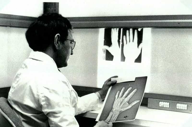


![Updated 2022] My personal customer review of Cosmedica Hair Transplantation in Turkey 2020 - With before and after pictures -...](https://thesurferinvestor.com/wp-content/uploads/2020/09/IMG_6476-min-scaled.jpg)