Contractile Proteins
Myosins
Tropomyosin
Muscle Proteins
Actins
Myofibrils
Myosin Heavy Chains
Troponin
Muscle Contraction
Myosin Light Chains
Escin
Troponin I
Calcium
Actomyosin
Myocardium
Muscle, Smooth
Muscle, Skeletal
Myocytes, Smooth Muscle
Troponin T
Actinin
Muscle Fibers, Skeletal
Postpericardiotomy Syndrome
Calmodulin-Binding Proteins
Mycoplasmatales
Papillary Muscles
Microfilament Proteins
Rabbits
Actin Cytoskeleton
Muscle Fibers, Slow-Twitch
Muscle Fibers, Fast-Twitch
Isometric Contraction
Potassium Chloride
Muscle Development
Cells, Cultured
Coturnix
Caffeine
Adenosine Triphosphatases
Cell Differentiation
Diaphragm
Myogenin
Pyridazines
Sarcoplasmic Reticulum
Muscular Atrophy
Heart Ventricles
Hypertrophy
RNA, Messenger
Halothane
Gene Expression Regulation
Guinea Pigs
Cytoskeleton
Ferrets
Rats, Inbred Strains
Cardiomegaly
Myosin-Light-Chain Kinase
Mesenteric Arteries
Calcium-Binding Proteins
MyoD Protein
Phosphorylation
Microscopy, Electron
Vasoconstriction
Electrophoresis, Polyacrylamide Gel
Phenotype
Mechanotransduction, Cellular
Chick Embryo
Rats, Wistar
Fluorescent Antibody Technique
Myosin Subfragments
Cardiomyopathy, Hypertrophic
Myocytes, Cardiac
Fetus
Trachea
Protein Isoforms
Muscle Strength
Cardiotonic Agents
Myogenic Regulatory Factors
Signal Transduction
Adenosine Triphosphate
Base Sequence
Potassium
Dogs
Rats, Sprague-Dawley
Dose-Response Relationship, Drug
Stress, Mechanical
Calcium-Transporting ATPases
Gene Expression
Chickens
Transcription, Genetic
Molecular Sequence Data
Phenylephrine
Isoenzymes
Cytoskeletal Proteins
Norepinephrine
Organ Specificity
Osmolar Concentration
DNA
Acetylcholine
Mutation
Swine
Biomechanical Phenomena
Aging
Protein Kinase C
Cytoplasm
Blotting, Western
Heart Failure
Cats
Mice, Transgenic
Gene Expression Regulation, Developmental
Immunohistochemistry
Blood Platelets
In Situ Hybridization
Disease Models, Animal
Carrier Proteins
Cyclic AMP
Genes
Transcription Factors
RNA
Amino Acid Sequence
Collagen
Magnesium
Cell Movement
Filament assembly from profilin-actin. (1/1105)
Profilin plays a major role in the assembly of actin filament at the barbed ends. The thermodynamic and kinetic parameters for barbed end assembly from profilin-actin have been measured turbidimetrically. Filament growth from profilin-actin requires MgATP to be bound to actin. No assembly is observed from profilin-CaATP-actin. The rate constant for association of profilin-actin to barbed ends is 30% lower than that of actin, and the critical concentration for F-actin assembly from profilin-actin units is 0.3 microM under physiological ionic conditions. Barbed ends grow from profilin-actin with an ADP-Pi cap. Profilin does not cap the barbed ends and is not detectably incorporated into filaments. The EDC-cross-linked profilin-actin complex (PAcov) both copolymerizes with F-actin and undergoes spontaneous self-assembly, following a nucleation-growth process characterized by a critical concentration of 0.2 microM under physiological conditions. The PAcov polymer is a helical filament that displays the same diffraction pattern as F-actin, with layer lines at 6 and 36 nm. The PAcov filaments bound phalloidin with the same kinetics as F-actin, bound myosin subfragment-1, and supported actin-activated ATPase of myosin subfragment-1, but they did not translocate in vitro along myosin-coated glass surfaces. These results are discussed in light of the current models of actin structure. (+info)The small GTPase RalA targets filamin to induce filopodia. (2/1105)
The Ras-related small GTPases Rac, Rho, Cdc42, and RalA bind filamin, an actin filament-crosslinking protein that also links membrane and other intracellular proteins to actin. Of these GTPases only RalA binds filamin in a GTP-specific manner, and GTP-RalA elicits actin-rich filopods on surfaces of Swiss 3T3 cells and recruits filamin into the filopodial cytoskeleton. Either a dominant negative RalA construct or the RalA-binding domain of filamin 1 specifically block Cdc42-induced filopod formation, but a Cdc42 inhibitor does not impair RalA's effects, which, unlike Cdc42, are Rac independent. RalA does not generate filopodia in filamin-deficient human melanoma cells, whereas transfection of filamin 1 restores the functional response. RalA therefore is a downstream intermediate in Cdc42-mediated filopod production and uses filamin in this pathway. (+info)Profilin and the Abl tyrosine kinase are required for motor axon outgrowth in the Drosophila embryo. (3/1105)
The ability of neuronal growth cones to be guided by extracellular cues requires intimate communication between signal transduction systems and the dynamic actin-based cytoskeleton at the leading edge. Profilin, a small, actin-binding protein, has been proposed to be a regulator of the cell motility machinery at leading edge membranes. However, its requirement in the developing nervous system has been unknown. Profilin associates with members of the Enabled family of proteins, suggesting that Profilin might link Abl function to the cytoskeleton. Here, genetic analysis in Drosophila is used to demonstrate that mutations in Profilin (chickadee) and Abl (abl) display an identical growth cone arrest phenotype for axons of intersegmental nerve b (ISNb). Moreover, the phenotype of a double mutant suggests that these components function together to control axonal outgrowth. (+info)Mena is required for neurulation and commissure formation. (4/1105)
Mammalian enabled (Mena) is a member of a protein family thought to link signal transduction pathways to localized remodeling of the actin cytoskeleton. Mena binds directly to Profilin, an actin-binding protein that modulates actin polymerization. In primary neurons, Mena is concentrated at the tips of growth cone filopodia. Mena-deficient mice are viable; however, axons projecting from interhemispheric cortico-cortical neurons are misrouted in early neonates, and failed decussation of the corpus callosum as well as defects in the hippocampal commissure and the pontocerebellar pathway are evident in the adult. Mena-deficient mice that are heterozygous for a Profilin I deletion die in utero and display defects in neurulation, demonstrating an important functional role for Mena in regulation of the actin cytoskeleton. (+info)Cyclosporine-induced renal artery smooth muscle contraction is associated with increases in the phosphorylation of specific contractile regulatory proteins. (5/1105)
Cyclosporine A (CSA) is a type 2B phosphatase inhibitor which can induce contraction of renal artery smooth muscle. In this investigation, we examined the phosphorylation events associated with CSA-induced contraction of bovine renal artery smooth muscle. Contractile responses were determined in a muscle bath and the corresponding phosphorylation events were determined with whole cell phosphorylation and two-dimensional gel electrophoresis. CSA-induced contractions were associated with increases in the phosphorylation of the 20 kDa myosin light chains (MLC20) and different isoforms of the small heat shock protein, HSP27. Cyclic nucleotide-dependent relaxation of CSA-induced contractions was associated with increases in the phosphorylation of another small heat shock protein, HSP20, and decreases in the phosphorylation of the MLC20, and some isoforms of HSP27. These data suggest that CSA-induced contraction and relaxation of vascular smooth muscle is associated with increases in the phosphorylation of specific contractile regulatory proteins. (+info)Role of proteins of the Ena/VASP family in actin-based motility of Listeria monocytogenes. (6/1105)
Intracellular propulsion of Listeria monocytogenes is the best understood form of motility dependent on actin polymerization. We have used in vitro motility assays of Listeria in platelet and brain extracts to elucidate the function of the focal adhesion proteins of the Ena (Drosophila Enabled)/VASP (vasodilator-stimulated phosphoprotein) family in actin-based motility. Immunodepletion of VASP from platelet extracts and of Evl (Ena/VASP-like protein) from brain extracts of Mena knockout (-/-) mice combined with add-back of recombinant (bacterial or eukaryotic) VASP and Evl show that VASP, Mena, and Evl play interchangeable roles and are required to transform actin polymerization into active movement and propulsive force. The EVH1 (Ena/VASP homology 1) domain of VASP is in slow association-dissociation equilibrium high-affinity binding to the zyxin-homologous, proline-rich region of ActA. VASP also interacts with F-actin via its COOH-terminal EVH2 domain. Hence VASP/ Ena/Evl link the bacterium to the actin tail, which is required for movement. The affinity of VASP for F-actin is controlled by phosphorylation of serine 157 by cAMP-dependent protein kinase. Phospho-VASP binds with high affinity (0.5 x 10(8) M-1); dephospho-VASP binds 40-fold less tightly. We propose a molecular ratchet model for insertional polymerization of actin, within which frequent attachment-detachment of VASP to F-actin allows its sliding along the growing filament. (+info)The quaternary structure of the sheaths of defective phages similar to PBS X. (7/1105)
The contractile sheaths of five defective, PBS X-like bacteriophages from Bacillus subtilis and B. licheniformis were investigated by electron microscopy, dodecylsulphate gel electrophoresis and immunodiffusion. Electron microscope images of the extended and contracted sheaths were of similar appearance, although their lengths were different. The surface lattices of both the extended and the contracted sheaths were determined by optical diffraction. This showed that the quaternary structure of the sheaths of all five defective phages originated from identical surface lattices, which could be approximately expressed by the selection rules L = -2n' + 3m and L = 9N' + 17M for the extended and contracted sheaths respectively, in which 6n' = n with n = 0 or an integer multiple of 6. These results indicated that the packing of the protein subunits in these sheaths differed from those of other bacteriophages, for example T4 and millimicron [Amos and Klug, J. Mol. Biol. 99, 51--73 (1975); Admiraal and Mellema, J. Ultrastruct. Res. 56, 48--64 (1976)]. The molecular weight of the main sheath protein of the defective phages, as determined by dodecylsulphate gel electrophoresis, was approximately 50000. This value differed from that for T4, but was similar to that of millimicron [Admiraal and Mellema, J. Ultrastruct. Res. 56, 48--64 (1976); King and Laemmli, J. Mol. Biol, 75, 315--337 (1973)]. The results of immunodiffusion experiments, however, pointed to a chemical difference between the sheath proteins of the defective phages and millimicron, in addition to T4. (+info)Identification of a suppressor of the Dictyostelium profilin-minus phenotype as a CD36/LIMP-II homologue. (8/1105)
Profilin is an ubiquitous G-actin binding protein in eukaryotic cells. Lack of both profilin isoforms in Dictyostelium discoideum resulted in impaired cytokinesis and an arrest in development. A restriction enzyme-mediated integration approach was applied to profilin-minus cells to identify suppressor mutants for the developmental phenotype. A mutant with wild-type-like development and restored cytokinesis was isolated. The gene affected was found to code for an integral membrane glycoprotein of a predicted size of 88 kD containing two transmembrane domains, one at the NH2 terminus and the other at the COOH terminus. It is homologous to mammalian CD36/LIMP-II and represents the first member of this family in D. discoideum, therefore the name DdLIMP is proposed. Targeted disruption of the lmpA gene in the profilin-minus background also rescued the mutant phenotype. Immunofluorescence revealed a localization in vesicles and ringlike structures on the cell surface. Partially purified DdLIMP bound specifically to PIP2 in sedimentation and gel filtration assays. A direct interaction between DdLIMP and profilin could not be detected, and it is unclear how far upstream in a regulatory cascade DdLIMP might be positioned. However, the PIP2 binding of DdLIMP points towards a function via the phosphatidylinositol pathway, a major regulator of profilin. (+info)Contractile proteins are a type of protein found in muscle cells that are responsible for the ability of the muscle to contract and generate force. The two main types of contractile proteins are actin and myosin, which are arranged in sarcomeres, the functional units of muscle fibers. When stimulated by a nerve impulse, actin and myosin filaments slide past each other, causing the muscle to shorten and generate force. This process is known as excitation-contraction coupling. Other proteins, such as tropomyosin and troponin, regulate the interaction between actin and myosin and control muscle contraction.
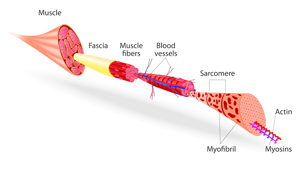
Myosins are a large family of motor proteins that play a crucial role in various cellular processes, including muscle contraction and intracellular transport. They consist of heavy chains, which contain the motor domain responsible for generating force and motion, and light chains, which regulate the activity of the myosin. Based on their structural and functional differences, myosins are classified into over 35 classes, with classes II, V, and VI being the most well-studied.
Class II myosins, also known as conventional myosins, are responsible for muscle contraction in skeletal, cardiac, and smooth muscles. They form filaments called thick filaments, which interact with actin filaments to generate force and movement during muscle contraction.
Class V myosins, also known as unconventional myosins, are involved in intracellular transport and organelle positioning. They have a long tail that can bind to various cargoes, such as vesicles, mitochondria, and nuclei, and a motor domain that moves along actin filaments to transport the cargoes to their destinations.
Class VI myosins are also unconventional myosins involved in intracellular transport and organelle positioning. They have two heads connected by a coiled-coil tail, which can bind to various cargoes. Class VI myosins move along actin filaments in a unique hand-over-hand motion, allowing them to transport their cargoes efficiently.
Overall, myosins are essential for many cellular functions and have been implicated in various diseases, including cardiovascular diseases, neurological disorders, and cancer.
Tropomyosin is a protein that plays a crucial role in muscle contraction. It is a long, thin filamentous protein that runs along the length of actin filaments in muscle cells, forming part of the troponin-tropomyosin complex. This complex regulates the interaction between actin and myosin, which are the other two key proteins involved in muscle contraction.
In a relaxed muscle, tropomyosin blocks the myosin-binding sites on actin, preventing muscle contraction from occurring. When a signal is received to contract, calcium ions are released into the muscle cell, which binds to troponin and causes a conformational change that moves tropomyosin out of the way, exposing the myosin-binding sites on actin. This allows myosin to bind to actin and generate force, leading to muscle contraction.
Tropomyosin is composed of two alpha-helical chains that wind around each other in a coiled-coil structure. There are several isoforms of tropomyosin found in different types of muscle cells, including skeletal, cardiac, and smooth muscle. Mutations in the genes encoding tropomyosin have been associated with various inherited muscle disorders, such as hypertrophic cardiomyopathy and distal arthrogryposis.
Muscle proteins are a type of protein that are found in muscle tissue and are responsible for providing structure, strength, and functionality to muscles. The two major types of muscle proteins are:
1. Contractile proteins: These include actin and myosin, which are responsible for the contraction and relaxation of muscles. They work together to cause muscle movement by sliding along each other and shortening the muscle fibers.
2. Structural proteins: These include titin, nebulin, and desmin, which provide structural support and stability to muscle fibers. Titin is the largest protein in the human body and acts as a molecular spring that helps maintain the integrity of the sarcomere (the basic unit of muscle contraction). Nebulin helps regulate the length of the sarcomere, while desmin forms a network of filaments that connects adjacent muscle fibers together.
Overall, muscle proteins play a critical role in maintaining muscle health and function, and their dysregulation can lead to various muscle-related disorders such as muscular dystrophy, myopathies, and sarcopenia.
Actin is a type of protein that forms part of the contractile apparatus in muscle cells, and is also found in various other cell types. It is a globular protein that polymerizes to form long filaments, which are important for many cellular processes such as cell division, cell motility, and the maintenance of cell shape. In muscle cells, actin filaments interact with another type of protein called myosin to enable muscle contraction. Actins can be further divided into different subtypes, including alpha-actin, beta-actin, and gamma-actin, which have distinct functions and expression patterns in the body.
Myofibrils are the basic contractile units of muscle fibers, composed of highly organized arrays of thick and thin filaments. They are responsible for generating the force necessary for muscle contraction. The thick filaments are primarily made up of the protein myosin, while the thin filaments are mainly composed of actin. Myofibrils are surrounded by a membrane called the sarcolemma and are organized into repeating sections called sarcomeres, which are the functional units of muscle contraction.
Myosin Heavy Chains are the large, essential components of myosin molecules, which are responsible for the molecular motility in muscle cells. These heavy chains have a molecular weight of approximately 200 kDa and form the motor domain of myosin, which binds to actin filaments and hydrolyzes ATP to generate force and movement during muscle contraction. There are several different types of myosin heavy chains, each with specific roles in various tissues and cellular functions. In skeletal and cardiac muscles, for example, myosin heavy chains have distinct isoforms that contribute to the contractile properties of these tissues.
Troponin is a protein complex found in cardiac and skeletal muscle cells that plays a critical role in muscle contraction. It consists of three subunits: troponin C, which binds calcium ions; troponin I, which inhibits the interaction between actin and myosin in the absence of calcium; and troponin T, which binds to tropomyosin and helps anchor the complex to the muscle filament.
In clinical medicine, "troponin" usually refers to cardiac-specific isoforms of these proteins (cTnI and cTnT) that are released into the bloodstream following damage to the heart muscle, such as occurs in myocardial infarction (heart attack). Measurement of troponin levels in the blood is a sensitive and specific biomarker for the diagnosis of acute myocardial infarction.
Muscle contraction is the physiological process in which muscle fibers shorten and generate force, leading to movement or stability of a body part. This process involves the sliding filament theory where thick and thin filaments within the sarcomeres (the functional units of muscles) slide past each other, facilitated by the interaction between myosin heads and actin filaments. The energy required for this action is provided by the hydrolysis of adenosine triphosphate (ATP). Muscle contractions can be voluntary or involuntary, and they play a crucial role in various bodily functions such as locomotion, circulation, respiration, and posture maintenance.
Myosin light chains are regulatory proteins that bind to the myosin head region of myosin molecules, which are involved in muscle contraction. There are two types of myosin light chains, essential and regulatory, that have different functions. The essential light chains are necessary for the assembly and stability of the myosin filaments, while the regulatory light chains control the calcium-sensitive activation of the myosin ATPase activity during muscle contraction. Phosphorylation of the regulatory light chains plays a critical role in regulating muscle contraction and relaxation.
Escin is a saponin mixture derived from the seeds of horse chestnut (Aesculus hippocastanum) trees. It has been used in traditional medicine to treat various conditions, including chronic venous insufficiency and hemorrhoids. Escin has anti-inflammatory, antioxidant, and vasoprotective properties, which contribute to its potential health benefits.
The primary mechanism of action for escin is the stabilization of capillary walls, reducing their permeability and fragility. This can help alleviate symptoms associated with venous insufficiency, such as swelling, pain, and skin changes. Additionally, escin has been shown to inhibit the activity of enzymes involved in inflammation, further contributing to its anti-inflammatory effects.
Escin is available in various forms, including oral supplements, topical creams, and gels. While it is generally considered safe when used as directed, potential side effects may include digestive issues, headaches, and skin irritation. Pregnant or breastfeeding women should consult their healthcare provider before using escin.
A muscle is a soft tissue in our body that contracts to produce force and motion. It is composed mainly of specialized cells called muscle fibers, which are bound together by connective tissue. There are three types of muscles: skeletal (voluntary), smooth (involuntary), and cardiac. Skeletal muscles attach to bones and help in movement, while smooth muscles are found within the walls of organs and blood vessels, helping with functions like digestion and circulation. Cardiac muscle is the specific type that makes up the heart, allowing it to pump blood throughout the body.
Troponin I is a protein that is found in the cardiac muscle cells (myocytes) of the heart. It is a component of the troponin complex, which also includes troponin C and troponin T, that regulates the calcium-mediated interaction between actin and myosin filaments during muscle contraction.
Troponin I is specific to the cardiac muscle tissue, making it a useful biomarker for detecting damage to the heart muscle. When there is injury or damage to the heart muscle cells, such as during a heart attack (myocardial infarction), troponin I is released into the bloodstream.
Measurement of cardiac troponin I levels in the blood is used in the diagnosis and management of acute coronary syndrome (ACS) and other conditions that cause damage to the heart muscle. Elevated levels of troponin I in the blood are indicative of myocardial injury, and the degree of elevation can help determine the severity of the injury.
Myocardial contraction refers to the rhythmic and forceful shortening of heart muscle cells (myocytes) in the myocardium, which is the muscular wall of the heart. This process is initiated by electrical signals generated by the sinoatrial node, causing a wave of depolarization that spreads throughout the heart.
During myocardial contraction, calcium ions flow into the myocytes, triggering the interaction between actin and myosin filaments, which are the contractile proteins in the muscle cells. This interaction causes the myofilaments to slide past each other, resulting in the shortening of the sarcomeres (the functional units of muscle contraction) and ultimately leading to the contraction of the heart muscle.
Myocardial contraction is essential for pumping blood throughout the body and maintaining adequate circulation to vital organs. Any impairment in myocardial contractility can lead to various cardiac disorders, such as heart failure, cardiomyopathy, and arrhythmias.
Calcium is an essential mineral that is vital for various physiological processes in the human body. The medical definition of calcium is as follows:
Calcium (Ca2+) is a crucial cation and the most abundant mineral in the human body, with approximately 99% of it found in bones and teeth. It plays a vital role in maintaining structural integrity, nerve impulse transmission, muscle contraction, hormonal secretion, blood coagulation, and enzyme activation.
Calcium homeostasis is tightly regulated through the interplay of several hormones, including parathyroid hormone (PTH), calcitonin, and vitamin D. Dietary calcium intake, absorption, and excretion are also critical factors in maintaining optimal calcium levels in the body.
Hypocalcemia refers to low serum calcium levels, while hypercalcemia indicates high serum calcium levels. Both conditions can have detrimental effects on various organ systems and require medical intervention to correct.
Actomyosin is a contractile protein complex that consists of actin and myosin filaments. It plays an essential role in muscle contraction, cell motility, and cytokinesis (the process of cell division where the cytoplasm is divided into two daughter cells). The interaction between actin and myosin generates force and movement through a mechanism called sliding filament theory. In this process, myosin heads bind to actin filaments and then undergo a power stroke, which results in the sliding of one filament relative to the other and ultimately leads to muscle contraction or cellular movements. Actomyosin complexes are also involved in various non-muscle cellular processes such as cytoplasmic streaming, intracellular transport, and maintenance of cell shape.
The myocardium is the middle layer of the heart wall, composed of specialized cardiac muscle cells that are responsible for pumping blood throughout the body. It forms the thickest part of the heart wall and is divided into two sections: the left ventricle, which pumps oxygenated blood to the rest of the body, and the right ventricle, which pumps deoxygenated blood to the lungs.
The myocardium contains several types of cells, including cardiac muscle fibers, connective tissue, nerves, and blood vessels. The muscle fibers are arranged in a highly organized pattern that allows them to contract in a coordinated manner, generating the force necessary to pump blood through the heart and circulatory system.
Damage to the myocardium can occur due to various factors such as ischemia (reduced blood flow), infection, inflammation, or genetic disorders. This damage can lead to several cardiac conditions, including heart failure, arrhythmias, and cardiomyopathy.
Cardiac myosins are a type of myosin protein that are specifically expressed in the cardiac muscle cells (or cardiomyocytes) of the heart. These proteins play a crucial role in the contraction and relaxation of heart muscles, which is essential for proper heart function and blood circulation.
Myosins are molecular motors that use chemical energy from ATP to generate force and movement. In the context of cardiac muscle cells, cardiac myosins interact with another protein called actin to form sarcomeres, which are the basic contractile units of muscle fibers. During contraction, the heads of cardiac myosin molecules bind to actin filaments and pull them together, causing the muscle fiber to shorten and generate force.
There are different isoforms of cardiac myosins that can vary in their structure and function. Mutations in the genes encoding these proteins have been linked to various forms of cardiomyopathy, which are diseases of the heart muscle that can lead to heart failure and other complications. Therefore, understanding the structure and function of cardiac myosins is an important area of research for developing therapies and treatments for heart disease.
Smooth muscle, also known as involuntary muscle, is a type of muscle that is controlled by the autonomic nervous system and functions without conscious effort. These muscles are found in the walls of hollow organs such as the stomach, intestines, bladder, and blood vessels, as well as in the eyes, skin, and other areas of the body.
Smooth muscle fibers are shorter and narrower than skeletal muscle fibers and do not have striations or sarcomeres, which give skeletal muscle its striped appearance. Smooth muscle is controlled by the autonomic nervous system through the release of neurotransmitters such as acetylcholine and norepinephrine, which bind to receptors on the smooth muscle cells and cause them to contract or relax.
Smooth muscle plays an important role in many physiological processes, including digestion, circulation, respiration, and elimination. It can also contribute to various medical conditions, such as hypertension, gastrointestinal disorders, and genitourinary dysfunction, when it becomes overactive or underactive.
A sarcomere is the basic contractile unit in a muscle fiber, and it's responsible for generating the force necessary for muscle contraction. It is composed of several proteins, including actin and myosin, which slide past each other to shorten the sarcomere during contraction. The sarcomere extends from one Z-line to the next in a muscle fiber, and it is delimited by the Z-discs where actin filaments are anchored. Sarcomeres play a crucial role in the functioning of skeletal, cardiac, and smooth muscles.
Skeletal muscle myosin, also known as myosin II, is a type of motor protein that plays a crucial role in muscle contraction. It is a hexameric protein composed of two heavy chains and four light chains. The heavy chains have a head region, which contains the ATPase activity and binds to actin filaments, and a tail region, which forms a coiled-coil structure that allows myosin molecules to self-associate into thick filaments.
During muscle contraction, the myosin heads bind to actin filaments in the sarcomere and undergo a power stroke, which results in the sliding of the actin filaments relative to the myosin filaments and thus shortening of the sarcomere. The ATP hydrolysis provides the energy for this power stroke.
Skeletal muscle myosin is essential for generating force and movement in skeletal muscles, and its dysfunction can lead to various muscle diseases and disorders.
Skeletal muscle, also known as striated or voluntary muscle, is a type of muscle that is attached to bones by tendons or aponeuroses and functions to produce movements and support the posture of the body. It is composed of long, multinucleated fibers that are arranged in parallel bundles and are characterized by alternating light and dark bands, giving them a striped appearance under a microscope. Skeletal muscle is under voluntary control, meaning that it is consciously activated through signals from the nervous system. It is responsible for activities such as walking, running, jumping, and lifting objects.
In medical terms, the heart is a muscular organ located in the thoracic cavity that functions as a pump to circulate blood throughout the body. It's responsible for delivering oxygen and nutrients to the tissues and removing carbon dioxide and other wastes. The human heart is divided into four chambers: two atria on the top and two ventricles on the bottom. The right side of the heart receives deoxygenated blood from the body and pumps it to the lungs, while the left side receives oxygenated blood from the lungs and pumps it out to the rest of the body. The heart's rhythmic contractions and relaxations are regulated by a complex electrical conduction system.
Smooth muscle myocytes are specialized cells that make up the contractile portion of non-striated, or smooth, muscles. These muscles are found in various organs and structures throughout the body, including the walls of blood vessels, the digestive system, the respiratory system, and the reproductive system.
Smooth muscle myocytes are smaller than their striated counterparts (skeletal and cardiac muscle cells) and have a single nucleus. They lack the distinctive banding pattern seen in striated muscles and instead have a uniform appearance of actin and myosin filaments. Smooth muscle myocytes are controlled by the autonomic nervous system, which allows them to contract and relax involuntarily.
These cells play an essential role in many physiological processes, such as regulating blood flow, moving food through the digestive tract, and facilitating childbirth. They can also contribute to various pathological conditions, including hypertension, atherosclerosis, and gastrointestinal disorders.
Troponin T is a subunit of the troponin complex, which is a protein complex that plays a crucial role in muscle contraction. In particular, Troponin T is responsible for binding the troponin complex to tropomyosin, another protein that helps regulate muscle contraction.
In the context of medical diagnostics, Troponin T is often measured as a biomarker for heart damage. When heart muscle cells are damaged or die, such as in a myocardial infarction (heart attack), troponin T is released into the bloodstream. Therefore, measuring the levels of Troponin T in the blood can help diagnose and assess the severity of heart damage.
It's important to note that Troponin T is specific to cardiac muscle cells, which makes it a more reliable biomarker for heart damage than other markers that may also be found in skeletal muscle cells. However, it's worth noting that Troponin T levels can also be elevated in conditions other than heart attacks, such as heart failure, myocarditis, and pulmonary embolism, so clinical context is important when interpreting test results.
Actinin is a protein that belongs to the family of actin-binding proteins. It plays an important role in the organization and stability of the cytoskeleton, which is the structural framework of a cell. Specifically, actinin crosslinks actin filaments into bundles or networks, providing strength and rigidity to the cell structure. There are several isoforms of actinin, with alpha-actinin and gamma-actinin being widely studied. Alpha-actinin is found in the Z-discs of sarcomeres in muscle cells, where it helps anchor actin filaments and maintains the structural integrity of the muscle. Gamma-actinin is primarily located at cell-cell junctions and participates in cell adhesion and signaling processes.
A smooth muscle within the vascular system refers to the involuntary, innervated muscle that is found in the walls of blood vessels. These muscles are responsible for controlling the diameter of the blood vessels, which in turn regulates blood flow and blood pressure. They are called "smooth" muscles because their individual muscle cells do not have the striations, or cross-striped patterns, that are observed in skeletal and cardiac muscle cells. Smooth muscle in the vascular system is controlled by the autonomic nervous system and by hormones, and can contract or relax slowly over a period of time.
Skeletal muscle fibers, also known as striated muscle fibers, are the type of muscle cells that make up skeletal muscles, which are responsible for voluntary movements of the body. These muscle fibers are long, cylindrical, and multinucleated, meaning they contain multiple nuclei. They are surrounded by a connective tissue layer called the endomysium, and many fibers are bundled together into fascicles, which are then surrounded by another layer of connective tissue called the perimysium.
Skeletal muscle fibers are composed of myofibrils, which are long, thread-like structures that run the length of the fiber. Myofibrils contain repeating units called sarcomeres, which are responsible for the striated appearance of skeletal muscle fibers. Sarcomeres are composed of thick and thin filaments, which slide past each other during muscle contraction to shorten the sarcomere and generate force.
Skeletal muscle fibers can be further classified into two main types based on their contractile properties: slow-twitch (type I) and fast-twitch (type II). Slow-twitch fibers have a high endurance capacity and are used for sustained, low-intensity activities such as maintaining posture. Fast-twitch fibers, on the other hand, have a higher contractile speed and force generation capacity but fatigue more quickly and are used for powerful, explosive movements.
Postpericardiotomy Syndrome (PPS) is a clinical entity that can occur after cardiac surgical procedures. It is characterized by the presence of pericardial effusion, pleural effusion, and/or inflammation of the serosal surfaces lining the heart and chest cavity (pericardium and pleura). The symptoms typically develop within 1-6 weeks after surgery and include fever, chest pain, and signs of fluid accumulation in the pericardial or pleural spaces.
The exact cause of PPS is not fully understood, but it is thought to be related to an immune response to the surgical trauma, leading to inflammation and increased production of cytokines and other mediators. The diagnosis of PPS is typically made based on clinical criteria, including the presence of fever, pleural or pericardial effusion, and evidence of inflammation. Treatment may include nonsteroidal anti-inflammatory drugs (NSAIDs), corticosteroids, or colchicine to reduce inflammation and relieve symptoms. In severe cases, drainage of the effusions may be necessary.
Calmodulin-binding proteins are a diverse group of proteins that have the ability to bind to calmodulin, a ubiquitous calcium-binding protein found in eukaryotic cells. Calmodulin plays a critical role in various cellular processes by regulating the activity of its target proteins in a calcium-dependent manner.
Calmodulin-binding proteins contain specific domains or motifs that enable them to interact with calmodulin. These domains can be classified into two main categories: IQ motifs and CaM motifs. The IQ motif is a short amino acid sequence that contains the consensus sequence IQXXXRGXXR, where X represents any amino acid. This motif binds to the C-lobe of calmodulin in a calcium-dependent manner. On the other hand, CaM motifs are longer sequences that can bind to both lobes of calmodulin with high affinity and in a calcium-dependent manner.
Calmodulin-binding proteins play crucial roles in various cellular functions, including signal transduction, gene regulation, cytoskeleton organization, and ion channel regulation. For example, calmodulin-binding proteins such as calcineurin and CaM kinases are involved in the regulation of immune responses, learning, and memory. Similarly, myosin regulatory light chains, which contain IQ motifs, play a critical role in muscle contraction by regulating the interaction between actin and myosin filaments.
In summary, calmodulin-binding proteins are a diverse group of proteins that interact with calmodulin to regulate various cellular processes. They contain specific domains or motifs that enable them to bind to calmodulin in a calcium-dependent manner, thereby modulating the activity of their target proteins.
Mycoplasmatales is an order of bacteria that lack a cell wall and are characterized by their small size and simple genome. They are commonly found in various environments, including the human body, where they can be part of the normal flora or associated with diseases. The order Mycoplasmatales contains several genera, including Mycoplasma, Ureaplasma, and Acholeplasma, among others. These bacteria can cause a variety of infections, such as respiratory tract infections, urinary tract infections, and sexually transmitted diseases. Due to their small size and lack of a cell wall, they can be resistant to many antibiotics, making them difficult to treat in some cases.
Papillary muscles are specialized muscle structures located in the heart, specifically in the ventricles (the lower chambers of the heart). They are attached to the tricuspid and mitral valves' leaflets via tendinous cords, also known as chordae tendineae. The main function of papillary muscles is to prevent the backflow of blood during contraction by providing tension to the valve leaflets through these tendinous cords.
There are two sets of papillary muscles in the heart:
1. Anterior and posterior papillary muscles in the left ventricle, which are attached to the mitral (bicuspid) valve.
2. Three smaller papillary muscles in the right ventricle, which are attached to the tricuspid valve.
These muscle structures play a crucial role in maintaining proper blood flow through the heart and ensuring efficient cardiac function.
Microfilament proteins are a type of structural protein that form part of the cytoskeleton in eukaryotic cells. They are made up of actin monomers, which polymerize to form long, thin filaments. These filaments are involved in various cellular processes such as muscle contraction, cell division, and cell motility. Microfilament proteins also interact with other cytoskeletal components like intermediate filaments and microtubules to maintain the overall shape and integrity of the cell. Additionally, they play a crucial role in the formation of cell-cell junctions and cell-matrix adhesions, which are essential for tissue structure and function.
I believe there may be some confusion in your question. "Rabbits" is a common name used to refer to the Lagomorpha species, particularly members of the family Leporidae. They are small mammals known for their long ears, strong legs, and quick reproduction.
However, if you're referring to "rabbits" in a medical context, there is a term called "rabbit syndrome," which is a rare movement disorder characterized by repetitive, involuntary movements of the fingers, resembling those of a rabbit chewing. It is also known as "finger-chewing chorea." This condition is usually associated with certain medications, particularly antipsychotics, and typically resolves when the medication is stopped or adjusted.
Arthrogryposis is a medical term that describes a condition characterized by the presence of multiple joint contractures at birth. A contracture occurs when the range of motion in a joint is limited, making it difficult or impossible to move the joint through its full range of motion. In arthrogryposis, these contractures are present in two or more areas of the body.
The term "arthrogryposis" comes from two Greek words: "arthro," meaning joint, and "gyros," meaning curved or bent. Therefore, arthrogryposis literally means "curving of the joints."
There are many different types of arthrogryposis, each with its own specific set of symptoms and causes. However, in general, arthrogryposis is caused by decreased fetal movement during pregnancy, which can be due to a variety of factors such as genetic mutations, nervous system abnormalities, or environmental factors that restrict fetal movement.
Treatment for arthrogryposis typically involves a combination of physical therapy, bracing, and surgery to help improve joint mobility and function. The prognosis for individuals with arthrogryposis varies depending on the severity and type of contractures present, as well as the underlying cause of the condition.
Smooth muscle myosin is a type of motor protein that is responsible for the contraction and relaxation of smooth muscles, which are found in various organs such as the bladder, blood vessels, and digestive tract. Smooth muscle myosin is composed of two heavy chains and four light chains, forming a hexameric structure. The heavy chains have an N-terminal head domain that contains the ATPase activity and a C-terminal tail domain that mediates filament assembly.
The smooth muscle myosin molecule has several unique features compared to other types of myosins, such as skeletal or cardiac myosin. For example, smooth muscle myosin has a longer lever arm, which allows for greater force generation during contraction. Additionally, the regulatory mechanism of smooth muscle myosin is different from that of skeletal or cardiac myosin. In smooth muscles, the contractile activity is regulated by phosphorylation of the light chains, which is mediated by a specific kinase called myosin light chain kinase (MLCK).
Overall, the proper regulation and function of smooth muscle myosin are critical for maintaining normal physiological functions in various organs. Dysregulation or mutations in smooth muscle myosin can lead to several diseases, such as hypertension, atherosclerosis, and gastrointestinal motility disorders.
The actin cytoskeleton is a complex, dynamic network of filamentous (threadlike) proteins that provides structural support and shape to cells, allows for cell movement and division, and plays a role in intracellular transport. Actin filaments are composed of actin monomers that polymerize to form long, thin fibers. These filaments can be organized into different structures, such as stress fibers, which provide tension and support, or lamellipodia and filopodia, which are involved in cell motility. The actin cytoskeleton is constantly remodeling in response to various intracellular and extracellular signals, allowing for changes in cell shape and behavior.
Slow-twitch muscle fibers, also known as type I muscle fibers, are specialized skeletal muscle cells that contract relatively slowly and generate less force than fast-twitch fibers. However, they can maintain contraction for longer periods of time and have a higher resistance to fatigue. These fibers primarily use oxygen and aerobic metabolism to produce energy, making them highly efficient during prolonged, lower-intensity activities such as long-distance running or cycling. Slow-twitch muscle fibers also have an abundant blood supply, which allows for efficient delivery of oxygen and removal of waste products.
Fast-twitch muscle fibers, also known as type II fibers, are a type of skeletal muscle fiber that are characterized by their rapid contraction and relaxation rates. These fibers have a larger diameter and contain a higher concentration of glycogen, which serves as a quick source of energy for muscle contractions. Fast-twitch fibers are further divided into two subcategories: type IIa and type IIb (or type IIx). Type IIa fibers have a moderate amount of mitochondria and can utilize both aerobic and anaerobic metabolic pathways, making them fatigue-resistant. Type IIb fibers, on the other hand, have fewer mitochondria and primarily use anaerobic metabolism, leading to faster fatigue. Fast-twitch fibers are typically used in activities that require quick, powerful movements such as sprinting or weightlifting.
Isometric contraction is a type of muscle activation where the muscle contracts without any change in the length of the muscle or movement at the joint. This occurs when the force generated by the muscle matches the external force opposing it, resulting in a balanced state with no visible movement. It is commonly experienced during activities such as holding a heavy object in static position or trying to push against an immovable object. Isometric contractions are important in maintaining posture and providing stability to joints.
Potassium chloride is an essential electrolyte that is often used in medical settings as a medication. It's a white, crystalline salt that is highly soluble in water and has a salty taste. In the body, potassium chloride plays a crucial role in maintaining fluid and electrolyte balance, nerve function, and muscle contraction.
Medically, potassium chloride is commonly used to treat or prevent low potassium levels (hypokalemia) in the blood. Hypokalemia can occur due to various reasons such as certain medications, kidney diseases, vomiting, diarrhea, or excessive sweating. Potassium chloride is available in various forms, including tablets, capsules, and liquids, and it's usually taken by mouth.
It's important to note that potassium chloride should be used with caution and under the supervision of a healthcare provider, as high levels of potassium (hyperkalemia) can be harmful and even life-threatening. Hyperkalemia can cause symptoms such as muscle weakness, irregular heartbeat, and cardiac arrest.
Muscle development, also known as muscle hypertrophy, refers to the increase in size and mass of the muscles through a process called myofiber growth. This is primarily achieved through resistance or strength training exercises that cause micro-tears in the muscle fibers, leading to an inflammatory response and the release of hormones that promote muscle growth. As the muscles repair themselves, they become larger and stronger than before. Proper nutrition, including adequate protein intake, and rest are also essential components of muscle development.
It is important to note that while muscle development can lead to an increase in strength and muscular endurance, it does not necessarily result in improved athletic performance or overall fitness. A well-rounded exercise program that includes cardiovascular activity, flexibility training, and resistance exercises is recommended for optimal health and fitness outcomes.
"Cells, cultured" is a medical term that refers to cells that have been removed from an organism and grown in controlled laboratory conditions outside of the body. This process is called cell culture and it allows scientists to study cells in a more controlled and accessible environment than they would have inside the body. Cultured cells can be derived from a variety of sources, including tissues, organs, or fluids from humans, animals, or cell lines that have been previously established in the laboratory.
Cell culture involves several steps, including isolation of the cells from the tissue, purification and characterization of the cells, and maintenance of the cells in appropriate growth conditions. The cells are typically grown in specialized media that contain nutrients, growth factors, and other components necessary for their survival and proliferation. Cultured cells can be used for a variety of purposes, including basic research, drug development and testing, and production of biological products such as vaccines and gene therapies.
It is important to note that cultured cells may behave differently than they do in the body, and results obtained from cell culture studies may not always translate directly to human physiology or disease. Therefore, it is essential to validate findings from cell culture experiments using additional models and ultimately in clinical trials involving human subjects.
"Coturnix" is a genus of birds that includes several species of quails. The most common species is the Common Quail (Coturnix coturnix), which is also known as the European Quail or the Eurasian Quail. This small ground-dwelling bird is found throughout Europe, Asia, and parts of Africa, and it is known for its distinctive call and its migratory habits. Other species in the genus Coturnix include the Rain Quail (Coturnix coromandelica), the Stubble Quail (Coturnix pectoralis), and the Harlequin Quail (Coturnix delegorguei). These birds are all similar in appearance and behavior, with small, round bodies, short wings, and strong legs that are adapted for running and scratching in leaf litter. They are also known for their cryptic coloration, which helps them blend in with their surroundings and avoid predators. Quails are popular game birds and are also kept as pets and for ornamental purposes in some parts of the world.
Caffeine is a central nervous system stimulant that occurs naturally in the leaves, seeds, or fruits of some plants. It can also be produced artificially and added to various products, such as food, drinks, and medications. Caffeine has a number of effects on the body, including increasing alertness, improving mood, and boosting energy levels.
In small doses, caffeine is generally considered safe for most people. However, consuming large amounts of caffeine can lead to negative side effects, such as restlessness, insomnia, rapid heart rate, and increased blood pressure. It is also possible to become dependent on caffeine, and withdrawal symptoms can occur if consumption is suddenly stopped.
Caffeine is found in a variety of products, including coffee, tea, chocolate, energy drinks, and some medications. The amount of caffeine in these products can vary widely, so it is important to pay attention to serving sizes and labels to avoid consuming too much.
Adenosine triphosphatases (ATPases) are a group of enzymes that catalyze the conversion of adenosine triphosphate (ATP) into adenosine diphosphate (ADP) and inorganic phosphate. This reaction releases energy, which is used to drive various cellular processes such as muscle contraction, transport of ions across membranes, and synthesis of proteins and nucleic acids.
ATPases are classified into several types based on their structure, function, and mechanism of action. Some examples include:
1. P-type ATPases: These ATPases form a phosphorylated intermediate during the reaction cycle and are involved in the transport of ions across membranes, such as the sodium-potassium pump and calcium pumps.
2. F-type ATPases: These ATPases are found in mitochondria, chloroplasts, and bacteria, and are responsible for generating a proton gradient across the membrane, which is used to synthesize ATP.
3. V-type ATPases: These ATPases are found in vacuolar membranes and endomembranes, and are involved in acidification of intracellular compartments.
4. A-type ATPases: These ATPases are found in the plasma membrane and are involved in various functions such as cell signaling and ion transport.
Overall, ATPases play a crucial role in maintaining the energy balance of cells and regulating various physiological processes.
Cell differentiation is the process by which a less specialized cell, or stem cell, becomes a more specialized cell type with specific functions and structures. This process involves changes in gene expression, which are regulated by various intracellular signaling pathways and transcription factors. Differentiation results in the development of distinct cell types that make up tissues and organs in multicellular organisms. It is a crucial aspect of embryonic development, tissue repair, and maintenance of homeostasis in the body.
A diaphragm is a thin, dome-shaped muscle that separates the chest cavity from the abdominal cavity. It plays a vital role in the process of breathing as it contracts and flattens to draw air into the lungs (inhalation) and relaxes and returns to its domed shape to expel air out of the lungs (exhalation).
In addition, a diaphragm is also a type of barrier method of birth control. It is a flexible dome-shaped device made of silicone that fits over the cervix inside the vagina. When used correctly and consistently, it prevents sperm from entering the uterus and fertilizing an egg, thereby preventing pregnancy.
Myogenin is defined as a protein that belongs to the family of myogenic regulatory factors (MRFs). These proteins play crucial roles in the development, growth, and repair of skeletal muscle cells. Myogenin is specifically involved in the differentiation and fusion of myoblasts to form multinucleated myotubes, which are essential for the formation of mature skeletal muscle fibers. It functions as a transcription factor that binds to specific DNA sequences, thereby regulating the expression of genes required for muscle cell differentiation. Myogenin also plays a role in maintaining muscle homeostasis and may contribute to muscle regeneration following injury or disease.
I apologize for any confusion, but "Pyridazines" is not a medical term. It is a chemical term that refers to a class of heterocyclic organic compounds which contain a six-membered ring with two nitrogen atoms. These types of compounds are often used in the synthesis of various pharmaceuticals and agrochemicals, but "Pyridazines" itself is not a medical concept or diagnosis. If you have any questions related to medicine or health, I would be happy to try to help answer those for you.
The sarcoplasmic reticulum (SR) is a specialized type of smooth endoplasmic reticulum found in muscle cells, particularly in striated muscles such as skeletal and cardiac muscles. It is a complex network of tubules that surrounds the myofibrils, the contractile elements of the muscle fiber.
The primary function of the sarcoplasmic reticulum is to store calcium ions (Ca2+) and regulate their release during muscle contraction and uptake during muscle relaxation. The SR contains a high concentration of calcium-binding proteins, such as calsequestrin, which help to maintain this storage.
The release of calcium ions from the sarcoplasmic reticulum is triggered by an action potential that travels along the muscle fiber's sarcolemma and into the muscle fiber's interior (the sarcoplasm). This action potential causes the voltage-gated calcium channels in the SR membrane, known as ryanodine receptors, to open, releasing Ca2+ ions into the sarcoplasm.
The increased concentration of Ca2+ ions in the sarcoplasm triggers muscle contraction by binding to troponin, a protein associated with actin filaments, causing a conformational change that exposes the active sites on actin for myosin heads to bind and generate force.
After muscle contraction, the calcium ions must be actively transported back into the sarcoplasmic reticulum by Ca2+ ATPase pumps, also known as sarco(endo)plasmic reticulum calcium ATPases (SERCAs). This process helps to lower the concentration of Ca2+ in the sarcoplasm and allows the muscle fiber to relax.
Overall, the sarcoplasmic reticulum plays a crucial role in excitation-contraction coupling, the process by which action potentials trigger muscle contraction.
Muscular atrophy is a condition characterized by a decrease in the size and mass of muscles due to lack of use, disease, or injury. This occurs when there is a disruption in the balance between muscle protein synthesis and degradation, leading to a net loss of muscle proteins. There are two main types of muscular atrophy:
1. Disuse atrophy: This type of atrophy occurs when muscles are not used or are immobilized for an extended period, such as after an injury, surgery, or prolonged bed rest. In this case, the nerves that control the muscles may still be functioning properly, but the muscles themselves waste away due to lack of use.
2. Neurogenic atrophy: This type of atrophy is caused by damage to the nerves that supply the muscles, leading to muscle weakness and wasting. Conditions such as amyotrophic lateral sclerosis (ALS), spinal cord injuries, and peripheral neuropathies can cause neurogenic atrophy.
In both cases, the affected muscles may become weak, shrink in size, and lose their tone and mass. Treatment for muscular atrophy depends on the underlying cause and may include physical therapy, exercise, and medication to manage symptoms and improve muscle strength and function.
The heart ventricles are the two lower chambers of the heart that receive blood from the atria and pump it to the lungs or the rest of the body. The right ventricle pumps deoxygenated blood to the lungs, while the left ventricle pumps oxygenated blood to the rest of the body. Both ventricles have thick, muscular walls to generate the pressure necessary to pump blood through the circulatory system.
Hypertrophy, in the context of physiology and pathology, refers to an increase in the size of an organ or tissue due to an enlargement of its constituent cells. It is often used to describe the growth of muscle cells (myocytes) in response to increased workload or hormonal stimulation, resulting in an increase in muscle mass. However, hypertrophy can also occur in other organs such as the heart (cardiac hypertrophy) in response to high blood pressure or valvular heart disease.
It is important to note that while hypertrophy involves an increase in cell size, hyperplasia refers to an increase in cell number. In some cases, both hypertrophy and hyperplasia can occur together, leading to a significant increase in the overall size and function of the organ or tissue.
Messenger RNA (mRNA) is a type of RNA (ribonucleic acid) that carries genetic information copied from DNA in the form of a series of three-base code "words," each of which specifies a particular amino acid. This information is used by the cell's machinery to construct proteins, a process known as translation. After being transcribed from DNA, mRNA travels out of the nucleus to the ribosomes in the cytoplasm where protein synthesis occurs. Once the protein has been synthesized, the mRNA may be degraded and recycled. Post-transcriptional modifications can also occur to mRNA, such as alternative splicing and addition of a 5' cap and a poly(A) tail, which can affect its stability, localization, and translation efficiency.
Halothane is a general anesthetic agent, which is a volatile liquid that evaporates easily and can be inhaled. It is used to produce and maintain general anesthesia (a state of unconsciousness) during surgical procedures. Halothane is known for its rapid onset and offset of action, making it useful for both induction and maintenance of anesthesia.
The medical definition of Halothane is:
Halothane (2-bromo-2-chloro-1,1,1-trifluoroethane) is a volatile liquid general anesthetic agent with a mild, sweet odor. It is primarily used for the induction and maintenance of general anesthesia in surgical procedures due to its rapid onset and offset of action. Halothane is administered via inhalation and acts by depressing the central nervous system, leading to a reversible loss of consciousness and analgesia.
It's important to note that Halothane has been associated with rare cases of severe liver injury (hepatotoxicity) and anaphylaxis (a severe, life-threatening allergic reaction). These risks have led to the development and use of alternative general anesthetic agents with better safety profiles.
'Gene expression regulation' refers to the processes that control whether, when, and where a particular gene is expressed, meaning the production of a specific protein or functional RNA encoded by that gene. This complex mechanism can be influenced by various factors such as transcription factors, chromatin remodeling, DNA methylation, non-coding RNAs, and post-transcriptional modifications, among others. Proper regulation of gene expression is crucial for normal cellular function, development, and maintaining homeostasis in living organisms. Dysregulation of gene expression can lead to various diseases, including cancer and genetic disorders.
I must clarify that the term "Guinea Pigs" is not typically used in medical definitions. However, in colloquial or informal language, it may refer to people who are used as the first to try out a new medical treatment or drug. This is known as being a "test subject" or "in a clinical trial."
In the field of scientific research, particularly in studies involving animals, guinea pigs are small rodents that are often used as experimental subjects due to their size, cost-effectiveness, and ease of handling. They are not actually pigs from Guinea, despite their name's origins being unclear. However, they do not exactly fit the description of being used in human medical experiments.
The cytoskeleton is a complex network of various protein filaments that provides structural support, shape, and stability to the cell. It plays a crucial role in maintaining cellular integrity, intracellular organization, and enabling cell movement. The cytoskeleton is composed of three major types of protein fibers: microfilaments (actin filaments), intermediate filaments, and microtubules. These filaments work together to provide mechanical support, participate in cell division, intracellular transport, and help maintain the cell's architecture. The dynamic nature of the cytoskeleton allows cells to adapt to changing environmental conditions and respond to various stimuli.
A ferret is a domesticated mammal that belongs to the weasel family, Mustelidae. The scientific name for the common ferret is Mustela putorius furo. Ferrets are native to Europe and have been kept as pets for thousands of years due to their playful and curious nature. They are small animals, typically measuring between 13-20 inches in length, including their tail, and weighing between 1.5-4 pounds.
Ferrets have a slender body with short legs, a long neck, and a pointed snout. They have a thick coat of fur that can vary in color from white to black, with many different patterns in between. Ferrets are known for their high level of activity and intelligence, and they require regular exercise and mental stimulation to stay healthy and happy.
Ferrets are obligate carnivores, which means that they require a diet that is high in protein and low in carbohydrates. They have a unique digestive system that allows them to absorb nutrients efficiently from their food, but it also means that they are prone to certain health problems if they do not receive proper nutrition.
Ferrets are social animals and typically live in groups. They communicate with each other using a variety of vocalizations, including barks, chirps, and purrs. Ferrets can be trained to use a litter box and can learn to perform simple tricks. With proper care and attention, ferrets can make loving and entertaining pets.
"Inbred strains of rats" are genetically identical rodents that have been produced through many generations of brother-sister mating. This results in a high degree of homozygosity, where the genes at any particular locus in the genome are identical in all members of the strain.
Inbred strains of rats are widely used in biomedical research because they provide a consistent and reproducible genetic background for studying various biological phenomena, including the effects of drugs, environmental factors, and genetic mutations on health and disease. Additionally, inbred strains can be used to create genetically modified models of human diseases by introducing specific mutations into their genomes.
Some commonly used inbred strains of rats include the Wistar Kyoto (WKY), Sprague-Dawley (SD), and Fischer 344 (F344) rat strains. Each strain has its own unique genetic characteristics, making them suitable for different types of research.
Cardiomegaly is a medical term that refers to an enlarged heart. It can be caused by various conditions such as high blood pressure, heart valve problems, cardiomyopathy, or fluid accumulation around the heart (pericardial effusion). Cardiomegaly can be detected through imaging tests like chest X-rays or echocardiograms. Depending on the underlying cause, treatment options may include medications, lifestyle changes, or in some cases, surgery. It is important to consult with a healthcare professional for proper diagnosis and treatment.
Myosin-Light-Chain Kinase (MLCK) is an enzyme that plays a crucial role in muscle contraction. It phosphorylates the regulatory light chains of myosin, a protein involved in muscle contraction, leading to the activation of myosin and the initiation of the contractile process. MLCK is activated by calcium ions and calmodulin, and its activity is essential for various cellular processes, including cytokinesis, cell motility, and maintenance of cell shape. In addition to its role in muscle contraction, MLCK has been implicated in several pathological conditions, such as hypertension, atherosclerosis, and cancer.
The mesenteric arteries are the arteries that supply oxygenated blood to the intestines. There are three main mesenteric arteries: the superior mesenteric artery, which supplies blood to the small intestine (duodenum to two-thirds of the transverse colon) and large intestine (cecum, ascending colon, and the first part of the transverse colon); the inferior mesenteric artery, which supplies blood to the distal third of the transverse colon, descending colon, sigmoid colon, and rectum; and the middle colic artery, which is a branch of the superior mesenteric artery that supplies blood to the transverse colon. These arteries are important in maintaining adequate blood flow to the intestines to support digestion and absorption of nutrients.
Calcium-binding proteins (CaBPs) are a diverse group of proteins that have the ability to bind calcium ions (Ca^2+^) with high affinity and specificity. They play crucial roles in various cellular processes, including signal transduction, muscle contraction, neurotransmitter release, and protection against oxidative stress.
The binding of calcium ions to these proteins induces conformational changes that can either activate or inhibit their functions. Some well-known CaBPs include calmodulin, troponin C, S100 proteins, and parvalbumins. These proteins are essential for maintaining calcium homeostasis within cells and for mediating the effects of calcium as a second messenger in various cellular signaling pathways.
MyoD protein is a member of the family of muscle regulatory factors (MRFs) that play crucial roles in the development and regulation of skeletal muscle. MyoD is a transcription factor, which means it binds to specific DNA sequences and helps control the transcription of nearby genes into messenger RNA (mRNA).
MyoD protein is encoded by the MYOD1 gene and is primarily expressed in skeletal muscle cells, where it functions as a master regulator of muscle differentiation. During myogenesis, MyoD is activated and initiates the expression of various genes involved in muscle-specific functions, such as contractile proteins and ion channels.
MyoD protein can also induce cell cycle arrest and promote the differentiation of non-muscle cells into muscle cells, a process known as transdifferentiation. This property has been explored in regenerative medicine for potential therapeutic applications.
In summary, MyoD protein is a key regulator of skeletal muscle development, differentiation, and maintenance, and it plays essential roles in the regulation of gene expression during myogenesis.
Phosphorylation is the process of adding a phosphate group (a molecule consisting of one phosphorus atom and four oxygen atoms) to a protein or other organic molecule, which is usually done by enzymes called kinases. This post-translational modification can change the function, localization, or activity of the target molecule, playing a crucial role in various cellular processes such as signal transduction, metabolism, and regulation of gene expression. Phosphorylation is reversible, and the removal of the phosphate group is facilitated by enzymes called phosphatases.
Electron microscopy (EM) is a type of microscopy that uses a beam of electrons to create an image of the sample being examined, resulting in much higher magnification and resolution than light microscopy. There are several types of electron microscopy, including transmission electron microscopy (TEM), scanning electron microscopy (SEM), and reflection electron microscopy (REM).
In TEM, a beam of electrons is transmitted through a thin slice of the sample, and the electrons that pass through the sample are focused to form an image. This technique can provide detailed information about the internal structure of cells, viruses, and other biological specimens, as well as the composition and structure of materials at the atomic level.
In SEM, a beam of electrons is scanned across the surface of the sample, and the electrons that are scattered back from the surface are detected to create an image. This technique can provide information about the topography and composition of surfaces, as well as the structure of materials at the microscopic level.
REM is a variation of SEM in which the beam of electrons is reflected off the surface of the sample, rather than scattered back from it. This technique can provide information about the surface chemistry and composition of materials.
Electron microscopy has a wide range of applications in biology, medicine, and materials science, including the study of cellular structure and function, disease diagnosis, and the development of new materials and technologies.
Vasoconstriction is a medical term that refers to the narrowing of blood vessels due to the contraction of the smooth muscle in their walls. This process decreases the diameter of the lumen (the inner space of the blood vessel) and reduces blood flow through the affected vessels. Vasoconstriction can occur throughout the body, but it is most noticeable in the arterioles and precapillary sphincters, which control the amount of blood that flows into the capillary network.
The autonomic nervous system, specifically the sympathetic division, plays a significant role in regulating vasoconstriction through the release of neurotransmitters like norepinephrine (noradrenaline). Various hormones and chemical mediators, such as angiotensin II, endothelin-1, and serotonin, can also induce vasoconstriction.
Vasoconstriction is a vital physiological response that helps maintain blood pressure and regulate blood flow distribution in the body. However, excessive or prolonged vasoconstriction may contribute to several pathological conditions, including hypertension, stroke, and peripheral vascular diseases.
Electrophoresis, polyacrylamide gel (EPG) is a laboratory technique used to separate and analyze complex mixtures of proteins or nucleic acids (DNA or RNA) based on their size and electrical charge. This technique utilizes a matrix made of cross-linked polyacrylamide, a type of gel, which provides a stable and uniform environment for the separation of molecules.
In this process:
1. The polyacrylamide gel is prepared by mixing acrylamide monomers with a cross-linking agent (bis-acrylamide) and a catalyst (ammonium persulfate) in the presence of a buffer solution.
2. The gel is then poured into a mold and allowed to polymerize, forming a solid matrix with uniform pore sizes that depend on the concentration of acrylamide used. Higher concentrations result in smaller pores, providing better resolution for separating smaller molecules.
3. Once the gel has set, it is placed in an electrophoresis apparatus containing a buffer solution. Samples containing the mixture of proteins or nucleic acids are loaded into wells on the top of the gel.
4. An electric field is applied across the gel, causing the negatively charged molecules to migrate towards the positive electrode (anode) while positively charged molecules move toward the negative electrode (cathode). The rate of migration depends on the size, charge, and shape of the molecules.
5. Smaller molecules move faster through the gel matrix and will migrate farther from the origin compared to larger molecules, resulting in separation based on size. Proteins and nucleic acids can be selectively stained after electrophoresis to visualize the separated bands.
EPG is widely used in various research fields, including molecular biology, genetics, proteomics, and forensic science, for applications such as protein characterization, DNA fragment analysis, cloning, mutation detection, and quality control of nucleic acid or protein samples.
A phenotype is the physical or biochemical expression of an organism's genes, or the observable traits and characteristics resulting from the interaction of its genetic constitution (genotype) with environmental factors. These characteristics can include appearance, development, behavior, and resistance to disease, among others. Phenotypes can vary widely, even among individuals with identical genotypes, due to differences in environmental influences, gene expression, and genetic interactions.
Cellular mechanotransduction is the process by which cells convert mechanical stimuli into biochemical signals, resulting in changes in cell behavior and function. This complex process involves various molecular components, including transmembrane receptors, ion channels, cytoskeletal proteins, and signaling molecules. Mechanical forces such as tension, compression, or fluid flow can activate these components, leading to alterations in gene expression, protein synthesis, and cell shape or movement. Cellular mechanotransduction plays a crucial role in various physiological processes, including tissue development, homeostasis, and repair, as well as in pathological conditions such as fibrosis and cancer progression.
"Newborn animals" refers to the very young offspring of animals that have recently been born. In medical terminology, newborns are often referred to as "neonates," and they are classified as such from birth until about 28 days of age. During this time period, newborn animals are particularly vulnerable and require close monitoring and care to ensure their survival and healthy development.
The specific needs of newborn animals can vary widely depending on the species, but generally, they require warmth, nutrition, hydration, and protection from harm. In many cases, newborns are unable to regulate their own body temperature or feed themselves, so they rely heavily on their mothers for care and support.
In medical settings, newborn animals may be examined and treated by veterinarians to ensure that they are healthy and receiving the care they need. This can include providing medical interventions such as feeding tubes, antibiotics, or other treatments as needed to address any health issues that arise. Overall, the care and support of newborn animals is an important aspect of animal medicine and conservation efforts.
A chick embryo refers to the developing organism that arises from a fertilized chicken egg. It is often used as a model system in biological research, particularly during the stages of development when many of its organs and systems are forming and can be easily observed and manipulated. The study of chick embryos has contributed significantly to our understanding of various aspects of developmental biology, including gastrulation, neurulation, organogenesis, and pattern formation. Researchers may use various techniques to observe and manipulate the chick embryo, such as surgical alterations, cell labeling, and exposure to drugs or other agents.
In the field of medicine, "time factors" refer to the duration of symptoms or time elapsed since the onset of a medical condition, which can have significant implications for diagnosis and treatment. Understanding time factors is crucial in determining the progression of a disease, evaluating the effectiveness of treatments, and making critical decisions regarding patient care.
For example, in stroke management, "time is brain," meaning that rapid intervention within a specific time frame (usually within 4.5 hours) is essential to administering tissue plasminogen activator (tPA), a clot-busting drug that can minimize brain damage and improve patient outcomes. Similarly, in trauma care, the "golden hour" concept emphasizes the importance of providing definitive care within the first 60 minutes after injury to increase survival rates and reduce morbidity.
Time factors also play a role in monitoring the progression of chronic conditions like diabetes or heart disease, where regular follow-ups and assessments help determine appropriate treatment adjustments and prevent complications. In infectious diseases, time factors are crucial for initiating antibiotic therapy and identifying potential outbreaks to control their spread.
Overall, "time factors" encompass the significance of recognizing and acting promptly in various medical scenarios to optimize patient outcomes and provide effective care.
"Wistar rats" are a strain of albino rats that are widely used in laboratory research. They were developed at the Wistar Institute in Philadelphia, USA, and were first introduced in 1906. Wistar rats are outbred, which means that they are genetically diverse and do not have a fixed set of genetic characteristics like inbred strains.
Wistar rats are commonly used as animal models in biomedical research because of their size, ease of handling, and relatively low cost. They are used in a wide range of research areas, including toxicology, pharmacology, nutrition, cancer, cardiovascular disease, and behavioral studies. Wistar rats are also used in safety testing of drugs, medical devices, and other products.
Wistar rats are typically larger than many other rat strains, with males weighing between 500-700 grams and females weighing between 250-350 grams. They have a lifespan of approximately 2-3 years. Wistar rats are also known for their docile and friendly nature, making them easy to handle and work with in the laboratory setting.
The Fluorescent Antibody Technique (FAT) is a type of immunofluorescence assay used in laboratory medicine and pathology for the detection and localization of specific antigens or antibodies in tissues, cells, or microorganisms. In this technique, a fluorescein-labeled antibody is used to selectively bind to the target antigen or antibody, forming an immune complex. When excited by light of a specific wavelength, the fluorescein label emits light at a longer wavelength, typically visualized as green fluorescence under a fluorescence microscope.
The FAT is widely used in diagnostic microbiology for the identification and characterization of various bacteria, viruses, fungi, and parasites. It has also been applied in the diagnosis of autoimmune diseases and certain cancers by detecting specific antibodies or antigens in patient samples. The main advantage of FAT is its high sensitivity and specificity, allowing for accurate detection and differentiation of various pathogens and disease markers. However, it requires specialized equipment and trained personnel to perform and interpret the results.
The aorta is the largest artery in the human body, which originates from the left ventricle of the heart and carries oxygenated blood to the rest of the body. It can be divided into several parts, including the ascending aorta, aortic arch, and descending aorta. The ascending aorta gives rise to the coronary arteries that supply blood to the heart muscle. The aortic arch gives rise to the brachiocephalic, left common carotid, and left subclavian arteries, which supply blood to the head, neck, and upper extremities. The descending aorta travels through the thorax and abdomen, giving rise to various intercostal, visceral, and renal arteries that supply blood to the chest wall, organs, and kidneys.
Myosin subfragments refer to the smaller components that result from the dissociation or proteolytic digestion of myosin, a motor protein involved in muscle contraction. The two main subfragments are called S1 and S2.
S1 is the "head" of the myosin molecule, which contains the actin-binding site, ATPase activity, and the ability to generate force and motion during muscle contraction. It has a molecular weight of approximately 120 kDa.
S2 is the "tail" of the myosin molecule, which has a molecular weight of about 350 kDa and is responsible for forming the backbone of the thick filament in muscle sarcomeres. S2 can be further divided into light meromyosin (LMM) and heavy meromyosin (HMM). HMM consists of S1 and part of S2, while LMM comprises the remaining portion of S2.
These subfragments are essential for understanding myosin's structure, function, and interactions with other muscle components at a molecular level.
Hypertrophic cardiomyopathy (HCM) is a genetic disorder characterized by the thickening of the heart muscle, specifically the ventricles (the lower chambers of the heart that pump blood out to the body). This thickening can make it harder for the heart to pump blood effectively, which can lead to symptoms such as shortness of breath, chest pain, and fatigue. In some cases, HCM can also cause abnormal heart rhythms (arrhythmias) and may increase the risk of sudden cardiac death.
The thickening of the heart muscle in HCM is caused by an overgrowth of the cells that make up the heart muscle, known as cardiomyocytes. This overgrowth can be caused by mutations in any one of several genes that encode proteins involved in the structure and function of the heart muscle. These genetic mutations are usually inherited from a parent, but they can also occur spontaneously in an individual with no family history of the disorder.
HCM is typically diagnosed using echocardiography (a type of ultrasound that uses sound waves to create images of the heart) and other diagnostic tests such as electrocardiogram (ECG) and cardiac magnetic resonance imaging (MRI). Treatment for HCM may include medications to help manage symptoms, lifestyle modifications, and in some cases, surgical procedures or implantable devices to help prevent or treat arrhythmias.
Cardiac myocytes are the muscle cells that make up the heart muscle, also known as the myocardium. These specialized cells are responsible for contracting and relaxing in a coordinated manner to pump blood throughout the body. They differ from skeletal muscle cells in several ways, including their ability to generate their own electrical impulses, which allows the heart to function as an independent rhythmical pump. Cardiac myocytes contain sarcomeres, the contractile units of the muscle, and are connected to each other by intercalated discs that help coordinate contraction and ensure the synchronous beating of the heart.
A fetus is the developing offspring in a mammal, from the end of the embryonic period (approximately 8 weeks after fertilization in humans) until birth. In humans, the fetal stage of development starts from the eleventh week of pregnancy and continues until childbirth, which is termed as full-term pregnancy at around 37 to 40 weeks of gestation. During this time, the organ systems become fully developed and the body grows in size. The fetus is surrounded by the amniotic fluid within the amniotic sac and is connected to the placenta via the umbilical cord, through which it receives nutrients and oxygen from the mother. Regular prenatal care is essential during this period to monitor the growth and development of the fetus and ensure a healthy pregnancy and delivery.
The trachea, also known as the windpipe, is a tube-like structure in the respiratory system that connects the larynx (voice box) to the bronchi (the two branches leading to each lung). It is composed of several incomplete rings of cartilage and smooth muscle, which provide support and flexibility. The trachea plays a crucial role in directing incoming air to the lungs during inspiration and outgoing air to the larynx during expiration.
Protein isoforms are different forms or variants of a protein that are produced from a single gene through the process of alternative splicing, where different exons (or parts of exons) are included in the mature mRNA molecule. This results in the production of multiple, slightly different proteins that share a common core structure but have distinct sequences and functions. Protein isoforms can also arise from genetic variations such as single nucleotide polymorphisms or mutations that alter the protein-coding sequence of a gene. These differences in protein sequence can affect the stability, localization, activity, or interaction partners of the protein isoform, leading to functional diversity and specialization within cells and organisms.
Muscle strength, in a medical context, refers to the amount of force a muscle or group of muscles can produce during contraction. It is the maximum amount of force that a muscle can generate through its full range of motion and is often measured in units of force such as pounds or newtons. Muscle strength is an important component of physical function and mobility, and it can be assessed through various tests, including manual muscle testing, dynamometry, and isokinetic testing. Factors that can affect muscle strength include age, sex, body composition, injury, disease, and physical activity level.
Cardiotonic agents are a type of medication that have a positive inotropic effect on the heart, meaning they help to improve the contractility and strength of heart muscle contractions. These medications are often used to treat heart failure, as they can help to improve the efficiency of the heart's pumping ability and increase cardiac output.
Cardiotonic agents work by increasing the levels of calcium ions inside heart muscle cells during each heartbeat, which in turn enhances the force of contraction. Some common examples of cardiotonic agents include digitalis glycosides (such as digoxin), which are derived from the foxglove plant, and synthetic medications such as dobutamine and milrinone.
While cardiotonic agents can be effective in improving heart function, they can also have potentially serious side effects, including arrhythmias, electrolyte imbalances, and digestive symptoms. As a result, they are typically used under close medical supervision and their dosages may need to be carefully monitored to minimize the risk of adverse effects.
Myogenic regulatory factors (MRFs) are a group of transcription factors that play crucial roles in the development, growth, and maintenance of skeletal muscle cells. They are essential for the determination and differentiation of myoblasts into multinucleated myotubes and ultimately mature muscle fibers. The MRF family includes four key members: MyoD, Myf5, Mrf4 (also known as Myf6), and myogenin. These factors work together to regulate the expression of genes involved in various aspects of skeletal muscle formation and function.
1. MyoD: This MRF is a critical regulator of muscle cell differentiation and can induce non-muscle cells to adopt a muscle-like fate. It binds to specific DNA sequences, known as E-boxes, within the regulatory regions of target genes to activate or repress their transcription.
2. Myf5: Similar to MyoD, Myf5 is involved in the early determination and differentiation of myoblasts. However, it has a more restricted expression pattern during development compared to MyoD.
3. Mrf4 (Myf6): This MRF plays a role in both muscle cell differentiation and maintenance. It is expressed later than MyoD and Myf5 during development and helps regulate the terminal differentiation of myotubes into mature muscle fibers.
4. Myogenin: Among all MRFs, myogenin has the most specific function in muscle cell differentiation. It is required for the fusion of myoblasts to form multinucleated myotubes and is essential for the maturation and maintenance of skeletal muscle fibers.
In summary, Myogenic Regulatory Factors are a group of transcription factors that regulate skeletal muscle development, growth, and maintenance by controlling the expression of genes involved in various aspects of muscle cell differentiation and function.
Signal transduction is the process by which a cell converts an extracellular signal, such as a hormone or neurotransmitter, into an intracellular response. This involves a series of molecular events that transmit the signal from the cell surface to the interior of the cell, ultimately resulting in changes in gene expression, protein activity, or metabolism.
The process typically begins with the binding of the extracellular signal to a receptor located on the cell membrane. This binding event activates the receptor, which then triggers a cascade of intracellular signaling molecules, such as second messengers, protein kinases, and ion channels. These molecules amplify and propagate the signal, ultimately leading to the activation or inhibition of specific cellular responses.
Signal transduction pathways are highly regulated and can be modulated by various factors, including other signaling molecules, post-translational modifications, and feedback mechanisms. Dysregulation of these pathways has been implicated in a variety of diseases, including cancer, diabetes, and neurological disorders.
Adenosine Triphosphate (ATP) is a high-energy molecule that stores and transports energy within cells. It is the main source of energy for most cellular processes, including muscle contraction, nerve impulse transmission, and protein synthesis. ATP is composed of a base (adenine), a sugar (ribose), and three phosphate groups. The bonds between these phosphate groups contain a significant amount of energy, which can be released when the bond between the second and third phosphate group is broken, resulting in the formation of adenosine diphosphate (ADP) and inorganic phosphate. This process is known as hydrolysis and can be catalyzed by various enzymes to drive a wide range of cellular functions. ATP can also be regenerated from ADP through various metabolic pathways, such as oxidative phosphorylation or substrate-level phosphorylation, allowing for the continuous supply of energy to cells.
A base sequence in the context of molecular biology refers to the specific order of nucleotides in a DNA or RNA molecule. In DNA, these nucleotides are adenine (A), guanine (G), cytosine (C), and thymine (T). In RNA, uracil (U) takes the place of thymine. The base sequence contains genetic information that is transcribed into RNA and ultimately translated into proteins. It is the exact order of these bases that determines the genetic code and thus the function of the DNA or RNA molecule.
Potassium is a essential mineral and an important electrolyte that is widely distributed in the human body. The majority of potassium in the body (approximately 98%) is found within cells, with the remaining 2% present in blood serum and other bodily fluids. Potassium plays a crucial role in various physiological processes, including:
1. Regulation of fluid balance and maintenance of normal blood pressure through its effects on vascular tone and sodium excretion.
2. Facilitation of nerve impulse transmission and muscle contraction by participating in the generation and propagation of action potentials.
3. Protein synthesis, enzyme activation, and glycogen metabolism.
4. Regulation of acid-base balance through its role in buffering systems.
The normal serum potassium concentration ranges from 3.5 to 5.0 mEq/L (milliequivalents per liter) or mmol/L (millimoles per liter). Potassium levels outside this range can have significant clinical consequences, with both hypokalemia (low potassium levels) and hyperkalemia (high potassium levels) potentially leading to serious complications such as cardiac arrhythmias, muscle weakness, and respiratory failure.
Potassium is primarily obtained through the diet, with rich sources including fruits (e.g., bananas, oranges, and apricots), vegetables (e.g., leafy greens, potatoes, and tomatoes), legumes, nuts, dairy products, and meat. In cases of deficiency or increased needs, potassium supplements may be recommended under the guidance of a healthcare professional.
I believe there might be a misunderstanding in your question. "Dogs" is not a medical term or condition. It is the common name for a domesticated carnivore of the family Canidae, specifically the genus Canis, which includes wolves, foxes, and other extant and extinct species of mammals. Dogs are often kept as pets and companions, and they have been bred in a wide variety of forms and sizes for different purposes, such as hunting, herding, guarding, assisting police and military forces, and providing companionship and emotional support.
If you meant to ask about a specific medical condition or term related to dogs, please provide more context so I can give you an accurate answer.
Sprague-Dawley rats are a strain of albino laboratory rats that are widely used in scientific research. They were first developed by researchers H.H. Sprague and R.C. Dawley in the early 20th century, and have since become one of the most commonly used rat strains in biomedical research due to their relatively large size, ease of handling, and consistent genetic background.
Sprague-Dawley rats are outbred, which means that they are genetically diverse and do not suffer from the same limitations as inbred strains, which can have reduced fertility and increased susceptibility to certain diseases. They are also characterized by their docile nature and low levels of aggression, making them easier to handle and study than some other rat strains.
These rats are used in a wide variety of research areas, including toxicology, pharmacology, nutrition, cancer, and behavioral studies. Because they are genetically diverse, Sprague-Dawley rats can be used to model a range of human diseases and conditions, making them an important tool in the development of new drugs and therapies.
A dose-response relationship in the context of drugs refers to the changes in the effects or symptoms that occur as the dose of a drug is increased or decreased. Generally, as the dose of a drug is increased, the severity or intensity of its effects also increases. Conversely, as the dose is decreased, the effects of the drug become less severe or may disappear altogether.
The dose-response relationship is an important concept in pharmacology and toxicology because it helps to establish the safe and effective dosage range for a drug. By understanding how changes in the dose of a drug affect its therapeutic and adverse effects, healthcare providers can optimize treatment plans for their patients while minimizing the risk of harm.
The dose-response relationship is typically depicted as a curve that shows the relationship between the dose of a drug and its effect. The shape of the curve may vary depending on the drug and the specific effect being measured. Some drugs may have a steep dose-response curve, meaning that small changes in the dose can result in large differences in the effect. Other drugs may have a more gradual dose-response curve, where larger changes in the dose are needed to produce significant effects.
In addition to helping establish safe and effective dosages, the dose-response relationship is also used to evaluate the potential therapeutic benefits and risks of new drugs during clinical trials. By systematically testing different doses of a drug in controlled studies, researchers can identify the optimal dosage range for the drug and assess its safety and efficacy.
Mechanical stress, in the context of physiology and medicine, refers to any type of force that is applied to body tissues or organs, which can cause deformation or displacement of those structures. Mechanical stress can be either external, such as forces exerted on the body during physical activity or trauma, or internal, such as the pressure changes that occur within blood vessels or other hollow organs.
Mechanical stress can have a variety of effects on the body, depending on the type, duration, and magnitude of the force applied. For example, prolonged exposure to mechanical stress can lead to tissue damage, inflammation, and chronic pain. Additionally, abnormal or excessive mechanical stress can contribute to the development of various musculoskeletal disorders, such as tendinitis, osteoarthritis, and herniated discs.
In order to mitigate the negative effects of mechanical stress, the body has a number of adaptive responses that help to distribute forces more evenly across tissues and maintain structural integrity. These responses include changes in muscle tone, joint positioning, and connective tissue stiffness, as well as the remodeling of bone and other tissues over time. However, when these adaptive mechanisms are overwhelmed or impaired, mechanical stress can become a significant factor in the development of various pathological conditions.
Calcium-transporting ATPases, also known as calcium pumps, are a type of enzyme that use the energy from ATP (adenosine triphosphate) hydrolysis to transport calcium ions across membranes against their concentration gradient. This process helps maintain low intracellular calcium concentrations and is essential for various cellular functions, including muscle contraction, neurotransmitter release, and gene expression.
There are two main types of calcium-transporting ATPases: the sarcoplasmic/endoplasmic reticulum Ca^2+^-ATPase (SERCA) and the plasma membrane Ca^2+^-ATPase (PMCA). SERCA is found in the sarcoplasmic reticulum of muscle cells and endoplasmic reticulum of other cell types, where it pumps calcium ions into these organelles to initiate muscle relaxation or signal transduction. PMCA, on the other hand, is located in the plasma membrane and extrudes calcium ions from the cell to maintain low cytosolic calcium concentrations.
Calcium-transporting ATPases play a crucial role in maintaining calcium homeostasis in cells and are important targets for drug development in various diseases, including heart failure, hypertension, and neurological disorders.
Gene expression is the process by which the information encoded in a gene is used to synthesize a functional gene product, such as a protein or RNA molecule. This process involves several steps: transcription, RNA processing, and translation. During transcription, the genetic information in DNA is copied into a complementary RNA molecule, known as messenger RNA (mRNA). The mRNA then undergoes RNA processing, which includes adding a cap and tail to the mRNA and splicing out non-coding regions called introns. The resulting mature mRNA is then translated into a protein on ribosomes in the cytoplasm through the process of translation.
The regulation of gene expression is a complex and highly controlled process that allows cells to respond to changes in their environment, such as growth factors, hormones, and stress signals. This regulation can occur at various stages of gene expression, including transcriptional activation or repression, RNA processing, mRNA stability, and translation. Dysregulation of gene expression has been implicated in many diseases, including cancer, genetic disorders, and neurological conditions.
Organ size refers to the volume or physical measurement of an organ in the body of an individual. It can be described in terms of length, width, and height or by using specialized techniques such as imaging studies (like CT scans or MRIs) to determine the volume. The size of an organ can vary depending on factors such as age, sex, body size, and overall health status. Changes in organ size may indicate various medical conditions, including growths, inflammation, or atrophy.
Ventricular function, in the context of cardiac medicine, refers to the ability of the heart's ventricles (the lower chambers) to fill with blood during the diastole phase and eject blood during the systole phase. The ventricles are primarily responsible for pumping oxygenated blood out to the body (left ventricle) and deoxygenated blood to the lungs (right ventricle).
There are several ways to assess ventricular function, including:
1. Ejection Fraction (EF): This is the most commonly used measure of ventricular function. It represents the percentage of blood that is ejected from the ventricle during each heartbeat. A normal left ventricular ejection fraction is typically between 55% and 70%.
2. Fractional Shortening (FS): This is another measure of ventricular function, which calculates the change in size of the ventricle during contraction as a percentage of the original size. A normal FS for the left ventricle is typically between 25% and 45%.
3. Stroke Volume (SV): This refers to the amount of blood that is pumped out of the ventricle with each heartbeat. SV is calculated by multiplying the ejection fraction by the end-diastolic volume (the amount of blood in the ventricle at the end of diastole).
4. Cardiac Output (CO): This is the total amount of blood that the heart pumps in one minute. It is calculated by multiplying the stroke volume by the heart rate.
Impaired ventricular function can lead to various cardiovascular conditions, such as heart failure, cardiomyopathy, and valvular heart disease. Assessing ventricular function is crucial for diagnosing these conditions, monitoring treatment response, and guiding clinical decision-making.
Muscle relaxation, in a medical context, refers to the process of reducing tension and promoting relaxation in the skeletal muscles. This can be achieved through various techniques, including progressive muscle relaxation (PMR), where individuals consciously tense and then release specific muscle groups in a systematic manner.
PMR has been shown to help reduce anxiety, stress, and muscle tightness, and improve overall well-being. It is often used as a complementary therapy in conjunction with other treatments for conditions such as chronic pain, headaches, and insomnia.
Additionally, muscle relaxation can also be facilitated through pharmacological interventions, such as the use of muscle relaxant medications. These drugs work by inhibiting the transmission of signals between nerves and muscles, leading to a reduction in muscle tone and spasticity. They are commonly used to treat conditions such as multiple sclerosis, cerebral palsy, and spinal cord injuries.
"Chickens" is a common term used to refer to the domesticated bird, Gallus gallus domesticus, which is widely raised for its eggs and meat. However, in medical terms, "chickens" is not a standard term with a specific definition. If you have any specific medical concern or question related to chickens, such as food safety or allergies, please provide more details so I can give a more accurate answer.
Genetic transcription is the process by which the information in a strand of DNA is used to create a complementary RNA molecule. This process is the first step in gene expression, where the genetic code in DNA is converted into a form that can be used to produce proteins or functional RNAs.
During transcription, an enzyme called RNA polymerase binds to the DNA template strand and reads the sequence of nucleotide bases. As it moves along the template, it adds complementary RNA nucleotides to the growing RNA chain, creating a single-stranded RNA molecule that is complementary to the DNA template strand. Once transcription is complete, the RNA molecule may undergo further processing before it can be translated into protein or perform its functional role in the cell.
Transcription can be either "constitutive" or "regulated." Constitutive transcription occurs at a relatively constant rate and produces essential proteins that are required for basic cellular functions. Regulated transcription, on the other hand, is subject to control by various intracellular and extracellular signals, allowing cells to respond to changing environmental conditions or developmental cues.
Molecular sequence data refers to the specific arrangement of molecules, most commonly nucleotides in DNA or RNA, or amino acids in proteins, that make up a biological macromolecule. This data is generated through laboratory techniques such as sequencing, and provides information about the exact order of the constituent molecules. This data is crucial in various fields of biology, including genetics, evolution, and molecular biology, allowing for comparisons between different organisms, identification of genetic variations, and studies of gene function and regulation.
In the context of medicine and pharmacology, "kinetics" refers to the study of how a drug moves throughout the body, including its absorption, distribution, metabolism, and excretion (often abbreviated as ADME). This field is called "pharmacokinetics."
1. Absorption: This is the process of a drug moving from its site of administration into the bloodstream. Factors such as the route of administration (e.g., oral, intravenous, etc.), formulation, and individual physiological differences can affect absorption.
2. Distribution: Once a drug is in the bloodstream, it gets distributed throughout the body to various tissues and organs. This process is influenced by factors like blood flow, protein binding, and lipid solubility of the drug.
3. Metabolism: Drugs are often chemically modified in the body, typically in the liver, through processes known as metabolism. These changes can lead to the formation of active or inactive metabolites, which may then be further distributed, excreted, or undergo additional metabolic transformations.
4. Excretion: This is the process by which drugs and their metabolites are eliminated from the body, primarily through the kidneys (urine) and the liver (bile).
Understanding the kinetics of a drug is crucial for determining its optimal dosing regimen, potential interactions with other medications or foods, and any necessary adjustments for special populations like pediatric or geriatric patients, or those with impaired renal or hepatic function.
Phenylephrine is a medication that belongs to the class of drugs known as sympathomimetic amines. It primarily acts as an alpha-1 adrenergic receptor agonist, which means it stimulates these receptors, leading to vasoconstriction (constriction of blood vessels). This effect can be useful in various medical situations, such as:
1. Nasal decongestion: When applied topically in the nose, phenylephrine causes constriction of the blood vessels in the nasal passages, which helps to relieve congestion and swelling. It is often found in over-the-counter (OTC) cold and allergy products.
2. Ocular circulation: In ophthalmology, phenylephrine is used to dilate the pupils before eye examinations. The increased pressure from vasoconstriction helps to open up the pupil, allowing for a better view of the internal structures of the eye.
3. Hypotension management: In some cases, phenylephrine may be given intravenously to treat low blood pressure (hypotension) during medical procedures like spinal anesthesia or septic shock. The vasoconstriction helps to increase blood pressure and improve perfusion of vital organs.
It is essential to use phenylephrine as directed, as improper usage can lead to adverse effects such as increased heart rate, hypertension, arrhythmias, and rebound congestion (when used as a nasal decongestant). Always consult with a healthcare professional for appropriate guidance on using this medication.
Isoenzymes, also known as isoforms, are multiple forms of an enzyme that catalyze the same chemical reaction but differ in their amino acid sequence, structure, and/or kinetic properties. They are encoded by different genes or alternative splicing of the same gene. Isoenzymes can be found in various tissues and organs, and they play a crucial role in biological processes such as metabolism, detoxification, and cell signaling. Measurement of isoenzyme levels in body fluids (such as blood) can provide valuable diagnostic information for certain medical conditions, including tissue damage, inflammation, and various diseases.
Cytoskeletal proteins are a type of structural proteins that form the cytoskeleton, which is the internal framework of cells. The cytoskeleton provides shape, support, and structure to the cell, and plays important roles in cell division, intracellular transport, and maintenance of cell shape and integrity.
There are three main types of cytoskeletal proteins: actin filaments, intermediate filaments, and microtubules. Actin filaments are thin, rod-like structures that are involved in muscle contraction, cell motility, and cell division. Intermediate filaments are thicker than actin filaments and provide structural support to the cell. Microtubules are hollow tubes that are involved in intracellular transport, cell division, and maintenance of cell shape.
Cytoskeletal proteins are composed of different subunits that polymerize to form filamentous structures. These proteins can be dynamically assembled and disassembled, allowing cells to change their shape and move. Mutations in cytoskeletal proteins have been linked to various human diseases, including cancer, neurological disorders, and muscular dystrophies.
Norepinephrine, also known as noradrenaline, is a neurotransmitter and a hormone that is primarily produced in the adrenal glands and is released into the bloodstream in response to stress or physical activity. It plays a crucial role in the "fight-or-flight" response by preparing the body for action through increasing heart rate, blood pressure, respiratory rate, and glucose availability.
As a neurotransmitter, norepinephrine is involved in regulating various functions of the nervous system, including attention, perception, motivation, and arousal. It also plays a role in modulating pain perception and responding to stressful or emotional situations.
In medical settings, norepinephrine is used as a vasopressor medication to treat hypotension (low blood pressure) that can occur during septic shock, anesthesia, or other critical illnesses. It works by constricting blood vessels and increasing heart rate, which helps to improve blood pressure and perfusion of vital organs.
Organ specificity, in the context of immunology and toxicology, refers to the phenomenon where a substance (such as a drug or toxin) or an immune response primarily affects certain organs or tissues in the body. This can occur due to various reasons such as:
1. The presence of specific targets (like antigens in the case of an immune response or receptors in the case of drugs) that are more abundant in these organs.
2. The unique properties of certain cells or tissues that make them more susceptible to damage.
3. The way a substance is metabolized or cleared from the body, which can concentrate it in specific organs.
For example, in autoimmune diseases, organ specificity describes immune responses that are directed against antigens found only in certain organs, such as the thyroid gland in Hashimoto's disease. Similarly, some toxins or drugs may have a particular affinity for liver cells, leading to liver damage or specific drug interactions.
Osmolar concentration is a measure of the total number of solute particles (such as ions or molecules) dissolved in a solution per liter of solvent (usually water), which affects the osmotic pressure. It is expressed in units of osmoles per liter (osmol/L). Osmolarity and osmolality are related concepts, with osmolarity referring to the number of osmoles per unit volume of solution, typically measured in liters, while osmolality refers to the number of osmoles per kilogram of solvent. In clinical contexts, osmolar concentration is often used to describe the solute concentration of bodily fluids such as blood or urine.
Deoxyribonucleic acid (DNA) is the genetic material present in the cells of organisms where it is responsible for the storage and transmission of hereditary information. DNA is a long molecule that consists of two strands coiled together to form a double helix. Each strand is made up of a series of four nucleotide bases - adenine (A), guanine (G), cytosine (C), and thymine (T) - that are linked together by phosphate and sugar groups. The sequence of these bases along the length of the molecule encodes genetic information, with A always pairing with T and C always pairing with G. This base-pairing allows for the replication and transcription of DNA, which are essential processes in the functioning and reproduction of all living organisms.
Acetylcholine is a neurotransmitter, a type of chemical messenger that transmits signals across a chemical synapse from one neuron (nerve cell) to another "target" neuron, muscle cell, or gland cell. It is involved in both peripheral and central nervous system functions.
In the peripheral nervous system, acetylcholine acts as a neurotransmitter at the neuromuscular junction, where it transmits signals from motor neurons to activate muscles. Acetylcholine also acts as a neurotransmitter in the autonomic nervous system, where it is involved in both the sympathetic and parasympathetic systems.
In the central nervous system, acetylcholine plays a role in learning, memory, attention, and arousal. Disruptions in cholinergic neurotransmission have been implicated in several neurological disorders, including Alzheimer's disease, Parkinson's disease, and myasthenia gravis.
Acetylcholine is synthesized from choline and acetyl-CoA by the enzyme choline acetyltransferase and is stored in vesicles at the presynaptic terminal of the neuron. When a nerve impulse arrives, the vesicles fuse with the presynaptic membrane, releasing acetylcholine into the synapse. The acetylcholine then binds to receptors on the postsynaptic membrane, triggering a response in the target cell. Acetylcholine is subsequently degraded by the enzyme acetylcholinesterase, which terminates its action and allows for signal transduction to be repeated.
A mutation is a permanent change in the DNA sequence of an organism's genome. Mutations can occur spontaneously or be caused by environmental factors such as exposure to radiation, chemicals, or viruses. They may have various effects on the organism, ranging from benign to harmful, depending on where they occur and whether they alter the function of essential proteins. In some cases, mutations can increase an individual's susceptibility to certain diseases or disorders, while in others, they may confer a survival advantage. Mutations are the driving force behind evolution, as they introduce new genetic variability into populations, which can then be acted upon by natural selection.
"Swine" is a common term used to refer to even-toed ungulates of the family Suidae, including domestic pigs and wild boars. However, in a medical context, "swine" often appears in the phrase "swine flu," which is a strain of influenza virus that typically infects pigs but can also cause illness in humans. The 2009 H1N1 pandemic was caused by a new strain of swine-origin influenza A virus, which was commonly referred to as "swine flu." It's important to note that this virus is not transmitted through eating cooked pork products; it spreads from person to person, mainly through respiratory droplets produced when an infected person coughs or sneezes.
Biomechanics is the application of mechanical laws to living structures and systems, particularly in the field of medicine and healthcare. A biomechanical phenomenon refers to a observable event or occurrence that involves the interaction of biological tissues or systems with mechanical forces. These phenomena can be studied at various levels, from the molecular and cellular level to the tissue, organ, and whole-body level.
Examples of biomechanical phenomena include:
1. The way that bones and muscles work together to produce movement (known as joint kinematics).
2. The mechanical behavior of biological tissues such as bone, cartilage, tendons, and ligaments under various loads and stresses.
3. The response of cells and tissues to mechanical stimuli, such as the way that bone tissue adapts to changes in loading conditions (known as Wolff's law).
4. The biomechanics of injury and disease processes, such as the mechanisms of joint injury or the development of osteoarthritis.
5. The use of mechanical devices and interventions to treat medical conditions, such as orthopedic implants or assistive devices for mobility impairments.
Understanding biomechanical phenomena is essential for developing effective treatments and prevention strategies for a wide range of medical conditions, from musculoskeletal injuries to neurological disorders.
Aging is a complex, progressive and inevitable process of bodily changes over time, characterized by the accumulation of cellular damage and degenerative changes that eventually lead to increased vulnerability to disease and death. It involves various biological, genetic, environmental, and lifestyle factors that contribute to the decline in physical and mental functions. The medical field studies aging through the discipline of gerontology, which aims to understand the underlying mechanisms of aging and develop interventions to promote healthy aging and extend the human healthspan.
Protein Kinase C (PKC) is a family of serine-threonine kinases that play crucial roles in various cellular signaling pathways. These enzymes are activated by second messengers such as diacylglycerol (DAG) and calcium ions (Ca2+), which result from the activation of cell surface receptors like G protein-coupled receptors (GPCRs) and receptor tyrosine kinases (RTKs).
Once activated, PKC proteins phosphorylate downstream target proteins, thereby modulating their activities. This regulation is involved in numerous cellular processes, including cell growth, differentiation, apoptosis, and membrane trafficking. There are at least 10 isoforms of PKC, classified into three subfamilies based on their second messenger requirements and structural features: conventional (cPKC; α, βI, βII, and γ), novel (nPKC; δ, ε, η, and θ), and atypical (aPKC; ζ and ι/λ). Dysregulation of PKC signaling has been implicated in several diseases, such as cancer, diabetes, and neurological disorders.
Cytoplasm is the material within a eukaryotic cell (a cell with a true nucleus) that lies between the nuclear membrane and the cell membrane. It is composed of an aqueous solution called cytosol, in which various organelles such as mitochondria, ribosomes, endoplasmic reticulum, Golgi apparatus, lysosomes, and vacuoles are suspended. Cytoplasm also contains a variety of dissolved nutrients, metabolites, ions, and enzymes that are involved in various cellular processes such as metabolism, signaling, and transport. It is where most of the cell's metabolic activities take place, and it plays a crucial role in maintaining the structure and function of the cell.
Western blotting is a laboratory technique used in molecular biology to detect and quantify specific proteins in a mixture of many different proteins. This technique is commonly used to confirm the expression of a protein of interest, determine its size, and investigate its post-translational modifications. The name "Western" blotting distinguishes this technique from Southern blotting (for DNA) and Northern blotting (for RNA).
The Western blotting procedure involves several steps:
1. Protein extraction: The sample containing the proteins of interest is first extracted, often by breaking open cells or tissues and using a buffer to extract the proteins.
2. Separation of proteins by electrophoresis: The extracted proteins are then separated based on their size by loading them onto a polyacrylamide gel and running an electric current through the gel (a process called sodium dodecyl sulfate-polyacrylamide gel electrophoresis or SDS-PAGE). This separates the proteins according to their molecular weight, with smaller proteins migrating faster than larger ones.
3. Transfer of proteins to a membrane: After separation, the proteins are transferred from the gel onto a nitrocellulose or polyvinylidene fluoride (PVDF) membrane using an electric current in a process called blotting. This creates a replica of the protein pattern on the gel but now immobilized on the membrane for further analysis.
4. Blocking: The membrane is then blocked with a blocking agent, such as non-fat dry milk or bovine serum albumin (BSA), to prevent non-specific binding of antibodies in subsequent steps.
5. Primary antibody incubation: A primary antibody that specifically recognizes the protein of interest is added and allowed to bind to its target protein on the membrane. This step may be performed at room temperature or 4°C overnight, depending on the antibody's properties.
6. Washing: The membrane is washed with a buffer to remove unbound primary antibodies.
7. Secondary antibody incubation: A secondary antibody that recognizes the primary antibody (often coupled to an enzyme or fluorophore) is added and allowed to bind to the primary antibody. This step may involve using a horseradish peroxidase (HRP)-conjugated or alkaline phosphatase (AP)-conjugated secondary antibody, depending on the detection method used later.
8. Washing: The membrane is washed again to remove unbound secondary antibodies.
9. Detection: A detection reagent is added to visualize the protein of interest by detecting the signal generated from the enzyme-conjugated or fluorophore-conjugated secondary antibody. This can be done using chemiluminescent, colorimetric, or fluorescent methods.
10. Analysis: The resulting image is analyzed to determine the presence and quantity of the protein of interest in the sample.
Western blotting is a powerful technique for identifying and quantifying specific proteins within complex mixtures. It can be used to study protein expression, post-translational modifications, protein-protein interactions, and more. However, it requires careful optimization and validation to ensure accurate and reproducible results.
Heart failure is a pathophysiological state in which the heart is unable to pump sufficient blood to meet the metabolic demands of the body or do so only at the expense of elevated filling pressures. It can be caused by various cardiac disorders, including coronary artery disease, hypertension, valvular heart disease, cardiomyopathy, and arrhythmias. Symptoms may include shortness of breath, fatigue, and fluid retention. Heart failure is often classified based on the ejection fraction (EF), which is the percentage of blood that is pumped out of the left ventricle during each contraction. A reduced EF (less than 40%) is indicative of heart failure with reduced ejection fraction (HFrEF), while a preserved EF (greater than or equal to 50%) is indicative of heart failure with preserved ejection fraction (HFpEF). There is also a category of heart failure with mid-range ejection fraction (HFmrEF) for those with an EF between 40-49%.
"Cat" is a common name that refers to various species of small carnivorous mammals that belong to the family Felidae. The domestic cat, also known as Felis catus or Felis silvestris catus, is a popular pet and companion animal. It is a subspecies of the wildcat, which is found in Europe, Africa, and Asia.
Domestic cats are often kept as pets because of their companionship, playful behavior, and ability to hunt vermin. They are also valued for their ability to provide emotional support and therapy to people. Cats are obligate carnivores, which means that they require a diet that consists mainly of meat to meet their nutritional needs.
Cats are known for their agility, sharp senses, and predatory instincts. They have retractable claws, which they use for hunting and self-defense. Cats also have a keen sense of smell, hearing, and vision, which allow them to detect prey and navigate their environment.
In medical terms, cats can be hosts to various parasites and diseases that can affect humans and other animals. Some common feline diseases include rabies, feline leukemia virus (FeLV), feline immunodeficiency virus (FIV), and toxoplasmosis. It is important for cat owners to keep their pets healthy and up-to-date on vaccinations and preventative treatments to protect both the cats and their human companions.
Transgenic mice are genetically modified rodents that have incorporated foreign DNA (exogenous DNA) into their own genome. This is typically done through the use of recombinant DNA technology, where a specific gene or genetic sequence of interest is isolated and then introduced into the mouse embryo. The resulting transgenic mice can then express the protein encoded by the foreign gene, allowing researchers to study its function in a living organism.
The process of creating transgenic mice usually involves microinjecting the exogenous DNA into the pronucleus of a fertilized egg, which is then implanted into a surrogate mother. The offspring that result from this procedure are screened for the presence of the foreign DNA, and those that carry the desired genetic modification are used to establish a transgenic mouse line.
Transgenic mice have been widely used in biomedical research to model human diseases, study gene function, and test new therapies. They provide a valuable tool for understanding complex biological processes and developing new treatments for a variety of medical conditions.
Developmental gene expression regulation refers to the processes that control the activation or repression of specific genes during embryonic and fetal development. These regulatory mechanisms ensure that genes are expressed at the right time, in the right cells, and at appropriate levels to guide proper growth, differentiation, and morphogenesis of an organism.
Developmental gene expression regulation is a complex and dynamic process involving various molecular players, such as transcription factors, chromatin modifiers, non-coding RNAs, and signaling molecules. These regulators can interact with cis-regulatory elements, like enhancers and promoters, to fine-tune the spatiotemporal patterns of gene expression during development.
Dysregulation of developmental gene expression can lead to various congenital disorders and developmental abnormalities. Therefore, understanding the principles and mechanisms governing developmental gene expression regulation is crucial for uncovering the etiology of developmental diseases and devising potential therapeutic strategies.
Immunohistochemistry (IHC) is a technique used in pathology and laboratory medicine to identify specific proteins or antigens in tissue sections. It combines the principles of immunology and histology to detect the presence and location of these target molecules within cells and tissues. This technique utilizes antibodies that are specific to the protein or antigen of interest, which are then tagged with a detection system such as a chromogen or fluorophore. The stained tissue sections can be examined under a microscope, allowing for the visualization and analysis of the distribution and expression patterns of the target molecule in the context of the tissue architecture. Immunohistochemistry is widely used in diagnostic pathology to help identify various diseases, including cancer, infectious diseases, and immune-mediated disorders.
A cell line is a culture of cells that are grown in a laboratory for use in research. These cells are usually taken from a single cell or group of cells, and they are able to divide and grow continuously in the lab. Cell lines can come from many different sources, including animals, plants, and humans. They are often used in scientific research to study cellular processes, disease mechanisms, and to test new drugs or treatments. Some common types of human cell lines include HeLa cells (which come from a cancer patient named Henrietta Lacks), HEK293 cells (which come from embryonic kidney cells), and HUVEC cells (which come from umbilical vein endothelial cells). It is important to note that cell lines are not the same as primary cells, which are cells that are taken directly from a living organism and have not been grown in the lab.
Blood platelets, also known as thrombocytes, are small, colorless cell fragments in our blood that play an essential role in normal blood clotting. They are formed in the bone marrow from large cells called megakaryocytes and circulate in the blood in an inactive state until they are needed to help stop bleeding. When a blood vessel is damaged, platelets become activated and change shape, releasing chemicals that attract more platelets to the site of injury. These activated platelets then stick together to form a plug, or clot, that seals the wound and prevents further blood loss. In addition to their role in clotting, platelets also help to promote healing by releasing growth factors that stimulate the growth of new tissue.
In situ hybridization (ISH) is a molecular biology technique used to detect and localize specific nucleic acid sequences, such as DNA or RNA, within cells or tissues. This technique involves the use of a labeled probe that is complementary to the target nucleic acid sequence. The probe can be labeled with various types of markers, including radioisotopes, fluorescent dyes, or enzymes.
During the ISH procedure, the labeled probe is hybridized to the target nucleic acid sequence in situ, meaning that the hybridization occurs within the intact cells or tissues. After washing away unbound probe, the location of the labeled probe can be visualized using various methods depending on the type of label used.
In situ hybridization has a wide range of applications in both research and diagnostic settings, including the detection of gene expression patterns, identification of viral infections, and diagnosis of genetic disorders.
Animal disease models are specialized animals, typically rodents such as mice or rats, that have been genetically engineered or exposed to certain conditions to develop symptoms and physiological changes similar to those seen in human diseases. These models are used in medical research to study the pathophysiology of diseases, identify potential therapeutic targets, test drug efficacy and safety, and understand disease mechanisms.
The genetic modifications can include knockout or knock-in mutations, transgenic expression of specific genes, or RNA interference techniques. The animals may also be exposed to environmental factors such as chemicals, radiation, or infectious agents to induce the disease state.
Examples of animal disease models include:
1. Mouse models of cancer: Genetically engineered mice that develop various types of tumors, allowing researchers to study cancer initiation, progression, and metastasis.
2. Alzheimer's disease models: Transgenic mice expressing mutant human genes associated with Alzheimer's disease, which exhibit amyloid plaque formation and cognitive decline.
3. Diabetes models: Obese and diabetic mouse strains like the NOD (non-obese diabetic) or db/db mice, used to study the development of type 1 and type 2 diabetes, respectively.
4. Cardiovascular disease models: Atherosclerosis-prone mice, such as ApoE-deficient or LDLR-deficient mice, that develop plaque buildup in their arteries when fed a high-fat diet.
5. Inflammatory bowel disease models: Mice with genetic mutations affecting intestinal barrier function and immune response, such as IL-10 knockout or SAMP1/YitFc mice, which develop colitis.
Animal disease models are essential tools in preclinical research, but it is important to recognize their limitations. Differences between species can affect the translatability of results from animal studies to human patients. Therefore, researchers must carefully consider the choice of model and interpret findings cautiously when applying them to human diseases.
Carrier proteins, also known as transport proteins, are a type of protein that facilitates the movement of molecules across cell membranes. They are responsible for the selective and active transport of ions, sugars, amino acids, and other molecules from one side of the membrane to the other, against their concentration gradient. This process requires energy, usually in the form of ATP (adenosine triphosphate).
Carrier proteins have a specific binding site for the molecule they transport, and undergo conformational changes upon binding, which allows them to move the molecule across the membrane. Once the molecule has been transported, the carrier protein returns to its original conformation, ready to bind and transport another molecule.
Carrier proteins play a crucial role in maintaining the balance of ions and other molecules inside and outside of cells, and are essential for many physiological processes, including nerve impulse transmission, muscle contraction, and nutrient uptake.
Cyclic adenosine monophosphate (cAMP) is a key secondary messenger in many biological processes, including the regulation of metabolism, gene expression, and cellular excitability. It is synthesized from adenosine triphosphate (ATP) by the enzyme adenylyl cyclase and is degraded by the enzyme phosphodiesterase.
In the body, cAMP plays a crucial role in mediating the effects of hormones and neurotransmitters on target cells. For example, when a hormone binds to its receptor on the surface of a cell, it can activate a G protein, which in turn activates adenylyl cyclase to produce cAMP. The increased levels of cAMP then activate various effector proteins, such as protein kinases, which go on to regulate various cellular processes.
Overall, the regulation of cAMP levels is critical for maintaining proper cellular function and homeostasis, and abnormalities in cAMP signaling have been implicated in a variety of diseases, including cancer, diabetes, and neurological disorders.
A gene is a specific sequence of nucleotides in DNA that carries genetic information. Genes are the fundamental units of heredity and are responsible for the development and function of all living organisms. They code for proteins or RNA molecules, which carry out various functions within cells and are essential for the structure, function, and regulation of the body's tissues and organs.
Each gene has a specific location on a chromosome, and each person inherits two copies of every gene, one from each parent. Variations in the sequence of nucleotides in a gene can lead to differences in traits between individuals, including physical characteristics, susceptibility to disease, and responses to environmental factors.
Medical genetics is the study of genes and their role in health and disease. It involves understanding how genes contribute to the development and progression of various medical conditions, as well as identifying genetic risk factors and developing strategies for prevention, diagnosis, and treatment.
Transcription factors are proteins that play a crucial role in regulating gene expression by controlling the transcription of DNA to messenger RNA (mRNA). They function by binding to specific DNA sequences, known as response elements, located in the promoter region or enhancer regions of target genes. This binding can either activate or repress the initiation of transcription, depending on the properties and interactions of the particular transcription factor. Transcription factors often act as part of a complex network of regulatory proteins that determine the precise spatiotemporal patterns of gene expression during development, differentiation, and homeostasis in an organism.
RNA (Ribonucleic Acid) is a single-stranded, linear polymer of ribonucleotides. It is a nucleic acid present in the cells of all living organisms and some viruses. RNAs play crucial roles in various biological processes such as protein synthesis, gene regulation, and cellular signaling. There are several types of RNA including messenger RNA (mRNA), ribosomal RNA (rRNA), transfer RNA (tRNA), small nuclear RNA (snRNA), microRNA (miRNA), and long non-coding RNA (lncRNA). These RNAs differ in their structure, function, and location within the cell.
An amino acid sequence is the specific order of amino acids in a protein or peptide molecule, formed by the linking of the amino group (-NH2) of one amino acid to the carboxyl group (-COOH) of another amino acid through a peptide bond. The sequence is determined by the genetic code and is unique to each type of protein or peptide. It plays a crucial role in determining the three-dimensional structure and function of proteins.
Collagen is the most abundant protein in the human body, and it is a major component of connective tissues such as tendons, ligaments, skin, and bones. Collagen provides structure and strength to these tissues and helps them to withstand stretching and tension. It is made up of long chains of amino acids, primarily glycine, proline, and hydroxyproline, which are arranged in a triple helix structure. There are at least 16 different types of collagen found in the body, each with slightly different structures and functions. Collagen is important for maintaining the integrity and health of tissues throughout the body, and it has been studied for its potential therapeutic uses in various medical conditions.
Molecular weight, also known as molecular mass, is the mass of a molecule. It is expressed in units of atomic mass units (amu) or daltons (Da). Molecular weight is calculated by adding up the atomic weights of each atom in a molecule. It is a useful property in chemistry and biology, as it can be used to determine the concentration of a substance in a solution, or to calculate the amount of a substance that will react with another in a chemical reaction.
Magnesium is an essential mineral that plays a crucial role in various biological processes in the human body. It is the fourth most abundant cation in the body and is involved in over 300 enzymatic reactions, including protein synthesis, muscle and nerve function, blood glucose control, and blood pressure regulation. Magnesium also contributes to the structural development of bones and teeth.
In medical terms, magnesium deficiency can lead to several health issues, such as muscle cramps, weakness, heart arrhythmias, and seizures. On the other hand, excessive magnesium levels can cause symptoms like diarrhea, nausea, and muscle weakness. Magnesium supplements or magnesium-rich foods are often recommended to maintain optimal magnesium levels in the body.
Some common dietary sources of magnesium include leafy green vegetables, nuts, seeds, legumes, whole grains, and dairy products. Magnesium is also available in various forms as a dietary supplement, including magnesium oxide, magnesium citrate, magnesium chloride, and magnesium glycinate.
Cell movement, also known as cell motility, refers to the ability of cells to move independently and change their location within tissue or inside the body. This process is essential for various biological functions, including embryonic development, wound healing, immune responses, and cancer metastasis.
There are several types of cell movement, including:
1. **Crawling or mesenchymal migration:** Cells move by extending and retracting protrusions called pseudopodia or filopodia, which contain actin filaments. This type of movement is common in fibroblasts, immune cells, and cancer cells during tissue invasion and metastasis.
2. **Amoeboid migration:** Cells move by changing their shape and squeezing through tight spaces without forming protrusions. This type of movement is often observed in white blood cells (leukocytes) as they migrate through the body to fight infections.
3. **Pseudopodial extension:** Cells extend pseudopodia, which are temporary cytoplasmic projections containing actin filaments. These protrusions help the cell explore its environment and move forward.
4. **Bacterial flagellar motion:** Bacteria use a whip-like structure called a flagellum to propel themselves through their environment. The rotation of the flagellum is driven by a molecular motor in the bacterial cell membrane.
5. **Ciliary and ependymal movement:** Ciliated cells, such as those lining the respiratory tract and fallopian tubes, have hair-like structures called cilia that beat in coordinated waves to move fluids or mucus across the cell surface.
Cell movement is regulated by a complex interplay of signaling pathways, cytoskeletal rearrangements, and adhesion molecules, which enable cells to respond to environmental cues and navigate through tissues.
Promoter regions in genetics refer to specific DNA sequences located near the transcription start site of a gene. They serve as binding sites for RNA polymerase and various transcription factors that regulate the initiation of gene transcription. These regulatory elements help control the rate of transcription and, therefore, the level of gene expression. Promoter regions can be composed of different types of sequences, such as the TATA box and CAAT box, and their organization and composition can vary between different genes and species.
 Thomas P. Stossel