Cochlea
Ear, Inner
Ear
Ear, Middle
Ear, External
Hair Cells, Auditory, Inner
Organ of Corti
Hair Cells, Auditory
Ear Canal
Spiral Ganglion
Cochlear Diseases
Hearing
Hair Cells, Auditory, Outer
Spiral Ligament of Cochlea
Round Window, Ear
Evoked Potentials, Auditory, Brain Stem
Labyrinth Supporting Cells
Stria Vascularis
Ear Ossicles
Cochlear Nerve
Stapes
Hearing Loss
Perilymph
Otoacoustic Emissions, Spontaneous
Cochlear Microphonic Potentials
Temporal Bone
Hearing Loss, Noise-Induced
Chinchilla
Scala Tympani
Cochlear Duct
Tympanic Membrane
Tectorial Membrane
Hearing Loss, Sensorineural
Endolymph
Labyrinth Diseases
Ear Neoplasms
Gerbillinae
Vestibule, Labyrinth
Guinea Pigs
Cochlear Implantation
Ear Auricle
Vestibulocochlear Nerve
Auditory Pathways
Cochlear Implants
Sound
Labyrinthine Fluids
Presbycusis
Evoked Potentials, Auditory
Cholesteatoma, Middle Ear
Meniere Disease
Hearing Disorders
Acoustic Impedance Tests
Audiometry
Audiometry, Pure-Tone
Cochlear Nucleus
Mechanotransduction, Cellular
Semicircular Canals
Endolymphatic Hydrops
Endolymphatic Sac
Petrous Bone
Endolymphatic Duct
Vibration
Acoustics
Hearing Loss, Conductive
Vestibulocochlear Nerve Diseases
Stereocilia
Gene Expression Regulation, Developmental
Ear Deformities, Acquired
Efferent Pathways
Mice, Inbred CBA
Hair Cells, Vestibular
Tinnitus
Otitis Media with Effusion
Immunohistochemistry
Middle Ear Ventilation
Kanamycin
Interferometry
Olivary Nucleus
Inferior Colliculi
Saccule and Utricle
Microscopy, Electron, Scanning
Anatomy, Regional
Models, Biological
Pitch Perception
Vestibular Nerve
Mice, Inbred C57BL
Hearing Loss, Central
Gentamicins
Membrane Proteins
Audiometry, Evoked Response
Auditory Perception
Transcription Factor Brn-3C
Vestibular Diseases
Chickens
Mice, Knockout
Kidney Medulla
The cochlea is a part of the inner ear that is responsible for hearing. It is a spiral-shaped structure that looks like a snail shell and is filled with fluid. The cochlea contains hair cells, which are specialized sensory cells that convert sound vibrations into electrical signals that are sent to the brain.
The cochlea has three main parts: the vestibular canal, the tympanic canal, and the cochlear duct. Sound waves enter the inner ear and cause the fluid in the cochlea to move, which in turn causes the hair cells to bend. This bending motion stimulates the hair cells to generate electrical signals that are sent to the brain via the auditory nerve.
The brain then interprets these signals as sound, allowing us to hear and understand speech, music, and other sounds in our environment. Damage to the hair cells or other structures in the cochlea can lead to hearing loss or deafness.
The inner ear is the innermost part of the ear that contains the sensory organs for hearing and balance. It consists of a complex system of fluid-filled tubes and sacs called the vestibular system, which is responsible for maintaining balance and spatial orientation, and the cochlea, a spiral-shaped organ that converts sound vibrations into electrical signals that are sent to the brain.
The inner ear is located deep within the temporal bone of the skull and is protected by a bony labyrinth. The vestibular system includes the semicircular canals, which detect rotational movements of the head, and the otolith organs (the saccule and utricle), which detect linear acceleration and gravity.
Damage to the inner ear can result in hearing loss, tinnitus (ringing in the ears), vertigo (a spinning sensation), and balance problems.
The ear is the sensory organ responsible for hearing and maintaining balance. It can be divided into three parts: the outer ear, middle ear, and inner ear. The outer ear consists of the pinna (the visible part of the ear) and the external auditory canal, which directs sound waves toward the eardrum. The middle ear contains three small bones called ossicles that transmit sound vibrations from the eardrum to the inner ear. The inner ear contains the cochlea, a spiral-shaped organ responsible for converting sound vibrations into electrical signals that are sent to the brain, and the vestibular system, which is responsible for maintaining balance.
The middle ear is the middle of the three parts of the ear, located between the outer ear and inner ear. It contains three small bones called ossicles (the malleus, incus, and stapes) that transmit and amplify sound vibrations from the eardrum to the inner ear. The middle ear also contains the Eustachian tube, which helps regulate air pressure in the middle ear and protects against infection by allowing fluid to drain from the middle ear into the back of the throat.
The external ear is the visible portion of the ear that resides outside of the head. It consists of two main structures: the pinna or auricle, which is the cartilaginous structure that people commonly refer to as the "ear," and the external auditory canal, which is the tubular passageway that leads to the eardrum (tympanic membrane).
The primary function of the external ear is to collect and direct sound waves into the middle and inner ear, where they can be converted into neural signals and transmitted to the brain for processing. The external ear also helps protect the middle and inner ear from damage by foreign objects and excessive noise.
Auditory inner hair cells are specialized sensory receptor cells located in the inner ear, more specifically in the organ of Corti within the cochlea. They play a crucial role in hearing by converting mechanical sound energy into electrical signals that can be processed and interpreted by the brain.
Human ears have about 3,500 inner hair cells arranged in one row along the length of the basilar membrane in each cochlea. These hair cells are characterized by their stereocilia, which are hair-like projections on the apical surface that are embedded in a gelatinous matrix called the tectorial membrane.
When sound waves cause the basilar membrane to vibrate, the stereocilia of inner hair cells bend and deflect. This deflection triggers a cascade of biochemical events leading to the release of neurotransmitters at the base of the hair cell. These neurotransmitters then stimulate the afferent auditory nerve fibers (type I fibers) that synapse with the inner hair cells, transmitting the electrical signals to the brain for further processing and interpretation as sound.
Damage or loss of these inner hair cells can lead to significant hearing impairment or deafness, as they are essential for normal auditory function. Currently, there is no effective way to regenerate damaged inner hair cells in humans, making hearing loss due to their damage permanent.
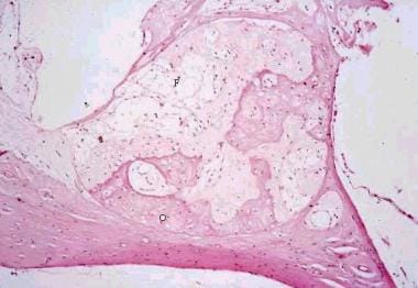
The Organ of Corti is the sensory organ of hearing within the cochlea of the inner ear. It is a structure in the inner spiral sulcus of the cochlear duct and is responsible for converting sound vibrations into electrical signals that are sent to the brain via the auditory nerve.
The Organ of Corti consists of hair cells, which are sensory receptors with hair-like projections called stereocilia on their apical surfaces. These stereocilia are embedded in a gelatinous matrix and are arranged in rows of different heights. When sound vibrations cause the fluid in the cochlea to move, the stereocilia bend, which opens ion channels and triggers nerve impulses that are sent to the brain.
Damage or loss of hair cells in the Organ of Corti can result in hearing loss, making it a critical structure for maintaining normal auditory function.
Auditory hair cells are specialized sensory receptor cells located in the inner ear, more specifically in the organ of Corti within the cochlea. They play a crucial role in hearing by converting sound vibrations into electrical signals that can be interpreted by the brain.
These hair cells have hair-like projections called stereocilia on their apical surface, which are embedded in a gelatinous matrix. When sound waves reach the inner ear, they cause the fluid within the cochlea to move, which in turn causes the stereocilia to bend. This bending motion opens ion channels at the tips of the stereocilia, allowing positively charged ions (such as potassium) to flow into the hair cells and trigger a receptor potential.
The receptor potential then leads to the release of neurotransmitters at the base of the hair cells, which activate afferent nerve fibers that synapse with these cells. The electrical signals generated by this process are transmitted to the brain via the auditory nerve, where they are interpreted as sound.
There are two types of auditory hair cells: inner hair cells and outer hair cells. Inner hair cells are the primary sensory receptors responsible for transmitting information about sound to the brain. They make direct contact with afferent nerve fibers and are more sensitive to mechanical stimulation than outer hair cells.
Outer hair cells, on the other hand, are involved in amplifying and fine-tuning the mechanical response of the inner ear to sound. They have a unique ability to contract and relax in response to electrical signals, which allows them to adjust the stiffness of their stereocilia and enhance the sensitivity of the cochlea to different frequencies.
Damage or loss of auditory hair cells can lead to hearing impairment or deafness, as these cells cannot regenerate spontaneously in mammals. Therefore, understanding the structure and function of hair cells is essential for developing therapies aimed at treating hearing disorders.
The ear canal, also known as the external auditory canal, is the tubular passage that extends from the outer ear (pinna) to the eardrum (tympanic membrane). It is lined with skin and tiny hairs, and is responsible for conducting sound waves from the outside environment to the middle and inner ear. The ear canal is typically about 2.5 cm long in adults and has a self-cleaning mechanism that helps to keep it free of debris and wax.
The spiral ganglion is a structure located in the inner ear, specifically within the cochlea. It consists of nerve cell bodies that form the sensory component of the auditory nervous system. The spiral ganglion's neurons are bipolar and have peripheral processes that form synapses with hair cells in the organ of Corti, which is responsible for converting sound vibrations into electrical signals.
The central processes of these neurons then coalesce to form the cochlear nerve, which transmits these electrical signals to the brainstem and ultimately to the auditory cortex for processing and interpretation as sound. Damage to the spiral ganglion or its associated neural structures can lead to hearing loss or deafness.
Ear diseases are medical conditions that affect the ear and its various components, including the outer ear, middle ear, and inner ear. These diseases can cause a range of symptoms, such as hearing loss, tinnitus (ringing in the ears), vertigo (dizziness), ear pain, and discharge. Some common ear diseases include:
1. Otitis externa (swimmer's ear) - an infection or inflammation of the outer ear and ear canal.
2. Otitis media - an infection or inflammation of the middle ear, often caused by a cold or flu.
3. Cholesteatoma - a skin growth that develops in the middle ear behind the eardrum.
4. Meniere's disease - a disorder of the inner ear that can cause vertigo, hearing loss, and tinnitus.
5. Temporomandibular joint (TMJ) disorders - problems with the joint that connects the jawbone to the skull, which can cause ear pain and other symptoms.
6. Acoustic neuroma - a noncancerous tumor that grows on the nerve that connects the inner ear to the brain.
7. Presbycusis - age-related hearing loss.
Treatment for ear diseases varies depending on the specific condition and its severity. It may include medication, surgery, or other therapies. If you are experiencing symptoms of an ear disease, it is important to seek medical attention from a healthcare professional, such as an otolaryngologist (ear, nose, and throat specialist).
Cochlear diseases refer to conditions that affect the structure or function of the cochlea, which is a part of the inner ear responsible for hearing. These diseases can cause various types and degrees of hearing loss, ranging from mild to profound. Some common cochlear diseases include:
1. Cochlear otosclerosis: A condition where there is abnormal bone growth in the cochlea, which can lead to conductive or sensorineural hearing loss.
2. Cochlear Meniere's disease: A disorder that affects the inner ear and causes vertigo, tinnitus, and fluctuating hearing loss.
3. Cochlear damage due to exposure to loud noises: Prolonged or sudden exposure to loud noises can cause permanent cochlear damage and hearing loss.
4. Presbycusis: Age-related hearing loss that affects the cochlea and other structures of the auditory system.
5. Cochlear nerve tumors: Rare benign or malignant growths on the cochlear nerve can cause hearing loss, tinnitus, and balance problems.
6. Infections: Bacterial or viral infections such as meningitis, labyrinthitis, or otitis media can damage the cochlea and lead to hearing loss.
7. Ototoxicity: Certain medications can be toxic to the cochlea and cause hearing loss, tinnitus, or balance problems.
8. Genetic factors: Inherited genetic mutations can cause various types of cochlear diseases, such as connexin 26 deficiency, Waardenburg syndrome, or Usher syndrome.
It is important to note that early diagnosis and treatment of cochlear diseases can help prevent or minimize hearing loss and other complications.
Hearing is the ability to perceive sounds by detecting vibrations in the air or other mediums and translating them into nerve impulses that are sent to the brain for interpretation. In medical terms, hearing is defined as the sense of sound perception, which is mediated by the ear and interpreted by the brain. It involves a complex series of processes, including the conduction of sound waves through the outer ear to the eardrum, the vibration of the middle ear bones, and the movement of fluid in the inner ear, which stimulates hair cells to send electrical signals to the auditory nerve and ultimately to the brain. Hearing allows us to communicate with others, appreciate music and sounds, and detect danger or important events in our environment.
Auditory outer hair cells are specialized sensory receptor cells located in the cochlea of the inner ear. They are part of the organ of Corti and play a crucial role in hearing by converting sound energy into electrical signals that can be interpreted by the brain.
Unlike the more numerous and simpler auditory inner hair cells, outer hair cells are equipped with unique actin-based molecular motors called "motile" or "piezoelectric" properties. These motors enable the outer hair cells to change their shape and length in response to electrical signals, which in turn amplifies the mechanical vibrations of the basilar membrane where they are located. This amplification increases the sensitivity and frequency selectivity of hearing, allowing us to detect and discriminate sounds over a wide range of intensities and frequencies.
Damage or loss of outer hair cells is a common cause of sensorineural hearing loss, which can result from exposure to loud noises, aging, genetics, ototoxic drugs, and other factors. Currently, there are no effective treatments to regenerate or replace damaged outer hair cells, making hearing loss an irreversible condition in most cases.
The spiral ligament of the cochlea is a fibrous structure located in the inner ear, more specifically in the cochlea. It is part of the membranous labyrinth and helps to maintain the shape and tension of the cochlear duct, which is essential for hearing.
The spiral ligament is attached to the bony wall of the cochlea and runs along the entire length of the cochlear duct, spiraling around it in a snail-like fashion. It consists of an outer, highly vascularized fibrous layer (the fibrous cap) and an inner, more cellular layer (the avascular zone).
The spiral ligament plays a crucial role in sound transmission and perception by helping to maintain the mechanical properties of the cochlear duct. The tension on the basilar membrane, where the sensory hair cells are located, is regulated by the spiral ligament's stiffness and elasticity. This tension affects the vibration amplitude and frequency selectivity of the basilar membrane, which in turn influences how we perceive different sounds and pitches.
Damage to the spiral ligament can result in hearing loss or impairment due to disrupted sound transmission and perception.
The round window ( membrana tympani rotunda) is a small, thin membrane-covered opening located in the inner ear between the middle ear and the cochlea. It serves as one of the two openings that lead into the cochlea, with the other being the oval window.
The round window's primary function is to help regulate and dampen the pressure changes within the cochlea that occur when sound waves reach the inner ear. This is accomplished through the movement of the fluid-filled spaces inside the cochlea (the scala vestibuli and scala tympani) caused by vibrations from the stapes bone, which connects to the oval window.
As the stapes bone moves in response to sound waves, it causes a corresponding motion in the perilymph fluid within the cochlea. This movement then creates pressure changes at the round window, causing it to bulge outward or move inward. The flexibility of the round window allows it to absorb and dissipate these pressure changes, which helps protect the delicate structures inside the inner ear from damage due to excessive pressure buildup.
It is important to note that any damage or dysfunction in the round window can negatively impact hearing ability and cause various hearing disorders.
Auditory brainstem evoked potentials (ABEPs or BAEPs) are medical tests that measure the electrical activity in the auditory pathway of the brain in response to sound stimulation. The test involves placing electrodes on the scalp and recording the tiny electrical signals generated by the nerve cells in the brainstem as they respond to clicks or tone bursts presented through earphones.
The resulting waveform is analyzed for latency (the time it takes for the signal to travel from the ear to the brain) and amplitude (the strength of the signal). Abnormalities in the waveform can indicate damage to the auditory nerve or brainstem, and are often used in the diagnosis of various neurological conditions such as multiple sclerosis, acoustic neuroma, and brainstem tumors.
The test is non-invasive, painless, and takes only a few minutes to perform. It provides valuable information about the functioning of the auditory pathway and can help guide treatment decisions for patients with hearing or balance disorders.
Labyrinth supporting cells are specialized cells that are located in the inner ear and provide structural and functional support to the sensory hair cells within the labyrinth, which is the complex system of tubes and sacs responsible for maintaining balance and hearing. These supporting cells form a crucial part of the architecture of the inner ear and help to maintain the proper functioning of the sensory hair cells by providing mechanical support, contributing to the development and maintenance of the extracellular matrix, and playing a role in the recycling of neurotransmitters. Additionally, labyrinth supporting cells can also transform into new hair cells in certain circumstances, which has implications for potential regenerative therapies aimed at treating hearing loss and balance disorders.
Stria vascularis is a highly vascularized (rich in blood vessels) structure located in the cochlea of the inner ear. It plays a crucial role in the process of hearing by maintaining the endocochlear potential, which is essential for the conversion of sound waves into electrical signals that can be interpreted by the brain. The stria vascularis is composed of three layers: the marginal cells, intermediate cells, and basal cells, which work together to maintain the ionic balance and generate the endocochlear potential. Damage to the stria vascularis can result in hearing loss.
The ear ossicles are the three smallest bones in the human body, which are located in the middle ear. They play a crucial role in the process of hearing by transmitting and amplifying sound vibrations from the eardrum to the inner ear. The three ear ossicles are:
1. Malleus (hammer): The largest of the three bones, it is shaped like a hammer and connects to the eardrum.
2. Incus (anvil): The middle-sized bone, it looks like an anvil and connects the malleus to the stapes.
3. Stapes (stirrup): The smallest and lightest bone in the human body, it resembles a stirrup and transmits vibrations from the incus to the inner ear.
Together, these tiny bones work to efficiently transfer sound waves from the air to the fluid-filled cochlea of the inner ear, enabling us to hear.
The cochlear nerve, also known as the auditory nerve, is the sensory nerve that transmits sound signals from the inner ear to the brain. It consists of two parts: the outer spiral ganglion and the inner vestibular portion. The spiral ganglion contains the cell bodies of the bipolar neurons that receive input from hair cells in the cochlea, which is the snail-shaped organ in the inner ear responsible for hearing. These neurons then send their axons to form the cochlear nerve, which travels through the internal auditory meatus and synapses with neurons in the cochlear nuclei located in the brainstem.
Damage to the cochlear nerve can result in hearing loss or deafness, depending on the severity of the injury. Common causes of cochlear nerve damage include acoustic trauma, such as exposure to loud noises, viral infections, meningitis, and tumors affecting the nerve or surrounding structures. In some cases, cochlear nerve damage may be treated with hearing aids, cochlear implants, or other assistive devices to help restore or improve hearing function.
The stapes is the smallest bone in the human body, which is a part of the middle ear. It is also known as the "stirrup" because of its U-shaped structure. The stapes connects the inner ear to the middle ear, transmitting sound vibrations from the ear drum to the inner ear. More specifically, it is the third bone in the series of three bones (the ossicles) that conduct sound waves from the air to the fluid-filled inner ear.
Deafness is a hearing loss that is so severe that it results in significant difficulty in understanding or comprehending speech, even when using hearing aids. It can be congenital (present at birth) or acquired later in life due to various causes such as disease, injury, infection, exposure to loud noises, or aging. Deafness can range from mild to profound and may affect one ear (unilateral) or both ears (bilateral). In some cases, deafness may be accompanied by tinnitus, which is the perception of ringing or other sounds in the ears.
Deaf individuals often use American Sign Language (ASL) or other forms of sign language to communicate. Some people with less severe hearing loss may benefit from hearing aids, cochlear implants, or other assistive listening devices. Deafness can have significant social, educational, and vocational implications, and early intervention and appropriate support services are critical for optimal development and outcomes.
Hearing loss is a partial or total inability to hear sounds in one or both ears. It can occur due to damage to the structures of the ear, including the outer ear, middle ear, inner ear, or nerve pathways that transmit sound to the brain. The degree of hearing loss can vary from mild (difficulty hearing soft sounds) to severe (inability to hear even loud sounds). Hearing loss can be temporary or permanent and may be caused by factors such as exposure to loud noises, genetics, aging, infections, trauma, or certain medical conditions. It is important to note that hearing loss can have significant impacts on a person's communication abilities, social interactions, and overall quality of life.
Perilymph is a type of fluid found in the inner ear, more specifically within the bony labyrinth of the inner ear. It fills the space between the membranous labyrinth and the bony labyrinth in the cochlea and vestibular system. Perilymph is similar in composition to cerebrospinal fluid (CSF) and contains sodium, chloride, and protein ions. Its main function is to protect the inner ear from damage, maintain hydrostatic pressure, and facilitate the transmission of sound waves to the hair cells in the cochlea for hearing.
Spontaneous otoacoustic emissions (SOAEs) are low-level sounds that are produced by the inner ear (cochlea) without any external stimulation. They can be recorded in a quiet room using specialized microphones placed inside the ear canal. SOAEs are thought to arise from the motion of the hair cells within the cochlea, which generate tiny currents in response to sound. These currents then cause the surrounding fluid and tissue to vibrate, producing sound waves that can be detected with a microphone.
SOAEs are typically present in individuals with normal hearing, although their presence or absence is not a definitive indicator of hearing ability. They tend to occur at specific frequencies and can vary from person to person. In some cases, SOAEs may be absent or reduced in individuals with hearing loss or damage to the hair cells in the cochlea.
It's worth noting that SOAEs are different from evoked otoacoustic emissions (EOAEs), which are sounds produced by the inner ear in response to external stimuli, such as clicks or tones. Both types of otoacoustic emissions are used in hearing tests and research to assess cochlear function and health.
Cochlear microphonic potentials (CMs) are electrical responses that originate from the hair cells in the cochlea, which is a part of the inner ear responsible for hearing. These potentials can be recorded using an electrode placed near the cochlea in response to sound stimulation.
The CMs are considered to be a passive response of the hair cells to the mechanical deflection caused by sound waves. They represent the receptor potential of the outer hair cells and are directly proportional to the sound pressure level. Unlike other electrical responses in the cochlea, such as the action potentials generated by the auditory nerve fibers, CMs do not require the presence of neurotransmitters or synaptic transmission.
Cochlear microphonic potentials have been used in research to study the biophysical properties of hair cells and their response to different types of sound stimuli. However, they are not typically used in clinical audiology due to their small amplitude and susceptibility to interference from other electrical signals in the body.
The temporal bone is a paired bone that is located on each side of the skull, forming part of the lateral and inferior walls of the cranial cavity. It is one of the most complex bones in the human body and has several important structures associated with it. The main functions of the temporal bone include protecting the middle and inner ear, providing attachment for various muscles of the head and neck, and forming part of the base of the skull.
The temporal bone is divided into several parts, including the squamous part, the petrous part, the tympanic part, and the styloid process. The squamous part forms the lateral portion of the temporal bone and articulates with the parietal bone. The petrous part is the most medial and superior portion of the temporal bone and contains the inner ear and the semicircular canals. The tympanic part forms the lower and anterior portions of the temporal bone and includes the external auditory meatus or ear canal. The styloid process is a long, slender projection that extends downward from the inferior aspect of the temporal bone and serves as an attachment site for various muscles and ligaments.
The temporal bone plays a crucial role in hearing and balance, as it contains the structures of the middle and inner ear, including the oval window, round window, cochlea, vestibule, and semicircular canals. The stapes bone, one of the three bones in the middle ear, is entirely encased within the petrous portion of the temporal bone. Additionally, the temporal bone contains important structures for facial expression and sensation, including the facial nerve, which exits the skull through the stylomastoid foramen, a small opening in the temporal bone.
The auditory threshold is the minimum sound intensity or loudness level that a person can detect 50% of the time, for a given tone frequency. It is typically measured in decibels (dB) and represents the quietest sound that a person can hear. The auditory threshold can be affected by various factors such as age, exposure to noise, and certain medical conditions. Hearing tests, such as pure-tone audiometry, are used to measure an individual's auditory thresholds for different frequencies.
Noise-induced hearing loss (NIHL) is a type of sensorineural hearing loss that occurs due to exposure to harmful levels of noise. The damage can be caused by a one-time exposure to an extremely loud sound or by continuous exposure to lower level sounds over time. NIHL can affect people of all ages and can cause permanent damage to the hair cells in the cochlea, leading to hearing loss, tinnitus (ringing in the ears), and difficulty understanding speech in noisy environments. Prevention measures include avoiding excessive noise exposure, wearing hearing protection, and taking regular breaks from noisy activities.
## I am not aware of a medical definition for the term "chinchilla."
A chinchilla is actually a type of rodent that is native to South America. They have thick, soft fur and are often kept as exotic pets or used in laboratory research. If you're looking for information about chinchillas in a medical context, such as their use in research or any potential health concerns related to keeping them as pets, I would be happy to help you try to find more information on those topics.
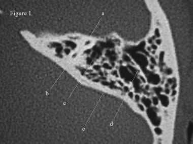
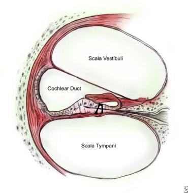
The Scala Tympani is a part of the inner ear's bony labyrinth, specifically within the cochlea. It is one of the two channels (the other being the Scala Vestibuli) that make up the bony duct of the cochlea, through which sound waves are transmitted to the inner ear.
The Scala Tympani starts at the round window, which is a membrane-covered opening located on the cochlea's outer wall. It runs parallel to the Scala Vestibuli and connects with it at the helicotrema, a small opening at the apex or tip of the cochlea.
When sound waves reach the inner ear, they cause vibrations in the fluid-filled Scala Tympani and Scala Vestibuli, which stimulate hair cells within the organ of Corti, leading to the conversion of mechanical energy into electrical signals that are then transmitted to the brain via the auditory nerve.
It's important to note that any damage or dysfunction in the Scala Tympani or other parts of the inner ear can lead to hearing loss or other auditory disorders.
The cochlear duct, also known as the scala media, is a membranous duct located within the cochlea of the inner ear. It is one of three fluid-filled compartments in the cochlea, along with the vestibular duct (scala vestibuli) and the tympanic duct (scala tympani).
The cochlear duct contains endolymph, a specialized fluid that carries electrical signals to the auditory nerve. The organ of Corti, which is responsible for converting sound vibrations into electrical signals, is located within the cochlear duct.
The cochlear duct runs along the length of the cochlea and is separated from the vestibular duct by Reissner's membrane and from the tympanic duct by the basilar membrane. These membranes help to create a highly sensitive and selective environment for sound perception, allowing us to hear and distinguish different frequencies and intensities of sound.
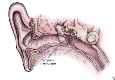
The tympanic membrane, also known as the eardrum, is a thin, cone-shaped membrane that separates the external auditory canal from the middle ear. It serves to transmit sound vibrations from the air to the inner ear, where they are converted into electrical signals that can be interpreted by the brain as sound. The tympanic membrane is composed of three layers: an outer layer of skin, a middle layer of connective tissue, and an inner layer of mucous membrane. It is held in place by several small bones and muscles and is highly sensitive to changes in pressure.
The tectorial membrane is a specialized structure in the inner ear, more specifically in the cochlea. It is a gelatinous, hair-like structure that is located above and parallel to the organ of Corti, which contains the sensory hair cells responsible for hearing. The tectorial membrane is composed of collagen fibers and a glycoprotein matrix.
The main function of the tectorial membrane is to deflect the stereocilia (hair-like projections) of the inner and outer hair cells as sound waves pass through the cochlea, which in turn triggers nerve impulses that are sent to the brain and interpreted as sound. The tectorial membrane moves in response to sound-induced vibrations of the fluid within the cochlea, causing shearing forces on the stereocilia, leading to the initiation of the hearing process.
Sensorineural hearing loss (SNHL) is a type of hearing impairment that occurs due to damage to the inner ear (cochlea) or to the nerve pathways from the inner ear to the brain. It can be caused by various factors such as aging, exposure to loud noises, genetics, certain medical conditions (like diabetes and heart disease), and ototoxic medications.
SNHL affects the ability of the hair cells in the cochlea to convert sound waves into electrical signals that are sent to the brain via the auditory nerve. As a result, sounds may be perceived as muffled, faint, or distorted, making it difficult to understand speech, especially in noisy environments.
SNHL is typically permanent and cannot be corrected with medication or surgery, but hearing aids or cochlear implants can help improve communication and quality of life for those affected.
Endolymph is a specific type of fluid that is found within the inner ear, more specifically in the membranous labyrinth of the inner ear. This fluid plays a crucial role in maintaining balance and hearing functions. It helps in the stimulation of hair cells present in the inner ear which then transmit signals to the brain, enabling us to hear and maintain our balance. Any disturbance or changes in the composition or flow of endolymph can lead to various vestibular disorders and hearing problems.
Acoustic stimulation refers to the use of sound waves or vibrations to elicit a response in an individual, typically for the purpose of assessing or treating hearing, balance, or neurological disorders. In a medical context, acoustic stimulation may involve presenting pure tones, speech sounds, or other types of auditory signals through headphones, speakers, or specialized devices such as bone conduction transducers.
The response to acoustic stimulation can be measured using various techniques, including electrophysiological tests like auditory brainstem responses (ABRs) or otoacoustic emissions (OAEs), behavioral observations, or functional imaging methods like fMRI. Acoustic stimulation is also used in therapeutic settings, such as auditory training programs for hearing impairment or vestibular rehabilitation for balance disorders.
It's important to note that acoustic stimulation should be administered under the guidance of a qualified healthcare professional to ensure safety and effectiveness.
Labyrinth diseases refer to conditions that affect the inner ear's labyrinth, which is the complex system of fluid-filled channels and sacs responsible for maintaining balance and hearing. These diseases can cause symptoms such as vertigo (a spinning sensation), dizziness, nausea, hearing loss, and tinnitus (ringing in the ears). Examples of labyrinth diseases include Meniere's disease, labyrinthitis, vestibular neuronitis, and benign paroxysmal positional vertigo. Treatment for these conditions varies depending on the specific diagnosis but may include medications, physical therapy, or surgery.
Ear neoplasms refer to abnormal growths or tumors that occur in the ear. These growths can be benign (non-cancerous) or malignant (cancerous) and can affect any part of the ear, including the outer ear, middle ear, inner ear, and the ear canal.
Benign ear neoplasms are typically slow-growing and do not spread to other parts of the body. Examples include exostoses, osteomas, and ceruminous adenomas. These types of growths are usually removed surgically for cosmetic reasons or if they cause discomfort or hearing problems.
Malignant ear neoplasms, on the other hand, can be aggressive and may spread to other parts of the body. Examples include squamous cell carcinoma, basal cell carcinoma, and adenoid cystic carcinoma. These types of tumors often require more extensive treatment, such as surgery, radiation therapy, and chemotherapy.
It is important to note that any new growth or change in the ear should be evaluated by a healthcare professional to determine the nature of the growth and develop an appropriate treatment plan.
Gerbillinae is a subfamily of rodents that includes gerbils, jirds, and sand rats. These small mammals are primarily found in arid regions of Africa and Asia. They are characterized by their long hind legs, which they use for hopping, and their long, thin tails. Some species have adapted to desert environments by developing specialized kidneys that allow them to survive on minimal water intake.
The vestibular system is a part of the inner ear that contributes to our sense of balance and spatial orientation. It is made up of two main components: the vestibule and the labyrinth.
The vestibule is a bony chamber in the inner ear that contains two important structures called the utricle and saccule. These structures contain hair cells and fluid-filled sacs that help detect changes in head position and movement, allowing us to maintain our balance and orientation in space.
The labyrinth, on the other hand, is a more complex structure that includes the vestibule as well as three semicircular canals. These canals are also filled with fluid and contain hair cells that detect rotational movements of the head. Together, the vestibule and labyrinth work together to provide us with information about our body's position and movement in space.
Overall, the vestibular system plays a crucial role in maintaining our balance, coordinating our movements, and helping us navigate through our environment.
I must clarify that the term "Guinea Pigs" is not typically used in medical definitions. However, in colloquial or informal language, it may refer to people who are used as the first to try out a new medical treatment or drug. This is known as being a "test subject" or "in a clinical trial."
In the field of scientific research, particularly in studies involving animals, guinea pigs are small rodents that are often used as experimental subjects due to their size, cost-effectiveness, and ease of handling. They are not actually pigs from Guinea, despite their name's origins being unclear. However, they do not exactly fit the description of being used in human medical experiments.
Cochlear implantation is a surgical procedure in which a device called a cochlear implant is inserted into the inner ear (cochlea) of a person with severe to profound hearing loss. The implant consists of an external component, which includes a microphone, processor, and transmitter, and an internal component, which includes a receiver and electrode array.
The microphone picks up sounds from the environment and sends them to the processor, which analyzes and converts the sounds into electrical signals. These signals are then transmitted to the receiver, which stimulates the electrode array in the cochlea. The electrodes directly stimulate the auditory nerve fibers, bypassing the damaged hair cells in the inner ear that are responsible for normal hearing.
The brain interprets these electrical signals as sound, allowing the person to perceive and understand speech and other sounds. Cochlear implantation is typically recommended for people who do not benefit from traditional hearing aids and can significantly improve communication, quality of life, and social integration for those with severe to profound hearing loss.
The ear auricle, also known as the pinna or outer ear, is the visible external structure of the ear that serves to collect and direct sound waves into the ear canal. It is composed of cartilage and skin and is shaped like a curved funnel. The ear auricle consists of several parts including the helix (the outer rim), antihelix (the inner curved prominence), tragus and antitragus (the small pointed eminences in front of and behind the ear canal opening), concha (the bowl-shaped area that directs sound into the ear canal), and lobule (the fleshy lower part hanging from the ear).
The vestibulocochlear nerve, also known as the auditory-vestibular nerve or cranial nerve VIII, is a paired peripheral nerve that transmits sensory information from the inner ear to the brain. It has two distinct parts: the cochlear part and the vestibular part.
The cochlear part is responsible for hearing and transmits sound signals from the cochlea to the brain. The vestibular part, on the other hand, is responsible for maintaining balance and spatial orientation by transmitting information about head movement and position from the vestibular apparatus (utricle, saccule, and semicircular canals) in the inner ear to the brain.
Together, these two parts of the vestibulocochlear nerve play a crucial role in our ability to hear and maintain balance. Damage to this nerve can result in hearing loss, tinnitus (ringing in the ears), vertigo (dizziness), or balance problems.
In the context of medicine, particularly in audiology and otolaryngology (ear, nose, and throat specialty), "noise" is defined as unwanted or disturbing sound in the environment that can interfere with communication, rest, sleep, or cognitive tasks. It can also refer to sounds that are harmful to hearing, such as loud machinery noises or music, which can cause noise-induced hearing loss if exposure is prolonged or at high enough levels.
In some medical contexts, "noise" may also refer to non-specific signals or interfering factors in diagnostic tests and measurements that can make it difficult to interpret results accurately.
Auditory pathways refer to the series of structures and nerves in the body that are involved in processing sound and transmitting it to the brain for interpretation. The process begins when sound waves enter the ear and cause vibrations in the eardrum, which then move the bones in the middle ear. These movements stimulate hair cells in the cochlea, a spiral-shaped structure in the inner ear, causing them to release neurotransmitters that activate auditory nerve fibers.
The auditory nerve carries these signals to the brainstem, where they are relayed through several additional structures before reaching the auditory cortex in the temporal lobe of the brain. Here, the signals are processed and interpreted as sounds, allowing us to hear and understand speech, music, and other environmental noises.
Damage or dysfunction at any point along the auditory pathway can lead to hearing loss or impairment.
Cochlear implants are medical devices that are surgically implanted in the inner ear to help restore hearing in individuals with severe to profound hearing loss. These devices bypass the damaged hair cells in the inner ear and directly stimulate the auditory nerve, allowing the brain to interpret sound signals. Cochlear implants consist of two main components: an external processor that picks up and analyzes sounds from the environment, and an internal receiver/stimulator that receives the processed information and sends electrical impulses to the auditory nerve. The resulting patterns of electrical activity are then perceived as sound by the brain. Cochlear implants can significantly improve communication abilities, language development, and overall quality of life for individuals with profound hearing loss.
In the context of medicine, particularly in the field of auscultation (the act of listening to the internal sounds of the body), "sound" refers to the noises produced by the functioning of the heart, lungs, and other organs. These sounds are typically categorized into two types:
1. **Bradyacoustic sounds**: These are low-pitched sounds that are heard when there is a turbulent flow of blood or when two body structures rub against each other. An example would be the heart sound known as "S1," which is produced by the closure of the mitral and tricuspid valves at the beginning of systole (contraction of the heart's ventricles).
2. **High-pitched sounds**: These are sharper, higher-frequency sounds that can provide valuable diagnostic information. An example would be lung sounds, which include breath sounds like those heard during inhalation and exhalation, as well as adventitious sounds like crackles, wheezes, and pleural friction rubs.
It's important to note that these medical "sounds" are not the same as the everyday definition of sound, which refers to the sensation produced by stimulation of the auditory system by vibrations.
Labyrinthine fluids, also known as endolymph and perilymph, are fluids that fill the inner ear structures, specifically the bony labyrinth. The bony labyrinth is divided into two main parts: the cochlea, responsible for hearing, and the vestibular system, responsible for balance.
Endolymph is a clear, plasma-like fluid found within the membranous labyrinth, which is a series of interconnected tubes and sacs that lie inside the bony labyrinth. Endolymph plays a crucial role in the functioning of both the cochlea and vestibular system by creating an electrochemical gradient necessary for the conversion of mechanical sound vibrations into electrical signals in the cochlea, as well as facilitating the detection of head movements and maintaining balance in the vestibular system.
Perilymph, on the other hand, is a clear, colorless fluid that fills the space between the bony labyrinth and the membranous labyrinth. It is similar in composition to cerebrospinal fluid (CSF) and serves as a protective cushion for the delicate inner ear structures. Perilymph also helps maintain the electrochemical gradient required for sound transduction in the cochlea.
Disorders related to these labyrinthine fluids, such as endolymphatic hydrops or perilymph fistula, can lead to hearing and balance problems.
Otologic surgical procedures refer to a range of surgeries performed on the ear or its related structures. These procedures are typically conducted by otologists, who are specialists trained in diagnosing and treating conditions that affect the ears, balance system, and related nerves. The goal of otologic surgery can vary from repairing damaged bones in the middle ear to managing hearing loss, tumors, or chronic infections. Some common otologic surgical procedures include:
1. Stapedectomy/Stapedotomy: These are procedures used to treat otosclerosis, a condition where the stapes bone in the middle ear becomes fixed and causes conductive hearing loss. The surgeon creates an opening in the stapes footplate (stapedotomy) or removes the entire stapes bone (stapedectomy) and replaces it with a prosthetic device to improve sound conduction.
2. Myringoplasty/Tympanoplasty: These are surgeries aimed at repairing damaged eardrums (tympanic membrane). A myringoplasty involves grafting a piece of tissue over the perforation in the eardrum, while a tympanoplasty includes both eardrum repair and reconstruction of the middle ear bones if necessary.
3. Mastoidectomy: This procedure involves removing the mastoid air cells, which are located in the bony prominence behind the ear. A mastoidectomy is often performed to treat chronic mastoiditis, cholesteatoma, or complications from middle ear infections.
4. Ossiculoplasty: This procedure aims to reconstruct and improve the function of the ossicles (middle ear bones) when they are damaged due to various reasons such as infection, trauma, or congenital conditions. The surgeon uses prosthetic devices made from plastic, metal, or even bone to replace or support the damaged ossicles.
5. Cochlear implantation: This is a surgical procedure that involves placing an electronic device inside the inner ear to help individuals with severe to profound hearing loss. The implant consists of an external processor and internal components that directly stimulate the auditory nerve, bypassing the damaged hair cells in the cochlea.
6. Labyrinthectomy: This procedure involves removing the balance-sensing structures (vestibular system) inside the inner ear to treat severe vertigo or dizziness caused by conditions like Meniere's disease when other treatments have failed.
7. Acoustic neuroma removal: An acoustic neuroma is a benign tumor that grows on the vestibulocochlear nerve, which connects the inner ear to the brain. Surgical removal of the tumor is necessary to prevent hearing loss, balance problems, and potential neurological complications.
These are just a few examples of the various surgical procedures performed by otolaryngologists (ear, nose, and throat specialists) to treat conditions affecting the ear and surrounding structures. Each procedure has its specific indications, benefits, risks, and postoperative care requirements. Patients should consult with their healthcare providers to discuss the most appropriate treatment options for their individual needs.
Presbycusis is an age-related hearing loss, typically characterized by the progressive loss of sensitivity to high-frequency sounds. It's a result of natural aging of the auditory system and is often seen as a type of sensorineural hearing loss. The term comes from the Greek words "presbus" meaning old man and "akousis" meaning hearing.
This condition usually develops slowly over many years and can affect both ears equally. Presbycusis can make understanding speech, especially in noisy environments, quite challenging. It's a common condition, and its prevalence increases with age. While it's not reversible, various assistive devices like hearing aids can help manage the symptoms.
Auditory evoked potentials (AEP) are medical tests that measure the electrical activity in the brain in response to sound stimuli. These tests are often used to assess hearing function and neural processing in individuals, particularly those who cannot perform traditional behavioral hearing tests.
There are several types of AEP tests, including:
1. Brainstem Auditory Evoked Response (BAER) or Brainstem Auditory Evoked Potentials (BAEP): This test measures the electrical activity generated by the brainstem in response to a click or tone stimulus. It is often used to assess the integrity of the auditory nerve and brainstem pathways, and can help diagnose conditions such as auditory neuropathy and retrocochlear lesions.
2. Middle Latency Auditory Evoked Potentials (MLAEP): This test measures the electrical activity generated by the cortical auditory areas of the brain in response to a click or tone stimulus. It is often used to assess higher-level auditory processing, and can help diagnose conditions such as auditory processing disorders and central auditory dysfunction.
3. Long Latency Auditory Evoked Potentials (LLAEP): This test measures the electrical activity generated by the cortical auditory areas of the brain in response to a complex stimulus, such as speech. It is often used to assess language processing and cognitive function, and can help diagnose conditions such as learning disabilities and dementia.
Overall, AEP tests are valuable tools for assessing hearing and neural function in individuals who cannot perform traditional behavioral hearing tests or who have complex neurological conditions.
Cholesteatoma, middle ear is a medical condition characterized by the abnormal growth of skin cells (keratinizing squamous epithelium) within the middle ear space. This skin cells accumulation forms a pearly, white, or gray mass that can erode and destroy surrounding structures such as the ossicles (the tiny bones in the middle ear), the mastoid process (a bony prominence behind the ear), and even the inner ear or brain.
Cholesteatomas can be congenital (present at birth) or acquired (develop later in life). Acquired cholesteatomas are more common and usually result from repeated middle ear infections that cause a retraction pocket of the eardrum, which then traps skin cells leading to their abnormal growth. Symptoms of cholesteatoma may include hearing loss, ear drainage, ear pain, vertigo, or facial weakness. Treatment typically involves surgical removal of the cholesteatoma and restoration of any damaged structures.
Efferent neurons are specialized nerve cells that transmit signals from the central nervous system (CNS), which includes the brain and spinal cord, to effector organs such as muscles or glands. These signals typically result in a response or action, hence the term "efferent," derived from the Latin word "efferre" meaning "to carry away."
Efferent neurons are part of the motor pathway and can be further classified into two types:
1. Somatic efferent neurons: These neurons transmit signals to skeletal muscles, enabling voluntary movements and posture maintenance. They have their cell bodies located in the ventral horn of the spinal cord and send their axons through the ventral roots to innervate specific muscle fibers.
2. Autonomic efferent neurons: These neurons are responsible for controlling involuntary functions, such as heart rate, digestion, respiration, and pupil dilation. They have a two-neuron chain arrangement, with the preganglionic neuron having its cell body in the CNS (brainstem or spinal cord) and synapsing with the postganglionic neuron in an autonomic ganglion near the effector organ. Autonomic efferent neurons can be further divided into sympathetic, parasympathetic, and enteric subdivisions based on their functions and innervation patterns.
In summary, efferent neurons are a critical component of the nervous system, responsible for transmitting signals from the CNS to various effector organs, ultimately controlling and coordinating numerous bodily functions and responses.
Menière disease is an inner ear disorder that is characterized by episodes of vertigo (a spinning sensation), tinnitus (ringing or buzzing in the ear), hearing loss, and aural fullness (a feeling of pressure or blockage in the ear). It is caused by an abnormal accumulation of endolymphatic fluid in the inner ear, which can lead to damage of the vestibular system and cochlea. The exact cause of this fluid buildup is not known, but it may be related to genetics, allergies, or autoimmune disorders. Menière disease is typically a chronic condition, with symptoms that can vary in frequency and severity over time. Treatment options include dietary modifications, diuretics, vestibular rehabilitation therapy, and, in some cases, surgery.
Hearing disorders, also known as hearing impairments or auditory impairments, refer to conditions that affect an individual's ability to hear sounds in one or both ears. These disorders can range from mild to profound and may result from genetic factors, aging, exposure to loud noises, infections, trauma, or certain medical conditions.
There are mainly two types of hearing disorders: conductive hearing loss and sensorineural hearing loss. Conductive hearing loss occurs when there is a problem with the outer or middle ear, preventing sound waves from reaching the inner ear. Causes include earwax buildup, fluid in the middle ear, a perforated eardrum, or damage to the ossicles (the bones in the middle ear).
Sensorineural hearing loss, on the other hand, is caused by damage to the inner ear (cochlea) or the nerve pathways from the inner ear to the brain. This type of hearing loss is often permanent and can be due to aging (presbycusis), exposure to loud noises, genetics, viral infections, certain medications, or head injuries.
Mixed hearing loss is a combination of both conductive and sensorineural components. In some cases, hearing disorders can also involve tinnitus (ringing or other sounds in the ears) or vestibular problems that affect balance and equilibrium.
Early identification and intervention for hearing disorders are crucial to prevent further deterioration and to help individuals develop appropriate communication skills and maintain a good quality of life.
Acoustic impedance tests are diagnostic procedures used to measure the impedance or resistance of various parts of the ear to sound waves. These tests are often used to assess hearing function and diagnose any issues related to the middle ear, such as fluid buildup or problems with the eardrum.
The most common type of acoustic impedance test is tympanometry, which measures the mobility of the eardrum and the middle ear system by creating variations in air pressure within the ear canal. During this test, a small probe is inserted into the ear canal, and sound waves are generated while the pressure is varied. The resulting measurements provide information about the condition of the middle ear and can help identify any issues that may be affecting hearing.
Another type of acoustic impedance test is acoustic reflex testing, which measures the body's natural response to loud sounds. This involves measuring the contraction of the stapedius muscle in the middle ear, which occurs in response to loud noises. By measuring the strength and timing of this reflex, audiologists can gain additional insights into the functioning of the middle ear and identify any abnormalities that may be present.
Overall, acoustic impedance tests are important tools for diagnosing hearing problems and identifying any underlying issues in the middle ear. They are often used in conjunction with other hearing tests to provide a comprehensive assessment of an individual's hearing function.
Audiometry is the testing of a person's ability to hear different sounds, pitches, or frequencies. It is typically conducted using an audiometer, a device that emits tones at varying volumes and frequencies. The person being tested wears headphones and indicates when they can hear the tone by pressing a button or raising their hand.
There are two main types of audiometry: pure-tone audiometry and speech audiometry. Pure-tone audiometry measures a person's ability to hear different frequencies at varying volumes, while speech audiometry measures a person's ability to understand spoken words at different volumes and in the presence of background noise.
The results of an audiometry test are typically plotted on an audiogram, which shows the quietest sounds that a person can hear at different frequencies. This information can be used to diagnose hearing loss, determine its cause, and develop a treatment plan.
Pure-tone audiometry is a hearing test that measures a person's ability to hear different sounds, pitches, or frequencies. During the test, pure tones are presented to the patient through headphones or ear inserts, and the patient is asked to indicate each time they hear the sound by raising their hand, pressing a button, or responding verbally.
The softest sound that the person can hear at each frequency is recorded as the hearing threshold, and a graph called an audiogram is created to show the results. The audiogram provides information about the type and degree of hearing loss in each ear. Pure-tone audiometry is a standard hearing test used to diagnose and monitor hearing disorders.
The cochlear nucleus is the first relay station in the auditory pathway within the central nervous system. It is a structure located in the lower pons region of the brainstem and receives sensory information from the cochlea, which is the spiral-shaped organ of hearing in the inner ear.
The cochlear nucleus consists of several subdivisions, each with distinct neuronal populations that process different aspects of auditory information. These subdivisions include the anteroventral cochlear nucleus (AVCN), posteroventral cochlear nucleus (PVCN), dorsal cochlear nucleus (DCN), and the granule cell domain.
Neurons in these subdivisions perform various computations on the incoming auditory signals, such as frequency analysis, intensity coding, and sound localization. The output of the cochlear nucleus is then sent via several pathways to higher brain regions for further processing and interpretation, including the inferior colliculus, medial geniculate body, and eventually the auditory cortex.
Damage or dysfunction in the cochlear nucleus can lead to hearing impairments and other auditory processing disorders.
Cellular mechanotransduction is the process by which cells convert mechanical stimuli into biochemical signals, resulting in changes in cell behavior and function. This complex process involves various molecular components, including transmembrane receptors, ion channels, cytoskeletal proteins, and signaling molecules. Mechanical forces such as tension, compression, or fluid flow can activate these components, leading to alterations in gene expression, protein synthesis, and cell shape or movement. Cellular mechanotransduction plays a crucial role in various physiological processes, including tissue development, homeostasis, and repair, as well as in pathological conditions such as fibrosis and cancer progression.
The semicircular canals are part of the vestibular system in the inner ear that contributes to the sense of balance and spatial orientation. They are composed of three fluid-filled tubes, each located in a different plane (anterior, posterior, and horizontal) and arranged at approximately right angles to each other. The semicircular canals detect rotational movements of the head, enabling us to maintain our equilibrium during movement.
When the head moves, the fluid within the semicircular canals moves in response to that motion. At the end of each canal is a structure called the ampulla, which contains hair cells with hair-like projections (stereocilia) embedded in a gelatinous substance. As the fluid moves, it bends the stereocilia, stimulating the hair cells and sending signals to the brain via the vestibular nerve. The brain then interprets these signals to determine the direction and speed of head movement, allowing us to maintain our balance and orientation in space.
Endolymphatic hydrops is a term used to describe the abnormal accumulation of fluid (endolymph) within the inner ear. This condition is most commonly associated with Meniere's disease, but can also be seen in other disorders that affect the inner ear.
The inner ear is made up of two main parts: the cochlea, which is responsible for hearing, and the vestibular system, which helps to control balance. Both of these systems are filled with fluid, including endolymph, which is a watery fluid that bathes the sensory hair cells in these structures.
In endolymphatic hydrops, there is an overproduction or decreased absorption of endolymph, leading to an abnormal buildup of fluid within the inner ear. This can cause a variety of symptoms, including vertigo (a spinning sensation), tinnitus (ringing in the ears), hearing loss, and a feeling of fullness or pressure in the affected ear.
The exact cause of endolymphatic hydrops is not fully understood, but it is thought to be related to changes in the inner ear's fluid balance. Treatment options may include medications to help control symptoms, as well as surgical procedures to relieve pressure on the inner ear.
The endolymphatic sac is a small, fluid-filled structure that is part of the inner ear. It is located near the vestibular aqueduct and is responsible for maintaining the balance of fluids in the inner ear. The endolymphatic sac also plays a role in the resorption of endolymph, which is the fluid that fills the membranous labyrinth of the inner ear. Disorders of the endolymphatic sac can lead to conditions such as Meniere's disease, which is characterized by vertigo, hearing loss, and tinnitus.
The petrous bone is a part of the temporal bone, one of the 22 bones in the human skull. It is a thick and irregularly shaped bone located at the base of the skull and forms part of the ear and the cranial cavity. The petrous bone contains the cochlea, vestibule, and semicircular canals of the inner ear, which are responsible for hearing and balance. It also helps protect the brain from injury by forming part of the bony structure surrounding the brain.
The term "petrous" comes from the Latin word "petrosus," meaning "stony" or "rock-like," which describes the hard and dense nature of this bone. The petrous bone is one of the densest bones in the human body, making it highly resistant to fractures and other forms of damage.
In medical terminology, the term "petrous" may also be used to describe any structure that resembles a rock or is hard and dense, such as the petrous apex, which refers to the portion of the petrous bone that points towards the sphenoid bone.
The endolymphatic duct is a narrow canal in the inner ear that is part of the membranous labyrinth. It connects the utricle and saccule (two sensory structures in the vestibular system responsible for detecting changes in head position and movement) to the endolymphatic sac (a dilated portion of the duct that helps regulate the volume and pressure of endolymph, a fluid found within the membranous labyrinth).
The endolymphatic duct plays a crucial role in maintaining the balance and homeostasis of the inner ear by allowing the absorption and circulation of endolymph. Disorders or abnormalities in this region can lead to various vestibular and hearing dysfunctions, such as Meniere's disease, endolymphatic hydrops, and other inner ear disorders.
Ear cartilage, also known as auricular cartilage, refers to the flexible connective tissue that makes up the structural framework of the external ear or pinna. The ear cartilage provides support and shape to the ear, helping to direct sound waves into the ear canal and towards the eardrum.
The ear cartilage is composed of type II collagen fibers and proteoglycans, which give it its flexibility and resiliency. It is covered by a thin layer of skin on both sides and contains no bones. Instead, the ear cartilage is shaped and maintained by the surrounding muscles and connective tissue.
There are three main parts of the ear cartilage: the helix, the antihelix, and the tragus. The helix is the outer rim of the ear, while the antihelix is the curved ridge that runs parallel to the helix. The tragus is the small piece of cartilage that projects from the front of the ear canal.
Ear cartilage can be affected by various conditions, including trauma, infection, and degenerative changes associated with aging. In some cases, surgical procedures may be required to reshape or reconstruct damaged ear cartilage.
In the context of medicine and physiology, vibration refers to the mechanical oscillation of a physical body or substance with a periodic back-and-forth motion around an equilibrium point. This motion can be produced by external forces or internal processes within the body.
Vibration is often measured in terms of frequency (the number of cycles per second) and amplitude (the maximum displacement from the equilibrium position). In clinical settings, vibration perception tests are used to assess peripheral nerve function and diagnose conditions such as neuropathy.
Prolonged exposure to whole-body vibration or hand-transmitted vibration in certain occupational settings can also have adverse health effects, including hearing loss, musculoskeletal disorders, and vascular damage.
A hearing test is a procedure used to evaluate a person's ability to hear different sounds, pitches, or frequencies. It is performed by a hearing healthcare professional in a sound-treated booth or room with calibrated audiometers. The test measures a person's hearing sensitivity at different frequencies and determines the quietest sounds they can hear, known as their hearing thresholds.
There are several types of hearing tests, including:
1. Pure Tone Audiometry (PTA): This is the most common type of hearing test, where the person is presented with pure tones at different frequencies and volumes through headphones or ear inserts. The person indicates when they hear the sound by pressing a button or raising their hand.
2. Speech Audiometry: This test measures a person's ability to understand speech at different volume levels. The person is asked to repeat words presented to them in quiet and in background noise.
3. Tympanometry: This test measures the function of the middle ear by creating variations in air pressure in the ear canal. It can help identify issues such as fluid buildup or a perforated eardrum.
4. Acoustic Reflex Testing: This test measures the body's natural response to loud sounds and can help identify the location of damage in the hearing system.
5. Otoacoustic Emissions (OAEs): This test measures the sound that is produced by the inner ear when it is stimulated by a sound. It can help identify cochlear damage or abnormalities.
Hearing tests are important for diagnosing and monitoring hearing loss, as well as identifying any underlying medical conditions that may be causing the hearing problems.
Acoustics is a branch of physics that deals with the study of sound, its production, transmission, and effects. In a medical context, acoustics may refer to the use of sound waves in medical procedures such as:
1. Diagnostic ultrasound: This technique uses high-frequency sound waves to create images of internal organs and tissues. It is commonly used during pregnancy to monitor fetal development, but it can also be used to diagnose a variety of medical conditions, including heart disease, cancer, and musculoskeletal injuries.
2. Therapeutic ultrasound: This technique uses low-frequency sound waves to promote healing and reduce pain and inflammation in muscles, tendons, and ligaments. It is often used to treat soft tissue injuries, arthritis, and other musculoskeletal conditions.
3. Otology: Acoustics also plays a crucial role in the field of otology, which deals with the study and treatment of hearing and balance disorders. The shape, size, and movement of the outer ear, middle ear, and inner ear all affect how sound waves are transmitted and perceived. Abnormalities in any of these structures can lead to hearing loss, tinnitus, or balance problems.
In summary, acoustics is an important field of study in medicine that has applications in diagnosis, therapy, and the understanding of various medical conditions related to sound and hearing.
Conductive hearing loss is a type of hearing loss that occurs when there is a problem with the outer or middle ear. Sound waves are not able to transmit efficiently through the ear canal to the eardrum and the small bones in the middle ear, resulting in a reduction of sound that reaches the inner ear. Causes of conductive hearing loss may include earwax buildup, fluid in the middle ear, a middle ear infection, a hole in the eardrum, or problems with the tiny bones in the middle ear. This type of hearing loss can often be treated through medical intervention or surgery.
The vestibulocochlear nerve, also known as the 8th cranial nerve, is responsible for transmitting sound and balance information from the inner ear to the brain. Vestibulocochlear nerve diseases refer to conditions that affect this nerve and can result in hearing loss, vertigo, and balance problems.
These diseases can be caused by various factors, including genetics, infection, trauma, tumors, or degeneration. Some examples of vestibulocochlear nerve diseases include:
1. Vestibular neuritis: an inner ear infection that causes severe vertigo, nausea, and balance problems.
2. Labyrinthitis: an inner ear infection that affects both the vestibular and cochlear nerves, causing vertigo, hearing loss, and tinnitus.
3. Acoustic neuroma: a benign tumor that grows on the vestibulocochlear nerve, causing hearing loss, tinnitus, and balance problems.
4. Meniere's disease: a inner ear disorder that causes vertigo, hearing loss, tinnitus, and a feeling of fullness in the ear.
5. Ototoxicity: damage to the inner ear caused by certain medications or chemicals that can result in hearing loss and balance problems.
6. Vestibular migraine: a type of migraine that is associated with vertigo, dizziness, and balance problems.
Treatment for vestibulocochlear nerve diseases varies depending on the specific condition and its severity. It may include medication, physical therapy, surgery, or a combination of these approaches.
Stereocilia are hair-like projections found in the inner ear, more specifically in the organ of Corti within the cochlea. They are present on the sensory cells known as hair cells and are involved in hearing by converting sound vibrations into electrical signals that can be transmitted to the brain.
Stereocilia are arranged in rows of graded height, with the tallest ones located near the opening of the cochlea (the base) and the shortest ones closer to the apex. When sound waves reach the inner ear, they cause the fluid within the cochlea to move, which in turn causes stereocilia to bend. This bending action triggers the release of chemical signals that stimulate nerve fibers connected to the hair cells, ultimately transmitting information about the sound to the brain.
Damage or loss of stereocilia can result in hearing impairment or deafness, as seen in various forms of hearing disorders and age-related hearing loss.
Developmental gene expression regulation refers to the processes that control the activation or repression of specific genes during embryonic and fetal development. These regulatory mechanisms ensure that genes are expressed at the right time, in the right cells, and at appropriate levels to guide proper growth, differentiation, and morphogenesis of an organism.
Developmental gene expression regulation is a complex and dynamic process involving various molecular players, such as transcription factors, chromatin modifiers, non-coding RNAs, and signaling molecules. These regulators can interact with cis-regulatory elements, like enhancers and promoters, to fine-tune the spatiotemporal patterns of gene expression during development.
Dysregulation of developmental gene expression can lead to various congenital disorders and developmental abnormalities. Therefore, understanding the principles and mechanisms governing developmental gene expression regulation is crucial for uncovering the etiology of developmental diseases and devising potential therapeutic strategies.
"Newborn animals" refers to the very young offspring of animals that have recently been born. In medical terminology, newborns are often referred to as "neonates," and they are classified as such from birth until about 28 days of age. During this time period, newborn animals are particularly vulnerable and require close monitoring and care to ensure their survival and healthy development.
The specific needs of newborn animals can vary widely depending on the species, but generally, they require warmth, nutrition, hydration, and protection from harm. In many cases, newborns are unable to regulate their own body temperature or feed themselves, so they rely heavily on their mothers for care and support.
In medical settings, newborn animals may be examined and treated by veterinarians to ensure that they are healthy and receiving the care they need. This can include providing medical interventions such as feeding tubes, antibiotics, or other treatments as needed to address any health issues that arise. Overall, the care and support of newborn animals is an important aspect of animal medicine and conservation efforts.
Acquired ear deformities refer to abnormal shapes or structures of the ear that result from injury, infection, inflammation, or other external factors after birth. These deformities can affect the appearance and function of the ear, causing symptoms such as hearing loss or discomfort. Examples of acquired ear deformities include:
1. Cauliflower ear: a condition characterized by swelling, thickening, and distortion of the ear caused by repeated trauma or injury to the ear cartilage.
2. Microtia: a congenital ear abnormality that can become worse over time due to infection, inflammation, or trauma, resulting in an underdeveloped or absent ear.
3. Macrotia: an abnormally large ear that may result from injury or other external factors.
4. Stenosis: a narrowing of the ear canal that can result from chronic inflammation, infection, or scarring.
5. Hematoma: a collection of blood in the ear tissue caused by trauma or injury, which can lead to deformity if not treated promptly.
6. Keloids: overgrowths of scar tissue that can form after injury or surgery and distort the shape of the ear.
Treatment for acquired ear deformities may include surgical reconstruction, splinting, or other interventions depending on the severity and underlying cause of the condition.
Efferent pathways refer to the neural connections that carry signals from the central nervous system (CNS), which includes the brain and spinal cord, to the peripheral effectors such as muscles and glands. These pathways are responsible for the initiation and control of motor responses, as well as regulating various autonomic functions.
Efferent pathways can be divided into two main types:
1. Somatic efferent pathways: These pathways carry signals from the CNS to the skeletal muscles, enabling voluntary movements and postural control. The final common pathway for somatic motor innervation is the alpha-motor neuron, which synapses directly onto skeletal muscle fibers.
2. Autonomic efferent pathways: These pathways regulate the function of internal organs, smooth muscles, and glands. They are further divided into two subtypes: sympathetic and parasympathetic. The sympathetic system is responsible for the 'fight or flight' response, while the parasympathetic system promotes rest and digestion. Both systems use a two-neuron chain to transmit signals from the CNS to the effector organs. The preganglionic neuron has its cell body in the CNS and synapses with the postganglionic neuron in an autonomic ganglion located near the effector organ. The postganglionic neuron then innervates the target organ or tissue.
In summary, efferent pathways are the neural connections that carry signals from the CNS to peripheral effectors, enabling motor responses and regulating various autonomic functions. They can be divided into somatic and autonomic efferent pathways, with further subdivisions within the autonomic system.
"CBA" is an abbreviation for a specific strain of inbred mice that were developed at the Cancer Research Institute in London. The "Inbred CBA" mice are genetically identical individuals within the same strain, due to many generations of brother-sister matings. This results in a homozygous population, making them valuable tools for research because they reduce variability and increase reproducibility in experimental outcomes.
The CBA strain is known for its susceptibility to certain diseases, such as autoimmune disorders and cancer, which makes it a popular choice for researchers studying those conditions. Additionally, the CBA strain has been widely used in studies related to transplantation immunology, infectious diseases, and genetic research.
It's important to note that while "Inbred CBA" mice are a well-established and useful tool in biomedical research, they represent only one of many inbred strains available for scientific investigation. Each strain has its own unique characteristics and advantages, depending on the specific research question being asked.
Vestibular hair cells are specialized sensory receptor cells located in the vestibular system of the inner ear. They play a crucial role in detecting and mediating our sense of balance and spatial orientation by converting mechanical stimuli, such as head movements and gravity, into electrical signals that are sent to the brain.
The hair cells are shaped like a tuft of hair, with stereocilia projecting from their tops. These stereocilia are arranged in rows of graded height, and they are embedded in a gel-like structure within the vestibular organ. When the head moves or changes position, the movement causes deflection of the stereocilia, which opens ion channels at their tips and triggers nerve impulses that are sent to the brain via the vestibular nerve.
There are two types of vestibular hair cells: type I and type II. Type I hair cells have a large, spherical shape and are more sensitive to changes in head position, while type II hair cells are more cylindrical in shape and respond to both linear and angular acceleration. Together, these hair cells help us maintain our balance, coordinate our movements, and keep our eyes focused during head movements.
Tinnitus is the perception of ringing or other sounds in the ears or head when no external sound is present. It can be described as a sensation of hearing sound even when no actual noise is present. The sounds perceived can vary widely, from a whistling, buzzing, hissing, swooshing, to a pulsating sound, and can be soft or loud.
Tinnitus is not a disease itself but a symptom that can result from a wide range of underlying causes, such as hearing loss, exposure to loud noises, ear infections, earwax blockage, head or neck injuries, circulatory system disorders, certain medications, and age-related hearing loss.
Tinnitus can be temporary or chronic, and it may affect one or both ears. While tinnitus is not usually a sign of a serious medical condition, it can significantly impact quality of life and interfere with daily activities, sleep, and concentration.
Otitis media with effusion (OME), also known as serous otitis media or glue ear, is a medical condition characterized by the presence of fluid in the middle ear without signs or symptoms of acute ear infection. The fluid accumulation occurs due to the dysfunction of the Eustachian tube, which results in negative pressure and subsequent accumulation of sterile fluid within the middle ear space.
OME can lead to hearing difficulties, especially in children, as the fluid buildup impairs sound conduction through the ossicles in the middle ear. Symptoms may include mild hearing loss, tinnitus (ringing in the ears), and a sensation of fullness or pressure in the affected ear. In some cases, OME can resolve on its own within a few weeks or months; however, persistent cases might require medical intervention, such as placement of tympanostomy tubes (ear tubes) to drain the fluid and restore hearing.
Immunohistochemistry (IHC) is a technique used in pathology and laboratory medicine to identify specific proteins or antigens in tissue sections. It combines the principles of immunology and histology to detect the presence and location of these target molecules within cells and tissues. This technique utilizes antibodies that are specific to the protein or antigen of interest, which are then tagged with a detection system such as a chromogen or fluorophore. The stained tissue sections can be examined under a microscope, allowing for the visualization and analysis of the distribution and expression patterns of the target molecule in the context of the tissue architecture. Immunohistochemistry is widely used in diagnostic pathology to help identify various diseases, including cancer, infectious diseases, and immune-mediated disorders.
Middle ear ventilation refers to the normal process of air movement between the middle ear and the back of the nose (nasopharynx) through the eustachian tube. This tube is a narrow canal that connects the middle ear to the nasopharynx and helps to regulate air pressure in the middle ear, preventing its accumulation and subsequent negative pressure or fluid build-up, which can lead to conditions such as otitis media (middle ear infection) or serous otitis media (fluid in the middle ear).
The eustachian tube opens during activities such as swallowing, yawning, or chewing, allowing fresh air to enter the middle ear and any accumulated fluid or gas to be drained out. Abnormalities in middle ear ventilation can result from dysfunction of the eustachian tube, leading to various middle ear disorders.
Kanamycin is an aminoglycoside antibiotic that is derived from the bacterium Streptomyces kanamyceticus. It works by binding to the 30S subunit of the bacterial ribosome, thereby inhibiting protein synthesis and leading to bacterial cell death. Kanamycin is primarily used to treat serious infections caused by Gram-negative bacteria, such as Pseudomonas aeruginosa, Escherichia coli, and Klebsiella pneumoniae. It is also used in veterinary medicine to prevent bacterial infections in animals.
Like other aminoglycosides, kanamycin can cause ototoxicity (hearing loss) and nephrotoxicity (kidney damage) with prolonged use or high doses. Therefore, it is important to monitor patients closely for signs of toxicity and adjust the dose accordingly. Kanamycin is not commonly used as a first-line antibiotic due to its potential side effects and the availability of safer alternatives. However, it remains an important option for treating multidrug-resistant bacterial infections.
Interferometry is not specifically a medical term, but it is used in certain medical fields such as ophthalmology and optics research. Here is a general definition:
Interferometry is a physical method that uses the interference of waves to measure the differences in phase between two or more waves. In other words, it's a technique that combines two or more light waves to create an interference pattern, which can then be analyzed to extract information about the properties of the light waves, such as their wavelength, amplitude, and phase.
In ophthalmology, interferometry is used in devices like wavefront sensors to measure the aberrations in the eye's optical system. By analyzing the interference pattern created by the light passing through the eye, these devices can provide detailed information about the shape and curvature of the cornea and lens, helping doctors to diagnose and treat various vision disorders.
In optics research, interferometry is used to study the properties of light waves and materials that interact with them. By analyzing the interference patterns created by light passing through different materials or devices, researchers can gain insights into their optical properties, such as their refractive index, thickness, and surface roughness.
The olivary nucleus is a structure located in the medulla oblongata, which is a part of the brainstem. It consists of two main parts: the inferior olive and the accessory olive. The inferior olive is further divided into several subnuclei.
The olivary nucleus plays an important role in the coordination of movements, particularly in the regulation of fine motor control and rhythmic movements. It receives input from various sources, including the cerebellum, spinal cord, and other brainstem nuclei, and sends output to the cerebellum via the climbing fibers.
Damage to the olivary nucleus can result in a variety of neurological symptoms, including ataxia (loss of coordination), tremors, and dysarthria (speech difficulties). Certain neurodegenerative disorders, such as multiple system atrophy, may also affect the olivary nucleus and contribute to its degeneration.
Otitis media is an inflammation or infection of the middle ear. It can occur as a result of a cold, respiratory infection, or allergy that causes fluid buildup behind the eardrum. The buildup of fluid can lead to infection and irritation of the middle ear, causing symptoms such as ear pain, hearing loss, and difficulty balancing. There are two types of otitis media: acute otitis media (AOM), which is a short-term infection that can cause fever and severe ear pain, and otitis media with effusion (OME), which is fluid buildup in the middle ear without symptoms of infection. In some cases, otitis media may require medical treatment, including antibiotics or the placement of ear tubes to drain the fluid and relieve pressure on the eardrum.
The inferior colliculi are a pair of rounded eminences located in the midbrain, specifically in the tectum of the mesencephalon. They play a crucial role in auditory processing and integration. The inferior colliculi receive inputs from various sources, including the cochlear nuclei, superior olivary complex, and cortical areas. They then send their outputs to the medial geniculate body, which is a part of the thalamus that relays auditory information to the auditory cortex.
In summary, the inferior colliculi are important structures in the auditory pathway that help process and integrate auditory information before it reaches the cerebral cortex for further analysis and perception.
The saccule and utricle are components of the vestibular system, which is responsible for maintaining balance and spatial orientation within the inner ear. Here are the medical definitions:
1. Saccule: A small sac-like structure located in the vestibular labyrinth of the inner ear. It is one of the two otolith organs (the other being the utricle) that detect linear acceleration and gravity. The saccule contains hair cells with stereocilia, which are embedded in a gelatinous matrix containing calcium carbonate crystals called otoconia. When the head changes position or moves linearly, the movement of these otoconia stimulates the hair cells, sending signals to the brain about the direction and speed of the motion.
2. Utricle: Another sac-like structure in the vestibular labyrinth, similar to the saccule but slightly larger. The utricle is also an otolith organ that detects linear acceleration and head tilts. It contains hair cells with stereocilia embedded in a gelatinous matrix filled with otoconia. When the head tilts or moves linearly, the movement of the otoconia stimulates the hair cells, providing information about the position and motion of the head to the brain.
In summary, both the saccule and utricle are essential for maintaining balance and spatial orientation by detecting linear acceleration and gravity through the movement of otoconia on their hair cell receptors.
Scanning electron microscopy (SEM) is a type of electron microscopy that uses a focused beam of electrons to scan the surface of a sample and produce a high-resolution image. In SEM, a beam of electrons is scanned across the surface of a specimen, and secondary electrons are emitted from the sample due to interactions between the electrons and the atoms in the sample. These secondary electrons are then detected by a detector and used to create an image of the sample's surface topography. SEM can provide detailed images of the surface of a wide range of materials, including metals, polymers, ceramics, and biological samples. It is commonly used in materials science, biology, and electronics for the examination and analysis of surfaces at the micro- and nanoscale.
Regional anatomy is a subfield of anatomy that focuses on the study of specific regions or parts of the human body, such as the head and neck, thorax, abdomen, or extremities. It involves the detailed examination of the structures and functions of the organs, tissues, and systems within these regions, including their relationships with adjacent regions. Regional anatomy is often taught in medical schools and other health professions programs to provide a foundation for understanding clinical conditions and performing medical procedures. It is also relevant for professionals in fields such as athletic training, physical therapy, and exercise science, who need to understand the regional anatomy of the body to design safe and effective exercise programs or rehabilitation plans.
Biological models, also known as physiological models or organismal models, are simplified representations of biological systems, processes, or mechanisms that are used to understand and explain the underlying principles and relationships. These models can be theoretical (conceptual or mathematical) or physical (such as anatomical models, cell cultures, or animal models). They are widely used in biomedical research to study various phenomena, including disease pathophysiology, drug action, and therapeutic interventions.
Examples of biological models include:
1. Mathematical models: These use mathematical equations and formulas to describe complex biological systems or processes, such as population dynamics, metabolic pathways, or gene regulation networks. They can help predict the behavior of these systems under different conditions and test hypotheses about their underlying mechanisms.
2. Cell cultures: These are collections of cells grown in a controlled environment, typically in a laboratory dish or flask. They can be used to study cellular processes, such as signal transduction, gene expression, or metabolism, and to test the effects of drugs or other treatments on these processes.
3. Animal models: These are living organisms, usually vertebrates like mice, rats, or non-human primates, that are used to study various aspects of human biology and disease. They can provide valuable insights into the pathophysiology of diseases, the mechanisms of drug action, and the safety and efficacy of new therapies.
4. Anatomical models: These are physical representations of biological structures or systems, such as plastic models of organs or tissues, that can be used for educational purposes or to plan surgical procedures. They can also serve as a basis for developing more sophisticated models, such as computer simulations or 3D-printed replicas.
Overall, biological models play a crucial role in advancing our understanding of biology and medicine, helping to identify new targets for therapeutic intervention, develop novel drugs and treatments, and improve human health.
Pitch perception is the ability to identify and discriminate different frequencies or musical notes. It is the way our auditory system interprets and organizes sounds based on their highness or lowness, which is determined by the frequency of the sound waves. A higher pitch corresponds to a higher frequency, while a lower pitch corresponds to a lower frequency. Pitch perception is an important aspect of hearing and is crucial for understanding speech, enjoying music, and localizing sounds in our environment. It involves complex processing in the inner ear and auditory nervous system.
The vestibular nerve, also known as the vestibulocochlear nerve or cranial nerve VIII, is a pair of nerves that transmit sensory information from the balance-sensing structures in the inner ear (the utricle, saccule, and semicircular canals) to the brain. This information helps the brain maintain balance and orientation of the head in space. The vestibular nerve also plays a role in hearing by transmitting sound signals from the cochlea to the brain.
C57BL/6 (C57 Black 6) is an inbred strain of laboratory mouse that is widely used in biomedical research. The term "inbred" refers to a strain of animals where matings have been carried out between siblings or other closely related individuals for many generations, resulting in a population that is highly homozygous at most genetic loci.
The C57BL/6 strain was established in 1920 by crossing a female mouse from the dilute brown (DBA) strain with a male mouse from the black strain. The resulting offspring were then interbred for many generations to create the inbred C57BL/6 strain.
C57BL/6 mice are known for their robust health, longevity, and ease of handling, making them a popular choice for researchers. They have been used in a wide range of biomedical research areas, including studies of cancer, immunology, neuroscience, cardiovascular disease, and metabolism.
One of the most notable features of the C57BL/6 strain is its sensitivity to certain genetic modifications, such as the introduction of mutations that lead to obesity or impaired glucose tolerance. This has made it a valuable tool for studying the genetic basis of complex diseases and traits.
Overall, the C57BL/6 inbred mouse strain is an important model organism in biomedical research, providing a valuable resource for understanding the genetic and molecular mechanisms underlying human health and disease.
Central hearing loss is a type of hearing disorder that occurs due to damage or dysfunction in the central auditory pathways of the brain, rather than in the ear itself. This condition can result from various causes, such as stroke, tumors, trauma, infection, or degenerative diseases affecting the brain.
In central hearing loss, the person may have difficulty understanding and processing speech, even when they can hear sounds at normal levels. They might experience problems with sound localization, discriminating between similar sounds, and comprehending complex auditory signals. This type of hearing loss is different from sensorineural or conductive hearing loss, which are related to issues in the outer, middle, or inner ear.
In the field of medicine, "time factors" refer to the duration of symptoms or time elapsed since the onset of a medical condition, which can have significant implications for diagnosis and treatment. Understanding time factors is crucial in determining the progression of a disease, evaluating the effectiveness of treatments, and making critical decisions regarding patient care.
For example, in stroke management, "time is brain," meaning that rapid intervention within a specific time frame (usually within 4.5 hours) is essential to administering tissue plasminogen activator (tPA), a clot-busting drug that can minimize brain damage and improve patient outcomes. Similarly, in trauma care, the "golden hour" concept emphasizes the importance of providing definitive care within the first 60 minutes after injury to increase survival rates and reduce morbidity.
Time factors also play a role in monitoring the progression of chronic conditions like diabetes or heart disease, where regular follow-ups and assessments help determine appropriate treatment adjustments and prevent complications. In infectious diseases, time factors are crucial for initiating antibiotic therapy and identifying potential outbreaks to control their spread.
Overall, "time factors" encompass the significance of recognizing and acting promptly in various medical scenarios to optimize patient outcomes and provide effective care.
Gentamicin is an antibiotic that belongs to the class of aminoglycosides. It is used to treat various types of bacterial infections, including:
* Gram-negative bacterial infections, such as those caused by Pseudomonas aeruginosa, Escherichia coli, Klebsiella pneumoniae, and Proteus mirabilis
* Certain Gram-positive bacterial infections, such as those caused by Staphylococcus aureus and Streptococcus pyogenes
Gentamicin works by binding to the 30S subunit of the bacterial ribosome, which inhibits protein synthesis and ultimately leads to bacterial cell death. It is typically given via injection (intramuscularly or intravenously) and is often used in combination with other antibiotics to treat serious infections.
Like all aminoglycosides, gentamicin can cause kidney damage and hearing loss, especially when used for long periods of time or at high doses. Therefore, monitoring of drug levels and renal function is recommended during treatment.
Membrane proteins are a type of protein that are embedded in the lipid bilayer of biological membranes, such as the plasma membrane of cells or the inner membrane of mitochondria. These proteins play crucial roles in various cellular processes, including:
1. Cell-cell recognition and signaling
2. Transport of molecules across the membrane (selective permeability)
3. Enzymatic reactions at the membrane surface
4. Energy transduction and conversion
5. Mechanosensation and signal transduction
Membrane proteins can be classified into two main categories: integral membrane proteins, which are permanently associated with the lipid bilayer, and peripheral membrane proteins, which are temporarily or loosely attached to the membrane surface. Integral membrane proteins can further be divided into three subcategories based on their topology:
1. Transmembrane proteins, which span the entire width of the lipid bilayer with one or more alpha-helices or beta-barrels.
2. Lipid-anchored proteins, which are covalently attached to lipids in the membrane via a glycosylphosphatidylinositol (GPI) anchor or other lipid modifications.
3. Monotopic proteins, which are partially embedded in the membrane and have one or more domains exposed to either side of the bilayer.
Membrane proteins are essential for maintaining cellular homeostasis and are targets for various therapeutic interventions, including drug development and gene therapy. However, their structural complexity and hydrophobicity make them challenging to study using traditional biochemical methods, requiring specialized techniques such as X-ray crystallography, nuclear magnetic resonance (NMR) spectroscopy, and single-particle cryo-electron microscopy (cryo-EM).
Audiometry, evoked response is a hearing test that measures the brain's response to sound. It is often used to detect hearing loss in infants and young children, as well as in people who are unable to cooperate or communicate during traditional hearing tests.
During the test, electrodes are placed on the scalp to measure the electrical activity produced by the brain in response to sounds presented through earphones. The responses are recorded and analyzed to determine the quietest sounds that can be heard at different frequencies. This information is used to help diagnose and manage hearing disorders.
There are several types of evoked response audiometry, including:
* Auditory Brainstem Response (ABR): measures the electrical activity from the brainstem in response to sound.
* Auditory Steady-State Response (ASSR): measures the brain's response to continuous sounds at different frequencies and loudness levels.
* Auditory Middle Latency Response (AMLR): measures the electrical activity from the auditory cortex in response to sound.
These tests are usually performed in a quiet, sound-treated room and can take several hours to complete.
Auditory perception refers to the process by which the brain interprets and makes sense of the sounds we hear. It involves the recognition and interpretation of different frequencies, intensities, and patterns of sound waves that reach our ears through the process of hearing. This allows us to identify and distinguish various sounds such as speech, music, and environmental noises.
The auditory system includes the outer ear, middle ear, inner ear, and the auditory nerve, which transmits electrical signals to the brain's auditory cortex for processing and interpretation. Auditory perception is a complex process that involves multiple areas of the brain working together to identify and make sense of sounds in our environment.
Disorders or impairments in auditory perception can result in difficulties with hearing, understanding speech, and identifying environmental sounds, which can significantly impact communication, learning, and daily functioning.
Transcription Factor Brn-3C, also known as POU4F3, is a protein involved in the regulation of gene expression. It belongs to the class IV POU domain transcription factor family and plays crucial roles in the development, maintenance, and function of inner ear hair cells, which are essential for hearing. Mutations in the Brn-3C gene have been associated with deafness disorders in humans. The protein works by binding to specific DNA sequences in the promoter regions of target genes and controlling their transcription into messenger RNA (mRNA). This process is critical for various cellular functions, including cell growth, differentiation, and survival.
Vestibular diseases are a group of disorders that affect the vestibular system, which is responsible for maintaining balance and spatial orientation. The vestibular system includes the inner ear and parts of the brain that process sensory information related to movement and position.
These diseases can cause symptoms such as vertigo (a spinning sensation), dizziness, imbalance, nausea, and visual disturbances. Examples of vestibular diseases include:
1. Benign paroxysmal positional vertigo (BPPV): a condition in which small crystals in the inner ear become dislodged and cause brief episodes of vertigo triggered by changes in head position.
2. Labyrinthitis: an inner ear infection that can cause sudden onset of vertigo, hearing loss, and tinnitus (ringing in the ears).
3. Vestibular neuronitis: inflammation of the vestibular nerve that causes severe vertigo, nausea, and imbalance but typically spares hearing.
4. Meniere's disease: a disorder characterized by recurrent episodes of vertigo, tinnitus, hearing loss, and a feeling of fullness in the affected ear.
5. Vestibular migraine: a type of migraine that includes vestibular symptoms such as dizziness, imbalance, and disorientation.
6. Superior canal dehiscence syndrome: a condition in which there is a thinning or absence of bone over the superior semicircular canal in the inner ear, leading to vertigo, sound- or pressure-induced dizziness, and hearing loss.
7. Bilateral vestibular hypofunction: reduced function of both vestibular systems, causing chronic imbalance, unsteadiness, and visual disturbances.
Treatment for vestibular diseases varies depending on the specific diagnosis but may include medication, physical therapy, surgery, or a combination of these approaches.
"Chickens" is a common term used to refer to the domesticated bird, Gallus gallus domesticus, which is widely raised for its eggs and meat. However, in medical terms, "chickens" is not a standard term with a specific definition. If you have any specific medical concern or question related to chickens, such as food safety or allergies, please provide more details so I can give a more accurate answer.
A "knockout" mouse is a genetically engineered mouse in which one or more genes have been deleted or "knocked out" using molecular biology techniques. This allows researchers to study the function of specific genes and their role in various biological processes, as well as potential associations with human diseases. The mice are generated by introducing targeted DNA modifications into embryonic stem cells, which are then used to create a live animal. Knockout mice have been widely used in biomedical research to investigate gene function, disease mechanisms, and potential therapeutic targets.
The kidney medulla is the inner portion of the renal pyramids in the kidney, consisting of multiple conical structures found within the kidney. It is composed of loops of Henle and collecting ducts responsible for concentrating urine by reabsorbing water and producing a hyperosmotic environment. The kidney medulla has a unique blood supply and is divided into an inner and outer zone, with the inner zone having a higher osmolarity than the outer zone. This region of the kidney helps regulate electrolyte and fluid balance in the body.
Anion transport proteins are specialized membrane transport proteins that facilitate the movement of negatively charged ions, known as anions, across biological membranes. These proteins play a crucial role in maintaining ionic balance and regulating various physiological processes within the body.
There are several types of anion transport proteins, including:
1. Cl-/HCO3- exchangers (also known as anion exchangers or band 3 proteins): These transporters facilitate the exchange of chloride (Cl-) and bicarbonate (HCO3-) ions across the membrane. They are widely expressed in various tissues, including the red blood cells, gastrointestinal tract, and kidneys, where they help regulate pH, fluid balance, and electrolyte homeostasis.
2. Sulfate permeases: These transporters facilitate the movement of sulfate ions (SO42-) across membranes. They are primarily found in the epithelial cells of the kidneys, intestines, and choroid plexus, where they play a role in sulfur metabolism and absorption.
3. Cl- channels: These proteins form ion channels that allow chloride ions to pass through the membrane. They are involved in various physiological processes, such as neuronal excitability, transepithelial fluid transport, and cell volume regulation.
4. Cation-chloride cotransporters: These transporters move both cations (positively charged ions) and chloride anions together across the membrane. They are involved in regulating neuronal excitability, cell volume, and ionic balance in various tissues.
Dysfunction of anion transport proteins has been implicated in several diseases, such as cystic fibrosis (due to mutations in the CFTR Cl- channel), distal renal tubular acidosis (due to defects in Cl-/HCO3- exchangers), and some forms of epilepsy (due to abnormalities in cation-chloride cotransporters).
 Inner ear
Critical band
Analog ear
Ear
Inner ear regeneration
Evolution of the cochlea
Basilar membrane
Zhe-Xi Luo
Glossary of communication disorders
Jugular fossa
Brian Moore (scientist)
Sensorineural hearing loss
Prostaglandin EP4 receptor
Pure-tone audiometry
Domenico Cotugno
Ototoxicity
Semicircular canals
Topographic map (neuroanatomy)
Bone conduction auditory brainstem response
Perilymph
Presbycusis
Transduction (physiology)
Stereocilia (inner ear)
Noise-induced hearing loss
Neuroscience of music
Sound localization in owls
Fountain syndrome
GNAZ
Seismic communication
Tectorial membrane
Cochlea
Internal auditory meatus
Baleen whale
Cochlear duct
Bird
 The Cochlea and Passages of the Ear | ClipArt ETC
Inner ear - Wikipedia
 Frontiers | Molecular Behavior of HMGB1 in the Cochlea Following Noise Exposure and in vitro
 Hearing and the cochlea - Health Video: MedlinePlus Medical Encyclopedia
Blast Injury Research Coordinating Office (BIRCO) - 3D Finite Element Modeling of Blast Wave Transmission from External Ear to...
 name the fluid present in the cochlea of the inner ear - Biology - Neural Control and Coordination - 1811575 | Meritnation.com
 scala vestibuli
 Teen Newsletter: Noise-Induced Hearing Loss | David J. Sencer CDC Museum | CDC
 Middle Ear Function: Overview, What is Sound?, External Ear
NIOSHTIC-2 Search Results - Full View
 Cochlear Implants (for Parents) - Children's Health System - Alabama (iFrame)
 Cochlea - Dana Foundation
 Flagellum - New World Encyclopedia
 Ear Research Center Dresden - English
 Which jobs will we see robots doing in the future?
The Open Video Project :: Video Details
Hearing Tests (for Parents) - Barbara Bush Children's Hospital
 Ear Anatomy, Diagram & Pictures | Body Maps
Your Ears (for Kids) - Primary Children's Hospital
 Noise-Exposure Hearing Loss - What to Know
 What's the Difference Between Hearing Aids and Cochlear Implant for Kids?
 Clinical aspects of hereditary hearing loss | Genetics in Medicine
View source for Microbiota of the Human Ear - microbewiki
 Overview of the Inner Ear - Ear, Nose, and Throat Disorders - MSD Manual Consumer Version
 Noise | Acoustics.org
 Copy this link to clipboard
Impedance analogy - Wikipedia
 Sound Transduction
Semicircular10
- The three major parts of the bony labyrinth are the vestibule of the ear, the semicircular canals, and the cochlea. (wikipedia.org)
- The vestibular system is the region of the inner ear where the semicircular canals converge, close to the cochlea. (wikipedia.org)
- The inner ear includes the cochlea (say: KOH-klee-uh) and the semicircular canals. (kidshealth.org)
- The cochlear nerve, which is attached to the cochlea and sends sound information to the brain, and the vestibular nerve, which carries balance information from the semicircular canals to the brain, together make up the vestibulocochlear (say: vess-tib-yuh-lo-KOH-klee-er) nerve. (kidshealth.org)
- There are five sensory epithelia in the balance portion of the inner ear (one for each of the three semicircular canals and two in a chamber where the three canals come together). (bcm.edu)
- This is due to the fluid in the semicircular canals attached to the cochlea. (acarc.com)
- The inner ear contains three distinct structures, the cochlea (spiral tube), vestibule, and three semicircular canals. (petplace.com)
- The three semicircular canals of the inner ear are oriented at right angles to each other. (petplace.com)
- The bony labyrinth can be subdivided into the vestibule, 3 semicircular canals, and the cochlea. (medscape.com)
- It is continuous with bony COCHLEA anteriorly, and SEMICIRCULAR CANALS posteriorly. (bvsalud.org)
Vibrations25
- In the middle ear, the energy of pressure waves is translated into mechanical vibrations by the three auditory ossicles. (wikipedia.org)
- The part of the inner ear that transforms sound vibrations into neural impulses. (dana.org)
- Ossicles, three tiny bones (including the stapes, the smallest bone in the body), pass vibrations to the oval window, which is a membrane at the entrance to the inner ear. (healthline.com)
- Three tiny bones - the malleus, incus, and stapes - within the middle ear transfer sound vibrations from the eardrum to the inner ear. (healthline.com)
- The middle ear is an air-filled cavity that turns sound waves into vibrations and delivers them to the inner ear. (kidshealth.org)
- This movement leads to vibrations of three very small bones in the middle ear known as the ossicles (say: AH-sih-kuls). (kidshealth.org)
- The vibrations from the middle ear change into nerve signals in the inner ear. (kidshealth.org)
- The snail-shaped cochlea changes the vibrations from the middle ear into nerve signals. (kidshealth.org)
- The eardrum vibrates, and sends the vibrations to the middle ear. (webmd.com)
- The middle ear amplifies the sound vibrations and passes them along to the inner ear. (webmd.com)
- The hammer, anvil, and stirrup respectively conduct the oscillations of the tympanic membrane from sound vibrations entering the outer ear to the inner ear via the oval window. (kenyon.edu)
- The oval window of the cochlea vibrates as sound is conducted into the inner ear and the vibrations of the oval window. (kenyon.edu)
- These vibrations are transmitted to three small bones in the middle ear (called ossicles), then through the oval window into the inner ear, where they enter the cochlea. (msdmanuals.com)
- The sound vibrations cause the fluid and cilia in the cochlea to vibrate. (msdmanuals.com)
- This difference in size is consistent with the fact that different frequencies of sound result in greater vibrations of the organ of Corti depending on where along the length of the cochlea you are measuring. (bcm.edu)
- It features bone induction technology that creates vibrations to transmit sound through the bones of your face (jawbones and cheekbones) directly to the inner ear. (ipsnews.net)
- These vibrations are then transmitted to the three small bones of the middle ear (the malleus, incus and stapes), which amplify the sound vibration. (petplace.com)
- As the stapes vibrates, it transmits the sound vibrations to the cochlea, the snail shaped portion of the inner ear, which transforms the vibrations into nerve signals that are transmitted to the brain where they are interpreted as sound. (petplace.com)
- this implant transfers the sound vibrations, via the skull, directly to the functioning cochlea. (sunnybrook.ca)
- Sound vibrations transmitted from the ossicles in the middle ear to the oval window in the inner ear cause the fluid and cilia to vibrate. (merckmanuals.com)
- Hair cells in different parts of the cochlea vibrate in response to different sound frequencies and convert the vibrations into nerve impulses. (merckmanuals.com)
- These bones amplify the sound vibrations and then these are sent to the cochlea which is snail-shaped structure full with fluids in the inner ear. (scirp.org)
- The vibrations cause the fluid inside the cochlea to ripple (vibrate) which produces a travelling wave along the basilar membrane. (scirp.org)
- The vibrations travel into the fluid of the upper tube of the cochlea and around the tip of the organ into the fluid of the lower tube. (scirp.org)
- Ripple wave in the fluid of the upper tube of the cochlea owing to vibrations of sound waves. (scirp.org)
Tympanic13
- Pressure waves move the tympanic membrane which in turns moves the malleus, the first bone of the middle ear. (wikipedia.org)
- Proper impedance matching requires the normal anatomy and functioning of an external ear and a middle ear with an intact tympanic membrane, a normal ossicular chain, and a well-ventilated tympanic cavity. (medscape.com)
- The middle ear is separated from the outer ear by the eardrum , or tympanic (say: tim-PAN-ik) membrane, a thin piece of tissue stretched tight across the ear canal. (kidshealth.org)
- The external auditory meatus is the ear canal that leads to the tympanic window. (kenyon.edu)
- The tympanic membrane is the thin membrane that separates the outer and middle ear [1,2]. (kenyon.edu)
- br>The middle ear, or tympanic cavity, is an air-filled cavity contain a set of three ossicles: the malleus, incus and stapes. (kenyon.edu)
- The ossicles are conjoined sequentially with the malleus anchored to the tympanic membrane and the stapes anchored to the inner ear. (kenyon.edu)
- The increase in pressure in the nasopharynx is transmitted into the middle ear via the Eustachian tube, causing the tympanic membrane to pop [1, 2]. (kenyon.edu)
- The external ear canal extends from the base of the pinna downward and inward towards the eardrum (also called the tympanic membrane). (petplace.com)
- The middle ear includes the eardrum and the bony tympanic cavity (osseous bulla), which lies just past the ear drum. (petplace.com)
- The pinna consists of cartilage covered by skin and is shaped to capture sound waves and funnel them through the ear canal to the eardrum (tympanic membrane), a thin membrane that separates the outer ear from the middle ear. (merckmanuals.com)
- In most tetrapods, the tympanic middle ear provides impedance matching between the air and inner ear fluids and enhances pressure hearing in air. (scielo.br)
- reported that, the terrestrial adult salamanders, the fully aquatic juvenile salamanders and even lung fish which are completely not adapted to aerial hearing were able to detect air borne sound without having a tympanic middle ear. (scielo.br)
Hearing61
- Noise-induced hearing loss (NIHL) is characterized by cellular damage to the inner ear, which is exacerbated by inflammation. (frontiersin.org)
- This action is passed onto the cochlea, a fluid-filled snail-like structure that contains the organ of Corti, the organ for hearing. (medlineplus.gov)
- The study contributes to further developing our understanding of how the cochlea responds during blast exposure and, thus, the ability to predict potential hearing loss in our Service members. (health.mil)
- Hearing loss can result from damage to structures and/or nerve fibers in the inner ear that respond to sound. (cdc.gov)
- Additionally, loud sound (noise) can damage sensitive parts of the ear, causing hearing loss, ringing or buzzing in the ear (tinnitus), and increased sensitivity to sound (hyperacusis). (cdc.gov)
- The longer the exposure, the greater the risk for hearing loss, especially when hearing protection is not used or there is not enough time for the ears to rest between exposures. (cdc.gov)
- Hearing protection devices reduce the level of sound entering your ear, but they do not block out sound completely. (cdc.gov)
- The study of hearing is often concerned with measuring the minimum intensity of sound that can be detected by the ear. (medscape.com)
- This team includes an audiologist (hearing specialist), an ear-nose-throat (ENT) doctor, a speech therapist , a psychologist , and a social worker . (kidshealth.org)
- Their hearing loss isn't due to a problem with the cochlea. (kidshealth.org)
- Depending on a child's hearing, the doctor may recommend getting two cochlear implants, one for each ear. (kidshealth.org)
- Ear and hearing care for all! (uniklinikum-dresden.de)
- The new Consensus Statement on Bone Conduction Devices and Active Middle Ear Implants in Conductive and Mixed Hearing Loss takes into account the positions and experience of ENT specialists, audiologists, health-policy scientists, and hearing tech companies. (uniklinikum-dresden.de)
- PITROS applies the highly promising, novel technology of resorbable organic sensors to the pressing medical issue of hearing disorders due to unregulated pressure in the middle ear. (uniklinikum-dresden.de)
- This reflex helps protect the ear against loud sounds, which can harm hearing. (kidshealth.org)
- When this happens, doctors check to see if the cochlea (the organ of hearing, which creates the messages) is working properly. (kidshealth.org)
- This is the hearing nerve that leads from the ear to the brain. (kidshealth.org)
- The ears are organs that provide two main functions - hearing and balance - that depend on specialized receptors called hair cells. (healthline.com)
- The inner ear, also called the labyrinth , operates the body's sense of balance and contains the hearing organ. (healthline.com)
- The cochlea, the hearing organ, is located inside the inner ear. (healthline.com)
- Hearing loss depends on the intensity of the sound exposure, how long the exposure lasts, and whether your ears get time to recover between successive exposures. (webmd.com)
- Symptoms of noise-exposure hearing loss include feeling pressure in the ears or hearing a ringing sound in otherwise quiet environments. (webmd.com)
- To understand the differences between hearing aids and cochlear implants and what they do, we also need to understand some basic ways the ear works. (whattoexpect.com)
- We often think hearing loss is about the ears, but it's really about the brain. (whattoexpect.com)
- With hearing aids, your baby will need something called an ear mold that's been specifically designed or created by your pediatric audiologist. (whattoexpect.com)
- 1 The expression patterns of these genes in the inner ear can be visualized on the Hereditary Hearing Loss Homepage ( http://webh01.ua.ac.be/hhh/ ) ( Fig. 1 ). (nature.com)
- br>The inner ear contains the organs and nerves that are involved in hearing and balance. (kenyon.edu)
- The authors of the paper, led by Anantha Chandrakasan at Massachusetts Institute of Technology and Konstantina Stankovic at Massachusetts Eye and Ear Infirmary, succeeded without damaging the guinea pigs' hearing. (ieee.org)
- Harvesting energy from the human ear to power small electronic devices could be a huge breakthrough for people grappling with hearing loss and other disorders. (ieee.org)
- All of these parts of the outer, middle, inner ear, and auditory nerve must be functioning for hearing to be normal. (hubpages.com)
- Cochlear implants can be an option for people who have severe hearing loss from cochlea damage and who receive little to no help from hearing aids. (hubpages.com)
- Hearing aids amplify sound, but cochlear implants bypass the damaged portions of the inner ear to deliver sound directly to the auditory nerve. (hubpages.com)
- Cochlear implants use a sound processor that fits behind the ear (looks like a large hearing aid and has a coil that uses a magnet to connect to the internal components). (hubpages.com)
- Loss of the hearing you have left, ringing in your ears also called tinnitus or leaks in the fluid around the brain. (hubpages.com)
- This may result in permanent damage to the cochlea and mild to moderate hearing loss. (smokefree.gov)
- The hearing organ in mammals is a spiraling structure called the "cochlea" from the Greek word for snail. (bcm.edu)
- The spiral shaped cochlea originates from one of the balance organs and contains the sensory epithelium for hearing. (bcm.edu)
- If you are in love with your playlist and can't help but have your earbuds or earphones on most of the time, but are also wary of the negative side effects on your ears and hearing, then this Inductivv Review will reveal some interesting high tech alternatives for you. (ipsnews.net)
- This audio device is primarily created to harness the conductive ability of the facial bones while reducing the risk of hearing disabilities caused by excessive and improper use of earphones that plug in the ears. (ipsnews.net)
- However, if there's too much wax in the ear it can interfere with hearing. (acarc.com)
- Whether it's ear infections, a hearing issue, sinusitis, or speech and swallowing our otolaryngology and audiology teams can help. (guthrie.org)
- A cat's ears are the paired receptor organs designed for the special senses of hearing and maintaining balance. (petplace.com)
- The cochlea contains the nerves that transmit the electrical impulses and is directly responsible for hearing. (petplace.com)
- The two main functions of the ear are to detect sound and allow for hearing, and to maintain balance. (petplace.com)
- It is a type of hearing impairment that occurs when one ear has no functional hearing ability. (sunnybrook.ca)
- A CROS (Contralateral Routing Of Sound) hearing aid transmits sound from the poorer ear to the better ear. (sunnybrook.ca)
- A microphone/transmitter is placed behind or inside the poorer ear, where it picks up sound and transmits it wirelessly to an amplifying system on or in the normal hearing ear. (sunnybrook.ca)
- Hearing in the good ear is preserved and remains completely unaffected. (sunnybrook.ca)
- Bone anchored hearing aid (BAHA) uses a surgically implanted titanium element to transmit sound to the inner ear via bone conduction. (sunnybrook.ca)
- The meningitis was stealing Diana's hearing, and fluid was causing her cochlea to close. (kcbd.com)
- We wanted to give her the best opportunity to have hearing of some kind, ideally in both ears, and this was the only way to do that," Justin Strohm said. (kcbd.com)
- Ear: The Organ of Hearing. (merckmanuals.com)
- The ear, which is the organ of hearing and balance, consists of the outer, middle, and inner ear. (merckmanuals.com)
- Acute noise-induced inner ear hearing loss is characterized by microcirculatory disturbance in the stria vascularis. (uni-muenchen.de)
- On the contralateral ear, hearing threshold was measured by auditory brainstem response after exposure of both ears to loud noise (106 dB SPL, 30 min). (uni-muenchen.de)
- Hearing with an atympanic ear: good vibration and poor soundpressure detection in the royal python, Python regius. (scielo.br)
- Hearing loss can result from a single loud sound (like firecrackers) near your ear. (cdc.gov)
- Sometimes hearing loss is temporary, but it can become permanent when vital parts of the ear have been damaged beyond repair. (cdc.gov)
- Listening to loud noise for a long time can overwork hair cells in the ear, which can cause these cells to die, resulting in permanent hearing loss. (cdc.gov)
- The inner ear is responsible for hearing and balance and consists of a membranous labyrinth within a bony labyrinth. (bvsalud.org)
- In mammals, the anatomy of the inner ear consists of the bony labyrinth, a system of passages making up the following 2 main functional parts: (1) the cochlea, which is dedicated to hearing, and (2) the vestibular system, which is dedicated to balance. (medscape.com)
Vibrate4
- Sound waves entering the ear travel through the external auditory canal before striking the eardrum and causing it to vibrate. (medlineplus.gov)
- Sound waves come into the ear and cause the eardrum to vibrate. (msdmanuals.com)
- The eardrum vibrates in response to the sound and makes the little bones in the middle ear vibrate too. (hubpages.com)
- When sound is transmitted to the inner ear the organ of Corti begins to vibrate up and down. (bcm.edu)
Eardrum15
- The eardrum is connected to the malleus, one of three small bones of the middle ear. (medlineplus.gov)
- Tympanometry (tim-peh-NOM-eh-tree) shows how well the eardrum moves and can help find middle ear problems, such as fluid behind the eardrum or a perforated eardrum (a hole in the eardrum). (kidshealth.org)
- The eardrum vibrates when sound waves enter the ear canal. (healthline.com)
- Before being recognized by the brain, sound waves enter the auditory canal, go through the eardrum, and then enter the middle ear. (healthline.com)
- The ear canal, also called the external acoustic meatus, is a passage comprised of bone and skin leading to the eardrum. (healthline.com)
- They adjust thanks to the narrow Eustachian (say: yoo-STAY-she-en) tube that connects the middle ear to the back of the nose and acts as a sort of pressure valve, so the pressure stays balanced on both sides of the eardrum. (kidshealth.org)
- Doing so can scratch the ear canal, push earwax deeper into the ear, and even rupture the eardrum . (kidshealth.org)
- Your ears funnel sound waves into the ear canal to the eardrum. (webmd.com)
- Sound typically enters the ear through the ear canal and travels to the eardrum. (hubpages.com)
- The middle ear consists of the eardrum and a small air-filled chamber containing a chain of three tiny bones (ossicles) that connect the eardrum to the inner ear. (merckmanuals.com)
- This tube allows outside air to enter the middle ear (behind the eardrum). (merckmanuals.com)
- The eustachian tube, which opens when a person swallows, helps maintain equal air pressure on both sides of the eardrum and prevents fluid from accumulating in the middle ear. (merckmanuals.com)
- Swallowing or voluntary "popping" of the ears can relieve pressure on the eardrum caused by sudden changes in air pressure, as often occurs when flying in an airplane. (merckmanuals.com)
- Sometimes, inappropriate cleansing of the ear causes a rupture in the eardrum that leads to infection. (pictures-of-cats.org)
- The sound waves enter the outer ear and then travel along the ear canal leading to the eardrum. (scirp.org)
Vestibular system3
- The vestibular system, dedicated to balance The inner ear is found in all vertebrates, with substantial variations in form and function. (wikipedia.org)
- The vestibular system of the inner ear is responsible for the sensations of balance and motion. (wikipedia.org)
- Balance receptors in the inner ear (the vestibular system) provide information to the CNS about head and body movements. (cdc.gov)
Bony labyrinth4
- Rosenthal's canal or the spiral canal of the cochlea is a section of the bony labyrinth of the inner ear that is approximately 30 mm long and makes 2¾ turns about the modiolus, the central axis of the cochlea that contains the spiral ganglion. (wikipedia.org)
- There are two main sections within the inner ear: the bony labyrinth and the membranous labyrinth. (healthline.com)
- The superficial contours of the inner ear are established by a layer of dense bone known as the bony labyrinth, which refers to the network of canals. (medscape.com)
- An oval, bony chamber of the inner ear, part of the bony labyrinth. (bvsalud.org)
Scala vestibuli2
- The vestibular wall will separate the cochlear duct from the perilymphatic scala vestibuli, a cavity inside the cochlea. (wikipedia.org)
- The results demonstrated the ability to model pressures along the two chambers of spiral cochlea, the scala vestibuli and scala tympani, displacement of the stapes footplate, and displacements of the basilar membrane. (health.mil)
Stapes6
- The footplate of the stapes connects to the oval window, the beginning of the inner ear. (wikipedia.org)
- When the stapes presses on the oval window, it causes the perilymph, the liquid of the inner ear to move. (wikipedia.org)
- There are three bones located in the middle ear: the incus, the malleus and the stapes. (healthline.com)
- The middle ear is connected to the inner ear through the oval window, which lies against the stapes bone. (petplace.com)
- The end of the stapes is connected to the oval window of the inner ear. (petplace.com)
- The oval window on its lateral wall is occupied by the base of the STAPES of the MIDDLE EAR. (bvsalud.org)
Fluid-filled3
- The snail-like cochlea is made up of three fluid-filled chambers that spiral around a bony core, which contains a central channel called the cochlear duct. (healthline.com)
- The round window is a small, membrane-covered opening between the fluid-filled cochlea and the middle ear. (merckmanuals.com)
- The inner ear is a fluid-filled sensory organ composed of hair cells with cilia on the upper part of the cells that convert changes in sound energy and balance into electric energy through the hair cells to transmit signals to the auditory nerve through synapses. (bvsalud.org)
Oval window1
- These three bones form a chain across the middle ear from the tympanum to the oval window of the inner ear. (petplace.com)
Outer18
- converting sound pressure patterns from the outer ear into electrochemical impulses which are passed on to the brain via the auditory nerve. (wikipedia.org)
- The outer ear includes an ear canal that is is lined with hairs and glands that secrete wax. (healthline.com)
- The auricle or pinna is the most visible part of the outer ear and what most people are referring to when they use the word "ear. (healthline.com)
- Sound waves enter through the outer ear, move into the middle ear, and finally reach the inner ear and its intricate network of nerves, bones, canals, and cells. (healthline.com)
- The human ear consists of three regions called the outer ear, middle ear, and inner ear. (healthline.com)
- The ear is made up of three different sections that work together to collect sounds and send them to the brain: the outer ear, the middle ear, and the inner ear. (kidshealth.org)
- The outer ear is made up of the pinna - also called the auricle (say: OR-ih-kul) - and the ear canal. (kidshealth.org)
- br>Three main compartments subdivide the human ear: the outer, middle and inner ear. (kenyon.edu)
- br>The outer ear consists of the fleshy outer portion most commonly thought of when picturing the ear. (kenyon.edu)
- The auricle functions to funnel sound from the environment into the next section of the outer ear, the external auditory meatus. (kenyon.edu)
- The outer ear is exposed to the environment and is covered in skin. (kenyon.edu)
- Earwax is produced in the outer ear in order to clean and lubricate the skin of the outer ear. (kenyon.edu)
- Earwax is produced by a combination of sebaceous and apocrine glands in the outer third portion of the outer ear [2]. (kenyon.edu)
- The Eustachian tube connects the middle ear to the nasopharynx and functions to equilibrate air pressure between the middle and outer ear to prevent perforation of the ear drum. (kenyon.edu)
- br>The outer ear is exposed to the external environment and much like skin on other parts of the human body is in contact with microbial life. (kenyon.edu)
- Numerous muscles are attached to the curved cartilage located between the inner and outer layers of skin around the ear, and these muscles allow the pinna to move and twitch. (petplace.com)
- The outer, middle, and inner ear function together to convert sound waves into nerve impulses that travel to the brain, where they are perceived as sound. (merckmanuals.com)
- Students use a simulation called " The Interactive Ear " which guides them though the outer, middle, and inner ear while explaining what each structure does. (biologycorner.com)
Ossicles1
- To help prevent damage to hair cells, the muscles in the middle ear contract to decrease the movement of the ossicles caused by loud noises, This response to loud noises is called the acoustic reflex. (msdmanuals.com)
Sound28
- In vertebrates, the inner ear is mainly responsible for sound detection and balance. (wikipedia.org)
- The middle ear thus serves to convert the energy from sound pressure waves to a force upon the perilymph of the inner ear. (wikipedia.org)
- Fibers near the upper end of the cochlea resonate to lower frequency sound. (medlineplus.gov)
- The FE model provides a comprehensive simulation that captures sound transmission within the ear. (health.mil)
- Hair cells in the ear, similar to blades of grass, will bend more if the sound is louder. (cdc.gov)
- Middle Ear Function: Overview, What is Sound? (medscape.com)
- Sound represents a combination of waves that are generated by a vibrating sound source (or sources) and propagated through the air until they reach the ear. (medscape.com)
- The ear performs the same type of analysis when it is stimulated by sound. (medscape.com)
- however, the broad range of sound intensities that can be detected by the ear complicates these measurements. (medscape.com)
- It sends a soft sound and a puff of air into the ear canal. (kidshealth.org)
- The ear gathers sound from the environment and turns them into messages the brain can understand. (kidshealth.org)
- This part of the ear provides protection and channels sound. (healthline.com)
- But loud, intense sound exposure can destroy some hair cells in your ear, causing permanent damage even if some healthy hair cells remain and you can hear well enough. (webmd.com)
- The ear picks up sound as transmitted, and that sound is acoustic energy. (whattoexpect.com)
- Inside the cochlea are little hair cells, and those hair cells propagate or move that sound along and help transduce or convert it. (whattoexpect.com)
- The device sits on the baby's ear and will amplify, or make sound louder, for your baby to hear sound. (whattoexpect.com)
- The external part of a cochlear implant is the part that collects sound, or takes sound and acoustic energy into the cochlea. (whattoexpect.com)
- The perilymph inside the cochlea conducts the sound waves to the vestibular membrane. (kenyon.edu)
- The sound waves then travel through the cochlea (the snail shell looking structure in the image) and transmit sound to the persons brain via the auditory nerve. (hubpages.com)
- The processor captures sound transmits those signals to the receiver implanted under the skin behind the ear. (hubpages.com)
- Bone conduction allows sound to pass directly into the auditory tuberosity without passing through the ear canal, greatly reducing the burden on the ear. (ipsnews.net)
- During the test, sound passes into the ear canals, and a computer picks up the brain's response to the sound. (rchsd.org)
- The pinna is a funnel-shaped structure that collects sound and directs it into the external ear canal. (petplace.com)
- Sound first enters the external ear canal as sound waves. (petplace.com)
- This response, called the acoustic reflex, helps protect the delicate inner ear from sound damage. (merckmanuals.com)
- This window helps dampen the pressure caused by sound waves in the cochlea. (merckmanuals.com)
- The simulation also includes a detailed view of the Organ of Corti and how hair cells within the cochlea perceive sound and pass that information to the brain via the cochlear nerve. (biologycorner.com)
- The concept which is proposed here for the unblocking of blood vessels (such as veins and arteries) that are hindered by fat deposits or small clots that are found inside the blood vessels is analogous to the working of sound waves in the inner ear. (scirp.org)
Electrode array1
- The electrode array that's been inserted into the cochlea stimulates that auditory nerve directly. (whattoexpect.com)
Apex of the cochlea1
- Throughout the model, monitors captured the blast wave transmission, including the simulated BOP from the base to the apex of the cochlea. (health.mil)
Parts of the cochlea1
- Cochlear implants bypass damaged parts of the cochlea to stimulate the auditory nerve directly. (kidshealth.org)
Brain8
- The cochlea propagates these mechanical signals as waves in the fluid and membranes and then converts them to nerve impulses which are transmitted to the brain. (wikipedia.org)
- It uses the same kinds of fluids and detection cells (hair cells) as the cochlea uses, and sends information to the brain about the attitude, rotation, and linear motion of the head. (wikipedia.org)
- These tests look at how well the ear, nerves, and brain are working. (kidshealth.org)
- In a typical ear, electric energy is carried onto the brain, and the signal gets to the brain. (whattoexpect.com)
- One of the key notions on which MQA is based is that our ear/brain system regards pre-ringing as unnatural and there's plenty of it here. (stereophile.com)
- The ears still pick up sounds when people are sleeping, but the brain ignores or blocks them, most times. (acarc.com)
- Another part of the inner ear responds to gravity and sends information to the brain when the head is held still, in a stationary position. (petplace.com)
- Title : Endogenous Concentrations of Biologically Relevant Metals in Rat Brain and Cochlea Determined by Inductively Coupled Plasma Mass Spectrometry Personal Author(s) : Wegst-Uhrich, Stacia R.;Mullin, Elizabeth J.;Ding, Dalian;Manohar, Senthilvelan;Salvi, Richard;Aga, Diana S.;Roth, Jerome A. (cdc.gov)
Nerves1
- Nerve damage can occur (your middle ear is very small and the nerves that run through these are close to where the implant needs to go). (hubpages.com)
Anatomy3
- The human ear serves as a unique environment with its own microbiome due to its distinct anatomy (Belkaid, 2014). (kenyon.edu)
- Students can also do the Anatomy of the Ear coloring to reinforce what they have learned. (biologycorner.com)
- Overall, ear infection in cats is quite uncomfortable because the ear canals are a very sensitive part of feline anatomy. (pictures-of-cats.org)
Tinnitus1
- Ear Ringing or Buzzing Ringing in the ears (tinnitus) is noise originating in the ear rather than in the environment. (msdmanuals.com)
Snail4
- The cochlea is a snail-shaped part of the inner ear . (kidshealth.org)
- The cochlea separates the inner and middle ear and is the snail-shaped auditory organ. (kenyon.edu)
- One effect of smoking is reduced oxygen supply to the cochlea, a snail-shaped organ in the inner ear. (smokefree.gov)
- It really does look like a snail, which is where the name, " cochlea ," comes from. (biologycorner.com)
Inner ear contains1
- It has been known for decades that the inner ear contains this biological battery, but until now, no one has harvested it. (ieee.org)
Perilymph2
- The external and middle ear are covered with skin and mucosa, respectively, and the space is filled with air, whereas the inner ear is composed of endolymph and perilymph. (bvsalud.org)
- The round window consists of a thin, membranous partition that separates the perilymph of the cochlear chambers from the air-filled middle ear. (medscape.com)
Canals2
- From this go out on the upper side the semi-circular canals (x,y,z) and on the lower side of the passages of the cochlea (k). (usf.edu)
- During the test, the audiologist places tiny earphones in the child's ear canals and soft electrodes (small sensor stickers) behind the ears and on the forehead. (kidshealth.org)
Week 4 of embryonic development1
- The human inner ear develops during week 4 of embryonic development from the auditory placode, a thickening of the ectoderm which gives rise to the bipolar neurons of the cochlear and vestibular ganglions. (wikipedia.org)
Consists3
- It consists of tiny hair cells that line the cochlea. (medlineplus.gov)
- The external ear consists of the prominent pinna (also called the auricle) and the external ear canal (also called the auditory canal or meatus). (petplace.com)
- The inner ear is located within the petrous temporal bone of the skull and consists of two parts. (petplace.com)
Organ of Co5
- Within the cochlea is the organ of Corti, which contains about 20,000 specialized cells called hair cells. (msdmanuals.com)
- The sensory epithelium of the inner ear is called the organ of Corti after the Italian scientist who first described it. (bcm.edu)
- Figure 5 shows a short section of the organ of Corti as it spirals in the cochlea. (bcm.edu)
- The organ of Corti is larger and the basilar membrane on which it sits is longer as it gets further away from the base of the cochlea. (bcm.edu)
- The cochlea, found deep inside the ear, is a hollow, spiral that has three chambers, the Organ of Corti and hair cells. (acarc.com)
Normal cochlea1
- That's because a limited number of electrodes take over the work of the thousands of hair cells in a normal cochlea. (kidshealth.org)
Damage to the inner ear2
- While there is available research on injuries to the middle ear, there are research gaps on the mechanisms of blast damage to the inner ear. (health.mil)
- In fact, damage to the inner ear or auditory neural system is generally permanent. (cdc.gov)
Sends2
- It sends electrical impulses by a thin wire to electrodes placed in the cochlea. (kidshealth.org)
- The receiver sends the signals to the electrodes implanted in the cochlea. (hubpages.com)
Electrodes4
- Threads the wires with the electrodes into the spirals of the cochlea. (kidshealth.org)
- In the experiments, the researchers implanted electrodes in the cochlea of anesthetized guinea pigs. (ieee.org)
- The electrodes were connected to the chip, which was located outside the animals' ears. (ieee.org)
- This test also uses tiny earphones and small electrodes (sensor stickers) put behind the ears and on the forehead. (rchsd.org)
Membranous labyrinth1
- The cochlea is a bony, spiral-shaped chamber that contains the cochlear duct of the membranous labyrinth. (medscape.com)
Chambers2
- The electric potential in this region arises from the electrical difference between two different chambers in the cochlea, which contain charged particles such as potassium and chloride ions. (ieee.org)
- The bony chambers (purple) of the inner ear form precise geometric shapes (see Figure 4). (bcm.edu)
Pinna3
- The auricle is also known as the pinna, and it is most commonly referred to as the ear. (healthline.com)
- The pinna is the part of the ear you see on the side of your head. (kidshealth.org)
- The external ear is identified by the erect or visible portion of the ear, called the pinna. (petplace.com)
Spiral-shaped1
- The inner ear's biological battery is located in a spiral-shaped auditory region called the cochlea. (ieee.org)
Cochlear implants1
- Cochlear implants work for people who have a cochlea that is not working quite right. (hubpages.com)
Loud4
- Loud noise can damage cells and membranes in the cochlea. (cdc.gov)
- A tiny muscle inside the ear tightens when we hear a loud noise. (kidshealth.org)
- Loud noise damages the hair cells in the cochlea. (webmd.com)
- Loud noise is particularly harmful to the inner ear (cochlea) where it can damage hair cells and membranes. (cdc.gov)
Otitis Media2
- When it comes to cat's ear infection, there are two common diagnoses a veterinarian may conclude: otitis externa (infection of the ear canal) and otitis media (infection of the middle ear). (pictures-of-cats.org)
- Otitis media is typically the result of an ear canal infection that has spread to the middle ear. (pictures-of-cats.org)
Implant3
- In that case, the ability of the cochlea to transduce or convert acoustic energy into electric energy needs to happen through a cochlear implant. (whattoexpect.com)
- A cochlear implant has different parts: an external piece that the child wears on the ear and an internal component, which is surgically implanted by a highly specialized physician. (whattoexpect.com)
- A cochlear implant is a device that is inserted into your cochlea and implanted under the skin. (hubpages.com)
Structures3
- The shorter, smaller structures near the base of the cochlea respond best to high frequencies, while the longer, larger structures near the top of cochlea respond best to low frequencies. (bcm.edu)
- They were found in the inner ear fluids and have caused damage to some of the inner ear structures and functions. (cdc.gov)
- Not only do students learn how the ear works, they must also learn the structures found in the ear. (biologycorner.com)
Human ear2
- Fig. 1 Anatomical Drawing of the Human Ear. (kenyon.edu)
- It is small enough to fit in a human ear. (ieee.org)
Cat's Ear6
- What Are the Functions of a Cat's Ear? (petplace.com)
- A natural cure for cat's ear infection can become an effective means to end the constant shaking of the head and scratching of the ear that accompanies this common ear problem. (pictures-of-cats.org)
- If you have personal experience of curing a cat's ear infection using natural means please leave a comment to pass on your knowledge to others. (pictures-of-cats.org)
- When it comes to keeping an eye out for the symptoms associated with cat's ear infection, keep in mind that the Persian breed seems more susceptible to ear infections than any other species of cat. (pictures-of-cats.org)
- The presence of mites can also cause infection to develop in a cat's ear. (pictures-of-cats.org)
- Cat's ear infection is rarely a condition that threatens the life of a feline, but the ear can only tolerate a certain level of inflammation before permanent damage becomes a result. (pictures-of-cats.org)
Hair6
- Within the inner ear is the cochlea, a delicate structure with hair cells inside. (webmd.com)
- But sometimes there are those little hair cells within that cochlea that are missing or absent. (whattoexpect.com)
- Often this means the tiny hair cells in the cochlea are bent, broken, or sheared. (hubpages.com)
- You can see the difference between normal hair cells in the cochlea and damaged hair cells here. (hubpages.com)
- The number of hair cells in the cochlea determines how well a person hears. (acarc.com)
- and matted hair in the canal is also behind an infection of the ear canal. (pictures-of-cats.org)
Labyrinth2
- In this figure are shown the winding passages of the ear (the labyrinth of the ear). (usf.edu)
- The inner ear is called the labyrinth because of its complex shape. (healthline.com)
Responds1
- A machine records how well the middle ear muscle reflex responds to the sounds. (kidshealth.org)
External ear3
- Researchers use a three-dimensional finite element model to study blast-related overpressure transmission from the external ear to the spiral cochlea. (health.mil)
- To achieve this goal, researchers modeled the blast wave transmission from the external ear to the spiral cochlea. (health.mil)
- The external ear canal is L-shaped, with the L lying on its side. (petplace.com)
Middle ear infec1
- Additionally, the spread of debris and ulceration are also behind the progression of a middle ear infection in cats. (pictures-of-cats.org)