Cholangiopancreatography, Endoscopic Retrograde
Cholangiopancreatography, Magnetic Resonance
Magnetic Resonance Imaging
Magnetic Resonance Spectroscopy
Sphincterotomy, Endoscopic
Pancreatitis
Biliary Tract Diseases
Pancreatic Ducts
Hyperamylasemia
Common Bile Duct
Common Bile Duct Diseases
Gallstones
Cholangitis
Bile Duct Diseases
Cholangiography
Jaundice, Obstructive
Cholecystectomy, Laparoscopic
Biliary Fistula
Sphincter of Oddi Dysfunction
Magnetic Resonance Angiography
Ampulla of Vater
Cholestasis
Choledochal Cyst
Cholestasis, Extrahepatic
Sphincter of Oddi
Endosonography
Choledochostomy
Cholelithiasis
Magnetic Resonance Imaging, Cine
Bile Ducts
Gabexate
Postcholecystectomy Syndrome
Cystic Duct
Endoscopes, Gastrointestinal
Common Bile Duct Neoplasms
Double-Balloon Enteroscopy
Gallbladder Diseases
Tomography, X-Ray Computed
Pancreatitis, Chronic
Mirizzi Syndrome
Hepatic Duct, Common
Constriction, Pathologic
Endoscopy
Retrospective Studies
Pancreatic Neoplasms
Treatment Outcome
Gastroenterology
Pancreas
Pancreatic Pseudocyst
Amylases
Jaundice
Catheterization
Diffusion Magnetic Resonance Imaging
Brain
Anastomosis, Roux-en-Y
Lithiasis
Sensitivity and Specificity
Bile Ducts, Intrahepatic
Gadolinium DTPA
Biliary Tract Neoplasms
Adenomyoma
Duodenal Diseases
Technetium Tc 99m Lidofenin
Jejunostomy
Nuclear Magnetic Resonance, Biomolecular
Calculi
Gadolinium
Biliary Dyskinesia
Breath Holding
Endoscopes
Imaging, Three-Dimensional
Brain Mapping
Digestive System Diseases
Reproducibility of Results
Prospective Studies
Predictive Value of Tests
Insufflation
Deep Sedation
Stents
Image Processing, Computer-Assisted
Postoperative Complications
Surface Plasmon Resonance
Neoplasms, Cystic, Mucinous, and Serous
Butylscopolammonium Bromide
Diagnostic Imaging
Cholangitis, Sclerosing
Pancreatic Function Tests
Image Interpretation, Computer-Assisted
Adenocarcinoma, Papillary
Preoperative Care
Ultrasonography
Image Enhancement
Electron Spin Resonance Spectroscopy
Cholecystitis
Colic
Pancreaticoduodenectomy
Dilatation, Pathologic
Cholecystitis, Acute
Magnetic Resonance Imaging, Interventional
Hemobilia
Bile Ducts, Extrahepatic
Biliary Atresia
Fluorescence Resonance Energy Transfer
Pancreatic Cyst
Duodenum
Pleural Diseases
Follow-Up Studies
Lithotripsy
Administration, Rectal
Diverticulum
Bile
Radiation Monitoring
Conscious Sedation
Gallbladder
Carcinoma, Pancreatic Ductal
Protons
Intraoperative Complications
Oxygen
Liver Function Tests
Bile Reflux
Trypsinogen
Adenocarcinoma, Mucinous
Creatine
Liver Transplantation
Secretin
Severity of Illness Index
Chi-Square Distribution
Chronic Disease
Phosphocreatine
Functional Laterality
Risk Factors
Brain Diseases
Organometallic Compounds
Patient Selection
Endoscopy, Gastrointestinal
Statistics, Nonparametric
Echo-Planar Imaging
Pancreatitis, Acute Necrotizing
Cerebral Cortex
Magnetite Nanoparticles
Intraoperative Care
Choline
Aspartic Acid
Biopsy
Algorithms
Atrophy
Frontal Lobe
Ferrosoferric Oxide
Temporal Lobe
Brain Neoplasms
Gyrus Cinguli
Prognosis
Artifacts
Laparoscopy
Photic Stimulation
Observer Variation
Neuropsychological Tests
Carbon Isotopes
Anastomosis, Surgical
Phantoms, Imaging
Fluorine
Phosphorus
Risk Assessment
Spin Labels
Parietal Lobe
Lipase
Prefrontal Cortex
Protein Conformation
Feasibility Studies
Reoperation
Spectrum Analysis, Raman
Nerve Net
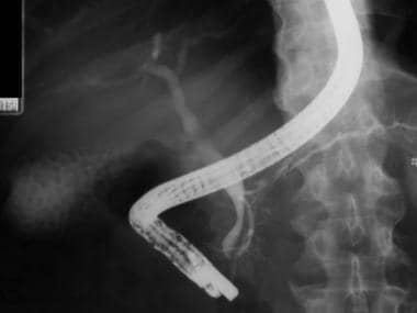
Pitfalls of MRCP in the diagnosis of pancreaticobiliary maljunction. (1/217)
CONTEXT: Magnetic resonance cholangiopancreatography (MRCP) is useful for examining the pancreatic duct system in patients with acute pancreatitis instead of using endoscopic retrograde cholangiopancreatography (ERCP), as ERCP-induced pancreatitis represents a serious problem. However, we present here a case of idiopathic acute pancreatitis in which MRCP suggested pancreaticobiliary maljunction, but ERCP indicated normal pancreaticobiliary union. CASE REPORT: A 22-year-old male was urgently admitted complaining of upper abdominal and back pain. He had no history of alcohol or drug intake. Serum amylase levels were elevated to 880 U/mL (reference value: less than 158 U/mL). Abdominal ultrasound demonstrated only a slight swelling of the pancreas, but no abnormal findings for the bile duct or gallbladder. Symptoms and hyperamylasemia improved with supportive therapy. Coronal heavily T2-weighted single-shot rapid acquisition with relaxation enhancement MRCP indicated a markedly long common channel, and pancreaticobiliary maljunction without biliary dilatation was diagnosed. Under the diagnosis of idiopathic acute pancreatitis associated with pancreaticobiliary maljunction without biliary dilatation, prophylactic laparoscopic cholecystectomy was planned. However, ERCP demonstrated a narrow main pancreatic duct and a normal common bile duct without the formation of a common channel. In a supine position, after withdrawal of the scope, the narrow main pancreatic duct at the head of the pancreas overlapped the lower common bile duct, giving the appearance of a long common channel as indicated by MRCP. CONCLUSIONS: In MRCP of cases with a narrow main pancreatic duct, there is a possibility for false-positive indications of pancreaticobiliary maljunction. MRCP with secretin stimulation or ERCP should be performed in such cases. (+info)Unilocular extrahepatic biliary cystadenoma mimicking choledochal cyst: a case report. (2/217)
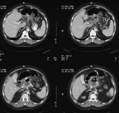
We report here on a case of extrahepatic biliary cystadenoma arising from the common hepatic duct. A 42-year-old woman was evaluated by us to find the cause of her jaundice. Ultrasonography and CT showed a cystic dilatation of the common hepatic duct and also marked dilatation of the intrahepatic duct. Direct cholangiography demonstrated a large filling defect between the left hepatic duct and the common hepatic duct; dilatation of the intrahepatic duct was also demonstrated. Following excision of the cystic mass, it was pathologically confirmed as a unilocular biliary mucinous cystadenoma arising from the common hepatic duct. (+info)A case of acute pancreatitis possibly associated with combined salicylate and simvastatin treatment. (3/217)
CONTEXT: Drug-induced acute pancreatitis is a rather rare clinical entity. From time to time, several cases have been reported in which statins or salicylates have been associated with the development of acute pancreatitis. There is only one report which implies the involvement of both drugs in pancreatic inflammation. CASE REPORT: A 58-year-old Caucasian male with a history of coronary heart disease and hypercholesterolemia, under treatment with acetyl-salicylate for 6 years and simvastatin for 2 months, presented to the Emergency Department of our hospital with epigastric pain and vomiting of 24-hour duration. The clinical and laboratory investigation led to the diagnosis of acute pancreatitis. Conservative and rich-in-fluid treatment resulted in clinical and laboratory amelioration, and the patient was discharged on day 15, after full restoration of his health. In our patient, all possible common causes of acute pancreatitis were excluded. CONCLUSION: Conclusion It is a rational assumption to connect this case to the co-administration of simvastatin and acetyl-salicylate. However, the pathophysiological mechanism behind the onset of acute pancreatitis due to a statin, or, even more, due to its combination with salicylate, remains vague. (+info)Preoperative evaluation of pancreaticobiliary tumor using MR multi-imaging techniques. (4/217)
AIM: To evaluate the clinical value of MR multi-imaging techniques in diagnosing and preoperative assessment of pancreaticobiliary tumor. METHODS: MR multi-imaging techniques, including MR cross-sectional imaging, MR cholangiopancreatography (MRCP) and 3D dynamic contrast-enhanced MR angiography (3D DCE MRA), were performed to make prospective diagnosis and preoperative evaluation in 28 patients with suspected pancreaticobiliary tumors. There were 17 cases of pancreatic adenocarcinoma, 8 cases of biliary system carcinoma and 3 cases of non-neoplastic lesions. RESULTS: Using MR multi-imaging techniques, the accuracy in diagnosing the patients with pancreaticobiliary tumors was 89.3% (25/28). The accuracy in detecting the range of tumor invasion was 80.3% (57/71). The sensitivity, specificity, accuracy, positive and negative predictive value of MR multi-imaging techniques in preoperative assessment of the resectability of pancreaticobiliary tumor were 83.3%, 89.5%, 88.0%, 71.4%, and 94.4%, respectively. There was well diagnostic consistency between MR multi-imaging techniques and CT (kappa = 0.64, P<0.01). The fusion image could be made from MRCP and 3D DCE MRA images. CONCLUSION: MR multi-imaging techniques can integrate the advantages of various MR images. The non-invasive "all-in-one" MR imaging protocol is the efficient method in diagnosing, staging and preoperative assessment of pancreaticobiliary tumor. (+info)Magnetic resonace appearance of gall bladder ascariasis. (5/217)
Ascariasis is a common disease in many developing countries and is a common cause of biliary and pancreatic diseases in endemic areas. Numerous studies have been published on biliary tract ascariasis. All these have documented ultrasonography as the primary imaging modality for biliary tract ascariasis. Magnetic Resonance Cholangiopancreatography (MRCP) has been the latest entrant for the study of bilary tract. MRCP findings of biliary tract ascariasis have been scarcely documented. MRCP is a unique non-invasive investigation for demonstrating ascariasis in gall bladder and bilary tract clearly. We present MR appearances of gall bladder and biliary tract in a proven case of biliary ascariasis. (+info)Dynamic MR cholangiography after fatty meal loading: cystic contractility and dynamic evaluation of biliary stasis. (6/217)
PURPOSE: Dynamic MR cholangiography was conducted on patients with cholelithiasis or choledocholithiasis who had consumed a fatty test meal (Molyork) and the cystic contractility and dynamics of biliary stasis was evaluated. SUBJECTS AND METHOD: The subjects were 25 with intracystic cholelithiasis, 10 with choledocholithiasis and 10 normal controls. For an imaging sequence, the rapid acquisition with relaxation enhancement (RARE) method was employed and imaging was conducted for 40 min (every 30 s following Molyork administration) without breath-holding. The gallbladder contraction ratio was computed and the contractile ratio for the common bile duct was calculated. To determine the bile flow to the duodenum, the high-intensity signal, indicating the flow from the lower common bile duct, and perfusion of the duodenum were observed in dynamic mode on the monitor with the naked eye and interpreted as positive bile flow. The frequency of this flow was visually monitored. RESULTS: The gallbladder contractile ratio was significantly reduced in patients with cholelithiasis or choledocholithiasis compared with the controls. In a comparison with the normal controls, no sequential changes were noted in the mean contractile ratio of the common bile duct of the patients with cholelithiasis or choledocholithiasis. The mean frequency of bile flow observed for each 40 min period was 13+/-2.4, 6+/-2.2, and 4+/-1.3 times for the controls, those with intracystic cholelithiasis, and those with choledocholithiasis, respectively. Compared with the controls, the latter two patient groups showed evident reductions in the frequency of bile flow to the duodenum (p<0.001). CONCLUSION: Dynamic MRC combined with Molyork loading makes it possible to compute cystic contractile ratios and perform a dynamic examination of bile flow under non-invasive, near-physiological conditions. (+info)Adenomyomatosis with marked subserosal fibrosis and lipomatosis of the gallbladder: mural stratification demonstrated with MR. (7/217)
The authors reported a case of fundal-type adenomyomatosis in which mural stratification corresponding to histopathological findings was clearly demonstrated with MR imaging. Single-shot fast spin echo images for MR cholangiopancreatography clearly visualized Rokitansky-Aschoff sinuses (RAS), which are a diagnostic clue for this disease. However, mural stratification comprising RAS with muscular proliferation, massive fibrosis and subserosal fat deposition was more precisely demonstrated in T(2)-weighted images obtained with fast spin echo. (+info)Complications of endoscopic retrograde cholangiography in the post-MRCP era: a tertiary center experience. (8/217)
AIM: To evaluate our experience in endoscopic retrograde cholangio-pancreatography (ERCP) in terms of fulfilling the ASGE guidelines in indications, positive findings, and complications in the post-magnetic resonance cholangiopancreatography (MRCP) era. METHODS: Between November 2001 and February 2003, consecutive ERCP cases were prospectively evaluated with regard to the indications, findings, cannulation techniques, devices used during the procedure, sedation given, duration of procedure, and complications. These data were entered in a database for subsequent processing and analysis. RESULTS: Of 336 cases, 21.4% were diagnostic and 78.6% therapeutic ERCP. The indications for ERCP fulfilled the ASGE guidelines in 323 cases (96.1%). Suspected bile duct stone was the most frequent indication (26.8%), and this was followed by cholangitis (24.4%), dilated common bile duct (14.9%), and cholestatic jaundice (13.4%). Cannulation success rate was 94%. Biliary sphincterotomy was performed in 175 (52.1%) patients. Repeated ERCP was performed on 31.5% of the patients. Overall, the complication rate was 9.8% with 0.3% being procedure-related mortality. The complications were pancreatitis (5.4%), bleeding (0.8%), cholangitis (2.4%) and others (1.5%). No significant difference was observed between the complication rate and the type of ERCP performed. CONCLUSION: Our study showed that post-ERCP complication rate was comparable with the other large prospective studies and there was no difference in the complication between the diagnostic and therapeutic ERCP. (+info)Endoscopic retrograde cholangiopancreatography (ERCP) is a medical procedure that combines upper gastrointestinal (GI) endoscopy and fluoroscopy to diagnose and treat certain problems of the bile ducts and pancreas.
During ERCP, a flexible endoscope (a long, thin, lighted tube with a camera on the end) is passed through the patient's mouth and throat, then through the stomach and into the first part of the small intestine (duodenum). A narrow plastic tube (catheter) is then inserted through the endoscope and into the bile ducts and/or pancreatic duct. Contrast dye is injected through the catheter, and X-rays are taken to visualize the ducts.
ERCP can be used to diagnose a variety of conditions affecting the bile ducts and pancreas, including gallstones, tumors, strictures (narrowing of the ducts), and chronic pancreatitis. It can also be used to treat certain conditions, such as removing gallstones from the bile duct or placing stents to keep the ducts open in cases of stricture.
ERCP is an invasive procedure that carries a risk of complications, including pancreatitis, infection, bleeding, and perforation (a tear in the lining of the GI tract). It should only be performed by experienced medical professionals in a hospital setting.
Magnetic resonance cholangiopancreatography (MRCP) is a non-invasive medical imaging technique that uses magnetic resonance imaging (MRI) to visualize the bile ducts and pancreatic duct. This diagnostic test does not use radiation like other imaging techniques such as computed tomography (CT) scans or endoscopic retrograde cholangiopancreatography (ERCP).
During an MRCP, the patient lies on a table that slides into the MRI machine. Contrast agents may be used to enhance the visibility of the ducts. The MRI machine uses a strong magnetic field and radio waves to produce detailed images of the internal structures, allowing radiologists to assess any abnormalities or blockages in the bile and pancreatic ducts.
MRCP is often used to diagnose conditions such as gallstones, tumors, inflammation, or strictures in the bile or pancreatic ducts. It can also be used to monitor the effectiveness of treatments for these conditions. However, it does not allow for therapeutic interventions like ERCP, which can remove stones or place stents.
Medical Definition:
Magnetic Resonance Imaging (MRI) is a non-invasive diagnostic imaging technique that uses a strong magnetic field and radio waves to create detailed cross-sectional or three-dimensional images of the internal structures of the body. The patient lies within a large, cylindrical magnet, and the scanner detects changes in the direction of the magnetic field caused by protons in the body. These changes are then converted into detailed images that help medical professionals to diagnose and monitor various medical conditions, such as tumors, injuries, or diseases affecting the brain, spinal cord, heart, blood vessels, joints, and other internal organs. MRI does not use radiation like computed tomography (CT) scans.
Choledocholithiasis is a medical condition characterized by the presence of one or more gallstones in the common bile duct, which is the tube that carries bile from the liver and gallbladder to the small intestine. Bile is a digestive fluid produced by the liver that helps break down fats in the small intestine. Gallstones are hardened deposits of digestive fluids that can form in the gallbladder or, less commonly, in the bile ducts.
Choledocholithiasis can cause a variety of symptoms, including abdominal pain, jaundice (yellowing of the skin and eyes), nausea, vomiting, and fever. If left untreated, it can lead to serious complications such as infection or inflammation of the bile ducts or pancreas, which can be life-threatening.
The condition is typically diagnosed through imaging tests such as ultrasound, CT scan, or MRI, and may require endoscopic or surgical intervention to remove the gallstones from the common bile duct.
Magnetic Resonance Spectroscopy (MRS) is a non-invasive diagnostic technique that provides information about the biochemical composition of tissues, including their metabolic state. It is often used in conjunction with Magnetic Resonance Imaging (MRI) to analyze various metabolites within body tissues, such as the brain, heart, liver, and muscles.
During MRS, a strong magnetic field, radio waves, and a computer are used to produce detailed images and data about the concentration of specific metabolites in the targeted tissue or organ. This technique can help detect abnormalities related to energy metabolism, neurotransmitter levels, pH balance, and other biochemical processes, which can be useful for diagnosing and monitoring various medical conditions, including cancer, neurological disorders, and metabolic diseases.
There are different types of MRS, such as Proton (^1^H) MRS, Phosphorus-31 (^31^P) MRS, and Carbon-13 (^13^C) MRS, each focusing on specific elements or metabolites within the body. The choice of MRS technique depends on the clinical question being addressed and the type of information needed for diagnosis or monitoring purposes.
Endoscopic sphincterotomy is a medical procedure that involves the use of an endoscope (a flexible tube with a light and camera) to cut the papilla of Vater, which contains the sphincter of Oddi muscle. This procedure is typically performed to treat gallstones or to manage other conditions related to the bile ducts or pancreatic ducts.
The sphincterotomy helps to widen the opening of the papilla, allowing stones or other obstructions to pass through more easily. It may also be used to relieve pressure and pain caused by spasms of the sphincter of Oddi muscle. The procedure is usually done under sedation or anesthesia and carries a risk of complications such as bleeding, infection, perforation, and pancreatitis.
Pancreatitis is a medical condition characterized by inflammation of the pancreas, a gland located in the abdomen that plays a crucial role in digestion and regulating blood sugar levels. The inflammation can be acute (sudden and severe) or chronic (persistent and recurring), and it can lead to various complications if left untreated.
Acute pancreatitis often results from gallstones or excessive alcohol consumption, while chronic pancreatitis may be caused by long-term alcohol abuse, genetic factors, autoimmune conditions, or metabolic disorders like high triglyceride levels. Symptoms of acute pancreatitis include severe abdominal pain, nausea, vomiting, fever, and increased heart rate, while chronic pancreatitis may present with ongoing abdominal pain, weight loss, diarrhea, and malabsorption issues due to impaired digestive enzyme production. Treatment typically involves supportive care, such as intravenous fluids, pain management, and addressing the underlying cause. In severe cases, hospitalization and surgery may be necessary.
Biliary tract diseases refer to a group of medical conditions that affect the biliary system, which includes the gallbladder, bile ducts, and liver. Bile is a digestive juice produced by the liver, stored in the gallbladder, and released into the small intestine through the bile ducts to help digest fats.
Biliary tract diseases can cause various symptoms such as abdominal pain, jaundice, fever, nausea, vomiting, and changes in stool color. Some of the common biliary tract diseases include:
1. Gallstones: Small, hard deposits that form in the gallbladder or bile ducts made up of cholesterol or bilirubin.
2. Cholecystitis: Inflammation of the gallbladder, often caused by gallstones.
3. Cholangitis: Infection or inflammation of the bile ducts.
4. Biliary dyskinesia: A motility disorder that affects the contraction and relaxation of the muscles in the biliary system.
5. Primary sclerosing cholangitis: A chronic autoimmune disease that causes scarring and narrowing of the bile ducts.
6. Biliary tract cancer: Rare cancers that affect the gallbladder, bile ducts, or liver.
Treatment for biliary tract diseases varies depending on the specific condition and severity but may include medications, surgery, or a combination of both.
The pancreatic ducts are a set of tubular structures within the pancreas that play a crucial role in the digestive system. The main pancreatic duct, also known as the duct of Wirsung, is responsible for transporting pancreatic enzymes and bicarbonate-rich fluid from the pancreas to the duodenum, which is the first part of the small intestine.
The exocrine portion of the pancreas contains numerous smaller ducts called interlobular ducts and intralobular ducts that merge and ultimately join the main pancreatic duct. This system ensures that the digestive enzymes and fluids produced by the pancreas are effectively delivered to the small intestine, where they aid in the breakdown and absorption of nutrients from food.
In addition to the main pancreatic duct, there is an accessory pancreatic duct, also known as Santorini's duct, which can sometimes join the common bile duct before emptying into the duodenum through a shared opening called the ampulla of Vater. However, in most individuals, the accessory pancreatic duct usually drains into the main pancreatic duct before entering the duodenum.
Hyperamylasemia is a medical condition characterized by an elevated level of amylase in the blood. Amylase is an enzyme that is primarily produced by the pancreas and salivary glands, and it plays a crucial role in digesting carbohydrates.
Normally, the levels of amylase in the blood are relatively low, but when there is damage to the pancreas or salivary glands, such as in cases of pancreatitis, salivary gland inflammation, or blockage, the levels of amylase can rise significantly. This condition is called hyperamylasemia.
Mild elevations in amylase levels may not cause any symptoms and may be discovered only during routine blood tests. However, more significant elevations can indicate a serious underlying medical condition that requires prompt treatment. Symptoms of hyperamylasemia may include abdominal pain, nausea, vomiting, fever, and rapid heartbeat.
It is important to note that hyperamylasemia can also be caused by non-pancreatic conditions such as macroamylasemia, a benign condition where large amylase-containing protein complexes are formed and circulate in the bloodstream, leading to elevated amylase levels. Therefore, it is essential to perform further diagnostic tests to determine the underlying cause of hyperamylasemia.
The common bile duct is a duct that results from the union of the cystic duct (which drains bile from the gallbladder) and the common hepatic duct (which drains bile from the liver). The common bile duct transports bile, a digestive enzyme, from the liver and gallbladder to the duodenum, which is the first part of the small intestine.
The common bile duct runs through the head of the pancreas before emptying into the second part of the duodenum, either alone or in conjunction with the pancreatic duct, via a small opening called the ampulla of Vater. The common bile duct plays a crucial role in the digestion of fats by helping to break them down into smaller molecules that can be absorbed by the body.
Common bile duct diseases refer to conditions that affect the common bile duct, a tube that carries bile from the liver and gallbladder into the small intestine. Some common examples of common bile duct diseases include:
1. Choledocholithiasis: This is the presence of stones (calculi) in the common bile duct, which can cause blockage, inflammation, and infection.
2. Cholangitis: This is an infection or inflammation of the common bile duct, often caused by obstruction due to stones, tumors, or strictures.
3. Common bile duct cancer (cholangiocarcinoma): This is a rare but aggressive cancer that arises from the cells lining the common bile duct.
4. Biliary strictures: These are narrowing or scarring of the common bile duct, which can be caused by injury, inflammation, or surgery.
5. Benign tumors: Non-cancerous growths in the common bile duct can also cause blockage and other symptoms.
Symptoms of common bile duct diseases may include abdominal pain, jaundice (yellowing of the skin and eyes), fever, chills, nausea, vomiting, and dark urine or light-colored stools. Treatment depends on the specific condition and severity but may include medications, endoscopic procedures, surgery, or a combination of these approaches.
Gallstones are small, hard deposits that form in the gallbladder, a small organ located under the liver. They can range in size from as small as a grain of sand to as large as a golf ball. Gallstones can be made of cholesterol, bile pigments, or calcium salts, or a combination of these substances.
There are two main types of gallstones: cholesterol stones and pigment stones. Cholesterol stones are the most common type and are usually yellow-green in color. They form when there is too much cholesterol in the bile, which causes it to become saturated and form crystals that eventually grow into stones. Pigment stones are smaller and darker in color, ranging from brown to black. They form when there is an excess of bilirubin, a waste product produced by the breakdown of red blood cells, in the bile.
Gallstones can cause symptoms such as abdominal pain, nausea, vomiting, and bloating, especially after eating fatty foods. In some cases, gallstones can lead to serious complications, such as inflammation of the gallbladder (cholecystitis), infection, or blockage of the bile ducts, which can cause jaundice, a yellowing of the skin and eyes.
The exact cause of gallstones is not fully understood, but risk factors include being female, older age, obesity, a family history of gallstones, rapid weight loss, diabetes, and certain medical conditions such as cirrhosis or sickle cell anemia. Treatment for gallstones may involve medication to dissolve the stones, shock wave therapy to break them up, or surgery to remove the gallbladder.
Cholangitis is a medical condition characterized by inflammation of the bile ducts, which are the tubes that carry bile from the liver to the small intestine. Bile is a digestive juice produced by the liver that helps break down fats in food.
There are two types of cholangitis: acute and chronic. Acute cholangitis is a sudden and severe infection that can cause symptoms such as abdominal pain, fever, jaundice (yellowing of the skin and eyes), and dark urine. It is usually caused by a bacterial infection that enters the bile ducts through a blockage or obstruction.
Chronic cholangitis, on the other hand, is a long-term inflammation of the bile ducts that can lead to scarring and narrowing of the ducts. This can cause symptoms such as abdominal pain, itching, and jaundice. Chronic cholangitis can be caused by various factors, including primary sclerosing cholangitis (an autoimmune disease), bile duct stones, or tumors in the bile ducts.
Treatment for cholangitis depends on the underlying cause of the condition. Antibiotics may be used to treat bacterial infections, and surgery may be necessary to remove blockages or obstructions in the bile ducts. In some cases, medications may be prescribed to manage symptoms and prevent further complications.
Pancreatic diseases refer to a group of medical conditions that affect the structure and function of the pancreas, a vital organ located in the abdomen. The pancreas has two main functions: an exocrine function, which involves the production of digestive enzymes that help break down food in the small intestine, and an endocrine function, which involves the production of hormones such as insulin and glucagon that regulate blood sugar levels.
Pancreatic diseases can be broadly classified into two categories: inflammatory and non-inflammatory. Inflammatory pancreatic diseases include conditions such as acute pancreatitis, which is characterized by sudden inflammation of the pancreas, and chronic pancreatitis, which is a long-term inflammation that can lead to scarring and loss of function.
Non-inflammatory pancreatic diseases include conditions such as pancreatic cancer, which is a malignant tumor that can arise from the cells of the pancreas, and benign tumors such as cysts or adenomas. Other non-inflammatory conditions include pancreatic insufficiency, which can occur when the pancreas does not produce enough digestive enzymes, and diabetes mellitus, which can result from impaired insulin production or action.
Overall, pancreatic diseases can have serious consequences on a person's health and quality of life, and early diagnosis and treatment are essential for optimal outcomes.
Bile duct diseases refer to a group of medical conditions that affect the bile ducts, which are tiny tubes that carry bile from the liver to the gallbladder and small intestine. Bile is a digestive juice produced by the liver that helps break down fats in food.
There are several types of bile duct diseases, including:
1. Choledocholithiasis: This occurs when stones form in the common bile duct, causing blockage and leading to symptoms such as abdominal pain, jaundice, and fever.
2. Cholangitis: This is an infection of the bile ducts that can cause inflammation, pain, and fever. It can occur due to obstruction of the bile ducts or as a complication of other medical procedures.
3. Primary Biliary Cirrhosis (PBC): This is a chronic autoimmune disease that affects the bile ducts in the liver, causing inflammation and scarring that can lead to cirrhosis and liver failure.
4. Primary Sclerosing Cholangitis (PSC): This is another autoimmune disease that causes inflammation and scarring of the bile ducts, leading to liver damage and potential liver failure.
5. Bile Duct Cancer: Also known as cholangiocarcinoma, this is a rare form of cancer that affects the bile ducts and can cause jaundice, abdominal pain, and weight loss.
6. Benign Strictures: These are narrowing of the bile ducts that can occur due to injury, inflammation, or surgery, leading to blockage and potential infection.
Symptoms of bile duct diseases may include jaundice, abdominal pain, fever, itching, dark urine, and light-colored stools. Treatment depends on the specific condition and may involve medication, surgery, or other medical interventions.
Cholangiography is a medical procedure that involves taking X-ray images of the bile ducts (the tubes that carry bile from the liver to the small intestine). This is typically done by injecting a contrast dye into the bile ducts through an endoscope or a catheter that has been inserted into the body.
There are several types of cholangiography, including:
* Endoscopic retrograde cholangiopancreatography (ERCP): This procedure involves inserting an endoscope through the mouth and down the throat into the small intestine. A dye is then injected into the bile ducts through a small tube that is passed through the endoscope.
* Percutaneous transhepatic cholangiography (PTC): This procedure involves inserting a needle through the skin and into the liver to inject the contrast dye directly into the bile ducts.
* Operative cholangiography: This procedure is performed during surgery to examine the bile ducts for any abnormalities or blockages.
Cholangiography can help diagnose a variety of conditions that affect the bile ducts, such as gallstones, tumors, or inflammation. It can also be used to guide treatment decisions, such as whether surgery is necessary to remove a blockage.
Obstructive Jaundice is a medical condition characterized by the yellowing of the skin, sclera (whites of the eyes), and mucous membranes due to the accumulation of bilirubin in the bloodstream. This occurs when there is an obstruction or blockage in the bile ducts that transport bile from the liver to the small intestine.
Bile, which contains bilirubin, aids in digestion and is usually released from the liver into the small intestine. When the flow of bile is obstructed, bilirubin builds up in the blood, causing jaundice. The obstruction can be caused by various factors, such as gallstones, tumors, or strictures in the bile ducts.
Obstructive jaundice may present with additional symptoms like dark urine, light-colored stools, itching, abdominal pain, and weight loss, depending on the cause and severity of the obstruction. It is essential to seek medical attention if jaundice is observed, as timely diagnosis and management can prevent potential complications, such as liver damage or infection.
Laparoscopic cholecystectomy is a surgical procedure to remove the gallbladder using a laparoscope, a thin tube with a camera, which allows the surgeon to view the internal structures on a video monitor. The surgery is performed through several small incisions in the abdomen, rather than a single large incision used in open cholecystectomy. This approach results in less postoperative pain, fewer complications, and shorter recovery time compared to open cholecystectomy.
The procedure is typically indicated for symptomatic gallstones or chronic inflammation of the gallbladder (cholecystitis), which can cause severe abdominal pain, nausea, vomiting, and fever. Laparoscopic cholecystectomy has become the standard of care for gallbladder removal due to its minimally invasive nature and excellent outcomes.
A biliary fistula is an abnormal connection or passage between the biliary system (which includes the gallbladder, bile ducts, and liver) and another organ or structure, usually in the abdominal cavity. This connection allows bile, which is a digestive fluid produced by the liver, to leak out of its normal pathway and into other areas of the body.
Biliary fistulas can occur as a result of trauma, surgery, infection, or inflammation in the biliary system. Symptoms may include abdominal pain, fever, jaundice (yellowing of the skin and eyes), nausea, vomiting, and clay-colored stools. Treatment typically involves addressing the underlying cause of the fistula, such as draining an infection or repairing damaged tissue, and diverting bile flow away from the site of the leak. In some cases, surgery may be necessary to repair the fistula.
Sphincter of Oddi dysfunction (SOD) is a condition characterized by abnormalities in the functioning of the Sphincter of Oddi, which is a muscular valve that controls the flow of bile and pancreatic juice from the pancreas and gallbladder into the duodenum (the first part of the small intestine).
In SOD, the sphincter may either fail to relax properly or become overactive, leading to a variety of symptoms such as abdominal pain, nausea, vomiting, bloating, and elevated liver enzymes. The condition can be classified into two types: Type I, which is associated with elevated liver enzymes and/or pancreatic enzymes, and Type II, which is characterized by abdominal pain without biochemical abnormalities.
The diagnosis of SOD typically involves a series of tests such as manometry (measuring the pressure inside the sphincter), endoscopic ultrasound, or magnetic resonance cholangiopancreatography (MRCP) to visualize the anatomy and function of the sphincter. Treatment options may include medications to relax the sphincter, endoscopic therapy to cut or stretch the muscle, or surgery in severe cases.
Magnetic Resonance Angiography (MRA) is a non-invasive medical imaging technique that uses magnetic fields and radio waves to create detailed images of the blood vessels or arteries within the body. It is a type of Magnetic Resonance Imaging (MRI) that focuses specifically on the circulatory system.
MRA can be used to diagnose and evaluate various conditions related to the blood vessels, such as aneurysms, stenosis (narrowing of the vessel), or the presence of plaques or tumors. It can also be used to plan for surgeries or other treatments related to the vascular system. The procedure does not use radiation and is generally considered safe, although people with certain implants like pacemakers may not be able to have an MRA due to safety concerns.
The ampulla of Vater, also known as hepatopancreatic ampulla, is a dilated portion of the common bile duct where it joins the main pancreatic duct and empties into the second part of the duodenum. It serves as a conduit for both bile from the liver and digestive enzymes from the pancreas to reach the small intestine, facilitating the digestion and absorption of nutrients. The ampulla of Vater is surrounded by a muscular sphincter, the sphincter of Oddi, which controls the flow of these secretions into the duodenum.
Cholestasis is a medical condition characterized by the interruption or reduction of bile flow from the liver to the small intestine. Bile is a digestive fluid produced by the liver that helps in the breakdown and absorption of fats. When the flow of bile is blocked or reduced, it can lead to an accumulation of bile components, such as bilirubin, in the blood, which can cause jaundice, itching, and other symptoms.
Cholestasis can be caused by various factors, including liver diseases (such as hepatitis, cirrhosis, or cancer), gallstones, alcohol abuse, certain medications, pregnancy, and genetic disorders. Depending on the underlying cause, cholestasis may be acute or chronic, and it can range from mild to severe in its symptoms and consequences. Treatment for cholestasis typically involves addressing the underlying cause and managing the symptoms with supportive care.
A Choledochal cyst is a congenital dilatation or abnormal enlargement of the bile ducts, which are the tubes that carry bile from the liver to the small intestine. Bile is a digestive juice produced by the liver that helps in the digestion of fats.
Choledochal cysts can be classified into several types based on their location and the anatomy of the biliary tree. The most common type, called Type I, involves dilatation of the common bile duct. Other types include dilatation of the intrahepatic bile ducts (Type II), dilatation of both the intrahepatic and extrahepatic bile ducts (Type III), and multiple cystic dilatations of the bile ducts (Type IV).
Choledochal cysts are more common in females than males, and they can present at any age. Symptoms may include abdominal pain, jaundice, vomiting, and fever. Complications of choledochal cysts can include bile duct stones, infection, and cancer. Treatment typically involves surgical removal of the cyst, followed by reconstruction of the biliary tree.
Extrahepatic cholestasis is a medical condition characterized by the impaired flow of bile outside of the liver. Bile is a digestive fluid produced by the liver that helps in the absorption and digestion of fats. When the flow of bile is obstructed or blocked, it can lead to an accumulation of bile components, such as bilirubin, in the bloodstream, resulting in jaundice, dark urine, light-colored stools, and itching.
Extrahepatic cholestasis can be caused by various factors, including gallstones, tumors, strictures, or inflammation of the bile ducts. It is essential to diagnose and treat extrahepatic cholestasis promptly to prevent further complications, such as liver damage or infection. Treatment options may include medications, endoscopic procedures, or surgery, depending on the underlying cause of the condition.
The Sphincter of Oddi is a muscular valve that controls the flow of bile and pancreatic juice from the pancreatic and bile ducts into the duodenum, which is the first part of the small intestine. It is named after Ruggero Oddi, an Italian physiologist who discovered it in 1887. The Sphincter of Oddi has two parts: the sphincter papillae, which surrounds the common opening of the pancreatic and bile ducts into the duodenum, and the sphincter choledochus, which is located more proximally in the bile duct. The contraction and relaxation of these muscles help regulate the release of digestive enzymes from the pancreas and the flow of bile from the liver to aid in digestion.
Endosonography, also known as endoscopic ultrasound (EUS), is a medical procedure that combines endoscopy and ultrasound to obtain detailed images and information about the digestive tract and surrounding organs. An endoscope, which is a flexible tube with a light and camera at its tip, is inserted through the mouth or rectum to reach the area of interest. A high-frequency ultrasound transducer at the tip of the endoscope generates sound waves that bounce off body tissues and create echoes, which are then translated into detailed images by a computer.
Endosonography allows doctors to visualize structures such as the esophageal, stomach, and intestinal walls, lymph nodes, blood vessels, and organs like the pancreas, liver, and gallbladder. It can help diagnose conditions such as tumors, inflammation, and infections, and it can also be used to guide biopsies or fine-needle aspirations of suspicious lesions.
Overall, endosonography is a valuable tool for the diagnosis and management of various gastrointestinal and related disorders.
Choledochostomy is a surgical procedure that involves creating an opening (stoma) into the common bile duct, which carries bile from the liver and gallbladder to the small intestine. This procedure is typically performed to relieve obstructions or blockages in the bile duct, such as those caused by gallstones, tumors, or scar tissue.
During the choledochostomy procedure, a surgeon makes an incision in the abdomen and exposes the common bile duct. The duct is then cut open, and a small tube (catheter) is inserted into the duct to allow bile to drain out of the body. The catheter may be left in place temporarily or permanently, depending on the underlying condition causing the obstruction.
Choledochostomy is typically performed as an open surgical procedure, but it can also be done using minimally invasive techniques such as laparoscopy or robotic-assisted surgery. As with any surgical procedure, choledochostomy carries risks such as bleeding, infection, and damage to surrounding tissues. However, these risks are generally low in the hands of an experienced surgeon.
Cholelithiasis is a medical term that refers to the presence of gallstones in the gallbladder. The gallbladder is a small pear-shaped organ located beneath the liver that stores bile, a digestive fluid produced by the liver. Gallstones are hardened deposits that can form in the gallbladder when substances in the bile, such as cholesterol or bilirubin, crystallize.
Gallstones can vary in size and may be as small as a grain of sand or as large as a golf ball. Some people with gallstones may not experience any symptoms, while others may have severe abdominal pain, nausea, vomiting, fever, and jaundice (yellowing of the skin and eyes) if the gallstones block the bile ducts.
Cholelithiasis is a common condition that affects millions of people worldwide, particularly women over the age of 40 and those with certain medical conditions such as obesity, diabetes, and rapid weight loss. If left untreated, gallstones can lead to serious complications such as inflammation of the gallbladder (cholecystitis), infection, or pancreatitis (inflammation of the pancreas). Treatment options for cholelithiasis include medication, shock wave lithotripsy (breaking up the gallstones with sound waves), and surgery to remove the gallbladder (cholecystectomy).
Magnetic Resonance Imaging (MRI) is a non-invasive diagnostic technique that uses a strong magnetic field and radio waves to create detailed cross-sectional images of the body's internal structures. In MRI, Cine is a specific mode of imaging that allows for the evaluation of moving structures, such as the heart, by acquiring and displaying a series of images in rapid succession. This technique is particularly useful in cardiac imaging, where it can help assess heart function, valve function, and blood flow. The term "Cine" refers to the continuous playback of these images, similar to watching a movie, allowing doctors to evaluate motion and timing within the heart.
Bile ducts are tubular structures that carry bile from the liver to the gallbladder for storage or directly to the small intestine to aid in digestion. There are two types of bile ducts: intrahepatic and extrahepatic. Intrahepatic bile ducts are located within the liver and drain bile from liver cells, while extrahepatic bile ducts are outside the liver and include the common hepatic duct, cystic duct, and common bile duct. These ducts can become obstructed or inflamed, leading to various medical conditions such as cholestasis, cholecystitis, and gallstones.
Cholecystectomy is a medical procedure to remove the gallbladder, a small pear-shaped organ located on the right side of the abdomen, just beneath the liver. The primary function of the gallbladder is to store and concentrate bile, a digestive fluid produced by the liver. During a cholecystectomy, the surgeon removes the gallbladder, usually due to the presence of gallstones or inflammation that can cause pain, infection, or other complications.
There are two primary methods for performing a cholecystectomy:
1. Open Cholecystectomy: In this traditional surgical approach, the surgeon makes an incision in the abdomen to access and remove the gallbladder. This method is typically used when there are complications or unique circumstances that make laparoscopic surgery difficult or risky.
2. Laparoscopic Cholecystectomy: This is a minimally invasive surgical procedure where the surgeon makes several small incisions in the abdomen, through which a thin tube with a camera (laparoscope) and specialized surgical instruments are inserted. The surgeon then guides these tools to remove the gallbladder while viewing the internal structures on a video monitor.
After the gallbladder is removed, bile flows directly from the liver into the small intestine through the common bile duct, and the body continues to function normally without any significant issues.
Gabexate is a medicinal drug that belongs to the class of agents known as serine protease inhibitors. It is used in the treatment and prevention of inflammation and damage to tissues caused by various surgical procedures, pancreatitis, and other conditions associated with the activation of proteolytic enzymes.
Gabexate works by inhibiting the activity of certain enzymes such as trypsin, chymotrypsin, and thrombin, which play a key role in the inflammatory response and blood clotting cascade. By doing so, it helps to reduce the release of inflammatory mediators, prevent further tissue damage, and promote healing.
Gabexate is available in various forms, including injectable solutions and enteric-coated tablets, and its use is typically reserved for clinical settings under the supervision of a healthcare professional. As with any medication, it should be used only under the direction of a qualified medical practitioner, and its potential benefits and risks should be carefully weighed against those of other available treatment options.
Contrast media are substances that are administered to a patient in order to improve the visibility of internal body structures or processes in medical imaging techniques such as X-rays, CT scans, MRI scans, and ultrasounds. These media can be introduced into the body through various routes, including oral, rectal, or intravenous administration.
Contrast media work by altering the appearance of bodily structures in imaging studies. For example, when a patient undergoes an X-ray examination, contrast media can be used to highlight specific organs, tissues, or blood vessels, making them more visible on the resulting images. In CT and MRI scans, contrast media can help to enhance the differences between normal and abnormal tissues, allowing for more accurate diagnosis and treatment planning.
There are several types of contrast media available, each with its own specific properties and uses. Some common examples include barium sulfate, which is used as a contrast medium in X-ray studies of the gastrointestinal tract, and iodinated contrast media, which are commonly used in CT scans to highlight blood vessels and other structures.
While contrast media are generally considered safe, they can sometimes cause adverse reactions, ranging from mild symptoms such as nausea or hives to more serious complications such as anaphylaxis or kidney damage. As a result, it is important for healthcare providers to carefully evaluate each patient's medical history and individual risk factors before administering contrast media.
Postcholecystectomy Syndrome is a condition that occurs in some patients following the surgical removal of the gallbladder (cholecystectomy). The syndrome encompasses a variety of symptoms such as abdominal pain, bloating, gas, indigestion, and diarrhea, which can be caused by several factors including:
1. Abnormal functioning or motility of the sphincter of Oddi (a muscle that controls the flow of bile and pancreatic juice into the small intestine)
2. Formation of gallstones in the bile ducts (choledocholithiasis)
3. Biliary dyskinesia (impaired functioning of the biliary tract muscles)
4. Persistent or recurrent infection or inflammation of the bile ducts (biliopathy)
5. Formation of abnormal bile-filled pouches (biliolethiasis or bile duct cysts)
6. Changes in bowel habits due to altered enterohepatic circulation of bile acids
The symptoms of Postcholecystectomy Syndrome can vary in severity and frequency, and they may appear soon after the surgery or develop months or even years later. The diagnosis of this condition typically involves a comprehensive medical evaluation, including a detailed history, physical examination, laboratory tests, and imaging studies such as ultrasound, CT scan, MRI, or endoscopic retrograde cholangiopancreatography (ERCP).
Treatment options for Postcholecystectomy Syndrome depend on the underlying cause of the symptoms and may include medications, dietary modifications, endoscopic procedures, or surgery. In some cases, the syndrome may resolve on its own without any specific treatment.
Drainage, in medical terms, refers to the removal of excess fluid or accumulated collections of fluids from various body parts or spaces. This is typically accomplished through the use of medical devices such as catheters, tubes, or drains. The purpose of drainage can be to prevent the buildup of fluids that may cause discomfort, infection, or other complications, or to treat existing collections of fluid such as abscesses, hematomas, or pleural effusions. Drainage may also be used as a diagnostic tool to analyze the type and composition of the fluid being removed.
The cystic duct is a short tube that connects the gallbladder to the common bile duct, which carries bile from the liver and gallbladder into the small intestine. The cystic duct allows bile to flow from the gallbladder into the common bile duct when it is needed for digestion. It is a part of the biliary system and plays an important role in the digestive process.
An endoscope is a medical device used for visualizing the internal surfaces of hollow organs or cavities in the body. Gastrointestinal (GI) endoscopes are specifically designed to examine the digestive tract, including the esophagus, stomach, small intestine, large intestine (colon), and rectum.
There are several types of GI endoscopes, including:
1. Gastroscope: Used for examining the stomach and upper part of the small intestine (duodenum).
2. Colonoscope: Used for examining the large intestine (colon) and rectum.
3. Sigmoidoscope: A shorter version of a colonoscope, used for examining the lower part of the large intestine (sigmoid colon) and rectum.
4. Duodenoscope: Used for examining and treating conditions in the pancreas and bile ducts.
5. Enteroscope: A longer endoscope used to examine the small intestine, which is more challenging to reach due to its length and location.
GI endoscopes typically consist of a long, flexible tube with a light source, camera, and channels for instruments to be passed through. The images captured by the camera are transmitted to a monitor, allowing the medical professional to inspect the internal surfaces of the digestive tract and perform various procedures, such as taking biopsies or removing polyps.
Common bile duct neoplasms refer to abnormal growths that can occur in the common bile duct, which is a tube that carries bile from the liver and gallbladder into the small intestine. These growths can be benign or malignant (cancerous).
Benign neoplasms of the common bile duct include papillomas, adenomas, and leiomyomas. Malignant neoplasms are typically adenocarcinomas, which arise from the glandular cells lining the duct. Other types of malignancies that can affect the common bile duct include cholangiocarcinoma, gallbladder carcinoma, and metastatic cancer from other sites.
Symptoms of common bile duct neoplasms may include jaundice (yellowing of the skin and eyes), abdominal pain, dark urine, and light-colored stools. Diagnosis may involve imaging tests such as CT scans or MRCP (magnetic resonance cholangiopancreatography) and biopsy to confirm the type of neoplasm. Treatment options depend on the type and stage of the neoplasm and may include surgery, radiation therapy, chemotherapy, or a combination of these approaches.
Double-balloon enteroscopy (DBE) is a medical procedure used to examine the small intestine, which is difficult to reach with traditional endoscopes due to its length and twists and turns. DBE uses a specialized endoscope with two inflatable balloons on its tip. The endoscope is inserted through the mouth or the rectum and advanced slowly into the small intestine while alternately inflating and deflating the balloons to help move the endoscope forward and provide better visualization of the intestinal lining.
DBE can be used for diagnostic purposes, such as evaluating obscure gastrointestinal bleeding, Crohn's disease, tumors, or polyps in the small intestine. It can also be used for therapeutic interventions, such as removing polyps, taking biopsies, or placing feeding tubes.
The procedure is usually done under sedation and takes several hours to complete. While it is considered a safe procedure, potential risks include perforation of the intestinal wall, bleeding, and adverse reactions to the anesthesia.
Gallbladder diseases refer to a range of conditions that affect the function and structure of the gallbladder, a small pear-shaped organ located beneath the liver. The primary role of the gallbladder is to store, concentrate, and release bile into the small intestine to aid in digesting fats. Gallbladder diseases can be chronic or acute and may cause various symptoms, discomfort, or complications if left untreated. Here are some common gallbladder diseases with brief definitions:
1. Cholelithiasis: The presence of gallstones within the gallbladder. Gallstones are small, hard deposits made of cholesterol, bilirubin, or a combination of both, which can vary in size from tiny grains to several centimeters.
2. Cholecystitis: Inflammation of the gallbladder, often caused by obstruction of the cystic duct (the tube connecting the gallbladder and the common bile duct) due to a gallstone. This condition can be acute or chronic and may cause abdominal pain, fever, and tenderness in the right upper quadrant of the abdomen.
3. Choledocholithiasis: The presence of gallstones within the common bile duct, which can lead to obstruction, jaundice, and potential infection of the biliary system (cholangitis).
4. Acalculous gallbladder disease: Gallbladder dysfunction or inflammation without the presence of gallstones. This condition is often seen in critically ill patients and can lead to similar symptoms as cholecystitis.
5. Gallbladder polyps: Small growths attached to the inner wall of the gallbladder. While most polyps are benign, some may have malignant potential, especially if they are larger than 1 cm in size or associated with certain risk factors.
6. Gallbladder cancer: A rare form of cancer that originates in the gallbladder tissue. It is often asymptomatic in its early stages and can be challenging to diagnose. Symptoms may include abdominal pain, jaundice, or a palpable mass in the right upper quadrant of the abdomen.
It is essential to consult with a healthcare professional if experiencing symptoms related to gallbladder disease for proper diagnosis and treatment.
X-ray computed tomography (CT or CAT scan) is a medical imaging method that uses computer-processed combinations of many X-ray images taken from different angles to produce cross-sectional (tomographic) images (virtual "slices") of the body. These cross-sectional images can then be used to display detailed internal views of organs, bones, and soft tissues in the body.
The term "computed tomography" is used instead of "CT scan" or "CAT scan" because the machines take a series of X-ray measurements from different angles around the body and then use a computer to process these data to create detailed images of internal structures within the body.
CT scanning is a noninvasive, painless medical test that helps physicians diagnose and treat medical conditions. CT imaging provides detailed information about many types of tissue including lung, bone, soft tissue and blood vessels. CT examinations can be performed on every part of the body for a variety of reasons including diagnosis, surgical planning, and monitoring of therapeutic responses.
In computed tomography (CT), an X-ray source and detector rotate around the patient, measuring the X-ray attenuation at many different angles. A computer uses this data to construct a cross-sectional image by the process of reconstruction. This technique is called "tomography". The term "computed" refers to the use of a computer to reconstruct the images.
CT has become an important tool in medical imaging and diagnosis, allowing radiologists and other physicians to view detailed internal images of the body. It can help identify many different medical conditions including cancer, heart disease, lung nodules, liver tumors, and internal injuries from trauma. CT is also commonly used for guiding biopsies and other minimally invasive procedures.
In summary, X-ray computed tomography (CT or CAT scan) is a medical imaging technique that uses computer-processed combinations of many X-ray images taken from different angles to produce cross-sectional images of the body. It provides detailed internal views of organs, bones, and soft tissues in the body, allowing physicians to diagnose and treat medical conditions.
The biliary tract is a system of ducts that transport bile from the liver to the gallbladder and then to the small intestine. Bile is a digestive fluid produced by the liver that helps in the breakdown and absorption of fats in the small intestine. The main components of the biliary tract are:
1. Intrahepatic bile ducts: These are the smaller branches of bile ducts located within the liver that collect bile from the liver cells or hepatocytes.
2. Gallbladder: A small pear-shaped organ located beneath the liver, which stores and concentrates bile received from the intrahepatic bile ducts. The gallbladder releases bile into the small intestine when food is ingested, particularly fats, to aid digestion.
3. Common hepatic duct: This is a duct that forms by the union of the right and left hepatic ducts, which carry bile from the right and left lobes of the liver, respectively.
4. Cystic duct: A short duct that connects the gallbladder to the common hepatic duct, forming the beginning of the common bile duct.
5. Common bile duct: This is a larger duct formed by the union of the common hepatic duct and the cystic duct. It carries bile from the liver and gallbladder into the small intestine.
6. Pancreatic duct: A separate duct that originates from the pancreas, a gland located near the liver and stomach. The pancreatic duct joins the common bile duct just before they both enter the duodenum, the first part of the small intestine.
7. Ampulla of Vater: This is the dilated portion where the common bile duct and the pancreatic duct join together and empty their contents into the duodenum through a shared opening called the papilla of Vater.
Disorders related to the biliary tract include gallstones, cholecystitis (inflammation of the gallbladder), bile duct stones, bile duct strictures or obstructions, and primary sclerosing cholangitis, among others.
Bile duct neoplasms, also known as cholangiocarcinomas, refer to a group of malignancies that arise from the bile ducts. These are the tubes that carry bile from the liver to the gallbladder and small intestine. Bile duct neoplasms can be further classified based on their location as intrahepatic (within the liver), perihilar (at the junction of the left and right hepatic ducts), or distal (in the common bile duct).
These tumors are relatively rare, but their incidence has been increasing in recent years. They can cause a variety of symptoms, including jaundice, abdominal pain, weight loss, and fever. The diagnosis of bile duct neoplasms typically involves imaging studies such as CT or MRI scans, as well as blood tests to assess liver function. In some cases, a biopsy may be necessary to confirm the diagnosis.
Treatment options for bile duct neoplasms depend on several factors, including the location and stage of the tumor, as well as the patient's overall health. Surgical resection is the preferred treatment for early-stage tumors, while chemotherapy and radiation therapy may be used in more advanced cases. For patients who are not candidates for surgery, palliative treatments such as stenting or bypass procedures may be recommended to relieve symptoms and improve quality of life.
Chronic pancreatitis is a long-standing inflammation of the pancreas that leads to irreversible structural changes and impaired function of the pancreas. It is characterized by recurrent or persistent abdominal pain, often radiating to the back, and maldigestion with steatorrhea (fatty stools) due to exocrine insufficiency. The pancreatic damage results from repeated episodes of acute pancreatitis, alcohol abuse, genetic predisposition, or autoimmune processes. Over time, the pancreas may lose its ability to produce enough digestive enzymes and hormones like insulin, which can result in diabetes mellitus. Chronic pancreatitis also increases the risk of developing pancreatic cancer.
Mirizzi Syndrome is a relatively uncommon condition that involves the compression of the common hepatic duct (the tube that carries bile from the liver to the gallbladder and small intestine) by a gallstone in the cystic duct or the neck of the gallbladder. This compression can lead to obstruction of the bile flow, causing symptoms such as jaundice, abdominal pain, and elevated levels of liver enzymes.
The syndrome is classified into two types based on the degree of involvement:
* Type I: The gallstone causes external compression of the common hepatic duct without any structural damage to the bile ducts.
* Type II: The gallstone erodes into the common hepatic duct, creating a fistula (an abnormal connection) between the gallbladder and the bile duct.
Mirizzi Syndrome can be challenging to diagnose due to its rarity and nonspecific symptoms. Imaging tests such as ultrasound, CT scan, or MRI may help in identifying the presence of a gallstone and the compression of the bile duct. Endoscopic retrograde cholangiopancreatography (ERCP) is often used to confirm the diagnosis and provide treatment by removing the gallstone and placing a stent to relieve the obstruction. In some cases, surgery may be required to remove the gallbladder and repair any damage to the bile ducts.
The common hepatic duct is a medical term that refers to the duct in the liver responsible for carrying bile from the liver. More specifically, it is the duct that results from the convergence of the right and left hepatic ducts, which themselves carry bile from the right and left lobes of the liver, respectively. The common hepatic duct then joins with the cystic duct from the gallbladder to form the common bile duct, which ultimately drains into the duodenum, a part of the small intestine.
The primary function of the common hepatic duct is to transport bile, a digestive juice produced by the liver, to the small intestine. Bile helps break down fats during the digestion process, making it possible for the body to absorb them properly. Any issues or abnormalities in the common hepatic duct can lead to problems with bile flow and potentially cause health complications such as jaundice, gallstones, or liver damage.
Pathological constriction refers to an abnormal narrowing or tightening of a body passage or organ, which can interfere with the normal flow of blood, air, or other substances through the area. This constriction can occur due to various reasons such as inflammation, scarring, or abnormal growths, and can affect different parts of the body, including blood vessels, airways, intestines, and ureters. Pathological constriction can lead to a range of symptoms and complications depending on its location and severity, and may require medical intervention to correct.
Endoscopy is a medical procedure that involves the use of an endoscope, which is a flexible tube with a light and camera at the end, to examine the interior of a body cavity or organ. The endoscope is inserted through a natural opening in the body, such as the mouth or anus, or through a small incision. The images captured by the camera are transmitted to a monitor, allowing the physician to visualize the internal structures and detect any abnormalities, such as inflammation, ulcers, or tumors. Endoscopy can also be used for diagnostic purposes, such as taking tissue samples for biopsy, or for therapeutic purposes, such as removing polyps or performing minimally invasive surgeries.
Retrospective studies, also known as retrospective research or looking back studies, are a type of observational study that examines data from the past to draw conclusions about possible causal relationships between risk factors and outcomes. In these studies, researchers analyze existing records, medical charts, or previously collected data to test a hypothesis or answer a specific research question.
Retrospective studies can be useful for generating hypotheses and identifying trends, but they have limitations compared to prospective studies, which follow participants forward in time from exposure to outcome. Retrospective studies are subject to biases such as recall bias, selection bias, and information bias, which can affect the validity of the results. Therefore, retrospective studies should be interpreted with caution and used primarily to generate hypotheses for further testing in prospective studies.
Pancreatic neoplasms refer to abnormal growths in the pancreas that can be benign or malignant. The pancreas is a gland located behind the stomach that produces hormones and digestive enzymes. Pancreatic neoplasms can interfere with the normal functioning of the pancreas, leading to various health complications.
Benign pancreatic neoplasms are non-cancerous growths that do not spread to other parts of the body. They are usually removed through surgery to prevent any potential complications, such as blocking the bile duct or causing pain.
Malignant pancreatic neoplasms, also known as pancreatic cancer, are cancerous growths that can invade and destroy surrounding tissues and organs. They can also spread (metastasize) to other parts of the body, such as the liver, lungs, or bones. Pancreatic cancer is often aggressive and difficult to treat, with a poor prognosis.
There are several types of pancreatic neoplasms, including adenocarcinomas, neuroendocrine tumors, solid pseudopapillary neoplasms, and cystic neoplasms. The specific type of neoplasm is determined through various diagnostic tests, such as imaging studies, biopsies, and blood tests. Treatment options depend on the type, stage, and location of the neoplasm, as well as the patient's overall health and preferences.
Treatment outcome is a term used to describe the result or effect of medical treatment on a patient's health status. It can be measured in various ways, such as through symptoms improvement, disease remission, reduced disability, improved quality of life, or survival rates. The treatment outcome helps healthcare providers evaluate the effectiveness of a particular treatment plan and make informed decisions about future care. It is also used in clinical research to compare the efficacy of different treatments and improve patient care.
Gastroenterology is a branch of medicine that deals with the study, diagnosis, management, and treatment of disorders and diseases of the digestive system, also known as the gastrointestinal (GI) tract. This includes the esophagus, stomach, small intestine, large intestine (colon), liver, pancreas, gallbladder, and bile ducts.
Physicians who specialize in this field are called gastroenterologists. They undergo extensive training in internal medicine and then complete a fellowship in gastroenterology, where they gain expertise in using various diagnostic techniques such as endoscopy, colonoscopy, and radiologic imaging to evaluate GI tract disorders.
Gastroenterologists treat a wide range of conditions affecting the digestive system, including but not limited to:
1. Gastroesophageal reflux disease (GERD)
2. Inflammatory bowel disease (IBD), which includes Crohn's disease and ulcerative colitis
3. Irritable bowel syndrome (IBS)
4. Celiac disease
5. Hepatitis and other liver diseases
6. Pancreatic disorders, such as pancreatitis
7. Gastrointestinal cancers, like colon, rectal, and esophageal cancer
8. Functional gastrointestinal disorders (FGIDs), which include chronic abdominal pain, bloating, and difficulty with bowel movements
By focusing on the prevention, diagnosis, and treatment of digestive diseases, gastroenterologists play a crucial role in maintaining overall health and well-being for their patients.
The pancreas is a glandular organ located in the abdomen, posterior to the stomach. It has both exocrine and endocrine functions. The exocrine portion of the pancreas consists of acinar cells that produce and secrete digestive enzymes into the duodenum via the pancreatic duct. These enzymes help in the breakdown of proteins, carbohydrates, and fats in food.
The endocrine portion of the pancreas consists of clusters of cells called islets of Langerhans, which include alpha, beta, delta, and F cells. These cells produce and secrete hormones directly into the bloodstream, including insulin, glucagon, somatostatin, and pancreatic polypeptide. Insulin and glucagon are critical regulators of blood sugar levels, with insulin promoting glucose uptake and storage in tissues and glucagon stimulating glycogenolysis and gluconeogenesis to raise blood glucose when it is low.
A pancreatic pseudocyst is a fluid-filled sac that forms in the abdomen, usually as a result of pancreatitis or trauma to the pancreas. It is composed of cells and tissues from the pancreas, along with enzymes, debris, and fluids. Unlike true cysts, pseudocysts do not have an epithelial lining. They can vary in size and may cause symptoms such as abdominal pain, nausea, vomiting, or fever. In some cases, they may resolve on their own, but larger or symptomatic pseudocysts may require medical intervention, such as drainage or surgery.
Amylases are enzymes that break down complex carbohydrates, such as starch and glycogen, into simpler sugars like maltose, glucose, and maltotriose. There are several types of amylases found in various organisms, including humans.
In humans, amylases are produced by the pancreas and salivary glands. Pancreatic amylase is released into the small intestine where it helps to digest dietary carbohydrates. Salivary amylase, also known as alpha-amylase, is secreted into the mouth and begins breaking down starches in food during chewing.
Deficiency or absence of amylases can lead to difficulties in digesting carbohydrates and may cause symptoms such as bloating, diarrhea, and abdominal pain. Elevated levels of amylase in the blood may indicate conditions such as pancreatitis, pancreatic cancer, or other disorders affecting the pancreas.
Jaundice is a medical condition characterized by the yellowing of the skin, sclera (whites of the eyes), and mucous membranes due to an excess of bilirubin in the bloodstream. Bilirubin is a yellow-orange pigment produced when hemoglobin from red blood cells is broken down. Normally, bilirubin is processed by the liver and excreted through bile into the digestive system. However, if there's an issue with bilirubin metabolism or elimination, it can accumulate in the body, leading to jaundice.
Jaundice can be a symptom of various underlying conditions, such as liver diseases (hepatitis, cirrhosis), gallbladder issues (gallstones, tumors), or blood disorders (hemolysis). It is essential to consult a healthcare professional if jaundice is observed, as it may indicate a severe health problem requiring prompt medical attention.
Catheterization is a medical procedure in which a catheter (a flexible tube) is inserted into the body to treat various medical conditions or for diagnostic purposes. The specific definition can vary depending on the area of medicine and the particular procedure being discussed. Here are some common types of catheterization:
1. Urinary catheterization: This involves inserting a catheter through the urethra into the bladder to drain urine. It is often performed to manage urinary retention, monitor urine output in critically ill patients, or assist with surgical procedures.
2. Cardiac catheterization: A procedure where a catheter is inserted into a blood vessel, usually in the groin or arm, and guided to the heart. This allows for various diagnostic tests and treatments, such as measuring pressures within the heart chambers, assessing blood flow, or performing angioplasty and stenting of narrowed coronary arteries.
3. Central venous catheterization: A catheter is inserted into a large vein, typically in the neck, chest, or groin, to administer medications, fluids, or nutrition, or to monitor central venous pressure.
4. Peritoneal dialysis catheterization: A catheter is placed into the abdominal cavity for individuals undergoing peritoneal dialysis, a type of kidney replacement therapy.
5. Neurological catheterization: In some cases, a catheter may be inserted into the cerebrospinal fluid space (lumbar puncture) or the brain's ventricular system (ventriculostomy) to diagnose or treat various neurological conditions.
These are just a few examples of catheterization procedures in medicine. The specific definition and purpose will depend on the medical context and the particular organ or body system involved.
Diffusion Magnetic Resonance Imaging (MRI) is a non-invasive medical imaging technique that uses magnetic fields and radio waves to produce detailed images of the body's internal structures, particularly the brain and nervous system. In diffusion MRI, the movement of water molecules in biological tissues is measured and analyzed to generate contrast in the images based on the microstructural properties of the tissue.
Diffusion MRI is unique because it allows for the measurement of water diffusion in various directions, which can reveal important information about the organization and integrity of nerve fibers in the brain. This technique has been widely used in research and clinical settings to study a variety of neurological conditions, including stroke, traumatic brain injury, multiple sclerosis, and neurodegenerative diseases such as Alzheimer's disease.
In summary, diffusion MRI is a specialized type of MRI that measures the movement of water molecules in biological tissues to generate detailed images of the body's internal structures, particularly the brain and nervous system. It provides valuable information about the microstructural properties of tissues and has important applications in both research and clinical settings.
The brain is the central organ of the nervous system, responsible for receiving and processing sensory information, regulating vital functions, and controlling behavior, movement, and cognition. It is divided into several distinct regions, each with specific functions:
1. Cerebrum: The largest part of the brain, responsible for higher cognitive functions such as thinking, learning, memory, language, and perception. It is divided into two hemispheres, each controlling the opposite side of the body.
2. Cerebellum: Located at the back of the brain, it is responsible for coordinating muscle movements, maintaining balance, and fine-tuning motor skills.
3. Brainstem: Connects the cerebrum and cerebellum to the spinal cord, controlling vital functions such as breathing, heart rate, and blood pressure. It also serves as a relay center for sensory information and motor commands between the brain and the rest of the body.
4. Diencephalon: A region that includes the thalamus (a major sensory relay station) and hypothalamus (regulates hormones, temperature, hunger, thirst, and sleep).
5. Limbic system: A group of structures involved in emotional processing, memory formation, and motivation, including the hippocampus, amygdala, and cingulate gyrus.
The brain is composed of billions of interconnected neurons that communicate through electrical and chemical signals. It is protected by the skull and surrounded by three layers of membranes called meninges, as well as cerebrospinal fluid that provides cushioning and nutrients.
Roux-en-Y anastomosis is a type of surgical connection between two parts of the gastrointestinal tract, typically performed during gastric bypass surgery for weight loss. In this procedure, a small pouch is created from the upper stomach, and the remaining portion of the stomach is bypassed. The Roux limb, a segment of the small intestine, is then connected to both the pouch and the bypassed stomach, creating two separate channels for food and digestive juices to mix. This surgical technique helps to reduce the amount of food that can be consumed and absorbed, leading to weight loss.
Lithiasis is a medical term that refers to the formation of stones or calculi in various organs of the body. These stones can develop in the kidneys (nephrolithiasis), gallbladder (cholelithiasis), urinary bladder (cystolithiasis), or salivary glands (sialolithiasis). The stones are usually composed of minerals and organic substances, and their formation can be influenced by various factors such as diet, dehydration, genetic predisposition, and chronic inflammation. Lithiasis can cause a range of symptoms depending on the location and size of the stone, including pain, obstruction, infection, and damage to surrounding tissues. Treatment may involve medication, shock wave lithotripsy, or surgical removal of the stones.
Sensitivity and specificity are statistical measures used to describe the performance of a diagnostic test or screening tool in identifying true positive and true negative results.
* Sensitivity refers to the proportion of people who have a particular condition (true positives) who are correctly identified by the test. It is also known as the "true positive rate" or "recall." A highly sensitive test will identify most or all of the people with the condition, but may also produce more false positives.
* Specificity refers to the proportion of people who do not have a particular condition (true negatives) who are correctly identified by the test. It is also known as the "true negative rate." A highly specific test will identify most or all of the people without the condition, but may also produce more false negatives.
In medical testing, both sensitivity and specificity are important considerations when evaluating a diagnostic test. High sensitivity is desirable for screening tests that aim to identify as many cases of a condition as possible, while high specificity is desirable for confirmatory tests that aim to rule out the condition in people who do not have it.
It's worth noting that sensitivity and specificity are often influenced by factors such as the prevalence of the condition in the population being tested, the threshold used to define a positive result, and the reliability and validity of the test itself. Therefore, it's important to consider these factors when interpreting the results of a diagnostic test.
Intrahepatic bile ducts are the small tubular structures inside the liver that collect bile from the liver cells (hepatocytes). Bile is a digestive fluid produced by the liver that helps in the absorption of fats and fat-soluble vitamins from food. The intrahepatic bile ducts merge to form larger ducts, which eventually exit the liver and join with the cystic duct from the gallbladder to form the common bile duct. The common bile duct then empties into the duodenum, the first part of the small intestine, where bile aids in digestion. Intrahepatic bile ducts can become obstructed or damaged due to various conditions such as gallstones, tumors, or inflammation, leading to complications like jaundice, liver damage, and infection.
Biliary tract surgical procedures refer to a range of operations that involve the biliary system, which includes the liver, gallbladder, and bile ducts. These procedures can be performed for various reasons, including the treatment of gallstones, bile duct injuries, tumors, or other conditions affecting the biliary tract. Here are some examples of biliary tract surgical procedures:
1. Cholecystectomy: This is the surgical removal of the gallbladder, which is often performed to treat symptomatic gallstones or chronic cholecystitis (inflammation of the gallbladder). It can be done as an open procedure or laparoscopically.
2. Bile duct exploration: This procedure involves opening the common bile duct to remove stones, strictures, or tumors. It is often performed during a cholecystectomy if there is suspicion of common bile duct involvement.
3. Hepaticojejunostomy: This operation connects the liver's bile ducts directly to a portion of the small intestine called the jejunum, bypassing a damaged or obstructed segment of the biliary tract. It is often performed for benign or malignant conditions affecting the bile ducts.
4. Roux-en-Y hepaticojejunostomy: This procedure involves creating a Y-shaped limb of jejunum and connecting it to the liver's bile ducts, bypassing the common bile duct and duodenum. It is often performed for complex biliary tract injuries or malignancies.
5. Whipple procedure (pancreaticoduodenectomy): This extensive operation involves removing the head of the pancreas, the duodenum, a portion of the jejunum, the gallbladder, and the common bile duct. It is performed for malignancies involving the pancreas, bile duct, or duodenum.
6. Liver resection: This procedure involves removing a portion of the liver to treat primary liver tumors (hepatocellular carcinoma or cholangiocarcinoma) or metastatic cancer from other organs.
7. Biliary stenting or bypass: These minimally invasive procedures involve placing a stent or creating a bypass to relieve bile duct obstructions caused by tumors, strictures, or stones. They can be performed endoscopically (ERCP) or percutaneously (PTC).
8. Cholecystectomy: This procedure involves removing the gallbladder, often for symptomatic cholelithiasis (gallstones) or cholecystitis (inflammation of the gallbladder). It can be performed laparoscopically or open.
9. Biliary drainage: This procedure involves placing a catheter to drain bile from the liver or bile ducts, often for acute or chronic obstructions caused by tumors, strictures, or stones. It can be performed endoscopically (ERCP) or percutaneously (PTC).
10. Bilioenteric anastomosis: This procedure involves connecting the biliary tract to a portion of the small intestine, often for benign or malignant conditions affecting the bile ducts or pancreas. It can be performed open or laparoscopically.
Gadolinium DTPA (Diethylenetriaminepentaacetic acid) is a type of gadolinium-based contrast agent (GBCA) used in medical imaging, particularly magnetic resonance imaging (MRI) and magnetic resonance angiography (MRA). It functions as a paramagnetic substance that enhances the visibility of internal body structures during these imaging techniques.
The compound Gadolinium DTPA is formed when gadolinium ions are bound to diethylenetriaminepentaacetic acid, a chelating agent. This binding helps to make the gadolinium ion safer for use in medical imaging by reducing its toxicity and improving its stability in the body.
Gadolinium DTPA is eliminated from the body primarily through the kidneys, making it important to monitor renal function before administering this contrast agent. In some cases, Gadolinium DTPA may cause adverse reactions, including allergic-like responses and nephrogenic systemic fibrosis (NSF) in patients with impaired kidney function.
Gallbladder neoplasms refer to abnormal growths in the tissue of the gallbladder, which can be benign or malignant. Benign neoplasms are non-cancerous and typically do not spread to other parts of the body. Malignant neoplasms, also known as gallbladder cancer, can invade nearby tissues and organs and may metastasize (spread) to distant parts of the body. Gallbladder neoplasms can cause symptoms such as abdominal pain, jaundice, and nausea, but they are often asymptomatic until they have advanced to an advanced stage. The exact causes of gallbladder neoplasms are not fully understood, but risk factors include gallstones, chronic inflammation of the gallbladder, and certain inherited genetic conditions.
Biliary tract neoplasms refer to abnormal growths or tumors that develop in the biliary system, which includes the gallbladder, bile ducts inside and outside the liver, and the ducts that connect the liver to the small intestine. These neoplasms can be benign (non-cancerous) or malignant (cancerous).
Malignant biliary tract neoplasms are often referred to as cholangiocarcinoma if they originate in the bile ducts, or gallbladder cancer if they arise in the gallbladder. These cancers are relatively rare but can be aggressive and difficult to treat. They can cause symptoms such as jaundice (yellowing of the skin and eyes), abdominal pain, weight loss, and dark urine.
Risk factors for biliary tract neoplasms include chronic inflammation of the biliary system, primary sclerosing cholangitis, liver cirrhosis, hepatitis B or C infection, parasitic infections, and certain genetic conditions. Early detection and treatment can improve outcomes for patients with these neoplasms.
Endoscopy of the digestive system, also known as gastrointestinal (GI) endoscopy, is a medical procedure that allows healthcare professionals to visually examine the inside lining of the digestive tract using a flexible tube with a light and camera attached to it, called an endoscope. This procedure can help diagnose and treat various conditions affecting the digestive system, including gastroesophageal reflux disease (GERD), ulcers, inflammatory bowel disease (IBD), and cancer.
There are several types of endoscopy procedures that focus on different parts of the digestive tract:
1. Esophagogastroduodenoscopy (EGD): This procedure examines the esophagus, stomach, and duodenum (the first part of the small intestine). It is often used to investigate symptoms such as difficulty swallowing, abdominal pain, or bleeding in the upper GI tract.
2. Colonoscopy: This procedure explores the large intestine (colon) and rectum. It is commonly performed to screen for colon cancer, as well as to diagnose and treat conditions like inflammatory bowel disease, diverticulosis, or polyps.
3. Sigmoidoscopy: Similar to a colonoscopy, this procedure examines the lower part of the colon (sigmoid colon) and rectum. It is often used as a screening tool for colon cancer and to investigate symptoms like rectal bleeding or changes in bowel habits.
4. Upper GI endoscopy: This procedure focuses on the esophagus, stomach, and duodenum, using a thin, flexible tube with a light and camera attached to it. It is used to diagnose and treat conditions such as GERD, ulcers, and difficulty swallowing.
5. Capsule endoscopy: This procedure involves swallowing a small capsule containing a camera that captures images of the digestive tract as it passes through. It can help diagnose conditions in the small intestine that may be difficult to reach with traditional endoscopes.
Endoscopy is typically performed under sedation or anesthesia to ensure patient comfort during the procedure. The images captured by the endoscope are displayed on a monitor, allowing the healthcare provider to assess the condition of the digestive tract and make informed treatment decisions.
Adenomyoma is a benign (non-cancerous) growth that occurs when the glands and muscle tissue from the lining of the uterus (endometrium) become embedded in the muscular wall of the uterus (myometrium). This condition most commonly affects women in their 40s and 50s, and it can cause symptoms such as heavy menstrual bleeding, painful periods, and pelvic pain or discomfort.
The term "adenomyoma" is derived from two words: "adeno," which means gland, and "myoma," which refers to a benign muscle tumor. Therefore, an adenomyoma can be thought of as a benign growth that contains both glandular tissue and muscle tissue.
Adenomyomas are typically found in the lower part of the uterus, near the cervix, and they can vary in size from small nodules to larger masses. In some cases, adenomyomas may cause no symptoms at all, while in other cases, they can lead to significant discomfort and pain.
The exact cause of adenomyoma is not fully understood, but it is thought to be related to hormonal factors, as well as trauma or injury to the uterus. Treatment options for adenomyoma may include medication to manage symptoms, such as pain relievers or hormone therapy, or surgical intervention, such as a hysterectomy (removal of the uterus).
Duodenal diseases refer to a range of medical conditions that affect the duodenum, which is the first part of the small intestine. Here are some examples of duodenal diseases:
1. Duodenitis: This is inflammation of the duodenum, which can cause symptoms such as abdominal pain, nausea, vomiting, and bloating. Duodenitis can be caused by bacterial or viral infections, excessive use of nonsteroidal anti-inflammatory drugs (NSAIDs), or chronic inflammation due to conditions like Crohn's disease.
2. Peptic ulcers: These are sores that develop in the lining of the duodenum, usually as a result of infection with Helicobacter pylori bacteria or long-term use of NSAIDs. Symptoms can include abdominal pain, bloating, and heartburn.
3. Duodenal cancer: This is a rare type of cancer that affects the duodenum. Symptoms can include abdominal pain, weight loss, and blood in the stool.
4. Celiac disease: This is an autoimmune disorder that causes the immune system to attack the lining of the small intestine in response to gluten, a protein found in wheat, barley, and rye. This can lead to inflammation and damage to the duodenum.
5. Duodenal diverticulosis: This is a condition in which small pouches form in the lining of the duodenum. While many people with duodenal diverticulosis do not experience symptoms, some may develop complications such as inflammation or infection.
6. Duodenal atresia: This is a congenital condition in which the duodenum does not form properly, leading to blockage of the intestine. This can cause symptoms such as vomiting and difficulty feeding in newborns.
Cholangiocarcinoma is a type of cancer that arises from the cells that line the bile ducts, which are small tubes that carry digestive enzymes from the liver to the small intestine. It can occur in different parts of the bile duct system, including the bile ducts inside the liver (intrahepatic), the bile ducts outside the liver (extrahepatic), and the area where the bile ducts join the pancreas and small intestine (ampulla of Vater).
Cholangiocarcinoma is a relatively rare cancer, but its incidence has been increasing in recent years. It can be difficult to diagnose because its symptoms are often nonspecific and similar to those of other conditions, such as gallstones or pancreatitis. Treatment options depend on the location and stage of the cancer, and may include surgery, radiation therapy, chemotherapy, or a combination of these approaches.
Technetium Tc 99m Lidofenin is a radiopharmaceutical used in nuclear medicine imaging procedures, specifically for hepatobiliary scintigraphy. It is a technetium-labeled compound, where the radioisotope technetium-99m (^99m^Tc) is bound to lidofenin, a liver-imaging agent.
The compound is used to assess the function and anatomy of the liver, gallbladder, and biliary system. After intravenous administration, Technetium Tc 99m Lidofenin is taken up by hepatocytes (liver cells) and excreted into the bile ducts and ultimately into the small intestine. The distribution and excretion of this radiopharmaceutical can be monitored using a gamma camera, providing functional information about the liver and biliary system.
It is essential to note that the use of Technetium Tc 99m Lidofenin should be under the guidance and supervision of healthcare professionals trained in nuclear medicine, as its administration and handling require specific expertise and safety measures due to the radioactive nature of the compound.
A jejunostomy is a surgical procedure where an opening (stoma) is created in the lower part of the small intestine, called the jejunum. This stoma allows for the passage of nutrients and digestive enzymes from the small intestine into a tube or external pouch, bypassing the mouth, esophagus, stomach, and upper small intestine (duodenum).
Jejunostomy is typically performed to provide enteral nutrition support in patients who are unable to consume food or liquids by mouth due to various medical conditions such as dysphagia, gastroparesis, bowel obstruction, or after certain surgical procedures. The jejunostomy tube can be used for short-term or long-term nutritional support, depending on the patient's needs and underlying medical condition.
Nuclear Magnetic Resonance (NMR) Biomolecular is a research technique that uses magnetic fields and radio waves to study the structure and dynamics of biological molecules, such as proteins and nucleic acids. This technique measures the magnetic properties of atomic nuclei within these molecules, specifically their spin, which can be influenced by the application of an external magnetic field.
When a sample is placed in a strong magnetic field, the nuclei absorb and emit electromagnetic radiation at specific frequencies, known as resonance frequencies, which are determined by the molecular structure and environment of the nuclei. By analyzing these resonance frequencies and their interactions, researchers can obtain detailed information about the three-dimensional structure, dynamics, and interactions of biomolecules.
NMR spectroscopy is a non-destructive technique that allows for the study of biological molecules in solution, which makes it an important tool for understanding the function and behavior of these molecules in their natural environment. Additionally, NMR can be used to study the effects of drugs, ligands, and other small molecules on biomolecular structure and dynamics, making it a valuable tool in drug discovery and development.
"Calculi" is a medical term that refers to abnormal concretions or hard masses formed within the body, usually in hollow organs or cavities. These masses are typically composed of minerals such as calcium oxalate, calcium phosphate, or magnesium ammonium phosphate, and can vary in size from tiny granules to large stones. The plural form of the Latin word "calculus" (meaning "pebble"), calculi are commonly known as "stones." They can occur in various locations within the body, including the kidneys, gallbladder, urinary bladder, and prostate gland. The presence of calculi can cause a range of symptoms, such as pain, obstruction, infection, or inflammation, depending on their size, location, and composition.
Gadolinium is a rare earth metal that is used as a contrast agent in medical imaging techniques such as Magnetic Resonance Imaging (MRI) and Magnetic Resonance Angiography (MRA). It works by shortening the relaxation time of protons in tissues, which enhances the visibility of internal body structures on the images. Gadolinium-based contrast agents are injected into the patient's bloodstream during the imaging procedure.
It is important to note that in some individuals, gadolinium-based contrast agents can cause a condition called nephrogenic systemic fibrosis (NSF), which is a rare but serious disorder that affects people with severe kidney disease. NSF causes thickening and hardening of the skin, joints, eyes, and internal organs. Therefore, it is essential to evaluate a patient's renal function before administering gadolinium-based contrast agents.
Biliary dyskinesia is a medical condition characterized by abnormal or impaired motility of the biliary system, which includes the gallbladder and the bile ducts. This can lead to symptoms such as abdominal pain, bloating, nausea, and vomiting, particularly after eating fatty foods.
In biliary dyskinesia, the gallbladder may not contract properly or may contract too much, leading to a backup of bile in the liver or bile ducts. This can cause inflammation and irritation of the biliary system and surrounding tissues.
The condition is often diagnosed through imaging tests such as ultrasound, nuclear medicine scans, or MRI, which can help assess gallbladder function and detect any abnormalities in the biliary system. Treatment for biliary dyskinesia may include medications to improve gallbladder motility, dietary modifications, or in some cases, surgery to remove the gallbladder.
Breath holding is a physiological response where an individual holds their breath, intentionally or unintentionally, for a period of time. This can occur in various situations such as during swimming underwater, while lifting heavy weights, or in response to emotional stress or pain. In some cases, it can also be associated with certain medical conditions like seizures or syncope (fainting).
In the context of medical terminology, breath holding is often described as "voluntary" or "involuntary." Voluntary breath-holding is when an individual consciously chooses to hold their breath, while involuntary breath-holding occurs unconsciously, usually in response to a trigger such as a sudden increase in carbon dioxide levels or a decrease in oxygen levels.
It's important to note that prolonged breath-holding can be dangerous and may lead to hypoxia (lack of oxygen) and hypercapnia (excessive carbon dioxide), which can cause dizziness, loss of consciousness, or even more severe consequences such as brain damage or death. Therefore, it's essential not to hold one's breath for extended periods and seek medical attention if experiencing any symptoms related to breath-holding.
An endoscope is a medical device used for examining the interior of a body cavity or organ. It consists of a long, thin, flexible (or rigid) tube with a light and a camera at one end. The other end is connected to a video monitor that displays the images captured by the camera. Endoscopes can be inserted through natural openings in the body, such as the mouth or anus, or through small incisions. They are used for diagnostic purposes, as well as for performing various medical procedures, including biopsies and surgeries. Different types of endoscopes include gastroscopes, colonoscopes, bronchoscopes, and arthroscopes, among others.
Three-dimensional (3D) imaging in medicine refers to the use of technologies and techniques that generate a 3D representation of internal body structures, organs, or tissues. This is achieved by acquiring and processing data from various imaging modalities such as X-ray computed tomography (CT), magnetic resonance imaging (MRI), ultrasound, or confocal microscopy. The resulting 3D images offer a more detailed visualization of the anatomy and pathology compared to traditional 2D imaging techniques, allowing for improved diagnostic accuracy, surgical planning, and minimally invasive interventions.
In 3D imaging, specialized software is used to reconstruct the acquired data into a volumetric model, which can be manipulated and viewed from different angles and perspectives. This enables healthcare professionals to better understand complex anatomical relationships, detect abnormalities, assess disease progression, and monitor treatment response. Common applications of 3D imaging include neuroimaging, orthopedic surgery planning, cancer staging, dental and maxillofacial reconstruction, and interventional radiology procedures.
Duodenoscopy is a medical procedure that involves the insertion of a duodenoscope, which is a flexible, lighted tube with a camera and tiny tools on the end, through the mouth and down the throat to examine the upper part of the small intestine (duodenum) and the opening of the bile and pancreatic ducts.
During the procedure, the doctor can take tissue samples for biopsy, remove polyps or other abnormal growths, or perform other interventions as needed. Duodenoscopy is commonly used to diagnose and treat conditions such as gastrointestinal bleeding, inflammation, infection, and cancer.
It's important to note that duodenoscopes have been associated with the spread of antibiotic-resistant bacteria in some cases, so healthcare providers must follow strict cleaning and disinfection protocols to minimize this risk.
An acute disease is a medical condition that has a rapid onset, develops quickly, and tends to be short in duration. Acute diseases can range from minor illnesses such as a common cold or flu, to more severe conditions such as pneumonia, meningitis, or a heart attack. These types of diseases often have clear symptoms that are easy to identify, and they may require immediate medical attention or treatment.
Acute diseases are typically caused by an external agent or factor, such as a bacterial or viral infection, a toxin, or an injury. They can also be the result of a sudden worsening of an existing chronic condition. In general, acute diseases are distinct from chronic diseases, which are long-term medical conditions that develop slowly over time and may require ongoing management and treatment.
Examples of acute diseases include:
* Acute bronchitis: a sudden inflammation of the airways in the lungs, often caused by a viral infection.
* Appendicitis: an inflammation of the appendix that can cause severe pain and requires surgical removal.
* Gastroenteritis: an inflammation of the stomach and intestines, often caused by a viral or bacterial infection.
* Migraine headaches: intense headaches that can last for hours or days, and are often accompanied by nausea, vomiting, and sensitivity to light and sound.
* Myocardial infarction (heart attack): a sudden blockage of blood flow to the heart muscle, often caused by a buildup of plaque in the coronary arteries.
* Pneumonia: an infection of the lungs that can cause coughing, chest pain, and difficulty breathing.
* Sinusitis: an inflammation of the sinuses, often caused by a viral or bacterial infection.
It's important to note that while some acute diseases may resolve on their own with rest and supportive care, others may require medical intervention or treatment to prevent complications and promote recovery. If you are experiencing symptoms of an acute disease, it is always best to seek medical attention to ensure proper diagnosis and treatment.
Brain mapping is a broad term that refers to the techniques used to understand the structure and function of the brain. It involves creating maps of the various cognitive, emotional, and behavioral processes in the brain by correlating these processes with physical locations or activities within the nervous system. Brain mapping can be accomplished through a variety of methods, including functional magnetic resonance imaging (fMRI), positron emission tomography (PET) scans, electroencephalography (EEG), and others. These techniques allow researchers to observe which areas of the brain are active during different tasks or thoughts, helping to shed light on how the brain processes information and contributes to our experiences and behaviors. Brain mapping is an important area of research in neuroscience, with potential applications in the diagnosis and treatment of neurological and psychiatric disorders.
The digestive system, also known as the gastrointestinal (GI) tract, is a series of organs that process food and liquids into nutrients and waste. Digestive system diseases refer to any conditions that affect the normal functioning of this system, leading to impaired digestion, absorption, or elimination of food and fluids.
Some common examples of digestive system diseases include:
1. Gastroesophageal Reflux Disease (GERD): A condition where stomach acid flows back into the esophagus, causing symptoms such as heartburn, chest pain, and difficulty swallowing.
2. Peptic Ulcer Disease: Sores or ulcers that develop in the lining of the stomach or duodenum, often caused by bacterial infection or long-term use of nonsteroidal anti-inflammatory drugs (NSAIDs).
3. Inflammatory Bowel Disease (IBD): A group of chronic inflammatory conditions that affect the intestines, including Crohn's disease and ulcerative colitis.
4. Irritable Bowel Syndrome (IBS): A functional gastrointestinal disorder characterized by abdominal pain, bloating, and changes in bowel habits.
5. Celiac Disease: An autoimmune disorder where the ingestion of gluten leads to damage in the small intestine, impairing nutrient absorption.
6. Diverticular Disease: A condition that affects the colon, characterized by the formation of small pouches or sacs (diverticula) that can become inflamed or infected.
7. Constipation: A common digestive system issue where bowel movements occur less frequently than usual or are difficult to pass.
8. Diarrhea: Loose, watery stools that occur more frequently than normal, often accompanied by cramps and bloating.
9. Gallstones: Small, hard deposits that form in the gallbladder, causing pain, inflammation, and potential blockages of the bile ducts.
10. Hepatitis: Inflammation of the liver, often caused by viral infections or toxins, leading to symptoms such as jaundice, fatigue, and abdominal pain.
These are just a few examples of digestive system disorders that can affect overall health and quality of life. If you experience any persistent or severe digestive symptoms, it is important to seek medical attention from a healthcare professional.
Reproducibility of results in a medical context refers to the ability to obtain consistent and comparable findings when a particular experiment or study is repeated, either by the same researcher or by different researchers, following the same experimental protocol. It is an essential principle in scientific research that helps to ensure the validity and reliability of research findings.
In medical research, reproducibility of results is crucial for establishing the effectiveness and safety of new treatments, interventions, or diagnostic tools. It involves conducting well-designed studies with adequate sample sizes, appropriate statistical analyses, and transparent reporting of methods and findings to allow other researchers to replicate the study and confirm or refute the results.
The lack of reproducibility in medical research has become a significant concern in recent years, as several high-profile studies have failed to produce consistent findings when replicated by other researchers. This has led to increased scrutiny of research practices and a call for greater transparency, rigor, and standardization in the conduct and reporting of medical research.
Prospective studies, also known as longitudinal studies, are a type of cohort study in which data is collected forward in time, following a group of individuals who share a common characteristic or exposure over a period of time. The researchers clearly define the study population and exposure of interest at the beginning of the study and follow up with the participants to determine the outcomes that develop over time. This type of study design allows for the investigation of causal relationships between exposures and outcomes, as well as the identification of risk factors and the estimation of disease incidence rates. Prospective studies are particularly useful in epidemiology and medical research when studying diseases with long latency periods or rare outcomes.
The Predictive Value of Tests, specifically the Positive Predictive Value (PPV) and Negative Predictive Value (NPV), are measures used in diagnostic tests to determine the probability that a positive or negative test result is correct.
Positive Predictive Value (PPV) is the proportion of patients with a positive test result who actually have the disease. It is calculated as the number of true positives divided by the total number of positive results (true positives + false positives). A higher PPV indicates that a positive test result is more likely to be a true positive, and therefore the disease is more likely to be present.
Negative Predictive Value (NPV) is the proportion of patients with a negative test result who do not have the disease. It is calculated as the number of true negatives divided by the total number of negative results (true negatives + false negatives). A higher NPV indicates that a negative test result is more likely to be a true negative, and therefore the disease is less likely to be present.
The predictive value of tests depends on the prevalence of the disease in the population being tested, as well as the sensitivity and specificity of the test. A test with high sensitivity and specificity will generally have higher predictive values than a test with low sensitivity and specificity. However, even a highly sensitive and specific test can have low predictive values if the prevalence of the disease is low in the population being tested.
Abdominal pain is defined as discomfort or painful sensation in the abdomen. The abdomen is the region of the body between the chest and the pelvis, and contains many important organs such as the stomach, small intestine, large intestine, liver, gallbladder, pancreas, and spleen. Abdominal pain can vary in intensity from mild to severe, and can be acute or chronic depending on the underlying cause.
Abdominal pain can have many different causes, ranging from benign conditions such as gastritis, indigestion, or constipation, to more serious conditions such as appendicitis, inflammatory bowel disease, or abdominal aortic aneurysm. The location, quality, and duration of the pain can provide important clues about its cause. For example, sharp, localized pain in the lower right quadrant of the abdomen may indicate appendicitis, while crampy, diffuse pain in the lower abdomen may suggest irritable bowel syndrome.
It is important to seek medical attention if you experience severe or persistent abdominal pain, especially if it is accompanied by other symptoms such as fever, vomiting, or bloody stools. A thorough physical examination, including a careful history and a focused abdominal exam, can help diagnose the underlying cause of the pain and guide appropriate treatment.
Insufflation is a medical term that refers to the act of introducing a gas or vapor into a body cavity or passage, typically through a tube or surgical instrument. This procedure is often used in medical and surgical settings for various purposes, such as:
* To administer anesthesia during surgery (e.g., introducing nitrous oxide or other gases into the lungs)
* To introduce medication or other substances into the body (e.g., insufflating steroids into a joint)
* To perform diagnostic procedures (e.g., insufflating air or a contrast agent into the gastrointestinal tract to visualize it with X-rays)
* To clean out a body cavity (e.g., irrigating and insufflating the bladder during urological procedures).
It's important to note that insufflation should be performed under controlled conditions, as there are potential risks associated with introducing gases or vapors into the body, such as barotrauma (damage caused by changes in pressure) and infection.
A pancreatic fistula is an abnormal connection or passage between the pancreas and another organ, often the digestive system. It usually occurs as a complication following trauma, surgery, or inflammation of the pancreas (such as pancreatitis). The pancreas secretes digestive enzymes, and when these enzymes escape the pancreas through a damaged or disrupted duct, they can cause irritation and inflammation in nearby tissues, leading to the formation of a fistula.
Pancreatic fistulas are typically characterized by the drainage of pancreatic fluid, which contains high levels of digestive enzymes, into other parts of the body. This can lead to various symptoms, including abdominal pain, swelling, fever, and malnutrition. Treatment may involve surgical repair of the fistula, as well as supportive care such as antibiotics, nutritional support, and drainage of any fluid collections.
Deep sedation, also known as general anesthesia, is a drug-induced depression of consciousness during which patients cannot be easily aroused but respond purposefully following repeated or painful stimulation. It is characterized by the loss of protective reflexes such as cough and gag, and the ability to ventilate spontaneously may be impaired. Patients may require assistance in maintaining a patent airway, and positive pressure ventilation may be required.
Deep sedation/general anesthesia is typically used for surgical procedures or other medical interventions that require patients to be completely unaware and immobile, and it is administered by trained anesthesia professionals who monitor and manage the patient's vital signs and level of consciousness throughout the procedure.
A stent is a small mesh tube that's used to treat narrow or weak arteries. Arteries are blood vessels that carry blood away from your heart to other parts of your body. A stent is placed in an artery as part of a procedure called angioplasty. Angioplasty restores blood flow through narrowed or blocked arteries by inflating a tiny balloon inside the blocked artery to widen it.
The stent is then inserted into the widened artery to keep it open. The stent is usually made of metal, but some are coated with medication that is slowly and continuously released to help prevent the formation of scar tissue in the artery. This can reduce the chance of the artery narrowing again.
Stents are also used in other parts of the body, such as the neck (carotid artery) and kidneys (renal artery), to help maintain blood flow and prevent blockages. They can also be used in the urinary system to treat conditions like ureteropelvic junction obstruction or narrowing of the urethra.
Computer-assisted image processing is a medical term that refers to the use of computer systems and specialized software to improve, analyze, and interpret medical images obtained through various imaging techniques such as X-ray, CT (computed tomography), MRI (magnetic resonance imaging), ultrasound, and others.
The process typically involves several steps, including image acquisition, enhancement, segmentation, restoration, and analysis. Image processing algorithms can be used to enhance the quality of medical images by adjusting contrast, brightness, and sharpness, as well as removing noise and artifacts that may interfere with accurate diagnosis. Segmentation techniques can be used to isolate specific regions or structures of interest within an image, allowing for more detailed analysis.
Computer-assisted image processing has numerous applications in medical imaging, including detection and characterization of lesions, tumors, and other abnormalities; assessment of organ function and morphology; and guidance of interventional procedures such as biopsies and surgeries. By automating and standardizing image analysis tasks, computer-assisted image processing can help to improve diagnostic accuracy, efficiency, and consistency, while reducing the potential for human error.
Postoperative complications refer to any unfavorable condition or event that occurs during the recovery period after a surgical procedure. These complications can vary in severity and may include, but are not limited to:
1. Infection: This can occur at the site of the incision or inside the body, such as pneumonia or urinary tract infection.
2. Bleeding: Excessive bleeding (hemorrhage) can lead to a drop in blood pressure and may require further surgical intervention.
3. Blood clots: These can form in the deep veins of the legs (deep vein thrombosis) and can potentially travel to the lungs (pulmonary embolism).
4. Wound dehiscence: This is when the surgical wound opens up, which can lead to infection and further complications.
5. Pulmonary issues: These include atelectasis (collapsed lung), pneumonia, or respiratory failure.
6. Cardiovascular problems: These include abnormal heart rhythms (arrhythmias), heart attack, or stroke.
7. Renal failure: This can occur due to various reasons such as dehydration, blood loss, or the use of certain medications.
8. Pain management issues: Inadequate pain control can lead to increased stress, anxiety, and decreased mobility.
9. Nausea and vomiting: These can be caused by anesthesia, opioid pain medication, or other factors.
10. Delirium: This is a state of confusion and disorientation that can occur in the elderly or those with certain medical conditions.
Prompt identification and management of these complications are crucial to ensure the best possible outcome for the patient.
Surface Plasmon Resonance (SPR) is a physical phenomenon that occurs at the interface between a metal and a dielectric material, when electromagnetic radiation (usually light) is shone on it. It involves the collective oscillation of free electrons in the metal, known as surface plasmons, which are excited by the incident light. The resonance condition is met when the momentum and energy of the photons match those of the surface plasmons, leading to a strong absorption of light and an evanescent wave that extends into the dielectric material.
In the context of medical diagnostics and research, SPR is often used as a sensitive and label-free detection technique for biomolecular interactions. By immobilizing one binding partner (e.g., a receptor or antibody) onto the metal surface and flowing the other partner (e.g., a ligand or antigen) over it, changes in the refractive index at the interface can be measured in real-time as the plasmons are disturbed by the presence of bound molecules. This allows for the quantification of binding affinities, kinetics, and specificity with high sensitivity and selectivity.
Neoplasms: Neoplasms refer to abnormal growths of tissue that can be benign (non-cancerous) or malignant (cancerous). They occur when the normal control mechanisms that regulate cell growth and division are disrupted, leading to uncontrolled cell proliferation.
Cystic Neoplasms: Cystic neoplasms are tumors that contain fluid-filled sacs or cysts. These tumors can be benign or malignant and can occur in various organs of the body, including the pancreas, ovary, and liver.
Mucinous Neoplasms: Mucinous neoplasms are a type of cystic neoplasm that is characterized by the production of mucin, a gel-like substance produced by certain types of cells. These tumors can occur in various organs, including the ovary, pancreas, and colon. Mucinous neoplasms can be benign or malignant, and malignant forms are often aggressive and have a poor prognosis.
Serous Neoplasms: Serous neoplasms are another type of cystic neoplasm that is characterized by the production of serous fluid, which is a thin, watery fluid. These tumors commonly occur in the ovary and can be benign or malignant. Malignant serous neoplasms are often aggressive and have a poor prognosis.
In summary, neoplasms refer to abnormal tissue growths that can be benign or malignant. Cystic neoplasms contain fluid-filled sacs and can occur in various organs of the body. Mucinous neoplasms produce a gel-like substance called mucin and can also occur in various organs, while serous neoplasms produce thin, watery fluid and commonly occur in the ovary. Both mucinous and serous neoplasms can be benign or malignant, with malignant forms often being aggressive and having a poor prognosis.
Butylscopolammonium Bromide is an anticholinergic drug, which is used as a smooth muscle relaxant and an anti-spasmodic agent. It works by blocking the action of acetylcholine, a neurotransmitter in the body, on certain types of receptors, leading to relaxation of smooth muscles and reduction of spasms.
This medication is commonly used to treat gastrointestinal disorders such as irritable bowel syndrome, intestinal cramps, and spastic constipation. It may also be used in the management of bladder disorders, including neurogenic bladder and urinary incontinence.
The drug is available in various forms, including tablets, suppositories, and solutions for injection. The dosage and route of administration depend on the specific condition being treated and the patient's overall health status. As with any medication, Butylscopolammonium Bromide can cause side effects, such as dry mouth, blurred vision, dizziness, and constipation. It should be used under the guidance of a healthcare professional to ensure safe and effective treatment.
Diagnostic imaging is a medical specialty that uses various technologies to produce visual representations of the internal structures and functioning of the body. These images are used to diagnose injury, disease, or other abnormalities and to monitor the effectiveness of treatment. Common modalities of diagnostic imaging include:
1. Radiography (X-ray): Uses ionizing radiation to produce detailed images of bones, teeth, and some organs.
2. Computed Tomography (CT) Scan: Combines X-ray technology with computer processing to create cross-sectional images of the body.
3. Magnetic Resonance Imaging (MRI): Uses a strong magnetic field and radio waves to generate detailed images of soft tissues, organs, and bones.
4. Ultrasound: Employs high-frequency sound waves to produce real-time images of internal structures, often used for obstetrics and gynecology.
5. Nuclear Medicine: Involves the administration of radioactive tracers to assess organ function or detect abnormalities within the body.
6. Positron Emission Tomography (PET) Scan: Uses a small amount of radioactive material to produce detailed images of metabolic activity in the body, often used for cancer detection and monitoring treatment response.
7. Fluoroscopy: Utilizes continuous X-ray imaging to observe moving structures or processes within the body, such as swallowing studies or angiography.
Diagnostic imaging plays a crucial role in modern medicine, allowing healthcare providers to make informed decisions about patient care and treatment plans.
Sclerosing cholangitis is a chronic progressive disease characterized by inflammation and scarring (fibrosis) of the bile ducts, leading to their narrowing or obstruction. This results in impaired bile flow from the liver to the small intestine, which can cause damage to the liver cells and eventually result in cirrhosis and liver failure.
The condition often affects both the intrahepatic (within the liver) and extrahepatic (outside the liver) bile ducts. The exact cause of sclerosing cholangitis is not known, but it is believed to involve an autoimmune response, genetic predisposition, and environmental factors.
Symptoms of sclerosing cholangitis may include jaundice (yellowing of the skin and eyes), itching, abdominal pain, fatigue, weight loss, dark urine, and light-colored stools. The diagnosis is typically made through imaging tests such as magnetic resonance cholangiopancreatography (MRCP) or endoscopic retrograde cholangiopancreatography (ERCP), which can visualize the bile ducts and detect any abnormalities.
Treatment for sclerosing cholangitis is aimed at managing symptoms, preventing complications, and slowing down the progression of the disease. This may include medications to relieve itching, antibiotics to treat infections, and drugs to reduce inflammation and improve bile flow. In severe cases, a liver transplant may be necessary.
Pancreatic function tests are a group of medical tests that are used to assess the functionality and health of the pancreas. The pancreas is a vital organ located in the abdomen, which has two main functions: an exocrine function, where it releases digestive enzymes into the small intestine to help break down food; and an endocrine function, where it produces hormones such as insulin and glucagon that regulate blood sugar levels.
Pancreatic function tests typically involve measuring the levels of digestive enzymes in the blood or stool, or assessing the body's ability to digest and absorb certain nutrients. Some common pancreatic function tests include:
1. Serum amylase and lipase tests: These tests measure the levels of digestive enzymes called amylase and lipase in the blood. Elevated levels of these enzymes may indicate pancreatitis or other conditions affecting the pancreas.
2. Fecal elastase test: This test measures the level of elastase, an enzyme produced by the pancreas, in a stool sample. Low levels of elastase may indicate exocrine pancreatic insufficiency (EPI), a condition where the pancreas is not producing enough digestive enzymes.
3. Secretin stimulation test: This test involves administering a medication called secretin, which stimulates the pancreas to release digestive enzymes. The levels of these enzymes are then measured in the blood or duodenum (the first part of the small intestine).
4. Fat absorption tests: These tests involve measuring the amount of fat that is absorbed from a meal. High levels of fat in the stool may indicate EPI.
5. Glucose tolerance test: This test involves measuring blood sugar levels after consuming a sugary drink. Low levels of insulin or high levels of glucose may indicate diabetes or other endocrine disorders affecting the pancreas.
Overall, pancreatic function tests are important tools for diagnosing and monitoring conditions that affect the pancreas, such as pancreatitis, EPI, and diabetes.
In the field of medicine, "time factors" refer to the duration of symptoms or time elapsed since the onset of a medical condition, which can have significant implications for diagnosis and treatment. Understanding time factors is crucial in determining the progression of a disease, evaluating the effectiveness of treatments, and making critical decisions regarding patient care.
For example, in stroke management, "time is brain," meaning that rapid intervention within a specific time frame (usually within 4.5 hours) is essential to administering tissue plasminogen activator (tPA), a clot-busting drug that can minimize brain damage and improve patient outcomes. Similarly, in trauma care, the "golden hour" concept emphasizes the importance of providing definitive care within the first 60 minutes after injury to increase survival rates and reduce morbidity.
Time factors also play a role in monitoring the progression of chronic conditions like diabetes or heart disease, where regular follow-ups and assessments help determine appropriate treatment adjustments and prevent complications. In infectious diseases, time factors are crucial for initiating antibiotic therapy and identifying potential outbreaks to control their spread.
Overall, "time factors" encompass the significance of recognizing and acting promptly in various medical scenarios to optimize patient outcomes and provide effective care.
Computer-assisted image interpretation is the use of computer algorithms and software to assist healthcare professionals in analyzing and interpreting medical images. These systems use various techniques such as pattern recognition, machine learning, and artificial intelligence to help identify and highlight abnormalities or patterns within imaging data, such as X-rays, CT scans, MRI, and ultrasound images. The goal is to increase the accuracy, consistency, and efficiency of image interpretation, while also reducing the potential for human error. It's important to note that these systems are intended to assist healthcare professionals in their decision making process and not to replace them.
Adenocarcinoma, papillary is a type of cancer that begins in the glandular cells and grows in a finger-like projection (called a papilla). This type of cancer can occur in various organs, including the lungs, pancreas, thyroid, and female reproductive system. The prognosis and treatment options for papillary adenocarcinoma depend on several factors, such as the location and stage of the tumor, as well as the patient's overall health. It is important to consult with a healthcare professional for an accurate diagnosis and personalized treatment plan.
Preoperative care refers to the series of procedures, interventions, and preparations that are conducted before a surgical operation. The primary goal of preoperative care is to ensure the patient's well-being, optimize their physical condition, reduce potential risks, and prepare them mentally and emotionally for the upcoming surgery.
Preoperative care typically includes:
1. Preoperative assessment: A thorough evaluation of the patient's overall health status, including medical history, physical examination, laboratory tests, and diagnostic imaging, to identify any potential risk factors or comorbidities that may impact the surgical procedure and postoperative recovery.
2. Informed consent: The process of ensuring the patient understands the nature of the surgery, its purpose, associated risks, benefits, and alternative treatment options. The patient signs a consent form indicating they have been informed and voluntarily agree to undergo the surgery.
3. Preoperative instructions: Guidelines provided to the patient regarding their diet, medication use, and other activities in the days leading up to the surgery. These instructions may include fasting guidelines, discontinuing certain medications, or arranging for transportation after the procedure.
4. Anesthesia consultation: A meeting with the anesthesiologist to discuss the type of anesthesia that will be used during the surgery and address any concerns related to anesthesia risks, side effects, or postoperative pain management.
5. Preparation of the surgical site: Cleaning and shaving the area where the incision will be made, as well as administering appropriate antimicrobial agents to minimize the risk of infection.
6. Medical optimization: Addressing any underlying medical conditions or correcting abnormalities that may negatively impact the surgical outcome. This may involve adjusting medications, treating infections, or managing chronic diseases such as diabetes.
7. Emotional and psychological support: Providing counseling, reassurance, and education to help alleviate anxiety, fear, or emotional distress related to the surgery.
8. Preoperative holding area: The patient is transferred to a designated area near the operating room where they are prepared for surgery by changing into a gown, having intravenous (IV) lines inserted, and receiving monitoring equipment.
By following these preoperative care guidelines, healthcare professionals aim to ensure that patients undergo safe and successful surgical procedures with optimal outcomes.
Ultrasonography, also known as sonography, is a diagnostic medical procedure that uses high-frequency sound waves (ultrasound) to produce dynamic images of organs, tissues, or blood flow inside the body. These images are captured in real-time and can be used to assess the size, shape, and structure of various internal structures, as well as detect any abnormalities such as tumors, cysts, or inflammation.
During an ultrasonography procedure, a small handheld device called a transducer is placed on the patient's skin, which emits and receives sound waves. The transducer sends high-frequency sound waves into the body, and these waves bounce back off internal structures and are recorded by the transducer. The recorded data is then processed and transformed into visual images that can be interpreted by a medical professional.
Ultrasonography is a non-invasive, painless, and safe procedure that does not use radiation like other imaging techniques such as CT scans or X-rays. It is commonly used to diagnose and monitor conditions in various parts of the body, including the abdomen, pelvis, heart, blood vessels, and musculoskeletal system.
Cholecystolithiasis is the medical term for the presence of gallstones in the gallbladder. The gallbladder is a small pear-shaped organ located under the liver that stores and concentrates bile, a digestive fluid produced by the liver. Gallstones are hardened deposits that can form in the gallbladder when substances in the bile, such as cholesterol or bilirubin, become concentrated and crystallize.
Gallstones can vary in size, from tiny grains of sand to large stones several centimeters in diameter. Some people may have a single gallstone, while others may have many. Gallstones may cause no symptoms at all, but if they block the flow of bile out of the gallbladder, they can cause pain, inflammation, and infection.
Symptoms of cholecystolithiasis may include abdominal pain, often in the upper right or center of the abdomen, that may be sharp or crampy and may occur after eating fatty foods. Other symptoms may include nausea, vomiting, fever, and chills. If gallstones are left untreated, they can lead to serious complications such as cholecystitis (inflammation of the gallbladder), pancreatitis (inflammation of the pancreas), or cholangitis (infection of the bile ducts). Treatment for cholecystolithiasis may include medication to dissolve the gallstones, shock wave lithotripsy to break up the stones, or surgery to remove the gallbladder.
Image enhancement in the medical context refers to the process of improving the quality and clarity of medical images, such as X-rays, CT scans, MRI scans, or ultrasound images, to aid in the diagnosis and treatment of medical conditions. Image enhancement techniques may include adjusting contrast, brightness, or sharpness; removing noise or artifacts; or applying specialized algorithms to highlight specific features or structures within the image.
The goal of image enhancement is to provide clinicians with more accurate and detailed information about a patient's anatomy or physiology, which can help inform medical decision-making and improve patient outcomes.
Electron Spin Resonance (ESR) Spectroscopy, also known as Electron Paramagnetic Resonance (EPR) Spectroscopy, is a technique used to investigate materials with unpaired electrons. It is based on the principle of absorption of energy by the unpaired electrons when they are exposed to an external magnetic field and microwave radiation.
In this technique, a sample is placed in a magnetic field and microwave radiation is applied. The unpaired electrons in the sample absorb energy and change their spin state when the energy of the microwaves matches the energy difference between the spin states. This absorption of energy is recorded as a function of the magnetic field strength, producing an ESR spectrum.
ESR spectroscopy can provide information about the number, type, and behavior of unpaired electrons in a sample, as well as the local environment around the electron. It is widely used in physics, chemistry, and biology to study materials such as free radicals, transition metal ions, and defects in solids.
Cholecystitis is a medical condition characterized by inflammation of the gallbladder, a small pear-shaped organ located under the liver that stores and concentrates bile produced by the liver. Bile is a digestive fluid that helps break down fats in the small intestine during digestion.
Acute cholecystitis is a sudden inflammation of the gallbladder, often caused by the presence of gallstones that block the cystic duct, the tube that carries bile from the gallbladder to the common bile duct. This blockage can cause bile to build up in the gallbladder, leading to inflammation, swelling, and pain.
Chronic cholecystitis is a long-term inflammation of the gallbladder, often caused by repeated attacks of acute cholecystitis or the presence of gallstones that cause ongoing irritation and damage to the gallbladder wall. Over time, chronic cholecystitis can lead to thickening and scarring of the gallbladder wall, which can reduce its ability to function properly.
Symptoms of cholecystitis may include sudden and severe abdominal pain, often in the upper right or center of the abdomen, that may worsen after eating fatty foods; fever; nausea and vomiting; bloating and gas; and clay-colored stools. Treatment for cholecystitis typically involves antibiotics to treat any infection present, pain relief, and surgery to remove the gallbladder (cholecystectomy). In some cases, a nonsurgical procedure called endoscopic retrograde cholangiopancreatography (ERCP) may be used to remove gallstones from the bile duct.
Colic is a term used to describe excessive, frequent crying or fussiness in a healthy infant, often lasting several hours a day and occurring several days a week. Although the exact cause of colic is unknown, it may be related to digestive issues, such as gas or indigestion. The medical community defines colic by the "Rule of Three": crying for more than three hours per day, for more than three days per week, and for longer than three weeks in an infant who is well-fed and otherwise healthy. It typically begins within the first few weeks of life and improves on its own, usually by age 3-4 months. While colic can be distressing for parents and caregivers, it does not cause any long-term harm to the child.
Pancreaticoduodenectomy, also known as the Whipple procedure, is a complex surgical operation that involves the removal of the head of the pancreas, the duodenum (the first part of the small intestine), the gallbladder, and the distal common bile duct. In some cases, a portion of the stomach may also be removed. The remaining parts of the pancreas, bile duct, and intestines are then reconnected to allow for the digestion of food and drainage of bile.
This procedure is typically performed as a treatment for various conditions affecting the pancreas, such as tumors (including pancreatic cancer), chronic pancreatitis, or traumatic injuries. It is a major surgical operation that requires significant expertise and experience to perform safely and effectively.
Papillary cystadenoma is a type of benign (non-cancerous) tumor that arises from the glandular cells in various organs. It is characterized by the growth of finger-like projections (papillae) inside the cysts. These tumors can occur in different parts of the body, including the ovaries, pancreas, and the lining of the abdominal cavity (peritoneum).
In general, papillary cystadenomas are slow-growing and do not typically spread to other organs. However, they can cause symptoms such as pain or discomfort if they become large enough to press on surrounding tissues. Treatment usually involves surgical removal of the tumor. It is important to note that while papillary cystadenomas are generally benign, there is a small risk that they may undergo malignant transformation and develop into cancerous tumors over time. Regular follow-up with a healthcare provider is recommended to monitor for any changes in the tumor or the development of new symptoms.
Pathologic dilatation refers to an abnormal and excessive widening or enlargement of a body cavity or organ, which can result from various medical conditions. This abnormal dilation can occur in different parts of the body, including the blood vessels, digestive tract, airways, or heart chambers.
In the context of the cardiovascular system, pathologic dilatation may indicate a weakening or thinning of the heart muscle, leading to an enlarged chamber that can no longer pump blood efficiently. This condition is often associated with various heart diseases, such as cardiomyopathy, valvular heart disease, or long-standing high blood pressure.
In the gastrointestinal tract, pathologic dilatation may occur due to mechanical obstruction, neuromuscular disorders, or inflammatory conditions that affect the normal motility of the intestines. Examples include megacolon in Hirschsprung's disease, toxic megacolon in ulcerative colitis, or volvulus (twisting) of the bowel.
Pathologic dilatation can lead to various complications, such as reduced organ function, impaired circulation, and increased risk of infection or perforation. Treatment depends on the underlying cause and may involve medications, surgery, or other interventions to address the root problem and prevent further enlargement.
Acute cholecystitis is a medical condition characterized by inflammation of the gallbladder (cholecystitis) that develops suddenly (acute). The gallbladder is a small pear-shaped organ located in the upper right part of the abdomen, beneath the liver. It stores bile, a digestive juice produced by the liver, which helps break down fats in the food we eat.
Acute cholecystitis occurs when the gallbladder becomes inflamed and irritated, often due to the presence of gallstones that block the cystic duct, the tube that carries bile from the gallbladder into the small intestine. When the cystic duct is obstructed, bile builds up in the gallbladder, causing it to become swollen, inflamed, and infected.
Symptoms of acute cholecystitis may include sudden and severe abdominal pain, often located in the upper right or middle part of the abdomen, that may radiate to the back or shoulder blade area. Other symptoms may include fever, nausea, vomiting, loss of appetite, and abdominal tenderness or swelling.
Acute cholecystitis is typically diagnosed through a combination of medical history, physical examination, laboratory tests, and imaging studies such as ultrasound or CT scan. Treatment may involve hospitalization, antibiotics to treat infection, pain relief medications, and surgery to remove the gallbladder (cholecystectomy). In some cases, nonsurgical treatments such as endoscopic sphincterotomy or percutaneous cholecystostomy may be used to relieve obstruction and inflammation.
Interventional Magnetic Resonance Imaging (MRI) is a medical imaging technique that combines the diagnostic capabilities of MRI with minimally invasive image-guided procedures. It uses a strong magnetic field, radio waves, and computer software to produce detailed images of the body's internal structures and soft tissues.
In interventional MRI, the technology is used in real-time to guide the placement of needles, catheters, or other medical instruments for diagnostic or therapeutic purposes. This can include biopsies, tumor ablations, or targeted drug deliveries. The primary advantage of interventional MRI over traditional interventional radiology techniques is its ability to provide high-resolution imaging without the use of radiation, making it a safer option for certain patients. However, it requires specialized equipment and trained personnel to perform these procedures.
Hemobilia is a medical condition that refers to the presence of blood in the bile ducts, which can lead to the passage of blood in the stool or vomiting of blood (hematemesis). This condition usually results from a traumatic injury, rupture of a blood vessel, or a complication from a medical procedure involving the liver, gallbladder, or bile ducts. In some cases, hemobilia may also be caused by tumors or abnormal blood vessels in the liver. Symptoms of hemobilia can include abdominal pain, jaundice, and gastrointestinal bleeding. Diagnosis typically involves imaging tests such as CT scans or endoscopic retrograde cholangiopancreatography (ERCP) to visualize the bile ducts and identify the source of bleeding. Treatment may involve endovascular procedures, surgery, or other interventions to stop the bleeding and manage any underlying conditions.
Extrahepatic bile ducts refer to the portion of the biliary system that lies outside the liver. The biliary system is responsible for producing, storing, and transporting bile, a digestive fluid produced by the liver.
The extrahepatic bile ducts include:
1. The common hepatic duct: This duct is formed by the union of the right and left hepatic ducts, which drain bile from the corresponding lobes of the liver.
2. The cystic duct: This short duct connects the gallbladder to the common hepatic duct, allowing bile to flow into the gallbladder for storage and concentration.
3. The common bile duct: This is the result of the fusion of the common hepatic duct and the cystic duct. It transports bile from the liver and gallbladder to the duodenum, the first part of the small intestine, where it aids in fat digestion.
4. The ampulla of Vater (or hepatopancreatic ampulla): This is a dilated area where the common bile duct and the pancreatic duct join and empty their contents into the duodenum through a shared opening called the major duodenal papilla.
Extrahepatic bile ducts can be affected by various conditions, such as gallstones, inflammation (cholangitis), strictures, or tumors, which may require medical or surgical intervention.
Biliary atresia is a rare, progressive liver disease in infants and children, characterized by the inflammation, fibrosis, and obstruction of the bile ducts. This results in the impaired flow of bile from the liver to the intestine, leading to cholestasis (accumulation of bile in the liver), jaundice (yellowing of the skin and eyes), and eventually liver cirrhosis and failure if left untreated.
The exact cause of biliary atresia is not known, but it is believed to be a combination of genetic and environmental factors. It can occur as an isolated condition or in association with other congenital anomalies. The diagnosis of biliary atresia is typically made through imaging studies, such as ultrasound and cholangiography, and confirmed by liver biopsy.
The standard treatment for biliary atresia is a surgical procedure called the Kasai portoenterostomy, which aims to restore bile flow from the liver to the intestine. In this procedure, the damaged bile ducts are removed and replaced with a loop of intestine that is connected directly to the liver. The success of the Kasai procedure depends on several factors, including the age at diagnosis and surgery, the extent of liver damage, and the skill and experience of the surgeon.
Despite successful Kasai surgery, many children with biliary atresia will eventually develop cirrhosis and require liver transplantation. The prognosis for children with biliary atresia has improved significantly over the past few decades due to earlier diagnosis, advances in surgical techniques, and better postoperative care. However, it remains a challenging condition that requires close monitoring and multidisciplinary management by pediatric hepatologists, surgeons, and other healthcare professionals.
Fluorescence Resonance Energy Transfer (FRET) is not strictly a medical term, but it is a fundamental concept in biophysical and molecular biology research, which can have medical applications. Here's the definition of FRET:
Fluorescence Resonance Energy Transfer (FRET) is a distance-dependent energy transfer process between two fluorophores, often referred to as a donor and an acceptor. The process occurs when the emission spectrum of the donor fluorophore overlaps with the excitation spectrum of the acceptor fluorophore. When the donor fluorophore is excited, it can transfer its energy to the acceptor fluorophore through non-radiative dipole-dipole coupling, resulting in the emission of light from the acceptor at a longer wavelength than that of the donor.
FRET efficiency depends on several factors, including the distance between the two fluorophores, their relative orientation, and the spectral overlap between their excitation and emission spectra. FRET is typically efficient when the distance between the donor and acceptor is less than 10 nm (nanometers), making it a powerful tool for measuring molecular interactions, conformational changes, and distances at the molecular level.
In medical research, FRET has been used to study various biological processes, such as protein-protein interactions, enzyme kinetics, and gene regulation. It can also be used in developing biosensors for detecting specific molecules or analytes in clinical samples, such as blood or tissue.
A pancreatic cyst is a fluid-filled sac that forms in the pancreas, a gland located behind the stomach that produces enzymes to help with digestion and hormones to regulate blood sugar levels. Pancreatic cysts can be classified into several types, including congenital (present at birth), retention (formed due to blockage of pancreatic ducts), and pseudocysts (formed as a result of injury or inflammation).
While some pancreatic cysts may not cause any symptoms, others can lead to abdominal pain, bloating, nausea, vomiting, or jaundice. Some cysts may also have the potential to become cancerous over time. Therefore, it is essential to monitor and evaluate pancreatic cysts through imaging tests such as ultrasound, CT scan, or MRI, and in some cases, endoscopic ultrasound (EUS) with fine-needle aspiration (FNA) may be necessary for further evaluation.
Treatment options for pancreatic cysts depend on the type, size, location, and symptoms of the cyst, as well as the patient's overall health condition. Some cysts may require surgical removal, while others can be managed with regular monitoring and follow-up care. It is essential to consult a healthcare provider for proper evaluation and management of pancreatic cysts.
The duodenum is the first part of the small intestine, immediately following the stomach. It is a C-shaped structure that is about 10-12 inches long and is responsible for continuing the digestion process that begins in the stomach. The duodenum receives partially digested food from the stomach through the pyloric valve and mixes it with digestive enzymes and bile produced by the pancreas and liver, respectively. These enzymes help break down proteins, fats, and carbohydrates into smaller molecules, allowing for efficient absorption in the remaining sections of the small intestine.
Pleural diseases refer to conditions that affect the pleura, which is the thin, double-layered membrane that surrounds the lungs and lines the inside of the chest wall. The space between these two layers contains a small amount of fluid that helps the lungs move smoothly during breathing. Pleural diseases can cause inflammation, infection, or abnormal collections of fluid in the pleural space, leading to symptoms such as chest pain, cough, and difficulty breathing.
Some common examples of pleural diseases include:
1. Pleurisy: Inflammation of the pleura that causes sharp chest pain, often worsened by breathing or coughing.
2. Pleural effusion: An abnormal accumulation of fluid in the pleural space, which can be caused by various underlying conditions such as heart failure, pneumonia, cancer, or autoimmune disorders.
3. Empyema: A collection of pus in the pleural space, usually resulting from a bacterial infection.
4. Pleural thickening: Scarring and hardening of the pleura, which can restrict lung function and cause breathlessness.
5. Mesothelioma: A rare form of cancer that affects the pleura, often caused by exposure to asbestos.
6. Pneumothorax: A collection of air in the pleural space, which can result from trauma or a rupture of the lung tissue.
Proper diagnosis and treatment of pleural diseases require a thorough evaluation by a healthcare professional, often involving imaging tests such as chest X-rays or CT scans, as well as fluid analysis or biopsy if necessary.
Follow-up studies are a type of longitudinal research that involve repeated observations or measurements of the same variables over a period of time, in order to understand their long-term effects or outcomes. In medical context, follow-up studies are often used to evaluate the safety and efficacy of medical treatments, interventions, or procedures.
In a typical follow-up study, a group of individuals (called a cohort) who have received a particular treatment or intervention are identified and then followed over time through periodic assessments or data collection. The data collected may include information on clinical outcomes, adverse events, changes in symptoms or functional status, and other relevant measures.
The results of follow-up studies can provide important insights into the long-term benefits and risks of medical interventions, as well as help to identify factors that may influence treatment effectiveness or patient outcomes. However, it is important to note that follow-up studies can be subject to various biases and limitations, such as loss to follow-up, recall bias, and changes in clinical practice over time, which must be carefully considered when interpreting the results.
The mesocolon is a peritoneal fold that attaches the colon to the posterior abdominal wall. It contains blood vessels, lymphatics, and nerves that supply the colon. The mesocolon allows for the mobility and flexibility of the colon within the abdominal cavity. There are several parts of the mesocolon, including the mesentery of the ascending colon (right mesocolon), the transverse mesocolon, and the mesentery of the descending and sigmoid colon (left mesocolon).
Lithotripsy is a medical procedure that uses shock waves or other high-energy sound waves to break down and remove calculi (stones) in the body, particularly in the kidneys, ureters, or gallbladder. The procedure is typically performed on an outpatient basis and does not require any incisions.
During lithotripsy, the patient lies on a cushioned table while a lithotripter, a device that generates shock waves, is positioned around the area of the stone. As the shock waves pass through the body, they break the stone into tiny fragments that can then be easily passed out of the body in urine.
Lithotripsy is generally a safe and effective procedure, but it may not be suitable for everyone. Patients with certain medical conditions, such as bleeding disorders or pregnancy, may not be able to undergo lithotripsy. Additionally, some stones may be too large or too dense to be effectively treated with lithotripsy. In these cases, other treatment options, such as surgery, may be necessary.
"Administration, Rectal" is a medical term that refers to the process of administering medication or other substances through the rectum. This route of administration is also known as "rectal suppository" or "suppository administration."
In this method, a solid dosage form called a suppository is inserted into the rectum using fingers or a special applicator. Once inside, the suppository melts or dissolves due to the body's temperature and releases the active drug or substance, which then gets absorbed into the bloodstream through the walls of the rectum.
Rectal administration is an alternative route of administration for people who have difficulty swallowing pills or liquids, or when rapid absorption of the medication is necessary. It can also be used to administer medications that are not well absorbed through other routes, such as the gastrointestinal tract. However, it may take longer for the medication to reach the bloodstream compared to intravenous (IV) administration.
Common examples of rectally administered medications include laxatives, antidiarrheal agents, analgesics, and some forms of hormonal therapy. It is important to follow the instructions provided by a healthcare professional when administering medication rectally, as improper administration can reduce the effectiveness of the medication or cause irritation or discomfort.
A diverticulum is a small sac or pouch that forms as a result of a weakness in the wall of a hollow organ, such as the intestine. These sacs can become inflamed or infected, leading to conditions like diverticulitis. Diverticula are common in the large intestine, particularly in the colon, and are more likely to develop with age. They are usually asymptomatic but can cause symptoms such as abdominal pain, bloating, constipation, or diarrhea if they become inflamed or infected.
Bile is a digestive fluid that is produced by the liver and stored in the gallbladder. It plays an essential role in the digestion and absorption of fats and fat-soluble vitamins in the small intestine. Bile consists of bile salts, bilirubin, cholesterol, phospholipids, electrolytes, and water.
Bile salts are amphipathic molecules that help to emulsify fats into smaller droplets, increasing their surface area and allowing for more efficient digestion by enzymes such as lipase. Bilirubin is a breakdown product of hemoglobin from red blood cells and gives bile its characteristic greenish-brown color.
Bile is released into the small intestine in response to food, particularly fats, entering the digestive tract. It helps to break down large fat molecules into smaller ones that can be absorbed through the walls of the intestines and transported to other parts of the body for energy or storage.
Radiation monitoring is the systematic and continuous measurement, assessment, and tracking of ionizing radiation levels in the environment or within the body to ensure safety and to take appropriate actions when limits are exceeded. It involves the use of specialized instruments and techniques to detect and quantify different types of radiation, such as alpha, beta, gamma, neutron, and x-rays. The data collected from radiation monitoring is used to evaluate radiation exposure, contamination levels, and potential health risks for individuals or communities. This process is crucial in various fields, including nuclear energy production, medical imaging and treatment, radiation therapy, and environmental protection.
Conscious sedation, also known as procedural sedation and analgesia, is a minimally depressed level of consciousness that retains the patient's ability to maintain airway spontaneously and respond appropriately to physical stimulation and verbal commands. It is typically achieved through the administration of sedative and/or analgesic medications and is commonly used in medical procedures that do not require general anesthesia. The goal of conscious sedation is to provide a comfortable and anxiety-free experience for the patient while ensuring their safety throughout the procedure.
The intraoperative period is the phase of surgical treatment that refers to the time during which the surgery is being performed. It begins when the anesthesia is administered and the patient is prepared for the operation, and it ends when the surgery is completed, the anesthesia is discontinued, and the patient is transferred to the recovery room or intensive care unit (ICU).
During the intraoperative period, the surgical team, including surgeons, anesthesiologists, nurses, and other healthcare professionals, work together to carry out the surgical procedure safely and effectively. The anesthesiologist monitors the patient's vital signs, such as heart rate, blood pressure, oxygen saturation, and body temperature, throughout the surgery to ensure that the patient remains stable and does not experience any complications.
The surgeon performs the operation, using various surgical techniques and instruments to achieve the desired outcome. The surgical team also takes measures to prevent infection, control bleeding, and manage pain during and after the surgery.
Overall, the intraoperative period is a critical phase of surgical treatment that requires close collaboration and communication among members of the healthcare team to ensure the best possible outcomes for the patient.
The gallbladder is a small, pear-shaped organ located just under the liver in the right upper quadrant of the abdomen. Its primary function is to store and concentrate bile, a digestive enzyme produced by the liver, which helps in the breakdown of fats during the digestion process. When food, particularly fatty foods, enter the stomach and small intestine, the gallbladder contracts and releases bile through the common bile duct into the duodenum, the first part of the small intestine, to aid in fat digestion.
The gallbladder is made up of three main parts: the fundus, body, and neck. It has a muscular wall that allows it to contract and release bile. Gallstones, an inflammation of the gallbladder (cholecystitis), or other gallbladder diseases can cause pain, discomfort, and potentially serious health complications if left untreated.
Pancreatic ductal carcinoma (PDC) is a specific type of cancer that forms in the ducts that carry digestive enzymes out of the pancreas. It's the most common form of exocrine pancreatic cancer, making up about 90% of all cases.
The symptoms of PDC are often vague and can include abdominal pain, jaundice (yellowing of the skin and eyes), unexplained weight loss, and changes in bowel movements. These symptoms can be similar to those caused by other less serious conditions, which can make diagnosis difficult.
Pancreatic ductal carcinoma is often aggressive and difficult to treat. The prognosis for PDC is generally poor, with a five-year survival rate of only about 9%. Treatment options may include surgery, chemotherapy, radiation therapy, or a combination of these approaches. However, because PDC is often not detected until it has advanced, treatment is frequently focused on palliative care to relieve symptoms and improve quality of life.
In the context of medicine, particularly in relation to cancer treatment, protons refer to positively charged subatomic particles found in the nucleus of an atom. Proton therapy, a type of radiation therapy, uses a beam of protons to target and destroy cancer cells with high precision, minimizing damage to surrounding healthy tissue. The concentrated dose of radiation is delivered directly to the tumor site, reducing side effects and improving quality of life during treatment.
Intraoperative complications refer to any unforeseen problems or events that occur during the course of a surgical procedure, once it has begun and before it is completed. These complications can range from minor issues, such as bleeding or an adverse reaction to anesthesia, to major complications that can significantly impact the patient's health and prognosis.
Examples of intraoperative complications include:
1. Bleeding (hemorrhage) - This can occur due to various reasons such as injury to blood vessels or organs during surgery.
2. Infection - Surgical site infections can develop if the surgical area becomes contaminated during the procedure.
3. Anesthesia-related complications - These include adverse reactions to anesthesia, difficulty maintaining the patient's airway, or cardiovascular instability.
4. Organ injury - Accidental damage to surrounding organs can occur during surgery, leading to potential long-term consequences.
5. Equipment failure - Malfunctioning surgical equipment can lead to complications and compromise the safety of the procedure.
6. Allergic reactions - Patients may have allergies to certain medications or materials used during surgery, causing an adverse reaction.
7. Prolonged operative time - Complications may arise if a surgical procedure takes longer than expected, leading to increased risk of infection and other issues.
Intraoperative complications require prompt identification and management by the surgical team to minimize their impact on the patient's health and recovery.
A pancreatectomy is a surgical procedure in which all or part of the pancreas is removed. There are several types of pancreatectomies, including:
* **Total pancreatectomy:** Removal of the entire pancreas, as well as the spleen and nearby lymph nodes. This type of pancreatectomy is usually done for patients with cancer that has spread throughout the pancreas or for those who have had multiple surgeries to remove pancreatic tumors.
* **Distal pancreatectomy:** Removal of the body and tail of the pancreas, as well as nearby lymph nodes. This type of pancreatectomy is often done for patients with tumors in the body or tail of the pancreas.
* **Partial (or segmental) pancreatectomy:** Removal of a portion of the head or body of the pancreas, as well as nearby lymph nodes. This type of pancreatectomy is often done for patients with tumors in the head or body of the pancreas that can be removed without removing the entire organ.
* **Pylorus-preserving pancreaticoduodenectomy (PPPD):** A type of surgery used to treat tumors in the head of the pancreas, as well as other conditions such as chronic pancreatitis. In this procedure, the head of the pancreas, duodenum, gallbladder, and bile duct are removed, but the stomach and lower portion of the esophagus (pylorus) are left in place.
After a pancreatectomy, patients may experience problems with digestion and blood sugar regulation, as the pancreas plays an important role in these functions. Patients may need to take enzyme supplements to help with digestion and may require insulin therapy to manage their blood sugar levels.
Oxygen is a colorless, odorless, tasteless gas that constitutes about 21% of the earth's atmosphere. It is a crucial element for human and most living organisms as it is vital for respiration. Inhaled oxygen enters the lungs and binds to hemoglobin in red blood cells, which carries it to tissues throughout the body where it is used to convert nutrients into energy and carbon dioxide, a waste product that is exhaled.
Medically, supplemental oxygen therapy may be provided to patients with conditions such as chronic obstructive pulmonary disease (COPD), pneumonia, heart failure, or other medical conditions that impair the body's ability to extract sufficient oxygen from the air. Oxygen can be administered through various devices, including nasal cannulas, face masks, and ventilators.
Liver function tests (LFTs) are a group of blood tests that are used to assess the functioning and health of the liver. These tests measure the levels of various enzymes, proteins, and waste products that are produced or metabolized by the liver. Some common LFTs include:
1. Alanine aminotransferase (ALT): An enzyme found primarily in the liver, ALT is released into the bloodstream in response to liver cell damage. Elevated levels of ALT may indicate liver injury or disease.
2. Aspartate aminotransferase (AST): Another enzyme found in various tissues, including the liver, heart, and muscles. Like ALT, AST is released into the bloodstream following tissue damage. High AST levels can be a sign of liver damage or other medical conditions.
3. Alkaline phosphatase (ALP): An enzyme found in several organs, including the liver, bile ducts, and bones. Elevated ALP levels may indicate a blockage in the bile ducts, liver disease, or bone disorders.
4. Gamma-glutamyl transferase (GGT): An enzyme found mainly in the liver, pancreas, and biliary system. Increased GGT levels can suggest liver disease, alcohol consumption, or the use of certain medications.
5. Bilirubin: A yellowish pigment produced when hemoglobin from red blood cells is broken down. Bilirubin is processed by the liver and excreted through bile. High bilirubin levels can indicate liver dysfunction, bile duct obstruction, or certain types of anemia.
6. Albumin: A protein produced by the liver that helps maintain fluid balance in the body and transports various substances in the blood. Low albumin levels may suggest liver damage, malnutrition, or kidney disease.
7. Total protein: A measure of all proteins present in the blood, including albumin and other types of proteins produced by the liver. Decreased total protein levels can indicate liver dysfunction or other medical conditions.
These tests are often ordered together as part of a routine health checkup or when evaluating symptoms related to liver function or disease. The results should be interpreted in conjunction with clinical findings, medical history, and other diagnostic tests.
Bile reflux is a condition in which bile flows backward from the small intestine into the stomach and sometimes into the esophagus, causing symptoms such as heartburn, nausea, vomiting a greenish-yellow fluid (bile), and abdominal pain. Bile is a digestive fluid produced by the liver that helps to break down fats in the small intestine. Normally, a muscle called the sphincter of Oddi prevents bile from flowing backward into the stomach. However, if this muscle becomes weak or damaged, bile reflux can occur.
Bile reflux is different from gastroesophageal reflux disease (GERD), which occurs when stomach acid flows backward into the esophagus. Although both conditions can cause similar symptoms, such as heartburn and regurgitation, they require different treatments. Bile reflux can increase the risk of complications such as inflammation of the stomach lining (gastritis), ulcers, and cancer of the esophagus. If left untreated, bile reflux can lead to serious health problems, so it is important to seek medical attention if you experience symptoms.
Trypsinogen is a precursor protein that is converted into the enzyme trypsin in the small intestine. It is produced by the pancreas and released into the duodenum, where it is activated by enterokinase, an enzyme produced by the intestinal mucosa. Trypsinogen plays a crucial role in digestion by helping to break down proteins into smaller peptides and individual amino acids.
In medical terms, an elevated level of trypsinogen in the blood may indicate pancreatic disease or injury, such as pancreatitis or pancreatic cancer. Therefore, measuring trypsinogen levels in the blood is sometimes used as a diagnostic tool to help identify these conditions.
Adenocarcinoma, mucinous is a type of cancer that begins in the glandular cells that line certain organs and produce mucin, a substance that lubricates and protects tissues. This type of cancer is characterized by the presence of abundant pools of mucin within the tumor. It typically develops in organs such as the colon, rectum, lungs, pancreas, and ovaries.
Mucinous adenocarcinomas tend to have a distinct appearance under the microscope, with large pools of mucin pushing aside the cancer cells. They may also have a different clinical behavior compared to other types of adenocarcinomas, such as being more aggressive or having a worse prognosis in some cases.
It is important to note that while a diagnosis of adenocarcinoma, mucinous can be serious, the prognosis and treatment options may vary depending on several factors, including the location of the cancer, the stage at which it was diagnosed, and the individual's overall health.
Creatine is a organic acid that is produced naturally in the liver, kidneys and pancreas. It is also found in small amounts in certain foods such as meat and fish. The chemical formula for creatine is C4H9N3O2. In the body, creatine is converted into creatine phosphate, which is used to help produce energy during high-intensity exercise, such as weightlifting or sprinting.
Creatine can also be taken as a dietary supplement, in the form of creatine monohydrate, with the goal of increasing muscle creatine and phosphocreatine levels, which may improve athletic performance and help with muscle growth. However, it is important to note that while some studies have found that creatine supplementation can improve exercise performance and muscle mass in certain populations, others have not found significant benefits.
Creatine supplements are generally considered safe when used as directed, but they can cause side effects such as weight gain, stomach discomfort, and muscle cramps in some people. It is always recommended to consult a healthcare professional before starting any new supplement regimen.
Liver transplantation is a surgical procedure in which a diseased or failing liver is replaced with a healthy one from a deceased donor or, less commonly, a portion of a liver from a living donor. The goal of the procedure is to restore normal liver function and improve the patient's overall health and quality of life.
Liver transplantation may be recommended for individuals with end-stage liver disease, acute liver failure, certain genetic liver disorders, or liver cancers that cannot be treated effectively with other therapies. The procedure involves complex surgery to remove the diseased liver and implant the new one, followed by a period of recovery and close medical monitoring to ensure proper function and minimize the risk of complications.
The success of liver transplantation has improved significantly in recent years due to advances in surgical techniques, immunosuppressive medications, and post-transplant care. However, it remains a major operation with significant risks and challenges, including the need for lifelong immunosuppression to prevent rejection of the new liver, as well as potential complications such as infection, bleeding, and organ failure.
Secretin is a hormone that is produced and released by the S cells in the duodenum, which is the first part of the small intestine. It is released in response to the presence of acidic chyme (partially digested food) entering the duodenum from the stomach. Secretin stimulates the pancreas to produce bicarbonate-rich alkaline secretions, which help neutralize the acidity of the chyme and create an optimal environment for enzymatic digestion in the small intestine.
Additionally, secretin also promotes the production of watery fluids from the liver, which aids in the digestion process. Overall, secretin plays a crucial role in maintaining the pH balance and facilitating proper nutrient absorption in the gastrointestinal tract.
A Severity of Illness Index is a measurement tool used in healthcare to assess the severity of a patient's condition and the risk of mortality or other adverse outcomes. These indices typically take into account various physiological and clinical variables, such as vital signs, laboratory values, and co-morbidities, to generate a score that reflects the patient's overall illness severity.
Examples of Severity of Illness Indices include the Acute Physiology and Chronic Health Evaluation (APACHE) system, the Simplified Acute Physiology Score (SAPS), and the Mortality Probability Model (MPM). These indices are often used in critical care settings to guide clinical decision-making, inform prognosis, and compare outcomes across different patient populations.
It is important to note that while these indices can provide valuable information about a patient's condition, they should not be used as the sole basis for clinical decision-making. Rather, they should be considered in conjunction with other factors, such as the patient's overall clinical presentation, treatment preferences, and goals of care.
The Chi-square distribution is a continuous probability distribution that is often used in statistical hypothesis testing. It is the distribution of a sum of squares of k independent standard normal random variables. The resulting quantity follows a chi-square distribution with k degrees of freedom, denoted as χ²(k).
The probability density function (pdf) of the Chi-square distribution with k degrees of freedom is given by:
f(x; k) = (1/ (2^(k/2) * Γ(k/2))) \* x^((k/2)-1) \* e^(-x/2), for x > 0 and 0, otherwise.
Where Γ(k/2) is the gamma function evaluated at k/2. The mean and variance of a Chi-square distribution with k degrees of freedom are k and 2k, respectively.
The Chi-square distribution has various applications in statistical inference, including testing goodness-of-fit, homogeneity of variances, and independence in contingency tables.
A chronic disease is a long-term medical condition that often progresses slowly over a period of years and requires ongoing management and care. These diseases are typically not fully curable, but symptoms can be managed to improve quality of life. Common chronic diseases include heart disease, stroke, cancer, diabetes, arthritis, and COPD (chronic obstructive pulmonary disease). They are often associated with advanced age, although they can also affect children and younger adults. Chronic diseases can have significant impacts on individuals' physical, emotional, and social well-being, as well as on healthcare systems and society at large.
Phosphocreatine (PCr) is a high-energy phosphate compound found in the skeletal muscles, cardiac muscle, and brain. It plays a crucial role in energy metabolism and storage within cells. Phosphocreatine serves as an immediate energy reserve that helps regenerate ATP (adenosine triphosphate), the primary source of cellular energy, during short bursts of intense activity or stress. This process is facilitated by the enzyme creatine kinase, which catalyzes the transfer of a phosphate group from phosphocreatine to ADP (adenosine diphosphate) to form ATP.
In a medical context, phosphocreatine levels may be assessed in muscle biopsies or magnetic resonance spectroscopy (MRS) imaging to evaluate muscle energy metabolism and potential mitochondrial dysfunction in conditions such as muscular dystrophies, mitochondrial disorders, and neuromuscular diseases. Additionally, phosphocreatine depletion has been implicated in various pathological processes, including ischemia-reperfusion injury, neurodegenerative disorders, and heart failure.
Functional laterality, in a medical context, refers to the preferential use or performance of one side of the body over the other for specific functions. This is often demonstrated in hand dominance, where an individual may be right-handed or left-handed, meaning they primarily use their right or left hand for tasks such as writing, eating, or throwing.
However, functional laterality can also apply to other bodily functions and structures, including the eyes (ocular dominance), ears (auditory dominance), or legs. It's important to note that functional laterality is not a strict binary concept; some individuals may exhibit mixed dominance or no strong preference for one side over the other.
In clinical settings, assessing functional laterality can be useful in diagnosing and treating various neurological conditions, such as stroke or traumatic brain injury, where understanding any resulting lateralized impairments can inform rehabilitation strategies.
Medical Definition:
"Risk factors" are any attribute, characteristic or exposure of an individual that increases the likelihood of developing a disease or injury. They can be divided into modifiable and non-modifiable risk factors. Modifiable risk factors are those that can be changed through lifestyle choices or medical treatment, while non-modifiable risk factors are inherent traits such as age, gender, or genetic predisposition. Examples of modifiable risk factors include smoking, alcohol consumption, physical inactivity, and unhealthy diet, while non-modifiable risk factors include age, sex, and family history. It is important to note that having a risk factor does not guarantee that a person will develop the disease, but rather indicates an increased susceptibility.
Brain diseases, also known as neurological disorders, refer to a wide range of conditions that affect the brain and nervous system. These diseases can be caused by various factors such as genetics, infections, injuries, degeneration, or structural abnormalities. They can affect different parts of the brain, leading to a variety of symptoms and complications.
Some examples of brain diseases include:
1. Alzheimer's disease - a progressive degenerative disorder that affects memory and cognitive function.
2. Parkinson's disease - a movement disorder characterized by tremors, stiffness, and difficulty with coordination and balance.
3. Multiple sclerosis - a chronic autoimmune disease that affects the nervous system and can cause a range of symptoms such as vision loss, muscle weakness, and cognitive impairment.
4. Epilepsy - a neurological disorder characterized by recurrent seizures.
5. Brain tumors - abnormal growths in the brain that can be benign or malignant.
6. Stroke - a sudden interruption of blood flow to the brain, which can cause paralysis, speech difficulties, and other neurological symptoms.
7. Meningitis - an infection of the membranes surrounding the brain and spinal cord.
8. Encephalitis - an inflammation of the brain that can be caused by viruses, bacteria, or autoimmune disorders.
9. Huntington's disease - a genetic disorder that affects muscle coordination, cognitive function, and mental health.
10. Migraine - a neurological condition characterized by severe headaches, often accompanied by nausea, vomiting, and sensitivity to light and sound.
Brain diseases can range from mild to severe and may be treatable or incurable. They can affect people of all ages and backgrounds, and early diagnosis and treatment are essential for improving outcomes and quality of life.
Organometallic compounds are a type of chemical compound that contain at least one metal-carbon bond. This means that the metal is directly attached to carbon atom(s) from an organic molecule. These compounds can be synthesized through various methods, and they have found widespread use in industrial and medicinal applications, including catalysis, polymerization, and pharmaceuticals.
It's worth noting that while organometallic compounds contain metal-carbon bonds, not all compounds with metal-carbon bonds are considered organometallic. For example, in classical inorganic chemistry, simple salts of metal carbonyls (M(CO)n) are not typically classified as organometallic, but rather as metal carbonyl complexes. The distinction between these classes of compounds can sometimes be subtle and is a matter of ongoing debate among chemists.
Patient selection, in the context of medical treatment or clinical research, refers to the process of identifying and choosing appropriate individuals who are most likely to benefit from a particular medical intervention or who meet specific criteria to participate in a study. This decision is based on various factors such as the patient's diagnosis, stage of disease, overall health status, potential risks, and expected benefits. The goal of patient selection is to ensure that the selected individuals will receive the most effective and safe care possible while also contributing to meaningful research outcomes.
Gastrointestinal endoscopy is a medical procedure that allows direct visualization of the inner lining of the digestive tract, which includes the esophagus, stomach, small intestine, large intestine (colon), and sometimes the upper part of the small intestine (duodenum). This procedure is performed using an endoscope, a long, thin, flexible tube with a light and camera at its tip. The endoscope is inserted through the mouth for upper endoscopy or through the rectum for lower endoscopy (colonoscopy), and the images captured by the camera are transmitted to a monitor for the physician to view.
Gastrointestinal endoscopy can help diagnose various conditions, such as inflammation, ulcers, tumors, polyps, or bleeding in the digestive tract. It can also be used for therapeutic purposes, such as removing polyps, taking tissue samples (biopsies), treating bleeding, and performing other interventions to manage certain digestive diseases.
There are different types of gastrointestinal endoscopy procedures, including:
1. Upper Endoscopy (Esophagogastroduodenoscopy or EGD): This procedure examines the esophagus, stomach, and duodenum.
2. Colonoscopy: This procedure examines the colon and rectum.
3. Sigmoidoscopy: A limited examination of the lower part of the colon (sigmoid colon) using a shorter endoscope.
4. Enteroscopy: An examination of the small intestine, which can be performed using various techniques, such as push enteroscopy, single-balloon enteroscopy, or double-balloon enteroscopy.
5. Capsule Endoscopy: A procedure that involves swallowing a small capsule containing a camera, which captures images of the digestive tract as it passes through.
Gastrointestinal endoscopy is generally considered safe when performed by experienced medical professionals. However, like any medical procedure, there are potential risks and complications, such as bleeding, infection, perforation, or adverse reactions to sedatives used during the procedure. Patients should discuss these risks with their healthcare provider before undergoing gastrointestinal endoscopy.
Nonparametric statistics is a branch of statistics that does not rely on assumptions about the distribution of variables in the population from which the sample is drawn. In contrast to parametric methods, nonparametric techniques make fewer assumptions about the data and are therefore more flexible in their application. Nonparametric tests are often used when the data do not meet the assumptions required for parametric tests, such as normality or equal variances.
Nonparametric statistical methods include tests such as the Wilcoxon rank-sum test (also known as the Mann-Whitney U test) for comparing two independent groups, the Wilcoxon signed-rank test for comparing two related groups, and the Kruskal-Wallis test for comparing more than two independent groups. These tests use the ranks of the data rather than the actual values to make comparisons, which allows them to be used with ordinal or continuous data that do not meet the assumptions of parametric tests.
Overall, nonparametric statistics provide a useful set of tools for analyzing data in situations where the assumptions of parametric methods are not met, and can help researchers draw valid conclusions from their data even when the data are not normally distributed or have other characteristics that violate the assumptions of parametric tests.
Liver diseases refer to a wide range of conditions that affect the normal functioning of the liver. The liver is a vital organ responsible for various critical functions such as detoxification, protein synthesis, and production of biochemicals necessary for digestion.
Liver diseases can be categorized into acute and chronic forms. Acute liver disease comes on rapidly and can be caused by factors like viral infections (hepatitis A, B, C, D, E), drug-induced liver injury, or exposure to toxic substances. Chronic liver disease develops slowly over time, often due to long-term exposure to harmful agents or inherent disorders of the liver.
Common examples of liver diseases include hepatitis, cirrhosis (scarring of the liver tissue), fatty liver disease, alcoholic liver disease, autoimmune liver diseases, genetic/hereditary liver disorders (like Wilson's disease and hemochromatosis), and liver cancers. Symptoms may vary widely depending on the type and stage of the disease but could include jaundice, abdominal pain, fatigue, loss of appetite, nausea, and weight loss.
Early diagnosis and treatment are essential to prevent progression and potential complications associated with liver diseases.
Echo-Planar Imaging (EPI) is a type of magnetic resonance imaging (MRI) technique that uses rapidly alternating magnetic field gradients and radiofrequency pulses to acquire multiple images in a very short period of time. This technique allows for the rapid acquisition of images, making it useful for functional MRI (fMRI) studies, diffusion-weighted imaging, and other applications where motion artifacts can be a problem.
In EPI, a single excitation pulse is followed by a series of gradient echoes that are acquired in a rapid succession, with each echo providing information about a different slice or plane of the object being imaged. The resulting images can then be combined to create a 3D representation of the object.
One of the key advantages of EPI is its speed, as it can acquire an entire brain volume in as little as 50 milliseconds. This makes it possible to capture rapid changes in the brain, such as those that occur during cognitive tasks or in response to neural activation. However, the technique can be susceptible to distortions and artifacts, particularly at higher field strengths, which can affect image quality and accuracy.
Acute necrotizing pancreatitis is a severe and potentially life-threatening form of acute pancreatitis, which is an inflammatory condition of the pancreas. In acute necrotizing pancreatitis, there is widespread death (necrosis) of pancreatic tissue due to autodigestion caused by the activation and release of digestive enzymes within the pancreas. This condition can lead to systemic inflammation, organ failure, and infection of the necrotic areas in the pancreas. It typically has a more complicated clinical course and worse prognosis compared to acute interstitial pancreatitis, which is another form of acute pancreatitis without significant necrosis.
The cerebral cortex is the outermost layer of the brain, characterized by its intricate folded structure and wrinkled appearance. It is a region of great importance as it plays a key role in higher cognitive functions such as perception, consciousness, thought, memory, language, and attention. The cerebral cortex is divided into two hemispheres, each containing four lobes: the frontal, parietal, temporal, and occipital lobes. These areas are responsible for different functions, with some regions specializing in sensory processing while others are involved in motor control or associative functions. The cerebral cortex is composed of gray matter, which contains neuronal cell bodies, and is covered by a layer of white matter that consists mainly of myelinated nerve fibers.
Magnetite nanoparticles are defined as extremely small particles, usually with a diameter less than 100 nanometers, of the mineral magnetite (Fe3O4). These particles have unique magnetic properties and can be manipulated using magnetic fields. They have been studied for various biomedical applications such as drug delivery, magnetic resonance imaging (MRI) contrast agents, hyperthermia treatment for cancer, and tissue engineering due to their ability to generate heat when exposed to alternating magnetic fields. However, the potential toxicity of magnetite nanoparticles is a concern that needs further investigation before widespread clinical use.
Intraoperative care refers to the medical care and interventions provided to a patient during a surgical procedure. This care is typically administered by a team of healthcare professionals, including anesthesiologists, surgeons, nurses, and other specialists as needed. The goal of intraoperative care is to maintain the patient's physiological stability throughout the surgery, minimize complications, and ensure the best possible outcome.
Intraoperative care may include:
1. Anesthesia management: Administering and monitoring anesthetic drugs to keep the patient unconscious and free from pain during the surgery.
2. Monitoring vital signs: Continuously tracking the patient's heart rate, blood pressure, oxygen saturation, body temperature, and other key physiological parameters to ensure they remain within normal ranges.
3. Fluid and blood product administration: Maintaining adequate intravascular volume and oxygen-carrying capacity through the infusion of fluids and blood products as needed.
4. Intraoperative imaging: Utilizing real-time imaging techniques, such as X-ray, ultrasound, or CT scans, to guide the surgical procedure and ensure accurate placement of implants or other devices.
5. Neuromonitoring: Using electrophysiological methods to monitor the functional integrity of nerves and neural structures during surgery, particularly in procedures involving the brain, spine, or peripheral nerves.
6. Intraoperative medication management: Administering various medications as needed for pain control, infection prophylaxis, or the treatment of medical conditions that may arise during the surgery.
7. Temperature management: Regulating the patient's body temperature to prevent hypothermia or hyperthermia, which can have adverse effects on surgical outcomes and overall patient health.
8. Communication and coordination: Ensuring effective communication among the members of the surgical team to optimize patient care and safety.
Recurrence, in a medical context, refers to the return of symptoms or signs of a disease after a period of improvement or remission. It indicates that the condition has not been fully eradicated and may require further treatment. Recurrence is often used to describe situations where a disease such as cancer comes back after initial treatment, but it can also apply to other medical conditions. The likelihood of recurrence varies depending on the type of disease and individual patient factors.
Choline is an essential nutrient that is vital for the normal functioning of all cells, particularly those in the brain and liver. It is a water-soluble compound that is neither a vitamin nor a mineral, but is often grouped with vitamins because it has many similar functions. Choline is a precursor to the neurotransmitter acetylcholine, which plays an important role in memory, mood, and other cognitive processes. It also helps to maintain the structural integrity of cell membranes and is involved in the transport and metabolism of fats.
Choline can be synthesized by the body in small amounts, but it is also found in a variety of foods such as eggs, meat, fish, nuts, and cruciferous vegetables. Some people may require additional choline through supplementation, particularly if they follow a vegetarian or vegan diet, are pregnant or breastfeeding, or have certain medical conditions that affect choline metabolism.
Deficiency in choline can lead to a variety of health problems, including liver disease, muscle damage, and neurological disorders. On the other hand, excessive intake of choline can cause fishy body odor, sweating, and gastrointestinal symptoms such as diarrhea and vomiting. It is important to maintain adequate levels of choline through a balanced diet and, if necessary, supplementation under the guidance of a healthcare professional.
Aspartic acid is an α-amino acid with the chemical formula HO2CCH(NH2)CO2H. It is one of the twenty standard amino acids, and it is a polar, negatively charged, and hydrophilic amino acid. In proteins, aspartic acid usually occurs in its ionized form, aspartate, which has a single negative charge.
Aspartic acid plays important roles in various biological processes, including metabolism, neurotransmitter synthesis, and energy production. It is also a key component of many enzymes and proteins, where it often contributes to the formation of ionic bonds and helps stabilize protein structure.
In addition to its role as a building block of proteins, aspartic acid is also used in the synthesis of other important biological molecules, such as nucleotides, which are the building blocks of DNA and RNA. It is also a component of the dipeptide aspartame, an artificial sweetener that is widely used in food and beverages.
Like other amino acids, aspartic acid is essential for human health, but it cannot be synthesized by the body and must be obtained through the diet. Foods that are rich in aspartic acid include meat, poultry, fish, dairy products, eggs, legumes, and some fruits and vegetables.
A biopsy is a medical procedure in which a small sample of tissue is taken from the body to be examined under a microscope for the presence of disease. This can help doctors diagnose and monitor various medical conditions, such as cancer, infections, or autoimmune disorders. The type of biopsy performed will depend on the location and nature of the suspected condition. Some common types of biopsies include:
1. Incisional biopsy: In this procedure, a surgeon removes a piece of tissue from an abnormal area using a scalpel or other surgical instrument. This type of biopsy is often used when the lesion is too large to be removed entirely during the initial biopsy.
2. Excisional biopsy: An excisional biopsy involves removing the entire abnormal area, along with a margin of healthy tissue surrounding it. This technique is typically employed for smaller lesions or when cancer is suspected.
3. Needle biopsy: A needle biopsy uses a thin, hollow needle to extract cells or fluid from the body. There are two main types of needle biopsies: fine-needle aspiration (FNA) and core needle biopsy. FNA extracts loose cells, while a core needle biopsy removes a small piece of tissue.
4. Punch biopsy: In a punch biopsy, a round, sharp tool is used to remove a small cylindrical sample of skin tissue. This type of biopsy is often used for evaluating rashes or other skin abnormalities.
5. Shave biopsy: During a shave biopsy, a thin slice of tissue is removed from the surface of the skin using a sharp razor-like instrument. This technique is typically used for superficial lesions or growths on the skin.
After the biopsy sample has been collected, it is sent to a laboratory where a pathologist will examine the tissue under a microscope and provide a diagnosis based on their findings. The results of the biopsy can help guide further treatment decisions and determine the best course of action for managing the patient's condition.
An algorithm is not a medical term, but rather a concept from computer science and mathematics. In the context of medicine, algorithms are often used to describe step-by-step procedures for diagnosing or managing medical conditions. These procedures typically involve a series of rules or decision points that help healthcare professionals make informed decisions about patient care.
For example, an algorithm for diagnosing a particular type of heart disease might involve taking a patient's medical history, performing a physical exam, ordering certain diagnostic tests, and interpreting the results in a specific way. By following this algorithm, healthcare professionals can ensure that they are using a consistent and evidence-based approach to making a diagnosis.
Algorithms can also be used to guide treatment decisions. For instance, an algorithm for managing diabetes might involve setting target blood sugar levels, recommending certain medications or lifestyle changes based on the patient's individual needs, and monitoring the patient's response to treatment over time.
Overall, algorithms are valuable tools in medicine because they help standardize clinical decision-making and ensure that patients receive high-quality care based on the latest scientific evidence.
Atrophy is a medical term that refers to the decrease in size and wasting of an organ or tissue due to the disappearance of cells, shrinkage of cells, or decreased number of cells. This process can be caused by various factors such as disuse, aging, degeneration, injury, or disease.
For example, if a muscle is immobilized for an extended period, it may undergo atrophy due to lack of use. Similarly, certain medical conditions like diabetes, cancer, and heart failure can lead to the wasting away of various tissues and organs in the body.
Atrophy can also occur as a result of natural aging processes, leading to decreased muscle mass and strength in older adults. In general, atrophy is characterized by a decrease in the volume or weight of an organ or tissue, which can have significant impacts on its function and overall health.
The frontal lobe is the largest lobes of the human brain, located at the front part of each cerebral hemisphere and situated in front of the parietal and temporal lobes. It plays a crucial role in higher cognitive functions such as decision making, problem solving, planning, parts of social behavior, emotional expressions, physical reactions, and motor function. The frontal lobe is also responsible for what's known as "executive functions," which include the ability to focus attention, understand rules, switch focus, plan actions, and inhibit inappropriate behaviors. It is divided into five areas, each with its own specific functions: the primary motor cortex, premotor cortex, Broca's area, prefrontal cortex, and orbitofrontal cortex. Damage to the frontal lobe can result in a wide range of impairments, depending on the location and extent of the injury.
Ferrosoferric oxide is commonly known as magnetite, which is a mineral form of iron(III) oxide (Fe2O3) and iron(II) oxide (FeO). Its chemical formula is often written as Fe3O4. It is a black colored, magnetic compound that occurs naturally in many environments, including rocks and soil. Magnetite has been used for various purposes throughout history, such as in the creation of early forms of magnetic storage media and as a pigment in paints. In the medical field, magnetite nanoparticles have been studied for potential use in targeted drug delivery systems and diagnostic imaging techniques.
The temporal lobe is one of the four main lobes of the cerebral cortex in the brain, located on each side of the head roughly level with the ears. It plays a major role in auditory processing, memory, and emotion. The temporal lobe contains several key structures including the primary auditory cortex, which is responsible for analyzing sounds, and the hippocampus, which is crucial for forming new memories. Damage to the temporal lobe can result in various neurological symptoms such as hearing loss, memory impairment, and changes in emotional behavior.
"Length of Stay" (LOS) is a term commonly used in healthcare to refer to the amount of time a patient spends receiving care in a hospital, clinic, or other healthcare facility. It is typically measured in hours, days, or weeks and can be used as a metric for various purposes such as resource planning, quality assessment, and reimbursement. The length of stay can vary depending on the type of illness or injury, the severity of the condition, the patient's response to treatment, and other factors. It is an important consideration in healthcare management and can have significant implications for both patients and providers.
Brain neoplasms, also known as brain tumors, are abnormal growths of cells within the brain. These growths can be benign (non-cancerous) or malignant (cancerous). Benign brain tumors typically grow slowly and do not spread to other parts of the body. However, they can still cause serious problems if they press on sensitive areas of the brain. Malignant brain tumors, on the other hand, are cancerous and can grow quickly, invading surrounding brain tissue and spreading to other parts of the brain or spinal cord.
Brain neoplasms can arise from various types of cells within the brain, including glial cells (which provide support and insulation for nerve cells), neurons (nerve cells that transmit signals in the brain), and meninges (the membranes that cover the brain and spinal cord). They can also result from the spread of cancer cells from other parts of the body, known as metastatic brain tumors.
Symptoms of brain neoplasms may vary depending on their size, location, and growth rate. Common symptoms include headaches, seizures, weakness or paralysis in the limbs, difficulty with balance and coordination, changes in speech or vision, confusion, memory loss, and changes in behavior or personality.
Treatment for brain neoplasms depends on several factors, including the type, size, location, and grade of the tumor, as well as the patient's age and overall health. Treatment options may include surgery, radiation therapy, chemotherapy, targeted therapy, or a combination of these approaches. Regular follow-up care is essential to monitor for recurrence and manage any long-term effects of treatment.
The gyrus cinguli, also known as the cingulate gyrus, is a structure located in the brain. It forms part of the limbic system and plays a role in various functions such as emotion, memory, and perception of pain. The gyrus cinguli is situated in the medial aspect of the cerebral hemisphere, adjacent to the corpus callosum, and curves around the frontal portion of the corpus callosum, forming a C-shaped structure. It has been implicated in several neurological and psychiatric conditions, including depression, anxiety disorders, and chronic pain syndromes.
Prognosis is a medical term that refers to the prediction of the likely outcome or course of a disease, including the chances of recovery or recurrence, based on the patient's symptoms, medical history, physical examination, and diagnostic tests. It is an important aspect of clinical decision-making and patient communication, as it helps doctors and patients make informed decisions about treatment options, set realistic expectations, and plan for future care.
Prognosis can be expressed in various ways, such as percentages, categories (e.g., good, fair, poor), or survival rates, depending on the nature of the disease and the available evidence. However, it is important to note that prognosis is not an exact science and may vary depending on individual factors, such as age, overall health status, and response to treatment. Therefore, it should be used as a guide rather than a definitive forecast.
The portal vein is the large venous trunk that carries blood from the gastrointestinal tract, spleen, pancreas, and gallbladder to the liver. It is formed by the union of the superior mesenteric vein (draining the small intestine and a portion of the large intestine) and the splenic vein (draining the spleen and pancreas). The portal vein then divides into right and left branches within the liver, where the blood flows through the sinusoids and gets enriched with oxygen and nutrients before being drained by the hepatic veins into the inferior vena cava. This unique arrangement allows the liver to process and detoxify the absorbed nutrients, remove waste products, and regulate metabolic homeostasis.
An artifact, in the context of medical terminology, refers to something that is created or introduced during a scientific procedure or examination that does not naturally occur in the patient or specimen being studied. Artifacts can take many forms and can be caused by various factors, including contamination, damage, degradation, or interference from equipment or external sources.
In medical imaging, for example, an artifact might appear as a distortion or anomaly on an X-ray, MRI, or CT scan that is not actually present in the patient's body. This can be caused by factors such as patient movement during the scan, metal implants or other foreign objects in the body, or issues with the imaging equipment itself.
Similarly, in laboratory testing, an artifact might refer to a substance or characteristic that is introduced into a sample during collection, storage, or analysis that can interfere with accurate results. This could include things like contamination from other samples, degradation of the sample over time, or interference from chemicals used in the testing process.
In general, artifacts are considered to be sources of error or uncertainty in medical research and diagnosis, and it is important to identify and account for them in order to ensure accurate and reliable results.
Laparoscopy is a surgical procedure that involves the insertion of a laparoscope, which is a thin tube with a light and camera attached to it, through small incisions in the abdomen. This allows the surgeon to view the internal organs without making large incisions. It's commonly used to diagnose and treat various conditions such as endometriosis, ovarian cysts, infertility, and appendicitis. The advantages of laparoscopy over traditional open surgery include smaller incisions, less pain, shorter hospital stays, and quicker recovery times.
Photic stimulation is a medical term that refers to the exposure of the eyes to light, specifically repetitive pulses of light, which is used as a method in various research and clinical settings. In neuroscience, it's often used in studies related to vision, circadian rhythms, and brain function.
In a clinical context, photic stimulation is sometimes used in the diagnosis of certain medical conditions such as seizure disorders (like epilepsy). By observing the response of the brain to this light stimulus, doctors can gain valuable insights into the functioning of the brain and the presence of any neurological disorders.
However, it's important to note that photic stimulation should be conducted under the supervision of a trained healthcare professional, as improper use can potentially trigger seizures in individuals who are susceptible to them.
Palliative care is a type of medical care that focuses on relieving the pain, symptoms, and stress of serious illnesses. The goal is to improve quality of life for both the patient and their family. It is provided by a team of doctors, nurses, and other specialists who work together to address the physical, emotional, social, and spiritual needs of the patient. Palliative care can be provided at any stage of an illness, alongside curative treatments, and is not dependent on prognosis.
The World Health Organization (WHO) defines palliative care as: "an approach that improves the quality of life of patients and their families facing the problems associated with life-threatening illness, through the prevention and relief of suffering by means of early identification and impeccable assessment and treatment of pain and other problems, physical, psychological and spiritual."
Observer variation, also known as inter-observer variability or measurement agreement, refers to the difference in observations or measurements made by different observers or raters when evaluating the same subject or phenomenon. It is a common issue in various fields such as medicine, research, and quality control, where subjective assessments are involved.
In medical terms, observer variation can occur in various contexts, including:
1. Diagnostic tests: Different radiologists may interpret the same X-ray or MRI scan differently, leading to variations in diagnosis.
2. Clinical trials: Different researchers may have different interpretations of clinical outcomes or adverse events, affecting the consistency and reliability of trial results.
3. Medical records: Different healthcare providers may document medical histories, physical examinations, or treatment plans differently, leading to inconsistencies in patient care.
4. Pathology: Different pathologists may have varying interpretations of tissue samples or laboratory tests, affecting diagnostic accuracy.
Observer variation can be minimized through various methods, such as standardized assessment tools, training and calibration of observers, and statistical analysis of inter-rater reliability.
Neuropsychological tests are a type of psychological assessment that measures cognitive functions, such as attention, memory, language, problem-solving, and perception. These tests are used to help diagnose and understand the cognitive impact of neurological conditions, including dementia, traumatic brain injury, stroke, Parkinson's disease, and other disorders that affect the brain.
The tests are typically administered by a trained neuropsychologist and can take several hours to complete. They may involve paper-and-pencil tasks, computerized tasks, or interactive activities. The results of the tests are compared to normative data to help identify any areas of cognitive weakness or strength.
Neuropsychological testing can provide valuable information for treatment planning, rehabilitation, and assessing response to treatment. It can also be used in research to better understand the neural basis of cognition and the impact of neurological conditions on cognitive function.
Carbon isotopes are variants of the chemical element carbon that have different numbers of neutrons in their atomic nuclei. The most common and stable isotope of carbon is carbon-12 (^{12}C), which contains six protons and six neutrons. However, carbon can also come in other forms, known as isotopes, which contain different numbers of neutrons.
Carbon-13 (^{13}C) is a stable isotope of carbon that contains seven neutrons in its nucleus. It makes up about 1.1% of all carbon found on Earth and is used in various scientific applications, such as in tracing the metabolic pathways of organisms or in studying the age of fossilized materials.
Carbon-14 (^{14}C), also known as radiocarbon, is a radioactive isotope of carbon that contains eight neutrons in its nucleus. It is produced naturally in the atmosphere through the interaction of cosmic rays with nitrogen gas. Carbon-14 has a half-life of about 5,730 years, which makes it useful for dating organic materials, such as archaeological artifacts or fossils, up to around 60,000 years old.
Carbon isotopes are important in many scientific fields, including geology, biology, and medicine, and are used in a variety of applications, from studying the Earth's climate history to diagnosing medical conditions.
Surgical anastomosis is a medical procedure that involves the connection of two tubular structures, such as blood vessels or intestines, to create a continuous passage. This technique is commonly used in various types of surgeries, including vascular, gastrointestinal, and orthopedic procedures.
During a surgical anastomosis, the ends of the two tubular structures are carefully prepared by removing any damaged or diseased tissue. The ends are then aligned and joined together using sutures, staples, or other devices. The connection must be secure and leak-free to ensure proper function and healing.
The success of a surgical anastomosis depends on several factors, including the patient's overall health, the location and condition of the structures being joined, and the skill and experience of the surgeon. Complications such as infection, bleeding, or leakage can occur, which may require additional medical intervention or surgery.
Proper postoperative care is also essential to ensure the success of a surgical anastomosis. This may include monitoring for signs of complications, administering medications to prevent infection and promote healing, and providing adequate nutrition and hydration.
In the field of medical imaging, "phantoms" refer to physical objects that are specially designed and used for calibration, quality control, and evaluation of imaging systems. These phantoms contain materials with known properties, such as attenuation coefficients or spatial resolution, which allow for standardized measurement and comparison of imaging parameters across different machines and settings.
Imaging phantoms can take various forms depending on the modality of imaging. For example, in computed tomography (CT), a common type of phantom is the "water-equivalent phantom," which contains materials with similar X-ray attenuation properties as water. This allows for consistent measurement of CT dose and image quality. In magnetic resonance imaging (MRI), phantoms may contain materials with specific relaxation times or magnetic susceptibilities, enabling assessment of signal-to-noise ratio, spatial resolution, and other imaging parameters.
By using these standardized objects, healthcare professionals can ensure the accuracy, consistency, and reliability of medical images, ultimately contributing to improved patient care and safety.
Fluorine is not a medical term itself, but it is a chemical element that is often discussed in the context of dental health. Here's a brief scientific/chemical definition:
Fluorine is a chemical element with the symbol F and atomic number 9. It is the most reactive and electronegative of all elements. Fluorine is never found in its free state in nature, but it is abundant in minerals such as fluorspar (calcium fluoride).
In dental health, fluoride, which is a compound containing fluorine, is used to help prevent tooth decay. It can be found in many water supplies, some foods, and various dental products like toothpaste and mouthwash. Fluoride works by strengthening the enamel on teeth, making them more resistant to acid attacks that can lead to cavities.
Phosphorus is an essential mineral that is required by every cell in the body for normal functioning. It is a key component of several important biomolecules, including adenosine triphosphate (ATP), which is the primary source of energy for cells, and deoxyribonucleic acid (DNA) and ribonucleic acid (RNA), which are the genetic materials in cells.
Phosphorus is also a major constituent of bones and teeth, where it combines with calcium to provide strength and structure. In addition, phosphorus plays a critical role in various metabolic processes, including energy production, nerve impulse transmission, and pH regulation.
The medical definition of phosphorus refers to the chemical element with the atomic number 15 and the symbol P. It is a highly reactive non-metal that exists in several forms, including white phosphorus, red phosphorus, and black phosphorus. In the body, phosphorus is primarily found in the form of organic compounds, such as phospholipids, phosphoproteins, and nucleic acids.
Abnormal levels of phosphorus in the body can lead to various health problems. For example, high levels of phosphorus (hyperphosphatemia) can occur in patients with kidney disease or those who consume large amounts of phosphorus-rich foods, and can contribute to the development of calcification of soft tissues and cardiovascular disease. On the other hand, low levels of phosphorus (hypophosphatemia) can occur in patients with malnutrition, vitamin D deficiency, or alcoholism, and can lead to muscle weakness, bone pain, and an increased risk of infection.
Risk assessment in the medical context refers to the process of identifying, evaluating, and prioritizing risks to patients, healthcare workers, or the community related to healthcare delivery. It involves determining the likelihood and potential impact of adverse events or hazards, such as infectious diseases, medication errors, or medical devices failures, and implementing measures to mitigate or manage those risks. The goal of risk assessment is to promote safe and high-quality care by identifying areas for improvement and taking action to minimize harm.
"Spin labels" are a term used in the field of magnetic resonance, including nuclear magnetic resonance (NMR) and electron paramagnetic resonance (EPR). They refer to molecules or atoms that have been chemically attached to a system of interest and possess a stable, unpaired electron. This unpaired electron behaves like a tiny magnet and can be manipulated using magnetic fields and radiofrequency pulses in EPR experiments. The resulting changes in the electron's spin state can provide information about the local environment, dynamics, and structure of the system to which it is attached. Spin labels are often used in biochemistry and materials science to study complex biological systems or materials at the molecular level.
The parietal lobe is a region of the brain that is located in the posterior part of the cerebral cortex, covering the upper and rear portions of the brain. It is involved in processing sensory information from the body, such as touch, temperature, and pain, as well as spatial awareness and perception, visual-spatial cognition, and the integration of different senses.
The parietal lobe can be divided into several functional areas, including the primary somatosensory cortex (which receives tactile information from the body), the secondary somatosensory cortex (which processes more complex tactile information), and the posterior parietal cortex (which is involved in spatial attention, perception, and motor planning).
Damage to the parietal lobe can result in various neurological symptoms, such as neglect of one side of the body, difficulty with spatial orientation, problems with hand-eye coordination, and impaired mathematical and language abilities.
Lipase is an enzyme that is produced by the pancreas and found in the digestive system of most organisms. Its primary function is to catalyze the hydrolysis of fats (triglycerides) into smaller molecules, such as fatty acids and glycerol, which can then be absorbed by the intestines and utilized for energy or stored for later use.
In medical terms, lipase levels in the blood are often measured to diagnose or monitor conditions that affect the pancreas, such as pancreatitis (inflammation of the pancreas), pancreatic cancer, or cystic fibrosis. Elevated lipase levels may indicate damage to the pancreas and its ability to produce digestive enzymes.
The prefrontal cortex is the anterior (frontal) part of the frontal lobe in the brain, involved in higher-order cognitive processes such as planning complex cognitive behavior, personality expression, decision making, and moderating social behavior. It also plays a significant role in working memory and executive functions. The prefrontal cortex is divided into several subregions, each associated with specific cognitive and emotional functions. Damage to the prefrontal cortex can result in various impairments, including difficulties with planning, decision making, and social behavior regulation.
Protein conformation refers to the specific three-dimensional shape that a protein molecule assumes due to the spatial arrangement of its constituent amino acid residues and their associated chemical groups. This complex structure is determined by several factors, including covalent bonds (disulfide bridges), hydrogen bonds, van der Waals forces, and ionic bonds, which help stabilize the protein's unique conformation.
Protein conformations can be broadly classified into two categories: primary, secondary, tertiary, and quaternary structures. The primary structure represents the linear sequence of amino acids in a polypeptide chain. The secondary structure arises from local interactions between adjacent amino acid residues, leading to the formation of recurring motifs such as α-helices and β-sheets. Tertiary structure refers to the overall three-dimensional folding pattern of a single polypeptide chain, while quaternary structure describes the spatial arrangement of multiple folded polypeptide chains (subunits) that interact to form a functional protein complex.
Understanding protein conformation is crucial for elucidating protein function, as the specific three-dimensional shape of a protein directly influences its ability to interact with other molecules, such as ligands, nucleic acids, or other proteins. Any alterations in protein conformation due to genetic mutations, environmental factors, or chemical modifications can lead to loss of function, misfolding, aggregation, and disease states like neurodegenerative disorders and cancer.
A feasibility study is a preliminary investigation or analysis conducted to determine the viability of a proposed project, program, or product. In the medical field, feasibility studies are often conducted before implementing new treatments, procedures, equipment, or facilities. These studies help to assess the practicality and effectiveness of the proposed intervention, as well as its potential benefits and risks.
Feasibility studies in healthcare typically involve several steps:
1. Problem identification: Clearly define the problem that the proposed project, program, or product aims to address.
2. Objectives setting: Establish specific, measurable, achievable, relevant, and time-bound (SMART) objectives for the study.
3. Literature review: Conduct a thorough review of existing research and best practices related to the proposed intervention.
4. Methodology development: Design a methodology for data collection and analysis that will help answer the research questions and achieve the study's objectives.
5. Resource assessment: Evaluate the availability and adequacy of resources, including personnel, time, and finances, required to carry out the proposed intervention.
6. Risk assessment: Identify potential risks and challenges associated with the implementation of the proposed intervention and develop strategies to mitigate them.
7. Cost-benefit analysis: Estimate the costs and benefits of the proposed intervention, including direct and indirect costs, as well as short-term and long-term benefits.
8. Stakeholder engagement: Engage relevant stakeholders, such as patients, healthcare providers, administrators, and policymakers, to gather their input and support for the proposed intervention.
9. Decision-making: Based on the findings of the feasibility study, make an informed decision about whether or not to proceed with the proposed project, program, or product.
Feasibility studies are essential in healthcare as they help ensure that resources are allocated efficiently and effectively, and that interventions are evidence-based, safe, and beneficial for patients.
I'm sorry for any confusion, but "magnetics" is not a term that is commonly used in medical definitions. It is a term more frequently used in physics and engineering to refer to things related to magnets or magnetic fields. If you have any questions about medical terminology or concepts, I would be happy to try to help with those!
A reoperation is a surgical procedure that is performed again on a patient who has already undergone a previous operation for the same or related condition. Reoperations may be required due to various reasons, such as inadequate initial treatment, disease recurrence, infection, or complications from the first surgery. The nature and complexity of a reoperation can vary widely depending on the specific circumstances, but it often carries higher risks and potential complications compared to the original operation.
Liver neoplasms refer to abnormal growths in the liver that can be benign or malignant. Benign liver neoplasms are non-cancerous tumors that do not spread to other parts of the body, while malignant liver neoplasms are cancerous tumors that can invade and destroy surrounding tissue and spread to other organs.
Liver neoplasms can be primary, meaning they originate in the liver, or secondary, meaning they have metastasized (spread) to the liver from another part of the body. Primary liver neoplasms can be further classified into different types based on their cell of origin and behavior, including hepatocellular carcinoma, cholangiocarcinoma, and hepatic hemangioma.
The diagnosis of liver neoplasms typically involves a combination of imaging studies, such as ultrasound, CT scan, or MRI, and biopsy to confirm the type and stage of the tumor. Treatment options depend on the type and extent of the neoplasm and may include surgery, radiation therapy, chemotherapy, or liver transplantation.
Cerebrovascular circulation refers to the network of blood vessels that supply oxygenated blood and nutrients to the brain tissue, and remove waste products. It includes the internal carotid arteries, vertebral arteries, circle of Willis, and the intracranial arteries that branch off from them.
The internal carotid arteries and vertebral arteries merge to form the circle of Willis, a polygonal network of vessels located at the base of the brain. The anterior cerebral artery, middle cerebral artery, posterior cerebral artery, and communicating arteries are the major vessels that branch off from the circle of Willis and supply blood to different regions of the brain.
Interruptions or abnormalities in the cerebrovascular circulation can lead to various neurological conditions such as stroke, transient ischemic attack (TIA), and vascular dementia.
Spectrum analysis in the context of Raman spectroscopy refers to the measurement and interpretation of the Raman scattering spectrum of a material or sample. Raman spectroscopy is a non-destructive analytical technique that uses the inelastic scattering of light to examine the vibrational modes of molecules.
When a monochromatic light source, typically a laser, illuminates a sample, a small fraction of the scattered light undergoes a shift in frequency due to interactions with the molecular vibrations of the sample. This shift in frequency is known as the Raman shift and is unique to each chemical bond or functional group within a molecule.
In a Raman spectrum, the intensity of the scattered light is plotted against the Raman shift, which is expressed in wavenumbers (cm-1). The resulting spectrum provides a "fingerprint" of the sample's molecular structure and composition, allowing for the identification and characterization of various chemical components within the sample.
Spectrum analysis in Raman spectroscopy can reveal valuable information about the sample's crystallinity, phase transitions, polymorphism, molecular orientation, and other properties. This technique is widely used across various fields, including materials science, chemistry, biology, pharmaceuticals, and forensics, to analyze a diverse range of samples, from simple liquids and solids to complex biological tissues and nanomaterials.
Ferric compounds are inorganic compounds that contain the iron(III) cation, Fe3+. Iron(III) is a transition metal and can form stable compounds with various anions. Ferric compounds are often colored due to the d-d transitions of the iron ion. Examples of ferric compounds include ferric chloride (FeCl3), ferric sulfate (Fe2(SO4)3), and ferric oxide (Fe2O3). Ferric compounds have a variety of uses, including as catalysts, in dye production, and in medical applications.
A nerve net, also known as a neural net or neuronal network, is not a medical term per se, but rather a concept in neuroscience and artificial intelligence (AI). It refers to a complex network of interconnected neurons that process and transmit information. In the context of the human body, the nervous system can be thought of as a type of nerve net, with the brain and spinal cord serving as the central processing unit and peripheral nerves carrying signals to and from various parts of the body.
In the field of AI, artificial neural networks are computational models inspired by the structure and function of biological nerve nets. These models consist of interconnected nodes or "neurons" that process information and learn patterns through a process of training and adaptation. They have been used in a variety of applications, including image recognition, natural language processing, and machine learning.
The occipital lobe is the portion of the cerebral cortex that lies at the back of the brain (posteriorly) and is primarily involved in visual processing. It contains areas that are responsible for the interpretation and integration of visual stimuli, including color, form, movement, and recognition of objects. The occipital lobe is divided into several regions, such as the primary visual cortex (V1), secondary visual cortex (V2 to V5), and the visual association cortex, which work together to process different aspects of visual information. Damage to the occipital lobe can lead to various visual deficits, including blindness or partial loss of vision, known as a visual field cut.
Neural pathways, also known as nerve tracts or fasciculi, refer to the highly organized and specialized routes through which nerve impulses travel within the nervous system. These pathways are formed by groups of neurons (nerve cells) that are connected in a series, creating a continuous communication network for electrical signals to transmit information between different regions of the brain, spinal cord, and peripheral nerves.
Neural pathways can be classified into two main types: sensory (afferent) and motor (efferent). Sensory neural pathways carry sensory information from various receptors in the body (such as those for touch, temperature, pain, and vision) to the brain for processing. Motor neural pathways, on the other hand, transmit signals from the brain to the muscles and glands, controlling movements and other effector functions.
The formation of these neural pathways is crucial for normal nervous system function, as it enables efficient communication between different parts of the body and allows for complex behaviors, cognitive processes, and adaptive responses to internal and external stimuli.
Molecular models are three-dimensional representations of molecular structures that are used in the field of molecular biology and chemistry to visualize and understand the spatial arrangement of atoms and bonds within a molecule. These models can be physical or computer-generated and allow researchers to study the shape, size, and behavior of molecules, which is crucial for understanding their function and interactions with other molecules.
Physical molecular models are often made up of balls (representing atoms) connected by rods or sticks (representing bonds). These models can be constructed manually using materials such as plastic or wooden balls and rods, or they can be created using 3D printing technology.
Computer-generated molecular models, on the other hand, are created using specialized software that allows researchers to visualize and manipulate molecular structures in three dimensions. These models can be used to simulate molecular interactions, predict molecular behavior, and design new drugs or chemicals with specific properties. Overall, molecular models play a critical role in advancing our understanding of molecular structures and their functions.
Hydrogen-ion concentration, also known as pH, is a measure of the acidity or basicity of a solution. It is defined as the negative logarithm (to the base 10) of the hydrogen ion activity in a solution. The standard unit of measurement is the pH unit. A pH of 7 is neutral, less than 7 is acidic, and greater than 7 is basic.
In medical terms, hydrogen-ion concentration is important for maintaining homeostasis within the body. For example, in the stomach, a high hydrogen-ion concentration (low pH) is necessary for the digestion of food. However, in other parts of the body such as blood, a high hydrogen-ion concentration can be harmful and lead to acidosis. Conversely, a low hydrogen-ion concentration (high pH) in the blood can lead to alkalosis. Both acidosis and alkalosis can have serious consequences on various organ systems if not corrected.
Analysis of Variance (ANOVA) is a statistical technique used to compare the means of two or more groups and determine whether there are any significant differences between them. It is a way to analyze the variance in a dataset to determine whether the variability between groups is greater than the variability within groups, which can indicate that the groups are significantly different from one another.
ANOVA is based on the concept of partitioning the total variance in a dataset into two components: variance due to differences between group means (also known as "between-group variance") and variance due to differences within each group (also known as "within-group variance"). By comparing these two sources of variance, ANOVA can help researchers determine whether any observed differences between groups are statistically significant, or whether they could have occurred by chance.
ANOVA is a widely used technique in many areas of research, including biology, psychology, engineering, and business. It is often used to compare the means of two or more experimental groups, such as a treatment group and a control group, to determine whether the treatment had a significant effect. ANOVA can also be used to compare the means of different populations or subgroups within a population, to identify any differences that may exist between them.
The myocardium is the middle layer of the heart wall, composed of specialized cardiac muscle cells that are responsible for pumping blood throughout the body. It forms the thickest part of the heart wall and is divided into two sections: the left ventricle, which pumps oxygenated blood to the rest of the body, and the right ventricle, which pumps deoxygenated blood to the lungs.
The myocardium contains several types of cells, including cardiac muscle fibers, connective tissue, nerves, and blood vessels. The muscle fibers are arranged in a highly organized pattern that allows them to contract in a coordinated manner, generating the force necessary to pump blood through the heart and circulatory system.
Damage to the myocardium can occur due to various factors such as ischemia (reduced blood flow), infection, inflammation, or genetic disorders. This damage can lead to several cardiac conditions, including heart failure, arrhythmias, and cardiomyopathy.
Functional neuroimaging is a branch of medical imaging that involves the use of various techniques to measure and visualize the metabolic activity or blood flow in different regions of the brain. These measurements can be used to infer the level of neural activation in specific brain areas, allowing researchers and clinicians to study the functioning of the brain in various states, such as during rest, cognitive tasks, or disease processes.
Some common functional neuroimaging techniques include:
1. Functional Magnetic Resonance Imaging (fMRI): This technique uses magnetic fields and radio waves to measure changes in blood flow and oxygenation levels in the brain, which are associated with neural activity.
2. Positron Emission Tomography (PET): This technique involves the injection of a small amount of radioactive tracer into the body, which is taken up by active brain cells. The resulting gamma rays are then detected and used to create images of brain activity.
3. Single-Photon Emission Computed Tomography (SPECT): Similar to PET, SPECT uses a radioactive tracer to measure blood flow in the brain, but with lower resolution and sensitivity.
4. Functional Near-Infrared Spectroscopy (fNIRS): This technique uses near-infrared light to measure changes in oxygenation levels in the brain, providing a non-invasive and relatively inexpensive method for studying brain function.
Functional neuroimaging has numerous applications in both research and clinical settings, including the study of cognitive processes, the diagnosis and monitoring of neurological and psychiatric disorders, and the development of new treatments and interventions.
Reference values, also known as reference ranges or reference intervals, are the set of values that are considered normal or typical for a particular population or group of people. These values are often used in laboratory tests to help interpret test results and determine whether a patient's value falls within the expected range.
The process of establishing reference values typically involves measuring a particular biomarker or parameter in a large, healthy population and then calculating the mean and standard deviation of the measurements. Based on these statistics, a range is established that includes a certain percentage of the population (often 95%) and excludes extreme outliers.
It's important to note that reference values can vary depending on factors such as age, sex, race, and other demographic characteristics. Therefore, it's essential to use reference values that are specific to the relevant population when interpreting laboratory test results. Additionally, reference values may change over time due to advances in measurement technology or changes in the population being studied.
Positron-Emission Tomography (PET) is a type of nuclear medicine imaging that uses small amounts of radioactive material, called a radiotracer, to produce detailed, three-dimensional images. This technique measures metabolic activity within the body, such as sugar metabolism, to help distinguish between healthy and diseased tissue, identify cancerous cells, or examine the function of organs.
During a PET scan, the patient is injected with a radiotracer, typically a sugar-based compound labeled with a positron-emitting radioisotope, such as fluorine-18 (^18^F). The radiotracer accumulates in cells that are metabolically active, like cancer cells. As the radiotracer decays, it emits positrons, which then collide with electrons in nearby tissue, producing gamma rays. A special camera, called a PET scanner, detects these gamma rays and uses this information to create detailed images of the body's internal structures and processes.
PET is often used in conjunction with computed tomography (CT) or magnetic resonance imaging (MRI) to provide both functional and anatomical information, allowing for more accurate diagnosis and treatment planning. Common applications include detecting cancer recurrence, staging and monitoring cancer, evaluating heart function, and assessing brain function in conditions like dementia and epilepsy.
 Magnetic resonance cholangiopancreatography
Cholangiography
Cholestasis
Cholangiocarcinoma
Malabsorption
Persistent cloaca
Gallbladder
Ascending cholangitis
Açaí palm
Iotroxic acid
Chronic pancreatitis
MRCP
Annular pancreas
Cholangiopancreatography
Magnetic resonance imaging
Primary biliary cholangitis
Common bile duct stone
Pancreatic cancer
Pancreas divisum
Primary sclerosing cholangitis
Liver cancer
Ann S. Fulcher
Pancreatic cyst
Endoscopic retrograde cholangiopancreatography
Klatskin tumor
Pancreatic pseudocyst
Magnetic resonance (disambiguation)
List of MeSH codes (E01)
List of medical tests
Biloma
Magnetic resonance cholangiopancreatography - Wikipedia
Cholangiopancreatography, Magnetic Resonance | Profiles RNS
 Choledochal Cysts Workup: Laboratory Studies, Imaging Studies, Histologic Findings
Magnetic Resonance Cholangiopancreatography
 Chronic pancreatitis: MedlinePlus Medical Encyclopedia
 Tests for Pancreatic Cancer | Diagnosing Pancreatic Cancer | American Cancer Society
 Pancreatic Cancer: Signs, Survival Rate, 5 Types, Causes, Stages & Curable
 Pancreaticoduodenectomy for an Ampullary Region Carcinoma Occurred in Annular Pancreas Coexistent with Replaced Common Hepatic...
Pancreatic Cancer: Practice Essentials, Pathophysiology, Etiology
 Intraoperative support with three-dimensional holographic cholangiography in hepatobiliary surgery | Langenbeck's Archives of...
 Magnetic resonance imaging surveillance detects early-stage pancreatic cancer in carriers of a p16-Leiden mutation
Pancreatic Cancer | Swedish
Pancreatic Cancer | BIDMC of Boston
 Post-COVID-19 cholangiopathy: Systematic review
 Gallstones in children: Symptoms and treatment
 Biliary Tract Disorders, Gallbladder Disorders, & Gallstone Pancreatitis | ACG
 Pilot Study of Pancreatic Cancer Screening | Clinical Research Trial Listing ( Pancreatic Carcinoma ) ( NCT05058846 )
 A Medication Guide to Internal Medicine Tests and Procedures, E-Book, 1st Edition - 9780323790086
 Pancreatitis
 Gallstones Information | Mount Sinai - New York
Definition of age-dependent reference values for the diameter of the common bile duct and pancreatic duct on MRCP: a population...
 Volume 13(8) 2007 (17)
 Gallbladder Cancer Treatment in Oklahoma | INTEGRIS Health
Dynamic imaging of pancreatic diseases by contrast enhanced coded phase inversion harmonic ultrasonography | Gut
 CDC - Fasciola - Resources for Health Professionals
![がん情報サイト | がん情報各論:[医療専門家向け]Bile Duct Cancer (Cholangiocarcinoma) Treatment](data:image/png;base64,iVBORw0KGgoAAAANSUhEUgAAABAAAAAQCAYAAAAf8/9hAAAC0klEQVQ4jcWTzWscZQDGf+/7zuzMZPY72W6baEIlmGD8KkSKVK1FUfxCEKkHMZeCUBAvgr0pFUFPimChqAcpFIuKIHjxJkEvCmporajZbtpkTbr5aJLdnZ2dmfd9PRRs7x58/oAfv4eHB/7vCAC7MQ+N96H+FMQtKEwD+ERXXmZo/+dEjTV+OgGP/yiwqRUjh/4FOPZToPkReHtA+XfgFGeQwTy9xVkGa69hTR2neI77T99HvPQNjte2+g+EmrpuYK29brJ85hmyzpNEzZcQqonuj2D6dXLlayjRxyldoHrwBOVHF9AXrHDuAkByVEDryzKD9QNEjRfRnQCzM0PO1CkWoKgqVMMqo8MrFNrHMT/UEN0bFfjgq8Ok6w8jdo/hk8ctgOehXUWiLD2hGfJCHbjyXiF4AzG+ib16E0D4P5Mftih/HLF9EDfndLk21ehusBWlQJ7by5Nnfa/4ZhwX1wL/d6B6EyDsdAj3r2B7p1ATpwY6mWtuL41cjcOyNsJ6Koy2k8rfteyhrcBZzZFNJCjvxoxrSYOaWEXSA+dueWm3caux3pFuuv5qX+8ERXfP2Xxu9Ot99q8DCl1M4osf9itHcUUesEhho0A6h+SfAwukxtjB5cnS7JkhN/9K4OTPV/zRb8fy078txpeeWx6sDBaRuITerr5C32wiLsffP5aY6Alp3Y83k6WLY94scRbRzdYLFvN23qm9lZro6Yzuu54sPe/mTGCtOVJ0xt8zNi05EvlLZuOTme48W1B7Twaq/JkQIhHSxL1sc0tK7W9HG3dmxGaiMLY30qvvONJ1d/XytECeV3OvH44CVU5SE88Zmz2obTbf6v+6vBovmErulgcMSSvL5EKgStHAdqa6WfsRhA6VVEoK9xNZz81QdW/7IifDc8amtZ2kdTqUtWP3hHM5KUXbke6+kj+cIvR4O2oe7+ktT5M1AznyQtWZ/O4/v/EfNE86A/msCDkAAAAASUVORK5CYII=) がん情報サイト | がん情報各論:[医療専門家向け]Bile Duct Cancer (Cholangiocarcinoma) Treatment
がん情報サイト | がん情報各論:[医療専門家向け]Bile Duct Cancer (Cholangiocarcinoma) Treatment
Acute calculous cholecystitis: Review of current best practices
 Image | Radiopaedia.org
 Clinical evaluation, imaging studies, indications for cytologic study and preprocedural requirements for duct brushing studies...
MRCP18
- Magnetic resonance cholangiopancreatography (MRCP) is a medical imaging technique. (wikipedia.org)
- MRCP has been slowly replacing endoscopic retrograde cholangiopancreatography (ERCP) as investigation of choice. (wikipedia.org)
- Normal MRCP (with visible renal cyst) Magnetic resonance myelography Mandarano G, Sim J (October 2008). (wikipedia.org)
- Like a standard MRI scan, MRCP uses a harmless magnetic field to produce clear, high-resolution images specifically of the liver, pancreas and gallbladder, as well as the bile ducts within them. (providence.org)
- To determine the diagnostic accuracy of combined magnetic resonance cholangiopancreatography (MRCP) and computed tomography (CT) for preoperative diagnosis of Mirizzi syndrome. (qxmd.com)
- Magnetic resonance imaging (MRI) and MR cholangiopancreatography (MRCP) are superior and are indicated especially in patients in whom no specific pathologic changes are seen on CT. (medscape.com)
- The aim of our study is to find out the prevalence of common and uncommon pattern of biliary tree anatomy in magnetic resonance cholangiopancreatography (MRCP) in our population. (kyobobook.co.kr)
- Magnetic resonance cholangiopancreatography (MRCP) is being used with increasing frequency as a noninvasive alternative to diagnostic endoscopic retrograde cholangiopancreatography (ERCP) in the evaluation of the pancreatic duct and various pathologic conditions of the pancreas, including pancreas divisum. (medscape.com)
- k Choledocholithiasis combined with cholecystolithiasis was diagnosed by Clinical imaging: abdominal ultrasonography, epigastrium computed tomography (CT), magnetic resonance cholangiopancreatography (MRCP). (researchsquare.com)
- [ 2 ] Magnetic resonance cholangiopancreatography (MRCP) is useful for detecting pancreaticobiliary injuries after blunt trauma. (medscape.com)
- Magnetic resonance cholangiopancreatography ( MRCP ) or endoscopic retrograde cholangiopancreatography ( ERCP ) is used for diagnostic confirmation. (amboss.com)
- magnetic resonance cholangiopancreatography (MRCP), which uses a magnetic resonance imaging (MRI) machine to create pictures of the bile ducts. (nih.gov)
- A specific MRI called magnetic resonance cholangiopancreatography (MRCP) can also be used but requires anesthesia in young children. (healthychildren.org)
- MRCP (magnetic resonance cholangiopancreatography): if your doctor thinks you have extrahepatic bile duct cancer. (bccancer.bc.ca)
- Percutaneous transhepatic cholangiography is usually performed when noninvasive diagnostic procedures such as magnetic resonance cholangiopancreatography (MRCP) and endoscopic retrograde cholangiopancreatography ( ERCP ) are not feasible. (medicinenet.com)
- Imaging tests, such as magnetic resonance cholangiopancreatography, or MRCP, can give us views of the pancreas, gallbladder and other areas. (medstarhealth.org)
- A special type, magnetic resonance cholangiopancreatography (MRCP), can find blockages in the pancreatic and bile ducts. (cedars-sinai.org)
- All jewellery and other accessories should be removed prior to the MRCP as metal and electronic items can interfere with the magnetic field of the MRI unit, they may cause burns or become harmful projectiles within the MRI scanner room. (apollocancercentres.com)
ERCP12
- Upon endoscopic retrograde cholangiopancreatography (ERCP), the major papilla is often difficult to cannulate, and the duct of Wirsung (ventral duct, see the following image) appears shortened and of small diameter (like a cutoff of the duct) with rapid filling of small accessory ducts. (medscape.com)
- Descriptive and prospective analysis of 51 patients who signed informed consent for study of choledocholithiasis by ERCP (endoscopic retrograde cholangiopancreatography). (scielo.org.pe)
- In stable patients, endoscopic retrograde cholangiopancreatography (ERCP) is extremely useful for the diagnosis of suspected biliary trauma and allows for therapeutic intervention in selected patients. (medscape.com)
- with endoscopic retrograde cholangiopancreatography (ERCP) before or at the time of laparoscopic cholecystectomy. (nice.org.uk)
- endoscopic retrograde cholangiopancreatography (ERCP) , which combines upper gastrointestinal (GI) endoscopy and x-rays to examine the bile and pancreatic ducts. (nih.gov)
- A procedure called Endoscopic Retrograde Cholangiopancreatography (ERCP) is used to remove stones or fix blockages. (healthychildren.org)
- Your doctor also may order a test called endoscopic retrograde cholangiopancreatography , or ERCP, which uses a long, flexible tube called an endoscope placed down the throat and into the stomach and small intestine to see what may be causing your symptoms. (medstarhealth.org)
- Urgent therapeutic endoscopic retrograde cholangiopancreatography (ERCP) should be performed in patients with acute pancreatitis of suspected or proven gall stone aetiology who satisfy the criteria for predicted or actual severe pancreatitis, or when there is cholangitis, jaundice, or a dilated common bile duct. (bmj.com)
- In this Pro-Con commentary article, the authors have been asked to refute or support a position regarding anesthesia for endoscopic retrograde cholangiopancreatography (ERCP). (apsf.org)
- In recent years, ERCP has been predominantly used for therapeutic interventions given the advent of advanced endoscopy therapeutic techniques and imaging technology (eg, magnetic resonance imaging with magnetic resonance cholangiopancreatography, endoscopic ultrasound). (apsf.org)
- This may be obtained using studies including dimethyl iminodiacetic acid (HIDA) scanning, magnetic resonance cholangiopancreatography, endoscopic retrograde cholangiopancreatography (ERCP) (selected centers), or intraoperative cholangiography. (medscape.com)
- Integrating next-generation sequencing to endoscopic retrograde cholangiopancreatography (ERCP)-obtained biliary specimens improves the detection and management of patients with malignant bile duct strictures. (cdc.gov)
Computed tomography3
- Combination of magnetic resonance cholangiopancreatography and computed tomography for preoperative diagnosis of the Mirizzi syndrome. (qxmd.com)
- Compared to helical computed tomography (CT), magnetic resonance imaging (MRI) of the abdomen and pelvis offers important advantages, including superior soft-tissue contrast. (cancernetwork.com)
- While helical computed tomography (CT) has been the workhorse of most radiology departments, recent advances in abdominal magnetic resonance imaging (MRI) have moved it to the forefront of oncologic imaging at our institution, the Sharp Memorial Hospital. (cancernetwork.com)
Biliary3
- It uses magnetic resonance imaging to visualize the biliary and pancreatic ducts non-invasively. (wikipedia.org)
- Direct cholangiopancreatography has long been considered the standard of reference for assessment of the biliary tree and is generally safe, reliable and widely available. (annals.edu.sg)
- Endoscopic retrograde cholangiopancreatography is the gold standard method for identifying and removing the nematode from the duodenal, biliary or pancreatic tract [3]. (who.int)
Ultrasound2
- Both the specificity of cholangitis for choledocholithiasis, the specificity of ultrasound and the specificity of the nuclear magnetic resonance were lower than reported in the literature. (scielo.org.pe)
- Consider pancreatic cancer screening in individuals with a family history of pancreatic cancer with alternating endoscopic ultrasound and/or MRI/magnetic resonance cholangiopancreatography. (nih.gov)
Imaging6
- Imaging tests use x-rays, magnetic fields, sound waves, or radioactive substances to create pictures of the inside of your body. (cancer.org)
- Advanced imaging, such as magnetic resonance cholangiopancreatography, may possibly have a greater role than previously reported in literature. (wjgnet.com)
- Diagnostic Performance of Quantitative Magnetic Resonance Imaging Biomarkers for Predicting Portal Hypertension in Children and Young Adults with Autoimmune Liver Disease. (chop.edu)
- CT scan and nuclear magnetic resonance imaging can also be helpful. (who.int)
- Magnetic resonance imaging (MRI) uses a large magnet and radio waves to look at organs and structures inside your body. (nih.gov)
- Functional magnetic resonance imaging or functional MRI ( fMRI ) measures brain activity by detecting changes associated with blood flow . (wikipedia.org)
Diagnostic1
- In the Sutter Health network, doctors have access to advanced diagnostic techniques, such as somatostatin receptor scintigraphy and magnetic resonance cholangiopancreatography, to identify tumors. (sutterhealth.org)
Percutaneous1
- Noninvasive magnetic resonance cholangiography isvunknown preferred over endoscopic retrograde cholangiopancreatography or percutaneous trans hepatio cholangiography. (standardofcare.com)
Tumor1
- Magnetic resonance images display a much wider range of soft-tissue contrast, making tumor masses easier to distinguish from adjacent soft tissues (Figure 1). (cancernetwork.com)
Pancreatic2
- With screenings like magnetic resonance cholangiopancreatography, our doctors are able to determine the causes of a wide range of pancreatic, liver and gallbladder conditions with detailed certainty. (providence.org)
- See Pancreatic Function Tests , Radiography and CT Scanning , Endoscopic Retrograde Cholangiopancreatography , Magnetic Resonance Cholangiopancreatography , and Endoscopic Ultrasonography for information on elements of the workup. (medscape.com)
Nuclear2
- The specificity of the nuclear magnetic resonance was 0.25 (95% CI 0.17-0.67). (scielo.org.pe)
- nuclear magnetic resonance. (scielo.org.pe)
Scan1
- Magnetic Resonance Cholangiopancreatography follows the same procedural steps as an MRI scan. (providence.org)
Bile duct1
- Endoscopic retrograde cholangiopancreatography showed a dilated bile duct with a patent ampulla with no lithiasis. (who.int)
Radiology1
- chapter 77 - Magnetic Resonance Cholangiopancreatography", Textbook of Gastrointestinal Radiology (Third Edition), Philadelphia: W.B. Saunders, pp. 1383-1398, doi:10.1016/b978-1-4160-2332-6.50082-8, ISBN 978-1-4160-2332-6, retrieved 2021-01-28 Al-Atia, Mohassad. (wikipedia.org)
Obstruction1
- case 1, 18 mm and case 2, 14 mm with no ductal obstruction on endoscopic retrograde cholangiopancreatography. (nih.gov)
Scans1
- MRI brain scans use a strong, permanent, static magnetic field to align nuclei in the brain region being studied. (wikipedia.org)
Radio waves1
- This uses magnetic fields and radio waves to create detailed images of soft tissue. (cedars-sinai.org)
Machine1
- Due to the incredibly strong magnetic field generated by the MRI, it is important to make sure there is no metal in your body before entering the machine. (providence.org)
Field1
- Another magnetic field, the gradient field, is then applied to spatially locate different nuclei. (wikipedia.org)