Choanal Atresia
Coloboma
Abnormalities, Multiple
Biliary Atresia
Follicular Atresia
Intestinal Atresia
Esophageal Atresia
Mouth Breathing
Encyclopedias as Topic
Cyanosis
Gastroschisis
Hypospadias
Abnormalities, Drug-Induced
Neural Tube Defects
Maternal Exposure
Microdeletion 22q11 and oesophageal atresia. (1/42)
Oesophageal atresia (OA) is a congenital defect associated with additional malformations in 30-70% of the cases. In particular, OA is a component of the VACTERL association. Since some major features of the VACTERL association, including conotruncal heart defect, radial aplasia, and anal atresia, have been found in patients with microdeletion 22q11.2 (del(22q11.2)), we have screened for del(22q11.2) by fluorescent in situ hybridisation (FISH) in 15 syndromic patients with OA. Del(22q11.2) was detected in one of them, presenting with OA, tetralogy of Fallot, anal atresia, neonatal hypocalcaemia, and subtle facial anomalies resembling those of velocardiofacial syndrome. The occurrence of del(22q11.2) in our series of patients with OA is low (1/15), but this chromosomal anomaly should be included among causative factors of malformation complexes with OA. In addition, clinical variability of del(22q11.2) syndrome is further corroborated with inclusion of OA in the list of the findings associated with the deletion. (+info)Diagnostic and therapeutic problems in a case of prenatally detected fetal hydrocolpos. (2/42)
We report on a female fetus with prenatally suspected hydrometrocolpos. Postnatal evaluation additionally revealed ambiguous genitalia, anorectal atresia, vertebral segmentation anomalies and congenital intestinal aganglionosis. Colostomy was performed, but postoperative recovery was complicated by pulmonary hypertension and renal failure, resulting in death at day 18. Postmortem examination furthermore revealed a small ventricular septal defect, as well as rectovaginal and urethrovaginal fistulae, causing massive dilatation of the septated vagina (hydrocolpos). The possibility of an overlapping VACTERL and MURCS association is discussed. (+info)Double partial monosomies (10p- and Xp-) in a female baby with choanal atresia. (3/42)
Chromosomal abnormalities involving double partial monosomies are very rare. A female infant with non-mosaic monosomy 10p13-->10pter along with monosomy Xp11.4-->Xpter which arose de novo is described. The clinical manifestations of this patient included microcephaly, mild synophrys, short and down-slanted palpebral fissures, ptosis of the left eye, long eyelashes, a depressed nasal bridge, dysplastic ears, micrognathia, a short neck. sensorineural hearing impairment, and severe growth retardation. Left choanal atresia and laryngomalacia were detected by flexible fibroscopy. No signs of hypoparathyroidism or defective cellular immunity could be found. Fluorescence in situ hybridization (FISH) with whole-chromosome painting probes for chromosomes 10 and X was performed, which excluded the possibility of cryptic translocations of the involved chromosome segments. No submicroscopic chromosome 22q11 deletion could be found by FISH. Thus this very rare coexistence of double independent partial monosomies was confirmed. There are no previous reports of such concurrent double partial monosomies. (+info)A newborn lethal defect due to inactivation of retinaldehyde dehydrogenase type 3 is prevented by maternal retinoic acid treatment. (4/42)
The retinoic acid (RA) signal, produced locally from vitamin A by retinaldehyde dehydrogenase (Raldh) and transduced by the nuclear receptors for retinoids (RA receptor and 9-cis-RA receptor), is indispensable for ontogenesis and homeostasis of numerous tissues. We demonstrate that Raldh3 knockout in mouse suppresses RA synthesis and causes malformations restricted to ocular and nasal regions, which are similar to those observed in vitamin A-deficient fetuses and/or in retinoid receptor mutants. Raldh3 knockout notably causes choanal atresia (CA), which is responsible for respiratory distress and death of Raldh3-null mutants at birth. CA is due to persistence of nasal fins, whose rupture normally allows the communication between nasal and oral cavities. This malformation, which is similar to isolated congenital CA in humans and may result from impaired RA-controlled down-regulation of Fgf8 expression in nasal fins, can be prevented by a simple maternal treatment with RA. (+info)A case of Antley-Bixler syndrome with severe skeletal Cl. III malocclusion. (5/42)
Antley-Bixler syndrome is a disorder characterized by craniosynostosis, midface hypoplasia, choana blockade, and radiohumeral synostosis. However, the features of occlusion remain unclear. In this paper, we report a case of Antley-Bixler syndrome, a 7-year-old boy, from the viewpoint of orthodontics. From lateral cephalometric head film analysis, remarkable retardation of the anterior subcranial base, infraorbitale, and maxilla were notable, as was vertical growth restriction of the maxilla. The choana blockade tendency was also recognized. Moreover, although reverse occlusion was present, a mandibular retrognathic tendency was also present, and a short ramus mandible, remarkable mandibular vertical growth pattern, and skeletal open bite were present. In the dentition, two of the lower incisors were missing, and the present lower incisors were large. Maxillary and mandibular first molars were delayed in eruption. For treatment, the solutions to such remarkable skeletal problems were limited by the insufficiency of recovery of cranial formation after the operation. We planned a non-surgical treatment to expand the maxilla. It will be necessary to continually consider the treatment of his malocclusion as he continues to grow. (+info)SNP genotyping to screen for a common deletion in CHARGE syndrome. (6/42)
BACKGROUND: CHARGE syndrome is a complex of birth defects including coloboma, choanal atresia, ear malformations and deafness, cardiac defects, and growth delay. We have previously hypothesized that CHARGE syndrome could be caused by unidentified genomic microdeletion, but no such deletion was detected using short tandem repeat (STR) markers spaced an average of 5 cM apart. Recently, microdeletion at 8q12 locus was reported in two patients with CHARGE, although point mutation in CHD7 on chromosome 8 was the underlying etiology in most of the affected patients. METHODS: We have extended our previous study by employing a much higher density of SNP markers (3258) with an average spacing of approximately 800 kb. These SNP markers are diallelic and, therefore, have much different properties for detection of deletions than STRs. RESULTS: A global error rate estimate was produced based on Mendelian inconsistency. One marker, rs431722 exceeded the expected frequency of inconsistencies, but no deletion could be demonstrated after retesting the 4 inconsistent pedigrees with local flanking markers or by FISH with the corresponding BAC clone. Expected deletion detection (EDD) was used to assess the coverage of specific intervals over the genome by deriving the probability of detecting a common loss of heterozygosity event over each genomic interval. This analysis estimated the fraction of unobserved deletions, taking into account the allele frequencies at the SNPs, the known marker spacing and sample size. CONCLUSIONS: The results of our genotyping indicate that more than 35% of the genome is included in regions with very low probability of a deletion of at least 2 Mb. (+info)CHARGE syndrome: the phenotypic spectrum of mutations in the CHD7 gene. (7/42)
BACKGROUND: CHARGE syndrome is a non-random clustering of congenital anomalies including coloboma, heart defects, choanal atresia, retarded growth and development, genital hypoplasia, ear anomalies, and deafness. A consistent feature in CHARGE syndrome is semicircular canal hypoplasia resulting in vestibular areflexia. Other commonly associated congenital anomalies are facial nerve palsy, cleft lip/palate, and tracheo-oesophageal fistula. Specific behavioural problems, including autistic-like behaviour, have been described. The CHD7 gene on chromosome 8q12.1 was recently discovered as a major gene involved in the aetiology of this syndrome. METHODS: The coding regions of CHD7 were screened for mutations in 107 index patients with clinical features suggestive of CHARGE syndrome. Clinical data of the mutation positive patients were sampled to study the phenotypic spectrum of mutations in the CHD7 gene. RESULTS: Mutations were identified in 69 patients. Here we describe the clinical features of 47 of these patients, including two sib pairs. Most mutations were unique and were scattered throughout the gene. All patients but one fulfilled the current diagnostic criteria for CHARGE syndrome. No genotype-phenotype correlations were apparent in this cohort, which is best demonstrated by the differences in clinical presentation in sib pairs with identical mutations. Somatic mosaicism was detected in the unaffected mother of a sib pair, supporting the existence of germline mosaicism. CONCLUSIONS: CHD7 mutations account for the majority of the cases with CHARGE syndrome, with a broad clinical variability and without an obvious genotype-phenotype correlation. In one case evidence for germline mosaicism was provided. (+info)Multiple mutations in mouse Chd7 provide models for CHARGE syndrome. (8/42)
Mouse ENU mutagenesis programmes have yielded a series of independent mutations on proximal chromosome 4 leading to dominant head-bobbing and circling behaviour due to truncations of the lateral semicircular canal of the inner ear. Here, we report the identification of mutations in the Chd7 gene in nine of these mutant alleles including six nonsense and three splice site mutations. The human CHD7 gene is known to be involved in CHARGE syndrome, which also shows inner ear malformations and a variety of other features with varying penetrance and appears to be due to frequent de novo mutation. We found widespread expression of Chd7 in early development of the mouse in organs affected in CHARGE syndrome including eye, olfactory epithelium, inner ear and vascular system. Closer inspection of heterozygous mutant mice revealed a range of defects with reduced penetrance, such as cleft palate, choanal atresia, septal defects of the heart, haemorrhages, prenatal death, vulva and clitoral defects and keratoconjunctivitis sicca. Many of these defects mimic the features of CHARGE syndrome. There were no obvious features of the gene that might make it more mutable than other genes. We conclude that the large number of mouse mutants and human de novo mutations may be due to the combination of the Chd7 gene being a large target and the fact that many heterozygous carriers of the mutations are viable individuals with a readily detectable phenotype. (+info)Choanal atresia is a medical condition where the back of the nasal passage (choana) is blocked or narrowed, usually by bone, membrane, or a combination of both. This blockage can be present at birth (congenital) or acquired later in life due to various reasons such as infection, injury, or tumor.
Congenital choanal atresia is more common and occurs during fetal development when the nasal passages fail to open properly. It can affect one or both sides of the nasal passage and can be unilateral (affecting one side) or bilateral (affecting both sides). Bilateral choanal atresia can cause breathing difficulties in newborns, as they are obligate nose breathers and cannot breathe through their mouth yet.
Treatment for choanal atresia typically involves surgical intervention to open up the nasal passage and restore normal breathing. The specific type of surgery may depend on the location and extent of the blockage. In some cases, follow-up surgeries or additional treatments may be necessary to ensure proper functioning of the nasal passage.
A coloboma is a congenital condition that results from incomplete closure of the optic fissure during fetal development. This results in a gap or hole in one or more structures of the eye, such as the iris, retina, choroid, or optic nerve. The size and location of the coloboma can vary widely, and it may affect one or both eyes.
Colobomas can cause a range of visual symptoms, depending on their size and location. Some people with colobomas may have no visual impairment, while others may experience reduced vision, double vision, or sensitivity to light. In severe cases, colobomas can lead to blindness.
Colobomas are usually diagnosed during routine eye exams and are typically not treatable, although some visual symptoms may be managed with glasses, contact lenses, or surgery in certain cases. Colobomas can occur as an isolated condition or as part of a genetic syndrome, so individuals with colobomas may benefit from genetic counseling to understand their risk of passing the condition on to their offspring.
The lingual frenum is a small fold of mucous membrane that attaches the tongue to the floor of the mouth. It contains muscle fibers and can vary in length, thickness, and attachment level. In some individuals, the lingual frenum may be too short or tight, restricting tongue movement, which is known as being "tongue-tied" or having ankyloglossia. This condition can potentially impact speech, feeding, and oral hygiene, although in many cases, it does not cause any significant problems.
'Abnormalities, Multiple' is a broad term that refers to the presence of two or more structural or functional anomalies in an individual. These abnormalities can be present at birth (congenital) or can develop later in life (acquired). They can affect various organs and systems of the body and can vary greatly in severity and impact on a person's health and well-being.
Multiple abnormalities can occur due to genetic factors, environmental influences, or a combination of both. Chromosomal abnormalities, gene mutations, exposure to teratogens (substances that cause birth defects), and maternal infections during pregnancy are some of the common causes of multiple congenital abnormalities.
Examples of multiple congenital abnormalities include Down syndrome, Turner syndrome, and VATER/VACTERL association. Acquired multiple abnormalities can result from conditions such as trauma, infection, degenerative diseases, or cancer.
The medical evaluation and management of individuals with multiple abnormalities depend on the specific abnormalities present and their impact on the individual's health and functioning. A multidisciplinary team of healthcare professionals is often involved in the care of these individuals to address their complex needs.
A syndrome, in medical terms, is a set of symptoms that collectively indicate or characterize a disease, disorder, or underlying pathological process. It's essentially a collection of signs and/or symptoms that frequently occur together and can suggest a particular cause or condition, even though the exact physiological mechanisms might not be fully understood.
For example, Down syndrome is characterized by specific physical features, cognitive delays, and other developmental issues resulting from an extra copy of chromosome 21. Similarly, metabolic syndromes like diabetes mellitus type 2 involve a group of risk factors such as obesity, high blood pressure, high blood sugar, and abnormal cholesterol or triglyceride levels that collectively increase the risk of heart disease, stroke, and diabetes.
It's important to note that a syndrome is not a specific diagnosis; rather, it's a pattern of symptoms that can help guide further diagnostic evaluation and management.
Biliary atresia is a rare, progressive liver disease in infants and children, characterized by the inflammation, fibrosis, and obstruction of the bile ducts. This results in the impaired flow of bile from the liver to the intestine, leading to cholestasis (accumulation of bile in the liver), jaundice (yellowing of the skin and eyes), and eventually liver cirrhosis and failure if left untreated.
The exact cause of biliary atresia is not known, but it is believed to be a combination of genetic and environmental factors. It can occur as an isolated condition or in association with other congenital anomalies. The diagnosis of biliary atresia is typically made through imaging studies, such as ultrasound and cholangiography, and confirmed by liver biopsy.
The standard treatment for biliary atresia is a surgical procedure called the Kasai portoenterostomy, which aims to restore bile flow from the liver to the intestine. In this procedure, the damaged bile ducts are removed and replaced with a loop of intestine that is connected directly to the liver. The success of the Kasai procedure depends on several factors, including the age at diagnosis and surgery, the extent of liver damage, and the skill and experience of the surgeon.
Despite successful Kasai surgery, many children with biliary atresia will eventually develop cirrhosis and require liver transplantation. The prognosis for children with biliary atresia has improved significantly over the past few decades due to earlier diagnosis, advances in surgical techniques, and better postoperative care. However, it remains a challenging condition that requires close monitoring and multidisciplinary management by pediatric hepatologists, surgeons, and other healthcare professionals.
Follicular atresia is a physiological process that occurs in the ovary, where follicles (fluid-filled sacs containing immature eggs or oocytes) undergo degeneration and disappearance. This process begins after the primordial follicle stage and continues throughout a woman's reproductive years. At birth, a female has approximately 1 to 2 million primordial follicles, but only about 400 of these will mature and release an egg during her lifetime. The rest undergo atresia, which is a natural process that helps regulate the number of available eggs and maintain hormonal balance within the body.
The exact mechanisms that trigger follicular atresia are not fully understood, but it is believed to be influenced by various factors such as hormonal imbalances, oxidative stress, and apoptosis (programmed cell death). In some cases, accelerated or excessive follicular atresia can lead to infertility or early menopause.
Intestinal atresia is a congenital condition characterized by the absence or complete closure of a portion of the intestine, preventing the passage of digested food from the stomach to the remaining part of the intestines. This results in a blockage in the digestive system, which can be life-threatening if not treated promptly after birth. The condition can occur anywhere along the small or large intestine and may affect either a single segment or multiple segments of the intestine.
There are several types of intestinal atresia, including:
1. Jejunal atresia: A closure or absence in the jejunum, a part of the small intestine located between the duodenum and ileum.
2. Ileal atresia: A closure or absence in the ileum, the lower portion of the small intestine that connects to the large intestine (cecum).
3. Colonic atresia: A closure or absence in the colon, a part of the large intestine responsible for storing and eliminating waste.
4. Duodenal atresia: A closure or absence in the duodenum, the uppermost portion of the small intestine that receives chyme (partially digested food) from the stomach.
5. Multiple atresias: When more than one segment of the intestines is affected by atresia.
The exact cause of intestinal atresia remains unclear, but it is believed to be related to disruptions in fetal development during pregnancy. Treatment typically involves surgical correction to reconnect the affected segments of the intestine and restore normal digestive function. The prognosis for infants with intestinal atresia depends on the severity and location of the atresia, as well as any associated conditions or complications.
Esophageal atresia is a congenital condition in which the esophagus, the tube that connects the throat to the stomach, does not develop properly. In most cases, the upper esophagus ends in a pouch instead of connecting to the lower esophagus and stomach. This condition prevents food and liquids from reaching the stomach, leading to difficulty swallowing and feeding problems in newborn infants. Esophageal atresia often occurs together with a congenital defect called tracheoesophageal fistula, in which there is an abnormal connection between the esophagus and the windpipe (trachea).
The medical definition of 'Esophageal Atresia' is:
A congenital anomaly characterized by the absence of a normal connection between the upper esophagus and the stomach, resulting in the separation of the proximal and distal esophageal segments. The proximal segment usually ends in a blind pouch, while the distal segment may communicate with the trachea through a tracheoesophageal fistula. Esophageal atresia is often associated with other congenital anomalies and can cause serious complications if not diagnosed and treated promptly after birth.
Mouth breathing is a condition characterized by the regular habit of breathing through the mouth instead of the nose during awake states and sometimes during sleep. This can occur due to various reasons such as nasal congestion, deviated septum, enlarged tonsils or adenoids, or structural abnormalities in the jaw or airway. Prolonged mouth breathing can lead to several oral and general health issues, including dry mouth, bad breath, gum disease, and orthodontic problems. It can also affect sleep quality and cognitive function.
An encyclopedia is a comprehensive reference work containing articles on various topics, usually arranged in alphabetical order. In the context of medicine, a medical encyclopedia is a collection of articles that provide information about a wide range of medical topics, including diseases and conditions, treatments, tests, procedures, and anatomy and physiology. Medical encyclopedias may be published in print or electronic formats and are often used as a starting point for researching medical topics. They can provide reliable and accurate information on medical subjects, making them useful resources for healthcare professionals, students, and patients alike. Some well-known examples of medical encyclopedias include the Merck Manual and the Stedman's Medical Dictionary.
Cyanosis is a medical term that refers to the bluish discoloration of the skin and mucous membranes due to an insufficient amount of oxygen in the blood. This occurs when the level of deoxygenated hemoglobin (the form of hemoglobin that has released its oxygen) in the blood is increased, causing a blue or purple tint to appear, especially in the lips, fingertips, and nail beds.
Cyanosis can be central or peripheral. Central cyanosis affects the entire body and results from low levels of oxygen in the arterial blood, often due to heart or lung conditions that impair oxygen exchange. Peripheral cyanosis is localized to the extremities, usually caused by poor circulation or cold exposure, which can lead to sluggish blood flow and slow oxygen uptake in the tissues.
It's important to note that cyanosis may not always be visually apparent, particularly in individuals with darker skin tones. In these cases, other signs of hypoxia (low oxygen levels) should be considered for proper diagnosis and treatment.
Gastroschisis is a congenital abdominal wall defect, characterized by an opening, usually to the right of the umbilical cord, through which the abdominal organs such as the intestines protrude. It's typically not covered by a sac or membrane. The exact cause of gastroschisis is unknown, but it's thought to be related to disrupted blood flow in the area where the abdominal wall develops during pregnancy. This condition is usually detected prenatally through ultrasound and requires surgical repair shortly after birth.
Congenital abnormalities, also known as birth defects, are structural or functional anomalies that are present at birth. These abnormalities can develop at any point during fetal development, and they can affect any part of the body. They can be caused by genetic factors, environmental influences, or a combination of both.
Congenital abnormalities can range from mild to severe and may include structural defects such as heart defects, neural tube defects, and cleft lip and palate, as well as functional defects such as intellectual disabilities and sensory impairments. Some congenital abnormalities may be visible at birth, while others may not become apparent until later in life.
In some cases, congenital abnormalities may be detected through prenatal testing, such as ultrasound or amniocentesis. In other cases, they may not be diagnosed until after the baby is born. Treatment for congenital abnormalities varies depending on the type and severity of the defect, and may include surgery, therapy, medication, or a combination of these approaches.
Hypospadias is a congenital condition in males where the urethral opening (meatus), which is the end of the urethra through which urine exits, is not located at the tip of the penis but instead appears on the underside of the penis. The severity of hypospadias can vary, with some cases having the meatus located closer to the tip and others further down on the shaft or even at the scrotum or perineum (the area between the scrotum and the anus). This condition affects about 1 in every 200-250 male newborns. The exact cause of hypospadias is not fully understood, but it's believed to be a combination of genetic and environmental factors. Surgical correction is usually recommended during infancy or early childhood to prevent complications such as difficulty urinating while standing, problems with sexual function, and psychological issues related to body image.
"Drug-induced abnormalities" refer to physical or physiological changes that occur as a result of taking medication or drugs. These abnormalities can affect various organs and systems in the body and can range from minor symptoms, such as nausea or dizziness, to more serious conditions, such as liver damage or heart rhythm disturbances.
Drug-induced abnormalities can occur for several reasons, including:
1. Direct toxicity: Some drugs can directly damage cells and tissues in the body, leading to abnormalities.
2. Altered metabolism: Drugs can interfere with normal metabolic processes in the body, leading to the accumulation of harmful substances or the depletion of essential nutrients.
3. Hormonal imbalances: Some drugs can affect hormone levels in the body, leading to abnormalities.
4. Allergic reactions: Some people may have allergic reactions to certain drugs, which can cause a range of symptoms, including rashes, swelling, and difficulty breathing.
5. Interactions with other drugs: Taking multiple medications or drugs at the same time can increase the risk of drug-induced abnormalities.
It is important for healthcare providers to monitor patients closely for signs of drug-induced abnormalities and to adjust medication dosages or switch to alternative treatments as necessary. Patients should also inform their healthcare providers of any symptoms they experience while taking medication, as these may be related to drug-induced abnormalities.
Neural Tube Defects (NTDs) are a group of birth defects that affect the brain, spine, or spinal cord. They occur when the neural tube, which forms the early brain and spinal cord of the embryo, does not close properly during fetal development. This can result in various conditions such as:
1. Anencephaly: a severe defect where most of the brain and skull are missing. Infants with anencephaly are usually stillborn or die shortly after birth.
2. Spina bifida: a condition where the spine does not close properly, leaving a portion of the spinal cord and nerves exposed. This can result in various neurological problems, including paralysis, bladder and bowel dysfunction, and hydrocephalus (fluid buildup in the brain).
3. Encephalocele: a condition where the skull does not close properly, allowing the brain to protrude through an opening in the skull. This can result in various neurological problems, including developmental delays, vision and hearing impairments, and seizures.
NTDs are thought to be caused by a combination of genetic and environmental factors, such as folic acid deficiency, obesity, diabetes, and exposure to certain medications during pregnancy. Folic acid supplementation before and during early pregnancy has been shown to reduce the risk of NTDs.
"Maternal exposure" is a medical term that refers to the contact or interaction of a pregnant woman with various environmental factors, such as chemicals, radiation, infectious agents, or physical environments, which could potentially have an impact on the developing fetus. This exposure can occur through different routes, including inhalation, ingestion, dermal contact, or even transplacentally. The effects of maternal exposure on the fetus can vary widely depending on the type, duration, and intensity of the exposure, as well as the stage of pregnancy at which it occurs. It is important to monitor and minimize maternal exposure to potentially harmful substances or environments during pregnancy to ensure the best possible outcomes for both the mother and developing fetus.
Pregnancy is a physiological state or condition where a fertilized egg (zygote) successfully implants and grows in the uterus of a woman, leading to the development of an embryo and finally a fetus. This process typically spans approximately 40 weeks, divided into three trimesters, and culminates in childbirth. Throughout this period, numerous hormonal and physical changes occur to support the growing offspring, including uterine enlargement, breast development, and various maternal adaptations to ensure the fetus's optimal growth and well-being.
 Choanal atresia
Lymphedema-posterior choanal atresia syndrome
Mandibulofacial dysostosis-microcephaly syndrome
Atresia
CHARGE syndrome
Rande Lazar
Ramesh C. Deka
Carbimazole
Deaf plus
Coloboma
Nonallergic rhinitis
FOXE1
Obligate nasal breathing
Abruzzo-Erickson syndrome
Treacher Collins syndrome
Fetal warfarin syndrome
Hanaoka Seishū
EFTUD2
List of diseases (C)
Thiamazole
List of ICD-9 codes 740-759: congenital anomalies
KMT2D
Human nose
List of diseases (R)
Beare-Stevenson cutis gyrata syndrome
Fryns syndrome
List of MeSH codes (C16)
List of diseases (A)
Arrhinia
Congenital tufting enteropathy
Choanal atresia - Wikipedia
 Choanal atresia: MedlinePlus Medical Encyclopedia
 Choanal Atresia: Practice Essentials, History of the Procedure, Epidemiology
 Choanal atresia | Multimedia Encyclopedia | Health Information | St. Luke's Hospital
 Choanal Atresia | Conditions | UCSF Benioff Children's Hospitals
 The Pathogenesis of Choanal Atresia | Congenital Defects | JAMA Otolaryngology-Head & Neck Surgery | JAMA Network
 Descriptive and risk factor analysis for choanal atresia: The National Birth Defects Prevention Study, 1997-2007
 Choanal Atresia Pic 2014 01 - PDFCOFFEE.COM
Choanal Atresia and Craniosynostosis: Development and Disease - HSC
Retrospective Study of a Series of Choanal Atresia Patients
![Choanal atresia[Clinical Features] OR 3395[uid] - MedGen -...](data:image/png;base64,iVBORw0KGgoAAAANSUhEUgAAABAAAAAQCAYAAAAf8/9hAAAB1ElEQVQ4jaWSPWgTcRjGf/ehuWhobO2JxGJRY3taTTRV2yoqSpW6iIWO4iAoUsRBioNDKUWKLU7i4KA4OfhVREQnETRia03k7IdiS0LaQYKJQg3mLtfc30GySNUDn/V5nx/vy/vAf0pqad3db2xquiBJku93s2Tb2eEHdw1rTcsxol23sObTjN7oIp9KVmaU9kMdTxcLAyiqGtA0bfms+XKQULSdQG2EmnUx0q9ughAA8p/CFW0IN3Sv0vUI5p2zIMpUrd5JeP/Jii//80ZJUlrb9lyV8qn3zI5dB8A4MoBWtcITAKBmZe3eRmPzccYf9uIUsyzx6zQd7fMMAIjFdgxpkuPy4clFANbu6qa6fouybXtznxeAoqoBn0/zz5kvBqVQ5DBasJ5gXaPnDQAWFpwCkiwLZekyAMp2wTPAsqy5d8nEZcIHThPQo7jlIua9854BibdvekqKX8PouARAOn6F+c8pT4Bc7svz6U8f77O1cwDVV439PcPU4yHw8AUhhDPyOn4OfWOMuuZfBZp41INTLACorhC2/Jc2zsxMX8vl8lMcPBUHFL5mnpEZGa748sS42esKYS8WLtl2NjE22s/6fScIhtr48W2S5O0zIFwvp3vST6Z+myCvkaonAAAAAElFTkSuQmCC) "Choanal atresia"[Clinical Features] OR 3395[uid] - MedGen -...
"Choanal atresia"[Clinical Features] OR 3395[uid] - MedGen -...
 Magnetic resonance imaging evaluation of choanal atresia in newborns - Tan
- Australian Journal of Otolaryngology
Magnetic resonance imaging evaluation of choanal atresia in newborns - Tan
- Australian Journal of Otolaryngology
Magnetic resonance imaging evaluation of choanal atresia in newborns - Tan
- Australian Journal of Otolaryngology
Anatomy, Head and Neck, Nasal Cavity - StatPearls - NCBI Bookshelf
 ICD-10-CM/PCS MS-DRG v39.0 Definitions Manual
 Dr. Balagopal Varma R. - Amrita Vishwa Vidyapeetham
 Pediatric Otolaryngology | Johns Hopkins Otolaryngology - Head and Neck Surgery
 Syndromic ear anomalies and renal ultrasounds
 Related Articles | Annals Singapore
 National Birth Defects Prevention Study (NBDPS) | CDC
 February 1973 - Volume 51 - Issue 2 : Plastic and Reconstructive Surgery
 James Nathan Palmer, MD profile | PennMedicine.org
 CHARGE Syndrome - EyeWiki
 Vol. 21 No. 2 (2015)
| Bangladesh Journal of Otorhinolaryngology
Vol. 21 No. 2 (2015)
| Bangladesh Journal of Otorhinolaryngology
Eyelid Coloboma Differential Diagnoses
 Delineation of phenotypes and genotypes related to cohesin structural protein RAD21 | Human Genetics
 AlpacaSeller | Alpacas For Sale | Alpaca Breeders
 Posterior Septal Widening as a Cause of Nasal Airway Obstruction
Bilateral19
- It causes persistent rhinorrhea, and with bilateral choanal atresia and obstructed airway that can cause cyanosis and hypoxia. (wikipedia.org)
- Choanal atresia can be unilateral or bilateral. (wikipedia.org)
- Bilateral choanal atresia is a life-threatening condition because the baby will be unable to breathe directly after birth as babies are obligate nasal breathers (they mainly use their noses to breathe). (wikipedia.org)
- Bilateral choanal atresia is more associated than unilateral choanal atresia. (wikipedia.org)
- As bilateral choanal atresia is an emergency, the airway is secured. (wikipedia.org)
- Surgery may be used to reopen the airway, particularly with bilateral choanal atresia. (wikipedia.org)
- Bilateral choanal atresia in a neonate is an emergency that is best initially treated by inserting an oral airway to break the seal formed by the tongue against the palate. (medscape.com)
- Infants with bilateral choanal atresia may need resuscitation at delivery. (stlukes-stl.com)
- Bilateral choanal atresia. (ucsfbenioffchildrens.org)
- The symptoms of bilateral choanal atresia are evident at birth. (ucsfbenioffchildrens.org)
- Bilateral choanal atresia is a life-threatening emergency because when both nasal passages are blocked, a baby can't breathe. (ucsfbenioffchildrens.org)
- Eighteen patients were included: 12 (66.6%) presented bilateral atresia, of which 77.8% were mixed bony-membranous type and 22.2% were pure bony type. (arquivosdeorl.org.br)
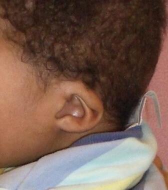
- From the 12 patients with bilateral atresia, 10 presented related malformations, 3 of whom had CHARGE syndrome (coloboma, heart defects, choanal atresia, retardation of growth and development, genitourinary problems, ear abnormalities). (arquivosdeorl.org.br)
- All patients with unilateral atresia needed only 1 surgical procedure, and patients with the bilateral form needed a median of 2.85 interventions ( p = 0.003). (arquivosdeorl.org.br)
- Choanal atresia may present as bilateral, unilateral or as a syndromic feature. (scirp.org)
- Bilateral choanal atresia presents at birth as asphyxia neonatorum. (scirp.org)
- Review of CT images of the skull revealed findings consistent with severe bilateral partial osseous choanal atresia and NPS. (avma.org)
- Babies with bilateral choanal atresia (blocked nose caused by bone or tissue) die of suffocation soon after birth. (sciforums.com)
- Every article about bilateral choanal atresia that I looked at clearly states this. (sciforums.com)
Stenosis5
- A 2012 epidemiological study looked at atrazine, a commonly used herbicide in the U.S., and found that women who lived in counties in Texas with the highest levels of this chemical being used to treat agricultural crops were 80 times more likely to give birth to infants with choanal atresia or stenosis compared to women who lived in the counties with the lowest levels. (wikipedia.org)
- This narrowing can exist in a spectrum of mild stenosis to complete atresia. (scirp.org)
- Multiple embryological models have been postulated to explain the development of choanal stenosis and atresia. (scirp.org)
- Herein, we present a case of nasal airway obstruction caused by widening of the posterior septum, a mild but clinically significant form of membranous choanal stenosis. (scirp.org)
- 7. Choanal atresia/stenosis, midfacial anomalies or recent craniofacial surgery. (who.int)
Congenital choanal atresia1
- In 1854, Emmert reported the first successful surgical procedure for congenital choanal atresia in a 7-year-old boy using a curved trocar transnasally. (medscape.com)
Evaluation of choanal atresia1
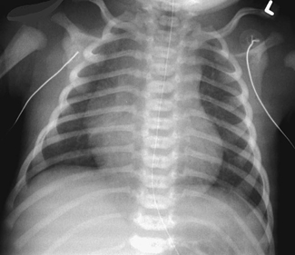
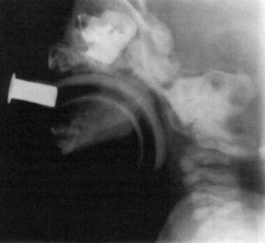
- Computed tomography (CT) scanning is the radiographic procedure of choice in the evaluation of choanal atresia. (medscape.com)
Unilateral choanal atresia3
- A unilateral choanal atresia may not be detected until much later in life because the baby manages to get along with only one nostril available for breathing. (wikipedia.org)
- Unilateral choanal atresia. (ucsfbenioffchildrens.org)
- Babies with unilateral choanal atresia often have a chronic runny nose on the blocked side but otherwise may breathe well enough to go undiagnosed and untreated for years. (ucsfbenioffchildrens.org)
Infants2
- Chromosomal anomalies are found in 6% of infants with choanal atresia. (medscape.com)
- For this reason, a genetic evaluation is often recommended for infants or children diagnosed with choanal atresia. (ucsfbenioffchildrens.org)
Nose3
- Choanal atresia blocking both sides of the nose causes acute breathing problems with bluish discoloration and breathing failure. (medlineplus.gov)
- Choanal atresia is a narrowing or blockage of the back of the nose that makes breathing difficult. (ucsfbenioffchildrens.org)
- Although rare (affecting one in 7,000 births), choanal atresia is the most common nose abnormality in newborns. (ucsfbenioffchildrens.org)
Airway4
- Choanal atresia is associated with a higher risk of other airway problems, including: tracheomalacia. (wikipedia.org)
- Choanal atresia is a narrowing or blockage of the nasal airway by tissue. (medlineplus.gov)
- Choanal atresia may affect one or both sides of the nasal airway. (medlineplus.gov)
- Choanal atresia may affect 1 or both sides of the nasal airway. (stlukes-stl.com)
Microtia2
- Associations between maternal occupational PAH exposure and selected rare defects of the face (cataracts, microphthalmia, glaucoma, microtia, and choanal atresia) and central nervous system (holoprosencephaly, hydrocephaly, cerebellar hypoplasia, and Dandy-Walker malformation) were evaluated using data from the National Birth Defects Prevention Study, a population-based case-control study in the United States. (cdc.gov)
- A bone conduction hearing device can be used by people with problems of the external or middle ears such microtia, atresia, otosclerosis or a previously failed ear surgery. (susheenduttent.com)
Coloboma1
- Sometimes, babies born with choanal atresia also have other abnormalities: coloboma. (wikipedia.org)
Pulmonary atresia1
- Tricuspid atresia , pulmonary atresia , and aortic atresia involve valves in the heart . (akronchildrens.org)
Abnormality1
- Although it has been more than 250 years since the first description of choanal atresia (CA), there are still doubts about this abnormality. (arquivosdeorl.org.br)
Diagnosis1
- One-sided atresia may not cause symptoms, and the infant may be sent home without a diagnosis. (medlineplus.gov)
Anatomic2
- 1996) did study of 63 patients - 47 CT scans reviewed - Additional 16 patients from their operative clinical experience (CT and histology) - Results: 18 pure bony (29%), 45 mixed bony-membranous (71%) * 0 pts were pure membranous "Choanal atresia: A new anatomic classification" 1996 Laryngoscopy -UT Southwestern. (pdfcoffee.com)
- Choanal atresia is an anatomic narrowing of the posterior choanal passageway. (scirp.org)
Babies1
- Babies with choanal atresia have difficulty breathing unless they are crying. (medlineplus.gov)
7,0001
- Choanal atresia is a fairly rare condition, affecting between 1 in 7,000 to 1 in 5,000 live births. (wikipedia.org)
Biliary1
- Biliary atresia is a defect in the liver or bile system. (akronchildrens.org)
Esophageal1
- Esophageal atresia is another defect of the digestive tract. (akronchildrens.org)
Birth4
- In general, choanal atresia is associated with a higher risk of other birth defects. (wikipedia.org)
- Choanal atresia is most often diagnosed shortly after birth while the infant is still in the hospital. (medlineplus.gov)
- Choanal atresia, especially when it affects both sides, is generally diagnosed shortly after birth while the infant is still in the hospital. (medlineplus.gov)
- Approximately 65% of patients with CHARGE syndrome may have obstructed breathing due to choanal atresia at birth. (aao.org)
Posterior choanae3
- citation needed] Choanal atresia causes closure of the posterior choanae in the nasal cavity. (wikipedia.org)
- The length of catheter that can be inserted indicates where choanal atresia has occurred: shorter distances indicate a problem with the vomer, while longer distances indicate a problem with the posterior choanae. (wikipedia.org)
- Choanal atresia is a congenital condition involving occlusion of the posterior choanae in the nasal cavity by bone, soft tissue, or both. (medscape.com)
Membranous1
- Choanal atresia is a congenital disorder where the back of the nasal passage (choana) is blocked, usually by abnormal bony or soft tissue (membranous) due to failed hole development of the nasal fossae during prenatal development. (wikipedia.org)
Nasal bridge1
- Also any condition that causes significant depression of the nasal bridge or midface retraction can be associated with choanal atresia. (wikipedia.org)
Cardiac1
- From the remaining 6 patients with unilateral atresia, only 2 showed malformations, 1 renal and 1 cardiac. (arquivosdeorl.org.br)
Theories4
- A number of theories exist as to how this developmental process causes choanal atresia. (wikipedia.org)
- 2 There are several theories that discuss the embryological development of choanal atresia. (jamanetwork.com)
- The purpose of this report is to highlight a clinical observation noted during the assessment and treatment of a patient with choanal atresia that highlights one of the embryological theories and a rare genetic correlation. (jamanetwork.com)
- 4 theories of etiology of atresia plate 1. (pdfcoffee.com)
Passages2
- Choanal atresia is diagnosed by examining the nasal passages. (ucsfbenioffchildrens.org)
- Choanal atresia is a defect of the nasal passages (choana). (akronchildrens.org)
Occurs3
- Unilateral atresia occurs more frequently on the right side. (medscape.com)
- Choanal atresia occurs with equal frequency in people of all races. (medscape.com)
- Choanal atresia is a defect that occurs when the nasal passage does not form properly. (ent-surgery.com.au)
Exposure1
- Another epidemiological report in 2010 found even higher associations between increased incidents of choanal atresia and exposure to second-hand-smoke, coffee consumption, high maternal zinc and B-12 intake and exposure to anti-infective urinary tract medications. (wikipedia.org)
Surgery1
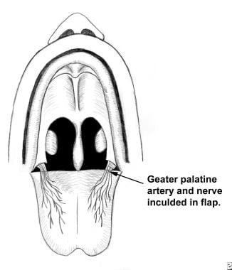
- [ 3 ] Combined transoral-transnasal is another technique that provides a good alternative for managing choanal atresia, with easier, 4-handed surgery to ensure adequate posterior choana for nasal breathing. (medscape.com)
Treatment4
- Treatment for choanal atresia can be divided into emergent and elective definitive categories. (medscape.com)
- The use of stents in the treatment of patients with choanal atresia is a controversial subject. (medscape.com)
- Unilateral atresia doesn't typically require immediate treatment. (ucsfbenioffchildrens.org)
- If your baby has been diagnosed with choanal atresia, the following information will help you better understand the problem, and what to expect during treatment. (ent-surgery.com.au)
Condition3
- Choanal atresia is a relatively uncommon condition with an estimated incidence of 1:7000 live births. (jamanetwork.com)
- Atresia (ah-TREE-zhah) is a condition in which a baby is born with a missing or closed valve or tube somewhere in his or her body. (akronchildrens.org)
- Choanal atresia is a congenital condition, meaning that it is present at the time the person is born. (sanw.org)
Development2
- Choanal atresia is caused by problems with the development of the nasal cavity and the palate. (wikipedia.org)
- The anti-thyroid medication methimazole has been associated with the development of choanal atresia in rare cases if given during the first trimester of pregnancy. (wikipedia.org)
Cases1
- The average rate of choanal atresia is 0.82 cases per 10,000 individuals. (medscape.com)