Respiratory System
Sensitivity and Specificity
Diagnostic Techniques and Procedures
Lung Compliance
Respiratory Mechanics
Respiratory Physiological Phenomena
Airway Resistance
Diagnostic Techniques, Radioisotope
Diagnostic Techniques, Otological
Diagnostic Techniques, Surgical
Lung Volume Measurements
Azure Stains
Diagnostic Techniques, Respiratory System
Diagnostic Tests, Routine
Diagnostic Techniques, Urological
Polymerase Chain Reaction
Diagnostic Techniques, Obstetrical and Gynecological
Diagnostic Techniques, Digestive System
Lung
Thoracic Wall
Tomography, X-Ray Computed
Functional Residual Capacity
Thorax
Tidal Volume
Respiration
Clinical Laboratory Techniques
Respiration, Artificial
Reproducibility of Results
Chest Tubes
Respiratory Function Tests
Air Pressure
Air Sacs
Predictive Value of Tests
Cytodiagnosis
Positive-Pressure Respiration
Diagnostic Techniques, Neurological
Pulmonary Ventilation
Radionuclide Imaging
Respiratory Tract Diseases
Biopsy, Needle
Radiography, Thoracic
Evaluation Studies as Topic
Ultrasonography
Biopsy
Diagnostic Techniques, Endocrine
Retrospective Studies
Prospective Studies
Biopsy, Fine-Needle
Diagnostic Techniques, Cardiovascular
Microscopy
Magnetic Resonance Imaging
Pulmonary Gas Exchange
Respiratory Paralysis
Locusta migratoria
Feces
Respiration Disorders
Total Lung Capacity
Respiratory Distress Syndrome, Adult
Enzyme-Linked Immunosorbent Assay
Pulmonary Atelectasis
Dog Diseases
Mycoses
Cytochromes
Trachea
Respiratory Tract Neoplasms
Treatment Outcome
Respiratory Insufficiency
Mass Chest X-Ray
Oxygen
Dogs
Risk Factors
Prevalence
Oxygen Consumption
Flail Chest
Pressure
Immunoenzyme Techniques
Respiratory Rate
Plethysmography, Whole Body
Forced Expiratory Volume
Residual Volume
Follow-Up Studies
Methacholine Chloride
Biological Markers
Ribs
Diagnostic Imaging
Asthma
Prognosis
Carbon Dioxide
Dinosaurs
Expiratory Reserve Volume
Inhalation Exposure
Intubation, Intratracheal
Lung Injury
Image Processing, Computer-Assisted
DNA Primers
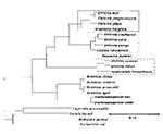
Molecular Sequence Data
Bronchoconstriction
Respiratory Distress Syndrome, Newborn
Disease Outbreaks
Aerosols
Work of Breathing
Models, Biological
Incidence
Thoracic Neoplasms
Inspiratory Capacity
Vital Capacity
Fluorescent Antibody Technique
Cytochrome a Group
Diaphragm
Positive-Pressure Respiration, Intrinsic
United States
Esophagus
Lung Diseases, Obstructive
Respiratory Tract Infections
Helium
Air
High-Frequency Ventilation
Respiratory Dead Space
Phrenic Nerve
Potassium Cyanide
Obesity Hypoventilation Syndrome
Immunohistochemistry
Cough
Cardiovascular System
Reagent Kits, Diagnostic
Pulmonary Alveoli
Tetramethylphenylenediamine
Bronchodilator Agents
Physiology
Hydroxyquinolines
Albuterol
Hyperventilation
Forced Expiratory Flow Rates
Cyanides
Pulmonary Disease, Chronic Obstructive
Air Pollutants
Occupational Exposure
Bronchial Provocation Tests
ROC Curve
Pulmonary Edema
Chemical Warfare Agents
Bronchitis, Chronic
Cardiovascular Physiological Phenomena
Chest Wall Oscillation
Diagnostic Techniques, Ophthalmological
Funnel Chest
Anesthesia, General
Administration, Inhalation
Bronchitis
Bronchi
Gases
Heart Massage
Air Pollutants, Occupational
Diving
Pneumothorax
Respiratory Sounds
Electron Transport
Mucus
Chronic Disease
Pulmonary Surfactants
Oxidoreductases
Severity of Illness Index
Diagnostic and Statistical Manual of Mental Disorders
Analysis of Variance
Case-Control Studies
The Respiratory System is a complex network of organs and tissues that work together to facilitate the process of breathing, which involves the intake of oxygen and the elimination of carbon dioxide. This system primarily includes the nose, throat (pharynx), voice box (larynx), windpipe (trachea), bronchi, bronchioles, lungs, and diaphragm.
The nostrils or mouth take in air that travels through the pharynx, larynx, and trachea into the lungs. Within the lungs, the trachea divides into two bronchi, one for each lung, which further divide into smaller tubes called bronchioles. At the end of these bronchioles are tiny air sacs known as alveoli where the exchange of gases occurs. Oxygen from the inhaled air diffuses through the walls of the alveoli into the bloodstream, while carbon dioxide, a waste product, moves from the blood to the alveoli and is exhaled out of the body.
The diaphragm, a large muscle that separates the chest from the abdomen, plays a crucial role in breathing by contracting and relaxing to change the volume of the chest cavity, thereby allowing air to flow in and out of the lungs. Overall, the Respiratory System is essential for maintaining life by providing the body's cells with the oxygen needed for metabolism and removing waste products like carbon dioxide.
Sensitivity and specificity are statistical measures used to describe the performance of a diagnostic test or screening tool in identifying true positive and true negative results.
* Sensitivity refers to the proportion of people who have a particular condition (true positives) who are correctly identified by the test. It is also known as the "true positive rate" or "recall." A highly sensitive test will identify most or all of the people with the condition, but may also produce more false positives.
* Specificity refers to the proportion of people who do not have a particular condition (true negatives) who are correctly identified by the test. It is also known as the "true negative rate." A highly specific test will identify most or all of the people without the condition, but may also produce more false negatives.
In medical testing, both sensitivity and specificity are important considerations when evaluating a diagnostic test. High sensitivity is desirable for screening tests that aim to identify as many cases of a condition as possible, while high specificity is desirable for confirmatory tests that aim to rule out the condition in people who do not have it.
It's worth noting that sensitivity and specificity are often influenced by factors such as the prevalence of the condition in the population being tested, the threshold used to define a positive result, and the reliability and validity of the test itself. Therefore, it's important to consider these factors when interpreting the results of a diagnostic test.
Chest pain is a discomfort or pain that you feel in the chest area. The pain can be sharp, dull, burning, crushing, heaviness, or tightness. It may be accompanied by other symptoms such as shortness of breath, sweating, nausea, dizziness, or pain that radiates to the arm, neck, jaw, or back.
Chest pain can have many possible causes, including heart-related conditions such as angina or a heart attack, lung conditions such as pneumonia or pleurisy, gastrointestinal problems such as acid reflux or gastritis, musculoskeletal issues such as costochondritis or muscle strain, and anxiety or panic attacks.
It is important to seek immediate medical attention if you experience chest pain that is severe, persistent, or accompanied by other concerning symptoms, as it may be a sign of a serious medical condition. A healthcare professional can evaluate your symptoms, perform tests, and provide appropriate treatment.
Diagnostic techniques and procedures are methods used by medical professionals to identify the cause of symptoms, illnesses, or diseases. These can include physical examinations, patient interviews, review of medical history, and various diagnostic tests. Diagnostic tests may involve invasive procedures such as biopsies or surgical interventions, or non-invasive imaging techniques like X-rays, CT scans, MRI scans, or ultrasounds. Functional tests, such as stress testing or electroencephalogram (EEG), can also be used to evaluate the functioning of specific organs or systems in the body. Laboratory tests, including blood tests, urine tests, and genetic tests, are also common diagnostic procedures. The choice of diagnostic technique or procedure depends on the presenting symptoms, the patient's medical history, and the suspected underlying condition.
Lung compliance is a measure of the ease with which the lungs expand and is defined as the change in lung volume for a given change in transpulmonary pressure. It is often expressed in units of liters per centimeter of water (L/cm H2O). A higher compliance indicates that the lungs are more easily distensible, while a lower compliance suggests that the lungs are stiffer and require more force to expand. Lung compliance can be affected by various conditions such as pulmonary fibrosis, pneumonia, acute respiratory distress syndrome (ARDS), and chronic obstructive pulmonary disease (COPD).
Molecular diagnostic techniques are a group of laboratory methods used to analyze biological markers in DNA, RNA, and proteins to identify specific health conditions or diseases at the molecular level. These techniques include various methods such as polymerase chain reaction (PCR), DNA sequencing, gene expression analysis, fluorescence in situ hybridization (FISH), and mass spectrometry.
Molecular diagnostic techniques are used to detect genetic mutations, chromosomal abnormalities, viral and bacterial infections, and other molecular changes associated with various diseases, including cancer, genetic disorders, infectious diseases, and neurological disorders. These techniques provide valuable information for disease diagnosis, prognosis, treatment planning, and monitoring of treatment response.
Compared to traditional diagnostic methods, molecular diagnostic techniques offer several advantages, such as higher sensitivity, specificity, and speed. They can detect small amounts of genetic material or proteins, even in early stages of the disease, and provide accurate results with a lower risk of false positives or negatives. Additionally, molecular diagnostic techniques can be automated, standardized, and performed in high-throughput formats, making them suitable for large-scale screening and research applications.
Respiratory mechanics refers to the biomechanical properties and processes that involve the movement of air through the respiratory system during breathing. It encompasses the mechanical behavior of the lungs, chest wall, and the muscles of respiration, including the diaphragm and intercostal muscles.
Respiratory mechanics includes several key components:
1. **Compliance**: The ability of the lungs and chest wall to expand and recoil during breathing. High compliance means that the structures can easily expand and recoil, while low compliance indicates greater resistance to expansion and recoil.
2. **Resistance**: The opposition to airflow within the respiratory system, primarily due to the friction between the air and the airway walls. Airway resistance is influenced by factors such as airway diameter, length, and the viscosity of the air.
3. **Lung volumes and capacities**: These are the amounts of air present in the lungs during different phases of the breathing cycle. They include tidal volume (the amount of air inspired or expired during normal breathing), inspiratory reserve volume (additional air that can be inspired beyond the tidal volume), expiratory reserve volume (additional air that can be exhaled beyond the tidal volume), and residual volume (the air remaining in the lungs after a forced maximum exhalation).
4. **Work of breathing**: The energy required to overcome the resistance and elastic forces during breathing. This work is primarily performed by the respiratory muscles, which contract to generate negative intrathoracic pressure and expand the chest wall, allowing air to flow into the lungs.
5. **Pressure-volume relationships**: These describe how changes in lung volume are associated with changes in pressure within the respiratory system. Important pressure components include alveolar pressure (the pressure inside the alveoli), pleural pressure (the pressure between the lungs and the chest wall), and transpulmonary pressure (the difference between alveolar and pleural pressures).
Understanding respiratory mechanics is crucial for diagnosing and managing various respiratory disorders, such as chronic obstructive pulmonary disease (COPD), asthma, and restrictive lung diseases.
Respiratory physiological phenomena refer to the various mechanical, chemical, and biological processes and functions that occur in the respiratory system during breathing and gas exchange. These phenomena include:
1. Ventilation: The movement of air into and out of the lungs, which is achieved through the contraction and relaxation of the diaphragm and intercostal muscles.
2. Gas Exchange: The diffusion of oxygen (O2) from the alveoli into the bloodstream and carbon dioxide (CO2) from the bloodstream into the alveoli.
3. Respiratory Mechanics: The physical properties and forces that affect the movement of air in and out of the lungs, such as lung compliance, airway resistance, and chest wall elasticity.
4. Control of Breathing: The regulation of ventilation by the central nervous system through the integration of sensory information from chemoreceptors and mechanoreceptors in the respiratory system.
5. Acid-Base Balance: The maintenance of a stable pH level in the blood through the regulation of CO2 elimination and bicarbonate balance by the respiratory and renal systems.
6. Oxygen Transport: The binding of O2 to hemoglobin in the red blood cells and its delivery to the tissues for metabolic processes.
7. Defense Mechanisms: The various protective mechanisms that prevent the entry and colonization of pathogens and foreign particles into the respiratory system, such as mucociliary clearance, cough reflex, and immune responses.
Airway resistance is a measure of the opposition to airflow during breathing, which is caused by the friction between the air and the walls of the respiratory tract. It is an important parameter in respiratory physiology because it can affect the work of breathing and gas exchange.
Airway resistance is usually expressed in units of cm H2O/L/s or Pa·s/m, and it can be measured during spontaneous breathing or during forced expiratory maneuvers, such as those used in pulmonary function testing. Increased airway resistance can result from a variety of conditions, including asthma, chronic obstructive pulmonary disease (COPD), bronchitis, and bronchiectasis. Decreased airway resistance can be seen in conditions such as emphysema or after a successful bronchodilator treatment.
Diagnostic techniques using radioisotopes, also known as nuclear medicine, are medical diagnostic procedures that use small amounts of radioactive material, called radioisotopes or radionuclides, to diagnose and monitor various diseases and conditions. The radioisotopes are introduced into the body through different routes (such as injection, inhalation, or ingestion) and accumulate in specific organs or tissues.
The gamma rays or photons emitted by these radioisotopes are then detected by specialized imaging devices, such as gamma cameras or PET scanners, which generate images that provide information about the structure and function of the organ or tissue being examined. This information helps healthcare professionals to make accurate diagnoses, monitor disease progression, assess treatment response, and plan appropriate therapies.
Common diagnostic techniques using radioisotopes include:
1. Radionuclide imaging (also known as scintigraphy): A gamma camera is used to produce images of specific organs or tissues after the administration of a radioisotope. Examples include bone scans, lung scans, heart scans, and brain scans.
2. Positron emission tomography (PET) scans: A PET scanner detects pairs of gamma rays emitted indirectly by a positron-emitting radionuclide, such as fluorodeoxyglucose (FDG), which is often used in oncology to assess metabolic activity and identify cancerous lesions.
3. Single-photon emission computed tomography (SPECT): A specialized gamma camera rotates around the patient, acquiring multiple images from different angles that are then reconstructed into a 3D image, providing detailed information about organ function and structure.
Diagnostic techniques using radioisotopes offer several advantages, including high sensitivity, non-invasiveness, and the ability to assess both anatomical and functional aspects of organs and tissues. However, they also involve exposure to ionizing radiation, so their use should be balanced against potential risks and benefits, and alternative diagnostic methods should be considered when appropriate.
Diagnostic techniques in otology refer to the methods and tests used by healthcare professionals to identify and diagnose various conditions related to the ear. These techniques can include:
1. Otoscopy: A visual examination of the external auditory canal and eardrum using an otoscope. This helps to identify any physical abnormalities, such as wax buildup, inflammation, or foreign objects in the ear.
2. Audiometry: A hearing test that measures a person's ability to hear different sounds, pitches, and volumes. This can help to identify any hearing loss or auditory processing issues.
3. Tympanometry: A test that measures the function of the middle ear by creating variations in air pressure in the ear canal. This can help to identify any issues with the eardrum or middle ear bones.
4. Acoustic reflex testing: A test that measures the body's involuntary response to loud sounds. This can help to identify any damage to the hearing nerves or brainstem.
5. Otoacoustic emissions (OAE) testing: A test that measures the sound waves produced by the inner ear in response to stimuli. This can help to identify any issues with the cochlea or hair cells in the inner ear.
6. Auditory brainstem response (ABR) testing: A test that measures the electrical activity of the hearing nerve and brainstem in response to sound. This can help to identify any issues with the auditory nervous system.
7. Vestibular testing: A series of tests that measure a person's balance and equilibrium. This can help to identify any issues with the vestibular system, which is responsible for maintaining balance.
These diagnostic techniques are used to diagnose various otological conditions such as hearing loss, tinnitus, vertigo, ear infections, and tumors of the ear.
Diagnostic techniques, surgical refers to the use of surgical procedures or methods to diagnose and evaluate various medical conditions. These techniques are often used when non-invasive tests are inconclusive or when more detailed information is required. Here are some examples:
1. Biopsy: A small sample of tissue is removed from the body for examination under a microscope. This can help to confirm a diagnosis of cancer, infection, or other diseases.
2. Endoscopy: A flexible tube with a light and camera on the end is inserted into the body through a natural opening (such as the mouth or anus) or a small incision. This allows the doctor to visualize internal organs and tissues, and may also involve taking biopsy samples.
3. Imaging studies: Various imaging techniques such as X-rays, CT scans, MRI scans, and ultrasound can be used to produce detailed images of internal structures. These can help to diagnose a wide range of medical conditions, from broken bones to tumors.
4. Exploratory surgery: In some cases, a surgical incision may be made to directly visualize and examine an organ or tissue. This can help to diagnose conditions that are difficult to detect with non-invasive tests.
5. Functional testing: Some surgical techniques involve stimulating or measuring the function of an organ or system. For example, a cardiac stress test may be performed during surgery to assess heart function.
Overall, diagnostic techniques, surgical play an important role in the diagnosis and management of many medical conditions. They can provide valuable information that helps doctors to make informed decisions about treatment options and improve patient outcomes.
Respiratory system abnormalities refer to any conditions or structures that do not function properly or are outside the normal range in the respiratory system. The respiratory system is responsible for taking in oxygen and expelling carbon dioxide through the process of breathing. It includes the nose, throat (pharynx), voice box (larynx), windpipe (trachea), bronchi, bronchioles, alveoli, and muscles and nerves that support breathing.
Respiratory system abnormalities can be congenital or acquired. Congenital abnormalities are present at birth and may include conditions such as cystic fibrosis, pulmonary hypoplasia, and congenital diaphragmatic hernia. Acquired abnormalities can develop at any time throughout a person's life due to various factors such as infections, injuries, environmental exposures, or aging. Examples of acquired respiratory system abnormalities include chronic obstructive pulmonary disease (COPD), asthma, pneumonia, lung cancer, and sleep apnea.
Respiratory system abnormalities can cause a range of symptoms, including coughing, wheezing, shortness of breath, chest pain, and fatigue. Treatment for respiratory system abnormalities depends on the specific condition and severity and may include medications, breathing treatments, surgery, or lifestyle changes.
Lung volume measurements are clinical tests that determine the amount of air inhaled, exhaled, and present in the lungs at different times during the breathing cycle. These measurements include:
1. Tidal Volume (TV): The amount of air inhaled or exhaled during normal breathing, usually around 500 mL in resting adults.
2. Inspiratory Reserve Volume (IRV): The additional air that can be inhaled after a normal inspiration, approximately 3,000 mL in adults.
3. Expiratory Reserve Volume (ERV): The extra air that can be exhaled after a normal expiration, about 1,000-1,200 mL in adults.
4. Residual Volume (RV): The air remaining in the lungs after a maximal exhalation, approximately 1,100-1,500 mL in adults.
5. Total Lung Capacity (TLC): The total amount of air the lungs can hold at full inflation, calculated as TV + IRV + ERV + RV, around 6,000 mL in adults.
6. Functional Residual Capacity (FRC): The volume of air remaining in the lungs after a normal expiration, equal to ERV + RV, about 2,100-2,700 mL in adults.
7. Inspiratory Capacity (IC): The maximum amount of air that can be inhaled after a normal expiration, equal to TV + IRV, around 3,500 mL in adults.
8. Vital Capacity (VC): The total volume of air that can be exhaled after a maximal inspiration, calculated as IC + ERV, approximately 4,200-5,600 mL in adults.
These measurements help assess lung function and identify various respiratory disorders such as chronic obstructive pulmonary disease (COPD), asthma, and restrictive lung diseases.
'Azure stains' is a term used in pathology to describe a histological staining technique that uses a type of dye called methyl blue, which turns the stained structures a blue-purple color. This technique is often used to stain acid mucins, which are found in various types of tissues and can be indicative of certain medical conditions.
In particular, azure stains are sometimes used to help diagnose certain types of cancer, such as mucoepidermoid carcinoma, a type of salivary gland tumor that produces acid mucins. The staining technique can help pathologists identify the presence and distribution of these mucins within the tumor cells, which can aid in making an accurate diagnosis and determining the best course of treatment.
It's worth noting that there are several different types of histological stains that use various dyes to highlight different structures or features within tissues. Azure stains are just one example of these techniques, and they are typically used in conjunction with other staining methods to provide a comprehensive picture of the tissue being examined.
Diagnostic techniques for the respiratory system are methods used to identify and diagnose various diseases and conditions affecting the lungs and breathing. Here are some commonly used diagnostic techniques:
1. Physical Examination: A healthcare provider will listen to your chest with a stethoscope to check for abnormal breath sounds, such as wheezing or crackles. They may also observe your respiratory rate and effort.
2. Chest X-ray: This imaging test can help identify abnormalities in the lungs, such as tumors, fluid accumulation, or collapsed lung sections.
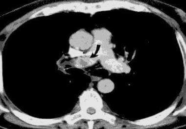
3. Computed Tomography (CT) Scan: A CT scan uses X-rays to create detailed cross-sectional images of the lungs and surrounding structures. It can help detect nodules, cysts, or other abnormalities that may not be visible on a chest X-ray.
4. Pulmonary Function Tests (PFTs): These tests measure how well your lungs are working by assessing your ability to inhale and exhale air. Common PFTs include spirometry, lung volume measurement, and diffusing capacity testing.
5. Bronchoscopy: A thin, flexible tube with a camera and light is inserted through the nose or mouth into the airways to examine the lungs' interior and obtain tissue samples for biopsy.
6. Bronchoalveolar Lavage (BAL): During a bronchoscopy, fluid is introduced into a specific area of the lung and then suctioned out to collect cells and other materials for analysis.
7. Sleep Studies: These tests monitor your breathing patterns during sleep to diagnose conditions like sleep apnea or other sleep-related breathing disorders.
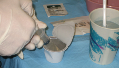
8. Sputum Analysis: A sample of coughed-up mucus is examined under a microscope to identify any abnormal cells, bacteria, or other organisms that may be causing respiratory issues.
9. Blood Tests: Blood tests can help diagnose various respiratory conditions by measuring oxygen and carbon dioxide levels, identifying specific antibodies or antigens, or detecting genetic markers associated with certain diseases.
10. Positron Emission Tomography (PET) Scan: A PET scan uses a small amount of radioactive material to create detailed images of the body's internal structures and functions, helping identify areas of abnormal cell growth or metabolic activity in the lungs.
'Diagnostic tests, routine' is a medical term that refers to standard or commonly used tests that are performed to help diagnose, monitor, or manage a patient's health condition. These tests are typically simple, non-invasive, and safe, and they may be ordered as part of a regular check-up or when a patient presents with specific symptoms.
Routine diagnostic tests may include:
1. Complete Blood Count (CBC): A test that measures the number of red and white blood cells, platelets, and hemoglobin in the blood. It can help diagnose conditions such as anemia, infection, and inflammation.
2. Urinalysis: A test that examines a urine sample for signs of infection, kidney disease, or other medical conditions.
3. Blood Chemistry Tests: Also known as a chemistry panel or comprehensive metabolic panel, this test measures various chemicals in the blood such as glucose, electrolytes, and enzymes to evaluate organ function and overall health.
4. Electrocardiogram (ECG): A test that records the electrical activity of the heart, which can help diagnose heart conditions such as arrhythmias or heart attacks.
5. Chest X-ray: An imaging test that creates pictures of the structures inside the chest, including the heart, lungs, and bones, to help diagnose conditions such as pneumonia or lung cancer.
6. Fecal Occult Blood Test (FOBT): A test that checks for hidden blood in the stool, which can be a sign of colon cancer or other gastrointestinal conditions.
7. Pap Smear: A test that collects cells from the cervix to check for abnormalities that may indicate cervical cancer or other gynecological conditions.
These are just a few examples of routine diagnostic tests that healthcare providers may order. The specific tests ordered will depend on the patient's age, sex, medical history, and current symptoms.
Diagnostic techniques in urology are methods used to identify and diagnose various urological conditions affecting the urinary tract and male reproductive system. These techniques include:
1. Urinalysis: A laboratory examination of a urine sample to detect abnormalities such as infection, kidney stones, or other underlying medical conditions.
2. Urine Culture: A test used to identify and grow bacteria from the urine to determine the type of bacterial infection present in the urinary tract.
3. Imaging Studies: Various imaging techniques such as X-rays, ultrasound, CT scans, and MRI scans are used to visualize the internal structures of the urinary tract and identify any abnormalities.
4. Cystoscopy: A procedure that involves inserting a thin tube with a camera into the bladder through the urethra to examine the bladder and urethra for signs of disease or abnormality.
5. Urodynamics: A series of tests used to evaluate bladder function, including measuring bladder pressure and urine flow rate.
6. Biopsy: The removal and examination of tissue from the urinary tract or male reproductive system to diagnose conditions such as cancer.
7. Prostate-Specific Antigen (PSA) Test: A blood test used to screen for prostate cancer by measuring the level of PSA, a protein produced by the prostate gland.
8. Voiding Diary: A record of urinary habits, including the frequency and volume of urination, that can help diagnose conditions such as overactive bladder or urinary incontinence.
Polymerase Chain Reaction (PCR) is a laboratory technique used to amplify specific regions of DNA. It enables the production of thousands to millions of copies of a particular DNA sequence in a rapid and efficient manner, making it an essential tool in various fields such as molecular biology, medical diagnostics, forensic science, and research.
The PCR process involves repeated cycles of heating and cooling to separate the DNA strands, allow primers (short sequences of single-stranded DNA) to attach to the target regions, and extend these primers using an enzyme called Taq polymerase, resulting in the exponential amplification of the desired DNA segment.
In a medical context, PCR is often used for detecting and quantifying specific pathogens (viruses, bacteria, fungi, or parasites) in clinical samples, identifying genetic mutations or polymorphisms associated with diseases, monitoring disease progression, and evaluating treatment effectiveness.
Diagnostic techniques in obstetrics and gynecology refer to the various methods used by healthcare professionals to diagnose and monitor conditions related to the female reproductive system and pregnancy. Here are some commonly used diagnostic techniques:
1. Physical examination: A thorough physical exam, including a pelvic exam, can help identify any abnormalities in the reproductive organs.
2. Medical history: A detailed medical history, including information about menstrual cycles, sexual activity, and family health, can provide valuable clues to diagnose various conditions.
3. Imaging tests: Ultrasound, CT scans, and MRIs can help healthcare professionals visualize the reproductive organs and detect any abnormalities.
4. Laboratory tests: Blood tests, urine tests, and cultures can help identify infections, hormonal imbalances, and other conditions.
5. Biopsy: A small sample of tissue is taken from the affected area and examined under a microscope to diagnose conditions such as cancer.
6. Colposcopy: This procedure involves using a special magnifying device to examine the cervix and vagina for signs of abnormalities.
7. Hysterosalpingography: This is an X-ray procedure that involves injecting a dye into the uterus and fallopian tubes to detect any blockages or other abnormalities.
8. Sonohysterography: This is an ultrasound procedure that involves injecting a fluid into the uterus to help visualize its interior and detect any abnormalities.
9. Minimally invasive surgery: Procedures such as laparoscopy and hysteroscopy can help healthcare professionals diagnose and treat various conditions related to the reproductive organs.
These diagnostic techniques can help healthcare professionals identify and manage a wide range of conditions, including infertility, pregnancy complications, infections, hormonal imbalances, and cancer.
Diagnostic techniques for the digestive system are medical tests and procedures used to diagnose and evaluate various conditions and diseases related to the gastrointestinal (GI) tract, including the esophagus, stomach, small intestine, large intestine, liver, gallbladder, pancreas, and associated organs. These techniques can be categorized into invasive and non-invasive methods.
Non-invasive diagnostic techniques:
1. Imaging tests: These include X-rays, computed tomography (CT) scans, magnetic resonance imaging (MRI), positron emission tomography (PET) scans, and ultrasounds. They help visualize the structure and function of the digestive organs without requiring any invasive procedures.
2. Laboratory tests: Blood, stool, and urine samples can be analyzed to detect signs of infection, inflammation, or other abnormalities related to digestive system disorders. Examples include complete blood count (CBC), liver function tests (LFTs), coagulation studies, and fecal occult blood test (FOBT).
3. Breath tests: These are used to diagnose conditions like lactose intolerance, small intestinal bacterial overgrowth (SIBO), or helicobacter pylori infection by analyzing the patient's exhaled air after consuming a specific substance.
Invasive diagnostic techniques:
1. Endoscopy: A thin, flexible tube with a light and camera attached to its end is inserted through the mouth or rectum to directly visualize the GI tract's inner lining. There are different types of endoscopies, such as gastroscopy (esophagus, stomach, and duodenum), colonoscopy (colon and rectum), sigmoidoscopy (lower part of the colon), and enteroscopy (small intestine).
2. Endoscopic ultrasound (EUS): This combines endoscopy with ultrasound technology to provide detailed images of the digestive organs' structure and surrounding tissues, allowing for accurate diagnosis and staging of conditions like cancer.
3. Biopsy: During an endoscopy or surgery, a small tissue sample can be taken from the affected area for further examination under a microscope to confirm a diagnosis or assess the severity of a condition.
4. Capsule endoscopy: A patient swallows a tiny camera-equipped capsule that transmits images as it passes through the GI tract, allowing doctors to diagnose conditions in the small intestine that may be difficult to reach with traditional endoscopes.
5. Imaging studies: Procedures like computed tomography (CT), magnetic resonance imaging (MRI), or positron emission tomography (PET) scans can provide detailed images of the digestive organs and help diagnose conditions like tumors, inflammation, or obstructions.
These diagnostic techniques help healthcare providers identify and manage various gastrointestinal conditions, ensuring appropriate treatment and improved patient outcomes.
A lung is a pair of spongy, elastic organs in the chest that work together to enable breathing. They are responsible for taking in oxygen and expelling carbon dioxide through the process of respiration. The left lung has two lobes, while the right lung has three lobes. The lungs are protected by the ribcage and are covered by a double-layered membrane called the pleura. The trachea divides into two bronchi, which further divide into smaller bronchioles, leading to millions of tiny air sacs called alveoli, where the exchange of gases occurs.
The thoracic wall refers to the anatomical structure that surrounds and protects the chest cavity or thorax, which contains the lungs, heart, and other vital organs. It is composed of several components:
1. Skeletal framework: This includes the 12 pairs of ribs, the sternum (breastbone) in the front, and the thoracic vertebrae in the back. The upper seven pairs of ribs are directly attached to the sternum in the front through costal cartilages. The lower five pairs of ribs are not directly connected to the sternum but are joined to the ribs above them.
2. Muscles: The thoracic wall contains several muscles, including the intercostal muscles (located between the ribs), the scalene muscles (at the side and back of the neck), and the serratus anterior muscle (on the sides of the chest). These muscles help in breathing by expanding and contracting the ribcage.
3. Soft tissues: The thoracic wall also contains various soft tissues, such as fascia, nerves, blood vessels, and fat. These structures support the functioning of the thoracic organs and contribute to the overall stability and protection of the chest cavity.
The primary function of the thoracic wall is to protect the vital organs within the chest cavity while allowing for adequate movement during respiration. Additionally, it provides a stable base for the attachment of various muscles involved in upper limb movement and posture.
X-ray computed tomography (CT or CAT scan) is a medical imaging method that uses computer-processed combinations of many X-ray images taken from different angles to produce cross-sectional (tomographic) images (virtual "slices") of the body. These cross-sectional images can then be used to display detailed internal views of organs, bones, and soft tissues in the body.
The term "computed tomography" is used instead of "CT scan" or "CAT scan" because the machines take a series of X-ray measurements from different angles around the body and then use a computer to process these data to create detailed images of internal structures within the body.
CT scanning is a noninvasive, painless medical test that helps physicians diagnose and treat medical conditions. CT imaging provides detailed information about many types of tissue including lung, bone, soft tissue and blood vessels. CT examinations can be performed on every part of the body for a variety of reasons including diagnosis, surgical planning, and monitoring of therapeutic responses.
In computed tomography (CT), an X-ray source and detector rotate around the patient, measuring the X-ray attenuation at many different angles. A computer uses this data to construct a cross-sectional image by the process of reconstruction. This technique is called "tomography". The term "computed" refers to the use of a computer to reconstruct the images.
CT has become an important tool in medical imaging and diagnosis, allowing radiologists and other physicians to view detailed internal images of the body. It can help identify many different medical conditions including cancer, heart disease, lung nodules, liver tumors, and internal injuries from trauma. CT is also commonly used for guiding biopsies and other minimally invasive procedures.
In summary, X-ray computed tomography (CT or CAT scan) is a medical imaging technique that uses computer-processed combinations of many X-ray images taken from different angles to produce cross-sectional images of the body. It provides detailed internal views of organs, bones, and soft tissues in the body, allowing physicians to diagnose and treat medical conditions.
Functional Residual Capacity (FRC) is the volume of air that remains in the lungs after normal expiration during quiet breathing. It represents the sum of the residual volume (RV) and the expiratory reserve volume (ERV). The FRC is approximately 2.5-3.5 liters in a healthy adult. This volume of air serves to keep the alveoli open and maintain oxygenation during periods of quiet breathing, as well as providing a reservoir for additional ventilation during increased activity or exercise.
The thorax is the central part of the human body, located between the neck and the abdomen. In medical terms, it refers to the portion of the body that contains the heart, lungs, and associated structures within a protective cage made up of the sternum (breastbone), ribs, and thoracic vertebrae. The thorax is enclosed by muscles and protected by the ribcage, which helps to maintain its structural integrity and protect the vital organs contained within it.
The thorax plays a crucial role in respiration, as it allows for the expansion and contraction of the lungs during breathing. This movement is facilitated by the flexible nature of the ribcage, which expands and contracts with each breath, allowing air to enter and exit the lungs. Additionally, the thorax serves as a conduit for major blood vessels, such as the aorta and vena cava, which carry blood to and from the heart and the rest of the body.
Understanding the anatomy and function of the thorax is essential for medical professionals, as many conditions and diseases can affect this region of the body. These may include respiratory disorders such as pneumonia or chronic obstructive pulmonary disease (COPD), cardiovascular conditions like heart attacks or aortic aneurysms, and musculoskeletal issues involving the ribs, spine, or surrounding muscles.
Tidal volume (Vt) is the amount of air that moves into or out of the lungs during normal, resting breathing. It is the difference between the volume of air in the lungs at the end of a normal expiration and the volume at the end of a normal inspiration. In other words, it's the volume of each breath you take when you are not making any effort to breathe more deeply.
The average tidal volume for an adult human is around 500 milliliters (ml) per breath, but this can vary depending on factors such as age, sex, size, and fitness level. During exercise or other activities that require increased oxygen intake, tidal volume may increase to meet the body's demands for more oxygen.
Tidal volume is an important concept in respiratory physiology and clinical medicine, as it can be used to assess lung function and diagnose respiratory disorders such as chronic obstructive pulmonary disease (COPD) or asthma.
Medical Definition of Respiration:
Respiration, in physiology, is the process by which an organism takes in oxygen and gives out carbon dioxide. It's also known as breathing. This process is essential for most forms of life because it provides the necessary oxygen for cellular respiration, where the cells convert biochemical energy from nutrients into adenosine triphosphate (ATP), and releases waste products, primarily carbon dioxide.
In humans and other mammals, respiration is a two-stage process:
1. Breathing (or external respiration): This involves the exchange of gases with the environment. Air enters the lungs through the mouth or nose, then passes through the pharynx, larynx, trachea, and bronchi, finally reaching the alveoli where the actual gas exchange occurs. Oxygen from the inhaled air diffuses into the blood, while carbon dioxide, a waste product of metabolism, diffuses from the blood into the alveoli to be exhaled.
2. Cellular respiration (or internal respiration): This is the process by which cells convert glucose and other nutrients into ATP, water, and carbon dioxide in the presence of oxygen. The carbon dioxide produced during this process then diffuses out of the cells and into the bloodstream to be exhaled during breathing.
In summary, respiration is a vital physiological function that enables organisms to obtain the necessary oxygen for cellular metabolism while eliminating waste products like carbon dioxide.
Clinical laboratory techniques are methods and procedures used in medical laboratories to perform various tests and examinations on patient samples. These techniques help in the diagnosis, treatment, and prevention of diseases by analyzing body fluids, tissues, and other specimens. Some common clinical laboratory techniques include:
1. Clinical chemistry: It involves the analysis of bodily fluids such as blood, urine, and cerebrospinal fluid to measure the levels of chemicals, hormones, enzymes, and other substances in the body. These measurements can help diagnose various medical conditions, monitor treatment progress, and assess overall health.
2. Hematology: This technique focuses on the study of blood and its components, including red and white blood cells, platelets, and clotting factors. Hematological tests are used to diagnose anemia, infections, bleeding disorders, and other hematologic conditions.
3. Microbiology: It deals with the identification and culture of microorganisms such as bacteria, viruses, fungi, and parasites. Microbiological techniques are essential for detecting infectious diseases, determining appropriate antibiotic therapy, and monitoring the effectiveness of treatment.
4. Immunology: This technique involves studying the immune system and its response to various antigens, such as bacteria, viruses, and allergens. Immunological tests are used to diagnose autoimmune disorders, immunodeficiencies, and allergies.
5. Histopathology: It is the microscopic examination of tissue samples to identify any abnormalities or diseases. Histopathological techniques are crucial for diagnosing cancer, inflammatory conditions, and other tissue-related disorders.
6. Molecular biology: This technique deals with the study of DNA, RNA, and proteins at the molecular level. Molecular biology tests can be used to detect genetic mutations, identify infectious agents, and monitor disease progression.
7. Cytogenetics: It involves analyzing chromosomes and genes in cells to diagnose genetic disorders, cancer, and other diseases. Cytogenetic techniques include karyotyping, fluorescence in situ hybridization (FISH), and comparative genomic hybridization (CGH).
8. Flow cytometry: This technique measures physical and chemical characteristics of cells or particles as they flow through a laser beam. Flow cytometry is used to analyze cell populations, identify specific cell types, and detect abnormalities in cells.
9. Diagnostic radiology: It uses imaging technologies such as X-rays, computed tomography (CT), magnetic resonance imaging (MRI), and ultrasound to diagnose various medical conditions.
10. Clinical chemistry: This technique involves analyzing body fluids, such as blood and urine, to measure the concentration of various chemicals and substances. Clinical chemistry tests are used to diagnose metabolic disorders, electrolyte imbalances, and other health conditions.
Artificial respiration is an emergency procedure that can be used to provide oxygen to a person who is not breathing or is breathing inadequately. It involves manually forcing air into the lungs, either by compressing the chest or using a device to deliver breaths. The goal of artificial respiration is to maintain adequate oxygenation of the body's tissues and organs until the person can breathe on their own or until advanced medical care arrives. Artificial respiration may be used in conjunction with cardiopulmonary resuscitation (CPR) in cases of cardiac arrest.
Reproducibility of results in a medical context refers to the ability to obtain consistent and comparable findings when a particular experiment or study is repeated, either by the same researcher or by different researchers, following the same experimental protocol. It is an essential principle in scientific research that helps to ensure the validity and reliability of research findings.
In medical research, reproducibility of results is crucial for establishing the effectiveness and safety of new treatments, interventions, or diagnostic tools. It involves conducting well-designed studies with adequate sample sizes, appropriate statistical analyses, and transparent reporting of methods and findings to allow other researchers to replicate the study and confirm or refute the results.
The lack of reproducibility in medical research has become a significant concern in recent years, as several high-profile studies have failed to produce consistent findings when replicated by other researchers. This has led to increased scrutiny of research practices and a call for greater transparency, rigor, and standardization in the conduct and reporting of medical research.
Chest tubes are medical devices that are inserted into the chest cavity to drain fluid, air, or blood. They are typically used to treat conditions such as pneumothorax (collapsed lung), hemothorax (blood in the chest cavity), pleural effusion (excess fluid in the chest cavity), and chylothorax (milky fluid in the chest cavity).
Chest tubes are usually inserted between the ribs and directed into the chest cavity, allowing for drainage of the affected area. The tubes are connected to a collection system that creates negative pressure, which helps to remove the air or fluid from the chest cavity.
The size and number of chest tubes used may vary depending on the severity and location of the condition being treated. Chest tubes are typically removed once the underlying condition has been resolved and the drainage has decreased to a minimal amount.
Respiratory Function Tests (RFTs) are a group of medical tests that measure how well your lungs take in and exhale air, and how well they transfer oxygen and carbon dioxide into and out of your blood. They can help diagnose certain lung disorders, measure the severity of lung disease, and monitor response to treatment.
RFTs include several types of tests, such as:
1. Spirometry: This test measures how much air you can exhale and how quickly you can do it. It's often used to diagnose and monitor conditions like asthma, chronic obstructive pulmonary disease (COPD), and other lung diseases.
2. Lung volume testing: This test measures the total amount of air in your lungs. It can help diagnose restrictive lung diseases, such as pulmonary fibrosis or sarcoidosis.
3. Diffusion capacity testing: This test measures how well oxygen moves from your lungs into your bloodstream. It's often used to diagnose and monitor conditions like pulmonary fibrosis, interstitial lung disease, and other lung diseases that affect the ability of the lungs to transfer oxygen to the blood.
4. Bronchoprovocation testing: This test involves inhaling a substance that can cause your airways to narrow, such as methacholine or histamine. It's often used to diagnose and monitor asthma.
5. Exercise stress testing: This test measures how well your lungs and heart work together during exercise. It's often used to diagnose lung or heart disease.
Overall, Respiratory Function Tests are an important tool for diagnosing and managing a wide range of lung conditions.
Air pressure, also known as atmospheric pressure, is the force exerted by the weight of air in the atmosphere on a surface. It is measured in units such as pounds per square inch (psi), hectopascals (hPa), or inches of mercury (inHg). The standard atmospheric pressure at sea level is defined as 101,325 Pa (14.7 psi/1013 hPa/29.92 inHg). Changes in air pressure can be used to predict weather patterns and are an important factor in the study of aerodynamics and respiratory physiology.
Air sacs, also known as alveoli, are tiny air-filled sacs in the lungs where the exchange of oxygen and carbon dioxide occurs during respiration. They are a part of the respiratory system in mammals and birds. In humans, the lungs contain about 300 million alveoli, which are clustered together in small groups called alveolar sacs. The walls of the air sacs are extremely thin, allowing for the easy diffusion of oxygen and carbon dioxide between the air in the sacs and the blood in the capillaries that surround them.
Oscillometry is a non-invasive method to measure various mechanical properties of the respiratory system, including lung volumes and airway resistance. It involves applying small pressure oscillations to the airways and measuring the resulting flow or volume changes. The technique can be used to assess lung function in patients with obstructive or restrictive lung diseases, as well as in healthy individuals. Oscillometry is often performed during tidal breathing, making it a comfortable method for both children and adults who may have difficulty performing traditional spirometry maneuvers.
The Predictive Value of Tests, specifically the Positive Predictive Value (PPV) and Negative Predictive Value (NPV), are measures used in diagnostic tests to determine the probability that a positive or negative test result is correct.
Positive Predictive Value (PPV) is the proportion of patients with a positive test result who actually have the disease. It is calculated as the number of true positives divided by the total number of positive results (true positives + false positives). A higher PPV indicates that a positive test result is more likely to be a true positive, and therefore the disease is more likely to be present.
Negative Predictive Value (NPV) is the proportion of patients with a negative test result who do not have the disease. It is calculated as the number of true negatives divided by the total number of negative results (true negatives + false negatives). A higher NPV indicates that a negative test result is more likely to be a true negative, and therefore the disease is less likely to be present.
The predictive value of tests depends on the prevalence of the disease in the population being tested, as well as the sensitivity and specificity of the test. A test with high sensitivity and specificity will generally have higher predictive values than a test with low sensitivity and specificity. However, even a highly sensitive and specific test can have low predictive values if the prevalence of the disease is low in the population being tested.
Cytodiagnosis is the rapid, initial evaluation and diagnosis of a disease based on the examination of individual cells obtained from a body fluid or tissue sample. This technique is often used in cytopathology to investigate abnormalities such as lumps, bumps, or growths that may be caused by cancerous or benign conditions.
The process involves collecting cells through various methods like fine-needle aspiration (FNA), body fluids such as urine, sputum, or washings from the respiratory, gastrointestinal, or genitourinary tracts. The collected sample is then spread onto a microscope slide, stained, and examined under a microscope for abnormalities in cell size, shape, structure, and organization.
Cytodiagnosis can provide crucial information to guide further diagnostic procedures and treatment plans. It is often used as an initial screening tool due to its speed, simplicity, and cost-effectiveness compared to traditional histopathological methods that require tissue biopsy and more extensive processing. However, cytodiagnosis may not always be able to distinguish between benign and malignant conditions definitively; therefore, additional tests or follow-up evaluations might be necessary for a conclusive diagnosis.
Positive-pressure respiration is a type of mechanical ventilation where positive pressure is applied to the airway and lungs, causing them to expand and inflate. This can be used to support or replace spontaneous breathing in patients who are unable to breathe effectively on their own due to conditions such as respiratory failure, neuromuscular disorders, or sedation for surgery.
During positive-pressure ventilation, a mechanical ventilator delivers breaths to the patient through an endotracheal tube or a tracheostomy tube. The ventilator is set to deliver a specific volume or pressure of air with each breath, and the patient's breathing is synchronized with the ventilator to ensure proper delivery of the breaths.
Positive-pressure ventilation can help improve oxygenation and remove carbon dioxide from the lungs, but it can also have potential complications such as barotrauma (injury to lung tissue due to excessive pressure), volutrauma (injury due to overdistention of the lungs), hemodynamic compromise (decreased blood pressure and cardiac output), and ventilator-associated pneumonia. Therefore, careful monitoring and adjustment of ventilator settings are essential to minimize these risks and provide safe and effective respiratory support.
Neurological diagnostic techniques are medical tests and examinations used to identify and diagnose conditions related to the nervous system, which includes the brain, spinal cord, nerves, and muscles. These techniques can be divided into several categories:
1. Clinical Examination: A thorough physical examination, including a neurological evaluation, is often the first step in diagnosing neurological conditions. This may involve assessing a person's mental status, muscle strength, coordination, reflexes, sensation, and gait.
2. Imaging Techniques: These are used to produce detailed images of the brain and nervous system. Common imaging techniques include:
- Computed Tomography (CT): This uses X-rays to create cross-sectional images of the brain and other parts of the body.
- Magnetic Resonance Imaging (MRI): This uses a strong magnetic field and radio waves to produce detailed images of the brain and other internal structures.
- Functional MRI (fMRI): This is a type of MRI that measures brain activity by detecting changes in blood flow.
- Positron Emission Tomography (PET): This uses small amounts of radioactive material to produce detailed images of brain function.
- Single Photon Emission Computed Tomography (SPECT): This is a type of nuclear medicine imaging that uses a gamma camera and a computer to produce detailed images of brain function.
3. Electrophysiological Tests: These are used to measure the electrical activity of the brain and nervous system. Common electrophysiological tests include:
- Electroencephalography (EEG): This measures the electrical activity of the brain.
- Evoked Potentials (EPs): These measure the electrical response of the brain and nervous system to sensory stimuli, such as sound or light.
- Nerve Conduction Studies (NCS): These measure the speed and strength of nerve impulses.
- Electromyography (EMG): This measures the electrical activity of muscles.
4. Laboratory Tests: These are used to analyze blood, cerebrospinal fluid, and other bodily fluids for signs of neurological conditions. Common laboratory tests include:
- Complete Blood Count (CBC): This measures the number and type of white and red blood cells in the body.
- Blood Chemistry Tests: These measure the levels of various chemicals in the blood.
- Lumbar Puncture (Spinal Tap): This is used to collect cerebrospinal fluid for analysis.
- Genetic Testing: This is used to identify genetic mutations associated with neurological conditions.
5. Imaging Studies: These are used to produce detailed images of the brain and nervous system. Common imaging studies include:
- Magnetic Resonance Imaging (MRI): This uses a strong magnetic field and radio waves to produce detailed images of the brain and nervous system.
- Computed Tomography (CT): This uses X-rays to produce detailed images of the brain and nervous system.
- Functional MRI (fMRI): This measures changes in blood flow in the brain during cognitive tasks.
- Diffusion Tensor Imaging (DTI): This is used to assess white matter integrity in the brain.
- Magnetic Resonance Spectroscopy (MRS): This is used to measure chemical levels in the brain.
Pulmonary ventilation, also known as pulmonary respiration or simply ventilation, is the process of moving air into and out of the lungs to facilitate gas exchange. It involves two main phases: inhalation (or inspiration) and exhalation (or expiration). During inhalation, the diaphragm and external intercostal muscles contract, causing the chest volume to increase and the pressure inside the chest to decrease, which then draws air into the lungs. Conversely, during exhalation, these muscles relax, causing the chest volume to decrease and the pressure inside the chest to increase, which pushes air out of the lungs. This process ensures that oxygen-rich air from the atmosphere enters the alveoli (air sacs in the lungs), where it can diffuse into the bloodstream, while carbon dioxide-rich air from the bloodstream in the capillaries surrounding the alveoli is expelled out of the body.
Parasitology is a branch of biology that deals with the study of parasites, their life cycles, the relationship between parasites and their hosts, the transmission of parasitic diseases, and the development of methods for their control and elimination. It involves understanding various types of parasites including protozoa, helminths, and arthropods that can infect humans, animals, and plants. Parasitologists also study the evolution, genetics, biochemistry, and ecology of parasites to develop effective strategies for their diagnosis, treatment, and prevention.
Radionuclide imaging, also known as nuclear medicine, is a medical imaging technique that uses small amounts of radioactive material, called radionuclides or radiopharmaceuticals, to diagnose and treat various diseases and conditions. The radionuclides are introduced into the body through injection, inhalation, or ingestion and accumulate in specific organs or tissues. A special camera then detects the gamma rays emitted by these radionuclides and converts them into images that provide information about the structure and function of the organ or tissue being studied.
Radionuclide imaging can be used to evaluate a wide range of medical conditions, including heart disease, cancer, neurological disorders, gastrointestinal disorders, and bone diseases. The technique is non-invasive and generally safe, with minimal exposure to radiation. However, it should only be performed by qualified healthcare professionals in accordance with established guidelines and regulations.
Bronchoscopy is a medical procedure that involves the examination of the inside of the airways and lungs with a flexible or rigid tube called a bronchoscope. This procedure allows healthcare professionals to directly visualize the airways, take tissue samples for biopsy, and remove foreign objects or secretions. Bronchoscopy can be used to diagnose and manage various respiratory conditions such as lung infections, inflammation, cancer, and bleeding. It is usually performed under local or general anesthesia to minimize discomfort and risks associated with the procedure.
Respiratory tract diseases refer to a broad range of medical conditions that affect the respiratory system, which includes the nose, throat (pharynx), windpipe (trachea), bronchi, bronchioles, and lungs. These diseases can be categorized into upper and lower respiratory tract infections based on the location of the infection.
Upper respiratory tract infections affect the nose, sinuses, pharynx, and larynx, and include conditions such as the common cold, flu, sinusitis, and laryngitis. Symptoms often include nasal congestion, sore throat, cough, and fever.
Lower respiratory tract infections affect the trachea, bronchi, bronchioles, and lungs, and can be more severe. They include conditions such as pneumonia, bronchitis, and tuberculosis. Symptoms may include cough, chest congestion, shortness of breath, and fever.
Respiratory tract diseases can also be caused by allergies, irritants, or genetic factors. Treatment varies depending on the specific condition and severity but may include medications, breathing treatments, or surgery in severe cases.
A needle biopsy is a medical procedure in which a thin, hollow needle is used to remove a small sample of tissue from a suspicious or abnormal area of the body. The tissue sample is then examined under a microscope to check for cancer cells or other abnormalities. Needle biopsies are often used to diagnose lumps or masses that can be felt through the skin, but they can also be guided by imaging techniques such as ultrasound, CT scan, or MRI to reach areas that cannot be felt. There are several types of needle biopsy procedures, including fine-needle aspiration (FNA) and core needle biopsy. FNA uses a thin needle and gentle suction to remove fluid and cells from the area, while core needle biopsy uses a larger needle to remove a small piece of tissue. The type of needle biopsy used depends on the location and size of the abnormal area, as well as the reason for the procedure.
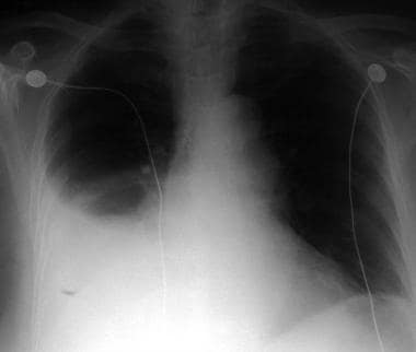
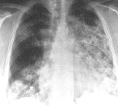
Thoracic radiography is a type of diagnostic imaging that involves using X-rays to produce images of the chest, including the lungs, heart, bronchi, great vessels, and the bones of the spine and chest wall. It is a commonly used tool in the diagnosis and management of various respiratory, cardiovascular, and thoracic disorders such as pneumonia, lung cancer, heart failure, and rib fractures.
During the procedure, the patient is positioned between an X-ray machine and a cassette containing a film or digital detector. The X-ray beam is directed at the chest, and the resulting image is captured on the film or detector. The images produced can help identify any abnormalities in the structure or function of the organs within the chest.
Thoracic radiography may be performed as a routine screening test for certain conditions, such as lung cancer, or it may be ordered when a patient presents with symptoms suggestive of a respiratory or cardiovascular disorder. It is a safe and non-invasive procedure that can provide valuable information to help guide clinical decision making and improve patient outcomes.
Spirometry is a common type of pulmonary function test (PFT) that measures how well your lungs work. This is done by measuring how much air you can exhale from your lungs after taking a deep breath, and how quickly you can exhale it. The results are compared to normal values for your age, height, sex, and ethnicity.
Spirometry is used to diagnose and monitor certain lung conditions, such as asthma, chronic obstructive pulmonary disease (COPD), and other respiratory diseases that cause narrowing of the airways. It can also be used to assess the effectiveness of treatment for these conditions. The test is non-invasive, safe, and easy to perform.
"Evaluation studies" is a broad term that refers to the systematic assessment or examination of a program, project, policy, intervention, or product. The goal of an evaluation study is to determine its merits, worth, and value by measuring its effects, efficiency, and impact. There are different types of evaluation studies, including formative evaluations (conducted during the development or implementation of a program to provide feedback for improvement), summative evaluations (conducted at the end of a program to determine its overall effectiveness), process evaluations (focusing on how a program is implemented and delivered), outcome evaluations (assessing the short-term and intermediate effects of a program), and impact evaluations (measuring the long-term and broad consequences of a program).
In medical contexts, evaluation studies are often used to assess the safety, efficacy, and cost-effectiveness of new treatments, interventions, or technologies. These studies can help healthcare providers make informed decisions about patient care, guide policymakers in developing evidence-based policies, and promote accountability and transparency in healthcare systems. Examples of evaluation studies in medicine include randomized controlled trials (RCTs) that compare the outcomes of a new treatment to those of a standard or placebo treatment, observational studies that examine the real-world effectiveness and safety of interventions, and economic evaluations that assess the costs and benefits of different healthcare options.
Ultrasonography, also known as sonography, is a diagnostic medical procedure that uses high-frequency sound waves (ultrasound) to produce dynamic images of organs, tissues, or blood flow inside the body. These images are captured in real-time and can be used to assess the size, shape, and structure of various internal structures, as well as detect any abnormalities such as tumors, cysts, or inflammation.
During an ultrasonography procedure, a small handheld device called a transducer is placed on the patient's skin, which emits and receives sound waves. The transducer sends high-frequency sound waves into the body, and these waves bounce back off internal structures and are recorded by the transducer. The recorded data is then processed and transformed into visual images that can be interpreted by a medical professional.
Ultrasonography is a non-invasive, painless, and safe procedure that does not use radiation like other imaging techniques such as CT scans or X-rays. It is commonly used to diagnose and monitor conditions in various parts of the body, including the abdomen, pelvis, heart, blood vessels, and musculoskeletal system.
A biopsy is a medical procedure in which a small sample of tissue is taken from the body to be examined under a microscope for the presence of disease. This can help doctors diagnose and monitor various medical conditions, such as cancer, infections, or autoimmune disorders. The type of biopsy performed will depend on the location and nature of the suspected condition. Some common types of biopsies include:
1. Incisional biopsy: In this procedure, a surgeon removes a piece of tissue from an abnormal area using a scalpel or other surgical instrument. This type of biopsy is often used when the lesion is too large to be removed entirely during the initial biopsy.
2. Excisional biopsy: An excisional biopsy involves removing the entire abnormal area, along with a margin of healthy tissue surrounding it. This technique is typically employed for smaller lesions or when cancer is suspected.
3. Needle biopsy: A needle biopsy uses a thin, hollow needle to extract cells or fluid from the body. There are two main types of needle biopsies: fine-needle aspiration (FNA) and core needle biopsy. FNA extracts loose cells, while a core needle biopsy removes a small piece of tissue.
4. Punch biopsy: In a punch biopsy, a round, sharp tool is used to remove a small cylindrical sample of skin tissue. This type of biopsy is often used for evaluating rashes or other skin abnormalities.
5. Shave biopsy: During a shave biopsy, a thin slice of tissue is removed from the surface of the skin using a sharp razor-like instrument. This technique is typically used for superficial lesions or growths on the skin.
After the biopsy sample has been collected, it is sent to a laboratory where a pathologist will examine the tissue under a microscope and provide a diagnosis based on their findings. The results of the biopsy can help guide further treatment decisions and determine the best course of action for managing the patient's condition.
Diagnostic techniques in endocrinology are methods used to identify and diagnose various endocrine disorders. These techniques include:
1. Hormone measurements: Measuring the levels of hormones in blood, urine, or saliva can help identify excess or deficiency of specific hormones. This is often done through immunoassays, which use antibodies to detect and quantify hormones.
2. Provocative and suppression tests: These tests involve administering a medication that stimulates or suppresses the release of a particular hormone. Blood samples are taken before and after the medication is given to assess changes in hormone levels. Examples include the glucose tolerance test for diabetes, the ACTH stimulation test for adrenal insufficiency, and the thyroid suppression test for hyperthyroidism.
3. Imaging studies: Various imaging techniques can be used to visualize endocrine glands and identify structural abnormalities such as tumors or nodules. These include X-rays, ultrasound, computed tomography (CT), magnetic resonance imaging (MRI), and nuclear medicine scans using radioactive tracers.
4. Genetic testing: Molecular genetic tests can be used to identify genetic mutations associated with certain endocrine disorders, such as multiple endocrine neoplasia type 1 or 2, or congenital adrenal hyperplasia.
5. Biopsy: In some cases, a small sample of tissue may be removed from an endocrine gland for microscopic examination (biopsy). This can help confirm the presence of cancer or other abnormalities.
6. Functional tests: These tests assess the ability of an endocrine gland to produce and secrete hormones in response to various stimuli. Examples include the glucagon stimulation test for gastrinoma and the calcium infusion test for hyperparathyroidism.
7. Wearable monitoring devices: Continuous glucose monitoring systems (CGMS) are wearable devices that measure interstitial glucose levels continuously over several days, providing valuable information about glycemic control in patients with diabetes.
Retrospective studies, also known as retrospective research or looking back studies, are a type of observational study that examines data from the past to draw conclusions about possible causal relationships between risk factors and outcomes. In these studies, researchers analyze existing records, medical charts, or previously collected data to test a hypothesis or answer a specific research question.
Retrospective studies can be useful for generating hypotheses and identifying trends, but they have limitations compared to prospective studies, which follow participants forward in time from exposure to outcome. Retrospective studies are subject to biases such as recall bias, selection bias, and information bias, which can affect the validity of the results. Therefore, retrospective studies should be interpreted with caution and used primarily to generate hypotheses for further testing in prospective studies.
Prospective studies, also known as longitudinal studies, are a type of cohort study in which data is collected forward in time, following a group of individuals who share a common characteristic or exposure over a period of time. The researchers clearly define the study population and exposure of interest at the beginning of the study and follow up with the participants to determine the outcomes that develop over time. This type of study design allows for the investigation of causal relationships between exposures and outcomes, as well as the identification of risk factors and the estimation of disease incidence rates. Prospective studies are particularly useful in epidemiology and medical research when studying diseases with long latency periods or rare outcomes.
In the field of medicine, "time factors" refer to the duration of symptoms or time elapsed since the onset of a medical condition, which can have significant implications for diagnosis and treatment. Understanding time factors is crucial in determining the progression of a disease, evaluating the effectiveness of treatments, and making critical decisions regarding patient care.
For example, in stroke management, "time is brain," meaning that rapid intervention within a specific time frame (usually within 4.5 hours) is essential to administering tissue plasminogen activator (tPA), a clot-busting drug that can minimize brain damage and improve patient outcomes. Similarly, in trauma care, the "golden hour" concept emphasizes the importance of providing definitive care within the first 60 minutes after injury to increase survival rates and reduce morbidity.
Time factors also play a role in monitoring the progression of chronic conditions like diabetes or heart disease, where regular follow-ups and assessments help determine appropriate treatment adjustments and prevent complications. In infectious diseases, time factors are crucial for initiating antibiotic therapy and identifying potential outbreaks to control their spread.
Overall, "time factors" encompass the significance of recognizing and acting promptly in various medical scenarios to optimize patient outcomes and provide effective care.
A fine-needle biopsy (FNB) is a medical procedure in which a thin, hollow needle is used to obtain a sample of cells or tissue from a suspicious or abnormal area in the body, such as a lump or mass. The needle is typically smaller than that used in a core needle biopsy, and it is guided into place using imaging techniques such as ultrasound, CT scan, or MRI.
The sample obtained during an FNB can be used to diagnose various medical conditions, including cancer, infection, or inflammation. The procedure is generally considered safe and well-tolerated, with minimal risks of complications such as bleeding, infection, or discomfort. However, the accuracy of the diagnosis depends on the skill and experience of the healthcare provider performing the biopsy, as well as the adequacy of the sample obtained.
Overall, FNB is a valuable diagnostic tool that can help healthcare providers make informed decisions about treatment options and improve patient outcomes.
Diagnostic techniques in cardiovascular medicine refer to the various tests and methods used to diagnose and evaluate conditions related to the heart and blood vessels. These techniques can be non-invasive or invasive and are designed to provide critical information about a patient's cardiovascular health, such as heart function, blood flow, and the presence of any abnormalities or diseases. Here are some common diagnostic techniques used in cardiovascular medicine:
1. Electrocardiogram (ECG): An ECG is a non-invasive test that records the electrical activity of the heart. It can help detect heart conditions such as arrhythmias, heart attacks, and structural abnormalities.
2. Echocardiogram: This is a non-invasive ultrasound test that produces images of the heart's structures, including the chambers, valves, and major blood vessels. It can help assess heart function, identify damage from heart attacks, and detect various cardiovascular conditions.
3. Stress testing: A stress test involves exercising on a treadmill or stationary bike while being monitored by an ECG to evaluate the heart's response to physical exertion. It can help diagnose coronary artery disease, assess exercise capacity, and determine the need for further testing or treatment.
4. Cardiac catheterization: This is an invasive procedure where a thin, flexible tube (catheter) is inserted into a blood vessel in the arm or leg and guided to the heart. It can help diagnose and treat various cardiovascular conditions, such as blocked arteries, heart valve problems, and congenital heart defects.
5. Coronary angiography: During a cardiac catheterization, a special dye is injected into the coronary arteries to visualize blood flow using X-ray imaging. This can help identify blockages or narrowing in the coronary arteries and guide treatment decisions.
6. Nuclear stress testing: This test combines the use of a radioactive tracer with exercise or pharmacological stress to evaluate heart function and blood flow. It can help diagnose coronary artery disease, assess the effectiveness of treatments, and determine the need for further interventions.
7. Cardiac magnetic resonance imaging (MRI): This non-invasive imaging technique uses a strong magnetic field and radio waves to create detailed images of the heart's structure and function. It can help diagnose various cardiovascular conditions, such as heart muscle disorders, valve problems, and congenital heart defects.
8. Transesophageal echocardiography (TEE): This is a specialized ultrasound technique where a probe is inserted through the esophagus to obtain detailed images of the heart's structure and function. It can help diagnose conditions such as blood clots, valve problems, and infective endocarditis.
9. Positron emission tomography (PET) scanning: This imaging technique uses a small amount of radioactive tracer to evaluate the metabolic activity of the heart. It can help diagnose coronary artery disease, assess the effectiveness of treatments, and determine the need for further interventions.
10. Electrophysiology studies (EPS): These are invasive procedures where catheters are inserted into the heart to study its electrical system. They can help diagnose and treat various arrhythmias, such as atrial fibrillation, ventricular tachycardia, and Wolff-Parkinson-White syndrome.
Microscopy is a technical field in medicine that involves the use of microscopes to observe structures and phenomena that are too small to be seen by the naked eye. It allows for the examination of samples such as tissues, cells, and microorganisms at high magnifications, enabling the detection and analysis of various medical conditions, including infections, diseases, and cellular abnormalities.
There are several types of microscopy used in medicine, including:
1. Light Microscopy: This is the most common type of microscopy, which uses visible light to illuminate and magnify samples. It can be used to examine a wide range of biological specimens, such as tissue sections, blood smears, and bacteria.
2. Electron Microscopy: This type of microscopy uses a beam of electrons instead of light to produce highly detailed images of samples. It is often used in research settings to study the ultrastructure of cells and tissues.
3. Fluorescence Microscopy: This technique involves labeling specific molecules within a sample with fluorescent dyes, allowing for their visualization under a microscope. It can be used to study protein interactions, gene expression, and cell signaling pathways.
4. Confocal Microscopy: This type of microscopy uses a laser beam to scan a sample point by point, producing high-resolution images with reduced background noise. It is often used in medical research to study the structure and function of cells and tissues.
5. Scanning Probe Microscopy: This technique involves scanning a sample with a physical probe, allowing for the measurement of topography, mechanical properties, and other characteristics at the nanoscale. It can be used in medical research to study the structure and function of individual molecules and cells.
Medical Definition:
Magnetic Resonance Imaging (MRI) is a non-invasive diagnostic imaging technique that uses a strong magnetic field and radio waves to create detailed cross-sectional or three-dimensional images of the internal structures of the body. The patient lies within a large, cylindrical magnet, and the scanner detects changes in the direction of the magnetic field caused by protons in the body. These changes are then converted into detailed images that help medical professionals to diagnose and monitor various medical conditions, such as tumors, injuries, or diseases affecting the brain, spinal cord, heart, blood vessels, joints, and other internal organs. MRI does not use radiation like computed tomography (CT) scans.
Bacteriological techniques refer to the various methods and procedures used in the laboratory for the cultivation, identification, and study of bacteria. These techniques are essential in fields such as medicine, biotechnology, and research. Here are some common bacteriological techniques:
1. **Sterilization**: This is a process that eliminates or kills all forms of life, including bacteria, viruses, fungi, and spores. Common sterilization methods include autoclaving (using steam under pressure), dry heat (in an oven), chemical sterilants, and radiation.
2. **Aseptic Technique**: This refers to practices used to prevent contamination of sterile materials or environments with microorganisms. It includes the use of sterile equipment, gloves, and lab coats, as well as techniques such as flaming, alcohol swabbing, and using aseptic transfer devices.
3. **Media Preparation**: This involves the preparation of nutrient-rich substances that support bacterial growth. There are various types of media, including solid (agar), liquid (broth), and semi-solid (e.g., stab agar). The choice of medium depends on the type of bacteria being cultured and the purpose of the investigation.
4. **Inoculation**: This is the process of introducing a bacterial culture into a medium. It can be done using a loop, swab, or needle. The inoculum should be taken from a pure culture to avoid contamination.
5. **Incubation**: After inoculation, the bacteria are allowed to grow under controlled conditions of temperature, humidity, and atmospheric composition. This process is called incubation.
6. **Staining and Microscopy**: Bacteria are too small to be seen with the naked eye. Therefore, they need to be stained and observed under a microscope. Gram staining is a common method used to differentiate between two major groups of bacteria based on their cell wall composition.
7. **Biochemical Tests**: These are tests used to identify specific bacterial species based on their biochemical characteristics, such as their ability to ferment certain sugars, produce particular enzymes, or resist certain antibiotics.
8. **Molecular Techniques**: Advanced techniques like PCR and DNA sequencing can provide more precise identification of bacteria. They can also be used for genetic analysis and epidemiological studies.
Remember, handling microorganisms requires careful attention to biosafety procedures to prevent accidental infection or environmental contamination.
Pulmonary gas exchange is the process by which oxygen (O2) from inhaled air is transferred to the blood, and carbon dioxide (CO2), a waste product of metabolism, is removed from the blood and exhaled. This process occurs in the lungs, primarily in the alveoli, where the thin walls of the alveoli and capillaries allow for the rapid diffusion of gases between them. The partial pressure gradient between the alveolar air and the blood in the pulmonary capillaries drives this diffusion process. Oxygen-rich blood is then transported to the body's tissues, while CO2-rich blood returns to the lungs to be exhaled.
Respiratory paralysis is a condition characterized by the inability to breathe effectively due to the failure or weakness of the muscles involved in respiration. This can include the diaphragm, intercostal muscles, and other accessory muscles.
In medical terms, it's often associated with conditions that affect the neuromuscular junction, such as botulism, myasthenia gravis, or spinal cord injuries. It can also occur as a complication of general anesthesia, sedative drugs, or certain types of poisoning.
Respiratory paralysis is a serious condition that requires immediate medical attention, as it can lead to lack of oxygen (hypoxia) and buildup of carbon dioxide (hypercapnia) in the body, which can be life-threatening if not treated promptly.
Lung diseases refer to a broad category of disorders that affect the lungs and other structures within the respiratory system. These diseases can impair lung function, leading to symptoms such as coughing, shortness of breath, chest pain, and wheezing. They can be categorized into several types based on the underlying cause and nature of the disease process. Some common examples include:
1. Obstructive lung diseases: These are characterized by narrowing or blockage of the airways, making it difficult to breathe out. Examples include chronic obstructive pulmonary disease (COPD), asthma, bronchiectasis, and cystic fibrosis.
2. Restrictive lung diseases: These involve stiffening or scarring of the lungs, which reduces their ability to expand and take in air. Examples include idiopathic pulmonary fibrosis, sarcoidosis, and asbestosis.
3. Infectious lung diseases: These are caused by bacteria, viruses, fungi, or parasites that infect the lungs. Examples include pneumonia, tuberculosis, and influenza.
4. Vascular lung diseases: These affect the blood vessels in the lungs, impairing oxygen exchange. Examples include pulmonary embolism, pulmonary hypertension, and chronic thromboembolic pulmonary hypertension (CTEPH).
5. Neoplastic lung diseases: These involve abnormal growth of cells within the lungs, leading to cancer. Examples include small cell lung cancer, non-small cell lung cancer, and mesothelioma.
6. Other lung diseases: These include interstitial lung diseases, pleural effusions, and rare disorders such as pulmonary alveolar proteinosis and lymphangioleiomyomatosis (LAM).
It is important to note that this list is not exhaustive, and there are many other conditions that can affect the lungs. Proper diagnosis and treatment of lung diseases require consultation with a healthcare professional, such as a pulmonologist or respiratory therapist.
"Locusta migratoria" is the scientific name for the migratory locust, which is a species of short-horned grasshopper. While it is not a medical term, I can provide some general information about this insect.
Migratory locusts are known for their ability to form large swarms that can travel long distances, consuming vast amounts of vegetation and causing significant damage to crops and pastures. They are native to Africa, Asia, and Europe but have been introduced to other parts of the world as well. The migratory locust is a agricultural pest and can pose a serious threat to food security in areas where they occur in large numbers.
From a medical perspective, some species of grasshoppers, including the migratory locust, can carry various pathogens that may affect humans and animals if ingested or come into contact with mucous membranes. However, there are no specific medical concerns associated with "Locusta migratoria" itself.
Feces are the solid or semisolid remains of food that could not be digested or absorbed in the small intestine, along with bacteria and other waste products. After being stored in the colon, feces are eliminated from the body through the rectum and anus during defecation. Feces can vary in color, consistency, and odor depending on a person's diet, health status, and other factors.
Respiratory disorders are a group of conditions that affect the respiratory system, including the nose, throat (pharynx), windpipe (trachea), bronchi, lungs, and diaphragm. These disorders can make it difficult for a person to breathe normally and may cause symptoms such as coughing, wheezing, shortness of breath, and chest pain.
There are many different types of respiratory disorders, including:
1. Asthma: A chronic inflammatory disease that causes the airways to become narrow and swollen, leading to difficulty breathing.
2. Chronic obstructive pulmonary disease (COPD): A group of lung diseases, including emphysema and chronic bronchitis, that make it hard to breathe.
3. Pneumonia: An infection of the lungs that can cause coughing, chest pain, and difficulty breathing.
4. Lung cancer: A type of cancer that forms in the tissues of the lungs and can cause symptoms such as coughing, chest pain, and shortness of breath.
5. Tuberculosis (TB): A bacterial infection that mainly affects the lungs but can also affect other parts of the body.
6. Sleep apnea: A disorder that causes a person to stop breathing for short periods during sleep.
7. Interstitial lung disease: A group of disorders that cause scarring of the lung tissue, leading to difficulty breathing.
8. Pulmonary fibrosis: A type of interstitial lung disease that causes scarring of the lung tissue and makes it hard to breathe.
9. Pleural effusion: An abnormal accumulation of fluid in the space between the lungs and chest wall.
10. Lung transplantation: A surgical procedure to replace a diseased or failing lung with a healthy one from a donor.
Respiratory disorders can be caused by a variety of factors, including genetics, exposure to environmental pollutants, smoking, and infections. Treatment for respiratory disorders may include medications, oxygen therapy, breathing exercises, and lifestyle changes. In some cases, surgery may be necessary to treat the disorder.
Total Lung Capacity (TLC) is the maximum volume of air that can be contained within the lungs at the end of a maximal inspiration. It includes all of the following lung volumes: tidal volume, inspiratory reserve volume, expiratory reserve volume, and residual volume. TLC can be measured directly using gas dilution techniques or indirectly by adding residual volume to vital capacity. Factors that affect TLC include age, sex, height, and lung health status.
Respiratory Distress Syndrome, Adult (RDSa or ARDS), also known as Acute Respiratory Distress Syndrome, is a severe form of acute lung injury characterized by rapid onset of widespread inflammation in the lungs. This results in increased permeability of the alveolar-capillary membrane, pulmonary edema, and hypoxemia (low oxygen levels in the blood). The inflammation can be triggered by various direct or indirect insults to the lung, such as sepsis, pneumonia, trauma, or aspiration.
The hallmark of ARDS is the development of bilateral pulmonary infiltrates on chest X-ray, which can resemble pulmonary edema, but without evidence of increased left atrial pressure. The condition can progress rapidly and may require mechanical ventilation with positive end-expiratory pressure (PEEP) to maintain adequate oxygenation and prevent further lung injury.
The management of ARDS is primarily supportive, focusing on protecting the lungs from further injury, optimizing oxygenation, and providing adequate nutrition and treatment for any underlying conditions. The use of low tidal volumes and limiting plateau pressures during mechanical ventilation have been shown to improve outcomes in patients with ARDS.
An Enzyme-Linked Immunosorbent Assay (ELISA) is a type of analytical biochemistry assay used to detect and quantify the presence of a substance, typically a protein or peptide, in a liquid sample. It takes its name from the enzyme-linked antibodies used in the assay.
In an ELISA, the sample is added to a well containing a surface that has been treated to capture the target substance. If the target substance is present in the sample, it will bind to the surface. Next, an enzyme-linked antibody specific to the target substance is added. This antibody will bind to the captured target substance if it is present. After washing away any unbound material, a substrate for the enzyme is added. If the enzyme is present due to its linkage to the antibody, it will catalyze a reaction that produces a detectable signal, such as a color change or fluorescence. The intensity of this signal is proportional to the amount of target substance present in the sample, allowing for quantification.
ELISAs are widely used in research and clinical settings to detect and measure various substances, including hormones, viruses, and bacteria. They offer high sensitivity, specificity, and reproducibility, making them a reliable choice for many applications.
Pulmonary atelectasis is a medical condition characterized by the collapse or closure of the alveoli (tiny air sacs) in the lungs, leading to reduced or absent gas exchange in the affected area. This results in decreased lung volume and can cause hypoxemia (low oxygen levels in the blood). Atelectasis can be caused by various factors such as obstruction of the airways, surfactant deficiency, pneumothorax, or compression from outside the lung. It can also occur after surgical procedures, particularly when the patient is lying in one position for a long time. Symptoms may include shortness of breath, cough, and chest discomfort, but sometimes it may not cause any symptoms, especially if only a small area of the lung is affected. Treatment depends on the underlying cause and may include bronchodilators, chest physiotherapy, or even surgery in severe cases.
There is no medical definition for "dog diseases" as it is too broad a term. However, dogs can suffer from various health conditions and illnesses that are specific to their species or similar to those found in humans. Some common categories of dog diseases include:
1. Infectious Diseases: These are caused by viruses, bacteria, fungi, or parasites. Examples include distemper, parvovirus, kennel cough, Lyme disease, and heartworms.
2. Hereditary/Genetic Disorders: Some dogs may inherit certain genetic disorders from their parents. Examples include hip dysplasia, elbow dysplasia, progressive retinal atrophy (PRA), and degenerative myelopathy.
3. Age-Related Diseases: As dogs age, they become more susceptible to various health issues. Common age-related diseases in dogs include arthritis, dental disease, cancer, and cognitive dysfunction syndrome (CDS).
4. Nutritional Disorders: Malnutrition or improper feeding can lead to various health problems in dogs. Examples include obesity, malnutrition, and vitamin deficiencies.
5. Environmental Diseases: These are caused by exposure to environmental factors such as toxins, allergens, or extreme temperatures. Examples include heatstroke, frostbite, and toxicities from ingesting harmful substances.
6. Neurological Disorders: Dogs can suffer from various neurological conditions that affect their nervous system. Examples include epilepsy, intervertebral disc disease (IVDD), and vestibular disease.
7. Behavioral Disorders: Some dogs may develop behavioral issues due to various factors such as anxiety, fear, or aggression. Examples include separation anxiety, noise phobias, and resource guarding.
It's important to note that regular veterinary care, proper nutrition, exercise, and preventative measures can help reduce the risk of many dog diseases.
A newborn infant is a baby who is within the first 28 days of life. This period is also referred to as the neonatal period. Newborns require specialized care and attention due to their immature bodily systems and increased vulnerability to various health issues. They are closely monitored for signs of well-being, growth, and development during this critical time.
Diagnostic errors refer to inaccurate or delayed diagnoses of a patient's medical condition, which can lead to improper or unnecessary treatment and potentially serious harm to the patient. These errors can occur due to various factors such as lack of clinical knowledge, failure to consider all possible diagnoses, inadequate communication between healthcare providers and patients, and problems with testing or interpretation of test results. Diagnostic errors are a significant cause of preventable harm in medical care and have been identified as a priority area for quality improvement efforts.
Mycoses are a group of diseases caused by fungal infections. These infections can affect various parts of the body, including the skin, nails, hair, lungs, and internal organs. The severity of mycoses can range from superficial, mild infections to systemic, life-threatening conditions, depending on the type of fungus and the immune status of the infected individual. Some common types of mycoses include candidiasis, dermatophytosis, histoplasmosis, coccidioidomycosis, and aspergillosis. Treatment typically involves antifungal medications, which can be topical or systemic, depending on the location and severity of the infection.
Contrast media are substances that are administered to a patient in order to improve the visibility of internal body structures or processes in medical imaging techniques such as X-rays, CT scans, MRI scans, and ultrasounds. These media can be introduced into the body through various routes, including oral, rectal, or intravenous administration.
Contrast media work by altering the appearance of bodily structures in imaging studies. For example, when a patient undergoes an X-ray examination, contrast media can be used to highlight specific organs, tissues, or blood vessels, making them more visible on the resulting images. In CT and MRI scans, contrast media can help to enhance the differences between normal and abnormal tissues, allowing for more accurate diagnosis and treatment planning.
There are several types of contrast media available, each with its own specific properties and uses. Some common examples include barium sulfate, which is used as a contrast medium in X-ray studies of the gastrointestinal tract, and iodinated contrast media, which are commonly used in CT scans to highlight blood vessels and other structures.
While contrast media are generally considered safe, they can sometimes cause adverse reactions, ranging from mild symptoms such as nausea or hives to more serious complications such as anaphylaxis or kidney damage. As a result, it is important for healthcare providers to carefully evaluate each patient's medical history and individual risk factors before administering contrast media.
Cytochromes are a type of hemeprotein found in the mitochondria and other cellular membranes of organisms. They contain a heme group, which is a prosthetic group composed of an iron atom surrounded by a porphyrin ring. This structure allows cytochromes to participate in redox reactions, acting as electron carriers in various biological processes.
There are several types of cytochromes, classified based on the type of heme they contain and their absorption spectra. Some of the most well-known cytochromes include:
* Cytochrome c: a small, mobile protein found in the inner mitochondrial membrane that plays a crucial role in the electron transport chain during cellular respiration.
* Cytochrome P450: a large family of enzymes involved in the metabolism of drugs, toxins, and other xenobiotics. They are found in various tissues, including the liver, lungs, and skin.
* Cytochrome b: a component of several electron transport chains, including those found in mitochondria, bacteria, and chloroplasts.
Cytochromes play essential roles in energy production, detoxification, and other metabolic processes, making them vital for the survival and function of living organisms.
The trachea, also known as the windpipe, is a tube-like structure in the respiratory system that connects the larynx (voice box) to the bronchi (the two branches leading to each lung). It is composed of several incomplete rings of cartilage and smooth muscle, which provide support and flexibility. The trachea plays a crucial role in directing incoming air to the lungs during inspiration and outgoing air to the larynx during expiration.
Mechanical Ventilators are medical devices that assist with breathing by providing mechanical ventilation to patients who are unable to breathe sufficiently on their own. These machines deliver breaths to the patient through an endotracheal tube or a tracheostomy tube, which is placed in the windpipe (trachea). Mechanical Ventilators can be set to deliver breaths at specific rates and volumes, and they can also be adjusted to provide varying levels of positive end-expiratory pressure (PEEP) to help keep the alveoli open and improve oxygenation.
Mechanical ventilation is typically used in critical care settings such as intensive care units (ICUs), and it may be employed for a variety of reasons, including respiratory failure, sedation, neuromuscular disorders, or surgery. Prolonged use of mechanical ventilation can lead to complications such as ventilator-associated pneumonia, muscle weakness, and decreased cardiac function, so the goal is usually to wean patients off the ventilator as soon as possible.
Respiratory tract neoplasms refer to abnormal growths or tumors that occur in the respiratory system, which includes the nose, throat (pharynx), voice box (larynx), windpipe (trachea), bronchi, and lungs. These growths can be benign or malignant (cancerous). Malignant neoplasms are cancerous tumors that can invade nearby tissues, spread to other parts of the body, and interfere with normal respiratory function, leading to serious health consequences.
Respiratory tract neoplasms can have various causes, including genetic factors, exposure to environmental carcinogens such as tobacco smoke, asbestos, and radon, and certain viral infections. Symptoms of respiratory tract neoplasms may include coughing, wheezing, shortness of breath, chest pain, hoarseness, or blood in the sputum. Diagnosis typically involves imaging tests such as X-rays, CT scans, or PET scans, as well as biopsies to determine the type and extent of the tumor. Treatment options may include surgery, radiation therapy, chemotherapy, targeted therapy, or a combination of these approaches.
Treatment outcome is a term used to describe the result or effect of medical treatment on a patient's health status. It can be measured in various ways, such as through symptoms improvement, disease remission, reduced disability, improved quality of life, or survival rates. The treatment outcome helps healthcare providers evaluate the effectiveness of a particular treatment plan and make informed decisions about future care. It is also used in clinical research to compare the efficacy of different treatments and improve patient care.
Respiratory insufficiency is a condition characterized by the inability of the respiratory system to maintain adequate gas exchange, resulting in an inadequate supply of oxygen and/or removal of carbon dioxide from the body. This can occur due to various causes, such as lung diseases (e.g., chronic obstructive pulmonary disease, pneumonia), neuromuscular disorders (e.g., muscular dystrophy, spinal cord injury), or other medical conditions that affect breathing mechanics and/or gas exchange.
Respiratory insufficiency can manifest as hypoxemia (low oxygen levels in the blood) and/or hypercapnia (high carbon dioxide levels in the blood). Symptoms of respiratory insufficiency may include shortness of breath, rapid breathing, fatigue, confusion, and in severe cases, loss of consciousness or even death. Treatment depends on the underlying cause and severity of the condition and may include oxygen therapy, mechanical ventilation, medications, and/or other supportive measures.
A "mass chest X-ray" is a term used to describe a radiological screening procedure where a large number of individuals undergo chest X-rays, usually as part of a public health campaign or community screening event. The goal is to identify any early signs of lung diseases such as tuberculosis, lung cancer, or other pulmonary abnormalities. It's important to note that while mass screenings can help detect diseases at an earlier stage, they also raise concerns about radiation exposure and the potential for overdiagnosis. Therefore, such procedures are typically carried out under strict medical guidelines and regulations.
Oxygen is a colorless, odorless, tasteless gas that constitutes about 21% of the earth's atmosphere. It is a crucial element for human and most living organisms as it is vital for respiration. Inhaled oxygen enters the lungs and binds to hemoglobin in red blood cells, which carries it to tissues throughout the body where it is used to convert nutrients into energy and carbon dioxide, a waste product that is exhaled.
Medically, supplemental oxygen therapy may be provided to patients with conditions such as chronic obstructive pulmonary disease (COPD), pneumonia, heart failure, or other medical conditions that impair the body's ability to extract sufficient oxygen from the air. Oxygen can be administered through various devices, including nasal cannulas, face masks, and ventilators.
I believe there might be a misunderstanding in your question. "Dogs" is not a medical term or condition. It is the common name for a domesticated carnivore of the family Canidae, specifically the genus Canis, which includes wolves, foxes, and other extant and extinct species of mammals. Dogs are often kept as pets and companions, and they have been bred in a wide variety of forms and sizes for different purposes, such as hunting, herding, guarding, assisting police and military forces, and providing companionship and emotional support.
If you meant to ask about a specific medical condition or term related to dogs, please provide more context so I can give you an accurate answer.
Medical Definition:
"Risk factors" are any attribute, characteristic or exposure of an individual that increases the likelihood of developing a disease or injury. They can be divided into modifiable and non-modifiable risk factors. Modifiable risk factors are those that can be changed through lifestyle choices or medical treatment, while non-modifiable risk factors are inherent traits such as age, gender, or genetic predisposition. Examples of modifiable risk factors include smoking, alcohol consumption, physical inactivity, and unhealthy diet, while non-modifiable risk factors include age, sex, and family history. It is important to note that having a risk factor does not guarantee that a person will develop the disease, but rather indicates an increased susceptibility.
Prevalence, in medical terms, refers to the total number of people in a given population who have a particular disease or condition at a specific point in time, or over a specified period. It is typically expressed as a percentage or a ratio of the number of cases to the size of the population. Prevalence differs from incidence, which measures the number of new cases that develop during a certain period.
Thoracic injuries refer to damages or traumas that occur in the thorax, which is the part of the body that contains the chest cavity. The thorax houses vital organs such as the heart, lungs, esophagus, trachea, and major blood vessels. Thoracic injuries can range from blunt trauma, caused by impacts or compressions, to penetrating trauma, resulting from stabbing or gunshot wounds. These injuries may cause various complications, including but not limited to:
1. Hemothorax - bleeding into the chest cavity
2. Pneumothorax - collapsed lung due to air accumulation in the chest cavity
3. Tension pneumothorax - a life-threatening condition where trapped air puts pressure on the heart and lungs, impairing their function
4. Cardiac tamponade - compression of the heart caused by blood or fluid accumulation in the pericardial sac
5. Rib fractures, which can lead to complications like punctured lungs or internal bleeding
6. Tracheobronchial injuries, causing air leaks and difficulty breathing
7. Great vessel injuries, potentially leading to massive hemorrhage and hemodynamic instability
Immediate medical attention is required for thoracic injuries, as they can quickly become life-threatening due to the vital organs involved. Treatment may include surgery, chest tubes, medications, or supportive care, depending on the severity and type of injury.
Oxygen consumption, also known as oxygen uptake, is the amount of oxygen that is consumed or utilized by the body during a specific period of time, usually measured in liters per minute (L/min). It is a common measurement used in exercise physiology and critical care medicine to assess an individual's aerobic metabolism and overall health status.
In clinical settings, oxygen consumption is often measured during cardiopulmonary exercise testing (CPET) to evaluate cardiovascular function, pulmonary function, and exercise capacity in patients with various medical conditions such as heart failure, chronic obstructive pulmonary disease (COPD), and other respiratory or cardiac disorders.
During exercise, oxygen is consumed by the muscles to generate energy through a process called oxidative phosphorylation. The amount of oxygen consumed during exercise can provide important information about an individual's fitness level, exercise capacity, and overall health status. Additionally, measuring oxygen consumption can help healthcare providers assess the effectiveness of treatments and rehabilitation programs in patients with various medical conditions.
Flail chest is a serious injury to the thorax characterized by a segment of the chest wall that moves paradoxically in relation to the rest of the chest wall during respiration. This occurs due to multiple rib fractures at two or more places, resulting in a free-floating section of the chest wall that is not connected to the sternum or spine.
During inspiration, when the chest normally expands, the flail segment moves inward, and during expiration, it moves outward, which can lead to significant impairment of ventilation and oxygenation. Flail chest can result from high-impact trauma such as motor vehicle accidents or falls, and it is often associated with underlying lung contusions or other injuries. It requires immediate medical attention and may necessitate mechanical ventilation and surgical stabilization of the rib cage to prevent complications such as pneumonia and respiratory failure.
In medical terms, pressure is defined as the force applied per unit area on an object or body surface. It is often measured in millimeters of mercury (mmHg) in clinical settings. For example, blood pressure is the force exerted by circulating blood on the walls of the arteries and is recorded as two numbers: systolic pressure (when the heart beats and pushes blood out) and diastolic pressure (when the heart rests between beats).
Pressure can also refer to the pressure exerted on a wound or incision to help control bleeding, or the pressure inside the skull or spinal canal. High or low pressure in different body systems can indicate various medical conditions and require appropriate treatment.
Immunoenzyme techniques are a group of laboratory methods used in immunology and clinical chemistry that combine the specificity of antibody-antigen reactions with the sensitivity and amplification capabilities of enzyme reactions. These techniques are primarily used for the detection, quantitation, or identification of various analytes (such as proteins, hormones, drugs, viruses, or bacteria) in biological samples.
In immunoenzyme techniques, an enzyme is linked to an antibody or antigen, creating a conjugate. This conjugate then interacts with the target analyte in the sample, forming an immune complex. The presence and amount of this immune complex can be visualized or measured by detecting the enzymatic activity associated with it.
There are several types of immunoenzyme techniques, including:
1. Enzyme-linked Immunosorbent Assay (ELISA): A widely used method for detecting and quantifying various analytes in a sample. In ELISA, an enzyme is attached to either the capture antibody or the detection antibody. After the immune complex formation, a substrate is added that reacts with the enzyme, producing a colored product that can be measured spectrophotometrically.
2. Immunoblotting (Western blot): A method used for detecting specific proteins in a complex mixture, such as a protein extract from cells or tissues. In this technique, proteins are separated by gel electrophoresis and transferred to a membrane, where they are probed with an enzyme-conjugated antibody directed against the target protein.
3. Immunohistochemistry (IHC): A method used for detecting specific antigens in tissue sections or cells. In IHC, an enzyme-conjugated primary or secondary antibody is applied to the sample, and the presence of the antigen is visualized using a chromogenic substrate that produces a colored product at the site of the antigen-antibody interaction.
4. Immunofluorescence (IF): A method used for detecting specific antigens in cells or tissues by employing fluorophore-conjugated antibodies. The presence of the antigen is visualized using a fluorescence microscope.
5. Enzyme-linked immunosorbent assay (ELISA): A method used for detecting and quantifying specific antigens or antibodies in liquid samples, such as serum or culture supernatants. In ELISA, an enzyme-conjugated detection antibody is added after the immune complex formation, and a substrate is added that reacts with the enzyme to produce a colored product that can be measured spectrophotometrically.
These techniques are widely used in research and diagnostic laboratories for various applications, including protein characterization, disease diagnosis, and monitoring treatment responses.
Respiratory rate is the number of breaths a person takes per minute. It is typically measured by counting the number of times the chest rises and falls in one minute. Normal respiratory rate at rest for an adult ranges from 12 to 20 breaths per minute. An increased respiratory rate (tachypnea) or decreased respiratory rate (bradypnea) can be a sign of various medical conditions, such as lung disease, heart failure, or neurological disorders. It is an important vital sign that should be regularly monitored in clinical settings.
Airway obstruction is a medical condition that occurs when the normal flow of air into and out of the lungs is partially or completely blocked. This blockage can be caused by a variety of factors, including swelling of the tissues in the airway, the presence of foreign objects or substances, or abnormal growths such as tumors.
When the airway becomes obstructed, it can make it difficult for a person to breathe normally. They may experience symptoms such as shortness of breath, wheezing, coughing, and chest tightness. In severe cases, airway obstruction can lead to respiratory failure and other life-threatening complications.
There are several types of airway obstruction, including:
1. Upper airway obstruction: This occurs when the blockage is located in the upper part of the airway, such as the nose, throat, or voice box.
2. Lower airway obstruction: This occurs when the blockage is located in the lower part of the airway, such as the trachea or bronchi.
3. Partial airway obstruction: This occurs when the airway is partially blocked, allowing some air to flow in and out of the lungs.
4. Complete airway obstruction: This occurs when the airway is completely blocked, preventing any air from flowing into or out of the lungs.
Treatment for airway obstruction depends on the underlying cause of the condition. In some cases, removing the obstruction may be as simple as clearing the airway of foreign objects or mucus. In other cases, more invasive treatments such as surgery may be necessary.
Whole-body plethysmography is a non-invasive medical technique used to measure changes in the volume of air in the lungs and chest during breathing. It is often utilized in the diagnosis and assessment of various respiratory disorders such as chronic obstructive pulmonary disease (COPD), asthma, and restrictive lung diseases.
During whole-body plethysmography, the patient enters a sealed, clear chamber, usually in a standing or sitting position. The patient is instructed to breathe normally while the machine measures changes in pressure within the chamber as the chest and abdomen move during respiration. These measurements are then used to calculate lung volume, airflow, and other respiratory parameters.
This technique provides valuable information about the functional status of the lungs and can help healthcare providers make informed decisions regarding diagnosis, treatment planning, and disease monitoring.
Forced Expiratory Volume (FEV) is a medical term used to describe the volume of air that can be forcefully exhaled from the lungs in one second. It is often measured during pulmonary function testing to assess lung function and diagnose conditions such as chronic obstructive pulmonary disease (COPD) or asthma.
FEV is typically expressed as a percentage of the Forced Vital Capacity (FVC), which is the total volume of air that can be exhaled from the lungs after taking a deep breath in. The ratio of FEV to FVC is used to determine whether there is obstruction in the airways, with a lower ratio indicating more severe obstruction.
There are different types of FEV measurements, including FEV1 (the volume of air exhaled in one second), FEV25-75 (the average volume of air exhaled during the middle 50% of the FVC maneuver), and FEV0.5 (the volume of air exhaled in half a second). These measurements can provide additional information about lung function and help guide treatment decisions.
Residual Volume (RV) is the amount of air that remains in the lungs after a forced exhale, also known as the "expiratory reserve volume." It is the lowest lung volume that can be reached during a forced exhalation and cannot be completely emptied due to the presence of alveoli that are too small or too far from the airways. This volume is important for maintaining the structural integrity of the lungs and preventing their collapse. Any additional air that enters the lungs after this point will increase the total lung capacity. The normal residual volume for an average adult human is typically around 1 to 1.5 liters.
Follow-up studies are a type of longitudinal research that involve repeated observations or measurements of the same variables over a period of time, in order to understand their long-term effects or outcomes. In medical context, follow-up studies are often used to evaluate the safety and efficacy of medical treatments, interventions, or procedures.
In a typical follow-up study, a group of individuals (called a cohort) who have received a particular treatment or intervention are identified and then followed over time through periodic assessments or data collection. The data collected may include information on clinical outcomes, adverse events, changes in symptoms or functional status, and other relevant measures.
The results of follow-up studies can provide important insights into the long-term benefits and risks of medical interventions, as well as help to identify factors that may influence treatment effectiveness or patient outcomes. However, it is important to note that follow-up studies can be subject to various biases and limitations, such as loss to follow-up, recall bias, and changes in clinical practice over time, which must be carefully considered when interpreting the results.
Methacholine chloride is a medication that is used as a diagnostic tool to help identify and assess the severity of asthma or other respiratory conditions that cause airway hyperresponsiveness. It is a synthetic derivative of acetylcholine, which is a neurotransmitter that causes smooth muscle contraction in the body.
When methacholine chloride is inhaled, it stimulates the muscarinic receptors in the airways, causing them to constrict or narrow. This response is measured and used to determine the degree of airway hyperresponsiveness, which can help diagnose asthma and assess its severity.
The methacholine challenge test involves inhaling progressively higher doses of methacholine chloride until a significant decrease in lung function is observed or until a maximum dose is reached. The test results are then used to guide treatment decisions and monitor the effectiveness of therapy. It's important to note that this test should be conducted under the supervision of a healthcare professional, as it carries some risks, including bronchoconstriction and respiratory distress.
A biological marker, often referred to as a biomarker, is a measurable indicator that reflects the presence or severity of a disease state, or a response to a therapeutic intervention. Biomarkers can be found in various materials such as blood, tissues, or bodily fluids, and they can take many forms, including molecular, histologic, radiographic, or physiological measurements.
In the context of medical research and clinical practice, biomarkers are used for a variety of purposes, such as:
1. Diagnosis: Biomarkers can help diagnose a disease by indicating the presence or absence of a particular condition. For example, prostate-specific antigen (PSA) is a biomarker used to detect prostate cancer.
2. Monitoring: Biomarkers can be used to monitor the progression or regression of a disease over time. For instance, hemoglobin A1c (HbA1c) levels are monitored in diabetes patients to assess long-term blood glucose control.
3. Predicting: Biomarkers can help predict the likelihood of developing a particular disease or the risk of a negative outcome. For example, the presence of certain genetic mutations can indicate an increased risk for breast cancer.
4. Response to treatment: Biomarkers can be used to evaluate the effectiveness of a specific treatment by measuring changes in the biomarker levels before and after the intervention. This is particularly useful in personalized medicine, where treatments are tailored to individual patients based on their unique biomarker profiles.
It's important to note that for a biomarker to be considered clinically valid and useful, it must undergo rigorous validation through well-designed studies, including demonstrating sensitivity, specificity, reproducibility, and clinical relevance.
In medical terms, ribs are the long, curved bones that make up the ribcage in the human body. They articulate with the thoracic vertebrae posteriorly and connect to the sternum anteriorly via costal cartilages. There are 12 pairs of ribs in total, and they play a crucial role in protecting the lungs and heart, allowing room for expansion and contraction during breathing. Ribs also provide attachment points for various muscles involved in respiration and posture.
A "County Hospital" is not a medical term per se, but rather a term used to describe a hospital that is owned, operated, and funded by a county government. These hospitals typically provide medical care and services to the residents of that particular county, regardless of their ability to pay. They may also serve as a safety net for uninsured or underinsured individuals who cannot afford medical care at private hospitals. The specific services and facilities offered by a county hospital can vary widely depending on the location and resources available.
Bacterial DNA refers to the genetic material found in bacteria. It is composed of a double-stranded helix containing four nucleotide bases - adenine (A), thymine (T), guanine (G), and cytosine (C) - that are linked together by phosphodiester bonds. The sequence of these bases in the DNA molecule carries the genetic information necessary for the growth, development, and reproduction of bacteria.
Bacterial DNA is circular in most bacterial species, although some have linear chromosomes. In addition to the main chromosome, many bacteria also contain small circular pieces of DNA called plasmids that can carry additional genes and provide resistance to antibiotics or other environmental stressors.
Unlike eukaryotic cells, which have their DNA enclosed within a nucleus, bacterial DNA is present in the cytoplasm of the cell, where it is in direct contact with the cell's metabolic machinery. This allows for rapid gene expression and regulation in response to changing environmental conditions.
Diagnostic imaging is a medical specialty that uses various technologies to produce visual representations of the internal structures and functioning of the body. These images are used to diagnose injury, disease, or other abnormalities and to monitor the effectiveness of treatment. Common modalities of diagnostic imaging include:
1. Radiography (X-ray): Uses ionizing radiation to produce detailed images of bones, teeth, and some organs.
2. Computed Tomography (CT) Scan: Combines X-ray technology with computer processing to create cross-sectional images of the body.
3. Magnetic Resonance Imaging (MRI): Uses a strong magnetic field and radio waves to generate detailed images of soft tissues, organs, and bones.
4. Ultrasound: Employs high-frequency sound waves to produce real-time images of internal structures, often used for obstetrics and gynecology.
5. Nuclear Medicine: Involves the administration of radioactive tracers to assess organ function or detect abnormalities within the body.
6. Positron Emission Tomography (PET) Scan: Uses a small amount of radioactive material to produce detailed images of metabolic activity in the body, often used for cancer detection and monitoring treatment response.
7. Fluoroscopy: Utilizes continuous X-ray imaging to observe moving structures or processes within the body, such as swallowing studies or angiography.
Diagnostic imaging plays a crucial role in modern medicine, allowing healthcare providers to make informed decisions about patient care and treatment plans.
Bronchoconstrictor agents are substances that cause narrowing or constriction of the bronchioles, the small airways in the lungs. This can lead to symptoms such as wheezing, coughing, and shortness of breath. Bronchoconstrictor agents include certain medications (such as some beta-blockers and prostaglandin F2alpha), environmental pollutants (such as tobacco smoke and air pollution particles), and allergens (such as dust mites and pollen).
In contrast to bronchodilator agents, which are medications that widen the airways and improve breathing, bronchoconstrictor agents can make it more difficult for a person to breathe. People with respiratory conditions such as asthma or chronic obstructive pulmonary disease (COPD) may be particularly sensitive to bronchoconstrictor agents and may experience severe symptoms when exposed to them.
Inhalation is the act or process of breathing in where air or other gases are drawn into the lungs. It's also known as inspiration. This process involves several muscles, including the diaphragm and intercostal muscles between the ribs, working together to expand the chest cavity and decrease the pressure within the thorax, which then causes air to flow into the lungs.
In a medical context, inhalation can also refer to the administration of medications or therapeutic gases through the respiratory tract, typically using an inhaler or nebulizer. This route of administration allows for direct delivery of the medication to the lungs, where it can be quickly absorbed into the bloodstream and exert its effects.
Asthma is a chronic respiratory disease characterized by inflammation and narrowing of the airways, leading to symptoms such as wheezing, coughing, shortness of breath, and chest tightness. The airway obstruction in asthma is usually reversible, either spontaneously or with treatment.
The underlying cause of asthma involves a combination of genetic and environmental factors that result in hypersensitivity of the airways to certain triggers, such as allergens, irritants, viruses, exercise, and emotional stress. When these triggers are encountered, the airways constrict due to smooth muscle spasm, swell due to inflammation, and produce excess mucus, leading to the characteristic symptoms of asthma.
Asthma is typically managed with a combination of medications that include bronchodilators to relax the airway muscles, corticosteroids to reduce inflammation, and leukotriene modifiers or mast cell stabilizers to prevent allergic reactions. Avoiding triggers and monitoring symptoms are also important components of asthma management.
There are several types of asthma, including allergic asthma, non-allergic asthma, exercise-induced asthma, occupational asthma, and nocturnal asthma, each with its own set of triggers and treatment approaches. Proper diagnosis and management of asthma can help prevent exacerbations, improve quality of life, and reduce the risk of long-term complications.
Prognosis is a medical term that refers to the prediction of the likely outcome or course of a disease, including the chances of recovery or recurrence, based on the patient's symptoms, medical history, physical examination, and diagnostic tests. It is an important aspect of clinical decision-making and patient communication, as it helps doctors and patients make informed decisions about treatment options, set realistic expectations, and plan for future care.
Prognosis can be expressed in various ways, such as percentages, categories (e.g., good, fair, poor), or survival rates, depending on the nature of the disease and the available evidence. However, it is important to note that prognosis is not an exact science and may vary depending on individual factors, such as age, overall health status, and response to treatment. Therefore, it should be used as a guide rather than a definitive forecast.
Carbon dioxide (CO2) is a colorless, odorless gas that is naturally present in the Earth's atmosphere. It is a normal byproduct of cellular respiration in humans, animals, and plants, and is also produced through the combustion of fossil fuels such as coal, oil, and natural gas.
In medical terms, carbon dioxide is often used as a respiratory stimulant and to maintain the pH balance of blood. It is also used during certain medical procedures, such as laparoscopic surgery, to insufflate (inflate) the abdominal cavity and create a working space for the surgeon.
Elevated levels of carbon dioxide in the body can lead to respiratory acidosis, a condition characterized by an increased concentration of carbon dioxide in the blood and a decrease in pH. This can occur in conditions such as chronic obstructive pulmonary disease (COPD), asthma, or other lung diseases that impair breathing and gas exchange. Symptoms of respiratory acidosis may include shortness of breath, confusion, headache, and in severe cases, coma or death.
Dinosaurs are a group of reptiles that were the dominant terrestrial vertebrates for over 160 million years, from the late Triassic period until the end of the Cretaceous period. They first appeared approximately 230 million years ago and went extinct around 65 million years ago.
Dinosaurs are characterized by their upright stance, with legs positioned directly under their bodies, and a wide range of body sizes and shapes. Some dinosaurs were enormous, such as the long-necked sauropods that could reach lengths of over 100 feet, while others were small and agile.
Dinosaurs are classified into two main groups: the saurischians (lizard-hipped) and the ornithischians (bird-hipped). The saurischians include both the large carnivorous theropods, such as Tyrannosaurus rex, and the long-necked sauropods. The ornithischians were primarily herbivores and included a diverse array of species, such as the armored ankylosaurs and the horned ceratopsians.
Despite their extinction, dinosaurs have left a lasting impact on our planet and continue to be a source of fascination for people of all ages. The study of dinosaurs, known as paleontology, has shed light on many aspects of Earth's history and the evolution of life on our planet.
In medicine, elasticity refers to the ability of a tissue or organ to return to its original shape after being stretched or deformed. This property is due to the presence of elastic fibers in the extracellular matrix of the tissue, which can stretch and recoil like rubber bands.
Elasticity is an important characteristic of many tissues, particularly those that are subjected to repeated stretching or compression, such as blood vessels, lungs, and skin. For example, the elasticity of the lungs allows them to expand and contract during breathing, while the elasticity of blood vessels helps maintain normal blood pressure by allowing them to expand and constrict in response to changes in blood flow.
In addition to its role in normal physiology, elasticity is also an important factor in the diagnosis and treatment of various medical conditions. For example, decreased elasticity in the lungs can be a sign of lung disease, while increased elasticity in the skin can be a sign of aging or certain genetic disorders. Medical professionals may use techniques such as pulmonary function tests or skin biopsies to assess elasticity and help diagnose these conditions.
Expiratory Reserve Volume (ERV) is the maximum amount of air that can be exhaled forcefully after a normal tidal exhalation. It is the difference between the functional residual capacity (FRC) and the residual volume (RV). In other words, ERV is the extra volume of air that can be exhaled from the lungs after a normal breath out, when one tries to empty the lungs as much as possible. This volume is an important parameter in pulmonary function tests and helps assess lung health and disease. A decreased ERV may indicate restrictive lung diseases such as pulmonary fibrosis or neuromuscular disorders affecting respiratory muscles.
Respiratory muscles are a group of muscles involved in the process of breathing. They include the diaphragm, intercostal muscles (located between the ribs), scalene muscles (located in the neck), and abdominal muscles. These muscles work together to allow the chest cavity to expand or contract, which draws air into or pushes it out of the lungs. The diaphragm is the primary muscle responsible for breathing, contracting to increase the volume of the chest cavity and draw air into the lungs during inhalation. The intercostal muscles help to further expand the ribcage, while the abdominal muscles assist in exhaling by compressing the abdomen and pushing up on the diaphragm.
Inhalation exposure is a term used in occupational and environmental health to describe the situation where an individual breathes in substances present in the air, which could be gases, vapors, fumes, mist, or particulate matter. These substances can originate from various sources, such as industrial processes, chemical reactions, or natural phenomena.
The extent of inhalation exposure is determined by several factors, including:
1. Concentration of the substance in the air
2. Duration of exposure
3. Frequency of exposure
4. The individual's breathing rate
5. The efficiency of the individual's respiratory protection, if any
Inhalation exposure can lead to adverse health effects, depending on the toxicity and concentration of the inhaled substances. Short-term or acute health effects may include irritation of the eyes, nose, throat, or lungs, while long-term or chronic exposure can result in more severe health issues, such as respiratory diseases, neurological disorders, or cancer.
It is essential to monitor and control inhalation exposures in occupational settings to protect workers' health and ensure compliance with regulatory standards. Various methods are employed for exposure assessment, including personal air sampling, area monitoring, and biological monitoring. Based on the results of these assessments, appropriate control measures can be implemented to reduce or eliminate the risks associated with inhalation exposure.
Intubation, intratracheal is a medical procedure in which a flexible plastic or rubber tube called an endotracheal tube (ETT) is inserted through the mouth or nose, passing through the vocal cords and into the trachea (windpipe). This procedure is performed to establish and maintain a patent airway, allowing for the delivery of oxygen and the removal of carbon dioxide during mechanical ventilation in various clinical scenarios, such as:
1. Respiratory failure or arrest
2. Procedural sedation
3. Surgery under general anesthesia
4. Neuromuscular disorders
5. Ingestion of toxic substances
6. Head and neck trauma
7. Critical illness or injury affecting the airway
The process of intubation is typically performed by trained medical professionals, such as anesthesiologists, emergency medicine physicians, or critical care specialists, using direct laryngoscopy or video laryngoscopy to visualize the vocal cords and guide the ETT into the correct position. Once placed, the ETT is secured to prevent dislodgement, and the patient's respiratory status is continuously monitored to ensure proper ventilation and oxygenation.
Lung injury, also known as pulmonary injury, refers to damage or harm caused to the lung tissue, blood vessels, or air sacs (alveoli) in the lungs. This can result from various causes such as infection, trauma, exposure to harmful substances, or systemic diseases. Common types of lung injuries include acute respiratory distress syndrome (ARDS), pneumonia, and chemical pneumonitis. Symptoms may include difficulty breathing, cough, chest pain, and decreased oxygen levels in the blood. Treatment depends on the underlying cause and may include medications, oxygen therapy, or mechanical ventilation.
Computer-assisted image processing is a medical term that refers to the use of computer systems and specialized software to improve, analyze, and interpret medical images obtained through various imaging techniques such as X-ray, CT (computed tomography), MRI (magnetic resonance imaging), ultrasound, and others.
The process typically involves several steps, including image acquisition, enhancement, segmentation, restoration, and analysis. Image processing algorithms can be used to enhance the quality of medical images by adjusting contrast, brightness, and sharpness, as well as removing noise and artifacts that may interfere with accurate diagnosis. Segmentation techniques can be used to isolate specific regions or structures of interest within an image, allowing for more detailed analysis.
Computer-assisted image processing has numerous applications in medical imaging, including detection and characterization of lesions, tumors, and other abnormalities; assessment of organ function and morphology; and guidance of interventional procedures such as biopsies and surgeries. By automating and standardizing image analysis tasks, computer-assisted image processing can help to improve diagnostic accuracy, efficiency, and consistency, while reducing the potential for human error.
DNA primers are short single-stranded DNA molecules that serve as a starting point for DNA synthesis. They are typically used in laboratory techniques such as the polymerase chain reaction (PCR) and DNA sequencing. The primer binds to a complementary sequence on the DNA template through base pairing, providing a free 3'-hydroxyl group for the DNA polymerase enzyme to add nucleotides and synthesize a new strand of DNA. This allows for specific and targeted amplification or analysis of a particular region of interest within a larger DNA molecule.
Molecular sequence data refers to the specific arrangement of molecules, most commonly nucleotides in DNA or RNA, or amino acids in proteins, that make up a biological macromolecule. This data is generated through laboratory techniques such as sequencing, and provides information about the exact order of the constituent molecules. This data is crucial in various fields of biology, including genetics, evolution, and molecular biology, allowing for comparisons between different organisms, identification of genetic variations, and studies of gene function and regulation.
Bronchoconstriction is a medical term that refers to the narrowing of the airways in the lungs (the bronchi and bronchioles) due to the contraction of the smooth muscles surrounding them. This constriction can cause difficulty breathing, wheezing, coughing, and shortness of breath, which are common symptoms of asthma and other respiratory conditions.
Bronchoconstriction can be triggered by a variety of factors, including allergens, irritants, cold air, exercise, and emotional stress. In some cases, it may also be caused by certain medications, such as beta-blockers or nonsteroidal anti-inflammatory drugs (NSAIDs). Treatment for bronchoconstriction typically involves the use of bronchodilators, which are medications that help to relax the smooth muscles around the airways and widen them, making it easier to breathe.
Respiratory Distress Syndrome (RDS), Newborn is a common lung disorder in premature infants. It occurs when the lungs lack a substance called surfactant, which helps keep the tiny air sacs in the lungs open. This results in difficulty breathing and oxygenation, causing symptoms such as rapid, shallow breathing, grunting noises, flaring of the nostrils, and retractions (the skin between the ribs pulls in with each breath). RDS is more common in infants born before 34 weeks of gestation and is treated with surfactant replacement therapy, oxygen support, and mechanical ventilation if necessary. In severe cases, it can lead to complications such as bronchopulmonary dysplasia or even death.
A disease outbreak is defined as the occurrence of cases of a disease in excess of what would normally be expected in a given time and place. It may affect a small and localized group or a large number of people spread over a wide area, even internationally. An outbreak may be caused by a new agent, a change in the agent's virulence or host susceptibility, or an increase in the size or density of the host population.
Outbreaks can have significant public health and economic impacts, and require prompt investigation and control measures to prevent further spread of the disease. The investigation typically involves identifying the source of the outbreak, determining the mode of transmission, and implementing measures to interrupt the chain of infection. This may include vaccination, isolation or quarantine, and education of the public about the risks and prevention strategies.
Examples of disease outbreaks include foodborne illnesses linked to contaminated food or water, respiratory infections spread through coughing and sneezing, and mosquito-borne diseases such as Zika virus and West Nile virus. Outbreaks can also occur in healthcare settings, such as hospitals and nursing homes, where vulnerable populations may be at increased risk of infection.
Aerosols are defined in the medical field as suspensions of fine solid or liquid particles in a gas. In the context of public health and medicine, aerosols often refer to particles that can remain suspended in air for long periods of time and can be inhaled. They can contain various substances, such as viruses, bacteria, fungi, or chemicals, and can play a role in the transmission of respiratory infections or other health effects.
For example, when an infected person coughs or sneezes, they may produce respiratory droplets that can contain viruses like influenza or SARS-CoV-2 (the virus that causes COVID-19). Some of these droplets can evaporate quickly and leave behind smaller particles called aerosols, which can remain suspended in the air for hours and potentially be inhaled by others. This is one way that respiratory viruses can spread between people in close proximity to each other.
Aerosols can also be generated through medical procedures such as bronchoscopy, suctioning, or nebulizer treatments, which can produce aerosols containing bacteria, viruses, or other particles that may pose an infection risk to healthcare workers or other patients. Therefore, appropriate personal protective equipment (PPE) and airborne precautions are often necessary to reduce the risk of transmission in these settings.
Work of breathing (WOB) is a term used in respiratory physiology to describe the amount of energy expended by the respiratory muscles to overcome the elastic and resistive forces in the lungs and chest wall during breathing. It is usually measured in joules per liter (J/L) or in breaths per minute (BPM).
WOB can be increased in various lung diseases, such as chronic obstructive pulmonary disease (COPD), asthma, and interstitial lung disease, due to increased airway resistance or decreased lung compliance. Increased WOB can lead to respiratory muscle fatigue, decreased exercise tolerance, and reduced quality of life.
WOB can be measured noninvasively using techniques such as esophageal pressure monitoring or transdiaphragmatic pressure measurement, or invasively through the use of indwelling catheters in the pleural space or within the airways. These measurements are often used in research settings to evaluate the effectiveness of various treatments for respiratory disorders.
Biological models, also known as physiological models or organismal models, are simplified representations of biological systems, processes, or mechanisms that are used to understand and explain the underlying principles and relationships. These models can be theoretical (conceptual or mathematical) or physical (such as anatomical models, cell cultures, or animal models). They are widely used in biomedical research to study various phenomena, including disease pathophysiology, drug action, and therapeutic interventions.
Examples of biological models include:
1. Mathematical models: These use mathematical equations and formulas to describe complex biological systems or processes, such as population dynamics, metabolic pathways, or gene regulation networks. They can help predict the behavior of these systems under different conditions and test hypotheses about their underlying mechanisms.
2. Cell cultures: These are collections of cells grown in a controlled environment, typically in a laboratory dish or flask. They can be used to study cellular processes, such as signal transduction, gene expression, or metabolism, and to test the effects of drugs or other treatments on these processes.
3. Animal models: These are living organisms, usually vertebrates like mice, rats, or non-human primates, that are used to study various aspects of human biology and disease. They can provide valuable insights into the pathophysiology of diseases, the mechanisms of drug action, and the safety and efficacy of new therapies.
4. Anatomical models: These are physical representations of biological structures or systems, such as plastic models of organs or tissues, that can be used for educational purposes or to plan surgical procedures. They can also serve as a basis for developing more sophisticated models, such as computer simulations or 3D-printed replicas.
Overall, biological models play a crucial role in advancing our understanding of biology and medicine, helping to identify new targets for therapeutic intervention, develop novel drugs and treatments, and improve human health.
In epidemiology, the incidence of a disease is defined as the number of new cases of that disease within a specific population over a certain period of time. It is typically expressed as a rate, with the number of new cases in the numerator and the size of the population at risk in the denominator. Incidence provides information about the risk of developing a disease during a given time period and can be used to compare disease rates between different populations or to monitor trends in disease occurrence over time.
Thoracic neoplasms refer to abnormal growths or tumors that develop in the thorax, which is the area of the body that includes the chest and lungs. These neoplasms can be benign (non-cancerous) or malignant (cancerous). Malignant thoracic neoplasms are often referred to as lung cancer, but they can also include other types of cancer such as mesothelioma, thymoma, and esophageal cancer.
Thoracic neoplasms can cause various symptoms depending on their location and size. Common symptoms include coughing, chest pain, shortness of breath, hoarseness, and difficulty swallowing. Treatment options for thoracic neoplasms depend on the type, stage, and location of the tumor, as well as the patient's overall health. Treatment may include surgery, radiation therapy, chemotherapy, targeted therapy, or a combination of these approaches.
Exhalation is the act of breathing out or exhaling, which is the reverse process of inhalation. During exhalation, the diaphragm relaxes and moves upwards, while the chest muscles also relax, causing the chest cavity to decrease in size. This decrease in size puts pressure on the lungs, causing them to deflate and expel air.
Exhalation is a passive process that occurs naturally after inhalation, but it can also be actively controlled during activities such as speaking, singing, or playing a wind instrument. In medical terms, exhalation may also be referred to as expiration.
Inspiratory Capacity (IC) is the maximum volume of air that can be breathed in after a normal expiration. It is the sum of the tidal volume (the amount of air displaced between normal inspiration and expiration during quiet breathing) and the inspiratory reserve volume (the additional amount of air that can be inspired over and above the tidal volume). IC is an important parameter used in pulmonary function testing to assess lung volumes and capacities in patients with respiratory disorders.
Vital capacity (VC) is a term used in pulmonary function tests to describe the maximum volume of air that can be exhaled after taking a deep breath. It is the sum of inspiratory reserve volume, tidal volume, and expiratory reserve volume. In other words, it's the total amount of air you can forcibly exhale after inhaling as deeply as possible. Vital capacity is an important measurement in assessing lung function and can be reduced in conditions such as chronic obstructive pulmonary disease (COPD), asthma, and other respiratory disorders.
The Fluorescent Antibody Technique (FAT) is a type of immunofluorescence assay used in laboratory medicine and pathology for the detection and localization of specific antigens or antibodies in tissues, cells, or microorganisms. In this technique, a fluorescein-labeled antibody is used to selectively bind to the target antigen or antibody, forming an immune complex. When excited by light of a specific wavelength, the fluorescein label emits light at a longer wavelength, typically visualized as green fluorescence under a fluorescence microscope.
The FAT is widely used in diagnostic microbiology for the identification and characterization of various bacteria, viruses, fungi, and parasites. It has also been applied in the diagnosis of autoimmune diseases and certain cancers by detecting specific antibodies or antigens in patient samples. The main advantage of FAT is its high sensitivity and specificity, allowing for accurate detection and differentiation of various pathogens and disease markers. However, it requires specialized equipment and trained personnel to perform and interpret the results.
Alligators and crocodiles are large, semi-aquatic reptiles belonging to the order Crocodylia. They are characterized by a long, broad snout, powerful tail, and sharp teeth designed for grabbing and holding onto prey. Alligators and crocodiles are similar in appearance but can be distinguished by their snouts: alligators have a wider, U-shaped snout, while crocodiles have a more V-shaped snout.
Alligators (family Alligatoridae) are native to the United States and China, with two living species: the American alligator (Alligator mississippiensis) and the Chinese alligator (Alligator sinensis). They prefer freshwater habitats such as rivers, lakes, and marshes.
Crocodiles (family Crocodylidae) are found in tropical regions around the world, including Africa, Asia, Australia, and the Americas. There are 14 species of crocodiles, including the Nile crocodile (Crocodylus niloticus), the Saltwater crocodile (Crocodylus porosus), and the American crocodile (Crocodylus acutus). Crocodiles can tolerate both freshwater and saltwater environments.
Both alligators and crocodiles are apex predators, feeding on a variety of animals such as fish, birds, and mammals. They are known for their powerful bite force and have been reported to take down large prey, including deer and cattle. Alligators and crocodiles play an important role in maintaining the balance of their ecosystems by controlling populations of other animals and helping to keep waterways clean.
While alligators and crocodiles are often feared due to their size and predatory nature, they are also threatened by habitat loss, pollution, and hunting. Several species are considered endangered or vulnerable, and conservation efforts are underway to protect them and their habitats.
Cytochrome a is a type of cytochrome found in the inner mitochondrial membrane of eukaryotic cells. It is a component of cytochrome c oxidase, the final enzyme in the electron transport chain responsible for reducing molecular oxygen to water during cellular respiration. Cytochrome a contains a heme group with a low redox potential, making it capable of accepting electrons from cytochrome c and transferring them to oxygen.
The "Cytochrome a Group" typically refers to a family of related cytochromes that share similar structural and functional properties, including the presence of a heme group with a low redox potential. This group includes cytochrome a, as well as other closely related cytochromes such as cytochrome aa3 and cytochrome o. These cytochromes play important roles in electron transfer and energy conservation during cellular respiration in various organisms.
A diaphragm is a thin, dome-shaped muscle that separates the chest cavity from the abdominal cavity. It plays a vital role in the process of breathing as it contracts and flattens to draw air into the lungs (inhalation) and relaxes and returns to its domed shape to expel air out of the lungs (exhalation).
In addition, a diaphragm is also a type of barrier method of birth control. It is a flexible dome-shaped device made of silicone that fits over the cervix inside the vagina. When used correctly and consistently, it prevents sperm from entering the uterus and fertilizing an egg, thereby preventing pregnancy.
Intrinsic Positive-Pressure Respiration (IPPR) is a type of positive-pressure breathing that occurs naturally within the body, without the use of mechanical ventilation or other external devices. It is also known as "auto-PEEP" or "occult PEEP," where PEEP stands for Positive End-Expiratory Pressure.
In normal, spontaneous breathing, the pressure inside the alveoli (air sacs) in the lungs becomes slightly negative during inhalation and returns to atmospheric pressure during exhalation. However, certain lung conditions or patient efforts can lead to an increase in resistance to airflow during exhalation, causing positive pressure to persist within the alveoli at the end of expiration. This results in intrinsic PEEP or auto-PEEP.
IPPR can be caused by several factors, including:
1. Air trapping due to obstructive lung diseases like chronic obstructive pulmonary disease (COPD) or asthma
2. High respiratory rates leading to incomplete exhalation before the next inspiration begins
3. Inadequate expiratory time, often seen in mechanically ventilated patients with high tidal volumes and/or low respiratory rates
4. Dynamic hyperinflation due to increased minute ventilation or high inspiratory flow rates
5. Bronchoconstriction or airway narrowing, which can occur during an asthma attack or in certain other lung conditions
IPPR has several clinical implications, such as reduced venous return and cardiac output, increased work of breathing, and potential for lung injury due to overdistension (volutrauma). Healthcare providers must consider IPPR when managing patients with respiratory distress, especially those on mechanical ventilation, to optimize their care and prevent complications.
I'm sorry for any confusion, but "United States" is a geopolitical entity, specifically the name of the country consisting of 50 states, and it is not a medical term or concept. Therefore, I can't provide a medical definition for it. If you have any questions related to health, medicine, or biology, I would be happy to try to help answer those!
The esophagus is the muscular tube that connects the throat (pharynx) to the stomach. It is located in the midline of the neck and chest, passing through the diaphragm to enter the abdomen and join the stomach. The main function of the esophagus is to transport food and liquids from the mouth to the stomach for digestion.
The esophagus has a few distinct parts: the upper esophageal sphincter (a ring of muscle that separates the esophagus from the throat), the middle esophagus, and the lower esophageal sphincter (another ring of muscle that separates the esophagus from the stomach). The lower esophageal sphincter relaxes to allow food and liquids to enter the stomach and then contracts to prevent stomach contents from flowing back into the esophagus.
The walls of the esophagus are made up of several layers, including mucosa (a moist tissue that lines the inside of the tube), submucosa (a layer of connective tissue), muscle (both voluntary and involuntary types), and adventitia (an outer layer of connective tissue).
Common conditions affecting the esophagus include gastroesophageal reflux disease (GERD), Barrett's esophagus, esophageal cancer, esophageal strictures, and eosinophilic esophagitis.
Obstructive lung disease is a category of respiratory diseases characterized by airflow limitation that causes difficulty in completely emptying the alveoli (tiny air sacs) of the lungs during exhaling. This results in the trapping of stale air and prevents fresh air from entering the alveoli, leading to various symptoms such as coughing, wheezing, shortness of breath, and decreased exercise tolerance.
The most common obstructive lung diseases include:
1. Chronic Obstructive Pulmonary Disease (COPD): A progressive disease that includes chronic bronchitis and emphysema, often caused by smoking or exposure to harmful pollutants.
2. Asthma: A chronic inflammatory disorder of the airways characterized by variable airflow obstruction, bronchial hyperresponsiveness, and an underlying inflammation. Symptoms can be triggered by various factors such as allergens, irritants, or physical activity.
3. Bronchiectasis: A condition in which the airways become abnormally widened, scarred, and thickened due to chronic inflammation or infection, leading to mucus buildup and impaired clearance.
4. Cystic Fibrosis: An inherited genetic disorder that affects the exocrine glands, resulting in thick and sticky mucus production in various organs, including the lungs. This can lead to chronic lung infections, inflammation, and airway obstruction.
5. Alpha-1 Antitrypsin Deficiency: A genetic condition characterized by low levels of alpha-1 antitrypsin protein, which leads to uncontrolled protease enzyme activity that damages the lung tissue, causing emphysema-like symptoms.
Treatment for obstructive lung diseases typically involves bronchodilators (to relax and widen the airways), corticosteroids (to reduce inflammation), and lifestyle modifications such as smoking cessation and pulmonary rehabilitation programs. In severe cases, oxygen therapy or even lung transplantation may be considered.
Respiratory tract infections (RTIs) are infections that affect the respiratory system, which includes the nose, throat (pharynx), voice box (larynx), windpipe (trachea), bronchi, and lungs. These infections can be caused by viruses, bacteria, or, less commonly, fungi.
RTIs are classified into two categories based on their location: upper respiratory tract infections (URTIs) and lower respiratory tract infections (LRTIs). URTIs include infections of the nose, sinuses, throat, and larynx, such as the common cold, flu, laryngitis, and sinusitis. LRTIs involve the lower airways, including the bronchi and lungs, and can be more severe. Examples of LRTIs are pneumonia, bronchitis, and bronchiolitis.
Symptoms of RTIs depend on the location and cause of the infection but may include cough, congestion, runny nose, sore throat, difficulty breathing, wheezing, fever, fatigue, and chest pain. Treatment for RTIs varies depending on the severity and underlying cause of the infection. For viral infections, treatment typically involves supportive care to manage symptoms, while antibiotics may be prescribed for bacterial infections.
Helium is not a medical term, but it's a chemical element with symbol He and atomic number 2. It's a colorless, odorless, tasteless, non-toxic, inert, monatomic gas that heads the noble gases section of the periodic table. In medicine, helium is sometimes used in medical settings for its unique properties, such as being less dense than air, which can help improve the delivery of oxygen to patients with respiratory conditions. For example, heliox, a mixture of helium and oxygen, may be used to reduce the work of breathing in patients with conditions like chronic obstructive pulmonary disease (COPD) or asthma. Additionally, helium is also used in cryogenic medical equipment and in magnetic resonance imaging (MRI) machines to cool the superconducting magnets.
In medical terms, 'air' is defined as the mixture of gases that make up the Earth's atmosphere. It primarily consists of nitrogen (78%), oxygen (21%), and small amounts of other gases such as argon, carbon dioxide, and trace amounts of neon, helium, and methane.
Air is essential for human life, as it provides the oxygen that our bodies need to produce energy through respiration. We inhale air into our lungs, where oxygen is absorbed into the bloodstream and transported to cells throughout the body. At the same time, carbon dioxide, a waste product of cellular metabolism, is exhaled out of the body through the lungs and back into the atmosphere.
In addition to its role in respiration, air also plays a critical role in regulating the Earth's climate and weather patterns, as well as serving as a medium for sound waves and other forms of energy transfer.
High-frequency ventilation (HFV) is a specialized mode of mechanical ventilation that delivers breaths at higher rates (usually 120-900 breaths per minute) and smaller tidal volumes (1-3 mL/kg) compared to conventional ventilation. This technique aims to reduce lung injury caused by overdistension and atelectasis, which can occur with traditional ventilator settings. It is often used in neonatal and pediatric intensive care units for the management of severe respiratory distress syndrome, meconium aspiration syndrome, and other conditions where conventional ventilation may be harmful.
There are two main types of high-frequency ventilation: high-frequency oscillatory ventilation (HFOV) and high-frequency jet ventilation (HFJV). Both techniques use different methods to generate the high-frequency breaths but share similar principles in delivering small tidal volumes at rapid rates.
In summary, high-frequency ventilation is a medical intervention that utilizes specialized ventilators to deliver faster and smaller breaths, minimizing lung injury and improving oxygenation for critically ill patients with severe respiratory distress.
Respiratory dead space is the portion of each tidal volume (the amount of air that moves in and out of the lungs during normal breathing) that does not participate in gas exchange. It mainly consists of the anatomical dead space, which includes the conducting airways such as the trachea, bronchi, and bronchioles, where no alveoli are present for gas exchange to occur.
Additionally, alveolar dead space can also contribute to respiratory dead space when alveoli are perfused inadequately or not at all due to conditions like pulmonary embolism, lung consolidation, or impaired circulation. In these cases, even though air reaches the alveoli, insufficient blood flow prevents efficient gas exchange from taking place.
The sum of anatomical and alveolar dead space is referred to as physiological dead space. An increased respiratory dead space can lead to ventilation-perfusion mismatch and impaired oxygenation, making it a critical parameter in assessing respiratory function, particularly during mechanical ventilation in critically ill patients.
The phrenic nerve is a motor nerve that originates from the cervical spine (C3-C5) and descends through the neck to reach the diaphragm, which is the primary muscle used for breathing. The main function of the phrenic nerve is to innervate the diaphragm and control its contraction and relaxation, thereby enabling respiration.
Damage or injury to the phrenic nerve can result in paralysis of the diaphragm, leading to difficulty breathing and potentially causing respiratory failure. Certain medical conditions, such as neuromuscular disorders, spinal cord injuries, and tumors, can affect the phrenic nerve and impair its function.
Potassium Cyanide (C6H5KN) is defined as a white, water-soluble, crystalline salt that is highly toxic. It is used in fumigation, electroplating, and metal cleaning. When combined with acids, it releases the deadly gas hydrogen cyanide. It can cause immediate death by inhibiting cellular respiration. It is also known as Cyanide of Potassium or Potassium Salt of Hydrocyanic Acid.
Obesity Hypoventilation Syndrome (OHS) is a medical condition characterized by the presence of obesity (generally defined as a body mass index of 30 or higher) and chronic hypoventilation, which means that the person is not breathing adequately, resulting in low levels of oxygen and high levels of carbon dioxide in the blood.
In OHS, the excess weight of the chest walls makes it difficult for the respiratory muscles to work effectively, leading to reduced lung volumes and impaired gas exchange. This results in chronic hypoxemia (low oxygen levels) and hypercapnia (high carbon dioxide levels) during wakefulness and sleep.
OHS is often associated with obstructive sleep apnea (OSA), a condition characterized by repeated episodes of upper airway obstruction during sleep, which can further exacerbate hypoventilation. However, not all patients with OHS have OSA, and vice versa.
The diagnosis of OHS is typically made based on the presence of obesity, chronic hypoventilation (as evidenced by elevated arterial carbon dioxide levels), and the absence of other causes of hypoventilation. Treatment usually involves the use of non-invasive ventilation to support breathing and improve gas exchange, as well as weight loss interventions to address the underlying obesity.
Plethysmography is a non-invasive medical technique used to measure changes in volume or blood flow within an organ or body part, typically in the lungs or extremities. There are several types of plethysmography, including:
1. **Whole Body Plethysmography (WBP):** This type of plethysmography is used to assess lung function and volumes by measuring changes in pressure within a sealed chamber that contains the patient's entire body except for their head. The patient breathes normally while wearing a nose clip, allowing technicians to analyze respiratory patterns, airflow, and lung volume changes.
2. **Segmental or Local Plethysmography:** This technique measures volume or blood flow changes in specific body parts, such as the limbs or digits. It can help diagnose and monitor conditions affecting peripheral circulation, like deep vein thrombosis, arterial occlusive disease, or Raynaud's phenomenon.
3. **Impedance Plethysmography (IPG):** This non-invasive method uses electrical impedance to estimate changes in blood volume within an organ or body part. By applying a small electrical current and measuring the opposition to flow (impedance), technicians can determine variations in blood volume, which can help diagnose conditions like deep vein thrombosis or heart failure.
4. **Optical Plethysmography:** This technique uses light to measure changes in blood volume, typically in the skin or mucous membranes. By shining a light on the area and analyzing the reflected or transmitted light, technicians can detect variations in blood volume related to cardiac output, respiration, or other physiological factors.
Overall, plethysmography is an essential tool for diagnosing and monitoring various medical conditions affecting circulation, respiratory function, and organ volumes.
Emphysema is a chronic respiratory disease characterized by abnormal, permanent enlargement of the airspaces called alveoli in the lungs, accompanied by destruction of their walls. This results in loss of elasticity and decreased gas exchange efficiency, causing shortness of breath and coughing. It is often caused by smoking or exposure to harmful pollutants. The damage to the lungs is irreversible, but quitting smoking and using medications can help alleviate symptoms and slow disease progression.
Immunohistochemistry (IHC) is a technique used in pathology and laboratory medicine to identify specific proteins or antigens in tissue sections. It combines the principles of immunology and histology to detect the presence and location of these target molecules within cells and tissues. This technique utilizes antibodies that are specific to the protein or antigen of interest, which are then tagged with a detection system such as a chromogen or fluorophore. The stained tissue sections can be examined under a microscope, allowing for the visualization and analysis of the distribution and expression patterns of the target molecule in the context of the tissue architecture. Immunohistochemistry is widely used in diagnostic pathology to help identify various diseases, including cancer, infectious diseases, and immune-mediated disorders.
A cough is a reflex action that helps to clear the airways of irritants, foreign particles, or excess mucus or phlegm. It is characterized by a sudden, forceful expulsion of air from the lungs through the mouth and nose. A cough can be acute (short-term) or chronic (long-term), and it can be accompanied by other symptoms such as chest pain, shortness of breath, or fever. Coughing can be caused by various factors, including respiratory infections, allergies, asthma, environmental pollutants, gastroesophageal reflux disease (GERD), and chronic lung diseases such as chronic obstructive pulmonary disease (COPD) and bronchitis. In some cases, a cough may be a symptom of a more serious underlying condition, such as heart failure or lung cancer.
Blood gas analysis is a medical test that measures the levels of oxygen and carbon dioxide in the blood, as well as the pH level, which indicates the acidity or alkalinity of the blood. This test is often used to evaluate lung function, respiratory disorders, and acid-base balance in the body. It can also be used to monitor the effectiveness of treatments for conditions such as chronic obstructive pulmonary disease (COPD), asthma, and other respiratory illnesses. The analysis is typically performed on a sample of arterial blood, although venous blood may also be used in some cases.
The cardiovascular system, also known as the circulatory system, is a biological system responsible for pumping and transporting blood throughout the body in animals and humans. It consists of the heart, blood vessels (comprising arteries, veins, and capillaries), and blood. The main function of this system is to transport oxygen, nutrients, hormones, and cellular waste products throughout the body to maintain homeostasis and support organ function.
The heart acts as a muscular pump that contracts and relaxes to circulate blood. It has four chambers: two atria on the top and two ventricles on the bottom. The right side of the heart receives deoxygenated blood from the body, pumps it through the lungs for oxygenation, and then sends it back to the left side of the heart. The left side of the heart then pumps the oxygenated blood through the aorta and into the systemic circulation, reaching all parts of the body via a network of arteries and capillaries. Deoxygenated blood is collected by veins and returned to the right atrium, completing the cycle.
The cardiovascular system plays a crucial role in regulating temperature, pH balance, and fluid balance throughout the body. It also contributes to the immune response and wound healing processes. Dysfunctions or diseases of the cardiovascular system can lead to severe health complications, such as hypertension, coronary artery disease, heart failure, stroke, and peripheral artery disease.
Reagent kits, diagnostic are prepackaged sets of chemical reagents and other components designed for performing specific diagnostic tests or assays. These kits are often used in clinical laboratories to detect and measure the presence or absence of various biomarkers, such as proteins, antibodies, antigens, nucleic acids, or small molecules, in biological samples like blood, urine, or tissues.
Diagnostic reagent kits typically contain detailed instructions for their use, along with the necessary reagents, controls, and sometimes specialized equipment or supplies. They are designed to simplify the testing process, reduce human error, and increase standardization, ensuring accurate and reliable results. Examples of diagnostic reagent kits include those used for pregnancy tests, infectious disease screening, drug testing, genetic testing, and cancer biomarker detection.
Pulmonary alveoli, also known as air sacs, are tiny clusters of air-filled pouches located at the end of the bronchioles in the lungs. They play a crucial role in the process of gas exchange during respiration. The thin walls of the alveoli, called alveolar membranes, allow oxygen from inhaled air to pass into the bloodstream and carbon dioxide from the bloodstream to pass into the alveoli to be exhaled out of the body. This vital function enables the lungs to supply oxygen-rich blood to the rest of the body and remove waste products like carbon dioxide.
Tetramethylphenylenediamine (TMPD) is not typically considered a medical term, but it is a chemical compound that is used in some scientific and research contexts. It's a type of aromatic amine, which is a class of organic compounds characterized by the presence of one or more amino groups (-NH2) attached to an aromatic hydrocarbon ring.
In biochemistry and molecular biology, TMPD is sometimes used as an electron carrier in experiments that involve redox reactions, such as those that occur during cellular respiration. It can also be used as a catalyst or reagent in various chemical reactions. However, it's important to note that TMPD is not a substance that is typically encountered in medical practice or patient care.
Bronchodilators are medications that relax and widen the airways (bronchioles) in the lungs, making it easier to breathe. They work by relaxing the smooth muscle around the airways, which allows them to dilate or open up. This results in improved airflow and reduced symptoms of bronchoconstriction, such as wheezing, coughing, and shortness of breath.
Bronchodilators can be classified into two main types: short-acting and long-acting. Short-acting bronchodilators are used for quick relief of symptoms and last for 4 to 6 hours, while long-acting bronchodilators are used for maintenance therapy and provide symptom relief for 12 hours or more.
Examples of bronchodilator agents include:
* Short-acting beta-agonists (SABAs) such as albuterol, levalbuterol, and pirbuterol
* Long-acting beta-agonists (LABAs) such as salmeterol, formoterol, and indacaterol
* Anticholinergics such as ipratropium, tiotropium, and aclidinium
* Combination bronchodilators that contain both a LABA and an anticholinergic, such as umeclidinium/vilanterol and glycopyrrolate/formoterol.
Physiology is the scientific study of the normal functions and mechanisms of living organisms, including all of their biological systems, organs, cells, and biomolecules. It focuses on how various bodily functions are regulated, coordinated, and integrated to maintain a healthy state in an organism. This field encompasses a wide range of areas such as cellular physiology, neurophysiology, cardiovascular physiology, respiratory physiology, renal physiology, endocrine physiology, reproductive physiology, and exercise physiology, among others. Physiologists use a combination of experimental and theoretical approaches to understand the principles underlying normal biological function and to investigate how these functions are altered in various disease states.
Occupational diseases are health conditions or illnesses that occur as a result of exposure to hazards in the workplace. These hazards can include physical, chemical, and biological agents, as well as ergonomic factors and work-related psychosocial stressors. Examples of occupational diseases include respiratory illnesses caused by inhaling dust or fumes, hearing loss due to excessive noise exposure, and musculoskeletal disorders caused by repetitive movements or poor ergonomics. The development of an occupational disease is typically related to the nature of the work being performed and the conditions in which it is carried out. It's important to note that these diseases can be prevented or minimized through proper risk assessment, implementation of control measures, and adherence to safety regulations.
Hydroxyquinolines are a group of synthetic antimicrobial agents that contain a hydroxyl group (-OH) attached to a quinoline ring. They have been used in the treatment of various bacterial, fungal, and parasitic infections. Some common examples of hydroxyquinolines include chloroquine, hydroxychloroquine, and quinacrine. These agents work by inhibiting the growth and multiplication of microorganisms, although their exact mechanisms of action may vary. Chloroquine and hydroxychloroquine, for example, are known to interfere with the replication of the malaria parasite within red blood cells, while quinacrine has been used to treat certain types of protozoal infections.
It is important to note that the use of hydroxyquinolines is associated with a number of potential side effects and risks, including gastrointestinal disturbances, visual disturbances, and cardiac toxicity. As such, they should only be used under the close supervision of a healthcare professional.
Albuterol is a medication that is used to treat bronchospasm, or narrowing of the airways in the lungs, in conditions such as asthma and chronic obstructive pulmonary disease (COPD). It is a short-acting beta-2 agonist, which means it works by relaxing the muscles around the airways, making it easier to breathe. Albuterol is available in several forms, including an inhaler, nebulizer solution, and syrup, and it is typically used as needed to relieve symptoms of bronchospasm. It may also be used before exercise to prevent bronchospasm caused by physical activity.
The medical definition of Albuterol is: "A short-acting beta-2 adrenergic agonist used to treat bronchospasm in conditions such as asthma and COPD. It works by relaxing the muscles around the airways, making it easier to breathe."
The prone position is a body posture in which an individual lies on their stomach, with their face down and chest facing the floor or bed. This position is often used in medical settings for various purposes, such as during certain surgical procedures, respiratory support, or to alleviate pressure ulcers. It's also important to note that the prone position can have implications for patient safety, particularly in critically ill patients, and should be carefully monitored.
Hyperventilation is a medical condition characterized by an increased respiratory rate and depth, resulting in excessive elimination of carbon dioxide (CO2) from the body. This leads to hypocapnia (low CO2 levels in the blood), which can cause symptoms such as lightheadedness, dizziness, confusion, tingling sensations in the extremities, and muscle spasms. Hyperventilation may occur due to various underlying causes, including anxiety disorders, lung diseases, neurological conditions, or certain medications. It is essential to identify and address the underlying cause of hyperventilation for proper treatment.
Forced expiratory flow rates (FEFR) are measures of how quickly and efficiently air can be exhaled from the lungs during a forced breath maneuver. These measurements are often used in pulmonary function testing to help diagnose and monitor obstructive lung diseases such as asthma or chronic obstructive pulmonary disease (COPD).
FEFR is typically measured during a forced expiratory maneuver, where the person takes a deep breath in and then exhales as forcefully and quickly as possible into a mouthpiece connected to a spirometer. The spirometer measures the volume and flow rate of the exhaled air over time.
There are several different FEFR measurements that can be reported, including:
* Forced Expiratory Flow (FEF) 25-75%: This is the average flow rate during the middle half of the forced expiratory maneuver.
* Peak Expiratory Flow Rate (PEFR): This is the maximum flow rate achieved during the first second of the forced expiratory maneuver.
* Forced Expiratory Volume in 1 Second (FEV1): This is the volume of air exhaled in the first second of the forced expiratory maneuver.
Abnormal FEFR values can indicate obstruction in the small airways of the lungs, which can make it difficult to breathe out fully and quickly. The specific pattern of abnormalities in FEFR measurements can help doctors differentiate between different types of obstructive lung diseases.
Cyanides are a group of chemical compounds that contain the cyano group, -CN, which consists of a carbon atom triple-bonded to a nitrogen atom. They are highly toxic and can cause rapid death due to the inhibition of cellular respiration. Cyanide ions (CN-) bind to the ferric iron in cytochrome c oxidase, a crucial enzyme in the electron transport chain, preventing the flow of electrons and the production of ATP, leading to cellular asphyxiation.
Common sources of cyanides include industrial chemicals such as hydrogen cyanide (HCN) and potassium cyanide (KCN), as well as natural sources like certain fruits, nuts, and plants. Exposure to high levels of cyanides can occur through inhalation, ingestion, or skin absorption, leading to symptoms such as headache, dizziness, nausea, vomiting, rapid heartbeat, seizures, coma, and ultimately death. Treatment for cyanide poisoning typically involves the use of antidotes that bind to cyanide ions and convert them into less toxic forms, such as thiosulfate and rhodanese.
Dyspnea is defined as difficulty or discomfort in breathing, often described as shortness of breath. It can range from mild to severe, and may occur during rest, exercise, or at any time. Dyspnea can be caused by various medical conditions, including heart and lung diseases, anemia, and neuromuscular disorders. It is important to seek medical attention if experiencing dyspnea, as it can be a sign of a serious underlying condition.
Chronic obstructive pulmonary disease (COPD) is a progressive lung disease characterized by the persistent obstruction of airflow in and out of the lungs. This obstruction is usually caused by two primary conditions: chronic bronchitis and emphysema. Chronic bronchitis involves inflammation and narrowing of the airways, leading to excessive mucus production and coughing. Emphysema is a condition where the alveoli (air sacs) in the lungs are damaged, resulting in decreased gas exchange and shortness of breath.
The main symptoms of COPD include progressive shortness of breath, chronic cough, chest tightness, wheezing, and excessive mucus production. The disease is often associated with exposure to harmful particles or gases, such as cigarette smoke, air pollution, or occupational dusts and chemicals. While there is no cure for COPD, treatments can help alleviate symptoms, improve quality of life, and slow the progression of the disease. These treatments may include bronchodilators, corticosteroids, combination inhalers, pulmonary rehabilitation, and, in severe cases, oxygen therapy or lung transplantation.
Thoracic diseases refer to a range of medical conditions that affect the thorax, which is the part of the body that includes the chest cage, lungs, pleura (lining of the lungs), mediastinum (the area between the lungs), and diaphragm. Examples of thoracic diseases include:
1. Chronic obstructive pulmonary disease (COPD): A progressive lung disease characterized by difficulty breathing, cough, and sputum production.
2. Asthma: A chronic inflammatory disorder of the airways that causes wheezing, shortness of breath, chest tightness, and coughing.
3. Lung cancer: A malignant tumor that forms in the tissues of the lungs, usually in the cells lining the air passages.
4. Pneumonia: An infection of the lung tissue that can be caused by bacteria, viruses, or fungi.
5. Tuberculosis (TB): A bacterial infection that mainly affects the lungs but can also affect other parts of the body.
6. Pleural effusion: An abnormal accumulation of fluid in the pleural space between the lungs and the chest wall.
7. Pneumothorax: A collection of air in the pleural space that causes the lung to collapse.
8. Lung fibrosis: A condition characterized by scarring and thickening of the lung tissue, leading to difficulty breathing.
9. Esophageal cancer: A malignant tumor that forms in the tissues of the esophagus, the tube that connects the throat to the stomach.
10. Thoracic aortic aneurysm: A bulging or weakened area in the wall of the thoracic aorta, the largest artery in the body.
These are just a few examples of thoracic diseases, and there are many others that can affect the chest and lungs.
Air pollutants are substances or mixtures of substances present in the air that can have negative effects on human health, the environment, and climate. These pollutants can come from a variety of sources, including industrial processes, transportation, residential heating and cooking, agricultural activities, and natural events. Some common examples of air pollutants include particulate matter, nitrogen dioxide, sulfur dioxide, ozone, carbon monoxide, and volatile organic compounds (VOCs).
Air pollutants can cause a range of health effects, from respiratory irritation and coughing to more serious conditions such as bronchitis, asthma, and cancer. They can also contribute to climate change by reacting with other chemicals in the atmosphere to form harmful ground-level ozone and by directly absorbing or scattering sunlight, which can affect temperature and precipitation patterns.
Air quality standards and regulations have been established to limit the amount of air pollutants that can be released into the environment, and efforts are ongoing to reduce emissions and improve air quality worldwide.
Occupational exposure refers to the contact of an individual with potentially harmful chemical, physical, or biological agents as a result of their job or occupation. This can include exposure to hazardous substances such as chemicals, heavy metals, or dusts; physical agents such as noise, radiation, or ergonomic stressors; and biological agents such as viruses, bacteria, or fungi.
Occupational exposure can occur through various routes, including inhalation, skin contact, ingestion, or injection. Prolonged or repeated exposure to these hazards can increase the risk of developing acute or chronic health conditions, such as respiratory diseases, skin disorders, neurological damage, or cancer.
Employers have a legal and ethical responsibility to minimize occupational exposures through the implementation of appropriate control measures, including engineering controls, administrative controls, personal protective equipment, and training programs. Regular monitoring and surveillance of workers' health can also help identify and prevent potential health hazards in the workplace.
In medical terms, "dust" is not defined as a specific medical condition or disease. However, generally speaking, dust refers to small particles of solid matter that can be found in the air and can come from various sources, such as soil, pollen, hair, textiles, paper, or plastic.
Exposure to certain types of dust, such as those containing allergens, chemicals, or harmful pathogens, can cause a range of health problems, including respiratory issues like asthma, allergies, and lung diseases. Prolonged exposure to certain types of dust, such as silica or asbestos, can even lead to serious conditions like silicosis or mesothelioma.
Therefore, it is important for individuals who work in environments with high levels of dust to take appropriate precautions, such as wearing masks and respirators, to minimize their exposure and reduce the risk of health problems.
Bronchial provocation tests are a group of medical tests used to assess the airway responsiveness of the lungs by challenging them with increasing doses of a specific stimulus, such as methacholine or histamine, which can cause bronchoconstriction (narrowing of the airways) in susceptible individuals. These tests are often performed to diagnose and monitor asthma and other respiratory conditions that may be associated with heightened airway responsiveness.
The most common type of bronchial provocation test is the methacholine challenge test, which involves inhaling increasing concentrations of methacholine aerosol via a nebulizer. The dose response is measured by monitoring lung function (usually through spirometry) before and after each exposure. A positive test is indicated when there is a significant decrease in forced expiratory volume in one second (FEV1) or other measures of airflow, which suggests bronchial hyperresponsiveness.
Other types of bronchial provocation tests include histamine challenges, exercise challenges, and mannitol challenges. These tests have specific indications, contraindications, and protocols that should be followed to ensure accurate results and patient safety. Bronchial provocation tests are typically conducted in a controlled clinical setting under the supervision of trained healthcare professionals.
A Receiver Operating Characteristic (ROC) curve is a graphical representation used in medical decision-making and statistical analysis to illustrate the performance of a binary classifier system, such as a diagnostic test or a machine learning algorithm. It's a plot that shows the tradeoff between the true positive rate (sensitivity) and the false positive rate (1 - specificity) for different threshold settings.
The x-axis of an ROC curve represents the false positive rate (the proportion of negative cases incorrectly classified as positive), while the y-axis represents the true positive rate (the proportion of positive cases correctly classified as positive). Each point on the curve corresponds to a specific decision threshold, with higher points indicating better performance.
The area under the ROC curve (AUC) is a commonly used summary measure that reflects the overall performance of the classifier. An AUC value of 1 indicates perfect discrimination between positive and negative cases, while an AUC value of 0.5 suggests that the classifier performs no better than chance.
ROC curves are widely used in healthcare to evaluate diagnostic tests, predictive models, and screening tools for various medical conditions, helping clinicians make informed decisions about patient care based on the balance between sensitivity and specificity.
Pulmonary edema is a medical condition characterized by the accumulation of fluid in the alveoli (air sacs) and interstitial spaces (the area surrounding the alveoli) within the lungs. This buildup of fluid can lead to impaired gas exchange, resulting in shortness of breath, coughing, and difficulty breathing, especially when lying down. Pulmonary edema is often a complication of heart failure, but it can also be caused by other conditions such as pneumonia, trauma, or exposure to certain toxins.
In the early stages of pulmonary edema, patients may experience mild symptoms such as shortness of breath during physical activity. However, as the condition progresses, symptoms can become more severe and include:
* Severe shortness of breath, even at rest
* Wheezing or coughing up pink, frothy sputum
* Rapid breathing and heart rate
* Anxiety or restlessness
* Bluish discoloration of the skin (cyanosis) due to lack of oxygen
Pulmonary edema can be diagnosed through a combination of physical examination, medical history, chest X-ray, and other diagnostic tests such as echocardiography or CT scan. Treatment typically involves addressing the underlying cause of the condition, as well as providing supportive care such as supplemental oxygen, diuretics to help remove excess fluid from the body, and medications to help reduce anxiety and improve breathing. In severe cases, mechanical ventilation may be necessary to support respiratory function.
Chemical warfare agents are defined as chemical substances that are intended or have the capability to cause death, injury, temporary incapacitation, or sensory irritation through their toxic properties when deployed in a military theater. These agents can be in gaseous, liquid, or solid form and are typically categorized based on their physiological effects. Common categories include nerve agents (e.g., sarin, VX), blister agents (e.g., mustard gas), choking agents (e.g., phosgene), blood agents (e.g., cyanide), and incapacitating agents (e.g., BZ). The use of chemical warfare agents is prohibited by international law under the Chemical Weapons Convention.
Chronic bronchitis is a long-term inflammation of the airways (bronchi) in the lungs. It is characterized by a persistent cough that produces excessive mucus or sputum. The cough and mucus production must be present for at least three months in two consecutive years to meet the diagnostic criteria for chronic bronchitis.
The inflammation of the airways can lead to narrowing, obstructing the flow of air into and out of the lungs, resulting in shortness of breath and wheezing. Chronic bronchitis is often associated with exposure to irritants such as tobacco smoke, dust, or chemical fumes over an extended period.
It is a significant component of chronic obstructive pulmonary disease (COPD), which also includes emphysema. While there is no cure for chronic bronchitis, treatments can help alleviate symptoms and slow the progression of the disease. These may include bronchodilators, corticosteroids, and pulmonary rehabilitation. Quitting smoking is crucial in managing this condition.
Cardiovascular physiological phenomena refer to the various functions and processes that occur within the cardiovascular system, which includes the heart and blood vessels. These phenomena are responsible for the transport of oxygen, nutrients, and other essential molecules to tissues throughout the body, as well as the removal of waste products and carbon dioxide.
Some examples of cardiovascular physiological phenomena include:
1. Heart rate and rhythm: The heart's ability to contract regularly and coordinate its contractions with the body's needs for oxygen and nutrients.
2. Blood pressure: The force exerted by blood on the walls of blood vessels, which is determined by the amount of blood pumped by the heart and the resistance of the blood vessels.
3. Cardiac output: The volume of blood that the heart pumps in one minute, calculated as the product of stroke volume (the amount of blood pumped per beat) and heart rate.
4. Blood flow: The movement of blood through the circulatory system, which is influenced by factors such as blood pressure, vessel diameter, and blood viscosity.
5. Vasoconstriction and vasodilation: The narrowing or widening of blood vessels in response to various stimuli, such as hormones, neurotransmitters, and changes in temperature or oxygen levels.
6. Autoregulation: The ability of blood vessels to maintain a constant blood flow to tissues despite changes in perfusion pressure.
7. Blood clotting: The process by which the body forms a clot to stop bleeding after an injury, which involves the activation of platelets and the coagulation cascade.
8. Endothelial function: The ability of the endothelium (the lining of blood vessels) to regulate vascular tone, inflammation, and thrombosis.
9. Myocardial contractility: The strength of heart muscle contractions, which is influenced by factors such as calcium levels, neurotransmitters, and hormones.
10. Electrophysiology: The study of the electrical properties of the heart, including the conduction system that allows for the coordinated contraction of heart muscle.
Chest wall oscillation is a technique used in physical therapy to help clear secretions from the airways in individuals with respiratory conditions such as cystic fibrosis, bronchiectasis, or chronic obstructive pulmonary disease (COPD). It involves the use of an inflatable vest or wrap that is connected to a machine that delivers rapid, small bursts of air. These bursts cause the chest wall to oscillate or vibrate, which helps to loosen and mobilize secretions in the airways.
The therapy can be administered in different ways, including high-frequency chest wall oscillation (HFCWO), intrapulmonary percussive ventilation (IPV), and mechanical insufflation-exsufflation (MI-E). The goal of chest wall oscillation is to improve lung function, reduce the risk of respiratory infections, and enhance overall quality of life.
It's important to note that chest wall oscillation should only be performed under the guidance and supervision of a trained healthcare professional, as improper use can lead to discomfort or injury.
Diagnostic techniques in ophthalmology refer to the various methods and tests used by eye specialists (ophthalmologists) to examine, evaluate, and diagnose conditions related to the eyes and visual system. Here are some commonly used diagnostic techniques:
1. Visual Acuity Testing: This is a basic test to measure the sharpness of a person's vision. It typically involves reading letters or numbers from an eye chart at a specific distance.
2. Refraction Test: This test helps determine the correct lens prescription for glasses or contact lenses by measuring how light is bent as it passes through the cornea and lens.
3. Slit Lamp Examination: A slit lamp is a microscope that allows an ophthalmologist to examine the structures of the eye, including the cornea, iris, lens, and retina, in great detail.
4. Tonometry: This test measures the pressure inside the eye (intraocular pressure) to detect conditions like glaucoma. Common methods include applanation tonometry and non-contact tonometry.
5. Retinal Imaging: Several techniques are used to capture images of the retina, including fundus photography, fluorescein angiography, and optical coherence tomography (OCT). These tests help diagnose conditions like macular degeneration, diabetic retinopathy, and retinal detachments.
6. Color Vision Testing: This test evaluates a person's ability to distinguish between different colors, which can help detect color vision deficiencies or neurological disorders affecting the visual pathway.
7. Visual Field Testing: This test measures a person's peripheral (or side) vision and can help diagnose conditions like glaucoma, optic nerve damage, or brain injuries.
8. Pupillary Reactions Tests: These tests evaluate how the pupils respond to light and near objects, which can provide information about the condition of the eye's internal structures and the nervous system.
9. Ocular Motility Testing: This test assesses eye movements and alignment, helping diagnose conditions like strabismus (crossed eyes) or nystagmus (involuntary eye movement).
10. Corneal Topography: This non-invasive imaging technique maps the curvature of the cornea, which can help detect irregularities, assess the fit of contact lenses, and plan refractive surgery procedures.
Pectus Excavatum, commonly referred to as "Funnel Chest," is a congenital deformity of the chest wall where the sternum (breastbone) and rib cartilages grow inward, creating a sunken or caved-in appearance of the chest. This condition can vary in severity, from mild to severe, and may affect one's appearance, breathing, and overall health. In some cases, surgical intervention might be required to correct the deformity and improve related symptoms.
General anesthesia is a state of controlled unconsciousness, induced by administering various medications, that eliminates awareness, movement, and pain sensation during medical procedures. It involves the use of a combination of intravenous and inhaled drugs to produce a reversible loss of consciousness, allowing patients to undergo surgical or diagnostic interventions safely and comfortably. The depth and duration of anesthesia are carefully monitored and adjusted throughout the procedure by an anesthesiologist or certified registered nurse anesthetist (CRNA) to ensure patient safety and optimize recovery. General anesthesia is typically used for more extensive surgical procedures, such as open-heart surgery, major orthopedic surgeries, and neurosurgery.
"Inhalation administration" is a medical term that refers to the method of delivering medications or therapeutic agents directly into the lungs by inhaling them through the airways. This route of administration is commonly used for treating respiratory conditions such as asthma, COPD (chronic obstructive pulmonary disease), and cystic fibrosis.
Inhalation administration can be achieved using various devices, including metered-dose inhalers (MDIs), dry powder inhalers (DPIs), nebulizers, and soft-mist inhalers. Each device has its unique mechanism of delivering the medication into the lungs, but they all aim to provide a high concentration of the drug directly to the site of action while minimizing systemic exposure and side effects.
The advantages of inhalation administration include rapid onset of action, increased local drug concentration, reduced systemic side effects, and improved patient compliance due to the ease of use and non-invasive nature of the delivery method. However, proper technique and device usage are crucial for effective therapy, as incorrect usage may result in suboptimal drug deposition and therapeutic outcomes.
Bronchitis is a medical condition characterized by inflammation of the bronchi, which are the large airways that lead to the lungs. This inflammation can cause a variety of symptoms, including coughing, wheezing, chest tightness, and shortness of breath. Bronchitis can be either acute or chronic.
Acute bronchitis is usually caused by a viral infection, such as a cold or the flu, and typically lasts for a few days to a week. Symptoms may include a productive cough (coughing up mucus or phlegm), chest discomfort, and fatigue. Acute bronchitis often resolves on its own without specific medical treatment, although rest, hydration, and over-the-counter medications to manage symptoms may be helpful.
Chronic bronchitis, on the other hand, is a long-term condition that is characterized by a persistent cough with mucus production that lasts for at least three months out of the year for two consecutive years. Chronic bronchitis is typically caused by exposure to irritants such as cigarette smoke, air pollution, or occupational dusts and chemicals. It is often associated with chronic obstructive pulmonary disease (COPD), which includes both chronic bronchitis and emphysema.
Treatment for chronic bronchitis may include medications to help open the airways, such as bronchodilators and corticosteroids, as well as lifestyle changes such as smoking cessation and avoiding irritants. In severe cases, oxygen therapy or lung transplantation may be necessary.
"Bronchi" are a pair of airways in the respiratory system that branch off from the trachea (windpipe) and lead to the lungs. They are responsible for delivering oxygen-rich air to the lungs and removing carbon dioxide during exhalation. The right bronchus is slightly larger and more vertical than the left, and they further divide into smaller branches called bronchioles within the lungs. Any abnormalities or diseases affecting the bronchi can impact lung function and overall respiratory health.
In medical terms, gases refer to the state of matter that has no fixed shape or volume and expands to fill any container it is placed in. Gases in the body can be normal, such as the oxygen, carbon dioxide, and nitrogen that are present in the lungs and blood, or abnormal, such as gas that accumulates in the digestive tract due to conditions like bloating or swallowing air.
Gases can also be used medically for therapeutic purposes, such as in the administration of anesthesia or in the treatment of certain respiratory conditions with oxygen therapy. Additionally, measuring the amount of gas in the body, such as through imaging studies like X-rays or CT scans, can help diagnose various medical conditions.
Heart massage, also known as cardiac massage or chest compression, is a medical procedure that involves applying pressure to the chest in order to manually pump blood through the heart and maintain circulation when the heart has stopped or is not functioning effectively. This is a critical component of cardiopulmonary resuscitation (CPR) and is typically performed during a cardiac arrest to help restore proper blood flow to vital organs and tissues.
During heart massage, the rescuer places their hands on the lower half of the victim's chest, typically at the center, and presses down with the heel of one or both hands. The recommended compression depth for adults is at least 2 inches (5 cm) and should be performed at a rate of 100-120 compressions per minute. It is essential to minimize interruptions in chest compressions and ensure that they are deep and fast enough to maintain adequate blood flow.
Heart massage can also be performed surgically during specific medical procedures, such as open-heart surgery or extracorporeal membrane oxygenation (ECMO). In these cases, the surgeon directly compresses the heart using their hands or specialized instruments. This technique is called a "surgical heart massage" or "direct cardiac compression."
It's important to note that heart massage should only be performed by trained individuals, as improper techniques can cause harm and potentially worsen the patient's condition.
Lung neoplasms refer to abnormal growths or tumors in the lung tissue. These tumors can be benign (non-cancerous) or malignant (cancerous). Malignant lung neoplasms are further classified into two main types: small cell lung carcinoma and non-small cell lung carcinoma. Lung neoplasms can cause symptoms such as cough, chest pain, shortness of breath, and weight loss. They are often caused by smoking or exposure to secondhand smoke, but can also occur due to genetic factors, radiation exposure, and other environmental carcinogens. Early detection and treatment of lung neoplasms is crucial for improving outcomes and survival rates.
Occupational air pollutants refer to harmful substances present in the air in workplaces or occupational settings. These pollutants can include dusts, gases, fumes, vapors, or mists that are produced by industrial processes, chemical reactions, or other sources. Examples of occupational air pollutants include:
1. Respirable crystalline silica: A common mineral found in sand, stone, and concrete that can cause lung disease and cancer when inhaled in high concentrations.
2. Asbestos: A naturally occurring mineral fiber that was widely used in construction materials and industrial applications until the 1970s. Exposure to asbestos fibers can cause lung diseases such as asbestosis, lung cancer, and mesothelioma.
3. Welding fumes: Fumes generated during welding processes can contain harmful metals such as manganese, chromium, and nickel that can cause neurological damage and respiratory problems.
4. Isocyanates: Chemicals used in the production of foam insulation, spray-on coatings, and other industrial applications that can cause asthma and other respiratory symptoms.
5. Coal dust: Fine particles generated during coal mining, transportation, and handling that can cause lung disease and other health problems.
6. Diesel exhaust: Emissions from diesel engines that contain harmful particulates and gases that can cause respiratory and cardiovascular problems.
Occupational air pollutants are regulated by various government agencies, including the Occupational Safety and Health Administration (OSHA) in the United States, to protect workers from exposure and minimize health risks.
The term "diving" is generally not used in the context of medical definitions. However, when referring to diving in relation to a medical or physiological context, it usually refers to the act of submerging the body underwater, typically for activities such as swimming, snorkeling, or scuba diving.
In a medical or physiological sense, diving can have specific effects on the human body due to changes in pressure, temperature, and exposure to water. Some of these effects include:
* Changes in lung volume and gas exchange due to increased ambient pressure at depth.
* Decompression sickness (DCS) or nitrogen narcosis, which can occur when dissolved gases form bubbles in the body during ascent from a dive.
* Hypothermia, which can occur if the water is cold and the diver is not adequately insulated.
* Barotrauma, which can occur due to pressure differences between the middle ear or sinuses and the surrounding environment.
* Other medical conditions such as seizures or heart problems can also be exacerbated by diving.
It's important for divers to undergo proper training and certification, follow safe diving practices, and monitor their health before and after dives to minimize the risks associated with diving.
Humidity, in a medical context, is not typically defined on its own but is related to environmental conditions that can affect health. Humidity refers to the amount of water vapor present in the air. It is often discussed in terms of absolute humidity (the mass of water per unit volume of air) or relative humidity (the ratio of the current absolute humidity to the maximum possible absolute humidity, expressed as a percentage). High humidity can contribute to feelings of discomfort, difficulty sleeping, and exacerbation of respiratory conditions such as asthma.
Pneumothorax is a medical condition that refers to the presence of air in the pleural space, which is the potential space between the lungs and the chest wall. This collection of air can result in a partial or complete collapse of the lung. The symptoms of pneumothorax may include sudden chest pain, shortness of breath, cough, and rapid heartbeat.
The two main types of pneumothorax are spontaneous pneumothorax, which occurs without any apparent cause or underlying lung disease, and secondary pneumothorax, which is caused by an underlying lung condition such as chronic obstructive pulmonary disease (COPD), asthma, or lung cancer.
Treatment for pneumothorax may include observation, oxygen therapy, needle aspiration, or chest tube insertion to remove the excess air from the pleural space and allow the lung to re-expand. In severe cases, surgery may be required to prevent recurrence.
Respiratory sounds are the noises produced by the airflow through the respiratory tract during breathing. These sounds can provide valuable information about the health and function of the lungs and airways. They are typically categorized into two main types: normal breath sounds and adventitious (or abnormal) breath sounds.
Normal breath sounds include:
1. Vesicular breath sounds: These are soft, low-pitched sounds heard over most of the lung fields during quiet breathing. They are produced by the movement of air through the alveoli and smaller bronchioles.
2. Bronchovesicular breath sounds: These are medium-pitched, hollow sounds heard over the mainstem bronchi and near the upper sternal border during both inspiration and expiration. They are a combination of vesicular and bronchial breath sounds.
Abnormal or adventitious breath sounds include:
1. Crackles (or rales): These are discontinuous, non-musical sounds that resemble the crackling of paper or bubbling in a fluid-filled container. They can be heard during inspiration and are caused by the sudden opening of collapsed airways or the movement of fluid within the airways.
2. Wheezes: These are continuous, musical sounds resembling a whistle. They are produced by the narrowing or obstruction of the airways, causing turbulent airflow.
3. Rhonchi: These are low-pitched, rumbling, continuous sounds that can be heard during both inspiration and expiration. They are caused by the vibration of secretions or fluids in the larger airways.
4. Stridor: This is a high-pitched, inspiratory sound that resembles a harsh crowing or barking noise. It is usually indicative of upper airway narrowing or obstruction.
The character, location, and duration of respiratory sounds can help healthcare professionals diagnose various respiratory conditions, such as pneumonia, chronic obstructive pulmonary disease (COPD), asthma, and bronchitis.
Electric impedance is a measure of opposition to the flow of alternating current (AC) in an electrical circuit or component, caused by both resistance (ohmic) and reactance (capacitive and inductive). It is expressed as a complex number, with the real part representing resistance and the imaginary part representing reactance. The unit of electric impedance is the ohm (Ω).
In the context of medical devices, electric impedance may be used to measure various physiological parameters, such as tissue conductivity or fluid composition. For example, bioelectrical impedance analysis (BIA) uses electrical impedance to estimate body composition, including fat mass and lean muscle mass. Similarly, electrical impedance tomography (EIT) is a medical imaging technique that uses electric impedance to create images of internal organs and tissues.
The Electron Transport Chain (ETC) is a series of complexes in the inner mitochondrial membrane that are involved in the process of cellular respiration. It is the final pathway for electrons derived from the oxidation of nutrients such as glucose, fatty acids, and amino acids to be transferred to molecular oxygen. This transfer of electrons drives the generation of a proton gradient across the inner mitochondrial membrane, which is then used by ATP synthase to produce ATP, the main energy currency of the cell.
The electron transport chain consists of four complexes (I-IV) and two mobile electron carriers (ubiquinone and cytochrome c). Electrons from NADH and FADH2 are transferred to Complex I and Complex II respectively, which then pass them along to ubiquinone. Ubiquinone then transfers the electrons to Complex III, which passes them on to cytochrome c. Finally, cytochrome c transfers the electrons to Complex IV, where they combine with oxygen and protons to form water.
The transfer of electrons through the ETC is accompanied by the pumping of protons from the mitochondrial matrix to the intermembrane space, creating a proton gradient. The flow of protons back across the inner membrane through ATP synthase drives the synthesis of ATP from ADP and inorganic phosphate.
Overall, the electron transport chain is a crucial process for generating energy in the form of ATP in the cell, and it plays a key role in many metabolic pathways.
Mucus is a viscous, slippery secretion produced by the mucous membranes that line various body cavities such as the respiratory and gastrointestinal tracts. It serves to lubricate and protect these surfaces from damage, infection, and foreign particles. Mucus contains water, proteins, salts, and other substances, including antibodies, enzymes, and glycoproteins called mucins that give it its characteristic gel-like consistency.
In the respiratory system, mucus traps inhaled particles such as dust, allergens, and pathogens, preventing them from reaching the lungs. The cilia, tiny hair-like structures lining the airways, move the mucus upward toward the throat, where it can be swallowed or expelled through coughing or sneezing. In the gastrointestinal tract, mucus helps protect the lining of the stomach and intestines from digestive enzymes and other harmful substances.
Excessive production of mucus can occur in various medical conditions such as allergies, respiratory infections, chronic lung diseases, and gastrointestinal disorders, leading to symptoms such as coughing, wheezing, nasal congestion, and diarrhea.
A chronic disease is a long-term medical condition that often progresses slowly over a period of years and requires ongoing management and care. These diseases are typically not fully curable, but symptoms can be managed to improve quality of life. Common chronic diseases include heart disease, stroke, cancer, diabetes, arthritis, and COPD (chronic obstructive pulmonary disease). They are often associated with advanced age, although they can also affect children and younger adults. Chronic diseases can have significant impacts on individuals' physical, emotional, and social well-being, as well as on healthcare systems and society at large.
Pulmonary surfactants are a complex mixture of lipids and proteins that are produced by the alveolar type II cells in the lungs. They play a crucial role in reducing the surface tension at the air-liquid interface within the alveoli, which helps to prevent collapse of the lungs during expiration. Surfactants also have important immunological functions, such as inhibiting the growth of certain bacteria and modulating the immune response. Deficiency or dysfunction of pulmonary surfactants can lead to respiratory distress syndrome (RDS) in premature infants and other lung diseases.
Oxidoreductases are a class of enzymes that catalyze oxidation-reduction reactions, which involve the transfer of electrons from one molecule (the reductant) to another (the oxidant). These enzymes play a crucial role in various biological processes, including energy production, metabolism, and detoxification.
The oxidoreductase-catalyzed reaction typically involves the donation of electrons from a reducing agent (donor) to an oxidizing agent (acceptor), often through the transfer of hydrogen atoms or hydride ions. The enzyme itself does not undergo any permanent chemical change during this process, but rather acts as a catalyst to lower the activation energy required for the reaction to occur.
Oxidoreductases are classified and named based on the type of electron donor or acceptor involved in the reaction. For example, oxidoreductases that act on the CH-OH group of donors are called dehydrogenases, while those that act on the aldehyde or ketone groups are called oxidases. Other examples include reductases, peroxidases, and catalases.
Understanding the function and regulation of oxidoreductases is important for understanding various physiological processes and developing therapeutic strategies for diseases associated with impaired redox homeostasis, such as cancer, neurodegenerative disorders, and cardiovascular disease.
A Severity of Illness Index is a measurement tool used in healthcare to assess the severity of a patient's condition and the risk of mortality or other adverse outcomes. These indices typically take into account various physiological and clinical variables, such as vital signs, laboratory values, and co-morbidities, to generate a score that reflects the patient's overall illness severity.
Examples of Severity of Illness Indices include the Acute Physiology and Chronic Health Evaluation (APACHE) system, the Simplified Acute Physiology Score (SAPS), and the Mortality Probability Model (MPM). These indices are often used in critical care settings to guide clinical decision-making, inform prognosis, and compare outcomes across different patient populations.
It is important to note that while these indices can provide valuable information about a patient's condition, they should not be used as the sole basis for clinical decision-making. Rather, they should be considered in conjunction with other factors, such as the patient's overall clinical presentation, treatment preferences, and goals of care.
The Diagnostic and Statistical Manual of Mental Disorders (DSM) is a publication of the American Psychiatric Association (APA) that provides diagnostic criteria for mental disorders. It is widely used by mental health professionals in the United States and around the world to diagnose and classify mental health conditions.
The DSM includes detailed descriptions of symptoms, clinical examples, and specific criteria for each disorder, which are intended to facilitate accurate diagnosis and improve communication among mental health professionals. The manual is regularly updated to reflect current research and clinical practice, with the most recent edition being the DSM-5, published in 2013.
It's important to note that while the DSM is a valuable tool for mental health professionals, it is not without controversy. Some critics argue that the manual medicalizes normal human experiences and that its categories may be too broad or overlapping. Nonetheless, it remains an essential resource for clinicians, researchers, and policymakers in the field of mental health.
Anoxia is a medical condition that refers to the absence or complete lack of oxygen supply in the body or a specific organ, tissue, or cell. This can lead to serious health consequences, including damage or death of cells and tissues, due to the vital role that oxygen plays in supporting cellular metabolism and energy production.
Anoxia can occur due to various reasons, such as respiratory failure, cardiac arrest, severe blood loss, carbon monoxide poisoning, or high altitude exposure. Prolonged anoxia can result in hypoxic-ischemic encephalopathy, a serious condition that can cause brain damage and long-term neurological impairments.
Medical professionals use various diagnostic tests, such as blood gas analysis, pulse oximetry, and electroencephalography (EEG), to assess oxygen levels in the body and diagnose anoxia. Treatment for anoxia typically involves addressing the underlying cause, providing supplemental oxygen, and supporting vital functions, such as breathing and circulation, to prevent further damage.
Analysis of Variance (ANOVA) is a statistical technique used to compare the means of two or more groups and determine whether there are any significant differences between them. It is a way to analyze the variance in a dataset to determine whether the variability between groups is greater than the variability within groups, which can indicate that the groups are significantly different from one another.
ANOVA is based on the concept of partitioning the total variance in a dataset into two components: variance due to differences between group means (also known as "between-group variance") and variance due to differences within each group (also known as "within-group variance"). By comparing these two sources of variance, ANOVA can help researchers determine whether any observed differences between groups are statistically significant, or whether they could have occurred by chance.
ANOVA is a widely used technique in many areas of research, including biology, psychology, engineering, and business. It is often used to compare the means of two or more experimental groups, such as a treatment group and a control group, to determine whether the treatment had a significant effect. ANOVA can also be used to compare the means of different populations or subgroups within a population, to identify any differences that may exist between them.
A case-control study is an observational research design used to identify risk factors or causes of a disease or health outcome. In this type of study, individuals with the disease or condition (cases) are compared with similar individuals who do not have the disease or condition (controls). The exposure history or other characteristics of interest are then compared between the two groups to determine if there is an association between the exposure and the disease.
Case-control studies are often used when it is not feasible or ethical to conduct a randomized controlled trial, as they can provide valuable insights into potential causes of diseases or health outcomes in a relatively short period of time and at a lower cost than other study designs. However, because case-control studies rely on retrospective data collection, they are subject to biases such as recall bias and selection bias, which can affect the validity of the results. Therefore, it is important to carefully design and conduct case-control studies to minimize these potential sources of bias.
Diagnostic services refer to medical tests, procedures, and evaluations performed by healthcare professionals to identify the nature and cause of a patient's health condition or symptom. These services may include various imaging techniques such as X-rays, CT scans, MRI, and ultrasound; laboratory tests such as blood tests, urine tests, and cultures; genetic testing; and specialized diagnostic procedures such as endoscopy, colonoscopy, and biopsy.
The goal of diagnostic services is to provide accurate and timely information about a patient's health status, which can help healthcare providers make informed decisions about treatment plans, monitor disease progression, and evaluate the effectiveness of therapy. Diagnostic services are an essential component of modern medicine and play a critical role in the prevention, diagnosis, and management of various medical conditions.
Acute Lung Injury (ALI) is a medical condition characterized by inflammation and damage to the lung tissue, which can lead to difficulty breathing and respiratory failure. It is often caused by direct or indirect injury to the lungs, such as pneumonia, sepsis, trauma, or inhalation of harmful substances.
The symptoms of ALI include shortness of breath, rapid breathing, cough, and low oxygen levels in the blood. The condition can progress rapidly and may require mechanical ventilation to support breathing. Treatment typically involves addressing the underlying cause of the injury, providing supportive care, and managing symptoms.
In severe cases, ALI can lead to Acute Respiratory Distress Syndrome (ARDS), a more serious and life-threatening condition that requires intensive care unit (ICU) treatment.
An algorithm is not a medical term, but rather a concept from computer science and mathematics. In the context of medicine, algorithms are often used to describe step-by-step procedures for diagnosing or managing medical conditions. These procedures typically involve a series of rules or decision points that help healthcare professionals make informed decisions about patient care.
For example, an algorithm for diagnosing a particular type of heart disease might involve taking a patient's medical history, performing a physical exam, ordering certain diagnostic tests, and interpreting the results in a specific way. By following this algorithm, healthcare professionals can ensure that they are using a consistent and evidence-based approach to making a diagnosis.
Algorithms can also be used to guide treatment decisions. For instance, an algorithm for managing diabetes might involve setting target blood sugar levels, recommending certain medications or lifestyle changes based on the patient's individual needs, and monitoring the patient's response to treatment over time.
Overall, algorithms are valuable tools in medicine because they help standardize clinical decision-making and ensure that patients receive high-quality care based on the latest scientific evidence.
Bronchial hyperresponsiveness (BHR) or bronchial hyperreactivity (BH) is a medical term that refers to the increased sensitivity and exaggerated response of the airways to various stimuli. In people with BHR, the airways narrow (constrict) more than usual in response to certain triggers such as allergens, cold air, exercise, or irritants like smoke or fumes. This narrowing can cause symptoms such as wheezing, coughing, chest tightness, and shortness of breath.
BHR is often associated with asthma and other respiratory conditions, including chronic obstructive pulmonary disease (COPD) and bronchiectasis. It is typically diagnosed through a series of tests that measure the degree of airway narrowing in response to various stimuli. These tests may include spirometry, methacholine challenge test, or histamine challenge test.
BHR can be managed with medications such as bronchodilators and anti-inflammatory drugs, which help to relax the muscles around the airways and reduce inflammation. It is also important to avoid triggers that can exacerbate symptoms and make BHR worse.
Anatomic models are three-dimensional representations of body structures used for educational, training, or demonstration purposes. They can be made from various materials such as plastic, wax, or rubber and may depict the entire body or specific regions, organs, or systems. These models can be used to provide a visual aid for understanding anatomy, physiology, and pathology, and can be particularly useful in situations where actual human specimens are not available or practical to use. They may also be used for surgical planning and rehearsal, as well as in medical research and product development.
Ozone (O3) is not a substance that is typically considered a component of health or medicine in the context of human body or physiology. It's actually a form of oxygen, but with three atoms instead of two, making it unstable and reactive. Ozone is naturally present in the Earth's atmosphere, where it forms a protective layer in the stratosphere that absorbs harmful ultraviolet (UV) radiation from the sun.
However, ozone can have both beneficial and detrimental effects on human health depending on its location and concentration. At ground level or in indoor environments, ozone is considered an air pollutant that can irritate the respiratory system and aggravate asthma symptoms when inhaled at high concentrations. It's important to note that ozone should not be confused with oxygen (O2), which is essential for human life and breathing.
Pneumonia is an infection or inflammation of the alveoli (tiny air sacs) in one or both lungs. It's often caused by bacteria, viruses, or fungi. Accumulated pus and fluid in these air sacs make it difficult to breathe, which can lead to coughing, chest pain, fever, and difficulty breathing. The severity of symptoms can vary from mild to life-threatening, depending on the underlying cause, the patient's overall health, and age. Pneumonia is typically diagnosed through a combination of physical examination, medical history, and diagnostic tests such as chest X-rays or blood tests. Treatment usually involves antibiotics for bacterial pneumonia, antivirals for viral pneumonia, and supportive care like oxygen therapy, hydration, and rest.
A cohort study is a type of observational study in which a group of individuals who share a common characteristic or exposure are followed up over time to determine the incidence of a specific outcome or outcomes. The cohort, or group, is defined based on the exposure status (e.g., exposed vs. unexposed) and then monitored prospectively to assess for the development of new health events or conditions.
Cohort studies can be either prospective or retrospective in design. In a prospective cohort study, participants are enrolled and followed forward in time from the beginning of the study. In contrast, in a retrospective cohort study, researchers identify a cohort that has already been assembled through medical records, insurance claims, or other sources and then look back in time to assess exposure status and health outcomes.
Cohort studies are useful for establishing causality between an exposure and an outcome because they allow researchers to observe the temporal relationship between the two. They can also provide information on the incidence of a disease or condition in different populations, which can be used to inform public health policy and interventions. However, cohort studies can be expensive and time-consuming to conduct, and they may be subject to bias if participants are not representative of the population or if there is loss to follow-up.
I am not aware of a medical definition for the term "birds." Birds are a group of warm-blooded vertebrates constituting the class Aves, characterized by feathers, toothless beaked jaws, the laying of hard-shelled eggs, and lightweight but strong skeletons. Some birds, such as pigeons and chickens, have been used in medical research, but the term "birds" itself does not have a specific medical definition.
Bronchoalveolar lavage (BAL) fluid is a type of clinical specimen obtained through a procedure called bronchoalveolar lavage. This procedure involves inserting a bronchoscope into the lungs and instilling a small amount of saline solution into a specific area of the lung, then gently aspirating the fluid back out. The fluid that is recovered is called bronchoalveolar lavage fluid.
BAL fluid contains cells and other substances that are present in the lower respiratory tract, including the alveoli (the tiny air sacs where gas exchange occurs). By analyzing BAL fluid, doctors can diagnose various lung conditions, such as pneumonia, interstitial lung disease, and lung cancer. They can also monitor the effectiveness of treatments for these conditions by comparing the composition of BAL fluid before and after treatment.
BAL fluid is typically analyzed for its cellular content, including the number and type of white blood cells present, as well as for the presence of bacteria, viruses, or other microorganisms. The fluid may also be tested for various proteins, enzymes, and other biomarkers that can provide additional information about lung health and disease.
Respiratory system agents are substances that affect the respiratory system, which includes the nose, throat (pharynx), voice box (larynx), windpipe (trachea), bronchi, and lungs. These agents can be classified into different categories based on their effects:
1. Respiratory Stimulants: Agents that increase respiratory rate or depth by acting on the respiratory center in the brainstem.
2. Respiratory Depressants: Agents that decrease respiratory rate or depth, often as a side effect of their sedative or analgesic effects. Examples include opioids, benzodiazepines, and barbiturates.
3. Bronchodilators: Agents that widen the airways (bronchioles) in the lungs by relaxing the smooth muscle around them. They are used to treat asthma, chronic obstructive pulmonary disease (COPD), and other respiratory conditions. Examples include albuterol, ipratropium, and theophylline.
4. Anti-inflammatory Agents: Agents that reduce inflammation in the airways, which can help relieve symptoms of asthma, COPD, and other respiratory conditions. Examples include corticosteroids, leukotriene modifiers, and mast cell stabilizers.
5. Antitussives: Agents that suppress coughing, often by numbing the throat or acting on the cough center in the brainstem. Examples include dextromethorphan and codeine.
6. Expectorants: Agents that help thin and loosen mucus in the airways, making it easier to cough up and clear. Examples include guaifenesin and iodinated glycerol.
7. Decongestants: Agents that narrow blood vessels in the nose and throat, which can help relieve nasal congestion and sinus pressure. Examples include pseudoephedrine and phenylephrine.
8. Antimicrobial Agents: Agents that kill or inhibit the growth of microorganisms such as bacteria, viruses, and fungi that can cause respiratory infections. Examples include antibiotics, antiviral drugs, and antifungal agents.
Nonpenetrating wounds are a type of trauma or injury to the body that do not involve a break in the skin or underlying tissues. These wounds can result from blunt force trauma, such as being struck by an object or falling onto a hard surface. They can also result from crushing injuries, where significant force is applied to a body part, causing damage to internal structures without breaking the skin.
Nonpenetrating wounds can cause a range of injuries, including bruising, swelling, and damage to internal organs, muscles, bones, and other tissues. The severity of the injury depends on the force of the trauma, the location of the impact, and the individual's overall health and age.
While nonpenetrating wounds may not involve a break in the skin, they can still be serious and require medical attention. If you have experienced blunt force trauma or suspect a nonpenetrating wound, it is important to seek medical care to assess the extent of the injury and receive appropriate treatment.
Rib fractures are breaks or cracks in the bones that make up the rib cage, which is the protective structure around the lungs and heart. Rib fractures can result from direct trauma to the chest, such as from a fall, motor vehicle accident, or physical assault. They can also occur from indirect forces, such as during coughing fits in people with weakened bones (osteoporosis).
Rib fractures are painful and can make breathing difficult, particularly when taking deep breaths or coughing. In some cases, rib fractures may lead to complications like punctured lungs (pneumothorax) or collapsed lungs (atelectasis), especially if multiple ribs are broken in several places.
It is essential to seek medical attention for suspected rib fractures, as proper diagnosis and management can help prevent further complications and promote healing. Treatment typically involves pain management, breathing exercises, and, in some cases, immobilization or surgery.
Pleural effusion is a medical condition characterized by the abnormal accumulation of fluid in the pleural space, which is the thin, fluid-filled space that surrounds the lungs and lines the inside of the chest wall. This space typically contains a small amount of fluid to allow for smooth movement of the lungs during breathing. However, when an excessive amount of fluid accumulates, it can cause symptoms such as shortness of breath, coughing, and chest pain.
Pleural effusions can be caused by various underlying medical conditions, including pneumonia, heart failure, cancer, pulmonary embolism, and autoimmune disorders. The fluid that accumulates in the pleural space can be transudative or exudative, depending on the cause of the effusion. Transudative effusions are caused by increased pressure in the blood vessels or decreased protein levels in the blood, while exudative effusions are caused by inflammation, infection, or cancer.
Diagnosis of pleural effusion typically involves a physical examination, chest X-ray, and analysis of the fluid in the pleural space. Treatment depends on the underlying cause of the effusion and may include medications, drainage of the fluid, or surgery.
"Swine" is a common term used to refer to even-toed ungulates of the family Suidae, including domestic pigs and wild boars. However, in a medical context, "swine" often appears in the phrase "swine flu," which is a strain of influenza virus that typically infects pigs but can also cause illness in humans. The 2009 H1N1 pandemic was caused by a new strain of swine-origin influenza A virus, which was commonly referred to as "swine flu." It's important to note that this virus is not transmitted through eating cooked pork products; it spreads from person to person, mainly through respiratory droplets produced when an infected person coughs or sneezes.
In the context of medicine, and specifically in physiology and respiratory therapy, partial pressure (P or p) is a measure of the pressure exerted by an individual gas in a mixture of gases. It's commonly used to describe the concentrations of gases in the body, such as oxygen (PO2), carbon dioxide (PCO2), and nitrogen (PN2).
The partial pressure of a specific gas is calculated as the fraction of that gas in the total mixture multiplied by the total pressure of the mixture. This concept is based on Dalton's law, which states that the total pressure exerted by a mixture of gases is equal to the sum of the pressures exerted by each individual gas.
For example, in room air at sea level, the partial pressure of oxygen (PO2) is approximately 160 mmHg (mm of mercury), which represents about 21% of the total barometric pressure (760 mmHg). This concept is crucial for understanding gas exchange in the lungs and how gases move across membranes, such as from alveoli to blood and vice versa.
The supine position is a term used in medicine to describe a body posture where an individual is lying down on their back, with their face and torso facing upwards. This position is often adopted during various medical procedures, examinations, or when resting, as it allows for easy access to the front of the body. It is also the position automatically assumed by most people who are falling asleep.
It's important to note that in the supine position, the head can be flat on the surface or raised with the use of pillows or specialized medical equipment like a hospital bed. This can help to alleviate potential issues such as breathing difficulties or swelling in the face and head.
Risk assessment in the medical context refers to the process of identifying, evaluating, and prioritizing risks to patients, healthcare workers, or the community related to healthcare delivery. It involves determining the likelihood and potential impact of adverse events or hazards, such as infectious diseases, medication errors, or medical devices failures, and implementing measures to mitigate or manage those risks. The goal of risk assessment is to promote safe and high-quality care by identifying areas for improvement and taking action to minimize harm.
Particulate Matter (PM) refers to the mixture of tiny particles and droplets in the air that are solid or liquid in nature. These particles vary in size, with some being visible to the naked eye while others can only be seen under a microscope. PM is classified based on its diameter:
* PM10 includes particles with a diameter of 10 micrometers or smaller. These particles are often found in dust, pollen, and smoke.
* PM2.5 includes particles with a diameter of 2.5 micrometers or smaller. These fine particles are produced from sources such as power plants, industrial processes, and vehicle emissions. They can also come from natural sources like wildfires.
Exposure to particulate matter has been linked to various health problems, including respiratory issues, cardiovascular disease, and premature death. The smaller the particle, the deeper it can penetrate into the lungs, making PM2.5 particularly harmful to human health.
Succinate dehydrogenase (SDH) is an enzyme complex that plays a crucial role in the process of cellular respiration, specifically in the citric acid cycle (also known as the Krebs cycle) and the electron transport chain. It is located in the inner mitochondrial membrane of eukaryotic cells.
SDH catalyzes the oxidation of succinate to fumarate, converting it into a molecule of fadaquate in the process. During this reaction, two electrons are transferred from succinate to the FAD cofactor within the SDH enzyme complex, reducing it to FADH2. These electrons are then passed on to ubiquinone (CoQ), which is a mobile electron carrier in the electron transport chain, leading to the generation of ATP, the main energy currency of the cell.
SDH is also known as mitochondrial complex II because it is the second complex in the electron transport chain. Mutations in the genes encoding SDH subunits or associated proteins have been linked to various human diseases, including hereditary paragangliomas, pheochromocytomas, gastrointestinal stromal tumors (GISTs), and some forms of neurodegenerative disorders.
A premature infant is a baby born before 37 weeks of gestation. They may face various health challenges because their organs are not fully developed. The earlier a baby is born, the higher the risk of complications. Prematurity can lead to short-term and long-term health issues, such as respiratory distress syndrome, jaundice, anemia, infections, hearing problems, vision problems, developmental delays, and cerebral palsy. Intensive medical care and support are often necessary for premature infants to ensure their survival and optimal growth and development.
"Age factors" refer to the effects, changes, or differences that age can have on various aspects of health, disease, and medical care. These factors can encompass a wide range of issues, including:
1. Physiological changes: As people age, their bodies undergo numerous physical changes that can affect how they respond to medications, illnesses, and medical procedures. For example, older adults may be more sensitive to certain drugs or have weaker immune systems, making them more susceptible to infections.
2. Chronic conditions: Age is a significant risk factor for many chronic diseases, such as heart disease, diabetes, cancer, and arthritis. As a result, age-related medical issues are common and can impact treatment decisions and outcomes.
3. Cognitive decline: Aging can also lead to cognitive changes, including memory loss and decreased decision-making abilities. These changes can affect a person's ability to understand and comply with medical instructions, leading to potential complications in their care.
4. Functional limitations: Older adults may experience physical limitations that impact their mobility, strength, and balance, increasing the risk of falls and other injuries. These limitations can also make it more challenging for them to perform daily activities, such as bathing, dressing, or cooking.
5. Social determinants: Age-related factors, such as social isolation, poverty, and lack of access to transportation, can impact a person's ability to obtain necessary medical care and affect their overall health outcomes.
Understanding age factors is critical for healthcare providers to deliver high-quality, patient-centered care that addresses the unique needs and challenges of older adults. By taking these factors into account, healthcare providers can develop personalized treatment plans that consider a person's age, physical condition, cognitive abilities, and social circumstances.
Environmental exposure refers to the contact of an individual with any chemical, physical, or biological agent in the environment that can cause a harmful effect on health. These exposures can occur through various pathways such as inhalation, ingestion, or skin contact. Examples of environmental exposures include air pollution, water contamination, occupational chemicals, and allergens. The duration and level of exposure, as well as the susceptibility of the individual, can all contribute to the risk of developing an adverse health effect.
Thoracostomy is a surgical procedure that involves the creation of an opening into the chest cavity to relieve excessive pressure, drain fluid or air accumulation, or provide access for surgery. It is commonly performed to treat conditions such as pneumothorax (collapsed lung), hemothorax (blood in the chest cavity), pleural effusion (excess fluid in the pleural space), and empyema (pus in the pleural space).
During a thoracostomy, a healthcare professional makes an incision on the chest wall and inserts a tube called a thoracostomy tube or chest tube. The tube is connected to a drainage system that helps remove the air, fluid, or blood from the chest cavity. This procedure can be performed as an emergency treatment or as a planned surgical intervention.
The medical definition of thoracostomy includes the following key components:
1. A surgical procedure
2. Involving the creation of an opening
3. Into the chest cavity (thorax)
4. To relieve pressure, drain fluids or air, or provide access for surgery
5. Often performed with the insertion of a thoracostomy tube or chest tube
6. Used to treat various conditions related to the pleural space and lungs
Regression analysis is a statistical technique used in medicine, as well as in other fields, to examine the relationship between one or more independent variables (predictors) and a dependent variable (outcome). It allows for the estimation of the average change in the outcome variable associated with a one-unit change in an independent variable, while controlling for the effects of other independent variables. This technique is often used to identify risk factors for diseases or to evaluate the effectiveness of medical interventions. In medical research, regression analysis can be used to adjust for potential confounding variables and to quantify the relationship between exposures and health outcomes. It can also be used in predictive modeling to estimate the probability of a particular outcome based on multiple predictors.
Electrocardiography (ECG or EKG) is a medical procedure that records the electrical activity of the heart. It provides a graphic representation of the electrical changes that occur during each heartbeat. The resulting tracing, called an electrocardiogram, can reveal information about the heart's rate and rhythm, as well as any damage to its cells or abnormalities in its conduction system.
During an ECG, small electrodes are placed on the skin of the chest, arms, and legs. These electrodes detect the electrical signals produced by the heart and transmit them to a machine that amplifies and records them. The procedure is non-invasive, painless, and quick, usually taking only a few minutes.
ECGs are commonly used to diagnose and monitor various heart conditions, including arrhythmias, coronary artery disease, heart attacks, and electrolyte imbalances. They can also be used to evaluate the effectiveness of certain medications or treatments.
Respiratory hypersensitivity, also known as respiratory allergies or hypersensitive pneumonitis, refers to an exaggerated immune response in the lungs to inhaled substances or allergens. This condition occurs when the body's immune system overreacts to harmless particles, leading to inflammation and damage in the airways and alveoli (air sacs) of the lungs.
There are two types of respiratory hypersensitivity: immediate and delayed. Immediate hypersensitivity, also known as type I hypersensitivity, is mediated by immunoglobulin E (IgE) antibodies and results in symptoms such as sneezing, runny nose, and asthma-like symptoms within minutes to hours of exposure to the allergen. Delayed hypersensitivity, also known as type III or type IV hypersensitivity, is mediated by other immune mechanisms and can take several hours to days to develop after exposure to the allergen.
Common causes of respiratory hypersensitivity include mold spores, animal dander, dust mites, pollen, and chemicals found in certain occupations. Symptoms may include coughing, wheezing, shortness of breath, chest tightness, and fatigue. Treatment typically involves avoiding the allergen, if possible, and using medications such as corticosteroids, bronchodilators, or antihistamines to manage symptoms. In severe cases, immunotherapy (allergy shots) may be recommended to help desensitize the immune system to the allergen.
Biomechanics is the application of mechanical laws to living structures and systems, particularly in the field of medicine and healthcare. A biomechanical phenomenon refers to a observable event or occurrence that involves the interaction of biological tissues or systems with mechanical forces. These phenomena can be studied at various levels, from the molecular and cellular level to the tissue, organ, and whole-body level.
Examples of biomechanical phenomena include:
1. The way that bones and muscles work together to produce movement (known as joint kinematics).
2. The mechanical behavior of biological tissues such as bone, cartilage, tendons, and ligaments under various loads and stresses.
3. The response of cells and tissues to mechanical stimuli, such as the way that bone tissue adapts to changes in loading conditions (known as Wolff's law).
4. The biomechanics of injury and disease processes, such as the mechanisms of joint injury or the development of osteoarthritis.
5. The use of mechanical devices and interventions to treat medical conditions, such as orthopedic implants or assistive devices for mobility impairments.
Understanding biomechanical phenomena is essential for developing effective treatments and prevention strategies for a wide range of medical conditions, from musculoskeletal injuries to neurological disorders.
Animal disease models are specialized animals, typically rodents such as mice or rats, that have been genetically engineered or exposed to certain conditions to develop symptoms and physiological changes similar to those seen in human diseases. These models are used in medical research to study the pathophysiology of diseases, identify potential therapeutic targets, test drug efficacy and safety, and understand disease mechanisms.
The genetic modifications can include knockout or knock-in mutations, transgenic expression of specific genes, or RNA interference techniques. The animals may also be exposed to environmental factors such as chemicals, radiation, or infectious agents to induce the disease state.
Examples of animal disease models include:
1. Mouse models of cancer: Genetically engineered mice that develop various types of tumors, allowing researchers to study cancer initiation, progression, and metastasis.
2. Alzheimer's disease models: Transgenic mice expressing mutant human genes associated with Alzheimer's disease, which exhibit amyloid plaque formation and cognitive decline.
3. Diabetes models: Obese and diabetic mouse strains like the NOD (non-obese diabetic) or db/db mice, used to study the development of type 1 and type 2 diabetes, respectively.
4. Cardiovascular disease models: Atherosclerosis-prone mice, such as ApoE-deficient or LDLR-deficient mice, that develop plaque buildup in their arteries when fed a high-fat diet.
5. Inflammatory bowel disease models: Mice with genetic mutations affecting intestinal barrier function and immune response, such as IL-10 knockout or SAMP1/YitFc mice, which develop colitis.
Animal disease models are essential tools in preclinical research, but it is important to recognize their limitations. Differences between species can affect the translatability of results from animal studies to human patients. Therefore, researchers must carefully consider the choice of model and interpret findings cautiously when applying them to human diseases.
Peak Expiratory Flow Rate (PEFR) is a measurement of how quickly a person can exhale air from their lungs. It is often used as a quick test to assess breathing difficulties in people with respiratory conditions such as asthma or chronic obstructive pulmonary disease (COPD). PEFR is measured in liters per minute (L/min) and the highest value obtained during a forceful exhalation is recorded as the peak expiratory flow rate. Regular monitoring of PEFR can help to assess the severity of an asthma attack or the effectiveness of treatment.
A cross-sectional study is a type of observational research design that examines the relationship between variables at one point in time. It provides a snapshot or a "cross-section" of the population at a particular moment, allowing researchers to estimate the prevalence of a disease or condition and identify potential risk factors or associations.
In a cross-sectional study, data is collected from a sample of participants at a single time point, and the variables of interest are measured simultaneously. This design can be used to investigate the association between exposure and outcome, but it cannot establish causality because it does not follow changes over time.
Cross-sectional studies can be conducted using various data collection methods, such as surveys, interviews, or medical examinations. They are often used in epidemiology to estimate the prevalence of a disease or condition in a population and to identify potential risk factors that may contribute to its development. However, because cross-sectional studies only provide a snapshot of the population at one point in time, they cannot account for changes over time or determine whether exposure preceded the outcome.
Therefore, while cross-sectional studies can be useful for generating hypotheses and identifying potential associations between variables, further research using other study designs, such as cohort or case-control studies, is necessary to establish causality and confirm any findings.
Physiological adaptation refers to the changes or modifications that occur in an organism's biological functions or structures as a result of environmental pressures or changes. These adaptations enable the organism to survive and reproduce more successfully in its environment. They can be short-term, such as the constriction of blood vessels in response to cold temperatures, or long-term, such as the evolution of longer limbs in animals that live in open environments.
In the context of human physiology, examples of physiological adaptation include:
1. Acclimatization: The process by which the body adjusts to changes in environmental conditions, such as altitude or temperature. For example, when a person moves to a high-altitude location, their body may produce more red blood cells to compensate for the lower oxygen levels, leading to improved oxygen delivery to tissues.
2. Exercise adaptation: Regular physical activity can lead to various physiological adaptations, such as increased muscle strength and endurance, enhanced cardiovascular function, and improved insulin sensitivity.
3. Hormonal adaptation: The body can adjust hormone levels in response to changes in the environment or internal conditions. For instance, during prolonged fasting, the body releases stress hormones like cortisol and adrenaline to help maintain energy levels and prevent muscle wasting.
4. Sensory adaptation: Our senses can adapt to different stimuli over time. For example, when we enter a dark room after being in bright sunlight, it takes some time for our eyes to adjust to the new light level. This process is known as dark adaptation.
5. Aging-related adaptations: As we age, various physiological changes occur that help us adapt to the changing environment and maintain homeostasis. These include changes in body composition, immune function, and cognitive abilities.
A Solitary Pulmonary Nodule (SPN) is a single, round or oval-shaped lung shadow that measures up to 3 cm in diameter on a chest radiograph. It is also known as a "coin lesion" due to its appearance. SPNs are usually discovered incidentally during routine chest X-rays or CT scans. They can be benign or malignant, and their nature is determined through further diagnostic tests such as PET scans, biopsies, or follow-up imaging studies.
Reference values, also known as reference ranges or reference intervals, are the set of values that are considered normal or typical for a particular population or group of people. These values are often used in laboratory tests to help interpret test results and determine whether a patient's value falls within the expected range.
The process of establishing reference values typically involves measuring a particular biomarker or parameter in a large, healthy population and then calculating the mean and standard deviation of the measurements. Based on these statistics, a range is established that includes a certain percentage of the population (often 95%) and excludes extreme outliers.
It's important to note that reference values can vary depending on factors such as age, sex, race, and other demographic characteristics. Therefore, it's essential to use reference values that are specific to the relevant population when interpreting laboratory test results. Additionally, reference values may change over time due to advances in measurement technology or changes in the population being studied.
The medulla oblongata is a part of the brainstem that is located in the posterior portion of the brainstem and continues with the spinal cord. It plays a vital role in controlling several critical bodily functions, such as breathing, heart rate, and blood pressure. The medulla oblongata also contains nerve pathways that transmit sensory information from the body to the brain and motor commands from the brain to the muscles. Additionally, it is responsible for reflexes such as vomiting, swallowing, coughing, and sneezing.
Thoracic surgical procedures refer to the operations that are performed on the thorax, which is the part of the body that lies between the neck and the abdomen and includes the chest cage, lungs, heart, great blood vessels, esophagus, diaphragm, and other organs in the chest cavity. These surgical procedures can be either open or minimally invasive (using small incisions and specialized instruments) and are performed to diagnose, treat, or manage various medical conditions affecting the thoracic organs, such as:
1. Lung cancer: Thoracic surgeons perform lung resections (lobectomy, segmentectomy, wedge resection) to remove cancerous lung tissue. They may also perform mediastinal lymph node dissection to assess the spread of the disease.
2. Esophageal surgery: Surgeries like esophagectomy are performed to treat esophageal cancer or other conditions affecting the esophagus, such as severe GERD (gastroesophageal reflux disease).
3. Chest wall surgery: This includes procedures to repair or replace damaged ribs, sternum, or chest wall muscles and treat conditions like pectus excavatum or tumors in the chest wall.
4. Heart surgery: Thoracic surgeons collaborate with cardiac surgeons to perform surgeries on the heart, such as coronary artery bypass grafting (CABG), valve repair/replacement, and procedures for treating aneurysms or dissections of the aorta.
5. Diaphragm surgery: Procedures like diaphragm plication are performed to treat paralysis or weakness of the diaphragm that can lead to respiratory insufficiency.
6. Mediastinal surgery: This involves operating on the mediastinum, the area between the lungs, to remove tumors, cysts, or other abnormal growths.
7. Pleural surgery: Procedures like pleurodesis or decortication are performed to manage conditions affecting the pleura (the membrane surrounding the lungs), such as pleural effusions, pneumothorax, or empyema.
8. Lung surgery: Thoracic surgeons perform procedures on the lungs, including lobectomy, segmentectomy, or pneumonectomy to treat lung cancer, benign tumors, or other lung diseases.
9. Tracheal surgery: This includes procedures to repair or reconstruct damaged trachea or remove tumors and growths in the airway.
10. Esophageal surgery: Collaborating with general surgeons, thoracic surgeons perform esophagectomy and other procedures to treat esophageal cancer, benign tumors, or other conditions affecting the esophagus.
Smoking is not a medical condition, but it's a significant health risk behavior. Here is the definition from a public health perspective:
Smoking is the act of inhaling and exhaling the smoke of burning tobacco that is commonly consumed through cigarettes, pipes, and cigars. The smoke contains over 7,000 chemicals, including nicotine, tar, carbon monoxide, and numerous toxic and carcinogenic substances. These toxins contribute to a wide range of diseases and health conditions, such as lung cancer, heart disease, stroke, chronic obstructive pulmonary disease (COPD), and various other cancers, as well as adverse reproductive outcomes and negative impacts on the developing fetus during pregnancy. Smoking is highly addictive due to the nicotine content, which makes quitting smoking a significant challenge for many individuals.
A cross-over study is a type of experimental design in which participants receive two or more interventions in a specific order. After a washout period, each participant receives the opposite intervention(s). The primary advantage of this design is that it controls for individual variability by allowing each participant to act as their own control.
In medical research, cross-over studies are often used to compare the efficacy or safety of two treatments. For example, a researcher might conduct a cross-over study to compare the effectiveness of two different medications for treating high blood pressure. Half of the participants would be randomly assigned to receive one medication first and then switch to the other medication after a washout period. The other half of the participants would receive the opposite order of treatments.
Cross-over studies can provide valuable insights into the relative merits of different interventions, but they also have some limitations. For example, they may not be suitable for studying conditions that are chronic or irreversible, as it may not be possible to completely reverse the effects of the first intervention before administering the second one. Additionally, carryover effects from the first intervention can confound the results if they persist into the second treatment period.
Overall, cross-over studies are a useful tool in medical research when used appropriately and with careful consideration of their limitations.
Cytochrome b is a type of cytochrome, which is a class of proteins that contain heme as a cofactor and are involved in electron transfer. Cytochromes are classified based on the type of heme they contain and their absorption spectra.
The cytochrome b group includes several subfamilies of cytochromes, including cytochrome b5, cytochrome b2, and cytochrome bc1 (also known as complex III). These cytochromes are involved in various biological processes, such as fatty acid desaturation, steroid metabolism, and the electron transport chain.
The electron transport chain is a series of protein complexes in the inner mitochondrial membrane that generates most of the ATP (adenosine triphosphate) required for cellular energy production. Cytochrome bc1 is a key component of the electron transport chain, where it functions as a dimer and catalyzes the transfer of electrons from ubiquinol to cytochrome c while simultaneously pumping protons across the membrane. This creates an electrochemical gradient that drives ATP synthesis.
Deficiencies or mutations in cytochrome b genes can lead to various diseases, such as mitochondrial disorders and cancer.
Succinates, in a medical context, most commonly refer to the salts or esters of succinic acid. Succinic acid is a dicarboxylic acid that is involved in the Krebs cycle, which is a key metabolic pathway in cells that generates energy through the oxidation of acetyl-CoA derived from carbohydrates, fats, and proteins.
Succinates can also be used as a buffer in medical solutions and as a pharmaceutical intermediate in the synthesis of various drugs. In some cases, succinate may be used as a nutritional supplement or as a component of parenteral nutrition formulations to provide energy and help maintain acid-base balance in patients who are unable to eat normally.
It's worth noting that there is also a condition called "succinic semialdehyde dehydrogenase deficiency" which is a genetic disorder that affects the metabolism of the amino acid gamma-aminobutyric acid (GABA). This condition can lead to an accumulation of succinic semialdehyde and other metabolic byproducts, which can cause neurological symptoms such as developmental delay, hypotonia, and seizures.
Intraoperative monitoring (IOM) is the practice of using specialized techniques to monitor physiological functions or neural structures in real-time during surgical procedures. The primary goal of IOM is to provide continuous information about the patient's status and the effects of surgery on neurological function, allowing surgeons to make informed decisions and minimize potential risks.
IOM can involve various methods such as:
1. Electrophysiological monitoring: This includes techniques like somatosensory evoked potentials (SSEP), motor evoked potentials (MEP), and electroencephalography (EEG) to assess the integrity of neural pathways and brain function during surgery.
2. Neuromonitoring: Direct electrical stimulation of nerves or spinal cord structures can help identify critical neuroanatomical structures, evaluate their functional status, and guide surgical interventions.
3. Hemodynamic monitoring: Measuring blood pressure, heart rate, cardiac output, and oxygen saturation helps assess the patient's overall physiological status during surgery.
4. Imaging modalities: Intraoperative imaging techniques like ultrasound, computed tomography (CT), or magnetic resonance imaging (MRI) can provide real-time visualization of anatomical structures and surgical progress.
The specific IOM methods employed depend on the type of surgery, patient characteristics, and potential risks involved. Intraoperative monitoring is particularly crucial in procedures where there is a risk of neurological injury, such as spinal cord or brain surgeries, vascular interventions, or tumor resections near critical neural structures.
Carbon monoxide (CO) is a colorless, odorless, and tasteless gas that is slightly less dense than air. It is toxic to hemoglobic animals when encountered in concentrations above about 35 ppm. This compound is a product of incomplete combustion of organic matter, and is a major component of automobile exhaust.
Carbon monoxide is poisonous because it binds to hemoglobin in red blood cells much more strongly than oxygen does, forming carboxyhemoglobin. This prevents the transport of oxygen throughout the body, which can lead to suffocation and death. Symptoms of carbon monoxide poisoning include headache, dizziness, weakness, nausea, vomiting, confusion, and disorientation. Prolonged exposure can lead to unconsciousness and death.
Carbon monoxide detectors are commonly used in homes and other buildings to alert occupants to the presence of this dangerous gas. It is important to ensure that these devices are functioning properly and that they are placed in appropriate locations throughout the building. Additionally, it is essential to maintain appliances and heating systems to prevent the release of carbon monoxide into living spaces.
Computer-assisted diagnosis (CAD) is the use of computer systems to aid in the diagnostic process. It involves the use of advanced algorithms and data analysis techniques to analyze medical images, laboratory results, and other patient data to help healthcare professionals make more accurate and timely diagnoses. CAD systems can help identify patterns and anomalies that may be difficult for humans to detect, and they can provide second opinions and flag potential errors or uncertainties in the diagnostic process.
CAD systems are often used in conjunction with traditional diagnostic methods, such as physical examinations and patient interviews, to provide a more comprehensive assessment of a patient's health. They are commonly used in radiology, pathology, cardiology, and other medical specialties where imaging or laboratory tests play a key role in the diagnostic process.
While CAD systems can be very helpful in the diagnostic process, they are not infallible and should always be used as a tool to support, rather than replace, the expertise of trained healthcare professionals. It's important for medical professionals to use their clinical judgment and experience when interpreting CAD results and making final diagnoses.
A diagnosis is the process of determining a disease or condition based on the patient's symptoms, medical history, and diagnostic tests. It is the conclusion reached by a healthcare professional after evaluating all available information about the patient's health. A diagnosis can be simple or complex, depending on the presenting symptoms and the underlying cause.
The process of making a diagnosis typically involves taking a thorough medical history, performing a physical examination, and ordering diagnostic tests such as blood tests, imaging studies, or genetic testing. The results of these tests are then analyzed to determine the most likely cause of the patient's symptoms. In some cases, a definitive diagnosis may not be possible, and the healthcare professional may use a process of elimination to narrow down the list of possible causes.
Once a diagnosis is made, the healthcare professional can develop an appropriate treatment plan for the patient. Accurate diagnosis is essential for effective treatment, as it allows healthcare professionals to target the underlying cause of the patient's symptoms and avoid unnecessary or ineffective treatments.
Heart rate is the number of heartbeats per unit of time, often expressed as beats per minute (bpm). It can vary significantly depending on factors such as age, physical fitness, emotions, and overall health status. A resting heart rate between 60-100 bpm is generally considered normal for adults, but athletes and individuals with high levels of physical fitness may have a resting heart rate below 60 bpm due to their enhanced cardiovascular efficiency. Monitoring heart rate can provide valuable insights into an individual's health status, exercise intensity, and response to various treatments or interventions.
Anaerobiosis is a state in which an organism or a portion of an organism is able to live and grow in the absence of molecular oxygen (O2). In biological contexts, "anaerobe" refers to any organism that does not require oxygen for growth, and "aerobe" refers to an organism that does require oxygen for growth.
There are two types of anaerobes: obligate anaerobes, which cannot tolerate the presence of oxygen and will die if exposed to it; and facultative anaerobes, which can grow with or without oxygen but prefer to grow in its absence. Some organisms are able to switch between aerobic and anaerobic metabolism depending on the availability of oxygen, a process known as "facultative anaerobiosis."
Anaerobic respiration is a type of metabolic process that occurs in the absence of molecular oxygen. In this process, organisms use alternative electron acceptors other than oxygen to generate energy through the transfer of electrons during cellular respiration. Examples of alternative electron acceptors include nitrate, sulfate, and carbon dioxide.
Anaerobic metabolism is less efficient than aerobic metabolism in terms of energy production, but it allows organisms to survive in environments where oxygen is not available or is toxic. Anaerobic bacteria are important decomposers in many ecosystems, breaking down organic matter and releasing nutrients back into the environment. In the human body, anaerobic bacteria can cause infections and other health problems if they proliferate in areas with low oxygen levels, such as the mouth, intestines, or deep tissue wounds.
A "false positive reaction" in medical testing refers to a situation where a diagnostic test incorrectly indicates the presence of a specific condition or disease in an individual who does not actually have it. This occurs when the test results give a positive outcome, while the true health status of the person is negative or free from the condition being tested for.
False positive reactions can be caused by various factors including:
1. Presence of unrelated substances that interfere with the test result (e.g., cross-reactivity between similar molecules).
2. Low specificity of the test, which means it may detect other conditions or irrelevant factors as positive.
3. Contamination during sample collection, storage, or analysis.
4. Human errors in performing or interpreting the test results.
False positive reactions can have significant consequences, such as unnecessary treatments, anxiety, and increased healthcare costs. Therefore, it is essential to confirm any positive test result with additional tests or clinical evaluations before making a definitive diagnosis.
Observer variation, also known as inter-observer variability or measurement agreement, refers to the difference in observations or measurements made by different observers or raters when evaluating the same subject or phenomenon. It is a common issue in various fields such as medicine, research, and quality control, where subjective assessments are involved.
In medical terms, observer variation can occur in various contexts, including:
1. Diagnostic tests: Different radiologists may interpret the same X-ray or MRI scan differently, leading to variations in diagnosis.
2. Clinical trials: Different researchers may have different interpretations of clinical outcomes or adverse events, affecting the consistency and reliability of trial results.
3. Medical records: Different healthcare providers may document medical histories, physical examinations, or treatment plans differently, leading to inconsistencies in patient care.
4. Pathology: Different pathologists may have varying interpretations of tissue samples or laboratory tests, affecting diagnostic accuracy.
Observer variation can be minimized through various methods, such as standardized assessment tools, training and calibration of observers, and statistical analysis of inter-rater reliability.
Hemothorax is a medical condition characterized by the presence of blood in the pleural space, which is the area between the lungs and the chest wall. This accumulation of blood can occur due to various reasons such as trauma, rupture of a blood vessel, or complications from lung or heart surgery.
The buildup of blood in the pleural space can cause the affected lung to collapse, leading to symptoms such as shortness of breath, chest pain, and cough. In severe cases, hemothorax can be life-threatening if not promptly diagnosed and treated. Treatment options may include chest tube drainage, blood transfusion, or surgery, depending on the severity and underlying cause of the condition.
The "cause of death" is a medical determination of the disease, injury, or event that directly results in a person's death. This information is typically documented on a death certificate and may be used for public health surveillance, research, and legal purposes. The cause of death is usually determined by a physician based on their clinical judgment and any available medical evidence, such as laboratory test results, autopsy findings, or eyewitness accounts. In some cases, the cause of death may be uncertain or unknown, and the death may be classified as "natural," "accidental," "homicide," or "suicide" based on the available information.
Transmission electron microscopy (TEM) is a type of microscopy in which an electron beam is transmitted through a ultra-thin specimen, interacting with it as it passes through. An image is formed from the interaction of the electrons with the specimen; the image is then magnified and visualized on a fluorescent screen or recorded on an electronic detector (or photographic film in older models).
TEM can provide high-resolution, high-magnification images that can reveal the internal structure of specimens including cells, viruses, and even molecules. It is widely used in biological and materials science research to investigate the ultrastructure of cells, tissues and materials. In medicine, TEM is used for diagnostic purposes in fields such as virology and bacteriology.
It's important to note that preparing a sample for TEM is a complex process, requiring specialized techniques to create thin (50-100 nm) specimens. These include cutting ultrathin sections of embedded samples using an ultramicrotome, staining with heavy metal salts, and positive staining or negative staining methods.
The sternum, also known as the breastbone, is a long, flat bone located in the central part of the chest. It serves as the attachment point for several muscles and tendons, including those involved in breathing. The sternum has three main parts: the manubrium at the top, the body in the middle, and the xiphoid process at the bottom. The upper seven pairs of ribs connect to the sternum via costal cartilages.
Radiographic image enhancement refers to the process of improving the quality and clarity of radiographic images, such as X-rays, CT scans, or MRI images, through various digital techniques. These techniques may include adjusting contrast, brightness, and sharpness, as well as removing noise and artifacts that can interfere with image interpretation.
The goal of radiographic image enhancement is to provide medical professionals with clearer and more detailed images, which can help in the diagnosis and treatment of medical conditions. This process may be performed using specialized software or hardware tools, and it requires a strong understanding of imaging techniques and the specific needs of medical professionals.
Pregnancy is a physiological state or condition where a fertilized egg (zygote) successfully implants and grows in the uterus of a woman, leading to the development of an embryo and finally a fetus. This process typically spans approximately 40 weeks, divided into three trimesters, and culminates in childbirth. Throughout this period, numerous hormonal and physical changes occur to support the growing offspring, including uterine enlargement, breast development, and various maternal adaptations to ensure the fetus's optimal growth and well-being.
Research, in the context of medicine, is a systematic and rigorous process of collecting, analyzing, and interpreting information in order to increase our understanding, develop new knowledge, or evaluate current practices and interventions. It can involve various methodologies such as observational studies, experiments, surveys, or literature reviews. The goal of medical research is to advance health care by identifying new treatments, improving diagnostic techniques, and developing prevention strategies. Medical research is typically conducted by teams of researchers including clinicians, scientists, and other healthcare professionals. It is subject to ethical guidelines and regulations to ensure that it is conducted responsibly and with the best interests of patients in mind.
"Sex factors" is a term used in medicine and epidemiology to refer to the differences in disease incidence, prevalence, or response to treatment that are observed between males and females. These differences can be attributed to biological differences such as genetics, hormones, and anatomy, as well as social and cultural factors related to gender.
For example, some conditions such as autoimmune diseases, depression, and osteoporosis are more common in women, while others such as cardiovascular disease and certain types of cancer are more prevalent in men. Additionally, sex differences have been observed in the effectiveness and side effects of various medications and treatments.
It is important to consider sex factors in medical research and clinical practice to ensure that patients receive appropriate and effective care.
An acute disease is a medical condition that has a rapid onset, develops quickly, and tends to be short in duration. Acute diseases can range from minor illnesses such as a common cold or flu, to more severe conditions such as pneumonia, meningitis, or a heart attack. These types of diseases often have clear symptoms that are easy to identify, and they may require immediate medical attention or treatment.
Acute diseases are typically caused by an external agent or factor, such as a bacterial or viral infection, a toxin, or an injury. They can also be the result of a sudden worsening of an existing chronic condition. In general, acute diseases are distinct from chronic diseases, which are long-term medical conditions that develop slowly over time and may require ongoing management and treatment.
Examples of acute diseases include:
* Acute bronchitis: a sudden inflammation of the airways in the lungs, often caused by a viral infection.
* Appendicitis: an inflammation of the appendix that can cause severe pain and requires surgical removal.
* Gastroenteritis: an inflammation of the stomach and intestines, often caused by a viral or bacterial infection.
* Migraine headaches: intense headaches that can last for hours or days, and are often accompanied by nausea, vomiting, and sensitivity to light and sound.
* Myocardial infarction (heart attack): a sudden blockage of blood flow to the heart muscle, often caused by a buildup of plaque in the coronary arteries.
* Pneumonia: an infection of the lungs that can cause coughing, chest pain, and difficulty breathing.
* Sinusitis: an inflammation of the sinuses, often caused by a viral or bacterial infection.
It's important to note that while some acute diseases may resolve on their own with rest and supportive care, others may require medical intervention or treatment to prevent complications and promote recovery. If you are experiencing symptoms of an acute disease, it is always best to seek medical attention to ensure proper diagnosis and treatment.
Hemodynamics is the study of how blood flows through the cardiovascular system, including the heart and the vascular network. It examines various factors that affect blood flow, such as blood volume, viscosity, vessel length and diameter, and pressure differences between different parts of the circulatory system. Hemodynamics also considers the impact of various physiological and pathological conditions on these variables, and how they in turn influence the function of vital organs and systems in the body. It is a critical area of study in fields such as cardiology, anesthesiology, and critical care medicine.
Neoplasms are abnormal growths of cells or tissues in the body that serve no physiological function. They can be benign (non-cancerous) or malignant (cancerous). Benign neoplasms are typically slow growing and do not spread to other parts of the body, while malignant neoplasms are aggressive, invasive, and can metastasize to distant sites.
Neoplasms occur when there is a dysregulation in the normal process of cell division and differentiation, leading to uncontrolled growth and accumulation of cells. This can result from genetic mutations or other factors such as viral infections, environmental exposures, or hormonal imbalances.
Neoplasms can develop in any organ or tissue of the body and can cause various symptoms depending on their size, location, and type. Treatment options for neoplasms include surgery, radiation therapy, chemotherapy, immunotherapy, and targeted therapy, among others.
In the context of medical and health sciences, particle size generally refers to the diameter or dimension of particles, which can be in the form of solid particles, droplets, or aerosols. These particles may include airborne pollutants, pharmaceutical drugs, or medical devices such as nanoparticles used in drug delivery systems.
Particle size is an important factor to consider in various medical applications because it can affect the behavior and interactions of particles with biological systems. For example, smaller particle sizes can lead to greater absorption and distribution throughout the body, while larger particle sizes may be filtered out by the body's natural defense mechanisms. Therefore, understanding particle size and its implications is crucial for optimizing the safety and efficacy of medical treatments and interventions.
Aerobiosis is the process of living, growing, and functioning in the presence of oxygen. It refers to the metabolic processes that require oxygen to break down nutrients and produce energy in cells. This is in contrast to anaerobiosis, which is the ability to live and grow in the absence of oxygen.
In medical terms, aerobiosis is often used to describe the growth of microorganisms, such as bacteria and fungi, that require oxygen to survive and multiply. These organisms are called aerobic organisms, and they play an important role in many biological processes, including decomposition and waste breakdown.
However, some microorganisms are unable to grow in the presence of oxygen and are instead restricted to environments where oxygen is absent or limited. These organisms are called anaerobic organisms, and their growth and metabolism are referred to as anaerobiosis.
A "false negative" reaction in medical testing refers to a situation where a diagnostic test incorrectly indicates the absence of a specific condition or disease, when in fact it is present. This can occur due to various reasons such as issues with the sensitivity of the test, improper sample collection, or specimen handling and storage.
False negative results can have serious consequences, as they may lead to delayed treatment, misdiagnosis, or a false sense of security for the patient. Therefore, it is essential to interpret medical test results in conjunction with other clinical findings, patient history, and physical examination. In some cases, repeating the test or using a different diagnostic method may be necessary to confirm the initial result.
Cardiopulmonary resuscitation (CPR) is a lifesaving procedure that is performed when someone's breathing or heartbeat has stopped. It involves a series of steps that are designed to manually pump blood through the body and maintain the flow of oxygen to the brain until advanced medical treatment can be provided.
CPR typically involves a combination of chest compressions and rescue breaths, which are delivered in a specific rhythm and frequency. The goal is to maintain circulation and oxygenation of vital organs, particularly the brain, until advanced life support measures such as defibrillation or medication can be administered.
Chest compressions are used to manually pump blood through the heart and into the rest of the body. This is typically done by placing both hands on the lower half of the chest and pressing down with enough force to compress the chest by about 2 inches. The compressions should be delivered at a rate of at least 100-120 compressions per minute.
Rescue breaths are used to provide oxygen to the lungs and maintain oxygenation of the body's tissues. This is typically done by pinching the nose shut, creating a seal around the person's mouth with your own, and blowing in enough air to make the chest rise. The breath should be delivered over about one second, and this process should be repeated until the person begins to breathe on their own or advanced medical help arrives.
CPR can be performed by trained laypeople as well as healthcare professionals. It is an important skill that can help save lives in emergency situations where a person's breathing or heartbeat has stopped.
I'm sorry for any confusion, but "Linear Models" is actually a term from the field of statistics and machine learning, rather than medicine. A linear model is a type of statistical model that is used to analyze the relationship between two or more variables. In a linear model, the relationship between the dependent variable (the outcome or result) and the independent variable(s) (the factors being studied) is assumed to be linear, meaning that it can be described by a straight line on a graph.
The equation for a simple linear model with one independent variable (x) and one dependent variable (y) looks like this:
y = β0 + β1*x + ε
In this equation, β0 is the y-intercept or the value of y when x equals zero, β1 is the slope or the change in y for each unit increase in x, and ε is the error term or the difference between the actual values of y and the predicted values of y based on the linear model.
Linear models are widely used in medical research to study the relationship between various factors (such as exposure to a risk factor or treatment) and health outcomes (such as disease incidence or mortality). They can also be used to adjust for confounding variables, which are factors that may influence both the independent variable and the dependent variable, and thus affect the observed relationship between them.
Coronary angiography is a medical procedure that uses X-ray imaging to visualize the coronary arteries, which supply blood to the heart muscle. During the procedure, a thin, flexible catheter is inserted into an artery in the arm or groin and threaded through the blood vessels to the heart. A contrast dye is then injected through the catheter, and X-ray images are taken as the dye flows through the coronary arteries. These images can help doctors diagnose and treat various heart conditions, such as blockages or narrowing of the arteries, that can lead to chest pain or heart attacks. It is also known as coronary arteriography or cardiac catheterization.
Aging is a complex, progressive and inevitable process of bodily changes over time, characterized by the accumulation of cellular damage and degenerative changes that eventually lead to increased vulnerability to disease and death. It involves various biological, genetic, environmental, and lifestyle factors that contribute to the decline in physical and mental functions. The medical field studies aging through the discipline of gerontology, which aims to understand the underlying mechanisms of aging and develop interventions to promote healthy aging and extend the human healthspan.
An emergency service in a hospital is a department that provides immediate medical or surgical care for individuals who are experiencing an acute illness, injury, or severe symptoms that require immediate attention. The goal of an emergency service is to quickly assess, stabilize, and treat patients who require urgent medical intervention, with the aim of preventing further harm or death.
Emergency services in hospitals typically operate 24 hours a day, 7 days a week, and are staffed by teams of healthcare professionals including physicians, nurses, physician assistants, nurse practitioners, and other allied health professionals. These teams are trained to provide rapid evaluation and treatment for a wide range of medical conditions, from minor injuries to life-threatening emergencies such as heart attacks, strokes, and severe infections.
In addition to providing emergency care, hospital emergency services also serve as a key point of entry for patients who require further hospitalization or specialized care. They work closely with other departments within the hospital, such as radiology, laboratory, and critical care units, to ensure that patients receive timely and appropriate treatment. Overall, the emergency service in a hospital plays a crucial role in ensuring that patients receive prompt and effective medical care during times of crisis.
Pleural diseases refer to conditions that affect the pleura, which is the thin, double-layered membrane that surrounds the lungs and lines the inside of the chest wall. The space between these two layers contains a small amount of fluid that helps the lungs move smoothly during breathing. Pleural diseases can cause inflammation, infection, or abnormal collections of fluid in the pleural space, leading to symptoms such as chest pain, cough, and difficulty breathing.
Some common examples of pleural diseases include:
1. Pleurisy: Inflammation of the pleura that causes sharp chest pain, often worsened by breathing or coughing.
2. Pleural effusion: An abnormal accumulation of fluid in the pleural space, which can be caused by various underlying conditions such as heart failure, pneumonia, cancer, or autoimmune disorders.
3. Empyema: A collection of pus in the pleural space, usually resulting from a bacterial infection.
4. Pleural thickening: Scarring and hardening of the pleura, which can restrict lung function and cause breathlessness.
5. Mesothelioma: A rare form of cancer that affects the pleura, often caused by exposure to asbestos.
6. Pneumothorax: A collection of air in the pleural space, which can result from trauma or a rupture of the lung tissue.
Proper diagnosis and treatment of pleural diseases require a thorough evaluation by a healthcare professional, often involving imaging tests such as chest X-rays or CT scans, as well as fluid analysis or biopsy if necessary.
A physical examination is a methodical and systematic process of evaluating a patient's overall health status. It involves inspecting, palpating, percussing, and auscultating different parts of the body to detect any abnormalities or medical conditions. The primary purpose of a physical examination is to gather information about the patient's health, identify potential health risks, diagnose medical conditions, and develop an appropriate plan for prevention, treatment, or further evaluation.
During a physical examination, a healthcare provider may assess various aspects of a patient's health, including their vital signs (such as blood pressure, heart rate, temperature, and respiratory rate), height, weight, body mass index (BMI), and overall appearance. They may also examine different organ systems, such as the cardiovascular, respiratory, gastrointestinal, neurological, musculoskeletal, and genitourinary systems, to identify any signs of disease or abnormalities.
Physical examinations are an essential part of preventive healthcare and are typically performed during routine check-ups, annual physicals, and when patients present with symptoms or concerns about their health. The specific components of a physical examination may vary depending on the patient's age, sex, medical history, and presenting symptoms.
Early diagnosis refers to the identification and detection of a medical condition or disease in its initial stages, before the appearance of significant symptoms or complications. This is typically accomplished through various screening methods, such as medical history reviews, physical examinations, laboratory tests, and imaging studies. Early diagnosis can allow for more effective treatment interventions, potentially improving outcomes and quality of life for patients, while also reducing the overall burden on healthcare systems.
Pulmonary tuberculosis (TB) is an infectious disease caused by the bacterium Mycobacterium tuberculosis. It primarily affects the lungs and can spread to other parts of the body through the bloodstream or lymphatic system. The infection typically enters the body when a person inhales droplets containing the bacteria, which are released into the air when an infected person coughs, sneezes, or talks.
The symptoms of pulmonary TB can vary but often include:
* Persistent cough that lasts for more than three weeks and may produce phlegm or blood-tinged sputum
* Chest pain or discomfort, particularly when breathing deeply or coughing
* Fatigue and weakness
* Unexplained weight loss
* Fever and night sweats
* Loss of appetite
Pulmonary TB can cause serious complications if left untreated, including damage to the lungs, respiratory failure, and spread of the infection to other parts of the body. Treatment typically involves a course of antibiotics that can last several months, and it is essential for patients to complete the full treatment regimen to ensure that the infection is fully eradicated.
Preventive measures include vaccination with the Bacillus Calmette-Guérin (BCG) vaccine, which can provide some protection against severe forms of TB in children, and measures to prevent the spread of the disease, such as covering the mouth and nose when coughing or sneezing, wearing a mask in public places, and avoiding close contact with people who have active TB.
Drainage, in medical terms, refers to the removal of excess fluid or accumulated collections of fluids from various body parts or spaces. This is typically accomplished through the use of medical devices such as catheters, tubes, or drains. The purpose of drainage can be to prevent the buildup of fluids that may cause discomfort, infection, or other complications, or to treat existing collections of fluid such as abscesses, hematomas, or pleural effusions. Drainage may also be used as a diagnostic tool to analyze the type and composition of the fluid being removed.
Thoracotomy is a surgical procedure that involves making an incision on the chest wall to gain access to the thoracic cavity, which contains the lungs, heart, esophagus, trachea, and other vital organs. The incision can be made on the side (lateral thoracotomy), back (posterolateral thoracotomy), or front (median sternotomy) of the chest wall, depending on the specific surgical indication.
Thoracotomy is performed for various indications, including lung biopsy, lung resection, esophagectomy, heart surgery, and mediastinal mass removal. The procedure allows the surgeon to directly visualize and access the organs within the thoracic cavity, perform necessary procedures, and control bleeding if needed.
After the procedure, the incision is typically closed with sutures or staples, and a chest tube may be placed to drain any accumulated fluid or air from the pleural space around the lungs. The patient will require postoperative care and monitoring in a hospital setting until their condition stabilizes.
Sodium Chloride is defined as the inorganic compound with the chemical formula NaCl, representing a 1:1 ratio of sodium and chloride ions. It is commonly known as table salt or halite, and it is used extensively in food seasoning and preservation due to its ability to enhance flavor and inhibit bacterial growth. In medicine, sodium chloride is used as a balanced electrolyte solution for rehydration and as a topical wound irrigant and antiseptic. It is also an essential component of the human body's fluid balance and nerve impulse transmission.
The pleura is the medical term for the double-layered serous membrane that surrounds the lungs and lines the inside of the chest cavity. The two layers of the pleura are called the parietal pleura, which lines the chest cavity, and the visceral pleura, which covers the surface of the lungs.
The space between these two layers is called the pleural cavity, which contains a small amount of lubricating fluid that allows the lungs to move smoothly within the chest during breathing. The main function of the pleura is to protect the lungs and facilitate their movement during respiration.
Body weight is the measure of the force exerted on a scale or balance by an object's mass, most commonly expressed in units such as pounds (lb) or kilograms (kg). In the context of medical definitions, body weight typically refers to an individual's total weight, which includes their skeletal muscle, fat, organs, and bodily fluids.
Healthcare professionals often use body weight as a basic indicator of overall health status, as it can provide insights into various aspects of a person's health, such as nutritional status, metabolic function, and risk factors for certain diseases. For example, being significantly underweight or overweight can increase the risk of developing conditions like malnutrition, diabetes, heart disease, and certain types of cancer.
It is important to note that body weight alone may not provide a complete picture of an individual's health, as it does not account for factors such as muscle mass, bone density, or body composition. Therefore, healthcare professionals often use additional measures, such as body mass index (BMI), waist circumference, and blood tests, to assess overall health status more comprehensively.
Tumor markers are substances that can be found in the body and their presence can indicate the presence of certain types of cancer or other conditions. Biological tumor markers refer to those substances that are produced by cancer cells or by other cells in response to cancer or certain benign (non-cancerous) conditions. These markers can be found in various bodily fluids such as blood, urine, or tissue samples.
Examples of biological tumor markers include:
1. Proteins: Some tumor markers are proteins that are produced by cancer cells or by other cells in response to the presence of cancer. For example, prostate-specific antigen (PSA) is a protein produced by normal prostate cells and in higher amounts by prostate cancer cells.
2. Genetic material: Tumor markers can also include genetic material such as DNA, RNA, or microRNA that are shed by cancer cells into bodily fluids. For example, circulating tumor DNA (ctDNA) is genetic material from cancer cells that can be found in the bloodstream.
3. Metabolites: Tumor markers can also include metabolic products produced by cancer cells or by other cells in response to cancer. For example, lactate dehydrogenase (LDH) is an enzyme that is released into the bloodstream when cancer cells break down glucose for energy.
It's important to note that tumor markers are not specific to cancer and can be elevated in non-cancerous conditions as well. Therefore, they should not be used alone to diagnose cancer but rather as a tool in conjunction with other diagnostic tests and clinical evaluations.
Electron Transport Complex IV is also known as Cytochrome c oxidase. It is the last complex in the electron transport chain, located in the inner mitochondrial membrane of eukaryotic cells and the plasma membrane of prokaryotic cells. This complex contains 13 subunits, two heme groups (a and a3), and three copper centers (A, B, and C).
In the electron transport chain, Complex IV receives electrons from cytochrome c and transfers them to molecular oxygen, reducing it to water. This process is accompanied by the pumping of protons across the membrane, contributing to the generation of a proton gradient that drives ATP synthesis via ATP synthase (Complex V). The overall reaction catalyzed by Complex IV can be summarized as follows:
4e- + 4H+ + O2 → 2H2O
Defects in Cytochrome c oxidase can lead to various diseases, including mitochondrial encephalomyopathies and neurodegenerative disorders.
The mediastinum is the medical term for the area in the middle of the chest that separates the two lungs. It contains various vital organs and structures, including:
* The heart and its blood vessels
* The trachea (windpipe) and esophagus (tube connecting the throat to the stomach)
* The thymus gland
* Lymph nodes
* Nerves, including the vagus nerve and phrenic nerves
* Connective tissue and fat
The mediastinum is enclosed by the breastbone in front, the spine in back, and the lungs on either side. Abnormalities in the structures contained within the mediastinum can lead to various medical conditions, such as tumors or infections.
A mutation is a permanent change in the DNA sequence of an organism's genome. Mutations can occur spontaneously or be caused by environmental factors such as exposure to radiation, chemicals, or viruses. They may have various effects on the organism, ranging from benign to harmful, depending on where they occur and whether they alter the function of essential proteins. In some cases, mutations can increase an individual's susceptibility to certain diseases or disorders, while in others, they may confer a survival advantage. Mutations are the driving force behind evolution, as they introduce new genetic variability into populations, which can then be acted upon by natural selection.
Computer-assisted radiographic image interpretation is the use of computer algorithms and software to assist and enhance the interpretation and analysis of medical images produced by radiography, such as X-rays, CT scans, and MRI scans. The computer-assisted system can help identify and highlight certain features or anomalies in the image, such as tumors, fractures, or other abnormalities, which may be difficult for the human eye to detect. This technology can improve the accuracy and speed of diagnosis, and may also reduce the risk of human error. It's important to note that the final interpretation and diagnosis is always made by a qualified healthcare professional, such as a radiologist, who takes into account the computer-assisted analysis in conjunction with their clinical expertise and knowledge.
Heart injuries, also known as cardiac injuries, refer to any damage or harm caused to the heart muscle, valves, or surrounding structures. This can result from various causes such as blunt trauma (e.g., car accidents, falls), penetrating trauma (e.g., gunshot wounds, stabbing), or medical conditions like heart attacks (myocardial infarction) and infections (e.g., myocarditis, endocarditis).
Some common types of heart injuries include:
1. Contusions: Bruising of the heart muscle due to blunt trauma.
2. Myocardial infarctions: Damage to the heart muscle caused by insufficient blood supply, often due to blocked coronary arteries.
3. Cardiac rupture: A rare but life-threatening condition where the heart muscle tears or breaks open, usually resulting from severe trauma or complications from a myocardial infarction.
4. Valvular damage: Disruption of the heart valves' function due to injury or infection, leading to leakage (regurgitation) or narrowing (stenosis).
5. Pericardial injuries: Damage to the pericardium, the sac surrounding the heart, which can result in fluid accumulation (pericardial effusion), inflammation (pericarditis), or tamponade (compression of the heart by excess fluid).
6. Arrhythmias: Irregular heart rhythms caused by damage to the heart's electrical conduction system.
Timely diagnosis and appropriate treatment are crucial for managing heart injuries, as they can lead to severe complications or even be fatal if left untreated.
I must clarify that the term "Guinea Pigs" is not typically used in medical definitions. However, in colloquial or informal language, it may refer to people who are used as the first to try out a new medical treatment or drug. This is known as being a "test subject" or "in a clinical trial."
In the field of scientific research, particularly in studies involving animals, guinea pigs are small rodents that are often used as experimental subjects due to their size, cost-effectiveness, and ease of handling. They are not actually pigs from Guinea, despite their name's origins being unclear. However, they do not exactly fit the description of being used in human medical experiments.
In medical terms, percussion is a diagnostic procedure in which the edge of a solid object (usually the finger or a small rubber hammer) is used to quickly and sharply strike the surface of the body, producing a sound that can help determine the size, shape, and density of underlying organs and structures. The resulting sound waves travel through the body and are interpreted by the practitioner to make assessments about the condition of the patient's internal organs.
Percussion is often used in conjunction with other diagnostic techniques, such as auscultation (listening to bodily sounds) and palpation (feeling the body for abnormalities), to help form a complete picture of a patient's health. It is commonly used to assess the size and position of the lungs, heart, liver, spleen, and other organs, as well as to identify any fluid or air accumulations in the body.
Percussion is a valuable tool in physical examinations and can help healthcare providers make informed decisions about patient care. However, it requires practice and skill to perform accurately, and should be used in conjunction with other diagnostic techniques for best results.
Point-of-care (POC) systems refer to medical diagnostic tests or tools that are performed at or near the site where a patient receives care, such as in a doctor's office, clinic, or hospital room. These systems provide rapid and convenient results, allowing healthcare professionals to make immediate decisions regarding diagnosis, treatment, and management of a patient's condition.
POC systems can include various types of diagnostic tests, such as:
1. Lateral flow assays (LFAs): These are paper-based devices that use capillary action to detect the presence or absence of a target analyte in a sample. Examples include pregnancy tests and rapid strep throat tests.
2. Portable analyzers: These are compact devices used for measuring various parameters, such as blood glucose levels, coagulation status, or electrolytes, using small volumes of samples.
3. Imaging systems: Handheld ultrasound machines and portable X-ray devices fall under this category, providing real-time imaging at the point of care.
4. Monitoring devices: These include continuous glucose monitors, pulse oximeters, and blood pressure cuffs that provide real-time data to help manage patient conditions.
POC systems offer several advantages, such as reduced turnaround time for test results, decreased need for sample transportation, and increased patient satisfaction due to faster decision-making and treatment initiation. However, it is essential to ensure the accuracy and reliability of these tests by following proper testing procedures and interpreting results correctly.
A pain clinic, also known as a pain management center or pain treatment center, is a healthcare facility that specializes in the diagnosis and treatment of various types and levels of pain. These clinics are typically staffed with interdisciplinary teams of medical professionals, such as anesthesiologists, neurologists, psychiatrists, psychologists, nurses, and physical therapists, who work together to provide comprehensive and personalized care for patients experiencing chronic or acute pain.
Pain clinics may offer a range of treatments, including medications, injections, nerve blocks, physical therapy, psychological counseling, and complementary therapies like acupuncture and massage. The goal of these clinics is to help patients manage their pain effectively, improve their quality of life, and increase their functionality and mobility.
It's important to note that while pain clinics can be very helpful for many people, it's essential to do your research and choose a reputable clinic with licensed and experienced medical professionals who prioritize patient safety and evidence-based treatments.
Thoracic surgery, video-assisted (VATS) is a minimally invasive surgical technique used to diagnose and treat various conditions related to the chest cavity, including the lungs, pleura, mediastinum, esophagus, and diaphragm. In VATS, a thoracoscope, a type of endoscope with a camera and light source, is inserted through small incisions in the chest wall to provide visualization of the internal structures. The surgeon then uses specialized instruments to perform the necessary surgical procedures, such as biopsies, lung resections, or esophageal repairs. Compared to traditional open thoracic surgery, VATS typically results in less postoperative pain, shorter hospital stays, and quicker recoveries for patients.
Radiopharmaceuticals are defined as pharmaceutical preparations that contain radioactive isotopes and are used for diagnosis or therapy in nuclear medicine. These compounds are designed to interact specifically with certain biological targets, such as cells, tissues, or organs, and emit radiation that can be detected and measured to provide diagnostic information or used to destroy abnormal cells or tissue in therapeutic applications.
The radioactive isotopes used in radiopharmaceuticals have carefully controlled half-lives, which determine how long they remain radioactive and how long the pharmaceutical preparation remains effective. The choice of radioisotope depends on the intended use of the radiopharmaceutical, as well as factors such as its energy, range of emission, and chemical properties.
Radiopharmaceuticals are used in a wide range of medical applications, including imaging, cancer therapy, and treatment of other diseases and conditions. Examples of radiopharmaceuticals include technetium-99m for imaging the heart, lungs, and bones; iodine-131 for treating thyroid cancer; and samarium-153 for palliative treatment of bone metastases.
The use of radiopharmaceuticals requires specialized training and expertise in nuclear medicine, as well as strict adherence to safety protocols to minimize radiation exposure to patients and healthcare workers.
A fatal outcome is a term used in medical context to describe a situation where a disease, injury, or illness results in the death of an individual. It is the most severe and unfortunate possible outcome of any medical condition, and is often used as a measure of the severity and prognosis of various diseases and injuries. In clinical trials and research, fatal outcome may be used as an endpoint to evaluate the effectiveness and safety of different treatments or interventions.
Sputum is defined as a mixture of saliva and phlegm that is expelled from the respiratory tract during coughing, sneezing or deep breathing. It can be clear, mucoid, or purulent (containing pus) depending on the underlying cause of the respiratory issue. Examination of sputum can help diagnose various respiratory conditions such as infections, inflammation, or other lung diseases.
Myocardial infarction (MI), also known as a heart attack, is a medical condition characterized by the death of a segment of heart muscle (myocardium) due to the interruption of its blood supply. This interruption is most commonly caused by the blockage of a coronary artery by a blood clot formed on the top of an atherosclerotic plaque, which is a buildup of cholesterol and other substances in the inner lining of the artery.
The lack of oxygen and nutrients supply to the heart muscle tissue results in damage or death of the cardiac cells, causing the affected area to become necrotic. The extent and severity of the MI depend on the size of the affected area, the duration of the occlusion, and the presence of collateral circulation.
Symptoms of a myocardial infarction may include chest pain or discomfort, shortness of breath, nausea, lightheadedness, and sweating. Immediate medical attention is necessary to restore blood flow to the affected area and prevent further damage to the heart muscle. Treatment options for MI include medications, such as thrombolytics, antiplatelet agents, and pain relievers, as well as procedures such as percutaneous coronary intervention (PCI) or coronary artery bypass grafting (CABG).
Radiation dosage, in the context of medical physics, refers to the amount of radiation energy that is absorbed by a material or tissue, usually measured in units of Gray (Gy), where 1 Gy equals an absorption of 1 Joule of radiation energy per kilogram of matter. In the clinical setting, radiation dosage is used to plan and assess the amount of radiation delivered to a patient during treatments such as radiotherapy. It's important to note that the biological impact of radiation also depends on other factors, including the type and energy level of the radiation, as well as the sensitivity of the irradiated tissues or organs.
Biological evolution is the change in the genetic composition of populations of organisms over time, from one generation to the next. It is a process that results in descendants differing genetically from their ancestors. Biological evolution can be driven by several mechanisms, including natural selection, genetic drift, gene flow, and mutation. These processes can lead to changes in the frequency of alleles (variants of a gene) within populations, resulting in the development of new species and the extinction of others over long periods of time. Biological evolution provides a unifying explanation for the diversity of life on Earth and is supported by extensive evidence from many different fields of science, including genetics, paleontology, comparative anatomy, and biogeography.
Thoracoscopy is a surgical procedure in which a thoracoscope, a type of endoscope, is inserted through a small incision between the ribs to examine the lungs and pleural space (the space surrounding the lungs). It allows the surgeon to directly view the chest cavity, take biopsies, and perform various operations. This procedure is often used in the diagnosis and treatment of pleural effusions, lung cancer, and other chest conditions.
Hemoptysis is the medical term for coughing up blood that originates from the lungs or lower respiratory tract. It can range in severity from streaks of blood mixed with mucus to large amounts of pure blood. Hemoptysis may be a sign of various underlying conditions, such as bronchitis, pneumonia, tuberculosis, cancer, or blood disorders. Immediate medical attention is required when hemoptysis occurs, especially if it's in significant quantities, to determine the cause and provide appropriate treatment.
An immunoassay is a biochemical test that measures the presence or concentration of a specific protein, antibody, or antigen in a sample using the principles of antibody-antigen reactions. It is commonly used in clinical laboratories to diagnose and monitor various medical conditions such as infections, hormonal disorders, allergies, and cancer.
Immunoassays typically involve the use of labeled reagents, such as enzymes, radioisotopes, or fluorescent dyes, that bind specifically to the target molecule. The amount of label detected is proportional to the concentration of the target molecule in the sample, allowing for quantitative analysis.
There are several types of immunoassays, including enzyme-linked immunosorbent assay (ELISA), radioimmunoassay (RIA), fluorescence immunoassay (FIA), and chemiluminescent immunoassay (CLIA). Each type has its own advantages and limitations, depending on the sensitivity, specificity, and throughput required for a particular application.
Esophageal diseases refer to a range of medical conditions that affect the esophagus, which is the muscular tube that connects the throat to the stomach. Here are some common esophageal diseases with their brief definitions:
1. Gastroesophageal reflux disease (GERD): A chronic condition in which stomach acid or bile flows back into the esophagus, causing symptoms such as heartburn, chest pain, and difficulty swallowing.
2. Esophagitis: Inflammation of the esophageal lining, often caused by GERD, infection, or medication.
3. Esophageal stricture: Narrowing of the esophagus due to scarring or inflammation, which can make swallowing difficult.
4. Esophageal cancer: Cancer that forms in the tissues of the esophagus, often as a result of long-term GERD or smoking.
5. Esophageal motility disorders: Disorders that affect the normal movement and function of the esophagus, such as achalasia, diffuse spasm, and nutcracker esophagus.
6. Barrett's esophagus: A condition in which the lining of the lower esophagus changes, increasing the risk of esophageal cancer.
7. Esophageal diverticula: Small pouches that form in the esophageal wall, often causing difficulty swallowing or regurgitation.
8. Eosinophilic esophagitis (EoE): A chronic immune-mediated disorder characterized by inflammation of the esophagus due to an allergic reaction.
These are some of the common esophageal diseases, and their diagnosis and treatment may vary depending on the severity and underlying cause of the condition.
A pulmonary embolism (PE) is a medical condition that occurs when a blood clot, often formed in the deep veins of the legs (deep vein thrombosis), breaks off and travels to the lungs, blocking one or more pulmonary arteries. This blockage can lead to various symptoms such as shortness of breath, chest pain, rapid heart rate, and coughing up blood. In severe cases, it can cause life-threatening complications like low oxygen levels, hypotension, and even death if not promptly diagnosed and treated with anticoagulant medications or thrombolytic therapy to dissolve the clot.
A syndrome, in medical terms, is a set of symptoms that collectively indicate or characterize a disease, disorder, or underlying pathological process. It's essentially a collection of signs and/or symptoms that frequently occur together and can suggest a particular cause or condition, even though the exact physiological mechanisms might not be fully understood.
For example, Down syndrome is characterized by specific physical features, cognitive delays, and other developmental issues resulting from an extra copy of chromosome 21. Similarly, metabolic syndromes like diabetes mellitus type 2 involve a group of risk factors such as obesity, high blood pressure, high blood sugar, and abnormal cholesterol or triglyceride levels that collectively increase the risk of heart disease, stroke, and diabetes.
It's important to note that a syndrome is not a specific diagnosis; rather, it's a pattern of symptoms that can help guide further diagnostic evaluation and management.
Medical mass screening, also known as population screening, is a public health service that aims to identify and detect asymptomatic individuals in a given population who have or are at risk of a specific disease. The goal is to provide early treatment, reduce morbidity and mortality, and prevent the spread of diseases within the community.
A mass screening program typically involves offering a simple, quick, and non-invasive test to a large number of people in a defined population, regardless of their risk factors or symptoms. Those who test positive are then referred for further diagnostic tests and appropriate medical interventions. Examples of mass screening programs include mammography for breast cancer detection, PSA (prostate-specific antigen) testing for prostate cancer, and fecal occult blood testing for colorectal cancer.
It is important to note that mass screening programs should be evidence-based, cost-effective, and ethically sound, with clear benefits outweighing potential harms. They should also consider factors such as the prevalence of the disease in the population, the accuracy and reliability of the screening test, and the availability and effectiveness of treatment options.
Blood pressure is the force exerted by circulating blood on the walls of the blood vessels. It is measured in millimeters of mercury (mmHg) and is given as two figures:
1. Systolic pressure: This is the pressure when the heart pushes blood out into the arteries.
2. Diastolic pressure: This is the pressure when the heart rests between beats, allowing it to fill with blood.
Normal blood pressure for adults is typically around 120/80 mmHg, although this can vary slightly depending on age, sex, and other factors. High blood pressure (hypertension) is generally considered to be a reading of 130/80 mmHg or higher, while low blood pressure (hypotension) is usually defined as a reading below 90/60 mmHg. It's important to note that blood pressure can fluctuate throughout the day and may be affected by factors such as stress, physical activity, and medication use.
"Wistar rats" are a strain of albino rats that are widely used in laboratory research. They were developed at the Wistar Institute in Philadelphia, USA, and were first introduced in 1906. Wistar rats are outbred, which means that they are genetically diverse and do not have a fixed set of genetic characteristics like inbred strains.
Wistar rats are commonly used as animal models in biomedical research because of their size, ease of handling, and relatively low cost. They are used in a wide range of research areas, including toxicology, pharmacology, nutrition, cancer, cardiovascular disease, and behavioral studies. Wistar rats are also used in safety testing of drugs, medical devices, and other products.
Wistar rats are typically larger than many other rat strains, with males weighing between 500-700 grams and females weighing between 250-350 grams. They have a lifespan of approximately 2-3 years. Wistar rats are also known for their docile and friendly nature, making them easy to handle and work with in the laboratory setting.
Auscultation is a medical procedure in which a healthcare professional uses a stethoscope to listen to the internal sounds of the body, such as heart, lung, or abdominal sounds. These sounds can provide important clues about a person's health and help diagnose various medical conditions, such as heart valve problems, lung infections, or digestive issues.
During auscultation, the healthcare professional places the stethoscope on different parts of the body and listens for any abnormal sounds, such as murmurs, rubs, or wheezes. They may also ask the person to perform certain movements, such as breathing deeply or coughing, to help identify any changes in the sounds.
Auscultation is a simple, non-invasive procedure that can provide valuable information about a person's health. It is an essential part of a physical examination and is routinely performed by healthcare professionals during regular checkups and hospital visits.
A dose-response relationship in the context of drugs refers to the changes in the effects or symptoms that occur as the dose of a drug is increased or decreased. Generally, as the dose of a drug is increased, the severity or intensity of its effects also increases. Conversely, as the dose is decreased, the effects of the drug become less severe or may disappear altogether.
The dose-response relationship is an important concept in pharmacology and toxicology because it helps to establish the safe and effective dosage range for a drug. By understanding how changes in the dose of a drug affect its therapeutic and adverse effects, healthcare providers can optimize treatment plans for their patients while minimizing the risk of harm.
The dose-response relationship is typically depicted as a curve that shows the relationship between the dose of a drug and its effect. The shape of the curve may vary depending on the drug and the specific effect being measured. Some drugs may have a steep dose-response curve, meaning that small changes in the dose can result in large differences in the effect. Other drugs may have a more gradual dose-response curve, where larger changes in the dose are needed to produce significant effects.
In addition to helping establish safe and effective dosages, the dose-response relationship is also used to evaluate the potential therapeutic benefits and risks of new drugs during clinical trials. By systematically testing different doses of a drug in controlled studies, researchers can identify the optimal dosage range for the drug and assess its safety and efficacy.
Delayed diagnosis is a term used in the medical field to describe a situation where a medical condition or disease was not diagnosed in a timely manner, despite the patient having sought medical attention and presented with symptoms that should have led to an earlier diagnosis. This can occur due to various reasons such as failure to recognize symptoms, misinterpretation of test results, lack of appropriate follow-up care, or communication breakdowns between healthcare providers and patients.
A delayed diagnosis can result in worsening of the medical condition, increased severity of symptoms, decreased treatment options, and potentially poorer outcomes for the patient. It may also lead to additional medical expenses, longer recovery times, and emotional distress for the patient and their family members. In some cases, a delayed diagnosis may be considered medical malpractice if it can be shown that the healthcare provider failed to meet the standard of care required in diagnosing the condition.
A computer simulation is a process that involves creating a model of a real-world system or phenomenon on a computer and then using that model to run experiments and make predictions about how the system will behave under different conditions. In the medical field, computer simulations are used for a variety of purposes, including:
1. Training and education: Computer simulations can be used to create realistic virtual environments where medical students and professionals can practice their skills and learn new procedures without risk to actual patients. For example, surgeons may use simulation software to practice complex surgical techniques before performing them on real patients.
2. Research and development: Computer simulations can help medical researchers study the behavior of biological systems at a level of detail that would be difficult or impossible to achieve through experimental methods alone. By creating detailed models of cells, tissues, organs, or even entire organisms, researchers can use simulation software to explore how these systems function and how they respond to different stimuli.
3. Drug discovery and development: Computer simulations are an essential tool in modern drug discovery and development. By modeling the behavior of drugs at a molecular level, researchers can predict how they will interact with their targets in the body and identify potential side effects or toxicities. This information can help guide the design of new drugs and reduce the need for expensive and time-consuming clinical trials.
4. Personalized medicine: Computer simulations can be used to create personalized models of individual patients based on their unique genetic, physiological, and environmental characteristics. These models can then be used to predict how a patient will respond to different treatments and identify the most effective therapy for their specific condition.
Overall, computer simulations are a powerful tool in modern medicine, enabling researchers and clinicians to study complex systems and make predictions about how they will behave under a wide range of conditions. By providing insights into the behavior of biological systems at a level of detail that would be difficult or impossible to achieve through experimental methods alone, computer simulations are helping to advance our understanding of human health and disease.
Incidental findings are diagnoses or conditions that are discovered unintentionally while evaluating a patient for a different condition or symptom. These findings are not related to the primary reason for the medical examination, investigation, or procedure. They can occur in various contexts such as radiology studies, laboratory tests, or physical examinations.
Incidental findings can sometimes lead to further evaluation and management, depending on their nature and potential clinical significance. However, they also pose challenges related to communication, informed consent, and potential patient anxiety or harm. Therefore, it is essential to have clear guidelines for managing incidental findings in clinical practice.
Serologic tests are laboratory tests that detect the presence or absence of antibodies or antigens in a patient's serum (the clear liquid that separates from clotted blood). These tests are commonly used to diagnose infectious diseases, as well as autoimmune disorders and other medical conditions.
In serologic testing for infectious diseases, a sample of the patient's blood is collected and allowed to clot. The serum is then separated from the clot and tested for the presence of antibodies that the body has produced in response to an infection. The test may be used to identify the specific type of infection or to determine whether the infection is active or has resolved.
Serologic tests can also be used to diagnose autoimmune disorders, such as rheumatoid arthritis and lupus, by detecting the presence of antibodies that are directed against the body's own tissues. These tests can help doctors confirm a diagnosis and monitor the progression of the disease.
It is important to note that serologic tests are not always 100% accurate and may produce false positive or false negative results. Therefore, they should be interpreted in conjunction with other clinical findings and laboratory test results.
Empyema is a collection of pus in a body cavity. Pleural empyema refers to the presence of pus in the pleural space, which is the thin fluid-filled space that surrounds the lungs. This condition usually develops as a complication of pneumonia or lung infection, and it can cause symptoms such as chest pain, cough, fever, and difficulty breathing. Treatment typically involves antibiotics to treat the underlying infection, as well as drainage of the pus from the pleural space through procedures such as thoracentesis or chest tube placement. In severe cases, surgery may be necessary to remove the infected pleura and prevent recurrence.
Echocardiography is a medical procedure that uses sound waves to produce detailed images of the heart's structure, function, and motion. It is a non-invasive test that can help diagnose various heart conditions, such as valve problems, heart muscle damage, blood clots, and congenital heart defects.
During an echocardiogram, a transducer (a device that sends and receives sound waves) is placed on the chest or passed through the esophagus to obtain images of the heart. The sound waves produced by the transducer bounce off the heart structures and return to the transducer, which then converts them into electrical signals that are processed to create images of the heart.
There are several types of echocardiograms, including:
* Transthoracic echocardiography (TTE): This is the most common type of echocardiogram and involves placing the transducer on the chest.
* Transesophageal echocardiography (TEE): This type of echocardiogram involves passing a specialized transducer through the esophagus to obtain images of the heart from a closer proximity.
* Stress echocardiography: This type of echocardiogram is performed during exercise or medication-induced stress to assess how the heart functions under stress.
* Doppler echocardiography: This type of echocardiogram uses sound waves to measure blood flow and velocity in the heart and blood vessels.
Echocardiography is a valuable tool for diagnosing and managing various heart conditions, as it provides detailed information about the structure and function of the heart. It is generally safe, non-invasive, and painless, making it a popular choice for doctors and patients alike.
Oxidation-Reduction (redox) reactions are a type of chemical reaction involving a transfer of electrons between two species. The substance that loses electrons in the reaction is oxidized, and the substance that gains electrons is reduced. Oxidation and reduction always occur together in a redox reaction, hence the term "oxidation-reduction."
In biological systems, redox reactions play a crucial role in many cellular processes, including energy production, metabolism, and signaling. The transfer of electrons in these reactions is often facilitated by specialized molecules called electron carriers, such as nicotinamide adenine dinucleotide (NAD+/NADH) and flavin adenine dinucleotide (FAD/FADH2).
The oxidation state of an element in a compound is a measure of the number of electrons that have been gained or lost relative to its neutral state. In redox reactions, the oxidation state of one or more elements changes as they gain or lose electrons. The substance that is oxidized has a higher oxidation state, while the substance that is reduced has a lower oxidation state.
Overall, oxidation-reduction reactions are fundamental to the functioning of living organisms and are involved in many important biological processes.
An exercise test, also known as a stress test or an exercise stress test, is a medical procedure used to evaluate the heart's function and response to physical exertion. It typically involves walking on a treadmill or pedaling a stationary bike while being monitored for changes in heart rate, blood pressure, electrocardiogram (ECG), and sometimes other variables such as oxygen consumption or gas exchange.
During the test, the patient's symptoms, such as chest pain or shortness of breath, are also closely monitored. The exercise test can help diagnose coronary artery disease, assess the severity of heart-related symptoms, and evaluate the effectiveness of treatments for heart conditions. It may also be used to determine a person's safe level of physical activity and fitness.
There are different types of exercise tests, including treadmill stress testing, stationary bike stress testing, nuclear stress testing, and stress echocardiography. The specific type of test used depends on the patient's medical history, symptoms, and overall health status.
Medical history taking is the process of obtaining and documenting a patient's health information through a series of questions and observations. It is a critical component of the medical assessment and helps healthcare providers understand the patient's current health status, past medical conditions, medications, allergies, lifestyle habits, and family medical history.
The information gathered during medical history taking is used to make informed decisions about diagnosis, treatment, and management plans for the patient's care. The process typically includes asking open-ended questions, actively listening to the patient's responses, clarifying any uncertainties, and documenting the findings in a clear and concise manner.
Medical history taking can be conducted in various settings, including hospitals, clinics, or virtual consultations, and may be performed by physicians, nurses, or other healthcare professionals. It is essential to ensure that medical history taking is conducted in a private and confidential setting to protect the patient's privacy and maintain trust in the provider-patient relationship.
Epithelial cells are types of cells that cover the outer surfaces of the body, line the inner surfaces of organs and glands, and form the lining of blood vessels and body cavities. They provide a protective barrier against the external environment, regulate the movement of materials between the internal and external environments, and are involved in the sense of touch, temperature, and pain. Epithelial cells can be squamous (flat and thin), cuboidal (square-shaped and of equal height), or columnar (tall and narrow) in shape and are classified based on their location and function.
Thoracoplasty is a surgical procedure that involves the removal or collapse of one or more ribs and the attached muscles from the chest wall. This procedure is typically performed to correct a deformity or to remove infected tissue in the chest cavity, such as in cases of chronic empyema (a collection of pus in the pleural space) or tuberculosis.
The removal of ribs can also help to reduce the size of an overexpanded lung, which can occur due to conditions like COPD (chronic obstructive pulmonary disease) or a bronchopleural fistula (an abnormal connection between the airways and the pleural space). Thoracoplasty can also be used for cosmetic purposes, such as in the treatment of pectus excavatum (a deformity where the breastbone is sunken into the chest).
A questionnaire in the medical context is a standardized, systematic, and structured tool used to gather information from individuals regarding their symptoms, medical history, lifestyle, or other health-related factors. It typically consists of a series of written questions that can be either self-administered or administered by an interviewer. Questionnaires are widely used in various areas of healthcare, including clinical research, epidemiological studies, patient care, and health services evaluation to collect data that can inform diagnosis, treatment planning, and population health management. They provide a consistent and organized method for obtaining information from large groups or individual patients, helping to ensure accurate and comprehensive data collection while minimizing bias and variability in the information gathered.
Emission-Computed Tomography, Single-Photon (SPECT) is a type of nuclear medicine imaging procedure that generates detailed, three-dimensional images of the distribution of radioactive pharmaceuticals within the body. It uses gamma rays emitted by a radiopharmaceutical that is introduced into the patient's body, and a specialized gamma camera to detect these gamma rays and create tomographic images. The data obtained from the SPECT imaging can be used to diagnose various medical conditions, evaluate organ function, and guide treatment decisions. It is commonly used to image the heart, brain, and bones, among other organs and systems.
"Newborn animals" refers to the very young offspring of animals that have recently been born. In medical terminology, newborns are often referred to as "neonates," and they are classified as such from birth until about 28 days of age. During this time period, newborn animals are particularly vulnerable and require close monitoring and care to ensure their survival and healthy development.
The specific needs of newborn animals can vary widely depending on the species, but generally, they require warmth, nutrition, hydration, and protection from harm. In many cases, newborns are unable to regulate their own body temperature or feed themselves, so they rely heavily on their mothers for care and support.
In medical settings, newborn animals may be examined and treated by veterinarians to ensure that they are healthy and receiving the care they need. This can include providing medical interventions such as feeding tubes, antibiotics, or other treatments as needed to address any health issues that arise. Overall, the care and support of newborn animals is an important aspect of animal medicine and conservation efforts.
Pericardial effusion is an abnormal accumulation of fluid in the pericardial space, which is the potential space between the two layers of the pericardium - the fibrous and serous layers. The pericardium is a sac that surrounds the heart to provide protection and lubrication for the heart's movement during each heartbeat. Normally, there is only a small amount of fluid (5-15 mL) in this space to ensure smooth motion of the heart. However, when an excessive amount of fluid accumulates, it can cause increased pressure on the heart, leading to various complications such as decreased cardiac output and even cardiac tamponade, a life-threatening condition that requires immediate medical attention.
Pericardial effusion may result from several causes, including infections (viral, bacterial, or fungal), inflammatory conditions (such as rheumatoid arthritis, lupus, or cancer), trauma, heart surgery, kidney failure, or iatrogenic causes. The symptoms of pericardial effusion can vary depending on the rate and amount of fluid accumulation. Slowly developing effusions may not cause any symptoms, while rapid accumulations can lead to chest pain, shortness of breath, cough, palpitations, or even hypotension (low blood pressure). Diagnosis is usually confirmed through imaging techniques such as echocardiography, CT scan, or MRI. Treatment depends on the underlying cause and severity of the effusion, ranging from close monitoring to drainage procedures or medications to address the root cause.
Mediastinal diseases refer to a group of conditions that affect the mediastinum, which is the area in the chest separating the lungs and containing various vital structures such as the heart, esophagus, trachea, thymus gland, lymph nodes, blood vessels, and nerves. These diseases can be benign or malignant (cancerous) and may cause symptoms due to compression or invasion of surrounding tissues. Examples of mediastinal diseases include:
1. Mediastinal tumors: Abnormal growths in the mediastinum, which can be benign or malignant. Common types include thymomas, germ cell tumors, lymphomas, and neurogenic tumors.
2. Mediastinitis: Inflammation of the mediastinal tissues, often caused by infections, trauma, or complications from medical procedures.
3. Enlarged lymph nodes: Abnormal swelling of the lymph nodes in the mediastinum can be a sign of various conditions, including infections, cancer, and autoimmune disorders.
4. Mediastinal cysts: Fluid-filled sacs that develop in the mediastinum, which are usually benign but may cause symptoms due to compression or infection.
5. Aneurysms or dissections of the aorta: Abnormal weakening or tearing of the aortic wall within the mediastinum, which can lead to life-threatening complications if not treated promptly.
6. Esophageal diseases: Conditions affecting the esophagus, such as tumors, strictures, or motility disorders, may present with symptoms related to the mediastinum.
7. Thyroid disorders: Enlargement of the thyroid gland (goiter) can extend into the mediastinum and cause compression symptoms.
8. Hematomas or effusions: Accumulation of blood (hematoma) or fluid (effusion) in the mediastinal space due to trauma, surgery, or other underlying conditions.
Early diagnosis and appropriate treatment are crucial for managing mediastinal diseases and improving patient outcomes.
Nitric oxide (NO) is a molecule made up of one nitrogen atom and one oxygen atom. In the body, it is a crucial signaling molecule involved in various physiological processes such as vasodilation, immune response, neurotransmission, and inhibition of platelet aggregation. It is produced naturally by the enzyme nitric oxide synthase (NOS) from the amino acid L-arginine. Inhaled nitric oxide is used medically to treat pulmonary hypertension in newborns and adults, as it helps to relax and widen blood vessels, improving oxygenation and blood flow.
Positron-Emission Tomography (PET) is a type of nuclear medicine imaging that uses small amounts of radioactive material, called a radiotracer, to produce detailed, three-dimensional images. This technique measures metabolic activity within the body, such as sugar metabolism, to help distinguish between healthy and diseased tissue, identify cancerous cells, or examine the function of organs.
During a PET scan, the patient is injected with a radiotracer, typically a sugar-based compound labeled with a positron-emitting radioisotope, such as fluorine-18 (^18^F). The radiotracer accumulates in cells that are metabolically active, like cancer cells. As the radiotracer decays, it emits positrons, which then collide with electrons in nearby tissue, producing gamma rays. A special camera, called a PET scanner, detects these gamma rays and uses this information to create detailed images of the body's internal structures and processes.
PET is often used in conjunction with computed tomography (CT) or magnetic resonance imaging (MRI) to provide both functional and anatomical information, allowing for more accurate diagnosis and treatment planning. Common applications include detecting cancer recurrence, staging and monitoring cancer, evaluating heart function, and assessing brain function in conditions like dementia and epilepsy.
Spiral Computed Tomography (CT), also known as Helical CT, is a type of computed tomography scan in which the X-ray tube and detector rotate around the patient in a spiral path, capturing data as the table moves the patient through the scanner. This continuous spiral motion allows for faster and more detailed volumetric imaging of internal organs and structures, reducing the need for multiple slices and providing improved image reconstruction. It is commonly used to diagnose and monitor various medical conditions, including cancer, heart disease, and trauma injuries.
Body Mass Index (BMI) is a measure used to assess whether a person has a healthy weight for their height. It's calculated by dividing a person's weight in kilograms by the square of their height in meters. Here is the medical definition:
Body Mass Index (BMI) = weight(kg) / [height(m)]^2
According to the World Health Organization, BMI categories are defined as follows:
* Less than 18.5: Underweight
* 18.5-24.9: Normal or healthy weight
* 25.0-29.9: Overweight
* 30.0 and above: Obese
It is important to note that while BMI can be a useful tool for identifying weight issues in populations, it does have limitations when applied to individuals. For example, it may not accurately reflect body fat distribution or muscle mass, which can affect health risks associated with excess weight. Therefore, BMI should be used as one of several factors when evaluating an individual's health status and risk for chronic diseases.
Pneumoconiosis is a group of lung diseases caused by inhaling dust particles, leading to fibrosis or scarring of the lungs. The type of pneumoconiosis depends on the specific dust inhaled. Examples include coal worker's pneumoconiosis (from coal dust), silicosis (from crystalline silica dust), and asbestosis (from asbestos fibers). These diseases are generally preventable by minimizing exposure to harmful dusts through proper engineering controls, protective equipment, and workplace safety regulations.
Practice guidelines, also known as clinical practice guidelines, are systematically developed statements that aim to assist healthcare professionals and patients in making informed decisions about appropriate health care for specific clinical circumstances. They are based on a thorough evaluation of the available scientific evidence, consensus of expert opinion, and consideration of patient preferences. Practice guidelines can cover a wide range of topics, including diagnosis, management, prevention, and treatment options for various medical conditions. They are intended to improve the quality and consistency of care, reduce unnecessary variations in practice, and promote evidence-based medicine. However, they should not replace clinical judgment or individualized patient care.
Malignant pleural effusion is a medical condition characterized by the abnormal accumulation of fluid in the pleural space (the area between the lungs and the chest wall) due to the spread of malignant (cancerous) cells from a primary tumor located elsewhere in the body. This type of effusion is typically associated with advanced-stage cancer, and it can cause symptoms such as shortness of breath, coughing, and chest pain. The presence of malignant pleural effusion often indicates a poor prognosis, and treatment is generally focused on palliating symptoms and improving quality of life.
Obesity is a complex disease characterized by an excess accumulation of body fat to the extent that it negatively impacts health. It's typically defined using Body Mass Index (BMI), a measure calculated from a person's weight and height. A BMI of 30 or higher is indicative of obesity. However, it's important to note that while BMI can be a useful tool for identifying obesity in populations, it does not directly measure body fat and may not accurately reflect health status in individuals. Other factors such as waist circumference, blood pressure, cholesterol levels, and blood sugar levels should also be considered when assessing health risks associated with weight.
Multiple trauma, also known as polytrauma, is a medical term used to describe severe injuries to the body that are sustained in more than one place or region. It often involves damage to multiple organ systems and can be caused by various incidents such as traffic accidents, falls from significant heights, high-energy collisions, or violent acts.
The injuries sustained in multiple trauma may include fractures, head injuries, internal bleeding, chest and abdominal injuries, and soft tissue injuries. These injuries can lead to a complex medical situation requiring immediate and ongoing care from a multidisciplinary team of healthcare professionals, including emergency physicians, trauma surgeons, critical care specialists, nurses, rehabilitation therapists, and mental health providers.
Multiple trauma is a serious condition that can result in long-term disability or even death if not treated promptly and effectively.
I believe there may be some confusion in your question. "Rabbits" is a common name used to refer to the Lagomorpha species, particularly members of the family Leporidae. They are small mammals known for their long ears, strong legs, and quick reproduction.
However, if you're referring to "rabbits" in a medical context, there is a term called "rabbit syndrome," which is a rare movement disorder characterized by repetitive, involuntary movements of the fingers, resembling those of a rabbit chewing. It is also known as "finger-chewing chorea." This condition is usually associated with certain medications, particularly antipsychotics, and typically resolves when the medication is stopped or adjusted.
Radiology is a medical specialty that uses imaging technologies to diagnose and treat diseases. These imaging technologies include X-rays, computed tomography (CT) scans, magnetic resonance imaging (MRI) scans, positron emission tomography (PET) scans, ultrasound, and mammography. Radiologists are medical doctors who have completed specialized training in interpreting these images to diagnose medical conditions and guide treatment plans. They also perform image-guided procedures such as biopsies and tumor ablations. The goal of radiology is to provide accurate and timely information to help physicians make informed decisions about patient care.
Cardiac arrest, also known as heart arrest, is a medical condition where the heart suddenly stops beating or functioning properly. This results in the cessation of blood flow to the rest of the body, including the brain, leading to loss of consciousness and pulse. Cardiac arrest is often caused by electrical disturbances in the heart that disrupt its normal rhythm, known as arrhythmias. If not treated immediately with cardiopulmonary resuscitation (CPR) and defibrillation, it can lead to death or permanent brain damage due to lack of oxygen supply. It's important to note that a heart attack is different from cardiac arrest; a heart attack occurs when blood flow to a part of the heart is blocked, often by a clot, causing damage to the heart muscle, but the heart continues to beat. However, a heart attack can sometimes trigger a cardiac arrest.
A feasibility study is a preliminary investigation or analysis conducted to determine the viability of a proposed project, program, or product. In the medical field, feasibility studies are often conducted before implementing new treatments, procedures, equipment, or facilities. These studies help to assess the practicality and effectiveness of the proposed intervention, as well as its potential benefits and risks.
Feasibility studies in healthcare typically involve several steps:
1. Problem identification: Clearly define the problem that the proposed project, program, or product aims to address.
2. Objectives setting: Establish specific, measurable, achievable, relevant, and time-bound (SMART) objectives for the study.
3. Literature review: Conduct a thorough review of existing research and best practices related to the proposed intervention.
4. Methodology development: Design a methodology for data collection and analysis that will help answer the research questions and achieve the study's objectives.
5. Resource assessment: Evaluate the availability and adequacy of resources, including personnel, time, and finances, required to carry out the proposed intervention.
6. Risk assessment: Identify potential risks and challenges associated with the implementation of the proposed intervention and develop strategies to mitigate them.
7. Cost-benefit analysis: Estimate the costs and benefits of the proposed intervention, including direct and indirect costs, as well as short-term and long-term benefits.
8. Stakeholder engagement: Engage relevant stakeholders, such as patients, healthcare providers, administrators, and policymakers, to gather their input and support for the proposed intervention.
9. Decision-making: Based on the findings of the feasibility study, make an informed decision about whether or not to proceed with the proposed project, program, or product.
Feasibility studies are essential in healthcare as they help ensure that resources are allocated efficiently and effectively, and that interventions are evidence-based, safe, and beneficial for patients.
'Unnecessary procedures' in a medical context refer to diagnostic or therapeutic interventions that are not indicated based on established guidelines, evidence-based medicine, or the individual patient's needs and preferences. These procedures may not provide any benefit to the patient, or the potential harm may outweigh the expected benefits. They can also include tests, treatments, or surgeries that are performed in excess of what is medically necessary, or when there are less invasive, cheaper, or safer alternatives available.
Unnecessary procedures can result from various factors, including defensive medicine (ordering extra tests or procedures to avoid potential malpractice claims), financial incentives (providers or institutions benefiting financially from performing more procedures), lack of knowledge or awareness of evidence-based guidelines, and patient pressure or anxiety. It is essential to promote evidence-based medicine and shared decision-making between healthcare providers and patients to reduce the frequency of unnecessary procedures.
Radiography is a diagnostic technique that uses X-rays, gamma rays, or similar types of radiation to produce images of the internal structures of the body. It is a non-invasive procedure that can help healthcare professionals diagnose and monitor a wide range of medical conditions, including bone fractures, tumors, infections, and foreign objects lodged in the body.
During a radiography exam, a patient is positioned between an X-ray machine and a special film or digital detector. The machine emits a beam of radiation that passes through the body and strikes the film or detector, creating a shadow image of the internal structures. Denser tissues, such as bones, block more of the radiation and appear white on the image, while less dense tissues, such as muscles and organs, allow more of the radiation to pass through and appear darker.
Radiography is a valuable tool in modern medicine, but it does involve exposure to ionizing radiation, which can carry some risks. Healthcare professionals take steps to minimize these risks by using the lowest possible dose of radiation necessary to produce a diagnostic image, and by shielding sensitive areas of the body with lead aprons or other protective devices.
The Chi-square distribution is a continuous probability distribution that is often used in statistical hypothesis testing. It is the distribution of a sum of squares of k independent standard normal random variables. The resulting quantity follows a chi-square distribution with k degrees of freedom, denoted as χ²(k).
The probability density function (pdf) of the Chi-square distribution with k degrees of freedom is given by:
f(x; k) = (1/ (2^(k/2) * Γ(k/2))) \* x^((k/2)-1) \* e^(-x/2), for x > 0 and 0, otherwise.
Where Γ(k/2) is the gamma function evaluated at k/2. The mean and variance of a Chi-square distribution with k degrees of freedom are k and 2k, respectively.
The Chi-square distribution has various applications in statistical inference, including testing goodness-of-fit, homogeneity of variances, and independence in contingency tables.
Abdominal radiography, also known as a KUB (kidneys, ureters, bladder) X-ray, is a medical imaging technique used to examine the abdominal cavity. It involves using ionizing radiation to produce images of the internal structures of the abdomen, including the bones, organs, and soft tissues.
The procedure typically involves the patient lying down on a table while a specialized X-ray machine captures images of the abdomen from different angles. The images produced can help doctors diagnose and monitor a variety of conditions, such as kidney stones, intestinal obstructions, and abnormalities in the spine or other bones.
Abdominal radiography is a quick, painless, and non-invasive procedure that requires little preparation on the part of the patient. However, it does involve exposure to radiation, so it is typically only used when necessary and when other imaging techniques are not appropriate.
The pleural cavity is the potential space between the visceral and parietal pleura, which are the two membranes that surround the lungs. The visceral pleura covers the outside of the lungs, while the parietal pleura lines the inside of the chest wall. Under normal conditions, these two layers are in contact with each other, and the space between them is virtually nonexistent. However, when air, fluid or inflammation accumulates within this space, it results in the formation of a pleural effusion, which can cause discomfort and difficulty breathing.
Microvascular angina, also known as cardiac syndrome X or microvascular ischemia, is a type of angina (chest pain) that results from reduced blood flow to the heart muscle due to dysfunction in the small coronary arteries (microvasculature). These vessels are too small to be visualized during conventional diagnostic tests like coronary angiography.
The medical definition of microvascular angina is:
A clinical syndrome characterized by the presence of anginal chest pain, often accompanied by evidence of myocardial ischemia (insufficient blood flow to the heart muscle), in the absence of obstructive coronary artery disease on conventional diagnostic imaging. The underlying mechanism involves dysfunction and impaired regulation of the microvasculature, leading to reduced vasodilatory capacity, increased vasoconstriction, and ultimately, inadequate oxygen supply to meet the metabolic demands of the heart muscle.
Microvascular angina is more prevalent in women, especially those with risk factors such as hypertension, diabetes, hyperlipidemia, and smoking. Diagnosis often requires specialized testing like coronary flow reserve assessment using positron emission tomography (PET) or cardiac magnetic resonance imaging (MRI). Treatment typically involves a combination of lifestyle modifications, medications to improve blood vessel function and reduce chest pain, and sometimes, invasive treatments such as transmyocardial laser revascularization.
Sprague-Dawley rats are a strain of albino laboratory rats that are widely used in scientific research. They were first developed by researchers H.H. Sprague and R.C. Dawley in the early 20th century, and have since become one of the most commonly used rat strains in biomedical research due to their relatively large size, ease of handling, and consistent genetic background.
Sprague-Dawley rats are outbred, which means that they are genetically diverse and do not suffer from the same limitations as inbred strains, which can have reduced fertility and increased susceptibility to certain diseases. They are also characterized by their docile nature and low levels of aggression, making them easier to handle and study than some other rat strains.
These rats are used in a wide variety of research areas, including toxicology, pharmacology, nutrition, cancer, and behavioral studies. Because they are genetically diverse, Sprague-Dawley rats can be used to model a range of human diseases and conditions, making them an important tool in the development of new drugs and therapies.
**Referral:**
A referral in the medical context is the process where a healthcare professional (such as a general practitioner or primary care physician) sends or refers a patient to another healthcare professional who has specialized knowledge and skills to address the patient's specific health condition or concern. This could be a specialist, a consultant, or a facility that provides specialized care. The referral may involve transferring the patient's care entirely to the other professional or may simply be for a consultation and advice.
**Consultation:**
A consultation in healthcare is a process where a healthcare professional seeks the opinion or advice of another professional regarding a patient's medical condition. This can be done in various ways, such as face-to-face meetings, phone calls, or written correspondence. The consulting professional provides their expert opinion to assist in the diagnosis, treatment plan, or management of the patient's condition. The ultimate decision and responsibility for the patient's care typically remain with the referring or primary healthcare provider.
Emergency Medical Services (EMS) is a system that provides immediate and urgent medical care, transportation, and treatment to patients who are experiencing an acute illness or injury that poses an immediate threat to their health, safety, or life. EMS is typically composed of trained professionals, such as emergency medical technicians (EMTs), paramedics, and first responders, who work together to assess a patient's condition, administer appropriate medical interventions, and transport the patient to a hospital or other medical facility for further treatment.
The goal of EMS is to quickly and effectively stabilize patients in emergency situations, prevent further injury or illness, and ensure that they receive timely and appropriate medical care. This may involve providing basic life support (BLS) measures such as cardiopulmonary resuscitation (CPR), controlling bleeding, and managing airway obstructions, as well as more advanced interventions such as administering medications, establishing intravenous lines, and performing emergency procedures like intubation or defibrillation.
EMS systems are typically organized and managed at the local or regional level, with coordination and oversight provided by public health agencies, hospitals, and other healthcare organizations. EMS providers may work for private companies, non-profit organizations, or government agencies, and they may be dispatched to emergencies via 911 or other emergency response systems.
In summary, Emergency Medical Services (EMS) is a critical component of the healthcare system that provides urgent medical care and transportation to patients who are experiencing acute illnesses or injuries. EMS professionals work together to quickly assess, stabilize, and transport patients to appropriate medical facilities for further treatment.
Reference standards in a medical context refer to the established and widely accepted norms or benchmarks used to compare, evaluate, or measure the performance, accuracy, or effectiveness of diagnostic tests, treatments, or procedures. These standards are often based on extensive research, clinical trials, and expert consensus, and they help ensure that healthcare practices meet certain quality and safety thresholds.
For example, in laboratory medicine, reference standards may consist of well-characterized samples with known concentrations of analytes (such as chemicals or biological markers) that are used to calibrate instruments and validate testing methods. In clinical practice, reference standards may take the form of evidence-based guidelines or best practices that define appropriate care for specific conditions or patient populations.
By adhering to these reference standards, healthcare professionals can help minimize variability in test results, reduce errors, improve diagnostic accuracy, and ensure that patients receive consistent, high-quality care.
Pericarditis is a medical condition characterized by inflammation of the pericardium, which is the thin sac-like membrane that surrounds the heart and contains serous fluid to reduce friction during heartbeats. The inflammation can cause symptoms such as chest pain, shortness of breath, and sometimes fever.
The pericardium has two layers: the visceral pericardium, which is tightly adhered to the heart's surface, and the parietal pericardium, which lines the inner surface of the chest cavity. Normally, there is a small amount of fluid between these two layers, allowing for smooth movement of the heart within the chest cavity.
In pericarditis, the inflammation causes the pericardial layers to become irritated and swollen, leading to an accumulation of excess fluid in the pericardial space. This can result in a condition called pericardial effusion, which can further complicate the situation by putting pressure on the heart and impairing its function.
Pericarditis may be caused by various factors, including viral or bacterial infections, autoimmune disorders, heart attacks, trauma, or cancer. Treatment typically involves addressing the underlying cause, managing symptoms, and reducing inflammation with medications such as nonsteroidal anti-inflammatory drugs (NSAIDs), colchicine, or corticosteroids. In severe cases, pericardiocentesis (removal of excess fluid from the pericardial space) or surgical intervention may be necessary.
Equipment design, in the medical context, refers to the process of creating and developing medical equipment and devices, such as surgical instruments, diagnostic machines, or assistive technologies. This process involves several stages, including:
1. Identifying user needs and requirements
2. Concept development and brainstorming
3. Prototyping and testing
4. Design for manufacturing and assembly
5. Safety and regulatory compliance
6. Verification and validation
7. Training and support
The goal of equipment design is to create safe, effective, and efficient medical devices that meet the needs of healthcare providers and patients while complying with relevant regulations and standards. The design process typically involves a multidisciplinary team of engineers, clinicians, designers, and researchers who work together to develop innovative solutions that improve patient care and outcomes.
A stab wound is a type of penetrating trauma to the body caused by a sharp object such as a knife or screwdriver. The injury may be classified as either a stabbing or a puncture wound, depending on the nature of the object and the manner in which it was inflicted. Stab wounds typically involve a forceful thrusting motion, which can result in damage to internal organs, blood vessels, and other structures.
The depth and severity of a stab wound depend on several factors, including the type and length of the weapon used, the angle and force of the strike, and the location of the wound on the body. Stab wounds to vital areas such as the chest or abdomen can be particularly dangerous due to the risk of internal bleeding and infection.
Immediate medical attention is required for stab wounds, even if they appear minor at first glance. Treatment may involve wound cleaning, suturing, antibiotics, and in some cases, surgery to repair damaged tissues or organs. In severe cases, stab wounds can lead to shock, organ failure, and even death if left untreated.
Diagnostic equipment refers to the instruments, tools, and machines used by healthcare professionals to identify and diagnose various medical conditions and diseases. These devices can range from simple handheld tools to complex imaging systems and laboratory analyzers. Some common examples of diagnostic equipment include:
1. Stethoscope: A handheld device used to listen to the internal sounds of the body, such as heartbeats, lung sounds, and intestinal movements.
2. Blood pressure monitor: A device that measures a person's blood pressure using an inflatable cuff placed around the arm or wrist.
3. Otoscope: A tool used to examine the ear canal and eardrum for signs of infection, injury, or other abnormalities.
4. Thermometer: A device used to measure body temperature, which can help identify fever or hypothermia.
5. Pulse oximeter: A non-invasive device that measures a person's oxygen saturation levels in the blood.
6. Electrocardiogram (ECG) machine: A device that records the electrical activity of the heart, which can help diagnose heart conditions such as arrhythmias or heart attacks.
7. X-ray machines: Equipment used to produce images of internal structures, such as bones and organs, to detect fractures, tumors, or other abnormalities.
8. Magnetic resonance imaging (MRI) scanners: Machines that use magnetic fields and radio waves to create detailed images of the body's internal structures, which can help diagnose a wide range of medical conditions.
9. Computed tomography (CT) scanners: Devices that use X-rays to produce cross-sectional images of the body, allowing healthcare professionals to visualize internal structures in three dimensions.
10. Laboratory analyzers: Machines used to analyze various bodily fluids, such as blood and urine, to detect signs of infection, disease, or other medical conditions.
Cost-benefit analysis (CBA) is a systematic process used to compare the costs and benefits of different options to determine which one provides the greatest net benefit. In a medical context, CBA can be used to evaluate the value of medical interventions, treatments, or policies by estimating and monetizing all the relevant costs and benefits associated with each option.
The costs included in a CBA may include direct costs such as the cost of the intervention or treatment itself, as well as indirect costs such as lost productivity or time away from work. Benefits may include improved health outcomes, reduced morbidity or mortality, and increased quality of life.
Once all the relevant costs and benefits have been identified and quantified, they are typically expressed in monetary terms to allow for a direct comparison. The option with the highest net benefit (i.e., the difference between total benefits and total costs) is considered the most cost-effective.
It's important to note that CBA has some limitations and can be subject to various biases and assumptions, so it should be used in conjunction with other evaluation methods to ensure a comprehensive understanding of the value of medical interventions or policies.
Abdominal pain is defined as discomfort or painful sensation in the abdomen. The abdomen is the region of the body between the chest and the pelvis, and contains many important organs such as the stomach, small intestine, large intestine, liver, gallbladder, pancreas, and spleen. Abdominal pain can vary in intensity from mild to severe, and can be acute or chronic depending on the underlying cause.
Abdominal pain can have many different causes, ranging from benign conditions such as gastritis, indigestion, or constipation, to more serious conditions such as appendicitis, inflammatory bowel disease, or abdominal aortic aneurysm. The location, quality, and duration of the pain can provide important clues about its cause. For example, sharp, localized pain in the lower right quadrant of the abdomen may indicate appendicitis, while crampy, diffuse pain in the lower abdomen may suggest irritable bowel syndrome.
It is important to seek medical attention if you experience severe or persistent abdominal pain, especially if it is accompanied by other symptoms such as fever, vomiting, or bloody stools. A thorough physical examination, including a careful history and a focused abdominal exam, can help diagnose the underlying cause of the pain and guide appropriate treatment.
Mitochondria are specialized structures located inside cells that convert the energy from food into ATP (adenosine triphosphate), which is the primary form of energy used by cells. They are often referred to as the "powerhouses" of the cell because they generate most of the cell's supply of chemical energy. Mitochondria are also involved in various other cellular processes, such as signaling, differentiation, and apoptosis (programmed cell death).
Mitochondria have their own DNA, known as mitochondrial DNA (mtDNA), which is inherited maternally. This means that mtDNA is passed down from the mother to her offspring through the egg cells. Mitochondrial dysfunction has been linked to a variety of diseases and conditions, including neurodegenerative disorders, diabetes, and aging.
Specimen handling is a set of procedures and practices followed in the collection, storage, transportation, and processing of medical samples or specimens (e.g., blood, tissue, urine, etc.) for laboratory analysis. Proper specimen handling ensures accurate test results, patient safety, and data integrity. It includes:
1. Correct labeling of the specimen container with required patient information.
2. Using appropriate containers and materials to collect, store, and transport the specimen.
3. Following proper collection techniques to avoid contamination or damage to the specimen.
4. Adhering to specific storage conditions (temperature, time, etc.) before testing.
5. Ensuring secure and timely transportation of the specimen to the laboratory.
6. Properly documenting all steps in the handling process for traceability and quality assurance.
Multidetector computed tomography (MDCT) is a type of computed tomography (CT) scan that uses multiple rows of detectors to acquire several slices of images simultaneously, thereby reducing the total time required for the scan and improving the spatial resolution. This technology allows for faster scanning of moving organs, such as the heart, and provides high-resolution images with detailed information about various body structures, including bones, soft tissues, and blood vessels. MDCT has numerous applications in diagnostic imaging, interventional procedures, and cancer staging and treatment follow-up.
Coronary artery disease, often simply referred to as coronary disease, is a condition in which the blood vessels that supply oxygen-rich blood to the heart become narrowed or blocked due to the buildup of fatty deposits called plaques. This can lead to chest pain (angina), shortness of breath, or in severe cases, a heart attack.
The medical definition of coronary artery disease is:
A condition characterized by the accumulation of atheromatous plaques in the walls of the coronary arteries, leading to decreased blood flow and oxygen supply to the myocardium (heart muscle). This can result in symptoms such as angina pectoris, shortness of breath, or arrhythmias, and may ultimately lead to myocardial infarction (heart attack) or heart failure.
Risk factors for coronary artery disease include age, smoking, high blood pressure, high cholesterol, diabetes, obesity, physical inactivity, and a family history of the condition. Lifestyle changes such as quitting smoking, exercising regularly, eating a healthy diet, and managing stress can help reduce the risk of developing coronary artery disease. Medical treatments may include medications to control blood pressure, cholesterol levels, or irregular heart rhythms, as well as procedures such as angioplasty or bypass surgery to improve blood flow to the heart.
Penetrating wounds are a type of traumatic injury that occurs when an object pierces through the skin and underlying tissues, creating a hole or cavity in the body. These wounds can vary in severity, depending on the size and shape of the object, as well as the location and depth of the wound.
Penetrating wounds are typically caused by sharp objects such as knives, bullets, or glass. They can damage internal organs, blood vessels, nerves, and bones, leading to serious complications such as bleeding, infection, organ failure, and even death if not treated promptly and properly.
The management of penetrating wounds involves a thorough assessment of the wound and surrounding tissues, as well as the identification and treatment of any associated injuries or complications. This may include wound cleaning and closure, antibiotics to prevent infection, pain management, and surgery to repair damaged structures. In some cases, hospitalization and close monitoring may be necessary to ensure proper healing and recovery.
The term "Area Under Curve" (AUC) is commonly used in the medical field, particularly in the analysis of diagnostic tests or pharmacokinetic studies. The AUC refers to the mathematical calculation of the area between a curve and the x-axis in a graph, typically representing a concentration-time profile.
In the context of diagnostic tests, the AUC is used to evaluate the performance of a test by measuring the entire two-dimensional area underneath the receiver operating characteristic (ROC) curve, which plots the true positive rate (sensitivity) against the false positive rate (1-specificity) at various threshold settings. The AUC ranges from 0 to 1, where a higher AUC indicates better test performance:
* An AUC of 0.5 suggests that the test is no better than chance.
* An AUC between 0.7 and 0.8 implies moderate accuracy.
* An AUC between 0.8 and 0.9 indicates high accuracy.
* An AUC greater than 0.9 signifies very high accuracy.
In pharmacokinetic studies, the AUC is used to assess drug exposure over time by calculating the area under a plasma concentration-time curve (AUC(0-t) or AUC(0-\∞)) following drug administration. This value can help determine dosing regimens and evaluate potential drug interactions:
* AUC(0-t): Represents the area under the plasma concentration-time curve from time zero to the last measurable concentration (t).
* AUC(0-\∞): Refers to the area under the plasma concentration-time curve from time zero to infinity, which estimates total drug exposure.
"Foreign bodies" refer to any object or substance that is not normally present in a particular location within the body. These can range from relatively harmless items such as splinters or pieces of food in the skin or gastrointestinal tract, to more serious objects like bullets or sharp instruments that can cause significant damage and infection.
Foreign bodies can enter the body through various routes, including ingestion, inhalation, injection, or penetrating trauma. The location of the foreign body will determine the potential for harm and the necessary treatment. Some foreign bodies may pass through the body without causing harm, while others may require medical intervention such as removal or surgical extraction.
It is important to seek medical attention if a foreign body is suspected, as untreated foreign bodies can lead to complications such as infection, inflammation, and tissue damage.
Heart disease is a broad term for a class of diseases that involve the heart or blood vessels. It's often used to refer to conditions that include:
1. Coronary artery disease (CAD): This is the most common type of heart disease. It occurs when the arteries that supply blood to the heart become hardened and narrowed due to the buildup of cholesterol and other substances, which can lead to chest pain (angina), shortness of breath, or a heart attack.
2. Heart failure: This condition occurs when the heart is unable to pump blood efficiently to meet the body's needs. It can be caused by various conditions, including coronary artery disease, high blood pressure, and cardiomyopathy.
3. Arrhythmias: These are abnormal heart rhythms, which can be too fast, too slow, or irregular. They can lead to symptoms such as palpitations, dizziness, and fainting.
4. Valvular heart disease: This involves damage to one or more of the heart's four valves, which control blood flow through the heart. Damage can be caused by various conditions, including infection, rheumatic fever, and aging.
5. Cardiomyopathy: This is a disease of the heart muscle that makes it harder for the heart to pump blood efficiently. It can be caused by various factors, including genetics, viral infections, and drug abuse.
6. Pericardial disease: This involves inflammation or other problems with the sac surrounding the heart (pericardium). It can cause chest pain and other symptoms.
7. Congenital heart defects: These are heart conditions that are present at birth, such as a hole in the heart or abnormal blood vessels. They can range from mild to severe and may require medical intervention.
8. Heart infections: The heart can become infected by bacteria, viruses, or parasites, leading to various symptoms and complications.
It's important to note that many factors can contribute to the development of heart disease, including genetics, lifestyle choices, and certain medical conditions. Regular check-ups and a healthy lifestyle can help reduce the risk of developing heart disease.
Abdominal injuries refer to damages or traumas that occur in the abdomen, an area of the body that is located between the chest and the pelvis. This region contains several vital organs such as the stomach, liver, spleen, pancreas, small intestine, large intestine, kidneys, and reproductive organs. Abdominal injuries can range from minor bruises and cuts to severe internal bleeding and organ damage, depending on the cause and severity of the trauma.
Common causes of abdominal injuries include:
* Blunt force trauma, such as that caused by car accidents, falls, or physical assaults
* Penetrating trauma, such as that caused by gunshot wounds or stabbing
* Deceleration injuries, which occur when the body is moving at a high speed and suddenly stops, causing internal organs to continue moving and collide with each other or the abdominal wall
Symptoms of abdominal injuries may include:
* Pain or tenderness in the abdomen
* Swelling or bruising in the abdomen
* Nausea or vomiting
* Dizziness or lightheadedness
* Blood in the urine or stool
* Difficulty breathing or shortness of breath
* Rapid heartbeat or low blood pressure
Abdominal injuries can be life-threatening if left untreated, and immediate medical attention is necessary to prevent complications such as infection, internal bleeding, organ failure, or even death. Treatment may include surgery, medication, or other interventions depending on the severity and location of the injury.
Logistic models, specifically logistic regression models, are a type of statistical analysis used in medical and epidemiological research to identify the relationship between the risk of a certain health outcome or disease (dependent variable) and one or more independent variables, such as demographic factors, exposure variables, or other clinical measurements.
In contrast to linear regression models, logistic regression models are used when the dependent variable is binary or dichotomous in nature, meaning it can only take on two values, such as "disease present" or "disease absent." The model uses a logistic function to estimate the probability of the outcome based on the independent variables.
Logistic regression models are useful for identifying risk factors and estimating the strength of associations between exposures and health outcomes, adjusting for potential confounders, and predicting the probability of an outcome given certain values of the independent variables. They can also be used to develop clinical prediction rules or scores that can aid in decision-making and patient care.
Fever, also known as pyrexia or febrile response, is a common medical sign characterized by an elevation in core body temperature above the normal range of 36.5-37.5°C (97.7-99.5°F) due to a dysregulation of the body's thermoregulatory system. It is often a response to an infection, inflammation, or other underlying medical conditions, and it serves as a part of the immune system's effort to combat the invading pathogens or to repair damaged tissues.
Fevers can be classified based on their magnitude:
* Low-grade fever: 37.5-38°C (99.5-100.4°F)
* Moderate fever: 38-39°C (100.4-102.2°F)
* High-grade or severe fever: above 39°C (102.2°F)
It is important to note that a single elevated temperature reading does not necessarily indicate the presence of a fever, as body temperature can fluctuate throughout the day and can be influenced by various factors such as physical activity, environmental conditions, and the menstrual cycle in females. The diagnosis of fever typically requires the confirmation of an elevated core body temperature on at least two occasions or a consistently high temperature over a period of time.
While fevers are generally considered beneficial in fighting off infections and promoting recovery, extremely high temperatures or prolonged febrile states may necessitate medical intervention to prevent potential complications such as dehydration, seizures, or damage to vital organs.
Interstitial lung diseases (ILDs) are a group of disorders characterized by inflammation and scarring (fibrosis) in the interstitium, the tissue and space around the air sacs (alveoli) of the lungs. The interstitium is where the blood vessels that deliver oxygen to the lungs are located. ILDs can be caused by a variety of factors, including environmental exposures, medications, connective tissue diseases, and autoimmune disorders.
The scarring and inflammation in ILDs can make it difficult for the lungs to expand and contract normally, leading to symptoms such as shortness of breath, cough, and fatigue. The scarring can also make it harder for oxygen to move from the air sacs into the bloodstream.
There are many different types of ILDs, including:
* Idiopathic pulmonary fibrosis (IPF): a type of ILD that is caused by unknown factors and tends to progress rapidly
* Hypersensitivity pneumonitis: an ILD that is caused by an allergic reaction to inhaled substances, such as mold or bird droppings
* Connective tissue diseases: ILDs can be a complication of conditions such as rheumatoid arthritis and scleroderma
* Sarcoidosis: an inflammatory disorder that can affect multiple organs, including the lungs
* Asbestosis: an ILD caused by exposure to asbestos fibers
Treatment for ILDs depends on the specific type of disease and its underlying cause. Some treatments may include corticosteroids, immunosuppressive medications, and oxygen therapy. In some cases, a lung transplant may be necessary.
Mediastinal neoplasms refer to abnormal growths or tumors located in the mediastinum, which is the central compartment of the thoracic cavity that lies between the lungs and contains various vital structures such as the heart, esophagus, trachea, blood vessels, lymph nodes, and nerves. Mediastinal neoplasms can be benign (non-cancerous) or malignant (cancerous), and they can arise from any of the tissues or organs within the mediastinum.
Benign mediastinal neoplasms may include thymomas, lipomas, neurofibromas, or teratomas, among others. These tumors are typically slow-growing and rarely spread to other parts of the body. However, they can still cause symptoms or complications by compressing adjacent structures within the mediastinum, such as the airways, blood vessels, or nerves.
Malignant mediastinal neoplasms are cancerous tumors that can invade and destroy surrounding tissues and may spread (metastasize) to other parts of the body. Common types of malignant mediastinal neoplasms include thymic carcinomas, lymphomas, germ cell tumors, and neuroendocrine tumors. These tumors often require aggressive treatment, such as surgery, radiation therapy, and chemotherapy, to control their growth and spread.
It is important to note that mediastinal neoplasms can present with various symptoms depending on their location, size, and type. Some patients may be asymptomatic, while others may experience cough, chest pain, difficulty breathing, hoarseness, or swallowing difficulties. A thorough diagnostic workup, including imaging studies and biopsies, is necessary to confirm the diagnosis and determine the best course of treatment for mediastinal neoplasms.
Recurrence, in a medical context, refers to the return of symptoms or signs of a disease after a period of improvement or remission. It indicates that the condition has not been fully eradicated and may require further treatment. Recurrence is often used to describe situations where a disease such as cancer comes back after initial treatment, but it can also apply to other medical conditions. The likelihood of recurrence varies depending on the type of disease and individual patient factors.
Breast neoplasms refer to abnormal growths in the breast tissue that can be benign or malignant. Benign breast neoplasms are non-cancerous tumors or growths, while malignant breast neoplasms are cancerous tumors that can invade surrounding tissues and spread to other parts of the body.
Breast neoplasms can arise from different types of cells in the breast, including milk ducts, milk sacs (lobules), or connective tissue. The most common type of breast cancer is ductal carcinoma, which starts in the milk ducts and can spread to other parts of the breast and nearby structures.
Breast neoplasms are usually detected through screening methods such as mammography, ultrasound, or MRI, or through self-examination or clinical examination. Treatment options for breast neoplasms depend on several factors, including the type and stage of the tumor, the patient's age and overall health, and personal preferences. Treatment may include surgery, radiation therapy, chemotherapy, hormone therapy, or targeted therapy.
Pulmonary medicine is a medical specialty that deals with the diagnosis, treatment, and prevention of diseases and conditions affecting the respiratory system, including the lungs, trachea, bronchi, bronchioles, and alveoli. Pulmonologists are specialists who treat a wide range of respiratory disorders such as chronic obstructive pulmonary disease (COPD), asthma, bronchitis, pneumonia, lung cancer, sleep-disordered breathing, tuberculosis, and interstitial lung diseases. They use various diagnostic techniques including chest X-rays, CT scans, pulmonary function tests, bronchoscopy, and sleep studies to evaluate and manage respiratory disorders. Pulmonologists also provide care for patients who require long-term mechanical ventilation or oxygen therapy.
Clinical competence is the ability of a healthcare professional to provide safe and effective patient care, demonstrating the knowledge, skills, and attitudes required for the job. It involves the integration of theoretical knowledge with practical skills, judgment, and decision-making abilities in real-world clinical situations. Clinical competence is typically evaluated through various methods such as direct observation, case studies, simulations, and feedback from peers and supervisors.
A clinically competent healthcare professional should be able to:
1. Demonstrate a solid understanding of the relevant medical knowledge and its application in clinical practice.
2. Perform essential clinical skills proficiently and safely.
3. Communicate effectively with patients, families, and other healthcare professionals.
4. Make informed decisions based on critical thinking and problem-solving abilities.
5. Exhibit professionalism, ethical behavior, and cultural sensitivity in patient care.
6. Continuously evaluate and improve their performance through self-reflection and ongoing learning.
Maintaining clinical competence is essential for healthcare professionals to ensure the best possible outcomes for their patients and stay current with advances in medical science and technology.
Triage is a medical term that refers to the process of prioritizing patients based on the severity of their condition or illness, and the resources available. The goal of triage is to ensure that the most critical patients receive care first, which can help reduce morbidity and mortality in emergency situations. This process is typically used in settings where there are more patients than can be treated immediately, such as during mass casualty incidents or in busy emergency departments. Triage nurses or doctors quickly assess each patient's condition, often using a standardized system, to determine the urgency of their medical needs and allocate resources accordingly.
Comorbidity is the presence of one or more additional health conditions or diseases alongside a primary illness or condition. These co-occurring health issues can have an impact on the treatment plan, prognosis, and overall healthcare management of an individual. Comorbidities often interact with each other and the primary condition, leading to more complex clinical situations and increased healthcare needs. It is essential for healthcare professionals to consider and address comorbidities to provide comprehensive care and improve patient outcomes.
Image enhancement in the medical context refers to the process of improving the quality and clarity of medical images, such as X-rays, CT scans, MRI scans, or ultrasound images, to aid in the diagnosis and treatment of medical conditions. Image enhancement techniques may include adjusting contrast, brightness, or sharpness; removing noise or artifacts; or applying specialized algorithms to highlight specific features or structures within the image.
The goal of image enhancement is to provide clinicians with more accurate and detailed information about a patient's anatomy or physiology, which can help inform medical decision-making and improve patient outcomes.
Hemopneumothorax is a medical condition that refers to the presence of both air (pneumothorax) and blood (hemothorax) in the pleural space, which is the area between the lungs and the chest wall. This condition can occur due to various reasons such as trauma, lung disease, or certain medical procedures. It can cause symptoms like chest pain, difficulty breathing, and low oxygen levels, and it may require urgent treatment, including chest tube drainage and surgery in severe cases.
Tuberculosis (TB) is a chronic infectious disease caused by the bacterium Mycobacterium tuberculosis. It primarily affects the lungs but can also involve other organs and tissues in the body. The infection is usually spread through the air when an infected person coughs, sneezes, or talks.
The symptoms of pulmonary TB include persistent cough, chest pain, coughing up blood, fatigue, fever, night sweats, and weight loss. Diagnosis typically involves a combination of medical history, physical examination, chest X-ray, and microbiological tests such as sputum smear microscopy and culture. In some cases, molecular tests like polymerase chain reaction (PCR) may be used for rapid diagnosis.
Treatment usually consists of a standard six-month course of multiple antibiotics, including isoniazid, rifampin, ethambutol, and pyrazinamide. In some cases, longer treatment durations or different drug regimens might be necessary due to drug resistance or other factors. Preventive measures include vaccination with the Bacillus Calmette-Guérin (BCG) vaccine and early detection and treatment of infected individuals to prevent transmission.
Bronchography is a medical imaging technique that involves the injection of a contrast material into the airways (bronchi) of the lungs, followed by X-ray imaging to produce detailed pictures of the bronchial tree. This diagnostic procedure was commonly used in the past to identify abnormalities such as narrowing, blockages, or inflammation in the airways, but it has largely been replaced by newer, less invasive techniques like computed tomography (CT) scans and bronchoscopy.
The process of bronchography involves the following steps:
1. The patient is sedated or given a local anesthetic to minimize discomfort during the procedure.
2. A radiopaque contrast material is introduced into the bronchi through a catheter that is inserted into the trachea, either via a nostril or through a small incision in the neck.
3. Once the contrast material has been distributed throughout the bronchial tree, X-ray images are taken from various angles to capture detailed views of the airways.
4. The images are then analyzed by a radiologist to identify any abnormalities or irregularities in the structure and function of the bronchi.
Although bronchography is considered a relatively safe procedure, it does carry some risks, including allergic reactions to the contrast material, infection, and bleeding. Additionally, the use of ionizing radiation during X-ray imaging should be carefully weighed against the potential benefits of the procedure.
Coronary artery disease (CAD) is a medical condition in which the coronary arteries, which supply oxygen-rich blood to the heart muscle, become narrowed or blocked due to the buildup of cholesterol, fatty deposits, and other substances, known as plaque. Over time, this buildup can cause the arteries to harden and narrow (a process called atherosclerosis), reducing blood flow to the heart muscle.
The reduction in blood flow can lead to various symptoms and complications, including:
1. Angina (chest pain or discomfort) - This occurs when the heart muscle doesn't receive enough oxygen-rich blood, causing pain, pressure, or discomfort in the chest, arms, neck, jaw, or back.
2. Shortness of breath - When the heart isn't receiving adequate blood flow, it can't pump blood efficiently to meet the body's demands, leading to shortness of breath during physical activities or at rest.
3. Heart attack - If a piece of plaque ruptures or breaks off in a coronary artery, a blood clot can form and block the artery, causing a heart attack (myocardial infarction). This can damage or destroy part of the heart muscle.
4. Heart failure - Chronic reduced blood flow to the heart muscle can weaken it over time, leading to heart failure, a condition in which the heart can't pump blood efficiently to meet the body's needs.
5. Arrhythmias - Reduced blood flow and damage to the heart muscle can lead to abnormal heart rhythms (arrhythmias), which can be life-threatening if not treated promptly.
Coronary artery disease is typically diagnosed through a combination of medical history, physical examination, and diagnostic tests such as electrocardiograms (ECGs), stress testing, cardiac catheterization, and imaging studies like coronary computed tomography angiography (CCTA). Treatment options for CAD include lifestyle modifications, medications, medical procedures, and surgery.
Endosonography, also known as endoscopic ultrasound (EUS), is a medical procedure that combines endoscopy and ultrasound to obtain detailed images and information about the digestive tract and surrounding organs. An endoscope, which is a flexible tube with a light and camera at its tip, is inserted through the mouth or rectum to reach the area of interest. A high-frequency ultrasound transducer at the tip of the endoscope generates sound waves that bounce off body tissues and create echoes, which are then translated into detailed images by a computer.
Endosonography allows doctors to visualize structures such as the esophageal, stomach, and intestinal walls, lymph nodes, blood vessels, and organs like the pancreas, liver, and gallbladder. It can help diagnose conditions such as tumors, inflammation, and infections, and it can also be used to guide biopsies or fine-needle aspirations of suspicious lesions.
Overall, endosonography is a valuable tool for the diagnosis and management of various gastrointestinal and related disorders.
Medical Definition: Multiple pulmonary nodules refer to multiple small rounded or irregularly shaped masses in the lungs, usually measuring less than 3 cm in diameter. These nodules can be caused by various conditions such as benign tumors, infections, inflammation, or malignancies like lung cancer. The presence of multiple pulmonary nodules often requires further evaluation with imaging studies and sometimes biopsy to determine the underlying cause and appropriate treatment.
Clinical enzyme tests are laboratory tests that measure the amount or activity of certain enzymes in biological samples, such as blood or bodily fluids. These tests are used to help diagnose and monitor various medical conditions, including organ damage, infection, inflammation, and genetic disorders.
Enzymes are proteins that catalyze chemical reactions in the body. Some enzymes are found primarily within specific organs or tissues, so elevated levels of these enzymes in the blood can indicate damage to those organs or tissues. For example, high levels of creatine kinase (CK) may suggest muscle damage, while increased levels of aspartate aminotransferase (AST) and alanine aminotransferase (ALT) can indicate liver damage.
There are several types of clinical enzyme tests, including:
1. Serum enzyme tests: These measure the level of enzymes in the blood serum, which is the liquid portion of the blood after clotting. Examples include CK, AST, ALT, alkaline phosphatase (ALP), and lactate dehydrogenase (LDH).
2. Urine enzyme tests: These measure the level of enzymes in the urine. An example is N-acetyl-β-D-glucosaminidase (NAG), which can indicate kidney damage.
3. Enzyme immunoassays (EIAs): These use antibodies to detect and quantify specific enzymes or proteins in a sample. They are often used for the diagnosis of infectious diseases, such as HIV or hepatitis.
4. Genetic enzyme tests: These can identify genetic mutations that cause deficiencies in specific enzymes, leading to inherited metabolic disorders like phenylketonuria (PKU) or Gaucher's disease.
It is important to note that the interpretation of clinical enzyme test results should be done by a healthcare professional, taking into account the patient's medical history, symptoms, and other diagnostic tests.
Nonparametric statistics is a branch of statistics that does not rely on assumptions about the distribution of variables in the population from which the sample is drawn. In contrast to parametric methods, nonparametric techniques make fewer assumptions about the data and are therefore more flexible in their application. Nonparametric tests are often used when the data do not meet the assumptions required for parametric tests, such as normality or equal variances.
Nonparametric statistical methods include tests such as the Wilcoxon rank-sum test (also known as the Mann-Whitney U test) for comparing two independent groups, the Wilcoxon signed-rank test for comparing two related groups, and the Kruskal-Wallis test for comparing more than two independent groups. These tests use the ranks of the data rather than the actual values to make comparisons, which allows them to be used with ordinal or continuous data that do not meet the assumptions of parametric tests.
Overall, nonparametric statistics provide a useful set of tools for analyzing data in situations where the assumptions of parametric methods are not met, and can help researchers draw valid conclusions from their data even when the data are not normally distributed or have other characteristics that violate the assumptions of parametric tests.
In medical terms, suction refers to the process of creating and maintaining a partial vacuum in order to remove fluids or gases from a body cavity or wound. This is typically accomplished using specialized medical equipment such as a suction machine, which uses a pump to create the vacuum, and a variety of different suction tips or catheters that can be inserted into the area being treated.
Suction is used in a wide range of medical procedures and treatments, including wound care, surgical procedures, respiratory therapy, and diagnostic tests. It can help to remove excess fluids such as blood or pus from a wound, clear secretions from the airways during mechanical ventilation, or provide a means of visualizing internal structures during endoscopic procedures.
It is important to use proper technique when performing suctioning, as excessive or improperly applied suction can cause tissue damage or bleeding. Medical professionals are trained in the safe and effective use of suction equipment and techniques to minimize risks and ensure optimal patient outcomes.
Fluorodeoxyglucose F18 (FDG-18) is not a medical condition, but a radiopharmaceutical used in medical imaging. It is a type of glucose (a simple sugar) that has been chemically combined with a small amount of a radioactive isotope called fluorine-18.
FDG-18 is used in positron emission tomography (PET) scans to help identify areas of the body where cells are using more energy than normal, such as cancerous tumors. The FDG-18 is injected into the patient's vein and travels throughout the body. Because cancer cells often use more glucose than normal cells, they tend to absorb more FDG-18.
Once inside the body, the FDG-18 emits positrons, which interact with electrons in nearby tissue, producing gamma rays that can be detected by a PET scanner. The resulting images can help doctors locate and assess the size and activity of cancerous tumors, as well as monitor the effectiveness of treatment.
Angiography is a medical procedure in which an x-ray image is taken to visualize the internal structure of blood vessels, arteries, or veins. This is done by injecting a radiopaque contrast agent (dye) into the blood vessel using a thin, flexible catheter. The dye makes the blood vessels visible on an x-ray image, allowing doctors to diagnose and treat various medical conditions such as blockages, narrowing, or malformations of the blood vessels.
There are several types of angiography, including:
* Cardiac angiography (also called coronary angiography) - used to examine the blood vessels of the heart
* Cerebral angiography - used to examine the blood vessels of the brain
* Peripheral angiography - used to examine the blood vessels in the limbs or other parts of the body.
Angiography is typically performed by a radiologist, cardiologist, or vascular surgeon in a hospital setting. It can help diagnose conditions such as coronary artery disease, aneurysms, and peripheral arterial disease, among others.
A Radiology Information System (RIS) is a type of healthcare software specifically designed to manage medical imaging data and related patient information. It serves as a centralized database and communication platform for radiology departments, allowing the integration, storage, retrieval, and sharing of patient records, orders, reports, images, and other relevant documents.
The primary functions of a RIS typically include:
1. Scheduling and tracking: Managing appointments, scheduling resources, and monitoring workflow within the radiology department.
2. Order management: Tracking and processing requests for imaging exams from referring physicians or other healthcare providers.
3. Image tracking: Monitoring the movement of images throughout the entire imaging process, from acquisition to reporting and storage.
4. Report generation: Assisting radiologists in creating structured, standardized reports based on the interpreted imaging studies.
5. Results communication: Sending finalized reports back to the referring physicians or other healthcare providers, often through integration with electronic health records (EHRs) or hospital information systems (HIS).
6. Data analytics: Providing tools for analyzing and reporting departmental performance metrics, such as turnaround times, equipment utilization, and patient satisfaction.
7. Compliance and security: Ensuring adherence to regulatory requirements related to data privacy, protection, and storage, while maintaining secure access controls for authorized users.
By streamlining these processes, a RIS helps improve efficiency, reduce errors, enhance communication, and support better patient care within radiology departments.
Troponin T is a subunit of the troponin complex, which is a protein complex that plays a crucial role in muscle contraction. In particular, Troponin T is responsible for binding the troponin complex to tropomyosin, another protein that helps regulate muscle contraction.
In the context of medical diagnostics, Troponin T is often measured as a biomarker for heart damage. When heart muscle cells are damaged or die, such as in a myocardial infarction (heart attack), troponin T is released into the bloodstream. Therefore, measuring the levels of Troponin T in the blood can help diagnose and assess the severity of heart damage.
It's important to note that Troponin T is specific to cardiac muscle cells, which makes it a more reliable biomarker for heart damage than other markers that may also be found in skeletal muscle cells. However, it's worth noting that Troponin T levels can also be elevated in conditions other than heart attacks, such as heart failure, myocarditis, and pulmonary embolism, so clinical context is important when interpreting test results.
An emergency is a sudden, unexpected situation that requires immediate medical attention to prevent serious harm, permanent disability, or death. Emergencies can include severe injuries, trauma, cardiac arrest, stroke, difficulty breathing, severe allergic reactions, and other life-threatening conditions. In such situations, prompt medical intervention is necessary to stabilize the patient's condition, diagnose the underlying problem, and provide appropriate treatment.
Emergency medical services (EMS) are responsible for providing emergency care to patients outside of a hospital setting, such as in the home, workplace, or public place. EMS personnel include emergency medical technicians (EMTs), paramedics, and other first responders who are trained to assess a patient's condition, provide basic life support, and transport the patient to a hospital for further treatment.
In a hospital setting, an emergency department (ED) is a specialized unit that provides immediate care to patients with acute illnesses or injuries. ED staff includes physicians, nurses, and other healthcare professionals who are trained to handle a wide range of medical emergencies. The ED is equipped with advanced medical technology and resources to provide prompt diagnosis and treatment for critically ill or injured patients.
Overall, the goal of emergency medical care is to stabilize the patient's condition, prevent further harm, and provide timely and effective treatment to improve outcomes and save lives.
Sarcoidosis is a multi-system disorder characterized by the formation of granulomas (small clumps of inflammatory cells) in various organs, most commonly the lungs and lymphatic system. These granulomas can impair the function of the affected organ(s), leading to a variety of symptoms. The exact cause of sarcoidosis is unknown, but it's thought to be an overactive immune response to an unknown antigen, possibly triggered by an infection, chemical exposure, or another environmental factor.
The diagnosis of sarcoidosis typically involves a combination of clinical evaluation, imaging studies (such as chest X-rays and CT scans), and laboratory tests (including blood tests and biopsies). While there is no cure for sarcoidosis, treatment may be necessary to manage symptoms and prevent complications. Corticosteroids are often used to suppress the immune system and reduce inflammation, while other medications may be prescribed to treat specific organ involvement or symptoms. In some cases, sarcoidosis may resolve on its own without any treatment.
Bronchiectasis is a medical condition characterized by permanent, abnormal widening and thickening of the walls of the bronchi (the airways leading to the lungs). This can lead to recurrent respiratory infections, coughing, and the production of large amounts of sputum. The damage to the airways is usually irreversible and can be caused by various factors such as bacterial or viral infections, genetic disorders, immune deficiencies, or exposure to environmental pollutants. In some cases, the cause may remain unknown. Treatment typically includes chest physiotherapy, bronchodilators, antibiotics, and sometimes surgery.
A manikin is commonly referred to as a full-size model of the human body used for training in various medical and healthcare fields. Medical manikins are often made from materials that simulate human skin and tissues, allowing for realistic practice in procedures such as physical examinations, resuscitation, and surgical techniques.
These manikins can be highly advanced, with built-in mechanisms to simulate physiological responses, such as breathing, heartbeats, and pupil dilation. They may also have interchangeable parts, allowing for the simulation of various medical conditions and scenarios. Medical manikins are essential tools in healthcare education, enabling learners to develop their skills and confidence in a controlled, safe environment before working with real patients.
Acute Coronary Syndrome (ACS) is a term used to describe a range of conditions associated with sudden, reduced blood flow to the heart muscle. This reduction in blood flow, commonly caused by blood clots forming in coronary arteries, can lead to damage or death of the heart muscle and is often characterized by symptoms such as chest pain, shortness of breath, and fatigue.
There are three main types of ACS:
1. Unstable Angina: This occurs when there is reduced blood flow to the heart muscle, causing chest pain or discomfort, but the heart muscle is not damaged. It can be a warning sign for a possible future heart attack.
2. Non-ST Segment Elevation Myocardial Infarction (NSTEMI): This type of heart attack occurs when there is reduced blood flow to the heart muscle, causing damage or death of some of the muscle cells. However, the electrical activity of the heart remains relatively normal.
3. ST Segment Elevation Myocardial Infarction (STEMI): This is a serious and life-threatening type of heart attack that occurs when there is a complete blockage in one or more of the coronary arteries, causing extensive damage to the heart muscle. The electrical activity of the heart is significantly altered, which can lead to dangerous heart rhythms and even cardiac arrest.
Immediate medical attention is required for anyone experiencing symptoms of ACS, as prompt treatment can help prevent further damage to the heart muscle and reduce the risk of complications or death. Treatment options may include medications, lifestyle changes, and procedures such as angioplasty or bypass surgery.
Computer-assisted image interpretation is the use of computer algorithms and software to assist healthcare professionals in analyzing and interpreting medical images. These systems use various techniques such as pattern recognition, machine learning, and artificial intelligence to help identify and highlight abnormalities or patterns within imaging data, such as X-rays, CT scans, MRI, and ultrasound images. The goal is to increase the accuracy, consistency, and efficiency of image interpretation, while also reducing the potential for human error. It's important to note that these systems are intended to assist healthcare professionals in their decision making process and not to replace them.
Myocardial ischemia is a condition in which the blood supply to the heart muscle (myocardium) is reduced or blocked, leading to insufficient oxygen delivery and potential damage to the heart tissue. This reduction in blood flow typically results from the buildup of fatty deposits, called plaques, in the coronary arteries that supply the heart with oxygen-rich blood. The plaques can rupture or become unstable, causing the formation of blood clots that obstruct the artery and limit blood flow.
Myocardial ischemia may manifest as chest pain (angina pectoris), shortness of breath, fatigue, or irregular heartbeats (arrhythmias). In severe cases, it can lead to myocardial infarction (heart attack) if the oxygen supply is significantly reduced or cut off completely, causing permanent damage or death of the heart muscle. Early diagnosis and treatment of myocardial ischemia are crucial for preventing further complications and improving patient outcomes.
Immunologic tests are a type of diagnostic assay that detect and measure the presence or absence of specific immune responses in a sample, such as blood or tissue. These tests can be used to identify antibodies, antigens, immune complexes, or complement components in a sample, which can provide information about the health status of an individual, including the presence of infection, autoimmune disease, or immunodeficiency.
Immunologic tests use various methods to detect these immune components, such as enzyme-linked immunosorbent assays (ELISAs), Western blots, immunofluorescence assays, and radioimmunoassays. The results of these tests can help healthcare providers diagnose and manage medical conditions, monitor treatment effectiveness, and assess immune function.
It's important to note that the interpretation of immunologic test results should be done by a qualified healthcare professional, as false positives or negatives can occur, and the results must be considered in conjunction with other clinical findings and patient history.
Cardiac catheterization is a medical procedure used to diagnose and treat cardiovascular conditions. In this procedure, a thin, flexible tube called a catheter is inserted into a blood vessel in the arm or leg and threaded up to the heart. The catheter can be used to perform various diagnostic tests, such as measuring the pressure inside the heart chambers and assessing the function of the heart valves.
Cardiac catheterization can also be used to treat certain cardiovascular conditions, such as narrowed or blocked arteries. In these cases, a balloon or stent may be inserted through the catheter to open up the blood vessel and improve blood flow. This procedure is known as angioplasty or percutaneous coronary intervention (PCI).
Cardiac catheterization is typically performed in a hospital cardiac catheterization laboratory by a team of healthcare professionals, including cardiologists, radiologists, and nurses. The procedure may be done under local anesthesia with sedation or general anesthesia, depending on the individual patient's needs and preferences.
Overall, cardiac catheterization is a valuable tool in the diagnosis and treatment of various heart conditions, and it can help improve symptoms, reduce complications, and prolong life for many patients.
Neoplasm staging is a systematic process used in medicine to describe the extent of spread of a cancer, including the size and location of the original (primary) tumor and whether it has metastasized (spread) to other parts of the body. The most widely accepted system for this purpose is the TNM classification system developed by the American Joint Committee on Cancer (AJCC) and the Union for International Cancer Control (UICC).
In this system, T stands for tumor, and it describes the size and extent of the primary tumor. N stands for nodes, and it indicates whether the cancer has spread to nearby lymph nodes. M stands for metastasis, and it shows whether the cancer has spread to distant parts of the body.
Each letter is followed by a number that provides more details about the extent of the disease. For example, a T1N0M0 cancer means that the primary tumor is small and has not spread to nearby lymph nodes or distant sites. The higher the numbers, the more advanced the cancer.
Staging helps doctors determine the most appropriate treatment for each patient and estimate the patient's prognosis. It is an essential tool for communication among members of the healthcare team and for comparing outcomes of treatments in clinical trials.
Three-dimensional (3D) imaging in medicine refers to the use of technologies and techniques that generate a 3D representation of internal body structures, organs, or tissues. This is achieved by acquiring and processing data from various imaging modalities such as X-ray computed tomography (CT), magnetic resonance imaging (MRI), ultrasound, or confocal microscopy. The resulting 3D images offer a more detailed visualization of the anatomy and pathology compared to traditional 2D imaging techniques, allowing for improved diagnostic accuracy, surgical planning, and minimally invasive interventions.
In 3D imaging, specialized software is used to reconstruct the acquired data into a volumetric model, which can be manipulated and viewed from different angles and perspectives. This enables healthcare professionals to better understand complex anatomical relationships, detect abnormalities, assess disease progression, and monitor treatment response. Common applications of 3D imaging include neuroimaging, orthopedic surgery planning, cancer staging, dental and maxillofacial reconstruction, and interventional radiology procedures.
Postural drainage is a medical technique that uses gravity to help clear secretions from the airways. It involves positioning the body in various ways to promote the flow of mucus or other fluids from the lungs, sinuses, or middle ear into the upper airway, where they can be more easily cleared by coughing or suctioning. This technique is often used in patients with respiratory conditions such as cystic fibrosis, bronchiectasis, and pneumonia to help improve lung function and reduce the risk of infection. It may also be used in patients with chronic sinusitis or middle ear effusions to promote drainage and relieve symptoms. The specific position used during postural drainage depends on the location of the secretions within the airway and may involve elevating the head, trunk, or legs, or turning the body to one side.
Fiber optic technology in the medical context refers to the use of thin, flexible strands of glass or plastic fibers that are designed to transmit light and images along their length. These fibers are used to create bundles, known as fiber optic cables, which can be used for various medical applications such as:
1. Illumination: Fiber optics can be used to deliver light to hard-to-reach areas during surgical procedures or diagnostic examinations.
2. Imaging: Fiber optics can transmit images from inside the body, enabling doctors to visualize internal structures and tissues. This is commonly used in medical imaging techniques such as endoscopy, colonoscopy, and laparoscopy.
3. Sensing: Fiber optic sensors can be used to measure various physiological parameters such as temperature, pressure, and strain within the body. These sensors can provide real-time data during surgical procedures or for monitoring patients' health status.
Fiber optic technology offers several advantages over traditional medical imaging techniques, including high resolution, flexibility, small diameter, and the ability to bend around corners without significant loss of image quality. Additionally, fiber optics are non-magnetic and can be used in MRI environments without causing interference.
The abdomen refers to the portion of the body that lies between the thorax (chest) and the pelvis. It is a musculo-fascial cavity containing the digestive, urinary, and reproductive organs. The abdominal cavity is divided into several regions and quadrants for medical description and examination purposes. These include the upper and lower abdomen, as well as nine quadrants formed by the intersection of the midline and a horizontal line drawn at the level of the umbilicus (navel).
The major organs located within the abdominal cavity include:
1. Stomach - muscular organ responsible for initial digestion of food
2. Small intestine - long, coiled tube where most nutrient absorption occurs
3. Large intestine - consists of the colon and rectum; absorbs water and stores waste products
4. Liver - largest internal organ, involved in protein synthesis, detoxification, and metabolism
5. Pancreas - secretes digestive enzymes and hormones such as insulin
6. Spleen - filters blood and removes old red blood cells
7. Kidneys - pair of organs responsible for filtering waste products from the blood and producing urine
8. Adrenal glands - sit atop each kidney, produce hormones that regulate metabolism, immune response, and stress response
The abdomen is an essential part of the human body, playing a crucial role in digestion, absorption, and elimination of food and waste materials, as well as various metabolic processes.
Decision support techniques are methods used to help individuals or groups make informed and effective decisions in a medical context. These techniques can involve various approaches, such as:
1. **Clinical Decision Support Systems (CDSS):** Computerized systems that provide clinicians with patient-specific information and evidence-based recommendations to assist in decision-making. CDSS can be integrated into electronic health records (EHRs) or standalone applications.
2. **Evidence-Based Medicine (EBM):** A systematic approach to clinical decision-making that involves the integration of best available research evidence, clinician expertise, and patient values and preferences. EBM emphasizes the importance of using high-quality scientific studies to inform medical decisions.
3. **Diagnostic Reasoning:** The process of formulating a diagnosis based on history, physical examination, and diagnostic tests. Diagnostic reasoning techniques may include pattern recognition, hypothetico-deductive reasoning, or a combination of both.
4. **Predictive Modeling:** The use of statistical models to predict patient outcomes based on historical data and clinical variables. These models can help clinicians identify high-risk patients and inform treatment decisions.
5. **Cost-Effectiveness Analysis (CEA):** An economic evaluation technique that compares the costs and benefits of different medical interventions to determine which option provides the most value for money. CEA can assist decision-makers in allocating resources efficiently.
6. **Multicriteria Decision Analysis (MCDA):** A structured approach to decision-making that involves identifying, evaluating, and comparing multiple criteria or objectives. MCDA can help clinicians and patients make complex decisions by accounting for various factors, such as efficacy, safety, cost, and patient preferences.
7. **Shared Decision-Making (SDM):** A collaborative approach to decision-making that involves the clinician and patient working together to choose the best course of action based on the available evidence, clinical expertise, and patient values and preferences. SDM aims to empower patients to participate actively in their care.
These techniques can be used individually or in combination to support medical decision-making and improve patient outcomes.
'Mycobacterium tuberculosis' is a species of slow-growing, aerobic, gram-positive bacteria that demonstrates acid-fastness. It is the primary causative agent of tuberculosis (TB) in humans. This bacterium has a complex cell wall rich in lipids, including mycolic acids, which provides a hydrophobic barrier and makes it resistant to many conventional antibiotics. The ability of M. tuberculosis to survive within host macrophages and resist the immune response contributes to its pathogenicity and the difficulty in treating TB infections.
M. tuberculosis is typically transmitted through inhalation of infectious droplets containing the bacteria, which primarily targets the lungs but can spread to other parts of the body (extrapulmonary TB). The infection may result in a spectrum of clinical manifestations, ranging from latent TB infection (LTBI) to active disease. LTBI represents a dormant state where individuals are infected with M. tuberculosis but do not show symptoms and cannot transmit the bacteria. However, they remain at risk of developing active TB throughout their lifetime, especially if their immune system becomes compromised.
Effective prevention and control strategies for TB rely on early detection, treatment, and public health interventions to limit transmission. The current first-line treatments for drug-susceptible TB include a combination of isoniazid, rifampin, ethambutol, and pyrazinamide for at least six months. Multidrug-resistant (MDR) and extensively drug-resistant (XDR) strains of M. tuberculosis present significant challenges in TB control and require more complex treatment regimens.
C57BL/6 (C57 Black 6) is an inbred strain of laboratory mouse that is widely used in biomedical research. The term "inbred" refers to a strain of animals where matings have been carried out between siblings or other closely related individuals for many generations, resulting in a population that is highly homozygous at most genetic loci.
The C57BL/6 strain was established in 1920 by crossing a female mouse from the dilute brown (DBA) strain with a male mouse from the black strain. The resulting offspring were then interbred for many generations to create the inbred C57BL/6 strain.
C57BL/6 mice are known for their robust health, longevity, and ease of handling, making them a popular choice for researchers. They have been used in a wide range of biomedical research areas, including studies of cancer, immunology, neuroscience, cardiovascular disease, and metabolism.
One of the most notable features of the C57BL/6 strain is its sensitivity to certain genetic modifications, such as the introduction of mutations that lead to obesity or impaired glucose tolerance. This has made it a valuable tool for studying the genetic basis of complex diseases and traits.
Overall, the C57BL/6 inbred mouse strain is an important model organism in biomedical research, providing a valuable resource for understanding the genetic and molecular mechanisms underlying human health and disease.
Psychiatric Status Rating Scales are standardized assessment tools used by mental health professionals to evaluate and rate the severity of a person's psychiatric symptoms and functioning. These scales provide a systematic and structured approach to measuring various aspects of an individual's mental health, such as mood, anxiety, psychosis, behavior, and cognitive abilities.
The purpose of using Psychiatric Status Rating Scales is to:
1. Assess the severity and improvement of psychiatric symptoms over time.
2. Aid in diagnostic decision-making and treatment planning.
3. Monitor treatment response and adjust interventions accordingly.
4. Facilitate communication among mental health professionals about a patient's status.
5. Provide an objective basis for research and epidemiological studies.
Examples of Psychiatric Status Rating Scales include:
1. Clinical Global Impression (CGI): A brief, subjective rating scale that measures overall illness severity, treatment response, and improvement.
2. Positive and Negative Syndrome Scale (PANSS): A comprehensive scale used to assess the symptoms of psychosis, including positive, negative, and general psychopathology domains.
3. Hamilton Rating Scale for Depression (HRSD) or Montgomery-Åsberg Depression Rating Scale (MADRS): Scales used to evaluate the severity of depressive symptoms.
4. Young Mania Rating Scale (YMRS): A scale used to assess the severity of manic or hypomanic symptoms.
5. Brief Psychiatric Rating Scale (BPRS) or Symptom Checklist-90 Revised (SCL-90-R): Scales that measure a broad range of psychiatric symptoms and psychopathology.
6. Global Assessment of Functioning (GAF): A scale used to rate an individual's overall psychological, social, and occupational functioning on a hypothetical continuum of mental health-illness.
It is important to note that Psychiatric Status Rating Scales should be administered by trained mental health professionals to ensure accurate and reliable results.
A "knockout" mouse is a genetically engineered mouse in which one or more genes have been deleted or "knocked out" using molecular biology techniques. This allows researchers to study the function of specific genes and their role in various biological processes, as well as potential associations with human diseases. The mice are generated by introducing targeted DNA modifications into embryonic stem cells, which are then used to create a live animal. Knockout mice have been widely used in biomedical research to investigate gene function, disease mechanisms, and potential therapeutic targets.
Bronchial neoplasms refer to abnormal growths or tumors in the bronchi, which are the large airways that lead into the lungs. These neoplasms can be benign (non-cancerous) or malignant (cancerous). Malignant bronchial neoplasms are often referred to as lung cancer and can be further classified into small cell lung cancer and non-small cell lung cancer, depending on the type of cells involved.
Benign bronchial neoplasms are less common than malignant ones and may include growths such as papillomas, hamartomas, or chondromas. While benign neoplasms are not cancerous, they can still cause symptoms and complications if they grow large enough to obstruct the airways or if they become infected.
Treatment for bronchial neoplasms depends on several factors, including the type, size, location, and stage of the tumor, as well as the patient's overall health and medical history. Treatment options may include surgery, radiation therapy, chemotherapy, or a combination of these approaches.
Pleural neoplasms refer to abnormal growths or tumors that develop in the pleura, which is the thin, double layered membrane that surrounds the lungs and lines the inside of the chest wall. These neoplasms can be benign (non-cancerous) or malignant (cancerous).
Malignant pleural neoplasms are often associated with lung cancer, mesothelioma, or metastasis from other types of cancer. They can cause symptoms such as chest pain, cough, shortness of breath, and weight loss. Diagnosis typically involves imaging tests like X-rays or CT scans, followed by biopsy to confirm the type of tumor. Treatment options may include surgery, radiation therapy, chemotherapy, or a combination of these approaches.
Postoperative complications refer to any unfavorable condition or event that occurs during the recovery period after a surgical procedure. These complications can vary in severity and may include, but are not limited to:
1. Infection: This can occur at the site of the incision or inside the body, such as pneumonia or urinary tract infection.
2. Bleeding: Excessive bleeding (hemorrhage) can lead to a drop in blood pressure and may require further surgical intervention.
3. Blood clots: These can form in the deep veins of the legs (deep vein thrombosis) and can potentially travel to the lungs (pulmonary embolism).
4. Wound dehiscence: This is when the surgical wound opens up, which can lead to infection and further complications.
5. Pulmonary issues: These include atelectasis (collapsed lung), pneumonia, or respiratory failure.
6. Cardiovascular problems: These include abnormal heart rhythms (arrhythmias), heart attack, or stroke.
7. Renal failure: This can occur due to various reasons such as dehydration, blood loss, or the use of certain medications.
8. Pain management issues: Inadequate pain control can lead to increased stress, anxiety, and decreased mobility.
9. Nausea and vomiting: These can be caused by anesthesia, opioid pain medication, or other factors.
10. Delirium: This is a state of confusion and disorientation that can occur in the elderly or those with certain medical conditions.
Prompt identification and management of these complications are crucial to ensure the best possible outcome for the patient.
X-ray computed tomography (CT) scanner is a medical imaging device that uses computer-processed combinations of many X-ray images taken from different angles to produce cross-sectional (tomographic) images (virtual "slices") of the body. These cross-sections can then be manipulated, through either additional computer processing or interactive viewing, to show various bodily structures and functions in 2D or 3D.
In contrast to conventional X-ray imaging, CT scanning provides detailed images of many types of tissue including lung, bone, soft tissue and blood vessels. CT is often used when rapid, detailed images are needed such as in trauma situations or for the detection and diagnosis of stroke, cancer, appendicitis, pulmonary embolism, and musculoskeletal disorders.
CT scanning is associated with some risks, particularly from exposure to ionizing radiation, which can lead to cancer and other diseases. However, the benefits of CT scanning, in particular its ability to detect life-threatening conditions early and accurately, generally outweigh the risks. As a result, it has become an important tool in modern medicine.
Signal transduction is the process by which a cell converts an extracellular signal, such as a hormone or neurotransmitter, into an intracellular response. This involves a series of molecular events that transmit the signal from the cell surface to the interior of the cell, ultimately resulting in changes in gene expression, protein activity, or metabolism.
The process typically begins with the binding of the extracellular signal to a receptor located on the cell membrane. This binding event activates the receptor, which then triggers a cascade of intracellular signaling molecules, such as second messengers, protein kinases, and ion channels. These molecules amplify and propagate the signal, ultimately leading to the activation or inhibition of specific cellular responses.
Signal transduction pathways are highly regulated and can be modulated by various factors, including other signaling molecules, post-translational modifications, and feedback mechanisms. Dysregulation of these pathways has been implicated in a variety of diseases, including cancer, diabetes, and neurological disorders.
Physician's practice patterns refer to the individual habits and preferences of healthcare providers when it comes to making clinical decisions and managing patient care. These patterns can encompass various aspects, such as:
1. Diagnostic testing: The types and frequency of diagnostic tests ordered for patients with similar conditions.
2. Treatment modalities: The choice of treatment options, including medications, procedures, or referrals to specialists.
3. Patient communication: The way physicians communicate with their patients, including the amount and type of information shared, as well as the level of patient involvement in decision-making.
4. Follow-up care: The frequency and duration of follow-up appointments, as well as the monitoring of treatment effectiveness and potential side effects.
5. Resource utilization: The use of healthcare resources, such as hospitalizations, imaging studies, or specialist consultations, and the associated costs.
Physician practice patterns can be influenced by various factors, including medical training, clinical experience, personal beliefs, guidelines, and local availability of resources. Understanding these patterns is essential for evaluating the quality of care, identifying potential variations in care, and implementing strategies to improve patient outcomes and reduce healthcare costs.
A diagnosis that is made based on the examination and evaluation of the oral cavity, including the teeth, gums, tongue, and other soft tissues. This type of diagnosis may involve a visual exam, medical history review, and various diagnostic tests such as imaging studies or tissue biopsies. The goal of an oral diagnosis is to identify any underlying conditions or diseases that may be present in the oral cavity and determine the appropriate course of treatment. Dentists, dental specialists, and other healthcare professionals may perform oral diagnoses.
Gastroesophageal reflux (GER) is the retrograde movement of stomach contents into the esophagus, which can cause discomfort and symptoms. It occurs when the lower esophageal sphincter (a ring of muscle between the esophagus and stomach) relaxes inappropriately, allowing the acidic or non-acidic gastric contents to flow back into the esophagus.
Gastroesophageal reflux becomes gastroesophageal reflux disease (GERD) when it is more severe, persistent, and/or results in complications such as esophagitis, strictures, or Barrett's esophagus. Common symptoms of GERD include heartburn, regurgitation, chest pain, difficulty swallowing, and chronic cough or hoarseness.
Primary health care is defined by the World Health Organization (WHO) as:
"Essential health care that is based on practical, scientifically sound and socially acceptable methods and technology made universally accessible to individuals and families in the community through their full participation and at a cost that the community and country can afford. It forms an integral part both of the country's health system, of which it is the central function and main focus, and of the overall social and economic development of the community. It is the first level of contact of individuals, the family and community with the national health system bringing health care as close as possible to where people live and work, and constitutes the first element of a continuing health care process."
Primary health care includes a range of services such as preventive care, health promotion, curative care, rehabilitation, and palliative care. It is typically provided by a team of health professionals including doctors, nurses, midwives, pharmacists, and other community health workers. The goal of primary health care is to provide comprehensive, continuous, and coordinated care to individuals and families in a way that is accessible, affordable, and culturally sensitive.
The thoracic cavity is the medical term for the chest region that lies between the neck and the diaphragm. It is one of the main body cavities, enclosed by the ribcage and protected by the sternum in front and the vertebral column behind. This cavity contains vital organs such as the heart and lungs, along with the esophagus, trachea, thoracic aorta, and various nerves and blood vessels. The thoracic cavity is lined by a serous membrane called the pleura, which covers the lungs (visceral pleura) and lines the inner surface of the chest wall (parietal pleura). This cavity plays a crucial role in respiration and protection of vital organs.
Coronary vasospasm refers to a sudden constriction (narrowing) of the coronary arteries, which supply oxygenated blood to the heart muscle. This constriction can reduce or block blood flow, leading to symptoms such as chest pain (angina) or, in severe cases, a heart attack (myocardial infarction). Coronary vasospasm can occur spontaneously or be triggered by various factors, including stress, smoking, and certain medications. It is also associated with conditions such as coronary artery disease and variant angina. Prolonged or recurrent vasospasms can cause damage to the heart muscle and increase the risk of cardiovascular events.
Thoracic surgery, also known as cardiothoracic surgery, is a branch of medicine that specializes in the surgical treatment of diseases affecting the organs inside the thorax (chest), specifically the heart, lungs, esophagus, and major blood vessels. This can include procedures such as lung biopsies, lobectomies, pneumonectomies, esophagectomies, heart valve repairs or replacements, coronary artery bypass grafting, and treatment of chest injuries. Thoracic surgeons are medical doctors who have completed extensive training in this field, including a general surgery residency followed by a fellowship in thoracic surgery.
An artifact, in the context of medical terminology, refers to something that is created or introduced during a scientific procedure or examination that does not naturally occur in the patient or specimen being studied. Artifacts can take many forms and can be caused by various factors, including contamination, damage, degradation, or interference from equipment or external sources.
In medical imaging, for example, an artifact might appear as a distortion or anomaly on an X-ray, MRI, or CT scan that is not actually present in the patient's body. This can be caused by factors such as patient movement during the scan, metal implants or other foreign objects in the body, or issues with the imaging equipment itself.
Similarly, in laboratory testing, an artifact might refer to a substance or characteristic that is introduced into a sample during collection, storage, or analysis that can interfere with accurate results. This could include things like contamination from other samples, degradation of the sample over time, or interference from chemicals used in the testing process.
In general, artifacts are considered to be sources of error or uncertainty in medical research and diagnosis, and it is important to identify and account for them in order to ensure accurate and reliable results.
A mental disorder is a syndrome characterized by clinically significant disturbance in an individual's cognition, emotion regulation, or behavior. It's associated with distress and/or impaired functioning in social, occupational, or other important areas of life, often leading to a decrease in quality of life. These disorders are typically persistent and can be severe and disabling. They may be related to factors such as genetics, early childhood experiences, or trauma. Examples include depression, anxiety disorders, bipolar disorder, schizophrenia, and personality disorders. It's important to note that a diagnosis should be made by a qualified mental health professional.
Disease progression is the worsening or advancement of a medical condition over time. It refers to the natural course of a disease, including its development, the severity of symptoms and complications, and the impact on the patient's overall health and quality of life. Understanding disease progression is important for developing appropriate treatment plans, monitoring response to therapy, and predicting outcomes.
The rate of disease progression can vary widely depending on the type of medical condition, individual patient factors, and the effectiveness of treatment. Some diseases may progress rapidly over a short period of time, while others may progress more slowly over many years. In some cases, disease progression may be slowed or even halted with appropriate medical interventions, while in other cases, the progression may be inevitable and irreversible.
In clinical practice, healthcare providers closely monitor disease progression through regular assessments, imaging studies, and laboratory tests. This information is used to guide treatment decisions and adjust care plans as needed to optimize patient outcomes and improve quality of life.
A psychological interview is a clinical assessment tool used by mental health professionals to gather information about a person's cognitive, emotional, and behavioral status. It is a structured or unstructured conversation between the clinician and the client aimed at understanding the client's symptoms, concerns, personal history, current life situation, and any other relevant factors that contribute to their psychological state.
The interview may cover various topics such as the individual's mental health history, family background, social relationships, education, occupation, coping mechanisms, and substance use. The clinician will also assess the person's cognitive abilities, emotional expression, thought processes, and behavior during the interview to help form a diagnosis or treatment plan.
The psychological interview is an essential component of a comprehensive mental health evaluation, as it provides valuable insights into the individual's subjective experiences and helps establish a therapeutic relationship between the clinician and the client. It can be conducted in various settings, including hospitals, clinics, private practices, or community centers.
Sarcoidosis, pulmonary is a specific form of sarcoidosis, which is a multisystem inflammatory disorder characterized by the formation of noncaseating granulomas (small clusters of immune cells) in one or more organs. In pulmonary sarcoidosis, these granulomas primarily affect the lungs, but can also involve the lymph nodes within the chest. The condition is often asymptomatic, but some individuals may experience symptoms such as cough, shortness of breath, chest pain, and fatigue. Pulmonary sarcoidosis can lead to complications like pulmonary fibrosis (scarring of lung tissue) and chronic interstitial lung disease, which can impact lung function and quality of life. The exact cause of sarcoidosis is unknown, but it is believed to involve an abnormal immune response triggered by exposure to certain antigens, such as environmental particles or infectious agents.
I'm sorry for any confusion, but "pilot projects" is not a medical term per se. Instead, it is a general term used in various fields, including healthcare and medicine, to describe a small-scale initiative that is implemented on a temporary basis to evaluate its feasibility, effectiveness, or impact before deciding whether to expand or continue it.
In the context of healthcare, pilot projects might involve testing new treatment protocols, implementing innovative care models, or introducing technology solutions in a limited setting to assess their potential benefits and drawbacks. The results of these projects can help inform decisions about broader implementation and provide valuable insights for improving the quality and efficiency of healthcare services.
Tuberculosis (TB) of the lymph node, also known as scrofula or tuberculous lymphadenitis, is a specific form of extrapulmonary tuberculosis. It involves the infection and inflammation of the lymph nodes (lymph glands) by the Mycobacterium tuberculosis bacterium. The lymph nodes most commonly affected are the cervical (neck) and supraclavicular (above the collarbone) lymph nodes, but other sites can also be involved.
The infection typically spreads to the lymph nodes through the bloodstream or via nearby infected organs, such as the lungs or intestines. The affected lymph nodes may become enlarged, firm, and tender, forming masses called cold abscesses that can suppurate (form pus) and eventually rupture. In some cases, the lymph nodes may calcify, leaving hard, stone-like deposits.
Diagnosis of tuberculous lymphadenitis often involves a combination of clinical evaluation, imaging studies (such as CT or MRI scans), and microbiological or histopathological examination of tissue samples obtained through fine-needle aspiration biopsy or surgical excision. Treatment typically consists of a standard anti-tuberculosis multi-drug regimen, which may include isoniazid, rifampin, ethambutol, and pyrazinamide for at least six months. Surgical intervention might be necessary in cases with complications or treatment failure.
Lymphatic diseases refer to a group of conditions that affect the lymphatic system, which is an important part of the immune and circulatory systems. The lymphatic system consists of a network of vessels, organs, and tissues that help to transport lymph fluid throughout the body, fight infection, and remove waste products.
Lymphatic diseases can be caused by various factors, including genetics, infections, cancer, and autoimmune disorders. Some common types of lymphatic diseases include:
1. Lymphedema: A condition that causes swelling in the arms or legs due to a blockage or damage in the lymphatic vessels.
2. Lymphoma: A type of cancer that affects the lymphatic system, including Hodgkin's and non-Hodgkin's lymphoma.
3. Infections: Certain bacterial and viral infections can affect the lymphatic system, such as tuberculosis, cat-scratch disease, and HIV/AIDS.
4. Autoimmune disorders: Conditions such as rheumatoid arthritis, lupus, and scleroderma can cause inflammation and damage to the lymphatic system.
5. Congenital abnormalities: Some people are born with abnormalities in their lymphatic system, such as malformations or missing lymph nodes.
Symptoms of lymphatic diseases may vary depending on the specific condition and its severity. Treatment options may include medication, physical therapy, surgery, or radiation therapy. It is important to seek medical attention if you experience symptoms of a lymphatic disease, as early diagnosis and treatment can improve outcomes.
Adenocarcinoma is a type of cancer that arises from glandular epithelial cells. These cells line the inside of many internal organs, including the breasts, prostate, colon, and lungs. Adenocarcinomas can occur in any of these organs, as well as in other locations where glands are present.
The term "adenocarcinoma" is used to describe a cancer that has features of glandular tissue, such as mucus-secreting cells or cells that produce hormones. These cancers often form glandular structures within the tumor mass and may produce mucus or other substances.
Adenocarcinomas are typically slow-growing and tend to spread (metastasize) to other parts of the body through the lymphatic system or bloodstream. They can be treated with surgery, radiation therapy, chemotherapy, targeted therapy, or a combination of these treatments. The prognosis for adenocarcinoma depends on several factors, including the location and stage of the cancer, as well as the patient's overall health and age.
Cardiac tamponade is a serious medical condition that occurs when there is excessive fluid or blood accumulation in the pericardial sac, which surrounds the heart. This accumulation puts pressure on the heart, preventing it from filling properly and reducing its ability to pump blood effectively. As a result, cardiac output decreases, leading to symptoms such as low blood pressure, shortness of breath, chest pain, and a rapid pulse. If left untreated, cardiac tamponade can be life-threatening, requiring emergency medical intervention to drain the fluid and relieve the pressure on the heart.
Evidence-Based Medicine (EBM) is a medical approach that combines the best available scientific evidence with clinical expertise and patient values to make informed decisions about diagnosis, treatment, and prevention of diseases. It emphasizes the use of systematic research, including randomized controlled trials and meta-analyses, to guide clinical decision making. EBM aims to provide the most effective and efficient care while minimizing variations in practice, reducing errors, and improving patient outcomes.
Fever of Unknown Origin (FUO) is a medical condition defined as a fever that remains undiagnosed after one week of inpatient evaluation or three days of outpatient evaluation, with temperatures repeatedly measuring at or above 38.3°C (101°F). The fevers can be continuous or intermittent and are often associated with symptoms such as fatigue, weight loss, and general malaise.
The causes of FUO can be broadly categorized into four groups: infections, inflammatory diseases, neoplasms (cancers), and miscellaneous conditions. Infections account for a significant proportion of cases, particularly in immunocompromised individuals. Other possible causes include connective tissue disorders, vasculitides, drug reactions, and factitious fever.
The diagnostic approach to FUO involves a thorough history and physical examination, laboratory tests, and imaging studies. The goal is to identify the underlying cause of the fever and provide appropriate treatment. In some cases, despite extensive evaluation, the cause may remain undiagnosed, and management focuses on supportive care and monitoring for any new symptoms or complications.
Hospitalization is the process of admitting a patient to a hospital for the purpose of receiving medical treatment, surgery, or other health care services. It involves staying in the hospital as an inpatient, typically under the care of doctors, nurses, and other healthcare professionals. The length of stay can vary depending on the individual's medical condition and the type of treatment required. Hospitalization may be necessary for a variety of reasons, such as to receive intensive care, to undergo diagnostic tests or procedures, to recover from surgery, or to manage chronic illnesses or injuries.
A sternotomy is a surgical procedure that involves making an incision through the sternum, also known as the breastbone. This type of incision allows surgeons to access the thoracic cavity, which contains the heart and lungs. Sternotomies are often performed during open-heart surgery or other procedures that require access to the heart or major blood vessels. After the procedure, the sternum is typically wired or stapled back together to allow for proper healing.
Gastrointestinal endoscopy is a medical procedure that allows direct visualization of the inner lining of the digestive tract, which includes the esophagus, stomach, small intestine, large intestine (colon), and sometimes the upper part of the small intestine (duodenum). This procedure is performed using an endoscope, a long, thin, flexible tube with a light and camera at its tip. The endoscope is inserted through the mouth for upper endoscopy or through the rectum for lower endoscopy (colonoscopy), and the images captured by the camera are transmitted to a monitor for the physician to view.
Gastrointestinal endoscopy can help diagnose various conditions, such as inflammation, ulcers, tumors, polyps, or bleeding in the digestive tract. It can also be used for therapeutic purposes, such as removing polyps, taking tissue samples (biopsies), treating bleeding, and performing other interventions to manage certain digestive diseases.
There are different types of gastrointestinal endoscopy procedures, including:
1. Upper Endoscopy (Esophagogastroduodenoscopy or EGD): This procedure examines the esophagus, stomach, and duodenum.
2. Colonoscopy: This procedure examines the colon and rectum.
3. Sigmoidoscopy: A limited examination of the lower part of the colon (sigmoid colon) using a shorter endoscope.
4. Enteroscopy: An examination of the small intestine, which can be performed using various techniques, such as push enteroscopy, single-balloon enteroscopy, or double-balloon enteroscopy.
5. Capsule Endoscopy: A procedure that involves swallowing a small capsule containing a camera, which captures images of the digestive tract as it passes through.
Gastrointestinal endoscopy is generally considered safe when performed by experienced medical professionals. However, like any medical procedure, there are potential risks and complications, such as bleeding, infection, perforation, or adverse reactions to sedatives used during the procedure. Patients should discuss these risks with their healthcare provider before undergoing gastrointestinal endoscopy.
I'm sorry for any confusion, but the term "Great Britain" is not a medical concept or condition. It is a geographical and political term referring to the largest island in the British Isles, on which the majority of England, Scotland, and Wales are located. It's also used to refer to the political union of these three countries, which is called the United Kingdom. Therefore, it doesn't have a medical definition.
Iatrogenic disease refers to any condition or illness that is caused, directly or indirectly, by medical treatment or intervention. This can include adverse reactions to medications, infections acquired during hospitalization, complications from surgical procedures, or injuries caused by medical equipment. It's important to note that iatrogenic diseases are unintended and often preventable with proper care and precautions.
I'm sorry for any confusion, but "Germany" is not a medical term or concept. It is the name of a country in central Europe. If you have any questions related to medical topics, I would be happy to try and help answer those for you!
Traffic accidents are incidents that occur when a vehicle collides with another vehicle, a pedestrian, an animal, or a stationary object, resulting in damage or injury. These accidents can be caused by various factors such as driver error, distracted driving, drunk driving, speeding, reckless driving, poor road conditions, and adverse weather conditions. Traffic accidents can range from minor fender benders to severe crashes that result in serious injuries or fatalities. They are a significant public health concern and cause a substantial burden on healthcare systems, emergency services, and society as a whole.
Creatine kinase (CK) is a muscle enzyme that is normally present in small amounts in the blood. It is primarily found in tissues that require a lot of energy, such as the heart, brain, and skeletal muscles. When these tissues are damaged or injured, CK is released into the bloodstream, causing the levels to rise.
Creatine kinase exists in several forms, known as isoenzymes, which can be measured in the blood to help identify the location of tissue damage. The three main isoenzymes are:
1. CK-MM: Found primarily in skeletal muscle
2. CK-MB: Found primarily in heart muscle
3. CK-BB: Found primarily in the brain
Elevated levels of creatine kinase, particularly CK-MB, can indicate damage to the heart muscle, such as occurs with a heart attack. Similarly, elevated levels of CK-BB may suggest brain injury or disease. Overall, measuring creatine kinase levels is a useful diagnostic tool for assessing tissue damage and determining the severity of injuries or illnesses.
"Terminology as a topic" in the context of medical education and practice refers to the study and use of specialized language and terms within the field of medicine. This includes understanding the meaning, origins, and appropriate usage of medical terminology in order to effectively communicate among healthcare professionals and with patients. It may also involve studying the evolution and cultural significance of medical terminology. The importance of "terminology as a topic" lies in promoting clear and accurate communication, which is essential for providing safe and effective patient care.
Empyema is a medical condition characterized by the accumulation of pus in a body cavity, most commonly in the pleural space surrounding the lungs. It is usually caused by a bacterial infection that spreads from the lung tissue to the pleural space. The buildup of pus can cause chest pain, cough, fever, and difficulty breathing. Empyema can be a complication of pneumonia or other respiratory infections, and it may require treatment with antibiotics, drainage of the pus, and sometimes surgery.
Thyroid neoplasms refer to abnormal growths or tumors in the thyroid gland, which can be benign (non-cancerous) or malignant (cancerous). These growths can vary in size and may cause a noticeable lump or nodule in the neck. Thyroid neoplasms can also affect the function of the thyroid gland, leading to hormonal imbalances and related symptoms. The exact causes of thyroid neoplasms are not fully understood, but risk factors include radiation exposure, family history, and certain genetic conditions. It is important to note that most thyroid nodules are benign, but a proper medical evaluation is necessary to determine the nature of the growth and develop an appropriate treatment plan.
"Quality control" is a term that is used in many industries, including healthcare and medicine, to describe the systematic process of ensuring that products or services meet certain standards and regulations. In the context of healthcare, quality control often refers to the measures taken to ensure that the care provided to patients is safe, effective, and consistent. This can include processes such as:
1. Implementing standardized protocols and guidelines for care
2. Training and educating staff to follow these protocols
3. Regularly monitoring and evaluating the outcomes of care
4. Making improvements to processes and systems based on data and feedback
5. Ensuring that equipment and supplies are maintained and functioning properly
6. Implementing systems for reporting and addressing safety concerns or errors.
The goal of quality control in healthcare is to provide high-quality, patient-centered care that meets the needs and expectations of patients, while also protecting their safety and well-being.
Fungal lung diseases, also known as fungal pneumonia or mycoses, refer to a group of respiratory disorders caused by the infection of fungi in the lungs. These fungi are commonly found in the environment, such as soil, decaying organic matter, and contaminated materials. People can develop lung diseases from fungi after inhaling spores or particles that contain fungi.
There are several types of fungal lung diseases, including:
1. Aspergillosis: This is caused by the Aspergillus fungus and can affect people with weakened immune systems. It can cause allergic reactions, lung infections, or invasive aspergillosis, which can spread to other organs.
2. Cryptococcosis: This is caused by the Cryptococcus fungus and is usually found in soil contaminated with bird droppings. It can cause pneumonia, meningitis, or skin lesions.
3. Histoplasmosis: This is caused by the Histoplasma capsulatum fungus and is commonly found in the Ohio and Mississippi River valleys. It can cause flu-like symptoms, lung infections, or disseminated histoplasmosis, which can spread to other organs.
4. Blastomycosis: This is caused by the Blastomyces dermatitidis fungus and is commonly found in the southeastern and south-central United States. It can cause pneumonia, skin lesions, or disseminated blastomycosis, which can spread to other organs.
5. Coccidioidomycosis: This is caused by the Coccidioides immitis fungus and is commonly found in the southwestern United States. It can cause flu-like symptoms, lung infections, or disseminated coccidioidomycosis, which can spread to other organs.
Fungal lung diseases can range from mild to severe, depending on the type of fungus and the person's immune system. Treatment may include antifungal medications, surgery, or supportive care. Prevention measures include avoiding exposure to contaminated soil or dust, wearing protective masks in high-risk areas, and promptly seeking medical attention if symptoms develop.
Coronary stenosis is a medical condition that refers to the narrowing of the coronary arteries, which supply oxygen-rich blood to the heart muscle. This narrowing is typically caused by the buildup of plaque, made up of fat, cholesterol, and other substances, on the inner walls of the arteries. Over time, as the plaque hardens and calcifies, it can cause the artery to become narrowed or blocked, reducing blood flow to the heart muscle.
Coronary stenosis can lead to various symptoms and complications, including chest pain (angina), shortness of breath, irregular heart rhythms (arrhythmias), and heart attacks. Treatment options for coronary stenosis may include lifestyle changes, medications, medical procedures such as angioplasty or bypass surgery, or a combination of these approaches. Regular check-ups and diagnostic tests, such as stress testing or coronary angiography, can help detect and monitor coronary stenosis over time.
A tuberculin test is a medical procedure used to determine if someone has developed an immune response to the bacterium that causes tuberculosis (TB), Mycobacterium tuberculosis. The test involves injecting a small amount of purified protein derivative (PPD) from the TB bacteria under the skin, usually on the forearm. After 48-72 hours, the area is examined for signs of a reaction, such as swelling, redness, or hardness. A positive result suggests that the person has been infected with TB at some point in the past, although it does not necessarily mean that they have active TB disease. However, individuals who have a positive tuberculin test should be evaluated further to determine if they need treatment for latent TB infection or active TB disease.
In the context of medicine and healthcare, 'probability' does not have a specific medical definition. However, in general terms, probability is a branch of mathematics that deals with the study of numerical quantities called probabilities, which are assigned to events or sets of events. Probability is a measure of the likelihood that an event will occur. It is usually expressed as a number between 0 and 1, where 0 indicates that the event is impossible and 1 indicates that the event is certain to occur.
In medical research and statistics, probability is often used to quantify the uncertainty associated with statistical estimates or hypotheses. For example, a p-value is a probability that measures the strength of evidence against a hypothesis. A small p-value (typically less than 0.05) suggests that the observed data are unlikely under the assumption of the null hypothesis, and therefore provides evidence in favor of an alternative hypothesis.
Probability theory is also used to model complex systems and processes in medicine, such as disease transmission dynamics or the effectiveness of medical interventions. By quantifying the uncertainty associated with these models, researchers can make more informed decisions about healthcare policies and practices.
A single-blind method in medical research is a study design where the participants are unaware of the group or intervention they have been assigned to, but the researchers conducting the study know which participant belongs to which group. This is done to prevent bias from the participants' expectations or knowledge of their assignment, while still allowing the researchers to control the study conditions and collect data.
In a single-blind trial, the participants do not know whether they are receiving the active treatment or a placebo (a sham treatment that looks like the real thing but has no therapeutic effect), whereas the researcher knows which participant is receiving which intervention. This design helps to ensure that the participants' responses and outcomes are not influenced by their knowledge of the treatment assignment, while still allowing the researchers to assess the effectiveness or safety of the intervention being studied.
Single-blind methods are commonly used in clinical trials and other medical research studies where it is important to minimize bias and control for confounding variables that could affect the study results.
Magnetic Resonance Angiography (MRA) is a non-invasive medical imaging technique that uses magnetic fields and radio waves to create detailed images of the blood vessels or arteries within the body. It is a type of Magnetic Resonance Imaging (MRI) that focuses specifically on the circulatory system.
MRA can be used to diagnose and evaluate various conditions related to the blood vessels, such as aneurysms, stenosis (narrowing of the vessel), or the presence of plaques or tumors. It can also be used to plan for surgeries or other treatments related to the vascular system. The procedure does not use radiation and is generally considered safe, although people with certain implants like pacemakers may not be able to have an MRA due to safety concerns.
Endoscopy of the digestive system, also known as gastrointestinal (GI) endoscopy, is a medical procedure that allows healthcare professionals to visually examine the inside lining of the digestive tract using a flexible tube with a light and camera attached to it, called an endoscope. This procedure can help diagnose and treat various conditions affecting the digestive system, including gastroesophageal reflux disease (GERD), ulcers, inflammatory bowel disease (IBD), and cancer.
There are several types of endoscopy procedures that focus on different parts of the digestive tract:
1. Esophagogastroduodenoscopy (EGD): This procedure examines the esophagus, stomach, and duodenum (the first part of the small intestine). It is often used to investigate symptoms such as difficulty swallowing, abdominal pain, or bleeding in the upper GI tract.
2. Colonoscopy: This procedure explores the large intestine (colon) and rectum. It is commonly performed to screen for colon cancer, as well as to diagnose and treat conditions like inflammatory bowel disease, diverticulosis, or polyps.
3. Sigmoidoscopy: Similar to a colonoscopy, this procedure examines the lower part of the colon (sigmoid colon) and rectum. It is often used as a screening tool for colon cancer and to investigate symptoms like rectal bleeding or changes in bowel habits.
4. Upper GI endoscopy: This procedure focuses on the esophagus, stomach, and duodenum, using a thin, flexible tube with a light and camera attached to it. It is used to diagnose and treat conditions such as GERD, ulcers, and difficulty swallowing.
5. Capsule endoscopy: This procedure involves swallowing a small capsule containing a camera that captures images of the digestive tract as it passes through. It can help diagnose conditions in the small intestine that may be difficult to reach with traditional endoscopes.
Endoscopy is typically performed under sedation or anesthesia to ensure patient comfort during the procedure. The images captured by the endoscope are displayed on a monitor, allowing the healthcare provider to assess the condition of the digestive tract and make informed treatment decisions.
Antitubercular agents, also known as anti-tuberculosis drugs or simply TB drugs, are a category of medications specifically used for the treatment and prevention of tuberculosis (TB), a bacterial infection caused by Mycobacterium tuberculosis. These drugs target various stages of the bacteria's growth and replication process to eradicate it from the body or prevent its spread.
There are several first-line antitubercular agents, including:
1. Isoniazid (INH): This is a bactericidal drug that inhibits the synthesis of mycolic acids, essential components of the mycobacterial cell wall. It is primarily active against actively growing bacilli.
2. Rifampin (RIF) or Rifampicin: A bactericidal drug that inhibits DNA-dependent RNA polymerase, preventing the transcription of genetic information into mRNA. This results in the interruption of protein synthesis and ultimately leads to the death of the bacteria.
3. Ethambutol (EMB): A bacteriostatic drug that inhibits the arabinosyl transferase enzyme, which is responsible for the synthesis of arabinan, a crucial component of the mycobacterial cell wall. It is primarily active against actively growing bacilli.
4. Pyrazinamide (PZA): A bactericidal drug that inhibits the synthesis of fatty acids and mycolic acids in the mycobacterial cell wall, particularly under acidic conditions. PZA is most effective during the initial phase of treatment when the bacteria are in a dormant or slow-growing state.
These first-line antitubercular agents are often used together in a combination therapy to ensure complete eradication of the bacteria and prevent the development of drug-resistant strains. Treatment duration typically lasts for at least six months, with the initial phase consisting of daily doses of INH, RIF, EMB, and PZA for two months, followed by a continuation phase of INH and RIF for four months.
Second-line antitubercular agents are used when patients have drug-resistant TB or cannot tolerate first-line drugs. These include drugs like aminoglycosides (e.g., streptomycin, amikacin), fluoroquinolones (e.g., ofloxacin, moxifloxacin), and injectable bacteriostatic agents (e.g., capreomycin, ethionamide).
It is essential to closely monitor patients undergoing antitubercular therapy for potential side effects and ensure adherence to the treatment regimen to achieve optimal outcomes and prevent the development of drug-resistant strains.
Troponin I is a protein that is found in the cardiac muscle cells (myocytes) of the heart. It is a component of the troponin complex, which also includes troponin C and troponin T, that regulates the calcium-mediated interaction between actin and myosin filaments during muscle contraction.
Troponin I is specific to the cardiac muscle tissue, making it a useful biomarker for detecting damage to the heart muscle. When there is injury or damage to the heart muscle cells, such as during a heart attack (myocardial infarction), troponin I is released into the bloodstream.
Measurement of cardiac troponin I levels in the blood is used in the diagnosis and management of acute coronary syndrome (ACS) and other conditions that cause damage to the heart muscle. Elevated levels of troponin I in the blood are indicative of myocardial injury, and the degree of elevation can help determine the severity of the injury.
I'm sorry for any confusion, but "Japan" is not a medical term. Japan is the name of a country, officially known as Nippon-koku or Nihon-koku in Japanese, and is located in East Asia. It is an island nation in the Pacific Ocean with a population of about 126 million people.
If you have any medical questions or terms that you would like me to define, please let me know!
Subcutaneous emphysema is a medical condition where air or gas collects in the subcutaneous tissue, which lies beneath the skin and above the muscle layer. This tissue covers the entire body, but the collection of air usually occurs in the chest wall, neck, or face. The accumulation of air can cause swelling, crepitus (a crackling or crunching sound when touched), and tightness in the affected area. Subcutaneous emphysema is often associated with underlying conditions such as trauma, pulmonary disease, or certain medical procedures that result in air leaks from the lungs or other structures into the subcutaneous tissue. It can be a serious condition if left untreated, as it may lead to complications like mediastinal emphysema or tension pneumothorax. Immediate medical attention is necessary for proper diagnosis and treatment.
Manometry is a medical test that measures pressure inside various parts of the gastrointestinal tract. It is often used to help diagnose digestive disorders such as achalasia, gastroparesis, and irritable bowel syndrome. During the test, a thin, flexible tube called a manometer is inserted through the mouth or rectum and into the area being tested. The tube is connected to a machine that measures and records pressure readings. These readings can help doctors identify any abnormalities in muscle function or nerve reflexes within the digestive tract.
"Diagnostic test approval" refers to the process by which a governmental regulatory agency, such as the US Food and Drug Administration (FDA), grants permission for a diagnostic test to be marketed and sold for clinical use. The approval process typically involves a rigorous evaluation of the test's safety, efficacy, and overall performance, based on data from clinical trials and other studies.
The regulatory agency reviews the manufacturer's application, which includes information about the test's design, development, and performance characteristics, as well as any potential risks or adverse effects associated with its use. The agency may also inspect the manufacturing facilities to ensure that they meet appropriate quality standards.
Once a diagnostic test is approved, it can be marketed and sold for clinical use. However, the regulatory agency may continue to monitor the test's performance in real-world settings and may take further action if new safety or efficacy concerns arise.
It's important to note that not all diagnostic tests require regulatory approval before they can be marketed and sold. Some tests, such as those that are intended for use in research settings or that pose minimal risks to users, may be exempt from the approval process. However, even if a test does not require formal approval, it should still meet appropriate standards of quality and performance.
Sickle cell anemia is a genetic disorder that affects the hemoglobin in red blood cells. Hemoglobin is responsible for carrying oxygen throughout the body. In sickle cell anemia, the hemoglobin is abnormal and causes the red blood cells to take on a sickle shape, rather than the normal disc shape. These sickled cells are stiff and sticky, and they can block blood vessels, causing tissue damage and pain. They also die more quickly than normal red blood cells, leading to anemia.
People with sickle cell anemia often experience fatigue, chronic pain, and jaundice. They may also have a higher risk of infections and complications such as stroke, acute chest syndrome, and priapism. The disease is inherited from both parents, who must both be carriers of the sickle cell gene. It primarily affects people of African descent, but it can also affect people from other ethnic backgrounds.
There is no cure for sickle cell anemia, but treatments such as blood transfusions, medications to manage pain and prevent complications, and bone marrow transplantation can help improve quality of life for affected individuals. Regular medical care and monitoring are essential for managing the disease effectively.
Multivariate analysis is a statistical method used to examine the relationship between multiple independent variables and a dependent variable. It allows for the simultaneous examination of the effects of two or more independent variables on an outcome, while controlling for the effects of other variables in the model. This technique can be used to identify patterns, associations, and interactions among multiple variables, and is commonly used in medical research to understand complex health outcomes and disease processes. Examples of multivariate analysis methods include multiple regression, factor analysis, cluster analysis, and discriminant analysis.
An autopsy, also known as a post-mortem examination or obduction, is a medical procedure in which a qualified professional (usually a pathologist) examines a deceased person's body to determine the cause and manner of death. This process may involve various investigative techniques, such as incisions to study internal organs, tissue sampling, microscopic examination, toxicology testing, and other laboratory analyses. The primary purpose of an autopsy is to gather objective evidence about the medical conditions and factors contributing to the individual's demise, which can be essential for legal, insurance, or public health purposes. Additionally, autopsies can provide valuable insights into disease processes and aid in advancing medical knowledge.
DNA Sequence Analysis is the systematic determination of the order of nucleotides in a DNA molecule. It is a critical component of modern molecular biology, genetics, and genetic engineering. The process involves determining the exact order of the four nucleotide bases - adenine (A), guanine (G), cytosine (C), and thymine (T) - in a DNA molecule or fragment. This information is used in various applications such as identifying gene mutations, studying evolutionary relationships, developing molecular markers for breeding, and diagnosing genetic diseases.
The process of DNA Sequence Analysis typically involves several steps, including DNA extraction, PCR amplification (if necessary), purification, sequencing reaction, and electrophoresis. The resulting data is then analyzed using specialized software to determine the exact sequence of nucleotides.
In recent years, high-throughput DNA sequencing technologies have revolutionized the field of genomics, enabling the rapid and cost-effective sequencing of entire genomes. This has led to an explosion of genomic data and new insights into the genetic basis of many diseases and traits.
A heart murmur is an abnormal sound heard during a heartbeat, which is caused by turbulent blood flow through the heart. It is often described as a blowing, whooshing, or rasping noise. Heart murmurs can be innocent (harmless and not associated with any heart disease) or pathological (indicating an underlying heart condition). They are typically detected during routine physical examinations using a stethoscope. The classification of heart murmurs includes systolic, diastolic, continuous, and functional murmurs, based on the timing and auscultatory location. Various heart conditions, such as valvular disorders, congenital heart defects, or infections, can cause pathological heart murmurs. Further evaluation with diagnostic tests like echocardiography is often required to determine the underlying cause and appropriate treatment.
Genetic testing is a type of medical test that identifies changes in chromosomes, genes, or proteins. The results of a genetic test can confirm or rule out a suspected genetic condition or help determine a person's chance of developing or passing on a genetic disorder. Genetic tests are performed on a sample of blood, hair, skin, amniotic fluid (the fluid that surrounds a fetus during pregnancy), or other tissue. For example, a physician may recommend genetic testing to help diagnose a genetic condition, confirm the presence of a gene mutation known to increase the risk of developing certain cancers, or determine the chance for a couple to have a child with a genetic disorder.
There are several types of genetic tests, including:
* Diagnostic testing: This type of test is used to identify or confirm a suspected genetic condition in an individual. It may be performed before birth (prenatal testing) or at any time during a person's life.
* Predictive testing: This type of test is used to determine the likelihood that a person will develop a genetic disorder. It is typically offered to individuals who have a family history of a genetic condition but do not show any symptoms themselves.
* Carrier testing: This type of test is used to determine whether a person carries a gene mutation for a genetic disorder. It is often offered to couples who are planning to have children and have a family history of a genetic condition or belong to a population that has an increased risk of certain genetic disorders.
* Preimplantation genetic testing: This type of test is used in conjunction with in vitro fertilization (IVF) to identify genetic changes in embryos before they are implanted in the uterus. It can help couples who have a family history of a genetic disorder or who are at risk of having a child with a genetic condition to conceive a child who is free of the genetic change in question.
* Pharmacogenetic testing: This type of test is used to determine how an individual's genes may affect their response to certain medications. It can help healthcare providers choose the most effective medication and dosage for a patient, reducing the risk of adverse drug reactions.
It is important to note that genetic testing should be performed under the guidance of a qualified healthcare professional who can interpret the results and provide appropriate counseling and support.
 Electromagnetic navigation bronchoscopy
Abdominal trauma
Pneumonia (non-human)
Pulmonary sequestration
Pulmonology
Bronchography
Primary ciliary dyskinesia
Respiratory inductance plethysmography
List of MeSH codes (E01)
Bronchoscopy
Emphysema
Pediatric advanced life support
Wernicke encephalopathy
Respiratory arrest
Internal bleeding
Pleural cavity
Ventilator-associated pneumonia
Panic attack
Endoscopic ultrasound
Chagas disease
Artificial cardiac pacemaker
Peripheral ulcerative keratitis
Allergic bronchopulmonary aspergillosis
Pneumonia
Chronic obstructive pulmonary disease
Computational human phantom
List of Medknow Publications academic journals
Jean-Nicolas Corvisart
Quantium Medical Cardiac Output
Allergy
Biofeedback
 ERIC - EJ1159075 - Evaluation of Chest Ultrasound Integrated Teaching of Respiratory System Physiology to Medical Students,...
NIOSHTIC-2 Search Results - Full View
Electromagnetic navigation bronchoscopy - Wikipedia
 Thieme E-Journals - Seminars in Respiratory and Critical Care Medicine / Abstract
Severe Acute Respiratory Syndrome: Relapse? Hospital Infection? - Volume 9, Number 9-September 2003 - Emerging Infectious...
 Granulomatosis With Polyangiitis (Wegener Granulomatosis) Thoracic Imaging: Practice Essentials, Radiography, Computed...
 A Study on Diagnostic Assist Systems of Chronic Obstructive Pulmonary Disease from Medical Images by Deep Learning
 Pulmonary Medicine | Cleveland Clinic Florida
 Cardiology & Resuscitation
 Global Respiratory Care Health Market- WishTree Insight
Emergence of the Ehrlichioses as Human Health Problems - Volume 2, Number 1-January 1996 - Emerging Infectious Diseases journal...
Improving standards in flexible bronchoscopy for lung cancer | European Respiratory Society
 Respiratory Therapist-PRN in Easton, MD for University of Maryland Medical System
 Pediatric Pulmonology Career and Training | SmartGuy
Hospital-Acquired Pneumonia (Nosocomial Pneumonia) and Ventilator-Associated Pneumonia: Overview, Pathophysiology, Etiology
 A novel sidestream ultrasonic flow sensor for multiple breath washout in children
 Details - Public Health Image Library(PHIL)
 Pulmonary Medicine | Bombay Hospital
Penetrating Chest Trauma: Background, History of the Procedure, Problem
 A Short Note on Cyanosis: Dermatological Disorder
 Clinical Management in Thoracic Trauma - WSAVA2006 - VIN
Interpretative strategies for lung function tests | European Respiratory Society
 Academics | New York Tech
 MURRAY & NADEL'S TEXTBOOK OF RESPIRATORY MEDICINE 2 VOLUME SET (INCLUDES DIGITAL VERSION). 7TH EDITION - Librer a ICEBE
 Aspiration Pneumonia in Large Animals - Respiratory System - MSD Veterinary Manual
 Physician's Exam, Ages 6 months - 74 years (1976-1980)
Respiratory Care Associate in Science - Seminole State College
 Alberta Medical Association: Fee Navigator™ | Health Service Code X 27A: Single lesion
Preanesthetic Evaluations in Small Animals - Which Values to Look For? - WSAVA2011 - VIN
Lungs14
- The classic triad of GPA consists of involvement of the lungs, upper respiratory tract/sinuses, and kidneys. (medscape.com)
- Your lungs play a critical role in the work of your respiratory system. (clevelandclinic.org)
- The respiratory system is responsible for breathing and includes organs such as the nose, throat, windpipe, and lungs. (wishtreeinsight.com)
- Pulmonary diseases include all conditions of respiratory ailments in the lungs, the chest and the bronchial tubesthis is particularly problematic for the young, as it is possible that major bodily and mental development may be impaired (not to mention the emotional stress inflicted on young patients) with the presence of any disease that affects a child's 'airways. (smartguy.com)
- Bronchoscopy is a technique that enables medical professionals to view your lungs and airways. (bombayhospital.com)
- Thoracentesis is a technique used to drain air or fluid from the area around the lungs. (bombayhospital.com)
- Bacterial Pneumonia in Cattle with Bovine Respiratory Disease Complex Mannheimia haemolytica serotype 1 is the bacterial pathogen most frequently isolated from the lungs of recently weaned feedlot cattle with bovine respiratory disease (BRD) and in dairy. (msdvetmanual.com)
- Thoracotomy is an operation in which the chest wall is opened to view the internal chest organs, to obtain samples of tissue for laboratory examination, and to treat disorders of the lungs, heart, or major arteries. (merckmanuals.com)
- Mediastinoscopy and Mediastinotomy Mediastinoscopy is the direct visual examination of the area inside the middle of the chest between the two lungs (the mediastinum) through a viewing tube (mediastinoscope). (merckmanuals.com)
- Care of patients undergoing thoracic surgery or procedures of the lungs and respiratory system is discussed in this chapter. (nursekey.com)
- Thoracic surgery involves procedures in the structures within the chest cavity including the lungs, heart, great vessels, and esophagus. (nursekey.com)
- In this chapter, discussion focuses on procedures of the lungs and respiratory system. (nursekey.com)
- This allows visualization of the lungs and surrounding chest area, which have traditionally required more invasive surgical procedures to evaluate. (nursekey.com)
- The respiratory tract consists of the trachea, the right and left lungs, and two types of branching airways in each lung--bronchi and bronchioli. (cdc.gov)
Therapist7
- This is an introduction to the science of the nervous system for the physical and occupational therapist and provides the basic understanding of the anatomy of the nervous system and its functions. (nyit.edu)
- Graduates of this program will be prepared to earn Certified Respiratory Therapist (CRT) and Registered Respiratory Therapist (RRT) credentials. (seminolestate.edu)
- Graduates are eligible to sit for the National Board for Respiratory Care examinations for Certified Respiratory Therapist (CRT) and Registered Respiratory Therapist (RRT). (seminolestate.edu)
- What Does A Respiratory Therapist Do? (betterhelp.com)
- A certified respiratory therapist is a medical professional trained to provide respiratory care and treat patients with cardiopulmonary disorders, such as lung disease, asthma, reduced lung function, and sleep apnea. (betterhelp.com)
- A registered respiratory therapist works and treats patients in various settings, including the following. (betterhelp.com)
- The Registered Respiratory Therapist is a respiratory care practitioner who has passed the registry exam administered by the National Board for Respiratory Care (NBRC) and who is employed under medical direction of the Chief of Respiratory Care. (pasa-asf.org)
Acute respiratory15
- Severe Acute Respiratory Syndrome: Relapse? (cdc.gov)
- Severe acute respiratory syndrome (SARS) is an emerging infectious disease worldwide, and relapsing SARS is a major concern. (cdc.gov)
- The hospital segment is expected to expand rapidly during the forecast period owing to managing acute respiratory conditions, critical care, and patient support during surgeries. (wishtreeinsight.com)
- pulmonary embolism (more common) and acute respiratory distress syndrome ( ARDS ). (medscape.com)
- Laboratory diagnosis revealed a novel coronavirus named severe acute respiratory syndrome-coronavirus-2 (SARS-CoV-2), that causes coronavirus disease-19 (COVID- 19). (benthamscience.com)
- Although the acute respiratory syndromes causing SARS-Coronaviruses are not new to humanity, the recent SARS-CoV-2 based epidemic has spread to almost every part of the world and claimed a large number of human lives without any discrimination of race, gender, and color. (benthamscience.com)
- COVID-19 (also known as coronavirus) pandemic is an ongoing infectious disease [ 1 ] caused by severe acute respiratory syndrome (SARS) coronavirus 2 [ 2 ]. (techscience.com)
- A recent study by the Acute Respiratory Distress Syndrome Network compared the traditional lower end-expiratory pressure strategy with a higher end-expiratory pressure strategy in patients with the acute respiratory distress syndrome ventilated with low tidal volumes. (atsjournals.org)
- In conclusion, the protocol proposed by the Acute Respiratory Distress Syndrome Network, lacking solid physiologic basis, frequently fails to induce alveolar recruitment and may increase the risk of alveolar overinflation. (atsjournals.org)
- The first lung-protective ventilatory strategy proposed by the Acute Respiratory Distress Syndrome (ARDS) Clinical Trials Network, based on low V t , resulted in a 22% mortality reduction in a prospective, multicenter, randomized study ( 2 ). (atsjournals.org)
- The Acute Respiratory Infection Diagnostic Aid (ARIDA) project was designed to begin the journey towards finding a tool that can monitor respiratory rate more accurately. (malariaconsortium.org)
- COVID-19 COVID-19 is an acute respiratory illness that can be severe and is caused by the coronavirus named SARS-CoV-2. (msdmanuals.com)
- Introduction: A cluster of pneumonia cases of unknown origin was first reported in Wuhan China then the causa- tive pathogen was identified and named severe acute respiratory syndrome coronavirus 2 (SARS-Cov2) and the associated disease was named coronavirus disease 2019 (COVID-19). (who.int)
- The two beta coronaviruses, severe acute respiratory symptoms associated with COVID-19 infection are syndrome (SARS-CoV) and Middle East respiratory fever (accounting for 98% of the symptoms), myalgia syndrome coronavirus (MERS-CoV) have caused or fatigue, and shortness of breath. (who.int)
- 8 WHO also provides global guidance on surveillance for influenza, including influenza-like illness (ILI) among outpatients and severe acute respiratory infection (SARI) among inpatients, to capture influenza epidemiology, including disease burden. (who.int)
Tract13
- Granulomatosis with polyangiitis (GPA), formerly known as Wegener granulomatosis, is a rare multisystem autoimmune disease of unknown etiology characterized by necrotizing granulomatous vasculitis of the upper and lower respiratory tract, glomerulonephritis, and small-vessel vasculitis of variable degree. (medscape.com)
- Primary inhalation pneumonia develops when these organisms bypass normal respiratory defense mechanisms or when the patient inhales aerobic gram-negative organisms that colonize the upper respiratory tract or respiratory support equipment. (medscape.com)
- Aspiration pneumonia results from aspiration of colonized upper respiratory tract secretions. (medscape.com)
- The stomach appears to be an important reservoir of gram-negative bacilli that can ascend and colonize the respiratory tract. (medscape.com)
- The development of hospital-acquired pneumonia (HAP) represents an imbalance between normal host defenses and the ability of microorganisms to colonize and then invade the lower respiratory tract. (medscape.com)
- abdominocentesis / diagnostic peritoneal lavage (DPL) GI tract assessment. (vin.com)
- Respiratory syncytial virus (RSV) is a very common cause of respiratory tract infection especially in young children. (quidel.com)
- Much of the pathology caused by RSV is related to damage of the lining of the small airways of the respiratory tract. (quidel.com)
- There are many other viruses that infect the respiratory tract and there is a large overlap of symptoms among these infections. (quidel.com)
- Diagnostic tests performed on specimens taken from the respiratory tract provide a useful aid to the diagnosis of RSV. (quidel.com)
- For example, the cold virus will only infect cells of the upper respiratory tract. (nativeremedies.com)
- The most common type of viral infections involves the respiratory tract. (nativeremedies.com)
- The main purpose of the respiratory tract is to conduct air between the external environment and the respiratory unit to permit exchange of oxygen and carbon dioxide. (cdc.gov)
Radiography4
- On day 15, the chest radiography was clear. (cdc.gov)
- Her chest radiography showed extensive shadowing. (cdc.gov)
- Imaging examinations such as radiography, computed tomography scans, and chest ultrasounds are the best ways to diagnose pulmonary causes of cyanosis such as pneumonia, pleural effusion, and pulmonary embolism. (jbclinpharm.org)
- The intent is a text which succinctly explains the physical basis of X-Rays and their modern application in Diagnostic Radiography. (freebookcentre.net)
Therapeutic10
- Interventional pulmonology (IP) is a maturing subspecialty of pulmonary medicine focused on advanced diagnostic and therapeutic pulmonary and thoracic medical procedures for a variety of illnesses. (thieme-connect.com)
- The therapeutic & respiratory care devices segment is expected to expand rapidly during the forecast period owing to the increasing use of oxygen concentrators, positive airway pressure, nebulizers, inhalers, ventilators, and humidifiers. (wishtreeinsight.com)
- The therapeutic a& respiratory devices produce high-purity devices and remove the impurities. (wishtreeinsight.com)
- The pulmonary function lab of our hospital also provides diagnostic and therapeutic services for pediatrics. (bombayhospital.com)
- Bombay Hospital is a tertiary care hospital with all specialities and super specialities under one roof performing entire range of diagnostic, therapeutic and interventional procedures. (bombayhospital.com)
- When these three elements are combined, clinicians and patients form a diagnostic and therapeutic alliance that optimizes clinical outcomes and quality of life. (nyit.edu)
- This predominantly laboratory-based course includes a presentation of theory, principles, and techniques of various forms of therapeutic massage. (nyit.edu)
- Cryotherapy is an evolving therapeutic and diagnostic tool used in bronchoscopy [ 1 ]. (hindawi.com)
- Breathing or respiratory practice can be utilized in a therapeutic setting to enhance respiratory health and calm your nervous system. (betterhelp.com)
- Ambu single-use bronchoscopes can be used for a wide range of procedures, from intubation, secretion management, bedside PDT, BAL & BW through OLV to advanced diagnostic and therapeutic bronchoscopies. (ambuusa.com)
Heart rate and respiratory rate2
- Rectal temperature, heart rate and respiratory rate, color of mucous membranes, capillary refill time and hydration status, pulse quality and rhythm, external bleeding assessment, skin integrity and lesions, level of consciousness (mentation). (vin.com)
- Patient's body temperature, heart rate and respiratory rate (TPR) will be recorded in the anesthesia record. (vin.com)
Symptoms6
- In accordance with the diagnostic criteria proposed by the World Health Organization (WHO), this patient's condition was diagnosed as SARS in view of her symptoms, temperature, and chest radiograph findings ( 1 ). (cdc.gov)
- Since fever is the most common feature of SARS, isolating febrile cases with respiratory or gastrointestinal symptoms may be appropriate. (cdc.gov)
- Because most patients have respiratory symptoms, chest radiographs are usually obtained first. (medscape.com)
- Respiratory therapy may help individuals feel in control of their bodies and reduce physical symptoms related to anxiety. (betterhelp.com)
- The gastrointestinal system is also commonly affected by viruses with symptoms such as diarrhea and/or vomiting. (nativeremedies.com)
- OBJECTIVE: The aims of this study were to identify clusters of physical HF symptoms (ie, dyspnea during daytime, dyspnea when lying down, fatigue, chest pain, edema, sleeping difficulty, and dizziness) and depressive symptoms and to examine their association with QOL in patients with HF. (bvsalud.org)
Lung disease3
- Diagnostic yield of transbronchial cryobiopsy in interstitial lung disease: a randomized trial. (thieme-connect.com)
- The current report describes a late preterm baby (36 weeks gestational age) with neonatal onset of interstitial lung disease eventually diagnosed as MPS I. The neonate required prolonged respiratory support and oxygen supplementation that further escalated the likely diagnosis of inherited disorders of pulmonary surfactant dysfunction. (mdpi.com)
- For community-acquired pneumonia, risk factors include: being very young or older, having a long-term (chronic) lung disease, having a compromised immune system, having a swallowing disorder, staying in dormitory conditions, exposure to smoke or pollutants, abusing drugs or alcohol. (adam.com)
Diagnosis9
- The major limitation of radiographic techniques is the broad differential diagnosis of abnormalities in GPA. (medscape.com)
- By discovering this COPD in the very early period 0 and improving the living conditions, subsequent severity can be avoided in many cases, so a system that will help diagnosis by professional radiologists is needed. (scirp.org)
- The diagnosis of COPD is generally performed by a respiratory exam called a spirometry test. (scirp.org)
- Therefore, in chest CT scan imaging diagnosis, it is possible to distinguish between emphysematous lesion and normal lung by verifying this difference. (scirp.org)
- In addition, medical image data and diagnostic information are increased due to enhancement of medical inspection equipment and necessity of multi-modality diagnosis, so that the diagnostic load of the radiologist is increasing. (scirp.org)
- Therefore, in this study, we attempt to construct a diagnosis support system for the earlier stage of COPD. (scirp.org)
- Respiratory care health refers to the branch of healthcare that focuses on the diagnosis, treatment, management, and prevention of diseases and conditions affecting the respiratory system. (wishtreeinsight.com)
- Flexible bronchoscopy (FB) is perhaps the most important single technique in lung cancer diagnosis and, therefore, maximising the detection rate of FB should be a key objective. (ersjournals.com)
- Studies have demonstrated that cryoprobe biopsy is an acceptable and useful technique for endobronchial mass diagnosis [ 5 , 6 ]. (hindawi.com)
Clinical7
- This clinical minute provides information on high-yield techniques. (emtreview.com)
- The aims of this study were to assess the feasibility and clinical validity of measuring lung volume (functional residual capacity, FRC) and the LCI using the sidestream ultrasonic flow sensor in children and adolescents with CF in relation to spirometry and plain chest radiographs. (nih.gov)
- Principles and techniques of electrotherapy, types of electric current, common amplitude and time dependent characteristics, waveform, frequency, and clinical applications will be presented. (nyit.edu)
- Further large-scale implementation science studies investigating the clinical impact of these types of diagnostic aids, as well as process and costing implications, also need to be undertaken at country level before these diagnostic aids are rolled out. (malariaconsortium.org)
- Part two of the call will focus on clinical presentation of melioidosis, followed by diagnostic considerations, treatment, prevention, and key messages. (cdc.gov)
- The aim of this paper was to compare the HEART, GRACE score and clinical parameters in the prediction of major cardiovascular events (cardiovascular mortality or acute myocardial infarction) during hospitalization, in patients with chest pain attended in the emergency department. (bvsalud.org)
- CONCLUSIONS: In patients with ischemic chest pain attended in the emergency department, the GRACE and HEART scale have a greater area under curve than clinical parameters. (bvsalud.org)
Computed tomography3
- These may be followed by computed tomography (CT) scans of the chest for better delineation of the abnormalities. (medscape.com)
- Prior computed tomography availability was associated with a higher diagnostic yield that did not reach statistical significance. (ersjournals.com)
- The chest computed tomography (CCT) [ 7 ] is one of the best chest imaging techniques. (techscience.com)
Modalities1
- 2. Initiates, monitors and documents performance of various respiratory care modalities. (snagajob.com)
Bronchoscopy3
- The detection rate of bronchoscopy, diagnostic yield of each biopsy modality and the possible impact of different service-improvement measures were assessed. (ersjournals.com)
- Transbronchial needle aspiration (TBNA) 5 , 6 and more sophisticated techniques, such as endobronchial ultrasound (EBUS) 7 , 8 and electromagnetic navigation bronchoscopy 9 , 10 , can further increase the detection rate. (ersjournals.com)
- 1) Patients undergoing bronchoscopy from July 2001 to December 2007, and 2) suspected lung malignancy detected from chest radiograph (CXR) or CT. (ersjournals.com)
Infection9
- As the definition of SARS is nonspecific, patients with upper respiratory infection or community-acquired pneumonia could be mislabeled as having SARS. (cdc.gov)
- Hospital-acquired pneumonia (HAP), or nosocomial pneumonia, is a lower respiratory infection that was not incubating at the time of hospital admission and that presents clinically 2 or more days after hospitalization. (medscape.com)
- The risk for infection with MDR organisms appears to depend much more on specific risk factors of the given patient than on contact with various aspects of the healthcare system. (medscape.com)
- When an RSV infection becomes more serious and progresses to bronchiolitis, patient management goals are to relieve respiratory distress, alleviate airway obstruction and improve oxygen levels. (quidel.com)
- Influenza, also known as the "flu", is a respiratory infection caused by viruses. (nativeremedies.com)
- White blood cells are immune system cells having responsibility to control inflammation, fight infection, clean up the breakdown products of day-to-day cellular function, and more. (thehonestkitchen.com)
- Despite the preliminary intent they are used by many physicians as in the event that they have been diagnostic criteria hiv infection rates in california [url=http://www.tlumacz-przysiegly.edu.pl/buy-online/Albendazole/]buy albendazole 400 mg amex[/url]. (ehd.org)
- Baseline chest x-ray of all patients who have confirmed COVID-19 infection was reviewed and analyzed. (who.int)
- Conclusion: Even if chest radiographs are important in the workup of patients with COVID-19 infection, the use of baseline radiographs in COVID-19 infection should not be a routine practice. (who.int)
Distress1
- They work closely with physicians and other healthcare providers to develop and implement treatment plans for patients with respiratory conditions, such as asthma, chronic obstructive pulmonary disease (COPD), pneumonia, and respiratory distress syndrome. (wishtreeinsight.com)
Pneumonia6
- Standard microbiologic investigations to exclude common respiratory virus and bacterium for community-acquired pneumonia, including Mycobacterium tuberculosis , were negative in our patient. (cdc.gov)
- Asthma and chest infections (particularly bacteriological ones, far more frequent in the young) and tubercular infections are common, and emphysema, cystic fibrosis and pneumonia may occur in children who have a genetic predisposition to it, or who live with second hand smoke. (smartguy.com)
- Typical chest radiograph of a patient with nosocomial pneumonia. (medscape.com)
- The best way to prevent serious respiratory infections such as pneumonia is to avoid sick people (if possible) and to practice good hygiene. (adam.com)
- Community health workers across the world currently use respiratory rate as a proxy sign for pneumonia. (malariaconsortium.org)
- So the need to find a more effective way to monitor respiratory rate, and therefore accurately diagnose pneumonia, is urgent. (malariaconsortium.org)
Airway2
- This research approaches this problem by taking Impulse Oscillometry (IOS) data and its model-derived parameters and assessing their utility in tracking respiratory function and more specifically, small airway function. (utep.edu)
- This is achieved through medications, airway clearance techniques, and nutritional interventions. (beaumonteh.com)
Defense mechanisms1
- Defense Mechanisms of the Respiratory System The average person who is moderately active during the daytime breathes about 20,000 liters (more than 5,000 gallons) of air every 24 hours. (msdmanuals.com)
Cardiovascular system3
- Transthoracic and Trans oesophageal echocardiography can be used to evaluate the cardiovascular system if a congenital heart disease is suspected. (jbclinpharm.org)
- Conditions affecting the respiratory system, cardiovascular system, and Central Nervous System may prevent oxygen from reaching haemoglobin in an acceptable or sufficient amount (CNS). (jbclinpharm.org)
- [ 1 ] The solitary nucleus, being an integrating hub for the baroreflex, receives sensory input about the state of the cardiovascular system. (medscape.com)
Ultrasound6
- Ultrasound imaging is a widely used diagnostic technique, whose integration in medical education is constantly growing. (ed.gov)
- The aim of this study was to evaluate chest ultrasound usefulness in teaching respiratory system physiology, students' perception of chest ultrasound integration into a traditional lecture in human physiology, and short-term concept retention. (ed.gov)
- A lecture about respiratory physiology was integrated with ultrasound and delivered to third-year medical students. (ed.gov)
- The chest ultrasound integrated lecture was appreciated by students. (ed.gov)
- Endobronchial ultrasound is used to ensure biopsy location, and endobronchial ultrasound-guided cryobiopsy is a feasible technique to biopsy peripheral lung lesions in selected cases at institutions without fluoroscopy equipment. (hindawi.com)
- which compared the diagnostic performance of supplemental optoacoustic imaging with conventional ultrasound compared to ultrasound alone for differentiating benign versus malignant breast masses. (arrs.org)
Procedures5
- Under general supervision and medical direction performs respiratory care related procedures. (snagajob.com)
- Participates in cardiopulmonary resuscitation procedures of bag ventilation and closed chest massage. (snagajob.com)
- 5. Administers proper technical respiratory care procedures. (snagajob.com)
- In some case, such as if standard diagnostic procedures yield no definitive results, invasive techniques may be employed. (adam.com)
- The tube is placed on the operative side after open chest procedures. (nursekey.com)
Pulmonary medicine2
- Pulmonary medicine is a subspeciality of internal medicine that deals with diagnosing, treating, and managing respiratory system disorders. (bombayhospital.com)
- The New Jersey Healthcare System is currently recruiting for 1 Registered Respiratory for the Respiratory Care Unit of the Pulmonary Medicine Section of Medical Service. (pasa-asf.org)
Examination4
- Because the cardiopulmonary system is usually implicated, a thorough examination of both systems is required. (jbclinpharm.org)
- B. Examination by physician A physician performed and recorded the results of a medical examination giving special attention to specified findings related to nutrition, to hearing, to the thyroid gland,and to the cardiovascular, respiratory, neurological and musculoskeletal systems. (cdc.gov)
- An incision is made in the chest wall, and tissue samples of the lung are removed for microscopic examination. (merckmanuals.com)
- Respiratory system examination revealed flattened left side chest with ipsilateral mediastinal shift and also there were reduced chest movements on the left side. (ispub.com)
Secretions2
- RSV is readily spread from contact with respiratory secretions from infected individuals or contaminated surfaces and objects. (quidel.com)
- Most commonly, this obstruction is caused by accumulation of respiratory secretions, but it may also be caused by diminished lung volumes, tumors, prolonged bronchospasm, and foreign bodies. (nursekey.com)
Absent respiratory2
- Decreased vesicular murmur, muffled or absent respiratory--heart sounds? (vin.com)
- including absent respiratory sounds when auscultation is performed over the consolidated lung regions, increased bronchovesicular sounds in less-affected portions of lung, cough, fetid and/or mucopurulent nasal discharge, fever, dehydration, and anorexia. (msdvetmanual.com)
Patients13
- In addition, chest radiographic findings may be normal in as many as 20% of patients with the disease. (medscape.com)
- We aim to alleviate the diagnostic burden of radiologists and to significantly increase the number of patients diagnosed by using this system. (scirp.org)
- In addition, they educate patients and their families on proper respiratory care techniques, such as breathing exercises and smoking cessation strategies. (wishtreeinsight.com)
- The increased prevalence of penetrating chest injury (associated with the "drug war" in the United States) and improved prehospital and perioperative care have resulted in an increasing number of critically injured but potentially salvageable patients presenting to trauma centers. (medscape.com)
- Powerback is a new way of delivering recovery, rehabilitation, respiratory, and wellness services to patients across the nation. (snagajob.com)
- There are a number of methods available to laboratories to detect RSV in respiratory specimens from patients. (quidel.com)
- However, registered respiratory therapists can also teach patients how to use conscious breathing to improve mental health and overall wellness. (betterhelp.com)
- Due to their knowledge of human anatomy, many respiratory therapists work in outpatient rehabilitation centers treating patients and providing chest physiotherapy to individuals who have had accidents, experienced lung or pulmonary disease, or require respiratory care. (betterhelp.com)
- Respiratory droplets of infected patients played a significant role in the transmission of COVID-19 from human to human. (benthamscience.com)
- Care of these patients in the PACU involves respiratory function, observation for complications, management of pain, and promotion of oxygenation and ventilation. (nursekey.com)
- With the volume and complexity of diagnostic imaging increasing faster than the availability of human expertise to interpret it (especially in low resource settings), screening for referable or detecting treatable disease in patients who would not otherwise receive eye exams may save both vision and money. (plos.org)
- METHODS: A descriptive observational study of patients with ischemic chest pain, who attended to the Miguel Servet University Hospital emergency department (Zaragoza, Spain) during one year was carried out. (bvsalud.org)
- COVID-19 with evidence of worsening of respiratory navirus 2 (SARS-CoV2) and the associated disease status and for patients with moderate to severe features was named coronavirus disease 2019 (COVID-19). (who.int)
Gastrointestinal2
- It is usually unilateral and is associated with congenital defects which involve skeletal, cardiovascular, gastrointestinal and genitourinary system 1 . (ispub.com)
- The afferent fibers of the autonomic nervous system of the heart share the same pathway with gastrointestinal, genitourinary, baroreceptors, and chemoreceptors and transmit signals to the medulla by cranial nerves X and IX. (medscape.com)
Nodules1
- Rachana Borkar, MBBS discusses a study that concluded that ACR TIRADS had the highest sensitivity and specificity among six risk stratification systems for thyroid nodules. (arrs.org)
Diseases6
- The market's growth is attributed to the increasing prevalence of respiratory diseases and the growing aging population. (wishtreeinsight.com)
- The increasing prevalence of respiratory diseases is expected to propel the market growth. (wishtreeinsight.com)
- The growing aging population, diseases, and respiratory conditions are anticipated to boost the respiratory healthcare market. (wishtreeinsight.com)
- Overview of Respiratory Diseases of Dogs and Cats A varying flora of indigenous commensal organisms (including Pasteurella multocida , Bordetella bronchiseptica , streptococci, staphylococci, pseudomonads, and coliform bacteria). (msdvetmanual.com)
- Respiratory diseases are a great health concern. (utep.edu)
- Cryobiopsy has been recommended for diagnosing parenchymal lung diseases, including diffuse and peripheral lung lesions, because of its safety and high diagnostic yield rate [ 3 , 4 ]. (hindawi.com)
Physiotherapy1
- Chest physiotherapy, which involves specific breathing exercises and techniques, helps loosen and remove mucus from the airways (Yale Medicine, 2022). (beaumonteh.com)
Nervous system5
- Some pathological changes in the central or peripheral nervous system? (vin.com)
- A review of the histology and embryology of the major divisions of the nervous system is followed by a systems approach to the study of the special senses, cognition, psychological, and motor function. (nyit.edu)
- The vagus nerve has 3 nuclei in the central nervous system (CNS) associated with cardiovascular control: (1) the dorsal motor nucleus, (2) the nucleus ambiguus, and (3) the solitary nucleus. (medscape.com)
- SNS = sympathetic nervous system. (medscape.com)
- autonomic nervous system, heart rate of weaning, ventilatory weaning. (bvsalud.org)
Cardiopulmonary3
- Any significant cardiopulmonary abnormalities may constitute a greater anesthetic hazard to the animal than abnormalities in other system such as benign skin tumor, and the anesthesia must be postponed until the condition in question is fully evaluated and corrected when possible. (vin.com)
- Cardiopulmonary system is assessed by ausculting the heart and lung on both sides of the chest, and particular attention must be given for any unusual characteristics of heart rate and rhythm, murmurs, pulmonary crackles, and clarity of the breathing sound. (vin.com)
- 3. Performs assessment of residents' cardiopulmonary status utilizing physical assessment and history taking skills, laboratory findings, radiological studies of the chest, pulmonary function testing and professional judgment to formulate a respiratory care plan upon the order of a physician. (snagajob.com)
Organs3
- Seat belts reduce the incidence of injuries such as head injury and chest injury, but present a threat to such abdominal organs as the pancreas and the intestines, which may be displaced or compressed against the spinal column. (wikipedia.org)
- One of the primary organs affected by CF is the respiratory system. (beaumonteh.com)
- Prolonged time in invasive MV imposes deleterious effects, not only on the respiratory system, but also on other organs of the human body. (bvsalud.org)
Infections4
- However, some pediatric specialists may do minor respiratory surgeries, particularly if there is scarring present due to chest infections, which are far more excoriating for children than for adults. (smartguy.com)
- The pediatric specialist will be called upon to diagnose the child's exposure to toxicity (asbestos, cigarette smoke, exhaust fumes and even, in some rural communities, chest infections from coal mine residue). (smartguy.com)
- Viruses can affect any part of the body or body system, and can cause infections such as the common cold, flu, gastroenteritis, chicken pox or herpes. (nativeremedies.com)
- Unlike many other viral respiratory infections, the flu can cause severe illness and life-threatening complications in many people. (nativeremedies.com)
Endobronchial1
- A recent prospective, randomized, controlled, multicenter trial revealed a higher diagnostic yield for endobronchial cryobiopsy than for forceps biopsy [ 9 ]. (hindawi.com)
Radiographic findings1
- Definitions of chest radiographic findings that are suggestive of TB disease are provided to assist the civil surgeon in determining the proper TB classification. (cdc.gov)
20211
- That's according to a new abstract that was submitted and presented at the recently completed CHEST 2021 Annual Meeting. (ambuusa.com)
Spirometry1
- Respiratory therapists are skilled in performing diagnostic tests to assess lung function, such as spirometry and arterial blood gas analysis. (wishtreeinsight.com)
Physiology1
- After 4 mo, four multiple-choice questions about respiratory physiology were administered during the normal human physiology examinations, and the results of students who attended the lesson and those of who did not were compared. (ed.gov)
Prevalence1
- We measured hypertension prevalence among persons living with HIV (PLHIV) in Zambia using a national electronic health record (EHR) system. (cdc.gov)
Influenza4
- Traditionally, laboratories used viral culture to detect influenza virus and improvements in virus culture techniques allowed for results within 48-72 hours. (quidel.com)
- We conducted a systematic review of articles published from 1996 to 2012 on laboratory-confirmed, influenza-associated paediatric respiratory hospitalizations in China. (who.int)
- Influenza is an important cause of paediatric respiratory hospitalizations in China. (who.int)
- To better understand the epidemiology of influenza and influenza-associated disease burden, China CDC has implemented national and provincial-level surveillance systems. (who.int)
Pulmonologists1
- Some pulmonologists may offer diagnostic and therapy services for sleep-disordered breathing conditions (such as sleep apnoea) in sleep laboratories. (bombayhospital.com)
Therapists4
- Respiratory care health professionals, known as respiratory therapists or respiratory care practitioners, play a crucial role in assessing and treating individuals with respiratory disorders. (wishtreeinsight.com)
- According to the US Bureau of Labor Statistics , careers in respiratory therapy are growing and respiratory therapists are in demand. (betterhelp.com)
- In psychology, respiratory therapists may help you recognize your breath patterns, bringing awareness to your body, thoughts, and feelings. (betterhelp.com)
- Working with respiratory therapists may help you develop skills for reducing anxiety or panic attacks. (betterhelp.com)
Findings1
- This left lateral chest x-ray, revealed normal findings in the lung fields of this patient. (cdc.gov)
Child's1
- In the study, they watched 60-second videos of the child's chest, and the mean respiratory rate between the two closest reviewers was used as the reference standard. (malariaconsortium.org)
Patient's1
- Prognosis varies according to the type of infecting organism and whether the patient's immune system is suppressed. (adam.com)
Pleural space3
- The pleural space is reached by inserting a needle through the chest wall. (bombayhospital.com)
- The pleural space is the little opening between the inner chest wall and the pleura of the lung. (bombayhospital.com)
- Chest Tube Insertion Chest tube insertion (also called tube thoracostomy) is a procedure in which a tube is inserted into the space between the lung and chest wall (called the pleural space). (merckmanuals.com)
Involves2
- A Feature Paper should be a substantial original Article that involves several techniques or approaches, provides an outlook for future research directions and describes possible research applications. (mdpi.com)
- The diagnostic process involves a sweat test, which measures the amount of salt in a person's sweat. (beaumonteh.com)
Sore throat1
- Less common symp- more than 10,000 cumulative cases in the past two toms include sputum production, headache, haemopty- decades with mortality rates of 10% for SARS-CoV sis, sore throat, chest pain, and diarrhea(2). (who.int)
COPD2
Assess2
- Perhaps one of the most frequent calls for EMS, it is vital to know how to assess the cardiac and respiratory system. (emtreview.com)
- Further studies are needed on device performance in routine practice, specifically in younger children so that we can better assess fast-breathing in children under five at different levels of the health system. (malariaconsortium.org)
Imaging2
- There are no imaging diagnostic criteria for GPA. (medscape.com)
- The other method is chest imaging. (techscience.com)
Disorders1
- Treatment and diagnostic tests for pleural disorders are also provided at our pulmonary function laboratory. (bombayhospital.com)
Radiology1
- NOTE: As stated in G.R. 11.1.1, claims for services in the Diagnostic Radiology section will not be payable unless the physician has been approved by the CPSA to provide those services. (albertadoctors.org)