Cervix Uteri
Uterus
Uterine Cervical Incompetence
Uterine Cervical Dysplasia
Neoplasms
Vaginal Smears
Carcinoma, Squamous Cell
Incidence
Registries
Cervical Ripening
Pregnancy
Pregnancy, Animal
Cervix Mucus
Progesterone
Endometrium
Labor, Obstetric
Genitalia, Female
Estradiol
Decidua
Cervical Intraepithelial Neoplasia
Pseudopregnancy
Estrus
Relaxin
Papillomaviridae
Mullerian Ducts
Fallopian Tubes
Immunohistochemistry
Oxytocin
Estrous Cycle
Papillomavirus Infections
Sperm Transport
Graphic monitoring of labour. (1/2687)
The parturograph is a composite record designed for the monitoring of fetal and maternal well-being and the progress of labour. It permits the early recognition of abnormalities and pinpoints the patients who would benefit most from intervention. Observations are made from the time of admission of the mother to the caseroom and recorded graphically. Factors assessed include fetal heart rate, maternal vital signs and urine, cervical dilatation, descent of the presenting fetal part, and frequency, duration and intensity of uterine contractions. (+info)Hybrid capture II, a new sensitive test for human papillomavirus detection. Comparison with hybrid capture I and PCR results in cervical lesions. (2/2687)
AIM: To test a new assay for the detection of human papillomavirus (HPV) DNA, hybrid capture II (HC II), compared with the previous commercialized hybrid capture I (HC I) and polymerase chain reaction (PCR) results on cervical scrapes from fresh cone excision biopsy samples. METHODS: The three methods were used on cervical scrapes from 42 fresh cone excision biopsy samples. There were nine metaplastic and inflammatory lesions, five low grade lesions, and 28 high grade lesions. PCR was performed using the general primers GP5+/GP6+. The viral load of high risk HPV DNA was estimated by the ratio of relative light units to positive control values in the samples. RESULTS: The sensitivity of HC I for the detection of high grade lesions was 71.4%, while it was 92.8% for HC II and 96.4% for the PCR. Considering only the absence of detectable cervical in situ neoplasia, the specificity was 88.9% for HC I, 66.7% for HC II, and 66.7% for PCR. With HC II, for a ratio of cervical sample to normal control of > 200, the sensitivity for the detection of high grade lesion was only 34.6% with a specificity of 66.7%. CONCLUSIONS: HPV detection with the HC II assay is more sensitive than the previous HC I and represents a more convenient and easier test than PCR for routine use. Nevertheless the viral load estimated with this test cannot be a reliable predictive indicator of high grade lesions. (+info)Immunohistochemical expression of mdm2 and p21WAF1 in invasive cervical cancer: correlation with p53 protein and high risk HPV infection. (3/2687)
AIM: To investigate the immunocytochemical staining pattern of mdm2 and p21WAF1 proteins in invasive cervical cancer and to determine its relation with the expression of p53 and with the high risk HPV infection. METHODS: Immunocytochemistry for p53, mdm2, and p21WAF1 was performed in 31 paraffin embedded sections of invasive cervical cancer. The results were assessed by image analysis, evaluating for each protein the optical density of the immunostained area, scored as percentage of the total nuclear area. The presence of high risk human papillomavirus (HPV) infection was detected by using the polymerase chain reaction. RESULTS: Immunostaining for both mdm2 and p21WAF1 was correlated with p53 expression; however, the correlation between p53 and mdm2 (R = 0.49; p < 0.01) was more significant than between p53 and p21WAF1 (R = 0.31; p < 0.05); the less stringent correlation between p53 and p21WAF1 might reflect the p53 independent mechanisms of p21WAF1 induction. Similar average levels of p53, mdm2, and p21WAF1 immunostaining were found in the presence or absence of high risk HPV-DNA, without significant differences between the two groups. CONCLUSIONS: These data suggest that mdm2 and p21WAF1 proteins are expressed in invasive cervical cancer and that their immunocytochemical staining pattern is not abrogated by the presence of high risk HPV genomic sequences. (+info)Vitamin D regulates human ectocervical epithelial cell proliferation and insulin-like growth factor-binding protein-3 level. (4/2687)
The differentiation status of the cervical epithelial cell has an important influence on responsiveness to estrogens and progestins. Several agents, including glucocorticoids and retinoids, are known to influence cervical cell differentiation. However, the effects of vitamin D have not been examined. Vitamin D is known to regulate cell proliferation and gene expression in a variety of epithelial cells. In the present study we investigated the ability of 1alpha25-dihydroxyvitamin D3 (D3) to regulate cell proliferation and expression of insulin-like growth factor-binding protein-3 (IGFBP-3) in human ectocervical epithelial cells. ECE16-1, a non-tumorigenic cervical cell line, was growth inhibited by D3 with maximal inhibition at 1000 nM. IGFBP-3 levels increased in parallel with the growth inhibition. IGFBP-3 levels were half-maximally increased at approximately 10-100 nM and maximally increased (10- to 30-fold) at 1000 nM D3. These studies show that vitamin D regulates cervical epithelial cell gene regulation and cell proliferation and that IGFBP-3 may be an in vivo marker of vitamin D action in the cervix. (+info)Mechanism of action and clinical effects of antiprogestins on the non-pregnant uterus. (5/2687)
Considerable progress has been made in elucidating the mechanism of action of antiprogestins. The biological response to a progesterone antagonist depends on many factors. The usual effect is that of an antagonist, but progesterone agnostic or even antioestrogenic or oestrogenic effects have also been observed. The present review focuses on the clinical applications of antiprogestins in the non-pregnant uterus. Whereas high doses of antiprogestins block ovulation, low doses impair endometrial development without affecting ovulation, hormonal levels or bleeding patterns Indeed, the endometrium is the tissue which is the most sensitive to antiprogestins. The effect of antiprogestins is to produce a delay in endometrial maturation and to postpone the appearance of the implantation window. This concept of 'endometrial contraception' requires further testing in humans, although the principle has been proven in monkeys. In contrast to the low doses of mifepristone which delay endometrial maturation, a minimum dose of 50 mg is required to produce endometrial bleeding. Late luteal phase antiprogestin administration does not disturb ovulation, hormonal levels or bleeding patterns. This has clinical application, and mifepristone has been used together with prostaglandins in women with delayed menses to successfully prevent implantation. Mifepristone has also been shown to be an effective post-coital agent. However, when used on a regular basis once monthly at the end of the cycle as a potential contraceptive, the results are disappointing. Because of their antiproliferative and anti-oestrogenic effects on the endometrium, antiprogestins are also used in the treatment of oestrogen-dependent conditions such as endometriosis and fibromyomas. In humans, chronic administration of high doses of antiprogestins has on rare occasions been associated with endometrial hyperplasia, presumably a consequence of unopposed oestrogen activity. This does not occur with low doses (1 mg daily for 5 months). (+info)Sonographic evidence for the involvement of the utero-ovarian counter-current system in the ovarian control of directed uterine sperm transport. (6/2687)
Sperm transport from the cervix into the tube is an important uterine function within the process of reproduction. This function is exerted by uterine peristalsis and is controlled by the dominant ovarian structure via a cascade of endocrine events. The uterine peristaltic activity involves only the stratum subvasculare of the myometrium, which exhibits a predominantly circular arrangement of muscular fibres that separate at the fundal level into the fibres of the cornua and continue into the circular muscles of the respective tubes. Since spermatozoa are transported preferentially into the tube ipsilateral to the dominant follicle, this asymmetric uterine function may be controlled by the ovary via direct effects utilizing the utero-ovarian counter-current system, in addition to the systemic circulation. To test this possibility the sonographic characteristics of the uterine vascular bed were studied during different phases of the menstrual cycle. Vaginal sonography with the measurement of Doppler flow characteristics of both uterine arteries and of the arterial anastomoses of the uterine and ovarian arteries (junctional vessels) in the cornual region of both sides of the uterus during the menstrual phase of regular-cycling women demonstrated significant lower resistance indices of the junctional vessels ipsilateral to the side of the dominant ovarian structure as compared with the corresponding arteries contralaterally. By the use of the perfusion mode technique, it could be observed that vascular perfusion of the fundal myometrium was significantly increased ipsilateral to the dominant follicle during the late follicular phase of the cycle. These results show that the endocrine control of the dominant ovarian structure over uterine function is not only exerted via the systemic circulation but also directly, most probably utilizing the utero-ovarian counter-current system. (+info)Control and assessment of the uterus and cervix during pregnancy and labour. (7/2687)
Preterm labour and resultant preterm birth are the most important problems in perinatology. Countless efforts have failed to establish a single effective treatment of preterm labour, partly because the mechanisms regulating the uterus and cervix during pregnancy are not well understood. New knowledge is needed to inhibit early progression of labour (uterine contractility and cervical ripening), and adequate quantitative tools to evaluate the uterus and cervix during pregnancy are lacking. In this review, we outline studies showing that the uterus (myometrium) and cervix pass through a conditioning step in preparation for labour. This step is not easily identifiable with present methods to assess the uterus or cervix. In the uterus, this seemingly irreversible step consists of changes in the electrical properties to make muscle more excitable and responsive to produce forceful contractions. In the cervix, the step consists of softening of the connective tissue components. Progesterone appears to have a dominant role in controlling both the uterus and cervix, as antiprogestins induce early, preterm conditioning leading to preterm labour. Apparently, nitric oxide (NO) also controls conditioning of the uterus and cervix. In the uterus, NO, in concert with progesterone, inhibits uterine contractility. At term, NO production by the uterus and placenta are decreased and allow labour to progress. In contrast, NO in the cervix increases at the end of pregnancy and it may be the final pathway for stimulating cervical ripening by activation of metalloenzymes. The progress of labour can be assessed non-invasively using electromyographic (EMG) signals from the uterus (the driving force for contractility) recorded from the abdominal surface. Uterine EMG bursts detected in this manner characterize uterine contractile events during human and animal pregnancy. A low uterine EMG activity, measured transabdominally throughout most of pregnancy, rises dramatically during labour. EMG activity also increases substantially during preterm labour in humans and rats. This method may be used one day to predict impending preterm labour and identify control steps and treatments. A quantitative method also assesses the cervix, using an optical device which measures collagen fluorescence in the cervix. The collascope estimates cervical collagen content from a fluorescent signal generated when collagen cross-links are illuminated with excitation light of about 340 nm. The system has proved useful in rats and humans at various stages of pregnancy, and indicates that cervical softening occurs progressively in the last one-third of pregnancy. In rats, collascope readings correlate with resistance measurements made in the isolated cervix, which may help to assess cervical function during pregnancy, and indicate control and treatments. (+info)Identification of Neisseria gonorrhoeae from primary cultures by a slide agglutination test. (8/2687)
Hen antigonococcal lipopolysaccharide hen serum was used in a simple slide agglutination test for the identification of Neisseria gonorrhoeae from primary isolates. (+info)The cervix uteri, often simply referred to as the cervix, is the lower part of the uterus (womb) that connects to the vagina. It has an opening called the external os through which menstrual blood exits the uterus and sperm enters during sexual intercourse. During childbirth, the cervix dilates or opens to allow for the passage of the baby through the birth canal.
Uterine cervical neoplasms, also known as cervical cancer or cervical dysplasia, refer to abnormal growths or lesions on the lining of the cervix that have the potential to become cancerous. These growths are usually caused by human papillomavirus (HPV) infection and can be detected through routine Pap smears.
Cervical neoplasms are classified into different grades based on their level of severity, ranging from mild dysplasia (CIN I) to severe dysplasia or carcinoma in situ (CIN III). In some cases, cervical neoplasms may progress to invasive cancer if left untreated.
Risk factors for developing cervical neoplasms include early sexual activity, multiple sexual partners, smoking, and a weakened immune system. Regular Pap smears and HPV testing are recommended for early detection and prevention of cervical cancer.
The uterus, also known as the womb, is a hollow, muscular organ located in the female pelvic cavity, between the bladder and the rectum. It has a thick, middle layer called the myometrium, which is composed of smooth muscle tissue, and an inner lining called the endometrium, which provides a nurturing environment for the fertilized egg to develop into a fetus during pregnancy.
The uterus is where the baby grows and develops until it is ready for birth through the cervix, which is the lower, narrow part of the uterus that opens into the vagina. The uterus plays a critical role in the menstrual cycle as well, by shedding its lining each month if pregnancy does not occur.
Uterine cervical incompetence, also known as cervical insufficiency, is a medical condition where the cervix begins to shorten and dilate (open) without any signs of labor or contractions, usually during the second trimester of pregnancy. This can lead to premature delivery or miscarriage. It is often caused by structural abnormalities or damage to the cervix, such as from a previous surgical procedure, trauma, or congenital defects. In some cases, the cause may be unknown. It's important to note that this condition is different from preterm labor, which involves both contractions and cervical changes.
Uterine cervical dysplasia is a condition characterized by abnormal cell growth on the lining of the cervix, which is the lower part of the uterus that connects to the vagina. It is also known as cervical intraepithelial neoplasia (CIN).
Cervical dysplasia can be caused by certain strains of human papillomavirus (HPV), a common sexually transmitted infection. The abnormal cells may develop into cancerous cells over time, although not all cases of cervical dysplasia will progress to cancer.
Cervical dysplasia is typically detected through a Pap test or HPV test, which are screening tests used to detect precancerous changes in the cervix. Depending on the severity and extent of the abnormal cells, treatment options may include close monitoring, surgical removal of the affected tissue, or more extensive surgery.
It is important for women to receive regular Pap tests and HPV tests as recommended by their healthcare provider to detect and treat cervical dysplasia early, before it has a chance to progress to cancer.
Vaginal neoplasms refer to abnormal growths or tumors in the vagina. These growths can be benign (non-cancerous) or malignant (cancerous). The two main types of vaginal neoplasms are:
1. Vaginal intraepithelial neoplasia (VAIN): This is a condition where the cells on the inner lining of the vagina become abnormal but have not invaded deeper tissues. VAIN can be low-grade or high-grade, depending on the severity of the cell changes.
2. Vaginal cancer: This is a malignant tumor that arises from the cells in the vagina. The two main types of vaginal cancer are squamous cell carcinoma and adenocarcinoma. Squamous cell carcinoma is the most common type, accounting for about 85% of all cases.
Risk factors for vaginal neoplasms include human papillomavirus (HPV) infection, smoking, older age, history of cervical cancer or precancerous changes, and exposure to diethylstilbestrol (DES) in utero. Treatment options depend on the type, stage, and location of the neoplasm but may include surgery, radiation therapy, chemotherapy, or a combination of these approaches.
Neoplasms are abnormal growths of cells or tissues in the body that serve no physiological function. They can be benign (non-cancerous) or malignant (cancerous). Benign neoplasms are typically slow growing and do not spread to other parts of the body, while malignant neoplasms are aggressive, invasive, and can metastasize to distant sites.
Neoplasms occur when there is a dysregulation in the normal process of cell division and differentiation, leading to uncontrolled growth and accumulation of cells. This can result from genetic mutations or other factors such as viral infections, environmental exposures, or hormonal imbalances.
Neoplasms can develop in any organ or tissue of the body and can cause various symptoms depending on their size, location, and type. Treatment options for neoplasms include surgery, radiation therapy, chemotherapy, immunotherapy, and targeted therapy, among others.
A vaginal smear, also known as a Pap test or Pap smear, is a medical procedure in which a sample of cells is collected from the cervix (the lower part of the uterus that opens into the vagina) and examined under a microscope. The purpose of this test is to detect abnormal cells, including precancerous changes, that may indicate the presence of cervical cancer or other conditions such as infections or inflammation.
During the procedure, a speculum is inserted into the vagina to allow the healthcare provider to visualize the cervix. A spatula or brush is then used to gently scrape cells from the surface of the cervix. The sample is spread onto a microscope slide and sent to a laboratory for analysis.
Regular Pap smears are recommended for women as part of their routine healthcare, as they can help detect abnormalities at an early stage when they are more easily treated. The frequency of Pap smears may vary depending on age, medical history, and other factors. It is important to follow the recommendations of a healthcare provider regarding the timing and frequency of Pap smears.
Squamous cell carcinoma is a type of skin cancer that begins in the squamous cells, which are flat, thin cells that form the outer layer of the skin (epidermis). It commonly occurs on sun-exposed areas such as the face, ears, lips, and backs of the hands. Squamous cell carcinoma can also develop in other areas of the body including the mouth, lungs, and cervix.
This type of cancer usually develops slowly and may appear as a rough or scaly patch of skin, a red, firm nodule, or a sore or ulcer that doesn't heal. While squamous cell carcinoma is not as aggressive as some other types of cancer, it can metastasize (spread) to other parts of the body if left untreated, making early detection and treatment important.
Risk factors for developing squamous cell carcinoma include prolonged exposure to ultraviolet (UV) radiation from the sun or tanning beds, fair skin, a history of sunburns, a weakened immune system, and older age. Prevention measures include protecting your skin from the sun by wearing protective clothing, using a broad-spectrum sunscreen with an SPF of at least 30, avoiding tanning beds, and getting regular skin examinations.
In epidemiology, the incidence of a disease is defined as the number of new cases of that disease within a specific population over a certain period of time. It is typically expressed as a rate, with the number of new cases in the numerator and the size of the population at risk in the denominator. Incidence provides information about the risk of developing a disease during a given time period and can be used to compare disease rates between different populations or to monitor trends in disease occurrence over time.
A registry in the context of medicine is a collection or database of standardized information about individuals who share a certain condition or attribute, such as a disease, treatment, exposure, or demographic group. These registries are used for various purposes, including:
* Monitoring and tracking the natural history of diseases and conditions
* Evaluating the safety and effectiveness of medical treatments and interventions
* Conducting research and generating hypotheses for further study
* Providing information to patients, clinicians, and researchers
* Informing public health policy and decision-making
Registries can be established for a wide range of purposes, including disease-specific registries (such as cancer or diabetes registries), procedure-specific registries (such as joint replacement or cardiac surgery registries), and population-based registries (such as birth defects or cancer registries). Data collected in registries may include demographic information, clinical data, laboratory results, treatment details, and outcomes.
Registries can be maintained by a variety of organizations, including hospitals, clinics, academic medical centers, professional societies, government agencies, and industry. Participation in registries is often voluntary, although some registries may require informed consent from participants. Data collected in registries are typically de-identified to protect the privacy of individuals.
Adenocarcinoma is a type of cancer that arises from glandular epithelial cells. These cells line the inside of many internal organs, including the breasts, prostate, colon, and lungs. Adenocarcinomas can occur in any of these organs, as well as in other locations where glands are present.
The term "adenocarcinoma" is used to describe a cancer that has features of glandular tissue, such as mucus-secreting cells or cells that produce hormones. These cancers often form glandular structures within the tumor mass and may produce mucus or other substances.
Adenocarcinomas are typically slow-growing and tend to spread (metastasize) to other parts of the body through the lymphatic system or bloodstream. They can be treated with surgery, radiation therapy, chemotherapy, targeted therapy, or a combination of these treatments. The prognosis for adenocarcinoma depends on several factors, including the location and stage of the cancer, as well as the patient's overall health and age.
Cervical ripening is a medical term that refers to the process of softening, thinning, and dilating (opening) the cervix, which is the lower part of the uterus that opens into the vagina. This process typically occurs naturally in preparation for childbirth, as the body prepares for labor.
Cervical ripening can also be induced medically, using various methods such as prostaglandin gels or medications, or mechanical means such as a Foley catheter or dilators. These interventions are used to help prepare the cervix for delivery in cases where labor is not progressing on its own or when there is a medical indication to induce labor.
It's important to note that cervical ripening is different from labor induction, which involves stimulating uterine contractions to begin or strengthen labor. Cervical ripening may be a necessary step before labor induction can occur.
Pregnancy is a physiological state or condition where a fertilized egg (zygote) successfully implants and grows in the uterus of a woman, leading to the development of an embryo and finally a fetus. This process typically spans approximately 40 weeks, divided into three trimesters, and culminates in childbirth. Throughout this period, numerous hormonal and physical changes occur to support the growing offspring, including uterine enlargement, breast development, and various maternal adaptations to ensure the fetus's optimal growth and well-being.
A uterine contraction is a rhythmic, involuntary muscle tightening that occurs in the uterus. These contractions are primarily caused by the activation of smooth muscle cells within the uterine wall, known as myometrial cells. They play a crucial role in various reproductive processes, including menstruation, implantation of a fertilized egg, and childbirth (labor).
During labor, strong and frequent uterine contractions help to dilate the cervix and efface (thin) the lower part of the uterus. As the contractions become more intense and regular, they assist in moving the baby down through the birth canal, ultimately resulting in delivery. Uterine contractions are regulated by a complex interplay of hormones, neurotransmitters, and other signaling molecules, ensuring proper coordination and timing throughout the reproductive process.
"Animal pregnancy" is not a term that is typically used in medical definitions. However, in biological terms, animal pregnancy refers to the condition where a fertilized egg (or eggs) implants and develops inside the reproductive tract of a female animal, leading to the birth of offspring (live young).
The specific details of animal pregnancy can vary widely between different species, with some animals exhibiting phenomena such as placental development, gestation periods, and hormonal changes that are similar to human pregnancy, while others may have very different reproductive strategies.
It's worth noting that the study of animal pregnancy and reproduction is an important area of biological research, as it can provide insights into fundamental mechanisms of embryonic development, genetics, and evolution.
The myometrium is the middle and thickest layer of the uterine wall, composed mainly of smooth muscle cells. It is responsible for the strong contractions during labor and can also contribute to bleeding during menstruation or childbirth. The myometrium is able to stretch and expand to accommodate a growing fetus and then contract during labor to help push the baby out. It also plays a role in maintaining the structure and shape of the uterus, and in protecting the internal organs within the pelvic cavity.
Uterine neoplasms refer to abnormal growths in the uterus, which can be benign (non-cancerous) or malignant (cancerous). These growths can originate from different types of cells within the uterus, leading to various types of uterine neoplasms. The two main categories of uterine neoplasms are endometrial neoplasms and uterine sarcomas.
Endometrial neoplasms develop from the endometrium, which is the inner lining of the uterus. Most endometrial neoplasms are classified as endometrioid adenocarcinomas, arising from glandular cells in the endometrium. Other types include serous carcinoma, clear cell carcinoma, and mucinous carcinoma.
Uterine sarcomas, on the other hand, are less common and originate from the connective tissue (stroma) or muscle (myometrium) of the uterus. Uterine sarcomas can be further divided into several subtypes, such as leiomyosarcoma, endometrial stromal sarcoma, and undifferentiated uterine sarcoma.
Uterine neoplasms can cause various symptoms, including abnormal vaginal bleeding or discharge, pelvic pain, and difficulty urinating or having bowel movements. The diagnosis typically involves a combination of imaging tests (such as ultrasound, CT, or MRI scans) and tissue biopsies to determine the type and extent of the neoplasm. Treatment options depend on the type, stage, and patient's overall health but may include surgery, radiation therapy, chemotherapy, or hormone therapy.
The cervix is the lower, narrow part of the uterus that opens into the vagina. Cervical mucus is a clear or cloudy secretion produced by glands in the cervix. The amount and consistency of cervical mucus changes throughout a woman's menstrual cycle, influenced by hormonal fluctuations.
During the fertile window (approximately mid-cycle), estrogen levels rise, causing the cervical mucus to become more abundant, clear, and stretchy (often described as resembling raw egg whites). This "fertile" mucus facilitates the movement of sperm through the cervix and into the uterus, increasing the chances of fertilization.
As the menstrual cycle progresses and progesterone levels rise after ovulation, cervical mucus becomes thicker, cloudier, and less abundant, making it more difficult for sperm to penetrate. This change in cervical mucus helps prevent additional sperm from entering and fertilizing an already-fertilized egg.
Changes in cervical mucus can be used as a method of natural family planning or fertility awareness, with women checking their cervical mucus daily to identify their most fertile days. However, this method should be combined with other tracking methods for increased accuracy and reliability.
A hysterectomy is a surgical procedure that involves the removal of the uterus (womb). Depending on the specific medical condition and necessity, a hysterectomy may also include the removal of the ovaries, fallopian tubes, and surrounding tissues. There are different types of hysterectomies, including:
1. Total hysterectomy: The uterus and cervix are removed.
2. Supracervical (or subtotal) hysterectomy: Only the upper part of the uterus is removed, leaving the cervix intact.
3. Radical hysterectomy: This procedure involves removing the uterus, cervix, surrounding tissues, and the upper part of the vagina. It is typically performed in cases of cervical cancer.
4. Oophorectomy: The removal of one or both ovaries can be performed along with a hysterectomy depending on the patient's medical condition and age.
5. Salpingectomy: The removal of one or both fallopian tubes can also be performed along with a hysterectomy if needed.
The reasons for performing a hysterectomy may include but are not limited to: uterine fibroids, heavy menstrual bleeding, endometriosis, adenomyosis, pelvic prolapse, cervical or uterine cancer, and chronic pelvic pain. The choice of the type of hysterectomy depends on the patient's medical condition, age, and personal preferences.
Embryo implantation is the process by which a fertilized egg, or embryo, becomes attached to the wall of the uterus (endometrium) and begins to receive nutrients from the mother's blood supply. This process typically occurs about 6-10 days after fertilization and is a critical step in the establishment of a successful pregnancy.
During implantation, the embryo secretes enzymes that help it to burrow into the endometrium, while the endometrium responds by producing receptors for the embryo's enzymes and increasing blood flow to the area. The embryo then begins to grow and develop, eventually forming the placenta, which will provide nutrients and oxygen to the developing fetus throughout pregnancy.
Implantation is a complex process that requires precise timing and coordination between the embryo and the mother's body. Factors such as age, hormonal imbalances, and uterine abnormalities can affect implantation and increase the risk of miscarriage or difficulty becoming pregnant.
Uterine diseases refer to a range of medical conditions that affect the uterus, which is the reproductive organ in females where fetal development occurs. These diseases can be categorized into structural abnormalities, infectious diseases, and functional disorders. Here are some examples:
1. Structural abnormalities: These include congenital malformations such as septate uterus or bicornuate uterus, as well as acquired conditions like endometrial polyps, fibroids (benign tumors of the muscular wall), and adenomyosis (where the endometrial tissue grows into the muscular wall).
2. Infectious diseases: The uterus can be affected by various infections, including bacterial, viral, fungal, or parasitic agents. Examples include pelvic inflammatory disease (PID), tuberculosis, and candidiasis.
3. Functional disorders: These are conditions that affect the normal functioning of the uterus without any apparent structural abnormalities or infections. Examples include dysmenorrhea (painful periods), menorrhagia (heavy periods), and endometriosis (where the endometrial tissue grows outside the uterus).
4. Malignant diseases: Uterine cancer, including endometrial cancer and cervical cancer, are significant health concerns for women.
5. Other conditions: Miscarriage, ectopic pregnancy, and infertility can also be considered as uterine diseases since they involve the abnormal functioning or structural issues of the uterus.
Uterine cervical diseases refer to conditions that affect the cervix, which is the lower part of the uterus that opens into the vagina. These diseases can range from minor abnormalities to more serious conditions, such as:
1. Cervical dysplasia: This is a precancerous condition characterized by the presence of abnormal cells on the cervix. It is usually caused by the human papillomavirus (HPV) and can be detected through a Pap test.
2. Cervical cancer: This is a malignant tumor that develops in the cervical tissue. The most common type of cervical cancer is squamous cell carcinoma, which arises from the cells lining the surface of the cervix.
3. Cervicitis: This is an inflammation of the cervix, which can be caused by infections, irritants, or allergies. Symptoms may include vaginal discharge, pain, and bleeding.
4. Cervical polyps: These are benign growths that develop on the cervix. They are usually small and asymptomatic but can cause abnormal vaginal bleeding or discharge.
5. Cervical incompetence: This is a condition where the cervix begins to open prematurely during pregnancy, leading to a risk of miscarriage or preterm labor.
It's important to note that regular screening and early detection can help prevent or manage many cervical diseases, including cervical cancer.
Progesterone is a steroid hormone that is primarily produced in the ovaries during the menstrual cycle and in pregnancy. It plays an essential role in preparing the uterus for implantation of a fertilized egg and maintaining the early stages of pregnancy. Progesterone works to thicken the lining of the uterus, creating a nurturing environment for the developing embryo.
During the menstrual cycle, progesterone is produced by the corpus luteum, a temporary structure formed in the ovary after an egg has been released from a follicle during ovulation. If pregnancy does not occur, the levels of progesterone will decrease, leading to the shedding of the uterine lining and menstruation.
In addition to its reproductive functions, progesterone also has various other effects on the body, such as helping to regulate the immune system, supporting bone health, and potentially influencing mood and cognition. Progesterone can be administered medically in the form of oral pills, intramuscular injections, or vaginal suppositories for various purposes, including hormone replacement therapy, contraception, and managing certain gynecological conditions.
The endometrium is the innermost layer of the uterus, which lines the uterine cavity and has a critical role in the menstrual cycle and pregnancy. It is composed of glands and blood vessels that undergo cyclic changes under the influence of hormones, primarily estrogen and progesterone. During the menstrual cycle, the endometrium thickens in preparation for a potential pregnancy. If fertilization does not occur, it will break down and be shed, resulting in menstruation. In contrast, if implantation takes place, the endometrium provides essential nutrients to support the developing embryo and placenta throughout pregnancy.
'Labor, Obstetric' refers to the physiological process that occurs during childbirth, leading to the expulsion of the fetus from the uterus. It is divided into three stages:
1. The first stage begins with the onset of regular contractions and cervical dilation and effacement (thinning and shortening) until full dilation is reached (approximately 10 cm). This stage can last from hours to days, particularly in nulliparous women (those who have not given birth before).
2. The second stage starts with complete cervical dilation and ends with the delivery of the baby. During this stage, the mother experiences strong contractions that help push the fetus down the birth canal. This stage typically lasts from 20 minutes to two hours but can take longer in some cases.
3. The third stage involves the delivery of the placenta (afterbirth) and membranes, which usually occurs within 15-30 minutes after the baby's birth. However, it can sometimes take up to an hour for the placenta to be expelled completely.
Obstetric labor is a complex process that requires careful monitoring and management by healthcare professionals to ensure the safety and well-being of both the mother and the baby.
Female genitalia refer to the reproductive and sexual organs located in the female pelvic region. They are primarily involved in reproduction, menstruation, and sexual activity. The external female genitalia, also known as the vulva, include the mons pubis, labia majora, labia minora, clitoris, and the external openings of the urethra and vagina. The internal female genitalia consist of the vagina, cervix, uterus, fallopian tubes, and ovaries. These structures work together to facilitate menstruation, fertilization, pregnancy, and childbirth.
The vagina is the canal that joins the cervix (the lower part of the uterus) to the outside of the body. It also is known as the birth canal because babies pass through it during childbirth. The vagina is where sexual intercourse occurs and where menstrual blood exits the body. It has a flexible wall that can expand and retract. During sexual arousal, the vaginal walls swell with blood to become more elastic in order to accommodate penetration.
It's important to note that sometimes people use the term "vagina" to refer to the entire female genital area, including the external structures like the labia and clitoris. But technically, these are considered part of the vulva, not the vagina.
Estradiol is a type of estrogen, which is a female sex hormone. It is the most potent and dominant form of estrogen in humans. Estradiol plays a crucial role in the development and maintenance of secondary sexual characteristics in women, such as breast development and regulation of the menstrual cycle. It also helps maintain bone density, protect the lining of the uterus, and is involved in cognition and mood regulation.
Estradiol is produced primarily by the ovaries, but it can also be synthesized in smaller amounts by the adrenal glands and fat cells. In men, estradiol is produced from testosterone through a process called aromatization. Abnormal levels of estradiol can contribute to various health issues, such as hormonal imbalances, infertility, osteoporosis, and certain types of cancer.
Ovariectomy is a surgical procedure in which one or both ovaries are removed. It is also known as "ovary removal" or "oophorectomy." This procedure is often performed as a treatment for various medical conditions, including ovarian cancer, endometriosis, uterine fibroids, and pelvic pain. Ovariectomy can also be part of a larger surgical procedure called an hysterectomy, in which the uterus is also removed.
In some cases, an ovariectomy may be performed as a preventative measure for individuals at high risk of developing ovarian cancer. This is known as a prophylactic ovariectomy. After an ovariectomy, a person will no longer have menstrual periods and will be unable to become pregnant naturally. Hormone replacement therapy may be recommended in some cases to help manage symptoms associated with the loss of hormones produced by the ovaries.
The decidua is a specialized type of tissue that lines the uterus during pregnancy. It forms after the implantation of a fertilized egg (embryo) into the uterine lining, and it plays an important role in supporting the growth and development of the embryo and fetus.
The decidua is composed of several layers, including the decidual capsularis, which surrounds the embryo, and the decidual parietalis, which lines the rest of the uterus. The tissue is rich in blood vessels and contains a variety of immune cells that help to protect the developing fetus from infection.
During pregnancy, the decidua produces various hormones and growth factors that support the growth of the placenta, which provides nutrients and oxygen to the fetus. After the birth of the baby, the decidua is shed along with the placenta in a process called childbirth or parturition.
It's worth noting that abnormalities in the decidua can contribute to pregnancy complications such as preeclampsia, preterm labor, and miscarriage.
Cervical intraepithelial neoplasia (CIN) is a term used to describe the abnormal growth and development of cells on the surface of the cervix. These changes are usually caused by human papillomavirus (HPV) infection, which is a common sexually transmitted infection. CIN is not cancer, but it can develop into cancer if left untreated.
The term "intraepithelial" refers to the fact that the abnormal cells are found in the epithelium, or the lining of the cervix. The term "neoplasia" means abnormal growth or development of cells. CIN is further classified into three grades based on the severity of the cell changes:
* CIN 1: Mild dysplasia (abnormal cell growth) affecting the lower third of the epithelium.
* CIN 2: Moderate dysplasia affecting the lower two-thirds of the epithelium.
* CIN 3: Severe dysplasia or carcinoma in situ, which means that the abnormal cells are found in the full thickness of the epithelium and have a high risk of progressing to invasive cancer if not treated.
It's important to note that CIN can regress on its own without treatment, especially in younger women. However, some cases may progress to invasive cervical cancer if left untreated. Regular Pap testing is recommended to detect and monitor any abnormal cell changes in the cervix. If CIN is detected, further diagnostic procedures such as a colposcopy or biopsy may be performed to determine the extent of the abnormality and guide treatment decisions.
Parturition is the process of giving birth, or the act of delivering newborn offspring. In medical terms, it refers to the expulsion of the products of conception (such as the fetus, placenta, and membranes) from the uterus of a pregnant woman during childbirth. This process is regulated by hormonal changes and involves complex interactions between the mother's body and the developing fetus. Parturition typically occurs after a full-term pregnancy, which is approximately 40 weeks in humans.
Pseudopregnancy, also known as pseudocyesis or phantom pregnancy, is a psychological condition where an individual (most commonly in women) believes they are pregnant when they are not. This belief is often accompanied by various physical symptoms such as weight gain, abdominal distention, and breast enlargement that mimic those of a genuine pregnancy, despite there being no actual fetal development. These symptoms are caused by the body's hormonal and physiological responses to the individual's strong belief of being pregnant. It is important to note that this condition is rare and can be resolved with proper medical evaluation, counseling, and support.
Estrus is a term used in veterinary medicine to describe the physiological and behavioral state of female mammals that are ready to mate and conceive. It refers to the period of time when the female's reproductive system is most receptive to fertilization.
During estrus, the female's ovaries release one or more mature eggs (ovulation) into the fallopian tubes, where they can be fertilized by sperm from a male. This phase of the estrous cycle is often accompanied by changes in behavior and physical appearance, such as increased vocalization, restlessness, and swelling of the genital area.
The duration and frequency of estrus vary widely among different species of mammals. In some animals, such as dogs and cats, estrus occurs regularly at intervals of several weeks or months, while in others, such as cows and mares, it may only occur once or twice a year.
It's important to note that the term "estrus" is not used to describe human reproductive physiology. In humans, the equivalent phase of the menstrual cycle is called ovulation.
Relaxin is a hormone produced by the ovaries and, during pregnancy, also by the placenta and the fetal membranes. Its primary function is to relax the uterus and pelvic joints in preparation for childbirth, hence its name. It does this by softening the connective tissues and increasing their elasticity, which allows them to stretch more easily. Relaxin also plays a role in the cardiovascular system during pregnancy, helping to maintain healthy blood pressure levels.
Additionally, relaxin has been shown to have effects on other parts of the body, such as reducing muscle stiffness and joint pain, increasing flexibility, and potentially even playing a role in bone metabolism. However, more research is needed to fully understand all of its functions and potential therapeutic uses.
Papillomaviridae is a family of small, non-enveloped DNA viruses that primarily infect the epithelial cells of mammals, birds, and reptiles. The name "papillomavirus" comes from the Latin word "papilla," which means nipple or small projection, reflecting the characteristic wart-like growths (papillomas) that these viruses can cause in infected host tissues.
The family Papillomaviridae includes more than 200 distinct papillomavirus types, with each type being defined by its specific DNA sequence. Human papillomaviruses (HPVs), which are the most well-studied members of this family, are associated with a range of diseases, from benign warts and lesions to malignant cancers such as cervical, anal, penile, vulvar, and oropharyngeal cancers.
Papillomaviruses have a circular, double-stranded DNA genome that is approximately 8 kbp in size. The viral genome encodes several early (E) proteins involved in viral replication and oncogenesis, as well as late (L) proteins that form the viral capsid. The life cycle of papillomaviruses is tightly linked to the differentiation program of their host epithelial cells, with productive infection occurring primarily in the differentiated layers of the epithelium.
In summary, Papillomaviridae is a family of DNA viruses that infect epithelial cells and can cause a variety of benign and malignant diseases. Human papillomaviruses are a significant public health concern due to their association with several cancer types.
Müllerian ducts are a pair of embryonic structures found in female mammals, including humans. They give rise to the female reproductive system during fetal development. In females, the Müllerian ducts develop into the fallopian tubes, uterus, cervix, and upper part of the vagina.
In males, the regression of Müllerian ducts is induced by a hormone called anti-Müllerian hormone (AMH), produced by the developing testes. In the absence of AMH or if it fails to function properly, the Müllerian ducts may persist and lead to conditions known as persistent Müllerian duct syndrome (PMDS) or Müllerian remnants in males.
In summary, Müllerian ducts are essential structures for female reproductive system development, and their regression is crucial for male reproductive organ formation.
The Fallopian tubes, also known as uterine tubes or oviducts, are a pair of slender tubular structures in the female reproductive system. They play a crucial role in human reproduction by providing a passageway for the egg (ovum) from the ovary to the uterus (womb).
Each Fallopian tube is typically around 7.6 to 10 centimeters long and consists of four parts: the interstitial part, the isthmus, the ampulla, and the infundibulum. The fimbriated end of the infundibulum, which resembles a fringe or frill, surrounds and captures the released egg from the ovary during ovulation.
Fertilization usually occurs in the ampulla when sperm meets the egg after sexual intercourse. Once fertilized, the zygote (fertilized egg) travels through the Fallopian tube toward the uterus for implantation and further development. The cilia lining the inner surface of the Fallopian tubes help propel the egg and the zygote along their journey.
In some cases, abnormalities or blockages in the Fallopian tubes can lead to infertility or ectopic pregnancies, which are pregnancies that develop outside the uterus, typically within the Fallopian tube itself.
Immunohistochemistry (IHC) is a technique used in pathology and laboratory medicine to identify specific proteins or antigens in tissue sections. It combines the principles of immunology and histology to detect the presence and location of these target molecules within cells and tissues. This technique utilizes antibodies that are specific to the protein or antigen of interest, which are then tagged with a detection system such as a chromogen or fluorophore. The stained tissue sections can be examined under a microscope, allowing for the visualization and analysis of the distribution and expression patterns of the target molecule in the context of the tissue architecture. Immunohistochemistry is widely used in diagnostic pathology to help identify various diseases, including cancer, infectious diseases, and immune-mediated disorders.
Oxytocin is a hormone that is produced in the hypothalamus and released by the posterior pituitary gland. It plays a crucial role in various physiological processes, including social bonding, childbirth, and breastfeeding. During childbirth, oxytocin stimulates uterine contractions to facilitate labor and delivery. After giving birth, oxytocin continues to be released in large amounts during breastfeeding, promoting milk letdown and contributing to the development of the maternal-infant bond.
In social contexts, oxytocin has been referred to as the "love hormone" or "cuddle hormone," as it is involved in social bonding, trust, and attachment. It can be released during physical touch, such as hugging or cuddling, and may contribute to feelings of warmth and closeness between individuals.
In addition to its roles in childbirth, breastfeeding, and social bonding, oxytocin has been implicated in other physiological functions, including regulating blood pressure, reducing anxiety, and modulating pain perception.
The estrous cycle is the reproductive cycle in certain mammals, characterized by regular changes in the reproductive tract and behavior, which are regulated by hormonal fluctuations. It is most commonly observed in non-primate mammals such as dogs, cats, cows, pigs, and horses.
The estrous cycle consists of several stages:
1. Proestrus: This stage lasts for a few days and is characterized by the development of follicles in the ovaries and an increase in estrogen levels. During this time, the female may show signs of sexual receptivity, but will not allow mating to occur.
2. Estrus: This is the period of sexual receptivity, during which the female allows mating to take place. It typically lasts for a few days and is marked by a surge in luteinizing hormone (LH) and follicle-stimulating hormone (FSH), which triggers ovulation.
3. Metestrus: This stage follows ovulation and is characterized by the formation of a corpus luteum, a structure that produces progesterone to support pregnancy. If fertilization does not occur, the corpus luteum will eventually regress, leading to the next phase.
4. Diestrus: This is the final stage of the estrous cycle and can last for several weeks or months. During this time, the female's reproductive tract returns to its resting state, and she is not sexually receptive. If pregnancy has occurred, the corpus luteum will continue to produce progesterone until the placenta takes over this function later in pregnancy.
It's important to note that the human menstrual cycle is different from the estrous cycle. While both cycles involve hormonal fluctuations and changes in the reproductive tract, the menstrual cycle includes a shedding of the uterine lining (menstruation) if fertilization does not occur, which is not a feature of the estrous cycle.
Papillomavirus infections are a group of diseases caused by various types of human papillomaviruses (HPVs). These viruses infect the skin and mucous membranes, and can cause benign growths such as warts or papillomas, as well as malignant growths like cervical cancer.
There are more than 100 different types of HPVs, and they can be classified into low-risk and high-risk types based on their potential to cause cancer. Low-risk HPV types, such as HPV-6 and HPV-11, commonly cause benign genital warts and respiratory papillomas. High-risk HPV types, such as HPV-16 and HPV-18, are associated with an increased risk of developing cancer, including cervical, anal, penile, vulvar, and oropharyngeal cancers.
HPV infections are typically transmitted through sexual contact, and most sexually active individuals will acquire at least one HPV infection during their lifetime. In many cases, the immune system is able to clear the virus without any symptoms or long-term consequences. However, persistent high-risk HPV infections can lead to the development of cancer over time.
Prevention measures for HPV infections include vaccination against high-risk HPV types, safe sex practices, and regular screening for cervical cancer in women. The HPV vaccine is recommended for both boys and girls aged 11-12 years old, and can also be given to older individuals up to age 45 who have not previously been vaccinated or who have not completed the full series of shots.
Sperm transport refers to the series of events that occur from the production of sperm in the testes to their release into the female reproductive tract during sexual intercourse. This process involves several stages:
1. Spermatogenesis: The production of sperm cells (spermatozoa) takes place in the seminiferous tubules within the testes.
2. Maturation: The newly produced sperm are immature and incapable of fertilization. They undergo a maturation process as they move through the epididymis, where they acquire motility and the ability to fertilize an egg.
3. Ejaculation: During sexual arousal, sperm are mixed with seminal fluid produced by the seminal vesicles, prostate gland, and bulbourethral glands to form semen. This mixture is propelled through the urethra during orgasm (ejaculation) and released from the penis into the female reproductive tract.
4. Transport within the female reproductive tract: Once inside the female reproductive tract, sperm must travel through the cervix, uterus, and fallopian tubes to reach the site of fertilization, the ampullary-isthmic junction of the fallopian tube. This journey can take several hours to a few days.
5. Capacitation: During their transport within the female reproductive tract, sperm undergo further changes called capacitation, which prepares them for fertilization by increasing their motility and making them more responsive to the egg's chemical signals.
6. Acrosome reaction: The final step in sperm transport is the acrosome reaction, where the sperm releases enzymes from the acrosome (a cap-like structure on the head of the sperm) to penetrate and fertilize the egg.
Brachytherapy is a type of cancer treatment that involves placing radioactive material directly into or near the tumor site. The term "brachy" comes from the Greek word for "short," which refers to the short distance that the radiation travels. This allows for a high dose of radiation to be delivered directly to the tumor while minimizing exposure to healthy surrounding tissue.
There are two main types of brachytherapy:
1. Intracavitary brachytherapy: The radioactive material is placed inside a body cavity, such as the uterus or windpipe.
2. Interstitial brachytherapy: The radioactive material is placed directly into the tumor or surrounding tissue using needles, seeds, or catheters.
Brachytherapy can be used alone or in combination with other cancer treatments such as surgery, external beam radiation therapy, and chemotherapy. It may be recommended for a variety of cancers, including prostate, cervical, vaginal, vulvar, head and neck, and skin cancers. The specific type of brachytherapy used will depend on the size, location, and stage of the tumor.
The advantages of brachytherapy include its ability to deliver a high dose of radiation directly to the tumor while minimizing exposure to healthy tissue, which can result in fewer side effects compared to other forms of radiation therapy. Additionally, brachytherapy is often a shorter treatment course than external beam radiation therapy, with some treatments lasting only a few minutes or hours.
However, there are also potential risks and side effects associated with brachytherapy, including damage to nearby organs and tissues, bleeding, infection, and pain. Patients should discuss the benefits and risks of brachytherapy with their healthcare provider to determine if it is an appropriate treatment option for them.
 Cervical cancer
Richard Fetherston (politician)
Gardasil
Health in Montenegro
Cervical screening
Cervix
Cancer screening
William Nixon (obstetrician)
Microglandular hyperplasia
Boston Society for Medical Improvement
Small supernumerary marker chromosome
Unicornuate uterus
Pessary
John G. S. Coghill
Chromopertubation
Alcohol and cancer
SLPI
Sexual and reproductive health
Copper IUD
Bovine vaginal prolapse
Ectopic decidua
Vaginal cysts
Vaginal bleeding
CFAP157
Vaginal epithelium
Toxicodynamics
Cardinal ligament
Dyspareunia
T-shaped uterus
XY gonadal dysgenesis
 Female pelvic area with labels for the cervix, vagina, urethra, bladder, small intestine, and uterus - Media Asset - NIDDK
![Cervix Uteri [V9: 2021+] | EOD Data SEER*RSA](data:image/png;base64,iVBORw0KGgoAAAANSUhEUgAAABAAAAAQCAYAAAAf8/9hAAABuUlEQVQ4jZXSP2hTURTH8e8574VCqxhSKC4q1kwaBymIUEk7aMBBxFIeOBQx8OogCh3s4OQoKlSUOFgddFCofxB0igpWCLgFa8lQopJFEWmsVCmP5t3jUAMOaZuc7fLj9zn3whWAqeERmevv7abFHHtfawSVYjSzN9cFEFSK0f+5jIXjB/3YzgG7WgFAbdXXifDp/BETOaXObmTrpdlm6PuxXQWG1ikDxF5sZTFemnDFqVyf7R08MbRY+gqgQGaDMoCnZvl7J/etqFkBOGAiY81QgcQmAMB+wUKDR0AZOPsuNZhuAm2NGPk7o5ltnrMCsNOphFPDI9I2AOxINNz5pa1dL4A3wOmBue/ZTgDECGZy6T2es5vAFqdyqSMASKjRbfD73znldwg8H31V/ehU7gJ4zi53coPFhieF5HJ0CDgKPPy2ved124AJD4Ji9VOscpG1J9wOKsWoXeDzqq/TyeXoOHAYuF/O9H2AtX9Q26ztRG6NP5n/FatMAgvqbHri7TMD8J3KBXU2CfSvtx14rEYuFv6I2bVsvVRthgJwJh/2OJFkq3bsycrAwo+f6S9LKYDq7mS9uR3gL/8mnBqnVBpcAAAAAElFTkSuQmCC) Cervix Uteri [V9: 2021+] | EOD Data SEER*RSA
Cervix Uteri [V9: 2021+] | EOD Data SEER*RSA
 Deaths(#), females, Malignant neoplasm of cervix uteri - European Health Information Gateway
 Septate uterus, double cervix | Anatomia Collection: anatomical plates 1522-1867
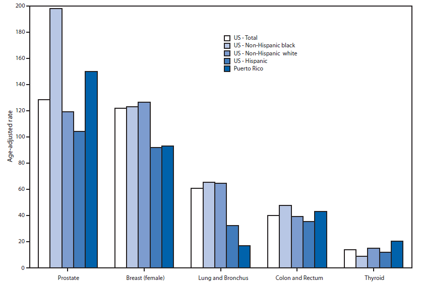
 Vital Signs: Disparities in Tobacco-Related Cancer Incidence and Mortality - United States, 2004-2013 | MMWR
 Category A - Anatomy
ICD-10 kod Q519 - Medfödd missbildning av uterus och cervix, ospecificerad
cDNA - (Cynomolgus) Normal Tissue: Uterus: Cervix | Technique alternative | 01011045990 - Pol DNA
Cervix Play with a Speculum Stretching Uterus and Insertion of long metal chain
 Japanese Wife Cervix Fucking and insertion 2 vibrators into uterus at tokyohot.love
 Mullerian Duct Anomalies: Overview, Incidence and Prevalence, Embryology
Cervical cancer - Wikipedia
Richard Fetherston (politician) - Wikipedia
 Prolapse of the uterus and pessaries at the Museum of
Menstruation and Women's Health
Prolapse of the uterus and pessaries at the Museum of
Menstruation and Women's Health
Table 2 - Multicenter Study of Cronobacter sakazakii Infections in Humans, Europe, 2017 - Volume 25, Number 3-March 2019 -...
Cervical Cancer: Practice Essentials, Background, Pathophysiology
 Cervix Play Insertion. Uterus fucking - Hot Close Up Pussy HD Videos, Wet Vagina Sex Movies
 Cervix distention take almost send back plus sex cream stick concerning in uterus ~ MyHotTits.com
I need comfort ♡ - Miscarriages - MedHelp
 Browse subject: Cancer -- Psychological aspects | The Online Books Page
 Article - Billing and Coding: MolDX: Next-Generation Sequencing for Solid Tumors (A57831)
 Cervix: MedlinePlus Medical Encyclopedia
 PAX2 distinguishes benign mesonephric and mullerian glandular lesions of the cervix from endocervical adenocarcinoma, including...
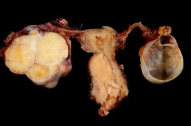
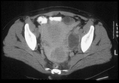
 OVARIAN CYST - ABC Homeopathy Forum
 The Case of "What Makes Sammy Run" - Deborah Walters
 United States Cancer Statistics: Public Information Data
 Vaginal Vein Anatomy, Function & Diagram | Body Maps
 U.S. Cancer Biopsy Market Research Report 2021-2028
 A laparoscopic supracervical hysterectomy (LH) is a minimally invasive procedure, which involves removal of the uterus while...
Ovaries5
- During a vaginal hysterectomy, the surgeon detaches the uterus from the ovaries, fallopian tubes and upper vagina, as well as from the blood vessels and connective tissue that support it, before removing the uterus. (astro-athena.ru)
- 2. WebHysterectomy is the procedure that removes the uterus, ovaries, fallopian tubes, and cervix. (astro-athena.ru)
- ovaries, fallopian tubes uterus, cervix and bladder. (youngwomenshealth.org)
- It may also involve removal of the cervix, ovaries, Fallopian tubes, and other surrounding structures. (health.mil)
- Staging fallopian tube cancer involves the removal of both fallopian tubes and of the ovaries, uterus, cervix, infracolic omentum, and retroperitoneal lymph nodes, in addition to peritoneal washings and peritoneal biopsies. (medscape.com)
Fallopian3
- They differentiate to form the fallopian tubes, uterus, uterine cervix, and superior aspect of the vagina. (medscape.com)
- The uterine tube (fallopian tube) carries an egg from the ovary to the uterus. (healthline.com)
- A hysterectomy (surgical removal of the uterus) is usually suggested with salpingo- oophorectomy (surgical removal of the fallopian tube and ovary) as a precautionary step. (medicinenet.com)
Uterine cervix3
- The cervix of the uterus, also known as the cervix or uterine cervix, attaches the vagina to the uterus. (healthline.com)
- Cervical cancer affects the uterine Cervix The cervix is the lower, narrow end of the uterus (womb). (health.mil)
- Societies where sexual activity starts at a changes of the uterine cervix [ 1 ], together young age and where multiple partners are with viral, bacterial, and fungal infections common are at a higher risk of exposure to of the cervix and vagina. (who.int)
Removal of the uterus5
- A laparoscopic supracervical hysterectomy (LH) is a minimally invasive procedure, which involves removal of the uterus while keeping the cervix intact. (astro-athena.ru)
- Total laparoscopic hysterectomy is a surgical procedure for the removal of the uterus. (astro-athena.ru)
- WebJun 29, · A hysterectomy is performed when you had a medical condition that makes the removal of the uterus, and cervix necessary. (astro-athena.ru)
- For women who don't wish to retain fertility, they may sometimes opt for a hysterectomy A partial or total surgical removal of the uterus. (health.mil)
- hysterectomy , removal of the uterus. (health.mil)
Womb4
- Speculum stretching cervix and semen insertion in uterus using funnel in womb. (videoshorny.com)
- The cervix is the lower end of the womb (uterus). (medlineplus.gov)
- The cervix is the lower narrow end of the womb. (astro-athena.ru)
- Your uterus is your womb. (msdmanuals.com)
Vaginal7
- They range from uterine and vaginal agenesis to duplication of the uterus and vagina to minor uterine cavity abnormalities. (medscape.com)
- Columbo reported the first documented case of vaginal agenesis (uterus and vagina) in the 16th century. (medscape.com)
- The vaginal vein also delivers blood flow to the uterine vein in the uterus. (healthline.com)
- WebFeb 10, · Vaginal hysterectomy is a surgical procedure to remove the uterus through the vagina. (astro-athena.ru)
- The most common causes for vaginal bleeding after sex both start in the cervix , which is the narrow, tube-like end of your uterus that opens into the vagina . (webmd.com)
- Imaging studies reveal that most vaginas are narrower toward the vaginal opening and wider toward the cervix. (medicalnewstoday.com)
- This swab collects a small sample of vaginal fluid from your cervix or from the back of your vagina. (clevelandclinic.org)
Bladder4
- The blood vessels work in conjunction with the venous plexuses located in the uterus, bladder, and rectum of the female body. (healthline.com)
- In the most advanced stage of uterine cancer, cancer has spread to the urinary bladder, rectum, or organs located far from the uterus, such as the lungs or bones. (medicinenet.com)
- 3D image tracing the uterus (dark blue), cervix (light blue) and bladder (yellow), from a sagittal section. (visualsonics.com)
- 3D image tracing the uterus (blue), cervix (green) and bladder (yellow), moving inferiorly and then superiorly. (visualsonics.com)
Lesions of the cervix2
- We hypothesized that PAX2 may also be expressed in mesonephric lesions of the cervix and may distinguish mesonephric hyperplasia from minimal deviation adenocarcinoma of the cervix. (nih.gov)
- Squamous cell precancerous lesions of the cervix uteri]. (bvsalud.org)
Double cervix3
- Uterus shown intact, vagina dissected to show double cervix. (utoronto.ca)
- Hatcher's doctor said only around three out of 1,000 women have a double uterus and double cervix. (yahoo.com)
- Richard Davis, an expert in high-risk pregnancies at the University of Alabama-Birmingham Hospital, told the WVTM that 'way under 1%' of women are born with a double uterus and double cervix. (yahoo.com)
Vagina and cervix2
- These may affect the movement of sperm through the vagina and cervix. (healthwise.net)
- Your doctor will also look at your vagina and cervix (with a speculum) and may do a Pap test and an HPV test . (cancer.org)
Sperm3
- Injection of sperm into uterus and insertion of vibrator in cervix. (videoshorny.com)
- It covers the cervix so sperm can't get in and fertilize an egg. (kidshealth.org)
- A diaphragm keeps sperm from entering the uterus by covering the cervix. (kidshealth.org)
Woman's cervix2
- Even so, studies have shown that magnesium sulfate can delay delivery for at least several days (depending on how far dilated a woman's cervix is when the medication is started). (healthline.com)
- The Pap test checks for cell changes on a woman's cervix that could turn into cancer if they are not treated. (familydoctor.org)
Cancer17
- Cervical cancer is a cancer arising from the cervix. (wikipedia.org)
- A Pap smear is a screening test to check for premalignant (precancerous) changes that can lead to cancer of the cervix. (medlineplus.gov)
- During a Pap test, the doctor can swab your cervix to test for any sign of abnormal, precancerous growths or cancer cells. (webmd.com)
- Cells of the cervix go through many changes before they turn into cancer. (familydoctor.org)
- Uterine cancer, which is also known as endometrial cancer, is characterized by the irregular growth of cells in the uterus. (medicinenet.com)
- Cancer begins when normal cells in the uterus begin to change, grow uncontrollably and form a mass of cells called a tumor. (medicinenet.com)
- Cancer is confined to the uterus. (medicinenet.com)
- Cancer has spread to the cervix (neck of the uterus). (medicinenet.com)
- Hormone therapy is often suggested for advanced cases of endometrial cancer that has spread beyond the uterus. (medicinenet.com)
- It is often recommended for individuals who have recurrent endometrial cancer that has spread beyond the uterus. (medicinenet.com)
- Among females, is the cancer of the breast and cancers of the cervix uteri. (who.int)
- Also, an IT based NCD application is being used to ensure continuum of care to the oral, breast and cervix cancer patients. (who.int)
- Treatment for cervical cancer can involve invasive surgeries, which a portion of the cervix is removed. (health.mil)
- Cancer of the cervix uteri was elevated, and cancer of the breast was decreased. (cdc.gov)
- RÉSUMÉ En 2006, le registre national du cancer iranien signalait qu'une petite province située dans le sud du pays, Kohgiluyeh et Boyer-Ahmad, avait une faible incidence pour presque tous les types de cancer. (who.int)
- En dépit de l'augmentation enregistrée, les taux d'incidence pour les différents sites de cancer (à l'exception du cancer de la peau) étaient significativement inférieurs aux taux nationaux correspondants en 2006. (who.int)
- Les résultats semblent indiquer des améliorations du diagnostic du cancer et de son enregistrement dans la province, même si de véritables changements dans l'incidence du cancer sur la période en question ne peuvent être exclus. (who.int)
Fetus2
- The amniotic sac is the fluid-filled membrane that surrounds the fetus inside your uterus . (clevelandclinic.org)
- According to the Mayo Clinic , a double uterus occurs in a female fetus when the two small tubes that typically form together to create the uterus instead develop into individual organs. (yahoo.com)
Dilation4
- A past surgery or procedure, such as a cervical cone biopsy or a dilation and curettage (D&C) . These may decrease fertility if the procedures have damaged the cervix or uterine lining. (healthwise.net)
- Cervix dilation video stretching the uterus with a steel Magill tweezer showing very close-up scenes of the stretched opened hole. (videoshorny.com)
- Watch the most extreme cervix dilation movie on Videoshorny.com uncensored. (videoshorny.com)
- Signs of dilation (opening of your cervix ). (clevelandclinic.org)
Speculum2
- Finally, she uses a Magill medical forceps as if it were a speculum and inserts it deep into the uterus to show the dilated cervix. (videoshorny.com)
- A speculum (medical instrument) is inserted into the vagina so that your provider can see your cervix. (ohsu.edu)
Cervical canal1
- The cervical canal passes through the cervix. (medlineplus.gov)
Abnormal1
- However, women who have symptoms, abnormal screening test results, or a gross lesion of the cervix are best evaluated with colposcopy and biopsy. (medscape.com)
Lesion1
- however, PAX2 should not be interpreted in isolation from the architectural and cytologic features of the lesion as it may be expressed in some stage II endometrial adenocarcinomas involving the cervix. (nih.gov)
Urinary1
- cervix , a part of the uterus, which can create problems with infertility and blockage of the urinary and bowel tracts. (health.mil)
Pregnant4
- The uterus is the place where a baby grows when a woman is pregnant . (medicinenet.com)
- A woman in Alabama born with two uteruses is pregnant in both, a development her doctors say is so rare that there aren't really any experts on it. (yahoo.com)
- Kelsey Hatcher, who was born with two uteruses and two cervixes, said she has known for a while that her body is special but was still surprised to learn that she was pregnant with twins, WVTM , a local NBC affiliate, reported. (yahoo.com)
- Hatcher, who already has three children, found out she was pregnant in each of her functioning uteri during her first ultrasound last Spring, according to the outlet. (yahoo.com)
Insertion1
- Cervix Play Insertion. (pussytubehd.com)
Hysterectomy6
- WebA hysterectomy is a surgical procedure that removes your uterus. (astro-athena.ru)
- WebA laparoscopic hysterectomy is less invasive than an abdominal hysterectomy surgery and is performed to remove a woman's uterus. (astro-athena.ru)
- WebOct 30, · A laparoscopic hysterectomy is a minimally invasive procedure that uses tiny instruments to remove the uterus through small incisions in the abdomen. (astro-athena.ru)
- WebJul 26, · Radical hysterectomy is a procedure to remove the uterus, cervix, and part of the vagina. (astro-athena.ru)
- Laparoscopic hysterectomy is a minimally invasive surgical procedure to remove the uterus. (astro-athena.ru)
- Hysterectomy is the surgical removal of a woman's uterus. (astro-athena.ru)
Cesarean delivery2
- Cesarean Delivery (C-Section) A C-section is surgery to deliver your baby through a cut made in your belly and uterus. (msdmanuals.com)
- For a cesarean delivery, an incision is made in the abdomen and into the uterus. (msdmanuals.com)
Pelvic exam2
Carcinoma1
- Revised FIGO staging for carcinoma of the cervix uteri. (medscape.com)
Tumor2
- The differential diagnosis of exuberant mesonephric hyperplasia includes minimal deviation adenocarcinoma of the cervix, a tumor with deceptively bland morphology for which no reliable diagnostic biomarkers currently exist. (nih.gov)
- Primitive neuroectodermal tumor of the cervix uteri: a case report -- changing concepts in therapy. (nih.gov)
Catheter2
- The woman then penetrates the cervix with a metal catheter 2 cm in diameter, inserting it 7 cm deep to the bottom of the uterus. (videoshorny.com)
- Next, the tiny catheter is inserted through your cervix into your uterus. (ohsu.edu)
Glandular1
- Glandular cells produce mucus in your cervix and uterus. (familydoctor.org)
Surgeries1
- Certain surgeries on your cervix or uterus. (clevelandclinic.org)
Fetal2
- Fetal fibronectin (fFN) is a protein that helps keep the amniotic sac attached to the lining of your uterus during pregnancy. (clevelandclinic.org)
- Fetal fibronectin acts like an adhesive that helps the amniotic sac stick to the wall of your uterus. (clevelandclinic.org)
Pregnancy1
- Your cervix may bleed more easily during pregnancy because extra blood vessels are developing in the area. (webmd.com)
Cancers1
- En revanche, le nombre de cancers de la prostate, de la thyroïde, de la vessie et des tissus mous a diminué au cours de la période de l'étude. (who.int)
Squamous1
- Squamous cells form the surface of your cervix. (familydoctor.org)
Symptoms1
- What are the symptoms of a postpartum uterus infection? (msdmanuals.com)
Structures1
- In this technique, the uterus is separated from the adjacent structures. (astro-athena.ru)
Abnormalities1
- Abnormalities of the uterus. (healthwise.net)
Infertility1
- These may cause deformities of the uterus, resulting in infertility. (healthwise.net)
Occurs1
- It occurs when the uterus contracts regularly and leads to changes in the cervix. (healthline.com)
Amniotic1
- If the result of your fFN is positive, it means your amniotic sac isn't sticking to your uterus. (clevelandclinic.org)
Penetration1
- Cervix penetration with kitchen steel peeler by the handle and then by the dangerous side of the blades. (videoshorny.com)