Cerebral Ventricle Neoplasms
Cerebral Ventricles
Fourth Ventricle
Cisterna Magna
Heart Ventricles
Shivering
Lateral Ventricles
Brain
Cerebrospinal Fluid
Pempidine
Hypothalamus
Ependyma
Cerebral Infarction
Hypnosis, Anesthetic
Body Temperature Regulation
Physostigmine
Hexamethonium Compounds
Cats
Tranylcypromine
Third Ventricle
Subarachnoid Space
Cerebral Palsy
Histamine Agents
Middle Cerebral Artery
Pyrogens
Hydrocephalus
Serotonin
Dogs
Choroid Plexus
Norepinephrine
Phentolamine
Lysergic Acid Diethylamide
Intraventricular meningiomas: MR imaging and MR spectroscopic findings in two cases. (1/146)
CT, MR imaging, MR spectroscopy, and angiography were performed in two men (ages 21 and 48, respectively) with intraventricular meningioma. In both cases, CT and MR imaging showed large tumors located in the trigone of the right lateral ventricle that enhanced intensely after contrast administration. MR spectroscopy was helpful in supporting a preoperative diagnosis of meningioma in both cases. (+info)Pineoblastoma showing unusual ventricular extension in a young adult--case report. (2/146)
A 19-year-old male presented with a 4-week history of headache. Neurological examination showed bilateral papilledema. Computed tomography revealed a pineal region mass with remarkable obstructive hydrocephalus. Magnetic resonance imaging showed a pineal region tumor continuously invading through the tectum into the cerebral aqueduct and the fourth ventricle with the preservation of the adjacent structures. The tumor appeared an iso- to hypointense mass on T1-weighted images, a heterogeneous iso- to hyperintense mass on T2-weighted images, and a heterogeneously enhanced mass after administration of contrast medium. Histological examination after endoscopic biopsy confirmed that the tumor was a pineoblastoma. Radiotherapy was given to the whole brain and the spinal cord, and magnetic resonance imaging showed complete remission of the tumor. Pineoblastomas are highly malignant tumors with seeding potential through the neighboring ventricle or along the meninges, and this type of tumor becomes larger with local extension. We found no previous reports of the continuous extension into the fourth ventricle. The present case showed ventricular extension with minimal mass effect to adjacent structures, and did not disturb ventricular configuration. According to the unusual finding of ventricular extension, this rare case of pineoblastoma requires adjuvant chemotherapy. (+info)Colloid cysts of the third ventricle: are MR imaging patterns predictive of difficulty with percutaneous treatment? (3/146)
BACKGROUND AND PURPOSE: Colloid cysts of the third ventricle are rare benign brain tumors. The purpose of this study was to correlate their patterns on MR images with the probability of success of percutaneous treatment. METHODS: Nineteen patients underwent endoscopic treatment for colloid cysts of the third ventricle. The cases were divided into two groups based on difficulty of the aspiration procedure. We reviewed CT scans and MR images and divided cysts into groups based on their signal intensity on the MR images and their density on CT scans. Intensity and density were correlated with difficulty of aspiration during the endoscopic procedure. RESULTS: The aspiration procedure was difficult in 63% of the cases. Eighty-nine percent of hyperdense cysts on unenhanced axial CT scans were categorized as difficult, and 75% of hypodense cysts were categorized as easy. On T2-weighted MR sequences, 100% of low-signal cyst contents were difficult and nearly 63% of high-signal lesions were easy. There was a significant correlation between the T2-weighted sequences and the CT scans regarding the difficulty of the aspiration procedure. CONCLUSION: T2-weighted MR sequences are useful for predicting difficulty of aspiration during stereotactic or endoscopic procedures. A T2-weighted low-signal cyst is correlated with high-viscosity intracystic contents. (+info)Isolated dilation of the trigono-inferior horn--four case reports. (4/146)
Four patients presented with isolated dilation of the trigono-inferior horn associated with either mass lesion at the trigone of the lateral ventricle or with shunt over-drainage. We investigated clinical symptoms, course, and neuroradiological findings of these cases. The pressure of the isolated ventricle was measured or estimated at surgery in all cases. The common symptoms were recent memory disturbance and contralateral homonymous hemianopia. Contralateral hemiparesis was observed occasionally. Rapid deterioration of the isolation caused uncal herniation in one case. Comma-shaped dilation of the inferior horn was observed in all cases. Midline shift was not conspicuous except in one case. Intraventricular pressure at surgery was 18 cmH2O, 35 cmH2O, 3 cmH2O, and within normal range. These cases had very similar clinical symptoms and neuroradiological findings. The pathophysiology of isolation suggested three types of isolation (high-, normal-, and low-pressure isolation), depending on the pressure of the isolated ventricle. The isolation of trigono-inferior horn is an important clinical entity as it may cause uncal herniation in patients with high-pressure lesions. (+info)Malignant spread of haemangioblastoma: report on two cases. (5/146)
Two cases are described in which, after successful removal of a cerebellar haemangioblastoma followed by several years of freedom from symptoms, there developed a progressive spinal cord compression, leading to death. At necropsy the spinal cords in both cases and the brainstem in one case, were irregularly plastered with haemangioblastoma. Although there was no doubt that malignant spread had occurred from one or more primary tumours, the histology of the tumour tissue was in no way different from that of conventional haemangioblastoma. (+info)Increased conspicuity of intraventricular lesions revealed by three-dimensional constructive interference in steady state sequences. (6/146)
We describe our preliminary experience with the three-dimensional constructive interference in steady state (3D-CISS) sequence for the evaluation of intraventricular lesions. Cyst walls, extent and margins of tumors, and intratumoral cystic structures were clearly depicted on 3D-CISS images. The 3D-CISS sequence can offer additional information to conventional MR studies to define intraventricular lesions better. (+info)Ependymoma with extensive lipidization mimicking adipose tissue: a report of five cases. (7/146)
Lipomatous ependymoma is a recently described entity and only 3 cases of this variant have been reported in the literature. We report 5 cases of this rare variant of ependymoma. Patients age ranged from 4 years to 45 years and, interestingly, all of them were males. Two tumors were supratentorial in location, 2 in the fourth ventricle and 1 was intramedullary. Microscopically all of them showed the classical histology of ependymoma along with lipomatous differentiation. The lipomatous component was composed of cells with a large clear vacuole pushing the nucleus to the periphery and giving a signet ring cell appearance. This component demonstrated positivity for GFAP and S-100 protein thereby confirming its glial lineage. Three of the 5 tumors were high grade (WHO-grade III), had a high MIB-1 labelling index (MIB-1 LI) and showed recurrence on follow-up. However, 2 were low grade (WHO grade II) and patients are free of disease till the last follow up. (+info)Intraventricular cryptococcal granuloma. (8/146)
A case is reported of a cryptococcal granuloma occurring within the lateral ventricle. The findings on angiography and brain-scanning led to a preoperative diagnosis of intraventricular meningioma. There are no previous reports of an isotope brain-scan in this condition and angiography usually shows an avascular swelling. (+info)Cerebral ventricle neoplasms refer to tumors that develop within the cerebral ventricles, which are fluid-filled spaces in the brain. These tumors can arise from various types of cells within the ventricular system, including the ependymal cells that line the ventricles, choroid plexus cells that produce cerebrospinal fluid, or other surrounding tissues.
Cerebral ventricle neoplasms can cause a variety of symptoms depending on their size and location, such as headaches, nausea, vomiting, vision changes, imbalance, weakness, or difficulty with mental tasks. The treatment options for these tumors may include surgical resection, radiation therapy, and chemotherapy, depending on the type and extent of the tumor. Regular follow-up care is essential to monitor for recurrence and manage any long-term effects of treatment.
The cerebral ventricles are a system of interconnected fluid-filled cavities within the brain. They are located in the center of the brain and are filled with cerebrospinal fluid (CSF), which provides protection to the brain by cushioning it from impacts and helping to maintain its stability within the skull.
There are four ventricles in total: two lateral ventricles, one third ventricle, and one fourth ventricle. The lateral ventricles are located in each cerebral hemisphere, while the third ventricle is located between the thalami of the two hemispheres. The fourth ventricle is located at the base of the brain, above the spinal cord.
CSF flows from the lateral ventricles into the third ventricle through narrow passageways called the interventricular foramen. From there, it flows into the fourth ventricle through another narrow passageway called the cerebral aqueduct. CSF then leaves the fourth ventricle and enters the subarachnoid space surrounding the brain and spinal cord, where it can be absorbed into the bloodstream.
Abnormalities in the size or shape of the cerebral ventricles can indicate underlying neurological conditions, such as hydrocephalus (excessive accumulation of CSF) or atrophy (shrinkage) of brain tissue. Imaging techniques, such as computed tomography (CT) or magnetic resonance imaging (MRI), are often used to assess the size and shape of the cerebral ventricles in clinical settings.
Intraventricular injections are a type of medical procedure where medication is administered directly into the cerebral ventricles of the brain. The cerebral ventricles are fluid-filled spaces within the brain that contain cerebrospinal fluid (CSF). This procedure is typically used to deliver drugs that target conditions affecting the central nervous system, such as infections or tumors.
Intraventricular injections are usually performed using a thin, hollow needle that is inserted through a small hole drilled into the skull. The medication is then injected directly into the ventricles, allowing it to circulate throughout the CSF and reach the brain tissue more efficiently than other routes of administration.
This type of injection is typically reserved for situations where other methods of drug delivery are not effective or feasible. It carries a higher risk of complications, such as bleeding, infection, or damage to surrounding tissues, compared to other routes of administration. Therefore, it is usually performed by trained medical professionals in a controlled clinical setting.
The fourth ventricle is a part of the cerebrospinal fluid-filled system in the brain, located in the posterior cranial fossa and continuous with the central canal of the medulla oblongata and the cerebral aqueduct. It is shaped like a cavity with a roof, floor, and lateral walls, and it communicates rostrally with the third ventricle through the cerebral aqueduct and caudally with the subarachnoid space through the median and lateral apertures (foramina of Luschka and Magendie). The fourth ventricle contains choroid plexus tissue, which produces cerebrospinal fluid. Its roof is formed by the cerebellar vermis and the superior medullary velum, while its floor is composed of the rhomboid fossa, which includes several important structures such as the vagal trigone, hypoglossal trigone, and striae medullares.
The term "cisterna magna" is derived from Latin, where "cisterna" means "reservoir" or "receptacle," and "magna" means "large." In medical anatomy, the cisterna magna refers to a large, sac-like space located near the lower part of the brainstem. It is a subarachnoid cistern, which means it is a space that contains cerebrospinal fluid (CSF) between the arachnoid and pia mater membranes covering the brain and spinal cord.
More specifically, the cisterna magna is situated between the cerebellum (the lower part of the brain responsible for coordinating muscle movements and maintaining balance) and the occipital bone (the bone at the back of the skull). This space contains a significant amount of CSF, which serves as a protective cushion for the brain and spinal cord, helps regulate intracranial pressure, and facilitates the circulation of nutrients and waste products.
The cisterna magna is an essential structure in neurosurgical procedures and diagnostic imaging techniques like lumbar puncture (spinal tap) or myelograms, where contrast agents are introduced into the CSF to visualize the spinal cord and surrounding structures. Additionally, it serves as a crucial landmark for various surgical approaches to the posterior fossa (the lower part of the skull that houses the cerebellum and brainstem).
The heart ventricles are the two lower chambers of the heart that receive blood from the atria and pump it to the lungs or the rest of the body. The right ventricle pumps deoxygenated blood to the lungs, while the left ventricle pumps oxygenated blood to the rest of the body. Both ventricles have thick, muscular walls to generate the pressure necessary to pump blood through the circulatory system.
Shivering is a physical response to cold temperature or emotional stress, characterized by involuntary muscle contractions and relaxations. It's a part of the body's thermoregulation process, which helps to generate heat and maintain a normal body temperature. During shivering, the muscles rapidly contract and relax, producing kinetic energy that is released as heat. This can be observed as visible shaking or trembling, often most noticeable in the arms, legs, and jaw. In some cases, prolonged or intense shivering may also be associated with fever or other medical conditions.
Cerebral arteries refer to the blood vessels that supply oxygenated blood to the brain. These arteries branch off from the internal carotid arteries and the vertebral arteries, which combine to form the basilar artery. The major cerebral arteries include:
1. Anterior cerebral artery (ACA): This artery supplies blood to the frontal lobes of the brain, including the motor and sensory cortices responsible for movement and sensation in the lower limbs.
2. Middle cerebral artery (MCA): The MCA is the largest of the cerebral arteries and supplies blood to the lateral surface of the brain, including the temporal, parietal, and frontal lobes. It is responsible for providing blood to areas involved in motor function, sensory perception, speech, memory, and vision.
3. Posterior cerebral artery (PCA): The PCA supplies blood to the occipital lobe, which is responsible for visual processing, as well as parts of the temporal and parietal lobes.
4. Anterior communicating artery (ACoA) and posterior communicating arteries (PComAs): These are small arteries that connect the major cerebral arteries, forming an important circulatory network called the Circle of Willis. The ACoA connects the two ACAs, while the PComAs connect the ICA with the PCA and the basilar artery.
These cerebral arteries play a crucial role in maintaining proper brain function by delivering oxygenated blood to various regions of the brain. Any damage or obstruction to these arteries can lead to serious neurological conditions, such as strokes or transient ischemic attacks (TIAs).
An injection is a medical procedure in which a medication, vaccine, or other substance is introduced into the body using a needle and syringe. The substance can be delivered into various parts of the body, including into a vein (intravenous), muscle (intramuscular), under the skin (subcutaneous), or into the spinal canal (intrathecal or spinal).
Injections are commonly used to administer medications that cannot be taken orally, have poor oral bioavailability, need to reach the site of action quickly, or require direct delivery to a specific organ or tissue. They can also be used for diagnostic purposes, such as drawing blood samples (venipuncture) or injecting contrast agents for imaging studies.
Proper technique and sterile conditions are essential when administering injections to prevent infection, pain, and other complications. The choice of injection site depends on the type and volume of the substance being administered, as well as the patient's age, health status, and personal preferences.
Body temperature is the measure of heat produced by the body. In humans, the normal body temperature range is typically between 97.8°F (36.5°C) and 99°F (37.2°C), with an average oral temperature of 98.6°F (37°C). Body temperature can be measured in various ways, including orally, rectally, axillary (under the arm), and temporally (on the forehead).
Maintaining a stable body temperature is crucial for proper bodily functions, as enzymes and other biological processes depend on specific temperature ranges. The hypothalamus region of the brain regulates body temperature through feedback mechanisms that involve shivering to produce heat and sweating to release heat. Fever is a common medical sign characterized by an elevated body temperature above the normal range, often as a response to infection or inflammation.
The lateral ventricles are a pair of fluid-filled cavities located within the brain. They are part of the ventricular system, which is a series of interconnected spaces filled with cerebrospinal fluid (CSF). The lateral ventricles are situated in the left and right hemispheres of the brain and are among the largest of the ventricles.
Each lateral ventricle has a complex structure and can be divided into several parts:
1. Anterior horn: This is the front part of the lateral ventricle, located in the frontal lobe of the brain.
2. Body: The central part of the lateral ventricle, which is continuous with the anterior horn and posterior horn.
3. Posterior horn: The back part of the lateral ventricle, located in the occipital lobe of the brain.
4. Temporal horn: An extension that projects into the temporal lobe of the brain.
The lateral ventricles are lined with ependymal cells, which produce cerebrospinal fluid. CSF circulates through the ventricular system, providing buoyancy and protection to the brain, and is eventually absorbed into the bloodstream. Abnormalities in the size or shape of the lateral ventricles can be associated with various neurological conditions, such as hydrocephalus, brain tumors, or neurodegenerative diseases.
The brain is the central organ of the nervous system, responsible for receiving and processing sensory information, regulating vital functions, and controlling behavior, movement, and cognition. It is divided into several distinct regions, each with specific functions:
1. Cerebrum: The largest part of the brain, responsible for higher cognitive functions such as thinking, learning, memory, language, and perception. It is divided into two hemispheres, each controlling the opposite side of the body.
2. Cerebellum: Located at the back of the brain, it is responsible for coordinating muscle movements, maintaining balance, and fine-tuning motor skills.
3. Brainstem: Connects the cerebrum and cerebellum to the spinal cord, controlling vital functions such as breathing, heart rate, and blood pressure. It also serves as a relay center for sensory information and motor commands between the brain and the rest of the body.
4. Diencephalon: A region that includes the thalamus (a major sensory relay station) and hypothalamus (regulates hormones, temperature, hunger, thirst, and sleep).
5. Limbic system: A group of structures involved in emotional processing, memory formation, and motivation, including the hippocampus, amygdala, and cingulate gyrus.
The brain is composed of billions of interconnected neurons that communicate through electrical and chemical signals. It is protected by the skull and surrounded by three layers of membranes called meninges, as well as cerebrospinal fluid that provides cushioning and nutrients.
Cerebrospinal fluid (CSF) is a clear, colorless fluid that surrounds and protects the brain and spinal cord. It acts as a shock absorber for the central nervous system and provides nutrients to the brain while removing waste products. CSF is produced by specialized cells called ependymal cells in the choroid plexus of the ventricles (fluid-filled spaces) inside the brain. From there, it circulates through the ventricular system and around the outside of the brain and spinal cord before being absorbed back into the bloodstream. CSF analysis is an important diagnostic tool for various neurological conditions, including infections, inflammation, and cancer.
I'm sorry for any confusion, but "Pempidine" does not appear to be a recognized term in medical terminology or pharmacology. It is possible that there may be a spelling error or typo in the term you are looking for. Please double-check the spelling and try again. If you meant "Pimobendan," I would be happy to provide information on that instead.
If you indeed meant "Pempidine," please let me know, and I will do my best to find more information about it.
The hypothalamus is a small, vital region of the brain that lies just below the thalamus and forms part of the limbic system. It plays a crucial role in many important functions including:
1. Regulation of body temperature, hunger, thirst, fatigue, sleep, and circadian rhythms.
2. Production and regulation of hormones through its connection with the pituitary gland (the hypophysis). It controls the release of various hormones by producing releasing and inhibiting factors that regulate the anterior pituitary's function.
3. Emotional responses, behavior, and memory formation through its connections with the limbic system structures like the amygdala and hippocampus.
4. Autonomic nervous system regulation, which controls involuntary physiological functions such as heart rate, blood pressure, and digestion.
5. Regulation of the immune system by interacting with the autonomic nervous system.
Damage to the hypothalamus can lead to various disorders like diabetes insipidus, growth hormone deficiency, altered temperature regulation, sleep disturbances, and emotional or behavioral changes.
The ependyma is a type of epithelial tissue that lines the ventricular system of the brain and the central canal of the spinal cord. These cells are specialized glial cells that help to form the blood-brain barrier, regulate the cerebrospinal fluid (CSF) composition, and provide support and protection for the nervous tissue.
Ependymal cells have a cuboidal or columnar shape and possess numerous cilia on their apical surface, which helps to circulate CSF within the ventricles. They also have tight junctions that help to form the blood-brain barrier and prevent the passage of harmful substances from the blood into the CSF.
In addition to their role in maintaining the integrity of the CNS, ependymal cells can also differentiate into other types of cells, such as neurons and glial cells, under certain conditions. This property has made them a topic of interest in regenerative medicine and the study of neurodevelopmental disorders.
Cerebral infarction, also known as a "stroke" or "brain attack," is the sudden death of brain cells caused by the interruption of their blood supply. It is most commonly caused by a blockage in one of the blood vessels supplying the brain (an ischemic stroke), but can also result from a hemorrhage in or around the brain (a hemorrhagic stroke).
Ischemic strokes occur when a blood clot or other particle blocks a cerebral artery, cutting off blood flow to a part of the brain. The lack of oxygen and nutrients causes nearby brain cells to die. Hemorrhagic strokes occur when a weakened blood vessel ruptures, causing bleeding within or around the brain. This bleeding can put pressure on surrounding brain tissues, leading to cell death.
Symptoms of cerebral infarction depend on the location and extent of the affected brain tissue but may include sudden weakness or numbness in the face, arm, or leg; difficulty speaking or understanding speech; vision problems; loss of balance or coordination; and severe headache with no known cause. Immediate medical attention is crucial for proper diagnosis and treatment to minimize potential long-term damage or disability.
I believe there may be a slight confusion in your question as hypnosis and anesthesia are two different concepts in the field of medicine. Here are separate definitions for each:
1. Hypnosis: This is a state of highly focused attention or concentration, often associated with relaxation, and heightened suggestibility. During hypnosis, a person may become more open to suggestions and their perception of reality may change. It's important to note that hypnosis is not a form of unconsciousness or sleep, and the person can usually hear and remember what happens during the session. Hypnosis is sometimes used in medical and psychological settings to help manage pain, anxiety, or symptoms of various conditions.
2. Anesthetic: An anesthetic is a drug that's used to block sensation in certain areas of the body or to induce sleep and reduce pain during surgical procedures. There are two main types of anesthetics: local and general. Local anesthetics numb a specific area of the body, while general anesthetics cause a state of unconsciousness and amnesia, so the person is unaware of the procedure taking place. Anesthetics work by depressing the function of the central nervous system, which includes the brain and spinal cord.
I hope this clarifies any confusion! If you have any further questions or need more information, please don't hesitate to ask.
Body temperature regulation, also known as thermoregulation, is the process by which the body maintains its core internal temperature within a narrow range, despite varying external temperatures. This is primarily controlled by the hypothalamus in the brain, which acts as a thermostat and receives input from temperature receptors throughout the body. When the body's temperature rises above or falls below the set point, the hypothalamus initiates responses to bring the temperature back into balance. These responses can include shivering to generate heat, sweating to cool down, vasodilation or vasoconstriction of blood vessels to regulate heat loss, and changes in metabolic rate. Effective body temperature regulation is crucial for maintaining optimal physiological function and overall health.
Physostigmine is a medication that belongs to a class of drugs called cholinesterase inhibitors. It works by blocking the breakdown of a neurotransmitter called acetylcholine, which is important for communication between nerves and muscles. This results in an increase in acetylcholine levels in the body, improving nerve impulse transmission and helping to restore normal muscle function.
Physostigmine is used in the treatment of certain medical conditions that are caused by a deficiency of acetylcholine, such as myasthenia gravis, which is a neuromuscular disorder characterized by weakness and fatigue of the muscles. It may also be used to reverse the effects of certain medications that block the action of acetylcholine, such as anticholinergics, which are sometimes used in anesthesia or to treat conditions like Parkinson's disease.
It is important to note that physostigmine should only be used under the close supervision of a healthcare provider, as it can have serious side effects if not used properly.
Hexamethonium compounds are a type of ganglionic blocker, which are medications that block the transmission of nerve impulses at the ganglia ( clusters of nerve cells) in the autonomic nervous system. These compounds contain hexamethonium as the active ingredient, which is a compound with the chemical formula C16H32N2O4.
Hexamethonium works by blocking the nicotinic acetylcholine receptors at the ganglia, which prevents the release of neurotransmitters and ultimately inhibits the transmission of nerve impulses. This can have various effects on the body, depending on which part of the autonomic nervous system is affected.
Hexamethonium compounds were once used to treat hypertension (high blood pressure), but they are rarely used today due to their numerous side effects and the availability of safer and more effective medications. Some of the side effects associated with hexamethonium include dry mouth, blurred vision, constipation, difficulty urinating, and dizziness upon standing.
"Cat" is a common name that refers to various species of small carnivorous mammals that belong to the family Felidae. The domestic cat, also known as Felis catus or Felis silvestris catus, is a popular pet and companion animal. It is a subspecies of the wildcat, which is found in Europe, Africa, and Asia.
Domestic cats are often kept as pets because of their companionship, playful behavior, and ability to hunt vermin. They are also valued for their ability to provide emotional support and therapy to people. Cats are obligate carnivores, which means that they require a diet that consists mainly of meat to meet their nutritional needs.
Cats are known for their agility, sharp senses, and predatory instincts. They have retractable claws, which they use for hunting and self-defense. Cats also have a keen sense of smell, hearing, and vision, which allow them to detect prey and navigate their environment.
In medical terms, cats can be hosts to various parasites and diseases that can affect humans and other animals. Some common feline diseases include rabies, feline leukemia virus (FeLV), feline immunodeficiency virus (FIV), and toxoplasmosis. It is important for cat owners to keep their pets healthy and up-to-date on vaccinations and preventative treatments to protect both the cats and their human companions.
Tranylcypromine is a type of antidepressant known as a non-selective, irreversible monoamine oxidase inhibitor (MAOI). It works by blocking the action of monoamine oxidase, an enzyme that breaks down certain neurotransmitters (chemical messengers) in the brain such as serotonin, dopamine, and noradrenaline. This leads to an increase in the levels of these neurotransmitters in the brain, which can help improve mood and alleviate symptoms of depression.
Tranylcypromine is used primarily for the treatment of major depressive disorder that has not responded to other antidepressants. It is also used off-label for the treatment of anxiety disorders, panic attacks, and obsessive-compulsive disorder.
It's important to note that MAOIs like tranylcypromine have several dietary and medication restrictions due to their potential to cause serious or life-threatening reactions when combined with certain foods or medications. Therefore, careful monitoring by a healthcare professional is necessary while taking this medication.
The third ventricle is a narrow, fluid-filled cavity in the brain that is located between the thalamus and hypothalamus. It is one of the four ventricles in the ventricular system of the brain, which produces and circulates cerebrospinal fluid (CSF) around the brain and spinal cord.
The third ventricle is shaped like a slit and communicates with the lateral ventricles through the interventricular foramen (also known as the foramen of Monro), and with the fourth ventricle through the cerebral aqueduct (also known as the aqueduct of Sylvius).
The third ventricle contains choroid plexus tissue, which produces CSF. The fluid flows from the lateral ventricles into the third ventricle, then through the cerebral aqueduct and into the fourth ventricle, where it can circulate around the brainstem and spinal cord before being absorbed back into the bloodstream.
Abnormalities in the third ventricle, such as enlargement or obstruction of the cerebral aqueduct, can lead to hydrocephalus, a condition characterized by an accumulation of CSF in the brain.
The subarachnoid space is the area between the arachnoid mater and pia mater, which are two of the three membranes covering the brain and spinal cord (the third one being the dura mater). This space is filled with cerebrospinal fluid (CSF), which provides protection and cushioning to the central nervous system. The subarachnoid space also contains blood vessels that supply the brain and spinal cord with oxygen and nutrients. It's important to note that subarachnoid hemorrhage, a type of stroke, can occur when there is bleeding into this space.
Cerebral palsy (CP) is a group of disorders that affect a person's ability to move and maintain balance and posture. According to the Mayo Clinic, CP is caused by abnormal brain development or damage to the developing brain that affects a child's ability to control movement.
The symptoms of cerebral palsy can vary in severity and may include:
* Spasticity (stiff or tight muscles)
* Rigidity (resistance to passive movement)
* Poor coordination and balance
* Weakness or paralysis
* Tremors or involuntary movements
* Abnormal gait or difficulty walking
* Difficulty with fine motor skills, such as writing or using utensils
* Speech and language difficulties
* Vision, hearing, or swallowing problems
It's important to note that cerebral palsy is not a progressive condition, meaning that it does not worsen over time. However, the symptoms may change over time, and some individuals with CP may experience additional medical conditions as they age.
Cerebral palsy is usually caused by brain damage that occurs before or during birth, but it can also be caused by brain injuries that occur in the first few years of life. Some possible causes of cerebral palsy include:
* Infections during pregnancy
* Lack of oxygen to the brain during delivery
* Traumatic head injury during birth
* Brain bleeding or stroke in the newborn period
* Genetic disorders
* Maternal illness or infection during pregnancy
There is no cure for cerebral palsy, but early intervention and treatment can help improve outcomes and quality of life. Treatment may include physical therapy, occupational therapy, speech therapy, medications to manage symptoms, surgery, and assistive devices such as braces or wheelchairs.
Cerebrovascular circulation refers to the network of blood vessels that supply oxygenated blood and nutrients to the brain tissue, and remove waste products. It includes the internal carotid arteries, vertebral arteries, circle of Willis, and the intracranial arteries that branch off from them.
The internal carotid arteries and vertebral arteries merge to form the circle of Willis, a polygonal network of vessels located at the base of the brain. The anterior cerebral artery, middle cerebral artery, posterior cerebral artery, and communicating arteries are the major vessels that branch off from the circle of Willis and supply blood to different regions of the brain.
Interruptions or abnormalities in the cerebrovascular circulation can lead to various neurological conditions such as stroke, transient ischemic attack (TIA), and vascular dementia.
Consciousness is a complex and multifaceted concept that is difficult to define succinctly, but in a medical or neurological context, it generally refers to an individual's state of awareness and responsiveness to their surroundings. Consciousness involves a range of cognitive processes, including perception, thinking, memory, and attention, and it requires the integration of sensory information, language, and higher-order cognitive functions.
In medical terms, consciousness is often assessed using measures such as the Glasgow Coma Scale, which evaluates an individual's ability to open their eyes, speak, and move in response to stimuli. A coma is a state of deep unconsciousness where an individual is unable to respond to stimuli or communicate, while a vegetative state is a condition where an individual may have sleep-wake cycles and some automatic responses but lacks any meaningful awareness or cognitive function.
Disorders of consciousness can result from brain injury, trauma, infection, or other medical conditions that affect the functioning of the brainstem or cerebral cortex. The study of consciousness is a rapidly evolving field that involves researchers from various disciplines, including neuroscience, psychology, philosophy, and artificial intelligence.
Histamine agents are substances that can either increase or decrease the level or action of histamine in the body. Histamine is a chemical mediator released by mast cells and basophils in response to allergies, inflammation, or injury. It causes various symptoms such as itching, sneezing, runny nose, and wheal and flare reactions in the skin.
Histamine-releasing agents are substances that can trigger the release of histamine from mast cells and basophils. Examples include certain medications (e.g., opioids, vancomycin), physical stimuli (e.g., heat, exercise), and venoms (e.g., bee stings).
Histamine-inhibiting agents are substances that can block the action of histamine or prevent its release from mast cells and basophils. Examples include antihistamines, which bind to histamine receptors and prevent histamine from exerting its effects, and mast cell stabilizers, which prevent the degranulation of mast cells and the subsequent release of histamine and other mediators.
Histamine-enhancing agents are substances that can increase the level or action of histamine in the body. Examples include histamine agonists, which mimic the effects of histamine by binding to its receptors, and histamine precursors, which provide the building blocks for the synthesis of histamine.
Overall, histamine agents have important clinical implications in the management of allergies, inflammation, and other conditions associated with histamine release or action.
The Middle Cerebral Artery (MCA) is one of the main blood vessels that supplies oxygenated blood to the brain. It arises from the internal carotid artery and divides into several branches, which supply the lateral surface of the cerebral hemisphere, including the frontal, parietal, and temporal lobes.
The MCA is responsible for providing blood flow to critical areas of the brain, such as the primary motor and sensory cortices, Broca's area (associated with speech production), Wernicke's area (associated with language comprehension), and the visual association cortex.
Damage to the MCA or its branches can result in a variety of neurological deficits, depending on the specific location and extent of the injury. These may include weakness or paralysis on one side of the body, sensory loss, language impairment, and visual field cuts.
Perfusion, in medical terms, refers to the process of circulating blood through the body's organs and tissues to deliver oxygen and nutrients and remove waste products. It is a measure of the delivery of adequate blood flow to specific areas or tissues in the body. Perfusion can be assessed using various methods, including imaging techniques like computed tomography (CT) scans, magnetic resonance imaging (MRI), and perfusion scintigraphy.
Perfusion is critical for maintaining proper organ function and overall health. When perfusion is impaired or inadequate, it can lead to tissue hypoxia, acidosis, and cell death, which can result in organ dysfunction or failure. Conditions that can affect perfusion include cardiovascular disease, shock, trauma, and certain surgical procedures.
Pyrogens are substances that can induce fever, or elevate body temperature above the normal range of 36-37°C (96.8-98.6°F). They can be either exogenous (coming from outside the body) or endogenous (produced within the body). Exogenous pyrogens include bacterial toxins, dead bacteria, and various chemicals. Endogenous pyrogens are substances produced by the immune system in response to an infection, such as interleukin-1 (IL-1), interleukin-6 (IL-6), and tumor necrosis factor-alpha (TNF-α). These substances act on the hypothalamus, a part of the brain that regulates body temperature, to raise the set point for body temperature, leading to an increase in body temperature.
Hydrocephalus is a medical condition characterized by an abnormal accumulation of cerebrospinal fluid (CSF) within the brain, leading to an increase in intracranial pressure and potentially causing damage to the brain tissues. This excessive buildup of CSF can result from either overproduction or impaired absorption of the fluid, which typically causes the ventricles (fluid-filled spaces) inside the brain to expand and put pressure on surrounding brain structures.
The condition can be congenital, present at birth due to genetic factors or abnormalities during fetal development, or acquired later in life as a result of injuries, infections, tumors, or other disorders affecting the brain's ability to regulate CSF flow and absorption. Symptoms may vary depending on age, severity, and duration but often include headaches, vomiting, balance problems, vision issues, cognitive impairment, and changes in behavior or personality.
Treatment for hydrocephalus typically involves surgically implanting a shunt system that diverts the excess CSF from the brain to another part of the body where it can be absorbed, such as the abdominal cavity. In some cases, endoscopic third ventriculostomy (ETV) might be an alternative treatment option, creating a new pathway for CSF flow within the brain. Regular follow-ups with neurosurgeons and other healthcare professionals are essential to monitor the condition and make any necessary adjustments to the treatment plan.
Serotonin, also known as 5-hydroxytryptamine (5-HT), is a monoamine neurotransmitter that is found primarily in the gastrointestinal (GI) tract, blood platelets, and the central nervous system (CNS) of humans and other animals. It is produced by the conversion of the amino acid tryptophan to 5-hydroxytryptophan (5-HTP), and then to serotonin.
In the CNS, serotonin plays a role in regulating mood, appetite, sleep, memory, learning, and behavior, among other functions. It also acts as a vasoconstrictor, helping to regulate blood flow and blood pressure. In the GI tract, it is involved in peristalsis, the contraction and relaxation of muscles that moves food through the digestive system.
Serotonin is synthesized and stored in serotonergic neurons, which are nerve cells that use serotonin as their primary neurotransmitter. These neurons are found throughout the brain and spinal cord, and they communicate with other neurons by releasing serotonin into the synapse, the small gap between two neurons.
Abnormal levels of serotonin have been linked to a variety of disorders, including depression, anxiety, schizophrenia, and migraines. Medications that affect serotonin levels, such as selective serotonin reuptake inhibitors (SSRIs), are commonly used to treat these conditions.
I believe there might be a misunderstanding in your question. "Dogs" is not a medical term or condition. It is the common name for a domesticated carnivore of the family Canidae, specifically the genus Canis, which includes wolves, foxes, and other extant and extinct species of mammals. Dogs are often kept as pets and companions, and they have been bred in a wide variety of forms and sizes for different purposes, such as hunting, herding, guarding, assisting police and military forces, and providing companionship and emotional support.
If you meant to ask about a specific medical condition or term related to dogs, please provide more context so I can give you an accurate answer.
The choroid plexus is a network of blood vessels and tissue located within each ventricle (fluid-filled space) of the brain. It plays a crucial role in the production of cerebrospinal fluid (CSF), which provides protection and nourishment to the brain and spinal cord.
The choroid plexus consists of modified ependymal cells, called plexus epithelial cells, that line the ventricular walls. These cells have finger-like projections called villi, which increase their surface area for efficient CSF production. The blood vessels within the choroid plexus transport nutrients, ions, and water to these epithelial cells, where they are actively secreted into the ventricles to form CSF.
In addition to its role in CSF production, the choroid plexus also acts as a barrier between the blood and the central nervous system (CNS), regulating the exchange of substances between them. This barrier function is primarily attributed to tight junctions present between the epithelial cells, which limit the paracellular movement of molecules.
Abnormalities in the choroid plexus can lead to various neurological conditions, such as hydrocephalus (excessive accumulation of CSF) or certain types of brain tumors.
Norepinephrine, also known as noradrenaline, is a neurotransmitter and a hormone that is primarily produced in the adrenal glands and is released into the bloodstream in response to stress or physical activity. It plays a crucial role in the "fight-or-flight" response by preparing the body for action through increasing heart rate, blood pressure, respiratory rate, and glucose availability.
As a neurotransmitter, norepinephrine is involved in regulating various functions of the nervous system, including attention, perception, motivation, and arousal. It also plays a role in modulating pain perception and responding to stressful or emotional situations.
In medical settings, norepinephrine is used as a vasopressor medication to treat hypotension (low blood pressure) that can occur during septic shock, anesthesia, or other critical illnesses. It works by constricting blood vessels and increasing heart rate, which helps to improve blood pressure and perfusion of vital organs.
Blood pressure is the force exerted by circulating blood on the walls of the blood vessels. It is measured in millimeters of mercury (mmHg) and is given as two figures:
1. Systolic pressure: This is the pressure when the heart pushes blood out into the arteries.
2. Diastolic pressure: This is the pressure when the heart rests between beats, allowing it to fill with blood.
Normal blood pressure for adults is typically around 120/80 mmHg, although this can vary slightly depending on age, sex, and other factors. High blood pressure (hypertension) is generally considered to be a reading of 130/80 mmHg or higher, while low blood pressure (hypotension) is usually defined as a reading below 90/60 mmHg. It's important to note that blood pressure can fluctuate throughout the day and may be affected by factors such as stress, physical activity, and medication use.
Phentolamine is a non-selective alpha-blocker drug, which means it blocks both alpha-1 and alpha-2 receptors. It works by relaxing the muscle around blood vessels, which increases blood flow and lowers blood pressure. Phentolamine is used medically for various purposes, including the treatment of high blood pressure, the diagnosis and treatment of pheochromocytoma (a tumor that releases hormones causing high blood pressure), and as an antidote to prevent severe hypertension caused by certain medications or substances. It may also be used in diagnostic tests to determine if a patient's blood pressure is reactive to drugs, and it can be used during some surgical procedures to help lower the risk of hypertensive crises.
Phentolamine is available in two forms: an injectable solution and oral tablets. The injectable form is typically administered by healthcare professionals in a clinical setting, while the oral tablets are less commonly used due to their short duration of action and potential for causing severe drops in blood pressure. As with any medication, phentolamine should be taken under the supervision of a healthcare provider, and patients should follow their doctor's instructions carefully to minimize the risk of side effects and ensure the drug's effectiveness.
Lysergic Acid Diethylamide (LSD) is defined in medical terms as a powerful synthetic hallucinogenic drug. It is derived from lysergic acid, which is found in ergot, a fungus that grows on grains such as rye. LSD is typically distributed as a liquid, tablets, or thin squares of gelatin (commonly known as window panes). It is odorless, colorless, and has a slightly bitter taste.
LSD is considered one of the most potent mood-changing chemicals. Its effects, often called a "trip," can be stimulating, pleasurable, and mind-altering or they can lead to an unpleasant, sometimes terrifying experience called a "bad trip." The effects of LSD are unpredictable depending on factors such as the user's personality, mood, expectations, and the environment in which the drug is used.
In the medical field, LSD has been studied for its potential benefits in treating certain mental health conditions, such as anxiety and depression associated with life-threatening illnesses, but further research is needed to establish its safety and efficacy. It's important to note that the use of LSD outside of approved medical settings and supervision is not legal in most countries and can lead to serious legal consequences.
The term "drinking" is commonly used to refer to the consumption of beverages, but in a medical context, it usually refers to the consumption of alcoholic drinks. According to the Merriam-Webster Medical Dictionary, "drinking" is defined as:
1. The act or habit of swallowing liquid (such as water, juice, or alcohol)
2. The ingestion of alcoholic beverages
It's important to note that while moderate drinking may not pose significant health risks for some individuals, excessive or binge drinking can lead to a range of negative health consequences, including addiction, liver disease, heart disease, and increased risk of injury or violence.
 List of MeSH codes (C10)
List of diseases (C)
List of MeSH codes (C04)
Astroblastoma
Intracranial pressure
Route of administration
Choroid plexus tumor
Timeline of tuberous sclerosis
Michael L. J. Apuzzo
Pineal gland
Papillary tumors of the pineal region
Meningioma
Angiocentric glioma
Astrocytoma
Cerebellar vermis
Brain tumor
Cerebral edema
Lesional demyelinations of the central nervous system
Glossary of medicine
Small supernumerary marker chromosome
Notch signaling pathway
List of MeSH codes (C16)
Tumor6
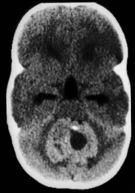
- Nonenhanced axial computed tomography image demonstrates a large, round tumor arising within the fourth ventricle with attenuating nodular calcifications. (medscape.com)
- Flat tumors, termed en plaque, infiltrate the dura and grow as a thin carpet or sheet of tumor along the convexity dura, falx, or tentorium. (medscape.com)
- Intracranial mesenchymal tumors with FET-CREB fusions are a recently described group of neoplasms in children and young adults characterized by fusion of a FET family gene (usually EWSR1, but rarely FUS) to a CREB family transcription factor (ATF1, CREB1, or CREM), and have been variously termed intracranial angiomatoid fibrous histiocytoma or intracranial myxoid mesenchymal tumor. (aku.edu)
- In combination with prior case series, this study provides further insight into intracranial mesenchymal tumors with FET-CREB fusion, which represent a distinct group of CNS tumors encompassing both intracranial myxoid mesenchymal tumor and angiomatoid fibrous histiocytoma-like neoplasms. (aku.edu)
- We identified only six published cases of RGNTs where the tumor either originated in or extended to the lateral ventricular system, of which three tumors were only localized to the lateral ventricles. (surgicalneurologyint.com)
- In dogs, the tumor occurs most commonly in the fourth ventricle, as in the present report, but can also occur in the third ventricle and the lateral ventricles. (unesp.br)
Lateral ventricles15
- CPPs are commonly observed in the lateral ventricles of children, but they can be encountered in adults. (naqlafshk.com)
- Tumors were uniformly extra-axial or intraventricular and located at the cerebral convexities (n = 7), falx (2), lateral ventricles (4), tentorium (2), cerebellopontine angle (4), and spinal cord (1). (aku.edu)
- Figure A ) Cranial magnetic resonance imaging shows generalized parenchymal atrophy and dilation of the bilateral lateral ventricles with patent basal cisterns and coarse supratentorial calcifications involving the cortex, bilateral basal ganglia, and the vermis. (contemporarypediatrics.com)
- A. Axial cranial CT of case patient showing generalized cortical calcifications with dilation of bilateral lateral ventricles. (contemporarypediatrics.com)
- B. Axial cranial MRI of case patient showing generalized parenchymal atrophy and dilation of the bilateral lateral ventricles (yellow arrows) with patent basal cisterns and coarse supratentorial calcifications involving the cortex (red arrows), basal ganglia bilaterally, and the vermis. (contemporarypediatrics.com)
- Pericallosal lipomas may extend into either or both lateral ventricles and appearing as lipomas of choroid plexus in the absence of corpus callosum. (ghrnet.org)
- Pericallosal lipomas may continue into the lateral ventricles, which is a very rare presentation[2,3]. (ghrnet.org)
- These lipomas are generally measuring greater than 2 cm in diameter, have a high incidence of corpus callosum dysgenesis and fronto-facial anomalies and can extend into the choroid plexus/lateral ventricles[4,5]. (ghrnet.org)
- In this article, I present a rare case of tubulonodular midline lipoma extending into both lateral ventricles to appear as a lipoma of choroid plexus associated with corpus callosum agenesis. (ghrnet.org)
- the occipital, but not the frontal, horns of the lateral ventricles were dilated. (ghrnet.org)
- Here, we describe the fourth case in literature where an RGNT was localized to the lateral ventricles and detail the treatment approach. (surgicalneurologyint.com)
- In this report, we describe the fourth case of an RGNT being isolated to the lateral ventricles and the first where it stained positive for EMA and D2-40. (surgicalneurologyint.com)
- Necropsy revealed fl attening of convolutions due to cerebral edema, dilatation of the lateral ventricles (hydrocephalus) and the presence of a greyish nodule, soft and measuring 0.6 x 0.8 cm in length coming out of the fourth ventricle and extending laterally between the cerebellum and brainstem and cranially to the thalamus. (unesp.br)
- 1) The choroid plexuses inside the ventricles of the brain, mainly the lateral ventricles - the bulk portion of the fluid is formed here. (biologydiscussion.com)
- The choroid plexus is a cauliflower-like tuft of blood vessels covered by a thin layer of cuboidal epithelial cells which contain plenty of mitochondria and vacuoles and it projects into the temporal horns of the lateral ventricles, the posterior portions of the third ventricle and the roof of the fourth ventricle. (biologydiscussion.com)
Intracranial neoplasms3
- CPPs comprise about 1% of intracranial neoplasms but 2-4% in children. (naqlafshk.com)
- MRI is the diagnostic modality of choice in the workup and follow-up observation of intracranial neoplasms, including ependymoma. (medscape.com)
- They represent approximately 38% of all intracranial neoplasms in females and 20% in males. (medscape.com)
Majority of these neoplasms2
- A majority of these neoplasms arise supratentorially within the white matter and in the periventricular area. (pathologyoutlines.com)
- While the vast majority of these neoplasms are benign, a small percentage can be malignant. (naqlafshk.com)
Hydrocephalus5
- The relationship with the ventricular system is more apparent in tumors of the posterior fossa (mostly of the fourth ventricle), which usually present with obstructive hydrocephalus with or without signs of brain stem compression. (medscape.com)
- This symptom however occurs secondary to hydrocephalus, which is a result from compression of the cerebral aqueduct. (wikipedia.org)
- In complex hydrocephalus or intraventricular cysts, ventriculography Gd-DTPA MR imaging helped to differentiate isolation of a ventricle or noncommunicating cyst in all 4 patients. (ajnr.org)
- Increase in the total quantity of the intracranial fluid in the brain substance causes raised intracranial tension but not hydrocephalus as in pseudo motor cerebri or cerebral oedema. (biologydiscussion.com)
- This leads to the hydrocephalus of the lateral and the third ventricles. (biologydiscussion.com)
Tumors3
- The critical diagnosis of this neoplasm is often difficult because of its similarity with other primary or secondary papillary lesions of the pineal region, including parenchymal pineal tumors, papillary ependymoma, papillary meningioma, choroid plexus papilloma, and metastatic papillary carcinoma. (wikipedia.org)
- Outcomes of the endoscopic endonasal approach for tumors in the third ventricle or invading the third ventricle. (nih.gov)
- Background: Choroid plexus tumors are uncommon neoplasms derived from the neuroepithelium that covers the ventricular cavity and the central canal of the spinal cord that are characterized by papillar aspect and intraventricular growth. (unesp.br)
Benign2
- Malignant neoplasms show a greater degree of anaplasia and have the properties of invasion and metastasis, compared to benign neoplasms . (lookformedical.com)
- Choroid plexus papillomas (CPPs) are benign neoplasms of the choroid plexus, a structure made from tufts of villi within the ventricular system that produces cerebrospinal fluid (CSF). (naqlafshk.com)
Hemorrhage3
- A 35-year-old man who presented with an intraventricular hemorrhage underwent magnetic resonance imaging and cerebral angiography that disclosed a right lateral intraventricular mass and a 7-mm fusiform aneurysm from a lateral lenticulostriate branch of the right middle cerebral artery. (nih.gov)
- Intraventricular hemorrhage (IVH) associated with intraparenchymal hemorrhage and blood completely fills one lateral ventricle or more than half of both ventricles. (stanford.edu)
- Non-contrast head computed-tomography examination reveals a voluminous acute hematic accumulation in the right cerebral hemisphere, located cortically and subcortically, lobar hemorrhage type, 10/6/4.5 cm in size (AP/LL/CC), with a volume of 140.4 cm3 (Figure 1). (fortunepublish.com)
Diagnosis2
- The radiologic differential diagnosis was lymphoma, primary glial neoplasm or subacute infarct. (pathologyoutlines.com)
- Cerebral spinal fluid (CSF) analysis supported the diagnosis with elevated immunoglobulin G, immunoglobulin G/albumin ratio, and immunoglobulin G index. (hindawi.com)
Ischemia3
- The compound is given by intravenous injection to do POSITRON-EMISSION TOMOGRAPHY for the assessment of cerebral and myocardial glucose metabolism in various physiological or pathological states including stroke and myocardial ischemia. (lookformedical.com)
- Initial differential was ischemia versus neoplasm. (hindawi.com)
- Possible episodes of cerebral ischemia, stroke. (medic-journal.com)
Ependymoma2
- Ependymoma is a central nervous system (CNS) neoplasm composed of glial cells that have differentiated along ependymal lines. (medscape.com)
- Ependymoma arising from the fourth ventricle. (medscape.com)
Supratentorial1
- PAs most frequently occur in the cerebellum, but are also found in other areas of the infratentorial (FCP) region like, brain stem, and fourth ventricle and in areas of the supratentorial (SVT) region like optic chiasm, diencephalon, third ventricle, and cerebral. (unica.it)
Diffuse cerebral2
- Cranial computed tomography done at the outside hospital shows diffuse cerebral calcifications. (contemporarypediatrics.com)
- 5] It is most commonly associated with increases in intracranial pressure caused by a variety of causes, including intraparenchymal hemorrhages, brain neoplasms and diffuse cerebral edema. (fortunepublish.com)
Effacement3
- Radiologically it was a homogeneous lesion with slight effacement of the right lateral ventricle. (pathologyoutlines.com)
- There is mass effect on the right cerebral hemisphere with effacement of the cortical sulci. (hindawi.com)
- The collection shows panventricular effacement, with marked compressive effect on the right lateral ventricle, 2 cm left midline shift (Figure 2), with subfalcine herniation and downwards transtentorial herniation (Figure 3) with compression on the midbrain where acute hematic petechiae are visible at the level of the tegmentum and the right paramedian tectal plate (Figure 4). (fortunepublish.com)
Ventricular1
- Three months after surgery, a CT scan revealed reduction of cerebral ventricular size. (unboundmedicine.com)
Choroid plexus pa1
- The fi ndings were consistent with choroid plexus papilloma located in the fourth ventricle. (unesp.br)
Corpus callosum1
- Cavum septum pellucidum (CSP) and cavum vergae are actually fluid-filled, generally communicating midline cavities located between the third ventricle and corpus callosum. (amrita.edu)
Pathology1
- Pathology showed a glioneuronal neoplasm with vague neurocytic rosettes and loose perivascular pseudorosettes. (surgicalneurologyint.com)
Children and young adults1
- LGG are the most frequent brain neoplasms in children and young adults. (unica.it)
Infarction1
- TMS can mimic clinical and radiological features of a neoplasm, infarction, or abscess and therefore can be diagnostically challenging for clinicians. (hindawi.com)
Ependymal3
- Ependymomas are neoplasms of ependymal cells that occur throughout the entire neuraxis in association with the lining of the cerebral ventricles and central canal of the spinal cord. (medscape.com)
- These lesions occur most commonly in the ependymal lining of the ventricles, but they also arise in the filum terminale and the central spinal cord. (medscape.com)
- The ependymal cells line the inside of the ventricles of the brain. (wikipedia.org)
Spinal fluid1
- Local recurrence is often observed, with rare dissemination to the cerebral spinal fluid. (thieme-connect.de)
Cerebellopontine1
- Rare locations include the third ventricle, cerebellopontine (CP) angle, and cerebral parenchyma. (naqlafshk.com)
Cavity3
- Colloid cyst of the third ventricle is a round-shaped neoplasm that is located in the cavity of the III ventricle of the brain. (medic-journal.com)
- Cerebral cyst is a voluminous intracranial formation, which is a cavity filled with fluid. (medic-journal.com)
- (b) Dandy-Walker Malformation (also known as atresia of the foramina of Nlagendie and Luschka) - here some congenital septa or membranes block the outlet of the fourth ventricle and as such the fourth ventricle is ballooned out into a large cavity above which lies the cerebellar vermis. (biologydiscussion.com)
Dilatation1
- A computed tomographic (CT) scan revealed dilatation of the cerebral ventricles, and a metrizamide CT scan showed reflux into all ventricles. (unboundmedicine.com)
Underwent1
- In 1 patient who underwent surgery for spinal cord neoplasm, the procedure excluded arachnoiditis. (ajnr.org)
Edema1
- There is enhancement of a presumed mass surrounding the occipital horn of the right lateral ventricle with adjacent vasogenic edema. (hindawi.com)
Fourth4
- 90% are in the fourth ventricle). (medscape.com)
- The most common location is the atrium of the lateral ventricle in children and the fourth ventricle in adults. (naqlafshk.com)
- The cerebral aqueduct is a narrow channel in the midbrain, which connects the third and fourth ventricles. (wikipedia.org)
- It is a congenital defect in the posterior fossa where a tongue-like projection of the cerebellum and the choroid plexus extend with an enlarged fourth ventricle into the spinal canal through the foramen magnum thereby stretching and kinking backward the upper cervical spinal cord. (biologydiscussion.com)
Roof2
- The subcommissural organ (SCO) is a secretory tissue located on the roof of the brain's third ventricle. (stanford.edu)
- The mass was emanating from the roof of the left lateral ventricle. (surgicalneurologyint.com)
Posterior1
- [ 1 ] MRI more accurately evaluates en plaque and posterior fossa meningiomas, which may be missed on CT scanning. (medscape.com)
Tissue2
- Ability of neoplasms to infiltrate and actively destroy surrounding tissue. (lookformedical.com)
- Abnormal growths of tissue that follow a previous neoplasm but are not metastases of the latter. (lookformedical.com)
Primary1
- Transfer of a neoplasm from its primary site to lymph nodes or to distant parts of the body by way of the lymphatic system. (lookformedical.com)
Occur1
- The second neoplasm may have the same or different histological type and can occur in the same or different organs as the previous neoplasm but in all cases arises from an independent oncogenic event. (lookformedical.com)
Patient3
- Methods which attempt to express in replicable terms the extent of the neoplasm in the patient. (lookformedical.com)
- We present a clinical scenario of a patient presenting with left homonymous hemianopia with atypical radiological features initially thought to be more consistent with neoplasm or infraction. (hindawi.com)
- Given this profound hypothermia, she is taken to an outside hospital emergency department (ED). En route to the hospital, the patient has right eye deviation concerning for seizure activity. (contemporarypediatrics.com)
Diseases1
- Finally, we discuss the possible future role of these white matter maps in the assessment of white matter diseases, congenital brain malformations, central nervous system neoplasms (presurgical evaluation), and brain function. (ajronline.org)