Keratinocytes
Epidermis
Skin
Cells, Cultured
Keratins
Ultraviolet Rays
Cell Differentiation
Organic Anion Transport Polypeptide C
Psoriasis
4-Nitroquinoline-1-oxide
Proline-Rich Protein Domains
RNA, Messenger
Dapsone
Protein Precursors
Gene Expression
Gene Expression Regulation
Cell Division
Cell Line, Transformed
Cell Survival
Interleukin-8
Fibroblasts
Catechin
Signal Transduction
Calcium
Fluorescent Antibody Technique
Transcription Factor AP-1
Keratin-10
Tretinoin
Caspase 14
Apoptosis
Up-Regulation
Keratin-14
Blotting, Western
Immunohistochemistry
Transglutaminases
Hair Follicle
Melanocytes
Keratin-1
Promoter Regions, Genetic
Dermis
Phosphorylation
Skin, Artificial
Molecular Sequence Data
Transfection
Base Sequence
Antioxidants
Plakins
Enzyme Activation
Dose-Response Relationship, Drug
Skin Diseases
Dermatitis, Atopic
Skin Physiological Phenomena
Viologens
Keratolytic Agents
Hair
Desmosomes
Papilloma
Sebaceous Glands
Tetradecanoylphorbol Acetate
Skin Aging
Sulfamethoxazole
Non-Fibrillar Collagens
Intermediate Filament Proteins
Sweat Glands
Mice, Transgenic
Fibroblast Growth Factor 7
Desmoplakins
Galactosylgalactosylglucosylceramidase
9,10-Dimethyl-1,2-benzanthracene
Cell Movement
Plasminogen Activator Inhibitor 2
Dermatitis, Allergic Contact
Interleukin-1alpha
Cornified Envelope Proline-Rich Proteins
Carcinoma, Squamous Cell
Mustard Gas
Integrin alpha6
Toll-Like Receptor 5
Cell Communication
Epidermal Growth Factor
Reverse Transcriptase Polymerase Chain Reaction
Cell Transformation, Neoplastic
Carcinogens
Keratinocytes are the predominant type of cells found in the epidermis, which is the outermost layer of the skin. These cells are responsible for producing keratin, a tough protein that provides structural support and protection to the skin. Keratinocytes undergo constant turnover, with new cells produced in the basal layer of the epidermis and older cells moving upward and eventually becoming flattened and filled with keratin as they reach the surface of the skin, where they are then shed. They also play a role in the immune response and can release cytokines and other signaling molecules to help protect the body from infection and injury.
The epidermis is the outermost layer of the skin, composed mainly of stratified squamous epithelium. It forms a protective barrier that prevents water loss and inhibits the entry of microorganisms. The epidermis contains no blood vessels, and its cells are nourished by diffusion from the underlying dermis. The bottom-most layer of the epidermis, called the stratum basale, is responsible for generating new skin cells that eventually move up to replace dead cells on the surface. This process of cell turnover takes about 28 days in adults.
The most superficial part of the epidermis consists of dead cells called squames, which are constantly shed and replaced. The exact rate at which this happens varies depending on location; for example, it's faster on the palms and soles than elsewhere. Melanocytes, the pigment-producing cells, are also located in the epidermis, specifically within the stratum basale layer.
In summary, the epidermis is a vital part of our integumentary system, providing not only physical protection but also playing a crucial role in immunity and sensory perception through touch receptors called Pacinian corpuscles.
In medical terms, the skin is the largest organ of the human body. It consists of two main layers: the epidermis (outer layer) and dermis (inner layer), as well as accessory structures like hair follicles, sweat glands, and oil glands. The skin plays a crucial role in protecting us from external factors such as bacteria, viruses, and environmental hazards, while also regulating body temperature and enabling the sense of touch.
"Cells, cultured" is a medical term that refers to cells that have been removed from an organism and grown in controlled laboratory conditions outside of the body. This process is called cell culture and it allows scientists to study cells in a more controlled and accessible environment than they would have inside the body. Cultured cells can be derived from a variety of sources, including tissues, organs, or fluids from humans, animals, or cell lines that have been previously established in the laboratory.
Cell culture involves several steps, including isolation of the cells from the tissue, purification and characterization of the cells, and maintenance of the cells in appropriate growth conditions. The cells are typically grown in specialized media that contain nutrients, growth factors, and other components necessary for their survival and proliferation. Cultured cells can be used for a variety of purposes, including basic research, drug development and testing, and production of biological products such as vaccines and gene therapies.
It is important to note that cultured cells may behave differently than they do in the body, and results obtained from cell culture studies may not always translate directly to human physiology or disease. Therefore, it is essential to validate findings from cell culture experiments using additional models and ultimately in clinical trials involving human subjects.
The foreskin is a double-layered fold of skin that covers and protects the head (glans) of the penis. It is a normal part of male anatomy and varies in length and coverage from person to person. The inner layer of the foreskin is highly sensitive and contains a high concentration of nerve endings, which can contribute to sexual pleasure.
In some cases, the foreskin may become tight or difficult to retract (a condition known as phimosis), which can cause discomfort or pain during sexual activity or other activities that stretch the foreskin. In these cases, medical intervention may be necessary to alleviate the problem. Some people choose to undergo circumcision, a surgical procedure in which the foreskin is removed, for cultural, religious, or personal reasons. However, circumcision is not medically necessary for most people and carries some risks, such as infection, bleeding, and scarring.
Keratins are a type of fibrous structural proteins that constitute the main component of the integumentary system, which includes the hair, nails, and skin of vertebrates. They are also found in other tissues such as horns, hooves, feathers, and reptilian scales. Keratins are insoluble proteins that provide strength, rigidity, and protection to these structures.
Keratins are classified into two types: soft keratins (Type I) and hard keratins (Type II). Soft keratins are found in the skin and simple epithelial tissues, while hard keratins are present in structures like hair, nails, horns, and hooves.
Keratin proteins have a complex structure consisting of several domains, including an alpha-helical domain, beta-pleated sheet domain, and a non-repetitive domain. These domains provide keratin with its unique properties, such as resistance to heat, chemicals, and mechanical stress.
In summary, keratins are fibrous structural proteins that play a crucial role in providing strength, rigidity, and protection to various tissues in the body.
According to the medical definition, ultraviolet (UV) rays are invisible radiations that fall in the range of the electromagnetic spectrum between 100-400 nanometers. UV rays are further divided into three categories: UVA (320-400 nm), UVB (280-320 nm), and UVC (100-280 nm).
UV rays have various sources, including the sun and artificial sources like tanning beds. Prolonged exposure to UV rays can cause damage to the skin, leading to premature aging, eye damage, and an increased risk of skin cancer. UVA rays penetrate deeper into the skin and are associated with skin aging, while UVB rays primarily affect the outer layer of the skin and are linked to sunburns and skin cancer. UVC rays are the most harmful but fortunately, they are absorbed by the Earth's atmosphere and do not reach the surface.
Healthcare professionals recommend limiting exposure to UV rays, wearing protective clothing, using broad-spectrum sunscreen with an SPF of at least 30, and avoiding tanning beds to reduce the risk of UV-related health problems.
Cell differentiation is the process by which a less specialized cell, or stem cell, becomes a more specialized cell type with specific functions and structures. This process involves changes in gene expression, which are regulated by various intracellular signaling pathways and transcription factors. Differentiation results in the development of distinct cell types that make up tissues and organs in multicellular organisms. It is a crucial aspect of embryonic development, tissue repair, and maintenance of homeostasis in the body.
Organic anion transport polypeptide C (OATPc or OATPC) is not a widely recognized or established term in the medical field. It seems that this terminology might be referring to one or more members of the organic anion transporting polypeptides (OATPs) family, specifically those localized to the canalicular membrane of hepatocytes.
OATPs are a group of membrane transporters primarily responsible for the uptake of various amphipathic organic molecules, including bile salts, steroid conjugates, thyroid hormones, and various drugs. They play a crucial role in the hepatic clearance and disposition of many endogenous and exogenous substances.
The term "OATPc" might be referring to OATP1B1 (SLCO1B1) and/or OATP1B3 (SLCO1B3), which are the two major isoforms found in the human liver's canalicular membrane. However, it is essential to note that there isn't a universally accepted or standardized definition for "OATPc."
To obtain accurate and reliable information, consult scientific literature, textbooks, or databases specializing in medical definitions and terminology.
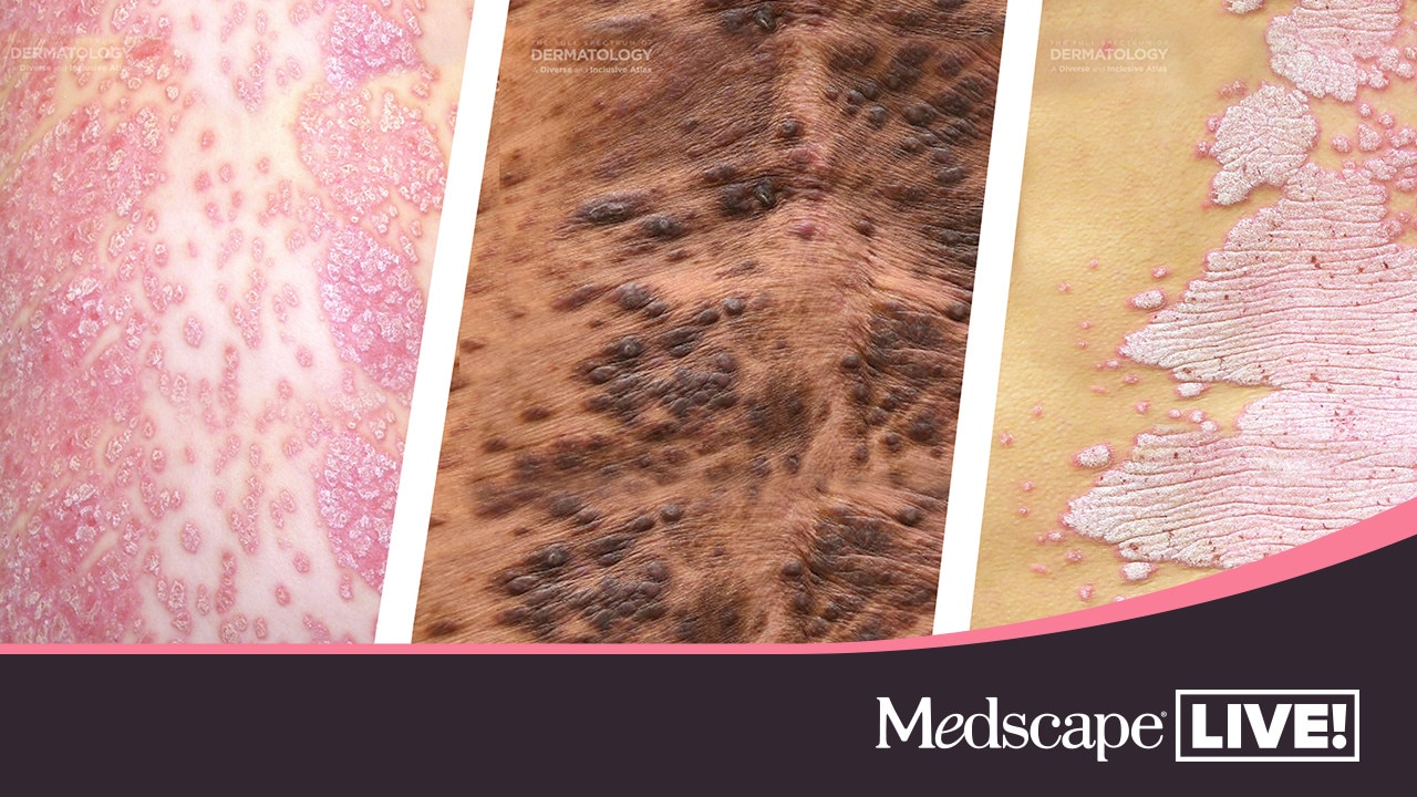
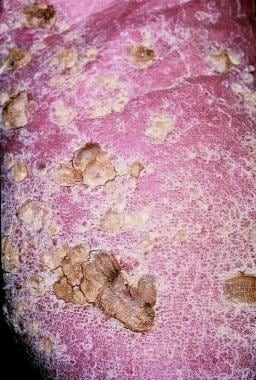
Psoriasis is a chronic skin disorder that is characterized by recurrent episodes of red, scaly patches on the skin. The scales are typically silvery-white and often occur on the elbows, knees, scalp, and lower back, but they can appear anywhere on the body. The exact cause of psoriasis is unknown, but it is believed to be related to an immune system issue that causes skin cells to grow too quickly.
There are several types of psoriasis, including plaque psoriasis (the most common form), guttate psoriasis, inverse psoriasis, pustular psoriasis, and erythrodermic psoriasis. The symptoms and severity of the condition can vary widely from person to person, ranging from mild to severe.
While there is no cure for psoriasis, various treatments are available that can help manage the symptoms and improve quality of life. These may include topical medications, light therapy, and systemic medications such as biologics. Lifestyle measures such as stress reduction, quitting smoking, and avoiding triggers (such as certain foods or alcohol) may also be helpful in managing psoriasis.
4-Nitroquinoline-1-oxide is a chemical compound that is often used in laboratory research as a carcinogenic agent. Its molecular formula is C6H4N2O3, and it is known to cause DNA damage and mutations, which can lead to the development of cancer. It is primarily used in scientific research to study the mechanisms of carcinogenesis and to test the effectiveness of potential cancer treatments.
It is important to note that 4-Nitroquinoline-1-oxide is not a medication or a treatment for any medical condition, and it should only be handled by trained professionals in a controlled laboratory setting.
Proline-rich protein domains are segments within proteins that contain an unusually high concentration of the amino acid proline. These domains are often involved in mediating protein-protein interactions and can play a role in various cellular processes, such as signal transduction, gene regulation, and protein folding. They are also commonly found in extracellular matrix proteins and may be involved in cell adhesion and migration. The unique chemical properties of proline, including its ability to form rigid structures and disrupt alpha-helices, contribute to the functional specificity of these domains.
Messenger RNA (mRNA) is a type of RNA (ribonucleic acid) that carries genetic information copied from DNA in the form of a series of three-base code "words," each of which specifies a particular amino acid. This information is used by the cell's machinery to construct proteins, a process known as translation. After being transcribed from DNA, mRNA travels out of the nucleus to the ribosomes in the cytoplasm where protein synthesis occurs. Once the protein has been synthesized, the mRNA may be degraded and recycled. Post-transcriptional modifications can also occur to mRNA, such as alternative splicing and addition of a 5' cap and a poly(A) tail, which can affect its stability, localization, and translation efficiency.
Dapsone is a medication that belongs to a class of drugs called sulfones. It is primarily used to treat bacterial skin infections such as leprosy and dermatitis herpetiformis (a skin condition associated with coeliac disease). Dapsone works by killing the bacteria responsible for these infections.
In addition, dapsone has anti-inflammatory properties and is sometimes used off-label to manage inflammatory conditions such as vasculitis, bullous pemphigoid, and chronic urticaria. It is available in oral tablet form and topical cream or gel form.
Like all medications, dapsone can cause side effects, which may include nausea, loss of appetite, and headache. More serious side effects, such as methemoglobinemia (a blood disorder that affects the body's ability to transport oxygen), peripheral neuropathy (nerve damage that causes pain, numbness, or weakness in the hands and feet), and liver damage, can occur but are less common.
It is important for patients taking dapsone to be monitored by a healthcare provider to ensure safe and effective use of the medication.
Protein precursors, also known as proproteins or prohormones, are inactive forms of proteins that undergo post-translational modification to become active. These modifications typically include cleavage of the precursor protein by specific enzymes, resulting in the release of the active protein. This process allows for the regulation and control of protein activity within the body. Protein precursors can be found in various biological processes, including the endocrine system where they serve as inactive hormones that can be converted into their active forms when needed.
Gene expression is the process by which the information encoded in a gene is used to synthesize a functional gene product, such as a protein or RNA molecule. This process involves several steps: transcription, RNA processing, and translation. During transcription, the genetic information in DNA is copied into a complementary RNA molecule, known as messenger RNA (mRNA). The mRNA then undergoes RNA processing, which includes adding a cap and tail to the mRNA and splicing out non-coding regions called introns. The resulting mature mRNA is then translated into a protein on ribosomes in the cytoplasm through the process of translation.
The regulation of gene expression is a complex and highly controlled process that allows cells to respond to changes in their environment, such as growth factors, hormones, and stress signals. This regulation can occur at various stages of gene expression, including transcriptional activation or repression, RNA processing, mRNA stability, and translation. Dysregulation of gene expression has been implicated in many diseases, including cancer, genetic disorders, and neurological conditions.
'Gene expression regulation' refers to the processes that control whether, when, and where a particular gene is expressed, meaning the production of a specific protein or functional RNA encoded by that gene. This complex mechanism can be influenced by various factors such as transcription factors, chromatin remodeling, DNA methylation, non-coding RNAs, and post-transcriptional modifications, among others. Proper regulation of gene expression is crucial for normal cellular function, development, and maintaining homeostasis in living organisms. Dysregulation of gene expression can lead to various diseases, including cancer and genetic disorders.
Cell division is the process by which a single eukaryotic cell (a cell with a true nucleus) divides into two identical daughter cells. This complex process involves several stages, including replication of DNA, separation of chromosomes, and division of the cytoplasm. There are two main types of cell division: mitosis and meiosis.
Mitosis is the type of cell division that results in two genetically identical daughter cells. It is a fundamental process for growth, development, and tissue repair in multicellular organisms. The stages of mitosis include prophase, prometaphase, metaphase, anaphase, and telophase, followed by cytokinesis, which divides the cytoplasm.
Meiosis, on the other hand, is a type of cell division that occurs in the gonads (ovaries and testes) during the production of gametes (sex cells). Meiosis results in four genetically unique daughter cells, each with half the number of chromosomes as the parent cell. This process is essential for sexual reproduction and genetic diversity. The stages of meiosis include meiosis I and meiosis II, which are further divided into prophase, prometaphase, metaphase, anaphase, and telophase.
In summary, cell division is the process by which a single cell divides into two daughter cells, either through mitosis or meiosis. This process is critical for growth, development, tissue repair, and sexual reproduction in multicellular organisms.
Skin neoplasms refer to abnormal growths or tumors in the skin that can be benign (non-cancerous) or malignant (cancerous). They result from uncontrolled multiplication of skin cells, which can form various types of lesions. These growths may appear as lumps, bumps, sores, patches, or discolored areas on the skin.
Benign skin neoplasms include conditions such as moles, warts, and seborrheic keratoses, while malignant skin neoplasms are primarily classified into melanoma, squamous cell carcinoma, and basal cell carcinoma. These three types of cancerous skin growths are collectively known as non-melanoma skin cancers (NMSCs). Melanoma is the most aggressive and dangerous form of skin cancer, while NMSCs tend to be less invasive but more common.
It's essential to monitor any changes in existing skin lesions or the appearance of new growths and consult a healthcare professional for proper evaluation and treatment if needed.
A "cell line, transformed" is a type of cell culture that has undergone a stable genetic alteration, which confers the ability to grow indefinitely in vitro, outside of the organism from which it was derived. These cells have typically been immortalized through exposure to chemical or viral carcinogens, or by introducing specific oncogenes that disrupt normal cell growth regulation pathways.
Transformed cell lines are widely used in scientific research because they offer a consistent and renewable source of biological material for experimentation. They can be used to study various aspects of cell biology, including signal transduction, gene expression, drug discovery, and toxicity testing. However, it is important to note that transformed cells may not always behave identically to their normal counterparts, and results obtained using these cells should be validated in more physiologically relevant systems when possible.
A cell line is a culture of cells that are grown in a laboratory for use in research. These cells are usually taken from a single cell or group of cells, and they are able to divide and grow continuously in the lab. Cell lines can come from many different sources, including animals, plants, and humans. They are often used in scientific research to study cellular processes, disease mechanisms, and to test new drugs or treatments. Some common types of human cell lines include HeLa cells (which come from a cancer patient named Henrietta Lacks), HEK293 cells (which come from embryonic kidney cells), and HUVEC cells (which come from umbilical vein endothelial cells). It is important to note that cell lines are not the same as primary cells, which are cells that are taken directly from a living organism and have not been grown in the lab.
Cell survival refers to the ability of a cell to continue living and functioning normally, despite being exposed to potentially harmful conditions or treatments. This can include exposure to toxins, radiation, chemotherapeutic drugs, or other stressors that can damage cells or interfere with their normal processes.
In scientific research, measures of cell survival are often used to evaluate the effectiveness of various therapies or treatments. For example, researchers may expose cells to a particular drug or treatment and then measure the percentage of cells that survive to assess its potential therapeutic value. Similarly, in toxicology studies, measures of cell survival can help to determine the safety of various chemicals or substances.
It's important to note that cell survival is not the same as cell proliferation, which refers to the ability of cells to divide and multiply. While some treatments may promote cell survival, they may also inhibit cell proliferation, making them useful for treating diseases such as cancer. Conversely, other treatments may be designed to specifically target and kill cancer cells, even if it means sacrificing some healthy cells in the process.
Interleukin-8 (IL-8) is a type of cytokine, which is a small signaling protein involved in immune response and inflammation. IL-8 is also known as neutrophil chemotactic factor or NCF because it attracts neutrophils, a type of white blood cell, to the site of infection or injury.
IL-8 is produced by various cells including macrophages, epithelial cells, and endothelial cells in response to bacterial or inflammatory stimuli. It acts by binding to specific receptors called CXCR1 and CXCR2 on the surface of neutrophils, which triggers a series of intracellular signaling events leading to neutrophil activation, migration, and degranulation.
IL-8 plays an important role in the recruitment of neutrophils to the site of infection or tissue damage, where they can phagocytose and destroy invading microorganisms. However, excessive or prolonged production of IL-8 has been implicated in various inflammatory diseases such as chronic obstructive pulmonary disease (COPD), rheumatoid arthritis, and cancer.
Fibroblasts are specialized cells that play a critical role in the body's immune response and wound healing process. They are responsible for producing and maintaining the extracellular matrix (ECM), which is the non-cellular component present within all tissues and organs, providing structural support and biochemical signals for surrounding cells.
Fibroblasts produce various ECM proteins such as collagens, elastin, fibronectin, and laminins, forming a complex network of fibers that give tissues their strength and flexibility. They also help in the regulation of tissue homeostasis by controlling the turnover of ECM components through the process of remodeling.
In response to injury or infection, fibroblasts become activated and start to proliferate rapidly, migrating towards the site of damage. Here, they participate in the inflammatory response, releasing cytokines and chemokines that attract immune cells to the area. Additionally, they deposit new ECM components to help repair the damaged tissue and restore its functionality.
Dysregulation of fibroblast activity has been implicated in several pathological conditions, including fibrosis (excessive scarring), cancer (where they can contribute to tumor growth and progression), and autoimmune diseases (such as rheumatoid arthritis).
A catechin is a type of plant phenol and antioxidant found in various foods and beverages, such as tea, cocoa, and certain fruits and vegetables. Chemically, catechins are flavan-3-ols, which are a subclass of flavonoids. They have several potential health benefits, including reducing the risk of cardiovascular disease, cancer, and neurodegenerative disorders.
Catechins are known to have anti-inflammatory, antimutagenic, and antidiabetic properties. They can also help improve oral health by inhibiting the growth of harmful bacteria in the mouth. The most well-known catechin is epigallocatechin gallate (EGCG), which is found in high concentrations in green tea and has been extensively studied for its potential health benefits.
In summary, a catechin is a type of antioxidant compound found in various plant-based foods and beverages that may have several health benefits, including reducing the risk of chronic diseases and improving oral health.
Signal transduction is the process by which a cell converts an extracellular signal, such as a hormone or neurotransmitter, into an intracellular response. This involves a series of molecular events that transmit the signal from the cell surface to the interior of the cell, ultimately resulting in changes in gene expression, protein activity, or metabolism.
The process typically begins with the binding of the extracellular signal to a receptor located on the cell membrane. This binding event activates the receptor, which then triggers a cascade of intracellular signaling molecules, such as second messengers, protein kinases, and ion channels. These molecules amplify and propagate the signal, ultimately leading to the activation or inhibition of specific cellular responses.
Signal transduction pathways are highly regulated and can be modulated by various factors, including other signaling molecules, post-translational modifications, and feedback mechanisms. Dysregulation of these pathways has been implicated in a variety of diseases, including cancer, diabetes, and neurological disorders.
Calcium is an essential mineral that is vital for various physiological processes in the human body. The medical definition of calcium is as follows:
Calcium (Ca2+) is a crucial cation and the most abundant mineral in the human body, with approximately 99% of it found in bones and teeth. It plays a vital role in maintaining structural integrity, nerve impulse transmission, muscle contraction, hormonal secretion, blood coagulation, and enzyme activation.
Calcium homeostasis is tightly regulated through the interplay of several hormones, including parathyroid hormone (PTH), calcitonin, and vitamin D. Dietary calcium intake, absorption, and excretion are also critical factors in maintaining optimal calcium levels in the body.
Hypocalcemia refers to low serum calcium levels, while hypercalcemia indicates high serum calcium levels. Both conditions can have detrimental effects on various organ systems and require medical intervention to correct.
Dermatitis is a general term that describes inflammation of the skin. It is often characterized by redness, swelling, itching, and tenderness. There are many different types of dermatitis, including atopic dermatitis (eczema), contact dermatitis, seborrheic dermatitis, and nummular dermatitis.
Atopic dermatitis is a chronic skin condition that often affects people with a family history of allergies, such as asthma or hay fever. It typically causes dry, scaly patches on the skin that can be extremely itchy.
Contact dermatitis occurs when the skin comes into contact with an irritant or allergen, such as poison ivy or certain chemicals. This type of dermatitis can cause redness, swelling, and blistering.
Seborrheic dermatitis is a common condition that causes a red, itchy rash, often on the scalp, face, or other areas of the body where oil glands are located. It is thought to be related to an overproduction of oil by the skin's sebaceous glands.
Nummular dermatitis is a type of eczema that causes round, coin-shaped patches of dry, scaly skin. It is more common in older adults and often occurs during the winter months.
Treatment for dermatitis depends on the underlying cause and severity of the condition. In some cases, over-the-counter creams or lotions may be sufficient to relieve symptoms. Prescription medications, such as corticosteroids or immunosuppressants, may be necessary in more severe cases. Avoiding triggers and irritants can also help prevent flare-ups of dermatitis.
The Fluorescent Antibody Technique (FAT) is a type of immunofluorescence assay used in laboratory medicine and pathology for the detection and localization of specific antigens or antibodies in tissues, cells, or microorganisms. In this technique, a fluorescein-labeled antibody is used to selectively bind to the target antigen or antibody, forming an immune complex. When excited by light of a specific wavelength, the fluorescein label emits light at a longer wavelength, typically visualized as green fluorescence under a fluorescence microscope.
The FAT is widely used in diagnostic microbiology for the identification and characterization of various bacteria, viruses, fungi, and parasites. It has also been applied in the diagnosis of autoimmune diseases and certain cancers by detecting specific antibodies or antigens in patient samples. The main advantage of FAT is its high sensitivity and specificity, allowing for accurate detection and differentiation of various pathogens and disease markers. However, it requires specialized equipment and trained personnel to perform and interpret the results.
Transcription Factor AP-1 (Activator Protein 1) is a heterodimeric transcription factor that belongs to the bZIP (basic region-leucine zipper) family. It is formed by the dimerization of Jun (c-Jun, JunB, JunD) and Fos (c-Fos, FosB, Fra1, Fra2) protein families, or alternatively by homodimers of Jun proteins. AP-1 plays a crucial role in regulating gene expression in various cellular processes such as proliferation, differentiation, and apoptosis. Its activity is tightly controlled through various signaling pathways, including the MAPK (mitogen-activated protein kinase) cascades, which lead to phosphorylation and activation of its components. Once activated, AP-1 binds to specific DNA sequences called TPA response elements (TREs) or AP-1 sites, thereby modulating the transcription of target genes involved in various cellular responses, such as inflammation, immune response, stress response, and oncogenic transformation.
Cell adhesion refers to the binding of cells to extracellular matrices or to other cells, a process that is fundamental to the development, function, and maintenance of multicellular organisms. Cell adhesion is mediated by various cell surface receptors, such as integrins, cadherins, and immunoglobulin-like cell adhesion molecules (Ig-CAMs), which interact with specific ligands in the extracellular environment. These interactions lead to the formation of specialized junctions, such as tight junctions, adherens junctions, and desmosomes, that help to maintain tissue architecture and regulate various cellular processes, including proliferation, differentiation, migration, and survival. Disruptions in cell adhesion can contribute to a variety of diseases, including cancer, inflammation, and degenerative disorders.
Keratin-10 is a type II keratin protein that is primarily expressed in the differentiated layers of stratified squamous epithelia, including the skin's epidermis. It plays a crucial role in providing structural support and protection to these epithelial tissues. Keratin-10 pairs with keratin-1 to form intermediate filaments, which are essential for maintaining the integrity and stability of epithelial cells. The expression of keratin-10 is often used as a marker for terminal differentiation in epidermal keratinocytes.
Tretinoin is a form of vitamin A that is used in the treatment of acne vulgaris, fine wrinkles, and dark spots caused by aging or sun damage. It works by increasing the turnover of skin cells, helping to unclog pores and promote the growth of new skin cells. Tretinoin is available as a cream, gel, or liquid, and is usually applied to the affected area once a day in the evening. Common side effects include redness, dryness, and peeling of the skin. It is important to avoid sunlight and use sunscreen while using tretinoin, as it can make the skin more sensitive to the sun.
Caspase-14 is a type of protease enzyme that belongs to the family of caspases, which are cysteine-aspartic acid proteases involved in the execution of apoptosis (programmed cell death) and inflammation. Caspase-14 is primarily expressed in the differentiated layers of the epidermis and plays a crucial role in keratinization, the process of forming an impermeable barrier to protect the body from external insults.
Caspase-14 is involved in the proteolytic processing of several structural proteins, such as loricrin, involucrin, and filaggrin, which are essential components of the cornified cell envelope, a structure that provides mechanical strength to the outermost layer of the skin. Additionally, caspase-14 has been implicated in the regulation of UV-induced apoptosis, contributing to the maintenance of skin homeostasis and preventing the development of skin cancers.
Defects or mutations in the CASP14 gene have been associated with several skin disorders, including dry skin, ichthyosis, and increased susceptibility to skin cancer.
Cell proliferation is the process by which cells increase in number, typically through the process of cell division. In the context of biology and medicine, it refers to the reproduction of cells that makes up living tissue, allowing growth, maintenance, and repair. It involves several stages including the transition from a phase of quiescence (G0 phase) to an active phase (G1 phase), DNA replication in the S phase, and mitosis or M phase, where the cell divides into two daughter cells.
Abnormal or uncontrolled cell proliferation is a characteristic feature of many diseases, including cancer, where deregulated cell cycle control leads to excessive and unregulated growth of cells, forming tumors that can invade surrounding tissues and metastasize to distant sites in the body.
Apoptosis is a programmed and controlled cell death process that occurs in multicellular organisms. It is a natural process that helps maintain tissue homeostasis by eliminating damaged, infected, or unwanted cells. During apoptosis, the cell undergoes a series of morphological changes, including cell shrinkage, chromatin condensation, and fragmentation into membrane-bound vesicles called apoptotic bodies. These bodies are then recognized and engulfed by neighboring cells or phagocytic cells, preventing an inflammatory response. Apoptosis is regulated by a complex network of intracellular signaling pathways that involve proteins such as caspases, Bcl-2 family members, and inhibitors of apoptosis (IAPs).
Up-regulation is a term used in molecular biology and medicine to describe an increase in the expression or activity of a gene, protein, or receptor in response to a stimulus. This can occur through various mechanisms such as increased transcription, translation, or reduced degradation of the molecule. Up-regulation can have important functional consequences, for example, enhancing the sensitivity or response of a cell to a hormone, neurotransmitter, or drug. It is a normal physiological process that can also be induced by disease or pharmacological interventions.
Keratin-14 is a type of keratin protein that is specifically expressed in the suprabasal layers of stratified epithelia, including the epidermis. It is a component of the intermediate filament cytoskeleton and plays an important role in maintaining the structural integrity and stability of epithelial cells. Mutations in the gene encoding keratin-14 have been associated with several genetic skin disorders, such as epidermolysis bullosa simplex and white sponge nevus.
Western blotting is a laboratory technique used in molecular biology to detect and quantify specific proteins in a mixture of many different proteins. This technique is commonly used to confirm the expression of a protein of interest, determine its size, and investigate its post-translational modifications. The name "Western" blotting distinguishes this technique from Southern blotting (for DNA) and Northern blotting (for RNA).
The Western blotting procedure involves several steps:
1. Protein extraction: The sample containing the proteins of interest is first extracted, often by breaking open cells or tissues and using a buffer to extract the proteins.
2. Separation of proteins by electrophoresis: The extracted proteins are then separated based on their size by loading them onto a polyacrylamide gel and running an electric current through the gel (a process called sodium dodecyl sulfate-polyacrylamide gel electrophoresis or SDS-PAGE). This separates the proteins according to their molecular weight, with smaller proteins migrating faster than larger ones.
3. Transfer of proteins to a membrane: After separation, the proteins are transferred from the gel onto a nitrocellulose or polyvinylidene fluoride (PVDF) membrane using an electric current in a process called blotting. This creates a replica of the protein pattern on the gel but now immobilized on the membrane for further analysis.
4. Blocking: The membrane is then blocked with a blocking agent, such as non-fat dry milk or bovine serum albumin (BSA), to prevent non-specific binding of antibodies in subsequent steps.
5. Primary antibody incubation: A primary antibody that specifically recognizes the protein of interest is added and allowed to bind to its target protein on the membrane. This step may be performed at room temperature or 4°C overnight, depending on the antibody's properties.
6. Washing: The membrane is washed with a buffer to remove unbound primary antibodies.
7. Secondary antibody incubation: A secondary antibody that recognizes the primary antibody (often coupled to an enzyme or fluorophore) is added and allowed to bind to the primary antibody. This step may involve using a horseradish peroxidase (HRP)-conjugated or alkaline phosphatase (AP)-conjugated secondary antibody, depending on the detection method used later.
8. Washing: The membrane is washed again to remove unbound secondary antibodies.
9. Detection: A detection reagent is added to visualize the protein of interest by detecting the signal generated from the enzyme-conjugated or fluorophore-conjugated secondary antibody. This can be done using chemiluminescent, colorimetric, or fluorescent methods.
10. Analysis: The resulting image is analyzed to determine the presence and quantity of the protein of interest in the sample.
Western blotting is a powerful technique for identifying and quantifying specific proteins within complex mixtures. It can be used to study protein expression, post-translational modifications, protein-protein interactions, and more. However, it requires careful optimization and validation to ensure accurate and reproducible results.
Immunohistochemistry (IHC) is a technique used in pathology and laboratory medicine to identify specific proteins or antigens in tissue sections. It combines the principles of immunology and histology to detect the presence and location of these target molecules within cells and tissues. This technique utilizes antibodies that are specific to the protein or antigen of interest, which are then tagged with a detection system such as a chromogen or fluorophore. The stained tissue sections can be examined under a microscope, allowing for the visualization and analysis of the distribution and expression patterns of the target molecule in the context of the tissue architecture. Immunohistochemistry is widely used in diagnostic pathology to help identify various diseases, including cancer, infectious diseases, and immune-mediated disorders.
Transglutaminases are a family of enzymes that catalyze the post-translational modification of proteins by forming isopeptide bonds between the carboxamide group of peptide-bound glutamine residues and the ε-amino group of lysine residues. This process is known as transamidation or cross-linking. Transglutaminases play important roles in various biological processes, including cell signaling, differentiation, apoptosis, and tissue repair. There are several types of transglutaminases, such as tissue transglutaminase (TG2), factor XIII, and blood coagulation factor XIIIA. Abnormal activity or expression of these enzymes has been implicated in various diseases, such as celiac disease, neurodegenerative disorders, and cancer.
Wound healing is a complex and dynamic process that occurs after tissue injury, aiming to restore the integrity and functionality of the damaged tissue. It involves a series of overlapping phases: hemostasis, inflammation, proliferation, and remodeling.
1. Hemostasis: This initial phase begins immediately after injury and involves the activation of the coagulation cascade to form a clot, which stabilizes the wound and prevents excessive blood loss.
2. Inflammation: Activated inflammatory cells, such as neutrophils and monocytes/macrophages, infiltrate the wound site to eliminate pathogens, remove debris, and release growth factors that promote healing. This phase typically lasts for 2-5 days post-injury.
3. Proliferation: In this phase, various cell types, including fibroblasts, endothelial cells, and keratinocytes, proliferate and migrate to the wound site to synthesize extracellular matrix (ECM) components, form new blood vessels (angiogenesis), and re-epithelialize the wounded area. This phase can last up to several weeks depending on the size and severity of the wound.
4. Remodeling: The final phase of wound healing involves the maturation and realignment of collagen fibers, leading to the restoration of tensile strength in the healed tissue. This process can continue for months to years after injury, although the tissue may never fully regain its original structure and function.
It is important to note that wound healing can be compromised by several factors, including age, nutrition, comorbidities (e.g., diabetes, vascular disease), and infection, which can result in delayed healing or non-healing chronic wounds.
A hair follicle is a part of the human skin from which hair grows. It is a complex organ that consists of several layers, including an outer root sheath, inner root sheath, and matrix. The hair follicle is located in the dermis, the second layer of the skin, and is surrounded by sebaceous glands and erector pili muscles.
The hair growth cycle includes three phases: anagen (growth phase), catagen (transitional phase), and telogen (resting phase). During the anagen phase, cells in the matrix divide rapidly to produce new hair fibers that grow out of the follicle. The hair fiber is made up of a protein called keratin, which also makes up the outer layers of the skin and nails.
Hair follicles are important for various biological functions, including thermoregulation, sensory perception, and social communication. They also play a role in wound healing and can serve as a source of stem cells that can differentiate into other cell types.
Melanocytes are specialized cells that produce, store, and transport melanin, the pigment responsible for coloring of the skin, hair, and eyes. They are located in the bottom layer of the epidermis (the outermost layer of the skin) and can also be found in the inner ear and the eye's retina. Melanocytes contain organelles called melanosomes, which produce and store melanin.
Melanin comes in two types: eumelanin (black or brown) and pheomelanin (red or yellow). The amount and type of melanin produced by melanocytes determine the color of a person's skin, hair, and eyes. Exposure to UV radiation from sunlight increases melanin production as a protective response, leading to skin tanning.
Melanocyte dysfunction or abnormalities can lead to various medical conditions, such as albinism (lack of melanin production), melasma (excessive pigmentation), and melanoma (cancerous growth of melanocytes).
Keratin-1 is a type of keratin protein that is primarily expressed in the differentiated cells of epithelial tissues, such as the hair follicles and the outermost layer of the skin (epidermis). It is a structural protein that provides strength and rigidity to these cells. In the hair follicle, keratin-1 is found in the cortex of the hair shaft where it contributes to the hair's overall structure and stability. It is also a key component of the outermost layer of the skin (stratum corneum) where it helps to form a protective barrier against external stressors such as chemicals, microorganisms, and physical damage.
Promoter regions in genetics refer to specific DNA sequences located near the transcription start site of a gene. They serve as binding sites for RNA polymerase and various transcription factors that regulate the initiation of gene transcription. These regulatory elements help control the rate of transcription and, therefore, the level of gene expression. Promoter regions can be composed of different types of sequences, such as the TATA box and CAAT box, and their organization and composition can vary between different genes and species.
The dermis is the layer of skin located beneath the epidermis, which is the outermost layer of the skin. It is composed of connective tissue and provides structure and support to the skin. The dermis contains blood vessels, nerves, hair follicles, sweat glands, and oil glands. It is also responsible for the production of collagen and elastin, which give the skin its strength and flexibility. The dermis can be further divided into two layers: the papillary dermis, which is the upper layer and contains finger-like projections called papillae that extend upwards into the epidermis, and the reticular dermis, which is the lower layer and contains thicker collagen bundles. Together, the epidermis and dermis make up the true skin.
Phosphorylation is the process of adding a phosphate group (a molecule consisting of one phosphorus atom and four oxygen atoms) to a protein or other organic molecule, which is usually done by enzymes called kinases. This post-translational modification can change the function, localization, or activity of the target molecule, playing a crucial role in various cellular processes such as signal transduction, metabolism, and regulation of gene expression. Phosphorylation is reversible, and the removal of the phosphate group is facilitated by enzymes called phosphatases.
Artificial Skin is a synthetic substitute or equivalent that is used to replace, support, or enhance the function of damaged or absent skin. It can be made from various materials such as biopolymers, composites, or biosynthetic materials. The main purpose of artificial skin is to provide a temporary or permanent covering for wounds, burns, or ulcers that cannot be healed with conventional treatments. Additionally, it may serve as a platform for the delivery of medications or as a matrix for the growth of cells and tissues during skin grafting procedures. Artificial skin must possess properties such as biocompatibility, durability, flexibility, and permeability to air and water vapor in order to promote optimal healing and minimize scarring.
Molecular sequence data refers to the specific arrangement of molecules, most commonly nucleotides in DNA or RNA, or amino acids in proteins, that make up a biological macromolecule. This data is generated through laboratory techniques such as sequencing, and provides information about the exact order of the constituent molecules. This data is crucial in various fields of biology, including genetics, evolution, and molecular biology, allowing for comparisons between different organisms, identification of genetic variations, and studies of gene function and regulation.
Transfection is a term used in molecular biology that refers to the process of deliberately introducing foreign genetic material (DNA, RNA or artificial gene constructs) into cells. This is typically done using chemical or physical methods, such as lipofection or electroporation. Transfection is widely used in research and medical settings for various purposes, including studying gene function, producing proteins, developing gene therapies, and creating genetically modified organisms. It's important to note that transfection is different from transduction, which is the process of introducing genetic material into cells using viruses as vectors.
A base sequence in the context of molecular biology refers to the specific order of nucleotides in a DNA or RNA molecule. In DNA, these nucleotides are adenine (A), guanine (G), cytosine (C), and thymine (T). In RNA, uracil (U) takes the place of thymine. The base sequence contains genetic information that is transcribed into RNA and ultimately translated into proteins. It is the exact order of these bases that determines the genetic code and thus the function of the DNA or RNA molecule.
Antioxidants are substances that can prevent or slow damage to cells caused by free radicals, which are unstable molecules that the body produces as a reaction to environmental and other pressures. Antioxidants are able to neutralize free radicals by donating an electron to them, thus stabilizing them and preventing them from causing further damage to the cells.
Antioxidants can be found in a variety of foods, including fruits, vegetables, nuts, and grains. Some common antioxidants include vitamins C and E, beta-carotene, and selenium. Antioxidants are also available as dietary supplements.
In addition to their role in protecting cells from damage, antioxidants have been studied for their potential to prevent or treat a number of health conditions, including cancer, heart disease, and age-related macular degeneration. However, more research is needed to fully understand the potential benefits and risks of using antioxidant supplements.
Plakins are a family of proteins that play important roles in maintaining the structure and function of various types of cells, particularly in epithelial tissues. They are large, multidomain proteins that interact with several other cellular components, including the cytoskeleton, cell adhesion molecules, and extracellular matrix proteins.
The name "plakin" comes from the Greek word "plax," which means "plate" or "plaque." This reflects the fact that these proteins help to form and maintain cell-cell and cell-matrix junctions, which are often referred to as "plaques" due to their plate-like appearance.
There are several different types of plakins, including:
1. BP230 (also known as BPAG1-e): This plakin is a component of hemidesmosomes, which are structures that help to anchor epithelial cells to the underlying basement membrane.
2. Plectin: This plakin is a large protein that interacts with several different components of the cytoskeleton, including intermediate filaments, microtubules, and actin filaments. It is found in many different types of cells, including epithelial cells, muscle cells, and neurons.
3. Desmoplakin: This plakin is a component of desmosomes, which are structures that help to anchor adjacent epithelial cells together.
4. Periplakin: This plakin is found in the upper layers of the skin, where it helps to form and maintain cell-cell junctions called corneodesmosomes.
5. Microtubule actin crosslinking factor 1 (MACF1): This plakin interacts with both microtubules and actin filaments, and is involved in regulating the organization and dynamics of these cytoskeletal components.
Mutations in genes encoding plakins have been associated with a variety of human diseases, including epidermolysis bullosa, a group of inherited skin disorders characterized by fragile skin and blistering.
"Hairless mice" is a term used to describe strains of laboratory mice that lack a functional fur coat. This condition is also known as "nude mice." The hairlessness in these mice is caused by a genetic mutation that results in the absence or underdevelopment of hair follicles and a weakened immune system.
Hairless mice are often used in scientific research because their impaired immune systems make them more susceptible to certain diseases, allowing researchers to study the progression and treatment of those conditions in a controlled environment. Additionally, their lack of fur makes it easier to observe and monitor skin conditions and wounds. These mice are also used as models for human diseases such as cancer, AIDS, and autoimmune disorders.
Enzyme activation refers to the process by which an enzyme becomes biologically active and capable of carrying out its specific chemical or biological reaction. This is often achieved through various post-translational modifications, such as proteolytic cleavage, phosphorylation, or addition of cofactors or prosthetic groups to the enzyme molecule. These modifications can change the conformation or structure of the enzyme, exposing or creating a binding site for the substrate and allowing the enzymatic reaction to occur.
For example, in the case of proteolytic cleavage, an inactive precursor enzyme, known as a zymogen, is cleaved into its active form by a specific protease. This is seen in enzymes such as trypsin and chymotrypsin, which are initially produced in the pancreas as inactive precursors called trypsinogen and chymotrypsinogen, respectively. Once they reach the small intestine, they are activated by enteropeptidase, a protease that cleaves a specific peptide bond, releasing the active enzyme.
Phosphorylation is another common mechanism of enzyme activation, where a phosphate group is added to a specific serine, threonine, or tyrosine residue on the enzyme by a protein kinase. This modification can alter the conformation of the enzyme and create a binding site for the substrate, allowing the enzymatic reaction to occur.
Enzyme activation is a crucial process in many biological pathways, as it allows for precise control over when and where specific reactions take place. It also provides a mechanism for regulating enzyme activity in response to various signals and stimuli, such as hormones, neurotransmitters, or changes in the intracellular environment.
A dose-response relationship in the context of drugs refers to the changes in the effects or symptoms that occur as the dose of a drug is increased or decreased. Generally, as the dose of a drug is increased, the severity or intensity of its effects also increases. Conversely, as the dose is decreased, the effects of the drug become less severe or may disappear altogether.
The dose-response relationship is an important concept in pharmacology and toxicology because it helps to establish the safe and effective dosage range for a drug. By understanding how changes in the dose of a drug affect its therapeutic and adverse effects, healthcare providers can optimize treatment plans for their patients while minimizing the risk of harm.
The dose-response relationship is typically depicted as a curve that shows the relationship between the dose of a drug and its effect. The shape of the curve may vary depending on the drug and the specific effect being measured. Some drugs may have a steep dose-response curve, meaning that small changes in the dose can result in large differences in the effect. Other drugs may have a more gradual dose-response curve, where larger changes in the dose are needed to produce significant effects.
In addition to helping establish safe and effective dosages, the dose-response relationship is also used to evaluate the potential therapeutic benefits and risks of new drugs during clinical trials. By systematically testing different doses of a drug in controlled studies, researchers can identify the optimal dosage range for the drug and assess its safety and efficacy.
Skin diseases, also known as dermatological conditions, refer to any medical condition that affects the skin, which is the largest organ of the human body. These diseases can affect the skin's function, appearance, or overall health. They can be caused by various factors, including genetics, infections, allergies, environmental factors, and aging.
Skin diseases can present in many different forms, such as rashes, blisters, sores, discolorations, growths, or changes in texture. Some common examples of skin diseases include acne, eczema, psoriasis, dermatitis, fungal infections, viral infections, bacterial infections, and skin cancer.
The symptoms and severity of skin diseases can vary widely depending on the specific condition and individual factors. Some skin diseases are mild and can be treated with over-the-counter medications or topical creams, while others may require more intensive treatments such as prescription medications, light therapy, or even surgery.
It is important to seek medical attention if you experience any unusual or persistent changes in your skin, as some skin diseases can be serious or indicative of other underlying health conditions. A dermatologist is a medical doctor who specializes in the diagnosis and treatment of skin diseases.
Atopic dermatitis is a chronic, inflammatory skin condition that is commonly known as eczema. It is characterized by dry, itchy, and scaly patches on the skin that can become red, swollen, and cracked over time. The condition often affects the skin on the face, hands, feet, and behind the knees, and it can be triggered or worsened by exposure to certain allergens, irritants, stress, or changes in temperature and humidity. Atopic dermatitis is more common in people with a family history of allergies, such as asthma or hay fever, and it often begins in infancy or early childhood. The exact cause of atopic dermatitis is not fully understood, but it is thought to involve a combination of genetic and environmental factors that affect the immune system and the skin's ability to maintain a healthy barrier function.
"Skin physiological phenomena" is not a standard medical term with a specific definition. However, I can provide some information about the general concepts that might be encompassed by this term.
Physiological phenomena refer to the functions and processes that occur in living organisms. When it comes to the skin, there are many different physiological phenomena that take place, including:
1. Barrier function: The skin acts as a barrier to protect the body from external elements such as bacteria, viruses, chemicals, and UV radiation.
2. Temperature regulation: The skin helps regulate body temperature through sweat production and blood flow.
3. Sensation: The skin contains nerve endings that allow us to feel touch, pressure, pain, and temperature.
4. Vitamin D synthesis: The skin can produce vitamin D when exposed to sunlight.
5. Moisture regulation: The skin helps maintain the body's moisture balance by producing sweat and preventing water loss.
6. Immunological function: The skin plays a role in the immune system by providing a physical barrier and containing immune cells that help fight off infections.
7. Excretion: The skin eliminates waste products through sweat.
8. Wound healing: The skin has the ability to repair itself after injury, through a complex process involving inflammation, tissue regeneration, and remodeling.
Therefore, "skin physiological phenomena" could refer to any or all of these functions and processes that take place in the skin.
A newborn infant is a baby who is within the first 28 days of life. This period is also referred to as the neonatal period. Newborns require specialized care and attention due to their immature bodily systems and increased vulnerability to various health issues. They are closely monitored for signs of well-being, growth, and development during this critical time.
Viologens, also known as methylviologen dyes or paraquat salts, are a group of chemical compounds that have the general structure of bis(dimethylpyridinium). They are widely used in research as electron acceptors and in commercial applications such as herbicides. Viologens can undergo redox reactions, which make them useful for studies involving electron transfer. However, they can also be toxic to living organisms, including humans, due to their ability to generate reactive oxygen species that damage cells.
Keratolytic agents are substances that cause the softening and sloughing off of excess keratin, the protein that makes up the outermost layer of the skin (stratum corneum). These agents help to break down and remove dead skin cells, increase moisture retention, and promote the growth of new skin cells. They are commonly used in the treatment of various dermatological conditions such as psoriasis, eczema, warts, calluses, and ichthyosis. Examples of keratolytic agents include salicylic acid, urea, lactic acid, and retinoic acid.
Medically, hair is defined as a threadlike structure that grows from the follicles found in the skin of mammals. It is primarily made up of a protein called keratin and consists of three parts: the medulla (the innermost part or core), the cortex (middle layer containing keratin filaments) and the cuticle (outer layer of overlapping scales).
Hair growth occurs in cycles, with each cycle consisting of a growth phase (anagen), a transitional phase (catagen), and a resting phase (telogen). The length of hair is determined by the duration of the anagen phase.
While hair plays a crucial role in protecting the skin from external factors like UV radiation, temperature changes, and physical damage, it also serves as an essential aspect of human aesthetics and identity.
Desmosomes are specialized intercellular junctions that provide strong adhesion between adjacent epithelial cells and help maintain the structural integrity and stability of tissues. They are composed of several proteins, including desmoplakin, plakoglobin, and cadherins, which form complex structures that anchor intermediate filaments (such as keratin) to the cell membrane. This creates a network of interconnected cells that can withstand mechanical stresses. Desmosomes are particularly abundant in tissues subjected to high levels of tension, such as the skin and heart.
A papilloma is a benign (noncancerous) tumor that grows on a stalk, often appearing as a small cauliflower-like growth. It can develop in various parts of the body, but when it occurs in the mucous membranes lining the respiratory, digestive, or genitourinary tracts, they are called squamous papillomas. The most common type is the skin papilloma, which includes warts. They are usually caused by human papillomavirus (HPV) infection and can be removed through various medical procedures if they become problematic or unsightly.
Sebaceous glands are microscopic, exocrine glands that are found in the dermis of mammalian skin. They are attached to hair follicles and produce an oily substance called sebum, which is composed of triglycerides, wax esters, squalene, and metabolites of fat-producing cells (fatty acids, cholesterol). Sebum is released through a duct onto the surface of the skin, where it forms a protective barrier that helps to prevent water loss, keeps the skin and hair moisturized, and has antibacterial properties.
Sebaceous glands are distributed throughout the body, but they are most numerous on the face, scalp, and upper trunk. They can also be found in other areas of the body such as the eyelids (where they are known as meibomian glands), the external ear canal, and the genital area.
Abnormalities in sebaceous gland function can lead to various skin conditions, including acne, seborrheic dermatitis, and certain types of skin cancer.
Tetradecanoylphorbol acetate (TPA) is defined as a pharmacological agent that is a derivative of the phorbol ester family. It is a potent tumor promoter and activator of protein kinase C (PKC), a group of enzymes that play a role in various cellular processes such as signal transduction, proliferation, and differentiation. TPA has been widely used in research to study PKC-mediated signaling pathways and its role in cancer development and progression. It is also used in topical treatments for skin conditions such as psoriasis.
Dermatologic agents are medications, chemicals, or other substances that are applied to the skin (dermis) for therapeutic or cosmetic purposes. They can be used to treat various skin conditions such as acne, eczema, psoriasis, fungal infections, and wounds. Dermatologic agents include topical corticosteroids, antibiotics, antifungals, retinoids, benzoyl peroxide, salicylic acid, and many others. They can come in various forms such as creams, ointments, gels, lotions, solutions, and patches. It is important to follow the instructions for use carefully to ensure safety and effectiveness.
Skin aging, also known as cutaneous aging, is a complex and multifactorial process characterized by various visible changes in the skin's appearance and function. It can be divided into two main types: intrinsic (chronological or natural) aging and extrinsic (environmental) aging.
Intrinsic aging is a genetically determined and time-dependent process that results from internal factors such as cellular metabolism, hormonal changes, and genetic predisposition. The primary features of intrinsic aging include gradual thinning of the epidermis and dermis, decreased collagen and elastin production, reduced skin cell turnover, and impaired wound healing. Clinically, these changes present as fine wrinkles, dryness, loss of elasticity, and increased fragility of the skin.
Extrinsic aging, on the other hand, is caused by external factors such as ultraviolet (UV) radiation, pollution, smoking, alcohol consumption, and poor nutrition. Exposure to these environmental elements leads to oxidative stress, inflammation, and DNA damage, which accelerate the aging process. The main features of extrinsic aging are coarse wrinkles, pigmentary changes (e.g., age spots, melasma), irregular texture, skin laxity, and increased risk of developing skin cancers.
It is important to note that intrinsic and extrinsic aging processes often interact and contribute to the overall appearance of aged skin. A comprehensive approach to skincare should address both types of aging to maintain healthy and youthful-looking skin.
Sulfamethoxazole is a type of antibiotic known as a sulfonamide. It works by interfering with the ability of bacteria to produce folic acid, which is necessary for their growth and survival. Sulfamethoxazole is often combined with trimethoprim (another antibiotic) in a single medication called co-trimoxazole, which is used to treat a variety of bacterial infections, including respiratory tract infections, urinary tract infections, and skin and soft tissue infections.
The medical definition of Sulfamethoxazole can be found in various pharmaceutical and medical resources, here are some examples:
* According to the Merck Manual, Sulfamethoxazole is a "synthetic antibacterial drug that inhibits bacterial synthesis of folic acid by competing with para-aminobenzoic acid for the enzyme dihydropteroate synthetase."
* According to the British National Formulary (BNF), Sulfamethoxazole is a "sulfonamide antibacterial agent, active against many Gram-positive and Gram-negative bacteria. It is often combined with trimethoprim in a 5:1 ratio as co-trimoxazole."
* According to the National Library of Medicine (NLM), Sulfamethoxazole is a "synthetic antibacterial agent that is used in combination with trimethoprim for the treatment of various bacterial infections. It works by inhibiting the bacterial synthesis of folic acid."
It's important to note that, as any other medication, Sulfamethoxazole should be taken under medical supervision and following the instructions of a healthcare professional, as it can cause side effects and interact with other medications.
Non-fibrillar collagens are a type of collagen that do not form fibrous structures, unlike the more common fibrillar collagens. They are a group of structurally diverse collagens that play important roles in various biological processes such as cell adhesion, migration, and differentiation. Non-fibrillar collagens include types IV, VI, VIII, X, XII, XIV, XVI, XIX, XXI, and XXVIII. They are often found in basement membranes and other specialized extracellular matrix structures.
Type IV collagen is a major component of the basement membrane and forms a network-like structure that provides a scaffold for other matrix components. Type VI collagen has a beaded filament structure and is involved in the organization of the extracellular matrix. Type VIII collagen is found in the eyes and helps to maintain the structural integrity of the eye. Type X collagen is associated with cartilage development and bone formation. Type XII and XIV collagens are fibril-associated collagens that help to regulate the organization and diameter of fibrillar collagens. The other non-fibrillar collagens have various functions, including cell adhesion, migration, and differentiation.
Overall, non-fibrillar collagens are important structural components of the extracellular matrix and play critical roles in various biological processes.
Intermediate filament proteins (IFPs) are a type of cytoskeletal protein that form the intermediate filaments (IFs), which are one of the three major components of the cytoskeleton in eukaryotic cells, along with microtubules and microfilaments. These proteins have a unique structure, characterized by an alpha-helical rod domain flanked by non-helical head and tail domains.
Intermediate filament proteins are classified into six major types based on their amino acid sequence: Type I (acidic) and Type II (basic) keratins, Type III (desmin, vimentin, glial fibrillary acidic protein, and peripherin), Type IV (neurofilaments), Type V (lamins), and Type VI (nestin). Each type of IFP has a distinct pattern of expression in different tissues and cell types.
Intermediate filament proteins play important roles in maintaining the structural integrity and mechanical strength of cells, providing resilience to mechanical stress, and regulating various cellular processes such as cell division, migration, and signal transduction. Mutations in IFP genes have been associated with several human diseases, including cancer, neurodegenerative disorders, and genetic skin fragility disorders.
Sweat glands are specialized tubular structures in the skin that produce and secrete sweat, also known as perspiration. They are part of the body's thermoregulatory system, helping to maintain optimal body temperature by releasing water and heat through evaporation. There are two main types of sweat glands: eccrine and apocrine.
1. Eccrine sweat glands: These are distributed throughout the body, with a higher concentration on areas like the palms, soles, and forehead. They are responsible for producing a watery, odorless sweat that primarily helps to cool down the body through evaporation.
2. Apocrine sweat glands: These are mainly found in the axillary (armpit) region and around the anogenital area. They become active during puberty and produce a thick, milky fluid that does not have a strong odor on its own but can mix with bacteria on the skin's surface, leading to body odor.
Sweat glands are controlled by the autonomic nervous system, meaning they function involuntarily in response to various stimuli such as emotions, physical activity, or changes in environmental temperature.
A blister is a small fluid-filled bubble that forms on the skin due to friction, burns, or contact with certain chemicals or irritants. Blisters are typically filled with a clear fluid called serum, which is a component of blood. They can also be filled with blood (known as blood blisters) if the blister is caused by a more severe injury.
Blisters act as a natural protective barrier for the underlying skin and tissues, preventing infection and promoting healing. It's generally recommended to leave blisters intact and avoid breaking them, as doing so can increase the risk of infection and delay healing. If a blister is particularly large or painful, medical attention may be necessary to prevent complications.
Transgenic mice are genetically modified rodents that have incorporated foreign DNA (exogenous DNA) into their own genome. This is typically done through the use of recombinant DNA technology, where a specific gene or genetic sequence of interest is isolated and then introduced into the mouse embryo. The resulting transgenic mice can then express the protein encoded by the foreign gene, allowing researchers to study its function in a living organism.
The process of creating transgenic mice usually involves microinjecting the exogenous DNA into the pronucleus of a fertilized egg, which is then implanted into a surrogate mother. The offspring that result from this procedure are screened for the presence of the foreign DNA, and those that carry the desired genetic modification are used to establish a transgenic mouse line.
Transgenic mice have been widely used in biomedical research to model human diseases, study gene function, and test new therapies. They provide a valuable tool for understanding complex biological processes and developing new treatments for a variety of medical conditions.
Skin pigmentation is the coloration of the skin that is primarily determined by two types of melanin pigments, eumelanin and pheomelanin. These pigments are produced by melanocytes, which are specialized cells located in the epidermis. Eumelanin is responsible for brown or black coloration, while pheomelanin produces a red or yellow hue.
The amount and distribution of melanin in the skin can vary depending on genetic factors, age, sun exposure, and various other influences. Increased production of melanin in response to UV radiation from the sun helps protect the skin from damage, leading to darkening or tanning of the skin. However, excessive sun exposure can also cause irregular pigmentation, such as sunspots or freckles.
Abnormalities in skin pigmentation can result from various medical conditions, including albinism (lack of melanin production), vitiligo (loss of melanocytes leading to white patches), and melasma (excessive pigmentation often caused by hormonal changes). These conditions may require medical treatment to manage or improve the pigmentation issues.
Fibroblast Growth Factor 7 (FGF-7), also known as Keratinocyte Growth Factor (KGF), is a protein that belongs to the fibroblast growth factor family. It plays an essential role in the regulation of cell growth, survival, and differentiation. Specifically, FGF-7/KGF primarily targets epithelial cells, including those found in the skin, lungs, and gastrointestinal tract. In the skin, FGF-7/KGF is produced by fibroblasts and stimulates the growth and migration of keratinocytes, which are crucial for wound healing and epidermal maintenance. Additionally, FGF-7/KGF has been implicated in various physiological and pathological processes, such as tissue repair, development, and cancer progression.
Desmoplakins are important proteins that play a crucial role in the structural integrity and function of certain types of cell-to-cell junctions called desmosomes. Desmosomes are specialized structures that connect adjacent cells in tissues that undergo significant mechanical stress, such as the skin, heart, and gut.
Desmoplakins are large proteins that are composed of several domains, including a plakin domain, which interacts with other desmosomal components, and a spectrin-like repeat domain, which binds to intermediate filaments. By linking desmosomes to the intermediate filament network, desmoplakins help to provide mechanical strength and stability to tissues.
Mutations in the genes that encode desmoplakins have been associated with several human genetic disorders, including arrhythmogenic right ventricular cardiomyopathy (ARVC), a heart condition characterized by abnormal heart rhythms and structural changes in the heart muscle, and epidermolysis bullosa simplex (EBS), a skin disorder characterized by blistering and fragility of the skin.
Galactosylgalactosylglucosylceramidase is a type of enzyme that is involved in the breakdown and recycling of complex lipids called glycosphingolipids in the body. More specifically, it helps to break down a particular type of glycosphingolipid known as globotriaosylceramide (Gb3 or CD77) into simpler components.
This enzyme is critical for maintaining the health and function of various tissues in the body, including the nervous system. Deficiencies in galactosylgalactosylglucosylceramidase have been linked to a number of serious genetic disorders, such as Tay-Sachs disease and Sandhoff disease, which are characterized by the accumulation of Gb3 and other glycosphingolipids in various tissues, leading to progressive neurological deterioration and other symptoms.
9,10-Dimethyl-1,2-benzanthracene (DMBA) is a synthetic, aromatic hydrocarbon that is commonly used in research as a carcinogenic compound. It is a potent tumor initiator and has been widely used to study chemical carcinogenesis in laboratory animals.
DMBA is a polycyclic aromatic hydrocarbon (PAH) with two benzene rings fused together, and two methyl groups attached at the 9 and 10 positions. This structure allows DMBA to intercalate into DNA, causing mutations that can lead to cancer.
Exposure to DMBA has been shown to cause a variety of tumors in different organs, depending on the route of administration and dose. In animal models, DMBA is often applied to the skin or administered orally to induce tumors in the mammary glands, lungs, or digestive tract.
It's important to note that DMBA is not a natural compound found in the environment and is used primarily for research purposes only. It should be handled with care and appropriate safety precautions due to its carcinogenic properties.
Cell movement, also known as cell motility, refers to the ability of cells to move independently and change their location within tissue or inside the body. This process is essential for various biological functions, including embryonic development, wound healing, immune responses, and cancer metastasis.
There are several types of cell movement, including:
1. **Crawling or mesenchymal migration:** Cells move by extending and retracting protrusions called pseudopodia or filopodia, which contain actin filaments. This type of movement is common in fibroblasts, immune cells, and cancer cells during tissue invasion and metastasis.
2. **Amoeboid migration:** Cells move by changing their shape and squeezing through tight spaces without forming protrusions. This type of movement is often observed in white blood cells (leukocytes) as they migrate through the body to fight infections.
3. **Pseudopodial extension:** Cells extend pseudopodia, which are temporary cytoplasmic projections containing actin filaments. These protrusions help the cell explore its environment and move forward.
4. **Bacterial flagellar motion:** Bacteria use a whip-like structure called a flagellum to propel themselves through their environment. The rotation of the flagellum is driven by a molecular motor in the bacterial cell membrane.
5. **Ciliary and ependymal movement:** Ciliated cells, such as those lining the respiratory tract and fallopian tubes, have hair-like structures called cilia that beat in coordinated waves to move fluids or mucus across the cell surface.
Cell movement is regulated by a complex interplay of signaling pathways, cytoskeletal rearrangements, and adhesion molecules, which enable cells to respond to environmental cues and navigate through tissues.
Plasminogen Activator Inhibitor 2 (PAI-2) is a protein involved in the regulation of fibrinolysis, which is the body's natural process of breaking down blood clots. PAI-2 is a specific inhibitor of two enzymes called tissue plasminogen activator (tPA) and urokinase-type plasminogen activator (uPA), which play a crucial role in the activation of plasminogen to plasmin, an enzyme that degrades fibrin clots.
PAI-2 is primarily produced by cells in the immune system, such as monocytes and macrophages, but it can also be found in other tissues, including the placenta during pregnancy. The main function of PAI-2 is to control and limit the activity of tPA and uPA, thereby preventing excessive fibrinolysis and maintaining a balance between clot formation and dissolution.
In certain pathological conditions, such as sepsis or cancer, PAI-2 levels can be elevated, leading to an impaired fibrinolytic system and contributing to the development of thrombosis (blood clots) and organ dysfunction.
Allergic contact dermatitis is a type of inflammatory skin reaction that occurs when the skin comes into contact with a substance (allergen) that the immune system recognizes as foreign and triggers an allergic response. This condition is characterized by redness, itching, swelling, blistering, and cracking of the skin, which usually develops within 24-48 hours after exposure to the allergen. Common allergens include metals (such as nickel), rubber, medications, fragrances, and cosmetics. It is important to note that a person must first be sensitized to the allergen before developing an allergic response upon subsequent exposures.
Interleukin-1 alpha (IL-1α) is a member of the interleukin-1 cytokine family, which plays a crucial role in the regulation of inflamation and immune responses. IL-1α is primarily produced by activated macrophages, epithelial cells, and fibroblasts. It is a potent proinflammatory cytokine that binds to the interleukin-1 receptor (IL-1R) and activates signaling pathways leading to the expression of genes involved in inflammation, fever, and cellular activation. IL-1α is involved in various physiological processes such as hematopoiesis, bone remodeling, and response to infection or injury. Dysregulation of IL-1α has been implicated in several pathological conditions including autoimmune diseases, atherosclerosis, and cancer.
Cornified envelope (CE) proline-rich proteins are a group of structural proteins that play an essential role in the formation and integrity of the cornified envelope, which is a tough, protective layer that surrounds the outermost layer of the skin (the stratum corneum). These proteins are rich in the amino acid proline and help to provide mechanical strength and resistance to friction and chemical stressors. They are important for maintaining the barrier function of the skin and preventing water loss. Some examples of CE proline-rich proteins include involucrin, loricrin, and hornerin.
Squamous cell carcinoma is a type of skin cancer that begins in the squamous cells, which are flat, thin cells that form the outer layer of the skin (epidermis). It commonly occurs on sun-exposed areas such as the face, ears, lips, and backs of the hands. Squamous cell carcinoma can also develop in other areas of the body including the mouth, lungs, and cervix.
This type of cancer usually develops slowly and may appear as a rough or scaly patch of skin, a red, firm nodule, or a sore or ulcer that doesn't heal. While squamous cell carcinoma is not as aggressive as some other types of cancer, it can metastasize (spread) to other parts of the body if left untreated, making early detection and treatment important.
Risk factors for developing squamous cell carcinoma include prolonged exposure to ultraviolet (UV) radiation from the sun or tanning beds, fair skin, a history of sunburns, a weakened immune system, and older age. Prevention measures include protecting your skin from the sun by wearing protective clothing, using a broad-spectrum sunscreen with an SPF of at least 30, avoiding tanning beds, and getting regular skin examinations.
Mustard gas, also known as sulfur mustard or HS, is a chemical warfare agent that has been used in military conflicts. It is a viscous, oily liquid at room temperature with a garlic-like odor. Its chemical formula is (ClCH2CH2)2S.
Mustard gas can cause severe burns and blistering of the skin, eyes, and respiratory tract upon contact or inhalation. It can also damage the immune system and lead to serious, potentially fatal, systemic effects. The onset of symptoms may be delayed for several hours after exposure, making it difficult to recognize and treat the injury promptly.
Mustard gas is classified as a vesicant, which means it causes blistering or tissue damage upon contact with the skin or mucous membranes. It can also have long-term effects, including an increased risk of cancer and other health problems. The use of mustard gas in warfare is banned by international law under the Chemical Weapons Convention.
Integrin α6 (also known as CD49f) is a type of integrin, which is a heterodimeric transmembrane receptor that mediates cell-cell and cell-extracellular matrix (ECM) interactions. Integrins play crucial roles in various biological processes such as cell adhesion, migration, proliferation, differentiation, and survival.
Integrin α6 is a 130 kDa glycoprotein that pairs with integrin β1, β4 or β5 to form three distinct heterodimeric complexes: α6β1, α6β4, and α6β5. Among these, the α6β4 integrin is the most extensively studied. It specifically binds to laminins in the basement membrane and plays essential roles in maintaining epithelial tissue architecture and function.
The α6β4 integrin has a unique structure with an extended cytoplasmic domain of β4 that can interact with intracellular signaling molecules, cytoskeletal proteins, and other adhesion receptors. This interaction allows the formation of stable adhesion complexes called hemidesmosomes, which anchor epithelial cells to the basement membrane and provide mechanical stability to tissues.
Mutations in integrin α6 or its partners can lead to various human diseases, including epidermolysis bullosa, a group of inherited skin disorders characterized by fragile skin and mucous membranes that blister and tear easily.
Toll-like receptor 5 (TLR5) is a protein that plays a crucial role in the innate immune system. It is a type of transmembrane receptor located on the surface of various cells, including immune cells such as macrophages and dendritic cells. TLR5 recognizes and binds to a specific molecular pattern called flagellin, which is a structural protein found in the bacterial flagellum, a whip-like structure that some bacteria use for motility.
Once TLR5 binds to flagellin, it triggers a signaling cascade that leads to the activation of nuclear factor kappa B (NF-κB) and mitogen-activated protein kinases (MAPKs), which in turn activate genes involved in inflammation, immune response, and cell survival. This activation results in the production of proinflammatory cytokines and chemokines that help to recruit other immune cells to the site of infection and initiate an effective immune response against the invading pathogen.
TLR5 has been implicated in various inflammatory and infectious diseases, including Crohn's disease, sepsis, and Legionnaires' disease. Understanding the role of TLR5 in the immune system can provide insights into the development of new therapies for these conditions.
Cell communication, also known as cell signaling, is the process by which cells exchange and transmit signals between each other and their environment. This complex system allows cells to coordinate their functions and maintain tissue homeostasis. Cell communication can occur through various mechanisms including:
1. Autocrine signaling: When a cell releases a signal that binds to receptors on the same cell, leading to changes in its behavior or function.
2. Paracrine signaling: When a cell releases a signal that binds to receptors on nearby cells, influencing their behavior or function.
3. Endocrine signaling: When a cell releases a hormone into the bloodstream, which then travels to distant target cells and binds to specific receptors, triggering a response.
4. Synaptic signaling: In neurons, communication occurs through the release of neurotransmitters that cross the synapse and bind to receptors on the postsynaptic cell, transmitting electrical or chemical signals.
5. Contact-dependent signaling: When cells physically interact with each other, allowing for the direct exchange of signals and information.
Cell communication is essential for various physiological processes such as growth, development, differentiation, metabolism, immune response, and tissue repair. Dysregulation in cell communication can contribute to diseases, including cancer, diabetes, and neurological disorders.
Epidermal Growth Factor (EGF) is a small polypeptide that plays a significant role in various biological processes, including cell growth, proliferation, differentiation, and survival. It primarily binds to the Epidermal Growth Factor Receptor (EGFR) on the surface of target cells, leading to the activation of intracellular signaling pathways that regulate these functions.
EGF is naturally produced in various tissues, such as the skin, and is involved in wound healing, tissue regeneration, and maintaining the integrity of epithelial tissues. In addition to its physiological roles, EGF has been implicated in several pathological conditions, including cancer, where it can contribute to tumor growth and progression by promoting cell proliferation and survival.
As a result, EGF and its signaling pathways have become targets for therapeutic interventions in various diseases, particularly cancer. Inhibitors of EGFR or downstream signaling components are used in the treatment of several types of malignancies, such as non-small cell lung cancer, colorectal cancer, and head and neck cancer.
Reverse Transcriptase Polymerase Chain Reaction (RT-PCR) is a laboratory technique used in molecular biology to amplify and detect specific DNA sequences. This technique is particularly useful for the detection and quantification of RNA viruses, as well as for the analysis of gene expression.
The process involves two main steps: reverse transcription and polymerase chain reaction (PCR). In the first step, reverse transcriptase enzyme is used to convert RNA into complementary DNA (cDNA) by reading the template provided by the RNA molecule. This cDNA then serves as a template for the PCR amplification step.
In the second step, the PCR reaction uses two primers that flank the target DNA sequence and a thermostable polymerase enzyme to repeatedly copy the targeted cDNA sequence. The reaction mixture is heated and cooled in cycles, allowing the primers to anneal to the template, and the polymerase to extend the new strand. This results in exponential amplification of the target DNA sequence, making it possible to detect even small amounts of RNA or cDNA.
RT-PCR is a sensitive and specific technique that has many applications in medical research and diagnostics, including the detection of viruses such as HIV, hepatitis C virus, and SARS-CoV-2 (the virus that causes COVID-19). It can also be used to study gene expression, identify genetic mutations, and diagnose genetic disorders.
Neoplastic cell transformation is a process in which a normal cell undergoes genetic alterations that cause it to become cancerous or malignant. This process involves changes in the cell's DNA that result in uncontrolled cell growth and division, loss of contact inhibition, and the ability to invade surrounding tissues and metastasize (spread) to other parts of the body.
Neoplastic transformation can occur as a result of various factors, including genetic mutations, exposure to carcinogens, viral infections, chronic inflammation, and aging. These changes can lead to the activation of oncogenes or the inactivation of tumor suppressor genes, which regulate cell growth and division.
The transformation of normal cells into cancerous cells is a complex and multi-step process that involves multiple genetic and epigenetic alterations. It is characterized by several hallmarks, including sustained proliferative signaling, evasion of growth suppressors, resistance to cell death, enabling replicative immortality, induction of angiogenesis, activation of invasion and metastasis, reprogramming of energy metabolism, and evading immune destruction.
Neoplastic cell transformation is a fundamental concept in cancer biology and is critical for understanding the molecular mechanisms underlying cancer development and progression. It also has important implications for cancer diagnosis, prognosis, and treatment, as identifying the specific genetic alterations that underlie neoplastic transformation can help guide targeted therapies and personalized medicine approaches.
Carcinogens are agents (substances or mixtures of substances) that can cause cancer. They may be naturally occurring or man-made. Carcinogens can increase the risk of cancer by altering cellular DNA, disrupting cellular function, or promoting cell growth. Examples of carcinogens include certain chemicals found in tobacco smoke, asbestos, UV radiation from the sun, and some viruses.
It's important to note that not all exposures to carcinogens will result in cancer, and the risk typically depends on factors such as the level and duration of exposure, individual genetic susceptibility, and lifestyle choices. The International Agency for Research on Cancer (IARC) classifies carcinogens into different groups based on the strength of evidence linking them to cancer:
Group 1: Carcinogenic to humans
Group 2A: Probably carcinogenic to humans
Group 2B: Possibly carcinogenic to humans
Group 3: Not classifiable as to its carcinogenicity to humans
Group 4: Probably not carcinogenic to humans
This information is based on medical research and may be subject to change as new studies become available. Always consult a healthcare professional for medical advice.
Retinoids are a class of chemical compounds that are derivatives of vitamin A. They are widely used in dermatology for the treatment of various skin conditions, including acne, psoriasis, and photoaging. Retinoids can help to reduce inflammation, improve skin texture and tone, and stimulate collagen production.
Retinoids work by binding to specific receptors in the skin cells, which triggers a series of biochemical reactions that regulate gene expression and promote cell differentiation and turnover. This can help to unclog pores, reduce the appearance of fine lines and wrinkles, and improve the overall health and appearance of the skin.
There are several different types of retinoids used in skincare products, including retinoic acid, retinaldehyde, and retinol. Retinoic acid is the most potent form of retinoid and is available by prescription only. Retinaldehyde and retinol are weaker forms of retinoid that can be found in over-the-counter skincare products.
While retinoids can be highly effective for treating various skin conditions, they can also cause side effects such as dryness, irritation, and sensitivity to the sun. It is important to use retinoids as directed by a healthcare professional and to follow proper sun protection measures when using these products.
 Keratinocyte
Retinoic acid receptor gamma
Protease-activated receptor 2
Keratinocyte transglutaminase
Galectin-7
Nucleolin
Stratifin
PSME1
S100A7
EIF5A
PSMB4
Tropomyosin 3
PSMB2
PSMA5
PTBP1
HSPA8
SPRR1B
Integrin beta 5
GPR132
TPM4
RNF227
ACER1
Calnexin
FOXM1
Keratin 17
Arsenical keratosis
Sphingosine-1-phosphate
Zika virus
Epidermis
Pancreatic elastase
SNAI2
Cord lining
Tm2 domain containing 1
Dermal equivalent
S100A11
Corticotropic cell
GBP1
PSMB3
Alternatives to animal testing
Ubiquitin carboxy-terminal hydrolase L1
Keratinocyte - Wikipedia
 Registration Dossier - ECHA
 Tart Cherry | GreenMedInfo | Substance | Natural Medicine
 Arsenical Keratosis: Practice Essentials, Background, Pathophysiology
 English Portal - Skin graft: a new molecular target for activating stem cells
 Pharmacology | Skin Pharmacology and Physiology | Karger Publishers
 Synthetic antimicrobial and LPS-neutralising peptides suppress inflammatory and immune responses in skin cells and promote...
 Frontiers | In vivo efficacy proof of concept of a large-size bioprinted dermo-epidermal substitute for permanent wound coverage
 Immortalized Human Neonatal Foreskin Keratinocytes (HKc16E6/E7-II) | T0726 | ABMGood
 CIL advanced search
 Royal jelly-derived proteins enhance proliferation and migration of human epidermal keratinocytes in an in vitro scratch wound...
 WikiGenes - Bm180 - basement membrane protein
Overexpression of Psoriasin (S100A7) Contributes to Dysregulated Differentiation in Psoriasis | HTML | Acta Dermato...
![ACVR1C activin A receptor type 1C [Homo sapiens (human)] - Gene - NCBI](data:image/png;base64,iVBORw0KGgoAAAANSUhEUgAAABAAAAAQCAYAAAAf8/9hAAAB1ElEQVQ4jaWSPWgTcRjGf/ehuWhobO2JxGJRY3taTTRV2yoqSpW6iIWO4iAoUsRBioNDKUWKLU7i4KA4OfhVREQnETRia03k7IdiS0LaQYKJQg3mLtfc30GySNUDn/V5nx/vy/vAf0pqad3db2xquiBJku93s2Tb2eEHdw1rTcsxol23sObTjN7oIp9KVmaU9kMdTxcLAyiqGtA0bfms+XKQULSdQG2EmnUx0q9ughAA8p/CFW0IN3Sv0vUI5p2zIMpUrd5JeP/Jii//80ZJUlrb9lyV8qn3zI5dB8A4MoBWtcITAKBmZe3eRmPzccYf9uIUsyzx6zQd7fMMAIjFdgxpkuPy4clFANbu6qa6fouybXtznxeAoqoBn0/zz5kvBqVQ5DBasJ5gXaPnDQAWFpwCkiwLZekyAMp2wTPAsqy5d8nEZcIHThPQo7jlIua9854BibdvekqKX8PouARAOn6F+c8pT4Bc7svz6U8f77O1cwDVV439PcPU4yHw8AUhhDPyOn4OfWOMuuZfBZp41INTLACorhC2/Jc2zsxMX8vl8lMcPBUHFL5mnpEZGa748sS42esKYS8WLtl2NjE22s/6fScIhtr48W2S5O0zIFwvp3vST6Z+myCvkaonAAAAAElFTkSuQmCC) ACVR1C activin A receptor type 1C [Homo sapiens (human)] - Gene - NCBI
ACVR1C activin A receptor type 1C [Homo sapiens (human)] - Gene - NCBI
 Robots can now be built with living humanlike skin
 Psoriasis - Dermatologic Disorders - Merck Manuals Professional Edition
Registration Dossier - ECHA
 A "traffic control" role for TGFβ3: orchestrating dermal and epidermal cell motility during wound healing | Journal of Cell...
NIOSHTIC-2 Search Results - Full View
 Camphor white oil induces tumor regression through cytotoxic T cell-dependent mechanisms | bioRxiv
Human Epidermal Keratinocytes-fetal
BRCA1 and Tip60 determine the cellular response to ultraviolet irradiation through distinct pathways | Journal of Cell Biology...
 Src Family Kinase Inhibitors Block Amphiregulin-Mediated Autocrine ErbB Signaling in Normal Human Keratinocytes | Molecular...
 Human ErbB3/Her3 Biotinylated Antibody BAF234: R&D Systems
 Tanshinone IIA Inhibits Growth of Keratinocytes through Cell Cycle Arrest and Apoptosis: Underlying Treatment Mechanism of...
 IJMS | Free Full-Text | Health Impact of Silver Nanoparticles: A Review of the Biodistribution and Toxicity Following Various...
 Paul A. Khavari, MD, PhD's Profile | Stanford Profiles
 Grape: MedlinePlus Supplements
Dermal13
- The study population comprised 21 symptomatic and ment might be attributable to Zika virus replication in per- consenting Zika virus patients infected during MarchSep- missive cells of the skin (e.g., human dermal fibroblasts tember 2016. (cdc.gov)
- SALPs inhibited LP-induced phosphorylation of NF-κB p65 and p38 MAPK and reduced cytokine release and gene expression in primary human keratinocytes and dermal fibroblasts. (nature.com)
- The robotic finger was first submerged in a solution of collagen, which is a fibrous protein, and human dermal fibroblasts, the two major components that make up human skin. (cbs58.com)
- Dermal fibroblasts are the primary cell type in the connective tissue of the skin. (cbs58.com)
- In this study, we report that human serum selectively promotes epidermal cell migration and halts dermal cell migration. (rupress.org)
- In contrast, human plasma promotes dermal but not epidermal cell migration. (rupress.org)
- The on-and-off switch is operated by transforming growth factor (TGF) β3 levels, which are undetectable in plasma and high in serum, and by TGFβ receptor (TβR) type II levels, which are low in epidermal cells and high in dermal cells. (rupress.org)
- To do this, the robot's three-joint electrical finger was submerged into a solution of collagen and human dermal fibroblasts, the key ingredients in human skin's connective tissue. (coastreporter.net)
- ZIKV infection (African lineage MR766) in human dermal fibroblasts, human epidermal keratinocytes, and HaCaT keratinocytes resulted in distinct expression changes in RIG-I-like receptors, such as RIG-I and MDA5. (korea.ac.kr)
- Our findings provide important insights into ZIKV tropism and subsequent antiviral signaling pathways that regulate ZIKV replication in human dermal fibroblasts and human epidermal keratinocytes. (korea.ac.kr)
- When the skin barrier is breached by UV irradiation (UVR) or toxic chemicals, living cell layers including epidermal keratinocytes, melanocytes and dermal fibroblasts are subjected to DNA damage. (sbir.gov)
- The collagen lattice containing fibroblasts is designed as a living dermal template, and the overlying human keratinocytes form the epidermal component of this composite skin replacement. (medbc.com)
- Toxicological studies have associated exposure to CNTs with adverse pulmonary (granulomas, fibrosis, diminished resistance to pathogenic attacks),(5-9) cardiac (oxidative damage, atherosclerotic lesions)(10) and dermal health effects (oxidative stress, increased cell apoptosis and necrosis). (cdc.gov)
Epidermis29
- Keratinocytes are the primary type of cell found in the epidermis, the outermost layer of the skin. (wikipedia.org)
- Keratinocytes differentiate from epidermal stem cells in the lower part of the epidermis and migrate towards the surface, finally becoming corneocytes and eventually be shed off, which happens every 40 to 56 days in humans. (wikipedia.org)
- Pathogens invading the upper layers of the epidermis can cause keratinocytes to produce proinflammatory mediators, particularly chemokines such as CXCL10 and CCL2 (MCP-1) which attract monocytes, natural killer cells, T-lymphocytes, and dendritic cells to the site of pathogen invasion. (wikipedia.org)
- Epidermal stem cells reside in the lower part of the epidermis (stratum basale) and are attached to the basement membrane through hemidesmosomes. (wikipedia.org)
- Some of the transit amplifying cells continue to proliferate then commit to differentiate and migrate towards the surface of the epidermis. (wikipedia.org)
- This test uses the EpiDerm™ reconstructed human epidermis model (MatTek) which consists of normal human-derived epidermal keratinocytes (NHEK) and therefore represents in vitro the target organ of the species of interest and closely resembles the biochemical and physiological properties of the upper parts of the human skin, i.e. the epidermis. (europa.eu)
- In this process, the keratinocytes progress from the basal layer of the epidermis through distinct stages to form the spinous, granular and cornified layers. (medicaljournals.se)
- In addition, the gene that encodes psoriasin is positioned within the epidermal differentiation complex (EDC) (10-12), a gene cluster that contains several genes of importance in the terminal differentiation of the human epidermis (10, 11). (medicaljournals.se)
- The high expression level in psoriatic epidermis in conjunction with the disturbed differentiation process suggests that psoriasin is involved in keratinocyte differentiation. (medicaljournals.se)
- A keratinocyte is the main type of cell that makes up the human epidermis, the outermost layer of skin, he said. (cbs58.com)
- Psoriasis is hyperproliferation of epidermal keratinocytes combined with inflammation of the epidermis and dermis. (merckmanuals.com)
- The predominant cell type in the epidermis is keratinocytes and they are located in the stratified squamous epithelia. (3hbiomedical.com)
- Progenitors of keratinocytes reside and divide in the basal layer of the epidermis. (3hbiomedical.com)
- They then differentiate, migrate towards the surface of epidermis, and eventually withdraw from the cell cycle permanently. (3hbiomedical.com)
- Excessive proliferation of keratinocytes, abnormalities in the differentiation process, and continuous shedding of the thickened epidermis are characteristics of psoriasis (Figures 1 (a) and 1 (b)) [ 2 ]. (hindawi.com)
- b) Epidermal regular psoriasiform hyperplasia in the epidermis and parakeratosis with Munro's microabscesses. (hindawi.com)
- Published in the Journal of Investigative Dermatology , a team from the Keio University School of Medicine in Tokyo sought to determine candidates for unidentified molecules involved in the barrier mechanism by analysing highly expressed genes in the upper epidermis of both humans and mice. (hospitalhealthcare.com)
- They conducted a comparative transcriptome analysis of the whole and upper epidermis, both of which were enzymatically separated as cell sheets from human and mouse skin. (hospitalhealthcare.com)
- They found that the messenger RNA level of haemoglobin α, an oxygen carrier in erythroid cells, was enriched in the upper epidermis compared with the levels in the whole epidermis. (hospitalhealthcare.com)
- To confirm their findings, they then used immunostaining to visualise the presence of haemoglobin α protein in keratinocytes of the upper epidermis. (hospitalhealthcare.com)
- Psoriasis lesions are characterized by hyper-proliferation of epidermal keratinocytes associated with inflammatory cellular infiltrate in both dermis and epidermis. (springer.com)
- The stem cells within the basal layer, which contact the basement membrane, continually divide during the lifetime of the organism, providing a source of cells which progressively migrate upwards through the epidermis, differentiating and stratifying to form the barrier layer of the skin [ 11 , 14 ]. (springer.com)
- In this thesis I demonstrate that IL-33 expression is up regulated in the epidermis of psoriatic lesions, compared to healthy skin, thus indicating that IL-33 may be a mediator regulating crosstalk between keratinocytes and infiltrating immune cells in psoriatic plaques. (gla.ac.uk)
- The team found that delaying healing wounds in mice and non-healing human diabetic foot ulcers displayed reduced levels of arginase1 in the keratinocytes - the primary cells found in the epidermis, the outermost skin layer. (ddw-online.com)
- Psoriasis is a complex, chronic, multifactorial, inflammatory disease that involves hyperproliferation of the keratinocytes in the epidermis, with an increase in the epidermal cell turnover rate (see the image below). (medscape.com)
- Human and in the epidermis, form the major protective barrier that pre- animal studies of atopic dermatitis have identified skin epithelial vents loss of water and protects against foreign substances. (cdc.gov)
- The barrier dysfunction, which may facilitate allergen entry, as a key epidermis also contains keratinocytes and Langerhans cells, a factor in the development of atopic asthma and sensitization. (cdc.gov)
- It is an autoimmune skin disorder characterized by the loss of intercellular adhesion of keratinocytes in the upper parts of the epidermis (acantholysis), resulting in the formation of superficial blisters. (medscape.com)
- Superficial blisters in pemphigus foliaceus are induced by immunoglobulin G (IgG) (mainly IgG4 subclass) autoantibodies directed against a cell adhesion molecule, desmoglein 1 (160 kd), expressed mainly in the granular layer of the epidermis. (medscape.com)
NHEK2
- The EpiDerm™ EPI-200 skin model consists of normal human epidermal keratinocytes (NHEK) from one single donor, derived from neonatal-foreskin tissue. (europa.eu)
- This cell line has been known to retain a capacity fornormal differentiation up to multiple passages similar to normal human epidermal keratinocytes (NHEK), and hence offers a suitable model for keratinisation studies. (reprocell.com)
Involved in keratinocyte differentiation1
- Vitamin D3 (cholecalciferol) regulates keratinocyte proliferation and differentiation mostly by modulating calcium concentrations and regulating the expression of genes involved in keratinocyte differentiation. (wikipedia.org)
HaCaT14
- Water-soluble proteins of royal jelly were fractionated and investigated for the proliferative and migratory effects on human epidermal keratinocytes (HaCaT) in an in vitro wound healing model. (biomedcentral.com)
- A protein fraction, mainly containing major royal jelly proteins 2 (MRJP2), MRJP3 and MRJP7, stimulated proliferative and migratory activities in HaCaT cells without visible cytotoxicity. (biomedcentral.com)
- It exerted the greatest effects on the growth of HaCaT cells in the first 48 h. (biomedcentral.com)
- We investigated adverse effects of single-wall carbon nanotube s (SWCNT) using a cell culture of immortalized human epidermal keratinocytes (HaCaT). (cdc.gov)
- After 18 h of exposure of HaCaT to SWCNT, oxidative stress and cellular toxicity were indicated by formation of free radicals, accumulation of peroxidative products, antioxidant depletion, and loss of cell viability. (cdc.gov)
- We investigated the effects of various concentrations of Tan IIA (5-10 μ g/mL) on mouse keratinocytes and human HaCat cells in vitro to confirm this hypothesis. (hindawi.com)
- HaCaT is an immortalised human adult keratinocyte cell line. (reprocell.com)
- This protocol describes a method for differentiation of HaCaT cells under air-exposed conditions for up to 21 days. (reprocell.com)
- HaCaT cells were routinely maintained in T-75 flasks in 2D using complete media (Dulbecco's Modified Eagle Medium supplemented with 10 % v/v FBS, 2 mM L-glutamine and 100 U/mL Penicillin/ Streptomycin). (reprocell.com)
- Phase contrast micrographs of HaCaT cells grown in conventional 2D culture flasks. (reprocell.com)
- Between 7 and 14 days the HaCaT cells colonised 100 % of the Alvetex Scaffold disc (Figure 2), with the majority of cells still mitotic in nature as indicated by ki67 antibody staining (Figure 3). (reprocell.com)
- Brightfield micrographs showing the structure of HaCaT cells cultured on Alvetex Scaffold in 12-well inserts (AVP005) by haematoxylin and eosin staining of Bouin's fixed, paraffin embedded sections. (reprocell.com)
- HaCaT cells were initially cultured for 3 days under submerged conditions prior to air exposure. (reprocell.com)
- Keratin 10 (mouse monoclonal), Ki67 (rabbit polyclonal) and involucrin (mouse monoclonal) expression of HaCaT cells cultured on Alvetex Scaffold in 12-well inserts (AVP005) by immunohistochemical analysis of 4 % formaldehyde fixed, paraffin embedded sections (secondary antibodies, Alexa Fluor 488 donkey, anti-mouse and Alexa Fluor 488 goat, anti-rabbit). (reprocell.com)
Vitro5
- Keratinocytes expanded under these conditions have an increased long-term regenerative potential in in vitro epidermal reconstruction models and in vivo grafts2. (cea.fr)
- They are not approved for human or animal use, or for application in in vitro diagnostic procedures. (3hbiomedical.com)
- The aim of the present investigation was to elucidate the cellular mechanisms whereby Tanshinone IIA (Tan IIA) leads to cell cycle arrest and apoptosis in vitro in keratinocytes, the target cells in psoriasis. (hindawi.com)
- The latter encompass human skin regenerated on immune deficient mice as well as organotypic constructs with epithelial and stromal cells embedded within architecturally faithful mesenchyma in vitro. (stanford.edu)
- Experiments will, in a first place, use in vitro cell death assays (Wst1, CytoTox Glo, caspase assays), immunofluorescence and live imaging to monitor cell death. (ukri.org)
Hyperproliferation of keratinocytes2
- An environmental trigger is thought to evoke an inflammatory response and subsequent hyperproliferation of keratinocytes. (merckmanuals.com)
- Histologically, the dermatosis is characterized by hyperproliferation of keratinocytes, impaired epidermal barrier function at the sites of skin lesions, and skin infiltration by activated inflammatory cells [ 5 ]. (springer.com)
Toxic epidermal10
- The dermatologic manifestations of either toxic epidermal necrolysis or Stevens-Johnson syndrome may constitute a true emergency. (medscape.com)
- Stevens-Johnson syndrome and toxic epidermal necrolysis are thought to be a single disease entity with common causes and mechanisms that is a severe cutaneous adverse reaction differentiated by the severity and extent of skin detachment. (medscape.com)
- As with toxic epidermal necrolysis, medications are important inciting agents. (medscape.com)
- [ 3 ] The mortality rate associated with Stevens-Johnson syndrome is much lower than the rate associated with toxic epidermal necrolysis, approaching 5% of cases. (medscape.com)
- Together, Stevens-Johnson syndrome and toxic epidermal necrolysis may represent a spectrum of a single disease process. (medscape.com)
- The initial skin lesions of Stevens-Johnson syndrome/toxic epidermal necrolysis are poorly defined, erythematous macules with darker, purpuric centers. (medscape.com)
- Our recent work focused on developing such markers and showed that necroptosis is activated in the severe drug-induced skin inflammatory disease Toxic Epidermal Necrolysis (TEN). (ukri.org)
- Recently, I have identified alarmin-triggered necroptosis as a central mechanism in the pathophysiology of skin inflammation in Toxic Epidermal Necrolysis (TEN). (ukri.org)
- As with toxic epidermal necrolysis, medications are important inciting agents, although Mycoplasma infections may induce some cases. (medscape.com)
- The mortality rate is much lower than in toxic epidermal necrolysis, approaching 5% of cases. (medscape.com)
Promoting keratinocyte differentiation1
- Factors promoting keratinocyte differentiation are: A calcium gradient, with the lowest concentration in the stratum basale and increasing concentrations until the outer stratum granulosum, where it reaches its maximum. (wikipedia.org)
Melanocytes2
- ErbB3 is expressed in keratinocytes, melanocytes, skeletal muscle cells, embryonic myoblasts and Schwann cells. (rndsystems.com)
- Vitiligo is the most frequent human pigmentary disorder, characterized by progressive autoimmune destruction of mature epidermal melanocytes. (iivs.org)
Apoptosis6
- Knockdown of ALK7 inhibits high glucose-induced oxidative stress and apoptosis in retinal pigment epithelial cells. (nih.gov)
- Tan IIA inhibited proliferation of mouse keratinocytes in a dose- and time-dependent manner and induced apoptosis, resulting in S phase arrest accompanied by down-regulation of pCdk2 and cyclin A protein expression. (hindawi.com)
- Furthermore, Tan IIA-induced apoptosis and mitochondrial membrane potential changes were also further demonstrated by DNA fragmentation, single-cell gel electrophoresis assay (SCGE), and flow cytometry methods. (hindawi.com)
- There was also no translocation of apoptosis-inducing factor (AIF) from mitochondria to the nucleus in apoptotic keratinocytes, indicating Tan IIA-induced apoptosis occurs mainly through the caspase pathway. (hindawi.com)
- Therefore, based on clinical experience as well as cytotoxic activity against multiple human cancer cells, we hypothesized that induction of apoptosis is the underlying mechanism for the treatment of psoriasis. (hindawi.com)
- Combined stimulation with the T helper cell type 2 cytokines interleukin (IL)-4 and IL-10 induces mouse mast cell apoptosis. (southernbiotech.com)
Primary human keratinocytes1
- In order to identify the genes that mediate epithelial development we used a tissue model of the skin derived from primary human keratinocytes. (ox.ac.uk)
Inflammation3
- Psoriasis is a common chronic autoimmune disease of the skin characterized by hyperplasia of epidermal keratinocytes with associated inflammation. (gla.ac.uk)
- This concludes that IL-33, via activation of mast cells and recruitment of neutrophils, may play a role in psoriasis plaque inflammation. (gla.ac.uk)
- CONCLUSIONS: The altered expression patterns of glypican and syndecan-1 and -4 in chronic ulcers reflect their possible roles during inflammation and cell proliferation. (lu.se)
Withdraw from the cell cycle1
- During this differentiation process, keratinocytes permanently withdraw from the cell cycle, initiate expression of epidermal differentiation markers, and move suprabasally as they become part of the stratum spinosum, stratum granulosum, and eventually corneocytes in the stratum corneum. (wikipedia.org)
Differentiation process1
- In psoriasis, the differentiation process of the epidermal cells is disturbed. (medicaljournals.se)
Proliferation and differentiation1
- The epidermal growth factor receptor (EGFR) regulates the proliferation and differentiation of keratinocytes. (europa.eu)
Squamous4
- In a spectrum of conditions of dysregulated keratinocyte differentiation, we have previously demonstrated strong psoriasin expression in differentiated squamous cell carcinoma and a lack of expression in undifferentiated basalioma (9). (medicaljournals.se)
- Daily topical treatment with CWO induced dramatic regression of pre-malignant skin tumors and a two-fold reduction in cutaneous squamous cell carcinomas. (biorxiv.org)
- SUMMARY BLURB Essential oil derived from the camphor tree acts by stimulating immune cell-dependent regression of skin tumors in a mouse model of cutaneous squamous cell carcinoma. (biorxiv.org)
- Skin malignancies, including epidermal squamous cell carcinoma (SCC), alone account for nearly as many cancers as all other tissues combined. (stanford.edu)
Promote keratinocyte1
- Since keratinocyte differentiation inhibits keratinocyte proliferation, factors that promote keratinocyte proliferation should be considered as preventing differentiation. (wikipedia.org)
Skin46
- In humans, they constitute 90% of epidermal skin cells. (wikipedia.org)
- Basal cells in the basal layer (stratum basale) of the skin are sometimes referred to as basal keratinocytes. (wikipedia.org)
- Human skin completely renews itself every month thanks to the presence of stem cells in the deepest layer, which generate all the upper layers of this tissue. (cea.fr)
- The deciphering of genes that regulate stemness remains an enigma that is only partially resolved, in particular for human skin. (cea.fr)
- It is performed using a skin sample from the patient that contains adult keratinocytes and a minority population of keratinocyte stem cells. (cea.fr)
- KLF4 is therefore a new molecular target for preserving the functionality of stem cells and making progress in the bio-engineering of skin grafts. (cea.fr)
- The study has shown that manipulation of KLF4 expression is also suitable for these cells, as reducing its expression in keratinocytes derived from embryonic stem cells (ESC) improves their proliferation capacity and their ability to reconstruct skin. (cea.fr)
- 1. A single functional stem cell is able to regenerate the skin throughout a person's life. (cea.fr)
- The skin comprises various cell types providing the potential to react to bacterial pathogenicity factors and can thus initiate inflammatory responses if exposed to these factors 5 . (nature.com)
- To establish the skin barrier and the epidermal structure, keratinocytes undergo a tightly regulated process of differentiation. (medicaljournals.se)
- For the first time, scientists learned to grow humanlike skin on a robotic finger using cells, a new study revealed . (cbs58.com)
- The humanlike skin is made using the same building blocks as human skin, Takeuchi said. (cbs58.com)
- In the trials, the elastic human skin moved freely while the finger made different motions, Takeuchi said. (cbs58.com)
- Collagen is a major component of human skin and has healing properties, according to a 2021 study. (cbs58.com)
- If the hand were soft like human skin, the robot could be less precise since more surface area on the mug would be covered as the soft skin conforms to the handle, he said. (cbs58.com)
- Takeuchi is interested in adding a vascular system, the way blood is circulated throughout our body, to help shuttle nutrients to and from the cells and keep the skin alive. (cbs58.com)
- Cell migration is a rate-limiting event in skin wound healing. (rupress.org)
- In unwounded skin, cells are nourished by plasma. (rupress.org)
- When skin is wounded, resident cells encounter serum for the first time. (rupress.org)
- Exposure to SWCNT also resulted in ultrastructural and morphological changes in cultured skin cells. (cdc.gov)
- We tested an essential-oil derivative, camphor white oil (CWO), for anti-tumor activity in a mouse model of keratinocyte-derived skin cancer. (biorxiv.org)
- The epidermal layer of the skin provides an essential function as a protective barrier against insults from the external environment. (3hbiomedical.com)
- Apart from their protective functions, keratinocytes express adhesion molecules and cytokines, further suggesting an implication in skin innate immunity, tissue homeostasis, wound healing, cancer development, and skin-based gene-therapy. (3hbiomedical.com)
- HEK-f are isolated from fetal human skin. (3hbiomedical.com)
- To address this and to also study the oncogenic potential of unregulated function of dominant regulators of epithelial homeostasis noted above, we developed Multi-Functional Human Tissue Genetics noted above which, when combined with skin tissue regeneration on immune deficient mice, has permitted the molecular reconstruction of events sufficient to trigger human cancer. (stanford.edu)
- Pachyonychia congenita (PC) is a rare disease caused by changes (mutations) in different genes coding for keratins (a type of proteins) expressed in keratinocyte (type of cells found in the outermost layer of the skin). (europa.eu)
- used a technique called patch-clamp to study the movement of ions across the membrane of melanosomes from skin and eye cells. (elifesciences.org)
- A sweaty, skin-covered robotic finger could be the first step in the journey toward a robot that looks very human. (coastreporter.net)
- Robots are one step closer - actually one finger closer - to living their mechanical lives in human skin. (coastreporter.net)
- Scientists from Japan have successfully fabricated human-like skin on a robot , according to new research published in the Matter journal. (coastreporter.net)
- Next, a layer of human epidermal keratinocytes, the cells in the outermost layer of human skin, were stuck to the existing coat to give the finger "skin-like texture and moisture-retaining barrier properties," according to the study. (coastreporter.net)
- Although this skin won't survive as natural human skin does due to its lack of nutrients and waste disposal, Takeuchi said it's an important first step in creating a robot that looks entirely 'real. (coastreporter.net)
- 1. Biochemistry: We will analyze the molecular organisation of the necrosome, the protein complex that regulates necroptosis, in skin cells as well as in joint cells, pancreatic cells or neurons. (ukri.org)
- Thus, host skin cells are permissive to ZIKV infection and are the first line of defense against the virus. (korea.ac.kr)
- Asian lineage ZIKV (PRVABC59) infection also showed a distinct pattern of antiviral immunity in human skin cells, compared with other ZIKV strains. (korea.ac.kr)
- Therefore, the expression of haemoglobin by keratinocytes represents an endogenous defence mechanism against skin aging and skin cancer . (hospitalhealthcare.com)
- Professor Amagai added: 'Previous studies have identified the expression of various genes with protective functions in keratinocytes during their differentiation and formation of the outer skin barrier. (hospitalhealthcare.com)
- An experimental protocol was therefore carried out on pigs, which are an excellent animal model because of the morphological similarities between their cutaneous system and that of human skin. (medbc.com)
- We systematically identified calcium-binding genes, which may operate with the epidermal calcium gradient to control keratinocyte division during skin repair. (ox.ac.uk)
- The team of scientists published research in the Journal of Investigative Dermatology 1 showing how the protein arginase1 speeds up the rate of chemical reactions in skin cells. (ddw-online.com)
- The researchers examined the role arginase1 - a biochemical catalyst found in the skin - played in the outer cells of the skin in response to wound formation. (ddw-online.com)
- Whilst scientists already know that arginase1 is expressed by cells in the skin including immune cells and keratinocytes, the function of arginase1 in keratinocytes was not well understood. (ddw-online.com)
- OBJECTIVES: The expression patterns of various cell-associated heparan and chondroitin/dermatan-sulphate PGs in human skin and chronic venous ulcers were investigated. (lu.se)
- Whereas the expression of syndecan-4 and glypican in intact skin was mostly in the pericellular regions of keratinocytes, the epidermal cells from the wound edge contained mostly intracellular PGs. (lu.se)
- A spontaneously transformed human epithelial cell line developed from a long-term primary culture of human adult skin KERATINOCYTES. (bvsalud.org)
- This major dendritic cell of the skin that can acquire antigen, migrate article highlights recent findings from a diverse literature that to draining lymph notes, and initiate immune responses (3). (cdc.gov)
Stem cells22
- Epidermal stem cells divide in a random manner yielding either more stem cells or transit amplifying cells. (wikipedia.org)
- Those stem cells and their differentiated progeny are organized into columns named epidermal proliferation units. (wikipedia.org)
- In humans, it is estimated that keratinocytes turn over from stem cells to desquamation every 40-56 days, whereas in mice the estimated turnover time is 8-10 days. (wikipedia.org)
- These factors include: The transcription factor p63, which prevents epidermal stem cells from differentiating into keratinocytes. (wikipedia.org)
- A CEA-Jacob team has just published a paper in which it demonstrates the central role of the transcription factor KLF4 in regulating the proliferation of epidermal stem cells and their ability to regenerate this tissue. (cea.fr)
- This expansion phase involves a risk: it may be accompanied by a quantitative loss or degradation of stem cells, leading to a loss of regenerative potential. (cea.fr)
- In the future, keratinocytes produced from pluripotent stem cells could be an alternative to adult stem cells in certain reconstructed tissue bio-engineering applications. (cea.fr)
- One of the difficulties encountered in this area is that fact that the keratinocytes obtained do not have all the functions of adult stem cells. (cea.fr)
- 3. Centre for the Study of Stem Cells (CECS), Institute for Stem Cell Therapy and Exploration of Monogenic Diseases (I Stem), 91100 Corbeil Essonne, France. (cea.fr)
- This process is mediated by 2 mutually exclusive programs of gene expression: 1) an undifferentiated program supporting proliferation by stem cells within the basal layer and 2) a differentiation program instructing growth arrest and differentiation-associated programmed cell death in suprabasal layers. (stanford.edu)
- Collagen-PCL Sheath-Core Bicomponent Electrospun Scaffolds Increase Osteogenic Differentiation and Calcium Accretion of Human Adipose-Derived Stem Cells. (ncsu.edu)
- Human adipose-derived stem cells (hASCs) are an abundant cell source capable of osteogenic differentiation, and have been investigated as an autologous stem cell source for bone tissue engineering applications. (ncsu.edu)
- In Situ Monitoring of Adipogenesis with Human-Adipose-Derived Stem Cells Using Surface-Enhanced Raman Spectroscopy. (ncsu.edu)
- Methods capable of nondestructively collecting high-quality, real-time chemical information from living human stem cells are of increasing importance given the escalating relevance of stem cells in therapeutic and regenerative medicines. (ncsu.edu)
- This report uses 40-nm colloidal gold nanoparticles to create volumes of surface-enhanced Raman scattering (SERS) within living human-adipose-derived adult stem cells enabling molecular information to be monitored. (ncsu.edu)
- We exploit this method to spectroscopically observe chemical changes that occur during the adipogenic differentiation of human-adipose-derived stem cells over a period of 22 days. (ncsu.edu)
- After 22 days of differentiation, the cells show visible and spectroscopic indications of completed adipogenesis yet still share spectral features common to the progenitor stem cells. (ncsu.edu)
- In addition, embryonic stem cells may be modified by homologous recombination for use in producing chimeric or transgenic mammalian hosts, which may be used as source of universal donor organs. (justia.com)
- Aim: To detect the expression of molecules associated with Notch signaling pathway in stem cells from human exfoliated deciduous teeth (SHED) cultured in specific differentiation medium, namely, keratinocyte growth medium (KGM). (bvsalud.org)
- Stem cells from human exfoliated deciduous teeth (SHED) are multipotent stem cells derived from the pulp tissues of extracted deciduous teeth 1 . (bvsalud.org)
- Knowledge on the expression analysis of Notch signaling pathway molecules in SHED cultured in KGM could highlight its involvement in controlling the biological activity of these stem cells, particularly during odontogenesis and other developmental process. (bvsalud.org)
- Stem cells from human exfoliated deciduous teeth (SHED) (ALLCells, Alameda, CA, USA) were employed in the current study. (bvsalud.org)
Vivo6
- Massive ex vivo expansion of epidermal cells (called keratinocytes) is needed for the production of grafts. (cea.fr)
- This study assessed the manufacturing reliability and in vivo efficacy of a large-size good manufacturing practice (GMP)-compatible bio-printed human DES, named Poieskin ® , for acute wound healing treatment. (frontiersin.org)
- In vivo , CWO induced transcriptional changes in immune-related genes, resulting in cytotoxic T cell-dependent tumor regression. (biorxiv.org)
- Scaffold based strategies for regenerative medicine have focused on the use of three dimensional, biocompatible, biodegradable structures to provide an adequate template for ex vivo cell expansion and maturation, native tissue ingrowth, and restoration of the original tissue qualities with respect to the tissue's biochemical constituents, morphology, form, and function. (ncsu.edu)
- The immune system which results from cells of the lymphoid and myeloid lineages is developed in vivo, so as to recognize self from non-self. (justia.com)
- Mellerio also described a recently completed phase 3 trial with introduction of ex vivo gene-corrected keratinocytes, which has been associated with long-term improvements among patients with recessive dystrophic EB (RDEB). (medscape.com)
Migration7
- In LPS-stimulated human monocyte-derived dendritic cells and Langerhans-like cells, the peptides blocked IL-6 secretion, downregulated expression of maturation markers and inhibited dendritic cell migration. (nature.com)
- Furthermore, SALPs markedly promoted cell migration via EGFR transactivation and ERK1/2 phosphorylation and accelerated artificial wound closure in keratinocytes. (nature.com)
- Peptide-induced keratinocyte migration was mediated by purinergic receptors and metalloproteases. (nature.com)
- Topographical regulation of keratinocyte migration. (cellimagelibrary.org)
- 2D, 3D, and 1D fibrillar migration of human epidermal keratinocytes. (cellimagelibrary.org)
- In stratified epithelia proliferative basal cells adherent to the underlying basement membrane undergo cell cycle arrest then outward migration and terminal differentiation. (stanford.edu)
- The cell-free media from MWCNT-polarized macrophages induced the migration of neutrophilic cells (differentiated from HL-60), which was blocked by Acebilustat, a specific leukotriene A4 hydrolase inhibitor, or LY239111, an LTB4 receptor antagonist, but not NS-398, a cyclooxygenase 2 inhibitor, revealing LTB4 as a major mediator of neutrophil chemotaxis from MWCNT-polarized macrophages. (cdc.gov)
Lesions1
- The keratinocytes in active psoriatic lesions display an altered sequence and altered expression levels of the differentiation markers (2). (medicaljournals.se)
Psoriasis6
- Psoriasin, which is highly expressed in psoriasis, is encoded by a gene located within the epidermal differentiation complex. (medicaljournals.se)
- The aim of this study was to investigate the effect of endogenous psoriasin on disturbed keratinocyte differentiation in psoriasis. (medicaljournals.se)
- Previous studies of the role of psoriasin in keratinocyte differentiation have focused on the effects of extracellular psoriasin, but as the psoriasin in psoriasis is keratinocyte-derived, it is imperative to consider the effects of psoriasin as an endogenously produced intracellular mediator. (medicaljournals.se)
- Family history is common, and certain genes and human leukocyte antigens (Cw6, B13, B17) are associated with psoriasis. (merckmanuals.com)
- Due to its role in proliferation and maturation of keratinocytes, vitamin D has become an important local therapeutic option in the treatment of psoriasis. (springer.com)
- Advances in the pathogenesis of psoriasis: From keratinocyte perspective. (msdmanuals.com)
Regulates1
- The Notch signaling pathway provides important intercellular signaling mechanisms essential for cell fate specification and it regulates differentiation and proliferation of stem or progenitor cells by para-inducing effects 3-4 . (bvsalud.org)
Endothelial cell2
- We have shown previously that extracellular psoriasin binds to the receptor for advanced glycation end products (RAGE), promoting endothelial cell proliferation (8). (medicaljournals.se)
- Splenic endothelial cell lines support development of dendritic cells from bone marrow. (southernbiotech.com)
Intracellular3
- Those elevations of extracellular calcium concentrations induces an increase in intracellular free calcium concentrations in keratinocytes. (wikipedia.org)
- Intracellular psoriasin has been found to interact with various signalling proteins, including the nuclear factor (NF)-kB regulator Jab1, while secreted psoriasin functions as an antimicrobial peptide and has the ability to induce chemotaxis of neutrophils and T cells (6). (medicaljournals.se)
- Necroptotic cell death is highly pro-inflammatory, due to the release upon cell death of intracellular molecules called alarmins. (ukri.org)
Proteins2
- We fractionated the water-soluble proteins of royal jelly and identified one fraction (Fraction 2) that induced both proliferative and migratory effects on a human epidermal keratinocyte cell line. (biomedcentral.com)
- Keratinocyte proteins form a physical epithelial barrier, protect against microbial damage, and prepare immune responses to danger. (ox.ac.uk)
TISSUE6
- In order to reduce morbidity associated with STSG harvesting, researchers have developed autologous dermo-epidermal substitutes (DESs) using cell culture, tissue engineering, and, more recently, bioprinting approaches. (frontiersin.org)
- Tissue section of human prostate containing adenocarcinoma that has been immunostained for the cell-surface antigen CD133. (cellimagelibrary.org)
- Our experimental focus is on the mammalian setting using multiomics, informatics, mouse genetics, human genetics, single cell studies, and new human tissue platforms. (stanford.edu)
- These new models, which we term Multi-Functional Human Tissue Genetics, allow up to 10 alleles or more to be altered simultaneously, permitting genetic experiments with an unprecedented degree of rapidity and complexity. (stanford.edu)
- The main approach for this method is the fabrication and use of scaffold materials to act as a surrogate framework and promote cell-seeded populations to develop into a mature and functional tissue. (ncsu.edu)
- A 60 kDa band corresponding to Fyn was observed across the cell lines and tissue tested. (thermofisher.com)
Wound7
- As the wound heals, the cells experience a transition of serum back to plasma. (rupress.org)
- Graftskin was used to close full-thickness wound defects on the dorsum of 24 athymic mice, which lack effective T-cell immunity to reject the human cells. (medbc.com)
- They also demonstrated the importance of epidermal arginase1 in wound healing for the ability of keratinocytes to repair and close the wound. (ddw-online.com)
- The researchers showed that arginase1 in keratinocytes is needed for production of factors such as putrescine and polyamines, which are needed to help the keratinocytes migrate and proliferate across the wound to heal it. (ddw-online.com)
- In the wound edge, syndecan-4 was predominantly expressed by epidermal basal layer cells. (lu.se)
- Syndecan-1 was less expressed at the epidermal wound margins. (lu.se)
- As an example of protein therapy, a completed phase I/II trial associated recombinant human collagen with wound healing and pain reduction in RDEB. (medscape.com)
Cytokine production3
- After 4 h of cyclic strain, the cells were examined for cell viability, QD uptake, and cytokine production. (ncsu.edu)
- The results indicate that addition of strain results in an increase in cytokine production and QD uptake, resulting in irritation and a negative impact on cell viability. (ncsu.edu)
- Vaccinia virus induces strong immunoregulatory cytokine production in healthy human epidermal keratinocytes: a novel strategy for immune evasion. (southernbiotech.com)
Collagen4
- The keratinocytes are plated on chemically modified, collagen-coated, 8 mm ID cell culture inserts (surface area 0.5 cm2). (europa.eu)
- hASCs were seeded at a density of 20 000 cells/cm2 on 1 cm2 electrospun nanofiber (pure PCL or collagen–PCL sheath–core) sheets. (ncsu.edu)
- Greater cell spreading occurred on bicomponent collagen–PCL scaffolds at earlier time points. (ncsu.edu)
- HEK were seeded at a density of 150,000 cells/mL on collagen-coated Flexcell culture plates (Flexcell Intl.). QD were introduced at a concentration of 3 nM and a 10% average strain was applied to the cells. (ncsu.edu)
Growth6
- c-Src potentiates proliferation, survival, and invasiveness in response to epidermal growth factor (EGF) in human mammary carcinoma cells. (aspetjournals.org)
- ErbB3, also called Her3 (human epidermal growth factor receptor 3), is a type I membrane glycoprotein that is a member of the ErbB family of tyrosine kinase receptors. (rndsystems.com)
- ErbB family members serve as receptors for the epidermal growth factor (EGF) family of growth factors. (rndsystems.com)
- this process often begins with mutations that inactivate normal cellular mechanisms for monitoring the fidelity of DNA replication, resulting in the rapid accumulation of mutations in genes involved in controlling the growth and death of cells. (ernolaszlo.com)
- It encodes a membrane-associated tyrosine kinase that has been implicated in the control of cell growth. (thermofisher.com)
- SHED was able to differentiate into epithelial like cells when cultured in keratinocyte growth medium (KGM) 2 . (bvsalud.org)
Cutaneous1
- This work has been extended to other types of cells of interest for cutaneous cell therapy. (cea.fr)
Stratum corneum1
- Calcium concentration in the stratum corneum is very high in part because those relatively dry cells are not able to dissolve the ions. (wikipedia.org)
Gene17
- We are currently pursuing studies of the dominant signaling and gene regulatory networks that control this process, including the Ras/MAPK cascade, which is required for stem cell-mediated self-renewal and the p53 transcription factor family member, p63, which is required for epidermal differentiation. (stanford.edu)
- Epigenetic control of gene expression lasts through multiple cell divisions without alterations in primary DNA sequence and can occur via mechanisms that include histone modification and DNA methylation. (stanford.edu)
- Epigenetic alterations, acting both independently and together with increasing mutational burden, genomic instability, and stem cell exhaustion, can influence gene expression in ways that promote aging (Saul 2021). (ernolaszlo.com)
- However, cell types are differentiated by their program of gene expression. (ernolaszlo.com)
- Thus, gene expression must be tightly regulated so that only appropriate genes are expressed in a particular cell type. (ernolaszlo.com)
- A comprehensive understanding of gene expression in human RB is essential for the development of safe and effective new therapies. (molvis.org)
- We reviewed published microarray and RNA sequencing studies in which gene expression profiles were compared between human RB and normal retina tissues. (molvis.org)
- We also demonstrated that the −657 to +411 DCT promoter fragment efficiently directs RB cell-specific transcription of the luciferase reporter gene in cell lines. (molvis.org)
- The regulatory elements required for this cell-specific gene expression are likely located within its proximal promoter. (molvis.org)
- Additionally, ZIKV infections in human neural progenitor cells induced the robust activation of RIG-I-like receptor-mediated signaling, followed by highly enhanced IFN-stimulated gene expression. (korea.ac.kr)
- In addition, heterozygous hosts having one wild-type gene and one mutated gene could be mated to obtain homozygous hosts, so that all of the cells would have the appropriate modification. (justia.com)
- Dynamic and physical clustering of gene expression during epidermal barrier formation in differentiating keratinocytes. (ox.ac.uk)
- We measured global gene expression in triplicate at five times over the ten days that the keratinocytes took to fully differentiate. (ox.ac.uk)
- Gene Ontology analyses showed that undifferentiated keratinocytes were characterised by genes for motility and the adaptive immune response. (ox.ac.uk)
- One and 28 d after acute exposure, and 1, 28, and 90 d following sub-chronic exposure, bronchoalveolar lavage was performed on the left lung to collect cells and fluid for analyses, the apical right lobe was preserved for histopathology, and the right cardiac and diaphragmatic lobes were processed for gene expression analyses. (cdc.gov)
- Published clinical studies of cell therapies and gene therapies date back at least 15 years, according to a review by Mellerio on why developments are starting to move so quickly. (medscape.com)
- In addition to cell therapies and gene therapies, Mellerio discussed progress in three additional areas: gene editing, protein therapy, and drug repurposing. (medscape.com)
Melanocyte1
- Of the current treatments offering partial and temporary relief, ultraviolet (UV) light is the most effective, coordinating an intricate network of keratinocyte and melanocyte factors that control numerous cellular and molecular signaling pathways. (iivs.org)
Undergo1
- Keratinization is part of the physical barrier formation (cornification), in which the keratinocytes produce more and more keratin and undergo terminal differentiation. (wikipedia.org)
Genes2
- Setting aside sporadic mutations, every somatic cell in the body contains an identical genome with an identical complement of genes, each of which encodes a specific protein. (ernolaszlo.com)
- However, other barrier-related genes escaped prior detection because of difficulties obtaining adequate amounts of isolated terminally differentiated keratinocytes for transcriptome analysis. (hospitalhealthcare.com)
Transcriptome1
- The transcriptome of p63 mutant keratinocytes deviated from the normal epidermal cell identity. (wikipedia.org)
Precursors1
- When the host lacks the ability to raise a defense against a particular disease, there may also be an interest in administering specific T-cells or B-cells or precursors thereof which may supplement the host's compromised immune system. (justia.com)
Experiments1
- Upon receiving, directly and immediately transfer the cells from dry ice to liquid nitrogen and keep the cells in liquid nitrogen until they are needed for experiments. (3hbiomedical.com)
Proliferate1
- It is therefore of interest to find effective ways to produce cells which may function, proliferate, and differentiate as appropriate, while being safe from attack by a recipient's immune system. (justia.com)
Oxidative2
- The researchers also discovered that epidermal haemoglobin was upregulated by oxidative stress and inhibited the production of reactive oxygen species in human keratinocyte cell cultures. (hospitalhealthcare.com)
- He said: 'Our findings suggest that haemoglobin α protects keratinocytes from oxidative stress derived from external or internal sources such as UV irradiation and impaired mitochondrial function, respectively. (hospitalhealthcare.com)
Calcium2
- In cultured keratinocytes, CWO stimulated calcium signaling, resulting in calcineurin-dependent activation of nuclear factor of activated T cells (NFAT). (biorxiv.org)
- Calcium quantification indicated cell-mediated calcium accretion was approx. (ncsu.edu)
Dendritic cells1
- Aberrant high expression of B lymphocyte chemokine (BLC/CXCL13) by C11b + CD11c + dendritic cells in murine lupus and preferential chemotaxis of B1 cells towards BLC. (southernbiotech.com)
Poorly2
- Although, most of these studies have focused on treatment of tumors, the effects and mechanism of action in keratinocytes are still poorly understood. (hindawi.com)
- However, to date, necroptosis has been poorly investigated in human diseases due to the lack of reliable biomarkers. (ukri.org)
Barrier function1
- As haemoglobin binds gases such as oxygen, carbon dioxide, and nitric oxide, and is also an iron carrier via the heme complex, it is therefore thought that epidermal haemoglobin is a prime candidate for antioxidant activity and potentially other roles in barrier function. (hospitalhealthcare.com)
Inflammatory3
- Bacteria can release endotoxins like lipopolysaccharide (LPS) or other pathogenicity factors such as lipopoteins/peptides (LP) from their cell envelope, even due to treatment with conventional antibiotics, being able to activate Toll-like receptors (TLRs) and induce a strong inflammatory response. (nature.com)
- 3. Cell biology: Identifying the molecules which trigger necroptosis in human inflammatory diseases. (ukri.org)
- Ultimately, if this study is successful, not only will we have demonstrated the role of necroptosis in human inflammatory diseases, but we will also have unravelled new therapeutic approaches with a great potential in a wide range of diseases. (ukri.org)
Epithelial cell1
- Notch signaling pathway is also involved in the regulation of epithelial cell differentiation in various tissues 5-6 . (bvsalud.org)
Immune system2
- In order for the cells to be effective, they must be safe from attack by the host, so that they may function without being destroyed by the immune system. (justia.com)
- These data do not contradict a crucial role for IL-17A in RA pathogenesis, however, suggest that in addition to Th17 cells, cells of the innate immune system, particularly mast cells, may be an important component of the effector IL-17A response. (gla.ac.uk)
Biology2
- This white paper will first provide a brief refresher on the central paradigm of molecular biology, the rigorously controlled process by which genetic information flows within cells and biological systems. (ernolaszlo.com)
- The results provide multiple novel insights into keratinocyte biology, in particular providing a comprehensive list of known and previously unrecognised major components of the epidermal barrier. (ox.ac.uk)
Keratin2
- Keratinocytes are named after keratin, which is the most abundant protein in this cell. (3hbiomedical.com)
- After 21 days, cell differentiation is noticeable within the construct, indicated by intense pink staining of the cell cytoplasm due to increased keratin production, anucleation and flattening of cell morphology. (reprocell.com)
Normal2
- Although several lines of evidence also suggest important interactions of ErbB and Src family kinase signaling in normal epithelial cells, little is known about the mechanism of this interaction. (aspetjournals.org)
- 1999. Response of normal human keratinocytes to sulfur mustard (HD): Cytokine release using a non-enzymatic detachment procedure. (cdc.gov)
Adhesion1
- Use of PriCoat TM T25 Flasks or Applied Cell Extracellular Matrix is required for cell adhesion to the culture vessels. (topsan.org)
Protective2
- A wide variety of hematopoietic cells exist, with the major protective lineages being lymphoid and myeloid. (justia.com)
- The protective mechanism which protects the host from disease, as a result of invasion of viruses, bacteria, or other pathogens, is also able to recognize cells which come from a different mammalian host, even an allogeneic host. (justia.com)
Necrosis1
- Targeted delivery of tumor necrosis factor-α to tumor vessels induces a therapeutic T cell-mediated immune response that protects the host against syngeneic tumors of different histologic origin. (southernbiotech.com)