Erythropoiesis
Erythroblasts
Erythropoietin
Erythroid Precursor Cells
Erythroid Cells
Phenylhydrazines
Hematopoiesis, Extramedullary
Receptors, Erythropoietin
GATA1 Transcription Factor
Erythrocytes
beta-Thalassemia
Iron
Hemoglobins
Hematocrit
Reticulocyte Count
Iron Isotopes
Reticulocytes
Yolk Sac
Anemia, Hemolytic
Bone Marrow
Colony-Forming Units Assay
Hepcidins
Globins
Receptors, Transferrin
Reticulocytosis
Anemia, Macrocytic
Cell Differentiation
Hematopoiesis
beta-Globins
Bone Marrow Cells
Bloodletting
Fetal Hemoglobin
alpha-Globins
Anemia, Diamond-Blackfan
Erythrocyte Aging
Iron Radioisotopes
Erythroid-Specific DNA-Binding Factors
Anemia, Hypochromic
Glycophorin
Anemia, Dyserythropoietic, Congenital
Leukemia, Erythroblastic, Acute
Hemoglobin E
Polycythemia Vera
Hematinics
Antimicrobial Cationic Peptides
Ferritins
Anemia, Iron-Deficiency
Phlebotomy
GATA2 Transcription Factor
Stem Cell Factor
Liver
Transferrin
K562 Cells
Cells, Cultured
Mice, Inbred C57BL
Thrombopoiesis
Gene Expression Regulation, Developmental
Erythrocyte Indices
gamma-Globins
Basic Helix-Loop-Helix Transcription Factors
Mice, Knockout
Iron Overload
Gene Expression Regulation
Pregnanes
Fetus
Hemolysis
Antigens, CD34
Blood Cell Count
SOXD Transcription Factors
Myelopoiesis
Anemia, Sideroblastic
Mice, Transgenic
Anemia, Megaloblastic
Anemia, Aplastic
Kruppel-Like Transcription Factors
Anemia, Refractory
Blood Transfusion
Embryo, Mammalian
Chromium Isotopes
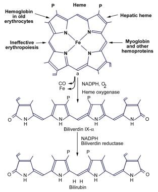
Heme
Stress, Physiological
Megaloblasts
Red-Cell Aplasia, Pure
Transcription Factors
STAT5 Transcription Factor
Flow Cytometry
Janus Kinase 2
Hematopoietic System
Bone Morphogenetic Protein 4
Hematopoietic Cell Growth Factors
Iron Compounds
LIM Domain Proteins
Thalassemia
RNA, Messenger
Doping in Sports
DNA-Binding Proteins
I Blood-Group System
Erythrocytes, Abnormal
Erythropoiesis is the process of forming and developing red blood cells (erythrocytes) in the body. It occurs in the bone marrow and is regulated by the hormone erythropoietin (EPO), which is produced by the kidneys. Erythropoiesis involves the differentiation and maturation of immature red blood cell precursors called erythroblasts into mature red blood cells, which are responsible for carrying oxygen to the body's tissues. Disorders that affect erythropoiesis can lead to anemia or other blood-related conditions.
Erythroblasts are immature red blood cells that are produced in the bone marrow. They are also known as normoblasts and are a stage in the development of red blood cells, or erythrocytes. Erythroblasts are larger than mature red blood cells and have a nucleus, which is lost during the maturation process. These cells are responsible for producing hemoglobin, the protein that carries oxygen in the blood. Abnormal increases or decreases in the number of erythroblasts can be indicative of certain medical conditions, such as anemia or leukemia.
Erythropoietin (EPO) is a hormone that is primarily produced by the kidneys and plays a crucial role in the production of red blood cells in the body. It works by stimulating the bone marrow to produce more red blood cells, which are essential for carrying oxygen to various tissues and organs.
EPO is a glycoprotein that is released into the bloodstream in response to low oxygen levels in the body. When the kidneys detect low oxygen levels, they release EPO, which then travels to the bone marrow and binds to specific receptors on immature red blood cells called erythroblasts. This binding triggers a series of events that promote the maturation and proliferation of erythroblasts, leading to an increase in the production of red blood cells.
In addition to its role in regulating red blood cell production, EPO has also been shown to have neuroprotective effects and may play a role in modulating the immune system. Abnormal levels of EPO have been associated with various medical conditions, including anemia, kidney disease, and certain types of cancer.
EPO is also used as a therapeutic agent for the treatment of anemia caused by chronic kidney disease, chemotherapy, or other conditions that affect red blood cell production. Recombinant human EPO (rhEPO) is a synthetic form of the hormone that is produced using genetic engineering techniques and is commonly used in clinical practice to treat anemia. However, misuse of rhEPO for performance enhancement in sports has been a subject of concern due to its potential to enhance oxygen-carrying capacity and improve endurance.
Erythroid precursor cells, also known as erythroblasts or normoblasts, are early stage cells in the process of producing mature red blood cells (erythrocytes) in the bone marrow. These cells are derived from hematopoietic stem cells and undergo a series of maturation stages, including proerythroblast, basophilic erythroblast, polychromatophilic erythroblast, and orthochromatic erythroblast, before becoming reticulocytes and then mature red blood cells. During this maturation process, the cells lose their nuclei and become enucleated, taking on the biconcave shape and flexible membrane that allows them to move through small blood vessels and deliver oxygen to tissues throughout the body.
Erythroid cells are a type of blood cell that develops in the bone marrow and mature into red blood cells (RBCs), also known as erythrocytes. These cells play a crucial role in the body's oxygen-carrying capacity by transporting oxygen from the lungs to the body's tissues and carbon dioxide from the tissues to the lungs.
The development of erythroid cells begins with hematopoietic stem cells, which can differentiate into various types of blood cells. Through a series of maturation stages, including proerythroblasts, basophilic erythroblasts, polychromatophilic erythroblasts, and orthochromatic erythroblasts, these cells gradually lose their nuclei and organelles to become reticulocytes. Reticulocytes are immature RBCs that still contain some residual ribosomes and are released into the bloodstream. Over time, they mature into fully functional RBCs, which have a biconcave shape and a flexible membrane that allows them to navigate through small blood vessels.
Erythroid cells are essential for maintaining adequate oxygenation of body tissues, and their production is tightly regulated by various hormones and growth factors, such as erythropoietin (EPO), which stimulates the proliferation and differentiation of erythroid progenitor cells. Abnormalities in erythroid cell development or function can lead to various blood disorders, including anemia, polycythemia, and myelodysplastic syndromes.
Anemia is a medical condition characterized by a lower than normal number of red blood cells or lower than normal levels of hemoglobin in the blood. Hemoglobin is an important protein in red blood cells that carries oxygen from the lungs to the rest of the body. Anemia can cause fatigue, weakness, shortness of breath, and a pale complexion because the body's tissues are not getting enough oxygen.
Anemia can be caused by various factors, including nutritional deficiencies (such as iron, vitamin B12, or folate deficiency), blood loss, chronic diseases (such as kidney disease or rheumatoid arthritis), inherited genetic disorders (such as sickle cell anemia or thalassemia), and certain medications.
There are different types of anemia, classified based on the underlying cause, size and shape of red blood cells, and the level of hemoglobin in the blood. Treatment for anemia depends on the underlying cause and may include dietary changes, supplements, medication, or blood transfusions.
Phenylhydrazines are organic compounds that contain a phenyl group (a benzene ring with a hydrogen atom substituted by a hydroxy group) and a hydrazine group (-NH-NH2). They are aromatic amines that have been used in various chemical reactions, including the formation of azos and hydrazones. In medicine, phenylhydrazines were once used as vasodilators to treat angina pectoris, but their use has largely been discontinued due to their toxicity and potential carcinogenicity.
Extramedullary hematopoiesis (EMH) is defined as the production of blood cells outside of the bone marrow in adults. In normal physiological conditions, hematopoiesis occurs within the bone marrow cavities of flat bones such as the pelvis, ribs, skull, and vertebrae. However, certain disease states or conditions can cause EMH to occur in various organs such as the liver, spleen, lymph nodes, and peripheral blood.
EMH can be seen in several pathological conditions, including hematologic disorders such as myeloproliferative neoplasms (e.g., polycythemia vera, essential thrombocytopenia), myelodysplastic syndromes, and leukemias. It can also occur in response to bone marrow failure or infiltration by malignant cells, as well as in some non-hematologic disorders such as fibrocystic disease of the breast and congenital hemolytic anemias.
EMH may lead to organ enlargement, dysfunction, and clinical symptoms depending on the site and extent of involvement. Treatment of EMH is generally directed at managing the underlying condition causing it.
Erythropoietin receptors are cell surface proteins found on immature red blood cell precursors in the bone marrow. They bind to the hormone erythropoietin (EPO), which is produced by the kidneys in response to low oxygen levels in the blood. When EPO binds to its receptor, it activates a signaling pathway that promotes the survival, proliferation, and differentiation of red blood cell precursors, leading to increased production of red blood cells. This process is critical for maintaining adequate oxygen delivery to tissues in the body. Mutations in the erythropoietin receptor gene can lead to various blood disorders, including anemia and polycythemia.
Erythrocyte count, also known as red blood cell (RBC) count, is a laboratory test that measures the number of red blood cells in a sample of blood. Red blood cells are important because they carry oxygen from the lungs to the rest of the body. A low erythrocyte count may indicate anemia, while a high count may be a sign of certain medical conditions such as polycythemia. The normal range for erythrocyte count varies depending on a person's age, sex, and other factors.
Polycythemia is a medical condition characterized by an abnormal increase in the total red blood cell (RBC) mass or hematocrit (the percentage of RBCs in the blood). This results in a higher-than-normal viscosity of the blood, which can lead to various complications such as impaired circulation, increased risk of blood clots, and reduced oxygen supply to the tissues.
There are two main types of polycythemia: primary and secondary. Primary polycythemia, also known as polycythemia vera, is a rare myeloproliferative neoplasm caused by genetic mutations that lead to excessive production of RBCs in the bone marrow. Secondary polycythemia, on the other hand, is a reactive condition triggered by various factors such as chronic hypoxia (low oxygen levels), high altitude, smoking, or certain medical conditions like sleep apnea, heart disease, or kidney tumors.
Symptoms of polycythemia may include fatigue, headaches, dizziness, shortness of breath, itching, and a bluish or reddish tint to the skin (cyanosis). Treatment depends on the underlying cause and severity of the condition and may involve phlebotomy, medications to reduce RBC production, and management of associated complications.
GATA1 (Global Architecture of Tissue/stage-specific Transcription Factors 1) is a transcription factor that belongs to the GATA family, which recognizes and binds to the (A/T)GATA(A/G) motif in the DNA. It plays a crucial role in the development and differentiation of hematopoietic cells, particularly erythroid, megakaryocytic, eosinophilic, and mast cell lineages.
GATA1 regulates gene expression by binding to specific DNA sequences and recruiting other co-factors that modulate chromatin structure and transcriptional activity. Mutations in the GATA1 gene can lead to various blood disorders such as congenital dyserythropoietic anemia type II, Diamond-Blackfan anemia, acute megakaryoblastic leukemia (AMKL), and myelodysplastic syndrome.
In summary, GATA1 Transcription Factor is a protein that binds to specific DNA sequences in the genome and regulates gene expression, playing a critical role in hematopoietic cell development and differentiation.
Erythrocytes, also known as red blood cells (RBCs), are the most common type of blood cell in circulating blood in mammals. They are responsible for transporting oxygen from the lungs to the body's tissues and carbon dioxide from the tissues to the lungs.
Erythrocytes are formed in the bone marrow and have a biconcave shape, which allows them to fold and bend easily as they pass through narrow blood vessels. They do not have a nucleus or mitochondria, which makes them more flexible but also limits their ability to reproduce or repair themselves.
In humans, erythrocytes are typically disc-shaped and measure about 7 micrometers in diameter. They contain the protein hemoglobin, which binds to oxygen and gives blood its red color. The lifespan of an erythrocyte is approximately 120 days, after which it is broken down in the liver and spleen.
Abnormalities in erythrocyte count or function can lead to various medical conditions, such as anemia, polycythemia, and sickle cell disease.
Beta-thalassemia is a genetic blood disorder that affects the production of hemoglobin, a protein in red blood cells that carries oxygen throughout the body. Specifically, beta-thalassemia is caused by mutations in the beta-globin gene, which leads to reduced or absent production of the beta-globin component of hemoglobin.
There are two main types of beta-thalassemia:
1. Beta-thalassemia major (also known as Cooley's anemia): This is a severe form of the disorder that typically becomes apparent in early childhood. It is characterized by a significant reduction or absence of beta-globin production, leading to anemia, enlarged spleen and liver, jaundice, and growth retardation.
2. Beta-thalassemia intermedia: This is a milder form of the disorder that may not become apparent until later in childhood or even adulthood. It is characterized by a variable reduction in beta-globin production, leading to mild to moderate anemia and other symptoms that can range from nonexistent to severe.
Treatment for beta-thalassemia depends on the severity of the disorder and may include blood transfusions, iron chelation therapy, and/or bone marrow transplantation. In some cases, genetic counseling and prenatal diagnosis may also be recommended for families with a history of the disorder.
In the context of medicine, iron is an essential micromineral and key component of various proteins and enzymes. It plays a crucial role in oxygen transport, DNA synthesis, and energy production within the body. Iron exists in two main forms: heme and non-heme. Heme iron is derived from hemoglobin and myoglobin in animal products, while non-heme iron comes from plant sources and supplements.
The recommended daily allowance (RDA) for iron varies depending on age, sex, and life stage:
* For men aged 19-50 years, the RDA is 8 mg/day
* For women aged 19-50 years, the RDA is 18 mg/day
* During pregnancy, the RDA increases to 27 mg/day
* During lactation, the RDA for breastfeeding mothers is 9 mg/day
Iron deficiency can lead to anemia, characterized by fatigue, weakness, and shortness of breath. Excessive iron intake may result in iron overload, causing damage to organs such as the liver and heart. Balanced iron levels are essential for maintaining optimal health.
Hemoglobin (Hb or Hgb) is the main oxygen-carrying protein in the red blood cells, which are responsible for delivering oxygen throughout the body. It is a complex molecule made up of four globin proteins and four heme groups. Each heme group contains an iron atom that binds to one molecule of oxygen. Hemoglobin plays a crucial role in the transport of oxygen from the lungs to the body's tissues, and also helps to carry carbon dioxide back to the lungs for exhalation.
There are several types of hemoglobin present in the human body, including:
* Hemoglobin A (HbA): This is the most common type of hemoglobin, making up about 95-98% of total hemoglobin in adults. It consists of two alpha and two beta globin chains.
* Hemoglobin A2 (HbA2): This makes up about 1.5-3.5% of total hemoglobin in adults. It consists of two alpha and two delta globin chains.
* Hemoglobin F (HbF): This is the main type of hemoglobin present in fetal life, but it persists at low levels in adults. It consists of two alpha and two gamma globin chains.
* Hemoglobin S (HbS): This is an abnormal form of hemoglobin that can cause sickle cell disease when it occurs in the homozygous state (i.e., both copies of the gene are affected). It results from a single amino acid substitution in the beta globin chain.
* Hemoglobin C (HbC): This is another abnormal form of hemoglobin that can cause mild to moderate hemolytic anemia when it occurs in the homozygous state. It results from a different single amino acid substitution in the beta globin chain than HbS.
Abnormal forms of hemoglobin, such as HbS and HbC, can lead to various clinical disorders, including sickle cell disease, thalassemia, and other hemoglobinopathies.
Hematocrit is a medical term that refers to the percentage of total blood volume that is made up of red blood cells. It is typically measured as part of a complete blood count (CBC) test. A high hematocrit may indicate conditions such as dehydration, polycythemia, or living at high altitudes, while a low hematocrit may be a sign of anemia, bleeding, or overhydration. It is important to note that hematocrit values can vary depending on factors such as age, gender, and pregnancy status.
A reticulocyte count is a laboratory test that measures the percentage of reticulocytes in the peripheral blood. Reticulocytes are immature red blood cells produced in the bone marrow and released into the bloodstream. They contain residual ribosomal RNA, which gives them a reticular or net-like appearance under a microscope when stained with certain dyes.
The reticulocyte count is often used as an indicator of the rate of red blood cell production in the bone marrow. A higher than normal reticulocyte count may indicate an increased production of red blood cells, which can be seen in conditions such as hemolysis, blood loss, or response to treatment of anemia. A lower than normal reticulocyte count may suggest a decreased production of red blood cells, which can be seen in conditions such as bone marrow suppression, aplastic anemia, or vitamin deficiencies.
The reticulocyte count is usually expressed as a percentage of the total number of red blood cells, but it can also be reported as an absolute reticulocyte count (the actual number of reticulocytes per microliter of blood). The normal range for the reticulocyte count varies depending on the laboratory and the population studied.
I must clarify that "Iron Isotopes" is not a medical term, but rather a scientific concept from the field of physics and chemistry. However, I can certainly provide a general explanation of isotopes and then focus on iron isotopes specifically.
An isotope is a variant of a chemical element that has the same number of protons (and thus the same atomic number) but a different number of neutrons within its nucleus. This results in variations of the atomic mass of isotopes of the same element. Some isotopes are stable, while others are unstable and will decay over time into other elements or isotopes, a process called radioactive decay.
Iron (Fe) has four naturally occurring stable isotopes: Fe-54, Fe-56, Fe-57, and Fe-58. These iron isotopes have different numbers of neutrons in their nuclei, resulting in slightly different atomic masses. The most abundant iron isotope is Fe-56, which contains 26 protons and 30 neutrons in its nucleus.
In the context of human health, iron is an essential nutrient that plays a crucial role in various biological processes, such as oxygen transport and energy production. However, the concept of iron isotopes does not have a direct medical relevance, but it can be useful in scientific research related to fields like geochemistry, environmental science, or nuclear physics.
Reticulocytes are immature red blood cells that still contain remnants of organelles, such as ribosomes and mitochondria, which are typically found in developing cells. These organelles are involved in the process of protein synthesis and energy production, respectively. Reticulocytes are released from the bone marrow into the bloodstream, where they continue to mature into fully developed red blood cells called erythrocytes.
Reticulocytes can be identified under a microscope by their staining characteristics, which reveal a network of fine filaments or granules known as the reticular apparatus. This apparatus is composed of residual ribosomal RNA and other proteins that have not yet been completely eliminated during the maturation process.
The percentage of reticulocytes in the blood can be used as a measure of bone marrow function and erythropoiesis, or red blood cell production. An increased reticulocyte count may indicate an appropriate response to blood loss, hemolysis, or other conditions that cause anemia, while a decreased count may suggest impaired bone marrow function or a deficiency in erythropoietin, the hormone responsible for stimulating red blood cell production.
The yolk sac is a structure that forms in the early stages of an embryo's development. It is a extra-embryonic membrane, which means it exists outside of the developing embryo, and it plays a critical role in providing nutrients to the growing embryo during the initial stages of development.
In more detail, the yolk sac is responsible for producing blood cells, contributing to the formation of the early circulatory system, and storing nutrients that are absorbed from the yolk material inside the egg or uterus. The yolk sac also has a role in the development of the gut and the immune system.
As the embryo grows and the placenta develops, the yolk sac's function becomes less critical, and it eventually degenerates. However, remnants of the yolk sac can sometimes persist and may be found in the developing fetus or newborn baby. In some cases, abnormalities in the development or regression of the yolk sac can lead to developmental problems or congenital disorders.
Hemolytic anemia is a type of anemia that occurs when red blood cells are destroyed (hemolysis) faster than they can be produced. Red blood cells are essential for carrying oxygen throughout the body. When they are destroyed, hemoglobin and other cellular components are released into the bloodstream, which can lead to complications such as kidney damage and gallstones.
Hemolytic anemia can be inherited or acquired. Inherited forms of the condition may result from genetic defects that affect the structure or function of red blood cells. Acquired forms of hemolytic anemia can be caused by various factors, including infections, medications, autoimmune disorders, and certain medical conditions such as cancer or blood disorders.
Symptoms of hemolytic anemia may include fatigue, weakness, shortness of breath, pale skin, jaundice (yellowing of the skin and eyes), dark urine, and a rapid heartbeat. Treatment for hemolytic anemia depends on the underlying cause and may include medications, blood transfusions, or surgery.
Bone marrow is the spongy tissue found inside certain bones in the body, such as the hips, thighs, and vertebrae. It is responsible for producing blood-forming cells, including red blood cells, white blood cells, and platelets. There are two types of bone marrow: red marrow, which is involved in blood cell production, and yellow marrow, which contains fatty tissue.
Red bone marrow contains hematopoietic stem cells, which can differentiate into various types of blood cells. These stem cells continuously divide and mature to produce new blood cells that are released into the circulation. Red blood cells carry oxygen throughout the body, white blood cells help fight infections, and platelets play a crucial role in blood clotting.
Bone marrow also serves as a site for immune cell development and maturation. It contains various types of immune cells, such as lymphocytes, macrophages, and dendritic cells, which help protect the body against infections and diseases.
Abnormalities in bone marrow function can lead to several medical conditions, including anemia, leukopenia, thrombocytopenia, and various types of cancer, such as leukemia and multiple myeloma. Bone marrow aspiration and biopsy are common diagnostic procedures used to evaluate bone marrow health and function.
A Colony-Forming Units (CFU) assay is a type of laboratory test used to measure the number of viable, or living, cells in a sample. It is commonly used to enumerate bacteria, yeast, and other microorganisms. The test involves placing a known volume of the sample onto a nutrient-agar plate, which provides a solid growth surface for the cells. The plate is then incubated under conditions that allow the cells to grow and form colonies. Each colony that forms on the plate represents a single viable cell from the original sample. By counting the number of colonies and multiplying by the known volume of the sample, the total number of viable cells in the sample can be calculated. This information is useful in a variety of applications, including monitoring microbial populations, assessing the effectiveness of disinfection procedures, and studying microbial growth and survival.
Hepcidin is a peptide hormone primarily produced in the liver that plays a crucial role in regulating iron homeostasis within the body. It acts by inhibiting the absorption of dietary iron in the intestines and the release of iron from storage sites, such as macrophages, into the bloodstream. By reducing the amount of iron available for use, hepcidin helps prevent excessive iron accumulation in tissues, which can be harmful and contribute to the development of various diseases, including iron overload disorders and certain types of anemia. The production of hepcidin is regulated by several factors, including iron levels, inflammation, and erythropoiesis (the production of red blood cells).
Globins are a group of proteins that contain a heme prosthetic group, which binds and transports oxygen in the blood. The most well-known globin is hemoglobin, which is found in red blood cells and is responsible for carrying oxygen from the lungs to the body's tissues. Other members of the globin family include myoglobin, which is found in muscle tissue and stores oxygen, and neuroglobin and cytoglobin, which are found in the brain and other organs and may have roles in protecting against oxidative stress and hypoxia (low oxygen levels). Globins share a similar structure, with a folded protein surrounding a central heme group. Mutations in globin genes can lead to various diseases, such as sickle cell anemia and thalassemia.
Transferrin receptors are membrane-bound proteins found on the surface of many cell types, including red and white blood cells, as well as various tissues such as the liver, brain, and placenta. These receptors play a crucial role in iron homeostasis by regulating the uptake of transferrin, an iron-binding protein, into the cells.
Transferrin binds to two ferric ions (Fe3+) in the bloodstream, forming a complex known as holo-transferrin. This complex then interacts with the transferrin receptors on the cell surface, leading to endocytosis of the transferrin-receptor complex into the cell. Once inside the cell, the acidic environment within the endosome causes the release of iron ions from the transferrin molecule, which can then be transported into the cytoplasm for use in various metabolic processes.
After releasing the iron, the apo-transferrin (iron-free transferrin) is recycled back to the cell surface and released back into the bloodstream, where it can bind to more ferric ions and repeat the cycle. This process helps maintain appropriate iron levels within the body and ensures that cells have access to the iron they need for essential functions such as DNA synthesis, energy production, and oxygen transport.
In summary, transferrin receptors are membrane-bound proteins responsible for recognizing and facilitating the uptake of transferrin-bound iron into cells, playing a critical role in maintaining iron homeostasis within the body.
Hematopoietic stem cells (HSCs) are immature, self-renewing cells that give rise to all the mature blood and immune cells in the body. They are capable of both producing more hematopoietic stem cells (self-renewal) and differentiating into early progenitor cells that eventually develop into red blood cells, white blood cells, and platelets. HSCs are found in the bone marrow, umbilical cord blood, and peripheral blood. They have the ability to repair damaged tissues and offer significant therapeutic potential for treating various diseases, including hematological disorders, genetic diseases, and cancer.
Reticulocytosis is a medical term that refers to an increased number of reticulocytes in the peripheral blood. Reticulocytes are immature red blood cells produced in the bone marrow and released into the bloodstream. They still have remnants of RNA, which gives them a reticular or "net-like" appearance under a microscope when stained with certain dyes.
Reticulocytosis is typically seen in conditions associated with increased red blood cell production, such as:
1. Hemolysis: This is a condition where there is excessive destruction of red blood cells, leading to anemia. The body responds by increasing the production of reticulocytes to replace the lost red blood cells.
2. Blood loss: When there is significant blood loss, the body tries to compensate for the decrease in red blood cells by boosting the production of reticulocytes.
3. Recovery from bone marrow suppression: In cases where the bone marrow has been suppressed due to illness, medication, or chemotherapy, and then recovers, an increase in reticulocytosis may be observed as the bone marrow resumes normal red blood cell production.
4. Megaloblastic anemias: Conditions like vitamin B12 or folate deficiency can lead to megaloblastic anemia, where the red blood cells are larger and immature. Reticulocytosis may be present as the bone marrow tries to correct the anemia.
5. Congenital disorders: Certain inherited conditions, such as hereditary spherocytosis or thalassemias, can cause chronic hemolysis and lead to reticulocytosis.
It is essential to evaluate the underlying cause of reticulocytosis for appropriate diagnosis and treatment.
Macrocytic anemia is a type of anemia in which the red blood cells are larger than normal in size (macrocytic). This condition can be caused by various factors such as deficiency of vitamin B12 or folate, alcohol abuse, certain medications, bone marrow disorders, and some inherited genetic conditions.
The large red blood cells may not function properly, leading to symptoms such as fatigue, weakness, shortness of breath, pale skin, and a rapid heartbeat. Macrocytic anemia can be diagnosed through a complete blood count (CBC) test, which measures the size and number of red blood cells in the blood.
Treatment for macrocytic anemia depends on the underlying cause. In cases of vitamin B12 or folate deficiency, supplements or dietary changes may be recommended. If the anemia is caused by medication, a different medication may be prescribed. In severe cases, blood transfusions or injections of vitamin B12 may be necessary.
Cell differentiation is the process by which a less specialized cell, or stem cell, becomes a more specialized cell type with specific functions and structures. This process involves changes in gene expression, which are regulated by various intracellular signaling pathways and transcription factors. Differentiation results in the development of distinct cell types that make up tissues and organs in multicellular organisms. It is a crucial aspect of embryonic development, tissue repair, and maintenance of homeostasis in the body.
Hematopoiesis is the process of forming and developing blood cells. It occurs in the bone marrow and includes the production of red blood cells (erythropoiesis), white blood cells (leukopoiesis), and platelets (thrombopoiesis). This process is regulated by various growth factors, hormones, and cytokines. Hematopoiesis begins early in fetal development and continues throughout a person's life. Disorders of hematopoiesis can result in conditions such as anemia, leukopenia, leukocytosis, thrombocytopenia, or thrombocytosis.
Beta-globins are the type of globin proteins that make up the beta-chain of hemoglobin, the oxygen-carrying protein in red blood cells. Hemoglobin is composed of four polypeptide chains, two alpha-globin and two beta-globin chains, arranged in a specific structure. The beta-globin gene is located on chromosome 11, and mutations in this gene can lead to various forms of hemoglobin disorders such as sickle cell anemia and beta-thalassemia.
Bone marrow cells are the types of cells found within the bone marrow, which is the spongy tissue inside certain bones in the body. The main function of bone marrow is to produce blood cells. There are two types of bone marrow: red and yellow. Red bone marrow is where most blood cell production takes place, while yellow bone marrow serves as a fat storage site.
The three main types of bone marrow cells are:
1. Hematopoietic stem cells (HSCs): These are immature cells that can differentiate into any type of blood cell, including red blood cells, white blood cells, and platelets. They have the ability to self-renew, meaning they can divide and create more hematopoietic stem cells.
2. Red blood cell progenitors: These are immature cells that will develop into mature red blood cells, also known as erythrocytes. Red blood cells carry oxygen from the lungs to the body's tissues and carbon dioxide back to the lungs.
3. Myeloid and lymphoid white blood cell progenitors: These are immature cells that will develop into various types of white blood cells, which play a crucial role in the body's immune system by fighting infections and diseases. Myeloid progenitors give rise to granulocytes (neutrophils, eosinophils, and basophils), monocytes, and megakaryocytes (which eventually become platelets). Lymphoid progenitors differentiate into B cells, T cells, and natural killer (NK) cells.
Bone marrow cells are essential for maintaining a healthy blood cell count and immune system function. Abnormalities in bone marrow cells can lead to various medical conditions, such as anemia, leukopenia, leukocytosis, thrombocytopenia, or thrombocytosis, depending on the specific type of blood cell affected. Additionally, bone marrow cells are often used in transplantation procedures to treat patients with certain types of cancer, such as leukemia and lymphoma, or other hematologic disorders.
Bloodletting is a medical procedure that was commonly used in the past to balance the four humors of the body, which were believed to be blood, phlegm, black bile, and yellow bile. The procedure involved withdrawing blood from a patient through various methods such as venesection (making an incision in a vein), leeches, or cupping.
The theory behind bloodletting was that if one humor became overabundant, it could cause disease or illness. By removing some of the excess humor, practitioners believed they could restore balance and promote healing. Bloodletting was used to treat a wide variety of conditions, including fever, inflammation, and pain.
While bloodletting is no longer practiced in modern medicine, it was once a common treatment for many different ailments. The practice dates back to ancient times and was used by various cultures throughout history, including the Greeks, Romans, Egyptians, and Chinese. However, its effectiveness as a medical treatment has been called into question, and it is now considered an outdated and potentially harmful procedure.
Fetal hemoglobin (HbF) is a type of hemoglobin that is produced in the fetus and newborn babies. It is composed of two alpha-like globin chains and two gamma-globin chains, designated as α2γ2. HbF is the primary form of hemoglobin during fetal development, replacing the embryonic hemoglobin (HbG) around the eighth week of gestation.
The unique property of HbF is its higher affinity for oxygen compared to adult hemoglobin (HbA), which helps ensure adequate oxygen supply from the mother to the developing fetus. After birth, as the newborn starts breathing on its own and begins to receive oxygen directly, the production of HbF gradually decreases and is usually replaced by HbA within the first year of life.
In some genetic disorders like sickle cell disease and beta-thalassemia, persistence of HbF into adulthood can be beneficial as it reduces the severity of symptoms due to its higher oxygen-carrying capacity and less polymerization tendency compared to HbS (in sickle cell disease) or unpaired alpha chains (in beta-thalassemia). Treatments like hydroxyurea are used to induce HbF production in these patients as a therapeutic approach.
Alpha-globins are a type of globin protein that combine to form the alpha-globin chains of hemoglobin, the oxygen-carrying protein in red blood cells. Hemoglobin is composed of four globin chains, two alpha-globin chains and two beta-globin chains, that surround a heme group. The alpha-globin genes are located on chromosome 16 and are essential for normal hemoglobin function. Mutations in the alpha-globin genes can lead to various forms of hemoglobin disorders such as alpha-thalassemia.
Diamond-Blackfan anemia is a rare, congenital bone marrow failure disorder characterized by a decreased production of red blood cells (erythroblasts) in the bone marrow. This results in a reduced number of circulating red blood cells, leading to anemia and related symptoms such as fatigue, weakness, and pallor. The disorder is typically diagnosed in infancy or early childhood and can also be associated with physical abnormalities.
The exact cause of Diamond-Blackfan anemia is not fully understood, but it is believed to involve genetic mutations that affect the development and function of the bone marrow. In many cases, the disorder is inherited in an autosomal dominant manner, meaning that a child has a 50% chance of inheriting the mutated gene from an affected parent. However, some cases may arise spontaneously due to new genetic mutations.
Treatment for Diamond-Blackfan anemia typically involves regular blood transfusions to maintain adequate red blood cell levels and alleviate symptoms. Corticosteroid therapy may also be used to stimulate red blood cell production in some cases. In severe or refractory cases, stem cell transplantation may be considered as a curative treatment option.
The spleen is an organ in the upper left side of the abdomen, next to the stomach and behind the ribs. It plays multiple supporting roles in the body:
1. It fights infection by acting as a filter for the blood. Old red blood cells are recycled in the spleen, and platelets and white blood cells are stored there.
2. The spleen also helps to control the amount of blood in the body by removing excess red blood cells and storing platelets.
3. It has an important role in immune function, producing antibodies and removing microorganisms and damaged red blood cells from the bloodstream.
The spleen can be removed without causing any significant problems, as other organs take over its functions. This is known as a splenectomy and may be necessary if the spleen is damaged or diseased.
Erythrocyte aging, also known as red cell aging, is the natural process of changes and senescence that occur in red blood cells (erythrocytes) over time. In humans, mature erythrocytes are devoid of nuclei and organelles, and have a lifespan of approximately 120 days.
During aging, several biochemical and structural modifications take place in the erythrocyte, including:
1. Loss of membrane phospholipids and proteins, leading to increased rigidity and decreased deformability.
2. Oxidative damage to hemoglobin, resulting in the formation of methemoglobin and heinz bodies.
3. Accumulation of denatured proteins and aggregates, which can impair cellular functions.
4. Changes in the cytoskeleton, affecting the shape and stability of the erythrocyte.
5. Increased expression of surface markers, such as Band 3 and CD47, that signal the spleen to remove aged erythrocytes from circulation.
The spleen plays a crucial role in removing senescent erythrocytes by recognizing and phagocytosing those with altered membrane composition or increased expression of surface markers. This process helps maintain the overall health and functionality of the circulatory system.
"Iron radioisotopes" refer to specific forms of the element iron that have unstable nuclei and emit radiation. These isotopes are often used in medical imaging and treatment procedures due to their ability to be detected by specialized equipment. Common iron radioisotopes include Iron-52, Iron-55, Iron-59, and Iron-60. They can be used as tracers to study the distribution, metabolism, or excretion of iron in the body, or for targeted radiation therapy in conditions such as cancer.
Erythroid-specific DNA-binding factors are transcription factors that bind to specific sequences of DNA and help regulate the expression of genes that are involved in the development and differentiation of erythroid cells, which are cells that mature to become red blood cells. These transcription factors play a crucial role in the production of hemoglobin, the protein in red blood cells that carries oxygen throughout the body. Examples of erythroid-specific DNA-binding factors include GATA-1 and KLF1.
Hypochromic anemia is a type of anemia characterized by the presence of red blood cells that have lower than normal levels of hemoglobin and appear paler in color than normal. Hemoglobin is a protein in red blood cells that carries oxygen from the lungs to the rest of the body. In hypochromic anemia, there may be a decrease in the production or increased destruction of red blood cells, leading to a reduced number of red blood cells and insufficient oxygen supply to the tissues.
Hypochromic anemia can result from various underlying medical conditions, including iron deficiency, thalassemia, chronic inflammation, lead poisoning, and certain infections or chronic diseases. Treatment for hypochromic anemia depends on the underlying cause and may include iron supplements, dietary changes, medications, or blood transfusions.
Glycophorin is a type of protein found on the surface of red blood cells, also known as erythrocytes. These proteins are heavily glycosylated, meaning they have many carbohydrate chains attached to them. Glycophorins play a crucial role in maintaining the structure and flexibility of the red blood cell membrane, and they also help to mediate interactions between the red blood cells and other cells or molecules in the body.
There are several different types of glycophorin proteins, including glycophorin A, B, C, and D. Glycophorin A is the most abundant type and is often used as a marker for identifying the ABO blood group. Mutations in the genes that encode glycophorin proteins can lead to various blood disorders, such as hereditary spherocytosis and hemolytic anemia.
Dyserythropoietic anemia, congenital is a rare type of inherited anemia characterized by ineffective red blood cell production (erythropoiesis) in the bone marrow. This means that the body has difficulty producing healthy and fully mature red blood cells. The condition is caused by mutations in genes responsible for the development and maturation of red blood cells, leading to the production of abnormally shaped and dysfunctional red blood cells.
There are two main types of congenital dyserythropoietic anemia (CDA), type I and type II, each caused by different genetic mutations:
1. CDA Type I (HEMPAS): This form is caused by a mutation in the SEC23B gene. It typically presents in early childhood with mild to moderate anemia, jaundice, and splenomegaly (enlarged spleen). The severity of the condition can vary widely among affected individuals.
2. CDA Type II (HIEM): This form is caused by a mutation in the KIF23 gene or, less commonly, the TCIRG1 gene. It typically presents in infancy with moderate to severe anemia, hepatomegaly (enlarged liver), and splenomegaly. The condition can lead to iron overload due to repeated blood transfusions, which may require chelation therapy to manage.
Both types of congenital dyserythropoietic anemia are characterized by ineffective erythropoiesis, abnormal red blood cell morphology, and increased destruction of red blood cells (hemolysis). Treatment typically involves supportive care, such as blood transfusions to manage anemia, and occasionally chelation therapy to address iron overload. In some cases, bone marrow transplantation may be considered as a curative option.
Erythroblastic Leukemia, Acute (also known as Acute Erythroid Leukemia or AEL) is a subtype of acute myeloid leukemia (AML), which is a type of cancer affecting the blood and bone marrow. In this condition, there is an overproduction of erythroblasts (immature red blood cells) in the bone marrow, leading to their accumulation and interference with normal blood cell production. This results in a decrease in the number of functional red blood cells, white blood cells, and platelets in the body. Symptoms may include fatigue, weakness, frequent infections, and easy bruising or bleeding. AEL is typically treated with chemotherapy and sometimes requires stem cell transplantation.
Hemoglobin E (HbE) is a structural variant of hemoglobin, which is the oxygen-carrying protein in red blood cells. This variant results from a specific mutation in the beta-globin gene, leading to the substitution of glutamic acid with lysine at position 26 of the beta-globin chain.
HbE is most commonly found in people from Southeast Asia, particularly in populations from Thailand, Cambodia, and Laos. It can also be found in other parts of the world, such as India, Bangladesh, and Pakistan. HbE is usually asymptomatic when it occurs in its heterozygous form (one normal beta-globin gene and one HbE gene). However, when it occurs in the homozygous form (two HbE genes), or in combination with other hemoglobinopathies like thalassemia, it can lead to a range of clinical manifestations, including mild to severe microcytic anemia, splenomegaly, and jaundice.
Individuals with HbE may have increased susceptibility to certain infections and may experience complications during pregnancy or surgery due to impaired oxygen-carrying capacity. Regular monitoring of hemoglobin levels, iron status, and potential complications is essential for managing individuals with Hemoglobin E effectively.
Polycythemia Vera is a type of myeloproliferative neoplasm, a group of rare blood cancers. In Polycythemia Vera, the body produces too many red blood cells, leading to an increased risk of blood clots and thickening of the blood, which can cause various symptoms such as fatigue, headache, dizziness, and itching. It can also lead to enlargement of the spleen. The exact cause of Polycythemia Vera is not known, but it is associated with genetic mutations in the JAK2 gene in most cases. It is a progressive disease that can lead to complications such as bleeding, thrombosis, and transformation into acute leukemia if left untreated.
Hematinics are a class of medications and dietary supplements that are used to enhance the production of red blood cells or hemoglobin in the body. They typically contain iron, vitamin B12, folic acid, or other nutrients that are essential for the synthesis of hemoglobin and the formation of red blood cells.
Iron is a critical component of hematinics because it plays a central role in the production of hemoglobin, which is the protein in red blood cells that carries oxygen throughout the body. Vitamin B12 and folic acid are also important nutrients for red blood cell production, as they help to regulate the growth and division of red blood cells in the bone marrow.
Hematinics are often prescribed to treat anemia, which is a condition characterized by a low red blood cell count or abnormally low levels of hemoglobin in the blood. Anemia can be caused by a variety of factors, including nutritional deficiencies, chronic diseases, and inherited genetic disorders.
Examples of hematinics include ferrous sulfate (an iron supplement), cyanocobalamin (vitamin B12), and folic acid. These medications are available in various forms, such as tablets, capsules, and liquids, and can be taken orally or by injection. It is important to follow the dosage instructions carefully and to inform your healthcare provider of any other medications you are taking, as hematinics can interact with certain drugs and may cause side effects.
Antimicrobial cationic peptides (ACPs) are a group of small, naturally occurring peptides that possess broad-spectrum antimicrobial activity against various microorganisms, including bacteria, fungi, viruses, and parasites. They are called "cationic" because they contain positively charged amino acid residues (such as lysine and arginine), which allow them to interact with and disrupt the negatively charged membranes of microbial cells.
ACPs are produced by a wide range of organisms, including humans, animals, and plants, as part of their innate immune response to infection. They play an important role in protecting the host from invading pathogens by directly killing them or inhibiting their growth.
The antimicrobial activity of ACPs is thought to be mediated by their ability to disrupt the membranes of microbial cells, leading to leakage of cellular contents and death. Some ACPs may also have intracellular targets, such as DNA or protein synthesis, that contribute to their antimicrobial activity.
ACPs are being studied for their potential use as therapeutic agents to treat infectious diseases, particularly those caused by drug-resistant bacteria. However, their clinical application is still in the early stages of development due to concerns about their potential toxicity to host cells and the emergence of resistance mechanisms in microbial pathogens.
Ferritin is a protein in iron-metabolizing cells that stores iron in a water-soluble form. It is found inside the cells (intracellular) and is released into the bloodstream when the cells break down or die. Measuring the level of ferritin in the blood can help determine the amount of iron stored in the body. High levels of ferritin may indicate hemochromatosis, inflammation, liver disease, or other conditions. Low levels of ferritin may indicate anemia, iron deficiency, or other conditions.
Iron-deficiency anemia is a condition characterized by a decrease in the total amount of hemoglobin or red blood cells in the blood, caused by insufficient iron levels in the body. Hemoglobin is a protein in red blood cells that carries oxygen from the lungs to the rest of the body. When iron levels are low, the body cannot produce enough hemoglobin, leading to the production of smaller and fewer red blood cells, known as microcytic hypochromic anemia.
Iron is essential for the production of hemoglobin, and a deficiency in iron can result from inadequate dietary intake, chronic blood loss, or impaired absorption. In addition to fatigue and weakness, symptoms of iron-deficiency anemia may include shortness of breath, headaches, dizziness, pale skin, and brittle nails. Treatment typically involves iron supplementation and addressing the underlying cause of the iron deficiency.
Phlebotomy is a medical term that refers to the process of making an incision in a vein, usually in the arm, in order to draw blood. It is also commonly known as venipuncture. This procedure is performed by healthcare professionals for various purposes such as diagnostic testing, blood donation, or therapeutic treatments like phlebotomy for patients with hemochromatosis (a condition where the body absorbs too much iron from food).
The person who performs this procedure is called a phlebotomist. They must be trained in the proper techniques to ensure that the process is safe and relatively pain-free for the patient, and that the blood sample is suitable for laboratory testing.
GATA2 transcription factor is a protein that plays a crucial role in the development and function of blood cells. It belongs to the family of GATA transcription factors, which are characterized by their ability to bind to specific DNA sequences called GATA motifs, through a zinc finger domain. The GATA2 transcription factor, in particular, is essential for the development of hematopoietic stem and progenitor cells (HSPCs), which give rise to all blood cell types.
GATA2 binds to the regulatory regions of genes involved in hematopoiesis and modulates their transcription, thereby controlling the differentiation, proliferation, and survival of HSPCs. Mutations in the GATA2 gene have been associated with various hematological disorders, such as acute myeloid leukemia (AML), myelodysplastic syndrome (MDS), and severe congenital neutropenia. These genetic alterations can lead to impaired hematopoiesis, dysfunctional immune cells, and an increased risk of developing blood cancers.
In summary, GATA2 transcription factor is a protein that regulates the development and function of blood cells by controlling the expression of genes involved in hematopoiesis. Genetic defects in this transcription factor can result in various hematological disorders and predispose individuals to blood cancers.
Stem Cell Factor (SCF), also known as Kit Ligand or Steel Factor, is a growth factor that plays a crucial role in the regulation of hematopoiesis, which is the process of producing various blood cells. It is a glycoprotein that binds to the c-Kit receptor found on hematopoietic stem cells and progenitor cells, promoting their survival, proliferation, and differentiation into mature blood cells.
SCF is involved in the development and function of several types of blood cells, including red blood cells, white blood cells, and platelets. It also plays a role in the maintenance and self-renewal of hematopoietic stem cells, which are essential for the continuous production of new blood cells throughout an individual's lifetime.
In addition to its role in hematopoiesis, SCF has been implicated in various other biological processes, such as melanogenesis, gametogenesis, and tissue repair and regeneration. Dysregulation of SCF signaling has been associated with several diseases, including certain types of cancer, bone marrow failure disorders, and autoimmune diseases.
The liver is a large, solid organ located in the upper right portion of the abdomen, beneath the diaphragm and above the stomach. It plays a vital role in several bodily functions, including:
1. Metabolism: The liver helps to metabolize carbohydrates, fats, and proteins from the food we eat into energy and nutrients that our bodies can use.
2. Detoxification: The liver detoxifies harmful substances in the body by breaking them down into less toxic forms or excreting them through bile.
3. Synthesis: The liver synthesizes important proteins, such as albumin and clotting factors, that are necessary for proper bodily function.
4. Storage: The liver stores glucose, vitamins, and minerals that can be released when the body needs them.
5. Bile production: The liver produces bile, a digestive juice that helps to break down fats in the small intestine.
6. Immune function: The liver plays a role in the immune system by filtering out bacteria and other harmful substances from the blood.
Overall, the liver is an essential organ that plays a critical role in maintaining overall health and well-being.
Transferrin is a glycoprotein that plays a crucial role in the transport and homeostasis of iron in the body. It's produced mainly in the liver and has the ability to bind two ferric (Fe3+) ions in its N-lobe and C-lobe, thus creating transferrin saturation.
This protein is essential for delivering iron to cells while preventing the harmful effects of free iron, which can catalyze the formation of reactive oxygen species through Fenton reactions. Transferrin interacts with specific transferrin receptors on the surface of cells, particularly in erythroid precursors and brain endothelial cells, to facilitate iron uptake via receptor-mediated endocytosis.
In addition to its role in iron transport, transferrin also has antimicrobial properties due to its ability to sequester free iron, making it less available for bacterial growth and survival. Transferrin levels can be used as a clinical marker of iron status, with decreased levels indicating iron deficiency anemia and increased levels potentially signaling inflammation or liver disease.
K562 cells are a type of human cancer cell that are commonly used in scientific research. They are derived from a patient with chronic myelogenous leukemia (CML), a type of cancer that affects the blood and bone marrow.
K562 cells are often used as a model system to study various biological processes, including cell signaling, gene expression, differentiation, and apoptosis (programmed cell death). They are also commonly used in drug discovery and development, as they can be used to test the effectiveness of potential new therapies against cancer.
K562 cells have several characteristics that make them useful for research purposes. They are easy to grow and maintain in culture, and they can be manipulated genetically to express or knock down specific genes. Additionally, K562 cells are capable of differentiating into various cell types, such as red blood cells and megakaryocytes, which allows researchers to study the mechanisms of cell differentiation.
It's important to note that while K562 cells are a valuable tool for research, they do not fully recapitulate the complexity of human CML or other cancers. Therefore, findings from studies using K562 cells should be validated in more complex model systems or in clinical trials before they can be translated into treatments for patients.
"Cells, cultured" is a medical term that refers to cells that have been removed from an organism and grown in controlled laboratory conditions outside of the body. This process is called cell culture and it allows scientists to study cells in a more controlled and accessible environment than they would have inside the body. Cultured cells can be derived from a variety of sources, including tissues, organs, or fluids from humans, animals, or cell lines that have been previously established in the laboratory.
Cell culture involves several steps, including isolation of the cells from the tissue, purification and characterization of the cells, and maintenance of the cells in appropriate growth conditions. The cells are typically grown in specialized media that contain nutrients, growth factors, and other components necessary for their survival and proliferation. Cultured cells can be used for a variety of purposes, including basic research, drug development and testing, and production of biological products such as vaccines and gene therapies.
It is important to note that cultured cells may behave differently than they do in the body, and results obtained from cell culture studies may not always translate directly to human physiology or disease. Therefore, it is essential to validate findings from cell culture experiments using additional models and ultimately in clinical trials involving human subjects.
C57BL/6 (C57 Black 6) is an inbred strain of laboratory mouse that is widely used in biomedical research. The term "inbred" refers to a strain of animals where matings have been carried out between siblings or other closely related individuals for many generations, resulting in a population that is highly homozygous at most genetic loci.
The C57BL/6 strain was established in 1920 by crossing a female mouse from the dilute brown (DBA) strain with a male mouse from the black strain. The resulting offspring were then interbred for many generations to create the inbred C57BL/6 strain.
C57BL/6 mice are known for their robust health, longevity, and ease of handling, making them a popular choice for researchers. They have been used in a wide range of biomedical research areas, including studies of cancer, immunology, neuroscience, cardiovascular disease, and metabolism.
One of the most notable features of the C57BL/6 strain is its sensitivity to certain genetic modifications, such as the introduction of mutations that lead to obesity or impaired glucose tolerance. This has made it a valuable tool for studying the genetic basis of complex diseases and traits.
Overall, the C57BL/6 inbred mouse strain is an important model organism in biomedical research, providing a valuable resource for understanding the genetic and molecular mechanisms underlying human health and disease.
Thrombopoiesis is the process of formation and development of thrombocytes or platelets, which are small, colorless cell fragments in our blood that play an essential role in clotting. Thrombopoiesis occurs inside the bone marrow, where stem cells differentiate into megakaryoblasts, then progressively develop into promegakaryocytes and megakaryocytes. These megakaryocytes subsequently undergo a process called cytoplasmic fragmentation to produce platelets.
The regulation of thrombopoiesis is primarily controlled by the hormone thrombopoietin (TPO), which is produced mainly in the liver and binds to the thrombopoietin receptor (c-Mpl) on megakaryocytes and their precursors. This binding stimulates the proliferation, differentiation, and maturation of megakaryocytes, leading to an increase in platelet production.
Abnormalities in thrombopoiesis can result in conditions such as thrombocytopenia (low platelet count) or thrombocytosis (high platelet count), which may be associated with bleeding disorders or increased risk of thrombosis, respectively.
Recombinant proteins are artificially created proteins produced through the use of recombinant DNA technology. This process involves combining DNA molecules from different sources to create a new set of genes that encode for a specific protein. The resulting recombinant protein can then be expressed, purified, and used for various applications in research, medicine, and industry.
Recombinant proteins are widely used in biomedical research to study protein function, structure, and interactions. They are also used in the development of diagnostic tests, vaccines, and therapeutic drugs. For example, recombinant insulin is a common treatment for diabetes, while recombinant human growth hormone is used to treat growth disorders.
The production of recombinant proteins typically involves the use of host cells, such as bacteria, yeast, or mammalian cells, which are engineered to express the desired protein. The host cells are transformed with a plasmid vector containing the gene of interest, along with regulatory elements that control its expression. Once the host cells are cultured and the protein is expressed, it can be purified using various chromatography techniques.
Overall, recombinant proteins have revolutionized many areas of biology and medicine, enabling researchers to study and manipulate proteins in ways that were previously impossible.
Developmental gene expression regulation refers to the processes that control the activation or repression of specific genes during embryonic and fetal development. These regulatory mechanisms ensure that genes are expressed at the right time, in the right cells, and at appropriate levels to guide proper growth, differentiation, and morphogenesis of an organism.
Developmental gene expression regulation is a complex and dynamic process involving various molecular players, such as transcription factors, chromatin modifiers, non-coding RNAs, and signaling molecules. These regulators can interact with cis-regulatory elements, like enhancers and promoters, to fine-tune the spatiotemporal patterns of gene expression during development.
Dysregulation of developmental gene expression can lead to various congenital disorders and developmental abnormalities. Therefore, understanding the principles and mechanisms governing developmental gene expression regulation is crucial for uncovering the etiology of developmental diseases and devising potential therapeutic strategies.
Erythrocyte indices are a set of calculated values that provide information about the size and hemoglobin content of red blood cells (erythrocytes). These indices are commonly used in the complete blood count (CBC) test to help diagnose various types of anemia and other conditions affecting the red blood cells.
The three main erythrocyte indices are:
1. Mean Corpuscular Volume (MCV): This is the average volume of a single red blood cell, measured in femtoliters (fL). MCV helps to differentiate between microcytic, normocytic, and macrocytic anemia. Microcytic anemia is characterized by low MCV values (100 fL).
2. Mean Corpuscular Hemoglobin (MCH): This is the average amount of hemoglobin present in a single red blood cell, measured in picograms (pg). MCH helps to assess the oxygen-carrying capacity of red blood cells. Low MCH values may indicate hypochromic anemia, where the red blood cells have reduced hemoglobin content.
3. Mean Corpuscular Hemoglobin Concentration (MCHC): This is the average concentration of hemoglobin in a single red blood cell, measured as a percentage. MCHC reflects the hemoglobin concentration relative to the size of the red blood cells. Low MCHC values may indicate hypochromic anemia, while high MCHC values could suggest spherocytosis or other conditions affecting red blood cell shape and integrity.
These erythrocyte indices are calculated based on the red blood cell count, hemoglobin concentration, and hematocrit results obtained from a CBC test. They provide valuable information for healthcare professionals to diagnose and manage various hematological conditions.
Gamma-globulins are a type of globulin, which are proteins found in the blood plasma. More specifically, gamma-globulins are a class of immunoglobulins, also known as antibodies, that play a crucial role in the immune system's response to foreign substances and infectious agents.
Immunoglobulins are divided into several classes based on their structure and function. Gamma-globulins include IgG, IgA, and IgD isotypes of immunoglobulins. Among these, IgG is the most abundant type found in the blood and other body fluids, responsible for providing protection against bacterial and viral infections.
Gamma-globulins are produced by B cells, a type of white blood cell involved in the immune response. They can be measured in the blood as part of a complete blood count (CBC) or specific protein electrophoresis tests to assess immune system function or diagnose various medical conditions such as infections, inflammation, and autoimmune disorders.
Basic Helix-Loop-Helix (bHLH) transcription factors are a type of proteins that regulate gene expression through binding to specific DNA sequences. They play crucial roles in various biological processes, including cell growth, differentiation, and apoptosis. The bHLH domain is composed of two amphipathic α-helices separated by a loop region. This structure allows the formation of homodimers or heterodimers, which then bind to the E-box DNA motif (5'-CANNTG-3') to regulate transcription.
The bHLH family can be further divided into several subfamilies based on their sequence similarities and functional characteristics. Some members of this family are involved in the development and function of the nervous system, while others play critical roles in the development of muscle and bone. Dysregulation of bHLH transcription factors has been implicated in various human diseases, including cancer and neurodevelopmental disorders.
A "knockout" mouse is a genetically engineered mouse in which one or more genes have been deleted or "knocked out" using molecular biology techniques. This allows researchers to study the function of specific genes and their role in various biological processes, as well as potential associations with human diseases. The mice are generated by introducing targeted DNA modifications into embryonic stem cells, which are then used to create a live animal. Knockout mice have been widely used in biomedical research to investigate gene function, disease mechanisms, and potential therapeutic targets.
Iron overload is a condition characterized by an excessive accumulation of iron in the body's tissues and organs, particularly in the liver, heart, and pancreas. This occurs when the body absorbs more iron than it can use or eliminate, leading to iron levels that are higher than normal.
Iron overload can result from various factors, including hereditary hemochromatosis, a genetic disorder that affects how the body absorbs iron from food; frequent blood transfusions, which can cause iron buildup in people with certain chronic diseases such as sickle cell anemia or thalassemia; and excessive consumption of iron supplements or iron-rich foods.
Symptoms of iron overload may include fatigue, joint pain, abdominal discomfort, irregular heartbeat, and liver dysfunction. If left untreated, it can lead to serious complications such as cirrhosis, liver failure, diabetes, heart problems, and even certain types of cancer. Treatment typically involves regular phlebotomy (removal of blood) to reduce iron levels in the body, along with dietary modifications and monitoring by a healthcare professional.
'Gene expression regulation' refers to the processes that control whether, when, and where a particular gene is expressed, meaning the production of a specific protein or functional RNA encoded by that gene. This complex mechanism can be influenced by various factors such as transcription factors, chromatin remodeling, DNA methylation, non-coding RNAs, and post-transcriptional modifications, among others. Proper regulation of gene expression is crucial for normal cellular function, development, and maintaining homeostasis in living organisms. Dysregulation of gene expression can lead to various diseases, including cancer and genetic disorders.
Pregnanes are a class of steroid hormones and steroids that contain a pregnane nucleus, which is a steroid core with a carbon skeleton consisting of 21 carbons. This structure includes four fused rings, labeled A through D, and is derived from cholesterol.
Pregnanes are important precursors for the synthesis of various steroid hormones in the body, including progesterone, which plays a crucial role in maintaining pregnancy and regulating the menstrual cycle. Other examples of pregnanes include cortisol, a stress hormone produced by the adrenal gland, and aldosterone, a hormone that helps regulate electrolyte balance and blood pressure.
It's worth noting that pregnanes can also refer to synthetic compounds that contain this steroid nucleus and are used in various medical and research contexts.
A fetus is the developing offspring in a mammal, from the end of the embryonic period (approximately 8 weeks after fertilization in humans) until birth. In humans, the fetal stage of development starts from the eleventh week of pregnancy and continues until childbirth, which is termed as full-term pregnancy at around 37 to 40 weeks of gestation. During this time, the organ systems become fully developed and the body grows in size. The fetus is surrounded by the amniotic fluid within the amniotic sac and is connected to the placenta via the umbilical cord, through which it receives nutrients and oxygen from the mother. Regular prenatal care is essential during this period to monitor the growth and development of the fetus and ensure a healthy pregnancy and delivery.
Hemolysis is the destruction or breakdown of red blood cells, resulting in the release of hemoglobin into the surrounding fluid (plasma). This process can occur due to various reasons such as chemical agents, infections, autoimmune disorders, mechanical trauma, or genetic abnormalities. Hemolysis may lead to anemia and jaundice, among other complications. It is essential to monitor hemolysis levels in patients undergoing medical treatments that might cause this condition.
CD34 is a type of antigen that is found on the surface of certain cells in the human body. Specifically, CD34 antigens are present on hematopoietic stem cells, which are immature cells that can develop into different types of blood cells. These stem cells are found in the bone marrow and are responsible for producing red blood cells, white blood cells, and platelets.
CD34 antigens are a type of cell surface marker that is used in medical research and clinical settings to identify and isolate hematopoietic stem cells. They are also used in the development of stem cell therapies and transplantation procedures. CD34 antigens can be detected using various laboratory techniques, such as flow cytometry or immunohistochemistry.
It's important to note that while CD34 is a useful marker for identifying hematopoietic stem cells, it is not exclusive to these cells and can also be found on other cell types, such as endothelial cells that line blood vessels. Therefore, additional markers are often used in combination with CD34 to more specifically identify and isolate hematopoietic stem cells.
Megakaryocytes are large, specialized bone marrow cells that are responsible for the production and release of platelets (also known as thrombocytes) into the bloodstream. Platelets play an essential role in blood clotting and hemostasis, helping to prevent excessive bleeding during injuries or trauma.
Megakaryocytes have a unique structure with multilobed nuclei and abundant cytoplasm rich in organelles called alpha-granules and dense granules, which store various proteins, growth factors, and enzymes necessary for platelet function. As megakaryocytes mature, they extend long cytoplasmic processes called proplatelets into the bone marrow sinuses, where these extensions fragment into individual platelets that are released into circulation.
Abnormalities in megakaryocyte number, size, or function can lead to various hematological disorders, such as thrombocytopenia (low platelet count), thrombocytosis (high platelet count), and certain types of leukemia.
A "Blood Cell Count" is a medical laboratory test that measures the number of red blood cells (RBCs), white blood cells (WBCs), and platelets in a sample of blood. This test is often used as a part of a routine check-up or to help diagnose various medical conditions, such as anemia, infection, inflammation, and many others.
The RBC count measures the number of oxygen-carrying cells in the blood, while the WBC count measures the number of immune cells that help fight infections. The platelet count measures the number of cells involved in clotting. Abnormal results in any of these counts may indicate an underlying medical condition and further testing may be required for diagnosis and treatment.
Splenomegaly is a medical term that refers to an enlargement or expansion of the spleen beyond its normal size. The spleen is a vital organ located in the upper left quadrant of the abdomen, behind the stomach and below the diaphragm. It plays a crucial role in filtering the blood, fighting infections, and storing red and white blood cells and platelets.
Splenomegaly can occur due to various underlying medical conditions, including infections, liver diseases, blood disorders, cancer, and inflammatory diseases. The enlarged spleen may put pressure on surrounding organs, causing discomfort or pain in the abdomen, and it may also lead to a decrease in red and white blood cells and platelets, increasing the risk of anemia, infections, and bleeding.
The diagnosis of splenomegaly typically involves a physical examination, medical history, and imaging tests such as ultrasound, CT scan, or MRI. Treatment depends on the underlying cause and may include medications, surgery, or other interventions to manage the underlying condition.
SOXD (SRY-related HMG box gene D) transcription factors are a subgroup of the SOX family of proteins that regulate gene expression during development and differentiation. The SOXD group includes two closely related members, SOX5 and SOX6, which contain a highly conserved HMG (high mobility group) DNA-binding domain. These transcription factors play crucial roles in various biological processes, such as chondrogenesis, neurogenesis, and spermatogenesis, by binding to specific DNA sequences and regulating the transcription of target genes. SOX5 and SOX6 can form heterodimers or homodimers and interact with other transcription factors and cofactors to modulate their activities, contributing to the precise control of gene expression during development.
Myelopoiesis is the process of formation and development of myeloid cells (a type of blood cell) within the bone marrow. This includes the production of red blood cells (erythropoiesis), platelets (thrombopoiesis), and white blood cells such as granulocytes (neutrophils, eosinophils, basophils), monocytes, and mast cells. Myelopoiesis is a continuous process that is regulated by various growth factors and hormones to maintain the normal levels of these cells in the body. Abnormalities in myelopoiesis can lead to several hematological disorders like anemia, leukopenia, leukocytosis, thrombocytopenia, or thrombocytosis.
Sideroblastic anemia is a type of anemia characterized by the presence of ringed sideroblasts in the bone marrow. Ringed sideroblasts are red blood cell precursors that have an abnormal amount of iron accumulated in their mitochondria, which forms a ring around the nucleus. This results in the production of abnormal hemoglobin and impaired oxygen transport.
Sideroblastic anemia can be classified as congenital or acquired. Congenital sideroblastic anemias are caused by genetic defects that affect heme synthesis or mitochondrial function, while acquired sideroblastic anemias are associated with various conditions such as myelodysplastic syndromes, chronic alcoholism, lead toxicity, and certain medications.
Symptoms of sideroblastic anemia may include fatigue, weakness, shortness of breath, and pallor. Diagnosis is typically made through a bone marrow aspiration and biopsy, which can identify the presence of ringed sideroblasts. Treatment depends on the underlying cause but may include iron chelation therapy, vitamin B6 supplementation, or blood transfusions.
Transgenic mice are genetically modified rodents that have incorporated foreign DNA (exogenous DNA) into their own genome. This is typically done through the use of recombinant DNA technology, where a specific gene or genetic sequence of interest is isolated and then introduced into the mouse embryo. The resulting transgenic mice can then express the protein encoded by the foreign gene, allowing researchers to study its function in a living organism.
The process of creating transgenic mice usually involves microinjecting the exogenous DNA into the pronucleus of a fertilized egg, which is then implanted into a surrogate mother. The offspring that result from this procedure are screened for the presence of the foreign DNA, and those that carry the desired genetic modification are used to establish a transgenic mouse line.
Transgenic mice have been widely used in biomedical research to model human diseases, study gene function, and test new therapies. They provide a valuable tool for understanding complex biological processes and developing new treatments for a variety of medical conditions.
Megaloblastic anemia is a type of macrocytic anemia, which is characterized by the presence of large, structurally abnormal, and immature red blood cells called megaloblasts in the bone marrow. This condition arises due to impaired DNA synthesis during erythropoiesis (the process of red blood cell production), often as a result of deficiencies in vitamin B12 or folate, or from the use of certain medications that interfere with DNA synthesis.
The hallmark feature of megaloblastic anemia is the presence of megaloblasts in the bone marrow, which exhibit an asynchrony between nuclear and cytoplasmic maturation. This means that although the cytoplasm of these cells may appear well-developed, their nuclei remain underdeveloped and fragmented. As a result, the peripheral blood shows an increase in mean corpuscular volume (MCV), reflecting the larger size of the red blood cells.
Additional hematological findings include decreased reticulocyte counts, neutrophil hypersegmentation, and occasionally thrombocytopenia or leukopenia. Neurological symptoms may also be present due to the involvement of the nervous system in vitamin B12 deficiency.
Megaloblastic anemia is typically treated with supplementation of the deficient vitamin (B12 or folate), which helps restore normal erythropoiesis and alleviate symptoms over time.
Aplastic anemia is a medical condition characterized by pancytopenia (a decrease in all three types of blood cells: red blood cells, white blood cells, and platelets) due to the failure of bone marrow to produce new cells. It is called "aplastic" because the bone marrow becomes hypocellular or "aplastic," meaning it contains few or no blood-forming stem cells.
The condition can be acquired or inherited, with acquired aplastic anemia being more common. Acquired aplastic anemia can result from exposure to toxic chemicals, radiation, drugs, viral infections, or autoimmune disorders. Inherited forms of the disease include Fanconi anemia and dyskeratosis congenita.
Symptoms of aplastic anemia may include fatigue, weakness, shortness of breath, pale skin, easy bruising or bleeding, frequent infections, and fever. Treatment options for aplastic anemia depend on the severity of the condition and its underlying cause. They may include blood transfusions, immunosuppressive therapy, and stem cell transplantation.
Kruppel-like transcription factors (KLFs) are a family of transcription factors that are characterized by their highly conserved DNA-binding domain, known as the Kruppel-like zinc finger domain. This domain consists of approximately 30 amino acids and is responsible for binding to specific DNA sequences, thereby regulating gene expression.
KLFs play important roles in various biological processes, including cell proliferation, differentiation, apoptosis, and inflammation. They are involved in the development and function of many tissues and organs, such as the hematopoietic system, cardiovascular system, nervous system, and gastrointestinal tract.
There are 17 known members of the KLF family in humans, each with distinct functions and expression patterns. Some KLFs act as transcriptional activators, while others function as repressors. Dysregulation of KLFs has been implicated in various diseases, including cancer, cardiovascular disease, and diabetes.
Overall, Kruppel-like transcription factors are crucial regulators of gene expression that play important roles in normal development and physiology, as well as in the pathogenesis of various diseases.
Refractory anemia is a type of anemia that does not respond to typical treatments, such as iron supplements or hormonal therapy. It is often associated with various bone marrow disorders, including myelodysplastic syndromes (MDS), a group of conditions characterized by abnormal blood cell production in the bone marrow.
In refractory anemia, the bone marrow fails to produce enough healthy red blood cells, leading to symptoms such as fatigue, weakness, shortness of breath, and pale skin. The condition can be difficult to treat, and treatment options may include more aggressive therapies such as immunosuppressive drugs, chemotherapy, or stem cell transplantation.
It is important to note that the term "refractory" in this context refers specifically to the lack of response to initial treatments, rather than a specific severity or type of anemia.
A blood transfusion is a medical procedure in which blood or its components are transferred from one individual (donor) to another (recipient) through a vein. The donated blood can be fresh whole blood, packed red blood cells, platelets, plasma, or cryoprecipitate, depending on the recipient's needs. Blood transfusions are performed to replace lost blood due to severe bleeding, treat anemia, support patients undergoing major surgeries, or manage various medical conditions such as hemophilia, thalassemia, and leukemia. The donated blood must be carefully cross-matched with the recipient's blood type to minimize the risk of transfusion reactions.
A mammalian embryo is the developing offspring of a mammal, from the time of implantation of the fertilized egg (blastocyst) in the uterus until the end of the eighth week of gestation. During this period, the embryo undergoes rapid cell division and organ differentiation to form a complex structure with all the major organs and systems in place. This stage is followed by fetal development, which continues until birth. The study of mammalian embryos is important for understanding human development, evolution, and reproductive biology.
Chromium isotopes are different forms of the chemical element Chromium (Cr), which have different numbers of neutrons in their atomic nuclei. This results in each isotope having a different atomic mass, although they all have the same number of protons (24) and therefore share the same chemical properties.
The most common and stable chromium isotopes are Chromium-52 (Cr-52), Chromium-53 (Cr-53), Chromium-54 (Cr-54), and Chromium-56 (Cr-56). The other less abundant isotopes of Chromium, such as Chromium-50 (Cr-50) and Chromium-51 (Cr-51), are radioactive and undergo decay to become stable isotopes.
Chromium is an essential trace element for human health, playing a role in the metabolism of carbohydrates, lipids, and proteins. It is also used in various industrial applications, such as in the production of stainless steel and other alloys.
Heme is not a medical term per se, but it is a term used in the field of medicine and biology. Heme is a prosthetic group found in hemoproteins, which are proteins that contain a heme iron complex. This complex plays a crucial role in various biological processes, including oxygen transport (in hemoglobin), electron transfer (in cytochromes), and chemical catalysis (in peroxidases and catalases).
The heme group consists of an organic component called a porphyrin ring, which binds to a central iron atom. The iron atom can bind or release electrons, making it essential for redox reactions in the body. Heme is also vital for the formation of hemoglobin and myoglobin, proteins responsible for oxygen transport and storage in the blood and muscles, respectively.
In summary, heme is a complex organic-inorganic structure that plays a critical role in several biological processes, particularly in electron transfer and oxygen transport.
Physiological stress is a response of the body to a demand or threat that disrupts homeostasis and activates the autonomic nervous system and hypothalamic-pituitary-adrenal (HPA) axis. This results in the release of stress hormones such as adrenaline, cortisol, and noradrenaline, which prepare the body for a "fight or flight" response. Increased heart rate, rapid breathing, heightened sensory perception, and increased alertness are some of the physiological changes that occur during this response. Chronic stress can have negative effects on various bodily functions, including the immune, cardiovascular, and nervous systems.
Megaloblasts are large, structurally abnormal immature red blood cells that appear in the bone marrow due to disorders in DNA synthesis, most commonly caused by deficiencies in folate or vitamin B12. They are characterized by an increased size, an oval or lobulated nucleus with condensed chromatin, and a cytoplasm filled with RNA and ribosomes. Megaloblasts can be found in megaloblastic anemias such as pernicious anemia and folate deficiency anemia. The presence of megaloblasts in the bone marrow is indicative of impaired maturation of red blood cells, which can lead to various hematological abnormalities.
Pure red cell aplasia (PRCA) is a rare hematologic disorder characterized by selective absence or severe reduction in the production of mature red blood cells (erythropoiesis) in the bone marrow, while the production of other blood cell lines such as white blood cells and platelets remains normal or near normal. This condition leads to anemia, which can be severe and require transfusions.
In PRCA, there is a specific absence or reduction of erythroblasts (immature red blood cells) in the bone marrow. The cause of this disorder can be congenital or acquired. Acquired forms are more common and can be idiopathic or associated with various conditions such as viral infections, immunological disorders, drugs, malignancies, or autoimmune diseases.
In pure red cell aplasia, the immune system often produces antibodies against erythroid progenitor cells, leading to their destruction and impaired red blood cell production. This results in anemia, which can be severe and require regular transfusions to maintain adequate hemoglobin levels.
The diagnosis of PRCA is confirmed through bone marrow aspiration and biopsy, which reveal a marked decrease or absence of erythroid precursors. Additional tests, such as immunological studies and viral serologies, may be performed to identify potential causes or associated conditions. Treatment options depend on the underlying cause and can include corticosteroids, immunosuppressive therapy, intravenous immunoglobulins, and occasionally, targeted therapies or stem cell transplantation.
Transcription factors are proteins that play a crucial role in regulating gene expression by controlling the transcription of DNA to messenger RNA (mRNA). They function by binding to specific DNA sequences, known as response elements, located in the promoter region or enhancer regions of target genes. This binding can either activate or repress the initiation of transcription, depending on the properties and interactions of the particular transcription factor. Transcription factors often act as part of a complex network of regulatory proteins that determine the precise spatiotemporal patterns of gene expression during development, differentiation, and homeostasis in an organism.
Stat5 (Signal Transducer and Activator of Transcription 5) is a transcription factor that plays a crucial role in various cellular processes, including growth, survival, and differentiation. It exists in two closely related isoforms, Stat5a and Stat5b, which are encoded by separate genes but share significant sequence homology and functional similarity.
When activated through phosphorylation by receptor or non-receptor tyrosine kinases, Stat5 forms homodimers or heterodimers that translocate to the nucleus. Once in the nucleus, these dimers bind to specific DNA sequences called Stat-binding elements (SBEs) in the promoter regions of target genes, leading to their transcriptional activation or repression.
Stat5 is involved in various physiological and pathological conditions, such as hematopoiesis, lactation, immune response, and cancer progression. Dysregulation of Stat5 signaling has been implicated in several malignancies, including leukemias, lymphomas, and breast cancer, making it an attractive therapeutic target for these diseases.
Flow cytometry is a medical and research technique used to measure physical and chemical characteristics of cells or particles, one cell at a time, as they flow in a fluid stream through a beam of light. The properties measured include:
* Cell size (light scatter)
* Cell internal complexity (granularity, also light scatter)
* Presence or absence of specific proteins or other molecules on the cell surface or inside the cell (using fluorescent antibodies or other fluorescent probes)
The technique is widely used in cell counting, cell sorting, protein engineering, biomarker discovery and monitoring disease progression, particularly in hematology, immunology, and cancer research.
Anoxia is a medical condition that refers to the absence or complete lack of oxygen supply in the body or a specific organ, tissue, or cell. This can lead to serious health consequences, including damage or death of cells and tissues, due to the vital role that oxygen plays in supporting cellular metabolism and energy production.
Anoxia can occur due to various reasons, such as respiratory failure, cardiac arrest, severe blood loss, carbon monoxide poisoning, or high altitude exposure. Prolonged anoxia can result in hypoxic-ischemic encephalopathy, a serious condition that can cause brain damage and long-term neurological impairments.
Medical professionals use various diagnostic tests, such as blood gas analysis, pulse oximetry, and electroencephalography (EEG), to assess oxygen levels in the body and diagnose anoxia. Treatment for anoxia typically involves addressing the underlying cause, providing supplemental oxygen, and supporting vital functions, such as breathing and circulation, to prevent further damage.
Janus Kinase 2 (JAK2) is a tyrosine kinase enzyme that plays a crucial role in intracellular signal transduction. It is named after the Roman god Janus, who is depicted with two faces, as JAK2 has two similar phosphate-transferring domains. JAK2 is involved in various cytokine receptor-mediated signaling pathways and contributes to hematopoiesis, immune function, and cell growth.
Mutations in the JAK2 gene have been associated with several myeloproliferative neoplasms (MPNs), including polycythemia vera, essential thrombocythemia, and primary myelofibrosis. The most common mutation is JAK2 V617F, which results in a constitutively active enzyme that promotes uncontrolled cell proliferation and survival, contributing to the development of these MPNs.
The hematopoietic system is the group of tissues and organs in the body that are responsible for the production and maturation of blood cells. These include:
1. Bone marrow: The spongy tissue inside some bones, like the hips and thighs, where most blood cells are produced.
2. Spleen: An organ located in the upper left part of the abdomen that filters the blood, stores red and white blood cells, and removes waste products.
3. Liver: A large organ in the upper right part of the abdomen that filters blood, detoxifies harmful substances, produces bile to aid in digestion, and stores some nutrients like glucose and iron.
4. Lymph nodes: Small glands found throughout the body, especially in the neck, armpits, and groin, that filter lymph fluid and help fight infection.
5. Thymus: A small organ located in the chest, between the lungs, that helps develop T-cells, a type of white blood cell that fights infection.
The hematopoietic system produces three main types of cells:
1. Red blood cells (erythrocytes): Carry oxygen from the lungs to the body's tissues and carbon dioxide from the tissues to the lungs.
2. White blood cells (leukocytes): Help fight infection and are part of the body's immune system.
3. Platelets (thrombocytes): Small cell fragments that help form blood clots to stop bleeding.
Disorders of the hematopoietic system can lead to conditions such as anemia, leukemia, and lymphoma.
Bone Morphogenetic Protein 4 (BMP-4) is a growth factor that belongs to the transforming growth factor-beta (TGF-β) superfamily. It plays crucial roles in various biological processes, including embryonic development, cell growth, and differentiation. In the skeletal system, BMP-4 stimulates the formation of bone and cartilage by inducing the differentiation of mesenchymal stem cells into chondrocytes and osteoblasts. It also regulates the maintenance and repair of bones throughout life. An imbalance in BMP-4 signaling has been associated with several skeletal disorders, such as heterotopic ossification and osteoarthritis.
Hematopoietic cell growth factors are a group of glycoproteins that stimulate the proliferation, differentiation, and survival of hematopoietic cells, which are the precursor cells that give rise to all blood cells. These growth factors include colony-stimulating factors (CSFs) such as granulocyte-colony stimulating factor (G-CSF), granulocyte-macrophage colony-stimulating factor (GM-CSF), and macrophage colony-stimulating factor (M-CSF), as well as erythropoietin (EPO) and thrombopoietin (TPO).
G-CSF primarily stimulates the production of neutrophils, a type of white blood cell that plays a crucial role in the immune response to bacterial infections. GM-CSF stimulates the production of both granulocytes and monocytes/macrophages, while M-CSF specifically stimulates the production of monocytes/macrophages. EPO stimulates the production of red blood cells, while TPO stimulates the production of platelets.
Hematopoietic cell growth factors are used clinically to treat a variety of conditions associated with impaired hematopoiesis, such as chemotherapy-induced neutropenia, aplastic anemia, and congenital disorders of hematopoiesis. They can also be used to mobilize hematopoietic stem cells from the bone marrow into the peripheral blood for collection and transplantation.
Iron compounds refer to chemical substances that contain iron (Fe) combined with other elements. Iron is an essential mineral for the human body, playing a crucial role in various bodily functions such as oxygen transport, DNA synthesis, and energy production.
There are several types of iron compounds, including:
1. Inorganic iron salts: These are commonly used in dietary supplements and fortified foods to treat or prevent iron deficiency anemia. Examples include ferrous sulfate, ferrous gluconate, and ferric iron.
2. Heme iron: This is the form of iron found in animal products such as meat, poultry, and fish. It is more easily absorbed by the body compared to non-heme iron from plant sources.
3. Non-heme iron: This is the form of iron found in plant-based foods such as grains, legumes, fruits, and vegetables. It is not as well-absorbed as heme iron but can be enhanced by consuming it with vitamin C or other organic acids.
It's important to note that excessive intake of iron compounds can lead to iron toxicity, which can cause serious health problems. Therefore, it's essential to follow recommended dosages and consult a healthcare professional before taking any iron supplements.
LIM domain proteins are a group of transcription factors that contain LIM domains, which are cysteine-rich zinc-binding motifs. These proteins play crucial roles in various cellular processes such as gene regulation, cell proliferation, differentiation, and migration. They are involved in the development and functioning of several organ systems including the nervous system, cardiovascular system, and musculoskeletal system. LIM domain proteins can interact with other proteins and DNA to regulate gene expression and have been implicated in various diseases such as cancer and neurological disorders.
Thalassemia is a group of inherited genetic disorders that affect the production of hemoglobin, a protein in red blood cells responsible for carrying oxygen throughout the body. The disorder results in less efficient or abnormal hemoglobin, which can lead to anemia, an insufficient supply of oxygen-rich red blood cells.
There are two main types of Thalassemia: alpha and beta. Alpha thalassemia occurs when there is a problem with the alpha globin chain production, while beta thalassemia results from issues in beta globin chain synthesis. These disorders can range from mild to severe, depending on the number of genes affected and their specific mutations.
Severe forms of Thalassemia may require regular blood transfusions, iron chelation therapy, or even a bone marrow transplant to manage symptoms and prevent complications.
Messenger RNA (mRNA) is a type of RNA (ribonucleic acid) that carries genetic information copied from DNA in the form of a series of three-base code "words," each of which specifies a particular amino acid. This information is used by the cell's machinery to construct proteins, a process known as translation. After being transcribed from DNA, mRNA travels out of the nucleus to the ribosomes in the cytoplasm where protein synthesis occurs. Once the protein has been synthesized, the mRNA may be degraded and recycled. Post-transcriptional modifications can also occur to mRNA, such as alternative splicing and addition of a 5' cap and a poly(A) tail, which can affect its stability, localization, and translation efficiency.
Doping in sports is the use of prohibited substances or methods to improve athletic performance. The World Anti-Doping Agency (WADA) defines doping as "the occurrence of one or more of the following anti-doping rule violations":
1. Presence of a prohibited substance in an athlete's sample
2. Use or attempted use of a prohibited substance or method
3. Evading, refusing, or failing to submit to sample collection
4. Whereabouts failures (three missed tests or filing failures within a 12-month period)
5. Tampering or attempted tampering with any part of the doping control process
6. Possession, trafficking, or administration of a prohibited substance or method
7. Complicity in an anti-doping rule violation
8. Prohibited association with a person who has been serving a period of ineligibility for an anti-doping rule violation
Doping is considered unethical and harmful to the integrity of sports, as it provides an unfair advantage to those who engage in it. It can also have serious health consequences for athletes. Various international and national organizations, including WADA and the United States Anti-Doping Agency (USADA), work to prevent doping in sports through education, testing, and enforcement of anti-doping rules.
DNA-binding proteins are a type of protein that have the ability to bind to DNA (deoxyribonucleic acid), the genetic material of organisms. These proteins play crucial roles in various biological processes, such as regulation of gene expression, DNA replication, repair and recombination.
The binding of DNA-binding proteins to specific DNA sequences is mediated by non-covalent interactions, including electrostatic, hydrogen bonding, and van der Waals forces. The specificity of binding is determined by the recognition of particular nucleotide sequences or structural features of the DNA molecule.
DNA-binding proteins can be classified into several categories based on their structure and function, such as transcription factors, histones, and restriction enzymes. Transcription factors are a major class of DNA-binding proteins that regulate gene expression by binding to specific DNA sequences in the promoter region of genes and recruiting other proteins to modulate transcription. Histones are DNA-binding proteins that package DNA into nucleosomes, the basic unit of chromatin structure. Restriction enzymes are DNA-binding proteins that recognize and cleave specific DNA sequences, and are widely used in molecular biology research and biotechnology applications.
The ABO blood group system is a classification system for human blood based on the presence or absence of two antigens, A and B, on the surface of red blood cells (RBCs). The system also includes the Rh factor, which is a separate protein found on the surface of some RBCs.
In the ABO system, there are four main blood groups: A, B, AB, and O. These groups are determined by the type of antigens present on the surface of the RBCs. Group A individuals have A antigens on their RBCs, group B individuals have B antigens, group AB individuals have both A and B antigens, and group O individuals have neither A nor B antigens on their RBCs.
In addition to the antigens on the surface of RBCs, the ABO system also involves the presence of antibodies in the plasma. Individuals with type A blood have anti-B antibodies in their plasma, those with type B blood have anti-A antibodies, those with type AB blood have neither anti-A nor anti-B antibodies, and those with type O blood have both anti-A and anti-B antibodies.
The ABO blood group system is important in blood transfusions and organ transplantation because of the potential for an immune response if there is a mismatch between the antigens on the donor's RBCs and the recipient's plasma antibodies. For example, if a type A individual receives a transfusion of type B blood, their anti-B antibodies will attack and destroy the donated RBCs, potentially causing a serious or life-threatening reaction.
It is important to note that there are many other blood group systems in addition to the ABO system, but the ABO system is one of the most well-known and clinically significant.
Abnormal erythrocytes refer to red blood cells that have an abnormal shape, size, or other characteristics. This can include various types of abnormalities such as:
1. Anisocytosis: Variation in the size of erythrocytes.
2. Poikilocytosis: Variation in the shape of erythrocytes, including but not limited to teardrop-shaped cells (dacrocytes), crescent-shaped cells (sickle cells), and spherical cells (spherocytes).
3. Anemia: A decrease in the total number of erythrocytes or a reduction in hemoglobin concentration, which can result from various underlying conditions such as iron deficiency, chronic disease, or blood loss.
4. Hemoglobinopathies: Abnormalities in the structure or function of hemoglobin, the protein responsible for carrying oxygen in erythrocytes, such as sickle cell anemia and thalassemia.
5. Inclusion bodies: Abnormal structures within erythrocytes, such as Heinz bodies (denatured hemoglobin) or Howell-Jolly bodies (nuclear remnants).
These abnormalities can be detected through a complete blood count (CBC) and peripheral blood smear examination. The presence of abnormal erythrocytes may indicate an underlying medical condition, and further evaluation is often necessary to determine the cause and appropriate treatment.
Embryo loss is a medical term that refers to the miscarriage or spontaneous abortion of an embryo, which is the developing offspring from the time of fertilization until the end of the eighth week of pregnancy. Embryo loss can occur at any point during this period and may be caused by various factors such as chromosomal abnormalities, maternal health issues, infections, environmental factors, or lifestyle habits.
Embryo loss is a common occurrence, with up to 30% of pregnancies ending in miscarriage, many of which happen before the woman even realizes she is pregnant. In most cases, embryo loss is a natural process that occurs when the body detects an abnormality or problem with the developing embryo and terminates the pregnancy to prevent further complications. However, recurrent embryo loss can be a sign of underlying medical issues and may require further evaluation and treatment.
 Ineffective erythropoiesis
SBDS
Erythropoietin in neuroprotection
Glycophorin C
Hematopoietic stem cell niche
TAL1
Erythropoietin
Darbepoetin alfa
Mir-451 microRNA
SOCS3
Mir-22
Heme transporter
Glycogen storage disease type I
Franz Ernst Christian Neumann
Mir-154 microRNA precursor family
Mir-185 microRNA precursor family
FLVCR1
Mir-32 microRNA precursor family
KLF2
Latent iron deficiency
CBFA2T3
Kidney
Red blood cell
HOXB6
Nucleated red blood cell
Renal physiology
Kenneth Kaushansky
MiR-155
Anemia of chronic disease
Transferrin
Vascular endothelial growth factor
 Erythropoiesis from human embryonic stem cells through erythropoietin-independent AKT signaling. - CIRM
 Cell cycle-independent roles of p19INK4d in human terminal erythropoiesis | Cancer Communications | Full Text
AQP0 is a novel surface marker for deciphering abnormal erythropoiesis | Stem Cell Research & Therapy | Full Text
A novel index to evaluate ineffective erythropoiesis in hematological diseases offers insights into sickle cell disease<...
 Medications for anemia: Goal, options, and dose
Ineffective erythropoiesis - Wikipedia
 Erythropoiesis is a used model program to review cell differentiation commonly. - bioerc-iend.org
Hydroxymethylation at Gene Regulatory Regions Directs Stem/Early Progenitor Cell Commitment during Erythropoiesis - Ludwig...
 ALAS2 gene: MedlinePlus Genetics
 Iron Metabolism Under Conditions of Ineffective Erythropoiesis in β-Thalassemia - TIF
 Targeting of Calbindin 1 rescues erythropoiesis in a human model of Diamond Blackfan anemia. | Blood Cells Mol Dis;102: 102759...
 D201801 - Study of erythropoiesis and circulating red blood cells in gene therapy for sickle cell disease - DIM Thérapie Génique
 Pathophysiology
 Single cell transcriptomics reveals unanticipated features of early hematopoietic precursors
Multiple Myeloma Medication: Chemotherapeutic Agents, Corticosteroids, Monoclonal Antibodies, Interferons, Immunosuppressant...
 In sickle cell disease, transfusion is not indicated for uncomplicated pregnancy
 Kym CARTER | Senior Clinical Trial Manager | B.Sc, M.Phil, PhD | Swansea University, Swansea | SWAN | College of Medicine |...
Veterinary Hematology, Schalm's, 6th edition edited by Douglas J. Weiss and K. Jane Wardrop
 Biological Proteomics | Max Planck Institute of Biochemistry
 Oxidoreductases Acting on CH-CH Group Donors | Harvard Catalyst Profiles | Harvard Catalyst
Volume 67 Issue 1 | Acta Haematologica | Karger Publishers
 Transcriptomic analysis of functional diversity of human umbilical cord blood hematopoietic stem/progenitor cells in erythroid...
 The Nobel Prize in Physiology or Medicine 2019 - Advanced information - NobelPrize.org
 Organoid-based single-cell spatiotemporal gene expression landscape of human embryonic development and hematopoiesis
 Hematology - 9780323733885
JCI - Factor XIII activity mediates red blood cell retention in venous thrombi
Effects of Exercise Training on Red Blood Cell Production: Implications for Anemia | Acta Haematologica | Karger Publishers
 Frontiers | Expression of RPRM/rprm in the Olfactory System of Embryonic Zebrafish (Danio rerio)
 Myelodysplastic Syndrome Treatment Options
News detail | ESH - European School of Haematology
Ineffective Erythropoiesis7
- Ineffective erythropoiesis is active erythropoiesis with premature death of red blood cells, a decreased output of RBCs from the bone marrow, and, consequently, anemia. (wikipedia.org)
- β-Thalassemia (BT) is an inherited genetic disorder that is characterized by ineffective erythropoiesis (IE), leading to anemia and abnormal iron metabolism. (thalassaemia.org.cy)
- The ESH conference on Erythropoiesis control and Ineffective Erythropoiesis , which will held in Paris in March 3rd-5th 2023 aims at providing an education program on basic science related to normal and pathological erythropoiesis and megakaryopoiesis giving opportunities to share experiences in clinical practice. (esh.org)
- Ineffective erythropoiesis relies on various mechanisms that may combine in some diseases or demonstrate specificities supporting particular phenotypes and opening the avenue for targeted therapies. (esh.org)
- Control ineffective erythropoiesis: Ineffective erythropoiesis, a distinctive and principal feature of thalassemia, causes bone marrow expansion, elevated basal metabolism, extra-medullary hematopoietic masses, skeletal deformities of face and skull, fragile bones, and increased absorption of dietary iron. (cdc.gov)
- The hemoglobin threshold to suppress ineffective erythropoiesis may be higher than the level needed to alleviate symptoms of anemia. (cdc.gov)
- The decision to initiate transfusions attempts to balance consequences from anemia and ineffective erythropoiesis against complications of chronic transfusion therapy. (cdc.gov)
Anemia26
- These studies reveal potentially new therapeutic targets for recovery from anemia and regeneration of blood cells for transplantation. (ca.gov)
- These findings will likely improve understanding of disordered erythropoiesis, including thalassemia, myelodysplastic syndrome, and congenital dyserythropoietic anemia, and guide future studies that focus on CDKIs. (biomedcentral.com)
- Some types of anemia, such as pernicious anemia, develop because the body is deficient in vitamin B12, which it requires to produce healthy blood cells. (medicalnewstoday.com)
- IE is an abnormal expansion of the number of erythroid progenitor cells with unproductive synthesis of enucleated erythrocytes, leading to anemia and hypoxia. (thalassaemia.org.cy)
- Recent studies suggest that decreased erythroid cell differentiation and survival also contribute to IE, aggravating the anemia in BT. (thalassaemia.org.cy)
- In fact, genetic or pharmacological manipulations of iron metabolism or erythroid cell differentiation and survival have been shown to improve IE, iron overload, and anemia in animal models of BT. (thalassaemia.org.cy)
- It is characterized by severe anemia and stress erythropoiesis that is not well characterized. (dim-tg.org)
- Targeting of Calbindin 1 rescues erythropoiesis in a human model of Diamond Blackfan anemia. (bvsalud.org)
- Induction of anemia resulted in mobilization of the quiescent to the active cluster and of the early to later stage of cell cycle, with marked increase in expression of certain transcription factors (TFs) while maintaining expression of interferon response genes. (nih.gov)
- Medical history is notable for homozygous sickle cell anemia (Hb SS). (kevinmd.com)
- Erythropoiesis-stimulating agents (ESAs) are used to treat anemia in conditions in which bone marrow stimulation of erythrocyte production is inadequate, such as chronic kidney disease. (kevinmd.com)
- In patients with sickle cell disease, including pregnant patients, transfusion is not indicated for uncomplicated pregnancy, routine painful episodes, minor surgery not requiring anesthesia, or asymptomatic anemia. (kevinmd.com)
- Tonelli M, Hemmelgarn B, Reiman T, Manns B, Reaume MN, Lloyd A, Wiebe N, Klarenbach S: Benefits and harms of erythropoiesis-stimulating agents for anemia related to cancer: a meta-analysis. (karger.com)
- Epogen (epoetin alfa) is a man-made form of a protein that helps your body produce red blood cells used to treat anemia (a lack of red blood cells in the body). (rxlist.com)
- The production of Epo by kidney cells is increased in response to hypoxia or anemia. (rndsystems.com)
- Therapeutic approaches for the treatment of chemotherapy induced anemia encompass red blood cell (RBC) transfusions and erythropoietin-stimulating agents (ESAs). (springermedizin.at)
- In this scenario, supportive treatment to raise hemoglobin levels and diminish symptoms from anemia, including erythropoiesis-stimulating agents (ESAs), may become necessary. (springermedizin.at)
- Inflammation affects three major steps essential for normal erythropoiesis and can, therefore, lead to the development of anemia of inflammation. (haematologica.org)
- Erythropoietin levels in ACD have been found to be inadequate for the degree of anemia in many but not all conditions including IBD 9 , 10 which may be partly due to interleukin-1- and TNF-α-mediated inhibition of erythropoietin promoter activity and a cytokine-driven formation of toxic radicals, thereby damaging erythropoietin-producing cells and inhibiting erythropoietin formation in the kidney. (haematologica.org)
- THOUSAND OAKS, Calif.--(BUSINESS WIRE)--March 27, 2006--Amgen (NASDAQ:AMGN), the world's largest biotechnology company, today announced the U.S. Food and Drug Administration (FDA) has approved every-three-week dosing of Aranesp(R) (darbepoetin alfa) for the treatment of chemotherapy-induced anemia (low red blood cell count) in patients with non-myeloid malignancies. (amgen.com)
- Anemia occurs when there are too few red blood cells and the body's tissues are "starved" of oxygen, which can make a patient feel short of breath, very weak, faint and tired. (amgen.com)
- Amgen revolutionized anemia treatment with the development of Epoetin alfa, a recombinant erythropoietin (a protein that stimulates the production of oxygen-carrying red blood cells). (amgen.com)
- Evaluation of Anemia Anemia is a decrease in the number of red blood cells (RBCs) as measured by the red cell count, the hematocrit, or the red cell hemoglobin content. (msdmanuals.com)
- Anemias due to decreased erythropoiesis (termed hypoproliferative anemias) are recognized by a reticulocyte count that is inappropriately low for the degree of the anemia. (msdmanuals.com)
- The primary management of severe anemia in β thalassemia is regular red cell transfusions. (cdc.gov)
- Anemia describes a diminished circulating red cell mass, expressed as grams of hemoglobin per 100 cc of whole blood. (medscape.com)
Differentiation20
- Experiments revealed that the fusion protein activates unusual signaling pathways during PSC differentiation leading to changes in cycling and survival of the red cells. (ca.gov)
- Unlimited self renewal capacity and differentiation potential make human pluripotent stem cells (PSC) a promising source for the ex vivo manufacture of red blood cells (RBC) for safe transfusion. (ca.gov)
- We found that, in the erythropoietin-induced, CD34-positive hematopoietic stem cell (HSC) differentiation system, knockdown of p19 INK4d delays terminal erythroid differentiation, inhibits erythroblast growth due to increased apoptosis, and leads to the generation of abnormally nucleated late-stage erythroblasts. (biomedcentral.com)
- Cul4a targets p27 for degradation and regulates proliferation, cell cycle exit, and differentiation during erythropoiesis. (biomedcentral.com)
- Erythropoiesis is a used model program to review cell differentiation commonly. (bioerc-iend.org)
- Notopterol As a result a current problem remains the introduction of a process to obtain fairly homogenous populations of principal HSCs and erythroid cells at several levels of differentiation in amounts that are enough to execute genomics and proteomics tests. (bioerc-iend.org)
- Here we explain an cell tradition process to induce erythroid differentiation from human being hematopoietic stem/progenitor cells which have been isolated from either wire blood bone tissue marrow or adult peripheral bloodstream mobilized with G-CSF (leukapheresis). (bioerc-iend.org)
- Applying this differentiation process we observe a solid amplification of erythroid progenitors an induction of differentiation specifically on the erythroid lineage and an entire maturation to the level of enucleated reddish colored blood cells. (bioerc-iend.org)
- differentiation of individual Compact disc34+ cells to older hemoglobin-containing red bloodstream cells. (bioerc-iend.org)
- During erythroid differentiation cells are extremely proliferative Notopterol as illustrated with the ~8 0 amplification proven on Body 2 that was attained using Compact disc34+ cells isolated from leukapheresis. (bioerc-iend.org)
- Cells at particular stages of differentiation display recognizable sizes and morphologies that are illustrated on Physique 3. (bioerc-iend.org)
- As differentiation proceeds CFU-GM lower considerably and at Time 6 they appear to have been Notopterol replaced by erythroid progenitors BFU-E and CFU-E. Additionally it is important to remember that from Time 4 to Time 6 the first erythroid progenitors BFU-E are steadily replaced by their even more differentiated counterparts CFU-E. By Time 10 erythroid cells possess shed their colony-forming capacity. (bioerc-iend.org)
- Molecular changes underlying stem cell differentiation are of fundamental interest. (nih.gov)
- This study reveals effects of cell cycle progression on the expression of lineage specific genes in precursor cells, and suggests that hematopoietic stress changes the balance of renewal and differentiation in these homeostatic cells. (nih.gov)
- Consequently, investigation of molecular heterogeneity in the differentiation potential of HSPC is vital to identify novel regulators that affect generation of specific cell types, especially RBC. (lu.se)
- erythroid cells after in vitro differentiation, despite having similar frequencies of phenotypic HSC initially. (lu.se)
- Recent advances deeply increase our understanding of erythropoiesis control and its deregulation in pathological conditions thanks to the studies of chromatin organization and post-transcriptional regulatory mechanisms occurring during erythroid cell differentiation. (esh.org)
- The ability to perform lineage tracing at the single-cell level is critical to reconstructing dynamic transitions during cell differentiation. (biorxiv.org)
- and (iii) inhibition of the differentiation and proliferation of erythroid progenitor cells. (haematologica.org)
- The 2024 edition of the Stem Cell Stars seminar series will explore diverse topics from the dynamic processes of cell differentiation to the latest advances in stem cell engineering. (lu.se)
Bone22
- Hematopoiesis occurs in the bone marrow, producing a complete spectrum of blood cells to maintain homeostasis. (biomedcentral.com)
- With the help of AQP0 staining, we could define up to five stages of erythropoiesis in both mouse and human bone marrow using flow cytometry. (biomedcentral.com)
- Erythropoiesis is the process of erythrocyte production in adult bone marrow [ 1 ]. (biomedcentral.com)
- This tradition program initially produced by the Douay lab 6 uses cytokines and co-culture on mesenchymal cells to mimic the bone tissue marrow microenvironment. (bioerc-iend.org)
- erythropoiesis of human hematopoietic stem/progenitor cells 1 Isolation of CD34+ human hematopoietic stem/progenitor cells Human CD34+ cell population which contains a mixture of hematopoietic stem cells (HSCs) and early progenitors 8 is usually harvested from umbilical cord blood peripheral blood mobilized with G-CSF (leukapheresis) or bone marrow (Physique 1-Step1). (bioerc-iend.org)
- Prepare a one cell suspension CREB4 of bone tissue marrow: Resuspend Notopterol the marrow in 10 vol. of RPMI 1640 formulated with 0.02% collagenase B and 100 U/ml DNase 1 Mix gently on the balance shaker for 45 min. (bioerc-iend.org)
- Compact disc34+ cells isolated from bone tissue marrow are somewhat much less proliferative with ~6 0 amplification while Compact disc34+ cells isolated from cable blood can attain 50 0 fold amplification. (bioerc-iend.org)
- The possible underlying mechanisms are proposed to come mainly from bone marrow, including stimulated erythropoiesis with hyperplasia of the hematopoietic bone marrow, improvement of the hematopoietic microenvironment induced by exercise training, and hormone- and cytokine-accelerated erythropoiesis. (karger.com)
- Myelodysplastic syndrome (MDS) refers to a group of bone marrow disorders that interfere with the healthy production of blood cells. (healthline.com)
- Your bone marrow produces immature blood cells, also called stem cells . (healthline.com)
- As a result, immature cells can remain in the bone marrow or die. (healthline.com)
- These medications help bone marrow produce more red blood cells. (healthline.com)
- Lenalidomide helps boost red blood cell production in your bone marrow. (healthline.com)
- You may take antithymocyte globulin to keep your immune system from attacking stem cells in your bone marrow. (healthline.com)
- Normal hematopoiesis requires an intricate balance in the bone marrow between the ability of stem cells to maintain themselves for decades of life while producing billions of mature blood cells every day. (upenn.edu)
- Red blood cells (RBC) are produced in the bone marrow. (ivis.org)
- Effete red cells are phagocytized and metabolized by the macrophages of spleen, bone marrow, and liver. (ivis.org)
- B) Representative image showing defect in medullary erythropoiesis in bone marrow (BM) isolated from control (+/+) and cre/+ mice. (medicalxpress.com)
- ESAs are a bioengineered version of a natural protein made in the kidney that stimulates the bone marrow to produce more red blood cells. (yourlawyer.com)
- Chemotherapy can reduce the bone marrow's ability to produce red blood cells that transport oxygen from the lungs to all of the body's muscles and organs. (amgen.com)
- Red Blood Cell Production Red blood cell (RBC) production (erythropoiesis) takes place in the bone marrow under the control of the hormone erythropoietin (EPO). (msdmanuals.com)
- [ 5 ] It is also associated with high levels of PTH, which has been shown to inhibit erythropoiesis, induce hemolysis and cause bone marrow fibrosis in CKD states. (medscape.com)
Administered erythropoiesis-stimul1
- In controlled trials, patients experienced greater risks for death, serious adverse cardiovascular reactions, and stroke when administered erythropoiesis-stimulating agents (ESAs) to target a hemoglobin level of greater than 11 g/dL [see WARNINGS AND PRECAUTIONS ]. (rxlist.com)
Inflammation3
- Memory of Inflammation in Regulatory T cells. (ucsf.edu)
- Redox-dependent metabolomic changes in immune cells and its relevance in resolution of inflammation. (psu.edu)
- Hyperphosphatemia is associated with inflammation and may affect normal cellular physiology including erythropoiesis. (medscape.com)
Erythropoietin7
- Erythropoiesis from human embryonic stem cells through erythropoietin-independent AKT signaling. (ca.gov)
- We also found that the synthetic protein acts to increase red cell production independently of the well known growth factor erythropoietin. (ca.gov)
- It has a molecular weight of approximately 30,400 daltons and is produced by mammalian cells into which the human erythropoietin gene has been introduced. (rxlist.com)
- Erythropoietin stimulates platelet production as well as red cell production. (ivis.org)
- However, erythropoietin does not stimulate white blood cell (WBC) production. (ivis.org)
- 12 In addition, the immunity-driven down-regulation of erythropoietin receptors on erythroid progenitor cells and the limited availability of iron for heme biosynthesis negatively affect the biological functions of erythropoetin. (haematologica.org)
- Juxtaglomerular cells in the kidney produce erythropoietin. (msdmanuals.com)
Progenitors4
- We have previously shown that homo-dimerization of the intracellular component of MPL (ic-MPL) induces erythropoiesis from human cord blood progenitors. (ca.gov)
- In addition to erythroid progenitors (CFU-E and BFU-E) we note that granulocyte-macrophage progenitors (CFU-GM) are significantly represented within the early Compact disc34+ cells at Time 0. (bioerc-iend.org)
- Research in the Paralkar Lab spans the spectrum from human patient sample studies and mouse models to cutting-edge molecular biology tools, high-throughput sequencing approaches, and novel computational algorithms, all with the goal of gaining insight into how the transcription of coding genes and noncoding ribosomal DNA genes is regulated in hematopoietic stem cells, myeloid progenitors, and in leukemia. (upenn.edu)
- We further applied mitochondrial variant lineage tracing to spatial transcriptomics and identified the clonal development of both erythro-myeloid progenitors and their niche cells. (biorxiv.org)
Transfusions3
- Use the lowest Epogen dose sufficient to reduce the need for red blood cell (RBC) transfusions [see WARNINGS AND PRECAUTIONS ]. (rxlist.com)
- Erythropoiesis-stimulating agents (ESAs) reduce red blood cell (RBC) transfusions in approximately 40% of patients with myelodysplastic syndrome (MDS) in clinical trials. (aamds.org)
- In clinical studies, Aranesp has proven effective in reducing the incidence of red blood cell transfusions and boosting and maintaining target hemoglobin levels when administered every three weeks," said Ralph Boccia, M.D., director of clinical research, Center for Cancer and Blood Disorders, Bethesda, MD. "Now, this less frequent dosing means patients can visit the doctor less frequently, which can result in less time away from loved ones and daily activities. (amgen.com)
Induce2
- Current methods to induce erythropoiesis from PSC suffer from low yields of RBCs, most of which are immature and contain embryonic and fetal rather than adult hemoglobins. (ca.gov)
- The goal of the present study was to investigate the potential of ic-MPL dimerization to induce erythropoiesis from human embryonic stem cells (hESC) and to identify the signaling pathways activated by this strategy. (ca.gov)
Regulates2
- The Paralkar Lab has identified that key hematopoietic and leukemic transcription factors bind to rDNA and regulate rRNA transcription, and we are interested in understanding how the binding of cell-type-specific transcription factors regulates the activity of Polymerase I and the transcription of rRNA in normal hematopoiesis, and how this regulation is co-opted in leukemia to drive abundant ribosome biogenesis. (upenn.edu)
- It is the principal factor that regulates erythropoiesis. (rndsystems.com)
Pluripotent stem2
- The tremendous ability of pluripotent stem cells (PSC) to generate diverse cell types has led to the possibility that these cells may one day be used as an inexhaustible supply of cells and tissue for therapy. (ca.gov)
- Human pluripotent stem cells (hPSC) even face risks of DNA-damage-induced toxicity-related cell death. (biorxiv.org)
Dyserythropoietic1
- However, because sufficient specific cell markers are scarce, dyserythropoietic diseases are challenging to identify through flow cytometry. (biomedcentral.com)
Epogen1
- Epogen, Procrit and Aranesp are known as erythropoiesis-stimulating agents (ESAs). (yourlawyer.com)
Lineage6
- Therefore this system offers an opportunity to research the molecular system of transcriptional rules as hematopoietic stem cells improvement along the erythroid lineage. (bioerc-iend.org)
- Lineage-specific genes were differently expressed between cells, and correlated with the cell cycle stages with a specific augmentation of erythroid related genes in the G2/M phase. (nih.gov)
- Most lineage specific TFs were stochastically expressed in the early precursor cells, but a few, such as Klf1, were detected only at very low levels in few precursor cells. (nih.gov)
- Hematopoietic stem/progenitor cells (HSPC) are known to be functionally diverse in terms of their self-renewal potential and lineage output. (lu.se)
- We explored the use of naturally occurring somatic mutations in mitochondrial transcripts detected in single-cell RNA-seq as genetic lineage barcodes in HPSC. (biorxiv.org)
- We modeled the development of embryonic tissues from hPSCs and delineated the ontogeny of hematopoietic cells by mitochondrial variant lineage tracing. (biorxiv.org)
Destruction2
- Conversely, hematocrit decreases when plasma volume increases or in states of decreased erythropoiesis or increased red cell destruction or loss. (medscape.com)
- Transfusion reaction complications such as delayed suppression of ERYTHROPOIESIS may occur after destruction of autologous RED BLOOD CELLS in Hemolytic Transfusion Reaction. (bvsalud.org)
Sickle2
- Sickle cell disease is a severe hereditary genetic disease caused by abnormal hemoglobin that causes sickling of red blood cells. (dim-tg.org)
- Our project aims to investigate and better characterize (normal and pathological) erythropoiesis and to determine the effect of therapeutic approaches, primarily gene therapy of sickle cell disease, on erythropoiesis and circulating red blood cell properties. (dim-tg.org)
Hematological1
- Engineering Naturally Occurring CD7 Negative T Cells for the Immunotherapy of Hematological Malignancies. (stjude.org)
Transcriptional5
- AKT activation upregulates the GATA-1 and FOXO3 transcriptional pathways with resulting inhibition of apoptosis, modulation of cell cycle and enhanced maturation of erythroid cells. (ca.gov)
- Unexpected role for p19ink4d in post-transcriptional regulation of gata1 and modulation of human terminal erythropoiesis. (biomedcentral.com)
- Learning erythropoiesis in the transcriptional level also needs the capability to over-express or knockdown particular elements in major erythroid cells. (bioerc-iend.org)
- Cell-specific transcriptional control of mitochondrial metabolism by TIF1? (harvard.edu)
- Given that rRNA accounts for the bulk of the transcriptional output of the cell, the inability to map datasets to rDNA has historically been a major limitation, and has created a significant knowledge gap in our understanding of the most abundant RNA in the cell. (upenn.edu)
Inhibit2
- Normally, cyclin interacts with cyclin-dependent kinase (CDK) to form a cyclin-CDK complex, which promotes cell cycle progression, whereas cyclin-dependent kinase inhibitor (CDKI) molecules inhibit the formation of cyclin-CDK complex, arresting cell cycle. (biomedcentral.com)
- these 2 events, in turn, can inhibit growth of neoplastic cells. (medscape.com)
Inhibition4
- Inhibition of notch signaling by gamma secretase inhibitor engages the rb pathway and elicits cell cycle exit in t-cell acute lymphoblastic leukemia cells. (biomedcentral.com)
- Reversible proteasome inhibition disrupts pathways supporting cell growth, thus decreasing cancer cell survival. (medscape.com)
- When we chemically modified two main signaling elements in this pathway, adenylyl cyclase (AC) and phosphodiesterase (PDE), we observed that inhibition of PDE led to 10 times higher yield of Glycophorin-A + cells than activation of AC. (lu.se)
- Splicing factor deficits render hematopoietic stem and progenitor cells sensitive to STAT3 inhibition. (stjude.org)
Physiology1
- Red cell physiology is geared to facilitate function and protect red cell integrity. (ivis.org)
Cellular4
- To explore these questions, we performed a genome -wide CRISPR screen in a cellular model of DBA and identified Calbindin 1 (CALB1), a member of the calcium -binding superfamily, as a potential modifier of the disordered erythropoiesis in DBA. (bvsalud.org)
- The proteasome pathway is an enzyme complex existing in all cells, which degrades ubiquitinated proteins that control the cell cycle and cellular processes and maintains cellular homeostasis. (medscape.com)
- Through the combined work of these three laureates it was thus demonstrated that the response by gene expression to changes in oxygen is directly coupled to oxygen levels in the animal cell, allowing immediate cellular responses to occur to oxygenation through the action of the HIF transcription factor. (nobelprize.org)
- Hematopoietic Stem Cell and Leukaemic Stem Cell environmental stress responses (In vitro biomimicry of hypoxia, normoxia, hypothermia, hyperthermia, hypoglycemia and hyperglycemia): Effect on cellular growth, cell cycle distribution, apoptosis and cellular metabolism. (imperial.ac.uk)
Induces1
- Polypyrimidine tract-binding protein induces p19(ink4d) expression and inhibits the proliferation of h1299 cells. (biomedcentral.com)
Myeloid1
- TSC2 deletion in myeloid cells impairs medullary erythropoiesis. (medicalxpress.com)
ScRNA-seq2
- scRNA-seq on murine hematopoietic stem cells (HSC) and their progeny MPP1 separated the cells into 3 main clusters with distinct features: active, quiescent, and an un-characterized cluster. (nih.gov)
- We identified multiple waves of erythropoiesis in time-series scRNA-seq data. (biorxiv.org)
Production6
- Erythropoiesis is a complex process that results in the production of red blood cells from stem cells. (dim-tg.org)
- These changes overactivate erythroid ALA-synthase, which increases the production of ALA within red blood cells. (medlineplus.gov)
- These changes impair the activity of the enzyme, which disrupts the normal production of heme in developing red blood cells. (medlineplus.gov)
- Because almost all of the iron transported into erythroblasts is normally incorporated into heme, the reduced production of heme leads to a buildup of excess iron in these cells. (medlineplus.gov)
- Erythropoiesis is a continuous process leading to the daily production of 1011 red cells. (esh.org)
- The rate of ribosome production has far-reaching influence on the fate of the cell, and dictates its size, proliferation, and ability to translate global or specific mRNAs. (upenn.edu)
Precursors2
- After cell sorting, the gene expressions of membrane proteins in early and late erythropoiesis precursors and in nonerythroid cells were characterized using microarray analysis. (biomedcentral.com)
- Among characterized gene candidates, aquaporin 0 (AQP0) expressed as a surface protein in early- and late-stage erythropoiesis precursors and was not expressed on nonerythroid cells. (biomedcentral.com)
Aranesp1
- Aranesp is the only erythropoiesis-stimulating agent approved by the FDA for every-three-week administration. (amgen.com)
Molecules3
- Based on the differential expression levels of cell-surface molecules during erythropoiesis, flow cytometry analysis is a useful tool for analyzing the erythropoiesis process [ 8 ]. (biomedcentral.com)
- Second, we have begun to identify and characterize the function of other signal transducing molecules that play an important role in T cell development (CCR9, CD5, TLAP). (nih.gov)
- The aim of these studies is to understand how these molecules participate in TCR mediated signaling and to determine what roles they and the signaling pathways they regulate play in T cell maturation, thymocyte selection and T cell activation. (nih.gov)
20222
- Cell Reports Cell Rep Dec 13;41(11):111825, 2022. (stjude.org)
- Nat Cell Biol Oct;24(10):1475-1486, 2022. (stjude.org)
Normal8
- This is characterized by a marked increase in fecal urobilinogen excretion and a normal or near-normal red blood cell lifespan. (medscape.com)
- Cells with surface markers of long term HSC increased the expression of a group of TFs expressed highly in normal cycling MPP1 cells. (nih.gov)
- It is an alkylating agent, and its mechanism of action of active metabolites may involve cross-linking of DNA, which may interfere with growth of normal and neoplastic cells. (medscape.com)
- Our signal transduction and PTM-proteomics team works on the global mapping and localization of posttranslational modifications (PTMs), in particular phosphorylation and ubiquitination in collaboration with our colleague Brenda Schulman ( Brenda Schulman's lab ) at our institute to better understand the signaling networks that regulate essentially all of the biology of cells and organisms in normal and disease states. (mpg.de)
- rRNA transcription rates vary dramatically between different normal cell types in the hematopoietic tree, and leukemic cells have characteristic prominent nucleoli, indicating robust ribosome synthesis. (upenn.edu)
- The Paralkar Lab has developed customized genomes and computational pipelines to map datasets to rDNA, and we are interested in developing advanced tools to map and interpret the genetic and epigenetic profiles of rDNA in normal and malignant cells. (upenn.edu)
- In dogs, the average normal red cell circulates approximately 100 days. (ivis.org)
- The fluidity of normal red cells allow them to traverse tortuous capillary beds leading to close approximation of red cells with tissue cells. (ivis.org)
Maturation of erythroid cells1
- To form mature RBCs, the terminal maturation of erythroid cells involves two steps: (1) the condensation and expulsion of the nucleus to form reticulocytes and (2) the organelle clearance and remodeling of the membrane and proteome [ 1 , 7 ]. (biomedcentral.com)
Progenitor1
- It is a condition characterised by the presence or abundance of dysfunctional progenitor cells. (wikipedia.org)
Metabolism2
- These changes in gene expression alter cell metabolism, tissue re-modeling, and even organismal responses such as increases in heart rate and ventilation. (nobelprize.org)
- Cell Metabolism. (ucsf.edu)
Acute lymphobl1
- DHODH: a promising target in the treatment of T-cell acute lymphoblastic leukemia. (harvard.edu)
20232
- Blood Cells Mol Dis;102: 102759, 2023 09. (bvsalud.org)
- Cell Rep. 2023 Jul 16;42(7):112794. (psu.edu)
Hematopoietic Stem Cell Trans1
- How to Facilitate Decision-Making for Hematopoietic Stem Cell Transplantation in Patients With Hemoglobinopathies. (thalassaemia.org.cy)
Oxygen9
- This protein enables red blood cells to transport oxygen throughout the body, which is essential for the proper functioning of the organs. (medicalnewstoday.com)
- but the molecular mechanisms underlying how cells adapt to variations in oxygen supply were unknown until the prize-winning work described here. (nobelprize.org)
- Animal cells undergo fundamental shifts in gene expression when there are changes in the oxygen levels around them. (nobelprize.org)
- for example, in 1858 Louis Pasteur was the first to show that there is a complex balance of oxygen use in animal cells, and that cells use multiple pathways to accomplish energy con-version (Pasteur, 1858). (nobelprize.org)
- In almost all animal cells, the ability to rapidly respond and adapt to variations in oxygen availability is essential. (nobelprize.org)
- Exercise training can increase total Hb and red cell mass, which enhances oxygen-carrying capacity. (karger.com)
- The primary function of the red cell is to carry oxygen to tissue cells and to carry carbon dioxide away. (ivis.org)
- The glycolytic pathway allows the cell to produce energy to maintain membrane stability with minimal utilization of oxygen. (ivis.org)
- Iron is an important component of the blood pigment hemoglobin, which is responsible for transporting oxygen in the red blood cells . (medicalxpress.com)
Transfusion therapy1
- Also known as transfusion therapy , this treatment boosts levels of red blood cells, white blood cells, or platelets. (healthline.com)
Gene3
- For this function we utilize a lentivirus-mediated gene delivery program which allows for the efficient disease of both dividing and nondividing cells 7. (bioerc-iend.org)
- William Kaelin, Jr. was in 1995 engaged in the study of the von Hippel-Lindau tumor suppressor gene, and after isolation of the first full-length clone of the gene showed that it could suppress tumor growth in VHL mutant tumorigenic cell lines. (nobelprize.org)
- Here, we defined the single-cell spatiotemporal gene expression landscape of human embryonic development with stem-cell-derived organoids. (newswise.com)
Agents2
- Erythropoiesis-stimulating agents (ESAs). (healthline.com)
- The authors wrote that the findings "show that erythropoiesis-stimulating agents increase mortality in all patients with cancer, and a similar increase might exist in patients on chemotherapy. (yourlawyer.com)
Stem cell13
- The choice of chemotherapy depends on several factors, including the patient's performance status, age, renal function, desire for inpatient or outpatient therapy, and likelihood of receiving future autologous stem cell transplantation. (medscape.com)
- Spatiotemporal analysis of stem-cell-derived embryos offers conceptual and technical advances in the field. (newswise.com)
- Overall, our study advances the spatiotemporal analysis of human embryonic development in stem-cell-derived organoids. (newswise.com)
- Dimeo FC, Tilmann MH, Bertz H, Kanz L, Mertelsmann R, Keul J: Aerobic exercise in the rehabilitation of cancer patients after high dose chemotherapy and autologous peripheral stem cell transplantation. (karger.com)
- Treatments for MDS can range from long-term medication to stem cell transplants. (healthline.com)
- Stem cell scaffold bioprocessing. (imperial.ac.uk)
- Three dimensional stem cell culture systems. (imperial.ac.uk)
- Mathematical modeling of Hematopoietic Stem Cell and Leukaemic Stem Cell culture systems. (imperial.ac.uk)
- This study has revealed important functions for Ldb1 in hematopoietic specification, erythropoiesis and hematopoietic stem cell maintenance. (nih.gov)
- Welcome to the Lund Stem Cell Center's, Stem Cell Stars seminar series! (lu.se)
- A new seminar series featuring some of the brightest minds in stem cell science and regenerative medicine from around the world. (lu.se)
- Immerse yourself in groundbreaking insights and discoveries, engage in captivating discussions, gain fresh perspectives, and network with fellow scientists and Stem Cell Center members during our post-seminar mingling sessions. (lu.se)
- Get ready for an exciting start to our 2024 Stem Cell Stars series! (lu.se)
Diseases1
- Accordingly, the level of AQP0 high in early erythroid precursor cells may serve as a reference parameter for diagnosing diseases associated with dyserythropoiesis. (biomedcentral.com)
Transcription factors1
- Thus our protocol provides a highly useful system for characterization of the regulatory network of transcription factors that control erythropoiesis. (bioerc-iend.org)
Macrophages3
- For the first time, the research team led by Nyamdelger Sukhbaatar and Thomas Weichhart from MedUni Vienna's Centre for Pathobiochemistry and Genetics has now shown that certain immune cells (macrophages) in this section of the intestine control iron absorption. (medicalxpress.com)
- 6 The latter effect is of major importance, because macrophages are involved in the re-utilization of iron from senescent red blood cells which are taken up via erythrophagocytosis. (haematologica.org)
- 1 , 7 In addition, pro-and anti-inflammatory cytokines further contribute to iron retention within monocytes/macrophages by stimulating iron uptake pathways while inhibiting ferroportin transcription in these cells. (haematologica.org)
Bloodstream3
- Isolate light-density mononuclear cells (MNCs) by Ficoll density gradient centrifugation: Dilute bloodstream or marrow suspension with 2-4 Notopterol vol. of PBS formulated with 2% fetal bovine serum (FBS) and 2mM EDTA. (bioerc-iend.org)
- You will have lower levels of mature blood cells in your bloodstream. (healthline.com)
- This means that the iron remains in the intestinal cells and can no longer enter the bloodstream," explains first author Nyamdelger Sukhbaatar. (medicalxpress.com)
Developmental biology2
- Single-cell level characterization of embryonic development is a major benchmark of human developmental biology. (newswise.com)
- Frontiers in cell and developmental biology. (lu.se)
Biol2
- Mol Cell Biol. (biomedcentral.com)
- Int J Biochem Cell Biol. (medlineplus.gov)
Single cell RNA-seq1
- Current bioinformatic pipelines for high-throughput studies like whole genome sequencing, RNA-seq, ChIP-seq, and single cell RNA-seq are limited in their ability to map repetitive elements of the genome like ribosomal DNA. (upenn.edu)
Findings1
- Our findings provide greater understanding of the role that CDKIs play in cell cycle regulation. (biomedcentral.com)