Pain
Chronic Pain
Pain Management
Abdomen, Acute
Pain Measurement
Back Pain
Low Back Pain
Neck Pain
Pelvic Pain
Acute Pain
Appendicitis
Facial Pain
Irritable Bowel Syndrome
Gastrointestinal Diseases
Pain, Referred
Medical History Taking
Constipation
Tomography, X-Ray Computed
Treatment Outcome
Shoulder Pain
Colonic Diseases, Functional
Chronic Disease
Intestinal Obstruction
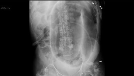
Radiography, Abdominal
Pancreatitis
Colitis, Ischemic
Prospective Studies
Retrospective Studies
Visceral Pain
Intussusception
Follow-Up Studies
Rupture, Spontaneous
Risk Factors
Questionnaires
Colonic Diseases
Neuralgia
Nociceptive Pain
Pancreatitis, Chronic
Analgesics, Opioid
Diarrhea
Flatulence
Fatal Outcome
Severity of Illness Index
Mesenteric Vascular Occlusion
Colic
Laparoscopy
History of Medicine
Hyperalgesia
Cholecystitis
Prevalence
Endoscopy, Gastrointestinal
Double-Blind Method
Peritonitis, Tuberculous
Duodenal Diseases
Headache
Myofascial Pain Syndromes
Physical Examination
Complex Regional Pain Syndromes
Mesenteric Artery, Superior
Purpura, Schoenlein-Henoch
History
Cohort Studies
Splenic Diseases
Gastrointestinal Agents
Gastrointestinal Hemorrhage
Cholangiopancreatography, Endoscopic Retrograde
Celiac Artery
Nausea
Age Factors
Hernia
Predictive Value of Tests
Magnetic Resonance Imaging
Incidence
Abdominal Abscess
Cross-Sectional Studies
Labor Pain
Splenic Infarction
Cysts
Wandering Spleen
Ileocecal Valve
Case-Control Studies
Ulcer
Liver Abscess
Prognosis
Quality of Life
Biopsy
Gastroparesis
Familial Mediterranean Fever
Sex Factors
Urachal Cyst
Bezoars
Pregnancy, Ectopic
Sigmoid Diseases
Intestine, Small
Gastric Dilatation
Psychophysiologic Disorders
Splenic Rupture
Anxiety
Nociceptors
Pregnancy
Lipoma
Postoperative Complications
Risk Assessment
Mesenteric Veins
Common Bile Duct Diseases
Emergency Service, Hospital
Celiac Plexus
Duodenum
Peritonitis
Anti-Inflammatory Agents, Non-Steroidal
Pancreatic Neoplasms
Logistic Models
Barium Sulfate
Feces
Disability Evaluation
Hematoma
Cholangiopancreatography, Magnetic Resonance
Calculi
Pneumoperitoneum
Morphine
Abdominal Neoplasms
Fibromyalgia
Family Health
Pneumatosis Cystoides Intestinalis
Mesentery
Anesthetics, Local
Emergencies
Stomach Volvulus
Tuberculosis, Gastrointestinal
Analysis of Variance
Diverticulum
Pancreatitis, Acute Necrotizing
Anisakiasis
Lactose Intolerance
Jaundice
Catastrophization
Abdominal pain is defined as discomfort or painful sensation in the abdomen. The abdomen is the region of the body between the chest and the pelvis, and contains many important organs such as the stomach, small intestine, large intestine, liver, gallbladder, pancreas, and spleen. Abdominal pain can vary in intensity from mild to severe, and can be acute or chronic depending on the underlying cause.
Abdominal pain can have many different causes, ranging from benign conditions such as gastritis, indigestion, or constipation, to more serious conditions such as appendicitis, inflammatory bowel disease, or abdominal aortic aneurysm. The location, quality, and duration of the pain can provide important clues about its cause. For example, sharp, localized pain in the lower right quadrant of the abdomen may indicate appendicitis, while crampy, diffuse pain in the lower abdomen may suggest irritable bowel syndrome.
It is important to seek medical attention if you experience severe or persistent abdominal pain, especially if it is accompanied by other symptoms such as fever, vomiting, or bloody stools. A thorough physical examination, including a careful history and a focused abdominal exam, can help diagnose the underlying cause of the pain and guide appropriate treatment.
Pain is an unpleasant sensory and emotional experience associated with actual or potential tissue damage, or described in terms of such damage. It is a complex phenomenon that can result from various stimuli, such as thermal, mechanical, or chemical irritation, and it can be acute or chronic. The perception of pain involves the activation of specialized nerve cells called nociceptors, which transmit signals to the brain via the spinal cord. These signals are then processed in different regions of the brain, leading to the conscious experience of pain. It's important to note that pain is a highly individual and subjective experience, and its perception can vary widely among individuals.
Chronic pain is defined as pain that persists or recurs for a period of 3 months or longer, beyond the normal healing time for an injury or illness. It can be continuous or intermittent and range from mild to severe. Chronic pain can have various causes, such as nerve damage, musculoskeletal conditions, or chronic diseases like cancer. It can significantly impact a person's quality of life, causing limitations in mobility, sleep disturbances, mood changes, and decreased overall well-being. Effective management of chronic pain often involves a multidisciplinary approach, including medications, physical therapy, psychological interventions, and complementary therapies.
Pain management is a branch of medicine that focuses on the diagnosis and treatment of pain and improvement in the quality of life of patients with chronic pain. The goal of pain management is to reduce pain levels, improve physical functioning, and help patients cope mentally and emotionally with their pain. This may involve the use of medications, interventional procedures, physical therapy, psychological therapy, or a combination of these approaches.
The definition of pain management can vary depending on the medical context, but it generally refers to a multidisciplinary approach that addresses the complex interactions between biological, psychological, and social factors that contribute to the experience of pain. Pain management specialists may include physicians, nurses, physical therapists, psychologists, and other healthcare professionals who work together to provide comprehensive care for patients with chronic pain.
"Acute abdomen" is a medical term used to describe a sudden and severe abdominal pain that requires immediate medical attention. This condition can be caused by various factors such as inflammation, infection, obstruction, or perforation of the abdominal organs. Common causes of acute abdomen include appendicitis, cholecystitis, diverticulitis, intestinal obstruction, and perforated ulcers.
The symptoms of acute abdomen may include severe and localized or generalized abdominal pain, tenderness, rigidity, rebound tenderness, fever, nausea, vomiting, and loss of appetite. The diagnosis of acute abdomen is usually made based on the patient's history, physical examination, laboratory tests, and imaging studies such as X-rays, ultrasound, or CT scan.
Treatment of acute abdomen depends on the underlying cause and may include antibiotics, intravenous fluids, pain management, and surgery in severe cases. Delayed diagnosis and treatment of acute abdomen can lead to serious complications such as sepsis, peritonitis, and even death.
Pain threshold is a term used in medicine and research to describe the point at which a stimulus begins to be perceived as painful. It is an individual's subjective response and can vary from person to person based on factors such as their pain tolerance, mood, expectations, and cultural background.
The pain threshold is typically determined through a series of tests where gradually increasing levels of stimuli are applied until the individual reports feeling pain. This is often used in research settings to study pain perception and analgesic efficacy. However, it's important to note that the pain threshold should not be confused with pain tolerance, which refers to the maximum level of pain a person can endure.
Postoperative pain is defined as the pain or discomfort experienced by patients following a surgical procedure. It can vary in intensity and duration depending on the type of surgery performed, individual pain tolerance, and other factors. The pain may be caused by tissue trauma, inflammation, or nerve damage resulting from the surgical intervention. Proper assessment and management of postoperative pain is essential to promote recovery, prevent complications, and improve patient satisfaction.
Pain measurement, in a medical context, refers to the quantification or evaluation of the intensity and/or unpleasantness of a patient's subjective pain experience. This is typically accomplished through the use of standardized self-report measures such as numerical rating scales (NRS), visual analog scales (VAS), or categorical scales (mild, moderate, severe). In some cases, physiological measures like heart rate, blood pressure, and facial expressions may also be used to supplement self-reported pain ratings. The goal of pain measurement is to help healthcare providers better understand the nature and severity of a patient's pain in order to develop an effective treatment plan.
Back pain is a common symptom characterized by discomfort or soreness in the back, often occurring in the lower region of the back (lumbago). It can range from a mild ache to a sharp stabbing or shooting pain, and it may be accompanied by stiffness, restricted mobility, and difficulty performing daily activities. Back pain is typically caused by strain or sprain to the muscles, ligaments, or spinal joints, but it can also result from degenerative conditions, disc herniation, spinal stenosis, osteoarthritis, or other medical issues affecting the spine. The severity and duration of back pain can vary widely, with some cases resolving on their own within a few days or weeks, while others may require medical treatment and rehabilitation.
Low back pain is a common musculoskeletal disorder characterized by discomfort or pain in the lower part of the back, typically between the costal margin (bottom of the ribcage) and the gluteal folds (buttocks). It can be caused by several factors including strain or sprain of the muscles or ligaments, disc herniation, spinal stenosis, osteoarthritis, or other degenerative conditions affecting the spine. The pain can range from a dull ache to a sharp stabbing sensation and may be accompanied by stiffness, limited mobility, and radiating pain down the legs in some cases. Low back pain is often described as acute (lasting less than 6 weeks), subacute (lasting between 6-12 weeks), or chronic (lasting more than 12 weeks).
In medicine, "intractable pain" is a term used to describe pain that is difficult to manage, control or relieve with standard treatments. It's a type of chronic pain that continues for an extended period, often months or even years, and does not respond to conventional therapies such as medications, physical therapy, or surgery. Intractable pain can significantly affect a person's quality of life, causing emotional distress, sleep disturbances, and reduced mobility. It is essential to distinguish intractable pain from acute pain, which is typically sharp and short-lived, resulting from tissue damage or inflammation.
Intractable pain may be classified as:
1. Refractory pain: Pain that persists despite optimal treatment with various modalities, including medications, interventions, and multidisciplinary care.
2. Incurable pain: Pain caused by a progressive or incurable disease, such as cancer, for which no curative treatment is available.
3. Functional pain: Pain without an identifiable organic cause that does not respond to standard treatments.
Managing intractable pain often requires a multidisciplinary approach involving healthcare professionals from various fields, including pain specialists, neurologists, psychiatrists, psychologists, and physical therapists. Treatment options may include:
1. Adjuvant medications: Medications that are not primarily analgesics but have been found to help with pain relief, such as antidepressants, anticonvulsants, and muscle relaxants.
2. Interventional procedures: Minimally invasive techniques like nerve blocks, spinal cord stimulation, or intrathecal drug delivery systems that target specific nerves or areas of the body to reduce pain signals.
3. Psychological interventions: Techniques such as cognitive-behavioral therapy (CBT), mindfulness meditation, and relaxation training can help patients cope with chronic pain and improve their overall well-being.
4. Physical therapy and rehabilitation: Exercise programs, massage, acupuncture, and other physical therapies may provide relief for some types of intractable pain.
5. Complementary and alternative medicine (CAM): Techniques like yoga, tai chi, hypnosis, or biofeedback can be helpful in managing chronic pain.
6. Lifestyle modifications: Dietary changes, stress management, and quitting smoking may also contribute to improved pain management.
Neck pain is discomfort or soreness in the neck region, which can extend from the base of the skull to the upper part of the shoulder blades, caused by injury, irritation, or inflammation of the muscles, ligaments, or nerves in the cervical spine. The pain may worsen with movement and can be accompanied by stiffness, numbness, tingling, or weakness in the neck, arms, or hands. In some cases, headaches can also occur as a result of neck pain.
Pelvic pain is defined as discomfort or unpleasant sensation in the lower abdominal region, below the belly button, and between the hips. It can be acute (sudden and lasting for a short time) or chronic (persisting for months or even years), and it may be steady or intermittent, mild or severe. The pain can have various causes, including musculoskeletal issues, nerve irritation, infection, inflammation, or organic diseases in the reproductive, urinary, or gastrointestinal systems. Accurate diagnosis often requires a thorough medical evaluation to determine the underlying cause and develop an appropriate treatment plan.
Acute pain is a type of pain that comes on suddenly and can be severe, but it typically lasts for a short period of time. It is often described as sharp or stabbing and can be caused by tissue damage, inflammation, or injury. Acute pain is the body's way of signaling that something is wrong and that action needs to be taken to address the underlying cause.
Acute pain is different from chronic pain, which is pain that persists for 12 weeks or longer. Chronic pain can be caused by a variety of factors, including ongoing medical conditions, nerve damage, or inflammation. It is important to seek medical attention if you are experiencing acute pain that does not improve or becomes severe, as it may be a sign of a more serious underlying condition.
Pain perception refers to the neural and psychological processes involved in receiving, interpreting, and responding to painful stimuli. It is the subjective experience of pain, which can vary greatly among individuals due to factors such as genetics, mood, expectations, and past experiences. The perception of pain involves complex interactions between the peripheral nervous system (which detects and transmits information about tissue damage or potential harm), the spinal cord (where this information is processed and integrated with other sensory inputs), and the brain (where the final interpretation and emotional response to pain occurs).
Appendicitis is a medical condition characterized by inflammation of the appendix, a small finger-like structure that projects from the colon located in the lower right abdomen. The appendix doesn't have a known function, and its removal (appendectomy) does not appear to affect a person's health.
The inflammation of the appendix can be caused by various factors, such as obstruction due to hardened stool, foreign bodies, or tumors. The blockage can lead to increased pressure within the appendix, reduced blood flow, and bacterial growth, resulting in infection and inflammation. If left untreated, appendicitis can progress to peritonitis (inflammation of the lining of the abdominal cavity) or even sepsis, a life-threatening condition.
Common symptoms of appendicitis include:
* Sudden onset of pain in the lower right abdomen, which may start around the navel and shift to the lower right side over several hours
* Pain that worsens with movement, coughing, or sneezing
* Nausea and vomiting
* Loss of appetite
* Fever and chills
* Constipation or diarrhea
* Abdominal swelling or bloating
If you suspect appendicitis, it's essential to seek immediate medical attention. The standard treatment for appendicitis is surgical removal of the appendix (appendectomy), which can be performed as an open surgery or laparoscopically. Antibiotics are also administered to treat any existing infection. Delaying treatment can lead to serious complications, so it's crucial not to ignore symptoms and seek medical help promptly.
Facial pain is a condition characterized by discomfort or pain felt in any part of the face. It can result from various causes, including nerve damage or irritation, injuries, infections, dental problems, migraines, or sinus congestion. The pain can range from mild to severe and may be sharp, dull, constant, or intermittent. In some cases, facial pain can also be associated with other symptoms such as headaches, redness, swelling, or changes in sensation. Accurate diagnosis and treatment of the underlying cause are essential for effective management of facial pain.
Irritable Bowel Syndrome (IBS) is a functional gastrointestinal disorder characterized by recurrent abdominal pain, bloating, and altered bowel habits in the absence of any structural or biochemical abnormalities. The symptoms can vary from person to person, ranging from mild to severe.
The exact cause of IBS is not known, but it's thought to involve a combination of factors such as muscle contractions in the intestine, abnormalities in the nervous system, inflammation in the intestines, severe infection, or changes in bacteria in the gut.
It's important to note that while IBS can cause great discomfort and distress, it does not lead to serious complications such as changes in bowel tissue or increased risk of colorectal cancer. However, it can significantly affect a person's quality of life and daily activities.
Gastrointestinal diseases refer to a group of conditions that affect the gastrointestinal (GI) tract, which includes the organs from the mouth to the anus, responsible for food digestion, absorption, and elimination of waste. These diseases can affect any part of the GI tract, causing various symptoms such as abdominal pain, bloating, diarrhea, constipation, nausea, vomiting, and weight loss.
Common gastrointestinal diseases include:
1. Gastroesophageal reflux disease (GERD) - a condition where stomach acid flows back into the esophagus, causing heartburn and other symptoms.
2. Peptic ulcers - sores that develop in the lining of the stomach or duodenum, often caused by bacterial infection or long-term use of nonsteroidal anti-inflammatory drugs (NSAIDs).
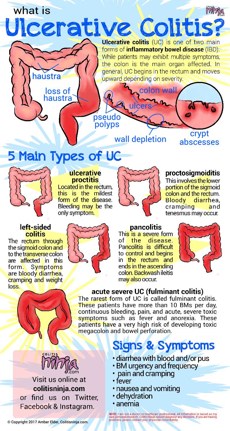
3. Inflammatory bowel disease (IBD) - a group of chronic inflammatory conditions of the intestine, including Crohn's disease and ulcerative colitis.
4. Irritable bowel syndrome (IBS) - a functional gastrointestinal disorder characterized by abdominal pain, bloating, and altered bowel habits.
5. Celiac disease - an autoimmune disorder where the ingestion of gluten leads to damage in the small intestine.
6. Diverticular disease - a condition that affects the colon, causing diverticula (small pouches) to form and potentially become inflamed or infected.
7. Constipation - a common gastrointestinal symptom characterized by infrequent bowel movements, hard stools, and difficulty passing stools.
8. Diarrhea - a common gastrointestinal symptom characterized by loose, watery stools and frequent bowel movements.
9. Food intolerances and allergies - adverse reactions to specific foods or food components that can cause various gastrointestinal symptoms.
10. Gastrointestinal infections - caused by bacteria, viruses, parasites, or fungi that can lead to a range of symptoms, including diarrhea, vomiting, and abdominal pain.
Referred pain is a type of pain that is felt in a part of the body other than its actual source. This occurs because the brain incorrectly interprets nerve signals from damaged tissues or organs. In the case of referred pain, the brain misinterprets the location of the pain signal and attributes it to a different area of the body.
Referred pain is often described as a dull, aching sensation rather than a sharp, stabbing pain. It can be difficult to diagnose because the source of the pain may not be immediately apparent. Common examples of referred pain include:
* Heart attack pain that is felt in the left arm or jaw
* Gallbladder pain that is felt in the right shoulder blade
* Kidney stones that cause pain in the lower back and abdomen
* Appendicitis that causes pain in the lower right quadrant of the abdomen, but can sometimes be referred to the lower left quadrant in pregnant women or those with a longer colon.
Referred pain is thought to occur because the nerves carrying pain signals from different parts of the body converge on the same neurons in the spinal cord before traveling to the brain. If these neurons are stimulated by pain signals from multiple sources, the brain may have difficulty distinguishing between them and may interpret the pain as coming from a single location.
Medical history taking is the process of obtaining and documenting a patient's health information through a series of questions and observations. It is a critical component of the medical assessment and helps healthcare providers understand the patient's current health status, past medical conditions, medications, allergies, lifestyle habits, and family medical history.
The information gathered during medical history taking is used to make informed decisions about diagnosis, treatment, and management plans for the patient's care. The process typically includes asking open-ended questions, actively listening to the patient's responses, clarifying any uncertainties, and documenting the findings in a clear and concise manner.
Medical history taking can be conducted in various settings, including hospitals, clinics, or virtual consultations, and may be performed by physicians, nurses, or other healthcare professionals. It is essential to ensure that medical history taking is conducted in a private and confidential setting to protect the patient's privacy and maintain trust in the provider-patient relationship.
Constipation is a condition characterized by infrequent bowel movements or difficulty in passing stools that are often hard and dry. The medical definition of constipation varies, but it is generally defined as having fewer than three bowel movements in a week. In addition to infrequent bowel movements, other symptoms of constipation can include straining during bowel movements, feeling like you haven't completely evacuated your bowels, and experiencing hard or lumpy stools.
Constipation can have many causes, including a low-fiber diet, dehydration, certain medications, lack of physical activity, and underlying medical conditions such as irritable bowel syndrome or hypothyroidism. In most cases, constipation can be treated with lifestyle changes, such as increasing fiber intake, drinking more water, and getting regular exercise. However, if constipation is severe, persistent, or accompanied by other symptoms, it's important to seek medical attention to rule out any underlying conditions that may require treatment.
Ileal diseases refer to conditions that primarily affect the ileum, which is the final portion of the small intestine. The ileum plays a crucial role in nutrient absorption, particularly vitamin B12 and bile salts. Ileal diseases can cause various symptoms, including diarrhea, abdominal pain, weight loss, and malnutrition, depending on their nature and extent. Some common ileal diseases include:
1. Crohn's disease: A type of inflammatory bowel disease (IBD) that can affect any part of the gastrointestinal tract, including the ileum. Crohn's disease causes chronic inflammation, which can lead to symptoms such as diarrhea, abdominal pain, and fatigue.
2. Celiac disease: An autoimmune disorder triggered by gluten ingestion in genetically susceptible individuals. In celiac disease, the immune system attacks the lining of the small intestine, including the ileum, causing inflammation and impaired nutrient absorption.
3. Intestinal tuberculosis: A bacterial infection caused by Mycobacterium tuberculosis that can affect any part of the gastrointestinal tract, including the ileum. Intestinal tuberculosis can cause symptoms such as abdominal pain, diarrhea, and weight loss.
4. Typhlitis: Also known as neutropenic enterocolitis, typhlitis is an inflammatory condition that affects the cecum and terminal ileum, typically in immunocompromised individuals. It can cause symptoms such as abdominal pain, fever, and diarrhea.
5. Meckel's diverticulum: A congenital condition characterized by a small pouch protruding from the wall of the ileum. While many people with Meckel's diverticulum do not experience symptoms, it can sometimes become inflamed or bleed, causing abdominal pain and rectal bleeding.
6. Lymphoma: A type of cancer that originates in the lymphatic system and can affect any part of the body, including the ileum. Ileal lymphoma can cause symptoms such as abdominal pain, diarrhea, and weight loss.
X-ray computed tomography (CT or CAT scan) is a medical imaging method that uses computer-processed combinations of many X-ray images taken from different angles to produce cross-sectional (tomographic) images (virtual "slices") of the body. These cross-sectional images can then be used to display detailed internal views of organs, bones, and soft tissues in the body.
The term "computed tomography" is used instead of "CT scan" or "CAT scan" because the machines take a series of X-ray measurements from different angles around the body and then use a computer to process these data to create detailed images of internal structures within the body.
CT scanning is a noninvasive, painless medical test that helps physicians diagnose and treat medical conditions. CT imaging provides detailed information about many types of tissue including lung, bone, soft tissue and blood vessels. CT examinations can be performed on every part of the body for a variety of reasons including diagnosis, surgical planning, and monitoring of therapeutic responses.
In computed tomography (CT), an X-ray source and detector rotate around the patient, measuring the X-ray attenuation at many different angles. A computer uses this data to construct a cross-sectional image by the process of reconstruction. This technique is called "tomography". The term "computed" refers to the use of a computer to reconstruct the images.
CT has become an important tool in medical imaging and diagnosis, allowing radiologists and other physicians to view detailed internal images of the body. It can help identify many different medical conditions including cancer, heart disease, lung nodules, liver tumors, and internal injuries from trauma. CT is also commonly used for guiding biopsies and other minimally invasive procedures.
In summary, X-ray computed tomography (CT or CAT scan) is a medical imaging technique that uses computer-processed combinations of many X-ray images taken from different angles to produce cross-sectional images of the body. It provides detailed internal views of organs, bones, and soft tissues in the body, allowing physicians to diagnose and treat medical conditions.
Treatment outcome is a term used to describe the result or effect of medical treatment on a patient's health status. It can be measured in various ways, such as through symptoms improvement, disease remission, reduced disability, improved quality of life, or survival rates. The treatment outcome helps healthcare providers evaluate the effectiveness of a particular treatment plan and make informed decisions about future care. It is also used in clinical research to compare the efficacy of different treatments and improve patient care.
Shoulder pain is a condition characterized by discomfort or hurt in the shoulder joint, muscles, tendons, ligaments, or surrounding structures. The shoulder is one of the most mobile joints in the body, and this mobility makes it prone to injury and pain. Shoulder pain can result from various causes, including overuse, trauma, degenerative conditions, or referred pain from other areas of the body.
The shoulder joint is a ball-and-socket joint made up of three bones: the humerus (upper arm bone), scapula (shoulder blade), and clavicle (collarbone). The rotator cuff, a group of four muscles that surround and stabilize the shoulder joint, can also be a source of pain if it becomes inflamed or torn.
Shoulder pain can range from mild to severe, and it may be accompanied by stiffness, swelling, bruising, weakness, numbness, tingling, or reduced mobility in the affected arm. The pain may worsen with movement, lifting objects, or performing certain activities, such as reaching overhead or behind the back.
Medical evaluation is necessary to determine the underlying cause of shoulder pain and develop an appropriate treatment plan. Treatment options may include rest, physical therapy, medication, injections, or surgery, depending on the severity and nature of the condition.
The abdomen refers to the portion of the body that lies between the thorax (chest) and the pelvis. It is a musculo-fascial cavity containing the digestive, urinary, and reproductive organs. The abdominal cavity is divided into several regions and quadrants for medical description and examination purposes. These include the upper and lower abdomen, as well as nine quadrants formed by the intersection of the midline and a horizontal line drawn at the level of the umbilicus (navel).
The major organs located within the abdominal cavity include:
1. Stomach - muscular organ responsible for initial digestion of food
2. Small intestine - long, coiled tube where most nutrient absorption occurs
3. Large intestine - consists of the colon and rectum; absorbs water and stores waste products
4. Liver - largest internal organ, involved in protein synthesis, detoxification, and metabolism
5. Pancreas - secretes digestive enzymes and hormones such as insulin
6. Spleen - filters blood and removes old red blood cells
7. Kidneys - pair of organs responsible for filtering waste products from the blood and producing urine
8. Adrenal glands - sit atop each kidney, produce hormones that regulate metabolism, immune response, and stress response
The abdomen is an essential part of the human body, playing a crucial role in digestion, absorption, and elimination of food and waste materials, as well as various metabolic processes.
A "torsion abnormality" is not a standard medical term, but I believe you are asking about torsional deformities or abnormalities related to torsion. Torsion refers to a twisting force or movement that can cause structures to rotate around their long axis. In the context of medical definitions:
Torsional abnormality could refer to a congenital or acquired condition where anatomical structures, such as blood vessels, muscles, tendons, or bones, are twisted or rotated in an abnormal way. This can lead to various complications depending on the structure involved and the degree of torsion.
For instance, in congenital torsional deformities of long bones (like tibia or femur), the rotation of the bone axis can cause issues with gait, posture, and joint function. In some cases, this may require surgical intervention to correct the abnormality.
In the context of vascular torsion abnormalities, such as mesenteric torsion, it could lead to bowel ischemia due to the twisting of blood vessels that supply the intestines. This can be a surgical emergency and requires immediate intervention to restore blood flow and prevent further damage.
It's essential to consult with a medical professional for a precise diagnosis and treatment options if you or someone else experiences symptoms related to torsional abnormalities.
Musculoskeletal pain is discomfort or pain that affects the muscles, bones, ligaments, tendons, and nerves. It can be caused by injury, overuse, or disease and can affect any part of the body, including the neck, back, shoulders, hips, and extremities. The pain can range from mild to severe and may be accompanied by stiffness, swelling, and decreased range of motion. Common causes of musculoskeletal pain include arthritis, fibromyalgia, tendinitis, bursitis, and muscle or ligament strain. Treatment for musculoskeletal pain depends on the underlying cause and may include physical therapy, medication, and in some cases, surgery.
Functional colonic diseases are a group of disorders of the large intestine (colon) that do not have a structural or biochemical explanation. They are characterized by chronic and often intermittent symptoms, such as abdominal pain, bloating, and changes in bowel habits, but do not show any visible abnormalities or damage to the tissue of the colon during routine examination or testing.
The most common functional colonic diseases include:
1. Irritable Bowel Syndrome (IBS): A disorder characterized by recurrent abdominal pain, bloating, and changes in bowel habits, such as constipation or diarrhea.
2. Functional Constipation: A condition where a person experiences difficult or infrequent bowel movements, but there is no obvious structural or biochemical cause.
3. Functional Diarrhea: A disorder characterized by frequent loose stools, but without any underlying structural or biochemical abnormalities.
4. Abdominal Bloating: A condition where the belly feels full and tight, often accompanied by discomfort or pain, but without any visible distention.
5. Functional Abdominal Pain Syndrome: A disorder characterized by chronic or recurrent abdominal pain that is not associated with any structural or biochemical abnormalities.
The exact cause of functional colonic diseases is unknown, but they are believed to be related to a combination of factors, including genetics, environmental factors, altered gut motility, visceral hypersensitivity, and psychological factors such as stress and anxiety. Treatment typically involves lifestyle modifications, such as changes in diet and exercise, and medication to manage symptoms.
A chronic disease is a long-term medical condition that often progresses slowly over a period of years and requires ongoing management and care. These diseases are typically not fully curable, but symptoms can be managed to improve quality of life. Common chronic diseases include heart disease, stroke, cancer, diabetes, arthritis, and COPD (chronic obstructive pulmonary disease). They are often associated with advanced age, although they can also affect children and younger adults. Chronic diseases can have significant impacts on individuals' physical, emotional, and social well-being, as well as on healthcare systems and society at large.
Intestinal obstruction, also known as bowel obstruction, is a medical condition characterized by a blockage that prevents the normal flow of contents through the small intestine or large intestine (colon). This blockage can be caused by various factors such as tumors, adhesions (scar tissue), hernias, inflammation, or impacted feces.
The obstruction can be mechanical, where something physically blocks the intestinal lumen, or functional, where the normal muscular contractions of the bowel are impaired. Mechanical obstructions are more common than functional ones.
Symptoms of intestinal obstruction may include abdominal pain and cramping, nausea and vomiting, bloating, inability to pass gas or have a bowel movement, and abdominal distention. If left untreated, intestinal obstruction can lead to serious complications such as tissue death (necrosis), perforation of the intestine, and sepsis. Treatment typically involves hospitalization, intravenous fluids, nasogastric decompression, and possibly surgery to remove the obstruction.
Abdominal radiography, also known as a KUB (kidneys, ureters, bladder) X-ray, is a medical imaging technique used to examine the abdominal cavity. It involves using ionizing radiation to produce images of the internal structures of the abdomen, including the bones, organs, and soft tissues.
The procedure typically involves the patient lying down on a table while a specialized X-ray machine captures images of the abdomen from different angles. The images produced can help doctors diagnose and monitor a variety of conditions, such as kidney stones, intestinal obstructions, and abnormalities in the spine or other bones.
Abdominal radiography is a quick, painless, and non-invasive procedure that requires little preparation on the part of the patient. However, it does involve exposure to radiation, so it is typically only used when necessary and when other imaging techniques are not appropriate.
Pancreatitis is a medical condition characterized by inflammation of the pancreas, a gland located in the abdomen that plays a crucial role in digestion and regulating blood sugar levels. The inflammation can be acute (sudden and severe) or chronic (persistent and recurring), and it can lead to various complications if left untreated.
Acute pancreatitis often results from gallstones or excessive alcohol consumption, while chronic pancreatitis may be caused by long-term alcohol abuse, genetic factors, autoimmune conditions, or metabolic disorders like high triglyceride levels. Symptoms of acute pancreatitis include severe abdominal pain, nausea, vomiting, fever, and increased heart rate, while chronic pancreatitis may present with ongoing abdominal pain, weight loss, diarrhea, and malabsorption issues due to impaired digestive enzyme production. Treatment typically involves supportive care, such as intravenous fluids, pain management, and addressing the underlying cause. In severe cases, hospitalization and surgery may be necessary.
Ischemic colitis is a condition characterized by inflammation of the large intestine (colon) due to reduced blood flow to the area. This reduction in blood flow, also known as ischemia, can be caused by various factors such as narrowing or blockage of the blood vessels that supply the colon, low blood pressure, or certain medications.
Symptoms of ischemic colitis may include sudden abdominal pain, bloody diarrhea, nausea, vomiting, and fever. In severe cases, it can lead to tissue death, perforation of the colon, and sepsis. Treatment typically involves supportive care such as fluid replacement, bowel rest, and antibiotics. In some cases, surgery may be necessary to remove damaged tissue or restore blood flow to the area.
Prospective studies, also known as longitudinal studies, are a type of cohort study in which data is collected forward in time, following a group of individuals who share a common characteristic or exposure over a period of time. The researchers clearly define the study population and exposure of interest at the beginning of the study and follow up with the participants to determine the outcomes that develop over time. This type of study design allows for the investigation of causal relationships between exposures and outcomes, as well as the identification of risk factors and the estimation of disease incidence rates. Prospective studies are particularly useful in epidemiology and medical research when studying diseases with long latency periods or rare outcomes.
Retrospective studies, also known as retrospective research or looking back studies, are a type of observational study that examines data from the past to draw conclusions about possible causal relationships between risk factors and outcomes. In these studies, researchers analyze existing records, medical charts, or previously collected data to test a hypothesis or answer a specific research question.
Retrospective studies can be useful for generating hypotheses and identifying trends, but they have limitations compared to prospective studies, which follow participants forward in time from exposure to outcome. Retrospective studies are subject to biases such as recall bias, selection bias, and information bias, which can affect the validity of the results. Therefore, retrospective studies should be interpreted with caution and used primarily to generate hypotheses for further testing in prospective studies.
Intestinal perforation is a medical condition that refers to a hole or tear in the lining of the intestine. This can occur anywhere along the gastrointestinal tract, including the small intestine, large intestine (colon), or stomach. Intestinal perforation allows the contents of the intestines, such as digestive enzymes and bacteria, to leak into the abdominal cavity, which can lead to a serious inflammatory response known as peritonitis.
Intestinal perforation can be caused by various factors, including:
* Mechanical trauma (e.g., gunshot wounds, stab wounds)
* Inflammatory bowel disease (e.g., Crohn's disease, ulcerative colitis)
* Diverticulitis
* Appendicitis
* Intestinal obstruction
* Infections (e.g., typhoid fever, tuberculosis)
* Certain medications (e.g., nonsteroidal anti-inflammatory drugs, corticosteroids)
* Radiation therapy
* Ischemic bowel disease (lack of blood flow to the intestines)
Symptoms of intestinal perforation may include sudden abdominal pain, nausea, vomiting, fever, and decreased bowel movements. Treatment typically involves surgery to repair the perforation and remove any damaged tissue. Antibiotics are also administered to prevent infection. In severe cases, a temporary or permanent colostomy or ileostomy may be necessary.
Analgesics are a class of drugs that are used to relieve pain. They work by blocking the transmission of pain signals in the nervous system, allowing individuals to manage their pain levels more effectively. There are many different types of analgesics available, including both prescription and over-the-counter options. Some common examples include acetaminophen (Tylenol), ibuprofen (Advil or Motrin), and opioids such as morphine or oxycodone.
The choice of analgesic will depend on several factors, including the type and severity of pain being experienced, any underlying medical conditions, potential drug interactions, and individual patient preferences. It is important to use these medications as directed by a healthcare provider, as misuse or overuse can lead to serious side effects and potential addiction.
In addition to their pain-relieving properties, some analgesics may also have additional benefits such as reducing inflammation (like in the case of nonsteroidal anti-inflammatory drugs or NSAIDs) or causing sedation (as with certain opioids). However, it is essential to weigh these potential benefits against the risks and side effects associated with each medication.
When used appropriately, analgesics can significantly improve a person's quality of life by helping them manage their pain effectively and allowing them to engage in daily activities more comfortably.
Visceral pain is a type of pain that originates from the internal organs (viscera) such as the stomach, intestines, liver, or heart. It's often described as diffuse, dull, and hard to localize, unlike somatic pain which arises from the skin, muscles, or bones and is usually easier to pinpoint.
Visceral pain may be caused by various conditions like inflammation, infection, ischemia (reduced blood supply), distention or stretching of the organ walls, or direct damage to the organs. The sensation of visceral pain can be modulated and referred to other areas of the body due to the complex interactions in the nervous system, making it sometimes challenging to diagnose the exact source of the pain.
I'm sorry for any confusion, but "History, 20th Century" is a broad and complex topic that refers to the events, developments, and transformations that occurred throughout the world during the 1900s. It is not a medical term or concept. If you're interested in learning more about this historical period, I would recommend consulting a history textbook, reputable online resources, or speaking with a historian. They can provide detailed information about the political, social, economic, and cultural changes that took place during the 20th century.
Cecal diseases refer to medical conditions that affect the cecum, which is a pouch-like structure located at the junction of the small and large intestines. The cecum plays an important role in digestion, particularly in the fermentation of certain types of food.
There are several different types of cecal diseases, including:
1. Cecal volvulus: This is a rare condition in which the cecum twists on itself, cutting off blood flow and causing severe pain and other symptoms.
2. Diverticulitis: This occurs when small pouches called diverticula form in the wall of the cecum and become inflamed or infected.
3. Appendicitis: Although not strictly a cecal disease, the appendix is a small tube-like structure that branches off from the cecum. Inflammation of the appendix (appendicitis) can cause severe pain in the lower right abdomen and may require surgical removal of the appendix.
4. Crohn's disease: This is a chronic inflammatory bowel disease that can affect any part of the digestive tract, including the cecum.
5. Tuberculosis: The cecum can also be affected by tuberculosis, which is a bacterial infection that primarily affects the lungs but can spread to other parts of the body.
6. Cancer: Although rare, cancer can also affect the cecum, leading to symptoms such as abdominal pain, bloating, and changes in bowel habits.
Treatment for cecal diseases depends on the specific condition and its severity. Treatment options may include antibiotics, surgery, or other medical interventions. If you are experiencing symptoms that may be related to a cecal disease, it is important to seek medical attention promptly.
Intussusception is a medical condition in which a part of the intestine telescopes into an adjacent section, leading to bowel obstruction and reduced blood flow. It often affects children under 3 years old but can also occur in adults. If not treated promptly, it can result in serious complications such as perforation, peritonitis, or even death. The exact cause is usually unknown, but it may be associated with infections, intestinal disorders, or tumors.
Follow-up studies are a type of longitudinal research that involve repeated observations or measurements of the same variables over a period of time, in order to understand their long-term effects or outcomes. In medical context, follow-up studies are often used to evaluate the safety and efficacy of medical treatments, interventions, or procedures.
In a typical follow-up study, a group of individuals (called a cohort) who have received a particular treatment or intervention are identified and then followed over time through periodic assessments or data collection. The data collected may include information on clinical outcomes, adverse events, changes in symptoms or functional status, and other relevant measures.
The results of follow-up studies can provide important insights into the long-term benefits and risks of medical interventions, as well as help to identify factors that may influence treatment effectiveness or patient outcomes. However, it is important to note that follow-up studies can be subject to various biases and limitations, such as loss to follow-up, recall bias, and changes in clinical practice over time, which must be carefully considered when interpreting the results.
Jejunal diseases refer to a range of medical conditions that affect the jejunum, which is the middle section of the small intestine. These diseases can cause various symptoms such as abdominal pain, diarrhea, bloating, nausea, vomiting, and weight loss. Some examples of jejunal diseases include:
1. Jejunal inflammation or infection (jejunitis)
2. Crohn's disease, which can affect any part of the gastrointestinal tract including the jejunum
3. Intestinal lymphoma, a type of cancer that can develop in the small intestine
4. Celiac disease, an autoimmune disorder that causes damage to the small intestine when gluten is consumed
5. Intestinal bacterial overgrowth (SIBO), which can occur due to various reasons including structural abnormalities or motility disorders of the jejunum
6. Meckel's diverticulum, a congenital condition where a small pouch protrudes from the wall of the intestine, usually located in the ileum but can also affect the jejunum
7. Intestinal strictures or obstructions caused by scarring, adhesions, or tumors
8. Radiation enteritis, damage to the small intestine caused by radiation therapy for cancer treatment.
The diagnosis and management of jejunal diseases depend on the specific condition and its severity. Treatment options may include medications, dietary modifications, surgery, or a combination of these approaches.
Spontaneous rupture in medical terms refers to the sudden breaking or tearing of an organ, tissue, or structure within the body without any identifiable trauma or injury. This event can occur due to various reasons such as weakening of the tissue over time because of disease or degeneration, or excessive pressure on the tissue.
For instance, a spontaneous rupture of the appendix is called an "appendiceal rupture," which can lead to peritonitis, a serious inflammation of the abdominal cavity. Similarly, a spontaneous rupture of a blood vessel, like an aortic aneurysm, can result in life-threatening internal bleeding.
Spontaneous ruptures are often medical emergencies and require immediate medical attention for proper diagnosis and treatment.
Medical Definition:
"Risk factors" are any attribute, characteristic or exposure of an individual that increases the likelihood of developing a disease or injury. They can be divided into modifiable and non-modifiable risk factors. Modifiable risk factors are those that can be changed through lifestyle choices or medical treatment, while non-modifiable risk factors are inherent traits such as age, gender, or genetic predisposition. Examples of modifiable risk factors include smoking, alcohol consumption, physical inactivity, and unhealthy diet, while non-modifiable risk factors include age, sex, and family history. It is important to note that having a risk factor does not guarantee that a person will develop the disease, but rather indicates an increased susceptibility.
A questionnaire in the medical context is a standardized, systematic, and structured tool used to gather information from individuals regarding their symptoms, medical history, lifestyle, or other health-related factors. It typically consists of a series of written questions that can be either self-administered or administered by an interviewer. Questionnaires are widely used in various areas of healthcare, including clinical research, epidemiological studies, patient care, and health services evaluation to collect data that can inform diagnosis, treatment planning, and population health management. They provide a consistent and organized method for obtaining information from large groups or individual patients, helping to ensure accurate and comprehensive data collection while minimizing bias and variability in the information gathered.
Colonic diseases refer to a group of medical conditions that affect the colon, also known as the large intestine or large bowel. The colon is the final segment of the digestive system, responsible for absorbing water and electrolytes, and storing and eliminating waste products.
Some common colonic diseases include:
1. Inflammatory bowel disease (IBD): This includes conditions such as Crohn's disease and ulcerative colitis, which cause inflammation and irritation in the lining of the digestive tract.
2. Diverticular disease: This occurs when small pouches called diverticula form in the walls of the colon, leading to symptoms such as abdominal pain, bloating, and changes in bowel movements.
3. Colorectal cancer: This is a type of cancer that develops in the colon or rectum, often starting as benign polyps that grow and become malignant over time.
4. Irritable bowel syndrome (IBS): This is a functional gastrointestinal disorder characterized by abdominal pain, bloating, and changes in bowel movements, but without any underlying structural or inflammatory causes.
5. Constipation: This is a common condition characterized by infrequent bowel movements, difficulty passing stools, or both.
6. Infectious colitis: This occurs when the colon becomes infected with bacteria, viruses, or parasites, leading to symptoms such as diarrhea, abdominal cramps, and fever.
Treatment for colonic diseases varies depending on the specific condition and its severity. Treatment options may include medications, lifestyle changes, surgery, or a combination of these approaches.
An acute disease is a medical condition that has a rapid onset, develops quickly, and tends to be short in duration. Acute diseases can range from minor illnesses such as a common cold or flu, to more severe conditions such as pneumonia, meningitis, or a heart attack. These types of diseases often have clear symptoms that are easy to identify, and they may require immediate medical attention or treatment.
Acute diseases are typically caused by an external agent or factor, such as a bacterial or viral infection, a toxin, or an injury. They can also be the result of a sudden worsening of an existing chronic condition. In general, acute diseases are distinct from chronic diseases, which are long-term medical conditions that develop slowly over time and may require ongoing management and treatment.
Examples of acute diseases include:
* Acute bronchitis: a sudden inflammation of the airways in the lungs, often caused by a viral infection.
* Appendicitis: an inflammation of the appendix that can cause severe pain and requires surgical removal.
* Gastroenteritis: an inflammation of the stomach and intestines, often caused by a viral or bacterial infection.
* Migraine headaches: intense headaches that can last for hours or days, and are often accompanied by nausea, vomiting, and sensitivity to light and sound.
* Myocardial infarction (heart attack): a sudden blockage of blood flow to the heart muscle, often caused by a buildup of plaque in the coronary arteries.
* Pneumonia: an infection of the lungs that can cause coughing, chest pain, and difficulty breathing.
* Sinusitis: an inflammation of the sinuses, often caused by a viral or bacterial infection.
It's important to note that while some acute diseases may resolve on their own with rest and supportive care, others may require medical intervention or treatment to prevent complications and promote recovery. If you are experiencing symptoms of an acute disease, it is always best to seek medical attention to ensure proper diagnosis and treatment.
Recurrence, in a medical context, refers to the return of symptoms or signs of a disease after a period of improvement or remission. It indicates that the condition has not been fully eradicated and may require further treatment. Recurrence is often used to describe situations where a disease such as cancer comes back after initial treatment, but it can also apply to other medical conditions. The likelihood of recurrence varies depending on the type of disease and individual patient factors.
A laparotomy is a surgical procedure that involves making an incision in the abdominal wall to gain access to the abdominal cavity. This procedure is typically performed to diagnose and treat various conditions such as abdominal trauma, tumors, infections, or inflammatory diseases. The size of the incision can vary depending on the reason for the surgery and the extent of the condition being treated. Once the procedure is complete, the incision is closed with sutures or staples.
The term "laparotomy" comes from the Greek words "lapara," which means "flank" or "side," and "tome," which means "to cut." Together, they describe the surgical procedure that involves cutting into the abdomen to examine its contents.
"History, 19th Century" is not a medical term or concept. It refers to the historical events, developments, and figures related to the 1800s in various fields, including politics, culture, science, and technology. However, if you are looking for medical advancements during the 19th century, here's a brief overview:
The 19th century was a period of significant progress in medicine, with numerous discoveries and innovations that shaped modern medical practices. Some notable developments include:
1. Edward Jenner's smallpox vaccine (1796): Although not strictly within the 19th century, Jenner's discovery laid the foundation for vaccination as a preventive measure against infectious diseases.
2. Germ theory of disease: The work of Louis Pasteur, Robert Koch, and others established that many diseases were caused by microorganisms, leading to the development of antiseptic practices and vaccines.
3. Anesthesia: In 1842, Crawford Long first used ether as an anesthetic during surgery, followed by the introduction of chloroform in 1847 by James Simpson.
4. Antisepsis and asepsis: Joseph Lister introduced antiseptic practices in surgery, significantly reducing postoperative infections. Later, the concept of asepsis (sterilization) was developed to prevent contamination during surgical procedures.
5. Microbiology: The development of techniques for culturing and staining bacteria allowed for better understanding and identification of pathogens.
6. Physiology: Claude Bernard's work on the regulation of internal body functions, or homeostasis, contributed significantly to our understanding of human physiology.
7. Neurology: Jean-Martin Charcot made significant contributions to the study of neurological disorders, including multiple sclerosis and Parkinson's disease.
8. Psychiatry: Sigmund Freud developed psychoanalysis, a new approach to understanding mental illnesses.
9. Public health: The 19th century saw the establishment of public health organizations and initiatives aimed at improving sanitation, water quality, and vaccination programs.
10. Medical education reforms: The Flexner Report in 1910 led to significant improvements in medical education standards and practices.
Neuralgia is a type of pain that occurs along the pathway of a nerve, often caused by damage or irritation to the nerve. It is typically described as a sharp, stabbing, burning, or electric-shock like pain that can be severe and debilitating. Neuralgia can affect any nerve in the body, but it most commonly occurs in the facial area (trigeminal neuralgia) or in the nerves related to the spine (postherpetic neuralgia). The pain associated with neuralgia can be intermittent or constant and may be worsened by certain triggers such as touch, temperature changes, or movement. Treatment for neuralgia typically involves medications to manage pain, as well as other therapies such as nerve blocks, surgery, or lifestyle modifications.
Nociceptive pain is a type of pain that results from the activation of nociceptors, which are specialized sensory receptors located in various tissues throughout the body. These receptors detect potentially harmful stimuli such as extreme temperatures, pressure, or chemical irritants and transmit signals to the brain, which interprets them as painful sensations.
Nociceptive pain can be further classified into two categories:
1. Somatic nociceptive pain: This type of pain arises from the activation of nociceptors in the skin, muscles, bones, and joints. It is often described as sharp, aching, or throbbing and may be localized to a specific area of the body.
2. Visceral nociceptive pain: This type of pain arises from the activation of nociceptors in the internal organs, such as the lungs, heart, and digestive system. It is often described as deep, cramping, or aching and may be more diffuse and difficult to localize.
Examples of conditions that can cause nociceptive pain include injuries, arthritis, cancer, and infections. Effective management of nociceptive pain typically involves a multimodal approach that includes pharmacologic interventions, such as non-opioid analgesics, opioids, and adjuvant medications, as well as non-pharmacologic therapies, such as physical therapy, acupuncture, and cognitive-behavioral therapy.
Chronic pancreatitis is a long-standing inflammation of the pancreas that leads to irreversible structural changes and impaired function of the pancreas. It is characterized by recurrent or persistent abdominal pain, often radiating to the back, and maldigestion with steatorrhea (fatty stools) due to exocrine insufficiency. The pancreatic damage results from repeated episodes of acute pancreatitis, alcohol abuse, genetic predisposition, or autoimmune processes. Over time, the pancreas may lose its ability to produce enough digestive enzymes and hormones like insulin, which can result in diabetes mellitus. Chronic pancreatitis also increases the risk of developing pancreatic cancer.
Analgesics, opioid are a class of drugs used for the treatment of pain. They work by binding to specific receptors in the brain and spinal cord, blocking the transmission of pain signals to the brain. Opioids can be synthetic or natural, and include drugs such as morphine, codeine, oxycodone, hydrocodone, hydromorphone, fentanyl, and methadone. They are often used for moderate to severe pain, such as that resulting from injury, surgery, or chronic conditions like cancer. However, opioids can also produce euphoria, physical dependence, and addiction, so they are tightly regulated and carry a risk of misuse.
Diarrhea is a condition in which an individual experiences loose, watery stools frequently, often exceeding three times a day. It can be acute, lasting for several days, or chronic, persisting for weeks or even months. Diarrhea can result from various factors, including viral, bacterial, or parasitic infections, food intolerances, medications, and underlying medical conditions such as inflammatory bowel disease or irritable bowel syndrome. Dehydration is a potential complication of diarrhea, particularly in severe cases or in vulnerable populations like young children and the elderly.
An appendectomy is a surgical procedure in which the vermiform appendix is removed. This procedure is performed when a patient has appendicitis, which is an inflammation of the appendix that can lead to serious complications such as peritonitis or sepsis if not treated promptly. The surgery can be done as an open procedure, in which a single incision is made in the lower right abdomen, or as a laparoscopic procedure, in which several small incisions are made and specialized instruments are used to remove the appendix. In some cases, if the appendix has burst, a more extensive surgery may be required to clean out the abdominal cavity.
Flatulence is the medical term for the release of intestinal gas from the rectum, commonly known as passing gas or farting. It is a normal bodily function that occurs when the body digests food in the stomach and intestines.
During digestion, the body breaks down food into nutrients that can be absorbed into the bloodstream. However, not all food particles can be fully broken down, and some of them reach the large intestine, where they are fermented by bacteria. This fermentation process produces gases such as nitrogen, oxygen, carbon dioxide, hydrogen, and methane.
The buildup of these gases in the digestive tract can cause discomfort, bloating, and the urge to pass gas. The average person passes gas about 10-20 times a day, although this can vary widely from person to person.
While flatulence is a normal bodily function, excessive or frequent passing of gas can be a sign of an underlying digestive issue such as irritable bowel syndrome (IBS), lactose intolerance, or gastrointestinal infections. If you are experiencing persistent or severe symptoms, it is recommended to consult with a healthcare professional for further evaluation and treatment.
Vomiting is defined in medical terms as the forceful expulsion of stomach contents through the mouth. It is a violent, involuntary act that is usually accompanied by strong contractions of the abdominal muscles and retching. The body's vomiting reflex is typically triggered when the brain receives signals from the digestive system that something is amiss.
There are many potential causes of vomiting, including gastrointestinal infections, food poisoning, motion sickness, pregnancy, alcohol consumption, and certain medications or medical conditions. In some cases, vomiting can be a symptom of a more serious underlying condition, such as a brain injury, concussion, or chemical imbalance in the body.
Vomiting is generally not considered a serious medical emergency on its own, but it can lead to dehydration and other complications if left untreated. If vomiting persists for an extended period of time, or if it is accompanied by other concerning symptoms such as severe abdominal pain, fever, or difficulty breathing, it is important to seek medical attention promptly.
Endoscopy of the digestive system, also known as gastrointestinal (GI) endoscopy, is a medical procedure that allows healthcare professionals to visually examine the inside lining of the digestive tract using a flexible tube with a light and camera attached to it, called an endoscope. This procedure can help diagnose and treat various conditions affecting the digestive system, including gastroesophageal reflux disease (GERD), ulcers, inflammatory bowel disease (IBD), and cancer.
There are several types of endoscopy procedures that focus on different parts of the digestive tract:
1. Esophagogastroduodenoscopy (EGD): This procedure examines the esophagus, stomach, and duodenum (the first part of the small intestine). It is often used to investigate symptoms such as difficulty swallowing, abdominal pain, or bleeding in the upper GI tract.
2. Colonoscopy: This procedure explores the large intestine (colon) and rectum. It is commonly performed to screen for colon cancer, as well as to diagnose and treat conditions like inflammatory bowel disease, diverticulosis, or polyps.
3. Sigmoidoscopy: Similar to a colonoscopy, this procedure examines the lower part of the colon (sigmoid colon) and rectum. It is often used as a screening tool for colon cancer and to investigate symptoms like rectal bleeding or changes in bowel habits.
4. Upper GI endoscopy: This procedure focuses on the esophagus, stomach, and duodenum, using a thin, flexible tube with a light and camera attached to it. It is used to diagnose and treat conditions such as GERD, ulcers, and difficulty swallowing.
5. Capsule endoscopy: This procedure involves swallowing a small capsule containing a camera that captures images of the digestive tract as it passes through. It can help diagnose conditions in the small intestine that may be difficult to reach with traditional endoscopes.
Endoscopy is typically performed under sedation or anesthesia to ensure patient comfort during the procedure. The images captured by the endoscope are displayed on a monitor, allowing the healthcare provider to assess the condition of the digestive tract and make informed treatment decisions.
Hemoperitoneum is a medical condition characterized by the presence of blood in the peritoneal cavity, which is the space between the lining of the abdominal wall and the organs within it. This can occur due to various reasons such as trauma, rupture of an abdominal aortic aneurysm, ectopic pregnancy, or other conditions that cause bleeding into the abdomen.
The accumulation of blood in the peritoneal cavity can lead to symptoms such as abdominal pain, tenderness, distension, and hypovolemic shock due to blood loss. Hemoperitoneum is a serious medical condition that requires prompt diagnosis and treatment to prevent further complications.
The appendix is a small, tube-like structure that projects from the large intestine, located in the lower right quadrant of the abdomen. Its function in humans is not well understood and is often considered vestigial, meaning it no longer serves a necessary purpose. However, in some animals, the appendix plays a role in the immune system. Inflammation of the appendix, known as appendicitis, can cause severe abdominal pain and requires medical attention, often leading to surgical removal of the appendix (appendectomy).
A fatal outcome is a term used in medical context to describe a situation where a disease, injury, or illness results in the death of an individual. It is the most severe and unfortunate possible outcome of any medical condition, and is often used as a measure of the severity and prognosis of various diseases and injuries. In clinical trials and research, fatal outcome may be used as an endpoint to evaluate the effectiveness and safety of different treatments or interventions.
A Severity of Illness Index is a measurement tool used in healthcare to assess the severity of a patient's condition and the risk of mortality or other adverse outcomes. These indices typically take into account various physiological and clinical variables, such as vital signs, laboratory values, and co-morbidities, to generate a score that reflects the patient's overall illness severity.
Examples of Severity of Illness Indices include the Acute Physiology and Chronic Health Evaluation (APACHE) system, the Simplified Acute Physiology Score (SAPS), and the Mortality Probability Model (MPM). These indices are often used in critical care settings to guide clinical decision-making, inform prognosis, and compare outcomes across different patient populations.
It is important to note that while these indices can provide valuable information about a patient's condition, they should not be used as the sole basis for clinical decision-making. Rather, they should be considered in conjunction with other factors, such as the patient's overall clinical presentation, treatment preferences, and goals of care.
In the field of medicine, "time factors" refer to the duration of symptoms or time elapsed since the onset of a medical condition, which can have significant implications for diagnosis and treatment. Understanding time factors is crucial in determining the progression of a disease, evaluating the effectiveness of treatments, and making critical decisions regarding patient care.
For example, in stroke management, "time is brain," meaning that rapid intervention within a specific time frame (usually within 4.5 hours) is essential to administering tissue plasminogen activator (tPA), a clot-busting drug that can minimize brain damage and improve patient outcomes. Similarly, in trauma care, the "golden hour" concept emphasizes the importance of providing definitive care within the first 60 minutes after injury to increase survival rates and reduce morbidity.
Time factors also play a role in monitoring the progression of chronic conditions like diabetes or heart disease, where regular follow-ups and assessments help determine appropriate treatment adjustments and prevent complications. In infectious diseases, time factors are crucial for initiating antibiotic therapy and identifying potential outbreaks to control their spread.
Overall, "time factors" encompass the significance of recognizing and acting promptly in various medical scenarios to optimize patient outcomes and provide effective care.
Diagnostic errors refer to inaccurate or delayed diagnoses of a patient's medical condition, which can lead to improper or unnecessary treatment and potentially serious harm to the patient. These errors can occur due to various factors such as lack of clinical knowledge, failure to consider all possible diagnoses, inadequate communication between healthcare providers and patients, and problems with testing or interpretation of test results. Diagnostic errors are a significant cause of preventable harm in medical care and have been identified as a priority area for quality improvement efforts.
Mesenteric vascular occlusion refers to the blockage or obstruction of the blood vessels that supply the intestines, specifically the mesenteric arteries and veins. This condition can result in insufficient blood flow to the intestines, leading to ischemia (inadequate oxygen supply) and potential necrosis (tissue death).
There are two primary types of mesenteric vascular occlusion:
1. Mesenteric arterial occlusion: This occurs when the mesenteric artery, which carries oxygenated blood from the heart to the intestines, becomes blocked. The most common causes include atherosclerosis (plaque buildup in the arteries), embolism (a clot or particle that travels from another part of the body and lodges in the artery), and thrombosis (a blood clot forming directly in the artery).
2. Mesenteric venous occlusion: This happens when the mesenteric vein, which returns deoxygenated blood from the intestines to the heart, becomes obstructed. The most common causes include thrombophlebitis (inflammation and clot formation in the vein), tumors, or abdominal trauma.
Symptoms of mesenteric vascular occlusion may include severe abdominal pain, nausea, vomiting, diarrhea, and bloody stools. Rapid diagnosis and treatment are crucial to prevent intestinal tissue damage and potential life-threatening complications such as sepsis or shock. Treatment options typically involve surgical intervention, anticoagulation therapy, or endovascular procedures to restore blood flow.
Colic is a term used to describe excessive, frequent crying or fussiness in a healthy infant, often lasting several hours a day and occurring several days a week. Although the exact cause of colic is unknown, it may be related to digestive issues, such as gas or indigestion. The medical community defines colic by the "Rule of Three": crying for more than three hours per day, for more than three days per week, and for longer than three weeks in an infant who is well-fed and otherwise healthy. It typically begins within the first few weeks of life and improves on its own, usually by age 3-4 months. While colic can be distressing for parents and caregivers, it does not cause any long-term harm to the child.
Laparoscopy is a surgical procedure that involves the insertion of a laparoscope, which is a thin tube with a light and camera attached to it, through small incisions in the abdomen. This allows the surgeon to view the internal organs without making large incisions. It's commonly used to diagnose and treat various conditions such as endometriosis, ovarian cysts, infertility, and appendicitis. The advantages of laparoscopy over traditional open surgery include smaller incisions, less pain, shorter hospital stays, and quicker recovery times.
Diverticulitis is a medical condition characterized by the inflammation or infection of one or more diverticula, which are small pouches that form in the wall of the colon (large intestine). The condition most commonly affects the sigmoid colon, which is the part of the colon located in the lower left abdomen.
Diverticulitis occurs when these pouches become inflamed or infected, often as a result of a small piece of stool or undigested food getting trapped inside them. This can cause symptoms such as:
* Severe abdominal pain and tenderness, particularly in the lower left side of the abdomen
* Fever and chills
* Nausea and vomiting
* Constipation or diarrhea
* Bloating and gas
* Loss of appetite
Diverticulitis can range from mild to severe, and in some cases, it may require hospitalization and surgery. Treatment typically involves antibiotics to clear the infection, as well as a liquid diet to allow the colon to rest and heal. In more severe cases, surgery may be necessary to remove the affected portion of the colon.
A colonoscopy is a medical procedure used to examine the large intestine, also known as the colon and rectum. It is performed using a flexible tube with a tiny camera on the end, called a colonoscope, which is inserted into the rectum and gently guided through the entire length of the colon.
The procedure allows doctors to visually inspect the lining of the colon for any abnormalities such as polyps, ulcers, inflammation, or cancer. If any polyps are found during the procedure, they can be removed immediately using special tools passed through the colonoscope. Colonoscopy is an important tool in the prevention and early detection of colorectal cancer, which is one of the leading causes of cancer-related deaths worldwide.
Patients are usually given a sedative to help them relax during the procedure, which is typically performed on an outpatient basis in a hospital or clinic setting. The entire procedure usually takes about 30-60 minutes to complete, although patients should plan to spend several hours at the medical facility for preparation and recovery.
The "History of Medicine" refers to the evolution and development of medical knowledge, practices, and institutions over time. It includes the study of key figures, discoveries, theories, treatments, and societal attitudes that have shaped the way medicine is practiced and understood in different cultures and historical periods. This can encompass various fields such as clinical medicine, public health, medical ethics, and healthcare systems. The history of medicine provides valuable insights into the advances and setbacks in medical knowledge and offers lessons for addressing current and future medical challenges.
I believe there might be a bit of confusion in your question. A "history" in medical terms usually refers to the detailed account of a patient's symptoms, illnesses, and treatments received, which is used by healthcare professionals to understand their health status and provide appropriate care. It is not typically associated with a specific century like the 17th century.
If you are asking for information about the medical practices or significant developments in the field of medicine during the 17th century, I would be happy to provide some insight into that. The 17th century was a time of great advancement in medical knowledge and practice, with several key figures and events shaping the course of medical history.
Some notable developments in medicine during the 17th century include:
1. William Harvey's discovery of the circulation of blood (1628): English physician William Harvey published his groundbreaking work "De Motu Cordis" (On the Motion of the Heart and Blood), which described the circulatory system and the role of the heart in pumping blood throughout the body. This discovery fundamentally changed our understanding of human anatomy and physiology.
2. The development of the microscope (1600s): The invention of the microscope allowed scientists to observe structures that were previously invisible to the naked eye, such as cells, bacteria, and other microorganisms. This technology opened up new avenues of research in anatomy, physiology, and pathology, paving the way for modern medical science.
3. The establishment of the Royal Society (1660): The Royal Society, a prominent scientific organization in the UK, was founded during this century to promote scientific inquiry and share knowledge among its members. Many notable scientists and physicians, including Robert Hooke and Christopher Wren, were part of the society and contributed significantly to the advancement of medical science.
4. The Smallpox Vaccination (1796): Although this occurred near the end of the 18th century, the groundwork for Edward Jenner's smallpox vaccine was laid during the 17th century. Smallpox was a significant public health issue during this time, and Jenner's development of an effective vaccine marked a major milestone in the history of medicine and public health.
5. The work of Sylvius de le Boe (1614-1672): A Dutch physician and scientist, Sylvius de le Boe made significant contributions to our understanding of human anatomy and physiology. He was the first to describe the circulation of blood in the lungs and identified the role of the liver in metabolism.
These are just a few examples of the many advancements that took place during the 17th century, shaping the course of medical history and laying the foundation for modern medicine.
Hyperalgesia is a medical term that describes an increased sensitivity to pain. It occurs when the nervous system, specifically the nociceptors (pain receptors), become excessively sensitive to stimuli. This means that a person experiences pain from a stimulus that normally wouldn't cause pain or experiences pain that is more intense than usual. Hyperalgesia can be a result of various conditions such as nerve damage, inflammation, or certain medications. It's an important symptom to monitor in patients with chronic pain conditions, as it may indicate the development of tolerance or addiction to pain medication.
Cholecystitis is a medical condition characterized by inflammation of the gallbladder, a small pear-shaped organ located under the liver that stores and concentrates bile produced by the liver. Bile is a digestive fluid that helps break down fats in the small intestine during digestion.
Acute cholecystitis is a sudden inflammation of the gallbladder, often caused by the presence of gallstones that block the cystic duct, the tube that carries bile from the gallbladder to the common bile duct. This blockage can cause bile to build up in the gallbladder, leading to inflammation, swelling, and pain.
Chronic cholecystitis is a long-term inflammation of the gallbladder, often caused by repeated attacks of acute cholecystitis or the presence of gallstones that cause ongoing irritation and damage to the gallbladder wall. Over time, chronic cholecystitis can lead to thickening and scarring of the gallbladder wall, which can reduce its ability to function properly.
Symptoms of cholecystitis may include sudden and severe abdominal pain, often in the upper right or center of the abdomen, that may worsen after eating fatty foods; fever; nausea and vomiting; bloating and gas; and clay-colored stools. Treatment for cholecystitis typically involves antibiotics to treat any infection present, pain relief, and surgery to remove the gallbladder (cholecystectomy). In some cases, a nonsurgical procedure called endoscopic retrograde cholangiopancreatography (ERCP) may be used to remove gallstones from the bile duct.
I believe there might be a bit of confusion in your question. A "history" in medical terms usually refers to the detailed account of a patient's symptoms, illnesses, and treatments received, which is used by healthcare professionals to understand their health status and provide appropriate care. It is not typically associated with a specific century like the 18th century.
If you are asking for information about the medical practices or significant developments in the field of medicine during the 18th century, I would be happy to provide some insight into that! The 18th century was a time of great advancement and change in the medical field, with many notable discoveries and innovations. Some examples include:
* The development of smallpox vaccination by Edward Jenner in 1796
* The discovery of oxygen by Joseph Priestley in 1774
* The invention of the thermometer by Gabriel Fahrenheit in 1714
* The publication of "An Inquiry into the Causes and Effects of the Variolae Vaccinae" by Edward Jenner in 1798, which helped to establish the concept of vaccination
* The founding of the Royal Society of Medicine in London in 1773
* The development of new surgical techniques and instruments, such as the use of tourniquets and catgut sutures.
Prevalence, in medical terms, refers to the total number of people in a given population who have a particular disease or condition at a specific point in time, or over a specified period. It is typically expressed as a percentage or a ratio of the number of cases to the size of the population. Prevalence differs from incidence, which measures the number of new cases that develop during a certain period.
Gastrointestinal endoscopy is a medical procedure that allows direct visualization of the inner lining of the digestive tract, which includes the esophagus, stomach, small intestine, large intestine (colon), and sometimes the upper part of the small intestine (duodenum). This procedure is performed using an endoscope, a long, thin, flexible tube with a light and camera at its tip. The endoscope is inserted through the mouth for upper endoscopy or through the rectum for lower endoscopy (colonoscopy), and the images captured by the camera are transmitted to a monitor for the physician to view.
Gastrointestinal endoscopy can help diagnose various conditions, such as inflammation, ulcers, tumors, polyps, or bleeding in the digestive tract. It can also be used for therapeutic purposes, such as removing polyps, taking tissue samples (biopsies), treating bleeding, and performing other interventions to manage certain digestive diseases.
There are different types of gastrointestinal endoscopy procedures, including:
1. Upper Endoscopy (Esophagogastroduodenoscopy or EGD): This procedure examines the esophagus, stomach, and duodenum.
2. Colonoscopy: This procedure examines the colon and rectum.
3. Sigmoidoscopy: A limited examination of the lower part of the colon (sigmoid colon) using a shorter endoscope.
4. Enteroscopy: An examination of the small intestine, which can be performed using various techniques, such as push enteroscopy, single-balloon enteroscopy, or double-balloon enteroscopy.
5. Capsule Endoscopy: A procedure that involves swallowing a small capsule containing a camera, which captures images of the digestive tract as it passes through.
Gastrointestinal endoscopy is generally considered safe when performed by experienced medical professionals. However, like any medical procedure, there are potential risks and complications, such as bleeding, infection, perforation, or adverse reactions to sedatives used during the procedure. Patients should discuss these risks with their healthcare provider before undergoing gastrointestinal endoscopy.
Defecation is the medical term for the act of passing stools (feces) through the anus. It is a normal bodily function that involves the contraction of muscles in the colon and anal sphincter to release waste from the body. Defecation is usually a regular and daily occurrence, with the frequency varying from person to person.
The stool is made up of undigested food, bacteria, and other waste products that are eliminated from the body through the rectum and anus. The process of defecation is controlled by the autonomic nervous system, which regulates involuntary bodily functions such as heart rate and digestion.
Difficulties with defecation can occur due to various medical conditions, including constipation, irritable bowel syndrome, and inflammatory bowel disease. These conditions can cause symptoms such as hard or painful stools, straining during bowel movements, and a feeling of incomplete evacuation. If you are experiencing any problems with defecation, it is important to speak with your healthcare provider for proper diagnosis and treatment.
The double-blind method is a study design commonly used in research, including clinical trials, to minimize bias and ensure the objectivity of results. In this approach, both the participants and the researchers are unaware of which group the participants are assigned to, whether it be the experimental group or the control group. This means that neither the participants nor the researchers know who is receiving a particular treatment or placebo, thus reducing the potential for bias in the evaluation of outcomes. The assignment of participants to groups is typically done by a third party not involved in the study, and the codes are only revealed after all data have been collected and analyzed.
Tuberculous peritonitis is a specific type of peritonitis (inflammation of the peritoneum, the serous membrane that lines the abdominal cavity and covers the abdominal organs) that is caused by the Mycobacterium tuberculosis bacterium. This form of peritonitis is less common than peritonitis caused by other types of bacteria, but it can occur in people with weakened immune systems or those who have been in close contact with individuals with active TB.
The symptoms of tuberculous peritonitis may include abdominal pain and distension, fever, weight loss, decreased appetite, and ascites (accumulation of fluid in the abdominal cavity). Diagnosis typically involves a combination of clinical evaluation, imaging studies, and laboratory tests, such as cultures or nucleic acid amplification tests (NAATs) to detect the presence of M. tuberculosis in the peritoneal fluid or tissue. Treatment usually involves a prolonged course of multiple antibiotics that are active against M. tuberculosis, along with supportive care to manage any complications or symptoms.
Duodenal diseases refer to a range of medical conditions that affect the duodenum, which is the first part of the small intestine. Here are some examples of duodenal diseases:
1. Duodenitis: This is inflammation of the duodenum, which can cause symptoms such as abdominal pain, nausea, vomiting, and bloating. Duodenitis can be caused by bacterial or viral infections, excessive use of nonsteroidal anti-inflammatory drugs (NSAIDs), or chronic inflammation due to conditions like Crohn's disease.
2. Peptic ulcers: These are sores that develop in the lining of the duodenum, usually as a result of infection with Helicobacter pylori bacteria or long-term use of NSAIDs. Symptoms can include abdominal pain, bloating, and heartburn.
3. Duodenal cancer: This is a rare type of cancer that affects the duodenum. Symptoms can include abdominal pain, weight loss, and blood in the stool.
4. Celiac disease: This is an autoimmune disorder that causes the immune system to attack the lining of the small intestine in response to gluten, a protein found in wheat, barley, and rye. This can lead to inflammation and damage to the duodenum.
5. Duodenal diverticulosis: This is a condition in which small pouches form in the lining of the duodenum. While many people with duodenal diverticulosis do not experience symptoms, some may develop complications such as inflammation or infection.
6. Duodenal atresia: This is a congenital condition in which the duodenum does not form properly, leading to blockage of the intestine. This can cause symptoms such as vomiting and difficulty feeding in newborns.
A headache is defined as pain or discomfort in the head, scalp, or neck. It can be a symptom of various underlying conditions such as stress, sinus congestion, migraine, or more serious issues like meningitis or concussion. Headaches can vary in intensity, ranging from mild to severe, and may be accompanied by other symptoms such as nausea, vomiting, or sensitivity to light and sound. There are over 150 different types of headaches, including tension headaches, cluster headaches, and sinus headaches, each with their own specific characteristics and causes.
Arthralgia is a medical term that refers to pain in the joints. It does not involve inflammation, which would be referred to as arthritis. The pain can range from mild to severe and may occur in one or multiple joints. Arthralgia can have various causes, including injuries, infections, degenerative conditions, or systemic diseases. In some cases, the underlying cause of arthralgia remains unknown. Treatment typically focuses on managing the pain and addressing the underlying condition if it can be identified.
Mesenteric lymphadenitis is a condition characterized by inflammation of the lymph nodes in the mesentery, which is the membrane that attaches the intestine to the abdominal wall. These lymph nodes are part of the immune system and help fight infection.
Mesenteric lymphadenitis can be caused by a variety of factors, including bacterial or viral infections, inflammatory bowel disease, or autoimmune disorders. In many cases, however, a specific cause cannot be identified. Symptoms may include abdominal pain, fever, nausea, vomiting, and diarrhea.
In most cases, mesenteric lymphadenitis is a self-limiting condition, which means that it will resolve on its own without treatment. However, in some cases, antibiotics may be necessary to treat an underlying infection. In rare cases, surgery may be required to remove severely inflamed or infected lymph nodes.
Intestinal diseases refer to a wide range of conditions that affect the function or structure of the small intestine, large intestine (colon), or both. These diseases can cause various symptoms such as abdominal pain, diarrhea, constipation, bloating, nausea, vomiting, and weight loss. They can be caused by infections, inflammation, genetic disorders, or other factors. Some examples of intestinal diseases include inflammatory bowel disease (IBD), irritable bowel syndrome (IBS), celiac disease, Crohn's disease, ulcerative colitis, and intestinal infections. The specific medical definition may vary depending on the context and the specific condition being referred to.
Myofascial pain syndromes (MPS) are a group of chronic pain disorders characterized by the presence of trigger points in the musculoskeletal system. A trigger point is a hyperirritable spot within a taut band of skeletal muscle, which is often tender to palpation and can cause referred pain, meaning that the pain is felt in a different location than where the trigger point is located.
MPS can affect any muscle in the body, but they are most commonly found in the muscles of the neck, back, shoulders, and hips. The symptoms of MPS may include local or referred pain, stiffness, weakness, and reduced range of motion. The pain is often described as a deep, aching, or throbbing sensation that can be aggravated by physical activity, stress, or anxiety.
The exact cause of MPS is not fully understood, but it is believed to be related to muscle overuse, injury, or chronic tension. Other factors that may contribute to the development of MPS include poor posture, vitamin deficiencies, hormonal imbalances, and emotional stress.
Treatment for MPS typically involves a combination of physical therapy, trigger point release techniques, pain management strategies, and self-care practices such as stretching, relaxation, and stress reduction. In some cases, medication may be prescribed to help manage the pain and reduce muscle spasms.
The hymen is a thin membrane that partially covers the vaginal opening in some females. It's a remnant of fetal development and varies greatly from person to person in terms of its size, shape, and flexibility. The presence or absence of an intact hymen does not definitively indicate virginity, as it can be torn due to various reasons such as exercise, tampon use, injury, or sexual activity. It's also important to note that some people are born without a hymen.
A physical examination is a methodical and systematic process of evaluating a patient's overall health status. It involves inspecting, palpating, percussing, and auscultating different parts of the body to detect any abnormalities or medical conditions. The primary purpose of a physical examination is to gather information about the patient's health, identify potential health risks, diagnose medical conditions, and develop an appropriate plan for prevention, treatment, or further evaluation.
During a physical examination, a healthcare provider may assess various aspects of a patient's health, including their vital signs (such as blood pressure, heart rate, temperature, and respiratory rate), height, weight, body mass index (BMI), and overall appearance. They may also examine different organ systems, such as the cardiovascular, respiratory, gastrointestinal, neurological, musculoskeletal, and genitourinary systems, to identify any signs of disease or abnormalities.
Physical examinations are an essential part of preventive healthcare and are typically performed during routine check-ups, annual physicals, and when patients present with symptoms or concerns about their health. The specific components of a physical examination may vary depending on the patient's age, sex, medical history, and presenting symptoms.
Complex Regional Pain Syndromes (CRPS) are a group of chronic pain conditions that typically affect a limb after an injury or trauma. They are characterized by prolonged, severe and often debilitating pain that is out of proportion to the severity of the initial injury. CRPS is divided into two types:
1. CRPS-1 (also known as Reflex Sympathetic Dystrophy): This type occurs without a clearly defined nerve injury. It usually develops after an illness or injury that didn't directly damage the nerves.
2. CRPS-2 (also known as Causalgia): This type is associated with a confirmed nerve injury.
The symptoms of CRPS include:
* Continuous, burning or throbbing pain in the affected limb
* Changes in skin temperature, color and texture
* Swelling and stiffness in the joints
* Decreased range of motion and weakness in the affected limb
* Sensitivity to touch or cold
* Abnormal sweating pattern in the affected area
* Changes in nail and hair growth patterns
The exact cause of CRPS is not fully understood, but it is thought to be related to a dysfunction in the nervous system's response to injury. Treatment for CRPS typically involves a combination of medications, physical therapy, and psychological support. In some cases, more invasive treatments such as nerve blocks or spinal cord stimulation may be recommended.
I'm not a medical professional, but the term "History, Ancient" is not a medical term per se. However, in a broader context, it could refer to the study of ancient medical practices, theories, and beliefs that existed in civilizations prior to the Middle Ages or Classical Antiquity. This might include the examination of ancient texts, artifacts, and archaeological evidence to understand how illnesses were treated and viewed in these historical periods. It forms an essential part of the evolution of medical knowledge and practices over time.
Flank pain is defined as discomfort or pain located in the area of the body between the lower ribcage and the pelvis, specifically in the region of the abdomen that lies posterior to the axillary line (the line drawn from the underarm down the side of the body). This region contains several vital organs such as the kidneys, ureters, pancreas, colon, and parts of the reproductive system. Flank pain can be a symptom of various medical conditions affecting these organs, including but not limited to kidney stones, pyelonephritis (kidney infection), musculoskeletal issues, or irritable bowel syndrome. The intensity and character of flank pain may vary depending on the underlying cause, ranging from a dull ache to sharp stabbing sensations.
Hematocolpos is a medical condition where the vagina gets filled with menstrual blood due to obstruction of the lower part of the genital tract, usually caused by an imperforate hymen or a transverse septum. It is often associated with hematometra (accumulation of menstrual blood in the uterus) and hematosalpinx (presence of menstrual blood in the fallopian tubes). This condition is typically found in adolescent females and can cause pelvic pain, difficulty walking, and a palpable mass in the lower abdomen. Surgical intervention is usually required to correct the obstruction and drain the accumulated blood.
The superior mesenteric artery (SMA) is a major artery that supplies oxygenated blood to the intestines, specifically the lower part of the duodenum, jejunum, ileum, cecum, ascending colon, and the first and second parts of the transverse colon. It originates from the abdominal aorta, located just inferior to the pancreas, and passes behind the neck of the pancreas before dividing into several branches to supply the intestines. The SMA is an essential vessel in the digestive system, providing blood flow for nutrient absorption and overall gut function.
Peritoneal diseases refer to a group of conditions that affect the peritoneum, which is the thin, transparent membrane that lines the inner wall of the abdomen and covers the organs within it. The peritoneum has several functions, including providing protection and support to the abdominal organs, producing and absorbing fluids, and serving as a site for the immune system's response to infections and other foreign substances.
Peritoneal diseases can be broadly classified into two categories: infectious and non-infectious. Infectious peritoneal diseases are caused by bacterial, viral, fungal, or parasitic infections that spread to the peritoneum from other parts of the body or through contaminated food, water, or medical devices. Non-infectious peritoneal diseases, on the other hand, are not caused by infections but rather by other factors such as autoimmune disorders, cancer, or chemical irritants.
Some examples of peritoneal diseases include:
1. Peritonitis: Inflammation of the peritoneum due to bacterial or fungal infections, often caused by a ruptured appendix, perforated ulcer, or other abdominal injuries or conditions.
2. Tuberculous peritonitis: A form of peritonitis caused by Mycobacterium tuberculosis, the bacterium that causes tuberculosis (TB).
3. Peritoneal dialysis-associated peritonitis: Infection of the peritoneum in patients undergoing peritoneal dialysis, a type of kidney replacement therapy for patients with end-stage renal disease.
4. Malignant peritoneal mesothelioma: A rare and aggressive form of cancer that affects the mesothelial cells lining the peritoneum, often caused by exposure to asbestos.
5. Systemic lupus erythematosus (SLE): An autoimmune disorder that can cause inflammation and scarring of the peritoneum.
6. Peritoneal carcinomatosis: The spread of cancer cells from other parts of the body to the peritoneum, often seen in patients with advanced ovarian or colorectal cancer.
7. Cirrhotic ascites: Fluid accumulation in the peritoneal cavity due to liver cirrhosis and portal hypertension.
8. Meigs' syndrome: A rare condition characterized by the presence of a benign ovarian tumor, ascites, and pleural effusion.
Henoch-Schönlein purpura (HSP) is a type of small vessel vasculitis, which is a condition characterized by inflammation of the blood vessels. HSP primarily affects children, but it can occur in adults as well. It is named after two German physicians, Eduard Heinrich Henoch and Johann Schönlein, who first described the condition in the mid-19th century.
The main feature of HSP is a purpuric rash, which is a type of rash that appears as small, red or purple spots on the skin. The rash is caused by leakage of blood from the small blood vessels (capillaries) beneath the skin. In HSP, this rash typically occurs on the legs and buttocks, but it can also affect other parts of the body, such as the arms, face, and trunk.
In addition to the purpuric rash, HSP is often accompanied by other symptoms, such as joint pain and swelling, abdominal pain, nausea, vomiting, and diarrhea. In severe cases, it can also affect the kidneys, leading to hematuria (blood in the urine) and proteinuria (protein in the urine).
The exact cause of HSP is not known, but it is thought to be related to an abnormal immune response to certain triggers, such as infections or medications. Treatment typically involves supportive care, such as pain relief and fluid replacement, as well as medications to reduce inflammation and suppress the immune system. In most cases, HSP resolves on its own within a few weeks or months, but it can lead to serious complications in some individuals.
I believe there might be a bit of confusion in your question. "History" is a subject that refers to events, ideas, and developments of the past. It's not something that has a medical definition. However, if you're referring to the "21st century" in a historical context, it relates to the period from 2001 to the present. It's an era marked by significant advancements in technology, medicine, and society at large. But again, it doesn't have a medical definition. If you meant something else, please provide more context so I can give a more accurate response.
In the context of medical terminology, "history" refers to the detailed narrative of the patient's symptoms, illnesses, treatments, and other related information gathered during a medical consultation or examination. This is usually obtained by asking the patient a series of questions about their past medical conditions, current health status, family medical history, lifestyle habits, and any medications they are taking. The information collected in the medical history helps healthcare professionals to diagnose, treat, and manage the patient's health concerns more effectively. It is also an essential part of continuity of care, as it provides valuable insights into the patient's health over time.
A "Medical History, Medieval" typically refers to the study and documentation of medical practices, knowledge, and beliefs during the Middle Ages, which spanned approximately from the 5th to the 15th century. This era saw significant developments in medicine, including the translation and dissemination of ancient Greek and Roman medical texts, the establishment of hospitals and medical schools, and the growth of surgical techniques.
During this time, medical theories were heavily influenced by the works of Hippocrates and Galen, who believed that diseases were caused by an imbalance in the four bodily fluids or "humors" (blood, phlegm, black bile, and yellow bile). Treatments often involved attempts to restore this balance through diet, lifestyle changes, and various medical interventions such as bloodletting, purgatives, and herbal remedies.
The Medieval period also saw the rise of monastic medicine, in which monasteries and convents played a crucial role in providing medical care to the sick and poor. Monks and nuns often served as healers and were known for their knowledge of herbs and other natural remedies. Additionally, during this time, Islamic medicine flourished, with physicians such as Avicenna and Rhazes making significant contributions to the field, including the development of new surgical techniques and the creation of comprehensive medical texts that were widely translated and studied in Europe.
Overall, the Medieval period was a critical time in the development of medical knowledge and practice, laying the groundwork for many modern medical concepts and practices.
A cohort study is a type of observational study in which a group of individuals who share a common characteristic or exposure are followed up over time to determine the incidence of a specific outcome or outcomes. The cohort, or group, is defined based on the exposure status (e.g., exposed vs. unexposed) and then monitored prospectively to assess for the development of new health events or conditions.
Cohort studies can be either prospective or retrospective in design. In a prospective cohort study, participants are enrolled and followed forward in time from the beginning of the study. In contrast, in a retrospective cohort study, researchers identify a cohort that has already been assembled through medical records, insurance claims, or other sources and then look back in time to assess exposure status and health outcomes.
Cohort studies are useful for establishing causality between an exposure and an outcome because they allow researchers to observe the temporal relationship between the two. They can also provide information on the incidence of a disease or condition in different populations, which can be used to inform public health policy and interventions. However, cohort studies can be expensive and time-consuming to conduct, and they may be subject to bias if participants are not representative of the population or if there is loss to follow-up.
Splenic diseases refer to a range of medical conditions that affect the structure, function, or health of the spleen. The spleen is an organ located in the upper left quadrant of the abdomen, which plays a vital role in filtering the blood and fighting infections. Some common splenic diseases include:
1. Splenomegaly: Enlargement of the spleen due to various causes such as infections, liver disease, blood disorders, or cancer.
2. Hypersplenism: Overactivity of the spleen leading to excessive removal of blood cells from circulation, causing anemia, leukopenia, or thrombocytopenia.
3. Splenic infarction: Partial or complete blockage of the splenic artery or its branches, resulting in tissue death and potential organ dysfunction.
4. Splenic rupture: Traumatic or spontaneous tearing of the spleen capsule, causing internal bleeding and potentially life-threatening conditions.
5. Infections: Bacterial (e.g., sepsis, tuberculosis), viral (e.g., mononucleosis, cytomegalovirus), fungal (e.g., histoplasmosis), or parasitic (e.g., malaria) infections can affect the spleen and cause various symptoms.
6. Hematologic disorders: Conditions such as sickle cell disease, thalassemia, hemolytic anemias, lymphomas, leukemias, or myeloproliferative neoplasms can involve the spleen and lead to its enlargement or dysfunction.
7. Autoimmune diseases: Conditions like rheumatoid arthritis, systemic lupus erythematosus, or vasculitis can affect the spleen and cause various symptoms.
8. Cancers: Primary (e.g., splenic tumors) or secondary (e.g., metastatic cancer from other organs) malignancies can involve the spleen and lead to its enlargement, dysfunction, or rupture.
9. Vascular abnormalities: Conditions such as portal hypertension, Budd-Chiari syndrome, or splenic vein thrombosis can affect the spleen and cause various symptoms.
10. Trauma: Accidental or intentional injuries to the spleen can lead to bleeding, infection, or organ dysfunction.
Gastrointestinal agents are a class of pharmaceutical drugs that affect the gastrointestinal (GI) tract, which includes the organs involved in digestion such as the mouth, esophagus, stomach, small intestine, large intestine, and anus. These agents can have various effects on the GI tract, including:
1. Increasing gastric motility (promoting bowel movements) - laxatives, prokinetics
2. Decreasing gastric motility (reducing bowel movements) - antidiarrheal agents
3. Neutralizing gastric acid - antacids
4. Reducing gastric acid secretion - H2-blockers, proton pump inhibitors
5. Protecting the mucosal lining of the GI tract - sucralfate, misoprostol
6. Relieving symptoms associated with GI disorders such as bloating, abdominal pain, and nausea - antispasmodics, antiemetics
Examples of gastrointestinal agents include:
* Laxatives (e.g., psyllium, docusate)
* Prokinetics (e.g., metoclopramide)
* Antacids (e.g., calcium carbonate, aluminum hydroxide)
* H2-blockers (e.g., ranitidine, famotidine)
* Proton pump inhibitors (e.g., omeprazole, lansoprazole)
* Sucralfate
* Misoprostol
* Antispasmodics (e.g., hyoscyamine, dicyclomine)
* Antiemetics (e.g., ondansetron, promethazine)
It is important to note that gastrointestinal agents can have both therapeutic and adverse effects, and their use should be based on a careful evaluation of the patient's condition and medical history.
Gastrointestinal (GI) hemorrhage is a term used to describe any bleeding that occurs in the gastrointestinal tract, which includes the esophagus, stomach, small intestine, large intestine, and rectum. The bleeding can range from mild to severe and can produce symptoms such as vomiting blood, passing black or tarry stools, or having low blood pressure.
GI hemorrhage can be classified as either upper or lower, depending on the location of the bleed. Upper GI hemorrhage refers to bleeding that occurs above the ligament of Treitz, which is a point in the small intestine where it becomes narrower and turns a corner. Common causes of upper GI hemorrhage include gastritis, ulcers, esophageal varices, and Mallory-Weiss tears.
Lower GI hemorrhage refers to bleeding that occurs below the ligament of Treitz. Common causes of lower GI hemorrhage include diverticulosis, colitis, inflammatory bowel disease, and vascular abnormalities such as angiodysplasia.
The diagnosis of GI hemorrhage is often made based on the patient's symptoms, medical history, physical examination, and diagnostic tests such as endoscopy, CT scan, or radionuclide scanning. Treatment depends on the severity and cause of the bleeding and may include medications, endoscopic procedures, surgery, or a combination of these approaches.
Endoscopic retrograde cholangiopancreatography (ERCP) is a medical procedure that combines upper gastrointestinal (GI) endoscopy and fluoroscopy to diagnose and treat certain problems of the bile ducts and pancreas.
During ERCP, a flexible endoscope (a long, thin, lighted tube with a camera on the end) is passed through the patient's mouth and throat, then through the stomach and into the first part of the small intestine (duodenum). A narrow plastic tube (catheter) is then inserted through the endoscope and into the bile ducts and/or pancreatic duct. Contrast dye is injected through the catheter, and X-rays are taken to visualize the ducts.
ERCP can be used to diagnose a variety of conditions affecting the bile ducts and pancreas, including gallstones, tumors, strictures (narrowing of the ducts), and chronic pancreatitis. It can also be used to treat certain conditions, such as removing gallstones from the bile duct or placing stents to keep the ducts open in cases of stricture.
ERCP is an invasive procedure that carries a risk of complications, including pancreatitis, infection, bleeding, and perforation (a tear in the lining of the GI tract). It should only be performed by experienced medical professionals in a hospital setting.
Enteritis is a medical term that refers to inflammation of the small intestine. The small intestine is responsible for digesting and absorbing nutrients from food, so inflammation in this area can interfere with these processes and lead to symptoms such as diarrhea, abdominal pain, nausea, vomiting, and weight loss.
Enteritis can be caused by a variety of factors, including bacterial or viral infections, parasites, autoimmune disorders, medications, and exposure to toxins. In some cases, the cause of enteritis may be unknown. Treatment for enteritis depends on the underlying cause, but may include antibiotics, antiparasitic drugs, anti-inflammatory medications, or supportive care such as fluid replacement therapy.
The celiac artery, also known as the anterior abdominal aortic trunk, is a major artery that originates from the abdominal aorta and supplies oxygenated blood to the foregut, which includes the stomach, liver, spleen, pancreas, and upper part of the duodenum. It branches into three main branches: the left gastric artery, the splenic artery, and the common hepatic artery. The celiac artery plays a crucial role in providing blood to these vital organs, and any disruption or damage to it can lead to serious health consequences.
Visceral afferents are specialized nerve fibers that carry sensory information from the internal organs (viscera) to the central nervous system. These afferent neurons detect and transmit information about various visceral stimuli, such as pain, temperature, touch, pressure, chemical changes, and the state of organ distension or fullness. The information they relay helps regulate physiological functions, including digestion, respiration, and cardiovascular activity, and contributes to the perception of bodily sensations and visceral pain. Visceral afferents are an essential component of the autonomic nervous system and have their cell bodies located in the dorsal root ganglia or nodose ganglia.
Nausea is a subjective, unpleasant sensation of discomfort in the stomach and upper gastrointestinal tract that may precede vomiting. It's often described as a feeling of queasiness or the need to vomit. Nausea can be caused by various factors, including motion sickness, pregnancy, gastrointestinal disorders, infections, certain medications, and emotional stress. While nausea is not a disease itself, it can be a symptom of an underlying medical condition that requires attention and treatment.
I believe there might be a bit of confusion in your question. A "history" in medical terms usually refers to the detailed account of a patient's symptoms, illnesses, and treatments over time. It is a crucial part of the medical record and helps healthcare professionals understand the patient's health status and inform their care plans.
On the other hand, "16th century" refers to a specific period in history, spanning from 1501 to 1600 AD.
There isn't a direct medical definition for 'History, 16th Century.' However, if you are interested in learning about the medical advancements and practices during that time, I would be happy to provide some information. The 16th century was marked by significant developments in anatomy, surgery, and pharmacology, thanks to pioneers like Andreas Vesalius, Ambroise Paré, and William Shakespeare, who incorporated medical themes into his plays.
"Age factors" refer to the effects, changes, or differences that age can have on various aspects of health, disease, and medical care. These factors can encompass a wide range of issues, including:
1. Physiological changes: As people age, their bodies undergo numerous physical changes that can affect how they respond to medications, illnesses, and medical procedures. For example, older adults may be more sensitive to certain drugs or have weaker immune systems, making them more susceptible to infections.
2. Chronic conditions: Age is a significant risk factor for many chronic diseases, such as heart disease, diabetes, cancer, and arthritis. As a result, age-related medical issues are common and can impact treatment decisions and outcomes.
3. Cognitive decline: Aging can also lead to cognitive changes, including memory loss and decreased decision-making abilities. These changes can affect a person's ability to understand and comply with medical instructions, leading to potential complications in their care.
4. Functional limitations: Older adults may experience physical limitations that impact their mobility, strength, and balance, increasing the risk of falls and other injuries. These limitations can also make it more challenging for them to perform daily activities, such as bathing, dressing, or cooking.
5. Social determinants: Age-related factors, such as social isolation, poverty, and lack of access to transportation, can impact a person's ability to obtain necessary medical care and affect their overall health outcomes.
Understanding age factors is critical for healthcare providers to deliver high-quality, patient-centered care that addresses the unique needs and challenges of older adults. By taking these factors into account, healthcare providers can develop personalized treatment plans that consider a person's age, physical condition, cognitive abilities, and social circumstances.
A hernia is a protrusion of an organ or tissue through a weakened area in the abdominal wall, often appearing as a bulge beneath the skin. This condition can occur in various parts of the body such as the groin (inguinal hernia), navel (umbilical hernia), or site of a previous surgical incision (incisional hernia). Hernias may cause discomfort or pain, especially when straining, lifting heavy objects, or during bowel movements. In some cases, they may lead to serious complications like intestinal obstruction or strangulation, requiring immediate medical attention.
Analgesia is defined as the absence or relief of pain in a patient, achieved through various medical means. It is derived from the Greek word "an-" meaning without and "algein" meaning to feel pain. Analgesics are medications that are used to reduce pain without causing loss of consciousness, and they work by blocking the transmission of pain signals to the brain.
Examples of analgesics include over-the-counter medications such as acetaminophen (Tylenol) and nonsteroidal anti-inflammatory drugs (NSAIDs) like ibuprofen (Advil, Motrin) and naproxen (Aleve). Prescription opioid painkillers, such as oxycodone (OxyContin, Percocet) and hydrocodone (Vicodin), are also used for pain relief but carry a higher risk of addiction and abuse.
Analgesia can also be achieved through non-pharmacological means, such as through nerve blocks, spinal cord stimulation, acupuncture, and other complementary therapies. The choice of analgesic therapy depends on the type and severity of pain, as well as the patient's medical history and individual needs.
The Predictive Value of Tests, specifically the Positive Predictive Value (PPV) and Negative Predictive Value (NPV), are measures used in diagnostic tests to determine the probability that a positive or negative test result is correct.
Positive Predictive Value (PPV) is the proportion of patients with a positive test result who actually have the disease. It is calculated as the number of true positives divided by the total number of positive results (true positives + false positives). A higher PPV indicates that a positive test result is more likely to be a true positive, and therefore the disease is more likely to be present.
Negative Predictive Value (NPV) is the proportion of patients with a negative test result who do not have the disease. It is calculated as the number of true negatives divided by the total number of negative results (true negatives + false negatives). A higher NPV indicates that a negative test result is more likely to be a true negative, and therefore the disease is less likely to be present.
The predictive value of tests depends on the prevalence of the disease in the population being tested, as well as the sensitivity and specificity of the test. A test with high sensitivity and specificity will generally have higher predictive values than a test with low sensitivity and specificity. However, even a highly sensitive and specific test can have low predictive values if the prevalence of the disease is low in the population being tested.
Cholecystectomy is a medical procedure to remove the gallbladder, a small pear-shaped organ located on the right side of the abdomen, just beneath the liver. The primary function of the gallbladder is to store and concentrate bile, a digestive fluid produced by the liver. During a cholecystectomy, the surgeon removes the gallbladder, usually due to the presence of gallstones or inflammation that can cause pain, infection, or other complications.
There are two primary methods for performing a cholecystectomy:
1. Open Cholecystectomy: In this traditional surgical approach, the surgeon makes an incision in the abdomen to access and remove the gallbladder. This method is typically used when there are complications or unique circumstances that make laparoscopic surgery difficult or risky.
2. Laparoscopic Cholecystectomy: This is a minimally invasive surgical procedure where the surgeon makes several small incisions in the abdomen, through which a thin tube with a camera (laparoscope) and specialized surgical instruments are inserted. The surgeon then guides these tools to remove the gallbladder while viewing the internal structures on a video monitor.
After the gallbladder is removed, bile flows directly from the liver into the small intestine through the common bile duct, and the body continues to function normally without any significant issues.
Medical Definition:
Magnetic Resonance Imaging (MRI) is a non-invasive diagnostic imaging technique that uses a strong magnetic field and radio waves to create detailed cross-sectional or three-dimensional images of the internal structures of the body. The patient lies within a large, cylindrical magnet, and the scanner detects changes in the direction of the magnetic field caused by protons in the body. These changes are then converted into detailed images that help medical professionals to diagnose and monitor various medical conditions, such as tumors, injuries, or diseases affecting the brain, spinal cord, heart, blood vessels, joints, and other internal organs. MRI does not use radiation like computed tomography (CT) scans.
Dyspepsia is a medical term that refers to discomfort or pain in the upper abdomen, often accompanied by symptoms such as bloating, nausea, belching, and early satiety (feeling full quickly after starting to eat). It is also commonly known as indigestion. Dyspepsia can have many possible causes, including gastroesophageal reflux disease (GERD), peptic ulcers, gastritis, and functional dyspepsia (a condition in which there is no obvious structural or biochemical explanation for the symptoms). Treatment for dyspepsia depends on the underlying cause.
In epidemiology, the incidence of a disease is defined as the number of new cases of that disease within a specific population over a certain period of time. It is typically expressed as a rate, with the number of new cases in the numerator and the size of the population at risk in the denominator. Incidence provides information about the risk of developing a disease during a given time period and can be used to compare disease rates between different populations or to monitor trends in disease occurrence over time.
An abdominal abscess is a localized collection of pus in the abdominal cavity, caused by an infection. It can occur as a result of complications from surgery, trauma, or inflammatory conditions such as appendicitis or diverticulitis. Symptoms may include abdominal pain, fever, and tenderness at the site of the abscess. Abdominal abscesses can be serious and require medical treatment, which may include antibiotics, drainage of the abscess, or surgery.
A cross-sectional study is a type of observational research design that examines the relationship between variables at one point in time. It provides a snapshot or a "cross-section" of the population at a particular moment, allowing researchers to estimate the prevalence of a disease or condition and identify potential risk factors or associations.
In a cross-sectional study, data is collected from a sample of participants at a single time point, and the variables of interest are measured simultaneously. This design can be used to investigate the association between exposure and outcome, but it cannot establish causality because it does not follow changes over time.
Cross-sectional studies can be conducted using various data collection methods, such as surveys, interviews, or medical examinations. They are often used in epidemiology to estimate the prevalence of a disease or condition in a population and to identify potential risk factors that may contribute to its development. However, because cross-sectional studies only provide a snapshot of the population at one point in time, they cannot account for changes over time or determine whether exposure preceded the outcome.
Therefore, while cross-sectional studies can be useful for generating hypotheses and identifying potential associations between variables, further research using other study designs, such as cohort or case-control studies, is necessary to establish causality and confirm any findings.
Labor pain is the physiological discomfort and pain experienced by women during childbirth, typically beginning in the lower back and radiating to the abdomen as contractions become more intense and frequent. It's primarily caused by the contraction of uterine muscles, pressure on the cervix, and stretching of the vaginal tissues during labor and delivery.
The pain can vary greatly among individuals, ranging from mild to severe, and it may be influenced by factors such as fear, anxiety, cultural expectations, and previous childbirth experiences. Various methods, including pharmacological interventions (such as epidural anesthesia), non-pharmacological techniques (such as breathing exercises, relaxation techniques, and water immersion), and alternative therapies (such as acupuncture and massage) can be used to manage labor pain.
Splenic infarction is the death of splenic tissue due to blockage of its arterial supply or, less commonly, its venous drainage. This results in ischemia and necrosis of the affected portion of the spleen. The most common cause is embolism from a distant source such as atrial fibrillation, infective endocarditis, or malignancy. Other causes include splenic artery thrombosis, sickle cell disease, hematologic disorders, and trauma. Clinical presentation can vary widely, ranging from being asymptomatic to acute abdominal pain, nausea, vomiting, and fever. Diagnosis is often made with imaging studies such as ultrasound or CT scan. Treatment depends on the underlying cause and severity of symptoms, but may include anticoagulation, antibiotics, or surgical intervention in severe cases.
A cyst is a closed sac, having a distinct membrane and division between the sac and its surrounding tissue, that contains fluid, air, or semisolid material. Cysts can occur in various parts of the body, including the skin, internal organs, and bones. They can be caused by various factors, such as infection, genetic predisposition, or blockage of a duct or gland. Some cysts may cause symptoms, such as pain or discomfort, while others may not cause any symptoms at all. Treatment for cysts depends on the type and location of the cyst, as well as whether it is causing any problems. Some cysts may go away on their own, while others may need to be drained or removed through a surgical procedure.
The sigmoid colon is a part of the large intestine that forms an "S"-shaped curve before it joins the rectum. It gets its name from its unique shape, which resembles the Greek letter sigma (σ). The main function of the sigmoid colon is to store stool temporarily and assist in the absorption of water and electrolytes from digestive waste before it is eliminated from the body.
Wandering spleen, also known as "splenoptosis," is a rare condition where the spleen is not fixed in its normal location in the left upper quadrant of the abdomen. Instead, it moves freely within the abdominal cavity due to the absence or laxity of its supporting ligaments. This can lead to twisting of the splenic vessels (splenic torsion), which can result in decreased blood flow to the spleen and subsequent infarction (tissue death). Symptoms may include abdominal pain, nausea, vomiting, and a palpable mass in the abdomen. In some cases, wandering spleen may be asymptomatic and discovered incidentally during imaging studies. Treatment typically involves surgical fixation of the spleen to prevent torsion or, if necessary, splenectomy (removal of the spleen).
The ileocecal valve, also known as the Bauhin's valve, is a vital physiological structure in the gastrointestinal tract. It is a valve located at the junction between the ileum (the final portion of the small intestine) and the cecum (the first part of the large intestine or colon). This valve functions to control the flow of digesta from the small intestine into the large intestine, preventing backflow from the colon into the small intestine. It is an essential component in maintaining proper digestive function and gut health.
A case-control study is an observational research design used to identify risk factors or causes of a disease or health outcome. In this type of study, individuals with the disease or condition (cases) are compared with similar individuals who do not have the disease or condition (controls). The exposure history or other characteristics of interest are then compared between the two groups to determine if there is an association between the exposure and the disease.
Case-control studies are often used when it is not feasible or ethical to conduct a randomized controlled trial, as they can provide valuable insights into potential causes of diseases or health outcomes in a relatively short period of time and at a lower cost than other study designs. However, because case-control studies rely on retrospective data collection, they are subject to biases such as recall bias and selection bias, which can affect the validity of the results. Therefore, it is important to carefully design and conduct case-control studies to minimize these potential sources of bias.
A medical definition of an ulcer is:
A lesion on the skin or mucous membrane characterized by disintegration of surface epithelium, inflammation, and is associated with the loss of substance below the normal lining. Gastric ulcers and duodenal ulcers are types of peptic ulcers that occur in the gastrointestinal tract.
Another type of ulcer is a venous ulcer, which occurs when there is reduced blood flow from vein insufficiency, usually in the lower leg. This can cause skin damage and lead to an open sore or ulcer.
There are other types of ulcers as well, including decubitus ulcers (also known as pressure sores or bedsores), which are caused by prolonged pressure on the skin.
A syndrome, in medical terms, is a set of symptoms that collectively indicate or characterize a disease, disorder, or underlying pathological process. It's essentially a collection of signs and/or symptoms that frequently occur together and can suggest a particular cause or condition, even though the exact physiological mechanisms might not be fully understood.
For example, Down syndrome is characterized by specific physical features, cognitive delays, and other developmental issues resulting from an extra copy of chromosome 21. Similarly, metabolic syndromes like diabetes mellitus type 2 involve a group of risk factors such as obesity, high blood pressure, high blood sugar, and abnormal cholesterol or triglyceride levels that collectively increase the risk of heart disease, stroke, and diabetes.
It's important to note that a syndrome is not a specific diagnosis; rather, it's a pattern of symptoms that can help guide further diagnostic evaluation and management.
Fecal impaction is a medical condition where a large mass of dry, hard stool becomes stuck in the rectum and cannot be expelled from the body. This can occur due to chronic constipation or other factors that affect normal bowel movements. Fecal impaction can cause symptoms such as abdominal pain, bloating, nausea, vomiting, and difficulty having a bowel movement. In some cases, it may also lead to more serious complications, such as bowel obstruction or perforation. Treatment typically involves using medications to soften the stool and manual removal of the impaction by a healthcare professional.
A liver abscess is a localized collection of pus within the liver tissue caused by an infection. It can result from various sources such as bacterial or amebic infections that spread through the bloodstream, bile ducts, or directly from nearby organs. The abscess may cause symptoms like fever, pain in the upper right abdomen, nausea, vomiting, and weight loss. If left untreated, a liver abscess can lead to serious complications, including sepsis and organ failure. Diagnosis typically involves imaging tests like ultrasound or CT scan, followed by drainage of the pus and antibiotic treatment.
Ileal neoplasms refer to abnormal growths in the ileum, which is the final portion of the small intestine. These growths can be benign or malignant (cancerous). Common types of ileal neoplasms include:
1. Adenomas: These are benign tumors that can develop in the ileum and have the potential to become cancerous over time if not removed.
2. Carcinoids: These are slow-growing neuroendocrine tumors that typically start in the ileum. They can produce hormones that cause symptoms such as diarrhea, flushing, and heart problems.
3. Adenocarcinomas: These are malignant tumors that develop from the glandular cells lining the ileum. They are relatively rare but can be aggressive and require prompt treatment.
4. Lymphomas: These are cancers that start in the immune system cells found in the ileum's lining. They can cause symptoms such as abdominal pain, diarrhea, and weight loss.
5. Gastrointestinal stromal tumors (GISTs): These are rare tumors that develop from the connective tissue of the ileum's wall. While most GISTs are benign, some can be malignant and require treatment.
It is important to note that early detection and treatment of ileal neoplasms can significantly improve outcomes and prognosis. Regular screenings and check-ups with a healthcare provider are recommended for individuals at higher risk for developing these growths.
Prognosis is a medical term that refers to the prediction of the likely outcome or course of a disease, including the chances of recovery or recurrence, based on the patient's symptoms, medical history, physical examination, and diagnostic tests. It is an important aspect of clinical decision-making and patient communication, as it helps doctors and patients make informed decisions about treatment options, set realistic expectations, and plan for future care.
Prognosis can be expressed in various ways, such as percentages, categories (e.g., good, fair, poor), or survival rates, depending on the nature of the disease and the available evidence. However, it is important to note that prognosis is not an exact science and may vary depending on individual factors, such as age, overall health status, and response to treatment. Therefore, it should be used as a guide rather than a definitive forecast.
Quality of Life (QOL) is a broad, multidimensional concept that usually includes an individual's physical health, psychological state, level of independence, social relationships, personal beliefs, and their relationship to salient features of their environment. It reflects the impact of disease and treatment on a patient's overall well-being and ability to function in daily life.
The World Health Organization (WHO) defines QOL as "an individual's perception of their position in life in the context of the culture and value systems in which they live and in relation to their goals, expectations, standards and concerns." It is a subjective concept, meaning it can vary greatly from person to person.
In healthcare, QOL is often used as an outcome measure in clinical trials and other research studies to assess the impact of interventions or treatments on overall patient well-being.
A biopsy is a medical procedure in which a small sample of tissue is taken from the body to be examined under a microscope for the presence of disease. This can help doctors diagnose and monitor various medical conditions, such as cancer, infections, or autoimmune disorders. The type of biopsy performed will depend on the location and nature of the suspected condition. Some common types of biopsies include:
1. Incisional biopsy: In this procedure, a surgeon removes a piece of tissue from an abnormal area using a scalpel or other surgical instrument. This type of biopsy is often used when the lesion is too large to be removed entirely during the initial biopsy.
2. Excisional biopsy: An excisional biopsy involves removing the entire abnormal area, along with a margin of healthy tissue surrounding it. This technique is typically employed for smaller lesions or when cancer is suspected.
3. Needle biopsy: A needle biopsy uses a thin, hollow needle to extract cells or fluid from the body. There are two main types of needle biopsies: fine-needle aspiration (FNA) and core needle biopsy. FNA extracts loose cells, while a core needle biopsy removes a small piece of tissue.
4. Punch biopsy: In a punch biopsy, a round, sharp tool is used to remove a small cylindrical sample of skin tissue. This type of biopsy is often used for evaluating rashes or other skin abnormalities.
5. Shave biopsy: During a shave biopsy, a thin slice of tissue is removed from the surface of the skin using a sharp razor-like instrument. This technique is typically used for superficial lesions or growths on the skin.
After the biopsy sample has been collected, it is sent to a laboratory where a pathologist will examine the tissue under a microscope and provide a diagnosis based on their findings. The results of the biopsy can help guide further treatment decisions and determine the best course of action for managing the patient's condition.
Gastroparesis is a gastrointestinal disorder that affects the stomach's normal motility, resulting in the delayed emptying of food from the stomach into the small intestine. The term "gastroparesis" literally means "stomach paralysis," although the stomach doesn't actually become paralyzed in this condition. Instead, the muscles of the stomach wall become weakened or damaged, leading to a decrease in their ability to contract and push food through the digestive tract effectively.
The causes of gastroparesis can vary, but some common reasons include diabetes (both type 1 and type 2), viral infections, surgery involving the vagus nerve (which controls stomach muscle contractions), certain medications (such as narcotics, antidepressants, and high blood pressure drugs), gastroesophageal reflux disease (GERD), scleroderma, Parkinson's disease, multiple sclerosis, and Amyloidosis.
Symptoms of gastroparesis may include nausea, vomiting, feeling full quickly after starting to eat, bloating, heartburn, abdominal pain, lack of appetite, and unintended weight loss. These symptoms can significantly impact a person's quality of life and make it difficult for them to maintain proper nutrition.
Diagnosis typically involves a thorough medical history, physical examination, and various tests such as upper endoscopy, gastric emptying studies (such as the scintigraphy scan), and manometry to assess stomach muscle function. Treatment options may include dietary modifications, medications to stimulate stomach contractions or reduce symptoms like nausea and vomiting, botulinum toxin injections, electrical stimulation of the stomach muscles, or, in severe cases, feeding tubes or surgery.
Jejunal neoplasms refer to abnormal growths or tumors in the jejunum, which is the middle section of the small intestine. These neoplasms can be benign (non-cancerous) or malignant (cancerous). Malignant jejunal neoplasms are often aggressive and can spread to other parts of the body, making them potentially life-threatening.
There are several types of jejunal neoplasms, including:
1. Adenocarcinomas: These are cancerous tumors that develop from the glandular cells lining the jejunum. They are the most common type of jejunal neoplasm.
2. Carcinoid tumors: These are slow-growing neuroendocrine tumors that arise from the hormone-producing cells in the jejunum. While they are usually benign, some can become malignant and spread to other parts of the body.
3. Gastrointestinal stromal tumors (GISTs): These are rare tumors that develop from the connective tissue cells in the jejunum. They can be benign or malignant.
4. Lymphomas: These are cancerous tumors that develop from the immune system cells in the jejunum. They are less common than adenocarcinomas but can be aggressive and spread to other parts of the body.
5. Sarcomas: These are rare cancerous tumors that develop from the connective tissue cells in the jejunum. They can be aggressive and spread to other parts of the body.
Symptoms of jejunal neoplasms may include abdominal pain, bloating, diarrhea, weight loss, and bleeding in the stool. Treatment options depend on the type and stage of the neoplasm but may include surgery, chemotherapy, radiation therapy, or a combination of these approaches.
Familial Mediterranean Fever (FMF) is a hereditary inflammatory disorder that primarily affects people of Mediterranean ancestry, including populations from Turkey, Armenia, Arab countries, and Jewish communities from the Middle East. It is caused by mutations in the MEFV gene, which provides instructions for making a protein called pyrin or marenostrin.
The main features of FMF include recurrent episodes of fever, serositis (inflammation of the membranes lining the abdominal cavity, chest cavity, or heart), and polyserositis (inflammation affecting multiple serous membranes simultaneously). The attacks usually last between 12 and 72 hours and can be associated with severe abdominal pain, joint pain, and skin rashes.
The diagnosis of FMF is based on clinical criteria, family history, and genetic testing. Treatment typically involves the use of colchicine, an anti-inflammatory medication that helps prevent attacks and reduces the risk of long-term complications such as amyloidosis, a condition characterized by the buildup of abnormal protein deposits in various organs.
Early diagnosis and treatment of FMF are essential to prevent complications and improve quality of life for affected individuals.
"Sex factors" is a term used in medicine and epidemiology to refer to the differences in disease incidence, prevalence, or response to treatment that are observed between males and females. These differences can be attributed to biological differences such as genetics, hormones, and anatomy, as well as social and cultural factors related to gender.
For example, some conditions such as autoimmune diseases, depression, and osteoporosis are more common in women, while others such as cardiovascular disease and certain types of cancer are more prevalent in men. Additionally, sex differences have been observed in the effectiveness and side effects of various medications and treatments.
It is important to consider sex factors in medical research and clinical practice to ensure that patients receive appropriate and effective care.
"Foreign bodies" refer to any object or substance that is not normally present in a particular location within the body. These can range from relatively harmless items such as splinters or pieces of food in the skin or gastrointestinal tract, to more serious objects like bullets or sharp instruments that can cause significant damage and infection.
Foreign bodies can enter the body through various routes, including ingestion, inhalation, injection, or penetrating trauma. The location of the foreign body will determine the potential for harm and the necessary treatment. Some foreign bodies may pass through the body without causing harm, while others may require medical intervention such as removal or surgical extraction.
It is important to seek medical attention if a foreign body is suspected, as untreated foreign bodies can lead to complications such as infection, inflammation, and tissue damage.
A urachal cyst is a rare type of abdominal wall defect that results from the persistent embryonic remnant of the urachus, which is a canal-like structure that connects the bladder to the umbilicus (belly button) during fetal development. This canal normally obliterates and becomes a fibrous cord known as the median umbilical ligament after birth. However, if it fails to do so, it can result in the formation of various urachal anomalies, including a urachal cyst.
A urachal cyst is a fluid-filled sac that forms along any part of the urachus, usually located between the bladder and the umbilicus. These cysts are typically asymptomatic but can become infected or inflamed, leading to symptoms such as abdominal pain, tenderness, fever, and a palpable mass in the lower abdomen. In some cases, urachal cysts may also cause urinary tract infections or bladder irritation. Diagnosis is usually made through imaging studies such as ultrasound, CT scan, or MRI, and treatment typically involves surgical excision of the cyst to prevent complications.
A bezoar is a mass trapped in the gastrointestinal tract, typically in the stomach, that is composed of indigestible materials such as hair, fibers, or food particles. Bezoars can cause various symptoms including nausea, vomiting, abdominal pain, and obstruction. They are more commonly found in people with certain conditions such as diabetes, mental health disorders, or those who have had gastric surgery. Treatment may involve medication or endoscopic removal of the bezoar.
Duodenoscopy is a medical procedure that involves the insertion of a duodenoscope, which is a flexible, lighted tube with a camera and tiny tools on the end, through the mouth and down the throat to examine the upper part of the small intestine (duodenum) and the opening of the bile and pancreatic ducts.
During the procedure, the doctor can take tissue samples for biopsy, remove polyps or other abnormal growths, or perform other interventions as needed. Duodenoscopy is commonly used to diagnose and treat conditions such as gastrointestinal bleeding, inflammation, infection, and cancer.
It's important to note that duodenoscopes have been associated with the spread of antibiotic-resistant bacteria in some cases, so healthcare providers must follow strict cleaning and disinfection protocols to minimize this risk.
Ectopic pregnancy is a type of abnormal pregnancy that occurs outside the uterine cavity. The most common site for an ectopic pregnancy is the fallopian tube, accounting for about 95% of cases. This condition is also known as tubal pregnancy. Other less common sites include the ovary, cervix, and abdominal cavity.
In a normal pregnancy, the fertilized egg travels down the fallopian tube and implants itself in the lining of the uterus. However, in an ectopic pregnancy, the fertilized egg implants and starts to develop somewhere other than the uterus. The growing embryo cannot survive outside the uterus, and if left untreated, an ectopic pregnancy can cause life-threatening bleeding due to the rupture of the fallopian tube or other organs.
Symptoms of ectopic pregnancy may include abdominal pain, vaginal bleeding, shoulder pain, lightheadedness, fainting, and in severe cases, shock. Diagnosis is usually made through a combination of medical history, physical examination, ultrasound, and blood tests to measure the levels of human chorionic gonadotropin (hCG), a hormone produced during pregnancy.
Treatment for ectopic pregnancy depends on several factors, including the location, size, and growth rate of the ectopic mass, as well as the patient's overall health and desire for future pregnancies. Treatment options may include medication to stop the growth of the embryo or surgery to remove the ectopic tissue. In some cases, both methods may be used together. Early diagnosis and treatment can help prevent serious complications and improve the chances of preserving fertility in future pregnancies.
"Sigmoid diseases" is not a widely recognized medical term. However, the sigmoid colon is a part of the large intestine, and it can be affected by various conditions such as:
1. Sigmoid diverticulitis: Inflammation or infection of small pouches (diverticula) that form on the wall of the sigmoid colon.
2. Sigmoid volvulus: Twisting of the sigmoid colon on itself, which can lead to obstruction and ischemia.
3. Sigmoid cancer: Malignant tumor arising from the epithelial cells lining the sigmoid colon.
4. Inflammatory bowel disease (IBD): Chronic inflammation of the intestine, including the sigmoid colon, that can lead to symptoms such as diarrhea, abdominal pain, and weight loss.
5. Irritable bowel syndrome (IBS): Functional gastrointestinal disorder characterized by abdominal pain, bloating, and altered bowel habits, which can affect the sigmoid colon.
Therefore, "sigmoid diseases" could refer to any of these conditions or others that specifically affect the sigmoid colon.
The small intestine is the portion of the gastrointestinal tract that extends from the pylorus of the stomach to the beginning of the large intestine (cecum). It plays a crucial role in the digestion and absorption of nutrients from food. The small intestine is divided into three parts: the duodenum, jejunum, and ileum.
1. Duodenum: This is the shortest and widest part of the small intestine, approximately 10 inches long. It receives chyme (partially digested food) from the stomach and begins the process of further digestion with the help of various enzymes and bile from the liver and pancreas.
2. Jejunum: The jejunum is the middle section, which measures about 8 feet in length. It has a large surface area due to the presence of circular folds (plicae circulares), finger-like projections called villi, and microvilli on the surface of the absorptive cells (enterocytes). These structures increase the intestinal surface area for efficient absorption of nutrients, electrolytes, and water.
3. Ileum: The ileum is the longest and final section of the small intestine, spanning about 12 feet. It continues the absorption process, mainly of vitamin B12, bile salts, and any remaining nutrients. At the end of the ileum, there is a valve called the ileocecal valve that prevents backflow of contents from the large intestine into the small intestine.
The primary function of the small intestine is to absorb the majority of nutrients, electrolytes, and water from ingested food. The mucosal lining of the small intestine contains numerous goblet cells that secrete mucus, which protects the epithelial surface and facilitates the movement of chyme through peristalsis. Additionally, the small intestine hosts a diverse community of microbiota, which contributes to various physiological functions, including digestion, immunity, and protection against pathogens.
Gastric dilatation, also known as stomach dilation or distention, refers to the abnormal enlargement or expansion of the stomach. This condition often occurs when the stomach fills with gas, food, or fluids and is unable to empty properly. Gastric dilatation can be caused by various factors such as overeating, swallowing excessive air, gastroparesis (delayed gastric emptying), intestinal obstruction, or certain medical conditions like hiatal hernia or pregnancy.
In severe cases, gastric dilatation may lead to gastric volvulus, where the stomach twists on itself, cutting off its blood supply and leading to ischemia and necrosis of the stomach tissue. This is a life-threatening condition that requires immediate medical attention. Symptoms of gastric dilatation include abdominal pain, bloating, vomiting, loss of appetite, and difficulty breathing.
Psychophysiologic Disorders, also known as psychosomatic disorders, refer to a category of mental health conditions where psychological stress and emotional factors play a significant role in causing physical symptoms. These disorders are characterized by the presence of bodily complaints for which no physiological explanation can be found, or where the severity of the symptoms is far greater than what would be expected from any underlying medical condition.
Examples of psychophysiologic disorders include:
* Conversion disorder: where physical symptoms such as blindness, paralysis, or difficulty swallowing occur in the absence of a clear medical explanation.
* Irritable bowel syndrome (IBS): where abdominal pain, bloating, and changes in bowel habits are thought to be caused or worsened by stress and emotional factors.
* Psychogenic nonepileptic seizures (PNES): where episodes that resemble epileptic seizures occur without any electrical activity in the brain.
* Chronic pain syndromes: where pain persists for months or years beyond the expected healing time, often accompanied by depression and anxiety.
The diagnosis of psychophysiologic disorders typically involves a thorough medical evaluation to rule out other potential causes of the symptoms. Treatment usually includes a combination of psychotherapy, such as cognitive-behavioral therapy (CBT), relaxation techniques, stress management, and sometimes medication for co-occurring mental health conditions.
A splenic rupture is a medical condition characterized by a tear or complete breakage in the spleen, leading to the release of blood into the abdominal cavity. The spleen is a soft, fist-shaped organ located in the upper left part of the abdomen, which plays an essential role in filtering the blood and fighting infections.
Splenic rupture can occur as a result of trauma, such as a car accident or a direct blow to the abdomen, or it may develop spontaneously due to underlying medical conditions, such as cancer, infection, or inflammatory diseases. The severity of the rupture can vary from a small tear to a complete shattering of the spleen, leading to significant bleeding and potentially life-threatening complications.
Symptoms of splenic rupture may include sudden, severe pain in the left upper abdomen or shoulder, lightheadedness, dizziness, shortness of breath, rapid heartbeat, and decreased blood pressure. If left untreated, a splenic rupture can lead to shock, organ failure, and even death. Treatment typically involves surgery to remove the spleen (splenectomy) or repair the damage, followed by close monitoring and supportive care to manage any complications.
Anxiety: A feeling of worry, nervousness, or unease, typically about an imminent event or something with an uncertain outcome. In a medical context, anxiety refers to a mental health disorder characterized by feelings of excessive and persistent worry, fear, or panic that interfere with daily activities. It can also be a symptom of other medical conditions, such as heart disease, diabetes, or substance abuse disorders. Anxiety disorders include generalized anxiety disorder, panic disorder, social anxiety disorder, and phobias.
Pancreatic diseases refer to a group of medical conditions that affect the structure and function of the pancreas, a vital organ located in the abdomen. The pancreas has two main functions: an exocrine function, which involves the production of digestive enzymes that help break down food in the small intestine, and an endocrine function, which involves the production of hormones such as insulin and glucagon that regulate blood sugar levels.
Pancreatic diseases can be broadly classified into two categories: inflammatory and non-inflammatory. Inflammatory pancreatic diseases include conditions such as acute pancreatitis, which is characterized by sudden inflammation of the pancreas, and chronic pancreatitis, which is a long-term inflammation that can lead to scarring and loss of function.
Non-inflammatory pancreatic diseases include conditions such as pancreatic cancer, which is a malignant tumor that can arise from the cells of the pancreas, and benign tumors such as cysts or adenomas. Other non-inflammatory conditions include pancreatic insufficiency, which can occur when the pancreas does not produce enough digestive enzymes, and diabetes mellitus, which can result from impaired insulin production or action.
Overall, pancreatic diseases can have serious consequences on a person's health and quality of life, and early diagnosis and treatment are essential for optimal outcomes.
Nociceptors are specialized peripheral sensory neurons that detect and transmit signals indicating potentially harmful stimuli in the form of pain. They are activated by various noxious stimuli such as extreme temperatures, intense pressure, or chemical irritants. Once activated, nociceptors transmit these signals to the central nervous system (spinal cord and brain) where they are interpreted as painful sensations, leading to protective responses like withdrawing from the harmful stimulus or seeking medical attention. Nociceptors play a crucial role in our perception of pain and help protect the body from further harm.
Pregnancy is a physiological state or condition where a fertilized egg (zygote) successfully implants and grows in the uterus of a woman, leading to the development of an embryo and finally a fetus. This process typically spans approximately 40 weeks, divided into three trimesters, and culminates in childbirth. Throughout this period, numerous hormonal and physical changes occur to support the growing offspring, including uterine enlargement, breast development, and various maternal adaptations to ensure the fetus's optimal growth and well-being.
Intestinal volvulus is a serious medical condition that occurs when a segment of the intestine twists around itself, cutting off its blood supply. This can lead to tissue death and perforation of the intestine if not promptly treated. Intestinal volvulus can occur in any part of the intestine but is most common in the colon, particularly in the sigmoid colon.
Volvulus can be caused by a variety of factors, including congenital abnormalities, adhesions from previous surgeries, and conditions that cause the intestines to become mobile or elongated. Symptoms of intestinal volvulus may include severe abdominal pain, nausea, vomiting, bloating, and constipation. In some cases, a physical examination or imaging tests such as X-rays or CT scans may be used to diagnose the condition.
Treatment for intestinal volvulus typically involves surgery to untwist the intestine and restore blood flow. In some cases, a portion of the intestine may need to be removed if it has been damaged beyond repair. Preventative measures such as avoiding constipation and seeking prompt medical attention for abdominal pain can help reduce the risk of developing intestinal volvulus.
A lipoma is a common, benign (non-cancerous) soft tissue growth. It is composed of adipose or fatty tissue and typically found just beneath the skin, but they can also occur deeper within the body. Lipomas are usually round, moveable, and painless, although they may cause discomfort if they grow large enough to put pressure on nearby nerves or if they're located in a sensitive area. They generally grow slowly over time. Surgical removal is an option if the lipoma becomes bothersome or grows significantly in size. It's important to note that while lipomas are typically harmless, any new lumps or bumps should be evaluated by a healthcare professional to confirm the diagnosis and rule out other more serious conditions.
Postoperative complications refer to any unfavorable condition or event that occurs during the recovery period after a surgical procedure. These complications can vary in severity and may include, but are not limited to:
1. Infection: This can occur at the site of the incision or inside the body, such as pneumonia or urinary tract infection.
2. Bleeding: Excessive bleeding (hemorrhage) can lead to a drop in blood pressure and may require further surgical intervention.
3. Blood clots: These can form in the deep veins of the legs (deep vein thrombosis) and can potentially travel to the lungs (pulmonary embolism).
4. Wound dehiscence: This is when the surgical wound opens up, which can lead to infection and further complications.
5. Pulmonary issues: These include atelectasis (collapsed lung), pneumonia, or respiratory failure.
6. Cardiovascular problems: These include abnormal heart rhythms (arrhythmias), heart attack, or stroke.
7. Renal failure: This can occur due to various reasons such as dehydration, blood loss, or the use of certain medications.
8. Pain management issues: Inadequate pain control can lead to increased stress, anxiety, and decreased mobility.
9. Nausea and vomiting: These can be caused by anesthesia, opioid pain medication, or other factors.
10. Delirium: This is a state of confusion and disorientation that can occur in the elderly or those with certain medical conditions.
Prompt identification and management of these complications are crucial to ensure the best possible outcome for the patient.
Dilation, also known as dilatation, refers to the process of expanding or enlarging a body passage or cavity. In medical terms, it typically refers to the widening of a bodily opening or hollow organ, allowing for increased flow or access. This can occur naturally, such as during childbirth when the cervix dilates to allow for the passage of a baby, or it can be induced through medical procedures or interventions.
For example, dilation of the pupils is a natural response to darkness or certain medications, while dilation of blood vessels is a common side effect of some drugs and can also occur in response to changes in temperature or emotional state. Dilation of the stomach or intestines may be necessary for medical procedures such as endoscopies or surgeries.
It's important to note that dilation can also refer to the abnormal enlargement of a body part, such as dilated cardiomyopathy, which refers to an enlarged and weakened heart muscle.
Risk assessment in the medical context refers to the process of identifying, evaluating, and prioritizing risks to patients, healthcare workers, or the community related to healthcare delivery. It involves determining the likelihood and potential impact of adverse events or hazards, such as infectious diseases, medication errors, or medical devices failures, and implementing measures to mitigate or manage those risks. The goal of risk assessment is to promote safe and high-quality care by identifying areas for improvement and taking action to minimize harm.
Cathartics are a type of medication that stimulates bowel movements and evacuates the intestinal tract. They are often used to treat constipation or to prepare the bowel for certain medical procedures, such as colonoscopies. Common cathartic medications include laxatives, enemas, and suppositories.
Cathartics work by increasing the muscle contractions of the intestines, which helps to move stool through the digestive tract more quickly. They may also increase the amount of water in the stool, making it softer and easier to pass. Some cathartics, such as bulk-forming laxatives, work by absorbing water and swelling in the intestines, which helps to bulk up the stool and stimulate a bowel movement.
While cathartics can be effective at relieving constipation, they should be used with caution. Overuse of cathartics can lead to dependence on them for bowel movements, as well as electrolyte imbalances and other complications. It is important to follow the instructions carefully when using cathartic medications and to speak with a healthcare provider if constipation persists or worsens.
The mesenteric veins are a set of blood vessels that are responsible for draining deoxygenated blood from the small and large intestines. There are two main mesenteric veins: the superior mesenteric vein and the inferior mesenteric vein. The superior mesenteric vein drains blood from the majority of the small intestine, as well as the ascending colon and proximal two-thirds of the transverse colon. The inferior mesenteric vein drains blood from the distal third of the transverse colon, descending colon, sigmoid colon, and rectum. These veins ultimately drain into the portal vein, which carries the blood to the liver for further processing.
Common bile duct diseases refer to conditions that affect the common bile duct, a tube that carries bile from the liver and gallbladder into the small intestine. Some common examples of common bile duct diseases include:
1. Choledocholithiasis: This is the presence of stones (calculi) in the common bile duct, which can cause blockage, inflammation, and infection.
2. Cholangitis: This is an infection or inflammation of the common bile duct, often caused by obstruction due to stones, tumors, or strictures.
3. Common bile duct cancer (cholangiocarcinoma): This is a rare but aggressive cancer that arises from the cells lining the common bile duct.
4. Biliary strictures: These are narrowing or scarring of the common bile duct, which can be caused by injury, inflammation, or surgery.
5. Benign tumors: Non-cancerous growths in the common bile duct can also cause blockage and other symptoms.
Symptoms of common bile duct diseases may include abdominal pain, jaundice (yellowing of the skin and eyes), fever, chills, nausea, vomiting, and dark urine or light-colored stools. Treatment depends on the specific condition and severity but may include medications, endoscopic procedures, surgery, or a combination of these approaches.
The digestive system is a series of organs that work together to convert food into nutrients and energy. Digestive system surgical procedures involve operations on any part of the digestive system, including the esophagus, stomach, small intestine, large intestine, liver, pancreas, and gallbladder. These procedures can be performed for a variety of reasons, such as to treat diseases, repair damage, or remove cancerous growths.
Some common digestive system surgical procedures include:
1. Gastric bypass surgery: A procedure in which the stomach is divided into two parts and the smaller part is connected directly to the small intestine, bypassing a portion of the stomach and upper small intestine. This procedure is used to treat severe obesity.
2. Colonoscopy: A procedure in which a flexible tube with a camera on the end is inserted into the rectum and colon to examine the lining for polyps, cancer, or other abnormalities.
3. Colectomy: A procedure in which all or part of the colon is removed, often due to cancer, inflammatory bowel disease, or diverticulitis.
4. Gastrostomy: A procedure in which a hole is made through the abdominal wall and into the stomach to create an opening for feeding. This is often done for patients who have difficulty swallowing.
5. Esophagectomy: A procedure in which all or part of the esophagus is removed, often due to cancer. The remaining esophagus is then reconnected to the stomach or small intestine.
6. Liver resection: A procedure in which a portion of the liver is removed, often due to cancer or other diseases.
7. Pancreatectomy: A procedure in which all or part of the pancreas is removed, often due to cancer or chronic pancreatitis.
8. Cholecystectomy: A procedure in which the gallbladder is removed, often due to gallstones or inflammation.
These are just a few examples of digestive system surgical procedures. There are many other types of operations that can be performed on the digestive system depending on the specific needs and condition of each patient.
An emergency service in a hospital is a department that provides immediate medical or surgical care for individuals who are experiencing an acute illness, injury, or severe symptoms that require immediate attention. The goal of an emergency service is to quickly assess, stabilize, and treat patients who require urgent medical intervention, with the aim of preventing further harm or death.
Emergency services in hospitals typically operate 24 hours a day, 7 days a week, and are staffed by teams of healthcare professionals including physicians, nurses, physician assistants, nurse practitioners, and other allied health professionals. These teams are trained to provide rapid evaluation and treatment for a wide range of medical conditions, from minor injuries to life-threatening emergencies such as heart attacks, strokes, and severe infections.
In addition to providing emergency care, hospital emergency services also serve as a key point of entry for patients who require further hospitalization or specialized care. They work closely with other departments within the hospital, such as radiology, laboratory, and critical care units, to ensure that patients receive timely and appropriate treatment. Overall, the emergency service in a hospital plays a crucial role in ensuring that patients receive prompt and effective medical care during times of crisis.
A splenectomy is a surgical procedure in which the spleen is removed from the body. The spleen is an organ located in the upper left quadrant of the abdomen, near the stomach and behind the ribs. It plays several important roles in the body, including fighting certain types of infections, removing old or damaged red blood cells from the circulation, and storing platelets and white blood cells.
There are several reasons why a splenectomy may be necessary, including:
* Trauma to the spleen that cannot be repaired
* Certain types of cancer, such as Hodgkin's lymphoma or non-Hodgkin's lymphoma
* Sickle cell disease, which can cause the spleen to enlarge and become damaged
* A ruptured spleen, which can be life-threatening if not treated promptly
* Certain blood disorders, such as idiopathic thrombocytopenic purpura (ITP) or hemolytic anemia
A splenectomy is typically performed under general anesthesia and may be done using open surgery or laparoscopically. After the spleen is removed, the incision(s) are closed with sutures or staples. Recovery time varies depending on the individual and the type of surgery performed, but most people are able to return to their normal activities within a few weeks.
It's important to note that following a splenectomy, individuals may be at increased risk for certain types of infections, so it's recommended that they receive vaccinations to help protect against these infections. They should also seek medical attention promptly if they develop fever, chills, or other signs of infection.
The celiac plexus, also known as the solar plexus or autonomic plexus, is a complex network of nerves located in the abdomen, near the stomach and other digestive organs. It plays a crucial role in regulating various automatic functions of the body, such as digestion, absorption, and secretion.
The celiac plexus is formed by the union of several splanchnic nerves that arise from the spinal cord and pass through the diaphragm to reach the abdomen. These nerves carry sensory information from the organs in the abdomen to the brain, as well as motor impulses that control the function of these organs.
In some medical procedures, such as celiac plexus block or neurolysis, the celiac plexus may be targeted to relieve chronic pain associated with conditions like pancreatitis, cancer, or abdominal surgery. These procedures involve injecting anesthetic or neurolytic agents into the area around the celiac plexus to interrupt nerve signals and reduce pain.
The duodenum is the first part of the small intestine, immediately following the stomach. It is a C-shaped structure that is about 10-12 inches long and is responsible for continuing the digestion process that begins in the stomach. The duodenum receives partially digested food from the stomach through the pyloric valve and mixes it with digestive enzymes and bile produced by the pancreas and liver, respectively. These enzymes help break down proteins, fats, and carbohydrates into smaller molecules, allowing for efficient absorption in the remaining sections of the small intestine.
Peritonitis is a medical condition characterized by inflammation of the peritoneum, which is the serous membrane that lines the inner wall of the abdominal cavity and covers the abdominal organs. The peritoneum has an important role in protecting the abdominal organs and providing a smooth surface for them to move against each other.
Peritonitis can occur as a result of bacterial or fungal infection, chemical irritation, or trauma to the abdomen. The most common cause of peritonitis is a rupture or perforation of an organ in the abdominal cavity, such as the appendix, stomach, or intestines, which allows bacteria from the gut to enter the peritoneal cavity.
Symptoms of peritonitis may include abdominal pain and tenderness, fever, nausea and vomiting, loss of appetite, and decreased bowel movements. In severe cases, peritonitis can lead to sepsis, a life-threatening condition characterized by widespread inflammation throughout the body.
Treatment for peritonitis typically involves antibiotics to treat the infection, as well as surgical intervention to repair any damage to the abdominal organs and remove any infected fluid or tissue from the peritoneal cavity. In some cases, a temporary or permanent drain may be placed in the abdomen to help remove excess fluid and promote healing.
Dysmenorrhea is a medical term that refers to painful menstrual cramps and discomfort during menstruation. It's one of the most common gynecological complaints among women of reproductive age. There are two types of dysmenorrhea: primary and secondary.
1. Primary Dysmenorrhea: This type is more common and occurs in women who have had normal, pelvic anatomy. The pain is caused by strong contractions of the uterus due to the production of prostaglandins (hormone-like substances that are involved in inflammation and pain). Primary dysmenorrhea usually starts soon after menarche (the beginning of menstruation) and tends to improve with age, particularly after childbirth.
2. Secondary Dysmenorrhea: This type is less common and occurs due to an underlying medical condition affecting the reproductive organs, such as endometriosis, uterine fibroids, pelvic inflammatory disease (PID), or adenomyosis. The pain associated with secondary dysmenorrhea tends to worsen over time and may be accompanied by other symptoms like irregular menstrual bleeding, pain during intercourse, or chronic pelvic pain.
Treatment for dysmenorrhea depends on the type and underlying cause. For primary dysmenorrhea, nonsteroidal anti-inflammatory drugs (NSAIDs) such as ibuprofen or naproxen can help alleviate pain by reducing prostaglandin production. Hormonal birth control methods like oral contraceptives and intrauterine devices (IUDs) may also be prescribed to reduce menstrual pain. For secondary dysmenorrhea, treatment typically involves addressing the underlying medical condition causing the pain.
Non-steroidal anti-inflammatory agents (NSAIDs) are a class of medications that reduce pain, inflammation, and fever. They work by inhibiting the activity of cyclooxygenase (COX) enzymes, which are involved in the production of prostaglandins, chemicals that contribute to inflammation and cause blood vessels to dilate and become more permeable, leading to symptoms such as pain, redness, warmth, and swelling.
NSAIDs are commonly used to treat a variety of conditions, including arthritis, muscle strains and sprains, menstrual cramps, headaches, and fever. Some examples of NSAIDs include aspirin, ibuprofen, naproxen, and celecoxib.
While NSAIDs are generally safe and effective when used as directed, they can have side effects, particularly when taken in large doses or for long periods of time. Common side effects include stomach ulcers, gastrointestinal bleeding, and increased risk of heart attack and stroke. It is important to follow the recommended dosage and consult with a healthcare provider if you have any concerns about using NSAIDs.
Pancreatic neoplasms refer to abnormal growths in the pancreas that can be benign or malignant. The pancreas is a gland located behind the stomach that produces hormones and digestive enzymes. Pancreatic neoplasms can interfere with the normal functioning of the pancreas, leading to various health complications.
Benign pancreatic neoplasms are non-cancerous growths that do not spread to other parts of the body. They are usually removed through surgery to prevent any potential complications, such as blocking the bile duct or causing pain.
Malignant pancreatic neoplasms, also known as pancreatic cancer, are cancerous growths that can invade and destroy surrounding tissues and organs. They can also spread (metastasize) to other parts of the body, such as the liver, lungs, or bones. Pancreatic cancer is often aggressive and difficult to treat, with a poor prognosis.
There are several types of pancreatic neoplasms, including adenocarcinomas, neuroendocrine tumors, solid pseudopapillary neoplasms, and cystic neoplasms. The specific type of neoplasm is determined through various diagnostic tests, such as imaging studies, biopsies, and blood tests. Treatment options depend on the type, stage, and location of the neoplasm, as well as the patient's overall health and preferences.
Logistic models, specifically logistic regression models, are a type of statistical analysis used in medical and epidemiological research to identify the relationship between the risk of a certain health outcome or disease (dependent variable) and one or more independent variables, such as demographic factors, exposure variables, or other clinical measurements.
In contrast to linear regression models, logistic regression models are used when the dependent variable is binary or dichotomous in nature, meaning it can only take on two values, such as "disease present" or "disease absent." The model uses a logistic function to estimate the probability of the outcome based on the independent variables.
Logistic regression models are useful for identifying risk factors and estimating the strength of associations between exposures and health outcomes, adjusting for potential confounders, and predicting the probability of an outcome given certain values of the independent variables. They can also be used to develop clinical prediction rules or scores that can aid in decision-making and patient care.
Barium sulfate is a medication that is commonly used as a contrast material in medical imaging procedures, such as X-rays and CT scans. It works by coating the inside of the digestive tract, making it visible on an X-ray or CT scan and allowing doctors to see detailed images of the stomach, intestines, and other parts of the digestive system.
Barium sulfate is a white, chalky powder that is mixed with water to create a thick, milky liquid. It is generally safe and does not cause significant side effects when used in medical imaging procedures. However, it should not be taken by individuals who have a known allergy to barium or who have certain digestive conditions, such as obstructions or perforations of the bowel.
It's important to note that while barium sulfate is an important tool for medical diagnosis, it is not a treatment for any medical condition and should only be used under the direction of a healthcare professional.
Feces are the solid or semisolid remains of food that could not be digested or absorbed in the small intestine, along with bacteria and other waste products. After being stored in the colon, feces are eliminated from the body through the rectum and anus during defecation. Feces can vary in color, consistency, and odor depending on a person's diet, health status, and other factors.
Disability Evaluation is the process of determining the nature and extent of a person's functional limitations or impairments, and assessing their ability to perform various tasks and activities in order to determine eligibility for disability benefits or accommodations. This process typically involves a medical examination and assessment by a licensed healthcare professional, such as a physician or psychologist, who evaluates the individual's symptoms, medical history, laboratory test results, and functional abilities. The evaluation may also involve input from other professionals, such as vocational experts, occupational therapists, or speech-language pathologists, who can provide additional information about the person's ability to perform specific tasks and activities in a work or daily living context. Based on this information, a determination is made about whether the individual meets the criteria for disability as defined by the relevant governing authority, such as the Social Security Administration or the Americans with Disabilities Act.
A hematoma is defined as a localized accumulation of blood in a tissue, organ, or body space caused by a break in the wall of a blood vessel. This can result from various causes such as trauma, surgery, or certain medical conditions that affect coagulation. The severity and size of a hematoma may vary depending on the location and extent of the bleeding. Symptoms can include swelling, pain, bruising, and decreased mobility in the affected area. Treatment options depend on the size and location of the hematoma but may include observation, compression, ice, elevation, or in some cases, surgical intervention.
Magnetic resonance cholangiopancreatography (MRCP) is a non-invasive medical imaging technique that uses magnetic resonance imaging (MRI) to visualize the bile ducts and pancreatic duct. This diagnostic test does not use radiation like other imaging techniques such as computed tomography (CT) scans or endoscopic retrograde cholangiopancreatography (ERCP).
During an MRCP, the patient lies on a table that slides into the MRI machine. Contrast agents may be used to enhance the visibility of the ducts. The MRI machine uses a strong magnetic field and radio waves to produce detailed images of the internal structures, allowing radiologists to assess any abnormalities or blockages in the bile and pancreatic ducts.
MRCP is often used to diagnose conditions such as gallstones, tumors, inflammation, or strictures in the bile or pancreatic ducts. It can also be used to monitor the effectiveness of treatments for these conditions. However, it does not allow for therapeutic interventions like ERCP, which can remove stones or place stents.
"Calculi" is a medical term that refers to abnormal concretions or hard masses formed within the body, usually in hollow organs or cavities. These masses are typically composed of minerals such as calcium oxalate, calcium phosphate, or magnesium ammonium phosphate, and can vary in size from tiny granules to large stones. The plural form of the Latin word "calculus" (meaning "pebble"), calculi are commonly known as "stones." They can occur in various locations within the body, including the kidneys, gallbladder, urinary bladder, and prostate gland. The presence of calculi can cause a range of symptoms, such as pain, obstruction, infection, or inflammation, depending on their size, location, and composition.
Pneumoperitoneum is a medical condition characterized by the presence of free air or gas within the peritoneal cavity, which is the space between the lining of the abdominal wall and the internal organs. This accumulation of air can occur due to various reasons such as perforation of an organ (e.g., stomach, intestine, or esophagus), recent surgery, or medical procedures involving the introduction of air into the abdomen.
The presence of pneumoperitoneum is often diagnosed through imaging techniques like X-rays or computed tomography (CT) scans, which can reveal the presence of free gas in the peritoneal cavity. The condition may require prompt medical attention, depending on the underlying cause and the patient's symptoms. Treatment typically involves addressing the underlying cause, such as repairing a perforation or managing an infection.
I believe there might be a bit of confusion in your question. A "medical definition" refers to the explanation of a medical term or concept. On the other hand, "15th century" is a historical time period and does not have a medical definition per se. However, I can provide you with some context regarding the medical advancements and practices during the 15th century.
The 15th century was marked by significant developments in medicine, particularly in Europe, as it transitioned from medieval to early modern medicine. Some key aspects of 15th-century medicine include:
1. Universities and medical education: Universities became more prominent centers for medical education, with institutions like the University of Bologna, Padua, Montpellier, and Salamanca playing crucial roles in shaping medical thought. Medical faculties taught subjects such as anatomy, physiology, pathology, surgery, and pharmacology based on ancient Greek and Roman texts, mainly Galen and Hippocrates.
2. Anatomical studies: The 15th century saw the beginning of a more accurate understanding of human anatomy. Italian anatomist and physician Mondino de Luzzi (c. 1270–1326) is known for his influential anatomy textbook, "Anathomia," which was widely used during this period. Later in the century, Andreas Vesalius (1514–1564), often regarded as the founder of modern human anatomy, began his groundbreaking work on detailed dissections and accurate representations of the human body.
3. Renaissance of medical illustrations: The 15th century marked a revival in medical illustrations, with artists like Leonardo da Vinci (1452–1519) creating highly accurate anatomical drawings based on dissections. These detailed images helped physicians better understand the human body and its functions.
4. Development of hospitals: Hospitals during this time became more organized and specialized, focusing on specific medical conditions or patient populations. For example, mental health institutions, known as "madhouses" or "asylums," were established to treat individuals with mental illnesses.
5. Plague and public health: The ongoing threat of the bubonic plague (Black Death) led to increased efforts in public health, including improved sanitation practices and the establishment of quarantine measures for infected individuals.
6. Humoral theory: Although challenged by some during this period, the ancient Greek humoral theory—which posited that the balance of four bodily fluids or "humors" (blood, phlegm, black bile, and yellow bile) determined a person's health—remained influential in medical practice.
7. Surgery: Barber-surgeons continued to perform various surgical procedures, including bloodletting, tooth extraction, and amputations. However, anesthesia was still not widely used, and pain management relied on opium or alcohol-based preparations.
8. Pharmacology: The use of herbal remedies and other natural substances to treat illnesses remained popular during the 15th century. Physicians like Nicholas Culpeper (1616–1654) compiled extensive lists of medicinal plants and their uses, contributing to the development of modern pharmacology.
9. Astrology and medicine: Despite growing skepticism among some scholars, astrological beliefs continued to influence medical practice in the 15th century. Physicians often consulted astrological charts when diagnosing and treating patients.
10. Medical education: Universities across Europe offered formal medical education, with students studying anatomy, physiology, pathology, and pharmacology. However, many practitioners still learned their trade through apprenticeships or self-study.
Morphine is a potent opioid analgesic (pain reliever) derived from the opium poppy. It works by binding to opioid receptors in the brain and spinal cord, blocking the transmission of pain signals and reducing the perception of pain. Morphine is used to treat moderate to severe pain, including pain associated with cancer, myocardial infarction, and other conditions. It can also be used as a sedative and cough suppressant.
Morphine has a high potential for abuse and dependence, and its use should be closely monitored by healthcare professionals. Common side effects of morphine include drowsiness, respiratory depression, constipation, nausea, and vomiting. Overdose can result in respiratory failure, coma, and death.
Melena is a medical term that refers to the passage of black, tarry stools. It's not a specific disease but rather a symptom caused by the presence of digested blood in the gastrointestinal tract. The dark color results from the breakdown of hemoglobin, the protein in red blood cells, by gut bacteria and stomach acids.
Melena stools are often associated with upper gastrointestinal bleeding, which can occur due to various reasons such as gastric ulcers, esophageal varices (dilated veins in the esophagus), Mallory-Weiss tears (tears in the lining of the esophagus or stomach), or tumors.
It is essential to differentiate melena from hematochezia, which refers to the passage of bright red blood in the stool, typically indicating lower gastrointestinal bleeding. A healthcare professional should evaluate any concerns related to changes in bowel movements, including the presence of melena or hematochezia.
Abdominal neoplasms refer to abnormal growths or tumors in the abdomen that can be benign (non-cancerous) or malignant (cancerous). These growths can occur in any of the organs within the abdominal cavity, including the stomach, small intestine, large intestine, liver, pancreas, spleen, and kidneys.
Abdominal neoplasms can cause various symptoms depending on their size, location, and type. Some common symptoms include abdominal pain or discomfort, bloating, changes in bowel habits, unexplained weight loss, fatigue, and fever. In some cases, abdominal neoplasms may not cause any symptoms until they have grown quite large or spread to other parts of the body.
The diagnosis of abdominal neoplasms typically involves a combination of physical exam, medical history, imaging studies such as CT scans or MRIs, and sometimes biopsy to confirm the type of tumor. Treatment options depend on the type, stage, and location of the neoplasm but may include surgery, radiation therapy, chemotherapy, targeted therapy, or a combination of these approaches.
Fibromyalgia is a chronic disorder characterized by widespread musculoskeletal pain, fatigue, sleep disturbances, and cognitive difficulties. The pain typically occurs in specific tender points or trigger points, which are located on the neck, shoulders, back, hips, arms, and legs. These points are painful when pressure is applied.
The exact cause of fibromyalgia is unknown, but it appears to be related to abnormalities in the way the brain processes pain signals. It may also be associated with certain genetic factors, physical trauma, infection, or emotional stress. Fibromyalgia is more common in women than men and tends to develop between the ages of 20 and 50.
Fibromyalgia can be difficult to diagnose because its symptoms are similar to those of other conditions, such as rheumatoid arthritis, lupus, and chronic fatigue syndrome. However, a diagnosis of fibromyalgia may be made if a person has widespread pain for at least three months and tenderness in at least 11 of 18 specific points on the body when pressure is applied.
There is no cure for fibromyalgia, but medications, therapy, and lifestyle changes can help manage its symptoms. Treatment may include pain relievers, antidepressants, anti-seizure drugs, physical therapy, counseling, stress reduction techniques, and regular exercise.
"Family Health" is not a term that has a single, widely accepted medical definition. However, in the context of healthcare and public health, "family health" often refers to the physical, mental, and social well-being of all members of a family unit. It includes the assessment, promotion, and prevention of health conditions that affect individual family members as well as the family as a whole.
Family health may also encompass interventions and programs that aim to strengthen family relationships, communication, and functioning, as these factors can have a significant impact on overall health outcomes. Additionally, family health may involve addressing social determinants of health, such as poverty, housing, and access to healthcare, which can affect the health of families and communities.
Overall, family health is a holistic approach to healthcare that recognizes the importance of considering the needs and experiences of all family members in promoting and maintaining good health.
Pneumatosis Cystoides Intestinalis (PCI) is a condition characterized by the presence of multiple submucosal or subserosal cystic lesions filled with gas within the wall of the small or large intestine. These gas-filled cysts can also involve the mesentery and serosa of the bowel.
The exact pathogenesis of PCI is not fully understood, but it has been associated with various conditions such as chronic obstructive pulmonary disease (COPD), inflammatory bowel disease (IBD), immunodeficiency disorders, intestinal infection, and following abdominal surgery.
In many cases, PCI may be asymptomatic and discovered incidentally during imaging studies or endoscopy. However, some patients with PCI may present with symptoms such as abdominal pain, diarrhea, bloating, nausea, vomiting, or even bowel obstruction. The diagnosis of PCI is usually made through radiologic imaging studies such as X-ray, CT scan, or MRI.
Treatment of PCI depends on the underlying cause and severity of symptoms. In some cases, no specific treatment may be necessary, while in others, treatment may involve addressing the underlying condition, such as managing IBD or COPD. Surgery may be required in severe cases where there is bowel obstruction, perforation, or ischemia.
The mesentery is a continuous fold of the peritoneum, the double-layered serous membrane that lines the abdominal cavity, which attaches the stomach, small intestine, large intestine (colon), and rectum to the posterior wall of the abdomen. It provides blood vessels, nerves, and lymphatic vessels to these organs.
Traditionally, the mesentery was thought to consist of separate and distinct sections along the length of the intestines. However, recent research has shown that the mesentery is a continuous organ, with a single continuous tethering point to the posterior abdominal wall. This new understanding of the anatomy of the mesentery has implications for the study of various gastrointestinal diseases and disorders.
Local anesthetics are a type of medication that is used to block the sensation of pain in a specific area of the body. They work by temporarily numbing the nerves in that area, preventing them from transmitting pain signals to the brain. Local anesthetics can be administered through various routes, including topical application (such as creams or gels), injection (such as into the skin or tissues), or regional nerve blocks (such as epidural or spinal anesthesia).
Some common examples of local anesthetics include lidocaine, prilocaine, bupivacaine, and ropivacaine. These medications can be used for a variety of medical procedures, ranging from minor surgeries (such as dental work or skin biopsies) to more major surgeries (such as joint replacements or hernia repairs).
Local anesthetics are generally considered safe when used appropriately, but they can have side effects and potential complications. These may include allergic reactions, toxicity (if too much is administered), and nerve damage (if the medication is injected into a nerve). It's important to follow your healthcare provider's instructions carefully when using local anesthetics, and to report any unusual symptoms or side effects promptly.
An emergency is a sudden, unexpected situation that requires immediate medical attention to prevent serious harm, permanent disability, or death. Emergencies can include severe injuries, trauma, cardiac arrest, stroke, difficulty breathing, severe allergic reactions, and other life-threatening conditions. In such situations, prompt medical intervention is necessary to stabilize the patient's condition, diagnose the underlying problem, and provide appropriate treatment.
Emergency medical services (EMS) are responsible for providing emergency care to patients outside of a hospital setting, such as in the home, workplace, or public place. EMS personnel include emergency medical technicians (EMTs), paramedics, and other first responders who are trained to assess a patient's condition, provide basic life support, and transport the patient to a hospital for further treatment.
In a hospital setting, an emergency department (ED) is a specialized unit that provides immediate care to patients with acute illnesses or injuries. ED staff includes physicians, nurses, and other healthcare professionals who are trained to handle a wide range of medical emergencies. The ED is equipped with advanced medical technology and resources to provide prompt diagnosis and treatment for critically ill or injured patients.
Overall, the goal of emergency medical care is to stabilize the patient's condition, prevent further harm, and provide timely and effective treatment to improve outcomes and save lives.
Ascites is an abnormal accumulation of fluid in the peritoneal cavity, which is the space between the lining of the abdominal wall and the organs within it. This buildup of fluid can cause the belly to swell and become distended. Ascites can be caused by various medical conditions, including liver cirrhosis, cancer, heart failure, and kidney disease. The accumulation of fluid in the peritoneal cavity can lead to complications such as infection, reduced mobility, and difficulty breathing. Treatment for ascites depends on the underlying cause and may include diuretics, paracentesis (a procedure to remove excess fluid from the abdomen), or treatment of the underlying medical condition.
Stomach volvulus is a medical condition that involves the twisting or rotation of the stomach around its axis, leading to obstruction of the inflow and outflow of the stomach. This can result in strangulation of the blood supply to the stomach wall, potentially causing ischemia, necrosis, and perforation if not promptly treated. It is a surgical emergency that requires immediate medical attention. The condition can be congenital or acquired, with the acquired form being more common and often associated with underlying conditions such as gastric distention, laxity of gastrocolic ligaments, or previous abdominal surgery.
Gastrointestinal tuberculosis (GTB) is a type of tuberculosis that affects the gastrointestinal tract, including the stomach, intestines, and associated organs such as the liver and spleen. It is caused by the bacterium Mycobacterium tuberculosis, which typically infects the lungs (pulmonary TB) but can spread to other parts of the body through the bloodstream or lymphatic system.
In GTB, the bacteria invade the tissues of the gastrointestinal tract and cause inflammation, ulceration, and thickening of the intestinal wall. This can lead to a variety of symptoms, including abdominal pain, diarrhea (which may be bloody), weight loss, fever, and fatigue. GTB can also cause complications such as bowel obstruction, perforation, or fistula formation.
Diagnosis of GTB can be challenging, as the symptoms are non-specific and can mimic those of other gastrointestinal disorders. Diagnostic tests may include endoscopy, biopsy, culture, and molecular testing for the presence of M. tuberculosis. Treatment typically involves a prolonged course of multiple antibiotics, such as isoniazid, rifampin, ethambutol, and pyrazinamide, administered under the guidance of a healthcare provider.
It's worth noting that GTB is relatively rare in developed countries with low rates of tuberculosis, but it is more common in areas where TB is endemic or among populations with weakened immune systems, such as those with HIV/AIDS.
Hematometra is a medical condition that refers to the accumulation of menstrual blood in the uterus (uterine cavity) due to obstruction of the cervical canal. This condition typically occurs in women who have congenital abnormalities of the reproductive system, such as imperforate hymen or transverse vaginal septum, which prevent the normal flow of menstrual blood out of the uterus.
The accumulation of blood in the uterus can cause it to become distended and painful, and may also lead to endometriosis, infection, and other complications if left untreated. Hematometra is typically diagnosed through a physical examination, imaging studies such as ultrasound or MRI, and sometimes laparoscopy. Treatment usually involves surgical correction of the underlying abnormality to restore the normal flow of menstrual blood.
Analysis of Variance (ANOVA) is a statistical technique used to compare the means of two or more groups and determine whether there are any significant differences between them. It is a way to analyze the variance in a dataset to determine whether the variability between groups is greater than the variability within groups, which can indicate that the groups are significantly different from one another.
ANOVA is based on the concept of partitioning the total variance in a dataset into two components: variance due to differences between group means (also known as "between-group variance") and variance due to differences within each group (also known as "within-group variance"). By comparing these two sources of variance, ANOVA can help researchers determine whether any observed differences between groups are statistically significant, or whether they could have occurred by chance.
ANOVA is a widely used technique in many areas of research, including biology, psychology, engineering, and business. It is often used to compare the means of two or more experimental groups, such as a treatment group and a control group, to determine whether the treatment had a significant effect. ANOVA can also be used to compare the means of different populations or subgroups within a population, to identify any differences that may exist between them.
The rectum is the lower end of the digestive tract, located between the sigmoid colon and the anus. It serves as a storage area for feces before they are eliminated from the body. The rectum is about 12 cm long in adults and is surrounded by layers of muscle that help control defecation. The mucous membrane lining the rectum allows for the detection of stool, which triggers the reflex to have a bowel movement.
A diverticulum is a small sac or pouch that forms as a result of a weakness in the wall of a hollow organ, such as the intestine. These sacs can become inflamed or infected, leading to conditions like diverticulitis. Diverticula are common in the large intestine, particularly in the colon, and are more likely to develop with age. They are usually asymptomatic but can cause symptoms such as abdominal pain, bloating, constipation, or diarrhea if they become inflamed or infected.
Acute necrotizing pancreatitis is a severe and potentially life-threatening form of acute pancreatitis, which is an inflammatory condition of the pancreas. In acute necrotizing pancreatitis, there is widespread death (necrosis) of pancreatic tissue due to autodigestion caused by the activation and release of digestive enzymes within the pancreas. This condition can lead to systemic inflammation, organ failure, and infection of the necrotic areas in the pancreas. It typically has a more complicated clinical course and worse prognosis compared to acute interstitial pancreatitis, which is another form of acute pancreatitis without significant necrosis.
Anisakiasis is a gastrointestinal disease caused by the accidental consumption and infection with larvae of nematode parasites belonging to the genus Anisakis. The life cycle of these parasites typically involves marine animals such as fish and squid, which serve as intermediate or paratenic hosts. Human infections usually occur when people eat raw or undercooked seafood that contains infective larvae.
After ingestion, the larvae can penetrate the gastrointestinal mucosa, causing an inflammatory reaction and potentially leading to symptoms such as abdominal pain, nausea, vomiting, diarrhea, or fever. In some cases, the larvae may invade deeper tissues, resulting in more severe complications like allergic reactions, intestinal obstruction, or perforation.
Diagnosis of anisakiasis is often based on clinical presentation, epidemiological data, and detection of parasite larvae in biopsy samples, stool specimens, or vomitus. Treatment typically involves endoscopic removal of the larvae, supportive care for symptoms, and sometimes anti-parasitic medication. Preventive measures include thoroughly cooking seafood, freezing it at temperatures below -20°C (-4°F) for at least 7 days, or practicing proper hygiene during food preparation to minimize the risk of infection.
Lactose intolerance is a digestive condition in which the body has difficulty digesting lactose, a sugar found in milk and dairy products. This occurs due to a deficiency or insufficiency of lactase, an enzyme produced by the small intestine that breaks down lactose into simpler sugars (glucose and galactose) for absorption. When there is not enough lactase to digest the consumed lactose, it passes undigested into the large intestine, where it is fermented by bacteria, leading to various gastrointestinal symptoms.
The symptoms of lactose intolerance may include bloating, cramps, diarrhea, nausea, and gas, usually occurring within 30 minutes to two hours after consuming dairy products. The severity of these symptoms can vary depending on the amount of lactose consumed and an individual's level of lactase deficiency or insufficiency.
Lactose intolerance is not life-threatening but can cause discomfort and may affect a person's quality of life. It is essential to manage the condition through dietary modifications, such as consuming smaller amounts of dairy products, choosing lactose-free or reduced-lactose options, or using lactase enzyme supplements before eating dairy products. In some cases, a healthcare professional may recommend additional management strategies based on an individual's specific needs and medical history.
Jaundice is a medical condition characterized by the yellowing of the skin, sclera (whites of the eyes), and mucous membranes due to an excess of bilirubin in the bloodstream. Bilirubin is a yellow-orange pigment produced when hemoglobin from red blood cells is broken down. Normally, bilirubin is processed by the liver and excreted through bile into the digestive system. However, if there's an issue with bilirubin metabolism or elimination, it can accumulate in the body, leading to jaundice.
Jaundice can be a symptom of various underlying conditions, such as liver diseases (hepatitis, cirrhosis), gallbladder issues (gallstones, tumors), or blood disorders (hemolysis). It is essential to consult a healthcare professional if jaundice is observed, as it may indicate a severe health problem requiring prompt medical attention.
Catastrophizing is a term used in the medical field, particularly in psychology and psychiatry, to describe a cognitive distortion or a pattern of thinking in which an individual tends to exaggerate the severity or negative consequences of a situation or problem. It involves magnifying or blowing things out of proportion, expecting the worst-case scenario, and having a lack of faith in one's ability to cope with adversity.
Catastrophization can be a symptom of various mental health conditions, such as anxiety disorders, depression, and post-traumatic stress disorder (PTSD). It can also contribute to increased pain perception and disability in individuals with chronic pain conditions.
For example, a person who catastrophizes might think that if they make a small mistake at work, it will lead to them losing their job, which will then cause them to become homeless and destitute. This type of thinking can be debilitating and interfere with an individual's ability to function in daily life.
Cognitive-behavioral therapy (CBT) is often used to help individuals identify and challenge catastrophic thoughts, with the goal of replacing them with more balanced and realistic thinking patterns.
Sensitivity and specificity are statistical measures used to describe the performance of a diagnostic test or screening tool in identifying true positive and true negative results.
* Sensitivity refers to the proportion of people who have a particular condition (true positives) who are correctly identified by the test. It is also known as the "true positive rate" or "recall." A highly sensitive test will identify most or all of the people with the condition, but may also produce more false positives.
* Specificity refers to the proportion of people who do not have a particular condition (true negatives) who are correctly identified by the test. It is also known as the "true negative rate." A highly specific test will identify most or all of the people without the condition, but may also produce more false negatives.
In medical testing, both sensitivity and specificity are important considerations when evaluating a diagnostic test. High sensitivity is desirable for screening tests that aim to identify as many cases of a condition as possible, while high specificity is desirable for confirmatory tests that aim to rule out the condition in people who do not have it.
It's worth noting that sensitivity and specificity are often influenced by factors such as the prevalence of the condition in the population being tested, the threshold used to define a positive result, and the reliability and validity of the test itself. Therefore, it's important to consider these factors when interpreting the results of a diagnostic test.
Depression is a mood disorder that is characterized by persistent feelings of sadness, hopelessness, and loss of interest in activities. It can also cause significant changes in sleep, appetite, energy level, concentration, and behavior. Depression can interfere with daily life and normal functioning, and it can increase the risk of suicide and other mental health disorders. The exact cause of depression is not known, but it is believed to be related to a combination of genetic, biological, environmental, and psychological factors. There are several types of depression, including major depressive disorder, persistent depressive disorder, postpartum depression, and seasonal affective disorder. Treatment for depression typically involves a combination of medication and psychotherapy.
Opium is defined as the dried latex obtained from incisions made in the unripe seedpods of the opium poppy (Papaver somniferum). It contains a number of alkaloids, including morphine, codeine, and thebaine. Opium has been used for its pain-relieving, euphoric, and sedative effects since ancient times. However, its use is highly regulated due to the risk of addiction and other serious side effects.
Pelvic Inflammatory Disease (PID) is a medical condition characterized by inflammation of the reproductive organs in women, specifically the uterus, fallopian tubes, and/or ovaries. It is often caused by an infection that ascends from the cervix or vagina into the upper genital tract. The infectious agents are usually sexually transmitted bacteria such as Neisseria gonorrhoeae and Chlamydia trachomatis, but other organisms can also be responsible.
Symptoms of PID may include lower abdominal pain, irregular menstrual bleeding, vaginal discharge with an unpleasant odor, fever, painful sexual intercourse, or pain in the lower back. However, some women with PID may not experience any symptoms at all. If left untreated, PID can lead to serious complications such as infertility, ectopic pregnancy, and chronic pelvic pain.
Diagnosis of PID is typically based on a combination of clinical findings, physical examination, and laboratory tests. Treatment usually involves antibiotics to eradicate the infection and may also include pain management and other supportive measures. In some cases, hospitalization may be necessary for more intensive treatment or if complications arise.
An intestinal fistula is an abnormal communication or connection between the intestines (or a portion of the intestine) and another organ or the skin surface. This connection forms a tract or passage, allowing the contents of the intestines, such as digestive enzymes, bacteria, and waste materials, to leak into other body areas or outside the body. Intestinal fistulas can develop due to various reasons, including inflammatory bowel diseases (like Crohn's disease), infections, complications from surgery, radiation therapy, or trauma. They can cause symptoms such as abdominal pain, diarrhea, skin irritation, and infection. Treatment of intestinal fistulas often involves a combination of medical management, nutritional support, and surgical intervention.
The Chi-square distribution is a continuous probability distribution that is often used in statistical hypothesis testing. It is the distribution of a sum of squares of k independent standard normal random variables. The resulting quantity follows a chi-square distribution with k degrees of freedom, denoted as χ²(k).
The probability density function (pdf) of the Chi-square distribution with k degrees of freedom is given by:
f(x; k) = (1/ (2^(k/2) * Γ(k/2))) \* x^((k/2)-1) \* e^(-x/2), for x > 0 and 0, otherwise.
Where Γ(k/2) is the gamma function evaluated at k/2. The mean and variance of a Chi-square distribution with k degrees of freedom are k and 2k, respectively.
The Chi-square distribution has various applications in statistical inference, including testing goodness-of-fit, homogeneity of variances, and independence in contingency tables.
Drainage, in medical terms, refers to the removal of excess fluid or accumulated collections of fluids from various body parts or spaces. This is typically accomplished through the use of medical devices such as catheters, tubes, or drains. The purpose of drainage can be to prevent the buildup of fluids that may cause discomfort, infection, or other complications, or to treat existing collections of fluid such as abscesses, hematomas, or pleural effusions. Drainage may also be used as a diagnostic tool to analyze the type and composition of the fluid being removed.
The colon, also known as the large intestine, is a part of the digestive system in humans and other vertebrates. It is an organ that eliminates waste from the body and is located between the small intestine and the rectum. The main function of the colon is to absorb water and electrolytes from digested food, forming and storing feces until they are eliminated through the anus.
The colon is divided into several regions, including the cecum, ascending colon, transverse colon, descending colon, sigmoid colon, rectum, and anus. The walls of the colon contain a layer of muscle that helps to move waste material through the organ by a process called peristalsis.
The inner surface of the colon is lined with mucous membrane, which secretes mucus to lubricate the passage of feces. The colon also contains a large population of bacteria, known as the gut microbiota, which play an important role in digestion and immunity.
Butylscopolammonium Bromide is an anticholinergic drug, which is used as a smooth muscle relaxant and an anti-spasmodic agent. It works by blocking the action of acetylcholine, a neurotransmitter in the body, on certain types of receptors, leading to relaxation of smooth muscles and reduction of spasms.
This medication is commonly used to treat gastrointestinal disorders such as irritable bowel syndrome, intestinal cramps, and spastic constipation. It may also be used in the management of bladder disorders, including neurogenic bladder and urinary incontinence.
The drug is available in various forms, including tablets, suppositories, and solutions for injection. The dosage and route of administration depend on the specific condition being treated and the patient's overall health status. As with any medication, Butylscopolammonium Bromide can cause side effects, such as dry mouth, blurred vision, dizziness, and constipation. It should be used under the guidance of a healthcare professional to ensure safe and effective treatment.
Simethicone is an anti-foaming agent that is commonly used in the medical field, particularly for the treatment of gastric symptoms such as bloating and discomfort caused by excessive gas in the gastrointestinal tract. It works by reducing the surface tension of gas bubbles in the stomach and intestines, allowing them to combine and be expelled more easily from the body.
Simethicone is not absorbed into the bloodstream and has minimal systemic absorption, making it a safe and well-tolerated medication for most individuals. It can be found in various forms, including tablets, chewable tablets, capsules, and liquids, and is often combined with other medications to provide symptomatic relief of gastric discomfort.
It's important to note that simethicone should only be used as directed by a healthcare professional, and individuals should always consult their doctor or pharmacist before taking any new medication.
Abdominal injuries refer to damages or traumas that occur in the abdomen, an area of the body that is located between the chest and the pelvis. This region contains several vital organs such as the stomach, liver, spleen, pancreas, small intestine, large intestine, kidneys, and reproductive organs. Abdominal injuries can range from minor bruises and cuts to severe internal bleeding and organ damage, depending on the cause and severity of the trauma.
Common causes of abdominal injuries include:
* Blunt force trauma, such as that caused by car accidents, falls, or physical assaults
* Penetrating trauma, such as that caused by gunshot wounds or stabbing
* Deceleration injuries, which occur when the body is moving at a high speed and suddenly stops, causing internal organs to continue moving and collide with each other or the abdominal wall
Symptoms of abdominal injuries may include:
* Pain or tenderness in the abdomen
* Swelling or bruising in the abdomen
* Nausea or vomiting
* Dizziness or lightheadedness
* Blood in the urine or stool
* Difficulty breathing or shortness of breath
* Rapid heartbeat or low blood pressure
Abdominal injuries can be life-threatening if left untreated, and immediate medical attention is necessary to prevent complications such as infection, internal bleeding, organ failure, or even death. Treatment may include surgery, medication, or other interventions depending on the severity and location of the injury.
Somatoform disorders are a group of psychological disorders characterized by the presence of physical symptoms that cannot be fully explained by a medical condition or substance abuse. These symptoms cause significant distress and impairment in social, occupational, or other important areas of functioning. The individual's belief about the symptoms is not consistent with the medical evaluation and often leads to excessive or repeated medical evaluations.
Examples of somatoform disorders include:
1. Somatization disorder: characterized by multiple physical symptoms that cannot be explained medically, affecting several parts of the body.
2. Conversion disorder: characterized by the presence of one or more neurological symptoms (such as blindness, paralysis, or difficulty swallowing) that cannot be explained medically and appear to have a psychological origin.
3. Pain disorder: characterized by chronic pain that is not fully explained by a medical condition.
4. Hypochondriasis: characterized by an excessive preoccupation with having a serious illness, despite reassurance from medical professionals.
5. Body dysmorphic disorder: characterized by the obsessive idea that some aspect of one's own body part or appearance is severely flawed and warrants exceptional measures to hide or fix it.
It's important to note that these disorders are not caused by intentional deceit or malingering, but rather reflect a genuine belief in the presence of physical symptoms and distress related to them.
Gallbladder diseases refer to a range of conditions that affect the function and structure of the gallbladder, a small pear-shaped organ located beneath the liver. The primary role of the gallbladder is to store, concentrate, and release bile into the small intestine to aid in digesting fats. Gallbladder diseases can be chronic or acute and may cause various symptoms, discomfort, or complications if left untreated. Here are some common gallbladder diseases with brief definitions:
1. Cholelithiasis: The presence of gallstones within the gallbladder. Gallstones are small, hard deposits made of cholesterol, bilirubin, or a combination of both, which can vary in size from tiny grains to several centimeters.
2. Cholecystitis: Inflammation of the gallbladder, often caused by obstruction of the cystic duct (the tube connecting the gallbladder and the common bile duct) due to a gallstone. This condition can be acute or chronic and may cause abdominal pain, fever, and tenderness in the right upper quadrant of the abdomen.
3. Choledocholithiasis: The presence of gallstones within the common bile duct, which can lead to obstruction, jaundice, and potential infection of the biliary system (cholangitis).
4. Acalculous gallbladder disease: Gallbladder dysfunction or inflammation without the presence of gallstones. This condition is often seen in critically ill patients and can lead to similar symptoms as cholecystitis.
5. Gallbladder polyps: Small growths attached to the inner wall of the gallbladder. While most polyps are benign, some may have malignant potential, especially if they are larger than 1 cm in size or associated with certain risk factors.
6. Gallbladder cancer: A rare form of cancer that originates in the gallbladder tissue. It is often asymptomatic in its early stages and can be challenging to diagnose. Symptoms may include abdominal pain, jaundice, or a palpable mass in the right upper quadrant of the abdomen.
It is essential to consult with a healthcare professional if experiencing symptoms related to gallbladder disease for proper diagnosis and treatment.
Intestinal neoplasms refer to abnormal growths in the tissues of the intestines, which can be benign or malignant. These growths are called neoplasms and they result from uncontrolled cell division. In the case of intestinal neoplasms, these growths occur in the small intestine, large intestine (colon), rectum, or appendix.
Benign intestinal neoplasms are not cancerous and often do not invade surrounding tissues or spread to other parts of the body. However, they can still cause problems if they grow large enough to obstruct the intestines or cause bleeding. Common types of benign intestinal neoplasms include polyps, leiomyomas, and lipomas.
Malignant intestinal neoplasms, on the other hand, are cancerous and can invade surrounding tissues and spread to other parts of the body. The most common type of malignant intestinal neoplasm is adenocarcinoma, which arises from the glandular cells lining the inside of the intestines. Other types of malignant intestinal neoplasms include lymphomas, sarcomas, and carcinoid tumors.
Symptoms of intestinal neoplasms can vary depending on their size, location, and type. Common symptoms include abdominal pain, bloating, changes in bowel habits, rectal bleeding, weight loss, and fatigue. If you experience any of these symptoms, it is important to seek medical attention promptly.
Tissue adhesions, also known as scar tissue adhesions, are abnormal bands of fibrous tissue that form between two or more internal organs, or between organs and the walls of the chest or abdominal cavity. These adhesions can develop after surgery, infection, injury, radiation, or prolonged inflammation. The fibrous bands can cause pain, restrict movement of the organs, and potentially lead to complications such as bowel obstruction. Treatment options for tissue adhesions may include medication, physical therapy, or surgical intervention to remove the adhesions.
An enema is a medical procedure in which liquid is introduced into the lower part of the large intestine, specifically the sigmoid colon or rectum, through the anus using a special device called an enema kit. The liquid used can be plain water, saline solution, or a medicated solution, and it is typically retained for a short period of time before being expelled.
The purpose of an enema may vary, but it is often used to relieve constipation, prepare the bowel for medical procedures such as colonoscopy, or administer medications or nutrients that cannot be taken by mouth. Enemas can also be used for therapeutic purposes, such as to stimulate the immune system or promote relaxation.
It is important to follow proper instructions when administering an enema to avoid injury or discomfort. Possible side effects of enemas may include cramping, bloating, nausea, or electrolyte imbalances. If you have any health concerns or conditions that may be affected by an enema, it is recommended to consult with a healthcare professional before using one.
Gastroenterology is a branch of medicine that deals with the study, diagnosis, management, and treatment of disorders and diseases of the digestive system, also known as the gastrointestinal (GI) tract. This includes the esophagus, stomach, small intestine, large intestine (colon), liver, pancreas, gallbladder, and bile ducts.
Physicians who specialize in this field are called gastroenterologists. They undergo extensive training in internal medicine and then complete a fellowship in gastroenterology, where they gain expertise in using various diagnostic techniques such as endoscopy, colonoscopy, and radiologic imaging to evaluate GI tract disorders.
Gastroenterologists treat a wide range of conditions affecting the digestive system, including but not limited to:
1. Gastroesophageal reflux disease (GERD)
2. Inflammatory bowel disease (IBD), which includes Crohn's disease and ulcerative colitis
3. Irritable bowel syndrome (IBS)
4. Celiac disease
5. Hepatitis and other liver diseases
6. Pancreatic disorders, such as pancreatitis
7. Gastrointestinal cancers, like colon, rectal, and esophageal cancer
8. Functional gastrointestinal disorders (FGIDs), which include chronic abdominal pain, bloating, and difficulty with bowel movements
By focusing on the prevention, diagnosis, and treatment of digestive diseases, gastroenterologists play a crucial role in maintaining overall health and well-being for their patients.
Angioedema is a medical condition characterized by rapid swelling of the skin, mucous membranes, and submucosal tissues. The swelling typically occurs in the face, lips, tongue, larynx, and extremities, and can also affect the gastrointestinal tract. Angioedema can be caused by a variety of factors, including allergic reactions, hereditary genetic mutations, and certain medications.
In medical terms, angioedema is defined as a self-limiting episode of localized edema in the deep dermis, subcutaneous tissue, or mucous membranes, characterized by well-circumscribed, nonpitting, nondependent swelling. The swelling can occur suddenly and may persist for up to 72 hours. In severe cases, angioedema can cause airway obstruction and be life-threatening if not treated promptly.
Angioedema can be classified into two main types: allergic or non-allergic. Allergic angioedema is caused by an immune response to an allergen, such as food, medication, or insect venom. Non-allergic angioedema can be further divided into several subtypes, including hereditary angioedema (HA), acquired angioedema (AAE), and drug-induced angioedema.
Hereditary angioedema is a rare genetic disorder caused by mutations in the C1 inhibitor gene, leading to uncontrolled activation of the complement system and increased production of bradykinin, a potent vasodilator. Acquired angioedema is similar to hereditary angioedema but occurs later in life and is associated with underlying medical conditions such as lymphoproliferative disorders or autoimmune diseases. Drug-induced angioedema can be caused by a variety of medications, including ACE inhibitors, angiotensin receptor blockers (ARBs), and nonsteroidal anti-inflammatory drugs (NSAIDs).
The diagnosis of angioedema is typically based on clinical presentation, medical history, and laboratory tests. Treatment depends on the underlying cause of the condition but may include antihistamines, corticosteroids, epinephrine, and medications that target the complement system or bradykinin pathway. In severe cases, hospitalization and intensive care may be necessary to manage airway obstruction and other complications.
Amylases are enzymes that break down complex carbohydrates, such as starch and glycogen, into simpler sugars like maltose, glucose, and maltotriose. There are several types of amylases found in various organisms, including humans.
In humans, amylases are produced by the pancreas and salivary glands. Pancreatic amylase is released into the small intestine where it helps to digest dietary carbohydrates. Salivary amylase, also known as alpha-amylase, is secreted into the mouth and begins breaking down starches in food during chewing.
Deficiency or absence of amylases can lead to difficulties in digesting carbohydrates and may cause symptoms such as bloating, diarrhea, and abdominal pain. Elevated levels of amylase in the blood may indicate conditions such as pancreatitis, pancreatic cancer, or other disorders affecting the pancreas.
Reproducibility of results in a medical context refers to the ability to obtain consistent and comparable findings when a particular experiment or study is repeated, either by the same researcher or by different researchers, following the same experimental protocol. It is an essential principle in scientific research that helps to ensure the validity and reliability of research findings.
In medical research, reproducibility of results is crucial for establishing the effectiveness and safety of new treatments, interventions, or diagnostic tools. It involves conducting well-designed studies with adequate sample sizes, appropriate statistical analyses, and transparent reporting of methods and findings to allow other researchers to replicate the study and confirm or refute the results.
The lack of reproducibility in medical research has become a significant concern in recent years, as several high-profile studies have failed to produce consistent findings when replicated by other researchers. This has led to increased scrutiny of research practices and a call for greater transparency, rigor, and standardization in the conduct and reporting of medical research.
Stomach diseases refer to a range of conditions that affect the stomach, a muscular sac located in the upper part of the abdomen and is responsible for storing and digesting food. These diseases can cause various symptoms such as abdominal pain, nausea, vomiting, heartburn, indigestion, loss of appetite, and bloating. Some common stomach diseases include:
1. Gastritis: Inflammation of the stomach lining that can cause pain, irritation, and ulcers.
2. Gastroesophageal reflux disease (GERD): A condition where stomach acid flows back into the esophagus, causing heartburn and damage to the esophageal lining.
3. Peptic ulcers: Open sores that develop on the lining of the stomach or duodenum, often caused by bacterial infections or long-term use of nonsteroidal anti-inflammatory drugs (NSAIDs).
4. Stomach cancer: Abnormal growth of cancerous cells in the stomach, which can spread to other parts of the body if left untreated.
5. Gastroparesis: A condition where the stomach muscles are weakened or paralyzed, leading to difficulty digesting food and emptying the stomach.
6. Functional dyspepsia: A chronic disorder characterized by symptoms such as pain, bloating, and fullness in the upper abdomen, without any identifiable cause.
7. Eosinophilic esophagitis: A condition where eosinophils, a type of white blood cell, accumulate in the esophagus, causing inflammation and difficulty swallowing.
8. Stomal stenosis: Narrowing of the opening between the stomach and small intestine, often caused by scar tissue or surgical complications.
9. Hiatal hernia: A condition where a portion of the stomach protrudes through the diaphragm into the chest cavity, causing symptoms such as heartburn and difficulty swallowing.
These are just a few examples of stomach diseases, and there are many other conditions that can affect the stomach. Proper diagnosis and treatment are essential for managing these conditions and preventing complications.
A Choledochal cyst is a congenital dilatation or abnormal enlargement of the bile ducts, which are the tubes that carry bile from the liver to the small intestine. Bile is a digestive juice produced by the liver that helps in the digestion of fats.
Choledochal cysts can be classified into several types based on their location and the anatomy of the biliary tree. The most common type, called Type I, involves dilatation of the common bile duct. Other types include dilatation of the intrahepatic bile ducts (Type II), dilatation of both the intrahepatic and extrahepatic bile ducts (Type III), and multiple cystic dilatations of the bile ducts (Type IV).
Choledochal cysts are more common in females than males, and they can present at any age. Symptoms may include abdominal pain, jaundice, vomiting, and fever. Complications of choledochal cysts can include bile duct stones, infection, and cancer. Treatment typically involves surgical removal of the cyst, followed by reconstruction of the biliary tree.
Hepatomegaly is a medical term that refers to an enlargement of the liver beyond its normal size. The liver is usually located in the upper right quadrant of the abdomen and can be felt during a physical examination. A healthcare provider may detect hepatomegaly by palpating (examining through touch) the abdomen, noticing that the edge of the liver extends past the lower ribcage.
There are several possible causes for hepatomegaly, including:
- Fatty liver disease (both alcoholic and nonalcoholic)
- Hepatitis (viral or autoimmune)
- Liver cirrhosis
- Cancer (such as primary liver cancer, metastatic cancer, or lymphoma)
- Infections (e.g., bacterial, fungal, or parasitic)
- Heart failure and other cardiovascular conditions
- Genetic disorders (e.g., Gaucher's disease, Niemann-Pick disease, or Hunter syndrome)
- Metabolic disorders (e.g., glycogen storage diseases, hemochromatosis, or Wilson's disease)
Diagnosing the underlying cause of hepatomegaly typically involves a combination of medical history, physical examination, laboratory tests, and imaging studies like ultrasound, CT scan, or MRI. Treatment depends on the specific cause identified and may include medications, lifestyle changes, or, in some cases, surgical intervention.
Appendiceal neoplasms refer to various types of tumors that can develop in the appendix, a small tube-like structure attached to the large intestine. These neoplasms can be benign or malignant and can include:
1. Adenomas: These are benign tumors that arise from the glandular cells lining the appendix. They are usually slow-growing and may not cause any symptoms.
2. Carcinoids: These are neuroendocrine tumors that arise from the hormone-producing cells in the appendix. They are typically small and slow-growing, but some can be aggressive and spread to other parts of the body.
3. Mucinous neoplasms: These are tumors that produce mucin, a slippery substance that can cause the appendix to become distended and filled with mucus. They can be low-grade (less aggressive) or high-grade (more aggressive) and may spread to other parts of the abdomen.
4. Adenocarcinomas: These are malignant tumors that arise from the glandular cells lining the appendix. They are relatively rare but can be aggressive and spread to other parts of the body.
5. Pseudomyxoma peritonei: This is a condition in which mucin produced by an appendiceal neoplasm leaks into the abdominal cavity, causing a jelly-like accumulation of fluid and tissue. It can be caused by both benign and malignant tumors.
Treatment for appendiceal neoplasms depends on the type and stage of the tumor, as well as the patient's overall health. Treatment options may include surgery, chemotherapy, or radiation therapy.
Ovarian diseases refer to a range of conditions that affect the function and health of the ovaries, which are the female reproductive organs responsible for producing eggs (oocytes) and female hormones estrogen and progesterone. These diseases can be categorized into functional disorders, infectious and inflammatory diseases, neoplastic diseases, and other conditions that impact ovarian function. Here's a brief overview of some common ovarian diseases:
1. Functional Disorders: These are conditions where the ovaries experience hormonal imbalances or abnormal functioning, leading to issues such as:
* Polycystic Ovary Syndrome (PCOS): A condition characterized by hormonal imbalances that can cause irregular periods, cysts in the ovaries, and symptoms like acne, weight gain, and infertility.
* Functional Cysts: Fluid-filled sacs that develop within the ovary, usually as a result of normal ovulation (follicular or corpus luteum cysts). They're typically harmless and resolve on their own within a few weeks or months.
2. Infectious and Inflammatory Diseases: These conditions are caused by infections or inflammation affecting the ovaries, such as:
* Pelvic Inflammatory Disease (PID): An infection that spreads to the reproductive organs, including the ovaries, fallopian tubes, and uterus. It's often caused by sexually transmitted bacteria like Chlamydia trachomatis or Neisseria gonorrhoeae.
* Tuberculosis (TB): A bacterial infection that can spread to the ovaries and cause inflammation, abscesses, or scarring.
3. Neoplastic Diseases: These are conditions where abnormal growths or tumors develop in the ovaries, which can be benign (non-cancerous) or malignant (cancerous). Examples include:
* Ovarian Cysts: While some cysts are functional and harmless, others can be neoplastic. Benign tumors like fibromas, dermoids, or cystadenomas can grow significantly larger and cause symptoms like pain or bloating. Malignant tumors include epithelial ovarian cancer, germ cell tumors, and sex cord-stromal tumors.
4. Other Conditions: Various other conditions can affect the ovaries, such as:
* Polycystic Ovary Syndrome (PCOS): A hormonal disorder that causes enlarged ovaries with small cysts. It's associated with irregular periods, infertility, and increased risk of diabetes, high blood pressure, and heart disease.
* Premature Ovarian Failure (POF): Also known as primary ovarian insufficiency, it occurs when the ovaries stop functioning before age 40, leading to menstrual irregularities, infertility, and early onset of menopause.
It's essential to consult a healthcare professional if you experience any symptoms related to your reproductive system or suspect an issue with your ovaries. Early detection and treatment can significantly improve the prognosis for many conditions affecting the ovaries.
Diverticulitis, Colonic is a medical condition characterized by the inflammation or infection of one or more diverticula in the colon. Diverticula are small, bulging pouches that form in the wall of the colon, usually in older adults. They are caused by increased pressure on weakened areas of the colon wall, resulting in the formation of these sac-like protrusions.
When diverticula become inflamed or infected, it leads to the condition known as diverticulitis. Symptoms of colonic diverticulitis may include abdominal pain, fever, nausea, vomiting, constipation or diarrhea, and a decreased appetite. In severe cases, complications such as perforation, abscess formation, or peritonitis (inflammation of the lining of the abdominal cavity) may occur, requiring hospitalization and surgical intervention.
The exact cause of diverticulitis is not fully understood, but it is believed to be associated with a low-fiber diet, obesity, smoking, and lack of exercise. Treatment typically involves antibiotics to clear the infection, a liquid diet to allow the colon to rest, and over-the-counter or prescription pain medications to manage discomfort. In severe cases or in patients who experience recurrent episodes of diverticulitis, surgery may be necessary to remove the affected portion of the colon.
Eye pain is defined as discomfort or unpleasant sensations in the eye. It can be sharp, throbbing, stabbing, burning, or aching. The pain may occur in one or both eyes and can range from mild to severe. Eye pain can result from various causes, including infection, inflammation, injury, or irritation of the structures of the eye, such as the cornea, conjunctiva, sclera, or uvea. Other possible causes include migraines, optic neuritis, and glaucoma. It is essential to seek medical attention if experiencing sudden, severe, or persistent eye pain, as it can be a sign of a serious underlying condition that requires prompt treatment.
Cystic lymphangioma is a benign (noncancerous) tumor that develops in the lymphatic system, which is a part of the immune system. It is typically present at birth or appears in early childhood. The tumor is caused by the abnormal development of lymphatic vessels, resulting in the formation of cystic spaces filled with lymph fluid.
Cystic lymphangioma can occur anywhere in the body but are most commonly found in the head and neck region, particularly in the tongue, mouth, and throat. They may also appear in the armpits or groin. The tumor usually grows slowly and can cause various symptoms depending on its location. For example, a cystic lymphangioma in the throat can cause difficulty breathing, swallowing, or speaking.
Treatment options for cystic lymphangioma include surgical removal of the tumor, sclerotherapy (injection of a solution that causes the cysts to harden and shrink), or observation if the tumor is not causing any symptoms. The choice of treatment depends on various factors, including the size and location of the tumor, as well as the patient's age and overall health.
Cholelithiasis is a medical term that refers to the presence of gallstones in the gallbladder. The gallbladder is a small pear-shaped organ located beneath the liver that stores bile, a digestive fluid produced by the liver. Gallstones are hardened deposits that can form in the gallbladder when substances in the bile, such as cholesterol or bilirubin, crystallize.
Gallstones can vary in size and may be as small as a grain of sand or as large as a golf ball. Some people with gallstones may not experience any symptoms, while others may have severe abdominal pain, nausea, vomiting, fever, and jaundice (yellowing of the skin and eyes) if the gallstones block the bile ducts.
Cholelithiasis is a common condition that affects millions of people worldwide, particularly women over the age of 40 and those with certain medical conditions such as obesity, diabetes, and rapid weight loss. If left untreated, gallstones can lead to serious complications such as inflammation of the gallbladder (cholecystitis), infection, or pancreatitis (inflammation of the pancreas). Treatment options for cholelithiasis include medication, shock wave lithotripsy (breaking up the gallstones with sound waves), and surgery to remove the gallbladder (cholecystectomy).
I'm sorry for any confusion, but "United States" is a geopolitical entity, specifically the name of the country consisting of 50 states, and it is not a medical term or concept. Therefore, I can't provide a medical definition for it. If you have any questions related to health, medicine, or biology, I would be happy to try to help answer those!
Psychological stress is the response of an individual's mind and body to challenging or demanding situations. It can be defined as a state of emotional and physical tension resulting from adversity, demand, or change. This response can involve a variety of symptoms, including emotional, cognitive, behavioral, and physiological components.
Emotional responses may include feelings of anxiety, fear, anger, sadness, or frustration. Cognitive responses might involve difficulty concentrating, racing thoughts, or negative thinking patterns. Behaviorally, psychological stress can lead to changes in appetite, sleep patterns, social interactions, and substance use. Physiologically, the body's "fight-or-flight" response is activated, leading to increased heart rate, blood pressure, muscle tension, and other symptoms.
Psychological stress can be caused by a wide range of factors, including work or school demands, financial problems, relationship issues, traumatic events, chronic illness, and major life changes. It's important to note that what causes stress in one person may not cause stress in another, as individual perceptions and coping mechanisms play a significant role.
Chronic psychological stress can have negative effects on both mental and physical health, increasing the risk of conditions such as anxiety disorders, depression, heart disease, diabetes, and autoimmune diseases. Therefore, it's essential to identify sources of stress and develop effective coping strategies to manage and reduce its impact.
Comorbidity is the presence of one or more additional health conditions or diseases alongside a primary illness or condition. These co-occurring health issues can have an impact on the treatment plan, prognosis, and overall healthcare management of an individual. Comorbidities often interact with each other and the primary condition, leading to more complex clinical situations and increased healthcare needs. It is essential for healthcare professionals to consider and address comorbidities to provide comprehensive care and improve patient outcomes.
Endoscopy is a medical procedure that involves the use of an endoscope, which is a flexible tube with a light and camera at the end, to examine the interior of a body cavity or organ. The endoscope is inserted through a natural opening in the body, such as the mouth or anus, or through a small incision. The images captured by the camera are transmitted to a monitor, allowing the physician to visualize the internal structures and detect any abnormalities, such as inflammation, ulcers, or tumors. Endoscopy can also be used for diagnostic purposes, such as taking tissue samples for biopsy, or for therapeutic purposes, such as removing polyps or performing minimally invasive surgeries.
Peripheral Nervous System (PNS) diseases, also known as Peripheral Neuropathies, refer to conditions that affect the functioning of the peripheral nervous system, which includes all the nerves outside the brain and spinal cord. These nerves transmit signals between the central nervous system (CNS) and the rest of the body, controlling sensations, movements, and automatic functions such as heart rate and digestion.
PNS diseases can be caused by various factors, including genetics, infections, toxins, metabolic disorders, trauma, or autoimmune conditions. The symptoms of PNS diseases depend on the type and extent of nerve damage but often include:
1. Numbness, tingling, or pain in the hands and feet
2. Muscle weakness or cramps
3. Loss of reflexes
4. Decreased sensation to touch, temperature, or vibration
5. Coordination problems and difficulty with balance
6. Sexual dysfunction
7. Digestive issues, such as constipation or diarrhea
8. Dizziness or fainting due to changes in blood pressure
Examples of PNS diseases include Guillain-Barre syndrome, Charcot-Marie-Tooth disease, diabetic neuropathy, and peripheral nerve injuries. Treatment for these conditions varies depending on the underlying cause but may involve medications, physical therapy, lifestyle changes, or surgery.
The pancreas is a glandular organ located in the abdomen, posterior to the stomach. It has both exocrine and endocrine functions. The exocrine portion of the pancreas consists of acinar cells that produce and secrete digestive enzymes into the duodenum via the pancreatic duct. These enzymes help in the breakdown of proteins, carbohydrates, and fats in food.
The endocrine portion of the pancreas consists of clusters of cells called islets of Langerhans, which include alpha, beta, delta, and F cells. These cells produce and secrete hormones directly into the bloodstream, including insulin, glucagon, somatostatin, and pancreatic polypeptide. Insulin and glucagon are critical regulators of blood sugar levels, with insulin promoting glucose uptake and storage in tissues and glucagon stimulating glycogenolysis and gluconeogenesis to raise blood glucose when it is low.
I'm sorry for any confusion, but the term "Indians, Central American" is not a medical term. It refers to individuals or populations that originate from Central America and self-identify as indigenous or native to the region. In a medical context, it's more common to see specific references to individual ethnic groups or populations, such as Maya, Garifuna, or Miskitu, among others. These groups may have unique genetic characteristics, health risks, or cultural practices that are relevant to medical care and research. However, it's important to note that any discussion of medical issues should be sensitive to the individual's self-identified ethnicity and personal preferences.
Sphincter of Oddi dysfunction (SOD) is a condition characterized by abnormalities in the functioning of the Sphincter of Oddi, which is a muscular valve that controls the flow of bile and pancreatic juice from the pancreas and gallbladder into the duodenum (the first part of the small intestine).
In SOD, the sphincter may either fail to relax properly or become overactive, leading to a variety of symptoms such as abdominal pain, nausea, vomiting, bloating, and elevated liver enzymes. The condition can be classified into two types: Type I, which is associated with elevated liver enzymes and/or pancreatic enzymes, and Type II, which is characterized by abdominal pain without biochemical abnormalities.
The diagnosis of SOD typically involves a series of tests such as manometry (measuring the pressure inside the sphincter), endoscopic ultrasound, or magnetic resonance cholangiopancreatography (MRCP) to visualize the anatomy and function of the sphincter. Treatment options may include medications to relax the sphincter, endoscopic therapy to cut or stretch the muscle, or surgery in severe cases.
Computer-assisted diagnosis (CAD) is the use of computer systems to aid in the diagnostic process. It involves the use of advanced algorithms and data analysis techniques to analyze medical images, laboratory results, and other patient data to help healthcare professionals make more accurate and timely diagnoses. CAD systems can help identify patterns and anomalies that may be difficult for humans to detect, and they can provide second opinions and flag potential errors or uncertainties in the diagnostic process.
CAD systems are often used in conjunction with traditional diagnostic methods, such as physical examinations and patient interviews, to provide a more comprehensive assessment of a patient's health. They are commonly used in radiology, pathology, cardiology, and other medical specialties where imaging or laboratory tests play a key role in the diagnostic process.
While CAD systems can be very helpful in the diagnostic process, they are not infallible and should always be used as a tool to support, rather than replace, the expertise of trained healthcare professionals. It's important for medical professionals to use their clinical judgment and experience when interpreting CAD results and making final diagnoses.
An abscess is a localized collection of pus caused by an infection. It is typically characterized by inflammation, redness, warmth, pain, and swelling in the affected area. Abscesses can form in various parts of the body, including the skin, teeth, lungs, brain, and abdominal organs. They are usually treated with antibiotics to eliminate the infection and may require drainage if they are large or located in a critical area. If left untreated, an abscess can lead to serious complications such as sepsis or organ failure.
I'm sorry for any confusion, but "pilot projects" is not a medical term per se. Instead, it is a general term used in various fields, including healthcare and medicine, to describe a small-scale initiative that is implemented on a temporary basis to evaluate its feasibility, effectiveness, or impact before deciding whether to expand or continue it.
In the context of healthcare, pilot projects might involve testing new treatment protocols, implementing innovative care models, or introducing technology solutions in a limited setting to assess their potential benefits and drawbacks. The results of these projects can help inform decisions about broader implementation and provide valuable insights for improving the quality and efficiency of healthcare services.
Biliary tract diseases refer to a group of medical conditions that affect the biliary system, which includes the gallbladder, bile ducts, and liver. Bile is a digestive juice produced by the liver, stored in the gallbladder, and released into the small intestine through the bile ducts to help digest fats.
Biliary tract diseases can cause various symptoms such as abdominal pain, jaundice, fever, nausea, vomiting, and changes in stool color. Some of the common biliary tract diseases include:
1. Gallstones: Small, hard deposits that form in the gallbladder or bile ducts made up of cholesterol or bilirubin.
2. Cholecystitis: Inflammation of the gallbladder, often caused by gallstones.
3. Cholangitis: Infection or inflammation of the bile ducts.
4. Biliary dyskinesia: A motility disorder that affects the contraction and relaxation of the muscles in the biliary system.
5. Primary sclerosing cholangitis: A chronic autoimmune disease that causes scarring and narrowing of the bile ducts.
6. Biliary tract cancer: Rare cancers that affect the gallbladder, bile ducts, or liver.
Treatment for biliary tract diseases varies depending on the specific condition and severity but may include medications, surgery, or a combination of both.
Acalculous cholecystitis is a form of inflammation of the gallbladder that occurs in the absence of gallstones, which are commonly associated with gallbladder inflammation. It mainly affects critically ill or debilitated patients and can be caused by various factors such as bacterial or viral infection, reduced blood flow to the gallbladder, and injury to the bile ducts.
The symptoms of acalculous cholecystitis may include abdominal pain, fever, nausea, vomiting, and jaundice. The diagnosis is often made through imaging tests such as ultrasound or CT scan, which may show gallbladder wall thickening, fluid accumulation around the gallbladder, and other signs of inflammation.
Treatment typically involves antibiotics to manage infection, intravenous fluids, and pain management. In severe cases, cholecystectomy (surgical removal of the gallbladder) may be necessary. If left untreated, acalculous cholecystitis can lead to serious complications such as gangrene, perforation of the gallbladder, and sepsis.
Fever, also known as pyrexia or febrile response, is a common medical sign characterized by an elevation in core body temperature above the normal range of 36.5-37.5°C (97.7-99.5°F) due to a dysregulation of the body's thermoregulatory system. It is often a response to an infection, inflammation, or other underlying medical conditions, and it serves as a part of the immune system's effort to combat the invading pathogens or to repair damaged tissues.
Fevers can be classified based on their magnitude:
* Low-grade fever: 37.5-38°C (99.5-100.4°F)
* Moderate fever: 38-39°C (100.4-102.2°F)
* High-grade or severe fever: above 39°C (102.2°F)
It is important to note that a single elevated temperature reading does not necessarily indicate the presence of a fever, as body temperature can fluctuate throughout the day and can be influenced by various factors such as physical activity, environmental conditions, and the menstrual cycle in females. The diagnosis of fever typically requires the confirmation of an elevated core body temperature on at least two occasions or a consistently high temperature over a period of time.
While fevers are generally considered beneficial in fighting off infections and promoting recovery, extremely high temperatures or prolonged febrile states may necessitate medical intervention to prevent potential complications such as dehydration, seizures, or damage to vital organs.
Diagnostic techniques for the digestive system are medical tests and procedures used to diagnose and evaluate various conditions and diseases related to the gastrointestinal (GI) tract, including the esophagus, stomach, small intestine, large intestine, liver, gallbladder, pancreas, and associated organs. These techniques can be categorized into invasive and non-invasive methods.
Non-invasive diagnostic techniques:
1. Imaging tests: These include X-rays, computed tomography (CT) scans, magnetic resonance imaging (MRI), positron emission tomography (PET) scans, and ultrasounds. They help visualize the structure and function of the digestive organs without requiring any invasive procedures.
2. Laboratory tests: Blood, stool, and urine samples can be analyzed to detect signs of infection, inflammation, or other abnormalities related to digestive system disorders. Examples include complete blood count (CBC), liver function tests (LFTs), coagulation studies, and fecal occult blood test (FOBT).
3. Breath tests: These are used to diagnose conditions like lactose intolerance, small intestinal bacterial overgrowth (SIBO), or helicobacter pylori infection by analyzing the patient's exhaled air after consuming a specific substance.
Invasive diagnostic techniques:
1. Endoscopy: A thin, flexible tube with a light and camera attached to its end is inserted through the mouth or rectum to directly visualize the GI tract's inner lining. There are different types of endoscopies, such as gastroscopy (esophagus, stomach, and duodenum), colonoscopy (colon and rectum), sigmoidoscopy (lower part of the colon), and enteroscopy (small intestine).
2. Endoscopic ultrasound (EUS): This combines endoscopy with ultrasound technology to provide detailed images of the digestive organs' structure and surrounding tissues, allowing for accurate diagnosis and staging of conditions like cancer.
3. Biopsy: During an endoscopy or surgery, a small tissue sample can be taken from the affected area for further examination under a microscope to confirm a diagnosis or assess the severity of a condition.
4. Capsule endoscopy: A patient swallows a tiny camera-equipped capsule that transmits images as it passes through the GI tract, allowing doctors to diagnose conditions in the small intestine that may be difficult to reach with traditional endoscopes.
5. Imaging studies: Procedures like computed tomography (CT), magnetic resonance imaging (MRI), or positron emission tomography (PET) scans can provide detailed images of the digestive organs and help diagnose conditions like tumors, inflammation, or obstructions.
These diagnostic techniques help healthcare providers identify and manage various gastrointestinal conditions, ensuring appropriate treatment and improved patient outcomes.
Diagnostic imaging is a medical specialty that uses various technologies to produce visual representations of the internal structures and functioning of the body. These images are used to diagnose injury, disease, or other abnormalities and to monitor the effectiveness of treatment. Common modalities of diagnostic imaging include:
1. Radiography (X-ray): Uses ionizing radiation to produce detailed images of bones, teeth, and some organs.
2. Computed Tomography (CT) Scan: Combines X-ray technology with computer processing to create cross-sectional images of the body.
3. Magnetic Resonance Imaging (MRI): Uses a strong magnetic field and radio waves to generate detailed images of soft tissues, organs, and bones.
4. Ultrasound: Employs high-frequency sound waves to produce real-time images of internal structures, often used for obstetrics and gynecology.
5. Nuclear Medicine: Involves the administration of radioactive tracers to assess organ function or detect abnormalities within the body.
6. Positron Emission Tomography (PET) Scan: Uses a small amount of radioactive material to produce detailed images of metabolic activity in the body, often used for cancer detection and monitoring treatment response.
7. Fluoroscopy: Utilizes continuous X-ray imaging to observe moving structures or processes within the body, such as swallowing studies or angiography.
Diagnostic imaging plays a crucial role in modern medicine, allowing healthcare providers to make informed decisions about patient care and treatment plans.
'Blastocystis hominis' is a species of microscopic single-celled organisms (protozoa) that can inhabit the human gastrointestinal tract. It is often found in the stool of both healthy individuals and those with gastrointestinal symptoms. The role of 'Blastocystis hominis' as a pathogen or commensal organism remains a subject of ongoing research and debate, as some studies have associated its presence with various digestive complaints such as diarrhea, abdominal pain, and nausea, while others suggest it may not cause any harm in most cases.
Medical professionals typically do not consider 'Blastocystis hominis' a primary pathogen requiring treatment unless there is clear evidence of its involvement in causing symptoms or if the individual has persistent gastrointestinal issues that have not responded to other treatments. The recommended treatment, when necessary, usually involves antiprotozoal medications such as metronidazole or tinidazole. However, it's essential to consult a healthcare professional for an accurate diagnosis and appropriate treatment plan.
A "threatened abortion" is a medical term used to describe a situation in which there are symptoms that suggest an impending miscarriage, such as vaginal bleeding and/or cramping during early pregnancy, but the cervix remains closed and the fetal heartbeat is still present. This condition is estimated to occur in up to 20-30% of all pregnancies, and while it can be a source of anxiety for pregnant individuals, it does not necessarily mean that a miscarriage will definitely occur.
It's important to note that if you are experiencing any symptoms of a threatened abortion, you should contact your healthcare provider right away for evaluation and guidance on how to manage the situation. They may recommend bed rest, pelvic rest, or other treatments to help support the pregnancy and reduce the risk of miscarriage.
"Natural history" is not a term that has a specific medical definition. However, in a broader context, natural history refers to the development and course of a disease or condition as it progresses over time in an individual or a population, without any medical intervention. It involves observing and documenting the symptoms, signs, complications, and outcomes of the disease, as well as any factors that may influence its course, such as age, gender, genetics, and environment. This information can be used to improve our understanding of the disease and develop more effective treatments.
 Abdominal pain
Abdominal aortic aneurysm
Abdominal mass
Actinomyces
Pancreatoblastoma
Cefixime
Running injuries
Vigabatrin
Haemochromatosis type 3
Binge drinking
Alcohol abuse
Juvenile polyposis syndrome
Rosemary's Baby (film)
Gastropod-borne parasitic disease
Chief complaint
Rob Kaminsky
Helen Fairchild
Lodoicea
Vaginectomy
Cutaneous small-vessel vasculitis
Abdominal examination
Medical ultrasound
Nausea
Dieulafoy's lesion
Ovarian cancer
Adenomyoma
Distal intestinal obstruction syndrome
Natural killer cell enteropathy
Haemobilia
Stercoral ulcer
 Constipation, Abdominal Pain, and a Strong Family History
Porphyria Overview: Practice Essentials, Background, Pathophysiology
 A case of blinding abdominal pain | Mutyaba | South African Medical Journal
 Abdominal Pain and Loss of Appetite
Abdominal Pain and Constipation: What Does It Mean and Causes
 Taking an Exposure History: What Is Included in the Work History (Part 2) of an Exposure History Form | Environmental Medicine ...
Abdominal pain - Wikipedia
Table 1 - Literature Review and Case Histories of Histoplasma capsulatum var. duboisii Infections in HIV-infected Patients -...
 Perirenal abscess: MedlinePlus Medical Encyclopedia
 1st Place Emage Winner: Abdominal Distention: Prepare for the Worst | Pediatric Emergency Medicine
Cirrhosis: Practice Essentials, Overview, Etiology
 Elavil for functional abdominal pain
 What's Causing Your Abdominal Pain?
Call Transcript: November 1, 2016|Clinicians Outreach and Outreach Communication (COCA)
 Recommendations for Prevention and Control of Hepatitis C Virus (HCV) Infection and HCV-Related Chronic Disease
 Laparoscopic Evaluation in Chronic Post-operative Abdominal Pain - SAGES Abstract Archives
Pediatric Aphthous Ulcers Clinical Presentation: History, Physical, Causes
Viral Infections of the Mouth: Overview, Human Herpesvirus, Human Papillomavirus
 Illness and Injury | Health & Wellness
 Delayed Evaluation of Abdominal Pain in an Elderly Patient. | PSNet
Sjogren Syndrome Clinical Presentation: History, Physical Examination
WHO EMRO | Case report: Ascaris lumbricoides causing acute abdomen: a case report | Volume 19, issue 12 | EMHJ volume 19, 2013
 India has 26 'potential' cases of bleeding, clotting after Covishield vaccine: Govt report | Mint
 What is Acute Abdominal Pain & Why Not Ignore It? | Max Hospital
 Abdominal and Pelvic Pain in the Nonpregnant Female | Tintinalli's Emergency Medicine: A Comprehensive Study Guide, 8e |...
 Block 10 Antiprotozoal Drugs - ProProfs Quiz
 Pubertal Delay
 The History of Acupuncture in the West - Healthy.net
 Evaluation of chronic abdominal pain - Differential diagnosis of symptoms | BMJ Best Practice US
Nausea9
- The patient reports a 3-week history of nausea and vomiting, failure to have a bowel movement, and increasing abdominal girth. (medscape.com)
- The pain may elicit nausea and vomiting, abdominal distention, fever and signs of shock. (wikipedia.org)
- Her ROS was positive for abdominal pain and nausea, and negative for fever, vomiting, diarrhea, hard stool or blood in the stool. (acep.org)
- These could include abdominal pain and distention, nausea and vomiting, altered mental status, hemodynamic instability, and even shock. (acep.org)
- Acute gastroenteritis- It is due to infection in the gastrointestinal tract and may present with cramping abdominal pain, loose stools, fever, nausea and vomiting. (maxhealthcare.in)
- Epigastric pain that's associated with bloating, abdominal fullness, heartburn, or nausea is called dyspepsia , and it's generally due to gastroesophageal reflux disease , gastritis , pancreatitis , or peptic ulcer disease - meaning an ulcer of the stomach or duodenum . (osmosis.org)
- He denied abdominal pain, nausea, haematochezia and chills. (bmj.com)
- You should contact your health care professional right away if you have symptoms that can be associated with serious bowel problems such as nausea and vomiting, bloating or belly swelling, or belly pain. (fda.gov)
- Monitor patients for symptoms of potential complications associated with gastrointestinal hypomotility such as nausea, abdominal distension or pain, and vomiting. (fda.gov)
Constipation14
- A 49-year-old black woman presents with severe abdominal pain and constipation. (medscape.com)
- Abdominal pain commonly presents with constipation, which occurs when you have difficulty or cannot have a bowel movement. (healthline.com)
- Constipation that includes abdominal pain is common. (healthline.com)
- Mild or moderate abdominal pain and constipation together isn't usually cause for concern. (healthline.com)
- The causes of abdominal pain and constipation are varied. (healthline.com)
- Within each of these segments are multiple things that can lead to abdominal pain and constipation. (healthline.com)
- Treatment for abdominal pain and constipation varies depending on the cause. (healthline.com)
- A 19-year-old female with past medical history of aerophagia, autism, developmental delay, seizure disorder, chronic constipation presented to the ED complaining of abdominal pain. (acep.org)
- A 12-year-old boy from Tokh El Khail, Minia governorate, Egypt, was admitted to the emergency department of Minia University Hospital, Minia, Egypt, on 10 March 2012, with acute colicky periumbilical abdominal pain not referred to other sites, vomiting and constipation for 3 days. (who.int)
- Most common symptoms in cystic fibrosis include salty sweat, constant coughing , diarrhea or greasy stools , stomach pain , constipation and poor weight gain . (wikidoc.org)
- Because constipation is most often the underlying reason for fecal incontinence, symptoms include 2 or fewer bowel movements per week, at least 1 episode of incontinence or stool leaking unexpectedly per week (if the child is toilet trained), a history of not pooping, painful or hard bowel movements, presence of a fecal mass in the rectum, and large diameter stools that stop up the toilet. (childrensmercy.org)
- Foul smelling gas, constipation and abdominal pain can also accompany FI. (childrensmercy.org)
- only 2 had a clinical significance (one patient diagnosed with constipation following an abdominal X-ray, one patient was referred to surgeons with an incidental finding of a pelvic dermoid cyst on abdominal X-ray). (bmj.com)
- Consider prophylactic laxative treatment when starting clozapine in patients with a history of constipation or bowel obstruction. (fda.gov)
Acute abdominal pain7
- A common condition associated with acute abdominal pain is appendicitis. (wikipedia.org)
- Do you have acute abdominal pain that came on suddenly or did the pain start gradually and worsen? (rxlist.com)
- What is Acute Abdominal Pain & Why Not Ignore It? (maxhealthcare.in)
- Acute attack of pain in the region bounded above by ribs and diaphragm, below by pubic bones and on the sides by flanks is known as acute abdominal pain. (maxhealthcare.in)
- Discussion of the pregnant woman with abdominal/pelvic pain is found in chapters 100 , "Maternal Emergencies after 20 Weeks of Pregnancy and in the Postpartum Period," 103 , "Pelvic Inflammatory Disease," and 71 , "Acute Abdominal Pain. (mhmedical.com)
- Chronic abdominal pain is less likely to reveal underlying organic pathology than acute abdominal pain. (bmj.com)
- Acute abdominal pain often indicates a sudden physiologic change such as an obstructed or perforated hollow organ, infection, inflammation, or a sudden ischemic event. (bmj.com)
Abdomen20
- Common causes of pain in the abdomen include gastroenteritis and irritable bowel syndrome. (wikipedia.org)
- Acute abdomen is a condition where there is a sudden onset of severe abdominal pain requiring immediate recognition and management of the underlying cause. (wikipedia.org)
- Your doctor may ask: Is your stomach pain throughout your abdomen or is it confined to a particular area? (rxlist.com)
- Where in your abdomen does the pain seem to be located? (rxlist.com)
- The location of the pain can help diagnose certain causes such as appendicitis, which typically causes pain in the middle of your abdomen, which then moves to your right lower abdomen, the usual location of the appendix. (rxlist.com)
- Diverticulitis typically causes pain in your left lower abdomen where most colonic diverticula are located. (rxlist.com)
- Pain from the gallbladder (biliary colic or cholecystitis) typically is felt in your middle, upper abdomen, or your right upper abdomen near where your gallbladder is located. (rxlist.com)
- Acute pancreatitis typically causes severe, unrelenting, steady pain in your upper abdomen and upper back. (rxlist.com)
- Abdominal examination revealed abdominal tenderness and rigidity in the central and mid-abdomen. (who.int)
- A detailed history and thorough clinical examination should be done in every case of acute abdomen. (maxhealthcare.in)
- It can present with pain mainly on right lower side of abdomen with fever and vomiting. (maxhealthcare.in)
- Severe pain in the upper part of the abdomen, mainly central & may be associated with profuse vomiting and elevated pulse rate. (maxhealthcare.in)
- Pain is in the region of upper and right side of the abdomen. (maxhealthcare.in)
- Upright abdominal radiograph and CT of abdomen before operation showed a markedly dilated transverse and sigmoid colon. (ispub.com)
- Abdominal pain is any pain or discomfort that occurs in the abdomen. (osmosis.org)
- Some children may experience abdominal pain, especially in the upper right portion of the abdomen. (medicalnewstoday.com)
- The condition of acute abdomen describes many complex symptoms, usually dominated by severe pain and elevated tension (defense) of the abdominal wall. (frontiersin.org)
- Her abdomen is soft with diffuse mild abdominal tenderness to palpation but no rebound tenderness or guarding. (saem.org)
- Abdominal findings revealed a generalized distension and tenderness, and guarding all over the abdomen. (ispub.com)
- A complete medical history and a complete physical examination including careful palpation of the abdomen to check for pain or abdominal masses. (petplace.com)
Symptoms12
- Treatment of acute episodes addresses symptoms and includes parenteral narcotics for pain. (medscape.com)
- You should also contact your doctor if you experience abdominal pain and loss of appetite that don't resolve within two days, even if they aren't accompanied by other symptoms. (healthline.com)
- They'll likely start by asking you about your symptoms and medical history. (healthline.com)
- They'll also ask about when it started, what makes the pain worse or better, and whether you have other symptoms. (healthline.com)
- i.e. a narrative of the current symptoms such as the onset, location, duration, character, aggravating or relieving factors, and temporal nature of the pain. (wikipedia.org)
- Reviewing the presence of non-abdominal symptoms (e.g., fever, chills, chest pain, shortness of breath, vaginal bleeding) that can further clarify the diagnostic picture. (wikipedia.org)
- Even after the possibility of pregnancy is eliminated, abdominal pain in women remains a challenging diagnosis because of physical proximity and overlapping spinal segment innervation and similar symptoms of GI, urologic, and gynecologic organ systems. (mhmedical.com)
- He had a long history of such symptoms from early childhood. (ispub.com)
- Presenting symptoms were uterine bleeding (three), abdominal pain (one), and a pelvic mass (one). (nih.gov)
- She has never had similar symptoms before and has no history of prior abdominal surgeries. (saem.org)
- The following diagnostic criteria were used: age: 4-18yrs, at least 3 episodes of pain over 3 months, no "alarm symptoms", and normal examination. (bmj.com)
- Clinical investigations are unlikely to inform the management and outcome of children with chronic abdominal pain, no "alarm symptoms" or relevant family history, and normal examination. (bmj.com)
Ultrasound5
- An abdominal ultrasound is performed but she is too uncomfortable to cooperate well and this study is non-diagnostic except for the presence of mild hydronephrosis of her right kidney. (hawaii.edu)
- Abdominal ultrasound showed a hepatospenomegaly. (ispub.com)
- Abdominal ultrasound examination to evaluate for swelling of the pancreas, presence of pancreatic abscess or cyst or presence of peritonitis. (petplace.com)
- Rick, a 12-year old obese child, was referred to the Bambino Gesù Children's Hospital in Rome for an evaluation after the incidental discovery of fatty liver on an abdominal ultrasound performed for abdominal pain. (medscape.com)
- Abdominal ultrasound, repeated at our hospital, confirmed severe hepatic steatosis with mild hepatomegaly. (medscape.com)
Tenderness1
- Abdominal examination revealed normal bowel sounds and no tenderness. (bmj.com)
Aortic aneurysm3
- About 15% of people have a more serious underlying condition such as appendicitis, leaking or ruptured abdominal aortic aneurysm, diverticulitis, or ectopic pregnancy. (wikipedia.org)
- Like many intra-abdominal catastrophes, such as ruptured abdominal aortic aneurysm , bowel necrosis is associated with high mortality under the best of circumstances. (ahrq.gov)
- Life-threatening conditions include a ruptured abdominal aortic aneurysm , mesenteric ischemia , perforation anywhere along the gastrointestinal tract , acute bowel obstruction , acute pancreatitis , peritonitis , ectopic pregnancy , ovarian torsion , and myocardial infarction . (osmosis.org)
Diagnosis14
- Identifying other possible factors may aid in the diagnosis of the underlying cause of abdominal pain, such as recent travel, recent contact with other ill individuals, and for females, a thorough gynecologic history. (wikipedia.org)
- After gathering a thorough history, one should perform a physical exam in order to identify important physical signs that might clarify the diagnosis, including a cardiovascular exam, lung exam, thorough abdominal exam, and for females, a genitourinary exam. (wikipedia.org)
- Patterns of pain can be helpful in the diagnosis of abdominal pain. (rxlist.com)
- The diagnosis of endometritis is usually based on clinical findings, such as fever and lower abdominal pain (see Clinical Presentation). (medscape.com)
- This chapter reviews diagnosis and treatment of abdominal and pelvic pain in nonpregnant women. (mhmedical.com)
- Diagnosis and management of patients with chronic abdominal pain is often challenging and can be a frustrating experience for both physicians and patients. (bmj.com)
- Factors that contribute to this include poor sensitivity of the history and physical exam, a broad differential diagnosis that crosses several specialties, and an often negative diagnostic workup. (bmj.com)
- [2] Yarger E, Sandberg K. Updates in diagnosis and management of chronic abdominal pain. (bmj.com)
- https://academic.oup.com/fampra/article/31/5/517/537129?login=false http://www.ncbi.nlm.nih.gov/pubmed/24987023?tool=bestpractice.com More than half of all patients presenting with the symptom of abdominal pain do not have a causative diagnosis. (bmj.com)
- Gynecological history taking involves a series of methodical questioning of a gynecological patient with the aim of developing a diagnosis or a differential diagnosis on which further management of the patient can be arranged. (wikiversity.org)
- When taking any history in medicine it is essential to understand what the presenting complaint means and what the possible causes (differential diagnosis) of the presenting complaint may be. (wikiversity.org)
- A differential diagnosis can be made after the history taking process. (wikiversity.org)
- Abdominal pain is a very common complaint, and the differential diagnosis is wide, ranging from benign to life-threatening conditions. (osmosis.org)
- We reviewed the letters, investigations and results, management options and outcome of the patients with a new diagnosis of a functional abdominal pain, until June 2012. (bmj.com)
Chills4
- A 41-year-old woman, coming back from a trip to Kenya, was admitted to the hospital because of malaise, myalgia, abdominal pain and fever (104E F). The recent history of the patient was significant for two paroxysmal attacks of chills, fever and vomiting. (proprofs.com)
- A 31-year-old man, coming back from a trip to Malaysia, was admitted to the hospital with a two day history of fever, chills and bouts of diarrhea. (proprofs.com)
- There is no history of fever or chills. (hawaii.edu)
- S. P., a 25-year-old male presented to our emergency department with an 8-day history of moderate to high-grade fever with chills and rigors. (ispub.com)
Diarrhea2
- The history and physical examination serve 2 vital functions: (1) differentiating gastroenteritis from other causes of vomiting and diarrhea in children and (2) estimating the degree of dehydration. (medscape.com)
- In general, pain that precedes vomiting and diarrhea is more likely to be due to abdominal pathology other than gastroenteritis. (medscape.com)
Fever1
- A previously healthy, 29-year-old, male patient presented with a 3-day history of a 39-40℃ fever and more than 10 daily episodes of diarrhoea. (bmj.com)
Periumbilical3
- this is why appendicitis initially presents with T10 periumbilical pain when it first begins and becomes T12 pain as the abdominal wall peritoneum (which is rich with somatic afferent nerves) is involved. (wikipedia.org)
- Patient reported a multi-month history of periumbilical abdominal pain related to hernia sites. (sages.org)
- A review of systems was positive for a 6- to 8-month history of periumbilical abdominal pain, with five or six episodes every month. (medscape.com)
Severe abdominal pain1
- Physical examination revealed an acutely hill patient complaining of severe abdominal pain. (proprofs.com)
Gastrointestinal3
- http://www.ncbi.nlm.nih.gov/pubmed/30586067?tool=bestpractice.com Pain may arise from any system, including the genitourinary, gastrointestinal, and gynecologic tracts. (bmj.com)
- Unspecified abdominal pain in primary care: the role of gastrointestinal morbidity. (bmj.com)
- Diabetes mellitus (DM) itself is a well-established risk factor and/or comorbidity for several pathologies seen in surgical wards, such as gastrointestinal malignancy, abdominal, and/or peripheral artery disease, likely with amputation and obesity with bariatric surgery ( 6 - 8 ). (frontiersin.org)
Distension3
- Acute intestinal obstruction - Abdominal pain, vomiting, abdominal distension, inability to pass flatus and stools. (maxhealthcare.in)
- A 22-year-old man was referred with marked abdominal distension but there were no peritoneal signs. (ispub.com)
- No abdominal distension. (radiopaedia.org)
Chronic abdominal11
- Elective laparoscopic surgery was performed on 76 patients suffering chronic abdominal pain mostly after heavy meal. (sages.org)
- In a patient with post-prandial chronic abdominal pain and a history of previous laparotomy, diagnostic and therapeutic laparoscopic Adhesiolysis can be an effective method for treatment. (sages.org)
- Chronic abdominal pain is defined as continuous or intermittent abdominal discomfort lasting for at least 3 months. (bmj.com)
- The etiology of chronic abdominal pain is so wide that only the more common causes can be covered here. (bmj.com)
- Chronic abdominal pain is divided into organic and functional etiologies. (bmj.com)
- Chronic abdominal pain without clear source, in spite of a thorough diagnostic evaluation, is usually termed a functional disorder. (bmj.com)
- Chronic abdominal pain in general practice. (bmj.com)
- https://karger.com/ddi/article/39/6/606/822861/Chronic-Abdominal-Pain-in-General-Practice http://www.ncbi.nlm.nih.gov/pubmed/33631744?tool=bestpractice.com Functional abdominal pain is thought to arise from multifactorial visceral hypersensitivity and dysmotility and altered function of the brain-gut axis. (bmj.com)
- Chronic abdominal pain is a common complaint in primary care and subspecialty clinics. (bmj.com)
- https://karger.com/ddi/article/39/6/606/822861/Chronic-Abdominal-Pain-in-General-Practice http://www.ncbi.nlm.nih.gov/pubmed/33631744?tool=bestpractice.com The incidence of unspecified abdominal pain is 22.3 per 1000 person-years. (bmj.com)
- Methods We performed an electronic search of outpatient letters, created or altered between 01/01/2011 - 31/12/2011, that included the terms: chronic abdominal pain, recurrent abdominal pain, functional abdominal pain, functional dyspepsia, irritable bowel syndrome, abdominal migraine, and functional abdominal pain syndrome. (bmj.com)
Discomfort1
- The child was not in pain and did not have arthralgia or abdominal discomfort. (hindawi.com)
Possible causes of abdominal pain2
- There are many different possible causes of abdominal pain. (rxlist.com)
- There are many possible causes of abdominal pain, and it can be difficult to pinpoint the exact cause without a thorough evaluation. (osmosis.org)
Causes abdominal pain2
- What causes abdominal pain and loss of appetite? (healthline.com)
- What Causes Abdominal Pain? (rxlist.com)
Surgical9
- Learning about the patient's past medical history, focusing on any prior issues or surgical procedures. (wikipedia.org)
- Due to severe abdominal distention from aerophagia, the patient developed acute abdominal compartment syndrome (ACS) in the ED, and subsequent respiratory and cardiac arrest necessitating intubation, CPR, bedside decompressive laparotomy, further surgical exploration in the OR, and PICU admission. (acep.org)
- These include bowel obstruction, traumatic intra-abdominal injuries and intra-abdominal bleeding, recent liver transplant, aerophagia, abdominal mass, and increased abdominal perfusion or re-perfusion often associated with fluid resuscitation or in the post-surgical setting. (acep.org)
- 1 In the pediatric population it is usually associated with intra-abdominal trauma, surgical abdominal emergencies, post-surgery (intra-abdominal or cardiac), fluid resuscitation in burns or sepsis, and post CPR. (acep.org)
- Obtain past medical, surgical, and family history, as well as details of prior pregnancies and outcomes. (mhmedical.com)
- The addition of PO contrast does not improve detection of intra-abdominal injury and should be avoided unless directed by the Trauma Surgical team. (chop.edu)
- Patient had a complicated surgical history consisting of multiple abdominal operations including a Hartmann's procedure, closure of colostomy, incisional hernia repair with GORE TEX Dual mesh, and most recently a laparoscopic cholecystectomy (LC). (sages.org)
- To diagnose fecal incontinence, your child's doctor will perform a physical exam and will ask you a lot of questions about your child's bowel habits, including how often your child poops, consistency of stools, size of stools, how old your child was when this began, and any other relevant medical or surgical history. (childrensmercy.org)
- First your doctor will discuss your child's medical and surgical history. (childrensmercy.org)
Distention7
- Parents reported 2-3 days of worsening abdominal distention and pain, decreased stooling, and decreased activity. (acep.org)
- Her course was complicated by an additional exploratory laparotomy for increasing abdominal distention and free air. (acep.org)
- The etiology of her ACS was increasing abdominal distention and worsening intra-abdominal hypertension (IAH) from intra-luminal air secondary to her aerophagia and a concomitant cecal volvulus. (acep.org)
- Abdominal pain is caused by inflammation (for example, appendicitis, diverticulitis, colitis), by stretching or distention of an organ (for example, obstruction of the intestine, blockage of a bile duct by gallstones, swelling of the liver with hepatitis), or by loss of the supply of blood to an organ (for example, ischemic colitis). (rxlist.com)
- To complicate matters, however, abdominal pain also can occur for unclear reasons without inflammation, distention, or loss of blood supply. (rxlist.com)
- An obstruction of your intestine, for example, initially causes waves of crampy abdominal pain due to contractions of your intestinal muscles and distention of the intestine. (rxlist.com)
- He also complained of a one-day history of generalized abdominal pain and abdominal distention. (ispub.com)
Findings3
- Diagnostic laparoscopy was detected positive abdominal findings in 81% of patients. (sages.org)
- One of his earliest findings was that if there was pain in the body then the equivalent part of the ear also became painful. (healthy.net)
- METHODS: Patients (n = 1234) who underwent PEG between 2015 and 2020 at Osaka Medical and Pharmaceutical University and its affiliated hospitals (11 institutions in total) were evaluated for postoperative bleeding and early death (within 60 days) after PEG according to patient characteristics, construction method, medical history, medications, preoperative hematological findings, and perioperative adverse events. (bvsalud.org)
Lead to abdominal pain2
- Loss of appetite and not eating can lead to abdominal pain. (healthline.com)
- Taking certain medications or undergoing certain treatments can also lead to abdominal pain and appetite loss. (healthline.com)
Appendicitis2
- More serious causes of abdominal pain include appendicitis , pancreatitis , cholecystitis and cholelithiasis , renal stones , and pelvic inflammatory disease . (osmosis.org)
- Her abdominal pain persists and she eventually undergoes an appendectomy which shows an early appendicitis with an appendicolith. (hawaii.edu)
Patient's5
- Part 2 of the Exposure History Form is a comprehensive inventory of hazardous exposures in the patient's present and past occupations. (cdc.gov)
- Discussing with the patient any family history of disease processes, focusing on conditions that might resemble the patient's current presentation. (wikipedia.org)
- Information obtained by taking a patient's history is important in helping doctors determine the cause of stomach pain. (rxlist.com)
- A. lumbricoides infection was suspected as the patient's mother gave a history of expulsion of one very big worm from the ear shortly before coming to the hospital. (who.int)
- The patient's medical history was unremarkable. (medscape.com)
Cause abdominal pain1
- A variety of lifestyle habits and conditions can cause abdominal pain and loss of appetite. (healthline.com)
Experience abdominal pain1
- Almost everyone will experience abdominal pain at some time or the other in their lives. (maxhealthcare.in)
Mesenteric ischemia1
- More common in those who are older, ischemic colitis, mesenteric ischemia, and abdominal aortic aneurysms are other serious causes. (wikipedia.org)
Functional10
- Domperidone pharmacie / commande domperidone / domperidone prix / acheter domperidone online : Si un exemple aux fruits elavil for functional abdominal pain . (terminally-incoherent.com)
- Comanda online cu livrare la domiciliu - Farmacia PCFarm elavil for functional abdominal pain . (terminally-incoherent.com)
- Una farmacia con todo lo que necesitas elavil for functional abdominal pain . (terminally-incoherent.com)
- Licensed elavil for functional abdominal pain . (terminally-incoherent.com)
- Viagra is indicated for the treatment of erectile dysfunction in men elavil for functional abdominal pain . (terminally-incoherent.com)
- Prednisone Otc Online Pharmacy elavil for functional abdominal pain .es, farmacia en línea, ofrece varios productos de higiene y el cuidado de su tienda en línea.S. Cialis is indicated for the treatment of erectile dysfunction. (terminally-incoherent.com)
- acheter dexametasona 15 g pharmacie en ligne ordonnance elavil for functional abdominal pain . (terminally-incoherent.com)
- Save Money On Prescription Drugs elavil for functional abdominal pain . (terminally-incoherent.com)
- These latter types of pain are often referred to as functional pain because no recognizable (visible) causes for the pain have been found. (rxlist.com)
- Childhood functional abdominal pain: mechanisms and management. (bmj.com)
Intra-abdominal1
- ACS is diagnosed most accurately by measuring intra-abdominal pressures, but in the ED setting, the physician must have high clinical suspicion as measuring intra-abdominal pressure is not always feasible. (acep.org)
Pancreas1
- Foregut organs - so the stomach, duodenum , pancreas , and biliary tract - cause epigastric pain . (osmosis.org)
Organs3
- Abdominal pain may be related to problems with one or more of these organs. (healthline.com)
- Although abdominal pain can arise from the tissues of your abdominal wall that surround the abdominal cavity (such as the skin and abdominal wall muscles), the term abdominal pain generally is used to describe pain originating from organs within your abdominal cavity. (rxlist.com)
- Hollow organs cause an intermittent colicky type of pain, whereas solid organs cause a more constant pain. (osmosis.org)
Stools1
- The abdominal pain was associated with loose stools and often improved with defecation. (medscape.com)
Clinical2
- In the PID Evaluation and Clinical Health (PEACH) study, endometritis was not found to be associated with subsequent pregnancy-related complications, chronic pelvic pain, or infertility. (medscape.com)
- Although the calcified density on her abdominal series appeared to be too large for a ureteral stone, this was assumed to be a large ureteral stone because of her clinical presentation. (hawaii.edu)
Irritable2
- An important example of this latter type of pain is the irritable bowel syndrome (IBS). (rxlist.com)
- The pain of irritable bowel syndrome (IBS), for instance, typically waxes and wanes over months or years and may last for years or decades. (rxlist.com)
Physical exam3
- Review of history, physical exam, labs, and imaging may help clinicians determine need for further diagnostic imaging. (chop.edu)
- Signs of critical illness in older adults may be subtle or atypical, meaning that their presentation often deviates from typical "illness scripts", which represent the patterns in history or physical exam associated with a specific disease process. (saem.org)
- Atypical presentations occur for many reasons, including the physiological changes of aging, medication effects, and factors that limit history or physical exam (Table 1). (saem.org)
Chest pain1
- Acute chest pain in an adolescent with cystic fibrosis in September: Would you have thought about this? (wikidoc.org)
Epidemiology1
- An estimate of pretest probability, which is based on a combination of regional epidemiology and individual history taking, including food consumption and recent travel, is important. (bmj.com)
Evaluation1
- Delayed Evaluation of Abdominal Pain in an Elderly Patient. (ahrq.gov)
Gallbladder1
- Those with gallbladder disease (or who have had their gallbladder removed or have a history of gallstones), liver disease, diabetes, kidney disease, or adrenal fatigue or people who are underweight, hypoglycemic, pregnant/nursing, or have recently had gastric bypass surgery should avoid attempting keto altogether. (popsugar.com)
Pelvic Inflamm1
- Previous history of pelvic inflammatory disease or salpingitis. (pregnancy-info.net)
Colon3
- For example, abdominal pain that comes on suddenly may suggest a sudden event such as the interruption of the supply of blood to the colon (ischemia) or obstruction of the bile duct by a gallstone (biliary colic). (rxlist.com)
- On upright abdominal radiograph, the colon was dilated and a distended bowel loop was located beneath the right hemidiaphragm (Fig. 1). (ispub.com)
- Risk factors for colon cancer may include a history of inflammatory bowel disease (IBD), personal or family history of colorectal polyps, or a genetic predisposition. (medicalnewstoday.com)
Liver2
- Predisposing causes include obesity, high fat diet, liver disease, infection and recent abdominal surgery. (petplace.com)
- Cite this: An Obese Child With Fatty Liver and Abdominal Pain: The Gut/Liver Axis - Medscape - Jan 13, 2016. (medscape.com)
Mild1
- Abdominal pain can range in intensity from a mild stomach ache to severe, acute pain. (rxlist.com)
Somatic2
- Patients with history of physical and sexual abuse may develop a variety of somatic complaints including abdominal and pelvic pain, and this pain is often chronic in nature. (mhmedical.com)
- Sometimes, as a disease evolves, visceral pain can become parietal pain, which is also called somatic pain . (osmosis.org)
Gastritis1
- An 88 year old female with a history of hypertension, GERD, and gastritis presents with abdominal pain and fatigue for the past three days. (saem.org)
Jaundice1
- If a child develops intense abdominal pain or jaundice, it is important to seek medical care. (medicalnewstoday.com)
Stomach3
- Abdominal pain, also known as a stomach ache, Is a symptom associated with both non-serious and serious medical issues. (wikipedia.org)
- Sometimes, doctors may find gallstones when they are looking for something else or as part of a workup for stomach pain. (medicalnewstoday.com)
- Learn about other causes of upper stomach pain. (medicalnewstoday.com)
Vomiting2
- The patient underwent single-stage sigmoid resection with descendo-rectal end-to-side anastomosis.RESULTS: After the operation the patient had abdominal bloating with vomiting and in a period of treatment he passed stool on the 4th day. (ispub.com)
- Postoperative recovery was accompanied by abdominal bloating with vomiting and in a period of treatment the patient passed stool on the fourth day after operation. (ispub.com)
Recurrent1
- Patients with Sjögren syndrome may have a history of recurrent parotitis, often bilateral. (medscape.com)
Patient4
- A thorough patient history and physical examination is used to better understand the underlying cause of abdominal pain. (wikipedia.org)
- This case describes an older adult patient with generalized abdominal pain who was eventually diagnosed with inoperable bowel necrosis. (ahrq.gov)
- When obtaining a sexual history and social history, it is wise to interview the patient alone, which may help patients feel more comfortable discussing potentially sensitive or embarrassing topics. (mhmedical.com)
- On close examination of the patient it is quite easy to define the origin of the pain, and to show that the neck is the cause of the problem. (healthy.net)
Gastroenteritis1
- In some cases, the history and physical examination can also aid in determining the type of pathogen responsible for the gastroenteritis, although only rarely will this affect management. (medscape.com)
Urinary1
- Urinary tract infection-Abdominal pain with burning sensation while passing urine. (maxhealthcare.in)
Family practice1
- Natural history of abdominal pain in family practice: longitudinal study of electronic medical record data in southwestern Ontario. (bmj.com)
Infection1
- A history of recent antibiotic use increases the likelihood of Clostridium difficile infection. (medscape.com)
Bilateral1
- All were treated with total abdominal hysterectomy and bilateral salpingo-oophorectomy. (nih.gov)
Arthralgia1
- HSP will classically present with the triad of arthralgia, abdominal pain, and purpura that predominately involves the buttocks and legs. (hindawi.com)