Caspase 3
Caspase 9
Caspase Inhibitors
Caspase 8
Caspase 7
Caspases
Caspase 1
Caspase 10
Apoptosis
Amino Acid Chloromethyl Ketones
Cysteine Proteinase Inhibitors
Caspase 12
Caspase 14
Enzyme Activation
DNA Fragmentation
Proto-Oncogene Proteins c-bcl-2
Cytochromes c
Mitochondria
Antigens, CD95
Cytochrome c Group
X-Linked Inhibitor of Apoptosis Protein
Poly(ADP-ribose) Polymerases
Apoptotic Protease-Activating Factor 1
bcl-2-Associated X Protein
Cell Death
Inhibitor of Apoptosis Proteins
Jurkat Cells
Caspases, Initiator
In Situ Nick-End Labeling
Cell Survival
BH3 Interacting Domain Death Agonist Protein
Apoptosis Regulatory Proteins
Signal Transduction
Cysteine Endopeptidases
Fas-Associated Death Domain Protein
Enzyme Inhibitors
bcl-X Protein
Apoptosis Inducing Factor
Blotting, Western
Tumor Cells, Cultured
Cells, Cultured
CASP8 and FADD-Like Apoptosis Regulating Protein
Staurosporine
Annexin A5
Membrane Potential, Mitochondrial
Carrier Proteins
HL-60 Cells
TNF-Related Apoptosis-Inducing Ligand
HeLa Cells
Necrosis
Caspases, Effector
Apoptosomes
Reactive Oxygen Species
Tumor Necrosis Factor-alpha
Transfection
Proteins
CRADD Signaling Adaptor Protein
Intracellular Signaling Peptides and Proteins
Death Domain Receptor Signaling Adaptor Proteins
Dose-Response Relationship, Drug
Flow Cytometry
Phosphatidylserines
CARD Signaling Adaptor Proteins
bcl-2 Homologous Antagonist-Killer Protein
Calpain
Granzymes
Proto-Oncogene Proteins
Mitochondrial Proteins
Tumor Suppressor Protein p53
Receptor-Interacting Protein Serine-Threonine Kinases
Antineoplastic Agents, Phytogenic
RNA, Small Interfering
Amino Acid Sequence
Receptors, Tumor Necrosis Factor
Receptors, TNF-Related Apoptosis-Inducing Ligand
Cytosol
bcl-Associated Death Protein
Molecular Sequence Data
NF-kappa B
Serpins
JNK Mitogen-Activated Protein Kinases
Etoposide
U937 Cells
Genes, bcl-2
Adaptor Proteins, Signal Transducing
Mitochondrial Membranes
Models, Biological
Down-Regulation
Mice, Inbred C57BL
Ceramides
Immunoblotting
Neurons
Protein Structure, Tertiary
Cell Cycle
Phosphorylation
Reverse Transcriptase Polymerase Chain Reaction
Microscopy, Fluorescence
Mice, Knockout
Receptor-Interacting Protein Serine-Threonine Kinase 2
Protein Synthesis Inhibitors
Myeloid Cell Leukemia Sequence 1 Protein
Mutation
Up-Regulation
Drosophila Proteins
Substrate Specificity
Membrane Potentials
Cell Nucleus
Neoplasm Proteins
Autophagy
RNA Interference
Protein-Serine-Threonine Kinases
Coumarins
RNA, Messenger
Protein Processing, Post-Translational
Oxidative Stress
Intracellular Membranes
Protein Binding
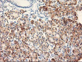
Immunohistochemistry
Mitogen-Activated Protein Kinases
Propidium
Receptors, Death Domain
Proto-Oncogene Proteins c-akt
Cycloheximide
Membrane Proteins
Rats, Sprague-Dawley
Protease Inhibitors
Drug Screening Assays, Antitumor
Receptors, Tumor Necrosis Factor, Type I
p38 Mitogen-Activated Protein Kinases
Proteolysis
Protein Transport
Serine Endopeptidases
Disease Models, Animal
DNA Damage
Acetylcysteine
Proteasome Endopeptidase Complex
Cell-Free System
Ultraviolet Rays
Fibroblasts
Hydrogen Peroxide
Drug Resistance, Neoplasm
Gene Expression Regulation
Gene Expression
Endoplasmic Reticulum Stress
Epithelial Cells
Recombinant Fusion Proteins
Microscopy, Confocal
Cell Division
Neuroprotective Agents
Cell Line, Transformed
Lamins
Drosophila
Sequence Homology, Amino Acid
Gene Expression Regulation, Neoplastic
Microtubule-Associated Proteins
Cytoplasm
Gene Knockdown Techniques
Permeability
DNA Primers
Nuclear Proteins
Interleukin-1beta
Flavoproteins
Cathepsin B
Cell Differentiation
Bongkrekic Acid
Pentanoic Acids
Hepatocytes
Mice, Transgenic
Doxorubicin
Cytoprotection
Aspartic Acid
L-Lactate Dehydrogenase
Protein Kinase C-delta
Gene Expression Regulation, Enzymologic
Lamin Type B
Isatin
Keratin-18
Neuroblastoma
Glutathione
Leupeptins
MAP Kinase Signaling System
Macrophages
Xenograft Model Antitumor Assays
Mitogen-Activated Protein Kinase 8
Endoplasmic Reticulum
MycN sensitizes neuroblastoma cells for drug-induced apoptosis. (1/2233)
Amplification of the MYCN gene is found in a large proportion of neuroblastoma and considered as an adverse prognostic factor. To investigate the effect of ectopic MycN expression on the susceptibility of neuroblastoma cells to cytotoxic drugs we used a human neuroblastoma cell line harboring tetracycline-controlled expression of MycN. Neither conditional expression of MycN alone nor low drug concentrations triggered apoptosis. However, when acting in concert, MycN and cytotoxic drugs efficiently induced cell death. Apoptosis depended on mitochondrial permeability transition and activation of caspases, since the mitochondrion-specific inhibitor bongkrekic acid and the caspase inhibitor zVAD-fmk almost completely abrogated apoptosis. Loss of mitochondrial transmembrane potential and release of cytochrome c from mitochondria preceded activation of caspase-8 and caspase-3 and cleavage of PARP. CD95 expression was upregulated by treatment with cytotoxic drugs, while MycN cooperated with cytotoxic drugs to increase sensitivity to CD95-induced apoptosis and enhancing CD95-L expression. MycN overexpression and cytotoxic drugs also synergized to induce p53 and Bax protein expression, while Bcl-2 and Bcl-X(L) protein levels remained unchanged. Since amplification of MYCN is usually associated with a poor prognosis, these findings suggest that dysfunctions in apoptosis pathways may be a mechanism by which MycN-induced apoptosis of neuroblastoma cells is inhibited. (+info)Identification of an endogenous dominant-negative short isoform of caspase-9 that can regulate apoptosis. (2/2233)
Alternatively spliced isoforms of certain apoptosis regulators, such as Bcl-x, Ced-4, and Ich-1, have been shown to play opposing roles in regulating apoptosis. Here, we describe the identification of an endogenous alternatively spliced isoform of caspase-9, named caspase-9b, which lacks the central large subunit caspase domain. Caspase-9b is detectable in many cell lines by PCR and at the mRNA and protein levels. Caspase-9b can interact with the caspase recruitment domain of Apaf-1, and like the active site mutant of caspase-9, it can inhibit multiple forms of apoptosis, including those triggered by oligomerization of death receptors. It can also block activation of caspase-9 and -3 by Apaf-1 in an in vitro cytochrome c-dependent caspase activation assay. These results suggest that caspase-9b functions as an endogenous apoptosis inhibitory molecule by interfering with the formation of a functional Apaf-1-caspase-9 complex. (+info)Caspase-9 can be activated without proteolytic processing. (3/2233)
The recombinant form of the proapoptotic caspase-9 purified following expression in Escherichia coli is processed at Asp315, but largely inactive; however, when added to cytosolic extracts of human 293 cells it is activated 2000-fold in the presence of cytochrome c and dATP. Thus, the characteristic activities of caspase-9 are context-dependent, and its activation may not recapitulate conventional caspase activation mechanisms. To explore this hypothesis we produced recombinant forms of procaspase-9 containing mutations that disabled one or both of the interdomain processing sites of the zymogen. These mutants were able to activate downstream caspases, but only in the presence of cytosolic factors. The mutant with both processing sites abolished had 10% of the activity of wild-type, and was able to support apoptosis, with equal vigor to wild-type, when transiently expressed in 293 cells. Thus caspase-9 has an unusually active zymogen that does not require proteolytic processing, but instead is dependent on cytosolic factors for expression of its activity. (+info)Tumor necrosis factor alpha regulation of the FAS-mediated apoptosis-signaling pathway in synovial cells. (4/2233)
OBJECTIVE: Fas-mediated apoptosis is observed in synoviocytes of patients with rheumatoid arthritis (RA), but not in those of patients with osteoarthritis (OA). The present study was conducted to elucidate the mechanisms that initiate induction of Fas-mediated apoptosis in RA synoviocytes. METHODS: Cultured OA synoviocytes, which are insensitive to Fas-mediated apoptosis in spite of Fas antigen expression, were used in these experiments. Synovial cell proliferation and cytotoxicity studies were performed using MTS and lactate dehydrogenase release assays. Surface expression of Fas antigen was analyzed by flow cytometry. The expression and function of apoptosis-signaling molecules, such as caspase 8 and caspase 3, were examined by immunoblot analysis. RESULTS: Tumor necrosis factor alpha (TNFalpha) induced proliferation of cultured OA synoviocytes. Fas ligation with anti-Fas monoclonal antibody (mAb) resulted in cytotoxic activity against cultured OA synoviocytes that had been pretreated with TNFalpha for 5 days, but not those pretreated for 2 days. In contrast, anti-Fas mAb did not show a cytotoxic effect against untreated cultured OA synoviocytes. A gradual up-regulation of caspase 8 and caspase 3, which played a role in the caspase cascade for Fas-mediated apoptosis, was observed in TNFalpha-treated cultured OA synoviocytes. In addition, Fas ligation to TNFalpha-treated cultured OA synoviocytes induced activation of caspase 8 and caspase 3, with subsequent cleavage of poly(ADP-ribose) polymerase (PARP), a substrate of activated caspase 3. More importantly, Z-IETD-FMK, a caspase 8 inhibitor, and Ac-DEVD-CHO, a caspase 3 inhibitor, almost completely inhibited Fas-mediated apoptosis of TNFalpha-treated cultured OA synoviocytes, whereas Ac-YVAD-CHO, a caspase 1 inhibitor, did not. CONCLUSION: Our results clearly demonstrate that TNFalpha stimulates synovial cells to proliferate as well as sensitizes the cells for Fas-mediated apoptosis, at least in part by up-regulation and activation of caspase 8 and caspase 3. These findings suggest that TNFalpha may be one of the factors providing sensitization of synovial cells to Fas-mediated apoptosis in RA. (+info)Solution structure of BID, an intracellular amplifier of apoptotic signaling. (5/2233)
We report the solution structure of BID, an intracellular cross-talk agent that can amplify FAS/TNF apoptotic signal through the mitochondria death pathway after Caspase 8 cleavage. BID contains eight alpha helices where two central hydrophobic helices are surrounded by six amphipathic ones. The fold resembles poreforming bacterial toxins and shows similarity to BCL-XL although sequence homology to BCL-XL is limited to the 16-residue BH3 domain. Furthermore, we modeled a complex of BCL-XL and BID by aligning the BID and BAK BH3 motifs in the known BCL-XL-BAK BH3 complex. Additionally, we show that the overall structure of BID is preserved after cleavage by Caspase 8. We propose that BID has both BH3 domain-dependent and -independent modes of action in inducing mitochondrial damage. (+info)Solution structure of the proapoptotic molecule BID: a structural basis for apoptotic agonists and antagonists. (6/2233)
Members of the BCL2 family of proteins are key regulators of programmed cell death, acting either as apoptotic agonists or antagonists. Here we describe the solution structure of BID, presenting the structure of a proapoptotic BCL2 family member. An analysis of sequence/structure of BCL2 family members allows us to define a structural superfamily, which has implications for general mechanisms for regulating proapoptotic activity. It appears two criteria must be met for proapoptotic function within the BCL2 family: targeting of molecules to intracellular membranes, and exposure of the BH3 death domain. BID's activity is regulated by a Caspase 8-mediated cleavage event, exposing the BH3 domain and significantly changing the surface charge and hydrophobicity, resulting in a change of cellular localization. (+info)Nitric-oxide-induced apoptosis in human leukemic lines requires mitochondrial lipid degradation and cytochrome C release. (7/2233)
We have previously shown that nitric oxide (NO) stimulates apoptosis in different human neoplastic lymphoid cell lines through activation of caspases not only via CD95/CD95L interaction, but also independently of such death receptors. Here we investigated mitochondria-dependent mechanisms of NO-induced apoptosis in Jurkat leukemic cells. NO donor glycerol trinitrate (at the concentration, which induces apoptotic cell death) caused (1) a significant decrease in the concentration of cardiolipin, a major mitochondrial lipid; (2) a downregulation in respiratory chain complex activities; (3) a release of the mitochondrial protein cytochrome c into the cytosol; and (4) an activation of caspase-9 and caspase-3. These changes were accompanied by an increase in the number of cells with low mitochondrial transmembrane potential and with a high level of reactive oxygen species production. Higher resistance of the CD95-resistant Jurkat subclone (APO-R) cells to NO-mediated apoptosis correlated with the absence of cytochrome c release and with less alterations in other mitochondrial parameters. An inhibitor of lipid peroxidation, trolox, significantly suppressed NO-mediated apoptosis in APO-S Jurkat cells, whereas bongkrekic acid (BA), which blocks mitochondrial permeability transition, provided only a moderate antiapoptotic effect. Transfection of Jurkat cells with bcl-2 led to a complete block of apoptosis due to the prevention of changes in mitochondrial functions. We suggest that the mitochondrial damage (in particular, cardiolipin degradation and cytochrome c release) induced by NO in human leukemia cells plays a crucial role in the subsequent activation of caspase and apoptosis. (+info)Targeted disruption of caspase genes in mice: what they tell us about the functions of individual caspases in apoptosis. (8/2233)
Cysteine proteases of the caspase family are crucial mediators of apoptosis. All mammalian cells contain a large number of caspases. Although many caspases are activated in a cell committed to apoptosis, recent data from caspase gene knockout mice suggest that individual caspases may be involved in the cell and stimulus-specific pathways of cell death. The gene disruption studies also establish the functional hierarchy between two structurally distinct classes of caspases. The present review discusses these recent findings and elaborates on how these mutant mouse models have helped the understanding of the mechanisms that govern programmed cell death in the immune and other systems. (+info)Caspase-3 is a type of protease enzyme that plays a central role in the execution-phase of cell apoptosis, or programmed cell death. It's also known as CPP32 (CPP for ced-3 protease precursor) or apopain. Caspase-3 is produced as an inactive protein that is activated when cleaved by other caspases during the early stages of apoptosis. Once activated, it cleaves a variety of cellular proteins, including structural proteins, enzymes, and signal transduction proteins, leading to the characteristic morphological and biochemical changes associated with apoptotic cell death. Caspase-3 is often referred to as the "death protease" because of its crucial role in executing the cell death program.
Caspase-9 is a type of protease enzyme that plays a crucial role in the execution phase of programmed cell death, also known as apoptosis. It is a member of the cysteine-aspartic acid protease (caspase) family, which are characterized by their ability to cleave proteins after an aspartic acid residue. Caspase-9 is activated through a process called cytochrome c-mediated caspase activation, which occurs in the mitochondria during apoptosis. Once activated, caspase-9 cleaves and activates other downstream effector caspases, such as caspase-3 and caspase-7, leading to the proteolytic degradation of cellular structures and ultimately resulting in cell death. Dysregulation of caspase-9 activity has been implicated in various diseases, including neurodegenerative disorders and cancer.
Caspase inhibitors are substances or molecules that block the activity of caspases, which are a family of enzymes involved in programmed cell death, also known as apoptosis. Caspases play a crucial role in the execution phase of apoptosis by cleaving various proteins and thereby bringing about characteristic changes in the cell, such as cell shrinkage, membrane blebbing, and DNA fragmentation.
Caspase inhibitors can be synthetic or natural compounds that bind to caspases and prevent them from carrying out their function. These inhibitors have been used in research to study the role of caspases in various biological processes and have also been explored as potential therapeutic agents for conditions associated with excessive apoptosis, such as neurodegenerative diseases and ischemia-reperfusion injury.
It's important to note that while caspase inhibitors can prevent apoptotic cell death, they may also have unintended consequences, such as promoting the survival of damaged or cancerous cells. Therefore, their use as therapeutic agents must be carefully evaluated and balanced against potential risks.
Caspase 8 is a type of protease enzyme that plays a crucial role in programmed cell death, also known as apoptosis. It is a key component of the extrinsic pathway of apoptosis, which can be initiated by the binding of death ligands to their respective death receptors on the cell surface.
Once activated, Caspase 8 cleaves and activates other downstream effector caspases, which then go on to degrade various cellular proteins, leading to the characteristic morphological changes associated with apoptosis, such as cell shrinkage, membrane blebbing, and DNA fragmentation.
In addition to its role in apoptosis, Caspase 8 has also been implicated in other cellular processes, including inflammation, differentiation, and proliferation. Dysregulation of Caspase 8 activity has been linked to various diseases, including cancer, neurodegenerative disorders, and autoimmune diseases.
Caspase-7 is a type of protease enzyme that plays a central role in the execution phase of apoptosis, which is programmed cell death. It is a member of the cysteine-aspartic acid protease (caspase) family, and is also known as caspase-3 like protease, or ICH-1/Mch2.
Caspase-7 is produced as an inactive precursor protein that is activated when cleaved by other upstream caspases during the apoptotic process. Once activated, it can cleave and activate other cellular proteins, leading to characteristic changes associated with apoptosis such as chromatin condensation, DNA fragmentation, and membrane blebbing.
Caspase-7 has been shown to be involved in various forms of programmed cell death, including developmental apoptosis, tissue homeostasis, and immune system regulation. Dysregulation of caspase-7 activity has been implicated in several diseases, including neurodegenerative disorders, ischemic injury, and cancer.
Caspases are a family of protease enzymes that play essential roles in programmed cell death, also known as apoptosis. These enzymes are produced as inactive precursors and are activated when cells receive signals to undergo apoptosis. Once activated, caspases cleave specific protein substrates, leading to the characteristic morphological changes and DNA fragmentation associated with apoptotic cell death. Caspases also play roles in other cellular processes, including inflammation and differentiation. There are two types of caspases: initiator caspases (caspase-2, -8, -9, and -10) and effector caspases (caspase-3, -6, and -7). Initiator caspases are activated in response to various apoptotic signals and then activate the effector caspases, which carry out the proteolytic cleavage of cellular proteins. Dysregulation of caspase activity has been implicated in a variety of diseases, including neurodegenerative disorders, ischemic injury, and cancer.
Caspase-1 is a type of protease enzyme that plays a crucial role in the inflammatory response and programmed cell death, also known as apoptosis. It is produced as an inactive precursor protein, which is then cleaved into its active form by other proteases or through self-cleavage.
Once activated, caspase-1 helps to process and activate several pro-inflammatory cytokines, such as interleukin (IL)-1β and IL-18, which are involved in the recruitment of immune cells to sites of infection or tissue damage. Caspase-1 also contributes to programmed cell death by cleaving and activating other caspases, leading to the controlled destruction of the cell.
Dysregulation of caspase-1 has been implicated in various inflammatory diseases, such as autoimmune disorders and neurodegenerative conditions. Therefore, understanding the mechanisms that regulate caspase-1 activity is an important area of research for developing new therapeutic strategies to treat these diseases.
Caspase-10 is a type of protease enzyme that plays a crucial role in programmed cell death, also known as apoptosis. It is a member of the cysteine-aspartic acid protease (caspase) family, which are proteases that specifically cleave their substrates after an aspartic acid residue. Caspase-10 is activated in response to various cellular signals, such as those triggered by immune responses or DNA damage, and it contributes to the execution of apoptosis by cleaving and activating other downstream effector caspases. Additionally, caspase-10 has been implicated in the regulation of inflammatory responses.
Apoptosis is a programmed and controlled cell death process that occurs in multicellular organisms. It is a natural process that helps maintain tissue homeostasis by eliminating damaged, infected, or unwanted cells. During apoptosis, the cell undergoes a series of morphological changes, including cell shrinkage, chromatin condensation, and fragmentation into membrane-bound vesicles called apoptotic bodies. These bodies are then recognized and engulfed by neighboring cells or phagocytic cells, preventing an inflammatory response. Apoptosis is regulated by a complex network of intracellular signaling pathways that involve proteins such as caspases, Bcl-2 family members, and inhibitors of apoptosis (IAPs).
Amino acid chloromethyl ketones (AACMKs) are a class of chemical compounds that are widely used in research and industry. They are derivatives of amino acids, which are the building blocks of proteins, with a chloromethyl ketone group (-CO-CH2Cl) attached to the side chain of the amino acid.
In the context of medical research, AACMKs are often used as irreversible inhibitors of enzymes, particularly those that contain active site serine or cysteine residues. The chloromethyl ketone group reacts with these residues to form a covalent bond, which permanently inactivates the enzyme. This makes AACMKs useful tools for studying the mechanisms of enzymes and for developing drugs that target specific enzymes.
However, it is important to note that AACMKs can also be highly reactive and toxic, and they must be handled with care in the laboratory. They have been shown to inhibit a wide range of enzymes, including some that are essential for normal cellular function, and prolonged exposure can lead to cell damage or death. Therefore, their use is typically restricted to controlled experimental settings.
Cysteine proteinase inhibitors are a type of molecule that bind to and inhibit the activity of cysteine proteases, which are enzymes that cleave proteins at specific sites containing the amino acid cysteine. These inhibitors play important roles in regulating various biological processes, including inflammation, immune response, and programmed cell death (apoptosis). They can also have potential therapeutic applications in diseases where excessive protease activity contributes to pathology, such as cancer, arthritis, and neurodegenerative disorders. Examples of cysteine proteinase inhibitors include cystatins, kininogens, and serpins.
Caspase-12 is a type of protease enzyme that belongs to the family of caspases, which are cysteine-aspartic acid proteases playing essential roles in programmed cell death (apoptosis). Caspase-12 is primarily expressed in the endoplasmic reticulum (ER) and is involved in ER stress-induced apoptosis.
During ER stress, misfolded or unfolded proteins accumulate in the ER lumen, triggering an adaptive response called the unfolded protein response (UPR). If the UPR fails to restore ER homeostasis, caspase-12 is activated and contributes to the initiation of the apoptotic process.
However, it's worth noting that the role of caspase-12 in human apoptosis remains controversial, as some studies suggest its function might be limited or absent in humans compared to other species like mice.
Caspase-14 is a type of protease enzyme that belongs to the family of caspases, which are cysteine-aspartic acid proteases involved in the execution of apoptosis (programmed cell death) and inflammation. Caspase-14 is primarily expressed in the differentiated layers of the epidermis and plays a crucial role in keratinization, the process of forming an impermeable barrier to protect the body from external insults.
Caspase-14 is involved in the proteolytic processing of several structural proteins, such as loricrin, involucrin, and filaggrin, which are essential components of the cornified cell envelope, a structure that provides mechanical strength to the outermost layer of the skin. Additionally, caspase-14 has been implicated in the regulation of UV-induced apoptosis, contributing to the maintenance of skin homeostasis and preventing the development of skin cancers.
Defects or mutations in the CASP14 gene have been associated with several skin disorders, including dry skin, ichthyosis, and increased susceptibility to skin cancer.
Enzyme activation refers to the process by which an enzyme becomes biologically active and capable of carrying out its specific chemical or biological reaction. This is often achieved through various post-translational modifications, such as proteolytic cleavage, phosphorylation, or addition of cofactors or prosthetic groups to the enzyme molecule. These modifications can change the conformation or structure of the enzyme, exposing or creating a binding site for the substrate and allowing the enzymatic reaction to occur.
For example, in the case of proteolytic cleavage, an inactive precursor enzyme, known as a zymogen, is cleaved into its active form by a specific protease. This is seen in enzymes such as trypsin and chymotrypsin, which are initially produced in the pancreas as inactive precursors called trypsinogen and chymotrypsinogen, respectively. Once they reach the small intestine, they are activated by enteropeptidase, a protease that cleaves a specific peptide bond, releasing the active enzyme.
Phosphorylation is another common mechanism of enzyme activation, where a phosphate group is added to a specific serine, threonine, or tyrosine residue on the enzyme by a protein kinase. This modification can alter the conformation of the enzyme and create a binding site for the substrate, allowing the enzymatic reaction to occur.
Enzyme activation is a crucial process in many biological pathways, as it allows for precise control over when and where specific reactions take place. It also provides a mechanism for regulating enzyme activity in response to various signals and stimuli, such as hormones, neurotransmitters, or changes in the intracellular environment.
DNA fragmentation is the breaking of DNA strands into smaller pieces. This process can occur naturally during apoptosis, or programmed cell death, where the DNA is broken down and packaged into apoptotic bodies to be safely eliminated from the body. However, excessive or abnormal DNA fragmentation can also occur due to various factors such as oxidative stress, exposure to genotoxic agents, or certain medical conditions. This can lead to genetic instability, cellular dysfunction, and increased risk of diseases such as cancer. In the context of reproductive medicine, high levels of DNA fragmentation in sperm cells have been linked to male infertility and poor assisted reproductive technology outcomes.
Proto-oncogene proteins c-bcl-2 are a group of proteins that play a role in regulating cell death (apoptosis). The c-bcl-2 gene produces one of these proteins, which helps to prevent cells from undergoing apoptosis. This protein is located on the membrane of mitochondria and endoplasmic reticulum and it can inhibit the release of cytochrome c, a key player in the activation of caspases, which are enzymes that trigger apoptosis.
In normal cells, the regulation of c-bcl-2 protein helps to maintain a balance between cell proliferation and cell death, ensuring proper tissue homeostasis. However, when the c-bcl-2 gene is mutated or its expression is dysregulated, it can contribute to cancer development by allowing cancer cells to survive and proliferate. High levels of c-bcl-2 protein have been found in many types of cancer, including leukemia, lymphoma, and carcinomas, and are often associated with a poor prognosis.
Cytochromes c are a group of small heme proteins found in the mitochondria of cells, involved in the electron transport chain and play a crucial role in cellular respiration. They accept and donate electrons during the process of oxidative phosphorylation, which generates ATP, the main energy currency of the cell. Cytochromes c contain a heme group, an organic compound that includes iron, which facilitates the transfer of electrons. The "c" in cytochromes c refers to the type of heme group they contain (cyt c has heme c). They are highly conserved across species and have been widely used as a molecular marker for evolutionary studies.
Mitochondria are specialized structures located inside cells that convert the energy from food into ATP (adenosine triphosphate), which is the primary form of energy used by cells. They are often referred to as the "powerhouses" of the cell because they generate most of the cell's supply of chemical energy. Mitochondria are also involved in various other cellular processes, such as signaling, differentiation, and apoptosis (programmed cell death).
Mitochondria have their own DNA, known as mitochondrial DNA (mtDNA), which is inherited maternally. This means that mtDNA is passed down from the mother to her offspring through the egg cells. Mitochondrial dysfunction has been linked to a variety of diseases and conditions, including neurodegenerative disorders, diabetes, and aging.
CD95 (also known as Fas or APO-1) is a type of cell surface receptor that can bind to specific proteins and trigger programmed cell death, also known as apoptosis. It is an important regulator of the immune system and helps to control the activation and deletion of immune cells. CD95 ligand (CD95L), the protein that binds to CD95, is expressed on activated T-cells and can induce apoptosis in other cells that express CD95, including other T-cells and tumor cells.
An antigen is any substance that can stimulate an immune response, leading to the production of antibodies or activation of immune cells. In the context of CD95, antigens may refer to substances that can induce the expression of CD95 on the surface of cells, making them susceptible to CD95L-mediated apoptosis. These antigens could include viral proteins, tumor antigens, or other substances that trigger an immune response.
Therefore, the medical definition of 'antigens, CD95' may refer to substances that can induce the expression of CD95 on the surface of cells and make them targets for CD95L-mediated apoptosis.
Cytochrome c is a small protein that is involved in the electron transport chain, a key part of cellular respiration in which cells generate energy in the form of ATP. Cytochrome c contains a heme group, which binds to and transports electrons. The cytochrome c group refers to a class of related cytochromes that have similar structures and functions. These proteins are found in the mitochondria of eukaryotic cells (such as those of plants and animals) and in the inner membranes of bacteria. They play a crucial role in the production of energy within the cell, and are also involved in certain types of programmed cell death (apoptosis).
The X-linked inhibitor of apoptosis protein (XIAP) is a member of the inhibitor of apoptosis (IAP) family, which are proteins that play a crucial role in regulating programmed cell death, also known as apoptosis. XIAP is located on the X chromosome and functions by binding to and inhibiting certain caspases, which are enzymes that play an essential role in initiating and executing the apoptotic process. By inhibiting these caspases, XIAP promotes cell survival and prevents excessive cell death, which can contribute to cancer development and resistance to therapy. Additionally, XIAP has been implicated in the regulation of inflammation and immune responses, making it a target for therapeutic intervention in various diseases.
Apoptotic protease-activating factor 1 (APAF-1) is a protein that plays a crucial role in the intrinsic pathway of programmed cell death, also known as apoptosis. APAF-1 is involved in the formation of the apoptosome, which is a multi-protein complex that activates caspases, a family of protease enzymes that dismantle cellular structures and contribute to the orderly demolition of cells during apoptosis.
APAF-1 contains a C-terminal WD40 domain, which is responsible for its oligomerization and interaction with other proteins, and an N-terminal caspase recruitment domain (CARD). In response to cellular stress or damage, cytochrome c is released from the mitochondria and binds to the WD40 domain of APAF-1. This binding induces a conformational change in APAF-1, exposing its CARD domain and allowing it to interact with the CARD domain of procaspase-9. The resulting apoptosome formation leads to the activation of caspase-9, which subsequently activates other downstream caspases, ultimately executing the apoptotic program.
Defects in APAF-1 function or regulation have been implicated in various diseases, including cancer and neurodegenerative disorders.
BCL-2-associated X protein, often abbreviated as BAX, is a type of protein belonging to the BCL-2 family. The BCL-2 family of proteins plays a crucial role in regulating programmed cell death, also known as apoptosis. Specifically, BAX is a pro-apoptotic protein, which means that it promotes cell death.
BAX is encoded by the BAX gene, and it functions by forming pores in the outer membrane of the mitochondria, leading to the release of cytochrome c and other pro-apoptotic factors into the cytosol. This triggers a cascade of events that ultimately leads to cell death.
Dysregulation of BAX and other BCL-2 family proteins has been implicated in various diseases, including cancer and neurodegenerative disorders. For example, reduced levels of BAX have been observed in some types of cancer, which may contribute to tumor growth and resistance to chemotherapy. On the other hand, excessive activation of BAX has been linked to neuronal death in conditions such as Alzheimer's disease and Parkinson's disease.
Cell death is the process by which cells cease to function and eventually die. There are several ways that cells can die, but the two most well-known and well-studied forms of cell death are apoptosis and necrosis.
Apoptosis is a programmed form of cell death that occurs as a normal and necessary process in the development and maintenance of healthy tissues. During apoptosis, the cell's DNA is broken down into small fragments, the cell shrinks, and the membrane around the cell becomes fragmented, allowing the cell to be easily removed by phagocytic cells without causing an inflammatory response.
Necrosis, on the other hand, is a form of cell death that occurs as a result of acute tissue injury or overwhelming stress. During necrosis, the cell's membrane becomes damaged and the contents of the cell are released into the surrounding tissue, causing an inflammatory response.
There are also other forms of cell death, such as autophagy, which is a process by which cells break down their own organelles and proteins to recycle nutrients and maintain energy homeostasis, and pyroptosis, which is a form of programmed cell death that occurs in response to infection and involves the activation of inflammatory caspases.
Cell death is an important process in many physiological and pathological processes, including development, tissue homeostasis, and disease. Dysregulation of cell death can contribute to the development of various diseases, including cancer, neurodegenerative disorders, and autoimmune diseases.
Inhibitor of Apoptosis Proteins (IAPs) are a family of proteins that play a crucial role in regulating programmed cell death, also known as apoptosis. These proteins function by binding to and inhibiting the activity of caspases, which are enzymes that drive the execution phase of apoptosis.
There are eight known human IAPs, including X-linked IAP (XIAP), cellular IAP1 (cIAP1), cIAP2, survivin, melanoma IAP (ML-IAP), ILP-2, NAIP, and Bruce. Each IAP contains at least one baculoviral IAP repeat (BIR) domain, which is responsible for binding to caspases and other regulatory proteins.
In addition to inhibiting caspases, some IAPs have been shown to regulate other cellular processes, such as inflammation, innate immunity, and cell cycle progression. Dysregulation of IAP function has been implicated in various diseases, including cancer, neurodegenerative disorders, and autoimmune diseases. Therefore, IAPs are considered important targets for the development of new therapeutic strategies aimed at modulating apoptosis and other cellular processes.
Jurkat cells are a type of human immortalized T lymphocyte (a type of white blood cell) cell line that is commonly used in scientific research. They were originally isolated from the peripheral blood of a patient with acute T-cell leukemia. Jurkat cells are widely used as a model system to study T-cell activation, signal transduction, and apoptosis (programmed cell death). They are also used in the study of HIV infection and replication, as they can be infected with the virus and used to investigate viral replication and host cell responses.
Caspases are a family of protease enzymes playing essential roles in programmed cell death, also known as apoptosis. They are produced as inactive precursors and activated upon cleavage into large and small subunits. Initiator caspases, including caspase-8, -9, and -10, are so called because they are the first to be activated during the execution of apoptosis. Once activated, initiator caspases cleave and activate other proteins, including executive or effector caspases such as caspase-3, -6, and -7, which in turn cleave various cellular substrates leading to the morphological changes associated with apoptotic cell death.
In situ nick-end labeling (ISEL, also known as TUNEL) is a technique used in pathology and molecular biology to detect DNA fragmentation, which is a characteristic of apoptotic cells (cells undergoing programmed cell death). The method involves labeling the 3'-hydroxyl termini of double or single stranded DNA breaks in situ (within tissue sections or individual cells) using modified nucleotides that are coupled to a detectable marker, such as a fluorophore or an enzyme. This technique allows for the direct visualization and quantification of apoptotic cells within complex tissues or cell populations.
Cell survival refers to the ability of a cell to continue living and functioning normally, despite being exposed to potentially harmful conditions or treatments. This can include exposure to toxins, radiation, chemotherapeutic drugs, or other stressors that can damage cells or interfere with their normal processes.
In scientific research, measures of cell survival are often used to evaluate the effectiveness of various therapies or treatments. For example, researchers may expose cells to a particular drug or treatment and then measure the percentage of cells that survive to assess its potential therapeutic value. Similarly, in toxicology studies, measures of cell survival can help to determine the safety of various chemicals or substances.
It's important to note that cell survival is not the same as cell proliferation, which refers to the ability of cells to divide and multiply. While some treatments may promote cell survival, they may also inhibit cell proliferation, making them useful for treating diseases such as cancer. Conversely, other treatments may be designed to specifically target and kill cancer cells, even if it means sacrificing some healthy cells in the process.
BH3 Interacting Domain Death Agonist Protein, also known as BAD protein, is a member of the Bcl-2 family of proteins. This protein is involved in the regulation of programmed cell death, or apoptosis. The BH3 domain of BAD protein allows it to interact with other members of the Bcl-2 family and modulate their function. When activated, BAD protein can promote cell death by binding to and inhibiting anti-apoptotic proteins such as Bcl-2 and Bcl-xL. This helps to release pro-apoptotic proteins such as Bax and Bak, which can then trigger the intrinsic pathway of apoptosis. The activation of BAD protein is tightly regulated by post-translational modifications, including phosphorylation and dephosphorylation, which can be influenced by various signals within the cell.
Apoptosis regulatory proteins are a group of proteins that play an essential role in the regulation and execution of apoptosis, also known as programmed cell death. This process is a normal part of development and tissue homeostasis, allowing for the elimination of damaged or unnecessary cells. The balance between pro-apoptotic and anti-apoptotic proteins determines whether a cell will undergo apoptosis.
Pro-apoptotic proteins, such as BAX, BID, and PUMA, promote apoptosis by neutralizing or counteracting the effects of anti-apoptotic proteins or by directly activating the apoptotic pathway. These proteins can be activated in response to various stimuli, including DNA damage, oxidative stress, and activation of the death receptor pathway.
Anti-apoptotic proteins, such as BCL-2, BCL-XL, and MCL-1, inhibit apoptosis by binding and neutralizing pro-apoptotic proteins or by preventing the release of cytochrome c from the mitochondria, which is a key step in the intrinsic apoptotic pathway.
Dysregulation of apoptosis regulatory proteins has been implicated in various diseases, including cancer, neurodegenerative disorders, and autoimmune diseases. Therefore, understanding the role of these proteins in apoptosis regulation is crucial for developing new therapeutic strategies to treat these conditions.
Signal transduction is the process by which a cell converts an extracellular signal, such as a hormone or neurotransmitter, into an intracellular response. This involves a series of molecular events that transmit the signal from the cell surface to the interior of the cell, ultimately resulting in changes in gene expression, protein activity, or metabolism.
The process typically begins with the binding of the extracellular signal to a receptor located on the cell membrane. This binding event activates the receptor, which then triggers a cascade of intracellular signaling molecules, such as second messengers, protein kinases, and ion channels. These molecules amplify and propagate the signal, ultimately leading to the activation or inhibition of specific cellular responses.
Signal transduction pathways are highly regulated and can be modulated by various factors, including other signaling molecules, post-translational modifications, and feedback mechanisms. Dysregulation of these pathways has been implicated in a variety of diseases, including cancer, diabetes, and neurological disorders.
A cell line that is derived from tumor cells and has been adapted to grow in culture. These cell lines are often used in research to study the characteristics of cancer cells, including their growth patterns, genetic changes, and responses to various treatments. They can be established from many different types of tumors, such as carcinomas, sarcomas, and leukemias. Once established, these cell lines can be grown and maintained indefinitely in the laboratory, allowing researchers to conduct experiments and studies that would not be feasible using primary tumor cells. It is important to note that tumor cell lines may not always accurately represent the behavior of the original tumor, as they can undergo genetic changes during their time in culture.
Cysteine endopeptidases are a type of enzymes that cleave peptide bonds within proteins. They are also known as cysteine proteases or cysteine proteinases. These enzymes contain a catalytic triad consisting of three amino acids: cysteine, histidine, and aspartate. The thiol group (-SH) of the cysteine residue acts as a nucleophile and attacks the carbonyl carbon of the peptide bond, leading to its cleavage.
Cysteine endopeptidases play important roles in various biological processes, including protein degradation, cell signaling, and inflammation. They are involved in many physiological and pathological conditions, such as apoptosis, immune response, and cancer. Some examples of cysteine endopeptidases include cathepsins, caspases, and calpains.
It is important to note that these enzymes require a reducing environment to maintain the reduced state of their active site cysteine residue. Therefore, they are sensitive to oxidizing agents and inhibitors that target the thiol group. Understanding the structure and function of cysteine endopeptidases is crucial for developing therapeutic strategies that target these enzymes in various diseases.
Oligopeptides are defined in medicine and biochemistry as short chains of amino acids, typically containing fewer than 20 amino acid residues. These small peptides are important components in various biological processes, such as serving as signaling molecules, enzyme inhibitors, or structural elements in some proteins. They can be found naturally in foods and may also be synthesized for use in medical research and therapeutic applications.
The Fas-Associated Death Domain Protein (FADD), also known as Mort1 or MORT1, is a protein that plays a crucial role in the programmed cell death pathway, also known as apoptosis. It is composed of an N-terminal death effector domain (DED), a middle domain, and a C-terminal death domain (DD).
FADD functions as an adaptor protein that links the Fas receptor to downstream signaling molecules in the extrinsic pathway of apoptosis. When the Fas receptor is activated by its ligand (FasL), it recruits FADD through homotypic interactions between their DED domains. This recruitment leads to the formation of the death-inducing signaling complex (DISC) and the activation of caspase-8, which subsequently activates downstream effector caspases that ultimately lead to cell death.
FADD is essential for maintaining tissue homeostasis by eliminating damaged or potentially harmful cells, and its dysregulation has been implicated in various pathological conditions, including cancer, neurodegenerative diseases, and autoimmune disorders.
Enzyme inhibitors are substances that bind to an enzyme and decrease its activity, preventing it from catalyzing a chemical reaction in the body. They can work by several mechanisms, including blocking the active site where the substrate binds, or binding to another site on the enzyme to change its shape and prevent substrate binding. Enzyme inhibitors are often used as drugs to treat various medical conditions, such as high blood pressure, abnormal heart rhythms, and bacterial infections. They can also be found naturally in some foods and plants, and can be used in research to understand enzyme function and regulation.
A cell line is a culture of cells that are grown in a laboratory for use in research. These cells are usually taken from a single cell or group of cells, and they are able to divide and grow continuously in the lab. Cell lines can come from many different sources, including animals, plants, and humans. They are often used in scientific research to study cellular processes, disease mechanisms, and to test new drugs or treatments. Some common types of human cell lines include HeLa cells (which come from a cancer patient named Henrietta Lacks), HEK293 cells (which come from embryonic kidney cells), and HUVEC cells (which come from umbilical vein endothelial cells). It is important to note that cell lines are not the same as primary cells, which are cells that are taken directly from a living organism and have not been grown in the lab.
Bcl-x is a protein that belongs to the Bcl-2 family, which regulates programmed cell death (apoptosis). Specifically, Bcl-x has both pro-survival and pro-apoptotic functions, depending on its splice variants. The long form of Bcl-x (Bcl-xL) is a potent inhibitor of apoptosis, while the short form (Bcl-xS) promotes cell death. Bcl-x plays critical roles in various cellular processes, including development, homeostasis, and stress responses, by controlling the mitochondrial outer membrane permeabilization and the release of cytochrome c, which eventually leads to caspase activation and apoptosis. Dysregulation of Bcl-x has been implicated in several diseases, such as cancer and neurodegenerative disorders.
Apoptosis Inducing Factor (AIF) is a protein that triggers programmed cell death, also known as apoptosis. It is primarily located in the mitochondria, but upon activation, it translocates to the nucleus where it contributes to DNA fragmentation and chromatin condensation, which are key features of apoptosis. AIF can be released from the mitochondria in response to various cellular stressors or signals, such as during development, tissue homeostasis, or in response to certain types of cellular damage or injury.
Western blotting is a laboratory technique used in molecular biology to detect and quantify specific proteins in a mixture of many different proteins. This technique is commonly used to confirm the expression of a protein of interest, determine its size, and investigate its post-translational modifications. The name "Western" blotting distinguishes this technique from Southern blotting (for DNA) and Northern blotting (for RNA).
The Western blotting procedure involves several steps:
1. Protein extraction: The sample containing the proteins of interest is first extracted, often by breaking open cells or tissues and using a buffer to extract the proteins.
2. Separation of proteins by electrophoresis: The extracted proteins are then separated based on their size by loading them onto a polyacrylamide gel and running an electric current through the gel (a process called sodium dodecyl sulfate-polyacrylamide gel electrophoresis or SDS-PAGE). This separates the proteins according to their molecular weight, with smaller proteins migrating faster than larger ones.
3. Transfer of proteins to a membrane: After separation, the proteins are transferred from the gel onto a nitrocellulose or polyvinylidene fluoride (PVDF) membrane using an electric current in a process called blotting. This creates a replica of the protein pattern on the gel but now immobilized on the membrane for further analysis.
4. Blocking: The membrane is then blocked with a blocking agent, such as non-fat dry milk or bovine serum albumin (BSA), to prevent non-specific binding of antibodies in subsequent steps.
5. Primary antibody incubation: A primary antibody that specifically recognizes the protein of interest is added and allowed to bind to its target protein on the membrane. This step may be performed at room temperature or 4°C overnight, depending on the antibody's properties.
6. Washing: The membrane is washed with a buffer to remove unbound primary antibodies.
7. Secondary antibody incubation: A secondary antibody that recognizes the primary antibody (often coupled to an enzyme or fluorophore) is added and allowed to bind to the primary antibody. This step may involve using a horseradish peroxidase (HRP)-conjugated or alkaline phosphatase (AP)-conjugated secondary antibody, depending on the detection method used later.
8. Washing: The membrane is washed again to remove unbound secondary antibodies.
9. Detection: A detection reagent is added to visualize the protein of interest by detecting the signal generated from the enzyme-conjugated or fluorophore-conjugated secondary antibody. This can be done using chemiluminescent, colorimetric, or fluorescent methods.
10. Analysis: The resulting image is analyzed to determine the presence and quantity of the protein of interest in the sample.
Western blotting is a powerful technique for identifying and quantifying specific proteins within complex mixtures. It can be used to study protein expression, post-translational modifications, protein-protein interactions, and more. However, it requires careful optimization and validation to ensure accurate and reproducible results.
'Tumor cells, cultured' refers to the process of removing cancerous cells from a tumor and growing them in controlled laboratory conditions. This is typically done by isolating the tumor cells from a patient's tissue sample, then placing them in a nutrient-rich environment that promotes their growth and multiplication.
The resulting cultured tumor cells can be used for various research purposes, including the study of cancer biology, drug development, and toxicity testing. They provide a valuable tool for researchers to better understand the behavior and characteristics of cancer cells outside of the human body, which can lead to the development of more effective cancer treatments.
It is important to note that cultured tumor cells may not always behave exactly the same way as they do in the human body, so findings from cell culture studies must be validated through further research, such as animal models or clinical trials.
"Cells, cultured" is a medical term that refers to cells that have been removed from an organism and grown in controlled laboratory conditions outside of the body. This process is called cell culture and it allows scientists to study cells in a more controlled and accessible environment than they would have inside the body. Cultured cells can be derived from a variety of sources, including tissues, organs, or fluids from humans, animals, or cell lines that have been previously established in the laboratory.
Cell culture involves several steps, including isolation of the cells from the tissue, purification and characterization of the cells, and maintenance of the cells in appropriate growth conditions. The cells are typically grown in specialized media that contain nutrients, growth factors, and other components necessary for their survival and proliferation. Cultured cells can be used for a variety of purposes, including basic research, drug development and testing, and production of biological products such as vaccines and gene therapies.
It is important to note that cultured cells may behave differently than they do in the body, and results obtained from cell culture studies may not always translate directly to human physiology or disease. Therefore, it is essential to validate findings from cell culture experiments using additional models and ultimately in clinical trials involving human subjects.
CASP8 and FADD-Like Apoptosis Regulating Protein, also known as CFLAR or FLIP, is a protein that plays a role in regulating cell death (apoptosis). It is a member of the inhibitor of apoptosis protein (IAP) family. The protein contains a death effector domain (DED), which allows it to interact with other proteins that contain DEDs, such as FADD and caspase-8.
CFLAR can function as an inhibitor or a promoter of apoptosis, depending on the context. When CFLAR is present in high levels, it can bind to and inhibit the activity of caspase-8, preventing the initiation of the apoptotic signaling pathway. However, when CFLAR is present in low levels or is cleaved by proteases, it can promote apoptosis by facilitating the activation of caspase-8.
Mutations in the gene that encodes CFLAR have been associated with an increased risk of developing certain types of cancer, such as Hodgkin lymphoma and diffuse large B-cell lymphoma.
Staurosporine is an alkaloid compound that is derived from the bacterium Streptomyces staurosporeus. It is a potent and broad-spectrum protein kinase inhibitor, which means it can bind to and inhibit various types of protein kinases, including protein kinase C (PKC), cyclin-dependent kinases (CDKs), and tyrosine kinases.
Protein kinases are enzymes that play a crucial role in cell signaling by adding phosphate groups to other proteins, thereby modulating their activity. The inhibition of protein kinases by staurosporine can disrupt these signaling pathways and lead to various biological effects, such as the induction of apoptosis (programmed cell death) and the inhibition of cell proliferation.
Staurosporine has been widely used in research as a tool to study the roles of protein kinases in various cellular processes and diseases, including cancer, neurodegenerative disorders, and inflammation. However, its use as a therapeutic agent is limited due to its lack of specificity and high toxicity.
Annexin A5 is a protein that belongs to the annexin family, which are calcium-dependent phospholipid-binding proteins. Annexin A5 has high affinity for phosphatidylserine, a type of phospholipid that is usually located on the inner leaflet of the plasma membrane in healthy cells. However, when cells undergo apoptosis (programmed cell death), phosphatidylserine is exposed on the outer leaflet of the plasma membrane.
Annexin A5 can bind to exposed phosphatidylserine on the surface of apoptotic cells and is commonly used as a marker for detecting apoptosis in various experimental settings, including flow cytometry, immunohistochemistry, and imaging techniques. Annexin A5-based assays are widely used in research and clinical settings to study the mechanisms of apoptosis and to develop diagnostic tools for various diseases, such as cancer, neurodegenerative disorders, and cardiovascular diseases.
Mitochondrial membrane potential is the electric potential difference (voltage) across the inner mitochondrial membrane. It is negative inside the mitochondria and positive outside. This electrical gradient is established by the active transport of hydrogen ions (protons) out of the mitochondrial matrix and into the intermembrane space by complexes in the electron transport chain during oxidative phosphorylation. The energy stored in this electrochemical gradient is used to generate ATP, which is the main source of energy for cellular metabolism.
Carrier proteins, also known as transport proteins, are a type of protein that facilitates the movement of molecules across cell membranes. They are responsible for the selective and active transport of ions, sugars, amino acids, and other molecules from one side of the membrane to the other, against their concentration gradient. This process requires energy, usually in the form of ATP (adenosine triphosphate).
Carrier proteins have a specific binding site for the molecule they transport, and undergo conformational changes upon binding, which allows them to move the molecule across the membrane. Once the molecule has been transported, the carrier protein returns to its original conformation, ready to bind and transport another molecule.
Carrier proteins play a crucial role in maintaining the balance of ions and other molecules inside and outside of cells, and are essential for many physiological processes, including nerve impulse transmission, muscle contraction, and nutrient uptake.
HL-60 cells are a type of human promyelocytic leukemia cell line that is commonly used in scientific research. They are named after the hospital where they were first isolated, the Hospital of the University of Pennsylvania (HUP) and the 60th culture attempt to grow these cells.
HL-60 cells have the ability to differentiate into various types of blood cells, such as granulocytes, monocytes, and macrophages, when exposed to certain chemical compounds or under specific culturing conditions. This makes them a valuable tool for studying the mechanisms of cell differentiation, proliferation, and apoptosis (programmed cell death).
HL-60 cells are also often used in toxicity studies, drug discovery and development, and research on cancer, inflammation, and infectious diseases. They can be easily grown in the lab and have a stable genotype, making them ideal for use in standardized experiments and comparisons between different studies.
TNF-Related Apoptosis-Inducing Ligand (TRAIL) is a type II transmembrane protein and a member of the tumor necrosis factor (TNF) ligand family. It binds to death receptors TRAIL-R1 (DR4) and TRAIL-R2 (DR5), leading to the activation of extrinsic apoptosis pathway in sensitive cells. This protein is involved in immune surveillance against tumor cells, as it can selectively induce apoptosis in malignant or virus-infected cells while sparing normal cells. TRAIL also plays a role in inflammation and innate immunity.
Enzyme precursors are typically referred to as zymogens or proenzymes. These are inactive forms of enzymes that can be activated under specific conditions. When the need for the enzyme's function arises, the proenzyme is converted into its active form through a process called proteolysis, where it is cleaved by another enzyme. This mechanism helps control and regulate the activation of certain enzymes in the body, preventing unwanted or premature reactions. A well-known example of an enzyme precursor is trypsinogen, which is converted into its active form, trypsin, in the digestive system.
HeLa cells are a type of immortalized cell line used in scientific research. They are derived from a cancer that developed in the cervical tissue of Henrietta Lacks, an African-American woman, in 1951. After her death, cells taken from her tumor were found to be capable of continuous division and growth in a laboratory setting, making them an invaluable resource for medical research.
HeLa cells have been used in a wide range of scientific studies, including research on cancer, viruses, genetics, and drug development. They were the first human cell line to be successfully cloned and are able to grow rapidly in culture, doubling their population every 20-24 hours. This has made them an essential tool for many areas of biomedical research.
It is important to note that while HeLa cells have been instrumental in numerous scientific breakthroughs, the story of their origin raises ethical questions about informed consent and the use of human tissue in research.
Necrosis is the premature death of cells or tissues due to damage or injury, such as from infection, trauma, infarction (lack of blood supply), or toxic substances. It's a pathological process that results in the uncontrolled and passive degradation of cellular components, ultimately leading to the release of intracellular contents into the extracellular space. This can cause local inflammation and may lead to further tissue damage if not treated promptly.
There are different types of necrosis, including coagulative, liquefactive, caseous, fat, fibrinoid, and gangrenous necrosis, each with distinct histological features depending on the underlying cause and the affected tissues or organs.
Caspases are a family of protease enzymes that play essential roles in programmed cell death, also known as apoptosis. There are two types of caspases: initiator caspases and effector (or executioner) caspases.
Effector caspases, also known as caspases-3, -6, and -7, are responsible for carrying out the proteolytic cleavage of various cellular substrates during apoptosis. Once activated by initiator caspases, effector caspases cleave key structural and regulatory proteins, leading to the characteristic morphological and biochemical changes associated with apoptotic cell death, such as chromatin condensation, DNA fragmentation, and membrane blebbing.
In summary, effector caspases are crucial components of the apoptotic machinery that mediate the execution phase of programmed cell death.
Apoptosomes are large protein complexes that play a crucial role in the process of programmed cell death, also known as apoptosis. They are formed when certain proteins in the cell, called caspases, are activated in response to signals indicating that the cell needs to die. The formation of apoptosomes leads to the activation of additional caspases, which then go on to break down various cellular structures and ultimately cause the cell to die.
Apoptosomes are composed of several proteins, including cytochrome c, Apaf-1 (apoptotic protease activating factor 1), and procaspase-9. When cytochrome c is released from the mitochondria into the cytoplasm, it binds to Apaf-1 and procaspase-9, leading to the formation of the apoptosome complex. This complex then cleaves and activates caspase-9, which in turn activates other caspases, setting off a chain reaction that results in apoptosis.
The formation of apoptosomes is an important mechanism for maintaining tissue homeostasis and getting rid of damaged or potentially harmful cells. Dysregulation of this process can contribute to the development of various diseases, including cancer and neurodegenerative disorders.
Reactive Oxygen Species (ROS) are highly reactive molecules containing oxygen, including peroxides, superoxide, hydroxyl radical, and singlet oxygen. They are naturally produced as byproducts of normal cellular metabolism in the mitochondria, and can also be generated by external sources such as ionizing radiation, tobacco smoke, and air pollutants. At low or moderate concentrations, ROS play important roles in cell signaling and homeostasis, but at high concentrations, they can cause significant damage to cell structures, including lipids, proteins, and DNA, leading to oxidative stress and potential cell death.
Tumor Necrosis Factor-alpha (TNF-α) is a cytokine, a type of small signaling protein involved in immune response and inflammation. It is primarily produced by activated macrophages, although other cell types such as T-cells, natural killer cells, and mast cells can also produce it.
TNF-α plays a crucial role in the body's defense against infection and tissue injury by mediating inflammatory responses, activating immune cells, and inducing apoptosis (programmed cell death) in certain types of cells. It does this by binding to its receptors, TNFR1 and TNFR2, which are found on the surface of many cell types.
In addition to its role in the immune response, TNF-α has been implicated in the pathogenesis of several diseases, including autoimmune disorders such as rheumatoid arthritis, inflammatory bowel disease, and psoriasis, as well as cancer, where it can promote tumor growth and metastasis.
Therapeutic agents that target TNF-α, such as infliximab, adalimumab, and etanercept, have been developed to treat these conditions. However, these drugs can also increase the risk of infections and other side effects, so their use must be carefully monitored.
Transfection is a term used in molecular biology that refers to the process of deliberately introducing foreign genetic material (DNA, RNA or artificial gene constructs) into cells. This is typically done using chemical or physical methods, such as lipofection or electroporation. Transfection is widely used in research and medical settings for various purposes, including studying gene function, producing proteins, developing gene therapies, and creating genetically modified organisms. It's important to note that transfection is different from transduction, which is the process of introducing genetic material into cells using viruses as vectors.
Proteins are complex, large molecules that play critical roles in the body's functions. They are made up of amino acids, which are organic compounds that are the building blocks of proteins. Proteins are required for the structure, function, and regulation of the body's tissues and organs. They are essential for the growth, repair, and maintenance of body tissues, and they play a crucial role in many biological processes, including metabolism, immune response, and cellular signaling. Proteins can be classified into different types based on their structure and function, such as enzymes, hormones, antibodies, and structural proteins. They are found in various foods, especially animal-derived products like meat, dairy, and eggs, as well as plant-based sources like beans, nuts, and grains.
CRADD, or Cav-1 related death domain protein, is a signaling adaptor protein that plays a role in regulating cell death and survival pathways. It contains a death domain that allows it to interact with other proteins involved in these pathways, including the tumor suppressor protein p53 and the death receptor Fas. CRADD has been implicated in a number of cellular processes, including apoptosis (programmed cell death), autophagy, and inflammation. Mutations in the CRADD gene have been associated with various diseases, including neurodevelopmental disorders and cancer.
Intracellular signaling peptides and proteins are molecules that play a crucial role in transmitting signals within cells, which ultimately lead to changes in cell behavior or function. These signals can originate from outside the cell (extracellular) or within the cell itself. Intracellular signaling molecules include various types of peptides and proteins, such as:
1. G-protein coupled receptors (GPCRs): These are seven-transmembrane domain receptors that bind to extracellular signaling molecules like hormones, neurotransmitters, or chemokines. Upon activation, they initiate a cascade of intracellular signals through G proteins and secondary messengers.
2. Receptor tyrosine kinases (RTKs): These are transmembrane receptors that bind to growth factors, cytokines, or hormones. Activation of RTKs leads to autophosphorylation of specific tyrosine residues, creating binding sites for intracellular signaling proteins such as adapter proteins, phosphatases, and enzymes like Ras, PI3K, and Src family kinases.
3. Second messenger systems: Intracellular second messengers are small molecules that amplify and propagate signals within the cell. Examples include cyclic adenosine monophosphate (cAMP), cyclic guanosine monophosphate (cGMP), diacylglycerol (DAG), inositol triphosphate (IP3), calcium ions (Ca2+), and nitric oxide (NO). These second messengers activate or inhibit various downstream effectors, leading to changes in cellular responses.
4. Signal transduction cascades: Intracellular signaling proteins often form complex networks of interacting molecules that relay signals from the plasma membrane to the nucleus. These cascades involve kinases (protein kinases A, B, C, etc.), phosphatases, and adapter proteins, which ultimately regulate gene expression, cell cycle progression, metabolism, and other cellular processes.
5. Ubiquitination and proteasome degradation: Intracellular signaling pathways can also control protein stability by modulating ubiquitin-proteasome degradation. E3 ubiquitin ligases recognize specific substrates and conjugate them with ubiquitin molecules, targeting them for proteasomal degradation. This process regulates the abundance of key signaling proteins and contributes to signal termination or amplification.
In summary, intracellular signaling pathways involve a complex network of interacting proteins that relay signals from the plasma membrane to various cellular compartments, ultimately regulating gene expression, metabolism, and other cellular processes. Dysregulation of these pathways can contribute to disease development and progression, making them attractive targets for therapeutic intervention.
Antineoplastic agents are a class of drugs used to treat malignant neoplasms or cancer. These agents work by inhibiting the growth and proliferation of cancer cells, either by killing them or preventing their division and replication. Antineoplastic agents can be classified based on their mechanism of action, such as alkylating agents, antimetabolites, topoisomerase inhibitors, mitotic inhibitors, and targeted therapy agents.
Alkylating agents work by adding alkyl groups to DNA, which can cause cross-linking of DNA strands and ultimately lead to cell death. Antimetabolites interfere with the metabolic processes necessary for DNA synthesis and replication, while topoisomerase inhibitors prevent the relaxation of supercoiled DNA during replication. Mitotic inhibitors disrupt the normal functioning of the mitotic spindle, which is essential for cell division. Targeted therapy agents are designed to target specific molecular abnormalities in cancer cells, such as mutated oncogenes or dysregulated signaling pathways.
It's important to note that antineoplastic agents can also affect normal cells and tissues, leading to various side effects such as nausea, vomiting, hair loss, and myelosuppression (suppression of bone marrow function). Therefore, the use of these drugs requires careful monitoring and management of their potential adverse effects.
Death domain receptor signaling adaptor proteins are a group of intracellular signaling molecules that play a crucial role in the transduction of signals from death receptors, which are a type of cell surface receptor involved in programmed cell death or apoptosis. These adaptor proteins contain a protein-protein interaction module called the death domain (DD), which allows them to interact with other DD-containing proteins and initiate downstream signaling pathways leading to apoptosis.
Some of the key death domain receptor signaling adaptor proteins include Fas-associated death domain protein (FADD), receptor-interacting protein (RIP) kinases, and TNF receptor-associated death domain protein (TRADD). These proteins help to recruit and activate various downstream effectors, such as caspases, which are a family of cysteine proteases that play an essential role in the execution of apoptosis.
Abnormalities in death domain receptor signaling adaptor protein function have been implicated in a variety of diseases, including cancer, neurodegenerative disorders, and autoimmune diseases. Therefore, understanding the mechanisms underlying their regulation and activity is an important area of research with potential therapeutic implications.
A dose-response relationship in the context of drugs refers to the changes in the effects or symptoms that occur as the dose of a drug is increased or decreased. Generally, as the dose of a drug is increased, the severity or intensity of its effects also increases. Conversely, as the dose is decreased, the effects of the drug become less severe or may disappear altogether.
The dose-response relationship is an important concept in pharmacology and toxicology because it helps to establish the safe and effective dosage range for a drug. By understanding how changes in the dose of a drug affect its therapeutic and adverse effects, healthcare providers can optimize treatment plans for their patients while minimizing the risk of harm.
The dose-response relationship is typically depicted as a curve that shows the relationship between the dose of a drug and its effect. The shape of the curve may vary depending on the drug and the specific effect being measured. Some drugs may have a steep dose-response curve, meaning that small changes in the dose can result in large differences in the effect. Other drugs may have a more gradual dose-response curve, where larger changes in the dose are needed to produce significant effects.
In addition to helping establish safe and effective dosages, the dose-response relationship is also used to evaluate the potential therapeutic benefits and risks of new drugs during clinical trials. By systematically testing different doses of a drug in controlled studies, researchers can identify the optimal dosage range for the drug and assess its safety and efficacy.
Flow cytometry is a medical and research technique used to measure physical and chemical characteristics of cells or particles, one cell at a time, as they flow in a fluid stream through a beam of light. The properties measured include:
* Cell size (light scatter)
* Cell internal complexity (granularity, also light scatter)
* Presence or absence of specific proteins or other molecules on the cell surface or inside the cell (using fluorescent antibodies or other fluorescent probes)
The technique is widely used in cell counting, cell sorting, protein engineering, biomarker discovery and monitoring disease progression, particularly in hematology, immunology, and cancer research.
Phosphatidylserines are a type of phospholipids that are essential components of the cell membrane, particularly in the brain. They play a crucial role in maintaining the fluidity and permeability of the cell membrane, and are involved in various cellular processes such as signal transduction, protein anchorage, and apoptosis (programmed cell death). Phosphatidylserines contain a polar head group made up of serine amino acids and two non-polar fatty acid tails. They are abundant in the inner layer of the cell membrane but can be externalized to the outer layer during apoptosis, where they serve as signals for recognition and removal of dying cells by the immune system. Phosphatidylserines have been studied for their potential benefits in various medical conditions, including cognitive decline, Alzheimer's disease, and depression.
CARD (caspase recruitment domain) signaling adaptor proteins are a group of intracellular signaling molecules that play a crucial role in the regulation of various cellular processes, including inflammation, immunity, and programmed cell death or apoptosis. These proteins contain a CARD domain, which is a protein-protein interaction module that enables them to bind to other CARD-containing proteins and form large signaling complexes.
CARD signaling adaptor proteins function as molecular scaffolds that help bring together various signaling components in response to different stimuli, such as pathogen-associated molecular patterns (PAMPs) or damage-associated molecular patterns (DAMPs). By doing so, they facilitate the activation of downstream signaling cascades and the initiation of appropriate cellular responses.
Some examples of CARD signaling adaptor proteins include:
1. Myeloid differentiation factor 88 (MyD88): This protein is involved in the signaling pathways of most Toll-like receptors (TLRs) and interleukin-1 receptor (IL-1R) family members, which are critical for the detection of microbial components and the initiation of innate immune responses.
2. CARD9: This protein is involved in the signaling pathways of several C-type lectin receptors (CLRs), which recognize fungal and other pathogens, and plays a key role in antifungal immunity.
3. ASC (apoptosis-associated speck-like protein containing a CARD): This protein is involved in the formation of inflammasomes, which are large cytosolic complexes that activate caspase-1 and promote the maturation and secretion of proinflammatory cytokines.
4. RIPK2 (receptor-interacting serine/threonine-protein kinase 2): This protein is involved in the signaling pathways of NOD1 and NOD2, which are intracellular sensors of bacterial peptidoglycan, and plays a role in the regulation of inflammation and apoptosis.
Overall, CARD-containing proteins play crucial roles in various immune signaling pathways by mediating protein-protein interactions and downstream signal transduction events, ultimately leading to the activation of innate immunity and inflammatory responses.
BAK (Bcl-2 Homologous Antagonist-Killer) protein is a member of the Bcl-2 family, which consists of proteins that regulate programmed cell death, also known as apoptosis. The Bcl-2 family includes both pro-apoptotic and anti-apoptotic members, and their interactions play a crucial role in determining whether a cell lives or dies.
BAK is a pro-apoptotic protein that forms oligomers and creates pores in the outer mitochondrial membrane, leading to the release of cytochrome c and other pro-apoptotic factors into the cytosol. This triggers a cascade of events that ultimately results in cell death.
BAK is kept in an inactive state under normal conditions by binding to anti-apoptotic Bcl-2 family members, such as Bcl-xL and Mcl-1. However, when cells receive signals to undergo apoptosis, the interactions between pro- and anti-apoptotic proteins are disrupted, allowing BAK to become activated and initiate the cell death process.
In summary, BAK is a crucial protein involved in regulating programmed cell death, and its dysregulation has been implicated in various diseases, including cancer and neurodegenerative disorders.
Calpains are a family of calcium-dependent cysteine proteases that play important roles in various cellular processes, including signal transduction, cell death, and remodeling of the cytoskeleton. They are present in most tissues and can be activated by an increase in intracellular calcium levels. There are at least 15 different calpain isoforms identified in humans, which are categorized into two groups based on their calcium requirements for activation: classical calpains (calpain-1 and calpain-2) and non-classical calpains (calpain-3 to calpain-15). Dysregulation of calpain activity has been implicated in several pathological conditions, such as neurodegenerative diseases, muscular dystrophies, and cancer.
Granzymes are a group of proteases (enzymes that break down other proteins) that are stored in the granules of cytotoxic T cells and natural killer (NK) cells. They play an important role in the immune response by inducing apoptosis (programmed cell death) in target cells, such as virus-infected or cancer cells. Granzymes are released into the immunological synapse between the effector and target cells, where they can enter the target cell and cleave specific substrates, leading to the activation of caspases and ultimately apoptosis. There are several different types of granzymes, each with distinct substrate specificities and functions.
In the field of medicine, "time factors" refer to the duration of symptoms or time elapsed since the onset of a medical condition, which can have significant implications for diagnosis and treatment. Understanding time factors is crucial in determining the progression of a disease, evaluating the effectiveness of treatments, and making critical decisions regarding patient care.
For example, in stroke management, "time is brain," meaning that rapid intervention within a specific time frame (usually within 4.5 hours) is essential to administering tissue plasminogen activator (tPA), a clot-busting drug that can minimize brain damage and improve patient outcomes. Similarly, in trauma care, the "golden hour" concept emphasizes the importance of providing definitive care within the first 60 minutes after injury to increase survival rates and reduce morbidity.
Time factors also play a role in monitoring the progression of chronic conditions like diabetes or heart disease, where regular follow-ups and assessments help determine appropriate treatment adjustments and prevent complications. In infectious diseases, time factors are crucial for initiating antibiotic therapy and identifying potential outbreaks to control their spread.
Overall, "time factors" encompass the significance of recognizing and acting promptly in various medical scenarios to optimize patient outcomes and provide effective care.
Proto-oncogene proteins are normal cellular proteins that play crucial roles in various cellular processes, such as signal transduction, cell cycle regulation, and apoptosis (programmed cell death). They are involved in the regulation of cell growth, differentiation, and survival under physiological conditions.
When proto-oncogene proteins undergo mutations or aberrations in their expression levels, they can transform into oncogenic forms, leading to uncontrolled cell growth and division. These altered proteins are then referred to as oncogene products or oncoproteins. Oncogenic mutations can occur due to various factors, including genetic predisposition, environmental exposures, and aging.
Examples of proto-oncogene proteins include:
1. Ras proteins: Involved in signal transduction pathways that regulate cell growth and differentiation. Activating mutations in Ras genes are found in various human cancers.
2. Myc proteins: Regulate gene expression related to cell cycle progression, apoptosis, and metabolism. Overexpression of Myc proteins is associated with several types of cancer.
3. EGFR (Epidermal Growth Factor Receptor): A transmembrane receptor tyrosine kinase that regulates cell proliferation, survival, and differentiation. Mutations or overexpression of EGFR are linked to various malignancies, such as lung cancer and glioblastoma.
4. Src family kinases: Intracellular tyrosine kinases that regulate signal transduction pathways involved in cell proliferation, survival, and migration. Dysregulation of Src family kinases is implicated in several types of cancer.
5. Abl kinases: Cytoplasmic tyrosine kinases that regulate various cellular processes, including cell growth, differentiation, and stress responses. Aberrant activation of Abl kinases, as seen in chronic myelogenous leukemia (CML), leads to uncontrolled cell proliferation.
Understanding the roles of proto-oncogene proteins and their dysregulation in cancer development is essential for developing targeted cancer therapies that aim to inhibit or modulate these aberrant signaling pathways.
Mitochondrial proteins are any proteins that are encoded by the nuclear genome or mitochondrial genome and are located within the mitochondria, an organelle found in eukaryotic cells. These proteins play crucial roles in various cellular processes including energy production, metabolism of lipids, amino acids, and steroids, regulation of calcium homeostasis, and programmed cell death or apoptosis.
Mitochondrial proteins can be classified into two main categories based on their origin:
1. Nuclear-encoded mitochondrial proteins (NEMPs): These are proteins that are encoded by genes located in the nucleus, synthesized in the cytoplasm, and then imported into the mitochondria through specific import pathways. NEMPs make up about 99% of all mitochondrial proteins and are involved in various functions such as oxidative phosphorylation, tricarboxylic acid (TCA) cycle, fatty acid oxidation, and mitochondrial dynamics.
2. Mitochondrial DNA-encoded proteins (MEPs): These are proteins that are encoded by the mitochondrial genome, synthesized within the mitochondria, and play essential roles in the electron transport chain (ETC), a key component of oxidative phosphorylation. The human mitochondrial genome encodes only 13 proteins, all of which are subunits of complexes I, III, IV, and V of the ETC.
Defects in mitochondrial proteins can lead to various mitochondrial disorders, which often manifest as neurological, muscular, or metabolic symptoms due to impaired energy production. These disorders are usually caused by mutations in either nuclear or mitochondrial genes that encode mitochondrial proteins.
Tumor suppressor protein p53, also known as p53 or tumor protein p53, is a nuclear phosphoprotein that plays a crucial role in preventing cancer development and maintaining genomic stability. It does so by regulating the cell cycle and acting as a transcription factor for various genes involved in apoptosis (programmed cell death), DNA repair, and cell senescence (permanent cell growth arrest).
In response to cellular stress, such as DNA damage or oncogene activation, p53 becomes activated and accumulates in the nucleus. Activated p53 can then bind to specific DNA sequences and promote the transcription of target genes that help prevent the proliferation of potentially cancerous cells. These targets include genes involved in cell cycle arrest (e.g., CDKN1A/p21), apoptosis (e.g., BAX, PUMA), and DNA repair (e.g., GADD45).
Mutations in the TP53 gene, which encodes p53, are among the most common genetic alterations found in human cancers. These mutations often lead to a loss or reduction of p53's tumor suppressive functions, allowing cancer cells to proliferate uncontrollably and evade apoptosis. As a result, p53 has been referred to as "the guardian of the genome" due to its essential role in preventing tumorigenesis.
Receptor-Interacting Protein Serine-Threonine Kinases (RIPKs) are a family of serine-threonine kinases that play crucial roles in the regulation of cell death, inflammation, and immune response. In humans, there are seven known members of this family, RIPK1 to RIPK7, which share a conserved N-terminal kinase domain and C-terminal domains involved in protein-protein interactions.
RIPKs can be activated by various stimuli, including cytokines, pathogens, and stress signals, leading to the phosphorylation of downstream substrates that modulate cellular processes such as apoptosis (programmed cell death), necroptosis (a programmed form of necrosis), and inflammation.
RIPK1 is one of the most well-studied members, acting as a key regulator of both cell survival and death pathways. In response to tumor necrosis factor (TNF) receptor engagement, RIPK1 can form complexes with other proteins that either promote cell survival through the activation of nuclear factor kappa B (NF-κB) or induce cell death via apoptosis or necroptosis.
Dysregulation of RIPKs has been implicated in several pathological conditions, including neurodegenerative diseases, inflammatory disorders, and cancer. Therefore, targeting RIPKs with small molecule inhibitors is an area of active research for the development of novel therapeutic strategies to treat these diseases.
Antineoplastic agents, phytogenic, also known as plant-derived anticancer drugs, are medications that are derived from plants and used to treat cancer. These agents have natural origins and work by interfering with the growth and multiplication of cancer cells, helping to slow or stop the spread of the disease. Some examples of antineoplastic agents, phytogenic include paclitaxel (Taxol), vincristine, vinblastine, and etoposide. These drugs are often used in combination with other treatments such as surgery, radiation therapy, and other medications to provide a comprehensive approach to cancer care.
Small interfering RNA (siRNA) is a type of short, double-stranded RNA molecule that plays a role in the RNA interference (RNAi) pathway. The RNAi pathway is a natural cellular process that regulates gene expression by targeting and destroying specific messenger RNA (mRNA) molecules, thereby preventing the translation of those mRNAs into proteins.
SiRNAs are typically 20-25 base pairs in length and are generated from longer double-stranded RNA precursors called hairpin RNAs or dsRNAs by an enzyme called Dicer. Once generated, siRNAs associate with a protein complex called the RNA-induced silencing complex (RISC), which uses one strand of the siRNA (the guide strand) to recognize and bind to complementary sequences in the target mRNA. The RISC then cleaves the target mRNA, leading to its degradation and the inhibition of protein synthesis.
SiRNAs have emerged as a powerful tool for studying gene function and have shown promise as therapeutic agents for a variety of diseases, including viral infections, cancer, and genetic disorders. However, their use as therapeutics is still in the early stages of development, and there are challenges associated with delivering siRNAs to specific cells and tissues in the body.
An amino acid sequence is the specific order of amino acids in a protein or peptide molecule, formed by the linking of the amino group (-NH2) of one amino acid to the carboxyl group (-COOH) of another amino acid through a peptide bond. The sequence is determined by the genetic code and is unique to each type of protein or peptide. It plays a crucial role in determining the three-dimensional structure and function of proteins.
Tumor Necrosis Factor (TNF) Receptors are cell surface receptors that bind to tumor necrosis factor cytokines. They play crucial roles in the regulation of a variety of immune cell functions, including inflammation, immunity, and cell survival or death (apoptosis).
There are two major types of TNF receptors: TNFR1 (also known as p55 or CD120a) and TNFR2 (also known as p75 or CD120b). TNFR1 is widely expressed in most tissues, while TNFR2 has a more restricted expression pattern and is mainly found on immune cells.
TNF receptors have an intracellular domain called the death domain, which can trigger signaling pathways leading to apoptosis when activated by TNF ligands. However, they can also activate other signaling pathways that promote cell survival, differentiation, and inflammation. Dysregulation of TNF receptor signaling has been implicated in various diseases, including cancer, autoimmune disorders, and neurodegenerative conditions.
TNF-related apoptosis-inducing ligand (TRAIL) receptors are a group of cell surface proteins that belong to the tumor necrosis factor (TNF) receptor superfamily. There are four known TRAIL receptors, referred to as TRAIL-R1, TRAIL-R2, TRAIL-R3, and TRAIL-R4.
TRAIL receptors play a crucial role in the regulation of programmed cell death, also known as apoptosis. TRAIL binding to its receptors TRAIL-R1 and TRAIL-R2 can trigger the activation of intracellular signaling pathways that lead to apoptotic cell death. This is an important mechanism for eliminating damaged or abnormal cells, including cancer cells.
On the other hand, TRAIL receptors TRAIL-R3 and TRAIL-R4 do not transmit apoptotic signals because they lack functional death domains. Instead, they act as decoy receptors that can bind to TRAIL and prevent it from interacting with TRAIL-R1 and TRAIL-R2, thereby inhibiting TRAIL-induced apoptosis.
Abnormalities in the regulation of TRAIL receptor signaling have been implicated in various pathological conditions, including cancer, autoimmune diseases, and neurodegenerative disorders. Therefore, targeting TRAIL receptors has emerged as a promising therapeutic strategy for the treatment of these diseases.
Cell proliferation is the process by which cells increase in number, typically through the process of cell division. In the context of biology and medicine, it refers to the reproduction of cells that makes up living tissue, allowing growth, maintenance, and repair. It involves several stages including the transition from a phase of quiescence (G0 phase) to an active phase (G1 phase), DNA replication in the S phase, and mitosis or M phase, where the cell divides into two daughter cells.
Abnormal or uncontrolled cell proliferation is a characteristic feature of many diseases, including cancer, where deregulated cell cycle control leads to excessive and unregulated growth of cells, forming tumors that can invade surrounding tissues and metastasize to distant sites in the body.
Membrane glycoproteins are proteins that contain oligosaccharide chains (glycans) covalently attached to their polypeptide backbone. They are integral components of biological membranes, spanning the lipid bilayer and playing crucial roles in various cellular processes.
The glycosylation of these proteins occurs in the endoplasmic reticulum (ER) and Golgi apparatus during protein folding and trafficking. The attached glycans can vary in structure, length, and composition, which contributes to the diversity of membrane glycoproteins.
Membrane glycoproteins can be classified into two main types based on their orientation within the lipid bilayer:
1. Type I (N-linked): These glycoproteins have a single transmembrane domain and an extracellular N-terminus, where the oligosaccharides are predominantly attached via asparagine residues (Asn-X-Ser/Thr sequon).
2. Type II (C-linked): These glycoproteins possess two transmembrane domains and an intracellular C-terminus, with the oligosaccharides linked to tryptophan residues via a mannose moiety.
Membrane glycoproteins are involved in various cellular functions, such as:
* Cell adhesion and recognition
* Receptor-mediated signal transduction
* Enzymatic catalysis
* Transport of molecules across membranes
* Cell-cell communication
* Immunological responses
Some examples of membrane glycoproteins include cell surface receptors (e.g., growth factor receptors, cytokine receptors), adhesion molecules (e.g., integrins, cadherins), and transporters (e.g., ion channels, ABC transporters).
Cytosol refers to the liquid portion of the cytoplasm found within a eukaryotic cell, excluding the organelles and structures suspended in it. It is the site of various metabolic activities and contains a variety of ions, small molecules, and enzymes. The cytosol is where many biochemical reactions take place, including glycolysis, protein synthesis, and the regulation of cellular pH. It is also where some organelles, such as ribosomes and vesicles, are located. In contrast to the cytosol, the term "cytoplasm" refers to the entire contents of a cell, including both the cytosol and the organelles suspended within it.
BCL-associated death protein, often referred to as BAD, is a type of protein that belongs to the BCL-2 family. These proteins play a crucial role in regulating programmed cell death, also known as apoptosis. Specifically, BAD is a pro-apoptotic protein, meaning it promotes cell death under certain conditions.
The function of BAD is tightly regulated through various post-translational modifications and interactions with other BCL-2 family members. When activated, BAD can bind to and inhibit anti-apoptotic proteins like BCL-2 or BCL-XL, thereby releasing pro-apoptotic proteins such as BAX and BAK, which form pores in the mitochondrial membrane and initiate the apoptotic cascade.
Dysregulation of BAD and other BCL-2 family members has been implicated in several diseases, including cancer and neurodegenerative disorders. For instance, overexpression of anti-apoptotic proteins or downregulation of pro-apoptotic proteins like BAD can contribute to tumor development and resistance to chemotherapy. Therefore, understanding the role of BAD and other BCL-2 family members in apoptosis regulation is essential for developing novel therapeutic strategies in cancer and other diseases.
Molecular sequence data refers to the specific arrangement of molecules, most commonly nucleotides in DNA or RNA, or amino acids in proteins, that make up a biological macromolecule. This data is generated through laboratory techniques such as sequencing, and provides information about the exact order of the constituent molecules. This data is crucial in various fields of biology, including genetics, evolution, and molecular biology, allowing for comparisons between different organisms, identification of genetic variations, and studies of gene function and regulation.
NF-κB (Nuclear Factor kappa-light-chain-enhancer of activated B cells) is a protein complex that plays a crucial role in regulating the immune response to infection and inflammation, as well as in cell survival, differentiation, and proliferation. It is composed of several subunits, including p50, p52, p65 (RelA), c-Rel, and RelB, which can form homodimers or heterodimers that bind to specific DNA sequences called κB sites in the promoter regions of target genes.
Under normal conditions, NF-κB is sequestered in the cytoplasm by inhibitory proteins known as IκBs (inhibitors of κB). However, upon stimulation by various signals such as cytokines, bacterial or viral products, and stress, IκBs are phosphorylated, ubiquitinated, and degraded, leading to the release and activation of NF-κB. Activated NF-κB then translocates to the nucleus, where it binds to κB sites and regulates the expression of target genes involved in inflammation, immunity, cell survival, and proliferation.
Dysregulation of NF-κB signaling has been implicated in various pathological conditions such as cancer, chronic inflammation, autoimmune diseases, and neurodegenerative disorders. Therefore, targeting NF-κB signaling has emerged as a potential therapeutic strategy for the treatment of these diseases.
SERPINs are an acronym for "serine protease inhibitors." They are a group of proteins that inhibit serine proteases, which are enzymes that cut other proteins. SERPINs are found in various tissues and body fluids, including blood, and play important roles in regulating biological processes such as inflammation, blood clotting, and cell death. They do this by forming covalent complexes with their target proteases, thereby preventing them from carrying out their proteolytic activities. Mutations in SERPIN genes have been associated with several genetic disorders, including emphysema, cirrhosis, and dementia.
JNK (c-Jun N-terminal kinase) Mitogen-Activated Protein Kinases are a subgroup of the Ser/Thr protein kinases that are activated by stress stimuli and play important roles in various cellular processes, including inflammation, apoptosis, and differentiation. They are involved in the regulation of gene expression through phosphorylation of transcription factors such as c-Jun. JNKs are activated by a variety of upstream kinases, including MAP2Ks (MKK4/SEK1 and MKK7), which are in turn activated by MAP3Ks (such as ASK1, MEKK1, MLKs, and TAK1). JNK signaling pathways have been implicated in various diseases, including cancer, neurodegenerative disorders, and inflammatory diseases.
Etoposide is a chemotherapy medication used to treat various types of cancer, including lung cancer, testicular cancer, and certain types of leukemia. It works by inhibiting the activity of an enzyme called topoisomerase II, which is involved in DNA replication and transcription. By doing so, etoposide can interfere with the growth and multiplication of cancer cells.
Etoposide is often administered intravenously in a hospital or clinic setting, although it may also be given orally in some cases. The medication can cause a range of side effects, including nausea, vomiting, hair loss, and an increased risk of infection. It can also have more serious side effects, such as bone marrow suppression, which can lead to anemia, bleeding, and a weakened immune system.
Like all chemotherapy drugs, etoposide is not without risks and should only be used under the close supervision of a qualified healthcare provider. It is important for patients to discuss the potential benefits and risks of this medication with their doctor before starting treatment.
U937 cells are a type of human histiocytic lymphoma cell line that is commonly used in scientific research and studies. They are derived from the peripheral blood of a patient with histiocytic lymphoma, which is a rare type of cancer that affects the immune system's cells called histiocytes.
U937 cells have a variety of uses in research, including studying the mechanisms of cancer cell growth and proliferation, testing the effects of various drugs and treatments on cancer cells, and investigating the role of different genes and proteins in cancer development and progression. These cells are easy to culture and maintain in the laboratory, making them a popular choice for researchers in many fields.
It is important to note that while U937 cells can provide valuable insights into the behavior of cancer cells, they do not necessarily reflect the complexity and diversity of human cancers. Therefore, findings from studies using these cells should be validated in more complex models or clinical trials before being applied to patient care.
Bcl-2 is a family of proteins that play a crucial role in regulating cell death (apoptosis), which is a normal process that eliminates damaged or unnecessary cells from the body. Specifically, Bcl-2 proteins are involved in controlling the mitochondrial pathway of apoptosis.
The bcl-2 gene provides instructions for making one member of this protein family, called B-cell lymphoma 2 protein. This protein is located primarily on the outer membrane of mitochondria and helps to prevent apoptosis by inhibiting the release of cytochrome c from the mitochondria into the cytoplasm.
In healthy cells, the balance between pro-apoptotic (promoting cell death) and anti-apoptotic (inhibiting cell death) proteins is critical for maintaining normal tissue homeostasis. However, in some cancers, including certain types of leukemia and lymphoma, the bcl-2 gene is abnormally overexpressed, leading to an excess of Bcl-2 protein that disrupts this balance and allows cancer cells to survive and proliferate.
Therefore, understanding the role of bcl-2 in apoptosis has important implications for developing new therapies for cancer and other diseases associated with abnormal cell death regulation.
Adaptor proteins are a type of protein that play a crucial role in intracellular signaling pathways by serving as a link between different components of the signaling complex. Specifically, "signal transducing adaptor proteins" refer to those adaptor proteins that are involved in signal transduction processes, where they help to transmit signals from the cell surface receptors to various intracellular effectors. These proteins typically contain modular domains that allow them to interact with multiple partners, thereby facilitating the formation of large signaling complexes and enabling the integration of signals from different pathways.
Signal transducing adaptor proteins can be classified into several families based on their structural features, including the Src homology 2 (SH2) domain, the Src homology 3 (SH3) domain, and the phosphotyrosine-binding (PTB) domain. These domains enable the adaptor proteins to recognize and bind to specific motifs on other signaling molecules, such as receptor tyrosine kinases, G protein-coupled receptors, and cytokine receptors.
One well-known example of a signal transducing adaptor protein is the growth factor receptor-bound protein 2 (Grb2), which contains an SH2 domain that binds to phosphotyrosine residues on activated receptor tyrosine kinases. Grb2 also contains an SH3 domain that interacts with proline-rich motifs on other signaling proteins, such as the guanine nucleotide exchange factor SOS. This interaction facilitates the activation of the Ras small GTPase and downstream signaling pathways involved in cell growth, differentiation, and survival.
Overall, signal transducing adaptor proteins play a critical role in regulating various cellular processes by modulating intracellular signaling pathways in response to extracellular stimuli. Dysregulation of these proteins has been implicated in various diseases, including cancer and inflammatory disorders.
Mitochondrial membranes refer to the double-layered structure that surrounds the mitochondrion, an organelle found in the cells of most eukaryotes. The outer mitochondrial membrane is a smooth, porous membrane that allows small molecules and ions to pass through freely, while the inner mitochondrial membrane is highly folded and selectively permeable, controlling the movement of larger molecules and maintaining the electrochemical gradient necessary for ATP synthesis. The space between the two membranes is called the intermembrane space, and the space within the inner membrane is called the matrix. Together, these membranes play a crucial role in energy production, metabolism, and cellular homeostasis.
Recombinant proteins are artificially created proteins produced through the use of recombinant DNA technology. This process involves combining DNA molecules from different sources to create a new set of genes that encode for a specific protein. The resulting recombinant protein can then be expressed, purified, and used for various applications in research, medicine, and industry.
Recombinant proteins are widely used in biomedical research to study protein function, structure, and interactions. They are also used in the development of diagnostic tests, vaccines, and therapeutic drugs. For example, recombinant insulin is a common treatment for diabetes, while recombinant human growth hormone is used to treat growth disorders.
The production of recombinant proteins typically involves the use of host cells, such as bacteria, yeast, or mammalian cells, which are engineered to express the desired protein. The host cells are transformed with a plasmid vector containing the gene of interest, along with regulatory elements that control its expression. Once the host cells are cultured and the protein is expressed, it can be purified using various chromatography techniques.
Overall, recombinant proteins have revolutionized many areas of biology and medicine, enabling researchers to study and manipulate proteins in ways that were previously impossible.
Biological models, also known as physiological models or organismal models, are simplified representations of biological systems, processes, or mechanisms that are used to understand and explain the underlying principles and relationships. These models can be theoretical (conceptual or mathematical) or physical (such as anatomical models, cell cultures, or animal models). They are widely used in biomedical research to study various phenomena, including disease pathophysiology, drug action, and therapeutic interventions.
Examples of biological models include:
1. Mathematical models: These use mathematical equations and formulas to describe complex biological systems or processes, such as population dynamics, metabolic pathways, or gene regulation networks. They can help predict the behavior of these systems under different conditions and test hypotheses about their underlying mechanisms.
2. Cell cultures: These are collections of cells grown in a controlled environment, typically in a laboratory dish or flask. They can be used to study cellular processes, such as signal transduction, gene expression, or metabolism, and to test the effects of drugs or other treatments on these processes.
3. Animal models: These are living organisms, usually vertebrates like mice, rats, or non-human primates, that are used to study various aspects of human biology and disease. They can provide valuable insights into the pathophysiology of diseases, the mechanisms of drug action, and the safety and efficacy of new therapies.
4. Anatomical models: These are physical representations of biological structures or systems, such as plastic models of organs or tissues, that can be used for educational purposes or to plan surgical procedures. They can also serve as a basis for developing more sophisticated models, such as computer simulations or 3D-printed replicas.
Overall, biological models play a crucial role in advancing our understanding of biology and medicine, helping to identify new targets for therapeutic intervention, develop novel drugs and treatments, and improve human health.
Down-regulation is a process that occurs in response to various stimuli, where the number or sensitivity of cell surface receptors or the expression of specific genes is decreased. This process helps maintain homeostasis within cells and tissues by reducing the ability of cells to respond to certain signals or molecules.
In the context of cell surface receptors, down-regulation can occur through several mechanisms:
1. Receptor internalization: After binding to their ligands, receptors can be internalized into the cell through endocytosis. Once inside the cell, these receptors may be degraded or recycled back to the cell surface in smaller numbers.
2. Reduced receptor synthesis: Down-regulation can also occur at the transcriptional level, where the expression of genes encoding for specific receptors is decreased, leading to fewer receptors being produced.
3. Receptor desensitization: Prolonged exposure to a ligand can lead to a decrease in receptor sensitivity or affinity, making it more difficult for the cell to respond to the signal.
In the context of gene expression, down-regulation refers to the decreased transcription and/or stability of specific mRNAs, leading to reduced protein levels. This process can be induced by various factors, including microRNA (miRNA)-mediated regulation, histone modification, or DNA methylation.
Down-regulation is an essential mechanism in many physiological processes and can also contribute to the development of several diseases, such as cancer and neurodegenerative disorders.
C57BL/6 (C57 Black 6) is an inbred strain of laboratory mouse that is widely used in biomedical research. The term "inbred" refers to a strain of animals where matings have been carried out between siblings or other closely related individuals for many generations, resulting in a population that is highly homozygous at most genetic loci.
The C57BL/6 strain was established in 1920 by crossing a female mouse from the dilute brown (DBA) strain with a male mouse from the black strain. The resulting offspring were then interbred for many generations to create the inbred C57BL/6 strain.
C57BL/6 mice are known for their robust health, longevity, and ease of handling, making them a popular choice for researchers. They have been used in a wide range of biomedical research areas, including studies of cancer, immunology, neuroscience, cardiovascular disease, and metabolism.
One of the most notable features of the C57BL/6 strain is its sensitivity to certain genetic modifications, such as the introduction of mutations that lead to obesity or impaired glucose tolerance. This has made it a valuable tool for studying the genetic basis of complex diseases and traits.
Overall, the C57BL/6 inbred mouse strain is an important model organism in biomedical research, providing a valuable resource for understanding the genetic and molecular mechanisms underlying human health and disease.
Ceramides are a type of lipid molecule that are found naturally in the outer layer of the skin (the stratum corneum). They play a crucial role in maintaining the barrier function and hydration of the skin. Ceramides help to seal in moisture, support the structure of the skin, and protect against environmental stressors such as pollution and bacteria.
In addition to their role in the skin, ceramides have also been studied for their potential therapeutic benefits in various medical conditions. For example, abnormal levels of ceramides have been implicated in several diseases, including diabetes, cardiovascular disease, and cancer. As a result, ceramide-based therapies are being investigated as potential treatments for these conditions.
Medically, ceramides may be mentioned in the context of skin disorders or diseases where there is a disruption in the skin's barrier function, such as eczema, psoriasis, and ichthyosis. In these cases, ceramide-based therapies may be used to help restore the skin's natural barrier and improve its overall health and appearance.
Immunoblotting, also known as western blotting, is a laboratory technique used in molecular biology and immunogenetics to detect and quantify specific proteins in a complex mixture. This technique combines the electrophoretic separation of proteins by gel electrophoresis with their detection using antibodies that recognize specific epitopes (protein fragments) on the target protein.
The process involves several steps: first, the protein sample is separated based on size through sodium dodecyl sulfate-polyacrylamide gel electrophoresis (SDS-PAGE). Next, the separated proteins are transferred onto a nitrocellulose or polyvinylidene fluoride (PVDF) membrane using an electric field. The membrane is then blocked with a blocking agent to prevent non-specific binding of antibodies.
After blocking, the membrane is incubated with a primary antibody that specifically recognizes the target protein. Following this, the membrane is washed to remove unbound primary antibodies and then incubated with a secondary antibody conjugated to an enzyme such as horseradish peroxidase (HRP) or alkaline phosphatase (AP). The enzyme catalyzes a colorimetric or chemiluminescent reaction that allows for the detection of the target protein.
Immunoblotting is widely used in research and clinical settings to study protein expression, post-translational modifications, protein-protein interactions, and disease biomarkers. It provides high specificity and sensitivity, making it a valuable tool for identifying and quantifying proteins in various biological samples.
Neurons, also known as nerve cells or neurocytes, are specialized cells that constitute the basic unit of the nervous system. They are responsible for receiving, processing, and transmitting information and signals within the body. Neurons have three main parts: the dendrites, the cell body (soma), and the axon. The dendrites receive signals from other neurons or sensory receptors, while the axon transmits these signals to other neurons, muscles, or glands. The junction between two neurons is called a synapse, where neurotransmitters are released to transmit the signal across the gap (synaptic cleft) to the next neuron. Neurons vary in size, shape, and structure depending on their function and location within the nervous system.
Tertiary protein structure refers to the three-dimensional arrangement of all the elements (polypeptide chains) of a single protein molecule. It is the highest level of structural organization and results from interactions between various side chains (R groups) of the amino acids that make up the protein. These interactions, which include hydrogen bonds, ionic bonds, van der Waals forces, and disulfide bridges, give the protein its unique shape and stability, which in turn determines its function. The tertiary structure of a protein can be stabilized by various factors such as temperature, pH, and the presence of certain ions. Any changes in these factors can lead to denaturation, where the protein loses its tertiary structure and thus its function.
The cell cycle is a series of events that take place in a cell leading to its division and duplication. It consists of four main phases: G1 phase, S phase, G2 phase, and M phase.
During the G1 phase, the cell grows in size and synthesizes mRNA and proteins in preparation for DNA replication. In the S phase, the cell's DNA is copied, resulting in two complete sets of chromosomes. During the G2 phase, the cell continues to grow and produces more proteins and organelles necessary for cell division.
The M phase is the final stage of the cell cycle and consists of mitosis (nuclear division) and cytokinesis (cytoplasmic division). Mitosis results in two genetically identical daughter nuclei, while cytokinesis divides the cytoplasm and creates two separate daughter cells.
The cell cycle is regulated by various checkpoints that ensure the proper completion of each phase before progressing to the next. These checkpoints help prevent errors in DNA replication and division, which can lead to mutations and cancer.
Phosphorylation is the process of adding a phosphate group (a molecule consisting of one phosphorus atom and four oxygen atoms) to a protein or other organic molecule, which is usually done by enzymes called kinases. This post-translational modification can change the function, localization, or activity of the target molecule, playing a crucial role in various cellular processes such as signal transduction, metabolism, and regulation of gene expression. Phosphorylation is reversible, and the removal of the phosphate group is facilitated by enzymes called phosphatases.
Reverse Transcriptase Polymerase Chain Reaction (RT-PCR) is a laboratory technique used in molecular biology to amplify and detect specific DNA sequences. This technique is particularly useful for the detection and quantification of RNA viruses, as well as for the analysis of gene expression.
The process involves two main steps: reverse transcription and polymerase chain reaction (PCR). In the first step, reverse transcriptase enzyme is used to convert RNA into complementary DNA (cDNA) by reading the template provided by the RNA molecule. This cDNA then serves as a template for the PCR amplification step.
In the second step, the PCR reaction uses two primers that flank the target DNA sequence and a thermostable polymerase enzyme to repeatedly copy the targeted cDNA sequence. The reaction mixture is heated and cooled in cycles, allowing the primers to anneal to the template, and the polymerase to extend the new strand. This results in exponential amplification of the target DNA sequence, making it possible to detect even small amounts of RNA or cDNA.
RT-PCR is a sensitive and specific technique that has many applications in medical research and diagnostics, including the detection of viruses such as HIV, hepatitis C virus, and SARS-CoV-2 (the virus that causes COVID-19). It can also be used to study gene expression, identify genetic mutations, and diagnose genetic disorders.
Fluorescence microscopy is a type of microscopy that uses fluorescent dyes or proteins to highlight and visualize specific components within a sample. In this technique, the sample is illuminated with high-energy light, typically ultraviolet (UV) or blue light, which excites the fluorescent molecules causing them to emit lower-energy, longer-wavelength light, usually visible light in the form of various colors. This emitted light is then collected by the microscope and detected to produce an image.
Fluorescence microscopy has several advantages over traditional brightfield microscopy, including the ability to visualize specific structures or molecules within a complex sample, increased sensitivity, and the potential for quantitative analysis. It is widely used in various fields of biology and medicine, such as cell biology, neuroscience, and pathology, to study the structure, function, and interactions of cells and proteins.
There are several types of fluorescence microscopy techniques, including widefield fluorescence microscopy, confocal microscopy, two-photon microscopy, and total internal reflection fluorescence (TIRF) microscopy, each with its own strengths and limitations. These techniques can provide valuable insights into the behavior of cells and proteins in health and disease.
A "knockout" mouse is a genetically engineered mouse in which one or more genes have been deleted or "knocked out" using molecular biology techniques. This allows researchers to study the function of specific genes and their role in various biological processes, as well as potential associations with human diseases. The mice are generated by introducing targeted DNA modifications into embryonic stem cells, which are then used to create a live animal. Knockout mice have been widely used in biomedical research to investigate gene function, disease mechanisms, and potential therapeutic targets.
Receptor-Interacting Protein Serine-Threonine Kinase 2 (RIPK2) is a protein that plays a crucial role in the regulation of inflammatory and cell death pathways. It is a serine-threonine kinase that interacts with receptors involved in innate immune signaling, such as TNFR1 and TLRs. RIPK2 is essential for the activation of NF-κB, a transcription factor that regulates the expression of genes involved in inflammation, immunity, and cell survival. Additionally, RIPK2 has been implicated in the regulation of programmed cell death pathways such as necroptosis. Mutations in RIPK2 have been associated with various immune-related disorders, including inflammatory bowel disease and Blau syndrome.
Protein synthesis inhibitors are a class of medications or chemical substances that interfere with the process of protein synthesis in cells. Protein synthesis is the biological process by which cells create proteins, essential components for the structure, function, and regulation of tissues and organs. This process involves two main stages: transcription and translation.
Translation is the stage where the genetic information encoded in messenger RNA (mRNA) is translated into a specific sequence of amino acids, resulting in a protein molecule. Protein synthesis inhibitors work by targeting various components of the translation machinery, such as ribosomes, transfer RNAs (tRNAs), or translation factors, thereby preventing or disrupting the formation of new proteins.
These inhibitors have clinical applications in treating various conditions, including bacterial and viral infections, cancer, and autoimmune disorders. Some examples of protein synthesis inhibitors include:
1. Antibiotics: Certain antibiotics, like tetracyclines, macrolides, aminoglycosides, and chloramphenicol, target bacterial ribosomes and inhibit their ability to synthesize proteins, thereby killing or inhibiting the growth of bacteria.
2. Antiviral drugs: Protein synthesis inhibitors are used to treat viral infections by targeting various stages of the viral replication cycle, including protein synthesis. For example, ribavirin is an antiviral drug that can inhibit viral RNA-dependent RNA polymerase and mRNA capping, which are essential for viral protein synthesis.
3. Cancer therapeutics: Some chemotherapeutic agents target rapidly dividing cancer cells by interfering with their protein synthesis machinery. For instance, puromycin is an aminonucleoside antibiotic that can be incorporated into elongating polypeptide chains during translation, causing premature termination and inhibiting overall protein synthesis in cancer cells.
4. Immunosuppressive drugs: Protein synthesis inhibitors are also used as immunosuppressants to treat autoimmune disorders and prevent organ rejection after transplantation. For example, tacrolimus and cyclosporine bind to and inhibit the activity of calcineurin, a protein phosphatase that plays a crucial role in T-cell activation and cytokine production.
In summary, protein synthesis inhibitors are valuable tools for treating various diseases, including bacterial and viral infections, cancer, and autoimmune disorders. By targeting the protein synthesis machinery of pathogens or abnormal cells, these drugs can selectively inhibit their growth and proliferation while minimizing harm to normal cells.
Drug synergism is a pharmacological concept that refers to the interaction between two or more drugs, where the combined effect of the drugs is greater than the sum of their individual effects. This means that when these drugs are administered together, they produce an enhanced therapeutic response compared to when they are given separately.
Drug synergism can occur through various mechanisms, such as:
1. Pharmacodynamic synergism - When two or more drugs interact with the same target site in the body and enhance each other's effects.
2. Pharmacokinetic synergism - When one drug affects the metabolism, absorption, distribution, or excretion of another drug, leading to an increased concentration of the second drug in the body and enhanced therapeutic effect.
3. Physiochemical synergism - When two drugs interact physically, such as when one drug enhances the solubility or permeability of another drug, leading to improved absorption and bioavailability.
It is important to note that while drug synergism can result in enhanced therapeutic effects, it can also increase the risk of adverse reactions and toxicity. Therefore, healthcare providers must carefully consider the potential benefits and risks when prescribing combinations of drugs with known or potential synergistic effects.
Medical Definition:
Myeloid Cell Leukemia Sequence 1 Protein (MCSFR1) is a transmembrane receptor protein that belongs to the class III receptor tyrosine kinase family. It is also known as CD115 or CSF1R. This protein plays a crucial role in the survival, differentiation, and proliferation of mononuclear phagocytes, including macrophages and osteoclasts. The MCSFR1 protein binds to its ligands, colony-stimulating factor 1 (CSF1) and interleukin-34 (IL-34), leading to the activation of various intracellular signaling pathways that regulate cellular functions.
In the context of cancer, particularly in myeloid leukemias, chromosomal rearrangements can lead to the formation of the MCSFR1 fusion proteins, which have been implicated in the pathogenesis of certain types of leukemia, such as acute myeloid leukemia (AML) and chronic myelomonocytic leukemia (CMML). These fusion proteins can lead to constitutive activation of signaling pathways, promoting cell growth and survival, ultimately contributing to leukemic transformation.
A mutation is a permanent change in the DNA sequence of an organism's genome. Mutations can occur spontaneously or be caused by environmental factors such as exposure to radiation, chemicals, or viruses. They may have various effects on the organism, ranging from benign to harmful, depending on where they occur and whether they alter the function of essential proteins. In some cases, mutations can increase an individual's susceptibility to certain diseases or disorders, while in others, they may confer a survival advantage. Mutations are the driving force behind evolution, as they introduce new genetic variability into populations, which can then be acted upon by natural selection.
Up-regulation is a term used in molecular biology and medicine to describe an increase in the expression or activity of a gene, protein, or receptor in response to a stimulus. This can occur through various mechanisms such as increased transcription, translation, or reduced degradation of the molecule. Up-regulation can have important functional consequences, for example, enhancing the sensitivity or response of a cell to a hormone, neurotransmitter, or drug. It is a normal physiological process that can also be induced by disease or pharmacological interventions.
'Drosophila proteins' refer to the proteins that are expressed in the fruit fly, Drosophila melanogaster. This organism is a widely used model system in genetics, developmental biology, and molecular biology research. The study of Drosophila proteins has contributed significantly to our understanding of various biological processes, including gene regulation, cell signaling, development, and aging.
Some examples of well-studied Drosophila proteins include:
1. HSP70 (Heat Shock Protein 70): A chaperone protein involved in protein folding and protection from stress conditions.
2. TUBULIN: A structural protein that forms microtubules, important for cell division and intracellular transport.
3. ACTIN: A cytoskeletal protein involved in muscle contraction, cell motility, and maintenance of cell shape.
4. BETA-GALACTOSIDASE (LACZ): A reporter protein often used to monitor gene expression patterns in transgenic flies.
5. ENDOGLIN: A protein involved in the development of blood vessels during embryogenesis.
6. P53: A tumor suppressor protein that plays a crucial role in preventing cancer by regulating cell growth and division.
7. JUN-KINASE (JNK): A signaling protein involved in stress response, apoptosis, and developmental processes.
8. DECAPENTAPLEGIC (DPP): A member of the TGF-β (Transforming Growth Factor Beta) superfamily, playing essential roles in embryonic development and tissue homeostasis.
These proteins are often studied using various techniques such as biochemistry, genetics, molecular biology, and structural biology to understand their functions, interactions, and regulation within the cell.
Substrate specificity in the context of medical biochemistry and enzymology refers to the ability of an enzyme to selectively bind and catalyze a chemical reaction with a particular substrate (or a group of similar substrates) while discriminating against other molecules that are not substrates. This specificity arises from the three-dimensional structure of the enzyme, which has evolved to match the shape, charge distribution, and functional groups of its physiological substrate(s).
Substrate specificity is a fundamental property of enzymes that enables them to carry out highly selective chemical transformations in the complex cellular environment. The active site of an enzyme, where the catalysis takes place, has a unique conformation that complements the shape and charge distribution of its substrate(s). This ensures efficient recognition, binding, and conversion of the substrate into the desired product while minimizing unwanted side reactions with other molecules.
Substrate specificity can be categorized as:
1. Absolute specificity: An enzyme that can only act on a single substrate or a very narrow group of structurally related substrates, showing no activity towards any other molecule.
2. Group specificity: An enzyme that prefers to act on a particular functional group or class of compounds but can still accommodate minor structural variations within the substrate.
3. Broad or promiscuous specificity: An enzyme that can act on a wide range of structurally diverse substrates, albeit with varying catalytic efficiencies.
Understanding substrate specificity is crucial for elucidating enzymatic mechanisms, designing drugs that target specific enzymes or pathways, and developing biotechnological applications that rely on the controlled manipulation of enzyme activities.
Membrane potential is the electrical potential difference across a cell membrane, typically for excitable cells such as nerve and muscle cells. It is the difference in electric charge between the inside and outside of a cell, created by the selective permeability of the cell membrane to different ions. The resting membrane potential of a typical animal cell is around -70 mV, with the interior being negative relative to the exterior. This potential is generated and maintained by the active transport of ions across the membrane, primarily through the action of the sodium-potassium pump. Membrane potentials play a crucial role in many physiological processes, including the transmission of nerve impulses and the contraction of muscle cells.
The cell nucleus is a membrane-bound organelle found in the eukaryotic cells (cells with a true nucleus). It contains most of the cell's genetic material, organized as DNA molecules in complex with proteins, RNA molecules, and histones to form chromosomes.
The primary function of the cell nucleus is to regulate and control the activities of the cell, including growth, metabolism, protein synthesis, and reproduction. It also plays a crucial role in the process of mitosis (cell division) by separating and protecting the genetic material during this process. The nuclear membrane, or nuclear envelope, surrounding the nucleus is composed of two lipid bilayers with numerous pores that allow for the selective transport of molecules between the nucleoplasm (nucleus interior) and the cytoplasm (cell exterior).
The cell nucleus is a vital structure in eukaryotic cells, and its dysfunction can lead to various diseases, including cancer and genetic disorders.
A neoplasm is a tumor or growth that is formed by an abnormal and excessive proliferation of cells, which can be benign or malignant. Neoplasm proteins are therefore any proteins that are expressed or produced in these neoplastic cells. These proteins can play various roles in the development, progression, and maintenance of neoplasms.
Some neoplasm proteins may contribute to the uncontrolled cell growth and division seen in cancer, such as oncogenic proteins that promote cell cycle progression or inhibit apoptosis (programmed cell death). Others may help the neoplastic cells evade the immune system, allowing them to proliferate undetected. Still others may be involved in angiogenesis, the formation of new blood vessels that supply the tumor with nutrients and oxygen.
Neoplasm proteins can also serve as biomarkers for cancer diagnosis, prognosis, or treatment response. For example, the presence or level of certain neoplasm proteins in biological samples such as blood or tissue may indicate the presence of a specific type of cancer, help predict the likelihood of cancer recurrence, or suggest whether a particular therapy will be effective.
Overall, understanding the roles and behaviors of neoplasm proteins can provide valuable insights into the biology of cancer and inform the development of new diagnostic and therapeutic strategies.
Autophagy is a fundamental cellular process that involves the degradation and recycling of damaged or unnecessary cellular components, such as proteins and organelles. The term "autophagy" comes from the Greek words "auto" meaning self and "phagy" meaning eating. It is a natural process that occurs in all types of cells and helps maintain cellular homeostasis by breaking down and recycling these components.
There are several different types of autophagy, including macroautophagy, microautophagy, and chaperone-mediated autophagy (CMA). Macroautophagy is the most well-known form and involves the formation of a double-membraned vesicle called an autophagosome, which engulfs the cellular component to be degraded. The autophagosome then fuses with a lysosome, an organelle containing enzymes that break down and recycle the contents of the autophagosome.
Autophagy plays important roles in various cellular processes, including adaptation to starvation, removal of damaged organelles, clearance of protein aggregates, and regulation of programmed cell death (apoptosis). Dysregulation of autophagy has been implicated in a number of diseases, including cancer, neurodegenerative disorders, and infectious diseases.
RNA interference (RNAi) is a biological process in which RNA molecules inhibit the expression of specific genes. This process is mediated by small RNA molecules, including microRNAs (miRNAs) and small interfering RNAs (siRNAs), that bind to complementary sequences on messenger RNA (mRNA) molecules, leading to their degradation or translation inhibition.
RNAi plays a crucial role in regulating gene expression and defending against foreign genetic elements, such as viruses and transposons. It has also emerged as an important tool for studying gene function and developing therapeutic strategies for various diseases, including cancer and viral infections.
Protein-Serine-Threonine Kinases (PSTKs) are a type of protein kinase that catalyzes the transfer of a phosphate group from ATP to the hydroxyl side chains of serine or threonine residues on target proteins. This phosphorylation process plays a crucial role in various cellular signaling pathways, including regulation of metabolism, gene expression, cell cycle progression, and apoptosis. PSTKs are involved in many physiological and pathological processes, and their dysregulation has been implicated in several diseases, such as cancer, diabetes, and neurodegenerative disorders.
Coumarins are a class of organic compounds that occur naturally in certain plants, such as sweet clover and tonka beans. They have a characteristic aroma and are often used as fragrances in perfumes and flavorings in food products. In addition to their use in consumer goods, coumarins also have important medical applications.
One of the most well-known coumarins is warfarin, which is a commonly prescribed anticoagulant medication used to prevent blood clots from forming or growing larger. Warfarin works by inhibiting the activity of vitamin K-dependent clotting factors in the liver, which helps to prolong the time it takes for blood to clot.
Other medical uses of coumarins include their use as anti-inflammatory agents and antimicrobial agents. Some coumarins have also been shown to have potential cancer-fighting properties, although more research is needed in this area.
It's important to note that while coumarins have many medical uses, they can also be toxic in high doses. Therefore, it's essential to use them only under the guidance of a healthcare professional.
Messenger RNA (mRNA) is a type of RNA (ribonucleic acid) that carries genetic information copied from DNA in the form of a series of three-base code "words," each of which specifies a particular amino acid. This information is used by the cell's machinery to construct proteins, a process known as translation. After being transcribed from DNA, mRNA travels out of the nucleus to the ribosomes in the cytoplasm where protein synthesis occurs. Once the protein has been synthesized, the mRNA may be degraded and recycled. Post-transcriptional modifications can also occur to mRNA, such as alternative splicing and addition of a 5' cap and a poly(A) tail, which can affect its stability, localization, and translation efficiency.
Post-translational protein processing refers to the modifications and changes that proteins undergo after their synthesis on ribosomes, which are complex molecular machines responsible for protein synthesis. These modifications occur through various biochemical processes and play a crucial role in determining the final structure, function, and stability of the protein.
The process begins with the translation of messenger RNA (mRNA) into a linear polypeptide chain, which is then subjected to several post-translational modifications. These modifications can include:
1. Proteolytic cleavage: The removal of specific segments or domains from the polypeptide chain by proteases, resulting in the formation of mature, functional protein subunits.
2. Chemical modifications: Addition or modification of chemical groups to the side chains of amino acids, such as phosphorylation (addition of a phosphate group), glycosylation (addition of sugar moieties), methylation (addition of a methyl group), acetylation (addition of an acetyl group), and ubiquitination (addition of a ubiquitin protein).
3. Disulfide bond formation: The oxidation of specific cysteine residues within the polypeptide chain, leading to the formation of disulfide bonds between them. This process helps stabilize the three-dimensional structure of proteins, particularly in extracellular environments.
4. Folding and assembly: The acquisition of a specific three-dimensional conformation by the polypeptide chain, which is essential for its function. Chaperone proteins assist in this process to ensure proper folding and prevent aggregation.
5. Protein targeting: The directed transport of proteins to their appropriate cellular locations, such as the nucleus, mitochondria, endoplasmic reticulum, or plasma membrane. This is often facilitated by specific signal sequences within the protein that are recognized and bound by transport machinery.
Collectively, these post-translational modifications contribute to the functional diversity of proteins in living organisms, allowing them to perform a wide range of cellular processes, including signaling, catalysis, regulation, and structural support.
Oxidative stress is defined as an imbalance between the production of reactive oxygen species (free radicals) and the body's ability to detoxify them or repair the damage they cause. This imbalance can lead to cellular damage, oxidation of proteins, lipids, and DNA, disruption of cellular functions, and activation of inflammatory responses. Prolonged or excessive oxidative stress has been linked to various health conditions, including cancer, cardiovascular diseases, neurodegenerative disorders, and aging-related diseases.
Intracellular membranes refer to the membrane structures that exist within a eukaryotic cell (excluding bacteria and archaea, which are prokaryotic and do not have intracellular membranes). These membranes compartmentalize the cell, creating distinct organelles or functional regions with specific roles in various cellular processes.
Major types of intracellular membranes include:
1. Nuclear membrane (nuclear envelope): A double-membraned structure that surrounds and protects the genetic material within the nucleus. It consists of an outer and inner membrane, perforated by nuclear pores that regulate the transport of molecules between the nucleus and cytoplasm.
2. Endoplasmic reticulum (ER): An extensive network of interconnected tubules and sacs that serve as a major site for protein folding, modification, and lipid synthesis. The ER has two types: rough ER (with ribosomes on its surface) and smooth ER (without ribosomes).
3. Golgi apparatus/Golgi complex: A series of stacked membrane-bound compartments that process, sort, and modify proteins and lipids before they are transported to their final destinations within the cell or secreted out of the cell.
4. Lysosomes: Membrane-bound organelles containing hydrolytic enzymes for breaking down various biomolecules (proteins, carbohydrates, lipids, and nucleic acids) in the process called autophagy or from outside the cell via endocytosis.
5. Peroxisomes: Single-membrane organelles involved in various metabolic processes, such as fatty acid oxidation and detoxification of harmful substances like hydrogen peroxide.
6. Vacuoles: Membrane-bound compartments that store and transport various molecules, including nutrients, waste products, and enzymes. Plant cells have a large central vacuole for maintaining turgor pressure and storing metabolites.
7. Mitochondria: Double-membraned organelles responsible for generating energy (ATP) through oxidative phosphorylation and other metabolic processes, such as the citric acid cycle and fatty acid synthesis.
8. Chloroplasts: Double-membraned organelles found in plant cells that convert light energy into chemical energy during photosynthesis, producing oxygen and organic compounds (glucose) from carbon dioxide and water.
9. Endoplasmic reticulum (ER): A network of interconnected membrane-bound tubules involved in protein folding, modification, and transport; it is divided into two types: rough ER (with ribosomes on the surface) and smooth ER (without ribosomes).
10. Nucleus: Double-membraned organelle containing genetic material (DNA) and associated proteins involved in replication, transcription, RNA processing, and DNA repair. The nuclear membrane separates the nucleoplasm from the cytoplasm and contains nuclear pores for transporting molecules between the two compartments.
Protein binding, in the context of medical and biological sciences, refers to the interaction between a protein and another molecule (known as the ligand) that results in a stable complex. This process is often reversible and can be influenced by various factors such as pH, temperature, and concentration of the involved molecules.
In clinical chemistry, protein binding is particularly important when it comes to drugs, as many of them bind to proteins (especially albumin) in the bloodstream. The degree of protein binding can affect a drug's distribution, metabolism, and excretion, which in turn influence its therapeutic effectiveness and potential side effects.
Protein-bound drugs may be less available for interaction with their target tissues, as only the unbound or "free" fraction of the drug is active. Therefore, understanding protein binding can help optimize dosing regimens and minimize adverse reactions.
Immunohistochemistry (IHC) is a technique used in pathology and laboratory medicine to identify specific proteins or antigens in tissue sections. It combines the principles of immunology and histology to detect the presence and location of these target molecules within cells and tissues. This technique utilizes antibodies that are specific to the protein or antigen of interest, which are then tagged with a detection system such as a chromogen or fluorophore. The stained tissue sections can be examined under a microscope, allowing for the visualization and analysis of the distribution and expression patterns of the target molecule in the context of the tissue architecture. Immunohistochemistry is widely used in diagnostic pathology to help identify various diseases, including cancer, infectious diseases, and immune-mediated disorders.
Mitogen-Activated Protein Kinases (MAPKs) are a family of serine/threonine protein kinases that play crucial roles in various cellular processes, including proliferation, differentiation, transformation, and apoptosis, in response to diverse stimuli such as mitogens, growth factors, hormones, cytokines, and environmental stresses. They are highly conserved across eukaryotes and consist of a three-tiered kinase module composed of MAPK kinase kinases (MAP3Ks), MAPK kinases (MKKs or MAP2Ks), and MAPKs.
Activation of MAPKs occurs through a sequential phosphorylation and activation cascade, where MAP3Ks phosphorylate and activate MKKs, which in turn phosphorylate and activate MAPKs at specific residues (Thr-X-Tyr or Ser-Pro motifs). Once activated, MAPKs can further phosphorylate and regulate various downstream targets, including transcription factors and other protein kinases.
There are four major groups of MAPKs in mammals: extracellular signal-regulated kinases (ERK1/2), c-Jun N-terminal kinases (JNK1/2/3), p38 MAPKs (p38α/β/γ/δ), and ERK5/BMK1. Each group of MAPKs has distinct upstream activators, downstream targets, and cellular functions, allowing for a high degree of specificity in signal transduction and cellular responses. Dysregulation of MAPK signaling pathways has been implicated in various human diseases, including cancer, diabetes, neurodegenerative disorders, and inflammatory diseases.
Propidium is not a medical condition or diagnosis, but rather it is a fluorescent dye that is used in medical and scientific research. It is often used in procedures such as flow cytometry and microscopy to stain and label cells or nucleic acids (DNA or RNA). Propidium iodide is the most commonly used form of propidium, which binds to DNA by intercalating between the bases.
Once stained with propidium iodide, cells with damaged membranes will take up the dye and can be detected and analyzed based on their fluorescence intensity. This makes it possible to identify and quantify dead or damaged cells in a population, as well as to analyze DNA content and cell cycle status.
Overall, propidium is an important tool in medical research and diagnostics, providing valuable information about cell health, viability, and genetic material.
'Death domain receptors' (also known as 'death receptors') are a type of transmembrane receptor proteins that play a crucial role in activating programmed cell death, or apoptosis, in response to specific signals. These receptors have an intracellular domain called the 'death domain,' which can interact with other proteins to initiate the signaling cascade leading to cell death. This process is essential for maintaining tissue homeostasis and eliminating damaged, infected, or potentially cancerous cells. Examples of death domain receptors include Fas (CD95), TNFR1 (Tumor Necrosis Factor Receptor 1), and DR3 (Death Receptor 3).
Protein-kinase B, also known as AKT, is a group of intracellular proteins that play a crucial role in various cellular processes such as glucose metabolism, apoptosis, cell proliferation, transcription, and cell migration. The AKT family includes three isoforms: AKT1, AKT2, and AKT3, which are encoded by the genes PKBalpha, PKBbeta, and PKBgamma, respectively.
Proto-oncogene proteins c-AKT refer to the normal, non-mutated forms of these proteins that are involved in the regulation of cell growth and survival under physiological conditions. However, when these genes are mutated or overexpressed, they can become oncogenes, leading to uncontrolled cell growth and cancer development.
Activation of c-AKT occurs through a signaling cascade that begins with the binding of extracellular ligands such as insulin-like growth factor 1 (IGF-1) or epidermal growth factor (EGF) to their respective receptors on the cell surface. This triggers a series of phosphorylation events that ultimately lead to the activation of c-AKT, which then phosphorylates downstream targets involved in various cellular processes.
In summary, proto-oncogene proteins c-AKT are normal intracellular proteins that play essential roles in regulating cell growth and survival under physiological conditions. However, their dysregulation can contribute to cancer development and progression.
Cycloheximide is an antibiotic that is primarily used in laboratory settings to inhibit protein synthesis in eukaryotic cells. It is derived from the actinobacteria species Streptomyces griseus. In medical terms, it is not used as a therapeutic drug in humans due to its significant side effects, including liver toxicity and potential neurotoxicity. However, it remains a valuable tool in research for studying protein function and cellular processes.
The antibiotic works by binding to the 60S subunit of the ribosome, thereby preventing the transfer RNA (tRNA) from delivering amino acids to the growing polypeptide chain during translation. This inhibition of protein synthesis can be lethal to cells, making cycloheximide a useful tool in studying cellular responses to protein depletion or misregulation.
In summary, while cycloheximide has significant research applications due to its ability to inhibit protein synthesis in eukaryotic cells, it is not used as a therapeutic drug in humans because of its toxic side effects.
Membrane proteins are a type of protein that are embedded in the lipid bilayer of biological membranes, such as the plasma membrane of cells or the inner membrane of mitochondria. These proteins play crucial roles in various cellular processes, including:
1. Cell-cell recognition and signaling
2. Transport of molecules across the membrane (selective permeability)
3. Enzymatic reactions at the membrane surface
4. Energy transduction and conversion
5. Mechanosensation and signal transduction
Membrane proteins can be classified into two main categories: integral membrane proteins, which are permanently associated with the lipid bilayer, and peripheral membrane proteins, which are temporarily or loosely attached to the membrane surface. Integral membrane proteins can further be divided into three subcategories based on their topology:
1. Transmembrane proteins, which span the entire width of the lipid bilayer with one or more alpha-helices or beta-barrels.
2. Lipid-anchored proteins, which are covalently attached to lipids in the membrane via a glycosylphosphatidylinositol (GPI) anchor or other lipid modifications.
3. Monotopic proteins, which are partially embedded in the membrane and have one or more domains exposed to either side of the bilayer.
Membrane proteins are essential for maintaining cellular homeostasis and are targets for various therapeutic interventions, including drug development and gene therapy. However, their structural complexity and hydrophobicity make them challenging to study using traditional biochemical methods, requiring specialized techniques such as X-ray crystallography, nuclear magnetic resonance (NMR) spectroscopy, and single-particle cryo-electron microscopy (cryo-EM).
Sprague-Dawley rats are a strain of albino laboratory rats that are widely used in scientific research. They were first developed by researchers H.H. Sprague and R.C. Dawley in the early 20th century, and have since become one of the most commonly used rat strains in biomedical research due to their relatively large size, ease of handling, and consistent genetic background.
Sprague-Dawley rats are outbred, which means that they are genetically diverse and do not suffer from the same limitations as inbred strains, which can have reduced fertility and increased susceptibility to certain diseases. They are also characterized by their docile nature and low levels of aggression, making them easier to handle and study than some other rat strains.
These rats are used in a wide variety of research areas, including toxicology, pharmacology, nutrition, cancer, and behavioral studies. Because they are genetically diverse, Sprague-Dawley rats can be used to model a range of human diseases and conditions, making them an important tool in the development of new drugs and therapies.
Protease inhibitors are a class of antiviral drugs that are used to treat infections caused by retroviruses, such as the human immunodeficiency virus (HIV), which is responsible for causing AIDS. These drugs work by blocking the activity of protease enzymes, which are necessary for the replication and multiplication of the virus within infected cells.
Protease enzymes play a crucial role in the life cycle of retroviruses by cleaving viral polyproteins into functional units that are required for the assembly of new viral particles. By inhibiting the activity of these enzymes, protease inhibitors prevent the virus from replicating and spreading to other cells, thereby slowing down the progression of the infection.
Protease inhibitors are often used in combination with other antiretroviral drugs as part of highly active antiretroviral therapy (HAART) for the treatment of HIV/AIDS. Common examples of protease inhibitors include saquinavir, ritonavir, indinavir, and atazanavir. While these drugs have been successful in improving the outcomes of people living with HIV/AIDS, they can also cause side effects such as nausea, diarrhea, headaches, and lipodystrophy (changes in body fat distribution).
Drug screening assays for antitumor agents are laboratory tests used to identify and evaluate the effectiveness of potential drugs or compounds that can inhibit the growth of tumor cells or induce their death. These assays are typically performed in vitro (in a test tube or petri dish) using cell cultures of various types of cancer cells.
The assays measure different parameters such as cell viability, proliferation, apoptosis (programmed cell death), and cytotoxicity to determine the ability of the drug to kill or inhibit the growth of tumor cells. The results of these assays can help researchers identify promising antitumor agents that can be further developed for clinical use in cancer treatment.
There are different types of drug screening assays for antitumor agents, including high-throughput screening (HTS) assays, which allow for the rapid and automated testing of a large number of compounds against various cancer cell lines. Other types of assays include phenotypic screening assays, target-based screening assays, and functional screening assays, each with its own advantages and limitations.
Overall, drug screening assays for antitumor agents play a critical role in the development of new cancer therapies by providing valuable information on the activity and safety of potential drugs, helping to identify effective treatments and reduce the time and cost associated with bringing new drugs to market.
Nucleic acid synthesis inhibitors are a class of antimicrobial, antiviral, or antitumor agents that block the synthesis of nucleic acids (DNA or RNA) by interfering with enzymes involved in their replication. These drugs can target various stages of nucleic acid synthesis, including DNA transcription, replication, and repair, as well as RNA transcription and processing.
Examples of nucleic acid synthesis inhibitors include:
1. Antibiotics like quinolones (e.g., ciprofloxacin), rifamycins (e.g., rifampin), and trimethoprim, which target bacterial DNA gyrase, RNA polymerase, or dihydrofolate reductase, respectively.
2. Antiviral drugs like reverse transcriptase inhibitors (e.g., zidovudine, lamivudine) and integrase strand transfer inhibitors (e.g., raltegravir), which target HIV replication by interfering with viral enzymes required for DNA synthesis.
3. Antitumor drugs like antimetabolites (e.g., methotrexate, 5-fluorouracil) and topoisomerase inhibitors (e.g., etoposide, doxorubicin), which interfere with DNA replication and repair in cancer cells.
These drugs have been widely used for treating various bacterial and viral infections, as well as cancers, due to their ability to selectively inhibit the growth of target cells without affecting normal cellular functions significantly. However, they may also cause side effects related to their mechanism of action or off-target effects on non-target cells.
Tumor Necrosis Factor Receptor 1 (TNFR1), also known as p55 or CD120a, is a type I transmembrane protein that belongs to the tumor necrosis factor receptor superfamily. It is widely expressed in various tissues and cells, including immune cells, endothelial cells, and fibroblasts. TNFR1 plays a crucial role in regulating inflammation, immunity, cell survival, differentiation, and apoptosis (programmed cell death).
TNFR1 is activated by its ligand, Tumor Necrosis Factor-alpha (TNF-α), which is a potent proinflammatory cytokine produced mainly by activated macrophages and monocytes. Upon binding of TNF-α to TNFR1, a series of intracellular signaling events are initiated through the recruitment of adaptor proteins, such as TNF receptor-associated death domain (TRADD), receptor-interacting protein kinase 1 (RIPK1), and TNF receptor-associated factor 2 (TRAF2). These interactions lead to the activation of several downstream signaling pathways, including nuclear factor kappa B (NF-κB) and mitogen-activated protein kinases (MAPKs), which ultimately regulate gene expression and cellular responses.
TNFR1 has been implicated in various physiological and pathological processes, such as inflammation, infection, autoimmunity, cancer, and neurodegenerative disorders. Dysregulation of TNFR1 signaling can contribute to the development and progression of several diseases, making it an attractive target for therapeutic interventions.
p38 Mitogen-Activated Protein Kinases (p38 MAPKs) are a family of conserved serine-threonine protein kinases that play crucial roles in various cellular processes, including inflammation, immune response, differentiation, apoptosis, and stress responses. They are activated by diverse stimuli such as cytokines, ultraviolet radiation, heat shock, osmotic stress, and lipopolysaccharides (LPS).
Once activated, p38 MAPKs phosphorylate and regulate several downstream targets, including transcription factors and other protein kinases. This regulation leads to the expression of genes involved in inflammation, cell cycle arrest, and apoptosis. Dysregulation of p38 MAPK signaling has been implicated in various diseases, such as cancer, neurodegenerative disorders, and autoimmune diseases. Therefore, p38 MAPKs are considered promising targets for developing new therapeutic strategies to treat these conditions.
Proteolysis is the biological process of breaking down proteins into smaller polypeptides or individual amino acids by the action of enzymes called proteases. This process is essential for various physiological functions, including digestion, protein catabolism, cell signaling, and regulation of numerous biological activities. Dysregulation of proteolysis can contribute to several pathological conditions, such as cancer, neurodegenerative diseases, and inflammatory disorders.
Protein transport, in the context of cellular biology, refers to the process by which proteins are actively moved from one location to another within or between cells. This is a crucial mechanism for maintaining proper cell function and regulation.
Intracellular protein transport involves the movement of proteins within a single cell. Proteins can be transported across membranes (such as the nuclear envelope, endoplasmic reticulum, Golgi apparatus, or plasma membrane) via specialized transport systems like vesicles and transport channels.
Intercellular protein transport refers to the movement of proteins from one cell to another, often facilitated by exocytosis (release of proteins in vesicles) and endocytosis (uptake of extracellular substances via membrane-bound vesicles). This is essential for communication between cells, immune response, and other physiological processes.
It's important to note that any disruption in protein transport can lead to various diseases, including neurological disorders, cancer, and metabolic conditions.
Serine endopeptidases are a type of enzymes that cleave peptide bonds within proteins (endopeptidases) and utilize serine as the nucleophilic amino acid in their active site for catalysis. These enzymes play crucial roles in various biological processes, including digestion, blood coagulation, and programmed cell death (apoptosis). Examples of serine endopeptidases include trypsin, chymotrypsin, thrombin, and elastase.
Animal disease models are specialized animals, typically rodents such as mice or rats, that have been genetically engineered or exposed to certain conditions to develop symptoms and physiological changes similar to those seen in human diseases. These models are used in medical research to study the pathophysiology of diseases, identify potential therapeutic targets, test drug efficacy and safety, and understand disease mechanisms.
The genetic modifications can include knockout or knock-in mutations, transgenic expression of specific genes, or RNA interference techniques. The animals may also be exposed to environmental factors such as chemicals, radiation, or infectious agents to induce the disease state.
Examples of animal disease models include:
1. Mouse models of cancer: Genetically engineered mice that develop various types of tumors, allowing researchers to study cancer initiation, progression, and metastasis.
2. Alzheimer's disease models: Transgenic mice expressing mutant human genes associated with Alzheimer's disease, which exhibit amyloid plaque formation and cognitive decline.
3. Diabetes models: Obese and diabetic mouse strains like the NOD (non-obese diabetic) or db/db mice, used to study the development of type 1 and type 2 diabetes, respectively.
4. Cardiovascular disease models: Atherosclerosis-prone mice, such as ApoE-deficient or LDLR-deficient mice, that develop plaque buildup in their arteries when fed a high-fat diet.
5. Inflammatory bowel disease models: Mice with genetic mutations affecting intestinal barrier function and immune response, such as IL-10 knockout or SAMP1/YitFc mice, which develop colitis.
Animal disease models are essential tools in preclinical research, but it is important to recognize their limitations. Differences between species can affect the translatability of results from animal studies to human patients. Therefore, researchers must carefully consider the choice of model and interpret findings cautiously when applying them to human diseases.
DNA damage refers to any alteration in the structure or composition of deoxyribonucleic acid (DNA), which is the genetic material present in cells. DNA damage can result from various internal and external factors, including environmental exposures such as ultraviolet radiation, tobacco smoke, and certain chemicals, as well as normal cellular processes such as replication and oxidative metabolism.
Examples of DNA damage include base modifications, base deletions or insertions, single-strand breaks, double-strand breaks, and crosslinks between the two strands of the DNA helix. These types of damage can lead to mutations, genomic instability, and chromosomal aberrations, which can contribute to the development of diseases such as cancer, neurodegenerative disorders, and aging-related conditions.
The body has several mechanisms for repairing DNA damage, including base excision repair, nucleotide excision repair, mismatch repair, and double-strand break repair. However, if the damage is too extensive or the repair mechanisms are impaired, the cell may undergo apoptosis (programmed cell death) to prevent the propagation of potentially harmful mutations.
Acetylcysteine is a medication that is used for its antioxidant effects and to help loosen thick mucus in the lungs. It is commonly used to treat conditions such as chronic bronchitis, emphysema, and cystic fibrosis. Acetylcysteine is also known by the brand names Mucomyst and Accolate. It works by thinning and breaking down mucus in the airways, making it easier to cough up and clear the airways. Additionally, acetylcysteine is an antioxidant that helps to protect cells from damage caused by free radicals. It is available as a oral tablet, liquid, or inhaled medication.
The proteasome endopeptidase complex is a large protein complex found in the cells of eukaryotic organisms, as well as in archaea and some bacteria. It plays a crucial role in the degradation of damaged or unneeded proteins through a process called proteolysis. The proteasome complex contains multiple subunits, including both regulatory and catalytic particles.
The catalytic core of the proteasome is composed of four stacked rings, each containing seven subunits, forming a structure known as the 20S core particle. Three of these rings are made up of beta-subunits that contain the proteolytic active sites, while the fourth ring consists of alpha-subunits that control access to the interior of the complex.
The regulatory particles, called 19S or 11S regulators, cap the ends of the 20S core particle and are responsible for recognizing, unfolding, and translocating targeted proteins into the catalytic chamber. The proteasome endopeptidase complex can cleave peptide bonds in various ways, including hydrolysis of ubiquitinated proteins, which is an essential mechanism for maintaining protein quality control and regulating numerous cellular processes, such as cell cycle progression, signal transduction, and stress response.
In summary, the proteasome endopeptidase complex is a crucial intracellular machinery responsible for targeted protein degradation through proteolysis, contributing to various essential regulatory functions in cells.
Viral proteins are the proteins that are encoded by the viral genome and are essential for the viral life cycle. These proteins can be structural or non-structural and play various roles in the virus's replication, infection, and assembly process. Structural proteins make up the physical structure of the virus, including the capsid (the protein shell that surrounds the viral genome) and any envelope proteins (that may be present on enveloped viruses). Non-structural proteins are involved in the replication of the viral genome and modulation of the host cell environment to favor viral replication. Overall, a thorough understanding of viral proteins is crucial for developing antiviral therapies and vaccines.
Hydrolysis is a chemical process, not a medical one. However, it is relevant to medicine and biology.
Hydrolysis is the breakdown of a chemical compound due to its reaction with water, often resulting in the formation of two or more simpler compounds. In the context of physiology and medicine, hydrolysis is a crucial process in various biological reactions, such as the digestion of food molecules like proteins, carbohydrates, and fats. Enzymes called hydrolases catalyze these hydrolysis reactions to speed up the breakdown process in the body.
A cell-free system is a biochemical environment in which biological reactions can occur outside of an intact living cell. These systems are often used to study specific cellular processes or pathways, as they allow researchers to control and manipulate the conditions in which the reactions take place. In a cell-free system, the necessary enzymes, substrates, and cofactors for a particular reaction are provided in a test tube or other container, rather than within a whole cell.
Cell-free systems can be derived from various sources, including bacteria, yeast, and mammalian cells. They can be used to study a wide range of cellular processes, such as transcription, translation, protein folding, and metabolism. For example, a cell-free system might be used to express and purify a specific protein, or to investigate the regulation of a particular metabolic pathway.
One advantage of using cell-free systems is that they can provide valuable insights into the mechanisms of cellular processes without the need for time-consuming and resource-intensive cell culture or genetic manipulation. Additionally, because cell-free systems are not constrained by the limitations of a whole cell, they offer greater flexibility in terms of reaction conditions and the ability to study complex or transient interactions between biological molecules.
Overall, cell-free systems are an important tool in molecular biology and biochemistry, providing researchers with a versatile and powerful means of investigating the fundamental processes that underlie life at the cellular level.
According to the medical definition, ultraviolet (UV) rays are invisible radiations that fall in the range of the electromagnetic spectrum between 100-400 nanometers. UV rays are further divided into three categories: UVA (320-400 nm), UVB (280-320 nm), and UVC (100-280 nm).
UV rays have various sources, including the sun and artificial sources like tanning beds. Prolonged exposure to UV rays can cause damage to the skin, leading to premature aging, eye damage, and an increased risk of skin cancer. UVA rays penetrate deeper into the skin and are associated with skin aging, while UVB rays primarily affect the outer layer of the skin and are linked to sunburns and skin cancer. UVC rays are the most harmful but fortunately, they are absorbed by the Earth's atmosphere and do not reach the surface.
Healthcare professionals recommend limiting exposure to UV rays, wearing protective clothing, using broad-spectrum sunscreen with an SPF of at least 30, and avoiding tanning beds to reduce the risk of UV-related health problems.
Fibroblasts are specialized cells that play a critical role in the body's immune response and wound healing process. They are responsible for producing and maintaining the extracellular matrix (ECM), which is the non-cellular component present within all tissues and organs, providing structural support and biochemical signals for surrounding cells.
Fibroblasts produce various ECM proteins such as collagens, elastin, fibronectin, and laminins, forming a complex network of fibers that give tissues their strength and flexibility. They also help in the regulation of tissue homeostasis by controlling the turnover of ECM components through the process of remodeling.
In response to injury or infection, fibroblasts become activated and start to proliferate rapidly, migrating towards the site of damage. Here, they participate in the inflammatory response, releasing cytokines and chemokines that attract immune cells to the area. Additionally, they deposit new ECM components to help repair the damaged tissue and restore its functionality.
Dysregulation of fibroblast activity has been implicated in several pathological conditions, including fibrosis (excessive scarring), cancer (where they can contribute to tumor growth and progression), and autoimmune diseases (such as rheumatoid arthritis).
Hydrogen peroxide (H2O2) is a colorless, odorless, clear liquid with a slightly sweet taste, although drinking it is harmful and can cause poisoning. It is a weak oxidizing agent and is used as an antiseptic and a bleaching agent. In diluted form, it is used to disinfect wounds and kill bacteria and viruses on the skin; in higher concentrations, it can be used to bleach hair or remove stains from clothing. It is also used as a propellant in rocketry and in certain industrial processes. Chemically, hydrogen peroxide is composed of two hydrogen atoms and two oxygen atoms, and it is structurally similar to water (H2O), with an extra oxygen atom. This gives it its oxidizing properties, as the additional oxygen can be released and used to react with other substances.
Drug resistance in neoplasms (also known as cancer drug resistance) refers to the ability of cancer cells to withstand the effects of chemotherapeutic agents or medications designed to kill or inhibit the growth of cancer cells. This can occur due to various mechanisms, including changes in the cancer cell's genetic makeup, alterations in drug targets, increased activity of drug efflux pumps, and activation of survival pathways.
Drug resistance can be intrinsic (present at the beginning of treatment) or acquired (developed during the course of treatment). It is a significant challenge in cancer therapy as it often leads to reduced treatment effectiveness, disease progression, and poor patient outcomes. Strategies to overcome drug resistance include the use of combination therapies, development of new drugs that target different mechanisms, and personalized medicine approaches that consider individual patient and tumor characteristics.
'Gene expression regulation' refers to the processes that control whether, when, and where a particular gene is expressed, meaning the production of a specific protein or functional RNA encoded by that gene. This complex mechanism can be influenced by various factors such as transcription factors, chromatin remodeling, DNA methylation, non-coding RNAs, and post-transcriptional modifications, among others. Proper regulation of gene expression is crucial for normal cellular function, development, and maintaining homeostasis in living organisms. Dysregulation of gene expression can lead to various diseases, including cancer and genetic disorders.
Cell extracts refer to the mixture of cellular components that result from disrupting or breaking open cells. The process of obtaining cell extracts is called cell lysis. Cell extracts can contain various types of molecules, such as proteins, nucleic acids (DNA and RNA), carbohydrates, lipids, and metabolites, depending on the methods used for cell disruption and extraction.
Cell extracts are widely used in biochemical and molecular biology research to study various cellular processes and pathways. For example, cell extracts can be used to measure enzyme activities, analyze protein-protein interactions, characterize gene expression patterns, and investigate metabolic pathways. In some cases, specific cellular components can be purified from the cell extracts for further analysis or application, such as isolating pure proteins or nucleic acids.
It is important to note that the composition of cell extracts may vary depending on the type of cells, the growth conditions, and the methods used for cell disruption and extraction. Therefore, it is essential to optimize the experimental conditions to obtain representative and meaningful results from cell extract studies.
Gene expression is the process by which the information encoded in a gene is used to synthesize a functional gene product, such as a protein or RNA molecule. This process involves several steps: transcription, RNA processing, and translation. During transcription, the genetic information in DNA is copied into a complementary RNA molecule, known as messenger RNA (mRNA). The mRNA then undergoes RNA processing, which includes adding a cap and tail to the mRNA and splicing out non-coding regions called introns. The resulting mature mRNA is then translated into a protein on ribosomes in the cytoplasm through the process of translation.
The regulation of gene expression is a complex and highly controlled process that allows cells to respond to changes in their environment, such as growth factors, hormones, and stress signals. This regulation can occur at various stages of gene expression, including transcriptional activation or repression, RNA processing, mRNA stability, and translation. Dysregulation of gene expression has been implicated in many diseases, including cancer, genetic disorders, and neurological conditions.
Endoplasmic reticulum (ER) stress refers to a cellular condition characterized by the accumulation of misfolded or unfolded proteins within the ER lumen, leading to disruption of its normal functions. The ER is a membrane-bound organelle responsible for protein folding, modification, and transport, as well as lipid synthesis and calcium homeostasis. Various physiological and pathological conditions can cause an imbalance between the rate of protein entry into the ER and its folding capacity, resulting in ER stress.
To cope with this stress, cells have evolved a set of signaling pathways called the unfolded protein response (UPR). The UPR aims to restore ER homeostasis by reducing global protein synthesis, enhancing ER-associated degradation (ERAD) of misfolded proteins, and upregulating the expression of genes involved in protein folding, modification, and quality control.
The UPR is mediated by three major signaling branches:
1. Inositol-requiring enzyme 1α (IRE1α): IRE1α is an ER transmembrane protein with endoribonuclease activity that catalyzes the splicing of X-box binding protein 1 (XBP1) mRNA, leading to the expression of a potent transcription factor, spliced XBP1 (sXBP1). sXBP1 upregulates genes involved in ERAD and protein folding.
2. Activating transcription factor 6 (ATF6): ATF6 is an ER transmembrane protein that, upon ER stress, undergoes proteolytic cleavage to release its cytoplasmic domain, which acts as a potent transcription factor. ATF6 upregulates genes involved in protein folding and degradation.
3. Protein kinase R-like endoplasmic reticulum kinase (PERK): PERK is an ER transmembrane protein that phosphorylates the α subunit of eukaryotic initiation factor 2 (eIF2α) upon ER stress, leading to a global reduction in protein synthesis and preferential translation of activating transcription factor 4 (ATF4). ATF4 upregulates genes involved in amino acid metabolism, redox homeostasis, and apoptosis.
These three branches of the UPR work together to restore ER homeostasis by increasing protein folding capacity, reducing global protein synthesis, and promoting degradation of misfolded proteins. However, if the stress persists or becomes too severe, the UPR can trigger cell death through apoptosis.
In summary, the unfolded protein response (UPR) is a complex signaling network that helps maintain ER homeostasis by detecting and responding to the accumulation of misfolded proteins in the ER lumen. The UPR involves three main branches: IRE1α, ATF6, and PERK, which work together to restore ER homeostasis through increased protein folding capacity, reduced global protein synthesis, and enhanced degradation of misfolded proteins. Persistent or severe ER stress can lead to the activation of cell death pathways by the UPR.
Epithelial cells are types of cells that cover the outer surfaces of the body, line the inner surfaces of organs and glands, and form the lining of blood vessels and body cavities. They provide a protective barrier against the external environment, regulate the movement of materials between the internal and external environments, and are involved in the sense of touch, temperature, and pain. Epithelial cells can be squamous (flat and thin), cuboidal (square-shaped and of equal height), or columnar (tall and narrow) in shape and are classified based on their location and function.
Recombinant fusion proteins are artificially created biomolecules that combine the functional domains or properties of two or more different proteins into a single protein entity. They are generated through recombinant DNA technology, where the genes encoding the desired protein domains are linked together and expressed as a single, chimeric gene in a host organism, such as bacteria, yeast, or mammalian cells.
The resulting fusion protein retains the functional properties of its individual constituent proteins, allowing for novel applications in research, diagnostics, and therapeutics. For instance, recombinant fusion proteins can be designed to enhance protein stability, solubility, or immunogenicity, making them valuable tools for studying protein-protein interactions, developing targeted therapies, or generating vaccines against infectious diseases or cancer.
Examples of recombinant fusion proteins include:
1. Etaglunatide (ABT-523): A soluble Fc fusion protein that combines the heavy chain fragment crystallizable region (Fc) of an immunoglobulin with the extracellular domain of the human interleukin-6 receptor (IL-6R). This fusion protein functions as a decoy receptor, neutralizing IL-6 and its downstream signaling pathways in rheumatoid arthritis.
2. Etanercept (Enbrel): A soluble TNF receptor p75 Fc fusion protein that binds to tumor necrosis factor-alpha (TNF-α) and inhibits its proinflammatory activity, making it a valuable therapeutic option for treating autoimmune diseases like rheumatoid arthritis, ankylosing spondylitis, and psoriasis.
3. Abatacept (Orencia): A fusion protein consisting of the extracellular domain of cytotoxic T-lymphocyte antigen 4 (CTLA-4) linked to the Fc region of an immunoglobulin, which downregulates T-cell activation and proliferation in autoimmune diseases like rheumatoid arthritis.
4. Belimumab (Benlysta): A monoclonal antibody that targets B-lymphocyte stimulator (BLyS) protein, preventing its interaction with the B-cell surface receptor and inhibiting B-cell activation in systemic lupus erythematosus (SLE).
5. Romiplostim (Nplate): A fusion protein consisting of a thrombopoietin receptor agonist peptide linked to an immunoglobulin Fc region, which stimulates platelet production in patients with chronic immune thrombocytopenia (ITP).
6. Darbepoetin alfa (Aranesp): A hyperglycosylated erythropoiesis-stimulating protein that functions as a longer-acting form of recombinant human erythropoietin, used to treat anemia in patients with chronic kidney disease or cancer.
7. Palivizumab (Synagis): A monoclonal antibody directed against the F protein of respiratory syncytial virus (RSV), which prevents RSV infection and is administered prophylactically to high-risk infants during the RSV season.
8. Ranibizumab (Lucentis): A recombinant humanized monoclonal antibody fragment that binds and inhibits vascular endothelial growth factor A (VEGF-A), used in the treatment of age-related macular degeneration, diabetic retinopathy, and other ocular disorders.
9. Cetuximab (Erbitux): A chimeric monoclonal antibody that binds to epidermal growth factor receptor (EGFR), used in the treatment of colorectal cancer and head and neck squamous cell carcinoma.
10. Adalimumab (Humira): A fully humanized monoclonal antibody that targets tumor necrosis factor-alpha (TNF-α), used in the treatment of various inflammatory diseases, including rheumatoid arthritis, psoriasis, and Crohn's disease.
11. Bevacizumab (Avastin): A recombinant humanized monoclonal antibody that binds to VEGF-A, used in the treatment of various cancers, including colorectal, lung, breast, and kidney cancer.
12. Trastuzumab (Herceptin): A humanized monoclonal antibody that targets HER2/neu receptor, used in the treatment of breast cancer.
13. Rituximab (Rituxan): A chimeric monoclonal antibody that binds to CD20 antigen on B cells, used in the treatment of non-Hodgkin's lymphoma and rheumatoid arthritis.
14. Palivizumab (Synagis): A humanized monoclonal antibody that binds to the F protein of respiratory syncytial virus, used in the prevention of respiratory syncytial virus infection in high-risk infants.
15. Infliximab (Remicade): A chimeric monoclonal antibody that targets TNF-α, used in the treatment of various inflammatory diseases, including Crohn's disease, ulcerative colitis, rheumatoid arthritis, and ankylosing spondylitis.
16. Natalizumab (Tysabri): A humanized monoclonal antibody that binds to α4β1 integrin, used in the treatment of multiple sclerosis and Crohn's disease.
17. Adalimumab (Humira): A fully human monoclonal antibody that targets TNF-α, used in the treatment of various inflammatory diseases, including rheumatoid arthritis, psoriatic arthritis, ankylosing spondylitis, Crohn's disease, and ulcerative colitis.
18. Golimumab (Simponi): A fully human monoclonal antibody that targets TNF-α, used in the treatment of rheumatoid arthritis, psoriatic arthritis, ankylosing spondylitis, and ulcerative colitis.
19. Certolizumab pegol (Cimzia): A PEGylated Fab' fragment of a humanized monoclonal antibody that targets TNF-α, used in the treatment of rheumatoid arthritis, psoriatic arthritis, ankylosing spondylitis, and Crohn's disease.
20. Ustekinumab (Stelara): A fully human monoclonal antibody that targets IL-12 and IL-23, used in the treatment of psoriasis, psoriatic arthritis, and Crohn's disease.
21. Secukinumab (Cosentyx): A fully human monoclonal antibody that targets IL-17A, used in the treatment of psoriasis, psoriatic arthritis, and ankylosing spondylitis.
22. Ixekizumab (Taltz): A fully human monoclonal antibody that targets IL-17A, used in the treatment of psoriasis and psoriatic arthritis.
23. Brodalumab (Siliq): A fully human monoclonal antibody that targets IL-17 receptor A, used in the treatment of psoriasis.
24. Sarilumab (Kevzara): A fully human monoclonal antibody that targets the IL-6 receptor, used in the treatment of rheumatoid arthritis.
25. Tocilizumab (Actemra): A humanized monoclonal antibody that targets the IL-6 receptor, used in the treatment of rheumatoid arthritis, systemic juvenile idiopathic arthritis, polyarticular juvenile idiopathic arthritis, giant cell arteritis, and chimeric antigen receptor T-cell-induced cytokine release syndrome.
26. Siltuximab (Sylvant): A chimeric monoclonal antibody that targets IL-6, used in the treatment of multicentric Castleman disease.
27. Satralizumab (Enspryng): A humanized monoclonal antibody that targets IL-6 receptor alpha, used in the treatment of neuromyelitis optica spectrum disorder.
28. Sirukumab (Plivensia): A human monoclonal antibody that targets IL-6, used in the treatment
Confocal microscopy is a powerful imaging technique used in medical and biological research to obtain high-resolution, contrast-rich images of thick samples. This super-resolution technology provides detailed visualization of cellular structures and processes at various depths within a specimen.
In confocal microscopy, a laser beam focused through a pinhole illuminates a small spot within the sample. The emitted fluorescence or reflected light from this spot is then collected by a detector, passing through a second pinhole that ensures only light from the focal plane reaches the detector. This process eliminates out-of-focus light, resulting in sharp images with improved contrast compared to conventional widefield microscopy.
By scanning the laser beam across the sample in a raster pattern and collecting fluorescence at each point, confocal microscopy generates optical sections of the specimen. These sections can be combined to create three-dimensional reconstructions, allowing researchers to study cellular architecture and interactions within complex tissues.
Confocal microscopy has numerous applications in medical research, including studying protein localization, tracking intracellular dynamics, analyzing cell morphology, and investigating disease mechanisms at the cellular level. Additionally, it is widely used in clinical settings for diagnostic purposes, such as analyzing skin lesions or detecting pathogens in patient samples.
Deoxyadenine nucleotides are the chemical components that make up DNA, one of the building blocks of life. Specifically, deoxyadenine nucleotides contain a sugar molecule called deoxyribose, a phosphate group, and the nitrogenous base adenine. Adenine always pairs with thymine in DNA through hydrogen bonding. Together, these components form the building blocks of the genetic code that determines many of an organism's traits and characteristics.
Cell division is the process by which a single eukaryotic cell (a cell with a true nucleus) divides into two identical daughter cells. This complex process involves several stages, including replication of DNA, separation of chromosomes, and division of the cytoplasm. There are two main types of cell division: mitosis and meiosis.
Mitosis is the type of cell division that results in two genetically identical daughter cells. It is a fundamental process for growth, development, and tissue repair in multicellular organisms. The stages of mitosis include prophase, prometaphase, metaphase, anaphase, and telophase, followed by cytokinesis, which divides the cytoplasm.
Meiosis, on the other hand, is a type of cell division that occurs in the gonads (ovaries and testes) during the production of gametes (sex cells). Meiosis results in four genetically unique daughter cells, each with half the number of chromosomes as the parent cell. This process is essential for sexual reproduction and genetic diversity. The stages of meiosis include meiosis I and meiosis II, which are further divided into prophase, prometaphase, metaphase, anaphase, and telophase.
In summary, cell division is the process by which a single cell divides into two daughter cells, either through mitosis or meiosis. This process is critical for growth, development, tissue repair, and sexual reproduction in multicellular organisms.
Neuroprotective agents are substances that protect neurons or nerve cells from damage, degeneration, or death caused by various factors such as trauma, inflammation, oxidative stress, or excitotoxicity. These agents work through different mechanisms, including reducing the production of free radicals, inhibiting the release of glutamate (a neurotransmitter that can cause cell damage in high concentrations), promoting the growth and survival of neurons, and preventing apoptosis (programmed cell death). Neuroprotective agents have been studied for their potential to treat various neurological disorders, including stroke, traumatic brain injury, Parkinson's disease, Alzheimer's disease, and multiple sclerosis. However, more research is needed to fully understand their mechanisms of action and to develop effective therapies.
A "cell line, transformed" is a type of cell culture that has undergone a stable genetic alteration, which confers the ability to grow indefinitely in vitro, outside of the organism from which it was derived. These cells have typically been immortalized through exposure to chemical or viral carcinogens, or by introducing specific oncogenes that disrupt normal cell growth regulation pathways.
Transformed cell lines are widely used in scientific research because they offer a consistent and renewable source of biological material for experimentation. They can be used to study various aspects of cell biology, including signal transduction, gene expression, drug discovery, and toxicity testing. However, it is important to note that transformed cells may not always behave identically to their normal counterparts, and results obtained using these cells should be validated in more physiologically relevant systems when possible.
Lamins are type V intermediate filament proteins that play a structural role in the nuclear envelope. They are the main components of the nuclear lamina, a mesh-like structure located inside the inner membrane of the nuclear envelope. Lamins are organized into homo- and heterodimers, which assemble into higher-order polymers to form the nuclear lamina. This structure provides mechanical support to the nucleus, helps maintain the shape and integrity of the nucleus, and plays a role in various nuclear processes such as DNA replication, transcription, and chromatin organization. Mutations in the genes encoding lamins have been associated with various human diseases, collectively known as laminopathies, which include muscular dystrophies, neuropathies, cardiomyopathies, and premature aging disorders.
"Drosophila" is a genus of small flies, also known as fruit flies. The most common species used in scientific research is "Drosophila melanogaster," which has been a valuable model organism for many areas of biological and medical research, including genetics, developmental biology, neurobiology, and aging.
The use of Drosophila as a model organism has led to numerous important discoveries in genetics and molecular biology, such as the identification of genes that are associated with human diseases like cancer, Parkinson's disease, and obesity. The short reproductive cycle, large number of offspring, and ease of genetic manipulation make Drosophila a powerful tool for studying complex biological processes.
Sequence homology, amino acid, refers to the similarity in the order of amino acids in a protein or a portion of a protein between two or more species. This similarity can be used to infer evolutionary relationships and functional similarities between proteins. The higher the degree of sequence homology, the more likely it is that the proteins are related and have similar functions. Sequence homology can be determined through various methods such as pairwise alignment or multiple sequence alignment, which compare the sequences and calculate a score based on the number and type of matching amino acids.
Neoplastic gene expression regulation refers to the processes that control the production of proteins and other molecules from genes in neoplastic cells, or cells that are part of a tumor or cancer. In a normal cell, gene expression is tightly regulated to ensure that the right genes are turned on or off at the right time. However, in cancer cells, this regulation can be disrupted, leading to the overexpression or underexpression of certain genes.
Neoplastic gene expression regulation can be affected by a variety of factors, including genetic mutations, epigenetic changes, and signals from the tumor microenvironment. These changes can lead to the activation of oncogenes (genes that promote cancer growth and development) or the inactivation of tumor suppressor genes (genes that prevent cancer).
Understanding neoplastic gene expression regulation is important for developing new therapies for cancer, as targeting specific genes or pathways involved in this process can help to inhibit cancer growth and progression.
I'm sorry for any confusion, but "Insect Proteins" is not a recognized medical term. However, I can provide some information about insect protein from a nutritional and food science perspective.
Insect proteins refer to the proteins that are obtained from insects. Insects are a rich source of protein, and their protein content varies by species. For example, mealworms and crickets have been found to contain approximately 47-63% and 60-72% protein by dry weight, respectively.
In recent years, insect proteins have gained attention as a potential sustainable source of nutrition due to their high protein content, low environmental impact, and the ability to convert feed into protein more efficiently compared to traditional livestock. Insect proteins can be used in various applications such as food and feed additives, nutritional supplements, and even cosmetics.
However, it's important to note that the use of insect proteins in human food is not widely accepted in many Western countries due to cultural and regulatory barriers. Nonetheless, research and development efforts continue to explore the potential benefits and applications of insect proteins in the global food system.
Medical Definition:
Microtubule-associated proteins (MAPs) are a diverse group of proteins that bind to microtubules, which are key components of the cytoskeleton in eukaryotic cells. MAPs play crucial roles in regulating microtubule dynamics and stability, as well as in mediating interactions between microtubules and other cellular structures. They can be classified into several categories based on their functions, including:
1. Microtubule stabilizers: These MAPs promote the assembly of microtubules and protect them from disassembly by enhancing their stability. Examples include tau proteins and MAP2.
2. Microtubule dynamics regulators: These MAPs modulate the rate of microtubule polymerization and depolymerization, allowing for dynamic reorganization of the cytoskeleton during cell division and other processes. Examples include stathmin and XMAP215.
3. Microtubule motor proteins: These MAPs use energy from ATP hydrolysis to move along microtubules, transporting various cargoes within the cell. Examples include kinesin and dynein.
4. Adapter proteins: These MAPs facilitate interactions between microtubules and other cellular structures, such as membranes, organelles, or signaling molecules. Examples include MAP4 and CLASPs.
Dysregulation of MAPs has been implicated in several diseases, including neurodegenerative disorders like Alzheimer's disease (where tau proteins form abnormal aggregates called neurofibrillary tangles) and cancer (where altered microtubule dynamics can contribute to uncontrolled cell division).
Cytoplasm is the material within a eukaryotic cell (a cell with a true nucleus) that lies between the nuclear membrane and the cell membrane. It is composed of an aqueous solution called cytosol, in which various organelles such as mitochondria, ribosomes, endoplasmic reticulum, Golgi apparatus, lysosomes, and vacuoles are suspended. Cytoplasm also contains a variety of dissolved nutrients, metabolites, ions, and enzymes that are involved in various cellular processes such as metabolism, signaling, and transport. It is where most of the cell's metabolic activities take place, and it plays a crucial role in maintaining the structure and function of the cell.
Gene knockdown techniques are methods used to reduce the expression or function of specific genes in order to study their role in biological processes. These techniques typically involve the use of small RNA molecules, such as siRNAs (small interfering RNAs) or shRNAs (short hairpin RNAs), which bind to and promote the degradation of complementary mRNA transcripts. This results in a decrease in the production of the protein encoded by the targeted gene.
Gene knockdown techniques are often used as an alternative to traditional gene knockout methods, which involve completely removing or disrupting the function of a gene. Knockdown techniques allow for more subtle and reversible manipulation of gene expression, making them useful for studying genes that are essential for cell survival or have redundant functions.
These techniques are widely used in molecular biology research to investigate gene function, genetic interactions, and disease mechanisms. However, it is important to note that gene knockdown can have off-target effects and may not completely eliminate the expression of the targeted gene, so results should be interpreted with caution.
In the context of medicine and physiology, permeability refers to the ability of a tissue or membrane to allow the passage of fluids, solutes, or gases. It is often used to describe the property of the capillary walls, which control the exchange of substances between the blood and the surrounding tissues.
The permeability of a membrane can be influenced by various factors, including its molecular structure, charge, and the size of the molecules attempting to pass through it. A more permeable membrane allows for easier passage of substances, while a less permeable membrane restricts the movement of substances.
In some cases, changes in permeability can have significant consequences for health. For example, increased permeability of the blood-brain barrier (a specialized type of capillary that regulates the passage of substances into the brain) has been implicated in a number of neurological conditions, including multiple sclerosis, Alzheimer's disease, and traumatic brain injury.
DNA primers are short single-stranded DNA molecules that serve as a starting point for DNA synthesis. They are typically used in laboratory techniques such as the polymerase chain reaction (PCR) and DNA sequencing. The primer binds to a complementary sequence on the DNA template through base pairing, providing a free 3'-hydroxyl group for the DNA polymerase enzyme to add nucleotides and synthesize a new strand of DNA. This allows for specific and targeted amplification or analysis of a particular region of interest within a larger DNA molecule.
Nuclear proteins are a category of proteins that are primarily found in the nucleus of a eukaryotic cell. They play crucial roles in various nuclear functions, such as DNA replication, transcription, repair, and RNA processing. This group includes structural proteins like lamins, which form the nuclear lamina, and regulatory proteins, such as histones and transcription factors, that are involved in gene expression. Nuclear localization signals (NLS) often help target these proteins to the nucleus by interacting with importin proteins during active transport across the nuclear membrane.
Interleukin-1 beta (IL-1β) is a member of the interleukin-1 cytokine family and is primarily produced by activated macrophages in response to inflammatory stimuli. It is a crucial mediator of the innate immune response and plays a key role in the regulation of various biological processes, including cell proliferation, differentiation, and apoptosis. IL-1β is involved in the pathogenesis of several inflammatory diseases, such as rheumatoid arthritis, inflammatory bowel disease, and atherosclerosis. It exerts its effects by binding to the interleukin-1 receptor, which triggers a signaling cascade that leads to the activation of various transcription factors and the expression of target genes.
Flavoproteins are a type of protein molecule that contain noncovalently bound flavin mononucleotide (FMN) or flavin adenine dinucleotide (FAD) as cofactors. These flavin cofactors play a crucial role in redox reactions, acting as electron carriers in various metabolic pathways such as cellular respiration and oxidative phosphorylation. Flavoproteins are involved in several biological processes, including the breakdown of fatty acids, amino acids, and carbohydrates, as well as the synthesis of steroids and other lipids. They can also function as enzymes that catalyze various redox reactions, such as oxidases, dehydrogenases, and reductases. Flavoproteins are widely distributed in nature and found in many organisms, from bacteria to humans.
Cathepsin B is a lysosomal cysteine protease that plays a role in various physiological processes, including intracellular protein degradation, antigen presentation, and extracellular matrix remodeling. It is produced as an inactive precursor (procathepsin B) and activated upon cleavage of the propeptide by other proteases or autocatalytically. Cathepsin B has a wide range of substrates, including collagen, elastin, and various intracellular proteins. Its dysregulation has been implicated in several pathological conditions, such as cancer, neurodegenerative diseases, and inflammatory disorders.
Cell differentiation is the process by which a less specialized cell, or stem cell, becomes a more specialized cell type with specific functions and structures. This process involves changes in gene expression, which are regulated by various intracellular signaling pathways and transcription factors. Differentiation results in the development of distinct cell types that make up tissues and organs in multicellular organisms. It is a crucial aspect of embryonic development, tissue repair, and maintenance of homeostasis in the body.
Bongkrekic acid is a toxic compound that is produced by certain strains of the bacterium Pseudomonas cocovenenans. This bacterium can contaminate foods, particularly coconut products such as tempeh, a traditional Indonesian soybean fermented food. Bongkrekic acid inhibits the function of the mitochondria, the energy-producing structures in cells, leading to cell death and potentially serious illness or death in humans. Consumption of food contaminated with bongkrekic acid can cause a severe form of food poisoning known as bongkrek fever, which is characterized by symptoms such as nausea, vomiting, diarrhea, abdominal pain, and neurological symptoms such as confusion, seizures, and coma. Bongkrek fever is often fatal if not treated promptly and effectively. It is important to handle and store food properly to prevent contamination with bongkrekic acid and other harmful bacteria.
I am sorry for any confusion, but "Pentanoic Acids" is not a recognized medical term or concept. Pentanoic acid, also known as valeric acid, is an organic compound with the formula CH3CH2CH2CH2CO2H. It is a fatty acid with five carbon atoms, and it may have some uses in industry, but it does not have specific relevance to medical definition or healthcare.
Hepatocytes are the predominant type of cells in the liver, accounting for about 80% of its cytoplasmic mass. They play a key role in protein synthesis, protein storage, transformation of carbohydrates, synthesis of cholesterol, bile salts and phospholipids, detoxification, modification, and excretion of exogenous and endogenous substances, initiation of formation and secretion of bile, and enzyme production. Hepatocytes are essential for the maintenance of homeostasis in the body.
Transgenic mice are genetically modified rodents that have incorporated foreign DNA (exogenous DNA) into their own genome. This is typically done through the use of recombinant DNA technology, where a specific gene or genetic sequence of interest is isolated and then introduced into the mouse embryo. The resulting transgenic mice can then express the protein encoded by the foreign gene, allowing researchers to study its function in a living organism.
The process of creating transgenic mice usually involves microinjecting the exogenous DNA into the pronucleus of a fertilized egg, which is then implanted into a surrogate mother. The offspring that result from this procedure are screened for the presence of the foreign DNA, and those that carry the desired genetic modification are used to establish a transgenic mouse line.
Transgenic mice have been widely used in biomedical research to model human diseases, study gene function, and test new therapies. They provide a valuable tool for understanding complex biological processes and developing new treatments for a variety of medical conditions.
Doxorubicin is a type of chemotherapy medication known as an anthracycline. It works by interfering with the DNA in cancer cells, which prevents them from growing and multiplying. Doxorubicin is used to treat a wide variety of cancers, including leukemia, lymphoma, breast cancer, lung cancer, ovarian cancer, and many others. It may be given alone or in combination with other chemotherapy drugs.
Doxorubicin is usually administered through a vein (intravenously) and can cause side effects such as nausea, vomiting, hair loss, mouth sores, and increased risk of infection. It can also cause damage to the heart muscle, which can lead to heart failure in some cases. For this reason, doctors may monitor patients' heart function closely while they are receiving doxorubicin treatment.
It is important for patients to discuss the potential risks and benefits of doxorubicin therapy with their healthcare provider before starting treatment.
Cytoprotection refers to the protection of cells, particularly from harmful agents or damaging conditions. This can be achieved through various mechanisms, such as:
1. Activation of cellular defense pathways that help cells resist damage.
2. Inhibition of oxidative stress and inflammation, which can cause cellular damage.
3. Enhancement of cell repair processes, enabling cells to recover from damage more effectively.
4. Prevention of apoptosis (programmed cell death) or promotion of cell survival signals.
In the medical context, cytoprotective agents are often used to protect tissues and organs from injury due to various factors like chemotherapy, radiation therapy, ischemia-reperfusion injury, or inflammation. These agents can include antioxidants, anti-inflammatory drugs, growth factors, and other compounds that help maintain cellular integrity and function.
Aspartic acid is an α-amino acid with the chemical formula HO2CCH(NH2)CO2H. It is one of the twenty standard amino acids, and it is a polar, negatively charged, and hydrophilic amino acid. In proteins, aspartic acid usually occurs in its ionized form, aspartate, which has a single negative charge.
Aspartic acid plays important roles in various biological processes, including metabolism, neurotransmitter synthesis, and energy production. It is also a key component of many enzymes and proteins, where it often contributes to the formation of ionic bonds and helps stabilize protein structure.
In addition to its role as a building block of proteins, aspartic acid is also used in the synthesis of other important biological molecules, such as nucleotides, which are the building blocks of DNA and RNA. It is also a component of the dipeptide aspartame, an artificial sweetener that is widely used in food and beverages.
Like other amino acids, aspartic acid is essential for human health, but it cannot be synthesized by the body and must be obtained through the diet. Foods that are rich in aspartic acid include meat, poultry, fish, dairy products, eggs, legumes, and some fruits and vegetables.
L-Lactate Dehydrogenase (LDH) is an enzyme found in various tissues within the body, including the heart, liver, kidneys, muscles, and brain. It plays a crucial role in the process of energy production, particularly during anaerobic conditions when oxygen levels are low.
In the presence of the coenzyme NADH, LDH catalyzes the conversion of pyruvate to lactate, generating NAD+ as a byproduct. Conversely, in the presence of NAD+, LDH can convert lactate back to pyruvate using NADH. This reversible reaction is essential for maintaining the balance between lactate and pyruvate levels within cells.
Elevated blood levels of LDH may indicate tissue damage or injury, as this enzyme can be released into the circulation following cellular breakdown. As a result, LDH is often used as a nonspecific biomarker for various medical conditions, such as myocardial infarction (heart attack), liver disease, muscle damage, and certain types of cancer. However, it's important to note that an isolated increase in LDH does not necessarily pinpoint the exact location or cause of tissue damage, and further diagnostic tests are usually required for confirmation.
Protein Kinase C-delta (PKC-δ) is a specific isoform of the Protein Kinase C (PKC) family, which are serine/threonine protein kinases that play crucial roles in various cellular signaling pathways. PKC-δ is involved in several cellular processes such as proliferation, differentiation, apoptosis, and motility. It is activated by second messengers like diacylglycerol (DAG) and calcium ions (Ca2+), and its activation leads to the phosphorylation of specific target proteins, thereby modulating their functions. Aberrant regulation of PKC-δ has been implicated in various diseases, including cancer and neurodegenerative disorders.
Gene expression regulation, enzymologic refers to the biochemical processes and mechanisms that control the transcription and translation of specific genes into functional proteins or enzymes. This regulation is achieved through various enzymatic activities that can either activate or repress gene expression at different levels, such as chromatin remodeling, transcription factor activation, mRNA processing, and protein degradation.
Enzymologic regulation of gene expression involves the action of specific enzymes that catalyze chemical reactions involved in these processes. For example, histone-modifying enzymes can alter the structure of chromatin to make genes more or less accessible for transcription, while RNA polymerase and its associated factors are responsible for transcribing DNA into mRNA. Additionally, various enzymes are involved in post-transcriptional modifications of mRNA, such as splicing, capping, and tailing, which can affect the stability and translation of the transcript.
Overall, the enzymologic regulation of gene expression is a complex and dynamic process that allows cells to respond to changes in their environment and maintain proper physiological function.
I'm sorry for any confusion, but "Lamin Type B" is not a recognized medical term or concept in human physiology or pathology. The term "lamin" refers to proteins that are part of the nuclear lamina, a filamentous network found inside the nucleus of cells. There are three types of lamin proteins: A, B, and C.
Lamin A and Lamin C are produced from the LMNA gene, while Lamin B1 and Lamin B2 are produced from the LMNB1 and LMNB2 genes, respectively. Therefore, "Lamin Type B" is not a specific designation, but rather encompasses two distinct proteins: Lamin B1 and Lamin B2.
If you have any questions about lamins or another medical topic, please provide more context or clarify your question so I can give you a more accurate answer.
Isatin is not a medical term, but rather an organic compound that has been used in various biochemical and medicinal research contexts. Here's the chemical definition:
Isatin, also known as indole-2,3-dione, is an organic compound with the formula C8H5NO2. It is a derivative of indole and consists of a benzene ring fused to a pyrrole ring, with two ketone functional groups (=O) at positions 2 and 3. Isatin is a white crystalline solid that is slightly soluble in water and more soluble in organic solvents. It occurs naturally in some plants and animals and can be synthesized in the laboratory.
In medical and biochemical research, isatin has been studied for its potential role as an inhibitor of various enzymes and biological targets, including monoamine oxidases, tyrosinase, and carbonic anhydrase. Some isatin derivatives have shown promising results in preclinical studies for the treatment of various diseases, such as cancer, neurodegenerative disorders, and infectious diseases. However, more research is needed to determine their safety and efficacy in humans before they can be approved for medical use.
Keratin-18 is a type I cytoskeletal keratin protein that is primarily expressed in simple epithelial cells, such as those found in the gastrointestinal tract, liver, and skin. It forms intermediate filaments, which are structural proteins that provide support and stability to the cell. Keratin-18 has been identified as a sensitive and specific marker for apoptosis (programmed cell death), making it useful in research and diagnosis of various diseases, including liver disease and cancer.
Neuroblastoma is defined as a type of cancer that develops from immature nerve cells found in the fetal or early postnatal period, called neuroblasts. It typically occurs in infants and young children, with around 90% of cases diagnosed before age five. The tumors often originate in the adrenal glands but can also arise in the neck, chest, abdomen, or spine. Neuroblastoma is characterized by its ability to spread (metastasize) to other parts of the body, including bones, bone marrow, lymph nodes, and skin. The severity and prognosis of neuroblastoma can vary widely, depending on factors such as the patient's age at diagnosis, stage of the disease, and specific genetic features of the tumor.
Glutathione is a tripeptide composed of three amino acids: cysteine, glutamic acid, and glycine. It is a vital antioxidant that plays an essential role in maintaining cellular health and function. Glutathione helps protect cells from oxidative stress by neutralizing free radicals, which are unstable molecules that can damage cells and contribute to aging and diseases such as cancer, heart disease, and dementia. It also supports the immune system, detoxifies harmful substances, and regulates various cellular processes, including DNA synthesis and repair.
Glutathione is found in every cell of the body, with particularly high concentrations in the liver, lungs, and eyes. The body can produce its own glutathione, but levels may decline with age, illness, or exposure to toxins. As such, maintaining optimal glutathione levels through diet, supplementation, or other means is essential for overall health and well-being.
Leupeptins are a type of protease inhibitors, which are substances that can inhibit the activity of enzymes called proteases. Proteases play a crucial role in breaking down proteins into smaller peptides or individual amino acids. Leupeptins are naturally occurring compounds found in some types of bacteria and are often used in laboratory research to study various cellular processes that involve protease activity.
Leupeptins can inhibit several different types of proteases, including serine proteases, cysteine proteases, and some metalloproteinases. They work by binding to the active site of these enzymes and preventing them from cleaving their protein substrates. Leupeptins have been used in various research applications, such as studying protein degradation, signal transduction pathways, and cell death mechanisms.
It is important to note that leupeptins are not typically used as therapeutic agents in clinical medicine due to their potential toxicity and lack of specificity for individual proteases. Instead, they are primarily used as research tools in basic science investigations.
Mitogen-activated protein kinase (MAPK) signaling system is a crucial pathway for the transmission and regulation of various cellular responses in eukaryotic cells. It plays a significant role in several biological processes, including proliferation, differentiation, apoptosis, inflammation, and stress response. The MAPK cascade consists of three main components: MAP kinase kinase kinase (MAP3K or MEKK), MAP kinase kinase (MAP2K or MEK), and MAP kinase (MAPK).
The signaling system is activated by various extracellular stimuli, such as growth factors, cytokines, hormones, and stress signals. These stimuli initiate a phosphorylation cascade that ultimately leads to the activation of MAPKs. The activated MAPKs then translocate into the nucleus and regulate gene expression by phosphorylating various transcription factors and other regulatory proteins.
There are four major MAPK families: extracellular signal-regulated kinases (ERK1/2), c-Jun N-terminal kinases (JNK1/2/3), p38 MAPKs (p38α/β/γ/δ), and ERK5. Each family has distinct functions, substrates, and upstream activators. Dysregulation of the MAPK signaling system can lead to various diseases, including cancer, diabetes, cardiovascular diseases, and neurological disorders. Therefore, understanding the molecular mechanisms underlying this pathway is crucial for developing novel therapeutic strategies.
Macrophages are a type of white blood cell that are an essential part of the immune system. They are large, specialized cells that engulf and destroy foreign substances, such as bacteria, viruses, parasites, and fungi, as well as damaged or dead cells. Macrophages are found throughout the body, including in the bloodstream, lymph nodes, spleen, liver, lungs, and connective tissues. They play a critical role in inflammation, immune response, and tissue repair and remodeling.
Macrophages originate from monocytes, which are a type of white blood cell produced in the bone marrow. When monocytes enter the tissues, they differentiate into macrophages, which have a larger size and more specialized functions than monocytes. Macrophages can change their shape and move through tissues to reach sites of infection or injury. They also produce cytokines, chemokines, and other signaling molecules that help coordinate the immune response and recruit other immune cells to the site of infection or injury.
Macrophages have a variety of surface receptors that allow them to recognize and respond to different types of foreign substances and signals from other cells. They can engulf and digest foreign particles, bacteria, and viruses through a process called phagocytosis. Macrophages also play a role in presenting antigens to T cells, which are another type of immune cell that helps coordinate the immune response.
Overall, macrophages are crucial for maintaining tissue homeostasis, defending against infection, and promoting wound healing and tissue repair. Dysregulation of macrophage function has been implicated in a variety of diseases, including cancer, autoimmune disorders, and chronic inflammatory conditions.
A xenograft model antitumor assay is a type of preclinical cancer research study that involves transplanting human tumor cells or tissues into an immunodeficient mouse. This model allows researchers to study the effects of various treatments, such as drugs or immune therapies, on human tumors in a living organism.
In this assay, human tumor cells or tissues are implanted into the mouse, typically under the skin or in another organ, where they grow and form a tumor. Once the tumor has established, the mouse is treated with the experimental therapy, and the tumor's growth is monitored over time. The response of the tumor to the treatment is then assessed by measuring changes in tumor size or weight, as well as other parameters such as survival rate and metastasis.
Xenograft model antitumor assays are useful for evaluating the efficacy and safety of new cancer therapies before they are tested in human clinical trials. They provide valuable information on how the tumors respond to treatment, drug pharmacokinetics, and toxicity, which can help researchers optimize dosing regimens and identify potential side effects. However, it is important to note that xenograft models have limitations, such as differences in tumor biology between mice and humans, and may not always predict how well a therapy will work in human patients.
Mitogen-Activated Protein Kinase 8 (MAPK8), also known as JNK1 (c-Jun N-terminal kinase 1), is a serine/threonine protein kinase that plays a crucial role in signal transduction pathways involved in various cellular processes, including inflammation, differentiation, apoptosis, and stress response. It is activated by dual phosphorylation on its threonine and tyrosine residues in the activation loop by upstream MAP2Ks (MKK4/SEK1 and MKK7). Once activated, MAPK8 can phosphorylate and regulate the activity of various transcription factors, such as c-Jun, ATF-2, and ELK1, thereby modulating gene expression. Dysregulation of this kinase has been implicated in several pathological conditions, including cancer, neurodegenerative diseases, and inflammatory disorders.
The endoplasmic reticulum (ER) is a network of interconnected tubules and sacs that are present in the cytoplasm of eukaryotic cells. It is a continuous membranous organelle that plays a crucial role in the synthesis, folding, modification, and transport of proteins and lipids.
The ER has two main types: rough endoplasmic reticulum (RER) and smooth endoplasmic reticulum (SER). RER is covered with ribosomes, which give it a rough appearance, and is responsible for protein synthesis. On the other hand, SER lacks ribosomes and is involved in lipid synthesis, drug detoxification, calcium homeostasis, and steroid hormone production.
In summary, the endoplasmic reticulum is a vital organelle that functions in various cellular processes, including protein and lipid metabolism, calcium regulation, and detoxification.
Fluorescent dyes are substances that emit light upon excitation by absorbing light of a shorter wavelength. In a medical context, these dyes are often used in various diagnostic tests and procedures to highlight or mark certain structures or substances within the body. For example, fluorescent dyes may be used in imaging techniques such as fluorescence microscopy or fluorescence angiography to help visualize cells, tissues, or blood vessels. These dyes can also be used in flow cytometry to identify and sort specific types of cells. The choice of fluorescent dye depends on the specific application and the desired properties, such as excitation and emission spectra, quantum yield, and photostability.
In the context of medical and biological sciences, a "binding site" refers to a specific location on a protein, molecule, or cell where another molecule can attach or bind. This binding interaction can lead to various functional changes in the original protein or molecule. The other molecule that binds to the binding site is often referred to as a ligand, which can be a small molecule, ion, or even another protein.
The binding between a ligand and its target binding site can be specific and selective, meaning that only certain ligands can bind to particular binding sites with high affinity. This specificity plays a crucial role in various biological processes, such as signal transduction, enzyme catalysis, or drug action.
In the case of drug development, understanding the location and properties of binding sites on target proteins is essential for designing drugs that can selectively bind to these sites and modulate protein function. This knowledge can help create more effective and safer therapeutic options for various diseases.
Proteasome inhibitors are a class of medications that work by blocking the action of proteasomes, which are protein complexes that play a critical role in the breakdown and recycling of damaged or unwanted proteins within cells. By inhibiting the activity of these proteasomes, proteasome inhibitors can cause an accumulation of abnormal proteins within cells, leading to cell death.
This effect is particularly useful in the treatment of certain types of cancer, such as multiple myeloma and mantle cell lymphoma, where malignant cells often have an overproduction of abnormal proteins that can be targeted by proteasome inhibitors. The three main proteasome inhibitors currently approved for use in cancer therapy are bortezomib (Velcade), carfilzomib (Kyprolis), and ixazomib (Ninlaro). These medications have been shown to improve outcomes and extend survival in patients with these types of cancers.
It's important to note that proteasome inhibitors can also have off-target effects on other cells in the body, leading to side effects such as neurotoxicity, gastrointestinal symptoms, and hematologic toxicities. Therefore, careful monitoring and management of these side effects is necessary during treatment with proteasome inhibitors.
Green Fluorescent Protein (GFP) is not a medical term per se, but a scientific term used in the field of molecular biology. GFP is a protein that exhibits bright green fluorescence when exposed to light, particularly blue or ultraviolet light. It was originally discovered in the jellyfish Aequorea victoria.
In medical and biological research, scientists often use recombinant DNA technology to introduce the gene for GFP into other organisms, including bacteria, plants, and animals, including humans. This allows them to track the expression and localization of specific genes or proteins of interest in living cells, tissues, or even whole organisms.
The ability to visualize specific cellular structures or processes in real-time has proven invaluable for a wide range of research areas, from studying the development and function of organs and organ systems to understanding the mechanisms of diseases and the effects of therapeutic interventions.
K562 cells are a type of human cancer cell that are commonly used in scientific research. They are derived from a patient with chronic myelogenous leukemia (CML), a type of cancer that affects the blood and bone marrow.
K562 cells are often used as a model system to study various biological processes, including cell signaling, gene expression, differentiation, and apoptosis (programmed cell death). They are also commonly used in drug discovery and development, as they can be used to test the effectiveness of potential new therapies against cancer.
K562 cells have several characteristics that make them useful for research purposes. They are easy to grow and maintain in culture, and they can be manipulated genetically to express or knock down specific genes. Additionally, K562 cells are capable of differentiating into various cell types, such as red blood cells and megakaryocytes, which allows researchers to study the mechanisms of cell differentiation.
It's important to note that while K562 cells are a valuable tool for research, they do not fully recapitulate the complexity of human CML or other cancers. Therefore, findings from studies using K562 cells should be validated in more complex model systems or in clinical trials before they can be translated into treatments for patients.
T-lymphocytes, also known as T-cells, are a type of white blood cell that plays a key role in the adaptive immune system's response to infection. They are produced in the bone marrow and mature in the thymus gland. There are several different types of T-cells, including CD4+ helper T-cells, CD8+ cytotoxic T-cells, and regulatory T-cells (Tregs).
CD4+ helper T-cells assist in activating other immune cells, such as B-lymphocytes and macrophages. They also produce cytokines, which are signaling molecules that help coordinate the immune response. CD8+ cytotoxic T-cells directly kill infected cells by releasing toxic substances. Regulatory T-cells help maintain immune tolerance and prevent autoimmune diseases by suppressing the activity of other immune cells.
T-lymphocytes are important in the immune response to viral infections, cancer, and other diseases. Dysfunction or depletion of T-cells can lead to immunodeficiency and increased susceptibility to infections. On the other hand, an overactive T-cell response can contribute to autoimmune diseases and chronic inflammation.
A peptide fragment is a short chain of amino acids that is derived from a larger peptide or protein through various biological or chemical processes. These fragments can result from the natural breakdown of proteins in the body during regular physiological processes, such as digestion, or they can be produced experimentally in a laboratory setting for research or therapeutic purposes.
Peptide fragments are often used in research to map the structure and function of larger peptides and proteins, as well as to study their interactions with other molecules. In some cases, peptide fragments may also have biological activity of their own and can be developed into drugs or diagnostic tools. For example, certain peptide fragments derived from hormones or neurotransmitters may bind to receptors in the body and mimic or block the effects of the full-length molecule.
"Serum-free culture media" refers to a type of nutrient medium used in cell culture and tissue engineering that does not contain fetal bovine serum (FBS) or other animal serums. Instead, it is supplemented with defined, chemically-defined components such as hormones, growth factors, vitamins, and amino acids.
The use of serum-free media offers several advantages over traditional media formulations that contain serum. For example, it reduces the risk of contamination with adventitious agents, such as viruses and prions, that may be present in animal serums. Additionally, it allows for greater control over the culture environment, as the concentration and composition of individual components can be carefully regulated. This is particularly important in applications where precise control over cell behavior is required, such as in the production of therapeutic proteins or in stem cell research.
However, serum-free media may not be suitable for all cell types, as some cells require the complex mixture of growth factors and other components found in animal serums to survive and proliferate. Therefore, it is important to carefully evaluate the needs of each specific cell type when selecting a culture medium.
 Caspase-9
Death regulator Nedd2-like caspase
Caspase 3
Caspase 7
Caspase
Caspase-activated DNase
Caspase 2
APAF1
Guy Salvesen
CARD9
Clostridium difficile toxin A
XIAP
Galectin-9
Bcl-2-like protein 1
NLRP1
AIFM1
BCL2L10
Caspase-2
Caspase-10
Caspase 11
Caspase 1
BIRC6
MZB1
Apoptosome
Patrick Mehlen
William Bachovchin
María Soengas
BECN1
Caspase 10
Caspase 6
Caspase-9 - Wikipedia
 Caspase recruitment domain-containing protein 9 signaling in innate immunity and inflammation
 Caspase 9, 500-2294 | AMSBIO
Figure 3 - Induction of Inflammation by West Nile virus Capsid through the Caspase-9 Apoptotic Pathway - Volume 8, Number 12...
 RCSB PDB - 1TFT: NMR Structure of an Antagonists of the XIAP-Caspase-9 Interaction Complexed to the BIR3 domain of XIAP
Scholarship 20/06177-3 - Caspase 1, Proteína 9 associada à CRISPR - BV FAPESP
 AID 1620772 - Induction of apoptosis in human SKHEP1 cells assessed as caspase-9 activation at 0.5 to 4 uM incubated for 48 hrs...
 The anti-apoptotic activity of XIAP is retained upon mutation of both the caspase 3- and caspase 9-interacting sites | Journal...
 Diagnostics | Free Full-Text | Evaluation of the Potential Diagnostic Utility of the Determination of Selected Caspases-Markers...
 Caspase 8 Colorimetric Activity Assay Kit, IETD Caspase-8 Colorimetric Activity Assay Kits provide a simple & convenient means...
 Caspase-9 cleavage: do you need it
 Caspase-9 Rabbit pAb - Fagus Antibody Services
CARD9base: Caspase recruitment domain family, member 9 | Liability
Differential Expression of Apoptotic Protease-Activating Factor-1 and Caspase-3 Genes and Susceptibility to Apoptosis during...
 Novel Mechanimsms Controlling Caspase-9 Processing and A
- Experts@Minnesota
Novel Mechanimsms Controlling Caspase-9 Processing and A
- Experts@Minnesota
 Mouse CASP9(Caspase 9) ELISA Kit - Książki z Nauk Biomedycznych
 Ac-Leu-Glu-His-Asp-CHO (Caspase 9 Inhibitor) - Echelon Biosciences
 Genetic encoding of DNA nanostructures and their self-assembly in living bacteria | Nature Communications
Fluorescein Active Caspase-9 Staining Kit 100 assays | Technique alternative | 01010339904 - Polabo
 Caspase-1 Rabbit Polyclonal Antibody - Басейновата дирекция
 Caspase-3 Products: R&D Systems
 Purified Mouse Anti-Human Caspase-8
Caspase-3 Rabbit Polyclonal Antibody - Басейновата дирекция
 Caspase Activity: Essential for Apoptosis - Caltag Medsystems
 Caspase (active) (recombinant) set III - ALX-850-243 - Enzo Life Sciences
IJMS | Free Full-Text | The Effect of Silica Nanoparticles (SiNPs) on Cytotoxicity, Induction of Oxidative Stress and Apoptosis...
Oxygen deprivation induced cell death: an update
 Non-fluorescent
 View of Red fruit oil increases trophoblast cells and decreases caspase-9 expression in placenta of lead exposed mice
 MedlinePlus: Genes: C
Inhibitor7
- Akt acts as an allosteric inhibitor of caspase-9 because the site of phosphorylation of serine-196 is far from the catalytic site. (wikipedia.org)
- MCF-7) and this phenotype is blocked by overexpression of the caspase-3 protease inhibitor, CrmA. (bdbiosciences.com)
- For instance, caspase-3 indirectly activates CAD (caspase-activated deoxyribonuclease) while inactivating ICAD (inhibitor of caspase-activated deoxyribonuclease), which contributes to chromatin fragmentation in nucleosome units. (caltagmedsystems.co.uk)
- Pretreatment with a potent caspase-3 inhibitor, Z-Asp-Glu-Val-Asp-fluoromethyl ketone, inhibited theasinensin A induced DNA fragmentation. (tmu.edu.tw)
- Healthy male Sprague‑Dawley rats were used in the present study to examine the radioprotective effect of a type of pan-caspase inhibitor, z-VAD-fmk, following radiation, to investigate the effects of caspase blockade in a model of the nucleus of the abducens nerve. (spandidos-publications.com)
- The inhibitor of apoptosis proteins (IAPs) inhibit apoptosis by interacting with and then regulating the functions of caspase-8 or caspase-9, -3 and -7 ( 9 , 11 , 12 ). (spandidos-publications.com)
- z-VAD-fmk ( N -benzyloxycarbonyl-Val-Ala-Asp-fluoromethylketone) is a powerful, irreversible and cell permeable inhibitor of caspases, and has been demonstrated to directly block the activity of caspases ( 13 ). (spandidos-publications.com)
Initiator caspases4
- initiator caspases (caspases 2, 8, 9 and 10), which are able to hydrolyse and activate a second family of effector caspases (caspases 3, 6 and 7), which themselves are able to hydrolyse further cellular proteins to bring about programmed cell death. (guidetopharmacology.org)
- Proapoptotic signals autocatalytically activate initiator caspases, such as Caspase-8 and Caspase-9. (neuromics.com)
- Activated initiator caspases then process effector caspases, such as Caspase-3 and Caspase-7, which in turn cause cell collapse. (neuromics.com)
- The apoptosome signals apoptosis by binding to and activating specific INITIATOR CASPASES such as CASPASE 9. (bvsalud.org)
Pathway11
- It is an initiator caspase, critical to the apoptotic pathway found in many tissues. (wikipedia.org)
- Here we demonstrate that, during brain maturation, the potential of the intrinsic apoptotic pathway is progressively reduced and that such repression is associated with downregulation of apoptotic protease-activating factor-1 (Apaf-1) and caspase-3 gene expression. (jneurosci.org)
- These results suggest that differential expression of Apaf-1 and caspase-3 genes may underlie regulation of apoptotic susceptibility during brain development, as well as after acute injury to mature brain, through the intrinsic pathway of caspase activation. (jneurosci.org)
- Eldadah and Faden, 2000 ), we hypothesized that suppression of apoptotic capability during maturation of mammalian brain results from repression of genes involved in the caspase-3 activation pathway, and that injury-induced neuronal apoptosis in the mature brain results from reactivation of these genes. (jneurosci.org)
- Promotes apoptosome formation and activates caspase-9/caspase-3 pathway. (rndsystems.com)
- On how was apoptotic of leidig cell of 18 months male rattus norvegicus testes happened, whether through the pathway of caspase 8 (extrinsic) or through the pathway of caspase 9 (intrinsic). (journalcra.com)
- Caspase-9 is involved in the intrinsic apoptotic pathway and suggested to play a role as a tumor suppressor. (elsevierpure.com)
- Radiation induces caspase activation fundamentally via the mitochondrial pathway. (spandidos-publications.com)
- The central element in the mitochondrial pathway is a specialised protein complex, the apoptosome, which enables and facilitates the activation of procaspase 9. (spandidos-publications.com)
- Radiation and other agents induce caspase activation fundamentally via the mitochondrial pathway, which includes mitochondrial integration of apoptotic signals and the subsequent release of cytochrome c into the cytosol ( 5 , 9 , 10 ). (spandidos-publications.com)
- Beneficial effects of collagen type III on β-cell function via activation of the cAMP/protein kinase A pathway, suppression of RhoA and caspase-3 activity, increased β-cell viability, and proliferation were abolished when ADGRG1 was downregulated in β-cells. (lu.se)
CASP94
- Caspase-9 is an enzyme that in humans is encoded by the CASP9 gene. (wikipedia.org)
- Caspase 9, apoptosis-related cysteine protease (CASP9, synonyms: MCH6, APAF3, APAF-3, ICE-LAP6, CASPASE-9c)is a member of the cysteine-aspartic acid protease (caspase) family. (fagusantibodies.com)
- Description: This is Double-antibody Sandwich Enzyme-linked immunosorbent assay for detection of Mouse Caspase 9 (CASP9) in tissue homogenates, cell lysates, cell culture supernates and other biological fluids. (ksiazkiwnauce.pl)
- Description: Enzyme-linked immunosorbent assay based on the Double-antibody Sandwich method for detection of Mouse Caspase 9 (CASP9) in samples from tissue homogenates, cell lysates, cell culture supernates and other biological fluids with no significant corss-reactivity with analogues from other species. (ksiazkiwnauce.pl)
Activates4
- Caspase-9 which is activated by association with the Apaf-1 apoptosome complex cleaves and activates the downstream effector caspases-3 and -7, thereby executing the caspase-cascade and cell death programme. (le.ac.uk)
- Active caspase-9, in turn, activates executioner caspases-3 and -7. (jneurosci.org)
- It activates Caspases 3, 6, & 7. (echelon-inc.com)
- Regarding the induction of inflammation, SARS-CoV-2 causes acute respiratory distress syndrome by activating the NLR family pyrin domain containing 3 (NLRP3) inflammasome, which activates interleukin (IL)-1β and IL-18 via caspase-1 ( 11 , 12 ), and the NLRP3 inflammasome is known to be an important molecule involved in intestinal homeostasis ( 13 ). (frontiersin.org)
Protein8
- Different protein isoforms of caspase-9 are produced due to alternative splicing. (wikipedia.org)
- Caspase recruitment domain-containing protein (Card)9 is a nonredundant adapter protein that functions in the innate immune system in the assembly of multifunctional signaling complexes. (nih.gov)
- Injury-induced cytochrome c -specific cleavage of caspase-9 followed by activation of caspase-3 in mature brain correlated with marked increases in Apaf-1 and caspase-3 mRNA and protein expression. (jneurosci.org)
- Caspase-8 (FLICE/MACH-1) is a 55 kDa cytosolic protein with homology to the CD95/Fas-associated signal transducer, FADD/MORT-1, as well as to other caspase (ICE/Ced-3) cysteine proteases. (bdbiosciences.com)
- Full-length recombinant human caspase-8 protein was used as immunogen. (bdbiosciences.com)
- Caspases are heterotetrameric, being made up of two pairs of subunits, generated by a single gene product, which is proteolysed to form the mature protein. (guidetopharmacology.org)
- Full-length recombinant human Caspase 7 protein. (neuromics.com)
- Three and 9) was noticed after URB597 administration within the coronary heart of each teams of hypertensive rats, whereas expression of the antiapoptotic protein (Bcl-2) elevated in SHR rats. (caspase-14.com)
Cleavage6
- Caspase-9 has a preferred cleavage sequence of Leu-Gly-His-Asp-(cut)-X. Negative regulation of caspase-9 occurs through phosphorylation. (wikipedia.org)
- Although, caspase-9 does not need to be cleaved to be active, apoptotic cell death is always accompanied by autocatalytic cleavage and by further downstream effector caspase-dependent cleavage of caspase-9. (le.ac.uk)
- In this issue of the Biochemical Journal, Denault and co-workers evaluate the role of caspase-3-dependent cleavage of caspase-9 and conclude that this mechanism mainly serves to enhance apoptosis by alleviating XIAP inhibition of the apical caspase. (le.ac.uk)
- Caspase also recognises several structural proteins as substrates, leading to cleavage that is associated with distinct morphological changes in apoptotic cells, including chromatin condensation, nucleus fragmentation, and cytoplasmic integrity. (caltagmedsystems.co.uk)
- Treatment with tea polyphenols caused rapid induction of caspase-3, but not caspase-1, activity and stimulated proteolytic cleavage of poly(ADP-ribose) polymerase (PARP). (tmu.edu.tw)
- We could show that inhibition of COX-2 causes increased apoptosis, mediated through activation of caspase-8, cleavage of Bid and mitochondrial release of cytochrome c. (lu.se)
Initiating the caspase cascade1
- Once activated, caspase-9 goes on to cleave caspase-3, -6, and -7, initiating the caspase cascade as they cleave several other cellular targets. (wikipedia.org)
Executioner3
- Active caspase-9 works as an initiating caspase by cleaving, thus activating downstream executioner caspases, initiating apoptosis. (wikipedia.org)
- Caspase-3 is a widely expressed dimeric peptidase that is the major executioner caspase, the primary downstream mediator of apoptotic-associated proteolysis. (rndsystems.com)
- Once activated by their respective upstream signals, caspase-8 and -9 may cleave and activate downstream executioner caspases -3 and -7, which, in turn, cleave a plethora of target proteins, resulting in apoptotic death ( 6 - 8 ). (spandidos-publications.com)
Polyclonal Antibody1
- Description: A polyclonal antibody for detection of Caspase-1 from Human, Mouse, Rat. (bd-ibr.org)
Involved in apoptosis2
- Caspase-9 belongs to a family of caspases, cysteine-aspartic proteases involved in apoptosis and cytokine signalling. (wikipedia.org)
- The most important enzymes involved in apoptosis are caspases, which hydrolyze important structural and functional proteins of the cell, ultimately leading to apoptosis. (spandidos-publications.com)
Antibody2
- This Caspase-1 antibody is for WB, IHC-P, ELISA. (bd-ibr.org)
- The antibody recognizes both the proform of human caspase-8 (55/50 kDa doublet) as well as the cleaved form (40/36 kDa doublet) on SDS/PAGE. (bdbiosciences.com)
Substrates3
- Active caspase identifies multiple molecules as substrates and cleaves them, leading to biological events associated with apoptosis. (caltagmedsystems.co.uk)
- Green fluorogenic peptide substrates include caspase substrates, collagen, calpain substrate, etc., twenty seven all together in the portfolio. (eurogentec.com)
- Caspases are cysteine proteases, expressed as inactive precursors, that mediate apoptosis by proteolysis of specific substrates. (neuromics.com)
Proteases1
- Activation of ICE-family proteases/caspases initiates apoptosis in mammalian cells. (sigmaaldrich.com)
Cleave2
- Previously activated caspases can cleave caspase-9, causing its dimerization. (wikipedia.org)
- Caspases have the ability to cleave after aspartic acid residues. (neuromics.com)
Protease activity3
- This is done by a serine-threonine kinase, Akt, on serine-196 which inhibits the activation and protease activity of caspase-9, suppressing caspase-9 and further activation of apoptosis. (wikipedia.org)
- Moreover, a spectrofluorometric enzyme assay was used to measure the protease activity of caspase-3 and -9. (cdc.gov)
- However, the level of apoptotic DNA fragmentation, cytochrome c, and caspases-3 protease activity tended to increase by 11% (P=0.056), 30% (P=0.061), and 39% (P=0.088) in the SSC muscle when compared to contralateral control muscle, respectively. (cdc.gov)
Colorimetric Assay Kit1
- MBLI 's APOPCYTO Caspase-3,8,9 Colorimetric Assay Kit detects caspase activity in cell extract with LEHD-pNA as a substrate, in which LEHD sequence is recognised by each active caspase selectively. (caltagmedsystems.co.uk)
Enzyme Activity1
- Caspase-9 enzyme activity was similar in the SSC and control samples (P (cdc.gov)
Assay8
- Caspase 8 Colorimetric Activity Assay Kit, IETD Caspase-8 Colorimetric Activity Assay Kits provide a simple & convenient means for assaying the activity of caspases that recognize the sequence IETD. (sigmaaldrich.com)
- APOPCYTO Caspase-3,8,9 Colorimetric Assay Kits are composed of reagents necessary for the detection, which includes Cell Lysis buffer, Reaction buffer and so on. (caltagmedsystems.co.uk)
- We recommend using 1U per assay for analyzing caspase activity. (enzolifesciences.com)
- The molecular mechanisms of tea polyphenol-induced apoptosis as determined by annexin V apoptosis assay, DNA fragmentation, and caspase activation were further investigated. (tmu.edu.tw)
- To evaluate the induction of apoptosis was carried-out by caspase-9 colorimetric assay and double staining acridine orange-ethidium bromide. (ijsr.net)
- The results of the study revealed the cell chemotactic migration activity were markedly suppressed by docetaxel hydrate followed by the induction of apoptosis through caspase-9 activity and double staining assay. (ijsr.net)
- Caspase Assay Laboratories manufactures the caspase-3 assay kit reagents distributed by Genprice. (chiptested.com)
- The Caspase-3 Assay Kit reagent is RUO (Research Use Only) to test human serum or cell culture lab samples. (chiptested.com)
Subunit2
- Similar to other caspases, caspase-9 has three domains: N-terminal pro-domain, large subunit, and a small subunit. (wikipedia.org)
- The caspase-9 monomer consists of one large and one small subunit, both comprising the catalytic domain. (wikipedia.org)
Active caspase1
- Active Caspase-3 contains two p17 and two p12 subunits. (rndsystems.com)
Release of cytochrome2
- Apoptotic signals cause the release of cytochrome c from mitochondria and activation of apaf-1 (apoptosome), which then cleaves the pro-enzyme of caspase-9 into the active dimer form. (wikipedia.org)
- Apoptotic signaling during oxygen deprivation occurs through the release of cytochrome c and apaf-1 mediated caspase-9 activation. (nih.gov)
Activation11
- The N-terminal pro-domain is also called the long pro-domain and this contains the caspase activation domain (CARD) motif. (wikipedia.org)
- One site closely resembles the catalytic site of other caspases, whereas the second has no 'activation loop', disrupting the catalytic machinery in that particular active site. (wikipedia.org)
- Before activation can occur, caspase-9 has to be processed. (wikipedia.org)
- Activation occurs when caspase-9 dimerizes, and there are two different ways for which this can occur: Caspase-9 is auto-activated when it binds to apaf-1(apoptosome), as apaf-1 oligomerizes the precursor molecules of pro-caspase-9. (wikipedia.org)
- Inflammasome is a multiproteic complex that assists the activation of various inflammatory caspases in response to environmental signals or the entry of pathogens into a cell. (fapesp.br)
- Sequential activation of caspases plays a central role in the execution-phase of cell apoptosis. (fagusantibodies.com)
- this step is thought to be one of the earliest in the caspase activation cascade. (fagusantibodies.com)
- FADD therefore acts as an adapter molecule, allowing caspase-8 to become recruited to the cytoplasmic region of Fas following receptor activation. (bdbiosciences.com)
- Viral proteins (v-FLIPS) which inhibit recruitment and activation of caspase-8 have been isolated. (bdbiosciences.com)
- Organic solvent-induced proximal tubular cell toxicity via caspase-3 activation. (cdc.gov)
- Fipronil mediated activation of caspase-3/7 in concurrence with compromised ATP production and viability are attributed to apoptotic cell death. (cdc.gov)
Nucleus2
- Within the cell, caspase-9 in humans is found in the mitochondria, cytosol, and nucleus. (wikipedia.org)
- These results indicate that theasinensin A allows caspase-activated deoxyribonuclease to enter the nucleus and degrade chromosomal DNA and induces DFF-45 (DNA fragmentation factor) degradation. (tmu.edu.tw)
Assays1
- Active recombinant caspases are useful in screening caspase inhibitors, studying enzyme kinetics and regulation, determining substrate specificity, and as a positive control in caspase activity assays. (enzolifesciences.com)
Cytosol2
- When caspase-9 is inactive, it exists in the cytosol as a zymogen, in its monomer form. (wikipedia.org)
- Furthermore, it was found that theasinensin A induced loss of mitochondrial transmembrane potential, elevation of ROS production, release of mitochondrial cytochrome c into the cytosol, and subsequent induction of caspase-9 activity. (tmu.edu.tw)
PARP1
- PARP [Poly (ADP-ribose) polymerase] is a 116 kDa nuclear chromatin-associated enzyme that is cleaved during apoptosis by caspase-3 into a 24 kDa fragment containing the DNA binding domain and an 89 kDa fragment containing the catalytic and automodification domains. (novusbio.com)
Mitochondrial1
- Nonapoptotic roles of caspase-9 include regulation of necroptosis, cellular differentiation, innate immune response, sensory neuron maturation, mitochondrial homeostasis, corticospinal circuit organization, and ischemic vascular injury. (wikipedia.org)
Activity7
- Increased caspase-9 activity is implicated in the progression of amyotrophic lateral sclerosis, retinal detachment, and slow-channel syndrome, as well as various other neurological, autoimmune, and cardiovascular disorders. (wikipedia.org)
- Within caspase-9's active site, in order for catalytic activity to occur there has to be specific amino acids in the right position. (wikipedia.org)
- Comparison of the absorbance of pNA from an apoptotic sample with an uninduced control allows determination of the fold increase in caspase-8 activity. (sigmaaldrich.com)
- The detection of caspase activity is crucial for apoptosis research. (caltagmedsystems.co.uk)
- As a result, the present study aimed to investigate changes in the expression of X-linked IAP (XIAP) induced by radiation injury, the activity and expression of caspase members following radiation and the effect of caspase blockade. (spandidos-publications.com)
- Description: For quantitative determination of the apoptosis target caspase-3 activity and HTS screen for apoptosis inducers and inhibitors. (chiptested.com)
- The possibility that cytotoxic effects were due to apoptosis was indicated by significant time- and dose-dependent induction of caspase-3/7 activity in both HepG2 cells and human hepatocytes. (cdc.gov)
Cell6
- Description: A sandwich ELISA kit for detection of Caspase 9 from Mouse in samples from blood, serum, plasma, cell culture fluid and other biological fluids. (ksiazkiwnauce.pl)
- Human and some mouse caspases are active in apoptosis and cell death and even in necrosis and inflammation. (polabo.com)
- Overexpression of caspase-8 is sufficient to induce apoptosis in certain cell lines (e.g. (bdbiosciences.com)
- Pro-apoptotic Bcl-2 family members such as bax or bak are clearly required to initiate cytochrome c/apaf-1/caspase-9 mediated cell death during oxygen deprivation. (nih.gov)
- Here we demonstrate that the ratio of caspase-9 splice variants is dysregulated in non-small cell lung cancer (NSCLC) tumors. (elsevierpure.com)
- Caspases are synthesized in the cell as inactive zymogens and require to be activated to be functional. (spandidos-publications.com)
Induce apoptosis1
- Our results are consistent with a mechanism in which ligands for the BIR3 domain of XIAP induce apoptosis by freeing up caspases. (rcsb.org)
Expression4
- Tissue expression of caspase-9 is ubiquitous with the highest expression in the brain and heart, specifically at the developmental stage of an adult in the heart's muscle cells. (wikipedia.org)
- Little is known about the mechanisms governing caspase-9 expression, but post-transcriptional pre-mRNA processing generates 2 splice variants from the caspase-9 gene, pro-apoptotic caspase-9a and anti-apoptotic caspase-9b. (elsevierpure.com)
- Heterogeneous nuclear ribonucleoprotein L (hnRNP L) interacted with this ESS, and downregulation of hnRNP L expression induced an increase in the caspase-9a/9b ratio. (elsevierpure.com)
- Although expression of hnRNP L lowered the caspase-9a/9b ratio in NSCLC cells, expression of hnRNP L produced the opposite effect in non-transformed cells, suggesting a post-translational modification specific for NSCLC cells. (elsevierpure.com)
Apoptosome complex1
- The processed caspase-9 stays bound to the apoptosome complex, forming a holoenzyme. (wikipedia.org)
Gene1
- Blau syndrome-associated mutations in exon 4 of the caspase activating recruitment domain 15 (CARD 15) gene are not found in ethnic Danes with sarcoidosis. (cdc.gov)
Cells7
- Lysates from control (lanes 1-3) and camptothecin treated Jurkat cells (lanes 4-6) were probed with anti-human caspase-8 (clone 4-1-20, Cat. (bdbiosciences.com)
- Caspase-8 is identified as 55/50 kDa (proform) and 40/36 kDa (cleaved) bands in treated cells and the 55 kDa in control cells. (bdbiosciences.com)
- In apoptosis test on testicular Leidiq cells immunohistochemically, there was a decrease of apoptotic in the given of ethanol extract with a mean is 2,78 1,202c, compared to water extract / decoction with mean 5,22 1,302b, and control the mean apoptotic is 9,78 1,716a in average. (journalcra.com)
- Mechanistic analysis revealed that an exonic splicing silencer (ESS) regulated caspase-9 pre-mRNA processing in NSCLC cells. (elsevierpure.com)
- Importantly, in a mouse xenograft model, downregulation of hnRNP L in NSCLC cells induced a complete loss of tumorigenic capacity that was due to the changes in caspase-9 pre-mRNA processing. (elsevierpure.com)
- This study therefore identifies a cancer-specific mechanism of hnRNP L phosphorylation and subsequent lowering of the caspase-9a/9b ratio, which is required for the tumorigenic capacity of NSCLC cells. (elsevierpure.com)
- [ 8 ] Caspase inhibition in clonal striatal cells showed no correlation between the reduction of aggregates in the cells and increased survival. (medscape.com)
Tumor1
- This enhances the production of IL-1, tumor necrosis factor (TNF), caspase-1, and fibroblast growth factor (FGF). (medscape.com)
Signals1
- Caspase belongs to the cysteine aspartic acid-specific protease family, which gets activated by various signals such as death receptor ligation, DNA damage, serum starvation, and stresses. (caltagmedsystems.co.uk)
Inactive2
- Initially, caspase-9 is made as an inactive single-chain zymogen. (wikipedia.org)
- Caspases exist as inactive proenzymes which undergo proteolytic processing at conserved aspartic residues to produce 2 subunits, large and small, that dimerize to form the active enzyme. (fagusantibodies.com)