Carpal Joints
Carpal Tunnel Syndrome
Carpal Bones
Joints
Median Nerve
Wrist Joint
Joint Diseases
Osteoarthritis of the first carpometacarpal joint: a study of radiology and clinical epidemiology. Results from the Copenhagen Osteoarthritis Study. (1/40)
BACKGROUND: The radiological and epidemiological data from the Copenhagen Osteoarthritis Study (COS) were analysed in order to assess the prevalence of osteoarthritis (OA) of the first carpometacarpal joint (CMCJ). Another aim of the study was to analyse relationships between radiologic CMCJ OA and self-reported pain. The third aim was to analyse if additional information could be obtained applying a new method of correlating individual radiological features to self-reported pain, compared to Kellgren and Lawrence's (K-L's) radiologic OA classification. METHODS: Between 1992 and 1994 standardised radiographs of both hands were recorded in 3,355 participants of the COS cohort. Subjects with known rheumatoid arthritis, other inflammatory arthritis or earlier fractures of the hand were excluded. OA of the CMCJ was assessed according to K-L's radiologic classification by two senior radiologists at our institution. The radiologists further evaluated individual radiologic features of CMCJ OA as recommended by K-L according to the text attached to each picture in their radiologic atlas of OA. To estimate inter- and intraobserver reproducibility a subset of 100 radiographs was reread. RESULTS: Our analyses demonstrated that the K-L method was not able to classify all X-rays. In 608 (18.1%) cases, combinations of joint space width (JSW) measurements, the graduation of osteofytes, sclerosis and cysts fell outside the classification. The radiological evaluation of individual features of OA demonstrated an acceptable reproducibility, intrapersonal (kappa=0.79) as well as interpersonal (kappa=0.65). The prevalence of each radiological feature increased after the fifth decade, progressively more so among women (P<0.001), with the highest prevalence (36.0%) of grades 3 and 4 JSW reduction among women>80 years. A significant correlation was found between signs of radiologic degeneration and self-reported pain (P<0.001); however, different combinations of OA features had different relations to symptoms. Logistic regression analyses revealed sclerosis to have an independent influence on pain in the thumb compared with the presence of osteofytes, cysts and diminished JSW. Body mass index (BMI) was positively related to radiological changes. In logistic regression analyses BMI did not demonstrate an independent positive relation to OA. CONCLUSION: Radiological degenerative changes in the CMCJ by age especially among women are quite common. However, it is demonstrated that global radiologic classifications of OA of the CMCJ have serious limitations in epidemiological studies. Not all cases fit into classification based on the K-L-atlas. Among the radiological features, subchondral sclerosis is significantly related to self-reported pain. Specific radiologic data should be incorporated in epidemiological studies on hand OA. (+info)Role of canonical Wnt-signalling in joint formation. (2/40)
The individual elements of the vertebrate skeleton are separated by three different types of joints, fibrous, cartilaginous and synovial joints. Synovial joint formation in the limbs is coupled to the formation of the prechondrogenic condensations, which precede the formation of the joint interzone. We are beginning to understand the signals involved in the formation of prechondrogenic condensations and the subsequent differentiation of cells within the condensations into chondrocytes. However, relatively little is known about the molecules and molecular pathways involved in induction of the early joint interzone and the subsequent formation of the synovial joints. Based on gain-of function studies Wnt-signalling, in particular the canonical pathway, has been implicated in the joint induction process. Here we provide genetic evidence from loss-of function analysis of embryos lacking either the central player of the canonical Wnt-pathway, beta-catenin, in the limb mesenchyme or the two ligands, Wnt9a and Wnt4, demonstrating that canonical Wnt-signalling plays an important role in suppressing the chondrogenic potential of cells in the joint thereby actively allowing joint formation. Furthermore our data show that the beta-catenin activity is not essential for the induction of molecular markers expressed in the joint interzone. Thus, suggesting that canonical Wnt-signalling is not required for the induction, but for the subsequent maintenance of the fate of the joint interzone cells. (+info)The use of veterinary cuttable plates for carpal and tarsal arthrodesis in small dogs and cats. (3/40)
The objective of the study was to evaluate, retrospectively, carpal and tarsal arthrodesis in small dogs and cats by using veterinary cuttable plates in 6 animals and comparing those with arthrodesis stabilized with other implants in 9 animals. Veterinary cuttable plates were used for 1 pancarpal, 2 partial tarsal, and 3 pantarsal arthrodeses. Other implants were used to stabilize 1 pancarpal, 6 partial tarsal, and 2 pantarsal arthrodeses. In the veterinary cuttable plates group, complications included 2 cases with pressure sores and 1 case with screw loosening. One animal was lost to follow-up and 4 of the remaining 5 were always weight-bearing. In the other group, there were 2 cases with pressure sores, 1 case with dermatitis, and 2 cases with pin migration. Six out of 9 animals were always weight-bearing. The use of veterinary cuttable plates appears to be a suitable option with a good clinical outcome. (+info)Cultivation of Tropheryma whipplei from the synovial fluid in Whipple's arthritis. (4/40)
This report describes a patient who presented with fever, weight loss, diarrhea, and adenopathy. At the time of presentation he had a 28-year history of unusually severe destructive polyarthritis. Duodenal biopsy revealed periodic acid-Schiff-positive macrophages. Polymerase chain reaction studies showed positivity for Tropheryma whipplei in synovial fluid, synovial tissue, and lymph node specimens, and Whipple's disease was diagnosed. T whipplei was successfully cultivated from the synovial fluid by both cell culture and axenic culture. This strain (named ART1) was subcultured and subsequently established and genotyped. Antibiotic treatment was instituted in the patient, after which his symptoms remitted. These findings show for the first time that Whipple's arthritis may be, at least in some cases, a septic arthritis. (+info)The mechanical properties of the rabbit carpal tunnel subsynovial connective tissue. (5/40)
The rabbit model is commonly used to study carpal tunnel syndrome (CTS). It has been proposed that the subsynovial connective tissue (SSCT) in the carpal tunnel may play a role in the etiology of CTS, but the material properties of the rabbit SSCT are unknown. The purpose of this study was to develop a method to measure the shear properties of the rabbit SSCT. In six rabbit cadaver forepaws, the excursion of the third digit flexor digitorum superficialis (FDS) and load to failure of the SSCT were measured in a custom device. The mean excursion to full flexion in this model was 7.08mm (S.D. 0.77). The mean shearing force at full flexion was 317 mN (S.D. 166). At full flexion percentage of maximum shear force in the SSCT was 54.5% (S.D. 19.4). The mean energy absorbed at full flexion was 0.29mJ (S.D. 0.31). The mean excursion needed to reach 5% of the maximum shear force was 3.04mm (S.D. 0.99). The testing model presented in this study demonstrates structural parameters to evaluate the shear properties of the SSCT in a rabbit model. The data presented could be used for estimating sample sizes in a more comprehensive study of the effect of CTS on the SSCT properties. (+info)Requirement for protein kinase R in interleukin-1alpha-stimulated effects in cartilage. (6/40)
Interleukin-1 (IL-1) has pleiotropic effects in cartilage. The interferon-induced, double stranded RNA-activated protein kinase PKR that phosphorylates eukaryotic initiation factor 2 (eIF2) alpha has been implicated in cytokine effects in chondrocytes. A compound was recently identified that potently suppresses PKR autophosphorylation (IC50 approximately 200 etaM) and partially restores PKR-inhibited translation in a cell-free system with significant effect in the nanomolar range. The objectives of this study were to exploit this potent PKR inhibitor to assess whether PKR kinase activity is required for catabolic and proinflammatory effects of IL-1alpha in cartilage and to determine whether IL-1alpha causes an increase in eIF2alpha phosphorylation that is antagonized by the PKR inhibitor. Cartilage explants were incubated with the PKR inhibitor and IL-1alpha. Culture media were assessed for sulfated glycosaminoglycan as an indicator of proteoglycan degradation and for prostaglandin E(2). Cartilage extracts were analyzed by Western blot for cyclooxygenase-2 and phosphorylated signaling molecules. Nanomolar concentrations of the PKR inhibitor suppressed proteoglycan degradation and cyclooxygenase-2 accumulation in IL-1alpha-activated cartilage. The PKR inhibitor stimulated or inhibited PGE(2) production with a biphasic dose response relationship. IL-1alpha increased the phosphorylation of both PKR and eIF2alpha, and nanomolar concentrations of PKR inhibitor suppressed the IL-1alpha-induced changes in phosphorylation. The results strongly support PKR involvement in pathways activated by IL-1alpha in chondrocytes. (+info)Simulated radioscapholunate fusion alters carpal kinematics while preserving dart-thrower's motion. (7/40)
(+info)Gender differences in capitate kinematics are eliminated after accounting for variation in carpal size. (8/40)
(+info)The carpal joints are a group of articulations in the wrist region of the human body. They consist of eight bones, which are arranged in two rows. The proximal row includes the scaphoid, lunate, triquetral, and pisiform bones, while the distal row includes the trapezium, trapezoid, capitate, and hamate bones.
The carpal joints can be further divided into several smaller joints, including:
1. The midcarpal joint: This joint is located between the proximal and distal rows of carpal bones and allows for flexion, extension, and circumduction movements of the wrist.
2. The radiocarpal joint: This joint is located between the distal end of the radius bone and the scaphoid and lunate bones in the proximal row. It allows for flexion, extension, radial deviation, and ulnar deviation movements of the wrist.
3. The intercarpal joints: These are the joints located between the individual carpal bones within each row. They allow for small gliding movements between the bones.
The carpal joints are surrounded by a fibrous capsule, ligaments, and muscles that provide stability and support to the wrist. The smooth articular cartilage covering the surfaces of the bones allows for smooth movement and reduces friction during articulation.
The carpus is the region of the forelimb in animals that corresponds to the wrist in humans. It is located between the radius and ulna bones of the forearm and the metacarpal bones of the paw. The carpus is made up of several small bones called carpals, which provide flexibility and support for movement of the limb. The number and arrangement of these bones can vary among different animal species.
Lameness in animals refers to an alteration in the animal's normal gait or movement, which is often caused by pain, injury, or disease affecting the locomotor system. This can include structures such as bones, joints, muscles, tendons, and ligaments. The severity of lameness can vary from subtle to non-weight bearing, and it can affect one or more limbs.
Lameness can have various causes, including trauma, infection, degenerative diseases, congenital defects, and neurological disorders. In order to diagnose and treat lameness in animals, a veterinarian will typically perform a physical examination, observe the animal's gait and movement, and may use diagnostic imaging techniques such as X-rays or ultrasound to identify the underlying cause. Treatment for lameness can include medication, rest, physical therapy, surgery, or a combination of these approaches.
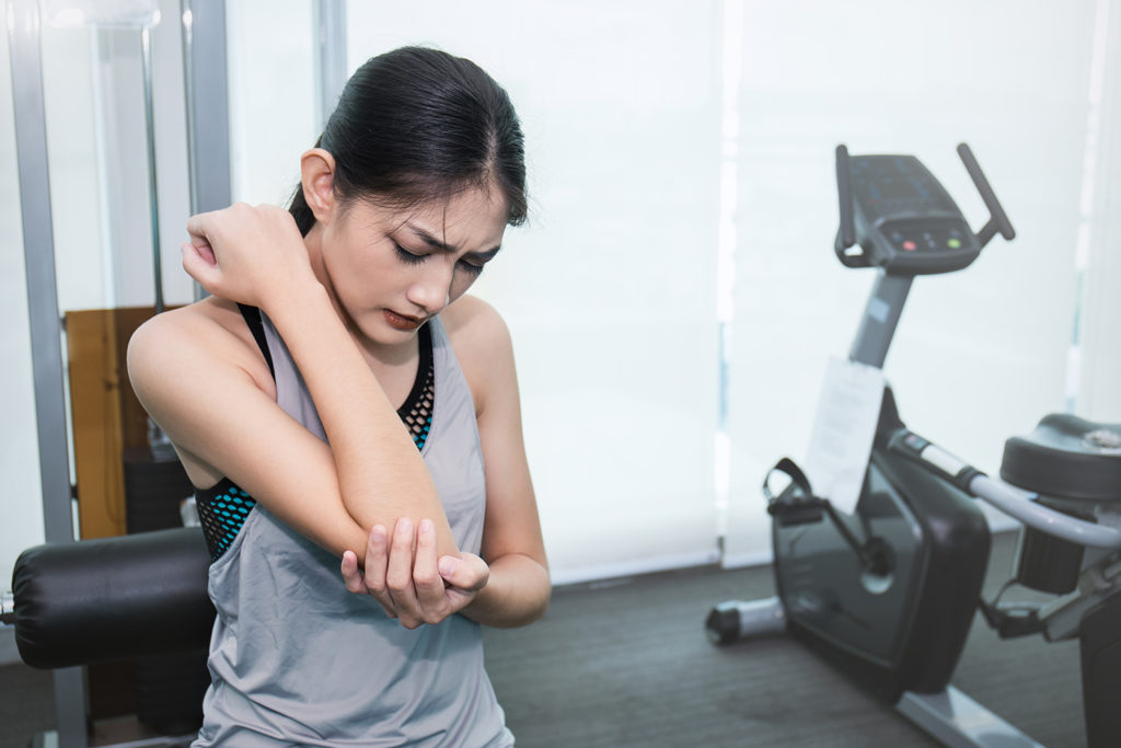
Carpal Tunnel Syndrome (CTS) is a common peripheral nerve disorder that affects the median nerve, which runs from the forearm into the hand through a narrow tunnel-like structure in the wrist called the carpal tunnel. The condition is caused by compression or pinching of the median nerve as it passes through this tunnel, leading to various symptoms such as numbness, tingling, and weakness in the hand and fingers.
The median nerve provides sensation to the thumb, index finger, middle finger, and half of the ring finger. It also controls some small muscles in the hand that allow for fine motor movements. When the median nerve is compressed or damaged due to CTS, it can result in a range of symptoms including:
1. Numbness, tingling, or burning sensations in the fingers (especially the thumb, index finger, middle finger, and half of the ring finger)
2. Pain or discomfort in the hand, wrist, or forearm
3. Weakness in the hand, leading to difficulty gripping objects or making a fist
4. A sensation of swelling or inflammation in the fingers, even if there is no visible swelling present
5. Nighttime symptoms that may disrupt sleep patterns
The exact cause of Carpal Tunnel Syndrome can vary from person to person, but some common risk factors include:
1. Repetitive hand and wrist motions (such as typing, writing, or using tools)
2. Prolonged exposure to vibrations (from machinery or power tools)
3. Wrist trauma or fractures
4. Pregnancy and hormonal changes
5. Certain medical conditions like diabetes, rheumatoid arthritis, and thyroid disorders
6. Obesity
7. Smoking
Diagnosis of Carpal Tunnel Syndrome typically involves a physical examination, medical history review, and sometimes specialized tests like nerve conduction studies or electromyography to confirm the diagnosis and assess the severity of the condition. Treatment options may include splinting, medication, corticosteroid injections, and in severe cases, surgery to relieve pressure on the median nerve.
Carpal bones are the eight small bones that make up the wrist joint in humans and other primates. These bones are arranged in two rows, with four bones in each row. The proximal row includes the scaphoid, lunate, triquetral, and pisiform bones, while the distal row includes the trapezium, trapezoid, capitate, and hamate bones.
The carpal bones play an essential role in the function of the wrist joint by providing stability, support, and mobility. They allow for a wide range of movements, including flexion, extension, radial deviation, ulnar deviation, and circumduction. The complex structure of the carpal bones also helps to absorb shock and distribute forces evenly across the wrist during activities such as gripping or lifting objects.
Injuries to the carpal bones, such as fractures or dislocations, can be painful and may require medical treatment to ensure proper healing and prevent long-term complications. Additionally, degenerative conditions such as arthritis can affect the carpal bones, leading to pain, stiffness, and decreased mobility in the wrist joint.
A joint is the location at which two or more bones make contact. They are constructed to allow movement and provide support and stability to the body during motion. Joints can be classified in several ways, including structure, function, and the type of tissue that forms them. The three main types of joints based on structure are fibrous (or fixed), cartilaginous, and synovial (or diarthrosis). Fibrous joints do not have a cavity and have limited movement, while cartilaginous joints allow for some movement and are connected by cartilage. Synovial joints, the most common and most movable type, have a space between the articular surfaces containing synovial fluid, which reduces friction and wear. Examples of synovial joints include hinge, pivot, ball-and-socket, saddle, and condyloid joints.
The median nerve is one of the major nerves in the human body, providing sensation and motor function to parts of the arm and hand. It originates from the brachial plexus, a network of nerves that arise from the spinal cord in the neck. The median nerve travels down the arm, passing through the cubital tunnel at the elbow, and continues into the forearm and hand.
In the hand, the median nerve supplies sensation to the palm side of the thumb, index finger, middle finger, and half of the ring finger. It also provides motor function to some of the muscles that control finger movements, allowing for flexion of the fingers and opposition of the thumb.
Damage to the median nerve can result in a condition called carpal tunnel syndrome, which is characterized by numbness, tingling, and weakness in the hand and fingers.
The wrist joint, also known as the radiocarpal joint, is a condyloid joint that connects the distal end of the radius bone in the forearm to the proximal row of carpal bones in the hand (scaphoid, lunate, and triquetral bones). It allows for flexion, extension, radial deviation, and ulnar deviation movements of the hand. The wrist joint is surrounded by a capsule and reinforced by several ligaments that provide stability and strength to the joint.
The knee joint, also known as the tibiofemoral joint, is the largest and one of the most complex joints in the human body. It is a synovial joint that connects the thighbone (femur) to the shinbone (tibia). The patella (kneecap), which is a sesamoid bone, is located in front of the knee joint and helps in the extension of the leg.
The knee joint is made up of three articulations: the femorotibial joint between the femur and tibia, the femoropatellar joint between the femur and patella, and the tibiofibular joint between the tibia and fibula. These articulations are surrounded by a fibrous capsule that encloses the synovial membrane, which secretes synovial fluid to lubricate the joint.
The knee joint is stabilized by several ligaments, including the medial and lateral collateral ligaments, which provide stability to the sides of the joint, and the anterior and posterior cruciate ligaments, which prevent excessive forward and backward movement of the tibia relative to the femur. The menisci, which are C-shaped fibrocartilaginous structures located between the femoral condyles and tibial plateaus, also help to stabilize the joint by absorbing shock and distributing weight evenly across the articular surfaces.
The knee joint allows for flexion, extension, and a small amount of rotation, making it essential for activities such as walking, running, jumping, and sitting.
Joint diseases is a broad term that refers to various conditions affecting the joints, including but not limited to:
1. Osteoarthritis (OA): A degenerative joint disease characterized by the breakdown of cartilage and underlying bone, leading to pain, stiffness, and potential loss of function.
2. Rheumatoid Arthritis (RA): An autoimmune disorder causing inflammation in the synovial membrane lining the joints, resulting in swelling, pain, and joint damage if left untreated.
3. Infectious Arthritis: Joint inflammation caused by bacterial, viral, or fungal infections that spread through the bloodstream or directly enter the joint space.
4. Gout: A type of arthritis resulting from the buildup of uric acid crystals in the joints, typically affecting the big toe and characterized by sudden attacks of severe pain, redness, and swelling.
5. Psoriatic Arthritis (PsA): An inflammatory joint disease associated with psoriasis, causing symptoms such as pain, stiffness, and swelling in the joints and surrounding tissues.
6. Juvenile Idiopathic Arthritis (JIA): A group of chronic arthritis conditions affecting children, characterized by joint inflammation, pain, and stiffness.
7. Ankylosing Spondylitis: A form of arthritis primarily affecting the spine, causing inflammation, pain, and potential fusion of spinal vertebrae.
8. Bursitis: Inflammation of the fluid-filled sacs (bursae) that cushion joints, leading to pain and swelling.
9. Tendinitis: Inflammation or degeneration of tendons, which connect muscles to bones, often resulting in pain and stiffness near joints.
These conditions can impact the function and mobility of affected joints, causing discomfort and limiting daily activities. Proper diagnosis and treatment are essential for managing joint diseases and preserving joint health.
A finger joint, also known as an articulation, is the point where two bones in a finger connect and allow for movement. The majority of finger joints are classified as hinge joints, permitting flexion and extension movements. These joints consist of several components:
1. Articular cartilage: Smooth tissue that covers the ends of the bones, enabling smooth movement and protecting the bones from friction.
2. Joint capsule: A fibrous sac enclosing the joint, providing stability and producing synovial fluid for lubrication.
3. Synovial membrane: Lines the inner surface of the joint capsule and produces synovial fluid to lubricate the joint.
4. Volar plate (palmar ligament): A strong band of tissue located on the palm side of the joint, preventing excessive extension and maintaining alignment.
5. Collateral ligaments: Two bands of tissue located on each side of the joint, providing lateral stability and limiting radial and ulnar deviation.
6. Flexor tendons: Tendons that attach to the bones on the palmar side of the finger joints, facilitating flexion movements.
7. Extensor tendons: Tendons that attach to the bones on the dorsal side of the finger joints, enabling extension movements.
Finger joints are essential for hand function and enable activities such as grasping, holding, writing, and manipulating objects.
 Intercarpal joints
Muscular system of the horse
Dog anatomy
Racehorse injuries
Spectacled flowerpecker
Eosinophilic fasciitis
Charybdis longicollis
Sheathbill
Mucopolysaccharidosis type I
Hurler syndrome
Development of The Last of Us
Midcarpal joint
Microraptor
Carpometacarpal bossing
Treatment of equine lameness
Glossary of bird terms
Bird flight
Joints of hand
Lupinus formosus
Growth hormone
Bonin petrel
Hyaluronic acid
Caprine arthritis encephalitis virus
Mountain goat
Northern royal albatross
Mucopolysaccharidosis
Charybdis hellerii
Bird measurement
Mobile wad
Extensor pollicis longus muscle
Evaluation of the effect of extracorporeal shock wave treatment on experimentally induced osteoarthritis in middle carpal...
 Beef Cattle Discovery - Skeletal - Carpal Joint | Animal & Food Sciences
 Hand joints, Carpal Tunnel Syndrome - Kineoself
 Carpal Tunnel Treatment | TENDLITE® - Fast Joint Pain Relief
Carpal Instability Webster, TX | Joint Surgeon Houston, Clear Lake, TX
 Joint Pain Blog- Tagged 'Carpal Tunnel Syndrome' - Page 2 - Grace & Able
 Hyaluronan Turnover in the Synovial Fluid in Metacarpophalangeal-and Middle Carpal Joints in Standardbred Horses | Acta...
Synovial Membranes of Wrist Hand, and Fingers. | anatomy of the carpal joints
 Carpal Tunnel Syndrome - Bone, Joint, and Muscle Disorders - MSD Manual Consumer Version
 Strain of carpal ligaments during wrist-joint motion<...
 Natural Carpal Tunnel Treatment with Chiropractic | Mt Sterling, KY Chiropractor | Linton Spine & Joint Chiropractic Center
Intercarpal joints - Wikipedia
 May 2018 - Neuropax Clinic is the St. Louis Leader for Carpal Tunnel, Headache Surgery, Nerve Compression, and Chronic Joint...
August 2023 - Neuropax Clinic is the St. Louis Leader for Carpal Tunnel, Headache Surgery, Nerve Compression, and Chronic Joint...
 Liebenberg syndrome: MedlinePlus Genetics
 Search Results
 Hand Surgery for Arthritis: Procedure, Recovery, Outcomes
 Perilunate Injury Imaging: Practice Essentials, Radiography, Computed Tomography
 Rough-legged Buzzard - BirdForum Opus | BirdForum
 Extending Breast Cancer Treatment may Reduce Risk of Breast Cancer Recurrence
 Comparison between high-field 3 Tesla MRI and computed tomography with and without arthrography for visualization of canine...
 CT angiography and MRI of hand vascular lesions: technical considerations and spectrum of imaging findings | Insights into...
Caprine Arthritis and Encephalitis - Generalized Conditions - Merck Veterinary Manual
2017 Cathedral Creek Gather | Bureau of Land Management
 Veal - Meat cuts manual - Canadian Food Inspection Agency
 Ms Margaret Strick</span><i class="icon" aria-hidden=...
 VM194/VM194: Comparing Waterbeds and Sand Beds for Cows: A Study at the UF/IFAS Dairy Unit
 Walkin' Carpal Splint for Dogs and Other Pets
Rheumatoid Arthritis Hand Imaging: Practice Essentials, Radiography, Magnetic Resonance Imaging
Bones23
- The "walls" of the carpal tunnel are formed by wrist and hand bones, and "the roof" is a ligament that runs lengthwise across the wrist known as the transverse carpal ligament. (kineoself.com)
- Carpal instability is the loss of alignment of the carpal bones and/or radioulnar joint. (andrewtylermd.com)
- It consists of 8 small bones called carpals that articulate with two long bones of the forearm (radius and ulna). (andrewtylermd.com)
- Instability occurs only to the carpal bones. (andrewtylermd.com)
- Misalignment of carpal bones with radius and ulna. (andrewtylermd.com)
- Combined misalignment of carpal bones, radius, and ulna. (andrewtylermd.com)
- Carpal instability can also be caused due to fracture of the wrist bones or degenerative arthritis. (andrewtylermd.com)
- Synovial sac of the wrist-joint Synovial sac of the carpus Synovial sac, occasionally separate, for the fourth and fifth metacarpal bones. (cloudaccess.net)
- Clear differences are observed between these 3D, experimentally obtained data and prevailing concepts on ligament behaviour based on 2D kinematics of the carpal bones. (tue.nl)
- The intercarpal joints (joints of the carpal bones of the wrist) can be subdivided into three sets of joints (also called articulations): Those of the proximal row of carpal bones, those of the distal row of carpal bones, and those of the two rows with each other. (wikipedia.org)
- The bones in each carpal row interlock with each other and each row can therefore be considered a single joint. (wikipedia.org)
- They are on a level with the superior surfaces of these bones, and their upper surfaces are smooth, and form part of the convex articular surface of the wrist-joint. (wikipedia.org)
- The prolongation between the greater and lesser multangulars, or that between the lesser multangular and capitate, is, owing to the absence of the interosseous ligament, often continuous with the cavity of the carpometacarpal joints, sometimes of the second, third, fourth, and fifth metacarpal bones, sometimes of the second and third only. (wikipedia.org)
- In the latter condition the joint between the hamate and the fourth and fifth metacarpal bones has a separate synovial membrane. (wikipedia.org)
- The synovial cavities of these joints are prolonged for a short distance between the bases of the metacarpal bones. (wikipedia.org)
- For example, bones in the elbows are abnormally shaped, which affects mobility of the joints. (medlineplus.gov)
- Joint fusion is surgery that joins two bones to form one solid bone. (healthline.com)
- The proximal row of carpal bones is tightly bound or held to the radius by the radiocarpal ligaments, particularly the radiolunate, radioscaphoid, and radiotriquetral, on the volar aspect of the wrist. (medscape.com)
- Erosions may be detected first either in the MCP and PIP joints or at the carpal bones. (medscape.com)
- Erosions may also be seen at the intra-articular portion of the distal end of the radius or within the carpal bones. (medscape.com)
- The distal end of the ulna tends to sublux dorsally, and the carpal bones sublux anteriorly to the distal radius and ulna. (medscape.com)
- Exercised horses also had a higher subchondral bone density in the metacarpal condyles than control horses, but such differences were not detected in the carpal bones. (avma.org)
- I can fully manipulate the paw, all of the joints and the bones any which way I want, until I force the pads of the paw into the weight bearing position and begin to place force upwards on the structure. (pets.ca)
Ligaments13
- The joint is supported by ligaments, tendons, nerves, blood vessels and muscles that help in movement. (andrewtylermd.com)
- Trauma or a fall on an outstretched arm can injure the wrist ligaments causing carpal instability. (andrewtylermd.com)
- To obtain a more accurate apprehension of the mechanics of the wrist-joint, the kinematical behaviour of the carpals and the length changes of the ligaments during hand-motions are determined. (tue.nl)
- It is suggested, based on results, that during some motions the carpal joint is not stabilized by one of the tested ligaments. (tue.nl)
- The joints of the proximal row are arthrodial joints, The scaphoid, lunate, and triquetrum are connected by dorsal, volar, and interosseous ligaments. (wikipedia.org)
- These joints are also arthrodial joints connected by dorsal, volar, and interosseous ligaments. (wikipedia.org)
- Caused by severe damage to the ligaments supporting the wrist of the forelimb, carpal hyperextension causes dogs to stand flat-footed with their wrist or carpus on the ground. (dogleggs.com)
- Although less common, carpal hyperextension may also develop in dogs with immune-mediated joint disease or degenerative conditions of the ligaments. (dogleggs.com)
- OBJECTIVE:To compare the quality of visualization of canine carpal ligaments by using computed tomography (CT), MRI, CT arthrography (CTA), and magnetic resonance arthrography (MRA). (uzh.ch)
- To evaluate the difference between imaging modalities, 3 observers graded carpal ligaments of clinical interest using a scale from 0 to 4 for their quality of visualization. (uzh.ch)
- CONCLUSION: Arthrography improved the capabilities of MRI but not of CT for visualization of the canine carpal ligaments. (uzh.ch)
- This is the peer reviewed version of the following article: Castelli E, Pozzi A, Klisch K, Scotti L, Hoey S, Dennler M. Comparison between high‐field 3 Tesla MRI and computed tomography with and without arthrography for visualization of canine carpal ligaments: A cadaveric study. (uzh.ch)
- Hormonal changes loosen ligaments in your pelvis, knees, and other joints, affecting your posture and range of motion. (babycenter.com)
Bone11
- Collection locations were chosen to represent an area directly adjacent to an experimentally induced OA fragment, a portion of the opposing articulating surface (third carpal bone), and a remote location (fourth carpal bone). (avma.org)
- Specimens for biochemical evaluation were obtained from the intermediate carpal bone (CI). (avma.org)
- CR = Radial carpal bone. (avma.org)
- CU = Ulnar carpal bone. (avma.org)
- C2 = Second carpal bone. (avma.org)
- C3 = Third carpal bone. (avma.org)
- C4 = Fourth carpal bone. (avma.org)
- Each carpal bone involved is included in the designation of a fracture-dislocation. (medscape.com)
- Carpal bone ankylosis is a common and fairly specific sign, particularly in the Asian population, in whom it tends to occur early in the disease process. (medscape.com)
- Objective -To determine effects of treadmill exercise on subchondral bone of carpal and metacarpophalangeal joints of 2-year-old horses. (avma.org)
- In addition, he commented, "whether rhGH can be used long-term is unclear, as we know that long-term exposure to high levels of growth hormone in adults may lead to adverse outcomes," such as an increased risk of diabetes, increased bone growth leading to joint abnormalities and increased pressure on nerves, which can result in carpal tunnel syndrome. (medscape.com)
Distal3
- and (2) a distal, the mid-carpal joint. (wikipedia.org)
- Line diagram illustrates the relative position of the capitate (distal carpal row), the lunate (proximal carpal row), and the radius. (medscape.com)
- The distal interphalangeal (DIP) joints are involved only in the presence of a coexisting MCP or PIP disease. (medscape.com)
Middle carpal joints2
- 24) experimentally induced in middle carpal joints and that were subsequently treated with placebo (n = 8 horses), ESWT (8), and PSGAG (8) 14 days after surgery, as measured 70 days after surgery. (avma.org)
- The mean t 1/2 in the metacarpophalangeal joints was 9.7 h for low molecular weight hyaluronan and 8.9 h for high molecular weight hya-luronan and in the middle carpal joints 16.0 h and 12.5 h respectively. (biomedcentral.com)
Developing carpal tunnel s1
- are at increased risk of developing carpal tunnel syndrome. (msdmanuals.com)
Ligament2
- These include arthrodesis, ligament reconstruction and tendon interposition (LRTI), and total joint arthroplasty. (healthline.com)
- If your thumb joints are severely damaged by arthritis or injury, your doctor may recommend ligament reconstruction and tendon interposition (LRTI) . (healthline.com)
Symptoms of carpal5
- Early symptoms of carpal tunnel syndrome include pain, numbness, and paresthesias. (kineoself.com)
- If you have mild or moderate symptoms of carpal tunnel syndrome, you may be able to relieve the pain with a few simple exercises. (kineoself.com)
- Keeping your hand in a neutral position while you sleep can improve symptoms of Carpal Tunnel Syndrome. (graceandable.com)
- Many people have symptoms of Carpal Tunnel Syndrome without knowing it. (atlantahandspecialist.com)
- Refusing to bear weight on the affected limb is one of the first symptoms of carpal hyperextension in dogs. (dogleggs.com)
Cause carpal2
- Conditions outside the wrist can cause carpal instability. (andrewtylermd.com)
- Prolonged exposure to vibrations (for example, by using certain power tools) has also been claimed to cause carpal tunnel syndrome. (msdmanuals.com)
Prevent carpal2
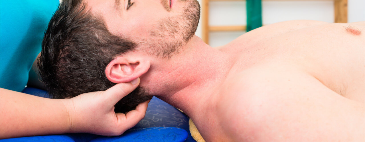
- Massage therapy can help treat and prevent carpal tunnel syndrome by promoting circulation, relieving inflammation, helping to eliminate metabolic residues and soothing irritated muscles and tendons. (kineoself.com)
- Although not always possible, the best way to prevent carpal hyperextension is to prevent dogs from jumping from high surfaces. (dogleggs.com)
Deformities3
- Affected individuals also have joint deformities (contractures) that limit movement of the elbows, wrists, and hands. (medlineplus.gov)
- Disorganization of the joint leads to deformities and loss of function. (medscape.com)
- Two equine surgeons shared their thoughts on rotational limb deformities, contracted carpal joints, club feet, and more. (thehorse.com)
Sprains2
- CLINICAL SIGNIFICANCE: 3 Tesla MRA and MRI allow excellent visualization of the ligamentous morphology and may be helpful in the diagnostic process of carpal sprains in dogs. (uzh.ch)
- As a cold compress it can be used for pain relief and to help with inflammation of sore joints, muscles and sprains. (57aromas.com)
Elbow3
- These painful conditions can affect the wrist, fingers, elbow or other joints, and only experienced joint surgeons can provide the treatment that allows patients to make a complete and comprehensive recovery. (atlantahandspecialist.com)
- Patients who suffer injuries to the joints of the hand, wrist, or elbow can benefit from the treatment skilled joint surgeons can provide. (atlantahandspecialist.com)
- In a prior version of this table, the code SRT: T-D8300 was used for (16953009, SCT, "Elbow Joint") . (nema.org)
Proximal3
- In the hands, the metacarpophalangeal (MCP), proximal interphalangeal (PIP), and thumb interphalangeal (IP) joints are most frequently involved. (medscape.com)
- DIP joint involvement without proximal involvement is rare. (medscape.com)
- Soft-tissue swelling and early erosions in the proximal interphalangeal joints in a patient with rheumatoid arthritis of the hands. (medscape.com)
Metacarpophalangeal2
- The biological turnover of hyaluronan (sodium hyaluronate) of different molecular weights (0.6×10 6 and 2.5×10 6 Daltons) was studied in the synovial fluid of the middle carpal and metacarpophalangeal joints of 6 clinically healthy Standardbred horses. (biomedcentral.com)
- Ultrasonography-guided synovial biopsy of the second metacarpophalangeal joint of the right hand in a patient with rheumatoid arthritis of the hands. (medscape.com)
Repetitive strain1
- If you suffer from carpal tunnel or repetitive strain injury, black spruce hydrosol can help alleviate your symptoms. (57aromas.com)
Tendon2
- Without sufficient rest, you're at risk for unnatural repair or chronic inflammation of your tendon which will give you ongoing Carpal Tunnel Syndrome . (tendlite.com)
- Ganglion Cysts are commonly filled with fluid, and form because of tendon or joint irritation. (atlantahandspecialist.com)
Flexion2
- Furthermore, they also found that the degree of the women's reported carpal tunnel discomfort was connected to the lateral flexion of the cervical spine. (myspinecenter.info)
- There is a lack of consensus regarding median nerve movement in the carpal tunnel during composite finger flexion in healthy individuals. (cdc.gov)
Carpus2
- See Midcarpal joint The synovial membrane of the carpus is very extensive, and bounds a synovial cavity of very irregular shape. (wikipedia.org)
- (8205005, SCT, "Carpus") is used in preference to carpal (wrist) joint. (nema.org)
Tunnel syndrome results2
- Carpal tunnel syndrome results from increased pressure in the carpal tunnel and subsequent compression of the median nerve. (kineoself.com)
- Carpal tunnel syndrome results from compression (pinching) of the median nerve. (msdmanuals.com)
Limb1
- Cartiledge would react when manipulation to the specific joints in the limb occured as well. (pets.ca)
Tendons3
- The carpal tunnel is called a tunnel because it is the narrow passageway through which nerves and tendons pass through the wrist to the hand. (msdmanuals.com)
- Surgeons can also reposition your tendons to help your joint function with less pain. (healthline.com)
- This procedure removes and replaces all or part of the affected joint with a graft made from one of your tendons or metal. (healthline.com)
Instability2
- What is Carpal Instability? (andrewtylermd.com)
- What Causes Carpal Instability? (andrewtylermd.com)
Inflammation2
- The artificial joint tends to break down over time from the wear and tear of daily use and chronic inflammation. (healthline.com)
- It might likewise give suggestive help to intense and constant circumstances like migraine, facial agony, carpal passage disorder and joint inflammation. (soundrite-acoustics.com)
Injuries3
- In certain critical carpal tunnel injuries, it will require up to a year. (tendlite.com)
- Carpal injuries can therefore be classified into perilunate fracture-dislocations and perilunate dislocations. (medscape.com)
- Patients with other neurological or musculoskeletal conditions including Cervical radiculopathy, History of wrist and hand fractures, Upper extremity joint dislocations, Brachial plexus injuries, Cubital tunnel syndrome, Rheumatoid arthritis, De quervain's tenosynovitis, Cut injuries of hand. (who.int)
Splint1
- It is important to take occasional breaks from wearing the splint, as constant wear can contribute to joints stiffening and muscles weakening. (kineoself.com)
Midcarpal2
- See Midcarpal joint This article incorporates text in the public domain from page 328 of the 20th edition of Gray's Anatomy (1918) Platzer 2004, p 130 Platzer, Werner (2004). (wikipedia.org)
- this area accentuates the stresses at the midcarpal joint, resulting in dislocation between the capitate and the lunate. (medscape.com)
Arthroplasty3
- Basal joint arthroplasty and carpal tunnel release comparing a single versus double incision: a prospective randomized study. (vhir.org)
- Joint replacement, also called arthroplasty, is best for larger joints in your hands, like your knuckles. (healthline.com)
- If other treatments have failed to provide relief in your thumb, your doctor may suggest a total joint arthroplasty. (healthline.com)
Neuropax1
- Neuropax Clinic is the St. Louis Leader for Carpal Tunnel, Headache Surgery, Nerve Compression, and Chronic Joint Pain. (neuropaxclinic.com)
Rheumatoid3
- The most common causes of carpal tunnel syndrome include genetic predisposition, history of repetitive wrist movements such as typing, or machine work as well as obesity, autoimmune disorders such as rheumatoid arthritis, and pregnancy. (kineoself.com)
- Rheumatoid Arthritis (RA) Rheumatoid arthritis is an inflammatory arthritis in which joints, usually including those of the hands and feet, are inflamed, resulting in swelling, pain, and often destruction of joints. (msdmanuals.com)
- Though not directly caused by arthritis, carpal tunnel syndrome is common in people with rheumatoid arthritis. (healthline.com)
Compression4
- Carpal tunnel syndrome (CTS) is an entrapment neuropathy caused by compression of the median nerve as it travels through the wrist's carpal tunnel. (kineoself.com)
- 1. R.I.C.E. (Rest, Ice, Compression & Elevation) - Start your Carpal Tunnel Syndrome treatment using first aid for your wrist. (tendlite.com)
- Simple compression of the wrist joint is used to cut back on the swelling generated by the inflammatory process. (tendlite.com)
- Carpal tunnel syndrome is a painful compression (pinching) of the median nerve as it passes through the carpal tunnel in the wrist. (msdmanuals.com)
Anatomical1
- To interpret nerve mobility findings among clinical populations and to be able to evaluate effects of functional hand use on pathological changes of the median nerve, it is essential to illustrate and understand the dynamic biomechanics of the normal anatomical structures in the carpal tunnel in healthy people. (cdc.gov)
Synovial5
- Brown TJ, Laurent UBG, Fraser JRE: Turnover of hyaluronan in synovial joints: elimination of la-belled hyaluronan from the knee joint of the rab-bit. (biomedcentral.com)
- Heinegãrd D, Inerot S, Wieslander J, Lindblad G: A method for the quantification of cartilage prote-oglycan structures liberated to the synovial fluid during developing degenerative joint disease. (biomedcentral.com)
- Roneus B, Lindblad G, Lindholm A, Jones B: Effects of intraarticular corticosteroid and sodium hyalu-ronate injections on synovial fluid production and synovial fluid content of sodium hyaluronate and proteoglycans in normal equine joints. (biomedcentral.com)
- anatomy of the carpal joints - Synovial Membranes of Wrist Hand, and Fingers. (cloudaccess.net)
- RA is characterized by a typical pattern and distribution of synovial joint involvement. (medscape.com)
Wrists1
- Carpal Tunnel is common in people that work primarily with their hands, fingers and wrists with repetitive actions. (neuropaxclinic.com)
Cervical1
- Women with carpal tunnel syndrome show restricted cervical range of motion. (myspinecenter.info)
Canine1
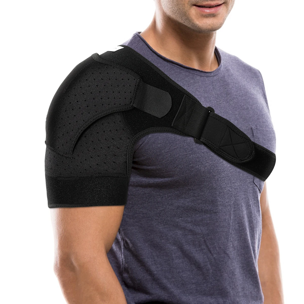
- Canine support products like DogLeggsTM Carpal Support provide optimal support of the joint following treatment. (dogleggs.com)
Articular2
- Illustration of the sites from which articular cartilage was obtained from a middle carpal joint for histologic evaluation of the effects of treatment with ESWT and PSGAG in horses. (avma.org)
- It also provides articular branches to the wrist and carpal joints. (medscape.com)
Median3
- The median nerve is located at the palm side of the wrist and passes through the carpal tunnel. (msdmanuals.com)
- Carpal Tunnel Syndrome is caused by repeated and frequent pressure on the median nerve. (neuropaxclinic.com)
- In patients with carpal tunnel syndrome, the median nerve tended to have more limited movements during finger movements than in healthy controls, with more restricted mobility as symptoms increased or the condition became more chronic. (cdc.gov)
Lateral1
- Line diagram depicts normal wrist and carpal joints in dorsipalmar and lateral projections. (medscape.com)
Fractures1
- The perilunate is an uncommon type of carpal dislocation and is commonly associated with carpal and other wrist fractures. (medscape.com)
Pain12
- Hand Therapist designed to reduce strain on the joint and muscles, and alleviate pain and swelling. (graceandable.com)
- If you are suffering from carpal tunnel pain, call our Mt Sterling location and ask how Linton Spine & Joint Chiropractic Center can help you get relief! (myspinecenter.info)
- This technique involves evaluating certain sensory nerves that provide pain fibers to the joint capsules. (neuropaxclinic.com)
- Equine veterinarians face challenges in treating horses with osteoarthritic joint pain in routine veterinary practice. (cabi.org)
- It can reduce pain and improve joint function. (healthline.com)
- Depending on the extent of injury, those affected may experience swelling around the joint and vocalize or cry out in pain. (dogleggs.com)
- Carpal hyperextension can cause significant pain and discomfort. (dogleggs.com)
- Aromatase inhibitors can cause osteoporosis, joint pain or carpal tunnel syndrome. (medindia.net)
- Although occasional flares of joint pain occur throughout the course of the disease, these can usually be controlled with the use of anti-inflammatory medication, especially early in their course. (medscape.com)
- Working from home is causing an increase in physical problems, from headaches to lower back pain to carpal tunnel syndrome. (scmp.com)
- It is also used for pain relief - carpal tunnel, joint pain, back pain, aftershave and pet care. (57aromas.com)
- The most common MSD in dentistry are chronic low back pain, neck tension syndrome, trapezius myalgia, shoulder joint injury e carpal tunnel syndrome and upper extremity tendonitis 7 . (bvsalud.org)
Fingers2
- It can treat arthritis that affects the small joints of the fingers. (healthline.com)
- Changes in your hormones may also affect smaller joints, like your fingers. (babycenter.com)
Mobility1
- This treatment can allow patients to heal and regain full mobility in their joints. (atlantahandspecialist.com)
Common2
- Carpal tunnel syndrome is very common, especially among women aged 30 to 50 years. (msdmanuals.com)
- Although carpal hyperextension can affect dogs of all ages and breeds, the condition is more common in large, active breeds. (dogleggs.com)
Treatment8
- If carpal tunnel syndrome is identified early conservative treatment is recommended. (kineoself.com)
- Tendlite® Device speeds up the carpal tunnel treatment up to 60% by increasing your body's natural healing response. (tendlite.com)
- At Linton Spine & Joint Chiropractic Center, we offer a broad range of treatment options and services to our Mt. Sterling patients. (myspinecenter.info)
- Because they are so skilled, joint surgeons like Dr. Patel are in high demand, so patients seeking treatment should schedule their appointment as soon as possible. (atlantahandspecialist.com)
- Find out how the condition is caused and what treatment you'll receive from Atlanta Hand Specialists for Carpal Tunnel Syndrome. (atlantahandspecialist.com)
- Treatment of carpal hyperextension depends on the cause and extent of injury. (dogleggs.com)
- With proper treatment, the prognosis is good for dogs with carpal hyperextension. (dogleggs.com)
- However, it's important to maintain adequate support of the joint during and after treatment using a quality carpal support like those offered by DogLeggs TM . (dogleggs.com)
Movement1
- Joints get nutrition in and waste out by physical movement. (gaia.com)
Occurs1
- The primary effect of RA is in joint deformity and fusion, which occurs in the advanced stages. (medscape.com)
Physical Therapy1
- Recent research printed in the Journal of Orthopaedic & Sports Physical Therapy assessed 71 women between the ages of 35 and 59 who were diagnosed with carpal tunnel syndrome. (myspinecenter.info)
Dogs2
- However, dogs may also develop carpal hyperextension from repeated injury caused by jumping from an elevated surface. (dogleggs.com)
- Sometimes, dogs require surgery to fuse the carpal joints together. (dogleggs.com)
People3
- This is a good solution for people with mild to moderate carpal tunnel syndrome whose symptoms flare up for a few weeks, then dissipate. (kineoself.com)
- Linton Spine & Joint Chiropractic Center sees a lot of people struggling with carpal tunnel syndrome in our busy Mt Sterling chiropractic office. (myspinecenter.info)
- Joint fusion has a high success rate, with only about 17% of people experiencing complications. (healthline.com)
Cavities1
- Ekman L, Nilsson G, Persson L, Lumsden LH: Vol-ume of the synovia in certain joint cavities in the horse. (biomedcentral.com)
Patients1
- Patients who have underwent recent carpal tunnel release (within 1 year). (who.int)