Carotid Sinus
Carotid Arteries
Pressoreceptors
Carotid Stenosis
Carotid Artery Diseases
Carotid Body
Syncope
Endarterectomy, Carotid
Carotid Artery, Internal
Chemoreceptor Cells
Massage
Carotid Artery, Common
Cranial Sinuses
Maxillary Sinus
Reflex
Cavernous Sinus
Paranasal Sinuses
Carotid Artery, External
Baroreflex
Frontal Sinus
Dogs
Sphenoid Sinus
Paranasal Sinus Diseases
Tilt-Table Test
Vagotomy
Sick Sinus Syndrome
Coronary Sinus
Syncope, Vasovagal
Vagus Nerve
Carotid Artery Thrombosis
Sinus Thrombosis, Intracranial
Glossopharyngeal Nerve
Carotid Artery Injuries
Sodium Cyanide
Ethmoid Sinus
Cats
Carotid Body Tumor
Bradycardia
Carotid Intima-Media Thickness
Respiration
Heart Massage
Sympathetic Nervous System
Autonomic Denervation
Vascular Resistance
Phrenic Nerve
Tunica Intima
Endarterectomy
Tunica Media
Reflex, Abnormal
Chloralose
Tachycardia, Sinus
Heart Sounds
Pacemaker, Artificial
Anesthesia
Maxillary Sinus Neoplasms
Carotid Artery, Internal, Dissection
Dizziness
Sympathectomy
Cross Circulation
Electrocardiography
Veratrine
Cerebral Angiography
Carbon Dioxide
Hemodynamics
Pressure
Transverse Sinuses
Prospective Studies
Efferent Pathways
Rabbits
Cardiac Pacing, Artificial
Ultrasonography, Doppler, Duplex
Medulla Oblongata
Superior Sagittal Sinus
Aorta, Thoracic
Treatment Outcome
Vasomotor System
Stents
Hypotension
Unconsciousness
Ischemic Attack, Transient
Autonomic Nerve Block
Cardiac Output
Afferent Pathways
Tomography, X-Ray Computed
Mechanoreceptors
Hypotension, Orthostatic
Reduction in baroreflex cardiovascular responses due to venous infusion in the rabbit. (1/508)
We studied reflex bradycardia and depression of mean arterial blood pressure (MAP) during left aortic nerve (LAN) stimulation before and after volume infusion in the anesthetized rabbit. Step increases in mean right atrial pressure (MRAP) to 10 mm Hg did not result in a significant change in heart rate or MAP. After volume loading, responses to LAN stimulation were not as great and the degree of attenuation was propoetional to the level of increased MRAP. A change in responsiveness was observed after elevation of MRAP by only 1 mm Hg, corresponding to less than a 10% increase in average calculated blood volume. after an increase in MRAP of 10 mm Hg, peak responses were attenuated by 44% (heart rate) and 52% (MAP), and the initial slopes (rate of change) were reduced by 46% (heart rate) and 66% (MAP). Comparison of the responses after infusion with blood and dextran solutions indicated that hemodilution was an unlikely explanation for the attenuation of the reflex responses. Total arterial baroreceptor denervation (ABD) abolished the volume-related attenuation was still present following bilateral aortic nerve section or vagotomy. It thus appears that the carotid sinus responds to changes inblood volume and influences the reflex cardiovascular responses to afferent stimulation of the LAN. On the other hand, cardiopulmonary receptors subserved by vagal afferents do not appear to be involved. (+info)Quantification of baroreceptor influence on arterial pressure changes seen in primary angiotension-induced hypertension in dogs. (2/508)
We studied the role of the sino-aortic baroreceptors in the gradual development of hypertension induced by prolonged administration of small amounts of angiotensin II (A II) in intact dogs and dogs with denervated sino-aortic baroreceptors. Short-term 1-hour infusions of A II(1.0-100 ng/kg per min) showed that conscious denervated dogs had twice the pressor sensitivity of intact dogs. Long-term infusions of A II at 5.0 ng/kg per min (2-3 weeks) with continuous 24-hour recordings of arterial pressure showed that intact dogs required 28 hours to reach the same level of pressure attained by denervated dogs during the 1st hour of infusion. At the 28th hour the pressure in both groups was 70% of the maximum value attained by the 7th day of infusion. Both intact and denervated dogs reached nearly the same plateau level of pressure, the magnitude being directly related both the the A II infusion rate and the daily sodium intake. Cardiac output in intact dogs initially decreased after the onset of A II infusion, but by the 5th day of infusion it was 38% above control, whereas blood volume was unchanged. Heart rate returned to normal after a reduction during the 1st day of infusion in intact dogs. Plasma renin activity could not be detected after 24 hours of A II infusion in either intact or denervated dogs. The data indicate that about 35% of the hypertensive effect of A II results from its acute pressor action, and an additional 35% of the gradual increase in arterial pressure is in large measure a result of baroreceptor resetting. We conclude that the final 30% increase in pressure seems to result from increased cardiac output, the cause of which may be decreased vascular compliance. since the blood volume remains unaltered. (+info)Hypoxia inhibits baroreflex vagal bradycardia via a central action in anaesthetized rats. (3/508)
It is known that arterial baroreflexes are suppressed in stressful conditions. The present study was designed to determine whether and how hypoxia affects arterial baroreflexes, especially the heart rate component, baroreflex vagal bradycardia. In chloralose-urethane-anaesthetized rats, baroreflex vagal bradycardia was evoked by electrical stimulation of the aortic depressor nerve, and the effect of 15 s inhalation of hypoxic gas (4% O2) was studied. Inhalation of hypoxic gas was found to inhibit baroreflex vagal bradycardia. The inhibition persisted after bilateral transection of the carotid sinus nerve. Cervical vagus nerves were cut bilaterally and their peripheral cut ends were stimulated to provoke vagal bradycardia of peripheral origin so as to determine whether hypoxia could inhibit vagal bradycardia by acting on a peripheral site. In contrast to baroreflex vagal bradycardia, the vagus-induced bradycardia was not affected by hypoxic gas inhalation. It is concluded that baroreflex vagal bradycardia is inhibited by hypoxia and the inhibition is largely mediated by its direct central action. (+info)Responses of abdominal vascular capacitance in the anaesthetized dog to changes in carotid sinus pressure. (4/508)
1. The abdominal circulation of anaesthetized dogs was vascularly isolated without opening the abdomen, by cutting or tying all structures immediately above the diaphragm and tying the proximal ends of the hind limbs. The region was perfused at constant flow through the aorta and drained at constant pressure from the inferior vena cava. 2. Vascular resistance responses were expressed as the changes in perfusion pressure and capacitance responses were determined by integrating changes in vena caval outflow. 3. Decreasing the pressure in the isolated carotid sinuses over the whole baroreceptor sensitivity range increased mean perfusion pressure from 91 to 149 mmHg (a 67% increase in resistance) and decreased mean capacitance by 111 ml. (5 ml. kg-1). 4. The range of carotid sinus pressures over which capacitance responses occurred was at a significantly higher level than the corresponding range for resistance responses. 5. Comparison of the reflex responses with the responses to direct stimulation of efferent sympathetic nerves shows that quantitatively similar responses of resistance and capacitance to those induced by a large step decrease in carotid pressure could be produced by stimulating maximally the efferent sympathetic nerves at 5 Hz. These results also suggest that at all levels of carotid sinus pressure there is no difference in the impulse traffic to resistance and capacitance vessels. (+info)Carotid sinus hypersensitivity--a modifiable risk factor for fractured neck of femur. (5/508)
BACKGROUND: the potential impact on morbidity, mortality and health care economics makes it important to identify patients at risk of fracture, in particular fractured neck of femur (FNOF). Older patients with carotid sinus hypersensitivity (CSH) are more likely to have unexplained falls and to experience fractures, particularly FNOF. Our objective was to determine the prevalence of CSH in patients with FNOF. DESIGN: case-controlled prospective series. METHODS: consecutive cases were admissions over 65 years with FNOF. Controls were consecutive patients admitted for elective hip surgery, frail elderly people admitted to hospital medical wards and day-hospital patients. All patients had a clinical assessment of cognitive function, physical abilities and history of previous syncope, falls and dizziness, in addition to repeated carotid sinus massage with continuous heart rate and phasic blood pressure measurement. RESULTS: heart rate slowing and fall in systolic blood pressure was greater for patients with FNOF than those admitted for elective hip surgery (P < 0.05 and P < 0.001). CSH was present in 36% of the FNOF group, none of the elective surgery group, 13% of the acutely ill controls and 17% of the outpatients. It was more likely to be present in FNOF patients with a previous history of unexplained falls or an unexplained fall causing the index fracture. The heart rate and systolic blood pressure responses to carotid sinus stimulation were reproducible. CONCLUSION: older patients with an acute neck of femur fracture who do not give a clear history of an accidental fall or who have had previously unexplained falls are likely to have CSH. CSH may be a modifiable risk factor for older patients at risk of hip fracture. (+info)New analytic framework for understanding sympathetic baroreflex control of arterial pressure. (6/508)
The sympathetic baroreflex is an important feedback system in stabilization of arterial pressure. This system can be decomposed into the controlling element (mechanoneural arc) and the controlled element (neuromechanical arc). We hypothesized that the intersection of the two operational curves representing their respective functions on an equilibrium diagram should define the operating point of the arterial baroreflex. Both carotid sinuses were isolated in 16 halothane-anesthetized rats. The vagi and aortic depressor nerves were cut bilaterally. Carotid sinus pressure (CSP) was sequentially altered in 10-mmHg increments from 80 to 160 mmHg while sympathetic efferent nerve activity (SNA) and systemic arterial pressure (SAP) were recorded simultaneously under various hemorrhagic conditions. The mechanoneural arc was characterized by the response of SNA to CSP and the neuromechanical arc by the response of SAP to SNA. We parametrically analyzed the relationship between input and output for each arc using a four-parameter logistic equation model. In baseline states, the two arcs intersected each other at the point at which the instantaneous gain of each arc attained its maximum. Severe hemorrhage lowered the gain and offset of the neuromechanical arc and moved the operating point, whereas the mechanoneural arc remained unchanged. The operating points measured under the closed-loop conditions were indistinguishable from those estimated from the intersections of the two arc curves on the equilibrium diagram. The average root mean square errors of estimate for arterial pressure and SNA were 2 and 3%, respectively. Such an analytic approach could explain a mechanism for the determination of the operating point of the sympathetic baroreflex system and thus helps us integratively understand its function. (+info)Carotid baroreflex function during prolonged exercise. (7/508)
The present investigation was designed to uncouple the hemodynamic physiological effects of thermoregulation from the effects of a progressively increasing central command activation during prolonged exercise. Subjects performed two 1-h bouts of leg cycling exercise with 1) no intervention and 2) continuous infusion of a dextran solution to maintain central venous pressure constant at the 10-min pressure. Volume infusion resulted in a significant reduction in the decrement in mean arterial pressure seen in the control exercise bout (6.7 +/- 1.8 vs. 11.6+/- 1.3 mmHg, respectively). However, indexes of central command such as heart rate and ratings of perceived exertion rose to a similar extent during both exercise conditions. In addition, the carotid-cardiac baroreflex stimulus-response relationship, as measured by using the neck pressure-neck suction technique, was reset from rest to 10 min of exercise and was further reset from 10 to 50 min of exercise in both exercise conditions, with the operating point being shifted toward the reflex threshold. We conclude that the progressive resetting of the carotid baroreflex and the shift of the reflex operating point render the carotid-cardiac reflex ineffectual in counteracting the continued decrement in mean arterial pressure that occurs during the prolonged exercise. (+info)Chronic hypoxia enhances the phrenic nerve response to arterial chemoreceptor stimulation in anesthetized rats. (8/508)
Chronic exposure to hypoxia results in a time-dependent increase in ventilation called ventilatory acclimatization to hypoxia. Increased O(2) sensitivity of arterial chemoreceptors contributes to ventilatory acclimatization to hypoxia, but other mechanisms have also been hypothesized. We designed this experiment to determine whether central nervous system processing of peripheral chemoreceptor input is affected by chronic hypoxic exposure. The carotid sinus nerve was stimulated supramaximally at different frequencies (0.5-20 Hz, 0.2-ms duration) during recording of phrenic nerve activity in two groups of anesthetized, ventilated, vagotomized rats. In the chronically hypoxic group (7 days at 80 Torr inspired PO(2)), phrenic burst frequency (f(R), bursts/min) was significantly higher than in the normoxic control group with carotid sinus nerve stimulation frequencies >5 Hz. In the chronically hypoxic group, peak amplitude of integrated phrenic nerve activity ( integral Phr, percent baseline) or change in integral Phr was significantly greater at stimulation frequencies between 5 and 17 Hz, and minute phrenic activity ( integral Phr x f(R)) was significantly greater at stimulation frequencies >5 Hz. These experiments show that chronic hypoxia facilitates the translation of arterial chemoreceptor afferent input to ventilatory efferent output through a mechanism in the central nervous system. (+info)The carotid sinus is a small, dilated area located at the bifurcation (or fork) of the common carotid artery into the internal and external carotid arteries. It is a baroreceptor region, which means it contains specialized sensory nerve endings that can detect changes in blood pressure. When the blood pressure increases, the walls of the carotid sinus stretch, activating these nerve endings and sending signals to the brain. The brain then responds by reducing the heart rate and relaxing the blood vessels, which helps to lower the blood pressure back to normal.
The carotid sinus is an important part of the body's autonomic nervous system, which regulates various involuntary functions such as heart rate, blood pressure, and digestion. It plays a crucial role in maintaining cardiovascular homeostasis and preventing excessive increases in blood pressure that could potentially damage vital organs.
The carotid arteries are a pair of vital blood vessels in the human body that supply oxygenated blood to the head and neck. Each person has two common carotid arteries, one on each side of the neck, which branch off from the aorta, the largest artery in the body.
The right common carotid artery originates from the brachiocephalic trunk, while the left common carotid artery arises directly from the aortic arch. As they ascend through the neck, they split into two main branches: the internal and external carotid arteries.
The internal carotid artery supplies oxygenated blood to the brain, eyes, and other structures within the skull, while the external carotid artery provides blood to the face, scalp, and various regions of the neck.
Maintaining healthy carotid arteries is crucial for overall cardiovascular health and preventing serious conditions like stroke, which can occur when the arteries become narrowed or blocked due to the buildup of plaque or fatty deposits (atherosclerosis). Regular check-ups with healthcare professionals may include monitoring carotid artery health through ultrasound or other imaging techniques.
Pressoreceptors are specialized sensory nerve endings found in the walls of blood vessels, particularly in the carotid sinus and aortic arch. They respond to changes in blood pressure by converting the mechanical stimulus into electrical signals that are transmitted to the brain. This information helps regulate cardiovascular function and maintain blood pressure homeostasis.
Carotid stenosis is a medical condition that refers to the narrowing or constriction of the lumen (inner space) of the carotid artery. The carotid arteries are major blood vessels that supply oxygenated blood to the head and neck. Carotid stenosis usually results from the buildup of plaque, made up of fat, cholesterol, calcium, and other substances, on the inner walls of the artery. This process is called atherosclerosis.
As the plaque accumulates, it causes the artery to narrow, reducing blood flow to the brain. Severe carotid stenosis can increase the risk of stroke, as a clot or debris from the plaque can break off and travel to the brain, blocking a smaller blood vessel and causing tissue damage or death.
Carotid stenosis is typically diagnosed through imaging tests such as ultrasound, CT angiography, or MRI angiography. Treatment options may include lifestyle modifications (such as quitting smoking, controlling blood pressure, and managing cholesterol levels), medications to reduce the risk of clots, or surgical procedures like endarterectomy or stenting to remove or bypass the blockage.
Carotid artery diseases refer to conditions that affect the carotid arteries, which are the major blood vessels that supply oxygen-rich blood to the head and neck. The most common type of carotid artery disease is atherosclerosis, which occurs when fatty deposits called plaques build up in the inner lining of the arteries.
These plaques can cause the arteries to narrow or become blocked, reducing blood flow to the brain and increasing the risk of stroke. Other carotid artery diseases include carotid artery dissection, which occurs when there is a tear in the inner lining of the artery, and fibromuscular dysplasia, which is a condition that affects the muscle and tissue in the walls of the artery.
Symptoms of carotid artery disease may include neck pain or pulsations, transient ischemic attacks (TIAs) or "mini-strokes," and strokes. Treatment options for carotid artery disease depend on the severity and type of the condition but may include lifestyle changes, medications, endarterectomy (a surgical procedure to remove plaque from the artery), or angioplasty and stenting (procedures to open blocked arteries using a balloon and stent).
The carotid body is a small chemoreceptor organ located near the bifurcation of the common carotid artery into the internal and external carotid arteries. It plays a crucial role in the regulation of respiration, blood pressure, and pH balance by detecting changes in the chemical composition of the blood, particularly oxygen levels, carbon dioxide levels, and hydrogen ion concentration (pH).
The carotid body contains specialized nerve endings called glomus cells that are sensitive to changes in these chemical parameters. When there is a decrease in oxygen or an increase in carbon dioxide or hydrogen ions, the glomus cells release neurotransmitters such as acetylcholine and dopamine, which activate afferent nerve fibers leading to the brainstem's nucleus tractus solitarius. This information is then integrated with other physiological signals in the brainstem, resulting in appropriate adjustments in breathing rate, depth, and pattern, as well as changes in heart rate and blood vessel diameter to maintain homeostasis.
Dysfunction of the carotid body can lead to various disorders, such as hypertension, sleep apnea, and chronic lung disease. In some cases, overactivity of the carotid body may result in conditions like primary breathing pattern disorders or pseudohypoxia, where the body responds as if it is experiencing hypoxia despite normal oxygen levels.
Syncope is a medical term defined as a transient, temporary loss of consciousness and postural tone due to reduced blood flow to the brain. It's often caused by a drop in blood pressure, which can be brought on by various factors such as dehydration, emotional stress, prolonged standing, or certain medical conditions like heart diseases, arrhythmias, or neurological disorders.
During a syncope episode, an individual may experience warning signs such as lightheadedness, dizziness, blurred vision, or nausea before losing consciousness. These episodes usually last only a few minutes and are followed by a rapid, full recovery. However, if left untreated or undiagnosed, recurrent syncope can lead to severe injuries from falls or even life-threatening conditions related to the underlying cause.
Carotid endarterectomy is a surgical procedure to remove plaque buildup (atherosclerosis) from the carotid arteries, which are the major blood vessels that supply oxygen-rich blood to the brain. The surgery involves making an incision in the neck, opening the carotid artery, and removing the plaque from the inside of the artery wall. The goal of the procedure is to restore normal blood flow to the brain and reduce the risk of stroke caused by the narrowing or blockage of the carotid arteries.
The internal carotid artery is a major blood vessel that supplies oxygenated blood to the brain. It originates from the common carotid artery and passes through the neck, entering the skull via the carotid canal in the temporal bone. Once inside the skull, it branches into several smaller vessels that supply different parts of the brain with blood.
The internal carotid artery is divided into several segments: cervical, petrous, cavernous, clinoid, and supraclinoid. Each segment has distinct clinical significance in terms of potential injury or disease. The most common conditions affecting the internal carotid artery include atherosclerosis, which can lead to stroke or transient ischemic attack (TIA), and dissection, which can cause severe headache, neck pain, and neurological symptoms.
It's important to note that any blockage or damage to the internal carotid artery can have serious consequences, as it can significantly reduce blood flow to the brain and lead to permanent neurological damage or even death. Therefore, regular check-ups and screening tests are recommended for individuals at high risk of developing vascular diseases.
Chemoreceptor cells are specialized sensory neurons that detect and respond to chemical changes in the internal or external environment. They play a crucial role in maintaining homeostasis within the body by converting chemical signals into electrical impulses, which are then transmitted to the central nervous system for further processing and response.
There are two main types of chemoreceptor cells:
1. Oxygen Chemoreceptors: These cells are located in the carotid bodies near the bifurcation of the common carotid artery and in the aortic bodies close to the aortic arch. They monitor the levels of oxygen, carbon dioxide, and pH in the blood and respond to decreases in oxygen concentration or increases in carbon dioxide and hydrogen ions (indicating acidity) by increasing their firing rate. This signals the brain to increase respiratory rate and depth, thereby restoring normal oxygen levels.
2. Taste Cells: These chemoreceptor cells are found within the taste buds of the tongue and other areas of the oral cavity. They detect specific tastes (salty, sour, sweet, bitter, and umami) by interacting with molecules from food. When a tastant binds to receptors on the surface of a taste cell, it triggers a series of intracellular signaling events that ultimately lead to the generation of an action potential. This information is then relayed to the brain, where it is interpreted as taste sensation.
In summary, chemoreceptor cells are essential for maintaining physiological balance by detecting and responding to chemical stimuli in the body. They play a critical role in regulating vital functions such as respiration and digestion.
Medical Definition of Massage:
Massage is defined as the manual manipulation of soft body tissues (such as muscle, connective tissue, tendons, and ligaments) to enhance health and well-being. It involves various techniques that include kneading, rubbing, pressing, and stretching the muscles and fascia (the connective tissue that covers the muscles).
The goal of massage is to increase circulation, relieve tension, reduce muscle stiffness and pain, promote relaxation, and improve range of motion and overall flexibility. Massage therapy may be used to treat a variety of medical conditions, including anxiety, headaches, insomnia, joint pain, soft tissue injuries, and sports-related injuries.
It is important to note that massage should be performed by a trained and licensed professional to ensure safety and effectiveness. Additionally, individuals with certain health conditions, such as deep vein thrombosis, fractures, or infectious diseases, should avoid massage or consult their healthcare provider before receiving treatment.
The common carotid artery is a major blood vessel in the neck that supplies oxygenated blood to the head and neck. It originates from the brachiocephalic trunk or the aortic arch and divides into the internal and external carotid arteries at the level of the upper border of the thyroid cartilage. The common carotid artery is an important structure in the circulatory system, and any damage or blockage to it can have serious consequences, including stroke.
Cranial sinuses are a part of the venous system in the human head. They are air-filled spaces located within the skull and are named according to their location. The cranial sinuses include:
1. Superior sagittal sinus: It runs along the top of the brain, inside the skull, and drains blood from the scalp and the veins of the brain.
2. Inferior sagittal sinus: It runs along the bottom of the brain and drains into the straight sinus.
3. Straight sinus: It is located at the back of the brain and receives blood from the inferior sagittal sinus and great cerebral vein.
4. Occipital sinuses: They are located at the back of the head and drain blood from the scalp and skull.
5. Cavernous sinuses: They are located on each side of the brain, near the temple, and receive blood from the eye and surrounding areas.
6. Sphenoparietal sinus: It is a small sinus that drains blood from the front part of the brain into the cavernous sinus.
7. Petrosquamosal sinuses: They are located near the ear and drain blood from the scalp and skull.
The cranial sinuses play an essential role in draining blood from the brain and protecting it from injury.
The maxillary sinuses, also known as the antrums of Highmore, are the largest of the four pairs of paranasal sinuses located in the maxilla bones. They are air-filled cavities that surround the nasolacrimal duct and are situated superior to the upper teeth and lateral to the nasal cavity. Each maxillary sinus is lined with a mucous membrane, which helps to warm, humidify, and filter the air we breathe. Inflammation or infection of the maxillary sinuses can result in conditions such as sinusitis, leading to symptoms like facial pain, headaches, and nasal congestion.
The Sinus of Valsalva are three pouch-like dilations or outpouchings located at the upper part (root) of the aorta, just above the aortic valve. They are named after Antonio Maria Valsalva, an Italian anatomist and physician. These sinuses are divided into three parts:
1. Right Sinus of Valsalva: It is located to the right of the ascending aorta and usually gives rise to the right coronary artery.
2. Left Sinus of Valsalva: It is situated to the left of the ascending aorta and typically gives rise to the left coronary artery.
3. Non-coronary Sinus of Valsalva: This sinus is located in between the right and left coronary sinuses, and it does not give rise to any coronary arteries.
These sinuses play a crucial role during the cardiac cycle, particularly during ventricular contraction (systole). The pressure difference between the aorta and the ventricles causes the aortic valve cusps to be pushed into these sinuses, preventing the backflow of blood from the aorta into the ventricles.
Anatomical variations in the size and shape of the Sinuses of Valsalva can occur, and certain conditions like congenital heart diseases (e.g., aortic valve stenosis or bicuspid aortic valve) may affect their structure and function. Additionally, aneurysms or ruptures of the sinuses can lead to severe complications, such as cardiac tamponade, endocarditis, or stroke.
A reflex is an automatic, involuntary and rapid response to a stimulus that occurs without conscious intention. In the context of physiology and neurology, it's a basic mechanism that involves the transmission of nerve impulses between neurons, resulting in a muscle contraction or glandular secretion.
Reflexes are important for maintaining homeostasis, protecting the body from harm, and coordinating movements. They can be tested clinically to assess the integrity of the nervous system, such as the knee-j jerk reflex, which tests the function of the L3-L4 spinal nerve roots and the sensitivity of the stretch reflex arc.
The cavernous sinus is a venous structure located in the middle cranial fossa, which is a depression in the skull that houses several important nerves and blood vessels. The cavernous sinus is situated on either side of the sphenoid bone, near the base of the skull, and it contains several important structures:
* The internal carotid artery, which supplies oxygenated blood to the brain
* The abducens nerve (cranial nerve VI), which controls lateral movement of the eye
* The oculomotor nerve (cranial nerve III), which controls most of the muscles that move the eye
* The trochlear nerve (cranial nerve IV), which controls one of the muscles that moves the eye
* The ophthalmic and maxillary divisions of the trigeminal nerve (cranial nerve V), which transmit sensory information from the face and head
The cavernous sinus is an important structure because it serves as a conduit for several critical nerves and blood vessels. However, it is also vulnerable to various pathological conditions such as thrombosis (blood clots), infection, tumors, or aneurysms, which can lead to serious neurological deficits or even death.
Paranasal sinuses are air-filled cavities in the skull that surround the nasal cavity. There are four pairs of paranasal sinuses, including the maxillary, frontal, ethmoid, and sphenoid sinuses. These sinuses help to warm, humidify, and filter the air we breathe. They also contribute to our voice resonance and provide a slight cushioning effect for the skull. The openings of the paranasal sinuses lead directly into the nasal cavity, allowing mucus produced in the sinuses to drain into the nose. Infections or inflammation of the paranasal sinuses can result in conditions such as sinusitis.
The external carotid artery is a major blood vessel in the neck that supplies oxygenated blood to the structures of the head and neck, excluding the brain. It originates from the common carotid artery at the level of the upper border of the thyroid cartilage, then divides into several branches that supply various regions of the head and neck, including the face, scalp, ears, and neck muscles.
The external carotid artery has eight branches:
1. Superior thyroid artery: Supplies blood to the thyroid gland, larynx, and surrounding muscles.
2. Ascending pharyngeal artery: Supplies blood to the pharynx, palate, and meninges of the brain.
3. Lingual artery: Supplies blood to the tongue and floor of the mouth.
4. Facial artery: Supplies blood to the face, nose, lips, and palate.
5. Occipital artery: Supplies blood to the scalp and muscles of the neck.
6. Posterior auricular artery: Supplies blood to the ear and surrounding muscles.
7. Maxillary artery: Supplies blood to the lower face, nasal cavity, palate, and meninges of the brain.
8. Superficial temporal artery: Supplies blood to the scalp, face, and temporomandibular joint.
The external carotid artery is an essential structure for maintaining adequate blood flow to the head and neck, and any damage or blockage can lead to serious medical conditions such as stroke or tissue necrosis.
Blood pressure is the force exerted by circulating blood on the walls of the blood vessels. It is measured in millimeters of mercury (mmHg) and is given as two figures:
1. Systolic pressure: This is the pressure when the heart pushes blood out into the arteries.
2. Diastolic pressure: This is the pressure when the heart rests between beats, allowing it to fill with blood.
Normal blood pressure for adults is typically around 120/80 mmHg, although this can vary slightly depending on age, sex, and other factors. High blood pressure (hypertension) is generally considered to be a reading of 130/80 mmHg or higher, while low blood pressure (hypotension) is usually defined as a reading below 90/60 mmHg. It's important to note that blood pressure can fluctuate throughout the day and may be affected by factors such as stress, physical activity, and medication use.
Denervation is a medical term that refers to the loss or removal of nerve supply to an organ or body part. This can occur as a result of surgical intervention, injury, or disease processes that damage the nerves leading to the affected area. The consequences of denervation depend on the specific organ or tissue involved, but generally, it can lead to changes in function, sensation, and muscle tone. For example, denervation of a skeletal muscle can cause weakness, atrophy, and altered reflexes. Similarly, denervation of an organ such as the heart can lead to abnormalities in heart rate and rhythm. In some cases, denervation may be intentional, such as during surgical procedures aimed at treating chronic pain or spasticity.
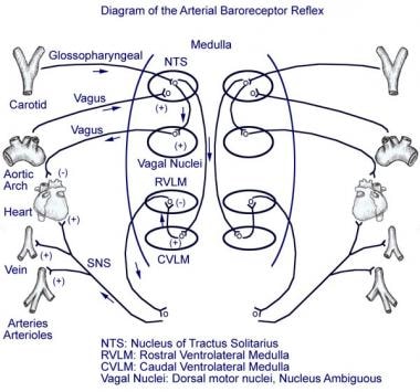
The baroreflex is a physiological mechanism that helps regulate blood pressure and heart rate in response to changes in stretch of the arterial walls. It is mediated by baroreceptors, which are specialized sensory nerve endings located in the carotid sinus and aortic arch. These receptors detect changes in blood pressure and send signals to the brainstem via the glossopharyngeal (cranial nerve IX) and vagus nerves (cranial nerve X), respectively.
In response to an increase in arterial pressure, the baroreceptors are stimulated, leading to increased firing of afferent neurons that signal the brainstem. This results in a reflexive decrease in heart rate and cardiac output, as well as vasodilation of peripheral blood vessels, which collectively work to reduce blood pressure back towards its normal level. Conversely, if arterial pressure decreases, the baroreceptors are less stimulated, leading to an increase in heart rate and cardiac output, as well as vasoconstriction of peripheral blood vessels, which helps restore blood pressure.
Overall, the baroreflex is a crucial homeostatic mechanism that helps maintain stable blood pressure and ensure adequate perfusion of vital organs.
A frontal sinus is a paired, air-filled paranasal sinus located in the frontal bone of the skull, above the eyes and behind the forehead. It is one of the four pairs of sinuses found in the human head. The frontal sinuses are lined with mucous membrane and are interconnected with the nasal cavity through small openings called ostia. They help to warm, humidify, and filter the air we breathe, and contribute to the resonance of our voice. Variations in size, shape, and asymmetry of frontal sinuses are common among individuals.
I believe there might be a misunderstanding in your question. "Dogs" is not a medical term or condition. It is the common name for a domesticated carnivore of the family Canidae, specifically the genus Canis, which includes wolves, foxes, and other extant and extinct species of mammals. Dogs are often kept as pets and companions, and they have been bred in a wide variety of forms and sizes for different purposes, such as hunting, herding, guarding, assisting police and military forces, and providing companionship and emotional support.
If you meant to ask about a specific medical condition or term related to dogs, please provide more context so I can give you an accurate answer.
The sphenoid sinuses are air-filled spaces located within the sphenoid bone, which is one of the bones that make up the skull base. These sinuses are located deep inside the skull, behind the eyes and nasal cavity. They are paired and separated by a thin bony septum, and each one opens into the corresponding nasal cavity through a small opening called the sphenoethmoidal recess. The sphenoid sinuses vary greatly in size and shape between individuals. They develop during childhood and continue to grow until early adulthood. The function of the sphenoid sinuses, like other paranasal sinuses, is not entirely clear, but they may contribute to reducing the weight of the skull, resonating voice during speech, and insulating the brain from trauma.
Paranasal sinus diseases refer to a group of medical conditions that affect the paranasal sinuses, which are air-filled cavities located within the skull near the nasal cavity. These sinuses include the maxillary, frontal, ethmoid, and sphenoid sinuses.
Paranasal sinus diseases can be caused by a variety of factors, including viral, bacterial, or fungal infections, allergies, structural abnormalities, or autoimmune disorders. Some common paranasal sinus diseases include:
1. Sinusitis: Inflammation or infection of the sinuses, which can cause symptoms such as nasal congestion, thick nasal discharge, facial pain or pressure, and reduced sense of smell.
2. Nasal polyps: Soft, benign growths that develop in the lining of the nasal passages or sinuses, which can obstruct airflow and cause difficulty breathing through the nose.
3. Sinonasal tumors: Abnormal growths that can be benign or malignant, which can cause symptoms such as nasal congestion, facial pain, and bleeding from the nose.
4. Sinus cysts: Fluid-filled sacs that form in the sinuses, which can cause symptoms similar to those of sinusitis.
5. Fungal sinusitis: Infection of the sinuses with fungi, which can cause symptoms such as nasal congestion, facial pain, and thick, discolored mucus.
Treatment for paranasal sinus diseases depends on the underlying cause and severity of the condition. Treatment options may include medications, such as antibiotics, antihistamines, or corticosteroids, as well as surgical intervention in more severe cases.
A tilt-table test is a diagnostic procedure used to evaluate symptoms of syncope (fainting) or near-syncope. It measures your body's cardiovascular response to changes in position. During the test, you lie on a table that can be tilted to change the angle of your body from horizontal to upright. This simulates what happens when you stand up from a lying down position.
The test monitors heart rate, blood pressure, and oxygen levels while you're in different positions. If you experience symptoms like dizziness or fainting during the test, these can provide clues about the cause of your symptoms. The test is used to diagnose conditions like orthostatic hypotension (a sudden drop in blood pressure when standing), vasovagal syncope (fainting due to an overactive vagus nerve), and other heart rhythm disorders.
A vagotomy is a surgical procedure that involves cutting or blocking the vagus nerve, which is a parasympathetic nerve that runs from the brainstem to the abdomen and helps regulate many bodily functions such as heart rate, gastrointestinal motility, and digestion. In particular, vagotomy is often performed as a treatment for peptic ulcers, as it can help reduce gastric acid secretion.
There are several types of vagotomy procedures, including:
1. Truncal vagotomy: This involves cutting the main trunks of the vagus nerve as they enter the abdomen. It is a more extensive procedure that reduces gastric acid secretion significantly but can also lead to side effects such as delayed gastric emptying and diarrhea.
2. Selective vagotomy: This involves cutting only the branches of the vagus nerve that supply the stomach, leaving the rest of the nerve intact. It is a less extensive procedure that reduces gastric acid secretion while minimizing side effects.
3. Highly selective vagotomy (HSV): Also known as parietal cell vagotomy, this involves cutting only the branches of the vagus nerve that supply the acid-secreting cells in the stomach. It is a highly targeted procedure that reduces gastric acid secretion while minimizing side effects such as delayed gastric emptying and diarrhea.
Vagotomy is typically performed using laparoscopic or open surgical techniques, depending on the patient's individual needs and the surgeon's preference. While vagotomy can be effective in treating peptic ulcers, it is not commonly performed today due to the development of less invasive treatments such as proton pump inhibitors (PPIs) that reduce gastric acid secretion without surgery.
Sick Sinus Syndrome (SSS) is a term used to describe a group of abnormal heart rhythm disturbances that originates in the sinoatrial node (the natural pacemaker of the heart). This syndrome is characterized by impaired functioning of the sinoatrial node, resulting in various abnormalities such as sinus bradycardia (abnormally slow heart rate), sinus arrest (complete cessation of sinus node activity), and/or sinoatrial exit block (failure of the electrical impulse to leave the sinus node and spread to the atria).
People with SSS may experience symptoms such as palpitations, dizziness, fatigue, shortness of breath, or syncope (fainting) due to inadequate blood supply to the brain caused by slow heart rate. The diagnosis of SSS is typically made based on the patient's symptoms and the results of an electrocardiogram (ECG), Holter monitoring, or event recorder that shows evidence of abnormal sinus node function. Treatment options for SSS may include lifestyle modifications, medications, or implantation of a pacemaker to regulate the heart rate.
The coronary sinus is a large vein that receives blood from the heart's muscle tissue. It is located on the posterior side of the heart and is a part of the cardiovascular system. The coronary sinus collects oxygen-depleted blood from the myocardium (the heart muscle) and drains it into the right atrium, where it will then be pumped to the lungs for oxygenation.
The coronary sinus is an essential structure in medical procedures such as cardiac catheterization and electrophysiological studies. It is also a common site for the implantation of pacemakers and other cardiac devices.
Vasovagal syncope is a type of fainting (syncope) that occurs when the body overreacts to certain triggers, such as the sight of blood or extreme emotional distress. This reaction causes the heart rate and blood pressure to drop, leading to reduced blood flow to the brain and loss of consciousness. Vasovagal syncope is usually not a cause for concern and does not typically indicate a serious underlying medical condition. However, it can be dangerous if it occurs during activities such as driving or operating heavy machinery. If you experience frequent episodes of vasovagal syncope, it is important to speak with a healthcare provider for evaluation and treatment options.
The vagus nerve, also known as the 10th cranial nerve (CN X), is the longest of the cranial nerves and extends from the brainstem to the abdomen. It has both sensory and motor functions and plays a crucial role in regulating various bodily functions such as heart rate, digestion, respiratory rate, speech, and sweating, among others.
The vagus nerve is responsible for carrying sensory information from the internal organs to the brain, and it also sends motor signals from the brain to the muscles of the throat and voice box, as well as to the heart, lungs, and digestive tract. The vagus nerve helps regulate the body's involuntary responses, such as controlling heart rate and blood pressure, promoting relaxation, and reducing inflammation.
Dysfunction in the vagus nerve can lead to various medical conditions, including gastroparesis, chronic pain, and autonomic nervous system disorders. Vagus nerve stimulation (VNS) is a therapeutic intervention that involves delivering electrical impulses to the vagus nerve to treat conditions such as epilepsy, depression, and migraine headaches.
Heart rate is the number of heartbeats per unit of time, often expressed as beats per minute (bpm). It can vary significantly depending on factors such as age, physical fitness, emotions, and overall health status. A resting heart rate between 60-100 bpm is generally considered normal for adults, but athletes and individuals with high levels of physical fitness may have a resting heart rate below 60 bpm due to their enhanced cardiovascular efficiency. Monitoring heart rate can provide valuable insights into an individual's health status, exercise intensity, and response to various treatments or interventions.
Carotid artery thrombosis is a medical condition characterized by the formation of a blood clot (thrombus) inside the carotid artery, which is one of the major blood vessels that supplies oxygenated blood to the head and neck. This condition can lead to serious complications such as a stroke or transient ischemic attack (TIA), also known as a "mini-stroke," if the clot dislodges and travels to the brain, blocking the flow of blood and oxygen.
Carotid artery thrombosis can result from various factors, including atherosclerosis (the buildup of fats, cholesterol, and other substances in the artery walls), hypertension (high blood pressure), diabetes, smoking, and genetic predisposition. Symptoms may include neck pain or stiffness, weakness or numbness in the face or limbs, difficulty speaking or understanding speech, vision problems, and sudden severe headaches. Diagnosis typically involves imaging tests such as ultrasound, CT angiography, or MRI angiography. Treatment options may include anticoagulant or antiplatelet medications, endovascular procedures to remove the clot, or surgery to clean out the artery (carotid endarterectomy).
Intracranial sinus thrombosis is a medical condition characterized by the formation of a blood clot (thrombus) within the intracranial venous sinuses, which are responsible for draining blood from the brain. The condition can lead to various neurological symptoms and complications, such as increased intracranial pressure, headaches, seizures, visual disturbances, and altered consciousness. Intracranial sinus thrombosis may result from various factors, including hypercoagulable states, infections, trauma, and malignancies. Immediate medical attention is necessary for proper diagnosis and treatment to prevent potential long-term neurological damage or even death.
The glossopharyngeal nerve, also known as the ninth cranial nerve (IX), is a mixed nerve that carries both sensory and motor fibers. It originates from the medulla oblongata in the brainstem and has several functions:
1. Sensory function: The glossopharyngeal nerve provides general sensation to the posterior third of the tongue, the tonsils, the back of the throat (pharynx), and the middle ear. It also carries taste sensations from the back one-third of the tongue.
2. Special visceral afferent function: The nerve transmits information about the stretch of the carotid artery and blood pressure to the brainstem.
3. Motor function: The glossopharyngeal nerve innervates the stylopharyngeus muscle, which helps elevate the pharynx during swallowing. It also provides parasympathetic fibers to the parotid gland, stimulating saliva production.
4. Visceral afferent function: The glossopharyngeal nerve carries information about the condition of the internal organs in the thorax and abdomen to the brainstem.
Overall, the glossopharyngeal nerve plays a crucial role in swallowing, taste, saliva production, and monitoring blood pressure and heart rate.
Carotid artery injuries refer to damages or traumas that affect the carotid arteries, which are a pair of major blood vessels located in the neck that supply oxygenated blood to the head and neck. These injuries can occur due to various reasons such as penetrating or blunt trauma, iatrogenic causes (during medical procedures), or degenerative diseases.
Carotid artery injuries can be categorized into three types:
1. Blunt carotid injury (BCI): This type of injury is caused by a sudden and severe impact to the neck, which can result in intimal tears, dissection, or thrombosis of the carotid artery. BCIs are commonly seen in motor vehicle accidents, sports-related injuries, and assaults.
2. Penetrating carotid injury: This type of injury is caused by a foreign object that penetrates the neck and damages the carotid artery. Examples include gunshot wounds, stab wounds, or other sharp objects that pierce the skin and enter the neck.
3. Iatrogenic carotid injury: This type of injury occurs during medical procedures such as endovascular interventions, surgical procedures, or the placement of central lines.
Symptoms of carotid artery injuries may include:
* Stroke or transient ischemic attack (TIA)
* Neurological deficits such as hemiparesis, aphasia, or visual disturbances
* Bleeding from the neck or mouth
* Pulsatile mass in the neck
* Hypotension or shock
* Loss of consciousness
Diagnosis of carotid artery injuries may involve imaging studies such as computed tomography angiography (CTA), magnetic resonance angiography (MRA), or conventional angiography. Treatment options include endovascular repair, surgical repair, or anticoagulation therapy, depending on the severity and location of the injury.
Sodium cyanide is a highly toxic chemical compound with the formula NaCN. It is a white solid that is readily soluble in water, and it has a bitter, almond-like odor that some people can detect. Sodium cyanide is used in various industrial processes, including metal cleaning and electroplating, but it is perhaps best known as a poison.
Cyanide ions (CN-) are extremely toxic because they bind to the ferric iron (Fe3+) in cytochrome c oxidase, a crucial enzyme in the mitochondria that is responsible for cellular respiration and energy production. When cyanide ions bind to this enzyme, it becomes unable to function, leading to a rapid depletion of ATP (adenosine triphosphate) and an accumulation of lactic acid, which can cause metabolic acidosis, coma, and death within minutes to hours.
It is important to note that sodium cyanide should be handled with extreme care and only by trained professionals who are familiar with its hazards and proper safety protocols. Exposure to this compound can cause severe health effects, including respiratory failure, convulsions, and cardiac arrest.
The ethmoid sinuses are a pair of air-filled spaces located in the ethmoid bone, which is a part of the skull that forms the upper portion of the nasal cavity and the inner eye socket. These sinuses are divided into anterior and posterior groups and are present in adults, but not at birth. They continue to grow and develop until early adulthood.
The ethmoid sinuses are lined with mucous membrane, which helps to warm, humidify, and filter the air we breathe. They are surrounded by a network of blood vessels and nerves, making them susceptible to inflammation and infection. Inflammation of the ethmoid sinuses can lead to conditions such as sinusitis, which can cause symptoms such as nasal congestion, headache, and facial pain.
"Cat" is a common name that refers to various species of small carnivorous mammals that belong to the family Felidae. The domestic cat, also known as Felis catus or Felis silvestris catus, is a popular pet and companion animal. It is a subspecies of the wildcat, which is found in Europe, Africa, and Asia.
Domestic cats are often kept as pets because of their companionship, playful behavior, and ability to hunt vermin. They are also valued for their ability to provide emotional support and therapy to people. Cats are obligate carnivores, which means that they require a diet that consists mainly of meat to meet their nutritional needs.
Cats are known for their agility, sharp senses, and predatory instincts. They have retractable claws, which they use for hunting and self-defense. Cats also have a keen sense of smell, hearing, and vision, which allow them to detect prey and navigate their environment.
In medical terms, cats can be hosts to various parasites and diseases that can affect humans and other animals. Some common feline diseases include rabies, feline leukemia virus (FeLV), feline immunodeficiency virus (FIV), and toxoplasmosis. It is important for cat owners to keep their pets healthy and up-to-date on vaccinations and preventative treatments to protect both the cats and their human companions.
A carotid body tumor is a rare, usually noncancerous (benign) growth that develops in the carotid body, a small structure located near the bifurcation (fork) of the common carotid artery in the neck. The carotid body is part of the chemoreceptor system that helps regulate breathing and blood pressure by responding to changes in oxygen, carbon dioxide, and pH levels in the blood.
Carotid body tumors are also known as carotid body paragangliomas or chemodectomas. They typically grow slowly and may not cause any symptoms for many years. However, as they enlarge, they can cause a visible or palpable mass in the neck, along with symptoms such as difficulty swallowing, hoarseness, or voice changes. In some cases, carotid body tumors can compress nearby nerves or blood vessels, leading to more serious complications like stroke or nerve damage.
Treatment for carotid body tumors typically involves surgical removal of the growth, which may be performed using traditional open surgery or minimally invasive techniques such as endovascular surgery or robotic-assisted surgery. Radiation therapy and chemotherapy are generally not effective in treating these tumors. Regular follow-up care is important to monitor for recurrence or development of new tumors.
Bradycardia is a medical term that refers to an abnormally slow heart rate, typically defined as a resting heart rate of less than 60 beats per minute in adults. While some people, particularly well-trained athletes, may have a naturally low resting heart rate, bradycardia can also be a sign of an underlying health problem.
There are several potential causes of bradycardia, including:
* Damage to the heart's electrical conduction system, such as from heart disease or aging
* Certain medications, including beta blockers, calcium channel blockers, and digoxin
* Hypothyroidism (underactive thyroid gland)
* Sleep apnea
* Infection of the heart (endocarditis or myocarditis)
* Infiltrative diseases such as amyloidosis or sarcoidosis
Symptoms of bradycardia can vary depending on the severity and underlying cause. Some people with bradycardia may not experience any symptoms, while others may feel weak, fatigued, dizzy, or short of breath. In severe cases, bradycardia can lead to fainting, confusion, or even cardiac arrest.
Treatment for bradycardia depends on the underlying cause. If a medication is causing the slow heart rate, adjusting the dosage or switching to a different medication may help. In other cases, a pacemaker may be necessary to regulate the heart's rhythm. It is important to seek medical attention if you experience symptoms of bradycardia, as it can be a sign of a serious underlying condition.
Carotid intima-media thickness (CIMT) is a measurement of the thickness of the inner two layers of the carotid artery, which are the intima and media layers. This measurement is used as a marker for assessing cardiovascular disease risk, particularly the risk of atherosclerosis, or the buildup of plaque in the arteries.
CIMT can be measured using ultrasound imaging, and it is typically measured at several points along the length of the common carotid artery, as well as at the bifurcation where the common carotid artery divides into the internal and external carotid arteries. Increased CIMT has been associated with an increased risk of cardiovascular events such as heart attack and stroke.
It is important to note that while CIMT can provide valuable information about a person's cardiovascular health, it should not be used as the sole determinant of cardiovascular disease risk. Other factors, such as age, family history, smoking status, blood pressure, cholesterol levels, and diabetes status, should also be taken into account when assessing cardiovascular disease risk.
Medical Definition of Respiration:
Respiration, in physiology, is the process by which an organism takes in oxygen and gives out carbon dioxide. It's also known as breathing. This process is essential for most forms of life because it provides the necessary oxygen for cellular respiration, where the cells convert biochemical energy from nutrients into adenosine triphosphate (ATP), and releases waste products, primarily carbon dioxide.
In humans and other mammals, respiration is a two-stage process:
1. Breathing (or external respiration): This involves the exchange of gases with the environment. Air enters the lungs through the mouth or nose, then passes through the pharynx, larynx, trachea, and bronchi, finally reaching the alveoli where the actual gas exchange occurs. Oxygen from the inhaled air diffuses into the blood, while carbon dioxide, a waste product of metabolism, diffuses from the blood into the alveoli to be exhaled.
2. Cellular respiration (or internal respiration): This is the process by which cells convert glucose and other nutrients into ATP, water, and carbon dioxide in the presence of oxygen. The carbon dioxide produced during this process then diffuses out of the cells and into the bloodstream to be exhaled during breathing.
In summary, respiration is a vital physiological function that enables organisms to obtain the necessary oxygen for cellular metabolism while eliminating waste products like carbon dioxide.
Efferent neurons are specialized nerve cells that transmit signals from the central nervous system (CNS), which includes the brain and spinal cord, to effector organs such as muscles or glands. These signals typically result in a response or action, hence the term "efferent," derived from the Latin word "efferre" meaning "to carry away."
Efferent neurons are part of the motor pathway and can be further classified into two types:
1. Somatic efferent neurons: These neurons transmit signals to skeletal muscles, enabling voluntary movements and posture maintenance. They have their cell bodies located in the ventral horn of the spinal cord and send their axons through the ventral roots to innervate specific muscle fibers.
2. Autonomic efferent neurons: These neurons are responsible for controlling involuntary functions, such as heart rate, digestion, respiration, and pupil dilation. They have a two-neuron chain arrangement, with the preganglionic neuron having its cell body in the CNS (brainstem or spinal cord) and synapsing with the postganglionic neuron in an autonomic ganglion near the effector organ. Autonomic efferent neurons can be further divided into sympathetic, parasympathetic, and enteric subdivisions based on their functions and innervation patterns.
In summary, efferent neurons are a critical component of the nervous system, responsible for transmitting signals from the CNS to various effector organs, ultimately controlling and coordinating numerous bodily functions and responses.
Heart massage, also known as cardiac massage or chest compression, is a medical procedure that involves applying pressure to the chest in order to manually pump blood through the heart and maintain circulation when the heart has stopped or is not functioning effectively. This is a critical component of cardiopulmonary resuscitation (CPR) and is typically performed during a cardiac arrest to help restore proper blood flow to vital organs and tissues.
During heart massage, the rescuer places their hands on the lower half of the victim's chest, typically at the center, and presses down with the heel of one or both hands. The recommended compression depth for adults is at least 2 inches (5 cm) and should be performed at a rate of 100-120 compressions per minute. It is essential to minimize interruptions in chest compressions and ensure that they are deep and fast enough to maintain adequate blood flow.
Heart massage can also be performed surgically during specific medical procedures, such as open-heart surgery or extracorporeal membrane oxygenation (ECMO). In these cases, the surgeon directly compresses the heart using their hands or specialized instruments. This technique is called a "surgical heart massage" or "direct cardiac compression."
It's important to note that heart massage should only be performed by trained individuals, as improper techniques can cause harm and potentially worsen the patient's condition.
The sympathetic nervous system (SNS) is a part of the autonomic nervous system that operates largely below the level of consciousness, and it functions to produce appropriate physiological responses to perceived danger. It's often associated with the "fight or flight" response. The SNS uses nerve impulses to stimulate target organs, causing them to speed up (e.g., increased heart rate), prepare for action, or otherwise respond to stressful situations.
The sympathetic nervous system is activated due to stressful emotional or physical situations and it prepares the body for immediate actions. It dilates the pupils, increases heart rate and blood pressure, accelerates breathing, and slows down digestion. The primary neurotransmitter involved in this system is norepinephrine (also known as noradrenaline).
Autonomic denervation is a medical term that refers to the interruption or loss of nerve supply to the autonomic nervous system. The autonomic nervous system is the part of the nervous system that controls involuntary actions, such as heart rate, blood pressure, digestion, and pupil dilation.
Autonomic denervation can occur due to various reasons, including surgical procedures, trauma, degenerative diseases, or medical conditions such as diabetes. The interruption of nerve supply can lead to a range of symptoms depending on the specific autonomic functions that are affected.
For example, autonomic denervation in the heart can lead to abnormal heart rhythms or low blood pressure. In the digestive system, it can cause problems with motility and secretion, leading to symptoms such as bloating, constipation, or diarrhea. Autonomic denervation in the eyes can result in pupil abnormalities, dry eyes, or light sensitivity.
Treatment for autonomic denervation depends on the underlying cause and the specific symptoms that are present. In some cases, medication may be used to manage symptoms, while in others, surgical intervention may be necessary to repair or restore nerve function.
Vascular resistance is a measure of the opposition to blood flow within a vessel or a group of vessels, typically expressed in units of mmHg/(mL/min) or sometimes as dynes*sec/cm^5. It is determined by the diameter and length of the vessels, as well as the viscosity of the blood flowing through them. In general, a decrease in vessel diameter, an increase in vessel length, or an increase in blood viscosity will result in an increase in vascular resistance, while an increase in vessel diameter, a decrease in vessel length, or a decrease in blood viscosity will result in a decrease in vascular resistance. Vascular resistance is an important concept in the study of circulation and cardiovascular physiology because it plays a key role in determining blood pressure and blood flow within the body.
The phrenic nerve is a motor nerve that originates from the cervical spine (C3-C5) and descends through the neck to reach the diaphragm, which is the primary muscle used for breathing. The main function of the phrenic nerve is to innervate the diaphragm and control its contraction and relaxation, thereby enabling respiration.
Damage or injury to the phrenic nerve can result in paralysis of the diaphragm, leading to difficulty breathing and potentially causing respiratory failure. Certain medical conditions, such as neuromuscular disorders, spinal cord injuries, and tumors, can affect the phrenic nerve and impair its function.
Paranasal sinus neoplasms refer to abnormal growths or tumors that develop within the paranasal sinuses, which are air-filled cavities located inside the skull near the nasal cavity. These tumors can be benign (noncancerous) or malignant (cancerous), and they can arise from various types of tissue within the sinuses, such as the lining of the sinuses (mucosa), bone, or other soft tissues.
Paranasal sinus neoplasms can cause a variety of symptoms, including nasal congestion, nosebleeds, facial pain or numbness, and visual disturbances. The diagnosis of these tumors typically involves a combination of imaging studies (such as CT or MRI scans) and biopsy to determine the type and extent of the tumor. Treatment options may include surgery, radiation therapy, chemotherapy, or a combination of these approaches, depending on the specific type and stage of the neoplasm.
Fludrocortisone is a synthetic corticosteroid hormone, specifically a mineralocorticoid. It is often used to treat conditions associated with low levels of corticosteroids, such as Addison's disease. It works by helping the body retain sodium and lose potassium, which helps to maintain fluid balance and blood pressure.
In medical terms, fludrocortisone is defined as a synthetic mineralocorticoid with glucocorticoid activity used in the treatment of adrenogenital syndrome and Addison's disease, and as an adjunct in the treatment of rheumatoid arthritis. It is also used to treat orthostatic hypotension by helping the body retain sodium and water, thereby increasing blood volume and blood pressure.
It is important to note that fludrocortisone can have significant side effects, particularly if used in high doses or for long periods of time. These can include fluid retention, high blood pressure, increased risk of infection, and slowed growth in children. As with any medication, it should be used under the close supervision of a healthcare provider.
Tunica intima, also known as the intima layer, is the innermost layer of a blood vessel, including arteries and veins. It is in direct contact with the flowing blood and is composed of simple squamous endothelial cells that form a continuous, non-keratinized, stratified epithelium. These cells play a crucial role in maintaining vascular homeostasis by regulating the passage of molecules and immune cells between the blood and the vessel wall, as well as contributing to the maintenance of blood fluidity and preventing coagulation.
The tunica intima is supported by a thin layer of connective tissue called the basement membrane, which provides structural stability and anchorage for the endothelial cells. Beneath the basement membrane lies a loose network of elastic fibers and collagen, known as the internal elastic lamina, that separates the tunica intima from the middle layer, or tunica media.
In summary, the tunica intima is the innermost layer of blood vessels, primarily composed of endothelial cells and a basement membrane, which regulates various functions to maintain vascular homeostasis.
Endarterectomy is a surgical procedure in which the inner lining of an artery (the endothelium) that has become thickened, damaged, or narrowed due to the buildup of fatty deposits, called plaques, is removed. This process helps restore normal blood flow through the artery and reduces the risk of serious complications such as stroke or limb loss.
The procedure typically involves making an incision in the affected artery, carefully removing the plaque and inner lining, and then closing the artery with sutures or a patch graft. Endarterectomy is most commonly performed on the carotid arteries in the neck, but it can also be done on other arteries throughout the body, including the femoral artery in the leg and the iliac artery in the pelvis.
Endarterectomy is usually recommended for patients with significant narrowing of their arteries who are experiencing symptoms such as pain, numbness, or weakness in their limbs, or who have a high risk of stroke due to carotid artery disease. The procedure is generally safe and effective, but like any surgery, it carries risks such as bleeding, infection, and damage to nearby nerves or tissues.
The tunica media is the middle layer of the wall of a blood vessel or hollow organ in the body. It is primarily composed of smooth muscle cells and elastic fibers, which allow the vessel or organ to expand and contract. This layer helps regulate the diameter of the lumen (the inner space) of the vessel or organ, thereby controlling the flow of fluids such as blood or lymph through it. The tunica media plays a crucial role in maintaining proper organ function and blood pressure regulation.
An abnormal reflex in a medical context refers to an involuntary and exaggerated response or lack of response to a stimulus that is not expected in the normal physiological range. These responses can be indicative of underlying neurological disorders or damage to the nervous system. Examples include hyperreflexia (overactive reflexes) and hyporeflexia (underactive reflexes). The assessment of reflexes is an important part of a physical examination, as it can provide valuable information about the functioning of the nervous system.
Chloralose is not a medical term commonly used in modern medicine. However, historically, it is a chemical compound that has been used in research and veterinary medicine as an sedative and hypnotic agent. It is a combination of chloral hydrate and sodium pentobarbital.
Chloralose has been used in research to study the effects of sedation on various physiological processes, such as respiration and circulation. In veterinary medicine, it has been used as an anesthetic for small animals during surgical procedures. However, due to its potential for serious side effects, including respiratory depression and cardiac arrest, chloralose is not commonly used in clinical practice today.
Sinus tachycardia is a type of rapid heart rate, characterized by an abnormally fast sinus rhythm, with a rate greater than 100 beats per minute in adults. The sinoatrial node (SA node), which is the natural pacemaker of the heart, generates these impulses regularly and at an increased rate.
Sinus tachycardia is usually a physiological response to various stimuli or conditions, such as physical exertion, strong emotions, fever, anxiety, pain, or certain medications. It can also be caused by hormonal imbalances, anemia, hyperthyroidism, or other medical disorders.
In most cases, sinus tachycardia is not harmful and resolves once the underlying cause is addressed. However, if it occurs persistently or is associated with symptoms like palpitations, shortness of breath, dizziness, or chest discomfort, further evaluation by a healthcare professional is recommended to rule out any underlying heart conditions or other medical issues.
Heart sounds are the noises generated by the beating heart and the movement of blood through it. They are caused by the vibration of the cardiac structures, such as the valves, walls, and blood vessels, during the cardiac cycle.
There are two normal heart sounds, often described as "lub-dub," that can be heard through a stethoscope. The first sound (S1) is caused by the closure of the mitral and tricuspid valves at the beginning of systole, when the ventricles contract to pump blood out to the body and lungs. The second sound (S2) is produced by the closure of the aortic and pulmonary valves at the end of systole, as the ventricles relax and the ventricular pressure decreases, allowing the valves to close.
Abnormal heart sounds, such as murmurs, clicks, or extra sounds (S3 or S4), may indicate cardiac disease or abnormalities in the structure or function of the heart. These sounds can be evaluated through a process called auscultation, which involves listening to the heart with a stethoscope and analyzing the intensity, pitch, quality, and timing of the sounds.
An accidental fall is an unplanned, unexpected event in which a person suddenly and involuntarily comes to rest on the ground or other lower level, excluding intentional changes in position (e.g., jumping to catch a ball) and landings that are part of a planned activity (e.g., diving into a pool). Accidental falls can occur for various reasons, such as environmental hazards, muscle weakness, balance problems, visual impairment, or certain medical conditions. They are a significant health concern, particularly among older adults, as they can lead to serious injuries, loss of independence, reduced quality of life, and increased mortality.
An artificial pacemaker is a medical device that uses electrical impulses to regulate the beating of the heart. It is typically used when the heart's natural pacemaker, the sinoatrial node, is not functioning properly and the heart rate is too slow or irregular. The pacemaker consists of a small generator that contains a battery and electronic circuits, which are connected to one or more electrodes that are placed in the heart.
The generator sends electrical signals through the electrodes to stimulate the heart muscle and cause it to contract, thereby maintaining a regular heart rhythm. Artificial pacemakers can be programmed to deliver electrical impulses at a specific rate or in response to the body's needs. They are typically implanted in the chest during a surgical procedure and can last for many years before needing to be replaced.
Artificial pacemakers are an effective treatment for various types of bradycardia, which is a heart rhythm disorder characterized by a slow heart rate. Pacemakers can significantly improve symptoms associated with bradycardia, such as fatigue, dizziness, shortness of breath, and fainting spells.
Anesthesia is a medical term that refers to the loss of sensation or awareness, usually induced by the administration of various drugs. It is commonly used during surgical procedures to prevent pain and discomfort. There are several types of anesthesia, including:
1. General anesthesia: This type of anesthesia causes a complete loss of consciousness and is typically used for major surgeries.
2. Regional anesthesia: This type of anesthesia numbs a specific area of the body, such as an arm or leg, while the patient remains conscious.
3. Local anesthesia: This type of anesthesia numbs a small area of the body, such as a cut or wound, and is typically used for minor procedures.
Anesthesia can be administered through various routes, including injection, inhalation, or topical application. The choice of anesthesia depends on several factors, including the type and duration of the procedure, the patient's medical history, and their overall health. Anesthesiologists are medical professionals who specialize in administering anesthesia and monitoring patients during surgical procedures to ensure their safety and comfort.
Maxillary sinus neoplasms refer to abnormal growths or tumors that develop in the maxillary sinuses, which are located in the upper part of your cheekbones, below your eyes. These growths can be benign (non-cancerous) or malignant (cancerous).
Benign neoplasms may include conditions such as an osteoma (a benign bone tumor), a papilloma (a benign growth of the lining of the sinus), or a fibrous dysplasia (a condition where bone is replaced by fibrous tissue).
Malignant neoplasms, on the other hand, can be primary (originating in the maxillary sinuses) or secondary (spreading to the maxillary sinuses from another site in the body). Common types of malignant tumors that arise in the maxillary sinus include squamous cell carcinoma, adenocarcinoma, and mucoepidermoid carcinoma.
Symptoms of maxillary sinus neoplasms may include nasal congestion, nosebleeds, facial pain or numbness, vision changes, and difficulty swallowing or speaking. Treatment options depend on the type, size, and location of the tumor but may include surgery, radiation therapy, chemotherapy, or a combination of these approaches.
A carotid artery, internal, dissection is a medical condition that affects the internal carotid artery, which is a major blood vessel in the neck that supplies oxygenated blood to the brain. In this condition, there is a separation (dissection) of the layers of the artery wall, causing blood to accumulate in the space between the layers. This can lead to narrowing or blockage of the artery, reducing blood flow to the brain and increasing the risk of stroke. Internal carotid artery dissection can be caused by trauma, high blood pressure, connective tissue disorders, or spontaneously. Symptoms may include neck pain, headache, facial pain, visual disturbances, weakness or numbness in the arms or legs, difficulty speaking or understanding speech, and dizziness or loss of balance.
Dizziness is a term used to describe a range of sensations, such as feeling lightheaded, faint, unsteady, or a false sense of spinning or moving. Medically, dizziness is often described as a non-specific symptom that can be caused by various underlying conditions or factors. These may include:
1. Inner ear disorders (such as benign paroxysmal positional vertigo, labyrinthitis, vestibular neuronitis, or Meniere's disease)
2. Cardiovascular problems (like low blood pressure, arrhythmias, or orthostatic hypotension)
3. Neurological issues (such as migraines, multiple sclerosis, or stroke)
4. Anxiety disorders and panic attacks
5. Side effects of medications
6. Dehydration or overheating
7. Infections (like viral infections or bacterial meningitis)
8. Head or neck injuries
9. Low blood sugar levels (hypoglycemia)
It is essential to consult a healthcare professional if you experience persistent dizziness, as it can be a sign of a more severe underlying condition. The appropriate treatment will depend on the specific cause of the dizziness.
Sympathectomy is a surgical procedure that involves interrupting the sympathetic nerve pathways. These nerves are part of the autonomic nervous system, which controls involuntary bodily functions such as heart rate, blood pressure, sweating, and digestion. The goal of sympathectomy is to manage conditions like hyperhidrosis (excessive sweating), Raynaud's phenomenon, and certain types of chronic pain.
There are different types of sympathectomy, including thoracic sympathectomy (which targets the sympathetic nerves in the chest), lumbar sympathectomy (which targets the sympathetic nerves in the lower back), and cervical sympathectomy (which targets the sympathetic nerves in the neck). The specific type of procedure depends on the location of the affected nerves and the condition being treated.
Sympathectomy is usually performed using minimally invasive techniques, such as endoscopic surgery, which involves making small incisions and using specialized instruments to access the nerves. While sympathectomy can be effective in managing certain conditions, it carries risks such as nerve damage, bleeding, infection, and chronic pain.
Cross circulation is a medical procedure in which blood from one person (the donor) is circulated through the body of another person (the recipient) by connecting their cardiovascular systems. This technique was first developed and used in open-heart surgery during the 1950s, before the invention of heart-lung machines.
In cross circulation, the donor's and recipient's circulatory systems are connected through anastomoses (surgical connections) between their blood vessels. The most common configuration involved connecting the donor's femoral artery to the recipient's aorta and the donor's femoral vein to the recipient's vena cava. This allowed the donor's heart to pump oxygenated blood to both the donor and the recipient during the surgery.
Cross circulation was used as a temporary measure to maintain the recipient's circulation and oxygenation while their own heart was stopped and repaired during open-heart surgery. However, this technique had several limitations and risks, including potential complications for the donor (such as bleeding, infection, or reactions to the recipient's blood) and ethical concerns related to using one person as a "human bridge" to save another.
With the development of more advanced and safer heart-lung machines in the early 1960s, cross circulation became obsolete in cardiac surgery. Nowadays, it is rarely used and mainly of historical interest.
Electrocardiography (ECG or EKG) is a medical procedure that records the electrical activity of the heart. It provides a graphic representation of the electrical changes that occur during each heartbeat. The resulting tracing, called an electrocardiogram, can reveal information about the heart's rate and rhythm, as well as any damage to its cells or abnormalities in its conduction system.
During an ECG, small electrodes are placed on the skin of the chest, arms, and legs. These electrodes detect the electrical signals produced by the heart and transmit them to a machine that amplifies and records them. The procedure is non-invasive, painless, and quick, usually taking only a few minutes.
ECGs are commonly used to diagnose and monitor various heart conditions, including arrhythmias, coronary artery disease, heart attacks, and electrolyte imbalances. They can also be used to evaluate the effectiveness of certain medications or treatments.
Veratrine is not a medical term, but it is a pharmacological term that refers to a mixture of alkaloids (veratridine and cevadine) extracted from the seeds of the sabadilla lily (Schoenocaulon officinale). Veratrine has been used in research and medicine for its effects on nerve cells, particularly in studying sodium channels. It can cause prolonged depolarization of nerve membranes leading to repetitive firing of action potentials. However, due to its high toxicity, it is not used clinically.
A syndrome, in medical terms, is a set of symptoms that collectively indicate or characterize a disease, disorder, or underlying pathological process. It's essentially a collection of signs and/or symptoms that frequently occur together and can suggest a particular cause or condition, even though the exact physiological mechanisms might not be fully understood.
For example, Down syndrome is characterized by specific physical features, cognitive delays, and other developmental issues resulting from an extra copy of chromosome 21. Similarly, metabolic syndromes like diabetes mellitus type 2 involve a group of risk factors such as obesity, high blood pressure, high blood sugar, and abnormal cholesterol or triglyceride levels that collectively increase the risk of heart disease, stroke, and diabetes.
It's important to note that a syndrome is not a specific diagnosis; rather, it's a pattern of symptoms that can help guide further diagnostic evaluation and management.
A pilonidal sinus is a small hole or tunnel in the skin that usually develops in the cleft at the top of the buttocks. It can be painful and may become infected, causing symptoms such as redness, swelling, pain, and pus discharge. The condition often affects young adults and is more common in men than women.
The term "pilonidal" comes from the Latin words "pilus," meaning hair, and "nidus," meaning nest. This refers to the fact that the sinus often contains hairs that have become embedded in the skin. The exact cause of pilonidal sinuses is not known, but they are thought to develop as a result of ingrown hairs or chronic irritation in the affected area.
Treatment for pilonidal sinuses typically involves surgical removal of the sinus and any associated hair follicles. In some cases, this may be done using a minor procedure that can be performed in a doctor's office. More complex cases may require hospitalization and a more extensive surgical procedure. After surgery, patients will need to take steps to prevent the sinus from recurring, such as keeping the area clean and avoiding prolonged periods of sitting or driving.
In medical terms, the "neck" is defined as the portion of the body that extends from the skull/head to the thorax or chest region. It contains 7 cervical vertebrae, muscles, nerves, blood vessels, lymphatic vessels, and glands (such as the thyroid gland). The neck is responsible for supporting the head, allowing its movement in various directions, and housing vital structures that enable functions like respiration and circulation.
Cerebral angiography is a medical procedure that involves taking X-ray images of the blood vessels in the brain after injecting a contrast dye into them. This procedure helps doctors to diagnose and treat various conditions affecting the blood vessels in the brain, such as aneurysms, arteriovenous malformations, and stenosis (narrowing of the blood vessels).
During the procedure, a catheter is inserted into an artery in the leg and threaded through the body to the blood vessels in the neck or brain. The contrast dye is then injected through the catheter, and X-ray images are taken to visualize the blood flow through the brain's blood vessels.
Cerebral angiography provides detailed images of the blood vessels in the brain, allowing doctors to identify any abnormalities or blockages that may be causing symptoms or increasing the risk of stroke. Based on the results of the cerebral angiography, doctors can develop a treatment plan to address these issues and prevent further complications.
Carbon dioxide (CO2) is a colorless, odorless gas that is naturally present in the Earth's atmosphere. It is a normal byproduct of cellular respiration in humans, animals, and plants, and is also produced through the combustion of fossil fuels such as coal, oil, and natural gas.
In medical terms, carbon dioxide is often used as a respiratory stimulant and to maintain the pH balance of blood. It is also used during certain medical procedures, such as laparoscopic surgery, to insufflate (inflate) the abdominal cavity and create a working space for the surgeon.
Elevated levels of carbon dioxide in the body can lead to respiratory acidosis, a condition characterized by an increased concentration of carbon dioxide in the blood and a decrease in pH. This can occur in conditions such as chronic obstructive pulmonary disease (COPD), asthma, or other lung diseases that impair breathing and gas exchange. Symptoms of respiratory acidosis may include shortness of breath, confusion, headache, and in severe cases, coma or death.
Hemodynamics is the study of how blood flows through the cardiovascular system, including the heart and the vascular network. It examines various factors that affect blood flow, such as blood volume, viscosity, vessel length and diameter, and pressure differences between different parts of the circulatory system. Hemodynamics also considers the impact of various physiological and pathological conditions on these variables, and how they in turn influence the function of vital organs and systems in the body. It is a critical area of study in fields such as cardiology, anesthesiology, and critical care medicine.
In the field of medicine, "time factors" refer to the duration of symptoms or time elapsed since the onset of a medical condition, which can have significant implications for diagnosis and treatment. Understanding time factors is crucial in determining the progression of a disease, evaluating the effectiveness of treatments, and making critical decisions regarding patient care.
For example, in stroke management, "time is brain," meaning that rapid intervention within a specific time frame (usually within 4.5 hours) is essential to administering tissue plasminogen activator (tPA), a clot-busting drug that can minimize brain damage and improve patient outcomes. Similarly, in trauma care, the "golden hour" concept emphasizes the importance of providing definitive care within the first 60 minutes after injury to increase survival rates and reduce morbidity.
Time factors also play a role in monitoring the progression of chronic conditions like diabetes or heart disease, where regular follow-ups and assessments help determine appropriate treatment adjustments and prevent complications. In infectious diseases, time factors are crucial for initiating antibiotic therapy and identifying potential outbreaks to control their spread.
Overall, "time factors" encompass the significance of recognizing and acting promptly in various medical scenarios to optimize patient outcomes and provide effective care.
Anoxia is a medical condition that refers to the absence or complete lack of oxygen supply in the body or a specific organ, tissue, or cell. This can lead to serious health consequences, including damage or death of cells and tissues, due to the vital role that oxygen plays in supporting cellular metabolism and energy production.
Anoxia can occur due to various reasons, such as respiratory failure, cardiac arrest, severe blood loss, carbon monoxide poisoning, or high altitude exposure. Prolonged anoxia can result in hypoxic-ischemic encephalopathy, a serious condition that can cause brain damage and long-term neurological impairments.
Medical professionals use various diagnostic tests, such as blood gas analysis, pulse oximetry, and electroencephalography (EEG), to assess oxygen levels in the body and diagnose anoxia. Treatment for anoxia typically involves addressing the underlying cause, providing supplemental oxygen, and supporting vital functions, such as breathing and circulation, to prevent further damage.
Electric stimulation, also known as electrical nerve stimulation or neuromuscular electrical stimulation, is a therapeutic treatment that uses low-voltage electrical currents to stimulate nerves and muscles. It is often used to help manage pain, promote healing, and improve muscle strength and mobility. The electrical impulses can be delivered through electrodes placed on the skin or directly implanted into the body.
In a medical context, electric stimulation may be used for various purposes such as:
1. Pain management: Electric stimulation can help to block pain signals from reaching the brain and promote the release of endorphins, which are natural painkillers produced by the body.
2. Muscle rehabilitation: Electric stimulation can help to strengthen muscles that have become weak due to injury, illness, or surgery. It can also help to prevent muscle atrophy and improve range of motion.
3. Wound healing: Electric stimulation can promote tissue growth and help to speed up the healing process in wounds, ulcers, and other types of injuries.
4. Urinary incontinence: Electric stimulation can be used to strengthen the muscles that control urination and reduce symptoms of urinary incontinence.
5. Migraine prevention: Electric stimulation can be used as a preventive treatment for migraines by applying electrical impulses to specific nerves in the head and neck.
It is important to note that electric stimulation should only be administered under the guidance of a qualified healthcare professional, as improper use can cause harm or discomfort.
In medical terms, pressure is defined as the force applied per unit area on an object or body surface. It is often measured in millimeters of mercury (mmHg) in clinical settings. For example, blood pressure is the force exerted by circulating blood on the walls of the arteries and is recorded as two numbers: systolic pressure (when the heart beats and pushes blood out) and diastolic pressure (when the heart rests between beats).
Pressure can also refer to the pressure exerted on a wound or incision to help control bleeding, or the pressure inside the skull or spinal canal. High or low pressure in different body systems can indicate various medical conditions and require appropriate treatment.
The transverse sinuses are a pair of venous channels located within the skull. They are part of the intracranial venous system and are responsible for draining blood from the brain. The transverse sinuses run horizontally along the upper portion of the inner skull, starting at the occipital bone (at the back of the head) and extending to the temporal bones (on the sides of the head).
These sinuses receive blood from the superior sagittal sinus, straight sinus, and the occipital sinus. After passing through the transverse sinuses, the blood is then drained into the sigmoid sinuses, which in turn drain into the internal jugular veins. The transverse sinuses are an essential component of the cerebral venous system, ensuring proper blood flow and drainage from the brain.
Prospective studies, also known as longitudinal studies, are a type of cohort study in which data is collected forward in time, following a group of individuals who share a common characteristic or exposure over a period of time. The researchers clearly define the study population and exposure of interest at the beginning of the study and follow up with the participants to determine the outcomes that develop over time. This type of study design allows for the investigation of causal relationships between exposures and outcomes, as well as the identification of risk factors and the estimation of disease incidence rates. Prospective studies are particularly useful in epidemiology and medical research when studying diseases with long latency periods or rare outcomes.
Efferent pathways refer to the neural connections that carry signals from the central nervous system (CNS), which includes the brain and spinal cord, to the peripheral effectors such as muscles and glands. These pathways are responsible for the initiation and control of motor responses, as well as regulating various autonomic functions.
Efferent pathways can be divided into two main types:
1. Somatic efferent pathways: These pathways carry signals from the CNS to the skeletal muscles, enabling voluntary movements and postural control. The final common pathway for somatic motor innervation is the alpha-motor neuron, which synapses directly onto skeletal muscle fibers.
2. Autonomic efferent pathways: These pathways regulate the function of internal organs, smooth muscles, and glands. They are further divided into two subtypes: sympathetic and parasympathetic. The sympathetic system is responsible for the 'fight or flight' response, while the parasympathetic system promotes rest and digestion. Both systems use a two-neuron chain to transmit signals from the CNS to the effector organs. The preganglionic neuron has its cell body in the CNS and synapses with the postganglionic neuron in an autonomic ganglion located near the effector organ. The postganglionic neuron then innervates the target organ or tissue.
In summary, efferent pathways are the neural connections that carry signals from the CNS to peripheral effectors, enabling motor responses and regulating various autonomic functions. They can be divided into somatic and autonomic efferent pathways, with further subdivisions within the autonomic system.
I believe there may be some confusion in your question. "Rabbits" is a common name used to refer to the Lagomorpha species, particularly members of the family Leporidae. They are small mammals known for their long ears, strong legs, and quick reproduction.
However, if you're referring to "rabbits" in a medical context, there is a term called "rabbit syndrome," which is a rare movement disorder characterized by repetitive, involuntary movements of the fingers, resembling those of a rabbit chewing. It is also known as "finger-chewing chorea." This condition is usually associated with certain medications, particularly antipsychotics, and typically resolves when the medication is stopped or adjusted.
Blood circulation, also known as cardiovascular circulation, refers to the process by which blood is pumped by the heart and circulated throughout the body through a network of blood vessels, including arteries, veins, and capillaries. This process ensures that oxygen and nutrients are delivered to cells and tissues, while waste products and carbon dioxide are removed.
The circulation of blood can be divided into two main parts: the pulmonary circulation and the systemic circulation. The pulmonary circulation involves the movement of blood between the heart and the lungs, where it picks up oxygen and releases carbon dioxide. The systemic circulation refers to the movement of blood between the heart and the rest of the body, delivering oxygen and nutrients to cells and tissues while picking up waste products for removal.
The heart plays a central role in blood circulation, acting as a pump that contracts and relaxes to move blood through the body. The contraction of the heart's left ventricle pushes oxygenated blood into the aorta, which then branches off into smaller arteries that carry blood throughout the body. The blood then flows through capillaries, where it exchanges oxygen and nutrients for waste products and carbon dioxide with surrounding cells and tissues. The deoxygenated blood is then collected in veins, which merge together to form larger vessels that eventually return the blood back to the heart's right atrium. From there, the blood is pumped into the lungs to pick up oxygen and release carbon dioxide, completing the cycle of blood circulation.
Artificial cardiac pacing is a medical procedure that involves the use of an artificial device to regulate and stimulate the contraction of the heart muscle. This is often necessary when the heart's natural pacemaker, the sinoatrial node, is not functioning properly and the heart is beating too slowly or irregularly.
The artificial pacemaker consists of a small generator that produces electrical impulses and leads that are positioned in the heart to transmit the impulses. The generator is typically implanted just under the skin in the chest, while the leads are inserted into the heart through a vein.
There are different types of artificial cardiac pacing systems, including single-chamber pacemakers, which stimulate either the right atrium or right ventricle, and dual-chamber pacemakers, which stimulate both chambers of the heart. Some pacemakers also have additional features that allow them to respond to changes in the body's needs, such as during exercise or sleep.
Artificial cardiac pacing is a safe and effective treatment for many people with abnormal heart rhythms, and it can significantly improve their quality of life and longevity.
The aorta is the largest artery in the human body, which originates from the left ventricle of the heart and carries oxygenated blood to the rest of the body. It can be divided into several parts, including the ascending aorta, aortic arch, and descending aorta. The ascending aorta gives rise to the coronary arteries that supply blood to the heart muscle. The aortic arch gives rise to the brachiocephalic, left common carotid, and left subclavian arteries, which supply blood to the head, neck, and upper extremities. The descending aorta travels through the thorax and abdomen, giving rise to various intercostal, visceral, and renal arteries that supply blood to the chest wall, organs, and kidneys.
Ultrasonography, Doppler, and Duplex are diagnostic medical techniques that use sound waves to create images of internal body structures and assess their function. Here are the definitions for each:
1. Ultrasonography: Also known as ultrasound, this is a non-invasive imaging technique that uses high-frequency sound waves to produce images of internal organs and tissues. A small handheld device called a transducer is placed on the skin surface, which emits and receives sound waves. The returning echoes are then processed to create real-time visual images of the internal structures.
2. Doppler: This is a type of ultrasound that measures the velocity and direction of blood flow in the body by analyzing the frequency shift of the reflected sound waves. It can be used to assess blood flow in various parts of the body, such as the heart, arteries, and veins.
3. Duplex: Duplex ultrasonography is a combination of both gray-scale ultrasound and Doppler ultrasound. It provides detailed images of internal structures, as well as information about blood flow velocity and direction. This technique is often used to evaluate conditions such as deep vein thrombosis, carotid artery stenosis, and peripheral arterial disease.
In summary, ultrasonography is a diagnostic imaging technique that uses sound waves to create images of internal structures, Doppler is a type of ultrasound that measures blood flow velocity and direction, and duplex is a combination of both techniques that provides detailed images and information about blood flow.
The medulla oblongata is a part of the brainstem that is located in the posterior portion of the brainstem and continues with the spinal cord. It plays a vital role in controlling several critical bodily functions, such as breathing, heart rate, and blood pressure. The medulla oblongata also contains nerve pathways that transmit sensory information from the body to the brain and motor commands from the brain to the muscles. Additionally, it is responsible for reflexes such as vomiting, swallowing, coughing, and sneezing.
The Superior Sagittal Sinus is a medical term that refers to a venous sinus (a channel for blood flow) located in the superior part (highest portion) of the sagittal suture, which is the line along the top of the skull where the two parietal bones join in the middle. It runs from front to back, starting at the frontal bone and ending at the occipital bone, and it receives blood from veins that drain the cerebral hemispheres (the right and left halves of the brain).
The Superior Sagittal Sinus is an important structure in the circulatory system of the brain as it plays a critical role in draining venous blood from the cranial cavity. It also contains valveless venous channels that allow for the flow of cerebrospinal fluid (CSF) between the intracranial and extracranial compartments.
It is worth noting that any damage to this structure, such as through trauma or infection, can lead to serious neurological complications, including increased intracranial pressure, seizures, and even death.
The thoracic aorta is the segment of the largest artery in the human body (the aorta) that runs through the chest region (thorax). The thoracic aorta begins at the aortic arch, where it branches off from the ascending aorta, and extends down to the diaphragm, where it becomes the abdominal aorta.
The thoracic aorta is divided into three parts: the ascending aorta, the aortic arch, and the descending aorta. The ascending aorta rises from the left ventricle of the heart and is about 2 inches (5 centimeters) long. The aortic arch curves backward and to the left, giving rise to the brachiocephalic trunk, the left common carotid artery, and the left subclavian artery. The descending thoracic aorta runs downward through the chest, passing through the diaphragm to become the abdominal aorta.
The thoracic aorta supplies oxygenated blood to the upper body, including the head, neck, arms, and chest. It plays a critical role in maintaining blood flow and pressure throughout the body.
Treatment outcome is a term used to describe the result or effect of medical treatment on a patient's health status. It can be measured in various ways, such as through symptoms improvement, disease remission, reduced disability, improved quality of life, or survival rates. The treatment outcome helps healthcare providers evaluate the effectiveness of a particular treatment plan and make informed decisions about future care. It is also used in clinical research to compare the efficacy of different treatments and improve patient care.
The vasomotor system is a part of the autonomic nervous system that controls the diameter of blood vessels, particularly the smooth muscle in the walls of arterioles and precapillary sphincters. It regulates blood flow to different parts of the body by constricting or dilating these vessels. The vasomotor center located in the medulla oblongata of the brainstem controls the system, receiving input from various sensory receptors and modulating the sympathetic and parasympathetic nervous systems' activity. Vasoconstriction decreases blood flow, while vasodilation increases it.
A stent is a small mesh tube that's used to treat narrow or weak arteries. Arteries are blood vessels that carry blood away from your heart to other parts of your body. A stent is placed in an artery as part of a procedure called angioplasty. Angioplasty restores blood flow through narrowed or blocked arteries by inflating a tiny balloon inside the blocked artery to widen it.
The stent is then inserted into the widened artery to keep it open. The stent is usually made of metal, but some are coated with medication that is slowly and continuously released to help prevent the formation of scar tissue in the artery. This can reduce the chance of the artery narrowing again.
Stents are also used in other parts of the body, such as the neck (carotid artery) and kidneys (renal artery), to help maintain blood flow and prevent blockages. They can also be used in the urinary system to treat conditions like ureteropelvic junction obstruction or narrowing of the urethra.
Hyperesthesia is a medical term that refers to an increased sensitivity to sensory stimuli, including touch, pain, or temperature. It can affect various parts of the body and can be caused by different conditions, such as nerve damage, multiple sclerosis, or complex regional pain syndrome. Hyperesthesia can manifest as a heightened awareness of sensations, which can be painful or uncomfortable, and may interfere with daily activities. It is essential to consult a healthcare professional for an accurate diagnosis and appropriate treatment if experiencing symptoms of hyperesthesia.
Hypotension is a medical term that refers to abnormally low blood pressure, usually defined as a systolic blood pressure less than 90 millimeters of mercury (mm Hg) or a diastolic blood pressure less than 60 mm Hg. Blood pressure is the force exerted by the blood against the walls of the blood vessels as the heart pumps blood.
Hypotension can cause symptoms such as dizziness, lightheadedness, weakness, and fainting, especially when standing up suddenly. In severe cases, hypotension can lead to shock, which is a life-threatening condition characterized by multiple organ failure due to inadequate blood flow.
Hypotension can be caused by various factors, including certain medications, medical conditions such as heart disease, endocrine disorders, and dehydration. It is important to seek medical attention if you experience symptoms of hypotension, as it can indicate an underlying health issue that requires treatment.
Unconsciousness is a state of complete awareness where a person is not responsive to stimuli and cannot be awakened. It is often caused by severe trauma, illness, or lack of oxygen supply to the brain. In medical terms, it is defined as a lack of response to verbal commands, pain, or other stimuli, indicating that the person's brain is not functioning at a level necessary to maintain wakefulness and awareness.
Unconsciousness can be described as having different levels, ranging from drowsiness to deep coma. The causes of unconsciousness can vary widely, including head injury, seizure, stroke, infection, drug overdose, or lack of oxygen supply to the brain. Depending on the cause and severity, unconsciousness may last for a few seconds or continue for an extended period, requiring medical intervention and treatment.
A Transient Ischemic Attack (TIA), also known as a "mini-stroke," is a temporary period of symptoms similar to those you'd get if you were having a stroke. A TIA doesn't cause permanent damage and is often caused by a temporary decrease in blood supply to part of your brain, which may last as little as five minutes.
Like an ischemic stroke, a TIA occurs when a clot or debris blocks blood flow to part of your nervous system. However, unlike a stroke, a TIA doesn't leave lasting damage because the blockage is temporary.
Symptoms of a TIA can include sudden onset of weakness, numbness or paralysis in your face, arm or leg, typically on one side of your body. You could also experience slurred or garbled speech, or difficulty understanding others. Other symptoms can include blindness in one or both eyes, dizziness, or a severe headache with no known cause.
Even though TIAs usually last only a few minutes, they are a serious condition and should not be ignored. If you suspect you or someone else is experiencing a TIA, seek immediate medical attention. TIAs can be a warning sign that a full-blown stroke is imminent.
An autonomic nerve block is a medical procedure that involves injecting a local anesthetic or other medication into or near the nerves that make up the autonomic nervous system. This type of nerve block is used to diagnose and treat certain medical conditions that affect the autonomic nervous system, such as neuropathy or complex regional pain syndrome (CRPS).
The autonomic nervous system is responsible for controlling many involuntary bodily functions, such as heart rate, blood pressure, digestion, and body temperature. It is made up of two parts: the sympathetic nervous system and the parasympathetic nervous system. The sympathetic nervous system is responsible for preparing the body for "fight or flight" responses, while the parasympathetic nervous system helps the body relax and rest.
An autonomic nerve block can be used to diagnose a problem with the autonomic nervous system by temporarily blocking the nerves' signals and observing how this affects the body's functions. It can also be used to treat pain or other symptoms caused by damage to the autonomic nerves. The injection is usually given in the area near the spine, and the specific location will depend on the nerves being targeted.
It is important to note that an autonomic nerve block is a medical procedure that should only be performed by a qualified healthcare professional. As with any medical procedure, there are risks and benefits associated with an autonomic nerve block, and it is important for patients to discuss these with their doctor before deciding whether this treatment is right for them.
Afferent neurons, also known as sensory neurons, are a type of nerve cell that conducts impulses or signals from peripheral receptors towards the central nervous system (CNS), which includes the brain and spinal cord. These neurons are responsible for transmitting sensory information such as touch, temperature, pain, sound, and light to the CNS for processing and interpretation. Afferent neurons have specialized receptor endings that detect changes in the environment and convert them into electrical signals, which are then transmitted to the CNS via synapses with other neurons. Once the signals reach the CNS, they are processed and integrated with other information to produce a response or reaction to the stimulus.
Cardiac output is a measure of the amount of blood that is pumped by the heart in one minute. It is defined as the product of stroke volume (the amount of blood pumped by the left ventricle during each contraction) and heart rate (the number of contractions per minute). Normal cardiac output at rest for an average-sized adult is about 5 to 6 liters per minute. Cardiac output can be increased during exercise or other conditions that require more blood flow, such as during illness or injury. It can be measured noninvasively using techniques such as echocardiography or invasively through a catheter placed in the heart.
Afferent pathways, also known as sensory pathways, refer to the neural connections that transmit sensory information from the peripheral nervous system to the central nervous system (CNS), specifically to the brain and spinal cord. These pathways are responsible for carrying various types of sensory information, such as touch, temperature, pain, pressure, vibration, hearing, vision, and taste, to the CNS for processing and interpretation.
The afferent pathways begin with sensory receptors located throughout the body, which detect changes in the environment and convert them into electrical signals. These signals are then transmitted via afferent neurons, also known as sensory neurons, to the spinal cord or brainstem. Within the CNS, the information is further processed and integrated with other neural inputs before being relayed to higher cognitive centers for conscious awareness and response.
Understanding the anatomy and physiology of afferent pathways is essential for diagnosing and treating various neurological conditions that affect sensory function, such as neuropathies, spinal cord injuries, and brain disorders.
Perfusion, in medical terms, refers to the process of circulating blood through the body's organs and tissues to deliver oxygen and nutrients and remove waste products. It is a measure of the delivery of adequate blood flow to specific areas or tissues in the body. Perfusion can be assessed using various methods, including imaging techniques like computed tomography (CT) scans, magnetic resonance imaging (MRI), and perfusion scintigraphy.
Perfusion is critical for maintaining proper organ function and overall health. When perfusion is impaired or inadequate, it can lead to tissue hypoxia, acidosis, and cell death, which can result in organ dysfunction or failure. Conditions that can affect perfusion include cardiovascular disease, shock, trauma, and certain surgical procedures.
X-ray computed tomography (CT or CAT scan) is a medical imaging method that uses computer-processed combinations of many X-ray images taken from different angles to produce cross-sectional (tomographic) images (virtual "slices") of the body. These cross-sectional images can then be used to display detailed internal views of organs, bones, and soft tissues in the body.
The term "computed tomography" is used instead of "CT scan" or "CAT scan" because the machines take a series of X-ray measurements from different angles around the body and then use a computer to process these data to create detailed images of internal structures within the body.
CT scanning is a noninvasive, painless medical test that helps physicians diagnose and treat medical conditions. CT imaging provides detailed information about many types of tissue including lung, bone, soft tissue and blood vessels. CT examinations can be performed on every part of the body for a variety of reasons including diagnosis, surgical planning, and monitoring of therapeutic responses.
In computed tomography (CT), an X-ray source and detector rotate around the patient, measuring the X-ray attenuation at many different angles. A computer uses this data to construct a cross-sectional image by the process of reconstruction. This technique is called "tomography". The term "computed" refers to the use of a computer to reconstruct the images.
CT has become an important tool in medical imaging and diagnosis, allowing radiologists and other physicians to view detailed internal images of the body. It can help identify many different medical conditions including cancer, heart disease, lung nodules, liver tumors, and internal injuries from trauma. CT is also commonly used for guiding biopsies and other minimally invasive procedures.
In summary, X-ray computed tomography (CT or CAT scan) is a medical imaging technique that uses computer-processed combinations of many X-ray images taken from different angles to produce cross-sectional images of the body. It provides detailed internal views of organs, bones, and soft tissues in the body, allowing physicians to diagnose and treat medical conditions.
Mechanoreceptors are specialized sensory receptor cells that convert mechanical stimuli such as pressure, tension, or deformation into electrical signals that can be processed and interpreted by the nervous system. They are found in various tissues throughout the body, including the skin, muscles, tendons, joints, and internal organs. Mechanoreceptors can detect different types of mechanical stimuli depending on their specific structure and location. For example, Pacinian corpuscles in the skin respond to vibrations, while Ruffini endings in the joints detect changes in joint angle and pressure. Overall, mechanoreceptors play a crucial role in our ability to perceive and interact with our environment through touch, proprioception (the sense of the position and movement of body parts), and visceral sensation (awareness of internal organ activity).
Orthostatic hypotension is a type of low blood pressure that occurs when you stand up from a sitting or lying position. The drop in blood pressure causes a brief period of lightheadedness or dizziness, and can even cause fainting in some cases. This condition is also known as postural hypotension.
Orthostatic hypotension is caused by a rapid decrease in blood pressure when you stand up, which reduces the amount of blood that reaches your brain. Normally, when you stand up, your body compensates for this by increasing your heart rate and constricting blood vessels to maintain blood pressure. However, if these mechanisms fail or are impaired, orthostatic hypotension can occur.
Orthostatic hypotension is more common in older adults, but it can also affect younger people who have certain medical conditions or take certain medications. Some of the risk factors for orthostatic hypotension include dehydration, prolonged bed rest, pregnancy, diabetes, heart disease, Parkinson's disease, and certain neurological disorders.
If you experience symptoms of orthostatic hypotension, it is important to seek medical attention. Your healthcare provider can perform tests to determine the underlying cause of your symptoms and recommend appropriate treatment options. Treatment may include lifestyle changes, such as increasing fluid intake, avoiding alcohol and caffeine, and gradually changing positions from lying down or sitting to standing up. In some cases, medication may be necessary to manage orthostatic hypotension.
In medical terms, constriction refers to the narrowing or tightening of a body part or passageway. This can occur due to various reasons such as spasms of muscles, inflammation, or abnormal growths. It can lead to symptoms like difficulty in breathing, swallowing, or blood flow, depending on where it occurs. For example, constriction of the airways in asthma, constriction of blood vessels in hypertension, or constriction of the esophagus in certain digestive disorders.
 Carotid sinus
 Carotid Sinus Hypersensitivity: Background, Pathophysiology, Epidemiology
 Carotid Sinus - Bradycardia | DeeperBlue.com Forums
 carotid sinus massage
 Fainting: Causes, diagnosis, and treatment
 Medical education | The Medical Journal of Australia
 carotid sinus massage
 Non-linear model of the carotid sinus baroreceptor
 Swallow-induced syncope and carotid sinus hypersensitivity: Coincident or associated conditions?<...
 EFFECT OF DRUGS ON CARDIAC STANDSTILL INDUCED BY PRESSURE ON THE CAROTID SINUS | JAMA Internal Medicine | JAMA Network
 Medicare Coverage Document - Final Decision for National Coverage Determinations Proposed for Removal on November 27, 2013
 Computed Tomography Scan Images of Sphenoidal Sinus Septa and Their Relations to the Internal Carotid Artery
| Pakistan...
Computed Tomography Scan Images of Sphenoidal Sinus Septa and Their Relations to the Internal Carotid Artery
| Pakistan...![Hasuo K[au] - Search Results - PubMed](data:image/png;base64,iVBORw0KGgoAAAANSUhEUgAAABAAAAAQCAMAAAAoLQ9TAAAARVBMVEVHcEwoU45gYmYAUpQAUpRPYGVgYmZLXnJgYmYAUZUAUpRJXnIAUpQAUpRgYmYAUpRgYmZgYmZhYmYAUpQAUpQAUpRgYmaDiPJuAAAAFXRSTlMADOJ+6QewGO8/uTRqtH7GdFJ11p1bCL3TAAAAZUlEQVQYlV2PVw7AIAxDTeney7n/UcsoldX3E+VJOAboEi7MBpHWMs1ADlG8u7UYWauwyZFeRQVPOhG2o+aiwhByJxUx91Jxhje3iJSqGfHuLKI0+0TpXvY1twCOPlFh5pa/++MB0vIOBm+1zaoAAAAASUVORK5CYII=) Hasuo K[au] - Search Results - PubMed
Hasuo K[au] - Search Results - PubMed Syncope: Evaluation and Differential Diagnosis | AAFP
 Hypertension | Nature Reviews Disease Primers
 Dr. Charles Porter, MD, Cardiology Specialist - Overland Park, KS | Sharecare
 Fainting: Causes & First Aid Tips
 Head & Neck Radiation / BP - Cancer Survivors Network
 Peripheral chemoreceptors (video) | Khan Academy
 Thieme E-Journals - Journal of Neurological Surgery Part B: Skull Base / Abstract
 Other syndromes of orthostatic intolerance : Delayed orthostatic hypotension, postprandial hypotension, postural orthostatic...
 Vol. 11 No. 1 (2022)
| Anatomy Journal of Africa
Vol. 11 No. 1 (2022)
| Anatomy Journal of Africa