Cardiography, Impedance
Plethysmography, Impedance
Cardiac Output
Isosorbide
Heart Sounds
Stroke Volume
Hemodynamics
Phonocardiography
Dielectric Spectroscopy
Vascular Resistance
Monitoring, Physiologic
Reproducibility of Results
Heart Failure
Exercise
Electrocardiography
Randomised comparison of electrode positions for cardioversion of atrial fibrillation. (1/174)
OBJECTIVE: To compare the relative efficacy of anteroanterior v anteroposterior electrode pad positions for external cardioversion of atrial fibrillation. DESIGN: Prospective randomised trial. SETTING: Tertiary referral cardiology centre in the United Kingdom. PATIENTS: 90 patients undergoing elective cardioversion for atrial fibrillation. INTERVENTIONS: Cardioversion was attempted with self adhesive electrode pads with an area of 106 cm2 placed either in the anteroanterior (AA) or anteroposterior (AP) positions. Initial shock was 100 J which, if unsuccessful, was followed by 200 J, 300 J, and 360 J if required. Peak current and transthoracic impedance were measured. MAIN OUTCOME MEASURES: Cardioversion success rate and energy requirements. RESULTS: Cardioversion was successful in 81% of the patients (73/90). There was no statistically significant difference in the cardioversion success rate (AA 84%, 38/45 patients; AP 78%, 35/45 patients; p = 0.42) or mean (SD) energy requirement for all patients (AA 223 (96.1) J; AP 232 (110) J) or for patients who were successfully cardioverted (AA 197.9 (82.4) J; AP 195.4 (97.2) J; p = 0.9) between the two pad positions. The mean transthoracic impedance (TTI) for the first shock (AA 77.5 (18.4) ohms; AP 73.7 (18.7) ohms; p = 0.34) was not significantly different between the two groups. TTI correlated significantly with body mass index, percentage body fat, and chest AP diameter. There was a progressive decrease in TTI with serial shocks. While aetiology and TTI were the two independent significant predictive factors for energy requirement, duration of atrial fibrillation was the only independent predictor of cardioversion success in a multivariate analysis. CONCLUSIONS: Electrode pad position is not a determinant of cardioversion success rate or energy requirement. (+info)Cyclic changes in right ventricular output impedance during mechanical ventilation. (2/174)
In a context such as acute respiratory distress syndrome, where optimum tidal volume and airway pressure levels are debated, the present study was designed to differentiate the right ventricular (RV) consequences of increasing lung volume from those secondary to increasing airway pressure during tidal ventilation. The study was conducted by combined two-dimensional echocardiographic and Doppler studies in 10 patients requiring mechanical ventilation in the controlled mode because of acute respiratory failure. Continuous monitoring of airway pressure on echocardiographic and Doppler recordings provided accurate timing of each cardiac event during the respiratory cycle, with particular attention being paid to end-expiratory and end-inspiratory atrial diameters, RV dimensions, and pulmonary artery and tricuspid flow estimated by the velocity-time integral (PA(VTI) and T(VTI), respectively). At baseline, lung inflation during the inspiratory phase of mechanical ventilation produced a drop in PA(VTI) from 14.3 +/- 2.6 cm at end expiration to 11.3 +/- 2.1 cm at end inspiration. This drop occurred without reduction in right atrial diameter or in RV diastolic dimensions. It was not preceded but was followed by a decrease in T(VTI), thus confirming an increase in RV outflow impedance. Manipulation of tidal volume without changing airway pressure and manipulation of airway pressure without changing tidal volume demonstrated that tidal volume, but not airway pressure, was the main determinant factor of RV afterloading during mechanical ventilation. (+info)Three-dimensional endocardial impedance mapping: a new approach for myocardial infarction assessment. (3/174)
Precise identification of infarcted myocardial tissue is of importance in diagnostic and interventional cardiology. A three-dimensional, catheter-based endocardial electromechanical mapping technique was used to assess the ability of local endocardial impedance in delineating the exact location, size, and border of canine myocardial infarction. Electromechanical mapping of the left ventricle was performed in a control group (n = 10) and 4 wk after left anterior descending coronary artery ligation (n = 10). Impedance, bipolar electrogram amplitude, and endocardial local shortening (LS) were quantified. The infarcted area was compared with the corresponding regions in controls, revealing a significant reduction in impedance values [infarcted vs. controls: 168.8 +/- 11. 7 and 240.7 +/- 22.3 Omega, respectively (means +/- SE), P < 0.05] bipolar electrogram amplitude (1.8 +/- 0.2 mV, 4.4 +/- 0.7 mV, P < 0. 05), and LS (-2.36 +/- 1.6%, 11.9 +/- 0.9%, P < 0.05). The accuracy of the impedance maps in delineating the location and extent of the infarcted region was demonstrated by the high correlation with the infarct area (Pearson's correlation coefficient = 0.942) and the accurate identification of the infarct borders in pathology. By accurately defining myocardial infarction and its borders, endocardial impedance mapping may become a clinically useful tool in differentiating healthy from necrotic myocardial tissue. (+info)Relations of stroke volume and cardiac output to body composition: the strong heart study. (4/174)
BACKGROUND: Although cardiac output (CO) plays the vital role of delivering nutrients to body tissues, few data are available concerning the relations of stroke volume (SV) and CO to body composition in large population samples. METHODS AND RESULTS: Doppler and 2D echocardiography and bioelectric impedance in 2744 Strong Heart Study participants were used to calculate SV and CO and to relate them to fat-free body mass (FFM), adipose mass, and demographic variables. Both SV and CO were higher in men than women and in overweight than normal-weight individuals, but these differences were diminished or even reversed by normalization for FFM or body surface area. In both sexes, SV and CO were more strongly related to FFM than adipose mass, other body habitus measures, arterial pressure, diabetes, or age. In multivariate analyses using the average of Doppler and left ventricular SV to minimize measurement variability, FFM was the strongest correlate of SV and CO; other independent correlates were adipose mass, systolic pressure, diabetes, age, and use of digoxin and calcium channel and beta-blockers. CONCLUSIONS: In a population-based sample, SV and CO are more strongly related to FFM than other variables; increased FFM may be the primary determinant of increased SV and CO in obesity. (+info)Ventricular afterload and ventricular work in fontan circulation: comparison with normal two-ventricle circulation and single-ventricle circulation with blalock-taussig shunts. (5/174)
BACKGROUND: Recent studies have indicated that there are inherent limitations associated with Fontan physiology. However, there have been no quantitative analyses of the effects of right heart bypass on ventricular afterload, hydraulic power, and resultant overall hemodynamics. Methods and Results- During routine cardiac catheterization, aortic impedance and ventricular hydraulic power were determined, both at rest and under increased ventricular work induced by dobutamine, in 17 patients with Fontan circulation, 15 patients with a single ventricle whose pulmonary circulation was maintained only by Blalock-Taussig shunts, and 13 patients who had normal 2-ventricle circulation. Both vascular resistance (nonpulsatile load on the ventricle) and pulsatile components of ventricular afterload (represented by low-frequency impedance) were significantly higher in the Fontan group than in the other groups (P<0.01), and this was associated with decreased cardiac output in the Fontan patients. In addition, hydraulic power cost per unit forward flow was 40% lower in the 2-ventricle circulation than in the single-ventricle circulation, suggesting lower ventricular efficiency in single-ventricle circulation attributable to the lack of a pulmonary ventricle. Furthermore, in the Fontan group, beta-adrenergic reserve was markedly decreased because of a limited preload reserve. CONCLUSIONS: Fontan physiology is associated with disadvantageous ventricular power and afterload profiles and has limited ventricular reserve capacity. Thus, to improve the long-term prognosis of patients after Fontan surgery, future research should be conducted into medical interventions that can overcome these limitations inherent in Fontan circulation. (+info)Endocardial and epicardial steroid lead pacing in the neonatal and paediatric age group. (6/174)
AIM: To compare the performance of steroid eluting epicardial and endocardial leads in infants and children requiring permanent pacing. METHODS: Evaluation of pacing and sensing characteristics, impedances, and longevity of 159 steroid eluting leads implanted in 95 children. Group A consisted of 24 children weighing less than 15 kg with 15 endocardial leads (five atrial, 10 ventricular) and 19 epicardial leads (five atrial, 14 ventricular). Group B consisted of 71 children weighing more than 15 kg with 106 endocardial leads (56 atrial, 58 ventricular) and 19 epicardial leads (nine atrial, 10 ventricular). RESULTS: Group A: Stimulation thresholds were lower for ventricular endocardial leads at implant (mean (SD) 0.84 (0.54) v 1.59 (0.64) V, p < 0.014) and at two year follow up (ventricular 0.64 (0.24) v 1.65 (0.69) V, p < 0.003). Impedance and sensing thresholds did not differ significantly at implant and follow up. Group B: Stimulation thresholds were lower for ventricular endocardial leads at implant (0.72 (0.48) v 1.48 (0.58) V, p < 0.001) and at follow up (0.88 (0.46) v 1.55 (0.96) V, p < 0.009). Impedance did not differ. Sensing thresholds were also better for ventricular endocardial leads at follow up (9.1 (5.2) v 14.2 (6.4) mV, p < 0.02). Complications requiring intervention occurred in both groups (n = 7 for endocardial v n = 18 for epicardial leads). CONCLUSIONS: Endocardial and epicardial steroid eluting leads have comparable performance in the paediatric population. (+info)Changes in the transthoracic impedance signal predict the outcome of a 70 degrees head-up tilt test. (7/174)
We determined whether early changes in central haemodynamics, as determined by transthoracic impedance, induced by a 70 degrees head-up tilt (HUT) test could predict syncope. Heart rate, arterial blood pressure and central haemodynamics [pre-ejection period and rapid left ventricular ejection time ( T (1)), slow ejection time ( T (2)) and d Z /d t (max) (where Z is thoracic impedance), assessed by the transthoracic impedance technique], were recorded during supine rest and during a 45 min 70 degrees HUT test in 68 patients (40+/-2 years) with a history of unexplained recurrent syncope. We found that 38 patients (42+/-3 years) had a symptomatic outcome to 70 degrees HUT (fainters) and 30 (39+/-2 years) had a negative outcome (non-fainters). When measured between 5 and 10 min of 70 degrees HUT, T (2) had increased significantly only in the fainters, and a change in T (2) of >40 ms from baseline predicted a positive outcome with a sensitivity of 68% and a specificity of 70%. During supine rest prior to 70 degrees HUT, the fainters exhibited a shorter T (2) than non-fainters (183+/-10 compared with 233+/-14 ms; P <0.01), and a T (2) of <199 ms predicted a positive outcome to 70 degrees HUT with a sensitivity of 68% and a specificity of 63%. Incorporation of the changes that occurred from rest to 70 degrees HUT in other haemodynamic variables (heart rate >11 beats/min, systolic pressure <2 mmHg, diastolic pressure <7 mmHg and pulse pressure <-3 mmHg) increased the specificity to 97% and the positive predictive value to 93%. Thus transthoracic impedance could detect differences in central haemodynamics between fainters and non-fainters during supine rest and during the initial period of 70 degrees HUT with a consistent sensitivity and specificity when combined with peripheral haemodynamic variables. (+info)Respiratory sinus arrhythmia during speech production. (8/174)
The amplitude of the respiratory sinus arrhythmia (RSA) was investigated during a reading aloud task to determine whether alterations in respiratory control during speech production affect the amplitude of RSA. Changes in RSA amplitude associated with speech were evaluated by comparing RSA amplitudes during reading aloud with those obtained during rest breathing. A third condition, silent reading, was included to control for potentially confounding effects of cardiovascular responses to cognitive processes involved in the process of reading. Calibrated respiratory kinematics, electrocardiograms (ECGs), and speech audio signals were recorded from 18 adults (9 men, 9 women) during 5-min trials of each condition. The results indicated that the increases in respiratory duration, lung volume, and inspiratory velocity associated with reading aloud were accompanied by similar increases in the amplitude of RSA. This finding provides support for the premise that sensorimotor pathways mediating metabolic respiration are actively modulated during speech production. (+info)Impedance cardiography is a non-invasive method to measure cardiac output and systemic vascular resistance. It uses low-frequency electrical currents passed through the thorax to measure changes in impedance or resistance to flow during each heartbeat. This allows for the calculation of stroke volume and cardiac output. Impedance cardiography can provide continuous, real-time monitoring of cardiovascular function, making it useful in critical care settings and for tracking changes in patients with heart failure or other cardiovascular conditions.
Electric impedance is a measure of opposition to the flow of alternating current (AC) in an electrical circuit or component, caused by both resistance (ohmic) and reactance (capacitive and inductive). It is expressed as a complex number, with the real part representing resistance and the imaginary part representing reactance. The unit of electric impedance is the ohm (Ω).
In the context of medical devices, electric impedance may be used to measure various physiological parameters, such as tissue conductivity or fluid composition. For example, bioelectrical impedance analysis (BIA) uses electrical impedance to estimate body composition, including fat mass and lean muscle mass. Similarly, electrical impedance tomography (EIT) is a medical imaging technique that uses electric impedance to create images of internal organs and tissues.
Impedance plethysmography is a non-invasive method used to measure changes in blood volume or flow in a particular area of the body. It works by passing a small electrical current through the tissue and measuring the opposition (impedance) to that current, which varies with the amount of blood present in the area.
In impedance cardiography, this technique is used to estimate cardiac output, stroke volume, and other hemodynamic parameters. The changes in impedance are measured across the chest wall, which correlate with the ventricular ejection of blood during each heartbeat. This allows for the calculation of various cardiovascular variables, such as the amount of blood pumped by the heart per minute (cardiac output) and the resistance to blood flow in the systemic circulation (systemic vascular resistance).
Impedance plethysmography is a safe and reliable method for assessing cardiovascular function, and it has been widely used in clinical settings to evaluate patients with various cardiovascular disorders, including heart failure, hypertension, and peripheral arterial disease.
Cardiac output is a measure of the amount of blood that is pumped by the heart in one minute. It is defined as the product of stroke volume (the amount of blood pumped by the left ventricle during each contraction) and heart rate (the number of contractions per minute). Normal cardiac output at rest for an average-sized adult is about 5 to 6 liters per minute. Cardiac output can be increased during exercise or other conditions that require more blood flow, such as during illness or injury. It can be measured noninvasively using techniques such as echocardiography or invasively through a catheter placed in the heart.
Isosorbide is a type of sugar alcohol (a sugary-tasting substance that is not actually sugar) used as a low-calorie sweetener and sugar substitute in various food and pharmaceutical products. It is also used as an active ingredient in some medications for treating chest pain (angina) and heart failure.
Medically, isosorbide can exist in two forms: isosorbide dinitrate and isosorbide mononitrate. These are both vasodilators, meaning they relax and widen blood vessels, improving blood flow and reducing the workload on the heart. Isosorbide dinitrate is often used to prevent angina attacks, while isosorbide mononitrate is used for both prevention and treatment of angina.
It's important to note that overuse of sugar alcohols like isosorbide can lead to digestive issues such as bloating, diarrhea, and gas due to their incomplete absorption in the gut.
The thoracic cavity is the medical term for the chest region that lies between the neck and the diaphragm. It is one of the main body cavities, enclosed by the ribcage and protected by the sternum in front and the vertebral column behind. This cavity contains vital organs such as the heart and lungs, along with the esophagus, trachea, thoracic aorta, and various nerves and blood vessels. The thoracic cavity is lined by a serous membrane called the pleura, which covers the lungs (visceral pleura) and lines the inner surface of the chest wall (parietal pleura). This cavity plays a crucial role in respiration and protection of vital organs.
Heart auscultation is a medical procedure in which a healthcare professional uses a stethoscope to listen to the sounds produced by the heart. The process involves placing the stethoscope on various locations of the chest wall to hear different areas of the heart.
The sounds heard during auscultation are typically related to the opening and closing of the heart valves, as well as the turbulence created by blood flow through the heart chambers. These sounds can provide important clues about the structure and function of the heart, allowing healthcare professionals to diagnose various cardiovascular conditions such as heart murmurs, valvular disorders, and abnormal heart rhythms.
Heart auscultation is a key component of a physical examination and requires proper training and experience to interpret the findings accurately.
Heart sounds are the noises generated by the beating heart and the movement of blood through it. They are caused by the vibration of the cardiac structures, such as the valves, walls, and blood vessels, during the cardiac cycle.
There are two normal heart sounds, often described as "lub-dub," that can be heard through a stethoscope. The first sound (S1) is caused by the closure of the mitral and tricuspid valves at the beginning of systole, when the ventricles contract to pump blood out to the body and lungs. The second sound (S2) is produced by the closure of the aortic and pulmonary valves at the end of systole, as the ventricles relax and the ventricular pressure decreases, allowing the valves to close.
Abnormal heart sounds, such as murmurs, clicks, or extra sounds (S3 or S4), may indicate cardiac disease or abnormalities in the structure or function of the heart. These sounds can be evaluated through a process called auscultation, which involves listening to the heart with a stethoscope and analyzing the intensity, pitch, quality, and timing of the sounds.
Stroke volume is a term used in cardiovascular physiology and medicine. It refers to the amount of blood that is pumped out of the left ventricle of the heart during each contraction (systole). Specifically, it is the difference between the volume of blood in the left ventricle at the end of diastole (when the ventricle is filled with blood) and the volume at the end of systole (when the ventricle has contracted and ejected its contents into the aorta).
Stroke volume is an important measure of heart function, as it reflects the ability of the heart to pump blood effectively to the rest of the body. A low stroke volume may indicate that the heart is not pumping efficiently, while a high stroke volume may suggest that the heart is working too hard. Stroke volume can be affected by various factors, including heart disease, high blood pressure, and physical fitness level.
The formula for calculating stroke volume is:
Stroke Volume = End-Diastolic Volume - End-Systolic Volume
Where end-diastolic volume (EDV) is the volume of blood in the left ventricle at the end of diastole, and end-systolic volume (ESV) is the volume of blood in the left ventricle at the end of systole.
Hemodynamics is the study of how blood flows through the cardiovascular system, including the heart and the vascular network. It examines various factors that affect blood flow, such as blood volume, viscosity, vessel length and diameter, and pressure differences between different parts of the circulatory system. Hemodynamics also considers the impact of various physiological and pathological conditions on these variables, and how they in turn influence the function of vital organs and systems in the body. It is a critical area of study in fields such as cardiology, anesthesiology, and critical care medicine.
Kinetocardiography (often abbreviated as KCG) is not a widely recognized or established medical term. However, in general terms, it appears to refer to a method of measuring and recording the motion or vibrations of the chest wall that may be related to cardiac activity. It's possible that this term is used in some specific research or technical contexts, but it does not have a standardized medical definition.
It's important to note that there is another term called "ballistocardiography" (BCG) which is a non-invasive method of measuring the mechanical forces generated by the heart and great vessels during each cardiac cycle. BCG can provide information about various aspects of cardiovascular function, such as stroke volume, contractility, and vascular compliance. However, kinetocardiography does not seem to be synonymous with ballistocardiography or any other established medical technique.
Phonocardiography is a non-invasive medical procedure that involves the graphical representation and analysis of sounds produced by the heart. It uses a device called a phonocardiograph to record these sounds, which are then displayed as waveforms on a screen. The procedure is often used in conjunction with other diagnostic techniques, such as electrocardiography (ECG), to help diagnose various heart conditions, including valvular heart disease and heart murmurs.
During the procedure, a specialized microphone called a phonendoscope is placed on the chest wall over the area of the heart. The microphone picks up the sounds generated by the heart's movements, such as the closing and opening of the heart valves, and transmits them to the phonocardiograph. The phonocardiograph then converts these sounds into a visual representation, which can be analyzed for any abnormalities or irregularities in the heart's function.
Phonocardiography is a valuable tool for healthcare professionals, as it can provide important insights into the health and functioning of the heart. By analyzing the waveforms produced during phonocardiography, doctors can identify any potential issues with the heart's valves or other structures, which may require further investigation or treatment. Overall, phonocardiography is an essential component of modern cardiac diagnostics, helping to ensure that patients receive accurate and timely diagnoses for their heart conditions.
Dielectric spectroscopy is a type of material characterization technique that measures the dielectric properties of a material as a function of frequency. The dielectric property of a material refers to its ability to store electrical energy in the form of polarization when an external electric field is applied. In dielectric spectroscopy, the material's response to an alternating electric field is measured, and the resulting complex permittivity (which includes both real and imaginary components) is used to characterize the material's dielectric behavior.
The technique involves applying a small amplitude AC voltage to the material while measuring the current flow through it. The frequency of the applied voltage can be varied over a wide range, typically from millihertz to gigahertz. By analyzing the phase shift and amplitude of the resulting current, the complex permittivity of the material can be determined as a function of frequency.
Dielectric spectroscopy is widely used in materials science, physics, chemistry, and biology to study the structure, dynamics, and composition of various materials, including polymers, ceramics, glasses, colloids, and biological tissues. The technique can provide valuable information about the material's molecular mobility, relaxation processes, conductivity, and other dielectric properties, which can be used for quality control, process monitoring, and fundamental research.
Vascular resistance is a measure of the opposition to blood flow within a vessel or a group of vessels, typically expressed in units of mmHg/(mL/min) or sometimes as dynes*sec/cm^5. It is determined by the diameter and length of the vessels, as well as the viscosity of the blood flowing through them. In general, a decrease in vessel diameter, an increase in vessel length, or an increase in blood viscosity will result in an increase in vascular resistance, while an increase in vessel diameter, a decrease in vessel length, or a decrease in blood viscosity will result in a decrease in vascular resistance. Vascular resistance is an important concept in the study of circulation and cardiovascular physiology because it plays a key role in determining blood pressure and blood flow within the body.
Heart rate is the number of heartbeats per unit of time, often expressed as beats per minute (bpm). It can vary significantly depending on factors such as age, physical fitness, emotions, and overall health status. A resting heart rate between 60-100 bpm is generally considered normal for adults, but athletes and individuals with high levels of physical fitness may have a resting heart rate below 60 bpm due to their enhanced cardiovascular efficiency. Monitoring heart rate can provide valuable insights into an individual's health status, exercise intensity, and response to various treatments or interventions.
Physiological monitoring is the continuous or intermittent observation and measurement of various body functions or parameters in a patient, with the aim of evaluating their health status, identifying any abnormalities or changes, and guiding clinical decision-making and treatment. This may involve the use of specialized medical equipment, such as cardiac monitors, pulse oximeters, blood pressure monitors, and capnographs, among others. The data collected through physiological monitoring can help healthcare professionals assess the effectiveness of treatments, detect complications early, and make timely adjustments to patient care plans.
Blood pressure is the force exerted by circulating blood on the walls of the blood vessels. It is measured in millimeters of mercury (mmHg) and is given as two figures:
1. Systolic pressure: This is the pressure when the heart pushes blood out into the arteries.
2. Diastolic pressure: This is the pressure when the heart rests between beats, allowing it to fill with blood.
Normal blood pressure for adults is typically around 120/80 mmHg, although this can vary slightly depending on age, sex, and other factors. High blood pressure (hypertension) is generally considered to be a reading of 130/80 mmHg or higher, while low blood pressure (hypotension) is usually defined as a reading below 90/60 mmHg. It's important to note that blood pressure can fluctuate throughout the day and may be affected by factors such as stress, physical activity, and medication use.
Reproducibility of results in a medical context refers to the ability to obtain consistent and comparable findings when a particular experiment or study is repeated, either by the same researcher or by different researchers, following the same experimental protocol. It is an essential principle in scientific research that helps to ensure the validity and reliability of research findings.
In medical research, reproducibility of results is crucial for establishing the effectiveness and safety of new treatments, interventions, or diagnostic tools. It involves conducting well-designed studies with adequate sample sizes, appropriate statistical analyses, and transparent reporting of methods and findings to allow other researchers to replicate the study and confirm or refute the results.
The lack of reproducibility in medical research has become a significant concern in recent years, as several high-profile studies have failed to produce consistent findings when replicated by other researchers. This has led to increased scrutiny of research practices and a call for greater transparency, rigor, and standardization in the conduct and reporting of medical research.
Heart failure is a pathophysiological state in which the heart is unable to pump sufficient blood to meet the metabolic demands of the body or do so only at the expense of elevated filling pressures. It can be caused by various cardiac disorders, including coronary artery disease, hypertension, valvular heart disease, cardiomyopathy, and arrhythmias. Symptoms may include shortness of breath, fatigue, and fluid retention. Heart failure is often classified based on the ejection fraction (EF), which is the percentage of blood that is pumped out of the left ventricle during each contraction. A reduced EF (less than 40%) is indicative of heart failure with reduced ejection fraction (HFrEF), while a preserved EF (greater than or equal to 50%) is indicative of heart failure with preserved ejection fraction (HFpEF). There is also a category of heart failure with mid-range ejection fraction (HFmrEF) for those with an EF between 40-49%.
Exercise is defined in the medical context as a physical activity that is planned, structured, and repetitive, with the primary aim of improving or maintaining one or more components of physical fitness. Components of physical fitness include cardiorespiratory endurance, muscular strength, muscular endurance, flexibility, and body composition. Exercise can be classified based on its intensity (light, moderate, or vigorous), duration (length of time), and frequency (number of times per week). Common types of exercise include aerobic exercises, such as walking, jogging, cycling, and swimming; resistance exercises, such as weightlifting; flexibility exercises, such as stretching; and balance exercises. Exercise has numerous health benefits, including reducing the risk of chronic diseases, improving mental health, and enhancing overall quality of life.
Electrocardiography (ECG or EKG) is a medical procedure that records the electrical activity of the heart. It provides a graphic representation of the electrical changes that occur during each heartbeat. The resulting tracing, called an electrocardiogram, can reveal information about the heart's rate and rhythm, as well as any damage to its cells or abnormalities in its conduction system.
During an ECG, small electrodes are placed on the skin of the chest, arms, and legs. These electrodes detect the electrical signals produced by the heart and transmit them to a machine that amplifies and records them. The procedure is non-invasive, painless, and quick, usually taking only a few minutes.
ECGs are commonly used to diagnose and monitor various heart conditions, including arrhythmias, coronary artery disease, heart attacks, and electrolyte imbalances. They can also be used to evaluate the effectiveness of certain medications or treatments.
 Impedance cardiography
Quantium Medical Cardiac Output
Focused impedance measurement
Cardiac output
Sensor fusion
Cardiac conduction system
Gary Berntson
Uncertainty quantification
Kriging
Edward Perl
Gaussian process
Multifidelity simulation
Data fusion
Brain natriuretic peptide 32
Electrical cardiometry
William V. Judy
Haemodynamic response
Kosmos 1667
John G. Webster
Psychophysiology
Electrical impedance
ICG
List of MeSH codes (E01)
Impedance cardiography - Wikipedia
 Comparison of impedance cardiography with thermodilution and direct Fick methods for noninvasive measurement of stroke volume...
Impedance Cardiography for Monitoring During C-Section
Variability of cardiac output as determined by impedance cardiography in pacemaker patients<...
 13th International Conference on Electrical Bioimpedance and 8th Conference on Electrical Impedance Tomography 2007: ICEBI 2007...
 The use of Impedance Cardiography to better characterize Resistant Hypertensive patients and therapeutic optimization versus 25...
Monitoring patients with left ventricular failure by electrical impedance tomography
 PRIME PubMed | Impairment of cardiovascular and vasomotor responses during tilt table simulation of "push-pull' maneuvers
 ICDS 2020 Program
 A restrictive dose of crystalloids in patients during laparoscopic cho | TCRM
 An Unobtrusive Wearable Device for Ambulatory Monitoring of Pulse Transit Time to Estimate Central Blood Pressure - SINTEF
 CardioDynamics: Study on BioZ ICG Validates Noninvasive
ThinkMind(TM) Digital Library
 Mountain View Hospital Purchases the Newest Technology to Help Treat Cardiovascular Diseas - Cascade Business News
 A Better Way to Control Blood Pressure
 The type of the functional cardiovascular response to upright posture is associated with arterial stiffness: a cross-sectional...
 Items where Year is 1983 - Enlighten Theses
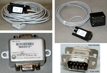
 204 - AcqKnowledge Peak Detector Operation | BIOPAC
 Robert P. Patterson, Ph.D. COF-1389 - AIMBE
 Supplier Partner: BIOPAC - SDR Scientific Introduces a Key Vendor
JMIR mHealth and uHealth - Nontraditional Electrocardiogram and Algorithms for Inconspicuous In-Home Monitoring: Comparative...
 Sonosite To Present At Jefferies 2010 Global Life Sciences Conference | Fujifilm Sonosite
AcqKnowledge 5 Demo | BIOPAC
 Frontiers | Linking Pain Sensation to the Autonomic Nervous System: The Role of the Anterior Cingulate and Periaqueductal Gray...
Clinical Trials Register
 Physical Medicine & Rehabilitation - Research output
- Experts@Minnesota
Physical Medicine & Rehabilitation - Research output
- Experts@Minnesota
Understanding the somatic consequences of depression: biological mechanisms and the role of depression symptom profile | BMC...
 Clinical Advisory Panel - Penlon
 BIO Web of Conferences
Hemodynamic5
- Background Impedance Cardiography (ICG) is a non-invasive tool for continuous hemodynamic monitoring. (medscape.com)
- The range of alternative treatments is large and the most recent guidelines are not prescriptive about the best choice of treatment The aim of this study is to conduct a randomized controlled triple-blind trial to compare two different approaches for resistant hypertensive patients: routine treatment with 25 mg spironolactone or treatment according to the hemodynamic study- Impedance Cardiography in the improvement of blood pressure control. (udg.edu)
- To analyze the vascular pattern we evaluated the database of NIVE (Phillips IU22 ® and CAP with Arteriograph ® ) with 2890 first ever evaluated patients, and analyzed 55 young controls (A), 56 young with ISH (B), 19 elderly controls (C) and 51 elderly with ISH (D). For the Hemodynamic analysis (Impedance Cardiography, Exxer ® ) the same groups with (A) 115, (B) 59, (C) 50 and (D) 148. (atlantis-press.com)
- Thirteen young adults (age 20.5 ± 0.7 years, BMI 22.0 ± 4.3) and 26 elderly individuals (age 60.2 ± 6.1 years, BMI 21.7 ± 2.2) were enrolled to measure real-time hemodynamic responses using non-invasive impedance cardiography during the 6-minute walk test. (biomedcentral.com)
- LVM, relative wall thickness [RWT]), vascular function (e.g., arterial stiffness, FMD) and impedance cardiography of hemodynamic function at rest. (elsevierpure.com)
Noninvasive5
- In the last decade, an inexpensive and simple noninvasive method (i.e., transthoracic electrical bioimpedance cardiography, has been tested in healthy subjects and patients with various heart disease for measuring stroke volume and cardiac output at rest and/or during exercise. (nih.gov)
- These results demonstrate that impedance cardiography is a noninvasive, simple, accurate, and reproducible method of measurement of cardiac output and stroke volume over a wide range of workloads. (nih.gov)
- Impedance cardiography permits noninvasive determinations of cardiac output at short intervals but data regarding variability of this method in patients with pacemakers is unavailable. (elsevierpure.com)
- Mountain View Hospital recently purchased the newest noninvasive Impedance Cardiography (ICG) Test machine. (cascadebusnews.com)
- PriMed's clinicians use the MedsEngine AI tool from MediSync and the NICaS (noninvasive cardiac system with impedance cardiography) to determine each patient's unique blood pressure pathophysiology. (medscape.com)
Cardiac3
- Stroke volume and cardiac output were simultaneously obtained at rest and at the end of each work rate stage with 3 methods: impedance, thermodilution, and direct Fick. (nih.gov)
- This data is very encouraging regarding the role that impedance cardiography technology can play in cardiac monitoring and healthcare. (globenewswire.com)
- Conclusions: This new contemporary bioimpedance cardiography device provided reliable measures of dynamic cardiac responses during a simulated apnea event. (pneumologia.eu)
Bioimpedance1
- Impedance cardiography (ICG), also referred to as electrical impedance plethysmography (EIP) or Thoracic Electrical Bioimpedance (TEB) has been researched since the 1940s. (wikipedia.org)
Tissue impedance1
- Fluid is an important determinant of tissue impedance. (nih.gov)
Thorax1
- With ICG, the placement of four dual disposable sensors on the neck and chest are used to transmit and detect electrical and impedance changes in the thorax, which are used to measure and calculate cardiodynamic parameters. (wikipedia.org)
Electrode1
- The replacement of the band-electrode to spot-electrode in the admittance cardiography was also successfully performed on a sitting position using the 64-ch impedance mapping system. (nii.ac.jp)
Plethysmography1
- The IsenseU-BP+ device presented in this article measures single channel ECG, impedance cardiography and photo plethysmography at the chest. (sintef.no)
Measurements3
- Each patient was studied in the supine position and repeated impedance measurements were obtained. (elsevierpure.com)
- Patients were monitored using serial chest radiographs and electrical impedance tomography measurements of lung impedance during hospital admission. (nih.gov)
- 5. By analyzing the measurements, you can determine the characteristic impedance of the PCB board. (pcba-manufacturers.com)
Cardiovascular2
- Volunteers ( n = 470) without medication with cardiovascular effects were examined using radial pulse wave analysis, whole-body impedance cardiography, and heart rate variability analysis. (biomedcentral.com)
- Gabor Vereczkey's special interests are non-invasive cardiovascular monitoring, impedance cardiography, blood gas analysis. (penlon.com)
Significantly1
- Choosing the right dielectric material for your PCB can significantly impact impedance control. (pcba-manufacturers.com)
Data1
- The precision of impedance cardiography demonstrated may be comparable or superior to other frequently used techniques, and the data obtained are valuable both investigationally and clinically. (elsevierpure.com)
Time2
- At the same time, the impedance board can also reduce signal crosstalk and improve signal reliability. (pcba-manufacturers.com)
- At the same time, the impedance board can also prevent external electromagnetic signals from entering the circuit and improve the anti-interference ability of the circuit. (pcba-manufacturers.com)
Results1
- Why is the measured impedance of the bladder tissue different from the computational modelling results? (springer.com)
Lower1
- Similarly, increasing the spacing between traces can lower impedance. (pcba-manufacturers.com)
Technology2
- SAN DIEGO, March 12, 2002 (PRIMEZONE) -- CardioDynamics (Nasdaq:CDIC), the innovator and global leader of Impedance Cardiography (ICG) technology, today announced that a significant ICG study was published in the February 2002 issue of peer-reviewed The Journal of Cardiothoracic and Vascular Anesthesia. (globenewswire.com)
- CardioDynamics (Nasdaq:CDIC), the ICG Company, is the innovator and global leader of breakthrough medical technology called Impedance Cardiography (ICG). (globenewswire.com)
Role1
- In large-scale integrated circuits, power consumption control is very important, and the impedance board plays an important role in power consumption control. (pcba-manufacturers.com)
Leader1
- Sonosite, Inc. ( www.sonosite.com ) is the innovator and world leader in hand-carried ultrasound and industry leader in impedance cardiography equipment. (sonosite.com)
Important2
- These proceedings continue the series edited in the framework of the traditional triennial International Conference on El- trical Bio-Impedance (ICEBI), the most important platform for presenting recent scientific achievements in the area of el- th nd trical bio-impedance. (springer.com)
- Controlling impedance in PCB design is an important aspect of ensuring the optimal performance of your circuit. (pcba-manufacturers.com)
Layers1
- PCB impedance board achieves specific impedance values by controlling parameters such as line width, spacing, layer spacing, lead length, and ground holes between layers. (pcba-manufacturers.com)
Control2
- How do you control impedance of PCB design? (pcba-manufacturers.com)
- Adjusting the trace width and spacing can help control the impedance on your PCB. (pcba-manufacturers.com)
Article1
- The use of impedance cardiography in psychophysiological research was pioneered by the publication of an article by Miller and Horvath in 1978. (wikipedia.org)
Relationship1
- We have investigated the relationship between LVF and the electrical impedance of lung tissue. (nih.gov)
Current2
- Impedance value refers to the resistance that current encounters when it travels in a circuit. (pcba-manufacturers.com)
- When the impedance matching of the circuit is not guaranteed, current will be reflected in the circuit, which will lead to energy loss and an increase in power consumption. (pcba-manufacturers.com)
Electrocardiography1
- The study is designed to examine the effects of early nutritional supplementation in infancy on the neurobiological and psychological functioning of young adolescents in Ghana using methods to assess neurophysiology in both the central and autonomic nervous systems, including magnetic resonance imaging (MRI) with diffusion-weighted imaging and functional connectivity, as well as electrocardiography and impedance cardiography. (nih.gov)
Measurement3
- [ 2 ] However, to our knowledge, no data are available on impedance measurement during spinal anesthesia with different local anesthetic doses whereas several studies were focused on the optimal dose of local anesthetics to obtain a good anesthesia and reduce the motor block. (medscape.com)
- All About Cardiac Impedance Cardiac impedance is a measurement of the mechanical activation of the heart. (mindwaretech.com)
- Measurement of Hemodynamic Variables using Impedance Cardiography on Remifentanil-Propofol Infusion during Anesthetic Induction. (ekja.org)
Cardiographies1
- I had 2 impedance cardiographies done around 2005-2007 I think and both were significantly abnormal, along the lines predicted by Peckerman. (phoenixrising.me)
NICaS1
- PriMed's clinicians use the MedsEngine AI tool from MediSync and the NICaS (noninvasive cardiac system with impedance cardiography) to determine each patient's unique blood pressure pathophysiology. (medscape.com)
Systemic vascular r1
- and systemic vascular resistance index (SVRI) assessed by impedance cardiography (ICG). (medscimonit.com)
Hypertension1
- 2014). Determination of the effects of pulmonary arterial hypertension and therapy on the cardiovascular system of rats by impedance cardiography. (unlp.edu.ar)
Electrodes1
- The monitoring system could be expanded with more sensors - electrodes configured for impedance cardiography, and electrocardiogram. (t3.com)
Signals2
- Only two signals are required for analysis in the Impedance Cardiography (IMP) analysis application - ECG and Zo. (mindwaretech.com)
- Novel characterization method of impedance cardiography signals using time-frequency distributions. (irsjd.org)
Clinical1
- Methodologies include orthostatic stress, electrophysiologic evaluation, vascular reactivity (ultrasound imaging), spectral analysis, impedance cardiography and pharmacological dissections of clinical and basic neurovascular physiology. (mayo.edu)
Method1
- Impedance Cardiography (ICG) has been proposed as a non-invasive method for hemodynamic monitoring of adult cardiopathic patients as well as for patients in the emergency department, wards and ambulatories. (medscape.com)
Heart1
- 1. Hemodynamic regulation during postural tilt assessed by heart rate- and blood-pressure variability combined with impedance cardiography. (nih.gov)
Article1
- The use of impedance cardiography in psychophysiological research was pioneered by the publication of an article by Miller and Horvath in 1978. (wikipedia.org)
Electrical1
- With ICG, the placement of four dual disposable sensors on the neck and chest are used to transmit and detect electrical and impedance changes in the thorax, which are used to measure and calculate cardiodynamic parameters. (wikipedia.org)