Cardiac Electrophysiology
Electrophysiological Processes
Epicardial Mapping
Electrophysiologic Techniques, Cardiac
Models, Cardiovascular
Arrhythmias, Cardiac
Electrophysiology
Heart Conduction System
Voltage-Sensitive Dye Imaging
Electrophysiological Phenomena
Myocytes, Cardiac
Action Potentials
Electrocardiography
Atrioventricular Node
Heart Ventricles
Computer Simulation
Catheter Ablation
Long QT Syndrome
Myocardium
Potassium Channels
Cardiac Output
Algorithms
Death, Sudden, Cardiac
Cardiac Pacing, Artificial
Patch-Clamp Techniques
Cardiomegaly
Cardiac Catheterization
Heart Diseases
Membrane Potentials
Encyclopedias as Topic
Defibrillators, Implantable
Fellowships and Scholarships
Maryland
Decennial follow-up in patients with recurrent tachycardia originating from the right ventricular outflow tract: electrophysiologic characteristics and response to treatment. (1/71)
AIMS: In the setting of right ventricular outflow tract-tachycardia (RVOT-T), data about long-term follow-up (FU) with respect to the therapeutic strategies are missing. All patients (pts) referred to our institution during the last 20 years for the treatment of RVOT-T were studied in a retrospective analysis to assess mortality and efficacy of treatment. METHODS AND RESULTS: One hundred and thirty-three patients (77 female; 39+/-13 years) with sustained RVOT-T were included in this study. At the time of first presentation, diagnosis of RVOT-T was made by complete invasive and non-invasive diagnostic assessment, including electrophysiology study and two-dimensional echocardiography. After 135+/-68 months (median 136, range 29-248), patients were invited to undergo clinical assessment. Of the 133 pts, 127 (95%) survived and six (5%) died from non-cardiac disease. Anti-arrhythmic (AA) drugs were given to 62 of the 133 pts (47%); of them 32 (52%) had recurrences during follow-up. The mean time to recurrence was 10.02 years (95% CI 7.46-12.59). The other 71 study patients (53%) underwent catheter ablation. The procedure was successful in 58 pts (82%). During follow-up, 30 (52%) of the 58 successfully treated patients had recurrences of RVOT-T. The mean time to recurrence was 6.28 years (95% CI 4.96-7.6). RVOT-T recurrences were similar in morphology to those treated previously in 33% and different in 67% of cases. CONCLUSIONS: Long-term follow-up in patients with RVOT-T is favourable. Catheter ablation is effective in this setting. However, late recurrences with similar or different morphology may arise in half of the patients after initially successful treatment. AA drug therapy is a valid initial therapeutic option, since it is effective in about half of the patients. (+info)Association of atrial arrhythmia and sinus node dysfunction in patients with catecholaminergic polymorphic ventricular tachycardia. (2/71)
BACKGROUND: This study was performed to investigate the frequency and importance of supraventricular arrhythmia and sinus node (SN) dysfunction in patients with catecholaminergic polymorphic ventricular tachycardia (CPVT). METHODS AND RESULTS: Eight patients with CPVT (mean age: 16.8+/-8.1 years) underwent an electrophysiological study. SN recovery time (1,389+/-394 ms) was slightly prolonged, and 4 of 8 patients had abnormal values. Atrial flutter (AF) was induced by low-rate atrial pacing in 2 patients and by isoproterenol infusion in 1 patient. Atrial fibrillation (Af) was induced by isoproterenol infusion in 2 patients. One patient presented with Af during the follow-up period, and 2 of 4 patients with AF/Af presented with increased SN recovery time. CONCLUSIONS: Patients with CPVT frequently have associated with SN dysfunction, and inducible atrial tachyarrhythmias, which indicate that the pathogenesis of CPVT is limited not only to the ventricular myocardium, but also to broad regions of the heart, including the SN and atrial muscle. (+info)Matters of the heart: the physiology of cardiac function and failure. (3/71)
Heart failure as a result of a myocardial infarction (MI) is a common condition with a poor prognosis. The adaptive changes in the surviving myocardium appear to be insufficient in terms of both mechanical/contractile performance and electrical stability. The modification of the underlying myocardial physiology is complex, varying across the different layers within the wall of the ventricle and within one layer. Two therapeutic strategies are briefly discussed, as outlined here. (i) Enhancing contractility by alteration of the expression of a single protein (e.g. sarco-endoplasmic reticulum Ca(2+) ATPase, SERCA) could potentially reverse both mechanical and electrical abnormalities. However, experimental data involving the upregulation of SERCA suggest that the therapeutic range of this approach is narrow. (ii) The use of regular exercise training to improve cardiac performance in heart failure. This appears to act by normalizing a number of aspects of myocardial physiology. (+info)Electrophysiological consequence of adipose-derived stem cell transplantation in infarcted porcine myocardium. (4/71)
AIMS: Aim of this study was to investigate the effect of intracoronary administration of freshly isolated adipose-derived mononuclear cells (ADMCs) on myocardial vulnerability to arrhythmia induction after infarction. METHODS AND RESULTS: A transmural myocardial infarction in an experimental porcine model was induced by occlusion of the mid-left anterior descending artery with an angioplasty balloon for 3 h. Upon reperfusion, a cellular suspension with freshly isolated ADMCs (1.5 x 10(6) cells/kg BW) or vehicle alone was injected into the infarct artery. All animals underwent a programmed ventricular stimulation at 8 weeks follow-up for possible induction of ventricular arrhythmias using a train of 8 S1 stimuli. Cell injections did not cause acute ventricular arrhythmia, bradycardia, or conduction block. The cycle length of the ventricular arrhythmia was compared at 1 and 10 s following its induction. Despite comparable infarct size in both groups, we found that the cycle length of the induced ventricular arrhythmia in the ADMC-treated group was significantly longer compared with control animals (P < 0.05). We also found that extra-stimuli were required for arrhythmia induction in the ADMC-treated group compared with control animals. CONCLUSION: Freshly isolated autologous stem cell therapy is not proarrhythmic in pigs. (+info)Mechanisms of calcium transient and action potential alternans in cardiac cells and tissues. (5/71)
Alternation of cardiac action potential duration (APD) from beat to beat and concurrent alternation of the amplitude of the calcium transient are regarded as important arrhythmia mechanisms. These phenomena are causally interrelated and can be reliably evoked by an increase in beat frequency or by ischemia. The first part of this historical review deals with the physiology of APD alternans. Sections recounting the evolution of knowledge about calcium-activated ion currents and calcium transient alternans are interspersed among sections describing the growth of the so-called "restitution hypothesis," which involves time-dependent recovery of potassium channels (including their passage through pre-open states) as a function of diastolic interval. Major developments are generally in chronological order, but it is necessary to move back and forth between the two theories to respect the overall time line, which runs from about l965 to the present. The concluding two sections deal with the pathophysiology of calcium transient and APD alternans during ischemia, which may be the basis for out-of-hospital cardiac arrest during the initial stages of acute myocardial infarction. (+info)Pharmacological separation of early afterdepolarizations from arrhythmogenic substrate in DeltaKPQ Scn5a murine hearts modelling human long QT 3 syndrome. (6/71)
AIM: To perform an empirical, pharmacological, separation of early afterdepolarizations (EADs) and transmural gradients of repolarization in arrhythmogenesis in a genetically modified mouse heart modelling human long QT syndrome (LQT) 3. METHODS: Left ventricular endocardial and epicardial monophasic action potentials and arrhythmogenic tendency were compared in isolated wild type (WT) and Scn5a+/Delta hearts perfused with 0.1 and 1 microm propranolol and paced from the right ventricular epicardium. RESULTS: All spontaneously beating bradycardic Scn5a+/Delta hearts displayed EADs, triggered beats and ventricular tachycardia (VT; n = 7), events never seen in WT hearts (n = 5). Perfusion with 0.1 and 1 microm propranolol suppressed all EADs, triggered beats and episodes of VT. In contrast, triggering of VT persisted following programmed electrical stimulation in 6 of 12 (50%), one of eight (12.5%), but six of eight (75%) Scn5a+/Delta hearts perfused with 0, 0.1 and 1 microm propranolol respectively in parallel with corresponding alterations in repolarization gradients, reflected in action potential duration (DeltaAPD(90)) values. Thus 0.1 microm propranolol reduced epicardial but not endocardial APD(90) from 54.7 +/- 1.6 to 44.0 +/- 2.0 ms, restoring DeltaAPD(90) from -3.8 +/- 1.6 to 3.5 +/- 2.5 ms (all n = 5), close to WT values. However, 1 microm propranolol increased epicardial APD(90) to 72.5 +/- 1.2 ms and decreased endocardial APD(90) from 50.9 +/- 1.0 to 24.5 +/- 0.3 ms, increasing DeltaAPD(90) to -48.0 +/- 1.2 ms. CONCLUSION: These findings empirically implicate EADs in potentially initiating spontaneous arrhythmogenic phenomena and transmural repolarization gradients in the re-entrant substrate that would sustain such activity when provoked by extrasystolic activity in murine hearts modelling human LQT3 syndrome. (+info)Arrhythmogenesis research: a perspective from computational electrophysiology viewpoint. (7/71)
The mechanisms by which arrhythmias are generated in the heart remains a field of intensive research. Recent advances in computational biology and electrophysiology have enabled researchers to use an alternative tool in the study of arrhythmia mechanisms, the multi-scale modeling and simulation of cardiac arrhythmogenesis at the organ level. This article reviews the recent advances and achievements using this approach. (+info)Hyperpolarization-activated cyclic nucleotide-modulated 'HCN' channels confer regular and faster rhythmicity to beating mouse embryonic stem cells. (8/71)
The hyperpolarization-activated cation current (I(f)), and the hyperpolarization-activated cyclic nucleotide-modulated 'HCN' subunits that underlie it, are important components of spontaneous activity in the embryonic mouse heart, but whether they contribute to this activity in mouse embryonic stem cell-derived cardiomyocytes has not been investigated. We address this issue in spontaneously beating cells derived from mouse embryonic stem cells (mESCs) over the course of development in culture. I(f) and action potentials were recorded from single beating cells at early, intermediate and late development stages using perforated whole-cell voltage- and current-clamp techniques. Our data show that the proportion of cells expressing I(f), and the density of I(f) in these cells, increased during development and correlated with action potential frequency and the rate of diastolic depolarization. The I(f) blocker ZD7288 (0.3 microm) reduced I(f) and the beating rate of embryoid bodies. Taken together, the activation kinetics of I(f) and results from Western blots are consistent with the presence of the HCN2 and HCN3 isoforms. At all stages of development, isoproterenol (isoprenaline) and acetylcholine shifted the voltage dependence of I(f) to more positive and negative voltages, respectively, and they also increased and decreased the beating rate of embryonic cell bodies, respectively. Together, the data suggest that current through HCN2 and HCN3 channels confers regular and faster rhythmicity to mESCs, which mirrors the developing embryonic mouse heart, and contributes to modulation of rhythmicity by autonomic stimulation. (+info)Cardiac electrophysiology is a branch of medicine that deals with the study and understanding of the electrical activities of the heart. It involves the diagnosis and treatment of various heart rhythm disorders (arrhythmias) such as bradycardia (slow heart rate), tachycardia (fast heart rate), atrial fibrillation, atrial flutter, ventricular fibrillation, and other rhythm abnormalities.
Cardiac electrophysiologists use various diagnostic tests, including electrocardiograms (ECGs), Holter monitors, event monitors, and invasive procedures such as electrophysiology studies (EPS) and catheter ablation to evaluate and treat heart rhythm disorders. The goal of treatment is to restore a normal heart rhythm and prevent complications associated with arrhythmias, such as stroke or heart failure.
Electrophysiological processes refer to the electrical activities that occur within biological cells or organ systems, particularly in nerve and muscle tissues. These processes involve the generation, transmission, and reception of electrical signals that are essential for various physiological functions, such as nerve impulse transmission, muscle contraction, and hormonal regulation.
At the cellular level, electrophysiological processes are mediated by the flow of ions across the cell membrane through specialized protein channels. This ion movement generates a voltage difference across the membrane, leading to the development of action potentials, which are rapid changes in electrical potential that travel along the cell membrane and transmit signals between cells.
In clinical medicine, electrophysiological studies (EPS) are often used to diagnose and manage various cardiac arrhythmias and neurological disorders. These studies involve the recording of electrical activity from the heart or brain using specialized equipment, such as an electrocardiogram (ECG) or an electroencephalogram (EEG). By analyzing these recordings, physicians can identify abnormalities in the electrical activity of these organs and develop appropriate treatment plans.
Epicardial mapping is a medical procedure used to create a detailed map of the electrical activity on the surface of the heart (epicardium). This technique is often used during electrophysiology studies to help diagnose and locate the source of abnormal heart rhythms, such as ventricular tachycardia or atrial fibrillation.
During epicardial mapping, a specialist (usually an electrophysiologist) will introduce a catheter through a vein or artery, which is then guided to the heart. Once in position, electrodes on the tip of the catheter record electrical signals from the heart's surface. These signals are used to create a detailed map of the heart's electrical activity, allowing the specialist to identify areas with abnormal electrical patterns.
This information can be crucial for determining the best course of treatment, such as targeted ablation therapy to eliminate the source of the arrhythmia. Epicardial mapping is typically performed in an electrophysiology lab or cardiac catheterization laboratory under fluoroscopy guidance, and it requires expertise in both cardiovascular medicine and interventional techniques.
Electrophysiologic techniques, cardiac, refer to medical procedures used to study the electrical activities and conduction systems of the heart. These techniques involve the insertion of electrode catheters into the heart through blood vessels under fluoroscopic guidance to record and stimulate electrical signals. The information obtained from these studies can help diagnose and evaluate various cardiac arrhythmias, determine the optimal treatment strategy, and assess the effectiveness of therapies such as ablation or implantable devices.
The electrophysiologic study (EPS) is a type of cardiac electrophysiologic technique that involves the measurement of electrical signals from different regions of the heart to evaluate its conduction system's function. The procedure can help identify the location of abnormal electrical pathways responsible for arrhythmias and determine the optimal treatment strategy, such as catheter ablation or medication therapy.
Cardiac electrophysiologic techniques are also used in device implantation procedures, such as pacemaker or defibrillator implantation, to ensure proper placement and function of the devices. These techniques can help program and test the devices to optimize their settings for each patient's needs.
In summary, cardiac electrophysiologic techniques are medical procedures used to study and manipulate the electrical activities of the heart, helping diagnose and treat various arrhythmias and other cardiac conditions.
Cardiovascular models are simplified representations or simulations of the human cardiovascular system used in medical research, education, and training. These models can be physical, computational, or mathematical and are designed to replicate various aspects of the heart, blood vessels, and blood flow. They can help researchers study the structure and function of the cardiovascular system, test new treatments and interventions, and train healthcare professionals in diagnostic and therapeutic techniques.
Physical cardiovascular models may include artificial hearts, blood vessels, or circulation systems made from materials such as plastic, rubber, or silicone. These models can be used to study the mechanics of heart valves, the effects of different surgical procedures, or the impact of various medical devices on blood flow.
Computational and mathematical cardiovascular models use algorithms and equations to simulate the behavior of the cardiovascular system. These models may range from simple representations of a single heart chamber to complex simulations of the entire circulatory system. They can be used to study the electrical activity of the heart, the biomechanics of blood flow, or the distribution of drugs in the body.
Overall, cardiovascular models play an essential role in advancing our understanding of the human body and improving patient care.
Cardiac arrhythmias are abnormal heart rhythms that result from disturbances in the electrical conduction system of the heart. The heart's normal rhythm is controlled by an electrical signal that originates in the sinoatrial (SA) node, located in the right atrium. This signal travels through the atrioventricular (AV) node and into the ventricles, causing them to contract and pump blood throughout the body.
An arrhythmia occurs when there is a disruption in this electrical pathway or when the heart's natural pacemaker produces an abnormal rhythm. This can cause the heart to beat too fast (tachycardia), too slow (bradycardia), or irregularly.
There are several types of cardiac arrhythmias, including:
1. Atrial fibrillation: A rapid and irregular heartbeat that starts in the atria (the upper chambers of the heart).
2. Atrial flutter: A rapid but regular heartbeat that starts in the atria.
3. Supraventricular tachycardia (SVT): A rapid heartbeat that starts above the ventricles, usually in the atria or AV node.
4. Ventricular tachycardia: A rapid and potentially life-threatening heart rhythm that originates in the ventricles.
5. Ventricular fibrillation: A chaotic and disorganized electrical activity in the ventricles, which can be fatal if not treated immediately.
6. Heart block: A delay or interruption in the conduction of electrical signals from the atria to the ventricles.
Cardiac arrhythmias can cause various symptoms, such as palpitations, dizziness, shortness of breath, chest pain, and fatigue. In some cases, they may not cause any symptoms and go unnoticed. However, if left untreated, certain types of arrhythmias can lead to serious complications, including stroke, heart failure, or even sudden cardiac death.
Treatment for cardiac arrhythmias depends on the type, severity, and underlying causes. Options may include lifestyle changes, medications, cardioversion (electrical shock therapy), catheter ablation, implantable devices such as pacemakers or defibrillators, and surgery. It is essential to consult a healthcare professional for proper evaluation and management of cardiac arrhythmias.
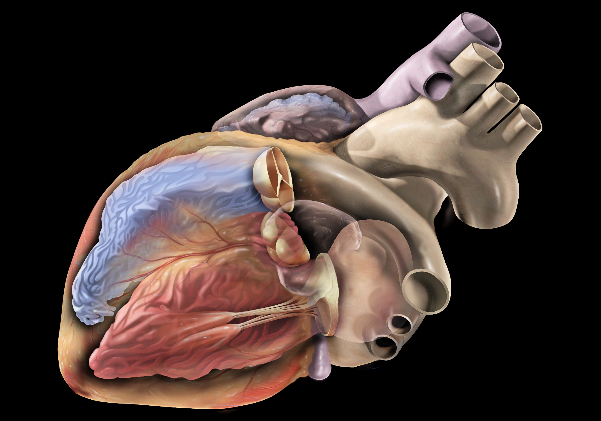
In medical terms, the heart is a muscular organ located in the thoracic cavity that functions as a pump to circulate blood throughout the body. It's responsible for delivering oxygen and nutrients to the tissues and removing carbon dioxide and other wastes. The human heart is divided into four chambers: two atria on the top and two ventricles on the bottom. The right side of the heart receives deoxygenated blood from the body and pumps it to the lungs, while the left side receives oxygenated blood from the lungs and pumps it out to the rest of the body. The heart's rhythmic contractions and relaxations are regulated by a complex electrical conduction system.
Electrophysiology is a branch of medicine that deals with the electrical activities of the body, particularly the heart. In a medical context, electrophysiology studies (EPS) are performed to assess abnormal heart rhythms (arrhythmias) and to evaluate the effectiveness of certain treatments, such as medication or pacemakers.
During an EPS, electrode catheters are inserted into the heart through blood vessels in the groin or neck. These catheters can record the electrical activity of the heart and stimulate it to help identify the source of the arrhythmia. The information gathered during the study can help doctors determine the best course of treatment for each patient.
In addition to cardiac electrophysiology, there are also other subspecialties within electrophysiology, such as neuromuscular electrophysiology, which deals with the electrical activity of the nervous system and muscles.
The heart conduction system is a group of specialized cardiac muscle cells that generate and conduct electrical impulses to coordinate the contraction of the heart chambers. The main components of the heart conduction system include:
1. Sinoatrial (SA) node: Also known as the sinus node, it is located in the right atrium near the entrance of the superior vena cava and functions as the primary pacemaker of the heart. It sets the heart rate by generating electrical impulses at regular intervals.
2. Atrioventricular (AV) node: Located in the interatrial septum, near the opening of the coronary sinus, it serves as a relay station for electrical signals between the atria and ventricles. The AV node delays the transmission of impulses to allow the atria to contract before the ventricles.
3. Bundle of His: A bundle of specialized cardiac muscle fibers that conducts electrical impulses from the AV node to the ventricles. It divides into two main branches, the right and left bundle branches, which further divide into smaller Purkinje fibers.
4. Right and left bundle branches: These are extensions of the Bundle of His that transmit electrical impulses to the respective right and left ventricular myocardium. They consist of specialized conducting tissue with large diameters and minimal resistance, allowing for rapid conduction of electrical signals.
5. Purkinje fibers: Fine, branching fibers that arise from the bundle branches and spread throughout the ventricular myocardium. They are responsible for transmitting electrical impulses to the working cardiac muscle cells, triggering coordinated ventricular contraction.
In summary, the heart conduction system is a complex network of specialized muscle cells responsible for generating and conducting electrical signals that coordinate the contraction of the atria and ventricles, ensuring efficient blood flow throughout the body.
Voltage-sensitive dye imaging (VSDI) is not a medical definition itself, but it is a technique used in the field of physiology and neuroscience to measure the electrical activity of cells, particularly excitable cells such as neurons and cardiac myocytes. Here's a brief explanation:
Voltage-sensitive dyes are fluorescent or luminescent molecules that change their optical properties in response to changes in membrane potential. When these dyes bind to the cell membrane, they can report on the electrical activity of the cell by changing their emission intensity, polarization, or lifetime depending on the voltage across the membrane.
VSDI is a technique that uses these voltage-sensitive dyes to measure changes in membrane potential in a population of cells or even in an entire organ. By illuminating the sample with light and measuring the emitted fluorescence or luminescence, researchers can visualize and quantify the electrical activity of cells in real-time.
VSDI has many applications in basic research, including studying the electrical properties of neurons, mapping neural circuits, investigating the mechanisms of excitation-contraction coupling in cardiac myocytes, and developing new drugs that target ion channels. However, it is not a commonly used clinical technique due to its limitations, such as the need for specialized equipment, the potential for phototoxicity, and the difficulty of interpreting signals from complex tissues.
Electrophysiological phenomena refer to the electrical properties and activities of biological tissues, cells, or organ systems, particularly in relation to nerve and muscle function. These phenomena can be studied using various techniques such as electrocardiography (ECG), electromyography (EMG), and electroencephalography (EEG).
In the context of cardiology, electrophysiological phenomena are often used to describe the electrical activity of the heart. The ECG is a non-invasive test that measures the electrical activity of the heart as it contracts and relaxes. By analyzing the patterns of electrical activity, doctors can diagnose various heart conditions such as arrhythmias, myocardial infarction, and electrolyte imbalances.
In neurology, electrophysiological phenomena are used to study the electrical activity of the brain. The EEG is a non-invasive test that measures the electrical activity of the brain through sensors placed on the scalp. By analyzing the patterns of electrical activity, doctors can diagnose various neurological conditions such as epilepsy, sleep disorders, and brain injuries.
Overall, electrophysiological phenomena are an important tool in medical diagnostics and research, providing valuable insights into the function of various organ systems.
Cardiac myocytes are the muscle cells that make up the heart muscle, also known as the myocardium. These specialized cells are responsible for contracting and relaxing in a coordinated manner to pump blood throughout the body. They differ from skeletal muscle cells in several ways, including their ability to generate their own electrical impulses, which allows the heart to function as an independent rhythmical pump. Cardiac myocytes contain sarcomeres, the contractile units of the muscle, and are connected to each other by intercalated discs that help coordinate contraction and ensure the synchronous beating of the heart.
An action potential is a brief electrical signal that travels along the membrane of a nerve cell (neuron) or muscle cell. It is initiated by a rapid, localized change in the permeability of the cell membrane to specific ions, such as sodium and potassium, resulting in a rapid influx of sodium ions and a subsequent efflux of potassium ions. This ion movement causes a brief reversal of the electrical potential across the membrane, which is known as depolarization. The action potential then propagates along the cell membrane as a wave, allowing the electrical signal to be transmitted over long distances within the body. Action potentials play a crucial role in the communication and functioning of the nervous system and muscle tissue.
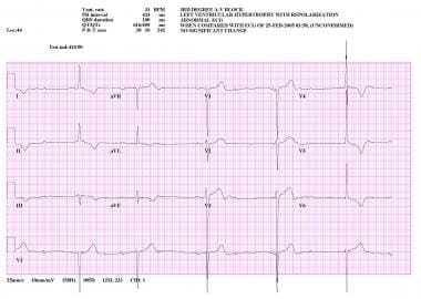
Electrocardiography (ECG or EKG) is a medical procedure that records the electrical activity of the heart. It provides a graphic representation of the electrical changes that occur during each heartbeat. The resulting tracing, called an electrocardiogram, can reveal information about the heart's rate and rhythm, as well as any damage to its cells or abnormalities in its conduction system.
During an ECG, small electrodes are placed on the skin of the chest, arms, and legs. These electrodes detect the electrical signals produced by the heart and transmit them to a machine that amplifies and records them. The procedure is non-invasive, painless, and quick, usually taking only a few minutes.
ECGs are commonly used to diagnose and monitor various heart conditions, including arrhythmias, coronary artery disease, heart attacks, and electrolyte imbalances. They can also be used to evaluate the effectiveness of certain medications or treatments.
The atrioventricular (AV) node is a critical part of the electrical conduction system of the heart. It is a small cluster of specialized cardiac muscle cells located in the lower interatrial septum, near the opening of the coronary sinus. The AV node receives electrical impulses from the sinoatrial node (the heart's natural pacemaker) via the internodal pathways and delays their transmission for a brief period before transmitting them to the bundle of His and then to the ventricles. This delay allows the atria to contract and empty their contents into the ventricles before the ventricles themselves contract, ensuring efficient pumping of blood throughout the body.
The AV node plays an essential role in maintaining a normal heart rhythm, as it can also function as a backup pacemaker if the sinoatrial node fails to generate impulses. However, certain heart conditions or medications can affect the AV node's function and lead to abnormal heart rhythms, such as atrioventricular block or atrial tachycardia.
The heart ventricles are the two lower chambers of the heart that receive blood from the atria and pump it to the lungs or the rest of the body. The right ventricle pumps deoxygenated blood to the lungs, while the left ventricle pumps oxygenated blood to the rest of the body. Both ventricles have thick, muscular walls to generate the pressure necessary to pump blood through the circulatory system.
A computer simulation is a process that involves creating a model of a real-world system or phenomenon on a computer and then using that model to run experiments and make predictions about how the system will behave under different conditions. In the medical field, computer simulations are used for a variety of purposes, including:
1. Training and education: Computer simulations can be used to create realistic virtual environments where medical students and professionals can practice their skills and learn new procedures without risk to actual patients. For example, surgeons may use simulation software to practice complex surgical techniques before performing them on real patients.
2. Research and development: Computer simulations can help medical researchers study the behavior of biological systems at a level of detail that would be difficult or impossible to achieve through experimental methods alone. By creating detailed models of cells, tissues, organs, or even entire organisms, researchers can use simulation software to explore how these systems function and how they respond to different stimuli.
3. Drug discovery and development: Computer simulations are an essential tool in modern drug discovery and development. By modeling the behavior of drugs at a molecular level, researchers can predict how they will interact with their targets in the body and identify potential side effects or toxicities. This information can help guide the design of new drugs and reduce the need for expensive and time-consuming clinical trials.
4. Personalized medicine: Computer simulations can be used to create personalized models of individual patients based on their unique genetic, physiological, and environmental characteristics. These models can then be used to predict how a patient will respond to different treatments and identify the most effective therapy for their specific condition.
Overall, computer simulations are a powerful tool in modern medicine, enabling researchers and clinicians to study complex systems and make predictions about how they will behave under a wide range of conditions. By providing insights into the behavior of biological systems at a level of detail that would be difficult or impossible to achieve through experimental methods alone, computer simulations are helping to advance our understanding of human health and disease.
Catheter ablation is a medical procedure in which specific areas of heart tissue that are causing arrhythmias (irregular heartbeats) are destroyed or ablated using heat energy (radiofrequency ablation), cold energy (cryoablation), or other methods. The procedure involves threading one or more catheters through the blood vessels to the heart, where the tip of the catheter can be used to selectively destroy the problematic tissue. Catheter ablation is often used to treat atrial fibrillation, atrial flutter, and other types of arrhythmias that originate in the heart's upper chambers (atria). It may also be used to treat certain types of arrhythmias that originate in the heart's lower chambers (ventricles), such as ventricular tachycardia.
The goal of catheter ablation is to eliminate or reduce the frequency and severity of arrhythmias, thereby improving symptoms and quality of life. In some cases, it may also help to reduce the risk of stroke and other complications associated with arrhythmias. Catheter ablation is typically performed by a specialist in heart rhythm disorders (electrophysiologist) in a hospital or outpatient setting under local anesthesia and sedation. The procedure can take several hours to complete, depending on the complexity of the arrhythmia being treated.
It's important to note that while catheter ablation is generally safe and effective, it does carry some risks, such as bleeding, infection, damage to nearby structures, and the possibility of recurrent arrhythmias. Patients should discuss the potential benefits and risks of the procedure with their healthcare provider before making a decision about treatment.
Long QT syndrome (LQTS) is a cardiac electrical disorder characterized by a prolonged QT interval on the electrocardiogram (ECG), which can potentially trigger rapid, chaotic heartbeats known as ventricular tachyarrhythmias, such as torsades de pointes. These arrhythmias can be life-threatening and lead to syncope (fainting) or sudden cardiac death. LQTS is often congenital but may also be acquired due to certain medications, medical conditions, or electrolyte imbalances. It's essential to identify and manage LQTS promptly to reduce the risk of severe complications.
The myocardium is the middle layer of the heart wall, composed of specialized cardiac muscle cells that are responsible for pumping blood throughout the body. It forms the thickest part of the heart wall and is divided into two sections: the left ventricle, which pumps oxygenated blood to the rest of the body, and the right ventricle, which pumps deoxygenated blood to the lungs.
The myocardium contains several types of cells, including cardiac muscle fibers, connective tissue, nerves, and blood vessels. The muscle fibers are arranged in a highly organized pattern that allows them to contract in a coordinated manner, generating the force necessary to pump blood through the heart and circulatory system.
Damage to the myocardium can occur due to various factors such as ischemia (reduced blood flow), infection, inflammation, or genetic disorders. This damage can lead to several cardiac conditions, including heart failure, arrhythmias, and cardiomyopathy.
Heart rate is the number of heartbeats per unit of time, often expressed as beats per minute (bpm). It can vary significantly depending on factors such as age, physical fitness, emotions, and overall health status. A resting heart rate between 60-100 bpm is generally considered normal for adults, but athletes and individuals with high levels of physical fitness may have a resting heart rate below 60 bpm due to their enhanced cardiovascular efficiency. Monitoring heart rate can provide valuable insights into an individual's health status, exercise intensity, and response to various treatments or interventions.
Potassium channels are membrane proteins that play a crucial role in regulating the electrical excitability of cells, including cardiac, neuronal, and muscle cells. These channels facilitate the selective passage of potassium ions (K+) across the cell membrane, maintaining the resting membrane potential and shaping action potentials. They are composed of four or six subunits that assemble to form a central pore through which potassium ions move down their electrochemical gradient. Potassium channels can be modulated by various factors such as voltage, ligands, mechanical stimuli, or temperature, allowing cells to fine-tune their electrical properties and respond to different physiological demands. Dysfunction of potassium channels has been implicated in several diseases, including cardiac arrhythmias, epilepsy, and neurodegenerative disorders.
Cardiac output is a measure of the amount of blood that is pumped by the heart in one minute. It is defined as the product of stroke volume (the amount of blood pumped by the left ventricle during each contraction) and heart rate (the number of contractions per minute). Normal cardiac output at rest for an average-sized adult is about 5 to 6 liters per minute. Cardiac output can be increased during exercise or other conditions that require more blood flow, such as during illness or injury. It can be measured noninvasively using techniques such as echocardiography or invasively through a catheter placed in the heart.
An algorithm is not a medical term, but rather a concept from computer science and mathematics. In the context of medicine, algorithms are often used to describe step-by-step procedures for diagnosing or managing medical conditions. These procedures typically involve a series of rules or decision points that help healthcare professionals make informed decisions about patient care.
For example, an algorithm for diagnosing a particular type of heart disease might involve taking a patient's medical history, performing a physical exam, ordering certain diagnostic tests, and interpreting the results in a specific way. By following this algorithm, healthcare professionals can ensure that they are using a consistent and evidence-based approach to making a diagnosis.
Algorithms can also be used to guide treatment decisions. For instance, an algorithm for managing diabetes might involve setting target blood sugar levels, recommending certain medications or lifestyle changes based on the patient's individual needs, and monitoring the patient's response to treatment over time.
Overall, algorithms are valuable tools in medicine because they help standardize clinical decision-making and ensure that patients receive high-quality care based on the latest scientific evidence.
Sudden cardiac death (SCD) is a sudden, unexpected natural death caused by the cessation of cardiac activity. It is often caused by cardiac arrhythmias, particularly ventricular fibrillation, and is often associated with underlying heart disease, although it can occur in people with no known heart condition. SCD is typically defined as a natural death due to cardiac causes that occurs within one hour of the onset of symptoms, or if the individual was last seen alive in a normal state of health, it can be defined as occurring within 24 hours.
It's important to note that sudden cardiac arrest (SCA) is different from SCD, although they are related. SCA refers to the sudden cessation of cardiac activity, which if not treated immediately can lead to SCD.
Cardiac surgical procedures are operations that are performed on the heart or great vessels (the aorta and vena cava) by cardiothoracic surgeons. These surgeries are often complex and require a high level of skill and expertise. Some common reasons for cardiac surgical procedures include:
1. Coronary artery bypass grafting (CABG): This is a surgery to improve blood flow to the heart in patients with coronary artery disease. During the procedure, a healthy blood vessel from another part of the body is used to create a detour around the blocked or narrowed portion of the coronary artery.
2. Valve repair or replacement: The heart has four valves that control blood flow through and out of the heart. If one or more of these valves become damaged or diseased, they may need to be repaired or replaced. This can be done using artificial valves or valves from animal or human donors.
3. Aneurysm repair: An aneurysm is a weakened area in the wall of an artery that can bulge out and potentially rupture. If an aneurysm occurs in the aorta, it may require surgical repair to prevent rupture.
4. Heart transplantation: In some cases, heart failure may be so severe that a heart transplant is necessary. This involves removing the diseased heart and replacing it with a healthy donor heart.
5. Arrhythmia surgery: Certain types of abnormal heart rhythms (arrhythmias) may require surgical treatment. One such procedure is called the Maze procedure, which involves creating a pattern of scar tissue in the heart to disrupt the abnormal electrical signals that cause the arrhythmia.
6. Congenital heart defect repair: Some people are born with structural problems in their hearts that require surgical correction. These may include holes between the chambers of the heart or abnormal blood vessels.
Cardiac surgical procedures carry risks, including bleeding, infection, stroke, and death. However, for many patients, these surgeries can significantly improve their quality of life and longevity.
Artificial cardiac pacing is a medical procedure that involves the use of an artificial device to regulate and stimulate the contraction of the heart muscle. This is often necessary when the heart's natural pacemaker, the sinoatrial node, is not functioning properly and the heart is beating too slowly or irregularly.
The artificial pacemaker consists of a small generator that produces electrical impulses and leads that are positioned in the heart to transmit the impulses. The generator is typically implanted just under the skin in the chest, while the leads are inserted into the heart through a vein.
There are different types of artificial cardiac pacing systems, including single-chamber pacemakers, which stimulate either the right atrium or right ventricle, and dual-chamber pacemakers, which stimulate both chambers of the heart. Some pacemakers also have additional features that allow them to respond to changes in the body's needs, such as during exercise or sleep.
Artificial cardiac pacing is a safe and effective treatment for many people with abnormal heart rhythms, and it can significantly improve their quality of life and longevity.
Patch-clamp techniques are a group of electrophysiological methods used to study ion channels and other electrical properties of cells. These techniques were developed by Erwin Neher and Bert Sakmann, who were awarded the Nobel Prize in Physiology or Medicine in 1991 for their work. The basic principle of patch-clamp techniques involves creating a high resistance seal between a glass micropipette and the cell membrane, allowing for the measurement of current flowing through individual ion channels or groups of channels.
There are several different configurations of patch-clamp techniques, including:
1. Cell-attached configuration: In this configuration, the micropipette is attached to the outer surface of the cell membrane, and the current flowing across a single ion channel can be measured. This configuration allows for the study of the properties of individual channels in their native environment.
2. Whole-cell configuration: Here, the micropipette breaks through the cell membrane, creating a low resistance electrical connection between the pipette and the inside of the cell. This configuration allows for the measurement of the total current flowing across all ion channels in the cell membrane.
3. Inside-out configuration: In this configuration, the micropipette is pulled away from the cell after establishing a seal, resulting in the exposure of the inner surface of the cell membrane to the solution in the pipette. This configuration allows for the study of the properties of ion channels in isolation from other cellular components.
4. Outside-out configuration: Here, the micropipette is pulled away from the cell after establishing a seal, resulting in the exposure of the outer surface of the cell membrane to the solution in the pipette. This configuration allows for the study of the properties of ion channels in their native environment, but with the ability to control the composition of the extracellular solution.
Patch-clamp techniques have been instrumental in advancing our understanding of ion channel function and have contributed to numerous breakthroughs in neuroscience, pharmacology, and physiology.
Cardiomegaly is a medical term that refers to an enlarged heart. It can be caused by various conditions such as high blood pressure, heart valve problems, cardiomyopathy, or fluid accumulation around the heart (pericardial effusion). Cardiomegaly can be detected through imaging tests like chest X-rays or echocardiograms. Depending on the underlying cause, treatment options may include medications, lifestyle changes, or in some cases, surgery. It is important to consult with a healthcare professional for proper diagnosis and treatment.
Cardiac catheterization is a medical procedure used to diagnose and treat cardiovascular conditions. In this procedure, a thin, flexible tube called a catheter is inserted into a blood vessel in the arm or leg and threaded up to the heart. The catheter can be used to perform various diagnostic tests, such as measuring the pressure inside the heart chambers and assessing the function of the heart valves.
Cardiac catheterization can also be used to treat certain cardiovascular conditions, such as narrowed or blocked arteries. In these cases, a balloon or stent may be inserted through the catheter to open up the blood vessel and improve blood flow. This procedure is known as angioplasty or percutaneous coronary intervention (PCI).
Cardiac catheterization is typically performed in a hospital cardiac catheterization laboratory by a team of healthcare professionals, including cardiologists, radiologists, and nurses. The procedure may be done under local anesthesia with sedation or general anesthesia, depending on the individual patient's needs and preferences.
Overall, cardiac catheterization is a valuable tool in the diagnosis and treatment of various heart conditions, and it can help improve symptoms, reduce complications, and prolong life for many patients.
The heart atria are the upper chambers of the heart that receive blood from the veins and deliver it to the lower chambers, or ventricles. There are two atria in the heart: the right atrium receives oxygen-poor blood from the body and pumps it into the right ventricle, which then sends it to the lungs to be oxygenated; and the left atrium receives oxygen-rich blood from the lungs and pumps it into the left ventricle, which then sends it out to the rest of the body. The atria contract before the ventricles during each heartbeat, helping to fill the ventricles with blood and prepare them for contraction.
Myocardial contraction refers to the rhythmic and forceful shortening of heart muscle cells (myocytes) in the myocardium, which is the muscular wall of the heart. This process is initiated by electrical signals generated by the sinoatrial node, causing a wave of depolarization that spreads throughout the heart.
During myocardial contraction, calcium ions flow into the myocytes, triggering the interaction between actin and myosin filaments, which are the contractile proteins in the muscle cells. This interaction causes the myofilaments to slide past each other, resulting in the shortening of the sarcomeres (the functional units of muscle contraction) and ultimately leading to the contraction of the heart muscle.
Myocardial contraction is essential for pumping blood throughout the body and maintaining adequate circulation to vital organs. Any impairment in myocardial contractility can lead to various cardiac disorders, such as heart failure, cardiomyopathy, and arrhythmias.
Heart disease is a broad term for a class of diseases that involve the heart or blood vessels. It's often used to refer to conditions that include:
1. Coronary artery disease (CAD): This is the most common type of heart disease. It occurs when the arteries that supply blood to the heart become hardened and narrowed due to the buildup of cholesterol and other substances, which can lead to chest pain (angina), shortness of breath, or a heart attack.
2. Heart failure: This condition occurs when the heart is unable to pump blood efficiently to meet the body's needs. It can be caused by various conditions, including coronary artery disease, high blood pressure, and cardiomyopathy.
3. Arrhythmias: These are abnormal heart rhythms, which can be too fast, too slow, or irregular. They can lead to symptoms such as palpitations, dizziness, and fainting.
4. Valvular heart disease: This involves damage to one or more of the heart's four valves, which control blood flow through the heart. Damage can be caused by various conditions, including infection, rheumatic fever, and aging.
5. Cardiomyopathy: This is a disease of the heart muscle that makes it harder for the heart to pump blood efficiently. It can be caused by various factors, including genetics, viral infections, and drug abuse.
6. Pericardial disease: This involves inflammation or other problems with the sac surrounding the heart (pericardium). It can cause chest pain and other symptoms.
7. Congenital heart defects: These are heart conditions that are present at birth, such as a hole in the heart or abnormal blood vessels. They can range from mild to severe and may require medical intervention.
8. Heart infections: The heart can become infected by bacteria, viruses, or parasites, leading to various symptoms and complications.
It's important to note that many factors can contribute to the development of heart disease, including genetics, lifestyle choices, and certain medical conditions. Regular check-ups and a healthy lifestyle can help reduce the risk of developing heart disease.
Membrane potential is the electrical potential difference across a cell membrane, typically for excitable cells such as nerve and muscle cells. It is the difference in electric charge between the inside and outside of a cell, created by the selective permeability of the cell membrane to different ions. The resting membrane potential of a typical animal cell is around -70 mV, with the interior being negative relative to the exterior. This potential is generated and maintained by the active transport of ions across the membrane, primarily through the action of the sodium-potassium pump. Membrane potentials play a crucial role in many physiological processes, including the transmission of nerve impulses and the contraction of muscle cells.
An encyclopedia is a comprehensive reference work containing articles on various topics, usually arranged in alphabetical order. In the context of medicine, a medical encyclopedia is a collection of articles that provide information about a wide range of medical topics, including diseases and conditions, treatments, tests, procedures, and anatomy and physiology. Medical encyclopedias may be published in print or electronic formats and are often used as a starting point for researching medical topics. They can provide reliable and accurate information on medical subjects, making them useful resources for healthcare professionals, students, and patients alike. Some well-known examples of medical encyclopedias include the Merck Manual and the Stedman's Medical Dictionary.
An implantable defibrillator is a medical device that is surgically placed inside the chest to continuously monitor the heart's rhythm and deliver electrical shocks to restore a normal heartbeat when it detects a life-threatening arrhythmia, such as ventricular fibrillation or ventricular tachycardia.
The device consists of a small generator that is implanted in the upper chest, along with one or more electrode leads that are threaded through veins and positioned in the heart's chambers. The generator contains a battery and a microcomputer that constantly monitors the heart's electrical activity and detects any abnormal rhythms.
When an arrhythmia is detected, the defibrillator delivers an electrical shock to the heart to restore a normal rhythm. This can be done automatically by the device or manually by a healthcare provider using an external programmer.
Implantable defibrillators are typically recommended for people who have a high risk of sudden cardiac death due to a history of heart attacks, heart failure, or inherited heart conditions that affect the heart's electrical system. They can significantly reduce the risk of sudden cardiac death and improve quality of life for those at risk.
Fellowships and scholarships in the medical context are awards given to individuals to support their education, training, or research in a specific medical field. Here are the definitions for each:
1. Fellowship: A fellowship is a competitive award given to a highly qualified individual, usually a physician or researcher, to pursue advanced training, education, or research in a specialized area of medicine. Fellowships can last from one to several years and often involve working in an academic medical center or research institution. They may include a stipend, tuition support, and other benefits.
2. Scholarship: A scholarship is a financial award given to an individual to support their education, typically for undergraduate or graduate studies. In the medical context, scholarships are often granted to students who demonstrate academic excellence, leadership potential, and a commitment to a career in medicine. Scholarships can cover tuition, fees, books, and living expenses and may be awarded by universities, professional organizations, or other entities.
Both fellowships and scholarships can provide valuable opportunities for individuals to advance their knowledge, skills, and careers in the medical field. They are often highly competitive, with selection based on a variety of factors including academic achievement, research experience, leadership potential, and personal qualities.
I'm sorry for any confusion, but "Maryland" is a proper noun and does not have a medical definition. It is a state located in the Mid-Atlantic region of the United States. However, if you are referring to a specific medical term or concept that includes "Maryland," could you please provide more context? I'll do my best to help with accurate information based on the provided context.
An artificial pacemaker is a medical device that uses electrical impulses to regulate the beating of the heart. It is typically used when the heart's natural pacemaker, the sinoatrial node, is not functioning properly and the heart rate is too slow or irregular. The pacemaker consists of a small generator that contains a battery and electronic circuits, which are connected to one or more electrodes that are placed in the heart.
The generator sends electrical signals through the electrodes to stimulate the heart muscle and cause it to contract, thereby maintaining a regular heart rhythm. Artificial pacemakers can be programmed to deliver electrical impulses at a specific rate or in response to the body's needs. They are typically implanted in the chest during a surgical procedure and can last for many years before needing to be replaced.
Artificial pacemakers are an effective treatment for various types of bradycardia, which is a heart rhythm disorder characterized by a slow heart rate. Pacemakers can significantly improve symptoms associated with bradycardia, such as fatigue, dizziness, shortness of breath, and fainting spells.
 Cardiac electrophysiology
Clinical cardiac electrophysiology
Cardiac Electrophysiology Society
Defibrillation threshold
QT interval variability
EGTA (chemical)
Wandering atrial pacemaker
Plakoglobin
Timeline of medicine and medical technology
Flatline
Sinus node dysfunction
Frank I. Marcus
Intravenous therapy
Arrhythmia
M. Stephen Heilman
Bueno-Orovio-Cherry-Fenton model
Cardiac action potential
Vrije Universiteit Brussel
Mark Josephson
Diastolic depolarization
Electrophysiology study
Atrioventricular node
Atrial fibrillation
Charles Antzelevitch
Robotic magnetic navigation
December 29
Pacemaker crosstalk
Ventricular tachycardia
Catheter ablation
Myocardial infarction complications
Cardiac electrophysiology - Wikipedia
 Cardiac Electrophysiology
 W2-Professorship for Cardiac Electrophysiology job with Rheinische Friedrich-Wilhelms-Universität | 12803778
 Dr. Ross Downey - Cardiac Electrophysiology - Albuquerque, NM
 Handbook of Cardiac Electrophysiology - 2nd Edition - Andrea Natale
OptoDyCE as an automated system for high-throughput all-optical dynamic cardiac electrophysiology | Nature Communications
 Rodney P Horton MD, Clinical Cardiac Electrophysiology | St. David's HealthCare
 Cardiac Electrophysiology - Conditions We Treat | Jefferson Health
 Nimrod Lavi, MD Clinical Cardiac Electrophysiology | Yale New Haven Hospital
 Dr. Manish Wadhwa - San Diego - Cardiology, Clinical Cardiac Electrophysiology
 Clinical Cardiac Electrophysiology Fellowship | Fellowship Programs | RUSH University
 Clinical Cardiac Electrophysiology Specialists: Lifelong Learning Statement - American College of Cardiology
 Cardiac Electrophysiology | Area of Expertise | UAMS Health
Elsevier: Clinical Cardiac Electrophysiology Katritsis & Morady
 OPUS 4 | Coupling of Monodomain and Eikonal Models for Cardiac Electrophysiology
 Clinical Cardiac Electrophysiology Fellowship | University of Maryland Medical Center
 Cardiology - Cardiovascular-Disease--Cardiac-Electrophysiology Physician Jobs in Cranston, Rhode Island
Elsevier: Cardiac Potassium Channel Disorders, An Issue of Cardiac Electrophysiology Clinics Shenasa & Nattel
 Cardiac Electrophysiology Simulator
Clinical Cardiac Electrophysiology (IM) Fellowship in Hershey, PA | 1544121084 | FREIDA™
 Smiths Interconnect - Electrophysiology Cardiac Mapping Catheters
 Subscribe to Cardiac Electrophysiology Clinics - 1877-9182 | Elsevier Shop
Cardiac Electrophysiologist to Join Practice in Thriving Wisconsin Community (Cardiology-Electrophysiology) in Wausau,...
 Cardiac Electrophysiology and Tissue Engineering | Duke Department of Medicine
 Cardiac Device/Electrophysiology Clinic
 Cardiac Electrophysiology<...
 Clinical Cardiac Electrophysiology Fellowship Program
Allon Rafael, MD | Clinical Cardiac Electrophysiology
 Electrophysiology - Parvathy Institute of Cardiac Sciences
 Biosense Webster | Cardiac Electrophysiology | J&J MedTech
Arrhythmias18
- Expertise in both basic science and clinical electrophysiology research as well as extensive experience in interventional-ablative and device-based treatment of cardiac arrhythmias is required. (nature.com)
- In response, international regulatory agreements were developed that mandate testing of all new drugs, both cardiac and non-cardiac, for cardiac liability, including drug-induced long QT interval (LQT) and risk for development of life-threatening arrhythmias, such as Torsade de Pointes (TdP) 5 . (nature.com)
- He specializes in implantation and follow-up for pacemakers and automatic cardiac defibrillators as well as invasive and non-invasive therapy of cardiac arrhythmias. (scripps.org)
- Discusses key topics such as mechanisms of arrhythmias, conventional and electroanatomic mapping systems, fundamentals of cardiac mapping, biophysics of catheter ablation, and much more. (elsevier.ca)
- You'll find clear, comprehensive coverage on valuable topics such as sudden cardiac death, diagnostic evaluation of arrhythmias, advances in arrhythmia analysis, arrhythmias in special populations, antiarrhythmic drug therapy, cardiac pacing and defibrillation, genetics and pharmacogentics, and much more. (elsevier.com)
- The same methods are employed for analysis of complicated spatio-temporal changes in electrical activity encountered in cardiac arrhythmias and fibrillation. (duke.edu)
- Studies have shown that they may have a role in preventing cardiac arrest in high-risk patients who haven't had but are at risk for, life-threatening ventricular arrhythmias . (hunterdoncardiovascular.com)
- A Cardiac Electrophysiology expert witness is a Cardiologist who treats arrhythmias such as atrial fibrillation, SVT, and ventricular tachycardia. (elitemedicalexperts.com)
- Cardiac Electrophysiology (also known as EP, Cardiac EP, or Clinical Cardiac Electrophysiology) is a subspecialty of Cardiology focusing on the diagnosis and treatment of cardiac arrhythmias (abnormal cardiac rhythms). (elitemedicalexperts.com)
- Most medical malpractice litigation in Clinical Cardiac Electrophysiology involves complications of medications or interventional procedures used to treat arrhythmias. (elitemedicalexperts.com)
- At Biosense Webster, Inc. we have one goal - to help those with cardiac arrhythmias live the lives they want. (jnjmedtech.com)
- Advanced 3D mapping, data integration, and technical innovation defines a range of instruments created for the treatment of cardiac arrhythmias. (jnjmedtech.com)
- Clinical research and evidence from the field of electrophysiology, cardiac arrhythmias, and atrial fibrillation. (jnjmedtech.com)
- Improvements in imaging technology, mapping systems, ablation techniques, and the use of artificial intelligence (AI) have further transformed the field, enabling more precise diagnosis and effective treatment of cardiac arrhythmias. (m3globalresearch.blog)
- Today, cardiac electrophysiology plays a crucial role in the diagnosis and management of various heart conditions, including cardiac arrhythmias. (m3globalresearch.blog)
- Cardiac electrophysiology studies (EPS) help doctors understand the nature of arrhythmias, or abnormal heart rhythms. (pbhnphysiciangroup.com)
- Medtronic acquired CardioInsight Technologies, developer of a clinical noninvasive imaging system, called ECGI, for noninvasive mapping of the electrical activity of the heart and cardiac arrhythmias. (wustl.edu)
- Inherited Cardiac Arrhythmias and Channelopathies. (cdc.gov)
Cardiology9
- Cardiac electrophysiology is a branch of cardiology and basic science focusing on the electrical activities of the heart. (wikipedia.org)
- Cardiac electrophysiology is a subspecialty of cardiology in most countries and usually requires two or more years of EP fellowship training after a general cardiology residency. (wikipedia.org)
- Cardiac electrophysiology is a relatively young subdiscipline of cardiology and internal medicine. (wikipedia.org)
- Marshfield Medical Center - Weston is seeking a BC/BE fellowship trained Cardiac Electrophysiologist to join a busy and established Cardiology department in Wausau/Weston, Wisconsin . (doccafe.com)
- Clinical Cardiac Electrophysiologists ("Electrophysiologists") complete four years of medical school followed by a staggering nine years of residency and fellowship in Internal Medicine, Cardiology, and Clinical Cardiac Electrophysiology. (elitemedicalexperts.com)
- Uloma Ijomah is a board-certified family nurse practitioner with years of experience in cardiology, caring for patients with cardiac rhythm disorders, including the management of clinical devices used as part of treatment (pacemakers, defibrillators, loop recorders). (wphphysicianassociates.org)
- Rawalpindi, Pakistan - Shanghai MicroPort EP MedTech Co., Ltd. ("MicroPort ® EP") recently attended the 14th International Conference on Cardiac Electrophysiology AFIC & NIHD sessions held in Armed Forces Institute of Cardiology/National Institute of Heart Diseases in Rawalpindi, Pakistan. (microport.com)
- 1 year if qualifying as Cardiovascular Invasive Specialist - recent Invasive Cardiology Laboratory experience or graduate of certificate or degree program in Electrophysiology. (providence-seattle.jobs)
- Electrophysiology is an incredibly dynamic and rapidly growing specialty in cardiology and this new resource will be a valuable source of information for those in the field, as well as for general cardiologists. (cardiacrhythmnews.com)
Electrophysiologist12
- A specialist in cardiac electrophysiology is known as an electrophysiologist, or "heart electrician" in layman' terms. (wikipedia.org)
- Raphael Sung, MD, is a cardiac electrophysiologist at National Jewish Health. (nationaljewish.org)
- Nimrod Lavi, MD, is a clinical cardiac electrophysiologist, or a doctor who treats abnormal heart rhythms. (bridgeporthospital.org)
- Our team of specialists includes board-certified diagnostic and interventional cardiologists, a board-certified cardiac electrophysiologist, certified physician assistants, and certified nurse practitioners. (lgphysicians.com)
- While routine cardiac conditions are treated by general Cardiologists, and surgical problems are handled by Cardiothoracic and Cardiovascular Surgeons, the Clinical Cardiac Electrophysiologist has a unique role in diagnosing and treating irregular cardiac rhythms that can be disabling and fatal. (elitemedicalexperts.com)
- An experienced Board-Certified Clinical Cardiac Electrophysiologist from a top university medical center is an invaluable resource in explaining complex cardiac rhythm issues and evaluating standards of care and causation in medical malpractice cases. (elitemedicalexperts.com)
- Amy Larson, PA-C, CCDS, is a cardiac electrophysiologist at the Allina Health Minneapolis Heart Institute - Minneapolis. (allinahealth.org)
- These results can help you and your doctor (cardiac electrophysiologist) decide upon the appropriate treatment option, which could range from medicine, a pacemaker, an implantable cardioverter defibrillator (ICD), cardiac ablation or cardiac surgery. (pbhnphysiciangroup.com)
- Who is a Cardiac Electrophysiologist? (pbhnphysiciangroup.com)
- Dr. Daniel Wang is a board certified Cardiologist and Cardiac Electrophysiologist and the Director of Cardiac Electrophysiology. (wphphysicianassociates.org)
- I am the director of electrophysiology research and I'm a cardiac electrophysiologist. (medscape.com)
- For me, this topic is near and dear to my heart because, one, I'm a cardiac electrophysiologist, and two, I am currently expecting my second child. (medscape.com)
Electrophysiologists13
- Specialists studying cardiac electrophysiology, either clinically or solely through research, are known as cardiac electrophysiologists. (wikipedia.org)
- Cardiac electrophysiologists are trained to perform interventional cardiac electrophysiology studies and cardiac rhythm management device implantations. (wikipedia.org)
- Cardiac electrophysiologists specialize in a sub-area of electrophysiology, which in turn is a sub-area of physiology. (wikipedia.org)
- The flagship tools used by cardiac electrophysiologists overlap with the toolbox of the neuroscientist including patch clamp and optical mapping. (wikipedia.org)
- The Heart Rhythm Society, founded in 1979, promotes education and advocacy for cardiac arrhythmia professionals (including cardiac electrophysiologists) and patients. (wikipedia.org)
- Cardiac device implantation is performed by our electrophysiologists in the state-of-the-art electrophysiology lab at Saint Joseph Hospital. (nationaljewish.org)
- Offers real-world guidance on contemporary practice from leading cardiac electrophysiologists Drs. Demosthenes G Katritsis and Fred Morady, with input from a multinational team of electrophysiology fellows and cardiologists. (elsevier.ca)
- The mission of the University of Maryland Clinical Cardiac Electrophysiology Fellowship is to educate and train leading clinical cardiac electrophysiologists through high quality and comprehensive clinical care, didactic and self-directed education, and diverse scholarly opportunities. (umms.org)
- As a program, it has excelled at comprehensive training of cardiac electrophysiologists in all possible aspects of consultative, invasive, and advanced cardiac arrhythmia management of common and rare arrhythmia disorders. (umms.org)
- At LewisGale Physicians, our cardiologists and cardiac electrophysiologists in Southwest Virginia are committed to providing expert heart care in a patient-centered atmosphere. (lgphysicians.com)
- CARTONET™ is a cloud-based smart storage solution that gives electrophysiologists and electrophysiology lab administrators the ability to review, analyze and share their CARTO ® 3 System cases and case data with the goal of helping to improve procedure efficiency, lab efficiency and patient outcomes. (jnjmedtech.com)
- Cardiac electrophysiologists are cardiologists who specialize in rhythm disturbances of the heart. (pbhnphysiciangroup.com)
- Electrophysiologists also implant special cardiac devices which can help control problems of slow or fast heart rate and help the heart pump more efficiently in advanced heart failure. (pbhnphysiciangroup.com)
Fellowship4
- The clinical cardiac electrophysiology fellowship offers an intensive, integrated training experience focused on fundamentals of the specialty as well as the newest technologies and procedures. (rush.edu)
- Our fellowship provides unparalleled opportunities, including clinical electrophysiology, pacing and research. (rush.edu)
- Welcome to the Westchester Medical Center Clinical Cardiac Electrophysiology Fellowship Program information page. (westchestermedicalcenter.org)
- The Clinical Cardiac Electrophysiology (CCEP) Fellowship Program at Westchester Medical Center is an ACGME accredited 2-year Fellowship Program. (westchestermedicalcenter.org)
Field of cardiac electrophysiology2
- Founded in 1985 as NASPExAM, the International Board of Heart Rhythm Examiners (IBHRE) offers knowledge based board exams for physicians and allied health professionals working in the field of cardiac electrophysiology and cardiac rhythm device management. (wikipedia.org)
- The field of cardiac electrophysiology faces the challenge of meeting an increasing demand for invasive procedures and device implants due to the growing prevalence of conditions like atrial fibrillation, heart failure, and cardiac arrest. (m3globalresearch.blog)
Specialty2
- In early 2011, the Centers for Medicare and Medicaid Services promoted cardiac electrophysiology to its own specialty category in the United States. (wikipedia.org)
- Dr. Edward Schloss joined me to talk about his journey to Cardiac Electrophysiology, what 17 years in the field looks like, and his likes and dislikes of his specialty. (medicalschoolhq.net)
Implantable4
- Other therapeutic modalities used in this field include antiarrhythmic drug therapy and implantation of pacemakers, implantable cardioverter-defibrillators and cardiac resynchronisation therapy devices. (wikipedia.org)
- An implantable loop recorder is an insertable cardiac monitor which is a small device that is implanted under the skin in order to continuously monitor heart rhythms. (hunterdoncardiovascular.com)
- Our CCEP Program offers training in the full spectrum of electrophysiology, including complex ablations (both endocardial and epicardial), left atrial appendage occlusion devices, cardiac implantable electronic device implantation, and laser lead extraction. (westchestermedicalcenter.org)
- A family nurse practitioner with a doctorate in nursing science from Stony Brook University, she is renowned in the remote monitoring field for cardiac implantable devices. (wphphysicianassociates.org)
20232
- The global electrophysiology devices market size was valued at USD 6.1 billion in 2022, and it is expected to exhibit a compound annual growth rate of 11.16% from 2023 to 2030. (m3globalresearch.blog)
- Join us for a transformative journey at the Northwell Health Electrophysiology Symposium on 11/17/2023. (inmotionhosting.com)
Pacemaker5
- Such studies may also be conducted in the presence of a newly implanted or newly replaced cardiac pacemaker or ICD. (wikipedia.org)
- The components of the cardiac pacemaker and conduction system are not uniform with respect to the function, morphology, and molecular phenotype. (medscape.com)
- The four-ring theory describes four conduction system rings within the cardiac tube that differentiate into components of the pacemaker and conduction system. (medscape.com)
- If the type and location of the arrhythmia is identified and an appropriate therapy decided, cardiac ablation or insertion of a pacemaker or ICD may be performed during or immediately after the EP study. (pbhnphysiciangroup.com)
- Poor health-related quality of life of patients with indication for chronic cardiac pacemaker therapy. (bvsalud.org)
Atrial3
- Cardiac ablation is a procedure that can help to correct abnormal heart rhythms such as atrial fibrillation, atrial flutter, supraventricular tachycardia, and ventricular tachycardia. (hunterdoncardiovascular.com)
- Atrial fibrillation is the most common sustained cardiac arrhythmia, affecting millions of people worldwide. (m3globalresearch.blog)
- One of the driving forces behind the growth of electrophysiology is the increasing prevalence of heart diseases like atrial fibrillation. (m3globalresearch.blog)
Myocyte Electrophysiology2
- Bursac N, Papadaki M, White JA, Eisenberg SR, Vunjak-Novakovic G, Freed E. Cultivation in Rotating Bioreactors Promotes Maintenance of Cardiac Myocyte Electrophysiology and Molecular Properties. (duke.edu)
- Differential Effects of Beta-Hydroxybutyrate Enantiomers on Induced Pluripotent Stem Derived Cardiac Myocyte Electrophysiology. (bvsalud.org)
Conduction5
- Knowledge of the anatomy and electrophysiology of the cardiac conduction system from the atrioventricular (AV) junction to the distal Purkinje fibers is essential to understanding the pathophysiology of left bundle branch (LBB) block (LBBB). (medscape.com)
- The cardiac conduction system (and AV node part of it) coordinates myocyte mechanical activity. (wikipedia.org)
- AV conduction during normal cardiac rhythm occurs through two different pathways: the first "pathway" has a slow conduction velocity but shorter refractory period the second "pathway" has a faster conduction velocity but longer refractory period. (wikipedia.org)
- The cardiac conduction system develops from rings of specialized tissue found in the embryonic heart tube. (medscape.com)
- Schematic illustration of the cardiac conduction system. (medscape.com)
Pacing2
- Bursac N and Tung L. Acceleration of Functional Reentry by Rapid Pacing in Uniformly Anisotropic Monolayers of Cardiac Myocytes: Formation of Novel Multi-Wave Functional Reentries, Cardiovascular Res, Vol. 69(2), p. 381-90, 2006. (duke.edu)
- Pacing and Clinical Electrophysiology, 31 (4), 480-486. (bvsalud.org)
Cardiologists1
- A Cardiac Electrophysiology (EP) Study is a process used by cardiologists to assess the functioning electrical system of the heart to determine the cause of heart anomalies. (heartrs.com)
Ablation catheter3
- While there are still questions regarding the long-term safety and efficacy of PFA, its potential impact on the electrophysiology ablation catheter market is significant. (m3globalresearch.blog)
- Specifically, it displayed Columbus™ 3D EP Navigation System, OptimAblate ® RF Generator, OptimAblate ® Irrigation Pump, FireMagic™ Irrigated Ablation Catheter, EasyLoop™ Circular Mapping Catheter, and EasyFinder™ Electrophysiology Diagnostic Catheter, all of which have gained CE mark approval and were highly recognized by domestic and international EP physicians. (microport.com)
- I personally favor MicroPort ® EP's FireMagic™ Cardiac Ablation Catheter and EasyFinder™ Electrophysiology Diagnostic Catheter for their reliable quality and competitive price, which help lower the operation cost, reduce patients' financial burdens and thereby create social economic benefits. (microport.com)
Practice1
- The second edition of this bestseller provides a practical, user-friendly manual guiding the theory and practice of cardiac electrophysiology. (routledge.com)
Disturbances4
- Offering a clear and consistent framework for recognition, diagnosis, and treatment of a wide range of cardiac arrhythmia disturbances, Clinical Cardiac Electrophysiology: A Practical Guide covers the fundamental analytical skills needed in this challenging area. (elsevier.ca)
- Cardiac EP specialists work in private practices and major medical centers treating cardiac rhythm disturbances using the latest medical and interventional (minimally invasive) approaches. (elitemedicalexperts.com)
- A Cardiac Electrophysiology expert witness is often called as an expert on causation and to educate the trier of fact on the cause, extent, and prognosis of various cardiac rhythm disturbances. (elitemedicalexperts.com)
- Many heart rhythm disturbances are potentially treatable without medications by cardiac ablation. (pbhnphysiciangroup.com)
Implantation2
- We work with our patients to determine the best treatment options, including cardiac ablation and cardiac device implantation. (nationaljewish.org)
- They perform minimally invasive procedures including cardiac ablation utilizing the latest in 3D technology, percutaneous laser lead extraction, and cardiac device implantation. (pbhnphysiciangroup.com)
Ventricular1
- The cardiac electrophysiology (EP) study typically measures the response of myocardium to programmed electrical stimulation (PES) on specific pharmacological regimens in order to assess the likelihood that the regimen will successfully prevent potentially fatal sustained ventricular tachycardia (VT) or ventricular fibrillation VF (VF) in the future. (wikipedia.org)
Catheter ablation2
- Systematic literature review and meta-analysis showed that ICE use in cardiac arrhythmia catheter ablation is associated with significant reductions in procedure/fluoroscopy times and fluoroscopy dose. (jnjmedtech.com)
- Depending on the results of the electrophysiology study, your doctor may suggest radiofrequency catheter ablation as a choice to treat your arrhythmia/tachycardia. (kbcsm.hr)
Procedure1
- Covers anatomic fundamentals of cardiac structures, clinical indications for electrophysiology studies, practicalities and methodology of performing an electrophysiology study, and problems encountered during the procedure. (elsevier.ca)
Disorders3
- Dr. Manish Wadhwa is a cardiologist, specializing in cardiac electrophysiology: invasive and non-invasive therapies for heart rhythm disorders. (scripps.org)
- This issue of Cardiac Electrophysiology Clinics, edited by Drs. Mohammad Shenasa and Stanley Nattel, will review Cardiac Potassium Channel Disorders in depth. (elsevier.ca)
- Cardiac Electrophysiology Clinics provides you with practical, hands-on advice on the evaluation and management of patients with heart rhythm disorders. (elsevier.com)
Rhythms2
- Pacemakers, a type of cardiac device, help control heart rhythms and heart rates. (hunterdoncardiovascular.com)
- I am passionate about cardiac electrophysiology and I strive to educate patients and their families in a compassionate manner so they can take a proactive role in the care of their heart rhythms. (allinahealth.org)
Specialists1
- Mapping specialists (EP techs, EP physiologists) are typically educated up to the Bachelor's or Master's level and are employed by either a cardiac electrophysiology company or department. (wikipedia.org)
Electrophysiological3
- Bursac N, Parker K, Irvanian S, Tung L. Cardiomyocyte Cultures with Controlled Macroscopic Anisotropy: A Model for Functional Electrophysiological Studies of Cardiac Muscle. (duke.edu)
- Tissue Engineering of Functional Cardiac Muscle: Molecular, Structural and Electrophysiological Studies. (duke.edu)
- Here you can download and complete consent forms for the cardiac electrophysiological study with or without radiofrequency ablation. (kbcsm.hr)
Catheters2
- A range of ultrasound catheters, including one that is fully integrated with the CARTO ® 3 System, offering comprehensive visualization of the target cardiac anatomy, bringing manageability to complex cases. (jnjmedtech.com)
- Through the same area where the electrophysiology catheters electrode were introduced, a special catheter is introduced, which has the possibility of controlled heat release at the catheter's tip. (kbcsm.hr)
Arrest5
- He actually got interested in serial drug testing back in the old days, where they would take people who had cardiac arrest into a lab and they would pace their hearts in order to induce the arrhythmia. (medicalschoolhq.net)
- They're facing the risk of cardiac arrest or they've been through it. (medicalschoolhq.net)
- Letters in English and Spanish were sent to donors before manifesting as cardiac complications, including car- who had T. cruzi antibodies by the blood centers for this diomyopathy, heart failure, and rare cardiac arrest ( 2 ). (cdc.gov)
- A Population-Based Registry of Patients With Inherited Cardiac Conditions and Resuscitated Cardiac Arrest. (cdc.gov)
- Sudden cardiac arrest in patients without overt heart disease: limited value of next generation sequencing. (cdc.gov)
Existing cardiac conditions1
- Consult with a healthcare provider before starting an exercise program, especially if there are existing cardiac conditions. (parvathyhospital.com)
Myocytes2
- In this study, we compared the effects of two commercial ßOHB formulations an enantiomer R ßOHB and a racemic mixture ±ßOHB on induced pluripotent stem cell cardiac myocytes (iPS-CMs) electrophysiology . (bvsalud.org)
- Cardiac myocytes were cultured in R ßOHB or ±ßOHB for at least ten days after lactate selection. (bvsalud.org)
Heart13
- Often international certification such as Certified Electrophysiology Specialist (CEPS) by the International Board of Heart Rhythm Examiners (IBHRE) or EHRA Certified Electrophysiology Specialist (ECES) or equivalent is required. (wikipedia.org)
- European Heart Rhythm Association (EHRA) provides knowledge and practical competency based certification to physicians and allied health professionals as well as accreditation of cardiac electrophysiology training centres in Europe and neighbouring countries. (wikipedia.org)
- Westchester Medical Center is located 20 miles from New York City, and is a busy quaternary care medical center with state-of-the-art Electrophysiology Laboratories, as well as a cardiac transplant/advanced heart failure program, structural heart program, congenital heart program, and one of the highest case mix indexes in the country. (westchestermedicalcenter.org)
- Heart health is a crucial aspect of overall well-being, and being aware of common cardiac conditions can help individuals take proactive steps to manage them effectively. (parvathyhospital.com)
- When advanced treatment is needed, our cardiac experts use interventional procedures to treat heart conditions. (lgphysicians.com)
- Cardiac electrophysiology has made significant advancements in recent years, with new cardiac treatments and devices addressing complex heart conditions, including cardiac arrhythmia. (m3globalresearch.blog)
- During cardiac EPS, doctors insert a thin tube called a catheter into a blood vessel that leads to your heart. (pbhnphysiciangroup.com)
- While standard 12-lead EKGs had become the gold standard for detecting many heart problems, EKGs still can miss cardiac problems because they only probe electrical potential at a limited number of points on the body. (wustl.edu)
- They occur in patients with preexisting cardiac disease or additional risk factors, such as congenital heart disease. (medscape.com)
- The conducting system of the heart consists of cardiac muscle cells and conducting fibers (not nervous tissue) that are specialized for initiating impulses and conducting them rapidly through the heart (see the image below). (medscape.com)
- For the heart to pump efficiently and the systemic and pulmonary circulations to operate in synchrony, the events in the cardiac cycle must be coordinated. (medscape.com)
- Patients with Genetic Heart Disease and COVID-19: A Cardiac Society of Australia and New Zealand (CSANZ) Consensus Statement. (cdc.gov)
- Coronary sinus, normally located between the LEFT ATRIUM and LEFT VENTRICLE on the posterior surface of the heart, can serve as an anatomical reference for cardiac procedures. (bvsalud.org)
Anatomy1
- Includes quick clinical summaries and more than 180 illustrations: electrophysiology recordings, ECGs, cardiac anatomy, radiographic images, and electroanatomic maps. (elsevier.ca)
Clinics2
- Cardiac Electrophysiology Clinics is published four times a year-March, June, September, and December. (elsevier.com)
- Read the Cardiac Electrophysiology Clinics Guide for Authors, Open Access policy, and latest articles on ScienceDirect. (elsevier.com)
20201
- The most recent published edition of Clinical Cardiac Electrophysiology: Techniques and Interpretations is the 6th edition in 2020. (wikipedia.org)
Normal cardiac1
- They initiate the normal cardiac cycle and coordinate the contractions of cardiac chambers. (medscape.com)
Common cardiac1
- Palpitations are the most common cardiac symptom of pregnancy. (medscape.com)
Catheterization1
- These studies take place in a special room called an electrophysiology (EP) lab or catheterization lab while you are mildly sedated. (pbhnphysiciangroup.com)
Rhythm device1
- Dr. Wadhwa is a co-founder and chief scientific officer at San Diego-based Geneva Healthcare, which collects and interprets data from cardiac rhythm device manufacturers. (scripps.org)
Intracardiac2
- The term is usually used in clinical context, to describe studies of such phenomena by invasive (intracardiac) catheter recording of spontaneous activity as well as of cardiac responses to programmed electrical stimulation - clinical cardiac electrophysiology. (wikipedia.org)
- Retrospective, observational cohort study found that use of intracardiac echocardiography (ICE) during AF ablation leads to 5x lower odds of cardiac perforation. (jnjmedtech.com)
Diagnosis and treatment1
- These conditions require extensive use of electrophysiology tests for both diagnosis and treatment. (m3globalresearch.blog)
Advancements2
- Earn valuable CME credits while staying updated on the latest advancements in cardiac care. (inmotionhosting.com)
- Perfect for Physicians, Nurses, and Medical Students who want to stay updated on the latest advancements in cardiac care while earning CME credits. (inmotionhosting.com)
Study6
- This portable, highly accessible handbook focuses on the basics of clinical electrophysiology- how and when to perform an electrophysiology study as well as principles of ablation and other invasive therapies-all in a succinct and modern format. (elsevier.ca)
- The primary goal of this paper is to study the coupling of monodomain and eikonal models for the numerical simulation of cardiac electrophysiology. (kobv.de)
- In addition, microfabricated cocultures of cardiac and other cell types are used to simulate and systematically study different scenarios encountered during cellular or tissue cardiomyoplasty in vivo. (duke.edu)
- Also, for the electrophysiology study, it is necessary to shave men's chest. (kbcsm.hr)
- Electrophysiology study is performed at the EP laboratory. (kbcsm.hr)
- Electrophysiology study can take 30-60 minutes. (kbcsm.hr)
Textbook1
- Ideal as a stand-alone resource or used in conjunction with Dr. Douglas Zipes' renowned textbook, Cardiac Electrophysiology: From Cell to Bedside. (elsevier.ca)

![APA format] Physiology References for the Native American Flute](https://flutopedia.com/img/JoinCVFluteNewsletter_150.jpg)